Note: Descriptions are shown in the official language in which they were submitted.
WO 2022/197900
PCT/US2022/020714
METHODS FOR TREATING CANCER WITH ANTI-ILT3 ANTIBODIES
SEQUENCE LISTING
The instant application contains a Sequence Listing which has been submitted
electronically in ASCII format and is hereby incorporated by reference in its
entirety. Said
ASCII copy, created on March 11, 2022, is named 25212-WO-PCT_SL.txt and is
410,136
bytes in size.
FIELD
This disclosure relates to methods for treating cancer in a subject comprising
administering an anti-ILT3 antigen binding protein, including an antibody or
antigen binding
fragment, alone or in combination to the subject.
BACKGROUND
Immune checkpoint therapies targeting the PD-1 axis have resulted in
groundbreaking
improvements in clinical responses in multiple human cancers (Brahmer et al.,
N Engl J Med
2012, 366: 2455-65; Garon et al. N Engl J Med 2015, 372: 2018-28; Hamid ei
al., N Engl J
Med 2013, 369: 134-44; Robert et al., Lancet 2014, 384: 1109-17; Robert et
al., JVEnglJ
Med 2015, 372: 2521-32; Robert et al., N Engl J Med 2015, 372: 320-30;
Topalian etal., N
Engl J Med 2012, 366: 2443-54; Topalian etal., J Clin Oncol 2014, 32: 1020-30;
Wolchok et
N Engl J Med 2013, 369: 122-33). Immune therapies targeting the PD-1 axis
include
monoclonal antibodies directed to the PD-1 receptor (KEYTRUDA (pembrolizumab),
Merck
and Co., Inc., Kenilworth, NJ, USA and OPDIVO (nivolumab), Bristol-Myers
Squibb
Company, Princeton, NJ, USA) and those that bind to the PD-Li ligand
(MPDL3280A;
TECENTRIQ (atezolizumab), Genentech, San Francisco, CA, USA; IMF1NZI
(durvalumab),
AstraZeneca Pharmaceuticals LP, Wilmington, DE; BAVENCIO (avelumab), Merck
KGaA,
Darmstadt, Germany). Both therapeutic approaches have demonstrated anti- tumor
effects in
numerous cancer types.
However, certain cancer indications are refractory to treatment with PD-1 or
PD-L 1
inhibitors. A role for myeloid cells in the molecular epidemiology of
resistance to checkpoint
inhibitors, including pembrolizumab, has been reported, and ILT3 is strongly
associated with
that myeloid signature. Studies have documented the infiltration of tumors
with myeloid cells
and an association of that feature with immunosuppression and resistance to
checkpoint
inhibitors (Kumar etal. The Nature of Myeloid- Derived Suppressor Cells in the
Tumor
1
CA 03212604 2023- 9- 18
WO 2022/197900
PCT/ITS2022/020714
Microenvironment. Trends Immunol. 2016 Mar;37(3):208-220; Solito et at.
Myeloid-derived
suppressor cell heterogeneity in human cancers. Ann NY Acad Sci. 2014;1319:47-
65;
Messmer et at. Tumor-induced myeloid dysfunction and its implications for
cancer
immunotherapy. Cancer Immunol Immunother. 2015;64:1-13). In patients with
previously
treated metastatic bladder cancer, a high baseline circulating monocytic
Myeloid-Derived
Suppressor Cell (lVfDSC) count was associated with a shorter overall survival
after treatment
with nivolumab compared to patients with a low MDSC count (Sharma, P., etal.
Nivolumab
in metastatic urothelial carcinoma after platinum therapy (CheckMate 275): a
multi centre,
single-arm, phase 2 trial. Lancet Oncol. 2017 Mar;18(3):312-322). Furthermore,
De Goeje et
at. have observed an inverse correlation between the level of ILT3 expression
on circulating
MDSCs and patient survival in NSCLC (de Goeje, P. L., et al.). Thus, there
exists a need for
additional therapies in the treatment of cancers that are resistant to
treatment with immune
checkpoint inhibitors.
Immunoglobulin-like transcript 3 (ILT3), designated CD85k and also known as
Leukocyte Immunoglobulin-Like Receptor subfamily B member 4 (LILRB4) and
Leukocyte
Immunoglobulin-like Receptor 5 (LIR-5), is a type I membrane protein that
contains
cytoplasmic immunoreceptor tyrosine-based inhibition motif (ITIM) motifs and
is involved in
the down-regulation of immune responses (Cella et at., J Exp Med. 185 (10):
1743-51
(1997); Samaridis et at., Eur J Immunol. 27 (3): 660-665 (1997). Expression of
ILT3 is up-
regulated on tolerogenic dendritic cells. This gene is a member of the
leukocyte
immunoglobulin-like receptor (LIR) family, which is found in a gene cluster at
chromosomal
region 19q13.4. The encoded protein belongs to the subfamily B class of LIR
receptors,
which contain two or four extracellular immunoglobulin domains, a
transmembrane domain,
and two to four ITIMs.
ILT3 is expressed by myeloid-derived suppressor cells (MDSCs) and correlates
with
survival in patients with non-small cell lung cancer. Oncoimmunology.
2015;4(7):e1014242).
Murine studies of an anti-ILT3 antibody in NOD seid gamma humanized mouse
model
systems reveal its ability to reduce tumor burden and shift cellular
phenotypes to a more
activated state (see W02019/099597).
The ILT3 pathway may be a key regulatory element responsible for the induction
and
maintenance of tumor immune tolerance. Inhibitors of ILT3 may provide an
innovative and
tractable method to treat malignancies alone or in combination with inhibitors
of the PD-
1/PD-L1 axis.
2
CA 03212604 2023- 9- 18
WO 2022/197900
PCT/ITS2022/020714
SUMMARY
Embodiment 1: the disclosure provides a pharmaceutical composition comprising
from 0.02 mg to 2250 mg of an anti-ILT3 antigen binding protein or antigen
binding
fragment and a pharmaceutically acceptable excipient.
Embodiment 2: the disclosure provides a method of treating cancer in a subject
in
need thereof comprising administering to a subject a therapeutically effective
dose of a
pharmaceutical composition comprising from 0.02 mg to 2250 mg of an anti-ILT3
antigen
binding protein or antigen binding fragment and a pharmaceutically acceptable
excipient
Embodiment 3: the method of embodiment 2, comprising further administering to
the
subject a therapeutically effective dose of an anti-PD1 antigen binding
protein, or antigen
binding fragment in combination, serially, or simultaneously with the
pharmaceutical
composition.
Embodiment 4: the method of any one of embodiments 2 and 3, wherein the cancer
is
metastatic triple negative breast cancer (mTNBC).
Embodiment 5: a method of embodiment 4, wherein before the administration
step,
the subject is identified as: a) having a PD-Li enriched tumor, wherein the PD-
Li enriched
tumor is a tumor identified as having a CPS score of > 1.
Embodiment 5.1: a method of embodiment 4, wherein before the administration
step,
the subject is identified as:
a) having a PD-Li enriched tumor, wherein the PD-Li enriched tumor is a tumor
identified as having a CPS score of > 1; and
b) having received no prior systemic therapy for mTNBC.
Embodiment 6: The method of any one of embodiments 2-3, wherein the cancer is
recurrent non-operable glioblastoma multiforme (GBM).
Embodiment 7: The method of embodiment 6, wherein, before the administration
step, the subject is identified as:
a) having a histologically confirmed diagnosis of GBM;
b) having received a standard first-line treatment for GBM including surgery
and
radiation therapy with or without chemotherapy and evidence of disease
recurrence or
pression by magnetic resonance imaging (MRI);
c) having time elapsed from prior treatment;
d) having Karnofsky performance status (KPS) > 80 within 7 days before start
of
study treatment;
e) being neurologically stable; and
3
CA 03212604 2023- 9- 18
WO 2022/197900
PCT/ITS2022/020714
f) having a known status of 06-methylguanine-DNA methyltransferase (MGMT)
methylation and isocitrate dehydrogenase (IDH).
Embodiment 7.1: The method of embodiment 6, wherein, before the administration
step, the subject is identified as:
a) having a histologically confirmed diagnosis of GBM; and
b) having received a standard first-line treatment for GBM
including surgery and
radiation therapy with or without chemotherapy and evidence of disease
recurrence or
pression by magnetic resonance imaging (MRI).
Embodiment 8: The method of any one of embodiments 2-3, wherein the cancer is
metastatic pancreatic ductal adenocarcinoma (mPDAC).
Embodiment 9: The method of embodiment 8, wherein before the administration
step,
the subject is identified as.
a) having a histologically confirmed diagnosis of mPDAC and has received no
prior
systemic therapy for mPDAC; and
b) having an albumin level of >3.0 g/dL in a serum sample.
Embodiment 9.1: The method of embodiment 8, wherein before the administration
step, the subject is identified as:
a) having a histologically confirmed diagnosis of mPDAC and
has received no
prior systemic therapy for mPDAC; and
b) having received no prior systemic therapy for mPDAC.
Embodiment 10: The method of any one of embodiments 2-3, wherein the cancer is
metastatic soft tissue sarcoma (mSTS).
Embodiment 11: The method of embodiment 10, wherein before the administration
step, the subject is identified as having progressed after receiving one prior
line of systemic
treatment for advanced mSTS.
Embodiment 11.1: The method of embodiment 10, wherein before the
administration
step, the subject is identified as:
a) having a histologically confirmed diagnosis of locally advanced or
metastatic
mSTS; and
b) having progressed after receiving one prior line of systemic treatment for
advanced
mSTS.
Embodiment 12. The method of any one of embodiments 2-3, wherein the cancer is
metastatic non-squamous non-small cell lung carcinoma (mNSCLC).
4
CA 03212604 2023- 9- 18
WO 2022/197900
PCT/ITS2022/020714
Embodiment 13: The method of embodiment 12, wherein before the administration
step, the subject is identified as:
a) having a histologically confirmed diagnosis of Stage IV or recurrent non-
operable
non-squamous non-small cell lung carcinoma (NSCLC);
b) not having epidermal growth factor receptor (EGFR), anaplastic lymphoma
kinase
(ALK), or c-ros oncogene 1 (ROS1) directed therapy indicated as a primary
therapy; and
c) not having received prior systemic treatment for metastatic NSCLC.
Embodiment 13.1: The method of embodiment 12, wherein before the
administration
step, the subject is identified as:
a) having a histologically confirmed diagnosis of Stage IV or recurrent non-
operable non-squamous non-small cell lung carcinoma (NSCLC);
b) not eligible for an approved targeted therapy,
c) having progressed on treatment with an anti-PD-(L)1 monoclonal antibody
(mAb) administered either as monotherapy, or in combination with other
checkpoint
inhibitors or other therapies; and
d) having progressive disease (PD) during/after platinum doublet
chemotherapy.
Embodiment 13.2: The method of embodiment 12, wherein before the
administration
step, the subject is identified as:
a) having a histologically confirmed diagnosis of Stage IV or recurrent non-
operable non-squamous non-small cell lung carcinoma (NSCLC);
b) not having epidermal growth factor receptor (EGFR), anaplastic lymphoma
kinase (ALK), or c-ros oncogene 1 (ROS1) directed therapy indicated as a
primary therapy;
c) not having received prior systemic treatment for metastatic NSCLC; and
d) having a PD-Li enriched tumor, wherein the PD-Li enriched tumor is a
tumor
identified as having a CPS score of > 1.
Embodiment 14: The method of any one of embodiments 2-13, wherein the subject
is
a human.
Embodiment 15: The method of any one of embodiments 2-14 or the pharmaceutical
composition of embodiment 1, wherein the anti-ILT3 antigen-binding protein or
antigen-
binding fragment is an anti-I1LT3 antibody or antigen-binding fragment.
Embodiment 16: The method or pharmaceutical composition of embodiment 15,
wherein the antibody or antigen binding fragment that binds human
immunoglobulin-like
transcript 3 (ILT3) comprising:
5
CA 03212604 2023- 9- 18
WO 2022/197900
PCT/ITS2022/020714
a heavy chain (HC) having a variable heavy domain (VH) comprising a
complementarity determining region (HC-CDR) 3 having an amino acid sequence
selected
from the group consisting of SEQ ID NO. 20, 47, 55, 63, 71, 79, 87, 95, and
103, or having
an amino acid sequence that has 3, 2, or 1 differences with an amino acid
sequence selected
from the group consisting of SEQ ID NO: 20, 47, 55, 63, 71, 79, 87, 95, and
103.
Embodiment 17: The method or pharmaceutical composition of embodiment 15,
wherein the anti-ILT3 antibody or antigen binding fragment comprises:
(a) a heavy chain (HC) having a variable heavy domain (VH) comprising a
complementarity determining region (HC-CDR) 1 having the amino acid sequence
set forth
in SEQ ID NO: 15, 45, 53, 61, 69, 77, 85, 93, or 101; an HC-CDR2 having the
amino acid
sequence set forth in SEQ ID NO: 16, 46, 54, 62, 69, 78, 86, 94, or 102; and
an HC-CDR3
having the amino acid sequence set forth in SEQ ID NO. 21, 47, 55, 63, 71, 79,
87, 95, or
103; and, variants thereof wherein one or more of the HC-CDRs has one, two, or
three amino
acid substitutions, additions, deletions, or combinations thereof; and
(b) a light chain (LC) having a variable light domain (VL) comprising a
complementarity determining region (LC-CDR) 1 having the amino acid sequence
set forth in
SEQ ID NO: 25, 48, 56, 64, 72, 80, 88, 96, or 104; an LC-CDR2 having the amino
acid
sequence set forth in SEQ ID NO: 41, 49, 57, 65, 73, 81, 89, 97, or 105; and
an LC-CDR3
having the amino acid sequence set forth in SEQ ID NO: 42, 50, 58, 66, 74, 82,
90, 98, or
106; and, variants thereof wherein one or more of the LC-CDRs has one, two, or
three amino
acid substitutions, additions, deletions, or combinations thereof.
Embodiment 18: The method or pharmaceutical composition of embodiment 17,
wherein
(a) the HC-CDR1 has the amino acid sequence set forth in SEQ ID NO: 15; the HC-
CDR2 has the amino acid sequence set forth in SEQ ID NO: 17, 18, or 19; the HC-
CDR3 has
the amino acid sequence set forth in SEQ ID NO: 21; and
(b) the LC-CDR1 has the amino acid sequence set forth in SEQ ID NO: 32, 33,
34,
35, 36, 37, 38, 39, or 40; the LC-CDR2 has the amino acid sequence set forth
in SEQ ID NO:
41; and, the LC-CDR3 has the amino acid sequence set forth in SEQ ID NO: 42.
Embodiment 19: The method or pharmaceutical composition of embodiment 18,
wherein
(a) the HC-CDR1 has the amino acid sequence set forth in SEQ ID NO: 15; the HC-
CDR2 has the amino acid sequence set forth in SEQ ID NO: 18; and the HC-CDR3
has the
amino acid sequence set forth in SEQ ID NO: 21; and
6
CA 03212604 2023- 9- 18
WO 2022/197900
PCT/ITS2022/020714
(b) the LC-CDR1 has the amino acid sequence set forth in SEQ ID NO: 39; the LC-
CDR2 has the amino acid sequence set forth in SEQ ID NO: 41; and, the LC-CDR3
has the
amino acid sequence set forth in SEQ ID NO: 42.
Embodiment 20: The method or pharmaceutical composition of any one of
embodiments 17-19, wherein the VH comprises a framework selected from the
group
consisting of human VH1, VH2, VH3, VH4, VHS, and VH6, and variants thereof
having 1,
2, 3, 4, 5, 6, 7, 8, 9, or 10 amino acid substitutions, additions, deletions,
or combinations
thereof; and, the VL comprises a framework selected from the group consisting
of human
Vic1, Vic2, Vic3, VK4, VK5, Vic6, Vx1, Vx2, Vx3, Vx4, Vx5, Vx6, Vx7, Vx8, Vx9,
and Vx10,
and variants thereof having 1, 2, 3, 4, 5, 6, 7, 8, 9, or 10 amino acid
substitutions, additions,
deletions, or combinations thereof.
Embodiment 21: The method or pharmaceutical composition of any one of
embodiments 17-20, wherein the antibody comprises an HC having a human IgGI,
IgG2,
IgG3, or IgG4 HC constant domain or variant thereof having 1, 2, 3, 4, 5, 6,
7, 8, 9, or 10
amino acid substitutions, additions, deletions, or combinations thereof
compared to the amino
acid sequence of the native IgGl, IgG2, IgG3, or IgG4 isotype constant domain.
Embodiment 22: The method or pharmaceutical composition of embodiment 20 or
21,
wherein the antibody comprises an LC having a human kappa or lambda LC
constant domain
or variant thereof comprising 1, 2, 3, 4, 5, 6, 7, 8, 9, or 10 amino acid
substitutions, additions,
deletions, or combinations thereof compared to the amino acid sequence of the
native human
kappa or lambda light chain constant domain.
Embodiment 23: The method or pharmaceutical composition of embodiment 19,
wherein the antibody comprises:
(i) a VH having a framework selected from human VH1, VH2, VH3, VH4, VHS, and
VH6 and a human IgGlor IgG4 HC constant domain or variant thereof comprising
1, 2, 3, 4,
5, 6, 7, 8, 9, or 10 amino acid substitutions, additions, deletions, or
combinations thereof
compared to the amino acid sequence of the native IgG1 or IgG4 isotype HC
constant
domain; and,
(ii) a VL having a framework selected from human VK , Vic2, VK3 , V-K4, VK5,
Vi<6,
VA,1, Vk2, Vx3, Vx,4, Vx5., Vx,6, Vx,7, Vk8, Vx9, and Vx,10 and a human kappa
or lambda LC
constant domain or variant thereof comprising 1, 2, 3, 4, 5, 6, 7, 8, 9, or 10
amino acid
7
CA 03212604 2023- 9- 18
WO 2022/197900
PCT/ITS2022/020714
substitutions, additions, deletions, or combinations thereof compared to the
amino acid
sequence of the native human kappa or lambda LC constant domain.
Embodiment 24: The method or pharmaceutical composition of embodiment 20,
wherein the antibody or antigen binding fragment comprises a VH and a VL
having the
amino acid sequences set forth in SEQ ID NO: 13 and SEQ ID NO: 14,
respectively; SEQ ID
NO. 43 and SEQ ID NO: 44, respectively, SEQ ID NO: 51 and SEQ ID NO: 52,
respectively;
SEQ ID NO: 59 and SEQ ID NO: 60, respectively; SEQ ID NO: 67 and SEQ ID NO:
68,
respectively; SEQ ID NO: 75 and SEQ ID NO: 76, respectively; SEQ ID NO: 83 and
SEQ ID
NO: 84, respectively; SEQ ID NO: 91 and SEQ ID NO: 92, respectively; or SEQ ID
NO: 99
and SEQ ID NO: 100, respectively.
Embodiment 25: The method or pharmaceutical composition of embodiment 20,
wherein the antibody or antigen binding fragment comprises a VH having the
amino acid
sequence set forth in SEQ ID NO: 115, 116, 117, 121, 122, or 123 and a VL
having the
amino acid sequence set forth in SEQ ID NO: 124, 125, 126, 127, 128, 129, 130,
131, 132,
133, 134, 135, 136, 137, 138, or 139.
Embodiment 26: The method or pharmaceutical composition of embodiment 25,
wherein the antibody or antigen binding fragment comprises a VH having the
amino acid
sequence set forth in SEQ ID NO: 116 and a VL having the amino acid sequence
set forth in
SEQ ID NO: 138.
Embodiment 27: The method or pharmaceutical composition of any one of
embodiments 23-26, wherein the antibody comprises a heavy chain (HC) constant
domain
comprising the amino acid sequence set forth in SEQ ID NO: 7, 8, 9, 10, or 11.
Embodiment 28: The method or pharmaceutical composition of any one of
embodiments 23-26, wherein the antibody comprises a light chain (LC) constant
domain
comprising the amino acid sequence set forth in SEQ ID NO: 12.
Embodiment 29: The method or pharmaceutical composition of any one of
embodiments 23-26, wherein the antibody comprises a heavy chain (HC)
comprising the
amino acid sequence of SEQ ID NO: 140, 141, 142, 146, 147, 148, 165, 166, 167,
168, 172,
173, 174, 175, 176, 180, 181, 182, 183, 184, 185, 189, 190, or 191.
Embodiment 30: The method or pharmaceutical composition of any one of
embodiments 23-29, wherein the antibody comprises a light chain (LC)
comprising the amino
acid sequence set forth in SEQ ID NO: 149, 150, 151, 152, 153, 154, 155, 156,
157, 158, 159,
160, 161, 162, 163, or 164.
8
CA 03212604 2023- 9- 18
WO 2022/197900
PCT/ITS2022/020714
Embodiment 31: The method or pharmaceutical composition of embodiment 23,
wherein the antibody comprises a heavy chain (HC) comprising the amino acid
sequence set
forth in SEQ ID NO: 141 and a light chain (LC) comprising the amino acid
sequence set forth
in SEQ ID NO: 163, and variants thereof wherein the HC lacks a C-terminal
Lysine residue
or a C-terminal glycine-lysine.
Embodiment 32: The method of any one of embodiments 2-31, wherein the anti-PD1
antigen binding protein or antigen binding fragment is an anti-PD-1 antibody
or antigen
binding fragment.
Embodiment 33: The method of embodiment 32, wherein the anti-PD-1 antibody or
antigen-binding fragment comprises:
(a) light chain complementarity determining regions (CDRs) comprising a
sequence
of amino acids as set forth in SEQ ID NOs. 224, 225 and 226 and heavy chain
CDRs
comprising a sequence of amino acids as set forth in SEQ ID NOs: 227, 228, and
229; or
(b) light chain CDRs comprising a sequence of amino acids as set forth in SEQ
ID
NOs: 230, 231 and 232 and heavy chain CDRs comprising a sequence of amino
acids as set
forth in SEQ ID NOs: 233, 234, and 235.
Embodiment 34: The method of any of embodiments 32-33, wherein the anti-PD-1
antibody or antigen-binding fragment comprises:
(a) a heavy chain variable region comprising a sequence of amino acids as set
forth in
SEQ ID NO: 236, or a variant of SEQ ID NO: 236, and
(b) a light chain variable region comprising:
(i) a sequence of amino acids as set forth in SEQ ID NO: 237, or a variant of
SEQ ID
NO: 237,
(ii) a sequence of amino acids as set forth in SEQ ID NO: 238, or a variant of
SEQ ID
NO: 238, or
(iii) a sequence of amino acids as set forth in SEQ ID NO: 239, or a variant
of SEQ
ID NO: 239.
Embodiment 35: The method of any one of embodiments 32-34, wherein the anti-PD-
1 antibody or antigen-binding fragment comprises a heavy chain variable region
comprising a
sequence of amino acids as set forth in SEQ ID NO: 236 and a light chain
variable region
comprising a sequence of amino acids as set forth in SEQ ID NO: 237.
Embodiment 36: The method of any one of embodiments 32-35, wherein the anti-PD-
1 antibody or antigen-binding fragment is a monoclonal antibody comprising:
9
CA 03212604 2023- 9- 18
WO 2022/197900
PCT/ITS2022/020714
(a) a heavy chain comprising a sequence of amino acids as set forth in SEQ ID
NO:
28, or a variant of SEQ ID NO: 240, and
(b) a light chain comprising a sequence of amino acids as set forth in SEQ ID
NO:
241, a variant of SEQ ID NO: 241, SEQ ID NO: 242, a variant of SEQ ID NO: 242,
SEQ ID
NO: 243, or a variant of SEQ ID NO: 243.
Embodiment 37: The method of any one of embodiments 32-36, wherein the anti-PD-
1 antibody or antigen-binding fragment is a monoclonal antibody comprising a
heavy chain
comprising a sequence of amino acids as set forth in SEQ ID NO: 240 and a
light chain
comprising a sequence of amino acids as set forth in SEQ ID NO: 241.
Embodiment 38: The method of embodiment 37, wherein the anti-PD1 antibody or
antigen binding fragment comprises heavy chain variable domain complementarity
determining regions (HC-CDR) 1, 2, and 3, and light chain variable domain
complementarily
determining regions (LC-CDR) 1, 2, and 3, wherein:
the HC-CDR1 comprises the amino acid sequence set forth in SEQ ID NO: 249; the
HC-CDR2 comprises the amino acid sequence set forth in SEQ ID NO: 250; the HC-
CDR3
comprises the amino acid sequence set forth in SEQ ID NO: 251; and
the LC-CDR1 comprises the amino acid sequence set forth in SEQ ID NO: 244; the
LC-CDR2 comprises the amino acid sequence set forth in SEQ ID NO: 245; and the
LC-
CDR3 comprises the amino acid sequence set forth in SEQ ID NO: 246.
Embodiment 39: The method of embodiment 37, wherein the anti-PD1 antibody or
antigen binding fragment has a heavy chain variable region comprising the
amino acid
sequence set forth in SEQ ID NO: 252 and a light chain variable region
comprising the amino
acid sequence set forth in SEQ ID NO: 247.
Embodiment 40: The method of embodiment 37, wherein the anti-PD-1 antibody or
antigen-binding fragment is a monoclonal antibody comprising a heavy chain
comprising a
sequence of amino acids as set forth in SEQ ID NO: 253 and a light chain
comprising a
sequence of amino acids as set forth in SEQ ID NO: 248.
Embodiment 41: The method or pharmaceutical composition of any one of
embodiments 2-40, wherein the therapeutically effective amount of the anti-
ILT3 antigen
binding protein is from about 7.5 mg to about 2250 mg and the therapeutically
effective
amount of the anti-PD1 antigen binding protein is about 200 mg.
Embodiment 42: The method or pharmaceutical composition of any one of
embodiments 2-41, wherein the therapeutically effective amount of the anti-
ILT3 antigen
CA 03212604 2023- 9- 18
WO 2022/197900
PCT/ITS2022/020714
binding protein is about 750 mg and the therapeutically effective amount of
the anti-PD1
antigen binding protein is about 200 mg.
Embodiment 43: The method of any one of embodiments 2-42, wherein the anti-PD-
1
antibody or antigen binding fragment and the anti-ILT3 antibody or antigen
binding fragment
are administered every three weeks (Q3W) of a 21-day cycle.
Embodiment 44: The method of any one of embodiments 4-43, comprising
administering a taxane.
Embodiment 45: The method of embodiment 44, wherein the taxane is paclitaxel.
Embodiment 46: The method of embodiment 45, comprising administering the
paclitaxel on days 1, 8 and 15 of a 28 day cycle.
Embodiment 47: The method of any one of embodiments 45-46, wherein the amount
of paclitaxel administered on each administration day is about 90 mg/m2.
Embodiment 48: The method of any one of embodiments 6-43, comprising
administering nab-paclitaxel and gemcitabine.
Embodiment 49: The method of embodiment 48, comprising administering nab-
paclitaxel in an amount of about 125 mg/m2 via IV infusion and gemcitabine in
an amount of
about 1000 mg/m2 via IV infusion on Days 1, 8 and 15 of a 28 day cycle.
Embodiment 50: The method of any one of embodiments 8-43, comprising
administering
a) pemetrexed in an amount of about 500 mg/m2 via IV infusion every three
weeks
(Q3W);
b) carboplatin with desired dose of area under the cure (AUC), administered
via IV
infusion Q3W for 4 administrations (up to about 3 months); and
c) pemetrexed in amount of about 500 mg/m2, administered via IV infusion Q3W
for
4 administrations (up to about 3 months), followed by maintenance therapy with
pemetrexed
in an amout of about 500 mg/m2 via IV infusion.
Embodiment 51: The method of any of embodiments 2-50, wherein the anti-ILT3
antibody or antigen-binding fragment is administered to the patient by
intravenous
administration.
Embodiment 52: The method of any of embodiments 2-51, wherein the anti-PD-1
antibody or antigen-binding fragment is administered to the patient by
intravenous or
subcutaneous administration.
Embodiment 53: The method or pharmaceutical composition of any one of
embodiments 2-52, wherein the pharmaceutical composition comprises an amount
of anti-
11
CA 03212604 2023- 9- 18
WO 2022/197900
PCT/ITS2022/020714
ILT3 antigen binding protein or antigen binding fragment selected from the
group consisting
of: 7.5 mg; 25 mg; 75 mg; 225 mg; 750 mg; and 2250 mg.
Embodiment 54: The method or pharmaceutical composition of embodiment 53,
wherein the amount of anti-ILT3 antigen binding protein or antigen binding
fragment is 7.5
mg.
Embodiment 55: The method or pharmaceutical composition of embodiment 53,
wherein the amount of anti-ILT3 antigen binding protein or antigen binding
fragment is 25
mg.
Embodiment 56: The method or pharmaceutical composition of embodiment 53,
wherein the amount of anti-ILT3 antigen binding protein or antigen binding
fragment is 75
mg.
Embodiment 57. The method or pharmaceutical composition of embodiment 53,
wherein the amount of anti-ILT3 antigen binding protein or antigen binding
fragment is 225
mg.
Embodiment 58: The method or pharmaceutical composition of embodiment 53,
wherein the amount of anti-ILT3 antigen binding protein or antigen binding
fragment is 750
mg.
Embodiment 59: The method or pharmaceutical composition of embodiment 53,
wherein the amount of anti-ILT3 antigen binding protein or antigen binding
fragment is 2250
mg.
Embodiment 60: The method or pharmaceutical composition of any one of
embodiments 2-59, wherein the anti-1LT3 antigen binding protein or antigen
binding
fragment comprises a heavy chain variable domain complementarity determining
regions
(HC-CDR) 1, 2, and 3, and light chain variable domain complementarity
determining regions
(LC-CDR) 1, 2, and 3, wherein:
(a) the HC-CDR1 comprises the amino acid sequence set forth in SEQ ID NO: 15;
the
HC-CDR2 comprises the amino acid sequence set forth in SEQ ID NO: 17; the HC-
CDR3
comprises the amino acid sequence set forth in SEQ ID NO: 21; the LC-CDR1
comprises the
amino acid sequence set forth in SEQ ID NO: 36; the LC-CDR2 comprises the
amino acid
sequence set forth in SEQ ID NO: 41; and the LC-CDR3 comprises the amino acid
sequence
set forth in SEQ ID NO: 42;
(b) the HC-CDR1 has the amino acid sequence set forth in SEQ ID NO: 15; the HC-
CDR2 has the amino acid sequence set forth in SEQ ID NO: 18; the HC-CDR3 has
the amino
acid sequence set forth in SEQ ID NO: 21; the LC-CDR1 has the amino acid
sequence set
12
CA 03212604 2023- 9- 18
WO 2022/197900
PCT/ITS2022/020714
forth in SEQ ID NO: 37; the LC-CDR2 has the amino acid sequence set forth in
SEQ ID NO:
41; and, the LC-CDR3 has the amino acid sequence set forth in SEQ ID NO: 42;
(c) the HC-CDR1 has the amino acid sequence set forth in SEQ ID NO. 15; the HC-
CDR2 has the amino acid sequence set forth in SEQ ID NO: 19; the HC-CDR3 has
the amino
acid sequence set forth in SEQ ID NO: 21; the LC-CDR1 has the amino acid
sequence set
forth in SEQ ID NO: 38; the LC-CDR2 has the amino acid sequence set forth in
SEQ ID NO:
41; and, the LC-CDR3 has the amino acid sequence set forth in SEQ ID NO: 42;
(d) the HC-CDR1 has the amino acid sequence set forth in SEQ ID NO: 15; the HC-
CDR2 has the amino acid sequence set forth in SEQ ID NO: 18; the HC-CDR3 has
the amino
acid sequence set forth in SEQ ID NO: 21; the LC-CDR1 has the amino acid
sequence set
forth in SEQ ID NO: 39; the LC-CDR2 has the amino acid sequence set forth in
SEQ ID NO:
41, and, the LC-CDR3 has the amino acid sequence set forth in SEQ ID NO. 42,
(e) the HC-CDR1 has the amino acid sequence set forth in SEQ ID NO: 15; the HC-
CDR2 has the amino acid sequence set forth in SEQ ID NO: 17; the HC-CDR3 has
the amino
acid sequence set forth in SEQ ID NO: 21; the LC-CDR1 has the amino acid
sequence set
forth in SEQ ID NO: 40; the LC-CDR2 has the amino acid sequence set forth in
SEQ ID NO:
41; and, the LC-CDR3 has the amino acid sequence set forth in SEQ ID NO: 42.
Embodiment 61: The method or pharmaceutical composition of embodiment 60,
wherein the anti-ILT3 antigen binding protein or antigen binding fragment
comprises a heavy
chain variable domain complementarity determining regions (HC-CDR) 1, 2, and
3, and light
chain variable domain complementarity determining regions (LC-CDR) 1, 2, and
3, wherein:
the HC-CDR1 comprises the amino acid sequence set forth in SEQ ID NO: 15; the
HC-
CDR2 comprises the amino acid sequence set forth in SEQ ID NO: 17; the HC-CDR3
comprises the amino acid sequence set forth in SEQ ID NO: 21; the LC-CDR1
comprises the
amino acid sequence set forth in SEQ ID NO: 36; the LC-CDR2 comprises the
amino acid
sequence set forth in SEQ ID NO: 41; and the LC-CDR3 comprises the amino acid
sequence
set forth in SEQ ID NO: 42.
Embodiment 62: The method or pharmaceutical composition of embodiment 60,
wherein the anti-lLT3 antigen binding protein or antigen binding fragment
comprises a heavy
chain variable domain complementarity determining regions (HC-CDR) 1, 2, and
3, and light
chain variable domain complementarity determining regions (LC-CDR) 1, 2, and
3, wherein:
the HC-CDR1 has the amino acid sequence set forth in SEQ ID NO: 15; the HC-
CDR2 has
the amino acid sequence set forth in SEQ ID NO: 18; the HC-CDR3 has the amino
acid
sequence set forth in SEQ ID NO: 21; the LC-CDR1 has the amino acid sequence
set forth in
13
CA 03212604 2023- 9- 18
WO 2022/197900
PCT/ITS2022/020714
SEQ ID NO: 37; the LC-CDR2 has the amino acid sequence set forth in SEQ ID NO:
41;
and, the LC-CDR3 has the amino acid sequence set forth in SEQ ID NO: 42.
Embodiment 63: The method or pharmaceutical composition of embodiment 60,
wherein the anti-ILT3 antigen binding protein or antigen binding fragment
comprises a heavy
chain variable domain complementarity determining regions (HC-CDR) 1, 2, and
3, and light
chain variable domain complementarity determining regions (LC-CDR) 1, 2, and
3, wherein:
the HC-CDR1 has the amino acid sequence set forth in SEQ ID NO: 15; the HC-
CDR2 has
the amino acid sequence set forth in SEQ ID NO: 19; the HC-CDR3 has the amino
acid
sequence set forth in SEQ ID NO: 21; the LC-CDR1 has the amino acid sequence
set forth in
SEQ ID NO: 38; the LC-CDR2 has the amino acid sequence set forth in SEQ ID NO:
41;
and, the LC-CDR3 has the amino acid sequence set forth in SEQ ID NO: 42.
Embodiment 64. The method or pharmaceutical composition of embodiment 60,
wherein the anti-ILT3 antigen binding protein or antigen binding fragment
comprises a heavy
chain variable domain complementarity determining regions (HC-CDR) 1, 2, and
3, and light
chain variable domain complementarity determining regions (LC-CDR) 1, 2, and
3, wherein:
the HC-CDR1 has the amino acid sequence set forth in SEQ ID NO: 15; the HC-
CDR2 has
the amino acid sequence set forth in SEQ ID NO: 18; the HC-CDR3 has the amino
acid
sequence set forth in SEQ ID NO: 21; the LC-CDR1 has the amino acid sequence
set forth in
SEQ ID NO: 39; the LC-CDR2 has the amino acid sequence set forth in SEQ ID NO:
41;
and, the LC-CDR3 has the amino acid sequence set forth in SEQ ID NO: 42.
Embodiment 65: The method or pharmaceutical composition of embodiment 60,
wherein the anti-ILT3 antigen binding protein or antigen binding fragment
comprises a heavy
chain variable domain complementarity determining regions (HC-CDR) 1, 2, and
3, and light
chain variable domain complementarity determining regions (LC-CDR) 1, 2, and
3, wherein:
the HC-CDR1 has the amino acid sequence set forth in SEQ ID NO: 15; the HC-
CDR2 has
the amino acid sequence set forth in SEQ ID NO: 17; the HC-CDR3 has the amino
acid
sequence set forth in SEQ ID NO: 21; the LC-CDR1 has the amino acid sequence
set forth in
SEQ ID NO: 40; the LC-CDR2 has the amino acid sequence set forth in SEQ ID NO:
41;
and, the LC-CDR3 has the amino acid sequence set forth in SEQ ID NO: 42.
Embodiment 66: The method or pharmaceutical composition of any one of
embodiments 2-59, wherein the anti-I1LT3 antigen binding protein or antigen
binding
fragment comprises:
(a) a heavy chain of SEQ ID NO: 140 and a light chain of SEQ ID NO: 149;
(b) a heavy chain of SEQ ID NO: 146 and alight chain of SEQ ID NO: 151;
14
CA 03212604 2023- 9- 18
WO 2022/197900
PCT/ITS2022/020714
(c) a heavy chain of SEQ ID NO: 141 and a light chain of SEQ ID NO: 150;
(d) a heavy chain of SEQ ID NO: 141 and alight chain of SEQ ID NO: 163;
(e) a heavy chain of SEQ ID NO: 144 and a light chain of SEQ ID NO: 150.
Embodiment 67: The method or pharmaceutical composition of embodiment 66,
wherein the anti-ILT3 antigen binding protein or antigen binding fragment
comprises a heavy
chain of SEQ ID NO: 140 and a light chain of SEQ ID NO: 149.
Embodiment 68: The method or pharmaceutical composition of embodiment 66,
wherein the anti-ILT3 antigen binding protein or antigen binding fragment
comprises a heavy
chain of SEQ ID NO: 146 and a light chain of SEQ ID NO: 151.
Embodiment 69: The method or pharmaceutical composition of embodiment 66,
wherein the anti-ILT3 antigen binding protein or antigen binding fragment
comprises a heavy
chain of SEQ ID NO. 141 and a light chain of SEQ ID NO. 150.
Embodiment 70: The method or pharmaceutical composition of embodiment 66,
wherein the anti-ILT3 antigen binding protein or antigen binding fragment
comprises a heavy
chain of SEQ ID NO: 141 and a light chain of SEQ ID NO: 163.
Embodiment 71: The method or pharmaceutical composition of embodiment 66,
wherein the anti-ILT3 antigen binding protein or antigen binding fragment
comprises a heavy
chain of SEQ ID NO: 144 and alight chain of SEQ ID NO: 150.
Embodiment 72: A pharmaceutical composition comprising from 0.02 mg to 2250 mg
of an anti-lLT3 antigen binding protein or antigen binding fragment and a
pharmaceutically
acceptable excipient for use in the methods of any one of embodiments 2-71.
Embodiment 73: Use of a pharmaceutical composition comprising from 0.02 mg to
2250 mg of an anti-1LT3 antigen binding protein or antigen binding fragment
and a
pharmaceutically acceptable excipient in the manufacture of a medicament for
use in the
methods of any one of embodiments 2-71.
The summary of the technology described above is non-limiting and other
features
and advantages of the technology will be apparent from the following detailed
description,
and from the claims.
BRIEF DESCRIPTION OF THE DRAWINGS
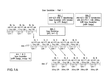
FIG. 1A is a schematic diagram depicting a dose escalation and cohort
expansion
study design. Participants may receive up to 35 cycles of anti-ILT3 antibody
in both
monotherapy and combination arms. Higher dose levels will be tested until
target saturation
in fresh tumor biopsies is achieved unless MTD/MAD is reached before. See
Table 1 for
CA 03212604 2023- 9- 18
WO 2022/197900
PCT/ITS2022/020714
details. Participants may transition to Arm 2 if disease progression is seen
in monotherapy
and after consultation with and approval by the Sponsor. Participants who
cross-over to
combination treatment will be eligible to receive a maximum of 35 cycles of
combination
treatment irrespective of the number of cycles or dose of anti-ILT3 antibody
received in
monotherapy.
FIG. 1B is a schematic diagram depicting study design for anti-ILT3 antibody
monotherapy patients crossing over to receiving combination therapy with an
anti-PD-1
antibody. If participants in Arm 1 (anti-ILT3 mAb monotherapy) experience
disease
progression, they may be eligible for cross-over to combination treatment (Arm
2).
Participants can only cross-over once they have completed the DLT period for
Arm 1 and
upon cross-over may receive the highest dose of anti-ILT3 mAb that has passed
the DLT
evaluation period in Arm 2 (combination) at the time of cross-over. Cross-over
is optional, is
at the discretion of the investigator, and requires the Sponsor's approval.
Disease progression,
toxicity or 35 administrations (24 months of treatment): participants who
cross-over to
combination treatment will be eligible to receive a maximum of 35 cycles of
combination
treatment irrespective of the number of cycles or dose of anti-ILT3 mAb
received in
monothcrapy.
FIG. 2 is a schematic diagram depicting cohorts of particular solid tumor
indications
to be treated with ILT3 antibody and PD-1 antibody. An IA may be conducted
after the first
15 participants (Cohorts B, C, and D) or 20 participants (Cohort A) have their
second post-
baseline imaging assessment. If 8 or fewer responses (Cohort A), 3 or fewer
responses
(Cohort C), or 1 or fewer responses (Cohorts B and D) are observed, enrollment
in the cohort
may be stopped early. An mTPI design will be applied to determine the safety
and tolerability
of the chemotherapy combinations.
DETAILED DESCRIPTION OF THE DISCLOSURE
Definitions and Abbreviations
As used throughout the specification and appended claims, the following
abbreviations apply.
1L first line
2L second line
AE adverse event
16
CA 03212604 2023- 9- 18
WO 2022/197900
PCT/ITS2022/020714
ATD accelerated titration design
AUC area under the concentration-time curve
BICR blinded independent central review
Cycle
CDR complementarity determining region
CI confidence interval
CPS combined positive score
Day
Discon discontinuation
DL dose level
DLT dose-limiting toxicity
DOR duration of response
ECOG Eastern Cooperative Oncology Group
FFPE formalin-fixed paraffin-embedded
FR framework region
FU follow-up
GBM glioblastoma multiforme
IA interim analysis
IgG immunoglobulin G
IHC immunohistochemistry or immunohistochemical
IA interim analysis
IV intravenous
mAb monoclonal antibody
MAD maximum administered dose
MDSC myeloid-derived suppressor cells
MPS modified proportion score
MRI magnetic resonance imaging
MTD maximum tolerated dose
mTPI Modified Toxicity Probability Interval
Design
NSCLC non-small cell lung cancer
NCI CTCAE National Cancer Institute ¨ Common Terminology Criteria for
Adverse Events
ORR objective response rate
OS overall survival
17
CA 03212604 2023- 9- 18
WO 2022/197900
PCT/ITS2022/020714
PD progressive disease
PDAC pancreatic ductal adenocarcinoma
PD-1 programmed death 1 (a k a programmed cell death-1 and
programmed death receptor 1)
PD-Li programmed cell death lligand 1
PD-L2 programmed cell death 1 ligand 2
Pembro pembrolizumab
PFS progression free survival
PK pharmacokinetic
Q2W one dose every two weeks
Q3W one dose every three weeks
Q6W one dose every six weeks
Q9W one dose every 9 weeks
Q12W one dose every 12 weeks
SAE serious adverse event
SC subcutaneous
STS soft tissue sarcoma
TNBC triple negative breast cancer
VH immunoglobulin heavy chain variable region
VL immunoglobulin light chain variable region
So that the invention may be more readily understood, certain technical and
scientific
terms are specifically defined below. Unless specifically defined elsewhere in
this document,
all other technical and scientific terms used herein have the meaning commonly
understood
by one of ordinary skill in the art to which this invention belongs.
Reference to "or" indicates either or both possibilities unless the context
clearly
dictates one of the indicated possibilities In some cases, "and/or" was
employed to highlight
either or both possibilities.
As used herein, the articles "a" and -an" refer to one or to more than one
(i.e., to at
least one) of the grammatical object of the article. By way of example, "an
element" means
one element or more than one element. Furthermore, use of the term "including"
as well as
other forms, such as "include," "includes," and "included," is not limiting.
The term "about", when modifying the quantity (e.g., mg) of a substance or
composition, or the value of a parameter characterizing a step in a method, or
the like, refers
18
CA 03212604 2023- 9- 18
WO 2022/197900
PCT/ITS2022/020714
to variation in the numerical quantity that can occur, for example, through
typical measuring,
handling and sampling procedures involved in the preparation, characterization
and/or use of
the substance or composition; through inadvertent error in these procedures;
through
differences in the manufacture, source, or purity of the ingredients employed
to make or use
the compositions or carry out the procedures; and the like. In certain
embodiments, "about"
means a variation of 10%.
As used herein, the term "comprising" may include the embodiments "consisting
of'
and "consisting essentially of." The terms "comprise(s)," "include(s),"
"having," "has,"
-may," -contain(s)," and variants thereof, as used herein, are intended to be
open-ended
transitional phrases, terms, or words that require the presence of the named
ingredients/steps
and permit the presence of other ingredients/steps. However, such description
should be
construed as also describing compositions or processes as "consisting of' and
"consisting
essentially of' the enumerated components, which allows the presence of only
the named
components or compounds, along with any acceptable carriers or fluids, and
excludes other
components or compounds.
"Consists essentially of," and variations such as "consist essentially of' or
"consisting
essentially of, as used throughout the specification and claims, indicate the
inclusion of any
recited elements or group of elements, and the optional inclusion of other
elements, of similar
or different nature than the recited elements, that do not materially change
the basic or novel
properties of the specified dosage regimen, method, or composition. As a non-
limiting
example, an anti-PD-1 antigen binding fragment that consists essentially of a
recited amino
acid sequence may also include one or more amino acids, including
substitutions of one or
more amino acid residues, which do not materially affect the properties of the
binding
compound.
"Administration" and "treatment," as they apply to an animal, human,
experimental
subject, cell, tissue, organ, or biological fluid, refer to contact of an
exogenous
pharmaceutical, therapeutic, diagnostic agent, or composition to the animal,
human, subject,
cell, tissue, organ, or biological fluid. "Treat" or "treating" cancer, as
used herein, means to
administer an anti-ILT3 antigen binding protein (e.g., an antibody) or antigen-
binding
fragment, alone or in combination with an anti-PD-1 antigen binding protein or
antigen
binding fragment to a subject having cancer, including but not limited to a
solid tumor (e.g.,
metastatic triple negative breast cancer (mTNBC), recurrent non-operable
glioblastoma
(GBM), metastatic pancreatic ductal adenocarcinoma (mPDAC), metastatic soft
tissue
sarcoma (mSTS), metastatic non-squamous non-small cell lung carcinoma
(mNSCLC)), or
19
CA 03212604 2023- 9- 18
WO 2022/197900
PCT/ITS2022/020714
diagnosed with a solid tumor disease (e.g., mTNBC, GBM, mPDAC, mSTS, or
mNSCLC) to
achieve at least one positive therapeutic effect, such as for example, reduced
number of
cancer cells, reduced tumor size, reduced rate of cancer cell infiltration
into peripheral
organs, or reduced rate of tumor metastasis or tumor growth. "Treatment" may
include one or
more of the following: inducing/increasing an antitumor immune response,
decreasing the
number of one or more tumor markers, halting or delaying the growth of a tumor
or blood
cancer or progression of disease associated with ILT-3 or, when administered
in combination
with the anti-PD-1 antigen binding protein or antigen binding fragment, PD-1
binding to its
ligands PD-L1 and/or PD-L2 (-PD-1-related disease") such as cancer,
stabilization of ILT-3-
related disease, or PD-1-related disease (when administered in combination
with the anti-PD-
1 antigen binding protein or antigen binding fragment), inhibiting the growth
or survival of
tumor cells, eliminating or reducing the size of one or more cancerous lesions
or tumors,
decreasing the level of one or more tumor markers, ameliorating or abrogating
the clinical
manifestations of ILT-3 or PD-1-related disease (when administered in
combination with the
anti-PD-1 antigen binding protein or antigen binding fragment), reducing the
severity or
duration of the clinical symptoms of ILT-3- or PD-1- (when administered in
combination
with the anti-PD-1 antigen binding protein or antigen binding fragment)
related disease such
as cancer, prolonging the survival of a patient relative to the expected
survival in a similar
untreated patient, and inducing complete or partial remission of a cancerous
condition or
other ILT-3 or PD-1 (when administered in combination with the anti-PD-1
antigen binding
protein or antigen binding fragment) related disease.
Positive therapeutic effects in cancer can be measured in a number of ways
(See, W.
A. Weber, J. Nucl. Med. 50:1S-10S (2009)). For example, with respect to tumor
growth
inhibition, according to NCI standards, a TIC 42% is the minimum level of anti-
tumor
activity. A TIC < 10% is considered a high anti-tumor activity level, with TIC
= Median
tumor volume of the treated/Median tumor volume of the control x 100. In some
embodiments, the treatment achieved by a therapeutically effective amount is
any of
progression free survival (PFS), disease free survival (DFS) or overall
survival (OS). PFS,
also referred to as "Time to Tumor Progression" indicates the length of time
during and after
treatment that the cancer does not grow and includes the amount of time
patients have
experienced a complete response or a partial response, as well as the amount
of time patients
have experienced stable disease. DFS refers to the length of time during and
after treatment
that the patient remains free of disease. OS refers to a prolongation in life
expectancy as
compared to naive or untreated individuals or patients. While an embodiment of
the methods,
CA 03212604 2023- 9- 18
WO 2022/197900
PCT/ITS2022/020714
compositions and uses of the present invention may not be effective in
achieving a positive
therapeutic effect in every patient, it should do so in a statistically
significant number of
subjects as determined by any statistical test known in the art such as the
Student's t-test, the
chi2-test, the U-test according to Mann and Whitney, the Kruskal-Wallis test
(H-test),
Jonckheere-Terpstra-test and the Wilcoxon-test.
The terms "effective amount", "therapeutically effective amount", and
"therapeutically effective dose" refer to an amount of an anti-ILT3 antigen
binding protein or
antigen binding fragment (e.g., an anti -ILT3 antibody), and/or an anti-PD1
antigen binding
protein or antigen binding fragment (e.g., an anti-PD1 antibody such as
pembrolizumab) of
the invention that, when administered alone or in combination with an
additional
therapeutic/prophylactic agent to a cell, tissue, or subject, is effective to
prevent or cause a
measurable improvement in one or more symptoms of disease or condition
associated with
the disease or condition being treated, e.g., whether that be cancer, mTNBC,
GBM, mPDAC,
mSTS, or mNSCLC as disclosed herein. An effective dose further refers to that
amount of the
anti-ILT3 antigen binding protein or antigen binding fragment or anti-PD1
antigen binding
protein or antigen binding fragment sufficient to result in at least partial
prevention or
amelioration of symptoms of the disease or condition being treated, either
alone or in
combination with another compound. When applied to an individual active
ingredient
administered alone, an effective dose refers to that ingredient alone. When
applied to a
combination, a therapeutically effective amount refers to combined amounts of
the active
ingredients that result in the prophylactic or therapeutic effect, whether
administered in
combination, serially, or simultaneously.
The antigen binding proteins or antigen binding proteins disclosed herein may
be
administered once or according to a dosing regimen wherein a number of doses
are
administered at varying intervals of time for a given period of time. For
example, doses may
be administered one, two, three, or four times per day. Doses may be
administered until the
desired therapeutic effect is achieved or indefinitely to maintain the desired
therapeutic
effect. Suitable dosing regimens for a compound or compounds disclosed herein
depend on
the pharmacokinetic properties of that compound or compounds, such as
absorption,
distribution and half-life which can be determined by a skilled artisan. In
addition, suitable
dosing regimens, including the duration such regimens are administered, for a
compound or
compounds disclosed herein depend on the disease or condition being treated,
the severity of
the disease or condition, the age and physical condition of the subject being
treated, the
medical history of the subject being treated, the nature of concurrent
therapy, the desired
21
CA 03212604 2023- 9- 18
WO 2022/197900
PCT/ITS2022/020714
therapeutic effect, and like factors within the knowledge and expertise of the
skilled artisan. It
will be further understood by such skilled artisans that suitable dosing
regimens may require
adjustment given an individual subject's response to the dosing regimen or
over time as the
individual subject needs change. Typical daily dosages may vary depending upon
the
particular route of administration chosen
The term "subject" (alternatively referred to as "patient" or "individual"
herein) refers
to a mammal (e.g., rat, mouse, dog, cat, rabbit) capable of being treated with
the methods and
compositions of the invention, most preferably a human. In some embodiments,
the subject is
an adult subject In other embodiments, the subject is a pediatric subject.
"Chemotherapeutic agent" is a chemical compound useful in the treatment of
cancer.
Classes of chemotherapeutic agents include, but are not limited to: alkylating
agents,
antimetabolites, kinase inhibitors, spindle poison plant alkaloids, cy
totoxidantitunior
antibiotics, topoisomerase inhibitors, photosensitizers, anti-estrogens and
selective estrogen
receptor modulators (SERN1s), anti-progesterones, estrogen receptor down-
regulators
(ERDs), estrogen receptor antagonists, leutinizing hormone-releasing hormone
agonists, anti-
androgens, aromatase inhibitors, EGFR inhibitors, VEGF inhibitors, anti-sense
oligonucleotides that that inhibit expression of genes implicated in abnormal
cell proliferation
or tumor growth. Chemotherapeutic agents useful in the methods of the present
invention
include cytostatic and/or cytotoxic agents. "Chemotherapy" refers to a cancer
treatment using
chemotherapeutic agents.
"Biologic agent" or "biotherapeutic agent" means a biological molecule, such
as an
antibody or fusion protein, that blocks ligand / receptor signaling in any
biological pathway
that supports tumor maintenance and/or growth or suppresses the anti-tumor
immune
response. "Biologic therapy" or "biological therapy" refers to a cancer
treatment using a
protein.
"Targeted agent" or "targeted therapeutic agent" refers to a therapeutic agent
(either a
small molecule or protein) that affects a specific protein type or class of
proteins that are
associated with tumor cell growth or spread in a patient's body.
"Systemic therapy" refers to a cancer treatment using therapeutic agents
injected in a
patient's bloodstream that affect cells throughout the patient's body,
including chemotherapy,
biological therapy, and targeted therapy.
"Platinum-containing chemotherapy" (also known as platins) refers to the use
of
chemotherapeutic agent(s) used to treat cancer that are coordination complexes
of platinum.
Platinum-containing chemotherapeutic agents are alkylating agents that
crosslink DNA,
22
CA 03212604 2023- 9- 18
WO 2022/197900
PCT/ITS2022/020714
resulting in ineffective DNA mismatch repair and generally leading to
apoptosis. Examples
of platins include cisplatin, carboplatin, and oxaliplatin.
The term "triple negative breast cancer" (TNBC) as used herein refers to a
cancer that
tests negative for estrogen receptors, progesterone receptors, and HER2.
The term "glioblastoma" (GBM) as used herein refers to cancer of glial cells
in
neuronal tissue. Under the World Health Organization (WHO) classification of
central
nervous system tumors, GBM are grade IV diffuse gliomas.
The term "Karnofsky performance status" (KPS) refers to a classification of
functional impairment in a patient. This can be used to compare effectiveness
of different
therapies and to assess the prognosis in individual patients. Lower Karnofsky
scores indicate
worse survival for most serious illnesses (see O'Toole and Golden, West J Med.
1991
Oct,155(4).384-7.). Karnofsky status and glades ate indicated in Table 1
below.
Table 1 ¨ Karnofsky Status and Grade
Karnofsky Status Karnofsky Grade
(%)
Normal, No complaints 100
Able to carry on normal activities. Minor signs or symptoms of 90
disease
Normal activity with effort 80
Care for self Unable to carry on normal activity or to do active 70
work
Requires occasional assistance, but able to care for most of 60
his/her needs
Requires considerable assistance and frequent medical care 50
Disabled. Requires special care and assistance 40
Severely disabled. Hospitalisation indicated though death 30
nonimminent
Very sick. Hospitalisation necessary. Active supportive 20
treatment necessary
Moribund 10
Dead 0
The term "pancreatic ductal adenocarcinoma" (PDAC) refers to exocrine cell
growth
in ducts of the pancreas (see Haeberle, Lena, and Irene Esposito. "Pathology
of pancreatic
cancer." Translational gastroenterology and hepatology vol. 4 50. 27 Jun.
2019).
23
CA 03212604 2023- 9- 18
WO 2022/197900
PCT/ITS2022/020714
The term "soft tissue sarcoma" (STS) refers to a malignant tumor of the soft
tissue,
such as fat, muscle, nerves, fibrous tissues, blood vessels, or deep skin
tissues.
The term "non-squamous non-small cell lung carcinoma" (non-squamous NSCLC)
refers to a non-small cell lung carcinoma that is non-squamous, and includes
large-cell
carcinoma, and adenocarcinoma. Non-squamous NSCLC accounts for about 50% of
all
NSCLC.
Cancer is staged for a given patient by combining Tumor score (T plus a number
0 to
4 describing the size and location of the tumor, and how much the tumor has
grown into
nearby tissues), Node score (N plus a number 0 to 3; often the number of lymph
nodes with
cancer), and Metastasis score (M plus a number 0 or 1; M1 indicates that the
cancer has
metastasized), as well as other factors specific to the particular cancer.
Stage 0 describes
cancer in situ, i.e., cancers still located in the tissue where they stalled
and have not spread to
nearby tissues. This stage of cancer is often highly curable, usually by
removing the entire
tumor with surgery. Stage I is usually a small cancer or tumor that has not
grown deeply into
nearby tissues and has not spread to the lymph nodes or other parts of the
body. Stage II and
Stage III indicate larger cancers or tumors that have grown more deeply into
nearby tissue
and may have spread to lymph nodes but not to other parts of the body. Stage
IV means that
the cancer has spread to other organs or parts of the body. It may also be
called advanced or
metastatic cancer.
Immune Responses to Tumor Cells
Regulatory T cells play an important role in the maintenance of immunological
self-
tolerance by suppressing immune responses against autoimmune diseases and
cancer.
Accordingly, in one embodiment, upmodulating an immune response would be
beneficial for
enhancing an immune response in cancer. Therefore, the anti-ILT3 antigen
binding proteins
or antigen binding fragments disclosed herein may be used in the treatment of
malignancies,
to inhibit tumor growth or metastasis. The anti-ILT3 antigen binding proteins
or antigen
binding fragments disclosed herein may be administered systemically or locally
to the tumor
site.
In one embodiment, modulation of human ILT3 function may be useful in the
induction of tumor immunity. An anti-ILT3 antigen binding protein may be
administered to a
patient having tumor cells (e.g., sarcoma, melanoma, lymphoma, leukemia,
neuroblastoma,
carcinoma) to overcome tumor-specific tolerance in the subject.
24
CA 03212604 2023- 9- 18
WO 2022/197900
PCT/ITS2022/020714
As used herein, the term "neoplastic disease" is characterized by malignant
tumor
growth or in disease states characterized by benign hyperproliferative and
hyperplastic cells.
The common medical meaning of the term "neoplasia" refers to "new cell growth"
that results
as a loss of responsiveness to normal growth controls, e.g., neoplastic cell
growth.
As used herein, the terms "hyperproliferative", "hyperplastic", malignant" and
"neoplastic" are used interchangeably, and refer to those cells in an abnoonal
state or
condition characterized by rapid proliferation or neoplasia. The terms are
meant to include all
types of hyperproliferative growth, hyperplastic growth, cancerous growths or
oncogenic
processes, metastatic tissues or malignantly transformed cells, tissues, or
organs, irrespective
of histopathologic type or stage of invasiveness. A "hyperplasia" refers to
cells undergoing an
abnormally high rate of growth. However, as used herein, the terms neoplasia
and hyperplasia
can be used interchangeably, as their context will reveal, referring generally
to cells
experiencing abnormal cell growth rates. Neoplasias and hyperplasias include
"tumors,"
which may be either benign, premalignant or malignant.
The terms "neoplasia," "hyperplasia," and "tumor" are often commonly referred
to as
"cancer," which is a general name for more than 100 disease that are
characterized by
uncontrolled, abnormal growth of cells.
In one embodiment, the cancer is selected from the group consisting of:
gastrointestinal cancer, gastric cancer, pancreatic cancer, melanomas, breast
cancer, lung
cancer (e.g., NSCLC), head and neck cancer, bronchus cancer, colorectal
cancer, colon
cancer, rectal cancer, prostate cancer, pancreatic cancer, stomach cancer,
ovarian cancer,
urinary bladder cancer, brain or central nervous system cancer (e.g., GBM),
peripheral
nervous system cancer, esophageal cancer, cervical cancer, uterine or
endometrial cancer,
cancer of the oral cavity or pharynx, liver cancer, renal cancer, testicular
cancer, biliary tract
cancer, small bowel or appendix cancer, salivary gland cancer, thyroid gland
cancer, adrenal
gland cancer, soft tissue sarcoma, osteosarcoma, chondrosarcoma, and cancer of
hematological tissues.
In one embodiment, the cancer is selected from the group consisting of:
metastatic
triple negative breast cancer (mTNBC); glioblastoma multiforme (GBM);
metastatic
pancreatic ductal adenocarcinoma (mPDAC); metastatic soft tissue sarcoma
(mSTS); and
metastatic non-squamous non-small cell lung carcinoma (mNSCLC). In one
embodiment, the
cancer is triple negative breast cancer (mTNBC). In one embodiment, the cancer
is
glioblastoma multiforme (GBM). In one embodiment, the cancer is metastatic
pancreatic
ductal adenocarcinoma (mPDAC). In one embodiment, the cancer is metastatic
soft tissue
CA 03212604 2023- 9- 18
WO 2022/197900
PCT/ITS2022/020714
sarcoma (mSTS). In one embodiment the cancer is metastatic non-squamous non-
small cell
lung carcinoma (mNSCLC).
Antibodies
As used herein, the term "antigen binding protein" refers to a polypeptide or
protein
that binds to an antigen, e.g., ILT3 or PD-1 protein. An antigen binding
protein includes, but
is not limited to, a bivalent antibody tetramer (2H+2L), a monovalent antibody
(H+L), a bi-
specifi c antibody that targets an antigen and another target, a Fab fragment,
a Fab' fragment,
a F(ab')2 fragment, an FAT region, and an ScFv. Unless otherwise indicated,
the antigen
binding proteins herein bind to and inhibit the activity of ILT3 or PD-1.
The term "antibody" refers to any form of antibody that exhibits the desired
biological
or binding activity. Thus, it is used in the broadest sense and specifically
covers, but is not
limited to, monoclonal antibodies (including full length monoclonal
antibodies), polyclonal
antibodies, humanized, fully human antibodies, and chimeric antibodies.
"Parental
antibodies" are antibodies obtained by exposure of an immune system to an
antigen prior to
modification of the antibodies for an intended use, such as humanization of an
antibody for
use as a human therapeutic.
In general, the basic antibody structural unit comprises a tetramer. Each
tetramer
includes two identical pairs of polypeptide chains, each pair having one
"light" (about 25
kDa) and one "heavy" chain (about 50-70 kDa). The amino-terminal portion of
each chain
includes a variable region of about 100 to 110 or more amino acids primarily
responsible for
antigen recognition. The carboxy-terminal portion of the heavy chain may
define a constant
region primarily responsible for effector function. Typically, human light
chains are
classified as kappa and lambda light chains. Furthermore, human heavy chains
are typically
classified as mu, delta, gamma, alpha, or epsilon, and define the antibody's
isotype as IgM,
IgD, IgG, IgA, and IgE, respectively. Within light and heavy chains, the
variable and constant
regions are joined by a "J" region of about 12 or more amino acids, with the
heavy chain also
including a "D" region of about 10 more amino acids. See generally,
Fundamental
Immunology Ch. 7 (Paul, W., ed., 2" 15 ed. Raven Press, N.Y. (1989).
The variable regions of each light/heavy chain pair form the antibody binding
site.
Thus, in general, an intact antibody has two binding sites. Except in
bifunctional or bispecific
antibodies, the two binding sites are, in general, the same.
Typically, the variable domains of both the heavy and light chains comprise
three
hypervariable regions, also called complementarity determining regions (CDRs),
which are
26
CA 03212604 2023- 9- 18
WO 2022/197900
PCT/ITS2022/020714
located within relatively conserved framework regions (FR). The CDRs are
usually aligned
by the framework regions, enabling binding to a specific epitope. In general,
from N-terminal
to C-terminal, both light and heavy chains variable domains comprise FR1,
CDR1, FR2,
CDR2, FR3, CDR3 and FR4. The assignment of amino acids to each domain is,
generally, in
accordance with the definitions of Sequences of Proteins of Immunological
Interest, Kabat, et
al.; National Institutes of Health, Bethesda, Md.; 5th ed.; NIH Publ. No. 91-
3242 (1991);
Kabat (1978) Adv. Prot. Chem. 32:1-75; Kabat, et al., (1977)1 Biol. Chem.
252:6609-6616;
Chothi a, et al., (1987)1AJol. Biol. 196:901-917 or Chothia, et al., (1989)
Nature 342:878-
883.
The term "hypervariable region" refers to the amino acid residues of an
antibody that
are responsible for antigen-binding. The hypervariable region comprises amino
acid residues
from a "complementarity determining region" or "CDR" (i.e., CDRL1, CDRL2 and
CDRL3
in the light chain variable domain and CDRH1, CDRH2 and CDRH3 in the heavy
chain
variable domain). See Kabat et al. (1991) Sequences of Proteins of
Immunological Interest,
5th Ed. Public Health Service, National Institutes of Health, Bethesda, Md.
(defining the CDR
35 regions of an antibody by sequence); see also Chothia and Lesk (1987) J.
Mol. Biol. 196:
901-917 (defining the CDR regions of an antibody by structure). The term
"framework" or
"FR" residues refers to those variable domain residues other than the
hypervariable region
residues defined herein as CDR residues.
Unless otherwise indicated, an "antibody fragment" or "antigen binding
fragment"
refers to antigen binding fragments of antibodies, i.e., antibody fragments
that retain the
ability to specifically bind to the antigen bound by the full-length antibody,
e.g., fragments
that retain one or more CDR regions. Examples of antibody binding fragments
include, but
are not limited to, Fab, Fab', F(ab')2, and Fv fragments.
An antibody that "specifically binds to" a specified target protein is an
antibody that
exhibits preferential binding to that target as compared to other proteins,
but this specificity
does not require absolute binding specificity. An antibody is considered
"specific" for its
intended target if its binding is determinative of the presence of the target
protein in a sample,
e.g., without producing undesired results such as false positives. Antibodies,
or binding
fragments thereof, useful in the present invention will bind to the target
protein with an
affinity that is at least two-fold greater, preferably at least ten times
greater, more preferably
at least 20-times greater, and most preferably at least 100-times greater than
the affinity with
non-target proteins. As used herein, an antibody is said to bind specifically
to a polypeptide
comprising a given amino acid sequence, e.g., the amino acid sequence of a
mature human
27
CA 03212604 2023- 9- 18
WO 2022/197900
PCT/ITS2022/020714
PD-1 or human PD-L1 molecule, if it binds to polypeptides comprising that
sequence but
does not bind to proteins lacking that sequence.
"Chimeric antibody" refers to an antibody in which a portion of the heavy
and/or light
chain is identical with or homologous to corresponding sequences in an
antibody derived
from a particular species (e.g., human) or belonging to a particular antibody
class or subclass,
while the remainder of the chain(s) is identical with or homologous to
corresponding
sequences in an antibody derived from another species (e.g., mouse) or
belonging to another
antibody class or subclass, as well as fragments of such antibodies, so long
as they exhibit the
desired biological activity.
"Human antibody" refers to an antibody that comprises human immunoglobulin
protein sequences only. A human antibody may contain murine carbohydrate
chains if
produced in a mouse, in a mouse cell, or in a hybridoma derived from a mouse
cell.
Similarly, "mouse antibody" or "rat antibody" refer to an antibody that
comprises only mouse
or rat immunoglobulin sequences, respectively.
"Humanized antibody" refers to forms of antibodies that contain sequences from
non-
human (e.g., murine) antibodies as well as human antibodies. Such antibodies
contain
minimal sequence derived from non-human immunoglobulin. In general, the
humanized
antibody will comprise substantially all of at least one, and typically two,
variable domains,
in which all or substantially all of the hypervariable loops correspond to
those of a non-
human immunoglobulin and all or substantially all of the FR regions are those
of a human
immunoglobulin sequence. The humanized antibody optionally also will comprise
at least a
portion of an immunoglobulin constant region (Fe), typically that of a human
immunoglobulin. The prefix "hum", "hu" or "h" is added to antibody clone
designations
when necessary to distinguish humanized antibodies from parental rodent
antibodies. The
humanized forms of rodent antibodies will generally comprise the same CDR
sequences of
the parental rodent antibodies, although certain amino acid substitutions may
be included to
increase affinity, increase stability of the humanized antibody, or for other
reasons.
-CDR" or -CDRs" means complementarity determining region(s) in an
immunoglobulin variable region.
"Framework region" or "FR" as used herein means the immunoglobulin variable
regions excluding the CDR regions.
"Isolated antibody" and "isolated antibody fragment" refers to the
purification status
and in such context means the named molecule is substantially free of other
biological
molecules such as nucleic acids, proteins, lipids, carbohydrates, or other
material such as
28
CA 03212604 2023- 9- 18
WO 2022/197900
PCT/ITS2022/020714
cellular 10 debris and growth media. Generally, the term ''isolated" is not
intended to refer to
a complete absence of such material or to an absence of water, buffers, or
salts, unless they
are present in amounts that substantially interfere with experimental or
therapeutic use of the
binding compound as described herein.
"Monoclonal antibody" or "m Ab" or "Mab", as used herein, refers to a
population of
substantially homogeneous antibodies, i.e., the antibody molecules comprising
the population
are identical in amino acid sequence except for possible naturally occurring
mutations that
may be present in minor amounts. In contrast, conventional (polyclonal)
antibody
preparations typically include a multitude of different antibodies having
different amino acid
sequences in their variable domains, particularly their CDRs, which are often
specific for
different epitopes. The modifier "monoclonal" indicates the character of the
antibody as
being obtained from a substantially homogeneous population of antibodies and
is not to be
construed as requiring production of the antibody by any particular method.
For example, the
monoclonal antibodies to be used in accordance with the present invention may
be made by
the hybridoma method first described by Kohler et al. (1975) Nature 256: 495,
or may be
made by recombinant DNA methods (see, e.g., U.S. Pat. No. 4,816,567). The
"monoclonal
antibodies" may also be isolated from phage antibody libraries using the
techniques described
in Clackson et al. (1991) Nature 352: 624-628 and Marks et al. (1991) J. Mol.
Biol. 222: 581-
597, for example. See also Presta (2005) J. Allergy Chn. Immunol. 116:731.
"Variable regions" or "V region" as used herein means the segment of IgG
chains
which is variable in sequence between different antibodies. It extends to
Kabat residue 109 in
the light chain and 113 in the heavy chain.
A variant of a heavy chain variable region sequence or full-length heavy chain
sequence is identical to the reference sequence except having up to 17
conservative amino
acid substitutions in the framework region (i.e., outside of the CDRs), and
preferably has less
than ten, nine, eight, seven, six or five conservative amino acid
substitutions in the
framework region. A variant of a light chain variable region sequence or full-
length light
chain sequence is identical to the reference sequence except having up to five
conservative
amino acid substitutions in the framework region (i.e., outside of the CDRs),
and preferably
has less than four, three or two conservative amino acid substitution in the
framework region.
"Conservatively modified variants" or "conservative substitution" refers to
substitutions of amino acids in a protein with other amino acids having
similar characteristics
(e.g., charge, side-chain size, hydrophobicity/hydrophilicity, backbone
conformation and
29
CA 03212604 2023- 9- 18
WO 2022/197900
PCT/ITS2022/020714
rigidity, etc.), such that the changes can frequently be made without altering
the biological
activity or other desired property of the protein, such as antigen affinity
and/or specificity.
Those of skill in the art recognize that, in general, single amino acid
substitutions in non-
essential regions of a polypeptide do not substantially alter biological
activity (see, e.g.,
Watson et at. (1987) Molecular Biology of the Gene, The Benjamin/Cummings Pub.
Co., p.
224 (4th Ed.)). In addition, substitutions of structurally or functionally
similar amino acids
are less likely to disrupt biological activity. Exemplary conservative
substitutions are set forth
in Table 2.
Table 2. Exemplary Conservative Amino Acid Substitutions
Original residue Conservative substitution
Ala (A) Gly; Ser
Arg (R) Lys; His
Asn (N) Gln; His
Asp (D) Glu; Asn
Cys (C) Ser; Ala
Gln (Q) Asn
Gin (F,) Asp; Gin
Gly (G) Ala
His (H) Asn; Gln
Ile (I) Leu; Val
Leu (L) Ile; Val
Lys (K) Arg; His
Met (M) Leu; Ile; Tyr
Phe (F) Tyr; Met; Leu
Pro (P) Ala
Ser (S) Thr
Thr (T) Ser
Trp (W) Tyr; Phe
Tyr (Y) Trp; Phe
Val (V) Ile; Leu
The VH and VL regions can be further subdivided into regions of
hypervariability,
termed complementarity determining regions (CDR), interspersed with regions
that are more
conserved, termed framework regions (FR). Each VH and VL is composed of three
CDR
regions and four FR regions, arranged from amino-terminus to carboxy-terminus
in the
following order. FR1, CDR1, FR2, CDR2, FR3, CDR3, FR4. The variable regions of
the
heavy and light chains contain a binding domain that interacts with an
antigen. The constant
regions of the antibodies may mediate the binding of the immunoglobulin to
host tissues or
factors, including various cells of the immune system (e.g., effector cells)
and the first
CA 03212604 2023- 9- 18
WO 2022/197900
PCT/ITS2022/020714
component (Clq) of the classical complement system. The assignment of amino
acids to each
domain is, generally, in accordance with the definitions of Sequences of
Proteins of
Immunological Interest, Kabat, et at.; National Institutes of Health,
Bethesda, Md.; 5th ed.;
NIH Publ. No. 91-3242 (1991); Kabat (1978) Adv. Prot. Chem. 32:1-75; Kabat,
etal., (1977)
J. Biol. Chem. 252:6609-6616; Chothia, etal., (1987) J Mol. Biol. 196:901-917
or Chothia, et
al., (1989) Nature 342:878-883.
The constant regions of the antibodies may mediate the binding of the
immunoglobulin to host tissues or factors, including various cells of the
immune system (e.g.,
effector cells) and the first component (Clq) of the classical complement
system. Typically,
the numbering of the amino acids in the heavy chain constant domain begins
with number
118, which is in accordance with the Eu numbering scheme. The Eu numbering
scheme is
based upon the amino acid sequence of human IgG1 (Eu), which has a constant
domain that
begins at amino acid position 118 of the amino acid sequence of the IgG1
described in
Edelman et al., Proc. Natl. Acad. Sci. USA. 63: 78-85 (1969), and is shown for
the IgGl,
IgG2, IgG3, and IgG4 constant domains in Beranger, et al., Ibid.
The variable regions of the heavy and light chains contain a binding domain
comprising the CDRs that interacts with an antigen. A number of methods are
available in the
art for defining CDR sequences of antibody variable domains (see Dondelinger
c/at.,
Frontiers in Immunol. 9: Article 2278 (2018)). The common numbering schemes
include the
following.
= Kabat numbering scheme is based on sequence variability and is the most
commonly
used (See Kabat et al. Sequences of Proteins of Immunological Interest, 5th Ed
Public Health Service, National Institutes of Health, Bethesda, Md. (1991)
(defining
the CDR regions of an antibody by sequence);
= Chothia numbering scheme is based on the location of the structural loop
region (See
Chothia & Lesk J. Mol. Biol. 196: 901-917 (1987); Al-Lazikani etal., J. Mol.
Biol.
273: 927-948 (1997));
= AbM numbering scheme is a compromise between the two used by Oxford
Molecular's AbM antibody modelling software (see Karu c/at., ILAR Journal 37.
132-141 (1995);
= Contact numbering scheme is based on an analysis of the available complex
crystal
structures (See www.bioinf. org.uk: Prof. Andrew C.R. Martin's Group;
Abhinandan
8z Martin, Mol. Immunol. 45:3832-3839 (2008).
31
CA 03212604 2023- 9- 18
WO 2022/197900
PCT/ITS2022/020714
= IMGT (ImMunoGeneTics) numbering scheme is a standardized numbering system
for
all the protein sequences of the immunoglobulin superfamily, including
variable
domains from antibody light and heavy chains as well as T cell receptor chains
from
different species and counts residues continuously from 1 to 128 based on the
germ-
line V sequence alignment (see Giudicelli et al., Nucleic Acids Res. 25:206-11
(1997); Lefranc, Immunol Today 18:509(1997); Lefranc et al., Dev Comp Immunol.
27:55-77 (2003)).
The following general rules disclosed in www.bioinforg.uk: Prof. Andrew C.R.
Martin's Group and reproduced in Table 3 below may be used to define the CDRs
in an
antibody sequence that includes those amino acids that specifically interact
with the amino
acids comprising the epitope in the antigen to which the antibody binds. There
are rare
examples where these generally constant features do not occur; however, the
Cys residues are
the most conserved feature.
Table 3¨ Antibody CDR Rules
Loop Kabat AbM Chothial Contact2
IMGT
Li L24--L34 L24--L34 L24--L34 L30--L36
L27--L32
L2 L50--L56 L50--L56 L50--L56 L46--L55
L50--L51
L3 L89--L97 L89--L97 L89--L97 L89--L96
L89--L97
H1 H31--H35B H26-- H26--H32 H30--H35B
H26--
(Kabat H35B to.34
H35B
Numbering)3
H1 H31--H35 H26--H35 H26--H32 H30--H35 H26--H33
(Chothia
Numbering)
H2 H50--H65 H50--H58 H52--H56 H47--H58 H51--H56
H3 H95--H102 H95-- H95--H102 H93--
H101 H93--
H102
H102
32
CA 03212604 2023- 9- 18
WO 2022/197900
PCT/ITS2022/020714
1-Some of these numbering schemes (particularly for Chothia loops) vary
depending on the
individual publication examined.
2Any of the numbering schemes can be used for these CDR definitions, except
the
Contact numbering scheme uses the Chothia or Martin (Enhanced Chothia)
definition.
3The end of the Chothia CDR-H1 loop when numbered using the Kabat numbering
convention varies between H32 and H34 depending on the length of the loop.
(This is
because the Kabat numbering scheme places the insertions at H35A and H35B.)
= If neither H35A nor H35B is present, the loop ends at H32
= If only H35A is present, the loop ends at H33
. If both H35A and H35B are present, the loop ends at H34
In general, the state of the art recognizes that in many cases, the CDR3
region of the
heavy chain is the primary determinant of antibody specificity, and examples
of specific
antibody generation based on CDR3 of the heavy chain alone are known in the
art (e.g.,
Beiboer et al, J. Mol. Biol. 296: 833-849 (2000); Klimka et al., British J.
Cancer 83: 252-260
(2000); Rader et al., Proc. Natl. Acad. Sci. USA 95: 8910-8915 (1998); Xu et
al., Immunity
13: 37-45 (2000).
Diagnostic anti-PD-L antibodies
"Diagnostic anti-PD-L monoclonal antibody" means a mAb which specifically
binds
to the mature form of the designated PD-L (PD-Li or PD-L2) that is expressed
on the surface
of certain mammalian cells. A mature PD-L lacks the presecretory leader
sequence, also
referred to as leader peptide. The terms "PD-L" and "mature PD-L" are used
interchangeably
herein and shall be understood to mean the same molecule unless otherwise
indicated or
readily apparent from the context.
As used herein, a diagnostic anti-human PD-Li mAb or an anti-hPD-L1 mAb refers
to a monoclonal antibody that specifically binds to mature human PD-L1 A
mature human
PD-Li molecule consists of amino acids 19-290 of the following sequence:
MRIFAVFIFMTYWHLLNAFTVTVPKDLYVVEYGSNWITIECKFPVEKQLDL
AALIVYWEMEDKNIIQFVHGEEDLKVQHSSYRQRARLLKDQLSLGNAALQITDVKL
QDAGVYRCMISYGGADYKRITVKVNAPYNKINQRILVVDPVTSEHELTCQAEGYPK
AEVIVVTSSDHQVLSGKTTTTNSKREEKLFNVTSTLRINTTTNEIFYCTFRRLDPEENHT
AELVIPELPLAHPPNERTHLVILGAILLCLGVALTFIFRLRKGRMMDVKKCGIQDTN
SKKQSDTHLEET (SEQ ID NO: 1).
Specific examples of diagnostic anti-human PD-Ll mAbs useful as diagnostic
mAbs
for immunohistochemistry (II-IC) detection of PD-1-1 expression in formalin-
fixed, paraffin-
33
CA 03212604 2023- 9- 18
WO 2022/197900
PCT/ITS2022/020714
embedded (FFPE) tumor tissue sections are antibody 20C3 and antibody 22C3,
which are
described in WO 2014/100079. These antibodies comprise the light chain and
heavy chain
variable region amino acid sequences shown in Table 4 below:
Table 4. Monoclonal Antibodies 20C3 and 22C3
20C3 Light Chain Mature Variable Region
DIVMSQSPSSLAVSAGEKVTMSCKSSQSLLNSRTRKNYLAWYQQ
KPGQSPKWYWASTRESGVPDRFTGSGSGTDFTLTISSVQAEDLA
VYYCQQSYDVVTFGAGTKLELK
SEQ ID NO:2
20C3 Heavy Chain Mature Variable Region
QVQVQQSGAELAEPGASVKMSCKASGYIFTSYWMHWLKQRPGQ
GLEWIGYINPSSDYNEYSEKFMDKATLTADKASTTAYMQLISLTS
ED SAVYYCARSGWLVHGDYYFDYWGQGTTLTVS S
SEQ ID NO:3
22C3 Light Chain Mature Variable Region
DIVMSQSPSSLAVSAGEKVTMTCKSSQSLLHTSTRKNYLAWYQQ
KPGQSPKWYWASTRESGVPDRFTGSGSGTDFTLTISSVQAEDLA
VYYCKQSYDVVTFGAGTKLELK
SEQ ID NO:4
22C3 Heavy Chain Mature Variable Region
QVHLQQSGAELAKPGASVKMSCKASGYTFTSYWIHWIKQRPGQG
LEWIGYINPSSGYHEYNQKFIDKAILTADRSSSTAYMHLTSLTSED SEQ ID NO:5
SAVYYCARSGWLIHGDYYFDFWGQGTTLTVSS
Another anti-human PD-L1 m Ab that has been reported to be useful for IHC
detection
of PD-Li expression in FFPE tissue sections (Chen, B.J. et al., Clin Cancer
Res 19:3462-
3473 (2013)) is a rabbit anti-human PD-Li mAb publicly available from Sino
Biological, Inc.
(Beijing, P.R. China; Catalog number 10084-R015).
PD-Li and PD-L2 Tissue Expression
"PD-Li" or "PD-L2" expression means any detectable level of expression of the
designated PD-L protein on the cell surface or of the designated PD-L mRNA
within a cell or
tissue, unless otherwise defined. PD-L protein expression may be detected with
a diagnostic
PD-L antibody in an IHC assay of a tumor tissue section or by flow cytometty.
Alternatively,
PD-L protein expression by tumor cells may be detected by PET imaging, using a
binding
agent (e.g., antibody fragment, affibody and the like) that specifically binds
to the desired
34
CA 03212604 2023- 9- 18
WO 2022/197900
PCT/ITS2022/020714
PD-L target, e.g., PD-Li or PD-L2. Techniques for detecting and measuring PD-L
mRNA
expression include RT- PCR and real-time quantitative RT-PCR.
Several approaches have been described for quantifying PD-Li protein
expression in
IHC assays of tumor tissue sections. See, e.g., Thompson et al., IWAS 101
(49): 17174-17179
(2004); Thompson et at., Cancer Res. 66:3381-3385 (2006); Gadiot et at.,
Cancer 117:2192-
2201 (2011); Taube et al., Sci Transl Med 4, 127ra37 (2012); and Toplian et
al. , New Eng. J
Med. 366 (26): 2443-2454 (2012).
One approach employs a simple binary endpoint of positive or negative for PD-
Li
expression, with a positive result defined in terms of the percentage of tumor
cells that exhibit
histologic evidence of cell-surface membrane staining. A tumor tissue section
is counted as
positive for PD-Li expression is at least 1%, and preferably 5% of total tumor
cells.
In another approach, PD-Li expression in the tumor tissue section is
quantified in the
tumor cells as well as in infiltrating immune cells, which predominantly
comprise
lymphocytes. The percentage of tumor cells and infiltrating immune cells that
exhibit
membrane staining are separately quantified as < 5%, 5 to 9%, and then in 10%
increments
up to 100%. For tumor cells, PD-Li expression is counted as negative if the
score is < 5%
score and positive if the score is > 5%. PD-Li expression in the immune
infiltrate is reported
as a semi-quantitative measurement called the adjusted inflammation score
(AIS), which is
determined by multiplying the percent of membrane staining cells by the
intensity of the
infiltrate, which is graded as none (0), mild (score of 1, rare lymphocytes),
moderate (score of
2, focal infiltration of tumor by lymphohistiocytic aggregates), or severe
(score of 3, diffuse
infiltration). A tumor tissue section is counted as positive for PD-L1
expression by immune
infiltrates if the AIS is > 5.
A tissue section from a tumor that has been stained by IHC with a diagnostic
PD- Li
antibody may also be scored for PD-Li protein expression by assessing PD-Li
expression in
both the tumor cells and infiltrating immune cells in the tissue section using
a scoring
process. See WO 2014/165422. One PD-Li scoring process comprises examining
each tumor
nest in the tissue section for staining and assigning to the tissue section
one or both of a
modified H score (MHS) and a modified proportion score (MPS). To assign the
1VIHS, four
separate percentages are estimated across all of the viable tumor cells and
stained
mononuclear inflammatory cells in all of the examined tumor nests: (a) cells
that have no
staining (intensity = 0), (b) weak staining (intensity =1+), (c) moderate
staining (intensity
=2+) and (d) strong staining (intensity ¨3+). A cell must have at least
partial membrane
staining to be included in the weak, moderate or strong staining percentages.
The estimated
CA 03212604 2023- 9- 18
WO 2022/197900
PCT/ITS2022/020714
percentages, the sum of which is 100%, are then input into the formula of 1 x
(percent of
weak staining cells) + 2 x (percent of moderate staining cells) + 3 x (percent
of strong
staining cells), and the result is assigned to the tissue section as the MFIS
The MPS is
assigned by estimating, across all of the viable tumor cells and stained
mononuclear
inflammatory cells in all of the examined tumor nests, the percentage of cells
that have at
least partial membrane staining of any intensity, and the resulting percentage
is assigned to
the tissue section as the MPS. In some embodiments, the tumor is designated as
positive for
PD-Li expression if the MHS or the MPS is positive.
Another method for scoring/quantifying PD-Li expression in a tumor is the
"combined positive score" or "CPS," which refers to an algorithm for
determining a PD-Li
expression score from a tumor sample of a patient. The CPS is useful in
selecting patients for
treatment with particular treatment regimens including methods of treatment
comptising
administration of an anti-PD-1 antigen binding protein or antigen binding
fragment in which
expression of PD-L1 is associated with a higher response rate in a particular
patient
population relative to same patient population that does not express PD-Li.
The CPS is
determined by determining the number of viable PD-Li positive tumor cells, the
number of
viable PD-Li negative tumor cells, and the number of viable PD-Li positive
mononuclear
inflammatory cells (MIC) in a tumor tissue from a patient having a tumor and
calculating the
CPS using the following formula:
(# PD-Ll positive tumor cells) + (# PD-Li positive MIC) x 100%
(# PD-Li positive tumor cells) + (PD-Li negative tumor cells).
Yet another scoring method for PD-L1 expression is the "TPS" or "tumor
proportion
score," which is the percentage of tumor cells expressing PD-Li on the cell
membrane. TPS
typically includes the percentage of neoplastic cells expressing PD-Li at any
intensity (weak,
moderate, or strong), which can be determining using an immunohistochemical
assay using a
diagnostic anti-human PD-Li mAb, e.g., antibody 20C3 and antibody 22C3,
described above.
Cells are considered to express PD-Ll if membrane staining is present,
including cells with
partial membrane staining.
The level of PD-Li mRNA expression may be compared to the mRNA expression
levels of one or more reference genes that are frequently used in quantitative
RT-PCR, such
as ubiquitin C.
In some embodiments, a level of PD-Li expression (protein and/or mRNA) by
malignant cells and/or by infiltrating immune cells within a tumor is
determined to be
"overexpressed" or "elevated" based on comparison with the level of PD-Li
expression
36
CA 03212604 2023- 9- 18
WO 2022/197900
PCT/ITS2022/020714
(protein and/ or mRNA) by an appropriate control. For example, a control PD-Li
protein or
mRNA expression level may be the level quantified in nonmalignant cells of the
same type or
in a section from a matched normal tissue In some preferred embodiments, PD-Li
expression in a tumor sample is determined to be elevated if PD-Li protein
(and/or PD-Li
mRNA) in the sample is at least 10%, 20%, or 30% greater than in the control.
"Tissue section" refers to a single part or piece of a tissue sample, e.g., a
thin slice of
tissue cut from a sample of a normal tissue or of a tumor.
"Tumor" as it applies to a subject diagnosed with, or suspected of having, a
cancer
refers to a malignant or potentially malignant neoplasm or tissue mass of any
size, and
includes primary tumors and secondary neoplasms. A solid tumor is an abnormal
growth or
mass of tissue that usually does not contain cysts or liquid areas. Different
types of solid
tumors are named for the type of cells that form them. Examples of solid
tumors are
sarcomas, carcinomas, and lymphomas. Leukemias (cancers of the blood)
generally do not
form solid tumors (National Cancer Institute, Dictionary of Cancer Terms).
"RECIST 1.1 Response Criteria" as used herein means the definitions set forth
in
Eisenhauer, E.A. et al., Ent% J. Cancer 45:228-247 (2009) for target lesions
or non-target
lesions, as appropriate based on the context in which response is being
measured.
Anti-ILT3 Antibodies and Antigen Binding Fragments Useful in the Invention
An "anti-ILT3 antigen binding protein or antigen binding fragment" useful in
the any
of the methods, compositions and uses of the present invention include
monoclonal
antibodies (m Ab), or antigen binding fragments thereof, which specifically
bind to human
ILT3. Alternative names or synonyms for ILT3 include: LILRB4; LIR5; and CD85K.
In any
of the methods, compositions and uses of the present invention in which a
human individual
is being treated, the anti-ILT3 antigen binding protein, antibody or antigen
binding fragment
binds to ILT3 and reduces the ability of MDSCs to suppress T-cell activation
and
proliferation. An anti-ILT3 antibody may be a human antibody, a humanized
antibody or a
chimeric antibody, and may include a human constant region. In some
embodiments the
human constant region is selected from the group consisting of IgGl, IgG2,
IgG3 and IgG4
constant regions, and in preferred embodiments, the human constant region is
an IgG1 or
IgG4 constant region. In some embodiments, the antigen binding fragment is
selected from
the group consisting of Fab, Fab'- SH, F(ab')2, scFv and Fv fragments.
The term "anti-ILT3 antigen binding protein" refers to a protein that binds
the
extracellular domain (amino acids 22-259) of GenPept Ace. No. Q8NHJ6.3:
37
CA 03212604 2023- 9- 18
WO 2022/197900
PCT/ITS2022/020714
QAGPLPKPTLWAEPGSVISWGNSVTIWCQGTLEAREYRLDKEESPAPWDRQN
PLEPKNKARFSIPSMTEDYAGRYRCYYRSPVGWSQPSDPLELVMTGAYSKPTLSALP
SPLVTSGKSVTLLCQSRSPMDTELLIKERAATIPLLTILRSEHGAQQHQAEFPMSPVTSV
HGGTYRCF SSHGF SHYLL S HP SDPLELIV S GSLEDPRP SP TRS VS TAAGPED QPLMP TG
SVPHSGLRRHWE (SEQ ID NO: 6)
Examples of mAbs that bind to human ILT3, useful in the methods and uses of
the
invention are described in W02019/099597 (incorporated by reference herein)
and
summarized below in Table 5
Table 5 ¨ Exemplary anti-ILT3 antibody regions and constant regions
SEQ
ID Description Sequence
NO:
7 Human IgG4 HC ASTKGPSVFPLAPCSRSTSESTAALGCLVKDYFPEPVTV
Constant domain SWNSGALTSGVHTFPAVLQSSGLYSLSSVVTVPSSSLG
TKTYTCNVDIIKPSNTKVDKRVESKYGPPCPPCPAPEFL
Residue 108 GGPSVFLEPPKPKDTLMISRTPEVTCVVVDVSQEDPEV
corresponds with QFNWYVDGVEVHINAKTKPREEQFNSTYRVVSVLTVL
S228P HQDWLNGKEYKCKVSNKGLPSSIEKTISKAKGQPREP
QVYTLPPSQEEMTKNQVSLTCLVKGFYPSDIAVEWES
NGQPENNYKT'TPPVLDSDGSFELYSRLTVDKSRWQEG
NVFSCSVMHEALHNHYTQKSLSLSLGK
8 Human IgG4 HC ASTKGPSVFPLAPCSRSTSESTAALGCLVKDYFPEPVTV
Constant domain SWNSGALTSGVHTFPAVLQSSGLYSLSSVVTVPSSSLG
TKTYTCNVDRKPSNTKVDKRVESKYGPPCPPCPAPEFL
Residue 108 GGPSVFLFPPKPKDTLMISRTPEVTCVVVDVSQEDPEV
corresponds with QFNWYVDGVEVIINAKTKPREEQFNSTYRVVSVLTVL
S228P HQDWLNGKEYKCKVSNKGLPSSIEKTISKAKGQPREP
QVYTLPPSQEEMTKNQVSLTCLVKGFYPSDIAVEWES
(lacks C-terminal NGQPENNYKTTPPVLDSDGSFFLYSRLTVDKSRWQEG
K, (herein referred NVFSCSVMHEALHNHYTQKSLSLSLG
to as "K-")
9 Human IgG1 HC ASTKGPSVFPLAPSSKSTSGGTAALGCLVKDYFPEPVT
constant domain VSWNSGALTSGVHTFPAVLQSSGLYSLSSVVTVPSSSL
GTQTYICNVNHKPSNTKVDKKVEPKSCDKTHTCPPCP
APELLGGP SVFLEPPKPKDTLMISRTPEVTCVVVDVSH
EDPEVKFNWYVDGVEVHNAKTKPREEQYNSTYRVVS
VLTVLHQDWLNGKEYKCKVSNKALPAPIEKTISKAKG
QPREPQVYTLPPSRDELTKNQVSLTCLVKGFYPSDIAV
EWESNGQPENNYKTTPPVLDSDGSFFLYSKLTVDKSR
WQQGNVF SCSVMHEALHNHYTQKSL SL SP GK
10 Human IgG1 HC ASTKGPSVFPLAPSSKSTSGGTAALGCLVKDYFPEPVT
Constant domain VSWNSGALTSGVHTFPAVLQSSGLYSLSSVVTVPSSSL
GTQTYICNVNIIKPSNTKVDKKVEPKSCDKTHTCPPCP
38
CA 03212604 2023- 9- 18
WO 2022/197900
PCT/ITS2022/020714
Table 5 ¨ Exemplary anti-ILT3 antibody regions and constant regions
SEQ
ID Description Sequence
NO:
Residue 117 APEAAGGPSVFLFPPKPKDTLMISRTPEVTCVVVSVSH
corresponds with EDPEVKFNW Y VDGVEVHNAKTKPREEQYN ST YRV V S
L23 4A, residue VLTVLHQDWLNGKEYKCKVSNKALPAPIEKTISKAKG
118 corresponds QPREPQVYTLPP SRDELTKNQ V SLTCLVKGF YP SDIAV
with L23 5A, EWE SNGQPENNYKTTPPVLD S DGSFFLY SKLTVDK SR
residue 148 WQQGNVF S C SVMHEALHNHYTQK SL SL SPGK
corresponds with
D265 S
11 Human IgG1 HC ASTKGPSVFPLAPSSKSTSGGTAALGCLVKDYFPEPVT
Constant domain VSWNSGALTSGVHTFPAVLQS SGLYSL SSVVTVPS S SL
GTQTYICNVNEIKPSNTKVDKKVEPKSCDKTHTCPPCP
(K-) Residue 117 APEAAGGPSVFLFPPKPKDTLMISRTPEVTCVVVSVSH
corresponds with EDPEVKFNWYVDGVEVHNAKTKPREEQYNSTYRVVS
L23 4A, residue VLTVLHQDWLNGKEYKCKVSNKALPAPIEKTISKAKG
118 corresponds QPREPQVYTLPP SRDELTKNQ V SLTCLVKGF YP SDIAV
with L23 5A, EWESNGQPENN YKTIPPVLDSDGSFFL Y SKLTVDKSR
residue 148 WQQGNVF S C SVMHEALHNHYTQK SL SL SPG
corresponds with
D265 S
12 Human LC Kappa RTVAAPSVFIFPPSDEQLKSGTASVVCLLNNFYPREAK
Constant domain VQWKVDNALQ SGNSQESVTEQD SKD S TY SL S STLTLS
K ADYEK HK VY A CEVTHQ GL S SP VTK SFNRGEC
13 Anti-ILT3 52B8 EVQLVE S GGDL VKP GGSLKL S CAA S GF TF
SNYGMSWV
parental HC RQTPDRRLEWVATISGGGDYTNYPDSMRGRETISRDN
variable domain AKNTLYLQMSSLKSEDTAMYYCGRRLWFRSLYYAMD
YWGQ GT SVTVS S
14 Anti-ILT3 52B 8 NIVLTQ SPA SLAVSL G QRATI S CRA SEKVD SF
GN SFMII
parental LC WYQQKPGQPPKLLIYLTSNLDSGVPARFSGSGSRTDFA
variable domain LTIDPVEADDAATYYCQQNNEDPYTEGGGTKLEIK
15 52B8 HC-CDR1 NYGMS
16 52B8 HC-CDR2 TISGGGDYTNYPD SXRG
(Wherein Xaal5 is
M, V. or L)
17 52B8 HC-CDR2 TISGGGDYTNYPD SMRG
18 52B8 HC-CDR2 V TISGGGDYTNYPDSVRG
19 52B8 HC-CDR2 L TISGGGDYTNYPDSLRG
20 52B8 HC-CDR3 RLXFRSLYYAMDY
(Wherein Xaa3 is
W, Y, Q, or F)
21 52B8 HC-CDR3 RLWFR SLYYAMDY
22 52B8 HC-CDR3 RLYFRSL Y YAMDY
23 52B8 HC-CDR3 RLQFRSLYYAMDY
39
CA 03212604 2023- 9- 18
WO 2022/197900
PCT/ITS2022/020714
Table 5 ¨ Exemplary anti-ILT3 antibody regions and constant regions
SEQ
ID Description Sequence
NO:
24 52B8 HC-CDR3 RLFFRSLYYAMDY
25 52B8 LC-CDR1 RASEKVD SF GXXFMH
(Wherein Xaal 1 is
N, D, or Q and
Xaa12 is S, N, or
A)
26 52B8 LC-CDR1 N RASEKVDSFGNXFMH
(Wherein Xaa12 is
S, N, or A)
27 52B8 LC-CDRI D RASEKVDSFGDXFMH
(Wherein Xaa12 is
S, N, or A)
28 52B8 LC-CDRI Q RASEKVDSFGQXFMH
(Wherein Xaa12 is
S. N, or A)
29 52B8 LC-CDR1 S RASEKVDSFGXSFMH
(Wherein Xaal 1 is
N, D, or Q)
30 52B8 LC-CDR1 N RASEKVDSFGXNFMH
(Wherein Xaal 1 is
N, D, or Q)
31 52B8 LC-CDR1 A RASEKVDSFGXAFMH
(Wherein Xaal 1 is
N, D, or Q)
32 52B8 LC-CDRI RASEKVDSFGNNFMH
(NN)
33 52B8 L C-CDR I RASEKVD SF GDNFMH
(DN)
34 52B8 LC-CDRI RASEKVD SF GQNFMH
(QN)
35 52B8 LC-CDR1 RASEKVD SF GN SFM H
(NS)
36 52B8 LC-CDR1 RASEKVD SF GD SFML1
(DS)
37 52B8 LC-CDR1 RASEKVD SF GNAFMH
(NA)
38 52B8 LC-CDR1 RASEKVD SF GDAFMH
(DA)
39 52B8 LC-CDRI RASEKVD SF GQ SF1V111
(QS)
40 52B8 LC-CDRI RASEKVDSFGQAFMH
(AF)
41 52B8 LC-CDR2 LT SNLDS
42 52B8 LC-CDR3 QQNNEDPYT
CA 03212604 2023- 9- 18
WO 2022/197900
PCT/ITS2022/020714
Table 5 ¨ Exemplary anti-ILT3 antibody regions and constant regions
SEQ
ID Description Sequence
NO:
43 Anti-ILT3 40A6 QVQLKESGPGLVQASETLSLTCTVSGFSLTSYSINWVR
parental HC QSSGKGPEWMGRFWYDEGIAYNLTLESRLSISGDTSK
variable domain NQVFLKMNSLRTGDTGTYYCTRDRDTVGITGWFAYW
GQGTLVTVSS
44 Anti-ILT3 40A6 ETVMTQSPTSLSASIGERVTLNCKASQSVGVNVDWYQ
parental LC QTPGQSPKLLIYGSANRHTGVPDRFTGSGFGSDFTLTIS
variable domain DVEPEDLGVYYCLQYGSVPYTFGAGTKLELK
45 40A6 HC-CDR1 SYSIN
46 40A6 HC-CDR2 RFWYDEGIAYNLTLES
47 40A6 HC-CDR3 DRDTVGITGWFAY
48 40A6 LC-CDR1 KASQSVGVNVD
49 40A6 LC-CDR2 GSANRHT
50 40A6 LC-CDR3 LQYGSVPYT
51 Anti -ILT3 16B1 QVQLKE SGPGLVQ A SETLSLTCTVSGF SLTNYCVNWV
parental HC RQPSGKGPEWLGRFWFDEGKAYNLTLESRLSISGDTSK
variable domain NQVFLRMNSLRADDTGTYYCTRDRDTVGITGWFAYW
GQGTLVTVSS
52 Anti-ILT3 16B1 ETVMTQSPTSLSASIGERVTLNCKASQSVGINVDWYQ
parental LC QTPGQSPKLLIYGSANRHTGVPDRFTGSGFGSDFTLTIS
variable domain NVEPEDLGVYYCLQYGSVPYTFGPGTKLELK
53 16BI HC-CDR1 NYCVN
54 16B1 HC-CDR2 RFWFDEGKAYNLTLES
55 16B1 HC-CDR3 DRDTVGITGWFAY
56 16B1 LC-CDR1 KASQSVGINVD
57 16B1 LC-CDR2 GSANRHT
58 16B1 LC-CDR3 LQYGSVPYT
59 Anti-ILT3 11D1 QVQLQQSGAELMKPGASVKISCKATGYTFRTYWIEWV
parental HC KQRPGHGLEWIGEILPGNGNTHFNENFKDKATFTADTS
variable domain SNAAYMQLSSLTSEDSAVYYCVRRLGRGPFDFWGQG
TTLTVSS
60 Anti-ILT3 11D1 DIQMTQSPSSLSVSLGGKVTITCKASQDINEYIGWYQR
parental LC KPGKGPRLLIHYTSTLQ S GIP SRF S GS
GSGRDYSLSISNL
variable domain EPEDIATYYCLQYANPLPTFGGGTKLEIK
61 11D1 HC-CDR1 TYWIE
62 IIDI HC-CDR2 EILPGNGNTRFNENFKD
63 I IDI HC-CDR3 RRLGRGPFDF
64 11D1 LC-CDR1 KASQDINEYIG
65 11D1 LC-CDR2 YTSTLQS
66 11D1 LC-CDR3 LQYANPLPT
67 Anti-ILT3 17H12 EVQLVESGGGLVQPGRSMKLSCAASGFTFSNFDMAW
parental HC VRQAPTRGLEWVSSITYDGGSTSYRDSVKGRFTISRDN
variable domain AKGTLYLQMDSLRSEDTATYYCTTVESIATISTYFDYW
GQGVIVIVTVSS
41
CA 03212604 2023- 9- 18
WO 2022/197900
PCT/ITS2022/020714
Table 5 ¨ Exemplary anti-ILT3 antibody regions and constant regions
SEQ
ID Description Sequence
NO:
68 Anti-ILT3 17H12 DIVLTQSPALAVSLGQRATISCRASQSVSMSRYDLIHW
parental LC YQQKPGQQPKLLIFRASDLASGIPARF S GS GS GTDF TLT
variable domain INPVQADDIATYYCQQTRKSPPTFGGGTRLELK
69 17H12 HC-CDR1 NFDMA
70 17H12 HC-CDR2 SITYDGGSTSYRDSVKG
71 17H12 HC-CDR3 VESIATISTYFDY
72 17H12 LC-CDR1 RASQSVSMSRYDLIH
73 17H12 LC-CDR2 RASDLAS
74 17H12 LC-CDR3 QQTRKSPPT
75 Anti-ILT3 37C8 QVQLKESGPGLVQASETLSLTCTVSGFSLTSYCVNWV
parental HC RQPSGKGPEWLGRFWYDEGKVYNLTLESRLSISGDTS
variable domain KNQVFLKMNRLRTDDTGTYYCTRDRDTMGITGWFAY
WGQGTLVTVSS
76 Anti -ILT3 37C8 ETVM TQSPT SLS A SIGERVTLNCK A SQ
SVGINVDWYQ
parental LC QTPGQSPKLLIYGSANRHTGVPDRFTGSGFGSGFTLTIS
variable domain NVEPEDLGVYYCLQYGSVPYTFGPGTKLELK
77 37C8 HC-CDR1 SYCVN
78 37C8 HC-CDR2 RFWYDEGKVYNLTLES
79 37C8 HC-CDR3 DRDTMGITGWFAY
80 37C8 LC-CDR1 KASQSVGINVD
81 37C8 LC-CDR2 GSANRHT
82 37C8 LC-CDR3 LQYGSVPYT
83 Anti-ILT3 1G12 QVQMQQSGTELMKPGASMKISCKATGYTFSTYWIQWI
parental HC KQRPGHGLEWIGEILPGSGTTNYNENFKGKATFSADTS
variable domain SNTAYIIII,SSLTSEDSAVFYCARRLGRGPFDYWGQGTT
LTVSS
84 Anti-ILT3 1G12 DIQMTQSPSSLSASLGGKVTITCEASQDINKHIDWYQH
parental LC QPGRGP SLLIHYASILQPGIPSRF S GSGS GRDY SF SIT
SLE
variable domain PEDIATYYCLQYDNLLPTFGGGTKLEIK
85 1G12 HC-CDR1 TYWIQ
86 1G12 HC-CDR2 EILPGSGTTNYNENFKG
87 1G12 HC-CDR3 RLGRGPFDY
88 1G12 LC-CDR1 EASQDINKHID
89 1G12 LC-CDR2 YASILQP
90 1G12 LC-CDR3 LQYDNLLPT
91 Anti -ILT3 20E4 QVQLKESGPGLVQ A SETLSLTCTVSGF SLT
SYSVNVVVR
parental HC QPSGKGLEWMGRFWYDGGTAYNSTLESRLSISGDTSK
variable domain NQVFLKMNSLQTDDTGTYYCTRDRDTMGITGWFAYW
GQGTLVTVSP
92 Anti-ILT3 20E4 ETVMTQSPTSLSASIGERVTLNCKASQSVGVNVDWYQ
parental LC QTPGQSPKLLIYGSANRHTGVPDRFTGSGFGSDFTLTIS
variable domain NVEPEDLGVYYCLQYGSVPYTFGAGTKLELK
93 20E4 HC-CDR1 SYSVN
94 20E4 HC-CDR2 RFWYDGGTAYNSTLES
95 20E4 HC-CDR3 DRDTMGITGWFAY
42
CA 03212604 2023- 9- 18
WO 2022/197900
PCT/ITS2022/020714
Table 5 ¨ Exemplary anti-ILT3 antibody regions and constant regions
SEQ
ID Description Sequence
NO:
96 20E4 LC-CDR1 KASQSVGVNVD
97 20E4 LC-CDR2 GSANRHT
98 20E4 LC-CDR3 LQYGSVPYT
99 Anti-ILT3 24A4 Q VQLKE S GP GLVQ A SETL SLT C TV S GF SLT
SYCVNWV
parental HC RQPSGKGPEWLGRFWYDEGKVYNLTLESRLSISGDTS
variable domain KNQVFLKMNRLRTDDTGTYYCTRDRDTLGITGWEAY
WGQGTLVTVS S
100 Anti-ILT3 24A4 ET VMTQ SPT SLSASIGERVTLNCKASQ SVGINVDWYQ
parental LC QTPGQ SP KLL IYGS ANRHT GVPDRF T G S GE GS
GE TLT I S
variable domain NVEPEDLGVYYCLQYGSVPYTFGPGTKLELK
101 24A4 HC-CDR1 SYCVN
102 24A4 HC-CDR2 RFWYDEGKVYNLTLES
103 24A4 HC-CDR3 DRDTLGITGWEAY
104 24A4 LC-CDR1 KASQSVGINVD
105 24A4 LC-CDR2 GSANRHT
106 24A4 LC-CDR3 LQYGSVPYT
107 Leader sequence A 1VIEWSWVELFELSVTTGVHS
108 Leader sequence B MSVPTQVLGLLLLWLTDARC
109 Mouse Anti -IL T3 EVQLVE S GGDL VKP GGSLKL S C AA S GF TF
SNYGMS WV
p52B8 parental RQTPDRRLEWVATISGGGDYTNYPDSMRGRFTISRDN
HC: Murine IgG2a AKNTLYLQ1VISSLKSEDTAMYYCGRRLWERSLYYAMD
heavy chain YWGQGTSVTVSSAKTTAPSVYPLAPVCGDTTGSSVTL
GCLVKGYFPEPVTLTWNSGSLS SGVHTFPAVLQ SDLYT
LSSSVTVTSSTWPSQSITCNVAHPASSTKVDKKIEPRGP
TIKPCPPCKCPAPNLLGGPSVFIFPPKIKDVLMISLSPIVT
CVVVDVSEDDPDVQISWEVNNVEVHTAQTQTHREDY
NSTLRVVSALPIQHQDWMSGKEEKCKVNNKDLPAPIE
RTISKPKGSVRAPQVYVLPPPEEE1VITKKQVTLTCMVT
DEMPEDIYVEWTNNGKTELNYKNTEPVLDSDGSVEM
Y SKLRVEKKNWVERNSYSC SVVHEGLHNHEIT TK SF SR
TPGK
110 Mouse Anti -ILT3 NIVLTQ SPA SLAVSL GQRATI S CRA SEKVD SF GN
SFMH
p52B8 parental W YQQKPGQPPKLLIYLT SNLD S GVPARFS GS GSRTDF
A
LC: murine Kappa LTIDPVEADDAATYYCQQNNEDPYTEGGGTKLEIKRA
light chain D AAP TV SIF PP S SEQLTSGGASVVCFLNNEYPKDINVK
WKIDGSERQNGVLNSWTDQDSKDSTYSMS STLTLTKD
EYERHNSYTCEATHKTSTSPIVKSFNRNEC
111 Chimeric Anti- EVQLVESGGDL VKPGGSLKL S C AA S GF TF
SNYGMS WV
ILT3 mouse 52B8 RQTPDRRLEWVATISGGGDYTNYPDSMRGRFTISRDN
VH AKNTLYLQ1VIS SLKSEDTAMYYCGRRLWERSLYYAMD
parental/hum an YWGQGT SVTVS SA STK GP SVFPLAPC SR ST SES TA
ALG
IgG4 (S228P) CLVKDYFPEPVTVSWNSGALTSGVHTFPAVLQSSGLY
SL S SVVTVP S S SLGTKTYTCNVDHKP SNTKVDKRVESK
YGPP CPP CP A PEF LGGP S VF LEPPKPK D TLMI SR TPEVT
CV V VD V SQEDPEVQFNW Y VDGVEVHNAKTKPREEQF
43
CA 03212604 2023- 9- 18
WO 2022/197900
PCT/ITS2022/020714
Table 5 ¨ Exemplary anti-ILT3 antibody regions and constant regions
SEQ
ID Description Sequence
NO:
NSTYRVVSVLTVLHQDWLNGKEYKCKVSNKGLP S SIE
K TI SKAK GQPREPQ V Y TLPP S QEEMTKN Q V SLTCL VKG
FYPSDIAVEWESNGQPENNYKTTPPVLDSDGSFFLYSR
LTVDKSRWQEGNVFSCSVMHEALHNHYTQKSLSLSLG
112 Chimeric Anti- EVQLVE S GGDL VKP GGSLKL S C AA S GF TF
SNYGMSWV
IL T3 mouse 52B 8 RQTPDRRL,EW V AT I S GGGD Y TN YPD S VRGRFTISRDN
VU M64V/hum an AKNTLYLQMSSLKSEDTAMYYCGRRLWFRSLYYAMD
IgG4 (5228P) YWGQGT SVTVS SASTKGP SVFPLAPCSRSTSES TAALG
CLVKDYFPEPVTVSWNSGALTSGVHTFPAVLQSSGLY
SL S SVVTVP S S SLGTKTYTCNVDHKP SNTKVDKRVESK
YGPPCPPCPAPEFLGGPSVFLFPPKPKDTLMISRTPEVT
CVVVDVSQEDPEVQFNWYVDGVEVHNAKTKPREEQF
NSTYRVVSVLTVLHQDWLNGKEYKCKVSNKGLP S SIE
KTISKAKGQPREPQVYTLPPSQEEMTKNQVSLTCLVKG
FYPSDIAVEWESNGQPENNYKTTPPVLDSDGSFFLYSR
LT VDKSRW QEGN VF SC SVMHEALHNHYTQKSL SLSLG
113 Mouse Anti -ILT3 EVQLVE S GGDL VKP GGSLKL S CAA S GF TF
SNYGMSWV
52B 8 VU RQTPDRRLEWVATISGGGDYTNYPDSLRGRFTISRDNA
M64L/human IgG4 KNTLYLQMSSLKSEDTAMYYCGRRLWFRSLYYAMDY
(S228P) WGQGTSVTVS SAS TKGP SVFPLAPCSRSTSES TAALGC
LVKDYFPEPVTVSWNSGALTSGVHTFPAVLQSSGLYSL
S S VVT VP S S SLGTK TYTCNVDHKP SN'TK VDKRVE SKY
GPP CPP CP APEFL GGP S VF LF PPKPKD TLMI SRTPE VT C
VVVDVSQEDPEVQFNWYVDGVEVHNAKTKPREEQFN
STYRVVSVLTVLHQDWLNGKEYKCKVSNKGLP S SILK
TISK AK GQPREPQVYTLPP S QEEMTKNQV SLTCL VK GF
YPSDLkVEWESNGQPENNYKTTPPVLDSDGSFFLYSRL
TVDKSRW QEGN VF S C S VMHEALHNHYTQKSLSL SLG
114 Chimeric Anti- NIVLTQ SP A SL AVSL GQRATI S CRA SEKVD SF
GN SFMH
ILT3 mouse 52B8 WYQQKPGQPPKLLIYLTSNLDSGVPARFSGSGSRTDFA
parental VL / LTIDPVEADDAATYYCQQNNEDPYTFGGGTKLEIKRT
human Kappa VAAP S VF IFPP SDEQLK SGT A S VVCLLNNF
YPREAKVQ
WKVDNALQ SGNSQESVTEQD SKD STYSLS STL TL SKA
DYEKHKVYACEVTHQGLS SPVTKSFNRGEC
115 Humanized 52B 8 EVQLVESGGGLVQPGGSLRLSCAASGFTF SNYGMSWV
HC variable RQ AP GK GLEWVATI S GGGD YTNYPD SMRGRFTISRDN
domain VH1 AKNSLYLQMNSLRAEDTAVYYCGRRLWFRSLYYAMD
YWGQGTLVTVS S
116 Humanized 52B 8 EVQLVESGGGLVQPGGSLRLSCAASGFTF SNYGMSWV
HC variable RQAPGKGLEWVATISGGGDYTNYPDSVRGRFTISRDN
domain VHI AKNSLYLQMNSLRAEDTAVYYCGRRLWFRSLYYNIVID
(1\464V) YWGQGTLVTVSS
44
CA 03212604 2023- 9- 18
WO 2022/197900
PCT/ITS2022/020714
Table 5 ¨ Exemplary anti-ILT3 antibody regions and constant regions
SEQ
ID Description Sequence
NO:
117 Humanized 52B8 EVQLVESGGGLVQPGGSLRLSCAASGFTF SNYGMSWV
HC variable RQAPGKGLEWVATISGGGDYTNYPDSLRGRFTISRDN
domain VH1 AKNSLYLQMNSLRAEDTAVYYCGRRLWFRSLYYAMD
(M64L) YWGQGTLVTVSS
118 Humanized 52B8 EVQLVESGGGLVQPGGSLRLSCAASGFTF SNYGMSWV
HC variable RQAPGKGLEWVATISGGGDYTNYPDSVRGRFTISRDN
domain VH1 AKNSLYLQMNSLRAEDTAVYYCGRRLFFRSLYYAMD
(M64V, W101F) YWGQGTLVTVSS
119 Humanized 52B8 EVQLVESGGGLVQPGGSLRLSCAASGFTF SNYGMSWV
HC variable RQAPGKGLEWVATISGGGDYTNYPDSVRGRFTISRDN
domain VH1 AKNSLYLQMNSLRAEDTAVYYCGRRLYFRSLYYAMD
(M64V, W101Y) YWGQGTLVTVSS
120 Humanized 52B8 EVQLVESGGGLVQPGGSLRLSCAASGFTF SNYGMSWV
HC variable RQAPGKGLEWVATISGGGDYTNYPDSVRGRFTISRDN
domain VH1 AKNSLYLQMNSLRAEDTAVYYCGRRLQFRSLYYAMD
(M64V, W 101Q) YWGQGTLVTVSS
121 Humanized 52B8 EVQLVESGGGLVQPGGSLRLSCAASGFTF SNYGMSWV
HC variable RQAPGKGLEWVATISGGGDYTNYPDS1VIRGRFTISRDN
domain VH2 AKNSLYLQMNSLKAEDTAVYYCGRRLWFRSLYYAM
DYWGQGTLVTVSS
122 Humanized 52B8 EVQLVESGGGLVQPGGSLRLSCAASGFTF SNYGMSWV
HC variable RQAPGKGLEWVATISGGGDYTNYPDSVRGRFTISRDN
domain VH2 AKNSLYLQMNSLKAEDTAVYYCGRRLWFRSLYYAM
(M64V) DYWGQGTLVTVSS
123 Humanized 52B8 EVQLVESGGGLVQPGGSLRLSCAASGFTF SNYGMSWV
HC variable RQAPGKGLEWVATISGGGDYTNYPDSLRGRFTISRDN
domain VH2 AKNSLYLQMNSLKAEDTAVYYCGRRLWFRSLYYAM
(M64L) DYWGQGTLVTVSS
124 Humanized 52B8 DIVLTQSPDSLAVSLGERATINCRASEKVDSFGNSFMH
LC variable WYQQKPGQPPKLLIYLTSNLDSGVPDRFSGSGSRTDFT
domain VL1 LTISSLQAEDVAVYYCQQNNEDPYTFGQGTKLEIK
125 Humanized 52B8 DIVLTQSPDSLAVSLGERATINCRASEKVDSFGNSFMH
LC variable WYQQKPGQPPKLLIYLTSNLDSGVPDRFSGSGSGTDFT
domain VL2 LTISSLQAEDVAVYYCQQNNEDPYTFGQGTKLEIK
126 Humanized 52B8 EIVLTQSPATLSLSPGERATLSCRASEKVDSFGNSFMH
LC variable WYQQKPGQAPRLLIYLTSNLDSGVPARFSGSGSRTDFT
domain VL3 LTISSLEPEDFAVYYCQQNNEDPYTFGQGTKLEIK
127 Humanized 52B8 EIVLTQSPATLSLSPGERATLSCRASEKVDSFGNSFMH
LC variable WYQQKPGQAPRLLIYLTSNLDSGIPARFSGSGSGTDFT
domain VL4 LTISSLEPEDFAVYYCQQNNEDPYTFGQGTKLEIK
128 Humanized 52B8 DIQLTQSPSSLSASVGDRVTITCRASEKVDSFGNSFMH
LC variable Y QQKPGKAPKLLIYLT SNLDSGVP SW' SGSGSGIDF T
domain VL5 LTISSLQPEDF A TYYC QQNNEDPYTF GQ GTK LEH(
CA 03212604 2023- 9- 18
WO 2022/197900
PCT/ITS2022/020714
Table 5 ¨ Exemplary anti-ILT3 antibody regions and constant regions
SEQ
ID Description Sequence
NO:
129 Humanized 52B 8 DIQMTQ SP S SLSASVGDRVTITCRASEKVD SFGNSFMH
LC variable W Y QQKPGKAPKLLIYL T SNLD S GVP SRF S GS
GSRTDF T
domain VL6 LTIS SLQPEDFATYYCQQNNEDPYTFGQGTKLEIK
130 Humanized 52B 8 DIQLTQ SP S SLSAS VGDRVTITCRASEKVD SFGNSFMH
LC variable WYQQKPGKAPKLLIYLTSNLDSGVP SRF S GS GSRTDF T
domain VL7 LTIS SLQPEDFATYYCQQNNEDPYTFGQGTKLEIK
131 Humanized 52B 8 DIQLTQ SP SSLSASVGDRVTITCRASEKVD SFGNSFMH
LC variable WYQQKPGKAPKLLIYLTSNLDSGVPARF SG S GSRTDF T
domain VL8 LTIS SLQPEDFATYYCQQNNEDPYTFGQGTKLEIK
132 Humanized 52B8 DIVLTQ SPD SLAVSLGERATINCRASEKVD SFGNAFMH
LC variable WYQQKPGQPPKLLIYLT SNLD S GVPDRFS GS GSGTDF T
domain VL2, LT I S SLQAEDVAVYYCQQNNEDPYTFGQGTKLEIK
(S3 5A)
133 Humanized 52B 8 DIVLTQ SPD SLAVSLGERATINCRASEKVD SFGNNFMH
LC variable WYQQKPGQPPKLLIYLT SNLD S GVPDRFS GS GSGTDF T
domain VL2, LTIS SLQAEDVAVYYCQQNNEDPYTFGQGTKLEIK
(S35N)
134 Humanized 52B 8 DIVLTQ SPD SLAVSLGERATINCRASEKVD SFGQ SFMH
LC variable W YQQKPGQPPKLLIYLT SNLD S GVPDRFS GS GSGTDF
T
domain VL2, LTIS SLQAEDVAVYYCQQNNEDPYTFGQGTKLEIK
(N34Q)
135 Humanized 52B 8 DIVLTQ SPD SLAVSLGERATINCRASEKVD SFGD SFMH
LC variable WYQQKPGQPPKLLIYLT SNLD S GVPDRF S GS GSGTDF
T
domain VL2, LTIS SLQAEDVAVYYCQQNNEDPYTFGQGTKLEIK
(N34D)
136 Humanized 52B 8 DIQLTQ SP S SL S A S VGDRVTITCRASEKVD SF
GNAFMH
LC variable WYQQKPGKAPKLLIYLTSNLDSGVP SRF S GS GSGTDF T
domain VL5, LTIS SLQPEDFATYYCQQNNEDPYTFGQGTKLEIK
(S3 5A)
137 Humanized 52B 8 DIQLTQ SP S SL SAS VGDRVTITCRASEKVD SFGNNFMH
LC variable WYQQKPGKAPKLLIYLTSNLDSGVP SRF S GS GSGTDF T
domain VL5, LTIS SLQPEDFATYYCQQNNEDPYTFGQGTKLEIK
(S35N)
138 Humanized 52B8 DIQLTQ SP S SLSASVGDRVTITCRASEKVD SFGQSFMH
LC variable WYQQKPGKAPKLLIYLTSNLDSGVP SRF S GS GSGTDF T
domain VL5 LTIS SLQPEDFATYYCQQNNEDPYTFGQGTKLEIK
(N34Q)
139 Humanized 52B 8 DIQLTQ SP S SL SAS VGDRVTITCRASEKVD SFGDSFMH
LC variable WYQQKPGKAPKLLIYLTSNLDSGVP SRF S GS GSGTDF T
domain VL5, LTIS SLQPEDFATYYCQQNNEDPYTFGQGTKLEIK
(N34D)
140 Humanized 52B8 EV QL VES GGGL V QPGGSLRL S CAAS (WIT SN Y GM
S W V
HC variable RQ AP GK GLEWVA TIS GGGD YTNYPD SMRGRFTISRDN
domain AKNSLYLQMNSLRAEDTAVYYCGRRLWFRSLYVA_MD
VH1/Human IgG4 YWGQGTLVTVS SAS TKGP SVFPLAPC SRSTSESTAALG
CLVKDYFPEPVTVSWNSGALTSGVHTFPAVLQSSGLY
46
CA 03212604 2023- 9- 18
WO 2022/197900
PCT/ITS2022/020714
Table 5 ¨ Exemplary anti-ILT3 antibody regions and constant regions
SEQ
ID Description Sequence
NO:
(S228P) constant SL S SVVTVP S S SLGTKTYTCNVDHKP SNTKVDKRVESK
domain Y GPP CPP CP APEF LGGP S VF LF PPKPKD TLMI
SRTPE V T
CVVVDVSQEDPEVQFNWYVDGVEVHNAKTKPREEQF
NSTYRVVSVLTVLHQDWLNGKEYKCKVSNKGLP S SIE
KTISKAKGQPREPQVYTLPPSQEEMTKNQVSLTCLVKG
FYPSDIAVEWESNGQPENNYKTTPPVLDSDGSFFLYSR
LT VDK SRWQEGNVF Sc SVMHEALHNHYTQK SL SLSLG
141 Humanized 52B 8 EVQLVESGGGLVQPGGSLRLSCAASGFTF SNYGMSWV
HC variable RQ AP GK GLEWVATI S GGGD YTNYPD S VRGRF T I
SRDN
domain VH1 AKNSLYLQMNSLRAEDTAVYYCGRRLWFRSLYYA1VID
(M64V)/Human YWGQGTLVTVS SAS TKGP SVFPLAPCSRSTSESTAALG
IgG4 (S228P) CLVKDYFPEPVTVSWNSGALTSGVHTFPAVLQSSGLY
constant domain SL S SVVTVP S S SLGTKTYTCNVDHKP SNTKVDKRVESK
YGPPCPPCPAPEFLGGPSVFLFPPKPKDTLMISRTPEVT
CVVVDVSQEDPEVQFNWYVDGVEVHNAKTKPREEQF
N ST YRV V S VL T VLHQDWLNGKEYKCK V SNKGLP S SIE
KTISKAKGQPREPQVYTLPPSQEEMTKNQVSLTCLVKG
FYPSDIAVEWESNGQPENNYKTTPPVLDSDGSFFLYSR
LTVDKSRWQEGNVFSCSVMHEALHNHYTQKSLSLSLG
142 Humanized 52B 8 EVQLVESGGGLVQPGGSLRLSCAASGFTF SNYGMSWV
HC variable RQ AP GK GLEWVATI S GGGD YTNYPD SLRGRFTISRDN
domain VH1 AKNSLYLQMNSLRAEDTAVYYCGRRLWFR SLYY AMD
(M64L)/Human YWGQGTLVTVS SAS TKGP SVFPLAPC SRSTSESTAALG
IgG4 (S228P) CLVKDYFPEPVTVSWNSGALTSGVHTFPAVLQSSGLY
constant domain SL S SVVTVP S S SLGTKTYTCNVDHKP SNTKVDKRVESK
YGPPCPPCPAPEFLGGPSVFLFPPKPKDTLMISRTPEVT
CVVVDVSQEDPEVQFNWYVDGVEVHNAKTKPREEQF
N ST YRV V S VL T VLHQDWLNGKEYKCK V SNKGLP S SIE
KTISKAKGQPREPQVYTLPPSQEEMTKNQVSLTCLVKG
FYPSDIAVEWESNGQPENN YKTTPPVLDSDGSFFL Y SR
LT VDK SRWQEGNVF SC SVN4HEALHNHYTQK SL SLSLG
143 Humanized 52B 8 EVQLVESGGGLVQPGGSLRLSCAASGFTF SNYGMSWV
HC variable RQ AP GK GLEWVATI S GGGD YTNYPD S VRGRF T I
SRDN
domain VH1 AKNSLYLQMNSLRAEDTAVYYCGRRLFFRSLYYAMD
(M64V, YWGQGTLVTVS SAS TKGP SVFPLAPCSRSTSESTAALG
W101F)/Hum an CLVK DYF PEPVT V SWN S GA L T SGVHTFP A VLQ
S SGLY
IgG4 (S228P) SL S SVVTVP SS SLGTKTYTCNVDHKP SNTKVDKRVESK
constant domain YGPPCPPCPAPEFLGGPSVFLFPPKPKDTLMISRTPEVT
CVVVDVSQEDPEVQFNWYVDGVEVHNAKTKPREEQF
NSTYRVVSVLTVLHQDWLNGKEYKCKVSNKGLP S S IE
KTISKAKGQPREPQVYTLPPSQEEMTKNQVSLTCLVKG
FYPSDIAVEWESNGQPENN YKTTPPVLDSDGSFFL Y SR
47
CA 03212604 2023- 9- 18
WO 2022/197900
PCT/ITS2022/020714
Table 5 ¨ Exemplary anti-ILT3 antibody regions and constant regions
SEQ
ID Description Sequence
NO:
LTVDKSRWQEGNVFSCSVMHEALHNHYTQKSLSLSLG
144 Humanized 52B 8 EVQL VESGGGLVQP GGSLRL SC A A SGFTF
SNYGNISWV
HC variable RQAPGKGLEWVATISGGGDYTNYPDSVRGRFTISRDN
domain VH1 AKNSLYLQMNSLRAEDTAVYYCGRRLYFRSLYYAMD
(M64V, YWGQGTLVTVS SAS TKGP SVFPLAPC SRSTSESTAALG
W 101 Y)/Human CLVKDYFPEPVTVSWN S GALT SGVHTFPAVLQS SGLY
IgG4 (S228P) SL S SVVTVP SS SLGTKTYTCNVDHKP SNTKVDKRVESK
constant domain YGPPCPPCPAPEFLGGPSVFLFPPKPKDTLMISRTPEVT
CVVVDVSQEDPEVQFNWYVDGVEVHNAKTKPREEQF
NSTYRVVSVLTVLHQDWLNGKEYKCKVSNKGLP SSIE
KTISKAKGQPREPQVYTLPPSQEEMTKNQVSLTCLVKG
FYPSDIAVEWESNGQPENNYKTTPPVLDSDGSFFLYSR
LTVDKSRWQEGNVFSCSVMHEALHNHYTQKSLSLSLG
145 Humanized 52B8 EVQLVESGGGLVQPGGSLRLSCAASGFTFSNYGMSWV
HC variable RQAPGKGLEWVATISGGGDYTNYPDSVRGRFTISRDN
domain VH1 AKNSLYLQMNSLRAEDTAVYYCGRRLQFRSLYYAMD
(M64V, YWGQGTLVTVS SAS TKGP SVFPLAPC SRSTSESTAALG
W101 Q)/Human CLVKDYFPEPVTV SWNS GAL T SGVHTFPAVLQ SSGLY
IgG4 (5228P) SL S SVVTVP SS SLGTKTYTCNVDHKP SNTKVDKRVESK
constant domain YGPPCPPCPAPEFLGGPSVFLFPPKPKDTLMISRTPEVT
CVVVDVSQEDPEVQFNWYVDGVEVHNAKTKPREEQF
NSTYRVVSVLTVLHQDWLNGKEYKCKVSNKGLPSSIE
KTISKAKGQPREPQVYTLPPSQEEMTKNQVSLTCLVKG
FYPSDIAVEWESNGQPENNYKTTPPVLDSDGSFFLYSR
LTVDKSRWQEGNVFSCSVMHEALHNHYTQKSLSLSLG
146 Humanized 52B8 EVQLVESGGGLVQPGGSLRLSCAASGFTFSNYGMSWV
HC variable RQAPGKGLEWVATISGGGDYTNYPDSMRGRFTISRDN
domain AKNSLYLQMNSLKAEDTAVYYCGRRLWFRSLYYANI
VH2/Hum an Ig G4 DYWGQGTLVTVS SAS TKGP SVFPLAPC SRS T SES TAAL
(S228P) constant GCLVKDYFPEPVTVSWNSGALTSGVHTFPAVLQSSGL
domain YSL SSVVTVP S SSLGTKTYTCNVDHKPSNTKVDKRVES
KYGPPCPPCPAPEFLGGPSVFLFPPKPKDTLMISRTPEV
TCVVVDVSQEDPEVQFNWYVDGVEVHNAKTKPREEQ
FNSTYRVVSVLTVLHQDWLNGKEYKCKVSNKGLPS SI
EKTISKAKGQPREPQVYTLPP S QEEMTKNQ V SLTCLVK
GFYPSDIAVEWESNGQPENNYK TTPPVLDSDGSFFLYS
RLTVDKSRWQEGNVFSCSVMHEALHNHYTQKSLSL SL
GK
147 Humanized 52B 8 EVQLVES CIGGLVQPIICi SLRL SC A A SCiFTF
SNYGMSWV
HC variable RQAPGKGLEWVATISGGGDYTNYPDSVRGRFTISRDN
domain VH2 AKNSLYLQMNSLKAEDTAVYYCGRRLWFRSLYYAM
(M64V)/Hum an DYWGQGTLVTVS SAS TKGP SVFPLAPC SRS T SES TAAL
GCLVKDYFPEPVTVSWNSGALTSGVHTFPAVLQS SGL
48
CA 03212604 2023- 9- 18
WO 2022/197900
PCT/ITS2022/020714
Table 5 ¨ Exemplary anti-ILT3 antibody regions and constant regions
SEQ
ID Description Sequence
NO:
IgG4 (S228P) YSL S SVVTVP S SSLGTKTYTCNVDHKPSNTKVDKRVES
constant domain KYGPPCPPCPAPEFLGGPSVFLFPPKPKDTLMISRTPEV
TCVVVDVSQEDPEVQFNWYVDGVEVHNAKTKPREEQ
FNSTYRVVSVLTVLHQDWLNGKEYKCKVSNKGLPS SI
EKTISKAKGQPREPQVYTLPP S QEEMTKNQ V SLTCLVK
GFYPSDIAVEWESNGQPENNYKTTPPVLDSDGSFFLYS
RLTVDK SRWQEGNVFSC SVIV1HEALHNHYTQK SLSL SL
GK
148 Humanized 52B 8 EVQLVESGGGLVQPGGSLRLSCAASGFTF SNYGMSWV
HC variable RQAPGKGLEWVATISGGGDYTNYPD SLRGRFTISRDN
domain VH2 AKNSLYLQMNSLKAEDTAVYYCGRRLWFRSLYYAM
(M64L)/Hum an DYWGQGTLVTVS SAS TK GP S VFPLAPC SRS T SES
TAAL
IgG4 (S228P) GCLVKDYFPEPVTVSWNSGALTSGVHTFPAVLQSSGL
constant domain YSL S SVVTVP S SSLGTKTYTCNVDHKPSNTKVDKRVES
KYGPPCPPCPAPEFLGGPSVFLFPPKPKDTLMISRTPEV
TCVVVDVSQEDPEVQFNWYVDGVEVHNAKTKPREEQ
FN ST YRV V SVLTVLHQDWLNGKEYKCK V SNKGLPS SI
EKTISKAKGQPREPQVYTLPP S QEEMTKNQ V SLTCLVK
GFYPSDIAVEWESNGQPENNYKTTPPVLDSDGSFFLYS
RLTVDKSRWQEGNVFSC SVMHEALHNHYTQKSLSL SL
GK
149 Humanized 52B8 DIVLTQSPDSLAVSLGERATINCRASEKVDSFGNSFMH
LC variable WYQQKPGQPPKLLIYLT SNLD S GVPDRF S GS GSRTDF
T
domain VL1/kappa LTIS SLQ A EDVA VYYC QQNNEDPYTFGQ GTKLEIKRTV
constant domain AAPSVFIFPPSDEQLKSGTASVVCLLNNFYPREAKVQW
KVDNALQSGNSQESVTEQD SKD STYSLS STLTLSKADY
EKHKVYACEVTHQGL S SPVTKSFNRGEC
150 Humanized 52B 8 DIVLTQ SPD SLAVSLGERATINCRASEKVD SFGNSFMH
LC variable WYQQKPGQPPKLLIYLT SNLD S GVPDRFS GS GSGTDF T
domain VL2/kappa LTIS SLQAEDVAVYYCQQNNEDPYTFGQGTKLEIKRTV
constant domain AAPSVFIFPPSDEQLKSGTASVVCLLNNFYPREAKVQW
KVDNALQSGNSQESVTEQD SKD STYSLS STLTLSKADY
EKHKVYACEVTHQGL S SPVTKSFNRGEC
151 Humanized 52B 8 EIVLTQ SPATLSLSPGERATL SCRASEKVD SF GNSFMH
LC variable WYQQKPGQAPRLLIYLTSNLDSGVPARF S GS GSRTDF T
domain VL3/kappa LTIS SLEPEDF AV Y Y CQQNNEDP YTFGQGTKLEIKRT V
constant domain AAPSVFIFPPSDEQLKSGTASVVCLLNNFYPREAKVQW
KVDNALQSGNSQESVTEQD SKD STYSLS STLTLSKADY
EKHKVYACEVTHQGL S SPVTKSFNRGEC
152 Humanized 52B 8 EIVLTQ SPATL SL SP GERATL SCRASEKVD SF
GNSFMH
LC variable WYQQKPGQAPRLLIYLTSNLD SGIPARF SGSGS GTDFT
domain VL4/kappa LTIS SLEPEDFAVYYCQQNNEDPYTFGQGTKLEIKRTV
constant domain AAPS VF IF PP SDEQLKSGTASVVCLLNNFYPREAKVQW
KVDNALQSGNSQESVTEQD SKD STYSLS STLTLSKADY
EKHKVYACEVTHQGL S SPVTKSFNRGEC
49
CA 03212604 2023- 9- 18
WO 2022/197900
PCT/ITS2022/020714
Table 5 ¨ Exemplary anti-ILT3 antibody regions and constant regions
SEQ
ID Description Sequence
NO:
153 Humanized 52B 8 DIQLTQ SP S SL SAS VGDRVTITCRASEKVD SFGNSFMH
LC variable W Y QQKPGKAPKLLIYL T SNLD S GVP SRF S GS
GSGTDF T
domain VL5/kappa LTIS SLQPEDFATYYCQQNNEDPYTFGQGTKLEIKRTV
constant domain AAPSVFIFPPSDEQLKSGTASVVCLLNNFYPREAKVQW
KVDNALQSGNSQESVTEQD SKD STYSLS STLTLSKADY
EKHKVYACEVTHQGL S SP VTK S FNRGEC
154 Humanized 52B 8 DIQMTQ SP S SL SAS VGDRVTITCRASEKVD SFGN
SFMH
LC variable WYQQKPGKAPKLLIYLTSNLDSGVP SRF S GS GSRTDF T
domain VL6/kappa LTIS SLQPEDFATYYCQQNNEDPYTFGQGTKLEIKRTV
constant domain AAPSVFIFPPSDEQLKSGTASVVCLLNNFYPREAKVQW
KVDNALQSGNSQESVTEQD SKD STYSLS STLTLSKADY
EKHKVYACEVTHQGL S SP VTK S FNRGEC
155 Humanized 52B8 DIQLTQSPSSLSASVGDRVTITCRASEKVDSFGNSFMH
LC variable WYQQKPGKAPKLLIYLTSNLDSGVP SRF S GS GSRTDF T
domain VL 7/kappa LTIS SLQPEDFATY Y CQQNNEDP YTF GQ GTKLEIKRT V
constant domain AAPSVFIFPPSDEQLKSGTASVVCLLNNFYPREAKVQW
KVDNALQSGNSQESVTEQD SKD STYSLS STLTLSKADY
EKHKVYACEVTHQGL S SP VTK S FNRGEC
156 Humanized 52B8 DIQLTQ SP S SL SAS VGDRVTITCRASEKVD SFGNSFMH
LC variable WYQQKPGKAPKLLIYLTSNLDSGVPARFSGSGSRTDFT
domain VL 8/kappa LTIS SLQPEDFATY Y CQQNNEDP YTF GQ GTKLEIKRT V
constant domain AAPSVFIFPPSDEQLKSGTASVVCLLNNFYPREAKVQW
KVDNALQSGNSQESVTEQD SKD STYSLS STLTLSKADY
EKHKVYACEVTHQGL S SP VTK S FNRGE C
157 Humanized 52B8 DIVLTQ SPD SLAVSLGERATINCRASEKVD SFGNAFMH
LC variable WYQQKPGQPPKLLIYLT SNLD S GVPDRFS GS GSGTDF T
domain VL2 LTIS SLQAEDVAVYYCQQNNEDPYTFGQGTKLEIKRTV
(S35A)/kapp a AAPS VF IF PP SDEQLK S GT A S
VVCLLNNFYPREAKVQW
constant domain KVDNALQSGNSQESVTEQD SKD STYSLS STLTLSKADY
EKHKVYACEVTHQGL S SP VTK S FNRGEC
158 Humanized 52B 8 DIVLTQ SPD SLAVSLGERATINCRASEKVD SFGNNFMLI
LC variable WYQQKPGQPPKLLIYLT SNLD S GVPDRFS GS GSGTDF T
domain VL2 LTIS SLQAEDVAVYYCQQNNEDPYTFGQGTKLEIKRTV
(S35N)/kapp a AAP S VFIFPPSDEQLKSGTAS V VCLLNNFYPREAK VQ W
constant domain KVDNALQSGNSQESVTEQD SKD STYSLS STLTLSKADY
EKHKVYACEVTHQGL S SP VTK S FNRGEC
159 Humanized 52B 8 DIVLTQ SPDSLAVSLGERATINCRASEKVDSFGQ SFMH
LC variable WYQQKPGQPPKLLIYLT SNLD S GVPDRFS GS GSGTDF T
domain VL2 LTIS SLQAEDVAVYYCQQNNEDPYTFGQGTKLEIKRTV
(N34Q)/kappa AAP S VF IF PP SDEQLK S GT A S VVCLLNNFYPREAKVQW
constant domain KVDNALQSGNSQESVTEQD SKD STYSLS STLTLSKADY
EKHKVYACEVTHQGL S SP VTK S FNRGEC
160 Humanized 52B 8 DIVLTQ SPD SLAVSLGERATINCRASEKVD SFGD SF MH
LC variable WYQQKPGQPPKLLIYLT SNLD S GVPDRFS GS GSGTDF T
domain VL2 LTIS SLQAEDVAVYYCQQNNEDPYTFGQGTKLEIKRTV
AAP S VF IF PP SDEQLK S GT A S VVCLLNNFYPREAKVQW
CA 03212604 2023- 9- 18
WO 2022/197900
PCT/ITS2022/020714
Table 5 ¨ Exemplary anti-ILT3 antibody regions and constant regions
SEQ
ID Description Sequence
NO:
(N34D)/kappa KVDNALQSGNSQESVTEQD SKD STYSLS STLTLSKADY
constant domain EKHK V YACEVTHQGL S SPVTKSFNRGEC
161 Humanized 52B8 DIQLTQ SP S SLS A SVGDRVTTTCRA SEK VD
SEGNAFMH
LC variable WYQQKPGKAPKLLIYLTSNLDSGVP SRF S GS GS GTDF T
domain VL5 LTIS SLQPEDFATYYCQQNNEDPYTFGQGTKLEIKRTV
(S35A)/kapp a AAP S VFIFPP SDEQLKSGTASVVCLLNNFYPREAKVQW
constant domain K VDNALQ S GN SQES V TEQD SKD STY SLS
STLTLSKADY
EKHKVYACEVTHQGL S SPVTKSFNRGEC
162 Humanized 52B 8 DIQLTQ SP S SL SAS VGDRVTITCRASEKVD SF
GNNFMH
LC variable WYQQKPGKAPKLLIYLTSNLDSGVP SRF S GS GS GTDF T
domain VL5 LTIS SLQPEDFATYYCQQNNEDPYTFGQGTKLEIKRTV
(S35N)/kapp a AAP S VFIFPP SDEQLKSGTASVVCLLNNFYPREAKVQW
constant domain KVDNALQSGNSQESVTEQD SKD STYSLS STLTLSKADY
EKHKVYACEVTHQGL S SPVTKSFNRGEC
163 Humanized 52B 8 DIQLTQ SP S SL SAS VGDRVTITCRASEKVD SFGQSFMH
LC variable W Y QQKPGKAPKLLIYL T SNLD S GVP SRF S GS
GSGTDF T
domain VL5 LTIS SLQPEDF A TYYC QQNNEDPYTF GQ GTKLEIKRTV
(N34Q)/kappa AAP S VFIFPP SDEQLKSGTASVVCLLNNFYPREAKVQW
constant domain KVDNALQSGNSQESVTEQD SKD STYSLS STLTLSKADY
EKHKVYACEVTHQGL S SPVTKSFNRGEC
164 Humanized 52B 8 DIQLTQ SP S SL SAS VGDRVTITCRASEKVD SFGDSFMH
LC variable WYQQKPGKAPKLLIYLTSNLDSGVP SRF S GS GS GTDF T
domain VL5 LTIS SLQPEDFATYYCQQNNEDPYTFGQGTKLEIKRTV
(N34D)/kappa AAP S VFIFPP SDEQLKSGTASVVCLLNNFYPREAKVQW
constant domain KVDNALQSGNSQESVTEQD SKD STYSLS STLTLSKADY
EKHKVYACEVTHQGL S SPVTKSFNRGEC
165 Humanized 52B 8 EVQLVESGGGLVQPGGSLRLSCAASGFTF SNYGMSWV
HC variable RQAPGKGLEWVATISGGGDYTNYPD S1VIRGRFTISRDN
domain VH1/ AKNSLYLQMNSLRAEDTAVYYCGRRLWFRSLYYAMD
Human IgG1 HC YWGQGTLVTVSSASTKGPSVFPLAPSSKSTSGGTAALG
(L23 4A L23 5A CLVKDYFPEPVTVSWNSGALTSGVHTFPAVLQSSGLY
D265 S) constant SL S SVVTVP S S SL GT Q TYICNVNHKP SNTKVDKKVEPK
domain SCDKTHTCPPCPAPEAAGGPSVFLFPPKPKDTLMISRTP
EVTCV VV S V SHEDPEVKFNW YVDGVEVHNAKTKPRE
EQYNSTYRVVSVLTVLHQDWLNGKEYKCKVSNKALP
APIEKTISKAKGQPREPQVYTLPPSRDELTKNQVSLTCL
VKGFYPSDIAVEWE SNGQPENNYKTTPPVLD SD GSFF L
YSKLTVDKSRWQQGNVF SC SVMHEALHNHYTQKSL S
LSPGK
166 Humanized 52B8 EVQLVE S GGGL VQP GGSLRL S C AA S GF TF
SNYGMSWV
HC variable RQAPGKGLEWVATISGGGDYTNYPD SVRGRFTISRDN
domain Viii AKNSLYLQMNSLRAEDTAVYYCGRRLWFRSLYYAMD
(M64V)/ Human YWGQGTI ,VTVS SA STK GP SVFPI , AP S SK S T
S GGT A AT ,G
IgG1 HC CLVKDYFPEPVTVSWNSGALTSGVHTFPAVLQ SSGLY
SL S SVVTVPSS SL GT Q TYICNVNEIKP SNTKVDKKVEPK
SCDKTHTCPPCP APEA A GGP SVFLEPPKPKDTLMISRTP
51
CA 03212604 2023- 9- 18
WO 2022/197900
PCT/US2022/020714
Table 5 ¨ Exemplary anti-ILT3 antibody regions and constant regions
SEQ
ID Description Sequence
NO:
(L23 4A, L23 5A, EVTCVVVSVSHEDPEVKFNWYVDGVEVHNAKTKPRE
D265 S) constant EQYN STYRV V S VLT VLHQDWLNGKEYKCK V SNKALP
domain APIEKTISKAKGQPREPQVYTLPPSRDELTKNQVSLTCL
VKGFYPSDIAVEWESNGQPENNYKTTPPVLD SDGSFFL
YSKLTVDKSRWQQGNVF SC SVMHEALHNHYTQKSL S
LSPGK
167 Humanized 52B8 EVQLVESGGGL V QPGGSLRL SCAASGFTF SNYGMSW
HC variable RQ AP GK GLEWVATI S GGGD YTNYPD SLRGRFTISRDN
domain VH1 AKNSLYLQMNSLRAEDTAVYYCGRRLWFRSLYYAMD
(M64L)/ Human YWGQGTLVTVS SAS TKGP SVFPLAP S SKS T S GGTAAL G
IgG1 HC CLVKDYFPEPVTVSWNSGALTSGVHTFPAVLQSSGLY
(L234 A, L23 5A, SL S SVVTVP S S SL GT Q TYICNVNEIKP SNTKVDKKVEPK
D265S) constant SCDKTHTCPPCPAPEAAGGPSVFLFPPKPKDTLMISRTP
domain EVTCVVVSVSHEDPEVKFNWYVDGVEVHNAKTKPRE
EQYNSTYRVVSVLTVLHQDWLNGKEYKCKVSNKALP
APIEKTISKAKGQPREPQVYTLPPSRDELTKNQVSLTCL
VKGF YPSDIAVEWESNGQPENN YKTTPPVLD SDGSFFL
YSKLTVDKSRWQQGNVF SC SVMHEALHNHYTQKSL S
LSPGK
168 Humanized 52B8 EVQLVESGGGLVQPGGSLRLSCAASGFTFSNYGMSWV
HC variable RQ AP GK GLEWVATI S GGGD YTNYPD S VRGRF T I
SRDN
domain VH1 AKNSLYLQMNSLRAEDTAVYYCGRRLFFRSLYYAMD
(M64V, W101F)/ YWGQGTLVTVS SAS TKGP SVFPLAP S SKS T S GGTAAL G
Hum an Ig G1 HC CLVK DYF PEP VTV SWN S GA L T SGVHTFP A VLQ S SGLY
(L234 A, L23 5A, SL S SVVTVP S S SL GT Q TYICNVNHKP SNTKVDKKVEPK
D265 S) constant SCDKTHTCPPCPAPEAAGGPSVFLEPPKPKDTLMISRTP
domain EVTCVVVSVSHEDPEVKFNWYVDGVEVHNAKTKPRE
EQYNSTYRVVSVLTVLHQDWLNGKEYKCKVSNKALP
APIEKTISKAKGQPREPQVYTLPPSRDELTKNQVSLTCL
VKGF YPSDIAVEWESNGQPENN YKTTPPVLD SDGSFFL
YSKLTVDKSRWQQGNVF SC SVMHEALHNHYTQKSL S
LSPGK
169 Humanized 52B8 EVQLVESGGGLVQPGGSLRLSCAASGFTFSNYGMSWV
HC variable RQ AP GK GLEWVATI S GGGD YTNYPD S VRGRF T I
SRDN
domain VH1 AKNSLYLQMNSLRAEDTAVYYCGRRLYFRSLYYAMD
(M64V, W 101Y)/ YWGQGTLVTVS SAS TKGP SVFPLAP S SKS T S GGTAALG
Human IgG1 HC CLVKDYFPEPVTVSWNSGALTSGVHTFPAVLQSSGLY
(L234 A, L23 5A, SL S SVVTVP S S SL GT Q TYICNVNI-1KP
SNTKVDKKVEPK
D265 S) constant SCDK THTCPP CP APEA A GGPSVFLFPPKPKDTLMISRTP
domain EVTCVVVSVSHEDPEVKFNWYVDGVEVHNAKTKPRE
EQYNSTYRVVSVLTVLHQDWLNGKEYKCKVSNKALP
APIEKTISKAKGQPREPQVYTLPPSRDELTKNQVSLTCL
VKGFYPSDIAVEWESNGQPENNYKTTPPVLD SDGSFFL
YSKLTVDKSRWQQGNNT SC SVMHEALHNHYTQKSL S
LSPGK
52
CA 03212604 2023- 9- 18
WO 2022/197900
PCT/ITS2022/020714
Table 5 ¨ Exemplary anti-ILT3 antibody regions and constant regions
SEQ
ID Description Sequence
NO:
170 Humanized 52B 8 EVQLVESGGGLVQPGGSLRLSCAASGFTF SNYGMSWV
HC variable RQAPGKGLEWVATISGGGDYTNYPDSVRGRFTISRDN
domain VH1 AKNSLYLQMNSLRAEDTAVYYCGRRLQFRSLYYAMD
(M64V, W101 Q)/ YWGQGTLVTVS SAS TKGP SVFPLAP S SKS T S GGTAAL G
Human IgG1 HC CLVKDYFPEPVTVSWNSGALTSGVHTFPAVLQSSGLY
(L234 A, L23 5A, SL S SVVTVP SS SL GT Q TYICNVNHKP SNTKVDKKVEPK
D265 S) constant SCDKTHTCPPCP APEA A GGPSVFLFPPKPKDTLMTSRTP
domain EVTCVVVSVSHEDPEVKFNWYVDGVEVHNAKTKPRE
EQYNSTYRVVSVLTVLHQDWLNGKEYKCKVSNKALP
APIEKTISKAKGQPREPQVYTLPPSRDELTKNQVSLTCL
VKGFYPSDIAVEWESNGQPENNYKTTPPVLDSDGSFEL
YSKLTVDKSRWQQGNVF SC SVMHEALHNHYTQKSL S
LSPGK
171 Humanized 52B 8 EVQLVESGGGLVQPGGSLRLSCAASGFTF SNYGMSWV
HC variable RQAPGKGLEWVATISGGGDYTNYPDSMRGRFTISRDN
domain VH2/ AKNSLYLQMNSLKAEDTAVYYCGRRLWFRSLYYAM
Human IgG1 HC D YW GQGTLV T V S SAS TKGP SVFPLAP S SKS T SGGTAAL
(L23 4A, L23 5A, GCLVKDYFPEP VTVSWNS GALT S GVHTFPAVLQ S SGL
D265 S) constant YSL S SVVTVP S SSLGTQTYICNVNIIKP SNTKVDKKVEP
domain K S C DKTH T CPP CP APEAA GGP
SVFLFPPKPKDTLMISRT
PEVTCVVVSVSHEDPEVKFNWYVDGVEVHNAKTKPR
EEQYNSTYRVVSVLTVLHQDWLNGKEYKCKVSNKAL
PAPIEKTISKAKGQPREPQVYTLPPSRDELTKNQVSLTC
LVKGFYPSDIAVEWESNGQPENNYKTTPPVLDSDGSFF
LYSKLTVDKSRWQQGNVF SC SV1VMEALHNHYTQKSL
SL SPGK
172 Humanized 52B 8 EVQLVESGGGLVQPGGSLRLSCAASGFTF SNYGMSWV
HC variable RQAPGKGLEWVATISGGGDYTNYPDSVRGRFTISRDN
domain VH2 AKNSLYLQMNSLKAEDTAVYYCGRRLWFRSLYYAM
(M64 V)/ Human D YW GQGTLV T V S SAS TKGP SVFPLAP S SKS T SGGTAAL
IgG1 HC GCLVKDYFPEPVTVSWNSGALTSGVHTFPAVLQSSGL
(L234A, L23 5A, Y SL SSV V TVP S SSLGTQTYICN VNHKP
SNTKVDKKVEP
D265 S) constant K SCDKTHTCPPCPAPEA A GGP SVFLFPPKPKDTLMISRT
domain PEVTCVVVSVSHEDPEVKFNWYVDGVEVHNAKTKPR
EEQYNSTYRVVSVLTVLHQDWLNGKEYKCKVSNKAL
PAPIEKTISKAKGQPREPQVYTLPPSRDELTKNQVSLTC
LVKGF YPSDIAVEWESNGQPENN YKTTPPVLDSDGSFF
LYSKLTVDKSRWQQGNVF SC SVMHEALHNHYTQKSL
SL SPGK
173 Humanized 52B 8 EVQLVESGGGLVQPGGSLRLSCAASGFTF SNYGMSWV
HC variable RQAPGKGLEWVATISGGGDYTNYPDSLRGRFTISRDN
domain VH2 AKNSLYLQMNSLKAEDTAVYYCGRRLWFRSLYYAM
(M64L)/ Human DYWGQGTLVTVS SAS TK GP SVFPLAP S SKS T SGGT AAL
IgG1 HC GCLVKDYFPEPVTVSWNSGALTSGVHTFPAVLQSSGL
YSLSSVVIVPSSSLGTQTYIENVNHKPSNTKVDKKVEP
K S CDKTH T CPP CP APEAA GGP SVFLEPPKPKDTLMISRT
53
CA 03212604 2023- 9- 18
WO 2022/197900
PCT/ITS2022/020714
Table 5 ¨ Exemplary anti-ILT3 antibody regions and constant regions
SEQ
ID Description Sequence
NO:
(L23 4A, L23 5A, PEVTCVVVSVSHEDPEVKFNWYVDGVEVHNAKTKPR
D265 S) constant EEQ YN ST YRV VS VL T VLHQDW LN GKEYKCK V SNKAL
domain PAPIEKTISKAKGQPREPQVYTLPPSRDELTKNQVSLTC
LVKGFYPSDIAVEWESNGQPENNYKTTPPVLDSDGSFF
LYSKLTVDKSRWQQGNVF Sc SVMHEALHNHYTQKSL
SL SPGK
174 Humanized 52B8 EVQLVESGGGLVQPGGSLRLSCAASGFTF SNYGMSW V
HC variable RQ AP GK GLEWVATI S GGGD YTNYPD SMRGRFTISRDN
domain AKNSLYLQMNSLRAEDTAVYYCGRRLWFRSLYYAMD
VH1/Human IgG4 YWGQGTLVTVS SAS TKGP SVFPLAPCSRSTSESTAALG
(5228P) (K-) CLVKDYFPEPVTVSWNSGALTSGVHTFPAVLQSSGLY
constant domain SL S SVVTVP S S SLGTKTYTCNVDHKP SNTKVDKRVESK
YGPPCPPCPAPEFLGCiPSVFLFPPKPKDTLMISRTPEVT
CVVVDVSQEDPEVQFNWYVDGVEVHNAKTKPREEQF
NSTYRVVSVLTVLHQDWLNGKEYKCKVSNKGLP S SIE
KTISKAKGQPREPQVYTLPPSQEEMTKNQVSLTCLVKG
FYPSDIAVEWESNGQPENN YKTTPPVLDSDGSFFL Y SR
LTVDKSRWQEGNVFSCSVMHEALHNHYTQKSLSLSLG
175 Humanized 52B 8 EVQLVESGGGLVQPGGSLRLSCAASGFTF SNYGMSWV
HC variable RQ AP GK GLEWVATI S GGGD YTNYPD S VRGRF T I
SRDN
domain VH1 AKNSLYLQMNSLRAEDTAVYYCGRRLWFRSLYYA_MD
(M64V)/Human YWGQGTLVTVS SAS TKGP SVFPLAPCSRSTSESTAALG
IgG4 (S228P) (K-) CLVKDYFPEPVTVSWNSGALTSGVHTFPAVLQSSGLY
constant domain SLSSVVTVPSSSLGTKTYTCNVDHKPSNTKVDKRVESK
YGPPCPPCPAPEFLGGPSVFLEPPKPKDTLMISRTPEVT
CVVVDVSQEDPEVQFNWYVDGVEVHNAKTKPREEQF
NSTYRVVSVLTVLHQDWLNGKEYKCKVSNKGLP S SIE
KTISK AK GQPREPQVYTLPP S QEEMTKNQV SLTCL VK G
FYPSDIAVEWESNGQPENNYKTTPPVLDSDGSFFLYSR
LT VDKSRW QEGN VF SC SVMHEALHNHYTQKSL SLSLG
176 Humanized 52B 8 EVQLVESGGGLVQPGGSLRLSCAASGFTF SNYGMSWV
HC variable RQ AP GK GLEWVATI S GGGD YTNYPD SLRGRFTISRDN
domain VH1 AKNSLYLQMNSLRAEDTAVYYCGRRLWERSLYYAMD
(M64L)/Human YWGQGTLVTVS SAS TKGP SVFPLAPCSRSTSESTAALG
IgG4 (S228P) (K-) CLVKDYFPEPVTVSWNSGALTSGVHTFPAVLQSSGLY
constant domain SL S SVVTVP S S SLGTKTYTCNVDIIKP
SNTKVDKRVESK
YGPPCPPCPAPEFLGGPSVFLEPPKPKDTLMISRTPEVT
CVVVDVSQEDPEVQFNWYVDGVEVHNAKTKPREEQF
NSTYRVVSVLTVLHQDWLNGKEYKCKVSNKGLPSSIE
KTISKAKGQPREPQVYTLPPSQEEMTKNQVSLTCLVKG
FYPSDIAVEWESNGQPENNYKTTPPVLDSDGSFFLYSR
LTVDKSRWQEGNVFSCSVMHEALHNHYTQKSLSLSLG
177 Humanized 52B 8 EVQLVESGGGLVQPGGSLRLSCAASGFTF SNYGMSWV
HC variable RQ AP GK GLEWVATI S GGGD YTNYPD S VRGRF T I
SRDN
domain VH1 AKNSLYLQMNSLRAEDTAVYYCGRRLFFRSLYYAMD
(M64V), Y W GQGTLV T V S SAS TKGP
SVFPLAPCSRSTSESTAALG
54
CA 03212604 2023- 9- 18
WO 2022/197900
PCT/ITS2022/020714
Table 5 ¨ Exemplary anti-ILT3 antibody regions and constant regions
SEQ
ID Description Sequence
NO:
W10 1F/Human CLVKDYFPEPVTVSWNSGALTSGVHTFPAVLQSSGLY
IgG4 (S228P) (K-) SLSSVVTVPSSSLGTKTYTCNVDHKPSNTKVDKRVESK
constant domain YGPPCPPCPAPEFLGGPSVFLFPPKPKDTLMISRTPEVT
CVVVDVSQEDPEVQFNWYVDGVEVHNAKTKPREEQF
NSTYRVVSVLTVLHQDWLNGKEYKCKVSNKGLP SSIE
KTISKAKGQPREPQVYTLPPSQEEMTKNQVSLTCLVKG
FYPSDIAVEWESNGQPENNYKTTPPVLDSDGSFFLYSR
LTVDKSRWQEGNVFSCSVMHEALHNHYTQKSLSLSLG
178 Humanized 52B 8 EVQLVESGGGLVQPGGSLRLSCAASGFTF SNYGMSWV
HC variable RQAPGKGLEWVATISGGGDYTNYPDSVRGRFTISRDN
domain VH1 AKNSLYLQMNSLRAEDTAVYYCGRRLYFRSLYYAMD
(M64V, YWGQGTLVTVS SAS TKGP SVFPLAPCSRSTSESTAALG
W101Y)/Human CLVKDYFPEPVTVSWNSGALTSGVHTFPAVLQSSGLY
IgG4 (5228P) (K-) SL S SVVTVP SS SLGTKTYTCNVDHKP SNTKVDKRVESK
constant domain YGPPCPPCPAPEFLGGPSVFLFPPKPKDTLMISRTPEVT
CVVVDVSQEDPEVQFNWYVDGVEVHNAKTKPREEQF
N ST YRV V S VL TVLHQDWLNGKEYKCK V SNKGLP SSIE
KTISKAKGQPREPQVYTLPPSQEEMTKNQVSLTCLVKG
FYPSDIAVEWESNGQPENNYKTTPPVLDSDGSFFLYSR
LTVDKSRWQEGNVFSCSVMHEALHNHYTQKSLSLSLG
179 Humanized 52B 8 EVQLVESGGGLVQPGGSLRLSCAASGFTF SNYGMSWV
HC variable RQAPGKGLEWVATISGGGDYTNYPDSVRGRFTISRDN
domain VH1 AKNSLYLQMNSLRAEDTAVYYCGRRLQFRSLYYAMD
(M64V, YWGQGTLVTVS SA S TK GP SVFPL APC SRS T SES
TA ALG
101Q)/Hum an CLVKDYFPEPVTVSWNSGALTSGVHTFPAVLQSSGLY
IgG4 (S 228P) (K-) SL S SVVTVP SS SLGTKTYTCNVDHKP SNTKVDKRVESK
constant domain YGPPCPPCPAPEFLGGPSVFLFPPKPKDTLMISRTPEVT
CVVVDVSQEDPEVQFNWYVDGVEVHNAKTKPREEQF
NSTYRVVSVLTVLHQDWLNGKEYKCKVSNKGLP SSIE
K TI SKAKGQPREPQ V Y TLPP S QEEMTKN Q V SLTCLVKG
FYPSDIAVEWESNGQPENNYKTTPPVLDSDGSFFLYSR
LT VDKSRW QEGN VF SCSVMHEALHNHYTQKSL SLSLG
180 Humanized 52B 8 EVQLVESGGGLVQPGGSLRLSCAASGFTF SNYGMSWV
HC variable RQAPGKGLEWVATISGGGDYTNYPDSMRGRFTISRDN
domain AKNSLYLQMNSLKAEDTAVYYCGRRLWFRSLYYAM
VH2/Human IgG4 DYWGQGTLVTVSSASTKGPSVFPLAPCSRSTSESTAAL
(5228P) (K-) GCLVKDYFPEPVTVSWNSGALTSGVHTFPAVLQSSGL
constant domain YSL S SVVTVP S SSLGTKTYTCNVDHKPSNTKVDKRVES
KYGPPCPPCPAPEFLGGPSVFLFPPKPKDTLMISRTPEV
TCVVVDVSQEDPEVQFNWYVDGVEVHNAKTKPREEQ
FN S TYRVV SVL T VLHQDWLNGKEYKCK V SNKGLP S SI
EKTISKAKGQPREPQVYTLPPSQEEMTKNQVSLTCLVK
GFYPSDIAVEWESNGQPENNYKTTPPVLDSDGSFFLYS
RLTVDKSRWQEGNVFSCSVMHEALHNHYTQKSLSL SL
CA 03212604 2023- 9- 18
WO 2022/197900
PCT/ITS2022/020714
Table 5 ¨ Exemplary anti-ILT3 antibody regions and constant regions
SEQ
ID Description Sequence
NO:
181 Humanized 52B8 EVQLVESGGGLVQPGGSLRLSCAASGFTF SNYGMSWV
HC variable RQAPGKGLEWVATISGGGDYTNYPDSVRGRFTISRDN
domain VH2 AKNSLYLQMNSLKAEDTAVYYCGRRLWFRSLYYAM
(M64V)/Hum an DYWGQGTLVTVS SAS TKGP SVFPLAPC SRS T SES TAAL
IgG4 (S228P) (K-) GCLVKDYFPEPVTVSWNSGALTSGVHTFPAVLQSSGL
constant domain YSLSSVVTVPSSSLGTKTYTCNVDHKPSNTKVDKRVES
KYGPPCPPCPAPEFLGGPSVFLFPPKPKDTLMISRTPEV
TCVVVDVSQEDPEVQFNWYVDGVEVHNAKTKPREEQ
FN S TYRVV SVL T VLHQDWLNGKEYKCK V SNKGLP S SI
EKTISKAKGQPREPQVYTLPPSQEEMTKNQVSLTCLVK
GFYPSDIAVEWESNGQPENNYKTTPPVLDSDGSFFLYS
RLTVDK SRWQEGNVF SC SVMHEALHN HY TQK SL SL SL
182 Humanized 52B8 EVQLVESGGGLVQPGGSLRLSCAASGFTFSNYGMSWV
HC variable RQAPGKGLEWVATISGGGDYTNYPDSLRGRFTISRDN
domain VH2 AKNSLYLQMNSLKAEDTAVYYCGRRLWFRSLYYAM
(M64L)/Human D YW GQGTLVT V S SAS TKGP S VFPLAPC SRS T
SES TAAL
IgG4 (S228P) (K-) GCLVKDYFPEPVTVSWNSGALTSGVHTFPAVLQSSGL
constant domain YSLSSVVTVPSSSLGTKTYTCNVDHKPSNTKVDKRVES
KYGPPCPPCPAPEFLGGPSVFLFPPKPKDTLMISRTPEV
TCVVVDVSQEDPEVQFNWYVDGVEVHNAKTKPREEQ
FN S TYRVV SVL T VLHQDWLNGKEYKCK V SNKGLP S SI
EKTISKAKGQPREPQVYTLPPSQEEMTKNQVSLTCLVK
GFYPSDIAVEWESNGQPENNYKTTPPVLDSDGSFFLYS
RLTVDKSRWQEGNVFSCSVMHEALHNHYTQKSLSLSL
183 Humanized 52B8 EVQLVESGGGLVQPGGSLRLSCAASGFTFSNYGMSWV
HC variable RQAPGKGLEWVATISGGGDYTNYPDSMRGRFTISRDN
domain VH1/ AKNSLYLQMNSLRAEDTAVYYCGRRLWFRSLYYA_MD
Human IgG1 HC YWGQGTLVTVSSASTKGPSVFPLAPSSKSTSGGTAALG
(L23 4A, L23 5A, CLVKDYFPEPVTVSWNSGALT SGVHTFPAVLQ SSGLY
D265 S) (K-) SL S S V VTVP SS SLGTQTYICNVNHKP
SNTKVDKKVEPK
constant domain SCDKTHTCPPCP APE A A GGPSVFLFPPKPKDTLMISRTP
EVTCVVVSVSHEDPEVKFNWYVDGVEVHNAKTKPRE
EQYNSTYRVVSVLTVLHQDWLNGKEYKCKVSNKALP
APIEKTISKAKGQPREPQVYTLPPSRDELTKNQVSLTCL
VKGF YPSDIAVEWESNGQPENN YKTTPPVLDSDGSFFL
YSKLTVDKSRWQQGNVF SCSVMHEALHNHYTQKSL S
LSPG
184 Humanized 52B8 EVQLVESGGGLVQPGGSLRLSCAASGFTFSNYGMSWV
HC variable RQAPGKGLEWVATISGGGDYTNYPDSVRGRFTISRDN
domain VH1 AKNSLYLQMNSLRAEDTAVYYCGRRLWFRSLYYAMD
(M64V)/ Human YWGQGTLVTVS SAS TKGP SVFPL AP S SKS T SGGTAAL G
IgG1 HC CLVKDYFPEPVTVSWNSGALTSGVHTFPAVLQSSGLY
SL S S V VTVP SS SLGTQTYICNVNHKP SNTKVDKKVEPK
SCDKTHTCPPCPAPEAAGGPSVFLFPPKPKDTLMISRTP
56
CA 03212604 2023- 9- 18
WO 2022/197900
PCT/ITS2022/020714
Table 5 ¨ Exemplary anti-ILT3 antibody regions and constant regions
SEQ
ID Description Sequence
NO:
(L23 4A, L23 5A, EVTCVVVSVSHEDPEVKFNWYVDGVEVHNAKTKPRE
D265 S) (K-) EQYN STYRV V S VLT VLHQDWLNGKEYKCK V SNKALP
constant domain APIEKTISKAKGQPREPQVYTLPPSRDELTKNQVSLTCL
VKGFYPSDIAVEWESNGQPENNYKTTPPVLD SD GSFF L
YSKLTVDKSRWQQGNVF SC SVMHEALHNHYTQKSL S
LSPG
185 Humanized 52B8 EVQLVESGGGLVQPGGSLRLSCAASGFTF SNYGMSW V
HC variable RQAPGKGLEWVATISGGGDYTNYPD SLRGRFTISRDN
domain VH1 AKNSLYLQMNSLRAEDTAVYYCGRRLWFRSLYYAMD
(M64L)/ Human YWGQGTLVTVS SAS TK GP SVFPLAP S SKSTSGGTAALG
IgG1 HC CLVKDYFPEPVTVSWNSGALTSGVHTFPAVLQSSGLY
(L234 A, L23 5A, SL S SVVTVP S S SL GT Q TYICNVNTIKP SNTKVDKKVEPK
D265 S) (K-) SCDKTHTCPPCPAPEAAGGPSVFLFPPKPKDTLMISRTP
constant domain EVTCVVVSVSHEDPEVKFNWYVDGVEVHNAKTKPRE
EQYNSTYRVVSVLTVLHQDWLNGKEYKCKVSNKALP
APIEKTISKAKGQPREPQVYTLPPSRDELTKNQVSLTCL
VKGF YPSDIAVEWESNGQPENN YKTTPPVLD SDGSFFL
YSKLTVDKSRWQQGNVF SC SVMHEALHNHYTQKSL S
LSPG
186 Humanized 52B8 EVQLVESGGGLVQPGGSLRLSCAASGFTF SNYGMSWV
HC variable RQAPGKGLEWVATISGGGDYTNYPD SVRGRFTISRDN
domain VH1 AKNSLYLQMNSLRAEDTAVYYCGRRLFFRSLYVAIVID
(M64V, W101F)/ YWGQGTLVTVS SAS TKGP SVFPLAP S SKS T S GGTAAL G
Hum an Ig G1 HC CLVK DYF PEPVT V SWN S GA L T SGVHTFP A VLQ S SGLY
(L234 A, L23 5A, SL S SVVTVP S S SL GT Q TYICNVNHKP SNTKVDKKVEPK
D265 S) (K-) SCDKTHTCPPCPAPEAAGGPSVFLEPPKPKDTLMISRTP
constant domain EVTCVVVSVSHEDPEVKFNWYVDGVEVHNAKTKPRE
EQYNSTYRVVSVLTVLHQDWLNGKEYKCKVSNKALP
APIEKTISKAKGQPREPQVYTLPPSRDELTKNQVSLTCL
VKGF YPSDIAVEWESNGQPENN YKTTPPVLD SDGSFFL
YSKLTVDKSRWQQGNVF Sc SVMHEALHNHYTQKSL S
LSPG
187 Humanized 52B 8 EVQLVESGGGLVQPGGSLRLSCAASGFTF SNYGMSWV
HC variable RQAPGKGLEWVATISGGGDYTNYPD SVRGRFTISRDN
domain VH1 AKNSLYLQMNSLRAEDTAVYYCGRRLYFRSLYYAMD
(M64V, W 101Y)/ YWGQGTLVTVS SAS TKGP SVFPLAP S SKS T S GGTAALG
Human IgG1 HC CLVKDYFPEPVTVSWNSGALTSGVHTFPAVLQSSGLY
(L234 A, L23 5A, SL S SVVTVP S S SL GT Q TYICNVNEIKP SNTKVDKKVEPK
D265 S) (K-) SCDKTHTCPPCP APEA A GGPSVFLFPPKPKDTLMISRTP
constant domain EVTCVVVSVSHEDPEVKFNWYVDGVEVHNAKTKPRE
EQYNSTYRVVSVLTVLHQDWLNGKEYKCKVSNKALP
APIEKTISKAKGQPREPQVYTLPPSRDELTKNQVSLTCL
VKGFYPSDIAVEWESNGQPENNYKTTPPVLD SD GSFF L
YSKLTVDKSRWQQGNNT SC SVMHEALHNHYTQKSL S
LSPG
57
CA 03212604 2023- 9- 18
WO 2022/197900
PCT/ITS2022/020714
Table 5 ¨ Exemplary anti-ILT3 antibody regions and constant regions
SEQ
ID Description Sequence
NO:
188 Humanized 52B8 EVQLVESGGGLVQPGGSLRLSCAASGFTFSNYGMSWV
HC variable RQAPGKGLEWVATISGGGDYTNYPDSVRGRFTISRDN
domain VH1 AKNSLYLQMNSLRAEDTAVYYCGRRLQFRSLYYAMD
(M64V, W101 Q)/ YWGQGTLVTVS SAS TKGP SVFPLAP S SKS T S GGTAAL G
Human IgG1 HC CLVKDYFPEPVTVSWNSGALTSGVHTFPAVLQSSGLY
(L234 A, L23 5A, SL S SVVTVP SS SL GT Q TYICNVNHKP SNTKVDKKVEPK
D265 S) (K-) SCDKTHTCPPCP APEA A GGPSVFLFPPKPKDTLMISRTP
constant domain EVTCVVVSVSHEDPEVKFNWYVDGVEVHNAKTKPRE
EQYNSTYRVVSVLTVLHQDWLNGKEYKCKVSNKALP
APIEKTISKAKGQPREPQVYTLPPSRDELTKNQVSLTCL
VKGFYPSDIAVEWESNGQPENNYKTTPPVLDSDGSFEL
YSKLTVDKSRWQQGNVF SC SVMHEALHNHYTQKSL S
LSPG
189 Humanized 52B8 EVQLVESGGGLVQPGGSLRLSCAASGFTFSNYGMSWV
HC variable RQAPGKGLEWVATISGGGDYTNYPDSMRGRFTISRDN
domain VH2/ AKNSLYLQMNSLKAEDTAVYYCGRRLWFRSLYYAM
Human IgG1 HC D YW GQGTLV T V S SAS TKGP SVFPLAP S SKS T SGGTAAL
(L23 4A, L23 5A, GCLVKDYFPEPVTVSWNSGALTSGVHTFPAVLQS SGL
D265 S) (K-) YSL S SVVTVP S SSLGTQTYICNVNIIKP SNTKVDKKVEP
constant domain KSCDKTHTCPPCPAPEAAGGPSVFLFPPKPKDTLMISRT
PEVTCVVVSVSHEDPEVKFNWYVDGVEVHNAKTKPR
EEQYNSTYRVVSVLTVLHQDWLNGKEYKCKVSNKAL
PAPIEKTISKAKGQPREPQVYTLPPSRDELTKNQVSLTC
LVKGFYPSDIAVEWESNGQPENNYKTTPPVLDSDGSFF
LYSKLTVDKSRWQQGNVF SC SV1VIHEALHNHYTQKSL
SL SPG
190 Humanized 52B8 EVQLVESGGGLVQPGGSLRLSCAASGFTFSNYGMSWV
HC variable RQAPGKGLEWVATISGGGDYTNYPDSVRGRFTISRDN
domain VH2 AKNSLYLQMNSLKAEDTAVYYCGRRLWFRSLYYAM
M64V/ Human D YW GQGTLV T V S SAS TKGP SVFPLAP S SKS T
SGGTAAL
IgG1 HC GCLVKDYFPEPVTVSWNSGALTSGVHTFPAVLQSSGL
(L234A, L23 5A, Y SL SSV V TVP S SSLGTQTYICN VNHKP
SNTKVDKKVEP
D265 S) (K-) K SCDKTHTCPPCPAPEA A GGP SVFLFPPKPKDTLMISRT
constant domain PEVTCVVVSVSHEDPEVKFNWYVDGVEVHNAKTKPR
EEQYNSTYRVVSVLTVLHQDWLNGKEYKCKVSNKAL
PAPIEKTISKAKGQPREPQVYTLPPSRDELTKNQVSLTC
LVKGF YP SDIAVE W ESN GQPENN YKTTPPVLDSDGSFF
LYSKLTVDKSRWQQGNVF SC SVMHEALHNHYTQKSL
SL SPG
191 Humanized 52B 8 EVQLVESGGGLVQPGGSLRLSCAASGFTF SNYGMSWV
HC variable RQAPGKGLEWVATISGGGDYTNYPDSLRGRFTISRDN
domain VH2 AKNSLYLQMNSLKAEDTAVYYCGRRLWFRSLYYAM
M64L/ Human DYWGQGTLVTVS SAS TKGP SVFPLAP S SKS T
SGGTAAL
IgG1 HC GCLVKDYFPEPVTVSWNSGALTSGVHTFPAVLQSSGL
YSLSSVVIVPSSSLGTQTYIENVNHKPSNTKVDKKVEP
K S CDKTH T CPP CP APEAA GGP SVFLEPPKPKDTLMISRT
58
CA 03212604 2023- 9- 18
WO 2022/197900
PCT/ITS2022/020714
Table 5 ¨ Exemplary anti-ILT3 antibody regions and constant regions
SEQ
ID Description Sequence
NO:
(L23 4A, L23 5A, PEVTCVVVSVSHEDPEVKFNWYVDGVEVHNAKTKPR
D265 S) (K-) EEQ YN ST YRV VS VLT VLHQDWLNGKEYKCKV SNKAL
constant domain PAPIEKTISKAKGQPREPQVYTLPPSRDELTKNQVSLTC
LVKGFYPSDIAVEWESNGQPENNYKTTPPVLDSDGSFF
LYSKLTVDKSRWQQGNVF SCSVMHEALHNHYTQKSL
SL SPG
192 Chimeric Anti- QVQLKESGPGLVQASETLSLTCTVSGF SLT SY SIN W
VR
ILT3 rat 40A6 QSSGKGPEWMGRFWYDEGIAYNLTLESRLSISGDTSK
parental HC NQVFLKMNSLRTGDTGTYYCTRDRDTVGITGWFAYW
variable GQGTLVTVS SA STKGP SVFPLAP S SKS T
SGGTAALGCL
domain/human VKDYFPEPVTVSWNSGALTSGVHTFPAVLQSSGLYSLS
IgG4 (S228P) SVVTVP SSSLGTQTYICNVNHKP SNTKVDKKVEPKSCD
constant domain KTHTCPPCPAPEAAGCiPSVFLFPPKPKDTLMISRTPEVT
CVVVSVSHEDPEVKFNWYVDGVEVHNAKTKPREEQY
NSTYRVVSVLTVLHQDWLNGKEYKCKVSNKALPAPIE
KTISKAKGQPREPQVYTLPPSRDELTKNQVSLTCLVKG
FYPSDIAVEWESNGQPENN YKTTPPVLDSDGSFFL Y SK
LT VDK SRWQQGNVF Sc SVMHEALHNHYT QK SLSL SP
GK
193 Chimeric Anti- ET VMTQ SPT SLSASIGERVTLNCKASQ SVGVNVDWYQ
ILT3 rat 40A6 QTPGQ SPKLLIYGSANRHTGVPDRFTGSGFGSDFTLTIS
parental LC DVEPEDLGVYYCLQYGSVPYTFGAGTKLELKRTVAAP
variable SVFIFPPSDEQLKSGTASVVCLLNNFYPREAKVQWKVD
domain/human NALQ SGNSQES VTEQDSKDS TY SL SSTLTL SKADYEKH
kappa KVYACEVTHQGLS SPVTKSFNRGEC
194 Chi m eri c Anti- QVQLKE SGPGLVQ A SETLSLTCTVSGF
SLTNYCVNWV
ILT3 rat 16B1 RQPSGKGPEWLGRFWFDEGKAYNLTLESRLSISGDTSK
parental HC NQVFLRMNSLRADDTGTYYCTRDRDTVGITGWFAYW
variable GQGTLVTVS SA STKGP SVFPLAP S SKS T
SGGTAALGCL
domain/human VKDYFPEPVTVSWNSGALTSGVHTFPAVLQSSGLYSLS
IgG4 (S228P) SVVTVP S SSLGTQTYICNVNHKP SNTKVDKKVEPK S CD
constant domain KTHTCPPCPAPEAAGGPSVFLFPPKPKDTLMISRTPEVT
CVVVSVSHEDPEVKFNWYVDGVEVHNAKTKPREEQY
NSTYRVVSVLTVLHQDWLNGKEYKCKVSNKALPAPIE
KTISKAKGQPREPQVYTLPPSRDELTKNQVSLTCLVKG
FYPSDIAVEWESNGQPENNYKTTPPVLDSDGSFFLYSK
LT VDK SRWQQGNVF SC SVMHEALHNHYT QK SLSL SP
GK
195 Chimeric Anti- ET VMTQ SPT SLSASIGERVTLNCKASQ SVGINVDWYQ
ILT3 rat 16B1 QTPGQ SPKLLIYGSANRHTGVPDRFTGSGFGSDFTLTIS
parental LC NVEPEDL GVYYCLQYGSVPYTF GP GTKLELKRTV A AP
variable SVFIFPPSDEQLKSCITASVVCLLNNF'YPREAKVQWKVD
domain/human NALQ S GN SQES VTEQD SKD S TY SL S STLTL
SKADYEKH
kappa KVYACEVTHQGLSSPVTKSFNRGEC
196 Chimeric Anti- QVQLQQSGAELMKPGASVKISCKATGYTFRTYWIEWV
ILT3 mouse 111 KQRPGHGLEWIGEILPGNGNTHFNENFKDKATFTADTS
59
CA 03212604 2023- 9- 18
WO 2022/197900
PCT/ITS2022/020714
Table 5 ¨ Exemplary anti-ILT3 antibody regions and constant regions
SEQ
ID Description Sequence
NO:
parental HC SNAAYMQL S SLT SED SAVYYCVRRLGRGPFDFWGQG
variable TTLTV S SASTKGPS VFPLAP SSKSTS GGTAALGCL VKD
domain/human YFPEPVTVSWNSGALTSGVHTFPAVLQSSGLYSLSSVV
IgG4 (S228P) TVPSSSLGTQTYICNVNEIKPSNTKVDKKVEPKSCDKTH
constant domain TCPPCPAPEAAGGPSVFLFPPKPKDTLMISRTPEVTCVV
VSVSHEDPEVKFNWYVDGVEVHNAKTKPREEQYNST
YRVV S VL TVLHQDWLNGK EYK CK V SNK ALP A PIEK TI
SKAKGQPREPQVYTLPPSRDELTKNQVSLTCLVKGFYP
SDIAVEWESNGQPENNYKTTPPVLD SDGSFF LY SKL TV
DK SRWQQGNVF SC SVMHEALHNHYTQK SL SL SPGK
197 Chimeric Anti- DIQMTQ SP S SLSVSLGGKVTITCKASQDINEYIGWYQR
ILT3 mouse 11D1 KPGKGPRLLIHYTSTLQ S GIP SRF S GS GSGRDYSLSISNL
parental LC EPEDIATYYCLQYANPLPTFGGOTKLEIKRTVAAPSVFI
variable FPP SDEQLKSGTASVVCLLNNFYPREAKVQWKVDNAL
domain/human Q SGNSQESVTEQDSKDSTYSL S STLTL SKADYEKHKVY
kappa ACEVTHQGL S SP VTK SFNRGEC
198 Chimeric Anti- EVQLVESGGGLVQPGRSMKLSCAASGFTF SNFDMAW
ILT3 rat 17H12 VRQAPTRGLEWVSSITYDGGSTSYRDSVKGRFTISRDN
parental HC AKGTLYLQMDSLRSEDTATYYCTTVESIATISTYFDYW
variable GQGVMVTVS SASTKGP SVFPLAP S SKS T SGGT AAL
GC L
domain/human VKDYFPEPVTVSWNSGALTSGVHTFPAVLQSSGLYSLS
IgG4 (S228P) SVVTVP S S SL GT Q TYICNVNIIKP SNTKVDKKVEPK
S CD
constant domain KTHTCPPCPAPEAAGGPSVFLFPPKPKDTLMISRTPEVT
CVVVSVSHEDPEVKFNWYVDGVEVHNAKTKPREEQY
NSTYRVVSVLTVLHQDWLNGKEYKCKVSNKALPAPIE
KTISKAKGQPREPQVYTLPPSRDELTKNQVSLTCLVKG
FYPSDIAVEWESNGQPENNYKTTPPVLDSDGSFFLYSK
LT VDK SRWQQGNVF SC SVMHEALHNHYTQK SL SL SP
GK
199 Chimeric Anti- DIVLTQ SPALAVSLGQRATIS CRASQ SVSMSRYDLIHW
ILT3 rat 17H12 YQQKPGQQPKLLIFRASDLASGIPARF SGSGSGTDFTLT
parental LC INPVQADDIATYYCQQTRKSPPTFGGGTRLELKRTVAA
variable PSVFIFPP SDEQLKSGTASVVCLLNNFYPREAKVQWKV
domain/human DNALQ SGNSQESVTEQD SKD STYSL S S TLTL
SKADYEK
kappa HKVYACEVTHQGLS SP VTK SFNRGEC
200 Chimeric Anti- QVQLKESGPGLVQASETLSLTCTVSGF SLT SYCVNW V
ILT3 rat 37C8 RQPSGKGPEWLGRFWYDEGKVYNLTLESRLSISGDTS
parental HC KNQVFLKMNRLRTDDTGTYYCTRDRDTMGITGWFAY
variable WGQGTLVTVS SAS TKGP S VFPLAP S SKSTSGGTAALGC
domain/human LVKDYFPEPVTVSWNSGALTSGVHTFPAVLQSSGLYSL
IgG4 (S228P) S S VVT VP S S SL GT Q TYICNVNHKP
SNTKVDKKVEPKSC
constant domain DK THT CPP CP A PE A AC3GP S VFLF PPK PKD
TLMI SRTPE V
TCVVVSVSTIEDPEVKFNWYVDGVEVHNAKTKPREEQ
YNSTYRVVSVLTVLHQDWLNGKEYKCKVSNKALPAPI
EKTISKAKGQPREPQVYTLPP SRDELTKNQVSLTCLVK
GFYPSDIAVEWESNGQPENN YKTTPPVLDSDGSFFL Y S
CA 03212604 2023- 9- 18
WO 2022/197900
PCT/ITS2022/020714
Table 5 ¨ Exemplary anti-ILT3 antibody regions and constant regions
SEQ
ID Description Sequence
NO:
KLTVDKSRWQQGNVF SC SVIVIHEALHNHYTQKSLSL S
PGK
201 Chimeric Anti- ETVMTQSPT SL S A SIGERVTLNCK A
SQSVGINVDWYQ
ILT3 rat 37C8 QTPGQ SP KLL IYGS ANRHT GVPDRF T G S GE GS
GF TLT I S
parental LC NVEPEDLGVYYCLQYGSVPYTFGPGTKLELKRTVAAP
variable SVFIFPPSDEQLKSGTASVVCLLNNFYPREAKVQWKVD
domain/human NALQ SGN SQES VTEQD SKD S TY SL SSTLTL
SKADYEKH
kappa KVYACEVTHQGLS SP VTK SFNRGEC
202 Chimeric Anti- QVQMQQ SGTEL1VIKP GA SMKISCKATGYTF
STYWIQWI
ILT3 mouse 1G12 KQRPGHGLEWIGEILPGSGTTNYNENFKGKATFSADTS
parental HC SNTAYIEILS SLT SED SAVF YC ARRLGRGPFD YW GQ
GT T
variable LTVSSASTKGPSVFPLAPSSKSTSGGTAALGCLVKDYF
domain/human PEPVTVSWNSGALT SGVHTFPAVLQ S SGLYSLS SVVTV
IgG4 (S228P) PS S SL GT Q T YICNVNH KP SNTK VDKKVEPK S
CDK THT C
constant domain PPCPAPEAAGGPSVFLFPPKPKDTLMISRTPEVTCVVVS
VSFIEDPEVKFNWYVDGVEVHNAKTKPREEQYNSTYR
VVSVLTVLHQDWLNGKEYKCKVSNKALPAPIEKTISK
AKGQPREPQVYTLPPSRDELTKNQVSL TCLVKGFYP SD
IAVEWESNGQPENNYKTTPPVLD SDGSFELYSKLTVDK
SRWQQGNVF SC SVMHEALHNHYTQK SL SL SP GK
203 Chimeric Anti- DIQMTQ SP S SLSASLGGKVTITCEASQDINKHIDWYQH
ILT3 mouse 1G12 QPGRGPSLLIHYASILQPGIPSRFSGSGSGRDYSFSITSLE
parental LC PEDIATYYCLQYDNLLPTFGGGTKLEIKRTVAAPSVFIF
variable PP SDEQLKSGTASVVCLLNNFYPREAKVQWKVDNAL
domain/human Q SGNSQESVTEQDSKDSTYSL S STLTL SKADYEKHKVY
kappa A CEV THQ GL S SP VTK SFNRGEC
204 Chimeric Anti- Q VQLKE S GP GLVQ A SETL SLT C TV S GF SLT
SYSVNVVVR
ILT3 rat 20E4 QPSGKGLEWMGRFWYDGGTAYNSTLESRLSISGDTSK
parental HC NQVFLKMNSLQTDDTGTYYCTRDRDTMGITGWFAYW
variable GQ GTL VTVSPA STK GP SVFPLAP S SKS T
SGGTAALGCL
domain/human VKDYFPEPVTVSWNSGALTSGVHTFPAVLQSSGLYSLS
IgG4 (S228P) SVVTVPSSSLGTQTYICNVNHKPSNTKVDKKVEPKSCD
constant domain KTHTCPPCPAPEAAGGPSVFLEPPKPKDTLMISRTPEVT
CVVVSVSHEDPEVKFNWYVDGVEVHNAKTKPREEQY
NSTYRVVSVLTVLHQDWLNGKEYKCKVSNKALPAPIE
K TI SKAK GQPREPQ V Y TLPP SRDEL TKN Q V SLTCL VKG
FYPSDIAVEWESNGQPENNYKTTPPVLDSDGSFFLYSK
LT VDK SRWQQGNVF SC SVMHEALHNHYT QK SLSL SP
GK
205 Chimeric Anti- ET VMTQ SPT SLSASIGERVTLNCKASQ SVGVNVDWYQ
ILT3 rat 20E4 QTPGQ SPKWYGSANRHTGVPDRFTGSGEGSDFTLTIS
parental LC NVEPEDLGVYYCLQYGSVPYTFGAGTKLELKRTVAAP
variable SVFIFPPSDEQLKSGTASVVCLLNNFYPREAKVQWKVD
domain/human NALQ S GN SQES VTEQD SKD S TY SL S STLTL
SKADYEKH
kappa KVYACEVTHQGLS SP VTK SFNRGEC
61
CA 03212604 2023- 9- 18
WO 2022/197900
PCT/ITS2022/020714
Table 5 ¨ Exemplary anti-ILT3 antibody regions and constant regions
SEQ
ID Description Sequence
NO:
206 Chimeric Anti- Q VQLKE S GP GLVQ A SETL SLT C TV S GF SLT
SYCVNWV
1LT3 rat 24A4 RQP SGKGPEWLGRFW YDEGK V YNLTLE SRL SI S GDT
S
parental HC KNQVFLKMNRLRTDDTGTYYCTRDRDTLGITGWFAY
variable WGQGTLVTVS SAS TKGP S VFPLAP S SKSTSGGTAALGC
domain/human LVKDYFPEPVTVSWNSGALTSGVHTFPAVLQSSGLYSL
IgG4 (S228P) S S VVT VP S S SL GT Q TYICNVNE1KP
SNTKVDKKVEPKSC
constant domain DK THT CPP CP A PE A A GGP S VFLF PPK PKD
TLMI SRTPE V
TCVVVSVSHEDPEVKFNWYVDGVEVHNAKTKPREEQ
YNSTYRVVSVLTVLHQDWLNGKEYKCKVSNKALPAPI
EKTISKAKGQPREPQVYTLPP SRDELTKNQVSLTCLVK
GFYPSDIAVEWESNGQPENNYK T TPP VLD SDGSF FL Y S
KLTVDK SRWQQGNVF Sc SVMHEALHNHYTQKSLSL S
PGK
207 Chimeric Anti- ET VMTQ SPT SLSASIGERVTLNCKASQ SVGINVDWYQ
1LT3 rat 24A4 QTPGQ SP KLL IYGS ANRHT GVPDRF T G S GF GS
GF TLT I S
parental LC NVEPEDL GVYYCLQYG SVPYTF GP GTKLELKRTVAAP
variable SVFIFPPSDEQLKSGTAS V V CLLNNF YPREAK VQ WKVD
domain/human NALQ S GN SQES VTEQD SKD S TY SL S STLTL
SKADYEKH
kappa KVYACEVTHQGLS SP VTK SFNRGEC
208 Humanized 52B8 EVQLVESGGGLVQPGGSLRLSCAASGFTFSNYGMSWV
HC variable RQ AP GK GLEWVATI S GGGD YTNYPD S VRGRF T I
SRDN
domain VH1 AKNSLYLQMNSLRAEDTAVYYCGRRLWERSLYYAMD
(M64 V)/ Human YW GQ GT LVTVS SAS TK GP SVFPL AP S SKSTSGGTAALG
IgG1 HC CLVK DYF PEPVT V SWN S GA L T SGVHTFP A VLQ
S SGLY
(N297A) constant SL S SVVTVP S S SL GT Q TYICNVNHKP SNTKVDKKVEPK
domain S CDK THT CPP CP APELL GGP
SVFLFPPKPKDTLMISRTP
EVTCVVVDVSHEDPEVKFNWYVDGVEVHNAKTKPRE
EQ Y A S TYRVV S VLT VLHQDWLNGKEYK CK V SNK ALP
APIEKTISKAKGQPREPQVYTLPPSRDELTKNQVSLTCL
VKGF YPSDIAVEWESNGQPENN YKTTPPVLD SDGSFFL
YSKLTVDKSRWQQGNVF SC SVMHEALHNHYTQKSL S
LSPGK
209 Human IgG1 HC ASTKGP SVFPLAPS SK ST SGGTAALGCLVKDYFPEPVT
constant domain VSWNSGALTSGVHTFPAVLQSSGLYSLSSVVTVPSSSL
(N297A) GTQTYICNVNHKPSNTKVDKKVEPKSCDKTHTCPPCP
APELLGGPSVFLFPPKPKDTLMISRTPEVTCVVVDVSH
EDPEVKFNWYVDGVEVHNAKTKPREEQYASTYRVVS
VLTVLHQDWLNGKEYKCKVSNKALPAPIEKTISKAKG
QPREPQVYTLPP SRDELTKNQVSLTCLVKGFYP SDIA V
EWE SNGQPENNYKTTPPVLD S DGSFFL Y SKLTVDK SR
WQQGNVF S C SVMHEALHNHYTQK SL SL SPGK
210 Chi m eri c anti - QVQLKESCIPC1LVQ A SETL SLT C TV S GF
SLT SYSINWVR
ILT3 40A6 rat Viii QSSGKGPEWMGRFWYDEGIAYNLTLESRLSISGDTSK
/human IgG1 NQVFLKMNSLRTGDTGTYYCTRDRDTVGITGWFAYW
(N297A) GQGTLVTVS SA STK GP SVFPLAP S SKST
SGGTAALGCL
VKDYFPEP VTVSWNS GALT SGVHTFPAVL Q S SGLY SLS
62
CA 03212604 2023- 9- 18
WO 2022/197900
PCT/ITS2022/020714
Table 5 ¨ Exemplary anti-ILT3 antibody regions and constant regions
SEQ
ID Description Sequence
NO:
SVVTVPSSSLGTQTYICNVNHKPSNTKVDKKVEPKSCD
KTHTCPPCPAPELLGGPSVFLFPPKPKDTLMISRTPEVT
CVVVDVSITEDPEVKFNWYVDGVEVHNAKTKPREEQY
ASTYRVVSVLTVLHQDWLNGKEYKCKVSNKALPAPIE
KTISKAKGQPREPQVYTLPPSRDELTKNQVSLTCLVKG
FYPSDIAVEWESNGQPENNYKTTPPVLDSDGSFFLYSK
LTVDK SRWQQGNVF SC SVM HE ALHN HYTQK SL SL SP
GK
211 Chimeric anti- QVQLKESGPGLVQA SETLSLTCTVSGF SLTNYCVNWV
ILT3 16B1 rat VH RQPSGKGPEWLGRFWFDEGKAYNLTLESRLSISGDTSK
/human IgG1 NQVFLRMNSLRADDTGTYYCTRDRDTVGITGWFAYW
(N297A) GQGTL VTVS SA STKGP SVFPLAP S SKS T
SGGTAALGCL
VKDYFPEPVTVSWNSGALTSGVHTFPAVLQSSGLYSLS
SVVTVP SSSLGTQTYICNVNHKP SNTKVDKKVEPKSCD
KTHTCPPCPAPELLGGPSVFLFPPKPKDTLMISRTPEVT
CVVVDVSFIEDPEVKFNWYVDGVEVHNAKTKPREEQY
AST YRV V S VLTVLHQDWLNGKEYKCK V SNKALPAPIE
KTISKAKGQPREPQVYTLPPSRDELTKNQVSLTCLVKG
FYP SDIAVEWE SNGQPENNYKT TPPVLD SD GSFFLY SK
LTVDKSRWQQGNVF SC S VIVIHEALHNHYT QK SL SL SP
GK
212 Chimeric anti- QVQLQQSGAELMKPGASVKISCKATGYTFRTYWIEWV
ILT3 11D1 mouse KQRPGHGLEWIGEILPGNGNTEIFNENFKDKATFTADTS
VH /human Ig G1 SNA A YMQL SSLT SEDS A VYYCVRRLGRGPFDFWGQ G
(N297A) TTLTVS SASTKGPSVFPLAPSSKSTSGGTAALGCLVKD
YFPEPVTVSWNSGALT SGVHTFPAVLQ SSGLYSLSSVV
TVP SSSLGTQTYICNVNTIKPSNTKVDKKVEPKSCDKTH
TCPP CP APELL GGP SVFLFPPKPKDTLMISR'TPEVTCVV
VDVSHEDPEVKFNWYVDGVEVHNAKTKPREEQYAST
YRV V S VL T VLHQD WLN GKEYKCK V SNKALPAPIEKTI
SKAKGQPREPQVYTLPPSRDELTKNQVSLTCLVKGFYP
SDIAVEWESNGQPENN YKTTPP VLD SDGSFFLY SKLT V
DK SRWQQGNVFSCSVIVIHEALHNHYTQK SL SL SPGK
213 Chimeric anti- EVQLVESGGGLVQPGRSMKL SCAASGFTF SNFDMAW
ILT3 17H12 rat VRQAPTRGLEWVS SITYDGGST SYRDSVKGRFTISRDN
VH /human IgG1 AKGTLYLQMDSLRSEDTATYYCTTVESIATISTYFDYW
(N297A) GQGVMVTVS SASTKGP SVFPLAP S SKS T SGGTAALGCL
VKDYFPEP VTV SWN S GALT S GVHTFPAVL Q S SGLYSLS
SVVTVP SSSLGTQTYICNVNHKP SNTKVDKKVEPK SCD
KTHTCPPCPAPELLGGPSVFLFPPKPKDTLMISRTPEVT
CVVVDVSHEDPEVKFNWYVDGVEVHNAKTKPREEQY
A S TYRVV SVL TVLHQDWLNGKEYKCKVSNKALPAPIE
KTISKAKGQPREPQVYTLPPSRDELTKNQVSLTCLVKG
FYP SDIAVEWE SNGQPENNYKT TPPVLD SD GSFFLY SK
LT VDKSRW QQGN VF SC S VMHEALHNHYTQK SLSL SP
GK
63
CA 03212604 2023- 9- 18
WO 2022/197900
PCT/ITS2022/020714
Table 5 ¨ Exemplary anti-ILT3 antibody regions and constant regions
SEQ
ID Description Sequence
NO:
214 Chimeric anti- Q VQLKE S GP GLVQ A SETL SLT C TV S GF SLT
SYCVNWV
ILT3 37C8 rat VH RQPSGKGPEWLGRFWYDEGKVYNLTLESRLS1SGDTS
/human IgG1 KNQVFLKMNRLRTDDTGTYYCTRDRDTMGITGWFAY
(N297A) WGQGTLVTVS SAS TKGP S VFPLAP S SKSTSGGTAALGC
LVKDYFPEPVTVSWNSGALTSGVHTFPAVLQSSGLYSL
S S VVT VP S S SL GT Q TYICNVNE1KP SNTKVDKKVEPKSC
DKTHTCPPCPAPELLGGPSVFLEPPKPKDTLMISRTPEV
TCVVVDVSHEDPEVKFNWYVDGVEVHNAKTKPREEQ
YASTYRVVSVLTVLHQDWLNGKEYKCKVSNKALPAPI
EKTISKAKGQPREPQVYTLPP SRDELTKNQVSLTCLVK
GFYPSDIAVEWESNGQPENNYKTTPPVLDSDGSFELYS
KLTVDK SRWQQGNVF SC SVATHEALHNHYTQKSLSL S
PGK
215 Chimeric anti- QVQMQQ SGTELMKP GA SMKISCKATGYTF STYWIQWI
ILT3 1G12 mouse KQRPGHGLEWIGEILPGSGTTNYNENFKGKATFSADTS
VH /human IgG1 SNTAYIEILSSLTSEDSAVFYCARRLGRGPFDYWGQGTT
(N297A) LTV S SAS TKGP S VFPLAP S SK S TS GGTAAL GCL
VKDYF
PEPVTVSWNSGALT SGVHTFPAVLQ S SGLYSLS SVVTV
PS S SL GT Q TYICNVNHK P SNTKVDKKVEPK S CDK THT C
PP CPAPELL GGP S VFLF PPKPKD TLMI SRTPEVTC VVVD
VSHEDPEVKFNWYVDGVEVHNAKTKPREEQYASTYR
VVSVLTVLHQDWLNGKEYKCKVSNKALPAPIEKTISK
AKGQPREPQVYTLPPSRDELTKNQVSLTCLVKGFYPSD
IAVEWESNGQPENNYKTTPPVLD SDGSFFLYSKLTVDK
SRWQQGNVF SC SV1VIHEALHNHYTQK SL SL SP GK
216 Chimeric anti- Q VQLKE S GP GLVQ A SETL SLT C TV S GF SLT
SYSVNWVR
ILT3 20E4 rat VH QPSGKGLEWMGRFWYDGGTAYNSTLESRLSISGDTSK
/human IgG1 NQVFLKMNSLQTDDTGTYYCTRDRDTMGITGWFAYW
(N297A) GQ GTL VTVSPA STK GP SVFPLAP S SKS T
SGGTAALGCL
VKDYFPEPVTVSWN S GALT SGVHTFPAVL Q S SGLY SLS
SVVTVP SS SLGTQTYICNVNHKP SNTKVDKKVEPKSCD
K THT CPP CP APEL L GGP S VF LF PPKPKD TLM1SRTPE V T
C VVVD V SHEDPE VK F NWYVD GVEVHN A K TKPREEQY
ASTYRVVSVLTVLHQDWLNGKEYKCKVSNKALPAPIE
KTISKAKGQPREPQVYTLPPSRDELTKNQVSLTCLVKG
FYPSDIAVEWESNGQPENNYKTTPPVLDSDGSFFLYSK
LT VDKSRW QQGN VF SC S VMHEALHNHYTQK SLSL SP
GK
217 Chi m eri c anti - Q VQLK E S GP GLVQ A SETL SLT C TV S
GF SLT SYCVNVVV
ILT3 24A4 rat VH RQPSGKGPEWLGRFWYDEGKVYNLTLESRLSISGDTS
/human IgG1 KNQVFLKMNRLRTDDTGTYYCTRDRDTLGITGWFAY
(N297A) WGQGTLVTVS SAS TK GP S VFPL AP S
SKSTSGGTAALGC
LVKDYFPEPVTVSWNSGALTSGVHTFPAVLQSSGLYSL
S S VVT VP S S SL GT Q TYICNVNE1 KP SNTKVDKKVEPKSC
DK THT CPP CP APEL L GGP S VF LF PPKPKD T LMI SRTPE V
TCVVVDVSHEDPEVKFNWYVDGVEVHNAKTKPREEQ
64
CA 03212604 2023- 9- 18
WO 2022/197900
PCT/ITS2022/020714
Table 5 ¨ Exemplary anti-ILT3 antibody regions and constant regions
SEQ
ID Description Sequence
NO:
YASTYRVVSVLTVLHQDWLNGKEYKCKVSNKALPAPI
EKTISKAKGQPREPQ V YTLPP SRDELTKN Q V SLTCLVK
GFYPSDIAVEWESNGQPENNYKTTPPVLDSDGSFFLYS
KLTVDKSRWQQGNVF SC SVIVIHEALHNHYTQKSLSL S
PGK
218 Chimeric anti- QVQLKE S GPGLVQ A SETL SLT C TV S GF SLT
SYSINWVR
ILT3 40A6 rat VH QSSGKGPEWMGRFWYDEGIAYNLTLESRLSISGDTSK
/human IgG1 NQVFLKMNSLRTGDTGTYYCTRDRDTVGITGWFAYW
(N297A) GQGTLVTVS SA STKGP SVFPLAP S SKS T
SGGTAALGCL
VKDYFPEPVTVSWNSGALTSGVHTFPAVLQSSGLYSLS
SVVTVP SSSLGTQTYICNVNHKP SNTKVDKKVEPKSCD
KTHTCPPCPAPELLGGPSVFLEPPKPKDTLMISRTPEVT
CVVVDVSHEDPEVKFNWYVDGVEVHNAKTKPREEQY
ASTYRVVSVLTVLHQDWLNGKEYKCKVSNKALPAPIE
KTISKAKGQPREPQVYTLPPSRDELTKNQVSLTCLVKG
FYPSDIAVEWESNGQPENNYKTTPPVLDSDGSFFLYSK
LT VDKSRW QQGN VF SC S VMHEALHNHYTQK SLSL SP
GK
219 Residues after LC-
CDR3
Xaa is any amino
acid FGXG
220 Residues before
HC-CDR1
Xaa is any amino
acid CXXX
221 Residues before
HC-CDR1 LEWIG
222 Residues after HC-
CDR3
Xaa is any residue WGXG
223 Human IgG1 HC A S TKGP SVFPLAPS SKST SGGTAALGCLVKDYFPEPVT
constant domain VSWNSGALTSGVHTFPAVLQSSGLYSL SSVVTVPS SSL
GTQTYICNVNHKPSNTKVDKKVEPKSCDKTHTCPPCP
Residue 148 APELLGGP S VFLFPPKPKDTLMISRTPEVTCV V VAV SH
corresponds with EDPEVKFNWYVDGVEVHNAKTKPREEQYASTYRVVS
N297A, residue VLTVLHQDWLNGKEYKCKVSNKALPAPIEKTISKAKG
180 corresponds QPREPQVYTLPP SRDELTKNQVSLTCLVKGFYP SDIAV
with D265A EWESNGQPENNYKTTPPVLDSDGSFFLYSKLTVDKSR
WQQGNVF SC SVMHEALHNHYTQKSL SL SP GK
In specific embodiments, the methods and uses of the present invention
provides the
anti-ILT3 antibodies shown in Table 6 below. With the exception of those
antibodies
CA 03212604 2023- 9- 18
WO 2022/197900
PCT/ITS2022/020714
comprising a replacement of the tryptophan residue at position 101 of the VH,
the antibodies
disclosed herein bind human ILT3.
Table 6 ¨ Exemplary anti-ILT3 antibodies
SEQ ID NO:
mAb Description Heavy
Light
No. Chain
Chain
1 Humanized anti-ILT3 mAb (52B8 VH1 / VL1) IgG4 140
149
S228P / Kappa
2 Humanized anti-ILT3 mAb (52B8 VH1 / VL2) IgG4 140
150
S228P / Kappa
3 Humanized anti-ILT3 mAb (52B8 VH1 / VL3) IgG4 140
151
S228P / Kappa
4 Humanized anti-ILT3 mAb (52B8 VH1 / VL4) IgG4 140
152
S228P / Kappa
Humanized anti-ILT3 mAb (52B8 VH2 / VL1) IgG4 146 149
S228P / Kappa
6 Humanized anti-ILT3 mAb (52B8 VH2 / VL2) IgG4 146
150
S228P / Kappa
7 Humanized anti-ILT3 mAb (52B8 VH2 / VL3) IgG4 146
151
S228P / Kappa
8 Humanized anti-ILT3 mAb (52B8 VH2 / VL4) IgG4 146
152
S228P / Kappa
9 Humanized anti-ILT3 mAb (52B8 VH1 M64V / VL1) 141
149
IgG4 S228P / Kappa
Humanized anti-ILT3 mAb (52B8 VH1 M64V / VL2) 141 150
IgG4 S228P / Kappa
11 Humanized anti-ILT3 mAb (52B8 VH1 M64V / VL3) 141
151
IgG4 S228P / Kappa
12 Humanized anti-ILT3 mAb (52B8 VH1 M64V / VL4) 141
152
IgG4 S228P / Kappa
13 Humanized anti-ILT3 mAb (52138 VH2 M64V / VL1) 147
149
IgG4 S228P / Kappa
14 Humanized anti-ILT3 mAb (52B8 VH2 M64V / VL2) 147
150
IgG4 S228P / Kappa
Humanized anti-ILT3 mAb (52138 VH2 M64V / VL3) 147 151
IgG4 S228P / Kappa
16 Humanized anti-ILT3 mAb (52B8 VH2 M64V / VL4) 147
152
IgG4 S228P / Kappa
17 Humanized anti-TLT3 mAb (52138 VH1 M641, / VT,1) 142
149
IgG4 S228P / Kappa
18 Humanized anti-ILT3 mAb (52B8 VH1 M64L / VL2) 142
150
IgG4 S228P / Kappa
19 Humanized anti-ILT3 mAb (52B8 VH1 M64L / VL3) 142
151
IgG4 S228P / Kappa
Humanized anti-ILT3 mAb (52B8 VH1 M64L / VL4) 142 153
IgG4 S228P / Kappa
66
CA 03212604 2023- 9- 18
WO 2022/197900
PCT/ITS2022/020714
Table 6 ¨ Exemplary anti-ILT3 antibodies
SEQ ID NO:
mAb Description Heavy
Light
No. Chain
Chain
21 Humanized anti-ILT3 mAb (52B8 VH2 M64L / VL1) 148
149
IgG4 S228P / Kappa
22 Humanized anti-ILT3 mAb (52B8 VH2 M64L / VL2) 148
150
IgG4 S228P / Kappa
23 Humanized anti-ILT3 mAb (52B8 VH2 M64L / VL3) 148
151
IgG4 S228P / Kappa
24 Humanized anti-ILT3 mAb (52B8 VH2 M64L / VL4) 148
152
IgG4 S228P / Kappa
25 Humanized anti-ILT3 mAb ((52B8 VH1 M64V / VL2) 167
150
L234A L235A D265S) IgG1 /Kappa
26 Humanized anti-ILT3 mAb ((52B8 VH1 M64V / VL5) 167
153
L234A L235A D265S) IgG1 /Kappa
27 Humanized anti-ILT3 mAb ((52B8 VH1 M64V / VL6) 167
154
L234A L235A D265S) IgG1 /Kappa
28 Humanized anti-ILT3 mAb ((52B8 VH1 M64V / VL7) 157
155
L234A L235A D265S) IgG1 /Kappa
29 Humanized anti-ILT3 mAb ((52B8 VH1 M64V / VL8) 167
156
L234A L235A D265S) IgG1 /Kappa
30 Humanized anti-ILT3 mAb (52B8 VH1 M64V / VL5) 141
153
IgG4 S228P / Kappa
31 Humanized anti-ILT3 mAb (52B8 VH1 M64V / VL6) 141
154
IgG4 S228P / Kappa
32 Humanized anti-ILT3 mAb (52B8 VH1 M64V / VL7) 141
155
IgG4 S228P / Kappa
33 Humanized anti-ILT3 mAb (52B8 VH1 M64V / VL8) 141
156
IgG4 S228P / Kappa
34 Humanized anti-ILT3 mAb (52B8 VH1 M64V W101F / 143
150
VL2) IgG4 S228P / Kappa
35 Humanized anti-ILT3 mAb (52B8 VH1 M64V W101Y / 144
150
VL2) IgG4 S228P / Kappa
36 Humanized anti-ILT3 mAb (52B8 VH1 M64V W101Q / 145
150
VL2) IgG4 S228P / Kappa
37 Humanized anti-ILT3 mAb ((52B8 VH1 M64V W101F / 143
150
VL2) L234A L235A D265S) IgG1 / Kappa
38 Humanized anti-ILT3 mAb ((52B8 VH1 M64V W101Y / 144
150
VL2) L234A L235A D265S) IgG1 / Kappa
39 Humanized anti-ILT3 mAb ((52B8 VH1 M64V W101Q / 145
150
VL2) L234A L235A D265S) IgG1 / Kappa
40 Humanized anti-ILT3 mAb (52B8 VH1 M64V / VL2 141
157
S35A) IgG4 S228P / Kappa
41 Humanized anti-ILT3 mAb (52B8 VH11\464V / VL2 141
158
S35N) IgG4 S228P / Kappa
42 Humanized anti-ILT3 mAb (52B8 VH1 M64V / VL2 141
159
N34Q) IgG4 S228P / Kappa
67
CA 03212604 2023- 9- 18
WO 2022/197900
PCT/ITS2022/020714
Table 6 ¨ Exemplary anti-ILT3 antibodies
SEQ ID NO:
mAb Description Heavy
Light
No. Chain
Chain
43 Humanized anti-ILT3 mAb (52B8 VH1 M64V / VL2 141
160
N34D) IgG4 S228P / Kappa
44 Humanized anti-ILT3 mAb (52B8 VH1 M64V / VL5 141
161
S35A) IgG4 S228P / Kappa
45 Humanized anti-ILT3 mAb (52B8 VH1 M64V / VL5 141
162
S35N) IgG4 S228P / Kappa
46 Humanized anti-ILT3 mAb (52B8 VH1 M64V / VL5 141
163
N34Q) IgG4 S228P / Kappa
47 Humanized anti-ILT3 mAb (52B8 VH1 M64V / VL5 141
164
N34D) IgG4 S228P / Kappa
48 Humanized anti-ILT3 mAb (52B8 VH1 M64V W101F / 143
153
VL5) IgG4 S228P / Kappa
49 Humanized anti-ILT3 mAb (52B8 VH1 M64V W101Y / 144
153
VL5) IgG4 S228P / Kappa
50 Humanized anti-ILT3 mAb (52B8 VH1 M64V W101Q / 145
153
VL5) IgG4 S228P / Kappa
51 Humanized anti-ILT3 mAb (52B8 VH1 M64V W101F / 143
161
VL5 S35A) IgG4 S228P / Kappa
52 Humanized anti-ILT3 mAb (52B8 VH1 M64V W101F / 143
161
VL5 S35N) IgG4 S228P / Kappa
53 Humanized anti-ILT3 mAb (52B8 VH1 M64V W101F / 143
163
VL5 N34Q) IgG4 S228P / Kappa
54 Humanized anti-ILT3 mAb (52B8 VH1 M64V W101F / 143
164
VL5 N34D) IgG4 S228P / Kappa
55 Humanized anti-ILT3 mAb (52B8 VH1 M64V W101Y / 144
161
VL5 S35A) IgG4 S228P / Kappa
56 Humanized anti-ILT3 mAb (52B8 VH1 M64V W101Y / 144
162
VL5 S35N) IgG4 S228P / Kappa
57 Humanized anti-ILT3 mAb (52B8 VH1 M64V W101Y / 144
163
VL5 N34Q) IgG4 S228P / Kappa
58 Humanized anti-ILT3 mAb (52B8 VH1 M64V W101Y / 144
164
VL5 N34D) IgG4 S228P / Kappa
59 Humanized anti-ILT3 mAb (52B8 VI-11 M64V W101Q / 145
161
VL5 S35A) IgG4 S228P / Kappa
60 Humanized anti-ILT3 mAb (52B8 VH1 M64V W101Q / 145
162
VL5 S35N) IgG4 S228P / Kappa
61 Humanized anti-ILT3 mAb (52B8 VH1 M64V W101Q / 145
163
VL5 N34Q) IgG4 S228P / Kappa
62 Humanized anti-ILT3 mAb (52B8 VH1 M64V W101Q / 145
164
VL5 N34D) IgG4 S228P / Kappa
63 Humanized anti-II,T3 mAb (52B8 VH1 M64V / V1,1 208
124
N34Q) IgG1 N297A / Kappa
64 Humanized anti-ILT3 mAb (52B8 VH1 M64V / VL2 208
125
IgG1 N297A / Kappa
68
CA 03212604 2023- 9- 18
WO 2022/197900
PCT/ITS2022/020714
Table 6 ¨ Exemplary anti-ILT3 antibodies
SEQ ID NO:
mAb Description Heavy
Light
No. Chain
Chain
65 Humanized anti-ILT3 mAb (52B8 VH1 M64V / VL2 208
159
N34Q) IgG1 N297A / Kappa
66 Humanized anti-ILT3 mAb (52B8 VH1 M64V / VL3 208
126
N34Q) IgG1 N297A / Kappa
67 Humanized anti-ILT3 mAb (52B8 VH1 M64V / VL4 208
127
N34Q) IgG1 N297A / Kappa
68 Humanized anti-ILT3 mAb (52B8 VH1 M64V / VL5 209
128
IgGI N297A / Kappa
69 Humanized anti-ILT3 mAb (52B8 VH1 M64V / VL5 208
163
N34Q) IgG1 N297A / Kappa
70 Humanized anti-ILT3 mAb (52B8 VH1 M64V / VL6 208
129
N34Q) IgG1 N297A / Kappa
71 Humanized anti-ILT3 mAb (52B8 VH1 M64V / VL7 208
130
N34Q) IgG1 N297A / Kappa
72 Humanized anti-ILT3 mAb (52B8 VH1 M64V / VL8 208
131
N34Q) IgG1 N297A / Kappa
73 Chimeric anti-ILT3 52B8 mouse VH/human IgG4 111
114
(S228P):mouse VL/human Kappa
74 Chimeric anti-ILT3 52B8 mouse VH M64V/human IgG4 112
114
(S228P):mouse VL/human Kappa
75 Chimeric anti-ILT3 52B8 mouse VH M64L/human IgG4 113
114
(S228P):mouse VL/human Kappa
76 Chimeric anti-ILT3 52B8 mouse VH/human IgG1 Residues
114
(N297A):mouse VL/human Kappa 1-122 of
SEQ ID
NO: 111
And
SEQ ID
NO: 209
77 Chimeric anti-ILT3 52B8 mouse VH M64V/human IgG1 Residues
114
(N297A):mouse VL/human Kappa 1-122 of
SEQ ID
NO: 112
And
SEQ ID
NO: 209
78 Chimeric anti-ILT3 52B8 mouse VH/human IgGl:mouse
Residues 114
VL/human Kappa 1-122 of
SEQ ID
NO: 111
And
SEQ ID
NO: 9
79 Chimeric anti-ILT3 52B8 mouse VH M64V/human Residues
114
IgGl:mouse VL/human Kappa 1-122 of
69
CA 03212604 2023- 9- 18
WO 2022/197900
PCT/ITS2022/020714
Table 6 ¨ Exemplary anti-ILT3 antibodies
SEQ ID NO:
mAb Description Heavy
Light
No. Chain
Chain
SEQ ID
NO: 112
And
SEQ ID
NO: 9
80 Chimeric anti-ILT3 40A6 rat VII /human IgG4 192
193
(S228P):rat VL/human Kappa
81 Chimeric anti-lLT3 16B1 rat VH /human IgG4 194
195
(S228P):rat VL/human Kappa
82 Chimeric anti-lLT3 11D1 mouse VH /human IgG4 196
197
(S228P):mouse VL/human Kappa
83 Chimeric anti-ILT3 17H12 rat VII /human IgG4 198
199
(S228P):rat VL/human Kappa
84 Chimeric anti-lLT3 37C8 rat VH /human IgG4 200
201
(S228P):rat VL/human Kappa
85 Chimeric anti-ILT3 1G12 mouse VH /human IgG4 201
203
(S228P):mouse VL/human Kappa
86 Chimeric anti-ILT3 20E4 rat VH /human IgG4 204
205
(S228P):rat VL/human Kappa
87 Chimeric anti-ILT3 24A4 rat VH /human IgG4 206
207
(S228P):rat VL/human Kappa
88 Chimeric anti-ILT3 40A6 rat VH /human IgG1 210
193
(N297A):rat VL/human Kappa
89 Chimeric anti-ILT3 16B1 rat VII /human IgG1 211
195
(N297A):rat VL/human Kappa
90 Chimeric anti-lLT3 11D1 mouse VH /human IgG1 212
197
(N297A):mouse VL/human Kappa
91 Chimeric anti-ILT3 17H12 rat VII /human IgG1 213
199
(N297A):rat VL/human Kappa
92 Chimeric anti-ILT3 37C8 rat VII /human IgG1 214
201
(N297A):rat VL/human Kappa
93 Chimeric anti-ILT3 1G12 mouse VH /human IgG1 215
203
(N297A):mouse VL/human Kappa
94 Chimeric anti-lLT3 20E4 rat VH /human IgG1 216
205
(N297A):rat VL/human Kappa
95 Chimeric anti-ILT3 24A4 rat VH /human IgG1 217
207
(N297A):rat VL/human Kappa
96 Chimeric anti-lLT3 40A6 rat VH /human IgG1 215
193
(N297A):rat VL/human Kappa
In particular embodiments of the invention, the anti-ILT3 antigen binding
protein or
fragment is a human or humanized anti-ILT3 antibody or antigen binding
fragment or a
chimeric anti-ILT3 antibody or antigen binding fragment that comprises HC-
CDR1, HC-
CA 03212604 2023- 9- 18
WO 2022/197900
PCT/ITS2022/020714
CDR2, HC-CDR3, LC-CDR1, LC-CDR2, and LC-CDR3 of an anti-ILT3 antibody molecule
disclosed herein or in Table 7 below.
Table 7
mAb HC-CDR1 SEQ HC-CDR2 SEQ HC-CDR3 SEQ
ID ID
ID
NO. NO.
NO.
52B8 NYGMS
15 TISGGGDYTNYP 18 RLWFR SLYYAM 21
DSVRG DY
40A6 SYSIN 45 RFWYDEGIAYNL 46 DRDTVGITGWFA 47
TLES
16B1 NYCVN 53 RFWFDEGKAYN 54 DRDTVGITGWFA 55
LTLES
11D1 TYWIE 61 EILPGNGNTHFN 62 RRLGRGPFDF 63
ENFKD
17H12 NF'DMA 69
SITYDGGSTSYR 70 VESIATISTYFDY 71
DSVKG
37C8 SYCVN
77 RFWYDEGKVYN 78 DRDTMGITGWF 79
LTLES AY
1G12 TYWIQ 85 EILP GS GT TNYN 86 RLGRGPFDY 87
ENFKG
20E4 SYSVN
93 RFWYDGGTAYN 94 DRDTMGITGWF 95
STLES AY
24A4 SYCVN 101 RF WYDE GKVYN 102 DRDTLGITGWFA 103
LTLES
mAb LC-CDR1 LC-CDR2 LC-
CDR3
52B8 RASEKVDSF 39 LTSNLDS 41
QQNNEDPYT 42
GQ SFMH
40A6 K A SQ SVGV 48 GSANRHT 49
LQYGSVPYT 50
NVD
16B1 KA S Q SVGIN 56 GSANRHT 57
LQYGSVPYT 58
VD
11D1 KASQDINEY 64 YTSTLQS 65
LQYANPLPT 66
IG
17H12 RASQSVSM 72 RASDLAS 73
QQTRKSPPT 74
SRYDLIH
37C8 KA S Q SVGIN 80 GSANRHT 81
LQYGSVPYT 82
VD
1G12 EASQDINKH 88 YASILQP 89
LQYDNLLPT 90
ID
20E4 KA S Q SVGV 96 GSANRHT 97
LQYGSVPYT 98
NVD
24A4 KA S Q SVGIN 104 GSANRHT 105
LQYGSVPYT 106
VD
71
CA 03212604 2023- 9- 18
WO 2022/197900
PCT/ITS2022/020714
Anti-PD-1 Antigen Binding Proteins and Antigen Binding Fragments Useful in the
Invention
An "anti-PD-1 antigen binding protein or antigen binding fragment" useful in
the any
of the methods, compositions and uses of the present invention include
monoclonal
antibodies (mAb), or antigen binding fragments thereof, which specifically
bind to human
PD-1. Alternative names or synonyms for PD-1 and its ligands include: PDCD1,
PD1,
CD279 and SLEB2 for PD-1; PDCD1L1, PDL1, B7H1, B7-4, CD274 and B7-H for PD-Li;
and PDCD1L2, PDL2, B7-DC, Btdc and CD273 for PD-L2 In any of the methods,
compositions and uses of the present invention in which a human individual is
being treated,
the PD-1 antigen binding protein or antigen binding fragment is a PD-1
antagonist that blocks
binding of human PD-Li to human PD-1, or blocks binding of both human PD-Li
and PD-
L2 to human PD-1 Human PD-1 amino acid sequences can be found in NCBI Locus
No.;
NP 005009. Human PD-Li and PD-L2 amino acid sequences can be found in NCBI
Locus
No.: NP 054862 and NP 079515, respectively. An anti-PD-1 antibody may be a
human
antibody, a humanized antibody or a chimeric antibody, and may include a human
constant
region. In some embodiments the human constant region is selected from the
group
consisting of IgGl, IgG2, IgG3 and IgG4 constant regions, and in preferred
embodiments, the
human constant region is an IgG1 or IgG4 constant region. In some embodiments,
the antigen
binding fragment is selected from the group consisting of Fab, Fab'- SH,
F(ab')2, scFy and Fv
fragments.
Examples of mAbs that bind to human PD-1, useful in the methods and uses of
the
invention, are described in US 7,521,051, US 8,008,449, and US 8,354,509.
Specific anti-
human PD-1 mAbs useful as a PD-1 antagonist in the methods, compositions, and
uses of the
present invention include: pembrolizumab (formerly known as 1VIK-3475, SCH
900475 and
lambrolizumab), a humanized IgG4 mAb with the structure described in WHO Drug
Information, Vol. 27, No. 2, pages 161-162 (2013) and which comprises the
heavy and light
chain amino acid sequences shown in FIG. 1, and the humanized antibodies
h409A11,
h409A16 and h409A17, which are described in WO 20081156712 and in Table 8.
In some embodiments of the methods, compositions, kits and uses of the present
invention, the anti-PD-1 antigen binding protein, antibody, or antigen binding
fragment,
comprises: (a) light chain CDRs comprising a sequence of amino acids as set
forth in SEQ ID
NOs: 224, 225, and 226 and heavy chain CDRs comprising a sequence of amino
acids as set
forth in SEQ ID NOs: 227, 228, and 229; or (b) light chain CDRs comprising a
sequence of
amino acids as set forth in SEQ ID NOs: 230, 231, and 232 and heavy chain CDRs
72
CA 03212604 2023- 9- 18
WO 2022/197900
PCT/ITS2022/020714
comprising a sequence of amino acids as set forth in SEQ ID NOs: 233, 234, and
235. In
some embodiments, the anti-PD-1 antigen binding protein, antibody or antigen
binding
fragment is a human antibody. In other embodiments, the anti-PD-1 antigen
binding protein,
antibody or antigen binding fragment is a humanized antibody. In other
embodiments, the
anti-PD-1 antigen binding protein, antibody or antigen binding fragment is a
chimeric
antibody. In specific embodiments, the anti-PD-1 antigen binding protein,
antibody or antigen
binding fragment is a monoclonal antibody.
In other embodiments of the methods, compositions, and uses of the present
invention, the anti-PD-1 antigen binding protein, antibody, or antigen binding
fragment,
specifically binds to human PD-1 and comprises (a) a heavy chain variable
region comprising
an amino acid sequence as set forth in SEQ ID NO: 236, or a variant thereof,
and (b) a light
chain variable region comprising an amino acid sequence selected from the
group consisting
of SEQ ID NO: 237 or a variant thereof; SEQ ID NO: 238 or a variant thereof;
and SEQ ID
NO: 239 or a variant thereof.
In another embodiment of the methods, compositions, and uses of the present
invention, the anti-PD-1 antigen binding protein or antigen binding fragment
is a monoclonal
antibody which specifically binds to human PD-1 and comprises (a) a heavy
chain
comprising or consisting of a sequence of amino acids as set forth in SEQ ID
NO: 240, or a
variant thereof; and (b) a light chain comprising or consisting of a sequence
of amino acids as
set forth in SEQ ID NO: 241, or a variant thereof; SEQ ID NO: 242, or a
variant thereof; or
SEQ ID NO: 243, or a variant thereof.
In yet another embodiment of the methods, compositions and uses of the
invention,
the anti-PD-1 antigen binding protein or antigen binding fragment is a
monoclonal antibody
which specifically binds to human PD-1 and comprises (a) a heavy chain
comprising or
consisting of a sequence of amino acids as set forth in SEQ ID NO: 240 and (b)
a light chain
comprising or consisting of a sequence of amino acids as set forth in SEQ ID
NO: 241.
In some embodiments of the methods, compositions, kits and uses of the present
invention, the anti-PD-1 antigen binding protein, antibody, or antigen binding
fragment,
comprises light chain CDRs comprising a sequence of amino acids as set forth
in SEQ ID
NOs: 244, 245, and 246; and heavy chain CDRs comprising a sequence of amino
acids as set
forth in SEQ ID NOs: 249, 250, and 251. In some embodiments, the anti-PD-1
antigen
binding protein, antibody or antigen binding fragment is a human antibody. In
other
embodiments, the anti-PD-1 antigen binding protein, antibody or antigen
binding fragment is
a humanized antibody. In other embodiments, the anti-PD-1 antigen binding
protein, antibody
73
CA 03212604 2023- 9- 18
WO 2022/197900
PCT/ITS2022/020714
or antigen binding fragment is a chimeric antibody. In specific embodiments,
the anti-PD-1
antigen binding protein, antibody or antigen binding fragment is a monoclonal
antibody.
In other embodiments of the methods, compositions, and uses of the present
invention, the anti-PD-1 antigen binding protein, antibody, or antigen binding
fragment,
specifically binds to human PD-1 and comprises (a) a heavy chain variable
region comprising
an amino acid sequence as set forth in SEQ ID NO: 252, or a variant thereof,
and (b) a light
chain variable region comprising an amino acid sequence selected from the
group consisting
of SEQ ID NO: 247 or a variant thereof.
In another embodiment of the methods, compositions, and uses of the present
invention, the anti-PD-1 antigen binding protein or antigen binding fragment
is a monoclonal
antibody which specifically binds to human PD-1 and comprises (a) a heavy
chain
comprising or consisting of a sequence of amino acids as set forth in SEQ ID
NO. 253, or a
variant thereof; and (b) a light chain comprising or consisting of a sequence
of amino acids as
set forth in SEQ ID NO: 248.
Table 8 and Table 9 below provides a list of the amino acid sequences of
exemplary
anti-PD-1 mAbs for use in the methods, compositions, kits and uses of the
present invention.
Table 8. Exemplary anti-human PD-1 antibodies
A. Comprises light and heavy chain CDRs of hPD-1.09A in W02008/156712 (light
and
heavy chain CDRs of pembrolizumab)
RASKGVSTSGYSYLH
CDRL1 SEQ ID NO: 224
LASYLES
CDRL2 SEQ ID NO: 225
QHSRDLPLT
CDRL3 SEQ ID NO: 226
NYYMY
CDRH1 SEQ ID NO: 227
GINPSNGGTNFNEKFKN
CDRH2 SEQ ID NO: 228
RDYRFDMGFDY
CDRH3 SEQ ID NO: 229
B. Comprises light and heavy chain CDRs of hPD-1.08A in W02008/156712
RASKSVSTSGFSYLH
CDRL1 SEQ ID NO: 230
LASNLES
CDRL2 SEQ ID NO: 231
74
CA 03212604 2023- 9- 18
WO 2022/197900
PCT/ITS2022/020714
Table 8. Exemplary anti-human PD-1 antibodies
QHSWELPLT
CDRL3 SEQ ID NO: 232
SYYLY
CDRH1 SEQ ID NO: 233
GVNPSNGGTNFSEKFKS
CDRH2 SEQ ID NO: 234
RDSNYDGGFDY
CDRH3 SEQ ID NO: 235
C. Comprises the mature h109A heavy chain variable (VH) region and one of the
mature
KO9A light chain variable (VL) regions in WO 2008/156712
QVQLVQSGVEVKKPGASVKVSCKASGYTFTNYYMYWVRQA
PGQGLEWMGGINP SNGGTNF'NEKFKNRVTLTTDS STTTAYME
Heavy chain VH LKSLQFDDTAVYYCARR_DYRFDMGFDYWGQGTTVT VS S
SEQ ID NO: 236 (VH of pembrolizumab)
EIVLTQSPATLSLSPGERATLSCRASKGVSTSGYSYLHWYQQK
Light chain VL PGQAPRLLIYLASYLESGVPARFSGSGSGTDFTLTISSLEPEDFA
VYYCQHSRDLPLTFGGGTKVEIK
SEQ ID NO: 237 (VL of pembrolizumab) or
EIVLTQSPLSLPVTPGEPASISCRASKGVSTSGYSYLHWYLQKP
GQSPQLLIYLASYLESGVPDRFSGSGSGTD_FILKISRVEAEDVG
VYYCQHSRDLPLTFGQGTKLEIK
(SEQ ID NO: 238) or
DIVMTQTPLSLPVTPGEPASISCRASKGVSTSGYSYLHWYLQK
PGQSPQLLIYLASYLESGVPDRFSGSGSGTAFTLKISRVEAEDV
GLYYCQHSRDLPLTFGQGTKLEIK
SEQ ID NO: 239
QVQLVQSGVEVKKPGASVKVSCKASGYTFTNYYMYWVRQA
PGQGLEWMGGINPSNGGTNFNEKFKNRVTLTTDSSTTTAYME
LKSLQFDDTAVYYCARRDYRFDMGFDYWGQGTTVTVSSAST
KGPSVFPLAPCSRSTSESTAALGCLVKDYFPEPVTVSWNSGAL
TSGVHTFPAVLQSSGLYSLSSVVTVPSSSLGTKTYTCNVDHKP
Heavy chain SNTKVDKRVESKYGPPCPPCPAPEFLGGPSVFLFPPKPKDTLMI
SRTPEVTCVVVDVSQEDPEVQFNVVYVDGVEVHNAKTKPREE
QFNSTYRVVSVLTVLHQDWLNGKEYKCKVSNKGLPSSIEKTIS
KAKGQPREPQVYTLPPSQEEMTKNQVSLTCLVKGFYPSDIAVE
WESNGQPENNYKTTPPVLDSDGSFFLYSRLTVDKSRWQEGNV
FSCSVMHEALHNHYTQKSLSLSLGK
SEQ ID NO: 240 (heavy chain of pembrolizumab)
EIVLTQSPATLSLSPGERATLSCRASKGVSTSGYSYLHWYQQK
PGQAPRLLIYLASYLESGVPARFSGSGSGTDFTLTISSLEPEDFA
VYYCQHSRDLPLTFGGGTKVEIKRTVAAPSVFIFPPSDEQLKSG
TASVVCLLNNFYPREAKVQWKVDNALQSGNSQESVTEQDSK
DSTYSLSSTLTLSKADYEKHKVYACEVTMCILSSPVTKSFNRCi
Light chain EC
CA 03212604 2023- 9- 18
WO 2022/197900
PCT/ITS2022/020714
Table 8. Exemplary anti-human PD-1 antibodies
SEQ ID NO: 241 (light chain of pembrolizumab) or
EIVLTQSPLSLPVTPGEPASISCRASKGVSTSGYSYLHWYLQKP
GQSPQLLIYLASYLESGVPDRFSGSGSGTDFTLKISRVEAEDVG
VYYCQHSRDLPLTFGQGTKLEIKRTVAAPSVFIFPPSDEQLKSG
TASVVCLLNNFYPREAKVQWKVDNALQSGNSQESVTEQDSK
DSTYSLSSTLTLSKADYEKHKVYACEVTHQGLSSPVTKSFNRG
EC
SEQ ID NO: 242 or
DIVIVITQTPLSLPVTPGEPASISCRASKGVSTSGYSYLHWYLQK
PGQSPQLLIYLASYLESGVPDRFSGSGSGTAFTLKISRVEAEDV
GLYYCQHSRDLPLTFGQGTKLEIKRTVAAPSVFIFPPSDEQLKS
GTASVVCLLNNFYPREAKVQWKVDNALQSGNSQESVTEQDS
KD S TY SL S STLTL SKADYEKHKVYACEVTHQGLSSPVTKSFNR
GEC
SEQ ID NO: 243
Table 9. Nivolumab Heavy and Light Chains
Nivolumab Light Chain
CDR1 RASQSVSSYLA
(SEQ ID NO: 244)
CDR2 DASNRAT
(SEQ ID NO: 245)
CDR3 QQSSNWPRT
(SEQ ID NO: 246)
Variable EIVLTQSPATLSLSPGERATLSCRASQSVSSYLAWYQQKPGQAPRLLIY
Region DASNRATGIPARFSGSGSGTDFTLTISSLEPEDFAVYYCQQSSNWPRTF
GQGTKVEIK
(SEQ ID NO: 247)
Light EIVLTQSPATLSLSPGERATLSCRASQSVSSYLAWYQQKPGQAPRLLIY
Chain DASNRATGIPARFSGSGSGTDFTLTISSLEPEDFAVYYCQQSSNWPRTF
GQGTKVEIKRTVAAPSVFIFPPSDEQLKSGTASVVCLLNNFYPREAKVQ
WKVDNALQSGNSQESVTEQDSKDSTYSLSSTLTLSKADYEKEIKVYAC
EVTHQGLSSPVTKSFNRGEC
(SEQ ID NO: 248)
Nivolumab Heavy Chain
CDR1 NSGMH
(SEQ ID NO: 249)
CDR2 VIWYDGSKRYYADSVKG
(SEQ ID NO: 250)
CDR3 NDDY
(SEQ ID NO: 251)
76
CA 03212604 2023- 9- 18
WO 2022/197900
PCT/ITS2022/020714
Table 9. Nivolumab Heavy and Light Chains
Variable QVQLVESGGGVVQPGRSLRLDCK A SGITF SN SGMHWVR Q AP GK GLE
Region WVAVIWYDGSKRYYADSVKGRFTISRDNSKNTLFLQMNSLRAEDTAV
YYCATNDDYWGQGTLVTVSS
(SEQ ID NO: 252)
Heavy QVQLVESGGGVVQPGRSLRLDCKASGITFSNSGMHWVRQAPGKGLE
Chain WVAVIWYDGSKRYYADSVKGRFTISRDNSKNTLFLQMNSLRAEDTAV
YYCATNDDYWGQGTLVTVSSASTKGPSVFPLAPCSRSTSESTAALGCL
VKDYFPEPVTVSWNSGALTSGVHTFPAVLQSSGLYSLSSVVTVPSSSLG
TKTYTCNVDHKPSNTKVDKRVESKYGPPCPPCPAPEFLGGPSVFLFPPK
PKDTLMISRTPEVTCVVVDVSQEDPEVQFNWYVIDGVEVHNAKTKPRE
EQFNSTYRVVSVLTVLHQDWLNGKEYKCKVSNKGLPSSIEKTISKAKG
QPREPQVYTLPPSQEEMTKNQVSLTCLVKGFYPSDIAVEWESNGQPEN
NYKTTPPVLDSDGSFFLYSRLTVDKSRWQEGNVFSCSVMHEALHNHY
TQKSLSLSLGK
(SEQ ID NO: 253)
The anti-ILT3 antigen binding proteins or antigen binding fragments herein may
be
used alone or in combination with other therapies. For example, the
combination therapy may
include a composition comprising an anti-ILT3 antigen binding protein,
antibody or antigen
binding fragment co-formulated with, and/or co-administered with, one or more
additional
therapeutic agents, e.g., one or more anti-cancer agents, cytotoxic or
cytostatic agents,
hormone treatment, vaccines, and/or other immunotherapies. In other
embodiments, the anti-
ILT3 antigen binding protein, antibody or antigen binding fragment is
administered in
combination with other therapeutic treatment modalities, including surgery,
radiation,
cryosurgery, and/or thermotherapy. Such combination therapies may
advantageously utilize
lower dosages of the administered therapeutic agents, thus avoiding possible
toxicities or
complications associated with the various monotherapies.
By "in combination with, it is not intended to imply that the therapy or the
therapeutic agents must be administered at the same time and/or formulated for
delivery
together, although these methods of delivery are within the scope described
herein. The anti-
ILT3 antigen binding protein, antibody or antigen binding fragment may be
administered
concurrently with, prior to, or subsequent to, one or more other additional
therapies or
therapeutic agents. The anti-ILT3 antigen binding protein, antibody or antigen
binding
fragment and the other agent or therapeutic protocol may be administered in
any order. In
general, each agent will be administered at a dose and/or on a time schedule
determined for
that agent. In will further be appreciated that the additional therapeutic
agent utilized in this
combination may be administered together in a single composition or
administered separately
77
CA 03212604 2023- 9- 18
WO 2022/197900
PCT/ITS2022/020714
in different compositions. In general, it is expected that additional
therapeutic agents utilized
in combination be utilized at levels that do not exceed the levels at which
they are utilized
individually. In some embodiments, the levels utilized in combination will be
lower than
those utilized individually.
In certain embodiments, an anti-ILT3 antigen binding protein or antigen
binding
fragment described herein is administered in combination with one or more
check point
inhibitors or antagonists of programmed death receptor 1 (PD-1) or its ligand
PD-Li and PD-
L2. The inhibitor or antagonist may be an antigen binding protein, an
antibody, an antigen
binding fragment, an immunoadhesin, a fusion protein, or oligopeptide. hi_
some
embodiments, the anti-PD-1 antibody is chosen from nivolumab (OPDIVO , Bristol
Myers
Squibb, New York, New York), pembrolizumab (KEYTRUDA , Merck Sharp & Dohme
Corp, Kenilworth, NJ USA), eetiplimab (Regeneron, Tarrytown, NY) or
pidilizumab (CT-
011). In some embodiments, the PD-1 inhibitor is an immunoadhesin (e.g., an
immunoadhesin comprising an extracellular or PD-1 binding portion of PD-L1 or
PD-L2
fused to a constant region (e.g., an Fc region of an immunoglobulin
sequence)). In some
embodiments, the PD-1 inhibitor is AMP-224. In some embodiments, the PD-Li
inhibitor is
anti-PD-Li antibody such durvalumab (IMFINZI , Astrazcneca, Wilmingon, DE),
atezolizumab (TECENTRIQ , Roche, Zurich, CH), or avelumab (BAVENCIO , EMD
Serono, Billerica, MA). In some embodiments, the anti-PD-L1 binding antagonist
is chosen
from YW243.55.S70, 1VIPDL3280A, MEDI-4736, MSB-0010718C, or MDX-1105.
MDX-1105, also known as BMS-936559, is an anti-PD-Li antibody described in
W02007/005874. Antibody YW243.55 S70 is an anti-PD-Li described in WO
2010/077634
(heavy and light chain variable region sequences shown in SEQ ID NOs. 20 and
21,
respectively).
Nivolumab, also known as OPDIVO , MDX-1106-04, ONO-4538, or BMS-936558,
is a fully human IgG4 anti-PD-1 antibody described in W02006/121168 and U.S.
Pat. No.
8,008,449.
Pembrolizumab, also known as KEYTRUDA , lambrolizumab, MK-3475 or SCH-
900475, is a humanized anti-PD-1 antibody described in U.S. Pat. No. 8,354,509
and
W02009/114335 and disclosed, e.g., in Hamid, et al., New England J. Med. 369
(2): 134-144
(2013). The heavy and light chains for pembrolizumab are shown by the amino
acid
sequences set forth in SEQ ID Nos: 225 and 226, respectively.
Pidilizumab, also known as CT-011 (Cure Tech) is a humanized IgG1 monoclonal
antibody that binds to PD-1. Pidilizumab and other humanized anti-PD-1
monoclonal
78
CA 03212604 2023- 9- 18
WO 2022/197900
PCT/ITS2022/020714
antibodies are disclosed in W02009/101611. Other anti-PD-1 antibodies include
AMP 514
(Amplimmune), among others, e.g., anti-PD-1 antibodies disclosed in U.S. Pat.
No.
8,609,089; U.S Publication No. 2010028330; and U.S Publication No 20120114649.
AMP-514 (MEDI0680; MedImmune LLC, Gaithersburg, MD) is a monoclonal
antibody that binds PD-1.
PDR001 (spartalizumab; Novartis) is a monoclonal antibody that binds PD-1 and
is
disclosed in U.S. Pat. No. 9,683,048.
BGB-A317 (tislelizumab; Beigene) is a monoclonal antibody that binds PD-1 and
is
disclosed in U.S. Pat. No. 8,735,553.
MDPL3280A (Genentech/Roche) is a human Fc optimized IgG1 monoclonal
antibody that binds to PD-Li. MDPL3280A and other human monoclonal antibodies
to PD-
Li are disclosed in U.S. Pat. No. 7,943,743 and U.S Publication No.
20120039906.
MGA012 (MacroGenies, Rockville, MD) a monoclonal antibody that binds PD-1.
AMP-224 (B7-DCIg; Amplimmune; e.g., disclosed in W02010/027827 and
W02011/066342), is a PD-L2 Fc fusion soluble receptor that blocks the
interaction between
PD-1 and B7-H1.
Other anti-PD-Li binding agents include YW243.55.S70 (heavy and light chain
variable regions are shown in SEQ ID NOs 20 and 21 in W02010/077634) and MDX-
1105
(also referred to as BMS-936559). It and other anti-PD-Li binding agents are
disclosed in
W02007/005874).
Dosing and Administration
Provided herein are dosing regimens and routes of administration for treating
cancer
and in specific embodiments, mTNBC, GBM, mPDAC, mSTS, or mNSCLC using an anti-
ILT3 antigen binding protein or antigen binding fragment (e.g., any of the
mAbs in Table 8),
or a combination of an anti-ILT3 antigen binding protein or antigen binding
fragment (e.g.,
any of the mAbs in Table 8) and an anti- PD-1 antigen binding protein or
antigen binding
fragment (e.g., pembrolizumab).
The anti-ILT3 antigen binding protein or antigen binding fragment and the anti-
PD1
antigen binding protein or antigen binding fragment disclosed herein may be
administered by
continuous infusion, or by doses administered, e.g., daily, 1-7 times per
week, weekly, bi-
weekly, tri-weekly, every four weeks, every five weeks, every 6 weeks,
monthly, bimonthly,
quarterly, semiannually, annually, etc., either concurrently or consecutively.
Doses may be
administered, e.g., intravenously, subcutaneously, topically, orally, nasally,
rectally,
79
CA 03212604 2023- 9- 18
WO 2022/197900
PCT/ITS2022/020714
intramuscular, intracerebrally, intraspinally, or by inhalation, In certain
embodiments, the
doses are administered intravenously. In certain embodiments, the doses are
administered
subcutaneously. A total dose for a treatment interval is generally at least
0.05 us/kg body
weight, more generally at least 0.2 ig/kg, 0.5 pg/kg, 1 u.g/kg, 10 ug/kg, 100
ug/kg, 0.25
mg/kg, 1,0 mg/kg, 2.0 mg/kg, 5.0 mg/ml, 10 mg/kg, 25 mg/kg, 50 mg/kg or more.
Doses may
also be provided to achieve a pre-determined target concentration of the
antigen binding
protein (e.g., anti4LT3 antibody or anti-PD I antibody) or antigen binding
fragment in the
subject's serum, such as 0.1, 0.3, 1,3, 10, 30, 100, 300 .tg/mL or more. In
some
embodiments, the anti-ILT3 antigen binding protein or antigen binding fragment
is
administered intravenously, on a weekly, biweekly, triweekly, every 3 weeks,
every 4 weeks,
every 5 weeks, every 6 weeks, monthly, bimonthly, or quarterly basis at 10,
20, 50, 80, 100,
200, 300, 400, 500, 1000 or 2500 mg/subject. In some embodiments, the anti-PD-
1 antigen
binding protein or antigen binding fragment is administered subcutaneously or
intravenously,
on a weekly, biweekly, triweekly, every 3 weeks, every 4 weeks, every 5 weeks,
every 6
weeks, monthly, bimonthly, or quarterly basis at 10, 20, 50, 80, 100, 200,
300, 400, 500, 1000
or 2500 mg/subject.
in some embodiments, the anti-ILT3 antigen binding protein or antigen binding
fragment is administered intravenously, on a weekly, biweekly, triweekly,
every 4 weeks,
every 5 weeks, every 6 weeks, monthly, bimonthly, or quarterly basis at 10,
20, 50, 80, 100,
200, 500, 1000 or 2500 mg/subject. In some specific methods, the dose of the
anti-lLT3
antigen binding protein or antigen binding fragment is from about 0.01 mg/kg
to about 50
mg/kg, from about 0.05 mg/kg to about 25 mg/kg, from about 0.1 mg/kg to about
10 mg/kg,
from about 0.2 mg/kg to about 9 mg/kg, from about 0.3 mg/kg to about 8 mg/kg,
from about
0.4 mg/kg to about 7 mg/kg, from about 0.5 mg/kg to about 6 mg/kg, from about
0.6 mg/kg
to about 5 mg/kg, from about 0.7 mg/kg to about 4 mg/kg, from about 0.8 mg/kg
to about 3
mg/kg, from about 0.9 mg/kg to about 2 mg/kg, from about 1.0 mg/kg to about
1.5 mg/kg,
from about 1.0 mg/kg to about 2.0 mg/kg, from about 1.0 mg/kg to about 3.0
mg/kg, from
about 2.0 mg/kg to about 4.0 mg/kg. In some specific methods, the dose of an
anti-ILT3
antigen binding protein or antigen binding fragment may be between about 0.2
mg and about
2 mg. In some specific methods, the dose of an anti-ILT3 antigen binding
protein or antigen
binding fragment may be between 0.2 mg and 2 mg. In some specific methods, the
dose of an
anti-ILT3 antigen binding protein or antigen binding fragment may be between
about 0.2 mg
and about 2250 mg. In some specific methods, the dose of an anti-1LT3 antigen
binding
protein or antigen binding fragment may be between 0.2 mg and 2250 mg. In some
specific
CA 03212604 2023- 9- 18
WO 2022/197900
PCT/ITS2022/020714
methods, the dose of an anti-ILT3 antigen binding protein or antigen binding
fragment may
be between about 7.5 mg and about 2250 mg. In some specific methods, the dose
of an anti-
ILT3 antigen binding protein or antigen binding fragment may be between 7.5 mg
and 2250
mg. In some specific methods, the dose of an anti-ILT3 antigen binding protein
or antigen
binding fragment may be about 0.2 mg, about 0.7 mg, or about 2 mg. In some
specific
methods, the dose of an anti-ILT3 antigen binding protein or antigen binding
fragment may
be about 7.5 mg, about 25 mg, about 75 mg, about 225 mg, about 750 mg, or
about 2250 mg.
In some specific methods, the dose of an anti-ILT3 antigen binding protein or
antigen binding
fragment may be 0.2 mg, 0.7 mg, or 2 mg. In some specific methods, the dose of
an anti-ILT3
antigen binding protein or antigen binding fragment may be 7.5 mg, 25 mg, 75
mg, 225 mg,
750 mg, or 2250 mg. In some specific methods, the dose of an anti-ILT3 antigen
binding
protein or antigen binding fragment may be about 750 mg. In some specific
methods, the
dose of an anti-ILT3 antigen binding protein or antigen binding fragment may
be 750 mg.
In some embodiments, the anti-PD-1 antigen binding protein or antigen binding
fragment is administered intravenously, concurrently or consecutively with the
anti4LT3
antigen binding protein, on a weekly, biweekly, triweekly, every 4 weeks,
every 5 weeks,
every 6 weeks, monthly, bimonthly, or quarterly basis at 10, 20, 50, 80, 100,
200, 500, 1000
or 2500 mg/subject. In some specific methods, the dose of the anti-PD-1
monoclonal
antibody or antigen binding fragment thereof is from about 0.01 mg/kg to about
50 mg/kg,
from about 0.05 mg/kg to about 25 mg/kg, from about 0.1 mg/kg to about 10
mg/kg, from
about 0.2 mg/kg to about 9 mg/kg, from about 0.3 mg/kg to about 8 mg/kg, from
about 0.4
mg/kg to about 7 mg/kg, from about 0.5 mg/kg to about 6 mg/kg, from about 0.6
mg/kg to
about 5 mg/kg, from about 0.7 mg/kg to about 4 mg/kg, from about 0.8 mg/kg to
about 3
mg/kg, from about 0.9 mg/kg to about 2 mg/kg, from about 1.0 mg/kg to about
1.5 mg/kg,
from about 1.0 mg/kg to about 2.0 mg/kg, from about 1.0 mg/kg to about 3.0
mg/kg, from
about 2.0 mg/kg to about 4.0 mg/kg. In some specific methods, the dose of the
anti-PD-1
monoclonal antibody or antigen binding fragment thereof is from about 10 mg to
about 500
mg, from about 25 mg to about 500 mg, from about 50 mg to about 500 mg, from
about 100
mg to about 500 mg, from about 200 mg to about 500 mg, from about 150 mg to
about 250
mg, from about 175 mg to about 250 mg, from about 200 mg to about 250 mg, from
about
150 mg to about 240 mg, from about 175 mg to about 240 mg, from about 200 mg
to about
240 mg. In some embodiments, the dose of the anti-PD-1 antigen binding protein
or antigen
binding fragment thereof is about 1 mg, about 5 mg, about 10 mg, about 15 mg,
about 20 mg,
about 30 mg, about 40 mg, about 45 mg, about 50 mg, about 75 mg, about 100 mg,
about 125
81
CA 03212604 2023- 9- 18
WO 2022/197900
PCT/ITS2022/020714
mg, about 150 mg, about 175 mg, about 200 mg, about 225 mg, about 240 mg,
about 250 mg,
about 300 mg, about 400 mg, or about 500 mg. In some embodiments, the dose of
the anti-
PD-1 monoclonal antibody or antigen binding fragment thereof is 50 mg, 75 mg,
100 mg, 125
mg, 150 mg, 175 mg, 200 mg, 225 mg, 240 mg, 250 mg, 300 mg, 400 mg, or 500 mg.
Methods and Uses of the Invention
The present invention provides a method for treating a cancer in a subject
comprising
administering to the subject an effective amount of an anti-ILT3 antigen
binding protein or
antigen binding fragment. In other embodiments, the present invention provides
a method for
treating a cancer in a subject comprising administering to the subject an
effective amount of
an anti-ILT3 antigen binding protein or antigen binding fragment and an anti-
PD-1 or anti-
PD-Li and/or PD-L2 antigen binding protein or antigen binding fragment
disclosed or
claimed herein sufficient to treat the cancer in the subject.
The present invention provides a method for treatment of cancer in a subject
comprising administering to the subject an effective amount of an anti-ILT3
antigen binding
protein or antigen binding fragment. The present invention further provides a
method for
treatment of a cancer in a subject comprising administering to the subject
concurrently or
consecutively an anti-ILT3 antigen binding protein or antigen binding fragment
disclosed
herein in combination with one or more inhibitors or antagonists of PD-1, PD-
Li and/or PD-
L2. In one embodiment, the antagonist of PD-1 is an antibody or antigen
binding fragment
that binds to human PD-1 and blocks the binding of PD1 to human PD-Li and PD-
L2. In one
embodiment, the antagonist of PD-Li or PD-L2 is an antibody or antigen binding
fragment
that binds to human PD-Li or PD-L2 and blocks the binding of human PD-Li or PD-
L2
PD 1.
In a further embodiment, the anti PD1 antagonist is an anti-PD-1 antibody. In
one
embodiment, the anti-PD-1 antibody is nivolumab, pembrolizumab, cemiplimab,
pidilizumab, AMP-514, PD001, BGB-A317, MDPL3280A, or MGA012 and the PD-Li
inhibitor is durvalumab, atezolizumab, avelumab, YW243.55.S70, MPDL3280A, MEDI-
4736, MSB-0010718C, or 1VIDX-1105.
In a further embodiment, the cancer being treated in the methods of treatment
disclosed herein is pancreatic cancer, melanomas, breast cancer, lung cancer,
head and neck
cancer, bronchus cancer, colorectal cancer, prostate cancer, pancreatic
cancer, stomach
cancer, ovarian cancer, urinary bladder cancer, brain or central nervous
system cancer,
peripheral nervous system cancer, esophageal cancer, cervical cancer, uterine
or endometrial
82
CA 03212604 2023- 9- 18
WO 2022/197900
PCT/ITS2022/020714
cancer, cancer of the oral cavity or pharynx, liver cancer, kidney cancer,
testicular cancer,
biliary tract cancer, small bowel or appendix cancer, salivary gland cancer,
thyroid gland
cancer, adrenal gland cancer, osteosarcoma, or chondrosarcoma.
In some embodiments, the cancer is metastatic triple negative breast cancer
(mTNBC), recurrent non-operable glioblastoma (GBM), metastatic pancreatic
ductal
adenocarcinoma (mPDAC), metastatic soft tissue sarcoma (mSTS), or metastatic
non-
squamous non-small cell lung carcinoma (mNSCLC).
GENERAL METHODS
Standard methods in molecular biology are described Sambrook, Fritsch and
Maniatis
(1982 8c. 1989 2nd Edition, 2001 3rd Edition) Molecular Cloning, A Laboratory
Manual,
Cold Spring Harbor Laboratory Press, Cold Spring Harbor, NY; Sambrook and
Russell
(2001) Molecular Cloning, 3rd ed., Cold Spring Harbor Laboratory Press, Cold
Spring
Harbor, NY; Wu (1993) Recombinant DNA, Vol. 217, Academic Press, San Diego,
CA).
Standard methods also appear in Ausubel, et al. (2001) Current Protocols in
Molecular
Biology, Vols.1-4, John Wiley and Sons, Inc. New York, NY, which describes
cloning in
bacterial cells and DNA mutagenesis (Vol. 1), cloning in mammalian cells and
yeast (Vol. 2),
glycoconjugates and protein expression (Vol. 3), and bioinformatics (Vol. 4).
Methods for protein purification including immunoprecipitation,
chromatography,
electrophoresis, centrifugation, and crystallization are described (Coligan,
et al. (2000)
Current Protocols in Protein Science, Vol. 1, John Wiley and Sons, Inc., New
York).
Chemical analysis, chemical modification, post-translational modification,
production of
fusion proteins, glycosylation of proteins are described (see, e.g., Coligan,
et al. (2000)
Current Protocols in Protein Science, Vol. 2, John Wiley and Sons, Inc., New
York;
Ausubel, et al. (2001) Current Protocols in Molecular Biology, Vol. 3, John
Wiley and Sons,
Inc., NY, NY, pp. 16Ø5- 16.22.17; Si gm a-Al dri ch, Co. (2001) Products for
Life Science
Research, St. Louis, MO; pp. 45-89; Amersham Pharmacia Biotech (2001)
BioDirector y,
Piscataway, N.J., pp. 384-391). Production, purification, and fragmentation of
polyclonal and
monoclonal antibodies are described (Coligan, et al. (2001) Current Protocols
in
Immunology, Vol. 1, John Wiley and Sons, Inc., New York; Harlow and Lane
(1999) Using
Antibodies, Cold Spring Harbor Laboratory Press, Cold Spring Harbor, NY;
Harlow and
Lane, supra). Standard techniques for characterizing ligand/receptor
interactions are available
83
CA 03212604 2023- 9- 18
WO 2022/197900
PCT/ITS2022/020714
(see, e.g., Coligan, et at. (2001) Current Protocols in Immunology, Vol. 4,
John Wiley, Inc.,
New York).
Monoclonal, polyclonal, and humanized antibodies can be prepared (see, e.g.,
Shepherd and Dean (eds.) (2000) Monoclonal Antibodies, Oxford Univ. Press, New
York,
NY; Kontermann and Dubel (eds.) (2001) Antibody Engineering, Springer-Verlag,
New
York; Harlow and Lane (1988) Antibodies A Laboratory Manual, Cold Spring
Harbor
Laboratory Press, Cold Spring Harbor, NY, pp. 139-243; Carpenter, et al.
(2000)1 Immunol.
165:6205; He, et al. (1998) J. Immunol. 160:1029; Tang et al. (1999)1 Biol.
Chem.
274:27371-27378; Baca et at. (1997) J. Biol. Chem. 272:10678-10684; Chothia et
at. (1989)
Nature 342:877-883; Foote and Winter (1992)1. Mot. Biol. 224:487-499; U.S.
Pat. No.
6,329,511).
An alternative to humanization is to use human antibody libraries displayed on
phage
or human antibody libraries in transgenic mice (Vaughan et at. (1996) Nature
Biotechnol.
14:309-314; Barbas (1995) Nature Medicine 1:837-839; Mendez et al (1997)
Nature
Genetics 15:146-156; Hoogenboom and Chames (2000) Immunol. Today 21:371-377;
Barbas
et at. (2001) Phage Display: A Laboratory Manual, Cold Spring Harbor
Laboratory Press,
Cold Spring Harbor, New York; Kay et al. (1996) Phage Display of Peptides and
Proteins: A
Laboratory Manual, Academic Press, San Diego, CA; de Bruin et at. (1999)
Nature
Biotechnol. 17:397- 399).
Purification of antigen is not necessary for the generation of antibodies.
Animals can
be immunized with cells bearing the antigen of interest. Splenocytes can then
be isolated
from the immunized animals, and the splenocytes can fused with a myeloma cell
line to
produce a hybridoma (see, e.g., Meyaard et al. (1997) Immunity 7:283-290;
Wright et at.
(2000) Immunity 13:233-242; Preston et at., supra; Kaithamana et at. (1999) J.
Immunol.
163:5157- 5164).
Antibodies or antigen binding fragments can be conjugated, e.g., to small drug
molecules, enzymes, liposomes, polyethylene glycol (PEG). Antibodies are
useful for
therapeutic, diagnostic, kit, or other purposes, and include antibodies
coupled, e.g., to dyes,
radioisotopes, enzymes, or metals, e.g., colloidal gold (see, e.g., Le Doussal
et at. (1991)1
Immunol. 146:169-175; Gibellini et at. (1998)1 Immunol. 160:3891-3898; Hsing
and Bishop
(1999)1 Immunol. 162:2804-2811; Everts etal. (2002)1. Immunol. 168:883-889).
Standard methods of histology of the immune system are described (see, e.g.,
Muller-
Harmelink (ed.) (1986) Human Thymus: Histopathology and Pathology, Springer
Verlag,
New York, NY; Hiatt, et al. (2000) Color Atlas of Histology, Lippincott,
Williams, and
84
CA 03212604 2023- 9- 18
WO 2022/197900
PCT/ITS2022/020714
Wilkins, Phila, PA; Louis, etal. (2002) Basic Histology: Text and Atlas,
McGraw-Hill, New
York, NY). Software packages and databases for determining, e.g., antigenic
fragments,
leader sequences, protein folding, functional domains, glycosylation sites,
and sequence
alignments, are available (see, e.g., GenBank, VECTOR NTI Suite (Informax,
Inc, Bethesda,
MD); GCG Wisconsin Package (Accelrys, Inc., San Diego, CA); DECYPHER
(TimeLogic
Corp., Crystal Bay, Nevada); Menne, etal. (2000) Bioir!forinatics 16: 741-742;
Menne, etal.
(2000) Bioinformatics Applications Note 16:741-742; Wren, et al. (2002)
Comput. Methods
Programs Biomed. 68:177- 181; von Heijne (1983) Eur. J. Biochem. 133:17-21;
von Heijne
(1986) Nucleic Acids Res. 14:4683-4690).
EXAMPLES
The following examples using mAb number 46 as a representative anti-IL,T3
antibody
are meant to be illustrative and should not be construed as further limiting.
The contents of
the figures and all references, patents, and published patent applications
cited throughout this
application are expressly incorporated herein by reference.
Example 1: Dose Escalation and Cohort Expansion Study to Evaluate the Safety,
Tolerability, Pharmacokinetics and Pharmacodynamics of an Anti-ILT3 Antibody
Arm 1: Anti-ILT3 Antibody Monotherapy Dose Escalation Accelerated Titration
Design (ATD)
Dose escalation is initiated by evaluating anti-ILT3 antibody monotherapy
using an
ATD to minimize the number of participants treated at potentially
subtherapeutic doses of
anti-ILT3 antibody. In the ATD, 1 to 3 participants per cohort will be treated
at increasing
DL of anti-ILT3 antibody with up to one-half log unit dose increases between
successive
levels (see FIG. 1A, FIG. 1B).
Transition from the ATD to the mTPI (modified toxicity probability interval)
design
will be triggered by the occurrence of either or both of the following events:
1) ?Grade 2
toxicity as assessed by the investigator to be related, probably related, or
possibly related to
the drug at any DL during the DLT period (Cycle 1); or 2) the highest DL
cohort in ATD has
completed the DLT evaluation period and anti-ILT3 antibody at that DL has been
determined
to be safe and well tolerated in this cohort, and if data are available,
levels >75% ILT3
receptor occupancy in peripheral blood mononuclear cells are demonstrated at
any ATD DL.
CA 03212604 2023- 9- 18
WO 2022/197900
PCT/ITS2022/020714
Intra-participant dose escalation will be allowed for participants in the ATD.
Participants may undergo more than 1 dose escalation. For this to occur, the
participant must
have completed at least 1 cycle of anti-ILT3 antibody (i.e., DLT period for
that DL) without a
>Grade 2 study drug-related toxicity and the safety of the higher DL must have
already been
evaluated in at least 1 participant (i.e., the participant at the higher DL
must have completed
the DLT period). For participants undergoing more than 1 dose escalation,
their contribution
to DLT will be counted only once at the initial cohort to which they were
enrolled, and
biopsy sample collection will not be repeated.
Modified Toxicity Probability Interval Design (mTPI)
After the ATD phase is completed, dose escalation of anti-ILT3 antibody
monotherapy will continue using an InTPI design ((Ji Y, Wang S-J. Modified
toxicity
probability interval design: a safer and more reliable method than the 3 + 3
design for
practical phase I trials. J Clin Oncol 2013;31:1-12.) to identify the MTD
and/or MAD,
targeting a DLT rate of 30%. The starting dose of anti-ILT3 antibody for mTPI
will be based
on the available safety, PK, and pharmacodynamics results from the ATD. Lower
and/or
higher doses and additional cohorts of anti-ILT3 antibody may be explored
depending on the
combined safety, PK, and pharmacodynamics data available at each DL.
During mTPI dose escalation, 3 to 6 participants will be initially enrolled to
receive
anti-ILT3 antibody monotherapy. Treatment allocation will be accomplished by
non-random
assignment. Enrollment in Arm 1 (anti-ILT3 antibody as monotherapy) at the
next DL will
begin once all participants in the current DL complete DLT evaluation of 21
days and a dose
escalation decision has been made. At least 24 hours must pass between when
the first and
second participants receive study treatment in Cycle 1 of each new DL. Each
subsequent DL
will proceed similarly. Intra-participant dose escalation is not permitted in
the mTPI phase of
Arm 1.
Participants who discontinue anti-ILT3 antibody at any DL in Arm 1 due to
progressive disease may, at the investigator's discretion and after
consultation with and
approval by the Sponsor, be eligible to receive combination treatment with
pembrolizumab
(please see sub-heading entitled "Transition to Combination Therapy" below for
more
details).
Collection of tumor biopsies (at screening and C3D1) is mandatory for Arm 1,
unless
a biopsy is deemed medically unsafe by the investigator.
86
CA 03212604 2023- 9- 18
WO 2022/197900
PCT/ITS2022/020714
Arm 2: Combination Therapy Dose Escalation Modified Toxicity Probability
Interval
Design
The starting dose for Arm 2 will be 2 DL below the current dose being tested
in mTPI
in Arm 1. Therefore, enrollment in Arm 2 will begin once all Arm 1
participants complete the
DLT evaluation period of DL2 in monotherapy and anti-ILT3 antibody has been
demonstrated to be safe and tolerable in this cohort and a dose escalation
decision has been
made. The starting dose in Arm 2 may be adjusted based on the available
safety, PK, and
pharmacodynamics results from Arm 1. Dose escalation will proceed using the
mTPI design
to determine the MTD and/or MAD of anti-ILT3 antibody in combination with
pembrolizumab; Arm 2 will always be 2 DL behind Arm 1 until dose escalation in
Arm 1 is
completed. Based on the emerging safety and/or efficacy signals, intermediate
dose levels
may also be explored. The final number of participants enrolled in Part 1 will
depend on the
empirical safety observations (ie, DLT), and what dose is ultimately
identified as the
MTD/MAD using the mTPI design. For Arm 2, collection of tumor biopsies (at
Screening
and C3D1) is optional but strongly encouraged, especially at C3D1 visit,
unless a biopsy is
deemed medically unsafe by the investigator.
The dose of pembrolizumab in Part 1, Arm 2 will remain constant at 200 mg Q3W.
Dose finding and confirmation in Arm 2 will end after a maximum of 14
participants
have been treated at any of the selected doses (which may include intermediate
doses). Dose
escalation will stop if the mTPI table indicates "S" for staying at current
dose. Otherwise, up
to 14 new participants may be enrolled at a lower dose if "D" or "DU" is
indicated or a
higher dose of "E" is indicated. The PAVA ((Ji Y, Wang S-J. Modified toxicity
probability
interval design: a safer and more reliable method than the 3 + 3 design for
practical phase I
trials. J Clin Oncol 2013;31:1-12.) will be used to estimate the DLT rates
across doses in each
treatment arm under the assumption of monotonicity between DLT rates and DLs.
The dose
with an estimated DLT rate closest to 30% may be treated as an MTD/MAD. The
totality of
the data will be considered before deciding on the RP2D dose(s) to carry
forward. The
MTD/MAD of anti-ILT3 antibody in Arm 2 will not exceed, but may equal, the
MTD/MAD
in the anti-ELT3 antibody Arm 1. Intra-participant dose escalation is not
permitted in the
mTPI phase of Arm 2.
Preliminary efficacy will be evaluated using ORR, PFS, and OS as exploratory
endpoints. ORR and PFS will be assessed by the investigator based on RECIST
1.1 and
iRECIST.
87
CA 03212604 2023- 9- 18
WO 2022/197900
PCT/ITS2022/020714
Accumulating data will be examined on a continuous basis to allow for dose
finding
decisions based on ATD and mTPI and to enable future study planning at the
Sponsor's
discretion
Transition to Combination Therapy
Participants who demonstrate radiographically confirmed progressive disease in
Part
1 Arm 1, will be eligible to receive combination therapy (ie, 'cross-over'
into Arm 2), after
consultation with and approval by the Sponsor.
A participant may not cross over from Arm 1 (monotherapy) into Arm 2
(combination
therapy with pembrolizumab) until that participant has completed the DLT
evaluation period
(ie, 21 days) in Arm 1. Participants who are eligible for crossover from Arm 1
to Arm 2 will
enter Arm 2 at Screening and will be allocated to the highest open combination
DL (see
Section 6.6.3). These participants will continue to undergo their scheduled
activities with the
addition of pembrolizumab PK and ADA assessments, as appropriate. Participants
may
receive the highest dose of anti-ILT3 antibody that has already demonstrated
safety and
tolerability in combination with pembrolizumab (DLT evaluation period
completed for that
combination dose).
Participants who are eligible to receive combination treatment due to
radiographically
confirmed progressive disease while in Arm 1, will not be included in the mTPI
dose
escalation determination for Arm 2, as that specific DL cohort must have
already
demonstrated safety and tolerability. However, their data may be included
retrospectively in
determination of the RP2D for the combination treatment. These participants'
safety and
efficacy data will be analyzed separately from that of the participants
enrolled in Arm 2.
Once they discontinue from any part of the study, participants will be treated
at the
discretion of the physician.
Participants who cross over to combination treatment will be eligible to
receive a
maximum of 35 cycles of combination treatment irrespective of the number of
cycles of anti -
ILT3 antibody received in monotherapy.
Participants who discontinued monotherapy in Arm 1 due to a DLT are not
eligible
for cross- over to Arm 2.
88
CA 03212604 2023- 9- 18
WO 2022/197900
PCT/ITS2022/020714
Cohort Expansion
This study includes 5 tumor-specific cohorts to evaluate anti-ILT3 antibody
plus
pembrolizumab 200 mg Q3W with chemotherapy (Cohort A, C, and E) or without
chemotherapy (Cohort B and D) as shown in FIG. 2.
Cohort A will enroll approximately 45 treatment-naive participants with PD-L1
CPS
>1 metastatic TNBC to evaluate the safety and preliminary efficacy of anti-
ILT3 antibody in
combination with pembrolizumab and paclitaxel. Cohort A includes a safety lead-
in with
approximately 10 participants to demonstrate a tolerable safety profile of the
combination
before continuing with the full enrollment.
Cohort B will enroll approximately 25 participants with 2L non-operable GBM to
evaluate the safety and preliminary efficacy of anti-ILT3 antibody in
combination with
pembrolizumab
Cohort C will enroll approximately 35 participants with 1L metastatic PDAC to
evaluate the safety and preliminary efficacy of anti-ILT3 antibody in
combination with
pembrolizumab, nab-paclitaxel, and gemcitabine. Cohort C includes a safety
lead-in with
approximately 10 participants to demonstrate a tolerable safety profile of the
combination
before continuing with the full enrollment.
Cohort D will enroll approximately 30 participants with 2L metastatic STS to
evaluate
the safety and preliminary efficacy of anti-ILT3 antibody in combination with
pembrolizumab.
Cohort E will enroll approximately 10 treatment-naive participants with
metastatic
non-squamous NSCLC as a safety lead-in to demonstrate a tolerable safety
profile of anti-
ILT3 antibody in combination with pembrolizumab, carboplatin, and pemetrexed.
Participants will be permitted to continue study treatment beyond progression
following Sponsor consultation if investigator-assessed clinical stability is
observed and the
participant is tolerating study treatment.
An interim analysis (IA) may be conducted after the first 15 participants
(Cohorts B,
C, and D) or 20 participants (Cohort A) have their second post-baseline
imaging assessment.
If 8 or fewer responses (Cohort A), 3 or fewer responses (Cohort C), or 1 or
fewer responses
(Cohorts B and D) are observed, enrollment in the cohort may be stopped early.
Safety Lead-in for Chemotherapy Combinations
For the first approximately 10 participants in Cohorts A and C and all
participants in
Cohort E, the mTPI table (Table 10) with a target dose limiting toxicity (DLT)
rate of 30%
89
CA 03212604 2023- 9- 18
WO 2022/197900
PCT/ITS2022/020714
will be applied to evaluate the safety and tolerability of the intended dose
of chemotherapy in
the triplet or quadruplet combinations for each cohort separately. Three to 6
DLT-evaluable
participants will initially be enrolled and evaluated for DLTs from first dose
of study
intervention. Up to 8 participants may be enrolled initially to achieve the
desired sample size
of 6 DLT-evaluable participants. If the decision based on the mTPI table (see
Table 10
herein) is to stay or escalate, the cohort will be expanded to enroll
additional participants to
have a total of 10 DLT-evaluable participants. If a dose de-escalation
decision is made,
enrollment in the cohort may be delayed to further evaluate the safety data of
the combination
and to determine if the cohort should be expanded. If data from the safety
lead-in indicate
that a combination has acceptable safety and tolerability, enrollment in the
cohort will
continue. If data from the safety lead-in are not acceptable, enrollment in
the cohort will stop.
The DLT evaluation period is 28 days for Cohorts A and C and 21 days for
Cohort E.
Follow-up
Participants who discontinue study treatment for reasons other than confirmed
progressive disease will have post-treatment follow-up for safety and disease
status
(including imaging) until progressive disease, initiating a new anticancer
therapy, pregnancy,
withdrawal of consent for study participation, death, or becoming lost to
follow-up.
Participants who experience confirmed disease progression or start a new
anticancer
therapy, will move into Safety and Survival Follow-up Phases.
Objective Response Rate (ORR)
This study will use ORR based on RECIST 1.1 criteria (RANO for Cohort B) as
assessed by the investigator as a secondary endpoint.
Tumor response in participants will be assessed using the RECIST 1.1 and the
iRECIST criteria by investigator review. A central imaging vendor will be used
to collect,
clean, and hold tumor imaging. Images will be collected for possible future
analysis by
BICR.
Response Rate Assessed by RECIST 1.1
RECIST 1.1 will be used by the investigator when assessing images for efficacy
measures and by the local site when determining eligibility. Although
traditional RECIST 1.1
references a maximum of 5 target lesions in total and 2 per organ, this
protocol has
CA 03212604 2023- 9- 18
WO 2022/197900
PCT/ITS2022/020714
implemented a modification to RECIST 1.1 to allow a maximum of 10 target
lesions in total
and 5 per organ.
Response Rate Assessed by Modified Response Evaluation Criteria in Solid
Tumors
1.1 for Immune-based Therapeutics (iRECIST)
RECIST 1.1 has been adapted to account for the unique tumor response
characteristics seen following treatment with pembrolizumab. Immunotherapeutic
agents
such as anti-ILT3 antibody and pembrolizumab may produce antitumor effects by
potentiating endogenous cancer-specific immune responses. The response
patterns seen with
such an approach may extend beyond the typical time course of responses seen
with cytotoxic
agents, and patients treated with immunotherapeutic agents may manifest a
clinical response
after an initial increase in tumor burden or even the appearance of new
lesions. Thus,
standard RECIST 1.1 may not provide an accurate response assessment of
immunotherapeutic agents such as anti-ILT3 antibody and pembrolizumab. Based
on an
analysis of participants with melanoma enrolled in KEYNOTE-001 (KNO01), 7% of
evaluable participants experienced delayed or early tumor pseudo-progression.
Of note,
participants who had progressive disease by RECIST 1.1 but not by the immune-
related
response criteria (Wolchok JD et al. Guidelines for the evaluation of immune
therapy activity
in solid tumors: immune-related response criteria. Clin Cancer Res 2009;
15(23):7412-20) had
longer overall survival than participants with progressive disease by both
criteria (Hodi FS et
al. Patterns of response in patients with advanced melanoma treated with
Pembrolizumab
(MK-3475) and evaluation of immune related response criteria (irRC). J
Immunother
Cancer. 2014;2(Suppl 3).P103). Additionally, the data suggest that RECIST 1.1
may
underestimate the benefit of pembrolizumab in approximately 15% of
participants. These
findings support the need to apply a modification to RECIST 1.1 that takes
into account the
unique patterns of atypical responses in immunotherapy and enables treatment
beyond initial
radiographic progression, if the participant is clinically stable.
Modified RECIST 1.1 for immune-based therapeutics (iRECIST) assessment has
been
developed and published by the RECIST Working Group, with input from leading
experts
from industry and academia, along with participation from the US FDA and the
European
Medicines Agency ( Seymour L, et al iRECIST: guidelines for response criteria
for use in
trials testing immunotherapeutics. Lancet Oncol. 2017 Mar;18(3):e143-52). The
unidimensional measurement of target lesions, qualitative assessment of non-
target lesions,
and response categories are identical to RECIST 1.1, until progression is seen
by RECIST
91
CA 03212604 2023- 9- 18
WO 2022/197900
PCT/ITS2022/020714
1.1. However, if a participant is clinically stable, additional imaging may be
performed to
confirm radiographic progression. iRECIST will be used by investigators to
assess tumor
response and progression and make treatment decisions as well as for
exploratory efficacy
analyses where specified.
Response Assessment in Neuro-Oncology (RANO)
RANO criteria have been the preferred criteria for assessing responses in GBM
trials
since their publication in 2010 (Wen PY, Macdonald DR, Reardon DA, et al.
Updated
response assessment criteria for high-grade gliomas: response assessment in
neuro-oncology
working group. J Clin Oncol 2010;28(11):1963-72) and incorporate measurements
of tumor
size as demonstrated in contrast-enhanced MRI with qualitative assessment of
both
enhancing and nonenhancing disease, and information on steroid dosing and
participant
functional performance status. Response assessments will be performed by
investigators and
by BICR.
RANO also makes provisions for the pseudoprogression frequently seen following
radiotherapy. The AVAglio study ( Gilbert MR etal. A randomized trial of
bevacizumab for
newly diagnosed glioblastoma. N Engl J Med. 2014 Feb 20;370(8):699-708.)
(B021990,
NCT00943826) modified the Macdonald criteria by using T2/FLAIR imaging,
clinical
assessment, and the qualitative review of all non-index lesions to correct for
non contrast-
enhancing lesions, residual disease, difficult to measure lesions, and
pseudoprogression. The
RANO Working Group further refined the measurements by relaxing criteria
around clinical
progression and in the timing, criteria, and confirmation of scans to detect
pseudoprogression
(Chinot OL et al. Response assessment criteria for glioblastoma: practical
adaptation and
implementation in clinical trials of antiangiogenic therapy. Curr Neurol
Neurosci Rep. 2013
May;13(5):347).
RTOG and ACRIN (RT0G0625/ACRIN6677) evaluated the predictive ability of
RANO in 107 patients with recurrent GUM treated with bevacizumab, irinotecan,
or
temozolomide (Boxerman JL et at. Early post-bevacizumab progression on
contrast enhanced
1V1RI as a prognostic marker for overall survival in recurrent glioblastoma:
results from the
ACRIN 6677/RTOG 0625 Central Reader Study. Neuro Oncol. 2013 Jul;15(7):945-
54). The
study concluded that progression observed at 8 and 16 weeks of bevacizumab
treatment on
2D-T1 and 3D-T1 imaging, had highly significant prognostic value for OS.
However,
progression detected by FLAIR alone did not correlate with OS and added
minimal additional
benefit to other imaging technologies.
92
CA 03212604 2023- 9- 18
WO 2022/197900
PCT/ITS2022/020714
Progression-free Survival
This study will use PFS as assessed by the investigator according to RECIST
1.1
(RANO for Cohort B) and iRECIST criteria (see above), modified to follow a
maximum of
10 target lesions and a maximum of 5 target lesions per organ, as an
exploratory endpoint.
For late stage studies, PFS is an acceptable measure of clinical benefit and
will be used in this
FIH study to provide a preliminary measure of efficacy of anti-ILT3 antibody
in combination
with pembrolizumab, and in combination with pembrolizumab and chemotherapy in
advanced solid tumors.
The PFS rate at 6 months, 12 months, 18 months, and 24 months for Cohorts A,
B, C,
and D, respectively, will also be evaluated.
Overall Survival (OS)
Overall survival has been recognized as the gold standard for the
demonstration of
superiority of a new antineoplastic therapy in randomized clinical studies. In
this study, OS
will be measured as an exploratory endpoint. The OS endpoint may be
potentially
confounded by the small sample sizes and absence of a control group for
comparison,
limiting its utility as a secondary endpoint. This study will enroll
participants with different
types of advanced solid tumors and this heterogeneity combined with the
variability in
salvage procedures will impact the utility of the OS exploratory endpoint.
The OS rate at 6 months, 12 months, 18 months, and 24 months for Cohorts A, B,
C,
and D, respectively, will also be evaluated.
Safety Endpoints
An objective of this study is to characterize the safety and tolerability of
anti-ILT3
antibody as combination therapy with pembrolizumab, and as combination therapy
with
pembrolizumab and chemotherapy in participants with advanced/ metastatic solid
tumors.
The primary safety analysis will be based on participants who experience
toxicities as
defined by NCI CTCAE, version 4.0 criteria. Safety will be assessed by
quantifying the
toxicities and grades of toxicities experienced by participants who have
received anti-ILT3
antibody as monotherapy and in combination with pembrolizumab with and without
chemotherapy.
93
CA 03212604 2023- 9- 18
WO 2022/197900
PCT/ITS2022/020714
For AEs, attribution to drug, time-of-onset, duration of the event, its
resolution, and
any concomitant medications administered will be recorded. Adverse events that
will be
analyzed include, but are not limited to, all AEs, SAEs, fatal AEs, and
laboratory changes
Rationale for Participant Populations
Participant populations for the cohort expansion were selected based on an
analysis of
human tumor expression arrays within the Moffitt and The Cancer Genome Atlas
(TCGA)databases. The inventors analyzed levels of ILT3 expression, and T
cell¨inflamed
gene expression profile scores, (GEP scores; see Cristecu et al. Science. 2018
Oct
12;362(6411):eaar3593). The GEP expression profile includes 18 inflammatory
genes related
to antigen presentation, chemokine expression, cytolytic activity, and
adaptive immune
resistance, including CCL5, CD27, CD274 (PD-L1), CD276 (B7-H3), CD8A,
C1V1KLR1,
CXCL9, CXCR6, HLA-DQA1, EILA-DRB1, HLA-E, IDOL LAG3, NKG7, PDCD1LG2
(PDL2), PSMBIO, STAT1, and TIGIT. High GEP scores indicate a T cell¨inflamed
tumor
microenvironment.
Tumor types with high levels of ILT3 expression, a correlation between high
ILT3
expression and GEP scores, an MDSC-enriched tumor microenvironment, as well as
unmet
medical need, were identified. Surprisingly, the inventors made several key
findings around
particular cancer types through their analysis, as compared to other cancer
types. The
inventors found that a large percentage of GBM tumors showed high levels of
ILT3
expression, and a low GEP score. Tumor myeloid cells in GBM account for 30-50%
of tumor
mass, and the majority of those cells are monocytic MDSCs. Immune suppression
in GBM
appears to be from macrophages, not microglia. NSCLC and TNBC showed a large
percentage of tumors with a high GEP score and high levels of ILT3 expression;
TNBC has a
limited response to pembrolizumab monotherapy. PDAC and STS shows a moderate
percentage of tumors with a high GEP score and high levels of ILT3 expression.
The tumor
environment for PDAC includes immune cells.
Based on the key findings of the inventors noted above, it is hypothesized
that the
anti-ILT3 antigen binding protein- or antigen binding fragment-mediated
inhibition of ILT3
in tumors with these attributes will reverse the tolerance or immune
suppression seen in the
tumor microenvironment and may show antitumor activity when used as a
monotherapy or in
combination with an anti-PD-1 antigen binding protein or antigen binding
fragment (e.g.,
pembrolizumab) or an anti-PD-1 antigen binding protein or antigen binding
fragment (e.g.,
pembrolizumab) and a standard of care chemotherapy in an additive or
synergistic fashion.
94
CA 03212604 2023- 9- 18
WO 2022/197900
PCT/ITS2022/020714
This strategy using anti-ILT3 antigen binding proteins or antigen binding
fragments alone or
in combination with an anti-PD-1 antigen binding protein or antigen binding
fragment and in
particular in the manner described herein has not be exploited as a
therapeutic strategy for
these difficult to treat cancers.
Based on the above criteria, Part 2 will enroll participants with treatment-
naive
metastatic TNBC with PD-Li CPS >1, 2L non-operable GBM, treatment-naive
metastatic
PDAC, 2L metastatic STS, and treatment-naive metastatic non-squamous NSCLC.
Additional rationale for the chemotherapy combinations in the 3 treatment-
naïve populations
(Cohorts A, C, and E) is provided below.
Cohort A will evaluate the safety and preliminary efficacy of anti-ILT3
antibody in
combination with pembrolizumab plus paclitaxel in treatment-naive participants
with PD-Li
CPS >1 metastatic TNBC. Pembrolizumab in combination with standard single
agent
chemotherapy (paclitaxel, nab-paclitaxel, or gemcitabine/carboplatin) compared
to
chemotherapy alone has been evaluated as a IL treatment for metastatic TNBC in
the
randomized Phase 3 KN355 study. Pembrolizumab plus chemotherapy showed a
significant
improvement in PFS (9.7 months vs 5.6 months; HR: 0.65, 95% CI: 0.49-0.86)
compared
with chemotherapy alone in participants with PD-Li CPS >10 in this study
(Cortes J, et al.
KEYNOTE-355: randomized, double-blind, phase III study of pembrolizumab +
chemotherapy versus placebo + chemotherapy for previously untreated locally
recurrent
inoperable or metastatic triple-negative breast cancer [abstract]. Presented
at: 2020 American
Society of Clinical Oncology (ASCO) Virtual Scientific Program; 2020 May 29-
31; [online
meeting]. J Clin Oncol. 2020;38(15 suppl). Abstract no. 1000). Internal data
from this study
also show a promising ORR with the combination of pembrolizumab and
paclitaxel. As
paclitaxel is a widely available standard therapy for 1L metastatic TNBC, the
standard dose
and schedule will be used in Cohort A, which is the same starting dose and
schedule used in
KN355.
Cohort C will evaluate the safety and preliminary efficacy of anti-ILT3
antibody in
combination with pembrolizumab, nab-paclitaxel, and gemcitabine in treatment-
naive
participants with PDAC. The combination of nab-paclitaxel and gemcitabine is a
standard of
care regimen for first- line treatment of patients with PDAC and is generally
better tolerated
than FOLFIRINOX (Von Hoff DD et al. Increased survival in pancreatic cancer
with nab-
paclitaxel plus gemcitabine. N Engl J Med. 2013 Oct 31;369(18):1691-703;
Conroy T etal.
FOLFIRINOX versus gemcitabine for metastatic pancreatic cancer. N Engl J Med.
2011 May
12;364(19):1817-25). The triplet combination of a PD-1 inhibitor with nab-
paclitaxel and
CA 03212604 2023- 9- 18
WO 2022/197900
PCT/ITS2022/020714
gemcitabine also has a tolerable safety profile in PDAC, as seen in studies
with
pembrolizumab and nivolumab (Weiss GJ et al. A phase lb study of pembrolizumab
plus
chemotherapy in patients with advanced cancer (PembroPlus). Br J Cancer. 2017
Jun
27;117(1):33-40; Wainberg ZA etal. Open-label, phase I study of nivolumab
combined with
nab-paclitaxel plus gemcitabine in advanced pancreatic cancer. Clin Cancer
Res. 2020 Sep
15;26(18):4814-22).
Cohort E will evaluate the safety of adding anti-ILT3 antibody to the
combination of
pembrolizumab, carboplatin, and pemetrexed in treatment-naïve participants
with metastatic
non-squamous NSCLC. A platinum doublet with pemetrexed is the most commonly
used IL
chemotherapy for chemotherapy-naive metastatic non-squamous NSCLC patients.
Based on
results of KNO21G, pembrolizumab in combination with carboplatin and
pemetrexed is
approved by FDA as 1L treatment of patients with metastatic non-squamous
NSCLC,
regardless of PD-Li status. These results have been confirmed in the
randomized Phase 3
KN189 study (Gandhi L etal. Pembrolizumab plus chemotherapy in metastatic non-
small-
cell lung cancer. N Engl J Med. May 31;378(22):2078-2092). The doublet
chemotherapy for
participants in Cohort E is the standard of care regimen for non-squamous
NSCLC. If safety
and tolerability is demonstrated in Cohort E, the combination may be further
evaluated.
Rationale for Starting and Maximum Dose of anti-ILT3 antibody
The human starting dose and dosing interval of anti-ILT3 antibody are based on
an
integration of nonclinical toxicological, pharmacological, and pre-clinical
efficacy data.
Due to the potentially immune-activating mechanism of action of anti-ILT3
antibody,
the FIH starting dose of anti-ILT3 antibody of 0.2 mg (equivalent to 0.003
mg/kg for a 70 kg
patient) is determined factoring in an integration of the comprehensive
nonclinical
pharmacology, toxicology data, and quantitative modeling.
Rhesus monkey was selected as a pharmacologically relevant species for
preclinical
studies.
Successful target engagement in rhesus monkeys was indicated by an observed
dose-
dependent increase in total soluble ILT3 levels across the dose range of 0.3
mg/kg to 30
mg/kg (Studies 17-M100-8994 and 18-M100-9870). The observed NOAEL in a
multiple
dose study in rhesus monkeys (dosed once every week for a total of 4 doses)
was 100
mg/kg/week. In a SK-MEL-5 tumor-bearing humanized mouse model, increases in
the
immune activation marker HLA-DR were observed systemically in blood at a dose
of 0.1
mg/kg and at the site of action (tumor) at a dose of 1.0 mg/kg. Based on the
results from the
96
CA 03212604 2023- 9- 18
WO 2022/197900
PCT/ITS2022/020714
SK-MEL-5 tumor bearing humanized mouse model and comparison of PK between
mouse
and human, a PAD approach based on blood HLA-DR activation, the inventors
determined
that a starting dose of 0.03 mg/kg would be appropriate. However, due to the
lack of a fully
human immune repertoire in the humanized mouse model, a more conservative dose
may be
warranted, as the mouse tumor model could be less sensitive than the patient
setting.
Therefore, a 0.003 mg/kg, 10-fold lower dose than PAD, is proposed as the FIH
starting dose.
Allometric scaling was used to predict human PK parameters from those
determined
in rhesus monkey. Based on analyzing the predicted Cmax of 0.081 ttg/mL at 0
003 mg/kg in
humans the inventors expect to provide approximately 70% target occupancy in
peripheral
blood based on a mechanistic PK modeling approach considering the levels of
membrane
ILT3 and soluble ILT3, and binding potency of anti-ILT3 antibody to primary
peripheral
blood CD14* monoeytes and plasma soluble ILT3. A review of the immune-
activating
oncology products published by the researchers at the FDA has reported
acceptable toxicities
at FIH doses associated with up to 80% target engagement (Saber H, et al. An
FDA oncology
analysis of immune activating products and first-in human dose selection.
Regul Toxicol
Pharmacol. 2016 Nov;81:448-456).
For anti-ILT3 antibody, the predicted Cmax of 0.081 tag/m1 at 0.003 mg/kg also
provides an 83,580- fold safety margin relative to the Cmax of 6770 ug/m1 at
steady state
observed in rhesus monkeys at the NOAEL of 100 mg/kg. In addition, in the in
vitro cytokine
release assay, no overall induction of cytokine release was observed at
concentrations up to
1000 ug/m1 MK- 0482 alone and in combination with pembrolizumab, which is
approximately 12,346-fold higher than the predicted human Cmax of 0.081 ug/m1
at the
starting dose of 0.003 mg/kg.
Thus, the induction of cytokine release is not expected at serum
concentrations
achieved at a dose of 0.003 mg/kg.
For a 70 kg patient, the body weight-based dose of 0.003 mg/kg is equivalent
to a
fixed dose of 0.2 mg
Rationale for Dose Interval and Escalation Increments
Before starting the mTPI approach, initial dose escalation will proceed
following an
ATD to minimize the number of participants treated at potentially
subtherapeutic doses of
anti-ILT3 antibody. The accelerated titration part of dose escalation will
treat with up to a
one-half log unit dose increment from the prior dose of anti-ILT3 antibody.
Based on
preclinical safety data of anti-ILT3 antibody and the desire to minimize
treatment of
97
CA 03212604 2023- 9- 18
WO 2022/197900
PCT/ITS2022/020714
advanced cancer participants with doses that may be ineffective, in the
beginning of the
study, one-half log unit increments are viewed as acceptable. The accelerated
titration part of
dose escalation will end with the occurrence of a Grade 2 or higher non-
disease-related
toxicity assessed by the investigator to be possibly, probably, or definitely
related to anti-
ILT3 antibody administration. After the accelerated titration part of dose
escalation ends,
dose finding will proceed with a model-based dose mTPI approach with 3 to 14
participants
treated per DL using dose increment increases of one-half log unit of the
prior dose.
In cohorts of participants treated with a combination of anti-ILT3 antibody
and
pembrolizumab, doses of anti-1LT3 antibody used in combination with
pembrolizumab will
be at least 2 DL behind the monotherapy dose until the MTD/MAD for anti-ILT3
antibody
monotherapy is established and will not exceed the MTD/MAD for monotherapy.
Once an
MTD/MAD for the monotherapy arm is established, the dose of anti-ILT3 antibody
in
combination with pembrolizumab may continue escalation up to that dose.
Accelerated Titration Design
The initial dose escalation will follow an ATD. Single participants will be
enrolled
into sequentially escalating dose levels with up to one-half log unit
increments between dose
levels (e.g., 0.2 mg, 0.7 mg, and 2 mg). A range of doses is outlined in Table
11A and 11B
below. The predicted target engagement at the planned ATD doses is about 70%
at 0.2 mg,
about 95% at 0.7 mg, and about 99% at 2 mg. As the predicted target engagement
approaches
full saturation at 2 mg, the transition from ATD to mTPI is planned at the
next dose level of
7.5 mg.
Intermediate dose levels may be evaluated, if warranted. The dose to be tested
in each
cohort of participants will be communicated to the investigators or designees
following the
dose escalation decision for the previous dose. Enrollment of up to 3
participants per cohort is
permitted upon approval by the Sponsor Medical Monitor or designee provided
the interval
between each of these participants is at least 24 hours. The 24-hour interval
was determined
based on the results from pre-clinical studies showing that there was no
significant cytokine
release using anti-ILT3 antibody with or without pembrolizumab. All
participants enrolled at
each dose level must complete the DLT period before the next dose level is
initiated.
The ATD will end when at least 1 of the following occurs:
= The highest dose level cohort has completed the DLT evaluation period and
anti-ILT3
antibody has been determined to be safe and well tolerated in this cohort.
98
CA 03212604 2023- 9- 18
WO 2022/197900
PCT/ITS2022/020714
= Occurrence of a Grade 2 or higher non-disease-related toxicity according
to NCI
CTCAE, version 4), if assessed by the investigator to be possibly, probably,
or
definitely related to anti-ILT3 antibody administration, at any dose level
during the
DLT period (Cycle 1).
Anytime a DLT occurs in the ATD phase, the cohort in which the DLT occurred
will
be expanded at this dose, per mTPI guidelines below. If no DLT occurs in the
ATD phase,
then the ATD phase will proceed to the mTPI phase once 1 of the above triggers
is met. In
such a case, the starting dose for the mTPI phase will increase by one-half
log unit increment
from the last ATD dose.
Table 11A. Doses Selected for Dose-Escalation Part 1: ATD and mTPI Design
Dose MK-
Dose Pembro-
Escalation 0482 Notes
Levels lizumab
Part 1 Dose
Dose
ATD 0,2 mg Due to the potentially immune-
(Arm 1) DLla Q3W NA
activating mechanism of action of anti-
DL2a 0,7 mg NA ILT3 antibody, the FIH
starting dose of
Q3W anti-ILT3 antibody of 0.2 mg
2 mg
DL3a NA (equivalent to 0.003 mg/kg
for a 70 kg
Q3W patient) is justified based
on an
integration of the comprehensive
nonclinical pharmacology, toxicology
data and quantitative modeling. The
initial dose escalation will follow an
ATD. Single participants will be
enrolled into sequentially escalating
dose levels with up to one-half log unit
increments between dose levels (e.g.,
0.2 mg, 0.7 mg,
and 2 mg).
mTPI Design
Arm 1
7.5 mg NA The predicted target
engagement at the
DL1
Q3W planned ATD doses is about
70% at 0.2
DL2 25 mg NA mg, about 95% at 0.7 mg, and
Q3W about 99% at 2 mg. As the
predicted
DL3 75 mg NA target engagement approaches
full
Q3W saturation at 2 mg, the
transitionfrom
225 mg ATD to mTPI is planned at the
next
DL4 Q3W NA
dose level of 7.5 mg.
DL5 75 mg NA
Q3W
DL6a 2250 mg
NA
Q3W
99
CA 03212604 2023- 9- 18
WO 2022/197900
PCT/ITS2022/020714
Table 11. Doses Selected for Dose-Escalation Part 1
Dose MK-
Dose Pembro-
Escalation 0482 Notes
Levels lizumab
Part 1 Dose
Dose
Arm 2 7.5 mg Pembrolizumab 200 mg
DL1 Q3W 200 mg
Q3W Q3W is the standard dosing
regimen. See above for
25 mg
DL2 200 mg rationale for anti-ILT3
Q3W
Q3W antibody dosing regimen.
DL3 75 mg
200 mg
Q3W
Q3W
225 mg
DL4 200 mg
Q3W
Q3W
750 mg
DL5 200 mg
Q3W
Q3W
2250 mg
DL6a 200 mg
Q3W
Q3W
Arm 1: anti-ILT3 antibody as Monotherapy Dose Escalation (ATD followed by mTPI
Design)
Arm 2: anti-ILT3 antibody in Combination with Pembrolizumab 200 mg Q3W Dose
Escalation (mTPI Design)
a Based on review of preliminary PK, receptor occupancy, and sILT3 levels up
to the 750
mg dose (DL5), an additional 3-fold higher dose (DL6) was exploredto better
characterize
the PK profile and pharmacodynamics of anti-ILT3 antibody and facilitate
selection of
the preliminary RP2D.
Abbreviations: a=ATD dose level; ATD=accelerated titration design; DL=dose
level;
mTPI= modified toxicity probability interval; Q3W=every 3 weeks
Rationale for Fixed Dose of Pembrolizumab
The planned dose of pembrolizumab for this study is 200 mg Q3W. Based on the
totality of data generated in the KEYTRUDA development program, 200 mg Q3W is
the
appropriate dose of pembrolizumab for adults across all indications and
regardless of tumor
type. As outlined below, this dose is justified by:
= Clinical data from 8 randomized studies demonstrating flat dose- and
exposure-
efficacy relationships from 2 mg/kg Q3W to 10 mg/kg Q2W,
= Clinical data showing meaningful improvement in benefit-risk including
overall
survival at 200 mg Q3W across multiple indications, and
= Pharmacology data showing full target saturation in both systemic
circulation
(inferred from PK data) and tumor (inferred from PIIPK analysis) at 200 mg
Q3W.
Among the 8 randomized dose-comparison studies, a total of 2262 participants
were
enrolled with melanoma and NSCLC, covering different disease settings
(treatment-naive,
100
CA 03212604 2023- 9- 18
WO 2022/197900
PCT/ITS2022/020714
previously treated, PD-Li enriched, and all-corners) and different treatment
settings
(monotherapy and in combination with chemotherapy). Five studies compared 2
mg/kg Q3W
versus 10 mg/kg Q3W (KNO01 Cohort B2, KNO01 Cohort D, KNO02, KNO10, and
KN021),
and 3 studies compared 10 mg/kg Q3W versus 10 mg/kg Q2W (KNO01 Cohort B3,
KNO01
Cohort F2 and KNO06). All studies demonstrated flat dose- and exposure-
response
relationships across the doses studied representing an approximate 5- to 7.5
fold difference in
exposure.
The 2 mg/kg (or 200 mg fixed-dose) Q3W provided similar responses to the
highest
doses studied. Subsequently, flat dose-exposure-response relationships were
also observed in
other tumor types including head and neck cancer, bladder cancer, gastric
cancer and
classical Hodgkin Lymphoma, confirming 200 mg Q3W as the appropriate dose
independent
of the tumor type. These findings are consistent with the mechanism of action
of
pembrolizumab, which acts by interaction with immune cells, and not via direct
binding to
cancer cells.
Additionally, pharmacology data clearly show target saturation at 200 mg Q3W.
First,
PK data in KNO01 evaluating target-mediated drug disposition conclusively
demonstrated
saturation of PD-1 in systemic circulation at doses much lower than 200 mg
Q3W. Second, a
PBPK analysis was conducted to predict tumor PD-1 saturation over a wide range
of tumor
penetration and PD-1 expression. This evaluation concluded that pembrolizumab
at 200 mg
Q3W achieves full PD-1 saturation in both blood and tumor.
Finally, population PK analysis of pembrolizumab, which characterized the
influence
of body weight and other participant covariates on exposure, has shown that
the fixed-dosing
provides similar control of PK variability as weight based dosing, with
considerable overlap
in the distribution of exposures from the 200 mg Q3W fixed dose and 2 mg/kg
Q3W dose.
Supported by these PK characteristics and given that fixed-dose has advantages
of reduced
dosing complexity and reduced potential of dosing errors, the 200 mg Q3W fixed-
dose was
selected for evaluation across all pembrolizumab protocols.
Rationale for anti-ILT3 antibody Preliminary RP2D
Anti-ILT3 antibody is well tolerated as a monotherapy and in combination with
pembrolizumab up to a dose of anti-ILT3 antibody 2250 mg Q3W. As of 9-NOV-
2020, there
were 29 participants in Arm 1 (anti-ILT3 antibody monotherapy) and 40
participants in Arm
2 (anti-ILT3 antibody in combination with pembrolizumab) who have received at
least 1 dose
of study intervention. Six participants from Arm 1 crossed over to Arm 2.
There were no
101
CA 03212604 2023- 9- 18
WO 2022/197900
PCT/ITS2022/020714
Grade 4 or Grade 5 treatment-related AEs in either arms. There was one Grade 3
treatment-
related AE (pyrexia) in Arm 1 and two treatment-related Grade 3 AEs (AST
elevation and
adrenal insufficiency) in Arm 2. No DLTs were observed in Arm 1. One DLT was
observed
in Arm 2, which was a Grade 2 treatment- related myositis experienced by a
participant in the
anti-ILT3 antibody 2250 mg DL during Cycle 1.
This treatment-related AE led to treatment discontinuation. Most treatment-
related
AEs were Grade 1 or Grade 2 and the overall incidence of treatment-related AEs
in the Arm
2 was nearly twice that in Arm 1 (60.9% vs. 34.5%). The most common (>5%)
treatment-
related AEs in Arm 2 included fatigue (17.4%), hyperthyroidism (10.9%),
hypothyroidism
(10.9%), arthralgia (10.9%), diarrhea (8.7%), influenza-like illness (6.5%),
and pruritus
(6.5%), which is consistent with what has been observed for pembrolizumab. Two
participants in Arm 2 (one at the 7.5 mg DL and another at the 2250 mg DL)
discontinued
study treatment due to treatment-related AEs of Grade 3 AST elevation and
Grade 2 myositis,
respectively. An MTD was not achieved in either Arm 1 or Arm 2.
Preliminary Part 1 PK data show target-mediated drug disposition at lower anti-
ILT3
antibody doses while linear PK was observed above the 75 mg dose level. Near
complete
receptor occupancy was also observed in blood samples from participants
treated with anti-
ILT3 antibody 75 mg and above. Even with stringent assumptions, anti-ILT3
antibody 750
mg is likely to maintain complete receptor occupancy in the tumor. While ADA
was
observed in 13 out of 58 participants, there was no clear impact of ADA on PK
or receptor
occupancy. No ADA was observed at the anti-ILT3 antibody 750 mg dose. A dose
dependent
increase in total soluble ILT3 concentration was seen in blood samples;
however, based on
Sponsor investigation, there was no confirmed immunosuppressive activity for
soluble ILT3.
Based on a comprehensive evaluation of data, anti-ILT3 antibody 750 mg Q3W in
combination with pembrolizumab 200 mg Q3W is the preliminary RP2D for further
evaluation in the expansion cohorts.
Rationale for Paclitaxel Dose for Cohort A
Paclitaxel is a widely available standard therapy for 1L metastatic TNBC. The
standard dose and schedule, which is the same dose and schedule used in Study
KN355, will
be used in Cohort A. Participants will receive paclitaxel 90 mg/m2 by IV
infusion Days 1, 8,
and 15 every 28 days until PD or unacceptable toxicity that requires
discontinuation.
102
CA 03212604 2023- 9- 18
WO 2022/197900
PCT/ITS2022/020714
Rationale for Nab-paclitaxel and Gemcitabine Doses for Cohort C
The doublet chemotherapy for participants in Cohort C is a standard of care
regimen
for 1L metastatic PDAC. Participants will receive nab-paclitaxel 125 mg/m2 by
IV infusion
followed by gemcitabine 1000 mg/m2 by IV infusion on Days 1, 8, and 15 every
28 days until
PD or unacceptable toxicity that requires discontinuation.
Rationale for Carboplatin and Pemetrexed Doses for Cohort E
The doublet chemotherapy for participants in Cohort E is a standard of care
regimen for 1L
non-squamous NSCLC. Participants will receive carboplatin AUC 5 and pemetrexed
500
mg/m2, both administered by IV infusion Q3W for 4 cycles, followed by
maintenance
therapy with pemetrexed for up to a total of 35 cycles.
STUDY POPULATION
Male/female participants at least 18 years of age with advanced/metastatic
solid
tumors will be enrolled in this study.
Prospective approval of protocol deviations to recruitment and enrollment
criteria,
also known as protocol waivers or exemptions, is not permitted.
Inclusion Criteria - Cohort A: TNBC
A participant will be eligible for inclusion in the study if the participant:
= Has histologically confirmed locally recurrent inoperable or metastatic
TNBC, as
defined by the most recent ASCO/CAP guidelines.
= Has received no prior systemic therapy for metastatic TNBC.
o Note: If with recurrent disease from prior Stage I-III TNBC, >6 months
must
have elapsed between completion of treatment with curative intent (e.g., date
of primary breast tumor surgery or date of last neoadjuvant or adjuvant
systemic treatment, whichever occurred last) and the first documented local or
distant disease recurrence (via biopsy, pathology report or imaging report).
o Note: Adjuvant radiation therapy is not considered treatment with
curative
intent for the purpose of calculating the >6 month interval requirement
described above.
= Has tumor PD-Li CPS >1 as assessed by the designated central laboratory.
103
CA 03212604 2023- 9- 18
WO 2022/197900
PCT/ITS2022/020714
Inclusion Criteria - Cohort B: GBM
A participant will be eligible for inclusion in the study if the participant:
= Has histologically confirmed diagnosis of GBM (WHO Grade IV malignant
glioblastoma).
= Has received a standard first-line treatment for GBM including surgery
(resection)
and radiation therapy with or without chemotherapy and shown unequivocal
evidence
of disease recurrence or tumor progression by MRI. The disease should be
considered
non- operable. Only participants with 2L GBM are eligible.
= Has time intervals elapsed for the following activities before the start
of study
treatment:
o at least 3 weeks from prior surgical resection,
o at least I week from prior stereotactie biopsy, and
o at least 12 weeks from completion of radiation therapy unless there is
unequivocal histologic confirmation of tumor progression or radiographic
progression outside of the prior radiation field.
= Has a KPS >80 within 7 days before the start of study tt eatment.
= Is neurologically stable (e.g., without a progression of neurological
symptoms or
requiring escalating doses of systemic steroid therapy within the last 2
weeks) and
clinically stable.
= Has known status of MGMT methylation (methylated or unmethylated) and IDH
(wild type or with mutation).
Inclusion Criteria - Cohort C: PDAC
A participant will be eligible for inclusion in the study if the participant:
= Has histologically confirmed diagnosis of metastatic PDAC.
= Has received no prior systemic therapy for metastatic PDAC including
chemotherapy,
biological therapy, or targeted therapy.
o Note: Participants who have received prior adjuvant or neoadjuvant
systemic
therapy (including gemcitabine, nab-paclitaxel, other chemotherapy) for non-
metastatic PDAC are eligible if treatment was completed for more than 4
months before the start of study treatment.
o Note: Palliative radiotherapy is allowed if it was completed at least 2
weeks
before the start of study treatment.
104
CA 03212604 2023- 9- 18
WO 2022/197900
PCT/ITS2022/020714
= Has albumin >3.0 g/dL.
Inclusion Criteria - Cohort D: mSTS
A participant will be eligible for inclusion in the study if the participant:
= Has a histologically confirmed diagnosis of locally advanced or
metastatic STS.
o Note: Patients with diagnosis of GIST are excluded.
= Has received and progressed after one prior line of systemic treatment
for advanced
STS. Only participants with 2L STS are eligible.
Inclusion Criteria - Cohort D: NSCLC
A participant will be eligible for inclusion in the study if the participant:
= Has histologically confirmed diagnosis of Stage IV or recurrent non-
operable non-
squamous NSCLC.
= Has confirmation that EGFR-, ALK-, or ROS1-directed therapy is not
indicated as
primary therapy (documentation of absence of tumor activating EGFR and absence
of
ALK or ROS1 gene rearrangements is required).
= Has not received prior systemic treatment for metastatic NSCLC.
o Note: Participants who received adjuvant or neoadjuvant therapy are
eligible if
the adjuvant/neoadjuvant therapy was completed at least 6 months prior to the
documented local or distant disease recurrence.
Additional umbrella studies co-administering anti-ILT3 antibody with
pembrolizumab
in other NSCLC patient sub-populations will have additional inclusion
criteria. In such
umbrella studies, pembrolizumab will be administered prior to anti-ILT3
antibody at a dose
of 200 mg using a 30-minute IV infusion Q3W, and anti-ILT3 antibody will be
administered
at a dose of 750 mg using a 30-minute IV infusion Q3W, for a maximum of 35
cycles
(approximately 2 years).
In a first umbrella study, subjects have refractory non-squamous NSCLC after
chemotherapy. In the study, a subject will be eligible for inclusion if the
subject:
= Has a histologically- or cytologically-confirmed diagnosis of Stage IV
squamous or
non-squamous NSCLC
= Has non-squamous NSCLC and is not eligible for an approved targeted
therapy
= Is able to provide archival tumor tissue sample collected either within 5
years or
within the interval from completion of last treatment but before entering the
screening
105
CA 03212604 2023- 9- 18
WO 2022/197900
PCT/ITS2022/020714
period or newly obtained core or excisional biopsy of a tumor lesion not
previously
irradiated obtained within 90 days of treatment initiation
= Has progressed on treatment with an anti-PD-(L)1 monoclonal antibody
(mAb)
administered either as monotherapy, or in combination with other checkpoint
inhibitors or other therapies
= Has progressive disease (PD) during/after platinum doublet chemotherapy
= Is able to complete all screening procedures within the 35-day screening
window
= Male participants must agree to use contraception and refrain from
donating sperm
during the treatment period and for at least 120 days after the last dose of
study
treatment
= Female participants must not be pregnant or breastfeeding, and at least
one of the
following conditions apply:
o Not a woman of childbearing potential (WOCBP) OR
o A WOCBP who agrees to use contraception during the treatment period and
for at least 120 days after the last dose of study treatment
= Has adequate organ function within 10 days of initiation of study
treatment
In a second umbrella study, subjects have non-squamous NSCLC and have not
received prior systemic treatment for mNSCLC. In the study, a subject will be
eligible for
inclusion if the subject:
= Has histologically confirmed diagnosis of Stage IV or recurrent non-
operable non-
squamous NSCLC.
= Has confirmation that EGFR-, ALK-, or ROS1-directed therapy is not
indicated as
primary therapy (documentation of absence of tumor activating EGFR and absence
of
ALK or ROS1 gene rearrangements is required).
= Has not received prior systemic treatment for metastatic NSCLC.
= Has tumor PD-Li CPS >1 as assessed by the designated central laboratory.
Exclusion Criteria ¨ All Participants
The participant must be excluded from the study if the participant:
Medical Conditions
= Has a history of a second malignancy, unless potentially curative
treatment has been
106
CA 03212604 2023- 9- 18
WO 2022/197900
PCT/ITS2022/020714
completed with no evidence of recurrence for at least 2 years.
o Note: The time requirement does not apply to participants who underwent
successful definitive resection of basal cell carcinoma of the skin, squamous
cell carcinoma of the skin, superficial bladder cancer, or carcinoma in situ
(e.g., breast carcinoma in situ, cervical cancer in situ).
= Has known active central nervous system metastases (i.e., brain and/or
spinal cord)
and/or carcinomatous meningitis. Participants with previously treated brain
metastases may participate provided they are radiologically stable and
asymptomatic
(i.e., without evidence of progression by MM for at least 4 weeks by repeat
imaging
(note that the repeat imaging should be performed during study screening),
have no
evidence of new or enlarging brain metastases, are evaluated within 4 weeks
before
the start of study treatment, and are off immunosuppressive doses of systemic
steroid
treatment for at least 14 days before the start of study treatment. This
eligibility
criterion does not apply to Cohort B.
= Has had a severe hypersensitivity reaction to treatment with a mAb and/ or
any
component of pembrolizumab or anti-ILT3 antibody.
= Has received any prior immunotherapy and was discontinued from that
treatment due
to a Grade 3 or higher irAE.
= Has an active infection requiring systemic therapy.
= Has a history of interstitial lung disease.
= Has a history of (non-infectious) pneumonitis that required steroids or
has current
pneumonitis.
= Has an active autoimmune disease that has required systemic treatment in
the past 2
years (i.e., with use of disease modifying agents, corticosteroids or
immunosuppressive drugs), except vitiligo or resolved childhood asthma/atopy.
Replacement therapy (e.g., thyroxine, insulin, or physiologic corticosteroid
replacement therapy for adrenal or pituitary insufficiency) is not considered
a form of
systemic treatment and is allowed.
o Note: Participants receiving ongoing replacement hormone therapy for
endocrine insufficiency will not be excluded from participation in the study,
if
the associated deficiency has recovered to NCI CTCAE, version 4 Grade 2
with replacement hormone therapy prior to the first dose of study treatment.
Use of non-systemic steroids is also permitted.
107
CA 03212604 2023- 9- 18
WO 2022/197900
PCT/ITS2022/020714
= HIV-infected participants with a history of Kaposi's sarcoma and/or
Multicentric
Castleman' s Disease.
= Has known Hepatitis B or C infections or known to be positive for
hepatitis B
antigen/hepatitis B virus DNA or hepatitis C antibody and RNA. Active
hepatitis C is
defined by a known positive hepatitis C antibody result and known quantitative
hepatitis C virus RNA results greater than the lower limits of detection of
the assay.
o Note: No testing for hepatitis B or hepatitis C is required unless
mandated by
local health authority.
= Has a history or current evidence of any condition, therapy, or
laboratory abnormality
that might confound the results of the study, interfere with the participant's
participation for the full duration of the study, make administration of the
study drugs
hazardous, or make it difficult to monitor adverse effects such that it is not
in the best
interest of the participant to participate, in the opinion of the treating
investigator.
= Has a known psychiatric or substance abuse disorder that would interfere
with the
participant's ability to cooperate with the requirements of the study.
Prior/Concomitant Therapy
= Has received prior systemic anticancer therapy, definitive radiotherapy,
including
investigational agents within 4 weeks (2 weeks for palliative radiation)
before the start
of study treatment.
o Note: Participants must have recovered from all AEs due to previous
therapies to <Grade 1 or baseline.
o Note: STS candidates for Cohort D can be eligible if 1L standard
chemotherapy was discontinued >3 weeks before the start of study treatment.
= Has had major surgery (<3 weeks before the start of study treatment)
o Note: If participant received major surgery, they must have recovered
adequately from the toxicity and/or complications from the intervention prior
to starting study treatment_
= Has received a live vaccine within 30 days prior to the start of study
treatment
Examples of live vaccines include, but are not limited to, the following:
measles,
mumps, rubella, varicella/zoster (chicken pox), yellow fever, rabies, Bacillus
Calmette¨Guerin (BCG), and typhoid vaccine. Seasonal influenza vaccines for
injection are generally killed virus vaccines and are allowed; however,
intranasal
108
CA 03212604 2023- 9- 18
WO 2022/197900
PCT/ITS2022/020714
influenza vaccines (e.g., FLUMIST) are live attenuated vaccines and are not
allowed.
= Has received prior treatment(s) with another agent targeting ILT3.
Prior/Concurrent Clinical Study Experience
= Is currently participating in or has participated in a study of an
investigational agent or
has used an investigational device within 28 days before the start of study
treatment.
o Note: Participants who have entered the follow-up phase of an
investigational
study may participate as long as it has been 4 weeks after the last dose of
the
previous investigational agent.
Diagnostic Assessments
= Has a diagnosis of immunodeficiency or is receiving chronic systemic
steroid therapy
(in dosing exceeding 10 mg daily of prednisone equivalent) or any other form
of
immunosuppressive therapy within 7 days before the start of study treatment.
o Note: Participants that require intermittent use of non-systemic steroids
such
as ocular, inhaled, intranasal, topical steroids, or local steroid injections
are
not excluded from the study.
Other Exclusions
= Has had an allogeneic tissue/solid organ transplant in the last 5 years or
has evidence
of graft-versus-host disease.
Exclusion Criteria ¨ Cohort Specific
= Has received prior therapy with an anti-PD-1, anti-PD-L1, or anti-PD-L2
agent or
prior therapy targeting other immune-regulatory receptors or mechanisms.
o Note, if any of the above immunotherapy was received as neoadjuvant or
adjuvant treatment with curative intent for localized disease (Stage Ito III),
a
candidate participant can be eligible if >12 months have elapsed.
Cohort A: TNBC
= Has a history of class II-IV congestive heart failure or myocardial
infarction within 6
months of the start of study treatment.
= Has a known sensitivity to any component of paclitaxel or any of its
excipients.
109
CA 03212604 2023- 9- 18
WO 2022/197900
PCT/ITS2022/020714
= Is receiving any medication prohibited in combination with paclitaxel as
described in
the product label unless medication was stopped within 7 days before the start
of
study treatment.
Cohort B: GBM
= Has carcinomatous meningitis
= Has recurrent tumor greater than 6 cm in maximum diameter.
= Has tumor primarily localized to the brainstem or spinal cord.
= Has presence of multifocal tumor, diffuse leptomeningeal or extracranial
disease.
= Has evidence of intratumoral or peritumoral hemorrhage on baseline MR1 scan
other
than those that are <Grade 1 and either post-operative OR stable on at least 2
consecutive MR_I scans.
= Requires treatment with moderate or high dose systemic corticosteroids
defined as
dexamethasone >2 mg /day or bioequivalent for at least 3 consecutive days
within 2
weeks of start of study treatment.
= Has received OPTUNE TTFields within 2 weeks of start of study treatment.
Cohort C: PDAC
= Has a history of class II-IV congestive heart failure, cerebral vascular
event (stroke or
transient ischemic attack), unstable angina, or myocardial infarction within 6
months
of the start of study treatment.
= Has symptomatic ascites
= Has a known hypersensitivity to nab-paclitaxel or gemcitabine, or any of
their
excipients.
Cohort E: NSCLC
= Has a diagnosis of small cell lung cancer. For mixed tumors, if small
cell elements are
present, the participant is ineligible.
= Has symptomatic ascites or pleural effusion.
o Note: A participant who is clinically stable following treatment for these
conditions (including therapeutic thoracentesis or paracentesis) is eligible.
= Is currently receiving either strong or moderate inhibitors and/or
inducers of CYP3A4
or CYP2C8 that cannot be discontinued for the duration of the study. The
required
110
CA 03212604 2023- 9- 18
WO 2022/197900
PCT/ITS2022/020714
washout period before the start of study treatment for CYP3A4 or CYP2C8
inhibitors
is 2 weeks; the required washout period for CYP3A4 or CYP2C8 inducers is 3
weeks.
o Note: A current list of strong/moderate inducers and
inhibitors of CYP3A4
can be found at the following website: www.fda.gov/drugs/drug-interactions-
labeling/drug- developm ent-and-drug-interacti ons-tabl e-substrates-
inhibitors-
and-inducers
= Is unable to interrupt aspirin or other NSAIDs, other than aspirin dose
<1.3 g/day for
a 5- day period (8-day period for long acting agents such as peroxicam).
= Is unable or unwilling to take folic acid or vitamin B12 supplementation.
= Has a known hypersensitivity to carboplatin or pemetrexed, or any of their
excipients.
Anti-ILT3 antibody Administration
Anti-ILT3 antibody will be administered at the dose level assigned in the
specific arm
or cohort as IV infusion or bolus administration Q3W according to the pharmacy
manual. In
Part 1 Arm 2 and Part 2 cohorts, anti-ILT3 antibody will be administered after
completion of
the pembrolizumab infusion on the days when pembrolizumab is administered, if
applicable.
The reason for any variability in the administration of anti-ILT3 antibody
outside of the
protocol-specified window should be documented in the participant's chart and
recorded on
the appropriate CRF. Study treatment should begin within 3 days of treatment
allocation. All
study treatments will begin on Day 1 of each cycle after all pre-dose study
procedures and
assessments have been completed and results reviewed by the investigator or
designee.
Pembrolizumab Administration
Pembrolizumab will be administered prior to anti-ILT3 antibody at a dose of
200 mg
using a 30-minute IV infusion Q3W. For both pembrolizumab and anti-ILT3
antibody, sites
should make every effort to target administration timing to be as close as
possible to the
duration(s) outlined in the pharmacy manual.
Paclitaxel Administration
Paclitaxel 90 mg/m2 will be administered as an IV infusion Days 1, 8, and 15
every
28 days. All participants should be premcdicated with oral or IV
corticostcroid and anti-
histamines according to the approved product label and/or standard practice
Additional
premedications should be administered as per standard practice.
111
CA 03212604 2023- 9- 18
WO 2022/197900
PCT/US2022/020714
On Day 1 of each 21-day cycle, paclitaxel will be administered after
completion of
the pembrolizumab and anti-ILT3 antibody infusions.
Nab-paclitaxel Administration
Nab-paclitaxel 125 mg/m2 will be administered as an IV infusion Days 1, 8, and
15
every 28 days. Nab-paclitaxel should be administered according to the approved
product
label and/or standard practice.
On Day 1 of each 21-day cycle, nab-paclitaxel will be administered after
completion
of the pembrolizumab and anti-ILT3 antibody infusions.
Gemcitabine Administration
Gemcitabine 1000 ing/m2 will be administered as an IV infusion Days 1, 8, and
15
every 28 days. Gemcitabine should be administered according to the approved
product label
and/or standard practice.
On Day 1 of each 21-day cycle, gemcitabine will be administered after
completion of
the pembrolizumab and anti-ILT3 antibody infusions.
Pemetrexed Administration
Pemetrexed 500 mg/m2 will be administered as an IV infusion Q3W for 35 cycles.
Pemetrexed should be administered after completion of the pembrolizumab and
anti-ILT3
antibody infusions and before carboplatin.
Participants will receive the appropriate premedications (folic acid
supplementation, vitamin
B12 supplementation, and dexamethasone prophylaxis) in accordance with local
regulations.
Carboplatin Administration
Carboplatin AUC 5 mg/mL=min will be administered as an IV infusion Q3W for 4
cycles. Carboplatin should be administered immediately after pemetrexed
administration as
per local practice and labels.
The disclosed subject matter is not to be limited in scope by the specific
embodiments
and examples described herein. Indeed, various modifications of the disclosure
in addition to
those described will become apparent to those skilled in the art from the
foregoing
description and accompanying figures. Such modifications are intended to fall
within the
scope of the appended claims.
112
CA 03212604 2023- 9- 18
WO 2022/197900
PCT/ITS2022/020714
All references cited herein are incorporated by reference to the same extent
as if each
individual publication, database entry (e.g., Genbank or GenelD entries),
patent application,
or patent, was specifically indicated to be incorporated by reference This
statement is
intended by Applicants, pursuant to 37 C.F.R. 1.57(b)(1), to relate to each
and every
individual publication, database entry (e.g., Genbank or GeneM entries),
patent application,
or patent, each of which is clearly identified in compliance with 37 C.F.R.
1.57(b)(2), even
if such citation is not immediately adjacent to a dedicated statement of
incorporation by
reference. Citation of the references herein is not intended as an admission
that the reference
is pertinent prior art, nor does it constitute any admission as to the
contents or date of these
publications or documents.
113
CA 03212604 2023- 9- 18