Note: Descriptions are shown in the official language in which they were submitted.
LASER SYSTEM AND METHOD FOR DETECTING AND PROCESSING
INFORMATION
Field of the Disclosure
The present disclosure concerns the field of medical laser technology. In
particular, the
disclosure relates to a laser system that may be applied in an endovascular
surgery.
Background
Coronary heart disease has a globally leading mortality rate. About 20.1
million adults in the
USA aged 20 and older have coronary artery disease (CAD). Nowadays, the
percutaneous
coronary intervention (PCI) is a key method for the treatment of coronary
heart disease. One
of the most important causes of the disease is the structural change in the
vessel walls due to
age, causing the vessels to become more rigid. This results in a formation of
calcified plaques.
A major challenge of the modern cardiology is partial decalcification, to
increase the
compliance of the aorta and arteries. Modern therapeutic and surgical methods
provide only
temporary effect and are often accompanied by undesirable side effects. The
population
burden of the peripheral artery disease (PAD) is estimated to be 8.5 million
in the USA and
over 200 million worldwide. Thus, the treatment of the PAD is of special
relevance.
Modern methods to treat calcified arteries include high-pressure non-compliant
balloons
(generating pressure up to 10 atm), ultrahigh-pressure balloons (generating a
pressure up to
40 atm), cutting balloons, and various forms of atherectomy, which are all
designed to
facilitate PCI in severely calcified coronary arteries. Balloon-based
techniques, often applied
.. in a balloon angioplasty, do not remove calcium but aim to increase plaque
elasticity and
allow stent expansion by cracking calcified plaques in one or multiple areas.
The
intravascular lithotripsy (IVL) system (Shockwave Medical) is a novel balloon
catheter-based
device that utilizes pulsatile mechanical energy to disrupt calcified lesions.
The disadvantages
of the IVL are associated with the side effects due to the possible tearing of
the vessel walls by
.. high pressure and recalcification of the arteries, which can occur more
than six months after
treatment. One of the problems to be solved is the treatment of chronic total
occlusions
(CTO), in particular the CTO of peripheral arteries. Femoropopliteal chronic
total occlusions
(FP-CT0s) are encountered in 40% to 5o% of patients presenting for
endovascular
management of symptomatic peripheral artery disease. However, even with
experienced
1
Date Recue/Date Received 2023-09-20
clinicians, a long occlusion with a heavy calcium burden can make crossing the
FP-CTO
challenging, which is why they are associated with a crossing failure rate as
high as 30%.
Restenosis occurs as a result of tissue growth at the spot of treatment and
can be viewed as a
result of the treatment following the localized trauma of angioplasty.
According to the 2020
National Cardiovascular Registry report, the intracoronary stent restenosis
(ICS) arises in
10.6% of the PCI procedures. To prevent restenosis, drug-eluting stents (DES)
are used, but
they help only in case of a fast (less than 6 months) follow-up, and for late
follow-ups (18
months) the rate of restenosis is higher. One of the reasons of
recalcification and restenosis is
a residue stress which is usually due to two factors: (i) a pathological
alteration in structure
and composition of the vessel wall, and (ii) a plastic deformation running in
the course of
stenting, cardio lithotripsy and other intravascular procedures. The residue
stress therefore is
one of the important factors of restenosis after many cardio interventions,
including stenting.
A combination of laser and balloon is known to be used to treat calcified
blood vessels. For
example, optical boring is used for reducing a size of calcified plaques in a
blood vessel and a
balloon is expanded to widen the narrowed pathway.
Conventional methods use invasive laser/ultrasonic conditions (for example
lasers
generating local temperatures of over 300 C), possibly damaging not only the
calcified
plaques but also other tissues in a blood vessel. Moreover, for conventional
methods, the
calcified plaques are untreated and rigid, thus a large pressure of the
balloon is needed,
increasing the hazard of further damaging the blood vessel. Further, after the
operation,
residue stress is generated on the blood vessel wall, which leads to a
recalcification of the
blood vessel. In a technical view, due to the destructive nature of the
conventional method,
an ideal effect cannot be achieved after a single surgery, thereby reducing
the resource
efficiency of the treatment.
WO 2019/070782 Al discloses a device for treating a patient with a coronary
artery chronic
total occlusion (CTO) which includes a combination of imaging, tissue
ablation, and tissue
removal capabilities.
US 2022 /0 183 756 Al discloses an apparatus, systems and methods for
fracturing calcium
in an artery of a patient using a combination of a laser and a balloon.
EP1665997131 discloses a method for generating a spatially and temporally
modulated laser
light.
2
Date Recue/Date Received 2023-09-20
Sobol, Emil, et al. "Laser-induced micropore formation and modification of
cartilage
structure in osteoarthritis healing." Journal of biomedical optics 22.9
(2017): 091515
discloses a laser induced micropore structure formation.
Summary of the disclosure
The objective of the present disclosure is to provide a device and a method
that solve one or
more of the above-mentioned problems of the prior art. The present disclosure
is defined by
the appended claims.
A first aspect of the disclosure provides a laser system suitable for
modification of a calcified
blood vessel, comprising: a laser source; a feedback controller, configured to
regulate a
dosimetry of the laser source to produce spatially and/or temporally modulated
laser light; a
catheter comprising a first optical delivery element, the first optical
delivery element
configured to guide the modulated laser light to an in-vivo object in the
blood vessel; and a
detecting element, configured to detect one or more physical, chemical,
mechanical and/or
dimensional characteristics of an area of the in-vivo object in real-time,
wherein the feedback
controller is configured to process the real-time detected information
pertaining to the one or
more physical, chemical, mechanical and/or dimensional characteristics of the
area in real-
.. time, wherein the feedback controller is further configured to regulate in
real-time the
dosimetry of the laser source based on the real-time-detected information for
a controlled
formation of a porous structure and/or a zone of denaturized tissue in the in-
vivo object.
Traditional methods for ablating a calcified plaque usually only control a
total power of a
.. laser or ultrasonic energy exerted on the calcified plaque and observe only
an initial state,
which is a complete calcified plaque, and a final state, which is an ablated
calcified plaque.
This method is inefficient and produces extra damage to the human body. In the
present
disclosure, the real-time regulated laser system may monitor a local
environment and use a
real-time feedback method. This may facilitate a controlled modification of
certain properties
of the blood vessel (such as compliance and elasticity of the blood vessel)
with reduced or no
negative influence on the surrounding tissues. In one embodiment where a
calcified plaque
need not be ablated, the treatment may soften the calcified plaque, so that it
may be
deformed under less pressure and/or deformed without abrupt fracturing of the
calcified
plaque. For example, this effect may be achieved while a blood vessel lumen
area is being
expanded by a balloon or a stent. A porous structure can further increase the
drug
transportation and can prevent recalcification and restenosis of the blood
vessels. In another
embodiment where a calcified plaque should be destroyed, for example in case
of the CTO,
the present disclosure may facilitate a more efficient laser ablation with a
less damage to the
3
Date Recue/Date Received 2023-09-20
blood vessel. The porous structure can be filled with water, which may enhance
the
interaction between the calcified plaque and the modulated laser light because
of the
enhanced water content in the porous structure. This can enhance the laser
ablation
efficiency of the calcified plaque. In yet another embodiment, the method can
be further
applied to reduce a residue stress on a vessel wall. In this embodiment, the
modulated laser
light may induce intermolecular bond breaking and may lead to denaturation of
fibrous
tissue instead of/or in addition to the porous structure formation. Through a
real-time
feedback regulation, the whole procedure may become smoother and can be
optimized to
different conditions. The new method may be efficient, controllable, may
produce less
damage to the environment, and may have a wide range of applications.
For instance, a medical doctor or a medical practitioner may introduce the
catheter to the
vicinity of a to-be treated blood vessel area manually. The doctor or
practitioner may then
select a function of the laser system, for example to determine whether to
perform a laser
ablation or a laser induced stress relaxation and start the laser treatment.
After the laser
treatment is started, it can run automatically until a detected information
reaches a
predetermined threshold, for example, when the stress of the in-vivo object is
reduced to a
predetermined value or when the size of the in-vivo object is reduced to a
predetermined
value. When such threshold is reached, the laser system can either stop the
laser treatment or
pause and wait for the next command of the doctor or the practitioner.
In conclusion, the present disclosure may provide a solution using a real-time
controlled
laser based on feedback information reflecting the treatment environment and
may realize a
non-invasive mild laser treatment condition. This may reduce the damage to the
blood vessel.
Although this disclosure addresses a calcified plaque in the blood vessel or
at the vessel wall
as an example, it is understood that the in-vivo object may refer to any other
object in a blood
vessel, for example an object whose volume should be reduced in the blood
vessel, or whose
mechanical stress should be relaxed.
In the context of the present disclosure, a porous structure may refer to a
structure with a
distributed plurality of structural defects. A pore in such a porous structure
does not need to
be of a rounded shape as in a conventional sense, but can also be a crack,
creek, cavity,
displacement, or another form of a structural defect. Yet a porous structure
should also be
distinguished from an assemble of fractures, wherein a porous structure may
still be
essentially intact on a macroscopic level. In an exemplary configuration, the
porous structure
may be a microporous structure. That is to say, the majority of the structural
defects in this
configuration can have the size less than 5 micrometers. At an initial stage
of formation of
4
Date Recue/Date Received 2023-09-20
such a microporous structure, the formation may only modify the physical
properties of the
affected area and may not significantly change the macroscopic appearance.
In some embodiments, the in-vivo object may already be porous before the laser
induced
porous formation. In these embodiments, a porous structure refers to a
structure with an
increased porosity over the untreated in-vivo object. In some embodiments, a
porous
structure formation may refer to an increase in the porosity and/or an
increase in the pore
sizes. This makes it possible to control the mechanical properties of
calcified plaques, in
particular their tensile strength and yield strength.
In the context of the present disclosure, "real-time" may generally refer to a
time scale in
which the one or more physical, chemical, mechanical and/or dimensional
characteristics of
the area of the in-vivo object are detected from the area of the in-vivo
object undergoing
modification and are subsequently processed, wherein the time scale is
sufficiently short to
allow the dosimetry of the laser source to be purposefully regulated by way of
feedback based
on the detected and processed information during the ongoing modification of
the calcified
blood vessel.
In the context of the present disclosure, "real-time" may refer to a time
scale smaller than
several minutes. For example, detecting in real-time may refer to detecting
continuously over
a time period of several minutes or detecting several minutes after an
external effect for
evaluation of such effect. Processing in real-time may refer to a processing
where the result
can be calculated several minutes after the calculation starts. Nonetheless, a
smaller time
scale is likewise possible.
In particular, real-time may refer to a time scale smaller than 10 minutes, in
particular
smaller than 5 minutes or smaller than 3 minutes or smaller than 1 minute or
smaller than 30
seconds or smaller than 10 seconds or smaller than 1 second.
In the context of the present disclosure, "regulating a dosimetry of a laser"
or "regulating a
laser" may refer to regulating the laser in operation. However, they may also
comprise
selecting a proper initial parameter of the laser for starting the laser
treatment. In that case,
detecting in real-time may refer to the situation where the characteristics
are detected within
a time span of up to several minutes before the laser starts to work. The
laser may be started
in different initial conditions depending on the exact situation of the to-be-
treated blood
vessel.
5
Date Recue/Date Received 2023-09-20
In an implementation of the laser system of the first aspect, the laser may be
regulated by
adjusting at least one of the following laser parameters: a laser pulse
repetition rate; a
duration of a laser pulse; a shape of a laser signal in the time domain; a
shape of the laser
signal in the frequency domain; a laser wavelength; a pulse energy; an
intensity of the laser
signal; a number of pulses in a pulse series; an interval duration between
series; a number of
total series; a spatial distribution of laser irradiation intensity; a
dimension of irradiated
area; a distance between neighboring irradiated areas; and a distance shift
due to the
propagation in the first optical delivery element.
Adjusting one or more of these parameters may facilitate a fine tuning based
on the
environment properties, which may increase the precision of the laser light
modulation and
the range of applications.
In a further implementation of the laser system of the first aspect, the
detecting element may
comprise at least one of the following: an X-Ray device; a CT device; an
ultrasonography
device (US); a Doppler US device; an MRI device; an Intravascular ultrasound
device (IVUS);
an OCT device; a (multispectral) optoacoustic tomography device (MSOT); a
fluorescence
molecular tomography device (FMT); and an acoustic tomography device.
These types of detecting elements may provide high resolution monitoring of
the in-vivo
object or a local environment but may generate a large volume of data. Through
combining a
plurality of different types of the detecting elements with a powerful (built-
in or external)
computer, for example a quantum computer, the disclosure may facilitate a
precise real time
control of the laser regulation.
In a further implementation of the laser system of the first aspect, the
detecting element may
be an optical coherent elastography equipment, and/ or an optoacoustic
diagnostic device.
The OCE and/or an optoacoustic diagnostic device may facilitate a real-time
high-resolution
detection of mechanical stress on the in-vivo object or in the environment.
This may increase
the precision of the laser light modulation.
In the context of the present disclosure, any characteristic relating to the
area of the in-vivo
object or the in-vivo object itself that allows to derive information to
assess the state of the in-
vivo object for a subsequent modification of the in-vivo object in a feedback
loop may be
considered a physical, chemical, mechanical and/or dimensional characteristic
of the area of
the in-vivo object.
6
Date Recue/Date Received 2023-09-20
In particular, the dimensional characteristic may relate to a size and/or
shape of the (area of
the) in-vivo object and/or a size of the pores and/or size of the denatured
area.
In a further implementation of the laser system the one or more physical,
chemical,
mechanical and/or dimensional characteristics may comprise one of the
following: a position
of the in-vivo object; a composition of the in-vivo object; a dimension of the
in-vivo object; a
temperature of the in-vivo object and/or of the environment of the in-vivo
object; a stress
distribution of the area of the in-vivo object; a stress distribution near a
vessel wall of the
blood vessel; a light scattering induced by the in-vivo object area; a
conductivity of the in-vivo
object; a Young's modulus of the in-vivo object area; a characteristic
pertaining to a porous
structure and/or a zone of denaturized tissue on the in-vivo object; a lumen
area of the blood
vessel; a compliance of the blood vessel; a strength of the in-vivo object;
and a plasticity
threshold of the in-vivo object.
In a further implementation of the laser system of the first aspect, the laser
system may
comprise a servo element.
In a further implementation of the laser system of the first aspect, the servo
element may be
configured to change the position of the detected area of the detecting
element, the position
of the first optical delivery element and/or the position of the catheter.
This may facilitate a spatial resolved distribution information, which may
provide more
information for the feedback controller, which may increase the precision of
the laser light
modulation.
In a further implementation of the laser system of the first aspect, the
feedback controller
may be configured to control a distance between the catheter and the in-vivo
object in the
course of the irradiation based on the real-time-detected information.
This may facilitate a change of the illuminated area during the laser
treatment.
In a further implementation of the laser system of the first aspect, the
detecting element may
comprise an optical receiving element.
In a further implementation of the laser system of the first aspect, the
optical receiving
element may be configured to receive a scattered light.
7
Date Recue/Date Received 2023-09-20
The scattered light may stem from the spatially and/or temporally modulated
laser light.
Light scattering may be sensitive to the formation of microporous structures,
or other defects
on a microscopic level. Detecting and analyzing scattered light of different
wavelengths
makes it possible to use the Mie and Rayleigh scattering laws, to determine
the size
distributions of pores or defects in the in-vivo object or potential gas
bubbles, which may be
generated during the porous structure formation.
In a further implementation of the laser system of the first aspect, the
detecting element may
comprise a second optical delivery element.
to
In a further implementation of the laser system of the first aspect, the
optical delivery
element may be configured to deliver a probing light signal for light
scattering analysis.
Scattered light may also stem from a probing light signal. The probing light
signal need not
interact with the in-vivo object so as to form pores or tissue denaturation
and can be used
before the laser operation to determine the initial condition of the laser.
This may facilitate
an initial condition of the laser with little or no destructive side effect on
the in-vivo object or
its environment.
In a further implementation of the laser system of the first aspect, the first
optical delivery
element comprises a bundle of optical fibers and/or is configured to multiplex
a plurality of
laser outputs of the laser source into one fiber at an input of the first
optical delivery element.
This may improve a flexibility of the spatial modulation of the laser light as
well as the
scattered light detection mentioned above.
In a further implementation of the laser system of the first aspect, the
detecting element may
comprise a conductivity detecting element.
In a further implementation of the laser system of the first aspect, the
conductivity detecting
element may be configured to detect a conductivity on the vessel wall.
In some embodiments, it may be beneficial to form a stabilized porous
structure. This can be
realized through a stabilized gas bubbles generation. A spatial and/or
temporal modulated
laser light is capable of generating microbubbles from gas dissolved from the
liquid in the
environment. These bubbles may be stabilized by positive charges on their
surfaces. The
conductivity information may therefore reflect the status of the bubble
formation.
Modulating the laser light considering this information may facilitate a
controlled generation
8
Date Recue/Date Received 2023-09-20
of stabilized gas bubbles. It may further facilitate a controlled formation of
stabilized porous
structure.
In a further implementation of the laser system of the first aspect, the laser
system may
comprise a balloon.
In a further implementation of the laser system of the first aspect, the
balloon may be
configured to be inflated and/or deflated in the blood vessel.
In a further implementation of the laser system of the first aspect, the
feedback controller
may be further configured to control the gas pressure in the balloon, and/or
to implement a
desired positioning of the balloon in real-time based on the real-time
detected information.
In some embodiments where a balloon is used, the disclosure may facilitate an
optimization
.. in controlling the balloon, which may reduce the pressure needed for the
balloon and thus
may reduce the destructive effect on the blood vessel.
In a further implementation of the laser system of the first aspect, the
feedback controller
may comprise or may be coupled to a (remote) high-performance computer, a
(remote)
hybrid quantum-classical computational facility, and/or a (remote) quantum
computer.
In a further implementation of the laser system of the first aspect, the
feedback controller
may comprise and/or may be connected to a storage device, the storage device
storing an
offline settings table for the treatment, wherein the setting table is
calculated by a remote
high-performance computer, a remote hybrid quantum-classical computational
facility,
and/or a remote quantum computer.
The real-time regulation of the laser based on feedbacked detected information
of the laser
affected area according to the present disclosure is a complex feedback
optimization problem.
A better evaluation of the laser effect and a precise regulation of the laser
relies on a large
volume of detected information, which is of multiple dimensions. Quantum
algorithms or
hybrid-quantum algorithms such as a variational quantum eigensolvers may be
employed in
this context and may outperform a conventional algorithm in optimizing a
system with
parameters of multiple dimensions. Therefore, using a quantum-computer and/or
a hybrid
computational facility may facilitate a better control of the laser system.
A second aspect of the disclosure provides a method for detecting and
processing
information, comprising:
9
Date Recue/Date Received 2023-09-20
a) detecting one or more physical, chemical, mechanical and/or dimensional
characteristics of an area of an in-vivo object in a calcified blood vessel;
and
b) processing the detected information pertaining to the physical,
chemical, mechanical
and/or dimensional characteristics to acquire a property of a porous structure
and/or a zone
of denaturized tissue in the in-vivo object.
A property of a porous structure and/or a zone of denaturized tissue on the in-
vivo object
may refer to a pore size, a stability, a quality, or a property used for an
evaluation of the
porous structure and/or the zone of denaturized tissue in the in-vivo object
In an implementation of the method of the second aspect, the detected
information
pertaining to the physical, chemical, mechanical and/or dimensional
characteristics of the
area of the in-vivo object may be processed to acquire a property of a porous
structure
formation and/or a formation of a zone of denaturized tissue in the in-vivo
object.
A property of a porous structure formation and/or a formation of a zone of
denaturized tissue
in the in-vivo object may refer to a formation speed, a stability, a quality,
or a property used
for evaluation of the porous structure formation and/or the formation of a
zone of
denaturized tissue in the in-vivo object.
In a further implementation of the method of the second aspect, the detected
information
pertaining to the physical, chemical, mechanical and/or dimensional
characteristics of the
area of the in-vivo object may be detected in real-time during the porous
structure formation
and/or a formation of a zone of denaturized tissue.
In a further implementation of the method of the second aspect, the porous
structure
formation and/or the formation of the zone of denaturized tissue may be
induced by a
temporally and/or spatially modulated laser light generated by a laser source.
Different porous structure formation mechanisms may require different
temperatures. The
temperature may reflect which kind of porous structure is formed, for example
whether the
porous structure formed is temporal or stabilized. Monitoring a stress
distribution may
reveal the fraction of the stress that has already been reduced and the
fraction of the stress
that still has to be reduced. It may also provide an information on a size
distribution of the
pores or the denaturized tissues in the porous structure or the zone of
denaturized tissue. In
addition, a mechanical stress of the in-vivo object need not be an internal
mechanical stress.
A stress may also arise in the area of the in-vivo object due to the
temperature gradient in this
area which may be generated by the laser. For example, a detected stress may
be mapped to
Date Recue/Date Received 2023-09-20
the detected temperature, since it may provide a more precise evaluation of
the laser
irradiation effect. The combination of the temperature detection and a
mechanical stress
detection may reflect different aspects of the in-vivo object and may
facilitate a precise
evaluation of the laser effect. Other physical, chemical, mechanical and/or
dimensional
characteristics of the area of the in-vivo object may provide information for
a better
evaluation of the in-vivo object area and a laser effect.
Although the present disclosure may provide an automatic feedback-control
laser system, for
example the laser system according to the first aspect, it is understood that
the method
according to the second aspect need not encompass the regulation of laser
system itself. For
example, the method according to the second aspect can provide the doctor or
the
practitioner operating a laser with the necessary information based on which
the doctor or
the practitioner may subsequently evaluate the in-vivo object and/or a laser
effect.
An evaluation system, configured to perform the method according to the second
aspect may
comprise an indicator, for example an indicating LED light bulb. In an
example, if the
evaluation system determines that the to-be treated blood vessel area shows a
high residue
stress or comprises a large, calcified plaque, it may indicate the doctor or
the practitioner to
perform a laser treatment, for example through showing a green light. In
another example, if
the evaluation system determines that the detected information in the to-be
treated blood
vessel reaches a predetermined value, for example if a stress is small enough,
or a
temperature is too high, it may indicate the doctor or the practitioner to
stop the laser
treatment, for example through showing a red light. The method may also
provide
indications to the doctor to perform other actions, for example to change the
dosimetry of the
laser. The threshold, the predetermined value and/or other evaluation criteria
may be
predetermined by the doctor or the practitioner based on concrete cases, or
stored in an
offline settings table for the treatment, wherein the setting table may be
calculated by a
remote high-performance computer, a remote hybrid quantum-classical
computational
facility, and/or a remote quantum computer. The threshold, the predetermined
value and/or
other evaluation criteria may be predetermined based on the laser used by the
doctor or the
practitioner, which may be a laser in the laser system according to the first
aspect of the
disclosure. The laser may also be a laser where the dosimetry can be adjusted
manually.
In a further implementation of the method of the second aspect, the processing
of the
detected information may comprise generating a value for a dosimetry of the
laser source in
real-time based on the detected information pertaining to the physical,
chemical, mechanical
and/or dimensional characteristics of the in-vivo object area.
11
Date Recue/Date Received 2023-09-20
In an automatic laser system such as the laser system according to the first
aspect of the
disclosure, the generated value for a dosimetry of the laser may be directly
used by the
feedback controller to regulate the laser dosimetry without human
interference. The value
may also be displaced to the doctor or the practitioner, so that they can use
it to manually
adjust a laser dosimetry or to decide whether to stop the laser treatment. As
long as the
detecting and the processing of the information can be performed in real-time,
for example
within several minutes, the doctor or the practitioner may have enough time to
react to
change the laser dosimetry in time for a real-time laser effect, even if the
doctor or the
practitioner chooses to change the laser dosimetry manually. Compared to
conventional
monitoring and evaluation systems, an evaluation system adopting the method
according to
the second aspect provides a more informative and precise feedback to the
doctor or the
practitioner operating a laser system for calcified blood vessel systems.
In a further implementation of the method of the second aspect, the value may
be generated
in real-time during the porous structure formation and/or the formation of the
zone of the
denaturized tissue.
In a further implementation of the method of the second aspect, the detecting
the physical,
chemical, mechanical and/or dimensional characteristics of the area of the in-
vivo object in a
blood vessel may comprise: detecting a temperature in the area.
In a further implementation of the method of the second aspect, the value for
a dosimetry of
a laser source may be generated if the temperature is within a predetermined
range.
For the laser ablation according to the present disclosure, for the cold
lithotripsy, the
parameter may be adjusted in such a way that the detected temperature can be
maintained
above the threshold. This may increase the efficiency of the laser effect and
may reduce the
waste of energy. It may therefore facilitate an efficient use of resources.
For a stabilized
porous structure formation, the parameter may be adjusted so that the detected
temperature
can be maintained below the threshold, within a predetermined range. The gas
bubbles can
be stabilized in a lower temperature range. In an exemplary configuration, the
threshold may
be determined to be a value no smaller than 40 C and/or no larger than 90 C.
In a further implementation of the method of the second aspect, the physical,
chemical,
mechanical and/or dimensional characteristics may comprise a characteristic of
a scattered
light.
12
Date Recue/Date Received 2023-09-20
In a further implementation of the method of the second aspect, the processing
of the
detected information may further comprise calculating a pore size distribution
of the porous
structure based on the scattered light.
.. The pore size distribution of the porous structure may be used to determine
the water
content in this area. The knowledge of the water content may be used for
modulating the
laser light to increase the thermomechanical stress and ablation efficiency.
In a further implementation of the method of the second aspect, the generating
of the value
for a dosimetry of a laser source may comprise generating a time interval
between two laser
pulses to be longer than a time it takes for a fluid to fill the porous
structure after a porous
structure formation.
The fluid can be blood, or water, or any other biological fluid in the
environment of the laser
treatment. For a cold lithotripsy, controlling the time interval between two
laser pulses may
increase the cross section of the interaction between the porous structure
filled with the fluid
and the laser radiation for every pulse. This may facilitate an efficient use
of laser energy.
In a further implementation of the method of the second aspect, the time
interval may be
generated based on the pore size distribution of the porous structure.
In a further implementation of the method of the second aspect, the processing
of the
detected information may comprise: analyzing a thermomechanical gradient of
the area of
the in-vivo object.
In a further implementation of the method of the second aspect, the processing
of the
detected information may further comprise calculating a stress distribution
and/or a
temperature distribution in the area of the in-vivo object.
In a further implementation of the method of the second aspect, the processing
of the
detected information may comprise mapping the stress distribution to the
temperature
distribution and/or evaluating the correlation between the stress distribution
and the
temperature distribution.
A spatially resolved distribution may provide more information about the laser
effect, which
may increase the precision of the laser regulation.
13
Date Recue/Date Received 2023-09-20
In a further implementation of the method of the second aspect, the method may
comprise
detecting a physical, chemical, mechanical and/or dimensional characteristic
of the area of
the in-vivo object in the calcified blood vessel before, during and/or after
the porous
structure formation and/or the formation of a zone of denaturized tissue in
the in-vivo object.
In a further implementation of the method of the second aspect, the method may
comprise
processing the detected information pertaining to the physical, chemical,
mechanical and/or
dimensional characteristics of the area of the in-vivo object in the calcified
blood vessel
before and/or after the porous structure formation and/or the formation of a
zone of
denaturized tissue in the in-vivo object to identify a location where a stress
is higher than a
predetermined value.
This location may correspond to the area needed to be treated or the location
with residue
stress. For example, laser radiation can be used on this location for
generating porous
structure to reduce the stress.
In a further implementation of the method of the second aspect, the in-vivo
object may be a
blood vessel wall.
In a further implementation of the method of the second aspect, the method may
further
comprise detecting a physical, chemical, mechanical and/or dimensional
characteristic of the
area of an in-vivo object on the blood vessel wall before and/or after the
blood vessel wall has
undergone a mechanical action.
Examining a residue stress on a blood vessel wall and relaxing it for example
through a laser
treatment may reduce the chance of recalcification and restenosis of the blood
vessel and/or
the risk of future operation. This may increase effectively the per-operation
output of the
laser radiation and may optimize the use of resources. The mechanical action
may be
generated by an expanded balloon, an inserted stent or by the thermomechanical
effect of
laser radiation in the course of another treatment step performed on the blood
vessel.
In a further implementation of the method of the second aspect, the laser may
be configured
to generate gas bubbles from gas molecules dissolved in a liquid in the blood
vessel.
This may facilitate formation of a stabilized microporous structure, which may
reduce the
stress on the in-vivo object without destroying it.
14
Date Recue/Date Received 2023-09-20
In a further implementation of the method of the second aspect, the processing
of the
detected information may be performed in a (built-in or remote) high-
performance
computer, a (built-in or remote) hybrid quantum-classical computational
facility, and/or a
(built-in or remote) quantum computer.
In a further implementation of the method of the second aspect, the method of
the second
aspect may be encompassed in an algorithm designed for the high-performance
computer,
the hybrid quantum-classical computational facility, and/or the quantum
computer.
to In a further implementation of the method of the second aspect, the
remote high-
performance computer, the remote hybrid quantum-classical computational
facility, and/or
the remote quantum computer may be located in a central server.
In a further implementation of the method of the second aspect, the central
server is
configured to regulate a plurality of laser systems.
A third aspect of the disclosure provides a method for treating a calcified
blood vessel using a
temporal and/or spatial modulated laser light comprising a treatment step,
wherein the
treatment step comprises:
a) detecting one or more physical, chemical, mechanical and/or dimensional
characteristics of an area of an in-vivo object in the vessel, and feedbacking
the detected
information pertaining to the physical, chemical, mechanical and/or
dimensional
characteristics in real-time to a feedback controller; and
b) modulating the laser light in real-time by the feedback controller
based on the
detected information, the modulating suitable for a controlled formation of a
porous
structure and/or a zone of denaturized tissue in the in-vivo object.
In an implementation of the method of the third aspect, the modulating the
laser light in real-
time may further comprise disrupting the laser when the value of the detected
information
achieves a predefined threshold or is located within a predefined range.
In a further implementation of the method of the third aspect, the method may
comprise a
first step of diagnosing the vessel and corresponding tuning of the necessary
initial laser
radiation parameters; a second step of treating a calcified plaque in the
vessel; and/or a third
step of treating a vessel wall, after the second step is concluded.
In a further implementation of the method of the third aspect, at least one of
the second or
third step may comprise the treatment step.
Date Recue/Date Received 2023-09-20
In a further implementation of the method of the third aspect, all three steps
may comprise a
method of the second aspect of the disclosure.
In the present disclosure, the numbering of steps may distinguish different
steps. It need not
impose a sequence in which the steps should be carried out. For example, a
first step of
diagnosing the vessel may also be carried out after a third step is concluded
to evaluate the
third step.
lo In a further implementation of the method of the third aspect, the
method may comprise a
fourth step of expanding a vessel lumen area after the porous structure and/or
the zone
where the denaturized tissue is formed in the in-vivo object in the treatment
step.
In a further implementation of the method of the third aspect, the expanding a
vessel lumen
area may be realized through a balloon and/or a stent.
In a further implementation of the method of the third aspect, the pressure to
be exerted by
the balloon and/or a stent may be determined by the feedback controller.
A fourth aspect of the disclosure provides a method for increasing lumen area
and
compliance of atherosclerotic cardio vessels, comprising:
a) detecting information about atherosclerotic cardio vessels,
including a stress
distribution, a plaque position, configuration, composition and dimensions,
Young's
modulus, and compliance of the arteries;
b) processing the detected information by a computational algorithm to
define an initial
laser dosimetry for treatment of atherosclerotic cardio vessels, including
softening and
molding the calcified plaque, stress relaxation, cold lithotripsy of chronic
total occlusions;
and
c) laser irradiation of atherosclerotic cardio vessels to produce a
softening and molding
of the calcified plaque, and stress relaxation in atherosclerotic cardio
vessels, and cold
lithotripsy of the chronic total occlusions.
In an implementation of the method of the fourth aspect, the various
operational procedures
may include, but not restricted to, stress relaxation, creating microdefects,
microcracking,
molding, and cold lithotripsy resulting from the effect of the spatial and
temporal modulated
laser beam. They may be applied simultaneously or step by step in various
combination.
16
Date Recue/Date Received 2023-09-20
In a further implementation of the method of the fourth aspect, the softening
and molding of
the calcified plaque, and/or the stress relaxation, and/or the cold
lithotripsy of the chronic
total occlusions may be produced due to formation of porous structure and/or
the formation
of the zone of denaturized tissue as induced by a temporal and/or spatial
modulated laser
light, and the parameters may be adjusted in real-time during increasing lumen
area and
compliance of the vessels due to stress relaxation, the porous structure
formation and/or the
formation of the zone of denaturized tissue.
In a further implementation of the method of the fourth aspect, the molding of
calcified
arteries may be produced by softening the calcium plaque followed by a
mechanical action on
the calcified vessels, wherein the mechanical action may be achieved by the
inflation of a
balloon inserted into the calcified vessels and using a thermo-mechanical
effect induced by
the spatially modulated laser radiation.
In a further implementation of the method of the fourth aspect, the detecting
a temperature
and a stress of the area of the in-vivo object in a blood vessel may comprises
detecting a
temperature distribution and a stress distribution of the area of the in-vivo
object; and
detecting and relaxation of the residual stress after the molding and/or cold
lithotripsy
procedures.
In a further implementation of the method of the fourth aspect, the processing
of the
detected information further may further comprise calculating temperature and
stress
distributions, and pore size distribution in the area of the in-vivo object.
In a further implementation of the method of the fourth aspect, the adjusting
of the
parameter may comprise adjusting a time interval between two laser pulses to
be longer than
a time it takes for the water to fill the porous structure.
In a further implementation of the method of the fourth aspect, the
stabilization of a residual
stress in the vessel wall may be controlled by establishing the optimal
temperatures achieved
in the course of the final stage of the laser treatment.
In a further implementation of the method of the fourth aspect, a laser
generation of the
structural microdefects, including micropores in the plaques may be used to
enhance the
delivery of drugs to the vessel walls through the calcium-fibrous formations
to prevent
recalcification and restenosis of the arteries.
17
Date Recue/Date Received 2023-09-20
A fifth aspect of the disclosure provides a method for detecting and
processing information,
comprising:
a) detecting a physical-chemical, mechanical and/or dimensional
characteristics of an
area of an in-vivo object in a calcified blood vessel;
b) processing the detected information pertaining to the physical-chemical,
mechanical
and/or dimensional characteristic of the in-vivo object area to modify vital
features of the
vessel, a compliance, strength and plasticity threshold, lumen area of the
blood vessel and the
stress distribution of the area of the in-vivo object, to acquire a property
of a porous structure
formation and/or a formation of a zone of denaturized tissue in the in-vivo
object, wherein
the information pertaining to the physical-chemical, mechanical and/or
dimensional
characteristics of the in-vivo object area is detected in real-time during the
porous structure
formation and/or the formation of a zone of denaturized tissue.
In an implementation of the method of the fifth aspect, the method may
comprise adjusting a
parameter of a laser in real-time based on the detected information pertaining
to the
characteristics of the in-vivo object area, wherein the porous structure
formation and/or the
formation of the zone of denaturized tissue is induced by the temporal and/or
spatial
modulated laser light generated by the laser.
In a further implementation of the method of the fifth aspect, the parameter
of the laser may
be adjusted simultaneously to, or step by step with the modification of the
vital features of
the blood vessel, a compliance, strength and plasticity thresholds, lumen area
of the blood
vessel and the stress distribution of the area of the in-vivo object, porous
structure formation
and/or the formation of the zone of denaturized tissue.
In a further implementation of the method of the fifth aspect, the method may
comprise
regulation of strength and plasticity thresholds of the calcified plaque of
the blood vessel
followed by the mechanical action to modify the shape and roughness of the
calcified plaque,
wherein the mechanical action is achieved by an inflation of a balloon
inserted into the
calcified vessels and/or a thermo-mechanical effect induced by the spatially
modulated laser
light.
In a further implementation of the method of the fifth aspect, the detecting a
physical-
chemical, mechanical and/or dimensional characteristics of the area of the in-
vivo object in
the calcified blood vessel may comprise detecting a temperature distribution
and a stress
distribution of the area of the in-vivo object before and/or during the
modification of the
lumen area and the compliance of the blood vessel, shape and roughness of the
calcified
plaque; followed by the detecting the stress distribution in the plaque and
vessel wall.
18
Date Recue/Date Received 2023-09-20
In a further implementation of the method of the fifth aspect, the processing
of the detected
information may further comprise calculating the temperature distribution, the
stress
distributions, and/or a pore size distribution in the area of the in-vivo
object.
In a further implementation of the method of the fifth aspect, the adjusting
of the parameter
may comprise adjusting a time interval between two laser pulses to be longer
than a time it
takes for the water to fill the porous structure.
to In a further implementation of the method of the fifth aspect, the
parameter of laser may be
adjusted to establish a predetermined temperatures on the in-vivo object area
during the
porous structure formation and/or a formation of a zone of denaturized tissue
on the in-vivo
object followed by the regulating the stress field in the blood vessel.
In a further implementation of the method of the fifth aspect, the method
further comprises
delivering a drug to the in-vivo object area, where the porous structure is
formed.
BRIEF DESCRIPTION OF THE DRAWINGS
To illustrate the technical features of embodiments of the present disclosure
more clearly, the
accompanying drawings provided for describing the embodiments are introduced
briefly in
the following. The accompanying drawings in the following description are
merely some
embodiments of the present disclosure, and modifications of these embodiments
are possible
without departing from the scope of the present disclosure as defined in the
claims.
FIG. 1 is a schematic illustration of a laser system according to an
embodiment,
FIG. 2 is a schematic illustration of a laser system according to an
embodiment,
FIG. 3 is a schematic illustration of a catheter according to an
embodiment,
FIG. 4 is a schematic illustration of a laser system in operation
according to an
embodiment,
FIG. 5 is a flow chart illustrating a method for detecting and processing
information
according to an embodiment
19
Date Recue/Date Received 2023-09-20
FIG. 6 is a flow chart illustrating a method for treating a calcified
blood vessel using a
temporal and/or spatial modulated laser light according to an embodiment
FIG. 7a is a schematic illustration of a method for destroying a
calcified plaque (cold
lithotripsy) according to an embodiment,
FIG. 7b is a graph illustrating the spatial change of the temperature
during a method
for destroying a calcified plaque in an example,
FIG. 7c is a graph illustrating the profile change of a calcified plaque
surface during a
method for destroying a calcified plaque in an example,
FIG. 8a is a schematic illustration of a mechanism of a stabilized gas
bubble for
stabilizing a microporous structure according to an embodiment,
FIG. 8b is a SIM image illustrating Ca+2 ions covering the surface of a
gas bubble in an
artery plaque in an example,
FIG. 8c is an AFM image illustrating of a den arisen in the vessel wall
between normal
collagen fibers in an example,
FIG. 9 is a graph illustrating the time dependency of a conductivity
of a vessel wall
under a continuous laser illumination in an example,
FIG. 10 is a graph illustrating the time dependencies of the laser
intensity and an
intensity of feedback signal in an example,
FIG. 11 is a schematic illustration of a method for reducing a residue
stress on a vessel
wall according to an embodiment
FIG. 12 is a graph illustrating the time dependencies of a relative
elastic energy and a
relative matrix mass in an example.
DETAILED DESCRIPTION OF FIGURES AND EXAMPLES
The following description presents examples of the implementation of the
present disclosure,
and the scope of the present disclosure, but the disclosure is not limited to
presented
Date Recue/Date Received 2023-09-20
examples. Any variations or replacements can be easily made by persons skilled
in the art.
Accordingly, the scope of protection of the present disclosure is defined by
the attached
claims.
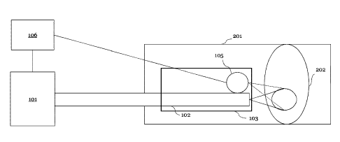
FIG. 1 is a schematic illustration of a laser system disclosed by the present
disclosure. The
laser system is suitable for modification of a calcified blood vessel 201. The
laser system
comprises: a laser source 101; a feedback controller 106, configured to
regulate a dosimetry of
the laser source 101 to produce spatially and/or temporally modulated laser
light; a catheter
103 comprising a first optical delivery element 102, the first optical
delivery element 102
configured to guide the modulated laser light to an in-vivo object 202 in the
blood vessel 201;
and
a detecting element io5, configured to detect one or more physical, chemical,
mechanical
and/or dimensional characteristics of an area of the in-vivo object 202 in
real-time,
wherein the feedback controller 106 is configured to process the real-time-
detected
information pertaining to the one or more physical, chemical, mechanical
and/or
dimensional characteristics of the area in real-time, wherein the feedback
controller 106 is
configured to regulate in real-time the dosimetry of the laser source 101
based on the real-
time-detected information for a controlled formation of a porous structure
and/or a zone of
denaturized tissue in the in-vivo object.
FIG. 2 is a schematic illustration of a laser system according to an
embodiment.
The laser system may comprise a diagnostic element i06a, configured to receive
a detected
information and process the detected information. The diagnostic element i06a
comprises a
User Interface, configured to present the detected information to a user,
e.g., a researcher or
a medical doctor. For example, the User Interface may be configured to present
a stress
distribution of an area of the in-vivo object 202. The diagnostic element i06a
may send the
detected information unprocessed to a remote high-performance computer, the
remote
hybrid computational facility, and/or the remote quantum computer io6d. The
diagnostic
element i06a may further be configured to preprocess the detected information.
For
example, the diagnostic element i06a may be configured to analyze a detected
information
pertaining to a scattered light and determine a size distribution of pores in
a porous
structure. The laser system may further comprise a feedback control element
io6b,
configured to manage a data flow in the laser system. The data flow may
comprise a flow of
real-time detected information pertaining to a temperature and a mechanical
property of an
area of the in-vivo object; a flow of processed/preprocessed detected
information; a
command generated for regulating a dosimetry of a laser source 101. The
feedback control
element io6b may be configured to control the direction and a sequence of the
data flow, so
21
Date Recue/Date Received 2023-09-20
that the irradiation of the laser source 101 can be modulated in real-time
based on real-time
detected information. The laser system may further comprise a radiation
modulation element
i06c, configured to modulate the radiation of the laser 101 source temporally
and spatially.
The radiation modulation element i06c may be configured to receive a command
generated
for modulating the radiation of the laser 101 and regulate a dosimetry of the
laser source 101,
or the radiation modulation element i06c may be configured to receive the
dosimetry value
directly from the external high-performance computer, the remote hybrid
computational
facility, and/or the remote quantum computer io6d. The laser system may
further comprise
an external high-performance computer, a remote hybrid computational facility,
and/or a
remote quantum computer io6d configured to process the detected information or
the
preprocessed detected information to generate a command for modulating the
radiation of
the laser source 101 or to regulate a dosimetry of the laser source 101. The
external high-
performance computer, the remote hybrid computational facility, and/or the
remote
quantum computer 106 may be configured to solve a thermal equation, a thermo-
mechanical
equation, a mechanical equation, and an equation of motion within a small time
interval, for
example within a millisecond up to several minutes, so that the method for
treating a
calcified blood vessel according to the present disclosure can be carried out
continuously. The
diagnostic element i06a, the feedback control element io6b, the radiation
modulation
element i06c and the high-performance computer, the remote hybrid
computational facility,
and/or the remote quantum computer io6d may be parts of the feedback
controller 106 in
FIG.i. Although FIG. 2 shows a separation of the diagnostic element i06a, the
feedback
control element io6b, the radiation modulation element i06c and the remote
ultra-fast
computer io6d, this separation should not be interpreted as a physical
separation but rather
a separation of their logical functions. The feedback controller 106 may also
refer to a
combination of one or more of the diagnostic elements i06a, the feedback
control element
io6b, the radiation modulation element i06c and the high-performance computer,
the
remote hybrid computational facility, and/or the remote quantum computer io6d.
For
example, if the feedback controller 106 is only configured to process the
detected information
pertaining to a temperature and a stress by a processor to acquire a
characteristic about a
porous structure and a zone of a denaturized tissue on an in-vivo object. The
diagnostic
element i06a alone or a combination of the diagnostic element i06a and the
high-
performance computer, the remote hybrid computational facility, and/or the
remote
quantum computer io6d can be seen as a feedback controller 106. In this case,
the feedback
controller 106 facilitates an evaluation of a porous structure and a zone of
denaturized tissue
on an in-vivo object and an initialization of parameters for the laser source
101. For example,
if the feedback controller 106 is further configured to process the detected
information in
real-time during the porous structure formation and the formation of a zone of
the
denaturized tissue induced by the temporally and spatially modulated radiation
of the laser
22
Date Recue/Date Received 2023-09-20
source 101. A combination of the feedback control element to6b and the
diagnostic element
t06a can be seen as a feedback controller 106. In this case, the feedback
controller 106
facilitates a monitoring of the laser-induced stress relaxation and molding of
the calcified
plaque or the lithotripsy (ablation) of the chronic total occlusion (CTO). For
example, a
doctor can decide on their own when to interrupt the laser irradiation
depending on whether
the size distribution of pores in the porous structure reaches a predetermined
threshold.
The laser system may comprise a laser tot, configured in a way that its
radiation be spatially
and temporally modulated by the feedback controller 106. Spatial modulation
may refer to
to varying a location, a shape of the laser beam and the laser-illuminated
area and a certain
intensity distribution of the laser-induced light in the laser illuminated
area. To realize such a
spatial modulation, the laser system may comprise one or more lasers sources
tot. FIG. 2
only shows two laser sources 101, yet a laser system according to the present
disclosure may
comprise more laser sources 101. A plurality of lasers 101 may facilitate a
complicated spatial
modulation of laser irradiation. A spatial modulation may also be realized
through a
combination of one or more lasers with other auxiliary passive elements, such
as lenses,
mirrors, an optical splitter and other optical systems thereof. Each one of
the lasers tot may
implement an independent temporally modulated irradiation. A temporally
modulated laser
irradiation is usually a sequence of pulses of laser irradiation with variable
pulse repetition
rate, pulse duration, pulse intensities or other variable attributes of a
laser pulse. A temporal
modulated laser radiation may also refer to non-pulsed laser radiation with a
variable shape
in the time domain and a variable shape in the frequency domain. The
irradiation of the laser
sources 101 may be real-time modulated. A real-time modulation may correspond
to a
constantly regulating a dosimetry of the laser source 101, regulating the
dosimetry upon
receiving a signal from the feedback controller 106 or updating the laser
dosimetry after a
certain number of pulses in a sequence. A laser source 101 in the present
disclosure may be a
combination of several types of lasers, including a solid state laser (for
example a NdYag laser
or a Holmium laser) and/or a diode laser.
The laser system may further comprise an optical delivery system 102,
configured to deliver
the modulated laser radiation or laser light to a target. The optical delivery
system 102 can be
an optical fiber, a bundle of optical fibers or other types of optical
delivery elements. The
optical delivery system 102 may also be configured to deliver other laser
signals, for example
a probing laser signal for detecting a certain property in a blood vessel 201.
In an exemplary
embodiment, laser modulation may take into account the possible distortion of
the laser
signal due to propagation in the laser delivery system 102 and implement
corresponding
compensations. The optical delivery system 102 may comprise an optical out-
coupler for
delivering the laser signals in a form of laser irradiation to the target. In
an embodiment, for
23
Date Recue/Date Received 2023-09-20
example in the cold lithotripsy method as will be illustrated in detail later,
the optical out-
coupler may be configured to physically contact an in-vivo object 202 without
exerting
damage on them.
The laser system may further comprise one or more detecting elements 105. The
detecting
elements 105 are configured to detect one or more physical, chemical,
mechanical and/or
dimensional characteristics of the in-vivo object 202 in the blood vessel 201.
The one or more
physical, chemical, mechanical and/or dimensional characteristics may comprise
a
temperature, a stress, the size and the number of pores, thermomechanical
characteristics,
.. optical characteristics, electrical characteristics, and other
characteristics characterizing the
environment of the in-vivo object 202 and the status of the in-vivo object
itself. The
characteristics may be detected in a direct and in an indirect way. For
example, the detecting
element 105 may comprise a conductivity measuring element, configured to
measure a
conductivity of a blood vessel. This characteristic can then be feedbacked as
an electrical
signal. In another example, the detecting element 105 may comprise an optical
receiving
element, configured to receive a scattered light. The scattered light can be
feedbacked as an
optical signal and be processed to deliver the information about a
temperature, a stress, a size
distribution of gas bubbles, and a size distribution of pores, based on the
characteristics of
the optical signal such as, for example but not restricted to, wavelength
distribution and an
angular intensity distribution. In an exemplary embodiment, the detecting
element 105 may
comprise a conventional diagnostic device, such as one of the following: an X-
Ray; a CT; an
Ultrasonography (US); a Doppler US; an MRI; an Intravascular ultrasound
(IVUS); an OCT;
an OCE; a Multispectral Optoacoustic tomography (MSOT); a fluorescence
molecular
tomography (FMT); and acoustic tomography. In particular, an IVUS allows to
detect
.. calcium, an OCT allows for measuring dimensions of the target and the OCE
measures the
stress and mechanical properties. The IVUS is a widely available clinical tool
for guiding
percutaneous interventions and intraluminal imaging. While the IVUS uses
frequencies from
20 to 40 MHz and provides fair penetration depth, it lacks the sufficient
resolution, having
the resolution restricted to-120 gm, necessary for studying the thin-cap
thrombus, atheroma
lesions, and other possible fine details of the vasculature. Conversely, while
the OCT provides
the high resolution of about 2-20 gm for tomographic visualization of the
coronary arteries,
its maximal penetration depth is only about 2-3 mm. Because the IVUS waves
penetrate
deeper into the media and adventitia, the OCT may be combined with the IVUS
modalities
which enhances and improves a quantitative analysis of the characteristics of
the in-vivo
object significantly. It is understood from this embodiment, that it may be
advantageous to
combine the different types of detecting elements 105 comprising conventional
diagnostic
devices for acquiring a more detailed picture of the in-vivo object and of the
environment of
24
Date Recue/Date Received 2023-09-20
the in-vivo object in a blood vessel. This improvement can lead to an
increased data volume
to be processed. In an exemplary embodiment, the performance of the remote
high-
performance computer, a remote hybrid computational facility, and/or a remote
quantum
computer 1o6d should be high enough to ensure implementing a real-time
modulation of the
laser irradiation.
The laser system further comprises a catheter 103 configured to carry an end
of the optical
delivery element 102 and the detecting element 105 in a blood vessel 201 to
the vicinity of the
in-vivo object 202. The catheter 103 may be configured to make fine position
adjustment in
the blood vessel 201 and record the position of the laser light exerted on the
target or a
position of the information detected by the detecting element 105, such that a
precise spatial
modulation of the laser signal and an acquisition of spatial distribution of
the detected
information is realized. In an exemplary embodiment, the catheter 103 may
further comprise
and be attached to a servo element for a precise position control. An example
of the catheter
103 comprising a detecting element measuring electrical conductivity, is
illustrated in FIG. 3.
The catheter 103 may comprise a coating 301, a conductive adhesive 302 and a
metal tip 303.
The laser system may further comprise a balloon 104 configured to be inflated
to expand a
lumen area in the blood vessel 201. The balloon 104 may be configured to be
modulated by
the controller 106 as well. For example, the balloon 104 may be configured to
be inflated
during the laser treatment. The balloon 104 may be configured to be inflated
after a laser
induced stress relaxation. The controller may be configured to modulate the
balloon 104 and
the laser 101 simultaneously for different purposes. For example, for molding
of a calcified
plaque, the balloon may be inflated simultaneously during a laser irradiation.
For another
example, for a minimum pressure required for the balloon 104, the balloon may
be inflated
only after the laser softening of the calcified plaque.
FIG. 4 is a schematic illustration of a laser system in operation according to
an embodiment.
The blood vessel 201 subject to the treatment suffers from the imposed
calcified plaques
202b. The calcified plaque 202b is expectedly attached to or located at the
vessel wall 202a.
The calcified plaque 202b may further restrict a lumen area of the blood
vessel 201
significantly, so that the pathway available for the blood flow is reduced. In
the case
illustrated in FIG. 4 the calcified plaque 202b on the right-hand side totally
blocks the
pathway of the blood flow, which is typical in a CTO. To treat and cure the
blood vessel 201,
the large piece of the calcified plaque 202b on the right-hand side of FIG. 4
needs to be
destroyed. According to the present disclosure, the regulated laser source 101
can be used to
generate temporal porous structures within the calcified plaque 202b
facilitating a more
efficient laser ablation of the calcified plaque 202b. This approach is called
the "cold
Date Recue/Date Received 2023-09-20
lithotripsy". After the calcified plaque 202b causing the total occlusion is
destroyed, or have
its volume reduced through implementing the cold lithotripsy, the laser source
um with the
modulated radiation may be further configured to form a stabilized porous
structure and
zones of denaturized tissues within the calcified plaque 202b attached to the
vessel wall 202a
for softening the calcified plaque 202b and reduce a mechanical stress
developing in it
without destroying it. After or during the mechanical stress relaxation in and
softening of the
calcified plaque 202b attached to the vessel wall 202a, a balloon 104 can be
inflated to
expand a lumen area of the blood vessel 201. Compared to the conventional
method, the
balloon 104 used after a laser induced softening of the calcified plaque 202b
and stress
relaxation may require less pressure, producing less damage to the
environment, including
the vessel wall 202a. After the laser treatment is concluded, the modulated
radiation of the
laser source 101 may further be configured to reduce a residue stress on the
vessel wall 202a.
This can minimize a chance of recalcification and restenosis of the blood
vessel 201.
FIG. 5 is a flow chart illustrating a method for detecting and processing
information
according to an embodiment. In this embodiment, the method comprises:
a) detecting a physical, chemical, mechanical and/or dimensional
characteristic of an
area of an in-vivo object in a calcified blood vessel;
b) processing the detected information pertaining to the physical,
chemical, mechanical
and/or dimensional characteristic to acquire a property of a porous structure
and/or a zone
of denaturized tissue on the in-vivo object.
As outlined above, the method illustrated in FIG. 5 may be used to evaluate a
porous
structure and a zone of the denaturized tissue. This structural evaluation may
be carried out
for initializing a working condition of the laser. This method may be further
used to monitor
and evaluate an effect of the modulated laser radiation on the in-vivo object.
This laser effect
evaluation may be carried out for controlling laser effect or damage induced
by the laser on
the in-vivo object.
FIG. 6 is a flow chart illustrating a method for treating a calcified blood
vessel using a
temporally and/or spatially modulated laser radiation according to an
embodiment. In this
embodiment, the method comprises: a treatment step, wherein the treatment step
comprises:
a) detecting a physical, chemical, mechanical and/or dimensional
characteristic of an
area of an in-vivo object in the vessel, and feedbacking the detected
information pertaining to
the physical, chemical, mechanical and/or dimensional characteristic in real-
time to a
feedback controller; and
26
Date Recue/Date Received 2023-09-20
b) modulating the laser light in real-time by the feedback controller
based on the
detected information, the modulating suitable for a controlled formation of a
porous
structure and/or a zone of denaturized tissue in the in-vivo object.
In a typical implementation of the method FIG. 6, the method may comprise:
i) a first step of diagnosing the vessel and initiating a working condition
of the laser;
ii) a second step of treating a calcified plaque in the vessel; and
iii) a third step of treating a vessel wall after the second step is
concluded, wherein at
least one of the second or third step may comprise the treatment step.
In particular, the second step may comprise softening and molding of the
calcified plaque
step,
a cold lithotripsy step and a laser induced stress relaxation step on the
calcified plaque. The
third step may comprise a laser induced stress relaxation step on the vessel
wall. All three
steps may comprise a method of FIG. 5. In the following, the molding, cold
lithotripsy, and a
laser induced stress relaxation carried out on a calcified plaque and on the
vessel wall will be
elaborated in detail.
Softening and molding of calcified plague
In an embodiment, an in-vivo object is a calcified plaque of uneven thickness.
The disclosure
makes it possible to increase the lumen and compliance of the vessel by
molding, i.e.,
smoothing, of the calcified plaque without destroying it. The molding process
includes two
steps: (i) the softening of the calcified plaque, which implies decreasing its
tensile strength
and yield strength through the formation of microstructural defects, including
micropores;
and (ii) changing the shape and smoothening the calcified plaque via the
application of the
mechanical force.
In an exemplary embodiment, this mechanical effect can be realized through a
balloon or a
stent or by thermomechanical action of the spatially modulated laser
radiation. In particular,
the applied pressure on a balloon may be 5 -12 atm which is lower than the
mechanical
strength of the vessel wall (15-20 atm), and is much less than the pressure 40-
50 atm applied
when using most of the conventional methods including the FDA approved shock
wave
lithotripsy.
This ensures the safety of the molding technique since it does not lead to
tearing or rupture of
the vessel walls.
27
Date Recue/Date Received 2023-09-20
Cold lithotripsy
In an embodiment, an in-vivo object is a thick and well-developed calcified
plaque, for
example a calcified plaque in the case of a CTO. In such case, the molding or
softening of the
calcified plaque may not occur because an applied power may appear not
sufficient enough
for expanding a lumen area and normalizing a blood flow in a blood vessel.
Yet, the
traditional destruction of the calcified plaques using conventional
lithotripsy, which requires
high temperatures and pressures more than 40 atm, can lead to tearing or
rupture of the
vessel wall. A CTO is present in nearly 50% of all patients undergoing
endovascular treatment
of the infrainguinal peripheral arteries where the treatment is especially
challenging. In these
cases, the present disclosure can be used for a partial destruction of the
calcified plaque
which allows for the safe solution of the curing problem.
FIG. 7a presents a schematic illustration of a method for destroying a
calcified plaque
according to an embodiment. In this embodiment, an area of a porous structure
is formed on
the calcified plaque due to a porous structure formation of the hydroxyapatite
in the calcified
plaque. The porous structure may have a depth of d = lk, where k = Woa is an
effective
absorption coefficient, a is the water absorption coefficient, Wo is the
initial porosity of a
calcified plaque, W is the porosity of the porous structure of the area of the
calcified plaque
after an initial porous structure formation. The in-vivo object may contain
different materials
and may have different physical properties. For example, when the laser is a
Tm laser; for the
Calcium Oxalate, Wo = 9% and k = 14 ; for Uric Acid Wo = 12%, k = 19 . The
porosity
CHI CHI
of a Bego stone can vary depending on the preparation conditions. The
threshold for an
energy density of the laser ablation (F) of the in-vivo object with the Tm
laser may be 20.8
J/cm2 for Calcium Oxalate and 7 J/cm2 for Uric Acid. This may give a
temperature increase
of AT = Fk/c = 70 C for the Ca Oxalate, and 40 C for the Uric Acid in the
porous structure.
1
c = 4.2 ¨cm3 is a heat capacity.
A thermo-stress due to water expansion is a = () G ( )3 a ¨ )3 0)T = 12 MPa
for Calcium
Oxalate and a = 8 MPa for Uric Acid. Here G = 2 GPa is the shear modulus, iga
¨ )30 = 10-41
K
is the thermo-expansion coefficient. The tensile strength is a = 3 ¨ 5 MPa for
Ca Oxalate, a =
2 ¨ 4 MPa for Uric Acid, a = 3 ¨ 16 MPa for the Bego stones.
After the laser-induced porous structure formation, the porosity in the porous
structure may
increase by factor of two or three. When the porous structure is filled with
water, the thermo-
mechanical stress between this area and the rest of the calcified plaque may
lead to a removal
28
Date Recue/Date Received 2023-09-20
of the porous structure. FIG. 7b shows the temperature gradient between the
illuminated
porous structure and the surrounding area of the in-vivo object. The boundary
between the
area of an enhanced water concentration and the surrounding area is denoted as
6.
To calculate the ablation rate in this scenario, one may first consider the
time necessary for
water to fill the porous structure. For a porous structure of the thickness d,
the filling time is
tw =L2, where D =-- 10_3 ¨cm2 is the water diffusion coefficient in the porous
structure, L is the
D s
spatial scale of a porous structure. For example, assuming the modulated laser
light is
delivered via a round optic fiber with a cross section of a radius R. The
ablation rate is A =
gnR2d gnR2dD
= ___ where g is a density of the calcified plaque. The value of A increases
with D
tw 4L2
which grows with the temperature and the average power density. If R > d = lk,
where k =
Woa is the effective absorption coefficient, a is a water absorption
coefficient, Wo is an initial
porosity of the calcified plaque, W is a porosity of the porous structure
after the porous
structure formation, the filling time tw = ¨d 2, then the ablation rate A =
gnR2d = gn-R2 Dk. The
D tw
value of A increases with the growth of the fiber diameter and with the
increase of the light
absorption coefficient. For R = 300 inn, A =-- 120 gg/s, for a Tm laser, and A
,-- 30 gg/s, for a
Ho laser. If d > R, t = 414 , and the ablation rate A = g it D/zik does not
depend on the fiber
radius and decreases with the light absorption coefficient. For R=loo gm, A ,--
90 gg/s for a
Tm laser, and A =-- 360 gg/s for a Ho laser. For the treatment of the
calcified plaque with the
Erbium glass fiber laser: a = 10 ¨1, Wo = 0.3, and A ,-- 500 gg/s. FIG. 7c
shows profiles of the
CHI
crater area in the Bego stone treated by the laser system according to the
present disclosure.
Laser wavelength 1.44 gm, penetration depth 6 = 200 gm, power 10 W, fiber
diameter 150
gm, pulse duration 1 ms, number of pulses 100, pulse repetition rate f = 80 Hz
(for the upper
curve), and 4 Hz, for a lower curve.
A significant volume reduction is observed for the lower pulse repetition rate
when a time
interval between two laser pulses was longer than a time it takes for the
water to fill the
porous structure.
In a sub-optimal implementation, a pulsed laser may be run with a shorter
pulse duration,
high pulse repetition rate and high peak power. The short pulse duration and
high peak
power lead to a dramatic increase of the ablation temperature resulting in the
intensive water
evaporation. The efficiency may reduce if the pulse repetition rate is too
high for the water to
fill the porous structure. In this embodiment, the laser radiation may be
temporally
29
Date Recue/Date Received 2023-09-20
modulated to magnify the above-mentioned effect. For example, the time period
between two
pulses (inverse of the repetition rate of the pulsed laser) may be optimized
to increase a laser
efficiency. In particular, a time period between two pulses should be long
enough for the
water to fill the porous structure, but short enough for a continuous
generation of
temperature gradient.
Thus, the present disclosure provides an effective method to treat severe
calcified blood
vessel including the calcified blood vessel in CTO.
.. Laser induced stress relaxation
The present disclosure can be used to reduce a mechanical stress on the
calcified plaque or on
the vessel wall without destroying their structures on a macroscopic level.
The mechanical stress of an in-vivo object in a blood vessel is one of the
origins of the poor
elasticity and low compliance of the blood vessel. Therefore, a stress
relaxation is one of the
effective methods of the blood vessel treatment. The stress relaxation occurs
when some
elements within an area of the in-vivo object acquire an ability to move with
respect to
another elements, and the elastic energy dissipates or transforms to other
types of energy like
thermal energy, phase transformation, surface energy, etc. For example, stress
relaxation can
be achieved due to porous structure formation when stress energy transforms
into a surface
energy of pores due to the formation of the porous structure.
In biological tissues, for example, vessel walls, the secondary and tertiary
structures of
proteins and nucleic acids contain, as basic elements of their structure
composure, the
intermolecular hydrogen bonds. With an increase in temperature and in the
presence of the
mechanical stresses, breaking of the intermolecular bonds may lead to a
movement of some
elements of the structure with respect to others. This relative movement
diminishes the
general energy of the system and leads to a relaxation of the stress. In the
zones of the broken
.. intermolecular bonds, structural defects appear, in particular, zones of
the denatured tissue
and porous structure, for example a microporous structure, where pores are
filled with gases
dissolved in the tissue fluid, mostly carbon dioxide, oxygen, and nitrogen. In
this case, two
types of defects are formed: dens, which are the zones of the denaturized
tissue, and
micropores filled with the gas bubbles, often referred to as nano bubbles.
Increasing the
pressure diminishes the gas solubility, and the gas transforms into its solved
state, the large
bubbles collapse, and the number of dens increases. FIG. 8c shows an AFM image
illustrating
a den arisen in the vessel wall between normal collagen fibers in an example.
Date Recue/Date Received 2023-09-20
On a macroscopic scale, the laser-induced breaking of the intermolecular bonds
in an in-vivo
object can lead to a denaturization, i.e., the violation of the ordered
protein structure, of
tissues and formation of a porous structure or other defects between the zones
with the
unchanged structures. The interaction of dens and bubbles can inhibit the
processes of
denaturation, plastic deformation, or the formation porous structure. Porous
structure or
other defects can play a versatile role in processes in a blood vessel. In
particular, the porous
structure can reduce the mechanical strength of calcified plaques and can
enhance the
delivery of drugs to the vessel walls through a calcified plaque. That
facilitates a healing of the
blood vessel, preventing recalcification and restenosis. On the other hand, an
uncontrolled
growth of the dens and porous structure may lead to development of the
processes of
denaturation and tearing in the vessel wall, which can be harmful for their
mechanical
properties.
FIG. 8a shows a stabilized gas bubble covered with Ca2+ ions, FIG. 8b shows a
SIM image
illustrating Ca2+ ions covering the surface of a CO2 gas bubble in an artery
plaque.
The porous structure can be either temporary or stabilized, hence long-living.
The porous
structure, in particular microporous structure, can be stabilized by gas
bubbles, for example
nanobubbles and their agglomerates, the surface of which is covered with the
positive ions
such as calcium ions. Gas bubbles arise in liquids in which gases are
dissolved with a
solubility dependent of temperature. Gas bubbles arising and growing during
the heating in
liquids are usually unstable and collapse rapidly. The presence of the calcium
ions in tissue
fluids can stabilize the bubbles. The ions located at the surface of the gas
bubbles repel each
other, which prevents the bubble from collapsing. Further, the ions at the
surface of the
bubbles slow down the bubbles' movement. The features associated with the
formation of the
stable bubbles and their destabilization, which occurs at a certain
temperature, manifests in
the dependence of the electric current upon temperature. FIG. 9 demonstrates
the time
dependency of the electric conductivity c(t) of the calcified artery of the
cadaver. In this
example, a detecting element comprises a conductivity detecting element, which
comprises
electrodes in the form of the two coaxial cylinders truncated at an acute
angle of about 20
degrees. A first electrode is a quartz light guide with a diameter of 600 gm,
covered with an
aluminum sheath. The proximal end of the light guide is glued with conductive
glue and then
covered with a thin layer of electrically insulating acrylic varnish. The
distal end of the fiber,
at a length of 3 mm, is decoated. A cylindrical metal tip is put on this end.
In the beginning of
the laser irradiation, the electrical conductivity increases due to
temperature increase. After
about 3 s of the irradiation, the electrical conductivity decreases due to
formation of gas
bubbles. After about 75 of irradiation these bubbles disappear due to heating
up to the
temperatures above 60 -70 C and an escape of the stabilizing ions.
31
Date Recue/Date Received 2023-09-20
Light scattering is also very sensitive to the formation of porous structure
or zones of the
denaturized tissues. Measurements of the scattering of light of different
wavelengths make it
possible, using the Mie and Rayleigh scattering laws, to determine the
prevailing defect size.
.. Measuring the time dependencies of the light scattering makes it possible
to study the
stability of the gas bubbles, porous structure, or zones of the denaturized
tissues and to
establish the boundaries of the treatment working conditions for the laser.
The controller
may process the detected light scattering and modulate the laser during laser
treatment. For
example, the feedback controller stops the irradiation when a preset threshold
is achieved by
.. the detected information pertaining to the scattered light. FIG. 10
demonstrates time
dependencies intensities of the modulated laser (denoted in bars) and the
intensity of
detected scattered light (denoted in lines) in an example, demonstrating how
the laser
intensity can be modulated based on the detected information. In this example,
a calcified
artery of a pig is treated using several series of laser pulses.
The feedback controller decreased the laser power by over 25% after the first
series of 8
pulses, and after the second series the power was increased by 20%. Then the
three series of
12 pulses each, were applied. Irradiation was terminated during the sixth
series, when the
intensity of the scattered light significantly decreased after each subsequent
pulse. This
reflects a tendency for individual defects (porous structure or zones of
denaturized tissues) to
grow excessively. The reliability of such a controller is tested using OCT and
histology.
Stress relaxation of the calcified blood vessel may include porous structure
formation in an
area of hydroxyapatite in the medial and intimal calcification zones.
Elimination of the stress can also occur due to a local denaturation of
tissues such as
pathological oriented collagen fibers which may be responsible for excessive
expansive stress
at the boundary between vessel wall and calcified plaque. Near the calcified
areas, a
pronounced change in the distribution of stresses can be observed from
predominantly
.. circumferential to predominantly longitudinal or in thickness. Stress can
be relaxed due to
local denaturation of abnormal orienteered collagen fibers.
For laser induced stress relaxation, a temperature should be controlled within
a certain
range. The mechanisms of the laser induced stress relaxation may include: (i)
a bound-to-free
transition of tissue water, (ii) a reorganization of the collagen structure at
the boundary zone
between vessel walls and calcified plaques including local denaturation and
breaking the
abnormal collagen fibers and crosslinks. (iii) formation and movement of
structural defects
(including zones of denaturized tissues and porous structures) resulting in
the reduced
32
Date Recue/Date Received 2023-09-20
energy in the in-vivo object. All the above processes require heating to
temperatures of 45-70
C for a time interval shorter than a time interval required for denaturation
of large zones
(larger than fifty microns).
Thus, the laser system according to the present disclosure makes it possible
to control the
laser-induced porous structure formation and laser-induced formation of zones
of the
denaturized tissues, and to provide the safety of the laser treatment.
Treating of residue stress on a vessel wall
In another embodiment, the laser system is used in a third step of treating a
vessel wall after
the second step is concluded. The third step may also be carried out after
other cardio
intervention including molding, a traditional lithotripsy, or stenting of the
blood vessel.
In an embodiment, the in-vivo object is calcified plaque or a vessel wall with
residue stress.
Residue stresses can arise after plastic deformation processes, which occur,
for example,
during the molding or lithotripsy in the second step of treating a calcified
plaque in the
vessel.
For example, the porous structure formation and stress relaxation of the
calcified plaques are
accompanied by plastic deformation which, after seeded external force, leads
to the
formation of the residue stress. Residue stresses after the plastic
deformation are also
inevitable after a stenting surgery. Residue stress amplitude can be
calculated using the
model of plastic deformation in calcified vessels and measured using an OCE
method.
.. Removal of the residue stress may be a final step for a laser treatment of
the calcified blood
vessel according to the present disclosure. In an example, this procedure can
be performed
using irradiation of a laser with a wavelength of 1060 nm, a pulse duration of
1 ms, a power of
2 W, a pulse repetition rate of 10 Hz, wherein the laser signal comprises
three series of laser
pulses of 10 pulses each with a period between series of 10 s.
Eliminating a residue stress on a vessel wall or a calcified plaque layer
attached to the vessel
wall is an important procedure which diminishes secondary effects and the long-
term
biological response of the blood vessel after any physical treatment
accompanied by plastic
deformation. In addition, the formation of a porous structure enhances the
diffusion of the
drug to the vessel walls and also prevents recalcification, calcinosis and
restenosis in the
blood vessel. As shown in FIG. 11, after a second step of treating the
calcified plaque in the
blood vessel, a volume of the calcified plaque can be reduced, and a lumen
area can be
expanded (the status of the blood vessel changes from the states denoted on
the left of FIG. 11
33
Date Recue/Date Received 2023-09-20
to the status denoted in the middle of FIG. ii). The blood vessel wall and the
calcified plaque
attached to the blood vessel wall may be then treated through a laser induced
stress
relaxation step, which leads to a long-term stability (the status denoted on
the bottom right of
FIG. ii). Without such stress relaxation, recalcification may occur several
months after the
treatment (the status denoted on the upper right of FIG. ii).
A functional stiffness of the vessels is governed by their material stiffness
and geometry
characterized by the vessel lumen area, the vessel radius r, and the thickness
of the vessel
walls h, which is a combination of the morphology and disease. An increase in
a smooth
muscle tone in the muscular arteries that represents most of the conduit
vessels alters the
pressure-to-volume relationship in these vessels and results in a decrease in
an arterial
compliance or elasticity. The laser-induced stress relaxation allows to
decrease the smooth
muscle tone in the arteries and to increase their compliance. The laser-
induced reduction of
the residue stress in the vessel walls also decreases the rate of the
recalcification processes,
disrupts or reverses processes of formation of the calcified plaques.
Mathematical model for the real-time laser modulation
In the following, the mathematical model of the laser-induced formation of a
porous
structure and a zone of denaturized tissue based on the thermal, thermo-
mechanical and
mechanical equations considering the laser induced intermolecular bond
breaking in the
calcified plaque is elaborated. With the mathematical model, the skilled
person in the art may
find no difficulties in realizing and optimizing a real-time modulation of the
laser for a
controlled formation of porous structure and a zone of denaturized tissue
based on the real-
time detected information, for example, through an encompassing the
mathematical model
into a feedback algorithm.
The processes occurring in a continuous medium under a laser radiation can be
described by
the equations of motion of the medium in the form of Lagrange equations which
describe the
motion of multilayer media, the properties of which change when passing
through the
interfaces. Consider the Lagrange equations for a three-dimensional motion of
a continuous
medium. The continuity equation in Lagrange variables has the form:
V = VA (1)
34
Date Recue/Date Received 2023-09-20
where A= 0(xe,ye,ze) is the Jacobean for the transition from the Euler
coordinates (xe, ye, ze) to
a (xi,yi,zi)
Lagrange coordinates (x1, z1); 110 = ¨ is an initial specific volume, V = ¨ is
a current
Po
specific volume. In an explicit form, the continuity equation can be written
as follows
" axe (aye aze aye aze) aye (axe aze axe aze) , aze (axe aye axe aye)]
V = vo r¨ ¨ ¨ ¨ ¨ ¨ ¨ (2)
^ 0y1 0z1 0z1 0y1 \0y1 0z1 0z1 0z1 0y1
0z1 0z1 ayi
The equations of motion in the Lagrange form are then
a2xe ax a2ye aye a2ze)oze
=
at2 p (3)
(X ¨ ¨a2xe) (y _ 2)'e) (z _ a2ze) Oze = 1 OP
¨
Ot2 Ot2 at2 P 0371' (4)
Ix 02xe'l x _L IV o2Ye Y 4- e 7 02ze \Oze 1 OP
0t2 ) Oz : ' 0t2) Oz i ' 0t2 aZl =p 0Z1' (5)
where = Xi+ Yj + Zic' is a force vector per unit mass, which hereafter can be
assumed to be
equal to zero, namely X = Y = Z = 0.
Considering the explicit form of the equations for changing the Euler
coordinates
axe aye aze
uxe = , uye = ^ , uze = , (6)
The system (3) - (5) acquires the form
auxeax auye aye auze aze ap
= ¨--(7)
at - azi at azi at Ox i p Oxi'
auze Ouye aye auze aze 1 OP
= (8)
at - 0y1 at 0y1 at 0y1 P 0311'
auzeox Ouye aye auze aze 1 ap
e = (9)
at - 0z1 at 0z1 at 0z1 p azi!
Resolving equations (7) ¨ (9) with respect to =au and considering the explicit
form of the
at
continuity equation (2), leads to the following system of equations of motion
auxe = _v r OP (Oye Oze aye aze) aye ( OP Oze OP Oze\ +Oze ( OP Oye OP Oyeyl
(10)
Ot I_Oxi kOyi Ozi Ozi Oxi 371,Ozi Ozi Oyd Oxi
kOYi P
auye raze ( OP Oze OP Oze\ OP (Oxe Oze Oxe Oz +Oze (Oxe OP _Oze OP
"
3 Ot = v Loxi koyi 0z1 0z1 0y1) Oxi koyi 0z1 0z1 0y1) Oxik.ayi 0z1
0z1 0y1.1P
auze = Foxe (aye OP _Oye OP '\ _O)'e (Oxe OP _Oxe OP '\ + OP (axe aye axe
aye)] (12)
at [Ox i kayi 0z1 0z103711 Ox1kayi 0z1 0z1 0371)
Ox1kayi 0z1 0z1 0371)1'
Date Recue/Date Received 2023-09-20
The Mie ¨ Griineisen equation in its two-term form to approximate the equation
of state may
be written as
P = Px + PT = pou6 (1 ¨ / ) + Fv '
cv(T-To) (13)
vo
where PT and Px are the thermal and cold components of pressure P , F = is the
Griineisen
cv
coefficient, )3 is the coefficient of volumetric expansion, cv is the heat
capacity, and u0 is the
speed of sound in the medium.
The change in the temperature of the medium can be found from solving the heat
conduction
equation:
OT , (02T . VT . VT\ . n
PCV ¨at = itT k.axe 1- aye 1- ¨04 ) 1- (45 (14)
The value of Qsin equation (14) is determined by the source of energy release:
Qs =
/ (Xe, ye, Ze, t)K, where /(t, xe, ye, Ze) = 10 ft (t)fxyz(xe, Ye, Ze) is the
intensity of the light beam at
the moment t at the point in space with the coordinate (xe, ye, Ze), K is the
absorption
coefficient of the medium.
A pulse periodic function f = ft (t) may take one or a combination of, but not
restricted to the
following forms:
(a) sin(wt),
(b) a step function: during the pulse f = 1 , and during time between pulses f
= 0 or
t f t (t) = --exp [¨ ¨t], where Tp is pulse duration.
Tp Tp
(c) several series of laser pulses with some delay between series.
The solution of the system (1) - (14) makes it possible to calculate spatial-
temporal
dependencies of the pressure, temperature, density, and velocity, to evaluate
the contribution
of thermal and acoustic mechanisms to the change in the physical parameters of
a continuous
medium.
To determine the distribution function of the intensity of a Gaussian light
beam in a
simultaneously absorbing and scattering medium, the following law of light
beam attenuation
can be used, considering the change in its amplitude and spatial shape.
34 +z2
I (Xe, ye, Ze) = /oexp [¨f ]exp [¨(kabs + kccat)x], (is)
rc (x)
36
Date Recue/Date Received 2023-09-20
where /cabs and k õat are absorption and scattering coefficients,
respectively. The radius of the
light beam changes as it penetrates the medium according to the law: d (x) = d
(x =
0)exp [Icscatx] , expanding exponentially due to scattering and maintaining a
Gaussian shape
in cross section. Using equation (15), the energy release function in equation
(14) acquires the
form:
Qs(t, xe, ye, ze) = lo (¨r) exp {¨ i exp r 2 , x 2
Ye ' ze 1 exp [¨(kabs + kscat)x] (16)
Tp Tp 71()
As a result, the 3D fields of thermal stresses and strains developing in the
biological tissues
under the effect of pulse-periodic laser radiation with a wide range of laser
parameters,
including laser wavelength, pulse duration, pulse repetition rate, spatial
distribution of power
density is obtained. In this way, the parameter defining a working condition
of the modulated
laser can be adjusted to achieve a desired effect on the illuminated area
based on the detected
information pertaining to the temperature and the mechanical stress.
In addition, a second mathematical model can be considered. As outlined above,
a laser
induced stress relaxation of the in-vivo object is due to the formation of
porous structures
and zones of denaturized tissues. The porous structures and zones of
denaturized tissues are
formed due to laser induced breaking of intermolecular bonds. In more detail,
the
mechanism of the laser-induced stress relaxation can be seen as stemming from
a non-
uniform distribution of the structure and mechanical properties in the area,
comprising
relatively strong domains separated by less strong interlayers, the
destruction of which gives
mobility to one domain of the area relative to another domain. Laser heating
leads to the
breaking of hydrogen intermolecular bonds. Breaking energy of the
intermolecular bonds
depends on external stresses. Therefore, in a deformed region, the
intermolecular bonds are
broken more intensively. Regions with the reduced binding energy will deform
more. The
inhomogeneity of the structure and mechanical properties of the in-vivo object
may increase
during heating induced by laser illumination, which may lead to the
inhomogeneity of
deformation to appear in areas where the deformation is large at low loads.
Solving a variational problem of minimizing an elastic energy by changing the
structure,
redistributing the shape and position of the elements of the system, becomes
possible with
the model describing a process of thermally activated breaking of the chemical
bonds. The
latter process determines the kinetics of stress relaxation in the system. In
this case, the
porous structure formation process depends essentially on only the
characteristic bond-
breaking energy in the Arrhenius formula for the probability of the bond-
breaking, but the
value of this parameter can be different at different locations and at
different times. It is a
37
Date Recue/Date Received 2023-09-20
given function of temperature, temperature gradient, and the spatial
distribution of the
binding energy.
Changing the density of intermolecular bonds may comprise two processes
occurring
simultaneously: a process of reducing the density of intermolecular bonds, and
a process of
deformation due to the tendency of a substance in the in-vivo object to
minimize its internal
energy. The potential barrier for the intermolecular bond breaking is high
enough, and the
probability of the intermolecular bond breaking per unit time is defined by
the Arrhenius law
p = poe kT (17)
where Po is the preexponential factor, U is the height of the barrier, and T
is the temperature.
If the tissue is heated, for example due to radiation induced by a laser,
above a normal
temperature for a short time, the rate of the thermal destruction of the
intermolecular bonds
given by equation (17) may increase, and during the heating period a
considerable part of the
intermolecular bonds may be broken. As a result, the stiffness of the area
decreases. If the
tissue is deformed by the external forces and constraints, or if it has
internal stresses, then
the height U of the potential barrier may decrease.
The exemplary, but not necessarily the only possible, description is based on
an assumption
that all the elastic properties of the tissue near the point x depend on one
parameter a (x)
which may be referred to as an "intermolecular bond density". The value a = 1
corresponds
to an intact tissue with its maximal strength, while a = 0 corresponds to a
tissue with
completely destroyed structure, effectively without any strength, so that the
shear modulus is
equal to zero. All the non-homogeneity of the tissue is described by the
variations of the
parameter a depending on the location; a = a (x).
The tissue in a free state, i.e., free of the external forces, may occupy a 3-
dimensional domain
Mo, and its bond density may be given by function a (x), x E Mo. A deformed
tissue may
occupy another domain M1, and its configuration can be defined by a mapping x
¨> y(x) from
Mo to M1. Assume that this mapping is a one-to-one mapping and sufficiently
smooth, and
that the inverse mapping y ¨> x(y) is also sufficiently smooth.
Oy Oy
The matrix Tii = T = ¨ = can be always represented as a product of a pure
deformation,
ax ax,
i.e., a transformation represented by a symmetric matrix D and a rotation
represented by a
rotation matrix R:T = RD. This representation is unique. The energy density
function E does
not depend on the rotation R, because rotating a deformed body (or its
element) does not
38
Date Recue/Date Received 2023-09-20
change its elastic energy. Further, the symmetric matrix D has 3 orthogonal
axes of
deformation and 3 positive eigenvalues A1, A2, A3, so that it can be
transformed with a proper
orthogonal basis into a diagonal form with the eigenvalues Ai. An assumption
based on the
local isotropy of the tissue means that the function E depends only on the
eigenvalues Ai.
Ti; = and o-i are the symmetric combinations of the eigenvalues of the
deformation matrix
ox,
D.
The free energy is defined by the integral
ay ,3
E = itioE (x,ak_x),¨) a x, (18)
Where: E(x, a, N) = F (x, a, al, 52,53),
Following the Mooney-Rivlin model used for biological tissues including for
cardiovascular
stresses:
1
F = (53 + ¨ ¨ 2) + av (o- ¨32),
cr3
= A1+ A2 + A3, Cr2 = A1A2 +A2A3 A3A1, Cr3 = A1A2A3 (19)
Here and v are the volume and shear modules.
The differential equations describing the equilibrium of the elastic body are
the Euler
equations of the variational problem which are the following:
E E- - X , = 0 (20)
Ox Ox/
DE
= (x, a, T) (21)
Equations (18) - (20) describe deformations that affect thermomechanical
intermolecular
bond breaking and the porous structure formation due to the breaking of
chemical bonds.
The strain reduces the potential barrier U for the intermolecular bond
breaking, so that
instead of U the potential barrier becomes U ¨ AU. The difference AU is
proportional to the
elastic energy released as a result of the breaking of one intermolecular
bond:
39
Date Recue/Date Received 2023-09-20
sE
AU (x) a Sa(x)dx (22)
where E is elastic energy, the variational derivative is defined as
SE 1 OE (x,a(x),T(x))
Sa(x)dx = 2 Oa (23)
Where E = E(x, a (x), ¨03 7 (x)) is the energy density for an energy
minimizing configuration y (x) .
a x
The probability of the intermolecular bond breaking per unit time at the point
x and at the
time t is given by the Arrhenius law
U-AU
p = poe ICE) ,
where
SE a OE(x,a(x),T(x))
AU(x) = a Sa(x)dx = 2 Oa (24)
and the equation defining the time evolution of the bond density a (x, t)
becomes
Oa (25)
¨at = ¨(a ¨ a 0)p
where p = p (x, t) is defined by the equations (24). The right-hand side of
(24), in its turn, is
defined by the solution of the variational problem with the function a (x, t)
obtained by this
moment, and the given boundary conditions and temperature field. Here a is the
coefficient
of proportionality between the decrease in the activation energy of
intermolecular bonds and
the variational derivative of the free energy of the tissue. The parameter cto
is the residue
intermolecular bond density, representing a strength remaining in the tissue
after a long
heating (about 5-10%). The entire volume is divided into small elementary
volumes (cubes),
in which all parameters are considered constant.
Solving the inverse problem of determining the parameters of the laser,
including parameters
defining the laser working conditions, facilitates increasing the elasticity
of the vessels,
softening the calcified plaque or the vessel wall, or destruction of the
calcified plaque, which
minimizes the negative outcomes such as unwanted vessel wall denaturation.
FIG. 12
Date Recue/Date Received 2023-09-20
demonstrates results in the changes over time in the total energy of the
system and the
number of unbroken bonds of an example.
Further, other important conclusions can be drawn from the theoretical model
when
applying it in an environment corresponding to an atherosclerotic vessel: (1)
The
deformation is concentrated in certain areas, while a significant part of the
in-vivo object
practically does not experience deformations. (2) The rupture of
intermolecular bonds and
stress relaxation occurs in both compressed and stretched areas of the in-
vivo object.
Therefore, a relaxed stress in this disclosure may refer to both reduced
compression and
reduced stretching (3) For stress relaxation, it is enough to break only 10-
20% of the
intermolecular bonds. (4) Three stages of stress relaxation may exist: slow,
rapid, and slow
again. (5) An abrupt change in the elastic energy is characteristic of the
evolution of
structural inhomogeneities in non-crystalline polymers in the in-vivo object.
In a disclosed example, the detected information for the information
processing is detected
using a combination of CT, OCT and US microscopy based on a Sound
Amplification by
Stimulated Emission of Radiation (SASER) that emits coherent acoustic waves
penetrating
the blood vessel on the depth of 1 mm and gives precise information about
structure and
dimensions of the calcified plaque. During the laser treatment, the detected
information is
processed in real-time by the feedback controller. The feedback controller
adjusts the laser
parameters during the laser treatment and stops the radiation when a
predetermined
threshold is achieved.
Examples of successful treatments
The present disclosure has been implemented in a couple of preliminary
experiments
disclosed below. Although the disclosure has been implemented in these
examples. These
examples may contain extra steps, which should be not seen as restrictive to
the present
disclosure.
First example
The atherosclerotic changes in eight iliac arteries of four pigs were modeled
by a
Diabetic/Hypercholesterolemic (DM/HC) method. Two pigs were sacrificed in 6
months and
the remaining two in 9 months after the start of the experiment. The presence
and
dimensions of the calcified plaques in the artery were established using
computer
tomography and IVUS. The diameter of the arteries was 3.6+/- 0.4 mm, vessel
wall thickness
was 380+/- 10 gm. A development of calcified plaques was demonstrated which
are 0.6+/-
41
Date Recue/Date Received 2023-09-20
0.2 and 1.0+/-0.4 mm thick, obtained at 6 and 9 months after DM/HC induction,
respectively.
Mechanical properties, including the Young's module and stress in the zone at
the boundary
between vessel wall and calcified plaque, were measured using OCE. The initial
Young
modulus was 20 and 24 GPa. The maximal stress at the boundary between vessel
wall and
calcified plaque was 15.4 atm.
Additional decrease of the artery compliance was obtained by creation of
crosslinks between
to fibers of collagen and elastin in the calcified plaques using injection
of 0.1% riboflavin and
irradiation with UVA light (350-400 nm) of intensity of 3 mW/cm2for 2 minutes.
The artery compliance was measured using the OCT catheter. The artery cross-
section areas
were measured under the pressure range from 40 to 200 mm Hg. The compliance
measured
at 140/90 mm Hg was in the range of 3.6 ¨ 5.4 %too mm Hg.
The laser-assisted formation of the porous structure was carried out using a
laser system
including two laser sources, detecting and modulating system, delivered
implement, a
catheter, optical fiber and three detecting elements. The laser system was
inserted into the
artery to be treated. Spatial and temporal modulation was carried out by
adjusting the laser
wavelength, power, pulse duration and repetition rate using a remote computer
as a
controller. Two laser sources were used (i) a OPOTEK laser with an adjustable
wavelength of
410-2500 nm, an adjustable laser spot diameter of 0.1 ¨ 2 mm, an adjustable
pulse duration
of 5 -7 ns, and a pulse repetition rate of to Hz, and (ii) a 1560 nm diode
laser with an
adjustable pulse duration of 1 ¨ 200 ms, an adjustable pulse repetition rate
of 1 ¨ too Hz,
and adjustable laser spot diameter of 50 ¨ 600 gm.
An initial laser working condition were established on the base of the
solution of inverse
math problems concerning heating, thermo-expansion, thermo-mechanical stress
propagation, chemical bond breaking, stress relaxation dynamics, formation and
development of gas nanobubbles and denaturation micro zones as outlined above.
The
following parameters have been adjusted: laser wavelength, pulse duration,
pulse repetition
rate, energy of pulses, duration of one series (number of pulses per a
series), interval
duration between series, total duration of effect (number of series),
dimensions of an area
exposed the laser illumination, distance between neighboring areas exposed to
the laser
illumination. The calculations shown that laser with a wavelength of 1450 nm,
pulse duration
of 6 ns, pulse energy of 3 mJ, laser spot radius of 0.2 mm, pulse repetition
rate of to Hz, time
exposure to of s, facilitated a generation of nanobubbles of 400-1000 nm, and
dens of 200-
42
Date Recue/Date Received 2023-09-20
600 nm in diameter.
A feedback controller and the detecting element comprising three types of
detecting element
(i) detection of backscattering of laser radiation, (ii) detection of
electrical conductivity, and
(iii) detection of optoacoustic (OA) signals was used to measure temperature
increment and
to establish formation of nano gas bubbles, micropores and micro-dens.
The sensitivity and resolution of these non-invasive methods were proved using
SEM, TEM,
SIM, OA microscopy, and Raman spectroscopy.
The feedback control facilitated adjusting the laser parameters during laser
treatment and
stopping the irradiation when a predetermined threshold was achieved.
In 5s of irradiation with 6 ns laser pulses, the laser source was changed. For
a calcified plaque
with average thickness of 0.8 mm, subsequent irradiation with a diode laser
operating at the
same wavelength of 1450 nm was performed with 10 ms pulses, operating 8 pulses
in a series
with an interval between series of 6 s. The total time of irradiation was 28
s. The maximal
temperature at the boundary between the vessel wall and the calcified plaque
was 46 C.
For the calcified plaque with average thickness of 1.9 mm, subsequent
irradiation with a
diode laser operating at a wavelength of 1450 nm was performed with 10 ms
pulses,
operating 10 pulses in a series with an interval between series of 10 s. The
total time of
irradiation was 34 s. The maximal temperature at the boundary between the
vessel wall and
the calcified plaque was 48 C.
After the laser treatment, the presence of the gas bubbles agglomerating 0.5-3
gm in size,
and zones of broken cross-links and denatured collagen fibers of 1-5 gm in
size was
demonstrated (using confocal two photon exited fluorescence and second
harmonic
generation microcopy). The stress at the interface between the vessel wall and
the calcified
plaque (measured with OCE) diminished by 30-50%. The compliance was in the
range of 7.0
- 9.8 %io o mm Hg that is 70-90% higher than before laser treatment.
Then the arteries samples were frozen, and six months later slowly (within 8
hours) thawed
to check the stability of the effects obtained. The compliance of the artery
was 6.0 ¨ 8.2 %loo
mm Hg. The laser-induced formation of pores of the micron and submicron sizes
has made
the effect of increasing compliance permanent due to the stabilization of
small microbubbles
by calcium ions, which tend to accumulate on the surface of microbubbles and
their
agglomerates. The repulsion of calcium ions prevents their constriction and
stabilizes the
pores.
43
Date Recue/Date Received 2023-09-20
The presence of the porous structure including micropores and gas bubble
agglomerates of
0.5-3 gm in size was established using confocal microcopy and structured
illumination
microscopy.
The stability of the positive effect was achieved by the formation of gas
(CO2) nanobubbles
whose long-term stability was provided by positive ions (Ca) covering the
bubble surface at
moderate temperatures (below 60 0C). To prove the effect of ca++ ions, the
marker Fluo-4 Ca
dye (Thermo Fisher Scientific, USA) was applied. Images were obtained with the
super-
resolution microscope, the optical microscopy experimental system v3.0
(Applied Precision,
Inc., the GE Healthcare company). A 532 nm laser was used for the fluorescence
excitation,
and the objective immersion oil with the refractive index of 1.514. Super-
resolution
fluorescence images were reconstructed using softWoRx 2.0 (Applied Precision,
Inc.). The
images clearly demonstrated white colored calcium layers covering the surfaces
of
microporous structure and gas bubbles agglomerates, as is shown in FIG 9b.
Thus, this ex-vivo experiment demonstrated the formation of microdefects
(microporous
structure, zones of denaturized tissue, bubbles, and their agglomerates) in
the calcified
plaques under laser radiation with controlled spatial and temporal modulation
instead of
macro fracturing of calcified plaques. These microdefects destruct abnormal
collagen fibers
and crosslinks at the boundary between the calcified plaque and the vessel
wall, resulting in
stress relaxation and increased compliance of the artery.
Second Example
Before Treatment
The patient had a history of the hypertension, moderate obstruction for the
left leg and severe
obstruction for the right leg. During life, this patient gave written informed
consent for the
use of his body for educational and research purposes after death. A selective
angiogram of
the superficial femoral artery showed CTO in the right femoral popliteal
artery above the
knee with a length of 4+/-0.5 mm and a highly calcified lesion.
The cross-sectional area of the artery was measured using IVUS. The diameter
of the artery
above the knee was 6.2 +/- 0.5 mm with mixed calcified plaque and varying
levels of
calcification, including a length of 4.3 mm and a highly calcified lesion.
Images were acquired
using an Eagle Eye Platinum catheter with a 2.5 mm tip.
44
Date Recue/Date Received 2023-09-20
Frequency domain of the OCT system provides the volumetric images, acquired at
the rate of
40 frames per second. The reconstruction time was about 0.5 second. The OCT
identified the
collagen within the lumen area as uniformly back-scattering region, and
microcalcifications
.. within the CTO as highly reflective dots with specific shadows. The OCT
demonstrated an
extensively calcified wall and dense collagen occupying the lumen area. The
wall thickness
was 0.48+/-0.02 mm, the thickness of the calcified plaque below CTO was 1.4+/-
0.5 mm,
therefore the roughness of calcified layer was about 1 mm.
Laser Treatment
The laser system according to the present disclosure with a balloon was used
for the
treatment of chronic total occlusions (CTO). The diameter of the balloon was 3
mm.
The cold lithotripsy was used to treat the CTO. It was controlled by
thermomechanical stress
due to the temperature gradients that arise between zones with different
content of water due
to different porosities of the calcified plaques absorbing laser radiation of
1550 nm in
wavelengths. Areas with different contents of water in calcified plaques are
formed by the
following dosimetry of laser radiation: a wavelength of 1550 nm, a pulse
duration of 10 ms, a
frequency of 10 Hz, an optical fiber diameter of 600 mm, an exposure time was
set by the
control system based on real-time detecting information pertaining to the
sizes of pores and
calculations of the tissue denaturation dynamics to prevent damage of the
vessel walls due to
long propagation of the porous structure formation and formation of zones of
denaturized
tissues. The dimensions of the pores were measured during laser treatment by
the OCT. The
final length was 4.2 +/- 0.2 mm, the maximal width was 3.0 mm, an average
ablation rate
was 1.5 mg/s.
Then the molding of the calcified plaque was proceeded in two stages. First
porous structure
formation on the calcified plaque along the 30 mm length of the vessel walls
was achieved
with the laser system. The detected information was based on OCT measurements
of the pore
size during irradiation with a pulse duration of 100 ms and a frequency of 10
Hz. The system
performed three series of six pulses in each spot, with a distance between
laser spots of 3
mm. The average laser power gradually decreased by the feedback controller
from 2.5 to 1.8
W during the exposure time of 3 minutes. At the second stage, a balloon was
used to expand
the lumen area. The pressure in the balloon was increased to 12 atm for 2
minutes. Then the
balloon was partially deflated and shifted to a deeper zone of the arteries,
and the laser
irradiation was repeated, followed by inflation of the balloon to 12 atm.
Date Recue/Date Received 2023-09-20
After laser treatment
The OCT demonstrated a fine long crack within the CTO, an increase in the
average lumen
diameter from 2.5 to 3.6 mm and a smoothing of the calcified plaque in the
rest of the artery.
The average roughness of the calcified plaque decreased from 1 to 0.2 mm.
Therefore, this
example demonstrated how effective cold lithotripsy and laser molding are in
the treatment
CTO and highly calcified lesions of peripheral arteries to increase the lumen
area and
decrease the roughness of the calcified vessel walls. The developed
theoretical model makes it
possible to predict the optimal laser dosimetry for such a procedure, and the
real-time
feedback guarantees effective and safe laser treatment.
Third Example
Before treatment
The surgical preparation was carried out analogously to the first example. The
atherosclerotic
changes in the iliac arteries of two pigs were modeled by the
diabetic/hypercholesterolemic
(DM/HC) method. The presence and size of the calcified plaques in the artery
were
determined 6 months after the induction of DM/HC using computed tomography and
IVUS.
The diameter of the arteries was 4.9 0.3 mm, the thickness of the vessel
wall was 410 10
gm, the thickness of the calcified plaque was 0.9 0.3 mm. The maximal stress
at the
boundary between vessel wall and calcified plaque was 12.5 atm. Compliance
measured was
6.8 +/- 3 mm2/mmHg x10-3. The lumen area was measured every forty milliseconds
by
means of a 30 MHz intravascular ultrasound catheter and an automatic edge
detection
program. Simultaneous high-precision pressure measurements were obtained by
means of a
catheter-tipped pressure micro-transducer positioned at the origin of the
iliac artery. The
slope of the area/pressure regression line was defined as an index of arterial
compliance.
Laser Treatment
The initial laser settings were established on the base of the solution of
inverse math problem
considering heating, thermo-expansion, thermo-mechanical stress propagation,
chemical
bond breaking, stress relaxation dynamics, formation, and development of
denaturation
micro zones. The following parameters have been varied: laser wavelength,
pulse duration,
pulse repetition rate, energy of pulses, duration of one series (number of
pulses per a series),
interval duration between series, total duration of effect (number of series),
dimensions of a
region exposed to effect, distance between neighboring regions exposed to
effect. The
46
Date Recue/Date Received 2023-09-20
calculations shown that laser wavelength 01 2010 nm, pulse duration 1 ms,
pulse energy 0.2 J,
laser spot 0.4 mm, pulse repetition rate 25 Hz, six pulses in a series, the
interval between
series of 5 s, allow to get thermomechanical stress of 20-40 atm, micropores
of 0.5-3.0 gm,
and zones of denaturized tissue less than 3 gm in size.
A balloon of 3 mm in diameter with device catheter maintaining optical fiber
and sensors of
the diagnostic and control systems was inserted into the artery. The laser
system began to
work with the initial parameters, which were adjusted by the controller based
on real-time
measurements and calculations of temperature dynamics and micropores sizes.
After 26 s of
irradiation, the feedback controller automatically adjusted the laser
parameters, and the laser
system continued irradiation with a pulse duration of 200 ms, a pulse
repetition rate of 2 Hz,
a pulse energy of 1.2 J.
The irradiation was stopped after 42 s, when the size of the porous structure
with a pore size
of 1-5 gm reached 5o% of the laser spot size, and the Young's modulus of the
calcified plaques
decreased from 22 GPa to 10 GPa. The temperature distribution was measured
during laser
irradiation to prove that the maximal temperature did not exceed 60 C, which
ensured the
stability of the positive effect of laser induced stress relaxation. Then the
optical fiber was
automatically shifted by 3 mm along the artery, and all the above operations
were repeated
for the next zone. A total of 5 zones were irradiated over 21 mm in the
artery. The total time
of laser exposure was 5 minutes 40 s. At the next stage, the pressure in the
inflated balloon
was increased to 10 atm for 3 minutes.
After laser treatment
The following characteristics of the artery were measured immediately and in
six months
after laser treatment: The thickness of the calcified plaque and the artery
compliance are
presented in the Table 1
Characteristics Before treatment Immediately after Six months
after
treatment treatment
Lumen diameter, 3.2 +/- 0.3 4.0 +/- 0.2 3.8 +/- 0.2
111111
Thickness of a 0.9 +/- o.3 mm 0.5 +/- 0.1 mm 0.6+/-0.1 mm
calcified plaque, mm
Roughness of a 450 120 140
calcified plaque, gm
Maximal pressure in 12 1.4 2.5
the zone between
plaque and vessel
wall, atm
47
Date Recue/Date Received 2023-09-20
Young's modulus, 20 7 9
GPa
Compliance, 6.8 +/- 3 21.6 +/- 2 17.2 +/- 2
mm2/mmHg x 10-3
Table 1
Thus, this example shows how the molding of the calcified plaque under the
combined effect
of thermomechanical action of laser radiation, which produces micropores, and
an inflated
.. balloon can significantly increase compliance and that this positive effect
persists for at least
6 months. The harmful effect of the mechanical stress on the boundary of the
vessel walls was
eliminated by the laser-induced stress relaxation. This example demonstrated
the stability of
stress redistribution in-vivo due to laser induced stress relaxation in
calcified blood vessel.
The laser system used multiple detecting elements and fast real-time data
processing by a
remote quantum computer to ensure the safety and efficacy of laser treatment.
Fourth example
The patient was 62 years old. The doppler ultrasound showed sub-occlusion of
the left
common iliac artery with a significant acceleration of blood flow (4.9 +/- 0.4
m/sec). The CT
showed a vessel diameter of 8.6 mm and atherosclerotic lesion 82 mm in length.
The
standard stenting procedure was performed under fluoroscopic guidance, the
patient was
consciously sedated, and 2% lidocaine was used as a local anesthetic. The Epic
Vascular Self-
Expanding Stent System (Boston Scientific, USA) was used. A 9-mm-diameter
balloon-
expandable stent was introduced through an 8-F guiding catheter.
Then the instrument of the laser system was introduced into the vessel trough
the catheter.
The residue stress in the vessel wall near the edge of the stent measured
using OCE was 1.9
atm (192 kPa) which is five times higher than the normal stress at the vessel
wall.
The remote computer established the laser dosimetry for stress relaxation: a
laser wavelength
of 1060 nm, a power of 5.4 W, a pulse duration of 1 ms, a pulse repetition
rate of 20 Hz. The
residue stress was measured during irradiation with the OCE. The feedback
controller
controlled the laser power during treatment and stopped the laser when the
residue stress
diminished to 0.35 atm (35 kPa) ¨ a normal stress in the artery wall. The
total irradiation
time was 170 s.
The dynamics of temperature field, and stress distribution was monitored in
real-time with
OCE and OA methods. The temperature rage 45-58 C provides stabilization of
structural
microdefects.
48
Date Recue/Date Received 2023-09-20
The doppler US examination after treatment demonstrated a normal blood flow
velocity of 1.1
+/- 0.2 m/s.
Examination after twelve months demonstrated the stability of the positive
effect achieved as
a result of laser treatment. CT did not reveal visible recalcification and
restenosis of the vessel
wall near the edge of the stent. The doppler ultrasound revealed patency of
the implanted
stent with normal flow (peak systolic velocity of 1.3+/- 0.2 m/sec).
Thus, this example shows that laser relaxation of residue stress makes it
possible to prevent
recalcification and restenosis of the artery after stenting. The laser system
with a feedback
control and a remote high-performance computer makes the positive effect
stable for a long
time.
Laser elimination of residue stress can be the final procedure for stenting
and other types of
treatment of calcified arteries. Removal of residue stress is an important
procedure which
diminish secondary effect and long-term biological response of the arteries
after any
treatment, accompanied by plastic deformation.
The description of the specific embodiments and the Figures merely serve to
illustrate the
techniques of the present disclosure and the advantageous effects associated
therewith, but
should not imply any limitation. The scope of the disclosure is to be inferred
from the
appended claims.
49
Date Recue/Date Received 2023-09-20
List of reference signs
101 laser
102 optical delivery element
103 catheter
104 balloon
to
105 detecting element
106 controller
to6a diagnostic element
to6b feedback control element
t06c radiation modulation element
to6d remote ultra-fast computer
201 blood vessel
202 in-vivo object
202a vessel wall
202b calcified plaque
301 coating
302 conductive adhesive
303 metal tip
Date Recue/Date Received 2023-09-20