Note: Descriptions are shown in the official language in which they were submitted.
WO 2022/219586 PCT/1B2022/053541
1
SYSTEM AND METHOD FOR USING DETECTABLE RADIATION IN SURGERY
BACKGROUND OF THE INVENTION
[0001] The present disclosure generally relates to a surgical
visualization system and,
more particularly, to devices and methods utilizing detectable radiation in
surgery.
[0002] Typically, during endoscopic surgery, a surgical field is
cluttered with different
anatomical structures and surgical implements as well as fluids that can
obscure a
surgeon's view of relevant anatomical structures and surgical implements. It
is often
difficult to see the position of surgical implements relative to different
anatomical
structures and to properly position surgical instruments in the surgical
field. The
disclosure provide for various systems and methods to improve the
visualization of
surgical implemented in surgery settings.
SUMMARY OF THE INVENTION
[0003] In various implementations, the disclosure provides for surgical
implements that
comprise a fluorescent agent. The fluorescent agents may be incorporated in
surgical
tools or implements to assist in distinguishing the implements, or portions of
the
implements, from their surroundings in a surgical field. In general, the
fluorescent agents
may be excited in response to receiving an excitation emission of radiation
over a range
of excitation wavelengths. In response to the excitation emission, the
fluorescent agent
emits a fluorescent emission of radiation in a known wavelength band that is
detectable
in image data captured by the surgical camera. In response to the detection of
the
fluorescent emission, the camera may respond in a number of ways to improve
the
visualization, detection, and/or identification of the surgical implement
associated with
the fluorescent agent. In some cases, the excitation emission and/or the
fluorescent
emission may correspond to wavelengths of light capable of penetrating
biological tissue.
In such cases, the fluorescent emission may be detected by the camera system
to identify
a position or presence of the surgical implement through the biological
tissue. Once
identified, a display controller of the camera system may overlay or provide a
visual
indication of the position of the fluorescent portion of the surgical
implement in the
image data for improved visualization during surgery. These and other features
are
described in the following detailed description.
CA 03213787 2023- 9- 27
WO 2022/219586 PCT/IB2022/053541
2
[0004] These and other features, objects and advantages of the present
disclosure will
become apparent upon reading the following description thereof together with
reference
to the accompanying drawings.
BRIEF DESCRIPTION OF THE DRAWINGS
[0005] The features, aspects and advantages of the present disclosure
will become
better understood with regard to the following description, appended claims
and
accompanying figures wherein:
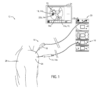
[0006] FIG. 1 is a representative diagram of a surgical environment
demonstrating a
camera system for improved visualization during surgery;
[0007] FIG. 2A is a simplified diagram of a camera configured to excite
a fluorescent
agent and identify a resulting fluorescent emission in a surgical field;
[0008] FIG. 2B is a simplified diagram demonstrating a surgical
implement illuminated
with visible light;
[0009] FIG. 2C is a simplified diagram demonstrating the surgical
instrument of FIG. 28
enhanced to emphasize a fluorescent portion;
[0010] FIG. 3 is a simplified, cutaway diagram demonstrating surgical
implements
including surgical sutures and anchors comprising a fluorescent agent;
[0011] FIG. 4 is a representative diagram demonstrating the sutures and
suture anchor of
FIG. 3 enhanced by a camera system;
[0012] FIG. 5A is a profile view of a shaver comprising a plurality of
fluorescent markings
configured to identify an orientation;
[0013] FIG. 5B is a profile view of a surgical probe demonstrating a
plurality of graduated
markings identifying a dimension of the surgical probe;
[0014] FIG. 6 is a representative diagram demonstrating enhanced image
data captured
by a surgical camera in a cavity of a patient;
[0015] FIG. 7 is a projected view of an arthroscopic operation
performed on a shoulder of
a patient;
[0016] FIG. 8 is a representative diagram demonstrating enhanced image
data in a
shoulder cavity of a patient;
[0017] FIG. 9 is a representative diagram demonstrating enhanced image
data in a
shoulder cavity of a patient;
CA 03213787 2023- 9- 27
WO 2022/219586 PCT/IB2022/053541
3
[0018] FIG. 10A is a projected view demonstrating a surgical
procedure for a shoulder;
[0019] FIG. 10B is a representative diagram demonstrating a plurality
of sutures
enhanced with distinctive colors or patterns for improved visualization;
[0020] FIG. 11 is a flowchart demonstrating a method of object or
surgical implement
detection in a surgical field;
[0021] FIG. 12 is a flowchart demonstrating a method for providing an
enhanced display
of surgical image data; and
[0022] FIG. 13 is a modified block diagram demonstrating a surgical
camera system and
display in accordance with the disclosure.
DETAILED DESCRIPTION
[0023] In the following description of the preferred implementations,
reference is made
to the accompanying drawings, which show specific implementations that may be
practiced. Wherever possible, the same reference numbers will be used
throughout the
drawings to refer to the same or like parts. It is to be understood that other
implementations may be utilized and structural and functional changes may be
made
without departing from the scope of this disclosure.
[0024] Referring to FIG. 1, a simplified representation of a camera
system 10 is shown
demonstrating an exemplary surgical environment 12. As shown, the camera
system 10
is implemented in combination with one or more surgical implements 14, for
example, a
surgical tool 14a or shaver in connection with a control console 16. In
operation, a
camera or endoscope 18 of the camera system 10 may capture image data in a
visible
light range (e.g., 400 nm to 650 nm) as well as a near-infrared range (e.g.,
650 nm to 900
nm). The image data may be communicated to a display controller 20 configured
to
generate enhanced image data. The enhanced image data may emphasize or visibly
define one or more fluorescent portions 22 of the surgical implements 14 to
assist in the
visualization of one or more of the surgical implements 14 presented on a
display device
24. In this configuration, the camera system 10 may provide for improved
visualization
and enhanced viewing of fluorescent portions 22 of the surgical implements 14
to
improve the visibility, detection, and identification of the surgical
implements 14 when
implemented in a surgical site 26 of a patient 28.
CA 03213787 2023- 9- 27
WO 2022/219586 PCT/IB2022/053541
4
[0025] FIGS. 2A-2C are simplified diagrams demonstrating the operation
of the camera
system 10 to identify a fluorescent emission 32 output from the fluorescent
portion of an
exemplary surgical implement 14. Referring now to FIGS. 1 and 2A-2C, in
various
implementations, the fluorescent portions 22 of the surgical implements 14 may
comprise a fluorescent agent implemented in a coating, insert, or embedded
structure
that may become excited and emit the fluorescent emission 32 in response to
receiving
an excitation emission 34. As demonstrated in FIG. 2A, the excitation emission
34 is
output from a first light source 36 and may correspond to an emission of light
outside the
visible spectrum. Additionally, a visible light emission 38 may be output from
a second
light source 40. The excitation emission may include a wavelength or range of
wavelengths configured to energize and excite the fluorescent agent
incorporated in the
fluorescent portion 22. In various examples, the excitation emission 34 may
comprise
wavelengths in a near-infrared range, which may correspond to wavelengths
ranging
from approximately 600 nm to 900 nm. The first light source 36 may correspond
to a
laser emitter module configured to output emissions ranging from 650 nm to 680
nm. In
some cases, the first light source 36 may output the excitation emission 34 in
a range of
wavelengths from approximately 740 nm to 780 nm. The specific excitation
wavelength
associated with the first light source 36 and the excitation emission 34 may
be selected
to effectively energize the fluorescent agent of the fluorescent portion 22,
such that the
resulting fluorescent emission 32 may be captured by one or more image sensors
42 of
the camera system 10. In this way, the camera system 10 may detect a presence
or a
location of the surgical implement 14 in response to the detection of the
fluorescent
emission 32 in the image data.
[0026] As previously discussed, the camera system 10 may be configured
to capture
image data associated with the visible light emission 38 as well as the
fluorescent
emission 32. Once captured, the system 10 may enhance the image data
representing
the visible light with one or more overlays or graphics to generate enhanced
image data
that emphasizes and/or identifies portions of a field of view 44 corresponding
to the
surgical implement 14. In order to provide the enhanced image data, a camera
controller
46 may be configured to selectively control each of the first and second light
sources 36,
40 as well as process image data received from a first image sensor 42a and a
second
image sensor 42b. In a standard operating mode, the camera controller 46 may
activate
CA 03213787 2023- 9- 27
WO 2022/219586 PCT/IB2022/053541
the visible light emission 38 output from the second light source 40 to
illuminate the
surgical site 26 in wavelengths of light in a visible range (e.g., 400 nm ¨
650 nm).
Reflections from the visible light emission 38 may be captured by the second
image
sensor 42b, which may correspond to a visible light image sensor. Such
operation may
provide for illumination of the surgical site 26 in visible wavelengths of
light, such that
the camera controller 46 can output image data demonstrating visible
characteristics of
the surgical site 26 to the display controller 20. An example of the surgical
implement 14
demonstrated illuminated by the visible light emission 38 and captured by the
second
image sensor 42b is shown in FIG. 2B. Though only a simplified representative
body is
demonstrated in FIG. 2B to represent the surgical implement 14, the
fluorescent portion
22 is represented as being nearly visibly indistinguishable from the depicted
surface
textures illuminated by the visible light emission 38.
[0027] In order to generate the enhanced image data, the camera
controller 46 may
activate the first light source 36 to output the excitation emission 34. In
response to the
excitation emission 34, the fluorescent agent of the fluorescent portion 22
may become
excited and output the fluorescent emission 32. Concurrent with the activation
of the
first light source 36, the camera controller 46 may also activate the second
light source
40 to illuminate the surgical site 26 in the visible light emission 38. As a
result, the
fluorescent emission 32 and the visible light emission 38 may be captured
within the field
of view 44 of each of the image sensors 42. While the second image sensor 42b
may be
configured to capture the reflected visible light emission 38, the first image
sensor 42a
may correspond to a near-infrared image sensor configured to capture
wavelengths of
light in a near-infrared range (e.g., 650 nm ¨ 900 nm). As shown, each of the
image
sensors 42 may comprise one or more light filters, exemplified as a first
light filter 52a
and a second light filter 52b. In operation, the light filters 52a, 52b may
filter the
combined wavelengths of the fluorescent emission 32 and the visible light
emission 38 in
the field of view 44 to improve the fidelity of the detection of the
corresponding
wavelengths detected by each of the image sensors 42a, 42b. In this way, the
camera
controller 46 may process image data recorded by each of the image sensors
42a, 42b to
detect and discriminate between the fluorescent emission 32 and the visible
light
emission 38 in the field of view 44 representative of the surgical site 26.
CA 03213787 2023- 9- 27
WO 2022/219586 PCT/IB2022/053541
6
[0028] Though generally described as light filters 52, the first filter
52a and the second
filter 52b may correspond to one or more high pass, low pass, and /or bandpass
filters
configured to transmit light over a range associated with a corresponding
detection
range of the image sensors 42a, 42b. For example, the first light filter 52a
may
correspond to a bandpass filter configured to pass a range of near-infrared
wavelengths
from approximately 800 nm to 850 nm. In this configuration, the first light
filter 52a may
be selected to have a center frequency of approximately 825 nm, which may
effectively
pass wavelengths of light associated with the fluorescent emission 32 to the
first image
sensor 42a. In such cases, the fluorescent emission 32 may correspond to an
emission
from a fluorescent agent in the form of an indocya nine green (ICG) dye.
Accordingly, the
fluorescent emission 32 output from the fluorescent portion 22 may pass
through the
first light filter 52a within the bandpass range, such that the associated
light from the
fluorescent emission 32 is captured and identified by the camera controller
46. Similarly,
the visible light emission 38 and the corresponding light reflected from the
surgical site
26 may pass through a second light filter 52b, which may be configured to pass
wavelengths of light in a visible range (e.g., 400 nm ¨ 650 nm). In this way,
the camera
system 10 may actively detect the fluorescent emission 32 and generate
overlays,
graphics, or other visual enhancements to augment the image data illuminated
by the
visible light emission 38 in the field of view 44.
[0029] In addition to the first and second light filters 52a, 52b, the
camera system 10
may further comprise additional filters, which may include one or more
dichroic filters or
mirrors configured to separate the fluorescent emission 32 from the visible
light emission
38. Such filters, generally referred to as light filters 52, may be
incorporated in an
endoscope or camera 60, which may comprise the image sensors 42, light sources
36,40,
and camera controller 46, as well as the light filters 52 in a unified
package. For example,
the camera 60 may comprise each of the light sources 36, 40, image sensors 42,
filters
52, and the camera controller 46 in a compact endoscope similar to that
discussed later
in reference to FIGS. 3, 7, etc. In this way, the camera system 10 may be
implemented in
an easily manipulated package well suited for operation in the surgical
environment 12.
Though ICG is discussed in various examples of the disclosure, other
fluorescents
including methylene blue (MB), fluorescence, and protoporphyrin IX [PpIX], may
be
similarly implemented with the camera system 10.
CA 03213787 2023- 9- 27
WO 2022/219586 PCT/IB2022/053541
7
[0030] With the image data associated with the visible light emission
38 detected
independently of the fluorescent emission 32, the camera system 10 may provide
for the
enhancement of the fluorescent portions 22 in the image data. In this way, one
or more
colors, patterns, or other visual enhancements or overlays 62 may be
superimposed or
overlaid on the image data to generate enhanced image data for presentation on
the
display device 24. As shown in FIG. 2C, the location of the fluorescent
portion 22 in the
image data is emphasized by the overlay 62, such that the fluorescent portion
22 is
clearly distinguishable from the remainder of the surgical implement 14 as
well as the
local environment in the surgical site 26. As discussed in further detail
throughout the
application, the enhanced image data may be implemented in a variety of ways
to
provide improved visualization of the surgical site 26 to assist in the
identification of a
presence, position, orientation, and/or dimension of various surgical
implements 14.
[0031] Referring generally to FIGS. 1, 2A, 2B, and 2C; implementations
and operating
aspects of the camera system 10 are described in further detail. In general,
ICG,
fluorescein, PpIX, and methylene blue may correspond to dyes used in medical
diagnostics. ICG has very low toxicity and high absorptance in a wavelength
range of from
about 600 nm to about 900 nm and a peak absorptance at about 780 nm. ICG emits
fluorescence at a wavelength of about 830 nm. Additionally, fluorescent
agents, such as
ICG, that emit near-infrared radiation may be detectable through biological
tissue. As
used herein the terms "radiation" and "light" are used interchangeably.
Another
example of a fluorescent agent, PpIX may be excited over a blue color range
(e.g., 405
nm) with a corresponding peak fluorescence of approximately 635 nm. MB is
excited
over a red-NIR color range (e.g., 600 nm) with a corresponding peak
fluorescence of
approximately 650 nm. Fluorescein has a peak absorption of approximately 490nm
with
a fluorescent emission of approximately 520nm. The gap between the absorption
range
and the emission range of each of the fluorescent agents is referred to as a
Stokes shift,
which may be utilized to distinguish between wavelengths associated with the
excitation
emission 34 and the resulting fluorescent emission 32.
[0032] In various examples, the fluorescent agent may be coated or used
as an integral
portion (e.g., embedded in a material or structure) of a surgical implement
14. In some
cases, the fluorescent agent may be incorporated in the fluorescent portion 22
of the
surgical implement 14 during manufacture. For example, a plastic surgical
implement
CA 03213787 2023- 9- 27
WO 2022/219586 PCT/IB2022/053541
8
may have a fluorescent dye mixed into the plastic during manufacture.
Additionally, light
blocking packaging may be used to protect the fluorescent dye from light until
the
surgical implement 14 is ready for use. The surgical implement 14, such as,
for example
and without limitation, a sponge, a suture, a pin, a screw, a plate, a
surgical tool, or an
implant may be painted with a fluorescent material. As used herein, the term
"surgical
tool", may comprise, without limitation, a biter, grasper, retriever, pick,
punch, hook,
probe, elevator, retractor or scissors. The surgical implement 14 may have a
fluorescent
agent coated on a portion to indicate a location, position, depth,
orientation, or other
characteristic of the surgical implement. Accordingly, the fluorescent portion
22 of the
surgical implement 14 may be readily identified or detected in the enhanced
image data
provided by the camera system 10.
[0033] As discussed later in specific reference to FIGS. 5A and 5B, the
fluorescent agent
may incorporated in various fluorescent portions 22 of surgical implements 14
in
patterns, shapes, and/or alphanumeric characters to identify the surgical
implement 14
or to indicate dimensions, orientations, or proportions of implements 14
represented in
the image data. The presence of a fluorescent agent in the surgical implement
14 may
also enable surgeons to quickly check to make sure that no portion of a
surgical
implement 14 has been left in a surgical site 26. In some cases, the display
controller 20
may be configured to process the image data associated with the fluorescent
emission 32
(e.g., corresponding pixels in the field of view 44) to identify or classify
one or more
surgical implements 14 in the surgical site 26. For example, the display
controller 20 may
be configured to process a characteristic shape of the surgical implement 14
or one or
more symbols represented in the image data captured by the first image sensor
42a (e.g.,
in the NIR range) to identify a type or category of the implement 14 based on
a computer
vision template. Such identification is discussed further in reference to FIG.
12.
[0034] In various implementations, the fluorescent agent in the
surgical implement 14
may be excited using a light source that emits excitation light in the
excitation
wavelength range of the particular fluorescent agent. For example, when ICG is
used as
the fluorescent agent, ICG fluorescence may be excited using light in a
wavelength range
of from about 600 nm to about 900 nm and in some cases around 780 nm. In such
cases,
the light source 36 may be a light emitting diode or a laser emitting diode
with a center
frequency within or centrally within the excitation range of the ICG. The
image sensors
CA 03213787 2023- 9- 27
WO 2022/219586
PCT/IB2022/053541
9
42 may be, for example, a complementary metal-oxide-semiconductor (CMOS)
sensor or
a charge-coupled device (CCD) sensor. The camera 60 may also include optics,
such as,
for example, lenses, filters, mirrors, and prisms to direct and independently
detect the
wavelengths of light associated with the visible light source 40 and the
fluorescent
emission 32.
[0035] In some implementations, the camera 60 is implemented as an
endoscopic
camera, which may include the image sensors 42, light sources 36, 40, as well
as the light
filters 52. Accordingly, the camera 60 may include both the first light source
36 as an
excitation light source for exciting the fluorescent agent and the second
light source 40 in
the form of a white light source for illuminating the surgical site 26 in the
visible range of
wavelengths. The camera 60 may further include a corresponding image sensor
42a or
detector for detecting the fluorescent emission 32 and an image sensor 42b or
detector
for detecting and recording image data in the visible light range. In some
cases, the
camera 60 may have additional light sources for exciting multiple fluorescent
agents or
for detecting other non-visible attributes of a surgical field. An example of
a camera
system usable to detect fluorescent agents in surgical implements is the
Arthrex Synergy
IDTM camera system which has a camera head and a camera control unit. The
Arthrex
Synergy IDTM camera system has a light source for exciting fluorescence from
ICG and is
capable of detecting visible and near infra-red (NIR) light such as light
emitted by ICG.
[0036] Referring again to FIG. 1, exemplary enhanced image data 70 is
demonstrated on
the display device 24. In the example, an acting end or distal end of the
shaver 14a is
shown demonstrating a first fluorescent portion 22a including a directional
orientation
marker 72. A similar example of the surgical implement 14 in the form of a
shaver 14a is
shown with improved detail in FIG. 4A. As shown the orientation marker 72 may
be
overlaid on the visible image data to provide a clear indication of the
relative orientation
of the shaver 14a in the surgical site 26. Though the orientation marker 72
may seem
trivial in cases where the surgical implement 14 is clearly visible in the
image data, the
overlay 62 aligned with the fluorescent emission 32 demonstrated in the
enhanced image
data may provide a clear indication of the orientation and/or position of the
surgical
implement 14 even in cases where a cavity of the surgical site is obstructed
or clouded by
particles, blood, tissue debris, etc.
CA 03213787 2023- 9- 27
WO 2022/219586
PCT/IB2022/053541
[0037] Additionally, FIG. 1 demonstrates an example of the surgical
implement 14 in the
form of an anchor 14b. In various cases, an anchor or various surgical
implants may
become over grown by tissue, calcium, or other substances that may mask them
from
visibility from the visible light emission 38 and the corresponding second
image sensor
42b. As shown, a colored overlay 62 is generated by the display controller 20
in a portion
of the image data associated with a second fluorescent portion 22b. The
overlaid or
superimposed color may highlight a portion of the anchor 14b, such that the
location of a
hexalobe or drive head 74 is visible in the enhanced image data. In cases
where the drive
head 74 is masked behind biological tissue, the excitation emission 34 and the
resulting
fluorescent emission 32 may penetrate the tissue such that the display
controller 20 may
detect the fluorescent portion 22 and demonstrate the location of the head 74
in the
enhanced image data.
[0038] Referring now to FIGS. 3 and 4, an example of an application of
the camera
system 10 is described in reference to an exemplary shoulder repair operation.
As
depicted, the camera 60 is implemented as an endoscope that incorporates the
second
light source 40 configured to output the visible light emission 38 within the
field of view
44 of the image sensors 42a, 42b. As shown in FIG. 3, the first light source
36 associated
with the excitation emission 34 may be incorporated in a dedicated lighting
device 80.
The lighting device 80 may comprise an elongated shaft 82 extending between a
proximal
end portion 82a and a distal end portion 82b. The excitation emission 34 may
be output
from the first light source 36 via the distal end portion 82b of the elongated
shaft 82. A
control circuit and power supply may be enclosed in a housing 84 in connection
with the
proximal end portion 82a. In this configuration, the excitation emission 34
may originate
from a different origin than the field of view 44. The dedicated lighting
device 80 may
project the excitation emission 34 into various portions or regions of the
surgical site 26
without having to maneuver the camera 60. Accordingly, implementations of the
camera
system 10 incorporating the dedicated lighting device 80 separate from the
camera 60
may provide for independent illumination of the various regions within the
surgical site
26 without maneuvering the camera 60 or independent of the position of the
camera 60.
[0039] Though discussed in reference to the excitation emission 34
being output from
the dedicated lighting device 80, either or both of the light sources 36, 40
may be
implemented in the dedicated lighting device 80 to output light in various
ranges of
CA 03213787 2023- 9- 27
WO 2022/219586
PCT/IB2022/053541
11
wavelengths. In some implementations, the lighting device 80 or the camera 60
may be
configured to emit a beam of light with a diameter small enough for targeting
items in
the surgical field for further action by a surgeon. In an implementation, the
beam
diameter may be less than about 5 mm. In some cases, the beam diameter may be
less
than about 2 mm or than about 1 mm. In general, the lighting device 80 or
camera 60
may be configured to emit a beam of light of sufficient brightness and density
to be
detected within a surgical field. For example, in some cases, high sensitivity
sensors 42
have been measured to detect light at intensities of 10 nW/cm2 or less (e.g.,
a high
sensitivity CMOS sensor). The light sources 36, 40 may be positioned proximal
to a distal
end of the light emitting device 80 or camera 60. Additionally, the light
source 36, 40 may
be positioned away from the distal end and light emitting device 80 or camera
60 from
the light source communicated to the distal end such as by, for example, fiber
optics. The
light emitted by the light emitting device 80 and/or camera 60 may have a
variable shape
that may be adjusted, such as by using optics to allow a user to better
illuminate a
desired target.
[0040] In some implementations, one or both of the light sources 36, 40
may be
incorporated into a surgical instrument 14 other than the endoscopic camera
system 10,
for example, in a probe, a shaver 14a, an ablation device, or other
instrument. In some
examples, an LED may be located at a distal end of the device or instrument.
In some
example, a probe or other device may be formed at least partially of a light
pipe that may
receive light from an LED, laser, or other light source external to the body
and transmit
the radiation to the distal end of the instrument. The light emitting device
80 may be
powered by an isolated power source coupled to the light emitting device.
Additionally,
the light emitting device 80 may be battery powered. The battery powered light
emitting
device may be configured for a single use or may be configured with a
rechargeable
battery for multiple uses. The light emitting device 80 may be packaged in a
sterile
container for a single use. Additionally, the light emitting device 80 may be
configured for
sterilization and repeated use. The light emitting device 80 may be a rigid
device or a
flexible device. The light emitting device may be an articulatable device.
[0041] Additionally, the light emitting device 80 or light sources 36,
40 may be placed
outside of a surgical field or site 26 and light directed through biological
tissue for
detection by the camera 60 positioned in the surgical field. Additionally, the
light
CA 03213787 2023- 9- 27
WO 2022/219586
PCT/IB2022/053541
12
emitting device may direct light from a surgical field through tissue for
detection by a
device positioned outside of a surgical field. In some cases, the light
emitting device 80
may be placed outside of a body and direct light through tissue for detection
by the
camera 60 positioned inside the body. Additionally, the light emitting device
80 may be
placed inside of a body and direct light through tissue for detection by a
camera (e.g., the
camera 60) positioned outside of the body. Additionally, the light emitting
device 80 may
be placed in a first portion of a surgical site 26 and direct light through
tissue for
detection in a second portion of the surgical site 26.
[0042] As demonstrated in FIG. 3, a shoulder cavity 86 is revealed via
a cutaway section
88. However, in a typical arthroscopic procedure, the shoulder cavity 86 would
be
enclosed, such that the internal anatomy of the patient 28 would not be
visible as
depicted in FIG. 3. To accurately visualize the shoulder operation
corresponding to FIG.
3, the distal end of the camera 60 and the dedicated light source 80 would
protrude
through the outer tissue and into the shoulder cavity 86, similar to the
examples
demonstrated in FIGS. 7 and 10A, as later discussed. According, the cutaway
section 88
in FIG. 3 may provide for a simplified representation of an arthroscopic
procedure to
demonstrate the internal anatomy and may similarly be representative of an
open
surgery where the camera 60 and dedicated lighting device 80 may be positioned
outside
and provide illumination into the shoulder cavity 86.
[0043] Referring now to FIGS. 3 and 4, a plurality of sutures 92a, 92b
and anchors 94a,
94b are shown implemented in connection with a shoulder tendon 96 and humorous
98
of the patient 28. As shown, the sutures 92 may comprise a first suture 92a
and a second
suture 92b. The first suture 92a is in connection with a first anchor 94a that
connects the
first suture to the humorous 98. The second suture 92b is in connection with
the
humorous 98 via a second anchor 94b. Though clearly represented in FIG. 3, a
view of
the surgical site 26 may be clouded by blood and particulates within the
shoulder cavity
86. Accordingly, the view and relative orientation of the camera 60 in
relation to the
surgical site 26 may not be readily apparent from the image data demonstrated
on the
display device 24.
[0044] In addition to the obstructions in the field of view 44 within
the shoulder cavity
44, in some cases, an anchor (represented in FIG. 4 as the second anchor 94b)
may be
masked or hidden beneath tissue or overgrowth. In such cases, the second
anchor 94b
CA 03213787 2023- 9- 27
WO 2022/219586
PCT/IB2022/053541
13
may be nearly completely hidden from view and challenging to detect within the
image
data captured by the camera 60. In order to improve the visibility of the
second anchor
94b, a fluorescent agent may be incorporated in a portion of the second anchor
94b,
exemplified as a first fluorescent portion 100a (See, FIG. 4) incorporated in
a drive head
74 or hexalobe. In addition to the first fluorescent portion incorporated in
the second
anchor 94b, each of the first suture 92a and the second suture 92b may also
include
corresponding second and third fluorescent portions 100b and 100c. Each of the
fluorescent portions 100a, 100b, and 100c may be illuminated by the excitation
emission
34 output, in this example, from the first light source 36 of the dedicated
lighting device
80. In response to receiving the excitation emission 34, each of the
fluorescent portions
100a, 100b, 100c may become excited to output corresponding fluorescent
emissions 32.
[0045] In some cases, the fluorescent emissions 32 output from the
fluorescent portions
100a, 100b, 100c may vary in wavelengths due to different compositions or
combinations
of fluorescent agents incorporated therein. In other cases, a concentration of
a common
fluorescent agent (e.g., ICG dye) may be incorporated at different levels in
each of the
fluorescent portions 100a, 100b, 100c. Accordingly, in response to receiving
the
excitation emission 34, each of the fluorescent emissions 32 output from the
fluorescent
portions 100a, 100b, 100c may vary in wavelength or intensity based on the
composition
of fluorescent agents or concentration of fluorescent agents incorporated
therein. Based
on the variations in the intensity or wavelengths associated with the
fluorescent
emissions 32, the display controller 20 may be operable to distinguish among
the
different fluorescent portions 100a, 100b, 100c and overlay each of the
fluorescent
portions 100a, 100b, 100c with different characteristic colors 102.
Accordingly, the
camera system 10 may be configured to distinguish among a plurality of
fluorescent
portions 100a, 100b, 100c and assign different respective characteristic
colors 102 or
patterns, such that the enhanced image data demonstrated on the display device
24
clearly distinguishes the locations of each of the surgical implements 14
(e.g., 92a, 92b,
and 94b).
[0046] To be clear, the sutures 92a, 92b and second anchor 94b
demonstrated in FIG. 4
may appear in the image data as being dull and nearly indistinguishable from
their
surroundings when viewed solely via the visible light emission 38. However,
based on
the overlays 62 applied by the display controller 20 over the corresponding
fluorescent
CA 03213787 2023- 9- 27
WO 2022/219586
PCT/IB2022/053541
14
portions 100a, 100b, 100c; the enhanced image data may clearly differentiate
each of the
surgical implements 14 based on a corresponding characteristic color 102a,
102b, 102c or
pseudo-color overlaid on the image data associated with the visible light
emission 38. As
shown, the characteristic colors 102 may include a first color 102a, a second
color 102b,
and a third color 102c. The first color 102a may be incorporated on the first
fluorescent
portion 1002 coating the drive head 74 or hexalobe of the second anchor 94b.
The
second color 102b and the third color 102c may be incorporated within a
constituent
material forming the first suture 92a and the second suture 92b, respectively.
Each of
the characteristic colors 102 may be visually distinguishable based on a
predetermined
display configuration stored within the display controller 20.
[0047] In some cases, the characteristic colors 102 or patterns
associated with the
enhanced image data may be customized or modified to suit the preferences of a
specific
user. For example, some users may prefer a wide range of colors to assist in
distinguishing among the various surgical implements 14, while others may
prefer subtle
color differences that may not distract their view from other aspects within
the surgical
site 26. In some cases, the display controller 20 may adjust a color template
or color
configuration of the characteristic colors 102 or patterns based on the colors
of the local
environment demonstrated in the image data captured by the second image sensor
42
associated with the visible light emission 38. For example, if the image data
illuminated
by the visible light emission 38 is displayed primarily in warm hues (e.g.,
red, yellow,
orange), the display controller 20 may assign a cool color template (e.g.,
blue, purple,
green) to distinguish the fluorescent portions 100a, 100b, 100c from the
remainder of
the image data in the field of view 44. Similarly, if the image data is dark,
light or
contrasting hues or patterns may be automatically applied to contrast the
image data.
Accordingly, the camera system 10 may provide for a variety of formats and
color
templates associated with the enhanced image data to assist in the
visualization of the
surgical site 26.
[0048] Referring to FIGS. 5A and 5B, exemplary surgical implements 14
are shown
comprising fluorescent portions 22 configured to assist a user in a
recognition of an
orientation or position of the surgical implements 14 as represented in the
enhanced
image data generated by the camera system 10. As shown in FIG. SA, an acting
end of
the shaver 14a is shown demonstrating a plurality of longitudinal markings 110
formed
CA 03213787 2023- 9- 27
WO 2022/219586
PCT/IB2022/053541
by the fluorescent portions 22. The longitudinal markings may extend along a
longitudinal axis 112 of the shaver 14a and be evenly spaced radially about an
elongated
body 114. A shaver head 116 is demonstrated in phantom opposing the face
pictured in
FIG. 5A. In this configuration, the longitudinal markings 110 comprising the
fluorescent
portions 22 may be illuminated to output the fluorescent emission 32 in
response to the
excitation emission 34, such that the enhanced image data may demonstrate an
orientation of the surgical implement 14 or shaver 14a in relation to an
actuator direction
(e.g., direction of the shaver head 116).
[0049] Referring to FIG. 5B, the surgical implement 14 is demonstrated
as an exemplary
needle or probe 14c shown comprising a plurality of lateral markings 120
corresponding
to the fluorescent portions 22. As shown, the lateral markings 120 are
implemented as a
plurality of graduated segments demonstrating a scale associated with a
position of the
surgical implement 14 or probe 14c. Similar to the longitudinal markings 110,
the lateral
markings 120 may incorporate the fluorescent agent in the fluorescent portions
22 and
output the fluorescent emission 32 in response to receiving the excitation
emission 34.
In addition to the lateral markings 120, the probe 14c may include one or more
characters 122 or symbols, which may also incorporate fluorescent dyes or
agents, such
that the characters 122 may be overlaid in the image data to emphasize the
associated
symbols in the image data. The longitudinal markings 110 and lateral markings
120 may
be implemented in various combinations to assist an operator of the associated
surgical
implements 14 to identify an orientation, position, and/or relative
measurement of the
surgical implement 14 as presented in the enhanced image data on the display
device 24.
[0050] In some cases, the longitudinal markings 110, lateral markings
120, or various
additional fluorescent portions 22 incorporated on the surgical implements 14
may be
disposed within a groove 124 or indentation formed in an exterior surface of
the surgical
implement 14. By including the fluorescent portions 22 in the grooves or
indentations
associated with the orientation or positional markings 110, 120; the resulting
fluorescent
emissions 32 output from the grooves 124 or indentations may be captured in
the field of
view 44 of the camera system 10 through an orientation aperture associated
with an
interior surface of each of the grooves 124 directed to or facing the
corresponding image
sensors 42a, 42b of the camera 60. In this configuration, the dimensional or
orientational
markings 110, 120 incorporated on the surgical implement 14 may be hidden from
the
CA 03213787 2023- 9- 27
WO 2022/219586
PCT/IB2022/053541
16
field of view 44 of the camera 60 until a portion of the fluorescent emission
32 is output
from the corresponding fluorescent portions 22 disposed in the grooves 124.
The result
of the fluorescent portions 22 disposed in the grooves 124 may be an improved
accuracy
achieved similar to a sight that only exposes the fluorescent emission 32 when
an interior
surface of each of the grooves 124 is visible through the corresponding
orientation
aperture. In this way, the dimensional and orientational features (e.g., 110,
120) of the
surgical implements 14 may provide for improved accuracy in determining the
relative
positioning or orientation of the surgical implement 14.
[0051] Referring now to FIG. 6, the exemplary shaver 14a is shown in
the field of view 44
of the camera 60 demonstrating enhanced image data including overlays 62 of
characteristic colors 102 over the longitudinal markings 110 formed by the
grooves 124
and the fluorescent portions 22. As shown, the longitudinal markings 110 may
assist an
operator in identifying a direction of the shaver head 116 demonstrated by the
arrow
126. For example, as a result of seeing two of the three longitudinal markings
110 on the
display device 24, a user of the shaver 14a may visually identify, from the
longitudinal
markings 110 enhanced by the overlay 62, that the shaver head 116 is directed
toward an
opposite side of the longitudinal markings 110. As shown, the longitudinal
markings 110
are positioned on a left-facing side of the shaver 14a, such that the operator
may
recognize that the shaver head 116 is directed toward a right side represented
on the
display device 24. Such indications of the orientation of the surgical
implement 14 may
be particularly beneficial in cases where the shaver head 116 is hidden behind
tissue 128
or debris in the field of view 44. Accordingly, the longitudinal markings 110
may assist a
user in determining the relative orientation of the surgical implement 14.
[0052] Referring now to FIG. 7, an additional exemplary illustration of
an arthroscopic
procedure on a shoulder 130 of the patient 28 is shown. FIG. 8 demonstrates
enhanced
image data associated with the field of view 44 captured by the camera 60
positioned as
depicted in FIG. 7. As demonstrated in FIGS. 7 and 8, the probe 14c is
demonstrated
penetrating biological tissue 132 within a shoulder cavity 134. As previously
discussed,
the excitation emission 34 may be output from the first light source 36
incorporated in
the dedicated lighting device 80. The excitation emission 34 may be
transmitted within
the cavity 134 and penetrate through the biological tissue 132 (e.g.,
cartilage, muscle,
tendons, bone, etc.) to impinge upon the fluorescent portions 22 formed by the
lateral
CA 03213787 2023- 9- 27
WO 2022/219586
PCT/IB2022/053541
17
markings 120. In response to receiving the excitation emission 34, the
fluorescent agent
incorporated in the fluorescent portions 22 of the lateral markings 120 may
output the
fluorescent emission 32. The light energy emitted from the fluorescent
portions 22 may
also be transmitted through the biological tissue 132 and into the cavity 134,
such that
the near-infrared image sensor 42a may capture the fluorescent emissions 32 in
the field
of view 44.
[0053] In response to detecting the fluorescent emission 32 in the
image data captured
by the first image sensor 42a, the display controller 20 of the camera system
10 may
overlay the pixels in the image data associated with the fluorescent emission
32 with the
overlay 62 (e.g., characteristic colors 102 or patterns) to generate the
enhanced image
data. Accordingly, the camera system 10 may provide for the detection and
tracking of
the position of one or more surgical implements 14 through biological tissue
132 by
detecting the fluorescent emission 32. Once detected, the display controller
20 may
further overlay, mark, or enhance corresponding portions of the image data to
demonstrate the surgical implements 14 that would otherwise be completely
hidden
from a conventional camera system.
[0054] Referring now to FIG. 9, an exemplary surgical cavity 140 is
shown demonstrating
a distal tip of a probe or needle 142 beginning to protrude through biological
tissue 144.
As depicted in the enhanced image data demonstrated on the display device 24
of the
camera system 10, a distal tip 146 of the needle 142 is overlaid by a
characteristic pattern
or color 102. Similar to other examples, the characteristic pattern or color
102 overlaid
on the distal tip 146 of the needle 142 may be detected by the display
controller 24 in
response to the corresponding presence of the fluorescent emission 32 in the
image data
captured by the combined image sensors 42a, 42b. In the example provided, the
distal
tip 146 of the needle 142 may be introduced blindly into the surgical cavity
140.
Accordingly, it may be challenging for a surgeon or physician to accurately
determine a
position of a depressing instrument 148 and grasper 150 to effectively guide
and interact
with the distal tip 146. However, due to the incorporation of the fluorescent
portion 22
on the distal tip 146, the fluorescent emission 32 may penetrate the
biological tissue 144
and be detected by the display controller 20 before the distal tip 146 begins
to protrude
through the biological tissue 144. For example, in response to identifying the
fluorescent
emission 32 in the field of view 44, the display controller 20 may enhance the
CA 03213787 2023- 9- 27
WO 2022/219586
PCT/IB2022/053541
18
corresponding portion of the image data associated with the fluorescent
emission 32
with the overlay 62. In this way, a surgeon may identify a location of the
biological tissue
144 through which the distal tip 146 of the needle 142 will protrude prior to
the distal tip
146 breaching the surface of the biological tissue 144. In this way, the
enhanced image
data provided by the camera system 10 may improve the accuracy associated with
an
operation by displaying a location of a surgical implement that would
otherwise be
invisible in a visible light range captured by the second imager or visible
light image
sensor 42b.
[0055] In some examples, the excitation light source or first light
source 36 may output
the excitation emission 34 at an intensity sufficient to penetrate biological
tissue as
discussed herein. For example, the first light source 36 may output the
excitation
emission 34 at an intensity ranging from approximately 1 mW/cm2 to 1 W/cm2. In
some
cases, the light intensity may be higher or lower depending on the specific
light emitter
technology implemented and the application. Depending on the application and
the
duration over which the excitation emission 34 is to be activated, the
intensity of the
excitation emission 34 may be limited or pulsed to control excess heat
generation and
limit damage to the biological tissue. As previously discussed, the excitation
emission 34
may comprise wavelengths of radiation ranging from approximately 650 nm to 900
nm in
the near-infrared range. For reference, the visible light emission 38
associated with the
second light source 40 may be output in wavelengths corresponding to visible
colors of
light associated with the acuity of a human eye ranging from 400 nm to
approximately
650 nm. The penetration of the excitation emission 34 and/or the fluorescent
emission
32 through biological tissue may extend approximately from a depth of 1 mm to
depths
or thicknesses of biological tissue exceeding 10 mm.
Experimental results have
demonstrated a loss of intensity of emissions similar to the excitation
emission 34 and
the fluorescent emission 32 in the near-infrared range at a rate of
approximately 3%-
10%/mm of biological tissue penetrated. Accordingly, the first image sensor
42a may
detect the fluorescent emission 32 or the excitation emission 34 after the
corresponding
light energy has penetrated multiple millimeters of biological tissue.
Therefore, the
camera system 10 may identify the relative location or orientation of the
various surgical
implements 14 and demonstrate the locations in the enhanced image data in a
variety of
CA 03213787 2023- 9- 27
WO 2022/219586
PCT/IB2022/053541
19
cases where the surgical implements 14 may be hidden behind layers of
biological tissue
having various thicknesses.
[0056] Referring now to FIGS. 10A and 10B, yet another exemplary
application of the
surgical camera system 10 is shown demonstrating an arthroscopic shoulder
repair of the
patient 28. As demonstrated in FIG. 9, an anterior cannula 152 provides access
into a
surgical cavity 154 to manipulate a plurality of sutures 156a, 156b. In
operation, a
surgeon may access the surgical cavity 154 via a skid 158. In order to reach
the sutures
156 within the surgical cavity 154, a grasper 160 may be implemented to
selectively
engage one of the sutures 156. As demonstrated in FIG. 10B, the field of view
44 of the
camera 60 demonstrates an arthroscopic view of a first suture 156a, second
suture 156b,
and a lasso 162 that may further be implemented to manipulate and loop the
sutures
156. Even with extensive knowledge of the procedures and associated visible
colors
incorporated on the sutures 156, surgeons and physicians still may have
difficulty
distinguishing the first suture 156a from the second suture 156b.
Distinguishing the
sutures 156 may become particularly challenging when the fluid within the
surgical cavity
154 is encumbered by debris or blood that may further mask any defining
features of the
sutures 156.
[0057] As previously discussed in reference to FIGS. 3 and 4, the first
suture 156a may
include a first concentration of the fluorescent agent and the second suture
156b may
include a second concentration of the fluorescent agent. Accordingly, in
response to
receiving the excitation emission 34, each of sutures 156a, 156b may output
different
intensities of the fluorescent emission 32. These intensities of the
fluorescent emission
32 may be identified and distinguished by the display controller 20 based on
the image
data in the near-infrared range captured by the first image sensor 42a. In
response to
the differing intensities of the fluorescent emissions 32, the display
controller 20 may
overlay each of the sutures 156a, 156b with different characteristic patterns
164a, 164b
as demonstrated by FIG. 10B. In this way, the display controller 20 may
identify the
fluorescent emissions 32 at various intensities to distinguish among a
plurality of surgical
implements 14 identified in the field of view 44 of the camera system 10. The
overlays
62, shown as characteristic patterns, of the sutures 156a, 156b may similarly
be
implemented as characteristic colors or markers (e.g., notification windows,
CA 03213787 2023- 9- 27
WO 2022/219586
PCT/IB2022/053541
superimposed graphics, etc.) to assist in identifying and distinguishing among
surgical
implements 14 depicted in the image data of the camera system 10.
[0058] Referring now to FIG. 11, an exemplary flowchart is shown
demonstrating a
method for detecting an object with the camera system 10 as discussed herein.
The
method 170 may begin in response to an activation of the camera system 10 or
initiation
of an object detection routine 172. As discussed in various examples, the
camera 60 may
be controlled by the camera controller 46 to capture image or sensor data via
one or
more of the image sensors 42a, 42b (174). Once the image data is captured by
the image
sensors 42a, 42b, the display controller 20 may detect one or more portions of
the image
data or pixels within the field of view 44 that include wavelengths of light
corresponding
to the fluorescent emission 32 from the fluorescent portions 22 (176). Based
on the
image data processed by the display controller 20, the method 170 may continue
in step
178 to determine if one or more surgical implements 14 are detected in
response to the
presence of the fluorescent emission 32. If no implements 14 are detected in
step 178,
the method 170 may return to step 174 to continue capturing the image or
sensor data
and processing the image data to identify the fluorescent emission 32 in steps
174 and
176.
[0059] In step 178, if an object associated with the fluorescent
emission 32 is detected in
the image data, the method 170 may continue to mark, overlay, or annotate the
image
data to emphasize the regions in the field of view 44 where the fluorescent
emission 32 is
detected (180). The marked or annotated image data generated in step 180 may
correspond to the enhanced image data comprising one or more overlays 62 in
the form
of characteristic colors, patterns, or other indicating features that may
assist a viewer in
recognizing a location, orientation, dimensions, proportions, or other
information related
to the surgical implement 14 from which the fluorescent emission 32 was
emitted and
detected by the camera system 10. Examples of surgical implements may include
a biter,
grasper, retriever, pick, punch, hook, probe, elevator, retractor or scissors.
In some
cases, the surgical implements 14 may correspond to items configured to
trigger an alert
or notification of the camera system 10 to indicate the detection of their
presence. For
example, partial components of tools, implants, sponges, or other various
surgical
implements within the surgical site 26 may be detected by camera system 10 in
response
to the presence of the fluorescent emission 32. In response to such a
detection, the
CA 03213787 2023- 9- 27
WO 2022/219586
PCT/IB2022/053541
21
method 170 may output an indication (e.g., an alert, instruction,
notification, etc.)
indicating the presence of a fluorescent portion 22 and alerting a surgeon or
medical
professional of the presence of the corresponding surgical implement 14 (182).
In some
cases, the programming of the camera system 10 may define specific surgical
implements
14 that may be associated with the fluorescent emission 32. In such cases, the
notification output in step 182 may indicate the specific type or category of
the surgical
implement 14 identified in the image data by the camera system 10. Following
step 182,
the detection routine may continue until it is deactivated by an operator, as
demonstrated in step 184.
[0060] Referring now to FIG. 12, a flowchart is shown demonstrating a
method 190 for
displaying enhanced image data in accordance with the disclosure. The method
190 may
begin in response to the initiation of an enhanced image data display routine
by the
camera system 10 (192). Once initiated, the method 190 may continue to step
194 to
capture image or sensor data with the image sensors 42a and 42b. Once
captured, the
display controller 20 may scan the image data and detect portions of the image
data with
wavelengths corresponding to the fluorescent emission 32 as detected by the
first image
sensor 42a (196). In some cases, the method 190 may identify a plurality of
fluorescent
emissions 32 depicted in the image data at a plurality of intensity levels
corresponding to
a plurality of fluorescent portions 22 that may include varying concentrations
of
fluorescent agents (198). As previously discussed, each of the fluorescent
portions 22 of
the surgical implements 14 detected in the field of view 44 may include a
distinctive
concentration of the fluorescent agent, such that the resulting fluorescent
emissions 32
may be output and detected by the first image sensor 42a at different
intensity levels.
Based on the different intensity levels, the display controller 20 may assign
the overlays
62 as different characteristic colors in the image data to generate the
enhanced image
data for display on the display device 24 (200).
[0061] In some cases, the display controller 20 may identify different
intensities of the
fluorescent emission 32 over time, such that the characteristic colors or
patterns
associated with the overlay 62 of the enhanced image data may be maintained
even in
cases where the corresponding surgical implements 14 are not simultaneously
presented
in the image data. For example, the display controller 20 may be preconfigured
to
associate a lower intensity fluorescent emission 32 with a first color, a
medium intensity
CA 03213787 2023- 9- 27
WO 2022/219586
PCT/IB2022/053541
22
fluorescent emission 32 with a second color, and a third intensity fluorescent
emission 32
with a third color. The relative intensities may correspond to percentages or
relative
levels of luminance associated with each of the fluorescent emissions 32. For
example, if
three levels of luminance are detected, a maximum intensity may be associated
with the
third color. An intermediate intensity may be associated with the second
color, and a
minimum or lowest intensity may be associated with the first color. Once the
enhanced
image data is generated, it may further be selectively displayed on the
display device 24
by controlling an interface of the display controller (202). Following step
202, the display
routine may continue until deactivated (204).
[0062] Referring now to FIG. 13, a block diagram of the camera system
10 is shown. As
discussed throughout the disclosure, the system 10 may comprise a camera 60 in
communication with a display controller 20. The camera 60 may comprise a
plurality of
light sources 36, 40; at least one image sensor 42 (e.g., 42a, 42b); a camera
controller 46;
and a user interface 210. In various implementations, the camera 60 may
correspond to
an endoscope with an elongated scope comprising a narrow distal end suited to
various
non-invasive surgical techniques. For example, the distal end may include a
diameter of
less than 2 mm. As demonstrated, the camera 60 may be in communication with
the
display controller 20 via communication interface. Though shown connected via
a
conductive connection, the communication interface may correspond to a
wireless
communication interface operating via one or more wireless communication
protocols
(e.g., WiFi, 802.11 b/g/n, etc.).
[0063]
The light sources 36, 40 may correspond various light emitters
configured to
generate light in the visible range and/or the near infrared range.
In various
implementations, the light sources 36, 40 may include light emitting diodes
(LEDs), laser
diodes, or other lighting technologies. As previously discussed, the first
light source 36
may generally correspond to a laser emitter configured to output emissions in
the near
infrared range including wavelengths from approximately 650 nm to 900 nm. In
some
instances, the first light source 36 may output the excitation emission 34
ranging from
650 nm to 680 nm with a center frequency of approximately 670 nm. In some
cases, the
first light source 36 may output the excitation emission 34 in a range of
wavelengths
from approximately 740 nm to 780 nm. More generally, the wavelengths
associated with
the first light source 36 and the excitation emission 34 may be selected to
effectively
CA 03213787 2023- 9- 27
WO 2022/219586
PCT/IB2022/053541
23
energize the fluorescent agent of the fluorescent portion 22. The second light
source 40
may correspond to a white light source in the visible spectrum including
wavelengths
ranging from approximately 380 nm to 700 nm or from approximately 400 nm to
650 nm.
[0064] The image sensors 42a, 42b may correspond to various sensors and
configurations comprising, for example, charge-coupled devices (CCD) sensors,
complementary metal-oxide semiconductor (CMOS) sensors, or similar sensor
technologies. As previously discussed, the system 10, particularly the display
controller
20 may process or compare the image data captured by each of the image sensors
42 to
identify the fluorescent emission 32 and apply the overlay 62 in the form of
one or more
colors (e.g., the characteristic colors 102), patterns, markers, graphics,
messages, and/or
annotations indicating the presence and/or location of the fluorescent
emission 32 in the
image data. In operation, the light filters 52a, 52b (e.g. bandpass filters)
may filter and
effectively separate the combined wavelengths of the fluorescent emission 32
and the
visible light emission 38 in the field of view 44. Accordingly, the filtered
light received by
the first image sensor 42a may provide a map identifying locations of the
fluorescent
emission 32 and the corresponding locations of the fluorescent portions 22 of
the
surgical implements 14 in the image data.
[0065] The camera controller 46 may correspond to a control circuit
configured to
control the operation of image sensors 42a, 42b and the light sources 36, 40
to provide
for the concurrent or simultaneous capture of the image data in the visible
light
spectrum as well as the near infrared spectrum or wavelength associated with
the
fluorescent emission 32.
Additionally, the camera controller 46 may be in
communication with a user interface 210, which may include one or more input
devices,
indicators, displays, etc. The user interface may provide for the control of
the camera 60
including the activation of one or more routines as discussed herein. The
camera
controller 46 may be implemented by various forms of controller,
microcontrollers,
application-specific integrated controllers (ASICs), and/or various control
circuits or
combinations.
[0066] The display controller 20 may comprise a processor 212 and a
memory 214. The
processor 212 may include one or more digital processing devices including,
for example,
a central processing unit (CPU) with one or more processing cores, a graphics
processing
unit (GPU), digital signal processors (DSPs), field programmable gate arrays
(FPGAs),
CA 03213787 2023- 9- 27
WO 2022/219586
PCT/IB2022/053541
24
application specific integrated circuits (ASICs) and the like. In some
configurations
multiple processing devices are combined into a System on a Chip (SoC)
configuration
while in other configurations the processing devices may correspond to
discrete
components. In operation, the processor 212 executes program instructions
stored in the
memory 214 to perform the operations described herein.
[0067] The memory 214 may comprise one or more data storage devices
including, for
example, magnetic or solid state drives and random access memory (RAM) devices
that
store digital data. The memory 214 may include one or more stored program
instructions, object detection templates, image processing algorithms, etc. As
shown,
the memory 214 may comprise a detection module 216 and an annotation module
218.
The detection module 216 include instructions to process the image data
identifying the
fluorescent emission 32 from the first image sensor 42a and detect the
locations in the
field of view 44 from which the fluorescent portion 22 of the surgical
implement 14
emitted the fluorescent emission 32. In some cases, the detection module 216
may
include instructions to detect or identify a type or classification associated
with the
surgical implement 14 in the image data captured by the camera 60. For
example, the
processor 212 may access instructions in the detection module 216 to perform
various
processing tasks on the image data including preprocessing, filtering,
masking, cropping
and various enhancement techniques to improve detection capability and
efficiency.
Additionally, the detection module 216 may provide instructions to process
various
feature detection tasks including template matching, character recognition,
feature
identification or matching, etc. In some examples, the detection module 216
may also
include various trained models for object detection and/or labeling surgical
implements
14 or related objects. In some implementations, the detection of a surgical
implement,
either by identity, presence, or classification, may initiate an instruction
to output an
alert or notification on the display device 24, the control console 16, an
external device
or server 220, or various connected devices associated with the surgical
camera system
10.
[0068] The annotation module 218 may comprise instructions indicating
various marking
or overlay options to generate the enhanced image data as well as
corresponding display
filters to superimpose or apply the overlays 62 to the image data. As
previously
discussed, the enhanced image data may also include one or more graphics,
annotations,
CA 03213787 2023- 9- 27
WO 2022/219586
PCT/IB2022/053541
labels, markers, and/or identifiers that indicate the location, presence,
identity, or other
information related to a classification or identification of the surgical
implement 14. The
annotation module 218 may further provide instructions to generate, graphics,
labels,
overlays or other associated graphical information that may be applied to the
image data
captured by the second image sensor 42b (e.g., the visible light sensor) to
generate the
enhanced image data for display on the display device 24.
[0069] The display controller 20 may further comprise one of more
formatting circuits
222, which may process the image data received from the camera 60, communicate
with
the processor 212, and output the enhanced image data to the display device
24. The
formatting circuits 222 may include one or more a signal processing circuit,
analog to
digital converters, digital to analog converters, etc. The display controller
may comprise
a user interface 224, which may be in the form of an integrated interface
(e.g., a
touchscreen, input buttons, an electronic display, etc.) or may be implemented
by one or
more connected input devices (e.g., a tablet) or peripheral devices (e.g.,
keyboard,
mouse, etc.). As shown, the controller 20 is also in communication with an
external
device or server 220, which may correspond to a network, local or cloud-based
server,
device hub, central controller, or various devices that may be in
communication with the
display controller 20 and more generally the camera system 10 via one or more
wired
(e.g., Ethernet) or wireless communication (e.g., WiFi, 802.11 b/g/n, etc.)
protocols. For
example, the display controller 20 may receive updates to the various modules
and
routines as well as communicate sample image data from the camera 60 to a
remote
server for improved operation, diagnostics, and updates to the system 10. The
user
interface 224, the external server 220, and/or the surgical control console 16
may be in
communication with the controller 20 via one or more I/O circuits 226. The I/O
circuits
may support various communication protocols including but not limited to
Ethernet/IP,
TCP/IP, Universal Serial Bus, Profibus, Profinet, Modbus, serial
communications, etc.
[0070] In various implementations, the disclosure provides for a
surgical camera system
configured to capture image data indicative of a surgical implement comprising
a
fluorescent agent. The surgical camera system comprises a camera comprising at
least
one sensor configured to capture image data comprising a first range of
wavelengths and
a second range of wavelengths. An excitation light source emits an excitation
emission at
an excitation wavelength. A controller is in communication with the at least
one sensor
CA 03213787 2023- 9- 27
WO 2022/219586
PCT/IB2022/053541
26
of the camera. The controller is configured process the image data from the at
least one
sensor and detect at least one fluorescent portion of the image data in
response to a
fluorescent emission generated by the fluorescent agent in the second range of
wavelengths. The controller is further configured to generate enhanced image
data
demonstrating the at least one fluorescent portion of the surgical implement
in the
image data.
[0071] In various implementations, the systems and methods described in
the
application may comprise one or more of the following features or steps alone
or in
combination:
- the first range of wavelengths comprises wavelengths from 400 nm to 650
nm in
the visible light range;
the second range of wavelengths comprises wavelengths ranging from 650 nm to
900 nm in a near-infrared range;
- the fluorescent emission is transmitted from the fluorescent agent at an
output
wavelength different from the excitation wavelength;
- a visible light source that emits light in the first range of
wavelengths;
the excitation light source, the visible light source, and the camera are
incorporated in an endoscope;
- the endoscope has a diameter of less than about 2 mm;
the at least one sensor of the camera comprises a plurality of sensors
comprising
a first sensor configured to capture first data in the first range of
wavelengths and a
second sensor configured to capture second data in the second range of
wavelengths;
- generate the enhanced image data by selectively applying an overlay
defined by
the second data from the second sensor over the first data from the first
sensor;
the controller is further configured to determine a plurality of intensity
levels of
the fluorescent emission output from the at least one fluorescent portion
generated by
the fluorescent agent in the second range of wavelengths;
the controller is further configured to assign a distinctive color or pattern
to each
of the plurality of intensity levels; and/or
- the enhancement of the image data comprises overlaying the distinctive
color or
pattern over the fluorescent portion demonstrating each of the plurality of
intensity
levels in the enhanced image data as the distinctive color or pattern.
CA 03213787 2023- 9- 27
WO 2022/219586
PCT/IB2022/053541
27
[0072] In various implementations, the disclosure provides for method
for displaying a
surgical implement may comprise illuminating a fluorescent portion of the
surgical
implement in light comprising a first range of wavelengths corresponding to
visible light
and a second range of wavelengths comprising an excitation emission. The
method may
further include capturing first image data comprising the first range of
wavelengths and
capturing second image data comprising the second range of wavelengths
demonstrating
a fluorescent emission output from the fluorescent portion in response to the
excitation
emission. The method further includes generating enhanced image data
demonstrating
the first image data with at least one overlay or graphic demonstrating the
fluorescent
portion defined by the second image data overlaid on the first image data and
communicating the enhanced image data for display on a display device.
[0073] In various implementations, the systems and methods described in
the
application may comprise one or more of the following features or steps alone
or in
combination:
- placing the surgical implement in a surgical field;
- targeting the surgical implement with the excitation emission;
detecting the fluorescent emission in the image data;
- outputting an indication of the surgical implement detected in the image
data in
response to detecting the fluorescent emission;
displaying the detected fluorescent emission on a display as the overlay in a
predefined pseudo-color;
- the fluorescent emission emitted from the fluorescent portion is output
at a
wavelength different from the excitation wavelength;
- identifying an intensity of the fluorescent emission output from the
fluorescent
portion generated by the fluorescent agent at a plurality of intensity levels;
- assigning a distinctive color or pattern to each of the plurality of
intensity levels;
- the enhancement of the image data comprises overlaying the distinctive
color or
pattern over the fluorescent portion demonstrating each of the plurality of
intensity
levels in the enhanced image data;
- detecting the fluorescent emission output from the fluorescent agent
through a
biological tissue; and/or
- the excitation emission is transmitted through the biological tissue.
CA 03213787 2023- 9- 27
WO 2022/219586
PCT/IB2022/053541
28
[0074] In some implementations, the disclosure provides for a surgical
camera system
configured to capture image data indicative of a surgical implement comprising
a
fluorescent agent. The surgical camera system comprises camera comprising at
least one
sensor configured to capture image data comprising a first range of
wavelengths and a
second range of wavelengths. An excitation light source emits an excitation
emission at
an excitation wavelength. A controller is in communication with the sensor of
the
camera. The controller is configured to process image data from the at least
one image
sensor comprising the first range of wavelengths and the second range of
wavelengths
and identify a plurality of intensity levels of at least one fluorescent
emission output from
the at least one fluorescent portion generated by the fluorescent agent in the
second
range of wavelengths. The controller is further configured to assign a
distinctive color or
pattern to each of the plurality of intensity levels and generate enhanced
image data
demonstrating the plurality of intensity levels of the fluorescent emission
with the a
distinctive colors or patterns. In some implementations, the enhancement of
the image
data comprises overlaying the distinctive color or pattern over the
fluorescent portion
demonstrating each of the plurality of intensity levels in the enhanced image
data.
[0075] In some implementations, a surgical implement may comprise a
body forming an
exterior surface comprising a proximal end portion and a distal end portion. A
fluorescent portion may comprise a fluorescent agent disposed on the exterior
surface.
The fluorescent portion may comprises at least one marking extending over the
exterior
surface and the fluorescent portion is configured to emit a fluorescent
emission in a
near-infrared range in response to an excitation emission.
[0076] In various implementations, the systems and methods described in
the
application may comprise one or more of the following features or steps alone
or in
combination:
- the at least one marking of the fluorescent portion indicates at least
one of a
group consisting of: an identity of the surgical implement, an orientation of
the surgical
implement, and a dimension of the surgical implement;
- the at least one marking comprises a plurality of graduated segments
demonstrating a scale associated with a position or orientation of the
surgical
implement;
CA 03213787 2023- 9- 27
WO 2022/219586
PCT/IB2022/053541
29
the at least one marking comprises a plurality of lateral graduated markings
extending between the proximal end portion and the distal end portion;
- the at least one marking comprises at least one longitudinal marking
along a
longitudinal axis between the proximal end portion and the distal end portion;
- the at least one marking comprises one or more indicator symbols formed
on the
exterior surface by the fluorescent portion, wherein the indicator symbols
comprise at
least one of a pattern, shape, and alphanumeric character;
- the indicator symbols identify a measurement unit or scale of the at
least one
marking;
- the at least one marking is disposed within a groove or indentation
formed in the
exterior surface;
an orientation aperture of the fluorescent portion is exposed in the groove or
indentation in response to an orientation of the surgical implement;
- the orientation aperture is illuminated by the excitation emission based
on the
orientation of the surgical implement relative to a light source from which
the excitation
emission is output;
the orientation is identifiable based on an extent of the fluorescent emission
projected through the aperture;
- the light source is incorporated in an endoscope;
the fluorescent agent is an indocyanine green dye comprising an excitation
wavelength of between about 600 nm and about 900 nm and an emission wavelength
of
about 830 nm;
- the surgical implement is selected from the group consisting of: a
suture, a pin, a
screw, a plate, a surgical tool, and an implant; and/or
the surgical implement is selected from the group consisting of: a biter,
grasper,
retriever, pick, punch, hook, probe, elevator, retractor or scissors.
[0077] In some implementations, the surgical detection system may be
configured to
identify at least one surgical implement in an operating region. The system
may
comprise
a camera comprising at least one sensor configured to capture image data
comprising a first range of wavelengths and a second range of wavelengths. An
excitation light source emits an excitation emission at an excitation
wavelength. A
controller is in communication with the at least one sensor of the camera, the
controller
CA 03213787 2023- 9- 27
WO 2022/219586
PCT/IB2022/053541
configured to process image data from the at least one sensor and identify the
fluorescent emission in the image data output from at least one fluorescent
portion of a
surgical implement. The controller is further configured to detect a presence
of the
surgical implement in response to the presence of the fluorescent emission.
[0078] In various implementations, the systems and methods described in
the
application may comprise one or more of the following features or steps alone
or in
combination:
- the fluorescent emission comprises a wavelength of light in the near-
infrared
range from approximately 650 nm to 900 nm;
- the controller is further configured to detect a plurality of pixels in
the image data
in the near-infrared range corresponding to a location of the surgical
implement;
the controller is further configured to identify the surgical instrument in
response
to at least one of a pattern, shape, and alphanumeric character of the
plurality of pixels;
- the controller is further configured to output an indication identifying
the
presence of the surgical implement;
- the indication is output as a notification on a display device
demonstrating the
location of the surgical implement in the image data;
- the controller is further configured to access a database comprising at
least one
computer vision template characterizing an appearance a potential surgical
implement
associated with a surgical procedure; and identify the potential surgical
implement as the
at least one surgical implement in response to the plurality of pixels in the
near-infrared
range corresponding to the computer vision template;
- the controller is further configured to output a notification to a
display device
identifying a type or category of the at least one surgical implement in
response to the
identification associated with the computer vision template;
- the surgical implement is selected from the group consisting of: a
sponge, a
suture, a pin, a screw, a plate, a surgical tool, and an implant;
the surgical implement is selected from the group consisting of: a biter,
grasper,
retriever, pick, punch, hook, probe, elevator, retractor, needle, or scissors;
- the at least one surgical implement comprises a plurality of surgical
implements
and the at least one fluorescent emission comprises a plurality of fluorescent
emissions
output from the plurality of surgical implements, and wherein the controller
is further
CA 03213787 2023- 9- 27
WO 2022/219586
PCT/IB2022/053541
31
configured to distinguish among the plurality of surgical implements in
response to at
least one of an intensity or a pattern of the fluorescent emissions output
from the
plurality of surgical implements; and/or
the plurality of surgical implements includes a plurality of sutures, and the
controller is configured to distinguish between or among the plurality of
sutures in
response to characteristic patterns of fluorescent portions of the surgical
implements.
[0079] In some implementations, the surgical camera system may be
configured to
capture image data indicative of a surgical implement comprising a fluorescent
agent.
The surgical camera system may comprise an endoscopic camera comprising at
least one
sensor configured to capture image data in a field of view comprising a first
range of
wavelengths and a second range of wavelengths. An excitation light source
emits an
excitation emission at an excitation wavelength. A controller is in
communication with
the sensor of the camera. The controller is configured to process the image
data from
the at least one sensor in the field of view depicting a cavity and detect a
fluorescent
emission output from at least one fluorescent portion of a surgical implement
in the
image data. The fluorescent emission is transmitted through a biological
tissue forming
at least a portion of the cavity. In response to a fluorescent emission, the
controller
generates enhanced image data demonstrating the at least one fluorescent
portion of
the surgical implement overlaid on the biological tissue depicted in the image
data.
[0080] In various implementations, the systems and methods described in
the
application may comprise one or more of the following features or steps alone
or in
combination:
- the excitation light source comprises an elongated shaft that forms a
needle-
shaped protrusion configured to output the excitation emission into the
cavity;
the excitation light source is configured to output the excitation emission
from a
distal penetrating end of a needle that forms the elongated shaft;
- the excitation light source originates from a first origin separate from
a second
origin of the field of view;
- the excitation light source is separate from the endoscopic camera and
each of
the excitation light source and the endoscopic camera independently access the
cavity;
the controller is further configured to detect the fluorescent emission
transmitted
through the biological tissue into the cavity in the image data;
CA 03213787 2023- 9- 27
WO 2022/219586
PCT/IB2022/053541
32
the controller is further configured to output an indication identifying the
presence of the fluorescent emission output from at least one fluorescent
portion of a
surgical implement in the image data;
the indication is output as the enhanced image data comprising an overlay over
the image data demonstrating a location in the image data of the surgical
implement
embedded in the biological tissue;
- the controller is further configured to output the enhanced image data to
a
display screen demonstrating the location of the surgical implement
superimposed over
the biological tissues as the overlay depicted in the image data;
- the excitation light source emits light at an intensity ranging from
about 0.1
mW/cm2 to 1 W/cm2, 0.5 mW/cm2 to 500 mW/cm2, 0.01 mW/cm2 to 200 mW/cm2, etc.,
and may vary significantly depending on the application and the emitter
technology
implemented;
- the excitation emission is emitted at an excitation wavelength of between
about
600 nm and about 900 nm and the fluorescent emission is output at an emission
wavelength of about 830 nm.
[0081] There is disclosed in the above description and the drawings, a
surgical camera
system and method that fully and effectively overcomes the disadvantages
associated
with the prior art. However, it will be apparent that variations and
modifications of the
disclosed implementations may be made without departing from the principles
described
herein. The presentation of the implementations herein is offered by way of
example
only and not limitation, with a true scope and spirit being indicated by the
following
claims.
[0082] As used herein, words of approximation such as, without
limitation,
"approximately, "substantially," or "about" refer to a condition that when so
modified is
understood to not necessarily be absolute or perfect but would be considered
close
enough to those of ordinary skill in the art to warrant designating the
condition as being
present. The extent to which the description may vary will depend on how great
a change
can be instituted and still have one of ordinary skill in the art recognize
the modified
feature as having the required characteristics or capabilities of the
unmodified feature. In
general, but subject to the preceding discussion, a numerical value herein
that is
CA 03213787 2023- 9- 27
WO 2022/219586
PCT/IB2022/053541
33
modified by a word of approximation such as "approximately" may vary from the
stated
value by 0.5%, 1%, 2%, 3%, 4%, 5%, 10%, 12%, or 15%.
[0083] It will be understood that any described processes or steps
within described
processes may be combined with other disclosed processes or steps to form
structures
within the scope of the present device. The exemplary structures and processes
disclosed herein are for illustrative purposes and are not to be construed as
limiting.
[0084] It is also to be understood that variations and modifications
can be made on the
aforementioned structures and methods without departing from the concepts of
the
present device, and further it is to be understood that such concepts are
intended to be
covered by the following claims unless these claims by their language
expressly state
otherwise.
[0085] The above description is considered that of the illustrated
embodiments only.
Modifications of the device will occur to those skilled in the art and to
those who make or
use the device. Therefore, it is understood that the embodiments shown in the
drawings
and described above are merely for illustrative purposes and not intended to
limit the
scope of the device, which is defined by the following claims as interpreted
according to
the principles of patent law.
CA 03213787 2023- 9- 27