Note: Descriptions are shown in the official language in which they were submitted.
WO 2022/216866
PCT/US2022/023725
CELL SELECTION METHODS AND RELATED COMPOSITIONS
CROSS-REFERENCE TO RELATED APPLICATIONS
This application claims the benefit of U.S. Provisional Patent Application No.
63/171,841,
filed April 7, 2021, which application is incorporated herein by reference in
its entirety.
INCORPORATION BY REFERENCE OF SEQUENCE LISTING
PROVIDED As A TEXT FILE
A Sequence Listing is provided herewith in a text file, (STAN-
1819W0 SEQ LIST 5T25), created on April 5, 2022 and having a size of 420,000
bytes of file.
The contents of the text file are incorporated herein by reference in its
entirety.
INTRODUCTION
There is a current lack of technologies that can be used to purify cells
engineered with
multiple genetic modifications. Current limitations in payload capacity
require the use of multiple
expression constructs for delivering transgenes. Serial sorting on multiple
surface markers is
expensive, time consuming, and results in massive cell losses. These
purification problems
impose limitations on the ability to engineer cells, e.g., for cell-based
therapies. Improved
approaches for purifying cells engineered with multiple genetic modifications
are therefore
needed.
SUMMARY
Provided are methods of selecting for cells that comprise two or more separate
expression
constructs. In certain embodiments, the methods comprise contacting a
population of cells with
two or more separate expression constructs under conditions in which the two
or more expression
constructs are delivered to cells of the population of cells. The two or more
separate expression
constructs comprise a first expression construct that encodes a fusion protein
comprising a
selection marker, a protein localization tag, and a protease cleavage site
disposed between the
selection marker and the protein localization tag. The second expression
construct encodes a
protein required for cell surface expression of the selection marker. Such
methods further
comprise selecting for cells exhibiting cell surface expression of the
selection marker. Related
cells, compositions, kits, and therapeutic methods are also provided.
BRIEF DESCRIPTION OF THE FIGURES
FIG. 1A-1B: 1A: Schematic illustration of a two-way cell selection system
according to
some embodiments of the present disclosure. The selection systems of the
present disclosure
are sometimes referred to herein as "STASH select" systems by virtue of the
selection marker
1
CA 03214025 2023- 9- 28
WO 2022/216866
PCT/US2022/023725
being "stashed" intracellularly in the absence of the desired combination of
expression constructs
being present in the cell. 1B: Schematic illustration of two separate
expression constructs (or
"expression constructs" as used interchangeably herein) encoding the
components of the
selection system shown in 1A, which two separate expression constructs are
required for cell
surface expression of the selection marker.
FIG. 2A-2B: 2A: Schematic illustration of AND gate logic that can be performed
using
the selection systems of the present disclosure. Cells which satisfy the two
input requirements
(expression of construct A and expression of construct B) result in the output
surface expression
of the selection marker. 2B: Schematic illustration of the four possible
outcomes of cells which
have been exposed to construct A and construct B. Cells which express only
construct A have a
selection marker which is retained intracellularly. Cells which express only
construct B have a
protease which is retained intracellularly. Cells which express both construct
A and construct B
have a selection marker which is expressed on the surface of the cell which
can be used for
enrichment and detection.
FIG. 3A-3B: 3A: Schematic illustration of separate expression constructs of a
two-way
cell selection system according to some embodiments of the present disclosure.
3B: Flow
cytometry data demonstrating high cell surface expression of the selection
marker only in the
presence of both expression constructs.
FIG. 4A-4B: 4A: Schematic illustration of separate expression constructs of a
cell
selection system according to some embodiments of the present disclosure. 4B:
Flow cytometry
data demonstrating high cell surface expression of the selection marker only
in the presence of
both expression constructs.
FIG. 5A-5B: 5A: Schematic illustration of a three-way cell selection system
according to
some embodiments of the present disclosure. 5B: Schematic illustration of
three separate
expression constructs encoding the components of the selection system shown in
5A, which
three separate expression constructs are required for cell surface expression
of the selection
marker.
FIG. 6A-6B: 6A: Schematic illustration of three separate expression constructs
of a three-
way cell selection system according to some embodiments of the present
disclosure. 6B: Flow
cytometry data demonstrating high cell surface expression of the selection
marker only in the
presence of all three expression constructs.
FIG. 7A-7B: 7A: Schematic illustration of a five-way cell selection system
according to
some embodiments of the present disclosure. 7B: Schematic illustration of five
separate
expression constructs encoding the components of the selection system shown in
7A, which five
separate expression constructs are required for cell surface expression of the
selection marker.
FIG. 8A-8B: 8A: Schematic illustration of five separate expression constructs
of a five-
way cell selection system according to some embodiments of the present
disclosure. 8B: Flow
2
CA 03214025 2023- 9- 28
WO 2022/216866
PCT/US2022/023725
cytometry data demonstrating high cell surface expression of the selection
marker in cells positive
for all five expression constructs.
FIG. 9A-9B: 9A: Schematic illustration of a two-way cell selection system
according to
some embodiments of the present disclosure. In this example, a truncated
receptor (here,
truncated EGFR, or "EGFRt") serves as the selection marker. 96 (adapted from
Labanieh et al.
(2018) Nature Biomedical Engineering 2:377-391): Schematic illustration of the
truncated
receptor serving as suicide switch. In this example, cells comprising both
expression constructs
express the truncated receptor on their surface. Subsequent to administration
of the cells to an
individual for therapeutic purposes, the cells may be ablated if desired by
administration of an
antibody specific for the truncated receptor. In the particular example shown
in FIG. 9, the suicide
switch is truncated EGFR (EGFRt), and the cells may be ablated by
administration of an anti-
EGFR antibody such as Cetuximab.
FIG. 10A-10C: 10A: Schematic illustration of two separate expression
constructs of a two-
way cell selection system according to some embodiments of the present
disclosure. 10B-10C:
Flow cytometry data assessing surface expression of the selection marker
(here, EGFRt) when
employing a particular ER localization tag.
FIG. 11A-11B: 11A: Schematic illustration of a two-way cell selection system
according
to some embodiments of the present disclosure. In this example, a truncated
receptor (here,
truncated EGFR, or "EGFRt") serves as the selection marker. 11B: Sequences of
fusion proteins
comprising a hinge domain, a transmembrane domain, various ER localization
tags, and a
protease cleavage site disposed between the transmembrane domain and the
particular ER
localization tag.
FIG. 12A-12B: 12A: Schematic illustration of a two-way cell selection system
according
to some embodiments of the present disclosure. In this example, a truncated
receptor (here,
truncated EGFR, or "EGFRt") serves as the selection marker. 12B: Sequences of
fusion proteins
comprising a transmembrane domain, an intracellular domain (ICD) of various ER-
resident
membrane proteins, and a protease cleavage site disposed between the
transmembrane domain
and the particular intracellular domain. In FIG. 12B, each ER-resident protein
is a human ER-
resident protein except for those of constructs 506 and 507.
FIG. 13A-13E: 13A: Schematic illustration of two separate expression
constructs of a two-
way cell selection system according to some embodiments of the present
disclosure. 13B-13E:
Flow cytometry data showing the identification of high-performing constructs
among the various
ER localization tags employed.
FIG. 14A-14B: 14A: An example workflow for selecting (or "purifying") cells
exhibiting cell
surface expression of the selection marker according to some embodiments of
the present
disclosure. Magnetic activated cell sorting (MACS)-based selection is employed
in this example.
In this particular example, the selection marker is EGFRt. 14B: A plot of the
percentage of double
positive (BFP+ GFP+) cells from the purified cell fraction after MACS-based
selection. The data
3
CA 03214025 2023- 9- 28
WO 2022/216866
PCT/US2022/023725
demonstrate that a number of the EGFRt-STASH ER localization tag variants can
be used to
isolate highly pure double positive populations using a single selection
marker.
FIG. 15A-15E: Flow cytometry data demonstrating a high degree of purity of
double
positive cell populations for a number of ER localization tag variants after
EGFR MACS-based
selection.
FIG. 16A-16B: 16A: Schematic illustration of two separate expression
constructs of a two-
way cell selection system according to some embodiments of the present
disclosure. 16B: Flow
cytometry data showing a high degree of purity of double positive cell
populations for a particular
ER localization tag variant after EGFR MACS-based selection.
FIG. 17A-17B: 17A: Schematic illustration of two separate expression
constructs of a two-
way cell selection system according to some embodiments of the present
disclosure. 17B: Flow
cytometry data showing a high degree of purity of double positive cell
populations for a particular
ER localization tag variant after EGFR MACS-based selection.
FIG. 18A-18B: 18A: Schematic illustration of two separate expression
constructs of a two-
way cell selection system according to some embodiments of the present
disclosure. 18B: Flow
cytometry data showing a high degree of purity of double positive cell
populations for a particular
ER localization tag variant after EGFR MACS-based selection.
FIG. 19A-19B: 19A: Schematic illustration of two separate expression
constructs of a two-
way cell selection system according to some embodiments of the present
disclosure. 19B: Flow
cytometry data showing a high degree of purity of double positive cell
populations for a particular
ER localization tag variant after EGFR MACS-based selection.
FIG. 20A-20D: 20A: Schematic illustration of a three-way cell selection system
according
to some embodiments of the present disclosure. 20B: Schematic illustration of
three separate
expression constructs encoding the components of the selection system shown in
20A, which
three separate expression constructs are required for cell surface expression
of the selection
marker. 200-20D: Flow cytometry data demonstrating a highly pure population of
tri-specific cells
(here, tri-specific CAR-T cells) transduced with the three separate expression
constructs.
FIG. 21A-21B: 21A: Schematic illustration of a two-way cell selection system
according
to some embodiments of the present disclosure. 21B: Schematic illustration of
a two-way cell
selection system according to some embodiments of the present disclosure.
FIG. 22A-22C: 22A: Schematic illustration of two separate expression
constructs of a two-
way cell selection system according to some embodiments of the present
disclosure. 22B-22C:
Flow cytometry data demonstrating the selection of double positive cells using
the selection
marker.
FIGs. 23-30: Schematic illustrations of three-, four-, five-, six-, seven-,
eight-, nine- and
ten-way cell selection systems, respectively, according to some embodiments of
the present
disclosure.
4
CA 03214025 2023- 9- 28
WO 2022/216866
PCT/US2022/023725
FIGs. 31-33: Schematic illustrations of five-, nine- and thirteen-way cell
selection
systems, respectively, according to some embodiments of the present
disclosure.
FIG. 34: Flow cytometry histograms of surface CD34 staining using the QBEnd/10
antibody on primary human T cells. As can be seen in the data, only double
positive cells display
high surface expression of the 034 epitope.
FIG. 35: Flow cytometry histograms showing surface EGFR expression on primary
human T cells transduced with a EGFRt-STASH variant and a TEV protease bearing
a 0I5D2
ER retention tag.
FIG. 36: Flow cytometry histograms showing surface EGFRt expression on primary
human T cells transduced with the three-way STASH Select system using a EGFRt-
STASH
variant bearing a CD8a or CD28 Tm domain and a CISD2 ER retention signal.
FIG. 37: Flow cytometry histograms showing surface EGFR expression on primary
human T cells transduced with the three-way STASH Select system using a EGFRt-
STASH
variant bearing a CD8a or 0D28 Tm domain and an IBV S protein retention
signal.
FIG. 38: Flow cytometry histograms showing surface EGFR expression on primary
human T cells transduced with the three-way STASH Select system using a EGFRt-
STASH
variant bearing a CD8a or 0D28 Tm domain and a degron fused to the adenovirus
E3-19K
retention signal.
FIG. 39A-39B: 39A: Schematic illustration of three separate expression
constructs of a
three-way cell selection system according to some embodiments of the present
disclosure. 39B:
Flow plot histograms showing surface expression of EGFR, cJun, CD19.BBz, and
HER2.BBz
CAR.
FIG. 40A-40B: 40A: a series of flow plots of primary human T cells
demonstrating BFP
and GFP expression after staining with anti-EGFR-biotin at the dilution
indicated above the flow
plot and MACS selection. Employed in this example was the variant 497 ER
retention tag. 40B:
a bar plot showing the yield of double positive cells after MACS selection for
the samples shown
in 40A.
FIG. 41A-41B: 41A: a series of flow plots of primary human T cells
demonstrating BFP
and GFP expression after staining with anti-EGFR-biotin at the dilution
indicated above the flow
plot and MACS selection. Employed in this example was the variant 501 ER
retention tag. 41B:
a bar plot showing the yield of double positive cells after MACS selection for
the samples shown
in 41A.
FIG. 42A-42D: A series of flow plots demonstrating BFP and GFP expression in
primary
human T cells.
FIG. 43A-43E: A series of flow plots demonstrating surface EGFR expression in
primary
human T cells.
5
CA 03214025 2023- 9- 28
WO 2022/216866
PCT/US2022/023725
FIG. 44: A series of flow plots demonstrating surface EGFR expression in
primary human
T cells transduced with the EGFR STASH variant indicated above each flow plot
and a minimized
TEV protease construct.
FIG. 45A-45D: Data demonstrating the identification of human proteases that
find use in
the STASH Select system. FIG. 45D indicates the amino acid sequence (#834-#837
SEQ ID
NOs:132-135, respectively; #839-#840 SEQ ID NOs:136-137, respectively) of the
protease
cleavage sites used.
FIG. 46A-46C: Schematic illustration and data demonstrating a two-way STASH
Select
using a combination of CRISPR knock-in and retroviral gene delivery methods.
FIG. 47A-47C: A series of flow plots demonstrating surface EGFR expression in
primary
human T cells in a two-way STASH Select system with various EGFR truncations.
DETAILED DESCRIPTION
Before the methods and compositions of the present disclosure are described in
greater
detail, it is to be understood that the methods and compositions are not
limited to particular
embodiments described, as such may, of course, vary. It is also to be
understood that the
terminology used herein is for the purpose of describing particular
embodiments only, and is not
intended to be limiting, since the scope of the methods and compositions will
be limited only by
the appended claims.
Where a range of values is provided, it is understood that each intervening
value, to the
tenth of the unit of the lower limit unless the context clearly dictates
otherwise, between the upper
and lower limit of that range and any other stated or intervening value in
that stated range, is
encompassed within the methods and compositions. The upper and lower limits of
these smaller
ranges may independently be included in the smaller ranges and are also
encompassed within
the methods and compositions, subject to any specifically excluded limit in
the stated range.
Where the stated range includes one or both of the limits, ranges excluding
either or both of those
included limits are also included in the methods and compositions.
Certain ranges are presented herein with numerical values being preceded by
the term
"about." The term "about" is used herein to provide literal support for the
exact number that it
precedes, as well as a number that is near to or approximately the number that
the term precedes.
In determining whether a number is near to or approximately a specifically
recited number, the
near or approximating unrecited number may be a number which, in the context
in which it is
presented, provides the substantial equivalent of the specifically recited
number.
Unless defined otherwise, all technical and scientific terms used herein have
the same
meaning as commonly understood by one of ordinary skill in the art to which
the methods and
compositions belong. Although any methods and compositions similar or
equivalent to those
6
CA 03214025 2023- 9- 28
WO 2022/216866
PCT/US2022/023725
described herein can also be used in the practice or testing of the methods
and compositions,
representative illustrative methods and compositions are now described.
All publications and patents cited in this specification are herein
incorporated by reference
as if each individual publication or patent were specifically and individually
indicated to be
incorporated by reference and are incorporated herein by reference to disclose
and describe the
materials and/or methods in connection with which the publications are cited.
The citation of any
publication is for its disclosure prior to the filing date and should not be
construed as an admission
that the present methods and compositions are not entitled to antedate such
publication, as the
date of publication provided may be different from the actual publication date
which may need to
be independently confirmed.
It is noted that, as used herein and in the appended claims, the singular
forms "a", "an",
and "the" include plural referents unless the context clearly dictates
otherwise. It is further noted
that the claims may be drafted to exclude any optional element. As such, this
statement is
intended to serve as antecedent basis for use of such exclusive terminology as
"solely," "only"
and the like in connection with the recitation of claim elements, or use of a
"negative" limitation.
It is appreciated that certain features of the methods and compositions, which
are, for
clarity, described in the context of separate embodiments, may also be
provided in combination
in a single embodiment. Conversely, various features of the methods and
compositions, which
are, for brevity, described in the context of a single embodiment, may also be
provided separately
or in any suitable sub-combination. All combinations of the embodiments are
specifically
embraced by the present disclosure and are disclosed herein just as if each
and every
combination was individually and explicitly disclosed, to the extent that such
combinations
embrace operable processes and/or compositions. In addition, all sub-
combinations listed in the
embodiments describing such variables are also specifically embraced by the
present methods
and compositions and are disclosed herein just as if each and every such sub-
combination was
individually and explicitly disclosed herein.
As will be apparent to those of skill in the art upon reading this disclosure,
each of the
individual embodiments described and illustrated herein has discrete
components and features
which may be readily separated from or combined with the features of any of
the other several
embodiments without departing from the scope or spirit of the present methods.
Any recited
method can be carried out in the order of events recited or in any other order
that is logically
possible.
CELL SELECTION METHODS
Aspects of the present disclosure include methods of selecting for cells that
comprise two
or more separate expression constructs. The methods find use in a variety of
applications
including both research and clinical applications. By way of example, the
methods find use in any
application in which it is desirable to efficiently engineer and select for
cells comprising multiple
7
CA 03214025 2023- 9- 28
WO 2022/216866
PCT/US2022/023725
genetic modifications (e.g., transgenic modifications, gene knockouts, and/or
the like), where
such genetic modifications are difficult or not feasible using a single
expression construct, e.g.,
due to the limitations in expression construct payload capacity. The methods
of the present
disclosure enable the selection of cells comprising the multiple desired
genetic modification using
a single selection marker. That is, cell surface expression of a single
selection marker is the
readout for cells comprising each of the desired multiple genetic
modifications, obviating the need
for serial sorting on multiple surface markers to obtain cells comprising the
multiple modifications.
Cells comprising the multiple desired genetic modifications can be readily
selected (sometimes
referred to herein as "purified" or "enriched") based on the single selection
marker using existing
reagents and systems for magnetic-activated cell sorting (MACS), fluorescence-
activated cell
sorting (FACS), and the like.
According to some embodiments, the multiple genetic modifications find use in
the context
of cell-based therapies, such that the methods of the present disclosure find
use in producing
and selecting cells for such therapies. Non-limiting examples of genetic
modifications that find
use in cell-based therapies include transgenic modification to express a
recombinant receptor
(e.g., a chimeric antigen receptor (CAR), a T cell receptor (TCR), etc.) that
targets undesirable
cells (e.g., cancer cells) when administered to an individual, transgenic
and/or knockout
modifications that reduce immunogenicity of the engineered cells upon
administration to an
individual, transgenic and/or knockout modifications that confer upon the
cells resistance to cell
exhaustion upon administration to an individual, transgenic and/or knockout
modifications that
enhance the effectiveness of the cells in the tumor microenvironment (TM E)
for treatment of solid
tumors, and/or the like. Any desired combination of such modifications may be
made and selected
for according to the methods of the present disclosure.
The selection approaches of the present disclosure are sometimes referred to
herein as
the "STASH selection system", "STASH select", etc. by virtue of the selection
marker being
"stashed" intracellularly in the absence of the desired combination of
expression constructs being
present in the cell. According to the selection system, one of the expression
constructs encodes
a fusion protein comprising the selection marker, a protein localization tag,
and a protease
cleavage site disposed between the selection marker and the protein
localization tag. In the
absence of one or more additional expression constructs which provide a
protease capable of
cleaving the protease cleavage site, the selection marker remains localized to
(i.e., retained or
"stashed" at) the intracellular location (e.g., organelle) determined by the
particular protein
localization tag employed. When the one or more additional expression
constructs are present in
the cell, thereby providing a protease capable of cleaving the protease
cleavage site, the
selection marker is cleaved from the protein localization tag and traffics to
the surface of the cell,
such that the cell comprising the desired multiple genetic modifications
exhibits cell surface
expression of the selection marker. The selection systems of the present
disclosure are modular
and include configurations such that the delivery to the cell of 2 or more, 3
or more, 4 or more, 5
8
CA 03214025 2023- 9- 28
WO 2022/216866
PCT/US2022/023725
or more, 6 or more, 7 or more, 8 or more, 9 or more, or 10 or more separate
expression constructs
(each of which may provide a desired genetic modification, e.g., transgene,
targeted gene
knockout, and/or the like) is required to provide the protease activity
necessary for cell surface
expression of the selection marker.
Shown in FIG. 1A is an example cell selection system (a "two-way" system) in
which the
delivery of two expression constructs to the cell is required for cell surface
expression of the
selection marker. In this particular example, an epitope-based selection
marker (EGFRt, 0D34,
Myc tag, etc.) is fused to a protease cleavage site and an intracellular
localization (or "retention")
tag, e.g., an endoplasmic reticulum (ER) localization tag. A co-expressed
protease that localizes
intracellularly recognizes the protease cleavage site, and cleaves the fusion
protein at the
protease cleavage site. This cleavage event liberates the selection marker
from the retention tag
and allows the selection marker to translocate to the surface of the cell. The
surface-expressed
selection tag can then be used as a selection handle to isolate cells
expressing both the selection
marker and the protease. Schematically illustrated in FIG. 1B are the two
expression constructs
that encode the components of the two-way selection system illustrated in FIG.
1A. In this
example, the two expression constructs each encode a protein of interest
(protein A and protein
B from constructs A and B, respectively), such that cell surface expression of
the selection marker
is a marker for cells that express proteins of interest A and B, and such
cells may be selected for
(enriched, purified) using the single selection marker. As shown in FIG. 1B, a
ribosome skipping
site (in this example, P2A from porcine teschovirus) may be provided to allows
for bicistronic
expression of the protein of interest and the selection system component. That
is, a ribosome
skipping site enables the expression of a protein of interest and a component
of the selection
system as separate proteins.
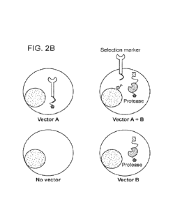
FIG. 2A schematically illustrates AND gate logic that can be performed using
the STASH
Select system. Cells which satisfy the two input requirements (expression of
construct A and
expression of construct B) result in the output surface expression of the
selection marker. FIG.
2B schematically illustrates the four possible outcomes of cells which have
been exposed to
construct A and construct B. Cells which express only construct A have a
selection marker which
is retained intracellularly. Cells which express only construct B have a
protease which is retained
intracellularly. Cells which express both construct A and construct B have a
selection marker
which is expressed on the surface of the cell which can be used for detection
and enrichment.
In certain embodiments, the methods of the present disclosure comprise
contacting a
population of cells with two or more separate expression constructs under
conditions in which the
two or more expression constructs are delivered to cells of the population of
cells. The two or
more separate expression constructs comprise a first expression construct that
encodes a fusion
protein comprising a selection marker, a protein localization tag, and a
protease cleavage site
disposed between the selection marker and the protein localization tag. The
two or more
separate expression constructs further comprise a second expression construct
that encodes a
9
CA 03214025 2023- 9- 28
WO 2022/216866
PCT/US2022/023725
protein required for cell surface expression of the selection marker. The
methods further
comprise selecting for cells exhibiting cell surface expression of the
selection marker.
The contacting step may comprise contacting the population of cells with the
two or more
separate expression constructs simultaneously, e.g., by combining the cells
and each of the two
or more separate expression constructs in a single mixture under conditions
suitable for delivery
(e.g., transfection, transduction, etc.) of each of the two or more separate
expression constructs
into cells of the population of cells. The contacting step may comprise
contacting the population
of cells with the two or more separate expression constructs sequentially,
e.g., where the
population of cells is first combined with less than each of the two or more
separate expression
constructs under expression construct delivery conditions, followed by
combining the population
of cells with the remaining two or more separate expression constructs in one
or more further
combining steps under suitable conditions.
A variety of suitable approaches and conditions for the delivery of expression
constructs
to cells are known. According to some embodiments, the two or more separate
expression
constructs are delivered to cells of the population of cells by
microinjection, transfection,
lipofection, heat-shock, electroporation, transduction, gene gun, DEAE-dextran-
mediated
transfer, and/or the like. In certain embodiments, the two or more separate
expression constructs
are introduced into cells of the population of cells by AAV transduction. The
AAV vector may
comprise ITRs from AAV2, and a serotype from any one of AAV1, AAV2, AAV3,
AAV4, AAV5,
AAV6, AAV7, AAV8, AAV9, or AAV 10. According to some embodiments, the AAV
vector
comprises ITRs from AAV2 and a serotype from AAV6. In certain embodiments, the
nucleic acid
(e.g., expression vector) encoding the CAR is introduced into the cell (e.g.,
a T cell) by lentiviral
or retroviral transduction. The lentiviral vector backbone may be derived from
HIV-1, HIV-2, visna-
maedi virus (VMV) virus, caprine arthritis-encephalitis virus (CAEV), equine
infectious anemia
virus (EIAV), feline immunodeficiency virus (FIV), bovine immune deficiency
virus (BIV), or simian
immunodeficiency virus (Sly). The lentiviral vector may be integration
competent or an integrase
deficient lentiviral vector (TDLV). In one embodiment, IDLV vectors including
an HIV-based
vector backbone (i.e., HIV cis-acting sequence elements) are employed. Non-
limiting example
approaches for the preparation of retroviral expression constructs and the
transduction of cells
with such constructs is provided in the Experimental section hereinbelow.
As used herein, an "expression construct" is a circular or linear
polynucleotide (a polymer
composed of naturally-occurring and/or non-naturally-occurring nucleotides)
comprising a region
that encodes a component of the cell selection system (e.g., a fusion protein
comprising a
selection marker, a protein localization tag, and a protease cleavage site;
and/or a protein
required for cell surface expression of the selection marker) operably linked
to a suitable
promoter, e.g., a constitutive or inducible promoter. In some embodiments,
expression of the cell
selection system component is under the control of one or more exogenous
(including
heterologous) regulatory elements, e.g., promoter, enhancer, etc., present in
the expression
CA 03214025 2023- 9- 28
WO 2022/216866
PCT/US2022/023725
construct, and operably linked to the region encoding the cell selection
system component, prior
to contacting with the population of cells. In some embodiments, expression of
the cell selection
system component may be controlled by one or more endogenous regulatory
elements, e.g.,
promoter, enhancer, etc., at or near a genomic locus into which the expression
construct is
inserted.
One or more of the two or more separate expression constructs may further
comprise one
or more regions encoding one or more proteins of interest (e.g., any of the
proteins of interest
described elsewhere herein), each operably linked to a suitable promoter,
where the promoter
may be a single shared promoter among each of the protein-encoding regions of
the expression
construct (including the cell selection system component), or at least one of
the protein-encoding
regions may be operably linked to a promoter which is not shared with any
other protein-encoding
region of the expression construct. In certain embodiments, when an expression
construct
comprises one or more protein-encoding regions in addition to the region
encoding the
component of the cell selection system, the expression construct may be
configured to allow for
polycistronic expression of two or more (e.g., each) of the protein-encoding
regions. That is, two
or more (e.g., each) of the proteins encoded by the expression construct may
be expressed as
separate proteins from the same promoter. In certain embodiments, the
expression construct
includes a ribosome skipping site to allow for polycistronic expression of two
or more (e.g., each)
of the protein-encoding regions. A non-limiting example of a suitable ribosome
skipping site which
may be incorporated into expression constructs is the P2A ribosome skipping
site from porcine
teschovirus.
The expression constructs (e.g., vectors) can be suitable for replication and
integration in
prokaryotes, eukaryotes, or both. The expression constructs may contain
functionally
appropriately oriented transcription and translation terminators, initiation
sequences, and
promoters useful for regulation of the expression of the nucleic acid encoding
the component of
the cell selection system. The expression constructs optionally contain
generic expression
cassettes containing at least one independent terminator sequence, sequences
permitting
replication of the cassette in both eukaryotes and prokaryotes, e.g., as found
in shuttle vectors,
and selection markers for both prokaryotic and eukaryotic systems.
To obtain high levels of expression of a cloned nucleic acid it is common to
construct
expression constructs which typically contain a strong promoter to direct
transcription, a ribosome
binding site for translational initiation, and a transcription/translation
terminator, each in functional
orientation to each other and to the protein-encoding sequence. Examples of
regulatory regions
suitable for this purpose in E. coli are the promoter and operator region of
the E. coli tryptophan
biosynthetic pathway, the leftward promoter of phage lambda (PO, and the L-
arabinose (araBAD)
operon. The inclusion of selection markers in DNA vectors transformed in E.
co//is also useful.
Examples of such markers include genes specifying resistance to ampicillin,
tetracycline, or
chloramphenicol. Expression systems for expressing the selection system
components are
11
CA 03214025 2023- 9- 28
WO 2022/216866
PCT/US2022/023725
available using, for example, E. coli, Bacillus sp. and Salmonella. E. coli
systems may also be
used. Nucleic acids encoding the selection system components. Transducing
cells with nucleic
acids can involve, for example, incubating lipidic microparticles containing
nucleic acids with cells
or incubating viral vectors containing nucleic acids with cells within the
host range of the vector.
The two or more expression constructs are "separate", meaning that none of the
two or
more expression constructs are part of the same polynucleotide molecule.
In certain embodiments, upon delivery of the two or more separate expression
constructs
to cells of the population of cells, one or more of the expression constructs
are episomal (e.g.,
extra-chromosomal), where by "episome" or "episomal" is meant a polynucleotide
that replicates
independently of the cell's chromosomal DNA. A non-limiting example of an
episome that may
be employed in the present methods is a plasmid.
According to some embodiments, upon delivery of the two or more separate
expression
constructs to cells of the population of cells, one or more of the expression
constructs integrates
into the genome of the cell. In certain embodiments, one or more of the
expression constructs
are adapted for site-specific integration into the genome. For example, an
expression construct
may be adapted for site-specific integration into the genome, where the site-
specific integration
inactivates a target gene within the genome of the cell. By way of example,
the site-specific
integration may knock-out the target gene by knock-in of the expression
construct. Any suitable
approach for site-specific gene editing and functional integration may be
employed. Functional
integration of an expression construct may be achieved through various means,
including through
the use of integrating vectors, including viral and non-viral vectors. In some
instances, a retroviral
vector, e.g., a lentiviral vector, may be employed. In some instances, a non-
retroviral integrating
vector may be employed. An integrating vector may be contacted with the cells
in a suitable
transduction medium, at a suitable concentration (or multiplicity of
infection), and for a suitable
time for the vector to infect the target cells, facilitating functional
integration of the expression
construct. Non-limiting examples of useful viral vectors include retroviral
vectors, lentiviral
vectors, adenoviral (Ad) vectors, adeno-associated virus (AAV) vectors, hybrid
Ad-AAV vector
systems, and the like.
Strategies for site-specific integration that find use in the methods of the
present
disclosure include those that employ homologous recombination, nonhomologous
end-joining
(NHEJ), and/or the like. Such strategies may employ a non-naturally occurring
or engineered
nuclease, including, but not limited to, zinc-ringer nucleases (ZNFs),
meganucleases,
transcription activator-like effector nucleases (TALENs)), or a CRISPR-Cas
system. Eukaryotic
cells utilize two distinct DNA repair mechanisms in response to DNA double
strand breaks
(DSBs): Homologous recombination (HR) and nonhomologous end-joining (NHEJ).
Mechanistically, HR is an error-free DNA repair mechanism because it requires
a homologous
template to repair the damaged DNA strand. Because of its homology-based
mechanism, HR
has been used as a tool to site-specifically engineer the genome. Gene
targeting by HR requires
12
CA 03214025 2023- 9- 28
WO 2022/216866
PCT/US2022/023725
the use of two homology arms that flank the transgene/target site of interest.
HR efficiency can
be increased by the introduction of DSBs at the target site using specific
rare-cutting
endonucleases. The discovery of this phenomenon prompted the development of
methods to
create site-specific DSBs in the genome of different species. Various chimeric
enzymes have
been designed for this purpose over the last decade, namely ZFNs,
meganucleases, and
TALENs. ZFNs are modular chimeric proteins that contain a ZF-based DNA binding
domain
(DBD) and a Fokl nuclease domain. DBD is usually composed of three ZF domains,
each with 3-
base pair specificity; the Fokl nuclease domain provides a DNA nicking
activity, which is targeted
by two flanking ZFNs. Owing to the modular nature of the DBD, any site in a
genome could be
targeted. TALENs are similar to ZFNs except that the DBD is derived from
transcription activator-
like effectors (TALEs). The TALE DBD is modular, and it is composed of 34-
residue repeats,
and its DNA specificity is determined by the number and order of repeats. Each
repeat binds a
single nucleotide in the target sequence through only two residues.
The methods of the present disclosure may be performed on any cell populations
of
interest. In certain embodiments, the population of cells is a population of
prokaryotic cells (e.g.,
bacteria), a population of yeast cells, a population of insect (e.g.,
drosophila) cells, a population
of amphibian (e.g., frog, e.g., Xenopus) cells, a population of plant cells,
etc. According to some
embodiments, the population of cells is a population of mammalian cells.
Mammalian cells of
interest include human cells, rodent cells, and the like. According to some
embodiments, the
population of cells is a population of peripheral blood mononuclear cells
(PBMCs). In certain
embodiments, the population of cells is a population of immune cells. The
population of immune
cells may comprise one or any combination of T cells, B cells, natural killer
(NK) cells,
macrophages, monocytes, neutrophils, dendritic cells, mast cells, basophils,
eosinophils. When
the immune cells comprise T cells, the T cells may comprise one or any
combination of naive T
cells (TN), cytotoxic T cells (Tcm), memory T cells (TmEm), T memory stem
cells (Tscm), central
memory T cells (Tcm), effector memory T cells (TEm), tissue resident memory T
cells (TRm), effector
T cells (TEFF), regulatory T cells (TREGs), helper T cells, CD4+ T cells, CD8+
T cells, virus-specific
T cells, alpha beta T cells (Tap), gamma delta T cells (To). According to some
embodiments, the
population of cells is a population of cells comprises stem cells, e.g.,
mammalian (e.g., human)
stem cells. For example, the population of cells may comprise embryonic stem
(ES) cells, adult
stem cells, hematopoietic stem cells (HSCs), induced pluripotent stem cells
(iPSCs),
mesenchymal stem cells (MSCs), neural stem cells (NSCs), or any combination
thereof.
Protein Localization Tags
As used herein, the term "protein localization tag" refers to an amino acid
sequence that
directs the cellular localization of the fusion protein comprising the
selection marker (and
optionally, any other cell selection system components expressed by the two or
more separate
expression constructs) to a particular cellular compartment. In certain
embodiments, the protein
13
CA 03214025 2023- 9- 28
WO 2022/216866
PCT/US2022/023725
localization tag is selected from an endoplasmic reticulum (ER) localization
tag, a Golgi apparatus
(Golgi) localization tag, a lysosome localization tag, a plasma membrane
localization tag, a
mitochondria localization tag, a peroxisome localization tag, a cytosolic
localization tag, and a
nuclear localization tag.
The fusion protein comprising the selection marker (and optionally, any other
cell
selection system component(s) expressed by the two or more separate expression
constructs)
may include any suitable protein localization tag for directing localization
to the desired cellular
compartment. In some embodiments, when two or more cell selection system
components
comprise a protein localization tag, the protein localization tag of each
component may direct
each component to the same cellular compartment (e.g., organelle). For
example, in certain
embodiments, when two or more cell selection system components comprise a
protein
localization tag, the protein localization tags are identical or substantially
identical to each other.
Suitable protein localization tags are known. In certain embodiments, a cell
selection
system component includes a protein localization tag in LocSigDB (a database
of protein
localization signals/tags available at genome.unmc.edu/LocSigDB/ and described
in Negi et al.
(2015) Database, Volume 2015:1-7); DBSubLoc (a database of protein subcellular
localization ¨
available at bioinfo.tsinghua.edu.cn/dbsubloc.html); LOCATE (a mammalian
protein subcellular
localization database available at locate.imb.uq.edu.au); LocDB (a protein
localization database
available at rostlab.org/services/locDB); eSLDB (a eukaryotic subcellular
localization database
available at gper.biocomp.unibo.it/esldb); and/or any other convenient
database of protein
localization tags. According to some embodiments, the protein localization tag
is located at the
N-terminus of the cell selection system component. For example, there are
naturally-occurring
N-terminal protein localization tags for type II membrane proteins (see, e.g.,
Schutz et al. (1994)
EMBO J. 13(7):1696-1705) and other proteins.
According to some embodiments, the protein localization tag is an ER
localization tag. In
certain embodiments, the ER localization tag comprises the amino acid sequence
KKMP. A non-
limiting example of an ER localization tag that may be included in a cell
selection system
component of the present disclosure is an ER localization tag comprising 85%
or greater, 90%
or greater, or 100% amino acid sequence identity to an ER localization tag
comprising, consisting
of, or present within, an amino acid sequence selected from LYKYKSRRSFIDEKKMP
(SEQ ID
NO:1); AEKDEL (SEQ ID NO:2); EQKLISEEDLKDEL (SEQ ID NO:3); GGGGSGGGGSKDEL
(SEQ ID NO:4); GGGGSGGGGSGGGGSGGGGSKDEL (SEQ ID NO:5);
GGGGSGGGGSGGGGSGGGGSAEKDEL (SEQ ID NO:6); KYKSRRSFIEEKKMP (SEQ ID
NO:7); LKYKSRRSFIEEKKMP (SEQ ID NO:8); LYKYKSRRSFIEEKKMP (SEQ ID NO:9);
LYCKYKSRRSFIEEKKMP (SEQ ID NO:10) ; LYCNKYKSRRSFIEEKKMP (SEQ ID NO:11);
LYCNKYKSRRSFIDEKKMP (SEQ ID NO:12); LYEQKLISEEDLKYKSRRSFIEEKKMP (SEQ ID
NO:13); LYCYPYDVPDYAKYKSRRSFIEEKKMP (SEQ ID NO:14); LYKKLETFKKTN (SEQ ID
NO:15); LYEQKLISEEDLKKLETFKKTN (SEQ ID NO:16); LYYQRL (SEQ ID NO:17);
14
CA 03214025 2023- 9- 28
WO 2022/216866
PCT/US2022/023725
LYEQKLISEEDLYQRL (SEQ ID NO:18); LYKRKIIAFALEGKRSKVTRRPKASDYQRL (SEQ ID
NO:19); LYRNIKCD (SEQ ID NO:20); and LYEQKLISEEDLRNIKCD (SEQ ID NO:21).
Another
example of an ER localization tag that may be included in a cell selection
system component of
the present disclosure is an ER localization tag comprising 85% or greater,
90% or greater, 95%
or greater, or 100% amino acid sequence identity to an ER localization tag
comprising, consisting
of, or present within, an amino acid sequence selected from:
PKKKQQKDSLINLKIQKENPKVVNEINIEDLCLIKAAYCRCWRSKTFPACDGSHNKHNE
LTGDNVGPLILKKKEV (SEQ ID NO:22);
QMRHLKSFFEAKKLV (SEQ ID NO:23);
AYRQRQHQDMPAPRPPGPRPAPPQQEGPPEQQPPQ (SEQ ID NO:24);
HMKEKEKSD (SEQ ID NO:25);
CFRKLAKTGKKKKRD (SEQ ID NO:26);
KCCAYGYRKCLGKKGRVKKAHKSKTH (SEQ ID NO:27);
YLSTCKDSKKKAE (SEQ ID NO:28);
RLTTDVDPDLDQDED (SEQ ID NO:29);
KYKSRRSFIDEKKMP (SEQ ID NO:30);
MTGCCGCCCGCFGIIPLMSKCGKKSSYYTTFDNDVVIEQYRPKKSV (SEQ ID NO:31);
NRSPRNRKPRRE (SEQ ID NO:32);
LYKYKSRRSFIEEKKMP (SEQ ID NO:9);
TKVLKGKKLSLPA (SEQ ID NO:33);
KSNRHKDGFHRLRGHHDEYEDEIRMMSTGSKKSLLSHEFQDETDTEETLYSSKH
(SEQ ID NO:34); AND
KCGKKSSYYTTFDNDVVIEQYRPKKSV (SEQ ID NO:35).
In certain embodiments, the protein localization tag is a Golgi localization
tag. A non-
limiting example of a Golgi localization tag that may be included in a cell
selection system
component of the present disclosure is a Golgi localization tag comprising the
amino acid
sequence YQRL (SEQ ID NO:36).
According to some embodiments, the protein localization tag is a lysosome
localization
tag. A non-limiting example of a lysosome localization tag that may be
included in a cell selection
system component of the present disclosure is a lysosome localization tag
comprising the amino
acid sequence KFERQ (SEQ ID NO:37).
Protease Cleavage Sites and Proteases
As described above, the first expression construct encodes a fusion protein
comprising a
protease cleavage site disposed between the selection marker and the protein
localization tag.
CA 03214025 2023- 9- 28
WO 2022/216866
PCT/US2022/023725
The term "cleavage site" refers to the bond (e.g., a scissile bond) cleaved by
an agent, e.g., a
protease. A cleavage site for a protease includes the specific amino acid
sequence recognized
by the protease during proteolytic cleavage and may include surrounding amino
acids (e.g., from
one to six amino acids) on either side of the scissile bond, which bind to the
active site of the
protease and are needed for recognition as a substrate. In some embodiments,
the cleavage
site is provided as a cleavable linker, where "cleavable linker" refers to a
linker including the
protease cleavage site. A cleavable linker is typically cleavable under
physiological conditions.
According to some embodiments, the protease cleavage site is a viral protease
cleavage
site. Non-limiting examples of viral protease cleavage sites include cleavage
sites for potyviral
family proteases. Potyviral family proteases of interest include Tobacco Etch
Virus (TEV)
protease, plum pox virus protease (PPVp), soybean mosaic virus protease
(SbMVp), sunflower
mild mosaic virus protease (SuMMVp), tobacco vein mottling virus protease
(TVMVp), and West
Nile virus protease (WNVp). In certain embodiments, the viral protease
cleavage site is a TEV
protease cleavage site. The amino acid sequence of an example TEV protease
cleavage site is
ENLYFQS. The amino acid sequence of an example TEV protease is the following:
GESLFKGPRDYNPISSTICHLTNESDGHTTSLYGIGFGPFIITNKHLFRRNNGILL
VQSLHGVFKVKNTTTLQQHLI DG RDM II I RMPKDFPPFPQKLKFREPQREERICL
VTIN FQTKSMSSMVSDTSCTFPSSDG I FWKHWIQTKDGQCGSPLVSTRDGFIV
GI HSASN FTNTNNYFTSVPKN FM ELLTNQEAQQWVSGWRLNADSVLWGG HK
VFMVKPEEPFQPVKEATQLMN (SEQ ID NO:38)
In some embodiments, the protease is a TEV protease comprising the amino acid
sequence set forth above, or is a functional (proteolytic) variant thereof
having 70% or greater,
75% or greater, 80% or greater, 85% or greater, 90% or greater, 95% or
greater, or 99% or
greater amino acid sequence identity to such a sequence, and/or a functional
(proteolytic)
fragment thereof such as a fragment having a length of from 100 to 125, 125 to
150, 150 to 175,
175 to 200, 200 to 225, or from 225 to 235 amino acids. Such a protease may be
provided by
two or more (e.g., two) complementary fragments of the protease, wherein the
two or more (e.g.,
two) complementary fragments form an active protease complex.
According to some embodiments, the viral protease cleavage site is for an HCV
protease.
In certain embodiments, the viral protease cleavage site is for a viral
protease derived from HCV
nonstructural protein 3 (NS3). NS3 consists of an N-terminal serine protease
domain and a C-
terminal helicase domain. By "derived from HCV NS3" is meant the protease is
the serine
protease domain of HCV NS3 or a proteolytically active variant thereof capable
of cleaving a
cleavage site for the serine protease domain of HCV NS3. The protease domain
of NS3 forms a
heterodimer with the HCV nonstructural protein 4A (NS4A), which activates
proteolytic activity.
A protease derived from HCV NS3 may include the entire NS3 protein or a
proteolytically active
fragment thereof, and may further include a cofactor polypeptide, such as a
cofactor polypeptide
16
CA 03214025 2023- 9- 28
WO 2022/216866
PCT/US2022/023725
derived from HCV nonstructural protein 4A (NS4A), e.g., an activating NS4A
region. NS3
protease is highly selective and can be inhibited by a number of non-toxic,
cell-permeable drugs,
which are currently available for use in humans. NS3 protease inhibitors that
may be employed
include, but are not limited to, simeprevir, danoprevir, asunaprevir,
ciluprevir, boceprevir,
sovaprevir, paritaprevir, telaprevir, grazoprevir, and any combination
thereof. Non-limiting
examples of proteases derived from HCV NS3 are provided below.
Example Proteases Derived from HCV NS3
APITAYAQQTRGLLGCI ITSLTGRDKNOVEGEVQ1VSTATQTFLATCINGVCWAVYHGA
GTRTIASPKG PVIQMYTNVDQDLVGWPAPQGSRSLTPCTCGSSDLYLVTRHADVI PVRRRGD
SRGSLLSPRPISYLKGSSGG PLLCPAG HAVGLFRAAVCTRGVAKAVDFI PVENLETTMRSPVFT
D (SEQ ID NO:39)
APITAYAQQTRGLLGCI ITSLTGRDKNQVEGEVQ1MSTATQTFLATC1 NGVCWTVYHGA
GTRTIASPKG PVIQMYTNVDQDLVGWPAPQGSRSLTPCTCGSSDLYLVTRHADVI PVRRRGD
GRGSLLSPRPISYLKGSSGGPLLCPAG HAVGLFRAAVCTRGVAKAVDFI PVENLETTMRSPVF
TD (SEQ ID NO:40)
APITAYAQQTRGLLGC1ITSLTGRDKNOVEGEVQ1VSTATQTFLATCINGVCWTVYHGA
GTRTIASPKG PVIQMYTNVDQDLVGWPAPQGSRSLTPCTCGSSDLYLVTRHADVI PVRRRGD
SRGSLLSPRPISYLKGSSGG PLLCPAG HAVGLFRAAVCTRGVAKAVDFI PVENLETTMRSPVFT
D (SEQ ID NO:41)
In some embodiments, the protease comprises one of the sequences set forth
above, or
is a functional (proteolytic) variant thereof having 70% or greater, 75% or
greater, 80% or greater,
85% or greater, 90% or greater, 95% or greater, or 99% or greater amino acid
sequence identity
to one of such sequences, and/or a functional (proteolytic) fragment thereof
such as a fragment
having a length of from 100 to 185, 120 to 185, 140 to 185, 160 to 185, 170 to
185, from 180 to
185, from 182 to 185, or from 184 to 185 amino acids.
In some embodiments, the protease cleavage site is a viral protease cleavage
site. For
example, when a protease derived from HCV NS3 is employed, the cleavage site
should
comprise an NS3 protease cleavage site. An NS3 protease cleavage site may
include the four
junctions between nonstructural (NS) proteins of the HCV polyprotein normally
cleaved by the
NS3 protease during HCV infection, including the NS3/NS4A, NS4A/NS4B,
NS4B/NS5A, and
NS5A/NS5B junction cleavage sites. For a description of NS3 protease and
representative
sequences of its cleavage sites for various strains of HCV, see, e.g.,
Hepatitis C Viruses:
Genomes and Molecular Biology (S.L. Tan ed., Taylor & Francis, 2006), Chapter
6, pp. 163-206;
the disclosure of which is incorporated herein by reference in its entirety.
In some embodiments, the protease is derived from HCV NS3 and engineered to
include
one or more amino acid substitutions relative to an HCV NS3 protease amino
acid sequence set
forth above. For example, the protease may include a substitution at the
position corresponding
17
CA 03214025 2023- 9- 28
WO 2022/216866 PCT/US2022/023725
to position 54 of the amino
acid sequence
APITAYAQQTRGLLGGIITSLTGRDKNQVEGEVQ1VSTATQTFLATCINGVCWAVYHGAGTRTIA
SPKGPVIQMYTNVDQDLVGWPAPQGSRSLTPCTCGSSDLYLVTRHADVI PVRRRG DS RGSLL
SPRPISYLKGSSGGPLLCPAGHAVGLFRAAVCTRGVAKAVDFI PVENLETTMRSPVFTD (SEQ
ID NO:39). In some embodiments, such a substitution is a threonine to alanine
substitution.
NS3 nucleic acid and protein sequences may be derived from HCV, including any
isolate
of HCV having any genotype (e.g., genotypes 1-7) or subtype. A number of NS3
nucleic acid
and protein sequences are known and described, e.g., in USSN 15/737,712, the
disclosure of
which is incorporated herein by reference in their entirety for all purposes.
Additional
representative NS3 sequences are listed in the National Center for
Biotechnology Information
(NCB!) database. See, for example, NCB! entries: Accession Nos. YP_001491553,
YP_001469631, YP_001469632, NP_803144, NP_671491, YP_001469634, YP_001469630,
YP_001469633, ADA68311, ADA68307, AFP99000, AFP98987, ADA68322, AFP99033,
ADA68330, AFP99056, AFP99041, CBF60982, CBF60817, AHH29575, A1Z00747,
A1Z00744,
AB136969, ABN05226, KF516075, KF516074, KF516056, AB826684, AB826683,
JX171009,
JX171008, JX171000, EU847455, EF154714, GU085487, JX171065, and JX171063; all
of which
sequences are herein incorporated by reference. Any of these sequences or
functional variants
thereof having 70% or greater, 75% or greater, 80% or greater, 85% or greater,
90% or greater,
91% or greater, 92% or greater, 93% or greater, 94% or greater, 95% or
greater, 96% or greater,
97% or greater, 98% or greater, or 99% or greater amino acid sequence identity
to any one of
these sequences, or proteolytic fragments thereof, may be employed.
NS4A nucleic acid and protein sequences may be derived from HCV, including any
isolate
of HCV having any genotype (e.g., seven genotypes 1-7) or subtype. A number of
NS4A nucleic
acid and protein sequences are known. Representative NS4A sequences are listed
in the
National Center for Biotechnology Information (NCB!) database. See, for
example, NCB! entries:
Accession Nos. NP_751925, YP_001491554, GU945462, H0822054, FJ932208,
FJ932207,
FJ932205, and FJ932199; all of which sequences (as entered by the date of
filing of this
application) are herein incorporated by reference. Any of these sequences or
functional variants
thereof having 70% or greater, 75% or greater, 80% or greater, 85% or greater,
90% or greater,
95% or greater, or 99% or greater amino acid sequence identity to any one of
these sequences,
or proteolytic fragments thereof, may be employed.
HCV polyprotein nucleic acid and protein sequences may be derived from HCV,
including
any isolate of HCV having any genotype (e.g., genotypes 1-7) or subtype. A
number of HCV
polyprotein nucleic acid and protein sequences are known. Representative HCV
polyprotein
sequences are listed in the National Center for Biotechnology Information
(NCB!) database. See,
for example, NCB! entries: Accession Nos. YP_001469631, NP_671491,
YP_001469633,
YP 001469630, YP 001469634, YP 001469632, NC 009824, NC 004102, NC 009825,
NC_009827, NC_009823, NC_009826, and EF108306; all of which sequences (as
entered by
18
CA 03214025 2023- 9- 28
WO 2022/216866
PCT/US2022/023725
the date of filing of this application) are herein incorporated by reference.
Any of these sequences
or functional variants thereof having 70% or greater, 75% or greater, 80% or
greater, 85% or
greater, 90% or greater, 95% or greater, or 99% or greater amino acid sequence
identity to any
one of these sequences, or proteolytic fragments thereof, may be employed.
In some embodiments, the protease is derived from HCV NS3 and the cleavage
site
includes an NS3 protease cleavage site. An NS3 protease cleavage site may
include the HCV
polyprotein NS3/NS4A, NS4A/NS4B, NS4B/NS5A, and NS5A/NS5B junction cleavage
sites.
Representative HCV NS4A/4B protease cleavage sites include DEMEECSQH and
DEMEECSQH. Representative HCV NS5A/5B protease cleavage sites include
EDVVPCSMG
and EDVVPCSMGS. A representative NS4B/5A protease cleavage site is
ECTTPCSGSWL.
Additional NS3 protease cleavage sites that may be included in a recombinant
polypeptide of the
present disclosure include those described in Shiryaev et al. (2012) PLoS One
7(4):e35759.
In certain embodiments, the protease cleavage site is a human protease
cleavage site.
Non-limiting examples of human protease cleavage sites include cleavages sites
for a human
kallikrein (KLK) protease, human enterokinase protease, human thrombin, a
human matrix
metalloprotease (MM P), human urokinase-type plasminogen activator receptor (u
PAR), human
plasmin, or human cathepsin. According to some embodiments, the protease
cleavage site is a
cleavage site for a human kallikrein (KLK) protease, non-limiting examples of
which include
human KLK3 (UniProtKB - 0546G3), human KLK4 (UniProtKB - 09Y5K2), human KLK6
(UniProtKB - 092876), human KLK8 (UniProtKB - 060259), human KLK11 (UniProtKB -
Q9UBX7), human KLK13 (UniProtKB - Q9UKR3), human KLK14 (UniProtKB - 09P0G3),
and
human KLK15 (UniProtKB - 09H2R5). Data demonstrating the utility of human KLK
proteases
and corresponding cleavage sites in the STASH Select system is provided in the
Experimental
section herein. Any of these sequences or functional variants thereof having
70% or greater, 75%
or greater, 80% or greater, 85% or greater, 90% or greater, 91% or greater,
92% or greater, 93%
or greater, 94% or greater, 95% or greater, 96% or greater, 97% or greater,
98% or greater, or
99% or greater amino acid sequence identity to any one of these sequences, or
proteolytic
fragments thereof, may be employed.
In certain embodiments, the protease cleavage site is a protease cleavage site
for a
human protease selected from acrosin (ACR), AGBL carboxypeptidase 1 (AGBL1),
AGBL
carboxypeptidase 2 (AGBL2), AGBL carboxypeptidase 3 (AGBL3), AGBL
carboxypeptidase 4
(AGBL4), AGBL carboxypeptidase 5 (AGBL5), ATP/GTP binding carboxypeptidase 1
(AGTPBP1), asparaginase and isoaspartyl peptidase 1 (ASRGL1), astacin like
metalloendopeptidase (ASTL), ATP23 metallopeptidase and ATP synthase assembly
factor
homolog (ATP23), ataxin 3 (ATXN3), ataxin 3 like (ATXN3L), azurocidin 1
(AZU1), beta-
secretase 1 (BACE1), beta-secretase 2 (BACE2), bone morphogenetic protein 1
(BMP1),
BRCA1/BRCA2-containing complex subunit 3 (BRCC3), calpain 14 (CAPN14), calpain
3
(CAPN3), caspase recruitment domain family member 8 (CARDS), caspase 4
(CASP4),
19
CA 03214025 2023- 9- 28
WO 2022/216866
PCT/US2022/023725
chymotrypsin like elastase 1 (CELA1), chymotrypsin like elastase 2A (CELA2A),
chymotrypsin
like elastase 2B (CELA2B), chymotrypsin like elastase 3A (CELA3A),
chymotrypsin like elastase
3B (CELA3B), CUGBP Elav-like family member 3 (CELF3), CUGBP Elav-like family
member 4
(CELF4), CUGBP Elav-like family member 5 (CELF5), CUGBP Elav-like family
member 6
(CELF6), cell growth regulator with EF-hand domain 1 (CGREF1), charged
multivesicular body
protein 3 (CHMP3), CLN5 intracellular trafficking protein (CLN5), chymase 1
(CMA1), collectin
subfamily member 11 (COLEC11), COP9 signalosome subunit 5 (COPS5), corin,
serine
peptidase (CORIN), carboxypeptidase A4 (CPA4), carboxypeptidase vitellogenic
like (CPVL),
cystatin SN (CST1), cystatin 11 (CST11), cystatin C (CST3), cystatin S (CST4),
cystatin D
(CST5), cystatin E/M (CST6), cystatin 8 (CST8), cystatin 9 (CST9), cystatin
like 1 (CSTL1),
chymotrypsinogen B2 (CTRB2), chymotrypsin like (CTRL), cathepsin L (CTSL), DNA
damage
inducible 1 homolog 2 (DDI2), DAP3 binding cell death enhancer 1 (DELE1),
adipsin (DF),
dickkopf WNT signaling pathway inhibitor 2 (DKK2), dickkopf WNT signaling
pathway inhibitor 4
(DKK4), dipeptidase 1 (DPEP1), dipeptidyl peptidase 3 (DPP3), dipeptidyl
peptidase 9 (DPP9),
FAM111 trypsin like peptidase A (FAM111A), ficolin 1 (FCN1), ficolin 2 (FCN2),
ficolin 3 (FCN3),
G3BP stress granule assembly factor 1 (G3BP1), hepsin (HPN), HtrA serine
peptidase 1
(HTRA1), insulin degrading enzyme (IDE), inner mitochondrial membrane
peptidase subunit 2
(IMMP2L), jumonji domain containing 7 (JMJD7), Josephin domain containing 2
(JOSD2),
kallikrein 1 (KLK1), kallikrein related peptidase 10 (KLK10), kallikrein
related peptidase 11
(KLK11), kallikrein related peptidase 12 (KLK12), kallikrein related peptidase
13 (KLK13),
kallikrein related peptidase 14 (KLK14), kallikrein related peptidase 15
(KLK15), kallikrein related
peptidase 2 (KLK2), kallikrein related peptidase 3 (KLK3), kallikrein related
peptidase 4 (KLK4),
kallikrein related peptidase 5 (KLK5), kallikrein related peptidase 6 (KLK6),
kallikrein related
peptidase 7 (KLK7), kallikrein related peptidase 8 (KLK8), kallikrein related
peptidase 9 (KLK9),
kallikrein pseudogene 1 (KLKP1), lipocalin 2 (LCN2), legumain (LGMN),
leishmanolysin like
peptidase (LMLN), MASI proto-oncogene like, G protein-coupled receptor
(MAS1L), MBL
associated serine protease 1 (MASP1), MBL associated serine protease 2
(MASP2), mannose
binding lectin 2 (MBL2), matrix metallopeptidase 10 (MMP10), matrix
metallopeptidase 11
(MMP11), matrix metallopeptidase 13 (MMP13), matrix metallopeptidase 16
(MMP16), matrix
metallopeptidase 2 (MMP2), napsin A aspartic peptidase (NAPSA), neurolysin
(NLN), NLR family
CARD domain containing 4 (NLRC4), NLR family pyrin domain containing 1
(NLRP1),
aminopeptidase puromycin sensitive (NPEPPS), opiorphin prepropeptide (OPRPN),
OTU
deubiquitinase, ubiquitin aldehyde binding 2 (OTUB2), poly (ADP-ribose)
polymerase family
member 9 (PARP9), proprotein convertase subtilisin/kexin type 1 (PCSK1),
proprotein
convertase subtilisin/kexin type 1 inhibitor (PCSK1N), proprotein convertase
subtilisin/kexin type
2 (PCSK2), proprotein convertase subtilisin/kexin type 4 (PCSK4), proprotein
convertase
subtilisin/kexin type 5 (PCSK5), proprotein convertase subtilisin/kexin type 6
(PCSK6), proprotein
convertase subtilisin/kexin type 7 (PCSK7), proprotein convertase
subtilisin/kexin type 9
CA 03214025 2023- 9- 28
WO 2022/216866
PCT/US2022/023725
(PCSK9), platelet derived growth factor C (PDGFC), pepsinogen A3 (PGA3),
pepsinogen A4
(PGA4), pepsinogen A5 (PGA5), pyroglutamyl-peptidase I like (PGPEP1L), PTEN
induced
kinase 1 (PINK1), prolyl endopeptidase like (PREPL), parkin RBR E3 ubiquitin
protein ligase
(PRKN), serine protease gene group (PRSS), serine protease 2 (PRSS2), serine
protease 21
(PRSS21), serine protease 22 (PRSS22), serine protease 23 (PRSS23), serine
protease 27
(PRSS27), serine protease 33 (PRSS33), serine protease 46, pseudogene
(PRSS46P), serine
protease 55 (PRSS55), serine protease 8 (PRSS8), proteinase 3 (PRTN3),
presenilin 2 (PSEN2),
PYD and CARD domain containing (PYCARD), retinoic acid receptor responder 1
(RARRES1),
ring finger and FYVE like domain containing E3 ubiquitin protein ligase
(RFFL), rhomboid like 2
(RHBDL2), SEC11 homolog A, signal peptidase complex subunit (SEC11A), SEC11
homolog B,
signal peptidase complex subunit (SEC11B ), SEC11 homolog C, signal peptidase
complex
subunit (SEC11BC), SUMO peptidase family member, NEDD8 specific (SENP8), SET
nuclear
proto-oncogene (SET), synaptosome associated protein 25 (SNAP25), secreted
phosphoprotein
2 (SPP2), small proline rich protein 3 (SPRR3), spleen associated tyrosine
kinase (SYK),
transcription factor EB (TFEB), transglutaminase 2 (TGM2), toll like receptor
adaptor molecule 1
(TICAM1), tubulointerstitial nephritis antigen like 1 (TINAGL1), transmembrane
serine protease
11D (TMPRSS11D), transmembrane serine protease 11E (TMPRSS11E), transmembrane
serine protease 4 (TMPRSS4), transmembrane serine protease 5 (TMPRSS5),
transmembrane
serine protease 6 (TMPRSS6), transmembrane serine protease 7 (TMPRSS7), TNF
receptor
superfamily member 10a (INFRSF10A), tryptase alpha/beta 1 (TPSAB1), tryptase
beta 2
(TPSB2), tryptase delta 1 (TPSD1), tryptase gamma 1 (TPSG1), tryptase
pseudogene 2
(TPSP2), tyrosylprotein sulfotransferase 1 (TPST1), tyrosylprotein
sulfotransferase 2 (TPST2),
tyrosylprotein sulfotransferase 2 pseudogene 1 (TPST2P1), thyrotropin
releasing hormone
degrading enzyme (TRHDE), thyroid hormone receptor interactor 4 (TRIP4),
ubiquitin C-terminal
hydrolase L1 (UCHL1), ubiquitin specific peptidase 27 X-linked (USP27X),
vasohibin 2 (VASH2),
valosin containing protein (VCP), and WAP four-disulfide core domain 1
(WFDC1).
In some embodiments, the protease is highly selective for the cleavage site.
Additionally,
the protease activity may be capable of inhibition by known small molecule
inhibitors that are cell-
permeable and not toxic to the cell or individual under study or treatment.
For a discussion of
proteases, see, e.g., V. Y. H. Hook, Proteolytic and cellular mechanisms in
prohormone and
proprotein processing, RG Landes Company, Austin, Tex., USA (1998); N. M.
Hooper et al.,
Biochem. J. 321: 265-279 (1997); Z. Werb, Cell 91: 439-442 (1997); T. G.
Wolfsberg et al., J.
Cell Biol. 131: 275-278 (1995); T. Berg et al., Biochem. J. 307: 313-326
(1995); M. J. Smyth and
J. A. Trapani, Immunology Today 16: 202-206 (1995); R. V. Talanian et al., J.
Biol. Chem. 272:
9677-9682 (1997); and N. A. Thornberry et al., J. Biol. Chem. 272: 17907-17911
(1997), the
disclosures of which are incorporated herein by reference in their entireties
for all purposes. In
some embodiments, the protease employed is a sequence-specific non-human
protease for
which FDA-approved pharmacological inhibitors are available.
21
CA 03214025 2023- 9- 28
WO 2022/216866
PCT/US2022/023725
Any of the proteases employed according to the methods of the present
disclosure,
including any of the proteases described above, may be provided by two or more
(e.g., two)
complementary fragments of the protease, where the two or more (e.g., two)
complementary
fragments form an active protease complex. A protease may be provided by two
or more (e.g.,
two) complementary fragments of the protease, e.g., in order to increase the
number of separate
expression constructs required for cell surface expression of the selection
marker.
Membrane Association Domains, Hinge Domains and Dimerization Domains
Any of the cell selection system components of the present disclosure may
comprise a
membrane association domain. Non-limiting examples of membrane association
domains
include transmembrane domains. A transmembrane (Tm) domain may be derived
either from a
natural, synthetic, semi-synthetic, or recombinant source. In some
embodiments, the Tm domain
is derived from (e.g., includes at least the transmembrane region(s) or a
functional portion
thereof) of the alpha or beta chain of 0D35, CD3, CD3y, CD3O, CD4, CD5, CD8a,
CD9, CD16,
0D22, 0D27, 0D28, 0D33, 0D37, 0D45, CD64, CD80, 0D86, 0D134, 0D137, 0D152,
CD154,
or PD-1. In certain embodiments, the transmembrane domain is a CD8a
transmembrane domain.
According to some embodiments, the transmembrane domain is a 0D28
transmembrane
domain. Non-limiting examples of transmembrane domains that may be included in
one or more
(e.g., each) of the cell selection system components are a transmembrane
domain comprising
80% or greater, 85% or greater, 90% or greater, 95% or greater, or 100% amino
acid sequence
identity to a transmembrane domain comprising, consisting of, or present
within, an amino acid
sequence selected from WLRLLPFLGVLALLGYLAVRPFL (SEQ ID NO:42);
VLWWSIAQTVILILTGIW (SEQ ID NO:43); LGPEWDLYLMTIIALLLGTVI (SEQ ID NO:44);
YYASAFSMMLGLFIFSIVFL (SEQ ID NO:45); IAFLLACVATMIFMITKCCLF (SEQ ID NO:46);
VIGFLLAVVLTVAFITF (SEQ ID NO:47); GLFLSAFLLLGLFKALGWAAV (SEQ ID NO:48);
VGLVLAAILALLLAFYAFFYL (SEQ ID NO:49); TFCSTALLITALALVCTLLYL (SEQ ID NO:50);
WYVWLAIFFAIIIFILILGWVLL (SEQ ID NO:51); WLWVVYILT VALPVFLVILFC (SEC ID NO:52);
IYIWAPLAGTCGVLLLSLVITLYC (SEQ ID NO:53); and FWVLVVVGGVLACYSLLVTVAFIIFWV
(SEQ ID NO:54).
Any of the cell selection system components of the present disclosure may
comprise a
hinge domain, e.g., a CD8a hinge domain, a CD28 hinge domain, or the like.
Exemplary amino acid sequences of transmembrane domains and hinge domains that
may be included in one or more (e.g., each) of the cell selection system
components are provided
herein.
Non-limiting examples of membrane association domains also include post-
translational
modifications that tether the cell selection system component to a membrane.
That is, the cell
selection system component may comprise a post-translationally added membrane-
tethering
domain. By "membrane-tethering domain" is meant a domain (e.g., moiety)
capable of stably
22
CA 03214025 2023- 9- 28
WO 2022/216866
PCT/US2022/023725
associating with a membrane (e.g., ER membrane) of the cell. Suitable membrane-
tethering
domains include, but are not limited to, post-translational modifications such
as palmitoylation,
myristoylation, prenylation, a glycosylphosphatidylinositol (GPI) anchor, and
the like.
In some embodiments, when two or more cell selection system components
comprise a
membrane association domain (e.g., transmembrane domain), the membrane
association
domain of each component may be identical or substantially identical to each
other.
In some embodiments, in order to increase the number of separate expression
constructs
required for cell surface expression of the selection marker, two or more cell
selection system
components each comprise a dimerization domain, where dimerization of the cell
selection
system components is required for cell surface expression of the selection
marker. Examples of
cell selection system configurations that employ one or more pairs of
dimerization domains are
described elsewhere. Non-limiting examples of dimerization domains that may be
employed
include domains comprising a coiled coil structure. When the dimerization
domain comprises a
coiled coil structure, in some embodiments, the dimerization domain comprises
a leucine zipper
domain.
Genetic Modifications ¨ Proteins of Interest and Gene Inactivations
The two or more separate expression constructs may each provide a genetic
modification
to the cells to which the two or more separate expression constructs are
delivered. Non-limiting
examples of genetic modifications include providing a region of the expression
construct that
encodes a protein of interest. Non-limiting examples of proteins of interest
include a receptor, a
ligand, a transcription factor, an antibody, a bispecific 1-cell engager
(BITE), an enzyme, a
cytokine, a chemokine, a toxin, a protein conferring resistance to cell
exhaustion, and a suicide
switch protein.
In some embodiments, a protein of interest further encoded by one or more
expression
constructs of the two or more separate expression constructs is a receptor.
For example, one or
more expression constructs of the two or more separate expression constructs
may encode a
receptor independently selected from a chimeric antigen receptor (CAR), a T
cell receptor (TCR)
such as a recombinant TCR, a chimeric cytokine receptor (CCR), a chimeric
chemokine receptor,
a synthetic notch receptor (synNotch), a Modular Extracellular Sensor
Architecture (MESA)
receptor, a Tango receptor, a ChaCha receptor, a generalized extracellular
molecule sensor
(GEMS) receptor, a growth factor receptor, a cytokine receptor, a chemokine
receptor, a switch
receptor, an adhesion molecule, an integrin, an inhibitory receptor, a
stimulatory receptor, an
immunoreceptor tyrosine-based activation motif (ITAM)-containing receptor, an
immunoreceptor
tyrosine-based inhibition motif (ITIM)-containing receptor, a hormone
receptor, a receptor
tyrosine kinase, an immune receptor such as CD28, CD80, ICOS, CTLA4, PD1, PD-
L1, BTLA,
HVEM, CD27, 4-1 BB, 4-1 BBL, 0X40, OX4OL, DR3, GITR, CD30, SLAM, CD2, 264,
TIM1, TIM2,
TIM3, TIGIT, 0D226, CD160, LAG3, LAI R1, 67-1, 137-H1, and 67-H3, a type I
cytokine receptor
23
CA 03214025 2023- 9- 28
WO 2022/216866
PCT/US2022/023725
such as Interleukin-1 receptor, Interleukin-2 receptor, Interleukin-3
receptor, Interleukin-4
receptor, Interleukin-5 receptor, Interleukin-6 receptor, Interleukin-7
receptor, Interleukin-9
receptor, Interleukin-11 receptor, Interleukin-12 receptor, Interleukin-13
receptor, Interleukin-15
receptor, Interleukin-18 receptor, Interleukin-21 receptor, Interleukin-23
receptor, Interleukin-27
receptor, Erythropoietin receptor, GM-CSF receptor, G-CSF receptor, Growth
hormone receptor,
Pro!actin receptor, Leptin receptor, Oncostatin M receptor, Leukemia
inhibitory factor, a type II
cytokine receptor such as interferon-alpha/beta receptor, interferon-gamma
receptor, Interferon
type III receptor, Interleukin-10 receptor, I nterleukin-20 receptor,
Interleukin-22 receptor,
Interleukin-28 receptor, a receptor in the tumor necrosis factor receptor
superfamily such as
Tumor necrosis factor receptor 2 (16), Tumor necrosis factor receptor 1,
Lymphotoxin beta
receptor, 0X40, CD40, Fas receptor, Decoy receptor 3, CD27, CD30, 4-1BB, Decoy
receptor 2,
Decoy receptor 1, Death receptor 5, Death receptor 4, RANK, Osteoprotegerin,
TWEAK
receptor, TACI, BAFF receptor, Herpesvirus entry mediator, Nerve growth factor
receptor, B-cell
maturation antigen, Glucocorticoid-induced TN FR-related, TROY, Death receptor
6, Death
receptor 3, Ectodysplasin A2 receptor, a chemokine receptor such as CCR1,
CCR2, CCR3,
CCR4, CCR5, CCR6, CCR7, CCR8, CCR9, CCR10, CXCR1, CXCR2, CXCR3, CXCR4, CXCR5,
CXCR6 , CX3CR1, XCR1, ACKR1, ACKR2, ACKR3 , ACKR4, CCRL2, a receptor in the
epidermal growth factor receptor (EGFR) family, a receptor in the fibroblast
growth factor receptor
(FGFR) family, a receptor in the vascular endothelial growth factor receptor
(VEGFR) family, a
receptor in the rearranged during transfection (RET) receptor family, a
receptor in the Eph
receptor family, a receptor that can induce cell differentiation (e.g., a
Notch receptor), a cell
adhesion molecule (CAM), an adhesion receptor such as integrin receptor,
cadherin, selectin,
and a receptor in the discoidin domain receptor (DDR) family, transforming
growth factor beta
receptor 1, and transforming growth factor beta receptor 2. In some
embodiments, such a
receptor is an immune cell receptor selected from a T cell receptor, a B cell
receptor, a natural
killer (NK) cell receptor, a macrophage receptor, a monocyte receptor, a
neutrophil receptor, a
dendritic cell receptor, a mast cell receptor, a basophil receptor, and an
eosinophil receptor.
According to some embodiments, one or more expression constructs of the two or
more
separate expression constructs may encode a CAR. When two or more separate
expression
constructs encode a CAR, the CAR may be the same CAR, or the two or more
separate
expression constructs may encode two or more different CARs. In certain
embodiments, when
the protein of interest is a CAR, the extracellular binding domain of the CAR
comprises a single
chain antibody. The single-chain antibody may be a monoclonal single-chain
antibody, a chimeric
single-chain antibody, a humanized single-chain antibody, a fully human single-
chain antibody,
and/or the like. In one non-limiting example, the single chain antibody is a
single chain variable
fragment (scFv). Suitable CAR extracellular binding domains include those
described in
Labanieh et al. (2018) Nature Biomedical Engineering 2:377-391. In some
embodiments, the
extracellular binding domain of the CAR is a single-chain version (e.g., an
scFv version) of an
24
CA 03214025 2023- 9- 28
WO 2022/216866
PCT/US2022/023725
antibody approved by the United States Food and Drug Administration and/or the
European
Medicines Agency (EMA) for use as a therapeutic antibody, e.g., for inducing
antibody-dependent
cellular cytotoxicity (ADCC) of certain disease-associated cells in a patient,
etc. Non-limiting
examples of single-chain antibodies which may be employed when the protein of
interest is a
CAR include single-chain versions (e.g., scFv versions) of Adecatumumab,
Ascrinvacumab,
Cixutumumab, Conatumumab, Daratumumab, Drozitumab, Duligotumab, Durvalumab,
Dusigitumab, Enfortumab, Enoticumab, Figitumumab, Ganitumab, Glembatumumab,
Intetumumab, Ipilimumab, Iratumumab, Icrucumab, Lexatumumab, Lucatumumab,
Mapatumumab, Narnatumab, Necitumumab, Nesvacumab, Ofatumumab, Olaratumab,
Panitumumab, Patritumab, Pritumumab, Radretumab, Ramucirumab, Rilotumumab,
Robatumumab, Seribantumab, Tarextumab, Teprotumumab, Tovetumab, Vantictumab,
Vesencumab, Votumumab, Zalutumumab, Flanvotumab, Altumomab, Anatumomab,
Arcitumomab, Bectumomab, Blinatumomab, Detumomab, Ibritumomab, Minretumomab,
Mitumomab, Moxetumomab, Naptumomab, Nofetumomab, Pemtumomab, Pintumomab,
Racotumomab, Satumomab, Solitomab, Taplitumomab, Tenatumomab, Tositumomab,
Tremelimumab, Abagovomab, lgovomab, Oregovomab, Capromab, Edrecolomab,
Nacolomab,
Amatuximab, Bavituximab, Brentuximab, Cetuximab, Derlotuximab, Dinutuximab,
Ensituximab,
Futuxinnab, Girentuximab, Indatuxinnab, Isatuxinnab, Margetuxinnab,
Rituxinnab, Siltuxinnab,
Ublituximab, Ecromeximab, Abituzumab, Alemtuzumab, Bevacizumab, Bivatuzumab,
Brontictuzumab, Cantuzumab, Cantuzumab, Citatuzumab, Clivatuzumab,
Dacetuzumab,
Denncizunnab, Dalotuzunnab, Denintuzumab, Elotuzunnab, Ennactuzunnab,
Ennibetuzunnab,
Enoblituzumab, Etaracizumab, Farletuzumab, Ficlatuzumab, Gemtuzumab,
Imgatuzumab,
Inotuzumab, Labetuzumab, Lifastuzumab, Lintuzumab, Lorvotuzumab, Lumretuzumab,
Matuzumab, Milatuzumab, Nimotuzumab, Obinutuzumab, Ocaratuzumab, Otlertuzumab,
Onartuzumab, Oportuzumab, Parsatuzumab, Pertuzumab, Pinatuzumab, Polatuzumab,
Sibrotuzumab, Simtuzumab, Tacatuzumab, Tigatuzumab, Trastuzumab, Tucotuzumab,
Vandortuzumab, Vanucizumab, Veltuzumab, Vorsetuzumab, Sofituzumab,
Catumaxomab,
Ertumaxomab, Depatuxizumab, Ontuxizumab, Blontuvetmab, Tamtuvetmab, or an
antigen-
binding variant thereof. According to some embodiments, when the protein of
interest is a CAR,
the extracellular binding domain of the CAR specifically binds an antigen
expressed on the
surface of a cancer cell. For example, the extracellular binding domain may
bind a cancer cell-
surface antigen selected from B7-H3 (CD276), CD19, GD2, CD22, and HER2.
According to some embodiments, one or more expression constructs of the two or
more
separate expression constructs may encode an antibody. When two or more
separate
expression constructs encode an antibody, the antibody may be the same
antibody, or the two
or more separate expression constructs may encode two or more different
antibodies. The term
"antibody" (also used interchangeably with "immunoglobulin") encompasses
antibodies of any
isotype (e.g., IgG (e.g., IgG1 , IgG2, IgG3, or IgG4), IgE, IgD, IgA, IgM,
etc.), whole antibodies
CA 03214025 2023- 9- 28
WO 2022/216866
PCT/US2022/023725
(e.g., antibodies composed of a tetramer which in turn is composed of two
dimers of a heavy and
light chain polypeptide); single chain antibodies (e.g., scFv); fragments of
antibodies (e.g.,
fragments of whole or single chain antibodies) which retain specific binding
to the antigen,
including, but not limited to single chain Fv (scFv), Fab, (Fab')2, (scFv')2,
and diabodies; chimeric
antibodies; monoclonal antibodies, humanized antibodies, human antibodies; and
fusion proteins
comprising an antigen-binding portion of an antibody and a non-antibody
protein.
Immunoglobulin polypeptides include the kappa and lambda light chains and the
alpha,
gamma (IgGi, IgG2, IgG3, IgG4), delta, epsilon and mu heavy chains or
equivalents in other
species. Full-length immunoglobulin "light chains" (usually of about 25 kDa or
about 214 amino
acids) comprise a variable region of about 110 amino acids at the NH2-terminus
and a kappa or
lambda constant region at the COOH-terminus. Full-length immunoglobulin "heavy
chains" (of
about 150 kDa or about 446 amino acids), similarly comprise a variable region
(of about 116
amino acids) and one of the aforementioned heavy chain constant regions, e.g.,
gamma (of about
330 amino acids).
An immunoglobulin light or heavy chain variable region (VL and VH,
respectively) is
composed of a "framework" region (FR) interrupted by three hypervariable
regions, also called
"complementarity determining regions" or "CDRs". The extent of the framework
region and CDRs
have been defined (see, E. Kabat et al., Sequences of proteins of
immunological interest, 4th ed.
U.S. Dept. Health and Human Services, Public Health Services, Bethesda, MD
(1987); and
Lefranc et al. IMGT, the international ImMunoGeneTics information system .
Nucl. Acids Res.,
2005, 33, D593-D597)). The sequences of the framework regions of different
light or heavy chains
are relatively conserved within a species. The framework region of an
antibody, that is the
combined framework regions of the constituent light and heavy chains, serves
to position and
align the CDRs. The CDRs are primarily responsible for binding to an epitope
of an antigen. All
CDRs and framework provided by the present disclosure are defined according to
Kabat, supra,
unless otherwise indicated.
An "antibody" thus encompasses a protein having one or more polypeptides that
can be
genetically encodable, e.g., by immunoglobulin genes or fragments of
immunoglobulin genes.
The recognized immunoglobulin genes include the kappa, lambda, alpha, gamma,
delta, epsilon
and mu constant region genes, as well as myriad immunoglobulin variable region
genes. Light
chains are classified as either kappa or lambda. Heavy chains are classified
as gamma, mu,
alpha, delta, or epsilon, which in turn define the immunoglobulin classes,
IgG, IgM, IgA, IgD and
IgE, respectively. In some embodiments, an antibody of the present disclosure
is an IgG
antibody, e.g., an IgG1 antibody, such as a human IgG1 antibody. In some
embodiments, an
antibody of the present disclosure comprises a human Fc domain.
A typical immunoglobulin (antibody) structural unit is known to comprise a
tetramer. Each
tetramer is composed of two identical pairs of polypeptide chains, each pair
having one "light"
(about 25 kD) and one "heavy" chain (about 50-70 kD). The N-terminus of each
chain defines a
26
CA 03214025 2023- 9- 28
WO 2022/216866
PCT/US2022/023725
variable region of about 100 to 110 or more amino acids primarily responsible
for antigen
recognition. The terms variable light chain (VII and variable heavy chain (VH)
refer to these light
and heavy chains respectively.
Antibodies encompass intact innnnunoglobulins as well as a number of well
characterized
fragments which may be genetically encoded or produced by digestion with
various peptidases.
Thus, for example, pepsin digests an antibody below the disulfide linkages in
the hinge region to
produce F(ab)2, a dimer of Fab which itself is a light chain joined to VH-CHI
by a disulfide bond.
The F(ab).2 may be reduced under mild conditions to break the disulfide
linkage in the hinge
region thereby converting the (Fab')2 dimer into an Fab' monomer. The Fab'
monomer is
essentially a Fab with part of the hinge region (see, Fundamental Immunology,
WE. Paul, ed.,
Raven Press, N.Y. (1993), for a more detailed description of other antibody
fragments). While
various antibody fragments are defined in terms of the digestion of an intact
antibody, one of skill
will appreciate that such Fab fragments may be synthesized de novo either
chemically or by
utilizing recombinant DNA methodology. Thus, the term antibody, as used
herein, also includes
antibody fragments either produced by the modification of whole antibodies or
synthesized de
novo using recombinant DNA methodologies. In certain embodiments, an antibody
of the present
disclosure is selected from an IgG, Fv, single chain antibody, scFv, Fab,
F(ab')2, and Fab'.
One or more of the two or more separate expression constructs may encode a
protein of
interest that finds use in the context of cell therapy (e.g., a cell-based
cancer therapy), non-limiting
examples of which include a therapy comprising administration of therapeutic
immune cells such
as T cells (e.g., CAR T cells, T cells that express an engineered T cell
receptor (TCR), and the
like), NK cells (e.g., CAR NK cells), macrophages (e.g., CAR macrophages),
etc. Examples of
such proteins that may be expressed by one or more of the two or more separate
expression
constructs include those described in Rodriguez-Garcia et al. (2020) Front
Immunol. 11:1109;
Martinez & Moon (2019) Front. Immunol. 10:128; Knochelmann et al. (2018) Front
Immunol.
9:1740; and the like, the disclosures of which are incorporated herein in
their entireties for all
purposes.
In some embodiments, one or more of the two or more separate expression
constructs
express a protein independently selected from a protein that reduces
immunogenicity of the
engineered cells upon administration to an individual, a protein that confers
upon the cells
resistance to cell exhaustion upon administration to an individual (e.g.,
cJun, etc.), a protein that
enhances the effectiveness of the cells in the tumor microenvironment (TME)
for treatment of
solid tumors (e.g., a switch receptor, a dominant negative receptor), an HLA-E
protein, a CD47
protein, a homing protein (e.g., a chemokine receptor), a persistence
promoting protein (e.g., a
cytokine receptor), an autonomous control unit protein (e.g., a gene circuit
protein, an oscillator
protein, etc.), a protein that rewires the metabolism of the cells, logic
gating proteins (e.g.,
SynNotch, iCAR), a suicide switch protein (e.g., EGFRt, iCASP9, etc.), and any
other proteins
useful in the context of cell therapy.
27
CA 03214025 2023- 9- 28
WO 2022/216866
PCT/US2022/023725
Non-limiting examples of genetic modifications also include inactivating
(e.g., knocking
out) one or more genes in the genome of the cell. Accordingly, in some
embodiments, one or
more of the two or more separate expression constructs are configured to site-
specifically
integrate into the genome of the cell in a manner that inactivates one or more
target genes.
In some embodiments, one or more of the two or more separate expression
constructs
are configured to site-specifically integrate into the genome of the cell in a
manner that inactivates
one or more target genes, where such gene inactivation finds use in the
context of cell therapy
(e.g., a cell-based cancer therapy), non-limiting examples of which include a
therapy comprising
administration of therapeutic immune cells such as T cells (e.g., CAR T cells,
T cells that express
an engineered T cell receptor (TCR), and the like), NK cells (e.g., CAR NK
cells), macrophages
(e.g., CAR macrophages), etc. Examples of such gene inactivations include
those described in
Rodriguez-Garcia et al. (2020) Front Immunol. 11:1109; Martinez & Moon (2019)
Front. lmmunol.
10:128; Knochelmann et al. (2018) Front lmmunol. 9:1740; and the like, the
disclosures of which
are incorporated herein in their entireties for all purposes.
In some embodiments, one or more of the two or more separate expression
constructs
are configured to site-specifically integrate into the genome of the cell in a
manner that inactivates
one or more target genes, where such gene inactivation reduces immunogenicity
of the
engineered cells upon administration to an individual (e.g., knockout of one
or more T cell
receptor genes, e.g., a TRAC knockout), confers upon the cells resistance to
cell exhaustion
upon administration to an individual, enhances the effectiveness of the cells
in the tumor
microenvironment (TME) for treatment of solid tumors, promotes persistence of
the cells, and
any other gene inactivation useful in the context of cell therapy.
Selection Markers and Cell Selection
The first expression construct may encode a fusion protein comprising any
convenient
selection marker that enables selection of cells comprising the two or more
separate expression
constructs. In some embodiments, the selection marker is one that is already
used for cell
selection purposes for which there are existing reagents (e.g., antibodies,
etc.) and devices for
selecting cells exhibiting cell surface expression of the selection marker.
For example, the
selection marker may be one that is currently employed in magnetic-activated
cell sorting (MACS)
workflows, flow cytometry workflows (e.g., fluorescence-activated cell sorting
(FACS) workflows),
and the like.
The selection marker may be a protein tag. For example, the selection marker
may be a
Myc-tag, a His-tag, an HA-tag, a FLAG-tag, a Strep-tag, an NE-tag, an Xpress
tag, an Avi-tag, a
polyglutamate tag, a polyarginine tag, or the like.
In certain embodiments, the selection marker comprises a cluster of
differentiation (CD)
protein. A non-limiting example of a CD protein that finds use as a selection
marker is CD34.
28
CA 03214025 2023- 9- 28
WO 2022/216866
PCT/US2022/023725
According to some embodiments, the selection marker comprises a truncated
receptor
comprising the extracellular domain of the receptor. Examples of the truncated
receptors that find
use as selection markers include a truncated epidermal growth factor receptor
(EGFRt), a
truncated nerve growth factor receptor (NGFRt), a truncated CD19 (CD19t), and
a truncated
CD20 (CD20t).
As will be appreciated with the benefit of the present disclosure, the
selection marker may
be chosen such that the selection marker provides a functionality in addition
to facilitating
selection of the cells comprising each of the two or more expression
constructs. For example, the
selection marker may further serve a useful function in the context of cell
therapy, e.g., during a
cell manufacturing process, or subsequent to administration of the cells to an
individual in need
thereof. In one non-limiting example, the selection marker may further serve
as a suicide switch
enabling ablation of the cells when the individual experiences excessive
adverse side effects
from the cell therapy. The use of a selection marker as a suicide switch is
schematically illustrated
in FIG. 96. The suicide switch in that particular example is a truncated EGFR
(EGFRt) which
enables targeting of the cells using an anti-EGFR antibody such as Cetuximab.
Any convenient approach may be used to selecting for cells exhibiting cell
surface
expression of the selection marker. In certain embodiments, a magnetic-based
cell selection
approach is employed. By way of example, cells exhibiting cell surface
expression of the
selection marker may be selected (purified, enriched) by magnetic-activated
cell sorting (MACS).
MACS involves labeling cells exhibiting cell surface expression of a selection
marker with
magnetic beads, e.g., by combining the population of cells with magnetic beads
coated with a
moiety (antibody, lectin, enzyme, or the like) that binds the selection marker
on the cell surface.
The labeled cells may then be transferred to a column, where a magnetic field
applied is applied
and magnetizes the labeled cells to the walls of the column while non-labeled
cells flow through
the column. The magnetic field is then removed and the labeled cells (i.e.,
those exhibiting cell
surface expression of the selection marker) may be retrieved from the column.
Also by way of example, cells exhibiting cell surface expression of the
selection marker
may be selected (purified, enriched) by flow cytometry, e.g., fluorescence-
activated cell sorting
(FAGS). FAGS involves labeling cells exhibiting cell surface expression of a
selection marker with
a fluorophore, e.g., by combining the population of cells with fluorophore-
labeled antibodies that
bind the selection marker on the cell surface. The fluorescently-labeled cells
may then be
separated from unlabeled cells using a fluorescence-activated cell sorter
according to the
manufacturer's instructions.
Further non-limiting examples of cell selection systems according to
embodiments of the
present disclosure will now be described.
An example three-way cell selection system is schematically illustrated in
FIG. 5A. In this
example, an epitope-based selection marker (EGFRt, CD34, Myc tag, etc.) is
fused to a protease
29
CA 03214025 2023- 9- 28
WO 2022/216866
PCT/US2022/023725
cleavage site and an intracellular retention tag (e.g., an endoplasmic
reticulum retention tag). Co-
expression of a split protease (a protease comprising first and second
complementary fragments
that form an active protease complex), whereby the N- terminal domain of the
protease is tethered
to one transmembrane protein and the C-terminal domain is tethered to another
transmembrane
protein, results in reconstitution of an active protease complex. The active
protease complex
cleaves the selection marker at the protease cleavage site, which liberates
the selection marker
from the protein localization tag (here, an ER retention tag) and allows the
selection marker to
translocate to the surface of the cell. The surface-expressed selection tag
can then be used as a
selection handle to isolate cells expressing both the selection marker and the
two protease
domains (N-term protease and C-term protease).
FIG. 5B is a schematic depicting separate expression constructs which encode
for three
proteins of interest (protein A, protein B, and protein C) and the components
of the selection
system (stashed selection marker, N-term protease, and C-term protease) shown
in FIG. 5A. A
ribosome skipping site (here, P2A from porcine teschovirus) allows for
bicistronic expression of
a protein of interest and the STASH selection system components.
An example five-way cell selection system is schematically illustrated in FIG.
7A. The
system is comprised of: 1) An epitope-based selection marker (EGFRt, CD34, Myc
tag, etc.)
fused to a protease cleavage site and an intracellular retention tag (e.g.,
endoplasmic reticulum
retention tag); 2) a transmembrane domain fused to a leucine zipper (Zip2) and
an ER retention
tag; 3) another transmembrane domain fused to an orthogonal leucine zipper
(Zip3), and an ER
retention tag; 4) an N-term protease domain fused to a leucine zipper (Zip4)
which binds Zip2 5)
a C-term protease domain fused to a leucine zipper (Zip5) which binds Zip3.
Binding events
between Zip2 + Zip4, Zip3 + Zip5, and the two transmembrane domains result in
reconstitution
of an active protease complex. The active protease complex cleaves the
selection marker at the
protease cleavage site, which liberates the selection marker from the ER
retention tag and allows
the selection marker to translocate to the surface of the cell. The surface-
expressed selection tag
can then be used as a selection handle to isolate cells expressing the
selection marker, the two
protease domains (N-term protease and C-term protease), and the two
transmembrane domains.
FIG. 7B is a schematic depicting separate expression constructs which encode
for five
proteins of interest (protein A, protein B, protein C, protein D, and protein
E) and the components
of the selection system (stashed selection marker, transmembrane-Zip2,
transmembrane Zip3,
N-term protease Zip4, and C-term protease Zip5) shown in FIG. 7A. A ribosome
skipping site
(here, P2A from porcine teschovirus) allows for bicistronic expression of a
protein of interest and
the STASH selection system components.
An example cell selection system in which a truncated epidermal growth factor
receptor
(EGFRt) is used as a selectable surface marker and a suicide switch is
schematically illustrated
in FIG. 9A. FIG. 9B is a schematic of the EGFRt suicide switch, whereby cells
expressing EGFRt
can be ablated by administration of an anti-EGFR antibody such as Cetuximab.
CA 03214025 2023- 9- 28
WO 2022/216866
PCT/US2022/023725
An example three-way cell selection system is schematically illustrated in
FIG. 23. The
first component (1) of the system is an epitope-based selection marker fused
to a cleavage site
A (Cut A) and a protein localization tag (here, an ER retention tag ("ER
Tag")). The second
component (2) is comprised of an N-terminal ER retention Tag (N-term ER Tag),
a
transmembrane domain, protease A (Prot A), cleavage site B (Cut B), a degron
which induces
degradation of the protein, and an ER retention tag. The third component (3)
is a transmembrane
domain fused to protease B (Prot B) and an ER retention tag. Cut A and Cut B
are cleavage sites
for Prot A and Prot B, respectively. When all three components are present
within the same cell,
they associate at the ER membrane. Prot B of component 3 cleaves at Cut B,
removing the
degron from component 2 and allowing component 2 to be expressed at high
levels. Component
2 in turn cleaves Cut A, which removes the ER Tag from the epitope marker and
allows it to
translocate to the cell surface. The surface-expressed selection tag can then
be used as a
selection handle to isolate cells expressing all three constructs.
As used herein, a "degron" is a sequence of amino acids which provides a
degradation
signal that directs a polypeptide to intracellular pathways for proteolytic
degradation. The degron
may promote degradation of an attached polypeptide through either the
proteasome or
autophagy-lysosome pathways. In some embodiments, the degron induces rapid
degradation of
the polypeptide. For a discussion of degrons and their function in protein
degradation, see, e.g.,
Kanemaki et al. (2013) Pflugers Arch. 465(3):419-425, Erales et al. (2014)
Biochim Biophys Acta
1843(1):216-221, Schrader et al. (2009) Nat. Chem. Biol. 5(11):815-822, Ravid
et al. (2008) Nat.
Rev. MoL Cell. Biol. 9(9):679-690, Tasaki et al. (2007) Trends Biochem Sci.
32(1 I):520-528,
Meinnel et al. (2006) Biol. Chem. 387(7):839-851, Kim et al. (2013) Autophagy
9(7): 1100-1103,
Varshaysky (2012) Methods MoL Biol. 832: 1-11, and Fayadat et al. (2003) Mol
Biol Cell. 14(3):
1268-1278; the disclosures of which are incorporated herein by reference in
their entireties for all
purposes.
According to some embodiments, the degron is one found in p53, HIFI alpha,
ubiquitin,
or a functional variant thereof. In certain embodiments, the degron includes
portions of the HCV
nonstructural proteins NS3 and NS4A. According to some embodiments, the degron
comprises
Or consists of the amino acid
sequence
PITKIDTKYIMTCMSADLEVVTSTWVLVGGVLAALAAYCLST (the amino acid sequence of a
degron from HCV genotype la; SEQ ID NO:55), or a functional variant thereof
having 70% or
greater, 75% or greater, 80% or greater, 85% or greater, 90% or greater, 95%
or greater, or 99%
or greater amino acid sequence identity to such an amino acid sequence, or a
fragment thereof,
such as a fragment having a length of from 30 to 41 amino acids, 32 to 41
amino acids, 34 to 41
amino acids, 36 to 41 amino acids, or 38 to 41 amino acids, wherein a
functional variant of the
degron is capable of promoting degradation of the polypeptide.
An example four-way cell selection system is schematically illustrated in FIG.
24. The first
component (1) of the system is an epitope-based selection marker fused to a
Cleavage site A
31
CA 03214025 2023- 9- 28
WO 2022/216866
PCT/US2022/023725
(Cut A) and an ER retention tag (ER Tag). The second component (2) is
comprised of an N-
terminal ER retention Tag (N-term ER Tag), a transmembrane domain, Protease A
(Prot A),
cleavage site B (Cut B), a degron which induces degradation of the protein,
and an ER retention
tag. The third component (3) is a transmembrane domain fused to the n-terminal
fragment of
Protease B (nB) and an ER retention tag. The fourth component (4) is a
transmembrane domain
fused to the c-terminal fragment of Protease B (cB) and an ER retention tag.
Cut A and Cut B are
cleavage sites for Prot A and Prot B, respectively. When all four components
are present within
the same cell, they associate at the ER membrane. Protease B is reconstituted
into an active
form by association of the two protease fragments on components 3 and 4. The
reconstituted
Protease B cleaves at Cut B, removing the degron from component 2 and allowing
component 2
to be expressed at high levels. Component 2 in turn cleaves Cut A, which
removes the ER Tag
from the epitope marker and allows it to translocate to the cell surface. The
surface-expressed
selection tag can then be used as a selection handle to isolate cells
expressing all four constructs.
An example five-way cell selection system is schematically illustrated in FIG.
25. The first
component (1) of the system is an epitope-based selection marker fused to a
Cleavage site A
(Cut A) and an ER retention tag (ER Tag). The second component (2) is
comprised of an N-
terminal ER retention Tag (N-term ER Tag), a transmembrane domain, Protease A
(Prot A),
cleavage site B (Cut B), a degron which induces degradation of the protein,
and an ER retention
tag. The third component (3) is a transmembrane domain fused to the n-terminal
fragment of
Protease B (nB) and an ER retention tag. The fourth component (4) is comprised
of an N-terminal
ER retention Tag, a transmembrane domain fused to the c-terminal fragment of
Protease B (cB),
cleavage site C (Cut C), a degron which induces degradation of the protein,
and an ER retention
tag. The fifth component (5) is a transmembrane domain fused to Protease C and
an ER retention
tag.
Cut A, Cut B, and Cut C are cleavage sites for Prot A, Prot 6, and Prot C,
respectively.
When all five components are present within the same cell, they associate at
the ER membrane.
Prot C on component 5 cleaves at Cut C, which removes the degron from
component 4 and
allows it to be expressed at high levels. Protease B is reconstituted by
association of components
3 and 4, which cleaves at Cut B, removing the degron from component 2 and
allowing component
2 to be expressed at high levels. Prot A on component 2 in turn cleaves at Cut
A, which removes
the ER Tag from the epitope marker and allows it to translocate to the cell
surface. The surface-
expressed selection tag can then be used as a selection handle to isolate
cells expressing all five
constructs.
An example six-way cell selection system is schematically illustrated in FIG.
26. The first
component (1) of the system is an epitope-based selection marker fused to a
Cleavage site A
(Cut A) and an ER retention tag (ER Tag). The second component (2) is
comprised of a an N-
terminal ER retention Tag (N-term ER Tag), a transmembrane domain, Protease A
(Prot A),
cleavage site B (Cut B), a degron which induces degradation of the protein,
and an ER retention
32
CA 03214025 2023- 9- 28
WO 2022/216866
PCT/US2022/023725
tag. The third component (3) is a transmembrane domain fused to the n-terminal
fragment of
Protease B (nB) and an ER retention tag. The fourth component (4) is comprised
of an N-terminal
ER retention Tag, a transmembrane domain fused to the c-terminal fragment of
Protease B (cB),
cleavage site C (Cut C), a degron which induces degradation of the protein,
and an ER retention
tag.
The fifth component (5) is a transmembrane domain fused to the N-terminal
fragment of
Protease C (nC), and ER retention tag. The sixth component (6) is a
transmembrane domain
fused to the C-terminal fragment of Protease C (cC), and ER retention tag. Cut
A, Cut B, and Cut
C are cleavage sites for Prot A, Prot B, and Prot C respectively. When all six
components are
present within the same cell, they associate at the ER membrane. Component 5
and 6 associate
and reconstitute Prot C, which cleaves at Cut C, removing the degron from
component 4 and
allows it to be expressed at high levels. Protease B is reconstituted by
association of components
3 and 4, which cleaves at Cut B, removing the degron from component 2 and
allowing component
2 to be expressed at high levels. Prot A on component 2 in turn cleaves at Cut
A, which removes
the ER Tag from the epitope marker and allows it to translocate to the cell
surface. The surface-
expressed selection tag can then be used as a selection handle to isolate
cells expressing all six
constructs.
An example seven-way cell selection system is schematically illustrated in
FIG. 27. The
first component (1) of the system is an epitope-based selection marker fused
to a Cleavage site
A (Cut A) and an ER retention tag (ER Tag). The second component (2) is
comprised of a an N-
terminal ER retention Tag (N-term ER Tag), a transmembrane domain, Protease A
(Prot A),
cleavage site B (Cut B), a degron which induces degradation of the protein,
and an ER retention
tag. The third component (3) is a transmembrane domain fused to the n-terminal
fragment of
Protease B (nB) and an ER retention tag. The fourth component (4) is comprised
of a an N-
terminal ER retention Tag, a transmembrane domain fused to the C-terminal
fragment of
Protease B (cB), cleavage site C (Cut C), a degron which induces degradation
of the protein, and
an ER retention tag.
The fifth component (5) is a transmembrane domain fused to the N-terminal
fragment of
Protease C (nC), and ER retention tag. The sixth component (6) is comprised of
an N-terminal
ER retention Tag, a transmembrane domain fused to the C-terminal fragment of
Protease C (cC),
cleavage site D (Cut D), a degron which induces degradation of the protein,
and an ER retention
tag. The seventh component (7) is comprised of a transmembrane domain fused to
Protease D
(Prot D), and an ER retention tag. Cut A, Cut B, Cut C, and Cut D are cleavage
sites for Prot A,
Prot B, Prot C, and Prot D, respectively. When all seven components are
present within the same
cell, they associate at the ER membrane. Prot D on component 7 cleaves at Cut
D, which
removes the degron from component 6 and allows component 6 to be expressed at
high levels.
Component 5 and 6 associate and reconstitute Prot C, which cleaves at Cut C,
removing the
degron from component 4 and allows it to be expressed at high levels. Protease
B is reconstituted
33
CA 03214025 2023- 9- 28
WO 2022/216866
PCT/US2022/023725
by association of components 3 and 4, which cleaves at Cut B, removing the
degron from
component 2 and allowing component 2 to be expressed at high levels. Prot A on
component 2
in turn cleaves at Cut A, which removes the ER Tag from the epitope marker and
allows it to
translocate to the cell surface. The surface-expressed selection tag can then
be used as a
selection handle to isolate cells expressing all seven constructs.
An example eight-way cell selection system is schematically illustrated in
FIG. 28. The
first component (1) of the system is an epitope-based selection marker fused
to a Cleavage site
A (Cut A) and an ER retention tag (ER Tag). The second component (2) is
comprised of a an N-
terminal ER retention Tag (N-term ER Tag), a transmembrane domain, Protease A
(Prot A),
cleavage site B (Cut B), a degron which induces degradation of the protein,
and an ER retention
tag. The third component (3) is a transmembrane domain fused to the n-terminal
fragment of
Protease B (nB) and an ER retention tag. The fourth component (4) is comprised
of an N-terminal
ER retention Tag, a transmembrane domain fused to the C-terminal fragment of
Protease B (cB),
cleavage site C (Cut C), a degron which induces degradation of the protein,
and an ER retention
tag.
The fifth component (5) is a transmembrane domain fused to the N-terminal
fragment of
Protease C (nC), and ER retention tag. The sixth component (6) is comprised of
an N-terminal
ER retention Tag, a transmembrane domain fused to the C-terminal fragment of
Protease C (cC),
cleavage site D (Cut D), a degron which induces degradation of the protein,
and an ER retention
tag.
The seventh component (7) is a transmembrane domain fused to the N-terminal
fragment
of Protease D (nD), and ER retention tag. The eighth component (8) is a
transmembrane domain
fused to the C-terminal fragment of Protease D (cD), and ER retention tag. Cut
A, Cut B, Cut C,
and Cut D are cleavage sites for Prot A, Prot B, Prot C, and Prot D,
respectively. When all eight
components are present within the same cell, they associate at the ER
membrane. Prot D, which
is reconstituted by association of components 7 and 8, cleaves at Cut D, which
removes the
degron from component 6 and allows component 6 to be expressed at high levels.
Component 5
and 6 associate and reconstitute Prot C, which cleaves at Cut C, removing the
degron from
component 4 and allows it to be expressed at high levels. Protease B is
reconstituted by
association of components 3 and 4, which cleaves at Cut B, removing the degron
from component
2 and allowing component 2 to be expressed at high levels. Prot A on component
2 in turn cleaves
at Cut A, which removes the ER Tag from the epitope marker and allows it to
translocate to the
cell surface. The surface-expressed selection tag can then be used as a
selection handle to
isolate cells expressing all eight constructs.
An example nine-way cell selection system is schematically illustrated in FIG.
29. The first
component (1) of the system is an epitope-based selection marker fused to a
Cleavage site A
(Cut A) and an ER retention tag (ER Tag). The second component (2) is
comprised of an N-
terminal ER retention Tag (N-term ER Tag), a transmembrane domain, Protease A
(Prot A),
34
CA 03214025 2023- 9- 28
WO 2022/216866
PCT/US2022/023725
cleavage site B (Cut B), a degron which induces degradation of the protein,
and an ER retention
tag. The third component (3) is a transmembrane domain fused to the n-terminal
fragment of
Protease B (nB) and an ER retention tag. The fourth component (4) is comprised
of an N-terminal
ER retention Tag, a transmembrane domain fused to the C-terminal fragment of
Protease B (cB),
cleavage site C (Cut C), a degron which induces degradation of the protein,
and an ER retention
tag.
The fifth component (5) is a transmembrane domain fused to the N-terminal
fragment of
Protease C (nC), and ER retention tag. The sixth component (6) is comprised of
an N-terminal
ER retention Tag, a transmembrane domain fused to the C-terminal fragment of
Protease C (cC),
cleavage site D (Cut D), a degron which induces degradation of the protein,
and an ER retention
tag. The seventh component (7) is a transmembrane domain fused to the N-
terminal fragment of
Protease D (nD), and ER retention tag. The eighth component (8) is comprised
of an N-terminal
ER retention Tag, a transmembrane domain fused to the C-terminal fragment of
Protease D (cD),
cleavage site E (Cut E), a degron which induces degradation of the protein,
and an ER retention
tag. The ninth component (9) is comprised of a transmembrane domain fused to
Protease E
(Prot E), and an ER retention tag.
Cut A, Cut B, Cut C, Cut D, and Cut E are cleavage sites for Prot A, Prot B,
Prot C, Prot
D, and Prot E, respectively. When all nine components are present within the
same cell, they
associate at the ER membrane. Protease E on component 9 cleaves at Cut E,
which removes
the degron from component 8 and allows component 8 to be expressed at high
levels. Prot D,
which is reconstituted by association of components 7 and 8, cleaves at Cut D,
which removes
the degron from component 6 and allows component 6 to be expressed at high
levels.
Component 5 and 6 associate and reconstitute Prot C, which cleaves at Cut C,
removing the
degron from component 4 and allows it to be expressed at high levels. Protease
B is reconstituted
by association of components 3 and 4, which cleaves at Cut B, removing the
degron from
component 2 and allowing component 2 to be expressed at high levels. Prot A on
component 2
in turn cleaves at Cut A, which removes the ER Tag from the epitope marker and
allows it to
translocate to the cell surface. The surface-expressed selection tag can then
be used as a
selection handle to isolate cells expressing all nine constructs.
An example ten-way cell selection system is schematically illustrated in FIG.
30. The first
component (1) of the system is an epitope-based selection marker fused to a
Cleavage site A
(Cut A) and an ER retention tag (ER Tag). The second component (2) is
comprised of a N-
terminal ER retention Tag (N-term ER Tag), a transmembrane domain, Protease A
(Prot A),
cleavage site B (Cut B), a degron which induces degradation of the protein,
and an ER retention
tag. The third component (3) is a transmembrane domain fused to the n-terminal
fragment of
Protease B (nB) and an ER retention tag. The fourth component (4) is comprised
of a N-terminal
ER retention Tag, a transmembrane domain fused to the C-terminal fragment of
Protease B (cB),
CA 03214025 2023- 9- 28
WO 2022/216866
PCT/US2022/023725
cleavage site C (Cut C), a degron which induces degradation of the protein,
and an ER retention
tag.
The fifth component (5) is a transmembrane domain fused to the N-terminal
fragment of
Protease C (nC), and ER retention tag. The sixth component (6) is comprised of
an N-terminal
ER retention Tag, a transmembrane domain fused to the C-terminal fragment of
Protease C (cC),
cleavage site D (Cut D), a degron which induces degradation of the protein,
and an ER retention
tag. The seventh component (7) is a transmembrane domain fused to the N-
terminal fragment of
Protease D (nD), and ER retention tag. The eighth component (8) is comprised
of an N-terminal
ER retention Tag, a transmembrane domain fused to the C-terminal fragment of
Protease D (cD),
cleavage site E (Cut E), a degron which induces degradation of the protein,
and an ER retention
tag.
The ninth component (9) is a transmembrane domain fused to the N-terminal
fragment of
Protease E (nE), and ER retention tag. The tenth component (10) is a
transmembrane domain
fused to the C-terminal fragment of Protease E (cE), and ER retention tag.
Cut A, Cut B, Cut C, Cut D, and Cut E are cleavage sites for Prot A, Prot B,
Prot C, Prot
D, and Prot E, respectively. When all ten components are present within the
same cell, they
associate at the ER membrane. Protease E, which is reconstituted by
association of components
9 and 10, cleaves at Cut E, which removes the degron from component 8 and
allows component
8 to be expressed at high levels. Prot D, which is reconstituted by
association of components 7
and 8, cleaves at Cut D, which removes the degron from component 6 and allows
component 6
to be expressed at high levels. Component 5 and 6 associate and reconstitute
Prot C, which
cleaves at Cut C, removing the degron from component 4 and allows it to be
expressed at high
levels. Protease B is reconstituted by association of components 3 and 4,
which cleaves at Cut
B, removing the degron from component 2 and allowing component 2 to be
expressed at high
levels. Prot A on component 2 in turn cleaves at Cut A, which removes the ER
Tag from the
epitope marker and allows it to translocate to the cell surface. The surface-
expressed selection
tag can then be used as a selection handle to isolate cells expressing all ten
constructs.
An example five-way cell selection system is schematically illustrated in FIG.
31. The first
component (1) of the system is an epitope-based selection marker fused to a
Cleavage site A
(Cut A) and an ER retention tag (ER Tag). The second component (2) is
comprised of a
transmembrane domain, a leucine zipper (Zip2), and an ER retention tag. The
third component
(3) is comprised of a transmembrane domain, a leucine zipper (Zip3), and an ER
retention tag.
The fourth component (4) is comprised of a leucine zipper (Zip4), which is a
cognate leucine
zipper to Zip2, and the N-terminal fragment of Protease A (nA). The fifth
component (5) is
comprised of a leucine zipper (Zip5), which is a cognate leucine zipper to
Zip3, and the C-terminal
fragment of Protease A (cA).
36
CA 03214025 2023- 9- 28
WO 2022/216866
PCT/US2022/023725
Binding events between Zip2 + Zip4, Zip3 + Zip5, and the transmembrane domains
result
in reconstitution of the Proteolytic complex A and localization at the ER in
close proximity to
component 1. The Proteolytic complex A cleaves at Cut A, which removes the ER
Tag from the
epitope marker and allows it to translocate to the cell surface. The surface-
expressed selection
tag can then be used as a selection handle to isolate cells expressing all
five constructs.
An example nine-way cell selection system is schematically illustrated in FIG.
32. The first
component (1) of the system is an epitope-based selection marker fused to a
Cleavage site A
(Cut A) and an ER retention tag (ER Tag). The second component (2) is
comprised of a
transmembrane domain, a leucine zipper (Zip2), and an ER retention tag. The
third component
(3) is comprised of a transmembrane domain, a leucine zipper (Zip3), and an ER
retention tag.
The fourth component (4) is comprised of a leucine zipper (Zip4), which is a
cognate leucine
zipper to Zip2, and the N-terminal fragment of Protease A (nA). The fifth
component (5) is
comprised of a leucine zipper (Zip5), which is a cognate leucine zipper to
Zip3, and the C-terminal
fragment of Protease A (cA), cleavage site B (Cut B), and a degron which
induces degradation
of the protein. The sixth component (6) is comprised of a transmembrane
domain, a leucine
zipper (Zip6), and an ER retention tag. The seventh component (7) is comprised
of a
transmembrane domain, a leucine zipper (Zip7), and an ER retention tag. The
eighth component
(8) is comprised of a leucine zipper (Zip8), which is a cognate leucine zipper
to Zip6, and the N-
terminal fragment of Protease B (nB). The ninth component (9) is comprised of
a leucine zipper
(Zip9), which is a cognate leucine zipper to Zip7, and the C-terminal fragment
of Protease B (cB).
Binding events between Zip6 + Zip8, Zip7 + Zip9, and the transmembrane domains
result
in reconstitution of the Proteolytic complex B and localization at the ER in
close proximity to
component Proteolytic complex A. Protease Complex B cleaves at Cut B, which
removes the
degron from component 5, allowing component 5 to be expressed at high levels.
Binding events between Zip2 + Zip4, Zip3 + Zip5, and the transmembrane domains
result
in reconstitution of the Proteolytic complex A and localization at the ER in
close proximity to
component 1. The Proteolytic complex A cleaves at Cut A, which removes the ER
Tag from the
epitope marker and allows it to translocate to the cell surface. The surface-
expressed selection
tag can then be used as a selection handle to isolate cells expressing all
nine constructs.
An example thirteen-way cell selection system is schematically illustrated in
FIG. 33. The
first component (1) of the system is an epitope-based selection marker fused
to a Cleavage site
A (Cut A) and an ER retention tag (ER Tag). The second component (2) is
comprised of a
transmembrane domain, a leucine zipper (Zip2), and an ER retention tag. The
third component
(3) is comprised of a transmembrane domain, a leucine zipper (Zip3), and an ER
retention tag.
The fourth component (4) is comprised of a leucine zipper (Zip4), which is a
cognate leucine
zipper to Zip2, and the N-terminal fragment of Protease A (nA). The fifth
component (5) is
comprised of a leucine zipper (Zip5), which is a cognate leucine zipper to
Zip3, and the C-terminal
fragment of Protease A (cA), cleavage site B (Cut B), and a degron which
induces degradation
37
CA 03214025 2023- 9- 28
WO 2022/216866
PCT/US2022/023725
of the protein. The sixth component (6) is comprised of a transmembrane
domain, a leucine
zipper (Zip6), and an ER retention tag. The seventh component (7) is comprised
of a
transmembrane domain, a leucine zipper (Zip7), and an ER retention tag. The
eighth component
(8) is comprised of a leucine zipper (Zip8), which is a cognate leucine zipper
to Zip6, and the N-
terminal fragment of Protease B (nB). The ninth component (9) is comprised of
a leucine zipper
(Zip9), which is a cognate leucine zipper to Zip7, and the C-terminal fragment
of Protease B (cB),
cleavage site C (Cut C), and a degron which induces degradation of the
protein. The tenth
component (10) is comprised of a transmembrane domain, a leucine zipper
(Zip10), and an ER
retention tag. The eleventh component (11) is comprised of a transmembrane
domain, a leucine
zipper (Zip11), and an ER retention tag. The twelfth component (12) is
comprised of a leucine
zipper (Zip12), which is a cognate leucine zipper to Zip10, and the N-terminal
fragment of
Protease C (nC). The thirteenth component (13) is comprised of a leucine
zipper (Zip13), which
is a cognate leucine zipper to Zip11, and the C-terminal fragment of Protease
C (cC).
Binding events between Zip10 + Zip12, Zip11 + Zip13, and the transmembrane
domains
result in reconstitution of the Proteolytic complex C and localization at the
ER in close proximity
to Proteolytic complex B. Protease Complex C cleaves at Cut C, which removes
the degron from
component 9, allowing component 9 to be expressed at high levels.
Binding events between Zip6 + Zip8, Zip7 + Zip9, and the transmembrane domains
result
in reconstitution of the Proteolytic complex B and localization at the ER in
close proximity to
Proteolytic complex A. Protease Complex B cleaves at Cut B, which removes the
degron from
component 5, allowing component 5 to be expressed at high levels.
Binding events between Zip2 + Zip4, Zip3 + Zip5, and the transmembrane domains
result
in reconstitution of the Proteolytic complex A and localization at the ER in
close proximity to
component 1. The Proteolytic complex A cleaves at Cut A, which removes the ER
Tag from the
epitope marker and allows it to translocate to the cell surface. The surface-
expressed selection
tag can then be used as a selection handle to isolate cells expressing all
thirteen constructs.
Fusion Proteins
Also provided by the present disclosure are fusion proteins. In some
embodiments,
provided are any of the fusion proteins employed in the cell selection methods
described above,
e.g., any of the fusion proteins encoded by the first, second, etc. expression
constructs described
elsewhere herein, including any of the fusion proteins or equivalents thereof
for which the amino
acid sequences are provided herein, e.g., in the sequence table(s) herein.
Also provided are
nucleic acids encoding such fusion proteins and expression vectors comprising
such nucleic
acids. Cells comprising such fusion proteins, nucleic acids and/or expression
vectors are also
provided.
In certain embodiments, provided are fusion proteins comprising a protein
fused to an ER
localization tag, wherein the ER localization tag comprises 80% or greater,
85% or greater, 90%
38
CA 03214025 2023- 9- 28
WO 2022/216866
PCT/US2022/023725
or greater, 95% or greater, or 100% amino acid sequence identity to an ER
localization tag
comprising, consisting of, or present within, an amino acid sequence selected
from
PKKKQQKDSLINLKIQKENPKVVNEINI EDLCLTKAAYCRCWRSKTFPACDGSHNKHNE
LTGDNVGPLILKKKEV (SEQ ID NO:22); QMRHLKSFFEAKKLV (SEQ ID NO:23);
AYRQRQHQDMPAPRPPGPRPAPPQQEGPPEQQPPQ (SEQ ID NO:24); HMKEKEKSD (SEQ
ID NO:25); CFRKLAKTGKKKKRD (SEQ ID NO:26); KCCAYGYRKCLGKKGRVKKAHKSKTH
(SEQ ID NO:27); YLSTCKDSKKKAE (SEQ ID NO:28); RLTTDVDPDLDQDED (SEQ ID NO:29);
KYKSRRSFI DEKKMP (SEQ ID
NO:30);
MTGCCGCCCGCFGI I PLMSKCGKKSSYYTTFDN DVVI EQYRPKKSV (SEQ ID NO:31);
NRSPRNRKPRRE (SEQ ID NO:32); LYKYKSRRSFIEEKKMP (SEQ ID NO:9);
TKVLKGKKLSLPA (SEQ ID
NO:33);
KSNRHKDGFHRLRGHHDEYEDEIRMMSTGSKKSLLSHEFQDETDTEETLYSSKH (SEQ ID
NO:34); and KCGKKSSYYTTFDNDVVIEQYRPKKSV (SEQ ID NO:35).
According to some embodiments, provided are fusion proteins comprising a
protein fused
to an ER localization tag, where the ER localization tag comprises a
transmembrane (Tm)
domain, an intracellular domain (ICD), or both, of an ER localization tag of a
polypeptide set forth
in Table 1, or a variant Tm and/or ICD thereof which retains the ability to
localize a polypeptide
to the ER. By a "variant" Tm and/or ICD is meant a variant that comprises an
amino acid sequence
having 70% or greater, 75% or greater, 80% or greater, 85% or greater, 90% or
greater, 91% or
greater, 92% or greater, 93% or greater, 94% or greater, 95% or greater, 96%
or greater, 97%
or greater, 98% or greater, or 99% or greater amino acid sequence identity to
the
parental/reference sequence, or a fragment thereof, where the variant retains
the ability of the
ER localization tag to localize a polypeptide to the ER.
In certain embodiments, provided are fusion proteins comprising a protein
fused to an
ER localization tag, wherein the ER localization tag comprises a Tm domain, an
ICD, or both,
of an ER localization tag of a human ER-resident protein, or a variant Tm
and/or ICD thereof
which retains the ability to localize a polypeptide to the ER. For example, as
demonstrated
in the Experimental section herein, aspects of the present disclosure include
novel human
ER localization tags that find use in localizing proteins to the ER. According
to some
embodiments, the human ER-resident protein is CDGSH iron sulfur domain 2
(CISD2). In
certain embodiments, such an ER localization tag comprises the Tm domain, the
ICD, or
both, of the polypeptide set forth in SEQ ID NO:91, or a variant Tm and/or ICD
thereof which
retains the ability to localize a polypeptide to the ER. According to some
embodiments, the
human ER-resident protein is UDP glucuronosyltransferase family 2 member B17
(UGT2B17). In certain embodiments, such an ER localization tag comprises the
Tm domain,
the ICD, or both, of the polypeptide set forth in SEQ ID NO:95, or a variant
Tm and/or ICD
thereof which retains the ability to localize a polypeptide to the ER.
39
CA 03214025 2023- 9- 28
WO 2022/216866
PCT/US2022/023725
The fusion protein may be fused directly to the ER localization tag, or
indirectly via one or
more domains, e.g., other protein-encoding domain(s), linker(s), and/or the
like. The fusion
protein may further comprise a protease cleavage site, e.g., disposed between
the protein and
the ER localization tag. The fusion protein may further comprise a membrane
association domain,
e.g., any of the transmembrane domains described elsewhere herein. The fusion
protein may
further comprise a protein localization tag, e.g., any of the protein
localization tags described
elsewhere herein. Also provided are nucleic acids encoding such fusion
proteins and expression
vectors comprising such nucleic acids. Cells comprising such fusion proteins,
nucleic acids
and/or expression vectors are also provided. Methods of producing such fusion
proteins are also
provided. In some embodiments, such methods comprise culturing a cell
comprising an
expression vector encoding the fusion protein under conditions suitable for
the cell to express the
fusion protein, wherein the fusion protein is produced.
Also provided are fusion proteins comprising a protein fused to a
transmembrane domain,
wherein the transmembrane domain comprises 80% or greater, 85% or greater, 90%
or greater,
95% or greater, or 100% amino acid sequence identity to a transmembrane domain
comprising,
consisting of, or present within, an amino acid sequence selected from
WLRLLPFLGVLALLGYLAVRPFL (SEQ ID NO:42); VLWWSIAQTVILILTGIW (SEQ ID NO:43);
LGPEWDLYLMTIIALLLGTVI (SEQ ID NO:44); YYASAFSMMLGLFIFSIVFL (SEQ ID NO:45);
IAFLLACVATMIFMITKCCLF (SEQ ID NO:46); VIGFLLAVVLTVAFITF (SEQ ID NO:47);
GLFLSAFLLLGLFKALGWAAV (SEQ ID NO:48); VGLVLAAILALLLAFYAFFYL (SEQ ID NO:49);
TFCSTALLITALALVCTLLYL (SEQ ID NO:50);
WYVWLAIFFAIIIFILILGWVLL (SEQ ID NO:51); WLWVVYILT VALPVFLVILFC (SEQ ID
NO:52); IYIWAPLAGTCGVLLLSLVITLYC (SEQ ID NO:53);
and
FWVLVVVGGVLACYSLLVTVAFIIFWV (SEQ ID NO :54).
The fusion protein may be fused directly to the transmembrane domain, or
indirectly via
one or more domains, e.g., other protein-encoding domain(s), linker(s), and/or
the like. The fusion
protein may further comprise a protease cleavage site. The fusion protein may
further comprise
a membrane association domain, e.g., any of the transmembrane domains
described elsewhere
herein. The fusion protein may further comprise a protein localization tag,
e.g., any of the protein
localization tags described elsewhere herein. Also provided are nucleic acids
encoding such
fusion proteins and expression vectors comprising such nucleic acids. Cells
comprising such
fusion proteins, nucleic acids and/or expression vectors are also provided.
Methods of producing
such fusion proteins are also provided. In some embodiments, such methods
comprise culturing
a cell comprising an expression vector encoding the fusion protein under
conditions suitable for
the cell to express the fusion protein, wherein the fusion protein is
produced.
The amino acid sequences of exemplary cell selection system components are
provided
in Table 1 below. For each sequence, the domains as ordered from N- to C-
terminus are listed
CA 03214025 2023- 9- 28
WO 2022/216866
PCT/US2022/023725
in the left column. The sequence in the right column indicates the domains by
alternating
underlining. The present disclosure provides each of the proteins provided in
Table 1, and each
of the individual domains therein, as well as nucleic acids that encode such
proteins and
individual domains. Cells comprising such proteins and nucleic acids are also
provided. As will
be appreciated, the present disclosure also provides variants of any of the
proteins and individual
domains therein, where in some instances a variant protein or domain thereof
comprises an
amino acid sequence having 70% or greater, 75% or greater, 80% or greater, 85%
or greater,
90% or greater, 91% or greater, 92% or greater, 93% or greater, 94% or
greater, 95% or greater,
96% or greater, 97% or greater, 98% or greater, or 99% or greater amino acid
sequence identity
to the parental/reference sequence, or a functional fragment thereof, where
the variant retains
the functionality (e.g., protease activity, cleavability by the protease,
localization/retention (e.g.,
at the ER), selectability by a cell selection system, and/or the like) of the
parental/reference
sequence.
Table 1 ¨ Amino Acid and Nucleotide Sequences of Exemplary Cell Selection
System
Components
ID
Description: Sequence:
NO:
#415 Two-way protease MVSKGEELIKENMHMKLYMEGTVDNHHFKCTSEGE 56
= mTagBFP2
GKPYEGTQTMRIKVVEGGPLPFAFDILATSFLYGSKT
FINHTIOGIPDFFKOSFPEGFTWERVTTYEDGGVLTAT
= Linker
QDTSLQDGCLIYNVKIRGVNFTSNGPVMOKKTLGWE
= P2A
AFTETLYPADGGLEGRNDMALKLVGGSHLIANAKTTY
= TCR8 Leader
RSKKPAKNLKMPGVYYVDYRLERIKEANNETYVEQH
EVAVARYCDLPSKLGHKLNNASATNFSLLKQAGDVE
= RQR8 Tag
ENPGPMGTSLLCWMALCLLGADHADACPYSNPSLC
= CD8a Hinge
SGGGGSELPTOGTFSNVSTNVSPAKPTTTACPYSNP
= CD8a Tm
SLCSGGGGSPAPRPPTPAPTIASOPLSLRPEACRPA
AGGAVHTRGLDFACDIYIWAPLAGTCGVLLLSLVITLY
= CD8a intracellular linker
CNHRNRRRVCKCPRPVVGSSGNSSGGSTGCVVIVG
= NS4A cofactor domain
RIVLSGSGTSAPITAYAQQTRGLLGCIITSLTGRDKNO
= linker
VEGEVQIMSTATQTFLATCINGVCWAVYHGAGTRTIA
SPKGPVIOMYTNVDODLVGWPAPOGSRSLTPCTCG
= HCV NS3 protease
SSDLYLVTRHADVIPVRRRGDGRGSLLSPRPISYLKG
= NS3 helicase fragment
SSGGPLLCPAGHAVGLFRAAVCTRGVAKAVDFIPVE
= linker
NLETTMRSPVFTDNSSPPAVTLTHAAASTGSSGGGG
= adenovirus E3-19K ER GSGGLYKYKSRRSFIDEKKMP
retention tag
#413 Two-way protease MVSKGEELIKENMHMKLYMEGTVDNHHFKCTSEGE 57
= mTagBFP2
GKPYEGTQTMRIKVVEGGPLPFAFDILATSFLYGSKT
FINHTOGIPDFFKOSFPEGFTWERVTTYEDGGVLTAT
= Linker
QDTSLQDGCLIYNVKIRGVNFTSNGPVMQKKTLGWE
= P2A
AFTETLYPADGGLEGRNDMALKLVGGSHLIANAKTTY
= TCR8 Leader
RSKKPAKNLKMPGVYYVDYRLERIKEANNETYVEOH
EVAVARYCDLPSKLGHKLNNASATNFSLLKQAGDVE
= ROR8 Tag
ENPGPMGTSLLCWMALCLLGADHADACPYSNPSLC
= CD8a Hinge
SGGGGSELPTQGTFSNVSTNVSPAKPTTTACPYSNP
= CD8a Tm
SLCSGGGGSPAPRPPTPAPTIASQPLSLRPEACRPA
AGGAVHTRGLDFACDIYIWAPLAGTCGVLLLSLVITLY
= CD8a intracellular linker
CNHRNRRRVCKCPRPVVGSSGNSSGGSGSTGSSG
= TEV protease
GSGGSGSSGGSGESLFKGPRDYNPISSTICHLTNES
41
CA 03214025 2023- 9- 28
WO 2022/216866
PCT/US2022/023725
= linker DGHTTSLYGIGFGPFIITN
KHLFRRNNGTLLVQSLHGV
= adenovirus E3-19K ER FKVKNTTTLQQHLIDGRDMIIIRMPKDFPPFPQKLKFR
retention La EPQREERICLVTTNFQTKSMSSMVSDTSCTFPSSDGI
g
FWKHWIQTKDGQCGSPLVSTRDGFIVGIHSASNFTN
TNNYFTSVPKN FMELLTNQEAQQWVSGWRLNADSV
LWGGHKVFMVKP EEPFQPVKEATQLMNAAASTGSS
GGGGGSGGLYKYKSRRSFIDEKKMP
#414 Three-way protease MVSKGEELIKENMHMKLYMEGTVDNHHFKCTSEGE 58
= mTagB F P2
GKPYEGTQTMRIKVVEGGPLPFAFDILATSFLYGSKT
FIN HTQG I PDF FKQSFP EGFTWERVTTYEDGGVLTAT
= Linker QDTSLQDGCLIYNVKIRGVN
FTSNGPVMQKKTLGWE
= P2A
AFTETLYPADGGLEGRNDMALKLVGGSHLIANAKTTY
= TCRI3 Leader
RSKKPAKNLKMPGVYYVDYRLERIKEANNETYVEQH
EVAVARYCDLPSKLGHKLNNASATNFSLLKQAGDVE
= RQR8 Tag EN PG
PMGTSLLCWMALCLLGADHADAC PYSN PSLC
= CD8a Hinge
SGGGGSELPTQGTFSNVSTNVSPAKPTTTACPYSNP
= CD8a Tm SLCSGGGGSPAP RP
PTPAPTIASQPLSLR P EACR PA
AGGAVHTRGLDFACDIYIWAPLAGTCGVLLLSLVITLY
= CD8a intracellular linker CNH RN
RRRVCKC PR PVVGSSGNSSGGSGSTGSSG
= nTEV protease
GSGGSGSSGGSGESLFKGPRDYNPISSTICHLTN ES
= linker DGHTTSLYGIGFGPFIITN
KHLFRRNNGTLLVQSLHGV
FKVKNITTLQQHLIDGRDMIIIRMPKDFPPFPQKLKFR
= adenovirus E3-19K ER
EPQREERICLVTTNFQTAAASTGSSGGGGGSGGLYK
retention tag
YKSRRSFIDEKKMP
#416 Three-way protease MVSKGEEVIKEFMRFKVRMEGSMNGHEFEIEGEGE 59
= td To m ato
GRPYEGTQTAKLKVTKGGPLPFAWDILSPQFMYGSK
AYVKHPADIPDYKKLSFPEGFKWERVMNFEDGGLVT
= Linker
VTQDSSLQDGTLIYKVKMRGTNFPPDGPVMQKKTM
= P2A GWEASTERLYP
RDGVLKGEIHQALKLKDGGHYLVEF
= TCRI3 Leader
KTIYMAKKPVOLPGYYYVDTKLDITSHNEDYTIVEOYE
RSEGRHHLFLGHGTGSTGSGSSGTASSEDNNMAVIK
= RQR8 Tag
EFMRFKVRMEGSMNGHEFEIEGEGEGRPYEGTQTA
= CD8a Hinge KLKVTKGG PLP FAWD ILS
PQFMYGSKAYVKH PAD I PD
= CD8a Tm
YKKLSFPEGFKWERVMNFEDGGLVTVTQDSSLQDG
TLIYKVKMRGTNFPPDGPVMQKKTMGWEASTERLYP
= CD8a intracellular linker
RDGVLKGEIHQALKLKDGGHYLVEFKTIYMAKKPVQL
= cTEV protease PGYYYVDTKLDITSHN
EDYTIVEQYERSEG RH HLFLY
= linker
GMDELYKNAGNSSIGATNFSLLKQAGDVEEN PGPM
GTSLLCWMALCLLGADHADACPYSNPSLCSGGGGS
= adenovirus E3-19K ER ELPTQGTFSNVSTNVSPAKPTTTACPYSN PSLCSGG
retention tag GGSPAPRPPTPAPTIASQPLSLRPEACRPAAGGAVH
TRGLDFACDIYIWAPLAGTCGVLLLSLVITLYCNH RN R
RRVCKCPRPVVGSSGNSSGGSGSTGSSGGSGGSG
SSGGSKSMSSMVSDTSCTFPSSDGIFWKHWIQTKD
GQCGSPLVSTRDGFIVG I HSASN FTNTN NYFTSVPKN
FMELLTNQEAQQWVSGWRLNADSVLWGGHKVFMV
KPEEPFQPVKEATQLMNAAASTGSSGGGGGSGGLY
KYKSRRSFIDEKKMP
#446 Three-way protease MVSKGEELIKENMHMKLYMEGTVDNHHFKCTSEGE 60
= mTagBFP2
GKPYEGTQTMRIKVVEGGPLPFAFDILATSFLYGSKT
FIN HTQG I PDF FKQSFP EGFTWERVTTYEDGGVLTAT
= Linker QDTSLQDGCLIYNVKIRGVN
FTSNGPVMQKKTLGWE
= P2A
AFTETLYPADGGLEGRNDMALKLVGGSHLIANAKTTY
= TCRI3 Leader
RSKKPAKNLKMPGVYYVDYRLERIKEANNETYVEQH
EVAVARYCDLPSKLGHKLNNASATNFSLLKQAGDVE
= RQR8 Tag EN PG
PMGTSLLCWMALCLLGADHADAC PYSN PSLC
= 0D28 Hinge
SGGGGSELPTQGTFSNVSTNVSPAKPTTTACPYSNP
= 0D28 Tm
SLCSGGGGSIEVMYPPPYLDNEKSNGTIIHVKGKHLC
PSPLF PGPSKPFWVLVVVGGVLACYSLLVTVAFIIFW
= 0D28 intracellular linker
VGSSGGSSGSSGGGSSGSGSSGNSSGGSGSTGSS
= nTEV protease
GGSGGSGSSGGSGESLFKGPRDYNPISSTICHLTNE
42
CA 03214025 2023- 9- 28
WO 2022/216866
PCT/US2022/023725
= linker
SDGHTTSLYGIGFGPFIITNKHLFRRNNGTLLVQSLHG
= adenovirus E3-19K ER VFKVKNTTTLQQHLIDGRDMIIIRMPKDFPPFPQKLKF
retention La REPQREERICLVTTNFQTAAASTGSSGGGGGSGGLY
g
KYKSRRSFIDEKKMP
#447 Three-way protease MVSKGEEVIKEFMRFKVRMEGSMNGHEFEIEGEGE 61
= tdTomato
GRPYEGTQTAKLKVTKGGPLPFAWDILSPQFMYGSK
AYVKHPADIPDYKKLSFPEGFKWERVMNFEDGGLVT
= Linker
VTQDSSLQDGTLIYKVKMRGTNFPPDGPVMQKKTM
= P2A GWEASTERLYP
RDGVLKGEIHQALKLKDGGHYLVEF
= TCR6 Leader
KTIYMAKKPVQLPGYYYVDTKLDITSHNEDYTIVEQYE
RSEGRHHLFLGHGTGSTGSGSSGTASSEDNNMAVIK
= RQR8 Tag
EFMRFKVRMEGSMNGHEFEIEGEGEGRPYEGTQTA
= CD28 Hinge KLKVTKGG PLP FAWD ILS
PQFMYGSKAYVKH PAD I PD
= 0D28 Tm
YKKLSFPEGFKWERVMNFEDGGLVTVTODSSLQDG
TLIYKVKMRGTNFPPDGPVMQKKTMGWEASTERLYP
= 0D28 intracellular linker
RDGVLKGEIHQALKLKDGGHYLVEFKTIYMAKKPVQL
= cTEV protease PGYYYVDTKLDITSHN
EDYTIVEQYERSEG RH HLFLY
= linker
GMDELYKNAGNSSIGATNFSLLKQAGDVEEN PGPM
GTSLLCWMALOLLGADHADACPYSNPSLCSGGGGS
= adenovirus E3-19K ER ELPTQGTFSNVSTNVSPAKPTTTACPYSN PSLCSGG
retention tag GGSIEVMYPPPYLDNEKSNGTIIHVKGKHLCPSPLFP
GPSKP FWVLVVVGGVLACYSLLVTVAFI I FWVGSSGG
SSGSSGGGSSGSGSSGNSSGGSGSTGSSGGSGGS
GSSGGSKSMSSMVSDTSCTFPSSDGIFWKHWIQTK
DGQCGSPLVSTRDGFIVGIHSASN FTNTNNYFTSVPK
NFMELLTNQEAQQWVSGWRLNADSVLWGGHKVFM
VKPEEPFQPVKEATQLMNAAASTGSSGGGGGSGGL
YKYKSRRSFIDEKKMP
# 575 Three-way protease MLLLVTSLLLCELPH
PAFLLIPDIQMTQTTSSLSASLG 62
= GM-CSFR Leader
DRVTISCRASQDISKYLNWYQQKPDGTVKLLIYHTSR
LHSGVPSR FSGSGSGTDYSLTISN LEO ED IATYFCQQ
= CD19 scFv
GNTLPYTFGGGTKLEITGSTSGSGKPGSGEGSTKGE
= Linker
VKLQESGPGLVAPSQSLSVTCTVSGVSLPDYGVSWI
= CD8a Hinge
RQPPRKGLEWLGVIWGSETTYYNSALKSRLTIIKDNS
KSQVFLKMNSLQTDDTAIYYCAKHYYYGGSYAMDY
= CD8a Tm
WGQGTSVTVSSAAATTTPAPRPPTPAPTIASQPLSLR
= 41 BB
PEACRPAAGGAVHTRGLDFACDIYIWAPLAGTCGVLL
= CD3
LSLVITLYCKRGRKKLLYIFKQPFMRPVQTTQEEDGC
SCRFPEEEEGGCELRVKFSRSADAPAYKQGQNQLY
= Linker
NELNLGRREEYDVLDKRRGRDPEMGGKPRRKNPQE
= P2A
GLYNELQKDKMAEAYSEIGMKGERRRGKGHDGLYQ
= TCR6 Leader
GLSTATKDTYDALHMQALPPRNASATNFSLLKQAGD
VEENPGPMGTSLLCWMALCLLGADHADACPYSNPS
= RQR8 Tag
LCSGGGGSELPTQGTFSNVSTNVSPAKPTTTAC PYS
= 0D28 Hinge
NPSLCSGGGGSIEVMYPPPYLDNEKSNGTIIHVKGKH
= 0D28 Tm LC PSPLF PG PSKP
FWVLVVVGGVLACYSLLVTVAF I I F
WVGSSGGSSGSSGGGSSGSGSSGNSSGGSGSTGS
= 0D28 intracellular linker
SGGSGGSGSSGGSGESLFKGPRDYN PISSTICH LIN
= nTEV protease
ESDGHTTSLYGIGFGPFIITNKHLFRRNNGTLLVQSLH
= linker
GVFKVKNITTLQQHLIDGRDMIIIRMPKDFPPFPOKLK
= adenovirus E3-19K ER F REPQR EERICLVTTN FQTAAASTGSSGGGGGSGGL
retention tag YKYKSRRSFIDEKKMP
# 576 Three-way protease MARSVTLVFLVLVSLTGLYAADIQMTQSPSSLSASVG
63
= B2M Leader
DRVTITCRASQDVNTAVAWYQQKPGKAPKLLIYSASF
LESGVPSRFSGSRSGTDFTLTISSLQP EDFATYYCQQ
= HER2 scFy
HYTTPPTFGQGTKVEIKGSTSGSGKPGSGEGSGEVQ
= Linker
LVESGGGLVQPGGSLRLSCAASGFNIKDTYIHWVRO
= CD8a Hinge
APGKGLEWVARIYPTNGYTRYADSVKGRFTISADTSK
NTAYLQMNSLRAEDTAVYYCSRWGGDGFYAMDVW
= CD8a Tm
GQGTLVTVSSAAATTTPAPRPPTPAPTIASQPLSLRP
EAC RPAAGGAVHTRGLDFACDIYIWAPLAGTCGVLLL
43
CA 03214025 2023- 9- 28
WO 2022/216866
PCT/US2022/023725
= 41 BB SLVITLYCKRG RKKLLYI FKQP FM
RPVQTTQEEDGCS
= CD3 CRF
PEEEEGGCELRVKFSRSADAPAYKQGQNQLYN
ELNLGRREEYDVLDKRRGRDPEMGGKPRRKNPQEG
= Linker LYN
ELQKDKMAEAYSEIGMKGERRRGKGHDGLYQG
= P2A LSTATKDTYDALHMQALPPRSIGATN
FSLLKQAGDVE
= TCRI3 Leader EN PG
PMGTSLLCWMALCLLGADHADAC PYSN PSLC
SGGGGSELPTQGTFSNVSTNVSPAKPTTTACPYSNP
= RQR8 Tag
SLCSGGGGSIEVMYPPPYLDNEKSNGTIIHVKGKHLC
= 0D28 Hinge PSPLF PGPSK P
FWVLVVVGGVLACYSLLVTVAF I IFW
= 0D28 Tm
VGSSGGSSGSSGGGSSGSGSSGNSSGGSGSTGSS
GGSGGSGSSGGSKSMSSMVSDTSCTF PSSDGIFWK
= 0D28 intracellular linker
HWIQTKDGQCGSP LVST RDG F IVG IHSASN FTNTN NY
= cTEV protease FTSVPKN FM ELLTNQ
EAQQWVSGWRLNADSVLWG
= linker
GHKVFMVKPEEPFQPVKEATQLMNAAASTGSSGGG
GGSGG LYKYKS R RS F ID EKKM P
= adenovirus E3-19K ER
retention tag
#534 Two-way protease MVSKGEELIKENMHMKLYMEGTVDNHHFKCTSEGE 64
= mTagBFP2
GKPYEGTQTMRIKVVEGGPLPFAFDILATSFLYGSKT
F IN HTQG I PDF FKQSFP EGFTWERVTTYEDGGVLTAT
= Linker QDTSLQDGCLIYNVKIRGVN
FTSNGPVMQKKTLGWE
= P2A
AFTETLYPADGGLEGRNDMALKLVGGSHLIANAKTTY
= Linker
RSKKPAKNLKMPGVYYVDYRLERIKEANNETYVEQH
EVAVARYCDLPSKLGHKLNNASATNFSLLKQAGDVE
= TCRI3 Leader EN PG PS
IGMGTSLLCWMALCLLGADHADGN SACPY
= Linker
SNPSLCSGGGGSELPTQGTFSNVSTNVSPAKPTTTA
= RQR8 Tag CPYSN
PSLCSGGGGSGGSWLRLLPFLGVLALLGYLA
VRP FLAAAGSSGGSSGGSGSTGSSGGSGGSGSSG
= CISD2 Tm
GSGESLFKGPRDYNPISSTICHLTNESDGHTTSLYGI
= Linker
GFGPFIITNKHLFRRNNGTLLVQSLHGVFKVKNTTTL
= TEV protease
QQHLIDGRDMIIIRMPKDFPPFPQKLKFREPOREERIC
LVTTNFQTKSMSSMVSDTSCTFPSSDGIFWKHWIQT
= linker
KDGQCGSPLVSTRDGFIVGIHSASNFTNTNNYFTSVP
= adenovirus E3-19K ER KNFMELLTNQEAQQWVSGWRLNADSVLWGGHKVF
retention tag MVKPEEPFQPVKEATQLMNGSSGGGGGSGGNASP
KKKQQKDSLINLKIQKENPKVVN EIN I EDLCLTKAAYC
RCWRSKTFPACDGSHNKHNELTGDNVGPLILKKKEV
# 531 Two-way protease MVSKGEELIKENMHMKLYMEGTVDNHHFKCTSEGE 65
= mTagBFP2
GKPYEGTQTMRIKVVEGGPLPFAFDILATSFLYGSKT
F IN HTQG I PDF FKQSFP EGFTWERVTTYEDGGVLTAT
= Linker QDTSLQDGCLIYNVKIRGVN
FTSNGPVMQKKTLGWE
= P2A
AFTETLYPADGGLEGRNDMALKLVGGSHLIANAKTTY
= TCRI3 Leader
RSKKPAKNLKMPGVYYVDYRLERIKEANNETYVEQH
EVAVARYCDLPSKLGHKLNNASATNFSLLKQAGDVE
= RQR8 Tag EN PG
PMGTSLLCWMALCLLGADHADAC PYSN PSLC
= CD8a Hinge
SGGGGSELPTQGTFSNVSTNVSPAKPTTTACPYSNP
= CD8a Tm SLCSGGGGSPAP RP
PTPAPTIASQPLSLR P EACR PA
AGGAVHTRGLDFACDIYIWAPLAGTCGVLLLSLVITLY
= CD8a intracellular linker CNH RN
RRRVCKC PR PVVGSSGNSSGGSGSTGSSG
= Human enterokinase light GSGGSGSSGGSIVGGSNAKEGAWPWVVGLYYGGR
chain LLCGASLVSSDWLVSAAHCVYGRNLEPSKWTAILGL
= linker
HMKSNLTSPOTVPRLIDEIVINPHYNRRRKDNDIAMM
HLEFKVNYTDYIQPICLPEENQVFPPGRNCSIAGWGT
= adenovirus E3-19K ER
VVYQGTTAN I LQEADVPLLSN E RCQQQM P EYNITEN
retention tag MICAGYEEGGIDSCQGDSGGPLMCQENNRWFLAGV
TSFGYKCALPN RPGVYARVSRFTEWIQSFLHAAAST
GSSGGGGGSGGLYKYKSRRSFIDEKKMP
# 405 STASHed myc with HCV NS3 MLLLVTSLLLCELPHPAFLLIPGGSEQKLISEEDLTTTP 66
cleavage site APR PPTPAPTIASQPLSLRPEACRPAAGGAVHTRGL
DFACDIYIWAPLAGTCGVLLLSLVITLYCAAAGSVSKG
= GM-CSFR Leader
EEDNMASLPATHELHIFGSINGVDFDMVGQGTGNPN
= Linker
DGYEELNLKSTKGDLQFSPWILVPHIGYGFHQYLPYP
44
CA 03214025 2023- 9- 28
WO 2022/216866
PCT/US2022/023725
= Myc Tag
DGMSPFQAAMVDGSGYQVHRTMQFEDGASLTVNY
= CD8a Hin RYTYEGSH IKGEAQVKGTGF PADGPVMTNSLTAAD
ge
WCRSKKTYPNDKTIISTFKWSYTTGNGKRYRSTART
= CD8a Tm
TYTFAKPMAANYLKNQPMYVERKTELKHSKTELNEK
= Linker
EWQKAFTDVMGMDELYKSIGGGSGGSDEMEECSQ
= mNeonGreen
HGSTGGSGGSLYKYKSRRSFIDEKKMP
= Linker
= HCV NS3 4a4b cleavage site
= Linker
= adenovirus E3-19K ER
retention tag
# 408 STASHed myc with TEV MLLLVTSLLLCELPHPAFLLIPGGSEQKLISEEDLTTTP 67
cleavage site APR PPTPAPTIASQPLSLRPEACRPAAGGAVHTRGL
= GM-CSFR Leader
DFACDIYIWAPLAGTCGVLLLSLVITLYCAAAGSVSKG
E ED N MASLPATH E LH I FGSINGVD FDMVGQGTGN P N
= Linker DGYE ELN LKSTKGDLQFSPWILVPH
IGYGF HQYLPYP
= Myc Tag
DGMSPFQAAMVDGSGYQVHRTMQFEDGASLTVNY
= CD8a Hinge RYTYEGSH IKG EAQVKGTG F
PADGPVMTNSLTAAD
WCRSKKTYPNDKTIISTFKWSYTTGNGKRYRSTART
= CD8a Tm
TYTFAKPMAANYLKNQPMYVERKTELKHSKTELNEK
= Linker
EWQKAFTDVMGMDELYKSIGGGSGGSTENLYFQSG
= mNeonGreen
STGGSGGSLYKYKSRRSFIDEKKMP
= Linker
= TEV cleavage site
= Linker
= adenovirus E3-19K ER
retention tag
# 409 STASHed NGFRt with TEV MGAGATGRAMDGPRLLLLLLLGVSLGGAKEACPTGL 68
cleavage site YTHSGECCKACNLGEGVAQPCGANQTVCEPCLDSV
= NGFRt
TESDVVSATEPCKPCTECVGLOSMSAPCVEADDAVC
RCAYGYYODETTGRCEACRVCEAGSGLVESCODKO
= Linker
NTVCEECPDGTYSDEANHVDPCLPCTVCEDTERQLR
= mNeonGreen ECTRWADAECEEI PG
RWITRSTPP EGSDSTAPSTQE
= Linker
PEAPPEQDLIASTVAGVVTTVMGSSQPVVTRGTTDN
LI PVYCSILAAVVVGLVAYIAFKRAAAGSVSKG EEDN M
= TEV cleavage site ASLPATH ELH I FGS
INGVD F DMVGQGTGN PNDGYEE
= Linker
LNLKSTKGDLQFSPWILVPHIGYGFHQYLPYPDGMSP
= adenovirus E3-19K ER FQAAMVDGSGYQVHRTMQFEDGASLTVNYRYTYEG
retention tag SHIKGEAQVKGTGFPADGPVMTNSLTAADWCRSKKT
YPN DKTIISTFKWSYTTGNGKRYRSTARTTYTFAKPM
AANYLKNOPMYVERKTELKHSKTELNEKEWQKAFTD
VMGMDELYKSIGGGSGGSTENLYFQSGSTGGSGGS
LYKYKSRRSFIDEKKMP
#407 STASHed NGFRt with HCV NS3 MGAGATGRAMDGPRLLLLLLLGVSLGGAKEACPTGL 69
cleavage site YTHSGECCKACNLGEGVAQPCGANQTVCEPCLDSV
= NGFRt
TFSDVVSATEPCKPCTECVGLQSMSAPCVEADDAVC
RCAYGYYQDETTGRCEACRVCEAGSGLVESCQDKQ
= Linker
NTVCEECPDGTYSDEANHVDPCLPCTVCEDTERQLR
= mNeonGreen ECTRWADAECEEI PG
RWITRSTPP EGSDSTAPSTQE
= Linker
PEAPPEQDLIASTVAGVVTTVMGSSQPVVTRGTTDN
I. L PVYCSILAAVVVGLVAYIAFKRAAAGSVSKGEEDNM
= HCV NS3 4a4b cleavage site ASLPATH ELH I FGS INGVD F DMVGOGTGN PNDGYEE
= Linker
LNLKSTKGDLQFSPWILVPHIGYGFHQYLPYPDGMSP
= adenovirus E3-19K ER FQAAMVDGSGYQVHRTMQFEDGASLTVNYRYTYEG
retention tag SHIKGEAQVKGTGFPADGPVMTNSLTAADWCRSKKT
YPN DKTIISTFKWSYTTGNGKRYRSTARTTYTFAKPM
AANYLKNOPMYVERKTELKHSKTELNEKEWQKAFTD
CA 03214025 2023- 9- 28
WO 2022/216866
PCT/US2022/023725
VMGMDELYKSIGGGSGGSDEMEECSQHGSTGGSG
GSLYKYKSR RS F ID EKKMP
# 437 STASHed RQR8 with CD34 MLLLVTSLLLCELPHPAFLLIPGGSACPYSNPSLCSG 70
epitope and TEV cleavage site GGGSELPTQGTFSNVSTNVSPAKPTTTACPYSN PSL
= GM-CSFR Leader
CSGGGGSPAPRPPTPAPTIASQPLSLRPEACRPAAG
GAVHTRGLDFACDIYIWAPLAGTCGVLLLSLVITLYCA
= Linker AAGSVSKG EEDNMASLPATH ELH I
FGSI NGVDFDMV
= RQR8
GQGTGNPNDGYEELNLKSTKGDLQFSPWILVPHIGY
= CD8a Hinge
GFHQYLPYPDGMSPFQAAMVDGSGYQVHRTMQFE
DGASLTVNYRYTYEGSHIKGEAQVKGTGFPADGPVM
= CD8a Tm
TNSLTAADWCRSKKTYPNDKTIISTFKWSYTTGNGKR
= Linker
YRSTARTTYTFAKPMAANYLKNQPMYVFRKTELKHS
= mNeonGreen KTELN
FKEWQKAFTDVMGMDELYKSIGGGSGGSTE
NLYFQSGSTGGSGGSLYKYKSRRSFIDEKKMP
= Linker
= TEV cleavage site
= Linker
= adenovirus E3-19K ER
retention tag
# 438 STASHed NGFR ECD on CD8 MGAGATGRAMDGPRLLLLLLLGVSLGGAKEACPTGL 71
H/Tm with TEV cleavage site YTHSGECCKACNLGEGVAQPCGANQTVCEPCLDSV
= NGFR ECD
TFSDVVSATEPCKPCTECVGLQSMSAPCVEADDAVC
RCAYGYYQDETTGRCEACRVCEAGSGLVFSCQDKQ
= Linker
NTVCEECPDGTYSDEANHVDPCLPCTVCEDTERQLR
= Myc Tag ECTRWADAECEEI PG RWITRSTPP
EGSDSTA PSTQ E
= CD8a Hinge
PEAPPEQDLIASTVAGVVTTVMGSSQPVVTRGTTDN
GGS EQKLIS E ED LTTTPA P RP PTPA PTIASQ PLSLR PE
= CD8a Tm
ACRPAAGGAVHTRGLDFACDIYIWAPLAGTCGVLLLS
= Linker
LVITLYCAAAGSVSKGEEDNMASLPATHELHIFGSING
= mNeonGreen
VDFDMVGQGTGNPNDGYEELNLKSTKGDLQFSPWIL
VPH IGYGFHQYLPYPDGMSPFQAAMVDGSGYQVHR
= Linker TMQ F EDGASLTVNYRYTY EGSH
IKG EAQVKGTG F PA
= TEV cleavage site
DGPVMTNSLTAADWCRSKKTYPNDKTIISTFKWSYTT
= Linker
GNGKRYRSTARTTYTFAKPMAANYLKNQPMYVFRKT
ELKHSKTELNFKEWQKAFTDVMGMDELYKSIGGGSG
= adenovirus E3-19K ER
GSTENLYFQSGSTGGSGGSLYKYKSRRSFIDEKKMP
retention tag
# 448 STASHed myc on 0D28 H/Tm MLLLVTSLLLCELPHPAFLLIPGGSEQKLISEEDLIEVM 72
with TEV cleavage site YPPPYLDNEKSNGTIIHVKGKHLCPSPLFPGPSKPFW
= GM-CSFR Leader VLVVVGGVLACYSLLVTVAF
I I FWVAAAGSVSKG E ED
NMASLPATH ELH I FGSINGVDFDMVGQGTGN PNDGY
= Linker
EELNLKSTKGDLQFSPWILVPHIGYGFHQYLPYPDGM
= Myc Tag
SPFQAAMVDGSGYQVHRTMQFEDGASLTVNYRYTY
= 0D28 Hinge EGSH IKG EAQVKGTG F
PADGPVMTNSLTAADWC RS
KKTYPNDKTIISTFKWSYTTGNGKRYRSTA RTTYTFA
= 0D28 Tm
KPMAANYLKNOPMYVFRKTELKHSKTELNFKEWOKA
= Linker
FTDVMGMDELYKSIGGGSGGSTENLYFQSGSTGGS
= mNeonGreen GGSLYKYKSRRSFIDEKKMP
= Linker
= TEV cleavage site
= Linker
= adenovirus E3-19K ER
retention tag
# 449 STASHed RQR8 with 0D34 MLLLVTSLLLCELPHPAFLLIPGGSACPYSNPSLCSG 73
epitope on 0D28 H/Tm and TEV GGGSELPTQGTFSNVSTNVSPAKPTTTACPYSNPSL
cleavage site CSGGGGSIEVMYPPPYLDNEKSNGTIIHVKGKHLCPS
= GM-CSFR Leader PLF PG PSK
PFWVLVVVGGVLACYSLLVTVAF I IFWVA
AAGSVSKG EEDNMASLPATH ELH I FGSI NGVDFDMV
= Linker
GQGTGNPNDGYEELNLKSTKGDLQFSPWILVPHIGY
46
CA 03214025 2023- 9- 28
WO 2022/216866
PCT/US2022/023725
= RQR8
GFHQYLPYPDGMSPFQAAMVDGSGYQVHRTMQFE
= CD28 Hin
DGASLTVNYRYTYEGSHIKGEAQVKGTGFPADGPVM
ge
TNSLTAADWCRSKKTYPNDKTIISTFKWSYTTGNGKR
= 0D28 Tm
YRSTARTTYTFAKPMAANYLKNIQPMYVFRKTELKHS
= Linker KTELN
FKEWQKAFTDVMGMDELYKSIGGGSGGSTE
= mNeonGreen
NLYFQSGSTGGSGGSLYKYKSRRSFIDEKKMP
= Linker
= TEV cleavage site
= Linker
= adenovirus E3-19K ER
retention tag
# 450 STASHed NGFR ECD on 0D28 MGAGATGRAMDGPRLLLLLLLGVSLGGAKEACPTGL 74
H/Tm with TEV cleavage site YTHSGECCKACNLGEGVAQPCGANQTVCEPCLDSV
= NGFR ECD
TFSDVVSATEPCKPCTECVGLQSMSAPCVEADDAVC
RCAYGYYQDETTGRCEACRVCEAGSGLVFSCQDKQ
= Linker
NTVCEECPDGTYSDEANHVDPCLPCTVCEDTERQLR
= Myc Tag ECTRWADAECEEI PG
RWITRSTPPEGSDSTAPSTQE
= 0D28 Hinge
PEAPPEQDLIASTVAGVVTTVMGSSQPVVTRGTTDN
GGS EQKLIS E ED LIE VMYP PPYLDNEKSNGTIIHVKGK
= CD28 Tm
HLCPSPLFPGPSKPFWVLVVVGGVLACYSLLVTVAFI I
= Linker FWVAAAGSVSKGEEDN MASLPATH
ELH I FGSINGVD
= mNeonGreen FDMVGQGTGN
PNDGYEELNLKSTKGDLQFSPWILVP
HIGYGFHQYLPYPDGMSP FQAAMVDGSGYQVHRTM
= Linker QFEDGASLTVNYRYTYEGSH
IKGEAQVKGTGFPADG
= TEV cleavage site
PVMTNSLTAADWCRSKKTYPNDKTIISTFKWSYTTGN
= Linker
GKRYRSTARTTYTFAKPMAANYLKNQPMYVFRKTEL
KHSKTELNFKEWQKAFTDVMGMDELYKSIGGGSGG
= adenovirus E3-19K ER
STEN LYFQSGSTGGSGGSLYKYKSR RSFID EKKMP
retention tag
# 451 Protease recruiting Tm protein MLLLVTSLLLCELPHPAFLLIPGGSDYKDDDDKGGTT 75
with SYNZIP 1 leucine zipper TPAPRPPTPAPTIASQPLSLRPEACRPAAGGAVHT R
= GM-CSFR Leader
GLDFACDIYIWAPLAGTCGVLLLSLVITLYCKGGGGS
GGAAAGSNLVAQLEN EVASLEN EN ETLKKKNLHKKD
= Linker
LIAYLEKEIANLRKKIEEGGSGSSGSTGGSGGSLYKY
= FLAG Tag KSRRSFIDEKKMP
= Linker
= CD8a Hinge
= CD8a Tm
= Linker
= SYNZIP1
= Linker
= adenovirus E3-19K ER
retention tag
# 452 Protease recruiting Tm protein MLLLVTSLLLCELPHPAFLLIPGGSYPYDVPDYAGGTT 76
with SYNZIP 3 leucine zipper TPAPRPPTPAPTIASQPLSLRPEACRPAAGGAVHT R
= GM-CSFR Leader
GLDFACDIYIWAPLAGTCGVLLLSLVITLYCKGGGGS
GGAAAGSNEVTTLENDAAFIENENAYLEKEIARLRKE
= Linker
KAALRNRLAHKKGGSGSSGSTGGSGGSLYKYKSRR
= HA Tag SFIDEKKMP
= Linker
= CD8a Hinge
= CD8a Tm
= Linker
= SYNZIP3
= Linker
47
CA 03214025 2023- 9- 28
WO 2022/216866
PCT/US2022/023725
= adenovirus E3-19K ER
retention tag
# 453 mTagBFP2 with SYNZIP2 AND MVSKGEELIKENMHMKLYMEGTVDNHHEKCTSEGE 77
nTEV GKPYEGTQTMRIKVVEGGPLPFAFDILATSFLYGSKT
= mTag B F P2 F IN HTQG I PDF FKQSEP
EGFTWERVTTYEDGGVLTAT
QDTSLQDGCLIYNVKIRGVN FTSNGPVMQKKTLGWE
= Linker
AFTETLYPADGGLEGRNDMALKLVGGSHLIANAKTTY
= P2A
RSKKPAKNLKMPGVYYVDYRLERIKEANNETYVEQH
= Linker
EVAVARYCDLPSKLGHKLNNASATNFSLLKQAGDVE
ENPGPGGSARNAYLRKKIARLKKDNLQLERDEQNLE
= SYNZIP2 KIIANLRDEIARLEN EVASH
EQGSTGSSGGSGGSGSS
= Linker GGSGESLFKGP RDYN
PISSTICHLTNESDGHTTSLYG
= nTEV protease IGFG PEI ITNKH LER
RNNGTLLVQSLHGVFKVKNTTTL
QQH LID G RD MIII RM PKDF P PFPQKLKFREPQR EER IC
= linker LVTTNFQTAAA
# 455 tdTomato with SYNZIP4 AND MVSKGEEVIKEFMRFKVRMEGSMNGHEFEIEGEGE 78
cTEV GRPYEGTQTAKLKVTKGGPLPFAWDILSPQFMYGSK
AYVKHPADIPDYKKLSFPEGFKWERVMNFEDGGLVT
= td To m ato
VTQDSSLQDGTLIYKVKMRGTNFPPDGPVMQKKTM
= Linker GWEASTERLYP
RDGVLKGEIHQALKLKDGGHYLVEF
= P2A
KTIYMAKKPVQLPGYYYVDTKLDITSHNEDYTIVEQYE
RSEGRHHLELGHGTGSTGSGSSGTASSEDNNMAVIK
= Linker
EFMRFKVRMEGSMNGHEFEIEGEGEGRPYEGTQTA
= SYNZIP4 KLKVTKGG PLP FAWD ILS
PQFMYGSKAYVKH PAD I PD
= Linker
YKKLSFPEGFKWERVMNFEDGGLVTVTQDSSLQDG
TLIYKVKMRGTNEPPDGPVMOKKTMGWEASTERLYP
= cTEV protease RDGVLKG E
IHQALKLKDGGHYLVEF KTIYMAKK PVQL
= linker PGYYYVDTKLDITSHN
EDYTIVEQYERSEG RH HLFLY
GMDELYKNAGNSGGGATNFSLLKQAGDVEEN PGPG
GSQKVAELKNRVAVKLNRNEQLKNKVEELKNRNAYL
KNELATLEN EVAR LEN DVAEGSTGSSGGSGGSGSS
GGSKSMSSMVSDTSCTEPSSDGIFWKHWIQTKDGQ
CGSPLVSTRDGFIVGIHSASN FTNTNNYFTSVPKN FM
ELLTNOEAQQWVSGWRLNADSVLWGGHKVFMVKP
EEPFQPVKEATQLMNAAA
# 464 STASHed EGFRt with MLLLVTSLLLCELPHPAFLLIPRKVONGIGIGEFKDSLS 79
mNeonGreen and TEV cleavage site INATNIKHEKNCTSISGDLHILPVAFRGDSFTHTPPLDP
= GM-CSFR Leader
QELDILKTVKEITGELLIQAWPENRIDLHAFENLEIIRG
RTKOHGQFSLAVVSLN ITSLGLRSLKEISDGDVIISGN
= EGFR ECD
KNLCYANTINWKKLEGTSGOKTKIISNRGENSCKATG
= EGFR Tm
QVCHALCSPEGCWGPEPRDCVSCRNVSRGRECVD
= Linker KCNLLEG EP R EFVENSECIQCH P
ECLPQAMN ITCTG
RGPDNCIQCAHYIDG PHCVKTCPAGVMGENNTLVW
= mNeonGreen
KYADAGHVCHLCHPNCTYGCTGPGLEGCPTNGPKI P
= Linker
SIATGMVGALLLLLVVALGIGLFMAAAGSVSKGEEDN
= TEV cleavage site
MASLPATHELHIEGSINGVDEDMVGQGTGNPNDGYE
ELNLKSTKGDLQFSPWILVPHIGYGFHQYLPYPDGMS
= Linker
PFQAAMVDGSGYOVHRTMOFEDGASLIVNYRYTYE
= adenovirus E3-19K ER GSHIKGEAQVKGTGEPADGPVMTNSLTAADWCRSK
retention tag KTYPNDKTIISTFKWSYTTGNGKRYRSTARTTYTFAK
PMAANYLKNQPMYVFRKTELKHSKTELNFKEWQKAF
TDVMGMDELYKSIGGGSGGSTENLYFQSGSTGGSG
GSLYKYKSR RS F ID EKKMP
# 467 STASHed EGFR ECD on CD8 MLLLVTSLLLCELPHPAELLIPRKVONGIGIGEFKDSLS 80
H/Tm with TEV cleavage site
INATNIKHEKNCTSISGDLHILPVAFRGDSFTHTPPLDP
= GM-CSFR Leader
QELDILKTVKEITGELLIQAWPENRIDLHAFENLEIIRG
RTKQHGQFSLAVVSLN ITSLGLRSLKEISDGDVIISGN
= EGFR ECD
KNLCYANTINWKKLEGTSGOKTKIISNRGENSCKATG
= Linker
QVCHALCSPEGCWGPEPRDCVSCRNVSRGRECVD
= Myc Tag
KCNLLEGEPREFVENSECIQCHPECLPQAMNITCTG
RGPDNCIQCAHYIDGPHCVKTCPAGVMGENNTLVW
48
CA 03214025 2023- 9- 28
WO 2022/216866
PCT/US2022/023725
= CD8a Hinge
KYADAGHVCHLCHPNCTYGCTGPGLEGCPTNGPKI P
= CD8a Tm
SGGSEQKLISEEDLTTTPAPRPPTPAPTIASQPLSLRP
EAC RPAAGGAVHTRGLDFACDIYIWAPLAGTCGVLLL
= Linker
SLVITLYCAAAGSIGGGSGGSTENLYFQSGSTGGSG
= TEV cleavage site
GSLYKYKSRRSFIDEKKMP
= Linker
= adenovirus E3-19K ER
retention tag
# 487 mNeonGreen-P2A-EGFR ECD MVSKGEEDNMASLPATHELHIFGSINGVDFDMVGQG 81
with CD8 H/Tm and adenovirus E3- TGNPNDGYEELNLKSTKGDLQFSPWILVPHIGYGFH
19K ER variant 1 QYLPYPDGMSPFQAAMVDGSGYQVH RTMQFEDGA
= mNeonGreen
SLTVNYRYTYEGSHIKGEAQVKGTGFPADGPVMTNS
LTAADWCRSKKTYPNDKTIISTFKWSYTTGNGKRYR
= Linker
STARTTYTFAKPMAANYLKNQPMYVFRKTELKHSKT
= P2A
ELNFKEWQKAFTDVMGMDELYKNASGATNFSLLKQ
= Linker AGDVEEN PG PIASMLLLVTSLLLC
ELPH PAFLLI PRKV
CNGIGIGEFKDSLSINATNIKHFKNCTSISGDLH ILPVA
= GM-CSFR Leader
FRGDSFTHIPPLDPQELDILKTVKEITGFLLIQAWPEN
= EGFR ECD
RTDLHAFENLEIIRGRTKQHGQFSLAVVSLNITSLGLR
= Linker
SLKEISDGDVIISGNKNLCYANTINWKKLFGTSGQKTK
I ISNRGENSCKATGQVCHALCSPEGCWGPEPRDCVS
= Myc Tag CRNVSRGRECVDKCNLLEGE PR
EFVENS EC IQCH PE
= CD8a Hinge
CLPQAMNITCTGRGPDNCIQCAHYIDG PHCVKTCPA
= CD8a TM
GVMGENNTLVWKYADAGHVCHLCHPNCTYGCTGP
GLEGCPTNG PKIPSGGSEQKLISEEDLTTTPAPRPPT
= Linker
PAPTIASQPLSLRP EACRPAAGGAVHTRGLDFACDIYI
= TEV cleavage site
WAPLAGTCGVLLLSLVITLYCAAAGGTENLYFQSGST
= Linker LYKYKSRRSFIEEKKMP
= adenovirus E3-19K ER variant
1
# 488 mNeonGreen-P2A-EGFR ECD MVSKGEEDNMASLPATHELHIFGSINGVDFDMVGQG 82
with CD8 H/Tm and adenovirus E3- TGNPNDGYEELNLKSTKGDLQFSPWILVPHIGYGFH
19K ER variant 2 QYLPYPDGMSPFQAAMVDGSGYQVH RTMQFEDGA
= mNeonGreen
SLTVNYRYTYEGSHIKGEAQVKGTGFPADGPVMTNS
LTAADWCRSKKTYPNDKTIISTFKWSYTTGNGKRYR
= Linker
STARTTYTFAKPMAANYLKNQPMYVFRKTELKHSKT
= P2A
ELNFKEWQKAFTDVMGMDELYKNASGATNFSLLKQ
= Linker AGDVEEN PG PIASMLLLVTSLLLC
ELPH PAFLLI PRKV
CNGIGIGEFKDSLSINATNIKHFKNCTSISGDLH ILPVA
= GM-CSFR Leader
FRGDSFTHIPPLDPQELDILKTVKEITGFLLIQAWPEN
= EGFR ECD
RTDLHAFENLEIIRGRTKQHGQFSLAVVSLNITSLGLR
= Linker
SLKEISDGDVIISGNKNLCYANTINWKKLFGTSGQKTK
I ISN RG ENSCKATGQVCHALCSPEGCWG PEPRDCVS
= Myc Tag CRNVSRGRECVDKCNLLEGE PR
EFVENS EC IQCH PE
= CD8a Hinge
CLPQAMNITCTGRGPDNCIQCAHYIDG PHCVKTCPA
= CD8a TM
GVMGENNTLVWKYADAGHVCHLCHPNCTYGCTGP
GLEGCPTNG PKIPSGGSEQKLISEEDLTTTPAPRPPT
= Linker
PAPTIASQPLSLRP EACRPAAGGAVHTRGLDFACDIYI
= TEV cleavage site
WAPLAGTCGVLLLSLVITLYCAAAGGTENLYFQSGST
= Linker LYKYKSRRSFIEEKKYN
= adenovirus E3-19K ER variant
2
# 489 mNeonGreen-P2A-EGFR ECD MVSKGEEDNMASLPATHELHIFGSINGVDFDMVGQG 83
with CD8 H/Tm and adenovirus E3- TGNPNDGYEELNLKSTKGDLQFSPWILVPHIGYGFH
19K ER variant 3 QYLPYPDGMSPFQAAMVDGSGYQVH RTMQFEDGA
= mNeonGreen
SLTVNYRYTYEGSHIKGEAQVKGTGFPADGPVMTNS
LTAADWCRSKKTYPNDKTIISTFKWSYTTGNGKRYR
= Linker
STARTTYTFAKPMAANYLKNQPMYVFRKTELKHSKT
= P2A
ELNFKEWQKAFTDVMGMDELYKNASGATNFSLLKQ
49
CA 03214025 2023- 9- 28
WO 2022/216866
PCT/US2022/023725
= Linker AGDVEEN PG PIASMLLLVTSLLLC
ELPH PAFLLI PRKV
= GM-CSFR Leader
CNGIGIGEFKDSLSINATNIKHFKNCTSISGDLHILPVA
FRGDSFTHTPPLDPQELDILKTVKEITGFLLIQAWPEN
= EGFR ECD
RTDLHAFENLEIIRGRTKQHGQFSLAVVSLNITSLGLR
= Linker
SLKEISDGDVIISGNKNLCYANTINWKKLEGTSGQKTK
= Myc Tag I ISN RG
ENSCKATGQVCHALCSPEGCWG PEPRDCVS
CRNVSRGRECVDKCNLL EGE PR EFVENS EC IQCH PE
= CD8a Hinge
CLPQAMNITCTGRGPDNCIQCAHYIDG PHCVKTCPA
= CD8a TM
GVMGENNTLVWKYADAGHVCHLCHPNCTYGCTGP
= Linker GLEGCPTNG
PKIPSGGSEQKLISEEDLTTTPAPRPPT
PAPTIASQPLSLRP EACRPAAGGAVHTRGLDFACDIYI
= TEV cleavage site
WAPLAGTCGVLLLSLVITLYCAAAGGTENLYFQSGST
= Linker LYKYKSRRSFIEEKKYL
= adenovirus E3-19K ER variant
3
# 490 mNeonGreen-P2A-EGFR ECD MVSKGEEDNMASLPATHELHIFGSINGVDFDMVGQG 84
with CD8 H/Tm and adenovirus E3- TGNPNDGYEELNLKSTKGDLQFSPWILVPHIGYGFH
19K ER variant 4 QYLPYPDGMSPFQAAMVDGSGYQVH RTMQFEDGA
= mNeonGreen
SLTVNYRYTYEGSHIKGEAQVKGTGFPADGPVMTNS
LTAADWCRSKKTYPNDKTIISTFKWSYTTGNGKRYR
= Linker
STARTTYTFAKPMAANYLKNOPMYVERKTELKHSKT
= P2A
ELNEKEWQKAFTDVMGMDELYKNASGATNESLLKO
= Linker AGDVEEN PG PIASMLLLVTSLLLC
ELPH PAFLLI PRKV
CNGIGIGEFKDSLSINATNIKHFKNCTSISGDLH ILPVA
= GM-CSFR Leader
FRGDSFTHTPPLDPQELDILKTVKEITGFLLIQAWPEN
= EGFR ECD
RIDLHAFENLEIIRGRTKQHGQFSLAVVSLNITSLGLR
= Linker
SLKEISDGDVIISGNKNLCYANTINWKKLEGTSGQKTK
I ISN RG ENSCKATGQVCHALCSPEGCWG PEPRDCVS
= Myc Tag CRNVSRGRECVDKCNLL EGE PR
EFVENS EC IQCH PE
= CD8a Hinge
CLPQAMNITCTGRGPDNCIQCAHYIDG PHCVKTCPA
= CD8a TM
GVMGENNTLVWKYADAGHVCHLCHPNCTYGCTGP
GLEGCPTNG PKIPSGGSEQKLISEEDLTTTPAPRPPT
= Linker
PAPTIASQPLSLRP EACRPAAGGAVHTRGLDFACDIYI
= TEV cleavage site
WAPLAGTCGVLLLSLVITLYCAAAGGTENLYFQSGST
= Linker LYKYKSRRSFIEEFLKKTN
= adenovirus E3-19K ER variant
4
# 491 mNeonGreen-P2A-EGFR ECD MVSKGEEDNMASLPATHELHIFGSINGVDFDMVGQG 85
with CD8 H/Tm and retention variant TGNPNDGYEELNLKSTKGDLQFSPWILVPHIGYGFH
KDELR2 ICD QYLPYPDGMSPFQAAMVDGSGYQVH RTMQFEDGA
= mNeonGreen
SLTVNYRYTYEGSHIKGEAQVKGTGFPADGPVMTNS
LTAADWCRSKKTYPNDKTIISTFKWSYTTGNGKRYR
= Linker
STARTTYTFAKPMAANYLKNQPMYVFRKTELKHSKT
= P2A
ELNEKEWQKAFTDVMGMDELYKNASGATNESLLKO
= Linker AGDVEEN PG PIASMLLLVTSLLLC
ELPH PAFLLI PRKV
CNGIG IGEFKDSLSINATNIKH FKNCTSISGDLH ILPVA
= GM-CSFR Leader
FRGDSFTHTPPLDPQELDILKTVKEITGFLLIQAWPEN
= EGFR ECD
RIDLHAFENLEIIRGRTKQHGQFSLAVVSLNITSLGLR
= Linker
SLKEISDGDVIISGNKNLCYANTINWKKLFGTSGQKTK
I ISN RG ENSCKATGQVCHALCSPEGCWG PEPRDCVS
= Myc Tag CRNVSRGRECVDKCNLL EGE PR
EFVENS EC IQCH PE
= CD8a Hinge
CLPQAMNITCTGRGPDNCIQCAHYIDG PHCVKTCPA
= CD8a TM
GVMGENNTLVWKYADAGHVCHLCHPNCTYGCTGP
GLEGCPTNG PKIPSGGSEQKLISEEDLTTTPAPRPPT
= Linker PAPTIASQPLSLRP
EACRPAAGGAVHTRGLDFACDIYI
= TEV cleavage site
WAPLAGTCGVLLLSLVITLYCAAAGGTENLYFQSGST
= Linker TKVLKGKKLSLPA
= KDELR2 ICD
CA 03214025 2023- 9- 28
WO 2022/216866
PCT/US2022/023725
# 492 mNeonGreen-P2A-EGFR ECD MVSKGEEDNMASLPATHELHIEGSINGVDEDMVGQG 86
with CD8 H/Tm and retention variant TGNPNDGYEELNLKSTKGDLQFSPWILVPHIGYGFH
carboxypeptidase D ICD QYLPYPDGMSPFQAAMVDGSGYQVHRTMQFEDGA
= mNeonGreen
SLTVNYRYTYEGSHIKGEAQVKGTGFPADGPVMTNS
LTAADWCRSKKTYPNDKTIISTFKWSYTTGNGKRYR
= Linker
STARTTYTFAKPMAANYLKNOPMYVERKTELKHSKT
= P2A
ELNEKEWQKAFTDVMGMDELYKNASGATNESLLKQ
= Linker
AGDVEENPGPIASMLLLVTSLLLCELPHPAFLLIPRKV
CNGIGIGEFKDSLSINATNIKHFKNCTSISGDLHILPVA
= GM-CSFR Leader
FRGDSFTHTPPLDPQELDILKTVKEITGELLIQAWPEN
= EGFR ECD
RTDLHAFENLEIIRGRTKQHGQFSLAVVSLNITSLGLR
= Linker
SLKEISDGDVIISGNKNLCYANTINWKKLFGTSGQKTK
I ISNRGENSCKATGQVCHALCSPEGCWGPEPRDCVS
= Myc Tag CRNVSRGRECVDKCNLLEGEPR
EFVENSECIQCH PE
= CD8a Hinge
CLPQAMNITCTGRGPDNCIQCAHYIDGPHCVKTCPA
= CD8a TM
GVMGENNTLVWKYADAGHVCHLCHPNCTYGCTGP
GLEGCPTNGPKIPSGGSEQKLISEEDLTTTPAPRPPT
= Linker
PAPTIASQPLSLRPEACRPAAGGAVHTRGLDFACDIYI
= TEV cleavage site
WAPLAGTCGVLLLSLVITLYCAAAGGTENLYFQSGST
= Linker
KSNRHKDGFHRLRGHHDEYEDEIRMMSTGSKKSLLS
= carboxypeptidase D ICD
HEFQDETDTEETLYSSKH
# 493 mNeonGreen-P2A-EGFR ECD MVSKGEEDNMASLPATHELHIEGSINGVDEDMVGQG 87
with CD8 H/Tm with degron and TGNPNDGYEELNLKSTKGDLQFSPWILVPHIGYGFH
adenovirus E3-19K ER retention tag QYLPYPDGMSPFQAAMVDGSGYQVHRTMQFEDGA
= mNeonGreen
SLTVNYRYTYEGSHIKGEAQVKGTGFPADGPVMTNS
LTAADWCRSKKTYPNDKTIISTFKWSYTTGNGKRYR
= Linker
STARTTYTFAKPMAANYLKNOPMYVERKTELKHSKT
= P2A
ELNEKEWQKAFTDVMGMDELYKNASGATNESLLKQ
= Linker
AGDVEENPGPIASMLLLVISLLLCELPHPAELLIPRKV
CNGIGIGEFKDSLSINATNIKHFKNCTSISGDLHILPVA
= GM-CSFR Leader
FRGDSFTHTPPLDPQELDILKTVKEITGFLLIQAWPEN
= EGFR ECD
RTDLHAFENLEIIRGRTKQHGQFSLAVVSLNITSLGLR
= Linker
SLKEISDGDVIISGNKNLCYANTINWKKLFGTSGQKTK
I ISNRGENSCKATGQVCHALCSPEGCWGPEPRDCVS
= Myc Tag CRNVSRGRECVDKCNLLEGEPR
EFVENSECIQCH PE
= CD8a Hinge
CLPQAMNITCTGRGPDNCIQCAHYIDGPHCVKTCPA
= CD8a TM
GVMGENNTLVWKYADAGHVCHLCHPNCTYGCTGP
GLEGCPTNGPKIPSGGSEQKLISEEDLTTTPAPRPPT
= Linker
PAPTIASQPLSLRPEACRPAAGGAVHTRGLDFACDIYI
= TEV cleavage site
WAPLAGTCGVLLLSLVITLYCAAAGGTENLYFQSGST
= Linker
PITKIDTKYIMTCMSADLEVVTSTWVLVGGVLAALAAY
= Degron CLSTLYKYKSRRSFIEEKKMP
= adenovirus E3-19K ER
retention tag
# 494 mNeonGreen-P2A-EGFR ECD MVSKGEEDNMASLPATHELHIEGSINGVDEDMVGQG 88
with 008 H/Tm and retention variant TGNPNDGYEELNLKSTKGDLQFSPWILVPHIGYGFH
Coronavirus IBV S protein ER motif QYLPYPDGMSPFQAAMVDGSGYQVHRTMQFEDGA
= mNeonGreen
SLTVNYRYTYEGSHIKGEAQVKGTGFPADGPVMTNS
LTAADWCRSKKTYPNDKTIISTFKWSYTTGNGKRYR
= Linker
STARTTYTFAKPMAANYLKNCIPMYVERKTELKHSKT
= P2A
ELNEKEWQKAFTDVMGMDELYKNASGATNESLLKQ
= Linker
AGDVEENPGPIASMLLLVTSLLLCELPHPAFLLIPRKV
CNGIGIGEFKDSLSINATNIKHFKNCTSISGDLHILPVA
= GM-CSFR Leader
FRGDSFTHIPPLDPOELDILKTVKEITGFLLIQAWPEN
= EGFR ECD
RTDLHAFENLEIIRGRTKQHGQFSLAVVSLNITSLGLR
= Linker
SLKEISDGDVIISGNKNLCYANTINWKKLFGTSGQKTK
I ISNRGENSCKATGQVCHALCSPEGCWGPEPRDCVS
= Myc Tag CRNVSRGRECVDKCNLLEGEPR
EFVENSECIQCH PE
= CD8a Hinge
CLPQAMNITCTGRGPDNCIQCAHYIDGPHCVKTCPA
= CD8a TM
GVMGENNTLVWKYADAGHVCHLCHPNCTYGCTGP
51
CA 03214025 2023- 9- 28
WO 2022/216866
PCT/US2022/023725
= Linker GLEGCPTNG
PKIPSGGSEQKLISEEDLTTTPAPRPPT
= TEV cleavage site PAPTIASQPLSLRP
EACRPAAGGAVHTRGLDFACDIYI
WAPLAGTCGVLLLSLVITLYCAAAGGTENLYFQSGST
= Linker KCGKKSSYYTTFDNDVVIEQYRPKKSV
= Coronavirus IBV S protein ER
motif
# 495 mNeonGreen-P2A-EGFR ECD MVSKGEEDNMASLPATHELHIFGSINGVDFDMVGQG 89
with CD8 H/Tm and retention variant TGNPNDGYEELNLKSTKGDLQFSPWILVPHIGYGFH
HCV NS3 Helix QYLPYPDGMSPFQAAMVDGSGYQVH RTMQFEDGA
= mNeonGreen
SLTVNYRYTYEGSHIKGEAQVKGTGFPADGPVMTNS
LTAADWCRSKKTYPNDKTIISTFKWSYTTGNGKRYR
= Linker
STARTTYTFAKPMAANYLKNQPMYVFRKTELKHSKT
= P2A
ELNFKEWQKAFTDVMGMDELYKNASGATNFSLLKQ
= Linker AGDVEEN PG PIASMLLLVTSLLLC
ELPH PAFLLI PRKV
CNGIGIGEFKDSLSINATNIKHFKNCTSISGDLH ILPVA
= GM-CSFR Leader
FRGDSFTHIPPLDPQELDILKTVKEITGFLLIQAWPEN
= EGFR ECD
RTDLHAFENLEIIRGRTKQHGQFSLAVVSLNITSLGLR
= Linker
SLKEISDGDVIISGNKNLCYANTINWKKLFGTSGQKTK
I ISN RG ENSCKATGQVCHALCSPEGCWG PEPRDCVS
= Myc Tag CRNVSRGRECVDKCNLLEGE PR
EFVENS EC IQCH PE
= CD8a Hinge
CLPQAMNITCTGRGPDNCIQCAHYIDG PHCVKTCPA
= CD8a TM
GVMGENNTLVWKYADAGHVCHLCHPNCTYGCTGP
GLEGCPTNG PKIPSGGSEQKLISEEDLTTTPAPRPPT
= Linker
PAPTIASQPLSLRP EACRPAAGGAVHTRGLDFACDIYI
= HCV NS3 Helix
WAPLAGTCGVLLLSLVITLYCAAATRGLLGCIITSLTG
= Linker
RGGGSGGSTENLYFQSGSTLYKYKSRRSFIDEKKMP
= TEV cleavage site
= Linker
= adenovirus E3-19K ER
retention tag
# 496 mNeonGreen-P2A-EGFR ECD MVSKGEEDNMASLPATHELHIFGSINGVDFDMVGQG 90
with CD8 H/Tm and retention variant TGNPNDGYEELNLKSTKGDLQFSPWILVPHIGYGFH
HCV NS3 helicase domain QYLPYPDGMSPFQAAMVDGSGYQVH RTMQFEDGA
= mNeonGreen
SLTVNYRYTYEGSHIKGEAQVKGTGFPADGPVMTNS
LTAADWCRSKKTYPNDKTIISTFKWSYTTGNGKRYR
= Linker
STARTTYTFAKPMAANYLKNQPMYVFRKTELKHSKT
= P2A
ELNFKEWQKAFTDVMGMDELYKNASGATNFSLLKQ
= Linker AGDVEEN PG PIASMLLLVTSLLLC
ELPH PAFLLI PRKV
CNGIGIGEFKDSLSINATNIKHFKNCTSISGDLH ILPVA
= GM-CSFR Leader
FRGDSFTHIPPLDPQELDILKTVKEITGFLLIQAWPEN
= EGFR ECD
RTDLHAFENLEIIRGRTKQHGQFSLAVVSLNITSLGLR
= Linker
SLKEISDGDVIISGNKNLCYANTINWKKLFGTSGQKTK
I ISN RG ENSCKATGQVCHALCSPEGCWG PEPRDCVS
= Myc Tag CRNVSRGRECVDKCNLLEGE PR
EFVENS EC IQCH PE
= CD8a Hinge
CLPQAMNITCTGRGPDNCIQCAHYIDG PHCVKTCPA
= CD8a TM
GVMGENNTLVWKYADAGHVCHLCHPNCTYGCTGP
GLEGCPTNG PKIPSGGSEQKLISEEDLTTTPAPRPPT
= Linker
PAPTIASQPLSLRP EACRPAAGGAVHTRGLDFACDIYI
= HCV NS3 Helix
WAPLAGTCGVLLLSLVITLYCAAATRGLLGCIITSLTG
= Linker
RGGGSGGSTENLYFQSGSTGGSNSSPPAVTLTHGG
SLYKYKSRRSFIDEKKMP
= TEV cleavage site
= Linker
= HCV NS3 helicase domain
= Linker
= adenovirus E3-19K ER
retention tag
52
CA 03214025 2023- 9- 28
WO 2022/216866
PCT/US2022/023725
# 497 mNeonGreen-P2A-EGFR ECD MVSKGEEDNMASLPATHELHIEGSINGVDEDMVGQG 91
with CISD2 Tm, TEV cleavage site, TGNPNDGYEELNLKSTKGDLQFSPWILVPHIGYGFH
and CISD2 ICD QYLPYPDGMSPFQAAMVDGSGYQVHRTMQFEDGA
= mNeonGreen
SLTVNYRYTYEGSHIKGEAQVKGTGFPADGPVMTNS
LTAADWCRSKKTYPNDKTIISTFKWSYTTGNGKRYR
= Linker
STARTTYTFAKPMAANYLKNOPMYVERKTELKHSKT
= P2A
ELNEKEWQKAFTDVMGMDELYKNASGATNESLLKQ
= Linker
AGDVEENPGPIASMLLLVTSLLLCELPHPAFLLIPRKV
CNGIGIGEFKDSLSINATNIKHFKNCTSISGDLHILPVA
= GM-CSFR Leader
FRGDSFTHTPPLDPOELDILKTVKEITGELLIQAWPEN
= EGFR ECD
RTDLHAFENLEIIRGRTKQHGQFSLAVVSLNITSLGLR
= Linker
SLKEISDGDVIISGNKNLCYANTINWKKLEGTSGOKTK
I ISNRGENSCKATGQVCHALCSPEGCWGPEPRDCVS
= CISD2 Tm
CRNVSRGRECVDKCNLLEGEPR EFVENSECIQCH PE
= Linker
CLPQAMNITCTGRGPDNCIOCAHYIDGPHCVKTCPA
= TEV cleavage site
GVMGENNTLVWKYADAGHVCHLCHPNCTYGCTGP
GLEGCPTNGPKIPSGGSWLRLLPFLGVLALLGYLAVR
= Linker
PFLAAAGGTENLYFQSGSTPKKKQQKDSLINLKIQKE
= CISD2 ICD
NPKVVNEINIEDLCLTKAAYCRCWRSKTFPACDGSH
NKHNELTGDNVGPLILKKKEV
# 498 mNeonGreen-P2A-EGFR ECD MVSKGEEDNMASLPATHELHIEGSINGVDEDMVGQG 92
with TMED4 Tm, TEV cleavage site, TGNPNDGYEELNLKSTKGDLQFSPWILVPHIGYGFH
and TMED4 ICD QYLPYPDGMSPFQAAMVDGSGYQVHRTMQFEDGA
= mNeonGreen
SLTVNYRYTYEGSHIKGEAQVKGTGFPADGPVMTNS
LTAADWCRSKKTYPNDKTIISTFKWSYTTGNGKRYR
= Linker
STARTTYTFAKPMAANYLKNOPMYVERKTELKHSKT
= P2A
ELNFKEWQKAFTDVMGMDELYKNASGATNFSLLKQ
= Linker
AGDVEENPGPIASMLLLVISLLLCELPHPAELLIPRKV
CNGIGIGEFKDSLSINATNIKHFKNCTSISGDLHILPVA
= GM-CSFR Leader
FRGDSFTHTPPLDPOELDILKTVKEITGELLIQAWPEN
= EGFR ECD
RTDLHAFENLEIIRGRTKQHGQFSLAVVSLNITSLGLR
= Linker
SLKEISDGDVIISGNKNLCYANTINWKKLFGTSGQKTK
I ISNRGENSCKATGQVCHALCSPEGCWGPEPRDCVS
= TMED4 Tm CRNVSRGRECVDKCNLLEGEPR
EFVENSECIQCH PE
= Linker
CLPQAMNITCTGRGPDNCIQCAHYIDGPHCVKTCPA
= TEV cleavage site
GVMGENNTLVWKYADAGHVCHLCHPNCTYGCTGP
GLEGCPTNGPKIPSGGSVLWWSIAQTVILILTGIWAAA
= Linker
GGTENLYFQSGSTQMRHLKSFFEAKKLV
= TMED4 ICD
# 499 mNeonGreen-P2A-EGFR ECD MVSKGEEDNMASLPATHELHIEGSINGVDEDMVGQG 93
with SEL1L Tm, TEV cleavage site, TGNPNDGYEELNLKSTKGDLQFSPWILVPHIGYGFH
and SEL1L ICD QYLPYPDGMSPFQAAMVDGSGYQVHRTMQFEDGA
= mNeonGreen
SLTVNYRYTYEGSHIKGEAQVKGTGFPADGPVMTNS
LTAADWCRSKKTYPNDKTIISTFKWSYTTGNGKRYR
= Linker
STARTTYTFAKPMAANYLKNOPMYVERKTELKHSKT
= P2A
ELNEKEWQKAFTDVMGMDELYKNASGATNESLLKQ
= Linker
AGDVEENPGPIASMLLLVISLLLCELPHPAELLIPRKV
CNGIGIGEFKDSLSINATNIKHFKNCTSISGDLHILPVA
= GM-CSFR Leader
FRGDSFTHIPPLDPIDELDILKTVKEITGELLIQAWPEN
= EGFR ECD
RTDLHAFENLEIIRGRTKQHGQFSLAVVSLNITSLGLR
= Linker
SLKEISDGDVIISGNKNLCYANTINWKKLEGTSGOKTK
I ISNRGENSCKATGQVCHALCSPEGCWGPEPRDCVS
= SEL1L Tm CRNVSRGRECVDKCNLLEGEPR
EFVENSECIQCH PE
= Linker
CLPQAMNITCTGRGPDNCIOCAHYIDGPHCVKTCPA
= TEV cleavage site
GVMGENNTLVWKYADAGHVCHLCHPNCTYGCTGP
GLEGCPTNGPKIPSGGSLGPEWDLYLMTIIALLLGTVI
= Linker
AAAGGTENLYFQSGSTAYRQRQHQDMPAPRP PG PR
= SEUL ICD PAPPQQEGPPEQQPPQ
# 500 mNeonGreen-P2A-EGFR ECD MVSKGEEDNMASLPATHELHIEGSINGVDEDMVGQG 94
with DDOST Tm, TEV cleavage site, TGNPNDGYEELNLKSTKGDLQFSPWILVPHIGYGFH
and DDOST ICD QYLPYPDGMSPFQAAMVDGSGYQVHRTMQFEDGA
53
CA 03214025 2023- 9- 28
WO 2022/216866
PCT/US2022/023725
= mNeonGreen
SLTVNYRYTYEGSHIKGEAQVKGTGFPADGPVMTNS
= Linker
LTAADWCRSKKTYPNDKTIISTFKWSYTTGNGKRYR
STARTTYTFAKPMAANYLKNQPMYVFRKTELKHSKT
= P2A
ELNFKEWQKAFTDVMGMDELYKNASGATNFSLLKQ
= Linker AGDV EEN PG P IASMLLLVISLLLC
ELPH PAFLLI PRKV
= GM-CSFR Leader
CNGIGIGEFKDSLSINATNIKHFKNCTSISGDLHILPVA
FRGDSFTHTPPLDPQELDILKTVKEITGFLLIQAWPEN
= EGFR ECD
RTDLHAFENLEIIRGRTKQHGQFSLAVVSLNITSLGLR
= Linker
SLKEISDGDVIISGNKNLCYANTINWKKLFGTSGQKTK
= DDOST Tm I ISN RG ENSCKATGQVCHALCSP
EGCWG P EP RDCVS
CRNVSRGR ECVDKCNLL EGE PR EFVENS EC IQCH PE
= Linker CLPQAMNITCTGRGPDNCIQCAHYIDG
PHCVKTCPA
= TEV cleavage site
GVMGENNTLVWKYADAGHVCHLCHPNCTYGCTGP
= Linker GLEGCPTNG PK
IPSGGSYYASAFSMMLGL F I FSIVFLA
AAGGTENLYFQSGSTHMKEKEKSD
= DDOST ICD
# 501 mNeonGreen-P2A-EGFR ECD MVSKGEEDNMASLPATHELHIFGSINGVDFDMVGQG 95
with UGT2B17 Tm, TEV cleavage site, TGNPNDGYEELNLKSTKGDLQFSPWILVPHIGYGFH
and UGT2B17 ICD QYLPYPDGMSPFQAAMVDGSGYQVH RTMQFEDGA
= mNeonGreen
SLTVNYRYTYEGSHIKGEAQVKGTGFPADGPVMTNS
LTAADWCRSKKTYPNDKTIISTFKWSYTTGNGKRYR
= Linker
STARTTYTFAKPMAANYLKNQPMYVFRKTELKHSKT
= P2A
ELNFKEWQKAFTDVMGMDELYKNASGATNFSLLKQ
= Linker AGDV EEN PG P IASMLLLVTSLLLC
ELPH PAFLLI PRKV
CNGIGIGEFKDSLSINATNIKHFKNCTSISGDLH ILPVA
= GM-CSFR Leader
FRGDSFTHTPPLDPQELDILKTVKEITGFLLIQAWPEN
= EGFR ECD
RTDLHAFENLEIIRGRTKQHGQFSLAVVSLNITSLGLR
= Linker
SLKEISDGDVIISGNKNLCYANTINWKKLFGTSGQKTK
I ISN RG ENSCKATGQVCHALCSP EGCWG P EP RDCVS
= UGT2B17 Tm
CRNVSRGRECVDKCNLLEGEPREFVENSECIQCH PE
= Linker CLPQAMNITCTGRGPDNCIQCAHYIDG
PHCVKTCPA
= TEV cleavage site
GVMGENNTLVWKYADAGHVCHLCHPNCTYGCTGP
GLEGCPTNGPKIPSGGSIAFLLACVATMIFMITKCCLF
= Linker
AAAGGTENLYFQSGSTCFRKLAKTGKKKKRD
= UGT2B17 ICD
# 502 mNeonGreen-P2A-EGFR ECD MVSKGEEDNMASLPATHELHIFGSINGVDFDMVGQG 96
with UGT1A1 Tm, TEV cleavage site, TGNPNDGYEELNLKSTKGDLQFSPWILVPHIGYGFH
and UGT1A1 ICD QYLPYPDGMSPFQAAMVDGSGYQVH RTMQFEDGA
= mNeonGreen
SLTVNYRYTYEGSHIKGEAQVKGTGFPADGPVMTNS
LTAADWCRSKKTYPNDKTIISTFKWSYTTGNGKRYR
= Linker
STARTTYTFAKPMAANYLKNQPMYVFRKTELKHSKT
= P2A
ELNFKEWQKAFTDVMGMDELYKNASGATNFSLLKQ
= Linker AGDV EEN PG P IASMLLLVISLLLC
ELPH PAFLLI PRKV
CNGIGIGEFKDSLSINATNIKHFKNCTSISGDLH ILPVA
= GM-CSFR Leader
FRGDSFTHTPPLDPQELDILKTVKEITGFLLIQAWPEN
= EGFR ECD
RTDLHAFENLEIIRGRTKQHGQFSLAVVSLNITSLGLR
= Linker
SLKEISDGDVIISGNKNLCYANTINWKKLFGTSGQKTK
I ISN R G ENSCKATGQVCH ALCSP EGCWG P EP R DCVS
= UGT1A1 Tm CRNVSRGR ECVDKCNLL EGE PR
EFVENSECIQCH PE
= Linker CLPQAMNITCTGRGPDNCIQCAHYIDG
PHCVKTCPA
= TEV cleavage site
GVMGENNTLVWKYADAGHVCHLCHPNCTYGCTGP
GLEGCPTNG PK IPSGGSVIG FLLAVVLTVAF ITFAAAG
= Linker
GTENLYFQSGSTKOCAYGYRKOLGKKGRVKKAHKS
= UGT1A1 ICD KTH
# 503 mNeonGreen-P2A-EGFR ECD MVSKGEEDNMASLPATHELHIFGSINGVDFDMVGQG 97
with TAPBP Tm, TEV cleavage site, TGNPNDGYEELNLKSTKGDLQFSPWILVPHIGYGFH
and TAPBP ICD QYLPYPDGMSPFQAAMVDGSGYQVH RTMQFEDGA
= mNeonGreen
SLTVNYRYTYEGSHIKGEAQVKGTGFPADGPVMTNS
LTAADWCRSKKTYPNDKTIISTFKWSYTTGNGKRYR
= Linker
STARTTYTFAKPMAANYLKNQPMYVFRKTELKHSKT
= P2A
ELNFKEWQKAFTDVMGMDELYKNASGATNFSLLK0
AGDV EEN PG P IASMLLLVISLLLC ELPH PAFLLI PRKV
54
CA 03214025 2023- 9- 28
WO 2022/216866
PCT/US2022/023725
= Linker CNGIG IGEFKDSLSINATNIKH
FKNCTSISGDLH ILPVA
= GM-CSFR Leader
FRGDSFTHTPPLDPIDELDILKTVKEITGELLIQAWPEN
RID LHAF EN LE II RG RTKQHGQ FSLAVVSLN ITS LG LR
= EGFR ECD
SLKEISDGDVIISGNKNLCYANTINWKKLFGTSGQKTK
= Linker I ISN RG ENSCKATGQVCHALCSP
EGCWG P EP RDCVS
= TAPBP Tm CRNVSRGR ECVDKCNLL EGE PR
EFVENS EC IQCH PE
CLPQAMNITCTGRGPDNCIQCAHYIDG PHCVKTCPA
= Linker
GVMGENNTLVWKYADAGHVCHLCHPNCTYGCTGP
= TEV cleavage site GLEGCPTNG PK
IPSGGSGLELSAFLLLGLFKALGWAA
= Linker
VAAAGGTENLYFQSGSTYLSTCKDSKKKAE
= TAPBP ICD
# 505 mNeonGreen-P2A-EGFR ECD MVSKGEEDNMASLPATHELHIEGSINGVDEDMVGQG 98
with TRIQK Tm, TEV cleavage site, TGNPNDGYEELNLKSTKGDLQFSPWILVPHIGYGFH
and TRIQK ICD QYLPYPDGMSPFQAAMVDGSGYQVH RTMQFEDGA
= mNeonGreen
SLTVNYRYTYEGSHIKGEAQVKGTGFPADGPVMTNS
LTAADWCRSKKTYPNDKTIISTFKWSYTTGNGKRYR
= Linker
STARTTYTFAKPMAANYLKNQPMYVERKTELKHSKT
= P2A
ELNFKEWQKAFTDVMGMDELYKNASGATNFSLLKQ
= Linker AGDVEEN PG P IASMLLLVISLLLC
ELPH PAELLI PRKV
CNGIG IGEFKDSLSINATNIKH FKNCTSISGDLH ILPVA
= GM-CSFR Leader
FRGDSFTHTPPLDPOELDILKTVKEITGELLIQAWPEN
= EGFR ECD
RTDLHAFENLEIIRGRTKQHGQFSLAVVSLNITSLGLR
= Linker
SLKEISDGDVIISGNKNLCYANTINWKKLEGTSGQKTK
I ISN RG ENSCKATGQVCHALCSP EGCWG P EP RDCVS
= TRIQK Tm CRNVSRGR ECVDKCNLL EGE PR
EFVENS EC IQCH PE
= Linker CLPQAMNITCTGRGPDNCIQCAHYIDG
PHCVKTCPA
= TEV cleavage site
GVMGENNTLVWKYADAGHVCHLCHPNCTYGCTGP
GLEGCPTNG PK IPSGGSVGLVLAAILALLLAFYAFFYL
= Linker
AAAGGTENLYFQSGSTRLTTDVDPDLDQDED
= TRIQK ICD
# 506 mNeonGreen-P2A-EGFR ECD MVSKGEEDNMASLPATHELHIEGSINGVDEDMVGQG 99
with mastadenovirus E3 19K Tm, TEV TGNPNDGYEELNLKSTKGDLQFSPWILVPHIGYGFH
cleavage site, and mastadenovirus E3 QYLPYPDGMSPFQAAMVDGSGYQVHRTMQFEDGA
19K ICD SLTVNYRYTYEGSHIKGEAQVKGTGFPADGPVMTNS
= mNeonGreen
LTAADWCRSKKTYPNDKTIISTFKWSYTTGNGKRYR
STARTTYTFAKPMAANYLKNOPMYVERKTELKHSKT
= Linker
ELNEKEWQKAFTDVMGMDELYKNASGATNESLLKO
= P2A AGDVEEN PG P IASMLLLVISLLLC
ELPH PAELLI PRKV
= Linker
CNGIGIGEFKDSLSINATNIKHFKNCTSISGDLH ILPVA
FRGDSFTHTPPLDPQELDILKTVKEITGELLIQAWPEN
= GM-CSFR Leader
RTDLHAFENLEIIRGRTKQHGQFSLAVVSLNITSLGLR
= EGFR ECD
SLKEISDGDVIISGNKNLGYANTINWKKLEGTSGOKTK
= Linker
IISNRGENSCKATGQVCHALCSPEGCWGPEPRDCVS
CRNVSRGR ECVDKCNLL EGE PR EFVENS EC IQCH PE
= mastadenovirus E3 19K Tm
CLPQAMN ITCTGRG PDNC IQCAHYIDG PHCVKTCPA
= Linker
GVMGENNTLVWKYADAGHVCHLCHPNCTYGCTGP
= TEV cleavage site GLEGCPTNG PK
IPSGGSTFCSTALLITALALVCTLLYL
AAAGGTENLYFQSGSTKYKSRRSFIDEKKMP
= Linker
= mastadenovirus E3 19K ICD
# 507 mNeonGreen-P2A-EGFR ECD MVSKGEEDNMASLPATHELHIEGSINGVDEDMVGQG 100
with IBV S protein Tm, TEV cleavage TGNPNDGYEELNLKSTKGDLQFSPWILVPHIGYGFH
site, and IBV S protein ICD QYLPYPDGMSPFQAAMVDGSGYQVH RTMQFEDGA
= mNeonGreen
SLTVNYRYTYEGSHIKGEAQVKGTGFPADGPVMTNS
LTAADWCRSKKTYPNDKTIISTFKWSYTTGNGKRYR
= Linker
STARTTYTFAKPMAANYLKNQPMYVFRKTELKHSKT
= P2A
ELNFKEWQKAFTDVMGMDELYKNASGATNFSLLKQ
= Linker AGDVEEN PG P IASMLLLVISLLLC
ELPH PAELLI PRKV
CNGIG IGEFKDSLSINATNIKH FKNCTSISGDLH ILPVA
= GM-CSFR Leader
FRGDSFTHTPPLDPQELDILKTVKEITGELLIQAWPEN
= EGFR ECD
RTDLHAFENLEIIRGRTKQHGQFSLAVVSLNITSLGLR
CA 03214025 2023- 9- 28
WO 2022/216866
PCT/US2022/023725
= Linker
SLKEISDGDVIISGNKNLCYANTINWKKLFGTSGQKTK
= IBV S protein Tm I ISN RG
ENSCKATGQVCHALCSP EGCWG P EP RDCVS
CRNVSRGRECVDKCNLLEGEPREFVENSECIQCH PE
= Linker CLPQAMNITCTGRGPDNCIQCAHYIDG
PHCVKTC PA
= TEV cleavage site
GVMGENNTLVWKYADAGHVCHLCHPNCTYGCTGP
= Linker GLEGCPTNG PK IPSGGSWYVWLAI F
FAII I F ILILGWVL
LAAAGGTENLYFQSGSTMTGCCGCCCGCFGIIPLMS
= IBV S protein ICD
KCGKKSSYYTTFDNDVVIEQYRPKKSV
# 508 mNeonGreen-P2A-EGFR ECD MVSKGEEDNMASLPATHELHIFGSINGVDFDMVGQG 101
with calnexin Tm, TEV cleavage site, TGNPNDGYEELNLKSTKGDLQFSPWILVPHIGYGFH
and calnexin ICD QYLPYPDGMSPFQAAMVDGSGYQVH RTMQFEDGA
= mNeonGreen
SLTVNYRYTYEGSHIKGEAQVKGTGFPADGPVMTNS
LTAADWCRSKKTYPNDKTIISTFKWSYTTGNGKRYR
= Linker
STARTTYTFAKPMAANYLKNQPMYVFRKTELKHSKT
= P2A
ELNFKEWQKAFTDVMGMDELYKNASGATNFSLLKQ
= Linker AGDV EEN PG P IASMLLLVTSLLLC
ELPH PAFLLI PRKV
CNGIGIGEFKDSLSINATNIKHFKNCTSISGDLH ILPVA
= GM-CSFR Leader
FRGDSFTHTPPLDPQELDILKTVKEITGFLLIQAWPEN
= EGFR ECD
RTDLHAFENLEIIRGRTKQHGQFSLAVVSLNITSLGLR
= Linker
SLKEISDGDVIISGNKNLCYANTINWKKLFGTSGQKTK
I ISN RG ENSCKATGQVCHALCSP EGCWG P EP RDCVS
= Calnexin Tm
CRNVSRGRECVDKCNLLEGEPREFVENSECIQCH PE
= Linker CLPQAMNITCTGRGPDNCIQCAHYIDG
PHCVKTC PA
= TEV cleavage site
GVMGENNTLVWKYADAGHVCHLCHPNCTYGCTGP
GLEGCPTNG PK IPSGGSWLWVVYILTVALPVFLVILFC
= Linker
AAAGGTENLYFQSGSTNRSPRNRKPRRE
= Calnexin ICD
532 mNeonGreen-P2A-EGFR ECD MVSKGEEDNMASLPATHELHIFGSINGVDFDMVGQG 102
with CISD2 Tm, human enterokinase TGNPNDGYEELNLKSTKGDLQFSPWILVPHIGYGFH
cleavage site 1, and CISD2 ICD QYLPYPDGMSPFQAAMVDGSGYQVH RTMQFEDGA
= mNeonGreen
SLTVNYRYTYEGSHIKGEAQVKGTGFPADGPVMTNS
LTAADWCRSKKTYPNDKTIISTFKWSYTTGNGKRYR
= Linker
STARTTYTFAKPMAANYLKNQPMYVFRKTELKHSKT
= P2A
ELNFKEWQKAFTDVMGMDELYKNASGATNFSLLKQ
= Linker AGDV EEN PG P IASMLLLVISLLLC
ELPH PAFLLI PRKV
CNGIGIGEFKDSLSINATNIKHFKNCTSISGDLH ILPVA
= GM-CSFR Leader
FRGDSFTHTPPLDPQELDILKTVKEITGFLLIQAWPEN
= EGFR ECD
RTDLHAFENLEIIRGRTKQHGQFSLAVVSLNITSLGLR
= Linker
SLKEISDGDVIISGNKNLCYANTINWKKLFGTSGQKTK
I ISN RG ENSCKATGQVCHALCSP EGCWG P EP RDCVS
= CISD2 Tm
CRNVSRGRECVDKCNLLEGEPREFVENSECIQCH PE
= Linker CLPQAMNITCTGRGPDNCIQCAHYIDG
PHCVKTC PA
= human enterokinase cleavage GVMGENNTLVWKYADAGHVCHLCHPNCTYGCTGP
site 1 GLEGCPTNG PK IPSGGSWLR LLP F
LGVLALLGYLAVR
PFLAAAGGTDDDDKGSTPKKKQQKDSLINLKIQKENP
= Linker
KVVN EIN I EDLC LTKAAYCR CWRSKTF PAC DGSH NK
= CISD2 ICD HNELTGDNVGPLILKKKEV
533 mNeonGreen-P2A-EGFR ECD MVSKGEEDNMASLPATHELHIFGSINGVDFDMVGQG 103
with CISD2 Tm, human enterokinase TGNPNDGYEELNLKSTKGDLQFSPWILVPHIGYGFH
cleavage site 2, and CISD2 ICD QYLPYPDGMSPFQAAMVDGSGYQVH RTMQFEDGA
= mNeonGreen
SLTVNYRYTYEGSHIKGEAQVKGTGFPADGPVMTNS
LTAADWCRSKKTYPNDKTIISTFKWSYTTGNGKRYR
= Linker
STARTTYTFAKPMAANYLKNQPMYVFRKTELKHSKT
= P2A
ELNFKEWQKAFTDVMGMDELYKNASGATNFSLLKQ
= Linker AGDV EEN PG P IASMLLLVISLLLC
ELPH PAFLLI PRKV
CNGIGIGEFKDSLSINATNIKHFKNCTSISGDLH ILPVA
= GM-CSFR Leader
FRGDSFTHTPPLDPQELDILKTVKEITGFLLIQAWPEN
= EGFR ECD
RTDLHAFENLEIIRGRTKQHGQFSLAVVSLNITSLGLR
= Linker
SLKEISDGDVIISGNKNLCYANTINWKKLFGTSGQKTK
IISNRGENSCKATGQVCHALCSPEGCWGPEPRDCVS
= CISD2 Tm
CRNVSRGRECVDKCNLLEGEPREFVENSECIQCH PE
56
CA 03214025 2023- 9- 28
WO 2022/216866
PCT/US2022/023725
= Linker
CLPQAMNITCTGRGPDNCIQCAHYIDGPHCVKTCPA
= human enterokinase cleavage GVMGENNTLVWKYADAGHVCHLCHPNCTYGCTGP
site 2 GLEGCPTNGPKIPSGGSWLRLLPFLGVLALLGYLAVR
PFLAAAGGTDDDDRGSTPKKKQQKDSLINLKIQKENP
= Linker
KVVNEINIEDLCLTKAAYCRCWRSKTFPACDGSHNK
= CISD2 ICD HNELTGDNVGPLILKKKEV
# 564 mNeonGreen-P2A-EGFR ECD MVSKGEEDNMASLPATHELHIEGSINGVDEDMVGQG 104
with 0D28 H/Tm with degron and TGNPNDGYEELNLKSTKGDLQFSPWILVPHIGYGFH
adenovirus E3-19K ER retention tag QYLPYPDGMSPFQAAMVDGSGYQVHRTMQFEDGA
= mNeonGreen
SLTVNYRYTYEGSHIKGEAQVKGTGFPADGPVMTNS
LTAADWCRSKKTYPNDKTIISTFKWSYTTGNGKRYR
= Linker
STARTTYTFAKPMAANYLKNQPMYVFRKTELKHSKT
= P2A
ELNFKEWQKAFTDVMGMDELYKNASGATNFSLLKQ
= Linker
AGDVEENPGPIASMLLLVISLLLCELPHPAELLIPRKV
CNGIGIGEFKDSLSINATNIKHFKNCTSISGDLHILPVA
= GM-CSFR Leader
FRGDSFTHTPPLDPQELDILKTVKEITGELLIQAWPEN
= EGFR ECD
RTDLHAFENLEIIRGRTKQHGQFSLAVVSLNITSLGLR
= Linker
SLKEISDGDVIISGNKNLCYANTINWKKLFGTSGQKTK
IISNRGENSCKATGQVCHALCSPEGCWGPEPRDCVS
= CD28 Hinge CRNVSRGRECVDKCNLLEGEPR
EFVENSECIQCH PE
= 0D28 TM
CLPQAMNITCTGRGPDNCIQCAHYIDGPHCVKTCPA
= Linker
GVMGENNTLVWKYADAGHVCHLCHPNCTYGCTGP
GLEGCPTNGPKIPSGGSIEVMYPPPYLDNEKSNGTII
= TEV cleavage site
HVKGKHLCPSPLFPGPSKPFWVLVVVGGVLACYSLL
= Linker
VTVAFIIFWVAAAGGTENLYFQSGSTPITKIDTKYIMTC
= Deg ron
MSADLEVVTSTWVLVGGVLAALAAYCLSTLYKYKSR
= adenovirus E3-19K ER RSFIEEKKMP
retention tag
# 565 mNeonGreen-P2A-EGFR ECD MVSKGEEDNMASLPATHELHIEGSINGVDEDMVGQG 105
with 0D28 H/Tm, TEV cleavage site, TGNPNDGYEELNLKSTKGDLQFSPWILVPHIGYGFH
and CISD2 ICD QYLPYPDGMSPFQAAMVDGSGYQVHRTMQFEDGA
= mNeonGreen
SLTVNYRYTYEGSHIKGEAQVKGTGFPADGPVMTNS
LTAADWCRSKKTYPNDKTIISTFKWSYTTGNGKRYR
= Linker
STARTTYTFAKPMAANYLKNQPMYVFRKTELKHSKT
= P2A
ELNFKEWQKAFTDVMGMDELYKNASGATNFSLLKQ
= Linker
AGDVEENPGPIASMLLLVISLLLCELPHPAELLIPRKV
CNGIGIGEFKDSLSINATNIKHFKNCTSISGDLHILPVA
= GM-CSFR Leader
FRGDSFTHTPPLDPQELDILKTVKEITGELLIQAWPEN
= EGFR ECD
RTDLHAFENLEIIRGRTKQHGQFSLAVVSLNITSLGLR
= Linker
SLKEISDGDVIISGNKNLCYANTINWKKLFGTSGQKTK
IISNRGENSCKATGQVCHALCSPEGCWGPEPRDCVS
= 0D28 hinge CRNVSRGRECVDKCNLLEGEPR
EFVENSECIQCH PE
= 0028 Tm
CLPQAMNITCTGRGPDNCIQCAHYIDGPHCVKTCPA
= Linker
GVMGENNTLVWKYADAGHVCHLCHPNCTYGCTGP
GLEGCPINGPKIPSGGSIEVMYPPPYLDNEKSNGT11
= TEV cleavage site
HVKGKHLCPSPLFPGPSKPFWVLVVVGGVLACYSLL
= Linker
VTVAFIIFWVAAAGGTENLYFQSGSTPKKKQQKDSLI
= CISD2 ICD
NLKIQKENPKVVNEINIEDLCLTKAAYCRCWRSKTFP
ACDGSHNKHNELTGDNVGPLILKKKEV
# 566 mNeonGreen-P2A-EGFR ECD MVSKGEEDNMASLPATHELHIEGSINGVDEDMVGQG 106
with 0028 H/Tm, TEV cleavage site, TGNPNDGYEELNLKSTKGDLQFSPWILVPHIGYGFH
and UGT2B17 ICD QYLPYPDGMSPFQAAMVDGSGYQVHRTMQFEDGA
= mNeonGreen
SLTVNYRYTYEGSHIKGEAQVKGTGFPADGPVMTNS
LTAADWCRSKKTYPNDKTIISTFKWSYTTGNGKRYR
= Linker
STARTTYTFAKPMAANYLKNQPMYVFRKTELKHSKT
= P2A
ELNFKEWQKAFTDVMGMDELYKNASGATNFSLLKQ
= Linker
AGDVEENPGPIASMLLLVISLLLCELPHPAELLIPRKV
CNGIGIGEFKDSLSINATNIKHFKNCTSISGDLHILPVA
= GM-CSFR Leader
FRGDSFTHTPPLDPQELDILKTVKEITGELLIQAWPEN
= EGFR ECD
RTDLHAFENLEIIRGRTKQHGQFSLAVVSLNITSLGLR
SLKEISDGDVIISGNKNLCYANTINWKKLFGTSGQKTK
57
CA 03214025 2023- 9- 28
WO 2022/216866
PCT/US2022/023725
= Linker
IISNRGENSCKATGQVCHALCSPEGCWGPEPRDCVS
= CD28 Hin CRNVSRGRECVDKCNLLEGEPR EFVENSECIQCH PE
ge
CLPQAMNITCTGRGPDNCIOCAHYIDGPHCVKTCPA
= 0D28 Tm
GVMGENNTLVWKYADAGHVCHLCHPNCTYGCTGP
= Linker
GLEGCPTNGPKIPSGGSIEVMYPPPYLDNEKSNGTII
= TEV cleavage site
HVKGKHLCPSPLFPGPSKPFWVLVVVGGVLACYSLL
VTVAFIIFWVAAAGGTENLYFQSGSTCFRKLAKTGKK
= Linker KKR D
= UGT2B17 ICD
# 567 mNeonGreen-P2A-EGFR ECD MVSKGEEDNMASLPATHELHIFGSINGVDFDMVGQG 107
with 0D28 H/Tm, TEV cleavage site, TGNPNDGYEELNLKSTKGDLQFSPWILVPHIGYGFH
and IBV S protein ICD QYLPYPDGMSPFQAAMVDGSGYQVHRTMQFEDGA
= mNeonGreen
SLTVNYRYTYEGSHIKGEAQVKGTGFPADGPVMTNS
LTAADWCRSKKTYPNDKTIISTFKWSYTTGNGKRYR
= Linker
STARTTYTFAKPMAANYLKNQPMYVFRKTELKHSKT
= P2A
ELNFKEWQKAFTDVMGMDELYKNASGATNFSLLKQ
= Linker
AGDVEENPGPIASMLLLVTSLLLCELPHPAFLLIPRKV
CNGIGIGEFKDSLSINATNIKHFKNCTSISGDLHILPVA
= GM-CSFR Leader
FRGDSFTHTPPLDPQELDILKTVKEITGFLLIQAWPEN
= EGFR ECD
RTDLHAFENLEIIRGRTKQHGQFSLAVVSLNITSLGLR
= Linker
SLKEISDGDVIISGNKNLCYANTINWKKLFGTSGQKTK
IISNRGENSCKATGQVCHALCSPEGCWGPEPRDCVS
= 0D28 Hinge
CRNVSRGRECVDKCNLLEGEPR EFVENSECIQCH PE
= 0D28 Tm
CLPQAMNITCTGRGPDNCIOCAHYIDGPHCVKTCPA
= Linker
GVMGENNTLVWKYADAGHVCHLCHPNCTYGCTGP
GLEGCPTNGPKIPSGOSIEVMYPPPYLDNEKSNGT11
= TEV cleavage site
HVKGKHLCPSPLFPGPSKPFWVLVVVGGVLACYSLL
= Linker
VTVAFIIFWVAAAGGTENLYFQSGSTMTGCCGCCCG
= IBV S protein ICD
CFGIIPLMSKCGKKSSYYTTFDNDVVIEQYRPKKSV
# 568 mNeonGreen-P2A-EGFR ECD MVSKGEEDNMASLPATHELHIFGSINGVDFDMVGQG 108
with CD8 H/Tm with PuroR, TEV TGNPNDGYEELNLKSTKGDLQFSPWILVPHIGYGFH
cleavage site, degron and adenovirus QYLPYPDGMSPFQAAMVDGSGYQVHRTMQFEDGA
E3-19K ER retention tag SLTVNYRYTYEGSHIKGEAQVKGTGFPADGPVMTNS
LTAADWCRSKKTYPNDKTIISTFKWSYTTGNGKRYR
STARTTYTFAKPMAANYLKNQPMYVFRKTELKHSKT
= mNeonGreen
ELNFKEWQKAFTDVMGMDELYKNASGATNFSLLKQ
= Linker
AGDVEENPGPIASMLLLVTSLLLCELPHPAFLLIPRKV
= P2A
CNGIGIGEFKDSLSINATNIKHFKNCTSISGDLHILPVA
FRGDSFTHTPPLDP0ELDILKTVKEITGFLLIQAWPEN
= Linker
RTDLHAFENLEIIRGRTKQHGQFSLAVVSLNITSLGLR
= GM-CSFR Leader
SLKEISDGDVIISGNKNLCYANTINWKKLFGTSGQKTK
= EGFR ECD
IISNRGENSCKATGQVCHALCSPEGCWGPEPRDCVS
CRNVSRGRECVDKCNLLEGEPR EFVENSECIQCH PE
= Linker
CLPQAMNITCTGRGPDNCIQCAHYIDGPHCVKTCPA
= Myc Tag
GVMGENNTLVWKYADAGHVCHLCHPNCTYGCTGP
= CD8a Hinge
GLEGCPTNGPKIPSGGSEQKLISEEDLTTTPAPRPPT
= CD8a TM
PAPTIASQPLSLRPEACRPAAGGAVHTRGLDFACDIYI
WAPLAGTCGVLLLSLVITLYCAAGAGGSGGTEYKPTV
= Linker
RLATRDDVPRAVRTLAAAFADYPATRHTVDPDRHI ER
= PuroR
VTELQELFLTRVGLDIGKVWVADDGAAVAVWTTPES
= linker
VEAGAVFAEIGPRMAELSGSRLAAQQQMEGLLAPHR
PKEPAWFLATVGVSPDHQGKGLGSAVVLPGVEAAE
= TEV cleavage site
RAGVPAFLETSAPRNLPFYERLGFTVTADVEVPEGP
= Linker
RTWCMTRKPGAAAAGGTENLYFQSGSTPITKIDTKYI
= Degron
MTCMSADLEVVTSTWVLVGGVLAALAAYCLSTLYKY
KSRRSFIEEKKMP
= adenovirus E3-19K ER
retention tag
# 569 mNeonGreen-P2A-EGFR ECD MVSKGEEDNMASLPATHELHIFGSINGVDFDMVGQG 109
with CD8 H/Tm with PuroR, dual TEV TGNPNDGYEELNLKSTKGDLQFSPWILVPHIGYGFH
QYLPYPDGMSPFQAAMVDGSGYQVHRTMQFEDGA
58
CA 03214025 2023- 9- 28
WO 2022/216866
PCT/US2022/023725
cleavage sites, degron and adenovirus SLTVNYRYTYEGSHIKGEAQVKGTGFPADGPVMTNS
E3-19K ER retention tag LTAADWCRSKKTYPNDKTIISTEKWSYTTGNGKRYR
= mNeonGreen
STARTTYTFAKPMAANYLKNQPMYVERKTELKHSKT
ELNEKEWQKAFTDVMGMDELYKNASGATNESLLKQ
= Linker AGDVEEN PG P IASMLLLVISLLLC
ELPH PAFLLI PRKV
= P2A
CNGIGIGEFKDSLSINATNIKHFKNCTSISGDLH ILPVA
= Linker
FRGDSFTHTPPLDMELDILKTVKEITGELLIQAWPEN
RTDLHAFENLEIIRGRTKQHGQFSLAVVSLNITSLGLR
= GM-CSFR Leader
SLKEISDGDVIISGNKNLCYANTINWKKLFGTSGQKTK
= EGFR ECD I ISN RG ENSCKATGQVCHALCSP
EGCWG P EP RDCVS
= Linker CRNVSRGRECVDKCNLL EGE PR
EFVENSECIQCH PE
CLPQAMNITCTGRGPDNCIQCAHYIDG PHCVKTCPA
= Myc Tag
GVMGENNTLVWKYADAGHVCHLCHPNCTYGCTGP
= CD8a Hinge
GLEGCPTNGPKIPSGGSEQKLISEEDLTTTPAPRPPT
= CD8a TM PAPTIASQPLSLRP
EACRPAAGGAVHTRGLDFACDIYI
WAPLAGTCGVLLLSLVITLYCAAGAGGSGGENLYFQ
= Linker
FTEYKPTVRLATRDDVPRAVRTLAAAFADYPATRHTV
= TEV cleavage site 2
DPDRHIERVTELQELELTRVGLDIGKVWVADDGAAVA
= PuroR
VWTTPESVEAGAVFAEIGPRMAELSGSRLAAQQQM
EGLLAPHRPKEPAWFLATVGVSPDHQGKGLGSAVVL
= linker
PGVEAAERAGVPAFLETSAPRNLPFYERLGFTVTAD
= TEV cleavage site VEVPEGP
RTWCMTRKPGAAAAGGTEN LYFQSGST PI
= Linker
TKIDTKYIMTCMSADLEVVTSTWVLVGGVLAALAAYC
= Degron LSTLYKYKSRRSFIEEKKMP
= adenovirus E3-19K ER
retention tag
# 577 CD22.BBz-P2A-EGFR ECD MLLLVISLLLCELPHPAELLIPQVQLQQSGPGLVKPS 110
with CISD2 Tm, TEV cleavage site, QTLSLTCAISGDSVSSNSAAWNWIRQSPSRGLEWLG
and CISD2 ICD RTYYRSKWYNDYAVSVKSRITIN PDTSKNQFSLQLNS
= Leader sequence
VTPEDTAVYYCAREVTGDLEDAFDIWGQGTMVTVSS
GGGGSDIQMTOSPSSLSASVGDRVTITCRASQT1WS
= 0D22 scFv
YLNWYQQRPGKAPNLLIYAASSLQSGVPSRFSG RGS
= Linker GTD FTLTISSLQAED FATYYCQQSYS
I PQTFGQGTKL
= CD8a H/Tm EIKAAATTTPAP RP
PTPAPTIASQPLSLRPEACRPAAG
GAVHTRGLDFACDIYIWAPLAGTCGVLLLSLVITLYCK
= 4-1 BB
RGRKKLLYIFKQPFMRPVQTTQEEDGCSCREPEEEE
= CD3z GGCELRVKFSRSADAPAYKQGQNQLYN
ELNLGRRE
= Linker EYDVLDKRRGRDPEMGGKPRRKN
PQEGLYN ELQKD
KMAEAYSE IGM KG ER R RGKGH DGLYQG LSTATKDT
= P2A YDALHMQALP P R NASGATN
FSLLKQAGDVEEN POP!
= Linker ASMLLLVTSLLLC ELPHPAFLLIP
RKVCNGIGIGEFKDS
= GM-CSFR Leader
LSINATNIKHEKNCTSISGDLHILPVAFRGDSFTHTPPL
= DPIDELDILKTVKEITGFLLIQAWPENRIDLHAFENLE11
EGFR ECD
RGRTKQHGQFSLAVVSLNITSLGLRSLKEISDGDVI IS
= Linker
GNKNLCYANTINWKKLEGTSGQKTKIISNRGENSCKA
= CISD2 Tm
TGQVCHALCSPEGCWGPEPRDCVSCRNVSRGREC
= Linker VDKCNLLEG EP REFVENSECIQCH
PECLPQAMN ITCT
GRGPDNCIQCAHYIDGPHCVKTCPAGVMGENNTLV
= TEV cleavage site WKYADAGHVCHLCH
PNCTYGCTGPGLEGCPTNG PK
= Linker I
PSGGSWLRLLPFLGVLALLGYLAVRPFLAAAGGTEN
= CISD2 ICD
LYFQSGSTPKKKQQKDSLINLKIQKENPKVVNEINIED
LCLTKAAYCRCWRSKTFPACDGSHNKHN ELTGDNV
GPLILKKKEV
# 578 0D22.BBz-P2A-EGFR ECD MLLLVTSLLLCELPHPAFLLIPQVQLQQSGPGLVKPS 111
with IBV S protein Tm, TEV cleavage QTLSLTCAISGDSVSSNSAAWNWIRQSPSRGLEWLG
site, and IBV S protein ICD RTYYRSKWYNDYAVSVKSRITIN PDTSKNQFSLQLNS
= Leader sequence
VTPEDTAVYYCAREVTGDLEDAFDIWGQGTMVTVSS
GGGGSDIQMTQSPSSLSASVGDRVTITCRASQTIWS
= 0D22 scFv
YLNWYQQRPGKAPNLLIYAASSLQSGVPSRFSG RGS
= Linker GTD ETLTISSLQAED FATYYCQQSYS
I POTEGQGTKL
E IKAAATTTPAP RP PTPAPTIASQPLSLRPEACRPAAG
59
CA 03214025 2023- 9- 28
WO 2022/216866
PCT/US2022/023725
= CD8a H/Tm
GAVHTRGLDFACDIYIWAPLAGTCGVLLLSLVITLYCK
= 4-1 BB
RGRKKLLYIFKQPFMRPVQTTQEEDGCSCRFPEEEE
GGCELRVKFSRSADAPAYKQGQNQLYN ELNLGRRE
= CD3z EYDVLDKRRGRDPEMGGKPRRKN
PQEGLYN ELQKD
= Linker
KMAEAYSEIGMKGERRRGKGHDGLYQGLSTATKDT
= P2A YDALHMQALP P R NASGATN
FSLLKQAGDVEEN PGP I
ASMLLLVTSLLLC ELPHPAFLLIP RKVCNGIGIGEFKDS
= Linker LSINATN IKHFKNCTSISGDLH IL
PVAF RGDSFTHTP PL
= GM-CSFR Leader
DPQELDILKTVKEITGFLLIQAWPENRTDLHAFENLEII
= EGFR ECD
RGRTKQHGQFSLAVVSLNITSLGLRSLKEISDGDVI IS
GNKNLCYANTINWKKLFGTSGQKTKIISNRGENSCKA
= Linker
TGQVCHALCSPEGCWGPEPRDCVSCRNVSRGREC
= IBV S protein Tm
VDKCNLLEGEPREFVENSECIOCHPECLPQAMNITCT
= Linker
GRGPDNCIQCAHYIDGPHCVKTCPAGVMGENNTLV
WKYADAGHVCHLCH PNCTYGCTGPGLEGCPTNG PK
= TEV cleavage site I PSGGSWYVWLAIF
FAII I FILILGWVLLAAAGGTENLYF
= Linker QSGSTMTGCCGCCCGCFGI I
PLMSKCGKKSSYYTTF
= IBV S protein ICD DNDVVIEQYRPKKSV
# 579 cJun-P2A-EGFR ECD with MTAKMETTFYDDALNASFLPSESGPYGYSNPKILKQS 112
CISD2 Tm, TEV cleavage site, and MTLNLADPVGSLKPHLRAKNSDLLTSPDVGLLKLASP
CISD2 ICD ELERLIIQSSNGHITTTPTPTQFLCPKNVTDEQEGFAE
= cJun GFVRALA
ELHSQNTLPSVTSAAQPVNGAGMVAPAVA
SVAGGSGSGGFSASLHSEPPVYANLSNFNPGALSSG
= Linker
GGAPSYGAAGLAFPAQPQQQQQPPHHLPQQMPVQ
= P2A
HPRLQALKEEPQTVPEMPGETPPLSPIDMESQERIKA
= Linker ERK RM RN
RIAASKCRKRKLERIARLEEKVKTLKAQNS
ELASTANMLREQVAQLKQKVMNHVNSGCQLMLTQQ
= GM-CSFR Leader
LQTFNASGATNFSLLKQAGDVEEN PGPIASMLLLVTS
= EGFR ECD
LLLCELPHPAFLLIPRKVCNGIGIGEFKDSLSINATNIK
= Linker
HFKNCTSISGDLHILPVAFRGDSFTHTPPLDPQELDIL
KTVKEITG FLLIQAWP EN RTDLHAF ENLEIIRGRTKQH
= CISD2 Tm
GQFSLAVVSLNITSLGLRSLKEISDGDVIISGNKNLCY
= Linker ANTINWKKLFGTSGQKTKI
ISNRGENSCKATGQVCHA
= TEV cleavage site
LCSPEGCWGPEPRDCVSCRNVSRGRECVDKCNLLE
GEPREFVENSECIQCHP ECLPQAMN ITCTGRGPDNC
= Linker
IQCAHYIDGPHCVKTCPAGVMGENNTLVWKYADAGH
= CISD2 ICD
VCHLCHPNCTYGCTGPGLEGCPTNGPKIPSGGSWL
RLLPFLGVLALLGYLAVRPFLAAAGGTENLYFQSGST
PKKKQQKDSLINLKIQKENPKVVNEIN I EDLCLTKAAY
CRCWRSKTFPACDGSHNKHN ELTGDNVGPLILKKKE
V
#580 cJun-P2A-EGFR ECD with IBV MTAKMETTFYDDALNASFLPSESGPYGYSNPKILKQS 113
S protein Tm, TEV cleavage site, and MTLNLADPVGSLKPHLRAKNSDLLTSPDVGLLKLASP
IBV S protein ICD ELERLIIQSSNGHITTTPTPTQFLCPKNVTDEQEGFAE
= cJun GFVRALA
ELHSQNTLPSVTSAAQPVNGAGMVAPAVA
SVAGGSGSGGFSASLHSEPPVYANLSNFNPGALSSG
= Linker
GGAPSYGAAGLAFPAQPQQQQQPPHHLPQQMPVQ
= P2A
HPRLQALKEEPQTVPEMPGETPPLSPIDMESQERIKA
= Linker ERK RM RN
RIAASKCRKRKLERIARLEEKVKTLKAQNS
ELASTANMLREQVAQLKQKVMNHVNSGCQLMLTQQ
= GM-CSFR Leader
LQTFNASGATNFSLLKQAGDVEEN PGPIASMLLLVTS
= EGFR ECD
LLLCELPHPAFLLIPRKVCNGIGIGEFKDSLSINATNIK
= Linker
HFKNCTSISGDLHILPVAFRGDSFTHTPPLDPQELDIL
KTVKEITG FLLIQAWP EN RTDLHAF ENLEIIRGRTKQH
= IBV S protein Tm
GQFSLAVVSLNITSLGLRSLKEISDGDVIISGNKNLCY
= Linker ANTINWKKLFGTSGQKTKI
ISNRGENSCKATGQVCHA
= TEV cleavage site
LCSPEGCWGPEPRDCVSCRNVSRGRECVDKCNLLE
GEPREFVENSECIQCHPECLPQAMNITCTGRGPDNC
= Linker
IQCAHYIDGPHCVKTCPAGVMGENNTLVWKYADAGH
= IBV S protein ICD
VCHLCHPNCTYGCTGPGLEGCPTNGPKIPSGGSWY
VWLAIF FAI I IF ILILGWVLLAAAGGTENLYFQSGSTMT
CA 03214025 2023- 9- 28
WO 2022/216866
PCT/US2022/023725
GCCGCCCGC FGI I PLMSKCGKKSSYYTT FDNDVVI EQ
YRPKKSV
# 797 Minimized Two-way TEV MVSKGEELIKENMHMKLYMEGTVDNHHFKCTSEGE
114
protease with UGT2B17 Tm and ICD GKPYEGTQTMRIKVVEGGPLPFAFDILATSFLYGSKT
F IN HTQG I PDF FKQSFP EGFTWERVTTYEDGGVLTAT
= mTagBFP2 QDTSLQDGCLIYNVKIRGVN
FTSNGPVMQKKTLGWE
= Linker
AFTETLYPADGGLEGRNDMALKLVGGSHLIANAKTTY
= P2A
RSKKPAKNLKMPGVYYVDYRLERIKEANNETYVEQH
= TCRI3 Leader
EVAVARYCDLPSKLGHKLNNASATNFSLLKOAGDVE
= Linker EN PG
PMGTSLLCWMALCLLGADHADGGS IAFLLACV
= UGT2B17 Tm
ATMIFMITKCCLFGNSGSSGGSGGSGSSGGSGESLF
= Intracellular linker
KGPRDYNPISSTICHLTNESDGHTTSLYGIGFGPFIITN
= TEV protease KHLF R RN
NGTLLVQSLHGVFKVKNTTTLQQHLIDG RD
= Linker
MIIIRMPKDFPPFPQKLKFREPOREERICLVTTNPOTK
= UGT2B17 ICD
SMSSMVSDTSCTFPSSDGIFWKHWIQTKDGQCGSP
LVSTRDGFIVGIHSASNFTNTNNYFTSVPKN FMELLT
NQEAQQWVSGWRLNADSVLWGGHKVFMVKP EEPF
QPVKEATQLMNGSTCFRKLAKTGKKKKRD
#821 Two-way protease using MVSKGEELIKENMHMKLYMEGTVDNHHFKCTSEGE
115
human protease KLK3 GKPYEGTQTMRIKVVEGGPLPFAFDILATSFLYGSKT
F IN HTQG I PDF FKQSFP EGFTWERVTTYEDGGVLTAT
= mTagBFP2 QDTSLQDGCLIYNVKIRGVN
FTSNGPVMQKKTLGWE
= Linker
AFTETLYPADGGLEGRNDMALKLVGGSHLIANAKTTY
= P2A
RSKKPAKNLKMPGVYYVDYRLERIKEANNETYVEQH
= TCRI3 Leader
EVAVARYCDLPSKLGHKLNNASATNFSLLKQAGDVE
= RQR8 Tag EN PG
PMGTSLLCWMALCLLGADHADAC PYSN PSLC
= CD8a Hinge
SGGGGSELPTQGTFSNVSTNVSPAKPTTTACPYSNP
= CD8a Tm SLCSGGGGSPAP RP
PTPAPTIASQPLSLR P EACR PA
= CD8a intracellular linker
AGGAVHTRGLDFACDIYIWAPLAGTCGVLLLSLVITLY
= KLK3 protease CNH RN RRRVCKC PR
PVVGSSGNSSGGSGSTGSSG
= linker
GSGGSGSSGGSIVGGWECEKHSQPWQVLVASRGR
= adenovirus E3-19K ER
AVCGGVLVHPQWVLTAAHCIRNKSVILLGRHSLFH PE
retention tag DTGOVFQVSHSFPHPLYDMSLLKNRFLRPGDDSSHD
LMLLRLSEPAELTDAVKVMDLPTQEPALGTTCYASG
WGSI EP E EFLTPKKLQCVDLHVISNDVCAQVHPQKVT
KFMLCAGRWIGGKSTCSGDSGGPLVCNGVLQGITS
WGSEPCALPERPSLYTKVVHYRKWIKDTIVAN PAAAS
TGSSGGGGGSGG LYKYKSR RS F ID EKKM P
# 822 Two-way protease using MVSKGEELIKENMHMKLYMEGTVDNHHFKCTSEGE
116
human protease KLK4 GKPYEGTQTMRIKVVEGGPLPFAFDILATSFLYGSKT
F IN HTQG I PDF FKQSFP EGFTWERVTTYEDGGVLTAT
= mTagBFP2 QDTSLQDGCLIYNVKIRGVN
FTSNGPVMQKKTLGWE
= Linker
AFTETLYPADGGLEGRNDMALKLVGGSHLIANAKTTY
= P2A
RSKKPAKNLKMPGVYYVDYRLERIKEANNETYVEQH
= TCRI3 Leader
EVAVARYCDLPSKLGHKLNNASATNFSLLKQAGDVE
= RQR8 Tag EN PG
PMGTSLLCWMALCLLGADHADAC PYSN PSLC
= CD8a Hinge
SGGGGSELPTQGTFSNVSTNVSPAKPTTTACPYSNP
= CD8a Tm SLCSGGGGSPAP RP
PTPAPTIASQPLSLR P EACR PA
= CD8a intracellular linker
AGGAVHTRGLDFACDIYIWAPLAGTCGVLLLSLVITLY
= KLK4 protease CNH RN RRRVCKC PR
PVVGSSGNSSGGSGSTGSSG
= linker GSGGSGSSGGSI ING EDCSP
HSQPWQAALVM EN EL
= adenovirus E3-19K ER FCSGVLVH
PQWVLSAAHCFQNSYTIGLGLHSLEADQ
retention tag EPGSQMVEASLSVRH PEYNRPLLANDLMLIKLDESVS
ESDTIRSISIASQCPTAGNSCLVSGWGLLANGRMPTV
LQCVNVSVVSEEVCSKLYDPLYHPSMFCAGGGHDQ
KDSCNGDSGGPLICNGYLQGLVSFGKAPCGQVGVP
GVYTNLCKFTEWIEKTVOASAAASTGSSGGGGGSG
GLYKYKSRRSFIDEKKMP
# 823 Two-way protease using MVSKGEELIKENMHMKLYMEGTVDNHHFKCTSEGE
117
human protease KLK6 GKPYEGTQTMRIKVVEGGPLPFAFDILATSFLYGSKT
______________________________________ F IN HMG I PDF FKOSFP
EGFTWERVTTYEDGGVLTAT
61
CA 03214025 2023- 9- 28
WO 2022/216866
PCT/US2022/023725
= mTag B F P2 QDTSLQDGCLIYNVKIRGVN
FTSNGPVMQKKTLGWE
= Linker
AFTETLYPADGGLEGRNDMALKLVGGSHLIANAKTTY
= P2A
RSKKPAKNLKMPGVYYVDYRLERIKEANNETYVEQH
= TCRI3 Leader
EVAVARYCDLPSKLGHKLNNASATNFSLLKQAGDVE
= RQR8 Tag EN PG
PMGTSLLCWMALCLLGADHADAC PYSN PSLC
= CD8a Hinge
SGGGGSELPTQGTFSNVSTNVSPAKPITTACPYSNP
= CD8a
SLCSGGGGSPAP RP PTPAPTIASQ PLSLR P EACR PA
Tm
= CD8a intracellular linker
AGGAVHTRGLDFACDIYIWAPLAGTCGVLLLSLVITLY
CNH RN RRRVCKC PR PVVGSSGNSSGGSGSTGSSG
= KLK6 protease
GSGGSGSSGGSLVHGGPCDKTSHPYQAALYTSGHL
= linker LCGGVLIH
PLWVLTAAHCKKPNLQVFLGKHNLRQR E
= adenovirus E3-19K ER SSQ EQSSVVRAVIH
PDYDAASHDQD I MLLRLARPAKL
retention tag SELIG) PLPLERDCSANTTSCH ILGWGKTADGDF
PDT!
QCAYIHLVSR EECEHAYPGQ ITQNMLCAGDEKYGKD
SCQG DSGG PLVCGDHLRGLVSWGN I PCGSKEKPGV
YTNVCRYTNWIQKTIQAKAAASTGSSGGGGGSGGLY
KYKSR RS F ID EKKM P
# 824 Two-way protease using MVSKGEELIKENMHMKLYMEGTVDNHHFKCTSEGE 118
human protease KLK8 GKPYEGTQTMRIKVVEGGPLPFAFDILATSFLYGSKT
F IN HTQG I PDF FKQSFP EGFTWERVTTYEDGGVLTAT
= mTagBF P2 QDTSLQDGCLIYNVKIRGVN
FTSNGPVMQKKTLGWE
= Linker
AFTETLYPADGGLEGRNDMALKLVGGSHLIANAKTTY
= P2A
RSKKPAKNLKMPGVYYVDYRLERIKEANNETYVEQH
= TCRI3 Leader
EVAVARYCDLPSKLGHKLNNASATNFSLLKQAGDVE
= RQR8 Tag EN PG
PMGTSLLCWMALCLLGADHADAC PYSN PSLC
= CD8a Hinge
SGGGGSELPTQGTFSNVSTNVSPAKPTTTACPYSNP
= CD8a Tm SLCSGGGGSPAP RP PTPAPTIASQ
PLSLR P EACR PA
= CD8a intracellular linker
AGGAVHTRGLDFACDIYIWAPLAGTCGVLLLSLVITLY
= KLK8 protease CNH RN RRRVCKC PR
PVVGSSGNSSGGSGSTGSSG
= linker
GSGGSGSSGGSVLGGHECQPHSQPWQAALFQGQQ
= adenovirus E3-19K ER
LLCGGVLVGGNWVLTAAHCKKPKYTVRLGDHSLQN
retention tag KDGPEQEIPVVQSIPHPCYNSSDVEDHNHDLMLLQL
RDQASLGSKVKP IS LAD H CTQPGQKCTVSGWGTVTS
PRENFPDTLNCAEVKIFPQKKCEDAYPGQITDGMVC
AGSSKGADTCQGDSGG PLVCDGALQG ITSWGSD PC
GRSDKPGVYTN IC RYLDW IKKI IGSKGAAASTGSSGG
GGGSGGLYKYKSR RS F ID EKKM P
# 825 Two-way protease using MVSKGEELIKENMHMKLYMEGTVDNHHFKCTSEGE 119
human protease KLK11 GKPYEGTQTMRIKVVEGGPLPFAFDILATSFLYGSKT
F IN HTQG I PDF FKQSFP EGFTWERVTTYEDGGVLTAT
= mTagBFP2 QDTSLQDGCLIYNVKIRGVN
FTSNGPVMQKKTLGWE
= Linker
AFTETLYPADGGLEGRNDMALKLVGGSHLIANAKTTY
= P2A
RSKKPAKNLKMPGVYYVDYRLERIKEANNETYVEQH
= TCRI3 Leader
EVAVARYCDLPSKLGHKLNNASATNFSLLKQAGDVE
= RQR8 Tag EN PG
PMGTSLLCWMALCLLGADHADAC PYSN PSLC
= CD8a Hinge
SGGGGSELPTQGTFSNVSTNVSPAKPTTTACPYSNP
= CD8a Tm SLCSGGGGSPAP RP PTPAPTIASQ
PLSLR P EACR PA
= CD8a intracellular linker
AGGAVHTRGLDFACDIYIWAPLAGTCGVLLLSLVITLY
= KLK11 protease CNH RN RRRVCKC PR
PVVGSSGNSSGGSGSTGSSG
= linker
GSGGSGSSGGSIIKGFECKPHSQPWQAALFEKTRLL
= adenovirus E3-19K ER
CGATLIAPRWLLTAAHCLKPRYIVHLGQHNLQKEEGC
retention tag EQTRTATESFPHPGFNNSLPNKDHRNDIMLVKMASP
VSITWAVRPLTLSSRCVTAGTSCLISGWGSTSSPQLR
LPHTLR CAN ITI I EHQKCENAYPGN ITDTMVCASVQEG
GKDSCQGDSGGPLVCNQSLQGIISWGQDPCAITRKP
GVYTKVCKYVDWIQETMKNNAAASTGSSGGGGGSG
GLYKYKSR RS F ID EKKM P
#826 Two-way protease using MVSKGEELIKENMHMKLYMEGTVDNHHFKCTSEGE 120
human protease KLK13 GKPYEGTQTMRIKVVEGGPLPFAFDILATSFLYGSKT
F IN HTQG I PDF FKQSFP EGFTWERVTTYEDGGVLTAT
= ____________________________________ mTagBF P2 ___________________________
QDTSLQDGCLIYNVKIRGVN FTSNGPVMQKKTLGWE --
62
CA 03214025 2023- 9- 28
WO 2022/216866
PCT/US2022/023725
= Linker
AFTETLYPADGGLEGRNDMALKLVGGSHLIANAKTTY
= P2A
RSKKPAKNLKMPGVYYVDYRLERIKEANNETYVEQH
= TCRI3 Leader
EVAVARYCDLPSKLGHKLNNASATNFSLLKQAGDVE
= RQR8 Tag EN PG
PMGTSLLCWMALCLLGADHADAC PYSN PSLC
= CD8a Hinge
SGGGGSELPTQGTFSNVSTNVSPAKPTTTACPYSNP
= CD8a Tm SLCSGGGGSPAP RP PTPAPTIASQ
PLSLR P EACR PA
= CD8a intracellular linker
AGGAVHTRGLDFACDIYIWAPLAGTCGVLLLSLVITLY
= KLK13 protease CNH RN RRRVCKC PR
PVVGSSGNSSGGSGSTGSSG
GSGGSGSSGGSGGVSQESSKVLNTNGTSGFLPGGY
= linker
TCFPHSQPWQAALLVOGRLLCGGVLVHPKWVLTAA
= adenovirus E3-19K ER
HCLKEGLKVYLGKHALGRVEAGEQVREVVHSI PH P E
retention tag YRRSPTHLNHDHDIMLLELQSPVQLTGYIQTLPLSHN
NRLTPGTTCRVSGWGTTTSPQVNYPKTLQCANIQLR
SDEECRQVYPGKITDNMLCAGTKEGGKDSCEGDSG
GPLVCNRTLYGIVSWGDFPCGQPDRPGVYTRVSRY
VLWIRETIRKYETQQQKWLKGPQAAASTGSSGGGG
GSGGLYKYKSRRSFIDEKKM P
#827 Two-way protease using MVSKGEELIKENMHMKLYMEGTVDNHHFKCTSEGE 121
human protease KLK14 GKPYEGTQTMRIKVVEGGPLPFAFDILATSFLYGSKT
F IN HTQG I PDF FKQSFP EGFTWERVTTYEDGGVLTAT
= mTagB F P2 QDTSLQDGCLIYNVKIRGVN
FTSNGPVMQKKTLGWE
= Linker
AFTETLYPADGGLEGRNDMALKLVGGSHLIANAKTTY
= P2A
RSKKPAKNLKMPGVYYVDYRLERIKEANNETYVEQH
= TCRI3 Leader
EVAVARYCDLPSKLGHKLNNASATNFSLLKQAGDVE
= RQR8 Tag EN PG
PMGTSLLCWMALCLLGADHADAC PYSN PSLC
= CD8a Hinge
SGGGGSELPTQGTFSNVSTNVSPAKPTTTACPYSNP
= CD8a Tm SLCSGGGGSPAP RP PTPAPTIASQ
PLSLR P EACR PA
= CD8a intracellular linker
AGGAVHTRGLDFACDIYIWAPLAGTCGVLLLSLVITLY
= KLK14 protease CNH RN RRRVCKC PR
PVVGSSGNSSGGSGSTGSSG
= linker GSGGSGSSGGSI IGGHTCTRSSQ
PWQAALLAG P RR
= adenovirus E3-19K ER
RFLCGGALLSGQWVITAAHCGRPILQVALGKHNLRR
retention tag WEATQQVLRVVRQVTH RN YNSRTHDNDLMLLQLQQ
PAR IGRAVRP I EVTQACAS PGTSCRVSGWGTISSP IA
RYPASLQCVN IN ISP DEVCQKAYP RT IT PGMVCAGVP
QGGKDSCQGDSGGPLVCRGOLQGLVSWGMERCAL
PGYPGVYTNLCKYRSWIEETMRDKAAASTGSSGGG
GGSGG LYKYKS R RS F ID EKKM P
#828 Two-way protease using MVSKGEELIKENMHMKLYMEGTVDNHHFKCTSEGE 122
human protease KLK15 GKPYEGTQTMRIKVVEGGPLPFAFDILATSFLYGSKT
F IN HTQG I PDF FKQSFP EGFTWERVTTYEDGGVLTAT
= mTagBFP2 QDTSLQDGCLIYNVKIRGVN
FTSNGPVMQKKTLGWE
= Linker
AFTETLYPADGGLEGRNDMALKLVGGSHLIANAKTTY
= P2A
RSKKPAKNLKMPGVYYVDYRLERIKEANNETYVEQH
= TCRI3 Leader
EVAVARYCDLPSKLGHKLNNASATNFSLLKQAGDVE
= RQR8 Tag EN PG
PMGTSLLCWMALCLLGADHADAC PYSN PSLC
= CD8a Hinge
SGGGGSELPTQGTFSNVSTNVSPAKPTTTACPYSNP
= CD8a Tm SLCSGGGGSPAP RP PTPAPTIASQ
PLSLR P EACR PA
= CD8a intracellular linker
AGGAVHTRGLDFACDIYIWAPLAGTCGVLLLSLVITLY
= KLK15 protease CNH RN RRRVCKC PR
PVVGSSGNSSGGSGSTGSSG
= linker
GSGGSGSSGGSLLEGDECAPHSQPWQVALYERGR
= adenovirus E3-19K ER
FNCGASLISPHWVLSAAHCQSRFMRVRLGEHNLRKR
retention tag DGPEQLRTTSRVIPHPRYEARSHRNDIMLLRLVQPAR
LN PQVRPAVLPTRCPH PG EACVVSGWGLVSHN EPG
TAGSPRSQVSLPDTLHCAN ISIISDTSCDKSYPGRLIN
TMVCAGAEGRGAESCEGDSGGPLVCGGILOGIVSW
GDVPCDNTTKPGVYTKVCHYLEWIRETMKRNAAAST
GSSGGGGGSGGLYKYKSRRSFIDEKKMP
#776 AAV6 vector for EGFR STASH CCTATTAAATAAAAGAATAAGCAGTATTATTAAGTA 123
501 TRAC knock-in (nucleotide GCCCTGCATTTCAGGTTTCCTTGAGTGGCAGGCCA
sequence) GGCCTGGCCGTGAACGTTCACTGAAATCATGGCCT
= ____________________________________ TRAC left homology arm _____________
CTTGGCCAAGATTGATAGCTTGTGCCTGTCCCTGA
63
CA 03214025 2023- 9- 28
WO 2022/216866
PCT/US2022/023725
= Linker
GTCCCAGTCCATCACGAGCAGCTGGTTTCTAAGAT
= EF1a promoter
GCTATTTCCCGTATAAAGCATGAGACCGTGACTTG
= Linker
CCAGCCCCACAGAGCCCCGCCCTTGTCCATCACT
= EGFR STASH variant 501
GGCATCTGGACTCCAGCCTGGGTTGGGGCAAAGA
= linker
GGGAAATGAGATCATGTCCTAACCCTGATCCTCTT
= bGH Poly(A) signal
GTCCCACAGATATCCAGAACCCTGACCCTGCCGTG
= TRAC right homology arm
TACCAGCTGAGAGACTCTAAATCCAGTGACAAGTC
TGTCTGCCTATTCACCAACGCGTCTTAGAAGGATC
TGCGATCGCTCCGGTGCCCGTCAGTGGGCAGAGC
GCACATCGCCCACAGTCCCCGAGAAGTTGGGGGG
AGGGGTCGGCAATTGAACGGGTGCCTAGAGAAGG
TGGCGCGGGGTAAACTGGGAAAGTGATGTCGTGT
ACTGGCTCCGCCTTTTTCCCGAGGGTGGGGGAGA
ACCGTATATAAGTGCAGTAGTCGCCGTGAACGTTC
TTTTTCGCAACGGGTTTGCCGCCAGAACACAGCTG
AAGCTTCGAGGGGCTCGCATCTCTCCTTCACGCG
CCCGCCGCCCTACCTGAGGCCGCCATCCACGCCG
GTTGAGTCGCGTTCTGCCGCCTCCCGCCTGTGGT
GCCTCCTGAACTGCGTCCGCCGTCTAGGTAAGTTT
AAAGCTCAGGTCGAGACCGGGCCTTTGTCCGGCG
CTCCCTTGGAGCCTACCTAGACTCAGCCGGCTCTC
CACGCTTTGCCTGACCCTGCTTGCTCAACTCTACG
TCTTTGTTTCGTTTTCTGTTCTGCGCCGTTACAGAT
CCAAGCTGTGACCGGCGCCTAatcgcgagcGCCGCC
ACcATGCTTCTCCTTGTAACGTCTCTCCTTTTGTGC
GAACTTCCCCATCCGGCCTTCCTTTTGATACCCCG
CAAGGTGTGTAACGGAATTGGGATTGGTGAATTCA
AGGATTCACTTAGTATAAATGCCACAAACATTAAGC
ATTTTAAGAACTGTACGTCCATTTCAGGAGACCTG
CATATTCTTCCCGTAGCTTTCCGGGGCGATTCATT
CACCCACACACCGCCACTTGATCCACAAGAACTTG
ACATCCTGAAAACGGTTAAGGAGATAACAGGATTC
CTCCTGATACAGGCCTGGCCCGAGAATAGAACCG
ACTTGCACGCCTTTGAAAATTTGGAGATAATTCG G
GGTCGGACTAAGCAACATGGACAATTTTCACTGGC
GGTAGTTTCTTTGAATATTACGAGCCTCGGCCTTA
GATCTCTCAAGGAGATCTCAGACGGCGACGTTATA
ATATCTGGGAACAAGAACCTGTGCTACGCTAACAC
AATCAATTGGAAAAAG CTGTTCGGCACGTCTGGAC
AAAAGACAAAGATAATTTCAAATCGAGGCGAAAATA
GCTGCAAGG CTACGGGACAGGTTTGTCACGCCCT
CTGTAGCCCAGAGGGCTGTTGGGGACCCGAGCCA
AGAGATTGCGTCTCATGTCGGAATGTGTCCCGAGG
CCGAGAATGTGTCGATAAATGCAATCTTCTGGAGG
GAGAACCACGGGAATTCGTTGAAAACAGTGAGTGC
ATTCAATGTCACCCGGAGTGCCTTCCGCAAGCGAT
GAATATTACATGTACAGGCCGG GGTCCCGATAATT
GCATCCAGTGTGCTCATTATATTGACGGACCACAC
TGTGTAAAGACATGCCCTGCCGGCGTTATGGGTGA
AAACAATACGCTGGTCTGGAAGTATGCAGACG GAG
GACATGTTTGTCACCTGTGCCATCCTAACTGCACG
TATGGCTGTACAGGACCGGGTCTGGAAGGCTGCC
CTACGAATGGTCCCAAAATACCATCAggaggatccATA
GCCTTCCTCCTCGCaTGCGTCGCTACCATGATCTT
CATGATAACTAAATGCTGTCTCTTCGCGGCCGCAg
gtGGTACAg ag aaccTCTATTITCAGTCAggg tcg acaTG
TTTCAGGAAACTGGCGAAGACAGGTAAGAAAAAAA
AAAGAGACtaatgatgagtataccctgcaggTtaattagctaacaatt
gCACAGTAAGAATTCTAACTAGAGCTCGCTGATCA
GCCTCGACTGTGCCTTCTAGTTGCCAGCCATCTGT
TGTTTGCCCCTC CCCCGTGCCTTCCTTGACCCTGG
AAG GTGCCACTCCCACTGTCCTTTCCTAATAAAAT
GAGGAAATTGCATCGCATTGTCTGAGTAGGTGTCA
64
CA 03214025 2023- 9- 28
WO 2022/216866
PCT/US2022/023725
TTCTATTCTGGGGGGTGGGGTGGGGCAGGACAGC
AAGGGGGAGGATTGGGAAGAGAATAGCAGGCATG
CTGGGGAGCGGCCGCGTTAAgttaacATTTTGATTCT
CAAACAAATGTGTCACAAAGTAAGGATTCTGATGT
GTATATCACAGACAAAACTGTGCTAGACATGAGGT
CTATGGACTTCAAGAGCAACAGTGCTGTGGCCTGG
AGCAACAAATCTGACTTTGCATGTGCAAACGCCTT
CAACAACAGCATTATTCCAGAAGACACCTTCTTCC
CCAGCCCAGGTAAGGGCAGCTTTGGTGCCTTCGC
AGGCTGTTTCCTTGCTTCAGGAATGGCCAGGTTCT
GCCCAGAGCTCTGGTCAATGATGTCTAAAACTCCT
CTGATTGGTGGTCTCGGCCTTATCCATTGCCACCA
AAACCCTCTTTTTACTAAGAAACAGTGAGCCTTGTT
CTGGCAGTCCAGAGAATGACACGGGAAAAAAGCA
GATG
EGFR Truncation
RKVCNGIGIGEFKDSLSINATNIKHFKNCTSISGDLHIL 124
# 789 EGFR Domain III only PVAFRGDSFTHTPPLDPQELDILKTVKEITGFLLIQAW
= EGFR Domain III PEN
RTDLHAFENLEIIRGRTKQHGQFSLAVVSLNITSL
GLRSLKEISDGDVIISGNKNLCYANTINWKKLFGTSGQ
KTKIISNRGENSCKATGQ
EGFR Truncation
RKVCNGIGIGEFKDSLSINATNIKHFKNCTSISGDLHIL 125
# 790 EGFR Domain III + Domain PVAFRGDSFTHTPPLDPQELDILKTVKEITGFLLIQAW
1V501 PEN
RTDLHAFENLEIIRGRTKQHGQFSLAVVSLNITSL
= EGFR Domain III
GLRSLKEISDGDVIISGNKNLCYANTINWKKLFGTSGQ
= EGFR Domain IV501
KTKIISNRGENSCKATGQVCHALCSPEGCWGPEPRD
CVS
EGFR Truncation
RKVCNGIGIGEFKDSLSINATNIKHFKNCTSISGDLHIL 126
# 791 EGFR Domain III + Domain PVAFRGDSFTHTPPLDPQELDILKTVKEITGFLLIQAW
IV514 PEN
RTDLHAFENLEIIRGRTKQHGQFSLAVVSLNITSL
= EGFR Domain III
GLRSLKEISDGDVIISGNKNLCYANTINWKKLFGTSGQ
= EGFR Domain IV514
KTKIISNRGENSCKATGQVCHALCSPEGCWGPEPRD
CVSCRNVSRGRECVDK
EGFR Truncation
RKVCNGIGIGEFKDSLSINATNIKHFKNCTSISGDLHIL 127
# 792 EGFR Domain III + Domain PVAFRGDSFTHTPPLDPQELDILKTVKEITGFLLIQAW
IV524 PEN
RTDLHAFENLEIIRGRTKQHGQFSLAVVSLNITSL
= EGFR Domain III
GLRSLKEISDGDVIISGNKNLCYANTINWKKLFGTSGQ
= EGFR Domain IV524
KTKIISNRGENSCKATGQVCHALCSPEGCWGPEPRD
CVSCRNVSRGRECVDKCNLLEGEPRE
EGFR Truncation
RKVCNGIGIGEFKDSLSINATNIKHFKNCTSISGDLHIL 128
# 793 EGFR Domain III + Domain PVAFRGDSFTHTPPLDPQELDILKTVKEITGFLLIQAW
IV529 PEN
RTDLHAFENLEIIRGRTKQHGQFSLAVVSLNITSL
= EGFR Domain III
GLRSLKEISDGDVIISGNKNLCYANTINWKKLFGTSGQ
= EGFR Domain IV529
KTKIISNRGENSCKATGQVCHALCSPEGCWGPEPRD
CVSCRNVSRGRECVDKCNLLEGEPREFVENS
EGFR Truncation
RKVCNGIGIGEFKDSLSINATNIKHFKNCTSISGDLHIL 129
# 794 EGFR Domain III + Domain PVAFRGDSFTHTPPLDPQELDILKTVKEITGFLLIQAW
IV533 PEN
RTDLHAFENLEIIRGRTKQHGQFSLAVVSLNITSL
= EGFR Domain III
GLRSLKEISDGDVIISGNKNLCYANTINWKKLFGTSGQ
= EGFR Domain IV533
KTKIISNRGENSCKATGQVCHALCSPEGCWGPEPRD
CVSCRNVSRGRECVDKCNLLEGEPREFVENSECIQ
Nucleotide sequence of CRISPR GGGAATCAAAATCGGTGAAT
130
guide for TRAC knock-in experiments
CA 03214025 2023- 9- 28
WO 2022/216866
PCT/US2022/023725
CELLS, COMPOSITIONS AND METHODS OF USE
Cells
Also provided by the present disclosure are cells. According to some
embodiments,
provided is a cell comprising two or more separate expression constructs,
wherein the two or
more separate expression constructs comprise a first expression construct that
encodes a fusion
protein comprising a selection marker, a protein localization tag, and a
protease cleavage site
disposed between the selection marker and the protein localization tag. The
two or more separate
expression constructs further comprise a second expression construct that
encodes a protein
required for cell surface expression of the selection marker. The first and/or
second expression
construct may further encode a protein of interest, e.g., any of the proteins
of interest described
elsewhere herein. In certain embodiments, the first and/or second expression
construct is site-
specifically integrated into the genome of the cell. The site-specific
integration may result in the
inactivation of one or more target genes in the genome of the cell.
The present disclosure also provides cells or progeny thereof selected
according to the
cell selection methods of the present disclosure.
Cells of the present disclosure may be autologous/autogeneic ("self") or non-
autologous
("non-self," e.g., allogeneic, syngeneic or xenogeneic). "Autologous" as used
herein, refers to
cells derived from the same individual to which they are subsequently
administered. "Allogeneic"
as used herein refers to cells of the same species that differ genetically
from the cell in
comparison. "Syngeneic," as used herein, refers to cells of a different
individual that are
genetically identical to the cell in comparison.
In some embodiments, the cells are T cells obtained from a mammal. In some
embodiments, the mammal is a primate. In some embodiments, the primate is a
human.
T cells may be obtained from a number of sources including, but not limited
to, peripheral
blood, peripheral blood mononuclear cells, bone marrow, lymph node tissue,
cord blood, thymus
tissue, tissue from a site of infection, ascites, pleural effusion, spleen
tissue, and tumors. In
certain embodiments, T cells can be obtained from a unit of blood collected
from an individual
using any number of known techniques such as sedimentation, e.g., FICOLLTM
separation.
In some embodiments, an isolated or purified population of T cells is used. In
some
embodiments, TCTL and TH lymphocytes are purified from PBMCs. In some
embodiments, the
TcTL and TH lymphocytes are sorted into naïve (TN), memory (TmEm), stem cell
memory (Tscm),
central memory (Tcm) , effector memory (TEm), and effector (TEFF) T cell
subpopulations either
before or after activation, expansion, and/or genetic modification. Suitable
approaches for such
sorting are known and include, e.g., magnetic-activated cell sorting (MACS),
where TN are
CD45RA+ CD62L+ 0D95-; TSCM are CD45RA+ CD62L+ CD95+; TCM are CD45R0+ CD62L+
66
CA 03214025 2023- 9- 28
WO 2022/216866
PCT/US2022/023725
CD95+; and TEM are CD45R0 CD62L- CD95'. An exemplary approach for such sorting
is
described in Wang et al. (2016) Blood 127(24):2980-90.
A specific subpopulation of T cells expressing one or more of the following
markers: CD3,
CD4, CD8, 0D28, CD45RA, CD45RO, 0D62, 0D127, and HLA-DR can be further
isolated by
positive or negative selection techniques. In some embodiments, a specific
subpopulation of T
cells, expressing one or more of the markers selected from the group
consisting of CD62L, CCR7,
0D28, 0D27, CD122, CD127, CD197; or 0D38 or CD62L, CD127, CD197, and 0D38, is
further
isolated by positive or negative selection techniques. In some embodiments,
the manufactured T
cell compositions do not express one or more of the following markers: CD57,
CD244, CD 160,
PD-1, CTLA4, 1IM3, and LAG3. In some embodiments, the manufactured T cell
compositions do
not substantially express one or more of the following markers: CD57, CD244,
CD 160, PD-1,
CTLA4, 1IM3, and LAG3.
In order to achieve therapeutically effective doses of T cell compositions,
the T cells may
be subjected to one or more rounds of stimulation, activation and/or
expansion. T cells can be
activated and expanded generally using methods as described, for example, in
U.S. Patents
6,352,694; 6,534,055; 6,905,680; 6,692,964; 5,858,358; 6,887,466; 6,905,681;
7,144,575;
7,067,318; 7,172,869; 7,232,566; 7,175,843; 5,883,223; 6,905,874; 6,797,514;
and 6,867,041,
each of which is incorporated herein by reference in its entirety for all
purposes. In some
embodiments, T cells are activated and expanded for about 1 to 21 days, e.g.,
about 5 to 21
days. In some embodiments, T cells are activated and expanded for about 1 day
to about 4 days,
about 1 day to about 3 days, about 1 day to about 2 days, about 2 days to
about 3 days, about 2
days to about 4 days, about 3 days to about 4 days, or about 1 day, about 2
days, about 3 days,
or about 4 days prior to introduction of a nucleic acid (e.g., expression
vector) encoding the
polypeptide into the T cells.
In some embodiments, T cells are activated and expanded for about 6 hours,
about 12
hours, about 18 hours or about 24 hours prior to introduction of a nucleic
acid (e.g., expression
vector) encoding the cell surface receptor the into the T cells. In some
embodiments, T cells are
activated at the same time that a nucleic acid (e.g., an expression vector)
encoding the cell
surface receptor is introduced into the T cells.
In some embodiments, conditions appropriate for T cell culture include an
appropriate
media (e.g., Minimal Essential Media or RPM! Media 1640 or, X-vivo 15,
(Lonza)) and one or
more factors necessary for proliferation and viability including, but not
limited to serum (e.g., fetal
bovine or human serum), interleukin-2 (IL-2), insulin, IFN-y, IL-4, IL-7, IL-
21, GM-CSF, IL-10, IL-
12, IL-15, TGFI3, and TNF-a or any other additives suitable for the growth of
cells known to the
skilled artisan. Further illustrative examples of cell culture media include,
but are not limited to
RPM! 1640, Clicks, AEVI-V, DMEM, MEM, a-MEM, F-12, X-Vivo 15, and X-Vivo 20,
Optimizer,
with added amino acids, sodium pyruvate, and vitamins, either serum-free or
supplemented with
67
CA 03214025 2023- 9- 28
WO 2022/216866
PCT/US2022/023725
an appropriate amount of serum (or plasma) or a defined set of hormones,
and/or an amount of
cytokine(s) sufficient for the growth and expansion of T cells.
Compositions
Also provided by the present disclosure are compositions. According to some
embodiments, provided are compositions comprising any of the cells of the
present disclosure or
progeny thereof, e.g., cells selected according to the methods of the present
disclosure, etc.
Such compositions may comprise the cells present in a liquid medium. The
liquid medium
may be an aqueous liquid medium, such as water, a buffered solution, or the
like. One or more
additives such as a salt (e.g., NaCI, MgCl2, KCI, MgSO4), a buffering agent (a
Iris buffer, N-(2-
Hydroxyethyl)piperazine-N'-(2-ethanesulfonic acid) (HEPES), 2-(N-
Morpholino)ethanesulfonic
acid (MES), 2-(N-Morpholino)ethanesulfonic acid sodium salt (MES), 3-(N-
Morpholino)propanesulfonic acid (MOPS), N-tris[Hydroxymethyl]nethy1-3-
aminopropanesulfonic
acid (TAPS), etc.), a solubilizing agent, a detergent (e.g., a non-ionic
detergent such as Tween-
20, etc.), a nuclease inhibitor, glycerol, a chelating agent, and the like may
be present in such
compositions. In certain embodiments, the liquid medium is a cell culture
medium. Non-limiting
examples of cell culture media include Minimal Essential Media, DMEM, a-MEM,
RPM! Media,
Clicks, F-12, X-Vivo 15, X-Vivo 20, Optimizer, and the like.
In certain embodiments, provided are pharmaceutical compositions comprising
cells or
progeny thereof selected according to the methods of the present disclosure.
The pharmaceutical
compositions may comprise such cells and a pharmaceutically acceptable
carrier. The
pharmaceutical compositions generally include a therapeutically effective
amount of the cells. By
"therapeutically effective amount" is meant a number of cells sufficient to
produce a desired result,
e.g., an amount sufficient to effect beneficial or desired therapeutic
(including preventative)
results, such as a reduction in a symptom of a disease (e.g., cancer) or
disorder associated, e.g.,
with the target cell or a population thereof (e.g., cancer cells), as compared
to a control. An
effective amount can be administered in one or more administrations. A
therapeutically effective
amount may vary according to factors such as the disease state, age, sex, and
weight of the
individual, and the ability of the cells to elicit a desired response in the
individual. A therapeutically
effective amount is also one in which any toxic or detrimental effects of the
cells are outweighed
by the therapeutically beneficial effects. The term "therapeutically effective
amount" includes an
amount that is effective to "treat" an individual, e.g., a patient. When a
therapeutic amount is
indicated, the precise amount of the compositions contemplated in particular
embodiments, to be
administered, can be determined by a physician in view of the specification
and with consideration
of individual differences in age, weight, tumor size, extent of infection or
metastasis, and condition
of the patient (individual). In some embodiments, a pharmaceutical composition
of the present
disclosure includes from 1x10⁶ to 5x101 of the cells of the present
disclosure.
68
CA 03214025 2023- 9- 28
WO 2022/216866
PCT/US2022/023725
The cells of the present disclosure can be incorporated into a variety of
formulations for
therapeutic administration. More particularly, the cells of the present
disclosure can be formulated
into pharmaceutical compositions by combination with appropriate,
pharmaceutically acceptable
excipients or diluents.
Formulations of the cells suitable for administration to a patient (e.g.,
suitable for human
administration) are generally sterile and may further be free of detectable
pyrogens or other
contaminants contraindicated for administration to a patient according to a
selected route of
administration.
The cells may be formulated for parenteral (e.g., intravenous, intra-arterial,
intraosseous,
intramuscular, intracerebral, intracerebroventricular, intrathecal,
subcutaneous, etc.)
administration, or any other suitable route of administration.
Pharmaceutical compositions that include the cells of the present disclosure
may be
prepared by mixing the cells having the desired degree of purity with optional
physiologically
acceptable carriers, excipients, stabilizers, surfactants, buffers and/or
tonicity agents. Acceptable
carriers, excipients and/or stabilizers are nontoxic to recipients at the
dosages and concentrations
employed, and include buffers such as phosphate, citrate, and other organic
acids; antioxidants
including ascorbic acid, glutathione, cysteine, methionine and citric acid;
preservatives (such as
ethanol, benzyl alcohol, phenol, m-cresol, p-chlor-m-cresol, methyl or propyl
parabens,
benzalkonium chloride, or combinations thereof); amino acids such as arginine,
glycine, ornithine,
lysine, histidine, glutamic acid, aspartic acid, isoleucine, leucine, alanine,
phenylalanine, tyrosine,
tryptophan, methionine, serine, proline and combinations thereof;
monosaccharides,
disaccharides and other carbohydrates; low molecular weight (less than about
10 residues)
polypeptides; proteins, such as gelatin or serum albumin; chelating agents
such as EDTA; sugars
such as trehalose, sucrose, lactose, glucose, mannose, maltose, galactose,
fructose, sorbose,
raffinose, glucosamine, N-methylglucosamine, galactosamine, and neuraminic
acid; and/or non-
ionic surfactants such as Tween, Brij Pluronics, Triton-X, or polyethylene
glycol (PEG).
An aqueous formulation of the cells may be prepared in a pH-buffered solution,
e.g., at
pH ranging from about 4.0 to about 7.0, or from about 5.0 to about 6.0, or
alternatively about 5.5.
Examples of buffers that are suitable for a pH within this range include
phosphate-, histidine-,
citrate-, succinate-, acetate-buffers and other organic acid buffers. The
buffer concentration can
be from about 1 mM to about 100 mM, or from about 5 mM to about 50 mM,
depending, e.g., on
the buffer and the desired tonicity of the formulation.
A tonicity agent may be included in the formulation to modulate the tonicity
of the
formulation. Example tonicity agents include sodium chloride, potassium
chloride, glycerin and
any component from the group of amino acids, sugars as well as combinations
thereof. In some
embodiments, the aqueous formulation is isotonic, although hypertonic or
hypotonic solutions
may be suitable. The term "isotonic" denotes a solution having the same
tonicity as some other
69
CA 03214025 2023- 9- 28
WO 2022/216866
PCT/US2022/023725
solution with which it is compared, such as physiological salt solution or
serum. Tonicity agents
may be used in an amount of about 5 mM to about 350 mM, e.g., in an amount of
100 mM to 350
mM.
A surfactant may also be added to the formulation to reduce aggregation and/or
minimize
the formation of particulates in the formulation and/or reduce adsorption.
Example surfactants
include polyoxyethylensorbitan fatty acid esters (Tween), polyoxyethylene
alkyl ethers (Brij),
alkylphenylpolyoxyethylene ethers (Triton-X), polyoxyethylene-polyoxypropylene
copolymer
(Poloxamer, Pluronic), and sodium dodecyl sulfate (SDS). Examples of suitable
polyoxyethylenesorbitan-fatty acid esters are polysorbate 20, (sold under the
trademark Tween
2OTM) and polysorbate 80 (sold under the trademark Tween 80Tm). Examples of
suitable
polyethylene-polypropylene copolymers are those sold under the names Pluronice
F68 or
Poloxamer 188TM. Examples of suitable Polyoxyethylene alkyl ethers are those
sold under the
trademark BrijTM. Example concentrations of surfactant may range from about
0.001% to about
1% w/v.
In some embodiments, the pharmaceutical composition comprises cells of the
present
disclosure, and one or more of the above-identified agents (e.g., a
surfactant, a buffer, a
stabilizer, a tonicity agent) and is essentially free of one or more
preservatives, such as ethanol,
benzyl alcohol, phenol, m-cresol, p-chlor-m-cresol, methyl or propyl parabens,
benzalkonium
chloride, and combinations thereof. In other embodiments, a preservative is
included in the
formulation, e.g., at concentrations ranging from about 0.001 to about 2%
(w/v).
Methods of Use
Also provided by the present disclosure are methods of using the cells and
compositions
of the present disclosure. In certain embodiments, the methods comprise
administering a
therapeutically effective amount of any of the pharmaceutical compositions of
the present
disclosure to an individual in need thereof.
In some embodiments, the individual in need thereof has cancer, and one or
more of the
two or more separate expression constructs encode a receptor (e.g., a CAR, a
TCR, and/or the
like) that binds to a molecule on the surface of the cancer cells. The
pharmaceutical composition
typically includes a therapeutically effective amount of such cells as
described above. The cells
may be any cells capable of effecting the desired therapy. In some
embodiments, the cells are
immune cells. Non-limiting examples of immune cells which may be administered
include T cells,
B cells, natural killer (NK) cells, macrophages, monocytes, neutrophils,
dendritic cells, mast cells,
basophils, and eosinophils. In certain embodiments, the cells are T cells.
According to some
embodiments, the cells are T cells and a protein of interest expressed by one
or more of the two
or more expression constructs is a CAR, such that the cells are CAR T cells.
In certain
embodiments, the cells are stem cells, e.g., embryonic stem cells or adult
stem cells. In some
embodiments, the pharmaceutical composition is an autologous composition
produced by a
CA 03214025 2023- 9- 28
WO 2022/216866
PCT/US2022/023725
method including removing cells from the individual and introducing into the
removed cells or
progeny thereof the desired two or more expression constructs, followed by
selection of such
cells based on cell surface expression of the selection marker.
In certain embodiments, the individual in need thereof has a cell
proliferative disorder. By
"cell proliferative disorder" is meant a disorder wherein unwanted cell
proliferation of one or more
subset(s) of cells in a multicellular organism occurs, resulting in harm, for
example, pain or
decreased life expectancy to the organism. Cell proliferative disorders
include, but are not limited
to, cancer, pre-cancer, benign tumors, blood vessel proliferative disorders
(e.g., arthritis,
restenosis, and the like), fibrotic disorders (e.g., hepatic cirrhosis,
atherosclerosis, and the like),
psoriasis, epidermic and dermoid cysts, lipomas, adenomas, capillary and
cutaneous
hemangiomas, lymphangiomas, nevi lesions, teratomas, nephromas,
myofibromatosis,
osteoplastic tumors, dysplastic masses, mesangial cell proliferative
disorders, and the like.
In some embodiments, the individual has cancer. The subject methods may be
employed
for the treatment of a large variety of cancers. "Tumor", as used herein,
refers to all neoplastic
cell growth and proliferation, whether malignant or benign, and all pre-
cancerous and cancerous
cells and tissues. The terms "cancer" and "cancerous" refer to or describe the
physiological
condition in mammals that is typically characterized by unregulated cell
growth/proliferation.
Examples of cancers that may be treated according to the methods of the
present disclosure
include, but are not limited to, carcinoma, lymphoma, blastoma, and sarcoma.
More particular
examples of such cancers include squamous cell cancer, small-cell lung cancer,
non-small cell
lung cancer, adenocarcinoma of the lung, squamous carcinoma of the lung,
cancer of the
peritoneum, hepatocellular cancer, gastrointestinal cancer, pancreatic cancer,
glioblastoma,
cervical cancer, ovarian cancer, liver cancer, bile duct cancer, bladder
cancer, hepatoma, breast
cancer, colon cancer, colorectal cancer, endometrial or uterine carcinoma,
salivary gland
carcinoma, kidney cancer, prostate cancer, vulval cancer, thyroid cancer,
hepatic carcinoma,
various types of head and neck cancer, and the like. In certain embodiments,
the individual has
a cancer selected from a solid tumor, recurrent glioblastoma multiforme (GBM),
non-small cell
lung cancer, metastatic melanoma, melanoma, peritoneal cancer, epithelial
ovarian cancer,
glioblastoma multiforme (GBM), metastatic colorectal cancer, colorectal
cancer, pancreatic ductal
adenocarcinoma, squamous cell carcinoma, esophageal cancer, gastric cancer,
neuroblastoma,
fallopian tube cancer, bladder cancer, metastatic breast cancer, pancreatic
cancer, soft tissue
sarcoma, recurrent head and neck cancer squamous cell carcinoma, head and neck
cancer,
anaplastic astrocytoma, malignant pleural mesothelioma, breast cancer,
squamous non-small
cell lung cancer, rhabdomyosarcoma, metastatic renal cell carcinoma, basal
cell carcinoma
(basal cell epithelioma), and gliosarcoma. In certain aspects, the individual
has a cancer selected
from melanoma, Hodgkin lymphoma, renal cell carcinoma (RCC), bladder cancer,
non-small cell
lung cancer (NSCLC), and head and neck squamous cell carcinoma (HNSCC).
71
CA 03214025 2023- 9- 28
WO 2022/216866
PCT/US2022/023725
KITS
Also provided by the present disclosure are kits. In certain embodiments,
provided are
kits that include any reagents that find use in practicing the methods of the
present disclosure.
By way of example, in certain embodiments, provided are kits that comprise a
first expression
construct that encodes a fusion protein comprising a selection marker, a
protein localization tag,
and a protease cleavage site disposed between the selection marker and the
protein localization
tag, and a second expression construct that encodes a protein required for
cell surface
expression of the selection marker. The first and/or second expression
constructs may further
comprise a cloning site for a nucleic acid encoding a protein of interest. In
certain embodiments,
the first and/or second expression constructs further encode one or more
proteins of interest,
e.g., any of the proteins of interest described elsewhere herein.
The kits of the present disclosure may further include any other reagents
useful for
practicing the methods of the present disclosure, such as
transfection/transduction reagents
useful for introducing the expression constructs into cells of interest, e.g.,
immune cells (e.g., T
cells) or other cells of interest.
Components of the kits may be present in separate containers, or multiple
components
may be present in a single container. For example, the first and second
expression constructs
may be provided in separate containers or the same container. A suitable
container includes a
single tube (e.g., vial), one or more wells of a plate (e.g., a 96-well plate,
a 384-well plate, etc.),
or the like.
The kits of the present disclosure may further comprise instructions for
contacting a
population of cells with the two or more expression constructs under
conditions in which the two
or more expression constructs are delivered to cells of the population of
cells. The kits of the
present disclosure may further comprise instructions for selecting for cells
exhibiting cell surface
expression of the selection marker.
The instructions of the kits may be recorded on a suitable recording medium.
For example,
the instructions may be printed on a substrate, such as paper or plastic, etc.
As such, the
instructions may be present in the kits as a package insert, in the labeling
of the container of the
kit or components thereof (i.e., associated with the packaging or sub-
packaging), etc. In other
embodiments, the instructions are present as an electronic storage data file
present on a suitable
computer readable storage medium, e.g., portable flash drive, DVD, CD-ROM,
diskette, etc. In
yet other embodiments, the actual instructions are not present in the kit, but
means for obtaining
the instructions from a remote source, e.g., via the internet, are provided.
An example of this
embodiment is a kit that includes a web address where the instructions can be
viewed and/or
from which the instructions can be downloaded. As with the instructions, the
means for obtaining
the instructions is recorded on a suitable substrate.
72
CA 03214025 2023- 9- 28
WO 2022/216866
PCT/US2022/023725
Notwithstanding the appended claims, the present disclosure is also defined by
the following
embodiments:
1. A method of selecting for cells that comprise two or more
separate expression
constructs, the method comprising:
contacting a population of cells with two or more separate expression
constructs under
conditions in which the two or more expression constructs are delivered to
cells of
the population of cells, wherein the two or more separate expression
constructs
comprise:
a first expression construct that encodes a fusion protein comprising a
selection
marker, a protein localization tag, and a protease cleavage site disposed
between the selection marker and the protein localization tag; and
a second expression construct that encodes a protein required for cell surface
expression of the selection marker; and
selecting for cells exhibiting cell surface expression of the selection
marker.
2. The method according to embodiment 1, wherein the first expression
construct further
encodes a protein of interest.
3. The method according to embodiment 1 or embodiment 2, wherein the first
expression
construct site-specifically integrates into the genome of the cell.
4. The method according to embodiment 3, wherein site-specific integration
of the first
expression construct into the genome of the cell inactivates a target gene
within the genome of
the cell.
5. The method according to any one of embodiments 1 to 4, wherein the
second
expression construct further encodes a protein of interest.
6. The method according to any one of embodiments 1 to 5, wherein the
second
expression construct site-specifically integrates into the genome of the cell.
7. The method according to embodiment 6, wherein site-specific integration
of the second
expression construct into the genome of the cell inactivates a target gene
within the genome of
the cell.
8. The method according to any one of embodiments 1 to 7, wherein the
protein
localization tag is selected from the group consisting of: an endoplasmic
reticulum (ER)
localization tag, a Golgi apparatus (Golgi) localization tag, a lysosome
localization tag, a
plasma membrane localization tag, a mitochondria localization tag, a
peroxisome localization
tag, a cytosolic localization tag, and a nuclear localization tag.
9. The method according to any one of embodiments 1 to 7, wherein the
protein
localization tag is an ER localization tag.
10. The method according to embodiment 9, wherein the ER localization tag
comprises the
amino acid sequence KKMP.
73
CA 03214025 2023- 9- 28
WO 2022/216866
PCT/US2022/023725
11. The method according to embodiment 9, wherein the ER
localization tag comprises
85% or greater, 90% or greater, or 100% amino acid sequence identity to an ER
localization tag
comprising, consisting of, or present within, an amino acid sequence selected
from the group
consisting of: LYKYKSRRSFIDEKKMP (SEQ ID NO:1); AEKDEL (SEQ ID NO:2);
EQKLISEEDLKDEL (SEQ ID NO:3); GGGGSGGGGSKDEL (SEQ ID NO:4);
GGGGSGGGGSGGGGSGGGGSKDEL (SEQ ID NO:5);
GGGGSGGGGSGGGGSGGGGSAEKDEL (SEQ ID NO:6); KYKSRRSFIEEKKMP (SEQ ID
NO:7); LKYKSRRSFIEEKKMP (SEQ ID NO:8); LYKYKSRRSFIEEKKMP (SEQ ID NO:9);
LYCKYKSRRSFIEEKKMP (SEQ ID NO:10); LYCNKYKSRRSFIEEKKMP (SEQ ID NO:11);
LYCNKYKSRRSFIDEKKMP (SEQ ID NO:12); LYEQKLISEEDLKYKSRRSFIEEKKMP (SEQ ID
NO:13); LYCYPYDVPDYAKYKSRRSFIEEKKMP (SEQ ID NO:14); LYKKLETFKKTN (SEQ ID
NO:15); LYEQKLISEEDLKKLETFKKTN (SEQ ID NO:16); LYYQRL (SEQ ID NO:17);
LYEQKLISEEDLYQRL (SEQ ID NO:18); LYKRKIIAFALEGKRSKVTRRPKASDYQRL (SEQ ID
NO:19); LYRNIKCD (SEQ ID NO:20); and LYEQKLISEEDLRNIKCD (SEQ ID NO:21).
12. The method according to embodiment 9, wherein the ER localization tag
comprises
85% or greater, 90% or greater, 95% or greater, or 100% amino acid sequence
identity to an
ER localization tag comprising, consisting of, or present within, an amino
acid sequence
selected from the group consisting of:
PKKKQQKDSLINLKIQKENPKVVNEINIEDLCLTKAAYCRCWRSKTFPACDGSHNKHNE
LTGDNVGPLILKKKEV (SEQ ID NO:22);
QMRHLKSFFEAKKLV (SEQ ID NO:23);
AYRQRQHQDMPAPRPPGPRPAPPQQEGPPEQQPPQ (SEQ ID NO:24);
HMKEKEKSD (SEQ ID NO:25);
CFRKLAKTGKKKKRD (SEQ ID NO:26);
KCCAYGYRKCLGKKGRVKKAHKSKTH (SEQ ID NO :27);
YLSTCKDSKKKAE (SEQ ID NO:28);
RLTTDVDPDLDQDED (SEQ ID NO:29);
KYKSRRSFIDEKKMP (SEQ ID NO:30);
MTGCCGCCCGCFGIIPLMSKCGKKSSYYTTFDNDVVIEQYRPKKSV (SEQ ID NO:31);
NRSPRNRKPRRE (SEQ ID NO:32);
LYKYKSRRSFIEEKKMP (SEQ ID NO:9);
TKVLKGKKLSLPA (SEQ ID NO:33);
KSNRHKDGFHRLRGHHDEYEDEIRMMSTGSKKSLLSHEFQDETDTEETLYSSKH
(SEQ ID NO:34); and
KCGKKSSYYTTFDNDVVIEQYRPKKSV (SEQ ID NO:35).
13. The method according to embodiment 11 or embodiment 12,
wherein the C-terminus of
the ER localization tag comprises the four C-terminal residues of one of the
sequences recited
in embodiment 11 or embodiment 12.
74
CA 03214025 2023- 9- 28
WO 2022/216866
PCT/US2022/023725
14. The method according to embodiment 9, wherein the ER
localization tag comprises a
transmembrane (Tm) domain, an intracellular domain (ICD), or both, of an ER
localization tag
of a polypeptide set forth in Table 1, or a variant Tm and/or ICD thereof
which retains the ability
to localize a polypeptide to the ER.
15. The method according to embodiment 9, wherein the ER localization tag
comprises a
Tm domain, an ICD, or both, of an ER localization tag of a human ER-resident
protein, or a
variant Tm and/or ICD thereof which retains the ability to localize a
polypeptide to the ER.
16. The method according to embodiment 15, wherein the human ER-
resident protein is
CDGSH iron sulfur domain 2 (0ISD2).
17. The method according to embodiment 16, wherein the ER localization tag
comprises the
Tm domain, the ICD, or both, of the polypeptide set forth in SEQ ID NO:91, or
a variant Tm
and/or ICD thereof which retains the ability to localize a polypeptide to the
ER.
18. The method according to embodiment 15, wherein the human ER-
resident protein is
UDP glucuronosyltransferase family 2 member B17 (UGT2B17).
19. The method according to embodiment 18, wherein the ER localization tag
comprises the
Tm domain, the ICD, or both, of the polypeptide set forth in SEQ ID NO:95, or
a variant Tm
and/or ICD thereof which retains the ability to localize a polypeptide to the
ER.
20. The method according to any one of embodiments 1 to 7, wherein
the protein
localization tag is a Golgi localization tag.
21. The method according to embodiment 20, wherein the wherein the Golgi
localization tag
comprises the amino acid sequence YQRL (SEQ ID NO:36).
22. The method according to any one of embodiments 1 to 7, wherein the
protein
localization tag is a lysosome localization tag.
23. The method according to embodiment 22, wherein the lysosonne
localization tag
comprises the amino acid sequence KFERQ (SEQ ID NO:37).
24. The method according to any one of embodiments 1 to 23, wherein the
protease
cleavage site is a viral protease cleavage site.
25. The method according to embodiment 24, wherein the viral protease
cleavage site is a
cleavage site for a potyviral family protease.
26. The method according to embodiment 25, wherein the potyviral family
protease is
Tobacco Etch Virus (TEV) protease, plum pox virus protease (PPVp), soybean
mosaic virus
protease (SbMVp), sunflower mild mosaic virus protease (SuMMVp), tobacco vein
mottling
virus protease (TVMVp), or West Nile virus protease (WNVp).
27. The method according to embodiment 25, wherein the viral protease
cleavage site is a
TEV protease cleavage site.
28. The method according to embodiment 24, wherein the viral protease
cleavage site is for
a viral protease derived from hepatitis C virus (HCV) nonstructural protein 3
(NS3).
CA 03214025 2023- 9- 28
WO 2022/216866
PCT/US2022/023725
29. The method according to embodiment 28, wherein the viral protease
cleavage site is for
a viral protease that further comprises a cofactor polypeptide derived from
HCV nonstructural
protein 4A (NS4A).
30. The method according to embodiment 28 or embodiment 29, wherein the
viral protease
cleavage site is selected from the group consisting of: an NS4A/4B junction
cleavage site, an
NS3/NS4A junction cleavage site, an NS4A/NS4B junction cleavage site, an
NS4B/NS5A
junction cleavage site, an NS5A/NS5B junction cleavage site, and variants
thereof cleavable by
the viral protease.
31. The method according to any one of embodiments 1 to 23, wherein the
protease
cleavage site is a human protease cleavage site.
32. The method according to embodiment 31, wherein the human protease
cleavage site is
a cleavage site for a human protease selected from the group consisting of: a
human kallikrein
(KLK) protease, human enterokinase protease, human thrombin, a human matrix
metalloprotease (MMP), human urokinase-type plasminogen activator receptor
(uPAR), human
plasmin, and human cathepsin.
33. The method according to embodiment 32, wherein the human kallikrein
protease is
selected from the group consisting of: human KLK3, human KLK4, human KLK6,
human KLK8,
human KLK11, human KLK13, human KLK14, and human KLK15.
34. The method according to any one of embodiments 1 to 33, wherein the
protein required
for cell surface expression of the selection marker is a protease, wherein the
protease cleavage
site is a cleavage site for the protease.
35. The method according to embodiment 34, wherein the protease is fused to
a protein
localization tag that localizes the protease to the same cellular compartment
as the fusion
protein comprising the selection marker.
36. The method according to embodiment 35, wherein the protease is fused to
a protein
localization tag having the same amino acid sequence as that of the protein
localization tag of
the fusion protein comprising the selection marker.
37. The method according to embodiment 35 or embodiment 36,
wherein the protease is
fused to a membrane association domain.
38. The method according to embodiment 37, wherein the membrane association
domain is
a transmembrane domain.
39. The method according to embodiment 38, wherein the transmembrane domain
is a
CD8a transmembrane domain.
40. The method according to embodiment 38, wherein the transmembrane domain
is a
CD28 transmembrane domain.
41. The method according to embodiment 38, wherein the transmembrane domain
comprises 80% or greater, 85% or greater, 90% or greater, 95% or greater, or
100% amino
76
CA 03214025 2023- 9- 28
WO 2022/216866
PCT/US2022/023725
acid sequence identity to a transmembrane domain comprising, consisting of, or
present within,
an amino acid sequence selected from the group consisting of:
WLRLLPFLGVLALLGYLAVRPFL (SEQ ID NO:42);
VLWWSIAQTVILILTGIW (SEQ ID NO:43);
LGPEWDLYLMTIIALLLGTVI (SEQ ID NO:44);
YYASAFSMMLGLFIFSIVFL (SEQ ID NO:45);
IAFLLACVATMIFMITKCCLF (SEQ ID NO:46);
VIGFLLAVVLTVAFITF (SEQ ID NO:47);
GLFLSAFLLLGLFKALGWAAV (SEQ ID NO :48);
VGLVLAAILALLLAFYAFFYL (SEQ ID NO:49);
TFCSTALLITALALVCTLLYL (SEQ ID NO:50);
WYVWLAIFFAIIIFILILGWVLL (SEQ ID NO:51);
WLWVVYILT VALPVFLVILFC (SEQ ID NO:52);
IYIWAPLAGTCGVLLLSLVITLYC (SEQ ID NO:53); and
FWVLVVVGGVLACYSLLVTVAFIIFWV (SEQ ID NO :54).
42. The method according to embodiment 38 or embodiment 41, wherein the
protease is
fused to a hinge domain.
43. The method according to embodiment 42, wherein the hinge domain is a
CD8a hinge
domain.
44. The method according to embodiment 34, wherein the protease is fused to
a
dimerization domain.
45. The method according to embodiment 44, wherein the method comprises
contacting the
population of cells with a third expression construct that encodes a fusion
protein comprising a
membrane association domain, a dimerization domain that dimerizes with the
dimerization
domain fused to the protease, and a protein localization tag that localizes
the dimerization
domain to the same cellular compartment as the fusion protein comprising the
selection marker.
46. The method according to embodiment 45, wherein the third expression
construct further
encodes a protein of interest.
47. The method according to embodiment 45 or embodiment 46, wherein the
first
expression construct site-specifically integrates into the genome of the cell.
48. The method according to embodiment 47, wherein site-specific
integration of the first
expression construct into the genome of the cell inactivates a target gene
within the genome of
the cell.
49. The method according to any one of embodiments 1 to 33, wherein the
protein required
for cell surface expression of the selection marker is a first complementary
fragment of a
protease, wherein the protease cleavage site is a cleavage site for the
protease.
50. The method according to embodiment 49, wherein the two or more
expression
constructs comprise a third expression construct that encodes a second
complementary
77
CA 03214025 2023- 9- 28
WO 2022/216866
PCT/US2022/023725
fragment of the protease, wherein the first and second complementary fragments
form an
active protease complex.
51. The method according to embodiment 50, wherein the third
expression construct further
encodes a protein of interest.
52. The method according to embodiment 50 or embodiment 51, wherein the
first
expression construct site-specifically integrates into the genome of the cell.
53. The method according to embodiment 52, wherein site-specific
integration of the first
expression construct into the genome of the cell inactivates a target gene
within the genome of
the cell.
54. The method according to any one of embodiments 50 to 53, wherein the
first and
second complementary fragments are each fused to a protein localization tag
that localizes the
first and second complementary fragments to the same cellular compartment as
the fusion
protein comprising the selection marker.
55. The method according to embodiment 54, wherein the first and second
complementary
fragments are each fused to a protein localization tag having the same amino
acid sequence as
that of the protein localization tag of the fusion protein comprising the
selection marker.
56. The method according to any one of embodiments 50 to 55, wherein the
first and
second complementary fragments are each fused to a membrane association
domain.
57. The method according to embodiment 56, wherein the membrane association
domain is
transmembrane domain.
58. The method according to embodiment 57, wherein the transnnennbrane
domain is as
defined in any one of embodiments 39 to 41.
59. The method according to embodiment 50, wherein the first and second
complementary
fragments are each fused to a dimerization domain.
60. The method according to embodiment 59, wherein the two or more
expression
constructs comprise:
a fourth expression construct that encodes a fusion protein comprising a
membrane
association domain, a dimerization domain that dimerizes with the dimerization
domain fused to the first complementary fragment, and a protein localization
tag that
localizes the dimerization domain to the same cellular compartment as the
fusion
protein comprising the selection marker; and
a fifth expression construct that encodes a fusion protein comprising a
membrane
association domain, a dimerization domain that dimerizes with the dimerization
domain fused to the second complementary fragment, and a protein localization
tag
that localizes the dimerization domain to the same cellular compartment as the
fusion protein comprising the selection marker.
61. The method according to embodiment 60, wherein the fourth
expression construct
further encodes a protein of interest.
78
CA 03214025 2023- 9- 28
WO 2022/216866
PCT/US2022/023725
62. The method according to embodiment 60 or embodiment 61, wherein the
fourth
expression construct site-specifically integrates into the genome of the cell.
63. The method according to embodiment 62, wherein site-specific
integration of the fourth
expression construct into the genome of the cell inactivates a target gene
within the genome of
the cell.
64. The method according to any one of embodiments 60 to 63, wherein the
fifth expression
construct further encodes a protein of interest.
65. The method according to any one of embodiments 60 to 64, wherein the
fifth expression
construct site-specifically integrates into the genome of the cell.
66. The method according to embodiment 65, wherein site-specific
integration of the fourth
expression construct into the genome of the cell inactivates a target gene
within the genome of
the cell.
67. The method according to any one of embodiments 60 to 66, wherein the
membrane
association domain of the fusion protein encoded by each of the fourth and
fifth expression
constructs is, independently, a transmembrane domain as defined in any one of
embodiments
39 to 41.
68. The method according to any one of embodiments 44, 45, or 59 to 67,
wherein the
dimerization domain comprises a coiled coil structure.
69. The method according to embodiment 68, wherein the dimerization domain
comprises a
leucine zipper domain.
70. The method according to any one of embodiments 2 to 69, wherein a
protein of interest
further encoded by one or more expression constructs of the two or more
separate expression
constructs is independently selected from the group consisting of: a receptor,
a ligand, a
transcription factor, an antibody, a bispecific T-cell engager (BiTE), an
enzyme, a cytokine, a
chemokine, a toxin, a protein conferring resistance to cell exhaustion, and a
suicide switch
protein.
71. The method according to embodiment 70, wherein a protein of interest
further encoded
by one or more expression constructs of the two or more separate expression
constructs is a
receptor.
72. The method according to embodiment 71, wherein the receptor is a
chimeric antigen
receptor (CAR), a T cell receptor (TCR), a synthetic Notch (SynNotch)
receptor, a Modular
Extracellular Sensor Architecture (MESA) receptor, a Tango receptor, a ChaCha
receptor, a
generalized extracellular molecule sensor (GEMS) receptor, a cytokine
receptor, a chemokine
receptor, a switch receptor, an adhesion molecule, an integrin, an inhibitory
receptor, a
stimulatory receptor, an immunoreceptor tyrosine-based activation motif (ITAM)-
containing
receptor, or an immunoreceptor tyrosine-based inhibition motif (ITIM)-
containing receptor.
73. The method according to embodiment 72, wherein the receptor is
a CAR.
79
CA 03214025 2023- 9- 28
WO 2022/216866
PCT/US2022/023725
74. The method according to any one of embodiments 1 to 73, wherein the
selection marker
comprises a protein tag.
75. The method according to embodiment 74, wherein the protein tag is
selected from the
group consisting of: a Myc-tag, a His-tag, an HA-tag, a FLAG-tag, a Strep-tag,
an NE-tag, an
Xpress tag, an Avi-tag, a polyglutamate tag, and a polyarginine tag.
76. The method according to any one of embodiments 1 to 75, wherein the
selection marker
comprises a cluster of differentiation (CD) protein.
77. The method according to embodiment 76, wherein the CD protein is CD34.
78. The method according to any one of embodiments 1 to 75, wherein the
selection marker
comprises a truncated receptor comprising the extracellular domain of the
receptor.
79. The method according to embodiment 78, wherein the truncated receptor
is truncated
epidermal growth factor receptor (EGFRt), a truncated nerve growth factor
receptor (NGFRt), a
truncated CD19 (CD19t), or a truncated CD20 (CD20t).
80. The method according to any one of embodiments 1 to 79, wherein the
selection marker
is fused to a membrane association domain.
81. The method according to embodiment 80, wherein the membrane association
domain is
a transmembrane domain as defined in any one of embodiments 39 to 41.
82. The method according to any one of embodiments 1 to 81, wherein the
fusion protein
encoded by the first expression construct further comprises a degron, wherein
the protease
cleavage site disposed between the selection marker and the degron.
83. The method according to any one of embodiments 1 to 82, wherein the
fusion protein
encoded by the first expression construct further comprises a domain that
confers antibiotic
resistance.
84. The method according to embodiment 83, wherein the domain that confers
antibiotic
resistance is disposed between the selection marker and the protease cleavage
site.
85. The method according to embodiment 83 or embodiment 84, wherein the
domain that
confers antibiotic resistance confers puromycin resistance.
86. The method according to embodiment 85, wherein the domain that confers
puromycin
resistance comprises a puromycin-N-acetyltransferase (PuroR).
87. The method according to any one of embodiments 1 to 86, wherein the
selecting
comprises magnetic-activated cell sorting (MACS).
88. The method according to any one of embodiments 1 to 86, wherein the
selecting
comprises flow cytometry.
89. The method according to embodiment 88, wherein the flow cytometry
comprises
fluorescence-activated cell sorting (FACS).
90. The method according to any one of embodiments 1 to 89, wherein the
population of
cells is a population of mammalian cells.
CA 03214025 2023- 9- 28
WO 2022/216866
PCT/US2022/023725
91. The method according to embodiment 90, wherein the mammalian cells
comprise
immune cells.
92. The method according to embodiment 91, wherein the immune cells
comprise T cells, B
cells, natural killer (NK) cells, macrophages, monocytes, neutrophils,
dendritic cells, mast cells,
basophils, eosinophils, and any combination thereof.
93. The method according to embodiment 91, wherein the immune cells
comprise T cells.
94. The method according to embodiment 93, wherein the T cells comprise
naive T cells
(TN), cytotoxic T cells (Tom), memory T cells (TmEm), T memory stem cells
(Tscm), central
memory T cells (Tcm), effector memory T cells (TEO, tissue resident memory T
cells (TRm),
effector T cells (TEFF), regulatory T cells (TREGs), helper T cells, CD4+ T
cells, CD8+ T cells,
virus-specific T cells, alpha beta T cells (Tap), gamma delta T cells (To),
and any combination
thereof.
95. The method according to embodiment 90, wherein the mammalian cells
comprise stem
cells.
6. The method according to embodiment 95, wherein the stem cells comprise
embryonic
stem (ES) cells, adult stem cells, hematopoietic stem cells (HSCs), induced
pluripotent stem
cells (iPSCs), mesenchymal stem cells (MSCs), neural stem cells (NSCs), or any
combination
thereof.
97. A cell comprising two or more separate expression constructs, wherein
the two or more
separate expression constructs comprise:
a first expression construct that encodes a fusion protein comprising a
selection marker,
a protein localization tag, and a protease cleavage site disposed between the
selection marker and the protein localization tag; and
a second expression construct that encodes a protein required for cell surface
expression of the selection marker.
98. The cell of embodiment 97, wherein the first expression construct
further encodes a
protein of interest.
99. The cell of embodiment 97 or embodiment 98, wherein the first
expression construct is
site-specifically integrated into the genome of the cell.
100. The cell of embodiment 99, wherein a target gene within the genome of the
cell is
inactivated as a result of the site-specific integration of the first
expression construct.
101. The cell of any one of embodiments 97 to 100, wherein the second
expression construct
further encodes a protein of interest.
102. The cell of any one of embodiments 97 to 101, wherein the second
expression construct
is site-specifically integrated into the genome of the cell.
103. The cell of embodiment 102, wherein a target gene within the genome of
the cell is
inactivated as a result of the site-specific integration of the second
expression construct.
104. The cell of any one of embodiments 97 to 103, wherein the cell is a
mammalian cell.
81
CA 03214025 2023- 9- 28
WO 2022/216866
PCT/US2022/023725
105. The cell of embodiment 104, wherein the mammalian cell is a human cell.
106. The cell of embodiment 104 or embodiment 105, wherein the cell is an
immune cell.
107. The cell of embodiment 106, wherein the immune cell is a T cell, a B
cell, a natural killer
(NK) cell, a macrophage, a monocyte, a neutrophil, a dendritic cell, a mast
cell, a basophil, or
an eosinophil.
108. The cell of embodiment 106, wherein the immune cell is a T cell.
109. The cell of embodiment 108, wherein the T cell is a naive T cell (TN), a
cytotoxic T cell
(Tom), a memory T cell (TmEm), a T memory stem cell (-Isom), a central memory
T cell (Tcm), an
effector memory T cell (TEm), a tissue resident memory T cell (TRm), an
effector T cell (TEFF), a
regulatory T cell (TREGO, a helper T cell, a CD4+ T cell, a CD8+ T cell, a
virus-specific T cell, an
alpha beta T cell (Tap), or a gamma delta T cell (To).
110. The cell of embodiment 104 or embodiment 105, wherein the cell is a stem
cell.
111. The cell of embodiment 110, wherein the stem cell is an embryonic stem
(ES) cell, an
adult stem cell, a hematopoietic stem cell (HSC), an induced pluripotent stem
cell (iPSC), a
mesenchymal stem cell (MSC), or a neural stem cell (NSC).
112. A kit comprising two or more separate expression constructs, wherein the
two or more
separate expression constructs comprise:
a first expression construct that encodes a fusion protein comprising a
selection marker,
a protein localization tag, and a protease cleavage site disposed between the
selection marker and the protein localization tag; and
a second expression construct that encodes a protein required for cell surface
expression of the selection marker.
113. The kit of embodiment 112, wherein the first expression construct further
encodes a
protein of interest.
114. The kit of embodiment 112, wherein the first expression construct
comprises a cloning
site for a nucleic acid encoding a protein of interest.
115. The kit of any one of embodiments 112 to 114, wherein the second
expression construct
further encodes a protein of interest.
116. The kit of any one of embodiments 112 to 114, wherein the second
expression construct
comprises a cloning site for a nucleic acid encoding a protein of interest.
117. The kit of any one of embodiments 112 to 116, further comprising
instructions for
contacting a population of cells with the two or more expression constructs
under conditions in
which the two or more expression constructs are delivered to cells of the
population of cells.
118. The kit of any one of embodiments 112 to 117, further comprising
instructions for
selecting for cells exhibiting cell surface expression of the selection
marker.
119. The cell of any one of embodiments 97 to 118, wherein the protein
localization tag is as
defined in any one of embodiments 8 to 23.
82
CA 03214025 2023- 9- 28
WO 2022/216866
PCT/US2022/023725
120. The cell or kit of any one of embodiments 97 to 119, wherein the protease
cleavage site
is as defined in any one of embodiments 24 to 33.
121. The cell or kit of any one of embodiments 97 to 120, wherein the protein
required for cell
surface expression of the selection marker is a protease, wherein the protease
cleavage site is
a cleavage site for the protease.
122. The cell or kit of embodiment 121, wherein the protease is fused to a
protein localization
tag that localizes the protease to the same cellular compartment as the fusion
protein
comprising the selection marker.
123. The cell or kit of embodiment 122, wherein the protease is fused to a
protein localization
tag having the same amino acid sequence as that of the protein localization
tag of the fusion
protein comprising the selection marker.
124. The cell or kit of embodiment 122 or embodiment 123, wherein the protease
is fused to
a membrane association domain.
125. The cell or kit of embodiment 124, wherein the membrane association
domain is a
transmembrane domain as defined in any one of embodiments 39 to 41.
126. The cell or kit of embodiment 121, wherein the protease is fused to a
dimerization
domain.
127. The cell or kit of embodiment 126, comprising a third expression
construct that encodes
a fusion protein comprising a transmembrane domain, a dimerization domain that
dimerizes
with the dimerization domain fused to the protease, and a protein localization
tag that localizes
the dimerization domain to the same cellular compartment as the fusion protein
comprising the
selection marker.
128. The cell or kit of any one of embodiments 97 to 120, wherein the protein
required for cell
surface expression of the selection marker is a first complementary fragment
of a protease,
wherein the protease cleavage site is a cleavage site for the protease.
129. The cell or kit of embodiment 128, comprising a third expression
construct that encodes
a second complementary fragment of the protease, wherein the first and second
complementary fragments form an active protease complex.
130. The cell or kit of embodiment 129, wherein the third expression construct
further
encodes a protein of interest.
131. The cell or kit of embodiment 129 or embodiment 130, wherein the first
and second
complementary fragments are each fused to a protein localization tag that
localizes the
protease to the same cellular compartment as the fusion protein comprising the
selection
marker.
132. The cell or kit of embodiment 131, wherein the first and second
complementary
fragments are each fused to a protein localization tag having the same amino
acid sequence as
that of the protein localization tag of the fusion protein comprising the
selection marker.
83
CA 03214025 2023- 9- 28
WO 2022/216866
PCT/US2022/023725
133. The cell or kit of any one of embodiments 129 to 132, wherein the first
and second
complementary fragments are each fused to a membrane association domain.
134. The cell or kit of embodiment 133, wherein the membrane association
domain is a
transmembrane domain.
135. The cell or kit of embodiment 134, wherein the transmembrane domain is as
defined in
any one of embodiments 39 to 41.
136. The cell or kit of any one of embodiments 129 to 132, wherein the first
and second
complementary fragments are each fused to a dimerization domain.
137. The cell or kit of embodiment 136, comprising:
a fourth expression construct that encodes a fusion protein comprising a
membrane
association domain, a dimerization domain that dimerizes with the dimerization
domain fused to the first complementary fragment, and a protein localization
tag that
localizes the dimerization domain to the same cellular compartment as the
fusion
protein comprising the selection marker; and
a fifth expression construct that encodes a fusion protein comprising a
membrane
association domain, a dimerization domain that dimerizes with the dimerization
domain fused to the second complementary fragment, and a protein localization
tag
that localizes the dimerization domain to the same cellular compartment as the
fusion protein comprising the selection marker.
138. The cell or kit of embodiment 137, wherein the fourth expression
construct further
encodes a protein of interest.
139. The cell or kit of embodiment 137 or embodiment 138, wherein the fifth
expression
construct further encodes a protein of interest.
140. The cell or kit of any one of embodiments 137 to 139, wherein the
membrane
association domain of the fusion protein encoded by each of the fourth and
fifth expression
constructs is, independently, a transmembrane domain as defined in any one of
embodiments
39 to 41.
141. The cell or kit of embodiment 126, 127, or 136 to 140, wherein the
dimerization domain
comprises a coiled coil structure.
142. The cell or kit of embodiment 141, wherein the dimerization domain
comprises a leucine
zipper domain.
143. The cell or kit of any one of embodiments 97 to 142, wherein a protein of
interest further
encoded by one or more expression constructs of the two or more separate
expression
constructs is independently selected from the group consisting of: a receptor,
a ligand, a
transcription factor, an antibody, a bispecific T-cell engager (BiTE), an
enzyme, a cytokine, a
chemokine, a toxin, a protein conferring resistance to cell exhaustion, and a
suicide switch
protein.
84
CA 03214025 2023- 9- 28
WO 2022/216866
PCT/US2022/023725
144. The cell or kit of embodiment 143, wherein a protein of interest further
encoded by one
or more expression constructs of the two or more separate expression
constructs is a receptor.
145. The cell or kit of embodiment 144, wherein the receptor is a chimeric
antigen receptor
(CAR), a T cell receptor (TCR), a synthetic Notch (SynNotch) receptor, a
Modular Extracellular
Sensor Architecture (MESA) receptor, a Tango receptor, a ChaCha receptor, a
generalized
extracellular molecule sensor (GEMS) receptor, a cytokine receptor, a
chemokine receptor, a
switch receptor, an adhesion molecule, an integrin, an inhibitory receptor, a
stimulatory
receptor, an immunoreceptor tyrosine-based activation motif (ITAM)-containing
receptor, or an
immunoreceptor tyrosine-based inhibition motif (ITIM)-containing receptor.
146. The cell or kit of embodiment 144, wherein the receptor is a CAR.
147. The cell or kit of any one of embodiments 97 to 146, wherein the
selection marker is as
defined in any one of embodiments 74 to 81.
148. The cell or kit of any one of embodiments 97 to 147, wherein the fusion
protein encoded
by the first expression construct further comprises a degron, wherein the
protease cleavage site
disposed between the selection marker and the degron.
149. The cell or kit of any one of embodiments 97 to 148, wherein the fusion
protein encoded
by the first expression construct further comprises a domain that confers
antibiotic resistance.
150. The cell or kit of embodiment 149, wherein the domain that confers
antibiotic resistance
is disposed between the selection marker and the protease cleavage site.
151. The cell or kit of embodiment 149 or embodiment 150, wherein the domain
that confers
antibiotic resistance confers puronnycin resistance.
152. The cell or kit of embodiment 151, wherein the domain that confers
puromycin
resistance comprises a puromycin-N-acetyltransferase (PuroR).
153. A composition comprising cells or progeny thereof selected according to
the method of
any one of embodiments 1 to 96 present in a liquid medium.
154. A composition comprising the cell of any one of embodiments 97 to 111 or
119 to 152
present in a liquid medium.
155. The composition of embodiment 153 or embodiment 154, wherein the liquid
medium is
a cell culture medium.
156. The composition of embodiment 153 or embodiment 154, wherein the liquid
medium is
suitable for administration of the composition to an individual in need
thereof.
157. The composition of embodiment 156 formulated for parenteral
administration to the
individual.
158. A method comprising administering a therapeutically effective amount of
the
composition of embodiment 156 or embodiment 157 to an individual in need
thereof.
159. A fusion protein comprising a protein fused to an ER localization tag,
wherein the ER
localization tag comprises 80% or greater, 85% or greater, 90% or greater, 95%
or greater, or
CA 03214025 2023- 9- 28
WO 2022/216866
PCT/US2022/023725
100% amino acid sequence identity to an ER localization tag comprising,
consisting of, or
present within, an amino acid sequence selected from the group consisting of:
PKKKQQKDSLINLKIQKENPKVVNEINIEDLCLTKAAYCRCWRSKTFPACDGSHNKHNE
LTGDNVGPLILKKKEV (SEQ ID NO:22);
QMRHLKSFFEAKKLV (SEQ ID NO:23);
AYRQRQHQDMPAPRPPGPRPAPPQQEGPPEQQPPQ (SEQ ID NO:24);
HMKEKEKSD (SEQ ID NO:25);
CFRKLAKTGKKKKRD (SEQ ID NO:26);
KCCAYGYRKCLGKKGRVKKAHKSKTH (SEQ ID NO:27);
YLSTCKDSKKKAE (SEQ ID NO:28);
RLTTDVDPDLDQDED (SEQ ID NO:29);
KYKSRRSFIDEKKMP (SEQ ID NO:30);
MTGCCGCCCGCFGIIPLMSKCGKKSSYYTTFDNDVVIEQYRPKKSV (SEQ ID NO:31);
NRSPRNRKPRRE (SEQ ID NO:32);
LYKYKSRRSFIEEKKMP (SEQ ID NO:9);
TKVLKGKKLSLPA (SEQ ID NO:33);
KSNRHKDGFHRLRGHHDEYEDEIRMMSTGSKKSLLSHEFQDETDTEETLYSSKH
(SEQ ID NO:34); and
KCGKKSSYYTTFDNDVVIEQYRPKKSV (SEQ ID NO:35).
160. The fusion protein of embodiment 159, wherein the C-terminus of the ER
localization
tag comprises the four C-terminal residues of one of the sequences recited in
embodiment 159.
161. A fusion protein comprising a protein fused to an ER localization tag,
wherein the ER
localization tag comprises a Tm domain, an ICD, or both, of an ER localization
tag of a
polypeptide set forth in Table 1, or a variant Tm and/or ICD thereof which
retains the ability to
localize a polypeptide to the ER.
162. A fusion protein comprising a protein fused to an ER localization tag,
wherein the ER
localization tag comprises a Tm domain, an ICD, or both, of an ER localization
tag of a human
ER-resident protein, or a variant Tm and/or ICD thereof which retains the
ability to localize a
polypeptide to the ER.
163. The fusion protein of embodiment 162, wherein the human ER-resident
protein is
CISD2.
164. The fusion protein of embodiment 163, wherein the ER localization tag
comprises the
Tm domain, the ICD, or both, of the polypeptide set forth in SEQ ID NO:91, or
a variant Tm
and/or ICD thereof which retains the ability to localize a polypeptide to the
ER.
165. The fusion protein of embodiment 162, wherein the human ER-resident
protein is
UGT2B17.
86
CA 03214025 2023- 9- 28
WO 2022/216866
PCT/US2022/023725
166. The fusion protein of embodiment 165, wherein the ER localization tag
comprises the
Tm domain, the ICD, or both, of the polypeptide set forth in SEQ ID NO:95, or
a variant Tm
and/or ICD thereof which retains the ability to localize a polypeptide to the
ER.
167. The fusion protein of embodiment 159, wherein the protein is fused
directly to the ER
localization tag.
168. The fusion protein of embodiment 159, wherein the protein is fused
indirectly to the ER
localization tag.
169. The fusion protein of any one of embodiments 159 to 168, further
comprising a protease
cleavage site.
170. The fusion protein of embodiment 169, wherein the protease cleavage site
is disposed
between the protein and the ER localization tag.
171. The fusion protein of embodiment 169 or embodiment 170, wherein the
protease
cleavage site is as defined in any one of embodiments 24 to 33.
172. The fusion protein of any one of embodiments 159 to 171, further
comprising a
transmembrane domain.
173. The fusion protein of embodiment 172, wherein the transmembrane domain is
as
defined in any one of embodiments 39 to 41.
174. A fusion protein comprising a protein fused to a transmembrane domain,
wherein the
transmembrane domain comprises 80% or greater, 85% or greater, 90% or greater,
95% or
greater, or 100% amino acid sequence identity to a transmembrane domain
comprising,
consisting of, or present within, an amino acid sequence selected from the
group consisting of:
WLRLLPFLGVLALLGYLAVRPFL (SEQ ID NO:42);
VLWWSIAQTVILILTGIW (SEQ ID NO:43);
LGPEWDLYLMTIIALLLGTVI (SEQ ID NO:44);
YYASAFSMMLGLFIFSIVFL (SEQ ID NO:45);
1AFLLACVATMIFMITKOOLF (SEQ ID NO:46);
VIGFLLAVVLTVAFITF (SEQ ID NO:47);
GLFLSAFLLLGLFKALGWAAV (SEQ ID NO :48);
VGLVLAAILALLLAFYAFFYL (SEQ ID NO :49);
TFCSTALLITALALVCTLLYL (SEQ ID NO:50);
WYVWLAIFFAIIIFILILGWVLL (SEQ ID NO:51);
WLWVVYILT VALPVFLVILFC (SEQ ID NO:52);
IYIWAPLAGTCGVLLLSLVITLYC (SEQ ID NO:53); and
FWVLVVVGGVLACYSLLVTVAFIIFWV (SEQ ID NO :54).
175. The fusion protein of embodiment 174, wherein the protein is fused
directly to the
transmembrane domain.
176. The fusion protein of embodiment 174, wherein the protein is fused
indirectly to the
transmembrane domain.
87
CA 03214025 2023- 9- 28
WO 2022/216866
PCT/US2022/023725
177. The fusion protein of any one of embodiments 174 to 176, further
comprising a protease
cleavage site.
178. The fusion protein of embodiment 177, wherein the protease cleavage site
is as defined
in any one of embodiments 24 to 33.
179. The fusion protein of any one of embodiments 174 to 177, further
comprising a protein
localization tag.
180. The fusion protein of embodiment 179, wherein the protein localization
tag is as defined
in any one of embodiments 8 to 23.
181. The fusion protein of any one of embodiments 159 to 180, wherein the
protein is a
receptor, a ligand, a transcription factor, an antibody, a bispecific T-cell
engager (BITE), an
enzyme, a cytokine, a chemokine, a toxin, a protein conferring resistance to
cell exhaustion,
and a suicide switch protein.
182. The fusion protein of any one of embodiments 159 to 180, wherein the
protein is a
receptor selected from the group consisting of: a chimeric antigen receptor
(CAR), a T cell
receptor (TCR), a synthetic Notch (SynNotch) receptor, a Modular Extracellular
Sensor
Architecture (MESA) receptor, a Tango receptor, a ChaCha receptor, a
generalized
extracellular molecule sensor (GEMS) receptor, a cytokine receptor, a
chemokine receptor, a
switch receptor, an adhesion molecule, an integrin, an inhibitory receptor, a
stimulatory
receptor, an immunoreceptor tyrosine-based activation motif (ITAM)-containing
receptor, and
an immunoreceptor tyrosine-based inhibition motif (ITIM)-containing receptor.
183. The fusion protein of embodiment 182, wherein the receptor is a CAR.
184. The fusion protein of any one of embodiments 159 to 176, wherein the
protein is a
selection marker.
185. A nucleic acid that encodes the fusion protein of any one of embodiments
159 to 184.
186. An expression construct comprising the nucleic acid of embodiment 185.
187. A cell comprising the nucleic acid of embodiment 185 or the expression
construct of
embodiment 186.
188. A method of producing the fusion protein of any one of embodiments 159 to
184,
comprising culturing the cell of embodiment 186 or embodiment 187 under
conditions suitable
for the cell to express the fusion protein, wherein the fusion protein is
produced.
The following examples are offered by way of illustration and not by way of
limitation.
EXPERIMENTAL
Selection of Cells with Multiple Genetic Modifications Using a Single
Selection Marker
Described herein are cell selection systems according to embodiments of the
present
disclosure. The systems are sometimes referred to herein as "STASH selection
systems",
88
CA 03214025 2023- 9- 28
WO 2022/216866
PCT/US2022/023725
"STASH select", etc. by virtue of the selection marker being "stashed"
intracellularly in the
absence of the desired combination of expression constructs being present in
the cell. According
to the selection systems, one of the expression constructs encodes a fusion
protein comprising
the selection marker, a protein localization tag, and a protease cleavage site
disposed between
the selection marker and the protein localization tag. In the absence of one
or more additional
expression constructs which provide a protease capable of cleaving the
protease cleavage site,
the selection marker remains localized to (i.e., retained or "stashed" at) the
intracellular location
(e.g., organelle) determined by the particular protein localization tag
employed. When the one or
more additional expression constructs are present in the cell, thereby
providing a protease
capable of cleaving the protease cleavage site, the selection marker is
cleaved from the protein
localization tag and traffics to the surface of the cell, such that the cell
comprising the desired
multiple genetic modifications exhibits cell surface expression of the
selection marker. Non-
limiting examples and data providing proof-of-concept of cell selection
systems according to
embodiments of the present disclosure will now be described.
High surface expression of a Myc tag selection marker in cells that are double
positive for
separate expression constructs A and B ¨ NS3 protease
Shown in FIG. 3A is a schematic of an exemplary embodiment of the STASH select
system. Expression construct A encodes a Myc-tag fused to the N terminus of a
fusion protein
comprising a CD8a hinge, 0D8 transmembrane, GFP, HCV NS3 cleavage site, and ER
retention
tag. Expression construct B is comprised of BFP-P2A-membrane tethered HCV NS3
protease
fused to an ER retention tag.
FIG. 3B is a series of flow cytometry histograms of surface Myc staining on
primary human
T cells that were retrovirally transduced with expression construct A and
expression construct B
(from FIG. 3A). As can be seen in the data, only cells engineered with
expression constructs A
and B have high surface expression of the Myc tag. Cells which have been
exposed to expression
construct A and expression construct B result in four populations of cells:
non transduced (BFP-
GFP-), single transduced expression construct A (BFP-GFP+), single transduced
expression
construct B (BFP+GFP-) and double transduced (BFP+GFP+). Only the double
transduced
population of cells have high surface expression of the Myc selection marker,
whereas the single
transduced expression construct A (BFP-GFP+) population has negligible levels
of Myc staining.
Addition of the NS3 protease inhibitor grazoprevir results in a dramatic
reduction in surface
expression of the Myc Tag, which demonstrates that the surface expression of
the Myc tag is
dependent on the proteolytic activity of NS3 protease.
High surface expression of a Myc tag selection marker in cells that are double
positive for
separate expression constructs A and B ¨ TEV protease variant
FIG. 4A is a schematic of an exemplary embodiment of the STASH select system.
Expression construct A encodes a Myc-tag fused to the N terminus of a fusion
protein comprising
89
CA 03214025 2023- 9- 28
WO 2022/216866
PCT/US2022/023725
a CD8a hinge, CD8 transmembrane, GFP, TEV cleavage site, and ER retention tag.
Expression
construct B is comprised of BFP-P2A-membrane tethered TEV protease fused to an
ER retention
tag.
FIG. 4B is a series of flow cytometry histograms of surface Myc staining on
primary human
T cells that were retroviral transduced with expression construct A and
expression construct B
(from FIG. 4A). As can be seen in the data, only cells engineered with
expression constructs A
and B have high surface expression of the Myc tag.
High surface expression of a Myc tag selection marker in cells that are triple
positive for
separate expression constructs A, B and C
FIG. 6A is a schematic of an exemplary embodiment of the three-way STASH
select
system. Expression construct A encodes a Myc-tag fused to the N terminus of a
fusion protein
comprising a CD8a hinge, CD8a transmembrane, GFP, TEV cleavage site, and ER
retention tag.
Expression construct B is comprised of BFP-P2A-CD8a hinge-CD8a N-term TEV
protease
domain fused to an ER retention tag. Expression construct C is comprised of
RFP-P2A-CD8a
hinge-CD8a N-term TEV protease domain fused to an ER retention tag.
FIG. 6B is a series of flow cytometry histograms of surface Myc staining on
primary human
T cells that were retrovirally transduced with expression construct A,
expression construct B, and
expression construct C (from FIG. 6A). As can be seen in the data, only cells
engineered with
expression construct A, expression construct B, and expression construct C
have high surface
expression of the Myc tag. Cells which are only positive for expression
construct A and
expression construct B or expression construct A and expression construct C
have minimal
surface expression of the Myc tag selection marker. These data demonstrate
that the three-way
STASH Select system can be used to identify and enrich cells which are triple
positive from cells
that are non-transduced, single positive, or double positive.
High surface expression of a Myc tap selection marker in cells that are
quintuple positive
for separate expression constructs A, B, C, D and E
FIG. 8A is a schematic of an exemplary embodiment of the five-way STASH select
system. Expression construct A encodes a Myc-tag fused to the N terminus of a
fusion protein
comprising a CD8a hinge, CD8 transmembrane, GFP, TEV cleavage site, and ER
retention tag.
Expression construct B is comprised of a FLAG Tag tethered to a CD8a hinge,
CD8a
transmembrane domain, a leucine zipper domain (Zip2), and an ER retention tag.
Expression
construct C is comprised of a HA Tag tethered to a CD8a hinge, CD8a
transmembrane domain,
a leucine zipper domain (Zip3), and an ER retention tag. Expression construct
D is comprised of
BFP-P2A-cytosolic N-terminal TEV protease domain fused to leucine zipper
(Zip4). Expression
construct E is comprised of RFP-P2A-cytosolic C-terminal TEV protease domain
fused to leucine
zipper (Zip5).
CA 03214025 2023- 9- 28
WO 2022/216866
PCT/US2022/023725
FIG. 8B is a series of flow cytometry dot plot of surface Myc staining on
primary human T
cells that were retrovirally transduced with expression construct A,
expression construct B,
expression construct C, expression construct D, and expression construct E
(from FIG. 8A). As
can be seen in the data, cells engineered with expression construct A,
expression construct B,
expression construct C, expression construct D, and expression construct E
have a quintuple
positive population of cells with high surface expression of the Myc tag.
Cells which are only
positive for expression construct A do not contain this population of high Myc
tag surface
expression. These data demonstrate that the five-way STASH Select system can
be used to
identify and enrich cells which are quintuple positive.
Assessment and Development of Various ER Localization Tags
FIG. 10A is a schematic of a two-way STASH Select system with EGFRt as the
STASHed
surface marker. The first expression construct encodes GFP, a P2A ribosome
skip sequence,
EGFRt, a TEV cleavage site, and an E3-19K protein ER retention tag
(LYKYKSRRSFIDEKKMP;
SEQ ID NO:1). Expression construct 2 encodes BFP, a P2A ribosome skip
sequence, a CD8
H/Tm, TEV protease, and an E3-19K protein ER retention tag (LYKYKSRRSFIDEKKMP;
SEQ
ID NO:1).
FIG. 10B is a series of flow cytometry histograms of surface EGFR on primary
human T
cells that were retrovirally transduced with expression construct 1 and
expression construct 2.
As can be seen in the data, cells that are positive for GEE' but not BEE'
(Single+) have high
residual surface EGFR expression, which suggests that the E3-19K protein ER
retention tag
(LYKYKSRRSFIDEKKMP; SEQ ID NO:1) is a STASH tag that results in sub-optimal
intracellular
retention.
FIG. 10C is a series of flow cytometry histograms of surface EGFR on primary
human T
cells that were retrovirally transduced with expression construct 2 and a
modified expression
construct 1, whereby the EGFRt extracellular domain (ECD) was fused to a CD8a
hinge and
transmembrane domain (CD8a H/Tm), a TEV cleavage site, and an E3-19K protein
ER retention
tag (LYKYKSRRSFIDEKKMP; SEQ ID NO:1). As can be seen in the data, cells that
are positive
for GFP but not BFP (Single+) have high residual surface EGFR expression,
which suggests that
the E3-19K protein ER retention tag (LYKYKSRRSFIDEKKMP; SEQ ID NO:1) is a
STASH tag
that results in sub-optimal intracellular retention even when the EGFRt ECD is
fused to a CD8a
H/Tm.
EGFRt fusion proteins with various ER tags (set 1) were then produced. FIG.
11A is a
schematic of a two-way STASH Select system with EGFRt as the STASHed surface
marker,
whereby the extracellular domain (ECD) of EGFRt is fused to a CD8a hinge and
transmembrane
domain (CD8a H/Tm), a TEV cleavage site, and an ER retention tag.
FIG. 11B is a table of EGFRt-STASH variants comprising EGFRt ECD fused to CD8a
H/Tm, a TEV cleavage site, and the indicate ER retention tag variant.
91
CA 03214025 2023- 9- 28
WO 2022/216866
PCT/US2022/023725
Additional EGFRt fusion proteins with various ER tags (set 2) were then
produced. FIG.
12A is a schematic of a two-way STASH Select system with EGFRt as the STASHed
surface
marker, whereby the extracellular domain (ECD) of EGFRt is fused to a
transmembrane domain
(Tm) and intracellular domain (ICD) of an ER-resident membrane protein
separated by a linker
containing a TEV cleavage site.
FIG. 12B is table of EGFRt-STASH variants comprising EGFRt ECD fused to the Tm
and
ICD of the indicated ER-resident membrane protein, separated by a linker
containing a TEV
cleavage site.
Development of High- Performance Stash Constructs
FIG. 13A is a schematic of a two-way STASH Select system with an EGFRt-STASH
variant as the STASHed surface marker. The first expression construct encodes
GFP, a P2A
ribosome skip sequence, a EGFRt STASH variant. Expression construct 2 encodes
BFP, a P2A
ribosome skip sequence, a CD8 H/Tm, TEV protease, and an E3-19K protein ER
retention tag
(LYKYKSRRSFIDEKKMP; SEQ ID NO:1).
FIG. 13B a series of flow cytometry histograms of surface EGFRt on primary
human T
cells that were retrovirally transduced with expression construct 1 and
expression construct 2.
The number above each flow plot indicates the ER Tag variant used. Mock
negative control cells,
cells that are singly positive for GFP, and cells which are doubly positive
for GFP and BFP are
indicated. As can be seen in the data, several ER Tag variants show
differential surface EGFRt
expression levels between the single and double positive populations, which
allow for selective
purification of the double positive population via surface expressed EGFRt.
Several of the high-
performing ER Tag variants are novel sequences derived from human proteins,
which is
preferable for clinical applications in humans due to reduced risk of
innnnunogenicity.
MACS-based enrichment of EGFRt-STASH Select variants
FIG. 14A is a schematic of the workflow for EGFR-based purification using
magnetic
activated cell sorting (MACS). Cells are labeled with anti-EGFR-biotin, which
only binds surface
expressed EGFR. Cells are washed then labeled with anti-biotin microbeads.
Cells are applied
to a magnetic separation column, the column is washed to remove the negative
cell fraction, then
the purfied cell fraction is eluted off the column.
FIG. 14B is a plot of the percentage of double positive (BFP+ GFP+) from the
purified cell
fraction after EGFR MACS selection (post-enrichment) of samples shown in FIG.
13. These data
demonstrate that several of the EGFRt-STASH variants can be used to isolate
highly pure double
positive populations using a single selection marker.
Two-way STASH Select using EGFRt with novel ER tags
FIG. 15 is a series of flow plots showing BFP, GFP and surface EGFR expression
on
primary human T cells for five EGFRt STASH variants pre and post-enrichment by
EGFR MACS
92
CA 03214025 2023- 9- 28
WO 2022/216866
PCT/US2022/023725
selection. For each variant (variant indicated by the number above the
histogram plot), a
histogram of surface EGFR expression is shown for single+ and double+
populations, along with
dot plots showing BFP and GFP expression pre and post-enrichment. Mock
negative control
cells, cells that are singly positive for GFP, and cells which are doubly
positive for GFP and BFP
are indicated. As can be seen in the data, variants 493, 497, 501, and 503
have large differential
surface EGFR expression between single and double positive populations, which
results in a high
degree of purity of double populations after EGFR MACS selection, whereas
construct 487, which
has limited differential surface EGFR expression results in an impure
population after MACS
selection, as indicated by the relatively large single+ fraction (GFP+ BFP-)
in the post-enrichment
sample.
Two-way STASH Select using EGFRt-STASH variant 497 at low initial double
positive
cell fractions
FIG. 16A is a schematic of a two-way STASH Select system with EGFRt-STASH
variant
497 as the STASHed surface marker. The first expression construct encodes GFP,
a P2A
ribosome skip sequence, a EGFRt STASH variant 497. Expression construct 2
encodes BFP, a
P2A ribosome skip sequence, a CD8 H/Tm, TEV protease, and an E3-19K protein ER
retention
tag (LYKYKSRRSFIDEKKMP; SEQ ID NO:1).
FIG. 16B is a series of flow plots showing BFP, GFP, and surface EGFR
expression for
EGFRt STASH variant 497 pre- and post-enrichment by EGFR MACS selection. As
can be seen
in the data, variant 497 results in a high degree of purity of double
populations after EGFR MACS
selection (96.3%), even when starting from low initial double positive
populations (16.4%).
Two-way STASH Select using EGFRt-STASH variant 493 at low initial double
positive
cell fractions
FIG. 17A is a schematic of a two-way STASH Select system with EGFRt-STASH
variant
493 as the STASHed surface marker. The first expression construct encodes GFP,
a P2A
ribosome skip sequence, a EGFRt STASH variant 493. Expression construct 2
encodes BFP, a
P2A ribosome skip sequence, a CD8 H/Tm, TEV protease, and an E3-19K protein ER
retention
tag (LYKYKSRRSFIDEKKMP; SEQ ID NO:1).
FIG. 17B is a series of flow plots showing BFP, GFP, and surface EGFR
expression for
EGFRt STASH variant 493 pre- and post-enrichment by EGFR MACS selection. As
can be seen
in the data, variant 493 results in a high degree of purity of double
populations after EGFR MACS
selection (91.3%), even when starting from low initial double positive
populations (17.0%)
Two-way STASH Select using EGFRt-STASH variant 491 at low initial double
positive
cell fractions
FIG. 18A is a schematic of a two-way STASH Select system with EGFRt-STASH
variant
491 as the STASHed surface marker. The first expression construct encodes GFP,
a P2A
93
CA 03214025 2023- 9- 28
WO 2022/216866
PCT/US2022/023725
ribosome skip sequence, a EGFRt STASH variant 491. Expression construct 2
encodes BFP, a
P2A ribosome skip sequence, a CD8 H/Tm, TEV protease, and an E3-19K protein ER
retention
tag (LYKYKSRRSFIDEKKMP; SEQ ID NO:1).
FIG. 18B is a series of flow plots showing BFP, GFP, and surface EGFR
expression for
EGFRt STASH variant 491 pre- and post-enrichment by EGFR MACS selection. As
can be seen
in the data, variant 491 results in a high degree of purity of double
populations after EGFR MACS
selection (87.1%), even when starting from low initial double positive
populations (13.1%)
Two-way STASH Select using EGFRt-STASH variant 501 at low initial double
positive
cell fractions
FIG. 19A is a schematic of a two-way STASH Select system with EGFRt-STASH
variant
501 as the STASHed surface marker. The first expression construct encodes GFP,
a P2A
ribosome skip sequence, a EGFRt STASH variant 501. Expression construct 2
encodes BFP, a
P2A ribosome skip sequence, a CD8 H/Tm, TEV protease, and an E3-19K protein ER
retention
tag (LYKYKSRRSFIDEKKMP; SEQ ID NO:1).
FIG. 19B is a series of flow plots showing BFP, GFP, and surface EGFR
expression for
EGFRt STASH variant 501 pre- and post-enrichment by EGFR MACS selection. As
can be seen
in the data, variant 501 results in a high degree of purity of double
populations after EGFR MACS
selection (96.3%), even when starting from low initial double positive
populations (12.3%)
Three-way STASH Select of 0D22, CD19 and HER2 CAR-T cells using EGFRt-STASH
variant 497
FIG. 20A is a schematic of the three-way STASH selection system. An epitope-
based
selection marker (e.g., EGFRt) is fused to a protease cleavage site and an
intracellular retention
tag (e.g. endoplasmic reticulum retention tag). Co-expression of a split
protease, whereby the N-
terminal domain of the protease is tethered to one transmembrane protein and
the C-terminal
domain is tethered to another transmembrane protein, results in reconstitution
of an active
protease complex. The active protease complex cleaves the selection marker at
the protease
cleavage site, which liberates the selection marker from the ER retention tag
and allows the
selection marker to translocate to the surface of the cell. The surface-
expressed selection tag
can then be used as a selection handle to isolate cells expressing both the
selection marker and
the two protease domains (N-term protease and C-term protease).
FIG. 20B is a schematic depicting expression constructs which encode for three
proteins
of interest (0D22, CD19, and HER2.BBz CAR) and the components of the STASH
selection
system (EGFRt-STASH variant 497, N-term protease, and C-term protease). A
ribosome
skipping site (P2A from porcine teschovirus) allows for bicistronic expression
of the proteins of
interest and the STASH selection system components.
FIG. 20C is a series of flow plot histograms showing surface expression of
EGFR, CD22,
CD19, and HER2.BBz CAR-T cells. Primary human T cells were activated at Day 0
with
94
CA 03214025 2023- 9- 28
WO 2022/216866
PCT/US2022/023725
CD3/0D28 activation beads. At Day 2, they were exposed to viral expression
construct 1 for 24
hours. At Day 3, the T cells were exposed to a 1:1 mixture of viral expression
construct 2 and
viral expression construct 3. On Day 7, T cells were harvested, purified by
EGFR MACS, stained
for the indicated surface markers, and analyzed by flow cytometry. As can be
seen in the data,
three way STASH Select allows for isolation of a highly pure population of tri-
specific CAR-T cells
that were transduced with three separate viral expression constructs. The
isolation was
accomplished using a single a single EGFR MACS selection.
STASH Select variant 493 with a degron domain allows for antibiotic selection
FIG. 21A is a schematic of the two-way STASH Selection system using EGFRt-
STASH
variant 493, which is comprised of the extracellular domain (ECD) of EGFRt
fused to CD8a
hinge and transmembrane domains, a TEV cleavage site, a degron, and an ER
retention tag.
The ER retention tag and degron reduce surface expression of EGFRt-STASH, in
the absence
of protease, by retaining EGFRt intracellularly and marking the protein for
degradation. Co-
expression of TEV protease, which is tethered to a CD8a transmembrane protein,
results in
cleavage of the selection marker at the TEV cleavage site, which liberates the
selection marker
from the ER retention tag and degron and allows the selection marker to
translocate to the surface
of the cell at high expression levels. The surface-expressed selection tag can
then be used as a
selection handle to isolate cells expressing both the STASHed selection marker
and protease
component.
FIG. 21B is a schematic of the two-way STASH Selection system using EGFRt-
STASH
variant 493, which is comprised of the extracellular domain (ECD) of EGFRt
fused to CD8a hinge
and transmembrane domains, a puromycin resistance gene (PuroR, puromycin-N-
acetyltransferase), a TEV cleavage site, a degron, and an ER retention tag.
The ER retention tag
and degron reduce expression of EGFRt-STASH, in the absence of protease, by
retaining EGFRt
intracellularly and marking the protein for degradation. Co-expression of TEV
protease, which is
tethered to a CD8a transmembrane protein, results in cleavage of the selection
marker at the
TEV cleavage site, which liberates the selection marker from the ER retention
tag and degron
and allows the selection marker to translocate to the surface of the cell at
high expression levels.
The surface-expressed selection tag can then be used as a selection handle to
isolate cells
expressing both the STASHed selection marker and protease component by
puromycin antibiotic
selection or by MACS using the surface expressed EGFRt.
Chemical-based selection of double positive cells using a single antibiotic
selection
marker
FIG. 22A is a schematic of a two-way STASH Select system with EGFRt-STASH
variant
493 with an integrated puromycin resistance gene as the STASHed selection
marker. The first
expression construct encodes GFP, a P2A ribosome skip sequence, the
extracellular domain
(ECD) of EGFRt fused to CD8a hinge and transmembrane domains, a puromycin
resistance
CA 03214025 2023- 9- 28
WO 2022/216866
PCT/US2022/023725
gene (PuroR, puromycin-N-acetyltransferase), a TEV cleavage site, a degron,
and an ER
retention tag. Expression construct 2 encodes BFP, a P2A ribosome skip
sequence, a CD8 H/Tm,
TEV protease, and an E3-19K protein ER retention tag (LYKYKSRRSFIDEKKMP; SEQ
ID NO:1).
FIG. 22B is a series of flow plots of primary human T cells transduced with a
mixture of
expression construct 1 and expression construct 2 shown in FIG. 22A,
demonstrating BFP and
GFP expression after 96 hours of puromycin exposure at the indicated puromycin
concentrations. As can be seen in the data, cells that are double positive for
expression construct
1 and 2 (BFP+ and GFP+) are progressively enriched with increasing
concentrations of
puromycin. These data demonstrate that this variant of STASH Select allows for
purification of
doubly positive cells using a single chemical selection marker.
Two-way STASH Select with CD34 as the epitope marker
FIG. 34 is a series of flow cytometry histograms of surface 0D34 staining
using the
QBEnd/10 antibody on primary human T cells. ). As can be seen in the data,
only double positive
cells display high surface expression of the C34 epitope.
Two-way STASH Select with TEV protease bearing a CISD2 ER retention tag
FIG. 35 is a series of flow cytometry histograms showing surface EGFR
expression on
primary human T cells transduced with a EGFRt-STASH variant and a TEV protease
bearing a
CISD2 ER retention tag. The specific EGFRt-STASH variant is indicated above
each plot. These
data demonstrate that TEV protease with a CISD2 ER retention tag is functional
in the ER STASH
Select system and displays compatibility with several EGFRt-STASH variants.
Three-way STASH Select with various Tm combinations for EGFRt-STASH variants
with
CISD2 retention signals
FIG. 36 is a series of flow cytometry histograms showing surface EGFR
expression on
primary human T cells transduced with the three-way STASH Select system using
a EGFRt-
STASH variant bearing a CD8a or 0D28 Tm domain and a 0I5D2 ER retention
signal. The cells
were cotransduced with split TEV variants fused to CD8a or CD28 transmembrane
(Tm)
domains. These data demonstrate that both CD8a and 0D28 Tm domains are
compatible in the
three-way STASH Select using split TEV protease and an EGFRt-STASH with a
CISD2 ER
retention signal.
Three-way STASH Select with various Tm combinations for EGFRt-STASH variants
with
IBV S protein ER tag
FIG. 37 is a series of flow cytometry histograms showing surface EGFR
expression on
primary human T cells transduced with the three-way STASH Select system using
a EGFRt-
STASH variant bearing a CD8a or CD28 Tm domain and an IBV S protein retention
signal. The
cells were cotransduced with split TEV variants fused to CD8a or CD28
transmembrane (Tm)
domains. These data demonstrate that both CD8a and CD28 Tm domains are
compatible in the
96
CA 03214025 2023- 9- 28
WO 2022/216866
PCT/US2022/023725
three-way STASH Select using split TEV protease and an EGFRt-STASH with an IBV
S protein
ER retention signal.
Three-way STASH Select with various Tm combinations for EGFRt-STASH variants
with
a degron fused ER tag
FIG. 38 is a series of flow cytometry histograms showing surface EGFR
expression on
primary human T cells transduced with the three-way STASH Select system using
a EGFRt-
STASH variant bearing a CD8a or 0D28 Tm domain and a degron fused to the
adenovirus E3-
19K retention signal. The cells were cotransduced with split TEV variants
fused to CD8a or CD28
transmembrane (Tm) domains. These data demonstrate that both CD8a and CD28 Tm
domains
are compatible in the three-way STASH Select using split TEV protease and an
EGFRt-STASH
with a degron fused to the adenovirus E3-19K retention signal.
Three-way STASH Select of cJun. CD19, and HER2 CAR-T cells using EGFRt-STASH
variant 507
FIG. 39A is a schematic of the three-way STASH selection expression constructs
used in
this experiment. Expression construct A encode the transcription factor cJun,
which renders T
cells exhaustion resistant, and a bicistronically expressed EGFRt-STASH
variant 507.
Expression construct 2 encode a CD19.BBz CAR and a bicistronically expressed N-
terminal
fragment of split TEV fused to a CD28 hinge an Tm domain. Expression construct
3 encode a
HER2.BBz CAR and a bicistronically expressed C-terminal fragment of split TEV
fused to a 0D28
hinge an Tm domain.
FIG. 39B is a series of flow plot histograms showing surface expression of
EGFR, cJun,
CD19.BBz, and HER2.BBz CAR. Primary human T cells were activated at Day 0 with
CD3/CD28
activation beads. At Day 2, they were exposed to viral expression construct 1
for 24 hours. At
Day 3, the T cells were exposed to a 1:1 mixture of viral expression construct
2 and viral
expression construct 3. On Day 7, T cells were harvested, purified by EGFR
MACS, stained for
the indicated surface markers, and analyzed by flow cytometry. As can be seen
in the data, three
way STASH Select allows for isolation of a highly pure population of bi-
specific CAR-T cells
(CD19 and HER2 CAR+) expressing the transcription factor cJun. The isolation
was
accomplished using a single a single EGFR MACS selection. These data
demonstrate that the
STASH Select system can be used to purify cells expressing three proteins of
interest, one of
which is a transcription and two of which are receptors, from three expression
constructs.
Anti-EGFR-biotin antibody titration for MACS using EGFR STASH 497
FIG. 40A is a series of flow plots of primary human T cells transduced with a
mixture of
vector 1 and vector 2 shown in FIG. 16A, demonstrating BFP and GFP expression
after staining
with anti-EGFR-biotin at the dilution indicated above the flow plot and MACS
selection. As can
97
CA 03214025 2023- 9- 28
WO 2022/216866
PCT/US2022/023725
be seen in the data, the concentration of anti-EGFR-biotin antibody during the
MACS procedure
influences purity of the selected product.
FIG. 40B is a bar plot showing the yield of double positive cells after MACS
selection for
the samples shown in FIG. 40A.
Anti-EGFR-biotin antibody titration for MACS using EGFR STASH 501
FIG. 41A is a series of flow plots of primary human T cells transduced with a
mixture of
vector 1 and vector 2 shown in FIG. 19A, demonstrating BFP and GFP expression
after staining
with anti-EGFR-biotin at the dilution indicated above the flow plot and MACS
selection. As can
be seen in the data, the concentration of anti-EGFR-biotin antibody during the
MACS procedure
influences purity of the selected product.
FIG. 41B is a bar plot showing the yield of double positive cells after MACS
selection for
the samples shown in FIG. 41A.
Viral supernatant dilutions of EGFR STASH 501 and TEV protease 413: BFP and
GFP
expression
FIG. 42 is a series of flow plots demonstrating BFP and GFP expression in
primary human
T cells transduced with a mixture of vector 1 and vector 2 shown in FIG. 19A.
The viral
supernatant dilution for each vector is indicated above and to the side of the
flow plots. The
intensity of the color scale indicates EGFR expression.
Viral supernatant dilutions of EGFR STASH 501 and TEV protease 413: surface
EGFR
expression
FIG. 43 is a series of flow plots demonstrating surface EGFR expression in
primary human
T cells transduced with a mixture of vector 1 and vector 2 shown in FIG. 19A.
The viral
supernatant dilution for each vector is indicated above and to the side of the
flow plots. Mock
untransduced T cells serve as a negative control. As can be seen in the data,
a high degree of
differential surface EGFR expression between single and double positive
populations was
achieved at all combinations of viral supernatant dilutions.
Surface EGFR expression with protease 797, a minimized protease construct with
a 501
ER retention tag
FIG. 44 is a series of flow plots demonstrating surface EGFR expression in
primary human
T cells transduced with the EGFR STASH variant indicated above each flow plot
and a minimized
TEV protease construct 797 comprised of BFP-P2A-UGT2B17 membrane tethered TEV
protease
fused to the variant 501 ER retention tag. As can be seen in the data, a high
degree of differential
surface EGFR expression between single and double positive populations was
achieved using
the TEV protease construct 797.
98
CA 03214025 2023- 9- 28
WO 2022/216866
PCT/US2022/023725
Identification of a human protease for use with STASH Select
FIG. 45A is a series of flow plots demonstrating surface EGFR expression in
primary
human T cells co-transduced with two vectors. The first vector is a modified
version of EGFR
STASH 501 containing a human protease cleavage site instead of a TEV protease
cleavage site.
The second vector contains a human protease which is cognate for the human
cleavage site. As
can be seen in the data, several human protease and protease cleavage site
combinations result
in a high degree of differential surface EGFR expression between single and
double positive
populations.
FIG. 45B is a table indicating the constructs used to transduce each sample
number. FIG.
450 is a table indicating the identity of the human protease used for each
protease construct.
FIG. 45D is a table indicating the amino acid sequence of the protease
cleavage sites used.
Two-way STASH Select using a combination of CRISPR knock-in and retroviral
gene
delivery methods
FIG. 46A is a schematic of AND gate logic that can be performed using the
STASH Select
system. Cells which satisfy the two input requirements (expression of vector A
delivered through
CRISPR knock-in and expression of vector B delivered through a retroviral
vector) result in the
output surface expression of the selection marker.
FIG. 46B is a schematic of the two-way STASH selection vectors used in this
experiment.
The first vector is construct 776, an AAV6 vector which contains a nucleotide
sequence with a
left homology arm for the TRAC locus, an EGFR-STASH 501 domain, and a right
homology arm
for the TRAC locus. The second vector is a retroviral expression vector 413
comprised of BFP-
P2A-membrane tethered TEV protease fused to an ER retention tag.
FIG. 460 is a series of flow plots demonstrating surface EGFR expression in
primary
human T cells. Cells were electroporated with Cas9 ribonucleoprotein with a
guide specific for
the TRAC locus then exposed to the AAV6 vector alone or in combination with
the retroviral
vector shown in FIG. 46B. Non-electroporated cells serve as a negative
control. As can be seen
in the data, only cells which have been electroporated with Cas9
ribonucleoprotein with a guide
specific for the TRAC locus and exposed to the AAV6 vector, and expressing BFP
from the
retroviral expression vector have high levels of surface EGFR. These data
indicate that the
STASH Select components can be delivered through a combination of a retroviral
expression
vector and site-specific CRISPR-based genome engineering.
Two-way STASH Select with various EGFR truncations
FIG. 47 is a series of flow plots demonstrating surface EGFR expression in
primary human
T cells transduced with truncated versions of EGFR STASH variant 501 and TEV
protease
construct #413. The specific truncations were made in domain IV of the EGFR
extracellular
domain and are indicated above each flow plot. The anti-EGFR antibody used for
staining is
indicated to the side of the flow plots. As can be seen in the data, a high
degree of differential
99
CA 03214025 2023- 9- 28
WO 2022/216866
PCT/US2022/023725
surface EGFR expression between single and double positive populations was
achieved with
various truncations of EGFR.
Materials and Methods
A. Construction of protease expression constructs
HCV NS3 protease expression constructs for two-way STASH Selection
The protease module was expressed as a bicistronic fusion construct according
to the
following from N to C terminus: Payload protein (BFP, CAR, cJun, etc.),
ribosome skip sequence
(P2A), leader sequence (TCR-13 Leader), protease detection domain (RQR8),
transmembrane
domain (CD8a hinge and Tm), linker, NS4A cofactor domain, linker, HCV NS3
protease, NS3
helicase fragment, linker, and ER retention tag (adenovirus E3-19K tag).
TEV protease expression constructs for two-way STASH Selection
The protease module was expressed as a bicistronic fusion construct according
to the
following from N to C terminus: Payload protein (BFP, tdTomato, CAR, cJun,
etc.), ribosome skip
sequence (P2A), leader sequence (TCR-f3 Leader), protease detection domain
(RQR8),
transmembrane domain (CD8a hinge and Tm, CD28 hinge and Tm, or CISD2 Tm),
linker, TEV
protease, linker, and ER retention tag (adenovirus E3-19K tag or CISD2
intracellular domain).
Human protease expression constructs for two-way STASH Selection
The protease module was expressed as a bicistronic fusion construct according
to the
following from N to C terminus: Payload protein (BFP, tdTomato, CAR, cJun,
etc.), ribosome skip
sequence (P2A), leader sequence (TCR-13 Leader), protease detection domain
(RQR8),
transmembrane domain (CD8a hinge and Tm, CD28 hinge and Tm, or CISD2 Tm),
linker, human
protease (such as Kallikrein-15 or enterokinase light chain), linker, and ER
retention tag
(adenovirus E3-19K tag or CISD2 intracellular domain).
nTEV protease expression constructs for three-way STASH Selection
The protease module was expressed as a bicistronic fusion construct according
to the
following from N to C terminus: Payload protein (BFP, tdTomato, CAR, cJun,
etc.), ribosome skip
sequence (P2A), leader sequence (TCR13 Leader), protease detection domain
(RQR8),
transmembrane domain (CD8a hinge and Tm, CD28 hinge and Tm, or CISD2 Tm),
linker, nTEV
protease (N-terminal domain of split TEV protease comprising 118 N-terminal
amino acids of the
protease), linker, and ER retention tag (adenovirus E3-19K tag).
cTEV protease expression constructs for three-way STASH Selection
The protease module was expressed as a bicistronic fusion construct according
to the
following from N to C terminus: Payload protein (BFP, tdTomato, CAR, cJun,
etc.), ribosome skip
sequence (P2A), leader sequence (TCR-13 Leader), protease detection domain
(RQR8),
100
CA 03214025 2023- 9- 28
WO 2022/216866
PCT/US2022/023725
transmembrane domain (CD8a hinge and Tm, 0D28 hinge and Tm, or CISD2 Tm),
linker, cTEV
protease (C-terminal domain of split TEV protease comprising 118 C-terminal
amino acids of the
protease), linker, and ER retention tag (adenovirus E3-19K tag).
nTEV protease expression constructs for five-way STASH Selection
The protease module was expressed as a bicistronic fusion construct according
to the
following from N to C terminus: Payload protein (BFP, tdTomato, CAR, cJun,
etc.), ribosome skip
sequence (P2A), leucine zipper (SYNZIP1, SYNZIP2, SYNZIP1, or SYNZIP4),
linker, and nTEV
protease (N-terminal domain of split TEV protease comprising 118 N-terminal
amino acids of the
protease).
cTEV protease expression constructs for five-way STASH Selection
The protease module was expressed as a bicistronic fusion construct according
to the
following from N to C terminus: Payload protein (BFP, tdTomato, CAR, cJun,
etc.), ribosome skip
sequence (P2A), leucine zipper (SYNZIP1, SYNZIP2, SYNZIP1, or SYNZIP4),
linker, and cTEV
protease (C-terminal domain of split TEV protease comprising 118 C-terminal
amino acids of the
protease).
B. Construction of protease-recruiting transmembrane protein expression
constructs
Protease-recruiting transmembrane protein 1 expression constructs for five-way
STASH
Selection
The protease-recruiting transmembrane protein module was expressed as a
bicistronic
fusion construct according to the following from N to C terminus: Payload
protein (BFP, tdTomato,
CAR, cJun, etc.), ribosome skip sequence (P2A), leader sequence (GM-CSFR
Leader), detection
tag (FLAG Tag or Myc Tag), transmembrane domain (CD8a hinge and Tm or 0D28
hinge and
Tm), linker, leucine zipper (SYNZIP1, SYNZIP2, SYNZIP1, or SYNZIP4), linker,
and ER retention
tag (adenovirus E3-19K tag).
Protease-recruiting transmembrane protein 2 expression constructs for five-way
STASH
Selection
The protease-recruiting transmembrane protein module was expressed as a
bicistronic
fusion construct according to the following from N to C terminus: Payload
protein (BFP, tdTomato,
CAR, cJun, etc.), ribosome skip sequence (P2A), leader sequence (GM-CSFR
Leader), detection
tag (HA Tag), transmembrane domain (CD8a hinge and Tm or CD28 hinge and Tm),
linker,
leucine zipper (SYNZIP1, SYNZIP2, SYNZIP1, or SYNZIP4), linker, and ER
retention tag
(adenovirus E3-19K tag).
101
CA 03214025 2023- 9- 28
WO 2022/216866
PCT/US2022/023725
C. Construction of STASH selection marker expression constructs
STASH selection marker expression constructs
The STASH selection module was expressed as a bicistronic fusion construct
according
to the following from N to C terminus: Payload protein (CAR, cJun, GFP, BFP,
tdTomato, etc.),
ribosome skip sequence (P2A), leader sequence (GM-CSFR leader), extracellular
domain of
epitope marker (EGFRt, CD34, Myc Tag, NGFRt), linker, transmembrane domain
(CD8a hinge
and Tm, CD28 hinge and Tm, CISD2 Tm, TMED4 Tm, Sell L Tm, DDOST Tm, UGT2B17
Tm,
UGT1A1 Tm, TAPBP Tm, TMED4 Tm, TRIQK Tm, mastadenovirus C E3 19K Tm, IBV S Tm,
or
Calnexin Tm), linker, protease cleavage site (TEV cleavage site, HCV NS3
cleavage site, or
human enterokinase light chain cleavage site), linker, and ER retention Tag
(adenovirus E3-19K
tag, adenovirus E3-19K variant 1 tag, adenovirus E3-19K variant 2 tag,
adenovirus E3-19K
variant 3 tag, KDELR2 ICD, carboxypeptidase D ICD, Coronavirus infectious
bronchitis virus
(IBV) S protein ER retention motif, HCV NS3 helix, HCV helicase domain, CISD2
ICD, TMED4
ICD, Sel1L ICD, DDOST ICD, UGT2B17 ICD, UGT1A1 ICD, TAPBP ICD, TRIQK ICD,
mastadenovirus C E3 19K ICD, IBV S ICD, or Calnexin ICD).
STASH selection marker with degron domain expression constructs
The STASH selection module was expressed as a bicistronic fusion construct
according
to the following from N to C terminus: Payload protein (CAR, cJun, GFP, BFP,
tdTomato, etc.),
ribosome skip sequence (P2A), leader sequence (GM-CSFR leader), extracellular
domain of
epitope marker (EGFRt, CD34, Myc Tag, NGFRt), linker, transmembrane domain
(CD8a hinge
and Tm, CD28 hinge and Tm), linker, protease cleavage site (TEV cleavage
site), linker, degron
domain (HCV NS4A degron domain), and ER retention Tag (adenovirus E3-19K tag).
STASH selection marker with puromycin resistance and degron domains expression
constructs
The STASH selection module was expressed as a bicistronic fusion construct
according
to the following from N to C terminus: Payload protein (CAR, cJun, GFP, BFP,
tdTomato, etc.),
ribosome skip sequence (P2A), leader sequence (GM-CSFR leader), extracellular
domain of
epitope marker (EGFRt), linker, transmembrane domain (CD8a hinge and Tm),
linker, puromycin-
N-acetyltransferase (PuroR), linker, protease cleavage site (TEV cleavage
site), linker, degron
domain (HCV NS4A degron domain), and ER retention Tag (adenovirus E3-19K tag).
STASH selection marker with puromycin resistance, degron domains, and dual
cleavage sites expression constructs
The STASH selection module was expressed as a bicistronic fusion construct
according
to the following from N to C terminus: Payload protein (CAR, cJun, GFP, BFP,
tdTomato, etc.),
ribosome skip sequence (P2A), leader sequence (GM-CSFR leader), extracellular
domain of
epitope marker (EGFRt), linker, transmembrane domain (CD8a hinge and Tm),
linker, protease
102
CA 03214025 2023- 9- 28
WO 2022/216866
PCT/US2022/023725
cleavage site (TEV cleavage site), puromycin-N-acetyltransferase (PuroR),
linker, protease
cleavage site (TEV cleavage site), linker, degron domain (HCV NS4A degron
domain), and ER
retention Tag (adenovirus E3-19K tag).
D. Isolation of primary human T cells from blood donors
Primary human T cells were extracted from buffy coats by negative selection
using the
RosetteSep Human T cell Enrichment kit (Stem Cell Technologies) and SepMate-50
tubes. T
cells were cryopreserved at CryoStor CS10 cryopreservation media (Stem Cell
Technologies)
until use.
E. Construction of retroviral plasmid expression constructs
DNA sequences were synthesized as gBlocks or oligonucleotides (Integrated DNA
Technologies) and cloned into the MSGV1 retroviral expression construct by In-
Fusion cloning.
In-Fusion reaction products were transformed into chemically competent cells
(Stellar Cell,
Takara Bio) by heat shock method. Transformants were sequence verified by
Sanger
sequencing. Bacteria cultures from sequence verified clones were grown for 16
hours at 37C with
shaking. Subsequently, the bacteria cells were harvested and DNA was extracted
using a
miniprep kit (QIAprep Spin Miniprep Kit, Qiagen).
F. Virus production
Retroviral supernatant was prepared using 293GP cells and the RD114 envelope
plasmid. In brief, 22pg of the corresponding MSGV1 transfer plasmid and 11pg
of RD114 and
were delivered to 293GP cells, grown to about 80% confluency on poly-D-lysine
dishes (Corning),
by transient transfection using the Lipofectamine 2000 reagent (Thermo
Fisher). 293GP cells
were cultured in media (DMEM, 10% FBS, 10mM HEPES, 2mM L-glutamine, 100 U/mL
penicillin,
and 100pg/mL streptomycin, Gibco) at 37 C in a 5% CO2 environment. Media was
replenished
every 24 hours. Retroviral supernatant was harvested 48 and 72-hour post
transfection,
centrifuged to deplete dead cells and debris, and stored at -80C until further
use.
G. Adeno-associated virus (AAV) production
The EGFR-STASH TRAC knock-in template was cloned into an AAV plasmid backbone
in the following configuration ITR, TRAC left homology arm, EF1a promoter,
EGFR STASH
variant 501, bGH poly(A) signal, TRAC right homology arm, and ITR. AAV was
produced by
transfecting five 150mm plates of 293T cells with 30 pg template plasmid and
110 pg AAV6
helper plasmid (pDGM6). 293T cells were cultured in media (DMEM, 10% FBS, 10mM
HEPES,
2mM L-glutamine, 100 U/mL penicillin, and 100pg/mL streptomycin, Gibco) at
3700 in a 5%
CO2 environment. Media was replenished every 24 hours. After 72 hours, AAV6
particles were
103
CA 03214025 2023- 9- 28
WO 2022/216866
PCT/US2022/023725
extracted using the AAVproe Purification Kit Maxi kit (Takara, catalog #6666),
according to the
manufacturer's instructions. See Wiebking et al. Nat. Biotechnology 2020 for
related methods.
H. T cell retroviral transductions
Primary human T cells were thawed at Day 0 and activated with anti-CD3/CD28
Human
1-Expander Dynabeads (Thermo Fisher) at a bead to cell ratio of 3:1. On Day 2
virus coated
culture plates were prepared on non IC-treated 12-well or 24 well plates that
had been pre-
coated with RetroNectin (Takara Bio) according to the manufacturer's
instructions, by incubating
with 0.1-1mL of each retroviral component diluted in DMEM and centrifugation
at 3200 RPM,
32 C for about two hours. Subsequently, the supernatant was aspirated off of
the wells and 0.25-
0.5x10⁶T cells were added in 1mL of T cell media comprised of: AIM V (Thermo
Fisher), 5% fetal
bovine serum (FBS), 100 U/mL penicillin (Gibco), 2 mM L-glutamine (Gibco), 100
mg/mL
streptomycin (Gibco), 10 mM HEPES (Gibco), and 100 U/mL rhIL-2 (Peprotech).
After addition
of the T cells, the plates were gently spun down at 1200 RPM for 2 min then
incubated for 24hrs
at 37 C 5% CO2. This transduction process was repeated at Day 3. For some
experiments with
greater than 3 expression constructs, this process was repeated on Day 4.
Dynabeads were
removed on Day 4 by magnetic separation. Cells were maintained between 0.4 -
2x10⁶ cells/mL
and expanded until Day 7-21. T cells were transduced with 1-5 different
viruses per transduction
day.
I. CRISPR and AAV6 based site specific knock-in
CRISPR guides were synthesized by Synthego and resuspended according to the
manufacturer's instructions. Alt-Re S.p. Cas9 Nuclease V3 was purchased from
IDT. To
generate Cas9 ribonucleoproteins, 0.5uL of sgRNA was added to 0.4uL of Cas9,
allowed to
complex at room temperature, then placed on ice until electroporation. Primary
human T cells
were thawed at Day 0 and activated with anti-CD3/0D28 Human 1-Expander
Dynabeads
(Thermo Fisher) at a bead to cell ratio of 3:1. On Day 2, beads were removed
from T cells by
magnetic separation. 1x10⁶ T cells were resuspended in 20uL P3 buffer (Lonza),
added to ca59
ribonucleoprotein complex, transferred to electroporation strips, then
electroporated using the
Lonza nucleofector 4D system using program EH-115. Immediately after
electroporation, cells
were transferred to 96 well plates containing T cell culture media and AAV6
viral particles.
J. Antibodies and Flow cytometry
Recombinant CD19 idiotype antibody, HER2-Fc, and CD22-Fc, fluorescently
labeled with
the DyLight 650 Microscale Antibody Labeling Kit (Thermo Fisher), were used
for CAR detection.
The following antibodies were used for staining: anti-human EGFR Antibody
(clone AY13,
BioLegend), Myc-Tag (71D10) Rabbit mAb (Cell Signaling Techology), anti-human
0D271
(NGFR) Antibody (clone ME20.4, BioLegend), CD34 Monoclonal Antibody (QBEND/10,
Thermo
Fisher Scientific), DYKDDDDK (SEQ ID NO:131) Tag (9A3) Mouse mAb (Cell
Signaling
104
CA 03214025 2023- 9- 28
WO 2022/216866
PCT/US2022/023725
Technology), anti-HA.11 Epitope Tag Antibody (BioLegend), EGF Receptor
Antibody anti-human
Biotin (Miltenyi Biotec), and c-myc Antibody Biotin (Miltenyi Biotec). Flow
cytometry was
performed on a BD Fortessa instrument and analyzed by FlowJo software (Tree
Star).
K. Magnetic activated cell sorting (MACS)
Cell were stained with the indicated biotinylated antibody according to the
manufacturer's
instructions. Subsequently, cells were labeled with magnetic microbeads
(Streptavidin
MicroBeads or Anti-Biotin MicroBeads UltraPure, Miltenyi Biotec) according to
the manufacturer's
instruction. Cells were loaded onto LS columns, washed with MACS buffer, and
magnetically
separated using the QuadroMACS separator (Miltenyi Biotec) according to the
manufacturer's
instructions.
L. Puromycin selection
Primary human T cells transduced with the indicated expression constructs were
grown
to Day 7-Day 10 post activation, as described above, then cultured in T cell
media containing the
indicated concentration or puromycin dihydrochloride (Thermo Fisher
Scientific) for 48-96 hours.
Accordingly, the preceding merely illustrates the principles of the present
disclosure. It
will be appreciated that those skilled in the art will be able to devise
various arrangements which,
although not explicitly described or shown herein, embody the principles of
the invention and are
included within its spirit and scope. Furthermore, all examples and
conditional language recited
herein are principally intended to aid the reader in understanding the
principles of the invention
and the concepts contributed by the inventors to furthering the art, and are
to be construed as
being without limitation to such specifically recited examples and conditions.
Moreover, all
statements herein reciting principles, aspects, and embodiments of the
invention as well as
specific examples thereof, are intended to encompass both structural and
functional equivalents
thereof. Additionally, it is intended that such equivalents include both
currently known equivalents
and equivalents developed in the future, i.e., any elements developed that
perform the same
function, regardless of structure. The scope of the present invention,
therefore, is not intended
to be limited to the exemplary embodiments shown and described herein.
105
CA 03214025 2023- 9- 28