Note: Descriptions are shown in the official language in which they were submitted.
WO 2022/214870
PCT/1B2022/000189
1
PULSED FIELD ABLATION DEVICE AND METHOD
CROSS-REFERENCE TO RELATED APPLICATIONS
[0001] This application claims priority to US Provisional Patent Application
Nos.
63/171,832, filed on April 7, 2021; 63/218,563, filed on July 6, 2021; and
63/249,965, filed
on September 29, 2021, all of which are incorporated herein by reference in
their entirety.
FIELD OF THE INVENTION
[0002] This invention relates to ablation devices and methods, specifically
devices and
methods of pulsed field ablation of a target tissue by pulsed electric fields
where one of the
main principles of the ablation may be an irreversible electroporation of cell
membranes.
BACKGROUND OF THE INVENTION
[0003] Atrial fibrillation is the most common persistent cardiac arrhythmia,
affecting 10% of
the population over 60 years of age. In addition to pharmacological treatment,
the established
therapy to improve the symptoms of the disease and reduce mortality is so-
called catheter
ablation.
[0004] Catheter ablation involves subcutaneously advancing one or more
flexible catheters
into the patients blood vessels, in case of a heart ablation usually either in
a femoral vein, an
internal jugular vein, or a subclavian vein. The catheters are then advanced
towards the target
treatment site in or on the heart.
[0005] The primary means of ablation therapy of cardiac arrhythmias is to
eliminate the pro-
arrhythmogenic substrate directly by destroying it or to prevent the spread of
non-
physiological action potential by linear or circular isolation. Both of these
approaches
basically require the formation of a lesion through which the action potential
of the
myocardium does not spread. By applying energy, a small part of the myocardium
is locally
destroyed and is transformed into non-myocardial connective tissue by natural
physiological
processes within several weeks.
[0006] Common methods of ablation known from the prior art are based on
thermal
destruction of the tissue either by high or by low temperatures. Such methods
include for
CA 03214189 2023- 9- 29
WO 2022/214870
PCT/1B2022/000189
2
example heating a target tissue by radiofrequency field (RF) or laser, or
freezing the tissue by
cryoablation. Those methods cause necrosis of the target tissue, which can add
risk to the
procedure.
[0007] Recently, methods and devices using electric fields for ablation have
been utilized.
The goal of these methods is to cause tissue destruction by inducing an
irreversible
electroporation of cell membranes instead of destruction by high or low
temperatures, and so
reduce the disadvantages and risks of ablation procedures based mainly on
thermal damage,
however there are still drawbacks that need to be solved.
[0008] Common design of such devices may be a catheter with a distal tip with
one or more
electrodes. The catheter can have for example one active electrode on the tip.
An indifferent
electrode can be placed for example on the skin of a patient. Ablation of the
target treatment
site with such a device has to be done point by point, which increases the
duration and
complexity of the procedure.
[0009] Another example of a prior device is a catheter with electrodes placed
in a row on a
distal tip of a single catheter body. The distal tip of such catheter is
delivered close to the
target treatment site and deployed (bent) into a specific shape near the
target treatment site.
With such a shape, more than one electrode can be used for the therapy and
less movement
with the distal tip is needed, but the deployment of the catheter into the
right shape, proper
positioning and further manipulation with such a catheter can be very
difficult. An indifferent
electrode can be placed on the skin of the patient as well or the ablation can
be carried out in
bipolar fashion between particular electrodes placed on the distal end of the
catheter.
[0010] Devices with catheter terminal baskets comprising single struts with
electrodes are
known as well from the prior art. Such a device may assure easier deployment
and
positioning against the target site. Because there are usually more electrodes
placed on the
catheter terminal, the ablation can be again either monopolar with an
indifferent electrode, for
example placed on the skin of the patient, or bipolar between particular
electrodes on the
catheter terminal. One disadvantage of this solution is limited struts, which
means a limited
number of electrodes creating a specific circular pattern in space. This
disadvantage is caused
by a need for mechanical stability of the particular struts to be able to keep
a stable shape of
the basket. This means to be rigid enough, the struts need to keep particular
dimensions. The
number of struts used is then limited by the size of the catheter. Another
disadvantage of this
CA 03214189 2023- 9- 29
WO 2022/214870
PCT/1B2022/000189
3
solution is such a construction cannot fully assure a mutual distance of the
struts in the
deployed configuration, which means the distance between electrodes cannot be
assured as
well. That means the device may need to be repositioned multiple times in
order to ensure
proper ablation, which prolongs the duration of the procedure.
[0011] The quality and safety of the ablation needs to be increased on one
hand, while risks
for patients and duration of therapy need to be reduced on the other hand.
There is thus a
need for improved devices and methods of ablation, which would be more gentle
and safer
for the patient, with reduced complexity and with enhanced quality and
reliability of the
method and device itself.
SUMMARY OF THE INVENTION
[0012] Disclosed herein is a device and method of an ablation system, in
particular an
ablation method and device for pulsed field ablation by electric fields
according to the
description, which can address and solve the above-mentioned problems, and
which would be
more gentle and safer for the patient, with reduced time and technical
complexity and with
enhanced quality, efficacy and reliability of the system, method and device
itself
BRIEF DESCRIPTION OF THE DRAWINGS
[0013] An exemplary aspect of the present disclosure is illustrated by way of
example in the
accompanying drawings in which like reference numbers indicate the same or
similar
elements and in which:
[0014] FIG. 1 is a block diagram of an exemplary ablation system.
[0015] FIG. 2 is an overview of an exemplary pulsed field ablation device with
catheter.
[0016] FIG. 3A shows an exemplary catheter with a shaft assembly.
[0017] FIG. 3B is an exemplary representation of a cross-section of a shaft
assembly.
[0018] FIG. 4 is an exemplary representation of a distal tip of the catheter
with a basket
assembly in expanded configuration.
[00 I 9] FIG. 5 shows an exemplary distal tip of the catheter with a basket
assembly in
collapsed configuration.
CA 03214189 2023- 9- 29
WO 2022/214870
PCT/1B2022/000189
4
[0020] FIG. 6A shows an exemplary expanded expandable basket.
[0021] FIG. 6B is a detail view of an exemplary expandable basket with
filaments.
[0022] FIG. 6C is a detail view of an exemplary expandable basket with
filaments and
conductive wires.
[0023] FIG. 7A is a front view of an exemplary distal tip of a catheter.
[0024] FIG. 7B is a side view of an exemplary distal tip of a catheter.
[0025] FIG. 8 shows an exemplary braided mesh with elongated electrodes.
[0026] FIG. 9 shows an exemplary braided mesh with filaments and conductive
wires inside
of the lumen of the filaments.
[0027] FIG. 10 is an exemplary schematic view of a position of the basket
assembly adjacent
to a treatment site.
[0028] FIG. 11 is a schematic view of an exemplary mode of operation of
electrodes.
[0029] FIG. 12 is a schematic view of another exemplary mode of operation of
electrodes.
[0030] FIG. 13A is an example of a spatial pattern of electrodes on a distal
tip of a catheter.
[0031] FIG. 13B is another example of a spatial pattern of electrodes on a
distal tip of a
catheter.
[0032] FIG. 14 is a view of a possible layout of electrodes already switched
into a hybrid
operation mode.
[0033] FIG. 15A shows an exemplary pattern of electrodes.
[0034] FIG. 15B shows another exemplary pattern of electrodes.
[0035] FIG. 15C shows another exemplary pattern of electrodes.
[0036] FIG. 16 shows a part of an exemplary pulsed field ablation protocol.
[0037] FIG. 17a shows an example of inter-pulse pauses with voltage different
than OV.
CA 03214189 2023- 9- 29
WO 2022/214870
PCT/1B2022/000189
[0038] FIG. 17b shows examples of different biphasic pulses.
[0039] FIG. 18 is a view of one example of a terminal assembly.
[0040] FIG. 19 shows another view of an exemplary terminal assembly.
[0041] FIG. 20 shows an example of filaments joined together at their crossing
point.
[0042] FIG. 21 is a view of a distal part of the basket assembly with merged
structures and
living hinges.
DETAILED DESCRIPTION
[0043] FIG. 1 shows an ablation system (100) for pulsed field ablation of a
target tissue. The
ablation system (100) described herein includes a pulsed field ablation device
(101). The
ablation system (100) may include or may be connected to other parts or
devices appropriate
for performing or for supporting during performance of a method of the pulsed
field ablation
described herein. The other parts or devices may be for example a control unit
(111), a
graphical user interface (GUI) unit (113), electrical control circuits (115),
electrocardiogram
(ECG) triggering circuits (117), an ECG recording device (129), ECG electrodes
(125), a
pacing device (131), catheter signal interconnection circuits (119) and/or an
electro
physiology (EP) display device (133), which may include an EP recording
system. The EP
display device may show and/or record data from one or more other devices
connected to the
ablation system (100). Further, the ablation system (100) may include a
mapping device
(135), for example three-dimensional (3D) mapping device or a real position
measurement
(RPM) device, and/or indifferent electrodes (127). The mapping device (135)
records EGM
(intracardial electrograms) for a place in a space measured for example by a
catheter and
creates a map of a heart's surface. It may also show a position and
orientation of the catheter.
Other possible methods for measurement of a catheter's real position may be
via a sensor in a
catheter (for example position measurement based on magnetics) or for example
using
impedance measurements on a catheter's electrodes or a measurement based on
radiofrequency or a combination thereof. Advantageously, in some examples, the
catheter
used for the position measurements is the same catheter that is used for the
ablation.
CA 03214189 2023- 9- 29
WO 2022/214870
PCT/1B2022/000189
6
[0044] The pulsed field ablation device (101) includes a pulse generator (103)
for generating
short high voltage electrical pulses and a catheter (105) suitable for
insertion into a cavity of a
patient's body with a catheter distal tip (107) suitable for performing the
pulsed field ablation
of target tissue by pulsed electric fields with a set of electrodes (109). The
catheter (105)
being in electrical connection with the pulse generator (103).
[0045] The pulsed field ablation device (101) may include or may be connected
to other parts
or devices appropriate for performing or for supporting during performance of
a method of
pulsed field ablation described herein. The other parts or devices may be for
example a
remote control unit (111), a graphical user interface (GUI) unit (113),
electrical control
circuits (115), electrocardiogram (ECG) device including ECG triggering
circuits (117), an
ECG recording device (129), ECG electrodes (125), a pacing device (131),
catheter signal
interconnection circuits (119) and/or an electro physiology (EP) display
device (133), which
may include an EP recording system. The EP display device may show and/or
record data
from other devices connected to the ablation system (100). Further, the
ablation system (100)
may include a mapping device (135), for example a three-dimensional (3D)
mapping device
or a real position measurement (RPM) device, and/or indifferent electrodes
(127). For
example, the pulsed field ablation device (101) may be configured for use in
or on a heart of
the patient for example for the treatment of the heart tissue, for example for
pulsed field
ablation of the heart tissue, for example for pulsed field ablation of a
myocardial tissue, for
example for pulmonary vein isolation. Devices and methods disclosed herein may
be used in
other locations, for example all tubular tissues, organs or vessels in a body
or for example
tumor sites.
[0046] The catheter (105) shown in FIG. 2 includes a shaft assembly (201) and
a catheter
distal tip (107) located adjacent the distal portion of the catheter (105).
The shaft assembly
(201) defines a longitudinal central axis (203) of the catheter (105). The
catheter (105) may
further include a handle assembly (123) and a connection assembly (121). The
catheter (105)
may be steerable or non-steerable and can be introduced into its position for
example via an
introducer sheath (not shown) and with or without help of a guide-wire (not
shown).
[0047] The connection assembly (121) of the catheter (105) may serve for
interconnection of
the catheter (105) with other parts of the ablation system (100). The
connection assembly
(121) may include a single connection portion or more spatially separated
connection
portions. The connection assembly (121) may be positioned at the proximal
portion of the
CA 03214189 2023- 9- 29
WO 2022/214870
PCT/1B2022/000189
7
catheter (105) and/or for example may be a part of a handle assembly (123).
The connection
assembly (121) portion may include for example one or more electrical
connections,
mechanical connections, fluid connections and/or an input for a guide-wire.
[0048] The handle assembly (123) may be attached to the catheter shaft
assembly (201) and
may serve for example for steering and manipulation of the catheter (105),
and/or for precise
control of the movement and deflection of the catheter (105). In order to
allow for the
steering function, there may be knobs (not shown) connected to steering wires
(not shown)
that may be attached adjacent to the distal section of the catheter (105) fed
through a separate
lumen and connected to a knob or a steering mechanism (not shown) inside the
handle
assembly (123). The handle assembly (123) may further include the connection
assembly
(121) or one or more connection portions of the connection assembly (121), as
well as other
parts for example a grip (not shown) and/or a deployment mechanism (not shown)
to
deploy/retract the distal tip basket assembly (401, see FIG. 4) and/or
expandable basket (409)
by means of a push/pull of an inner elongated shaft (301) and/or an outer
elongated shaft
(303) relative to each other. The deployment mechanism may include for example
an actuator
for actuating the inner elongated shaft (301) against the outer elongated
shaft (303) in a
longitudinal direction.
[0049] FIG. 3A shows the catheter (105) with a shaft assembly (201). The shaft
assembly
may comprise an outer elongated shaft (303) and/or an inner elongated shaft
(301). A cross
section of an exemplary shaft assembly (201) in a section A-A shown in FIG.
3B, may
include two concentric tubes, the outer tube being the outer elongated shaft
(303), the inner
tube being the inner elongated shaft (301). The shafts can translate relative
to each other in a
longitudinal direction along the longitudinal central axis (203). This
translation can for
example allow the deployment/retraction of the expandable basket (409) from a
collapsed
configuration to a fully expanded configuration and back.
[0050] The outer elongated shaft may comprise a proximal portion, a distal
portion, and a
body extending between a proximal and a distal end. The outer elongated shaft
may be
coupled to the handle assembly adjacent to its proximal portion and to the
catheter distal tip
adjacent to its distal portion.
[0051] The body of the outer elongated shaft (303) may include one or more
lumens (309,
311), extending for instance along its entire length between the proximal and
distal ends. The
CA 03214189 2023- 9- 29
WO 2022/214870
PCT/1B2022/000189
8
lumens may be for example adapted to lead wires or fluids, for example an
irrigation fluid.
One or more of the lumens may be configured to accept one or more of the inner
elongated
shafts. The body of the outer elongated shaft can be for example further
defined by a
proximal section (305) and a midsection (307). The midsection of the body may
be designed
with a flexible jacket compared to the proximal section to allow bending and
increase
flexibility of the outer elongated shaft. The proximal section for instance
includes a stiffer
material jacket to increase the torque and rigidity of the body of the outer
elongated shaft.
Suitable materials for construction of the jacket include, but are not limited
to Nylon, TPU,
HDPE or PEBA.
[0052] The body of the outer elongated shaft may include conductive wires. The
conductive
wires may lead through the outer elongated shaft's central lumen (309), or the
outer
elongated shaft may include several other lumens (311), hence one or more of
the wires may
lead through one or more of the other lumens (311). For example, the number of
other lumens
may match the number of filaments of a braided mesh on the catheter distal
tip, for example
if 20 filaments are used in the construction of the catheter distal tip, 20
other lumens may be
used.
[0053] The conductive wires may extend from the basket assembly to the
connection
assembly for example adjacent to the handle assembly.
[0054] In some aspects, the inner elongated shaft may be configured to slide
along the
longitudinal central axis relative to the outer elongated shaft. Therefore,
one or more of the
lumens may for instance comprise a low friction liner, for example a
polytetrafluoroethylene
(PTFE) liner.
[0055] Rigidity and torque are important features that the outer elongated
shaft should have,
hence laterally above/around the PTFE liner the outer elongated shaft may
include for
example a braid of a metal or a rigid polymer wire wrapped around the inner
layer of the
body, which in some aspects is embedded within the outer jacket of the body,
or may
comprise a rigid polymer including but not limited to Polyimide, Polyamide,
Polyether ether
ketone (PEEK) or any other suitable material.
[0056] The outer layer of the outer elongated shaft may comprise a laminated
polymer to
provide a seamless, smooth and soft surface. Note that, as mentioned earlier,
the outermost
layers of the midsection and proximal section may be formed of different
polymers, for
CA 03214189 2023- 9- 29
WO 2022/214870
PCT/1B2022/000189
9
example a nylon material could be used on the proximal section, while for
example a PEBA,
which is more flexible compared to nylon, could be used on the outermost layer
of the
midsection. Yet, both sections may have the same innermost layers. The outer
elongated shaft
may have a substantially constant outer diameter along its length.
[0057] The Outer Diameter (OD) dimension of the outer elongated shaft may for
example fit
the French catheter scale that is commonly used for catheter sizing
standardization. The
diameter in this scale is defined in Frenches (FR), where 1 mm = 3 FR. The
scale is usually
from a 3 FR catheter up to a 34 FR catheter. For instance, the diameter of the
outer elongated
shaft may be between 5 FR to 20 FR, or from 7 FR to 16 FR, or from 9 FR to 15
FR. The
diameter of the central lumen of the outer elongated shaft can be
approximately between 0.1
mm and 5mm, or 1 mm to 4 mm, or 2 mm to 3.5 mm, or 2.5 mm to 3 mm.
[0058] The inner elongated shaft may comprise a proximal end, a distal end,
and a body
extending between proximal and distal ends. The body of the inner elongated
shaft may
include one or more lumens (313), extending for example along an entire length
between the
proximal and the distal end of the inner elongated shaft or can have no lumen.
The one or
more lumens (313) of the inner elongated shaft may be for example designed to
accommodate a standard guide-wire (not shown) and/or to lead a fluid, for
example an
irrigation fluid. The diameter of the one or more lumens (313) may be from 0.1
mm to 3 mm,
or from 0.5 mm to 1.5 mm, or from 0.9 mm to 1 mm, or from 0.94 mm to 0.99 mm.
One or
more of the inner elongated shafts can be suitable for placing in the one or
more lumens (309,
311) of the outer elongated shaft. Dimensions of the inner elongated shaft may
be chosen to
match the diameter of the designated lumen of the outer elongated shaft, but
still the two
structures need to allow their smooth relative translation. That means the
outer dimensions of
the inner elongated shaft (301) can be from 0.1 mm to 4.9 mm, or from 0.5mm to
3.5 mm, or
from 1 mm to 3 mm, or from 1.28 mm to 2.8 mm.
[0059] Since the inner elongated shaft can be suitable for accommodation of a
guide-wire
inside its lumen, a low friction liner, for example a PTFE liner, of the inner
lumen can be
used.
[0060] As mentioned above, the inner elongated shaft can be translated
relative to the outer
elongated shaft to deploy the basket assembly/expandable basket, hence for
instance a
braided socket is weaved along the length of the PTFE liner creating a body of
the inner
CA 03214189 2023- 9- 29
WO 2022/214870
PCT/1B2022/000189
elongated shaft. Another aspect may include a cut hypotube instead of a braid
in a body of the
inner elongated shaft to improve its flexibility and torque.
[0061] Laterally above the layer with the braid or the hypotube, a polymer
jacket can be
melted/laminated to enhance the softness of the tube and provide a seamless
surface. A
variety of polymers could be used for the jacket, exemplary materials may be
NYLON,
polyether block amide (PEBA), Polyether ether ketone (PEEK) or Polyimide.
[0062] The distal tip (107) of the catheter of the example shown in FIG. 4.
further includes a
basket assembly (401). The basket assembly (401) may comprise a basket
assembly proximal
portion (403), a basket assembly distal portion (405) and a basket assembly
body (407)
extending between the proximal and distal portions. The basket assembly body
may include a
central body portion (419) spreading around the plane (425) intersecting the
basket assembly
in a portion with a highest diameter (in one of its expanded configurations)
in proximal and
distal directions, occupying about 1/3 of the basket assembly body. The basket
assembly
body may further include a distal body portion (421) extending distally from
the central body
portion (419) and proximal body portion (423) extending proximally from the
central body
portion (419), each of them occupying about 1/3 of the basket assembly body
(407).
[0063] The basket assembly (401) comprises an expandable basket (409). The
basket
assembly proximal portion (403) may include an attachment of the proximal
portion of the
expandable basket (409) adjacent to the distal end of the outer elongated
shaft (303). The
distal portion of the basket assembly (401) may include an attachment of the
distal portion of
the expandable basket (409) adjacent to the distal end of the one or more of
the inner
elongated shafts (301) creating a terminal assembly (411).
[0064] The terminal assembly (411) may be advantageously designed without, or
at least
with reduced structures protruding in the distal direction from the basket
assembly distal
portion (405), for example a cap or similar formation. This is especially
advantageous in
situations where at least part of the ablation method needs to be performed on
a relatively flat
treatment site.
[0065] An exemplary solution of terminal assembly may be an ovennolded
structure.
Filaments may be fixed to each other and/or to distal end of the inner
elongated shaft by an
overmolding process, creating an overmolded terminal assembly. Another
fixation procedure
(and/or terminal assembly creating procedure) similar to overmolding may be
for example
CA 03214189 2023- 9- 29
WO 2022/214870
PCT/1B2022/000189
11
tipping, where the filaments are at least partially melted and pressed into a
pre-shaped mold
and so connected together and/or to the inner elongated shaft. A lamination is
another
example process to fix the filaments at their distal ends to create a terminal
assembly. The
terminal assembly may be created by swaging or crimping of a filament's distal
ends as well.
The filaments may be brought together at the terminal assembly area and swaged
or crimped
together by for example some kind of metal ring.
[0066] In another example a terminal assembly may be created as a hinged
mechanical
structure as shown in FIG. 18. For example, one or more filaments may be at
their distal end
in the area of terminal assembly fixed to articulated elements (1801), which
comprise for
example lateral narrow portion (1803) and distal portion (1805) which is wider
than lateral
narrow portion (1803). The lateral narrow portion (1803) may be for example in
a form of pin
with square, rectangular, circular, oval or other suitable cross section. The
distal portion
(1805) may have for example a form of an oval or a circle or in another
example of ball or
sphere. Other possible shapes of the distal portion (1805) could be cylinder,
cone, cube or
block. It may have one of the dimensions the same as the lateral narrow
portion (1803), for
example in case the whole articulated element (1801) is made out of one piece
of sheet-like
material (metal sheet, polymer sheet) or not (for example in case the
articulated element is
casted or forged. The articulated element (1801) may be for example made of
metal (for
example nitinol) or other material for example polymer or thermoplastic. The
fixation of
filaments to the articulated elements may be done for example by welding,
gluing or
crimping. An area of the connection (1807) may be for example at least
partially laminated to
prevent possible tissue damage and to seal the assembly. The articulated
elements are then
fixed in a central bullet structure (1809). This may be for example a hollow
structure with cut
windows (1811) suitable for accommodation of the proximal part (1803) of the
articulated
elements (1801). The distal parts (1805) of the articulated elements are in
this case placed in
the cavity (1813) inside the hollow structure. The distal parts (1805) of the
articulated
elements may in some examples have dimensions (cross section or width) bigger
than
dimensions of windows (1811). This prevents slipping of the distal parts
(1805) of the
articulated elements (1801) through the windows (1811) thus holding
articulated elements,
and together with them the connection area (1807) and distal parts of the
filaments attached
to the central bullet structure (1809). The central bullet structure (1809)
may comprise several
parts connected together (for example by welding, gluing or other mechanical
means like
snaps, threading, screws, bolts...). It may have different outer shapes as
well, for example a
CA 03214189 2023- 9- 29
WO 2022/214870
PCT/1B2022/000189
12
cylindrical, spherical or oval. The shape of the cavity (1813) may correspond
to the outer
shape or may differ. The central bullet structure may include fixation part
(1815) for fixation
of a distal end of an inner elongated shaft to the central bullet structure.
The fixation part
(1815) may have for example a shape of a hollow tube connected to the central
bullet
structure. The fixation part is suitable for accommodation and/or connection
of a distal part of
the inner elongated shaft and may allow for a flow and/or redirection of a
fluid, for example
an irrigation fluid coming out of a lumen of the inner elongated shaft. The
fixation part may
interfere or may be in mechanical and/or fluid connection with the cavity
(1813). It may be
adapted to direct at least part of the irrigation fluid into the cavity of a
central bullet structure
for example by apertures (1901) as shown in fig. 19.
[0067] Such a hinge mechanical structure as described above may allow for
easier radial
movement (regarding longitudinal central axis of the catheter) of the
filaments in the area of
terminal assembly, which may be advantageous during manipulation with an
expandable
basket, particularly with transition (deployment/retraction) between a
collapsed configuration
and one or more expanded configurations.
[0068] In case metal parts are used in the design of a terminal assembly, they
may be for
example used as electrodes, either for ablation or for sensing or mapping or
combination of
thereof
[0069] The expandable basket may be attached to the inner elongated shaft
and/or to the
outer elongated shaft for example by gluing, welding, lamination or by
mechanical means.
[0070] The expandable basket (409) is for instance configured for transition
(deployment/retraction) between a collapsed configuration, shown in FIG. 5,
and one or more
expanded configurations. The transition (deployment/retraction) can be caused
by a pre-
tension shape of the braided mesh (413) and/or filaments (415) and/or by a
linear
displacement of the inner elongated shaft (301) against the outer elongated
shaft (303) along
a longitudinal central axis (203) of the catheter (105) or by combinations
thereof Another
possibility for deployment/retraction of the expandable basket (409) may be by
a tension of
an additional supportive structure for example an inner coil or balloon (not
shown).
[0071] The expandable basket comprises filaments braided into a mesh. In the
collapsed
configuration, the cross-section of the expandable basket may be equal or
dimensionally
close to the cross-section of the outer elongated shaft, though in one aspect
the cross-section
CA 03214189 2023- 9- 29
WO 2022/214870
PCT/1B2022/000189
13
of the expandable basket may be smaller than the cross-section of the outer
elongated shaft
and may depend on the dimensions of the outer elongated shaft. In the expanded
configuration the cross-section of the expandable basket may be significantly
larger than the
cross-section of the outer elongated shaft. Fully expanded expandable basket
may have a
maximum cross-sectional diameter of, for example, from 20 mm to 40 mm or from
22 mm to
38 mm or from 25 mm to 35 mm. Such dimensions of a fully expanded expandable
basket
may be suitable for example for placement in heart cavities. For larger body
cavities, for
example, the expandable basket may have larger dimensions, e.g. from 30 mm to
150 mm, or
from 40 mm to 120 mm, or from 50 mm to 100 mm. In other situations, a fully
expanded
expandable basket having smaller dimensions may be suitable for smaller body
cavities. Such
a smaller expandable basket may have dimensions in its fully expanded state
for example
from 3 mm to 25 mm, or from 5 mm to 15 mm, or from 7 mm to 10 mm.
[0072] In some aspects, the filaments (415) braided into the braided mesh
(413) are not cut
adjacent to the distal portion of the expandable basket (409), but the
filaments (415) may
rather be bent at the distal portion and attached adjacent to the distal
portion of the inner
elongated shaft creating a terminal assembly. The bent filaments may then be
directed back to
the expandable basket (409) or the outer elongated shaft, where they can be
terminated. FIG.
6A shows the expandable basket (409) in greater detail with bent filaments in
its distal
portion (603).
[0073] The expandable basket made out of the braided mesh has advantages over
a prior art
solution with unbraided struts, in that the expandable basket has higher
mechanical stability
even while using comparably thinner filaments. More filaments in the structure
may also
allow more electrodes to be used. The electrodes placed on the filaments can
also be
distributed more optimally, which means for example they can be placed closer
together or
can create a desirable pattern on the expandable basket. Another advantage of
the expandable
basket made of the braided mesh is the higher mechanical stability of the
structure that can
ensure stable and predictable distances between electrodes.
[0074] The braided mesh may be heat-treated which may ensure deformations and
fixation of
such deformations of the filaments. Such deformed filaments then ensure that
during
expansion and collapse of the basket assembly (expandable basket) the crossing
points of the
filaments (points, where the filaments intersect each other) stays relatively
stable regarding a
filament length. It means the filament crossing points stay at the relatively
same filament
CA 03214189 2023- 9- 29
WO 2022/214870
PCT/1B2022/000189
14
length distances in the collapsed state as well as in all expanded states of
the basket assembly
(expandable basket). What is changing is a mutual angle of the particular
filaments creating
the crossing points (for example from about 2 degrees up to 178 degrees or
vice versa). Some
kind of minor lengthwise movement of the crossing points may not be completely
avoided by
this process, however it stays in limits where it doesn't compromise
dimensional and/or
mechanical stability of the braided mesh. This feature may then for example
allow placement
of the electrodes in the crossing points of the filaments and/or ensure
stabile, predictable
desired mutual positions of electrodes and/or their mutual distances.
[0075] Even further structure stability of the expandable basket, made out of
the braided
mesh, may be achieved for example by joining of the particular filaments
(included in the
braided mesh) together. The filaments may be for example joined together at
their mutual
crossing points. An exemplary solution may be seen in FIG. 20. The joints
(2001) may be
fixed (not allowing any mutual movement of the filaments in the joining point)
or interacting
(some kind of mutual movement of the filaments in the joining point is
possible). The joining
may be achieved for example by gluing, welding, lamination, bonding, tying
(for example
with some kind of string) or melting. Another option could be tying the
filaments together for
example by a ring structure or by crimping. In case the ring structure is made
out of
conducting material (for example metal), it can serve also as an electrode.
The same is true
for the crimping. The metal connector may serve as an electrode as well.
[0076] Particular meshes within the braided mesh do not need have to have
uniform size, on
the contrary, the sizes of particular meshes may differ. The sizes may for
example increase
from the distal portion and the proximal portion of the expandable basket
(where they may be
smallest) in the direction toward the middle part of the expandable basket,
where they may be
the largest. In other words, the dimensions of the meshes in the central body
portion of the
basket assembly may be larger than the dimensions of meshes in the proximal
and distal body
portions of the basket assembly. The dimensions may for example increase
linearly or
exponentially. The circumference of the meshes in the proximal and distal body
portions may
be for example between 1 mm to 40 mm, while the circumference of the meshes in
the central
body portion may be for example between 5 mm to 80 mm. The number of rows of
the
meshes, creating a complete braided mesh of the expandable basket may be
between 4 to 40.
[0077] Two or more filaments creating a braided mesh and hence expandable
basket may be
merged or joined together at their proximal and/or distal ends to create a
merged structure
CA 03214189 2023- 9- 29
WO 2022/214870
PCT/1B2022/000189
(2101) in the proximal and/or distal portion of the expandable basket as shown
schematically
in FIG. 21. Such a solution may reduce a number of filaments at the proximal
and/or distal
portion of the expandable basket. Lowering a number of filaments entering
related structures
like the basket assembly proximal portion which may include an attachment of
the proximal
portion of the expandable basket adjacent to the distal end of the outer
elongated shaft, and/or
the distal portion of the basket assembly which may include a terminal
assembly may reduce
a complexity and/or enhance mechanical stability of those structures hence of
the whole
basket assembly. It may even help to reduce risk of an ablation procedure, due
to a reduction
of the number of members in the structures with reduced number of filaments.
In terms of the
filament length the merged structure in proximal or distal part of the
filament may occupy
from 1% to 30% or from 3% to 20% or from 5% to 15% of the total length of the
filament
included in the expandable basket. As stated before, the filaments may be
merged in distal or
proximal ends of the filaments or in both. In the case where the filaments are
merged in both
ends, the merged length may be the same on both ends, or it may differ.
Relative to the length
of the expandable basket in its collapsed configuration, the merged part of
the filaments,
either on proximal or distal end of the basket, may occupy from 1% to 35% or
from 4% to
25% or from 6% to 20% of the length of the collapsed basket. The filaments may
be merged
for example by gluing, welding, lamination, bonding, tying or melting. Another
option could
be joining the filaments together for example by some kind of tubular
structure or by
crimping. The tubular structure may be for example a tube made of metal or
polymer or
thermoplastic with lumen. In this case the end parts of the filaments would be
put through the
lumen of the tube, fixed there (for example by gluing, welding, lamination,
bonding, tying,
melting or swaging) and so joined together. Another option could be usage of a
multi-lumen
tube, made of metal or polymer or thermoplastic, where each end part of each
filament to be
joined would be put through a separate (its own) lumen of the multi-lumen
tube, fixed there
(for example by gluing, welding, lamination, bonding, tying, melting or
swaging) and so
joined together.
[0078] The diameter of the filaments in the braided mesh may be from 0.2 mm to
1 mm or
from 0.4mm to 0.8mm or from 0.5mm to 0.7mm. The number of the filaments
braided into
the braided mesh creating an expandable basket can vary from 5 to 150 or from
10 to 60 or
from 15 to 50 or from 16 to 32.
CA 03214189 2023- 9- 29
WO 2022/214870
PCT/1B2022/000189
16
[0079] The filaments may be made out of electrically insulating, nonconductive
material, for
example polymers or thermoplastic elastomers like Nylon, Fluorinated ethylene
propylene
(FEP), Polyethylene (PE), PEBA, PEEK, Polyimide (PI), Polypropylene (PP),
PTFE,
Polyurethane (PU), Polyethylene terephthalate (PET) or for example Silicon.
The material
may be further reinforced for example by glass fibers. The cross-section of
the filament may
be circular, or alternatively other cross-section shapes are possible, for
example but not
limited to oval, round, semicircular, rectangular, square, flat, or star-
shaped. The filaments
(415) may be for instance formed of tubes with at least partially hollow
structures with lumen
(601) as can be seen on FIG. 6B. Some or all of the filaments (415) can be
hollow along their
entire length or for example the lumen (601) may be present only in a portion
of the length of
one or more filaments (415). Another aspect may include a braided mesh (413)
comprising a
first subset of the filaments (415) including lumens (601) and another subset
of the filaments
(415) without lumens, or all of the filaments may be without lumen.
[0080] There are further options to enhance a mechanical stability of the
filaments. A use of a
multilayer wall may be one of them. The wall of the filament may include for
example more
than one layer of material. Materials of different properties may be used,
which in
combination may result in more mechanically stable wall thus more mechanically
stable
filament. Such a combination may use layers made each one from different
material from a
group of polymers or thermoplastics, for example from Nylon, Fluorinated
ethylene
propylene (FEP), Polyethylene (PE), PEBA, PEEK, Polyimide (PI), Polypropylene
(PP),
PTFE, Polyurethane (PU), Polyethylene terephthalate (PET) or for example
Silicon. Another
possible option may be usage of layers from the same kind of material, but
different
subgroups of the materials with different properties for each layer. Materials
used in the
particular layers may be further reinforced for example by glass fibers.
[0081] In another aspect, the filaments may be for example further
mechanically reinforced
by insertion of a mechanical support into a lumen of a filament. Such a
mechanical support
may be for example in form of struts placed in the filament lumen. The struts
may be placed
into the full length of the filament, or in a full length of filament lumen,
in the case that the
filament does not have a lumen in its entire length. Another possible option
would be to place
the struts into only a portion of length of the lumen, thus leaving part of
the filament
reinforced with a strut and another part without a strut reinforcement. The
struts may be for
example made of nitinol, for example with electrical insulation layer, for
example from
CA 03214189 2023- 9- 29
WO 2022/214870
PCT/1B2022/000189
17
Poly amide (PA), Polyimide (PI) or PTFE. Other possible materials suitable for
struts may be
polymers or thermoplastics, for example from Nylon, Fluorinated ethylene
propylene (FEP),
Polyethylene (PE), PEBA, PEEK, Polyimide (PI), Polypropylene (PP), PTFE,
Polyurethane
(PU), Polyethylene terephthalate (PET) or for example Silicon.
[0082] Yet another option suitable for further reinforcement of the filaments
is to fill at least
part of the lumen of the filament by glue or melted polymer or thermoplastic
material.
[0083] A braided mesh then may be constructed in a way that all of the
filaments included in
the mesh may be reinforced or only a portion of the filaments included in the
mesh may
comprise a reinforcement and another portion of the filaments may be without
it.
[0084] At least one of the filaments creating a braided mesh may include at
least one place
where the structure of the filament is locally mechanically weaker than rest
of the filament.
Such a place may create so called living hinge (2103), schematically shown in
FIG. 21. The
living hinges may be useful for defining more or less exact places, where
filaments included
in the braided mesh, and hence in the expandable basket bend easier and where
the bends on
the filaments create smaller radiuses (or directly kinks) in comparison with
filaments without
such a living hinge. This may further help in defining a more predictable
shape of the
deployed expandable basket in at least one of its deployed positions.
Establishment of such a
living hinge on the filament may include thinning or cutting of part of the
filament. Thinning
may be done for example by squeezing or thermoforming of a particular place of
the
filament. The thinning may be made around whole circumference of the filament,
or only
partially. Partial asymmetrical thinning may be advantageous, since such
created hinge may
define a particular direction in which the filament bends easier compared to
other directions.
In one example of the expandable basket, the living hinges created on the
filaments may
allow easier bending of the filaments, and hence the braided mesh, for example
in a radial
direction from longitudinal central axis of the catheter. For example living
hinges creating
smaller radiuses or kinks on the filaments in a distal body portion (421) of
the basket
assembly body or in an area of terminal assembly may help in the shaping of
the expandable
basket (basket assembly body) in an area located distally from a plane
intersecting the basket
assembly at the portion with the highest diameter (in one of its expanded
configurations) in a
way such that at least some of the distal part of the basket (in an area of a
distal body portion)
may form larger angles (radially from elongated axis) compared to a proximal
part of the
basket (in an area of a proximal body portion). In extreme cases the distal
part of the basket
CA 03214189 2023- 9- 29
WO 2022/214870
PCT/1B2022/000189
18
(in an area of a distal body portion) may form an angle of 900 or more
(radially from
elongated axis) to achieve an expanded state where at least part of the
expandable basket
including electrodes becomes longitudinally the most distal part of the
catheter, without any
other part protruding more distally (for example terminal assembly). Such a
configuration
may be advantageous for example in an ablation of a relatively flat treatment
site.
[0085] At least one living hinge as described in previous paragraph may be
included on at
least one part of the braided mesh, where the filaments are merged together
(on a merged
structure). In this case the living hinge is a place on the merged structure,
which is locally
mechanically weaker then rest of the merged structure and may be created by
thinning or
cutting of the merged structure for example after merging. Another option to
establish a
living hinge on the merged structure, particularly in the case where the
merged structure
includes polymer tube and where the filaments are merged in the lumen of the
tube or in the
multiple lumens of multi-lumen tube, is to pre-thin or pre-cut the polymer
tube before
inserting the filaments. Such a pre-thinning of the tube may be done for
example by
squeezing, thermoforming or by molding, for example injection molding.
[0086] The living hinges may be created in an area of a distal body portion,
central body
portion and/or proximal body portion of the basket assembly body. They may be
placed for
example in a proximal area from 0% to 20% or 0% to 15% or 0% to 10% of the
length of the
collapsed basket in a case where they are in an area of a proximal body
portion. They may be
placed in a distal area from 0% to 20% or 0% to 15% or 0% to 10% of the length
of the
collapsed basket in a case where they are in an area of a distal body portion.
They may be
part of the terminal assembly as well. In a case where they are placed in the
central body
portion, the hinges may be placed on a plane intersecting the basket assembly
in a portion
with a highest diameter or from -20% to +20% or from -10% to +10% or from -5%
to +5%
distally from this plane or from the center of the collapsed basket.
[0087] The expandable basket may include one or more electrodes or a set of
electrodes. The
electrodes can be configured for at least one of generating an electric field
for ablating tissue,
or obtaining or sending electrical or other signals, for example signals for
tissue mapping,
ECG monitoring, impedance measurement and/or detection of contact with a
tissue. Another
function of the electrodes may be serving as markers for an X-ray. The
electrodes may be
coupled to particular filaments of the expandable basket. Electrodes can be
placed on each of
the filaments or only on some of the filaments. Each filament comprising the
electrode may
CA 03214189 2023- 9- 29
WO 2022/214870
PCT/1B2022/000189
19
include one or more of the electrodes, for example from 1 to 15, or from 1 to
10, or from 1 to
6, or from 1 to 3 electrodes. The electrodes can be of one type or of
different types. The
overall number of electrodes placed on the expandable basket may be from 1 to
200, or from
to 100, or from 10 to 50, or from 15 to 40, or from 20 to 35. Spatial
distances between
electrodes in the fully expanded configuration of the expandable basket may be
from 0.1mm
to 15mm, or from 0.5mm to lOmm, or from lmm to 6mm, or from 2mm to 4mm.
[0088] In an example, the electrodes may be placed in areas where the
filaments cross each
other (filaments crossing points). Such a position may be advantageous due to
the ability to
keep a more stable distance between electrodes during different configurations
of an
expandable basket and such a configuration may also advantageously prevent
unwanted
contact between electrodes, especially in cases where the expandable basket is
not in a fully
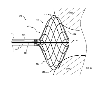
expanded configuration.
[0089] Each filament may also include electrodes of one type or different
types, or different
filaments can accommodate different types of electrodes. Different types of
electrodes may
be understood as electrodes with different functions, for example ablation
electrodes,
measurement electrodes and so on, or physically different electrodes with for
example
different shape, size, design, materials and so on, or a combination of types
of electrodes with
different functionality and physical properties. For example, in
configurations with ring-
shaped electrodes placed on the filaments, all electrodes may have the same
diameter and
may differ in length, so there may be for example two or more groups of such
electrodes,
each group having different length. A number of electrodes in each of the
groups may be the
same or may differ. In an extreme example, each electrode on the expandable
basket may
have a different length. In configurations with ring-shaped electrodes, such
electrodes may
have a diameter between 0.2 mm to 3 mm, or from 0.4 mm to 2 mm, or from 0.5 mm
to 1
mm, and may have a length between 0.1 mm to 10 mm, or from 0.2 mm to 8 mm, or
from 0.3
mm to 6 mm, or from 0.4 mm to 4 mm.
[0090] In one example there may be a first group of 5 to 20 shorter
electrodes, with lengths
of for example 0.3 mm to 3 mm, and a second group of 5 to 30 electrodes which
may be
longer, for example with lengths from 0.6 mm to 4 mm. Advantageously the
electrodes from
the first group may be used for at least one type of measurement, for example
for
measurement of an intracardial ECG (EGM), or an ablation, and the electrodes
from the
CA 03214189 2023- 9- 29
WO 2022/214870
PCT/1B2022/000189
second group may be used for an ablation, either independently or in
combination with the
electrodes from the first group.
[0091] The electrodes can be placed on the body of the basket assembly. For
example, the
electrodes may be placed on the central or distal body portion, in some cases
the electrodes
may be even placed on the proximal body portion. Other electrodes may be
placed on or in an
outer elongated shaft, inner elongated shaft, catheter distal tip or terminal
assembly. In
configurations where the electrodes are placed on the elongated shafts, distal
tip or a terminal
assembly and where ring-shaped electrodes are used, then they may have a
diameter of 0.2
mm to 10 mm, or from 0.5 mm to 8 mm , or from 1 mm to 6 mm, or from 2 mm to 5
mm and
may have a length between 0.1 mm to 20 mm, or from 0.2 mm to 15 mm, or from
0.3 mm to
12 mm, or from 0.4 mm to 10 mm.
[0092] The layout of the electrodes on the expandable basket may ensure
continual, for
example circular ablation areas while the expandable basket is in the expanded
position and
may create a pattern.
100931 For instance, the layout of the electrodes on the expandable basket may
ensure
continual, circular ablation areas even while the expandable basket is held in
various
expanded positions between a fully collapsed and a fully expanded position and
may create a
pattern as well.
[0094] Additional electrodes, for example the ones placed on or in an outer
elongated shaft,
inner elongated shaft, catheter distal tip or terminal assembly may be part of
the pattern or
may be operated independently to other electrodes. For example, electrodes at
the area of
catheter distal tip or terminal assembly may be used for point-like ablation.
There may be
special dedicated electrodes at the area of distal tip or terminal assembly or
for example metal
parts of the terminal assembly may serve as an electrode, or combination of
thereof may
possible.
[0095] The pattern (701) created by the electrodes (109) may be for example a
circular
pattern in space around the longitudinal central axis (203) at least when the
expandable
basket (409) is in one of its expanded configurations as can be seen in FIG.
7A. Other two
dimensional or three-dimensional patterns created by the electrodes (109) are
possible. The
patterns (701) may be centered around the longitudinal central axis (203) or
not. The patterns
(701) may have different shapes, including but not limited to circular,
ellipsoidal, square,
CA 03214189 2023- 9- 29
WO 2022/214870
PCT/1B2022/000189
21
rectangle, polygonal, planar or other or the placement of the electrodes (109)
on the
expandable basket can be irregular. There can be for example one pattern (701)
in one plane
or more patterns (701) in one plane or more patterns (701) in different
planes.
[0096] Patterns created by the electrodes may be positioned on the basket
assembly body,
particularly on the distal body portion, central body portion or proximal body
portion as
shown in FIG. 7B. Patterns may even extend into more than one of these
portions. For
example, for a treatment of a flat treatment site positioned distally from the
basket assembly,
the pattern of electrodes may be positioned advantageously on the basket
assembly distal
portion. Particularly the pattern may be positioned in a section of the basket
assembly
bounded by an area making an angle (703) of 0 to 90 to the central axis
(203) in a center of
a plane (425) intersecting the basket assembly in a portion with a highest
diameter (in one of
its expanded configurations). In some configurations, a pattern may be
positioned partially on
the basket assembly body distal portion and partially on the basket assembly
body central
portion. In some configurations, a pattern may be positioned in a section of
the basket
assembly bounded by an area making an angle (705) of 0 to 120 to the
central axis (203) in
a center of a plane (425). Such placement of the pattern may be particularly
advantageous for
treatment of a vessel orifice, for example an orifice of a pulmonary vein. In
situations where
the treatment site has a tubular shape, the pattern may be placed on the
basket assembly
middle portion, particularly in a section of the basket assembly bounded by
areas making an
angle (707) of 45 to 135 to the central axis (203) in a center of a plane
(425). If a flat
treatment site is positioned proximally from the basket assembly, for example
a septum, the
pattern of electrodes may be positioned on the basket assembly proximal body
portion or
partially on the proximal body portion and partially on the central body
portion, particularly
in a section of the basket assembly bounded by areas making an angle (709) of
90 to NO to
the central axis (203) in a center of a plane (425). Optionally electrodes may
be placed in all
portions of the basket assembly, thus creating patterns in all of the portions
and only patterns
necessary or optimal for performing a particular therapy may be chosen to
perform the
therapy.
[0097] Particular patterns may be created by all electrodes placed on the
expandable basket
or with just a portion of the electrodes. The patterns may have different
numbers of electrodes
in various expanded positions between fully collapsed and fully expanded
positions of the
expandable basket. The neighboring electrodes in the pattern may have
distances between
CA 03214189 2023- 9- 29
WO 2022/214870
PCT/1B2022/000189
22
each other for example 0.1 mm ¨ 15 inm, or 0.5 nun ¨ 10 111111, or 1 nun ¨ 6
HIM or 2 MITI ¨ 4
mm.
[0098] Electrodes are for example electrically connected to the pulse
generator, for example
with conductive wires. The electrodes may be electrically or communicatively
connected to
other units or parts of the pulsed field ablation device as well as for
example with the
mapping device, EP display device, pacing device, ECG recording device,
catheter signal
interconnection circuits, ECG triggering circuits, electrical control
circuits, GUI unit or
remote control unit. Apart from the ring-shaped electrodes mentioned before,
the electrodes
may have any of many different shapes, for example tubes threaded around the
filaments,
coiled metal sheets, square and/or rectangle or other shapes of conductive
materials attached
to the filaments. Other possible forms of electrodes (109) may be elongated
continuous
electrodes drawn along the surface of a portion of the filament (415) in a way
they do not
touch at crossing points of the filaments (415) in the braided mesh (413) as
shown in FIG. 8.
The electrodes (109) may be attached on the particular filaments (415) of the
expandable
basket by any means, for example by way of mechanical attachment, swagging,
crimping,
gluing, lamination, deposition and/or soldering. The electrodes may be made
out of any
electrically conductive material for example copper, gold, steel, titanium,
platinum, platinum-
iridium, and so on. In a case where there is at least one filament made out of
conducting
material, it could serve as an electrode as well. In a case where the whole
conducting filament
is uninsulated the whole filament may serve as an electrode, in case the
filament is for
example partially electrically insulated, the bare, uninsulated portion may
serve as an
electrode.
[0099] Conductive wires may provide an electrical connection between the
electrodes and a
pulse generator. The conductive wires may be a part of a structure of the
basket assembly
(401). For instance, the conductive wires (417) may be positioned at least
partially in the
lumen (601) of the filaments (415) as shown in FIG. 6C or in FIG. 9. There can
be one or
more conductive wires (417) coupled to each of the electrodes, or one or more
electrodes can
be coupled to a single leading wire. the conductive wires (417) may be
incorporated into the
one of the walls of the shaft assembly, for example into the wall of the outer
elongated shaft.
The conductive wires can also be positioned in the central lumen of the outer
elongated shaft
or there can be separate lumens in the outer elongated shaft suitable for the
placement of
conductive wires. The conductive wires may be terminated adjacent to the
electrodes or may
CA 03214189 2023- 9- 29
WO 2022/214870
PCT/1B2022/000189
23
lead spatially further along the length of the filament past the electrode.
The conductive wires
may for example be positioned along the whole length of the filaments of the
basket
assembly. Optionally some of the conductive wires (417) may be terminated
adjacent to the
electrodes while others may lead spatially further along the filaments past
the electrode or
may be positioned along the whole length of the filaments of the basket
assembly.
[0100] In a case where the conductive wires are positioned along the whole
length of the
filament, the design solution of the expandable basket, where the filaments
are bent and
returned to the expandable basket, rather than cut, at the expendable basket's
distal end is
particularly advantageous. Because the particular conductive wires are
configured to carry
electrical pulses between electrodes and the pulse generator, an insulation of
the cut filaments
with the conductive wires inside would be extremely challenging at the
terminal assembly.
On the other hand, in examples comprising bent filaments with conductive wires
inside, the
insulation of the terminal assembly can be easily assured.
[0101] The material used for conductive wires may be any electrically
conductive material
for example copper, stainless steel, steel, nitinol, aluminum, gold, platinum,
silver and so on.
The conductive wires may be insulated or uninsulated. The wires may be
insulated using any
suitable material, for example polyimide, polyurethane, polyester,
polyvinylchloride (PVC),
rubber, rubber-like polymers, nylon, polyethylene, polypropylene, silicone,
fiberglass,
ethylene propylene diene monomer (EPDM), different fluoropolymers like
polytetrafluoroethylene (PTFE) and so on. The wires may be made of a single
conductor or
with a group of conductors, whereas a wire made of a group of conductors is
sometimes
called -cable-. In case the wires are insulated a minimum breakdown voltage of
the wire
insulation should be at least 1 00V, or 500V, or 1000V or 4000V or 10000V. The
diameter of
the wires with insulation may be limited by the dimensions of other structures
of the device
such as for example the filaments and a minimum voltage it has to be able to
carry without
risk of breakdown. Typical diameter of the wires with or without insulation
may be between
0.05mm and 0.7mm, or between 0.07mm and 0.5mm, or 0.1mm to 0.3mm or between
0.11mm to 0.2mm or between 0.12mm to 0.18mm.
[0102] The construction of the braided mesh out of electrically insulating
material as
described with one or more conductive wires inside hollow filaments may be
particularly
advantageous for an ablation system based on the principle of pulsed field
ablation by pulsed
electric fields. The pulsed field ablation method as described further,
requires electric fields
CA 03214189 2023- 9- 29
WO 2022/214870
PCT/1B2022/000189
24
generated around electrodes. To generate the fields, electrical pulses have to
be carried by
particular conductive wires between the electrodes and the pulse generator.
When the
filaments are electrically nonconductive, and the conductive wires are kept
inside the
filaments as described herein, the electrical insulation of the particular
conductive wires can
be ensured even at voltage levels of several kV, for example from lkV to 10kV,
carried by
the conductive wires. However, an option of braided mesh with at least one or
more filaments
made out electric conducting material (for example nitinol, copper, stainless
steel, steel,
aluminum, gold, platinum or silver) may be possible as well. Such conducting
filaments may
be insulated or not or only partially. They not only that could possibly lead
electrical current,
but could act as an electrode (when uninsulated or insulated only partially)
and/or as a further
mechanical support of the braided mesh hence the expandable basket.
[0103] Another advantage of a braided mesh made of polymer or thermoplastic
elastomer
filaments is the ease of manufacturing compared for example to a metallic
braided mesh. The
braided mesh may be for example made with the help of a three-dimensional
mandrel device.
The particular filaments creating the mesh may be placed over the mandrel in a
desired
pattern. The filaments may already include the conductive wires. The whole
structure may
then be heated up, for example close to the melting point of a material of the
filaments and
after that the structure may rapidly be cooled down. The filaments made of
thermoplastic
elastomer or polymers generally require lower temperatures to reach the
melting point over
most metals, so the manufacturing process can be faster, more efficient and
can demand less
energy input. Another advantage of such a manufacturing process is the
conductive wires do
not need to be heated to extreme temperatures, to a degree where the
electrical properties of
the wire may be compromised. This situation can happen, for example when the
braided
mesh is made of the metallic wires, where the mesh wires also serve as the
electrically
conductive wires.
[0104] The braided mesh with inserted conductive wires may be attached to the
outer
elongated shaft and inner elongated shaft creating an expandable basket and
part of the basket
assembly. The electrodes may be attached at the particular filaments of the
braided mesh
before or after the attachment of the braided mesh to the elongated shafts.
The pulse
generator is a part providing generation of electric signals for catheter
electrodes. The pulse
generator may allow settings for example of an amplitude, a shape of the
electrical pulse
and/or a number of pulses during activation. The pulse generator may diagnose
electrical
CA 03214189 2023- 9- 29
WO 2022/214870
PCT/1B2022/000189
waveforms to measure power as well. The pulse generator may enable synchronous
operation
with an ECG device or another part of the ablation system or device.
[0105] Further, a method of ablation with the described pulsed field ablation
device is
disclosed.
[0106] One method comprises the step of disposing a catheter (105) adjacent to
the treatment
site, for example a cardiac chamber, in the patient via a blood vessel. The
catheter (105) may
be inserted into the blood vessel of the patient percutaneously.
[0107] Other support structures and/or devices may be used to help navigate
the distal tip of
the catheter to its desired location. Examples of such devices include a guide-
wire or a
sheath. The catheter distal tip may be delivered proximally to the treatment
site in a collapsed
state, for example through a sheath. In the collapsed state the diameter of
the basket assembly
at the catheter distal tip may be less than or approximately equivalent to the
diameter of the
outer elongated shaft of the catheter. Such a configuration allows easy access
of the catheter
distal tip proximal to the treatment site.
[0108] The treatment site may be for example located inside the body, for
example in or on a
heart, for example in a heart cavity, particularly for example in a left
atrium of the heart. The
treatment site may for example include a pulmonary vein orifice. Other
locations of the
treatment site may be for example all tubular tissues, organs or vessels in a
body or for
example tumor sites.
[0109] When the catheter distal tip is delivered to the treatment site, the
basket assembly of
the catheter is deployed from the collapsed or semi-collapsed configuration to
one of the
expanded configurations. This deployment may be caused by a pre-tension shape
of the
braided mesh or its filaments or by a linear displacement of the inner
elongated shaft against
the outer elongated shaft along a longitudinal central axis of the catheter,
by a tension of an
additional supportive structure for example an inner coil or balloon (not
shown), or by a
combination of thereof
[0110] The catheter distal tip (107) may then be placed adjacent to a target
tissue of the
treatment site (1001), for instance at least part of the basket assembly
(401), and/or part of the
expandable basket (409) is brought in contact with the treatment site (1001).
In this position
at least a portion of the set of electrodes (109), placed on the basket
assembly (401) may be in
CA 03214189 2023- 9- 29
WO 2022/214870
PCT/1B2022/000189
26
contact with the tissue of the treatment site (1001). A schematic of an
example position can
be seen in FIG. 10. The terminal assembly (411) may improve contact of the
electrodes with
the treatment site by its flat design without distally protruding structures.
When there are no
distally protruding formations on the basket assembly (401), especially on the
basket
assembly distal portion (405), it is easier to get the electrodes in contact
with the treatment
site even in situations where the treatment site is relatively flat.
[0111] After positioning the catheter distal tip adjacent to the treatment
site an optional step
of measurement can be carried out with or without the catheter. Different
kinds of
measurements can be performed with the goal of, for example, diagnostics of
type or quality
of a tissue at or around the treatment site, spatial position of the catheter
distal tip,
particularly for example the spatial position of the catheter distal tip
against the treatment
site, contact of the catheter distal tip and/or particular electrodes with the
target tissue of the
treatment site or with a goal of understanding electrophysiological processes
of a tissue
adjacent to the electrodes. For example, the electrodes may be used for a
measurement of
contact with a target tissue as well and may be placed on the expandable
basket, for example
on the filaments of the braided mesh. The measurement electrodes may be
different
electrodes than the ablation electrodes or the ablation electrodes may be used
for the
measurements. It is possible to combine separate measurement electrodes with
the ablation
electrodes with measurement functions on one catheter distal tip as well. A
separate
measurement device may be used to carry out the measurement step, for example
a separate
measurement catheter (not shown), an ECG device including ECG triggering
circuits, an
ECG recording device, ECG electrodes, an intracardial ECG (EGM), an
intracardial echo
device, an esophagus temperature measurement device, a fluoroscopy device, RTG
device,
MR device, and so on. The measurement step may be carried out once or may be
repeated
several times during an ablation procedure.
[0112] The ablation of the target tissue of the treatment site (1001) for
instance uses a
principle of pulsed field ablation caused by pulsed electric fields of proper
parameters.
Although the terms -electric fields" or -pulsed electric fields" are mentioned
here, electric
fields as contemplated herein may further comprise a magnetic component.
[0113] The procedure of basket assembly deployment, measurements and ablation
can be
carried out in several stages. For example, the expandable basket may be
delivered adjacent
to the treatment site in a fully collapsed configuration. After delivery it
can be deployed to its
CA 03214189 2023- 9- 29
WO 2022/214870
PCT/1B2022/000189
27
first expanded configuration. For example, the pre-tension shape of the
braided mesh and/or
filaments may cause this first transition. In this configuration for example
further
manipulation with the basket assembly can be carried out as well as
measurements and/or
ablation. Further repositioning, measurement and/or ablation can be carried
out in this
position in any order as well.
[0114] Then the basket assembly may be deployed into a second expanded
configuration.
The second expanded configuration can be achieved for example by a linear
displacement of
the inner elongated shaft against the outer elongated shaft along a
longitudinal central axis of
the catheter. In this configuration for example further manipulation of the
basket assembly
can be carried out as well as measurements and/or ablation. Further
repositioning,
measurement, and/or ablation can be carried out in this position in any order
as well.
[0115] The basket assembly can be for example deployed into several different
expanded
positions, during which further repositioning, measurement and/or ablation can
be carried
out.
101161 In the case of pulmonary vein isolation ablation the set of electrodes
may create a
circular shape around the pulmonary vein orifice. After the ablation the shape
of the ablated
tissue may have a circular shape around the pulmonary vein orifice as well.
Several such
shapes of ablated tissue may be created by repositioning the basket assembly
or by switching
between different electrodes.
[0117] The pulsed electric field (PEF) is for instance created by electrical
pulses, for example
high frequency electrical pulses. The electrical pulses may be generated by a
pulse generator
and may be delivered to the target tissue by the electrodes which may be
placed on the
catheter distal tip and which may be in electrical contact with the pulse
generator. The
electrical pulses can be created by a wide variety of electrical pulses
ranging from
monophasic (single polarity) pulses to symmetrical and/or asymmetrical
biphasic pulses. The
pulses may be combined with extra pre-pulses for tissue conditioning or extra
measurement
pulses as well. Pulses can be single pulses, or they may be repeated in
trains, where
parameters of the pulses may vary or remain constant. Trains of pulses can be
run in
sequences as well. A maximal amplitude of the pulses may depend on the target
tissue,
electrode's size and/or electrode's distance in order to create an electric
field with a
maximum electric field magnitude for example between 0.1kV to 10kV or between
0.4kV to
CA 03214189 2023- 9- 29
WO 2022/214870
PCT/1B2022/000189
28
5kV or between 0.5kV to 2kV per centimeter in a target tissue volume. A
duration of the
pulse can be from a nanosecond range to milliseconds range, for example from
2ns to 10ms,
or from lOns to 5ms or from lOus to lms. The shape of the pulse may be for
example a
square, a curve similar to exponential discharge, a rectangle, a saw, a
triangle or a sinusoidal
shape.
[0118] The pulses can be monophasic or biphasic. Biphasic pulses can be
symmetrical or
asymmetrical. The pulses can repeat from lx to 100000x. The frequency of the
high
frequency pulses may vary from 0.1 Hz to 10 Hz. Amplitude (Um) of the
monophasic pulses
can vary from 100V up to 10kV, and the peak-to-peak amplitude of biphasic
pulses may vary
from 200V to 20kV.
[0119] Fig. 16 may serve as an example of a possible part of a pulsed field
ablation (PFA)
protocol and as a clarification of terms and expressions regarding the PFA
protocol. The PFA
protocol includes a series of electrical pulses (1601) and pauses (1603, 1607,
1615). The
electrical pulses (1601) may be further organized in units with a certain
hierarchy like trains
(TR) and bursts (B).
[0120] The electrical pulse (1601) may be defined for example by shape,
amplitude (Urn)
with certain voltage and pulse length with time duration (M. The pulse
amplitude (Urn) may
be either negative or positive (the pulse may have negative voltage or
positive voltage) in
case of monophasic pulses. The electrical pulses (1601) may be separated from
each other by
an inter-pulse pause (1603), which is defined by a time duration (t2) and a
voltage (Up). The
voltage during the inter-pulse pause (1603) may drop to OV or it may have a
positive or
negative voltage value (IJp)_ The absolute voltage value (IJp) of the inter-
pulse pause is
smaller than an absolute voltage (amplitude (Urn)) of the adjacent electrical
pulse (1601),
particularly up to 50% of the amplitude (Urn) of the adjacent electrical
pulse. In situations
where the electrical pulse has a positive amplitude (Urn), the voltage value
(Up) of the inter-
pulse pause (1603) will stay positive between OV and the electrical pulse
(1601) amplitude
(Um), and in situations where the electrical pulse (1601) has a negative
amplitude (Urn), the
voltage value (Up) of the inter-pulse pause (1603) will stay negative between
OV and the
electrical pulse amplitude (Um). An example of inter-pulse pauses (1603) with
a voltage
different than OV is shown in FIG. 17a. Biphasic pulses may be symmetrical or
asymmetrical
in at least one of time, amplitude or energy.
CA 03214189 2023- 9- 29
WO 2022/214870
PCT/1B2022/000189
29
[0121] Examples of biphasic electrical pulses are shown in FIG. 17b. The
biphasic pulses
may have the same amplitude (voltage) of a positive phase (1701) and a
negative phase
(1703) with the same duration (tl 0, t12) of both phases (exemplary pulse A,
D), or the
amplitude and/or duration (t10) of the positive phase and the amplitude and/or
duration (t12)
of the negative phase may differ (exemplary pulses B, C). The resulting pulses
then may have
the same energy in the positive and negative phases of the pulse, or the
energy in the positive
and negative phases of the pulse may be different. Biphasic pulses with the
same energy in
both phases may be called symmetrical biphasic pulses. Symmetrical biphasic
pulses may be
balanced (in case the duration and amplitude of both phases of the pulse are
identical, or
imbalanced (in cases where the amplitude and/or duration are different in each
phase).
Asymmetrical biphasic pulses have phases with different energies. Exemplary
biphasic pulses
A, B, C are without a pause between particular phases (inter-phase pauses) of
the pulse,
exemplary pulse D is a biphasic pulse with an inter-phase pause (1705). The
duration of the
inter-phase pause of the pulse may be from 0 ps to 50 t.ts or from 0 ps to 10
tis or from 0 pts
to 5 tis.
[0122] A series or sequence of pulses in a row, with or without inter-pulse
pauses may be
called a train (TR). Particular trains (TR) may be characterized for example
by a time
duration (t4), or number of pulses and may be separated from one another by
inter-train
pauses (1607) with a time duration (t5) or the inter-train pause (1607) may
separate a train
with an individual single pulse. A series or sequence of the trains (TR) and
inter-train pauses
(1607) can be called a burst (B), and may be characterized for example by a
time duration
(t6), number of trains (TR), number of pulses or by inter-burst pause (1615)
(with time
duration (t7) between particular bursts (B).
[0123] As already stated above, a voltage value (Up) at the electrodes may not
decrease to
OV between pulses, particularly during inter-pulse pauses (1603) but may
remain at a level,
where the risk of creating bubbles by electrolysis or temperature increment is
either non-
existent or very small, for example up to 50% of the amplitude (Urn) of an
adjacent electrical
pulse. This may reduce an unwanted relaxation of the polar molecules as well,
which may
lead to shorter length of at least some parts of the PFA protocol and so
increase an efficacy of
the PEF therapy.
[0124] When pulses with amplitude (Um) of hundreds of volts to a few thousand
volts are
applied, there is a certain risk of causing a ventricular muscle
depolarization and unwanted
CA 03214189 2023- 9- 29
WO 2022/214870
PCT/1B2022/000189
ventricular rhythms in the heart, even when applied in a heart atrium.
Depolarization can be
caused directly by electric field or by secondary energy induction in another
device, for
example a catheter, which is placed in or near either atria or ventricles or
both. Setting the
timing of the active sequences (individual pulses, trains and/or bursts) with
pauses described
below results in an effect called overdrive. The overdrive effect is commonly
used in ablation
catheterization procedures to suppress a risk of unwanted heart rhythms by
using an external
pacemaker. An advantage of the proposed PFA protocol is that the therapeutic
(ablation)
electrical pulses may, in a case where they cause a myocardium depolarization,
also act as
pace stimulation pulses for the heart, and therefore it is not necessary to
use an additional
pacing device (for example an external pacemaker) to synchronize pulses of the
pacing
device with the therapeutic pulses of the PFA protocol. This in turn means
that in this case it
is not necessary to use a pacing device to control the number of ventricular
contractions per
minute, detect the individual ventricular contractions from a surface ECG and
then trigger the
ablation pulses accordingly.
[0125] The duration (t8) of one cycle (1609) of a burst (B), and inter-burst
pause (1615)
between bursts, which is between 201ms to 800ms, is given by a range between
the need to
deliver pulses safely faster than the patient's actual heart rate (the
overdrive effect) and the
need to maintain heart rate at a safe level (which is stated to be
approximately 220 beats per
minute minus age). The cycle duration may be fixed or variable in the stated
range (201ms to
800ms) within a PFA protocol, for example according to a sinusoidal or
triangular function.
The individual burst (B) may have a duration (t6) from lms to 200ms, or 30ms
to 180ms, or
60ms to 160ms, which is a safe time to contract the heart chamber by an
applied burst (B) of
pulses, protecting ventricles from injury or unwanted rhythm. The burst (B)
duration (t6) may
too be fixed or variable in the stated range (lms to 200m5) within a PFA
protocol, for
example according to a sinusoidal or triangular function.
[0126] This PFA protocol may have other positive effects on the ablation
results, for example
reducing the risk of causing an unwanted ventricular rhythm and/or maximized
PEF
application efficiency.
[0127] However an electroporation is described as the primary trigger of death
of the
myocardial cells after application of the PEF, but actual cell death may
alternatively be
caused for example by electrical breakdown of the membrane of cardiomyocytes,
mitochondria or nucleus; by tearing individual cells / cardiomyocytes (or
groups of cells) of
CA 03214189 2023- 9- 29
WO 2022/214870
PCT/1B2022/000189
31
the myocardium apart (for example, by damaging the intercalated discs, either
directly by
electric fields or by mechanical damage by hypercontraction); by damage to
sarcolemma or
myofibrils of muscle fiber; by depletion and insufficient production of ATP in
cardiomyocytes due to hypercontraction; by loosening of intercellular
junctions of
cardiomyocytes; by muscle cell myolysis; by wrinkling cardiomyocytes either
directly under
the influence of the electric field or by mechanical damage by
hypercontraction; by
irreversible damage to the calcium cycle (whether by non-physiological
function of the
sarcoplasmic reticulum or ion pumps or calcium channels or calcium binding
proteins); by
calcium overload of the heart muscle ¨ mitochondrial swelling (as a result of
hypercontraction or damage to cardiomyocyte sarcolemma or non-physiological
function of
calcium channels); or by formation of reactive oxygen species (ROS) and
subsequent
oxidation of membrane phospholipids by PEF.
[0128] The electric fields may be created among one or more electrodes placed
on the
catheter distal tip and one indifferent electrode placed in the distance, for
example on the skin
of the patient. The indifferent electrode may in some aspects have a
significantly larger
surface than the sum of the surfaces of the active distal tip electrodes. This
mode of action is
usually called monopolar. Another option for creating an electric field is in
a bipolar mode. In
this mode the electric field arises between two or more, usually closely-
placed or adjacent
distal tip electrodes with different polarities. In this case the sum of the
surfaces of active
electrodes with the first polarity is similar to the sum of the surfaces of
the active electrodes
with the second polarity.
[0129] In some aspects, the electrodes (109) placed on the distal assembly may
be operated
in a hybrid mode of the previous two types. An example of such a mode is shown
in FIG. 11.
Only the electrodes (109) placed on the distal tip (107) are used for ablation
in this mode.
There is a first single electrode, or group of electrodes operating in a mode
with first polarity
(P1) and a second single electrode or group of electrodes operating in a mode
with different
polarity (P2) (which may be an opposite polarity) than the operating mode of
the first
electrode or group of electrodes. A surface or a sum of the surfaces of the
first electrode or
the first group of electrodes is significantly smaller than a surface or a sum
of the surfaces of
the second electrode or group of electrodes. For example, there may be a third
group of
electrodes operating in a third mode in state of high impedance (HI), wherein
the impedance
of the electrodes in the third group is for example higher than 500 a The
electrodes
CA 03214189 2023- 9- 29
WO 2022/214870
PCT/1B2022/000189
32
operating in the third mode may be adjacent to the electrode or group of
electrodes operating
in the first mode.
[0130] One advantage of the operation of electrodes in this hybrid mode is
that the generated
electric field will have a more homogenous current density in comparison to
bipolar mode.
Another advantage of the hybrid operation mode is the electric fields created
in this mode
may in some aspects be able to reach deeper into the target tissue compared to
bipolar mode.
In case of ablation of a heart cavity, the depth of the ablated target tissue,
(in one example the
target tissue may comprise a myocardial tissue), may be up to 5 mm.
[0131] A variant of the hybrid mode of operation of the electrodes (109) with
a group of
electrodes (more than one electrode) operating in the mode with the first
polarity (P1) is
shown in FIG. 12. The functional principle of this mode of operation is
similar to the variant
with one electrode (109) operating in the mode with the first polarity (P1).
For example, the
sum of surfaces of the electrodes operating in the mode with the first
polarity (P1) is
significantly smaller than the sum of surfaces of the electrodes operating in
a mode with a
different polarity (P2).
[0132] Examples with a group of electrodes (more than one electrode) operating
in a mode
with a first polarity (P1) can have an advantage over examples with a single
electrode
operating in the mode with the first polarity (P1) for example in situations
where it is
advantageous to reduce the size of the electrodes. Reducing the size of the
electrodes can be
advantageous or necessary in cases where it is necessary or desirable to
increase the number
of electrodes. A higher number of electrodes is desirable for example where
more precise
mapping of the treatment site or more precise and/or more homogenous ablation
of the target
tissue of the treatment site is desired. Because the treatment site can be
part of a human
anatomy, the overall size of the pulsed field ablation device, especially the
catheter with a
catheter distal tip must be restricted according to human anatomy. It follows
that if more
electrodes are needed for the ablation device, then for a certain number of
electrodes the size
of the electrodes must be limited to able to fit into the restricted
dimensions of the critical
parts of the pulsed field ablation device for example the catheter and/or its
distal tip, and/or
its basket assembly. Another advantage of the smaller size of the electrodes
is that such an
arrangement may help to increase a depth of the ablation.
CA 03214189 2023- 9- 29
WO 2022/214870
PCT/1B2022/000189
33
[0133] Smaller size of the electrodes can have other advantages, for example
in examples
where the same electrodes are used for ablation and for measurements, it means
the same
electrode must be configured to deliver high voltage pulses and record
measurements. For
example, in measurement of ECG signals, smaller electrodes may be
advantageous.
[0134] There are however some challenges associated with smaller electrodes as
well. In
examples including pulsed field ablation, the electric fields are for instance
created by
electrical pulses, for example high frequency electrical pulses generated by a
pulse generator.
For effective ablation of the whole target area of the treatment site, it may
be important to
create an electric field with a maximum electric field magnitude of several
hundred volts to
several kilovolts per centimeter in a target tissue volume. Using smaller
electrodes means a
smaller surface area of the electrodes. With a smaller surface area of the
electrodes, the
voltage induced on the electrode has to be higher compared to bigger
electrodes with larger
surface area to achieve the desired electric field density in a target tissue.
Adverse effects of
such a configuration may include higher density of the electric field, higher
intensity of the
electric field and/or possible sparking on the edges of the electrodes.
However, using a
chosen group of electrodes (more than one electrode) operating in the mode
with the first
polarity instead of one electrode operating in the mode with the first
polarity can address and
overcome some or all of these issues. With a well-chosen first group of
electrodes operating
in the mode with the first polarity together with the second group of
electrodes operating in a
mode with different polarity and possibly with a third group of electrodes
operating in a third
mode in the state of high impedance, the first group of electrodes and/or the
second group of
electrodes may act as virtual electrodes. That means the electrodes in the
first group may act
together as one virtual electrode and/or the electrodes in the second group
may act as another
virtual electrode. With such a configuration, the intensity and/or the density
of the electric
field near the electrodes may be reduced. Other positive effects of this
configuration may be a
reduced risk of sparking and increased depth of ablation, or increased depth
of an ablated
tissue at the treatment site.
[0135] The enlargement of the surface area of the electrodes in the first
group and the
creation of the resulting virtual electrode may cause a reduction in the
voltage needed to be
induced in the electrodes and/or elimination of sparking, mainly on the edges
of the
electrodes. However, the concept of disproportional surface areas of the
electrodes in the first
and the second groups of electrodes can be preserved, which means the surface
area or a sum
CA 03214189 2023- 9- 29
WO 2022/214870
PCT/1B2022/000189
34
of the surface areas of the first electrode or the first group of electrodes
is significantly
smaller than a surface area or a sum of the surface areas of the second
electrode or group of
electrodes. The ratio of the surface area of the electrode or the sum of the
surface areas of the
electrodes in the first group to the sum of the surface areas of the
electrodes in the second
group of electrodes may be between 2:3 to 1:100, or 3:5 to 1:80, or 3:5 to
1:70, or 1:2 to 1:50,
or 1:2 to 1:40, or 1:2 to 1:30, or 1:2 to 1:20, or 1:3 to 1:15, or 1:3 to
1:10, or 1:4 to 1:8.
[0136] Adding electrodes to the first group of electrodes operating in the
mode with the first
polarity may significantly reduce the intensity of the electric field near the
electrodes. Using
four electrodes instead of one for example in the first group of electrodes
operating in the
mode with the first polarity, the intensity of the electric field at the
electrode surface
decreases by a factor of four, while in examples where three electrodes are
used, the intensity
of the electric field decreases by a factor of two. This reduction in
intensity may allow for the
use of lower voltage on the electrodes, compared to a solution with just one
electrode
operating in the mode with the first polarity. The reduction may additionally
or alternatively
increase of the depth of the ablated target tissue by increasing an area of
the electric field
with a certain voltage per cm2. The value of the voltage per cm2 in an area of
the electric
field may be for example from 50 V/cm2 to 3000 V/cm2, or from 100 V/cm2 to
1500 V/cm2,
or from 250 V/cm2 to 1000 V/cm2.
[0137] The particular electrodes on the catheter distal tip can be switched to
one or more than
one of the modes during the ablation. They can be switched during one ablation
cycle or
during several ablation cycles. The electrodes may be switched to one or more
of the modes
several times during one ablation cycle or during several ablation cycles. In
some aspects it is
even possible to have two or more groups of electrodes operating
simultaneously in a mode
with the first polarity and a group of electrodes operating in a different
polarity, with or
without electrodes operating in a state of high impedance.
[0138] A layout or spatial pattern of the electrodes on the distal tip may be
created with a
consideration of the hybrid mode of operation of electrodes and/or with the
goal of creating
virtual electrodes. Because the electrodes may be switched to one or more than
one of the
modes during the ablation, it is possible the resulting virtual electrodes may
have different
spatial shapes which means the electric fields created around and between the
virtual
electrodes may have different shapes with different structures of the magnetic
field and/or
different density and intensity of the electric field. An example of a spatial
pattern of
CA 03214189 2023- 9- 29
WO 2022/214870
PCT/1B2022/000189
electrodes on the distal tip, specifically on the expandable basket may be
seen in FIG. 13A
and in FIG. 13B. FIG. 13A shows a frontal view of the basket assembly (401)
with a spatial
pattern of electrodes (109) suitable for creation of virtual electrodes by
switching the
electrodes (109) into different modes of operation with the first polarity and
with the different
polarity and/or with a state of high impedance.
[0139] FIG. 13B shows again a frontal view of the basket assembly (401) with a
spatial
pattern of electrodes suitable for creation of virtual electrodes, by
switching the electrodes
(109) into different modes, however this time the electrodes are placed in
areas where the
filaments (415) cross each other (filaments crossing points).
[0140] One possible layout of electrodes already switched into the hybrid
operation mode
may be seen in the FIG. 14, which is again a frontal view of the basket
assembly (401). A
first group of electrodes (109) is operating in the mode with a first polarity
(P1) and together
creates a first virtual electrode (1401). Another group of electrodes (109) is
operating in the
mode with a different polarity (P2) and together creates a second virtual
electrode (1403).
When electrical pulses are delivered from the pulse generator (103) to the
electrodes (109) in
this configuration, electric fields will be created between and around the
virtual electrodes
(1401, 1403). Some of the electrodes (109) may be operating in a third mode,
for example in
a state of high impedance (HI).
[0141] The electrodes in a state of high impedance (higher than 500 Q) may
help in shaping
an electric field created among and around electrodes from the first group and
the second
group of electrodes and/or between or around the virtual electrodes. In one
example,
assigning a state of high impedance to electrodes which are spatially adjacent
to the
electrodes operating in the mode with a first polarity may have a positive
effect on the shape
of the electric field in a way that a portion of the electric field which is
able to cause an
ablation reaches deeper into the target tissue of the treatment site, compared
to an operation
mode without electrodes in a state of high impedance. This phenomenon may have
positive
effects in the quality and homogeneity of an ablation procedure. The
electrodes in a state of
high impedance may be spatially placed between the first group of electrodes
and the second
group of electrodes.
[0142] An exemplary pattern of electrodes (109) is displayed in more detail in
FIG. 15A. The
electrodes (109) create a pattern of repeating crosses or squares or
rectangles on the filaments
CA 03214189 2023- 9- 29
WO 2022/214870
PCT/1B2022/000189
36
(415) of the braided mesh in one of the expanded configurations of the
expandable basket.
From this view, which is perpendicular to the tangent plane (which is touching
the
expandable basket for example at an intersection (1501) of four neighboring
electrodes), the
pattern seems two-dimensional, but in reality, it is three-dimensional,
because the electrodes
(109) are fixed to, or are part of the filaments (415) of the braided mesh,
which creates an
expandable basket, and therefore the pattern fits into the curvature of the
expandable basket.
This pattern of the electrodes is advantageous in embodiments using a group of
electrodes
operating in a mode with first polarity (P1). In this example a group of four
adjacent
electrodes operating in a mode with first polarity (P1) and hence creating a
first virtual
electrode (1401) will have either a cross shape, as indicated in FIG. 15A or a
square or
rectangle shape as indicated in FIG. 15B. The advantage is that both virtual
electrodes (1401)
created by both shapes are, in combination with a second virtual electrode,
and possibly with
the help of the electrodes in a state of high impedance, capable of creating
electric fields
having certain qualities (shape, magnitude, density, gradient of potential)
suitable for the
ablation of a target tissue.
[0143] FIG. 15C shows an example of a pattern of electrodes where the
electrodes (109) are
placed in areas where the filaments (415) cross each other (filament crossing
points). An
exemplary group of electrodes operating in a mode with first polarity (P1) is
also shown here.
[0144] The exact shape of the pattern of electrodes partially depends on the
shape of the
expandable basket. It also means the pattern and the shape of the groups of
electrodes
creating the virtual electrodes may be different in a collapsed configuration
and/or in
different expanded configurations of the expandable basket. For most of the
expanded
configurations of the expandable basket, the rectangles and squares created by
the electrodes
as described above will be inclined and will be creating shapes closer to
rhombuses or
rhomboids. The same applies to the angles between the two imaginary lines
creating a cross
and passing through the electrodes, which will not be right angles in most of
the expanded
configurations.
[0145] When using high voltage pulses in the human body, it may be necessary
to
synchronize the delivery of the pulses with a cardiac cycle for safety
reasons, for example in
order to avoid ventricular rhythm. The pulsed field ablation device can
incorporate or use a
means for such a synchronization including triggering of the pulse delivery by
this
synchronization means. The synchronization means can be for example an ECG
device.
CA 03214189 2023- 9- 29