Note: Descriptions are shown in the official language in which they were submitted.
CA 03214310 2023-09-20
WO 2022/201123 PCT/IB2022/052765
1
[0001] ANTI-TAU ANTIBODIES AND USES THEREOF
FIELD OF THE INVENTION
[0002] The application relates to anti-PHF-tau antibodies, nucleic acids
and expression
vectors encoding the antibodies, recombinant cells containing the vectors, and
compositions
comprising the antibodies. Methods of making the antibodies, methods of using
the antibodies to
treat conditions including tauopathies, and methods of using the antibodies to
diagnose diseases
such as tauopathies are also provided.
REFERENCE TO SEQUENCE LISTING SUBMITTED ELECTRONICALLY
[0003] This application contains a sequence listing, which is submitted
electronically via EFS-
Web as an ASCII formatted sequence listing with a file name
"065768.96U52_Sequence_Listing"
and a creation date of June 3, 2021 and having a size of 66 kb. The sequence
listing submitted via
EFS-Web is part of the specification and is herein incorporated by reference
in its entirety.
BACKGROUND OF THE INVENTION
[0004] Alzheimer's Disease (AD) is a degenerative brain disorder
characterized clinically by
progressive loss of memory, cognition, reasoning, judgment and emotional
stability that
gradually leads to profound mental deterioration and ultimately death. AD is a
very common
cause of progressive mental failure (dementia) in aged humans and is believed
to represent the
fourth most common medical cause of death in the United States. AD has been
observed in
ethnic groups worldwide and presents a major present and future public health
problem.
[0005] The brains of individuals with AD exhibit characteristic lesions
termed senile (or
amyloid) plaques, amyloid angiopathy (amyloid deposits in blood vessels) and
neurofibrillary
tangles. Large numbers of these lesions, particularly amyloid plaques and
neurofibrillary tangles
of paired helical filaments, are generally found in several areas of the human
brain important for
memory and cognitive function in patients with AD.
[0006] The current AD treatment landscape includes only therapies approved
to treat
cognitive symptoms in patients with dementia. There are no approved therapies
that modify or
CA 03214310 2023-09-20
WO 2022/201123 PCT/IB2022/052765
2
slow the progression of AD. Potential disease modifiers are anti amyloid
antibodies including Eli
Lilly's Donanemab, a humanized IgG1 monoclonal antibody recognizing Af3(p3-
42), a
pyroglutamate form of AP, and aducanumab, a human IgG1 monoclonal antibody
against a
conformational epitope found on AP. These therapies, and most other potential
disease modifiers
that may launch in the next decade, target Ap (the principle component of the
amyloid plaques
that are one of the two "hallmark" pathological signs of AD).
[0007] Neurofibrillary tangles, the second hallmark pathological sign of
AD, are primarily
composed of aggregates of hyper-phosphorylated tau protein. The main
physiological function of
tau is microtubule polymerization and stabilization. The binding of tau to
microtubules takes
place by ionic interactions between positive charges in the microtubule
binding region of tau and
negative charges on the microtubule lattice (Butner and Kirschner, J Cell
Biol. 115(3):717-30,
1991). Tau protein contains 85 possible phosphorylation sites, and
phosphorylation at many of
these sites interferes with the primary function of tau. Tau that is bound to
the axonal
microtubule lattice is in a hypo-phosphorylation state, while aggregated tau
in AD is hyper-
phosphorylated, providing unique epitopes that are distinct from the
physiologically active pool
of tau.
[0008] A tauopathy transmission and spreading hypothesis has been described
and is based
on the Braak stages of tauopathy progression in the human brain and tauopathy
spreading after
tau aggregate injections in preclinical tau models (Frost et al., J Biol Chem.
284:12845-52, 2009;
Clavaguera et al., Nat Cell Biol. 11:909-13, 2009).
[0009] Developing therapeutics preventing or clearing tau aggregation has
been of interest
for many years and candidate drugs, including anti-aggregation compounds and
kinase
inhibitors, have entered in clinical testing (Brunden et al., Nat Rev Drug
Discov. 8:783-93,
2009). Multiple studies have been published that show the beneficial
therapeutic effects of both
active and passive tau immunization in transgenic mouse models (Chai et al., J
Biol Chem.
286:34457-67, 2011; Boutajangout et al., J Neurochem. 118:658-67, 2011;
Boutajangout et al., J
Neurosci. 30:16559-66, 2010; Asuni et al., J Neurosci. 27:9115-29, 2007).
Activity has been
reported with both phospho-directed and non-phospho-directed antibodies
(Schroeder et al., J
Neuroimmune Pharmacol. 11(1):9-25, 2016).
CA 03214310 2023-09-20
WO 2022/201123 PCT/IB2022/052765
3
[0010] Despite the progress, there remains a need for effective
therapeutics that prevent tau
aggregation and tauopathy progression to treat tauopathies such as AD, and
other
neurodegenerative diseases.
BRIEF SUMMARY OF THE INVENTION
[0011] The application satisfies this need by providing anti-PHF-tau
antibodies or antigen-
binding fragments thereof that have high binding affinity towards paired
helical filament (PHF)-
tau and are selective for phosphorylated tau. Antibodies of the application
were generated by
human framework adaptation (HFA) of mouse PHF-tau-specific antibodies. It is
thought that the
selectivity of the antibodies for phosphorylated tau allows for efficacy
against pathogenic tau
without interfering with normal tau function. The application also provides
nucleic acids
encoding the antibodies, compositions comprising the antibodies, and methods
of making and
using the antibodies. Anti-PHF-tau antibodies or antigen-binding fragments
thereof of the
application inhibit tau seeds, as measured by cellular assays using tau seeds
derived from HEK
cell lysates or from spinal cord lysates from mutant tau transgenic mice. In
addition, a chimeric
antibody with variable regions of anti-PHF-tau antibodies of the application
and mouse Ig
constant regions, such as mouse IgG2a constant regions, blocked seeding
activity in an in vivo
mutant tau transgenic mouse model.
[0012] The progression of tauopathy in an AD brain follows distinct special
spreading
patterns. It has been shown in preclinical models that extracellular phospho-
tau seeds can induce
tauopathy in neurons (Clavaguera et al., PNAS 110(23):9535-40, 2013). It is
therefore believed
that tauopathy can spread in a prion-like fashion from one brain region to the
next. This
spreading process would involve an externalization of tau seeds that can be
taken up by nearby
neurons and induce further tauopathy. While not wishing to be bound by theory,
it is thought that
anti-PHF-tau antibodies or antigen-binding fragments thereof of the
application prevent tau
aggregation or the spreading of tauopathy in the brain by interacting with
phospho-tau seeds.
[0013] In one general aspect, the application relates to an isolated
monoclonal antibody or an
antigen-binding fragment thereof that binds PHF-tau.
[0014] In another general aspect, the application relates to an isolated
monoclonal antibody
or antigen-binding fragment thereof that binds to a tau protein at an epitope
of the tau protein
consisting of or within the amino acid sequence of SEQ ID NO: 1, wherein the
antibody or
CA 03214310 2023-09-20
WO 2022/201123 PCT/IB2022/052765
4
antigen-binding fragment thereof binds paired helical filament (PHF)-tau,
preferably human
PHF-tau.
[0015] According to a particular aspect, the application relates to an
isolated monoclonal
antibody or antigen-binding fragment thereof, wherein:
(a) the epitope of the tau protein comprises either one of phosphorylated S433
or
phosphorylated S435 of the tau protein, but does not comprise phosphorylated
S433 and
phosphorylated S435;
(b) the epitope of the tau protein comprises one or more of phosphorylated
T427,
phosphorylated S433 and phosphorylated S435 of the tau protein, but does not
comprise
all of phosphorylated T427, phosphorylated S433 and phosphorylated S435;
(c) the epitope of the tau protein comprises one or more of phosphorylated
T427 and
phosphorylated S433 of the tau protein, but does not comprise phosphorylated
S435, and
does not comprise all of phosphorylated T427, phosphorylated S433 and
phosphorylated
S435; or
(d) the epitope of the tau protein comprises phosphorylated T427 of the tau
protein, but does
not comprise phosphorylated S433 or phosphorylated S435.
[0016] According to another particular aspect, the isolated monoclonal
antibody or antigen-
binding fragment thereof comprises:
(a) immunoglobulin heavy chain HCDR1, HCDR2 and HCDR3 having the polypeptide
sequences of SEQ ID NOs: 4, 5 and 6, respectively; and immunoglobulin light
chain
LCDR1, LCDR2 and LCDR3 having the polypeptide sequences of SEQ ID NOs: 7, 8
and
9, respectively;
(b) immunoglobulin heavy chain HCDR1, HCDR2 and HCDR3 having the polypeptide
sequences of SEQ ID NOs: 14, 15 and 16, respectively; and immunoglobulin light
chain
LCDR1, LCDR2 and LCDR3 having the polypeptide sequences of SEQ ID NOs: 17, 18
and 19, respectively;
(c) immunoglobulin heavy chain HCDR1, HCDR2 and HCDR3 having the polypeptide
sequences of SEQ ID NOs: 24, 25 and 26, respectively; and immunoglobulin light
chain
LCDR1, LCDR2 and LCDR3 having the polypeptide sequences of SEQ ID NOs: 27, 18
and 19, respectively;
CA 03214310 2023-09-20
WO 2022/201123 PCT/IB2022/052765
(d) immunoglobulin heavy chain HCDR1, HCDR2 and HCDR3 having the polypeptide
sequences of SEQ ID NOs: 32, 33 and 34, respectively; and immunoglobulin light
chain
LCDR1, LCDR2 and LCDR3 having the polypeptide sequences of SEQ ID NOs: 17, 18
and 35, respectively; or
(e) immunoglobulin heavy chain HCDR1, HCDR2 and HCDR3 having the polypeptide
sequences of SEQ ID NOs: 40, 41 and 42, respectively; and immunoglobulin light
chain
LCDR1, LCDR2 and LCDR3 having the polypeptide sequences of SEQ ID NOs: 17, 18
and 43, respectively.
[0017] According to another particular aspect, the isolated monoclonal
antibody or antigen-
binding fragment thereof comprises a heavy chain variable region having a
polypeptide sequence
at least 90% identical to SEQ ID NO: 2, 12, 22, 30 or 38, or a light chain
variable region having
a polypeptide sequence at least 90% identical to SEQ ID NO: 3, 13, 23, 31 or
39.
[0018] According to another particular aspect, the isolated monoclonal
antibody or antigen-
binding fragment thereof comprises a heavy chain variable region having a
polypeptide sequence
of SEQ ID NO: 2, 12, 22, 30 or 38, or a light chain variable region having a
polypeptide
sequence of SEQ ID NO: 3, 13, 23, 31 or 39.
[0019] According to another particular aspect, the isolated monoclonal
antibody or antigen-
binding fragment thereof comprises:
(a) a heavy chain variable region having the polypeptide sequence of SEQ ID
NO: 2, and a
light chain variable region having the polypeptide sequence of SEQ ID NO: 3;
(b) a heavy chain variable region having the polypeptide sequence of SEQ ID
NO: 12, and a
light chain variable region having the polypeptide sequence of SEQ ID NO: 13;
(c) a heavy chain variable region having the polypeptide sequence of SEQ ID
NO: 22, and a
light chain variable region having the polypeptide sequence of SEQ ID NO: 23;
(d) a heavy chain variable region having the polypeptide sequence of SEQ ID
NO: 30, and a
light chain variable region having the polypeptide sequence of SEQ ID NO: 31;
or
(e) a heavy chain variable region having the polypeptide sequence of SEQ ID
NO: 38, and a
light chain variable region having the polypeptide sequence of SEQ ID NO: 39.
[0020] According to another particular aspect, the isolated monoclonal
antibody or antigen-
binding fragment thereof comprises:
CA 03214310 2023-09-20
WO 2022/201123 PCT/IB2022/052765
6
(a) a heavy chain having the polypeptide sequence of SEQ ID NO: 10, and a
light chain
having the polypeptide sequence of SEQ ID NO: 11;
(b) a heavy chain having the polypeptide sequence of SEQ ID NO: 20, and a
light chain
having the polypeptide sequence of SEQ ID NO: 21;
(c) a heavy chain having the polypeptide sequence of SEQ ID NO: 28, and a
light chain
having the polypeptide sequence of SEQ ID NO: 29;
(d) a heavy chain having the polypeptide sequence of SEQ ID NO: 36, and a
light chain
having the polypeptide sequence of SEQ ID NO: 37; or
(e) a heavy chain having the polypeptide sequence of SEQ ID NO: 44, and a
light chain
having the polypeptide sequence of SEQ ID NO: 45.
[0021] In another general aspect, the application relates to an isolated
monoclonal antibody
or antigen-binding fragment thereof, comprising:
(a) an immunoglobulin heavy chain HCDR1, HCDR2 and HCDR3 having the
polypeptide
sequences of SEQ ID NOs: 4, 5 and 6, respectively; and immunoglobulin light
chain
LCDR1, LCDR2 and LCDR3 having the polypeptide sequences of SEQ ID NOs: 7, 8
and
9, respectively;
(b) a heavy chain variable region having the polypeptide sequence of SEQ ID
NO: 2, and a
light chain variable region having the polypeptide sequence of SEQ ID NO: 3;
or
(c) a heavy chain having the polypeptide sequence of SEQ ID NO: 10, and a
light chain
having the polypeptide sequence of SEQ ID NO: 11.
[0022] In another general aspect, the application relates to an isolated
monoclonal antibody
or antigen-binding fragment thereof, comprising:
(a) immunoglobulin heavy chain HCDR1, HCDR2 and HCDR3 having the polypeptide
sequences of SEQ ID NOs: 14, 15 and 16, respectively; and immunoglobulin light
chain
LCDR1, LCDR2 and LCDR3 having the polypeptide sequences of SEQ ID NOs: 17, 18
and 19, respectively;
(b) a heavy chain variable region having the polypeptide sequence of SEQ ID
NO: 12, and a
light chain variable region having the polypeptide sequence of SEQ ID NO: 13;
or
(c) a heavy chain having the polypeptide sequence of SEQ ID NO: 20, and a
light chain
having the polypeptide sequence of SEQ ID NO: 21.
CA 03214310 2023-09-20
WO 2022/201123 PCT/IB2022/052765
7
[0023] In another general aspect, the application relates to an isolated
nucleic acid encoding
the isolated monoclonal antibody or antigen-binding fragment thereof of the
application.
[0024] In another general aspect, the application relates to a vector
comprising an isolated
nucleic acid encoding a monoclonal antibody or antigen-binding fragment
thereof of the
application.
[0025] In another general aspect, the application relates to a host cell
comprising an isolated
nucleic acid encoding a monoclonal antibody or antigen-binding fragment
thereof of the
application.
[0026] In another general aspect, the application relates to a
pharmaceutical composition
comprising an isolated monoclonal antibody or antigen-binding fragment thereof
of the
application and a pharmaceutically acceptable carrier.
[0027] In another general aspect, the application relates to a method of
blocking tau seeding
in a subject in need thereof, comprising administering to the subject a
pharmaceutical
composition of the application.
[0028] In another general aspect, the application relates to a method of
treating a tauopathy
in a subject in need thereof, comprising administering to the subject a
pharmaceutical
composition of the application. The tauopathy includes, but is not limited to,
one or more
selected from the group consisting of familial Alzheimer's disease, sporadic
Alzheimer's
disease, frontotemporal dementia with parkinsonism linked to chromosome 17
(FTDP-17),
progressive supranuclear palsy, corticobasal degeneration, Pick's disease,
progressive subcortical
gliosis, tangle only dementia, diffuse neurofibrillary tangles with
calcification, argyrophilic grain
dementia, amyotrophic lateral sclerosis parkinsonism-dementia complex, Down
syndrome,
Gerstmann-Straussler-Scheinker disease, Hallervorden-Spatz disease, inclusion
body myositis,
Creutzfeld-Jakob disease, multiple system atrophy, Niemann-Pick disease type
C, prion protein
cerebral amyloid angiopathy, subacute sclerosing panencephalitis, myotonic
dystrophy, non-
Guamanian motor neuron disease with neurofibrillary tangles, postencephalitic
parkinsonism,
chronic traumatic encephalopathy, and dementia pugulistica (boxing disease).
[0029] In another general aspect, the application relates to a method of
reducing pathological
tau aggregation or spreading of tauopathy in a subject in need thereof,
comprising administering
to the subject a pharmaceutical composition of the application. The tauopathy
includes, but is not
limited to, one or more selected from the group consisting of familial
Alzheimer's disease,
CA 03214310 2023-09-20
WO 2022/201123 PCT/IB2022/052765
8
sporadic Alzheimer's disease, frontotemporal dementia with parkinsonism linked
to chromosome
17 (FTDP-17), progressive supranuclear palsy, corticobasal degeneration,
Pick's disease,
progressive subcortical gliosis, tangle only dementia, diffuse neurofibrillary
tangles with
calcification, argyrophilic grain dementia, amyotrophic lateral sclerosis
parkinsonism-dementia
complex, Down syndrome, Gerstmann-Straussler-Scheinker disease, Hallervorden-
Spatz disease,
inclusion body myositis, Creutzfeld-Jakob disease, multiple system atrophy,
Niemann-Pick
disease type C, prion protein cerebral amyloid angiopathy, subacute sclerosing
panencephalitis,
myotonic dystrophy, non-Guamanian motor neuron disease with neurofibrillary
tangles,
postencephalitic parkinsonism, chronic traumatic encephalopathy, and dementia
pugulistica
(boxing disease).
[0030] In another general aspect, the application relates to a method of
producing a
monoclonal antibody or antigen-binding fragment thereof of the application,
comprising
culturing a cell comprising a nucleic acid encoding the monoclonal antibody or
antigen-binding
fragment thereof under conditions to produce the monoclonal antibody or
antigen-binding
fragment thereof, and recovering the monoclonal antibody or antigen-binding
fragment thereof
from the cell or cell culture.
[0031] In another general aspect, the application relates to a method of
detecting the presence
of PHF-tau in a biological sample from a subject, comprising contacting the
biological sample
with a monoclonal antibody or antigen-binding fragment thereof of the
application, and detecting
binding of the monoclonal antibody or antigen-binding fragment thereof to PHF-
tau in the
sample from the subject. The biological sample includes, but is not limited
to, one or more
selected from the group consisting of a blood, serum, plasma, interstitial
fluid, or cerebral spinal
fluid sample.
[0032] Other aspects, features and advantages of the invention according to
embodiments of
the application will be apparent from the following disclosure, including the
detailed description
of the application and its preferred embodiments and the appended claims.
BRIEF DESCRIPTION OF THE DRAWINGS
[0033] The foregoing summary, as well as the following detailed description
of the
application, will be better understood when read in conjunction with the
appended drawings. It
CA 03214310 2023-09-20
WO 2022/201123 PCT/IB2022/052765
9
should be understood that the application is not limited to the precise
embodiments shown in the
drawings.
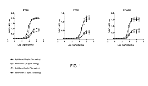
[0034] FIG. 1 shows binding of recombinantly expressed PT66, PT69/PT87,
hTau60 to
recombinant 2N4R tau analyzed by ELISA.
[0035] FIG. 2 shows western blot profiling of PT66, hTau60 and PT69/PT87 on
brain
extracts from (from left to right): WT (slot 1) and Tau -/- (slot 2) mouse
brain, dog brain (slot 3),
monkey brain (slot 4), and human brain (slot 5), and a PHF prep derived from
postmortem AD
brain (slot 6).
[0036] FIG. 3 shows representative SPR binding data of PT66, hTau60 and
PT69/PT87
monoclonal antibodies (mAbs) to PHF-Tau and their respective Fab fragments to
PHF-Tau and
recombinant Tau. SPR binding sensorgrams of anti-tau antibodies and their
corresponding Fab
fragments against PHF-Tau and full-length rec. Tau protein. Individual traces
within each
sensorgram represent different concentrations of antibodies or Fabs injected.
The individual
traces correspond to 75 nM, 15 nM, 3 nM, 0.6 nM and 0.12 nM from top to
bottom. For HT7, the
top trace (concentration) is 15 nM. The solid black lines indicate global
kinetics fitting with
either bivalent analyte model (for mAbs with PHF-Tau) or 1:1 Langmuir model
(for Fabs with
PHF-Tau and recombinant Tau). For HT7, Fab fragment is not available.
[0037] FIG. 4 shows binding data on cryosections of on AD and non-AD brain
of PT66,
hTau60 and PT69/PT87.
[0038] FIG. 5 shows IHC profiling data from PT66, hTau60 and PT69/PT87.
Binding on
paraffin sections from WT, Tau -/- and P30 1S mice is presented.
[0039] FIG. 6A shows a schematic of the immunodepletion assay.
[0040] FIG. 6B shows the results of immunodepletion assay on the tested
antibodies
(hTau60, PT69, PT66 and an internal N-term binding tau mAb (PT26) tau seeds
derived from the
human AD brain tissue (square) and P30 1S spinal cord (triangle). PT66, hTau60
and PT69/PT87
inhibited tau seeding more effectively than the N-term antibody, as determined
using the FRET
assay. The immunodepleted fractions from human AD brain homogenates were also
analyzed
with a hTau60/hTau60 tau aggregate-specific MSD assay (circle).
[0041] FIG. 6C shows the results of a sequential immunodepletion (ID)
assay, wherein the
first round of immunodepletion assay (ID1) was conducted with each of the
antibodies PT93
(targeting N-terminal portion of Tau), PT51 (a HT7-like antibody) and hTau60
(targeting C-
CA 03214310 2023-09-20
WO 2022/201123 PCT/IB2022/052765
terminal portion of Tau) or without any antibody (no mAb), and the second
round of
immunodepletion assay (ID2) was conducted with the same or different antibody
as that used for
ID1. It was shown that ID2 with same antibody used for ID1 did not deplete
additional
aggregates, and that after ID1 with PT93, ID2 with PT51 (HT7-like) and hTau60
(C-term)
resulted in additional depletion of the Tau aggregates with hTau60 depleted
all remaining
aggregates.
[0042] FIG. 7A shows efficacy of hTau60 and PT69/PT87, compared to AT120,
PT/76 and
PT53 antibodies in an ePHF injection model (see, e.g., U.S. Pat. No.
10,766,953) co-injected
with Tau PHFs and test antibodies. *** P<0.0001 Bonferroni multiple
comparisons.
[0043] FIG. 7B shows efficacy of antibodies that bind to C-terminal PHF-tau
(C-term) PT81
and PT66 compared to antibodies that bind to other epitopes in the N-terminal
(N-term) or
middle (Mid) portion of Tau, in the ePHF injection model. The C-terminal
antibodies showed
strong reduction in tau seeding in vivo.
[0044] FIG. 7C shows that both C-terminal antibodies (PT66 and hTau60) and
PT3 retained
in vivo activity after i.p. dosing. Mice were injected with 20 mg/kg antibody
2x/week.
[0045] FIG. 8 shows epitope mapping data using linear peptide mapping of
internal C-
terminal antibodies.
[0046] FIG. 9 shows that the lower efficacy by N-terminal antibodies can be
explained by
more extensive processing at N-terminus of PHF-tau: C-terminal PHF-tau
antibodies PT66 and
hTau60 (C-term) are compared to N-terminal PHF-tau antibodies (PT93) in a
Western blotting
screen analyzing PHF-tau.
DETAILED DESCRIPTION OF THE INVENTION
[0047] Various publications, articles and patents are cited or described in
the background and
throughout the specification; each of these references is herein incorporated
by reference in its
entirety. Discussion of documents, acts, materials, devices, articles or the
like which has been
included in the present specification is for the purpose of providing context
for the application.
Such discussion is not an admission that any or all of these matters form part
of the prior art with
respect to any inventions disclosed or claimed.
Definitions
CA 03214310 2023-09-20
WO 2022/201123 PCT/IB2022/052765
11
[0048] Unless defined otherwise, all technical and scientific terms used
herein have the same
meaning commonly understood to one of ordinary skill in the art to which this
application
pertains. Otherwise, certain terms used herein have the meanings as set in the
specification. All
patents, published patent applications and publications cited herein are
incorporated by reference
as if set forth fully herein. It must be noted that as used herein and in the
appended claims, the
singular forms "a," "an," and "the" include plural reference unless the
context clearly dictates
otherwise.
[0049] Unless otherwise stated, any numerical value, such as a
concentration or a
concentration range described herein, is to be understood as being modified in
all instances by
the term "about." Thus, a numerical value typically includes 10% of the
recited value. For
example, a concentration of 1 mg/mL includes 0.9 mg/mL to 1.1 mg/mL. Likewise,
a
concentration range of 1% to 10% (w/v) includes 0.9% (w/v) to 11% (w/v). As
used herein, the
use of a numerical range expressly includes all possible subranges, all
individual numerical
values within that range, including integers within such ranges and fractions
of the values unless
the context clearly indicates otherwise.
[0050] As used herein, the conjunctive term "and/or" between multiple
recited elements is
understood as encompassing both individual and combined options. For instance,
where two
elements are conjoined by "and/or," a first option refers to the applicability
of the first element
without the second. A second option refers to the applicability of the second
element without the
first. A third option refers to the applicability of the first and second
elements together. Any one
of these options is understood to fall within the meaning, and therefore
satisfy the requirement of
the term "and/or" as used herein. Concurrent applicability of more than one of
the options is also
understood to fall within the meaning, and therefore satisfy the requirement
of the term "and/or."
[0051] Throughout this specification and the claims which follow, unless
the context requires
otherwise, the word "comprise," and variations such as "comprises" and
"comprising," will be
understood to imply the inclusion of a stated integer or step or group of
integers or steps but not
the exclusion of any other integer or step or group of integer or step. When
used herein the term
"comprising" can be substituted with the term "containing" or "including" or
sometimes when
used herein with the term "having."
[0052] When used herein "consisting of' excludes any element, step, or
ingredient not
specified in the claim element. When used herein, "consisting essentially of'
does not exclude
CA 03214310 2023-09-20
WO 2022/201123 PCT/IB2022/052765
12
materials or steps that do not materially affect the basic and novel
characteristics of the claim.
Any of the aforementioned terms of "comprising," "containing," "including,"
and "having,"
whenever used herein in the context of an aspect or embodiment of the
application can be
replaced with the term "consisting of' or "consisting essentially of' to vary
scopes of the
disclosure.
[0053] As used herein, the term "isolated" means a biological component
(such as a nucleic
acid, peptide or protein) has been substantially separated, produced apart
from, or purified away
from other biological components of the organism in which the component
naturally occurs, i.e.,
other chromosomal and extrachromosomal DNA and RNA, and proteins. Nucleic
acids, peptides
and proteins that have been "isolated" thus include nucleic acids and proteins
purified by
standard purification methods. "Isolated" nucleic acids, peptides and proteins
can be part of a
composition and still be isolated if such composition is not part of the
native environment of the
nucleic acid, peptide, or protein. The term also embraces nucleic acids,
peptides and proteins
prepared by recombinant expression in a host cell as well as chemically
synthesized nucleic
acids.
[0054] As used herein, the term "antibody" or "immunoglobulin" is used in a
broad sense
and includes immunoglobulin or antibody molecules including polyclonal
antibodies,
monoclonal antibodies including murine, human, human-adapted, humanized and
chimeric
monoclonal antibodies and antibody fragments.
[0055] In general, antibodies are proteins or peptide chains that exhibit
binding specificity to
a specific antigen. Antibody structures are well known. Immunoglobulins can be
assigned to five
major classes, namely IgA, IgD, IgE, IgG and IgM, depending on the heavy chain
constant
domain amino acid sequence. IgA and IgG are further sub-classified as the
isotypes IgA 1, IgA2,
IgG 1, IgG2, IgG3 and IgG4. Antibodies of the application include those that
have variations in
their Fc region such that they have altered properties as compared to wild
type Fc regions
including, but not limited to, extended half-life, reduced or increased ADCC
or CDC and
silenced Fc effector functions. Accordingly, the antibodies of the application
can be of any of the
five major classes or corresponding sub-classes. Preferably, the antibodies of
the application are
IgG 1, IgG2, IgG3 or IgG4. Antibody light chains of any vertebrate species can
be assigned to
one of two clearly distinct types, namely kappa and lambda, based on the amino
acid sequences
of their constant domains. Accordingly, the antibodies of the application can
contain a kappa or
CA 03214310 2023-09-20
WO 2022/201123 PCT/IB2022/052765
13
lambda light chain constant domain. According to particular embodiments, the
antibodies of the
application include heavy and/or light chain constant regions from mouse
antibodies or human
antibodies.
[0056] In addition to the heavy and light chain constant domains,
antibodies contain light and
heavy chain variable regions. An immunoglobulin light or heavy chain variable
region consists
of a "framework" region interrupted by "antigen-binding sites." The antigen-
binding sites are
defined using various terms and numbering schemes as follows:
(i) Kabat: "Complementarity Determining Regions" or "CDRs" are based on
sequence
variability (Wu and Kabat, J Exp Med. 132:211-50, 1970). Generally, the
antigen-binding
site has three CDRs in each variable region (e.g., HCDR1, HCDR2 and HCDR3 in
the
heavy chain variable region (VH) and LCDR1, LCDR2 and LCDR3 in the light chain
variable region (VL));
(ii) Chothia: The term "hypervariable region," "HVR" or "HV" refers to the
regions of an
antibody variable domain which are hypervariable in structure as defined by
Chothia and
Lesk (Chothia and Lesk, J Mol Biol. 196:901-17, 1987). Generally, the antigen-
binding
site has three hypervariable regions in each VH (H1, H2, H3) and VL (L1, L2,
L3).
Numbering systems as well as annotation of CDRs and HVs have been revised by
Abhinandan and Martin (Abhinandan and Martin, Mol Immunol. 45:3832-9, 2008);
(iii) IMGT: Another definition of the regions that form the antigen-binding
site has been
proposed by Lefranc (Lefranc et al., Dev Comp Immunol. 27:55-77, 2003) based
on the
comparison of V domains from immunoglobulins and T-cell receptors. The
International
ImMunoGeneTics (IMGT) database provides a standardized numbering and
definition of
these regions. The correspondence between CDRs, HVs and IMGT delineations is
described in Lefranc et al., 2003, Id.;
(iv) AbM: A compromise between Kabat and Chothia numbering schemes is the AbM
numbering convention described by Martin (Martin ACR (2010) Antibody
Engineering,
eds Kontermann R, Dubel S (Springer-Verlag, Berlin), Vol 2, pp 33-51);
(v) The antigen-binding site can also be delineated based on "Specificity
Determining
Residue Usage" (SDRU) (Almagro, Mol Recognit. 17:132-43, 2004), where SDR,
refers
to amino acid residues of an immunoglobulin that are directly involved in
antigen
contact.
CA 03214310 2023-09-20
WO 2022/201123 PCT/IB2022/052765
14
[0057] "Framework" or "framework sequence" is the remaining sequences
within the
variable region of an antibody other than those defined to be antigen-binding
site sequences.
Because the exact definition of an antigen-binding site can be determined by
various delineations
as described above, the exact framework sequence depends on the definition of
the antigen-
binding site. The framework regions (FRs) are the more highly conserved
portions of variable
domains. The variable domains of native heavy and light chains each comprise
four FRs (FR1,
FR2, FR3 and FR4, respectively) which generally adopt a beta-sheet
configuration, connected by
the three hypervariable loops. The hypervariable loops in each chain are held
together in close
proximity by the FRs and, with the hypervariable loops from the other chain,
contribute to the
formation of the antigen-binding site of antibodies. Structural analysis of
antibodies revealed the
relationship between the sequence and the shape of the binding site formed by
the
complementarity determining regions (Chothia et al., J. Mol. Biol. 227: 799-
817, 1992;
Tramontano et al., J. Mol. Biol. 215:175-182, 1990). Despite their high
sequence variability, five
of the six loops adopt just a small repertoire of main-chain conformations,
called "canonical
structures." These conformations are first of all determined by the length of
the loops and
secondly by the presence of key residues at certain positions in the loops and
in the framework
regions that determine the conformation through their packing, hydrogen
bonding or the ability
to assume unusual main-chain conformations.
[0058] As used herein, the term "antigen-binding fragment" refers to an
antibody fragment
such as, for example, a diabody, a Fab, a Fab', a F(ab')2, an Fv fragment, a
disulfide stabilized
Fv fragment (dsFv), a (dsFv)2, a bispecific dsFy (dsFv-dsFv'), a disulfide
stabilized diabody (ds
diabody), a single-chain antibody molecule (scFv), a single domain antibody
(sdab), an scFv
dimer (bivalent diabody), a multispecific antibody formed from a portion of an
antibody
comprising one or more CDRs, a camelized single domain antibody, a nanobody, a
domain
antibody, a bivalent domain antibody, or any other antibody fragment that
binds to an antigen but
does not comprise a complete antibody structure. An antigen-binding fragment
is capable of
binding to the same antigen to which the parent antibody or a parent antibody
fragment binds.
According to particular embodiments, the antigen-binding fragment comprises a
light chain
variable region, a light chain constant region, and an Fd segment of the
constant region of the
heavy chain. According to other particular embodiments, the antigen-binding
fragment
comprises Fab and F(ab').
CA 03214310 2023-09-20
WO 2022/201123 PCT/IB2022/052765
[0059] As used herein, the term "humanized antibody" refers to a non-human
antibody that is
modified to increase the sequence homology to that of a human antibody, such
that the antigen-
binding properties of the antibody are retained, but its antigenicity in the
human body is reduced.
[0060] As used herein, the term "epitope" refers to a site on an antigen to
which an
immunoglobulin, antibody, or antigen-binding fragment thereof, specifically
binds. Epitopes can
be formed either from contiguous amino acids or from noncontiguous amino acids
juxtaposed by
tertiary folding of a protein. Epitopes formed from contiguous amino acids are
typically retained
on exposure to denaturing solvents, whereas epitopes formed by tertiary
folding are typically lost
on treatment with denaturing solvents. An epitope typically includes at least
3, 4, 5, 6, 7, 8, 9, 10,
11, 12, 13, 14 or 15 amino acids in a unique spatial conformation. Methods of
determining
spatial conformation of epitopes include, for example, x-ray crystallography
and 2-dimensional
nuclear magnetic resonance. See, e.g., Epitope Mapping Protocols in Methods in
Molecular
Biology, Vol. 66, G. E. Morris, Ed. (1996).
[0061] As used herein, the term "tau" or "tau protein" refers to an
abundant central and
peripheral nervous system protein having multiple isoforms. In the human
central nervous
system (CNS), six major tau isoforms ranging in size from 352 to 441 amino
acids in length exist
due to alternative splicing (Hanger et al., Trends Mol Med. 15:112-9, 2009).
The isoforms differ
from each other by the regulated inclusion of 0-2 N-terminal inserts, and 3 or
4 tandemly
arranged microtubule-binding repeats, and are referred to as ON3R (SEQ ID NO:
46), 1N3R
(SEQ ID NO: 47), 2N3R (SEQ ID NO: 48), ON4R (SEQ ID NO: 49), 1N4R (SEQ ID NO:
50)
and 2N4R (SEQ ID NO: 51). As used herein, the term "control tau" refers to the
tau isoform of
SEQ ID NO: 51 that is devoid of phosphorylation and other post-translational
modifications. As
used herein, the term "tau" includes proteins comprising mutations, e.g.,
point mutations,
fragments, insertions, deletions and splice variants of full-length wild type
tau. The term "tau"
also encompasses post-translational modifications of the tau amino acid
sequence. Post-
translational modifications include, but are not limited to, phosphorylation.
As used herein, the
phrase "phosphorylated S433 of the tau protein" and similar phrases refer to a
phosphorylated
amino acid at a certain position, e.g., serine at position 433, of the full-
length wild type tau
protein.
[0062] Tau binds microtubules and regulates transport of cargo through
cells, a process that
can be modulated by tau phosphorylation. In AD and related disorders, abnormal
CA 03214310 2023-09-20
WO 2022/201123 PCT/IB2022/052765
16
phosphorylation of tau is prevalent and thought to precede and/or trigger
aggregation of tau into
fibrils, termed paired helical filaments (PHF). The major constituent of PHF
is hyper-
phosphorylated tau. As used herein, the term "paired helical filament-tau" or
"PHF-tau" refers to
tau aggregates in paired helical filaments. Two major regions in PHF structure
are evident in
electron microscopy, the fuzzy coat and the core filament; the fuzzy coat
being sensitive to
proteolysis and located outside of the filaments, and the protease-resistant
core of filaments
forming the backbone of PHFs (Wischik et al. Proc Natl Acad Sci USA. 85:4884-
8, 1988).
[0063] An "isolated monoclonal antibody that binds PHF-tau" or an "isolated
monoclonal
anti-PHF-tau antibody," as used herein, is intended to refer to a monoclonal
anti-PHF-tau
antibody which is substantially free of other antibodies having different
antigenic specificities
(for instance, an isolated monoclonal anti-PHF-tau antibody is substantially
free of antibodies
that specifically bind antigens other than PHF-tau). An isolated monoclonal
anti-PHF-tau
antibody can, however, have cross-reactivity to other related antigens, for
instance from other
species (such as PHF-tau species homologs).
[0064] As used herein, the term "specifically binds" or "specific binding"
refers to the ability
of an anti-PHF-tau antibody of the application to bind to a predetermined
target with a
dissociation constant (KD) of about 1x10⁶ M or tighter, for example, about 1
xl 0-7 M or less,
about 1x10⁸ M or less, about 1x10⁹ M or less, about 1x101 M or less, about
1x10¹¹ M or less,
about 1 xl 0- 12 M or less, or about 1x10¹³ M or less. The KD is obtained from
the ratio of Kd to
Ka (i.e., Kd/Ka) and is expressed as a molar concentration (M). KD values for
antibodies can be
determined using methods in the art in view of the present disclosure. For
example, the KD value
of an anti-PHF-tau antibody can be determined by using surface plasmon
resonance, such as by
using a biosensor system, e.g., a Biacore system, a ProteOn instrument
(BioRad) , a KinExA
instrument (Sapidyne), ELISA or competitive binding assays known to those
skilled in the art.
Typically, an anti-PHF-tau antibody binds to a predetermined target (i.e. PHF-
tau) with a KD that
is at least ten fold less than its KD for a nonspecific target as measured by
surface plasmon
resonance using, for example, a ProteOn Instrument (BioRad). The anti-PHF-tau
antibodies that
specifically bind to PHF-tau can, however, have cross-reactivity to other
related targets, for
example, to the same predetermined target from other species (homologs).
[0065] As used herein, the term "polynucleotide," synonymously referred to
as "nucleic acid
molecule," "nucleotides" or "nucleic acids," refers to any polyribonucleotide
or
CA 03214310 2023-09-20
WO 2022/201123 PCT/IB2022/052765
17
polydeoxyribonucleotide, which can be unmodified RNA or DNA or modified RNA or
DNA.
"Polynucleotides" include, without limitation single- and double-stranded DNA,
DNA that is a
mixture of single- and double-stranded regions, single- and double-stranded
RNA, and RNA that
is mixture of single- and double-stranded regions, hybrid molecules comprising
DNA and RNA
that can be single-stranded or, more typically, double-stranded or a mixture
of single- and
double-stranded regions. In addition, "polynucleotide" refers to triple-
stranded regions
comprising RNA or DNA or both RNA and DNA. The term polynucleotide also
includes DNAs
or RNAs containing one or more modified bases and DNAs or RNAs with backbones
modified
for stability or for other reasons. "Modified" bases include, for example,
tritylated bases and
unusual bases such as inosine. A variety of modifications can be made to DNA
and RNA; thus,
"polynucleotide" embraces chemically, enzymatically or metabolically modified
forms of
polynucleotides as typically found in nature, as well as the chemical forms of
DNA and RNA
characteristic of viruses and cells. "Polynucleotide" also embraces relatively
short nucleic acid
chains, often referred to as oligonucleotides.
[0066] As used herein, the term "vector" is a replicon in which another
nucleic acid segment
can be operably inserted so as to bring about the replication or expression of
the segment.
[0067] As used herein, the term "host cell" refers to a cell comprising a
nucleic acid
molecule of the application. The "host cell" can be any type of cell, e.g., a
primary cell, a cell in
culture, or a cell from a cell line. In one embodiment, a "host cell" is a
cell transfected with a
nucleic acid molecule of the application. In another embodiment, a "host cell"
is a progeny or
potential progeny of such a transfected cell. A progeny of a cell may or may
not be identical to
the parent cell, e.g., due to mutations or environmental influences that can
occur in succeeding
generations or integration of the nucleic acid molecule into the host cell
genome.
[0068] The term "expression" as used herein, refers to the biosynthesis of
a gene product.
The term encompasses the transcription of a gene into RNA. The term also
encompasses
translation of RNA into one or more polypeptides, and further encompasses all
naturally
occurring post-transcriptional and post-translational modifications. The
expressed monoclonal
antibody or antigen-binding fragment thereof that binds PHF-tau can be within
the cytoplasm of
a host cell, into the extracellular milieu such as the growth medium of a cell
culture, or anchored
to the cell membrane.
CA 03214310 2023-09-20
WO 2022/201123 PCT/IB2022/052765
18
[0069] As used herein, the term "carrier" refers to any excipient, diluent,
filler, salt, buffer,
stabilizer, solubilizer, oil, lipid, lipid containing vesicle, microsphere,
liposomal encapsulation,
or other material well known in the art for use in pharmaceutical
formulations. It will be
understood that the characteristics of the carrier, excipient or diluent will
depend on the route of
administration for a particular application. As used herein, the term
"pharmaceutically acceptable
carrier" refers to a non-toxic material that does not interfere with the
effectiveness of a
composition according to the application or the biological activity of a
composition according to
the application. According to particular embodiments, in view of the present
disclosure, any
pharmaceutically acceptable carrier suitable for use in an antibody
pharmaceutical composition
can be used in the invention.
[0070] As used herein, the term "subject" refers to an animal, and
preferably a mammal.
According to particular embodiments, the subject is a mammal including a non-
primate (e.g., a
camel, donkey, zebra, cow, pig, horse, goat, sheep, cat, dog, rat, rabbit,
guinea pig or mouse) or a
primate (e.g., a monkey, chimpanzee, or human). In particular embodiments, the
subject is a
human.
[0071] As used herein, the term "therapeutically effective amount" refers
to an amount of an
active ingredient or component that elicits the desired biological or
medicinal response in a
subject. A therapeutically effective amount can be determined empirically and
in a routine
manner, in relation to the stated purpose. For example, in vitro assays can
optionally be
employed to help identify optimal dosage ranges. Selection of a particular
effective dose can be
determined (e.g., via clinical trials) by those skilled in the art based upon
the consideration of
several factors, including the disease to be treated or prevented, the
symptoms involved, the
patient's body mass, the patient's immune status and other factors known by
the skilled artisan.
The precise dose to be employed in the formulation will also depend on the
route of
administration, and the severity of disease, and should be decided according
to the judgment of
the practitioner and each patient's circumstances. Effective doses can be
extrapolated from dose-
response curves derived from in vitro or animal model test systems.
[0072] As used herein, the terms "treat," "treating," and "treatment" are
all intended to refer
to an amelioration or reversal of at least one measurable physical parameter
related to a
tauopathy which is not necessarily discernible in the subject, but can be
discernible in the
subject. The terms "treat," "treating," and "treatment," can also refer to
causing regression,
CA 03214310 2023-09-20
WO 2022/201123 PCT/IB2022/052765
19
preventing the progression, or at least slowing down the progression of the
disease, disorder, or
condition. In a particular embodiment, "treat," "treating," and "treatment"
refer to an alleviation,
prevention of the development or onset, or reduction in the duration of one or
more symptoms
associated with the tauopathy. In a particular embodiment, "treat,"
"treating," and "treatment"
refer to prevention of the recurrence of the disease, disorder, or condition.
In a particular
embodiment, "treat," "treating," and "treatment" refer to an increase in the
survival of a subject
having the disease, disorder, or condition. In a particular embodiment,
"treat," "treating," and
"treatment" refer to elimination of the disease, disorder, or condition in the
subject.
[0073] As used herein a "tauopathy" encompasses any neurodegenerative
disease that
involves the pathological aggregation of tau within the brain. In addition to
familial and sporadic
AD, other exemplary tauopathies are frontotemporal dementia with parkinsonism
linked to
chromosome 17 (FTDP-17), progressive supranuclear palsy, corticobasal
degeneration, Pick's
disease, progressive subcortical gliosis, tangle only dementia, diffuse
neurofibrillary tangles with
calcification, argyrophilic grain dementia, amyotrophic lateral sclerosis
parkinsonism-dementia
complex, Down syndrome, Gerstmann-Straussler-Scheinker disease, Hallervorden-
Spatz disease,
inclusion body myositis, Creutzfeld-Jakob disease, multiple system atrophy,
Niemann-Pick
disease type C, prion protein cerebral amyloid angiopathy, subacute sclerosing
panencephalitis,
myotonic dystrophy, non-Guamanian motor neuron disease with neurofibrillary
tangles,
postencephalitic parkinsonism, and chronic traumatic encephalopathy, such as
dementia
pugulistica (boxing disease) (Morris et al., Neuron, 70:410-26, 2011).
[0074] As used herein, the term "in combination," in the context of the
administration of two
or more therapies to a subject, refers to the use of more than one therapy.
The use of the term "in
combination" does not restrict the order in which therapies are administered
to a subject. For
example, a first therapy (e.g., a composition described herein) can be
administered prior to (e.g.,
minutes, 15 minutes, 30 minutes, 45 minutes, 1 hour, 2 hours, 4 hours, 6
hours, 12 hours, 16
hours, 24 hours, 48 hours, 72 hours, 96 hours, 1 week, 2 weeks, 3 weeks, 4
weeks, 5 weeks, 6
weeks, 8 weeks, or 12 weeks before), concomitantly with, or subsequent to
(e.g., 5 minutes, 15
minutes, 30 minutes, 45 minutes, 1 hour, 2 hours, 4 hours, 6 hours, 12 hours,
16 hours, 24 hours,
48 hours, 72 hours, 96 hours, 1 week, 2 weeks, 3 weeks, 4 weeks, 5 weeks, 6
weeks, 8 weeks, or
12 weeks after) the administration of a second therapy to a subject.
Anti-PHF-tau antibodies
CA 03214310 2023-09-20
WO 2022/201123 PCT/IB2022/052765
[0075] In one general aspect, the application relates to isolated
monoclonal antibodies or
antigen-binding fragments thereof that bind PHF-tau. Such anti-PHF-tau
antibodies can have the
properties of binding a phosphorylated epitope on PHF-tau or binding to a non-
phosphorylated
epitope on PHF-tau. Anti-PHF-tau antibodies can be useful as therapeutics, and
as research or
diagnostic reagents to detect PHF-tau in biological samples, for example in
tissues or cells.
[0076] According to a particular aspect, the application relates to an
isolated antibody or an
antigen-binding fragment thereof that binds to a tau protein at an epitope in
the C-terminus
domain of the tau protein. In some embodiments, the isolated monoclonal
antibody or antigen-
binding fragment thereof binds to a tau protein at an epitope of the tau
protein having or within
the amino acid sequence of SEQ ID NO: 1, wherein the antibody or antigen-
binding fragment
thereof binds PHF-tau, preferably human PHF-tau. Preferably, the isolated
monoclonal antibody
or antigen-binding fragment thereof binds to a tau protein at an epitope of
the tau protein
consisting of or within the amino acid sequence of SEQ ID NO: 1, wherein the
antibody or
antigen-binding fragment thereof binds PHF-tau, preferably human PHF-tau.
[0077] In some embodiments, the epitope of the tau protein comprises either
one of
phosphorylated S433 or phosphorylated S435 of the tau protein, but does not
comprise
phosphorylated S433 and phosphorylated S435; or the epitope of the tau protein
comprises one
or more of phosphorylated T427, phosphorylated S433 and phosphorylated S435 of
the tau
protein, but does not comprise all of phosphorylated T427, phosphorylated S433
and
phosphorylated S435; or the epitope of the tau protein comprises one or more
of phosphorylated
T427 and phosphorylated S433 of the tau protein, but does not comprise
phosphorylated S435,
and does not comprise all of phosphorylated T427, phosphorylated S433 and
phosphorylated
S435; or the epitope of the tau protein comprises phosphorylated T427 of the
tau protein, but
does not comprise phosphorylated S433 or phosphorylated S435.
[0078] Antibodies of the present invention can be produced by a variety of
techniques, for
example by the hybridoma method (Kohler and Milstein, Nature. 256:495-7,
1975). Chimeric
monoclonal antibodies containing a light chain and heavy chain variable region
derived from a
donor antibody (typically murine) in association with light and heavy chain
constant regions
derived from an acceptor antibody (typically another mammalian species such as
human) can be
prepared by a method disclosed in US4816567. CDR-grafted monoclonal antibodies
having
CDRs derived from a non-human donor immunoglobulin (typically murine) and the
remaining
CA 03214310 2023-09-20
WO 2022/201123 PCT/IB2022/052765
21
immunoglobulin-derived parts of the molecule being derived from one or more
human
immunoglobulins can be prepared by techniques known to those skilled in the
art such as that
disclosed in US5225539. Fully human monoclonal antibodies lacking any non-
human sequences
can be prepared from human immunoglobulin transgenic mice by techniques
referenced in
Lonberg et al., Nature. 368:856-9, 1994; Fishwild et al., Nat Biotechnol.
14:845-51, 1996; and
Mendez et al., Nat Genet. 15:146-56, 1997. Human monoclonal antibodies can
also be prepared
and optimized from phage display libraries (see, e.g., Knappik et al., J Mol
Biol. 296:57-86,
2000; Krebs et al., J Immunol Methods. 254:67-84, 2001; Shi et al., J Mol
Biol. 397:385-96,
2010).
[0079] According to a particular aspect, the application relates to
isolated monoclonal
antibodies or antigen-binding fragments thereof comprising:
(a) immunoglobulin heavy chain HCDR1, HCDR2 and HCDR3 having the polypeptide
sequences of SEQ ID NOs: 4, 5 and 6, respectively; and immunoglobulin light
chain
LCDR1, LCDR2 and LCDR3 having the polypeptide sequences of SEQ ID NOs: 7, 8
and
9, respectively;
(b) immunoglobulin heavy chain HCDR1, HCDR2 and HCDR3 having the polypeptide
sequences of SEQ ID NOs: 14, 15 and 16, respectively; and immunoglobulin light
chain
LCDR1, LCDR2 and LCDR3 having the polypeptide sequences of SEQ ID NOs: 17, 18
and 19, respectively;
(c) immunoglobulin heavy chain HCDR1, HCDR2 and HCDR3 having the polypeptide
sequences of SEQ ID NOs: 24, 25 and 26, respectively; and immunoglobulin light
chain
LCDR1, LCDR2 and LCDR3 having the polypeptide sequences of SEQ ID NOs: 27, 18
and 19, respectively;
(d) immunoglobulin heavy chain HCDR1, HCDR2 and HCDR3 having the polypeptide
sequences of SEQ ID NOs: 32, 33 and 34, respectively; and immunoglobulin light
chain
LCDR1, LCDR2 and LCDR3 having the polypeptide sequences of SEQ ID NOs: 17, 18
and 35, respectively; or
(e) immunoglobulin heavy chain HCDR1, HCDR2 and HCDR3 having the polypeptide
sequences of SEQ ID NOs: 40, 41 and 42, respectively; and immunoglobulin light
chain
LCDR1, LCDR2 and LCDR3 having the polypeptide sequences of SEQ ID NOs: 17, 18
and 43, respectively.
CA 03214310 2023-09-20
WO 2022/201123 PCT/IB2022/052765
22
[0080] Provided herein are isolated monoclonal antibodies or antigen-
binding fragments
thereof comprising a heavy chain variable region having a polypeptide sequence
at least 90%
identical to SEQ ID NO: 2, 12, 22, 30 or 38, or a light chain variable region
having a polypeptide
sequence at least 90% identical to SEQ ID NO: 3, 13, 23, 31 or 39. In some
embodiments, the
isolated monoclonal antibody or antigen-binding fragment thereof comprises a
heavy chain
variable region having a polypeptide sequence of SEQ ID NO: 2, 12, 22, 30 or
38, or a light
chain variable region having a polypeptide sequence of SEQ ID NO: 3, 13, 23,
31 or 39. In some
embodiments, the isolated monoclonal antibody or antigen-binding fragment
thereof comprises a
heavy chain variable region consisting of a polypeptide sequence of SEQ ID NO:
2, 12, 22, 30 or
38, or a light chain variable region consisting of a polypeptide sequence of
SEQ ID NO: 3, 13,
23,31 or 39.
[0081] According to a particular aspect, the application relates to
isolated monoclonal
antibodies or antigen-binding fragments thereof comprising:
(a) a heavy chain variable region having a polypeptide sequence at least 90%,
such as at least
90%, 91%, 92%, 93%, 94%, 95%, 96%, 97%, 98%, 99% or 100% identical to the
amino
acid sequence of SEQ ID NO: 2, and a light chain variable region having a
polypeptide
sequence at least 90%, such as at least 90%, 91%, 92%, 93%, 94%, 95%, 96%,
97%,
98%, 99% or 100% identical to the amino acid sequence of SEQ ID NO: 3;
(b) a heavy chain variable region having a polypeptide sequence at least 90%,
such as at least
90%, 91%, 92%, 93%, 94%, 95%, 96%, 97%, 98%, 99% or 100% identical to the
amino
acid sequence of SEQ ID NO: 12, and a light chain variable region having a
polypeptide
sequence at least 90%, such as at least 90%, 91%, 92%, 93%, 94%, 95%, 96%,
97%,
98%, 99% or 100% identical to the amino acid sequence of SEQ ID NO: 13;
(c) a heavy chain variable region having a polypeptide sequence at least 90%,
such as at least
90%, 91%, 92%, 93%, 94%, 95%, 96%, 97%, 98%, 99% or 100% identical to the
amino
acid sequence of SEQ ID NO: 22, and a light chain variable region having a
polypeptide
sequence at least 90%, such as at least 90%, 91%, 92%, 93%, 94%, 95%, 96%,
97%,
98%, 99% or 100% identical to the amino acid sequence of SEQ ID NO: 23;
(d) a heavy chain variable region having a polypeptide sequence at least 90%,
such as at least
90%, 91%, 92%, 93%, 94%, 95%, 96%, 97%, 98%, 99% or 100% identical to the
amino
acid sequence of SEQ ID NO: 30, and a light chain variable region having a
polypeptide
CA 03214310 2023-09-20
WO 2022/201123 PCT/IB2022/052765
23
sequence at least 90%, such as at least 90%, 91%, 92%, 93%, 94%, 95%, 96%,
97%,
98%, 99% or 100% identical to the amino acid sequence of SEQ ID NO: 31; or
(e) a heavy chain variable region having a polypeptide sequence at least 90%,
such as at least
90%, 91%, 92%, 93%, 94%, 95%, 96%, 97%, 98%, 99% or 100% identical to the
amino
acid sequence of SEQ ID NO: 38, and a light chain variable region having a
polypeptide
sequence at least 90%, such as at least 90%, 91%, 92%, 93%, 94%, 95%, 96%,
97%,
98%, 99% or 100% identical to the amino acid sequence of SEQ ID NO: 39.
[0082] According to another particular aspect, the application relates to
isolated monoclonal
antibodies or antigen-binding fragments thereof comprising:
(a) a heavy chain variable region having, preferably consisting of, the
polypeptide sequence
of SEQ ID NO: 2, and a light chain variable region having the polypeptide
sequence of
SEQ ID NO: 3;
(b) a heavy chain variable region having, preferably consisting of, the
polypeptide sequence
of SEQ ID NO: 12, and a light chain variable region having, preferably
consisting of, the
polypeptide sequence of SEQ ID NO: 13;
(c) a heavy chain variable region having, preferably consisting of, the
polypeptide sequence
of SEQ ID NO: 22, and a light chain variable region having, preferably
consisting of, the
polypeptide sequence of SEQ ID NO: 23;
(d) a heavy chain variable region having, preferably consisting of, the
polypeptide sequence
of SEQ ID NO: 30, and a light chain variable region having, preferably
consisting of, the
polypeptide sequence of SEQ ID NO: 31; or
(e) a heavy chain variable region having, preferably consisting of, the
polypeptide sequence
of SEQ ID NO: 38, and a light chain variable region having, preferably
consisting of, the
polypeptide sequence of SEQ ID NO: 39.
[0083] According to another particular aspect, the application relates to
isolated monoclonal
antibodies or antigen-binding fragments thereof comprising:
(a) a heavy chain having a polypeptide sequence at least 90%, such as at least
90%, 91%,
92%, 93%, 94%, 95%, 96%, 97%, 98%, 99% or 100% identical to the amino acid
sequence of SEQ ID NO: 10, and a light chain having a polypeptide sequence at
least
90%, such as at least 90%, 91%, 92%, 93%, 94%, 95%, 96%, 97%, 98%, 99% or 100%
identical to the amino acid sequence of SEQ ID NO: 11;
CA 03214310 2023-09-20
WO 2022/201123 PCT/IB2022/052765
24
(b) a heavy chain having a polypeptide sequence at least 90%, such as at least
90%, 91%,
92%, 93%, 94%, 95%, 96%, 97%, 98%, 99% or 100% identical to the amino acid
sequence of SEQ ID NO: 20, and a light chain having a polypeptide sequence at
least
90%, such as at least 90%, 91%, 92%, 93%, 94%, 95%, 96%, 97%, 98%, 99% or 100%
identical to the amino acid sequence of SEQ ID NO: 21;
(c) a heavy chain having a polypeptide sequence at least 90%, such as at least
90%, 91%,
92%, 93%, 94%, 95%, 96%, 97%, 98%, 99% or 100% identical to the amino acid
sequence of SEQ ID NO: 28, and a light chain having a polypeptide sequence at
least
90%, such as at least 90%, 91%, 92%, 93%, 94%, 95%, 96%, 97%, 98%, 99% or 100%
identical to the amino acid sequence of SEQ ID NO: 29;
(d) a heavy chain having a polypeptide sequence at least 90%, such as at least
90%, 91%,
92%, 93%, 94%, 95%, 96%, 97%, 98%, 99% or 100% identical to the amino acid
sequence of SEQ ID NO: 36, and a light chain having a polypeptide sequence at
least
90%, such as at least 90%, 91%, 92%, 93%, 94%, 95%, 96%, 97%, 98%, 99% or 100%
identical to the amino acid sequence of SEQ ID NO: 37; or
(e) a heavy chain having a polypeptide sequence at least 90%, such as at least
90%, 91%,
92%, 93%, 94%, 95%, 96%, 97%, 98%, 99% or 100% identical to the amino acid
sequence of SEQ ID NO: 44, and a light chain having a polypeptide sequence at
least
90%, such as at least 90%, 91%, 92%, 93%, 94%, 95%, 96%, 97%, 98%, 99% or 100%
identical to the amino acid sequence of SEQ ID NO: 45.
[0084] According to another particular aspect, the application relates to
isolated monoclonal
antibodies or antigen-binding fragments thereof comprising:
(a) a heavy chain having, preferably consisting of, the polypeptide sequence
of SEQ ID NO:
10, and a light chain having the polypeptide sequence of SEQ ID NO: 11;
(b) a heavy chain having, preferably consisting of, the polypeptide sequence
of SEQ ID NO:
20, and a light chain having the polypeptide sequence of SEQ ID NO: 21;
(c) a heavy chain having, preferably consisting of, the polypeptide sequence
of SEQ ID NO:
28, and a light chain having the polypeptide sequence of SEQ ID NO: 29;
(d) a heavy chain having, preferably consisting of, the polypeptide sequence
of SEQ ID NO:
36, and a light chain having the polypeptide sequence of SEQ ID NO: 37; or
CA 03214310 2023-09-20
WO 2022/201123 PCT/IB2022/052765
(e) a heavy chain having, preferably consisting of, the polypeptide sequence
of SEQ ID NO:
44, and a light chain having the polypeptide sequence of SEQ ID NO: 45.
[0085] In some embodiments, an isolated monoclonal antibody or antigen-
binding fragment
thereof of the application comprises:
(a) an immunoglobulin heavy chain HCDR1, HCDR2 and HCDR3 having, preferably
consisting of, the polypeptide sequences of SEQ ID NOs: 4, 5 and 6,
respectively; and
immunoglobulin light chain LCDR1, LCDR2 and LCDR3 having, preferably
consisting
of, the polypeptide sequences of SEQ ID NOs: 7, 8 and 9, respectively;
(b) a heavy chain variable region having, preferably consisting of, the
polypeptide sequence
of SEQ ID NO: 2, and a light chain variable region having, preferably
consisting of, the
polypeptide sequence of SEQ ID NO: 3; or
(c) a heavy chain having, preferably consisting of, the polypeptide sequence
of SEQ ID NO:
10, and a light chain having, preferably consisting of, the polypeptide
sequence of SEQ
ID NO: 11.
[0086] In some embodiments, an isolated monoclonal antibody or antigen-
binding fragment
thereof of the application comprises:
(a) an immunoglobulin heavy chain HCDR1, HCDR2 and HCDR3 having, preferably
consisting of, the polypeptide sequences of SEQ ID NOs: 14, 15 and 16,
respectively;
and immunoglobulin light chain LCDR1, LCDR2 and LCDR3 having, preferably
consisting of, the polypeptide sequences of SEQ ID NOs: 17, 18 and 19,
respectively;
(b) a heavy chain variable region having, preferably consisting of, the
polypeptide sequence
of SEQ ID NO: 12, and a light chain variable region having, preferably
consisting of, the
polypeptide sequence of SEQ ID NO: 13; or
(c) a heavy chain having the polypeptide sequence of SEQ ID NO: 20, and a
light chain
having the polypeptide sequence of SEQ ID NO: 21.
[0087] According to another particular aspect, the application relates to
an isolated
monoclonal antibody or antigen-binding fragment thereof, wherein the antibody
or antigen-
binding fragment binds to PHF-tau with a dissociation constant (KD) of 5x10⁻⁹
M or less,
preferably a KD of lx10-9 M or less or lx10-10 M or less, wherein the KD is
measured by
surface plasmon resonance analysis, such as by using a Biacore or ProteOn
system.
CA 03214310 2023-09-20
WO 2022/201123 PCT/IB2022/052765
26
[0088] The functional activity of monoclonal antibodies and antigen-binding
fragments
thereof that bind PHF-tau can be characterized by methods known in the art and
as described
herein. Methods for characterizing antibodies and antigen-binding fragments
thereof that bind
PHF-tau include, but are not limited to, affinity and specificity assays
including Biacore, ELISA,
and FACS analysis; immunohistochemistry analysis; in vitro cellular assays and
in vivo injection
assays to determine the efficacy of the antibodies in inhibiting tau seeding;
cell cytotoxicity
assays to detect the presence of antibody-dependent cell-mediated cytotoxicity
(ADCC), and
complement dependent cytotoxicity (CDC) activity of the antibodies; etc.
According to particular
embodiments, methods for characterizing antibodies and antigen-binding
fragments thereof that
bind PHF-tau include those described in the Examples below. An exemplary mouse
parental
antibody of monoclonal antibodies binding PHF-tau but not control tau is
antibody PT3, which is
described in US Patent No. 9,371,376, the content of which is incorporated
herein by reference
in its entirety.
[0089] Several well-known methodologies can be employed to determine the
binding epitope
of the antibodies of the application. For example, when the structures of both
individual
components are known, in silico protein-protein docking can be carried out to
identify
compatible sites of interaction. Hydrogen-deuterium (H/D) exchange can be
carried out with the
antigen and antibody complex to map regions on the antigen that are bound by
the antibody.
Segment and point mutagenesis of the antigen can be used to locate amino acids
important for
antibody binding. The co-crystal structure of an antibody-antigen complex is
used to identify
residues contributing to the epitope and paratope. According to particular
embodiments, methods
for determining the binding epitope of antibodies of the application include
those described in
Examples below.
[0090] Antibodies of the application can be bispecific or multispecific. An
exemplary
bispecific antibody can bind two distinct epitopes on PHF-tau or can bind PHF-
tau and amyloid
beta (Abeta). Another exemplary bispecific antibody can bind PHF-tau and an
endogenous
blood-brain barrier transcytosis receptor such as insulin receptor,
transferring receptor, insulin-
like growth factor-1 receptor, and lipoprotein receptor. An exemplary antibody
is of IgG1 type.
[0091] Immune effector properties of the antibodies of the application can
be enhanced or
silenced through Fc modifications by techniques known to those skilled in the
art. For example,
Fc effector functions such as C lq binding, complement dependent cytotoxicity
(CDC), antibody-
CA 03214310 2023-09-20
WO 2022/201123 PCT/IB2022/052765
27
dependent cell-mediated cytotoxicity (ADCC), phagocytosis, down regulation of
cell surface
receptors (e.g., B cell receptor; BCR), etc. can be provided and/or controlled
by modifying
residues in the Fc responsible for these activities. Pharmacokinetic
properties can also be
enhanced by mutating residues in the Fc domain that extend antibody half-life
(Strohl, Curr Opin
Biotechnol. 20:685-91, 2009).
[0092] Additionally, antibodies of the application can be post-
translationally modified by
processes such as glycosylation, isomerization, deglycosylation or non-
naturally occurring
covalent modification such as the addition of polyethylene glycol moieties and
lipidation. Such
modifications can occur in vivo or in vitro. For example, the antibodies of
the application can be
conjugated to polyethylene glycol (PEGylated) to improve their pharmacokinetic
profiles.
Conjugation can be carried out by techniques known to those skilled in the
art. Conjugation of
therapeutic antibodies with PEG has been shown to enhance pharmacodynamics
while not
interfering with function (Knight et al., Platelets. 15:409-18, 2004; Leong et
al., Cytokine.
16:106-19, 2001; Yang et al., Protein Eng. 16:761-70, 2003).
[0093] In another general aspect, the application relates to an isolated
polynucleotide
encoding a monoclonal antibody or antigen-binding fragment thereof of the
application. It will
be appreciated by those skilled in the art that the coding sequence of a
protein can be changed
(e.g., replaced, deleted, inserted, etc.) without changing the amino acid
sequence of the protein.
Accordingly, it will be understood by those skilled in the art that nucleic
acid sequences
encoding monoclonal antibodies or antigen-binding fragments thereof of the
application can be
altered without changing the amino acid sequences of the proteins. Exemplary
isolated
polynucleotides are polynucleotides encoding polypeptides comprising the
immunoglobulin
heavy chain and light chains described in the Examples (e.g., SEQ ID NOs: 10,
11, 20, 21, 28,
29, 36, 37, 44, 45) and polynucleotides encoding polypeptides comprising the
heavy chain
variable regions (VH) and light chain variable regions (VL) (e.g., SEQ ID NOs:
2, 3, 12, 13, 22,
23, 30, 31, 38, 39). Other polynucleotides which, given the degeneracy of the
genetic code or
codon preferences in a given expression system, encode the antibodies of the
application are also
within the scope of the application. The isolated nucleic acids of the present
invention can be
made using well known recombinant or synthetic techniques. DNA encoding the
monoclonal
antibodies is readily isolated and sequenced using methods known in the art.
Where a hybridoma
is produced, such cells can serve as a source of such DNA. Alternatively,
display techniques
CA 03214310 2023-09-20
WO 2022/201123 PCT/IB2022/052765
28
wherein the coding sequence and the translation product are linked, such as
phage or ribosomal
display libraries, can be used.
[0094] In another general aspect, the application relates to a vector
comprising an isolated
polynucleotide encoding a monoclonal antibody or antigen-binding fragment
thereof of the
application. Any vector known to those skilled in the art in view of the
present disclosure can be
used, such as a plasmid, a cosmid, a phage vector or a viral vector. In some
embodiments, the
vector is a recombinant expression vector such as a plasmid. The vector can
include any element
to establish a conventional function of an expression vector, for example, a
promoter, ribosome
binding element, terminator, enhancer, selection marker, and origin of
replication. The promoter
can be a constitutive, inducible or repressible promoter. A number of
expression vectors capable
of delivering nucleic acids to a cell are known in the art and can be used
herein for production of
an antibody or antigen-binding fragment thereof in the cell. Conventional
cloning techniques or
artificial gene synthesis can be used to generate a recombinant expression
vector according to
embodiments of the application.
[0095] In another general aspect, the application relates to a host cell
comprising an isolated
polynucleotide encoding a monoclonal antibody or antigen-binding fragment
thereof of the
application. Any host cell known to those skilled in the art in view of the
present disclosure can
be used for recombinant expression of antibodies or antigen-binding fragments
thereof of the
application. Such host cells can be eukaryotic cells, bacterial cells, plant
cells or archaeal cells.
Exemplary eukaryotic cells can be of mammalian, insect, avian or other animal
origins.
Mammalian eukaryotic cells include immortalized cell lines such as hybridomas
or myeloma cell
lines such as 5P2/0 (American Type Culture Collection (ATCC), Manassas, Va.,
CRL-1581),
NSO (European Collection of Cell Cultures (ECACC), Salisbury, Wiltshire, UK,
ECACC No.
85110503), FO (ATCC CRL-1646) and Ag653 (ATCC CRL-1580) murine cell lines. An
exemplary human myeloma cell line is U266 (ATTC CRL-TIB-196). Other useful
cell lines
include those derived from Chinese Hamster Ovary (CHO) cells such as CHO-K1 SV
(Lonza
Biologics), CHO-K1 (ATCC CRL-61, Invitrogen) or DG44.
[0096] In another general aspect, the application relates to a method of
producing a
monoclonal antibody or antigen-binding fragment thereof of the application,
comprising
culturing a cell comprising a polynucleotide encoding the monoclonal antibody
or antigen-
binding fragment thereof under conditions to produce a monoclonal antibody or
antigen-binding
CA 03214310 2023-09-20
WO 2022/201123 PCT/IB2022/052765
29
fragment thereof of the application, and recovering the antibody or antigen-
binding fragment
thereof from the cell or cell culture (e.g., from the supernatant). Expressed
antibodies or antigen-
binding fragments thereof can be harvested from the cells and purified
according to conventional
techniques known in the art.
Pharmaceutical Compositions and Methods of Treatment
[0097] Anti-PHF-tau antibodies of the application or fragments thereof of
the application can
be used to treat, reduce or prevent symptoms in patients having a
neurodegenerative disease that
involves pathological aggregation of tau within the brain, or a tauopathy,
such as patients
suffering from AD.
[0098] Thus, in another general aspect, the application relates to a
pharmaceutical
composition comprising an isolated monoclonal antibody or antigen-binding
fragment thereof of
the application and a pharmaceutically acceptable carrier.
[0099] In another general aspect, the application relates to a method of
blocking tau seeding
in a subject in need thereof, comprising administering to the subject a
pharmaceutical
composition of the application. A "tau seed" as used herein refers to a tau
aggregate capable of
nucleating or "seeding" intracellular tau aggregation when internalized by a
cell, or when
exposed to monomeric tau in vitro. Tau seeding activity may be assessed in
cellular tau
aggregation assays as described herein (see also e.g., US Patent No. 9,834,596
which is
incorporated by reference in its entirety).
[00100] In another general aspect, the application relates to a method of
treating or reducing
symptoms of a disease, disorder or condition, such as a tauopathy, in a
subject in need thereof,
comprising administering to the subject a pharmaceutical composition of the
application.
[00101] In another general aspect, the application relates to a method of
reducing pathological
tau aggregation or spreading of tauopathy in a subject in need thereof,
comprising administering
to the subject a pharmaceutical composition of the application.
[00102] According to embodiments of the application, the pharmaceutical
composition
comprises a therapeutically effective amount of the monoclonal anti-PHF-tau
antibody or
antigen-binding fragment thereof. As used herein with reference to monoclonal
anti-PHF-tau
antibodies or antigen-binding fragments thereof, a therapeutically effective
amount means an
amount of the monoclonal anti-PHF-tau antibody or antigen-binding fragment
thereof that results
in treatment of a disease, disorder, or condition; prevents or slows the
progression of the disease,
CA 03214310 2023-09-20
WO 2022/201123 PCT/IB2022/052765
disorder, or condition; or reduces or completely alleviates symptoms
associated with the immune
disease, disorder, or condition.
[00103] According to particular embodiments, a therapeutically effective
amount refers to the
amount of therapy which is sufficient to achieve one, two, three, four, or
more of the following
effects: (i) reduce or ameliorate the severity of the disease, disorder or
condition to be treated or
a symptom associated therewith; (ii) reduce the duration of the disease,
disorder or condition to
be treated, or a symptom associated therewith; (iii) prevent the progression
of the disease,
disorder or condition to be treated, or a symptom associated therewith; (iv)
cause regression of
the disease, disorder or condition to be treated, or a symptom associated
therewith; (v) prevent
the development or onset of the disease, disorder or condition to be treated,
or a symptom
associated therewith; (vi) prevent the recurrence of the disease, disorder or
condition to be
treated, or a symptom associated therewith; (vii) reduce hospitalization of a
subject having the
disease, disorder or condition to be treated, or a symptom associated
therewith; (viii) reduce
hospitalization length of a subject having the disease, disorder or condition
to be treated, or a
symptom associated therewith; (ix) increase the survival of a subject with the
disease, disorder or
condition to be treated, or a symptom associated therewith; (xi) inhibit or
reduce the disease,
disorder or condition to be treated, or a symptom associated therewith in a
subject; and/or (xii)
enhance or improve the prophylactic or therapeutic effect(s) of another
therapy.
[00104] According to particular embodiments, the disease, disorder or
condition to be treated
is a tauopathy. According to more particular embodiments, the disease,
disorder or condition to
be treated, includes, but is not limited to, familial Alzheimer's disease,
sporadic Alzheimer's
disease, frontotemporal dementia with parkinsonism linked to chromosome 17
(FTDP-17),
progressive supranuclear palsy, corticobasal degeneration, Pick's disease,
progressive subcortical
gliosis, tangle only dementia, diffuse neurofibrillary tangles with
calcification, argyrophilic grain
dementia, amyotrophic lateral sclerosis parkinsonism-dementia complex, Down
syndrome,
Gerstmann-Straussler-Scheinker disease, Hallervorden-Spatz disease, inclusion
body myositis,
Creutzfeld-Jakob disease, multiple system atrophy, Niemann-Pick disease type
C, prion protein
cerebral amyloid angiopathy, subacute sclerosing panencephalitis, myotonic
dystrophy, non-
Guamanian motor neuron disease with neurofibrillary tangles, postencephalitic
parkinsonism,
chronic traumatic encephalopathy, or dementia pugulistica (boxing disease).
CA 03214310 2023-09-20
WO 2022/201123 PCT/IB2022/052765
31
[00105] A tauopathy-related behavioral phenotype includes, but is not limited
to, cognitive
impairments, early personality change and disinhibition, apathy, abulia,
mutism, apraxia,
perseveration, stereotyped movements/behaviors, hyperorality, disorganization,
inability to plan
or organize sequential tasks, selfishness/callousness, antisocial traits, a
lack of empathy, halting,
agrammatic speech with frequent paraphasic errors but relatively preserved
comprehension,
impaired comprehension and word-finding deficits, slowly progressive gait
instability,
retropulsions, freezing, frequent falls, non-levodopa responsive axial
rigidity, supranuclear gaze
palsy, square wave jerks, slow vertical saccades, pseudobulbar palsy, limb
apraxia, dystonia,
cortical sensory loss, and tremor.
[00106] Patients amenable to treatment include, but are not limited to,
asymptomatic
individuals at risk of AD or other tauopathy, as well as patients presently
showing symptoms.
Patients amenable to treatment include individuals who have a known genetic
risk of AD, such
as a family history of AD or presence of genetic risk factors in the genome.
Exemplary risk
factors are mutations in the amyloid precursor protein (APP), especially at
position 717 and
positions 670 and 671 (Hardy and Swedish mutations, respectively). Other risk
factors are
mutations in the presenilin genes PS1 and PS2 and in ApoE4, family history of
hypercholesterolemia or atherosclerosis. Individuals presently suffering from
AD can be
recognized from characteristic dementia by the presence of risk factors
described above. In
addition, a number of diagnostic tests are available to identify individuals
who have AD. These
include measurement of cerebrospinal fluid tau and Abeta 42 levels. Elevated
tau and decreased
Abeta 42 levels signify the presence of AD. Individuals suffering from AD can
also be diagnosed
by AD and Related Disorders Association criteria.
[00107] Anti-PHF-tau antibodies of the application are suitable both as
therapeutic and
prophylactic agents for treating or preventing neurodegenerative diseases that
involve
pathological aggregation of tau, such as AD or other tauopathies. In
asymptomatic patients,
treatment can begin at any age (e.g., at about 10, 15, 20, 25, 30 years).
Usually, however, it is not
necessary to begin treatment until a patient reaches about 40, 50, 60, or 70
years. Treatment
typically entails multiple dosages over a period of time. Treatment can be
monitored by assaying
antibody or activated T-cell or B-cell responses to the therapeutic agent over
time. If the
response falls, a booster dosage can be indicated.
CA 03214310 2023-09-20
WO 2022/201123 PCT/IB2022/052765
32
[00108] In prophylactic applications, pharmaceutical compositions or
medicaments are
administered to a patient susceptible to, or otherwise at risk of, AD in an
amount sufficient to
eliminate or reduce the risk, lessen the severity, or delay the outset of the
disease, including
biochemical, histologic and/or behavioral symptoms of the disease, its
complications and
intermediate pathological phenotypes presented during development of the
disease. In
therapeutic applications, compositions or medicaments are administered to a
patient suspected
of, or already suffering from, such a disease in an amount sufficient to
reduce, arrest, or delay
any of the symptoms of the disease (biochemical, histologic and/or
behavioral). Administration
of a therapeutic can reduce or eliminate mild cognitive impairment in patients
that have not yet
developed characteristic Alzheimer's pathology.
[00109] The therapeutically effective amount or dosage can vary according to
various factors,
such as the disease, disorder or condition to be treated, the means of
administration, the target
site, the physiological state of the subject (including, e.g., age, body
weight, health), whether the
subject is a human or an animal, other medications administered, and whether
the treatment is
prophylactic or therapeutic. Treatment dosages are optimally titrated to
optimize safety and
efficacy.
[00110] The antibodies of the application can be prepared as pharmaceutical
compositions
containing a therapeutically effective amount of the antibody as an active
ingredient in a
pharmaceutically acceptable carrier. The carrier can be liquids, such as water
and oils, including
those of petroleum, animal, vegetable or synthetic origin, such as peanut oil,
soybean oil, mineral
oil, sesame oil and the like. For example, 0.4% saline and 0.3% glycine can be
used. These
solutions are sterile and generally free of particulate matter. They can be
sterilized by
conventional, well-known sterilization techniques (e.g., filtration). The
compositions can contain
pharmaceutically acceptable auxiliary substances as required to approximate
physiological
conditions such as pH adjusting and buffering agents, stabilizing, thickening,
lubricating and
coloring agents, etc. The concentration of the antibodies of the application
in such
pharmaceutical formulation can vary widely, i.e., from less than about 0.5%,
usually at or at least
about 1% to as much as 15 or 20% by weight and will be selected primarily
based on required
dose, fluid volumes, viscosities, etc., according to the particular mode of
administration selected.
[00111] The mode of administration for therapeutic use of the antibodies of
the application
can be any suitable route that delivers the agent to the host. For example,
the compositions
CA 03214310 2023-09-20
WO 2022/201123 PCT/IB2022/052765
33
described herein can be formulated to be suitable for parenteral
administration, e.g., intradermal,
intramuscular, intraperitoneal, intravenous, subcutaneous, intranasal or
intracranial
administration, or they can be administered into the cerebrospinal fluid of
the brain or spine.
[00112] The treatment can be given in a single dose schedule, or as a multiple
dose schedule
in which a primary course of treatment can be with 1-10 separate doses,
followed by other doses
given at subsequent time intervals required to maintain and or reinforce the
response, for
example, at 1-4 months for a second dose, and if needed, a subsequent dose(s)
after several
months. Examples of suitable treatment schedules include: (i) 0, 1 month and 6
months, (ii) 0, 7
days and 1 month, (iii) 0 and 1 month, (iv) 0 and 6 months, or other schedules
sufficient to elicit
the desired responses expected to reduce disease symptoms or reduce severity
of disease.
[00113] The antibodies of the application can be lyophilized for storage and
reconstituted in a
suitable carrier prior to use. This technique has been shown to be effective
with antibody and
other protein preparations and art-known lyophilization and reconstitution
techniques can be
employed.
[00114] According to particular embodiments, a composition used in the
treatment of a
tauopathy can be used in combination with other agents that are effective for
treatment of related
neurodegenerative diseases. In the case of AD, antibodies of the application
can be administered
in combination with agents that reduce or prevent the deposition of amyloid-
beta (Abeta). It is
possible that PHF-tau and Abeta pathologies are synergistic. Therefore,
combination therapy
targeting the clearance of both PHF-tau and Abeta and Abeta-related
pathologies at the same
time can be more effective than targeting each individually. In the case of
Parkinson's Disease
and related neurodegenerative diseases, immune modulation to clear aggregated
forms of the
alpha-synuclein protein is also an emerging therapy. A combination therapy
which targets the
clearance of both tau and alpha-synuclein proteins simultaneously can be more
effective than
targeting either protein individually.
[00115] In another general aspect, the application relates to a method of
producing a
pharmaceutical composition comprising a monoclonal antibody or antigen-binding
fragment
thereof of the application, comprising combining a monoclonal antibody or
antigen-binding
fragment thereof with a pharmaceutically acceptable carrier to obtain the
pharmaceutical
composition.
Diagnostic Methods and Kits
CA 03214310 2023-09-20
WO 2022/201123 PCT/IB2022/052765
34
[00116] Monoclonal anti-PHF-tau antibodies of the application can be used in
methods of
diagnosing AD or other tauopathies in a subject.
[00117] Thus, in another general aspect, the application relates to methods of
detecting the
presence of PHF-tau in a subject and methods of diagnosing tauopathies in a
subject by detecting
the presence of PHF-tau in the subject using a monoclonal antibody or antigen-
binding fragment
thereof of the application.
[00118] Phosphorylated tau can be detected in a biological sample from a
subject (e.g., blood,
serum, plasma, interstitial fluid, or cerebral spinal fluid sample) by
contacting the biological
sample with the diagnostic antibody reagent and detecting binding of the
diagnostic antibody
reagent to phosphorylated tau in the sample from the subject. Assays for
carrying out the
detection include well-known methods such as ELISA, immunohistochemistry,
western blot, or
in vivo imaging.
[00119] Diagnostic antibodies or similar reagents can be administered by
intravenous
injection into the body of the patient, or directly into the brain by any
suitable route that delivers
the agent to the host. The dosage of antibody should be within the same ranges
as for treatment
methods. Typically, the antibody is labeled, although in some methods, the
primary antibody
with affinity for phosphorylated tau is unlabeled, and a secondary labeling
agent is used to bind
to the primary antibody. The choice of label depends on the means of
detection. For example, a
fluorescent label is suitable for optical detection. Use of paramagnetic
labels is suitable for
tomographic detection without surgical intervention. Radioactive labels can
also be detected
using PET or SPECT.
[00120] Diagnosis is performed by comparing the number, size, and/or intensity
of labeled
PHF-tau, tau aggregates, and/or neurofibrillary tangles in a sample from the
subject or in the
subject, to corresponding baseline values. The baseline values can represent
the mean levels in a
population of healthy individuals. Baseline values can also represent previous
levels determined
in the same subject.
[00121] The diagnostic methods described above can also be used to monitor a
subject's
response to therapy by detecting the presence of phosphorylated tau in a
subject before, during or
after the treatment. A decrease in values relative to baseline signals a
positive response to
treatment. Values can also increase temporarily in biological fluids as
pathological tau is being
cleared from the brain.
CA 03214310 2023-09-20
WO 2022/201123 PCT/IB2022/052765
[00122] The present application is further directed to a kit for performing
the above described
diagnostic and monitoring methods. Typically, such kits contain a diagnostic
reagent such as the
antibodies of the application, and optionally a detectable label. The
diagnostic antibody itself can
contain the detectable label (e.g., fluorescent molecule, biotin, etc.) which
is directly detectable
or detectable via a secondary reaction (e.g., reaction with streptavidin).
Alternatively, a second
reagent containing the detectable label cab be used, where the second reagent
has binding
specificity for the primary antibody. In a diagnostic kit suitable for
measuring PHF-tau in a
biological sample, the antibodies of the kit can be supplied pre-bound to a
solid phase, such as to
the wells of a microtiter dish.
[00123] The contents of all cited references (including literature references,
issued patents,
published patent applications, and co-pending patent applications) cited
throughout this
application are hereby expressly incorporated by reference.
EMBODIMENTS
[00124] The application provides also the following non-limiting embodiments.
[00125] Embodiment 1 is an isolated monoclonal antibody or antigen-binding
fragment
thereof that binds to a tau protein at an epitope of the tau protein having or
within the amino acid
sequence of SEQ ID NO: 1, wherein the antibody or antigen-binding fragment
thereof binds
paired helical filament (PHF)-tau, preferably human PHF-tau.
[00126] Embodiment la is an isolated monoclonal antibody or antigen-binding
fragment
thereof that binds to a tau protein at an epitope of the tau protein
consisting of or within the
amino acid sequence of SEQ ID NO: 1, wherein the antibody or antigen-binding
fragment
thereof binds paired helical filament (PHF)-tau, preferably human PHF-tau.
[00127] Embodiment 2 is the isolated monoclonal antibody or antigen-binding
fragment
thereof of embodiment 1 or I a, wherein:
(a) the epitope of the tau protein comprises either one of phosphorylated S433
or
phosphorylated S435 of the tau protein, but does not comprise phosphorylated
S433 and
phosphorylated S435;
(b) the epitope of the tau protein comprises one or more of phosphorylated
T427,
phosphorylated S433 and phosphorylated S435 of the tau protein, but does not
comprise
all of phosphorylated T427, phosphorylated S433 and phosphorylated S435;
CA 03214310 2023-09-20
WO 2022/201123 PCT/IB2022/052765
36
(c) the epitope of the tau protein comprises one or more of phosphorylated
T427 and
phosphorylated S433 of the tau protein, but does not comprise phosphorylated
S435, and
does not comprise all of phosphorylated T427, phosphorylated S433 and
phosphorylated
S435; or
(d) the epitope of the tau protein comprises phosphorylated T427 of the tau
protein, but does
not comprise phosphorylated S433 or phosphorylated S435.
[00128] Embodiment 3 is the isolated monoclonal antibody or antigen-binding
fragment
thereof of any one of embodiments 1-2, wherein the monoclonal antibody or
antigen-binding
fragment thereof comprises:
(a) immunoglobulin heavy chain HCDR1, HCDR2 and HCDR3 having the polypeptide
sequences of SEQ ID NOs: 4, 5 and 6, respectively; and immunoglobulin light
chain
LCDR1, LCDR2 and LCDR3 having the polypeptide sequences of SEQ ID NOs: 7, 8
and
9, respectively;
(b) immunoglobulin heavy chain HCDR1, HCDR2 and HCDR3 having the polypeptide
sequences of SEQ ID NOs: 14, 15 and 16, respectively; and immunoglobulin light
chain
LCDR1, LCDR2 and LCDR3 having the polypeptide sequences of SEQ ID NOs: 17, 18
and 19, respectively;
(c) immunoglobulin heavy chain HCDR1, HCDR2 and HCDR3 having the polypeptide
sequences of SEQ ID NOs: 24, 25 and 26, respectively; and immunoglobulin light
chain
LCDR1, LCDR2 and LCDR3 having the polypeptide sequences of SEQ ID NOs: 27, 18
and 19, respectively;
(d) immunoglobulin heavy chain HCDR1, HCDR2 and HCDR3 having the polypeptide
sequences of SEQ ID NOs: 32, 33 and 34, respectively; and immunoglobulin light
chain
LCDR1, LCDR2 and LCDR3 having the polypeptide sequences of SEQ ID NOs: 17, 18
and 35, respectively; or
(e) immunoglobulin heavy chain HCDR1, HCDR2 and HCDR3 having the polypeptide
sequences of SEQ ID NOs: 40, 41 and 42, respectively; and immunoglobulin light
chain
LCDR1, LCDR2 and LCDR3 having the polypeptide sequences of SEQ ID NOs: 17, 18
and 43, respectively.
CA 03214310 2023-09-20
WO 2022/201123 PCT/IB2022/052765
37
[00129] Embodiment 3a is the isolated monoclonal antibody or antigen-binding
fragment
thereof of any one of embodiments 1-3, wherein the monoclonal antibody or
antigen-binding
fragment thereof comprises:
(a) immunoglobulin heavy chain HCDR1, HCDR2 and HCDR3 consisting of the
polypeptide sequences of SEQ ID NOs: 4, 5 and 6, respectively; and
immunoglobulin
light chain LCDR1, LCDR2 and LCDR3 consisting of the polypeptide sequences of
SEQ
ID NOs: 7, 8 and 9, respectively;
(b) immunoglobulin heavy chain HCDR1, HCDR2 and HCDR3 consisting of the
polypeptide sequences of SEQ ID NOs: 14, 15 and 16, respectively; and
immunoglobulin
light chain LCDR1, LCDR2 and LCDR3 consisting of the polypeptide sequences of
SEQ
ID NOs: 17, 18 and 19, respectively;
(c) immunoglobulin heavy chain HCDR1, HCDR2 and HCDR3 consisting of the
polypeptide sequences of SEQ ID NOs: 24, 25 and 26, respectively; and
immunoglobulin
light chain LCDR1, LCDR2 and LCDR3 consisting of the polypeptide sequences of
SEQ
ID NOs: 27, 18 and 19, respectively;
(d) immunoglobulin heavy chain HCDR1, HCDR2 and HCDR3 consisting of the
polypeptide sequences of SEQ ID NOs: 32, 33 and 34, respectively; and
immunoglobulin
light chain LCDR1, LCDR2 and LCDR3 consisting of the polypeptide sequences of
SEQ
ID NOs: 17, 18 and 35, respectively; or
(e) immunoglobulin heavy chain HCDR1, HCDR2 and HCDR3 consisting of the
polypeptide sequences of SEQ ID NOs: 40, 41 and 42, respectively; and
immunoglobulin
light chain LCDR1, LCDR2 and LCDR3 consisting of the polypeptide sequences of
SEQ
ID NOs: 17, 18 and 43, respectively.
[00130] Embodiment 4 is the isolated monoclonal antibody or antigen-binding
fragment
thereof of any one of embodiments 1-3a, comprising a heavy chain variable
region having a
polypeptide sequence at least 90% identical to SEQ ID NO: 2, 12, 22, 30 or 38,
or a light chain
variable region having a polypeptide sequence at least 90% identical to SEQ ID
NO: 3, 13, 23,
31 or 39.
[00131] Embodiment 5 is the isolated monoclonal antibody or antigen-binding
fragment
thereof of any one of embodiments 1-4, comprising a heavy chain variable
region having a
CA 03214310 2023-09-20
WO 2022/201123 PCT/IB2022/052765
38
polypeptide sequence of SEQ ID NO: 2, 12, 22, 30 or 38, or a light chain
variable region having
a polypeptide sequence of SEQ ID NO: 3, 13, 23, 31 or 39.
[00132] Embodiment 5a is the isolated monoclonal antibody or antigen-binding
fragment
thereof of any one of embodiments 1-5, comprising a heavy chain variable
region consisting of a
polypeptide sequence of any one of SEQ ID NO: 2, 12, 22, 30 or 38, or a light
chain variable
region consisting of a polypeptide sequence of any one of SEQ ID NO: 3, 13,
23, 31 or 39.
[00133] Embodiment 6 is the isolated monoclonal antibody or antigen-binding
fragment
thereof of any one of embodiments 1-5a, comprising:
(a) a heavy chain variable region having the polypeptide sequence of SEQ ID
NO: 2, and a
light chain variable region having the polypeptide sequence of SEQ ID NO: 3;
(b) a heavy chain variable region having the polypeptide sequence of SEQ ID
NO: 12, and a
light chain variable region having the polypeptide sequence of SEQ ID NO: 13;
(c) a heavy chain variable region having the polypeptide sequence of SEQ ID
NO: 22, and a
light chain variable region having the polypeptide sequence of SEQ ID NO: 23;
(d) a heavy chain variable region having the polypeptide sequence of SEQ ID
NO: 30, and a
light chain variable region having the polypeptide sequence of SEQ ID NO: 31;
or
(e) a heavy chain variable region having the polypeptide sequence of SEQ ID
NO: 38, and a
light chain variable region having the polypeptide sequence of SEQ ID NO: 39.
[00134] Embodiment 6a is the isolated monoclonal antibody or antigen-binding
fragment
thereof of any one of embodiments 1-6, comprising:
(a) a heavy chain variable region consisting of the polypeptide sequence of
SEQ ID NO: 2,
and a light chain variable region consisting of the polypeptide sequence of
SEQ ID NO:
3;
(b) a heavy chain variable region consisting of the polypeptide sequence of
SEQ ID NO: 12,
and a light chain variable region consisting of the polypeptide sequence of
SEQ ID NO:
13;
(c) a heavy chain variable region consisting of the polypeptide sequence of
SEQ ID NO: 22,
and a light chain variable region consisting of the polypeptide sequence of
SEQ ID NO:
23;
CA 03214310 2023-09-20
WO 2022/201123 PCT/IB2022/052765
39
(d) a heavy chain variable region consisting of the polypeptide sequence of
SEQ ID NO: 30,
and a light chain variable region consisting of the polypeptide sequence of
SEQ ID NO:
31; or
(e) a heavy chain variable region consisting of the polypeptide sequence of
SEQ ID NO: 38,
and a light chain variable region consisting of the polypeptide sequence of
SEQ ID NO:
39.
[00135] Embodiment 7 is the isolated monoclonal antibody or antigen-binding
fragment
thereof of any one of embodiments 1-6a, comprising:
(a) a heavy chain having the polypeptide sequence of SEQ ID NO: 10, and a
light chain
having the polypeptide sequence of SEQ ID NO: 11;
(b) a heavy chain having the polypeptide sequence of SEQ ID NO: 20, and a
light chain
having the polypeptide sequence of SEQ ID NO: 21;
(c) a heavy chain having the polypeptide sequence of SEQ ID NO: 28, and a
light chain
having the polypeptide sequence of SEQ ID NO: 29;
(d) a heavy chain having the polypeptide sequence of SEQ ID NO: 36, and a
light chain
having the polypeptide sequence of SEQ ID NO: 37; or
(e) a heavy chain having the polypeptide sequence of SEQ ID NO: 44, and a
light chain
having the polypeptide sequence of SEQ ID NO: 45.
[00136] Embodiment 7a is the isolated monoclonal antibody or antigen-binding
fragment
thereof of any one of embodiments 1-7, comprising:
(a) a heavy chain consisting of the polypeptide sequence of SEQ ID NO: 10, and
a light
chain consisting of the polypeptide sequence of SEQ ID NO: 11;
(b) a heavy chain consisting of the polypeptide sequence of SEQ ID NO: 20, and
a light
chain consisting of the polypeptide sequence of SEQ ID NO: 21;
(c) a heavy chain consisting of the polypeptide sequence of SEQ ID NO: 28, and
a light
chain consisting of the polypeptide sequence of SEQ ID NO: 29;
(d) a heavy chain consisting of the polypeptide sequence of SEQ ID NO: 36, and
a light
chain consisting of the polypeptide sequence of SEQ ID NO: 37; or
(e) a heavy chain consisting of the polypeptide sequence of SEQ ID NO: 44, and
a light
chain consisting of the polypeptide sequence of SEQ ID NO: 45.
CA 03214310 2023-09-20
WO 2022/201123 PCT/IB2022/052765
[00137] Embodiment 8 is the isolated monoclonal antibody or antigen-binding
fragment
thereof of any one of embodiments 1-7a, comprising:
(a) an immunoglobulin heavy chain HCDR1, HCDR2 and HCDR3 having the
polypeptide
sequences of SEQ ID NOs: 4, 5 and 6, respectively; and immunoglobulin light
chain
LCDR1, LCDR2 and LCDR3 having the polypeptide sequences of SEQ ID NOs: 7, 8
and
9, respectively;
(b) a heavy chain variable region having the polypeptide sequence of SEQ ID
NO: 2, and a
light chain variable region having the polypeptide sequence of SEQ ID NO: 3;
or
(c) a heavy chain having the polypeptide sequence of SEQ ID NO: 10, and a
light chain
having the polypeptide sequence of SEQ ID NO: 11.
[00138] Embodiment 9 is the isolated monoclonal antibody or antigen-binding
fragment
thereof of any one of embodiments 1-7a, comprising:
(a) an immunoglobulin heavy chain HCDR1, HCDR2 and HCDR3 having the
polypeptide
sequences of SEQ ID NOs: 14, 15 and 16, respectively; and immunoglobulin light
chain
LCDR1, LCDR2 and LCDR3 having the polypeptide sequences of SEQ ID NOs: 17, 18
and 19, respectively;
(b) a heavy chain variable region having the polypeptide sequence of SEQ ID
NO: 12, and a
light chain variable region having the polypeptide sequence of SEQ ID NO: 13;
or
(c) a heavy chain having the polypeptide sequence of SEQ ID NO: 20, and a
light chain
having the polypeptide sequence of SEQ ID NO: 21.
[00139] Embodiment 10 is an isolated nucleic acid encoding the monoclonal
antibody or
antigen-binding fragment thereof of any one of embodiments 1 to 9.
[00140] Embodiment 11 is a vector comprising the isolated nucleic acid of
embodiment 10.
[00141] Embodiment 12 is a host cell comprising the isolated nucleic acid of
embodiment 10.
[00142] Embodiment 13 is a pharmaceutical composition comprising the isolated
monoclonal
antibody or antigen-binding fragment thereof of any one of embodiments 1 to 9
and a
pharmaceutically acceptable carrier.
[00143] Embodiment 14 is a method of blocking tau seeding in a subject in need
thereof,
comprising administering to the subject the pharmaceutical composition of
embodiment 13.
[00144] Embodiment 15 is a method of treating a tauopathy in a subject in need
thereof,
comprising administering to the subject the pharmaceutical composition of
embodiment 13.
CA 03214310 2023-09-20
WO 2022/201123 PCT/IB2022/052765
41
[00145] Embodiment 16 is a method of reducing pathological tau aggregation or
spreading of
tauopathy in a subject in need thereof, comprising administering to the
subject the
pharmaceutical composition of embodiment 13.
[00146] Embodiment 17 is the method of embodiment 15 or 16, wherein the
tauopathy is
selected from the group consisting of familial Alzheimer's disease, sporadic
Alzheimer's
disease, frontotemporal dementia with parkinsonism linked to chromosome 17
(FTDP-17),
progressive supranuclear palsy, corticobasal degeneration, Pick's disease,
progressive subcortical
gliosis, tangle only dementia, diffuse neurofibrillary tangles with
calcification, argyrophilic grain
dementia, amyotrophic lateral sclerosis parkinsonism-dementia complex, Down
syndrome,
Gerstmann-Straussler-Scheinker disease, Hallervorden-Spatz disease, inclusion
body myositis,
Creutzfeld-Jakob disease, multiple system atrophy, Niemann-Pick disease type
C, prion protein
cerebral amyloid angiopathy, subacute sclerosing panencephalitis, myotonic
dystrophy, non-
Guamanian motor neuron disease with neurofibrillary tangles, postencephalitic
parkinsonism,
chronic traumatic encephalopathy, and dementia pugulistica (boxing disease).
[00147] Embodiment 17a is the method of any one of embodiments 15-17, further
comprising
administering to the subject an additional agent for treating the tauopathy in
the subject in need
thereof.
[00148] Embodiment 18 is a method of producing the monoclonal antibody or
antigen-
binding fragment thereof of any one of embodiments 1 to 9, comprising
culturing a cell
comprising a nucleic acid encoding the monoclonal antibody or antigen-binding
fragment thereof
under conditions to produce the monoclonal antibody or antigen-binding
fragment thereof and
recovering the monoclonal antibody or antigen-binding fragment thereof from
the cell or cell
culture.
[00149] Embodiment 19 is a method of detecting the presence of PHF-tau in a
biological
sample from a subject, comprising contacting the biological sample with the
isolated monoclonal
antibody or antigen-binding fragment thereof of any one of embodiments 1 to 9,
and detecting
binding of the monoclonal antibody or antigen-binding fragment thereof to PHF-
tau in the
sample from the subject.
[00150] Embodiment 20 is the method of embodiment 19, wherein the biological
sample is a
blood, serum, plasma, interstitial fluid, or cerebral spinal fluid sample.
CA 03214310 2023-09-20
WO 2022/201123 PCT/IB2022/052765
42
[00151] Embodiment 21 is a method of producing a pharmaceutical composition
comprising
the monoclonal antibody or antigen-binding fragment thereof of any one of
embodiments 1 to 9,
comprising combining the monoclonal antibody or antigen-binding fragment
thereof with a
pharmaceutically acceptable carrier to obtain the pharmaceutical composition.
[00152] Embodiment 22 is an isolated monoclonal antibody or antigen-binding
fragment
thereof of any one of embodiments 1 to 9 for use in treating a tauopathy, in a
subject in need
thereof.
[00153] Embodiment 23 is an isolated monoclonal antibody or antigen-binding
fragment
thereof of any one of embodiments 1 to 9 or the pharmaceutical composition of
embodiment 13
for use in treating a tauopathy, such as familial Alzheimer's disease,
sporadic Alzheimer's
disease, frontotemporal dementia with parkinsonism linked to chromosome 17
(FTDP-17),
progressive supranuclear palsy, corticobasal degeneration, Pick's disease,
progressive subcortical
gliosis, tangle only dementia, diffuse neurofibrillary tangles with
calcification, argyrophilic grain
dementia, amyotrophic lateral sclerosis parkinsonism-dementia complex, Down
syndrome,
Gerstmann-Straussler-Scheinker disease, Hallervorden-Spatz disease, inclusion
body myositis,
Creutzfeld-Jakob disease, multiple system atrophy, Niemann-Pick disease type
C, prion protein
cerebral amyloid angiopathy, subacute sclerosing panencephalitis, myotonic
dystrophy, non-
Guamanian motor neuron disease with neurofibrillary tangles, postencephalitic
parkinsonism,
chronic traumatic encephalopathy, or dementia pugulistica (boxing disease), in
a subject in need
thereof.
[00154] Embodiment 24 is a use of an isolated monoclonal antibody or antigen-
binding
fragment thereof of any one of embodiments 1 to 9 for manufacturing a
medicament in treating a
tauopathy in a subject in need thereof.
[00155] Embodiment 25 is a use of an isolated monoclonal antibody or antigen-
binding
fragment thereof of any one of embodiments 1 to 9 for manufacturing a
medicament for treating
a tauopathy, such as familial Alzheimer's disease, sporadic Alzheimer's
disease, frontotemporal
dementia with parkinsonism linked to chromosome 17 (FTDP-17), progressive
supranuclear
palsy, corticobasal degeneration, Pick's disease, progressive subcortical
gliosis, tangle only
dementia, diffuse neurofibrillary tangles with calcification, argyrophilic
grain dementia,
amyotrophic lateral sclerosis parkinsonism-dementia complex, Down syndrome,
Gerstmann-
Straussler-Scheinker disease, Hallervorden-Spatz disease, inclusion body
myositis, Creutzfeld-
CA 03214310 2023-09-20
WO 2022/201123 PCT/IB2022/052765
43
Jakob disease, multiple system atrophy, Niemann-Pick disease type C, prion
protein cerebral
amyloid angiopathy, subacute sclerosing panencephalitis, myotonic dystrophy,
non-Guamanian
motor neuron disease with neurofibrillary tangles, postencephalitic
parkinsonism, chronic
traumatic encephalopathy, or dementia pugulistica (boxing disease), in a
subject in need thereof.
[00156] Embodiment 26 is a method of diagnosing a tauopathy in a subject by
detecting the
presence of PHF-tau in a biological sample from the subject, comprising
contacting the
biological sample with the isolated monoclonal antibody or antigen-binding
fragment thereof of
any one of embodiments 1 to 9, and detecting binding of the antibody or
antigen-binding
fragment to PHF-tau in the sample from the subject.
EXAMPLES
[00157] The following examples of the application are to further illustrate
the nature of the
invention. It should be understood that the following examples do not limit
the application and
that the scope of the application is to be determined by the appended claims.
Example 1 ¨ Antibody generation
[00158] Anti-PHF-tau (PT/53, PT/66, PT/69, PT/81) and anti-in vitro aggregated
tau
antibodies (hTau/60) were generated using standard hybridoma technology in Tau
knockout
(KO) mice (Kohler and Milstein Nature 256:495-7, 1975). Obtained hybridomas
were seeded in
96-well plates and screened after 10 days in a direct ELISA on 25 ng/well
coated PHF-tau as
described below. Positive cells were tested for cross-reactivity on 10 ng/well
coated with control
tau (SEQ ID NO: 51) expressed in E. Coli BL21 cells and purified by heat
treatment and
ammonium sulphate precipitation. PT/53, PT/66, PT/69, PT/81 and hTau60 were
found to bind
to both PHF tau and control tau (SEQ ID NO: 51). PT/66, PT/69 and hTau/60 were
prioritized
for V-region cloning and humanization.
[00159] Positive cells were immediately subcloned and positive clones were
frozen in liquid
nitrogen. All hybridomas were grown in Dulbecco's modified Eagle's medium
supplemented
with 10% fetal calf serum (Hyclone, Europe), Hybridoma Fusion Cloning
Supplement (2%)
(Roche, Brussels, Belgium), 2% HT (Sigma, USA), 1 mM sodium pyruvate, 2 mM L-
glutamine
and penicillin (100 U/ml), and Streptomycin (50 mg/ml).
CA 03214310 2023-09-20
WO 2022/201123 PCT/IB2022/052765
44
[00160] Antibody variable regions were cloned from select hybridoma cells onto
mouse
IgG1/IgG2hc background and expressed and purified using routine methods.
Briefly, hybridoma
cells were lysed in RLT Buffer (Qiagen catalog # 79216) and frozen at -70 C.
The lysate was
thawed at 37 C and RNA was isolated using RNeasy 96 Kit (Qiagen catalog#
74182).
[00161] An aliquot of RNA was used to synthesize cDNA using a gene specific
reverse
primer mix using primers designed to anneal to the constant region for mouse
IgG heavy chain,
mouse Kappa light chain and mouse Lambda light chain.
[00162] An aliquot of cDNA was used in PCR reactions with mouse primer sets
designed to
amplify either IgG heavy chain variable regions, kappa light chain variable
regions or lambda
light chain variable regions. The forward primers consisted of multiple
primers designed to
anneal to Framework 1 and the reverse primer was designed to anneal to the
constant region. An
aliquot of the PCR products was run on a 2% agarose gel and the heavy and
kappa PCR products
showed a visible band of correct size.
[00163] The heavy chain and kappa light chain PCR products were sequenced
(Sanger
method) using a heavy chain or kappa light chain reverse primer designed to
anneal to the
respective constant region. The sequences were analyzed and aligned to
identify the closest
matching mouse germline. The first ten amino acids of the heavy and kappa
chain Framework 1
sequence were replaced using the matching germline sequence. The IgG heavy
chain and kappa
variable region amino acid sequences were codon optimized and synthesized. The
codon
optimized IgG heavy chain and kappa light chain variable regions were
synthesized and cloned
the fragments into a mouse IgG2a isotype heavy chain and kappa light chain
isotype expression
vectors.
[00164] Antibody variable regions were cloned from selected hybridoma
cells, sequenced using
standard methods, and subcloned into expression vectors for mAb and Fab. Mab
was produced on a
mouse IgG2a/K background and expressed and purified by affinity chromatography
(protein A). Fab was
produced as chimeric versions with the mouse variable domains fused to human
IgGl/K constant domains
and a His tag at the C-terminus of the heavy chain. Fab was transiently
expressed in HEK293F cells and
purified by affinity chromatography (HisTrap).
Example 2 ¨ Antibody characterization
ELISA and Western blotting
CA 03214310 2023-09-20
WO 2022/201123 PCT/IB2022/052765
[00165] Binding to recombinant WT (2N4R) tau was analyzed by ELISA. Full-
length Tau
protein (1 ng/mL or 10 ng/mL) was directly coated to the plate and incubated
with different
concentrations of either recombinantly- or hybridoma-produced PT/66, PT/69 or
hTau/60
antibodies (Fig. 1). As expected, the lower coating concentrations of Tau
resulted in lower
maximal values. No substantial difference was observed between binding
profiles of
recombinant and hybridoma produced antibodies.
[00166] Further profiling of antibodies was performed by evaluating their
binding to tau in
brain samples from different species (mouse, dog, monkey, and human). For
human tau, a
distinction was made between soluble Tau (heat-stable extract from non-AD
human brain) and
aggregated PHF Tau (sarcosyl insoluble prep from human AD brain). To be able
to detect lower
affinity interactions to non-Tau related proteins, antibodies were tested at 1
mg/mL, and
relatively high amounts of brain homogenates (20 mg of total protein) were
loaded on the gel. An
overview of these profiles is shown in Figure 2.
Binding assessment by surface plasmon resonance (SPR)
[00167] The interactions with PHF-tau and recombinant tau were assessed by SPR
on ProteOn
(Bio-Rad, Hercules, CA) instrument for PT66, PT69, hTau60 anti-tau antibodies
and their
corresponding Fab fragments. Because of the multimeric/aggregated nature of
PHF-tau with
multiple copies of the epitope, and the bivalent nature of IgGs, mAb affinity
was influenced by
avidity in this study format. Fab affinity provides information on the
intrinsic affinity of the
antibody. Representative sensorgrams of mAbs and their Fab fragments binding
to PHF Tau are
shown in Figure 3 and a summary is represented in Table 1 below. HT7 was used
as a reference.
Table 1. SPR binding affinities of hTau60, PT66, PT69 and their Fab fragments
to PHF and
recombinant Tau
CA 03214310 2023-09-20
WO 2022/201123 PCT/IB2022/052765
46
PHF-tau Rec. Tau
mAb K Fab KD Fab KD
Antibody name
(PM)* (PM) (PM)
HT7 (reference mAb) 291 n.a n.a
hTau60 (PT18469) 69 427 26
PT66 (PT1B545) 56 514 40
PT69 (PT18548) 102 437 69
* Reported affinities should be treated as apparent affinities due to bivalent
nature of IgGs and
multimeric/aggregated nature of PHF-tau
n.a = not available
Rec. = recombinant
Antibody profiling using IHC
[00168] Immunohistochemical analysis was performed on cryosections of AD and
non-AD
brain to confirm reactivity with physiological and pathophysiological tau in
situ. PT66,
PT69/PT87 and hTau60 showed strong binding to both soluble (non-AD and AD
brain) and
aggregated tau (AD brain) (Figure 4). Additional IHC analysis was performed on
formalin-fixed
paraffin-embedded tissue from WT, Tau knock-out (KO), and P30 1S (5 month)
mouse brain.
Lack of signal in brain slices from Tau KO mice confirmed the specificity of
the antibodies.
Non-aggregated tau was detected in sections from WT and P30 1S mice while
aggregated tau in
brainstem of P30 1S mice was also stained by PT/66, PT/69 and hTau/60 (Fig.
5).
Example 3 ¨ Functional testing in cellular assays
[00169] PT/66, PT/69 and hTau/60 were tested for inhibition of tau seeding in
an
immunodepletion assay which makes use of HEK cells expressing two chromophore-
tagged K18
tau fragments that generate a signal when in close proximity by aggregation.
When the cells are
treated with seeds of aggregated and phosphorylated full-size tau derived from
different sources,
a K18 aggregate is induced that can be quantified by counting fluorescence
resonance energy
transfer (FRET)-positive cells using fluorescence-activated cell sorting
(FACS) (Holmes et al.,
CA 03214310 2023-09-20
WO 2022/201123 PCT/IB2022/052765
47
2014, PNAS. 111(41): E4376-85). Biochemical analysis of the immunodepleted
samples was
done by a hTau60/hTau60 self-sandwich MSD assay.
[00170] To investigate if the maximum percentage inhibition value is related
to the density of
epitopes on the seeds or to the number of seeds that contain the PT/66, PT/69
and hTau/60
epitope, immunodepletion assays were performed. AD tau seeds were incubated
with test
antibody and removed from the solution with protein G beads. The depleted
supernatant was
tested for residual seeding capacity in the chromophore-K18-containing HEK
cells and analyzed
by FACS as previously described (Holmes et al., PNAS. 111(41): E4376-85,
2014).
[00171] Homogenates containing tau seeds for immunodepletion were generated
from spinal
cords from 22- to 23-week old P301S transgenic animals or from cryopreserved
human AD brain
tissue. In the human AD brain immunodepletion assay, the supernatant after
depletion was tested
in the presence of the transfection reagent Lipofectamine2000 to obtain an
acceptable assay
window. The tau seeding (and hTau60/hTau60 aggregation signal) could be
reduced completely
with C-terminal antibodies, but not with the N-terminal antibody PT93 (which
has been
described in Vandermeeren et al., J Alzheimers Dis, 2018; 65(1):265-281, the
relevant content of
which is incorporated herein by reference), in total homogenates from human AD
brain and in
spinal cord homogenates from P30 1S transgenic mice, (>95% inhibition; FIGs.
6A-B and Table
2, showing % inhibition in comparison to negative control, average of at least
2 independent
experiments).
Table 2. Immunodepletions in the cellular assays
Conc. Immunodepletion FRET Immunodepletion FRET biosensor
Antibody
(nM) biosensor cells _AD pool cells _P301S spinal cord
PT66 3 38.6 4.8 46.5 4.5
300 98.6 0.6 99.7 0.2
PT69 3 40.3 4.2 45.8 24.1
300 97.9 1.6 99.8 0.1
hTau60 3 17.7 6.7 22 13.7
300 97.2 1.1 99.7 0.05
PT26 3 47.6 5.9 79.1 2.1
300 80.4 2.1 99.5 0.2
[00172] In a separate experiment, it was shown that in a sequential
immunodepletion assay,
after the initial immunodepletion with the N-terminal antibody PT93, the C-
terminal antibody
CA 03214310 2023-09-20
WO 2022/201123 PCT/IB2022/052765
48
hTau60 further depleted all remaining PHF aggregates, while no or some further
depletion was
observed with PT93 or PT51 (HT7-like) (which is described in Vandermeeren et
al., J
Alzheimers Dis, 2018; 65(1):265-281, the relevant content of which is
incorporated herein by
reference), respectively (Fig. 6C).
[00173] The mechanism of action for tau antibody therapy is still a matter of
debate and
multiple mechanisms have been proposed. Antibody-mediated clearance of
extracellular seeds
by microglial cells has recently been suggested as one dominant mechanism of
action (Funk et
al., J Biol Chem. 290(35):21652-62, 2015 and McEwan et al., 2017, PNAS 114:574-
9). In this
context, immunodepletion of human-brain-derived seeding material can be
considered the most
translational cellular result, and the high efficacy of the C-terminal
antibodies PT66, PT68/PT87
and hTau60 in this type of cellular assay suggests that the HFA versions of
these antibodies will
be effective therapeutics.
Example 4 ¨ In vivo efficacy of PT/66, PT/69 and hTau/60 in the ePHF injection
model
Introduction
[00174] To evaluate tau antibody efficacy in vivo, mice displaying brain tau
pathology are
essential model systems (Julien et al., Methods Mol Biol. 849:473-91, 2012).
Several of these
models have been described, and they can generally be divided in three groups:
1) tau transgenic
mice overexpressing WT or mutant (e.g., P301L or P301S) tau with the mutants
showing severe
pathology after 5-9 months, depending on the strain (Allen et al., J Neurosci.
22(21):9340-51,
2002; Scattoni et al., Behav Brain Res. 208(1):250-7, 2010; Terwel et al., J
Biol Chem.
280(5):3963-73, 2005; Yoshiyama et al., Neuron. 53(3):337-51, 2007) mice with
spatio-
temporally-regulated expression of mutant tau (e.g., P301L) (Liu et al., Brain
Imaging Behay.
6(4):610-20, 2012) or a pro-aggregating fragment (e.g., K18) (Mocanu et al., J
Neurosci.
28(3):737-48, 2008); and 3) mice with expression of both mutant tau and APP
displaying both
plaque and tau pathologies (Oddo et al., J Neurochem. 102(4):1053-63, 2007).
[00175] While mice expressing mutant tau develop a strong pathology, the onset
of pathology
can vary between animals, causing variability in studies, and the relative
contribution of cell-
autonomous tau aggregation and spreading to the overall tau aggregation signal
is not clear.
Therefore, models that can be used to effectively study tau seeding and
spreading (e.g., de
Calignon et al., 2012, Neuron. 73(4):685-97, 2012; Liu et al., Id.) are of
high value. The
CA 03214310 2023-09-20
WO 2022/201123 PCT/IB2022/052765
49
translational value of such models is further strengthened by the finding that
injection of ALZ17
mice (a strain expressing normal human tau) with brain homogenates derived
from different
tauopathies induces the formation of tau inclusions with a morphology that
resembles tauopathy
in the human brain. For example, injection of mice with material from
Argyrophilic grain disease
samples resulted in deposits with a spheroid or comma-like structure
characteristic of the disease
itself, and AD-like tau pathology was observed in mice injected with AD
material (Clavaguera et
al., 2013, PNAS 110(23):9535-40).
[00176] Thus, a transgenic P301L mouse injection model has been established,
wherein a pro-
aggregating fragment of tau, such as synthetic K18 fibrils (Li and Lee,
Biochemistry.
45(51):15692-701, 2006) or PFH-tau seeds derived from human AD brain, is
injected in cortical
or hippocampal regions of P301L transgenic mouse models at an age at which
cell-autonomous
aggregation has not started. The injection model aims to mimic the critical
extracellular seeding
component of tau spreading. The injected K18 or PHF-tau seed induces tauopathy
at the
injection site and, to a lesser degree, at the connected contralateral region
(Peeraer et al.,
Neurobiol Dis. 73:83-95, 2015). The model enables testing of the anti-seeding
potential of
antibodies, such as anti-tau antibodies of the application, when co-injected
with the AD-brain-
derived PHF-tau seeds or the K18 fibrils (Iba et al., 2015, J Neurosci.
33(3):1024-37, 2013; Iba
et al., Acta Neuropathol. 130(3):349-62).
[00177] Cortical injection of a sarcosyl-insoluble fraction of post-mortem AD
brain triggers a
slowly progressing increase of tau aggregation. In the injected hemisphere,
the first signals are
measured 1 month after injection and progress further 3 months after
injection. Five months after
injection, some animals start to form tangles driven by the P301L mutation
(Terwel et al., 2005,
Id.). AT8 staining levels increase between 1 and 3 months (US Pat. No.
10,766,953), so antibody
efficacy experiments are analyzed 2 months after co-injection. Additionally,
hippocampal
injection of a sarcosyl-insoluble fraction of post-mortem AD brain triggers a
dose-dependent
progressing increase of tau aggregation measured by MesoScale Discoveries
(MSD) analysis of
sarcosyl insoluble fractions from the injected hemispheres.
Animal treatment and intracranial injections
[00178] For injection studies, transgenic tau-P301L mice, expressing the
longest human tau
isoform with the P301L mutation (tau-4R/2N-P301L) (Terwel et al., 2005, Id.)
were used for
surgery at the age of 3 months. All experiments were performed in compliance
with protocols
CA 03214310 2023-09-20
WO 2022/201123 PCT/IB2022/052765
approved by the local ethical committee. For stereotactic surgery, the mice
received a unilateral
(right hemisphere) injection in the hippocampus (AP -2.0, ML +2.0 (from
bregma), DV 1.8 mm
(from dura)) 3 pl (speed 0.25 pl/min) with a sarcosyl insoluble prep from
postmortem AD tissue
(enriched paired helical filaments, ePHF) in the presence or absence of
monoclonal antibodies.
Mice were sacrificed for dissection (2 months after intracranial injection).
Extraction procedure
[00179] Mouse tissue from the injected hemisphere was weighed and homogenized
in 6
volumes of homogenization buffer (10 mM Tris HC1 (pH7.6); 0.8 M NaCl; 10 % w/v
sucrose; 1
mM EGTA; PhosStop phosphatase inhibitor cocktail; complete EDTA-free mini
protease
inhibitors). The homogenate was centrifuged at 28,000 x g for 20 minutes, and
1% N-
lauroylsarcosine was added after taking an aliquot from the resulting
supernatant (total
homogenate). After 90 minutes (900 rpm, 37 C), the solutions were again
centrifuged at 184,000
x g for 1 hour. The supernatants were kept as sarcosyl-soluble fraction,
whereas the pellet
containing the sarcosyl-insoluble material was resuspended in homogenization
buffer.
Biochemical analysis
[00180] Coating antibody (AT8) was diluted in PBS (1 gimp and aliquoted into
MSD plates
(30 ML per well) (L15XA, Mesoscale Discoveries), which were incubated
overnight at 4 C. After
washing with 5 x 200 1 of PBS/0.5%Tween-20, the plates were blocked with 0.1%
casein in
PBS and washed again with 5 x 200 1 of PBS/0.5%Tween-20. After adding samples
and
standards (both diluted in 0.1% casein in PBS), the plates were incubated
overnight at 4 C.
Subsequently, the plates were washed with 5 x 200 1 of PBS/0.5%Tween-20, and
SULFO-
TAGTm conjugated detection antibody (AT8) in 0.1% casein in PBS was added and
incubated for
2 hours at room temperature while shaking at 600rpm. After a final wash (5 x
200 1 of
PBS/0.5%Tween-20), 150p1 of 2 X buffer T was added, and plates were read with
an MSD
imager. Raw signals were normalized against a standard curve consisting of 16
dilutions of a
sarcosyl insoluble prep from postmortem AD brain (ePHF) and were expressed as
arbitrary units
(AU) ePHF. Statistical analysis (ANOVA with Bonferroni post-test) was
performed with the
GraphPad prism software.
Results
[00181] Several of the internal anti-Tau antibodies had been evaluated in this
co-injection
model (see, e.g., U.S. Pat.No. 10,766,953 and Vandermeeren et al., J.
Alzheimers Dis.
CA 03214310 2023-09-20
WO 2022/201123 PCT/IB2022/052765
51
65(1):265-81 (2018)). Activity of these antibodies (recombinantly expressed as
IgG2a) under the
hippocampal co-injection model were compared according to Table 3 below. Co-
injection of Tau
antibodies attenuated ePHF-induced tau aggregation in P301L mice (FIG 7A).
AT120 is
described in Vandermeeren et al., J Alzheimers Dis, 2018; 65(1):265-281, the
relevant content of
which is incorporated herein by reference, and it binds to the proline-rich
domain (PRD) of Tau.
PT/76 is described Vandermeeren et al., J Alzheimers Dis, 2018; 65(1):265-281,
the relevant
content of which is incorporated herein by reference, and it binds close to
the microtubule-
ninding domain (MTBD) in tau.
Table 3. ePHF co-injection groups
Group epitope pmole ePHF pmole Ab n
IgG Non Tau 0.6 4.5 15
AT120 PRD 0.6 4.5 15
hTau/60 C-terminal 0.6 4.5 15
PT/69 C-terminal 0.6 4.5 15
PT/53 C-terminal 0.6 4.5 14
PT/76 MTBD 0.6 4.5 15
[00182] Data are summarized in Table 4.
Table 4. Summary of results from the co-injection studies
% inhibition
Antibody Epitope P-value*
to control
AT120 mid-term 82 <0001
PT/76 MTBD 86 <0001
PT/53 C-term 73 <0001
PT/69 C-term 92 <0001
hTau/60 C-term 75 <0001
* One way ANOVA using Bonferroni correction for multiple testing.
[00183] In separate experiments, the activity of C-terminal antibodies PT66
and PT81 in the
hippocampal co-injection mouse model was compared to the activity of internal
Janssen anti-tau
antibodies that bind to the N-terminal portion and the middle portion of PHF
Tau. Antibodies
were co-injected with ePHF tau (0.6 pmoles) into the cortex. Co-injection of C-
terminal
antibodies attenuated ePHF-induced tau aggregation in P301L mice significantly
more than other
antibodies (FIG. 7B).
CA 03214310 2023-09-20
WO 2022/201123 PCT/IB2022/052765
52
[00184] In a follow-up study, efficacy of PT66, hTau60 and PT3 (see US
10,633,435, the
contents of which are incorporated herein by reference) were compared upon
peripheral dosing
(20 mg/kg; 2x/week) of each antibody after intracranial co-injection of
antibody+PHF. The
peripheral dosing started 2 weeks before intracranial injections of PHF and
continued during the
life phase of the experiment. Consistent with the first study, co-
administration of each of PT66,
hTau60 and PT3 reduced the ePHF-induced aggregation signal and inhibited the
seeding induced
by ePHF (FIG. 7C).
Example 5¨ Epitope Mapping of C-terminal antibodies
Materials and Methods.
Synthesis of array peptides
[00185] To reconstruct epitopes of the target molecule, a library of peptides
(20-mers with an
overlap of 18 amino acids) covering the Tau 441 sequence was synthesized. An
amino
functionalized polypropylene support was obtained by grafting with a
proprietary hydrophilic
polymer formulation, followed by reaction with t-butyloxycarbonyl-
hexamethylenediamine
(BocHMDA) using dicyclohexylcarbodiimide (DCC) with N-hydroxybenzotriazole
(HOBt), and
subsequent cleavage of the Boc-groups using trifluoroacetic acid (TFA).
Standard Fmoc-peptide
synthesis was used to synthesize peptides on the amino-functionalized solid
support by custom
modified JANUS liquid handling stations (Perkin Elmer). Synthesis of
structural mimics was
done using Pepscan's proprietary Chemically Linked Peptides on Scaffolds
(CLIPS) technology.
CLIPS technology allows to structure peptides into single loops, double loops,
triple loops,
sheet-like folds, helix-like folds, and combinations thereof. CLIPS templates
are coupled to
cysteine residues. The side-chains of multiple cysteines in the peptides are
coupled to one or two
CLIPS templates. For example, a 0.5 mM solution of the P2 CLIPS (2,6-
bis(bromomethyl)pyridine) is dissolved in ammonium bicarbonate (20 mM, pH
7.8)/acetonitrile
(1:3(v/v)). This solution is added onto the peptide arrays. The CLIPS template
will bind to side-
chains of two cysteines as present in the solid-phase bound peptides of the
peptide-arrays (455
wells plate with 3 pl wells). The peptide arrays are gently shaken in the
solution for 30 to 60
minutes while completely covered in solution. Finally, the peptide arrays are
washed extensively
with excess of H20 and sonicated in disrupt-buffer containing 1 % SDS/0.1 %
beta-
mercaptoethanol in PBS (pH 7.2) at 70 C for 30 minutes, followed by sonication
in H20 for
CA 03214310 2023-09-20
WO 2022/201123 PCT/IB2022/052765
53
another 45 minutes. The T3 CLIPS carrying peptides were made in a similar way
but now with
three cysteines.
Elisa screening
[00186] The binding of antibodies (recombinantly expressed as IgG2a) to each
of the
synthesized peptides was tested in a pepscan-based ELISA. The peptide arrays
were incubated
with primary antibody solution (overnight at 4 C). After washing, the peptide
arrays were
incubated with a 1/1000 dilution of an appropriate antibody peroxidase
conjugate (SBA; Table 4)
for one hour at 25 C. After washing, the peroxidase substrate 2,2'-azino-di-3-
ethylbenzthiazoline
sulfonate (ABTS) and 20 pi/ml of 3 percent H202 were added. After one hour,
the color
development was measured. The color development was quantified with a charge
coupled device
(CCD) camera and an image processing system.
Results
[00187] Data show binding of five related antibodies, PT/66, PT/53, hTau60,
PT/81 and
PT/69, to a series of peptides starting from 1 until residue 441 (FIG. 8). For
convenient
interpretation, only the first 2 N-terminal peptides and a series of peptides
from 411 until 441 are
shown. For these antibodies, no binding to other Tau peptides (i.e., other
locations on Tau) was
observed. Detailed mapping is shown in Table 5 below.
Table 5. Epitope mapping
Antibody Epitope SEQ ID Tolerated Non-tolerated
NO: phospho phospho
hTau60 431EVSASLAKQG446 52 S433, S435 S433+S435
PT50/PT53 426ATLADEVSASLAK438 53 T427, S433 S435,
T427+5433+5435
PT66 426ATLADEVSAS L436 54 T427, S433, T427+5433+5435
S435
PT69/PT87 422SPQLATLADEVSASLAK438 55 T427, S433 S435,
T427+5433+5435
PT81 428LADEVSASL436 56 T427 S433, S435,
T427+5433+5435
Example 6 ¨ Epitope processing
[00188] While not wishing to be bound by theories, it is believed that the
improved efficacy
by a C-terminal antibody, such as PT66, and the lower efficacy by N-terminal
antibodies, can be
explained by extensive processing of epitopes at the N-terminus of PHF-tau.
This is based on
CA 03214310 2023-09-20
WO 2022/201123 PCT/IB2022/052765
54
the Western blot profile obtained by the analysis of a human AD brain derived
PHF sample with
a Western blotting screen (Fig. 9). In this experiment, a large amount of
sample was loaded on a
1-well gel. After blotting, individual strips were incubated with a panel of
anti-tau mAbs binding
to different epitopes. The strips of lanes 10, 21 and 24 were incubated with C-
terminal tau mAbs,
which showed extensive staining of lower molecular weight bands. In
comparison, the strips of
lanes 13, 18 and 20 were incubated with N-terminal tau mAbs, which did not
show extensive
staining of lower molecular weight bands. Further rationalization is described
in (Vandermeeren
et al., 2018, J Alzheimers Dis, 2018; 65(1):265-281).
[00189] While embodiments of the invention have been described in detail, and
with reference
to specific embodiments thereof, it will be apparent to one of ordinary skill
in the art that various
changes and modifications can be made therein without departing from the
spirit and scope of the
invention.
SEQUENCES
SEQ ID Description Sequence
NO:
1 Tau epitope SPQLATLADEVSASLAK
(aa422-438)
2 hTau60 VH EVQLQQSGPELVKPGTSVKISCKVFGYTFTDYYMNWVKQSHGKSLEWIGDINPDNGET
TYNQKFKGKATLTVDKSSSTAYMELRSLTSEDSAVYYCAKGATFVYWGQGTLVTVSA
3 hTau60 VL DVVMTQTPLSLPVSLGDQASISCISSQSLVHSTGTTFLHWFLQKPGQSPKLLIYKVSNRF
SGVPDRFSGSGSGTDFTLKISRVEAEDLGFYFCSQSTYFPLTFGSGTKLEIK
4 hTau60 HCDR1 GYTFTDYYMN
hTau60 HCDR2 D1NPDNGETTYNQKFKG
6 hTau60 HCDR3 GATFVY
7 hTau60 LCDR1 ISSQSLVHSTGTTFLH
8 hTau60 LCDR2 KVSNRFS
9 hTau60 LCDR3 SQSTYFPLT
hTau60 HC EVQLQQSGPELVKPGTSVKISCKVFGYTFTDYYMNWVKQSHGKSLEWIGDINPDNGET
TYNQKFKGKATLTVDKSSSTAYMELRSLTSEDSAVYYCAKGATFVYWGQGTLVTVSA
AKTTAPSVYPLAPVCGDTTGSSVTLGCLVKGYFPEPVTLTWNSGSLSSGVHTFPAVLQS
DLYTLSSSVTVTSSTWPSQSITCNVAHPASSTKVDKKIEPRGPTIKPCPPCKCPAPNLLGG
PSVF1FPPKIKDVLMISLSPIVTCVVVDVSEDDPDVQISWFVNNVEVHTAQTQTHREDYN
STLRVVSALPIQHQDWMSGKEFKCKVNNKDLPAPIERTISKPKGSVRAPQVYVLPPPEE
EMTKKQVTLTCMVTDFMPEDIYVEWTNNGKTELNYKNTEPVLDSDGSYFMYSKLRVE
KKNWVERNSYSCSVVHEGLHNHHTTKSFSRTPGK
11 hTau60 LC DVVMTQTPLSLPVSLGDQASISCISSQSLVHSTGTTFLHWFLQKPGQSPKLLIYKVSNRF
SGVPDRFSGSGSGTDFTLKISRVEAEDLGFYFCSQSTYFPLTFGSGTKLEIKRADAAPTV
SIFPPSSEQLTSGGASVVCFLNNFYPKDINVKWKIDGSERQNGVLNSWTDQDSKDSTYS
MSSTLTLTKDEYERHNSYTCEATHKTSTSPIVKSFNRNEC
CA 03214310 2023-09-20
WO 2022/201123 PCT/IB2022/052765
12 PT/66 VH QVQLQQPGAELVKPGAS VKMSCKA SGYTFTSYWITWVKQRPGQGLEWIGDIHPGRGS
TKSNEKFKSKATLTVDTS S STAYMQFS SLTSED SAVYYCARRWGFDYWGQGTTLTVS S
13 PT/66 VL DIVITQDELSNPVTSGES VS IS CRS SKSLLYKD GKTYLNWFLQRPGQ
SPQLLIYLMSTRA
SGVS DRFSGS GS GTDFTLEISRVKAED VGVYYCQQLVDYPLTFGAGTKLELK
14 PT/66 HCDR1 GYTFTSYWIT
15 PT/66 HCDR2 DIHPGRGSTKSNEKFKS
16 PT/66 HCDR3 RWGFDY
17 PT/66 LCDR1 RS SKSLLYKDGKTYLN
18 PT/66 LCDR2 LMSTRAS
19 PT/66 LCDR3 QQLVDYPLT
20 PT/66 HC QVQLQQPGAELVKPGAS VKMSCKA SGYTFTSYWITWVKQRPGQGLEWIGDIHPGRGS
TKSNEKFKSKATLTVDTS S STAYMQFS SLTSEDS AVYYCARRWGFDYWGQGTTLTVS S
AKTTAPS V YPLAPVCGD TTGSS VTLGCLVKGYFPEPVTLTWNSGSL S SG VHTFPA VLQ S
DLYTLS SSVTVTSSTWPSQ SITCNVAHPAS STKVDKKIEPRGPTIKPCPPCKC PAPNLLGG
PS VF1FPPKIKD VLMISLSPIVTCVVVD VSEDDPD VQISWFVNNVEVHTAQTQTHREDYN
STLRVV SALPIQ HQDWM S GKEFKC KVNNKDLPAPIERTISKPKGS VRAPQVYVLPPPEE
EMTKKQ VTLTCMVTDFMPED IYVEWTNNGKTELNYKNTEPVLD SD GSYFMY SKLRVE
KKNWVERNS YSC SVVHEGLHNHHTTKSFSRTPGK
21 PT/66 LC DIVITQDELSNPVTSGES VS IS CRS SKSLLYKD GKTYLNWFLQRPGQ
SPQLLIYLMSTRA
SGVS DRFSGS GS GTDFTLEISRVKAED VGVYYCQQLVDYPLTFGAGTKLELKRADAAP
TV SIFPP S SEQLTS GGAS VVCFLNNFYPKDIN VKWKIDGSERQNGVLNSWTDQD SKDS T
YSMSS TLTLTKDEYERHNSYTCEATHKTS TS PIVKSFNRNEC
22 PT/69 VH QVQLQQPGAELVKPGAS VKMSCKA SGYTFTNYWITWVKQRPGQGLEWIGDIYPGSGR
TKSNEKFKNKATLTADTS SS TA YMQL S SLTSED S AVYYCTRRWGLDYWGQGTTLTVS
S
23 PT/69 VL DIVITQDELSNPVTSGES VS IS CRSNKSLLYKDGKTYLNWFLQRPGQ
SPQLLIYLMSTRA
SGVS DRFSGS GS GTDFTLEISRVKAED VGVYYCQQLVDYPLTFGAGTKLELK
24 PT/69 HCDR1 GYTFTNY WIT
25 PT/69 HCDR2 DIYPGSGRTKSNEKFKN
26 PT/69 HCDR3 RWGLDY
27 PT/69 LCDR1 RSNKSLLYKDGKTYLN
18 PT/69 LCDR2 LMSTRAS
19 PT/69 LCDR3 QQLVDYPLT
28 PT/69 HC QVQLQQPGAELVKPGAS VKMSCKA SGYTFTNYWITWVKQRPGQGLEWIGDIYPGSGR
TKSNEKFKNKATLTADTS SS TA YMQL S SLTSED S AVYYCTRRWGLDYWGQGTTLTVS
SAKTTAPS VYPLAPVCGD TTGS S VTLGCLVKGYFPEPVTLTWNS GSL S SG VHTFPA VLQ
SDLYTLSSS VTVTSS TWPS QSITCNVAHPAS STKVDKKIEPRGPTIKPCPPCKCPAPNLLG
GPSVFIFPPKIKDVLMISLSPIVTCVVVDVSEDDPDVQISWFVNNVEVHTAQTQTHREDY
NSTLRV VS ALPIQHQDWMSGKEFKCKVNNKDLPAPIERTISKPKGS VRAPQVYVLPPPE
EEMTKKQVTLTCMVTDFMPEDIYVEWTNNGKTELNYKNTEPVLD SD GSYFMYS KLRV
EKKNWVERNS YSC SVVHEGLHNHHTTKSFSRTPGK
29 PT/69 LC DIVITQDELSNPVTSGES VS IS CRSNKSLLYKDGKTYLNWFLQRPGQ
SPQLLIYLMSTRA
SGVS DRFSGS GS GTDFTLEISRVKAED VGVYYCQQLVDYPLTFGAGTKLELKRADAAP
TV SIFPP S SEQLTS GGAS VVCFLNNFYPKDIN VKWKIDGSERQNGVLNSWTDQD SKDS T
YSMSS TLTLTKDEYERHNSYTCEATHKTS TS PIVKSFNRNEC
30 PT/53 VH EVKLMESGGGLVQPGASLRLSCAASGFTFTDYYMSWVRQPPGKAPEWLALIRNKANG
YTTKYAASVKGRFTISRDNS QNILYLQMNTLRAED SA TYYCVKAVWFAYWGQ GTLVT
VSA
31 PT/53 VL DIVITQDELSNPVTSGES VS IS CRS SKSLLYKD GKTYLNWFLQRPGQ
SPQLLIYLMSTRA
SGVS DRFSGS GS GTDFTLEISRVKAED VGVYYCLQLVEYPYTFGGGTKLEIK
32 PT/53 HCDR1 GFTFTDYYMS
CA 03214310 2023-09-20
WO 2022/201123 PCT/IB2022/052765
56
33 PT/53 HCDR2 LIRNKANGYTTKYAASVKG
34 PT/53 HCDR3 AVWFAY
17 PT/53 LCDR1 RS SKSLLYKDGKTYLN
18 PT/53 LCDR2 LMSTRAS
35 PT/53 LCDR3 LQLVEYPYT
36 PT/53 HC EVKLMESGGGLVQPGASLRLSCAASGFTFTDYYMSWVRQPPGKAPEWLALIRNKANG
YTTKYAASVKGRFTISRDNSQNILYLQMNTLRAED SATYYCVKAVWFAYWGQGTLVT
VSAAKTTAPS VYPLAPVCGDTTGSS VTLGCLVKGYFPEPVTLTWNSGSLS SGVHTFPAV
LQSDLYTLSS SVTVTSSTWPSQSITCNVAHPASSTKVDKKIEPRGPTIKPCPPCKCPAPNL
LGGPSVFIFPPKIKDVLMISLSPIVTCVVVDVSEDDPDVQISWFVNNVEVHTAQTQTHRE
DYNSTLRVVSALPIQHQDWMSGKEFKCKVNNKDLPAPIERTISKPKGSVRAPQVYVLPP
PEEEMTKKQVTLTCMVTDFMPED IYVEWTNNGKTELNYKNTEPVLD S DGSYFMY SKL
RVEKKNWVERN SY SC SV VHEGLHNHHTTKSFS RTPGK
37 PT/53 LC DIVITQDELSNPVTSGES VS IS CRS SKSLLYKD GKTYLNWFLQRPGQ
SPQLLIYLMSTRA
SGVS DRFSGS GS GTDFTLEISRVKAED VGVYYCLQLVEYPYTFGGGTKLEIKRADAAPT
VSIFPPS SEQLTSGGASVVCFLNNFYPKDINVKWKIDGSERQNGVLNSWTDQD S KD STY
SMSSTLTLTKDEYERHNSYTCEATHKTSTSPIVKSFNRNEC
38 PT/81 VH QVQLQQPGAELVRPGASVILSCKASGYTFTNSWIHWVKQRPGRVLEWIGRIDPNSGGT
RYNENFKSKATLTVDKPS STAYMQLS SLTSEDSAVYFCSRGVLHDYWGQGTTLTVSS
39 PT/81 VL DIVITQDELSNPVTSGES VS IS CRS SKSLLYKD GKTYLNWFLQRPGQ
SPQLLIYLMSTRA
SGVS DRFSVS GS GTDFTLEISRVQAED VGVYYCQQLVEYPYTFGGGTKLEIK
40 PT/81 HCDR1 GYTFTNSWIH
41 PT/81 HCDR2 RIDPNSGGTRYNENFKS
42 PT/81 HCDR3 GVLHDY
17 PT/81 LCDR1 RS SKSLLYKDGKTYLN
18 PT/81 LCDR2 LMSTRAS
43 PT/81 LCDR3 QQLVEYPYT
44 PT/81 HC QVQLQQPGAELVRPGASVILSCKASGYTFTNSWIHWVKQRPGRVLEWIGRIDPNSGGT
RYNENFKSKATLTVDKPS STAYMQLS SLTSED S AVYFCS RGVLHDYWGQGTTLTVS S A
KTTAPSVYPLAPVCGDTTGSSVTLGCLVKGYFPEPVTLTWNSGSLSSGVHTFPAVLQSD
LYTLS SSVTVTS STWPSQ SITCNVAHPAS S TKVD KKIEPRGPTIKPCPPCKCPAPNLLGGP
SVFIFPPKIKDVLMISLSPIVTCV VVDV SEDDPDVQISWFVNNVEVHTAQTQTHRED YNS
TLRVVSALPIQHQDWMSGKEFKCKVNNKDLPAPIERTISKPKGS VRAPQVYVLPPPEEE
MTKKQVTLTCMVTDFMPEDIYVEWTNNGKTELNYKNTEPVLD SD GS YFMYSKLRVE
KKNWVERNSYSCSVVHEGLHNHHTTKSFSRTPGK
45 PT/81 LC DIVITQDELSNPVTSGES VS IS CRS SKSLLYKD GKTYLNWFLQRPGQ
SPQLLIYLMSTRA
SGVS DRFSVS GS GTDFTLEISRVQAED VGVYYCQQLVEYPYTFGGGTKLEIKRADAAPT
VSIFPPS SEQLTSGGASVVCFLNNFYPKDINVKWKIDGSERQNGVLNSWTDQD S KD STY
SMSSTLTLTKDEYERHNSYTCEATHKTSTSPIVKSFNRNEC
46 Tau isoform MAEPRQEFE,VMEDHAGTYGLGD RKDQGGYTMHQD QEGDTDAGLKAEEAGIGD TPSL
ON3R EDEAAGHVTQARMVSKSKDGTGSDDKKAKGADGKTKIATPRGAAPPGQKGQANATR
IPAKTPPAPKTPPSSGEPPKSGDRSGYS SPG SPGTPG SR SRTPSLPTPPTREPKKVAVVRTP
PKSPS SAKSRLQTAPVPMPDLKNVKSKIGSTENLKHQPGGGKVQIVYKPVDLSKVTSKC
GSLGNIHHKPGGGQVEVKSEKLDFKD RVQ SKIGSLDNITHVPGGGNKKIETHKLTFREN
AKAKTDHGAEIVYKSPVVSGDTSPRHLSNVSSTGSIDMVDSPQLATLADEVSASLAKQ
GL
CA 03214310 2023-09-20
WO 2022/201123 PCT/IB2022/052765
57
47 Tau isoform MAEPRQEFE,VMEDHAGTYGLGDRKDQGGYTMHQDQEGDTDAGLKESPLQTPTEDGS
1N3R EEPGSETSDAKSTPTAEAEEAGIGDTPSLEDEAAGHVTQARMVSKSKDGTGSDDKKAK
GADGKTKIATPRGAAPPGQKGQANATRIPAKTPPAPKTPPSSGEPPKSGDRSGYSSPGSP
GTPGSRSRTPSLPTPPTREPKKVAVVRTPPKSPSSAKSRLQTAPVPMPDLKNVKSKIGST
ENLKHQPGGGKVQIVYKPVDLSKVTSKCGSLGNIHHKPGGGQVEVKSEKLDFKDRVQ
SKIGSLDNITHVPGGGNKKIETHKLTFRENAKAKTDHGAEIVYKSPVVSGDTSPRHLSN
VSSTGSIDMVDSPQLATLADEVSASLAKQGL
48 Tau isoform MAEPRQEFE,VMEDHAGTYGLGDRKDQGGYTMHQDQEGDTDAGLKESPLQTPTEDGS
2N3R EEPGSETSDAKSTPTAEDVTAPLVDEGAPGKQAAAQPHTEIPEGTTAEEAGIGDTPSLED
EAAGHVTQARMVSKSKDGTGSDDKKAKGADGKTKIATPRGAAPPGQKGQANATRIPA
KTPPAPKTPPSSGEPPKSGDRSGYSSPGSPGTPGSRSRTPSLPTPPTREPKKVAVVRTPPK
SPSSAKSRLQTAPVPMPDLKNVKSKIGSTENLKHQPGGGKVQIVYKPVDLSKVTSKCGS
LGNIHHKPGGGQVEVKSEKLDFKDRVQSKIGSLDNITHVPGGGNKKIETHKLTFRENAK
AKTDHGAEIVYKSPVVSGDTSPRHLSNVS STGSIDMVDSPQLATLADEVSASLAKQGL
49 Tau isoform MAEPRQEFE,VMEDHAGTYGLGDRKDQGGYTMHQDQEGDTDAGLKAEEAGIGDTPSL
0N4R EDEAAGHVTQARMVSKSKDGTGSDDKKAKGADGKTKIATPRGAAPPGQKGQANATR
IPAKTPPAPKTPPSSGEPPKSGDRSGYS SPGSPGTPGSRSRTPSLPTPPTREPKKVAVVRTP
PKSPSSAKSRLQTAPVPMPDLKNVKSKIGSTENLKHQPGGGKVQIINKKLDLSNVQSKC
GSKDNIKHVPGGGSVQIVYKPVDLSKVTSKCGSLGNIHHKPGGGQVEVKSEKLDFKDR
VQSKIGSLDNITHVPGGGNKKIETHKLTFRENAKAKTDHGAEIVYKSPVVSGDTSPRHL
SNVSSTGSIDMVDSPQLATLADEVSASLAKQGL
50 Tau isoform MAEPRQEFE,VMEDHAGTYGLGDRKDQGGYTMHQDQEGDTDAGLKESPLQTPTEDGS
1N4R EEPGSETSDAKSTPTAEDVTAPLVDEGAPGKQAAAQPHTEIPEGTTAEEAGIGDTPSLED
EAAGHVTQARMVSKSKDGTGSDDKKAKGADGKTKIATPRGAAPPGQKGQANATRIPA
KTPPAPKTPPSSGEPPKSGDRSGYSSPGSPGTPGSRSRTPSLPTPPTREPKKVAVVRTPPK
SPSSAKSRLQTAPVPMPDLKNVKSKIGSTENLKHQPGGGKVQIINKKLDLSNVQSKCGS
KDNIKHVPGGGSVQIVYKPVDLSKVTSKCGSLGNIHHKPGGGQVEVKSEKLDFKDRVQ
SKIGSLDNITHVPGGGNKKIETHKLTFRENAKAKTDHGAEIVYKSPVVSGDTSPRHLSN
VSSTGSIDMVDSPQLATLADEVSASLAKQGL
51 Tau isoform MAEPRQEFE,VMEDHAGTYGLGDRKDQGGYTMHQDQEGDTDAGLKESPLQTPTEDGS
2N4R EEPGSETSDAKSTPTAEDVTAPLVDEGAPGKQAAAQPHTEIPEGTTAEEAGIGDTPSLED
EAAGHVTQARMVSKSKDGTGSDDKKAKGADGKTKIATPRGAAPPGQKGQANATRIPA
KTPPAPKTPPSSGEPPKSGDRSGYSSPGSPGTPGSRSRTPSLPTPPTREPKKVAVVRTPPK
SPSSAKSRLQTAPVPMPDLKNVKSKIGSTENLKHQPGGGKVQIINKKLDLSNVQSKCGS
KDNIKHVPGGGSVQIVYKPVDLSKVTSKCGSLGNIHHKPGGGQVEVKSEKLDFKDRVQ
SKIGSLDNITHVPGGGNKKIETHKLTFRENAKAKTDHGAEIVYKSPVVSGDTSPRHLSN
VSSTGSIDMVDSPQLATLADEVSASLAKQGL
REFERENCES
Abhinandan and Martin, Mol Immunol. 45:3832-9, 2008
Adams et al., Acta Crystallogr D Biol Crystallogr. 66(Pt 2):213-21, 2010
Allen et al., J Neurosci. 22(21):9340-51, 2002
Almagro, Mol Recognit. 17:132-43, 2004
Asuni et al., J Neurosci. 27:9115-29, 2007
Boutajangout et al., J Neurochem. 118:658-67, 2011
Boutajangout et al., J Neurosci. 30:16559-66, 2010
Brunden et al., Nat Rev Drug Discov. 8:783-93, 2009
CA 03214310 2023-09-20
WO 2022/201123 PCT/IB2022/052765
58
Butner and Kirschner, J Cell Biol. 115(3):717-30, 1991
Chai et al., J Biol Chem. 286:34457-67, 2011
Chothia and Lesk, J Mol Biol. 196:901-17, 1987
Clavaguera et al., Nat Cell Biol. 11:909-13, 2009
Clavaguera et al., Proc Natl Acad Sci US A. 110(23):9535-40, 2013
Clavaguera et al., Proc Natl Acad Sci USA. 110(23):9535-40, 2013
Collaborative Computational Project, Number 4, Acta Crystallogr D Biol
Crystallogr. 50(Pt
5):760-3, 1994
Collin et al., Brain. 137(Pt 10):2834-46, 2014
de Calignon et al., Neuron. 73(4):685-97, 2012
Emsley and Cowtan, Acta Crystallogr D Biol Crystallogr. 60(Pt 12 Pt 1):2126-
32, 2004
Epitope Mapping Protocols in Methods in Molecular Biology, Vol. 66, G. E.
Morris, Ed. (1996)
Fishwild et al., Nat Biotechnol. 14:845-51, 1996
Fransson et al., J Mol Biol. 398(2):214-31, 2010
Frost et al., J Biol Chem. 284:12845-52, 2009
Funk et al., J Biol Chem. 290(35):21652-62, 2015
Goedert et al., Biochemical J. 301(Pt3):871-877
Hanger et al., Trends Mol Med. 15:112-9, 2009
Hoffmann et al., Biochemistry. 36(26):8114-24, 1997
Holmes et al., Proc Natl Acad Sci USA. 111(41): E4376-85, 2014
lba et al., Acta Neuropathol. 130(3):349-62, 2015
lba et al., J Neurosci. 33(3):1024-37, 2013
Julien et al., Methods Mol Biol. 849:473-91, 2012
Kabsch, Acta Crystallogr D Biol Crystallogr. 66(Pt 2):125-32, 2010
Knappik et al., J Mol Biol. 296:57-86, 2000
Knight et al., Platelets. 15:409-18, 2004
Kohler and Milstein, Nature. 256:495-7, 1975
Krebs et al., J Immunol Methods. 254:67-84, 2001
Lee et al., Cell Rep. 16(6):1690-700, 2016
Lefranc et al., Dev Comp Immunol. 27:55-77, 2003
Leong et al., Cytokine. 16:106-19, 2001
CA 03214310 2023-09-20
WO 2022/201123
PCT/IB2022/052765
59
Li and Lee, Biochemistry. 45(51):15692-701, 2006
Liu et al., Brain Imaging Behay. 6(4):610-20, 2012
Lonberg et al., Nature. 368:856-9, 1994
Malia et al., Proteins. 84:427-434, 2016
Martin and Thornton, J Mol Biol. 263(5):800-15, 1996
Matsuo et al., Neuron. 13(4):989-1002, 1994
McCoy et al., J Appl Crystallogr. 40(Pt 4):658-674, 2007
McEwan et al., 2017, PNAS 114(3):574-9
Mendez et al., Nat Genet. 15:146-56, 1997
Mercken et al., Acta Neuropathol. 84(3):265-72, 1992
Mercken, Ph.D. Thesis: University of Antwerp, Wilrijk-Antwerp, 1991
Mocanu et al., J Neurosci. 28(3):737-48, 2008
Morris et al., Nat Neurosci. 18(8):1183-9, 2015
Morris et al., Neuron, 70:410-26, 2011
Murshudov et al., Acta Crystallogr D Biol Crystallogr. 53(Pt 3):240-55, 1997
Oddo et al., J Neurochem. 102(4):1053-63, 2007
Otvos et al., J Neurosci Res. 39(6):669-73, 1994
Padlan et al., Mol. Immunol. 28:489-98, 1991
Peeraer et al., Neurobiol Dis. 73:83-95, 2015
Queen et al., Proc Natl Acad Sci USA. 86:10029-33, 1989
Scattoni et al., Behav Brain Res. 208(1):250-7, 2010
Schroeder et al., J Neuroimmune Pharmacol. 11(1):9-25, 2016
Seubert et al., J Biol Chem. 270(32):18917-22, 1995
Shi et al., J Mol Biol. 397:385-96, 2010
Strohl, Curr Opin Biotechnol. 20:685-91, 2009
Terwel et al., J Biol Chem. 280(5):3963-73, 2005
Wischik et al. Proc Natl Acad Sci USA. 85:4884-8, 1988
Wu and Kabat, J Exp Med. 132:211-50, 1970
Yang et al., Protein Eng. 16:761-70, 2003
Yoshiyama et al., Neuron. 53(3):337-51, 2007
Zhao et al., Protein Expr Purif. 67(2):182-9, 2009