Note: Descriptions are shown in the official language in which they were submitted.
WO 2022/220766 PCT/TR2021/051516
1
HYBRID, ARTIFICIAL BONE TISSUE IMPLANT ABSORBING MECHANICAL
VIBRATIONS, WHOSE ARCHITECTURAL STRUCTURE IMITATES
TRABECULAR BONE, ALLOWING THE SATURATION OF BONE MARROW,
BLOOD, AND NUTRIENTS, SUPPORTING AUTOLOGICAL REGENERATION,
WHICH CAN BE USED WITH TITANIUM STRUCTURES
Technical Field:
This invention is related to biological tissue implant that allows its use
with titanium
mesh plaque or contoured structures, by creating a scaffold structure (tissue
scaffold) as a result of the proportional combination of 3D polymer that
allows/supports cell infiltration and 13-Tricalcium Phosphate (P-TCP), which
will
increase the osteoconductive effect, by using extrusion deposition, in other
words,
added manufacturing process, and the deep coting method and by increasing the
transmission rate of the growth factors and expanding their area as a result
of
coating with physiological-buffer HA solution.
Previous Technique:
There are bioresorbable implants used in surgeries to repair various fractures
such as
foot fractures and to fill surgical defects. The bone marrow allows saturation
with
blood and nutrients with the developed architecture, thereby providing the
patient's
cells with the chemical signals needed for bone growth and remodeling. The
meshwork or pores provide a rigid but flexible scaffold with adequate
mechanical
strength to support the growth of bone. As bone regeneration and remodeling
takes
place, it deteriorates. Long-term clinical trials showed that it provides
significant bone
regeneration because the materials are slowly absorbed by the body and
replaced by
autologous bone.
CA 03215063 2023- 10- 10
WO 2022/220766
PCT/TR2021/051516
2
The tissue engineering field has advanced at a significant level in the last
decade
offering the potential to regenerate almost every tissue and organ of the
human
body. A typical tissue engineering strategy can be broken down into three
components, which are the scaffold, the cells, and the biological factors. The
scaffold
serves as a template for tissue regeneration playing important roles in cell
adhesion,
proliferation, differentiation, and new tissue formation. The features
expected from
an ideal scaffold can be listed as follows, biocompatible and biodegradable
structure
with controllable degradation rates, decomposition products that will not
cause
inflammation and toxicity, a 3D and porous design to support cell adhesion,
penetration, proliferation, and Extracellular Matrix ([CM (Extracellular
matrix))
deposition, a network of interconnected pores to facilitate the passage of
nutrients
and waste, a suitable mechanical strength to support regeneration, and
suitable
surface chemistry and surface topography to promote cellular interactions and
tissue
development. Osteoconductivity is needed because of its porosity, and
biodegradation, which are essential properties for scaffolds to be successful
in bone
tissue engineering applications, increase bone formation and angiogenesis and
support osteoblast attachment and proliferation. Scaffolds can be produced
from a
variety of materials, including metals, ceramics, and polymers. Metallic
alloys are
used widely for dental and bone implants, and ceramics that have good
osteoconductivity are used for bone tissue engineering. However, polymer
materials
are the dominant materials in the field of tissue engineering since metals are
not
biodegradable and cannot provide a matrix for cell growth and tissue
formation.
Ceramics also have limited biodegradability and cannot be processed as porous
structures with their brittleness. Generally, polymer materials that are
employed for
scaffold production are immunologically advantageous as they are easy to
process,
biodegradable, and affect cell adhesion and function positively. For this
reason,
functionalized scaffolds can be produced that combine the advantage of both
synthetic and natural polymeric materials by incorporating bioactive
substances into
synthetic polymers.
CA 03215063 2023- 10- 10
WO 2022/220766
PCT/TR2021/051516
3
Polycaprolactone (PCL) is a fully biodegradable, thermoplastic polyester that
has
potential applications for bone and cartilage repair, and has been used
successfully
as a scaffold material in a variety of areas. PCL has thermal stability in the
melted
state with its positive features such as low glass transition temperature (60
C), low
melting temperature (60 C), and high decomposition temperature (350 C). For
this
reason, semicrystalline PCL reaches a rubbery state at physiological
temperature, and
this results in high toughness and superior mechanical features (e.g. high
strength
and elasticity depending on its molecular weight) [9]. PCL degrades very
slowly when
compared to other biopolymers used in the body, and is suitable for use in
long-term
load-bearing applications with its high hydrophobicity and crystallinity.
Various
studies are conducted to produce PCL biocomposites with both natural and
synthetic
polymers and copolymers. PCL scaffolds can be produced with various rapid
prototyping techniques e.g. FDM, SLS, low temperature, and multi-nozzle added
manufacturing in which it was observed that the cells began to grow by
adhering to
the PCL scaffolds, and the feasibility of the produced scaffolds was
demonstrated in
vitro and in vivo. PCL, which is a synthetic biodegradable aliphatic
polyester, is
relatively inexpensive compared to other biomaterials, and its ability to mold
into
different forms makes it different from other biomaterials employed in
scaffold
development. PCL is an FDA-approved polyester and is suitable for both load-
bearing
and non-load-bearing tissue engineering applications. For this reason, it is
also
suitable for surface changes, its features such as hydrophobicity and
degradation can
be changed greatly. Recent advances in tissue engineering have led to the
development of a scaffold that has ideal features by using composites or
mixtures.
With its hydrophobic nature, which affects the cell attractant properties of
PCL, it is
used in many experiments for blending with natural polymers, functionalizing
its
surface by using short amino acid stretches and peptide sequences such as
fibrin.
Adhesion, proliferation, and differentiation of seeded cells are enhanced by
improving
their biocompatibility.
CA 03215063 2023- 10- 10
WO 2022/220766
PCT/TR2021/051516
4
Hyaluronic Acid (HA) is a glycosaminoglycan in extracellular tissue in many
parts of
the body as an increasingly important material for the science of biomaterials
and
finds applications in a wide variety of fields from tissue culture scaffolds
to cosmetic
materials. Its physical and biochemical features in solution or hydrogel form
make it
extremely attractive for technologies related to body repair. Since HA is rich
in
carboxyl and hydroxyl groups, it can form a hydrogel under conditions such as
chemical modification, cross-linking, or photo-crosslinking. The mechanical
strength
and physical and chemical features of materials depend on the degree of
modification
and crosslinking. The purpose of cross-linking HA is to convert it from solid-
state to
hydrogel state under suitable conditions and prolong its residence time in the
human
body. Also, the mechanical strength of the cross-linked HA is higher at
significant
levels when compared to non-crosslinked ones, and makes it more suitable for
tissue
engineering applications. Cross-linked HA shows relatively higher mechanical
features
compared to its linear state. For this reason, its use as a composite may
combine the
advantages of different materials.
The technique that is employed to produce PCL scaffolds depends on the type of
scaffold required. Methods such as 3D printing, phase separation technique,
and
freeze-drying are used for porous scaffolds. However, techniques e.g.
electrospinning
are also used to produce fibrous scaffolds. Features such as pore structure,
pore size,
hardness, and permeability require precise process control. For this reason,
3D
printing technology can overcome many of the limitations of traditional
manufacturing techniques offering ease of control of production parameters,
versatile
pore geometry, 100% pore connectivity, and repeatability.
In the patent document showing the state of the art with the number of
2018/11205
and with the title "Osteogenic osteoconductive biocompatible composite
nanofiber
scaffold for bone and cartilage tissue damage repair" it is stated that it is
made of
Polycaprolactone (PCL), bovine gelatin (GE) and Bovine Hydroxyapatite (BHA) to
be
used in bone and cartilage tissue damage repair applications as a
biocompatible,
CA 03215063 2023- 10- 10
WO 2022/220766
PCT/TR2021/051516
osteogenic, osteoconductive biomimetic composite nanofiber scaffold produced
with
the electrospinning method and which are selected because of their high
biocompatibility, chemical properties, and similarity to bone, with non-toxic
solvent
system of these materials, and the parameters of the composite nanofiber
production
5 process by electrospinning method are described along with the physical,
morphological, chemical, mechanical, and biological properties of PCL/GE/BHA
nanofibers, and with the statistical significance of cell proliferation and
viability test
results, composite nanofiber scaffolds produced by osteoblast biocompatible
with
human cells.
In the patent with the document number of 2019/12510 and with the title "A
composite biomaterial suitable to be used in bone tissue repair", which shows
the
state of the art, including the steps of boron and polylactic acid-containing
composite
biomaterials suitable for use in bone tissue repair and their production,
weighing and
melting the polylactic acid powder, adding and mixing boron powder into molten
polylactic acid, cooling the polylactic acid and boron mixture to become
pellets,
producing polylactic acid and boron-containing filaments by extruding the
pellets, and
producing composite biomaterials by processing the filaments.
In the patent document with the number of 2016/18844 and wit the title "Silver-
ion
added calcium phosphate-based bioceramic artificial bone tissue with
antimicrobial
properties", which shows the state of the art, an artificial bone tissue
material, which
can be used for the treatment of osteomyelitis and implant-related bone
infections in
humans and animals is produced the most important feature of which is also to
treat
bone infections which result in cavities, as a biocompatible artificial bone
tissue
material with anti-microbial properties obtained by applying nano-
technological
approaches, with the basic structure of calcium phosphate with silver ion
added with
the Wet Chemical Method used in the production of silver-added antimicrobial
bioceramics, the powders are Biphasic (HAP+TCP) and Triphasic
(HAP+TCP+Bioglass), silver ions are added to the structure, giving
antimicrobial
CA 03215063 2023- 10- 10
WO 2022/220766
PCT/TR2021/051516
6
properties, the synthesized nanopowder and the material that will form the
porous
structure mixed in the desired amounts as an artificial bone tissue material.
Aims of the Invention:
With our invention, the purpose is to increase the transmission rate of the
growth
factors of the 3D polymer and P-Tricalcium Phosphate ([3-TCP) scaffold
structure
formed here.
Another purpose of the invention was to obtain a hybrid artificial bone tissue
implant
absorbing mechanical vibrations, which can be used with titanium structures
supporting autologous regeneration, allowing the saturation of bone marrow,
blood,
and nutrients with an architectural structure imitating trabecular bone.
Another purpose of the invention was to obtain a 3D and porous design to
support
cell adhesion, penetration, proliferation, and extracellular matrix (ECM
(Extracellular
Matrix)) deposition.
Another purpose of the invention was to reinforce bone tissues that cannot be
shaped or volumized again.
Another purpose of the invention was to increase the growth factors by
providing cell
adhesion with HA coating with scaffold production and 3D printing technique.
Explanation of the Invention:
In the drawings on the biological tissue implant, which is the subject of the
invention;
Figure 1: The schematic view of the cylindrical tissue scaffold.
Figure 2: Detail view of the cylindrical tissue scaffold.
Figure 3: The schematic view of the front face of the cylindrical scaffold.
Figure 4: The schematic view of the cartilage repair patch.
CA 03215063 2023- 10- 10
WO 2022/220766
PCT/TR2021/051516
7
Figure 5: The schematic and detailed view of the filament structure.
Figure 6: The schematic view of the filament array (cross).
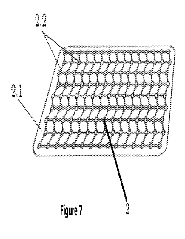
Figure 7: The schematic-perspective view of the hybrid system.
The parts in the figures are numbered one by one and are given below:
1. Tissue scaffold
1.1. Filaments
1.2. Chamber
2. Hybrid implant
2.1. Polymer layer
2.2. Titanium mesh
Detailed explanation of the Invention:
Our invention is basically the creation of a 3D polymer and 13-Tricalcium
Phosphate
(I3-TCP) scaffold structure (tissue scaffold) and a biological tissue implant
allowing/supporting cell infiltration by using the added manufacturing
process,
coating it with physiological-buffered HA solution with the deep coting method
to
increase the delivery rate of growth factors and enlarge their areas allowing
the use
of titanium mesh plate or contoured structures.
Since a normal hyaluronic acid molecule is metabolized and excreted 12 hours
after it
is injected into the human skin, cross-links are used in HA molecules to make
it
permanent. The cross-linking of hyaluronic acid makes the solution more
viscous
increasing its effect by prolonging the residence time in the implant.
The layers are connected at angles to support extracellular matrix (ECM)
interaction
in our invention, and the overlapping structures are overlapped obliquely. The
resulting structures have an hfa 50-70-micron porous structure and support the
formation of vascularized tissue with osteoconductive effect.
CA 03215063 2023- 10- 10
WO 2022/220766
PCT/TR2021/051516
8
The 3D tissue scaffold formed as a result of extrusion crates micro-cracks on
the
body with the cryo-shocking method, and the deep encapsulation of hyaluronic
acid
into the body is increased. The HA solution is 20-70 pm-1 mL per square
centimeter
of scaffold body.
It is a biological tissue implant and its features are;
- Overlapping filament layers (1.1) connected at angles to support
Extracellular
Matrix (ECM) interaction with each other connected by angling at 90 to each
other,
- Oblige overlapping structures of the third filament layer,
- Supporting the formation of vascularized tissue with the osteoconductive
effects of
50-70-micron pore structure of the obtained structures,
- Increasing the encapsulation of the hyaluronic acid deep into the body by
creating
micro cracks with the cryo-shock method or vacuum drying system of the 3D
tissue
scaffold (1) formed as a result of extrusion,
- Attachment and coating of empass or hot polymer (2.1) onto the surface of
the
titanium mesh (2.2) by extrusion,
Bone augmentation (to patients with a bone deficiency) to repair severely
traumatic
and degraded tissues is not suitable for reshaping especially in the jaw
region if the
discomfort has gained aesthetic concern. It will be an important solution for
bone
tissue that cannot be reshaped or volumized.
Another feature of the system is the polymer implant technology that has a
hybrid
structure. Bionic titanium, which is the raw material of the scaffold
obtained, is
coated on the mesh plate with the extrusion or drying method. The vascularized
tissue is re-grown and formed inward by reshaping the volumetric defect. The
tissue
is protected against environmental loads while shaped with a titanium mesh
scaffold
thanks to this technology. The reinforced titanium plates provide a barrier by
minimizing softness. The titanium mesh also provides radiographic visibility.
It is of
CA 03215063 2023- 10- 10
WO 2022/220766
PCT/TR2021/051516
9
vital importance especially for the defects in the head region with its high
ability to
imitate bone.
A device and method for providing surgical therapy for the in situ treatment
and
repair of intra-articular cartilage lesions and/or defects are described with
this
invention.
The device is an implantable, biocompatible, and physiologically absorbable
laminate
cartilage repair patch. The cartilage repair patch is adapted to be placed
near a first
outer cell occlusive layer, a subchondral bone wound site.
The hybrid structure (2) fully supports bone augmentation, acts as a barrier,
has
high-density polymer tissue, helps in the regeneration, provides potential
fibrovascular growth, covers the polymer structure (2.1) on titanium mesh
(2.2), and
provides form and volume to the tissue, which has lost its volumetric
integrity, and
has the feature allowing the tissue to grow inward. The thin polymer layer
(2.1) on
the implants minimizes the upper surface tissue adhesion supporting vascular
tissue
growth by increasing the lower surface porous structure.
A second outer cell has a permeable HA layer and a cartilagenic matrix
(architecture)
between the first and second layers. The cartilagenic matrix and the permeate
layer
surface area have the characteristics of a receiving point for the diffusion
of
autologous stem cells and has components supporting the production of hyaline-
like
cartilage in the presence of autologous stem cells.
The accurate combination of nano-enhancer and hydrogel polymer, hyaluronic
acid
coating method to produce mechanically firm, electrically conductive,
bioactive;
It contains the following process steps;
CA 03215063 2023- 10- 10
WO 2022/220766
PCT/TR2021/051516
- The preparation of solutions with a magnetic stirrer at room temperature
with 10mg/m1 sodium hyaluronate (¨ 1 million Da, medical-grade) in
physiological
buffer (PBS pH 7.4),
- Completing the work by coating the scaffolds with dip-coating method into
5 the solution and drying at vacuum oven at 50 C for 3 days,
The biological tissue implant can be manufactured in cylindrical, square, and
free
forms anatomical shapes allowing rapid implantation requiring minimal
manipulation.
The tissue implant, which is suitable for specific shapes, can be produced in
multiple
10 thicknesses and models that are specific to anatomical regions to meet
clinical needs,
and the reinforced layer increases strength and contours. The fixation
hole/position
allows optimum screw placement, anatomical shape, and the radial titanium mesh
design minimize the cutting option offering many fixation options and
promoting cell
preinflation by gaining micro-mobility.
If desired, therapeutic concentration, stem cell, or growth factor can be
integrated
into the scaffold structure.
B-Tricalcium Phosphate (B-TCP) is included in the prepared PCL granule by 3-
15%. In
this way, the toughness values of the scaffold body formed by reducing the
viscosity
of the polymer are increased.
B-Tricalcium Phosphate (P-TCP) is a biocompatible, radiopaque, and resorbable
osteoconductive material as an important factor supporting the formation of
new
bone in the defect area.
CA 03215063 2023- 10- 10