Note: Descriptions are shown in the official language in which they were submitted.
WO 2022/226113
PCT/US2022/025630
DEVICES, SYSTEMS, AND METHODS FOR PULSED ELECTRIC FIELD
TREATMENT OF TISSUE
CROSS-REFERENCE TO RELATED APPLICATIONS
[00011 This application claims the benefit of U.S. Provisional Application No.
63/177,290,
filed April 20, 2021, the content of which is hereby incorporated by reference
in its entirety.
TECHNICAL FIELD
[00021 Devices, systems, and methods herein relate to applying pulsed electric
fields to tissue
to treat a chronic disease, including but not limited to diabetes
BACKGROUND
[00031 Diabetes is a widespread condition, affecting millions worldwide. In
the United States
alone, over 20 million people are estimated to have the condition. Diabetes
accounts for
hundreds of billions of dollars annually in direct and indirect medical costs.
Depending on the
type (Type 1, Type 2, and the like), diabetes may be associated with one or
more symptoms such
as fatigue, blurred vision, and unexplained weight loss, and may further be
associated with one
or more complications such as hypoglycemia, hyperglycemia, ketoacidosis,
neuropathy, and
nephropathy.
[00041 The treatment of chronic diseases such as obesity and diabetes through
duodenal
resurfacing has been proposed. For example, removing the majority of the
mucosal cells from
the section of the large intestine nearest the stomach may allow a rejuvenated
mucosal layer to
be regenerated, thereby restoring healthy (non-diabetic) signaling.
Conventional treatments that
apply thermal energy to the duodenum risk excessively heating and thus
damaging more layers
of the duodenum (e.g., muscularis) than desired, and/or must compensate for
this excessive
thermal heating. Conversely, conventional solutions may generate incomplete
and/or uneven
treatment. As such, additional systems, devices, and methods for treatment of
duodenal tissue
may be desirable.
1
CA 03215513 2023- 10- 13
WO 2022/226113
PCT/US2022/025630
SUMMARY
100051 Described here are devices, systems, and methods for applying pulsed or
modulated
electric fields to tissue. These systems, devices, and methods may, for
example, treat duodenal
tissue of a patient to treat diabetes. In some variations, a method of
treating diabetes may
comprise advancing a pulsed electric field device into a duodenum of a
patient, the pulsed
electric field device comprising an elongate body and an expandable member
coupled to the
elongate body. The expandable member may comprise an electrode array. A pulsed
waveform
may be delivered to the electrode array to generate a pulsed or modulated
electric field thereby
treating the duodenum. The pulse waveform may comprise a frequency between
about 50 kHz
and about 950 kHz, a drive voltage at the electrode array between about 400 V
and about 600 V,
and a current through tissue between about 36 A and about 48 A from the
electrode array per
square centimeter of the tissue.
100061 In some variations, the frequency may be between about 300 kHz and
about 400 kHz.
In some variations, the pulsed or modulated electric field at the tissue may
be between about
2,000 V/cm and about 3,000 V/cm In some variations, the drive voltage (e.g.,
voltage measured
at the electrode array) may be between about 440 V and about 550 V. In some
variations, the
pulse waveform may comprise a set of about 50 pulses in groups of between
about 8 and about
13, with a delay of between about 4 seconds and about 10 seconds between each
group.
100071 In some variations, the method may include measuring a temperature of
the tissue
using a temperature sensor between about 37 C and about 45 C during delivery
of the pulsed
waveform. In some variations, the method may include increasing a temperature
of the tissue to
about 41 C before delivering the pulsed waveform.
100081 In some variations, the pulsed or modulated electric field may be a
therapeutic electric
field at a first compressed tissue depth of between about 0.25 mm and about
0.75 mm. In some
variations, the pulsed or modulated electric field may be a therapeutic
electric field at a first
uncompressed tissue depth of between about 0.50 mm and about 1.5 mm.
100091 In some variations, the method may include modulating pulse waveform
delivery based
on the measured temperature. In some variations, modulating pulse waveform
delivery may
comprise inhibiting delivery of the pulse waveform.
2
CA 03215513 2023- 10- 13
WO 2022/226113
PCT/US2022/025630
100101 In some variations, the method may include suctioning the tissue to the
expandable
member at a pressure between about 10 mmHg and about 200 mmHg. In some
variations, the
pulsed or modulated electric field may be a therapeutic electric field that
treats cells but leaves
intact tissue scaffolding. In some variations, the pulse waveform may comprise
a pulse width
between about 0.5 p.s and about 4 p.s.
100111 In some variations, the method may include generating a visual marker
on the tissue
using a fiducial generator. In some variations, the method may include
visualizing the visual
marker. In some variations, the treated duodenum may be histologically
indistinguishable from
native tissue after about 30 days.
[0012] Also described herein is a method of treating diabetes comprising
advancing a pulsed
electric field device into a stomach of a patient, the pulsed electric field
device comprising an
elongate body and an expandable member coupled to the elongate body, wherein
the expandable
member comprises an electrode array, and delivering a pulsed waveform to the
electrode array to
generate a pulsed or modulated electric field thereby treating the stomach,
wherein the pulsed
waveform comprises a frequency between about 50 kHz and about 950 kHz, a drive
voltage at
the electrode array between about 400 V and about 600 V, and produces a
current through tissue
between about 36 A and about 48 A from the electrode array per square
centimeter of the tissue.
BRIEF DESCRIPTION OF THE DRAWINGS
100131 The patent or application file contains at least one drawing executed
in color.
Copies of this patent or patent application publication with color drawing(s)
will be provided by
the Office upon request and payment of the necessary fee.
[0014] FIG. IA is a cross-sectional representation of a gastrointestinal tract
showing various
anatomical structures.
[0015] FIG. 1B is a cross-sectional representation of a duodenum.
100161 FIGS. 2A-2C are cross-sectional schematic views of a portion of the
small intestine.
100171 FIG. 3A is a cross-sectional image of a duodenum. FIGS. 3B-3F are
detailed cross-
sectional images of various duodenal tissue.
3
CA 03215513 2023- 10- 13
WO 2022/226113
PCT/US2022/025630
100181 FIG. 4 is a block diagram of an illustrative variation of a pulsed
electric field system.
100191 FIG. 5A is a perspective view of an illustrative variation of a pulsed
electric field
device in a compressed configuration. FIG. 5B is a perspective view of an
illustrative variation
of a pulsed electric field device in an expanded configuration. FIG. 5C is a
detailed perspective
view of the pulsed electric field device shown in FIG. 5A. FIG. 5D is a
detailed perspective view
of the pulsed electric field device shown in FIG. 5B.
100201 FIG. 6A is a perspective view of an illustrative variation of an
expandable member in a
rolled configuration. FIG. 6B is a perspective view of an illustrative
variation of an expandable
member in an unrolled configuration
100211 FIG. 7A is a cross-sectional perspective view of an illustrative
variation of an
expandable member in an unrolled configuration. FIG. 7B is a detailed cross-
sectional
perspective view of the expandable member shown in FIG. 7A.
100221 FIG. 8A is a perspective view of an illustrative variation of a pulsed
electric field
device in a rolled configuration. FIG. 8B is a perspective view of an
illustrative variation of a
visualization device and the pulsed electric field device shown in FIG. 8A in
a partially unrolled
configuration. FIG. 8C is a perspective view of the visualization device and
the pulsed electric
field device shown in FIG. 8B in an unrolled configuration.
100231 FIG. 9A is a perspective view of an illustrative variation of a pulsed
electric field
device in a rolled configuration. FIG. 9B is a perspective view of an
illustrative variation of a
visualization device and the pulsed electric field device shown in FIG. 9A in
an unrolled
configuration.
100241 FIG. 10A is a perspective view of an illustrative variation of a pulsed
electric field
device in a rolled configuration. FIG. 10B is a detailed perspective view of
the pulsed electric
field device shown in FIG. 10A. FIGS. 10C and 10D are perspective views of an
illustrative
variation of a pulsed electric field device in an unrolled configuration. FIG.
10E is a detailed
perspective view of the pulsed electric field device shown in FIG. 10D.
100251 FIG. 11 is a perspective view of an illustrative variation of a
visualization device and a
pulsed electric field device in a partially unrolled configuration.
4
CA 03215513 2023- 10- 13
WO 2022/226113
PCT/US2022/025630
[0026] FIG. 12A is a perspective view of an illustrative variation of a pulsed
electric field
device. FIG. 12B is a cross-sectional side view of the pulsed electric field
device shown in FIG.
12A. FIG. 12C is a detailed cutaway perspective view of the pulsed electric
field device shown
in FIG. 12A.
[0027] FIG. 13A is a perspective view of an illustrative variation of an
expandable member.
FIG. 13B is a plan view of the expandable member shown in FIG. 13A in an
unrolled
configuration. FIG. 13C is a cross-sectional view of an illustrative variation
of an expandable
member in a rolled configuration and gear.
[0028] FIG_ 14A is a perspective view of an illustrative variation of a pulsed
electric field
device and visualization device. FIG. 14B is a cutaway perspective view of the
pulsed electric
field device and visualization device shown in FIG. 14A.
[0029] FIGS. 15A and 15B are cutaway perspective views of illustrative
variations of a pulsed
electric field device and visualization device.
[0030] FIG. 16 is a perspective view of an illustrative variation of a pulsed
electric field
device and visualization device.
[0031] FIGS. 17 is a perspective view of an illustrative variation of a pulsed
electric field
device and visualization device.
[0032] FIGS. 18 is a perspective view of an illustrative variation of a pulsed
electric field
device and visualization device.
[0033] FIG. 19 is a perspective view of an illustrative variation of a pulsed
electric field
device and visualization device.
100341 FIG. 20 is a perspective view of an illustrative variation of a pulsed
electric field
device and visualization device.
[0035] FIG. 21 is a perspective view of an illustrative variation of a pulsed
electric field
device and visualization device.
CA 03215513 2023- 10- 13
WO 2022/226113
PCT/US2022/025630
100361 FIG. 22 is a perspective view of an illustrative variation of a pulsed
electric field
device and visualization device.
100371 FIG. 23 is a perspective view of an illustrative variation of a pulsed
electric field
device and visualization device.
100381 FIG. 24 is a perspective view of an illustrative variation of a pulsed
electric field
device and visualization device
100391 FIG. 25 is a perspective view of an illustrative variation of a pulsed
electric field
device and visualization devi ce
100401 FIG. 26 is a perspective view of an illustrative variation of a pulsed
electric field
device and visualization device.
100411 FIG. 27 is a perspective view of an illustrative variation of a pulsed
electric field
device and visualization device.
100421 FIG. 28A is a perspective view of an illustrative variation of an
expandable member of
a pulsed electric field device and visualization device. FIGS. 28B-28E are
perspective views of
the pulsed electric field device and visualization device shown in FIG. 28A.
100431 FIG. 29A is a perspective view of an illustrative variation of a pulsed
electric field
device and visualization device. FIG. 29B is a perspective view of the pulsed
electric field
device detached from the visualization device shown in FIG. 29A.
100441 FIG. 30A is a perspective view of an illustrative variation of a pulsed
electric field
device. FIG. 30B is a perspective view the pulsed electric field device shown
in FIG. 30A in a
tissue lumen.
100451 FIG. 31 is a perspective view of an illustrative variation of a pulsed
electric field
device.
100461 FIG. 32 is a perspective view of an illustrative variation of a pulsed
electric field
device. FIG. 33A is a side view of an illustrative variation of a pulsed
electric field device. FIG.
33B is a perspective view of the pulsed electric field device shown in FIG.
33A
6
CA 03215513 2023- 10- 13
WO 2022/226113
PCT/US2022/025630
100471 FIG. 34A is a perspective view of an illustrative variation of an
electrode array. FIG.
34B is a cross-sectional side view of the electrode array shown in FIG. 34A.
FIG. 34C is a
perspective view of an illustrative variation of an electrode array in an
unrolled configuration.
100481 FIG. 35 is an electric field strength plot of an illustrative variation
of an electrode
array.
100491 FIG. 36 is an electric field strength plot of a conventional electrode
array.
100501 FIG. 37 is a schematic cross-sectional view of an illustrative
variation of an electrode
array and embossing dies_
100511 FIG. 38 is a schematic cross-sectional view of an illustrative
variation of an electrode
array comprising a tissue contact layer.
100521 FIG. 39 is a schematic cross-sectional depiction of an illustrative
variation of an
electrode array comprising a tissue contact layer.
100531 FIG. 40 is a schematic cross-sectional side view of an illustrative
variation of an
electrode array.
100541 FIGS. 41A-41D are electric field strength plots of illustrative
electrode array
configurations.
100551 FIG. 42 is an electric field strength plot of an illustrative variation
of an electrode
array.
100561 FIG. 43 is a perspective view of an illustrative variation of an
expandable member
comprising an electrode array.
100571 FIG. 44 is a perspective view of an illustrative variation of an
expandable member
comprising an electrode array.
100581 FIGS. 45A-45C are schematic diagrams of an illustrative variation of an
electrode
array. FIG. 45D is a plan view of an electric field strength plot of an
illustrative variation of an
electrode array. FIG. 45E is a cross-sectional view of an electric field
strength plot of the
electrode array depicted in FIG. 45D.
7
CA 03215513 2023- 10- 13
WO 2022/226113
PCT/US2022/025630
[0059] FIG. 46A is a schematic perspective view of an illustrative variation
of a coordinate
system for an electrode array. FIG. 46B are electric field strength plots
corresponding to the
electrode array shown in FIG. 46A.
[0060] FIG. 47A is a schematic plan view of an illustrative variation of a
polarity
configuration of an electrode array. FIG. 47B are electric field strength
plots corresponding to
the electrode array shown in FIG. 47A.
[0061] FIG. 48 is a schematic plan view of an illustrative variation of an
electrode array.
[0062] FIG. 49 is a perspective view of an illustrative variation of an
electrode array of a
pulsed electric field device.
[0063] FIG. 50 is a perspective view of an illustrative variation of an
electrode array of a
pulsed electric field device.
[0064] FIGS. 51A-51B, and HD, are schematic circuit diagrams of illustrative
variations of an
electrode array, temperature sensor array, and fiducial generator. FIG. 51C is
an image of a
visual marker generated by a fiducial generator.
[0065] FIG. 52A is a schematic circuit diagram of an illustrative variation of
an electrode
array, temperature sensor array, and fiducial generator. FIG. 52B is a
detailed view of the
schematic circuit diagram of the electrode array, temperature sensor, and
fiducial generator
shown in FIG. 52A.
[0066] FIG. 53 is a schematic circuit block diagram of an illustrative
variation of a signal
generator.
[0067] FIG. 54 is a flowchart describing an illustrative variation of a method
of treating
diabetes.
[0068] FIGS. 55A-55F are schematic views of an illustrative variation of a
method of treating
diabetes.
[0069] FIGS. 56A-56H are perspective views of an illustrative variation of a
method of
treating diabetes using a pulsed electric field device and visualization
device.
8
CA 03215513 2023- 10- 13
WO 2022/226113
PCT/US2022/025630
[0070] FIG. 57 is an image of an illustrative variation of a thermal marking
on tissue.
[0071] FIGS. 58A-58E are images of an illustrative variation of a treatment
procedure in a
patient using a pulsed electric field device and visualization device.
[0072] FIG. 59 is an image of an illustrative variation of an electrode array.
[0073] FIG. 60 is an image of an illustrative variation of a pulsed electric
field device.
100741 FIG. 61A is a perspective view of an image of an illustrative variation
of a pulsed
electric field device and visualization device. FIG. 61B is a detailed image
of the pulsed electric
field device and visualization device shown in FIG. 61A.
[0075] FIG. 62A is an image of illustrative variations of pulsed electric
field devices. FIG.
62B is an image of an illustrative variation of a pulsed electric field device
comprising a balloon.
FIG. 62C is a perspective view of the pulsed electric field devices shown in
FIG. 62A.
[0076] FIG. 63A is an image of an illustrative variation of a pulsed electric
field device in a
rolled configuration. FIG. 63B is an image of an illustrative variation of a
pulsed electric field
device in an unrolled configuration. FIG. 63C is a perspective view of the
pulsed electric field
device shown in FIG. 63B.
[0077] FIG. 64A is an image of an illustrative variation of a pulsed electric
field device and
visualization device. FIG. 64B is an image of an illustrative variation of a
pulsed electric field
device in an unrolled configuration within a tissue lumen.
100781 FIG. 65 is an image of an illustrative variation of a pulsed electric
field device.
[0079] FIG. 66 is a schematic circuit diagram of an illustrative variation of
an electrode array.
[0080] FIG. 67 is an image of an illustrative variation of an electrode array.
[0081] FIG. 68 is an image of an illustrative variation of an electrode array.
[0082] FIG. 69A is a plan view of an illustrative variation of an electrode
array. FIGS. 69B
and 69C are perspective views of the electrode array shown in FIG. 69A. FIG.
69D is a
perspective cross-sectional view of the electrode array shown in FIG. 69A.
9
CA 03215513 2023- 10- 13
WO 2022/226113
PCT/US2022/025630
100831 FIG. 70 is an illustrative variation of a voltage plot comparing the
voltage output of
pulsed electric field treatment to the voltage output of radiofrequency
treatment over time.
100841 FIG. 71A is a cross-sectional image of a pulsed electric field device
in an expanded
configuration that dilates a duodenum. FIG. 71B is a cross-sectional image of
an undilated
duodenum. FIG. 71C is a cross-sectional image of an undilated duodenum. FIG.
71D is a
detailed cross-sectional image of the undilated duodenum shown in FIG. 71C.
FIG. 71E is a
cross-sectional image of a dilated duodenum. FIG. 71F is a detailed cross-
sectional image of the
dilated duodenum depicted in FIG. 71E.
100851 FIGS 72A and 72B are detailed cross-sectional images of duodenal tissue
about a day
after treatment.
100861 FIG. 73 is a detailed cross-sectional image of duodenal tissue about
three days after
treatment.
100871 FIG. 74A and 74B are detailed cross-sectional images of duodenal tissue
about seven
days after treatment.
100881 FIG. 75 is a detailed cross-sectional image of duodenal tissue about
fourteen days after
treatment.
100891 FIG. 76 is a perspective view of an illustrative variation of an
electrode array in an
unrolled configuration.
100901 FIG. 77 is a perspective view of an illustrative variation of a pulsed
electric field
device in an expanded configuration.
100911 FIG. 78A is an image of an illustrative variation of a pulsed electric
field device in a
retracted or compressed configuration. FIG. 78B is a detailed image of an
unrolled or expanded
electrode array of the pulsed electric field device depicted in FIGS. 78B.
100921 FIG. 79A is an image of an illustrative variation of a pulsed electric
field device in a
compressed configuration. FIG. 79B is an image of an illustrative variation of
a pulsed electric
field device in an expanded configuration. FIG. 79C is a detailed image of an
unrolled electrode
array of the pulsed electric field device depicted in FIGS. 79A and 79B.
CA 03215513 2023- 10- 13
WO 2022/226113
PCT/US2022/025630
[0093] FIGS. 80A and 80B are electric field strength plots of illustrative
variations of an
electrode array.
[0094] FIGS. 81A-81C are schematic views of an illustrative variation of a
method of treating
diabetes.
[0095] FIGS. 82A-82D are images of an illustrative variation of a method of
treating diabetes
using a pulsed electric field device and visualization device_
[0096] FIGS. 83A and 83B are tissue temperature, voltage, and current plots
over time for
illustrative variations of methods of treating tissue
[0097] FIG. 84 is a cross-sectional perspective view of a set of twisted pair
lead wires.
[0098] FIG. 85 is a perspective view of an illustrative variation of an
electrode array of a
pulsed electric field device.
[0099] FIG. 86 is a temperature plot over time of illustrative variations of
methods of treating
tissue.
[0100] FIG. 87 is a plot of impedance distribution and temperature
distribution of illustrative
variations of methods of treating tissue.
DETAILED DESCRIPTION
101011 Described here are devices, systems, and methods for treating tissue to
address a chronic
disease. For example, devices, systems, and methods may include those for
treating diabetes by
treating duodenal tissue of a patient. In some variations, treatment of the
duodenum may
comprise treating at least about 30% of the mucosal lining of the duodenum
with minimal
trauma, damage or scarring to the submucosa, vasculature, and muscles. For
example, a mucosa
layer of the duodenum may be treated using a pulsed electric field (PEF)
system.
[0102] It may be helpful to briefly identify and describe the relevant small
intestine anatomy.
FIG. lA is a cross-sectional view of the gastrointestinal tract of a patient
(100). Shown there is a
visualization device (150) (e.g., endoscope) advanced into the stomach (120)
through the
esophagus (110). The stomach (120) is connected to the duodenum (130). FIG. 1B
is a detailed
11
CA 03215513 2023- 10- 13
WO 2022/226113
PCT/US2022/025630
cross-sectional view of the duodenum (130), which surrounds the head of the
pancreas (140).
The duodenum is a "C" shaped hollow jointed tube structure that is typically
between about 20
cm and about 35 cm in length and about 20 mm and about 45 mm in diameter.
FIGS. 2A-2C are
cross-sectional schematic views of the layers of the small intestine (200)
including the mucosa
(210), submucosa (220), muscularis externa (230), and serosa (240). Treatment
of the duodenum
may comprise resurfacing the mucosa (210) as described herein. Access to the
duodenum may
be performed by advancing the systems and devices described herein through one
or more of the
esophagus, stomach, pylorus, lower esophageal junction, crackle pharyngeal
junction, and
several acute small radius bends throughout the length of the digestive tract.
101031 It may further be helpful to briefly discuss electroporation and the
role of ohmic heating.
Electroporation is the application of an electric field to living cells to
cause ions of opposite
charge to accumulate on opposite sides of cell membranes. Generally,
electroporation requires a
potential difference across the cell membrane on the order of about 0.5 to
about 1 volt and for a
cumulative duration on the order of about 1 to about 2 milliseconds.
Electroporation necessarily
generates ohmic heating but there is considerable confusion in the literature
about this, including
a significant number of references that incorrectly assert the existence of
non-thermal
electroporation. For example, an external uniform electric field of magnitude
E applied to an
intracellular fluid with ionic conductivity ai, will generate a current
density Eo-i, and dissipate a
thermal power density E2o-ic. If the medium has a heat capacity Cp and density
p, the resulting
rate of temperature rise is given by equation (1):
dT E2 (sic
¨ = eqn. (1)
dt
101041 For example, a 1 KV/cm electric field acting on tissue with a
conductivity of about 0.3
S/m, a heat capacity of about 3.7 joule/(gm C), and a density of about lgm/cc
will heat the
tissue at a rate of about 800 C/second. Note that, without current passing
through the tissue,
there is no electric field in the tissue since the tissue is an ionic
conductor. The initial time after
an external field is abruptly applied to the membrane to accumulate charge may
be on the order
of about 30 nanoseconds, which suggests that, during an initial membrane-
charging phase, the
average temperature rise may be in the tens of microdegrees. When an external
electric field is
applied, and ionic currents have charged the membrane surfaces to collapse the
field into the
lipid bilayers, leakage current may still flow, though the heating may be
confined to the
12
CA 03215513 2023- 10- 13
WO 2022/226113
PCT/US2022/025630
membranes for sub-microsecond timescales. For example, using a lipid layer
conductivity of
o-u=0.002 S/m, a 1 volt potential across an 8 nm layer may locally heat at an
instantaneous rate
of about 8 C/microsecond. This heating rate drops with time from the
application of the
external electric field, as the heat may diffuse further from the membrane.
101051 If the ionic currents are confined to pores in the cell membranes,
current crowding will
cause the heating rate in the pores to be correspondingly higher. Since the
pore area might be 1%
or less of the membrane area, the current density in the pores may be one
hundred times higher
than in the bulk tissue. This gives a ten thousand times increase in heating
rate, leading to local
heating rates on the order of 10 C/microsecond.
101061 Local temperature rise is a contributing mechanism to the transition
from electroporation
to irreversible electroporation. Thermal diffusion lowers the local
temperature excursions. For
example, assuming a tissue thermal diffusivity lc of 0.13 mm2/s, the thermal
diffusion length at
pec is \/(10 [is)(0.13 mm2 /s) or 1.1 micron, which is much larger than a
typical pore. At
1 millisecond, the thermal diffusion length is on the order of the cell size,
so the localized
heating effects may be ignored.
101071 The bulk tissue remains a good ionic conductor during the
electroporation treatment,
heating at a rate on an order of magnitude of about 800 C/s while the
external field is being
applied. If the external field is removed, the cell membranes may discharge on
the order of about
30 nanoseconds, obliging the continued application of external voltage and
current to induce
pore formation and growth. As the maximum tolerable temperature rise of the
bulk tissue may
be on the order of about 13 C, the maximum duration that the external field
may be applied,
even in a bipolar configuration, may be within an order of magnitude of about
10 milliseconds.
As this heat is generated to a treatment depth in the tissue of about several
millimeters, the
required time to cool the tissue by conduction may be about 70 seconds (e.g.,
(3
mm2)/(0.13mm2/sec)). Blood convection likely dominates the observed cooling
times that are on
the order of about 10 seconds. Electroporation may also increase with the
temperature of the
bulk tissue due to the phase transition of the lipid cell membrane, which for
some cells on the
duodenum is 41 C The phase transition temperature may be the temperature
required to induce
a change in the lipid physical state from the ordered gel phase to the liquid
crystalline phase.
13
CA 03215513 2023- 10- 13
WO 2022/226113
PCT/US2022/025630
101081 Electroporation parameters may be varied to produce different effects
on tissue. FIG. 3A
is a cross-sectional image of an untreated duodenum (300A) including a
muscular layer (310A)
and villi (320A). FIG. 3D is an image of an illustrative variation of duodenal
tissue in its native
untreated state including a muscularis layer (310D), submucosa (330D), villus
crypts (340D) and
villi (320D). As described in more detail herein, FIG. 3E depict duodenal
tissue that has
undergone majority thermal heat treatment and FIG. 3F depict duodenal tissue
that has
undergone majority pulsed or modulated electric field treatment. The
treatments described
herein (e.g., FIG. 3F) that primarily treat the mucosa layer with preserved
tissue architecture
appearing similar to the native tissue reduces trauma to tissue relative to
the thermal treatment
shown in FIG. 3E.
101091 The application of a pulsed electric field to duodenal tissue results
in non-thermal tissue
changes. For example, FIG. 3D is an image of normal untreated (e.g., native
tissue) porcine
duodenal mucosa. FIG. 3F is an image of the initial mucosal histologic
appearance with
evolving epithelial loss and lamina propria structural/architectural
preservation. For example,
FIG. 3F depicts the histologic evolution with complete native epithelial loss
and early crypt
regeneration within the preserved lamina propria. The glandular layer across
FIGS. 3A-3D and
3F demonstrates the structural preservation of the lamina propria following
treatment. For
example, histopathology confirms that the PEF treatment as described herein
applied at a depth
of about 1 mm in duodenal tissue will treat the mucosal layer without the
pulsed electric field
energy affecting the muscularous propria at a therapeutic level.
101101 In some variations, a pulsed electric field (PEF) treatment may be
combined with
localized thermal treatment. For example, thermal treatment may be applied to
surface tissue or
near-surface tissue while PEF treatment may be applied to relatively deeper
tissue. As described
in more detail herein, the depth of tissue treatment received by one or more
layers may be
adjusted based on one or more of electrode design, applied voltage, time or
duration of energy
delivery, frequency of applied energy, and tissue configuration. An example of
such control is
thermal treatment applied up to a tissue depth of about 0.1 mm and a PEF
treatment applied to a
tissue depth of up to about 1 mm. The ratio and depth of thermal treatment to
PEF treatment may
be based on a desired clinical outcome (e.g., effect). In some variations,
thermal treatment may
be applied up to a tissue depth of about 3 mm, and PEF treatment may be
applied up to a tissue
depth of about 5 mm. Therefore, in some variations, more thermal treatment
than PEF treatment
14
CA 03215513 2023- 10- 13
WO 2022/226113
PCT/US2022/025630
may be applied to tissue. Based on a depth or type of tissue, different
healing cascades maybe
optimal. In some variations, the villas mucosa at up to about 1 mm may be
thermally treated to
allow substantially the entire tissue architecture to be replaced, while the
submucosa may be
PEF treated to preserve the tissue architecture and promote rapid healing of
that layer.
Furthermore, neither the thermal treatment nor PEF treatment may affect the
deeper muscularis
propria layer.
101111 FIG. 3B is an image of an illustrative variation of duodenal tissue
that has undergone
different treatments. In particular, the tissue (360) was treated with pulsed
or modulated electric
field energy and first mucosa region (362) was further subjected to
radiofrequency energy. The
ablated villi of the first mucosa region (362) have broken cellular membranes
and destroyed cell
structures such that those cells are no longer viable or functioning. By
contrast, a second mucosa
region (360) has cells that have undergone cell lysis where the cellular
membranes remain intact
but the cells are no longer viable and functioning. That is, cell lysis
corresponds to functional
cell death with intact cellular structures while ablation refers to loss of
both cell structure and
function. The submucosa (370) and muscularis (380) remain healthy (e.g.,
viable and fully
functioning with cell integrity). In FIG. 3B, villi in the first mucosa region
(362) are thermally
ablated while the cell lysis in the second mucosa region (360) is generated by
a pulsed or
modulated electric field. A third mucosa region (363) adjacent to the thermal
lesion of the first
mucosa region (362) is not treated at all and comprises viable tissue.
101121 FIG. 3C illustrates a histological slide of the duodenum from tissue
about 24 hours after
treatment with heat and pulsed electric field, showing a partial treatment of
the mucosa down to
the crypt layer, with injured cells. A fourth mucosa region (391) corresponds
to thermal/heat
fixed tissue of the villi, including the villi-associated enteroendocrine
cells. The fourth mucosa
region (391) demonstrates architectural and cytological preservation with
cellular detail with
hyperchrom ati c nucl ear and hypereosi nophili c cytopl a smi c staining.
Overall, interstitial
hemorrhage and infiltrating post-treatment-associated inflammatory cells are
not identified. The
heat fixed tissue may be expected to slough off, followed by surface re-
epithelializati on and
villous structural healing with crypt cell repopulation. The crypt tissues are
partially affected by
a combination of heat and pulsed electric field effects. The tissue healing
timeline is expected to
be longer than that of a pulsed electric field treatment without thermal
effect. The submucosa
(370) and muscularis (380) are histologically unaffected. FIG. 3E is an image
of an illustrative
CA 03215513 2023- 10- 13
WO 2022/226113
PCT/US2022/025630
variation of 24 hour porcine duodenal histology following an isolated
hyperthermic tissue
treatment (i.e., no concomitant pulsed electrical field exposure) which
destroys the lamina
propria in that tissue scaffolding is burned and destroyed, and will be
sloughed off and removed
during healing. This demonstrates the histologic features of a thermal tissue
dose, consistent
with thermal/heat-induced coagulative necrosis without thermal/heat fixation.
In this region, the
glandular epithelium and neuroendocrine cells (321) show a loss of cytologic
detail, consistent
with cellular "ghost images." Interstitial hemorrhage and reactive
inflammatory cells of the
mucosal layer (341) are present at the region's edge. The submucosa (331) and
muscularis (311)
also show injury related changes. This region may be anticipated to heal
similar to an ischemic
type coagulative necrosis with resorption and remodeling with mucosal
regeneration. The
thermal lesion destroyed the lamina propria. Scaffolding is burned and
destroyed and will be
sloughed off and removed during healing. The tissue healing time frame for
this region should
be longer than that expected for a pulsed electric field treatment.
101131 FIG. 3F is an image of an illustrative variation of duodenal tissue
that has undergone
treatment with pulsed or modulated electric field energy to a controlled depth
not including the
muscularis, untreated muscularis propria layer (310), submucosa (330), treated
submucosa
(332), treated villus crypts, with partial cell lysis and maintained tissue
scaffolding (342), and
treated villi with villas sloughing (322). The treated submucosa (332) also
maintains tissue
scaffolding. These treated tissues illustrate cells that have undergone a cell
death where the
cellular membranes remain intact but the cells are no longer viable and
functioning. The healing
cascade will replace these cells without infiltration of large number of
inflammatory cells, and
the surface will re-epithelialize and with villous structural healing and
crypt cell repopulation.
The muscularis (310) remains healthy (e.g., viable and fully functioning with
cell integrity)
without therapeutic effect from the pulsed electric field energy. That is,
with pulsed or
modulated electric field energy cell death corresponds to functional cell
death with intact cellular
structures while ablation refers to loss of both cell structure and function
and an aggressive
necrotic inflammatory response healing cascade.
101141 In some variations, a target depth of treatment includes the mucosal
layer but excludes
treatment of the muscularous propria. Human tissue data assessed through
histopathology
supports about a 1 mm target depth for PEF tissue treatment where the pulsed
electric field does
not penetrate through to the muscularous propria at a therapeutic level. As a
result, the mucosa
16
CA 03215513 2023- 10- 13
WO 2022/226113
PCT/US2022/025630
exhibits a healing progression with a first day initiation of crypt and
glandular epithelial
regeneration (e.g., FIGS. 72A, 72B), a third day continuation of epithelial
development with
surface re-epithelization (e.g., FIG. 73), a seventh day of early cobblestone-
like blunted villous
development (e.g., FIGS. 74A, 74B), and continues through a fourteenth day of
villous
elongation and narrowing (FIG. 75). Based on the methods described herein, the
healing
response may be essentially completed in about thirty days. Moreover, the
systems, devices, and
methods described herein may provide uniform treatment coverage throughout a
circumference
and length of the duodenum.
[0115] Some methods for treating diabetes may include treating the submucosa
layer of the
duodenum without treating the muscularis. Conventional solutions do not
consistently treat the
submucosa layer without negatively impacting the muscularis. Instead,
conventional solutions
may add complicated mitigating steps such as lifts with saline injection in an
attempt to protect
the muscularis. For reference, the mucosal layer typically has a thickness
between about 0.5 mm
to about 1 mm, the submucosa layer typically has a thickness of about 0.5 mm
and about 1 mm,
and the muscularis typically has a thickness of about 0.5 mm. Inducing injury
to the muscularis
may result in adverse clinical outcomes. Furthermore, the anatomical structure
along a
circumference of the duodenum is not uniform, thus complicating efforts to
treat just the
submucosa and not the muscularis.
101161 The methods described herein may selectively change tissue viability
without losing the
integrity of the majority of the treated tissue in the duodenum by applying a
predetermined
pulsed or modulated electric field and, optionally, without other treatment of
the tissue to
mitigate the pulsed or modulated electric field to a portion of tissue. By
contrast, RF based
energy treatment may predominantly generate heat-induced cell lysis (e.g.,
cell death) or
ablation that may indiscriminately damage tissue and destroy cellular
structure, and which may
be difficult to modulate, thus negatively impacting treatment outcomes In some
variations, the
methods described here may comprise applying a pulsed or modulated electric
field to
thermally-induce local necrotic cell death (e.g., local ablation) for duodenal
tissue immediately
adjacent to an electrode array and to induce cell lysis (e.g., functional cell
death) within a
predetermined range of depths of duodenal tissue (e.g., up to about 1 mm,
between about 0.5
mm and 0.9 mm) while minimizing the physiological impact to tissue greater
than the selected
depth.
17
CA 03215513 2023- 10- 13
WO 2022/226113
PCT/US2022/025630
101171 FIG. 3F is an image of an illustrative variation of duodenal tissue
that has undergone
treatment with pulsed or modulated electric field energy to a controlled
depth. In FIG. 3F, the
muscularis layer (310) and a portion of the submucosa (330) are untreated
(i.e., energy delivered
to tissue does not affect the tissue) and the villus crypts (342), villi (322)
and a different portion
of the submucosa (332) have been treated. Thus, the treatment applied to the
duodenal tissue
shown in FIG. 3F results in a more superficial (e.g., closer to the tissue
surface) treated
submucosa (332) and a deeper, untreated muscularis layer (310). The treated
tissues contain cells
that have undergone cell lysis where the tissue scaffolding remain intact but
the cells are no
longer viable and functioning. A mild healing cascade will replace these
cells. The muscularis
(310) adjacent to the treated submucosa (332) remains healthy (e.g., viable
and fully functioning
with cell integrity).
101181 The pulsed or modulated electric fields near an electrode array may
generate some
thermal heating of tissue leading to tissue ablation that destroys both cell
structure and function.
However, cell lysis in tissue resulting from the pulsed or modulated electric
fields applied herein
are at least 50% pore-induced and less than 50% heat-induced such that a
majority of cell death
comprises functional cell death with intact cellular structures. For example,
the thermal heating
generated by a pulsed or modulated electric field is generally localized to a
relatively small
radius from each electrode of an electrode array and does not affect deeper
layers of tissue such
as the muscularis.
101191 The systems, devices, and methods described herein deliver energy to
provide treatment
characteristics optimized for each tissue layer to improve treatment outcomes.
Near the surface
of the tissue (e.g., less than about 0.5 mm, between about 0.1 mm and about
0.5 mm), thermal
heating may generate local necrotic cell death of tissue that may slough off
after treatment. At a
tissue depth of between about 0.5 mm and about 1.3 mm (e.g., mucosa of
duodenum), cell lysis
may be generated by the pulsed or modulated electric field while thermal
heating is limited (e.g.,
to less than about a 13 C increase or 6 C increase). For example, an
electric field strength at
about 1.0 mm may be about 2.5 kV/cm. At tissue depths beyond 1.0 mm, the
energy delivered to
tissue generates reversible electroporation with even less thermal heating
such that deeper tissue
may be substantially untreated. Thus, thermal heating may be limited to a
surface tissue layer
(e.g., less than about 0.5 mm, between about 0.1 mm and about 0.5 mm) while
still delivering
pulsed or modulated electric field energy for cell lysis of the mucosa.
18
CA 03215513 2023- 10- 13
WO 2022/226113
PCT/US2022/025630
101201 For example, FIG. 3C is an image of an illustrative variation of
duodenal tissue that has
undergone a method of treating duodenal tissue described herein where villi
(391) has been
treated by a combination of thermal heating (e.g., more than 50%) and pore-
induced cell death
(e.g., less than 50%). The pulsed or modulated electric field applied to the
villus crypts and
submucosa (370) has treated the tissue to a majority (e.g., more than 50%) of
pore-induced cell
death with a lesser contribution (e.g., less than 50%) of cell death due to
thermal heating. The
muscularis (380) is substantially untreated by the pulsed or modulated
electric field or other
methods. For example, the submucosa in FIG. 3C is not subject to saline
injection. The depth of
treatment may be controlled such that a predetermined portion of the mucosal
layer such as the
villus crypts may remain untreated if desired. The configuration and geometry
of the electrode
arrays as described herein may enable the tissue treatment characteristics
described herein.
101211 By contrast, conventional solutions that apply other forms of thermal
energy (e.g., steam,
radiofrequency, laser, heated liquid) to the duodenum thermally ablate through
multiple layers of
the tissue (e.g., inducing more than 50% heat-induced necrotic cell death and
less than 50%
pore-induced cell death), thereby destroying the cellular structure of the
mucosa at similar
depths and which may detrimentally thermally damage the muscularis. In an
attempt to mitigate
the risk of unintentional thermal damage during application of thermal energy
to deeper layers
(e.g., muscularis) of the duodenum, saline may be injected into portions of
duodenal tissue (e.g.,
the submucosa (330)). This additional step further complicates the procedure
and is not always
sufficient to prevent unwanted thermal tissue damage. The pulsed or modulated
electric field
based methods described here eliminate this additional step and provide
greater protection
against unwanted tissue damage by improving the energy delivery
characteristics generated by a
pulsed electric field device.
101221 In some variations, pulsed electric field treatment may be applied
while monitoring
and/or minimizing tissue temperature increases. For example, a predetermined
rise in tissue
temperature (e.g., about 1 C, about 2 C, about 3 C) may be followed by a
pause (e.g., of a
predetermined time interval) in energy delivery to allow the tissue to cool.
In this manner, the
total energy delivered may increase the tissue temperature below a
predetermined threshold
(e.g., below a safety limit). In some variations, the predetermined threshold
may be up to about 3
C, about 6 C, about 10 C, about 13 C, including all ranges and sub-values
in-between.
19
CA 03215513 2023- 10- 13
WO 2022/226113
PCT/US2022/025630
101231 Moreover, the difficulty faced by conventional solutions in controlling
unwanted thermal
tissue damage would lead one of ordinary skill away from using the pulsed or
modulated electric
field energy levels and methods described herein. In some variations, the
tissue power densities
generated by a pulsed or modulated electric field may be several orders of
magnitude higher than
the tissue power densities generated by radiofrequency ablation. For example,
a power density
ratio of an analogous design for radio frequency ablation may be about 576
where a
radiofrequency device is driven at about 25 V., and a pulsed electric field
device is driven at
about 600 V. Thus, it would be unexpected for the pulsed or modulated electric
field methods
described here to not only treat tissue, but to do so without excess thermal
tissue damage
requiring mitigation procedures. Furthermore, the increased power densities
may require
additional insulation and protection of the pulsed electric field device, as
well as a signal
generator capable of generating such peak power levels. Generally, the duty
cycle for PEF
treatment may be several orders of magnitude lower than radio frequency
ablation in order to
keep a bulk tissue temperature rise below about 10 C). For example, radio
frequency ablation
energy may generally be delivered continuously for several seconds. In some
variations, PEF
treatment may collectively accumulate about 15 milliseconds of ON time over
about 10 seconds,
for a net duty cycle of about 0.0015.
101241 FIG. 70 is plot (7000) comparing the voltage output of pulsed electric
field treatment
(7010) to the voltage output of radiofrequency treatment (7020) over time.
During the RF
treatment (7020) energy may be delivered continuously within the time scale of
FIG. 70, while
during PEF treatment (7010) energy is pulsed intermittently with a voltage
output being orders
of magnitude higher than the voltage output for the RF treatment (7020).
[0125] Generally, the devices described here may comprise an elongate body
coupled to an
electrode array, which may be disposed in a lumen of a duodenum. In some
variations, the
devices may further comprise an expandable member configured to releasably
engage to a
portion of the duodenum. The expandable member may comprise or be coupled to
an electrode
array configured to generate a pulsed or modulated electric field. The
electrodes of the electrode
array may have predetermined dimensions and spacing configured to generate a
pulsed or
modulated electric field having predetermined uniformity for treating desired
tissue while
limiting damage to other tissue. In some variations, the expandable member may
expand and
compress as necessary to engage an inner diameter of the duodenum. In some
variations, a
CA 03215513 2023- 10- 13
WO 2022/226113
PCT/US2022/025630
system comprising the devices described herein may further comprise a signal
generator
configured to generate a pulse waveform for delivery to the electrode array to
thereby treat the
engaged tissue.
101261 Also described here are methods. In some variations, a method of
treating duodenal
tissue, to, for example, treat diabetes, may include advancing a pulsed
electric field device
toward a first portion of a duodenum of a patient. The pulsed electric field
device may comprise
an expandable member comprising an electrode array. The expandable member may
be
transitioned from a compressed configuration into an expanded configuration
bringing the
expandable member (and the electrode array) closer to or in contact with the
inner surface of the
duodenum. The expandable member may comprise a flexibility to apply force
against and
conform to an inner circumference of the duodenum that may itself comprise a
range of
diameters. A first pulse waveform may be delivered to the electrode array to
generate a first
pulsed or modulated electric field, which may treat the tissue in the first
portion. The pulsed
electric field device may be moved (e.g., advanced or retracted) toward a
second portion of the
duodenum (which may be distal or proximal to the first portion), and a second
pulse waveform
may be delivered to the electrode array to generate a second pulsed or
modulated electric field
thereby treating the tissue in the second portion. For example, in some
variations, a signal
generator may generate a drive voltage (e.g., voltage measured at an electrode
array) of between
about 400 V and about 1500 V that may correspond to an electric field strength
of about 400
V/cm and about 7000 V/cm at the treatment portions of the duodenum. The
expandable member
may be in a compressed configuration, semi-expanded configuration, and
expanded
configuration during movement of the pulsed electric field device. In some
variations, a
visualization device may be configured to visualize one or more of the pulsed
electric field
device and tissue. In some variations, temperature sensor measurements may be
used to monitor
and/or control pulse waveform delivery. In some variations, current and
voltage measurements
may be used to monitor and/or control pulse waveform delivery.
I. System
Overview
101271 Systems described here may include one or more of the components used
to treat tissue,
such as, for example, a pulsed electric field device and a visualization
device. Suitable examples
21
CA 03215513 2023- 10- 13
WO 2022/226113
PCT/US2022/025630
of such systems and devices are described in International Application Serial
No.
PCT/US2020/056720, filed on October 21, 2020, the disclosure of which is
hereby incorporated
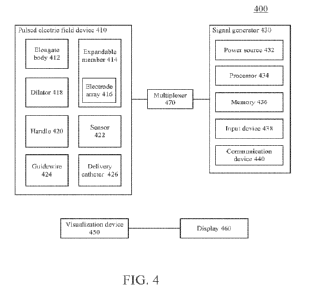
by reference in its entirety. FIG. 4 is a block diagram of a variation of a
pulsed electric field
system (400) comprising one or more of a pulsed electric field device (410), a
signal generator
(430), multiplexer (470), a visualization device (450), and a display (460).
101281 In some variations, the pulsed electric field device (410) may comprise
one or more (e.g.,
a first and a second) elongate bodies (412) sized and shaped to be placed in
one or more body
cavities of the patient such as, for example, an esophagus, a stomach, large
intestine, small
intestine, and any portion of the gastrointestinal tract. In some variations,
the pulsed electric field
device (410) may further comprise one or more expandable members (414), one or
more
electrode arrays (416), one or more dilators (418), a handle (420), one or
more sensors (422), a
guidewire (424), and a delivery catheter (426). A distal end of the pulsed
electric field device
(410) may comprise the dilator (418), and the guidewire (424) may extend from
a lumen of the
dilator (418). The expandable member (414) may comprise the electrode array
(416). For
example, as will be described in more detail herein, in some variations the
electrode array (416)
may be coupled to a surface (e.g., outer surface) of the expandable member
(416), while in other
variations, the electrode array itself may form the expandable member and/or
the electrode array
may be integral with the expandable member. In some variations, the expandable
member (414)
and/or the electrode array (416) may be disposed adjacent to one or more
dilators, for example,
between at least a pair of dilators (418). In some variations, the pulsed
electric field system (400)
may optionally comprise a delivery catheter (426) configured to advance over
the pulsed electric
field device (410). Additionally or alternatively, the pulsed electric field
device (410) may
comprise one or more sensors (422) configured to measure one or more
predetermined
characteristics such as temperature, pressure, impedance and the like.
101291 As mentioned above, the pulsed electric field system (400) may comprise
a visualization
device (450). In some variations, the visualization device (450) may be
configured to visualize
one or more steps of a treatment procedure. The visualization device (450) may
aid one or more
of advancement of the pulsed electric field device (410), positioning of the
pulsed electric field
device and/or components thereof (e.g., the electrode array (416)), and
confirmation of the
treatment procedure. For example, the visualization device (450) may be
configured to generate
an image signal that is transmitted to a display (460) or output device. In
some variations, the
22
CA 03215513 2023- 10- 13
WO 2022/226113
PCT/US2022/025630
visualization device (450) may be advanced separately from and alongside the
pulsed electric
field device (410) during the treatment procedure. For example, an expandable
member (414) of
the pulsed electric field device (410) may be configured to hold the
visualization device (450)
such that the pulsed electric field device (410) translates together with the
visualization device
(450) as they are moved through the body. The expandable member (414) may
expand to release
the visualization device (450), thus allowing freedom of movement for the
visualization device
(450). In other variations, the visualization device (450) may be integrated
with the pulsed
electric field device (450). For example, the dilator (418) may comprise the
visualization device
(450).
101301 The visualization device (450) may be any device (internal or external
to the body) that
assists a user in visualizing a treatment procedure. In some variations, the
visualization device
(450) may comprise one or more of an endoscope (e.g., chip-on-the-tip camera
endoscope, three
camera endoscope), image sensor (e.g., CMOS or CCD array with or without a
color filter array
and associated processing circuitry), camera, fiberscope, external light
source, and ultrasonic
catheter. In some variations, an external light source (e.g., laser, LED,
lamp, or the like) may
generate light that may be carried by fiber optic cables. Additionally or
alternatively, the
visualization device (450) may comprise one or more LEDs to provide
illumination. For
example, the visualization device (450) may comprise a bundle of flexible
optical fibers (e.g., a
fiberscope). The bundle of fiber optic cables or fiberscope may be configured
to receive and
propagate light from an external light source. The fiberscope may comprise an
image sensor
configured to receive reflected light from the tissue and the pulsed electric
field device. It should
be appreciated that the visualization device (450) may comprise any device or
devices that
allows for or facilitates visualization of any portion of the pulsed electric
field device and/or of
the internal structures of the body. For example, the visualization device may
comprise a
capacitive sensor array and/or a fluoroscopic technique for real-time X-ray
imaging.
101311 In some variations, the signal generator (430) may be configured to
provide energy (e.g.,
energy waveforms, pulse waveform) to the pulsed electric field device (410) to
treat
predetermined portions of tissue, such as, for example, duodenal tissue In
some variations, a
PEF system as described herein may include a signal generator that comprises
an energy source
and a processor. The signal generator may be configured to deliver a bipolar
waveform to an
electrode array, which may deliver energy to the tissue (e.g., duodenal
tissue). The delivered
23
CA 03215513 2023- 10- 13
WO 2022/226113
PCT/US2022/025630
energy may aid in resurfacing the mucosa of the duodenum while minimizing
damage to
surrounding tissue. In some variations, the signal generator may generate one
or more bipolar
waveforms. In some variations, the signal generator may be configured to
control waveform
generation and delivery in response to received sensor data. For example,
energy delivery may
be modulated (e.g., inhibited) unless a measured temperature falls within a
predetermined range.
101321 In some variations, in order to limit nerve stimulation, a pulse
waveform may, on
average, comprise a net current of about zero (e.g., generally balanced
positive and negative
current), and have a non-zero time of less than about 2 p.sec or less than
about 5 sec. In some
variations, the pulse waveform may comprise a square waveform. For example,
the pulse
waveform may comprise a square shape in voltage drive and in current drive, or
the pulse
waveform may comprise a square shape in voltage drive and a sawtooth shape in
current drive.
In some variations, one or more pulses may comprise a half sine-wave for both
current and
voltage. In some variations, one or more pulses may comprise two exponentials
with different
rise and fall times. In some variations, one or more pulses may comprise
bipolar pulse at a first
potential followed by pulse pairs at a second potential less than the first
potential.
101331 In some variations, a multiplexer (470) may be coupled to the pulsed
electric field device
(410) For example, the multiplexer (470) may be coupled between the signal
generator (430)
and the pulsed electric field device (410), or the signal generator (430) may
comprise the
multiplexer (470). The multiplexer (470) may be configured to select a subset
of electrodes of an
electrode array (416) receiving a pulse waveform generated by the signal
generator (430)
according to a predetermined sequence. Additionally or alternatively, the
multiplexer (470) may
be coupled to a plurality of signal generators and may be configured to select
between a
waveform generated by one of the plurality of signal generators (430) for a
selected subset of
electrodes.
Pulsed electric field device
101341 Generally, the pulsed electric field devices described herein may
comprise an elongate
body and an expandable member comprising an electrode array. The pulsed
electric field devices
may be configured to facilitate deployment in, and treatment of, the duodenum.
In some
variations, the pulsed electric field device may be configured to apply pulsed
or modulated
electric field energy to an inner circumference of the duodenum. The devices
described herein
24
CA 03215513 2023- 10- 13
WO 2022/226113
PCT/US2022/025630
may be used to treat only a particular, pre-specified portion of the duodenum,
and/or an entire
length of the duodenum. In some variations, an electrode array of the pulsed
electric field device
may generate an electric field strength of from about 400 V/cm to about 1500
V/cm, from about
1500 V/cm to about 4500 V/cm, including all values and sub-ranges in-between,
at a treatment
depth of from about 0.5 mm to about 1.5 mm from an inner surface of the
duodenum, for
example, at about 1 mm. In some variations, the electric field may decay such
that the electric
field strength is less than about 400 V/cm at about 3 mm from the inner
surface of the
duodenum. In some variations, a predetermined bipolar current and voltage
sequence may be
applied to an electrode array of the pulsed electric field device to generate
the pulsed or
modulated electric field. The generated pulsed or modulated electric field may
be substantially
uniform to robustly induce cell lysis in a predetermined portion of duodenal
tissue. For example,
a generated pulsed or modulated electric field may spatially vary up to about
20% at a
predetermined depth of tissue, between about 5% and about 20%, between about
10% and 20%,
and between about 5% and about 15%, including all ranges and sub-values in-
between.
Furthermore, the pulsed electric field device may be biocompatible and
resistant to stomach
acids and intestinal fluids.
Expandable member
[0135] Generally, the expandable members described here may be configured to
change
configurations to aid in positioning of the electrode array relative to the
duodenum during a
treatment procedure. For example, the expandable member may expand to contact
tissue to hold
the pulsed electric field device in place (e.g., elongate body, electrode
array, sensor) relative to
the tissue. The expandable member may also partially expand to hold a
visualization device in
place relative to the pulsed electric field device. The expandable members may
comprise a
compressed configuration and an expanded configuration. As will be discussed
in more detail
herein, in some instances, the compressed configuration may be a rolled
configuration and the
expanded configuration may be an unrolled configuration. Moreover, in some
variations, the
expandable member may comprise a semi-expanded (or partially unrolled)
configuration
between the compressed configuration and the expanded configuration. Placing
the expandable
member in the compressed configuration may allow the pulsed electric field
device to be
compact in size, which may allow for easier advancement through one or more
body cavities.
Once appropriately positioned, the expandable member may be transitioned to
the expanded
CA 03215513 2023- 10- 13
WO 2022/226113
PCT/US2022/025630
configuration, which may allow an electrode array of the expandable member to
contact all or a
portion of an inner circumference of the duodenum. In some variations, the
semi-expanded
configuration may allow the expandable member to hold another device (e.g.,
visualization
device) within a lumen of the expandable member. Additionally or
alternatively, a lumen may
refer to a tubular or non-tubular structure having one or more openings,
apertures, holes, slots,
combinations thereof, and the like.
101361 FIG. 5A is a perspective view of a variation of a pulsed electric field
device (500). As
depicted there, the pulsed electric field device (500) may comprise a first
elongate body (510)
comprising a lumen therethrough and a second elongate body (520) at least
partially positioned
within the lumen of the first elongate body (510). The pulsed electric field
device (500) may
further comprise an expandable member (530), which may be rolled around (e.g.,
in mechanical
contact with) the second elongate body (520) about a longitudinal axis
thereof. For example, as
shown in FIGS. 5A-5D, the expandable member (530) may comprise a plurality of
turns about
the second elongate body (520) such that the expandable member (530) forms a
plurality (e.g.,
two, three, four, five, or more) layers wrapped around or rolled about the
second elongate body
(520). That is, the expandable member (530) may be in mechanical contact with
the second
elongate body (520). In some variations, the expandable member (530) (e.g.,
circuit substrate,
flex circuit) may comprise an electrode array (not shown for the sake of
clarity), which may
comprise any of the electrode arrays described herein. For example, in some
variations, the
expandable member may be a flex circuit, while in other variations, the
expandable member may
comprise a base layer and a flex circuit may be coupled to the base layer. The
electrode array
may be disposed on an outer surface of the expandable member (530). In some
variations, a
connector (540) may couple the first elongate body (510) to the expandable
member (530). For
example, the connector (540) may be configured to provide structural support
to the expandable
member (530) such that at least a portion of the expandable member (530) may
be substantially
fixed relative to the first elongate body (510).
101371 FIG. 5A depicts the pulsed electric field device (500) with the
expandable member (530)
in a compressed or rolled configuration configured for advancement through one
or more body
cavities. When in the compressed or rolled configuration, the expandable
member (530) may
have a generally cylindrical shape with a first inner diameter (e.g., lumen
diameter) and a first
outer diameter. FIG. 5B depicts the pulsed electric field device (500) with
the expandable
26
CA 03215513 2023- 10- 13
WO 2022/226113
PCT/US2022/025630
member (530) in an expanded or unrolled configuration configured for
engagement with tissue
such as an inner surface of a duodenum (not shown for the sake of clarity).
When in the
expanded or unrolled configuration, the expandable member (530) may have a
generally elliptic
or cylindrical shape with a second inner diameter and a second outer diameter
having a
predetermined larger than a respective first inner diameter and first outer
diameter. The
expandable member in the expanded configuration may have a predetermined
flexibility
configured to conform to a shape of the tissue to which it is engaged.
101381 In some variations, the first and second elongate bodies (510, 520) may
be configured to
axially rotate relative to one another to transition the expandable member
(530) between the
compressed configuration, the expanded configuration, and the semi-expanded
configuration
therebetween. For example, the second elongate body (520) (e.g., inner torsion
member,
rotatable member) may be rotatably positioned within a lumen of the first
elongate body (510),
such that rotation of the second elongate body (520) relative to the first
elongate body (510) may
transition the expandable member (530) between a rolled configuration and an
unrolled
configuration. In some of these variations, the inner diameter of the lumen
(550) of the
expandable member (530) may be at least about 8 mm in the unrolled
configuration, at least
about 10 mm, or from about 8 mm to about 10 mm, including all values and sub-
ranges in-
between. As described in more detail herein, a visualization device (not
shown) may be disposed
within the lumen (550) of the expandable member (530) to aid in visualization.
It should be
appreciated that the pulsed electric field device (500) may be advanced next
to a visualization
device and/or over a guidewire. In some variations, a visualization device may
be used to guide
advancement and to visualize a treatment procedure such that a guidewire
and/or other
visualization modalities (e g , fluoroscopy) are not needed
101391 In some variations, the expandable member (530) may be configured to
transition to a
configuration between the compressed and expanded configurations. For example,
the
expandable member (530) may transition to a partially or semi-expanded
configuration (between
the compressed configuration and expanded configuration) that may allow a
visualization device
(e g , endoscope) to be disposed within a lumen of the expandable member (530)
In some
variations, an inner surface of the expandable member may engage and hold a
visualization
device in a semi-expanded configuration.
27
CA 03215513 2023- 10- 13
WO 2022/226113
PCT/US2022/025630
101401 As shown in the detailed perspective views of FIGS. 5C and 5D, the
expandable member
(530) may comprise an inner end (532) (e.g., innermost portion of roll) and an
outer end (534)
(e.g., outermost portion of roll). FIG. 5C depicts the expandable member (530)
in the
compressed configuration and FIG. 5D depicts the expandable member (530) in
the expanded
configuration. In some variations, the inner end (532) may be coupled to the
second elongate
body (e.g., attached to an external surface thereof) (520) and the outer end
(534) may be coupled
to the first elongate body (510) (e.g., an external surface thereof). Coupling
the ends of the
expandable member (530) to the first and second elongate bodies (510, 520) in
this way allows
for better control over the size and shape of the expandable member (530). For
example, an edge
of the inner end (532) substantially parallel to a longitudinal axis of the
second elongate body
(520) may be attached to an outer surface of the second elongate body (520)
such that the inner
end (532) rotates with the rotation of the second elongate body (520). A
direction of the rotation
(e.g., clockwise, counter-clockwise) of the second elongate body (520) may
determine the
configuration (e.g., expansion or compression) of the expandable member (530).
For example,
rotating the second elongate body (520) in a clockwise direction relative to
the first elongate
body (510) may expand or unroll the expandable member (530), while rotating
the second
elongate body (520) in a counter-clockwise direction relative to the first
elongate body (510)
may compress or roll the expandable member, or vice versa.
101411 In some variations, a connector (540) may couple the first elongate
body (510) to the
outer end (534) of the expandable member (530), which may allow the expandable
member
(530) to expand and compress while maintaining its relative position to the
first elongate body
(510). In some variations, the connector may function as a torsional control
arm between the
expandable member (530) and the first elongate body (510) In some variations,
the connector
(540) may comprise a curved shape such as an "S" shape, or may be straight
(linear). The
configurations shown in FIGS. 5C and 5D minimize the size of the connector
(540) to facilitate
advancement of the device (500) in the compressed configuration by reducing a
diameter of the
compressed device (500).
101421 In some variations, an electrode array may be electrically coupled to
the first elongate
body (510) through the connector (540). For example, one or more leads may be
coupled to the
electrode array through a lumen of the first elongate body (510) and a lumen
of the connector
(540) Additionally or alternatively, one or more leads may be coupled to the
electrode array
28
CA 03215513 2023- 10- 13
WO 2022/226113
PCT/US2022/025630
through a lumen of the second elongate body (520). In some variations, the
connector (540) may
be composed of a rigid or semi-rigid material or combination thereof such that
the position of
the outer end (534) relative to the first elongate body (510) remains
substantially the same
between a compressed configuration and an expanded configuration. Additionally
or
alternatively, the expandable members described herein may comprise a
bimetallic strip
configured to expand and compress through ohmic heating.
101431 FIG. 6A is a perspective view of a variation of an expandable member
(600) in a rolled
configuration and FIG. 6B is a perspective view of the expandable member (600)
in an unrolled
configuration. In some variations, the expandable member (600) may comprise a
substrate (610)
such as a flex circuit. Furthermore the expandable member (600) may comprise
or be coupled to
an electrode array (not shown). In some variations, the expandable member
(600) may be
composed of a self-expanding material biased to expand to a predetermined
shape and/or
diameter. For example, the expandable member (600) may comprise one or more of
a flexible
polymeric material (e.g., polyamide, PET), nitinol, stainless steel, copper,
gold, other metals,
adhesives, combinations thereof, and the like. In sonic variations, the
expansion and
compression of an expandable member (600) may be caused by respective
retraction and
advancement of a sheath (e.g., delivery catheter) over the expandable member
(600). The
expandable member in the rolled configuration may comprise one or more turns.
In some
variations, the expandable member (600) in the rolled configuration may have a
diameter
between about 6 mm and about 15 mm, including all ranges and sub-values in-
between. In some
variations, the expandable member (600) in the expanded configuration may have
a diameter
between about 10 mm and about 50 mm, including all ranges and sub-values in-
between.
101441 FIG. 7A is a cross-sectional perspective view of a portion of an
expandable member
(700) in an unrolled configuration. In some variations, the expandable member
(700) may
comprise a substrate (710) such as a fl ex circuit and a support (720). In
these variations, the
support (720) may provide structural reinforcement to allow the expandable
member (700) to
expand and appose an inner surface of a duodenum. That is, the support (720)
may help apply
appositional force against tissue to allow engagement with the expandable
member (700) during
a procedure. In some variations, the support (720) may comprise a stiffness
greater than that of
the substrate (710) and/or may comprise one or more components (e.g., sensors,
fiducial
generators). In some instances, the support (720) may extend circumferentially
along a radial
29
CA 03215513 2023- 10- 13
WO 2022/226113
PCT/US2022/025630
edge of the expandable member (700). In some variations, the support (720) may
be configured
to add stiffness to the substrate (710) coupled to the electrode array. In
some variations, the
support (720) may be disposed along a surface of the substrate (710) opposite
the electrode array
(730). In some variations, the support (720) may be composed of a rigid or
semi-rigid material
or a combination thereof configured to facilitate expansion and compression of
the expandable
member (700), and may include one or more of nitinol, stainless steels,
carbon, polymers, and
the like.
101451 FIG. 7B is a detailed cross-sectional perspective view of the
expandable member (700)
comprising the substrate (710), the support (720), and the electrode array
(730). As depicted in
FIG. 7B, the electrode array (730) may comprise a plurality of substantially
parallel elongate
electrodes disposed on an outer surface of the substrate (710). Additionally
or alternatively, the
plurality of elongate electrodes may comprise an interdigitated configuration.
For example, the
plurality of elongate electrodes may comprise a curved shape (e.g., S-shape, W-
shape).
101461 The electrode array (730) may be configured to modify a flexural
stiffness of the
expandable member (700) to facilitate consistent expansion and compression of
the expandable
member (700). In some variations, the electrode array (730) may comprise a
plurality of
electrodes comprising a ratio of a center-to-center distance between proximate
electrodes to a
width of the electrodes between about 2.3:1 and about 3.3:1, and about 2.8:1
and about 3.0:1. In
some variations, the plurality of elongate electrodes comprise a center-to-
center distance
between proximate electrodes of less than about 5 mm. In some instances, the
electrode array
may comprise a plurality of hemi-elliptical electrodes. In some variations,
the electrode array
(730) may comprise a plurality of electrodes configured to protrude and/or
recess relative to a
surface of the substrate (710). In some variations, one or more electrodes of
the electrode array
(730) may differ in height relative to the substrate (710) between about -0.25
mm and about
0 765 mm.
101471 FIGS. 8A-33B illustrate additional pulsed electric field device
variations. FIG. 8A is a
perspective view of a variation of a pulsed electric field device (800) in a
rolled configuration.
The device (800) in the rolled configuration may be configured to be advanced
through one or
more body cavities. In some variations, the pulsed electric field device (800)
may comprise a
first elongate body (810) comprising a lumen therethrough and a second
elongate body (820) at
least partially positioned within the lumen of the first elongate body (810).
An expandable
CA 03215513 2023- 10- 13
WO 2022/226113
PCT/US2022/025630
member (830) may be rolled about or around the second elongate body (820). For
example, the
expandable member (830) may comprise a plurality of turns about the second
elongate body
(820). The expandable member (830) may be coupled to a distal portion of the
first elongate
body (810) and second elongate body (820). In some variations, the expandable
member (830)
(e.g., circuit substrate, flex circuit) may comprise an electrode array (not
shown for the sake of
clarity) which may comprise any of the electrode arrays described herein. For
example, the
electrode array may be disposed on an outer surface of the expandable member
(830). In some
variations, a connector (840) may couple the first elongate body (810) to the
expandable member
(830).
101481 In some variations, a system comprising the device (800) further
comprises a third
elongate body (850) disposed within the lumen of the expandable member (830).
In some of
these variations, the third elongate body (850) comprises a visualization
device (e.g., an
endoscope). FIG. 8B is a perspective view of a variation of a visualization
device (850) (e.g.,
endoscope) and the pulsed electric field device (800). In FIG. 8B, the
expandable member (830)
may transition to a partially unrolled configuration (e.g., semi-expanded)
sufficient for the
visualization device (850) to be disposed within a lumen of the expandable
member (830). For
example, the device (800) may be configured to hold the visualization device
(850) in place
relative to the device (800). In this manner, the pulsed electric field device
(800) and
visualization device (850) may be advanced together through one or more body
cavities to
facilitate navigation and delivery to the duodenum. Once delivered to a target
tissue area, the
visualization device (850) may be decoupled from the pulsed electric field
device (800) such that
the visualization device (850) may move independently of the pulsed electric
field device (800).
Additionally or alternatively, the device (800) may comprise a coupling
mechanism configured
to releasably couple the device (800) to the visualization device (850). For
example, the
coupling mechanism may comprise one or more of a snare, snap fitting, wire
loop, grabber,
forceps, combinations thereof, and the like.
101491 FIG. 8C is a perspective view of the visualization device (850) and the
pulsed electric
field device (800) in an unrolled (i e , fully unrolled) configuration For
example, the third
elongate body (850) may be configured to be translated relative to the first
elongate body (810)
in the unrolled configuration. The pulsed electric field device (800) and the
expandable member
(830) in FIG. 8C depicts an unrolled configuration configured for engagement
with tissue such
31
CA 03215513 2023- 10- 13
WO 2022/226113
PCT/US2022/025630
as an inner surface of a duodenum (not shown for the sake of clarity). In some
variations, the
second elongate body (820) (e.g., inner torsion member, rotatable member) may
be configured to
rotate relative to the first elongate body (810) to transition the expandable
member (830)
between the rolled configuration and the unrolled configuration. In some of
these variations, the
expandable member (830) may comprise a lumen (860) of at least 10 mm in
diameter in the
unrolled configuration.
101501 Similar to the pulsed electric field device (500) of FIGS. 5A-5D, the
expandable member
(830) may comprise an inner end (e.g., innermost portion of roll) and an
opposite outer end (e.g.,
outermost portion of roll) and the inner end may be coupled to the second
elongate body (820)
and the outer end may be coupled to the first elongate body (810). A direction
of the rotation
(e.g., clockwise, counter-clockwise) of the second elongate body (820) may
determine the
expansion or compression of the expandable member (830), as described in more
detail above
with respect to FIGS. 5A-5D.
101511 In some variations, the connector (840) may couple the first elongate
body (810) to the
outer end of the expandable member (830). In some variations, the electrode
array may be
electrically coupled to the first elongate body (810) through the connector
(840). For example,
one or more leads may couple to the electrode array through the first elongate
body (810) and
connector (840). Additionally or alternatively, one or more leads may couple
to the electrode
array through the second elongate body (820). In some variations, the
connector (840) may be
composed of a rigid or semi-rigid material or a combination thereof such that
the position of the
outer end relative to the first elongate body (810) remains substantially the
same between the
rolled configuration and unrolled configuration.
101521 FIG. 9A is a perspective view of a variation of a pulsed electric field
device (900)
comprising a plurality of expandable members in a rolled configuration. The
device (900) in the
rolled configuration may be configured to be advanced through one or more body
cavities. In
some variations, the pulsed electric field device (900) may comprise a
plurality of outer elongate
bodies (910) each comprising a lumen and a second elongate body (920) at least
partially
positioned within each lumen of the outer elongate bodies (910). A plurality
of expandable
members (930) may be disposed along a length of the device (900) and rolled
about the second
elongate body (920). For example, each expandable member (930) may comprise a
plurality of
turns about the second elongate body (920). The plurality of expandable
members (930) may be
32
CA 03215513 2023- 10- 13
WO 2022/226113
PCT/US2022/025630
coupled to a distal portion of the second elongate body (920). In some
variations, each of the
expandable members (930) (e.g., circuit substrate, flex circuit) may comprise
an electrode array
(not shown for the sake of clarity) which may comprise any of the electrode
arrays described
herein. The expandable members (930) may comprise the same electrode arrays or
different
electrode arrays. The electrode arrays may be disposed on an outer surface of
each of the
expandable members (930). In some variations, each expandable member (930) may
be coupled
to a respective outer elongate body (910) by a respective connector (940).
Thus, in some
variations, the pulsed electric field device (900) may comprise two, three, or
more connectors
(940), and one or more for each expandable member (930). A pulsed electric
field device (900)
comprising a plurality of expandable members (930) may allow a longer length
of tissue to be
treated at once, thereby reducing the need to reposition the device (900)
multiple times for
different portions of tissue. The length of each expandable member (930) and
spacing between
each expandable member (930) may be the same or different. Energy may be
delivered to a
plurality of the electrode arrays of the device (900) in any predetermined
sequence. For example,
the electrode arrays may simultaneously generate a pulsed or modulated
electric field or in series
with the same or different pulsed waveforms. That is, the electrode arrays may
be operated
independently.
101531 In some variations, a system comprising the pulsed electric field
device (900) may
further comprise a third elongate body (950) disposed within a lumen of the
expandable member
(930). In some of these variations, the third elongate body (950) may comprise
a visualization
device (e.g., an endoscope). FIG. 9B is a perspective view of a variation of a
visualization device
(950) (e.g., endoscope) and the pulsed electric field device (900). For
example, the third elongate
body (950) may be configured to be translated relative to the first elongate
body (910) in the
unrolled configuration. The pulsed electric field device (900) and expandable
member (30) in
FIG. 9B depicts an unrolled configuration configured for engagement with
tissue such as an
inner surface of a duodenum (not shown for the sake of clarity). In some
variations, the inner
elongate body (920) (e.g., inner torsion member, rotatable member) may be
configured to rotate
relative to the outer elongate bodies (910) to transition the plurality of
expandable members
(930) between the rolled configuration and the unrolled configuration. In some
of these
variations, the plurality of expandable members (930) may each comprise a
lumen (960) of at
least 10 mm in diameter in the unrolled configuration. In some variations, the
visualization
33
CA 03215513 2023- 10- 13
WO 2022/226113
PCT/US2022/025630
device (950) may be disposed within a respective lumen (960) of the plurality
of expandable
members (930).
101541 Similar to the pulsed electric field device (500) of FIGS. 5A-5D, each
of the expandable
members (930) may comprise an inner end (e.g., innermost portion of roll) and
an outer end
(e.g., outermost portion of roll) where the inner end is coupled to the inner
elongate body (920)
and the outer end is coupled to at least one of the outer elongate bodies
(910) and the electrode
array. A direction of the rotation (e.g., clockwise, counter-clockwise) of the
inner elongate body
(920) may determine the expansion or compression of each of the plurality of
expandable
members (930), as described in more detail above with respective to FIGS. 5A-
5D.
101551 In some variations, the connectors (940) may couple the outer elongate
body (910) to the
outer end of a respective expandable member (930). In some variations, the
electrode array of
each expandable member may be electrically coupled to the outer elongate body
(910) through
the connectors (940). For example, one or more leads may couple to each
electrode array
through the outer elongate body (910) and the connector (940). Additionally or
alternatively, one
or more leads may couple to the electrode array through the inner elongate
body (920). In some
variations, each connector (940) may be composed of a rigid or semi-rigid
material or a
combination thereof such that the position of the outer end relative to the
outer elongate body
(910) remains substantially the same between the rolled configuration and
unrolled
configuration. In some variations, each electrode may comprise independent
leads.
101561 In some variations, a pulsed electric field device may comprise one or
more dilators
configured to aid advancement of the device through one or more body cavities.
FIG. 10A is a
perspective view of a variation of a pulsed electric field device (1000) in a
rolled configuration.
As shown there, the pulsed electric field device (1000) may comprise a first
elongate body
(1010) comprising a lumen therethrough and a second elongate body (1020) at
least partially
positioned within the lumen of the first elongate body (1010). An expandable
member (1030)
may be rolled about the second elongate body (1020) as described in more
detail herein. For
example, the expandable member (1030) may comprise a plurality of turns about
the second
elongate body (1020). The expandable member (1030) may be coupled to a distal
portion of the
first elongate body (1010) and second elongate body (1020).
34
CA 03215513 2023- 10- 13
WO 2022/226113
PCT/US2022/025630
101571 In some variations, the expandable member (1030) (e.g., circuit
substrate, flex circuit)
may comprise an electrode array (not shown for the sake of clarity) which may
comprise any of
the electrode arrays described herein. For example, the electrode array may be
disposed on an
outer surface of the expandable member (1030). In some variations, the pulsed
electric field
device (1000) may further comprise one or more dilators. For example, the
pulsed electric field
device (1000) may comprise a distal dilator (1060) and a proximal dilator
(1062), each coupled
to one of the first elongate body (1010) and the second elongate body (1020).
The dilators (1060,
1062) may assist in smoothly advancing and/or retracting the pulsed electric
field device (1000)
through one or more body cavities and may assist in preventing the expandable
member from
catching on tissue. For example, dilators (1060, 1062) may be configured to
protect an edge of
the expandable member (1030) from contacting tissue as it is being advanced
through a body
cavity. One or more of the dilators may comprise a recess (1064). In some
variations, the recces
(1064) may have a shape configured to facilitate the mating or coupling with
another elongate
member such as a visualization device (e.g., endoscope). The expandable member
(1030) may
be disposed between the distal dilator (1060) and the proximal dilator (1062).
The length and
taper of the dilators of the device may be the same or different. For example,
a distal dilator
(1060) may have a steeper taper than the proximal dilator (1062). In some
variations, the pulsed
electric field device (1000) may comprise just a single distal dilator (1060).
101581 FIG. 10B is a detailed perspective view of the pulsed electric field
device (1000) with the
expandable member (1030) in the rolled configuration. In some variations, the
pulsed electric
field device (1000) may further comprise a connector (1040), which may couple
one or more of
the first elongate body (1010), the distal dilator (1060), and the proximal
dilator (1062) to the
expandable member (1030) For example, the connector (1040) may couple the
first elongate
body (1010) to the outer end of the expandable member (1030). In some
variations, the electrode
array may be electrically coupled to the first elongate body (1010) through
the connector (1040).
For example, one or more leads may couple to the electrode array through the
first elongate body
(1010) and the connector (1040). Additionally or alternatively, one or more
leads may couple to
the electrode array through the second elongate body (1020). In some
variations, the connector
(1040) may be composed of a rigid or semi-rigid material, or a combination
thereof, such that
the position of the outer end relative to the first elongate body (1010)
remains substantially the
same between the rolled configuration and unrolled configuration. In some
variations, the distal
dilator (1060) and proximal dilator (1062) are attached to the first elongate
body (1010). In some
CA 03215513 2023- 10- 13
WO 2022/226113
PCT/US2022/025630
variations, the maximum diameter of the dilator (1060, 1062) may be about the
same as a
diameter of the expandable member in the rolled configuration. For example,
the dilator (1060,
1062) may have a maximum diameter of between about 10 mm and about 15 mm,
including all
ranges and sub-values in-between where the expandable member (1030) in the
rolled
configuration may have a diameter between about 8 mm and about 15 mm,
including all ranges
and sub-values in-between.
101591 FIGS. 10C, 10D, and 10E are perspective views of the pulsed electric
field device (1000)
with the expandable member (1030) in an unrolled configuration. In the
unrolled configuration,
the expandable member (1030) may be configured for engagement with tissue,
such as an inner
surface of a duodenum (not shown for the sake of clarity). In some variations,
the second
elongate body (1020) (e.g., inner torsion member, rotatable member) may be
configured to rotate
relative to the first elongate body (1010) to transition the expandable member
(1030) between
the rolled configuration and the unrolled configuration. For example, the
second elongate body
(1020) may be rotatably positioned within a lumen of the first elongate body
(1010). In some of
these variations, the expandable member (1030) may comprise a lumen (1080),
the diameter of
which may enlarge between the rolled and unrolled configurations. In some
variations, the
diameter of the lumen of the expandable member may be at least 8 mm in the
unrolled
configuration. In some variations, the expandable member (1030) in the
unrolled configuration
may have a diameter between about 10 mm and about 50 mm, and between about 15
mm and
about 50 mm, including all ranges and sub-values in-between.
101601 In some variations, a system comprising the device may further comprise
a third elongate
body disposed within the lumen of the expandable member. In some of these
variations, the third
elongate body comprises a visualization device (e.g., an endoscope). FIG. 11
is a perspective
view of a variation of a visualization device (1150) (e.g., endoscope) and a
pulsed electric field
device (1100) The pulsed electric field device (1100) may comprise a first
elongate body (1110)
comprising a lumen therethrough and a second elongate body (1120) at least
partially positioned
within the lumen of the first elongate body (1110). An expandable member
(1130) may be rolled
about the second elongate body (1120). In some variations, the pulsed electric
field device
(1100) may further comprise one or more dilators. For example, the pulsed
electric field device
(1100) may comprise a distal dilator (1160) and a proximal dilator (1162),
each coupled to one
of the first elongate body (1110) and the second elongate body (1120). In FIG.
11, the
36
CA 03215513 2023- 10- 13
WO 2022/226113
PCT/US2022/025630
expandable member (1130) may transition to a partially unrolled configuration
sufficient for the
visualization device (1150) to be disposed within a lumen of the expandable
member (1130). For
example, the device (1100) may be configured to hold the visualization device
(1150) in place
relative to the device (1100). In this manner, the pulsed electric field
device (1100) and
visualization device (1150) may be advanced together through one or more body
cavities.
101611 In some variations, a rolled expandable member of a pulsed electric
field device may
transition configurations by using an actuator that allows improved control
over the expansion
and/or compression of the expandable member. For example, the actuator may
comprise a set of
gears and/or friction rollers (e.g., knurled friction rollers), and tracks
configured for consistent
transmission of rotational torque from the rotating elongate body to the
expandable member.
FIG. 12A is a perspective view and FIG. 12B is a cross-sectional side view of
a variation of a
pulsed electric field device (1200) comprising an actuator (1270). As shown
there, the pulsed
electric field device (1200) may comprise a first elongate body (1210)
comprising a lumen
therethrough and a second elongate body (1212) at least partially positioned
within the lumen of
the first elongate body (1210), and an actuator (1270). The pulsed electric
field device (1200)
may further comprise an expandable member (1230) rolled about the second
elongate body
(1212), as described in more detail herein, and operably coupled to the
actuator (1270). In some
variations, the pulsed electric field device (1200) may further comprise one
or more dilators, for
example, a distal dilator (1250) and a proximal dilator (1252), coupled to one
of the first
elongate body (1210) and the second elongate body (1212). In some variations,
one or more of
the dilators (1250, 1252) may have a sigmoidal shape. The actuator (1270) may
be disposed
between the distal dilator (1250) and the proximal dilator (1252). The
expandable member
(1230) may be disposed between the distal dilator (1250) and the proximal
dilator (1252) The
dilators (1250, 1252) may allow the pulsed electric field device (1200) to be
smoothly translated
through one or more body cavities, as described in more detail herein.
101621 As mentioned above, the pulsed electric field device (1200) may
comprise an actuator
operably coupled to the expandable member (1230) and configured to assist in
expanding (e.g.,
unrolling) and compressing (e g , rolling) the expandable member (1230) In
some variations, the
actuator may comprise one or more gears, which may interface with one or more
tracks formed
in the expandable member (1230). For example, in the variation depicted in
FIGS. 12A-12C, the
actuator (1270) may comprise a first gear (1220) and a second gear (1222),
each of which may
37
CA 03215513 2023- 10- 13
WO 2022/226113
PCT/US2022/025630
be coupled to the second elongate body (1212). The expandable member (1230)
may further
comprise a first track (1232) on a first side thereof and a second track
(1234) on a second side
thereof The first track (1232) may be operably coupled to the first gear
(1220) and the second
track (1234) may be operably coupled to the second gear (1222). In some of
these variations, the
first and/or second tracks (1232, 1234) may comprise a plurality of spaced
apart openings in the
expandable member (1230) configured to receive the teeth of the respective
gears (1220, 1222).
The expandable member (1230) may be coupled to the second elongate body (1212)
via the
gears (1220, 1222). FIG. 12C is a detailed cutaway perspective view of the
pulsed electric field
device (1200) depicting engagement of the teeth of the gears (1220, 1222) with
the respective
tracks (1232, 1234) of the expandable member (1230). Additionally or
alternatively, the actuator
may comprise a metal roller comprising a plurality of teeth textures
configured to directly press
against the expandable member (1230). The metal roller may be configured to
operate with a
drum plotter or a film canister type of mechanism. Similar to the pulsed
electric field device
(500) of FIGS. 5A-5D, the expandable member (1230) may comprise an inner end
(e.g.,
innermost portion of roll) and an outer end (e.g., outermost portion of roll)
where the inner end
is coupled to the second elongate body (1212) and the outer end is coupled to
the first elongate
body (1210). A direction of the rotation (e.g., clockwise, counter-clockwise)
of the second
elongate body (1212) may determine the expansion or compression of the
expandable member
(1230). In some variations, a connector (1240) may couple the second elongate
body (1212) to
the inner end of the expandable member (1230). An outer end of the expandable
member (1230)
may be coupled to one or more of the dilators (1220, 1222) and the first
elongate body (1210).
However, FIG. 12A shows an unattached outer end of the expandable member
(1230) for the
sake of illustration. In some variations, the expandable member (1230) in the
rolled
configuration may have a diameter between about 6 mm and about 15 mm,
including all ranges
and sub-values in-between. The expandable member (1230) in the rolled
configuration may
comprise one or more turns. In some variations, the expandable member (1230)
in the expanded
configuration may have a diameter between about 10 mm and about 50 mm,
including all ranges
and sub-values in-between.
101631 In some variations, the electrode array may be electrically coupled to
the second elongate
body (1212) through the connector (1240). For example, one or more leads may
couple to the
electrode array through the second elongate body (1212) and connector (1240).
Additionally or
38
CA 03215513 2023- 10- 13
WO 2022/226113
PCT/US2022/025630
alternatively, one or more leads may couple to the electrode array through the
first elongate body
(1210).
101641 FIG. 13A is a perspective view of a variation of an expandable member
(1330) of the
pulsed electric field device (1300) depicting the expandable member (1330) in
the compressed
configuration and corresponding alignment of the openings of the tracks (1332,
1334). The
openings of the tracks (1332, 1334) may be sized and positioned to
substantially overlap with
each other when the expandable member (1330) is in the compressed
configuration such that the
teeth of the gears (e.g., gears (1220, 1222)) may pass through and be
positioned within a
plurality of the openings in a track (1332, 1334), as will be described in
more detail herein. In
some variations, the size and spacing of the tracks (1332, 1334) may change
along a length of
the expandable member (1330) to aid smooth rolling and unrolling.
101651 FIG. 13B is a plan view of the expandable member (1330) and the tracks
(1332, 1334) in
an unrolled configuration. In some variations, a distance between adjacent
openings (e.g., tracks)
(1362, 1366) may change along a length of the expandable member (1330). In
particular, a
distance (1362, 1366) between adjacent openings may increase along a
longitudinal axis of the
expandable member (1330) from a first end (1302) of the expandable member to a
second end
(1304) of the expandable member. For example, Dim D (1366) adjacent to or near
the first end
(1302), or in a first portion of the expandable member (1330) at the first end
(first end portion),
may be smaller than Dim B (1362) adjacent to or near the second end (1304), or
in a second
portion of the expandable member (1330) at the second end (second end
portion). Conversely, a
length of each opening (1360, 1364) may decrease along a longitudinal axis of
the expandable
member (1330) from the first end (1302) to the second end (1304). For example,
a length of Dim
C (1364) adjacent to or near the first end (1302) or in the first end portion
may be greater than a
length of Dim A (1360) adjacent to or near the second end (1304) or in the
second end portion.
This spacing and opening geometry may allow the expandable member to form a
more precise
and compact shape about a gear in the rolled configuration, as shown in FIG.
13C described in
more detail below.
101661 An expandable member (1330) comprising variable length openings and
distances
between openings may allow for a more compact rolled configuration around a
gear comprising
a gear body (1342) and curved or angled teeth extending therefrom, as shown
FIG. 13C. FIG.
13C is an illustrative variation of an expandable member (1330) (such as the
expandable
39
CA 03215513 2023- 10- 13
WO 2022/226113
PCT/US2022/025630
member shown in FIG. 13B) in a rolled configuration. The expandable member
(1330) is
depicted rolled around a gear (1310) comprising one or more teeth (1312).
While depicted in
FIG. 13C as a cylindrical gear (e.g., having a cylindrical body), the gear
(1310) need not be and
the gear body (1342) may have any suitable cross-sectional shape, such as, for
example,
elliptical, square, rectangular, and the like. Each tooth (1312) may comprise
a predetermined
tapered (e.g., sloped, curved) shape configured to facilitate equal load
transfer between openings
of the tracks (1332, 1334). The variable spacing and opening geometry of the
expandable
member (1330) may facilitate precise rolling of the expandable member about
the gear (1310).
In the rolled configuration shown in FIG. 13C, the expandable member (1330)
may comprise
one or more overlapping layers (e.g., turns). For example, in a radial outward
direction from a
radial center of the rolled expandable member (1330), the expandable member
(1330) may
comprise a first layer (1345) (inner most layer), a second layer (1347), third
layer (1349), and a
fourth layer (1351) (outer most layer). A number of layers of the expandable
member (1330) in a
rolled configuration may be based at least on a length and thickness of the
expandable member,
a diameter of a gear, a number of teeth, and the like. A distance (1341, 1343)
(e.g., spiral pitch)
between adjacent openings (e.g., tracks) may increase from the first layer
(1345) to the fourth
layer (1351) (e.g., in a radial outward direction). A length (1341) of an
opening (1332) may
decrease from the first layer (1345) to the fourth layer (1351) (e.g., in a
radial outward
direction). This may allow the expandable member (1330) to be rolled around
the gear (1310)
with minimal spacing between layers. Therefore, the openings the tracks (1332,
1334) may fit
smoothly onto and/or around the gear teeth (1312), while the portions of the
expandable member
(1330) between the tracks (1332, 1334) may fit smoothly around the gear body
between the gear
teeth (1312), which may reduce interference, binding, and bunching of the
expandable member
(1330) in the rolled configuration.
101671 In some variations, the expandable member (1330) (e.g., circuit
substrate, flex circuit)
may comprise an electrode array (not shown for the sake of clarity) which may
comprise any of
the electrode arrays described herein. For example, the electrode array may be
disposed on an
outer surface of the expandable member (1330).
101681 In some variations, a distance (1341, 1343) (e.g., spiral pitch)
between the openings of
the tracks (1332, 1334) may be a function of a thickness of the expandable
member (1330) and
the number of turns (e.g., layers) of the expandable member (1330). For
example, the
CA 03215513 2023- 10- 13
WO 2022/226113
PCT/US2022/025630
expandable member (1330) may comprise one or more electrodes (e.g., electrode
pad) of an
electrode array (not shown in FIG. 13A-13C) that may increase a thickness of
those portions of
the expandable member (1330). The length of an opening (1332, 1334) and/or
distance between
adjacent openings may increase with increasing thickness of the expandable
member (1330).
101691 In some variations, the second elongate body (1312) (e.g., inner
torsion member,
rotatable member) may be configured to rotate relative to the first elongate
body (1310) to
transition the expandable member (1330) between the rolled configuration and
the unrolled
configuration. In some of these variations, the expandable member (1330) may
comprise a
lumen of at least 10 mm in diameter in the unrolled configuration.
101701 FIGS. 14-29B illustrate additional pulsed electric field device
variations including
expandable members comprising inflatable members (e.g., balloons). FIG. 14A is
a perspective
view of a variation of a pulsed electric field device (1400) and a
visualization device (1450).
FIG. 14B is a cutaway perspective view of the pulsed electric field device
(1400) and the
visualization device (1450) without the base layer (1430) and electrode array.
In some
variations, the pulsed electric field device (1400) may comprise a first
elongate body (1410)
comprising a lumen and a second elongate body (1420) at least partially
positioned within the
lumen of the first elongate body (1410). A plurality of expandable members
(1460) may be
coupled to the first elongate body (1410). For example, a plurality of torus-
shaped or spiral tube-
shaped expandable members (1460) may be coupled to the first elongate body
(1410) in parallel.
In some variations, the expandable members (1460) may be helical, spiral,
and/or serpentine
shaped. For example, one or more expandable members (1460) may comprise one or
more
spirals or coils. In these variations, the expandable member need not comprise
an inner end or
outer end coupled to respective elongate bodies. In some variations, the
expandable member
(1460) may comprise an inflatable member.
101711 In some variations, the expandable members (1460) may comprise a base
layer (1430)
(e.g., circuit substrate, flex circuit) which may couple to any of the
electrode arrays described
herein. For example, the electrode array (1430) may be disposed on an outer
surface of the
expandable members (1460). A second expandable member (1440) may optionally be
coupled to
the second elongate body (1420) and configured to dilate tissue and/or improve
visualization of
tissue in a body cavity. For example, the second expandable member (1440) may
be coupled
concentrically to a distal end of the second elongate body (1420). That is, a
central longitudinal
41
CA 03215513 2023- 10- 13
WO 2022/226113
PCT/US2022/025630
axis of the second expandable member (1440) may be coupled to a longitudinal
axis of the
second elongate body (1420). In some variations, the second expandable member
(1440) may be
an inflatable member such as a balloon.
101721 FIGS. 14A and 14B depict the pulsed electric field device (1400) and
the plurality of
expandable members (1460) in an expanded or inflated configuration in which
the expandable
members (1460) are configured for engagement with tissue such as an inner
surface of a
duodenum (not shown for the sake of clarity). In some variations, the
expandable members
(1460) may comprise a lumen of at least 10 mm in diameter in the inflated
configuration. In
some variations, the plurality of expandable members (1460) may be configured
to transition to
a configuration between the compressed and expanded configurations, such as a
partially or
semi-expanded configuration. In some variations, the expandable member (1600)
in the
expanded configuration may have a diameter between about 10 mm and about 50
mm, and
between about 15 mm and about 50 mm, including all ranges and sub-values in-
between. The
visualization device (1440) may be disposed within the lumen of the expandable
members
(1460) in the expanded configuration. In some variations, at least a proximal
end and a distal end
of the second expandable member (1440) may be transparent, thereby allowing
the visualization
device (1450) to image through the second expandable member (1440).
101731 FIGS. 15A and 15B are cutaway perspective views of variations of a
pulsed electric field
device (1500) and a visualization device (1550) similar to that described for
FIGS. 14A and 14B.
As shown there, the pulsed electric field device (1500) may comprise a first
elongate body
(1510) comprising a lumen therethrough and a second elongate body (1520) at
least partially
positioned within the lumen of the first elongate body (1510). A plurality of
expandable
members (1560) may be coupled to the first elongate body (1510). For example,
a plurality of
torus-shaped expandable members (1560) may be coupled in parallel to the first
elongate body
(1510)
101741 In some variations, the expandable member (1560) may comprise an
electrode array (not
shown for the sake of clarity) that may comprise any of the electrode arrays
described herein
For example, the electrode array may be disposed on or coupled to an outer
surface of the
expandable members (1560). A second expandable member (1540) may be coupled to
the
second elongate body (1520). For example, the second expandable member (1540)
may be
coupled concentrically to a distal end of the second elongate body (1520).
That is, a central
42
CA 03215513 2023- 10- 13
WO 2022/226113
PCT/US2022/025630
longitudinal axis of the second expandable member (1540) may be coupled to a
longitudinal axis
of the second elongate body (1520). In some variations, the second expandable
member (1540)
may be an inflatable member such as a balloon. The visualization device (1540)
may be
disposed within the lumen of the expandable members (1560) in the expanded
configuration. In
some variations, at least a proximal end and a distal end of the second
expandable member
(1540) may be transparent, thereby allowing the visualization device (1550) to
image through
the second expandable member (1540).
101751 FIG. 16 is a perspective view of a variation of a pulsed electric field
device (1600) and a
visualization device (1650). In some variations, the pulsed electric field
device (1600) may
comprise a first elongate body (1610) comprising a lumen therethrough and a
second elongate
body (1620) at least partially positioned within the lumen of the first
elongate body (1610). An
expandable member (1630) may be coupled to the first elongate body (1610). In
some
variations, the expandable member (1630) may comprise an electrode array (not
shown for the
sake of clarity) that may comprise any of the electrode arrays described
herein. For example, the
electrode array may be disposed on or coupled to an outer surface of the
expandable members
(1630). The expandable member (1630) may comprise a lumen and a plurality of
elongate
recesses (1632) formed by longitudinally coupling an outer sidewall of the
expandable member
(1630) to an inner sidewall of the expandable member (1630). For example, the
elongate recess
(1632) may be pleated to control an inner diameter and outer diameter of the
expandable
member (1630). This configuration may aid the expansion of the expandable
member (1630)
comprising the electrode array (not shown for the sake of clarity). For
example, one or more
electrodes may be disposed on the expandable member (1630) between elongate
recesses (1632).
101761 A second expandable member (1640) may be coupled to the second elongate
body
(1620). For example, the second expandable member (1640) is offset relative to
a longitudinal
axis of the second elongate body (1620) For example, a sidewall of the second
expandable
member (1640) may be coupled to a distal end of the second elongate body
(1620). In some
variations, the second expandable member (1640) may be an inflatable member
such as a
balloon. The visualization device (1640) may be disposed within the lumen of
the expandable
members (1640) in the expanded configuration.
101771 In some variations, the expandable member (1630) may be concentrically
coupled to the
first elongate body (1610). In some variations, the first elongate body (1610)
may be coupled to
43
CA 03215513 2023- 10- 13
WO 2022/226113
PCT/US2022/025630
a sidewall of the expandable member (1630). In some variations, a second
expandable member
(1640) may be coupled to the second elongate body (1620) and disposed distal
to the expandable
member (1630). In some variations, the visualization device (1650) may be
disposed within a
lumen of the expandable member (1630). In some variations, at least a proximal
end and a distal
end of the second expandable member (1640) may be transparent, thereby
allowing the
visualization device (1650) to image through the second expandable member
(1640). In some
variations, a plurality of electrodes may comprise a plurality of parallel
elongate electrodes as
described in more detail herein. Additionally or alternatively, the plurality
of elongate electrodes
may comprise an interdigitated configuration. For example, the plurality of
elongate electrodes
may comprise a curved shape (e.g., S-shape, W-shape).
101781 In some variations, a pulsed electric field device may comprise an
expandable member
and/or electrode array of predetermined length to ablate a predetermined
length of tissue. FIGS.
17 and 18 are perspective views of variations of a pulsed electric field
device (1700, 1800) and a
visualization device (1750, 1850) similar to FIGS. 16A and 16B but having a
plurality of
expandable members (1730, 1830). A spacing between the plurality of expandable
members
(1730, 1830) may determine the degree to which the distal end of the device
(1700, 1800) bends.
For example, the device (1700) may have a greater flexibility than the device
(1800) due to the
larger distance between expandable members (1730).
101791 In some variations, the pulsed electric field devices (1700, 1800) may
comprise a first
elongate body (1710, 1810) comprising a lumen therethrough and a second
elongate body (1720,
1820) at least partially positioned within the lumen of the first elongate
body (1710, 1810). A
plurality of expandable members (1730, 1830) may be coupled to the first
elongate body (1710,
1810). In some variations, the expandable member (1730, 1830) may comprise an
electrode
array (not shown for the sake of clarity) that may comprise any of the
electrode arrays described
herein A second expandable member (1740, 1840) may be coupled to the second
elongate body
(1720, 1820). For example, the second expandable member (1740, 1840) is offset
relative to a
longitudinal axis of the second elongate body (1620). In some variations, the
second expandable
member (1740) may be an inflatable member such as a balloon. The visualization
device (1640)
may be disposed within the lumen of the expandable members (1640) in the
expanded
configuration. At least a proximal and distal portion of the expandable member
(1740, 1840)
may be transparent.
44
CA 03215513 2023- 10- 13
WO 2022/226113
PCT/US2022/025630
101801 In some variations, a pulsed electric field device may comprise an
expandable member
comprising a transparent inflatable member. FIG. 19 is a perspective view of a
variation of a
pulsed electric field device (1900) and a visualization device (1940). In some
variations, the
pulsed electric field device (1900) may comprise an elongate body (1910) and
an expandable
member (1920) may be coupled to the elongate body (1910). In some variations,
the expandable
member (1920) may comprise an electrode array (1930) which may comprise any of
the
electrode arrays described herein. For example, the electrode array may be
disposed on or
coupled to an outer surface of the expandable member (1920). At least a
proximal and distal
portion of the expandable member (1920) may be transparent to allow the
visualization device
(1940) to visualize through the expandable member (1920). In some variations,
the expandable
member (1920) may be concentrically coupled to a distal end of the elongate
body (1920). That
is, a central longitudinal axis of the expandable member (1920) may align and
be the same as a
longitudinal axis of the elongate body (1910).
101811 FIG. 20 is a perspective view of a variation of a pulsed electric field
device (2000) and
visualization device (2040) similar to FIG. 19 and further comprising a second
expandable
member (2050) disposed distal to the expandable member (2020). The second
expandable
member (2050) may be configured to dilate tissue. The second expandable member
(2050) may
be an inflatable member such as a balloon. In some variations, the pulsed
electric field device
(2000) may comprise an elongate body (2010) and an expandable member (2020)
may be
coupled to the elongate body (2010). In some variations, the expandable member
(2020) may
comprise an electrode array (2030) which may comprise any of the electrode
arrays described
herein. At least a proximal and distal portion of the expandable member (2020)
may be
transparent.
101821 FIG. 21 is a perspective view of a variation of a pulsed electric field
device (2100) and a
visualization device (2150) similar to FIG. 20 but having a plurality of
expandable members
(2130) proximal to a distal second expandable member (2140) (e.g., inflatable
member). A
spacing between the plurality of expandable members (2130) may determine the
degree to which
the distal end of the device (2130) bends_ In some variations, the pulsed
electric field device
(2100) may comprise an elongate body (2110) and the plurality of expandable
members (2130)
may be coupled to the elongate body (2110). In some variations, the plurality
of expandable
members (2120) may comprise an electrode array (2130) which may comprise any
of the
CA 03215513 2023- 10- 13
WO 2022/226113
PCT/US2022/025630
electrode arrays described herein. At least a proximal and distal portion of
the expandable
member (2120) may be transparent.
101831 FIG. 22 is a perspective view of a variation of a pulsed electric field
device (2200) and a
visualization device (2250) similar to FIG. 19 but having a sidewall of the
expandable member
(2220) and second expandable member (2240) attached to the elongate body
(2210). This may
aid visualization through the device (2200) by a visualization device (2250)
since the
visualization device (2250) may be aligned to a center of the expandable
member (2220). In
some variations, the expandable member (2220) may comprise an electrode array
(2230) which
may comprise any of the electrode arrays described herein. At least a proximal
and distal portion
of the expandable member (2220) may be transparent.
101841 FIG. 23 is a perspective view of a variation of a pulsed electric field
device (2300) and a
visualization device (2340) similar to FIG. 19 but having a sidewall of the
expandable member
(2320) and second expandable member (2330) attached to the elongate body
(2310). This may
aid visualization through the device (2300) by a visualization device (2340)
since the
visualization device (2340) may be aligned to a center of the expandable
member (2320). In
some variations, the expandable member (2320) may comprise an electrode array
(not shown)
which may comprise any of the electrode arrays described herein. At least a
proximal and distal
portion of the expandable member (2320) may be transparent.
101851 FIG. 24 is a perspective view of a variation of a pulsed electric field
device (2400) and a
visualization device (2450) similar to FIG. 21 but having a sidewall of the
expandable member
(2420) and second expandable member (2440) (e.g., inflatable member) attached
to the elongate
body (2410). In some variations, the pulsed electric field device (2400) may
comprise an
elongate body (2410) and the plurality of expandable members (2420) may be
coupled to the
elongate body (2410). In some variations, the plurality of expandable members
(2420) may
comprise an electrode array (2430) which may comprise any of the electrode
arrays described
herein. At least a proximal and distal portion of the plurality of expandable
members (2420) may
be transparent A spacing between the plurality of expandable members (2420)
may determine
the degree to which the distal end of the device (2400) bends.
101861 FIG. 25 is a perspective view of a variation of a pulsed electric field
device (2500) and
visualization device (2540) similar to FIG. 23 but having an expandable member
(2530)
46
CA 03215513 2023- 10- 13
WO 2022/226113
PCT/US2022/025630
concentrically coupled to a distal end of a first elongate body (2520). That
is, a central
longitudinal axis of the expandable member (2530) may align and be the same as
a longitudinal
axis of the elongate body (2520). Similarly, a second expandable member (2330)
(e.g., inflatable
member) is concentrically coupled to a distal end of a second elongate body
(2510) disposed at
least partially within a lumen of the first elongate body (2520). In some
variations, the
expandable member (2530) may comprise an electrode array (not shown) which may
comprise
any of the electrode arrays described herein. At least a proximal and distal
portion of the
expandable member (2530) may be transparent.
101871 FIG. 26 is a perspective view of a variation of a pulsed electric field
device (2600) and
visualization device (2650) similar to FIG. 21 but bent to show the
flexibility of the device
(2600). A spacing between the plurality of expandable members (2620) may
determine the
degree to which the distal end of the device (2600) bends. In some variations,
the pulsed electric
field device (2600) may comprise an elongate body (2610) and the plurality of
expandable
members (2620) may be coupled to the elongate body (2610). In some variations,
the plurality of
expandable members (2620) may comprise an electrode array (2630) which may
comprise any
of the electrode arrays described herein. At least a proximal and distal
portion of the plurality of
expandable members (2620) may be transparent. A second expandable member
(2610) may be
attached to the elongate body (2610) proximal to the plurality of expandable
members (2620).
101881 FIG. 27 is a perspective view of a variation of a pulsed electric field
device (2700) and a
visualization device (2750) similar to FIG. 24. For example, a sidewall of
each expandable
member (2720) and a second expandable member (2740) is attached to the
elongate body
(2710). In some variations, the plurality of expandable members (2720) may
comprise an
electrode array (2730) which may comprise any of the electrode arrays
described herein. At least
a proximal and distal portion of the plurality of expandable members (2720)
may be transparent.
A spacing between the plurality of expandable members (2720) may determine the
degree to
which the distal end of the device (2700) bends.
101891 FIG. 28A is a perspective view of a variation of an expandable member
(2810) of a
pulsed electric field device (2800) and a visualization device (2830). FIGS.
28B-28E are
perspective views of the pulsed electric field device (2800) and the
visualization device (2830).
As shown there, in some variations, the pulsed electric field device (2800)
may comprise a
releasable elongate body (2840) and an expandable member (2810) coupled to the
elongate body
47
CA 03215513 2023- 10- 13
WO 2022/226113
PCT/US2022/025630
(2840). The expandable member (2810) may comprise a lumen, a compressed
configuration, a
semi-expanded configuration, and an expanded configuration. The expandable
member (2810)
may further comprise an electrode array (2820). The lumen of the expandable
member may be
configured to releasably couple to a visualization device (2830). In some
variations, the lumen
defines a central longitudinal axis of the expandable member (2830). The
elongate body (2840)
may be configured to provide one or more of power to the electrode array
(2820) and fluid to the
expandable member (2810) for expansion and compression. As used herein, a
fluid refers to a
liquid, gas, or combinations thereof. For example, in some variations, a gas
commonly used in
interventional procedures may be used such as CO2 and/or air.
101901 In FIGS. 28A and 28C, the visualization device (2830) is disposed
within a lumen of the
expandable member (2810) and allows the visualization device (2830) to
translate the
expandable member (2810) through one or more body cavities. FIG. 28B depicts
the
visualization device (2830) detached (e.g., decoupled, separated) from the
expandable member
(2830). This may allow the visualization device (2830) to, for example, image
a proximal
portion of the expandable member (2810) and maneuver independently of the
expandable
member (2830). After completion of energy delivery, the visualization device
(2830) may be
recoupled to the expandable member (2830) and withdrawn from the patient. In
FIG. 28D, the
visualization device (2830) is further advanced relative to the expandable
member (2810) such
that a distal end of the visualization device (2830) may bend. In some
variations, as shown in
FIG. 28E, the visualization device (2830) may bend within a lumen of the
expandable member
(2810).
101911 FIG. 29A is a perspective view of a variation of a pulsed electric
field device (2900) and
a visualization device (2930) similar to FIGS. 28A-28E but having a plurality
of expandable
members (2910). A spacing between the plurality of expandable members (2910)
may determine
the degree to which the distal end of the device (2900) bends In some
variations, the plurality of
expandable members (2910) may comprise an electrode array (2920) which may
comprise any
of the electrode arrays described herein. FIG. 29B is a perspective view of
the pulsed electric
field device (2900) and the visualization device (2930) detached (e.g.,
decoupled, separated)
from the plurality of expandable members (2910) shown in FIG. 29A.
101921 FIG. 30A is a perspective view of a variation of a pulsed electric
field device (3000)
comprising an expandable member (3010) (e.g., inflatable member) comprising an
electrode
48
CA 03215513 2023- 10- 13
WO 2022/226113
PCT/US2022/025630
array (3020). In some variations, the expandable member (3010) may comprise a
base layer
(e.g., circuit substrate, flex circuit) which may couple to any of the
electrode arrays described
herein. For example, the electrode array (3020) may be disposed on an outer
surface of the
expandable member (3010). The electrode array (3020) may comprise a plurality
of substantially
parallel elongate electrodes disposed circumferentially about a longitudinal
axis of the
expandable member. The expandable member (3010) in FIG. 30A is shown in the
expanded
configuration. FIG. 30B is a perspective view the pulsed electric field device
(3000) of FIG. 30A
positioned within a tissue lumen (3030). The expandable member (3010) in FIG.
30A is shown
in the expanded configuration such that the electrode array (3020) contacts
the tissue lumen
(3030).
101931 FIG. 31 is a perspective view of a variation of a pulsed electric field
device (3100)
comprising an expandable member (3110) (e.g., inflatable member) comprising an
electrode
array (3122). The electrode array (3122) may comprise a helical shape
comprising a
predetermined number of turns. In some variations, the expandable member
(3110) may
comprise a base layer (e.g., circuit substrate, flex circuit) which may couple
to any of the
electrode arrays described herein. For example, the electrode array (3122) may
be disposed on
an outer surface of the expandable member (3110).
101941 FIG. 32 is a perspective view of a variation of a pulsed electric field
device (3200)
comprising a visualization device (3230) coupled to an expandable member
(3210) comprising
an electrode array (3220). The expandable member (3210) may comprise a stent-
like structure
that may be configured to transition between a compressed configuration and an
expanded
configuration. For example, the expandable member (3210) may change
configurations by one
or more of changing length and spiral rotation. In some variations, the
expandable member
(3210) may comprise a base layer (e.g., circuit substrate, flex circuit) which
may couple to any
of the electrode arrays described herein. For example, the electrode array
(3220) may be
disposed on an outer surface of the expandable member (3210). The electrode
array (3220) may
comprise a plurality of substantially parallel elongate electrodes disposed
circumferentially
about a longitudinal axis of the expandable member. Additionally or
alternatively, the plurality
of elongate electrodes may comprise an interdigitated configuration. The
expandable member
(3210) in FIG. 32 is shown in the expanded configuration. The expandable
member (3210) may
comprise a lumen configured to receive the visualization device (3230). FIGS.
33A and 33B are
49
CA 03215513 2023- 10- 13
WO 2022/226113
PCT/US2022/025630
a side view and a perspective view, respectively, of an expandable member
(3310) similar to the
pulsed electric field device (3200) of FIG. 32.
Electrode array
101951 Generally, the electrodes and electrode arrays described herein may be
configured to
treat tissue, such as the duodenal tissue, of a patient. In some variations,
the electrode array may
engage the duodenum and be energized to treat a predetermined portion of
tissue to resurface the
duodenum. For example, tissue may undergo cell lysis using PEF energy during a
treatment
procedure. PEF energy tissue treatment may be uniformly delivered at a
predetermined depth
(e.g., about 1 mm) to quickly and precisely treat tissue without significant
damage to
surrounding (e.g., deeper) tissue.
101961 In some variations, tissue treatment characteristics may be controlled
by the size, shape,
spacing, composition, and/or geometry of the electrode array. For example, the
electrode array
may be flexible to conform to non-planar tissue surfaces. In some variations,
the electrode array
may be embossed or reflowed to form a non-planar electrode surface. In some
variations, the
electrode array may comprise a tissue contact layer. In some variations, the
tissue contact layer
may function as a salt bridge between the electrodes and tissue. In some
variations, the electrode
array may comprise a hydrophilic coating. In some variations, the electrode
array may be
divided into sub-arrays to reduce drive current requirements.
101971 In some variations, raised and/or rounded (e.g., semi-ellipsoid)
electrodes may generally
promote more reliable contact with tissue than flat electrodes and therefore a
more uniform
electrical field and improved treatment outcomes. For example, tissue contact
(e.g., apposition)
with the electrodes completes an electrical circuit during energy delivery and
therefore provides
the resistance in the circuit for a uniform electric field distribution. The
raised and/or rounded
(e.g., semi-ellipsoid) electrodes may reduce sharp edges to reduce arcing. The
spaced-apart
electrodes of the electrode array may further reduce ion concentration and
associated
electrolysis. The electrode array configurations (e.g., geometry, spacing,
shape, size) shown and
described herein provide uniform and spaced-apart electrodes that also allow a
corresponding
expandable member to repeatedly expand and compress.
CA 03215513 2023- 10- 13
WO 2022/226113
PCT/US2022/025630
101981 In some variations, one or more of the electrodes (e.g., a plurality of
the electrodes, a
portion of the electrodes in an array, all of the electrodes in an array) may
comprise one or more
biocompatible metals such as gold, titanium, stainless steel, nitinol,
palladium, silver, platinum,
combinations thereof, and the like. In some variations, one or more electrodes
(e.g., a plurality of
the electrodes, a portion of the electrodes in an array, all of the electrodes
in an array) may
comprise an atraumatic (e.g., blunt, rounded) shape such that the electrode
does not puncture
tissue when pressed against tissue. For example, the electrode array may
engage an inner
circumference of the duodenum.
101991 In some variations, the electrode array may be connected by one or more
leads (e.g.,
conductive wire) to a signal generator. For example, a lead may extend through
an elongate body
(e.g., outer catheter, outer elongate body) to the electrode array. One or
more portions of the lead
may be insulated (e.g., PTFE, ePTFE, PET, polyolefin, parylene, FEP, silicone,
nylon, PEEK,
polyimide). The lead may be configured to sustain a predetermined voltage
potential without
dielectric breakdown of its corresponding insulation.
102001 In some variations, an electrode array may comprise a plurality of
elongate electrodes in
a substantially parallel or interdigitated configuration. The shape and
configuration of the
electrode arrays described herein may generate an electric field of
predetermined strength (e.g.,
between about 400 V/cm and about 7,500 V/cm) at a predetermined tissue depth
(e.g., about 0.7
mm, about 1 mm) without excess heat, breakdown, steam generation, and the
like. By contrast,
some electrode configurations comprise a geometry (e.g., radius of curvature)
where the electric
fields generated decreases too quickly without application of very high
voltages (e.g., thousands
of volts) that may lead to the aforementioned excess heat, breakdown, and
steam generation.
102011 FIG. 34A is a perspective view of a variation of an electrode array
(3400) comprising a
plurality of elongate electrodes (3410) on a substrate (3420). In some
variations, at least one of
the electrodes (3410) may comprise a semi-elliptical cross-sectional shape. In
some instances, all
of the electrodes (3410) in the electrode array (3400) may comprise a semi-
elliptical cross-
sectional shape_ Generally, electric fields are intense near points and edges
of electrodes due to
the high concentration of surface charges there. Sharp-edged electrodes and
high electric fields
may generate one or more of electric discharge (e.g., arcing), high heat rates
(e.g., boiling), high
current density (e.g., electrolysis), and bubbles. The semi-elliptical cross-
sectional shapes
described herein may reduce one or more of these effects relative to sharp-
edged electrodes. In
51
CA 03215513 2023- 10- 13
WO 2022/226113
PCT/US2022/025630
some variations, a major axis of the electrode (34110) is twice the electrode
width and the minor
axis of the electrode is equal to the electrode height in the middle of the
electrode.
102021 The electrode arrays described herein may be formed using any suitable
manufacturing
technique. For example, as shown in FIG. 37, in some variations an electrode
array (3700) may
be formed by pressing the electrode array (3700) between a pair of embossing
dies (3750) to
form a plurality of spaced-apart rounded electrodes. The electrode arrays
described herein may
be manufactured using any suitable technique including, but not limited to,
deposition of solder
or other metal, dimpling of the substrate, plating of a metal (e.g., gold),
and lamination.
102031 In some variations, additional layers and/or coatings may be applied to
the electrode For
example, the electrode array (3800) depicted in FIG 38 may depict a tissue
contact layer (3810)
as further described herein.
102041 If the edges of a flat electrode are 2 d apart (the width of the
electrode), the equivalent
electrical field is provided by an elliptical conductor with a height h (minor
axis) and a width
2 w (w being the major axis), where the foci of the ellipse are d from the
center. The
eccentricity may be given by equation (2):
E = (1 + (h/d)2)-1/2 eqn. (2)
102051 The footprint of the mounded electrode is 2 w = 2 dle, and is increased
from the flat
electrode by the factor 1/2c. If mounded or solder-reflowed electrodes are
used, they generally
will have some mechanical resistance to flexing about a central line other
than one parallel to the
electrodes.
102061 In some variations, a drive voltage applied to the electrode array may
depend at least on
the spacing between electrodes of the electrode array as well as electrode
dimensions. For
example, relatively wide elongate electrodes may reduce the effect of strong
electric field
intensities at sharply curved edges. In some variations, the electrode array
may be configured in
a plurality of sets (e.g., groups, zones) to aid energy delivery for a
treatment procedure. For
example, an electrode array may comprise a plurality of zones disposed along a
length of the
expandable member. The plurality of zones may, for example, be activated in a
predetermined
sequence.
52
CA 03215513 2023- 10- 13
WO 2022/226113
PCT/US2022/025630
[0207] FIG. 34B is a cross-sectional side view of the electrode array (3400).
In some variations,
an electrode array may comprise a plurality (e.g., 4, 8, 12, 16, 20, 24, 30,
and any range therein)
of elongate electrodes. For example, the electrode array may comprise more
than about 6
electrodes. In some variations, the plurality of elongate electrodes may
comprise a ratio of a
center-to-center distance between proximate (i.e., directly adjacent)
electrodes to electrode width
(3414) between about 2.3:1 and about 3.3:1, and about 2.8:1 and about 3.0:1.
For example, a
distance (3412) between proximate electrodes (3410) may be from about 1 mm to
about 1.8 mm,
a width (3414) of the electrode (3410) may be from about 0.6 mm to about 1.8
mm, and a height
(3416) of the electrode (3410) may be from about 0.15 mm to about 0.5 mm,
including all values
and sub-ranges in between, such as about 0.3 mm. In some variations, the
plurality of elongate
electrodes comprise a center-to-center distance between proximate electrodes
of less than about
mm, less than about 7 mm, and less than about 5 mm, including all values and
sub-ranges in-
between. In some variations, the plurality of elongate electrodes may comprise
a first electrode
and a second electrode in parallel to the first electrode. Additionally or
alternatively, the
plurality of electrodes may comprise an interdigitated configuration. In some
variations, the
center-to-center distance between proximate electrodes and the width of the
plurality of elongate
electrodes may be substantially equal.
[0208] In some variations, the proximate electrodes may be spaced apart by a
weighted average
distance of between about 0.3 mm and about 6 mm. Weighted average distance may
be defined
as follows. Each electrode of the plurality of elongate electrodes may
comprise coordinates s(xõ
y,) (equation 3) where x and y are parallel to a surface of the electrode
array, a first distance (s+)
to the closest electrode of a first polarity (e.g., positive polarity), and a
second distance (s) to the
closest electrode of a second polarity (e g , negative polarity) opposite the
first polarity. The
weighted average distance (5) may be given by equation (4):
s+ +s_
s (xi, yi) = - 2 eqn. (3)
s(xt, yi)
= n-1 eqn. (4)
[0209] In some variations, a ratio of a height of an electrode to a width of
an electrode may be
between about 1:4 and about 1:8. In some variations, a surface area of the
plurality of electrodes
may comprise between about 20% and about 75% of a surface area of the
electrode array,
53
CA 03215513 2023- 10- 13
WO 2022/226113
PCT/US2022/025630
including all ranges and sub-values in-between. In some variations, a surface
area of the
plurality of electrodes comprises between about 20% and about 45% of a surface
area of the
expandable member in a predetermined configuration, including all ranges and
sub-values in-
between. In some variations, the electrode array may comprise about 36%
conductor by area. In
some variations, a surface area of the plurality of electrodes comprises
between about 4% and
about 30% of a surface area of a duodenum, including all ranges and sub-values
in-between. A
typical duodenum may comprise a circumference between about 20 mm and about 45
mm, a
length between about 25 mm and about 35 mm, and a surface area between about
700 mm2 and
about 1850 mm2.
102101 In some variations, an electrode array may comprise a plurality of
groups of electrodes
(e.g., see zones A, B, C in FIG. 51) where each group may be activated in a
predetermined
sequence. In some variations, a more uniform treatment of tissue (e.g., in
areas where the
electrode groups intersect) may be obtained by reducing the widths of the end-
most electrodes of
each group and reducing the distance between those electrodes. In some
variations, a more
uniform treatment of tissue (e.g., in areas where the electrode groups
intersect) may be enabled
by interdigitating the end-most electrodes of each group to overlap the
treatment areas.
102111 As described in detail herein, a pulsed electric field device may
comprise an expandable
member having a compressed (e.g., rolled) configuration and an expanded (e.g.,
unrolled)
configuration. In some variations, the expandable member may comprise or may
otherwise be
formed from an electrode array (e.g., a plurality of electrodes). In some
variations, the
expandable member may comprise a flex circuit comprising a plurality of
electrodes. FIG. 34C
is a perspective view of an illustrative variation of an expandable member
comprising an
electrode array (3400). The electrode array (3400) may comprise a plurality of
elongate
electrodes (3410) on a substrate (3420). In some variations, the electrode
array (3400) may be in
the form of a flex circuit. As shown there, the fl ex circuit may comprise an
electrode array
(3400) or a plurality of electrodes, for example, a plurality of elongate,
parallel electrodes. The
expandable member is depicted in an unrolled, cylindrical configuration in
FIG. 34C.
102121 FIG. 35 is an electric field strength plot of an electrode array having
the ratio of electrode
spacing to electrode width described herein. As can be seen there, these
electrode arrays
generate a substantially uniform electric field. The pulsed or modulated
electric field may
spatially vary up to about 20% at a predetermined treatment distance from the
electrode array.
54
CA 03215513 2023- 10- 13
WO 2022/226113
PCT/US2022/025630
For example, the electric field (3520) generated by the electrodes (3510) may
spatially vary up
to about 20% at a distance of about 0.7 mm (within a submucosa layer of tissue
in contact with
the electrode array) from the electrode array. This may improve the
consistency of energy
delivery and treatment outcomes.
102131 FIG. 80A is an electric field strength plot (8000) of a variation of an
electrode array
(8010). In some variations, the electrode array (8010) may be configured to
generate a
substantially uniform electric field (8020) at a predetermined tissue
treatment depth (8030)
across its entire surface. For example, a predetermined tissue depth may be
configured to receive
a voltage field of about 2,500 V/cm. A voltage of about 600 V with a current
of about 50 A and
a frequency of about 350 kHz may be applied at the electrodes. This may
improve the
consistency of energy delivery and treatment outcomes.
102141 FIG. 80B is an electric field strength plot (8100) of a variation of an
electrode array
(8110). In some variations, the electrode array (8110) may be configured to
generate a
substantially uniform electric field (8120) at a first predetermined tissue
treatment depth (8130)
with an electric field magnitude that falls below a therapeutic treatment
threshold at a second
predetermined tissue depth (8140). For example, the electrode array (8110) may
receive a
voltage of about 600 V and generate an electric field (8120) that falls below
a therapeutic
treatment threshold at a tissue depth of about 1.48 mm.
102151 In some variations, a tissue treatment depth (e.g., mm) receiving about
a 2,500 V/cm
voltage field may depend on an electrode configuration and the voltage applied
to the electrode
array. For example, the tissue treatment may require about 2,000 V/cm in which
the values in the
table would adjust to a deeper tissue treatment for the same applied voltage.
The current may
depend on tissue conductivity and electrode configuration. Assuming a constant
voltage, an
electric field penetration is also constant. The tissue treatment ratio may
depend on the state of
the tissue during treatment (e.g., stretched, compressed, in-contact with the
electrodes). The
tissue treatment depth may depend on one or more of a tissue treatment ratio,
current, effective
voltage, and tissue type Table 1 below provides an illustrative variation of a
set of parameters
(e.g., voltage, current, power) configured to provide a predetermined ratio of
depth of voltage
field to depth of tissue treatment.
CA 03215513 2023- 10- 13
WO 2022/226113
PCT/US2022/025630
Table 1
Depth of 2,500 Effective Current Power (W) Estimated
V/cm voltage voltage at (A) Tissue
treatment
field (mm) electrode (V) depth (mm)
0.2 450 36 16,200 0.43
0.3 500 40 20,000 0.64
0.4 550 44 24,200 0.85
0.5 600 48 28,800 1.06
0.6 675 54 36,450 1.28
0.7 750 60 45,000 1.31
0.8 950 76 72,200 1.34
0.9 1100 88 96,800 1.38
102161 FIG. 36 is an electric field strength plot of a conventional electrode
array that lacks
electric field uniformity. The electrodes (3610) have a shape and spacing such
that the electric
field (3620) generated provides an electric field strength of up to about 200
V/cm to some
portions of the submucosa while other portions receive little if any of the
electric field (3620).
Similarly, an electric field strength of up to about 1000 V/cm is provided to
some portions of the
mucosa while other portions receive little if any of the electric field
(3620). Therefore, for
conventional electrodes, even if some portions of tissue are delivered a
predetermined amount of
energy, the poor consistency of energy delivery has limited positive effects
on treatment
outcomes.
102171 In some variations, the electrode arrays described herein may further
comprise a tissue
contact layer. The tissue contact layer may be provided between electrodes and
tissue to improve
issue conduction and reduce burns from current crowding at the edges of the
electrodes. FIG. 38
is a schematic cross-sectional view of an illustrative variation of an
electrode array (3800)
comprising a tissue contact layer (3810). The electrode array (3800) may be
formed by a pair of
embossing dies (e.g., dies (3750)) that form a plurality of spaced-apart
rounded electrodes (e.g.,
embossed dimples).
56
CA 03215513 2023- 10- 13
WO 2022/226113
PCT/US2022/025630
102181 FIG. 39 is a schematic cross-sectional depiction of the electrode array
(3900) comprising
the tissue contact layer (3920) and in contact with tissue (3910) (e.g.,
duodenum). In some
variations, a tissue contact layer (3920) may be disposed over the electrodes
and/or the substrate
of the electrode array. The tissue contact layer (3920) may comprise a
conductivity less than a
conductivity of the electrodes. In some variations, the conductivity of the
tissue contact layer
may be between about 0.03 S/m and about 0.9 S/m, between about 0.03 S/m and
about 0.3 S/m,
and between about 0.01 S/m and about 0.7 S/m, including all ranges and sub-
values in-between.
In some variations, the tissue contact layer may comprise a thickness of
between about 10% and
about 20% of a width of an electrode. In some variations, the tissue contact
layer may be
composed of an ohmic electrical conductor such as carbon particulate loaded
rubber or a porous
material such as an open cell sponge with an ionic conductor such as sodium
chloride or carbon.
102191 In some variations, a portion of a tissue contact layer disposed
between the electrodes
and/or on the edges of the electrodes may comprise a thickness of between
about 0.02 mm and
about 0.08 mm, and a conductivity of between about 0.02 S/m and about 0.4 S/m,
including all
ranges and sub-values in-between. The tissue contact layer disposed over the
electrode edges
may reduce heating by reducing the current draw of the high electric field
strength portions of
the electrodes. For example, this portion of the tissue contact layer may
comprise carbon black
disposed in a polymer matrix (e.g., acrylic). For example, one or more
electrode edges may
comprise a tissue contact layer (e.g., carbon black) comprising a thickness of
between about 0.02
mm and about 0.05 mm and a conductivity of between about 0.02 S/m and about
0.4 S/m.
Carbon black may improve the performance of an electrode array by absorbing
ultraviolet light
energy and reducing spark over.
102201 In some variations, the electrode array may further comprise a
hydrophilic layer disposed
over the electrodes and/or the substrate to improve slidability of a pulsed
electric field device
relative to tissue. Similarly, a dilator or any component of a pulsed electric
field device may
comprise a hydrophilic layer to improve slidability of the pulsed electric
field device relative to
tissue.
102211 FIG. 40 is a schematic cross-sectional side view of an illustrative
variation of an
electrode array (4000). To uniformly treat tissue at a predetermined treatment
distance away
from an electrode (4000), it may be beneficial to have the electric field
strength above the
57
CA 03215513 2023- 10- 13
WO 2022/226113
PCT/US2022/025630
electrode (e.g., along Ez) and above the space between electrodes (e.g., along
Ex) be as uniform
as possible such that tissue may be treated with the same energy.
102221 FIGS. 41A-41D are electric field strength plots of illustrative
electrode array variations
showing how a ratio of center-to-center electrode spacing to electrode width
affects electric field
strength uniformity. For a treatment depth of 1 mm or less, a ratio of 2:1
(FIG. 41A) may
generate a non-uniform electric field, while a ratio between about 2.3:1 and
about 3.3:1, and
about 2.8:1 and about 3.0:1 (FIGS. 41B-41D) may generate a substantially
uniform electric field.
For example, at a treatment depth of about 0.7 mm, the difference between Ex
and Ez in FIG.
41A is significantly larger than in any of FIGS. 41B-41C.
102231 FIG. 42 is a histogram of electric field strength of total field
strength of an electrode
array at a treatment depth of about 0.7 mm and twice the treatment depth at
about 1.4 mm. At
the treatment depth, there is about a 5% spread in the dose of about 3,100
V/cm. At twice the
treatment depth, there is less than 2% spread in the dose of about 1,550 V/cm.
Thus, pulsed or
modulated electric field energy is substantially delivered uniformly to a
predetermined tissue
depth.
102241 In some variations, the pulsed electric field systems disclosed herein
may comprise a
return electrode to draw PEF current out of the patient. In some variation, a
catheter (e.g., third
elongate body) may compri se a return electrode. In some variations, the
return electrode may be
external to and in contact with the patient (e.g., a skin patch electrode,
grounding pad). For
example, a set of return electrodes may be disposed on a back of a patient to
allow current to
pass from the electrode array through the patient and then to the return
electrode. For example,
one or more return electrodes may be disposed on a skin of a patient. A
conductive gel may be
applied between the return electrodes and the skin to improve contact.
192251 FIG. 76 is a perspective view of a variation of an expandable member
(e.g., electrode
array) (7600) in a partially unrolled or expanded configuration. The electrode
array (7600) may
comprise a plurality of elongate electrodes (7610) on a substrate (7620). In
some variations, the
substrate (7620) may comprise a flex circuit comprising a plurality of
electrodes. The electrode
array (7600) may comprise a plurality of elongate electrodes (7610) on the
substrate (7620). As
shown there, the flex circuit may comprise an electrode array (7600) or a
plurality of electrodes,
for example, a plurality of elongate, parallel electrodes.
58
CA 03215513 2023- 10- 13
WO 2022/226113
PCT/US2022/025630
102261 In some variations, the substrate (7620) of the electrode array (7600)
may define one or
more openings (7630) (e.g., fluid openings) configured to generate suction
(e.g., negative
pressure) and/or output fluid (e.g., saline) between adjacent electrodes
(7610). The use of suction
or negative pressure applied through the openings may draw tissue toward the
electrode array
(7600) and may facilitate contact between the tissue and the electrode array
(e.g., may increase a
contact area between the surface of the tissue and the electrode surface). For
example, the
electrode array (7600) may be engaged to the duodenum via suction through the
one or more
openings (7630) that may promote more reliable (e.g., consistent) electrical
contact between the
pulsed electric field device and tissue, and therefore a more uniform electric
field and an
improvement to treatment outcomes. Furthermore, the applied suction may be
configured to
secure tissue apposition to the electrode array in a uniform manner. In some
variations, a
plurality of openings (7630) (e.g., row of openings (7630)) may be disposed
between each pair
of adjacent electrodes (7610) with a predetermined spacing. For example, the
openings (7630)
may be spaced apart along a length of an electrode (6920). In some variations,
the fluid opening
(7630) may be disposed closer to one of the electrodes to promote contact
between the tissue and
at least one of the electrodes (7610). Additionally or alternatively, the
openings (7630) may be
disposed equally between adjacent electrodes (7610).
102271 Additionally or alternatively, the openings (7630) may be configured
for fluid irrigation.
The electrode array (7600) may be in fluid communication with (e.g.,
fluidically coupled to) a
fluid source (not shown) for fluid irrigation. For example, fluid may be
removed from (e.g.,
suctioned out of) a body cavity after applying the pulsed or modulated
electric field using the
electrodes (7610). In some variations, removal of the fluid may facilitate
apposition and/or
contact between the tissue and the electrode array (7600).
102281 In some variations, at least one of the electrodes (7610) may comprise
a semi-elliptical
cross-sectional shape Tn some instances, all of the electrodes (7610) in the
electrode array
(7600) may comprise a semi-elliptical cross-sectional shape. In some
variations, a major axis of
the electrode (7610) may be about twice the electrode width and the minor axis
of the electrode
may be about equal to the electrode height in the middle of the electrode.
102291 FIG. 77 is a perspective view of an illustrative variation of a pulsed
electric field device
(7700) in an expanded configuration configured for engagement with tissue such
as an inner
surface of a duodenum (not shown). The pulsed electric field device (7700) may
comprise a first
59
CA 03215513 2023- 10- 13
WO 2022/226113
PCT/US2022/025630
elongate body (7710), second elongate body (7720), expandable member (7730),
and dilators
(7760, 7762). When in the expanded or unrolled configuration, the expandable
member (7730)
may have a generally elliptic or cylindrical shape with a second inner
diameter and a second
outer diameter having a predetermined diameter larger than a respective first
inner diameter and
first outer diameter. The expandable member (7730) in the expanded
configuration may have a
predetermined flexibility configured to conform to a shape of the tissue to
which it is engaged.
The expandable member (7730) may comprise, for example, the electrode array
(7600) depicted
in FIG. 76.
102301 In some variations, the first and second elongate bodies (7710, 7720)
may be configured
to axially rotate relative to one another to transition the expandable member
(7730) between the
compressed configuration, the expanded configuration, and the semi-expanded
configuration
therebetween. For example, the second elongate body (7720) (e.g., inner
torsion member,
rotatable member) may be rotatably positioned within a lumen of the first
elongate body (7710)
such that rotation of the second elongate body (7720) relative to the first
elongate body (7710)
may transition the expandable member (7730) between a rolled configuration and
an unrolled
configuration. In some of these variations, the inner diameter of the lumen
(7750) of the
expandable member (7730) may be at least about 8 mm in the unrolled
configuration, at least
about 10 mm, or from about 8 mm to about 10 mm, including all values and sub-
ranges in-
between. As described in more detail herein, a visualization device (not
shown) may be disposed
within the lumen (7750) of the expandable member (7730) to aid in
visualization. It should be
appreciated that the pulsed electric field device (7700) may be advanced next
to a visualization
device and/or over a guidewire. In some variations, a visualization device may
be used to guide
advancement and to visualize a treatment procedure such that a guidewire
and/or other
visualization modalities (e.g., fluoroscopy) are not needed.
102311 In some variations, the expandable member (7730) may be configured to
transition to a
configuration between the compressed and expanded configurations. For example,
the
expandable member (7730) may transition to a partially or semi-expanded
configuration
(between the compressed configuration and expanded configuration) that may
allow a
visualization device (e.g., endoscope) to be disposed within a lumen of the
expandable member
(7730). In some variations, an inner surface of the expandable member may
engage and hold a
visualization device in a semi-expanded configuration.
CA 03215513 2023- 10- 13
WO 2022/226113
PCT/US2022/025630
102321 FIG. 78A is an image of a pulsed electric field device (7800) in a
compressed
configuration and FIG. 78B is a detailed image of an unrolled electrode array
(7800) of the
pulsed electric field device depicted in FIGS. 77 and 78A. The electrodes
shown in FIGS. 76-
78B may have a generally hemi-spherical shape, as described herein. In some
variations, one or
more of the electrodes of the electrode array (7610) may have a height of
between about 0.07
mm and about 0.38 mm, about 0.178 mm, including all ranges and sub-values in-
between. In
some variations, a distance between adjacent (e.g., proximate) electrodes
(7610) may be between
about 1.0 mm and about 1.4 mm, about 1.2 mm including all ranges and sub-
values in-between.
In some variations, one or more of the electrodes of the electrode array
(7610) may have a pad
width of between about 0.5 mm and about 0.7 mm, and about 0.6 mm, including
all ranges and
sub-values in-between. In some variations, a distance between an electrode
(7610) and a
temperature trace (not shown) may be between about 1.0 mm and about 1.4 mm,
about 1.2 mm,
including all ranges and sub-values in-between.
102331 FIG. 43 is a perspective view of an illustrative variation of an
expandable member (4300)
comprising an electrode array (4310) comprising a plurality of spaced apart
and hemi-elliptical
electrodes. The hemi-elliptical electrodes may form a plurality (e.g., 4, 8,
12, 16, 20, or any
value therebetween) of parallel or interdigitated lines. Additionally or
alternatively, the hemi-
elliptical electrodes may be raised relative to a substrate of the electrode
array and may comprise
a rounded or hemispherical shape. In some variations, the electrode array may
comprise a tissue
contact layer disposed over one or more of the electrodes and the space
between the electrodes,
as described in detail herein.
102341 FIG. 44 is a perspective view of an illustrative variation of another
expandable member
(4450) comprising an electrode array. As depicted there, the electrode array
may comprise a
plurality of hemi-elliptical electrodes (4460) and a plurality of leads (4470)
coupling two or
more of the electrodes to one another in a zig-zag pattern The electrode array
may comprise a
flex circuit.
102351 FIGS. 45A-45C are schematic diagrams of an illustrative variation of an
electrode array
configuration such as a pair of twisted pair wires driven 90 degrees out of
phase with alternating
polarity. This configuration may allow generation of a substantially uniform
pulsed or
modulated electric field. For example, electrode pairs A (4510) and C (4530)
may comprise
opposite polarities while electrode pairs B (4540) and D (4520) may comprise
opposite
61
CA 03215513 2023- 10- 13
WO 2022/226113
PCT/US2022/025630
polarities. Other electrode array configurations types may be activated with
alternate
combinations to yield a uniform treatment in the tissue, (e.g., electrode pair
A and B, electrode
pair A and C, electrode pair A and D, electrode pair B and C, electrode pair B
and D, electrode
pair C and D). The distance between the electrode pairs will directly affect
the magnitude of the
electric field or tissue treatment distance into the tissue. Electrode pairs
may be selected by a
controller to treat tissue at one or more predetermined tissue treatment
depths.
102361 FIG. 45D is a plan view of an electric field strength plot (4500) of an
illustrative
variation of an electrode array (4550). FIG. 45E is a cross-sectional view of
an electric field
strength plot (4502) of the electrode array (4550) depicted in FIG. 45D. The
electrode array
(4550) may be configured in a bipolar configuration and primarily apply non-
thermal therapy to
duodenal tissue. For example, current passes from anode electrodes to cathode
electrodes
through tissue.
102371 In some variations, a depth of electric field penetration into tissue
may be based at least
in part on an electrode spacing (e.g., 1.2 mm) of the electrode array and
voltage at the electrode
array (e.g., 600 V). For example, the electrode array (4550) may be configured
to generate a
pulsed electric field that penetrates tissue at a depth of about 1 mm while
dissipating rapidly
beyond a tissue depth of about 1.5 mm and at the edges of the electrode array
(4550)
102381 FIG. 46A is a schematic perspective view of an illustrative variation
of a coordinate
system of an electrode array (4610) and a corresponding set of planes. FIG.
46B depicts electric
field strength plots corresponding to the electrode array (4610) at positions
defined with respect
to the principle planes depicted in FIG. 46A. The two bottom charts in FIG.
46B illustrate an
isopotential plot at a target treatment depth (e.g., z = 0.7 mm) and the
histogram of the total
electric field at the target treatment depth (e.g., z = 0 .7mm).
102391 FIG. 47A is a schematic plan view of an illustrative variation of a
polarity configuration
of an electrode array (4700). FIG. 47B depicts electric field strength plots
corresponding to the
electrode array (4700) shown in FIG. 47A at positions defined with respect to
the principle
planes depicted in FIG. 46A. The two bottom charts in FIG. 47B illustrate an
isopotential plot at
a target treatment depth (e.g., z = 0.7 mm, 1.4 mm) and the histogram of the
total electric field at
the target treatment depth (e.g., z = 0 .7mm, 1.4 mm). The electric field
densities depicted in
FIG. 47B and corresponding to the electrode array (4700) are denser than those
depicted in FIG.
62
CA 03215513 2023- 10- 13
WO 2022/226113
PCT/US2022/025630
46B and corresponding to electrode array (4600). The corresponding non-active
set of electrodes
may be floating potentials while the other electrode set is activated.
102401 FIG. 48 is a schematic plan view of an illustrative variation of an
electrode array (4800)
having illustrative dimensions and thermal couple traces on the right side of
the electrode array
(4800). FIG. 49 is a perspective view of a variation of an electrode array
(4900) of a pulsed
electric field device comprising a plurality of pairs of twisted pair wires.
FIG. 50 is a perspective
view of another variation of an electrode array (5000) of a pulsed electric
field device
comprising a plurality of pairs of twisted pair wires. The twisted pair wires
with exposed core
locations may function in a similar manner to a dot electrode configuration.
102411 FIG. 85 is a perspective view of a variation of an electrode (8530) of
a pulsed electric
field device (8500). The pulsed electric field device (8500) may comprise a
first catheter (8510)
(e.g., inner shaft) and a second catheter (8520) (e.g., outer shaft). In some
variations, the second
catheter (8520) may be slidably advanced over the first catheter (8510) and
the electrode (8530)
to hold the electrode (8530) in a compression configuration. As shown in FIG.
85, the first
catheter (8510) advanced distally relative to the second catheter (8520) may
transition the
electrode (8530) o an expanded configuration. ). In some variations, The
electrode (8530) may
be coupled (e.g., attached) to the first catheter (8510) on one end and the
second catheter (8520)
on the other end. The second catheter (8520) may be slidably advanced and/or
retracted over the
first catheter (8510). The electrode (8530) may transition between the
expanded configuration
and the compressed configuration.
102421 The electrode (8510) may comprise an expandable metal mesh and be
configured to have
a first polarity. Another electrode having a second polarity opposite the
first polarity may be
disposed, for example, on a skin of the patient (e.g., a grounding pad). In
some variations, a size
of the grounding pad may have a sufficient surface area to minimize current
concentration and
heat generation. In some variations, the pulsed electric field device (8500)
may be configured in
a monopolar configuration or a bipolar configuration. In some variations, the
expandable
electrode (8510) may be configured to contact tissue in an expanded
configuration In some
variations, negative suction may be applied through a lumen of the electrode
(8510) to enhance a
tissue-electrode interface. In some variations, the pulsed electric field
device (8500) may be
irrigated using a liquid (e.g., conductive liquid, saline) while treating
tissue as described herein.
In some variations, the pulsed electric field device (8500) in a compressed
configuration may be
63
CA 03215513 2023- 10- 13
WO 2022/226113
PCT/US2022/025630
configured to be slidably advanced through a lumen (e.g., working lumen) of a
visualization
device (e.g., endoscope). For example, the pulsed electric field device (8500)
in a compressed
configuration may comprise a diameter of between about 1.5 mm and about 4 mm.
Irrigation
102431 Generally, the tissue treatment procedures using a pulsed electric
field device as
described herein may optionally comprise fluid delivery (e g , fluid
irrigation) during tissue
treatment. In some variations, the tissue treatment procedures may benefit
from fluid irrigation
that may promote more reliable (e.g., consistent) electrical contact between
the pulsed electric
field device and tissue and therefore a more uniform electric field and an
improvement to
treatment outcomes. Fluid irrigation to tissue may further reduce tissue
temperature through
forced convention and may reduce arcing. Furthermore, fluid delivery may
reduce the
accumulation of electrically insulating corrosion and electrolysis products.
In some variations,
the fluid may function as a salt bridge between the electrodes and tissue that
allows control of
resistivity. In variations in which fluid is delivered, the fluid may be
removed from (e.g.,
suctioned out of) a body cavity after applying the pulsed or modulated
electric field. In some
variations, the conductivity of the fluid introduced or removed may have an
effect on the
delivered therapy. For example, adding a solution that is less conductive than
the tissue may
facilitate more current being introduced into the tissue. Conductivity that is
about the same as
the tissue may facilitate a transfer of electric field energy into the tissue
even if tissue contact
between the electrodes and tissue is lacking. Finally, a fluid having a higher
conductivity than
the tissue may be removed.
102441 In some variations, the pulsed electric field devices described herein
may be configured
to output fluid to irrigate tissue, such as duodenal tissue, of a patient. For
example, an electrode
array of a pulsed electric field device may engage the duodenum and may be
configured to
output fluid (e.g., saline), for example, where the electrodes contact tissue.
The electrode array,
for example, one or more electrodes of the electrode array, may output fluid
between the
electrode and tissue, which may directly target the electrodes and may allow a
reduction in fluid
volume. The electrode array may be energized to treat a predetermined portion
of tissue to
resurface the duodenum. Utilizing an electrode array that is configured to
deliver fluid may
eliminate the need for a separate irrigation device and/or system. FIGS. 69A
and 69B are
respective plan and perspective views of an illustrative variation of an
electrode array (6900)
64
CA 03215513 2023- 10- 13
WO 2022/226113
PCT/US2022/025630
comprising a substrate (6910) (e.g., flex circuit) and a plurality of
electrodes (6920). For
example, the plurality of electrodes (6920) may comprise a plurality of
substantially elongate
electrodes disposed on the substrate (6910). In some variations, one or more
of the electrodes
(6920) (e.g., all, half, one third, two third) may comprise one or more fluid
openings (6930)
(e.g., one, two, three, four or more) configured to output fluid such as
saline for irrigation. For
example, the openings (6930) may be spaced apart along a length of an
electrode (6920). As
shown in FIG. 69C, the one or more openings (6930) may be disposed at an apex
of each
electrode (6920), although an opening (6930) may be disposed at any part of
the electrode
(6920) (e.g., base, sidewall, edges). Additionally or alternatively, the
substrate (6910) may
comprise one or more fluid openings (not shown) such as between proximate
electrodes (6920).
The electrode array (6900) may be in fluid communication with (e.g.,
fluidically coupled to) a
fluid source (not shown) for fluid irrigation.
102451 FIG. 69D is a perspective cross-sectional view of the electrode array
(6900) depicting an
electrode (6920) comprising a fluid channel (6940). The fluid channel (6940)
of the electrode
(6920) may be in fluid communication with the fluid openings (6930) of that
electrode (6920).
One or more of the fluid channels (6940) may be in fluid communication with a
fluid source
such that fluid flows through the electrode array (6900). In some variations,
the electrode array
(6900) may output fluid at a predetermined rate between about 0.001 cc/(s-cm2)
and about 1
cc/(s=cm2). For example, the electrode array (6900) may be configured to weep
when in an
expanded configuration. The fluid between the electrode array (6900) and
tissue may function in
a similar manner to the tissue contact layer described herein.
102461 In some variations, the expandable member may comprise one or more
fluid channels. In
some variations, the fluid channels may be configured to facilitate fluid flow
for conduction
(e.g., ionic fluid) and heat transfer (e.g., temperature control during
treatment). In some
variations, the fluid channels may be configured to remove fluid (e.g., via
suction or negative
pressure) used, for example, for conduction. The use of suction or negative
pressure applied
through the fluid channels may draw the tissue toward the expandable member
(e.g., the
electrodes) and may facilitate uniform contact (e.g., apposition) between the
tissue and the
electrode array (e.g., may increase a contact area between the surface of the
tissue and the
electrode surface). In some variations, the fluid opening may be disposed at
an apex of one or
more of the plurality of electrodes (6920). In some of these variations, the
fluid opening may be
CA 03215513 2023- 10- 13
WO 2022/226113
PCT/US2022/025630
disposed between electrodes, for example, at a nadir (e.g., recess, valley)
between a pair of
electrodes (6920). In some variations, a fluid source may be in fluid
communication with the
electrode array (6900). In some variations, removal of the fluid may
facilitate apposition and/or
contact between the tissue and the electrode array (6900).
Sensor
102471 In some variations, the pulsed electric field devices and systems
described here may
comprise one or more sensors. Generally, the sensors may be configured to
receive and/or
transmit a signal corresponding to one or more parameters. In some variations,
the sensor may
comprise one or more of a temperature sensor, imaging sensor (e.g., CCD),
pressure sensor,
electrical sensor (e.g., impedance sensors, electrical voltage sensor,
magnetic sensor (e.g., RF
coil), electromagnetic sensor (e.g., infrared photodiode, optical photodiode,
RF antenna), force
sensor (e.g., a strain gauge), flow or velocity sensor (e.g., hot wire
anemometer, vortex
flowmeter), acceleration sensor (e.g., accelerometer), chemical sensor (e.g.,
pH sensors, protein
sensor, glucose sensor), oxygen sensor (e.g., pulse oximetry sensor), audio
sensor, sensor for
sensing other physiological parameters, combinations thereof, and the like In
some variations,
the electrical properties of cells can also be determined by applying an
alternating current signal
at a specific frequency to measure voltage.
[0248] Temperature measurements performed during a tissue treatment procedure
may be used
to determine one or more of tissue contact (e.g., complete contact, partial
contact, no contact)
with a pulsed field device and successful energy delivery to tissue. Thus, the
safety of the tissue
treatment procedures described herein may be enhanced through temperature
measurement and
monitoring. In some variations, temperature monitoring of the tissue may be
used to prevent
excess energy delivery to tissue that may otherwise lead to poor or suboptimal
treatment
outcomes. For example, energy delivery may be inhibited or delayed when tissue
temperature
measurements exceed a predetermined threshold.
102491 As described herein, pulsed or modulated electric field treatment of
tissue necessarily
heats tissue locally around the electrodes. Temperature feedback allows
variability in
conductivity and contact resistance to be considered so as to not overheat
tissue into necrotic cell
death (e.g., heat-induced ablation). In some variations, a four-point probe
may be configured as
interstitial sensor elements within an electrode array. For four-point-probe
connections, a
66
CA 03215513 2023- 10- 13
WO 2022/226113
PCT/US2022/025630
differential voltage generated through a sense line may be sensed by a first
pair of conductors,
and the current drive generating that differential voltage may be applied by a
second pair of
conductors. In some variations, the drive current or voltage may be pulsed.
For example, FIG.
51A is a schematic circuit diagram of an illustrative variation of an
expandable member (5100)
and tissue temperature sensor array (5120). The electrode array (5110) may
comprise a plurality
of elongate electrodes parallel to and spaced apart from each other. The
electrode array (5110)
may further comprise a tissue temperature sensor array described herein. For
example, in some
variations, one or more tissue temperature sensors may be disposed between
proximate
electrodes of the array (5110). For example, a tissue temperature sensor may
be configured to
extend in parallel or be interdigitated between proximate elongate electrodes
(5110). FIG. 51A
depicts a plurality of groups of electrodes (e.g., zones A, B, C) comprising
corresponding
temperature sensors. The tissue temperature sensor array may comprise a common
point (5120)
where a 4 point prove drive current (e.g., sense current) begins passing
through the temperature
sensing trace (5140). A plurality of temperature sensors may be provided for
each zone. For
example, trace (5140) is between the Zone A and Zone B sense point, and trace
(5140) is in
series with trace (5130). The voltage difference between the sense current for
each zone divided
by the sense current driven through the entire trace may provide the
resistance of the trace
(5140). A measured change in resistance of the trace (5140) may correspond to
a temperature
change where the resistance change of copper due to temperature is known.
102501 The temperature sensor may be configured to be thermally connected and
in contact with
the tissue such that the measured sensor temperature corresponds to the tissue
temperature. The
temperature sensor may be electrically isolated from the tissue, such that a
sense current only
passes through the temperature sensor and a high voltage drive for the
electrodes does not
damage the temperature sensor. In some variations, the electrode array may
comprise one or
more drive circuits for applying a voltage or current pulse to the temperature
sensor and a sense
circuit for measuring the voltage or current across the temperature sensor.
102511 In some variations, the temperature sensor (5120) may comprise an
insulator configured
to sustain, without dielectric breakdown, a pulse waveform configured to
generate a pulsed or
modulated electric field for treating tissue. In some variations, the
insulator may comprise a
thickness of at least about 002 mm. In some variations, the temperature sensor
(5120) may
comprise a width of up to about 0.07 mm and a length of at least about 2 cm.
In some variations,
67
CA 03215513 2023- 10- 13
WO 2022/226113
PCT/US2022/025630
a distance between the temperature sensor (5120) and the electrode (5110) may
be at least about
0.2 mm. In some variations, temperature the sensor (5120) may extend
substantially parallel to
the elongate electrodes (5110).
102521 In some variations, each of the temperature sensors may comprise a
temperature
resolution of less than about 0.5 C. For example, a half-ounce copper
electrode comprising a
width of about 0.075 mm and a length of about 2 cm may comprise a resistance
of about 0.267
ohms at 37 C, a resistance of about 0.273 ohms at 43 C, and provide about a
0.5 C resolution
for each 2 cm of the electrode. A longer electrode may provide proportionately
better sensitivity.
In some variations, the temperature sensor may comprise a thermal diffusion
time constant of
less than about 5 milliseconds.
102531 In some variations, a measured temperature may be used to determine
whether the
electrode array is in contact with tissue. For example, a current pulse of T
length may sample the
material surrounding a sense line to a depth of approximately r = A sensor
comprising
length Ls, resistance Rs, and drive current Is may dissipate Is2 Rs/L, watts
per unit length during
the pulse. The surrounding material has a heat capacity and density C, p. The
temperature rise
for well-contacted tissue is given by equation (5):
AT = Rs eqn. (5)
cv p K TC Ls
102541 Using Is = 0.5 A and Ls= 2 cm and Rs = 0.276 ohm, then AT = 1.6 C.
This constant
temperature difference is present in all measurements and will therefore
cancel from temperature
rise measurements. The temperature rise where no tissue is contacted is given
by equation (6):
AT = Rs
eqn. (6)
K 2 Cv p Z f Ls
102551 Zf is a substrate thickness. For Zf- = 0.135 mm and T = 1 ms, AT = 0.65
C. Pulses longer
than about 6 ms may generate an increased measured temperature corresponding
to tissue
temperature due to line heating. The maximum pulse duration is given by
equation (7).
68
CA 03215513 2023- 10- 13
WO 2022/226113
PCT/US2022/025630
T = S2/K eqn. (7)
102561 s is the temperature sensor spacing. For temperature sensors spaced
apart by about 0.075
mm, a maximum pulse duration may be about 21 msec before the line starts to
heat linearly with
time. By monitoring a rate of temperature rise, a tissue contact status may be
determined. Energy
delivery may be modified (e.g., reduced, inhibited) if the measured
temperature exceeds a
predetermined threshold.
102571 In some variations, a temperature sensor may be configured to operate
in a second mode
where one conductor of the temperature sensor carries current and voltage to
each end of the fine
trace. In the second mode, temperature may be calculated using V/I = R for the
entire trace,
assuming that a quickly changing resistance is at the temperature sensor.
102581 FIG. 51B is a schematic circuit diagram of an illustrative variation of
an electrode array
(5110) comprising a plurality of temperature sensors (5120) and fiducial
generators (5160)
(described in more detail with respect to FIGS. 52 and 59). FIG. 51C is an
image of visual
markers on duodenal tissue generated by the expandable member (5100) shown in
FIG. 51B.
The expandable member (5100) may define a plurality of openings (5170) (e.g.,
fluid openings,
through holes) configured for one or more of suction and/or fluid irrigation,
as described in
detail herein. Furthermore, the expandable member (5100) may comprise one or
more tracks
(5180) configured to couple to one or more of a gear and friction roller. The
tracks (5180) may
comprise a plurality of spaced apart openings in the expandable member (5100)
configured to
aid expansion and contraction of the expandable member between compressed and
expanded
configurations. In some variations, the fiducial generators (5160) may
comprise a length of
about 2 mm and a width of about 2 mm. In some variations, one or more of the
fiducial
generators (5160) may comprise a shape having one or more vertices (e.g.,
corner, angular point,
intersection) such as in a square, rectangle, triangle, polygon, etc. Visual
markers generated on
tissue may be easier to identify and visualize if formed with sharp corners
rather than rounded
edges. For example, a visual marker having a circular shape may be relatively
difficult to discern
from native tissue.
102591 In some variations, one or more of the fiducial generators (5160) may
comprise DC
resistive heaters configured to mark tissue. The fiducial generators (5160)
may be electrically
isolated from the electrode array (5110). In some variations, one or more of
the fiducial
69
CA 03215513 2023- 10- 13
WO 2022/226113
PCT/US2022/025630
generators (5160) may be configured to raise the temperature of a top layer of
mucosa tissue
(e.g., less than 0.1 mm depth) to an average of about 49 C for less than
about 2.5 seconds. In
this manner, one or more of the visual markers may fade and may not be
visually visible after
about one day. In some variations, histological evidence of the visual markers
may not be
present after about three days. The visual markers may be configured to
identify treatment
locations and aid repositioning of an ablation device. For example, an
operator may advance the
expandable member (5100) beyond the distal-most visual marker in the duodenum
during an
ablation procedure.
102601 The duodenal tissue (5102) shown in FIG. 51C includes a pair of visual
markers (5162)
generated on the tissue (5102) by the fiducial generators (5160). In some
variations, the visual
markers may be identified based on one or more of color, shape, number, and
size of the marker
left on tissue. The visual marker may be visualized by using, for example, an
endoscope. A set
of repeated shapes may be easier to discern than a single visual marker. FIG.
51B depicts a set of
8 fiducial generators (5160).
102611 FIG. 51D is a detailed schematic circuit diagram of the expandable
member (5100)
showing a temperature sensor (5120) and openings (5170) without the electrode
array (5110) for
the sake of clarity. As shown in FIG. 51D, the temperature sensor (5120) may
comprise a
serpentine shape that may snake back and forth along a predetermined path. For
example, the
temperature sensor (5120) may curve around each opening (5170) of the
expandable member
(5100). As described herein, one or more openings (5170) may extend through
the expandable
member (5100) such that tissue may be in contact with the expandable member
(5100) and
suctioned uniformly into and through the openings (5170). In some variations,
the fiducial
generators (5160) may be spaced between adjacent electrode array (5110)
sections (e.g., between
Section 1 and Section 2).
102621 In some variations, the electrode array (5110) may comprise a length of
about 60 mm
and a width of about 20 mm. Therefore, about a 20 mm length of the duodenum
may be treated
at a time. In some variations, the electrode array (5110) may be divided into
two or more
independently powered sections in order to reduce signal generator
requirements. For example,
an electrode array (5110) may have a circumferential length of about 60 mm. An
electrode array
(5110) comprising two sections may have each section comprise a
circumferential length of
about 27 mm. In some variations, the configuration and placement of the
electrode array (5110)
CA 03215513 2023- 10- 13
WO 2022/226113
PCT/US2022/025630
on the expandable member may facilitate one or more of manufacturing
techniques and
temperature measurement of tissue at a predetermined depth. In some
variations, a set of fiducial
generators may be disposed between sections of the electrode array where, for
example, each
fiducial generator may generate a visual marker having a length and width of
about 2 mm. The
expandable member (5100) depicted in FIG. 51B and as described herein may
correspond to the
expandable member (7600) depicted in FIG. 77.
102631 In some variations, one or more of the temperature sensors (5120)
(e.g., temperature
traces) may extend generally across a plurality of the electrodes of the
electrode array (5110).
For example, one or more of the temperature sensors (5120) may comprise a
generally
serpentine shape which may be continuous. In some variations, a temperature
sensor (5120) may
measure an average temperature across a predetermined portion of the sensor
(5120) that may be
a better representation of tissue temperature. By contrast, a temperature
measurement taken very
close to an electrode edge may have a misleadingly high temperature, which is
not representative
of the overall tissue temperature. In some variations, the temperature trace
lines may be disposed
on an electrode side of the expandable member (5100) and/or along an opposite
side of the
expandable member (5100). In some variations, temperature measurements from
the one or
more temperature sensors (5120) may correspond to a temperature of tissue at a
predetermined
depth.
102641 In some variations, one or more of the temperature sensors (5120) may
comprise a
thickness of between about 0.030 mm and about 0.040 mm and a width of between
about 0.09
mm and about 0.12 mm. In some variations, a temperature sensor may be spaced
apart from
itself and/or other temperature sensors by between about 0.10 mm and about
0.17 mm. In some
variations, one or more temperature sensors (5120) may be disposed on the
expandable member
(5100) using button plating.
102651 In some variations, visually marking treated tissue may aid an operator
in performing a
tissue treatment procedure where discrete portions of tissue are treated
sequentially. In some
variations, a fiducial generator may be configured to generate a visual marker
on tissue. This
may allow treated portions of tissue to be visualized within a body cavity
(e.g., duodenum). In
some of these variations, the fiducial generator may be disposed on a
substrate of an electrode
array along a perimeter of the elongate electrodes. In some variations, the
fiducial generator may
comprise one or more temperature sensors as described herein. In some
variations, the fiducial
71
CA 03215513 2023- 10- 13
WO 2022/226113
PCT/US2022/025630
generator may comprise a spiral or serpentine shape. In some variations, high
current pulses may
be configured to heat one or more fiducial generators above 80 C, thus
creating a visually
discernable mark on tissue in contact with the fiducial generator. FIG. 52A is
a schematic circuit
diagram (5200) of an illustrative variation of an electrode array (5210) and a
plurality (e.g., four)
fiducial generators (5220). FIG. 52B is a detailed view of the schematic
circuit diagram of the
electrode array (5210) and one spiral fiducial generator (5220). FIG. 59 is an
image of an
illustrative variation of an electrode array (5900) comprising a plurality of
electrodes grouped in
different sections (5910, 5920, 5930, 5940), connector pad (5950), and a
plurality of fiducial
generators (5960). For example, the electrode array (5900) may be grouped into
a first section
(5910), second section (5920), third section (5930), and fourth section
(5940). Each of the
sections may be wired to a corresponding pad (Si, S2, S3, S4) of the connector
pad (5950). In
some variations, each section (5910, 5920, 5930, 5940), may comprise at least
one fiducial
generator (5960). The fiducial generators (5960) may be wired in series. In
some variations,
electrode array (5210) in tissue may unroll with different diameters based on
a local diameter of
the tissue (e.g., duodenum) to be treated. The electrode array (5210) may be
configured such that
only the sections of the electrode array (5900) in at least partial contact
with the tissue may be
energized by a signal generator. In some variations, the signal generator may
be configured to
sequentially drive each section of the electrode array (5900).
102661 In some variations, one or more fiducial generators may be disposed
between electrodes
of an electrode array. For example, a fiducial generator may comprise an
elongate shape
between adjacent electrodes and disposed near an edge of the electrode array,
which may reduce
a length of one or more of the elongate electrodes.
Dilator
102671 Generally, the dilators described here may be configured to assist
advancement of one or
more portions of a pulsed electric field device into and through a body cavity
or lumen such as,
for example, a duodenum. In some variations, a dilator may generally be
configured to dilate a
body cavity or lumen, such as a lumen of a duodenum The dilator may be
atraumatic in shape to
minimize any inadvertent or unintended damage and may comprise any shape
suitable to enlarge
a tissue lumen (e.g., conical). For example, in some variations, a dilator may
comprise a conical
shape comprising a taper of between about 1 degree and about 45 degrees, which
may facilitate
PEF device advancement through the gastrointestinal tract. In some variations,
the dilator may
72
CA 03215513 2023- 10- 13
WO 2022/226113
PCT/US2022/025630
comprise PET, PEBA, PEEK, PTFE, silicone, PS, PEI, latex, sulphate, barium
sulfate, a
copolymer, combinations thereof, and the like. In some variations, the dilator
may comprise a
solid configuration. In some variations, the dilator may comprise a plurality
of materials
configured to provide a desired stiffness and compliance along a length of the
dilator. In some
variations, the dilator may comprise one or more components configured to
facilitate
advancement of a guidewire.
102681 In some variations, the dilator may comprise a length of between about
2 mm and about
cm. In some variations, the dilator may comprise a taper of between about 5
degrees and
about 30 degrees relative to a longitudinal axis of the dilator. In some
variations, a distal end of
the dilator may be atraumatic (e.g., rounded, blunted). In some variations, a
pulsed electric field
device may comprise a plurality of dilators (e.g., 2, 3,4, 5,6, or more). For
example, respective
dilators may be disposed proximal and distal to an expandable member. This
allows smooth
proximal and distal advancement of the pulsed electric field device.
102691 In some variations, a dilator may comprise a recess configured to
facilitate mating or
coupling with another elongate member such as a visualization device (e.g.,
endoscope). For
example, this may enable the dilator and expandable member to removably couple
to a
visualization device during a treatment procedure. The length and taper of a
plurality of dilators
of a pulsed electric field device may be the same or different. For example, a
distal dilator may
have a steeper taper than a proximal dilator.
Elongate body
102701 Generally, the elongate bodies (e.g., catheters) described here may be
configured to
deliver an electrode array to the duodenum for treating tissue such as
duodenal tissue. In some
variations, an elongate body may comprise a shaft composed of a flexible
polymeric material
such as Teflon, Nylon, Pebax, urethane, combinations thereof, and the like. In
some variations,
the pulsed electric field device may comprise one or more steerable or
deflectable catheters (e.g.,
unidirectional, bidirectional, 4-way, omnidirectional). In some variations,
the elongate body may
comprise one or more pull wires configured to steer or deflect a portion of
the elongate body. In
some variations, the elongate body may have a bend radius between about 5 cm
and about 23 cm
and/or between about 45 degrees and about 270 degrees. In some variations, the
elongate bodies
described herein may comprise a lumen through which another elongate body
and/or a guidewire
73
CA 03215513 2023- 10- 13
WO 2022/226113
PCT/US2022/025630
may slide. In some variations, the elongate bodies may comprise a plurality of
lumens. For
example, the elongate body may comprise one or more of an inflation lumen,
fluid lumen,
guidewire lumen, and lead lumen.
102711 In some variations, the elongate body may be woven and/or braided
and/or coiled, and
may be composed of a material (e.g., nylon, stainless steel, nitinol, polymer)
configured to
enhance pushability, torquabilty and flexibility. In some variations, one or
more of the first and
second elongate bodies may comprise a metal-based radiopaque marker comprising
one or more
of a ring, band, and ink (e.g. platinum, platinum-iridium, gold, nitinol,
palladium) configured to
permit fluoroscopic visualization. In some variations, one or more of the
first and second
elongate bodies may comprise magnetic members configured to attract and couple
to the bodies
to each other. In this manner, the first elongate body need not comprise a
lumen for the second
elongate body. In some variations, the elongate body may comprise from about 2
layers to about
15 layers of materials to achieve a predetermined set of characteristics.
Handle
102721 Generally, the handles described here may be configured to allow an
operator to grasp
and control one or more of the position, orientation, and operation of a
pulsed electric field
device. In some variations, a handle may comprise an actuator to permit
translation and/or
rotation of the first and second elongate bodies in addition to steering by an
optional delivery
catheter. Control of an expandable member, in some variations, may be
performed by an
expansion member (e.g., screw/rotation actuator, inflation actuator) of the
handle In some
variations, the handle may be configured to control PEF energy delivery to the
electrode array of
an expandable member, using, for example, a handheld switch, and/or
footswitch.
102731 FIG. 84 is a cross-sectional perspective view of a set of lead wires
(8400) (e.g., power
transmission wire, wiring harness). In some variations, a set of lead wires
(8400) may couple a
signal generator and/or handle to one or more distal components (e.g.,
electrode, fiducial
generator, temperature sensor) of a pulsed electric field device (not shown
for the sake of
clarity). The lead wires (8400) may be configured for one or more of power
delivery,
temperature sensing, and fiducial generation. In some variations, a power
transmission wire
(8430) may comprise a plurality of twisted pair wires. In some variations, the
set of twisted pair
wires (8430) may comprise between about 1 and about 20 twisted pairs based on
the frequency
74
CA 03215513 2023- 10- 13
WO 2022/226113
PCT/US2022/025630
and current of the energy delivered. In some variations, the set of twisted
pair lead wires (8430)
may facilitates high amperage and frequency transmission while minimizing
loss. The set of
twisted pair wires (8430) may have the same or different diameters. For
example, the wire size
and insulation thickness may be configured to minimize one or more of
inductance between the
wires, resistance of the wires, temperature increase of the wires, and a skin
effect of the wires.
Additionally or alternatively, specially woven litz wire and/or tubular
conductors (e.g., coaxial
cable) may be used to minimize these variable (e.g., inductance, resistance,
temperature, skin
effect) and mitigate loss. In some variations, a fiducial generation wire
(8410) may be
configured to deliver energy to one or more fiducial generators as described
herein. In some
variations, the fiducial generation wire (8410) may be non-twisted. In some
variations, a
temperature sensing wire (8420) may be configured to measure temperature from
one or more
temperature sensors of a pulsed electric field device. In some variations, the
fiducial generation
wire (8410) may be non-twisted and may have a larger diameter than the power
transmission
wire (8430) and the fiducial generation wire (8410).
Insulator
102741 Generally, the insulators described here may be configured to
electrically isolate one
more portions of the electrode array, expandable member, inflatable member,
dilator, and/or
elongate body of the pulsed electric field device from each other. In some
variations, the
insulator may comprise one or more of a poly(p-xylylene) polymer such (e.g.
parylene C,
parylene N), polyurethane (PU), polytetrafluoroethylene (PTFE), expanded PTFE
(ePTFE),
polyimide (PI), polyester, polyethylene terephthalate (PET), PEEK, polyolefin,
silicone,
copolymer, a ceramic, combinations thereof, and the like.
Guidewire
102751 In some variations, a guidewire may be slidably disposed within a lumen
of an elongate
body of a pulsed electric field device. The guidewire may be configured to
assist in advancement
of the pulsed electric field device through a gastrointestinal tract. In some
variations, first and
second elongate bodies of the pulsed electric field device may be translated
along the guidewire
relative to one another and/or the duodenum. In some variations, the guidewire
may comprise
one or more of stainless steel, nitinol, platinum, and other suitable
biocompatible materials. In
some variations, the guidewire may comprise a variable stiffness along its
length. For example, a
CA 03215513 2023- 10- 13
WO 2022/226113
PCT/US2022/025630
distal tip may be configured to be compliant (e.g., floppy) and an elongate
body of the guidewire
may be relatively stiff to aid pushability through patient anatomy. In some
variations, a
guidewire may comprise a diameter between about 0.014 inches to about 0.060
inches, and a
length between about 180 cm and about 360 cm.
Signal Generator
102761 Generally, the signal generators described here may be configured to
provide energy
(e.g., PEF energy waveforms) to a pulsed electric field device to treat
predetermined portions of
tissue such as duodenal tissue. In some variations, a PEF system as described
herein may include
a signal generator having an energy source and a processor configured to
deliver a waveform to
deliver energy to tissue. The waveforms disclosed herein may aid in treating
diabetes. In some
variations, the signal generator may be configured to control waveform
generation and delivery
in response to received sensor data. For example, energy delivery may be
inhibited when a
temperature sensor measurement confirms tissue temperature exceeding a
predetermined
threshold or ranges (e.g., above a predetermined maximum temperature).
102771 The signal generator may generate and deliver several types of signals
including, but not
limited to, AC current, square wave AC current, sine wave AC current, AC
current interrupted at
predetermined time intervals, multiple profile current pulses trains of
various power intensities,
direct current (DC) impulses, stimulus range impulses, and/or hybrid
electrical impulses. For
example, the signal generator may generate monophasic (DC) pulses and biphasic
(DC and AC)
pulses. In some variations, a signal generator may be configured to generate
between about I V
and about 3,000 V, and between about 1 A and about 200 A of current delivered
into a system
resistance of between about 2 Q and about 30 Q, at frequencies of between
about 50 kIIz and
about 950 kHz. The signal generator may comprise a processor, memory, energy
source (e.g.,
current source), and user interface. The processor may incorporate data
received from one or
more of the memory, the energy source, the user interface, and the pulsed
electric field device.
The memory may further store instructions to cause the processor to execute
modules, processes
and/or functions associated with the system, such as waveform generation and
delivery. For
example, the memory may be configured to store patient data, clinical data,
procedure data,
safety data, and/or the like.
76
CA 03215513 2023- 10- 13
WO 2022/226113
PCT/US2022/025630
102781 Generally, more than about 1,000 V/cm to about 2,500 V/cm is required
at a treatment
depth of tissue to induce electric fields across cell membranes greater than
about 0.5 V in the
duodenum. In some variations, more than about 1,500 to about 4,500 V/cm,
including all ranges
and sub-values in-between, is required at a treatment depth of tissue to
induce electric fields
across cell membranes greater than about 0.5 V in the duodenum Even relatively
low tissue
conductivity (e.g., about 0.3 S/m) may generate bulk tissue heating rates of
at least about 800
C/s. The maximum temperature rise that should occur may be about 8 "V such
that a maximum
continuous on-time (100% duty cycle of alternating polarity pulses) may be
about 10 msec. For
example, the pulse waveform may comprise pairs of unipolar pulses of about 1
tts in groups
between about 5 and about 500, with a delay between each group. In some
variations, a series of
these groups may be repetitively applied with increasingly longer delays
between series. In some
variations, a sequence of series may be applied with longer delays between
sequences. In some
variations, about 15 milliseconds of cumulative ON time may be distributed
across about 10
seconds.
102791 In some variations, the signal generator may be configured to generate
current, voltage,
and power in the pulsed or modulated electric field spectrum between about 250
kHz and about
950 MHz, a pulse width between about 0.5 [is and about 4 [is, a voltage
applied by the electrode
array of between about 100 V and about 2 kV, and a current density between
about 0.6 A and
about 100 A from the electrode array per square centimeter of tissue. In some
variations, the
signal generator may be configured to drive into tissue resistance of from
about 5 ohms to about
30 ohms of load. For example, the current density may be between about 0.6 A
and about 100 A
from the electrode array per square centimeter of tissue. In some variations,
the pulse waveform
may comprise a pulse group of between about 1 and about 50 with between about
1 and about
100 pulses per group. In some of these variations, the pulse waveform may
comprise a group
delay between about 10 !is and about 4000 [Is, and a replenish rate of between
about 50 ms and
about 4000 ms. For example, a balanced bipolar pulse waveform (e.g., within
10%) may reduce
sympathetic nerve excitation, which may reduce perceived pain and spontaneous
muscle
contraction. Microsecond pulsing between about 1 las and about 10 is may
generate cell lysis
while minimizing nerve stimulation. An electric field distribution produced by
short bipolar
pulses does not depend as strongly on tissue homogeneity especially in
anisotropic areas.
77
CA 03215513 2023- 10- 13
WO 2022/226113
PCT/US2022/025630
102801 In some variations, a set of bipolar pulses may be divided into bursts
of bipolar pairs with
a time delay between the bursts. This may allow the heat generated at the cell
membranes to
disperse, allowing more treatment before the transition from cell lysis to
necrosis. The total time
that pulsed or modulated electric field is applied to the tissue determines
the density and size of
the membrane pores, and the extent that ion flow has altered the contents of a
cell. For example,
given a tissue thermal diffusivity ,< of 0.13 mm2/s and a cell diameter Dõii
of 10 micron, the
thermal diffusion time is roughly Dc2e/1/ic=0.8 msec. Thus, applying a pulse
burst and then
waiting a millisecond allows the temperature to equilibrate across the cell.
102811 FIG. 53 is a circuit block of a signal generator (5300) comprising a
power supply (5310),
high voltage DC supply (5320), output amplifier (5330), controller (5340),
user interface (5350),
and display (5360). The controller (5340) may comprise a processor. Generally,
the processor
(e.g., CPU) described here may process data and/or other signals to control
one or more
components of the system. The processor may be configured to receive, process,
compile,
compute, store, access, read, write, and/or transmit data and/or other
signals. In some variations,
the processor may be configured to access or receive data and/or other signals
from one or more
of a sensor (e.g., temperature sensor) and a storage medium (e.g., memory,
flash drive, memory
card). In some variations, the processor may be any suitable processing device
configured to run
and/or execute a set of instructions or code and may include one or more data
processors, image
processors, graphics processing units (GPU), physics processing units, digital
signal processors
(DSP), analog signal processors, mixed-signal processors, machine learning
processors, deep
learning processors, finite state machines (FSM), compression processors
(e.g., data
compression to reduce data rate and/or memory requirements), encryption
processors (e.g., for
secure wireless data and/or power transfer), and/or central processing units
(CPU). The
processor may be, for example, a general purpose processor, Field Programmable
Gate Array
(FPGA), an Application Specific Integrated Circuit (ASIC), a processor board,
and/or the like.
The processor may be configured to run and/or execute application processes
and/or other
modules, processes and/or functions associated with the system. The underlying
device
technologies may be provided in a variety of component types (e.g., metal-
oxide semiconductor
field-effect transistor (MOSFET) technologies like complementary metal-oxide
semiconductor
(CMOS), bipolar technologies like emitter-coupled logic (ECL), polymer
technologies (e.g.,
silicon-conjugated polymer and metal-conjugated polymer-metal structures),
mixed analog and
digital, and/or the like.
78
CA 03215513 2023- 10- 13
WO 2022/226113
PCT/US2022/025630
102821 The systems, devices, and/or methods described herein may be performed
by software
(executed on hardware), hardware, or a combination thereof. Hardware modules
may include,
for example, a general-purpose processor (or microprocessor or
microcontroller), a field
programmable gate array (FPGA), and/or an application specific integrated
circuit (ASIC).
Software modules (executed on hardware) may be expressed in a variety of
software languages
(e.g., computer code), including C, C++, Java , Python, Ruby, Visual Basic ,
and/or other
object-oriented, procedural, or other programming language and development
tools. Examples of
computer code include, but are not limited to, micro-code or micro-
instructions, machine
instructions, such as produced by a compiler, code used to produce a web
service, and files
containing higher-level instructions that are executed by a computer using an
interpreter.
Additional examples of computer code include, but are not limited to, control
signals, encrypted
code, and compressed code.
102831 Generally, the pulsed electric field device described here may comprise
a memory
configured to store data and/or information. In some variations, the memory
may comprise one
or more of a random access memory (RAM), static RAM (SRAM), dynamic RAM
(DRAM), a
memory buffer, an erasable programmable read-only memory (EPROM), an
electrically erasable
read-only memory (EEPROM), a read-only memory (ROM), flash memory, volatile
memory,
non-volatile memory, combinations thereof, and the like. In some variations,
the memory may
store instructions to cause the processor to execute modules, processes,
and/or functions
associated with a pulsed electric field device, such as signal waveform
generation, pulsed
electric field device control, data and/or signal transmission, data and/or
signal reception, and/or
communication. Some variations described herein may relate to a computer
storage product with
a non-transitory computer-readable medium (also may be referred to as a non-
transitory
processor-readable medium) having instructions or computer code thereon for
performing
various computer-implemented operations. The computer-readable medium (or
processor-
readable medium) is non-transitory in the sense that it does not include
transitory propagating
signals per se (e.g., a propagating electromagnetic wave carrying information
on a transmission
medium such as space or a cable). The media and computer code (also may be
referred to as
code or algorithm) may be those designed and constructed for the specific
purpose or purposes.
102841 In some variations, the pulsed electric field device may further
comprise a
communication device configured to permit an operator to control one or more
of the devices of
79
CA 03215513 2023- 10- 13
WO 2022/226113
PCT/US2022/025630
the PEF system. The communication device may comprise a network interface
configured to
connect the pulsed electric field device to another system (e.g., Internet,
remote server, database)
by wired or wireless connection. In some variations, the pulsed electric field
device may be in
communication with other devices (e.g., cell phone, tablet, computer, smart
watch, and the like)
via one or more wired and/or wireless networks. In some variations, the
network interface may
comprise one or more of a radiofrequency receiver/transmitter, an optical
(e.g., infrared)
receiver/transmitter, and the like, configured to communicate with one or more
devices and/or
networks. The network interface may communicate by wires and/or wirelessly
with one or more
of the pulsed electric field device, network, database, and server.
102851 The network interface may comprise RF circuitry configured to receive
and/or transmit
RF signals. The RF circuitry may convert electrical signals to/from
electromagnetic signals and
communicate with communications networks and other communications devices via
the
electromagnetic signals. The RF circuitry may comprise well-known circuitry
for performing
these functions, including but not limited to an antenna system, an RF
transceiver, one or more
amplifiers, a tuner, one or more oscillators, a mixer, a digital signal
processor, a CODEC
chipset, a subscriber identity module (SIM) card, memory, and so forth.
102861 Wireless communication through any of the devices may use any of
plurality of
communication standards, protocols and technologies, including but not limited
to, Global
System for Mobile Communications (GSM), Enhanced Data GSM Environment (EDGE),
high-
speed downlink packet access (HSDPA), high-speed uplink packet access (HSUPA),
Evolution,
Data-Only (EV-DO), HSPA, HSPA+, Dual-Cell HSPA (DC-HSPDA), long term evolution
(LTE), near field communication (NFC), wideband code division multiple access
(W-CDMA),
code division multiple access (CDMA), time division multiple access (TDMA),
Bluetooth,
Wireless Fidelity (WiFi) (e.g., IEEE 802.11a, IEEE 802.11b, IEEE 802.11g, IEEE
802.11n, and
the like), voice over Internet Protocol (Vc-)IP), Wi -MAX, a protocol for e-
mail (e g , Internet
message access protocol (IMAP) and/or post office protocol (POP)), instant
messaging (e.g.,
extensible messaging and presence protocol (XMPP), Session Initiation Protocol
for Instant
Messaging and Presence Leveraging Extensions (SIMPLE), Instant Messaging and
Presence
Service (IMPS)), and/or Short Message Service (SMS), or any other suitable
communication
protocol. In some variations, the devices herein may directly communicate with
each other
CA 03215513 2023- 10- 13
WO 2022/226113
PCT/US2022/025630
without transmitting data through a network (e.g., through NFC, Bluetooth,
WiFi, RFID, and the
like).
[0287] In some variations, the user interface may comprise an input device
(e.g., touch screen)
and output device (e.g., display device) and be configured to receive input
data from one or more
of the pulsed electric field device, network, database, and server. For
example, operator control
of an input device (e.g., keyboard, buttons, touch screen) may be received by
the user interface
and may then be processed by processor and memory for the user interface to
output a control
signal to the pulsed electric field device. Some variations of an input device
may comprise at
least one switch configured to generate a control signal. For example, an
input device may
comprise a touch surface for an operator to provide input (e.g., finger
contact to the touch
surface) corresponding to a control signal. An input device comprising a touch
surface may be
configured to detect contact and movement on the touch surface using any of a
plurality of touch
sensitivity technologies including capacitive, resistive, infrared, optical
imaging, dispersive
signal, acoustic pulse recognition, and surface acoustic wave technologies. In
variations of an
input device comprising at least one switch, a switch may comprise, for
example, at least one of
a button (e.g., hard key, soft key), touch surface, keyboard, analog stick
(e.g., joystick),
directional pad, mouse, trackball, jog dial, step switch, rocker switch,
pointer device (e.g.,
stylus), motion sensor, image sensor, and microphone. A motion sensor may
receive operator
movement data from an optical sensor and classify an operator gesture as a
control signal. A
microphone may receive audio data and recognize an operator voice as a control
signal.
[0288] A haptic device may be incorporated into one or more of the input and
output devices to
provide additional sensory output (e.g., force feedback) to the operator. For
example, a haptic
device may generate a tactile response (e.g., vibration) to confirm operator
input to an input
device (e.g., touch surface). As another example, haptic feedback may notify
that operator input
is overridden by the pulsed electric field device
Methods
[0289] Also described here are methods of treating tissue. In some variations,
methods may
comprise treating diabetes of a patient using the systems and devices
described herein. In
particular, the systems, devices, and methods described herein may resurface a
predetermined
81
CA 03215513 2023- 10- 13
WO 2022/226113
PCT/US2022/025630
portion of tissue, for example, duodenal tissue, for the treatment of, for
example, diabetes using
a pulsed or modulated (e.g., sine wave) electric field.
102901 Generally, the methods of treating tissue may deliver pulsed or
modulated electric field
energy to remove native endothelial cell populations through non-thermal cell
death that may
address metabolic disorders such as, for example, obesity, and Type I and II
diabetes. Gastric
mucosal devitalization (GMD) without thermal injury to muscularis propria may
modify one or
more of serum ghrelin levels, relative weight loss, visceral adiposity, organ
lipid content, liver
lipid/protein ratio, gluconeogenesis, and liver lipid accumulation. Energy
delivery may be
performed using a monopolar or bipolar configuration in the gastrointestinal
tract (e.g., small
intestines, large intestines, esophagus). For example, energy delivery for
treating Barrett's
esophagus may provide long-term symptom management and reduce complications
such as
cancer. In some variations, precancerous esophageal cells may be treated while
preserving
healthy esophageal tissue. Any of the methods described herein may be
performed in any portion
of the gastrointestinal tract (e.g., small intestine, large intestine, and
esophagus).
102911 In some variations, the generated pulsed or modulated electric field
may be substantially
uniform such that pulsed or modulated electric field energy for tissue
treatment may be delivered
to a predetermined portion of the duodenum (e.g., mucosal layer) without
significant energy
delivery to deeper layers of the duodenum. Thus, the methods may improve the
efficiency and
effectiveness of energy delivery to duodenal tissue. Moreover, the methods
described here may
also avoid the excess thermal tissue heating necessarily generated by
application of one or more
other thermal energy modalities to tissue.
102921 In some variations, methods may include using a pulsed electric field
system comprising
a closed-loop temperature feedback system. The temperature feedback system may
comprise a
temperature sensor configured to monitor tissue temperature. In these
variations, the methods
may inhibit pulse waveform delivery by a signal generator based on sensor
measurements. In
some variations, a temperature rise in the tissue may be limited to from about
3 C to about 10
C, from about 2 C to about 5 C, or from about 3 C to about 8 C, including
all sub-values
and ranges in-between. In some of these variations, a fiducial generator may
be configured to
thermally generate a visual marker (e.g., fiducial) on tissue. The visual
marker may aid in
identification of a tissue treatment area during and after a procedure.
82
CA 03215513 2023- 10- 13
WO 2022/226113
PCT/US2022/025630
Method of Treating Diabetes
102931 Generally, methods of treating diabetes may comprise generating a
pulsed or modulated
electric field to cause a change in (e.g., treat) duodenal tissue. Normally,
the small intestine
sends signals to the brain, pancreas, and liver to promote glycemic
hemostasis. For example,
enteroendocrine cells of the mucosal villa may generate these signals.
Duodenal mucosal
resurfacing using the systems, methods, and devices described herein may be
used to treat, for
example, type 2 diabetes. Clinical studies have demonstrated that duodenal
mucosal resurfacing
of the mucosal layer of the duodenum is a safe procedure that may have a
positive impact on
glycemic hemostasis in patients with type 2 diabetes.
102941 In some variations, the pulsed or modulated electric field may cause
cell lysis in tissue
that is at least 50% pore-induced and less than 50% heat-induced. In some
variations, a method
of treating diabetes may include advancing a pulsed electric field device
towards a duodenum of
a patient. In some of these variations, a patient may be positioned on their
left lateral side during
the procedure, and the duodenum may optionally be insufflated (e.g., using CO2
or saline). The
pulsed electric field device may comprise an elongate body and an expandable
member
comprising an electrode array. Once in the duodenum, the expandable member may
be
transitioned into an expanded configuration. In some variations, one or more
turns of the
expandable member may be unrolled to contact the duodenum. In some variations,
a
visualization device (e.g., endoscope) may be advanced into the duodenum to
visualize, inspect,
and/or confirm a treatment area during a procedure. For example, one or more
transparent
portions of a pulsed electric field device may allow the visualization device
to identify an
ampulla of the duodenum. Once the device is located at a desired position
within the duodenum,
a pulse waveform may be delivered to the electrodes to generate a pulsed
electric field to treat a
portion of the duodenum. It should be appreciated that any of systems and
devices described
herein may be used in the methods described here
102951 In some variations, a method of treating diabetes may include one or
more of application
of a radially outward force to the tissue resulting in tissue stretching
(e.g., dilating) tissue and
applying negative pressure (e.g., suction) to the tissue to facilitate a
consistent (e.g., uniform)
tissue-electrode interface. For example, tissue stretched or dilated by an
expandable member of a
pulsed electric field device in the expanded configuration, whether through
the application of a
radial force and/or negative pressure, may have a more uniform tissue
thickness, which may aid
83
CA 03215513 2023- 10- 13
WO 2022/226113
PCT/US2022/025630
in a consistent energy delivery and treatment. In some variations, tissue may
be in contact with
the expandable member in the expanded configuration within the duodenum. A
visualization
device (e.g., endoscope) may be advanced into and disposed within a lumen of
the expandable
member in the expanded configuration. Then, the visualization device may be
configured to
generate a negative pressure sufficient to pull tissue into and/or through one
or more openings
(e.g., fluid openings) of the expandable member. This may reduce tissue
tenting and/or air
pockets over the electrodes and ensure a consistent tissue-electrode interface
tissue around a
circumference of the duodenum. Furthermore, suction may enable a reduction in
the radial force
applied by the expandable member. In some variations, the negative pressure
(e.g., suction)
applied to the tissue may be between about 50 mmHg and about 75 mmHg. In some
variations,
the negative pressure (e.g., suction) applied to the tissue may be applied
intermittently or in
relatively short time periods at a pressure of between about 100 mmHg and
about 250 mmHg.
For example, higher negative pressure may be applied in spurts or feathered so
as to ensure
contact between the tissue and the electrodes without tissue pressure
necrosis.
102961 FIG. 71B is a cross-sectional image of an undilated duodenum (7100)
that has varying
thicknesses around its circumference. As shown there, in a natural state
(e.g., without external
force applied, undilated), the duodenal tissue has a variable thickness around
the circumference
of the duodenum. FIG. 71A is a cross-sectional image of a pulsed electric
field device (7110) in
an expanded configuration in the duodenum (7100). The pulsed electric field
device (7110)
comprises an expandable member (7120), electrode array (7122), dilator (7130),
and elongate
body (7140). The expanded pulsed electric field device (7110) expands to apply
a radial force to
the duodenal tissue to dilate the duodenum (7110), reduce the thickness of the
duodenal tissue,
and/or create a more uniform duodenal tissue thickness around the
circumference of the
duodenum as compared with an undilated duodenum. Stretched or dilated tissue
may comprise a
smaller range of tissue thicknesses than unstretched tissue. In some
variations, about 1 inch to
about 15 inches of water (inH20) may be applied to dilate but not damage the
tissue through
pressure necrosis. For example, an expandable member may be configured to
generate about 2
inches to about 6 inches of water (inH20) to slightly dilate tissue such as
duodenum tissue.
Stretched tissue dilated by the expandable member in the expanded
configuration may reduce a
wall thickness of the tissue, thereby allowing for a lower dose of energy to
treat a predetermined
depth of tissue. Stretched tissue may comprise realign (e.g., reoriented)
cellular structures that
increase tissue circumference. Reducing total energy delivery may correspond
to a lower overall
84
CA 03215513 2023- 10- 13
WO 2022/226113
PCT/US2022/025630
temperature increase of the tissue, which may increases the safety profile of
the treatment
procedure as well as promote a faster and safer healing cascade.
102971 In some variations, negative pressure may be applied to the tissue to
ensure even contact
between tissue and an electrode array during treatment. For example, negative
pressure or
suction may be applied by an expandable member to a tissue lumen (e.g.,
duodenum, duodenal
tissue) to facilitate tissue apposition with an electrode array of the
expandable member. Higher
tissue apposition may further enable a reduction in total energy delivery and
improved treatment
outcomes.
102981 In some variations, stretching the tissue by applying a radially
outward force using the
expandable member and/or application of negative pressure to the tissue from
the expandable
member may reduce a range of tissue thicknesses as shown in FIG. 71A. For
example, the
expandable member may stretch tissue such that a ratio of manipulated (e.g.,
compressed/stretched/dilated) tissue thickness to unmanipulated tissue
thickness is about 0.5. In
some variations, the combination of tissue stretching and application of a
pulsed electric field as
described herein may synergistically treat a tissue of a patient.
102991 FIGS. 71C and 71D are cross-sectional images of an undilated (e.g.,
unstretched)
duodenum. FIGS. 71E and 71F are cross-sectional images of a dilated (e.g.,
stretched)
duodenum. In some variations, an ablation device as described herein may
transition to an
expanded configuration to dilate (e.g., stretch, extend) the tissue during a
treatment procedure. In
some variations, tissue may be treated within a predetermined range of
dilation ratios. In some
variations, a ratio of dilated to undilated mucosa tissue may be between about
0.40 and about
0.60, between about 0.45 and about 0.55, and about 0.50, including all ranges
and sub-values in-
between. In some variations, a ratio of dilated to undilated submucosa tissue
may be between
about 0.15 and about 0.35, between about 0.20 and about 0.30, and about 0.26,
including all
ranges and sub-values in-between. In some variations, a ratio of dilated
duodenum diameter to
undilated duodenum diameter may be between about 1.5 and about 2.3, between
about 1.7 and
about 2.1, and about 1.91, including all ranges and sub-values in-between. In
some variations, a
ratio of a dilated duodenum diameter to an undilated duodenum diameter may be
between about
1.5 and about 2.3, between about 1.7 and about 2.1, and about 1.91, including
all ranges and sub-
values in-between.
CA 03215513 2023- 10- 13
WO 2022/226113
PCT/US2022/025630
103001 In some variations, an ablation device may be configured to
simultaneously dilate and
suction tissue to the ablation device. In some variations, a ratio of suction
and dilated to
undilated mucosa tissue may be between about 0.40 and about 0.60, between
about 0.45 and
about 0.55, and about 0.47, including all ranges and sub-values in-between. In
some variations, a
ratio of suction and dilated to undilated submucosa tissue may be between
about 0.20 and about
0.50, between about 0.30 and about 0.40, and about 0.33, including all ranges
and sub-values in-
between.
103011 In some variations, the suction may be generated by the device itself
while in the
expanded configuration. Additionally or alternatively, the suction may be
generated by a
visualization device such as an endoscope. An amount of suction may be
configured to secure
uniform apposition of tissue to the surface of the expandable member (e.g.,
electrode surfaces).
However, the amount of suction should not exceed a predetermined threshold
corresponding to
pressure necrosis. In some variations, the negative pressure (e.g., suction)
applied to the tissue
may be between about 50 mmHg and about 75 mmHg for less than about one minute.
In some
variations, the negative pressure (e.g., suction) applied to the tissue may be
between about 10
mmHg and about 200 mmHg. The amount of suction may be a function of one or
more of total
surface area of the expandable member, number and size of the openings, time
that suction is
applied, edge condition of the openings, compliance of tissue, vascularization
of tissue, and
friability of tissue.
103021 In some variations, an amount of tissue compliance may correspond to an
amount of
dilation and suction needed to ensure uniform surface contact of the
electrodes and the desired
tissue treatment. In some variations, the tissue may respond better to less
dilation and more
suction (or vice versa) depending on compliance and structure. In some
variations, apposition
may be assessed visually and/or through impedance measurement. In some
variations, apposition
may be measured using one or more temperature sensors and/or pressure sensors
103031 The introduction and advancement of various devices into the duodenum
is illustrated in
the schematic views of FIGS_ 55A-55F where the gastrointestinal tract (5500)
comprises the
stomach (5510), the pylorus (5520), and the duodenum (5530). FIG. 55B depicts
a visualization
device (e.g., endoscope) (5540) advanced through the stomach (5510) and into
the duodenum
(5530). The visualization device (5540) may be configured to image tissue,
pulsed electric field
devices, and visual markers (e.g., anatomical landmarks, thermal markers,
fiducials), and aid
86
CA 03215513 2023- 10- 13
WO 2022/226113
PCT/US2022/025630
positional determination. For example, the imaged tissue may be used to
identify tissue as one or
more of treated, marked, affected, untreated, and the like. FIG. 55C depicts a
guidewire (5560)
advanced through the stomach (5510) and into the duodenum (5530). In some
variations, as
shown in FIG. 55D, a visualization device (5540) may be advanced over the
guidewire (5530)
and into the duodenum (5530). In some variations, as shown in FIG. 55D, a
therapeutic device
(5560) may be advanced over the guidewire (5560) that was placed with a
visualization device
(5540) and into the duodenum (5530). FIGS. 56A-56H are detailed perspective
views of the
pulsed electric field device (5650) and the visualization device (5640) in the
duodenum (5630)
and are described with respect to the methods for treating diabetes in more
detail herein. FIGS.
81A-81C are schematic views of another variation of a method of treating
diabetes as described
in more detail herein. FIGS. 82A-82D are images corresponding to the method
shown in FIGS.
81A-81C.
103041 FIG. 54 is a flowchart that generally describes a variation of a method
of treating
diabetes (5400). The method (5400) may include advancing a pulsed electric
field device
comprising an expandable member comprising an electrode array toward a first
portion of the
duodenum (5402). For example, FIG. 55E depicts a pulsed electric field device
(5550) advanced
through the stomach (5510) and into the duodenum (5530) over a guidewire.
Similarly, a
visualization device may be advanced into the duodenum. FIG. 55F depicts a
visualization
device (5540) (e.g., endoscope) advanced into the duodenum (5530) alongside
(e.g.,
substantially parallel to) the pulsed electric field device (5550). In FIG.
56A, the pulsed electric
field device (5650) comprises the expandable member (5652) in a compressed
configuration
within the duodenum (5630). For example, the expandable member (5652) is in a
rolled
configuration comprising a plurality of turns about a longitudinal axis of the
pulsed electric field
device (5650). In the compressed configuration, the expandable member (5652)
may comprise a
lumen having a first inner diameter. The visualization device (5640) may be
manipulated
independently of the pulsed electric field device (5650). Likewise, FIG. 81A
depicts a method of
treating diabetes (8100) including a pulsed electric field device (8120)
comprising an
expandable member (8130) and a visualization device (8140) advanced into a
duodenum (8110)
along a guidewire (8122). In some variations, one or more of the device (8120)
and the
visualization device (8140) may be disposed distal to the papilla. FIG. 82A is
an image of an
expandable member (8220) of a pulsed electric field device from the
perspective of a distal end
87
CA 03215513 2023- 10- 13
WO 2022/226113
PCT/US2022/025630
of a visualization device (e.g., endoscope). The expandable member (8220) may
be in a
compressed configuration as it is advanced through the duodenum (8210).
103051 In step 5404, the expandable member of the pulsed or modulated electric
field device
may transition from a compressed configuration to an expanded configuration
to, for example,
engage tissue and/or allow a visualization device to advance through a lumen
of the expandable
member. As shown in the expanded configuration of FIG. 56B, the expandable
member (5652)
may comprise a lumen having a second inner diameter larger than the first
inner diameter. In
some of these variations, the visualization device (5640) may be advanced
through the lumen of
the expandable member (5652) in the expanded configuration to allow the
visualization device
(5640) to visualize, for example, tissue (5600) and a distal portion of the
pulsed electric field
device (5650). Additionally or alternatively, the pulsed electric field device
(5650) may
comprise a second expandable member (e.g., inflatable member, balloon) (not
shown) disposed
distal to the expandable member (5652). In some of these variations, the
second expandable
member may be inflated to aid in one or more of advancement, positioning, and
visualization of
the pulsed electric field device (5650) and tissue (5630). For example, one or
more portions of
the second expandable member may be transparent to allow a visualization
device to see through
the second expandable member.
103061 As shown in FIG. 56B, the expandable member (5652) may unroll by one or
more turns
to transition the expandable member (5652) to an expanded configuration (e.g.,
unrolled
configuration). As shown in FIG. 56C, the pulsed electric field device (5650)
may comprise a
first elongate body (5654) and a second elongate body (5656) positioned within
the first elongate
body (5654). The expandable member (5652) may be rolled about the second
elongate body
(5656) a predetermined number of turns. In some of these variations, the
second elongate body
(5656) may be rotated relative to the first elongate body (5654) to unroll the
expandable member
(5652), causing the expandable member to contact the duodenum (5630) Complete
circumferential contact between the expandable member (5652) and duodenum
(5630) may
improve energy delivery and treatment outcomes. For example, FIG. 64B is an
image of a
variation of a pulsed electric field device (6400) in an unrolled
configuration within a tissue
lumen (6430) imaged by a visualization device retracted relative to the pulsed
electric field
device (6400) to allow visualization of a proximal end of the expandable
member (6410) and
tissue (6430).
88
CA 03215513 2023- 10- 13
WO 2022/226113
PCT/US2022/025630
103071 In some variations, as shown in FIG. 81B, the expandable member (8130)
of device
(8120) may transition to the expanded configuration to contact tissue. In some
variations, a distal
end of the visualization device (8120) may be disposed either within a lumen
of the expandable
member (8130), proximal to a proximal end of the expandable member (8130), or
distal to a
distal end of the expandable member (8130). As shown in FIG. 81B, the
visualization device
(8120) may be configured to generate a negative pressure (e.g., suction)
within a lumen of the
expandable member (8130) that suctions tissue (8110) to a surface of the
expandable member
(8130). Additionally or alternatively, the device (8120) may be configured to
generate negative
pressure to suction tissue (8110) to the surface of the surface of the
expandable member (8130).
In some variations, suction may be applied during delivery of a pulse waveform
and reduced
during time periods when pulsed electric field energy is not delivered. For
example, suction may
be reduced (or halted) during a time period when tissue is cooling after
energy delivery, and
when one or more of the device (8130) and visualization device are advanced
within tissue
(8110). Thus, suction may be generated intermittently throughout a treatment
process. An
amount of suction applied to one or more portions of tissue may be as
described herein.
103081 FIG. 82B is an image of the expandable member (8220) in an expanded
configuration
where the expandable member (8220) contacts the duodenum (8210). FIG. 82C is
an image of
the of the tissue (8210) in contact with the expandable member (8220) after
applying negative
pressure as described herein. In FIG. 82C, tissue is pulled through a
plurality of openings (8222)
that extend through a thickness of the expandable member (8220). The close
contact between the
tissue (8210) and the expandable member (8220) may improve energy delivery and
treatment
outcomes. One or more pulse waveforms may be delivered while suction is being
applied.
103091 In step 5406, one or more pulse waveforms may be delivered to an
electrode array of an
expandable member to generate a pulsed or modulated electric field. For
example, FIG. 56C
depicts the expandable member (5652) in the expanded configuration comprising
electrodes (not
depicted) configured to receive the pulse waveform to generate a pulsed or
modulated electric
field for treating the duodenum (5630). In some variations, one or more of the
visualization
member (5640) and pulsed electric field device (5650) may be configured to
apply suction or
negative pressure to tissue in order to increase the apposition of tissue
(5630) to an electrode
array of the expandable member (5652). In some variations, fluid may be drawn
or suctioned
89
CA 03215513 2023- 10- 13
WO 2022/226113
PCT/US2022/025630
between the pulsed electric field device and the duodenum from the expandable
member. For
example, suction or negative pressure may be applied by the visualization
device.
103101 In some variations, the pulse wavefolin may comprise a frequency
between about 250
kHz and about 950 kHz, between about 250 kHz and about 950 kHz, about 350 kHz,
a pulse
width between about 0.5 .is and about 4 tts, a voltage applied by the
electrode array of between
about 100 V and about 2 kV, and a current density between about 0.6 A and
about 100 A or
between about 0.6 A and about 65 A from the electrode array per square
centimeter of tissue,
including all ranges and sub-values in-between. For example, the current
density may be
between about 0.6 A and about 100 A or between about 0.6 A and about 65 A from
the electrode
array per square centimeter of tissue.
103111 In some variations, the pulse wavefolin may comprise a pulse group of
between about 1
and about 100 with between about 1 and about 100 pulses per group. In some of
these variations,
the pulse waveform may comprise a group delay between about 10 l_ts and about
2000 !is or
between about 10 .is and about 500 [Es, and a replenish rate of between about
50 ms and about
4000 ms or between about 50 ms and about 500 ms. In some variations, the
pulsed or modulated
electric field generated by the pulsed electric field device (5650) spatially
varies up to about
20% within tissue (5360) at a predetermined treatment distance from the
expandable member
(5652). For example, treatment of a 4 cm2 treatment area of the duodenum may
comprise
delivering about 900 V applied into 10 0 or about 600 V applied into 50 0 for
an instantaneous
power of about 81,000 watts or about 20,250 watts/cm2, or about 1,800
watts/cm2, respectively.
Voltage may be applied for about 2 tts for a corresponding dose of about 0.04
joules/cm2 or for
about .015 s for a corresponding dose of about 27 joules/cm2. In some
variations, a treatment
pulse may be repeated about 1000 times to equal about 40.5 Joules of total
energy. For example,
a treatment area of the duodenum of about 400 cm2 may comprise a dose of about
16,200 J. As
another example, a treatment area of the duodenum of about 100 cm2 may
comprise a dose of
about 27 kJ.
103121 In some variations, the pulse waveform delivered to a portion (e.g.,
section) of tissue
may comprise a plurality of pulse waveforms. That is, a portion may be treated
a plurality of
times (e.g., two times, three times, four times).
CA 03215513 2023- 10- 13
WO 2022/226113
PCT/US2022/025630
103131 In some variations, a temperature sensor may measure a temperature of
the tissue and the
temperature may be used to inhibit pulse waveform delivery, thereby adding a
margin of safety
to the procedure. In step 5408, a temperature of the tissue may be measured
using a temperature
sensor. For example, temperature may be measured at least during pulse
waveform delivery or
immediately after each packet of energy. In step 5410, pulse waveform delivery
may be adjusted
in response to the measured temperature. For example, pulse waveform delivery
may be
inhibited when the measured temperature exceeds a predetermined threshold.
This may prevent
unintended damage to tissue due to thermal heating.
103141 In some variations, a visual marker may be generated on the duodenal
tissue using a
fiducial generator. The visual marker may be visualized using, for example,
the visualization
devices described herein, to identify a treatment area to aid complete
treatment coverage of the
duodenum. In step 5412, one or more visual markers may be generated on the
tissue using a
fiducial generator (e.g., temperature sensor). As shown in FIG. 57, one or
more visual markers
(5710) may be generated along an inner circumference of the duodenum (5700).
103151 In step 5414, a treatment area may be identified based on one or more
of the visual
marker and suctioned tissue. For example, a visualization device in the
duodenum may image
one or more visual markers. The area between visual markers (5710) may
correspond to a
treated area having undergone PEF-induced cell death. FIGS. 56D-56H illustrate
visual markers
(5634) generated on tissue. Moreover, re-treatment of the duodenum in another
procedure may
be guided by one or more of the visual markers generated by the fiducial
generator. FIG. 82D
depicts an image of tissue (8210) comprising suctioned tissue (8212) that may
be visually
identified by a visualization device. The visual markers may be used to
identify the treated
portions of tissue and align the pulsed electric field device to non-treated
portions of tissue to be
treated.
103161 In some variations, the pulsed electric field device may be retracted
proximally through
the duodenum to treat the entire duodenum with a pulsed or modulated electric
field. Generally,
the duodenum comprises an area of about 260 cm2. In step 5416, the expandable
member may
transition from the expanded configuration to the compressed configuration (or
the partially or
semi-expanded configuration in which the expandable member is collapsed to the
outer diameter
of the visualization device) to aid translation of the pulsed electric field
device through the
duodenum. FIG. 56D depicts the expandable member (5652) in a partially
expanded
91
CA 03215513 2023- 10- 13
WO 2022/226113
PCT/US2022/025630
configuration such that the expandable member (5652) disengages from the
treated portion
(5632) of the duodenum (5630) and engages an outer surface of the
visualization device (5640).
This allows the pulsed electric field device (5650) and the visualization
device (5640) to be
slidably translated together relative to the duodenum (5630).
103171 In step 5418, the pulsed electric field device may be translated to
another portion of the
duodenum. In some variations, the duodenum may be treated over about 2
portions to about 20
portions, about 6 portions to about 15 portions, about 10 portions to about 12
portions, including
all ranges and sub-values in-between. For example, FIG. 56E depicts the pulsed
electric field
device (5650) retracted proximally relative to the treated portion (5632). In
some of these
variations, retraction may be guided by a position of the visual marker
visualized by a
visualization device. For example, the visualization device (5640) may be
retracted to view
duodenal tissue (5630) proximal of the expandable member (5652) in FIG. 56F.
Similarly, as
shown in FIG. 81C, the device (8120) and/or visualization device (8140) may be
advanced
through the duodenum (8110) multiple times to repeat the energy delivery
process described
herein. In some variations, a total treatment length of tissue may be between
about 6 cm and
about 20 cm. In some variations, a portion of the tissue may have a
circumference between about
22 mm and an average of about 25 mm. In some variations, more than about 60
percent of a
circumference of a portion of the duodenum may be treated.
103181 As shown in FIG. 54, steps 5404 to 5418 may be repeated until a
predetermined length of
the duodenum has been treated. That is, the same portion of tissue may be
treated multiple times
(e.g., double treated). For example, after transitioning the expandable member
from the
expanded configuration to the compressed configuration in step 5416 and
translating the device
to a previously treated portion of the duodenum in step 5418, the expandable
member may
transition to an expandable configuration in step 5404 and the previously
treated portion of the
duodenum may be treated by another pulsed electric field in step 5406 Treating
a same portion
of tissue a plurality of times (e.g., two times, three times, four times) may
increase the
percentage of the tissue in the portion having been treated, thus yielding a
more complete lesion
leading to improved outcomes. The same pulse waveform energy parameters as
first delivered in
step 5406 or different pulse waveform energy parameters may be delivered to
the same portion
of tissue (e.g., gastrointestinal tract, including but not limited to, the
duodenum, pylorus,
esophagus, stomach, small intestine, and large intestine) when treating the
same portion of tissue
92
CA 03215513 2023- 10- 13
WO 2022/226113
PCT/US2022/025630
a plurality of times. In some variations, the pulsed waveform comprises a
first pulsed waveform,
and delivering at least a second pulsed waveform to the electrode array to
generate a second
pulsed or modulated electric field thereby treating at least a portion of the
tissue previously
treated.
103191 As another example, FIG. 56F depicts the expandable member (5652)
transitioned to the
expanded configuration just proximal to the treated portion (5632). The
visualization device
(5640) is retracted proximally relative to the expandable member (5652) such
that the
expandable member (5652) and treated portion (5632) may be visualized. The
expandable
member (5652) may be positioned proximal to the visual markers (5634). FIG.
56G depicts the
duodenum (5630) and pulsed electric field device (5650) after delivering a
second pulse
waveform. In particular, an area of the treated portion (5632) has increased
and the expandable
member (5652) has transitioned to the compressed configuration. For example,
the second
elongate body (5656) may be rotated relative to the first elongate body (5654)
to turn the
expandable member (5652) about a longitudinal axis of the second elongate body
(5656) to
reduce a diameter of the expandable member (5652). In some variations, the
pulse waveform
and generated pulsed or modulated electric field may be the same or different
for each portion of
the duodenum.
103201 In some variations, the electrode array may be configured such that a
total surface area of
electrodes in contact with the tissue may comprise resistance of the system or
impedance that
matches a voltage and current output of a signal generator. For example, the
number of
electrodes arrays may be independently matched to a desired treatment area,
thereby controlling
the amount of voltage and current generated by a signal generator. This
multiplexing technique
may significantly reduce the cost and complexity of a signal generator.
103211 In step 5420, the pulsed electric field device and the visualization
device may be
withdrawn from the patient. The pulsed electric field device and the
visualization device may be
withdrawn from the patient sequentially or simultaneously. FIG. 56H depicts
the pulsed electric
field device (5650) being withdrawn out of the duodenum (5630) after treating
a predetermined
area of tissue (e.g., the treated portion (5632)). For example, FIG. 64A is a
plan view image of a
variation of a pulsed electric field device (6400) engaged to a visualization
device (6410) and
withdrawn from the patient. In FIG. 64A, an expandable member of the pulsed
electric field
93
CA 03215513 2023- 10- 13
WO 2022/226113
PCT/US2022/025630
device (6400) is in the semi-expanded configuration to hold the pulsed
electric field device to
the visualization device (6410).
103221 An example of a treatment procedure in a patient using a pulsed
electric field device is
shown in the fluoroscopic images of FIGS. 58A-58E. FIG. 58A depicts a pulsed
electric field
device (5810) and visualization device (5820) (e.g., endoscope) advanced into
a distal portion of
a duodenum (5800). FIG. 58B depicts the pulsed electric field device (5810) in
an expanded
configuration with an endoscope (5820) proximal to the expandable member
(5812). FIGS. 58C,
58D, and 58E depict the pulsed electric field device being translated
proximally through the
duodenum (5800). Although depicted here as being translated proximally through
the duodenum
(5800) during a treatment procedure, the pulsed electric field device (5810)
may be advanced
distally through the duodenum (5800) instead (e.g., a proximal portion of the
duodenum (5800)
may be treated prior to one or more portions distal of the proximal portion).
In some variations,
the treatment procedures performed herein may utilize fluoroscopic guidance
without a
visualization device.
103231 In some variations, pulsed electric field energy may be delivered while
safely controlling
tissue temperature. For example, energy delivery may be pulsed such that
sufficient delay is
given for a tissue temperature to fall before another energy burst is
delivered. Furthermore,
delivery may be inhibited when a predetermined tissue temperature is exceeded
(e.g., relative
change in temperature, absolute temperature). For example, tissue temperature
rise may be
limited to about 6 C and/or about 43 C as an absolute temperature. In the
methods described
herein, heat is a byproduct of energy delivery and not the desired mode of
action.
103241 FIGS. 83A and 83B are tissue temperature, voltage, and current plots
over time
corresponding to methods of treating tissue described herein. FIG. 83A depicts
a temperature
rise of about 4 C where temperature is measured at, for example, the
expandable member of the
pulsed electric field device.
103251 Alternatively, one or more pulsed waveforms may be delivered in a
manner in which the
tissue is first heated to about 41 C and then the pulsed waveforms delivered
in a manner to
prevent tissue from exceeding a predetermined tissue temperature (e.g., 45
C). For example, the
initial heating of the tissue could be done with a low power energy
application to control the
94
CA 03215513 2023- 10- 13
WO 2022/226113
PCT/US2022/025630
time and depth of tissue brought up to temperature. This method may decrease
the tissue critical
threshold value for the pulsed electric field to affect the cell structure.
Energy parameters
103261 Methods of treating diabetes may generally comprise advancing a pulsed
electric field
device, such as any of the pulsed electric field devices described herein,
into a gastrointestinal
tract of a patient, such as, for example, into one or more of a duodenum, a
pylonis, a esophagus,
a stomach, a small intestine, and/or a large intestine of a patient. As
described in more detail
herein, the pulsed electric field device may comprise an elongate body and an
expandable
member coupled to the elongate body. The expandable member may comprise an
electrode array
configured to deliver an electric field to the patient's tissue to treat the
tissue. For example, a
pulsed waveform may be delivered to the electrode array to generate a pulsed
or modulated
electric field thereby treating the tissue, such as tissue of the duodenum.
Any of the methods
described herein may comprise delivering a pulsed waveform comprising any
combination of
the following energy parameters (e.g., any of the frequency ranges in
combination with any of
the drive voltages, pulse widths, current, etc.).
103271 The tissue to be treated using any of the methods described herein may
include one or
more portions of the gastrointestinal tract, including but not limited to, the
duodenum, pylorus,
esophagus, stomach, small intestine, and large intestine.
103281 The pulsed waveform may comprise a frequency between about 50 kHz and
about 950
kHz, between about 100 kHz and about 900 kHz, between about 200 kHz and about
500 kHz,
between about 300 kHz and about 400 kHz, or of about 350 kHz, between about
0.1 Hz and
about 10,000 Hz, between about 1 Hz and about 1,000 Hz, between about 1 Hz and
about 100
Hz, between about 100 Hz and about 1,000 Hz, between about 1,000 Hz and about
5,000 Hz,
between about 5,000 Hz and about 10,000 Hz, between about 2,000 Hz and about
8,000 Hz,
between about 4,000 Hz and about 6,000 Hz, including all values and sub-ranges
in-between any
of the aforementioned ranges.
103291 In some variations, the pulsed waveform may comprise a drive voltage at
the electrode
array between about 400 V and about 600 V, between about 400 V and about 550
V, between
about 440 V and about 600 V, or between about 440 V and about 550 V, between
about 5 kV
CA 03215513 2023- 10- 13
WO 2022/226113
PCT/US2022/025630
and about 500 kV, between about 5 kV and about 15 kV, between about 5 kV and
about 20 kV,
between about 10 kV and about 20 kV, between about 15 kV and about 20 kV,
including all
values and sub-ranges in-between any of the aforementioned ranges.
103301 In some variations, the pulsed waveform may produce a current through
the tissue
between about 0.6 A and about 100A, between about 1 A and about 75 A, between
about 20 A
and about 60 A, between about 30 A and about 50 A, or between about 36 A and
about 48 A
from the electrode array per square centimeter of the tissue, including all
values and sub-ranges
in-between any of the aforementioned ranges.
103311 In some variations, the pulsed waveform may produce a pulsed or
modulated electric
field at the tissue between about 2,000 V/cm and about 3,000 V/cm, between
about 2,000 V/cm
and about 2,500 V/cm, or of about 2,500 V/cm, including all values and sub-
ranges in-between
any of the aforementioned ranges.
103321 In some variations, the pulsed waveform may comprise a set of between
about 10 pulses
and about 100 pulses in a group, between about 25 pulses and about 75 pulses
in a group,
between about 40 pulses and about 60 pulses in a group, or a set of about 50
pulses, including all
values and sub-ranges in-between any of the aforementioned ranges. In some
variations, the
pulsed waveform may comprise between about 5 groups and about 20 groups or
between about 8
groups and about 13 groups, including all values and sub-ranges in-between any
of the
aforementioned ranges. In some variations, the pulsed waveform may comprise a
delay between
groups of between about I second and about 20 seconds, or between about 4
seconds and about
seconds, including all values and sub-ranges in-between any of the
aforementioned ranges. In
some variations, the pulsed waveform may comprise a pulsed width between about
0.5 [is and
about 4 is.
103331 In some variations, the method may include measuring a temperature of
the tissue during
treatment using a temperature sensor as described herein, and the measured
temperature may be
between about 37 C and about 45 C (e.g., an increase of between about 3 C
and 8 C) during
delivery of the pulsed waveform. Put another way, delivery of the pulsed or
modulated electric
field created by the pulsed waveforms described herein may produce an increase
in tissue
temperature of between about 3 C and 8 C and a resultant tissue temperature
of between about
37 C and about 45 C. For example, a target temperature achieved by
application of the pulsed
96
CA 03215513 2023- 10- 13
WO 2022/226113
PCT/US2022/025630
or modulated electric fields created by the pulsed waveforms described herein
may be at about
41 C, which may correspond to about a 4 C to about 5 C temperature increase
in the tissue. In
some variations, the method may include increasing a temperature of the tissue
to about 41 C
before delivering the pulsed waveform.
103341 In some variations, as described in more detail herein, tissue may be
compressed during
treatment with the pulsed or modulated electric field. In these variations,
the pulsed or
modulated electric field may be a therapeutic electric field that treats
tissue at a compressed
tissue depth of between about 0.25 mm and about 0.75 mm and at an uncompressed
tissue depth
of between about 0.50 mm and about 1.5 mm.
103351 In some variations, the pulsed waveform may comprise a pulse width
between about
0.5 [is and about 4 [is, between about 0.1 ns and about 1000 ns, between about
1 ns and about
100 ns, between about 1 ns and about 500 ns, between about 500 ns and about
1000 ns, between
about 200 ns and about 800 ns, between about 400 ns and about 600 ns,
including all values and
sub-ranges in-between any of the aforementioned ranges.
103361 It should be appreciated that any combination of energy parameters as
disclosed herein
may be used. For example, the pulsed waveform in some variations may comprise
a frequency
between about 50 kHz and about 950 kHz or between about 300 kHz and about 400
kHz, a drive
voltage at the electrode array between about 400 V and about 600 V or between
about 440 V and
about 550 V, and produces a current through tissue between about 36 A and
about 48 A from the
electrode array per square centimeter of the tissue. The pulsed or modulated
electric field at the
tissue may be between about 2,000 V/cm and about 3,000 V/cm. In some
variations, the pulsed
waveform may comprise a set of about 50 pulses in groups of between about 8
and about 13,
with a delay of between about 4 seconds and about 10 seconds between each
group. In some
variations, the pulsed or modulated electric field may be a therapeutic
electric field at a
compressed tissue depth of between about 0.25 mm and about 0.75 mm and/or an
uncompressed
tissue depth of between about 0.50 mm and about 1.5 mm. In some variations,
the pulse
waveform may comprise a pulse width between about 0.5 [is and about 4 [is
103371 As another example, the pulsed waveform in some variations may comprise
a drive
voltage at the electrode array between about 5 kV and about 500 kV, between
about 5 kV and
about 15 kV, between about 5 kV and about 20 kV, between about 10 kV and about
20 kV,
97
CA 03215513 2023- 10- 13
WO 2022/226113
PCT/US2022/025630
between about 15 kV and about 20 kV, including all values and sub-ranges in-
between any of
the aforementioned ranges. In some variations, the pulsed waveform may
comprise a pulse width
between about 0.1 ns and about 1000 ns, between about 1 ns and about 100 ns,
between about 1
ns and about 500 ns, between about 500 ns and about 1000 ns, between about 200
ns and about
800 ns, between about 400 ns and about 600 ns, including all values and sub-
ranges in-between
any of the aforementioned ranges. In some variations, the pulsed waveform may
comprise a
frequency between about 0.1 Hz and about 10,000 Hz, between about 1 Hz and
about 1,000 Hz,
between about 1 Hz and about 100 Hz, between about 100 Hz and about 1,000 Hz,
between
about 1,000 Hz and about 5,000 Hz, between about 5,000 Hz and about 10,000 Hz,
between
about 2,000 Hz and about 8,000 Hz, between about 4,000 Hz and about 6,000 Hz,
including all
values and sub-ranges in-between any of the aforementioned ranges. In some
variations, the
pulsed waveform may comprise an amplitude of at least 10 kV/cm.
103381 Tables 2 and 3 below provide an illustrative variation of a set of
parameters (e.g.,
voltage, current, power) configured to provide a predetermined tissue
treatment depth.
Table 2
Depth of Depth of Effective
treatment 2000 treatment 2500 Voltage
V/cm Voltage V/cm Voltage at Tissue
Field in mm of Field in mm of Electrode Current Power
Treatment
tissue tissue (V) (A) (W) depth (mm)
0.4 0.25 400 36 14400
0.5
0.6 0.3 500 40 20000
0.64
0.75 0.5 600 48 28800
1.6
Table 3
Max
Measured Total Average
High Measured Temp
Voltage Current (A) Energy In Energy
Setting Rise
(C)
750V
8 Bursts
733.6 64.9 54.4 1.7 3.8
Average
STD 10.4 5.8 4.1 0.1 0.4
98
CA 03215513 2023- 10- 13
WO 2022/226113
PCT/US2022/025630
Max
Measured Total Average
Low Measured Temp
Selling
Voltage Current (A) Energy In Energy
Rise
(C)
600V
13 Bursts
602,2 40.9 45.8 0.9 3.1
Average
STD 5.6 2.5 2.5 0.0 0.3
[0339] In some variations, the method may include modulating pulsed waveform
delivery based
on the measured temperature. For example, modulating pulsed waveform delivery
may comprise
inhibiting delivery of the pulsed waveform based on the measured temperature.
In some
variations, the pulsed or modulated electric field may be a therapeutic
electric field that treats
cells but leaves intact tissue scaffolding.
Examples
[0340] FIGS. 72A-75 are images of duodenal tissue healing (e.g., healing
cascade) after
treatment using the systems, device, and methods described herein.
Advantageously, the healing
processes described herein may reduce a necrotic response (e.g., macrophage
response) that may
otherwise create a large areas of inflammation within the duodenal tissue.
103411 FIGS. 72A and 72B are detailed cross-sectional images of duodenal
tissue about a day
after treatment. The tissue depicted in FIGS. 72A and 72B may include
increased
vascularization. FIG. 73 is a detailed cross-sectional image of duodenal
tissue about three days
after treatment having an increased blood supply for new cells and without a
significant
macrophage response. FIGS. 74A and 74B are detailed cross-sectional images of
duodenal tissue
about seven days after treatment. The tissue viewed through an endoscope at
about seven days
may be indistinguishable from native tissue. For example, the blood supply in
FIGS. 74A and
7B may be indistinguishable from native (e.g., untreated) tissue and the
dimensions of the new
villi will have about the same dimensions as natural villi. FIG. 75 is a
detailed cross-sectional
image of duodenal tissue about fourteen days after treatment where the treated
tissue may be
histologically indistinguishable from native tissue.
99
CA 03215513 2023- 10- 13
WO 2022/226113
PCT/US2022/025630
103421 FIG. 60 is an image of a variation of a pulsed electric field device
(6000) comprising an
expandable member (6030), a proximal dilator (6060), and a distal dilator
(6062). The
expandable member (6030) may comprise a plurality of turns about a
longitudinal axis of the
device (6000). The expandable member (6030) comprise an electrode array such
as shown in
FIG. 59. The dilators (6060, 6062) may assist in smoothly advancing and/or
retracting the pulsed
electric field device (6000) through one or more body cavities and may assist
in preventing the
expandable member (6030) from catching on tissue. For example, dilators (6060,
6062) may be
configured to protect an edge of the expandable member (6030) from contacting
tissue as it is
being translated (e.g., advanced, retracted) through a body cavity. The
expandable member
(6030) is disposed between the distal dilator (6062) and the proximal dilator
(6060).
103431 FIG. 61A is an image showing a perspective view of a pulsed electric
field device (6100)
and a visualization device (6150). FIG. 61B is a detailed image of the pulsed
electric field device
(6100) and the visualization device (6150). The pulsed electric field device
(6100) shown in
FIGS. 61A-61B is similar to the pulsed electric field device (6000) shown in
FIG. 60 and
comprises an elongate body (6110), an expandable member (6030), a proximal
dilator (6060),
and a distal dilator (6062). The visualization device (6150) may comprise a
diameter sufficient
to be advanced through a lumen of the expandable member (6130) when in a semi-
expanded or
expanded configuration.
103441 FIG. 62A is an image of illustrative variations of pulsed electric
field devices (6200,
6250). The pulsed electric field devices (6200, 6250) shown in FIGS. 62A-62C
are similar to the
pulsed electric field device (6000) shown and described with respect to FIGS.
60 and 61A-61B.
Furthermore, the pulsed electric field device (6250) may comprise an
inflatable member (6232)
(e.g., balloon). As shown in FIG. 62A, an inflation actuator (6234) may be
fluidically coupled to
the balloon (6232) of the pulsed electric field device (6250). FIG. 62B is an
image of an
illustrative variation of a pulsed electric field device (6250) comprising the
inflatable member
(6232) in a compressed configuration (e.g., uninflated, deflated). FIG. 62C is
a perspective view
of the pulsed electric field devices (6200, 6250) shown in FIG. 62A.
103451 FIGS. 63A-63C are additional variations of a pulsed electric field
device (6300)
comprising a first elongate body (6310), second elongate body (6320),
expandable member
(6330), proximal dilator (6360), distal dilator (6362), leads (6332) coupled
to the expandable
member (6330), and guidewire (6370). The expandable member (6330) may comprise
a plurality
100
CA 03215513 2023- 10- 13
WO 2022/226113
PCT/US2022/025630
of turns about a longitudinal axis of the device (6300). The expandable member
(6330) may
comprise an electrode array such as shown in FIG. 66. The dilators (6360,
6362) may assist in
smoothly advancing and/or retracting the pulsed electric field device (6300)
through one or more
body cavities and may assist in preventing the expandable member (6330) from
catching on
tissue. For example, dilators (6360, 6362) may be configured to protect an
edge of the
expandable member (6330) from contacting tissue as it is being translated
(e.g., advanced,
retracted) through a body cavity. The expandable member (6330) is disposed
between the distal
dilator (6362) and the proximal dilator (6360). FIG. 63A is an image of a
pulsed electric field
device (6300) with the expandable member (6330) in a rolled configuration.
FIG. 63B is an
image of the pulsed electric field device (6300) with the expandable member
(6330) in an
unrolled configuration. FIG. 63C is a perspective view of the pulsed electric
field device (6300)
with the expandable member (6330) in the unrolled configuration. The pulsed
electric field
device (6300) may be slidably translated along the guidewire (6370) that
extends through the
second elongate body (6320).
103461 FIG. 65 is an image of a variation of a pulsed electric field device
(6500) comprising an
elongate body (6510), first expandable member (6520) comprising an electrode
array (6530),
and a second expandable member (6540) disposed distal to the first expandable
member (6520).
The first expandable member (6330) and second expandable member (6540) may
comprise an
inflatable member such as a balloon. The first expandable member (6530) may
comprise an
electrode array such as shown in FIG. 67. The second expandable member (6530)
may assist in
smoothly advancing and/or retracting the pulsed electric field device (6500)
through one or more
body cavities and may improve visualization of the tissue and first expandable
member (6530).
In some variations, at least a proximal and distal portions of the first and
second expandable
members (6530, 6540) may be transparent. The elongate body (6510) may comprise
one or more
inflation lumens configured to transition the first and second expandable
members (6530, 6540)
between compressed and expanded configurations.
103471 FIG. 66 is a schematic circuit diagram of a variation of an electrode
array (6600) of the
pulsed electric field devices described herein FIGS. 67 and 68 are images of
variations of an
electrode array (6700, 6800) of the pulsed electric field devices described
herein. FIG. 67 depicts
a flexible circuit comprising raised (e.g., domed) electrodes. FIG. 68 depicts
a rigid circuit board
comprising raised (e.g., domed) electrodes.
101
CA 03215513 2023- 10- 13
WO 2022/226113
PCT/US2022/025630
103481 FIG. 79A is an image of a variation of a pulsed electric field device
(7900) in a
compressed configuration. The pulsed electric field device (7900) may comprise
an expandable
member (7930), a distal dilator (7960), and a proximal dilator (7962). FIG.
79B is an image of
the pulsed electric field device (7900) in an expanded configuration. The
expandable member
(7930) may comprise a plurality of turns about a longitudinal axis of the
device (7900). The
expandable member (7930) comprise an electrode array such as shown in FIG.
79C. The dilators
(7960, 7962) may assist in smoothly advancing and/or retracting the pulsed
electric field device
(7900) through one or more body cavities and may assist in preventing the
expandable member
(7930) from catching on tissue. For example, dilators (7960, 7962) may be
configured to protect
an edge of the expandable member (7930) from contacting tissue as it is being
translated (e.g.,
advanced, retracted) through a body cavity. The expandable member (7930) is
disposed between
the distal dilator (7962) and the proximal dilator (7960). FIG. 79C is a
detailed image of an
unrolled electrode array (7930) of the pulsed electric field device (7900)
depicted in FIGS. 79A
and 79B. The electrode array (7930) may comprise a plurality of electrodes
(7932) defining one
or more openings (7934) as described in more detail herein.
103491 FIG. 86 is a temperature plot over time of methods of treating tissue
in simulations and
animal experiments corresponding to the above energy parameters. A set of 10
bursts of bipolar
current pulses were applied to generate corresponding sharp rises in
temperature, followed by
temperature decreases as heat diffuses away from the surface of the duodenum.
FIG. 87 are
respective plots of a corresponding impedance distribution and a maximum
temperature
distribution of the pulsed electric field device used to treat tissue, as well
as a table of measured
parameters (e.g., voltage, current impedance, maximum temperature rise).
103501 Methods of treating diabetes may generally comprise advancing a pulsed
electric field
device, such as any of the pulsed electric field devices described herein,
into a gastrointestinal
tract of a patient As described in more detail herein, the pulsed electric
field device may
comprise an elongate body and an expandable member coupled to the elongate
body. The
expandable member may comprise an electrode array configured to deliver an
electric field to
the patient's tissue to treat the tissue. For example, a pulsed waveform may
be delivered to the
electrode array to generate a pulsed or modulated electric field thereby
treating the tissue, such
as tissue of the duodenum. Any of the methods described herein may comprise
delivering a
pulsed waveform comprising any of the following energy parameters to any of
portion of the
102
CA 03215513 2023- 10- 13
WO 2022/226113
PCT/US2022/025630
gastrointestinal tract such as, for example, into a duodenum, esophagus, a
stomach, and/or a
pylorus of a patient.
103511 The pulsed waveform may comprise a frequency of about 350 kHz, a drive
voltage at an
electrode array between about 440 V and about 550 V, produce a current through
the tissue
between about 36 A and about 48 A from the electrode array per square
centimeter of the tissue,
and produce a pulsed or modulated electric field at the tissue of about 2,500
V/cm. The pulsed
waveform may comprise a set of about 50 pulses in a group and between about 8
groups and
about 13 groups, and with a delay between groups of between about 4 seconds
and about 10
seconds.
103521 In some variations, the method may include measuring a temperature of
the tissue during
treatment using a temperature sensor as described herein, and the measured
temperature may be
at a target temperature of about 41 C. For example, a target temperature
achieved by application
of the pulsed or modulated electric fields created by the pulsed waveforms
described herein may
be at about 41 C, which may correspond to about a 4 C to about 5 C
temperature increase in
the tissue.
103531 In some variations, as described in more detail herein, tissue may be
compressed during
treatment with the pulsed or modulated electric field to treat a patient. In
these variations, the
pulsed or modulated electric field may be a therapeutic electric field that
treats tissue at a
compressed tissue depth of between about 0.25 mm and about 0.75 mm and an
uncompressed
tissue depth of between about 0.50 mm and about 1.5 mm.
103541 It should be understood that the examples and illustrations in this
disclosure serve
exemplary purposes and departures and variations such as the number of
electrodes and devices,
and so on can be built and deployed according to the teachings herein without
departing from the
scope of this invention.
103551 As used herein, the terms "about" and/or "approximately" when used in
conjunction with
numerical values and/or ranges generally refer to those numerical values
and/or ranges near to a
recited numerical value and/or range. In some instances, the terms "about" and
"approximately"
may mean within 10% of the recited value. For example, in some instances,
"about 100
103
CA 03215513 2023- 10- 13
WO 2022/226113
PCT/US2022/025630
[units]- may mean within 10% of 100 (e.g., from 90 to 110). The terms "about-
and
"approximately" may be used interchangeably.
103561 The specific examples and descriptions herein are exemplary in nature
and variations
may be developed by those skilled in the art based on the material taught
herein without
departing from the scope of the present invention, which is limited only by
the attached claims.
104
CA 03215513 2023- 10- 13