Note: Descriptions are shown in the official language in which they were submitted.
CA 03215594 2023-09-28
WO 2022/212831
PCT/US2022/023034
MAGEB2 BINDING CONSTRUCTS
FIELD OF THE INVENTION
[0001] The field of this invention relates to compositions and methods
related to cancer
treatments and diagnostics, including binding constructs that bind to MAGEB2.
BACKGROUND OF THE INVENTION
The MAGE (melanoma antigen genes) family contains about 60 genes that are
categorized into several
subfamilies. The MAGE-k -B, and -C subfamilies are expressed mainly in the
testis and are aberrantly
expressed in various cancer types. The MAGE-D, -E, -G, -H, -
L, and -N subfamilies are expressed in a
wide variety of tissues. See, e.g., Lee and Potts, J. Mol. Biol.õ 2018. One of
the MAGE family members,
MAGEB2, is typically only expressed in normal testis. MAGEB2, which may
function to enhance
ubiquitin ligase activity of RING-type zinc finger containing E3 ubiquitin
protein ligases, has been found
to be aberrantly expressed in a variety of human tumors such as lung
carcinoma, breast carcinoma,
melanoma, and others. Given this aberrant expression/ MAGEB2 is a potential
target for new
therapeutic agents.
Early detection and classification of cancer is a crucial factor in successful
treatment of the disease. A
sensitive and precise diagnostic assay that allows detection and
quantification of tumor a ntigens, e.g,
MAGEB2, would aid in earlier detection and classification of cancer in
patients and could also predict
clinical response and outcome for appropriate cancer therapeutics.
The ability to reliably detect these tumor antigens may provide early
indication of the disease and/or
the disease progression. immunological diagnostic assays are an important tool
for detecting a variety
of disease conditions, including cancer. However, such assays may not always
be sensitive and/or
specific enough to reliably detect particular tumor antigens located on tumor
cells, particularly if they
are expressed in low levels and/or are not expressed on the surface of tumor
cells.
In some instances, a molecular diagnostic assay may be desired and may provide
the required
specificity and sensitivity, and therefore be the best option to detect a
particular tumor antigen.
However, in other instances it may also be desired to confirm proper
expression of the tumor antigen
within the tumor tissue sample and thus an immunohistochernical assay may be
better suited.
Accordingly, there remains a need for sensitive and precise diagnostic assays
to detect cancer antigens
that are useful for detecting malignant cells and/or to help predict efficacy
and to improve safety of
the relevant therapeutic.
SUBSTITUTE SHEET (RULE 26)
CA 03215594 2023-09-28
WO 2022/212831
PCT/US2022/023034
SUMMARY OF THE INVENTION
In one embodiment, the invention provides an isolated antigen binding
construct that binds MAGEB2,
wherein the binding construct binds to an epitope comprising a sequence
selected from SEQ ID NOs: 2,
3, 4, or 388 - 554.
In another embodiment, the invention provides an isolated antigen binding
construct, wherein the
antigen binding construct comprises a CDRL1., a CDRL2, a CDRL3, a CORAL a
CDRH2, and a CDRH3,
wherein the ORLI comprises a sequence set forth in SEQ ID NO: 85; the CDRL2
comprises a sequence
set forth in SEC). ID NC): 86; the CDRL3 comprises a sequence set forth in SEQ
ID NO: 87; the CDRH1
comprises a sequence set forth in SEQ ID NO: 229; the CDRH2 comprises a
sequence set forth in SEQ ID
NO: 230; and the CDRH3 comprises a sequence set forth in SEQ ID NO: 231.
In another embodiment, the invention provides an antigen binding construct,
wherein the antigen
binding construct comprises a CD,Ri.1, a CDRL2, a CDRL3, a CDRH1, a CDRH2, and
a CDRH3, wherein the
CDRL1 comprises a sequence set forth in SEQ ID NO: 73; the CDRL2 comprises a
sequence set forth in
SEQ ID NO: 74; the CDRL3 comprises a sequence set forth in SEQ ID NO: 75; the
CDRH1 comprises a
sequence set forth in SEQ ID NO: 217; the CDRH2 comprises a sequence set forth
in SEQ ID NO: 218;
and the CDRH3 comprises a sequence set forth in SEQ ID NO: 219,
In another embodiment, the invention provides an isolated antigen binding
construct, wherein the
antigen binding construct comprises a CDRL1, a CDRL2õ a CDRL3, a CDRH1, a
CDRH2, and a CDRH3,
wherein the CDRL1 comprises a sequence set forth in SEQ ID NO: 91; the CDRL2
comprises a sequence
set forth in SEQ ID NO: 92; the CDRL3 comprises a sequence set forth in SEQ ID
NC): 93; the CDRH1
comprises a sequence set forth in SEQ ID NO: 235; the CDRH2 comprises a
sequence set forth in SEQ. ID
NO: 236; and the CDRH3 comprises a sequence set forth in SEQ ID NO: 237.
In a further embodiment, the invention provides an isolated antigen binding
construct, wherein the
antigen binding construct comprises a light chain variable region comprising a
sequence set forth in
SEQ ID NO: 346 and a heavy chain variable region comprising a sequence set
forth in SEQ ID NO: 347.
In yet a further embodiment, the invention provides an isolated antigen
binding construct, wherein
the antigen binding construct comprises a light chain variable region
comprising a sequence set forth
in SEQ. ID NO: 338 and a heavy chain variable region comprising a sequence set
forth in SEC), ID NC):
339.
In another embodiment, the invention provides an antigen binding construct,
wherein the antigen
binding construct comprises a light chain variable region comprising a
sequence set forth in SEQ ID NO:
350 and a heavy chain variable region comprising a sequence set forth in SEQ
ID NO: 351.
In another embodiment, the invention provides a method of making an antibody
that binds to
MAGEB2 comprising immunizing an animal with a peptide comprising a sequence
selected from SEQ ID
NO: 2, 3, or 4, and isolating from said animal antibodies that bind to MAGEB2,
2
SUBSTITUTE SHEET (RULE 26)
CA 03215594 2023-09-28
WO 2022/212831
PCT/US2022/023034
In another embodiment, the invention provides a method for treating a tumor in
a subject, said
method comprising: determining the subject as responsive to treatment with an
anti-MAGEB2
therapeutic by obtaining 3 sample from the subject, wherein the sample
comprises a cell from the
tumor, measuring the level of MAGEB2 in the sample using an antigen binding
construct provided
herein, and determining the subject as responsive to treatment with an anti-
MAGEB2 therapeutic, and
administering to the subject an effective amount of the anti-MAGEB2
therapeutic.
In another embodiment, the invention provides a method of identifying a
subject as needing an anti-
MAGEB2 therapeutic: comprising: a) determining the level of MAGEB2 in a sample
obtained from the
subject using an antigen binding construct provided herein; and b) identifying
the subject as needing
the anti-MAGEB2 therapeutic when the level of MAGEB2 is increased relative to
a control.
In another embodiment, the invention provides a method of determining
treatment for a subject with
MAGEB2 positive tumor comprising: determining the level of MAGE32 in a sample
obtained from
the subject using an antigen binding construct provided herein; and
determining the treatment as
comprising an anti-MAGEB2 therapeutic when the level of MAGEB2 is increased,
relative to a control.
In another embodiment, the invention provides a method of determining efficacy
of treatment with an
anti-MAGEB2 therapeutic in a subject comprising: determining the level of
MAGEB2 in a sample
obtained from the subject using an antigen binding construct provided herein
before treatment with
an anti-MAGEB2 therapeutic and after treatment with an anti-MAGEB2
therapeutic; and determining
the treatment as effective when the level of MAGEB2 positive tumor cells is
decreased after treatment
with the anti-MAGE32 therapeutic.
In another embodiment, the invention provides a method of diagnosing a subject
with a tumor,
comprising: a) determining the level of MAGEB2 in a sample obtained from the
subject using an
antigen binding construct provided herein; and b) diagnosing the subject as
having a MAGEB2 positive
tumor when the level of MAGEB2 is increased relative to a control.
In another embodiment, the invention provides a method of identifying a
subject having a MAGEB2
positive tumor comprising: a) determining the level of MAGEB2 in a sample
obtained from the subject
using an antigen binding construct provided herein; and b) identifying the
subject as having a MAGEB2
positive tumor when the level of MAGEB2 is increased relative to a control.
In another embodiment, the invention provides a method of identifying a
subject as needing an anti-
MAGEB2 therapeutic comprising: a) determining the level of MAGEB2 in a sample
obtained from the
subject using an antigen binding construct provided herein; and b) identifying
the subject as needing
the anti-MAGEB2 therapeutic when the level of MAGE32 is increased relative to
a control.
In another embodiment, the invention provides a method of determining
treatment for a subject with
a MAGEB2 positive tumor comprising: determining the level of MAGEB2 in a
sample obtained from
3
SUBSTITUTE SHEET (RULE 26)
CA 03215594 2023-09-28
WO 2022/212831
PCT/US2022/023034
the subject using an antigen binding construct provided herein; and
determining the treatment as
comprising an anti-MAGEB2 therapeutic when the level of MAGEB2 is increased,
relative to a control.
BRIEF DESCRIPTION OF THE DRAWINGS
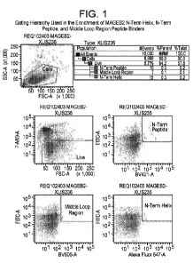
Figure 1. This figure shows the gating hierarchy used in the enrichment of
MAGEB2 N-Term Helix, N-
Term Peptide, and Middle Loop Region peptide binders as described in Example 2
herein.
Figure 2. This figure shows two plots that show binding by various anti-MAGEB2
antibodies binding to
immunogen peptide and full length MAGEB2 protein. The MAGEB2 panel was run in
a limited antigen
style assay (only highest concentration shown) to identify highest affinity
binders to full length
MAGEB2 protein.
Figure 3. This figure shows immunohistochemical results for a MAGEB2 1HC:
assay that measured
immunoreactivity in MAGE132 transfected cells. Intense MAGE1321HC: staining
was observed in CHO-
MAGE-B2+ cells with the four anti-MAGEB2 antibodies 4GI7, h15, 1C3, 1114, but
not with IgG control.
Figure 4. This shows immunohistochemical results for a MAGEB2 IHC assay that
measured
immunoreactivity in MAGEB5 transfected cells. No MAGEB21HC staining was
observed in CHO-
MAGEB5+ cells with the four anti-MAGEB2 antibodies 4G17, 1115, 1C3, 1114, and
with IgG control.
Figure 5. This figure shows immunohistochemical results for a MAGEB2 1HC assay
that measured
immunoreactivity in control testis tissues. Intense MAGEB2 IHC staining in
sperrnatogonia cells was
observed in testis with the four anti-MAGEB2 antibodies 4G17, 1.115, IC3,
1114, but not 1gG control at 2
uglrnl. Some nuclear stain was seen with antibodies 4G17, 1J15, IC3; Leydig
cell stain was seen with
antibody 1J15.
Figure 6. This figure shows immunohistochemical results for a MAGEB2 IHC Assay
that measured
immunoreactivity in normal human tissue. Non-specific particulate staining in
hepatocytes was seen in
liver tissue core (in normal human tissue with anti-MAGEB2 antibodies 4G17,
1115, 1C3, but not with
anti-MAGEB2 antibody 1114, and IgG control at 5 ugfml,
Figure 7. This figure shows irnmunohistochernical results for a MAGEB2 11-1C
assay that measured
immunoreactivity in testis and liver tissue, with titration down of the 1C3
antibody. At 2 ug/m1 Ab
concentration, anti-MAGEB2 antibody 1C3 clone has weak to mild intensity
specific staining in
appropriate proportions of sperrnatogonia cells as previously observed. There
is rare nuclear staining
and the intensity is slightly less than that observed at higher
concentrations. However, at this
concentration there is no non-specific background staining in liver.
4
SUBSTITUTE SHEET (RULE 26)
CA 03215594 2023-09-28
WO 2022/212831
PCT/US2022/023034
DETAILED DESCRIPTION
MAGEB2 is aberrantly expressed in a variety of human cancer types. MAGEB2 is
not expressed on the
surface of cells. MAGEB2 peptides; however, are displayed on the surface of
tumor cells by the MI-IC
class I molecule. In particular, the MAGEB2 peptide GVYDGEEHSV (HQ ID NO: 1)
is displayed on the
surface of tumor cells by the NiFIC class I molecule as a peptide-MHC complex.
See, for example, U.S.
Patent Appl. Publ. No. U52016/0250307A1 (U.S. Patent Appl. No. 14/975,952) and
US2017/0080070A1
(U.S, Patent Appl. No. 15/357,757). Although the MAGEB2 peptide-MHC complex is
displayed on the
surface of cells and would potentially be a target for a diagnostic agent, the
number of copies on the
cell surface of this peptide-MHC complex is far too low to be detected.
Accordingly, binding constructs
(e.g., antibodies) that are able to bind and detect MAGEB2 that is
intracellularly expressed are
described herein.
Binding Constructs
The present invention provides binding constructs comprising domains which
bind to a MAGEB2
protein, These binding constructs are alternatively referred to as antigen
binding constructs.
The term binding construct refers to a construct that is capable of binding to
its specific target or
antigen and comprises the variable heavy chain (VH) and/or variable light
chain (VL) domains of an
antibody or fragment thereof. Typically; a binding domain according to the
present invention
comprises the minimum structural requirements of an antibody which allow for
the target binding.
This minimum requirement may e.g, be defined by the presence of at least the
three light chain CORs
(i.e. COM , CDR2 and CDR3 of the VL region) and/or the three heavy chain CDRs
(i.e. CDR1, CDR2 and
CDR3 of the VH region); preferably of all six CDRs. An alternative approach to
define the minimal
structure requirements of an antibody is the definition of the epitope the
antibody binds within the
structure of a specific target, respectively, the protein domain of the target
protein composing the
epitope region (epitope cluster) or by reference to a specific antibody
competing with the epitope of
the defined antibody. Alternatively, the minimal structure requirements may be
defined by the
paratope sequences within the binding domain of the antibody.
Binding constructs of the present invention comprise at least one binding
domain. The term "binding
domain" characterizes in connection with the present invention a domain which
specifically binds to,
interacts with, or recognizes a given target epitope or a given target region
on the target molecule,
e.g,., MAGEB2 or a specific region within MAGEB2. The structure and function
of the binding domain is
based on the structure and/or function of an antibody; e.g. of a full-length
or whole immunoglobulin
molecule, and is from the variable heavy chain (VH) and/or variable light
chain (VL) domains of an
antibody or fragment thereof. In certain embodiments, the binding domain
comprises the presence of
three light chain CDRs (i.e. CDR1, CDR2 and CDR3 of the VL region) and/or
three heavy chain CDRs (i.e.
SUBSTITUTE SHEET (RULE 26)
CA 03215594 2023-09-28
WO 2022/212831
PCT/US2022/023034
CORI, CDR2 and CDR3 of the VH region). In certain embodiments, the binding
domain is produced by
or obtainable by phage-display or library screening methods rather than by
grafting CDR sequences
from a pre-existing (monoclonal) antibody into a scaffold.
The binding domain of a binding construct according to the invention may e.g.
comprise the above
referred groups of CDRs. Preferably, those CDRs are comprised in the framework
of an antibody light
chain variable region (VL) and an antibody heavy chain variable region (VH);
however, it does not have
to comprise both. Fd fragments, for example, have two VH regions and often
retain some antigen-
binding function of the intact antigen-binding domain. Additional examples for
the format of antibody
fragments, antibody variants or binding domains include (1) a Fab fragment, a
monovalent fragment
having the VL, VHõ CL and CHI domains; (2) a F(ab);! fragment, a bivalent
fragment having two Fab
fragments linked by a disulfide bridge at the hinge region; (3) an Fd fragment
having the two VH and
CHI domains; (4) an Fv fragment having the VL and VH domains of a single arm
of an antibody, (5) a
dAb fragment (Ward et al., (1989) Nature 341 :544-546), which has a VH domain;
(6) an isolated
cornplementarity determining region (CDR), and (7) a single chain Fv (scFv);
the latter being preferred
(for example, derived from an scFV-library). Examples for embodiments of
binding constructs
according to the invention are e,g, described in WO 00/006605, WO 2005/040220,
WO 2008/119567,
WO 2010/037838, WO 2013/026837, WO 2013/026833, us 2014/0308285, us
2014/0302037,
WO 2014/144722, WO 2014/151910, and WO 2015/048272.
In a specific embodiment of the invention, the binding construct is a full-
length antibody, as described
herein below.
Also within the definition of "binding domain" or "domain which binds" are
fragments of full-length
antibodies, such as VHõ VHH, VL, (s)dAb, Fv, light chain (VL-CL), Fd (VH-CH1),
heavy chain, Fab, Fab',
F(ab')2 or "r IgG" ("half antibody" consisting of a heavy chain and a light
chain). Binding constructs
according to the invention may also comprise modified fragments of antibodies,
also called antibody
variants or antibody derivatives. Examples include, but are not limited to,
scFv, di-scFy or bi(s)-scFv,
scFv-Fcõ scFv-zipper, scFabõ Fah:', Fab3, diabodies, single chain diabodies,
tandem diabodies (Tandab's)õ
tandem di-scFv, tandem tri-scFv, õminibodies" exemplified by a structure which
is as follows: (VH-VL-
CH3)2, (scFv-CI-13)2, ((scFv)2-CH3 CH3), ((scFv)2-CH3) or (scFv-CH3-scFv)2;
multibodies such as
triabodies or tetrabodies, and single domain antibodies such as nanohodies or
single variable domain
antibodies comprising merely one variable region, which might be VHH, VH or
VL, that specifically
binds to an antigen or target independently of other variable regions or
domains. Further possible
formats of the binding constructs according to the invention are cross bodies,
maxi bodies, hetero Fc
constructs, mono Fc constructs and scFc constructs. Examples for those formats
will be described
herein below,
6
SUBSTITUTE SHEET (RULE 26)
CA 03215594 2023-09-28
WO 2022/212831
PCT/US2022/023034
According to the present invention, binding domains are in the form of one or
more polypeptides.
Such polypeptides may include proteinaceous parts and non-proteinaceous parts
(e.g. chemical linkers
or chemical cross-linking agents such as glutaraldehyde). Proteins (including
fragments thereof,
preferably biologically active fragments, and peptides, usually having less
than 30 amino acids)
comprise two or more amino acids coupled to each other via a covalent peptide
bond (resulting in a
chain of amino acids).
The term "polypeptide as used herein describes a group of molecules, which
usually consist of more
than 30 amino acids. Polypeptides may further form multimers such as dialers,
turners and higher
oligomers, i.e., consisting of more than one polypeptide molecule, Polypeptide
molecules forming such
dirnersõ trimers etc. may be identical or non-identical. The corresponding
higher order structures of
such multimers are, consequently, termed homo- or heterodimers, homo- or
heterotrirners etc. An
example for a heterornultimer is an antibody molecule, which, in its naturally
occurring form, consists
of two identical light polypeptide chains and two identical heavy polypeptide
chains. The terms
"peptide", "polypeptide" and "protein" also refer to naturally modified
peptides / polypeptides /
proteins wherein the modification is effected e.g. by post-translational
modifications like glycosylation,
acetylation, phosphoryletion and the like. A "peptide"; "polypeptide " or
"protein" when referred to
herein may also be chemically modified such as pegylated. Such modifications
are well known in the
art and described herein below.
The definition of "antibody" according to the invention comprises full-length
antibodies, also including
camelid antibodies and other immunoglobulins generated by biotechnological or
protein engineering
methods or processes. These full-length antibodies may be for example
monoclonal, recombinant,
chimeric, deimmunized, humanized and human antibodies, as well as antibodies
from other species
such as mouse, hamster; rabbit, rat, goat, or non-human primates.
For the antibodies provided herein, the variable regions of immunaglobulin
chains generally exhibit
the same overall structure, comprising relatively conserved framework regions
(FR) joined by three
hypervariable regions, more often called "complementarity determining regions"
or CDRs. The CDRs
from the two chains of each heavy chain/light chain pair mentioned above
typically are aligned by the
framework regions to form a structure that binds specifically to the target
epitope. From N-terminalto
C-terminal, naturally-occurring light and heavy chain variable regions both
typically conform with the
following order of these elements: FR1, CDR1, F.R2, CDR2, FR3, CIDR3 and FRA.
A numbering system has
been devised for assigning numbers to amino acids that occupy positions in
each of these domains.
This numbering system is defined in Kabat Sequences of Proteins of
Immunological interest (1987 and
1991, NIH, Bethesda, Md.), or Chothia & Lesk, 1987, J. Mol. Biol. 195:901-917;
Chothia et al., 1989,
Nature 342:878-883.
SUBSTITUTE SHEET (RULE 26)
CA 03215594 2023-09-28
WO 2022/212831
PCT/US2022/023034
The term "variable" refers to the portions of the antibody or irnmunaglobulin
domains that exhibit
variability in their sequence and that are involved in determining the
specificity and binding affinity of
a particular antibody (i.e., the "variable domain(s)"). The pairing of a
variable heavy chain (VI-1) and a
variable light chain (VL) together forms an antigen-binding domain.
Variability is not evenly distributed throughout the variable domains of
antibodies; it is concentrated in
sub-domains of each of the heavy and light chain variable regions. These sub-
domains are called
"hypervariable regions" or "complementarity determining regions" (CDRs). The
more conserved (i.e.,
non-hypervariable) portions of the variable domains are called the "framework"
regions (FRM or FR)
and provide a scaffold for the six CDRs in three-dimensional space to form an
antigen-binding surface.
The variable domains of naturally occurring heavy and light chains each
comprise four framework
(FWI) regions (FR1, FR2, FR3, and FR4), largely adopting a 3-sheet
configuration, connected by three
hypervariable regions, which form loops connecting, and in some cases forming
part of, the P-sheet
structure. The hypervariable regions in each chain are held together in close
proximity by the
framework regions and, with the hypervariable regions from the other chain,
contribute to the
formation of the antigen-binding site.
The terms "CDR", and its plural "CDRs", refer to the complernentarity
determining region of which
three make up the binding character of a light chain variable region (CDR-11,
CDR-I..2 and CDR-13) and
three make up the binding character of a heavy chain variable region (CDR-H1,
CDR-H2 and CDR-H3).
CDRs contain most of the residues responsible for specific interactions of the
antibody with the
antigen and hence contribute to the functional activity of an antibody
molecule, i.e., they are the main
determinants of binding specificity to a particular target.
The exact definitional CDR boundaries and lengths are subject to different
classification and
numbering systems. CDRs may therefore be referred to by Kabat, Chothia,
contact or any other
boundary definitions, including the numbering system described herein. Despite
differing boundaries,
each of these systems has some degree of overlap in what constitutes the so
called "hypervariable
regions" within the variable sequences. CDR definitions according to these
systems may therefore
differ in length and boundary areas with respect to the adjacent framework
region. See for example
Kabat (an approach based on cross-species sequence variability), Chothia (an
approach based on
crystallographic studies of antigen-antibody complexes), and/or MacCallurn
(Kabat et al,, Sequences of
Proteins of Immunological Interest, 5th Ed. Public Health Service, National
Institutes of Health,
Bethesda, Md., 1991; Chothia et al., J. Mol. Biol, 1987, 196: 901-917; and
MacCallum et al., J. Mol. Biol,
1996, 262: 732). Still another standard for characterizing the antigen binding
side is the AbN1
definition used by Oxford Molecular's AbM antibody modeling software. See,
e.g., Protein Sequence
and Structure Analysis of Antibody Variable Domains, Antibody Engineering Lab
Manual (Ed.: Duebel,
S. and Kontermann, R., Springer-Verlag, Heidelberg). To the extent that two
residue identification
8
SUBSTITUTE SHEET (RULE 26)
CA 03215594 2023-09-28
WO 2022/212831
PCT/US2022/023034
techniques define regions of overlapping, but not identical regions, they can
be combined to define a
hybrid CDR. However, the numbering in accordance with the Kabat system is
preferred.
Typically, CDRs form a loop structure that can be classified as a canonical
structure. The term
"canonical structure" refers to the main chain conformation that is adopted by
the antigen binding
(CDR) loops. Each canonical structure can be characterized by the torsion
angles of the polypeptide
backbone. Correspondent loops between antibodies may, therefore, have very
similar three
dimensional structures, despite high amino acid sequence variability in most
parts of the loops
(Chothia and i.esk, J. Mol. Biol., 1987, 196: 901; Chothia et al., Nature,
1989, 342: 877; Martin and
Thornton, J. Moi. Bid, 1996, 263: 800). Furthermore, there is a relationship
between the adopted loop
structure and the amino acid sequences surrounding it. The conformation of a
particular canonical
class is determined by the length of the loop and the amino acid residues
residing at key positions
within the loop, as well as within the conserved framework (i.e., outside of
the loop). Assignment to a
particular canonical class can therefore be made based on the presence of
these key amino acid
residues.
The term "canonical structure" may also include considerations as to the
linear sequence of the
antibody, for example, as catalogued by Kabat (Kabat et al.). The Kabat
numbering scheme (system) is
a widely adopted standard for numbering the amino acid residues of an antibody
variable domain in a
consistent manner and is the preferred scheme applied in the present invention
as also mentioned
elsewhere herein. Additional structural considerations can also be used to
determine the canonical
structure of an antibody. For example, those differences not fully reflected
by Kabat numbering can
be described by the numbering system of Chothia et al. and/or revealed by
other techniques, for
example, crystallography and two- or three-dimensional computational modeling.
Accordingly, a given
antibody sequence may be placed into a canonical class which allows for, among
other things,
identifying appropriate chassis sequences (e.g., based on a desire to include
a variety of canonical
structures in a library). Kabat numbering of antibody amino acid sequences and
structural
considerations as described by Chothia et al., Inc. cit. and their
implications for construing canonical
aspects of antibody structure, are described in the literature. The subunit
structures and three-
dimensional configurations of different classes of immunoglobulins are well
known in the art. For a
review of the antibody structure, see Antibodies: A Laboratory Manual, Cold
Spring Harbor Laboratory,
eds. Harlow et al., 1988.
As used herein, the terms "single-chain Fv," "single-chain antibodies" or
"scFv" refer to single
polypeptide chain antibody fragments that comprise the variable regions from
both the heavy and
light chains, but lack the constant regions. Generally, a single-chain
antibody further comprises a
polypeptide linker between the VH and VL domains which enables it to form the
desired structure
which would allow for antigen binding. Single chain antibodies are discussed
in detail by Pluckthun in
9
SUBSTITUTE SHEET (RULE 26)
CA 03215594 2023-09-28
WO 2022/212831
PCT/US2022/023034
The Pharmacology of Monoclonal Antibodies, vol. 113, Rosenburg and Moore eds.
Springer-Verlag,
New York, pp. 269-315 (1994). Various methods of generating single chain
antibodies are known,
including, those described in U.S. Pat, Nos, 4,694,778 and 5,260,203;
International Patent Application
Publication No. WO 88/01649; Bird (1988) Science 242;423-442; Huston at al,
(1988) Proc. Natl. Acad,
Sci. USA 85:5879-5883; Ward at al. (1989) Nature 334:54454; Skerra at al.
(1988) Science 242:1038-
1041. In specific embodiments, single-chain antibodies can also be bispecific,
multispecific, human,
and/or humanized and/or synthetic.
In some embodiments, the binding constructs of the present invention are "in
vitro generated binding
constructs'''. This term refers to a binding construct according to the above
definition where all or part
of the variable region (e.g., at least one CDR) is generated in a non-immune
cell selection, e.g., an in
vitro phage display, protein chip or any other method in which candidate
sequences can be tested for
their ability to bind to an antigen. In other embodiments, the binding
construct sequences are
generated by genomic rearrangement in an immune cell in an animal. This term
thus preferably
excludes sequences generated solely by genomic rearrangement in an immune cell
in an animal. It is
envisaged that the first and/or second domain of the binding construct is
produced by or obtainable by
phage display or library screening methods rather than by grafting CDR
sequences from a pre-existing
(monoclonal) antibody into a scaffold,
A "recombinant antibody" is an antibody made through the use of recombinant
DNA technology or
genetic engineering.
The term "monoclonal antibody" (rnAb) or monoclonal antibody construct as used
herein refers to an
antibody obtained from a population of substantially homogeneous antibodies,
i.e., the individual
antibodies comprising the population are identical except for possible
naturally occurring mutations
and/or post-translation modifications (e.g., isomerizations, amidations) that
may be present in minor
amounts. Monoclonal antibodies are highly specific, being directed against a
single antigenic site or
epitope on the antigen, in contrast to conventional (polyclonal) antibody
preparations which typically
include different antibodies directed against different antigenic sites or
epitopes. In addition to their
specificity, the monoclonal antibodies are advantageous in that they are
synthesized by clonal cell
culture and are uncontaminated by other immunoglobulins. The modifier
"monoclonal" indicates the
character of the antibody as being obtained from a substantially homogeneous
population of
antibodies, and is not to be construed as requiring production of the antibody
by any particular
method.
For the preparation of monoclonal antibodies, any technique providing
antibodies produced by
continuous cell line cultures can be used. For example, monoclonal antibodies
to be used may be
made by the hybridoma method first described by Koehler at al., Nature, 256:
495 (1975), or may be
made by recombinant DNA methods (see, e.g., U.S. Patent No. 4,816,567).
Examples for further
SUBSTITUTE SHEET (RULE 26)
CA 03215594 2023-09-28
WO 2022/212831
PCT/US2022/023034
techniques to produce human monoclonal antibodies include the trioma
technique, the human B-cell
hybridorria technique (Kozbor, Immunology Today 4 (1983), 72) and the EBV-
hybridoma technique
(Cole et al., Monoclonal Antibodies and Cancer Therapy, Alan R. Liss, Inc,
(1985), 77-96).
Hybridomas can then be screened using standard methods, such as enzyme-linked
immunosorbent
assay (ELEA) and surface plasmon resonance analysis, e.g. BiacoreT"i to
identify one or more
hybridomas that produce an antibody that specifically binds with a specified
antigen. Any form of the
relevant antigen may be used as the irnmunogen, e.g., recombinant antigen,
naturally occurring forms,
any variants or fragments thereof, as well as an antigenic peptide thereof.
Surface plasmon resonance
as employed in the Biacore system can be used to increase the efficiency of
phage antibodies which
bind to an epitope of a target cell surface antigen (Schier, Human Antibodies
Hybridornas 7 (1996), 97-
105; Malmhorg, J. Immunol. Methods 133 (1995), 7-13),
Another exemplary method of making monoclonal antibodies includes screening
protein expression
libraries, e.g., phage display or ribosome display libraries. Phage display is
described, for example, in
Ladner etal., U.S. Patent No. 5,223,409; Smith (1985) Science 228:1315-1317,
Clackson etal., Nature,
352: 624-628 (1991) and Marks eta?., J. Mol. Biol., 222: 581-597 (1991).
In addition to the use of display libraries, the relevant antigen can be used
to immunize a non-human
e.g,, a rodent (such as a mouse, hamster, rabbit or rat). In one embodiment,
the non-human
animal includes at least a part of a human immunoglobulin gene. For example,
it is possible to
engineer mouse strains deficient in mouse antibody production with large
fragments of the human Ig
(immunoglobulin) loci. Using the hybridorna technology, antigen-specific
monoclonal antibodies
derived from the genes with the desired specificity may be produced and
selected. See, e.g.,
XENOMOUSE"1õ Green etal. (1994) Nature Genetics 7:13-21, US 2003-0070185, WC
96/34096, and
WO 96/33735,
A monoclonal antibody can also be obtained from a non-human animal, and then
modified, e.g.,
humanized, deimmunized, rendered chimeric etc., using recombinant DNA
techniques known in the
art. Examples of modified binding constructs include humanized variants of non-
human antibodies,
"affinity matured" antibodies (see, Lg. Hawkins et al. J. Mol. Biol. 254, 889-
896 (1992) and Lowman et
al., Biochemistry 30, 10832- 10837 (1991)) and antibody mutants with altered
effector function(s) (see,
e.g., US Patent 5,648,260, Kontermann and DUbel (2010), ioc. cit. and Little
(2009), loc. cit.).
In immunology, affinity maturation is the process by which B cells produce
antibodies with increased
affinity for antigen during the course of an immune response. With repeated
exposures to the same
antigen, a host will produce antibodies of successively greater affinities.
Like the natural prototype, the
in vitro affinity maturation is based on the principles of mutation and
selection. The in vitro affinity
maturation has successfully been used to optimize binding constructs, e.g.,
antibodies or antibody
fragments. Random mutations inside the CDRs are introduced using radiation,
chemical mutagens or
SUBSTITUTE SHEET (RULE 26)
CA 03215594 2023-09-28
WO 2022/212831
PCT/US2022/023034
error-prone PCR. in addition, the genetic diversity can be increased by chain
shuffling. Two or three
rounds of mutation and selection using display methods like phage display
usually results in antibody
fragments with affinities in the low nanomolar range.
A preferred type of an amino acid substitutional variation of the binding
constructs involves
substituting one or more hypervariable region residues of a parent antibody
(e. g. a humanized or
human antibody), Generally, the resulting variant(s) selected for further
development will have
improved biological properties relative to the parent antibody from which they
are generated. A
convenient way for generating such substitutional variants involves affinity
maturation using phage
display. Briefly, several hypervariable region sides (e. g. 6-7 sides) are
mutated to generate all possible
amino acid substitutions at each side. The antibody variants thus generated
are displayed in a
monovalent fashion from filamentous phage particles as fusions to the gene Ill
product of M13
packaged within each particle. The phage-displayed variants are then screened
for their biological
activity (e. g. binding affinity) as herein disclosed, in order to identify
candidate hypervariable region
sides for modification, alanine scanning mutagenesis can be performed to
identify hypervariable
region residues contributing significantly to antigen binding. Alternatively,
or additionally, it may be
beneficial to analyze a crystal structure of the antigen-antibody complex to
identify contact points
between the binding domain and, eq., the MAGEB2. Such contact residues and
neighboring residues
are candidates for substitution according to the techniques elaborated herein.
Once such variants are
generated, the panel of variants is subjected to screening as described herein
and antibodies with
superior properties in one or more relevant assays may be selected for further
development.
The antibodies of the present invention specifically include "chimeric"
antibodies (immunoglobulins) in
which a portion of the heavy and/or light chain is identical with or
homologous to corresponding
sequences in antibodies derived from a particular species or belonging to a
particular antibody class or
subclass, while the remainder of the chain(s) is/are identical with or
homologous to corresponding
sequences in antibodies derived from another species or belonging to another
antibody class or
subclass, as well as fragments of such antibodies, so long as they exhibit the
desired biological activity
(U.S. Patent No. 4,816,567; Morrison et al., Proc. Natl. Acad. Sci. USA, 81:
6851-6855 (1984)). Chimeric
antibodies of interest herein include "primitized" antibodies comprising
variable domain antigen-
binding sequences derived from a non-human primate (e.g., Old World Monkey,
Ape etc,) and human
constant region sequences. A variety of approaches for making chimeric
antibodies have been
described. See e.g,/ Morrison etal., Proc. Natl. Acad. ScL U.S.A. 81:6851,
1985; Takeda et al., Nature
314:452, 1985, Cabilly et al., U.S. Patent No, 4,816,567; Boss at al., U.S.
Patent No. 4,816,397;
Tanaguchi etal., EP 0171496; EP 0173494; and GB 2177096.
"Humanized" antibodies, variants or fragments thereof (such as Fv, Fab, Fab',
F(ab`)2 or other antigen-
binding subsequences of antibodies) are antibodies or immunoglobulins of
mostly human sequences,
12
SUBSTITUTE SHEET (RULE 26)
CA 03215594 2023-09-28
WO 2022/212831
PCT/US2022/023034
which contain (a) minimal sequence(s) derived from non-human immunoglobulin.
For the most part,
humanized antibodies are human immunoglobulins (recipient antibody) in which
residues from a
hypervariable region (also CDR) of the recipient are replaced by residues from
a hypervariable region
of a non-human (e.g.., rodent) species (donor antibody) such as mouse, rat,
hamster or rabbit having
the desired specificity, affinity, and capacity. In some instances, Fv
framework region (FR) residues of
the human immunoglobulin are replaced by corresponding non-human residues.
Furthermore,
"humanized antibodies" as used herein may also comprise residues which are
found neither in the
recipient antibody nor the donor antibody. These modifications are made to
further refine and
optimize antibody performance. The humanized antibody may also comprise at
least a portion of an
immunoglobulin constant region (Fc), typically that of a human immunoglobulin.
For further details,
see Jones et al., Nature, 321: 522-525 (1986); Reichmarin etal., Nature, 332:
323-329 (1988); arid
Presta, Cum Op. Struct. Biol., 2: 593-596 (1992).
Humanized antibodies or fragments thereof can be generated by replacing
sequences of the Fv
variable domain that are not directly involved in antigen binding with
equivalent sequences from
human Fv variable domains. Exemplary methods for generating humanized
antibodies or fragments
thereof are provided by Morrison (1985) Science 229:1202-1207; by Oi et of.
(1986) BioTechnioues
4:214; and by US 5,585,089; US 5,693,761; US 5,693,762; US 5,859,205; and US
6,407,213. Those
methods include isolating, manipulating, and expressing the nucleic acid
sequences that encode all or
part of immunoglobulin Fv variable domains from at least one of a heavy or
light chain. Such nucleic
acids may be obtained from a hybridorna producing an antibody against a
predetermined target, as
described above, as well as from other sources. The recombinant DNA encoding
the humanized
antibody molecule can then be cloned into an appropriate expression vector.
Humanized antibodies may also be produced using transgenic animals such as
mice that express
human heavy and light chain genes, but are incapable of expressing the
endogenous mouse
immunoglobulin heavy and light chain genes. Winter describes an exemplary CDR
grafting method that
may be used to prepare the humanized antibodies described herein (U.S. Patent
No. 5,225,539). All of
the CDRs of a particular human antibody may be replaced with at least a
portion of a non-human CDR,
or only some of the CDRs may be replaced with non-human CDRs. it is only
necessary to replace the
number of CDRs required for binding of the humanized antibody to a
predetermined antigen.
A humanized antibody can be optimized by the introduction of conservative
substitutions, consensus
sequence substitutions; gerrnline substitutions and/or back mutations. Such
altered immunoglobulin
molecules can be made by any of several techniques known in the art, (e.g.,
Teng et al., Proc. Natl.
Acad. Sci. U.S.A., 80: 7308-7312, 1983; Kozbor et al., Immunology Today, 4:
7279, 1983; Olsson etal.,
Meth. Enzymol., 92: 3-16, 1982, and EP 239 400).
13
SUBSTITUTE SHEET (RULE 26)
CA 03215594 2023-09-28
WO 2022/212831
PCT/US2022/023034
The term "human antibody", "human antibody construct" and "human binding
domain" includes
antibodies, antibody fragments, and binding domains having antibody regions
such as variable and
constant regions or domains which correspond substantially to human gerrnline
immunoglobulin
sequences known in the art, including, for example, those described by Kabat
et al. (1991) (loc. cit.).
The human antibodies, antibody fragments or binding domains of the invention
may include amino
acid residues not encoded by human germline immunoglobulin sequences (e,g.õ
mutations introduced
by random or side-specific rnutagenesis in vitro or by somatic mutation in
vivo), for example in the
CORs, and in particular, in CDR3. The human antibodies, antibody fragments or
binding domains can
have at least one, two, three, four, five, or more positions replaced with an
amino acid residue that is
not encoded by the human germline immunoglobulin sequence. The definition of
human antibodies,
antibody fragments and binding domains as used herein also contemplates fully
human antibodies,
which include only non-artificially and/or genetically altered human sequences
of antibodies as those
can be derived by using technologies or systems such as the Xenornouse.
Preferably, a "fully human
antibody" does not include amino acid residues not encoded by human germline
immunoglobulin
sequences.
The ability to clone and reconstruct rnegabase-sized human lad in yeast
artificial chromosomes YACs
and to introduce them into the mouse germline provides a powerful approach to
elucidating the
functional components of very large or crudely mapped loci as well as
generating useful models of
human disease. Furthermore, the use of such technology for substitution of
mouse loci with their
human equivalents could provide unique insights into the expression and
regulation of human gene
products during development, their communication with other systems, and their
involvement in
disease induction and progression.
An important practical application of such a strategy is the "humanization" of
the mouse hurnoral
immune system. Introduction of human immunoglobulin hg) loci into mice in
which the endogenous Ig
genes have been inactivated offers the opportunity to study the mechanisms
underlying programmed
expression and assembly of antibodies as well as their role in B-cell
development. Furthermore, such a
strategy could provide an ideal source for production of fully human
monoclonal antibodies (rnAbs) --
an important milestone towards fulfilling the promise of antibody therapy in
human disease, Fully
human antibodies are expected to minimize the immunogenic and allergic
responses intrinsic to
mouse or mouse-derivatized rnAbs and thus to increase the efficacy and safety
of the administered
antibodies. The use of fully human antibodies can be expected to provide a
substantial advantage in
the treatment of chronic and recurring human diseases, such as inflammation,
autoimmunity, arid
cancer, which require repeated compound administrations.
One approach towards this goal was to engineer mouse strains deficient in
mouse antibody production
with large fragments of the human Ig loci in anticipation that such mice would
produce a large
14
SUBSTITUTE SHEET (RULE 26)
CA 03215594 2023-09-28
WO 2022/212831
PCT/US2022/023034
repertoire of human antibodies in the absence of mouse antibodies. Large human
Ig fragments would
preserve the large variable gene diversity as well as the proper regulation of
antibody production and
expression. By exploiting the mouse machinery for antibody diversification and
selection and the lack
of immunological tolerance to human proteins, the reproduced human antibody
repertoire in these
mouse strains should yield high affinity antibodies against any antigen of
interest, including human
antigens. Using the hybridoma technology, antigen-specific human mAbs with the
desired specificity
could be readily produced and selected. This general strategy was demonstrated
in connection with
the generation of the first XenoMouse mouse strains (see Green et al. Nature
Genetics 7:13-21
(1994)). The XenoMouse strains were engineered with YACs containing 245 kb and
190 kb-sized
germline configuration fragments of the human heavy chain locus and kappa
light chain locus,
respectively, which contained core variable arid constant region sequences.
The human Ig containing
YACs proved to be compatible with the mouse system for both rearrangement and
expression of
antibodies and were capable of substituting for the inactivated mouse Ig
genes. This was
demonstrated by their ability to induce 5* cell development, to produce an
adult-like human repertoire
of fully human antibodies, and to generate antigen-specific human mAbs. These
results also suggested
that introduction of larger portions of the human lg loci containing greater
numbers of V genes,
additional regulatory elements, and human Ig constant regions may recapitulate
substantially the full
repertoire that is characteristic of the human humoral response to infection
and immunization. The
work of Green et al. was recently extended to the introduction of greater than
approximately 80% of
the human antibody repertoire through introduction of rnegabase sized,
germline configuration YAC
fragments of the human heavy chain loci and kappa light chain loci,
respectively. See Mendez et 01.
Nature Genetics 15:146-156 (1997) and U.S. patent application Ser. No.
08/759,620.
The production of the XenoMouse animals is further discussed and delineated in
U.S. patent
applications Ser. No. 07/466,008, Ser. No. 07/610,515, Ser. No. 07/919,297,
Ser, No, 07/922,649,
Ser, No, 08/031,801, Ser. No. 08/112õ848, Ser. No. 08/234,145, Ser. No,
08/376,279,
Ser. No. 08/430,938, Ser. No. 08/464,584, Ser. No. 08/464,582, Ser. No.
08/463,191,
Ser. No. 08/462,837, Ser. No, 08/486,853, Ser. No. 08/486,857, Ser. No.
08/486,859,
Ser. No. 08/462,513, Ser, No, 08/724,752, and Ser. No, 08/759,620; and U.S.
Pat, Nos, 6,162,963;
6,150,584; 6,114,598; 6,075,181, and 5,939,598 and Japanese Patent Nos. 3 068
180 82, 3 068 506 82,
and 3 068 507 82. See also Mendez et al. Nature Genetics 15:146-156 (1997) and
Green and Jakobovits
J. Exp. Med. 188:483-495 (1998), EP 0 463 151 81, WO 94/02602, WO 96/34096, WO
98/24893,
WO 00/76310, and WO 03/47336.
In an alternative approach, others, including GenPharrn International, Inc.,
have utilized a "minilocus"
approach. In the minilocus approach, an exogenous Ig locus is mimicked through
the inclusion of
pieces (individual genes) from the Ig locus. Thus, one or more VIal genes, one
or more DH genes, one or
SUBSTITUTE SHEET (RULE 26)
CA 03215594 2023-09-28
WO 2022/212831
PCT/US2022/023034
more .iH genes, a mu constant region, and a second constant region (preferably
a gamma constant
region) are formed into a construct for insertion into an animal. This
approach is described in
U.S. Pat. No. 5,545,807 to Surani et al, and U.S, Pat, Nos. 5,545,806;
5,625,825; 5,625,126; 5,633,425;
5,661,016; 5,770,429; 5,789,650; 5,814,318; 5,877,397; 5,874,299; and
6,255,458 each to Lonberg and
Kay, U.S. Pat. Nos. 5,591,669 and 6,023.010 to Krimpenfort and Berns, U.S.
Pat. Nos. 5,612,205;
5,721,367; and 5,789,215 to Berns et al., and U.S. Pat. No. 5,643,763 to Choi
and Dunn, and Ger:Pharm
International U.S, patent application Ser. No. 07/574,748, Ser. No.
07/575,962, Ser. No. 07/810,279,
Ser, No, 07/853,408, Ser. No. 07/904õ068, Ser. No. 07/990,860, Ser. No.
08/053,131,
Ser, No, 08/096,762, Ser. No. 08/155,301, Ser. No. 08/161,739, Ser. No.
08/165,699,
Ser. No. 08/209,741. See also EP 0 546 073 81, WO 92/03918, WO 92/22645, WO
92/22647,
WO 92/22670, WO 93/12227, WO 94/00569, WO 94/25535, WO 96/14436, WO 97/13852,
and
WO 98/24884 and U.S. Pat, No, 5,981,175. See further Taylor et al. (1992),
Chen etal. (1993), Tuaillon
etal. (1993), Choi etal. (1993), Lonberg etal. (1994), Taylor et GI. (1994),
and Tuaillon etal. (1995),
FishlAtild etal. (1996).
Kirin has also demonstrated the generation of human antibodies from mice hi
which, through microcell
fusion, large pieces of chromosomes, or entire chromosomes, have been
introduced. See European
Patent Application Nos. 773 288 and 843 961. Xenerex Biosciences is developing
a technology for the
potential generation of human antibodies. in this technology, SOD mice are
reconstituted with human
lymphatic cells, e.g., B and/or T cells. Mice are then immunized with an
antigen and can generate an
immune response against the antigen. See U.S, Pat. Nos. 5,476,996; 5,698,767;
and 5,958,765.
In some embodiments, the binding constructs of the invention are "isolated" or
"substantially pure"
binding constructs. "Isolated" or "substantially pure", when used to describe
the binding constructs
disclosed herein, means a binding construct that has been identified,
separated and/or recovered from
a component of its production environment. Preferably, the binding construct
is free or substantially
free of association with all other components from its production environment.
Contaminant
components of its production environment, such as that resulting from
recombinant transfected cells,
are materials that would typically interfere with diagnostic or therapeutic
uses for the polypeptide,
and may include enzymes, hormones, and other proteinaceous or non-
proteinaceous solutes. It is
understood that the isolated protein may constitute a wide range of percent
concentration, e.g., from
5% to 99.9% by weight of the total protein content, depending on the
circumstances. The polypeptide
may be made at a significantly higher concentration through the use of an
inducible promoter or high
expression promoter; such that it is made at increased concentration levels.
The definition includes the
production of a binding construct in a wide variety of organisms and/or host
cells that are known in
the art. In preferred embodiments, the binding construct will be purified (1)
to a degree sufficient to
obtain at least 15 residues of N-terminal or internal amino acid sequence by
use of a spinning cup
16
SUBSTITUTE SHEET (RULE 26)
CA 03215594 2023-09-28
WO 2022/212831
PCT/US2022/023034
sequenator, or (2) to homogeneity by SOS-PAGE under non-reducing or reducing
conditions using
Coornassie blue or, preferably, silver stain. Ordinarily, however, an isolated
binding construct wi be
prepared by at least one purification step.
Peptides are short chains of amino acid monomers linked by covalent peptide
(amide) bonds. Hence,
peptides fall under the broad chemical classes of biological oligorners and
polymers. Amino acids that
are part of a peptide or polypeptide chain are termed "residues" and can be
consecutively numbered.
All peptides except cyclic peptides have an N-terminal residue at one end and
a C-terminal residue at
the other end of the peptide. An oligopeptide consists of only a few amino
acids (usually between two
and twenty). A polypeptide is a longer, continuous, and unbranched peptide
chain. Peptides are
distinguished from proteins on the basis of size, and as an arbitrary
benchmark can be understood to
contain approximately SO or fewer amino acids. Proteins consist of one or more
polypeptidesõ usually
arranged in a biologically functional way. While aspects of the lab techniques
applied to peptides
versus polypeptides and proteins differ (e.g., the specifics of
electrophoresis, chromatography, etc.),
the size boundaries that distinguish peptides from polypeptides and proteins
are not absolute.
Therefore, in the context of the present invention, the terms "peptide", "poly-
peptide" and "protein"
may be used interchangeably, and the term 'polypeptide" is often preferred.
Polypeptides may further form multimers such as dirners, trimers and higher
oligornersõ which consist
of more than one polypeptide molecule. Polypeptide molecules forming such
dirners, trimers etc. may
be identical or non-identical. The corresponding structures of higher order of
such muitimers are,
consequently, termed horno- or heterodimers, homo- or heterotrimers etc. An
example for a
hereteromultimer is a full-length antibody or imrnunoglobulin molecule, which,
in its naturally
occurring form, consists of two identical light polypeptide chains and two
identical heavy polypeptide
chains. The terms "peptide", "polypeptide" and "protein" also refer to
naturally modified peptides /
polypeptides / proteins wherein the modification is accomplished e.g. by post-
translational
modifications like glycosylation, acetylation, phosphorylation and the like. A
"peptide", "polypeptide"
or "protein" when referred to herein may also be chemically modified such as
pegylated. Such
modifications are well known in the art and described herein below.
A binding construct is said to "specifically bind" or "immunospecifically
bind" to its antigen when the
binding construct binds its antigen with a dissociation constant (KID) is 510-
7 M as measured via a
surface plasma resonance technique (e.g., BACoreõ GE-Healthcare Uppsala,
Sweden) or Kinetic
Exclusion Assay (KinExA, Sapidyne, Boise, Idaho), In accordance with this
invention a binding construct
specifically binds or irnmunospecifically binds to MAGEB2.
Because of the sequence similarity between homologous proteins in different
species, a binding
construct or a binding domain that specifically binds to its target (such as a
human target) may,
however, cross-react with homologous target molecules from different species
(such as, from non-
17
SUBSTITUTE SHEET (RULE 26)
CA 03215594 2023-09-28
WO 2022/212831
PCT/US2022/023034
human primates). The term "specific / immunospecific binding' can hence
include the binding of a
binding construct or binding domain to epitopes or structurally related
epitopes in more than one
species.
The term "epitope" refers to a region on an antigen, or specific amino acid
residues; to which a binding
domain, such as an antibody or immunoglobulin, or a derivative, fragment or
variant of an antibody or
an immunoglobulin, specifically binds. An "epitope" is antigenic and thus the
term epitope is
sometimes also referred to herein as "antigenic structure" or "antigenic
determinant". Thus, the
binding domain is an "antigen interaction site". Said binding/interaction is
also understood to define a
"specific recognition",
"Epitopes" can be formed both by contiguous amino acids or non-contiguous
amino acids juxtaposed
by tertiary folding of a protein. A "linear epitope" is an epitope where a
contiguous amino acid primary
sequence comprises the recognized epitope. A linear epitope typically includes
at least 3 or at least 4,
and more typically, at least 5 or at least 6 or at least 7, and frequently,
about 8 to about 10 amino acids
in 3 unique sequence, or even longer than 10 amino acids.
A "conformational epitope", in contrast to a linear epitope, is an epitope
wherein the primary
sequence of the amino acids comprising the epitope is not the sole defining
component of the epitope
recognized (e.g.; an epitope wherein the primary sequence of amino acids is
not necessarily
recognized by the binding domain). Typically, a conformational epitope
comprises an increased
number of amino acids relative to a linear epitope, and comprises
noncontiguous amino acid
sequences. With regard to recognition of conformational epitopes, the binding
domain paratope
recognizes a three-dimensional structure of the antigen, preferably a peptide
or protein or fragment
thereof (in the context of the present invention, the antigenic structure for
one of the binding domains
is comprised within the target cell surface antigen protein). For example,
when a protein molecule
folds to form a three-dimensional structure, certain amino acids and/or the
polypeptide backbone
forming the conformational epitope become juxtaposed enabling the antibody to
recognize the
epitope. Methods of determining the conformation of epitopes include, but are
not limited to, x-ray
crystallography, two-dimensional nuclear magnetic resonance (2D-NMR)
spectroscopy and site-
directed spin labelling and electron paramagnetic resonance (EPR)
spectroscopy,
Various methods for identifying proteins, regions of proteins, or peptides
that are useful as
immunogens or for screening assays are known in the art. In one embodiment,
and as described
herein in the Examples, a Shannon entropy analysis of the MAGEB2 protein amino
acid sequence can
identify particular peptide sequences that can be used as immlinogens.
Exemplary MAGEB2 peptides that were identified to be used as irnmunogens
included: MAGEB2
peptide a.a43-76: S.SVSGGAASSSPAAGiPQEPQRAPITAAAAAAGV (N-terminal region
peptide) (SEC). ID
NO: 2), MAGEB2 peptide 2.2.95-125: SSSQASTSTKSPSEDPLTRKSGSLVQFLYK (MHD N-
terminal helix
18
SUBSTITUTE SHEET (RULE 26)
CA 03215594 2023-09-28
WO 2022/212831
PCT/US2022/023034
peptide) (5E0 i0 NO: 3), MAGEB2 peptide a.a.185-200: DLTDEESLLSSWDFPR (MHD
middle loop
peptide) (SEQ 0 NO: 4).
Accordingly, in one embodiment of the present invention, provided is a method
of immunizing an
animal comprising administration to the animal any of the peptides of HQ ID
NOs: 2, 3, or 4.
In another embodiment, provided is a method of generating an isolated antibody
comprising
immunizing an animal with any of the peptides of SEQ ID NOs: 2, 3, or 4 and
isolating said antibody. In
further embodiments, provided is a method of generating an isolated monoclonal
antibody comprising
immunizing an animal with any of the peptides of SEQ iD NOs: 2, 3, or 4 and
further generating a
monoclonal antibody using art recognized steps described herein.
In yet another embodiment, provided is an isolated antibody generated by a
process comprising
immunizing an animal with any of the peptides of SEQ ID NOs: 2, 3, or 4,
in yet other embodiments, fragments of the peptides of SEQ ID NOs: 2, 3, or 4
can be used as the
immunogen. Accordingly, in one embodiment of the present invention, the
peptide immunogen
comprises a fragment of the MAGEB2 peptide 2.2.43-76:
SSVSGGAASSSPAAGIPQEPQRAPTIAAAAAAGV
(N-terminal region peptide) (HQ ID NO: 2) that is 10, 11, 12, 13, 14, 15, 16,
17, 18, 19, 20, 21, 22, 23,
24, 25, 26, 27, 28, 29, 30, 31, 32, or 33 amino acids long.
In another embodiment of the present invention, the peptide immunogen
comprises a fragment of the
MAGEB2 peptide 2.2.95-125: SSSQASTSTKSPSEDPLTRKSGSLVQFLLYK (MHD N-terminal
helix peptide)
(HQ ID NO: 3) that is 10, 11, 12, 13, 14, 15, 16, 17, 18, 19, 20, 21, 22, 23,
24, 25, 26, 27, 28, 29, or 30,
amino acids long.
In another embodiment of the present invention, the peptide immunogen
comprises a fragment of the
MAGEB2 peptide a.a.185-200: DLTDEESLLSSWDFPR (MHD middle loop peptide) (SEQ ID
NO: 4) that is
10, 11, 12, 13, 14, or 15 amino acids long.
In further embodiments of the present invention, provided are binding
constructs that compete for
binding with any of the binding constructs described in the present invention.
Competition assays are
well known in the art and exemplary assays are described further herein.
19
SUBSTITUTE SHEET (RULE 26)
CA 03215594 2023-09-28
WO 2022/212831
PCT/US2022/023034
In other embodiments, alternative peptides that can be used as immunog,ens for
screening are
provided in Table 1 below. Accordingly, the invention provides
Table 1.
SUBSTITUTE SHEET (RULE 26)
CA 03215594 2023-09-28
WO 2022/212831
PCT/US2022/023034
MAGEB2 Peptide Amino Acid Sequence SEQ ID NO:
SVSGGAASSSPAAGIPQEPQRAPTTAAAAAAGV 388
VSGGAASSSPAAGIPQEPQRAP TTAAAAAAGV 389
SGGAASSSPAAGIPQEPQRAPTTAAAAAAGV 390
SGGAASSSPAAGIPQEPQRAPTTAAAAAAGV 391
GGAASSSPAAGIPQEPQRAPTTAAAAAAGV 392
GAASSSPAAGIPQEPQRAPTTAAAAAAGV 393
AASSSPAAGIPQEPQRAPTTAAAAAAGV 394
ASSSPAAGIPQEPQRAPTTAAAAAAGV 395
SSSPAAGIPQEPQRAPTTAMAAAGV 396
SSPMGIPQEPQRAPTTAAAAAAGV 397
SPAAGIPQEPQRAPTTAAAAAAGV 398
PAAGIPQEPQRAPTTAAAAAAGV 399
MGI PQEPQRAPTTAAAAAAGV 400
AGIPQEPQRAPTTAAAAAAGV 401
GI PQEPQRAPTTAAAAAAGV 402
I PQEPQRAPTTAAAAAAGV 403
PQEPQRAPTTAAAAAAGV 404
QEPQRAPTIAMMAGV 405
EPQRAPTTAAAAAAGV 406
PQRAPTTAAAAAAGV 407
QRAPTTAAAAAAGV 408
RA PTTAAAAAAGV 409
APTTAAAAAAGV 410
PTTAAAAAAGV 411
TTAAAAAAGV 412
AAAAAAGV 413
'""AAAAAAGV 414
AAAAAGV 415
AAAAGV 416
AAAGV 417
SSVSGGAASSSPAAGIPQEPQRAPTTAAAAAAG 418
SSVSGGAASSSPAAGIPQEPQRAPTTAAAAAA 419
SSVSGGAASSSPAAGIPQEPQRAPTTAAAAA 420
21
SUBSTITUTE SHEET (RULE 26)
CA 03215594 2023-09-28
WO 2022/212831
PCT/US2022/023034
SSVSGGAASSS PAAG I PQEPQRAPTrAAAA 421
SSVSGGAASSS PAAG I P QEP QR A PUMA 422
SSVSGGAASSS PAAG I P QEP QR A P TrAA 423
SSVSGGAASSS PAAG I P QEP QR A PITA 424
SSVSGGAASSS PAAG I PQEPQRAPTr 425
SSVSGGAASSS PAAG I P QEP QR APT 426
SSVSGGAASSS PAAG I P QEPQ R AP 427
SSVSGGAASSS PAAG I P QEPQ R A 428
SSVSGGAASSS PAAG I PQEPQR 429
SSVSGGAASSS PAAG I PQEPQ 430
SSVSGGAASSS PAAG I PQEP 431
SSVSGGAASSS PAAG I PQE 432
SSVSGGAASSS PAAG I PQ 433
SSVSGGAASSS PAAG I P 434
SSVSGGAASSS PAAG I 435
SSVSGGAASSS PAAG 436
SSVSGGAASSS PAA 437
SSVSGGAASSS PA 438
SSVSGGAASSS P 439
SSVSGGAASSS 440
SSVSGGAASS 441
SSVSGGAAS 442
SSVSGGAA 443
SSVSGGA 444
SSVSGG 445
SSVSG 446
SVSGGAASSSPAAG I PQEPQRAPTTAAAAAAG 447
VSGGAASSSPAAG I PQE PQR A PTTAAAAAA 448
SGG AA SSS PAAG I PQE PQRAPTTAAAAA 449
GGAASSSPAAG I PQEPQRAPTTAAAA 450
GAASSSPAAG I PQE PQR A PT1AAA 451
AASSS P AAG I PQEPQRAPTTAA 452
ASSSPAAG I PQ EP QRA PTrA 453
SSSPAAG I PQ EP QRA PIT 454
22
SUBSTITUTE SHEET (RULE 26)
CA 03215594 2023-09-28
WO 2022/212831
PCT/US2022/023034
SSPAAGIPQEPQRAPT 455
SPAAGIPQEPQRAP 456
PAAGIPQEPQRA 457
AAGIPQEPQR 458
AGIPQEPQ 459
GIPQEP 460
IPQE 461
SSQASTSIKSPSEDPLTRKSGSLVQFLLYK 462
SQASTSTKSPSEDPLTRKSGSLVQFLLYK 463
QASTSTKSPSEDPLTRKSGSLVQFLLYK 464
ASTSTKSPSEDPLTRKSGSLVQFLLYK 465
STSTKSPSEDPLTRKSGSLVQFLLYK 466
TSTKSPSEDPLTRKSGSLVQFLLYK 467
STKSPSEDPLTRKSGSLVQFLLYK 468
TKSPSEDPLTRKSGSLVQFLLYK 469
KSPSEDPLIRKSGSLVQFLLYK 470
SPSEDPLTRKSGSLVQFLLYK 471
PSEDPLTRKSGSLVQFLLYK 472
SEDPLTRKSGSLVQFLLYK 473
EDPLTRKSGSLVQFLLYK 474
DPLTRKSGSLVQFLLYK 475
PLTRKSGSLVQFLLYK 476
LTRKSGSLVQFLLYK 477
TRKSGSLVQFLLYK 478
RKSGSLVQFLLYK 479
KSGSLVQFLLYK 480
SGSLVQFLLYK 481
'.-GSLVQFLLYK 482
SLVQFLLYK 483
LVQFLLYK 484
VQFLLYK 485
QFLLYK 486
FLLYK 487
SSSQASTSTKSPSEDPLTRKSGSLVQFLLY 488
23
SUBSTITUTE SHEET (RULE 26)
CA 03215594 2023-09-28
WO 2022/212831
PCT/US2022/023034
SSSQASTSTKS P SE D PLTRKSGSLVQFLL 489
SSSQASTSTKSPSED PLTRKSGSLVQFL 490
SSSQASTST KS PSE D PO* R KSGSLVQF 491
SSSO.ASTST KS PSE D P LT R KSGSLVQ 492
SSSOASTSTKSPSED PLTRKSGSLV 493
SSSQ.ASTSTKSPSED PLTRKSGSL 494
SSSQASTSTKSPSED PLTRKSGS 495
SSSQASTSTKSPSED PLTRKSG 496
SSSQASTSTKSPSED PLTRKS 497
SSSQASTSTKSPSED PLTRK 498
SSSQASTSTKS PS E D PLTR 499
SSSQASTST KS PS E D P LT 500
SSSO.ASTST KS PSE D P L 501
SSSQASTSTKS PS E D P 502
SSSQASTSTK S PS ED 503
SSSQASTSTKSPSE 504
SSSQASTSTKSPS 505
SSSQASTSTKSP 506
SSSQASTSTKS 507
SSSQASTSTK 508
SSSO.ASTST 509
SSSQASTS 510
SSSQAST 511
SSSQAS 512
SSSQA 513
SSQASTST KS PS ED P LTR KSGSLVQFLLY 514
SQASTST KS PSE DP LT R KSGSLVQFLL 515
QASTSTKSPS ED PLTRKSGSLVQFL 516
ASTSTKS PS E D PLTR KSGSLVQF 517
STSTKSPSEDP LTR K SG SLVQ 518
TSTKSPSE D P LT RKSG SLV 519
STKSPSEDPLTR KSGSL 520
TKSPSEDP LTR KSGS 521
KSPSED PLTRKSG 522
24
SUBSTITUTE SHEET (RULE 26)
CA 03215594 2023-09-28
WO 2022/212831
PCT/US2022/023034
SPSEDPLTRKS 523
PSEDPLTRK 524
SEDPLTR 525
EDPLT 526
LTDEESLLSSWDFPR 527
TDEESLLSSWDEPR 528
DEESLLSSWDFPR 529
EESLISSWIDEPR 530
ESLLSSWDFPR 531
SLLSSWDFPR 532
LLSSWDFPR 533
LSSWDFPR 534
SSWDFPR 535
SWDFPR 536
WDFPR 537
DLTDEESLLSSWDFP 538
DLTDEESLLSSWDF 539
DLTDEESLLSSWD 540
DLTDEESLLSSW 541
DLTDEESLLSS 542
DLTDEESLLS 543
DLTDEESLL 544
DLTDEESL 545
DLTDEES 546
DLTDEE 547
DLTDE 548
LTDEESLISSWDFP 549
TDEESLLSSWDF 550
DEESLLSSWD 551
EESLLSSW 552
ESLLSS 553
SLLS 554
Affinity
SUBSTITUTE SHEET (RULE 26)
CA 03215594 2023-09-28
WO 2022/212831
PCT/US2022/023034
The interaction between the binding domain and the epitope or the region
comprising the epitope
implies that a binding domain exhibits appreciable affinity for the epitope
and/or the region
comprising the epitope on a particular protein or antigen, and unless
otherwise specified, does not
exhibit significant binding or reactivity with proteins or antigens other than
MAGEB2. This affinity can
be measured by various techniques known to one skilled in the art, such as in
a surface plasrnon
resonance assay, such as a Biacore assay, or in a cell based assay.
"Appreciable affinity" includes binding with an affinity of about 10-6 M (KG)
or stronger. Preferably,
binding is considered specific when the binding affinity is about 10-12 to 10-
8 M, 1012 to 10-9 M, 1012 to
10' M, 1041 to 10-8 M, preferably of about 10-11 to 10-9 M. Whether a binding
domain specifically
reacts with or binds to a target can be tested readily by, inter alio,
comparing the reaction of said
binding domain with a target protein or antigen with the reaction of said
binding domain with proteins
or antigens other than the MAGEB2, Preferably, a binding domain of the
invention does not essentially
or substantially bind to proteins or antigens other than MAGEB2.
Specificity
The term "does not significantly bind" means that a binding construct or
binding domain of the
present invention does not bind to a protein or antigen other than MAGEB2, For
example, a binding
construct or binding domain that exhibits binding to proteins with similar
amino acid sequences, e.g.,
MAGEA4 or MAGEA8, would not be a desirable binding construct.
In the present invention, the MAGEB2 binding constructs possess surprising
levels of specificity and
selectivity to their target, as evidenced by its lack of binding to target
negative cells. See, e.g.,
Examples 2 and 3 herein.
This specificity and selectivity are highly desired, yet difficult to achieve,
properties for a binding
construct in a diagnostic assay as it limits, reduces, or eliminates off-
target binding and any potential
false readouts.
Specific binding is believed to be effected by specific motifs in the amino
acid sequence of the binding
domain and the antigen. Thus, binding is achieved as a result of their
primary, secondary and/or
tertiary structure as well as the result of secondary modifications of said
structures. The specific
interaction of the antigen-interaction-side with its specific antigen may
result in a simple binding of
said side to the antigen. Moreover, the specific interaction of the antigen-
interaction-side with its
specific antigen may alternatively or additionally result in the initiation of
a signal, ag, due to the
induction of a change of the conformation of the antigen, an oligomerization
of the antigen, etc.
MAGEB2 Binding Constructs
26
SUBSTITUTE SHEET (RULE 26)
CA 03215594 2023-09-28
WO 2022/212831 PCT/US2022/023034
The invention provides binding constructs comprising a domain which binds to
MAGEB2, and in
specific embodiments, to the MAGEB2 peptides SSVSGGAASSSPAAGIPQEPQRAP I I
AAAAAAGV (N-
terminal region peptide) (HQ ID NO; 2), 3.3.95-125;
SSSQASTSTICSPSEDPLTRKSGSLVQFLLYK (MHD N-
terminal helix peptide) (SEQ ID NO: 3), or a.a.185-200: DLTDEESLLSSWDFPR (SEQ
ID NO: 4).
Table 2 below provides amino add sequences of exemplary MAGEB2 binding
constructs VH-CDRs and
VL-CDRs. Table 3 below provides amino add sequences of exemplary MAGEB2
binding construct VH
and VL domains.
Table 2. Exemplary Light Chain CDR and Heavy Chain CDR Sequences
Molecule CDR.-1.1 CDR-12 CDR-13 CDR-1-11 CDR-H2 CDR-H3
1114 QSSOSVYDNNAL GASTLAS QC.TYWSSYQND SYAMS SIGGGGSAVYASW GFYSIDL
A (SEQ ID NO: 85) (SEQ ID NO: 87) (SEQ ID NO: 229)
AKG (SEQ ID NO: 231)
(SEQ ID NC: 85) (SEQ ID NO: 230)
1,C3 QASONISSYLA RAST LAS QSYDDSRSSNFFY NYYIC
CIDNANGRTYYAS SLATPL.
(SEQ ID NO: 73) (SEQ ID NO: 74) A (SEQ ID NO: 217) WAKG
(SEQ ID NO: 219)
(SEQ ID NO: 75) (SEQ ID NO: 218)
11117 Q5SKSVYNKretVL GAST LAS AGGYSSSS DT F A SG QL C
CIGSGSNA;STFYAS VGSDDYGDSDVFDP
(SEQ ID NO: 92) (SEQ ID NO: 93) (SEQ ID NO: 235) WAO,G
(SEQ ID NO: 237)
(SE , ID NO: 91) (SEQ ID NO: 236)
Table 3. Exemplary Variable Light Chain and Variable Heavy Chain Sequences
Molecule VL 1111
1114 DVVPATQFPASVEATVGGTVIIKCQSSOS\NDNNALA
OSVEESGGRLVTPUFPLILICTISGFSLSSYAMSVVVRQAPOKOL
WYOONAGORPRLLIYGASTLASGVPSRFSASGSGTEF
EVVIGSIGGGGSAVYASWAKGRFTISKTSTTVDLRITSPITEDTAM
ILTISDLECADAATTICLICMVSSYQNDFGGGTEVVV YFCGROFYSIDLINGPOTLVIVSS
(8E0 ID NO: 347)
(SEQ ID NO: 345)
1C3 DIVMMTPSSVEAAVGC1TVTIKCOASONISSYLAWYO
OSLEESGEGINOPEGSLTITCTAFGVT1TNYVICVIVROAPGKG1
QKPCOPPI(LLIVRASTLASGVPSRFKGSGSGIQH1115 EuVV(.5121DNANGRTYYASWAKGRFTISKI-
SSTIGT1QMI-SLTAA
DLECADAATYYCQSYDDSRSSNFRAFGGGIEVVVK DIATYFCARSLATPLWGPG LVIVSS
(SEQ. ID NO: 338) (SEQ. ID NO: 339)
1H17 AAVLIOTPSPVSAAVGGIVSASCQSSKSVYNKNWLS
QEQLVESGGOLVKPGASLTLICKASGFSFSSGQLrviCVVVRQAPG
WFQQKPOQPPKLLIYGASTLASGVPSRFKGSGSGTQF
KGLEWIACIGSGSNAISTFYASWAQGRFTISKSSSTTVTLQLTSLT
ILTISDVQCDDAATYKAGGYSSSSMTAFGGGIEVVV AADTATYFCARVGSDDYGDSOVEDPWGPGILVTVSS
(SEQ ID NO: 351)
(SEQ ID NO: 350)
In one embodiment, the invention provides an isolated binding domain that
binds to MAGEB2,
wherein the binding domain comprises;
a) a VH region comprising CDR-H1 as depicted in HQ ID NO: 229, CDR-H2 as
depicted in SEQ ID
NO: 230, and CDR-H3 as depicted in SEQ ID NO: 231, and a Vt. region comprising
CDR-Li as depicted in
SEQ ID NO: 85, CDR-12 as depicted in SEQ ID NO: 86 and CDR-L3 as depicted in
SEQ ID NO: 87; or
b) a VH region comprising CDR-H1 as depicted in SEQ ID NO: 217, CDR-H2 as
depicted in SEQ ID
NO: 218, and CDR-H3 as depicted in SEQ ID NO: 219, and a V1 region comprising
CDR-1.1 as depicted in
SEQ ID NO: 73, CDR-L2 as depicted in SEQ ID NO: 74 and CDR-L3 as depicted in
SEQ ID NO: 75; or
27
SUBSTITUTE SHEET (RULE 26)
CA 03215594 2023-09-28
WO 2022/212831
PCT/US2022/023034
c) a VH region comprising CDR-H1 as depicted in SEQ ID NC): 235, CDR-H2 as
depicted in SEQ. ID
NO: 236, and CDR-H3 as depicted in SEQ ID NO: 237, and a VL region comprising
CDR-L1 as depicted in
SEQ ID NO: 91, CDR-L2 as depicted in SEQ ID NO: 92 and CDR-L3 as depicted in
SEQ ID NO; 930
In one embodiment, the invention provides an isolated binding construct
comprising a binding domain
that binds to N1AGEB2, wherein the binding domain comprises:
a) a VH region comprising CDR-H1 as depicted in SEQ ID NO: 229, CDR-H2 as
depicted in SEQ ID
NO: 230, and C08-H3 as depicted in SEQ ID NC): 231, and a VI. region
comprising CDR-L1 as depicted in
SEQ ID NO; 85, CD 8-L2 as depicted in SEQ ID NO: 86 and CDR-L3 as depicted in
SEQ ID NO: 87; or
b) a VH region comprising CDR-H1 as depicted in SEQ ID NO: 217, CDR-H2 as
depicted in SEQ ID
NO: 218, and CDR-H3 as depicted in SEQ ID NO: 219, and a VL region comprising
CDR-L1 as depicted in
SEO, ID NC): 73, CDR-L2 as depicted in HQ ID NO: 74 and CDR-L3 as depicted in
SEQ ID NO: 75; or
C) a VH region comprising CDR-H1 as depicted in SEQ ID NO: 235, CDR-H2 as
depicted in HQ ID
NO: 236, and CDR-H3 as depicted in SEQ. ID NO: 237, and a VL region comprising
CDR-L1 as depicted in
SEQ ID NO: 91, CDR-L2 as depicted in SEQ ID NO: 92 and CDR-L3 as depicted in
SEQ ID NO: 93.
In one embodiment, the invention provides an isolated binding domain that
binds to MAGEB2,
wherein the binding domain binds to the same epitope as an antibody or binding
construct that
comprises:
a) a VI-1 region comprising CDR-H1 as depicted in SEQ ID NO: 229; CDR-H2 as
depicted in SEQ ID
NO: 230, and CDR-H3 as depicted in SEQ. ID NC): 231, and a VL region
comprising CDR-1.1 as depicted in
SEO, ID NO: 85, CDR-L2 as depicted in SEQ ID NO: 86 and CDR-L3 as depicted in
SEQ ID NO: 87; or
b) a VH region comprising CDR-H1 as depicted in SEQ ID NO: 217, CDR-H2 as
depicted in SEQ ID
NO: 218, and CDR-H3 as depicted in SEQ ID NO: 219, and a VL region comprising
CDR-L1 as depicted in
SEQ ID NO; 73, CDR-L2 as depicted in SEQ. ID NO: 74 and CDR-L3 as depicted in
SEQ ID NO: 75; or
c) a VH region comprising CDR-H1 as depicted in SEQ ID NO: 235, CDR-H2 as
depicted in SEQ ID
NO: 236, and CDR-H3 as depicted in SEQ ID NO: 237, and a VL region comprising
CDR-L1 as depicted in
SEQ ID NO: 91, CDR-L2 as depicted in SEQ ID NO: 92 and CDR-L3 as depicted in
SEQ ID NO; 930
In one embodiment, the invention provides an isolated binding construct
comprising a binding domain
that binds to N1AGEB2, wherein the binding domain binds to the same epitope as
an antibody or
binding construct that comprises:
a) a VH region comprising CDR-H1 as depicted in SEQ ID NC): 229, CDR-H2 as
depicted in SEQ ID
NO: 230, and CDR-H3 as depicted in SEQ ID NO: 231, and a VL region comprising
CDR-L1 as depicted in
SEQ ID NO: 85, CDR-L2 as depicted in SEQ ID NO: 86 and CDR-L3 as depicted in
SEQ ID NO: 87; or
23
SUBSTITUTE SHEET (RULE 26)
CA 03215594 2023-09-28
WO 2022/212831
PCT/US2022/023034
h) a VH region comprising CDR-H1 as depicted in SEQ ID NO: 217, CDR-H2 as
depicted in SEQ. ID
NO: 218, and CDR-H3 as depicted hi SEQ ID NO: 219, and a VL region comprising
CDR-L1 as depicted in
SEQ ID NO: 73, CDR-L2 as depicted in SEQ ID NO: 74 and CDR-L3 as depicted in
SEQ ID NO; 75; or
c) a VI-1 region comprising CDR-H1 as depicted in SEQ ID NO: 235, CDR-H2 as
depicted in SEQ ID
NO: 236, and CDR-H3 as depicted in SEQ ID NO: 237, and a VL region comprising
CDR-11 as depicted in
SEQ ID NO: 91, CDR-12 as depicted in SEQ ID NO: 92 and CDR-L3 as depicted in
SEQ ID NO: 93.
In one embodiment, the invention provides an isolated binding domain that
binds to MAGEB2,
wherein the binding domain comprises:
a) a VH region comprising SEQ ID NO: 347 and a VL region comprising SEQ ID
NO: 346; or
b) a VI-1 region comprising SEQ ID NO: 339 and a VL region comprising SEQ
ID NO: 338; or
c) a VH region comprising SEQ ID NO: 351 and a VL region comprising SEQ ID
NO: 350.
In one embodiment, the invention provides an isolated binding construct
comprising a binding domain
that binds to MAGEB2, wherein the binding domain comprises:
a VH region comprising SEQ ID NO: 347 and a VL region comprising SEQ ID NO:
346; or
h) a VH region comprising SEQ ID NO: 339 and a Vt. region comprising SEQ ID
NO; :338; or
c) a VH region comprising SEQ ID NO: 351 and a VL region comprising SEQ ID
NO: 350.
In one embodiment, the invention provides an isolated binding domain that
binds to MAGEF32,
wherein the binding domain binds to the same epitope as an antibody or binding
construct that
comprises:
VH region comprising SEQ ID NO: 347 and a VL region comprising SEQ ID NO: 346;
or
h) a VH region comprising SEQ ID NO: 339 and a VL region comprising SEQ ID
NO; 338; or
c) a VH region comprising SEQ ID NO: 351 and a VL region comprising SEQ ID
NO: 350.
In one embodiment, the invention provides an isolated binding construct
comprising a binding domain
that binds to MAGEB2, wherein the binding domain binds to the same epitope as
an antibody or
binding construct that comprises:
a) a VH region comprising SEQ ID NO: 347 and a VL region comprising SEQ ID
NO: 346; or
h) a VH region comprising SEQ ID NO: 339 and a VL region comprising SEQ ID
NO: 338; or
c) a VH region comprising SEQ ID NO: 351 and a VL region comprising SEQiD
NO; 35(L
29
SUBSTITUTE SHEET (RULE 26)
CA 03215594 2023-09-28
WO 2022/212831
PCT/US2022/023034
In another embodiment, the invention provides an isolated binding construct
comprising a binding
domain that binds to MAGEB2, wherein the binding domain comprises a VH region
selected from the
group consisting of any VH region and a corresponding VL region as depicted in
any of the sequences
in Tables 21-25 herein,
In another embodiment, the invention provides an isolated binding construct
comprising a binding
domain that binds to MAGEB2, wherein the binding domain binds to the same
epitope as an antibody
or binding construct that comprises a VH region selected from the group
consisting of any VH region
and a corresponding VL region as depicted in any of the sequences in Table 23
herein.
In another embodiment, the invention provides an isolated binding construct
comprising a binding
domain that binds to MAGEB2, wherein the binding domain comprises a CDR-H1,
CDR-H2, CDR-H3
from a VH region selected from the group consisting of any Vld region as
depicted in any of the
sequences in Table 22 herein and further comprises a corresponding CDR-L1.,
CDR-L.2, CDR-L3 from a VL
region selected from the group consisting of any VL region as depicted in any
of the sequences in Table
21 herein.
In another embodiment; the invention provides an isolated binding construct
comprising a binding
domain that binds to MAGEB2; wherein the binding domain binds to the same
epitope as an antibody
or binding construct that comprises a CDR-H1, CDR-H2, CDR-H3 from a VH region
selected from the
group consisting of any VH region as depicted in any of the sequences in Table
22 herein and further
comprises a corresponding CDR-L1, CDR-L2, CDR-L3 from a VL region selected
from the group
consisting of any VL region as depicted in any of the sequences in Table 21
herein.
In another embodiment, the invention provides an isolated binding construct
comprising a binding
domain that binds to MAGEB2, wherein the binding domain comprises a VH and a
VL region consensus
sequence selected from any of the sequences in Table 25 herein.
In another embodiment; the invention provides an isolated binding construct
comprising a binding
domain that binds to MAGEB2, wherein the binding domain comprises a CDR-H1.,
CDR-H2, CDR-H3
from a VH region consensus sequence selected from the group consisting of any
VH region as depicted
in any of the sequences in Table 25 herein and further comprises a
corresponding CDR-L1, CDR-L2,
CDR-L3 from a VL region consensus sequence selected from the group consisting
of any VL region as
depicted in any of the sequences in Table 25 herein,
Exemplary binding construct full sequences are presented below:
1C3 _LC (SEQ ID NO; 555)
MDMRVPACILLGLLLLWLRGARCDIVMTCITPSSVEAAVGGTVTIKCCIASCINISSYLAWMOOGORPKWYRASTLA
SGVPSRFKGSGSGTQFTLT1SDLECADAATYYCQSYDDSRSSNFFYAFGGGTEVVVKGDPVAPTVLLFPPSSDEVATGT
VTIVCVANKYFPOVIVIWEVDGTMITGIENSKTPONSADCTYNLSSTLTLTSTQYNSHKEYICKVTOGiTsvvc6FsR
KNIC
SUBSTITUTE SHEET (RULE 26)
CA 03215594 2023-09-28
WO 2022/212831
PCT/US2022/023034
1C3 HC (SEQ ID NO: 555)
M DM RVPAQLLGLILLW LRGA RCQSLEESGGG LVQP EGS LT LTCTAFGVTLTNYY ICWVRQAPG KG
LEWVG 0 DNAN
G RTYYASWAKG RFT1S KTSSTTGTLQMTSLTAADTATYFCARS LATPLWG PGTLVTVSSGQP KAPSVF P
LAPCCG DTP
SSTVTLGCLVKGYLP E PVTVTW NSGTLTNGVRTFPSVRQSSG LYS LSSVVSVTSSSOPVTCNVAH
PATNTKVDKTVAPS
TCSKPTCPPPELLGGPSVFI FP P KP K DTLM 1SRTP EVICVVVDVSQD DP EVQFTWY1 N N
EQVRTARPP LREQQFNST1R
VVST LP FAHQDWLRG K E KCKVH N KALPAP EKTISKARGQPLE P KVYTMG P P REELSSRSVSLTCM
1NG FY PSD 1SVE
WEKNGKAEDNYKTTPAVLDSDGSYFLYSKLSVPTSEWQRGDVFTCSVMHEALH NHYTQKSISRSPGK
1114 (SEQ ID NO: 557)
M DM RVPAQLLGLLLLWLRGARCDVVMTQTPASVEATVGGTVTIKCQSSQSVYDN NALAWYQQNAGQRP R LING
AST LASGV PS R FSASGSGTE D
LECADAATYYCQCIYYVSSYQN D G GGTEVVVKG D PVA PTVLLF P PSSD EVA
TGTVT1VCVAN KYFPDVTVTW EVDGTTQTTGIENSKTPQNSADCTYN LSSTLTLTSTQYNSH
KEYTCKVTQGTTSVVQ
SFS RKNC
1114_FIC (SEQ ID NO: 558)
M DM RVPAQLLGLILLWLRGARCQSVEESGGRLVTPGTPLTLICTISG FSLSSYAMSVVVRQAPG KG LEW
IGSIGGGGS
AVYASWAKG R FTISKTSTTVD LRITS PTTEDTA M YFCG RG FYS I D LWG PGTLVTVSSGQP
KAPSVFP LAPCCG DIPSST
VTLGCLVKGYLP E PVTVTW NSGTLTNEVRTF PSVRQSSG LYS LSSVVSVTSSSQPVTC !WAN
PATNTKVDKTVAPSTC
SKPTCP PP E LLGG PSVFI FP PK P KDTLM 1SRTP EVTCVVVDVSO,DDP EVQFTWY I N N
EQVRTARPPLREQQFNSTIRVV
STLP1AHQDWLRGKEFKCKVH NKALPAPIEKTISKARGOP LEP KVYTMG P P REELSSRSVSLTCM I NG
FYPSD !MINE
KNG KAEDNYKTTPAVLDSDGSYFLYSKLSVPTSEWQRGDVFICSVM HEALHN HYTQKS1SRSPG K
11-117 _LC (SEQ ID NO: 559)
M DM RVPAQLLG LLLLW LRGARCAAV LTQTPS PVSAAVG GTVSASCQSSKSVY N K NW LSW FQQKPG
QP PKLLIYGA
STLASGVPSRFKGSGSGTQFTLTISDVQCDDAATYYCAGGYSSSSDTFAFGGGTEVVVKGDPVAPTVLLFPPSSDEVAT
GTVT1VCVAN KYF P DVTVTW EVDGTTQTTG ENSKTPQNSADCTYN LSSTLTLTSTQYNSH K
EYTCKVTQGTTSVVQS
FSRKNC
1H17_11C (HQ ID NO: 560)
M DM RVPAQLLGUILVILRGARCQEQLVESGGG
LVKPGASLTLTCKASGFSFSSGQLMCWVRQAPGKGLEWIACIGS
GSNA1STFYASWAQG R FT1S KSSSTIVTLQLTSLTAADTATY FCARVGSD DYG DS DVFD PVVG
PGTLVTVSSGQP KAPS
VFP LAPCCG DTPSSTVTLG CLVKG Y E PVTVIWNSGTLTN GVRTFPWRQSSG
LYSLSSVVSVISSSQPVICNVAH PA
TNTKVD KTVAPSTCS KPTCP P P E LLGG PSVF1 FP P KP KDTLM
ISRTPEVICVVVDVSQDDPEVQFTWYIN N EQVRTAR
PPLREQQFNSTIRVVSTLPIAHQDWLRGKEFKCKVH N KALPAP I E KT1SKARGQP LEP KVYTMGP PREE
LSSRSVSLTC
M I NG FYPSDISVEW E KNG KAEDNYKTTPAVLDSDGSYFLYSKLSVPTSEWQRGDVFTCSVMH EALF-I N
HYTQKS1SRS
PG K
Competitive Binding
Whether or not an antibody or binding construct competes for binding to an
antigen (such as
IVIAGEB2) with another given antibody or binding construct can be measured in
a competition assay
such as a competitive EISA. Avidin-coupled microparticles (beads) can also be
used. Similar to an
avidin-coated HASA plate, when reacted with a biotinylated protein, each of
these beads can be used
as a substrate on which an assay can be performed. Antigen is coated onto a
bead and then precoated
31
SUBSTITUTE SHEET (RULE 26)
CA 03215594 2023-09-28
WO 2022/212831
PCT/US2022/023034
with the first antibody. The second antibody is added, and any additional
binding is determined. Read-
out occurs via flow cytometry. Preferably a cell-based competition assay that
allows binding to
intracellularly expressed MAGEB2 is used, using either cells that naturally
express MAGEB2, or cells
that were stably or transiently transformed with MAGEB2. The term "competes
for binding", in the
present context, means that competition occurs between the two tested
antibodies of at least 20%, at
least 30%, at least 40%, at least 50%, at least 60%, at least 70%, at least
80% or at least 90%, as
determined by any one of the assays disclosed above, preferably the cell-based
assay.
Competitive antibody binding assays include assays determining the competitive
binding of two
antibodies/ binding constructs to a cell surface bound antigen. Common methods
aim to detect
binding of two antibodies/ binding constructs, A and B, to the same antigen
expressed by a cell may
include steps of:
blocking of the antigen by pre-incubation of cells with antibody/ binding
construct A followed by a sub-
maximal addition of labeled antibody/ binding construct B and detecting the
binding of B compared
with binding in the absence of A;
titration (i.e. adding different amounts) of antibody/ binding construct A in
the presence of sub-
maximal amounts of labeled antibody/ binding construct B and detecting the
effect on binding of B; or
co-titration of A and B, wherein both antibodies/ binding constructs are
incubated together at maximal
concentration and detecting whether the total binding equals or exceeds that
of either A or B alone,
i.e, a method which cannot be affected by the order of addition or relative
amounts of the antibodies/
binding constructs.
When two antibodies/ binding constructs A and B compete for a antigen, the
antibodies will very often
compete with each other in blocking assays independently from the order of the
addition of the
antibodies. In other words, competition is detected if the assay is carried
out in either direction.
However, this is not always the case, and under certain circumstances the
order of the addition of the
antibodies or the direction of the assay may have an effect on the signal
generated. This may be due to
differences in affinities or avidities of the potentially competing
antibodies/ binding constructs. If the
order of the addition has a significant effect on the signal generated, it is
concluded that the two
antibodies/binding constructs do compete if competition is detected in at
least one order.
Epitope Amino Acid Residues
Structural analysis of the interaction between the target polypeptide and
binding domain of, for
example, a construct contemplated herein can provide amino acid residues of
the target epitope
involved with the binding interaction. In certain embodiments, the crystal
structure of the target-
hinder interaction can provide these amino acid residues. In other
embodiments, various other
analyses can be performed to ascertain amino acid residues involved in the
binding interaction. For
32
SUBSTITUTE SHEET (RULE 26)
CA 03215594 2023-09-28
WO 2022/212831
PCT/US2022/023034
example, Xscan, alanine scanning, arginine scanning, and other techniques
known to those skilled in
the art. In the present invention, the binding constructs were generated using
specific peptide
immunogens, and must therefore bind to these specific peptide sequences.
In embodiments where a peptide is used as the immunogen, such as those
provided herein in SEQ ID
NOs 2, 3, or 4, antibodies that are isolated wi be expected to bind an epitope
that comprises all of, or
a fragment of, the peptide immunogen. This epitope can be a linear epitope
with that comprises all of,
or a fragment of, the peptide immunogen amino acid sequencesõAlternatively,
and more likely in the
case of longer peptide imrnunogens, this epitope can be a conformational
epitope that comprises
discontinuous amino acid sequences from within the peptide immunogen.
Accordingly, in one
embodiment of the present invention, the epitope that the binding constructs
(e.g., antibodies) bind to
comprises the MAGEB2 peptide a.a.43-76: SSVSGGAASSSPAAGIPQEPQRAPTTAAAAAAGV (N-
terminal
region peptide) (SEQ ID NO: 2).
In another embodiment, the epitope that the binding constructs (e.g.,
antibodies) bind to comprises a
fragment of the MAGEB2 peptide 3.3.43-76: SSVSGGAASSSPAAGIPQEPQRAPTTAAAAAAGV
(N-terminal
region peptide) (SEQ ID NO: 2) that is 10, 11, 12, 13, 14, 15, 16, 17, 18, 19,
20, 21, 22, 23, 24, 25, 26,
27, 28, 29, 30, 31, 32, or 33 amino acids long.
In another embodiment, the epitope that the binding constructs (e.g..,
antibodies) bind to comprises at
least two, at least three, at least four, or at least five discontinuous amino
acid sequence fragments of
the MAGEB2 peptide a.a.43-76: SSVSGGAASSSPAAGIPQEPQRAPTTAAAAAAGV (N-terminal
region
peptide) (SEQ ID NO: 2), wherein each discontinuous amino acid sequence
fragment is 1, 2, 3,4, 5, 6,
7, 8,9, 10, 11, 12, 13, 14, 15, 16, 17, 18, 19, or 20 amino acids long
In another embodiment of the present invention, the epitope that the binding
constructs of the
invention bind to comprises the MAGEB2 peptide a.a.95-125:
SSSQASTSTKSPSEDPLTRKSGSLVQFLYK
(MHD N-terminal helix peptide) (SEQ ID NO: 3).
In another embodiment, the epitope that the binding constructs (e.g..,
antibodies) bind to comprises a
fragment of the MAGEB2 peptide 3.3.95-125: 5SSQASTSTKSPSEDPLTRKSGSLVQFLLYK
(MHD N-terminal
helix peptide) (SEQ ID NO: 3) that is 10, 11, 12, 13, 14, 15, 16, 17, 18, 19,
20, 21, 22, 23, 24, 25, 26, 27,
28, 29, or 30, amino acids long.
In another embodiment, the epitope that the binding constructs (e.g.,
antibodies) bind to comprises at
least two, at least three, at least four, or at least five discontinuous amino
acid sequence fragments of
the MAGEB2 peptide a.a.95-125: 55SQ.ASTSTKSPSEDPLTRKSGSLVQFLYK (MHD N-terminal
helix
peptide) (SEQ ID NO: 3), wherein each discontinuous amino acid sequence
fragment is 1, 2, 3, 4, 5, 6,
7, 8, 9, 10, 11, 12, 13, 14, 15, 16, 17, 18, 19, or 20 amino acids long.
33
SUBSTITUTE SHEET (RULE 26)
CA 03215594 2023-09-28
WO 2022/212831
PCT/US2022/023034
In another embodiment of the present invention, the epitope that the binding
constructs of the
invention bind to comprises the MAGEB2 peptide a.a.185-200: DLTDEESLLSSWDFPR
(MHD middle
loop peptide) (SEQ. ID Na 4),
In another embodiment, the epitope that the binding constructs (e.g.,
antibodies) bind to comprises a
fragment of the MAGEB2 peptide a.a.185-200: DLTDEESLLSSWDFPR (MHD middle loop
peptide) (SEQ.
ID NO: 4) that is 10,11,12, 13, 14, or 15 amino acids long.
In another embodiment, the epitope that the binding constructs (e.g.,
antibodies) bind to comprises at
least two, at least three, at least four, or at least five discontinuous amino
acid sequence fragments of
the MAGEB2 peptide a,a,185-200: DLTDEESLLSSWDFPR (MHD middle loop peptide)
(SEQ ID NO: 4),
wherein each discontinuous amino acid sequence fragment is 1, 2, 3, 4, 5, 6,
7, 8, 9, or 10 amino acids
long.
In one embodiment, the invention provides a binding construct that binds to an
epitope comprising
the peptides of any of SEQ. ID NOs: 2, 3, 4, or 388 - 554.
The region of the binding domain that binds to the epitope is called a
"paratope." Specific binding is
believed to be accomplished by specific motifs in the amino acid sequence of
the binding domain and
the antigen. Thus, binding is achieved as a result of their primary, secondary
and/or tertiary structure
as well as the result of potential secondary modifications of said structures.
The term "Fc portion" or "Fc monomer" means in connection with this invention
a polypeptide
comprising at least one domain having the function of a CH2 domain and at
least one domain having
the function of a CH3 domain of an immunoglobulin molecule. As apparent from
the term "Fc
monomer", the polypeptide comprising those CH domains is a ÷polypeptide
monomer". An Fc
monomer can be a polypeptide comprising at least a fragment of the constant
region of an
immunoglobulin excluding the first constant region immunoglobulin domain of
the heavy chain (CH1),
but maintaining at least a functional part of one CH2 domain and a functional
part of one CH3 domain,
wherein the CH2 domain is amino terminal to the CH3 domain.
In one embodiment of this definition, an Fc monomer can be a polypeptide
constant region comprising
a portion of the Ig-Fc hinge region, a CH2 region and a CH3 region, wherein
the hinge region is amino
terminal to the CH2 domain. It is envisaged that the hinge region of the
present invention promotes
dirnerization, Such Ft: polypeptide molecules can be obtained by papain
digestion of an
immunoglobulin region (of course resulting in a dimer of two Fc polypeptide),
for example and not
limitation. In another aspect of this definition, an Fc monomer can be a
polypeptide region comprising
a portion of a CH2 region and a CH3 region. Such Fc polypeptide molecules can
be obtained by pepsin
digestion of an immunoglobulin molecule, for example and not limitation.
In one embodiment, the polypeptide sequence of an Fc monomer is substantially
similar to an Fc
polypeptide sequence of: an igG1 Fc region, an IgG2 Fc region, an IgG3 Fc
region, an IgG4 Fc region, an
34
SUBSTITUTE SHEET (RULE 26)
CA 03215594 2023-09-28
WO 2022/212831
PCT/US2022/023034
101 Fc region, an IgA Fc region, an IgD Fc region and an IgE Fc. region. (See,
e.g,, Padlan, Molecular
Immunology, 31(3), 169-217 (1993)). Because there is some variation between
immuroglobulins, and
solely for clarity, Fc monomer refers to the last two heavy chain constant
region immunoglobulin
domains of IgA, IgD, and IgG, and the last three heavy chain constant region
irnmunoglobulin domains
of IgE and IgM. As mentioned, the Fc monomer can also include the flexible
hinge N-terminal to these
domains. For IgA and IgM, the Fc monomer may include the Jchain. For IgG, the
Fc portion comprises
immunoglobulin domains CH2 and CH3 and the hinge between the first two domains
and CH2.
Although the boundaries of the Fc portion may vary an example for a human IgG
heavy chain Fc
portion comprising a functional hinge, CH2 and CH3 domain can be defined e.g.
to comprise residues
D231 (of the hinge domain¨ corresponding to D234 in Table 4 below) to P476,
respectively L476 (for
igG4) of the carboxyl-terminus of the CF-I3 domain, wherein the numbering is
according to Kabat. The
two Fc portion or Fc monomer, which are fused to each other via a peptide
linker define the third
domain of the binding construct of the invention, which may also be defined as
scFc domain.
In one embodiment of the invention it is envisaged that a scFc domain as
disclosed herein, respectively
the Fc monomers fused to each other are comprised only in the third domain of
the binding construct.
In some embodiments an IgG hinge region can be identified by analogy using the
Kabat numbering as
set forth in Table 4. in line with the above, it is envisaged that for a hinge
domain/region of the present
invention the minimal requirement comprises the amino acid residues
corresponding to the IgG1
sequence stretch of D231 D234 to P243 according to the Kabat numbering. It is
likewise envisaged that
a hinge domain/region of the present invention comprises or consists of the
101 hinge sequence
DKTHTCPPCP (SEQ. ID NO: 4) (corresponding to the stretch 0234 to P243 as shown
in Table 4 below¨
variations of said sequence are also envisaged provided that the hinge region
still promotes
dimeritation). In a preferred embodiment of the invention the glycosylation
site at Kabat position 314
of the CH2 domains in the third domain of the binding construct is removed by
a N1314x substitution,
wherein X is any amino acid excluding 0. Said substitution is preferably a
N314G substitution, in a
more preferred embodiment, said CH2 domain additionally comprises the
following substitutions
(position according to Kabat) V321C and R309C (these substitutions introduce
the intra domain
cysteine disulfide bridge at Kabat positions 309 and 321).
Table 4: Kabat numbering of the amino acid residues of the hinge region
IrviGT numbering igGi amino acid
Kabat numbering
for the hinge translation
rtinaggiMMEMilifiggigiUginiMiningiOhtEMOMNIMai!
2 P 227
SUBSTITUTE SHEET (RULE 26)
CA 03215594 2023-09-28
WO 2022/212831
PCT/US2022/023034
4 1 232
6 I. 234
8 T 236
237
T 238
12 P 240
14 C 242.
õ..õ.,
In further embodiments of the present invention, the hinge domain/region
comprises or consists of
the IgG2 subtype hinge sequence ERKCCVECPPCP (SEQ ID NO: 5), the IgG3 subtype
hinge sequence
ELKTPLDTTHTCPRCP (SEQ ID NO: 6) or ELKTPLGDTTHTCPRCP (SEQ ID NO: 7), and/or
the igG4 subtype
hinge sequence ESKYGPPCPSCP (SEQ ID NO: 8). The IgG1 subtype hinge sequence
may be the following
one EPKSCDKTHTCPPCP (SEQ ID NO: 9). These core hinge regions are thus also
envisaged in the context
of the present invention.
The location and sequence of the IgG CH2 and IgG CH3 domain can be identified
by analogy using the
Kabat numbering as set forth in Table 5:
Table 5: Kabat numbering of the amino acid residues of the IgG 012 and CH3
region
012 aa 012 Kabat 013 aa 013 Kabat
IgG subtype
translation numbering translation numbering
IgG2 APP...... KTK 244... ...360 .. GOY PGK 361......478
m,00EmRRRNNNugRRRRRRRmnnnummgnmmmnEggngmEgmmEgg,,,,,,,
IgG4 APE... ...KAK 244......360 .. GQP LGK 361... ...478
In one embodiment of the invention, the emphasized bold amino acid residues in
the CH3 domain of
the first or both Fe monomers are deleted.
In a classical full-length antibody or immunoglobulin, each light (L.) chain
is linked to a heavy (H) chain
by one covalent disulfide bond, while the two H chains are linked to each
other by one or more
36
SUBSTITUTE SHEET (RULE 26)
CA 03215594 2023-09-28
WO 2022/212831
PCT/US2022/023034
disulfide bonds depending on the H chain isotype. The heavy chain constant
(CH) domain most
proximal to VH is usually designated as CH1. The constant ("C") domains are
not directly involved in
antigen binding, but exhibit various effector functions, such as antibody-
dependent cell-mediated
cytotoxicity (ADCC) and complement activation (complement dependent
cytotoxicity, CDC). The Pc
region of an antibody is the "tail" region of a classical antibody that
interacts with cell surface
receptors called Fc receptors and some proteins of the complement system. In
IgG, IgA and IgD
antibody isotypes, the Fc region is composed of two identical protein
fragments, derived from the
second and third constant domains (CH2 and CH3) of the antibody's two heavy
chains. iM and IgE Pc
regions contain three heavy chain constant domains (CH2, CH3 and CH4) in each
polypeptide chain.
The Fc regions also contains part of the so-called "hinge" region held
together by one or more
disulfides and noncovalent interactions. The Pc region of a naturally
occurring ig,G bears a highly
conserved N-glycosylation site. C-;lycosylation of the Pc fragment is
essential for Pc receptor-mediated
activity.
In some embodiments of the invention the CH2 domain of one or preferably each
(both) polypepticie
monomers of the third domain comprises an intra domain cysteine disulfide
bridge. As known in the
art the term "cysteine disulfide bridge" refers to a functional group with the
general structure R¨S¨S--
R. The linkage is also called an SS-bond or a disulfide bridge or a cysteine
clamp and is derived by the
coupling of two thiol groups of cysteine residues. In certain embodiments, the
cysteines forming the
cysteine disulfide bridge in the mature binding construct are introduced into
the amino acid sequence
of the CH2 domain corresponding to 309 and 321 (Kabat numbering). In other
embodiments, the
cysteine clamps are introduced in other domains of the binding constructs. See
also, e.g.
US 2016/0193295.
In one embodiment of the invention a glycosylation site in Kabat position 314
of the CH2 domain is
removed. It is preferred that this removal of the glycosylation site is
achieved by a N314X substitution,
wherein X is any amino acid excluding 0. Said substitution is preferably a
N:3146 . In a more preferred
embodiment, said CH2 domain additionally comprises the following substitutions
(position according
to Kabat) V321C and R309C (these substitutions introduce the intra domain
cysteine disulfide bridge at
Kabat positions 309 and 321).
Covalent modifications of the binding constructs are also included within the
scope of this invention,
and are generally, but not always, done post-translationally. For example,
several types of covalent
modifications of the binding construct are introduced into the molecule by
reacting specific amino acid
residues of the binding construct with an organic derivatizing agent that is
capable of reacting with
selected side chains or the N- or C-terminal residues.
Cysteinyl residues most commonly are reacted with a-haloacetates (and
corresponding amines), such
as chloroacetic acid or chloroacetarnide; to give carboxymethyl or
carboxyamidomethyl derivatives.
37
SUBSTITUTE SHEET (RULE 26)
CA 03215594 2023-09-28
WO 2022/212831
PCT/US2022/023034
Cysteinyl residues also are derivatized by reaction with
bromotrifluoroacetoneõ rx-brorno-13-(5-
imidozoyl)propionic acid, chloroacetyl phosphate, N-alkylrnaleirnides, 3-nitro-
2-pyridyl disulfide,
methyl 2-pyridyl disulfide, p-chloromercuribenzoate, 2-chloromercuri-4-
nitrophenol, Or chloro-7-
nitrobenzo-2-oxa-1,3-diazole,
Histidyi residues are derivatized by reaction with diethylpyrocarbonate at pH
5.5-7.0 because this
agent is relatively specific for the histidyl side chain. Para-bromophenacyl
bromide also is useful; the
reaction is preferably performed in 0,1 M sodium cacodylate at pH 6,0. Lysinyl
and amino terminal
residues are reacted with SUCCilliC or other carboxylic acid anhydrides.
Derivatization with these agents
has the effect of reversing the charge of the lysinyl residues. Other suitable
reagents for derivatizing
alpha-amino-containing residues include iir:idoesters such as methyl
picolinirnidate; pyridoxal
phosphate; pyricloxal; chloroborohyclricle; trinitrobenzenesulfonic acid; 0-
methylisourea, 2,4-
pentanedione; and transaminase-catalyzed reaction with glyoxylate.
Arginyi residues are modified by reaction with one or several conventional
reagents, among them
pherylglyoxalõ 2,3-butanedioneõ 1,2-cyclohexanedione, and ninhydrin.
Derivatization of arginine
residues requires that the reaction be performed in alkaline conditions
because of the high pKa of the
guanidine functional group. Furthermore, these reagents may react with the
groups of lysine as well as
the arginine epsilon-amino group.
The specific modification of tyrosyl residues may be made, with particular
interest in introducing
spectral labels into tyrosyl residues by reaction with aromatic diezonium
compounds or
tetranitromethane. Most commonly, N-acetylimidizole and tetranitrornethane are
used to form 0-
acetyl tyrosyl species and 3-nitro derivatives, respectively. Tyrosyl residues
are iodinated using 1251 or
1311 to prepare labeled proteins for use in radioimmunoassayõ the chlorarnine
T method described
above being suitable.
Carboxyl side groups (aspartyl or glutarnyl) are selectively modified by
reaction with carbociiimicies
(R'-111=C=N--R'), where Rand Rare optionally different alkyl groups, such as 1-
cyclohexy1-3-(2-
rnorpholiny1-4-ethyl) carbodiimide or 1-ethyl-3-(4-azonia-4,4-dimethylpentyl)
carbodiimide.
Furthermore, aspartyl and glutarnyl residues are converted to asparaginyl and
glutaminyl residues by
reaction with ammonium ions.
Derivatization with bifunctional agents is useful for crosslinking the binding
constructs of the present
invention to a water-insoluble support matrix or surface for use in a variety
of methods. Commonly
used crosslinking agents include, e.g., 1,1-bis(diazoacetyl)-2-phenylethane,
glutaraldehyde, N-
hydroxysuccinirnicie esters, for example, esters with 4-aziciosalicylic acid,
homobifunctional
imidoestersõ including disuccinimidyl esters such as 3,3'-
dithiobis(succinimidylpropionate), and
bifunctional maleimides such as bis-N-maleimido-1,8-octane. Derivatizing
agents such as methyl-34(p-
azidophenyl)dithio]propioimidate yield photoactivatable intermediates that are
capable of forming
38
SUBSTITUTE SHEET (RULE 26)
CA 03215594 2023-09-28
WO 2022/212831
PCT/US2022/023034
crosslinks in the presence of light. Alternatively, reactive water-insoluble
matrices such as cyanogen
bromide-activated carbohydrates and the reactive substrates as described in
U.S. Pat. Nos. 3,969,287;
3,691,016; 4,195,128; 4,247,642; 4,229,537; and 4,330,440 are employed for
protein immobilization,
Gluta Minyl and asparaginyl residues are 'frequently deamiciated to the
corresponding glutamyl and
aspartyl residues, respectively. Alternatively, these residues are dearnidated
under mildly acidic
conditions. Either form of these residues falls within the scope of this
invention.
Other modifications include hydroxylation of proline and lysine,
phosphorylation of hydroxyl groups of
seryl or threonyl residues, methylation of the a-amino groups of lysine,
arginine, and histidine side
chains (T. E. Creighton, Proteins: Structure and Molecular Properties, W. H.
Freeman & Co., San
Francisco, 1983, pp. 79-86), acetylation of the N-terminal amine, and
amidation of any C-terminal
carboxyl group.
Another type of covalent modification of the binding constructs included
within the scope of this
invention comprises altering the glycosylation pattern of the protein. As is
known in the art,
glycosylation patterns can depend on both the sequence of the protein (e.g.,
the presence or absence
of particular glycosviation amino acid residues, discussed below), or the host
cell or organism in which
the protein is produced. Particular expression systems are discussed below.
Glycosylation of polypeptides is typically either N-linked or 0-linked. N-
linked refers to the attachment
of the carbohydrate moiety to the side chain of an asparagine residue. The tri-
peptide sequences
asparagine-X-serine and asparagine-X-threonine, where X is any amino acid
except proline, are the
recognition sequences for enzymatic attachment of the carbohydrate moiety to
the asparagine side
chain. Thus, the presence of either of these tri-peptide sequences in a
polypeptide creates a potential
glycosylation site. 0-linked glycosylation refers to the attachment of one of
the sugars N-
acetylgalactosamine, galactose, or xylose, to a hydroxya mine acid, most
commonly serine or
threonine, although 5-hydroxyproline or 5-hydroxylysine may also be used.
Addition of glycosylation sites to the binding construct is conveniently
accomplished by altering the
amino acid sequence such that it contains one or more of the above-described
tri-peptide sequences
(for N-linked glycosylation sites). The alteration may also be made by the
addition of, or substitution
by; one or more serine or threonine residues to the starting sequence (for 0-
linked glycosylation sites).
For ease, the amino acid sequence of a binding construct is preferably altered
through changes at the
DNA level, particularly by mutating the DNA encoding the poiypeptide at
preselected bases such that
codons are generated that will translate into the desired amino acids.
Another means of increasing the number of carbohydrate moieties on the binding
construct is by
chemical or enzymatic coupling of glycosides to the protein. These procedures
are advantageous in
that they do not require production of the protein in a host cell that has
glycosylation capabilities for
N- and 0-linked glycosylation. Depending on the coupling mode used, the
sugar(s) may be attached to
39
SUBSTITUTE SHEET (RULE 26)
CA 03215594 2023-09-28
WO 2022/212831
PCT/US2022/023034
(a) arginine and histidine, (b) free carboxyl groups, (c) free sulfhydryl
groups such as those of cysteine,
(d) free hydroxyl groups such as those of serineõ threonine, or
hydroxyproline, (e) aromatic residues
such as those of phenylalanine, tyrosine, or tryptophan, or (1) the amide
group of glutamine. These
methods are described in WO 87/05330, and in Aplin and Wriston, 1981, CRC
Crit, Rev. Biochern,, pp.
259-306.
Removal of carbohydrate moieties present on the starting binding construct may
be accomplished
chemically or enzymatically. Chemical deglycosyiation requires exposure of the
protein to the
compound trifluoromethanesulfonic acid, or an equivalent compound. This
treatment results in the
cleavage of most or all sugars except the linking sugar (N-acetylglucosamine
or N-
acetylgalactosamine), while leaving the polypeptide intact. Chemical
deglycosylation is described by
Hakimuddin et al,, 1987, Arch. Biochern, Biophys. 259:52 and by Edge et al.,
1981, Anal. Biochern,
118:131. Enzymatic cleavage of carbohydrate moieties on polypeptides can be
achieved by the use of a
variety of endo- and exo-glycosidases as described by Thotakura et al., 1987,
Meth. Enzyrnol. 138:350.
Glycosylation at potential glycosylation sites may be prevented by the use of
the compound
tunicamycin as described by Duskin et al., 1982, J. Biol. Chem. 257:3105.
Tunicamycin blocks the
formation of protein-N-glycoside linkages.
Other modifications of the binding construct are also contemplated herein. For
example., another type
of covalent modification of the binding construct comprises linking the
binding construct to various
non-proteinaceous polymers, including, but not limited to, various polyols
such as polyethylene glycol,
polypropylene glycol, polyoxyalkylenes, or copolymers of polyethylene glycol
and polypropylene
glycol, in the manner set forth in U.S. Patent Nos. 4,640,835; 4,496,689;
4,301,144; 4,670,417;
4,791,192 or 4,179,337. In addition, as is known in the art, amino acid
substitutions may be made in
various positions within the binding construct, e.g. in order to facilitate
the addition of polymers such
as PEG.
In some embodiments, the covalent modification of the binding constructs of
the invention comprises
the addition of one or more labels. The labelling group may be coupled to the
binding construct via
spacer arms of various lengths to reduce potential steric hindrance. Various
methods for labelling
proteins are known in the art and can be used in performing the present
invention. The term "label" or
"labelling group" refers to any detectable label. In general, labels fall into
a variety of classes,
depending on the assay in which they are to be detected the following examples
include, but are not
limited to:
isotopic labels, which may be radioactive or heavy isotopes, such as
radioisotopes or radionuclides
(e.g., "H, 15N, "55, 83Zr, 93Tc, mln,'"1,1"1)
magnetic labels (e.g., magnetic particles)
red ox active moieties
SUBSTITUTE SHEET (RULE 26)
CA 03215594 2023-09-28
WO 2022/212831
PCT/US2022/023034
optical dyes (including, but not limited to, chromophoresõ phosphors and
fluorophores) such as
fluorescent groups (e.g., FITC, rhociarnine, lanthanide phosphors),
cherniluminescent groups, and
fluorophores which can be either "small molecule" fluors or proteinaceous
fiuors
enzymatic groups (e.g. horseradish peroxidase, (3-galactosidase, luciferase,
alkaline phosphatase)
biotinylated groups
predetermined polypeptide epitopes recognized by a secondary reporter (e.g.,
leucine zipper pair
sequences, binding sides for secondary antibodies, metal binding domains,
epitope tags, etc.)
By "fluorescent label" is meant any molecule that may be detected via its
inherent fluorescent
properties. Suitable fluorescent labels include, but are not limited to,
fluorescein, rhodarnine,
tetramethylrhodarnine, eosin, erythrosin, cournarin, methyl-coumarins, pyrene,
IViaizicite green,
stilbene, Lucifer Yellow, Cascade Blue.iõ Texas Red, IAEDANS, .EDANS, BODIPY
FL, LC Red 640, Cy 5, Cy
5.5, LC Red 705, Oregon green, the Alexa-Fluor dyes (Alexa Fluor 350õ Alexa
Fluor 430, Alexa Fluor 488,
Alexa Fluor 546, Alexa Fluor 568, Alexa Fluor 594, Alexa Fluor 633, Alexa
Fluor 660, Alexa Fluor 680),
Cascade Blue, Cascade Yellow and R-phycoerythrin (PE) (Molecular Probes,
Eugene, OR), FTC,
Rhodarnineõ and Texas Red (Pierce, Rockford, iL), Cy5, Cy5.5, Cy7 (Amersham
Life Science, Pittsburgh,
PA). Suitable optical dyes, including fluorophoresõ are described in Molecular
Probes Handbook by
Richard P. Haugland.
Suitable proteinaceous fluorescent labels also include, but are not limited
to, green fluorescent
protein, including a Renilla, Ptilosarcusõ or Aequorea species of Gil)
(Chalfie et al,, 1994, Science
263:802-805), EGFP (Clontech Laboratories, Inc., Genbank Accession Number
U55762), blue
fluorescent protein (BFP, Quantum Biotechnologies, inc. 1801 de Maisonneuve
Blvd. West, 8th Floor,
Montreal, Quebec, Canada H3E-I 1.l9; Stauber, 1998, Biotechniques 24:462-471;
Heim et al., 1996, Cum
Biol. 6:178-182), enhanced yellow fluorescent protein (EYFP, Clontech
Laboratories, inc.), luciferase
(lchiki et al., 1993, J. Immunol. 150:5408-5417), 3 galactosidase (Nolan et
al., 1988, Proc. Natl. Acad.
Sci, U.S.A. 85:2603-2607) and Renilla (W092/15673, W095/07463, W098/14605,
W098/26277,
W099/49019, U.S. Patent Nos. 5,292,658; 5,418,155; 5,683,888; 5,741,668;
5,777,079; 5,804,387;
5,874,304; 5,876,995; 5,925,558).
Leucine zipper domains are peptides that promote oligomerization of the
proteins in which they are
found. Leucine zippers were originally identified in several DNA-binding
proteins (Landschulz et al.,
1988, Science 240:1759), and have since been found in a variety of different
proteins. Among the
known leucine zippers are naturally occurring peptides and derivatives thereof
that dimerize or
trirnerize. Examples of leucine zipper domains suitable for producing soluble
oligorneric proteins are
described in PCT application WO 94/10308, and the leucine zipper derived from
lung surfactant
protein 0 (SPD) described in Hoppe et al., 1994, FEBS Letters 344:191. The use
of a modified leucine
41
SUBSTITUTE SHEET (RULE 26)
CA 03215594 2023-09-28
WO 2022/212831
PCT/US2022/023034
zipper that allows for stable trimerization of a heterologous protein fused
thereto is described in
Fanslow et al., 1994, Semin, Irnmunol. 6:267-78.
The binding construct of the invention may also comprise additional domains,
which are e.g. helpful in
the isolation of the molecule or relate to an adapted pharmacokinetic profile
of the molecule. Domains
helpful for the isolation of a binding construct may be selected from peptide
motives or secondarily
introduced moieties, which can be captured in an isolation method, e.g. an
isolation column. Non-
limiting embodiments of such additional domains comprise peptide motives known
as Myc-tag, HAT-
tag, HA-tag, TAP-tag, GST-tag, chitin binding domain (030-tag), maltose
binding protein (IVIBP-tag),
Flag-tag, Strep-tag and variants thereof (e.g. Strepll-tag) and His-tag. All
herein disclosed binding
constructs may comprise a His-tag domain, which is generally known as a repeat
of consecutive His
residues in the amino acid sequence of a molecule, preferably of five, and
more preferably of six His
residues (hexa-histidine). The His-tag may be located e,g, at the N- or C-
terminus of the binding
construct, preferably it is located at the C-terminus. Most preferably, a hexa-
histidine tag (HHHHHH)
(SEQ ID NC: 30) is linked via peptide bond to the C-terminus of the binding
construct according to the
invention. Additionally, a conjugate system of PLGA-PEG-PLGA may be combined
with a poly-histidine
tag for sustained release application and improved pharmacokinetic profile.
Diagnostic Methods
The binding constructs that are provided herein are useful for detecting
MAGEB2 in biological samples
and can be used for diagnostic purposes to detect, diagnose, or monitor
diseases and/or conditions
associated with MAGEB2.
The disclosed binding constructs provide a means for the detection of the
presence of MAGEB2 in a
sample using, for example, classical irnmunohistological methods known to
those of skill in the art
(e.g., Tijssen, 1993, Practice and Theory of Enzyme Immunoassays, Vol 15 (Eds
R. It Burdon and P. H.
van Knippenberg, Elsevier, Amsterdam); Zola, 1987, Monoclonal Antibodies: A
Manual of Techniques,
pp. 147-158 (CRC Press, Inc.); Jalkanen et al.õ 1985, J. Cell. Biol. 101:976-
985; jalkanen et al., 1987, J.
Cell Biol. 105:3087-3096). The detection of MAGEB2 can be performed in vivo or
in vitro, but is
preferably done in vitro on a sample that comprises cells obtained from a
patient, The methods used
to detect MAGEB2 expression in human tissue, e.g., tumor tissueõ can be
qualitative, semi-
quantitative, or quantitative. For example, art-recognized antibody-based
methods for detecting
protein levels in biological samples include, but are not limited to, enzyme-
linked immunosorbent
assay (ELISA), radioimmunoassays, electrochemiluninescence (ECL) assays,
surface plasmon resonance,
western blot, irnmunoprecipitation, fluorescence-activated cell sorting
(I:AC.3), irnmunofluorescence,
imrnunohistochemistry, and the like. Methods for detecting MAGEB2 expression
in a biological sample
may include control samples (negative and positive controls). For example, a
negative control sample
42
SUBSTITUTE SHEET (RULE 26)
CA 03215594 2023-09-28
WO 2022/212831
PCT/US2022/023034
may be a sample containing no MAGEB2 protein, and a positive controls sample
is a sample containing
MAGEB2 protein. Comparing the results with the negative and positive controls
can confirm the
presence or absence of MAGEB2 in the biological sample;
In a specific embodiment, the binding construct can be used in an
immunohistochemistry (NC) assay.
These IHC assays can be performed on modern, higher throughput platforms that
in some instances
are automated. For example; the Roche-Ventana 9.enchMark ULTRA system;
IHC is a commonly used assay that involves the use of binding constructs such
as antibodies to
specifically bind to a target antigen in a tissue sample; This allows the
assay user to ascertain whether
the target antigen is or is not present in the tissue sample.
Immunohistochernlcal methods are well-known in the art and are described in,
e.g.,
immunohistochernical Staining Methods 6th Edition 2013, Taylor C R, Ruclbeck
L; eds., Dako North
Americaõ available as an Education Guide at www,dako,com;
"Immunohistochernical Staining Method
guide"). Irnmunohistochemical staining may be conducted on fixed tissue
sections or on frozen tissue
sections. When using fixed tissue sections, e.g.., formalin fixed paraffin
embedded (FFPE) tissue
sections, the procedure can generally use the following exemplary steps:
obtaining a tumor tissue
sample (e.g., by biopsy), fixation of the tumor sample; embedding (e.g., in
paraffin); sectioning and
mounting; antigen retrieval; incubation with a primary antibody (e.g., a
MAGEB2 antibody described
herein), detection (e.g., after amplification of the antigen/antibody complex
signal), and interpretation
by a skilled practitioner (e.g.; using an art-recognized scoring system).
Suitable, non-limiting fixatives include, for example; paraforrnaldehyde,
glutaraldehyde; formaldehyde,
acetic acid, acetone, osmium tetroxide, chromic acid, mercuric chloride,
picric acid, alcohols (e.g,,
methanol, ethanol), Gendre's fluid, Rossrnan's fluid, 95 fixative, Bouin's
fluid, Carnoy's fixative, and
rnethacarn. In one embodiment, the tumor sample is fixed in formaldehyde
(e.g., 10% neutral
buffered forrnalin, which corresponds to 4% formaldehyde in a buffered
solution). Once fixed; the
tumor sample can be serially dehydrated using alcohol or xylene, embedded in
paraffin, and cut with a
rnicrotome to generate tumor tissue sections (e.g.., with a thickness of about
4-5 pm), which can then
be mounted onto microscope slides (e.g.; adhesive-coated glass slides).
Exemplary embedding
materials include paraffin, celloidin, OCT' compound; agar, plastics; or
acrylics. Another exemplary
material, and as described in the Examples herein, is Histogel containing
agarose/glycerine
(ThermoFisher Scientific).
Provided herein are methods for detecting MAGEB2 expression in a biological
sample, e.g., a human
tumor tissue sample, by contacting the sample with an anti-MAGEB2 antigen
binding construct, such
as an anti-MAGEB2 antibody comprising the heavy and light chain variable
region CDR sequences set
forth herein, or the heavy and light chain variable region sequences set forth
herein, wherein the
43
SUBSTITUTE SHEET (RULE 26)
CA 03215594 2023-09-28
WO 2022/212831
PCT/US2022/023034
antigen binding construct specifically binds to human MAGEB2, and detecting
the binding of the
antibody to MAGEB2 hi the sample, e.g., by irnmunohistochernistry.
Accordingly, the invention provides a method of diagnosing a subject with a
tumor, comprising
determining the level of MAGEB2 in a sample obtained from the subject; and b)
diagnosing the subject
as having a MAGEB2 positive tumor when the level of MAGEB2 is increased
relative to a control.
Provided herein is a method of identifying a subject having a MAGEB2 positive
tumor comprising: a)
determining the level of MAGEB2 in a sample obtained from the subject; and b)
identifying the subject
as having a MAGEB2 positive tumor when the level of MAGEB2 is increased
relative to a control.
Provided herein is a method of identifying a subject as needing an anti-MAGEB2
therapeutic
comprising: a) determining the level of MAGEB2 in a sample obtained from the
subject; and b)
identifying the subject as needing the anti-MAGEB2 therapeutic when the level
of MAGEB2 is
increased relative to a control.
Provided herein is a method of determining treatment for a subject with a
MAGEB2 positive tumor
comprising: determining the level of MAGEB2 in a sample obtained from the
subject; and determining
the treatment as comprising an anti-MAGEB2 therapeutic when the level of
MAGEB2 is increased,
relative to a control.
In certain embodiments, exemplary biological samples include blood samples,
serum samples, cells.,
surgically-resected tissue, and biopsied tissue (e.g. cancer tissue) obtained
from the cancer patient.
Biological samples for use in the methods described herein can be fresh,
frozen, or fixed. Biological
samples, e.g., tumor samples, can be obtained from the patient using routine
methods, such as, but
not limited to, biopsy, surgical resection, or aspiration.
MAGEB2 protein expression in tissue, e.g., tumor, samples can be detected
using direct or indirect
methods. For example, in assays where the binding construct is an antibody,
the primary antibody may
comprise a detectable moiety, such as the enzyme horseradish peroxidase (F-
IRP) or a fluorescent label
(e.g.., RTC., TRITC), as described herein. in certain embodiments, the primary
antibody does not itself
comprise a detectable moiety, but is instead detected by binding of a
secondary antibody to it, for
indirect immunohistochernistry. Accordingly, in certain embodiments, the
secondary antibody
comprises a detectable label, e.g., an enzymatic, chromogenic, or fluorescent
label. In certain
embodiments, the primary antibody is a chimeric antibody comprising human
variable region
sequences and a non-human Fc region. A secondary antibody can be used to
recognize the non-human
Fc region of the primary antibody in order to reduce background staining.
In other embodiments, MAGEB2 protein expression can be detected using the
avidin-biotin complex
method (ABC method). In these embodiments, for example when the binding
construct is an antibody,
the secondary antibody is biotinylated and can serve as a bridge between the
primary antibody and
biotin-avidin-peroxidase complex. Other suitable non-limiting methods for
imrnunohistochemistry
44
SUBSTITUTE SHEET (RULE 26)
CA 03215594 2023-09-28
WO 2022/212831
PCT/US2022/023034
include those described in C:hapter 6 of the immunohistochernical Staining
Method guide, e.g.,
methods described in Chilosi et al., Biotech Histochern 1994; 69:235;
Sabattini et al., 1Clin Pathol 1998;
51:506-11; and Gross et al0, JBC 1959; 234:16220
General methods for preparing and staining frozen tissue sections are well
known in the art, and are
described in, e.g., the lmrnunohistochemical Staining Method guide, Chapter 3
of
immunohistochernistry and Methods (Buchwalow and Bocker, Springer-Verlag
Berlin Heidelberg
2010), and Chapter 21 of Theory and Practice of Histological Techniques
(Bancroft and Gamble,
6th Edition, Elsevier Ltd, 2008). in some embodiments, slides stained
with, e.g., the anti-MAGEB2
antibodies described herein can be further counterstained with, e.g.,
hematoxylin and/or eosin, using
methods well known in the art.
MAGEB2 protein expression detected by immunohistochernistry can be digitally
scanned and analyzed
(e.g., as described herein in the Examples), or otherwise evaluated and scored
using art-recognized
scoring methods. Non-limiting examples of scoring methods are described below.
For example, in certain embodiments, MAGEB2 expression can be evaluated using
the H-score
(histochemical score) system, which is widely used in the art and is useful
given its dynamic range and
use of weighted percentiles. The H-score is a serniouantitative scoring system
based on the formula:
3x percentage of strongly staining cells (3+ staining) + 2x percentage of
moderately staining (2+
staining) cells t ix percentage of weakly staining (1+ staining) cells +0x
percentage of non-stained (0
staining) cells, giving 3 score range of 0 to 300. See, e.g., McCarty et al.,
Cancer Res 1986; 46:4244-8;
Bosman et al., J Clin Pathol 1992; 45:120-4; Dieset et al., Analyt Quant Cytol
Histol 1996; 18351-4,
Control tissue can include, e.g,, matching non-tumor tissue from the same
subject. Accordingly, in
certain embodiments, the number of cells staining positive for MAGEB2 using
the binding constructs
herein (e.g., antibodies), and the intensity of staining, can be used to
determine an H-score.
In other embodiments, MAGEB2 expression can be evaluated using the Allred
scoring system (Allred et
al,, Mod Pathol 1998; 11:155-68; Harvey et al., J Clin Oncol 1999; 17:1474-
91). This scoring system
involves adding proportion and intensity scores to obtain a total score.
Accordingly, in some
embodiments, the proportion score is obtained based on the estimated
proportion of tumor cells that
are positive for MAGEB2 (0: none, 1: < 1/100, 2; 1/100 to 1/101 3: 1/10 to
1/3; 4:1/3 to 2/3; and 5:
>2/3), and the intensity score is obtained based on the average intensity of
MAGEB2, expression in
positive tumor cells (0: none, 1: weak, 2: intermediate, 3: strong). Allred
scores range from 0 to 8, with
a score ranging from 3 to 8 considered positive (i.e., positive detection).
Accordingly, in certain
embodiments, tumors with an Allred score of 3 to 8, for example, an Allred
score of 3, 4, 5, 6, 7, or 8,
for MAGEB2 staining are considered MAGEB2-positive tumors.
in other embodiments, and as discussed herein above, the scoring system can be
automated, e.g.,
computerized, and quantified by image analysis. Automated methods are well
known in the art For
SUBSTITUTE SHEET (RULE 26)
CA 03215594 2023-09-28
WO 2022/212831
PCT/US2022/023034
example, an average threshold measure (ATM) score, which obtains an average of
255 staining
intensity levels., can be calculated as described in, e.g., Choudhury et al.,
J Histochem Cytochem 2010;
58:95-107, Rizzardi et al,, Diagnostic Pathology 2012; 7:42-52. Another
automated scoring system is
AQUA (automated quantitative analysis), which is performed using, e.g.,
tissue rnicroarrays (TMAs),
on a continuous scale. AQUA is a hybrid of standard irnmunohistochemistry and
flow cytometry that
provides an objective numeric score ranging from 1-255, and involves antigen
retrieval, use of primary
and secondary antibodies, and multiplexed fluorescent detection. Optimal
cutoff points can be
determined as described in Camp et al. din Cancer Res 2004; 10:7252-9. The
AQUA scoring system is
described in detail in Camp et al., Nat Med 2002; 8:1323-7; Camp et al.,
Cancer Res 2003; 63:1445-8;
Ghosh et al., Hum Pathol 2008; 39:1835-43; Bose et al., BrViC Cancer 2012;
12:332; Mascaux et al., Clin
Cancer Res 2011; 17:7796-807, Other suitable automated immunohistochernistry
platforms include
commercially available platforms, such as the [Rica BOND RX staining platform.
The platform detects
target protein expression in tumor tissue on the basis of staining intensity
on the following scale:
minimal, <1 cells per 20x objective field; mild, 1 - 10 cells per 20x
objective field; moderate, 10 -50
cells per 20x objective field, marked, 50- 200 cells per 20x objective; and
intense, >200 cells per 20x
objective field.
In some embodiments, a tumor is considered MAGEB2-positive and likely to
benefit from a
therapeutic that targets MAGEB2 if a sample of the tumor has more MAGEB2-
positive cells than a
matching normal tissue from the same cancer patient.
In certain embodiments, the cancer patient is likely to or predicted to
benefit from a therapeutic that
targets MAGEB2 if the number of MAGEB2-positive cells in the tumor sample
exceeds a threshold
level. A threshold level may be the lowest level of MAGEB2 as determined with
a given detection
system, e.g., immunohistochemistry, as further described herein, such as in
the Examples. In certain
embodiments, the cancer patient is likely to or predicted to benefit from a
therapeutic that targets
MAGEB2 if the number of MAGEB2-positive cells in the tumor sample exceeds a
threshold number or
proportion using an art-recognized scoring system described supra.
Accordingly, in some
embodiments, if the number or proportion of MAGEB2-positive cells exceeds a
certain threshold
number or proportion using a scoring system described herein, then the sample
can be defined as
being "MAGEB2-positive."
In certain embodiments, a tumor sample having at least 1%, for example, at
least 5%, at least 10%, at
least 15%, at least 20%, at least 25%, at least 30%, at least 35%, at least
40%, at least 45%, at least
50%, at least 55%, at least 60%, at least 65%, at least 70%, at least 7.5%, at
least 80%, at least 90%, or
at least 95% of cells in the tumor expressing MAGEB2 is designated as being
"MAGEB2-positive" and
46
SUBSTITUTE SHEET (RULE 26)
CA 03215594 2023-09-28
WO 2022/212831
PCT/US2022/023034
indicates that the cancer patient is likely to or predicted to respond to a
therapeutic that targets
MAGEB2.
In some embodiments, an H score can be calculated for the tumor sample.
Accordingly, in certain
embodiments, a tumor sample with an H score oat least 5, such as at least 10,
at least 15, at least 20,
at least 25, at least 30, at least 40, at least 50, at least 60, at least 70,
at least 80, at least 90, at least
100, at least 125, at least 150, at least 175, at least 200, at least 225, at
least 250, at least 275, or at
least 290 is designated as being "MAGEB2-positive" and indicates that the
cancer patient is likely to or
predicted to respond to a therapeutic that targets MAGEB2.
In some embodiments, an Allred score can be calculated for the tumor sample.
Accordingly, in certain
embodiments, a tumor sample with an Alfred score of at least 3, such as at
least 4, at least 5, at least 6,
at least 7, or 8 is designated as being "MAGEB2-positive" and indicates that
the cancer patient is likely
to or predicted to respond to a therapeutic that targets MAGEB2.
In some embodiments, an AQUA score can be calculated. Accordingly, in certain
embodiments, a
tumor sample with an AQUA score of at least 5, such as at least 10, at least
15, at least 20, at least 25,
at least 30, at least 40, at least 50, at least 60, at least 70, at least 80,
at least 90, at least 100, at least
125, at least 150, at least 175, at least 200, at least 225, or at least 250
is designated as being
"MAGEB2-positive" and indicates that the cancer patient is likely to or
predicted to respond to a
therapeutic that targets MAGEB2.
In certain embodiments, a combination of one of the scoring systems above with
another detection
modality, such as FISH, is used to increase the accuracy of determining
whether a tumor is MAGEB2-
positive or MAGEB2-negative.
In certain embodiments, the patient has a cancer selected from the group
consisting of squarnous cell
carcinoma, small-cell lung cancer, non-small cell lung cancer, squamous non-
small cell lung cancer
(NSCLC), non squarnous INSCLC, gliorna, gastrointestinal cancer, renal cancer
(e.g. clear cell carcinoma),
ovarian cancer, liver cancer, colorectal cancer, endometrial cancer, kidney
cancer (e.g.; renal cell
carcinoma (RCC))õ prostate cancer (e.g. hormone refractory prostate
adenocarcinoma), thyroid cancer,
neuroblastorna, pancreatic cancer, glioblastoma (glioblastoma multiforme),
cervical cancer, stomach
cancer, bladder cancer, hepatoma, breast cancer, colon carcinoma, and head and
neck cancer (or
carcinoma), gastric cancer, germ cell tumor, pediatric sarcoma, sinonasal
natural killer, melanoma
(e.g., metastatic malignant melanoma, such as cutaneous or intraocular
malignant melanoma), bone
cancer, skin cancer, uterine cancer, cancer of the anal region, testicular
cancer, carcinoma of the
fallopian tubes, carcinoma of the endometrium, carcinoma of the cervix,
carcinoma of the vagina,
carcinoma of the vulva, cancer of the esophagus, cancer of the small
intestine, cancer of the endocrine
system, cancer of the parathyroid gland, cancer of the adrenal gland, sarcoma
of soft tissue, cancer of
the urethra, cancer of the penis, solid tumors of childhood, cancer of the
ureter, carcinoma of the
47
SUBSTITUTE SHEET (RULE 26)
CA 03215594 2023-09-28
WO 2022/212831
PCT/US2022/023034
renal pelvis, neoplasm of the central nervous system (CNS), primary C:NS
lymphoma, tumor
angiogenesis, spinal axis tumor, brain cancer, brain stem gliornaõ pituitary
adenoma, Kaposi's sarcoma,
epidermoid cancer, so UaMOUS Ceil cancer, T-cell lymphoma, environmentally-
induced cancers
including those induced by asbestos, virus-related cancers or cancers of viral
origin (e.g., human
papilloma virus (HPV-related or -originating tumors)), and hematologic
malignancies derived from
either of the two major blood cell lineages, i.e., the myeloid cell line
(which produces granulocytes,
erythrocytes, thrornbocytes, macrophages and mast cells) or lymphoid cell line
(which produces B, T,
NK and plasma cells), such as all types of leukemias, lymphomas, and myelomas,
e.g., acute, chronic,
lymphocytic and/or myelogenous leukemias, such as acute leukemia (ALL), acute
myelogenous
leukemia (AML), chronic lymphocytic leukemia (CU), and chronic myelogenous
leukemia (CML),
undifferentiated AML (MO), tnyeloblastic leukemia (M1), myeloblastic leukemia
(M2; with cell
maturation), promyelocytic leukemia (M3 or M3 variant [M:3V]), myelomonocytic
leukemia (M4 or M4
variant with eosinophilia [M4Ej)õ monocytic leukemia (MS), erythroleukemia
(M6), rnegakaryoblastic
leukemia (M7), isolated granulocytic sarcoma, and chlororna; lymphomas, such
as Hodgkin's
lymphoma (HO, non-Hodgkin's lymphoma (NHL), B cell hematologic malignancy,
e.g., B-cell
lymphomas, T-cell lymphomas, lymphoplasmacytoid lymphoma, monocytoid B-cell
lymphoma,
mucosa-associated lymphoid tissue (MALT) lymphoma, anaplastic (e.g., Ki 1+)
large-cell lymphoma,
adult T-cell lymphoma/leukemia, mantle cell lymphoma, angio immunoblastic T-
cell lymphoma,
angiocentric lymphoma, intestinal T-cell lymphoma, primary mediastinal B-cell
lymphoma, precursor T-
lymphoblastic lymphoma, T-lymphoblastic; and lymphoma/leukaemia (T-Lbly/T-
ALL), peripheral T-cell
lymphoma, lymphoblastic lymphoma, post-transplantation lymphoproliferative
disorder, true
histiocytic lymphoma, primary central nervous system lymphoma, primary
effusion lymphoma, B cell
lymphoma, lymphoblastic lymphoma (LBL), hematopoietic tumors of lymphoid
lineage, acute
lymphoblastic leukemia, diffuse large B-cell lymphoma, Burkitt's lymphoma,
follicular lymphoma,
diffuse histiocytic lymphoma (DHL), immunoblastic large cell lymphoma,
precursor 0-lymphoblastic
lymphoma, cutaneous T-cell lymphoma (CRC) (also called mycosis fungoides or
Sezary syndrome), and
lyrnphoplasmacytoid lymphoma (LPL) with Waldenstrom's macroglobulinemia;
myelornas, such as IgG
myelorna, light chain myeloma, nonsecretory myeloma, smoldering myeloma (also
called indolent
myeloma), solitary plasmocytoma, and multiple myelornas, chronic lymphocytic
leukemia (Cll.), hairy
cell lymphoma; hematopoietic tumors of myeloid lineage, tumors of rnesenchymal
origin, including
fibrosarcorna and rhabdomyoscarcorna; serninoma, teratocarcinoma, tumors of
the central and
peripheral nervous, including astrocytoma, schwannornas; tumors of
mesenchyrnal origin, including
fibrosarcoma, rhabdomyoscaromaõ and osteosarcorna; and other tumors, including
melanoma,
xeroderma pigmentosum, keratoacanthoma, seminorna, thyroid follicular cancer
and teratocarcinoma,
hematopoietic tumors of lymphoid lineage, for example T-cell and B-cell
tumors, including but not
43
SUBSTITUTE SHEET (RULE 26)
CA 03215594 2023-09-28
WO 2022/212831
PCT/US2022/023034
limited to T-cell disorders such as T-prolymphocytic leukemia (T-PLL),
including of the small cell and
cerebriform cell type; large granular lymphocyte leukemia (LGL) preferably of
the T-cell type; alci T-
NI-IL hepatosplenic lymphoma; peripheral/post-thymic T cell lymphoma
(pleomorphic and
immunoblastic subtypes); angiocentric (nasal) T-cell lymphoma; cancer of the
head or neck, renal
cancer, rectal cancer, cancer of the thyroid gland; acute myeloid lymphoma, as
well as any
combinations of said cancers. In certain embodiments, the cancer is a
metastatic cancer, refractory
cancer, or recurrent cancer.
Treatment of Patients
Once a cancer patient has been identified as being likely to benefit from
treatment with a therapeutic
that targets MAGEB2 expressing tumors (i.e., has a MAGEB2-positive tumor), the
patient can be
treated with an anti-MAGEB2 therapeutic (e.g., an anti-MAGEB2
immunotherapeutic such as those
found in U.S. Patent Application Nos. 63/027,148 filed on May 19, 2020 and
entitled "MAGEB2 Binding
Constructs" and 63/128,773 filed on December 21, 2020 and entitled "MAGEB2
Binding Constructs."
Accordingly, provided herein is a method for treating a tumor in a subject,
said method comprising:
determining the subject as responsive to treatment with an anti-MAGEB2
therapeutic by obtaining a
sample from the subject, wherein the sample comprises a cell from the tumor,
measuring the level of
MAGEB2 in the sample, and determining the subject as responsive to treatment
with an anti-MAGEB2
therapeutic, and administering to the subject an effective amount of the anti-
MAGEB2 therapeutic.
Provided herein are methods of treating a cancer patient comprising: (a)
determining whether a tumor
of a cancer patient is MAGEB2-positive using the methods described herein,
e.g., contacting a tumor
sample from the patient with binding construct described herein, wherein the
antibody or antigen-
binding fragment thereof specifically binds to human MAGEB2, detecting the
binding of the antibody
to MAGEB2 in the sample, and determining the level of MAGEB2 protein
expression in the sample,
wherein a level of MAGEB2 protein above a threshold level in tumor cells
indicates that the tumor is
MAGEB2-positive, and (b) if the tumor is determined to be MAGEB2-positive,
then administering a
therapeutically effective amount of an agent that targets cells that express
MAGEB2 and kills them.
Provided herein is a method of treating a tumor in a subject, wherein the
subject's tumor has been
tested for MAGEB2 levels and the subject's tumor is MAGEB2 positive,
comprising administering an
anti-MAGEB2 therapeutic to the subject.
Provided herein is a method of determining efficacy of treatment with an anti-
MAGEB2 therapeutic in
a subject comprising: determining the level of MAGEB2 in a sample obtained
from the subject before
treatment with an anti-MAGEB2 therapeutic and after treatment with an anti-
MAGEB2 therapeutic;
49
SUBSTITUTE SHEET (RULE 26)
CA 03215594 2023-09-28
WO 2022/212831
PCT/US2022/023034
and determining the treatment as effective when the level of MAGE62 positive
tumor cells is
decreased after treatment with the anti-MAGEB2 therapeutic.
Provided herein is a method of monitoring treatment with an anti-MAGEB2
therapeutic in a subject
comprising: determining the level of MAGEB2 in a sample obtained from the
subject at a first
timepoint; determining the level of MAGEB2 in a sample obtained from the
subject at a second
timepoint; and optionally continuing treatment with an anti-MAGEB2 therapeutic
when the level of
MAGEB2 is decreased at the second tirnepoint relative to the level at the
first threpoint.
Provided herein is a method of identifying a subject as responsive to
treatment with an anti-MAGEB2
therapeutic comprising; determining the level of MAGEB2 in a sample obtained
from the subject;
identifying the subject as responsive to treatment with an anti-MAGEB2
therapeutic when the level of
MAGEB2 decreases upon treatment with an anti-MAGEB2 therapeutic.
In a specific embodiment of the above embodiments, the subject or patient is
treated with a
therapeutically-effective amount of any of the molecules disclosed in U.S.
Patent Application Nos.
63/027,148 filed on May 19, 2020 and entitled "MAGEB2 Binding Constructs" and
63/128,773 filed on
December 21, 2020 and entitled "MAGEB2 Binding Constructs," the contents of
which are herein
incorporated by reference.
In a further specific embodiment of the above embodiments, various reference
levels of MAGEB2 in a
patient sample can be compared to each other. For example, a first reference
level taken at a first
time point can be compared to a second level taken at a second time point.
Further, e.g., MAGEB2
levels can be compared prior to drug treatment and after drug treatment, In
this example, it can be
envisaged that successful treatment with a drug will yield a lower measured
MAGEB2 level after
treatment as compared to the reference MAGEB2 level measured before treatment.
The first and the
second tissue sample can be obtained, e.g., within 3-7 days, 1 week to 3
weeks, or 1 month to 3
months from each other, or even a longer duration.
In certain embodiments, a method for monitoring a MAGEB2-expressing tumor in a
cancer patient
comprises: (a) detecting MAGEB2 protein expression in a tissue (e.g., tumor)
sample at a first time
point by using an anti-MAGEB2 antibody, or antigen-binding portion thereof,
(b) determining the level
of MAGEB2 protein expression at the first time point, (c) detecting MAGEB2
protein expression in a
tissue (e.g., tumor, which may be the same tumor as the one that was biopsied
at the first time point)
sample at a second time point using the same antibody used in step (a), and
(d) determining the level
of MAGEB2 protein expression in the tumor from the second time point.
Provided herein are methods for monitoring the efficacy of anti-MAGEB2
immunotherapy (e.g., one of
the molecules provided in U.S. Patent Application Nos. 63/027,148 filed on May
19, 2020 and entitled
"MAGEB2 Binding Constructs" and 63/128,773 filed on December 21, 2020 and
entitled "MAGEB2
Binding Constructs") in a patient having a MAGEB2-positive tumor comprising:
(a) detecting MAGEB2
SUBSTITUTE SHEET (RULE 26)
CA 03215594 2023-09-28
WO 2022/212831
PCT/US2022/023034
protein expression in the MAGEB2-positive tumor at a first time point before
or after initiating anti-
MAGEB2 immunotherapy by using an anti- MAGEB2 antibody, (b) determining the
level of MAGEB2
protein expression in the tumor from the first time point, (c) detecting
MAGEB2 protein expression in
the MAGEB2-positive tumor at a second time point after initiating anti- MAGEB2
immunotherapy using
the same antibody from step (a), (d) determining the level of MAGEB2 protein
expression in the tumor
from the second time point; (e) comparing the levels of MAGEB2 protein
expression determined at the
first and second time points, wherein a higher level at the first time point
relative to the second time
point may be indicative of effective anti- MAGEB2 immunotherapy, a lower score
at the first time point
relative to the second time point is indicative of ineffective anti- MAGEB2
immunotherapy, and an
unchanged score at the first time point relative to the second time point may
be indicative of anti-
MAGEB2 immunotherapy being stabilizing. MAGEB2 expression may be determined in
the tumor,
tumor tissue, or tumor cells.
In certain embodiments, the status of a MAGEB2-expressing tumor can be
monitored repeatedly after
initial diagnosis (Le., after making a determination that the tumor is MAGEB2-
positive), such as one
month after initial diagnosis, two months after initial diagnosis, three
months after initial diagnosis,
four months after initial diagnosis, five months after initial diagnosis, six
months after initial diagnosis,
one year after initial diagnosis, etc. In other embodiments, the efficacy of
anti- MAGEB2
immunotherapy against a MAGEB2-expressing tumor can be monitored repeatedly
after initiating anti-
MAGEB2 immunotherapy, such as one month after initiation of therapy, two
months after initiation of
therapy, three months after initiation of therapy, four months after
initiation of therapy, five months
after initiation of therapy, six months after initiation of therapy, one year
after initiation of therapy,
etc.
Sequence Modifications of MAGEB2 Binding Constructs
The term "amino acid" or "amino acid residue" typically refers to an amino
acid having its art
recognized definition such as an amino acid selected from the group consisting
of: alanine (Ala or A);
arginine (Arg or R); asparagine (Asn or N); aspartic acid (Asp or D); cysteine
(Cys or C); glutamine (Gin
or Q); glutarnic acid (Glu or E); glycine (Gly or G); histidine (His or H);
isoleucine (Ile or I): leucine (Leo
or L); lysine (Lys or K); methionine (Met or M); phenylalanine (Phe or F);
proline (Pro or P); serine (Ser
or 5); threonine (Thr (KT); tryptophan (Trp or W); tyrosine (Tyr or Y); and
valine (Val or V), although
51
SUBSTITUTE SHEET (RULE 26)
CA 03215594 2023-09-28
WO 2022/212831
PCT/US2022/023034
modified, synthetic, or rare amino acids may be used as desired. There are
basically four different
classes of amino acids determined by different side chains:
(1) non-polar and neutral (uncharged): Ala, Gly, ile, Lou, Met, Phe, Pro, Val
(2) polar and neutral (uncharged): Asn, Cys (being only slightly polar), Gin,
Ser, Thr, Trp (being only
slightly polar), Tyr
(3) acidic and polar (negatively charged): Asp and Glu
(4) basic and polar (positively charged): Arg, His, Lys
Hydrophobic amino acids can be divided according to whether they have
aliphatic or aromatic side
chains. Phe and -Ira (being very hydrophobic), Tyr and His (being less
hydrophobic) are classified as
aromatic amino acids. Strictly speaking, aliphatic means that the side chain
contains only hydrogen and
carbon atoms. By this strict definition, the amino acids with aliphatic side
chains are alanine,
isoleucine, leucine (also norleucineL praline and valine. Alanine's side
chain, being very short, means
that it is not particularly hydrophobic, and proline has an unusual geometry
that gives it special roles in
proteins, it is often convenient to consider methionine in the same category
as isoleucine, leucine and
valine, although it also contains a sulphur atom. The unifying theme is that
these amino acids contain
largely non-reactive and flexible side chains. The amino acids alanineõ
cysteine, glycine, proline, serine
and threonine are often grouped together for the reason that they are all
small in size. Gly and Pro
may influence chain orientation.
Amino acid modifications include, for example, deletions of residues from,
insertions of residues into,
and/or substitutions of residues within the amino acid sequences of the
binding constructs. Any
combination of deletion, insertion, and/or substitution is made to arrive at a
final binding construct,
provided that the final construct possesses the desired characteristics, e.g.
the biological activity of the
unmodified parental molecule (such as binding to MAGE32). The amino acid
changes may also alter
post-translational processes of the binding constructs, such as changing the
number or position of
glycosylation sites.
For example, 1, 2, 3, 4, 5, or 6 amino acids may be inserted, deleted and/or
substituted in each of the
CDRs (of course, dependent on their respective length), while 1, 2, 3,4, 5, 6,
7, 8, 9, 10, 11, 12, 13, 14,
15, 16, 17, 18, 19, 20, or 25 amino acids may be inserted, deleted and/or
substituted in each of the
Ms. Amino acid sequence insertions also include N-terminal and/or C-terminal
additions of amino
acids ranging in length from e.g. 1, 2, 3, 4, 5, 6, 7, 8, 9 or 10 residues to
polypeptides containing more
than 10, e.g. one hundred or more residues, as well as intra-sequence
insertions of single or multiple
amino acid residues. An insertional variant of the binding construct of the
invention includes the fusion
of a polypeptide which increases or extends the serum half-life of the binding
construct to the N-
52
SUBSTITUTE SHEET (RULE 26)
CA 03215594 2023-09-28
WO 2022/212831
PCT/US2022/023034
terminus or to the C-terminus of the binding construct. It is also conceivable
that such insertion occurs
within the binding construct, e.g. between the first and the second domain.
The sites of greatest interest for amino acid modifications, in particular for
amino acid substitutions,
include the the hypervariable regions, in particular the individual CDRs of
the heavy and/or light chain,
but FR alterations in the heavy and/or light chain are also contemplated. The
substitutions can be
conservative substitutions as described herein. Preferably, 1, 2, 3, 4, 5, 6,
7, 8, 9, or 10 amino acids
may be substituted in a CDR, while 1, 2, 3, 4, 5, 6, 7, 8, 9, 10, 11, 12, 13,
14, 15, 16, 17, 18, 19, 20, or 25
amino acids may be substituted in the framework regions (F.Rs), depending on
the length of the CDR or
FR, respectively. For example, if a CDR sequence encompasses 6 amino acids, it
is envisaged that one,
two or three of these amino acids are substituted. Similarly, if a CDR
sequence encompasses 15 amino
acids it is envisaged that one, two, three, four, five or six of these amino
acids are substituted.
A useful method for the identification of certain residues or regions within
the binding constructs that
are preferred locations for mutagenesis is called "alanine scanning
mutagenesis" and is described e.g.
in Cunningham B.C. and Wells J.A. (Science. 1989 Jun 2;244(4908):1081-5).
Here, a residue or group of
residues within the binding construct is/are identified (e.g. charged residues
such as Arg, His, Lys, Asp,
and Glu) and replaced by a neutral or non-polar amino acid (most preferably
alanine or polyalanine) to
affect the interaction of the respective amino acid(s) with the epitope of the
target protein. Alanine
scanning is a technique used to determine the contribution of a specific
residue to the stability or
function of given protein. Alanine is used because of its non-bulky,
chemically inert, methyl functional
group that nevertheless mimics the secondary structure preferences that many
of the other amino
acids possess. Sometimes bulky amino acids such as valine or leucine can be
used in cases where
conservation of the size of mutated residues is needed. This technique can
also be useful to determine
whether the side chain of a specific residue plays a significant role in
bioactivity. Alanine scanning is
usually accomplished by site-directed mutagenesis or randomly by creating a
PCR library. Furthermore,
computational methods to estimate thermodynamic parameters based on a
theoretical alanine
substitutions have been developed. The data can be tested by IR, NMR
Spectroscopy, mathematical
methods, bioassays, etc.
Those amino acid locations demonstrating functional sensitivity to the
substitutions (as determined
e.g, by alanine scanning) can then be refined by introducing further or other
variants at, or for, the
sites of substitution. Thus, while the site or region for introducing an amino
acid sequence variation is
predetermined, the nature of the mutation per se needs not to be
predetermined. For example, to
analyze or optimize the performance of a mutation at a given site, alanine
scanning or random
mutagenesis may be conducted at a target coclon or region, and the expressed
binding construct
variants are screened for the optimal combination of desired activity.
Techniques for making
substitution mutations at predetermined sites in the DNA having a known
sequence are well known,
53
SUBSTITUTE SHEET (RULE 26)
CA 03215594 2023-09-28
WO 2022/212831
PCT/US2022/023034
for example, MI3 primer mutagenesis and PCR mutagenesis. Screening of the
mutants is done e.g.
using assays of antigen (e.g. EVlAGEB2) binding activity and/or of cytotoxic
activity.
Generally, if amino acids are substituted in one or more or ali of the CDRs of
the heavy and/or light
chain, it is envisaged that the then-obtained "substituted" sequence is at
least 60% or 65%, more
preferably 70% or 75%, even more preferably 80% or 85%, and particularly
preferably 90% or 95%
identical / homologous to the "original" or "parental" CDR sequence. This
means that the degree of
identity / homology between the original and the substituted sequence depends
on the length of the
CDR. For example, a CDR having 5 amino acids in total and comprising one amino
acid substitution is
80% identical to the "original" or "parental" CDR sequence, while a CDR having
10 amino acids in total
and comprising one amino acid substitution is 90% identical to the "original"
or "parental" CDR
sequence. Accordingly, the substituted CDRs of the binding construct of the
invention may have
different degrees of identity to their original sequences, e.g., CDRL1 may
have 80%õ while CDRi3 may
have 90% of homology. The same considerations apply to the framework regions
and to the entire VH
and VL regions.
A "variant CDR" is a CDR with a specific sequence homology, similarity, or
identity to the parent CDR of
the invention, and shares biological function with the parent CDR, including,
but not limited to, at least
60%, 65%, 70%, 75%, 80%, 81%, 82%, 83%, 84%, 85%, 86%, 87%, 88%, 89%, 90%,
91%õ 92%, 93%, 94%,
95%, 96%, 97%, 98%, or 99% of the specificity and/or activity of the parent
CDR. Generally, the amino
acid homology, similarity, or identity between individual variant CDRs is at
least 60% to the parent
sequences depicted herein, and more typically with increasing homologies,
similarities or identities of
at least 65% or 70%, preferably at least 75% or 80%, more preferably at least
85%, 90%, 91%, 92%,
93%, 94%, and most preferably 95%, 96%, 97%, 98%, 99%, and almost 100%. The
same applies to
"variant VH" and "variant VL". According to one embodiment, the sequence
variations within a
"variant VH" and/or a "variant VL" do not extend to the CDRs. The present
invention is hence directed
to a binding construct as defined herein, comprising VH and VI. sequences
having a certain sequence
homology (see above) to the specific sequences as defined herein (the
"parental" VH and VL), wherein
the CDR sequences are 100% identical to the specific CDR sequences as defined
herein (the "parental"
CDRs).
Preferred substitutions (or replacements) are conservative substitutions.
However, any substitution
(including non-conservative substitutions or one or more from the "exemplary
substitutions" listed in
Table 6, below is envisaged, as long as the binding construct retains its
capacity to bind to MAGEB2,
and/or provided its CDRs, FRs, VI-I and/or VL sequences have a degree of
identity to the original or
54
SUBSTITUTE SHEET (RULE 26)
CA 03215594 2023-09-28
WO 2022/212831
PCT/US2022/023034
parental sequence of at least 60% or 65%, more preferably at least 70% or 75%,
even more preferably
at least 80% or 85%, and particularly preferably at least 90% or 95%.
A conservative replacement (also called a conservative mutation or a
conservative substitution) is an
amino acid replacement that changes a given amino acid to a different amino
acid with similar
biochemical properties (e.g. charge, hydrophobicity, size). Conservative
replacements in proteins
often have a smaller effect on protein function than non-conservative
replacements. Conservative
substitutions are shown in Table 6. Exemplary conservative substitutions are
shown as "exemplary
substitutions". If such substitutions result in a change in biological
activity, then more substantial
changes, as further described herein in reference to amino acid classes, may
be introduced and the
products screened for a desired characteristic.
Table 6: Amino acid substitutions (aa = amino acid)
Original aa Conservative substitutions Exemplary Substitutions
Ala (A) Small aa Gly, Ser, Thr
Arg (R) Polar aa, in particular Lys Lys, Gin, Asn
Asn (N) Polar aa, in particular Asp Asp, Gin, His, Lys, Arg
Asp (D) G11.1 or other polar aa, in particular Asn Glu, Asn
Cys (C) Small aa Ser, Ala
Gin (Q) Polar aa, in particular Glu Glu, Asn
Glu (E) Asp or other polar aa, in particular Gin Asp, Gin
Gly (G) Small aa, such as Ala Ala
His (H) Asn, Gin, Arg, Lys, Tyr
lie (I) Hydrophobic, in particular aliphatic aa Ala, Val, Met, Leu, Phe
Leu (L) Hydrophobic, in particular aliphatic aa Norleucine, Ile, Ala,
Val, Met
Lys (K) Polar aa, in particular Arg Arg, Gin, Asn
Met (M) Hydrophobic, in particular aliphatic aa Leu, Ala, Ile, Val, Phe
Phe (F) Aromatic or hydrophobic aaõ in particular Tyr Tyr, Trp, Leu,
Val, Ile, Ala
Pro (P) Small aa Ala
Ser (S) Polar or small aa, in particular Thr Thr
Thr (T) Polar aa, in particular Ser Ser
Trp (W) Aromatic aa Tyr, Phe
Tyr (Y) Aromatic aa, in particular Phe Phe, Trp, Thr, Ser
Val (V) Hydrophobic, in particular aliphatic aa Leu, Ile, Ala, Met, Phe
SUBSTITUTE SHEET (RULE 26)
CA 03215594 2023-09-28
WO 2022/212831
PCT/US2022/023034
Substantial modifications in the biological properties of the binding
construct of the present invention
are accomplished by selecting substitutions that differ significantly in their
effect on maintaining (a)
the structure of the polypeptide backbone in the area of the substitution, for
example, as a sheet or
helical conformation, (b) the charge or hydrophobicity of the molecule at the
target site, or (c) the bulk
of the side chain. Non-conservative substitutions will usually entail
exchanging a member of one of the
above defined amino acid classes (such as polar, neutral, acidic, basic,
aliphatic, aromatic, small...) for
another class, Any cysteine residue not involved in maintaining the proper
conformation of the binding
construct may be substituted, generally with serine, to improve the oxidative
stability of the binding
construct.
In addition to the above described substitutions, other substitutions within
the CDRs that contribute to
binding can be made. For example, the consensus sequences set forth in Table
25 herein provide such
substitutions. In certain embodiments, deletions as guided by the provided
consensus sequences may
also be made. With the guidance provided herein by the consensus sequences set
forth in Table 25,
various amino acid substitutions, and functional equivalents thereof, or
deletions, can be readily made
by one of ordinary skill in the art.
Sequence identity, homology and/or similarity of amino acid sequences is
determined by using
standard techniques known in the art, including, but not limited to, the local
sequence identity
algorithm of Smith and Waterman, 1981, Adv. Appl. Math, 2:482, the sequence
identity alignment
algorithm of Needleman and Wunsch (J Mol Biol. 1970 Mar;48(3):443-53), the
search for similarity
method of Pearson and Lipman (Proc. Nati Acad Sci USA. 1988 Apr;85(8):2444-8),
computerized
implementations of these algorithms (GAP, BESTF-IT, FASTAõ and TFASTA in the
Wisconsin Genetics
Software Package, Genetics Computer Group, 575 Science Drive, Madison, Wis.),
the Best Fit sequence
program described by Devereux et al. (Nucleic Acids Res.1984 Jan 11;12(1 Pt
1)387-95), preferably
using the default settings, or by inspection, it is envisaged that percent
identity is calculated by FastIDB
based upon the following parameters: mismatch penalty of 1; gap penalty of 1;
gap size penalty of
0.33; and joining penalty of 30. See also "Current Methods in Sequence
Comparison and Analysis,"
Macromolecule Sequencing and Synthesis, Selected Methods and Applications, pp
127149 (1988),
Alan R. Liss, Inc.
An example of a useful algorithm is PILEUP. PILEUP creates a multiple sequence
alignment from a
group of related sequences using progressive, pairwise alignments. It can also
plot a tree showing the
clustering relationships used to create the alignment. PILEUP uses a
simplification of the progressive
alignment method of Feng and Doolittle (J Moi Evol.1987;25(4);351-60); the
method is similar to that
56
SUBSTITUTE SHEET (RULE 26)
CA 03215594 2023-09-28
WO 2022/212831
PCT/US2022/023034
described by Higgins and Sharp (Comput Appl Biosci. 1989 Apr;5(2):151-3).
Useful PILEUP parameters
include a default gap weight of 3.00, a default gap length weight of 0.10, and
weighted end gaps.
Another example of a useful algorithm is the BLAST algorithm, described in:
Altschul et al, (j Mol
Biol. 1990 Oct 5;215(3):403-10.); Altschul et al., (Nucleic Adds Res. 1997 Sep
1;25(17);3389-402); and
Karlin and Altschul (Proc Nati Aced Sci U S A. 1993 Jun 15;90(12):5873-7). A
particularly useful BLAST
program is the WU-Blast-2 program which was obtained from Altschul et al.,
(Methods Enzyrnol. 1996;
266:460-80). WU-Blast-2 uses several search parameters, most of which are set
to the default values.
The adjustable parameters are set with the following values; overlap span.1,
overlap fraction-41125,
word threshold The HSP
S and HSP 52 parameters are dynamic values and are established by the
program itself depending upon the composition of the particular sequence and
composition of the
particular database against which the sequence of interest is being searched;
however, the values may
be adjusted to increase sensitivity.
An additional useful algorithm is gapped BLAST as reported by Altschul et al.
(Nucleic Acids Res. 1997
Sep 1;25(17).3389-402). Gapped BLAST uses BLOSUFVI-62 substitution scores;
threshold T parameter
set to 9; the two-hit method to trigger ungapped extensions, charges gap
lengths of k a cost of 10+k;
Xii set to 16, and Xg set to 40 for database search stage arid to 67 for the
output stage of the
algorithms. Gapped alignments are triggered by a score corresponding to about
22 bits.
Nucleotides Encoding the Binding constructs
The invention provides a polynucleotide/nucleic acid molecule encoding a
binding construct of the
invention. Nucleic acid molecules are biopolymers composed of nucleotides. A
polynucleotide is a
biopolymer composed of 13 or more nucleotide monomers covalently bonded in a
chain. DNA (such as
cDNA) and RNA (such as mRNA) are examples of polynucleotides / nucleic acid
molecules with distinct
biological function. Nucleotides are organic molecules that serve as the
monomers or subunits of
nucleic acid molecules like DNA or RNA. The nucleic acid molecule or
polynucleotide of the present
invention can be double stranded or single stranded, linear or circular. It is
envisaged that the nucleic
acid molecule or polynucleotide is comprised in a vector. It is furthermore
envisaged that such vector
is comprised in a host cell. Said host cell is, e.g. after transformation or
transfection with the vector or
the polynucleotide / nucleic acid molecule of the invention, capable of
expressing the binding
construct. For this purpose, the polynucleotide or nucleic acid molecule is
operatively linked with
control sequences.
The genetic code is the set of rules by which information encoded within
genetic material (nucleic:
acids) is translated into proteins. Biological decoding in living cells is
accomplished by the ribosome
which links amino acids in an order specified by mRNA, using tRNA molecules to
carry amino acids and
to read the rnRNA three nucleotides at a time. The code defines how sequences
of these nucleotide
57
SUBSTITUTE SHEET (RULE 26)
CA 03215594 2023-09-28
WO 2022/212831
PCT/US2022/023034
triplets, called codons, specify which amino acid will be added next during
protein synthesis. With
some exceptions, a three-nucleotide codon in a nucleic acid sequence specifies
a single amino acid.
Because the vast majority of genes are encoded with exactly the same code,
this particular code is
often referred to as the canonical or standard genetic code.
Degeneracy of codons is the redundancy of the genetic code, exhibited as the
multiplicity of three-
base pair codon combinations that specify an amino acid. Degeneracy results
because there are more
codons than encodable amino acids. The codons encoding one amino acid may
differ in any of their
three positions; however, more often than not, this difference is in the
second or third position. For
instance, codons GAP. and GAG both specify glutamic acid and exhibit
redundancy; hut, neither
specifies any other amino acid and thus demonstrate no ambiguity. The genetic
codes of different
organisms can be biased towards using one of the several codons that encode
the same amino acid
over the others ¨ that is, a greater frequency of one will be found than
expected by chance. For
example, leucine is specified by six distinct codons, some of which are rarely
used. Codon usage tables
detailing genornic codon usage frequencies for most organisms are available.
Recombinant gene
technologies commonly take advantage of this effect by implementing a
technique termed codon
optimization, in which those codons are used to design a polynucleotide which
are preferred by the
respective host cell (such as a cell of human hamster origin, an Escherichia
coli cell, or a
Saccharomyces cerevisiae cell), e.g. in order to increase protein expression.
It is hence envisaged that
the polynucleotides / nucleic acid molecules of the present disclosure are
codon optimized.
Nevertheless, the polynucleotide / nucleic acid molecule encoding a binding
construct of the invention
may be designed using any codon that encodes the desired amino acid.
In line herewith, the term "percent (%) nucleic acid sequence identity /
homology / similarity" with
respect to the nucleic acid sequence encoding the binding constructs
identified herein is defined as the
percentage of nucleotide residues in a candidate sequence that are identical
with the nucleotide
residues in the coding sequence of the binding construct. One method to align
two sequences and
thereby dertermine their homology uses the BLASTN module of WU-31ast2 set to
the default
parameters, with overlap span and overlap fraction set to 1 and 0,125,
respectively. Generally, the
nucleic acid sequence homology, similarity, or identity between the nucleotide
sequences encoding
individual variant CDRs and the nucleotide sequences depicted herein are at
least 60%, and more
typically with increasing homologies, similarities or identities of at least
65%, 70%, 75%, 80%, 81%,
82%, 83%, 84%, 85%, 86%, 87%, 88%, 89%, 90%, 91%, 92%, 93%, 94%, 95%, 96%,
97%, 98%, or 99%,
and almost 100%, Again, the same applies to nucleic acid sequence encoding the
"variant VI-1" and/or
"variant Vt.".
in one embodiment, the percentage of identity to human germline of the binding
constructs according
to the invention, or of the first and second domain (binding domains) of these
binding constructs, is
53
SUBSTITUTE SHEET (RULE 26)
CA 03215594 2023-09-28
WO 2022/212831
PCT/US2022/023034
?. 70% or :? 75%, more preferably :E.: 80% or :? 85%, even more preferably
90%, and most preferably
9:1%, 92%, 93%,?_94%, ?_ 95% or even 96%. identity to human antibody gerrnline
gene products
is thought to be an important feature to reduce the risk of therapeutic
proteins to elicit an immune
response against the drug in the patient during treatment, Hwang WY. and Foote
J. (Methods. 2005
May;36(1):3-10) demonstrate that the reduction of non-human portions of drug
binding constructs
leads to a decrease of risk of inducing anti-drug antibodies in the patients
during treatment. By
comparing an exhaustive number of clinically evaluated antibody drugs and the
respective
immunogenicity data, the trend is shown that humanization of the variable
regions of antibodies
binding constructs makes the protein less immunogenic (average 5.1% of
patients) than antibodies 1
binding constructs carrying unaltered non-human variable regions (average
23.59 % of patients). A
higher degree of identity to human sequences is hence desirable for protein
therapeutics based on
variable regions and in the form of binding constructs. For the purpose of
determining the germline
identity, the V-regions of VL can be aligned with the amino acid sequences of
human germline
V segments and j segments (http://www2.mrc-Imb.cam.ac.uk/vbasel) using Vector
NTI software and
the amino acid sequence calculated by dividing the identical amino acid
residues by the total number
of amino acid residues rale VL in percent. The same can be done for the VH
segments
(http;11www2.mrc-Imb.carn.ac.uk/vbasen with the exception that the VH CDR:3
may be excluded due
to its high diversity and a lack of existing human germline VH CDR3 alignment
partners. Recombinant
techniques can then be used to increase sequence identity to human antibody
germline genes.
According to one embodiment, the polynucleotide / nucleic acid molecule of the
present invention
encoding the binding construct of the invention is in the form of one single
molecule or in the form of
two or more separate molecules. If the binding construct of the present
invention is a single chain
binding construct, the polynucleotide / nucleic acid molecule encoding such
construct will most likely
also be in the form of one single molecule.
The same applies for the vector comprising a polynucleotide / nucleic acid
molecule of the present
invention. if the binding construct of the present invention is a single chain
binding construct, one
vector may comprise the polynucleotide which encodes the binding construct in
one single location (as
one single open reading frame, ORF). One vector may also comprise two or more
polynucleotides
nucleic acid molecules at separate locations (with individual ORFs), each one
of them encoding a
different component of the binding construct of the invention. It is envisaged
that the vector
comprising the polynucleotide / nucleic acid molecule of the present invention
is in the form of one
single vector or two or more separate vectors. In one embodiment, and for the
purpose of expressing
the binding construct in a host cell, the host cell of the invention should
comprise the polynucleotide /
nucleic acid molecule encoding the binding construct or the vector comprising
such polynucleotide /
nucleic acid molecule in their entirety, meaning that all components of the
binding construct --
59
SUBSTITUTE SHEET (RULE 26)
CA 03215594 2023-09-28
WO 2022/212831
PCT/US2022/023034
whether encoded as one single molecule or in separate molecules locations¨
will assemble after
translation and form together the biologically active binding construct of the
invention.
The invention also provides a vector comprising a polynucleoticie if nucleic
acid molecule of the
invention. A vector is a nucleic add molecule used as a vehicle to transfer
(foreign) genetic material
into a cell, usually for the purpose of replication and/or expression. The
term "vector" encompasses ¨
but is not restricted to plasrnids, viruses, cosmids, and artificial
chromosomes, Some vectors are
designed specifically for doffing (cloning vectors), others for protein
expression (expression vectors).
So-called transcription vectors are mainly used to amplify their insert. The
manipulation of DNA is
normally conducted on E. coli vectors, which contain elements necessary for
their maintenance in
E. coll. However, vectors may also have elements that allow them to be
maintained in another
organism such as yeast, plant or mammalian cells, and these vectors are called
shuttle vectors.
Insertion of a vector into the target or host cell is usually called
transformation for bacterial cells and
transfection for eukaryotic cells, while insertion of a viral vector is often
called transduction.
In general, engineered vectors comprise an origin of replication, a
multicloning site and a selectable
marker. The vector itself is generally a nucleotide sequence, commonly a DNA
sequence, that
comprises an insert (transgene) and a larger sequence that serves as the
"backbone" of the vector.
While the genetic code determines the polypeptide sequence for a given coding
region, other genornic
regions can influence when and where these polypeptides are produced. Modern
vectors may
therefore encompass additional features besides the transgene insert and a
backbone: promoter,
genetic marker, antibiotic resistance, reporter gene, targeting sequence,
protein purification tag.
Vectors called expression vectors (expression constructs) specifically are for
the expression of the
transgene in the target cell, and generally have control sequences.
The term "control sequences" refers to DNA sequences necessary for the
expression of an operably
linked coding sequence in a particular host organism. The control sequences
that are suitable for
prokaryotes, for example, include a promoter, optionally an operator sequence,
and a ribosome
binding site. Eukaryotic cells are known to utilize promoters, polyadenylation
signals, a Kozak sequence
and enhancers,
A nucleic acid is "operably linked" when it is placed into a functional
relationship with another nucleic
acid sequence. For example, DNA for a presequence or secretory leader is
operably linked to DNA for a
polypeptide if it is expressed as a preprotein that participates in the
secretion of the polypeptide; a
promoter or enhancer is operably linked to a coding sequence if it affects the
transcription of the
sequence; or a ribosome binding site is operably linked to a coding sequence
if it is positioned so as to
facilitate translation. Generally, "operably linked" means that the nucleotide
sequences being linked
are contiguous, and, in the case of a secretory leader, contiguous and in
reading phase. However,
enhancers do not have to be contiguous. Linking is accomplished by ligation at
convenient restriction
SUBSTITUTE SHEET (RULE 26)
CA 03215594 2023-09-28
WO 2022/212831
PCT/US2022/023034
sites, If such sites do not exist; the synthetic oligonucleotide adaptors or
linkers are used in accordance
with conventional practice.
"Transfection" is the process of deliberately introducing nucleic acid
molecules or polynucleotides
(including vectors) into target cells. The term is mostly used for non-viral
methods in eukaryotic
Transduction is often used to describe virus-mediated transfer of nucleic add
molecules or
polynucleotides. Transfection of animal cells typically involves opening
transient pores or "holes" in
the cell membrane, to allow the uptake of material. Transfection can be
carried out using biological
particles (such as viral transfection, also called viral transciuction)õ
chemical-based methods (such as
using calcium phosphate, lipofection, Fugene, cationic polymers,
nanoparticle.$) or physical treatment
(such as electroporation, microinjection, gene gun, cell squeezing,
magnetofection, hydrostatic
pressure, impalefection, sonication, optical transfection, heat shock).
The term "transformation" is used to describe non-viral transfer of nucleic
acid molecules or
polynucleotides (including vectors) into bacteria, and also into non-animal
eukaryotic cells, including
plant cells. Transformation is hence the genetic alteration of a bacterial or
non-animal eukaryotic cell
resulting from the direct uptake through the cell membrane(s) from its
surroundings and subsequent
incorporation of exogenous genetic material (nucleic acid molecules),
Transformation can be effected
by artificial means. For transformation to happen, cells or bacteria must be
in a state of competence,
which might occur as a time-limited response to environmental conditions such
as starvation and cell
density, and can also be artificially induced.
Moreover, the invention provides a host cell transformed or transfected with
the polynucleotide /
nucleic acid molecule of the invention or with the vector of the invention.
As used herein, the terms "host cell" or "recipient cell" are intended to
include any individual cell or
cell culture that can be or has been recipient of vectors, exogenous nucleic
acid molecules and/or
polynucleotides encoding the binding construct of the present invention;
and/or recipients of the
binding construct itself. The introduction of the respective material into the
cell is carried out by way
of transformation, transfection and the like (vide supra). The term "host
cell" is also intended to
include progeny or potential progeny of a single cell. Because certain
modifications may occur in
succeeding generations due to either natural, accidental, or deliberate
mutation or due to
environmental influences, such progeny may not, in fact, be completely
identical (in morphology or in
genornic or total DNA complement) to the parent cell, but is still included
within the scope of the term
as used herein. Suitable host cells include prokaryotic or eukaryotic cells,
and also include .-- but are not
limited to ¨ bacteria (such as E. coli), yeast cells, fungi cells, plant
cells, and animal cells such as insect
cells and mammalian cells, e.g., hamster, murine, rat, macaque or human.
In addition to prokaryotes, eukaryotic microbes such as filamentous fungi or
yeast are suitable cloning
or expression hosts for the binding construct of the invention. Saccharornyces
cerevisiae, or common
61
SUBSTITUTE SHEET (RULE 26)
CA 03215594 2023-09-28
WO 2022/212831
PCT/US2022/023034
baker's yeast, is the most commonly used among lower eukaryotic host
microorganisms. However, a
number of other genera, species, and strains are commonly available and useful
herein, such as
Schizosaccharornyces pombe, Kluyveromyces hosts such as K. iactis, K. fragilis
(ATCC 12424),
K. bulgaricus (ATCC 16045), K. ),vickerarnii (ATCC 24178), K. waltii (ATCC
56500), K. drosophilarum
(ATCC 36906), K. thermotolerans, and K. marxianus; yarrowia (EP 402 226);
Pichia pastoris (EP 183
070); Candida; Trichoderrna reesia (EP 244 234); Neurospora crassa;
Schvvannioi-nyces such as
Schwanniornyces occidentalis; and filamentous fungi such as Neurospora,
Penicilliurn, Tolypocladium,
and Aspergillus hosts such as A. nidulans and A. niger.
Suitable host cells for the expression of a glycosylated binding construct are
derived from rnulticellular
organisms. Examples of invertebrate cells include plant and insect cells.
Numerous baculoviral strains
and variants and corresponding permissive insect host cells from hosts such as
Spodoptera frugiperda
(caterpillar), Aedes aegypti (mosquito), Aecies albopictus (mosquito),
Drosophila melanogaster (fruit
fly), and Bombyx rnori have been identified. A variety of viral strains for
transfection are publicly
available, e.g., the L-1 variant of Autographa califomica NPV and the 3m-5
strain of Bombyx mon NPV,
and such viruses may be used as the virus herein according to the present
invention, particularly for
transfection of Soodoptera frugiperda cells.
Plant cell cultures of cotton, corn, potato, soybean, petunia, tomato,
Arabidopsis and tobacco can also
be used as hosts. Cloning and expression vectors useful in the production of
proteins in plant cell
culture are known to those of skill in the art. See e.g. Hiatt et al., Nature
(1989) 342: 76-78, Owen et al.
(1992) BiolTechnology 10: 790-794, Artseenko et al. (1995) The Plant .1 8: 745-
750, and Fecker et al.
(1996) Plant Mol Biol 32: 979-986,
However, interest has been greatest in vertebrate cells, and propagation of
vertebrate cells in culture
(cell culture) has become a routine procedure. Examples of useful mammalian
host cell lines are
monkey kidney CV1 line transformed by 5V40 (such as COS-7, ATCC CRL 1651);
human embryonic
kidney line (such as 293 or 293 cells subcloned for growth in suspension
culture, Graham et al, , J. Gen
Virol. 36: 59 (1977)); baby hamster kidney cells (such as BHK, ATCC CCL 10);
Chinese hamster ovary
cells/-DHFR (such as CHO, Urlaub et al., Proc. Natl. Acad. Sci. USA 77: 4216
(1980)); mouse sertoli cells
(such as TM4, Mather, Biol, Reprod, 23: 243-251 (1980)); monkey kidney cells
(such as CVI ATCC CCL
70); African green monkey kidney cells (such as VERO-76, ATCC CRL1587); human
cervical carcinoma
cells (such as HELA, ATCC CCL 2); canine kidney cells (such as MDCK, ATCC CCL
34); buffalo rat liver
cells (such as BRL 3A, ATCC CRL 1442); human lung cells (such as W138, ATCC
CCL 75); human liver cells
(such as Hop 02,1413 8065); mouse mammary tumor (such as MMT 060562, ATCC CCL-
51); TRI cells
62
SUBSTITUTE SHEET (RULE 26)
CA 03215594 2023-09-28
WO 2022/212831
PCT/US2022/023034
(Mather et al., Annals N. Y Acad. Sci. (1982) 383: 44-68); MRC 5 cells; F54
cells; and a human hepatoma
line (such as Hep G2).
Production of Binding constructs
In a further embodiment, the invention provides a process for producing a
binding construct of the
invention, said process comprising culturing a host cell of the invention
under conditions allowing the
expression of the binding construct of the invention and recovering the
produced binding construct
from the culture.
As used herein, the term "culturing" refers to the in vitro maintenance,
differentiation, growth,
proliferation and/or propagation of cells under suitable conditions in a
medium. Cells are grown and
maintained in a cell growth medium at an appropriate temperature and gas
mixture. Culture
conditions vary widely for each cell type. Typical growth conditions are a
temperature of about 37 C, a
CO2 concentration of about 5% and a humidity of about 95%. Recipes for growth
media can vary e.g. in
p1-I, concentration of the carbon source (such as glucose), nature and
concentration of growth
factors, and the presence of other nutrients (such as amino acids or
vitamins). The growth factors
used to supplement media are often derived from the serum of animal blood,
such as fetal bovine
serum (FBS), bovine calf serum (FCS), equine serum, and porcine serum. Cells
can be grown either in
suspension or as adherent cultures. There are also cell lines that have been
modified to be able to
survive in suspension cultures so they can be grown to a higher density than
adherent conditions
would allow.
The term "expression" includes any step involved in the production of a
binding construct of the
invention including, but not limited to, transcription, post-transcriptional
modification, translation,
folding, post-translational modification, targeting to specific subcellular or
extracellular locations, and
secretion. The term "recovering" refers to a series of processes intended to
isolate the binding
construct from the cell culture. The "recovering" or 'purification" process
may separate the protein
and non-protein parts of the cell culture, and finally separate the desired
binding construct from all
other polypeptides and proteins. Separation steps usually exploit differences
in protein size, physico-
chemical properties, binding affinity and biological activity. Preparative
purifications aim to produce a
relatively large quantity of purified proteins for subsequent use, while
analytical purification produces
a relatively small amount of a protein for a variety of research or analytical
purposes.
When using recombinant techniques, the binding construct can be produced
intracellularly, in the
periplasrnic space, or directly secreted into the medium. If the binding
construct is produced
intracellularly, as a first step, the particulate debris, either host cells or
lysed fragments, are removed,
for example, by centrifugation or LI itrafiltration, The binding construct of
the invention may e.g. be
produced in bacteria such as E. coli. After expression, the construct is
isolated from the bacterial cell
63
SUBSTITUTE SHEET (RULE 26)
CA 03215594 2023-09-28
WO 2022/212831
PCT/US2022/023034
paste in a soluble fraction and can be purified e.g. via affinity
chromatography and/or size exclusion.
Final purification can be carried out in a manner similar to the process for
purifying a binding construct
expressed in mammalian cells and secreted into the medium. Carter et al.
(Biotechnology (NY) 1992
Feb;10(2):163-7) describe a procedure for isolating antibodies which are
secreted to the periplasrnic
space of E. coli.
Where the antibody is secreted into the medium, supernatants from such
expression systems are
generally first concentrated using a commercially available protein
concentration filter, for example,
an ultrafiltration unit.
The binding construct of the invention prepared from the host cells can be
recovered or purified using,
for example, hydroxylapatite chromatography, gel electrophoresis, dialysis,
and affinity
chromatography. Other techniques for protein purification such as
fractionation on an ion-exchange
column, mixed mode ion exchange, HIC, ethanol precipitation, size exclusion
chromatography, reverse
phase HPLC, chromatography on silica, chromatography on heparin sepharoseõ
chromatography on an
anion or cation exchange resin (such as a polyaspartic acid column),
immunoaffinity (such as
Protein A/G/L) chromatography, chrornato-focusing, SDS-PAGE,
ultracentrifugation, and ammonium
sulfate precipitation are also available depending on the binding construct to
be recovered.
A protease inhibitor may be included in any of the foregoing steps to inhibit
proteolysis, and antibiotics
may be included to prevent the growth of contaminants.
Formulations
Moreover, the invention provides a composition or formulation comprising a
binding construct of the
invention or a binding construct produced according to the process of the
invention.
Compositions of the invention include, but are not limited to, liquid, frozen,
and lyophilized
compositions.
The compositions may comprise a pharmaceutically acceptable carrier. In
general, as used herein,
"pharmaceutically acceptable carrier" means any and all aqueous and non-
aqueous solutions, sterile
solutions, solvents, buffers, e.g. phosphate buffered saline (PBS) solutions,
water, suspensions,
emulsions, such as oil/water emulsions, various types of wetting agents,
liposomes, dispersion media
and coatings, which are compatible with pharmaceutical administration, in
particular with parenteral
administration. The use of such media and agents in pharmaceutical
compositions is well known in the
64
SUBSTITUTE SHEET (RULE 26)
CA 03215594 2023-09-28
WO 2022/212831
PCT/US2022/023034
art, and the compositions comprising such carriers can be formulated by well-
known conventional
methods.
Certain embodiments provide compositions comprising the binding construct of
the invention and
further one or more excipients such as those illustratively described in this
section and elsewhere
herein Excipients can be used in the invention for a wide variety of purposes,
such as adjusting
physical, chemical, or biological properties of formulations, such as
adjustment of viscosity, and or
processes of the invention to improve effectiveness and/or to stabilize such
formulations and
processes against degradation and spoilage e,g, due to stresses that occur
during manufacturing,
shipping, storage, pre-use preparation, administration, and thereafter.
Excipients should in general be
used in their lowest effective concentrations.
in certain embodiments, the composition may contain formulation materials for
the purpose of
modifying, maintaining or preserving certain characteristics of the
composition such as the pH,
osmolarity, viscosity, clarity, color, isotonicity, odor, sterility,
stability, rate of dissolution or release,
adsorption or penetration (see, Remington's Pharmaceutical Sciences, 18"
Edition, 1990, Mack
Publishing Company). In such embodiments, suitable formulation materials may
include, but are not
limited to, e.g., amino acids, antimicrobials such as antibacterial and
antifungal agents, antioxidants,
buffers, buffer systems and buffering agents which are used to maintain the
composition at
physiological pH or at a slightly lower pH, typically within a pH range of
from about 5 to about 8 or 9,
non-aqueous solvents, vegetable oils, and injectable organic esters, aqueous
carriers including water,
alcoholic/aqueous solutions, emulsions or suspensions, including saline and
buffered media,
biodegradable polymers such as polyesters, bulking agents, chelating agents,
isotonic and absorption
delaying agents, complexing agents, fillers, carbohydrates, (low molecular
weight) proteins,
polypeptides or proteinaceous carriers, preferably of human origin, coloring
and flavouring agents,
sulfur containing reducing agents, diluting agents, emulsifying agents,
hydrophilic: polymers, salt-
forming counter-ions, preservatives, metal complexes, solvents and co-
solvents, sugars and sugar
SUBSTITUTE SHEET (RULE 26)
CA 03215594 2023-09-28
WO 2022/212831
PCT/US2022/023034
alcohols, suspending agents, surfactants or wetting agents, stability
enhancing agents, tonicity
enhancing agents, parenteral delivery vehicles, or intravenous delivery
vehicles.
Different constituents of the composition can have different effects, for
example, and amino acid can
act as a buffer, a stabilizer and/or an antioxidant; mannitol can act as a
bulking agent and/or a tonicity
enhancing agent; sodium chloride can act as delivery vehicle and/or tonicity
enhancing agent; etc.
Salts may be used in accordance with certain embodiments of the invention,
e.g. in order to adjust the
ionic strength and/or the isotonicity of a composition or formulation and/or
to improve the solubility
and/or physical stability of a binding construct or other ingredient of a
composition in accordance with
the invention. ions can stabilize the native state of proteins by binding to
charged residues on the
protein's surface and by shielding charged and polar groups in the protein and
reducing the strength of
their electrostatic interactions, attractive, and repulsive interactions. Ions
also can stabilize the
denatured state of a protein by binding to, in particular, the denatured
peptide linkages (--CONH) of
the protein. Furthermore, ionic interaction with charged and polar groups in a
protein also can reduce
intermolecular electrostatic interactions and, thereby, prevent or reduce
protein aggregation and
insolubility.
Ionic species differ significantly in their effects on proteins. A number of
categorical rankings of ions
and their effects on proteins have been developed that can be used in
formulating pharmaceutical
compositions in accordance with the invention. One example is the Hofmeister
series, which ranks
ionic and polar non-ionic solutes by their effect on the conformational
stability of proteins in solution.
Stabilizing solutes are referred to as "kosrnotropic". Destabilizing solutes
are referred to as
"chaotropic". Kosmotropes are commonly used at high concentrations to
precipitate proteins from
solution ("salting-out"). Chaotropes are commonly used to denature and/or to
solubilize proteins
("salting-in"). The relative effectiveness of ions to "salt-in" and "salt-out"
defines their position in the
Hormeister series.
Free amino acids can be used in formulations or compositions comprising the
binding construct of the
invention in accordance with various embodiments of the invention as bulking
agents, stabilizers, and
antioxidants, as well as for other standard uses. Certain amino acids can be
used for stabilizing
proteins in a formulation, others are useful during lyophilization to ensure
correct cake structure and
properties of the active ingredient. Some amino acids may be useful to inhibit
protein aggregation in
both liquid and lyophilized formulations, and others are useful as
antioxidants.
Polyals are kosmotropic and are useful as stabilizing agents in both liquid
and lyophilized formulations
to protect proteins from physical and chemical degradation processes. Polyols
are also useful for
adjusting the tonicity of formulations and for protecting against freeze-thaw
stresses during transport
66
SUBSTITUTE SHEET (RULE 26)
CA 03215594 2023-09-28
WO 2022/212831
PCT/US2022/023034
or the preparation of bulks during the manufacturing process. Poiyols can also
serve as
cryoprotectants in the context of the present invention.
Certain embodiments of the formulation or composition comprising the binding
construct of the
invention can comprise surfactants. Proteins may be susceptible to adsorption
on surfaces and to
denaturation and resulting aggregation at air-liquid, solid-liquid, and liquid-
liquid interfaces. These
deleterious interactions generally scale inversely with protein concentration
and are typically
exacerbated by physical agitation, such as that generated during the shipping
and handling of a
product. Surfactants are routinely used to prevent, minimize, or reduce
surface adsorption.
Surfactants also are commonly used to control protein conformational
stability. The use of surfactants
in this regard is protein specific, since one specific surfactant will
typically stabilize some proteins and
destabilize others.
Certain embodiments of the formulation or composition comprising the binding
construct of the
invention can comprise one or more antioxidants. To some extent deleterious
oxidation of proteins
can be prevented in pharmaceutical formulations by maintaining proper levels
of ambient oxygen and
temperature and by avoiding exposure to light. Antioxidant excipients can also
be used to prevent
oxidative degradation of proteins. It is envisaged that antioxidants for use
in therapeutic protein
formulations in accordance with the present invention can be water-soluble and
maintain their activity
throughout the shelf life of the product (the cornpositon comprising the
binding construct).
Antioxidants can also damage proteins and should hence among other things be
selected in a way
to eliminate or sufficiently reduce the possibility of antioxidants damaging
the binding construct or
other proteins in the formulation.
Formulations in accordance with the invention may include metal ions that are
protein co-factors and
that are necessary to form protein coordination complexes, such as zinc
necessary to form certain
insulin suspensions. Metal ions also can inhibit some processes that degrade
proteins. However, metal
ions also catalyze physical and chemical processes that degrade proteins.
Magnesium ions (10-120
rnM) can be used to inhibit isornerization of aspartic acid to isoaspartic
acid. Ca-f2 ions (up to 100 rnM)
can increase the stability of human deoxyribonuclease. Mg+2, Mn+2, and Zn+2,
however, can
destabilize rhONase, Similarly, Ca+2 and Sr+2 can stabilize Factor VIII, it
can be destabilized by Mg+2,
Mn+2 and Zn+2, Cu+2 and Fe+2, and its aggregation can be increased by Al+3
ions,
Certain embodiments of the formulation or composition comprising the binding
construct of the
invention can comprise one or more preservatives. Preservatives are necessary
for example when
developing multi-dose parenteral formulations that involve more than one
extraction from the same
container, Their primary function is to inhibit microbial growth and ensure
product sterility throughout
the shelf-life or term of use of the drug product. Although preservatives have
a long history of use with
small-molecule parenterals, the development of protein formulations that
include preservatives can
67
SUBSTITUTE SHEET (RULE 26)
CA 03215594 2023-09-28
WO 2022/212831
PCT/US2022/023034
be challenging. Preservatives very often have a destabilizing effect
(aggregation) on proteins, and this
has become a major factor in limiting their use in multi-dose protein
formulations. Several aspects
need to be considered during the formulation and development of preserved
dosage forms. The
effective preservative concentration in the product must be optimized. This
requires testing a given
preservative in the dosage form with concentration ranges that confer anti-
microbial effectiveness
without compromising protein stability.
Development of liquid formulations containing preservatives are more
challenging than lyophilized
formulations. Freeze-dried products can be lyophilized without the
preservative and reconstituted
with a preservative containing diluent at the time of use. This shortens the
time during which a
preservative is in contact with the binding construct, significantly
minimizing the associated stability
risks. With liquid formulations, preservative effectiveness and stability
should be maintained over the
entire product shelf-life. An important point to note is that preservative
effectiveness should be
demonstrated in the final formulation containing the active drug and all
excipient components. Once
the pharmaceutical composition has been formulated, it may be stored in
sterile vials as a solution,
suspension, gel, emulsion, solid, crystal, or as a dehydrated or lyophilized
powder. Such formulations
may be stored either in a ready-to-use form or in a form (e.g., lyophilized)
that is reconstituted prior to
administration.
Kits
In further embodiments, the invention provides a kit comprising a binding
construct that binds to
MAGEB2 and is useful for detection of MAGEB2 in a tumor sample, wherein the
binding construct
comprises any of the binding constructs disclosed herein. In one embodiment,
the binding construct
that is useful for detection of MAGEB2 is a monoclonal antibody that binds to
MAGEB2. In further
embodiments, the kit further comprises a therapeutic that is effective for
treating cancer that
expresses MAGEB2. in certain embodiments, the kit can optionally provide a
package insert
comprising instructions for detecting MAGEB2 and administration of a cancer
therapeutic.
In further embodiments, the kit further comprises binding constructs (e.g.,
antibodies) that are useful
to detect different biornarkers other than MAGEB2, e.g,., further comprising a
MAGEA4 or a IVIAGEAS
binding construct.
As used herein, the singular forms "a", "an", and "the" include plural
references unless the context
clearly indicates otherwise. Thus, for example, reference to "a reagent"
includes one or more of such
different reagents and reference to "the method" includes reference to
equivalent steps and methods
63
SUBSTITUTE SHEET (RULE 26)
CA 03215594 2023-09-28
WO 2022/212831
PCT/US2022/023034
known to those of ordinary skill in the art that could be modified or
substituted for the methods
described herein.
Unless otherwise indicated, the term "at least" preceding a series of elements
is to be understood to
refer to every element in the series. Those skilled in the art will recognize,
or be able to ascertain using
no more than routine experimentation, many equivalents to the specific
embodiments of the
invention described herein, Such equivalents are intended to be encompassed by
the present
invention.
The term "and/or" wherever used herein includes the meaning of "and", "or" and
"all or any other
combination of the elements connected by said term".
The term "about" or "approximately" as used herein means within 20%,
preferably within - 1.5%,
more preferably within -10%, and most preferably within 5% of a given value
or range. It also
includes the concrete value, e.g,, "about 50" includes the value "50".
Throughout this specification and the claims, unless the context requires
otherwise, the word
"comprise", and variations such as "comprises" and "comprising", will be
understood to imply the
inclusion of a stated integer or step or group of integers or steps but not
the exclusion of any other
integer or step or group of integer or step. When used herein the term
"comprising" can be
substituted with the term "containing" or "including" or sometimes when used
herein with the term
"having".
When used herein "consisting of" excludes any element, step, or ingredient not
specified in the claim
element. When used herein, "consisting essentially of" does not exclude
materials or steps that do not
materially affect the basic and novel characteristics of the claim.
In each instance herein, any of the terms "comprising", "consisting
essentially of" and "consisting of"
may be replaced with either of the other two terms.
It should be understood that the above description and the below examples
provide exemplary
arrangements, but the present invention is not limited to the particular
methodologies, techniques,
protocols, material, reagents, substances, etc., described herein and as such
can vary. The terminology
used herein is for the purpose of describing particular embodiments only, and
is not intended to limit
the scope of the present invention, which is defined solely by the claims.
Aspects of the invention are
provided in the independent claims. Some optional features of the invention
are provided in the
dependent claims.
All publications and patents cited throughout the text of this specification
(including all patents, patent
applications, scientific publications, manufacturer's specifications,
instructions, etc.), whether supra or
infra, are hereby incorporated by reference in their entirety. Nothing herein
is to be construed as an
admission that the invention is not entitled to antedate such disclosure by
virtue of prior invention. To
69
SUBSTITUTE SHEET (RULE 26)
CA 03215594 2023-09-28
WO 2022/212831
PCT/US2022/023034
the extent the material incorporated by reference contradicts or is
inconsistent with this specification,
the specification will supersede any such material.
A better understanding of the present invention and of its advantages will be
obtained from the
following examples, offered for illustrative purposes only. The examples are
not intended and should
not be construed as to limit the scope of the present invention in any way.
EXAMPLES
Example 1: MAGEB2 Immunogen Design
Protein sequences of 35 H. sapiens MAGE family members were downloaded from
UniProtK5
(wvvw.uniprot.org): MAGEA1 (P43355), MAGEA2 (P43356), MAGEA3 (P43357),
IVIAGEA4 (P43358),
MAGEA5 (P43359), MAGEA6 (P43360), MAGEA8 (P43361), MAGEA9 (P43362), IVIAGEA10
(P43363),
MAGEAll (P43364), MAGEA12 (P43365), MAGEB1 (P43366), MAGEB2 (015479), MAGEB3
(015479),
MAGEB4 (015481), MAGEB5 (09E81), MAGEB6 (08N7X4), MAGEB10 (0.96LZ2)õ MAGEB16
(A2A368),
MAGEB17 (A8MXT2), MAGEB18 (096M61), MAGEC1 (060732), MAGEC2 (09U3F10), MAGEC3
(Q8TD91), IVIAGED1 (09YSV3), MAGED2 (Q9UNE-1), MAGED4 (0,96.108), MAGEE1 (094-
105), MAGEE2
(08TD90), MAGEH1 (09H213), MAGEN. (09HAY2), MAGEL2 (091.1155), NSE3
(096rvIG7), TROP
(012816), and NECD (099608). Clades were determined using the Neighbor-joining
Tree Builder
algorithm in Geneious v10.2.
Shannon entropy, a quantitative metric describing the diversity of amino acids
observed at a given
position in an alignment, was used to select MAGEB2 peptide imrnunogens having
low amino acid
conservation across the entire MAGE family and MAGE-B clade (B1, B2, 53, B4,
85, B17). MAGE family
or MAGE-B clade members were aligned using MUSCLE (R. C. Edgar. Nucleic Acids
Res. 2004; 32(5):
1792-1797). The Shannon entropy was calculated at each position in the
alignment using Hs(i) =
fij log2 fit, where HS(j) is the Shannon entropy at position j and fij is the
frequency of AA i at
position j. Shannon entropy was mapped onto the structure of the MAGE homology
domain (MHD) of
MAGEA4 (PDB ID 2wa0) or a Rosetta homology model of the MF-ID of MAGEB2 using
a custom script
that replaces the PDB b-factor of each atom with the normalized Shannon
entropy value for its
corresponding residue and then visualizing in PyMOL v1.8 (The PyMOL Molecular
Graphics System,
Version 1.8 Schrodinger, LLC) using color by b-factor,
A custom script was used to deterministically rank regions of length n (n = 5-
40 amino acids) in the
alignment from highest to lowest average Shannon entropy (lowest to highest
conservation) using a
sliding window approach. For the entire MAGE family, regions of length 10-30
with lowest
conservation across the family cluster to three distinct areas: two areas N-
terminal to the MI-ID and
SUBSTITUTE SHEET (RULE 26)
CA 03215594 2023-09-28
WO 2022/212831
PCT/US2022/023034
one area within the helical N-terminus of the MHD. Analysis of the MAGE-B
clade revealed a clade-
specific area of low conservation located on a loop at the interface of two
globular lobes of the MHD.
MAGEB2 peptide iMmunogens were selected from the following three identified
regions of low
conservation: the region N-terminal to the MHD, the helical N-terrninus of the
MI-ID, and the MDH
interface loop region. The peptide ranges within these regions were refined
based on average
Shannon entropy score, visual inspection of sequence alignments, consideration
of structural features
such as solvent accessibility and conservation of secondary structure as an
isolated peptide as
predicted by PSIPRED Clones Di. (1999) Protein secondary structure prediction
based or position-
specific scoring matrices. J. Md. Biol. 292: 195-202), and whether a sequence
blast against the non-
red uctant h. sapiens proteome revealed hits with high homology in non-MAGE
proteins. Regions of
MAGEB2 selected as peptides immunogens were MAGEB2 3.3.43-76:
SSVSGGAASSSPAAGIPQEPQRAPITAAAAAAC-Al (N-terminal region peptide); MAGEB2
a.a.95-125:
SSSOASTSTI<SPSEDPLTRKSGSLVCIFLLYK (MHD N-terminal helix peptide); and MAGEB2
a.a.185-200:
DLTDEESLLSSWDFPR (MHD middle loop peptide).
Peptides from MAGE homologs with >50% pairwise identity to the MAGEB2
immunogens were
selected for experimental counter-screening. Additional MAGEB2 hornolog
peptides were selected for
counter-screening, For MAGEB2 3.3.43-76 (N-terminal region peptide), selected
counter screening
peptides were derived from MAGE- 91, 94, 917, A4, Al, AS, Al2, A6, A3, A2,
All, and A8. For MAGE
92 a.a.95-125 (MHD N-terminal helix peptide), selected counter screening
peptides were derived from
MAGE- 94, 95, 917, C3, All, A8, A9, A3, A10, Fl, A4, AS, Al2, C2, A2, El, and
Cl. For MAGEB2 a.a.185-
200 (MHD middle loop peptide), a single counter screening peptide derived from
MAGE.91 was
selected.
Example 2: Generation and Selection of Antibodies
Immunizations
Rabbits were immunized according to standard protocols, and splenocytes were
isolated and frozen
from immune animals.
Preparation of Peptides for Antigen-Specific Sorting
27 biotin-labelled MAGE counter-screening peptides (Table 7) were pooled
together in equal molar
concentration to create a working cocktail. The cocktail was complexed to
neutravidin (Fisher:
PI31000) at a molar ratio of 4:1 (neutravidin:peptide) in 2% F9S FACS buffer
(calcium and magnesium
free PBS with 2 % v/v FBS). The mixture was left to incubate at 4 CC:for 15
minutes. Each of the 3
biotin-labelled MAGEB2 target peptides (N-Terminal Region Peptide, Middle Loop
Region Peptide, N-
71
SUBSTITUTE SHEET (RULE 26)
CA 03215594 2023-09-28
WO 2022/212831
PCT/US2022/023034
Terminal Helix Peptide, see Table 8) was COETI pi ex ed to a different
fluorescentiv-tagged streptavidin
using the same 4:1 (streptavidin:peptide) molar ratio in 2% FBS FAGS buffer
and then left to incubate
at 4 'C for 15 minutes. MAGEB2 N-Terminal Peptide was compiexed to BV421
streptavidin (BD:
563259), MAGEB2 Middle Loop Region Peptide was cornplexed to BV605
streptavidin (BD: 563855),
and MAGEB2 N-Terminal Helix Peptide was cornplexed to Alexa Fluor 647
streptavidin (Jackson: 016-
600-084).
Unbound streptavidin or neutravidin was quenched by adding excess amount of D-
biotin (Fisher:
BP232-1) at molar ratio of 600:1 (D-biotin:streptavidin or neutravidin), the
peptides were then left to
incubate for another 15 minutes at 4 'C. The 3 MAGEB2 target peptides were
pooled together just
before use.
Table 7: 27 IVIAGE counter-screening peptides.
....... A I CTennHelix B iotin-NH1 SY VKVLEY VIKVSARVREFFP SLREAA COOH-
{Biotin-NH}
2 A I NTenninalPeptide SSPLVLGTLEEWTAGSTDPPQSPQGASAFPTTINFT { COOH}
3 A2 NTertnHeliK {Biotin-NH } PDLESETQAAISRK.MVELVHFULK COOF11
{B iotin-NH1
4 A2 NTeratinalPeptide SS'11- ,VE \ /PAM) SP S SPQGAS SF
SIT INY I {COOH}
{Biotin-NE} STFRDLESITQAALSRKVAI--,1
....... A3 NTernEelix COOH
{Biotin-NH }
S STINE VTL GE VPAAE SPDPPO SPOGA S SLPTTIVINY
6 A3 NTerminalPeptide {COOH}{Biotin-NH} ST SPDPE SWRAAL
SKI<NADLIHFILLK
7 AS NTennHelix {COOH}
{Biotin-NH}
8 A5 NTenninalPeptide SSPLVPGILGEVPAAGSPGPLKSPQGASAIPTAIDFT {COOH},
9 A9 CTerinHelix (C195) {Biotin-NH} SYEKVINYLVIVILNAREPI S YP Y EDI
{COOH}
{Biotin-NM S S S VD PAQ-LITNIFOIHALKI VilifI,LH K.
A9 NTermHelix --- {C.0014-1
{Biotin-NH} S1I.,QVI.,.P.DS.ESLPIRSEIDEKVIDLNQH.117K
11 A 10 NTerinHelix {COOH}
{8iotin.-N11} STSPDLIDPESESQDHADKIIDLVI-ILLLRK
12 All NTerniHelix -- {COOH1
{Biotin-NH}
13 All NTerninalPeptide SSIINVGILEELPAAESPSPPQSPQEESESPTAMDAI
{COOH}{Biotin-N}I} STFPDLETSFQVAL SRKMAEL VHFLIIK
14 Al2 NTerinHelix COOH1
{Biotin-NH}
A.I.2 NTernainalPeptide SSTUVEVILREVPAAESPSPPHSPQGASTLPTTINYT {COOH}
16 .131 MiddleLoopRegion {B iotin-NH } NtINDGNE ,S1\11-1WD.FPR. COOI-
I1
{B iotin-NH1
17 131 NTenninalPeEtide SSP VLGDTPTSSPAAGIPQKPQGAPPTTTAAAAV {C.001-
1}
{Biotin-N}i} SSSQAsTSTERSLKDSUIRKTKIVILVOI-MILYK.
18 134 NTernillelix {WOW} __
{Biotin-NH } SSVLRITIASSSLAFGIPQEPQRE pprr s AAAA
19 B4 NTenninalPeEtide {(200H}
72
SUBSTITUTE SHEET (RULE 26)
CA 03215594 2023-09-28
WO 2022/212831
PCT/US2022/023034
{Biotin-NH} SSEVSPSTESSSSNEINIKVGILLEOFLII.K
20 B5 NterrnHelix (C12S) {COOT-1}
{Biotia-N}I} NSF[-IGPSSSES'IGRDLLNTKTGELNQFLINK.
21 B17 NTenaHelix rCOOM
1317NTemtinalPe-pticie {Biotin-NH{ SSPASOSIPPOSFPNAGIPQESQRASYPS.,SPASAY
22 (C5S) {C0014,}
{Biotia-NI-I} TDSE:SLIESEPLIFTYTLDEKVDE,:i:ARFLULK
23 Cl NTerritHelix {COOP k
t--- "
{Biotin-NH{ GYSOGIYIJSBSS.FTYTI.DI-T,KVAELVEFILLK
24 C2 NTennHelix (C3 S) {COOH,{
{Biotia-N1-I} FIALPESES.L.PRYALDEKVAEL. VOFE,E ,K
25 C3 NTerrnHelix {COOH}
{Biotin-NH} NISRVAITLKPODPMEONVAELLOFLINK
26 El NTernfielix {COOH}
fBiotin-N141 ALAAKALARIRRAYRRLIN'RTVAEINQFLINK
27 Fl NTennHelix {COOH}
Table 8: 3 MAGEB2 Target Peptides
EEEE'E.MMEMEEJ_____ j__Mt
I. 1-32 MiddleLoopRegion {Biotin-NII} 1)1..TDEESI,I,SSWDF.PR. ICOOM
{Biotin-NH} SSSOAST'S'FKSPSEDPLIRKSCSINQFLILYK
132 NTertnHelix {COOH{
{Biotin-NH{ SSVSGGAASSSPAAGIPOEPQRAPTTAAAAAAGV
3 1-32 NTerrninalPeptide {COOH}
Preparation of Rabbit Splenocvtes for Antigen-Specific Sorting
Rabbit splenocytes were thawed into 10% ICM (immune Cell Culture Medium) and
dead cells were
removed using the Miltenyi Biotec MACS Dead Cell Removal Kit (Miltenyi: 130-
090401). The cells
were then washed in 10 mt. of cold 2% FBS FACS buffer. After a 4-minute
centrifugation at 400G, the
supernatant was removed using a serological pipette and the cocktail of
neutravidin-cornplexecl MAGE
counter-screening peptides was added. The cells were left to incubate at 4 eC
for 30 minutes, at which
point the 3 streptavidin-complexed IVIAGEB2 target peptides along with 5 [kg
of l'ITC:-conjugated goat
anti-rabbit IgG Fc. antibody (Serotec: STAR121F) were added to stain gG-
expressing, antigen-specific
cells. The final concentrations of the peptides in the 1 mL reaction volume
were: 120 nM for the
cocktail of counter-screening peptides, and 16.5 riM for each target peptide.
The cells were incubated
for another 30 minutes at 4 eC, then washed in 10 mi_ of cold 2% FBS FACS
buffer and centrifuged
again. After removing the supernatant, the cells were resuspended in 2 mLof
10% CM and then put
73
SUBSTITUTE SHEET (RULE 26)
CA 03215594 2023-09-28
WO 2022/212831
PCT/US2022/023034
through a 40 pm cell strainer to remove any dumps. 10 pt. of 7-AAD viability
staining solution (Fisher:
00-6993-50) was added to the cells 10 minutes prior to sorting.
Sorting and Culturing of Antigen-Specific Cells
Cells were sorted on a BD FACSAria IH equipped with 405 nm laser (detection:
450/50 nm, 610/20 nm),
483 nm laser (detection: 530/30 nm, 695/40 nm), and 640 nm laser (detection:
730/45 nm) using a 100
micron nozzle at 20 Psi sheath pressure. Dead cells were excluded by gating on
population not
fluorescent in 7-AAD, and MAGEB2 target peptide binders were sorted by drawing
3 gates using the
following selection criteria: FITC+P3V421+, FITC+/8V605+, FITC-F/Alexa Fluor
647+ (see Figure 1), An
initial sort was performed using Yield precision mode to enrich target
populations from ¨0.1 % to
¨50%, this was followed by a second round of sorting using Purity precision
mode to single cell sort
MAGE82 target peptide binders into 384-well tissue culture plates pre-filled
with 10% ICM
supplemented with 4% rabbit 1-cell supernatant, 2 u.1../mi_ of anti-rabbit IgG
rnicrobeads (Miltenyi: 130-
048-602), and gamma-ray-irradiated EL4 cells at a density of 437,500 cells/mL.
The plates were stored
in tissue culture incubator (37 'C, 5 % CO2), and outgrowth was assessed 7
days later by visual scoring
using an inverted microscope. After 8 days, supernatants were collected from
the tissue culture plates
and the cells were lysed in Buffer RI.T (Cliagen: 79216) and frozen at -$0 C.
Initial Detection of MAGEB2 Antibodies
B-cell culture supernatants were screened for the presence of positive
secreting antibodies using a
rabbit IgG capture ELSA. Goat anti rabbit IgG (Fc) (Jackson, Cat. No. 111-005-
046) was coated in 1 x
phosphate buffered saline (PBS) overnight at 4 C onto Corning 3702 384 well
polystyrene plates.
Plates were washed 4 cycles using a Titertek plate washer and blocked with
1xPBS/1%milk. Then the
plates were aspirated and B-cell culture supernatants were diluted at 1:5
final in 1xPBS/1%milk and
incubated for 1 hour at room temperature. Plates were washed 4 cycles and then
horseradish
peroxidase (HRP) conjugated goat anti human igG (Fc) (Jackson, Cat.No. 111-035-
046) made up in
1xPBS/1%milk was added and incubated for 1 hour at room temperature. Plates
were washed 4
cycles, then 1 step TN1B (Neogen cat, No. 319177) was added for 30 minutes at
room temperature and
subsequently quenched with IN hydrochloric acid. Plates were read on the
Multiscan Ascent plate
reader at OD 450nm and a total of 129 rabbit antibodies were positive with the
breakdown of hits per
region represented in Table 9 below.
74
SUBSTITUTE SHEET (RULE 26)
CA 03215594 2023-09-28
WO 2022/212831
PCT/US2022/023034
Table 9:
Binding region peptide Total 4 Positive
MAGEB2 middle loop region peptide SO
MAGEB2 N-term helix peptide 35
MAGEB2 N-term peptide 45
Identification of MAGEB2 Specific Antibodies
MAGEB2 antibodies were tested for binding using biotinylated MAGEB2 peptides
coated on xMAP
LumAvidin Nlicrospheres or Lurninex beads (Luminex Cat No.L100-LXXX-01). These
avidin coupled
rnicroparticles are color coded into different spectrally distinct regions,
with unique fluorescent dye
combinations allowing for non-covalent binding of biotinylated proteins. To
validate the rabbit IgG
capture ELISA results in the first bead-based FACS assay the panel of 129
rabbit antibodies was tested
against the same 3 peptides that the rabbits were immunized with: MAGEB2
middle loop region
peptide, MAGEB2 N-terminal helix peptide and MAGEB2 N-terminal region peptide.
The three
biotinylated peptides were coated using three different LumAvidin beads in
FM:5 buffer [1xP3S+2%
fetal bovine serum (Hyclone Cat No. SH30396.03)] for 30 minutes at room
temperature in the dark.
The beads were then washed twice in FACS buffer by centrifugation for 2 min at
3500 RPM. The
coated beads were resuspended and pooled together (3-plex) and added to the 3-
cell culture
supernatants at a 1:2 final dilution onto Corning 3897 96 well V-bottom
polystyrene plates and
incubated for 1 hour at room temperature in the dark. Another 2 washes in FACS
buffer were done
before the addition of Alexa 488 conjugated goat anti rabbit IgG (Fc)
(Jackson, Cat No. 111-545-046)
made up in FACS buffer was added and incubated for 15 minutes at room
temperature in the dark, A
final wash was done before plates were read on the iQuo FACS machine with
intellicyte autosampler to
obtain flow cytometry measurements according to the manufacturer's
recommendations; data
analysis was done on intellicyt ForeCyt Enterprise Client Edition 6.2 (R3). A
total of 82 wells were
identified as MAGEB2 positive with the breakdown of hits per region as
presented in Table 10 below,
Table 10.
Binding region Total Positive
MAGE32 middle loop region peptide 21
MAGEB2 N-term helix peptide 24
MAGEB2 N-term region peptide 37
In the second bead-based FACS assay the panel of 129 rabbit antibodies was
tested against 9 other
peptides of similar sequence to determine MAGEB2 specificity: MAGEB1 middle
loop region peptide,
MAGEB4 N-term helix peptide, MAGEB5 N-term helix peptide, MAGE17 N-term helix
peptide, MAGEF1
N-term helix peptide, MAGEE1 N-term helix peptide; NIIAG'F.31. N-term peptide
peptide, MAGEB4 N-
SUBSTITUTE SHEET (RULE 26)
CA 03215594 2023-09-28
WO 2022/212831
PCT/US2022/023034
term region peptide and MAGE.B17 N-term region peptide. The nine biotinyiated
peptides were
coated using nine different LurnAvidin beads in FAG buffer [1xPBS-F.2% fetal
bovine serum (FB5)] for 30
minutes at room temperature in the dark. The beads were then washed twice in
FACS buffer by
centrifugation for 2 min at 3500 RPM, The coated beads were resuspended and
pooled together (9-
plex) and added to the rabbit B-cell supernatants at a 1:2 final dilution onto
Corning 3897 96 well V-
bottom polystyrene plates and incubated for 1 hour at room temperature in the
dark. Another 2
washes in FACS buffer were done before the addition of Alexzi 488 conjugated
goat anti rabbit IgG (Fc)
(Jackson, Cat No. 111-545-046) made up in FACS buffer was added and incubated
for 15 minutes at
room temperature in the dark. A final wash was done before plates were read on
the iQue FACS
machine with intellicyte autosarnpler to obtain flow cytometry measurements
according to the
manufacturer's recommendations; data analysis was done on Intellicyt ForeCye
Enterprise Client
Edition 6.2 (R3). A total of 43 wells were identified as MAGEB2 specific with
the breakdown of hits per
region as represented in Table 11 below. All 43 wells underwent heavy chain
and light chain
sequencing.
Table 11.
Binding region Total # Positive
MAGEB2 middle loop region peptide 12
MAGEB2 N-term helix peptide 19
MAGEB2 N-term region peptide 12
Identification of MAGEB2 Highest Affinity Antibodies
To determine which of the 43 MAGEB2 specific antibodies to take forward to
cloning, a limited antigen
(LA) kinetics FACS assay done on LumAvidin beads using full length MAGEB2
protein, to rank the panel
and select the highest affinity antibodies,
huMAGEB2(1-319) in pET23 was transformed into BL21(DE)-star pLysS strain
(lnvitrogen). Single
colonies were used to inoculate 2mL Luria Broth supplemented with
carbenicillin (SO ug/mL) and
chlorarnphenicol (341.1g/mL) and grown shaking at 37 C. for several hours
until turbid. Two hundred
microliters of culture were used to inoculate 400 rriL of Luria Broth
(Teknova) supplemented with
selection antibiotics and antifoam 204 (Sigma). These cultures were grown
shaking at 24 C overnight.
Turbid cultures were used to seed 1-liter Terrific Broth (Teknova)
supplemented with 1% (w/v) glucose,
2rnM MgSO4, 17 uf,,IL car benicillift 12 g/L chloramphenicol and antifoarn
204 (in 4L baffled shake
flasks) to an 006w= 0.2. These cultures were grown shaking at 37 C until an
006c0= 1.0, then were
shifted to 16 C and continued to grow to OD600= 2Ø At this point, protein
expression was induced,
76
SUBSTITUTE SHEET (RULE 26)
CA 03215594 2023-09-28
WO 2022/212831
PCT/US2022/023034
with 100 p.M IPIG, for 16 hrs. The cells were pelleted by centrifugation and
stored at -80 C until
processing.
The MAGEB2 pellets were resuspended in Lysis Buffer (100mM IRIS pH 8.0, 500
ITIM NZICI, 15 mM
bME, 5mM Benzamidine, 0.01mg/mL lysozyrne, 0,05 mg/mL Dnasel, 5%(v/v)
glycerol, and Haft
Protease Inhibitor Tablets (Pierce)) using a Polytron PT2100 homogenizer. The
homogenized cells
were lysed using Microfluidizer M-110Y (Mircofluidics) in three passes. The
lysate was clarified by
centrifugation at 13,000 rpm in a Beckman Centrifuge in a .ILA-16.250 rotor.
The clarified lyszite was
filtered through 0.22 pm filter before being applied to HisTrap Excel column
(Cytiva). The resin was
washed with 25mM TRIS pH 8.5, 0.5M NaCI, 10mM BME, 5% glycerol, 10 mM
imidazole and MAGEB2
was eluted with 25rnM TRIS pH 8.5, 0.5M NaCi, 10m1Vi BME, 5% glycerol, 500 mM
imidazole.
The affinity captured MAGEB2 was dialyzed against 20mM IRIS pH 8.5, 0.5M NaCi,
5%(v,/v) glycerol, 1
rnm TCEP and diluted 25-fold into 20 mIVI IRIS pH 8.5, 5% (v/v) glycerol, 1 mM
TCEP before ion-
exchange chromatography using Q-sepharose HP (Cytiva). Bound MAGEB2 was eluted
of the column
using a 20mM to 500 mM NaCI gradient over 20 CV. Peak fractions were analyzed
by SOS-PAGE and
pooled based on the absence of low-molecular weight bands. The n-terminal6xHis
tag was removed
from MAGEB2 using caspase 3 protease at a 1:100 (\v/w) ratio, after the
addition of 1 mM EDTA and
0.1% CHAPS. The digest proceeded overnight at zi.cC:. The digest was dialyzed
against 20 mrvl TRIS pH
8.5, 0.25M NaCI, 1 mM TCEP to remove CHAPS and EDTA before ion-exchange
chromatography.
Dialyzed MAGEB2 was diluted 10-fold into 20 mM TRIS pH 8.5, lrnM TCEP and
applied to Q-sepharose
HP resin (Cytiva). Bound MAGEB2 was eluted of the column using a 20mM to 500
rriM NaCI gradient
over 20 CV. Peak fractions were analyzed by SOS-PAGE and pooled based on the
absence of high-
molecular weight bands. This pool was biotinylated using recombinant BirA with
50 m114 Bicine pH 8.3,
mM Mg-acetate, 10 mIVI ATP and 50p.fkil d-biotin. The reaction proceeded
overnight at 4 C and 5 lig
of protein was analyzed by Mass spec (ask Dylan for the about instrument and
methods) to monitor
biotin addition to MAGE32.
The biotinylation reaction was dialyzed against 20 mM TRIS pH 8.5, 150 mM
NaCI, 1 1/1111 TCEP to
remove excess ATP and biotin, After dialysis the sample was diluted 10-fold,
in 20 mm TRIS pH 8.5,
imIVI TCEP, and applied to Q-sepharose HP (Cytiva). Bound MAGEB2 was eluted
off the column using
a 20mM to 500 mM Nadi gradient over 20 CV, Peak fractions were analyzed by SOS-
PAGE and pooled
based on the presence of a 37 Kd band. The pooled fractions were concentrated
using 10Kd MWCO
spin filters (Sartorius). The concentrated MAGEB2 was dialyzed against 30 mM
HEPES pH 8.2, 0.15M
NaCI, 1 mM TCEP, filtered through 0.22 pm Posidyne syringe filter (Pall) and
stored at -80 C.
During purification, mass spec analysis was used to confirm the identity of
intact MAGE32, caspase-3
cleaved MAGEB2, and biotinylated MAGEB2. The homogeneity of purification
intermediates and final
protein lot was measured by sizing HPLC analysis on Agilent 1200 LC system
using a VARA 3 pm SEC
77
SUBSTITUTE SHEET (RULE 26)
CA 03215594 2023-09-28
WO 2022/212831
PCT/US2022/023034
2000 column (Phenomenex). Protein concentration was determined by A280
absorbance using
Spectrarnax spectrophotometer (Molecular Devices).
Biotinylated MAGE32 full length protein was titrated 3e2 across 6 different
limited antigen
concentrations and then coated onto six different LurnAvidin beads in FACS
buffer [1xPB54-2% fetal
bovine serum (Hyclone Cat No. 5H30396.03)j for 30 minutes at room temperature
in the dark. The
beads were then washed twice in FACS buffer by centrifugation for 2 min at
3500 RPM. The coated
beads were resuspended and pooled together (6-plex) and added to the rabbit 3-
cell supernatants at a
1:2 final dilution onto Corning 3897 96 well V-bottom polystyrene plates and
incubated overnight at
room temperature in the dark. The next day 2 washes were done in FACS buffer
before the addition of
Alexa 488 conjugated goat anti rabbit igG (Fc) (Jackson, Cat No. 111-545-046)
made up in FACS buffer
was added and incubated for 15 minutes at room temperature in the dark.. final
wash was done
before plates were read on the iQue FACS machine with intellicyte autosampler
to obtain flow
cytometry measurements according to the manufacturer's recommendations; data
analysis was done
on intellicyt ForeCye Enterprise Client Edition 6,2 (R3). The LA data was
aligned with the sequence
data and a total of 24 highest affinity, sequence diverse antibodies were
identified, with the
breakdown of hits per region as shown in Table 12 below and in Figure 2
herein,
Table 12.
Binding region Total # Positive
MAGEB2 middle loop region peptide 6
MAGEB2 N-term helix peptide 16
MAGEB2 N-term region peptide 2
Heavy and Light Chain Sequencing of Antibodies
Messenger RNA (mRNA) was purified from wells containing the antibody-producing
cells using the
mRNA catcher plus kit (Invitrogen). Purified RNA was used to amplify the
antibody heavy and light
chain variable region (V) genes using cDNA synthesis via reverse
transcription, followed by a
polyrnerase chain reaction (RT-PCR), The rabbit antibody gamma heavy chain was
obtained using the
Qiagen One Step Reverse Transcriptase PCR kit (Qiagen). This method was used
to generate the first
strand cDNA from the RNA template and then to amplify the variable region of
the gamma heavy chain
using multiplex PCR. The 5' gamma chain-specific primer annealed to the signal
sequence of the
antibody heavy chain, while the :3' primer annealed to a region of the gamma
constant domain. The
rabbit kappa light chain was obtained using the One Step Reverse Transcriptase
PCR kit (Qiagen). This
method was used to generate the first strand cDNA from the RNA template and
then to amplify the
variable region of the kappa light chain using multiplex PCR. The 5' kappa
light chain-specific primer
73
SUBSTITUTE SHEET (RULE 26)
CA 03215594 2023-09-28
WO 2022/212831
PCT/US2022/023034
annealed to the signal sequence of the antibody light chain while the 3'
primer annealed to a region of
the kappa constant domain.
The PCR product was sequenced and amino acid sequences were deduced from the
corresponding
nucleic acid sequences bioinformatically. Two additional, independent RT-PCR
amplification and
sequencing cycles were completed for each sample in order to confirm that any
mutations observed
were not a consequence of the PCR, The derived amino acid sequences were then
analyzed to
determine the germline sequence origin of the antibodies and to identify
deviations from the germline
sequence. A comparison of each of the heavy and light chain sequences to their
original germline
sequences are indicated in sequence Table 4 herein. The amino acid sequences
were used to group
the clones by similarity.
The variable region kappa light chain DNA sequences for the selected
antibodies were made as a
synthetic genes. These synthetic genes were cloned with the rabbit kappa
constant region in to a pTT5
based expression vector. The variable region heavy chain DNA sequences for the
selected antibodies
were made as synthetic genes. These synthetic genes were cloned with a rabbit
IgG constant region in
to a pTT5 based expression vector. After cloning, these antibodies were
expressed in HEK2936E cells
transfected with PEI.
Example 3: Immunohistochemical Assays
Obectives
To assess the specificity of twenty-four rabbit anti-human iVIAGE32 antibodies
to human MAGEB2, and
potential cross reactivity for other MAGE-B and MAGE-A family members by
immunohistochernical
(1K)staining.
Overview Summary
Twenty-four rabbit-anti human MAGEB2 supernatants were assessed for binding to
human MAGEB2
on transfected human l's,IAGEB2+ control cells by lHC. After initial screening
assays, small batch
purified antibody aliquots from four MAGEB2 clones (4G17, 1.115, 10, 1114)
with desired attributes
were assessed on transfected human rviAGE-A and MAGE-B+ family member control
cells, normal
human testes tissues, normal human tissue IMAs, and thirteen human cancer cell
lines with known
79
SUBSTITUTE SHEET (RULE 26)
CA 03215594 2023-09-28
WO 2022/212831
PCT/US2022/023034
endogenous MAGEB2 liNA expression level by INC. MAGEB2 antibody clones 1i14
and 10 displayed
properties that were desired for IHC.
Materials and Methods
Control Cells and tissues
Negative control cells included parental Chinese hamster ovary (CHO) cells.,
CHO celis stably
transfected with triCherry Vector Control (minuslVlyciDDK tags), CHO cells
stably transfected with
mCherry-human DCW-41_2 (phis MycilDDK tags). The cross-reactivity control
cells included CHO cells
transfected with mCherry Vector Control (plus MycIDDK tags) with one of the
human MAGE-B or
MAGE-A family members (MAGEA1, MAGEA2, MAGEA3, MAGEA4, MAGEA5, MAGEA6.,
iViAGEAS.,
MAGEA9, MAGEAll, MAGEA12, IVIAGEB1, MAGEB3,IV1AGEB4, MAGEBS, MAGEB6, MAGEE ,
MAGE816, MAGEB17, IVI.AGEB18), Positive control cell lines included CHO cells
transfected with
mCherry Vector (plus Myc/DOK tags) with human MAGE-82 or CHO cells transfected
with mCherry
Vector (open reading frame (ORF); minus Myc/DDK tags) with human MAGEB2., and
thirteen human
cancer cell lines with known endogenous RNA expression level of MAGEB2
(U266B1, T98G, KMM4,
U205, CFPAC-1, SCaBER, UACC-257, CFPAC-1, VMRC-LCD, NCI-H1703, UM-UC-3, NCI-
H1395, N01-182).
Cell pellets were fixed in 10% neutral buffered formalin, suspended in
Histogel w containing
agarose/glycerine (cat # HG-4000-12, Thermo Fisher Scientific, Waltham, MA),
and processed to
paraffin blocks. Two FFPE normal human testes tissues, and one FFPE normal
human liver tissue were
used as positive and negative controls, respectively, obtained from Human
Tissue Science Center
(HTSC).
Table 13, Cells and Tissues
HTSC Inventory Code
Species (Block ID) Descriptions
Hamster 694001 Amgen in house TMA contain CHO, CHO- mCherry,
CHO- mCherry- human DCAF4L2+, CHO- MAGE-
Al +, MAGE-A2+, MAGE-A3+, MAGE-A4+ (variant
2), MAGE-A5+, MAGE-A6+, and MAGE-A8+ cell
line
Hamster 697782 Amgen in house TMA contain CHO, CHO- mCherry,
CHO- mCherry- human DCAF4L2+, CHO- MAGE-
A4+ (variant 4), MAGE-A9+, MAGE-Al 0+, MAGE-
Co
SUBSTITUTE SHEET (RULE 26)
CA 03215594 2023-09-28
WO 2022/212831
PCT/US2022/023034
All+ (variant 1), MACE-Al 1+ (variant 2). MACE-
Al2+ cell lines
Hamster 697548 Amgen in house TMA contain CHO, CHO- mCherry,
CHO- mCherry- human DCAF412+, CHO- MACE-
B1+ (variant 1), MAGE-131+ (variant 3), MACE-82+
(ORF minus MyciDDK tags), MAC E-82 MACE-
83+, MAGE-135+ (variant 1) cell lines
Hamster 697639 Amgen in house TMA contain CHO, CHO- mCherry,
CHO- mCherry- human DCAF4L2+, CHO- MACE-
B5+ (variant 2), MAGE-136+, MAGE-B10+, MACE-
816+, MACE-17+, MACE-18+cell lines
Hamster 694686 Amgen in house TMA contain CHO, CHO- mCherry,
CHO- mCherry- human DCAF4L2+, CHO- MACE-
B5+ (variant 2), MAGE-B6+, MAGE-810+, MACE-
1316+, MACE-17+, MACE-18+cell lines
Human 627446 Amgen in house TMA contain U26681, KMM-1,
NCI-H1703. SCaBER,198G, UACC-257 and
CFPAC-1 cell
Human 685358 Amgen in house TMA contain NCI-Hi 703. NCI-
H1395, VMRC-LCD. SCaBER cell
Human 685359 Amgen in house TMA contain U20S, UM-UC-3,
NCI-H82, CFPAC-1 cell
Human 14749 Normal Testis
Human 8497 Normal Testis
Human 356137 Normal Liver
81
SUBSTITUTE SHEET (RULE 26)
CA 03215594 2023-09-28
WO 2022/212831
PCT/US2022/023034
Tissues Microarray
FFPE normal human TMA slides (normal human multiple organ tissue rnicroarray)
were obtained from
US BiolViax (FDA999x, Crabbs Branch Way, Derwood, MD;
https://www.biornax.us/FDA999x).
Antibodies
Twenty-four human MAGEB2 clone supernatants (monoclonal rabbit IgG1 antibody)
were assessed for
binding to human MAGEB2 and potentizil cross reactivity for other MAGE-A and
MAGE-B family
members on control cells by IHC staining. Four of the twenty-four human MAGEB2
clones (4G17, 1.115,
1C3, 1114) were the most promising candidates for further assessment of MAGEB2
specificity and
sensitivity by IHC staining. Small batch purified antibody aliquots from the
four MAGEB2 clones (4G17,
1115, 1C3, 1114) were assessed on MAGE-A and MAGE-B family cross-reactivity
control cells, control
testes tissues, and normal human tissue TMAs by HC. Rabbit Monoclonal IgG1
isotype control
(EPR25A) was purchased from Abcarn (cat # ab172730, Lot # 0R3235749-11,
Cambridge, United
Kingdom; https://www.abcarn.cornirabbit-igg-monoclonal-epr253-isotype-control-
ab172730.html).
Immunohistochernistry Assay
FFPE samples were sectioned at 4 pm and mounted on positive charged glass
slides. After mounting,
paraffin sections were air-dried at room temperature. FFPE TMA samples were
received as freshly
sectioned and mounted on glass slides. The fully automated 1HC assay was
performed on a Discovery
Ultra Stainer (Ventana Medical Systems, Tucson, AZ) using Ventana Anti-Rabbit
HQ (cat # 760-4815),
Ventana Anti-HQ H.RP (cat # 760-4820), and Ventana Antibody Diluent with
Casein (cat # 760-219)
(Ventana Medical Systems, Tucson, AZ). The slides were baked on the Discovery
Ultra Stainer at 60 C
for 8 minutes, de-paraffinized with Ventana Discovery wash (cat # 950-510) for
3 cycles (8 minutes
each) at 69 C, and underwent cell conditioning (target retrieval) at 9.5 C for
16 minutes (for cell arrays)
or 32 minutes (tissues or TIVIAs) in CO. buffer (cat # 950-500). Following
this, slides were incubated
with Background Sniper (cat# BS966M, Biocare Medical, Pacheco, CA) in a
Ventana Option Prep kit (cat
# 771-751) for 28 minutes, incubated with CM inhibitor (a part of the Ventana
DAB kit cat # 760-159)
for 12 minutes, and incubated for lh at RT with MAGE-82 antibody diluted with
Ventana diluent with
Casein in a Ventana Prep kit (cat ti 770-001) (working concentration at 0.1
pg,irnIfor cell pellet, or 1-5
ug/ml for tissues) or with a matched concentration of isotype control Rabbit
igG1 antibody. Following
this, slides were incubated with the Ventana Anti-Rabbit HO, (cat # 760-4815)
for 28 minutes at RT,
Ventana Anti-HQ HRP (cat # 760-4820) for 28 minutes at RT, and signals
detected with Ventana DAB
chrornogen kit (cat # 760-159) for 5 minutes at RI. Slides were counterstained
with Ventana
Hematoxylin II (cat # 5266726001) for 8 minutes and Ventana Bluing reagent
(cat # 526676900) for 4
82
SUBSTITUTE SHEET (RULE 26)
CA 03215594 2023-09-28
WO 2022/212831
PCT/US2022/023034
minutes, Slides were coverslipped and digitized using an Aperio Al2 scanner
(Leica Biosystems Inc,
Buffalo Grove, ILL
Table 14. Assay steps,
Steps on Ventana Ultra Vendor Catalog Time
Baking Ventana / NA 60 C, 8 minutes
Deparaffinization Ventana / 950-510 69 C, 3 cycles (8
minutes each)
Antigen Retrieval Ventana / 950-500 CC1, 95 C, 16
minutes (for FFPE cell
pellets) or 32 minutes
(for FFPE tissues)
Background Sniper Biocare Medical/ B5966M RT, 28 minutes
Peroxidase inhibitor (CM) Ventana / 760-159 RT, 12
minutes
In house MAGEB2 antibody at Ventana / 760-219 RT, 60 minutes
0.1 (cells) or 1-5 tiglnil
(tissues) in Ventana diluent
with Casein
Anti-Rabbit HQ Ventana /760-4815 RT, 28 minutes
Anti-HQ HRP Ventana / 760-4820 RT, 28 minutes
DAB+ Chromogen Ventana / 760-159 RT, 5 minutes
Hematoxylin II Ventana / 5266726001 RI-, 8 minutes
Bluing reagent Ventana / 526676900 RT,4 minutes
Results
Twenty-four rabbit-anti human MAGEB2 antibody supernatants were assessed for
binding to MAGEB2
and potential cross reactivity for other MAGE-A and MAGE-B family members by
1HC. Five of twenty-
four human MAGEB2 antibody supernatants (51-120, 1.113, 1J14, 714, 7J6) did
not detect MAGEB2 by
iHC on human MAG.E.132+ transfected cells. Nineteen of the twenty-four
antibody supernatants
detected MAGEB2 by1HC on human MAGEB2+ transfected cells and were assessed on
other human
MAGE-A+ and MAGE-B+ family member cross-reactivity control cells. Four of
nineteen supernatants
(4G17, 1J15, 10, 1114) showed the strongest ihIC signal on human MAGEB2+
transfected control cells,
and the lowest/ or no cross reactivity on other MAGE-A and MAGE-B family
members and selected as
candidates for further assessment.
Note that the twenty-four MAGEB2 antibody supernatants were not purified
antibodies. A fake
positive cross reactivity result with one of the discarded clones may be due
to non-specific staining by
the supernatants. Small batch purified antibody aliquots from four MAGEB2
clones (4G17, 1115, 1C3,
1114) were re-tested on transfected human MAGEA+ and MAGE-B+ family member
control cells, and
03
SUBSTITUTE SHEET (RULE 26)
CA 03215594 2023-09-28
WO 2022/212831
PCT/US2022/023034
tested on control testes tissues, normal tissue IMAs, and thirteen human
cancer ceil lines with known
endogenous RNA expression level of MAGEB2 by IHC.
There was intense membranous/ cytoplasmic staining in the transfected human
MAGEB2+ CellS with
the four anti-MAGEB2 antibodies (4617, 1.115, 1C3, 1114) (Figure 3), no cross
reactivity on other human
MAGE-A+ and MAGE-6+ family member cross reactivity control cells (Figure 4),
(Table 15). There was
intense MAGEB2 IHC staining in testis, characterized by cytoplasmic to
membrane staining of
sperrnatogonia cells. Nuclear staining in a subset of testis germ cells was
observed with 4617, 1J15,
and 10 clones, and immunostaining in Leydig cells was observed with 1.11.5,
and 4617 clones, Leydig
staining is likely non-specific (Figure 5), Nuclear staining was considered
likely specific because it was
observed only in testis tissues, and nuclear localization is thought to play a
role in enhancement of E2F
transcriptional activity, cell proliferation, and resistance to ribotoxin
stress (Pecne et al 2015).
The four anti-MAGEB2 antibodies (4617, 1J15, 1C3, 1114) were assessed on
normal tissue TMA.
MAGEB2 specific immunostaining was observed only in testis cores.
Immunostaining observed with
4617,1115/ and 1C3 clones in other tissues such as liver, adrenal gland, and
intestines were presumed
nonspecific, The MAGEB2 antibody (1114) showed the lowest non-specific/
background
immunostaining when compared to immunoglobulin 6 (1gG) isotype control (Tables
16, 17, 18, 19).
Antibody clone 1114 displays properties that indicate it will be an excellent
IHC reagent for detecting
MAGEB2 (Figure 6). Furthermore, down titration of MAGEB2 (1C3) antibody
completely mitigated the
nonspecific background immunostaining that was observed in liver tissues,
while still retaining
MAGEB2 specific immunostaining in testis (Figure 7).
Anti-MAGEB2 antibodies 4617, 1.115, 10, 1114 detected varying levels of MAGEB2
expression in the
thirteen human cancer cell lines. MAGEB2 HC expression frequency was high (-90
%) in 026661 cells,
moderate (-70%) in T986, WM-1, U2OS cells, and undetectable in CFPAC-1, UACC-
257, CFPAC-1,
VMRC-LCD, NCI-H1703, UM-L1C-3, NCI-H1395, NC1-H82 cell lines. MAGEB2 IHC
expression frequency in
SC:aBER cells were inconsistent with the four MAGEB2 clones (4617, 1.115, 1C3,
1i14) and maybe due to
different binding intensity of each antibody or unoptimized IHC assay
conditions (Table 20).
Conclusions
Twenty-four rabbit anti-human MAGEB2 antibodies were assessed for specificity
to MAGEB2, and
potential cross reactivity for other MAGE family members by IHC staining on
FRE tissues. Nineteen of
the twenty-four detected MAGEB2 by IHC on control MAGEB2+ cells. Four of the
nineteen antibodies
(4617, 1J15, 1C3, 1114) were tested thoroughly and showed no cross reactivity
to other MAGE-A or
MAGE-B family members, strong 1HC signal on control testis tissues, and
detected the MAGEB2 IHC
signal in thirteen human cancer cell lines with known endogenous RNA
expression level of MAGEB2.
Three of the four MAGEB2 antibodies (4617, 1j15, 1C3) showed some non-specific
background IHC
84
SUBSTITUTE SHEET (RULE 26)
CA 03215594 2023-09-28
WO 2022/212831
PCT/US2022/023034
staining on liver and adrenal gland in normal human tissue TmA. Data is
summarized in Tables 15 -20
herein.
SEQUENCES
Human MAGEB2 Amino Add Sequence (SEQ ID NO: 1):
>sp 015479IMAGB2 HUMAN Melanoma-associated antigen B2 OS=Homo sapiens OX=9606
GN=MAGEB2 PE=1 SV=3
M PRGQKSKI_RAREKRRKARDETRGL.NVPQVIEAEEEEAPCCSS.SVSGGAASSSPAAGiPQ
EPQRAPTTAAAAAAGVSSTKSKKGAKSHQGEKNASSSQASTSTKSPSEDPLTRKSGSLVQ
FLLYKYKIKKSVTKGEMLKIVGKRFREE-IFPEILKKASEGLSVVFGLELNKVNPNGHTYTF
1DKVDLTD EESLLSSW 0 FPRRK LLM P LLGV1FLNG NSATE E EMMA MLGVYDGEEHSV
FGEPWKLITKDLVQEKYLEYKQVPSSDPP RFQFLWG PRAYAETSKMKVLEFLAKVNGTTP
CAFPTHYEEALKDEEKAGV
Fu ii amino add sequences for three exemplary monoclonal antibodies light
chains and heavy chains
follow:
>IC3 _LC (SEQ ID NO: 555)
M DM RVPAOLLGLLILWLRGARCDIVMTOTPSSVEAAVGGTVTIKCO,ASO,N
ISSYLAWYQQKPGQPPKWYRASTLA
SGVPSRFKGSGSGTQFTLTISDLECADAATYYCOSYDDSRSSNFFYAFGGGTEVWKGDPVAPTVLLFPPSSDEVATGT
VTI VCVAN KYF PDVT VTW EVDGTTOTTG ENSKTPQN SADCTYN LSSTLTLTSTQYNSH K
EYTCKVTOGTTSVVOS FS R
KNC
>1C3 (SEQ ID NO: 556)
MOM RVPAQLLGLILLW LRGA RCQSLEESGGG INQP EGS LT LTCTAFGVILTNYY ICWVROAPG KG
LEWVGCIDNAN
G RTYYASWAKG RETIS KTSSTTGROMTSLTAADTATYFCARS LAT PLWG PGTLVTVSSGQP KAPSVF P
LAPCCG DTP
SSTVTLGCLVKGYLP E PVTV-1W NSGTLTNGVRTFPWROSSG LYS LSSVVSVTSSSOPVTCNVAH
PATNTKVDKTVAPS
TCSKPTCP P P ELLGG PSVFI FP P KP K DILM ISRTP EVTCVVVDVSQD DP EVQFTWYIN N
EQVRTARPPLREQQFNSTIR
VVSTL.PlAHQDWLRGKEFKCKVHNKALPAPIEKTISKARGQPL.EPKVYTMGPPREELSSRSVSLTCMINGFYPSDIS
VE
WE KNG KAEDNYKTTPAVLDSDGSYFLYSKLSVPTSEWQRG DVFICSVM H EALH NHYTQKS1SRSPG K
>1111LIC (SEQ ID NO: 557)
M DM RVIDAOLLGIILLINLRGARCDVVMTOTPASVEATVGENTIKCQSSOSVYDN NALAWYQQNAGQRP R
LING
AST LASGV PS R FSASG SGTE FILTISD LECADAATYYUKTYYVSSYQN D GGTEVVV KG D PVA
PTVLI.FP PSS D EVA
TGIVTIVCVAN KW' PDVTVTW EVDOTTOTTG E NSKTPONSADCIVN I_SSTI_TLTSTOYNSH
KEYTCKVTQGTTSVVQ
SFSRKNC
>1114 (SEQ ID NO: 558)
M DM RVPAOLLGUILWLRGARCQSVEESGGRLVTPGTPLTLICTISG FaSSYAMSWVRQAPG KG LEW
IGSIGGGGS
AVYASWAKG RFTISKTSTTVD LRITS PTTEDTA M YFCG RG FYS D LWG PGTI.V1-VSSGOP
KAPSVFP LAPCCG DTPSST
VTLGCLVKGYLP E PVT VTVV NSGTLTNGVRTF PSVRO,SSG LYS LSSVVSVTSSSCIPVTC NVAH
PATNTKVDKTVAPSTC
SUBSTITUTE SHEET (RULE 26)
CA 03215594 2023-09-28
WO 2022/212831
PCT/US2022/023034
STLPIAHODWLRGKEFKCKVFINKAL.PAPIEKTISKARGQPLEPKVYTrvIGPP.REELSSRSVSLTCMINGFYPSID
ISVEWE
KNGKAEDNYKTTPAVLDSDGSYFLYSKLSVPTSEWCIRGDVFTCSVNIi-lEAL.1-iNHYTQKSISRSPGK
>11-117 _LC (SEQ ID NO: 559)
M DM
RVPAQI.I.G1.1..1.1.W1.RGARCAAVLIQTPSPVSAAVGGTVSASCQS5KSVYNKNWLSWFQQKPGOPPKI.I.H
GA
STLASGVPSRFIKGSGSGTQFILTISDVOCDDAATYYCAGGYSSSSDTFAFGGGIE.VVVKGDPVAPTVIA.FPPS5DE
VAT
GTVTIVCVANKYFPOVTVTWEVDGITOTTGIENSKTPQNSADCTYNLSSTULTSTOYNSHKEy'TCKVICIGTTSVVO,
S
FSRKNC
>11-117_11C (5EQ ID NO: 560)
VI
RVPACIt.i.C-
31.1.1.1..WLRGARC:QEQ1..VESGGGLAIKPGASLTI.TC:KASGFSFSSGQL.MCWVRQAPC-
3KGLEWIAC:1GS
GSNAISITYASWAOGRFTISKSSSTWILCILTSLTAADTATYF--
CARVGSDDYGDSDVFDPWGRGTINTVSSGQPKAPS
VFPLAPCCGDTPSSTVILGCLVKGYLPEPVTVTWNSGTITNGVRTFPWROSSGLYSLSSVVSVTSSSO,PVTCNVAHPA
TNTKVDKTVAPSTCSKPTCPPPELLGGPSVFIFPPKPKDTLMISRTPETTC\ANDVSCIDDPEVOFTWYINNECWRTAR
PPLREQQFNSTIRVVSTLPIAHODWLRGKEFKCKVHNKALPAPIEKTISKARGORLEPKWINGPPREELSSRSVSLTC
MINGF-YPSDISVEWEKNGKAEDNYKTTPAVLDSCGSYFLYSKLSVPTSEWQRGDVFTCSVMHEALHNHYTQKSISRS
PGK
Additional exemplary sequences of the present invention are provided in Tables
21-25 herein.
Table 15, MAGEB2 Infimuinohistochemistry Staining in MAGE-NB control cells
Other Other
MACE-B MACE-A
Antibody HTSC Inventory
CHO MAGE-B2 family family
ID Code (Block ID) cells members members
cells
4G17 694001, 697782, negative positive negative negative
697548, 697639,
694686
1J15 694001, 697732, negative positive negative negative
697548, 697639,
694686
103 694001, 697782, negative positive negative negative
697548, 697639,
694686
1114 694001, 697782, negative positive negative negative
697548, 697639,
694686
IgG 694001, 697782, negative negative negative negative
697548, 697639,
694686
86
SUBSTITUTE SHEET (RULE 26)
CA 03215594 2023-09-28
WO 2022/212831 PCT/US2022/023034
Table 16. MAGE-B2 (1i14)Immunohistochennistry Staining in Normal Human Tissue
TMA
(FDA999x)
Tissue identification IHC isotype Control Target IHC
0
>
E d õik
> 4.1:
o c
co to to 7-1 k E To
V) CCP) :13 2/ 0it: CO(3.) 0
0 Z O.. L.) 2 0. C.) ¨.14--; 0
Al 1 Cerebrum Nct05N012, X X
A2 2 Cerebrum Nct05N016 X X
A3 3 Cerebrum Nct08N046 X X Very weak
non-specific
staining in
the vessels
A4 4 Cerebellum Ncb05N008 X X
A5 5 Cerebellum Ncb15N146 X X Very weak
non-specific
staining in
the vessels
A6 6 Cerebellum Ncb05N011 X X Very weak
non-specific
staining in
the vessels
A7 7 Adrenal Eag06N019 X Non-specific X
gland particulate
staining in
cortex (Zona
fasiculata
and zona
reticular's)
A8 8 Adrenal Eag06N010 X Non-specific X Non-
specific
gland particulate particulate
staining in staining in
cortex (Zona cortex (Zona
fasiculata fasiculata
and zona and zona
reticularis) reticularis)
as observed
in isotype
A9 9 Adrenal Eagl5N145 X Non-specific X
gland particulate
staining in
cortex (Zona
fasiculata
and zona
reticularis)
A10 10 Ovary FovO7N034 X X
All 11 Ovary Fov13N015 X X
07
SUBSTITUTE SHEET (RULE 26)
CA 03215594 2023-09-28
WO 2022/212831 PCT/US2022/023034
1
Tissue identification IHC Isotype Control Target IHC
0 ...
C 4: c E
> o a) ft p ki
. ci)
0 d c
w E
?
= 17.- > 5 =a
E z ca 0 co 'iEE. I
to El (1) CP ti) E co 0 ¨ .0c? uc E
1) 0 I= w 0 0 0 0 7 t 0 00 0
is 0 2 CI C.) 2 0. o ¨ _J 7.: C)
A1212 Ovary Fov13N023+ X X
B1 13 Pancreas Dpa15N145 Ti Tissue TI Tissue
autolyzed autolyzed
B2 14 Pancreas Dpa06N023 X X
B3 15 Pancreas Dpa11N012 X X
B4 16 Lymph Ily06N015 X X
node _
B5 17 Lymph lly08N047 X Non-specific X Non-
specific
node staining in staining in
exfoliated exfoliated
cells cells as
observed in
isotype
B6 18 Lymph lly08N014 X X
node
B7 19 Hypophysis Ept14N001 X X .
B8 20 Hypophysis Ept13N019 X , X .
B9 21 Hypophysis Ept13N020 X X
. .
B10 22 Testis Mtt09N048 X X X .13 3
a
0 'a
0)
0 E P-
cn
co 03 =
P 75. fa
a:
0 11 co a
5, (D
OCig OE
co
B11 23 Testis Mtl08N001 X X X =,- 3
6" b
a E 9?
o
cr, to zii
03 ,_
E TS -a
()75 le o1::
CD 0 QE
:13
B12 24 Testis Mtl08N009 X X X 7-- 3
6" b
0 7) cli
cCi 03 =
E
8 (,) 0 t
CO(..) (..)
88
SUBSTITUTE SHEET (RULE 26)
CA 03215594 2023-09-28
WO 2022/212831 PCT/US2022/023034
Tissue identification IHC Isotype Control Target IHC
E.
o d c
> w (1)
> > > 5 =a
2 co tv I I
(1) El E ¨ (Do uc
E
o o 0 0 t 00
0 zo. o z a, o¨ (..)
Cl 25 Thyroid Etg07N002 X Non-specific Non-specific
gland staining in staining as
colloid observed in
isotype
C2 26 Thyroid Etg08N047 X Non-specific Non-specific
gland staining in staining as
colloid observed in
isotype
C3 27 Thyroid Etg06N003 X Non-specific Non-specific
gland staining in staining as
coHoid observed in
isotype
C4 28 Breast Frng08N034 X X
C5 29 Breast Fmg5ON034 x X
==
C6 30 Breast Fmg07N040 x X
C7 31 Spleen Isp15N145 IX X
08 32 Spleen Isp15N146 X X
C9 33 Spleen Isp11N001 X X
010 34 Tonsil Rts13N011 TI Only adipose X
tissue present
C11 35 Tonsil Rts50N001 X X
012 36 Tonsil Rts08N042 X Minimal X
lymphoid
tissue present
D1 37 Thymus Ith06N024 X X
gland
D2 38 Thymus Ith06N002 X X
gland
D3 39 Thymus Ithl 5N145 X X
gland
D4 40 Bone ibrn06N024 X Non-specific X Non-
marrow staining in specific
exfoliated staining as
cells observed
in isotype ,
89
SUBSTITUTE SHEET (RULE 26)
CA 03215594 2023-09-28
WO 2022/212831 PCT/US2022/023034
Tissue identification IHC Isotype Control Target IHC
*C.
E.
o d c
> > > 5 =a
2 co tv I
(1) El (1) CP E ¨ 047 uc E
o w 0 0 71) u 00
0. 0 Z0. C) 20. _9...J
D5 41 Bone Ibmil NO15 X Non-specific X Non-
marrow staining in specific
exfoliated staining as
cells observed
in isotype
D6 42 Bone Ibm12N001 X Non-specific X Non-
marrow staining in specific
exfoliated staining as
cells observed
in isotype
D7 43 Lung RinO6N004 X X
D8 44 Lung RinO6N027 X X
D9 45 Lung RinO8N023 X X
D1046 Heart Chtl 1N009 X X
D1 1 47 Heart Cht05N005 X X
D1248 Heart Chtl 1N001 X X
El 49 Esophagus Des06N027 X X Intense
non-
specific
staining in
superficial
epithelium
E2 50 Esophagus Des17N004 X Minimal X
epithelial tissue
E3 51 Esophagus Des15N117, X X
E4 52 Stomach Dst07N016 X Hazy non- X Non-
specific specific
staining in staining as
gastric gland observed
cells in isotype
E5 53 Stomach Dst15N003 X Hazy non- X Non-
specific specific
staining in staining as
gastric gland observed
cells in isotype
SUBSTITUTE SHEET (RULE 26)
CA 03215594 2023-09-28
WO 2022/212831 PCT/US2022/023034
Tissue identification IHC Isotype Control Target IHC
> w (1)
d c p > > 5 =a
E. 2 E a f, co it. 4-=
co ¨ (DT ac E
ta 0 0 0 0 70 t 0 0 0 0
0. Z O. C.) Z a. ¨ 1,74
E6 54 Stomach Dst06N017 X Hazy non- X
specific
staining in
gastric gland
cells
E7 55 Small Din07N016 X Weak non- X Non-specific
intestine specific cytoplasmic
mucosal staining in
staining epithelial
cells as
obseived in
isotype
E8 56 Small Din08N014 X X
intestine
E9 57 Small DinO8N007 X X
intestine
E10 58 Colon Dcol5N083 X X
Ell 59 Colon DcoO6N022 X X
E12 60 Colon Dco15N082 TI No mucosal X
tissue
Fl 61 Liver DivO5N015 X X
F2 62 Liver DIv15N146 X Non-specific X
particulate
staining in
hepqtqc_yte.s
F3 63 Liver ,Div15N145 X X
F4 64 Salivary Doc 1N007 X X
gland
F5 65 Salivary DocO6N023 X X
gland ________________________________________________________ --
F6 66 Salivary Doc06N022 X X
gland
F7 67 Kidney likn08N047 X X
F8 68 Kidney LIkn08N035 X X
F9 69 Kidney LIkn06N004 X X
91
SUBSTITUTE SHEET (RULE 26)
CA 03215594 2023-09-28
WO 2022/212831 PCT/US2022/023034
1
Tissue identification IHC Isotype Control Target IHC
*C.
> w
0 d c > > > 5 =a
Z a f) E 4-=
im = (9 u
3 0 g. t 3 .2. 0. 0 z cu 0. c.)
F10 70 Prostate MprO8N004., X X
Fll 71 Prostate Mpr08N005 X ..... X
F12 72 Prostate Mprl 1N013 X X
G1 73 Uterus Fur17N003 X X
G2 74 Uterus Fur5ON029 X X
G3 75 Uterus FurO7N040 X X
G4 76 Cervix Fdu5ON020 X X =
G5 77 Cervix Fdu17N003 X X
===
,G6 78 Cervix Fdu50N019 X X
G7 79 Bladder Ubd08N007 X X
G8 80 Bladder Ubd08N001 X X
G9 81 Bladder Ubd08N009 X X
G10 82 Skeletal Srm15N008 X X
muscle
G11 83 Skeletal Srm15N009 X X
muscle
G12 84 Skeletal Srm05N014 X X
muscle . =
,H1 85 Skin Kin15N007 X X
H2 86 Skin Kin07N025 X X
H3 87 Skin Kin15N009 X X
H4 88 Nerve ScflON003 X X
H5 89 Nerve Scf08N010 X X
,H6 90 Nerve Scf11N005 X X
H7 91 Artery Sbv07N029 X X
H8 92 Pericardium Apr15N145 X X
H9 93 Pericardium Aprl5N146 X X
H10 94 Eye Vey130002 Ti Only pigmented TI Only
92
SUBSTITUTE SHEET (RULE 26)
CA 03215594 2023-09-28
WO 2022/212831 PCT/US2022/023034
Tissue identification IHC Isotype Control Target IHC
in t '5
> a, a)
c > ID a sll' as
0 ci o > > N
'S2 Z cd =
t+9 `471`
ft "P" E z:
ft 'P .:?: 0
c -4-- 7a"- E
o E.).' 0 a 0 E 0 V; = el
o P o 0 0 ft 0 0 t 6
0
a. 0 z a. 0 z o.. 0 ..... ¨
.....i T.1 0
retina present pigmented
retina
present
H11 95 Eye Veyl 40001 T1 Only TI Only
pigmented pigmented
retina retina
, present present
. . .
H12 96 Eye Vey070007 X Intense X Intense
pigmentation pigmentation
. .
11 , 97 Larynx Ria13N011 X , X
12 98 Larynx Rial3N020 X X
.._..... +
13 99 Larynx Ria07N010 X X
MAGE-B2 specific1HO staining with MAGE-B2 (1i14) was observed only in testis
tissues
characterized by cytoplasmic to membrane staining of spermatogonia cells.
Staining in other
tissues were either observed in isotype or deemed non-specific.
93
SUBSTITUTE SHEET (RULE 26)
CA 03215594 2023-09-28
WO 2022/212831 PCT/US2022/023034
Table 17, MAGE-B2 (IJ15) Immunohistochemistry Staining in Normal Human Tissue
TMA
(FDA999x)
Tissue identification Target IHC
a
>,
CO ,12 0 To
(4)
0) ¨
ti) 0 ir)
z
Al I Cerebrum Nct03N002 X
A2 2 Cerebrum Nct03N005 X Non-specific staining
in
blood vessels
A3 3 Cerebrum Nct04N001 X Non-specific staining
in
blood vessels
A4 4 Cerebrum Nctl 1N001 X
AS 5 Cerebrum NctO7N015 X Non-specific staining
in
blood vessels
A6 6 Cerebrum Nct05N006 X Non-specc staining in
blood vessels
A7 7 Cerebellum Ncb04N001 X
A8 8 Cerebellum Ncb05N006 X Non-specific staining
in
blood vessels
A9 9 Cerebellum Ncb05N011 X
81 10 Adrenal Eag07N001 X Epithelial 3+ Cytoplasmic/ intense
staining in
gland cells membrane cortex and likely
non-
specific
132 11 Adrenal Eag06N010 X Epithelial 3+ Cytoplasmic/ intense
staining in
gland cells membrane cortex and likely
non-
specific
63 12 Adrenal Eag06N009 X Epithelial 3+ Cytoplasmic; intense
staining in
gland cells membrane cortex and likely
non-
specific
134 13 Ovary Fov110136 X
135 14 Ovary Fov160119 X
94
SUBSTITUTE SHEET (RULE 26)
CA 03215594 2023-09-28
WO 2022/212831 PCT/US2022/023034
Tissue identification Target INC
B6 15 Ovary Fdu100022 X
B7 16 Pancreas Dpa05N010 X Epithelial 2+ Cytoplasmic/ Weak to mild
staining in
cells membrane acinar epithelium
and
likely non-specific
B8 17 Pancreas Dpa03N009 X Non-specific staining
in
blood vessels
B9 18 Pancreas Dpa08N040 X Epithelial 1+ Cytoplasmic/ Very weak
staining in
cells membrane acinar epithelium
and
likely non-specific
Cl 19 Lymph lly08N047 X Non-specific staining
in
node exfoliated cells and
some blood vessels
C2 20 Lymph Ily11N004 X Non-specific staining
in
node exfoliated cells and
some blood vessels
C3 21 Lymph Ily07N015 X
node
C4 22 Hypophysis Ept17N003 X Non-specific staining
in
blood vessels
C5 23 Hypophysis Eptl7N003 X Non-specific staining
in
blood vessels
C6 24 Hypophysis Ept17N003 X Non-specific staining
in
blood vessels
Spennato-
C7 25 Testis M1107N016 X gonia cells; 3+ Cytoplasmic/ Leydig cells
staining is
Leydig cells membrane; likely non-specific
nuclear
staining in
some
Spermato-
C8 26 Testis Mttl 1N004 X gonia cells; 2+ Cytoplasmic/ Leydig cells
staining is
Leydig cells membrane; likely non-specific
nuclear
staining in
some
Spermato-
C9 27 Testis Mtt07N026 X gonia cells; 3+ Cytoplasmic/ Leydig cells
staining is
Leydig cells membrane; likely non-specific
nuclear
staining in
some
D1 28 Thyroid Etg06N003 X Non-specific staining
in
gland colloid and serum
SUBSTITUTE SHEET (RULE 26)
CA 03215594 2023-09-28
WO 2022/212831 PCT/U62022/023034
Tissue identification Target NC
D2 29 Thyroid EtgO8N010 X Non-specific staining
in
gland colloid and serum
D3 30 Thyroid Etg05N007 X Non-specific staining
in
gland colloid and serum
04 31 Breast Fmg 140091 X
05 32 Breast Fmg07N034 X
D6 33 Breast Fmg12N003 X
07 34 Spleen IspO6N023 X Non-specific staining
in
exfoliated cells and
some blood vessels
D8 35 Spleen Isp05N009 X Non-specific staining
in
exfoliated cells and
some blood vessels
D9 36 Spleen Isp07N028 X Non-specific staining
in
exfoliated cells and
some blood vessels
---+
El 37 Tonsil Doc041188 X
E2 38 iTonsil Doc041188 X
E3 39 1Tonsil Doc041188 X
E4 40 Thymus Ith06N002 X Non-specific staining
in
gland endothelial cells
E5 41 Thymus 111106N024 X Non-specific staining
in
gland endothelial cells
E6 42 Thymus Ith06N025 X Non-specific staining
in
gland endothelial cells
E7 43 Bone Ibm07N029 X Non-specific staining
in
marrow exfoliated cells
E8 44 Bone Ibm07N023 X , Non-
specific staining in
marrow exfoliated cells
96
SUBSTITUTE SHEET (RULE 26)
CA 03215594 2023-09-28
WO 2022/212831 PCT/US2022/023034
Tissue identification Target NC
E9 45 Bone Ibm07N026 X Non-specific staining
in
marrow exfoliated cells
Fl 46 Lung R11105N023 X Non-specific staining
in
alveolar septum,
exfoliated cells
F2 47 Lung RIn05N010 X Non-specific staining
in
alveolar septum,
exfoliated cells
F3 48 Lung Rin08N014 X Non-specific staining
in
alveolar septum,
exfoliated cells
+
F4 49 Heart Cht05N005 X Non-specific staining
in
serum and blood
vessels
F5 50 Heart Cht03N009 X Non-specific staining
in
serum and blood
vessels
F6 51 Heart Chtl 1N005 X Non-specific staining
in
serum and blood
vessels
F7 52 Esophagus Des06N003 X 3+ Cytoplasmic/ Staining in
superficial
membrane epithelium, blood
vessels and connective
tissues are likely non-
specific
F8 53 Esophagus Des15N145 X 2+ Cytoplasmic/ Staining in
superficial
membrane epithelium, blood
vessels and connective
tissues are likely non-
+specific
F9 54 Esophagus Des06N009 X Non-specific staining
n
blood vessels and
connective tissue
G1 55 Stomach Dst08N033 X 2+ Cytoplasmic/ Non-specific
staining in
membrane glandular epithelial
cells
G2 56 Stomach Dst08N011 X Epithelium 3+ Cytoplasmic/ Non-specific
staining in
membrane glandular epithelial
cells
G3 57 Stomach Dstl 7N005 X
97
SUBSTITUTE SHEET (RULE 26)
CA 03215594 2023-09-28
WO 2022/212831 PCT/US2022/023034
Tissue identification Target NC
G4 58 Small Din07N016 X Non-specific staining
in
intestine blood vessels; Weak
hazy cytoplasmic
staining in MUCOSai
epithelium
G5 59 Small Din09N051 X Non-specific staining
in
blood vessels; Weak
intestine hazy cytoplasmic
staining in mucosal
epithelium
G6 60 Small Din06N022 X Non-specific staining
in
blood vessels; Weak
intestine hazy cytoplasmic
staining in mucosal
epithelium
G7 61 Colon Deol5N142 X
G8 62 Colon Dco15N103 X
G9 63 Colon Dcol5N115 X
H1 64 Liver Div05N015 X IX Hepatocytes 2+ Cytoplasmic/ Mild to
intense staining
membrane and likely non-
specific
H2 65 Liver DIv15N145 X X Hepatocytes 2+ Cytoplasmic/ Mild staining
and likely
membrane non-specific
H3 66 Liver Div03N005 X X Hepatocytes 1+ Cytoplasmic/ Very weak staining
and
membrane likely non-specific
H4 67 Salivary 0oc120045 X Non-specific staining
in
gland blood vessels and
serumn
H5 68 Salivary Doc05N009 X Non-specific staining
in
gland blood vessels and
serum
H6 69 Salivary DocO6N019 X Non-specific staining
in
gland blood vessels and
serum
H7 70 Kidney Likn06N006 X Non-specific staining
in
vasculature serum
proteins
93
SUBSTITUTE SHEET (RULE 26)
CA 03215594 2023-09-28
WO 2022/212831 PCT/US2022/023034
Tissue identification Target NC
H8 71 Kidney Ukn05N003 X Non-specific staining
in
vasculature serum
proteins
H9 72 Kidney Ukn06N023 X Tubular 2+ C/M Staining in
vasculature
epithelium serum proteins and
tubular epithelium likely
non-specific
11 73 Prostate Mpr07N029 TI Tissue inadequate
12 74 Prostate Mprl 1 N008 X Weak hazy non-
specific
staining in stroma and
glandular epithelium
13 75 Prostate Mpr09N052 X Weak hazy non-
specific
staining in stroma and
glandular epithelium
14 76 Uterus Fur07N010 X
15 77 Uterus Fur07N001 X Non-specific staining
in
blood vessels
16 78 Uterus Furl 50196 X Non-specific nuclear
staining in some stromal
cells: hazy cytoplasmic
staining in glandular
epithelium that is likely
non-specific
17 79 Cervix Fll rl 70174 X
18 80 Cervix Fdu060889 X
19 81 Cervix Fdti170148 X
=
J1 82 Skeletal Srm06N015 X Very weak non-
specific
hazy staining
muscle
J2 83 Skeletal Srm05N005 X Very weak non-
specific
hazy staining
muscle
J3 84 Skeletal Srm05N007 X Very weak non-
specific
hazy staining
muscle
99
SUBSTITUTE SHEET (RULE 26)
CA 03215594 2023-09-28
WO 2022/212831 PCT/US2022/023034
Tissue identification Target 111C
J4 85 Skin Kin07N017 X
J5 86 Skin Kin06N025 X
J6 87 Skin Kin08N034 X
J7 88 Nerve Scf15N098 X
J8 89 Nerve Scf15N009 X
J9 90 Nerve Scf07N017 X
K1 91 Pericardium Apr07N027 TI Tissue inadequate
K2 92 Diaphragm Srm17N006 X
K3 93 Pericardium Apr07N026 X
K4 94 Eye Vey090007 X intense pigmentation
K5 95 Eye Vey140007 TI Tissue inadequate
K6 96 Eye Vey090002 X
K7 97 Larynx Rid l 7N006 X Non-specific staining
in
superficial epithelium
K8 98 Larynx Ria17N005 X
K9 99 Larynx Ria06N023 TI Tissue inadequate
MAGE-B21HO staining with MAGE-B2 (IJ15) is observed in testis tissues
characterized by
cytoplasmic to membrane staining of spermatogonia cells: nuclear staining in
some and staining in
Leydia cells. Leydia staining is likely non-specific. Staining in other
tissues were either observed in
isotype or deemed non-specific.
100
SUBSTITUTE SHEET (RULE 26)
0
n.)
Table 18. MAGE-132 (IC3) Immunohistochemistry Staining in Normal Human Tissue
TMA (FDA999x) o
n.)
n.)
1-,
n.)
oe
IHC Isotype Control Target IHC
c,.)
.
1-,
P -=;-% > >,
el; ...... e C.: P
+
7o 0 6 E
ce) a, tp
Qs 0 E
0
kW 0 =
0 '442
'7 t..) .47.-
o
Ns 0
0 z o.. 0 = Cs
-1 ,N 0
Ul
C
OJ 1 Cerebrum NdO5N012 X X
VI
Scattered gal cell staining that is likely
H 2 Cerebrum NctO5N016 X X
non- specific due to high antibody
H
concentration P
C
¨I
Scattered glial cell staining that is likely
,,
No
in 3 Cerebrum NctO8N046 X X
non- specific due to high antibody ,
VI -
concentration .
.
2 .õ
No
.
in 4 _ Cerebellum Ncb05N008 X _ X
" ,,
in
.
H
.
,
Cerebellum Ncb15N146 X , X
No
...... +
00
C 6 Cerebellum Ncb05N011 X X
I¨ Adrenal EagO6N019 Non-specific Epithelial cells
Cytoplasmic/ Intense staining in cortex and likely
in
7 gland X particulate staining X
3+ membrane non-specific
ni in cortex (Zona
Crl
fasiculata and zona
reticularis)
Non-specific
IV
n
8 Adrenal EagO6N010 X particulate staining X
Epithelial cells 3+ Cytoplasmic! Intense
staining in cortex and likely 1-3
gland in cortex (Zona
membrane non-specific
cp
fasiculata and zona
n.)
o
reticularis)
n.)
n.)
-1
n.)
o
.6.
-------------------------------------------------------------------------------
----------------------------------------- ,
0
IHC Isotype Control Target IHC
n.)
o
..,
... n.)
z VS P erS P Z''
E
evs M +
P
nrai 0 t=.)
P
u1 CM teS CS1 ti) = 0 e?
c.) 47, oe
6 E. 41 w o o o o co '6
C:3 0 MI 0 w
_I .N3 Z 0 i::: Z Cl. 0 Z 0. 0
¨ ..... 0 I¨,
Non-specific
Intense staining in cortex and likely
9 Adrenal Eagl5N145 X particulate staining X
Epithelial cells 3+ Cytoplasmic/ non-specific
gland in cortex (Zona
membrane
Crl fasiculata and zona
C reticularis)
OJ
C/1 10 Ovary FovO7N034 X X
H
H 11 Ovary Fovi3N015 X X
p
H
.
,,
N)
in 12 Ovary Fov13N023 X X
,
u.,
u.,
1/1
Severe autoiysis and non-specific .
13 Pancreas Dpa15N145 TI Tissue autolyzed
TI staining r.,
in
,,
in 14 Pancreas Dpa06N023 X, X Epithelial cells
3+ CiM Intense staining and
likely non-specific ,
0
H
Very weak staining and likely non-
.
,
r.,
0
70 15 Pancreas , Dpal 11'4012 X X Epithelial cells .
1+ C/M specific
C
I¨ 16 Lymph lly06N015 X X
in node
N.J ,
Lymph Non-specific +
+
Cl) 17 node lly08N047 X staining in X
Non-specific staining in exfoliated cells
exfoliated cells
Lymph
IV
18 node lly08N014 X X
n
1-3
19 Hypophysis Ept14N001 X X
cp
Non-specific nuclear staining in
n.)
o
20 Hypophysis Ept13N019 X X
glandular cells n.)
n.)
Non-specific nuclear staining in
-1
n.)
21 Hypophysis Ept13N020 X X
glandular cells c,.)
o
.6.
...............................................................................
......................................... ,
0
IHC Isotype Control Target IHC
w
N
1-.1
e ,
w
> E ...7,
, c E
en, ¨
.
co = As kt ¨ E to YE 4. c +
eis 0 w
0) (I) = 0
4) e?
0 0) co 0 0 4) 0 0 4-6
CS 0 4 o w
-,
Z 0 i- z O. 0 Z O. 0 ¨ ¨1
.¨ 0
Spermatogonia
Intense nuclear staining suggests non-
22 Testis MttO9N048 X X cells: 3+
C/M in Leydig: specific staining with this antibody clone
I spermatids;
N in others at this high concentration
V1 _ 1 , Leydig cells
C Spermatogonia
Intense nuclear staining suggests non-
Testis Mtt08N001 X X cells; 3+
C/M in Leydig: specific staining with this antibody clone
In 23 spermatids;
N in others at this high concentration
¨I Leydig cells
¨I
Spermatogonia ntense nuclear staining
suggests non- 0
C
¨I 24 Testis Mtt08N009 X X
cells; 3+ C/M in Leydig;
specific staining with this antibody clone 0
mspermatids;
N in others at this high concentration .
0
V1 _
c, Leydig cells
"
1 (,) Thyroid Hazy non-specific
Non-specific staining in colloid and .
fT1 25 gland Etg07N002 X
staining in colloid serum 0
M s
i
¨I Thyroid Hazy non-specific
Non-specific staining in colloid and
i
26 land Etg08N047 X
staining in colloid serum .
0
7:1 Thyroid Hazy non-specific
Non-specific staining in colloid and
C 27 gland Etg06N003 X
staining in colloid serum
I¨
fT1
IV 28 Breast Fmg08N034 X X
al
29 Breast Fmg50N034 X ----- X ----------------------
------------------------------------------ -,
30 Breast Fm 07N040 X X
4:
A
mi
31 Spleen Isp15N145 X X
Hazy non-specific staining in red pulp
t
_______________________________________________________________________________
_______________________________________ ,
cn
S.)
32 Spleen Isp15N146 X X
Hazy non-specific staining in red pulp 0
S.)
S.)
a
33 Spleen Ispl 1N001 X X
S.)
to)
0
to)
A
...............................................................................
......................................... ,
0
IHC Isotype Control Target IHC
w
0 e 4a. IS
4.6. kJ'
N
c cu > .¨ >
, c w
lo E 4. el E
.
co c As F-1 rui YE C.
ets 0 r a
e Cf) Co v) E a) vi = 0 47
0 ".4: E Ge
0
c.4
E co
o 4 -,
z 0 i- Z 0. (..) Z a. 0
Only adipose tissue
Only adipose tissue present
34 Tonsil Rts13N011 TI present TI
Few nuclear staining in superficial
V1 35 Tonsil Rts5ON001 X ----- X
epithelium
C Minimal lymphoid
Minimal lymphoid tissue present
03 36 Tonsil Rts08N042 X tissue present X
V1
¨I Thymus
37 Aland Ith06N024 X X
C Thymus
0
¨I 38 gland ithO6N002 X
X __-- ____
0
W
rn
0
W
L/I
. L/I VI ¨ 39 Thymus
Ith15N145 X X .
1 2 gland
.>
0
mNon-specific
.>
w
i
IT1
¨I 40 Bone Ibm06N024 X staining in
X Non-specific staining in
exfoliated cells 0
0
I
___________ marrow _______________ exfoliated cells
___________________________________________________________________________ "
0
-------------------------------------------------------------------------------
----------------------------------------- -4
73 Non-specific
C 41 Bone Ibm11N015 X staining in
X Non-specific staining in exfoliated cells
I¨
IT1 marrow exfoliated cells
Non-specific
IV
4:71 42 Bone Ibm12N001 X staining in
X Non-specific staining in exfoliated cells
marrow exfoliated cells
4
Non-specific staining in exfoliated cells
43 Lung Rin06N004 X X
and alveolar macrophages 4:
A
44 Lung RIn06N027 X X
cn
t=.>
I
_______________________________________________________________________________
______________________________________________ 0
Non-specific staining in exfoliated cells
t=.>
t=.>
45 Lung RIn06N023 X X
and alveolar macrophages a
t=.>
Non-specific hazy staining in
.:::.
46 Heart Cht 1 1N009 X X
cardiomyocytes
4.
...............................................................................
......................................... ,
0
IHC Isotype Control Target IHC
w
ra'
w
c ta 1c =
.
E ...7,=4. ..,,
¨ E As k....¨ eu YE
co c +
eis 0 w
e 0 a) co E a) 0 = 1 ri 0
E
Z 0 i- z O. 0 Z a. 0 -1 .-
C.)
Non-specific hazy staining in
47 Heart Cht05N005 X X
cardiornyocytes
V1 48 Heart Cht11N001 X X
C
Non-specific cytoplasmic staining in
CO 49 Esophagus Des06N027 X X
squamous epithelial
V1
¨I Minimal epithelial
¨I 50 Esophagus Des17N004 X
tissue X Minimal epithelial
tissue 0
C
¨I 0
fT1 51 Esophagus Des15N117 X X
.
0
0
1/1 ¨
c, Hazy non-specific
0
I (.71 52 Stomach Dst07N016 X staining in gastric
X Intense non-specific
staining in gastric 0
0
mgland cells
gland .. cells "
M +
........................................................................ i
0
¨I Hazy non-specific
0
i
53 Stomach Dst15N003 X staining in gastric
X Weak non-specific
staining in gastric 0
0
7:1
gland cells
gland cells
. .
C Hazy non-specific
I¨ 54 Stomach Dst06N017 X staining in gastric
X Intense non-specific staining in gastric
M gland cells
gland cells
IV ---------------------------------------------------------------------------
------------------------------------------ -,
Small Weak non-specific
Non-specific cytoplasmic staining in
al
55 intestine Din07N016 X mucosal staining X
epithelial cells as observed in isotype
i
Small
56 intestine Din08N014 X X
4:
Small
A
mi
57 intestine Din08N007 X X
cn
t.)
58 Colon Dco15N083 X X
o
t.)
t.)
a
59 Colon Dco06N022 X X ---------------------
------------------------------------------------- t.)
to)
-------------------------------------------------------------------------------
----------------------------------------- , 0
to)
A
...............................................................................
......................................... ,
0
IHC Isotype Control Target IHC
t..)
,..)
00 o > 0 a
,.,
c 0 >
fts 7.,- ,- E r. a z, en ¨
_ E
co =
Z.)
co o E ) --
E
oc
d e
co cm c
0 0 0 0 te
,0 '76 0 e?
c...1 .-
46
0 M 0 t=J
Z 0 1¨ z0_ 0 Z CL 0 .a
...i .N 0 ....
60 Colon Dco15N082 Ti No mucosal tissue T1
No mucosal tissue
L/1
Very intense staining and likely non-
61 Liver DIv05N015 X X Hepatocytes 3+
C/M specific
CO
L/1 Non-specific
Very intense staining and likely non-
-1 62 Liver Divl 5N 146 X particulate staining X
Hepatocytes 3+ WM specific
¨I in hepatocytes
0
C
Very intense staining and likely non-
-1 63 Liver ________________________________________
Div15N145 X X Hepatocytes 3+ CM
specific 0
M ----------------------------------------------------------------------------
------------------------------------------ -4 .
0
0
VI
.
I 64 Salivary Doc11N007 X
8
.
gland
0
M
.
M
i
0
¨I 65 Salivary Doc06N023 X
X 'C
,
gland
0
C 66 Salivary Doc06N022 X X
i¨
M gland
NJ
Cn 67 Kidney Ukn08N047 X X
68 Kidney Ukn08N035 X X
mig
A
L-3
69 Kidney Ukn06N004 X X
cil
b.)
70 , Prostate Mpr08N004 X X
o
k4
t.>
71 Prostate Mpr08N005 X X
ra'
w
4-
-------------------------------------------------------------------------------
------------------------------------------ ,
0
1HC lsotype Control Target 1HC
n.)
o
sa, a)
a)
iZ.1
E-4,- >
, c
c VS P erS P u1 P ez
Z' E P M + nrai 0 t=.) CM teS
CS1 ti) = 0 e? c.) 47, oe
6 E. 41 w 0 0 0., 0 co '6 C:3 0 MI
0 w
_I .N3 Z 0 i::: Z Cl. 0 Z 0. 0
¨ ..... 0 I¨,
72 Prostate Mprl 1N013 X X
VI 73 Uterus Fur17N003 X. X
t
.
C
on-specific nuclear staining in glandular
OJ 74 Uterus Fur50N029 X X
epithelial cells and strornal c,ells
Ul
H
75 Uterus FurO7N040 X . X
¨I -,-
C
Non-specific nuclear staining in P
¨I 76 Cervix . Fthi50N020 X
X epithelial and stromal cells
No
,
M +
+
u.,
u.,
cn -,-,; 77 Cervix Fdu17N003. X.
X .
2
,,,
,D
ill
,,,
ill 78 Cervix Fdu50N019 X X
,
,D
H
Non-specific nuclear staining in ' ,
79 Bladder Ubd08N007 X X
epithelial and strornal cells 00
C
Non-specific nuclear staining in
I¨ 80 Bladder Ubd08N001 X X
epithelial and strornal cells
ill
NJ 81 Bladder UbdO8N009 X X
0)
82 Skeletal Srm15N008 X X
. muscle
_______________________________________________________________________________
_______________
n
83 Skeletal Srm15N009 X X
1-3
muscle
n.)
o
84 Skeletal Srm05N014 X X
Hazy non-specific staining in cytoplasm n.)
n.)
muscle
-1
+
n.)
o
85 Skin Kin15N007 X X
c,.)
.6.
...............................................................................
......................................... ,
0
IHC Isotype Control Target IHC
t..)
,..)
00 o > 0 S.
c 0 >
1,----, ¨ E ¨
E ,..)
sa --- a.%
RI .:C eib
..w.. ....
,0
Z.)
e co 0 co E a) ta ¨ 0 e?
L.) .- E ot
d 0 0 0 0 ,0 '76
o fc 0 w
Z 0 I¨ z0_ 0 z O. 0 C 0
...i .N.3 0 ...,
86 Skin Kin07N025 X X
C/1 87 Skin Kin15N009 X X
C
CO 88 Nerve Scfl ON003 X X
C/1
H 89 Nerve Sc108N010 X X .
¨I
C
0
¨I 90 Nerve Scf11N005 X
X 0
0
M
.
0
0
VI ¨
e
0
2 0, 91 Artery Sbv07N029 X X
0
M
0
0
0
i
M
¨192 ,Pericardium Apr15N145 X _. X
0
1
0
i
0
0
70 93 Pericardium Apr15N146 X X
C
i¨ Only pigmented
M
94 Eye Vey130002 TI retina present
TI Only pigmented retina present
NJ
CFI Only pigmented
95 Eye Vey140001 TI retina present
TI Only pigmented retina present
i
Intense
intense pigmentation
96 Eye Vey070007 X pigmentation
X mo
A
97 Larynx RIa13N011 X ______ X
_______________________________________________________________________ õI
-------------------------------------------------------------------------------
----------------------------------------- -4
Non-specific nuclear staining in
cil
98 Larynx Ria13N020 X X
epithelial and stromal cells b.)
o
k4
t=4
Non-specific nuclear staining in
ra'
99 Larynx Ria07N010 X X
epithelial and stromal cells w
4-
KilAGE-B2 II-1C staining with IVIAGE-B2 (IC3) is observed in testes tissues
characterized by cytoplasmic to membrane staining of spe.rmatogonia cells, and
0
nuclear staining in some cells. There are IV1AGE-B2 IFIC staining in other
tissues such as liver-, adrenal gland, intestines that are most likely non-
specc. n.)
o
n.)
n.)
1--,
n.)
of:
1--,
Table 19. MAGE-B2 (4G17) immunohistuchernistry Staining in Normal Human Tissue
TIMA (FDA999x)
(J1
C
OJ
111 Tissue identification IHC Isotype Control Target IHC
¨I .- ¨
¨I
5 P
C
r- Q) as >,
==;--; 4E-'
¨I =
o es > 0
'47.; >
>
NI
E 7z , Pi5
."'"'. ' E ,
m .F, ,
et m et '47' co F; ...., = +
7ii u,
P
u,
1/1 ¨
c,
O 6 E P.' 0
Cf: a 'Fe; E
a) 0 0 0, cf)
a) 0 =
a) gp
v> u
t" 8
0 0 .
Ii0 0.. Z 0 P 20. 0 20. 0
...... ""'" --I 0 n,
0
in .
n,
w
in
1
0
¨I Al 1 Cerebrum NctO5N012 X
i- 1- i- . . X
r.,
.3
70 A2 2 Cerebrum NctO5N016 X X
Non-specific staining in few gal cells
C .
I¨
M A3 3 Cerebrum NctO8N046 X X
Non-specific staining in few capillaries
NJ
0) A4 4 Cerebellum Ncb05N008 X X
AS 5 Cerebellum , Ncbl 5N146 X X
Non-specific staining in few capillaries
IV
AU 6 Cerebellum Ncb05N011 , X X ,
Non-specific staining in few capillaries n
.
1-i
A7 7 Adrenal EagO6N019 X Non-specific X
Non-specific staining as observed in cp
n.)
gland particulate
isotype butwith increased intensity =
n.)
staining in
n.)
-1
cortex (Zona
r..)
fasiculata and
o
zona reticuiaris)
.6.
0
Tissue identification IHC Isotype Control Target IHC
s..)
r.)
0
,
c) a) t a)
&
a > 0 ai > 0
o a) >
:. > ae
c .4"k; P E ..¨
F CO = co P . c +
TO ,...
Cf) - CO tli CM ri; E co vs = i,
(4) 0 E
o 0 6.. v) WO 0 4) 0 4)
t C> 0
a. Z 0 1::: Z a. 0 Z a. 0 ....
¨. ..j 0
, -----------
A8 8 Adrenal EagO6N010 X Non-specific
X Non-specific staining as observed in
V) gland particulate
isotype butwith increased intensity
C staining in
CO cortex (Zona
V)
¨1 fasiculata and
zona reticularis)
¨1 0
C
¨1 9 Adrenal Eagl5N145 X
Non-specific X Non-specific
staining as observed in o
M gland particulate
isotype butwith increased intensity "
v
0
VI ¨ A9 staining in
.
2 8
cortex (Zona
A
,,)
0
M fasiculata and
,
M
¨1 zona reticularis)
.
,
----.
0,
70 10 Ovary FovO7N034 X X
Non-specific particulate staining in cortex
C A10
(Zona fasiculata and zona reticularis) as
i¨ ....i....._ -------------------------------------------
---------- observed in isotype
M
INJ All 11 Ovary Fov13N015 X X
0)
Al2 12 Ovary Fov13N023 X X
B1
9:1
13 Pancreas Dpa15N145 TI Tissue autolyzed TI
Tissue autolyzed A
...._
e
B2 14 Pancreas Dpa06N023 X X Epithelial cells
1+ Cytoplasmic/ Weak to mild staining and likely non-specific cil
b.)
membrane --------------------------------------------------------------------
------------------------------------------------- o
¨
r.)
t=.>
B3 15 Pancreas peal 1N012 X -------------------------- X
____________________________________________________________________ ra'
Lymph
w
B4 16 node ily06N015 X X
'C'J
4-
.
.
0
Tissue identification IHC Isotype Control Target IHC
w
ra'
c
w
o
,
c
w
c > 0 = > C1) a Zi
M 0 N
0 0 ====== > E ¨ >
eh ¨ = S Ge
c.,
F-* c
E
0 0 0 . 0 0 0 g 2
8 0
6 0 0 4., co
a. z 0 i= z Cl.. C.) Z a. C.) ....
..... ...3 C..)
¨
_______________________________________________________________________________
_________________________________
----------------------------------------------------------------------- i
______
65 Lymph lly08N047 X Non-specific
X Non-specific staining as in isotype, but with
VI 17 node staining in
increased intensity
C exfoliated cells
CO -------------------------------------------------------------------- t --
VI
¨I Be 18 Lymph Ily08N014 X X
¨I node ,
0
C
¨I ---------------------------------------------------- 67 19 Hypophysis
Eyt14N001 X X 0
0
ITI
.
0
0
In ¨ 68 20 Hypophysis Ept13N019 X X Epithelial cells
2+ Cytoplasmic/ Predominantly in loosely adhered cells and 0
_
I ¨
membrane likely non-specific "
0
rTI
0
ITI
i
0
¨I 69 21 Hypophysis Eptl3N020 X
X Epithelial cells 2+
Cytoplasmic/ Predominantly in loosely adhered cells and 0
i
membrane
likely non-specific 0
0
,
7:1
C B10 22 Testis Mft09N048 X X Spermato- 3
Cytoplasmic/ There is nuclear staining in some
I¨
M gonia cells
membrane, spermatids and hazy cytoplasmic staining
Nuclear
in some Leydig cells. Both are likely non-
al pecific
-I¨
B11 23 Testis Mtt08N001 X X Spermato- 3 Cytoplasmic/ There is
nuclear staining in some
gonia cells
membrane, sperrnatids and hazy cytoplasmic staining iv
Nuclear
in some Leydig cells. Both are likely non- A
specific
cn
612 24 Testis Mft08N009 X X Spermato- 3 C/M; N There is
nuclear staining in some N
0
N
gonia cells
spermatids and hazy cytoplasmic staining N
in some Leydig cells. Both are likely non-
a
N
_______________________________________________________________________________
_________ s_pe cific
=
t..,)
4.
0
Tissue identification IHC Isotype Control Target IHC
)..)
r.)
0
,
0 a) t a)
,... )..)
t Z) c > 0 a)
> 0 a
co w ¨ > E ¨ >
oe
E
w
F c
as = Vs P ta. E. . c +
TO ,...
(1) . 0) en CM ri; E CM th =E
o o 1- ra a) 0 o w 0 13 g 2
8 0
Cl. z o i= Z0.- 0 z a. C).... ......
.....1 0
Cl 25 Thyroid Etg07N002 X
Hazy non- Non-specific staining as observed in
1/1 gland specific
isotype butwith increased intensity
C staining in
1:0 colloid
If)
¨I
¨I C2 26 Thyroid
Eto08N047 X Hazy non- Non-specific staining as observed in
C gland specific
isotype butwith increased intensity 0
¨I
staining in o
M colloid
"
v
v
Li'l ¨
.
I st.) C3 27 Thyroid Etg06N003 X
Hazy non- Non-specific staining as
observed in .
a
M gland specific
isotype butwith increased intensity 14
la
M
I
¨I
staining in ,
----. . colloid
" ,
C C4 28 Breast Fing08N034 X X
M
C5 29 Breast ,Frng50N034 X X
NJ
(3) i
Ce 30 Breast Frrtg0714040 X X
i .
C7 31 Spleen Isp15N145 X X
Weak non-specific staining in exfoliated mig
_cells in red pulp
A
....t___..
. 1-3
=
:
C8 32 Spleen 1 Ispl 5N146 X X
Weak non-specific staining in exfoliated cil
i
b.)
i
cells in red pulp o
k4
1
t=.>
C9 33 Spleen Ispl 'I N001 X X
Weak non-specific staining in exfoliated ra'
cells in red pulp
w
4-
0
Tissue identification IHC Isotype Control Target IHC
t..)
r.)
0 ,
0 co t a)
a
c > 0 =:.) > 0
N = Z.,
o o >
¨ > oe
E
(...,
F c
as = ';
CA . a) ri) 04 '4). E a cis =
a 1 '-' E
o o 6. crs 4) 0 0 4) 0
o o
0. Z 0 I= z a. 0 z O. 0
c c) 3 0
,
_______________________________________________________________________________
____ _
C10 34 Tonsil Rts13N011 TI Only adipose TI
Only adipose tissue present
Vi tissue present
C
CO C 1 1 35 Tonsil Rts50N001 XI X
Non-specific nuclear staining in some cells
¨I
¨I C12 36 Tonsil Rts08N042 X Minimal lymphoid X
C tissue present .
0
H
0
M Di 7 Thymus Ith06N024 X X
.-
0
0
Li") ... gland
0
__.. I 1
I
0
M D2 38 Thymus Ith06N002 X X
0
0
M
i
0
¨I gland 0
i
0
0
70 D3 39 Thymus Ith15N145 X X
C gland
I¨ ,
in
NJ D4 40 Bone lbrn06N024 X Non-specific X
Non-specific staining as observed in
CO marrow staining in
isotype butwith Increased intensity
exfoliated cells .
D5 41 Bone Ibm11N015 X Non-specific X
Non-specific staining as observed in isotype miv
A
marrow staining in
but with increased intensity ...._
exfoliated cells
e cil
k..)
D6 42 Bone Ibrn12N001 X Non-specific X
Non-specific staining as observed in isotype o
r.)
marrow staining in
but with increased intensity t.>
exfoliated cells
ra'
w
Non-specific staining in some degenerating
i74
D7 43 Lung Rin06N004 X X
alveolar macrophages 4.
0
Tissue identification IHC Isotype Control Target IHC
ra
ra'
c
ra
o ,
0 0 E 0 10 E
ra
C > 0 =0
0 0 ¨ > E ......¨ >
0 e
E
w
P c
a s = *4 P ra P Z'
eh .... =
0 . 0) - 0 im iii E cr) FA =
E
o 0 1. ch 0 co 0 0.) 0
0 g 2 8 0
0. z 0i= Z0. 0 Z0. 0
.... ,.... ..3 0
........
,
D8 44 Lung Rin06N027 X X 1
1
4¨ = =
C D9 45 Lung RIn06N023 X X
CO
VI
¨I D10 46 Heart Cht11N009 X1 X
¨I 0
C D1 1 47 Heart Cht05N005 X X _
0
¨I
,==
.>
M D12 48 Heart Chtl 1N001 X X
..
L.
0
VI
.
1 .1: El 49 Esophagus Des06N027 X X
" .:,
,==
M
i
e
¨I E2 50 Esophagus Des17N004 XI Minimal epithelial X
.,
,
1 tissue
0)
7:1
C E3 51 Esophagus Des15N117 X, X
I¨
rT1 Hazy non-specific
IV E4 52 Stomach Dst07N016 X staining in gastric X
Non-specific staining as observed in
al gland cells
isotype butwith increased intensity .
E5 53 Stomach Dstl5N003 X Hazy non-specific X
Non-specific staining as observed in
staining in gastric
isotype butwith increased intensity .0
_______________________________________ gland cells
A
...,.......
E6 54 Stomach Dst06N017 X Hazy non-specific X
Non-specific staining as observed in cn
t=.>
staining in gastric
isotype butwith increased intensity
t=.>
, gland cells
t=.>
a
t=.>
to)
E7 55 Small Din07N016 X2 Weak non-specific X
Non-specific cytoplasmic staining in o
t..,)
=
. intestine .......... 1 mucosalstaining
epithelial cells as observed in isotype 4.
0
Tissue identification IHC Isotype Control Target IHC
t..)
r.)
a
,
E ¨ > >.
E oe
F c
ar = Vs P 'raj E. -.., c +
TO C.)
,...
to . 0) 0 0) Ff; E co vs ¨
E
co 0 1- 0 ca) 0 o a.) 0 76 g el
'8 0
a. Z 0 1::: z a.. 0 20. 0 ....
,... ...j 0
---------- __,...._ _____
,
i
E8 56 Small DinO8N014 X X
V) intestine
C
CO E9 57 Small Din08N007 X X
V)
¨I ___________________________________ intestine ,---- -[
¨I
C El 58 Colon Dcol5N083 X X
0
H
.
w
ps,
M Eli 59 Colon Dco06N022 X X
,-
VI
.
1 F.P, E12 60 Colon Dcol5N082 TI No
rnucosal X "
M
ps,
tissue
w
M
,
H
.
,
Fl 61 Liver DIv05N015 X X ps,
-,--
70 Non-specific
C F2 62 Liver DIvl 5N145 X particulate X Hepatocytes 2+
Cytoplasmic/ Weak to mild staining in hepatocytes, likely
i¨
M staining in
membrane non- specific
hepatocytes
NJ
CrI
F3 63 Liver Div15N145 X X Hepatocytes 2+
Cytoplasmic/ Weak to mild staining in hepatocytes, likely
membrane -- non- specific
9:1
F4 64 Salivary Docl1N007 X X Hepatocytes 2+
Cytoplasmic/ Weak to mild staining in hepatocytes, likely A
...._
gland membrane
non- specific e
cil
k..)
F5 65 Salivary Doc06N023 X X
=
).)
____________________ _gland
t=.>
----------------------------------------------------------------------- t -----
------
ra.
F6 66 Salivary Doc06N022 X X
w
gland
4.
, -----------------------------------------------------------------------------
--------------------------------------------
0
Tissue identification 1HCIsotype Control Target 1HC
n.)
o
n.)
S
iZ.1
P.
I..,
co cr > F ,-. >
N 00
0- = 7.," ?-,..
E
-4 co = ctt P m F
P 1--,
7) LT 0 cy) F.6 E ol. "di =
o 6 o o 0 0 Q) 0 43
4E' C> C3 0
a_ z a P 2 a. oz o. (.)
..,.... ¨ ..,3 c...)
+
F7 67 Kidney Ukn08N047 X , , X
Ul
C F8 68 Kidney Ukn08N035 X X
'
OJ
Ul
H F9 69 Kidney Ukn06N004 X X
H
C F10 70 Prostate Mpr08N004 , X X ,
P
H
.
,,
N)
m õFl 1 71 Prostate Mpr08N005 X X
,
u.,
+
1/1 ¨
¨
.
1 C., F12 +72 Prostate +
Mprl1N013 X+ X
M
N).
+
r.,
,,
,
ITI
.
¨I GI 73 Uterus Fur17N003 X X X Epithelial 1+
Cytoplasmic! Granular cytoplasmic staining in few .
,
r.,
membrane
prostate gland epithelial cells, likely non- 00
70
specific
= =
C
I¨
ill G2 74 Uterus Fur50N029 X X
Non-specific nuclear staining in some
stromal cells and epithelial cells
NJ . 0)
G3 75 Uterus FurO7N040 X X .
G4 76 Cervix Fd0501\1020 X X
IV
n
1-3
G5 77 Cervix Fdu17N003 X X
cp
n.)
GB 78 Cervix , Fdu50N019 X X
=
n.)
n.)
-1
G7 79 Bladder Ubd08N007 X X
Non-specific nuclear staining in some k.)
stromal cells and epithelial cells
o
.6.
0
Tissue identification IHC Isotype Control Target IHC
t..)
r.)
a ,
C V 0 a Z1
N
Z.,
o w a: g! =
- > 3,
ir, c
as c t Fr E Iris P Z"
us ¨ =- i
c +
TO C.)
o . 0) 01 oi 0 E
CO V) E -
a a 6. 4) 0 0 4) a T)
CC3 te3 0
a. Z 0 1:7 Z a. 0 Z a. c...) ....
¨ ....1 0
G8 80 Bladder Ubd08N001 X X
.
1/1
C G9 81 Bladder Ubd08N009 X X
CO
1/1
¨I G10 82 Skeletal Srm15N008 X X
muscle
C
0
¨I G11 83 Skeletal Srm15N009 XT
x .
m muscle --------------------------------------------------------------------
----------------------------------------------------- .
----------------------------------------------------------------------- + ----
---------------------------------------------------- 0
0
111 ---
0
A
I --I G12 84 Skeletal Srm05N014 X X
0
m muscle
.
0
M
r
0
¨I H1 85 Skin
_______________________________________________________________________________
_______________ Kinl5N007 X X ,t,
,,,
0
C H2 86 Skin Kin07N025 X X
i¨
M H3 87 Skin Kin15N009 X X
NJ
CFI H4 88 Nerve I Scf10N003 X X
H5 89 Nerve Sc108N010 X X
9:1
A
H6 90 Nerve Scfl 1N005 X X
13
cis
H7 91 Artery Sbv07N029 X X
cs
t.)
t.>
H8 92 Pericardium Aprl5N145 X X
e'7;
w
H9 93 Pericardium Apr15N146 X X
(.1
.I.
, -----------------------------------------------------------------------------
--------------------------------------------
0
Tissue identification 1HC isotype
Control Target 1HC n.)
o
n.)
S
iZ.1
P.
I..,
co , >
N 00
0- ''.'.1 - -4?-,..
E c,.)
,G ca :.-To
P
a ol. "'di = o 1
o
o 6 LT 0 a) 0 0 0 0 0
4E' C> C3 0
a. 2 0 P 2 a, 0 z a. o
..,.... ¨ ..,.1 ci
+
H10 94 Eye Vey130002 71 Only pigmented TI
Only pigmented retina present
Cf1 retina present
C
OJ H11 95 Eye Vey140001 71 Only pigmented TI
Only pigmented retina present
ul
¨I --------------------- ,_ , retina present
¨I
C H12 96 Eye Vey070007 X intense X
Intense pigmentation P
¨I
pigmentation .
,,
N)
171
,
U,
1/1 ¨ 11 97 Larynx Mal 3N011 X X ' ¨
2 co , .
r.,
.
171
r.,
12 98 Larynx Ria13N020 X X
,,
171 . .
,
¨I
.
I
, 13 .99 Larynx RIa07N010 X..
X r.,
.3
,. ----------------------------------------------------------------------------
--------------------------------------------
C MAGE-B21HC staining with MAGE-B2 (4G17) was observed in testes,
characterized by cytoplasmic to membrane staining of spermatogonia cells,
nuclear
I¨ staining in some and staining in Leydig cells. Leydig staining is
likely non-specific, There was MAGE-B2 IHC staining in other tissues such as
liver, adrenal
171 gland, intestines that was most likely non-specific.
NJ
0)
Iv
n
1-i
cp
t.)
o
t.)
t.)
-,i-:--,
t.)
o
.6.
Table 20. Testing the 4 MAGE-82 antibodies (4G17, 1J15, 1C3, 1314) on thirteen
human cancer cell lines with knownendogenous RNA
0
expression level by IHC. w
ra'
w
,
w
Iron
RNASeq
.
w
Ge
Cell line Database (exon
CCLE
w
specific WIC (1J15)
WIC (1i14) IHC(1C3) IHC (4G17)
[RPM] FPKM) (FPKM)
_________________________ _
VI MAGEB2 MAGEB2 MAGEB2 ,
-
c.) . . ,
0
P .
c., .>, >,
0
C =
expression expression expression 0 c
0 0 C
03
ch a
cts vs a
a)
03 a 0
-... =
0 a --e. =
0
c -9.. a
0
a 0
-.. =
0
in 4)
z CT
4> 4>
t Cr
a>
a>
+6 a-
4> CD
t
CT
0>
¨I ¨ it
¨ U.¨ it. ¨ µ..
LI¨
=I
0
C 1986 0.05 - 0 0.01 1 70 2
70 2 70 2 70 0
¨I ,.,
0
M LIACC-257 0.04 0.01 0.01 0 0 0/1 0
0/1 0 0 0 ..
0
0
in -
_______________________________________________________________________________
______________________________________________ .
KMM-1 56.91 31.58 42.69 0/1 70 1 70
1 70 2 70 0
0
rrl
0
M 1)26681 102.32 103.73 89.35 1 90 3 90
2 90 2 80 ...,,
0
¨I
0
i
CFPAC-1 0.45 0.18 0.40 0/1 0 0/1 0
0/1 0 0/1 0 0
0
X
C SCaBER 0.01 0.01 0.02 2 50 0 0
2 70 2 70
1¨
m VMRC-LCD 0 0 0 0 0
0 0 0 0
ry NCI-H1703 0 0 0.00 0/1 0 0/1 0
0/1 0 0 0
CI)
UM-11C-3 0.02 0 0 0
0/1 0 0
NCI-H1395 3.52 0 0 0 0
0 0 0 0
0208 45.42 1 50 2 70
2 60 2 60 n
i-i
NCI-H82 6.33 0.06 0 0 0 0
1 0 0 0
cn
t=.>
0
MAGEB2 IHC expression frequency with the four MAGE-82 (4G17, 1J15, 1C3, 1i14)
was high (-'-90%) in U26661 cells, and moderate (-70%) in 198G, t=.>
t=.>
KMM-1. U2OS cells; and undetectable in CFPAC-1. UACC-257, CFPAC-1. VMRC-LCD,
NC1-H1703, UM-UC-3, NC1-1-11395, NCI-1-182 cell lines with the 4 a
t=.>
MAGE-82 clones (4G17, 1J15, 1C3, 1i14). MAGEB2 INC expression frequencies in
SCaBER cells were inconsistent with the four MAGE-B2 clones (4G17,
1J15, 1C3, 1i14).
o
4.
Table
0
21
r..)
.
o
Standard igG Antibody VL CDRs 1
.
.
r..)
.
. r..)
Ab Type CDR1 CDR2
CDR3 iZ.1
CAGGCCAGTCAGAGCATTAGGAAT GCTGCATCCAAACTGGCCTCT CAATGCAGTTATGTTAGTAGTAGTGOTACT
w
oe
NA GA ATI AT"TT
TATOGAAATOTT c,.)
7IV15 (SEQ ID NO: 5) ,(SEQ ID NO: 6)
,(SEQ ID NO: 7)
AA QASQSIRNELF AASKLAS
OCSYVSSSGTYGNV
(SEQ ID NO: 7) (SEQ ID NO: 8)
(SEQ ID NO: 9)
CAGGC CA OTGAA A GC A TT A GC AAC 'FOGOCATC CA CT CT GOC ATC7 CAACAGOOTTATA
OTA GT A OTAAT OTTGAT
ul NA TACTTATCC
AATCTT
C 1i14 (SEQ ID NO: 10) (SEQ ID NO: 11)
(SEQ ID NO: 12)
co
u-1 AA OASESISNYLS . WA STLAS
,QQGYSSSNVDNI,
¨i (SEQ ID NO: 13)
(SEQ ID NO: 14) (SEQ ID NO: 15)
CAGTCCAGTCAGAGIGITTGGCATA GGTGCATCCACTTTGGCATCT GCAGGCGGTTATGGACGTAGTAGTGAAAA
P
C
¨I NA
A C GA CT A crTATec TOOT 0
L.
in 7F115 (SEQ ID NO: 16) (SEQ ID NO: 17)
(SEQ DD NO: 18) "
u,
u,
Ul - AA QSSQSVW}INDYLS GASTLAS
AGGYGRSSENG w
F.)
.
I ID (SEQ ID NO: 19) (SEQ ID NO: 20)
(SEQ ID NO: 21) N,
M C A OTC CAGTAAGAGTGTTTATA ATA GATOCATCOACTCTA GA TTCT
GTAGGCGGTTAT,A.GTAGTCGTA OTGATA AT N,
L.
M
,
¨i
NA ACAACTGGTTAGCC GOT w
,
519 (SEQ ID NO: 22) (SEQ ID NO: 23)
(SEQ ID NO: 24) N,
0
70 A A OSSKSVYNNNWLA DASTLDS
VOGYSSRSDNO
C
I¨ (SEQ ID NO: 25) (SEQ H) NO: 26)
(SEQ ID NO: 27)
in CAGGCCAGTCAGAGTATTAGTAGTT AGGGCATCCACTCTGGCATCT
CAAAGCTATGATGATAGTAGTGATAATAAT
NJ T,,A ACT"TATCC
1"1 T7I-ITTAT001
Cn 1.1115 (SEQ ID NO: 28) (SEQ ID NO: 29)
(SEQ ID NO: 30)
AA QA SQ SIS S'YIL S RASTLAS
OSYDDSSDNNFFYG
(SEQ ID NO: 31) (SEQ ID NO: 32)
(SEQ ID NO: 33)
00
CAGOCCAOTCAGAACATTGATAGT AGGOCATCCACTCTOOCATCT CAAAGCTATGATGATAGTAGGAGTAGTAG
n
NA TACTTAGCC
TTTTTTTTATGGT 1-3
4G17 (SEQ ID NO: 34) (SEQ ID NO: 35)
(SEQ ID NO: 36)
ci)
w
AA QASONIDSYLA RA STE A S
QSYDDSIZSSSEEYG o
w
(SEQ ID NO: 37) (SEQ ID NO: 38)
(SEQ ID NO: 39) w
CB
CA GGCCAGTCAGAACATTAATAGT AGGOCATCC A CTCTGGCATCT
CAAAGCTATGATGATAGTAGGAGTATTAGT
w
NA TACil AGCC
TTEFETTATOCT =
.6.
4A15 SEQ ID NO: 40) 4SEQ ID NO: 41)
(SEQ ID NO: 42) 0
AA IASQN:INSYL A RASTLAS
QSYDDSRSI SFFY A Ne
-------------------------------------------------- SEQ ID NO: 43)
'(SEQ ID NO: 44) (SEQ ID NO: 45) t7J
Ne
AGGCCAGTCAGAGCATTAGTAGC GCTGCATCCACTCTGGCATCT CAAAGCTATGATGATAGTAGGAGTAGTAG
,
Ne
NA FACTTAGCC
rriTrrrrATGer
oe
1.1116 , SE ID NO: 49 (SEQ ID NO: 47) --------
(SEQ ID NO: 48) w
_
AA = ASQSISSYLA AASTLAS
PSYDDSRSSSFFYA
-SEQ ID NO: 49) ASEQ ID NO: 50)
(SEQ ID NO: 51)
AGGCCAGTCAGAGC.ATTAGCAGT AGGGCAACCACTC'TGGCATCT CAA AGTrATG ATGATAGTAGTAGTAGTA
AT
NA GGTTATCC
TTITTTTATGCT
w I C18 SEQ ID NO: 52) (SEQ ID NO: 53)
(SEQ ID NO: 54)
C
CO AA = ASQSISSWLS RATTLAS QS
YDDSSSS.NFF I' A
w SEQ ID NO: 55) (SEQ ID NO: 56)
(SEQ ID NO: 57)
¨1 .AGTCCAGTCAGAGTGTTTATAGTA AAGGCATCCACTCTGGCATCT
CAAGGCTATTATAGTGGTGTTATTTATATG
NA A.CAAccTcTTATcT
g
c 7H4 ¨1 SEQ ID NO: 58) ..........
(SEQ ID NO: 59) (SEQ ID NO: 60) c= .
,s=
MI AA = SSQSVYSNNLLS ICASTLAS
QGYYSGVIYM .
,.==
,.==
SEQ ID NO: 61) (SEQ ID NO: 62)
(SEQ ID NO: 63) '
,.
I ¨ 1124 .AGGCCAGTCAGAGCATTAGTAGT AGGGCATCCACTC7TGGCATCT
CA..AAGC:TATGATGATAGTAGTAGTAATAAT "
M NA ACTTATCC __________________________________________________
TTTTTTTATGGT .
=
M
c=
¨1 :SEll ID NO: 64L ........................................................
(SEQ ID NO: 65) (SEQ ID NO: 66) .
=
AA =I..ASQSISSYLS ............. RASTLAS QSYDDSSSNNFFYG
..................................................... 0
70
SEQ ID NO: 67) (SEQ ID NO: 68)
(SEQ ID NO: 69)
C
1¨ AGGCCAGTCAGAACATTAGTAGC AGGGCATCCACTCTGGCATCT
CAAAGCTATGATGATAGTAGGAGTAGTAA
1T1 NA FACTTAGCC rriTrrrrATGer
IV 1C3 SEQ ID NO: 70) (SEQ ID NO: 71)
ASEQ ID NO: 72)
61 AA = ASQNISSYLA fRASTLAS
QSYDDSRSSNFFYA
-SEQ ID NO: 73) (SEQ ID NO: 74)
(SEQ ID NO: 75)
AGTCC.AGTGAGAGCGTTTATAATc G.ATGC.ATCCACTCTGGCATC7 CAAGGCTATTATCA A ACTA
GTGTTFGGccr
9:1
NA - CAACTGGTTAGGC
en
51120 = SEQ ID NO: 76) (SEQ ID NO: 77)
(SEQ ID NO: 78) ...._
SSESVYNHNWLG DASTLAS
QGYYQTSVWA cil
SEQ ID NO: 79) (SEQ ID NO: 80)
(SEQ ID NO: 81) o
t.)
, AGTC CA GTCAG A GTGTTTATG AT.A GGTGCATCCACTCTGGCATCT CAATGTA CT:TATTATOTTA
Gm ciTT AT C A A t=.>
NA A.CAATGCTTTAGCC
AATGAT
w
1114 SEQ ID NO: 82) (SEQ ID NO: 83)
KSEQ ID NO: 84) i1
4.=
AA QSSQSVYDNNALA =GASTLAS
QCTYYVSSYQND 0
_____________________ (SEQ ID NO: 85) (SEQ ID NO: 86)
(SEQ ID NO: 87) Ne
CAGTCCAGTAAGAGTGTTTATAATA "GGTGCATCCACTCTGGCATCT
GCAGGCGGTTATAGTAGTAGTAGTGATACA
re
Ne
NA AGAACTGG'TTATCC
TTTGCT ,
Ne
1H17 (SEQ ID NO: 88) KSEQ ID NO: 89)
(SEQ ID NO: 90)
AA QSSKSVYNKNWLS \GASTLAS
AGGYSSSSDTFA w3e
¨
_____________________ (SEQ ID NO: 91) '(SEQ ID NO: 92)
(SEQ ID NO: 93)
CAGTCCAGTCAGAGCGTTTATAGTA AAGGCATCCACTCTGGCATCT CAAGGCTACTATAGTGGTGTGGTTTATATT
NA GCGACCTCTTATCC
71420 (SEQ ID NO: 94) '(SEQ ID NO: 95)
(SEQ ID NO: 96)
VI .
C AA QSSQSVYSSDLLS ,KASTLAS
QGYYSGVVYI
CO (SEQ ID NO: 97) (SEQ ID NO: 98)
(SEQ ID NO: 99)
VI CAGTCCAGTCAGAGCGTTTATAGTA ,AAGGCATCCACTCTGGCATCT
CAAGGCTACTATAGTGGTGTGGTTTATATT
¨1 NA GCGACCTCTTATCC
7J6 (SEQ ID NO: 100) '(SEQ ID NO: 101)
(SEQ ID NO: 102) 0
C . ___
H AA QSSQSVYSSDLLS KASTLAS
QGYYSGV'VYI = .
MI (SEQ ID NO: 103) '(SEQ ID NO: 104)
(SEQ ID NO: 105) ,.=
VI .-,=) ¨CAGGCCAGTCAGAGCATTAGTAGT AGGGCATCCCCTCTGGCATCT
CAAAGCTACGATGATAGTAGTAGTAATAAT .
I '') N A TACTTATCT
TTTTTTTATGGT 0"
rn 11)2 (SEQ ID NO: 106) M ,(SEQ ID NO: 107)
(SEQ ID NO: 108) ." =
c=
¨1 AA
...............................................................................
........................... QASQ.SISSYLS RASPLAS Q.SYDDSSSNNFFYG .. .
4
(SEQ ID NO: 109) (SEQ ID NO: 110)
(SEQ ID NO: 111) 0
70 CAGTCCAGTAAGAGTGTTTATAATA GGTGCATCCACTCTGGCATCT
GCAGGCGGTTATAGTAGTAGTAGTGATACG
C
1¨ NA AGAACTGGTTATCC
TTTGCT
rrl 4 G8 (SEQ ID NO: 112) ------------ (SEQ ID NO: 113)
(SEQ ID NO: 114)
IV AA QSSKSVYNKNWLS =GASTLAS
AGGYSSSSDTFA
0)
(SEQ ID NO: 115) `(SEQ ID NO: 116)
(SEQ ID NO: 117) 4
CAGTCCAGTCAGAGCCTTTATAAT.A AAGGCATCCACTCTGGCATCT
CAAGGCTACTATAGTGGTGTGGTTTATATr
NA GCGACCTCTTATCC
714 (SEQ ID NO: 118) '(SEQ ID NO: 119)
(SEQ ID NO: 120) 9:1
A
AA QSSQSLYNSDLLS KASTLAS
QGYYSGVVY I ..1
(SEQ ID NO: 121) (SEQ ID NO: 122)
(SEQ ID NO: 123) cil
b.)
CAGTCCAGTAAGAGTGTTTATAATA eGGTGCATCCACTCTGGCATCT
GCAGGCGGTTATAGTAGTAGTAGTGATACA
o
t.)
NA A.GAACFGG'ITATCC .........................................................
rrrocr k.>
1 123 (SEQ ID NO: 124) ........... (SEQ ID NO: 125)
(SEQ ID NO: 126)
w
QSSKSVYNKNWLS GASTLAS
AGGYSSSSDTFA i1
4.=
¨ -- AA '(SEQ ID NO: 127) ,(SEQ ID NO: 128)
(SEQ ID NO: 129)
0
t..)
CAGGCCCTTCAGAGTGTTTATGATA GGTGCATCCACTCTGGCATCT CAATGTACTTATTATGTTAGTAGTTATCAA
,..,
NA A.CAAmcm.A.Tcc
AATGAT --..
t..)
1J13 (SEQ ID NO: 130) (SEQ ID NO: 131)
(SEQ ID NO: 132)
oe
A A QALQSVYDNNALS GASTLAS
QCTYYVSSYQND w
¨
,(SEQ ID NO: 133) (SEQ ID NO: 134)
(SEQ ID NO: 135)
CAGGCCAGTGAGAGCATTGGCAAT AGGGCATccACT(717GocATcr CA..AAGCTATGATGATA
GTAGTAGTAGTA GT
NA GCATTAGCC
TTTTTTTATGCT
1624 (SEQ ID NO: 136) (SEQ ID NO: 137)
(SEQ ID NO: 138)
V)
AA Q.ASESIGNALA RASTLAS
_QSYDDSSSSSFFYA
C
CO -- (SEQ ID NO: 139) (SEQ ID NO: 140)
(SEQ ID NO: 141)
-
V) CAGTCCAGTAAGAGTGTTTATAATA GGTGCATCCACTCTGGCATCT
GCAGGCGGTTATAGTAGTAGTAGTGATACG
¨1 NA
AG A Aci-GurrATcc rri.Gcr
4A7 (SEQ ID NO: 14Q. (SEQ ID NO: 143)
(SEQ ID NO: 144) -------------------- ¨1
0
C
i
H AA QSSKSVYNKNWLS GASTLAS
AGGYSSSSDTFA
i 0
M (SEQ ID NO: 145) (SEQ ID NO: 146)
(SEQ ID NO: 147) 1 .
0
0
0
I
0
1T1
.
=
1T1
0
¨1 .
=
0
C Table 21
i¨
M
NJ
+
0) Standard igG Antibody VI-I CDRs
.
Ab Type CDR1 CDR2
CDR3
AGCCATGCA ATGATC ACCATTGGGAGTCGTGATACTATATATTAT A
ACGCCTTG
7M5 NA GCGAGCTGGGCGAAAGGC
V
A
(SEQ ID NO: 148) (SEQ ID NO: 149)
(SEQ ID NO: 150) õI
AA SHAM! TIGSRDTIYYASWAKG
.NAL
cil=
(SEQ ID NO: 151) (SEQ ID NO: 152)
(SEQ ID NO: 153) b.)
o
AGCTACGACATGAGC ATTATTTATGCTAGTGGTAGCAcATA cr AC
'ACCCTGCTGGITATA.GCATTAGCTITGGC t.)
t=4
NA GCGAGCTGGGCGAAAGGC
TG
1-114 (SEQ ID NO: 154) (SEQ ID NO: 155)
,(SEQ ID NO: 156)
w
SYDNIS AYASGSTY.-YASWAKG
................. DPAGYSISFGL ............................... i1
4-
---------------- AA (SEQ ID NO: 157) (SEQ ID NO: 158)
(SEQ ID NO: 159) 0
AGCTACAACATGGGC ATCATTGGTGCTAGTGATAGCGCATTGTAC
GGTGGTCTTGOTTTGAGTACTGG1 ________ 1 1 1GCGT Ne
re
NA GCGAGCTGOGCAAAAGGC
TO Ne
,
7F15 (SEQ ID NO: 160) ------------------- (SEQ ID NO: 161)
__________________ (SEQ ID NO: 162) Ne
'..-).
AA
SYNMG \IIGASDSALYASWAKG
GGLGLSTGFAL oe
w
(SEQ ID NO: 163) (SEQ ID NO: 164)
(SEQ ID NO: 165) ¨
519 AGCTACGACATGAGC TATATTGCTACTGATGGTAGGCCATATTAC
GGGGGGTATGCTGGTGGCTTG
NA GCGAGCTGGGCGAAAGGC
(SEQ ID NO: 166) (SEQ ID NO: 167)
(SEQ ID NO: 168)
VI AA 5 YINVIS YIATDGRPYYASWAKG
GGY A GG L
C (SEQ ID NO: 169) (SEQ ID NO: 170)
(SEQ ID NO: 171)
CO AATTATTATATTTGC TGCATTGACAATGCTAATGGTAGGACTTAC
TCATTGTCTACTCCCITG
VI NA TACGCGAGCTGGGCGAAAGGC
¨1
1 J1 5 (SEQ ID NO: 172) ------------------ (SEQ ID NO: 173)
(SEQ ID NO: 174)
C AA NYYIC CIDNANGRTYYASWAKG
SLSTPL 0
0
H (SEQ ID NO: 175) (SEQ ID NO: 176)
(SEQ ID NO: 177) 0
MI
.
AACTATTATATTTGT TGCATTGACAATGTTA.ATGGTAGGAccrAc
TCCTTGGCTACTCCCTTG w 0
VI Rs, NA TACGCGAGCTGGGCGAAAGGC
0
1 4' 4G17 M . (SEQ ID NO: 178)
(SEQ ID NO: 179) (SEQ ID NO: 180) w
IT1 NYYIC' CIDNVNORTYY A SWA K G
______________ SLATPL '
AA
.
¨1 (SEQ ID NO: 181) (SEQ ID NO: 182)
(SEQ ID NO: 183) T
70 AACTACTACATCTGC TGCATTCiACAATGTTAATGGTAGGACTTAC
TCCTTGGCTACTCCCTTG
C NA _______________________ TACGCGAGCTGGGCGAAAGGC .....
1¨ 4A15 (SEQ ID NO: 184) _ (SEQ ID NO: 185)
(SEQ ID NO: 186)
rrl
NJ AA NYYIC CIDNVNGRTYYASWAKG
SLATPL
CFI (SEQ ID NO: 187) (SEQ ID NO: 188)
(SEQ ID NO: 189)
AACTACTACATCTGC TGCATTGACAATATTAATGGTAGGACTTAC
TCCTTGGCTACTCCCTTG
N A ........................................ TACGCGAGCTGGGCGAAAGGC
V
1J16 4.(SEQ ID NO: 190) Th(SEQ ID NO: 191)
(SEQ ID NO: 192) A
13
A A NYYIC CIDNINGRTYYASWAKG
SLATPL
ci)
(SEQ ID NO: 193) (SEQ ID NO: 19.4)
............................................. (SEQ ID NO: 195)
b.)
AATTATTATAT.ATGT rarATTGATA.ACGCTA.ATGGTA
GGACITAC7 TcArrarcrACTAACTTG o
t.)
NA TACGCGACCTGGGCGAAAGGC
t=.>
1C13 (SEQ ID NO: 196) (S.N ID NO: 197)
(SEQ ID NO: 198L ................
w
: NYY1C. CID NANORTY Y ATW AK G
SLSTNL i1
4.=
AA (SEQ ID NO: 199) (SEQ ID NO: 200)
(SEQ ID NO: 201) 0
AGCTACTACTACATATGC TGTATTGGTGGTGGTAATACCGATGCCACT
GGCGGTCCTGATAATAATGTCCAATTTAAC
Ne
re
GCCTACGCGAGGTGGGCGAAAGGC
TTG Ne
NA
_______________________________________________________________________________
______________________________________________ ,
Ne
`7H4 (SEQ ID NO: 202) (SEQ ID NO: 203)
(SEQ ID NO: 204) '..-).
AA SYYY1C C1GGGNTDATAY.ARW.AKG
GGPDNNVQFNL oe
w
(SEQ ID NO: 205) (SEQ ID NO: 206)
(SEQ ID NO: 207) ¨
'1 H24 AATTATTATATTTGC TGCATTGACAATAGTAATGGTAGGACTTAC
TCATTGTCTACTCCCTTG
NA _________________________________________ TACGCGAGCTGGGCGAAAGGC
(SEQ ID NO: 208) ........................... ISEQ ID NO: 209)
(SEQ ID NO: 210)
VI AA NYYIC CIDNSNGRTYYASWAKG
SLSTPL
C (SEQ ID NO: 211) (SEQ ID NO: 212)
(SEQ ID NO: 213)
CO
VI A ACTACTAC.ATCTGC TGTATI-GACAATGCTA.ATGGTAGGAcTrAc
TCCTTGGCTACTCCCTTG
H NA TACGCGAGCTGGGCGAAAGGC
1C3 (SEQ ID NO: 214) (SEQ ID NO: 215)
(SEQ ID NO: 216)
0
C ¨1 AA NYYIC CIDNANGRTYY.ASWAK.G SLATPL ---
----------------------------------------------------- e
u,
M (SEQ ID NO: 217) (SEQ ID NO: 218)
(SEQ ID NO: 219)
i
AGCAGTGCAGTGACC TTCCTCCAAGCTGGGGATGGTAGCGCATAC
CATAAGGGTAATAGTTACGTGCCTAACTTG
A
NA TACGCGAGCTGGGCGAAAGGC
t.
MI $H20 (SEQ ID NO: 220) (SEQ ID NO: 221)
(SEQ ID NO: 222) 2
u,
M ¨1
AA SSAVT FLQAGDGSAYYASWAKG
HKGNSYVPNL 1
0 "
1
(SEQ ID NO: 223) (SEQ ID NO: 224) (SEQ ID NO:
225) ------------------------------------------------- "
co
r"--
70 AGCTATGC.AATGAGC AGCATTGGTGGTGGTGGTAGCGC.AGTCTAC
GG.ATTTTATAGTATAG.ACTTG
C NA GCGAGCTGGGCGAAAGGC
1¨ 1114
M (SEQ ID NO: 226)_ ASEQ ID NO: 227) (SEQ ID NO:
228) ................................
NJ AA SYAMS SIGGGGSAVYASWAKG
................... GFYSIDL
61 (SEQ ID NO: 229) (SEQ ID NO: 230)
(SEQ ID NO: 231)
1
AGCGGCCAACTCATGTGC NT'GCATTGGTTCTGGTAGTAATGCTATTAGC
GTGGGCTCCGATGACTATGGTGACTCTGAT
NA ACTITCTACGCG.AGCTGGGCGCAA.GGC
Grrirro ATccc
ocl
1 H17 ,(SEQ ID NO: 232) ... (SEQ ID NO: 233)
(SEQ ID NO: 234) A
AA QQLMC CIGSGSNAISTFYASWAQG VGSDDYGDSDVFDP
...............................................................................
. 13
(SEQ ID NO: 235) (SEQ ID NO: 236)
(SEQ ID NO: 237)
b.)
A GCGCCT ACTA CA TA TGC TGTATTGGTGGTGTTA ATCGCGTTGCC ACT GGCGGTCCTGATA AT A A
"E'CiTCCA ..6, -r-rTAAC =
t.)
NA GCCT.ACGCG.ACCTGGGCGAAAGGC
TrG t=.>
71320 (SEQ ID NO: 238) (SEQ ID NO: 239)
................ _ (SEQ ID NO: 240) .............................
w
,SAYY1C CIGGVNRVATAYATWAKG
GGPDNNVQFNL i1
-
4.=
1 AA (SEQ ID NO: 241) (SEQ ID NO: 242)
(SEQ ID NO: 243)
0
t..)
1 ---------
AGGTACTACTACAGTTGC \MTGTTGGTGGTGTTAATCGCGATGCCACT GGCGGTCCTGATAATAATGTCCAA I n
A:Al-e- re
Ne
NA
GCCTACGCGACCTGGGCGAAAGGC
FIG --..
Ne
_______________________________________________________________________________
_____________________________________ '
7J6 (SEQ ID NO: 244) (SEQ ID NO: 245)
(SEQ ID NO: 246) '..-).
oe
c..=
AA RYYYSC CVGGVNRDATAYATWAK.G
............................. GGPDNNVQFN1,
(SEQ ID NO: 247) (SEQ ID NO: 248)
(SEQ ID NO: 249)
AATTATTATATTTGT TGTATTG.ACAATGTTAATGGTAGGACTTAC
TcATTGICTACIVCCITG
NA TACGCGAGCTGGGCGAAAGGC
VI 1 D2
C (SEQ ID NO: 250) (SEQ ID NO: 251)
(SEQ ID NO: 252)
CO AA NYYIC CIDNVNGRTYYASWAKG
SLSTPL
VI
¨1 (SEQ ID NO: 253)
___________________ (SEQ ID NO: 25-1) (SEQ ID NO: 255)
0
C AGCAGCTACTTCATGTGC TGCATTGCTGTTGGTAGTAGTGGTAGCACT
GTGGGCTACGATGACTATGGTGACTCTGAT
H TACEACGCGAGCrGGGc.GAAAGGC
GCTITTGATCCC c=
rrl NA
.
4G8 (SEQ ID NO: 256) ASEQ ID NO: 257)
4SE9 ID NO: 258) .
I f.;)
AA SSYFMC CI AVGSSGSTYY.ASWA.KG
VGYDDYGDSDAFDP _________________________________ .
c=
MI (SEQ ID NO: 259) (SEQ ID NO: 260)
(SEQ ID NO: 261) .
=
M c=
¨1
AGGTAcrAcrACAGTTGC
\TGTGITGGTGGTGTTAATCGCGATGCCACT GGCGGTCCTGATAATA.ATcaccAATrTAAC .
=
NA
GCCTACGC GACCTGGGCG A A A GGC TTG
" 70
C 714 (SEQ ID NO: 262) (SEQ ID NO: 263)
(SEQ ID NO: 264)
1¨ AA RYYYSC CVGGVNRDATAYATWAKG
GGPDNNVQFNL
1T1 (SEQ ID NO: 265) (SEQ ID NO: 266)
(SEQ ID NO: 267)
NJ AGCAGCTAcrTcATGTGc TGCATTGGTTCTGGTAGTAGTGCTATrAGC
GTGGGCTACG.ATGAcTATGGTGAcrcrGAT
61 NA A CTTTCTACGCGA GCTGGGCGC A AGGC
GerTTTGATCCC
1123 (SEQ ID NO: 268) (SEQ ID NO: 269)
.(SEQ ID NO: 270)
AA S SYFMC CIGSGSSAISTFYASWAQG
VGYDDYGDSDAFDP ocl
(SEQ ID NO: 271) (SEQ ID NO: 272)
(SEQ ID NO: 273) A
13
AGCTATGGA.ATGAcr
TACATITGGACTG.ATGGGAGGAC.ATACT.AC CCCTTTG.ATGGTA ATTATAGGGACATc
NA GCAAACTGGGCGAAAGGC
k.)
1J13 (SEQ ID NO: 274) (SEQ ID NO: 275)
(SEQ ID NO: 276) o
t.)
I
t=.>
SYGMT YIWTDGRTYYANWAKG
................... PFDGNYRDI ....................... J i7i
w
i1
4.=
AA (SEQ ID NO: 277) (SEQ ID NO: 278)
(SEQ ID NO: 279) 0
t7J
A A CFACFAC.ATCTGC TGTATFG ATA.ATG CC A .ATG GTCG
G.A CTTA C TCCITGGCTA CFCCCTTG
NA TACGCGAACTGGGCGAAAGGC
G24 (SEQ ID NO: 280) (SEQ ID NO: 281)
(SEQ H) NO: 282) oe
AA NYYI C NANGRTY Y .ANVVAKG ----------
--- RATH..
(SEQ ID NO: 283) (SEQ ID NO: 284)
(SEQ ID NO: 285)
AGCAGCTACTTCATGTGC TGCATTGGTGTTGGTGGTAGTGGTAGCACT
GTGGCCTACGATGACGATGGTGACTCTGAT
4A7 NA TACFACGCGAACFGGGCG A AAGGC
GCTITTGATCCC
co
(SEQ ID NO: 286) (SEQ ID NO: 287)
.(SEQ ID NO: 288)
SSYFMC CIGVGGSGSTYYANWAKG
VAYDDDGDSDAFDP
AA
------------------------------------------- (SEQ ID NO: 289)
(SEQ ID NO: 290) EQ ID NO: 290
ps,
rrl
ps,
rrl
ps,
rrl
ps,
rrl
, ..................
Table 23 Standard IgG Antibody Variable Region Sequences
0
LC V-region
HC V-region t..)
,
GATGTTGTGATGACCCAGACTCCAGCCTCCGTGTCTGAACCTG
CAGTCGCTGGAGGAGTCCGGGGGTCGCCTGGTAACGCCTGGAGGATC
oe
(...,
TGGGAGGCA.C.Acac ACCATCAAffroccAGGccAGTC.AGAGC.A
CCTGACACTCAccrGCACAGTCTCTGGAATCGA.CCTCAGTAGCCATG
¨
TTAGGAATGAATTA i.' l'il GGTGGCAGCAGAAACCAGGGCAGCC
CAATGATCTGGGTCCGCCAGGCTCCAGGGGAGGGGCTGGAATGGAT
TCCCAAGCTCCTGATCTATGCTGCATCCAAACTGGCCTCTGGGG
CGGAACCATTGGGAGTCGTGATACTATATATTATGCGAGCTGGGCG
NA TCCCATCGCGGTrtAGCGGCAGTGGATCTGGGAC.AGAGT.TCAC
AAAGGCCGATTCACCATCTCCAAA.ACCTCGTCGACCACAATGGATC
TCTCACCATCAGCGACCMGAGTGTGCCGATGCTGCCACTTACT
TGAAAATGACC.AGTCTGACAATCGAGGACACGGCCACCTATTTCTG
V) ACTGTCAATGCAGTTATGITAGTAGTAGTGGTACTTATGGAA A
TGTCAGAAACGCCTTGTGGGGCCCAGGCACCCTGGTCACC
C 7M5 TG I' 1-i i CGGCGGA GGG A CCG A GGTG GTGGTC A A A
GTCTCCTCA
CO
V) (SEQ ID NO: 292) (SEQ ID
NO: 293)
H DVVMTQTPASIv'SEPVGGTVTIKCQASQ.SIRNELFWVVQQKPGQPPICLL
QSLEESGGRLVTPGGSLTLTCTVSGIDLSSHAMINVVRQAPGEGLEWIGTIGSR
IYAASKLA SGVPSRFSGSGSGTEFTLTISDL EC AD AATYYCQCSYV SSS DTIVYA SW AKGRETIS Kers
SITMDLICNITSLTIEDTATY FCVRNALWGP art
C AA GTYGNVFGGGTEVVVK VTVSS
0
.
c.
H (SEQ. ID NO: 294) (SEQ. ID
NO: 295) u,
M
i
GCCTATGATATGACCCAGACTCCAGCCTCCGTGGAGGCAGCTGTG
CAGTCGCTGGAGGAGTCCGGGGGTCGCCTGGTCACGCCTGGGACACCCC
VI R; GGAGGCACAGTCACCATCAATTGCCAGGCCAGTGAAAGCATTAGCA
TGACACTCACCTGCAAAGTCTCTGGATTCTCCCICAGCAGCTACGACATG 0
A
mACTACTTATCCTGGTATCAGCAGAAACCAGGGCAGCCTCCCAAGC
AGCTGGGTCCGCCAGGCTCCAGGGAAGGGGCTGGAATGGATCGGAATTA
0
t.)
MI TCCTGATCTACTGGG'CATCCACTCTGGCATCTGGGGTCTCATCGCG
ITTATGCTAGTGGTAGCACATACTACGCGAGCTGGGCGAAAGGCCGATT ue
c.
¨I NA GTTCAAAGGCAGTGGATCTGGGACACAGTTCACTCTCACCATCAGC
CACCATCTCCAAAACCTCGACCACGGTGGATCTGAAAATCGCCAGTCCG 4'
1
t.
- GGCGTGGAGTGTGCCGATGCTGCCACTTACTACTGTCAACAGGGTT
ACAACCGAGGACACGGCCACCTATTFCTGTGCCAGAGACCCTGCTGGIT 0)
70 ATAGTAGTAGTAATCiITGATAATCTTITCGGCGGAGGGACCG
ATAGCATTAGC11 i GGCTTC ; GGGGCCCAGGCACCCTGGTCACCGTCTC
C 1J14 AGGTGGTGGTCAAA CTCA
M (SEQ. ID NO: 296) (SEQ ID
NO: 297)
NJ AYDIATQTPAS'VEA A.VGGIVTIN CQA SE SI S MI. SWYQQKPGQPP QSLEE
SGG RL VTPGTPLPII:ICK V SGF SL SSYDM SW VRQ.A PG:KG L EWI GI I
CFI KLLIYWASTL A SGV SSRFKGSGS GTQFTLTISGVECAD AATYYCQQ Y A S
GSTYY A SWAK GRFTI SKT STTVDLKI A SPTTEDTATY FCARDP A GY S
AA GYSSSNVDNLFGGGTEVVVK
ISFGLWGPGTLVTVSS
(SEQ ID NO: 298) (SEQ ID
NO: 299) V
A
L-s.3
cil
k=-)
o
r.)
r4
e'7;
w
(7J
4-
' ----------------------------------------------------------------------------
----------------------------------------
GCCGCCGTGCTGACCCAGACTCCATCTCCCGTGTCTGCAG
CAGGAGCAGCTGAAGGAGTCCGGAGGAGGCCTGGITGCGCCTGGAG
0
CTGTGGGAGGCA.CAGTCAGCATCAGTTGCCA.GTCCAGTC GA
ACCcroACACTCACCTGCGC.AGTCTcrcGATTCTCCCTCAGTAGCTA
)..)
AG.AGTGTTTGGCATAAccAcrAcTrATcc-rcarATcAAc
CAACATGGGCTGGGTCCGCCAGGCTCCAGGGGAGGGGCTGGAATAC NA
AGAAACCAGGGCAGCCTCCCAAGCTCCTGATCTATGGT
ATCGGAATCATTGGTGCTAGTGATAGCGCATTGTACGCGAGCTGGGC
)..)
-....
)..)
GCATCCACTTTGGCATCTGGGGTCCCATCGCGGTTCAAA
,AAAAGGCCGATTCACCATCTCCAAAACCTCGACCACGGTGGATCTG
7 5 GGCA GTGG ATCTGGG A CACAGTTCAGTCTCACCATCAGC A A A ATCACCA
GTCCGA CA A CCG A GG A CA CGGCC ACCTATTTCTGTGC oe
F1
w
GGCGTGCAGTGTGACGATGCTGCCACTTACTACTGTGCA
CAGAGGTGGTCTTGGTTTGAGTACTGGTTTTGCGTTGTGGGGCCCAG
...
GGCGGTTATGGACGTAGTAGTGAAAATGGTTTCGGCGG GCACCCTGGTCACCGTCTCCTCA
AGGGA CCGACICiTGGTGGTCA AA
(SE.(,) ID NO: 300) (SEQ ID NO:
301)
A A VLTQTP SPVS.kA VGGTVSISCQ SSQ S VWFINDYL SWYQQ QEQLKESGGGL V A P Garururc
A VSGFSL SSY NM G WVRQAPGEGLEY I
V)
C AA KPGQPPKWYCiASILASCiVPSRFKGSGSGTQFSLTISGVQCD
GIIGASDSALYASWAKGRITISKTSTTVDLKITSPTTEDTATYFCARGGL
CO D A ATYYCAGGYGRSSENGFGGGTEVVVK GL
STGFALWGPG11, V TVS S
V) (SEQ ID NO: 302) (SEQ ID NO:
303)
H .
_______________________________________________________________________________
_________________________
GCCGCCGTGCTGACCCAGACTCCATCTCCCGTGTCTGCAG
CAGTCGCTGGAGGAGTCCGGGGGTCGCCTGGTAACGCCTGGAGGAC
CTGTGGGAGGCACAGTCAGCATCAGTTGCCAGTCCAGTA
CCCTGACACTCACCTGCACAGTCTCTGGATTCTCCCTCAGCAGCTACGA
0
C
H AGAGTGTTTATAATAACAACTGGTTAGCCTGGTATCAGC
CA'TGAGCTGGGTCCGCCAGGCTCCAGGGAAGGGGC7TGGAGTGGATC 0
rrl NA A GA A A CC A GG G C A Gcurc CCAAG CTC CTG ATCT.A CG AT GG
ATATATTGCT ACTG.ATG GT A G GCCATATT A C GCGAG crc GGCG A A
1
VI R; GCATCGACTCTAGATTCTGGGGTCTCATCGCGGTTCAAA
AGGCCGATTCACCATCTCCAAACCCTCGTCGACCACGGTGGATCTGAA it
1 'D 519 GGCAGTGGATCTGGGACACAGTTCACTCTCACCATCAG
AATCACCAGTCCGACAACCGAGGACACGGCCACCTATITCTGTGTCAG
t.)
rn CGACGTGCAGTGTGACGATGGTGCCACTTACTACTGTGT
AGGGGG(iTATGCTGGTGGCTTGTGGGGCCCAGGCACCCTGGTCACC 2
L.
rrl AGGCGGTTATAGTAGTCGTAGTGATAATGGTTTCGGCG GTTTCCTCA
1
H GAGGGACCGAGGTGGTGGTCAAA
4a
...-..
_______________________________________________________________________________
_________________________________ . 0)
70 (SEQ ID NO: 304) (SEQ ID NO:
305)
C A A VLTQTPSP VS A A vGarysi SCQ S SK SVYNNNWL A WYQ
\QSLEESGGRL VTPGGPL TLTCTVSGF SI, SSY DM S WVR.QAPGK GLE WI G Y
I¨ AA QKPCIQRPKLLIYDASTLDSGVSSRFKGSGSGTQFILTISDVQ IATDGRPYY A S
WAKGRFTISKPSSTTVDLKITSP ii .EDTATYFCVRGGYA
rrl
CDDGATYYCVGGYS SR SD NGFGGGTEVVVIC GGLWGPGTLVTVS
S
IV
(SEQ ID NO: 306) (SEQ ID NO:
307)
61
GACATTGTGATGACCCAG A CTCC.AGCCTCCGTGGAGGC C A G-rc GTTGGAGGA GFCCGGGG G A
GGCCTGGTCCA cccrGA GGGAT
AGCTGTGGGAGGCACAG-rcAccATcAAGTGCCAGGCCA CCCTG.AC.ACTCAccrGCACAGCTI-
TTGG.ATFCACCCFCAATAAT-r.ATT
GTCAGAGTATTAGTAGTTACTTATCCTGGTATCAGCAGA
ATATTTGCTGGGTCCGCCAGGCTCCAGGGAAGGGACTGGAGTGGAT
9:I
NA A ACC A GGGC A GCC TCC C A A GCTC CTGATCTACAGGGCA CGC ATG
cATTG A C A ATGCTA A TG GTAGGACTT ACT A CGCGA GCTGG G A
TCC ACTCTGGCATCTGGGGTCCCATCGCGGTTCAG A GGC CGA AA G GCCGATTCACCATC17CC AAA
ACCTCGTCGACCACGGT GACTC 13
AGTGGATCTGGGACACAGTTCACTCTCACCATCAGCGA
TACAAATGACCAGTCTGACAGCCGCGGACACGGCCACCTATTTCTGTG
ci)
'1 J15 CCTGGAGTGTGCCGATGCTGCCACTTATTACTGTCAAAG
CGAGGTCATTGTCTACTCCCTTGTGGGGCCCAGGCACCCTGGTCACCG b.)
0
CTATGA'17GATA.GTAGTGATAATAATTITrmATcurrra3 TCTCCTCA
t.)
t.)
GCGGAG GGACCGAGGTGGTGGTCAG A
r)
(SEQ ID NO: 308) (SEQ ID NO:
309) w
. (7)
4.=
DI VMTQTPAS VEAA VGGTVTIKCQASQSISSYLSWYQQKP
QSLEESGGGLVQPEGSLILTCTAFGFTLNNYYICWVRQAPGKGLEWIAC
0
AA GQPPICLLIYRASTLASGVPSRFRGSGSGTQFMTISDLECAD
IDNANGRTYYASWAKGRFTISKTSSTTVTLQNITSLTAADTATYFCARSL
t..)
AATYYCQSYDDSSDNNFFYGFGGGTEVVVR
STPLWGPGTLVT'VSS __
,..,
(SEQ ID NO: 310) (SEQ ID NO:
311) --..
t..)
GACATTGTGATGACCCAGACTCCAGCCTCCGTGGAGGC
CAGTCGTTGGAGGAGTCCGGGGGAGGCCTGGTCCAGCCTGAGGGAT
Z.)
AGCTGTGGGAGGC AC A GTCA CC A TC A A GTGC CAGGCC A CCCTGA CA CTCA CCTGCAC
AGCTTCTGGA.GTCA CCCTC A GTA A CT A T oe
w
GTCAGAACATTGATAGTTACTTAGCCTGGTATCAGCAG
TATATTTGTTGGGTCCGCCAGGCTCCAGGGAAGGGGCTGGAGTGGAT
-
AAACCAGGGCAGCCTCCCAAGCTCCTGATCTACAGGGC
CGGGTGCATTGACAATGITAATGGTAGGACCTACTACGCGAGCTGGG
NA ATCCACTCTGGCATCTGGGGTCCCATCGCGGTTCAAAG
CGAAAGGCCGATTCACCATCTCCAAGACCTCGTCGACCACAGGGACTC
GC A G TGGATCTGGG.A C A G Aarrt A eTc=rc A CC ATCAG C
TACAAATGACCAGTCTGACAGCCGCGG ACACGGCCACCTATrrerGro
4G-17 GACCTGGAGTGTGCCGATGCTGCCACTTACTACTGTCA
CGAGGTCCTTGGCTACTCCCTTGTGGGGCCCAGGCACCCTAGTCACC
1/1
C AAGCTATGATGATAGTAGGAGTAGTAG i 1 i ITi ii ATGG GTCTCCTCA
CO TTTCGGCGGA GGGACCGAGGTGGTGGTCA A A
1/1 (SEQ ID NO: 312) ,(SEQ ID NO:
313) .
¨1 DI VMTQTPA S VEAAV GGTVTIKCQA SQN1DS Y L AWYQQKP QSLEESGGGL
VQPEGSL TLTCT A SGVIL SNY Y IC WVRQAPGKGLEW IG
GQPPKLLIYRASTLASGVP SRFKGSGSGTEFTLTISDLECADA
CIDNVNGRTYYASWAKGFtFTISKTSSTTGTLQMTSLTAADTATYFCAR
0
C
¨1 AA ATYYCQSYDDSRSSSFEYGEGGGTEVVVIC
SLATH_WGPGTLVTVSS 0
u,
rn (S:EQ ID NO: 314) (SEQ ID NO:
315)
i
GACATTGTGATGACCCAGACTCCAGCCTCCGTGGAGGC
CAGTCGTTGGAGGAGTCCGGGGGAGGCCTGGTCCAGCCTGAGGGAT
u,
A
I AGCTGTGGGAGGCACAGTCACCATCAAGTGCCAGGCCA
CCCTGACACTCACCTGCACAGCTTCTGGAGTCACCCTCACTAACTACT t.
.0
rrl GTC AGA AC ATTAATA GTTACTTAGCCTGGTA TC A GCAG A
ACATCTGCTGGGTCCGCCAGGCTCCAGGGA.AGGGGCTGGAGTGGATC t.
u,
1
M AACCAGGGCAGCCTCCC.AAGCTCCTG.ATCTACAGGGCA
GGGTGCATTGACAATGTTAATGGTAGGACTTAC7TACGCGAGCTGGGC .0
1
NA TCCACTCTGGCATCTGGGGTCCCATCGCGGTTCAAAGGC
GAAAGGCCGATTCACCATCTCCAAGGCCTCGTCCiACCACACiGGACTC ^)
..---.
0)
70 AGTGGATCTGGGACAGAGTTCACTCTCACCATCAGCGA
TACAAATGACCAGICTGACAGCCGCGGACACGGCCACCTAMCTGTG
C 4A15 CCTGGAGTGTGCCGATGCTGCC A CTTAC TA CTG'17CAAAG CGA
GGTCCTTGGCTA CTCCCTTGTGGGGCCC AGGCACCCTGGTC A CC
1¨ CTATGATGATAGTAGGAGTATTAGrrrn it i ATGCTTTCG GTCTCCTCA
rrl GCGGAG GGACCGAGGTGGTGGTCAAA .
____________________________________________________
1NJ (SEQ ID NO: 316) (SEQ ID NO:
317)
CO
DI VMTQT PA S VE AA V GGTVTIKCQA SQNINSYL .A WY QQ. K. QSL EESGGGL VQPE G
SL'n,Tc7 A SGVIL TN Y YI CWVRQA PGKGI,E WIG
AA PGQPPKWYRASTLASGVPSRFKGSGSGTEFTLTISDLECAD
CIDNVNGRTYYASVv'AKGRFTISKASsrran.QNrrSLTAADTATYFCAR
AATYYCQSYDDSRSISFFYAFGGGTEVVVK SLATPLWGPGTLVT
VSS V
(SEQ ID NO: 318) (SEQ ID NO:
319) A
G A C A TTGTG A TGACCC A GA MCC AGCCTCC GTGG A GGC CA GTC GTTGGA GG A
GTCCGGGGGAGGCCTGGTCC A GCCTGAGGGAT 13
cil
AGCrGTGGGAGGCACAGTCACCATC.AAGTGCCAGGCCA
CCCTGACAGTCACCTGCACAGCTTTTGGAGTCACCCTCACTAACTACT
GTCAGAGCATTAGTAGCTACTTAGCCTGGTATCACCAGA
ACATCTGCTGGGTCCGCCAGGCTCCAGGGAAGGGGCTGGAGTGGATC
b.)
o
t.)
AACCAGGGCAGCCTCCCAAGCTCCTGATCTATGCTGCATC
GGGTGCATTGACAATATTAATGGTAGGA.CTTACTACGCGAGCTGGGC
t=4
NA cAcrerGocATCTGGGGTCCCATCGCGGITCGAAGGCA
GAAAGGCCG.ATTCACCATCTCCA.AGACCTCGTCGACCACAGGGAcrc
r,
GTGGATCTGGGACACAGTTCACTCTCACCATCAGCGAC
TGCAAATGACCAGTC,TGACAGCCGCGGACACGGCCACCTATTTCTGTG
w
CTG GA GTGTGCCGATGCTGCCACTTACTACTGTCAAAGC CGAGGTCCTTGGCTA CTCCCTTGTGGGGCCC
AGGCAC CCTGGTC ACC (7)
4-
I .116 TATGATGATAGTAGGAGTAGTAGIIT I"I "1 IT ATGCTT
--
TC 1GTCTCCTCA
0
GGCGGA GGGACCG AGGTGGTGGTCA AA
t..)
(SEQ ID NO: 320) (SEQ ID NO:
321) re
Ne
,
DIVMTQTPASVEAAVGGTVTIKCQASQSISSYLAWYHQKP
QSLEESGGGLVQPEGSLINTCTAFGVILTNYYICWVRQAPGKGLEWIGC
t..)
GQPPKLLIYAASTLAS IDNINGR
r...)
oe
AA GVPSRFEGSGSGTQFTLTISDLECADAATYYCQSYDDSRSS
TYYASWAKGRFTISKTSSTTGTLQMTSLTAADT.ATYFCARSLATPLAVGP w
¨
SFFYAFGGGTEVVV K GTLVTVS S
(SEQ ID NO: 322) (SEQ ID NO:
323)
GACATTGTGATGACCCAGACTCCAGCCTCCGTGGAGGC
CAGTCGTTGGAGGAGTCCGGGGGAGGCCTGGTCCAGCCTGAGGGAT
AGCrGI'CiGGAGGC A CCCTGAC
VI cAGrc ACCATCAAGTGCC.AGGCCAGrcAGAGCATTAGe Acrc
AccroCACAGCITCTGG.Arrc ACCCTC ACTAATTATTATATATG
C AGTTGGTTATCCTGG TTGGGT
CO NA TATCAGCAGAAACCAGGGCAGCCTCCCAAGCTCCTGCTCT
CCGCCAGGCTCCAGGGAAGGGACTGGAGTGGATCGCATGTATTGAT
VI
¨1 ACAGGGCAACCA
AACGCTA
CTC'17GGC A TCTGGG GTCCC A TCGC G arrc A A A GGC A G T A TGGT A GG A CTTA CTA
CGCG.A CCTGGGC G A.A AGGCCGATTC.A CC ATC
C 1C18 GGATCTGGGACAC A TCCAAA
0
¨1
GTTC A CTCTC A CCATCAGCG
A CCTGG A G TG TG CCG A TG ACCTCGTCG ACCACGGTG ACTCTG CA A ATGCCCAOTCTGACA
GCCGCG 0
u,
m ca-GccAcrrArrAcr GACAC
i
VI -,-' GTCAAAGTTATGATGATAGTAGTAGTAGTAATTTTTTTTA
GGCCACCTATTTCTGTGCGAGGICATTGTCTACTA A CTTGTGGGGCCCA .
A
1 - s TGCTTTCGGCGGAG GGACCGAGGTGGTGGTCA GA GGCAC
CCTGGTCACCGTCTCCTCA h)
0
m (SEQ ID NO: 32-1) i(SEQ ID NO:
325) t.
u,
m
.
¨1
Di VMTQTPAS VE AA V
GGTVIIKCQASQSISS WLS WYQQK P 1QSL EESGGGL VQPEGSL.r.t..rcrAsGFTLTNYYIC W
VRQAPGKGLE WI AC u,
1
.---. GQPPKLI.I. YR. A TTL A IDNANGR
t.
0)
70 AA SGVPSRFKGSGSGTQFTLTISDLECADAATYYCQSYDDSSS
TYYATWAKGRFTISKTSSTTVTLQMPSLTAADTATYFCARSLSTN. LWGP
C SNFFYAFGGGTEVV VR GTLVTVS S
1¨
m (SEQ ID NO: 326) (SEQ ID NO:
327) .........
NJ CAAGCCGTGGTGACCCAGACTCCATCGTCCGTGTCTGC
CAGTCGTTGGAGGAGTCCGGGGGAGGCCTGGTCAAGCCTGGGGCAT
61 AGCTGTGGGAGGCACAGTCACCATCAGTTGCCAGTCCA
CCCTGACACTCACCTGCAAAGCCTCTGGGTCAGACTTCAGAAGCTAC
GTC AGA GTGTTT A TAGT A A CA ACCTCTTAT CTTGGTATC TA CTA C AT A TGCTGGGTCC GCC
A GGCTCC A GGGA A.GGGGCTGG A GT
AGCAGAAACCAGGGCAGCCTCCCAAGCTCCTGATCTAC
GGGTCGC.ATGTATTGGTGGTGGTAATACCGATGCCACTGCCTACGCGA
NA AAGGCATCCACTCTGGCATCTGGGGTCCCATCGCGGTTG
GGTGGGCGAAAGGCCGATTCACCATCTCCAAAACCTCGGCGACCACGG V
AAAGGCACiTGGATCrGGGACACAGTTCACTCTCACAAT
TGGCTCTCCAAATGACCAGTCTGACAGCCGCGGACACGGCCACCTA17
A
13
7H4 C7AGCGAAGTACAGTGTGACG.ATGCTGCCACITATT.ACTG
TCTG'17GTGAGAGGCGGTCCTGATAATAATGTCcAATTrAACTTGTGGG
TCAAGGCTATTATAGTGGTGTTAMATATGTTCGGCGG GCCCAGGCACCCTGGTCACCGTCTCCTCA
cil
b.)
AGGGACCG AGGTGGTGGTCAAA
o
t.)
(SEQ ID NO: :328) (SEQ ID NO:
329) t=.>
w
i1
4.=
QAVVTQTPSSVS A AVGGTVTISCQSSQSVYSNN. LLSWYQQK QSLEESGGGLVKPGASLTLTCKASGSDERSY
YYICWVRQAPGKGLEWV 0
PGQPPICLLIYKASTLASGVPSRLKGSGSGTQFnmsEVQCD
ACIGGGNTDATAYARWAKGRFTISKTSATTVALQMTSLTAADTATYFC
t..)
AA DAATYYCQGYYSGVIYMFGGGTE'VV'VK
VRGGPDNNV(217NL WGPGTLVTVSS g.)-
t..)
(SEQ ID NO: 330) (SEQ ID NO:
331) -.....
t..)
GACATTGTGATGACCCAGACTCCATTCTCCGTGGAGGC
CAGTCGTTGGAGGAGTCCGGGGGAGGCCTGGTCCAGCCTGAGGGAT
r..)
oe
AGCTGTGGGAGGCACA GTCACCATCAAGTGCCAGGCCA
CCCTGACACFCACCTGCACAGC1TCTGGAITCACCCTCAATAA1TA7rTA
c..,
GTCAGAGCATTAGTAGTTACTTATCCTGGTATCGGCAGA
TATTTGCTGGGTCCGCCAGGCTCCAGGGAAGGGACTGGAGTGGATCG
-.
AACCAGGGCAGCCTCCCAAGCTCCTGATCTACAGGGCA
CATGCATTGACAATAGTAATGGTAGGACTTACTACGCGAGCMGGCG
NA TCCACTCTGGCATCTGGGGTCCCATCGCGGTTCAAAGGC A AA GGCCGTTTC
ACCATCTCCAA AACCTCGTCGACCACGGTGACTCrG
AGTGGATCTGGG.AC.ACAGTTC.Acarrc.AccATCAGCGA
CAAATGACCAurcrGACAGCCGCGGACACGGCCACCIATTTC17GTGCG
V) 1 H24 CCTGGAGTGTGCCGATGCTGCCACTTACTACTGTCAAAG
AGGTCATTGTCTACTCCCTTGTGGGGCCCAGGCACCCTGGTCACCGTC
C CTATGATGATAGTAGTAGTAATAAIIIIIIIIATGGTTTCG TCCTCA
CO GCGGAG GGACCGAGGTGGTGGTCAGA
V) (SEQ ID NO: 332) ,(SEQ ID NO:
333) .
¨1
DI'VIVITQTPFSVEAAVGGTVTIKCQASQSISSYLSWYRQKP
QSLEESGGGLVQPEGSLTLTCTASGFTLNNYYICWVRQAPGKGLEWIAC
AA GQPPKLLIYRASTLASGVPSRFKGSGSGTQFTLTISDLECAD
IDNSNGRTYYASWAKGRFTISKTSSTTVTLQMTSLTAADTATYFCARSL
0
C
¨1 AATYYCQSYDDSSSNNTFYGEGGGTEVVVR
ST.PLWGPGTLvrvs s .
43
rn (SEQ ID NO: 334) (SEQ ID NO:
3)5)
i
GACATTGTGATGACCCAGACTCCATCCTCCGTGGAGGC
CAGTCGTTGGAGGAGTCCGGGGGAGGCCTGGTCCAGCCTGAGGGAT
u,
A
I " AGCTGTGGGAGGCACAGTCACCATCAAGTGCCAGGCCA
CCCTGACACTCACCTGCACAGC1-1-1-IGGAGTCACCCTCACTAACTACTA
c.
rrl GTCAGAACATTAGTAGCTACTTAGCCTGGTATCAGCAG
CATCTGCTGGGTCCGCCAGGCTCCAGGAAAGGGGCTGGAGTGGGTCG
43
m AAACCAGGGCAGCCTCCCAAGCTCCTGATCTACAGGGC
GGTGTATTGACAATGCTAATGGTAGGACTrACT.ACGCGAGCTGGGCG
1
NA ATCCACTCTGGCATCTGGGGTCCCATCGCGGTTCAAAG
AAAGGCCGATTCACCATCTCCAAGACCTCGTCGACCACAGGGACTCT
..--.
0)
70 GCAGTGGATCTGGGACACAGTTCACTCTCACCATCAGCG
GCAAATGACCAGTCTCiACAGCCGCGGACACGGCCACCTATTTCTGTGC
C "I C3 ACCTGGAGTGTGCCGATGCTGCCACTTACTACTGTCAAAG
GAGGTC(.7TTGGCTACTCCerTGTGGGGCCCAGGCACCCTGGTCACCG
I¨ CTATGATGATAGTAGGAGTAGTAATTTTTTTTATGCTTTCG TCTCCTCA
rrl
GCGGAGGGACCGAGGTGGTGGTCAAA
IV
(SEQ ID NO: 336) (SEQ ID NO:
337)
61
DI VMTQTPSSVE AAVGGTVrIKCQASQNI SS'' L A WYQQKP QS L EESGGGL
VQPEGSLTUTCTAFGVILTNYY1CWVRQA PGKGI,E WVG
GQPPKIL I Y R A STI. A SG VP SRFKGSGSGTQFTLTISDL EC AD CI DNA NGRTYY A S Vv'AK
G RFTI sK-rs SITGILQMTSLTA A DTATYFCAR S
AA AATYYCQSYDDSRSSNFFYAFGGGTEVVVK
LATPLWGPGTLVTVSS V
(SEQ ID NO: 338) (SEQ ID NO:
339) A
13
C`;)
0
t.)
)=.)
r)
w
i 1
4 = =
, ----------------------------------------------------------------------------
--------------------------------------
GCGCAAGCGCTGACCCAGACTCCATCCCCTGTGTCTGC
CAGTCGGTGGAGGAGTCCGGGGGTCGCCTGGTCACGCCTGGGACACCC
0
A GCTGTGGGAGGC A CA GTCACCA TC A GTTGCCAGTCCA arGAr ACTCACCEGC A C A
GTITCTGGAGTCGA CCTCAG'ICAG C A.G TGC. A t..)
GTG.AG AGCGTITAT A.ATC ACA ACTG MT AGGCTG GT.ATC
GTGACCTGGGTCCGCCAGGCTCCAGGGATGGGACTGGAATACATCGGA
g.)-
AGCAGAAACCAGGGCAGCCTCCCAAACTCCTGATCTAT
TTCCTCCAAGCTGGGGATGGTAGCGCATACTACGCGAGCTGGGCGAAA
t..)
-.....
t..)
NA GATGCATCCACTCTGGCATCTGGGGTCCCATCGCGGTTCA
GGCCGATICACcxrcrccAAAACCTCGTCGACCACAGTGGATCTGAAA r...)
A A GGC AGTGG A TCTGG GA CAC AGTTC ACT CTC A CA A TC A A TG A C CA GTCTCA CA A
CCO AG GA CA CGGCCA CCT ATTTCTGTGCC A CiA oe
c..,
,
GCGAAAGTCAGTGTGACGATGCTGCCATTTACTACTGTCA
CATAAGGGTAATAGTTACGTGCCTAACTTGTGGGGCCCAGGC A CCCT
-.
6 1-120
AGGCTATTATCAAACTAGTGTTTGGGCMCGGCGGAGG GGTCACCGTCTCCTCA
GACCG AGGTGGTGGTC. AAA
(SE(,) ID NO: 340) (SEQ ID NO:
341)
V) AQALTQTPSPVS-kA,VGGTVTISCQSSESVYNHNWLGWYQQ
QS'VEESGGRLVTPGTPLTLTCTVSGVDLSSSAVTVVVRQAPGMGLEYIGFL
C KPGQPPKWYDASTLASCi'VPSRFKGSGSGTQFTLTISESQCD
QAGDGSAYYASWAKGRFTISKTSSTINDLKNITSLITEDTATYFCARHKG
CO AA DAAIYYCQGYYQTSVWAFGGGTEVVVK IN SYWNL WG PG
TI., VTV SS
V) (SEQ ID NO: 342) <SEQ ID NO:
343)
H .
_______________________________________________________________________________
_____________________________ .
GATGTTGTGATGACCCAGACTCCAGCCTCCGTGGAGGC
CAGTCGGTGGAGGAGTCCGGGGGTCGCCTGGTCACGCCTGGGACACCC
AACTGTGGGAGGCACAGTCACCATCAAGTGCCAGTCCA
CTGACACTCACCTGCACAATCTCTGGATTCTCCCTCAGTAGCTATGCAA
0
C
H GTCAGAGTGTTTATGATAACAATGCTTTAGCCTGGTATC
TGAGCTGGGTCCGCCA.GGCTCCAGGGAAGGGGCTGGAATGGATCGGA 0
rrl AGCAGAATGCAGGACAGCGTCCCAGACTCCTGATC7TAT
AGCATI7GGTGGTGGTGGTAGCGCAGTCTACGCGAGCTGGGCGAAAGGC
1
NA GGTGCATCCACTCTGGCATCTGGGGTCCCATCGCGGTTCA
CGATTCACCATCTCCAAAACCTCGACCACGGTGGATCTGAGAATCACC it
GTGCCAGTGGATCTGGGACAGAGT.TCACTCTCACCATCAG
AGTCCGACAACCGAGGACACGGCCATGTATITCTGTGGCAGGGGATT
rn CGACCTGGAGTGTGCCGATGCAGCCACITAcrAcrarcA
TTATAGTATAGACITGTGGGGCCCAGGCACCCTGGTCACCGTCTCCT
m'1
1114
ATGTACTTATTATGTTAGTAGTTATCAAAATGATTTCGG CA
0
1
H CGGAG GGACCGAGGTGGTGGTC AAA
.
70 (SEQ ID NO: 344) (SEQ ID NO:
345)
C DVVIA'17QTPAS VEATVGGTVTIK CQ S SQ S VYDNN A I.. AW Y Q
\QSVEES GGR INTPGTPL MTCTIS GF SI, SSY AM S WVR.QAPGK GLE WI GSI
I¨ QNAGQRPRLLIYGASTLASGVPSRFSASGSGTEFTLTISDLEC
GGGGSAVYASWAKGRFTISKTSTT'VDLRITSPTTEDTAMYFCGRGFYSI
rrl
AA ADAATYYCQCTYWSSYQN'DFGGGTEVVVK pLWGPGTLVTVSS
NJ
(SEQ ID NO: 346) (SEQ ID NO:
347)
61
GCCGCCGTG CTG A CC CAGACTC CATCTCCCGTG'I7CTGCAG iC. A G G.A G CAGCTGGTG G.A
GTCCGG GGG A GG CCTOG TCA A GCCTGGGG
CTGTGGGAGGCACAGTCAGCGCCAGTTGCCAGTCCAGTA
'CATCCCTGACACTCACCTGCAAAGCCTCTGGATTCTCCITCAGTAGCGG
AGAGTGMATAATAAGAACTGGTTATCCTGGITTCAGC
CCAACTCATGTGCTGGGTCCGCCAGGCTCCAGGGAAGGGGCTGGAGT
V
AGAAACCAGGGCAGCCTCCCAAGCTCCTGATCTATGGTG GGATCGC
ATGCATTGGTTCTGGTAGTAATGCTATTAGC ACTTTCTAC
A
NA CATCCA.ercrc GC ATCTG GG GTC CC evrc GC GG'ITC.A A AG
GCGAGCTGGGCGC A AG GC CG A TTC A CCATCTC CA A A TC CTC GTC GA 13
GCAGTGGATCTOGGACACAGTTCACTCTCACCATCAGCG
CCACGGTGACTCTGCAATTGACCAGTCTOACAGCCGCGGACACGGCC
'CA
11-117
ACGTGCAGTGTGACGATGCTGCCACTTACTACTGTGCAG
ACCTATTTCTGTGCGAGAGTGGGCTCCGATGACTATGGTGACTCTGAT
b.)
o
GCGGTTATAGTACiTAGTAGTGATACA'TTTGCTITCGGCG CT rill
GATCCCTGGGGCCCAGGCACCCTGGTCACCGTCTCCT CA
t.)
r.)
GAGGG ACCGAGGTGGTGGTCAAG
(SEQ ID NO: 348) <SEQ ID NO:
349) w
i 1
4 = =
A AVLTQTPSP VSA AVGGTVSA SCQ SSK SVYNKNWL SWFQ QEQLVESGGGL VICP GA SLTLTCKA
SGFSF SSGQL MC WVRQAPGK GLE 0
QK P GQ P PICLLIYGA STL A SGVPSRFICGSGSGTQFTLTISDV 'WI A CI G SGSNA I ST FY A
S W.AQGRFTI SK S SSTTVTI.. QLTSL TA A DT A TYF t..)
AA QCDDAATYYCAGGYSSSSDTFAFGGGTEV VVK CARVGSDDY G D
SD V FDPWGPGT.L VT VS S re
Ne
t
_ (SEQ ID NO: 350) (SEQ ID NO:
351)
GCCCAAGTGCTGACCCAGACTCCAGCCTCCGTGTCTGC
CAGTCGTIGGAGGAGTCCGGGGGAGGCCTGGTCAAGCCTGGGGCATCC
r..)
oe
AGCTGTGGGAGGCACA.GTCACCATCAGTFGCCAGTCCAG
CTGAcAercACCTGC.AAAGCCTCTGGGrcAGACCTCAGT.AGCGCCTA
c..,
TCAGAGCGTITATAGTAGCGACCTCTTATCCTGGTATCAG
CTACATATGCTGGATCCGCCAGGCTCCAGGGAAGGGGCTGGAGTGG
-.
CAGAAACCAGGGCAGCCTCCCAAGCFCCFGATCTACAA
'GFCGCATGTKITGGTGGTGTTAATCGCGTFGCCACTGCCTACGCGAC
NA GGCATCCACTCTGGCATCTGGGGTCCCATCGCGGITCAAA
CTGGGCGAAAGGCCGATTCACCATCTCCAAAA.CCTCGTCGACCACGG
GGCAGTGGATCTGGGACAcAarrCACICTCACAATCAGC
TGACTCTGCAAATGACCAurcrcACAGCCGCGGACACGGecAcTrATr
7B20 ; GAACTACA(3TGTGACGATGCTGCCACTTATTACTGTCAA
TCTGTGTGAGAGGCGGTCCTGATAATAATGTCCAATTTAACTTGTGG
VI
C GGCTACTATAGTGGTGTGGTTTATATTITCGGCGGAGGG
GGCCCAGGCACCCTGGTCACCGTCTCCTCA
CO ACCGAGGTGGTGGTCAAG
VI (SEQ ID NO: 352) ,(SEQ ID NO:
353) .
¨1 AQ'VLTQTPASVSAAVGGTVTISCQSSQSVYSSDLLSWYQQ QSLEESGGGL VICPGA
SLTLTCKA SGSDL SS A YY1CWIRQAPGKGLEWVA
AA KPGQPPICLLIYICASTLASGVPSRFKGSGSGTQFTLTISELQC CIGGVN.
RVATAYATWAKGRFTISKTSSTINTLQMTSLTAADTATITCVR 0
C
¨1 DDAATYYCQG YY SGV VY I FGGGTE VVVK
GGPDNNVQFNLWGPGT1.. WINS S 0
co
rn (SEQ ID NO: 354) (SEQ ID NO:
355)
I
GCCCAAGTGCTGACCCAGACTCCACiCCTCCGTGTCTGC
CAGTCGTTGGAGGAGTCCGGGGGAGGCCTGGTCAAGCCTGGGGCATCC
4,
A
I t AGCTGTGGGAGGCACAGTCACCATCAGTTGCCAGTCCAG
CTGACACTCACCTGCAAAGCCTCTGGGTCAGACCTCAGTAGGTACTA to
c.
rrl TCAGAGCGTTTATAGTAGCGACCTCTTATCCTGGTATCAG
CTACAGTTGCTGGATCCGCCAGGCTCC.AGGGAAGGGGCTGGAGTGG to
co
1
m CAAAAACCAGGGCAGCcrccCAAGerccrciacrACAA
arcocATGTGTTGGTGGTGTTAATCGCGATGCCACTGCCTACGCGACC
1
NA GGCATCCACTCTGGCATCTGGGGTCCCATCGCGGITCAAA
TOGGCGAAAGGCCGATTCACCATCTCCAAAACCTCGTCGACCACGGTG
^)
..--.
co
70 GGCAGTGGATCTGGGACACAGT.TCACTCTCACAATCAGC
ACTCTGCAAATGACCAGTCTGACAGCCGCGGACACGGCCACTTATTTC
C 1J6 GAAGTACAGTGTGACGA17GcrucCACITATTACTGTC.AA
TGTGTGAGAGGCGGTCCTGATAATAATGTCCAATTTAACTTGTGGGG
1¨ GGCTACTATAGTGGTGTGGTTTATATTTTCGGCGGAGGG
CCCAGGCACCCTGGTCACCGTCTCCTCA
rrl
ACCGAGGTGGTGGTCAAG NJ (SEQ ID NO:
356) .. '(SEQ ID NO: 357)
CFI
AQVLTQTPA S V SAA VG GTVTISCQS SQS VY SSDLLS WYQQ QSL E E SGGG I.. VK PG A
sun,Tc KA SG SDL SRYYY SC WIR QA PGKGLE WV
1 KPGQP PK I, LI Y KA STLA SG VPSRFKG SGS GTQFTLT1SEVQC AC V
GG VNRDAT AYAT W AKGR Fri SKTSsrryn.QMTSLTAADTA17YFC
AA DDAATYYCQGYYSGVVYIFGGGTEVVVK .VRGGPDNNVQFNL
WGPGTLVTVSS V
(SEQ ID NO: 358) (SEQ ID NO:
359) A
13
'CA
! _
b.)
o
t.)
r.>
c.J
i1
4.=
GACATTGTGATGACCCAGACTCCATTCTCCGTGGAGGC
CAGTCGTTGGAGGAGTCCGGGGGAGGCCTGGTCCAGCCTGAGGGAT
0
A GCTGTGGGA GGCACAGTC A CCATCA A.GTGCC A GGCCA CCCTGACACTCACCTGC AC A
GCTTCTGGATTCACC CTCAATA ATTATT t..)
GTCAG.AGCArrAGTAGITACITATCTTGGTATCAGCAGA
AT.ATITGITGGGTCCGCCAGGCTCCAGGGAAGGGACTGGAGTGGATC
re
Ne
AACCAGGGCAGCCTCCCAAGCTCCTGATCTACAGGGCAT
GCATGTATTGACAATGTTAATGGTAGGACTTACTACGCGAGCTGGGC
--..
t..)
NA CCCCTCTGGCATCTGGGGTCCCATCGCGGTTCAAAGGCAG
GAAAGGCCGATTCACCATCTCCAGAACCTCGTCGACCACGGTGACT'CT
r...)
TGGATCTGGG ACA cAurrc ACE-0T A CCA TCA GCG A CCTG GC A A ATG ACC A GTCTG AC A
GCCGCGGA CA CGGCCACCTATTTCTGTGC oe
c..,
GAGTGTGCCGATGCTGCCACTTACTACTGTCAAAGCTAC
GAGGTCATTGTCTACTCCCTTGTGGGGCCCAGGCACCCTGGTCACCGT
¨.
I D2 GATGATAGTAGTAGTAATAATT Fit ini ATGGTTTCGGC CTCCTCG
GGAGGGACCGAGGTGGTGGTCAGA
(SEQ ID NO: 360) (SEQ ID NO:
361)
D1VMTQTPFS VEA A VGGT vn KCQ A SQSISSYLSWYQQKP QSLIEESGGGLVQPEG SI. TLTCT A
SGFTLNNYY1CWVRQAPGKGL E WI AC
VI
C GQPPKLLIYRASPL A S G VPSRFK. G SG SGTQFTLTI SDLEC AD
IDNVNGRTYYASWAKGRFTI SRTSSTTVTLQMTSLTAADTATYFCARSL
CO AA AATYYCQSYDDSSSN'NFFYGEGGGTEVVVR STPL WGPG11.
VTV SS
VI (SEQ ID NO: 362) (SEQ ID NO:
363)
GCCGCCGTGCTGACCCAGACTCCATCTCCCGTGTCTGCAG
CAGGAGCAGCTGGTGGAGTCCGGGGGAGGCCTGGTCCAGCCTGAGG
CTGTGGGAGGCACAGTCACCATCAATTGCCAGTCCAGTA
GATCCCTGACACTCACCTGCGCAGCTTCTGGATTCTCCTTCAGTAGCA
0
C
H AGAGTGTTTATAATAAGAACTGGTTATCcroGTITCAGC
GCTACTTCATGTGCTGGGTCCGCCAGGCTCCA.GGGAAGGGGCTGGAG 0
co
rrl AGAAACCAGGGCAGCCTCCCAAGCTCCTGATerAmarc
TGGATCGCATGCATTGCTGITGGTAGTAGTGGTAGCACTTACTACGC
Po
NA CATCCACTCTGGCATCTGGGGTCCCATCGCGGTTCAAAG
GAGCTGGGCGAAAGGCCGATTCACCATCTCCAAAACCTCGTCGACCA
co
A
1 ul GCAGTGGATCTGGGACACAG'TTCACTCTCACCATCAGCG
CGGTGACTCTGCAAATGACCAGTCTGACAGCCGCGGACACGGCCACC co
0
rn 4G8 ACGTGC AGTGTGA CGATGTTGCC AcrrAcr A CTGTGC A GG
TATTTCTGTGCGAGAGTGGGCTACG ATGACTATGGTGACTCTGATGC ^)
co
rrl CGGTTATAGTAGTAGTAGTGATACGITTGCTTTCGGCGG mu _____ t
GATCCCTGGGGCCCAGGCACCCTGGTCACCGTCTCCTCA 1
H AGGGACCGAGGTGGTGGTC AAA
?
co
70 (SEQ ID NO: 364) (SEQ ID NO:
365)
C AAVLTQTPSPVSAAVGGTVTINCQSSKSVYNKNWL SWFQ
QEQLVESGGGLVQPEGSLTLTCAASGFSESSSYFMCWVRQAPGK.GLEWI
I¨ QKPCiQPPICLLIYGASTLASGVPSRFKGSGSGTQFTLTISDV
ACIAVGSSGST'YYASWAKGRFTISKTSSTTVTLQMTSLTAADTAT'YFCA
rrl
AA QCDDVATYYCAGGYSSSSDTFAFGGGTEVVVK RVGYDDYGDSDAF
DPWGPGTLVTVSS
IV (SEQ ID NO: 366) (SEQ ID NO:
367)
61
GCCCAAGTGCTGACCCAGACTCCAGCCTCCGTGTCTGC
CAGTCGTIGGAGGAGTCCGGGGGAGGCCTGGTCAAGCCTGGGGCATCC
AGCTGTGGGAGGCACAGTCACCATCAGTTGCCAGTCCAG
CTGACACTCACCTGCAAAGCCTCTGGGTCAGACCTCAGTAGGTACTA
TCAGAGCCTTTATAATAGCGACCTCTTATCCTGGTATCAG
CTACAGTTGCTGGATCCGCCAGGCTCCAGGGAAGGGGCTGGAGTGG
V
CAA AA ACCAGGGCAGCCTCCCAAGCTCCTGATCTAC AA
GTCGCATGTGTTGGTGGTOTTAATCGCGATGCCACTGCCTACGCGACC
A
NA GGCATCCACTCTGGCATCTGGGGTCCCATCGCGGTTCAAA
TGGGCGAAAGGCCGATTCACCATCTCCAAAACCTCGTCGACCA.CGGTG
13
GGCAGTGGATCTGGGACACAGTTCACTCTCACAATCAGC
ACTCTGCAAATGACCAGTCTGACAGCCGCGGACACGGCCACTTATTTC
"cili
/14 GAAGTACAGTGTGACGATGCTGCCACTTATTACTGTCAA
TGTGTGAGAGGCGGTCCTGATAATAATGTCCAATTTAACTTGTGGGG b.)
o
GGCTA CT ATA GTGGTGTGGTTTA TATITTCGGCCiGA GGG CC C A GGC A C CC TGGTC A
CCGTCTCCTC A t.)
r.>
ACCG AGGTGGTGGTCAAG
...............................................................................
............................ e'7;
(SEQ ID NO: 368) (SEQ ID NO:
369) w
i 1
4 = =
AQVLTQTPASVSAAVG DSLEESGGGL Vic-
PGA SLTLTCICA SGSDL SRYYYSCWIRQA PGICGLE WV 0
AA GTVTISCQSSQSLYNSDLL SWYQQK PGQPPKLLIYK A ST LA AC V GGVINIR DATA Y ATWAK
GRFTISK TSSTTNITL QMTSLTA ADT A TYFC Ne
SGVPSRFKGSGSGTQFTLTISEVQCDDAATYYCQGYYSGVV VRGGPDNNVQFNI, Vv'GPGTLvervss
re
Ne
YIFGGGTEVVVK
,
Ne
(SEQ ID NO: 370) (SEQ ID NO:
371) ..-).
GCC GCCGTGCTGACCCAG A CTC CA TCTCCC GTGTC TGC A G CA GGA GC A
GCTGGTGGAGTCCGGGGGAGGCCTGGTCC A GCCTA A GG oe
c.,
CTGTGGGAGGCACAGTCAGCATCAGTTGCCAGTCCAGTA
GATCCCTGACACTCACCTGCGCAGCTTCTGGATTCTCCTTCAGTAGCA
¨,
AGAGTGTITATAATAAGAACTGGITATCCTGGITTCAGC
(iCTACTTCATGTGCTGGGTCCGCCAGGCTCCAGGGAAGGGGCTGGAG
NA AGAAACCAGGGCAGCCTCCCAAGCTCCTGATCTATGGTG
TGGATCGCATGCATTGGTTCTGGTAGTAGTGCTATTAGCACMCTAC
CATCCACTCTGGCATCTGGGGTCCCATCGCGGITCA A AG =GCG AGCTGGGCGCA A
GGCCGATTC.ACCATCTCCA.AAACCTCGTCGA
VI GCAGTGGATCTGGGACACAGTTCACTCTCACCATCAGCG
CCACGGTGACTCTGCAAATGACCAGTCTGACAGCCGCGGACACGGC
C 1123 ACGTGCAGTGTGACGATGCTGCCACTTACTACTGTGCAG
CACCTAT1TCTGTGCGAGAGTGGGCTACGATGACTATGGTGACTCTG
CO GCGGTTATAGTAGTAGTAGTGATACATTTGCTTTCGGCG
ATGCTTTTGATCCCTGGGGCCCAGGCACCCTGGTCACCGTCTCCTCA
VI GAGGG ACCGAGGTGGTGGTCAAA ....
¨1 (SEQ ID NO:
372) SEQ ID NO: 373)
AAVLTQTPSPVSAAVGGTVSISCQSSKSVYNKINIVvI, SWFQ QEQL VESGGGL VQPKGSLTLTC AASGFSF
SSSYFMC VVVRQA PG I< GL E W 0
C
¨1
QKPGQPPKLLIYGASTLASGV.PSRFKGSGSGTQFTI."[ISDVQ
IACIGSGSSAISTFYASWAQGRFTISKTSSTTVTLQMT SI.TAADTA -I)* F CA 0
u,
rrl AA CDDAATYYCAGGYSSSSDTFAFGGGTEVVVK ............ IR.VG YDDY
GDSDAF DP WGPGTINTVSS ...................
I
(SEQ ID NO: 374) (SEQ ID NO:
375) 4,
A
I 0) GATGTTGTGATGACCCACACTCCAGCCTCCCTCTTTGAAA
CAGTCGCTGGAGGAGTCCGGGGGTCGCCTGGTAACGCCTGGGACACCC t.
0
MI CTGTGGGAGGCACAGTCACCATCAAGTGCCAGGCCCTTC
CTGACACTCACCTGCACAGTCTCTGGAATCGACCTCAGTAGCTATGGA "
L.
1
M AGAGTGTTTATGATAACAATGCTTTATCCTGGTATCAACA
ATGACTTGGGTCCOCCAGGCTCCAGGGAAGGGGCTGG.AGTGGATCGG e
1
AAATGCAGGACAGCGTCCCATACTCCTGATCTATGGTGCA
ATACATTTGGACTGATGGGAGGACATACTACGCAAACTGGGCGAAAG
h)
.---.
co
70 NA TCCACTCTGGCATCTGGGGCCCCATCGCGGTTCAGTGCC
GCCGATTCACCATCICCAAAACCTCGACCACAGTGGATCTCAAGATCA
C AGTGGATCTGGGACAGATTTCACTCTCACCATCATCGAC
CCAGFCCGACAGCCGAGGACACGGCCACCTATTTCTGTGCCAGAcccr
r 1 J13 CTGGAGTGTGCCGATGCTTCCACTTACTACTGTCAATGT
TTGATGGTAATTATAGGGACATCTGGGGCCCAGGCACCCTGGTCACG
1T1
ACTTATTATGTTAGTAGTTATCAAAATGATTTCGGCGGA GTCTCCTTA
NJ GGG ACCGAGGTGGTGGTCAA A
CFI
(SEQ ID NO: 376) (SEQ ID NO:
377)
DVVMTHTPASLFETVGGTVT1KCQALQSVYDNNALSWYQ
QSLEESGGRLVTPGTPLTLTCTVSGIDLSSYGMTWVRQA.PGKGLEWIGY
QNAGQRPILLIYGASTLASGAPSRFSASGSGTDFTLTIIDLEC
IWTDGRTYYANWAICGRFTISICTSTTVDLKITSPTAEDTATYFCARPFDG
V
AA ADASTYYCQCTYYVSSYQNDFGGGTEVVVK
NEYRDIWGPGILVTV SL A
(SEQ ID NO: 378) (SEQ ID NO:
379) 13
`-V
b.)
t.)
t=.>
w
c7,
4.=
GACATTGTGATGACCCAGACTCCAGCCTCCGTGGAGGC
CAGTCGTTGGAGGAGTCCGGGGGAGGCCTGGTCCAGCCTGAGGGAT
0
AGCTGTGGGAGGCACAGTC ACC Al7CAAGTGCCAGGCCA
CCCTGACA.CTCACCTGCACA.GcrrerGoATTCACCCTCAATAACTACT
t..
GTGAGAGCATTGGCAATGCATTAGCCTGGTATCAGCAGA
ACATCTGCTGGGTCCGCCAGGCTCCAGGGAAGGGGCTGGAGTGGATC
re
AACCAGGGCAGCCTCCCAGTCTCCTGATCTACAGGGCA
GCATGTATTGATAATGCCAATGGTCGGACTTACTACGCGAACTGGGC
t..
,
t..
NA TCCACTCTGGCATCTGGAATCCCATCGCGGGTCA.AAGG GAAAGGCCGATTCACC
ATCTCCAAGACCTCGTCGACCACGGTGACTC r...)
CAGTGGATC,TGGGACACAGTFCACTCTCACCATCAGCGA TGCAA ATG ACCAGTCrG
ACAGCCGCGGACACGGCCA CCTATITCTGTG oe
c.,
I G24 CCTGGAGTGTGCCGATGCTGCCACTTACTATTGTCAAAGC
CGAGGTCCTTGGCTACTCCCTTGTGGGGCCCAGGCACCCTGGTCACC ¨
TATGATGATAGTAGTAGTAGTAGT ti ti rut ATGCTTTCG GTCTCCTCA
GCGGA GGGACCGAGGTGGTGGTCAAA
(SEQ ID NO: 380) (SEQ ID NO:
381)
D1VMTQTPASVEAAVGGTVTIKCQASESIGNALAWYQQ:KP QSLIEESGGGLVQPEG SI,11.-rer.A
SGFTJLNNYYICWVRQAPGKGLE WI AC
VI
C GQPPSLL TYRA STL A SGIPSR VKGSGSGTQFTLTISDLECADA
IDNANGRTYYANWAKGRFTISKTSSTTNITLQMTSLTAADTATYFCARSL
CO AA ATYYCQSYDDSSSSSFFYAFGGGTEVVVK
ATALWGPGTLVTVSS
VI (SEQ ID NO: 382) (SEQ ID NO:
383)
¨1
GCCGCCGTGCTGACCCAGACTCCATCTCCCGTGTCTGCAG
CAGGAGCAGCTGGTGGAGTCCGGGGGAGGCCTGGTCCAGCCTGAGG
0
C
¨1
CTGTGGGAGGCACAGTCAGCATCAGTTGCCAGTCCAGTA
GATCCCTGACACTCACCTGCGCAGCTTCTGGATTCTCCTTCAGTAGCA 0
c.
rrl AGAGTGTTTATAATAAGAACTGGTTATCCTGGTTTCAGC
GCTACTTCATGTGCTGGGTCCGCCAGGCTCCAGGGAAGGGGC'TGGAG
Po
AGAAACCAGGGCAGCCTCCCAAGCTCCTGATCTATGGTG
TGGATCGCATGCATTGGTGTTGGTGGTAGTGGTAGCACTTACTACGC
co
A
1 -'4 NA CATCC A CTCTGGC ATCTGGGGTCCCATCGCGGTTCAA AG
GAACTGGGCGAAAGGCCGATTCACCATCTCCAAAACcrcurCGACCA. co
0
rrl GC A G TGG A TeTGGG A C ACAGTTCACTCTCACCATC AGCG
CGGTGACTCTGCAA ATGACCAGTCTGA CAGCCGC GG A CA CGGC C A CC co
c.
rn 4A7 GCGTGCAGTGTGACGATGCTGCCACTTACTACTGTGCAG
TATTTC'TGTGCGAGAGTGGCCTACGATGACGATGGTGACTCTGATGC 1
¨1 GCGGTTATAGTAGTAGTAGTGATACGTTTGCMCGGCG
TTTTGATCCCTGGGGCCCAGGCACCCTGGTCACCGTCTCCTCA ?
co
----.
co
70 GAGGG ACCGAGGTGGTGGTCA A A
_____________________________________________________________________
.........
C (SEQ ID NO: 384) (SEQ ID NO:
385)
I¨ AA'VLTQTPSPVSAAVGGTVSISCQSSKSVYNKNWLSVv'FQ
QEQLVESGGGINQPEGSLTLTCAASGESESSSYFMCWVRQAPGKGLEWI
rrl
QICP GQPPKI.I.IYGA SU, A SGVP SRFKGSGSGTQFTLTISGV
ACIGVGGSGSTYYANWAKGRFTISKTSSTIVTLQMTSLTAADTATYFC
NJ AA QCDDAATYYCAGGYSSSSDTFAFGGGTEVVVK AR
VAYDDDGDSDAFDPW GPGTL VTVS S ..
61
(SEQ ID NO: 386) (SEQ ID NO:
387)
9:1
A
13
'CA
r.)
o
r.)
r.)
r)
w
iI
4-