Note: Descriptions are shown in the official language in which they were submitted.
CA 03216040 2023-10-03
WO 2022/221852
PCT/US2022/071705
MULTI-LAYER AMNIOTIC TISSUE GRAFTS AND USES THEREOF
[0001] This application claims priority to U.S. Provisional Patent
Application Nos.
63/174,280, filed April 13, 2021, and 63/267,820, filed February 10, 2022, the
contents of
which are incorporated herein by reference in their entireties.
FIELD
[0002] The present invention relates, in part, to multi-layered amniotic
tissue grafts and
their use in oclar applications.
BACKGROUND
[0003] Human amniotic membrane (amnion) is the innermost layer of the
amniotic sac
which comes in direct contact with amnion fluid. It consists of a single layer
of cuboidal
epithelial cells, a basement membrane, and an avascular stromal matrix loosely
attached to
the chorion. The major components of human amnion are reported to be collagen
and elastin.
Other biochemical component, such as laminins and proteoglycans, also present
in small
quantities.
[0004] BIOVANCE is manufactured from human amniotic membrane. The raw-
material
amniotic membrane undergoes rinse and decellularization processes which are
designed to
clean the blood component contaminantion and remove cells from the membrane
without
altering the native collagen-based architecture. The cleaned and
decellularized amniontic
membrane is dehydrated at a mild temperature of 50 oC so that the final
product will be easy
to store, transport and have longer shelf life. The product is terminally
sterilized using e-
beam radiation.
[0005] Amniotic membrane (AM) is used for ocular surface reconstruction to
treat a variety
of ocular pathologies, including corneal surface disorders with and without
limbal stem cell
deficiency, as a carrier for ex vivo expansion of limbal epithelial cells,
conjunctival surface
1
CA 03216040 2023-10-03
WO 2022/221852
PCT/US2022/071705
reconstruction, (e.g., pterygium removal, after removal of large lesions other
than pterygium,
after symblepharon lysis), glaucoma, neoplasia, pterygium, as well as sclera
melts and
perforations (Walkden, 2020; Elhassan, 2019; Malhotra & Jain, 2014, Mamede et
al. 2012).
[0006] AM can be used as either a patch or a graft. By placing the AM
epithelial cell up, the
AM acts as a substrate and scaffold for epithelial cell growth (Malhotra &
Jain, 2014). As a
patch, the AM acts as a temporary biological bandage or contact lens,
promoting re-epithelization
of the host tissue beneath the patch (Walden, 2020, Malhotra & Jain, 2014).
Placing the AM as a
patch stromal side down is thought to downregulate the inflammatory response
by trapping
inflammatory cells and inducing apoptosis (Dua et al. 2004). Therefore, AM is
placed stromal
side down in the presence of acute inflammation, especially when associated
with epithelial
defects, to protect the ocular surface from inflammatory cells and mediators
(Malhotra & Jain,
2014; Mamede et al. 2012).
Table 1 Uses of AM as a graft or patch based on the ocular pathology (Safa
Elhassan, 2019;
Understanding Amniotic Membrane Grafts).
Graft Patch
Cornea Small perforations or melts, Persistent epithelial defects;
secondary to corneal ulcers or Neurotrophic keratitis; Band
thinning (sterile) keratopathy; Bullous keratopathy;
Limbal stem cell disease; recurrent
epithelial erosion; corneal
dystrophy
Conjunctiva Pterygium excision; Bleb Symblepharon; Mechanical
reconstruction; Symblepharon; trauma; Chemical trauma;
reconstruction of the conjunctiva Stevens-Johnson syndrome
and fornix, following tumour
excision or cicatrizing disease
Scleral Small scleral perforations and Large melts or perforations
melts
Others Eyelid reconstruction Glaucoma or cataract surgery;
Dysfunctional tear syndrome
[0007] AM Orientation & Application Methods: Selection of the application
method is
dependent upon the indication(s) for use, the desired outcome, and the depth
and size of the
wound (Walkden, 2020; Elhassan, 2019; Malhotra & Jain, 2014).
2
CA 03216040 2023-10-03
WO 2022/221852
PCT/US2022/071705
[0008] Three application methods are consistently reported throughout the
literature:
Inlay Technique (Permanent Graft);
Onlay Technique (Temporary Biological Bandage or Contact Lens); and
Combined Inlay-Onlay Technique (Permanent Graft & Temporary Biological
Bandage).
[0009] Inlay Technique (Permanent Graft): The AM is placed
epithelial/basement membrane
side up to provide the host's cells a substrate on which they can grow. Over
time, the AM matrix
is remodeled into the host cornea. Hence, it is serving as a permanent graft.
[0010] The AM is trimmed to fit the defect, placed epithelial side up, and
is usually sutured
to the cornea. Approximately, 2 mm of the host's corneal epithelium is
debrided. This allows the
regenerating epithelium to grow over the epithelial/basement membrane of the
AM. Depending
on the size of the defect, a single or multilayer technique can be used. With
the multilayer
technique, the AM can be cut into several pieces or blanket folded.
[0011] Onlay Technique (Temporary Biological Bandage or Contact Lens): The
AM can be
placed either epithelial/basement membrane side up or stromal side up because
the host's
epithelium is intended to grow under the membrane. The AM is expected to fall
off, be removed,
or self-degrade over a period of time. Therefore, the AM is serving as
temporary biological
bandage or contact lens, providing a physical barrier. It is not intended to
be incorporated into
host tissue.
[0012] The AM is sized larger than the defect, so that there is host
epithelium present
beneath the membrane. It is either sutured or glued in place.
[0013] Combined Inlay-Onlay Technique: This combines both the inlay and the
onlay
techniques. As described above, AM is placed epithelial side up in the defect
and is expected to
incorporate into the host tissue. Either a single layer or multilayer
technique can be used. This is
combined with the onlay technique where the graft is placed either
epithelial/basement membrane
3
CA 03216040 2023-10-03
WO 2022/221852
PCT/US2022/071705
side up or stromal side up, extending beyond the perimeter of the defect. With
this technique, the
epithelium is expected to grow under the patch but over the uppermost inlay
graft.
SUMMARY
[0014] The present invention provides tissue graft products comprising a
plurality of layers
of extracellular matrix laminated together, wherein the extracellular matrix
is derived from an
amniotic membrane, and wherein the stromal side of an extracellular matrix
layer is presented on
both the upper and lower surfaces of the tissue graft product.
[0015] The present invention also provides ocular tissue grafts comprising
tissue graft
products of the invention.
[0016] The present invention also provides methods of treating a disease or
injury of the eye
in a subject, the method comprising the step of contacting the eye of the
subject with tissue graft
products or the ocular tissue grafts of the invention.
BRIEF DESCRIPTION OF THE FIGURES
[0017] FIG. 1 shows cell adhesion by side and amniotic membrane. Means and
standard
deviations are plotted. Cell adhesion measured in fluorescent intensity (AU).
[0018] FIG. 2 shows cell proliferation. Means and standard deviations are
plotted.
[0019] FIG. 3 shows crelative proliferation rate. Means and standard
deviations are
plotted.
[0020] FIG. 4 shows migration area by Amniotic Membrane. Means and standard
deviations are plotted. Migration area is reported as px2.
[0021] FIG. 5 shows cell viability on the E&S sides of AMs over 7 days. *p
< 0.05,
compared with DDHAM-S.
[0022] FIG. 6 shows that after 4 days, cells on the S side of AMs were
stained with
CalceinAM to visualize viable cells (A) and with phalloidin to visualize actin
(B).
4
CA 03216040 2023-10-03
WO 2022/221852
PCT/US2022/071705
[0023] FIG. 7 shows gene expression of TNFa in HCECs cultured on AMs for
24h, 48 h
and 72 h. *p < 0.05.
[0024] FIGS. 8A and 8B show immunofluorescent and H&E staining of amniotic
membranes. Immunofluorescent staining of DDHAM, DHAM, and CHAM is shown (A).
The cross-sections of the membranes were stained with Hoechst Dye (DNA in
blue),
phalloidin (Actin in green) and anti-human type I collagen antibodies (Coll in
red).
Representative images are shown and the scale bar=50 um. H&E staining (nuclei
in blue and
cytoplasm in red) of DDHAM, DHAM, and CHAM is shown (B). Representative images
are
shown and the scale bar=20 um.
[0025] FIG. 9 shows cell adhesion. Human corneal epithelial cells seeded
onto the
epithelial and stromal sides of amniotic membranes and incubated for 24 h.
Comparisons
between the epithelial and stromal sides of each amniotic membrane are shown,
and
comparisons between amniotic membranes for each side are shown. Means and
standard
deviations are plotted. Fluorescent intensity is expressed in arbitrary units
(AU). Data shown
are mean SD. *p < 0.05. Abbreviations: CHAM, cryopreserved human amniotic
membrane;
DDHAM, decellularized dehydrated human amniotic membrane; DHAM, dehydrated
human
amniotic membrane.
[0026] FIG. 10 shows Staining of human corneal epithelial cells on AMs at
day 4.
Human corneal epithelial cells were seeded onto the stromal side of the three
AMs, cultured,
and stained with Calcein AM to visualize viable cells at Day 4 (A). The
morphology of
human corneal epithelial cells on AMs was monitored by actin staining on Day 4
and
pseudo-colored red (B). Images were captured using epi-fluorescent microscope.
Scale
bar=100 p.m. Abbreviations: CHAM, cryopreserved human amniotic membrane;
DDHAM,
decellularized dehydrated human amniotic membrane; DHAM, dehydrated human
amniotic
membrane.
CA 03216040 2023-10-03
WO 2022/221852
PCT/US2022/071705
[0027] FIGS. 11A and 11B show cell viability over time. Human corneal
epithelial cells
were seeded onto the epithelial and stromal sides of amniotic membranes and
incubated for
1, 4 and 7 days. The viabilities of cells on amniotic membranes were measured
by
alamarBlue assay at each time point. Fluorescent intensity is expressed in
arbitrary units
(AU). Means and standard deviations are plotted over time for each side of the
amniotic
membranes (A). The relative cell viability, expressed as a percentage of day
1, and standard
deviations are plotted across time for each of the amniotic membranes.
Comparisons
between the epithelial and stromal sides of each amniotic membrane are shown
and
comparisons between the amniotic membranes for each side are shown. Data shown
are
mean SD. *p <0.05. Abbreviations: CHAM, cryopreserved human amniotic
membrane;
DDHAM, decellularized dehydrated human amniotic membrane; DHAM, dehydrated
human amniotic membrane.
[0028] FIGS. 12A and 12B show quantification of migration. Representative
scratch
wound images are shown to demonstrate the effects of conditioned media on the
migration
of human corneal epithelial cells at 0 h and 24 h (A). The conditioned media
from different
amniotic membranes (with and without cells) were tested to evaluate the effect
of AMs
alone on the migration of human corneal epithelial cells (B). The wound areas
were
measured using Image J and expressed in square pixels (px2). The migrated area
= Area0h -
Area24h. Data shown are mean SD. *p <0.05. Abbreviations: CHAM,
cryopreserved
human amniotic membrane; DDHAM, decellularized dehydrated human amniotic
membrane; DHAM, dehydrated human amniotic membrane; Medium Ctrl, control.
[0029] FIGS. 13A ¨ 13D show mRNA Expression at 24 hours. Relative mRNA
expression of GM-CSF (A), IL-6 (B), IL-8 (C), and TNF- a (D) at 24 hours are
shown.
Relative mRNA expression at 24 hours is normalized to TCP in the resting
condition. Data
shown are mean SD. *p <0.05. Abbreviations: CHAM, cryopreserved human
amniotic
6
CA 03216040 2023-10-03
WO 2022/221852
PCT/US2022/071705
membrane; DDHAM, decellularized dehydrated human amniotic membrane; DHAM,
dehydrated human amniotic membrane; GM-CSF, granulocyte macrophage colony-
stimulating factor; IL-6, interleukin-6; IL-8, interleukin-8; TNF-a, tumor
necrosis factor
alpha.
[0030] FIGS. 14A ¨ 14D show mRNA expression across time. Relative mRNA
expression of GM-CSF (A), IL-6 (B), IL-8 (C), and TNF-a (D) across time in the
stimulated
condition (+TNF-a) are shown. Relative mRNA expression across time is
normalized to
expression at 24 hours. Statistical comparisons are between time points for
each amniotic
membrane in the stimulated condition. Data shown are mean SD. *p <0.05.
Abbreviations: CHAM, cryopreserved human amniotic membrane; DDHAM,
decellularized
dehydrated human amniotic membrane; DHAM, dehydrated human amniotic membrane;
GM-CSF, granulocyte macrophage colony-stimulating factor; IL-6, interleukin-6;
IL-8,
interleukin-8; TNF-a, tumor necrosis factor alpha.
[0031] FIGS. 15A ¨ 15F show a clinical case study. Images of the epithelial
surface
were taken to illustrate the clinical course: pre-operatively, showing the
poor irregular
surface of the epithelium (A), post removal of poor epithelium with visible
sub epithelial
debris from Anterior Basement Membrane Dystrophy (B), post burring of all sub-
epithelial
scarring and Anterior Basement Membrane Dystrophy debris (C), placement of
DDHAM
(D), placement of bandage contact lens over DDHAM (E), and one month
postoperatively,
showing a clear surface.
[0032] FIGS. 16A ¨ 16C show ocular AM preparation. DDHAM is packaged as 10
mm
discs (A). For the study, DHAM (B) and CHAM (C) were made into 10 mm discs
using a 10
mm biopsy punch. Abbreviations: CHAM, cryopreserved human amniotic membrane;
DDHAM, decellularized dehydrated human amniotic membrane; DHAM, dehydrated
human amniotic membrane.
7
CA 03216040 2023-10-03
WO 2022/221852
PCT/US2022/071705
[0033] FIG. 17 shows models of 3D printed molds allowing Biovance 3L ocular
to be dried
into curved shapes.
[0034] FIG. 18 shows Biovance 3L ocular which has been dried into curved
shapes.
DETAILED DESCRIPPTION
[0035] The present invention provides tissue graft products comprising a
plurality of layers
of extracellular matrix laminated together, wherein the extracellular matrix
is derived from an
amniotic membrane, and wherein the stromal side of an extracellular matrix
layer is presented on
both the upper and lower surfaces of the tissue graft product.
[0036] In some embodiments, the product comprises three or more layers of
extracellular
matrix. In some embodiments, product comprises exactly three layers of
extracellular matrix.
[0037] In some embodiments, the amniotic membrane is decellularized. In
some
embodiments, the amniotic membrane is decellularized with a detergent and or
mechanical
disruption. In some embodiments, the detergent is deoxycholic acid.
[0038] In some embodiments, the plurality of layers of extracellular matrix
are laminated
together by drying. In some embodiments, the product is dried by heat and or
vacuum.
[0039] In some embodiments, the tissue graft product is dehydrated. In some
embodiments,
the product comprises less than about 20% water by dry weight. In some
embodiments, the
product comprises less than about 15% water by dry weight. In some
embodiments, the product
comprises about 10% water by dry weight.
[0040] In some embodiments, the product comprises about 40% to about 70%
total collagen
by dry weight. In some embodiments, the product comprises about 45% to about
60% total
collagen by dry weight. In some embodiments, the product comprises about 50%
to about 55%
total collagen by dry weight. In some embodiments, the collagen is primarily
collagen type I and
collagen type III.
8
CA 03216040 2023-10-03
WO 2022/221852
PCT/US2022/071705
[0041] In some embodiments, the product comprises about 8% to about 24%
elastin by dry
weight. In some embodiments, the product comprises about 12% to about 20%
elastin by dry
weight. In some embodiments, the product comprises about 15% to about 20%
elastin by dry
weight.
[0042] In some embodiments, the product comprises less than about 1%
glycosaminoglycan
by dry weight. In preferred embodiments, the product comprises less than about
0.5%
glycosaminoglycan by dry weight. In some embodiments, the product comprises
less than about
1% fibronectin by dry weight. In preferred embodiments, the product comprises
less than about
0.5% fibronectin by dry weight. In some embodiments, the product comprises
less than about 1%
laminin by dry weight. In preferred embodiments, the product comprises less
than about 0.5%
laminin by dry weight.
[0043] In some embodiments, the amniotic membrane is a human amniotic
membrane. In
some embodiments, the amniotic membrane is derived from a full-term pregnancy.
[0044] The present invention also provides ocular tissue grafts comprising
tissue graft
products of the invention.
[0045] In some embodiments, the ocular tissue graft is approximately
circular. In some
embodiments, the ocular tissue graft comprises a curved portion in the shape
of a portion of a
sphere.
[0046] In some embodiments, the shape is imparted by drying the tissue
graft product onto a
mold.
[0047] The present invention also provides methods of treating a disease or
injury of the eye
in a subject, the method comprising the step of contacting the eye of the
subject with tissue graft
products or the ocular tissue grafts of the invention.
[0048] In some embodiments, the injury of the eye comprises an abrasion. In
some
embodiments, the injury of the eye comprises a chemical exposure. In some
embodiments, the
9
CA 03216040 2023-10-03
WO 2022/221852
PCT/US2022/071705
injury of the eye comprises a cut or laceration. In some embodiments, the
disease or injury of the
eye comprises a disease or injury of the cornea.
[0049] In some embodiments, the treatment comprises repair of a damaged
tissue. In some
embodiments, the treatment comprises reduction in scar tissue or reduction in
scar tissue
formation relative to an untreated eye. In some embodiments, the treatment
comprises increased
epithelial cell migration relative to an untreated eye. In some embodiments,
the treatment
comprises increased epithelial cell adhesion relative to an untreated eye. In
some embodiments,
the treatment comprises increased epithelial cell proliferation relative to an
untreated eye. In
some embodiments, the treatment comprises increased epithelial cell coverage
relative to an
untreated eye.
[0050] In some embodiments, the subject is a mammal. In preferred
embodiments, the
subject is a human.
EXAMPLES
Exaple 1: Biochemical Composition of Biovance
[0051] BIOVANCE is composed primarily of collagen and elastin.
Glycosaminoglycan,
fibronectin, and laminin are also present in small amounts.
Table 2: Biochemical Composition.
CA 03216040 2023-10-03
WO 2022/221852
PCT/US2022/071705
MEAN VALUE RANGE
(%TOTAL WEIGHT OF (% TOTAL WEIGHT OF
TEST PARAMETER
MEMBRANE) MEMBRANE)
N = 15 (3 PER LOT)
TOTAL COLLAGEN 52.9 40.7 - 66.2
ELASTIN 18.9 10.6 ¨ 23.2
GLYCOSAMINOGLYC
0.3 0.19 ¨ 0.33
AN
FIBRONECTIN 0.3 0.12¨ 0.73
LAMININ 0.1 0.01 ¨0.16
OTHER PROTEINS 17
* Proteins listed as "Other" include, but not limited to, collagen Types V,
VI and VII,
integrin, and fibronectin precursor.
Table 3: Collagen Sub-Type Composition
11
CA 03216040 2023-10-03
WO 2022/221852
PCT/US2022/071705
MEAN VALUE
(%TOTAL WEIGHT OF (% TOTAL WEIGHT OF
TEST PARAMETER
MEMBRANE) MEMBRANE)
N = 15 (3 PER LOT)
COLLAGEN TYPE I 23.2 13 ¨33
COLLAGEN TYPE III 28.9 20 ¨ 44
COLLAGEN TYPE IV 0.8 0.5 ¨ 1.2
Example 2: A comparison study of the effects of ocular scaffolds on human
ocular
epithelial cells
[0052] Amniotic scaffolds due to their unique biological properties have
been used for
the treatment of various ocular diseases. Bench top data has demonstrated that
the
regenerative properties of scaffolds can impact innate healing mechanisms.
Amniotic
scaffolds can help to speed natural healing and reduce subjective pain and
surgical
complications. Despite extensive research documenting the inherent
regenerative capacity of
amniotic scaffolds, the acquisition and processing of the tissue is constantly
evolving.
Additional efforts are needed to elucidate which processing methodology
produces a scaffold
ideal for Ophthalmic application.
[0053] Purpose: To determine the effect of three amniotic scaffolds
(Biovance3L Ocular,
AMBI020, AmnioGraft0) on human ocular epithelial cell adhesion and
proliferation.
[0054] Methods: Human corneal epithelial cells (HCEC) and human
conjunctival
epithelial cells (HConEpiC) were seeded into wells. Adhesion and proliferation
were
12
CA 03216040 2023-10-03
WO 2022/221852
PCT/US2022/071705
measured at days 1, 4 and 7 on the scaffolds. Conditioned media were extracted
from wells
and used for growth assays.
[0055] Results: Compared with the two other scaffolds, Biovance3L Ocular
showed
significantly higher epithelial cell viability (P < 0.001) and demonstrated
significantly greater
epithelial cell adhesion (P < 0.011). Moreover, the rate of epithelial cell
proliferation was
significantly greater on Biovance3L than AnimioGraft0 (P <0.001). HCEC
migration in the
presence of conditioned media from cells cultured on Biovance3L Ocular and
AMBI020
were comparable (P = 0.885) and significantly greater than cells grown on
AnimioGraft0 (P <
0.006). The migration of HCEC in the presence of conditioned media from cells
cultured on
ocular scaffolds was significantly greater than control conditioned media from
cells grown on
tissue culture plastic (P <0.001). The conditioned media from different
scaffolds did not
affect the migration of HConEpiC.
[0056] Conclusions: Biovance3L Ocular had a significant effect on human
epithelial
cells by supporting greater viability, adhesion, and proliferation of both
HCEC and
HConEpiC when compared with other scaffolds. Additional research is needed to
assess the
clinical impact of these findings.
[0057] Summary: Biovance3L Ocular was compared against market competitors
AMBI020 and AnimioGraft0 to determine cellular growth differences in various
assays.
Biovance3L demonstrated superior viability, adhesion, and proliferation of
HCEC and
HConEpiC when compared with other scaffolds. Biovance3L Ocular, which is
devoid of
residual cells, DNA, growth factors and cytokines, demonstrated superior
growth
measurements for ocular epithelial cells, which is critical to achieving
natural repair and
regeneration.
Example 3: Biovance 3L
13
CA 03216040 2023-10-03
WO 2022/221852
PCT/US2022/071705
[0058] Background: Amniotic membranes have broad clinical uses. Typically,
a single
layer membrane is utilized across clinical applications.
[0059] It has been documented that the preferred orientation for corneal
epithelial cell re-
epithelialization is the epithelial side of the amniotic membrane, supporting
re-
epithelialization, as compared to the stromal side.
[0060] For example, D.J Hu (Investigative Ophthalmology & Visual Science
May 2003,
Vo1.44, 3151), has compared corneal re-epithelialization over amniotic
membrane (AM)
sutured on corneal defects in two orientations: AM basement membrane anterior
(BMA) and
AM basement membrane posterior (BMP) side. His conclusion was that corneal re-
epithelialization rates are not influenced by the orientation of the AM.
Corneal epithelium has
a greater affinity toward the basement membrane (epithelial side) of the AM,
regardless of
orientation. Clinicians should consider this finding and realize that while
epithelium may
grow on both sides of the amniotic membrane, the majority of re-
epithelialization takes place
on the basement membrane surface.
[0061] Our Research Question/Hypothesis: How does the sidedness (i.e.,
epithelial,
stromal) and different membrane processing methodologies (i.e., DDHAM, DHAM,
and
CHAM) of amniotic membranes (AM) influence the adhesion, proliferation, and
migration of
HCECs?
[0062] In addition, our hypothesis is that our proprietary
decellularization process, aiming
at complete removal of residual cellular components, cells, cell debris, DNA,
growth factors,
and cytokines, as well as retention of an intact innate collagen framework
with essential
extracellular matrix molecules, in a native 3-dimensional form, provides
superior
biocompatibility and the ability to support cell differentiated functions, as
compared with
other amnion ¨ derived products, containing residual cells, cell debris, DNA
and growth
factors and cytokines.
14
CA 03216040 2023-10-03
WO 2022/221852
PCT/US2022/071705
[0063] We conceived a 3 -layered membrane, called 3L, which differs from
single
layered membranes, in those three layers, instead of one are dried together,
forming a new
material configuration. The novel layering step was added before the membrane
drying
process creating a product with unexpected new properties and clinical uses.
As part of this
novel composition, the amnion is layered so that it is three layers thick with
the stromal side
outward on the top and the bottom. This membrane is layered onto itself and
dried to create a
membrane of three layers from the same amniotic membrane.
[0064] Our new product consists of amniotic membrane that is stripped from
the placenta
and placed into a mild detergent, 1% Deoxycholic acid, for soaking. The amnion
membrane
undergoes mechanical scraping intended to remove nearly 100% of the amniocytes
and
chorionic cells from the surface of the membrane and the vast majority of the
fibroblasts from
the substance of the tissue.
[0065] The final product is a tri-layer amniotic membrane structural tissue
composed of
extracellular matrix that retains the native collagen structure of amniotic
membrane,
including elastin and fibronectin that binds to collagen and other matrix
components.
[0066] The novel layering step was added before the membrane drying process
creating a
product with unexpected new properties and clinical uses.
[0067] As part of this novel composition, the amnion is layered so that it
is three layers
thick with the stromal side outward on the top and the bottom. Once dry the
amnion is cut
into the desired sizes, each individual piece is placed into an inner pouch,
labeled, sealed, and
sterilized.
[0068] Biovance 3L unexpected results are related to differences in human
corneal
epithelial and human conjunctival cell attachment, proliferation, and
migration on the stromal
CA 03216040 2023-10-03
WO 2022/221852
PCT/US2022/071705
versus epithelial side of 3L BIOVANCE Ocular. In addition, the
decellularization process has
an impact on amniotic membrane performance.
[0069] Statistical Analysis: The independent variables are AM (DDHAM, DHAM,
CHAM), side (epithelial, stromal), and time (day 1, day 4, and day 7). The
dependent
variables are cell adhesion, proliferation, and migration. The following
results are for human
corneal epithelial cells on both the epithelial and stromal sides of three
amniotic membranes
(AM): Biovance3L Ocular (DDHAM), AMBIO2 (DHAM), and AmnioGraft (CHAM).
[0070] Data are shown as mean standard deviation (SD). The data were
tested and
found to be approximately normally distributed. Cell adhesion and migration
were analyzed
with a two-way analysis of variance (ANOVA) with Tukey post-hoc tests. Cell
proliferation
was analyzed with a three-way ANOVA with Tukey post-hoc tests. ANOVA results
are
reported as an F-statistic and its associated degrees of freedom. Unpaired t-
tests were
conducted as post-hoc tests when indicated. A p-value < 0.05 was considered
significant. All
analyses were conducted using IBM SPSS (Build 1Ø0.1444).
[0071] Biovance0 Tr-layer is a tri-layered, decellularized, dehydrated
human amniotic
membrane (DDHAM) with a preserved natural epithelial basement membrane and an
intact
extracellular matrix structure with its biochemical components. The epithelial
basement
membrane and extracellular matrix of this allograft provide a natural scaffold
that allows
cellular attachment or infiltration and growth factor storage. Biovance0 Tr-
layer provides a
protective cover and supports the body's wound healing processes. Biovance0 Tr-
layer is
currently being marketed as 3L Biovance0 and Biovance0 3L Ocular.
[0072] Biovance0 Tr-layer, Decellularized, Dehydrated Human Amniotic
Membrane
(DDHAM) consists of amniotic membrane that is stripped from the placenta and
placed into a
mild detergent, 1% Deoxycholic acid, for soaking. The amnion membrane
undergoes
mechanical scraping intended to remove nearly 100% of the amniocytes and
chorionic cells
16
CA 03216040 2023-10-03
WO 2022/221852
PCT/US2022/071705
from the surface of the membrane. The vast majority of the fibroblasts are
also removed from
the substance of the tissue. This membrane is layered onto itself and dried to
create a three-
layered product version of Biovance0. The final product is a structural tissue
composed of
extracellular matrix that retains the native collagen structure of amniotic
membrane,
including fibronectin that binds to collagen and other matrix components. The
finished
product is a tri-layer amniotic membrane, devoid of cells, hormones, growth
factors, and
cytokines.
[0073] To streamline and optimize the manufacturing process of Biovance0 Tr-
layer,
the process development team leveraged all the processing steps for the
Biovance0 process.
The layering step was added before drying the membrane. Sterilization steps
and release
criteria were also carried over from the Biovance0 process to the Biovance0
Tri-layer
process.
[0074] The amniotic membrane is harvested into a 1% Deoxycholic Acid
solution and
can be stored at 2- 8oC for up to 14 days. Once acceptable maternal blood
results have been
received, the amnion is removed from storage and processing initiates. The
Amnion
undergoes a series of manual scrapings and washes before being layered. The
amnion is
layered so that it is three layers thick with the stromal side outward on the
top and the bottom.
Once dry the amnion is cut into the desired sizes, each individual piece is
placed into an inner
pouch, labeled, sealed, and submitted for visual inspection. During visual
inspection, the
tissue is inspected for size, shape, holes, rips/tears, debris, and stains.
After visual inspection,
each piece in the inner pouch is placed into a labeled outer pouch, sealed,
and sterilized.
Results:
Cell Adhesion:
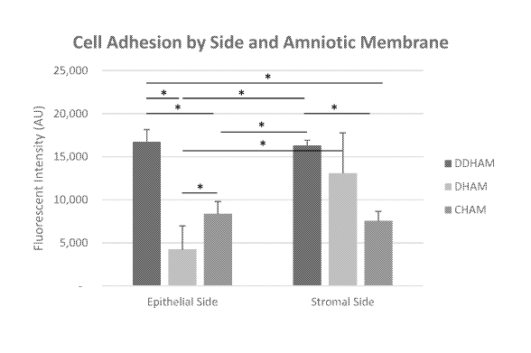
[0075] Cell adhesion was greater on the stromal side (12,342.42 4,536.60
AU) than on
the epithelial side of AMs (9,788.50 5,704.17 AU) (side main effect, F(1,18)
= 6.'714,p =
17
CA 03216040 2023-10-03
WO 2022/221852
PCT/US2022/071705
0.018), which can be attributed to the lower cell adhesion on the epithelial
side of DHAM
(4,247.75 2,732.87 AU), compared with the stromal side of DHAM (13,100.25
4,675.24
AU, p = 0.017), the epithelial side of DDHAM (16,725.25 1,453.62 AU, p
<0.001), the
epithelial side of CHAM (8,392.50 1,425.86 AU, p <0.001), the stromal side
of DDHAM
(16,334.75 591.85 AU, p = 0.002), and the stromal side of CHAM (7,592.25
1,073.22
AU, p <0.001) (side x AM, p = 0.001).
100761 Additionally, there was a significant difference in cell adhesion
between AMs
(AM main effect, F(2,18) = 30.896,p < 0.001), with significantly greater cell
adhesion on
DDHAM (16,530.00 1,048.46 AU) than on DHAM (8,674.00 5,912.61 AU, p <
0.001)
and CHAM (7,992.38 1,244.16 AU, p <0.001). However, as indicated above, cell
adhesion
varied with side and AM. Cell adhesion was similar between the epithelial side
of DDHAM
and the stromal side of DDHAM (p = 0.645) and between the epithelial side of
CHAM and
the stromal side of CHAM (p = 0.404). However, cell adhesion was significantly
greater on
the stromal side of DHAM than the epithelial side of DHAM (P = 0.017).
Therefore, cell
adhesion was significantly greater on the stromal and epithelial side of DDHAM
than the
epithelial side of DHAM (p < 0.002), the epithelial side of CHAM (post hoc
tests, p < 0.001),
and the stromal side of CHAM (post hoc tests, p <0.001), while cell adhesion
was similar
between the stromal side of DDHAM and the stromal side of DHAM (p = 0.219).
Table 4. Cell adhesion by side and amniotic membrane. Means and standard
deviations are
provided.
Amniotic
Epithelial Side Stromal Side Total
Membrane
DDHAM 16,725.25 1,453.62 16,334.75
591.85 16,530.00 1,048.46
DHAM 4,247.75 2,732.87 13,100.25
4,675.24* 8,674.00 5,912.61
CHAM 8,392.50 1,425.86 7,592.25
1,073.22 7,992.38 1,244.16
18
CA 03216040 2023-10-03
WO 2022/221852
PCT/US2022/071705
TOTAL 9,788.50 5,704.17 12,342.42 4,536.60* 11,065.46
5,206.33
*Statistically significant difference between epithelial side and stromal
side.
Cell Proliferation:
[0077] Although the number of viable cells significantly declined over 7-
day culturing
(time main effect; (F(2,54) = 44.880,p < 0.001), cell number significantly
varied with side,
AM, and time (side x AM x time interaction; (F(4,54) = 3.633,p = 0.011). Most
notably, cell
number declined for all variables across time, except for the stromal side of
DDHAM on day
4. On day 4, the relative proliferation rate was significantly greater on the
stromal side of
DDHAM (115.29 15.54%) than on the epithelial side of DDHAM (52.27 14.41%,
p <
0.001), the epithelial side of DHAM (12.54 16.79%,p = 0.012), and the
stromal side of
CHAM (15.00 6.73%,p < 0.001). There was no significant difference in the
relative
proliferation rate between the stromal side of DDHAM and the epithelial side
of CHAM
(46.83 25.69%,p = 0.731) or between the stromal side of DDHAM and the
stromal side of
DHAM (95.54 44.25%, p = 0.430). However, the stromal side of DHAM was
significantly
greater than the stromal side of CHAM (p = 0.012). Despite a decline in cell
number from
day 4, on day 7, the relative proliferation rate was significantly greater on
the stromal side of
DDHAM (59.47 28.48%) than on the stromal side of CHAM (6.87 1.77%, p =
0.035) and
the epithelial side of DHAM (7.54 5.84%,p = 0.017).
100781 The number of cells was also significantly greater on the stromal
side (9,383.33
6,469.15 AU) than on the epithelial side of AMs (5,648.00 5,312.56 AU, main
effect side;
F(1,54) = 39.545,p < 0.001), which is largely driven by significantly more
cells on the
stromal side than the epithelial side of DDHAM and DHAM (side x AM
interaction; p <
0.001); DDHAM Stromal: 14,972.00 4,973.00 AU vs DDHAM Epithelial: 10,438.50
5,555.98 AU, p = 0.047; DHAM Stromal: 10,103.33 4,336.49 AU vs DHAM
Epithelial:
1,590.42 2,431.25 AU, t(22) = 5.932,p <0.001). Conversely, CHAM a similar
number of
19
CA 03216040 2023-10-03
WO 2022/221852
PCT/US2022/071705
cells on the epithelial side (4,915.08 3,072.42 AU) and the stromal side
(3,074.67
3,401.09 AU, p = 0.178).
100791 Cell number was also significantly different between AMs (main
effect AM;
F(2,54) = 79.570,p < 0.001) with significantly more cells on DDHAM (12,705.25
5,652.67
AU) than on CHAM (3,994.88 3,306.13 AU, p < 0.001). There was no significant
difference in cell number between DDHAM and DHAM (5,846.88 5,543.10, P =
0.065) or
between DHAM and CHAM (p = 0.085). The similar cell count for DHAM and CHAM
can
be explained by the low cell count on the epithelial side of DHAM (1,590.42
2,431.25 AU),
which was significantly lower than the stromal side of DHAM (10,103.33
4,336.49 AU, p <
0.001), the stromal side of DDHAM (14,972.00 4,973.00 AU, p <0.001), the
epithelial side
of DDHAM (10,438.50 5,555.98 AU, p < 0.001), and the epithelial side of CHAM
(4,915.08 3,072.42 AU, p = 0.008). The cell count on the epithelial side of
DHAM and the
stromal side of CHAM were similar (3,074.67 3,401.09 AU, p = 0.117).
Table 5. Cell proliferation by side, amniotic membrane, and time. Means and
standard
deviations are provided. Cell proliferation measured in fluorescent intensity
(AU).
Side &
Amniotic
Membrane DAY! DAY 4 DAY 7 TOTAL
Epithelial
Side
16,725.25 8,679.25 5,911.00 10,438.50
DDHAM 1,453.62 2,092.53 4,747.52 5,555.98
4,247.75 1,590.42
DHAM 2,732.87 279.50 205.39 244.00 197.88 2,431.25
8,392.50 3,884.50 2,468.25 4,915.08
CHAM 1,425.86 2,025.36 1,719.06 3,072.42
Epithelial 9,788.50 4,281.08 2,874.42
Side Total 5,704.17 3,903.66 3,590.64 5,648 5,312.56
Stromal
Side
CA 03216040 2023-10-03
WO 2022/221852
PCT/US2022/071705
16,334.75 18,852.25 9,729.00 14,972.00
DDHAM 591.85 2,882.54 4,776.66 4,973.00
13,100.25 10,992 6,217.75 10,103.33
DHAM 4,675.24 1,830.40 3,253.52 4,336.49
7,592.25 1,102.00 3,074.67
CHAM 1,073.23 442.57 529.75 175.43
3,401.09
Stromal 12,342.42 10,315.42 5,492.17 9,383.33
Side Total 4,536.60 7,795.43 4,979.13 6,469.15
11, 574.50 7,298.25 4,183.29 7,515.67
TOTAL 5,206.34 6,771.29 4,450.91 6,170.94
Cell Migration:
[0080] Cell
migration significantly differed between AMs (AM main effect; F(2,49) = 6.819,
p =
0.002), with cell migration significantly greater on DDHAM (466,085.13
98,339.52 px2) than CHAM
(344,471.06 106,094.18 px2, p = 0.003). In addition, cell migration was
significantly lower on the
medium control than DDHAM (p < 0.001), DHAM (420,349.88 95,109.86 px2, p <
0.001), and CHAM
(p < 0.001). There was no main effect of side, which indicates cell migration
was similar on the
epithelial (421,669.96 113,435.95 px2) and stromal sides of the AMs
(389,934.08 107,979.51 px2,
F(1,49) = 0.701, P = 0.407). Cell migration was not statistically different
across amniotic membrane
and side (p = 0.159).
Table. 6. Migration Area. Counts, means, and standard deviations are provided.
Migration
area is reported as px2.
Amniotic Membrane &
Side Cell Migration
DDHAM
Epithelial Side 482,961.50 99,654.98
Stromal Side 449,208.75 100,701.20
DDHAM Total 466,085.13 98,339.52t
DHAM
Epithelial Side 461,119.13 90,282.12
Stromal Side 379,580.63 86,220.78
DHAM Total 420,349.88 95,109.86t
CHAM
Epithelial Side 320,929.25 80,791.60
21
CA 03216040 2023-10-03
WO 2022/221852
PCT/US2022/071705
Stromal Side 368,012.88 127,772.77
CHAM Total 344,471.06 106,094.18*t
Medium Control 145,349.00 58,822.77
TOTAL 372,451.59 139,865.11
* Statistically significant difference compared with DDHAM.
t Statistically significant difference compared with medium control.
Example 4: A Decellularized Dehydrated Human Amniotic Membrane-Derived
Biomaterial Supports Human Corneal Epithelial Cell Function and Inflammatory
Response
[0081] Statement of Purpose: Successful application of decellularized
tissue-based
biomaterials for wound healing requires matrix components that support cell
function and
differentiation. Amniotic membrane (AM) is a naturally derived biomaterial
from human
placental tissue with unique biological and mechanical properties that render
it suitable for
use in ocular healing (1,2). The purpose of this study is to evaluate the
effects of sidedness
and AM processing methodology on human corneal epithelial cell (HCEC) function
in vitro.
Experimental variables include AM sidedness (epithelial [E] and stromal [S])
and AM
processing methodology (decellularized and dehydrated [DDHAM], dehydrated
[DHAM],
and cryopreserved [CHAM]). Dependent variables include HCEC viability,
migration, and
inflammatory response.
[0082] Methods: Three differently processed, commercially available ocular
AMs were
selected: Biovance3L Ocular (DDHAM), Ambio20 (DHAM), and AmnioGraft0 (CHAM).
HCECs were seeded onto the E and S sides of AMs and incubated for 1, 4 and 7
days. Cell
viability was measured at each time point on the AMs using alamarBlue assay.
Conditioned
media from HCECs cultured on the AMs were collected, and the effect of
conditioned media
on the migration of HCECs was evaluated using a scratch wound assay. An
inflammatory
response was induced by TNFa treatment. The effect of AM on the expression of
pro-
22
CA 03216040 2023-10-03
WO 2022/221852
PCT/US2022/071705
inflammatory genes in HCECs was compared using quantitative polymerase chain
reaction
(qPCR). The significance level for all statistical tests was set at p = 0.05.
Cell viability was
analyzed with a two-way analysis of variance (ANOVA), cell proliferation with
a three-way
ANOVA, and mRNA expression with a one-way ANOVA. Tukey's and unpaired t-tests
were
used for post-hoc analyses.
[0083] Results: On day 1, cell viability was significantly higher on DDHAM-
E&S than
CHAM-E&S (p < 0.001) and DHAM-E (p < 0.002). On day 4, cell viability was
significantly
higher on DDHAM-S than all other variables (p < 0.004, FIG. 1). In addition,
on day 4, cell
viability was comparable
[0084] between DDHAM-E and DHAM-S (p = 0.147) and significantly higher than
DHAM-E (p < 0.004), CHAM-S&E (p < 0.017). On day 7, cell viability was
significantly
higher on DDHAM-S than DHAM-E (p = 0.028) and CHAM-S&E (p < 0.049). Cell
viability
was similar between DDHAM-E and all other variables (p >0.097). HCEC migration
in the
presence of conditioned media from cells cultured on DDHAM and DHAM was
comparable
(p = 0.885) and significantly greater than cells grown on CHAM (p < 0.005).
Interestingly,
HCECs cultured on DDHAM adapted a cobblestone morphology (FIG. 2), which
mimics the
morphology of ocular epithelial cells in situ (3). The migration of HCEC in
the presence of
conditioned media from cells cultured on ocular scaffolds was significantly
greater than
control conditioned media from cells grown on tissue culture plastic (p <
0.001). Moreover,
in response to inflammatory stimulation by TNFa, the gene expression of pro-
inflammatory
cytokines (IL-6, IL-8, and TNFa) in HCECs on DDHAM showed an initial increase
followed
by a decline across time (FIG. 3).
[0085] Conclusion: In this in vitro study, DDHAM-S best supported HCEC
viability and
migration. The presence of DDHAM also attenuated the inflammatory response of
HCECs
over time.
23
CA 03216040 2023-10-03
WO 2022/221852
PCT/US2022/071705
[0086] References:
1. Walkden A. Clin Ophthalmol. 2020;14:2057-2072.
2. Malhotra C. World J Transplant. 2014;4(2):111-121.
3. Sosnova-Netukova M. Br J Ophthalmol. 2007;91(3):372-378.
Example 5: An in-vitro comparison of human corneal epithelial cell activity
and
inflammatory response on differently designed ocular amniotic membranes and
clinical case study
[0087] Amniotic membrane (AM) is a naturally derived biomaterial with
biological and
mechanical properties important to Ophthalmology. The epithelial side of the
AM promotes
epithelialization, while the stromal side regulates inflammation. However, not
all AMs are
equal. AMs undergo different processing with resultant changes in cellular
content and
structure. This study evaluates the effects of sidedness and processing on
human corneal
epithelial cell (HCEC) activity and the effect of processing on HCEC
inflammatory response
and then presents a case study. Three differently processed, commercially
available ocular
AMs were selected: (1) Biovance3L Ocular, a decellularized, dehydrated human
AM
(DDHAM), (2) AMBI020, a dehydrated human AM (DHAM), and (3) AnmioGraft0, a
cryopreserved human AM (CHAM). HCECs were seeded onto the AMs and incubated
for 1,
4 and 7 days. Cell adhesion and viability were evaluated using alamarBlue
assay. HCEC
migration was evaluated using a scratch wound assay. An inflammatory response
was
induced by TNFa treatment. The effect of AM on the expression of pro-
inflammatory genes
in HCECs was compared using quantitative polymerase chain reaction (qPCR).
Staining
confirmed complete decellularization and the absence of nuclei in DDHAM. HCEC
activity
was best supported on the stromal side of DDHAM. Under inflammatory
stimulation,
DDHAM promoted a higher initial inflammatory response with a declining trend
across time.
24
CA 03216040 2023-10-03
WO 2022/221852
PCT/US2022/071705
Clinically, DDHAM was used to successfully treat anterior basement membrane
dystrophy.
Compared with DHAM and CHAM, DDHAM had significant positive effects on the
cellular
activities of HCECs in vitro, which may suggest greater ocular cell
compatibility in vivo.
[0088] Introduction: Amniotic membrane (AM) is a naturally derived
biomaterial with
unique biological and mechanical properties that render it particularly
suitable for use in
ophthalmology (Leal-Marin et al. 2021; Walden, 2020; Liu et al. 2019; Malhotra
& Jain,
2014; Fernandes et al. 2005). Amnion tissue is thought to promote healing and
reconstruction
of the ocular surface through the promotion of epithelialization (Shayan et
al. 2019; Meller et
al. 2002; Meller et al. 1999), reduction of inflammation (Sharma et al. 2016;
Tabatabaei et al.
2017; Tandon et al. 2011), inhibition of scar tissue formation (Niknejad et
al. 2008, Tseng et
al. 1999 Lee et al. 2000), blockage of new blood vessels (Hao et al. 2000),
and the ability to
act as an antimicrobial agent (Mamede & Botelho, 2015; Tehrani et al. 2013;
Sangwan et al.
2011; Kjaergaard et al. 2001; Kjaergaard et al. 1999, Inge et al. 1991). In
ophthalmology, the
AM is widely used to treat a variety of ocular conditions. Clinically, the AM
can be used as a
surgical patch, as a substrate to replace damaged ocular tissue, or in
combination as both a
patch and a substrate.
[0089] As a patch, the AM acts as a temporary biological bandage or contact
lens,
promoting re-epithelization of the host tissue beneath the patch (Walden,
2020, Malhotra &
Jain, 2014) and is placed stromal side down to downregulate the inflammatory
response by
trapping inflammatory cells and inducing apoptosis (Dua et al. 2004; Shimmura
et al. 2001).
By placing the AM epithelial side up, the AM acts as a substrate and scaffold
for epithelial
cell migration and growth (Malhotra & Jain, 2014). Although it is widely
accepted that the
AM should be placed epithelial side up to promote re-epithelialization (Hu et
al. 2003), the
stromal side of the membrane has been shown to support epithelial cell growth
(Seitz et al.
2006). Notably, much of the existing research is limited to cryopreserved AMs,
and it
CA 03216040 2023-10-03
WO 2022/221852
PCT/US2022/071705
remains unclear whether these findings also apply to other AMs that have
undergone
different processing methodologies.
[0090] Prior to clinical application, the AM is sterilized and processed
with resultant
changes to cellular content and structure (Leal-Marin et al. 2021; von Versen-
Hoynck et al.
2004; Lim et al. 2010). This tissue can be used directly, or it can undergo
the additional
process of decellularization (Tehrani et al. 2021). Decellularization is a
process whereby
endogenous cells, cell debris, and DNA remnants are removed to prevent an
immune
response, while retaining the natural structural and chemical elements of the
extracellular
matrix (ECM) (Gholipourmalekabadi et al. 2015). Previous studies have
demonstrated a
correlation between the quantity of residual DNA in ECM products and the host
inflammatory response (Keane et al. 2012; Seif-Naraghi et al. 2013). As with
the preservation
of tissue, decellularization can also affect the structures and entities
within the ECM (Aamodt
& Grainger, 2016). Therefore, successful preservation-decellularization
protocols must
delicately balance the removal of cellular material and the retention of the
innate properties
and functional characteristics of ECM (Gholipourmalekabadi et al. 2015; Aamodt
&
Grainger, 2016; Balestrini et al. 2015). To our knowledge, no studies have
evaluated how
differing preservation-decellularization protocols affect the cellular
activity and inflammatory
response of human corneal epithelial cells (HCECs).
[0091] For the first time, this project aims to evaluate:
the effect of amniotic membrane sidedness (i.e., epithelial vs stromal) and
processing
methodology on the cellular activities of HCEC (i.e., adhesion, viability, and
migration),
the effect of different processing methodologies on the inflammatory response
of HCECs
(i.e., expression of pro-inflammatory cytokines).
[0092] Therefore, three differently processed, commercially available
ocular AMs were
used for comparison:
26
CA 03216040 2023-10-03
WO 2022/221852
PCT/US2022/071705
Biovance3L Ocular (Celularity, Florham Park, NJ), a decellularized, dehydrated
human
amniotic membrane (DDHAM),
AMBI020 (Katena, Parsippany, NJ), a dehydrated human amniotic membrane (DHAM),
AnimioGraft0 (Biotissue, Miami, FL), a cryopreserved human amniotic membrane
(CHAM).
[0093] Biovance03L Ocular is a three-layer DDHAM. It is designed uniquely
with the
stromal side facing out. Therefore, the stromal side interfaces with the
ocular surface
regardless of its orientation. Furthermore, having three layers enhances its
handling
properties. The AM is excised from qualified term placentas, washed, and
scraped to remove
extraneous tissues and cells. The tissue is then decellularized using an
osmotic shock
followed by a mild detergent treatment, dried, and sterilized. Previous
research has confirmed
that this proprietary decellularization process removes residual cells, cell
debris, growth
factors, and cytokines, while retaining an ECM structure with high collagen
content and key
bioactive molecules, such as fibronectin, laminin, glycosaminoglycans, and
elastin (Bhatia et
al. 2007).
[0094] AMBI020 is a single-layer, aseptically processed DHAM. The
dehydration
process removes moisture, while preserving the structural matrix and
biological components
of the tissue (Instructions for Use, 2021), including growth factors and
cytokines.
[0095] AnimioGraft0 is a single-layer CHAM. The AM is preserved using a
proprietary
cryopreservation method, CRYOTEKO. The cryopreservation preservation process
renders
the amniotic epithelial cells nonviable, while maintaining an intact cellular
structure and
preserving growth factors and cytokines (Rodriguez-Ares et al. 2009).
[0096] DDHAM retains its native ECM and is devoid of all cellular
components, DNA,
growth factors and cytokines. Therefore, the authors hypothesize that DDHAM
will provide a
more cell-friendly matrix supporting the cellular activity and inflammatory
response of
27
CA 03216040 2023-10-03
WO 2022/221852
PCT/US2022/071705
HCECs compared with the two other ocular AMs containing residual DNA and other
cellular
components. Results from this in vitro study will further the basic
understanding of how the
preservation and decellularization of amnion tissue affects the activity of
human ocular
epithelial cells. It also has the potential to elucidate the clinical
application of DDHAM to
support corneal and conjunctival related injuries or defects, such as corneal
epithelial defect
healing, pterygium repair, fornix reconstruction, and other ocular procedures.
[0097] Materials & Methods: Since the testing materials are commercially
available
products and this study did not require direct interaction with human subjects
(donors),
institutional review board approval was not required.
[0098] Ocular AMs: Three ocular AMs were used in this study: DDHAM, DHAM,
and
CHAM. DDHAM (Lot # OCLR0010) and DHAM samples were stored at room temperature.
CHAM samples were stored at -80 C. All AMs were handled according to the
manufacturer's instructions. DDHAM samples came as individually packaged 10 mm
discs.
Therefore, 10 mm discs were made from DHAM sheet, using a 10 mm biopsy punch
(Thermo Fisher Scientific, Waltham, MA, USA). Each piece (5 cm x10 cm) of CHAM
was
thawed and washed in 20 mL of phosphate buffered saline (PBS) in a petri dish
for 10
minutes (min) to remove the cryoprotectants and 10 mm discs were made from the
washed
AMs using 10 mm biopsy punch. DDHAM is multilayered (three layered) with
stromal side
of AM facing out on both sides. To evaluate the sidedness of DDHAM, a
differently designed
version was prepared (three layered) with epithelial side of AM facing out on
both sides,
DDHAM(E). 10 mm discs of each AM sample were placed in the wells of a 48-well
plate (1
disc/well) (Cell-Repellent 48-Well Microplate, Greiner Bio-One, Monroe, NC,
USA) with
either stromal side or epithelial side of AM in contact with cells. A sterile
0-ring (McMaster-
Carr, Robbinsville, NJ, USA), measuring 2 mm in width with 7 mm inner
diameter, was
placed on the top of each AM to hold the AM in place. Amniotic membranes were
pre-
28
CA 03216040 2023-10-03
WO 2022/221852
PCT/US2022/071705
conditioned with growth medium (0.4 mL/well) at 37 C for 2 hours (h) before
they were
seeded with cells. At least two lots (donors) of each type of AM were used in
this study. In
each independent experiment, four samples (n=4) from each AM were used, of
which two
samples were from one lot and two samples were from another lot. At least two
independent
experiments were performed for each individual assay.
[0099] Primary cells: The human corneal epithelial cells (HCECs, Cat#PCS-
700-010
Lot# 80915170), corneal epithelial cell base medium, and corneal epithelial
cell growth kit
were purchased from ATCC (Manassas, VA, USA). The complete growth medium for
HCECs was prepared according to the manufacturer's instructions.
[00100] Assessment of cell adhesion to AMs: HCECs at passage 4 (P4) were
cultured to
80% confluence in 10 cm cell culture dishes following the manufacturer's
instructions. Cells
were rinsed once with 5 mL phosphate-buffered saline (PBS)/dish. One
milliliter of 0.25%
trypsin (Thermo Fisher Scientific, Waltham, MA, USA) was added to each dish
and
incubated at 37 C for 5 min. Two milliliters of minimum essential medium-alpha
(Thermo
Fisher Scientific, Waltham, MA, USA) medium containing 10% FBS was added to
the dish
to neutralize the trypsin. Cells were transferred to 15 mL conical tubes and
centrifuged at
1000 RPM (Revolutions Per Minute) for 5 min. Cells were re-suspended in
complete growth
medium and counted using a hemocytometer.
[00101] HCECs (2 x 10⁴/well) were added to each well containing pre-
conditioned AMs.
The plates were incubated at 37 C with 5% CO2 and 95% humidity. After
incubation for 24
h, the media were removed, and the cells were washed once with PBS. The
viability of
adhered cells was detected using the alamarBlue assay. Briefly, 0.2 mL/well of
alamarBlue
solution, consisting of complete growth medium + 10% alamarBlue reagent (Bio-
Rad,
Hercules, CA, USA) was added to each well and incubated at 37 C for 45 min
After
incubation, 0.1 mL/well of supernatant was transferred to a 96-well plate.
Fluorescence
29
CA 03216040 2023-10-03
WO 2022/221852
PCT/US2022/071705
intensity was measured using a multimode microplate reader (Spark , TECAN,
Switzerland)
at excitation/emission (Ex/Em) = 540 nm/590 nm. The fluorescence intensity was
expressed
in arbitrary units (AU).
[00102] Staining of AMs and cells: To visualize the structural features of
AMs, three
different AMs were rehydrated, washed, and embedded in Tissue-Tek OCT.
compound
(Sakura, Torrance, CA, USA) vertically. Five micron/slice cryosections were
made using
Leica CM1850 cryostat (Leica Biosystems, Buffalo Grove, IL, USA). The
cryosections on
microscope slides were fixed with 4% paraformaldehyde for 1 h and
permeabilized in 0.5%
Triton X100 in PBS for 1 h. The fixed and permeabilized samples were stained
with anti-
human type I antibodies (ab34710, Abcam, Cambridge, MA, USA) overnight.
Samples were
then stained with Alexa Fluor 555-anti-rabbit IgG, Alexa 488-Phalloidin (Life
Technology,
Carlsbad, CA, USA) and Hoechst dye 33258 (Thermo Fisher Scientific, Waltham,
MA,
USA) for 60 min. After staining, a coverslip was mounted onto each sample in
the presence
of ProLong Gold Antifade Mountant (Thermo Fisher Scientific, Waltham, MA,
USA).
[00103] To visualize the viable cells on different AMs, HCECs were cultured
on different
AMs as described in "Assessment of Cell Adhesion to Amniotic Membranes" for 1
or 4 days.
At each time point, the medium was removed from each well, and 0.2 mL/well of
fresh
complete growth medium containing 50 nM Calcein AM (Thermo Fisher Scientific,
Waltham, MA, USA) was added to each well. After incubation for 30 min at 37 C,
the
medium was removed. Cells were washed twice with PBS and ready to be imaged.
[00104] To visualize the cell morphology, HCEC cells cultured on different AMs
for 4
days were fixed with 4% paraformaldehyde for 1 h and permeabilized in 0.5%
Triton X100 in
PBS for 1 h. The fixed and permeabilized cells were stained with Alexa 488-
Phalloidin (Life
Technology, Carlsbad, CA, USA) for 30 min and observed under an epi-
fluorescent
microscope (Zeiss Observer D1, Jena, Germany).
CA 03216040 2023-10-03
WO 2022/221852
PCT/US2022/071705
[00105] H&E staining of AMs: Cryosections of AMs were baked at 60 C overnight,
fixed
in 4% paraformaldehyde for 30 min, and rinsed three times with PBS. Samples
were stained
in Harris Hematoxylin Solution (Sigma-Aldrich, Inc., St. Louis, MO) for 10 min
and rinsed
in running tap water for 1 min. Slides were then immersed two times in
differentiation
solution (0.25 mL concentrated Hydrochloric Acid to 100 mL of 70% alcohol).
Subsequently,
slides were rinsed under running tap water for 1 min, followed by immersion in
Scott's Tap
Water Substitute (1% Magnesium sulfate (MgSO4) and 0.06% Sodium Bicarbonate)
for 60
seconds. After a 30 second wash in 95% reagent alcohol, samples were
counterstained in
Alcoholic Eosin Y Solution (Sigma-Aldrich, Inc., St. Louis, MO 68178) for 10
min. Upon
completion of staining, slides were dehydrated by three washes in 100%
absolute ethanol,
followed by three Histoclear II washes. Slides were mounted using Permount
mounting
medium (Fisher Scientific Inc.) and imaged using Zeiss Axio Observer Al
microscope.
[00106] Assessment of cell viability on AMs over time: HCECs (1 x 10⁴/well)
were added
to each well of 48-well plates containing pre-conditioned AMs. Three sets of
plates for each
cell type were set up and incubated at 37 C with 5% CO2 and 95% humidity for
1, 4, and 7
days. At the first time point, the medium from each well of all plates was
removed, and fresh
medium was added. The viability of cells in the first set of plates was
measured using the
alamarBlue assay. The second and third sets of plates were cultured at 37 C.
At the second
time point, the viability of cells in the second set of plates was measured.
The third set of
plates was cultured in fresh medium at 37 C. The viability of cells in the
third set of plates
was measured using the alamarBlue assay at the third time point.
[00107] Conditioned media for migration assay: In the test condition, HCECs
(2 x
104/well) were added to each well of 48-well plates containing pre-conditioned
AMs. In the
control condition, no HCECs were added to the pre-conditioned AMs. After
culturing for 24
h, the medium was removed. 0.4 mL/well of fresh growth medium was added to
each well
31
CA 03216040 2023-10-03
WO 2022/221852
PCT/US2022/071705
with or without cells and incubated at 37 C for 24 h. The supernatants (24-h
conditioned
media) were collected from each well and immediately used for the migration
assay. The
stromal sides of AMs were used for this experiment.
[00108] Scratch wound migration assay: 5 x10⁴/well HCECs were added to each
well of
tissue culture-treated polystyrene 48-well plates and cultured at 37 C with 5%
CO2 and 95%
humidity for 2 days. Scratch wounds were made on a confluent monolayer using
the tip of a
sterile metal rod. The medium was removed, and conditioned medium collected
from cells
cultured on AMs was added to the wound. Images of the wound areas were
captured at 0 h.
At minimum, four areas were monitored for each testing group. The plates were
incubated at
37 C for 24 h. The exact same wound areas (with marker reference) were imaged
at 24 h.
Wound areas were measured using ImageJ software (NIH) in arbitrary units
(square pixels,
px2). Migrated area=Area0h-Area24h.
[00109] Stimulation of inflammatory responses of HCECs: 2 x10⁴/well HCECs were
seeded and cultured on different AMs for 24 h. Media were removed and fresh
medium "-
Tumor Necrosis Factor-alpha (TNF-0)" or fresh medium containing 10 ng/mL of
human
TNF-0 (Cat#300-01A, PeproTech Cranbury, NJ) "+TNF-0" were added to cells and
incubated for 24 h, 48 h or 72 hr. At each time point, the supernatants were
collected for
multiplex analysis and the cells were lysed in 0.2 mL of RNA lysis buffer
(Promega,
Durham, NC) for quantitative polymerase chain reaction (qPCR) analysis as
described below.
[00110] Assessment of relative mRNA expression by qPCR: The quantification of
the
relative gene expression of cytokines by qPCR was performed as previously
described (Mao
et al. 2021). Briefly, total RNA from cell lysates was purified using SV 96
Total RNA
Isolation System (Promega). RNA concentration and purity were measured using
TECAN
Spark Nano plate (TECAN, Morrisville, NC). cDNA preparation and qPCR were
performed
as described (Mao et al. 2017). The primers for qPCR used for this study were
from
32
CA 03216040 2023-10-03
WO 2022/221852
PCT/US2022/071705
QuantiTect (Qiagen, Germantown, MD): granulocyte-macrophage colony-stimulating
factor
(GM-CSF: QT00000896), interleukin 6 (IL-6: QT00083720), interleukin-8 (IL-8:
QT00000322), Tumor Necrosis Factor alpha (TNF-0: QT01079561), and
glyceraldehyde 3-
phosphate dehydrogenase (GAPDH: QT01192646). Each sample was run in duplicate.
After
the run was completed, a second derivative analysis was performed using the
raw data to
determine the mean Cp (Crossing point-PCR-cycle) for each sample. For each
gene
expression, expression of GAPDH served as an internal control. Relative mRNA
expression
was determined by Pfaffl analysis (EACp target/EACp reference) in which primer
efficiency
E= 10^(-1/slope) and ACp= mean Cp of sample - mean Cp of Control. The
expression of
cells on tissue culture polystyrene (TCP) or the expression of cells at 24 h
was used as
"Control" for analyses, which was defined in the specific analysis in
"Results".
[00111] Statistical Methods: In the evaluation of HCEC activity, the
independent variables
were AM (DDHAM, DHAM, CHAM), side (epithelial, stromal), and time (day 1, day
4, and
day 7). The dependent variables were cell adhesion, cell viability, and
migration. In the
evaluation of HCEC inflammatory response by mRNA expression, the independent
variables
were amniotic membrane (DDHAM, DHAM, CHAM, Control [TCP]), condition (resting,
stimulated), and time (24 h, 48 h, and 72 h). In the evaluation of HCEC
inflammatory
response by protein levels, the independent variables were amniotic membrane
(DDHAM,
DHAM, CHAM, Control [TCP]) and condition (resting, stimulated, AM only). The
dependent variables were relative mRNA expression of cytokines (GM-CSF, IL-6,
IL-8 and
TNF-a) and protein levels of cytokines and chemokines (GM-CSF, IL-1(3, IL-1RA,
IL-6, IL-
8, TGF132, and VEGF).
[00112] All analyses were conducted using IBM SPSS (Build 1Ø0.1444). The
significance level for all statistical tests was set at p = 0.05. The data
were tested and found to
be normally distributed. Cell adhesion and migration were analyzed with a two-
way analysis
33
CA 03216040 2023-10-03
WO 2022/221852
PCT/US2022/071705
of variance (ANOVA) with Tukey post-hoc tests. Cell proliferation was analyzed
with a
three-way ANOVA with Tukey post-hoc tests. Relative mRNA expression at 24 h
was
analyzed with a two-way ANOVA with Tukey post-hoc tests to evaluate each
dependent
variable in each of the testing conditions. Relative mRNA expression across
time was
analyzed with a one-way ANOVA with Tukey post-hoc tests to evaluate each
dependent
variable in each of the testing conditions. Significant interactions were
evaluated with simple
main effects analysis with Sidak correction for multiple comparisons. Data are
reported as
mean standard deviation (SD) within the text and FIGS.
Results:
[00113] Structure of AMs: To evaluate the structures of these three AMs,
cross-sections of
AMs were stained for cellular components (DNA and actin) and ECM (type I
collagen) (FIG.
8A). While strong nuclei staining and actin staining were detected in DHAM and
CHAM,
neither actin nor nuclei staining was detected in DDHAM. The presence of type
I collagen
was detected in all three AMs. H&E staining of the three AMs (FIG. 8B)
confirmed complete
decellularization and absence of nuclei in DDHAM, compared with DHAM and CHAM.
DHAM showed meagre staining of dark blue nuclear remnants, while CHAM showed
intact
dark blue staining for nuclei, showing the presence of cells.
[00114] Adhesion of HCECs on different AMs: Cell adhesion on different AMs and
different sides of AMs was evaluated by comparing the cell viability
(reflecting the quantity
of adhered cells) at 24 h. The fluorescence intensity was expressed in
arbitrary units (AU).
[00115] Effect of sidedness. Cell adhesion was greater on the stromal side
than on the
epithelial side of AMs (side main effect, p = 0.018), which can be explained
by the lower cell
adhesion on the epithelial side of DHAM, compared with the stromal side of
DHAM (p <
0.001; side x AM, p = 0.001; FIG. 9). There was no significant difference
between the
34
CA 03216040 2023-10-03
WO 2022/221852
PCT/US2022/071705
epithelial and stromal sides of DDHAM (p = 0.822) or between the epithelial
and stromal
sides of CHAM (p = 0.645).
[00116] Effect of AM. Additionally, there was a significant difference in
cell adhesion
between AMs (AM main effect, p < 0.001), with significantly greater cell
adhesion on
DDHAM than on DHAM (p < 0.001) and CHAM (p <0.001). However, as previously
indicated, cell adhesion varied with side and AM (p = 0.001; FIG. 9). On the
epithelial side,
cell adhesion was significantly greater on DDHAM than on DHAM (p < 0.001) and
CHAM
(p < 0.001), and there was no significant difference between CHAM and DHAM (p
= 0.076).
On the stromal side, cell adhesion was significantly lower on CHAM than on
DDHAM (p <
0.001) and DHAM (p = 0.014), and there was no significant difference between
DDHAM
and DHAM (p = 0.207). These results indicate that among these three AMs, the
epithelial and
stromal sides of DDHAM best supported cell adhesion.
[00117] Viability and morphology of HCECs on different AMs on Day 4: Live
Staining of
Epithelial Cells. The viability of HCECs on the stromal side of different AMs
(DDHAM,
CHAM, DHAM) was observed 4 days after cell seeding (FIG. 10). Consistent with
the
quantitative results, the HCECs on DDHAM and DHAM appeared to have adhered and
spread on day 4 after cell seeding, whereas HCECs on CHAM appeared to be
disorganized
and adopted a heterogeneous morphology. The morphology of HCECs on the AMs was
monitored by actin staining on day 4 (FIG. 10). The HCECs on DDHAM adapted a
cobblestone morphology with a dense actin ring structure.
[00118] Cell viability on different AMs over time: The viability of cells
on different AMs
was monitored up to 7 days. Although the number of viable cells significantly
declined over
the 7-day culture (time main effect, p < 0.001), cell viability significantly
varied with side,
AM, and time (side x AM x time interaction, p = 0.011). Most notably, cell
viability declined
for all variables across time, except for the stromal side of DDHAM on day 4
(FIG. 11A).
CA 03216040 2023-10-03
WO 2022/221852
PCT/US2022/071705
[00119] Effect of sidedness. Cell viability was also significantly greater
on the stromal
side than on the epithelial side of AMs (main effect side, p < 0.001), which
can be explained
by differences in relative cell viability between sides on days 4 and 7 (FIG.
11B). On day 4,
the relative cell viability was significantly greater on the stromal side of
DDHAM than the
epithelial side of and DDHAM (p <0.001), and the relative cell viability was
significantly
greater on the stromal side of DHAM than the epithelial side of DHAM (p <
0.001).
Conversely, the relative cell viability was significantly greater on the
epithelial side of
CHAM than the stromal side of CHAM (p = 0.039). On day 7, there were no
significant
differences in relative cell viability between the epithelial and stromal
sides of DDHAM (p =
0.102) or CHAM (p = 0.157). However, the relative cell viability was
significantly greater on
the stromal side of DHAM than the epithelial side of DHAM (p < 0.001).
1001201 Effect of AM. Cell number was also significantly different between AMs
(main
effect AM, p <0.001) with significantly more viable cells on DDHAM than on
DHAM (p <
0.001) and CHAM (p < 0.001) and significantly more viable cells on DHAM than
CHAM (p
= 0.036). The main effect of AM is largely explained by the significant
differences in relative
cell viability on days 4 and 7 (FIG. 11B).
[00121] On the epithelial side on day 4, the relative cell viability was
significantly greater
on DDHAM than on DHAM (p = 0.032), meanwhile the relative cell viability was
similar
between DDHAM and CHAM (p = 0.978) and between CHAM and DHAM (p = 0.077). On
the epithelial side on day 7, there were no significant differences between
the three amniotic
membranes (p? 0.219).
[00122] On the stromal side on day 4, the relative cell viability was
significantly greater on
DDHAM than on CHAM (p < 0.001), and the relative cell viability was
significantly greater
on DHAM than on CHAM (p < 0.001). There was no significant difference in the
relative
cell viability on the stromal side on day 4 between DDHAM and DHAM (p =
0.477). On the
36
CA 03216040 2023-10-03
WO 2022/221852
PCT/US2022/071705
stromal side on day 7, however, the relative cell viability was significantly
lower on CHAM
than DDHAM (p = 0.003) and DHAM (p = 0.002). As with the epithelial side, on
the stromal
side on day 7, there was no significant difference in the relative cell
viability between
DDHAM and DHAM (p = 0.999).
[00123] The findings of higher cell viability on the stromal side of AMs
and better
maintenance of viability on DDHAM compared with DHAM and CHAM suggests that
cell
viability was best maintained on the stromal side of DDHAM.
[00124] Migration of HCECs on different AMs: The conditioned media from
different
AMs in the absence of HCECs were tested to evaluate the effect of AM alone on
the
migration of HCECs. Additionally, differences in migration were compared
between AMs in
the presence of cells to determine if the factors released by HCECs cultured
on different AMs
affect cell migration. Cells cultured on AMs were conditioned for 24 h. The
migration of
HCECs in the presence of conditioned media from different AMs was evaluated
using a
scratch wound assay. Wound closure was monitored for 24 h (FIGS. 12A and 12B).
[00125] There was a significant interaction between the effects of amniotic
membrane and
the presence of cells (p = 0.006; FIGS. 12A and 12B). Migration was
significantly higher
with cells than without cells on DDHAM (p = 0.009) and DHAM (p < 0.001).
Migration was
not significantly different with or without cells on CHAM (p = 0.291) or on
the control (p =
0.265).
[00126] Effect of AM. Furthermore, among the conditioned media (CM) collected
in the
presence of cells, migration was significantly lower in CM from cells on CHAM
than on
DDHAM (p = 0.004) and DHAM (p = 0.002). There was no significant difference in
migration between DDHAM and DHAM (p = 1.000). Compared with the control in the
presence of cells, migration was significantly higher in CM from cells on
DDHAM (p <
0.001), DHAM (p < 0.001), and CHAM (p = 0.005).
37
CA 03216040 2023-10-03
WO 2022/221852
PCT/US2022/071705
[00127] Gene expression of inflammatory cytokines in HCECs: Since the
stromal side of
AM has been reported to regulate the inflammatory response (Dua et al. 2004;
Shimmura et
al. 2001), the effect of the stromal side of these three AMs on the
inflammatory responses of
HCECs was evaluated. Cytokines with previously demonstrated roles in wound
healing were
selected, including GM-CSF, IL-6, IL-8 or TNF-0 (Rho et al. 2015; Arranz-
Valsero et al.
2014; Ebihara et al. 2011; Nishida et al. 1992; Hafezi et al. 2018; Strieter
et al. 1992; Koch et
al. 1992; Wang et al. 2020; Yang et al. 2019). To this end, the inflammatory
response of
HCECs under an in vitro inflammatory condition was mimicked by the stimulation
with
TNF-0 for 24 h. The gene expression (relative mRNA levels) of GM-CSF, IL-6, IL-
8, or
TNF-0 in HCECs on different AMs was assessed by qPCR compared with the gene
expression in cells cultured on standard cell culture surface, TCP.
[00128] GM-CSF. The expression of GM-CSF at 24 h varied significantly by
stimulation
condition ( TNFa) and AM (p = 0.049) (FIG. 13A). With stimulation, the
expression of GM-
CSF significantly increased on DHAM (p < 0.001), but not DDHAM (p = 0.226),
CHAM (p
= 0.664), or TCP (p = 0.827). Comparing the expression of GM-CSF between
amniotic
membranes in the resting condition showed a similar expression of GM-CSF on
DDHAM,
DHAM, CHAM, and TCP (p? 0.134). Comparing the expression of GM-CSF between
amniotic membranes in the stimulated condition showed significantly greater
expression on
DHAM than on DDHAM (p = 0.001), CHAM (p < 0.001), and TCP (p < 0.001).
[00129] IL-6. The expression of IL-6 at 24 h varied significantly by
stimulation condition
and amniotic membrane (p = 0.002) (FIG. 13B). With stimulation, the expression
of IL-6
significantly increased on DDHAM (p < 0.001), CHAM (p = 0.017), and TCP (p =
0.014),
but not DHAM (p = 0.128). Comparing the expression of IL-6 between amniotic
membranes
in the resting condition showed a similar expression of IL-6 on DDHAM, DHAM,
CHAM,
and TCP (p? 0.717). In the stimulated condition, there was significantly
higher expression of
38
CA 03216040 2023-10-03
WO 2022/221852
PCT/US2022/071705
IL-6 on DDHAM than on DHAM (p < 0.001), CHAM (p < 0.001), and TCP (p < 0.001).
No
other significant differences were found.
[00130] IL-8. Although the expression of IL-8 at 24 h did not vary
significantly by
stimulation condition and AM (p = 0.188), there were main effects for
stimulation condition
(p < 0.001) and AM (p = 0.002). The overall expression of IL-8 significantly
increased with
stimulation. Post-hoc analyses revealed that overall IL-8 expression was
significantly greater
on DHAM than CHAM (p = 0.018) and TCP (p = 0.014) and on DDHAM than CHAM (p =
0.022) and TCP (p = 0.017). There was no significant difference in IL-8
expression between
DHAM and DDHAM (p = 1.000) or between CHAM and TCP (p = 0.999).
[00131] TNFa. Although the expression of TNFa at 24 h did not vary
significantly by
stimulation condition and AM p = 0.194), there were main effects for
stimulation condition
(p = 0.001) and AM (p < 0.001) (FIG. 13D). The overall expression of TNFa
significantly
increased with stimulation. Post-hoc analyses revealed that overall TNFa
expression was
significantly greater on DDHAM than CHAM (p < 0.001) and TCP (p < 0.001) and
on
DHAM than CHAM (p = 0.022) and TCP (p = 0.024). There was no significant
difference in
TNFa expression between DDHAM and DHAM (p = 0.095) or between CHAM and TCP (p
= 1.000).
[00132] These results indicate that at 24 h, the presence of DDHAM and DHAM
stimulated the expression of GM-CSF, IL-6, IL-8, and TNF-a in HCECs more than
cells on
CHAM or TCP.
[00133] Gene expression of inflammatory cytokines in HCECs over time: The
inflammatory response is a dynamic process. The expression of cytokines at
different time
points indicates the stage in the wound healing process. To evaluate the
expression of
cytokines over a 72-h time course, the expression of each cytokine was
analyzed at 24 h
intervals (FIGS. 14A ¨ 14D).
39
CA 03216040 2023-10-03
WO 2022/221852
PCT/US2022/071705
[00134] There were no significant changes across time in the expression of GM-
CSF in the
stimulated condition for DDHAM (p = 0.206), DHAM (p = 0.078), or CHAM (p =
0.215)
(FIG. 14A). TCP was an exception with significant changes across time in the
expression of
GM-CSF in the stimulated condition (p < 0.001). The expression of GM-CSF on
TCP across
time significantly increased from 24 to 72 h (p < 0.001) and from 48 to 72 h
(p < 0.001). GM-
CSF expression on TCP remained similar from 24 to 48 h (p = 0.700).
1001351 IL-6. There were statistically significant changes in the
expression of IL-6 in the
stimulated condition across time on DDHAM (p = 0.007), DHAM (p < 0.001), CHAM
(p <
0.001), and TCP (p = 0.002) (FIG. 14B). Comparing the expression of IL-6 on
DDHAM
showed significant declines from 24 to 72 h (p = 0.007) and from 48 to 72 h (p
= 0.021). IL-6
expression on DDHAM remained similar from 24 to 48 h (p = 0.623). Comparing
the
expression of IL-6 on DHAM across time showed a significant increase from 24
to 48 h (p =
0.003) and then a significant decrease from 48 to 72 h (p < 0.001). IL-6
expression on
DHAM remained similar from 24 to 72 h (p = 0.321). Comparing the expression of
IL-6 on
CHAM across time showed significant declines from 24 to 48 h (p < 0.001) and
from 24 to
72 h (p <0.001). IL-6 expression on CHAM was non-detectable at both 48 and 72
h. The
expression of IL-6 on TCP across time showed a significant increase from 24 to
48 h (p =
0.008) and then a significant decline from 48 to 72 h (p = 0.002). IL-6
expression on TCP
remained similar from 24 to 72 h (p = 0.407).
[00136] IL-8. Although there were statistically significant changes in the
expression of IL-
8 in the stimulated condition across time on CHAM (p = 0.024) and TCP (p
<0.001), IL-8
expression remained similar across time on DDHAM (p = 0.179) and DHAM (p =
0.282)
(FIG. 14C). The expression of IL-8 on CHAM significantly increased from 24 to
72 h (p =
0.040) and from 48 to 72 h (p = 0.033). IL-8 expression on CHAM remained
similar from 24
to 48 h (p = 0.984). Like CHAM, the expression of IL-8 on TCP significantly
increased from
CA 03216040 2023-10-03
WO 2022/221852
PCT/US2022/071705
24 to 72 h (p <0.001) and from 48 to 72 h (p < 0.001). IL-8 expression on TCP
remained
similar from 24 to 48 h (p = 0.071).
[00137] TNF-a. Although there were statistically significant changes in the
expression of
TNF-a in the stimulated condition across time on DHAM (p <0.001) and TCP (p =
0.005),
TNF-a expression remained similar across time on DDHAM (p = 0.125) and CHAM (p
=
0.519) (FIG. 14D). The expression of TNF-a on DHAM across time significantly
increased
from 24 to 48 h (p = 0.009) and significantly declined from 24 to 72 h (p =
0.048), and from
48 to 72 h (p < 0.001). In addition, the expression of TNF-a on TCP across
time showed
significant increases from 24 to 48 h (p = 0.035) and from 24 to 72 h (p =
0.004). TNF-a
expression on TCP remained similar from 48 to 72 h (p = 0.201).
[00138] The changes in relative mRNA levels across time showed different
trends for
different AMs and cytokines. While the expression levels increased over time
in cells
cultured on TCP, the expression of such cytokines showed a trend of decline in
cells cultured
on DDHAM.
[00139] Clinical Case Study: An 87-year-old female presented with a chief
complaint of
left eye deterioration, occurring over the previous few months. She reported
difficulty seeing
small print, due to discomfort and a foreign body sensation with prolonged
reading. Her
medical history was significant for dry eye syndrome, primary open-angle
glaucoma,
epiretinal membrane and macular drusen in both eyes. Supportive treatments
included
lubricant eye drops, hyperosmotic agents, and bandage contact lenses. Her
ophthalmic
surgical history consisted of cataract extraction in both eyes and YAG laser
capsulotomy in
both eyes. Upon examination, epithelial and sub-epithelial scarring in map/dot
configuration
was noted. Based on her presentation, history, and careful examination of the
cornea, the
patient was diagnosed with anterior basement membrane dystrophy (ABMD). With
the
patient's consent, the decision was made to treat the anterior basement
membrane dystrophy
41
CA 03216040 2023-10-03
WO 2022/221852
PCT/US2022/071705
surgically, using DDHAM as a substrate to repopulate the anterior corneal
surface with
normal Bowman's membrane (i.e., epithelium and epithelial basement membrane).
[00140] Debridement of the corneal epithelium and Bowman's membrane and
placement
of an AM (without sutures) was performed as an outpatient procedure. A local
anesthetic was
applied, and the irregular surface epithelium was visualized (FIG. 15A). A
diamond burr was
used to remove all abnormal, loose corneal epithelium (FIG. 15B) as well as
the underlying
sub-epithelial scarring and ABMD debris gently and uniformly (FIG. 15C). The
epithelial
surface was then rinsed with balanced salt solution. The DDHAM was carefully
placed over
the debrided membrane (FIG. 15D) and covered with a bandage contact lens to
help with
discomfort and healing (FIG. 15E). Postoperatively, the patient was instructed
to use a
steroid/antibiotic drop 4 times per day for 10 days, which was slowly tapered
over 6 weeks.
She was seen postoperatively at 1 week, 2 weeks, 1 month, and 2 months. The
patient
reported improved comfort in activities of daily living almost immediately. At
the 1-month
postoperative visit, the graft had fully dissolved into the tissue and no
remnants were visible.
The corneal surface was smooth and recognizable as normal (FIG. 15F).
Discussion
[00141] The structure of the AM basement membrane is hypothesized to promote
epithelialization on the ocular surface. The collagen composition closely
resembles that of the
conjunctiva and cornea, making the AM a suitable substrate for the growth of
epithelial cells.
The AM promotes the growth of corneal epithelium through four proposed
mechanisms
(Malhotra & Jain, 2014; Walkden et al. 2020): 1) the facilitation of
epithelial cell migration
(Meller et al. 2002; Meller et al. 1999), 2) the reinforcement of basal
epithelial cell adhesion
(Keene et al. 1987, Sonnenberg et al. 1991, Terranova et al. 1987), 3) the
promotion of
epithelial cell differentiation (Guo etal. 1989; Streuli et al. 1991; Kurpakus
et al. 1992), and
4) the prevention of apoptosis (Boudreau et al. 1996; Boudreau et al. 1995).
Although there is
42
CA 03216040 2023-10-03
WO 2022/221852
PCT/US2022/071705
evidence that the stromal surface can support epithelial cell growth (Seitz et
al. 2006),
epithelialization is believed to occur preferentially on the basement membrane
(Hu et al.
2003). However, most of the existing research is limited to cryopreserved AMs,
making it
unclear whether these findings are applicable to differently processed AMs.
[00142] Different processing methodologies have the potential to alter the
cellular content
and structure of the AM with the potential to impact the functional
characteristics of ECM
(Gholipourmalekabadi et al. 2015). Previous work has demonstrated significant
differences in
composition and ultrastructure between DDHAM and CHAM (Lim et al. 2010).
Although
cryopreservation is one of the most widely used preservation techniques, it
has some
disadvantages, namely, impacting the viability and proliferative capacity of
cells as well as
the need to be shipped and stored at ¨80 C (Kruse et al. 2000). Therefore, the
present study
sought to compare how sidedness and different methods of sterilization,
preservation, and
decellularization impact HCEC adhesion, viability, and migration. As indicated
in previous
reports (Bhatia et al. 2007), the authors postulate that an ideal ocular AM
requires the
removal of cells, DNA, cellular debris, and residual growth factors and
cytokines as well as
adequate preservation of the native ECM architecture and bioactive components
to prevent an
inflammatory response and promote dynamic interactions between the ECM and
host cells.
The present study results support our hypothesis by demonstrating that DDHAM
is a fully
decellularized AM, whereas DHAM and CHAM contain residual cells and DNA. DDHAM
was then found to best support the cellular activities of HCECs. In addition,
the presence of
DDHAM enhances an initial inflammatory response and prevents a prolonged
inflammatory
response in HCECs under an in vitro inflammatory condition.
[00143] Staining confirms the absence of cells and nuclei in DDHAM.
Previous research
documents that the biological effectiveness of AMs in ophthalmology is
facilitated by its
ECM, rather than cells preserved in the AM (Dua et al. 2004; Kubo et al. 2001;
Kruse et al.
43
CA 03216040 2023-10-03
WO 2022/221852
PCT/US2022/071705
2000). In decellularized AM, the ECM is presumed to serve as a physical
conduit for cellular
infiltration, whereby the host cells and ECM interact to provide the necessary
biochemical
stimulus to activate a healing response (Bhatia et al. 2007). Therefore, as a
preliminary step,
staining was performed on each of the three AMs to visualize the cellular
content and
structure. Both immunofluorescent and H&E staining confirmed complete
decellularization
and the absence of nuclei in DDHAM, whereas both DHAM and CHAM showed nuclear
content, remnants in DHAM and the presence of cells in CHAM.
[00144] Stromal side of DDHAM best supports the cellular activities of
HCEC. The
results from this in vitro investigation suggest that the stromal side of
DDHAM best supports
HCEC activity. Sidedness did not impact HCEC adhesion on DDHAM or CHAM, but
HCEC
adhesion was significantly lower on the epithelial side of DHAM. The
difference in cellular
adhesion between DDHAM and DHAM, two dehydrated AMs, suggests that the removal
of
cellular components, DNA, growth factors and cytokines provides a more cell-
friendly
environment, supporting the attachments of HCECs.
[00145] When examined across time, cell viability was found to decrease for
all sidedness
and AM combinations, except for the stromal side of DDHAM. On the stromal side
of
DDHAM, cell viability increased from day 1 to day 4. The specific cause of the
overall
decrease in cell viability is not clear. The presence of amnion cells
(cryopreserved or dried) in
the CHAM or DHAM may inhibit the ability of these AMs to support corneal cell
proliferation. Although it has been reported previously that decellularized
amniotic
membrane is a better substrate than fresh amnion for corneal epithelial cells
(Koizumi et al.
2000), these results suggest that sidedness may also be a factor. This study
found that the
stromal side of DDHAM is the most compatible substrate for the growth of
HCECs, whereas
neither the epithelial or stromal sides of CHAM and DHAM appear to
consistently support
their adhesion and growth.
44
CA 03216040 2023-10-03
WO 2022/221852
PCT/US2022/071705
[00146] These findings are further supported by staining. On day four, DDHAM
demonstrated the most homogeneous growth pattern of HCECs (FIG. 10). As
indicated by
actin staining, the morphology and organization of cells on DDHAM is similar
to the
morphology of corneal epithelial cells in situ (FIGS. 11A and 11B) (Sosnova-
Netukova et al.
2007). These observations suggest orderly growth on the AM. Conversely, the
growth pattern
on DHAM appears disorganized, and it remains unclear whether the HCECs on CHAM
are
viable or existent. It has been well-established that when cells are stressed,
they change
phenotype (Kumar et al. 2013). While there are many factors to consider, these
results
suggest that the differences in the dehydration, cryopreservation, and
decellularization
processes may impact how the cells interact with the membrane, specifically in
terms of cell
adhesion and cell viability.
[00147] Differently processed AMs may also affect the release of factors
from epithelial
cells cultured on them. To evaluate the effect of AM alone on the migration of
HCECs, the
present study tested the conditioned media from three different AMs with and
without cells
and found that HCECs migrated more in the presence of conditioned media with
cells than
without cells on DDHAM and DHAM. However, there was no difference in HCEC
migration
in the presence of conditioned media with or without cells on CHAM or on the
control. These
findings suggest that the factors released by the cells promote cell migration
beyond that of
the AM (i.e., DDHAM and DHAM) alone. In addition, the migration of HCEC in the
presence of conditioned media from cells on DDHAM and from cells on DHAM were
comparable, and both were significantly greater than cells on CHAM. One
possible
explanation of this finding is that there were fewer cells on CHAM when the
conditioned
medium was collected. With fewer cells, the stimulatory effect of the
conditioned media may
be lower, resulting in less migration in the presence of conditioned medium
from cells on
CHAM. Additionally, the migration of HCECs in the presence of conditioned
media with
CA 03216040 2023-10-03
WO 2022/221852
PCT/US2022/071705
cells was significantly greater on all three AMs than the medium control.
Collectively, these
findings suggest that factors released from cells and AMs promote cell
migration and that the
factors released vary by AM, resulting in more HCEC migration on DDHAM and
DHAM
than CHAM. Additional studies are needed to determine the identity and sources
of these
factors.
[00148] An additional independent experiment was conducted to determine
whether
sidedness influences HCEC migration. The experiment followed the same
methodology as
described in the 'Conditioned Media for Migration Assay' and 'Scratch Wound
Migration
Assay' sections. In this experiment, however, the migration of HCECs in the
presence of
conditioned media was evaluated on both the stromal and epithelial sides of
the AMs. The
results from this experiment confirmed that there is no difference in HCEC
migration in the
presence of conditioned media from cells on the epithelial or on stromal sides
of the AMs (p
= 0.407; data not shown).
[00149] Traditionally, the AM is placed as a graft with epithelial side up
to promote
epithelialization over a defect. Both DHAM and CHAM have this clinical
applicability due to
their sidedness. However, DDHAM is manufactured with the stromal side facing
out to
interface with the ocular surface regardless of orientation. The results from
this in vitro study
demonstrated that HCEC activity was highest on the stromal side of DDHAM, thus
supporting its clinical applicability as a graft. Moreover, the included case
study
demonstrated the successful application of DDHAM to treat anterior basement
membrane
dystrophy. One-month postoperatively, the corneal surface was smooth and
recognizable as
normal, which could be indicative of progressing re-epithelialization.
However, histology at
additional time points is necessary to demonstrate reorganization and
remodeling of the
corneal epithelium, its basement membrane, and Bowman's layer. While
encouraging,
46
CA 03216040 2023-10-03
WO 2022/221852
PCT/US2022/071705
additional, in vivo investigations with a larger sample size are needed to
evaluate DDHAM
more fully as well as its ability to promote epithelialization on the ocular
surface.
[00150] DDHAM supports an initial inflammatory response, followed by a
declining trend
across time.
[00151] The anti-inflammatory properties of AM have been well-documented
(Sharma et
al. 2016; Tabatabaei et al. 2017; Tandon et al. 2011). Based on in-vitro
research, AMs reduce
the expression of growth factors and pro-inflammatory cytokines from the
damaged ocular
tissue (Solomon et al. 2001), while also trapping inflammatory cells and
inducing apoptosis
(Dua et al. 2004; Shimmura et al. 2001). Therefore, the secondary aim of this
investigation
was to evaluate the inflammatory response of HCECs on different AMs. This was
accomplished by examining the immediate mRNA expression as well as trends
across time.
Given their known roles in corneal wound healing, the pro-inflammatory
cytokines, GM-
CSF, IL-6, IL-8, and TNF-a were selected to assess the inflammatory response
of HCECs.
[00152] GM-CSF is recognized as both an inflammatory (van Nieuwenhuijze et al.
2013)
and immunoregulatory cytokine (Parmiani et al. 2007) with its effects
dependent on dose and
context (Bhattacharya et al. 2015; Parmiani et al. 2007; Shachar and Karin
2013). This
multipotent cytokine has been recognized for having important roles in
inflammation and
wound healing and more specifically has a proven ability to enhance corneal
wound healing
both in vitro and in vivo (Rho et al. 2015). IL-6, IL-8, and TNF-a are more
traditional pro-
inflammatory cytokines. In addition to regulating the inflammatory and immune
responses,
IL-6 has been shown to facilitate corneal wound healing in vitro and in vivo
(Arranz-Valsero
et al. 2014; Ebihara et al. 2011; Nishida et al. 1992; Hafezi et al. 2018). IL-
8 is a corneal
factor that induces neovascularization and is thought to modulate wound
healing (Strieter et
al. 1992; Koch et al. 1992). Lastly, TNF-a is involved in the corneal
inflammatory response
and wound healing following corneal injuries (Wang et al. 2020; Yang et al.
2019).
47
CA 03216040 2023-10-03
WO 2022/221852
PCT/US2022/071705
[00153] In the present study, there was a higher expression of inflammatory
cytokines (i.e.,
IL-6, IL-8, TNF-a) in cells cultured on DDHAM in the first 24 h, followed by a
declining
trend across time. These observations suggest that the presence of DDHAM may
promote an
initial inflammatory response and prevent a prolonged inflammatory response in
HCEC cells,
which may be advantageous in a wound healing environment. However, additional
in vivo
research is needed to evaluate these findings more fully.
[00154] The AM is used for ocular surface reconstruction to treat a wide
variety of ocular
pathologies, including corneal surface disorders with and without limbal stem
cell deficiency
(Maharajan et al. 2007; Sangwan et al. 2012), reconstruction of the
conjunctival surface (e.g.,
pterygium removal [Rock et al. 2019; Akbari et al. 20171), as a carrier for ex
vivo expansion
of limbal epithelial cells (Rama et al. 2010; Shorn et al. 2009), glaucoma
(Sheha et al. 2008),
neoplasia (Agraval et al. 2017), sclera melts and perforations (Hanada et al.
2001; Ma et al.
2002), among others. Given its potential to enhance healing, integrate with
host tissue, and
avoid a foreign body response, decellularized AM has gained increasing
interest in recent
years (Gholipourmalekabadi et al. 2015; Fenelon et al. 2019; Lim et al. 2010;
Koizumi et al.
2000; Salah et al. 2018; Fransisco et al. 2016; Gholipourmalekabadi et al.
2016; Taghiabadi
et al. 2015). Adequate preservation of the ECM in decellularized AM has been
shown to
improve the interaction of various cell types within the AM, with evidence of
improved cell
adhesion, proliferation, and differentiation (Fenelon et al. 2019; Koizumi et
al. 2000; Salah et
al. 2018; Fransisco et al. 2016; Gholipourmalekabadi et al. 2016; Taghiabadi
et al. 2015).
Moreover, and perhaps most importantly, decellularized AM has been shown to
integrate into
biological tissue with low immunogenicity (Fenelon et al. 2019; Fransisco et
al. 2016;
Gholipourmalekabadi et al. 2016).
[00155] AmbioDryTM is a single-layer AM that has been low-dose electron beam
sterilized and preserved through dehydration with the epithelial layer
mechanically
48
CA 03216040 2023-10-03
WO 2022/221852
PCT/US2022/071705
eliminated (Hovanesian, 2012). Although the product is no longer available,
much can be
garnered from the scientific evaluation of this DDHAM product (Memarzadeh et
al. 2008;
Chuck et al. 2004). Memarzadeh et al. demonstrated its ability to act as an
effective
conjunctival autograft in preventing pterygium recurrence (Memarzadeh et al.
2008).
Additionally, a biomechanical research study confirmed that this DDHAM
maintains
desirable elastic characteristics when rehydrated, making it an easy-to-
manipulate tissue for
ocular surface reconstruction (Chuck et al. 2004). Despite distinct
differences between
AmbioDryTM and Biovance03L Ocular, such as Biovance03L Ocular's unique three-
layer
design as well as its complete removal of cells and associated growth factors
(Bhatia et al.
2007), these previous publications provide additional insight into DDHAM
products and their
clinical application in ophthalmology.
[00156] While the results from the present study are encouraging, there are
several
limitations. First and foremost, findings from in vitro investigations do not
directly translate
to clinical application. A superb compatibility with ocular epithelial cells
does not necessarily
equate to clinical improvements in ocular wound healing. Unlike this in vitro
study, many
types of cells exist and interact with each other in tissues in vivo. The
cellular behavior of one
cell type does not necessarily represent the responses of the tissue. Despite
these limitations,
however, this study is unique in its comparison of ocular cell activity and
inflammatory
response on three commercially available AMs. Furthermore, this study is the
first to
demonstrate the effect of AM sidedness on cellular activities.
Conclusion
[00157] Overall, DDHAM was shown to support better HCEC functionality in
vitro,
which may suggest greater ocular cell compatibility in vivo. Additional
research is warranted
to evaluate the wound healing response of DDHAM as well as its clinical
application and
outcomes.
49
CA 03216040 2023-10-03
WO 2022/221852
PCT/US2022/071705
References
Leal-Marin S, Kern T, Hofmann N, Pogozhykh 0, Framme C, Borgel M, Figueiredo
C,
Glasmacher B, Gryshkov 0. Human Amniotic Membrane: A review on tissue
engineering,
application, and storage. J Biomed Mater Res B Appl Biomater. 2021;109:1198-
1215.
Walkden A. Amniotic membrane transplantation in Ophthalmology: an updated
perspective.
Clin Ophthalmol. 2020;14:2057-2072.
Liu J, Li L, Li X. Effectiveness of Cryopreserved Amniotic Membrane
Transplantation in
Corneal Ulceration: A Meta-Analysis. Cornea. 2019 Apr;38:454-462.
Malhotra C, Jain AK. Human amniotic membrane transplantation: different
modalities of its
use in ophthalmology. World J Transplant. 2014;4:111-121.
Meller D, Pauklin M, Thomasen H, Westekemper H, Steuhl KP. Amniotic membrane
transplantation in the human eye. Dtsch Arztebl Int. 2011;108:243-248.
Fernandes M, Sridhar MS, Sangwan VS, Rao GN. Amniotic membrane transplantation
for
ocular surface reconstruction. Cornea. 2005;24:643-653.
Shayan As1N, Nejat F, Mohammadi P, Nekoukar A, Hesam S, Ebrahimi M, Jadidi K.
Amniotic membrane extract eye drop promotes limbal stem cell proliferation and
corneal
epithelium healing. Cell J. 2019;20:459-468.
Meller D, Pires RT, Tseng SC. Ex vivo preservation and expansion of human
limbal
epithelial stem cells on amniotic membrane cultures. Br J Ophthalmol.
2002;86:463-471.
Meller D, Tseng SC. Conjunctival epithelial cell differentiation on amniotic
membrane.
Invest Ophthalmol Vis Sci. 1999;40:878-886.
Sharma N, Singh D, Maharana PK, Kriplani A, Velpandian T, Pandey RM, Vaj payee
RB.
Comparison of amniotic membrane transplantation and umbilical cord serum in
acute ocular
chemical burns: a randomized controlled trial. Am J Ophthalmol. 2016;168:157-
163.
CA 03216040 2023-10-03
WO 2022/221852
PCT/US2022/071705
Tabatabaei SA, Soleimani M, Behrouz MJ, Torkashvand A, Anvari P, Yaseri M. A
randomized clinical trial to evaluate the usefulness of amniotic membrane
transplantation in
bacterial keratitis healing. Ocul Surf. 2017;15:218-226.
Tandon R, Gupta N, Kalaivani M, Sharma N, Titiyal JS, Vajpayee RB. Amniotic
membrane
transplantation as an adjunct to medical therapy in acute ocular burns. Br J
Ophthalmol.
2011;95:199-204.
Niknejad H, Peirovi H, Jorjani M, Ahmadiani A, Ghanavi J, Seifalian AM.
Properties of the
amniotic membrane for potential use in tissue engineering. Eur Cells Mater.
2008;15:88-99.
Tseng SC, Li DQ, Ma X. Suppression of transforming growth factor-beta
isoforms, TGF-
beta receptor type II, and myofibroblast differentiation in cultured human
corneal and limbal
fibroblasts by amniotic membrane matrix. J Cell Physiol. 1999;179:325-335.
Lee SB, Li DQ, Tan DT, Meller DC, Tseng SC. Suppression of TGF-beta signaling
in both
normal conjunctival fibroblasts and pterygial body fibroblasts by amniotic
membrane. Curr
Eye Res. 2000;20:325-334.
Hao Y, Ma DH, Hwang DG, Kim WS, Zhang F. Identification of antiangiogenic and
antiinflammatory proteins in human amniotic membrane. Cornea. 2000; 19:348-
352.
Mamede AC, Botelho MF. Amniotic membrane: origin, characterization and medical
applications. Mamede AC, Botelho MF, editors. New York, NY: Springer; 2015.
Tehrani FA, Peirovi H, Niknej ad. Determination of antibacterial effect of the
epithelial and
mesenchymal surfaces of amniotic membrane on escherichia coli, staphylococcus
aureus
and pseudomonas aeruginosa. Qom Univ Med Sci J. 2013;7:12-22.
Sangwan VS, Basu S. Antimicrobial properties of amniotic membrane. Br J
Ophthalmol.
2011;95:1.
51
CA 03216040 2023-10-03
WO 2022/221852
PCT/US2022/071705
Kjaergaard N, Hein M, Hyttel L, Helmig RB, Schonheyder HC, Uldbj erg N, Madsen
H.
Antibacterial properties of human amnion and chorion in vitro. Eur J Obstet
Gynecol Reprod
Biol. 2001;94:224-229.
Kjaergaard N, Helmig RB, Schonheyder HC, Uldbj erg N, Hansen ES, Madsen H.
Chorioanmiotic membranes constitute a competent barrier to group b
streptococcus in vitro.
Eur J Obstet Gynecol Reprod Biol. 1999;83:165-169.
Inge E, Talmi YP, Sigler L, Finkelstein Y, Zohar Y. Antibacterial properties
of human
amniotic membranes. Placenta. 1991;12:285-288.
Dua HS, Gomes JA, King AJ, Maharajan VS. The amniotic membrane in
ophthalmology.
Sury Ophthalmol. 2004;49:51-77.
Shimmura S, Shimazaki J, Ohashi Y, Tsubota K. Antiinflammatory effects of
amniotic
membrane transplantation in ocular surface disorders. Cornea. 2001;20:408-413.
Hu DJ, Basti A, Wen A, Bryar PJ. Prospective comparison of corneal re-
epithelialization
over the stromal and basement membrane surfaces of preserved human amniotic
membrane.
ARVO Annual Meeting Abstract, 2003.
Seitz B, Resch MD, Schlotzer-Schrehardt U, Hofmann-Rummelt C, Sauer R, Kruse
FE.
Histopathology and ultrastructure of human corneas after amniotic membrane
transplantation. Arch Ophthalmol; 2006;124:1487-1490.
von Versen-Hoynck F, Syring C, Bachmann S, Moller DE. The influence of
different
preservation and sterilisation steps on the histological properties of amnion
allografts--light
and scanning electron microscopic studies. Cell Tissue Bank. 2004;5:45-56.
Lim LS, Poh RW, Riau AK, Beuerman RW, Tan D, Mehta JS. Biological and
ultrastructural
properties of acelagraft, a freeze-dried y-irradiated human amniotic membrane.
Arch
Ophthalmol. 2010;128:1303-1310.
52
CA 03216040 2023-10-03
WO 2022/221852
PCT/US2022/071705
Tehrani FD, Firouzeh A, Shabani I, Shabani A. A review on modifications of
amniotic
membrane for biomedical applications. Front Bioeng Biotechnol.
2021;13;8:606982.
Gholipourmalekabadi M, Mozafari M, Salehi M, et al. Development of a cost-
effective and
simple protocol for decellularization and preservation of human amniotic
membrane as a
soft tissue replacement and delivery system for bone marrow stromal cells. Adv
Healthc
Mater. 2015;4:918-926.
Keane TJ, Londono R, Turner NJ, Badylak SF. Consequences of ineffective
decellularization of biologic scaffolds on the host response. Biomaterials.
2012;33:1771-
1781.
Seif-Naraghi SB, Singelyn JM, Salvatore MA, Osborn KG, Wang JJ, Sampat U, Kwan
OL,
Strachan GM, Wong J, Schup-Magoffin PJ, Braden RL, Bartels K, DeQuach JA,
Preul M,
Kinsey AM, DeMaria AN, Dib N, Christman KL. Safety and efficacy of an
injectable
extracellular matrix hydrogel for treating myocardial infarction. Sci Transl
Med.
2013;5:173ra25.
Aamodt JM, Grainger DW. Extracellular matrix-based biomaterial scaffolds and
the host
response. Biomaterials. 2016;86:68-82.
Balestrini JL, Gard AL, Liu A, et al. Production of decellularized porcine
lung scaffolds for
use in tissue engineering. Integr Biol (Camb). 2015;7:1598-1610.
Bhatia M, Pereira M, Rana H, et al. The mechanism of cell interaction and
response on
decellularized human amniotic membrane: implications in wound healing. Wounds.
2007;19:207-217.
Rodriguez-Ares MT, Lopez-Valladares MJ, Tourifio R, Vieites B, Gude F, Silva
MT,
Couceiro J. Effects of lyophilization on human amniotic membrane. Acta
Ophthalmol.
2009;87:396-403.
53
CA 03216040 2023-10-03
WO 2022/221852
PCT/US2022/071705
Keene DR, Sakai LY, Lunstrum GP, Morris NP, Burgeson RE. Type VII collagen
forms an
extended network of anchoring fibrils. J Cell Biol. 1987;104:611-621.
Sonnenberg A, Calafat J, Janssen H, Daams H, van der Raaij-Helmer LM, Falcioni
R,
Kennel SJ, Aplin JD, Baker J, Loizidou M, et al. Integrin alpha 6/beta 4
complex is located
in hemidesmosomes, suggesting a major role in epidermal cell-basement membrane
adhesion. J Cell Biol. 1991;113(4):907-917.
Terranova VP, Lyall RM. Chemotaxis of human gingival epithelial cells to
laminin. A
mechanism for epithelial cell apical migration. J Periodontol. 1986;57(5):311-
317.
Guo M, Grinnell F. Basement membrane and human epidermal differentiation in
vitro. J
Invest Dermatol. 1989;93:372-378.
Kurpakus MA, Stock EL, Jones JC. The role of the basement membrane in
differential
expression of keratin proteins in epithelial cells. Dev Biol. 1992;150:243-
255.
Streuli CH, Bailey N, Bissell MJ. Control of mammary epithelial
differentiation: basement
membrane induces tissue-specific gene expression in the absence of cell-cell
interaction and
morphological polarity. J Cell Biol. 1991;115:1383-95.
Boudreau N, Werb Z, Bissell MJ. Suppression of apoptosis by basement membrane
requires
three-dimensional tissue organization and withdrawal from the cell cycle. Proc
Natl Acad
Sci USA. 1996;93:3509-3513.
Boudreau N, Sympson CJ, Werb Z, Bissell MJ. Suppression of ICE and apoptosis
in
mammary epithelial cells by extracellular matrix. Science. 1995;267:891-893.
Kubo M, Sonoda Y, Muramatsu R, Usui M. Immunogenicity of human amniotic
membrane
in experimental xenotransplantation. Invest Ophthal Vis Sci. 2001; 42:1539-
1546.
Kruse FE, Joussen AM, Rohrschneider K, et al. Cryopreseryed human amniotic
membrane
for ocular surface reconstruction. Graefe's Arch Clin Exp Ophthalmol.
2000;238: 68-75.
54
CA 03216040 2023-10-03
WO 2022/221852
PCT/US2022/071705
Koizumi N, Fullwood NJ, Bairaktaris G, Inatomi T, Kinoshita S, Quantock AJ.
Cultivation
of corneal epithelial cells on intact and denuded human amniotic membrane.
Invest
Ophthalmol Vis Sci. 2000;41:2506-2513.
Sosnova-Netukova M, Kuchynka P, Forrester JV. The suprabasal layer of corneal
epithelial
cells represents the major barrier site to the passive movement of small
molecules and
trafficking leukocytes. Br J Ophthalmol. 2007;91:372-378.
Kumar V, Abbas AK, Aster JC. Cell Injury, Cell Death, and Adaptations. In:
Kumar V,
Abbas AK, Aster JC, ed. Robins Basic Pathology. Philadelphia: Elsevier
Saunders, 2013:1-
28.
Solomon A, Rosenblatt M, Monroy D, Ji Z, Pflugfelder SC, Tseng SC. Suppression
of
Interleukin 1 alpha and Interleukin 1 beta in the human limbal epithelial
cells cultured on the
amniotic membrane stromal matrix. Br J Ophthalmol. 2001;85: 444-449.
van Nieuwenhuijze A, Koenders M, Roeleveld D, Sleeman MA, van den Berg W,
Wicks IP.
GM-CSF as a therapeutic target in inflammatory diseases. Mol Immunol.
2013;56:675-682.
Parmiani G, Castelli C, Pilla L, Santinami M, Colombo MP, Rivoltini L.
Opposite immune
functions of GM-CSF administered as vaccine adjuvant in cancer patients. Ann
Oncol.
2007;18:226-232.
Bhattacharya P, Budnick I, Singh M, Thiruppathi M, Alharshawi K, Elshabrawy H,
Holterman MJ, Prabhakar BS. Dual Role of GM-CSF as a Pro-Inflammatory and a
Regulatory Cytokine: Implications for Immune Therapy. J Interferon Cytokine
Res.
2015;35:585-599.
Shachar I, Karin N. The dual roles of inflammatory cytokines and chemokines in
the
regulation of autoimmune diseases and their clinical implications. J Leukoc
Biol.
2013;93:51-61.
CA 03216040 2023-10-03
WO 2022/221852
PCT/US2022/071705
Rho CR, Park MY, Kang S. Effects of granulocyte-macrophage colony-stimulating
(GM-
CSF) factor on corneal epithelial cells in corneal wound healing model. PLoS
One.
2015;10:e0138020.
Hafezi F, Gatzioufas Z, Angunawela R, Inner LM. Absence of IL-6 prevents
corneal wound
healing after deep excimer laser ablation in vivo. Eye (Lond). 2018;32:156-
157.
Arranz-Valsero I, Soriano-Romani L, Garcia-Posadas L, L6pez-Garcia A, Diebold
Y. IL-6
as a corneal wound healing mediator in an in vitro scratch assay. Exp Eye Res.
2014;125:183-192.
Ebihara N, Matsuda A, Nakamura S, Matsuda H, Murakami A. Role of the IL-6
classic- and
trans-signaling pathways in corneal sterile inflammation and wound healing.
Invest
Ophthalmol Vis Sci. 2011;52:8549-8557.
Nishida T, Nakamura M, Mishima H, Otori T, Hikida M. Interleukin 6 facilitates
corneal
epithelial wound closure in vivo. Arch Ophthalmol. 1992;110:1292-1294.
Strieter RM, Kunkel SL, Elner VM, Martonyi CL, Koch AE, Polverini PJ, Elner
SG.
Interleukin-8. A corneal factor that induces neovascularization. Am J Pathol.
1992;141:1279-1284.
Koch AE, Polverini PJ, Kunkel SL, Harlow LA, DiPietro LA, Elner VM, Elner SG,
Strieter
RM. Interleukin-8 as a macrophage-derived mediator of angiogenesis. Science.
1992;258:1798-1801.
Wang X, Zhang S, Dong M, Li Y, Zhou Q, Yang L. The proinflammatory cytokines
IL-113
and TNF-a modulate corneal epithelial wound healing through pl6Ink4a
suppressing
STAT3 activity. J Cell Physiol. 2020;235:10081-10093.
Yang L, Zhang S, Duan H, Dong M, Hu X, Zhang Z, Wang Y, Zhang X, Shi W, Zhou
Q.
Different effects of pro-Inflammatory factors and hyperosmotic stress on
corneal epithelial
stem/progenitor cells and wound healing in mice. Stem Cells Trans! Med.
2019;8:46-57.
56
CA 03216040 2023-10-03
WO 2022/221852
PCT/US2022/071705
Sangwan VS, Basu S, MacNeil S, Balasubramanian D. Simple limbal epithelial
transplantation (SLET): a novel surgical technique for the treatment of
unilateral limbal stem
cell deficiency. Br J Ophthalmol. 2012 Jul;96(7):931-934.
Maharajan VS, Shanmuganathan V, Currie A, Hopkinson A, Powell-Richards A, Dua
HS.
Amniotic membrane transplantation for ocular surface reconstruction:
indications and
outcomes. Clin Exp Ophthalmol. 2007 Mar;35:140-147.
Rock T, Bramkamp M, Bartz-Schmidt KU, Rock D. A retrospective study to compare
the
recurrence rate after treatment of pterygium by conjunctival autograft,
primary closure, and
amniotic membrane transplantation. Med Sci Monit. 2019;25:7976-7981.
Akbari M, Soltani-Moghadam R, Elmi R, Kazemnejad E. Comparison of free
conjunctival
autograft versus amniotic membrane transplantation for pterygium surgery. J
Curr
Ophthalmol. 2017;29:282-286.
Rama P, Matuska S, Paganoni G, Spinelli A, De Luca M, Pellegrini G. Limbal
stem-cell
therapy and long-term corneal regeneration. N Engl J Med. 2010;363:147-155.
Shortt AJ, Secker GA, Lomas RJ, Wilshaw SP, Kearney JN, Tuft SJ, Daniels JT.
The effect
of amniotic membrane preparation method on its ability to serve as a substrate
for the ex-
vivo expansion of limbal epithelial cells. Biomaterials. 2009;30:1056-65.
Sheha H, Kheirkhah A, Taha H. Amniotic membrane transplantation in
trabeculectomy with
mitomycin C for refractory glaucoma. J Glaucoma. 2008;17:303-307.
Agraval U, Rundle P, Rennie IG, Salvi S. Fresh frozen amniotic membrane for
conjunctival
reconstruction after excision of neoplastic and presumed neoplastic
conjunctival lesions. Eye
(Lond). 2017;31:884-889.
Hanada K, Shimazaki J, Shimmura S, Tsubota K. Multilayered amniotic membrane
transplantation for severe ulceration of the cornea and sclera. Am J
Ophthalmol.
2001;131:324-331.
57
CA 03216040 2023-10-03
WO 2022/221852
PCT/US2022/071705
Ma DH, Wang SF, Su WY, Tsai RJ. Amniotic membrane graft for the management of
scleral melting and corneal perforation in recalcitrant infectious scleral and
comeoscleral
ulcers. Cornea. 2002;21:275-283.
Fenelon M, Maurel DB, Siadous R, et al. Comparison of the impact of
preservation methods
on amniotic membrane properties for tissue engineering applications. Mat Sci
Eng C.
2019;104: 109903.
Salah RA, Mohamed IK, El-Badri N. Development of decellularized amniotic
membrane as
a bioscaffold for bone marrow-derived mesenchymal stem cells: ultrastructural
study. J Mol
Histol. 2018;49:289-301.
Francisco JC, Correa Cunha R, Cardoso MA, Baggio Simeoni R, Mogharbel BF,
Picharski
GL, Silva Moreira Dziedzic D, Guarita-Souza LC, Carvalho KA. Decellularized
Amniotic
Membrane Scaffold as a Pericardial Substitute: An In Vivo Study. Transplant
Proc.
2016;48:2845-2849.
Gholipourmalekabadi M, Sameni M, Radenkovic D, Mozafari M, Mossahebi-Mohammadi
M, Seifalian A. Decellularized human amniotic membrane: how viable is it as a
delivery
system for human adipose tissue-derived stromal cells? Cell Prolif.
2016;49:115-121.
Taghiabadi E, Nasri S, Shafieyan S, Firoozinezhad SJ, Aghdami N. Fabrication
and
characterization of spongy denuded amniotic membrane based scaffold for tissue
engineering. Cell J. 2015;16:476-487.
Memarzadeh F, Fand AK, Shamie N, Chuck RS. Comparison of de-epithelialized
amniotic
membrane transplantation and conjunctival autograft after primary pterygium
excision. Eye
(Lond). 2008;22:107-112.
Chuck RS, GraffJM, Bryant MR, Sweet PM. Biomechanical characterization of
human
amniotic membrane preparations for ocular surface reconstruction. Ophthalmic
Res.
2004;36:341-348.
58
CA 03216040 2023-10-03
WO 2022/221852
PCT/US2022/071705
Hovanesian JA. History of amniotic membranes in pterygium surgery. In:
Hovanesian JA,
editor. Pterygium: techniques and technologies for surgical success.
Thorofare: SLACK
Incorporated; 2012: 65-75.
Exaple 6: Curved Biovance 3L Ocular
[00158] In the present example, Biovance 3L ocular is created in a curved
format to better fit
the cornea and eyeball.
[00159] Molds were fabricated by 3D printing having different spherical
radii, heights, and
diameters (FIG. 17). Layered membranes are dried to the molds and carefully
removed. The
dried product, FIG. 18, is cut in the space between curved units.
Table 7. Curved biovance 3L ocular
Spherical Total Bottom Radial
Radius Height Angle Diameter Distance Assumptions:
Current 2D die cutter diameter =
5.00 5.00 90.00 10.00 15.71 lOmm
5.00 2.30 57.32 8,42 10.00,
Dimensions of our first mold
Average cornea spherical diameter
8,00 1.50 35,66 9.33 9.96 =1.6mm
Most common .staes ¨ 1.0 and 12mm
Average eyeball spherical diameter =
11..50 1.07 24.91 9.69 1Ø00 23mm
[00160] The present invention is not to be limited in scope by the specific
embodiments
described herein. Indeed, various modifications of the invention in addition
to those
described will become apparent to those skilled in the art from the foregoing
description and
accompanying figures. Such modifications are intended to fall within the scope
of the
appended claims.
59
CA 03216040 2023-10-03
WO 2022/221852
PCT/US2022/071705
[00161] All references cited herein are incorporated herein by reference in
their entirety
and for all purposes to the same extent as if each individual publication,
patent or patent
application was specifically and individually indicated to be incorporated by
reference in its
entirety for all purposes. The citation of any publication is for its
disclosure prior to the filing
date and should not be construed as an admission that the present invention is
not entitled to
antedate such publication by virtue of prior invention.