Note: Descriptions are shown in the official language in which they were submitted.
CA 03216047 2023-10-03
WO 2022/217020 PCT/US2022/023963
MEASUREMENT OF THERAPEUTIC PROTEINS CO-ADMINISTERED TO A
SUBJECT BY LC-MR1VI-MS ASSAY
CROSS-REFERENCE TO RELATED APPLICATIONS
[0001] This application claims priority to and the benefit of U.S.
Provisional Patent
Application No. 63/172,567, filed April 8, 2021 and U.S. Provisional Patent
Application No.
63/224,952, filed July 23, 2021 which are each herein incorporated by
reference.
FIELD
[0002] This application relates to assay methods for the quantitation of
one or more
therapeutic proteins co-administered to a subject.
BACKGROUND
[0003] Liquid chromatography coupled to tandem mass spectrometry (LC-MS/MS)
is
becoming a preferred method for analysis of biopharmaceuticals, such as
therapeutic proteins.
While ligand binding assays (LBAs) have conventionally been used for this
purpose, LC-MS/MS
offers a number of advantages that provide for a much faster method
development process. A
key aspect of using LC-MS/MS to analyze a therapeutic protein is through
quantitation of a
surrogate peptide, derived from proteolytic digestion, as a unique identifier
of the protein.
[0004] Therapeutic proteins may be administered to a subject individually
or may be co-
administered, for example, in an antibody cocktail. In this case, interference
from matrix
components or competition from co-administered therapeutic proteins may make
it difficult to
identify and quantitate a unique surrogate peptide for a therapeutic protein
of interest, and
therefore to quantitate said therapeutic protein of interest, or multiple
therapeutic proteins of
interest.
[0005] Therefore, it will be appreciated that a need exists for methods to
accurately,
rapidly and simultaneously quantitate multiple co-administered therapeutic
proteins.
SUMMARY
[0006] A liquid chromatography-multiple reaction monitoring mass
spectrometry (LC-
MRM-MS) based approach combined with dual enzymatic digestion was developed
for
1
CA 03216047 2023-10-03
WO 2022/217020 PCT/US2022/023963
determination of total concentrations of each antibody component of an
antibody cocktail in
serum samples. The performance characteristics of this bioanalytical assay
were evaluated with
respect to linearity, accuracy, precision, selectivity, specificity, and
analyte stability before and
after enzymatic digestion. The developed LC-MRM-MS assay has a dynamic range
from about
to 2000 i.tg/mL of antibody drug in human serum matrix, which was able to
cover the serum
drug concentration from Day 0 to Day 28 after drug administration in two
dosage groups for
clinical pharmacokinetic study. The pharmacokinetic profiles in two dosage
groups measured by
the MRM assay were comparable to those measured by fully validated
electrochemiluminescence (ECL) immunoassays.
[0007] This disclosure provides a method for simultaneously quantitating at
least two co-
administered therapeutic proteins. In some exemplary embodiments, the method
comprises (a)
obtaining a sample including a first therapeutic protein and a second
therapeutic protein; (b)
generating a unique surrogate peptide for each of said first and second
therapeutic proteins by
contacting said sample to at least two digestive enzymes; (c) quantitating
said surrogate peptides
using a mass spectrometer; and (d) quantitating said first and second
therapeutic proteins using
the quantitated surrogate peptides.
[0008] In one aspect, said digestive enzymes are chosen from a group
consisting of trypsin,
chymotrypsin, LysC, LysN, AspN, GluC and ArgC. In a specific aspect, said
digestive enzymes
comprise trypsin and AspN.
[0009] In one aspect, said first and second therapeutic proteins comprise
an antibody, a
monoclonal antibody, a bispecific antibody, an antibody fragment, a Fab region
of an antibody,
an antibody-drug conjugate, or a fusion protein. In another aspect, said first
therapeutic protein
comprises casirivimab and said second therapeutic protein comprises imdevimab.
[0010] In one aspect, said mass spectrometer is an electrospray ionization
mass
spectrometer, nano-electrospray ionization mass spectrometer, or a triple
quadrupole mass
spectrometer. In another aspect, said mass spectrometer is coupled to a
chromatography system.
In a specific aspect, said chromatography system comprises reverse phase
liquid
chromatography, ion exchange chromatography, size exclusion chromatography,
affinity
chromatography, hydrophobic interaction chromatography, hydrophilic
interaction
chromatography, mixed-mode chromatography, or a combination thereof
2
CA 03216047 2023-10-03
WO 2022/217020 PCT/US2022/023963
[0011] In one aspect, said sample comprises human serum. In another aspect,
said method
further comprises selecting said digestive enzymes using in sit/co analysis of
potential surrogate
peptides. In yet another aspect, said quantitation of surrogate peptides
comprises the use of
multiple reaction monitoring. In a further aspect, said method further
comprises administering
said first and second therapeutic proteins to a subject.
[0012] In one aspect, said method has a dynamic range of about 10 to about
2000 ug/mL of
the first therapeutic protein in the sample. In another aspect, said method
has a dynamic range of
about 10 to about 2000 ug/mL of the second therapeutic protein in the sample.
In yet another
aspect, said mass spectrometer is capable performing a multiple reaction
monitoring or parallel
reaction monitoring.
[0013] In one aspect, said method further comprises the steps of conducting
peptide
mapping of said surrogate peptides, selecting unique peptides and fragment
ions of the surrogate
peptides to generate multiple reaction monitoring transitions, selecting the
top two or top three
transitions of the surrogate peptides, optimizing collision energy of the
surrogate peptides,
subsequently generating a calibration curve, and determining a LLOQ (lower
limit of
quantification) according to the calibration curve.
[0014] In one aspect, said method further comprises selecting said at least
one surrogate
peptide specific to said first or said second therapeutic protein, wherein the
at least one surrogate
peptide is pre-selected by determining that (i) the surrogate peptide is
specific to a digest of the
therapeutic protein to be quantified; (ii) the surrogate peptide is
specifically absent from the
protease digest of the preparation in the absence of the at least one
therapeutic protein; (iii) the
surrogate peptide produces a strong signal in a mass-spectrographic analysis;
and (iv) the
surrogate peptide produces a distinguishable signal in a mass-spectrographic
analysis.
[0015] In one aspect, said method further comprises denaturing said first
therapeutic
protein and said second therapeutic protein prior to step (b). In another
aspect, said denaturing
comprises contacting said first therapeutic protein and said second
therapeutic protein to a
denaturation solution. In a further aspect, said denaturation solution
comprises tris(2-
carboxyethyl)phosphine hydrochloride (TCEP-HC1), urea, or a combination
thereof. In another
specific aspect, said denaturing comprises heating said sample to about 80 C.
3
CA 03216047 2023-10-03
WO 2022/217020 PCT/US2022/023963
[0016] In one aspect, said method further comprises reducing said first
therapeutic protein
and said second therapeutic protein prior to step (b). In another aspect, said
reducing comprises
contacting said first therapeutic protein and said second therapeutic protein
to a reduction agent.
In a further aspect, said reduction agent is tris(2-carboxyethyl)phosphine
hydrochloride (TCEP-
HC1).
[0017] In one aspect, said method further comprises alkylating said first
therapeutic protein
and said second therapeutic protein prior to step (b). In another aspect, said
alkylating comprises
contacting said first therapeutic protein and said second therapeutic protein
to an alkylating
agent. In a further aspect, said alkylating agent is iodoacetamide.
[0018] This disclosure also provides a method for simultaneously
quantitating casirivimab
and imdevimab from an administered antibody cocktail. In some exemplary
embodiments, the
method comprises (a) obtaining a serum sample including casirivimab and
imdevimab; (b)
generating at least one unique surrogate peptide for each of casirivimab and
imdevimab by
contacting said sample to trypsin and AspN; (c) quantitating said surrogate
peptides using a mass
spectrometer; and (d) quantitating casirivimab and imdevimab using the
quantitated surrogate
peptides.
[0019] In one aspect, said surrogate peptides comprise the amino acid
sequences
LLIYAASNLETGVPSR and DTAVYYCASGS. In another aspect, said mass spectrometer is
an
electrospray ionization mass spectrometer, nano-electrospray ionization mass
spectrometer, or a
triple quadrupole mass spectrometer. In yet another aspect, said mass
spectrometer is coupled to
a liquid chromatography system.
[0020] In one aspect, said method further comprises administering
casirivimab and
imdevimab to a subject. In another aspect, said mass spectrometer is capable
performing a
multiple reaction monitoring or parallel reaction monitoring.
[0021] These, and other, aspects of the invention will be better
appreciated and understood
when considered in conjunction with the following description and accompanying
drawings.
The following description, while indicating various embodiments and numerous
specific details
thereof, is given by way of illustration and not of limitation. Many
substitutions, modifications,
additions, or rearrangements may be made within the scope of the invention.
4
CA 03216047 2023-10-03
WO 2022/217020 PCT/US2022/023963
BRIEF DESCRIPTION OF THE DRAWINGS
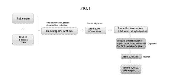
[0022] FIG. 1 illustrates a workflow for a LC-MRM-MS/MS assay according to
an
exemplary embodiment.
[0023] FIG. 2A shows a collision-induced dissociation (CID) MS/MS spectrum
of a
surrogate peptide for mAbl generated from trypsin and AspN digestion according
to an
exemplary embodiment. FIG 2B shows a CID MS/MS spectrum of a surrogate peptide
for
mAb2 generated from trypsin and AspN digestion according to an exemplary
embodiment.
[0024] FIG. 3 shows extracted ion chromatograms (XICs) of surrogate
peptides (FIG. 3A,
FIG. 3D), internal standards (FIG. 3B, FIG. 3E) and calibration curve plots
(FIG. 3C, FIG. 3F) of
mAbl (FIGs. 3A-C) and mAb2 (FIGs. 3D-F) generated from LC-MRM-MS of
calibration
standards according to an exemplary embodiment.
[0025] FIG. 4A shows overlaid XICs of a surrogate peptide of mAbl from ten
individual
naive human serum samples co-spiked with 10 pg/mL of mAbl and 20 pg/mL of mAb2
according to an exemplary embodiment. FIG. 4B shows overlaid XICs of a
surrogate peptide of
mAb2 from ten individual naive human serum samples co-spiked with 10 pg/mL of
mAbl and
20 pg/mL of mAb2 according to an exemplary embodiment. FIG. 4C shows a
measured
accuracy percentage of drug concentrations in the ten individual human serum
samples with
drugs spiked at lower limit of quantitation (LLOQ) level according to an
exemplary embodiment.
[0026] FIG. 5A shows XICs of the MRM transition for the surrogate peptide
of mAbl
according to an exemplary embodiment. FIG. 5B shows XICs of the MRM transition
for the
surrogate peptide of mAb2 according to an exemplary embodiment. FIG. 5C shows
a
comparison of the accuracy percentage of drug concentrations of mAbl at five
quality control
(QC) levels measured without the presence of mAb2 in serum matrix or with 2
mg/mL mAb2 in
the serum matrix background according to an exemplary embodiment. FIG. 5D
shows a
comparison of the accuracy percentage of drug concentrations of mAb2 at five
QC levels
measured without the presence of mAbl in serum matrix or with 2 mg/mL mAbl in
the serum
matrix background according to an exemplary embodiment.
[0027] FIG. 6A shows the accuracy percentage of measuring mAbl stability in
three
different conditions at five QC levels according to an exemplary embodiment.
FIG. 6B shows
CA 03216047 2023-10-03
WO 2022/217020 PCT/US2022/023963
the accuracy percentage of measuring mAb2 stability in three different
conditions at five QC
levels according to an exemplary embodiment.
[0028] FIG. 7A shows the pharmacokinetic profile of mAbl measured from
serum samples
by the LC-MRM-MS assay and a fully validated electrochemiluminescence (ECL)
immunoassay
according to an exemplary embodiment. FIG. 7B shows the pharmacokinetic
profile of mAb2
measured from serum samples by the LC-MRM-MS assay and a fully validated ECL
immunoassay according to an exemplary embodiment.
DETAILED DESCRIPTION
[0029] REGEN-COV (casirivimab and imdevimab) is an investigational antibody
cocktail
therapy developed by Regeneron Pharmaceuticals, Inc. for the treatment of
coronavirus disease
2019 (COVID-19) caused by severe acute respiratory syndrome coronavirus 2
(SARS-CoV-2)
(Hansen et al., 2020, Science, 369:1010-1014; Baum et al., 2020, Science,
370:1110-1115;
Weinreich et al., 2021, N Engl J Med, 384:238-251). The antibody cocktail
includes two
humanized IgG1 monoclonal antibodies (herein referred to as mAbl and mAb2),
which are
designed to target non-overlapping epitopes on the SARS-CoV-2 spike protein,
and thereby
blocking the interaction of SARS-CoV-2 virus with human ACE2, and preventing
viral escape
due to rapid genetic mutation of the virus (Hansen et al.; Baum et al., 2020,
Science, 369:1014-
1018). A recent clinical study has shown that REGEN-COV therapy can reduce
viral load and
improve symptoms for non-hospitalized COVID-19 patients, especially those who
were
seronegative or had high viral loads at baseline (Weinrich et al.). Based on
the promising results
from the clinical investigation, REGEN-COV was granted Emergency Use
Authorization (EUA)
by the U.S. Food and Drug Administration (FDA) in November 2020 for the
treatment of
recently diagnosed, mild-to-moderate COVID-19 in adults and pediatric patients
at least 12 years
of age and weighing at least 40 kg who are at high risk for progressing to
severe COVID-19
and/or hospitalization.
[0030] Measurement of the time profile of antibody drug concentration in
serum after
drug administration in patients is critical for pharmacokinetic (PK)
characterization of protein
therapeutic and drug dose optimization. To meet this need and manage the
accelerated
development for a COVID-19 therapy, a fit-for-purpose liquid chromatography-
multiple reaction
monitoring mass spectrometry (LC-MRM-MS) assay for REGEN-COV pharmacokinetic
study
6
CA 03216047 2023-10-03
WO 2022/217020
PCT/US2022/023963
was developed and qualified in one month, a much shorter timeframe than that
required for the
development of a conventional ligand-binding assay. Unlike a ligand-binding
assay, an LC-
MRM-MS assay does not require highly specific affinity capture and detection
reagents for the
antibody therapeutics, which typically take several months to develop and
produce. In addition,
the LC-MRM-MS assay of the present invention also provides wide dynamic range,
good
accuracy and precision, and excellent selectivity and specificity for
quantification of protein-
based biopharmaceuticals in serum matrix (van den Broek et at., 2013, J
Chromatogr B,
929:161-179). Recently, LC-MRM-MS has become a more frequently adopted
bioanalytical
strategy for both preclinical and clinical sample analysis due to the
continuous improvement on
the performance of LC-MS instrumentation (Jiang et al., 2013, Anal Chem,
85:9859-9867;
Zhang et al., 2014, Anal Chem, 86:8776-8784; Li et al., 2012, Anal Chem,
84:1267-1273;
Cardozo et al., 2020, Nat Commun, 11:6201; Fernandez Ocana et al., 2012, Anal
Chem,
84:5959-5967; Shen et al., 2015, Anal Chem, 87:8555-8563).
[0031]
Quantification of total antibody drug concentration, including free and bound
antibodies, in human serum samples using LC-MRM-MS can be based on the
measurement of
ion intensities of the surrogate peptides derived from the variable
complementarity-determining
regions (CDRs) of the antibody drugs (Jenkins et al., 2015, AAPS J, 17:1-16).
To process patient
serum samples, typically a few microliters of serum sample was reduced,
alkylating, and then
underwent protease digestion. Stable heavy isotope labeled proteins or
surrogate peptides are
usually used as internal standards (ISs) to normalize the signal variation
from sample processing
and instrument performance fluctuation. The sensitivity, selectivity and
specificity of the assay
can rely on the unique CDR peptides that have been selected for
quantification. For a co-
administered antibody cocktail, the LC-MRM-MS can be readily multiplexed to
measure
multiple drug analytes simultaneously. Despite limited throughput due to the
chromatographic
separation, the LC-MRM-MS method of the present invention met the required
dynamic range,
sensitivity, selectivity, stability, and specificity for the early measurement
of drug concentrations
of REGEN-COV in a limited number of serum samples in clinical trials. The
concentrations of
REGEN-COV in two dose groups of ambulatory patients measured by the LC-MRM-MS
assay
of the invention were compared with the results obtained from a fully
validated ligand binding
immunoassay, which demonstrated that the two assays were in good agreement.
This disclosure
sets an example as a fit-for-purpose application of LC-MRM-MS for clinical
sample analysis
7
CA 03216047 2023-10-03
WO 2022/217020 PCT/US2022/023963
when there are challenges to deliver a validated immunoassay to meet an urgent
timeline, or if
high-quality anti-idiotypic antibody reagents for a ligand binding assay are
not available.
[0032] Unless described otherwise, all technical and scientific terms used
herein have the
same meaning as commonly understood by one of ordinary skill in the art to
which this invention
belongs. Although any methods and materials similar or equivalent to those
described herein can
be used in the practice or testing, particular methods and materials are now
described.
[0033] The term "a" should be understood to mean "at least one" and the
terms "about"
and "approximately" should be understood to permit standard variation as would
be understood
by those of ordinary skill in the art and where ranges are provided, endpoints
are included. As
used herein, the terms "include," "includes," and "including" are meant to be
non-limiting and
are understood to mean "comprise," "comprises," and "comprising" respectively.
[0034] As used herein, the term "protein" or "protein of interest" can
include any amino
acid polymer having covalently linked amide bonds. Proteins comprise one or
more amino acid
polymer chains, generally known in the art as "polypeptides." "Polypeptide"
refers to a polymer
composed of amino acid residues, related naturally occurring structural
variants, and synthetic
non-naturally occurring analogs thereof linked via peptide bonds, related
naturally occurring
structural variants, and synthetic non-naturally occurring analogs thereof.
"Synthetic peptides or
polypeptides" refers to a non-naturally occurring peptide or polypeptide.
Synthetic peptides or
polypeptides can be synthesized, for example, using an automated polypeptide
synthesizer.
Various solid phase peptide synthesis methods are known to those of skill in
the art. A protein
may comprise one or multiple polypeptides to form a single functioning
biomolecule. A protein
can include antibody fragments, nanobodies, recombinant antibody chimeras,
cytokines,
chemokines, peptide hormones, and the like. Proteins of interest can include
any of bio-
therapeutic proteins, recombinant proteins used in research or therapy, trap
proteins and other
chimeric receptor Fc-fusion proteins, chimeric proteins, antibodies,
monoclonal antibodies,
polyclonal antibodies, human antibodies, and bispecific antibodies. Proteins
may be produced
using recombinant cell-based production systems, such as the insect
bacculovirus system, yeast
systems (e.g., Pichia sp.), mammalian systems (e.g., CHO cells and CHO
derivatives like CHO-
K1 cells). For a recent review discussing biotherapeutic proteins and their
production, see
Ghaderi et at., "Production platforms for biotherapeutic glycoproteins.
Occurrence, impact, and
8
CA 03216047 2023-10-03
WO 2022/217020 PCT/US2022/023963
challenges of non-human sialylation" (Darius Ghaderi et al., Production
platforms for
biotherapeutic glycoproteins. Occurrence, impact, and challenges of non-human
sialylation, 28
BIOTECHNOLOGY AND GENETIC ENGINEERING REVIEWS 147-176 (2012), the entire
teachings of which are herein incorporated). Proteins can be classified on the
basis of
compositions and solubility and can thus include simple proteins, such as
globular proteins and
fibrous proteins; conjugated proteins, such as nucleoproteins, glycoproteins,
mucoproteins,
chromoproteins, phosphoproteins, metalloproteins, and lipoproteins; and
derived proteins, such
as primary derived proteins and secondary derived proteins.
[0035] In some exemplary embodiments, a protein of interest can be a
recombinant protein,
an antibody, a bispecific antibody, a multispecific antibody, antibody
fragment, monoclonal
antibody, fusion protein, scFv and combinations thereof.
[0036] As used herein, the term "recombinant protein" refers to a protein
produced as the
result of the transcription and translation of a gene carried on a recombinant
expression vector
that has been introduced into a suitable host cell. In certain exemplary
embodiments, the
recombinant protein can be an antibody, for example, a chimeric, humanized, or
fully human
antibody. In certain exemplary embodiments, the recombinant protein can be an
antibody of an
isotype selected from group consisting of: IgG (e.g., IgGl, IgG2, IgG3, IgG4),
IgM, IgAl, IgA2,
IgD, or IgE. In certain exemplary embodiments the antibody molecule is a full-
length antibody
(e.g., an IgG1 or IgG4 immunoglobulin) or alternatively the antibody can be a
fragment (e.g., an
Fc fragment or a Fab fragment).
[0037] The term "antibody," as used herein includes immunoglobulin
molecules
comprising four polypeptide chains, two heavy (H) chains and two light (L)
chains inter-
connected by disulfide bonds, as well as multimers thereof (e.g., IgM). Each
heavy chain
comprises a heavy chain variable region (abbreviated herein as HCVR or VH) and
a heavy chain
constant region. The heavy chain constant region comprises three domains, CH1,
CH2 and CH3.
Each light chain comprises a light chain variable region (abbreviated herein
as LCVR or VL) and
a light chain constant region. The light chain constant region comprises one
domain (CL1). The
VH and VL regions can be further subdivided into regions of hypervariability,
termed
complementarity determining regions (CDRs), interspersed with regions that are
more
conserved, termed framework regions (FR). Each VH and VL is composed of three
CDRs and
9
CA 03216047 2023-10-03
WO 2022/217020
PCT/US2022/023963
four FRs, arranged from amino-terminus to carboxy-terminus in the following
order: FR1,
CDR1, FR2, CDR2, FR3, CDR3, and FR4. An amino acid consensus sequence may be
defined
based on a side-by-side analysis of two or more CDRs. The term "antibody," as
used herein,
also includes antigen-binding fragments of full antibody molecules. The terms
"antigen-binding
portion" of an antibody, "antigen-binding fragment" of an antibody, and the
like, as used herein,
include any naturally occurring, enzymatically obtainable, synthetic, or
genetically engineered
polypeptide or glycoprotein that specifically binds an antigen to form a
complex. Antigen-
binding fragments of an antibody may be derived, for example, from full
antibody molecules
using any suitable standard techniques such as proteolytic digestion or
recombinant genetic
engineering techniques involving the manipulation and expression of DNA
encoding antibody
variable and optionally constant domains. Such DNA is known and/or is readily
available from,
for example, commercial sources, DNA libraries (including, e.g., phage-
antibody libraries), or
can be synthesized. The DNA may be sequenced and manipulated chemically or by
using
molecular biology techniques, for example, to arrange one or more variable
and/or constant
domains into a suitable configuration, or to introduce codons, create cysteine
residues, modify,
add or delete amino acids, etc.
[0038] As
used herein, an "antibody fragment" includes a portion of an intact antibody,
such as, for example, the antigen-binding or variable region of an antibody.
Examples of
antibody fragments include, but are not limited to, a Fab fragment, a Fab'
fragment, a F(ab')2
fragment, a scFv fragment, a Fv fragment, a dsFy diabody, a dAb fragment, a
Fd' fragment, a Fd
fragment, and an isolated complementarity determining region (CDR) region, as
well as
triabodies, tetrabodies, linear antibodies, single-chain antibody molecules,
and multi specific
antibodies formed from antibody fragments. Fv fragments are the combination of
the variable
regions of the immunoglobulin heavy and light chains, and ScFv proteins are
recombinant single
chain polypeptide molecules in which immunoglobulin light and heavy chain
variable regions
are connected by a peptide linker. In some exemplary embodiments, an antibody
fragment
comprises a sufficient amino acid sequence of the parent antibody of which it
is a fragment that
it binds to the same antigen as does the parent antibody; in some exemplary
embodiments, a
fragment binds to the antigen with a comparable affinity to that of the parent
antibody and/or
competes with the parent antibody for binding to the antigen. An antibody
fragment may be
produced by any means. For example, an antibody fragment may be enzymatically
or
CA 03216047 2023-10-03
WO 2022/217020 PCT/US2022/023963
chemically produced by fragmentation of an intact antibody and/or it may be
recombinantly
produced from a gene encoding the partial antibody sequence. Alternatively, or
additionally, an
antibody fragment may be wholly or partially synthetically produced. An
antibody fragment
may optionally comprise a single chain antibody fragment. Alternatively, or
additionally, an
antibody fragment may comprise multiple chains that are linked together, for
example, by
disulfide linkages. An antibody fragment may optionally comprise a multi-
molecular complex.
A functional antibody fragment typically comprises at least about 50 amino
acids and more
typically comprises at least about 200 amino acids.
[0039] The term "bispecific antibody" includes an antibody capable of
selectively binding
two or more epitopes. Bispecific antibodies generally comprise two different
heavy chains with
each heavy chain specifically binding a different epitope¨either on two
different molecules
(e.g., antigens) or on the same molecule (e.g., on the same antigen). If a
bispecific antibody is
capable of selectively binding two different epitopes (a first epitope and a
second epitope), the
affinity of the first heavy chain for the first epitope will generally be at
least one to two or three
or four orders of magnitude lower than the affinity of the first heavy chain
for the second
epitope, and vice versa. The epitopes recognized by the bispecific antibody
can be on the same
or a different target (e.g., on the same or a different protein). Bispecific
antibodies can be made,
for example, by combining heavy chains that recognize different epitopes of
the same antigen.
For example, nucleic acid sequences encoding heavy chain variable sequences
that recognize
different epitopes of the same antigen can be fused to nucleic acid sequences
encoding different
heavy chain constant regions and such sequences can be expressed in a cell
that expresses an
immunoglobulin light chain.
[0040] A typical bispecific antibody has two heavy chains each having three
heavy chain
CDRs, followed by a CHI domain, a hinge, a CH2 domain, and a CH3 domain, and
an
immunoglobulin light chain that either does not confer antigen-binding
specificity but that can
associate with each heavy chain, or that can associate with each heavy chain
and that can bind
one or more of the epitopes bound by the heavy chain antigen-binding regions,
or that can
associate with each heavy chain and enable binding of one or both of the heavy
chains to one or
both epitopes. BsAbs can be divided into two major classes, those bearing an
Fc region (IgG-
like) and those lacking an Fc region, the latter normally being smaller than
the IgG and IgG-like
bispecific molecules comprising an Fc. The IgG-like bsAbs can have different
formats such as,
11
CA 03216047 2023-10-03
WO 2022/217020 PCT/US2022/023963
but not limited to, triomab, knobs into holes IgG (kih IgG), crossMab, orth-
Fab IgG, Dual-
variable domains Ig (DVD-Ig), two-in-one or dual action Fab (DAF), IgG-single-
chain Fv (IgG-
scFv), or la-bodies. The non-IgG-like different formats include tandem scFvs,
diabody format,
single-chain diabody, tandem diabodies (TandAbs), Dual-affinity retargeting
molecule (DART),
DART-Fc, nanobodies, or antibodies produced by the dock-and-lock (DNL) method
(Gaowei
Fan, Zujian Wang & Mingju Hao, Bispecific antibodies and their applications, 8
JOURNAL OF
HEMATOLOGY & ONCOLOGY 130; Dafne MUller & Roland E. Kontermann, Bispecific
Antibodies, HANDBOOK OF THERAPEUTIC ANTIBODIES 265-310 (2014), the entire
teachings of which are herein incorporated).
[0041] As used herein "multispecific antibody" refers to an antibody with
binding
specificities for at least two different antigens. While such molecules
normally will only bind
two antigens (i.e., bispecific antibodies, bsAbs), antibodies with additional
specificities such as
trispecific antibody and KIH Trispecific can also be addressed by the system
and method
disclosed herein.
[0042] The term "monoclonal antibody" as used herein is not limited to
antibodies
produced through hybridoma technology. A monoclonal antibody can be derived
from a single
clone, including any eukaryotic, prokaryotic, or phage clone, by any means
available or known
in the art. Monoclonal antibodies useful with the present disclosure can be
prepared using a
wide variety of techniques known in the art including the use of hybridoma,
recombinant, and
phage display technologies, or a combination thereof
[0043] In some exemplary embodiments, a protein of interest can be produced
from
mammalian cells. The mammalian cells can be of human origin or non-human
origin, and can
include primary epithelial cells (e.g., keratinocytes, cervical epithelial
cells, bronchial epithelial
cells, tracheal epithelial cells, kidney epithelial cells and retinal
epithelial cells), established cell
lines and their strains (e.g., 293 embryonic kidney cells, BHK cells, HeLa
cervical epithelial
cells and PER-C6 retinal cells, MDBK (NBL-1) cells, 911 cells, CRFK cells,
MDCK cells, CHO
cells, BeWo cells, Chang cells, Detroit 562 cells, HeLa 229 cells, HeLa S3
cells, Hep-2 cells, KB
cells, L5I80 cells, L5174T cells, NCI-H-548 cells, RPMI2650 cells, SW-13
cells, T24 cells, WI-
28 VA13, 2RA cells, WISH cells, BS-C-I cells, LLC-MK2 cells, Clone M-3 cells,
1-10 cells,
RAG cells, TCMK-1 cells, Y-1 cells, LLC-PKi cells, PK(15) cells, GHi cells,
GH3 cells, L2
12
CA 03216047 2023-10-03
WO 2022/217020 PCT/US2022/023963
cells, LLC-RC 256 cells, MHiCi cells, XC cells, MDOK cells, VSW cells, and TH-
I, B1 cells,
BSC-1 cells, RAf cells, RK-cells, PK-15 cells or derivatives thereof),
fibroblast cells from any
tissue or organ (including but not limited to heart, liver, kidney, colon,
intestines, esophagus,
stomach, neural tissue (brain, spinal cord), lung, vascular tissue (artery,
vein, capillary),
lymphoid tissue (lymph gland, adenoid, tonsil, bone marrow, and blood),
spleen, and fibroblast
and fibroblast-like cell lines (e.g., CHO cells, TRG-2 cells, IMR-33 cells,
Don cells, GHK-21
cells, citrullinemia cells, Dempsey cells, Detroit 551 cells, Detroit 510
cells, Detroit 525 cells,
Detroit 529 cells, Detroit 532 cells, Detroit 539 cells, Detroit 548 cells,
Detroit 573 cells, HEL
299 cells, IMR-90 cells, MRC-5 cells, WI-38 cells, WI-26 cells, Midi cells,
CHO cells, CV-1
cells, COS-1 cells, COS-3 cells, COS-7 cells, Vero cells, DBS-FrhL-2 cells,
BALB/3T3 cells, F9
cells, SV-T2 cells, M-MSV-BALB/3T3 cells, K-BALB cells, BLO-11 cells, NOR-10
cells,
C3H/IOTI/2 cells, HSDMiC3 cells, KLN205 cells, McCoy cells, Mouse L cells,
Strain 2071
(Mouse L) cells, L-M strain (Mouse L) cells, L-MTK' (Mouse L) cells, NCTC
clones 2472 and
2555, SCC-PSA1 cells, Swiss/3T3 cells, Indian muntjac cells, SIRC cells, Cn
cells, and Jensen
cells, 5p2/0, NSO, NS1 cells or derivatives thereof).
[0044] As used herein, the term "therapeutic protein" refers to any protein
that can be
administered to a subject for the treatment of a disease or disorder. A
therapeutic protein may be
any protein with a pharmacological effect, for example, an antibody, a soluble
receptor, an
antibody-drug conjugate, or an enzyme. In some exemplary embodiments, the
therapeutic
protein can be an anti-SARS-CoV-2 antibody, including casirivimab or
imdevimab. Multiple
therapeutic proteins may be co-administered in order to achieve a
pharmacological effect, for
example, to prevent viral escape due to mutation of a target virus. As used
herein, the term
"antibody cocktail" refers to co-administered therapeutic proteins comprising
at least two
therapeutic antibodies. In some exemplary embodiments, an antibody cocktail
can comprise
REGEN-COV.
[0045] In some exemplary embodiments, the number of therapeutic proteins in
the sample
can be at least two. In some specific embodiments, one of the therapeutic
proteins can be a
monoclonal antibody, a polyclonal antibody, a bispecific antibody, an antibody
fragment, a
fusion protein, or an antibody-drug complex. In some other specific
embodiments, a
concentration of one of the therapeutic proteins in a sample can be about 10
i.tg/mL to about
2000 pg/mL. In some exemplary embodiments, the number of therapeutic proteins
in the sample
13
CA 03216047 2023-10-03
WO 2022/217020 PCT/US2022/023963
is three. In some exemplary embodiments, the number of therapeutic proteins in
the sample is
four. In some exemplary embodiments, the number of therapeutic proteins in the
sample is five.
[0046] As used herein, a "sample" can be obtained from any step of a
bioprocess, such as
cell culture fluid (CCF), harvested cell culture fluid (HCCF), any step in the
downstream
processing, drug substance (DS), or a drug product (DP) comprising the final
formulated
product. In some specific exemplary embodiments, the sample can be selected
from any step of
the downstream process of clarification, chromatographic production, viral
inactivation, or
filtration.
[0047] In some exemplary embodiments, the sample is a biological sample. As
used here,
the term "biological sample" refers to a sample taken from a living organism,
for example a
human or a non-human mammal. A biological sample may comprise, for example,
whole blood,
plasma, serum, saliva, tears, semen, cheek tissue, organ tissue, urine, feces,
skin, or hair. A
sample may be taken from a patient, for example, a clinical sample.
[0048] As used herein, the term "pharmacokinetics" (PK) refers to a field
of study dealing
with features of a drug after administration to a subject. Exemplary
components of
pharmacokinetic analysis include liberation of a drug from a pharmaceutical
formulation,
absorption of a drug into blood circulation, distribution of a drug throughout
the body,
metabolism (also called biotransformation) of a drug into metabolites, and
excretion of a drug
from a body. Pharmacokinetics of a drug are a key feature for evaluation of a
biotherapeutic
candidate. In particular, a pharmacokinetic study may be conducted to evaluate
how levels of a
drug and its modified forms and metabolites change over time after
administration to a subject.
Biotherapeutic proteins may be evaluated through the analysis of
representative peptides, or
"target peptides" or "surrogate peptides," using liquid chromatography-mass
spectrometry. A
peptide may be a suitable target peptide if it is unique to or strongly
representative of a protein,
for example a complementarity-determining region of an antibody, and if it can
be reliably
recovered and measured.
[0049] As used herein, the term "liquid chromatography" refers to a process
in which a
biological/chemical mixture carried by a liquid can be separated into
components as a result of
differential distribution of the components as they flow through (or into) a
stationary liquid or
solid phase. Non-limiting examples of liquid chromatography include reverse
phase liquid
chromatography, ion-exchange chromatography, size exclusion chromatography,
affinity
14
CA 03216047 2023-10-03
WO 2022/217020 PCT/US2022/023963
chromatography, hydrophobic interaction chromatography, hydrophilic
interaction
chromatography, or mixed-mode chromatography.
[0050] As used herein, the term "mass spectrometer" includes a device
capable of
identifying specific molecular species and measuring their accurate mass-to-
charge ratios. The
term is meant to include any molecular detector into which a polypeptide or
peptide may be
characterized. A mass spectrometer can include three major parts: the ion
source, the mass
analyzer, and the detector. The role of the ion source is to create gas phase
ions. Analyte atoms,
molecules, or clusters can be transferred into gas phase and ionized either
concurrently (as in
electrospray ionization) or through separate processes. The choice of ion
source depends on the
application. In some exemplary embodiments, the mass spectrometer can be a
tandem mass
spectrometer.
[0051] As used herein, the term "tandem mass spectrometry" includes a
technique where
structural information on sample molecules is obtained by using multiple
stages of mass
selection and mass separation. A prerequisite is that the sample molecules be
transformed into a
gas phase and ionized so that fragments are formed in a predictable and
controllable fashion after
the first mass selection step. Multistage MS/MS, or MS, can be performed by
first selecting and
isolating a precursor ion, fragmenting it (MS2), isolating a primary fragment
ion, fragmenting it
(MS3), isolating a secondary fragment, and so on (MS4), as long as one can
obtain meaningful
information, or the fragment ion signal is detectable. Tandem MS has been
successfully
performed with a wide variety of analyzer combinations. What analyzers to
combine for a
certain application can be determined by many different factors, such as
sensitivity, selectivity,
and speed, but also size, cost, and availability. The two major categories of
tandem MS methods
are tandem-in-space and tandem-in-time, but there are also hybrids where
tandem-in-time
analyzers are coupled in space or with tandem-in-space analyzers. A tandem-in-
space mass
spectrometer comprises an ion source, a precursor ion activation device, and
at least two non-
trapping mass analyzers. Specific m/z separation functions can be designed so
that in one section
of the instrument ions are selected, dissociated in an intermediate region,
and the product ions
are then transmitted to another analyzer for m/z separation and data
acquisition. In tandem-in-
time, mass spectrometer ions produced in the ion source can be trapped,
isolated, fragmented,
and m/z separated in the same physical device.
CA 03216047 2023-10-03
WO 2022/217020 PCT/US2022/023963
[0052] The peptides identified by the mass spectrometer can be used as
surrogate
representatives of the intact protein and their post translational
modifications. They can be used
for protein characterization by correlating experimental and theoretical MS/MS
data, the latter
generated from possible peptides in a protein sequence database. The
characterization includes,
but is not limited, to sequencing amino acids of the protein fragments,
determining protein
sequencing, determining protein de novo sequencing, locating post-
translational modifications,
or identifying post translational modifications, or comparability analysis, or
combinations
thereof.
[0053] As used herein, the term "database" refers to a compiled collection
of protein
sequences that may possibly exist in a sample, for example in the form of a
file in a FASTA
format. Relevant protein sequences may be derived from cDNA sequences of a
species being
studied. Public databases that may be used to search for relevant protein
sequences included
databases hosted by, for example, Uniprot or Swiss-prot. Databases may be
searched using what
are herein referred to as "bioinformatics tools". Bioinformatics tools provide
the capacity to
search uninterpreted MS/MS spectra against all possible sequences in the
database(s), and
provide interpreted (annotated) MS/MS spectra as an output. Non-limiting
examples of such
tools are Mascot (www.matrixscience.com), Spectrum Mill
(www.chem.agilent.com), PLGS
(www.waters.com), PEAKS (www.bioinformaticssolutions.com), Proteinpilot
(download.appliedbiosystems.com//proteinpilot), Phenyx (www.phenyx-ms.com),
Sorcerer
(www.sagenresearch.com), OMS SA (www.pubchem.ncbi.nlm.nih.gov/omssa/), X!
Tandem
(www.thegpm.org/TANDEM/), Protein Prospector
(prospector.ucsfedu/prospector/mshome.htm), Byonic
(www.proteinmetrics.com/products/byonic) or Sequest
(fields.scripps.edu/sequest).
[0054] In some exemplary embodiments, the mass spectrometer can be coupled
to a liquid
chromatography system.
[0055] In some exemplary embodiments, the mass spectrometer can be coupled
to a liquid
chromatography-multiple reaction monitoring system. More generally, a mass
spectrometer may
be capable of analysis by selected reaction monitoring (SRM), including
consecutive reaction
monitoring (CRM) and parallel reaction monitoring (PRM).
[0056] As used herein, "multiple reaction monitoring" or "MiRM" refers to a
mass
spectrometry-based technique that can precisely quantify small molecules,
peptides, and proteins
16
CA 03216047 2023-10-03
WO 2022/217020 PCT/US2022/023963
within complex matrices with high sensitivity, specificity and a wide dynamic
range (Paola
Picotti & Ruedi Aebersold, Selected reaction monitoring¨based proteomics:
workflows,
potential, pitfalls and future directions, 9 NATURE METHODS 555-566 (2012)).
MRM can be
typically performed with triple quadrupole mass spectrometers wherein a
precursor ion
corresponding to the selected small molecules/ peptides is selected in the
first quadrupole and a
fragment ion of the precursor ion was selected for monitoring in the third
quadrupole (Yong
Seok Choi et al., Targeted human cerebrospinal fluid proteomics for the
validation of multiple
Alzheimers disease biomarker candidates, 930 JOURNAL OF CHROMATOGRAPHY B 129-
135 (2013)).
[0057] In some aspects, the mass spectrometer in the method or system of
the present
application can be an electrospray ionization mass spectrometer, nano-
electrospray ionization
mass spectrometer, or a triple quadrupole mass spectrometer, wherein the mass
spectrometer can
be coupled to a liquid chromatography system, wherein the mass spectrometer is
capable of
performing LC-MS (liquid chromatography-mass spectrometry) or LC-MRM-MS
(liquid
chromatography-multiple reaction monitoring-mass spectrometry) analyses.
[0058] As used herein, the term "digestion" refers to hydrolysis of one or
more peptide
bonds of a protein. There are several approaches to carrying out digestion of
a protein in a
sample using an appropriate hydrolyzing agent, for example, enzymatic
digestion or non-
enzymatic digestion.
[0059] As used herein, the term "digestive enzyme" refers to any of a large
number of
different agents that can perform digestion of a protein. Non-limiting
examples of hydrolyzing
agents that can carry out enzymatic digestion include protease from
Aspergillus Saitoi, elastase,
subtilisin, protease XIII, pepsin, trypsin, Tryp-N, chymotrypsin,
aspergillopepsin I, LysN
protease (Lys-N), LysC endoproteinase (Lys-C), endoproteinase Asp-N (Asp-N),
endoproteinase
Arg-C (Arg-C), endoproteinase Glu-C (Glu-C) or outer membrane protein T
(OmpT),
immunoglobulin-degrading enzyme of Streptococcus pyogenes (IdeS), thermolysin,
papain,
pronase, V8 protease or biologically active fragments or homologs thereof or
combinations
thereof. For a recent review discussing the available techniques for protein
digestion see
Switazar et al., "Protein Digestion: An Overview of the Available Techniques
and Recent
Developments" (Linda Switzar, Martin Giera & Wilfried M. A. Niessen, Protein
Digestion: An
17
CA 03216047 2023-10-03
WO 2022/217020 PCT/US2022/023963
Overview of the Available Techniques and Recent Developments, 12 JOURNAL OF
PROTEOME RESEARCH 1067-1077 (2013)).
[0060] The amount of digestive enzyme and the time required for digestion
can be
appropriately selected. When the enzyme to substrate ratio is unsuitably high,
the
correspondingly high digestion rate will not allow sufficient time for the
peptides to be analyzed
by mass spectrometer, and sequence coverage will be compromised. On the other
hand, a low
enzyme to substrate ratio would need a long digestion time and thus a long
data acquisition time.
The enzyme to substrate ratio can range from about 1:0.5 to about 1:200.
[0061] In some exemplary embodiments, the method of quantitating a
therapeutic protein
can optionally comprise contacting a therapeutic protein to a protein reducing
agent.
[0062] As used herein, the term "protein reducing agent" refers to the
agent used for
reduction of disulfide bridges in a protein. Non-limiting examples of protein
reducing agents are
dithiothreitol (DTT), B-mercaptoethanol, Ellman's reagent, hydroxylamine
hydrochloride,
sodium cyanoborohydride, tris(2-carboxyethyl)phosphine hydrochloride (TCEP-
HC1), or
combinations thereof.
[0063] In some exemplary embodiments, the method of quantitating protein
can optionally
comprise contacting a therapeutic protein to a protein alkylating agent.
[0064] As used herein, the term "protein alkylating agent" refers to the
agent used to
alkylate certain free amino acid residues in a protein. Non-limiting examples
of protein
alkylating agents are iodoacetamide (TAM), chloroacetamide (CAA), acrylamide
(AA), N-
ethylmaleimide (NEM), methyl methanethiosulfonate (MMTS), and 4-vinylpyridine
or
combinations thereof.
[0065] In some exemplary embodiments, the method of quantitating a
therapeutic protein
can comprise denaturing a therapeutic protein.
[0066] As used herein, "protein denaturing" can refer to a process in which
the three-
dimensional shape of a molecule is changed from its native state. Protein
denaturation can be
carried out using a protein denaturing agent. Non-limiting examples of a
protein denaturing
agent include heat, high or low pH, reducing agents like DTT (see below) or
exposure to
chaotropic agents. Several chaotropic agents can be used as protein denaturing
agents.
Chaotropic solutes increase the entropy of the system by interfering with
intramolecular
interactions mediated by non-covalent forces such as hydrogen bonds, van der
Waals forces, and
18
CA 03216047 2023-10-03
WO 2022/217020 PCT/US2022/023963
hydrophobic effects. Non-limiting examples for chaotropic agents include
butanol, ethanol,
guanidinium chloride, lithium perchlorate, lithium acetate, magnesium
chloride, phenol,
propanol, sodium dodecyl sulfate, thiourea, N-lauroylsarcosine, urea, and
salts thereof
[0067] It is understood that the present invention is not limited to any of
the aforesaid
therapeutic protein(s), antibody cocktail(s), host cell(s), protein denaturing
agent(s), protein
alkylating agent(s), protein reducing agent(s), digestive enzyme(s), mass
analyzer(s),
instrument(s) used for identification, or chromatographic method(s), and any
therapeutic
protein(s), antibody cocktail(s), host cell(s), protein denaturing agent(s),
protein alkylating
agent(s), protein reducing agent(s), digestive enzyme(s), mass analyzer(s),
instrument(s) used for
identification, or chromatographic method(s) can be selected by any suitable
means.
[0068] The present invention will be more fully understood by reference to
the following
Examples. They should not, however, be construed as limiting the scope of the
invention.
EXAMPLES
[0069] The overall workflow of the LC-MRM-MS/MS assay of the invention
according to
an exemplary embodiment is illustrated in FIG. 1.
[0070] Chemicals and reagents. Tris (2-carboxyethyl) phosphine
hydrochloride (TCEP-
HC1), trifluoroacetic acid (TFA), 0.1% formic acid (v/v) in water (LC-MS
grade), and 0.1%
formic acid (v/v) in acetonitrile (LC-MS grade) were purchased from Thermo
Fisher Scientific
(Rockford, IL). Ultrapure 1 M Tris-HC1 pH 8.0 was obtained from Invitrogen
(Carlsbad, CA).
Urea and iodoacetamide (IAM) were purchased from Sigma-Aldrich (St. Louis,
MO). Trypsin
(Mass Spectrometry grade) and rAspN were purchased from Promega (Madison, WI).
Pooled
human serum and single human serum from 10 individuals were purchased from
Innovative
Research (Novi, MI). AUQA grade custom synthetic heavy peptides for internal
standards (ISs),
LLIYAASNLETGVPSR*(10 Da), DTAV*(6Da) YYCASGS, were ordered from Thermo Fisher
Scientific (Rockford, IL). mAbl and mAb2 drug substance (DS) were developed
and obtained
from Regeneron Pharmaceuticals (Tarrytown, NY). COVID-19 patient serum samples
were
from a clinical trial of REGEN-COV sponsored by Regeneron Pharmaceuticals
(ClinicalTrials.gov Identifier: NCT04425629).
[0071] Preparation of standard solutions. Stock solutions of REGEN-COV in
human
serum were made by spiking mAbl and mAb2 DS into pooled human serum.
Calibration
19
CA 03216047 2023-10-03
WO 2022/217020 PCT/US2022/023963
standards (20, 25, 30, 50, 100, 250, 500, 1000, 2000 [tg/mL for mAbl; 10, 20,
25, 30, 50, 100,
250, 500, 1000, 2000 g/mL for mAb2) were made through a serial dilution of
the stock solution
using the pooled human serum. Five qualification QC standards including the
Upper Limit of
Quantitation (ULOQ, 2000 g/mL mAbl, 2000 g/mL mAb2), High QC (HQC, 1500
[tg/mL
mAbl, 1500 g/mL mAb2), Mid QC (MQC, 750 g/mL mAbl, 750 g/mL mAb2), Low QC
(LQC, 60 [tg/mL mAbl, 30 g/mL mAb2), and the Lower Limit of Quantitation
(LLOQ, 20
[tg/mL mAbl, 10 [tg/mL mAb2), were also prepared by spiking mAbl and mAb2 DS
into the
pooled human serum and serial dilutions.
[0072] LLOQ (20 [tg/mL of mAbl, 10 g/mL of mAb2) spiked individual human
serum
samples were prepared by co-spiking mAbl and mAb2 DS into ten individual human
serum
blanks. For the drug specificity assay, the QC standards containing one drug
were made by
serial dilution of stock solution of the antibody drug using the pooled human
serum as the
diluent. The QC standards containing one drug with the presence of co-
administered drug as
matrix background were made from serial dilution of the stock solution using 2
mg/mL of the co-
administered drug in the pooled human serum as the diluent.
[0073] Digestion of serum samples. Prior to sample processing, serum
samples
(calibration standards, QC standards, and patient samples) were thawed on ice.
The serum
sample digestion was conducted in a 96-well plate (0.5 mL, polypropylene,
Agilent
Technologies, Santa Clara, CA). 5 of serum sample was added to each sample
well prefilled
with 80 tL denaturation solution (10 mM TCEP, 8 M urea). The 96-well plate was
sealed with
an adhesive plate seal (Waters, Milford, MA), and heated at 80 C for 10
minutes on
Thermomixer C (Eppendorf, Hamburg, Germany) at 650 rpm. After cooling to room
temperature, 15 of 0.25 M TAM was added to each sample well and the plate
was incubated
by shaking at 650 rpm in dark for 30 minutes at room temperature. Prior to
use, digestion
solution containing two enzymes and two IS peptides were made by
reconstitution of 200 [tg of
trypsin, 100 [tg of rAspN, 150 tL of mAbl IS stock solution (5 pmol/ L), and
100 11.1 of mAb2
IS stock solution (5 pmol/ L) in 9 mL of 0.1 M Tris buffer. Following
alkylation, 10 tL of each
sample were transferred to a second 96-well plate and mixed with 90
digestion solution
containing two enzymes and IS peptides. The sample plate was sealed and
incubated at 37 C for
3 hours with 650 rpm shaking. When the digestion finished, 10 tL of 10% TFA
was added to
CA 03216047 2023-10-03
WO 2022/217020 PCT/US2022/023963
each sample well to quench the reaction. The sample plate was spun at 700 rpm
for 1 minute
prior to LC-MRM-MS analysis.
[0074] LC-MR1VI-MS methods. The LC-MRM-MS experiments were performed using
an Agilent Infinity II UPLC system coupled with 6495 Triple Quadrupole Mass
Spectrometer
(Agilent Technologies, Santa Clara, CA). 10 tL of digested serum sample,
corresponding to
approximately 45 nL of original serum, were loaded onto a C18 column (ACQUITY
UPLC
BEH300 1.7 p.m, 2.1 mm x 100 mm, Waters), and separated by reversed phase
gradient elution
using mobile phase A as 0.1% formic acid in water, and mobile phase B as 0.1%
formic acid in
acetonitrile at flow rate of 0.3 mL/min. Prior to each injection, the sample
injection path was
sequentially flushed with IPA/ACN/H20 v/v/v (3:1:1), ACN/H20/FA v/v/v
(25:75:0.1), and
ACN/H20/FA v/v/v (5:95:0.1). The LC gradient for MRM experiments was set as
follows: 0-0.5
min, 5% B; 0.5-16 min, 5-25% B; 16-18 min, 25-90% B; 18-20 min, 90% B; 20-20.5
min,
90-5% B, and 20.5-25, 5% B. The column temperature was set at 60 C and the
autosampler
was maintained at 7 C during sample analysis.
[0075] The triple quadrupole MS ion source parameters were set as
follows: gas
temperature 200 C, gas flow rate 12 L/min, nebulizer gas 20 psi, sheath gas
temperature 300 C,
sheath gas flow 11 L/min, capillary voltage 3500 V, nozzle voltage 500 V. Time
scheduled
MRM transitions for the two surrogate peptides and two IS peptides, with
parameters of each
transition channel listed in Table 1-1 and Table 1-2, were applied for all the
quantitative analysis
experiments.
21
CA 03216047 2023-10-03
WO 2022/217020 PCT/US2022/023963
Table 1-1. Time scheduled 1VIR1'1 transitions for LC-MR1VI-MS data acquisition
Precursor Product Precursor Product
Time Drug Peptide
Charge Type Ion Ion
DTAVYYC
2 y5+ 597.6 481.2
3.0-10.0 (Cam)ASGS
mAb2
min
DTAV*(6Da)
2 y5+ 600.6 487.2
YYC(Cam)ASGS
LLIYAASN
2 y3+ 853.0 359.2
LETGVPSR
10.0-16.0
mm mAbl
LLIYAAS
n
NLETGVPSR* 2 y3+ 858.0 369.2
(10Da)
Table 1-2. Time scheduled 1VIR1'I transitions for LC-MR1VI-MS data acquisition
Cell
Dwell
Time Drug Fragmentor CE Accelerator
Time (ms)
Voltage
3.0-10.0 300 380 6 5
mAb2
min
300 380 6 5
10.0-16.0 300 380 18 5
mAbl
min
300 380 18 5
[0076] Data analysis. The raw data from LC-MRM-MS experiments were analyzed
using
Agilent MassHunter Quantitative Analysis software. The extracted ion
chromatogram (XIC)
peak areas of the monitored transitions were integrated with the Agile2
algorithm. To construct
22
CA 03216047 2023-10-03
WO 2022/217020 PCT/US2022/023963
the calibration curve for each drug, the peak areas of surrogate peptide from
calibration
standards, normalized by the peak areas from the corresponding coeluting IS
peptide, were
plotted against their respective nominal concentrations using a 1/x2 weighted
three parameter
quadratic model (with variable weight for each point of the standard curve),
from which all other
readings were subsequently calculated. The equation for quadratic fit is y =
ax2 + bx + c, where
y = ratio of the XIC peak area of the surrogate peptide and that of the
corresponding IS peptide, x
= concentration of drug ( g/mL), and a, b, c = quadratic coefficient, linear
coefficient and
constant term, respectively. The weight for each point of the standard curve
is inversely
proportional to the analyte concentration. The calibration curve parameters
were automatically
computed by Agilent MassHunter Quantitative Analysis software.
[0077] Electrochemiluminescent immunoassay. The assay procedures employed
streptavidin microplates coated with either biotinylated mouse anti-mAbl
monoclonal antibody,
or biotinylated mouse anti-mAb2 monoclonal antibody. mAbl and mAb2 captured on
plates
specific for each molecule were detected using two ruthenylated, non-competing
mouse
monoclonal antibodies that are specific to either mAbl or mAb2.
Electrochemiluminescent
signal generated from the ruthenium label when voltage is applied to the plate
was measured by
the MSD reader. The measured electrochemiluminescence is proportional to the
concentration
of total mAbl or total mAb2 in the serum samples.
Example 1. Method development for MR1VI assay
[0078] Both mAbl and mAb2 are dimer molecules composed of a pair of light
chains and
a pair of heavy chains. mAbl light chain comprises 221 amino acid residues,
and its heavy chain
comprises 450 amino acid residues. mAb2 light chain comprises 216 amino acid
residues, and
its heavy chain comprises 450 amino acid residues. Because IgG proteins are
abundant in human
serum, and greater than 93% of the amino acid sequences of these two humanized
IgG1 drugs
are identical to the endogenous serum IgG, the selection of suitable surrogate
peptides for MRM-
based IgG antibody drug quantification is restricted to peptides derived from
the CDR regions
(typically 3 from the heavy chain, 3 from the light chain) of the variable
domains. Peptides from
the constant regions cannot be differentiated from those derived from
endogenous antibodies in
human serum.
23
CA 03216047 2023-10-03
WO 2022/217020 PCT/US2022/023963
[0079] To select suitable surrogate peptides for MRM quantification, the
following
considerations were applied to screen the candidate peptides generated by
protease cleavage in
the CDR region of the human IgG drugs: 1) no identical BLAST match hit in
Uniprot human
proteome database (www.uniprot.orgiblast/); 2) peptide length shorter than 20
amino acid
residues; 3) sequence does not contain sites prone to missed cleavages during
enzymatic
digestion, such as KR, RDR for trypsin cleavage; and 4) sequence does not
contain sites
susceptible to in vivo biotransformation or residues prone to partial
modification during sample
processing, such as methionine. By applying these criteria to examine the in
silico trypsin
digestion generated CDR peptides of mAbl and mAb2, it was found that only one
peptide,
LLIYAASNLETGVPSR, which is from the light chain CDR2 of mAbl, could serve as
the
surrogate peptide for mAbl quantification. None of the tryptic peptides from
mAb2 CDR
regions could satisfy all of the criteria listed above, as shown in Table 2-1.
In this case, another
protease, rAspN, was used to generate a unique surrogate peptide with
appropriate length from
the heavy chain CDR3, DTAVYYCASGS, for mAb2 quantification, as shown in Table
2-2.
CDR region sequences are indicated in bold letters. The reasons for excluding
the peptide as an
MRM surrogate peptide for MRM method development are marked with "x".
24
CA 03216047 2023-10-03
WO 2022/217020 PCT/US2022/023963
Table 2-1. Prediction of peptide sequences containing CDRs by trypsin
digestion of mAbl
and mAb2.
Peptide length Identical PTM
CDR sequence
Peptide sequence shorter than mod
region match by
20
(Met)
BLAST
LSCAASGFTFSDYYMS
HC CDR1 x x
WIR
GLEWVSYITYSGSTIY
HC CDR2 x
YADSVK
HC CDR3 AEDTAVYYCAR x
GTTMVPFDYWGQGT
HC CDR3 x
mAb 1 LVTVSSASTK
VTITCQASQDITNYLN
LC CDR1 x
WYQQKPGK
LC CDR2 LLIYAASNLETGVP SR
FSGSGSGTDFTFTISGL
LC CDR3 QPEDIATYYCQQYDN x
LPLTFGGGTK
LSCAASGFTFSNYAM
HC CDR1 x
YWVR
HC CDR2 GLEWVAVISYDGSNK x
TEDTAVYYCASGSDY
HC CDR3 GDYLLVYWGQGTLVT x
VS SASTK
mAb2 QSALTQPASVSGSPGQ
LC CDR1 SITISCTGTSSDVGGYN x x
YVSWYQQHPGK
LC CDR2 LMIYDVSK x
SGNTASLTISGLQSEDE
LC CDR3 ADYYCNSLTSISTWVF x
GGGTK
CA 03216047 2023-10-03
WO 2022/217020 PCT/US2022/023963
Table 2-2. Prediction of peptide sequences containing CDRs by combined trypsin
and
AspN digestion of mAb2.
Identical
Peptide length PTM
CDR sequence
Peptide sequence shorter than mod
region match by
20 (Met)
BLAST
LSCAASGFTFSNYAMY
HC CDR1
WVR
HC CDR2 GLEWVAVISY
HC CDR2 DGSNK
HC CDR3 DTAVYYCASGS
HC CDR3 DYLLVYWGQGTLVTVS
mAb2 SASTK
LC CDR1 QSALTQPASVSGSPGQSI
TISCTGTSS
LC CDR1 DVGGYNYVSWYQQHP
GK
LC CDR2 DVSK
DYYCNSLTSISTWVFGG
LC CDR3
GTK
[0080] The trypsin digests of mAbl drug substance and rAspN digests of mAb2
drug
substance were used to optimize the MRM transition parameters on an Agilent
QQQ system.
Time scheduled product ion scan experiments for the surrogate peptide
candidates during reverse
phase LC separation were performed to select the best transition and
collisional energy. Based
on the CID MS/MS spectra acquired, the transition from the +2 precursor ion to
y3 product ion
was selected to monitor the abundance of the mAbl surrogate peptide
LLIYAASNLETGVPSR
(FIG. 2A); and transition from the +2 precursor ion to y5 product ion was
selected to monitor the
abundance of the alkylated mAb2 surrogate peptide DTAVYYC(Cam)ASGS (FIG. 2B).
Notably, the optimal collisional energy for this doubly charged mAb2 surrogate
peptide is about
V, which is much smaller compared to the typical collisional energy required
for doubly
charged tryptic peptides.
[0081] The selected transition channels of the surrogate peptides, and
their corresponding
transitions for internal standard peptides, were examined for human serum
matrix background
interference. Pooled human serum blank, as well 10 individual serum blank
samples (5 female, 5
male), digested with a combination of trypsin and rAspN, were analyzed with a
16 minute
26
CA 03216047 2023-10-03
WO 2022/217020 PCT/US2022/023963
reversed phase LC gradient and time scheduled MRM acquisition of the four
transition channels.
Signal interference was not observed from either the light or heavy transition
channels of the two
surrogate peptides. After evaluation of the matrix interference of the
selected transitions for the
two surrogate peptides, the instrument parameters were further optimized under
MRM mode, and
the parameters listed in Table 1-1 and Table 1-2 were applied for assay
qualification and patient
sample analysis.
Example 2. LC-MR1VI-MS assay qualification
[0082] Prior to application on clinical sample analysis, the performance of
the developed
LC-MRM-MS assay was evaluated using the following most critical parameters: 1)
linearity, 2)
accuracy and precision, 3) selectivity, 4) specificity, and 5) analyte
stability before and after
sample digestion.
2.1 Linearity
[0083] Linearity refers to the proportionality of the instrument response
to the standard
concentrations with the appropriate statistical model of linear or non-linear
regression. In this
LC-MRM-MS assay, linearity was determined by the normalized extracted ion
chromatogram
(XIC) peak areas of nine non-zero standards for mAbl and ten non-zero
standards for mAb2
over three days. Representative XICs of the surrogate peptides and IS peptides
from the
calibration standards, as well as the calibration curves for the two antibody
drugs, are shown in
FIG. 3. The back-calculated drug concentrations of the calibration standards
using the
normalized responses and the respective standard curve equation were used to
estimate the
accuracy of the standards using the following equation.
[0084] Accuracy % (% ACC) = 100% x (measured concentration / nominal
concentration).
[0085] The statistical profile of the measured concentrations of the non-
zero standards for
both drugs from three independent experiments are summarized in Table 3 and
Table 4. The
average % ACC values of the all the standards ranged from 89% to 105% for
mAbl, and 92% to
106% for mAb2. The CV % (coefficient of variation) of measured concentration
values for all
non-zero standards varied from 2.6% to 11% for mAbl, and 0.7% to 12% for mAb2.
These
results met the criteria for bioanalysis that % ACC should be within 20% of
the nominal value
for non-zero standards, except for standards at LLOQ or ULOQ level, which must
be within
25%; and that CV % must be < 20% for all non-zero standards, except for
standards at LLOQ or
27
CA 03216047 2023-10-03
WO 2022/217020
PCT/US2022/023963
ULOQ level, which must be < 25%.
Table 3. Accuracy and precision for all non-zero standards of mAbl from three
independent experiments
mAbl calibration
Average %
standard Plate 1 Plate 2 Plate 3 CV %
ACC
nominal conc. (ug/mL) ACC % ACC % ACC %
20 78 97 92 89 10.8
25 104 99 113 105 6.9
30 101 109 96 102 6.3
50 113 96 99 103 8.7
100 109 103 101 104 3.8
250 100 91 99 96 4.8
500 109 100 96 102 6.4
1000 93 109 104 102 8.3
2000 101 96 99 99 2.6
Table 4. Accuracy and precision for all non-zero standards of mAb2 from three
independent experiments
mAb2 calibration
Average %
standard nominal conc. Plate 1 Plate 2 Plate 3 CV %
ACC
(pg/mL) ACC % ACC % ACC %
80 94 101 92 11.6
91 104 93 96 7.4
104 104 108 105 2.1
109 111 97 106 7.3
50 114 96 101 103 8.9
100 111 102 103 105 4.5
250 99 89 100 96 6.1
500 108 97 97 101 6.0
28
CA 03216047 2023-10-03
WO 2022/217020 PCT/US2022/023963
1000 93 102 101 99 5.2
2000 102 101 100 101 0.7
2.2 Accuracy and Precision
[0086] Accuracy refers to the closeness of agreement between a measured
result and its
theoretical true value, and is expressed as percent accuracy (% ACC).
Precision refers to the
quantitative measure of the random variation between repeated measurements of
the same
sample, which is expressed as the percentage of coefficient of variation (CV %
Conc). The intra-
day accuracy and precision were determined by five replicates of qualification
QCs per run,
prepared as described in the Preparation of standard solutions above, in three
independent
measurements over three days. The inter-day accuracy and precision were
determined by three
independent measurements from sample preparation to LC-MS/MS analysis, each
with five
replicates of qualification QCs, over three days. Data for the intra-day and
inter-day accuracy
and precision parameters are presented in Table 5-1, Table 5-2 and Table 5-3.
Table 5-1. Intra-day (N=5) accuracy and precision of mAbl at five QC levels
for the LC-
MR1VI-MS assay.
QC standards Intra-day Replicate Intra-day Replicate Intra-day Replicate 3
1 (N=5) 2 (N=5) (N=5)
Nominal Average Precision Average Precision Average Precision
Conc. ACC% ACC% ACC%
Level (ug/mL)
LLOQ 20 114 0.02 89 0.03 100 0.06
LQC 60 98 0.01 92 0.04 96 0.03
MQC 750 107 0.03 99 0.02 100 0.02
HQC 1500 107 0.03 99 0.03 100 0.02
ULOQ 2000 105 0.02 99 0.05 106 0.06
Table 5-2. Intra-day (N=5) accuracy and precision of mAb2 at five QC levels
for the LC-
MR1VI-MS assay.
29
CA 03216047 2023-10-03
WO 2022/217020 PCT/US2022/023963
QC standards Intra-day Replicate Intra-day Replicate 2 Intra-day Replicate 3
1 (N=5) (N=5) (N=5)
Nominal Average Precision Average Precision Average Precision
Conc. ACC% ACC% ACC%
Level (ug/mL)
LLOQ 10 111 0.07 93 0.06 107 0.09
LQC 30 100 0.03 92 0.02 96 0.05
MQC 750 107 0.02 102 0.02 102 0.02
HQC 1500 106 0.03 98 0.03 102 0.02
ULOQ 2000 101 0.03 110 0.04 106 0.02
Table 5-3. Inter-day (N=3) accuracy and precision of mAbl and mAb2 at five QC
levels
for the LC-MR1VI-MS assay.
QC standards
Inter-day (N=3)
mAbl mAb2 mAbl mAb2 mAbl mAb2
Nominal Nominal Average Precision Average Precision
Conc. Conc. ACC% ACC%
Level (ug/mL) (ug/mL)
LLOQ 20 10 101 0.13 104 0.09
LQC 60 30 95 0.03 96 0.04
MQC 750 750 102 0.04 103 0.03
HQC 1500 1500 102 0.04 102 0.04
ULOQ 2000 2000 103 0.03 106 0.04
[0087] The statistical profile of the inter-day accuracy and precision
assessment show that
% ACC values for mAbl of all five QCs ranged from 95% to 103%, and % ACC
values for
mAb2 of all five QCs ranged from 96% to 106%. The inter-day CV % Conc values
for all QCs
varied 3% and 13% for mAbl and 3% and 9% for mAb2. For the intra-day
assessment, the %
CA 03216047 2023-10-03
WO 2022/217020 PCT/US2022/023963
ACC values for mAbl of the five QCs were between 89% and 114%, with CV % Cone
values
between 1% and 6%, and the % ACC values for mAb2 of the five QCs were between
92% and
111%, with CV % Cone values between 2% and 9%. These results demonstrated
that, for both
mAbl and mAb2, both intra-day and inter-day accuracy of this LC-MRM-MS assay
are between
80% and 120% for HQC, MQC, and LQC, and between 75% and 125% for LLOQ and
ULOQ.
Both intra-day and inter-day CV % of measured concentration are within 20% for
HCQ, MQC,
and LQC, and within 25% for LLOQ and ULOQ.
2.3 Selectivity
[0088] Selectivity refers to the selective and specific quantitation of the
analyte in the
presence of varying endogenous and non-assay-specific matrix constituents. A
set of ten
individual naive human serum samples were analyzed to examine if the assay was
subject to
non-specific matrix interference. For both mAbl and mAb2, all samples were
shown to be
below the limit of quantitation (BLQ), which is 20 i.tg/mL for mAbl, and 10
pg/mL for mAb2.
Further evidence of selectivity was evaluated by accuracy assessment of LLOQ
spiked individual
naive human serum samples. As shown in FIG. 4, for each of the ten individual
serum samples
co-spiked with 20 pg/mL of mAbl and 10 i.tg/mL of mAb2, the measured
concentrations of
mAbl were within 25% of the nominal value, with % ACC values ranging from
92% to 116%.
The % ACC values for mAb2 ranged from 87% to 126%, with measured concentration
of mAb2
in one sample out of the 25% of the nominal value. These results all met the
acceptance
criteria stated in the Bioanalytical Method Validation Guidance for Industry,
that at least 80% of
the LLOQ-spiked naive samples must meet the acceptance criteria of % ACC
within 25% of
the nominal value (U.S. Department of Health and Human Services, Food and Drug
Administration, Center for Drug Evaluation and Research, Center for Veterinary
Medicine.
Bioanalytical Method Validation Guidance for Industry 2018).
[0089] From these evaluations, it was demonstrated that the LC-MRM-MS assay
developed is selective for human serum samples containing mAbl and mAb2. In
addition, the
results obtained with the LLOQ-spiked samples confirmed that the assay in neat
serum can
quantitate levels of total mAbl as low as 20 pg/mL, and total mAb2 as low as
10 pg/mL, further
establishing the LLOQ for the MRM assay.
2.4 Specificity
31
CA 03216047 2023-10-03
WO 2022/217020 PCT/US2022/023963
[0090] Specificity refers to the ability of the method to assess the
analyte in the presence of
other components that are expected to be present. Because mAbl and mAb2 are co-
administered
as an antibody cocktail therapy, the interference from concomitant medication
for the method
specificity of each drug was evaluated. As shown in the XICs of FIG. 5, the
signal from the
transition channel of mAbl surrogate peptides (m/z 853.0 -> m/z 359.2) at
retention time of 14
minutes was not detectable in 2 mg/mL mAb2 in human serum (FIG. 5A). A similar
response
was observed for the signal from the transition channel of mAb2 (m/z 597.6 ->
m/z 481.2) in the
sample of 2 mg/mL mAbl in human serum (FIG. 5B).
[0091] To systematically evaluate the drug specificity for each drug, the
accuracy of QC
standards made with one drug alone were compared with the accuracy of QC
standards
containing 2 mg/mL of the co-administered drug, at five different QC
concentration levels. The
% ACC values of mAbl QCs ranged from 98% to 103% without the presence of mAb2,
which
was comparable to the ACC % range (97% to 120%) measured for mAbl QCs with 2
mg/mL of
mAb2 in serum (FIG. 5C). The % ACC values of mAb2 QCs ranged from 100% to 114%
without the presence of mAbl, which was also comparable to the % ACC range
(96% to 108%)
measured for mAb2 QCs with 2 mg/mL of mAbl spiked in serum (FIG. 5D). The %
ACC
values of QCs for each drug at all five QCs level, with or without the
presence of the co-
administered drug, met the acceptance criteria of % ACC within 20% of the
nominal value,
except % ACC of ULOQ and LLOQ within 25% of nominal value.
2.5 Analyte stability
[0092] Analyte stability refers to the ability to accurately measure an
analyte within the
acceptance criteria of the assay after the sample has been subjected to stress
or different storage
conditions. To fit for the purpose of this assay, the stability of intact
therapeutic protein in
human serum after exposure to three freeze/thaw (FT) cycles and the stability
of digested analyte
during storage in the UPLC autosampler were evaluated.
[0093] To assess analyte stability under the conditions of freeze/thaw
cycles, five replicates
of ULOQ, HQC, MQC, and LQC were frozen/thawed for three cycles before
digestion. To
assess the analyte stability in the instrument autosampler, five replicates of
ULOQ, HQC, MQC,
LQC and LLOQ were analyzed after 72 hours of storage at 7 C in the
autosampler. The
measured accuracy of fresh QC samples was compared with that of the QC samples
subjected to
32
CA 03216047 2023-10-03
WO 2022/217020 PCT/US2022/023963
freeze/thaw cycles before digestion, as well as QC samples stored for long
hours in autosampler
after digestion. As shown in FIG. 6, no significant changes of measured
accuracy of the drug
concentrations were observed for the evaluated conditions. All QCs met the
acceptance criteria
of % ACC of all QC within 20% of the nominal value, except % ACC of ULOQ
within 25%
of the nominal value, indicating that both mAbl and mAb2 are stable in human
serum under the
conditions tested and this stability could be accurately assessed using the
method of the
invention.
Example 3. Measurement of REGEN-COV concentrations in COVID-19 patient serum
samples
[0094] Due to the urgent need for a reliable quantitative pharmacokinetic
assay for the
characterization of REGEN-COV, the developed LC-MRM-MS assay served as an
interim
method to determine total concentrations of mAbl and mAb2 in serum patient
samples collected
from a Phase I clinical trial of REGEN-COV in outpatients with COVID-19
(Weinrich et al.).
The patients participating in the double-blind clinical study were randomly
assigned (1:1:1) to
receive placebo, 2.4 g of REGEN-COV (1.2 g of each antibody), or 8.0 g of
REGEN-COV (4 g
of each antibody) by intravenous injection. Serum samples were collected from
patients in the
2.4 g and 8.0 g dosage groups pre-dose, 1 hour after drug infusion, and 2, 4,
6, 14, and 28 days
after drug administration, and were analyzed by the LC-MRM-MS assay of the
invention.
[0095] Calibration standards, as well as QC standards at four concentration
levels in
triplicates (LLOQ, LQC, MQC, HQC), were digested and analyzed together with
each batch of
patient samples. The run acceptance criteria for the LC-MRM-MS assay were
defined by the
percent accuracy of non-zero calibration standards, as well as the percent
accuracy and
coefficients of variation of QCs. The accuracy of measured concentration of
the calibration
standard must be within 25% of the nominal concentration at LLOQ, and within
20% of the
nominal concentration at all other concentrations. At least two thirds of the
measured QC
concentrations must be within 20% or 25% (LLOQ) of their respective nominal
values. At least
two replicates of the QCs at each level should be within 20% or 25% (LLOQ) of
their nominal
concentrations. Acceptance criteria for precision is determined at each
concentration level and
should be within 20%. All the pre-dosage patient serum samples measured had
responses below
33
CA 03216047 2023-10-03
WO 2022/217020 PCT/US2022/023963
LLOQ for both mAbl and mAb2, which further validated the selectivity of this
LC-MRM-MS
assay.
[0096] The time profile of serum drug concentration after a single dose
injection was used
to determine the pharmacokinetic parameters of mAbl and mAb2, as shown in FIG.
7. The
results showed that the antibody drug concentration reaches a maximum within 1
hour of
intravenous injection and decays over time. The pharmacokinetics of each
antibody were linear
and dose-proportional, and the drug concentration in serum at Day 29 remained
above the
predicted neutralization target concentration based on in vitro and
preclinical data (Hansen et at.;
Baum et at.; Weinrich et al.). The linearity range of the developed assay
covers all the clinical
samples from both dosage groups and six PK sampling time points over one month
after drug
infusion.
Example 4. Method validation by immunoassay
[0097] Immunoassays traditionally have been the method of choice for the
determination
of antibody drug concentration in serum matrix for clinical sample analysis.
Immunoassays
measure a protein target as an intact molecule, instead of measuring surrogate
fragments derived
from protease digestion as in the LC-MRM-MS assay. The most critical reagents
for clinical PK
immunoassay development are the anti-idiotypic antibodies that specifically
bind to the idiotypes
of the antibody drugs, and it normally takes several months to screen and
produce these reagents.
Immunoassays typically provide very good sensitivity in the ng-mL range, which
could not be
reached by conventional LC-MRM assay without additional immunoaffinity
enrichment steps.
Another advantage of the immunoassay is that a large batch of patient sample
analysis can be
carried out in a high-throughput format once the method is established.
[0098] After an electrochemiluminescence immunoassay for REGEN-COV was
fully
validated, the Phase I clinical samples were reanalyzed with this method.
Results were
compared between measured concentrations by immunoassay and by the LC-MRM-MS
method
of the invention, as shown in FIG. 7. The comparison indicated that there is
good agreement
between the results of both assays. The differences of average drug
concentrations from
individual patients (n=15-20 per dose group) measured by the two assays were
within 23% for
mAbl, and 11% for mAb2. This validated that the LC-MRM-MS method of the
invention is
effective for accurate, sensitive and specific measurement of antibody
therapeutics, while having
34
CA 03216047 2023-10-03
WO 2022/217020 PCT/US2022/023963
the advantage of faster development compared to an immunoassay that relies on
an anti-idiotypic
antibody reagent.