Note: Descriptions are shown in the official language in which they were submitted.
WO 2022/234586
PCT/IL2022/050474
METHOD FOR INDUCING HYPERTROPHIC MUSCLE FIBERS FOR INDUSTRIAL MEAT
PRODUCTION
RELATED APPLICATION/S
This application claims the benefit of priority of Israel Patent Application
No. 283011
filed on 6 May, 2021, the contents of which are incorporated herein by
reference in their entirety.
This application also claims the benefit of priority of US Provisional Patent
Application No.
63/283,242, filed on 25 November, 2021. The contents of the above applications
are all
incorporated by reference as if fully set forth herein in their entirety.
FIELD AND BACKGROUND OF THE INVENTION
The present invention, in some embodiments thereof, relates to methods for
cell culture
and, more particularly, but not exclusively, to cultured meat.
The meat industry is one of the largest contributors to environmental stress,
through
pollution, through fossil fuel usage, methane and other waste production, as
well as water and
land consumption. In parallel, the global population is estimated to reach
nearly 9.7 billion by
the year 2050, and 11 billion by 2100 and with that increase will come an
increased demand for
meat products, a demand that is not sustainable by the current environmental
situation.
Therefore, alternative meat sources are essential.
Meat, in common usage, is comprised primarily of muscle tissue. The concept of
cultured meat, or in vitro meat, or laboratory grown meat, is based on
techniques that have been
used in the laboratory setting for many years in the field of investigation of
processes related to
muscle biology. In simple terms, a muscle biopsy is harvested and
enzymatically dissociated.
Then the muscle precursor (stem) cells are isolated and expanded by several
orders of magnitude
in growth conditions (i.e. proliferation medium). Then, once enough cells have
been obtained,
they are transferred into reduced serum media (differentiation media), which
leads to their
eventual cell-cycle exit, initiation of a muscle differentiation program, and
finally the fusion of
myoblasts to form multinucleated myotubes. Myotubes are similar to adult
muscle fibers found
in the original organism. Therefore, myotubes achieved through this process
are considered
equivalent to meat.
The process of myoblast proliferation4differentiation4 fusion is complex, yet
several
molecular signaling pathways have been implicated in regulating various
components of this
process. The cultured meat industry takes advantage of this well characterized
process and
utilizes this differentiation scheme in order to generate multinucleated
myotubes from either
CA 03216903 2023- 10- 26
WO 2022/234586
PCT/IL2022/050474
2
primary derived myoblasts or muscle cell lines on the large scale. This is
typically accomplished
by expanding large numbers of precursor cells in bio-reactors over time (30-40
days) and then
collecting the cells and seeding them onto a surface while simultaneously
changing them from
proliferation media to differentiation media and allowing differentiation and
fusion to proceed
spontaneously until multinucleated myotubes are acquired. Currently, the
process of in vitro
differentiation and myotube formation is very inefficient and time consuming.
The time until
myotube formation varies depending on the original species of the muscle
tissue (i.e avian.
between 4-6 days; bovine, between 10-14 days). The use of molecules which
target mechanisms
which specifically activate differentiation, and enhance myoblast fusion and
multinucleated
myotube formation may enhance the efficiency and thus overall
productivity/yield of the
cultured meat industry.
The mitogen-activated protein kinases (MAPK), including p38, JNK, ERK1/2 and
ERK 5,
mediate diverse signaling pathways, and are all implicated in muscle
development and
myoblast differentiation. The role of ERK1/2 in muscle differentiation and
fusion remains unclear
as both positive and negative roles have been suggested. ERK1/2 promotes
myoblast
proliferation in response to various growth factors; inhibition of signaling
pathways leading to
ERK1/2 activation results in cell-cycle exit and differentiation.
Calcium (Ca2+) has long been implicated as a regulator of mammalian muscle
fusion;
transient Ca2+ depletion from the sarcoplasmic reticulum (SR) is associated
with myoblast
differentiation and fusion. Moreover, the Ca2+- sensitive transcription
factor, NFATc2, was
reported to mediate myoblast recruitment and myotube expansion. Yet, the
signaling cascades
which lead to Ca2+ mediated myoblast fusion remain elusive. CaMKII is a member
of the
Ca2+/Calmodulin (CaM) dependent serine/threonine kinase family. CaMKII delta
(6) and
gamma (7), and to some extent beta (0) are the primary i soforms expressed in
skeletal muscle
Upon Ca2+/CaM binding to individual subunits, cross-phosphorylation of
neighboring subunits
at T287 leads to a state of autonomous activation, by increasing the affinity
for Ca2+/CaM
several thousand-fold. Previously, CaMKII was identified for its role in Ca2+-
dependent
regulation of gene expression associated with muscle oxidative metabolism as
well as
components of the contractile machinery. However, to date, the role of CaMKII
specifically as a
mediator of the myoblast fusion has not been shown.
Additional background art includes U.S. Pat. No. 7,270,829, International
Patent
Application WO 2018/189738A1 (U.S. Publication No. 2020/100525A1),
International Patent
Application WO 2018/227016A1, International Patent Application WO
2017/124100A1, U.S.
Patent Application Publication 2016/0227830A1, U.S. Patent Application
Publication
CA 03216903 2023- 10- 26
WO 2022/234586
PCT/IL2022/050474
3
20200165569, US Patent Application Publication 2020/0140821, US Patent
Application
Publication 2017/0218329, US Patent Application Publications 20200392461,
20200245658,
20200140810, 20200080050, 20160251625, 20190376026, 20210037870 and
20200140821.
Relevant non-patent publications include Bunge, J., Wall Street Journal, March
15, 2017 (2017-
03-15); Hong, Tae Kyung et al, Food Science of Animal Resources, 41:355-372,
2021 and
Michailovici, I. et al, Development 141:2611-2620, 2014.
SUMMARY OF THE INVENTION
According to an aspect of some embodiments of the present invention there is
provided a
method of inducing multinucleated myotube formation, the method comprising
contacting
myogenic precursor cells from a farmed animal with an Extracellular Regulated
Signaling
Kinase (ERK1/2) inhibitor and/or an upregulator of intracellular Ca 2+.
According to an aspect of sonic embodiments of the present invention there is
provided a
method of inducing multinucleated myotube formation, the method comprising
contacting
myogenic precursor cells from a farmed animal with at least one molecule
selected from the
group consisting of an Extracellular Regulated Signaling Kinase (ERK1/2)
inhibitor, a Mitogen-
Activated Protein Kinase Kinase 1 (MEK1) inhibitor, a Fibroblast Growth Factor
(FGF)
inhibitor, a Transforming Growth Factor-Beta (TGF-Beta) inhibitor, a Retinoid-
X Receptor
(RXR) agonist, a Retinoid-X Receptor (RXR) activator, a Retinoic Acid Receptor
(RAR)
agonist. a Retinoic Acid Receptor (RAR) activator, a Ryanodine Receptor (RYR1,
RYR3)
agonist. a Ryanodine Receptor (RYR1, RYR3) activator, an upregulator of
intracellular Ca 2+, a
Calmodulin-dependent Protein Kinase II (CaMKII) agonist, a calcium ionophore
and a
Calmodulin-dependent Protein Kinase II (CaMKII) activator.
According to some embodiments of the invention, the ERK1/2 inhibitor is
selected from
the group consisting of MK-8353 (SCH900353), SCH772984, CC-90003, Corynoxeine,
ERK1/2
inhibitor 1, magnolin, ERK 1N-1, ERK IN-2, ERK 1N-3, LY3214996, Ravoxertinib,
Ravoxertinib hydrochloride, VX-11e, FR 180204, Ulixertinib, Ulixertinib
hydrochloride,
ADZ0364, K0947, FRI-20 (ON-01060), Bromacetoxycalcidiol (B3CD), BVD523,
DEL22379,
FR180204, GDC0994, K0947, AEZ-131(AEZS-131), AEZS-136, AZ-13767370, BL-EI-001,
LTT, ASTX-029, TCS ERK lie and CAY10561.
According to some embodiments of the invention, the MEK1 inhibitor is selected
from
the group consisting of Trametinib, PD98059, U0126 (U0126-Et0H), PD0325901,
Selumetinib
(AZD6244), Cobimetinib (GDC-0973, RG7420), Binimetinib (MEK162), CI-1040 (PD
184352),
Refametinib (BAY 869766; RDEA119), Pimasertib
(A5703026), Selumetinib
CA 03216903 2023- 10- 26
WO 2022/234586
PCT/IL2022/050474
4
(AZD6244), Cobimetinib hemifumarate, GDC-0623 (RU 7421), R04987655, AZD8330,
(ARRY-424704), SL327, MEK inhibitor, PD318088, Cobimetinib racemate (GDC-0973
racemate; XL518 racemate) and EB I- 1051.
According to some embodiments of the invention, the FGF inhibitor is selected
from the
group consisting of Derazantinib, PD 161570, SSR 128129E, CH5183284, PD 166866
and Pemigatinib.
According to some embodiments of the invention, the TGF-beta inhibitor is
selected
from the group consisting of SD208, LY364947, RepSox, SB 525334, R 268712 and
GW
788388.
According to some embodiments of the invention, the RXR/RAR agonist is
selected from
the group consisting of CD3254õ Docosahexaenoic acid, LG100268, SR11237,
AC261066,
AC55649, Adapalene, BMS961, CD1530, CD2314, CD437, BMS453, EC23, all-trans
retinoic
acid, all-trans-4-hydroxy retinoic acid, all-trans retinoic acid-d5,
cyantraniliprole, Vitamin A, all-
trans retinol, LG100754, Beta Carotene, beta-apo-13 carotene, lycopene, all-
trans-5,6-epoxy
retinoic acid. all-transe-13,14-Dihydroretinol, Retinyl Acetate, Hanokiol,
Valerenic acid,
HX630, HX600, LG101506, 9cUAB30, AGN194204, LG101305, UVI3003, Net-41B, CBt-
PMN, XCT0135908, PA024, methoprene acid, 9-cis retinoic acid, AM80, AM580, and
CH55,
TTNPB, and Fenretinide, LG-100064, Fluorobexarotene (compound 20). Bexarotene
(LGD1069), Bexarotene D4, NBD- 125 (B-12), LGD1069 D4 and 9- cis-Retinoic acid
(ALRT1057).
According to some embodiments of the invention, the RYR1, RYR3 agonist is
selected
from the group consisting
of Caffeine,
Chlorocrcsol, CHEB 1:67113 ,chlorantraniliprole, S107hydrochloride, JTV519,
Trifluoperazinc(T
FP), Xanthines, Suramin, Suramin sodium. NAADP tetrasodium salt. S100A1,
Cyclic ADP-
Ribose (ammonium salt), pentifylline, 4-chloro-3-methylphenol (4-chloro-m-
cresol),
tetraniliprole, trifluoperazine (TFP), cyclaniliprole and Cyantraniliprole.
According to some embodiments of the invention, the upregulator of
intracellular Ca2+ is
selected from the group consisting of NAADP tetrasodium salt, Cyclic ADP-
Ribose, 4-bromo
A23187, Ionomycin, A23187 and isopruterenol.
According to some embodiments of the invention, the CaMKII agonist is selected
from
the group consisting of Calcium, Calmodulin, CALP1 and CALP3.
According to some embodiments of the invention, the myogenic precursor cells
are
selected from the group consisting of myoblasts, satellite cells, muscle side
population (mSP)
cells, muscle-derived stem cells (MDSCs), mesenchymal stem cells (MSCs),
muscle-derived
CA 03216903 2023- 10- 26
WO 2022/234586
PCT/IL2022/050474
pericytes, embryonic stem cells (ESCs), induced muscle progenitor cells
(iMPCs) and Induced
Pluripotent Stem cells (iPSCs).
According to some embodiments of the invention, the myogenic precursor cells
express
MyoD, Pax3 and Pax7, or the corresponding orthologs thereof.
According to some embodiments of the invention, the myogenic precursor cells
are
myoblasts.
According to some embodiments of the invention, the myogenic precursor cells
are from
a biopsy of said farmed animal.
According to some embodiments of the invention, the biopsy is a muscle biopsy.
According to some embodiments of the invention, the myogenic precursor cells
are
isolated from the biopsy by enzymatic dissociation and/or mechanical
dissociation.
According to some embodiments of the invention, the myogenic progenitor cells
are
undifferentiated myogenic precursor cells cultured in proliferation medium
prior to inducing
multinucleated myotube formation.
According to some embodiments of the invention, the proliferation medium is
devoid of
molecules selected from the group consisting of an Extracellular Regulated
Signaling Kinase
(ERK1/2) inhibitor, a Mitogen-Activated Protein Kina se Kinase 1 (MEK1)
inhibitor, a
Fibroblast Growth Factor (FGF) inhibitor, a Transforming Growth Factor-Beta
(TGF-Beta)
inhibitor, a Retinoid-X Receptor (RXR) agonist, a Retinoid-X Receptor (RXR)
activator, a
Retinoic Acid Receptor (RAR) agonist, a Retinoic Acid Receptor (RAR)
activator, a Ryanodine
Receptor (RYR1, RYR3) agonist, a Ryanodine Receptor (RYR1, RYR3) activator, an
upregulator of intracellular Ca 2+, a Calmodulin-dependent Protein Kinase II
(CaMKII) agonist,
calcium ionophore and a Calmodulin-dependent Protein Kinase II (CaMKII)
activator.
According to some embodiments of the invention, the myogenic progenitor cells
are
myogenic precursor cells cultured in a differentiation medium prior to
inducing multinucleated
myotube formation.
According to some embodiments of the invention, the culturing is effected in a
single
vessel.
According to some embodiments of the invention, the method of the invention is
effected
by supplementing said medium with any of said molecules.
According to some embodiments of the invention, the method is effected in the
presence
of scrum or serum replacement at an amount which allows cell proliferation
and/or under
normoxic conditions.
According to some embodiments of the invention, the farmed animals are
selected from
the group consisting of mammals, birds, fish, invertebrates, reptiles and
amphibians.
CA 03216903 2023- 10- 26
WO 2022/234586
PCT/IL2022/050474
6
According to some embodiments of the invention, the multinucleated myotubes
comprise
at least three nuclei.
According to some embodiments of the invention, the multinucleated myotubes
comprise
at least ten nuclei.
According to some embodiments of the invention, the multinucleated myotubes
express
myogenic differentiation and fusion factors selected from the group consisting
of MyoD, MyoG,
Mymk and Mymx.
According to some embodiments of the invention, inducing multinucleated
myotubes
results in increased fraction of MYOG-positive nuclei, as compared to nuclei
of myogenic
progenitor cells cultured in differentiation medium without said at least one
molecule.
According to some embodiments of the invention, inducing multinucleated
myotube
formation results in classical ladder-like striation of actinin and troponin
signals and/or
phalloidin staining representing actin filaments.
According to some embodiments of the invention, the multinucleated myotube
formation
comprises mononucleated myoblast-myotube fusion and/or expansion of bi- and
tri-nucleated
myotubes into large multinucleated fibers.
According to some embodiments of the invention, contacting the myogenic
precursor
cells is effected for 12-48 hours.
According to some embodiments of the invention, contacting the myogenic
precursor
cells is effected for 16-24 hours.
According to an aspect of some embodiments of the present invention there is
provided a
cultured meat composition comprising multinucleated myotubes produced by the
methods of the
invention.
According to an aspect of some embodiments of the present invention there is
provided a
comestible comprising the cultured meat composition of the invention.
According to some embodiments of the invention, the comestible is processed to
impart
an organoleptic sensation and texture of meat.
According to some embodiments of the invention, the comestible further
comprises
plant- and/or animal-originated foodstuffs.
According to some embodiments of the invention, the comestible further
comprises
adipocytes, muscle cells, blood cells, cartilage cells, bone cells, connective
tissue cells,
fibroblasts and/or cardiomyocytes.
According to some embodiments of the invention, the comestible of the
invention, further
comprises plant based protein.
CA 03216903 2023- 10- 26
WO 2022/234586
PCT/IL2022/050474
7
According to an aspect of some embodiments of the present invention there is
provided a
method of producing food, the method comprising combining the cultured meat
composition or
the comestible of the invention with an edible composition for human or animal
consumption.
According to an aspect of some embodiments of the present invention there is
provided a
method of treating a muscle injury in a farmed animal, the method comprising
contacting injured
muscle tissue with at least one molecule selected from the group consisting of
an Extracellular
Regulated Signaling Kinase (ERK1/2) inhibitor, a Mitogen-Activated Protein
Kinase Kinase 1
(MEK1) inhibitor, a Fibroblast Growth Factor (FGF) inhibitor, a Transforming
Growth Factor-
Beta (TGF-Beta) inhibitor, a Retinoid-X Receptor (RXR) agonist, a Retinoid-X
Receptor (RXR)
activator, a Retinoic Acid Receptor (RAR) agonist, a Retinoic Acid Receptor
(RAR) activator, a
Ryanodine Receptor (RYR1, RYR3) agonist, a Ryanodine Receptor (RYR1, RYR3)
activator,
an upregulator of intracellular Ca 2+, a Calmodulin-dependent Protein Kinase
II (CaMKII)
agonist and a Calmodulin-dependent Protein Kinase II (CaMKII) activator,
thereby inducing
myotube regeneration and treating said muscle injury.
According to an aspect of some embodiments of the present invention there is
provided at
least one molecule selected from the group consisting of an Extracellular
Regulated Signaling
Kinase (ERK1/2) inhibitor, a Mitogen-Activated Protein Kinase Kinase 1 (MEK1)
inhibitor, a
Fibroblast Growth Factor (FGF) inhibitor, a Transforming Growth Factor-Beta
(TGF-Beta)
inhibitor, a Retinoid-X Receptor (RXR) agonist, a Retinoid-X Receptor (RXR)
activator, a
Retinoic Acid Receptor (RAR) agonist, a Retinoic Acid Receptor (RAR)
activator, a Ryanodine
Receptor (RYR1, RYR3) agonist, a Ryanodine Receptor (RYR1, RYR3) activator, an
upregulator of intracellular Ca 2+, a Calmodulin-dependent Protein Kinase II
(CaMKII) agonist
and a Calmodulin-dependent Protein Kinasc II (CaMKII) activator, for use in
inducing myotube
regeneration and treating a muscle injury in a farmed animal.
According to an aspect of some embodiments of the present invention there is
provided a
cell culture medium for preparing multinucleated myotubes from myogenic
precursor cells, the
culture medium comprising a base medium and an Extracellular Regulated
Signaling Kinase
(ERK1/2) inhibitor.
According to some embodiments of the invention the cell culture medium further
comprises at least one of a Mitogen-Activated Protein Kinase Kinase 1 (MEK1)
inhibitor, a
Fibroblast Growth Factor (FGF) inhibitor, a Transforming Growth Factor-Beta
(TGF-Beta)
inhibitor, a Retinoid-X Receptor (RXR) agonist, a Retinoid-X Receptor (RXR)
activator, a
Retinoic Acid Receptor (RAR) agonist, a Retinoic Acid Receptor (RAR)
activator, a Ryanodine
Receptor (RYR1, RYR3) agonist, a Ryanodine Receptor (RYR1, RYR3) activator, an
CA 03216903 2023- 10- 26
WO 2022/234586
PCT/IL2022/050474
8
upregulator of intracellular Ca 2+, a Calmodulin-dependent Protein Kinase II
(CaMKII) agonist,
calcium ionophore and a Calmodulin-dependent Protein Kinase II (CaMKII)
activator.
According to some embodiments of the invention the cell culture medium
consisting of
ingredients certified Generally Regarded As Safe (GRAS).
According to some embodiments of the invention the cell culture medium is a
serum-free
medium.
According to some embodiments of the invention the cell culture medium
comprises a
scrum replacement ingredient.
According to some embodiments of the invention the cell culture medium
consists of
ingredients certified xeno-free.
According to an aspect of some embodiments of the present invention there is
provided a
method of inducing multinucleated myotube formation, the method comprising
contacting
myogenic precursor cells from a farmed animal with an Extracellular Regulated
Signaling Kinase
(ERK1/2) inhibitor and/or an upregulator of intracellular Ca 2+, wherein when
the myogenic
precursor cells are chicken myogenic precursor cells the contacting is
performed in the presence
of Extracellular Regulated Signaling Kinase (ERK1/2) inhibitor and an
upregulator of
intracellular Ca 2+.
According to an aspect of some embodiments of the present invention there is
provided a
method of inducing multinucleated myotube formation, the method comprising
contacting
myogenic precursor cells from a farmed animal with at least one molecule
selected from the
group consisting of an Extracellular Regulated Signaling Kinase (ERK1/2)
inhibitor, a Mitogen-
Activated Protein Kinase Kinase 1 (MEK1) inhibitor, a Fibroblast Growth Factor
(FGF)
inhibitor, a Transforming Growth Factor-Beta (TGF-Beta) inhibitor, a Retinoid-
X Receptor
(RXR) agonist, a Retinoid-X Receptor (RXR) activator, a Ryanodine Receptor
(RYR1, RYR3)
agonist. a Ryanodine Receptor (RYR1, RYR3) activator, an upregulator of
intracellular Ca 2+, a
Calmodulin-dependent Protein Kinase II (CaMKII) agonist and a Calmodulin-
dependent Protein
Kinase II (CaMKII) activator wherein when the myogenic precursor cells are
chicken myogenic
precursor cells the contacting is performed in the presence of Extracellular
Regulated Signaling
Kinase (ERK1/2) inhibitor and an upregulator of intracellular Ca 2+.
According to some embodiments of the invention, the ERK1/2 inhibitor is
selected from
the group consisting of MK-8353 (SCH900353), CC-90003, Corynoxeine, ERK1/2
inhibitor 1,
magnolin, ERK IN-1, ERK IN-2, ERK IN-3, LY3214996, Ravoxertinib, Ravoxertinib
hydrochloride, VX-lie, FR 180204, Ulixertinib, Ulixertinib hydrochloride,
ADZ0364, K0947,
FRI-20 (ON-01060), Bromacetoxycalcidiol (B3CD), AEZ-131(AEZS-131), AEZS-136,
AZ-
CA 03216903 2023- 10- 26
WO 2022/234586
PCT/IL2022/050474
9
13767370, BL-EI-001, LTT, Peptide inhibitors EPE, ERK Activation Inhibitor
Peptide I (ERK
inhibitor IV), ERK Activation Inhibitor Peptide II (ERK inhibitor V).
According to some embodiments of the invention, the MEK1 inhibitor is selected
from
the group consisting of Trametinib, PD98059, U0126 (U0126-Et0H), PD0325901,
Selumetinib
(AZD6244), Cobimetinib (GDC-0973, RG7420), Binimetinib (MEK162), CI-1040 (PD
184352),
Refametinib (BAY 869766; RDEA119), Pimasertib
(AS703026), Selumetinib
(AZD6244), Cobimetinib hemifumarate, GDC-0623 (RG 7421), R04987655, AZD8330,
(ARRY-424704), SL327, MEK inhibitor, PD318088, Cobimetinib racemate (GDC-0973
racemate; XL518 racemate) and EB1- 1051.
According to some embodiments of the invention, the FOE inhibitor is selected
from the
group consisting of Derazantinib, PD 161570, SSR 128129E, CH5183284. PD 166866
and Pemigatinib.
According to some embodiments of the invention, the TGF-beta inhibitor is
selected
from the group consisting of SD208, LY364947, RepSox, SB 525334, R 268712 and
GW
788388.
According to some embodiments of the invention, the RXR agonist is selected
from the
group consisting of CD3254, LG100268, LG-100064, SR11237 (BMS-649),
Fluorobexarotene
(compound 20), AGN194204 (IRX4204), Bexarotene (LGD1069), NBD-125 (B-12).
Bexarotene
D4, LGD1069 D4 and 9-cis-Retinoic acid (ALRT1057).
According to some embodiments of the invention, the RYR1, RYR3 agonist is
selected
from the group consisting of Chlorocresol, CHEBI:67113 - chlorantraniliprole,
S107
hydrochloride, JTV519, Trifluoperazine (TFP), Xanthines, Suramin, NAADP
tetrasodium
salt, S100A1, Cyclic ADP-Ribose (ammonium salt) and Cyantraniliprole.
According to some embodiments of the invention, the upregulator of
intracellular Ca2+ is
selected from the group consisting of NAADP tetrasodium salt, Cyclic ADP-
Ribose, 4-bromo
A23187, Ionomycin, A23187 and isoproterenol.
According to some embodiments of the invention, the CaMKII agonist is selected
from
the group consisting of Calcium, Calmodulin, CALP1 and CALP3.
According to some embodiments of the invention, the myogenic precursor cells
are
selected from the group consisting of myoblasts, satellite cells, muscle side
population (mSP)
cells, muscle-derived stem cells (MDSCs), mesenchymal stem cells (MSCs),
muscle-derived
pericytes, embryonic stem cells (ESCs) and Induced Pluripotent Stem cells
(iPSCs).
According to some embodiments of the invention, the myogenic precursor cells
are
myoblasts.
CA 03216903 2023- 10- 26
WO 2022/234586
PCT/IL2022/050474
According to some embodiments of the invention, the myogenic precursor cells
are from
a biopsy of the farmed animal.
According to some embodiments of the invention, the biopsy is a muscle biopsy.
According to some embodiments of the invention, the myogenic precursor cells
are
5 isolated from the biopsy by enzymatic dissociation and/or mechanical
dissociation.
According to some embodiments of the invention, the myogenic progenitor cells
are
undifferentiated myogenic precursor cells cultured in proliferation medium
prior to inducing the
multinucleated myotube formation.
According to some embodiments of the invention, the proliferation medium is
devoid of
10 molecules selected from the group consisting of an Extracellular
Regulated Signaling Kinase
(ERK1 /2) inhibitor, a Mitogen- Activated Protein Kinase Kinase 1 (MEK1)
inhibitor, a
Fibroblast Growth Factor (FGF) inhibitor, a Transforming Growth Factor-Beta
(TGF-Beta)
inhibitor, a Retinoid-X Receptor (RXR) agonist, a Retinoid-X Receptor (RXR)
activator, a
Ryanodine Receptor (RYR1, RYR3) agonist, a Ryanodine Receptor (RYR1, RYR3)
activator,
an upregulator of intracellular Ca 2+, a Calmodulin-dependent Protein Kinase
II (CaMKII)
agonist and a Calmodulin-dependent Protein Kinase II (CaMKII) activator.
According to some embodiments of the invention, the method is effected in the
presence
of serum or serum replacement at an amount which allows cell proliferation
and/or under
normoxic conditions.
According to some embodiments of the invention, the farmed animals are
selected from
the group consisting of mammals, birds, fish, invertebrates, reptiles and
amphibians.
According to some embodiments of the invention, the multinucleated myotubes
comprise
at least three nuclei.
According to some embodiments of the invention, the multinucleated myotubes
comprise
at least 10 nuclei.
According to some embodiments of the invention, the multinucleated myotubes
express
myogenic differentiation and fusion factors selected from the group consisting
of MyoD, MyoG,
Myrnk and Mymx.
According to some embodiments of the invention, the inducing multinucleated
myotubes
results in increased fraction of MYOG-positive nuclei, as compared to nuclei
of myogenic
progenitor cells cultured in differentiation medium without the at least one
molecule.
According to some embodiments of the invention, the multinucleated myotube
formation
is evident by classical ladder-like striation of actinin and troponin signals
and/or phalloidin
staining representing actin filaments.
CA 03216903 2023- 10- 26
WO 2022/234586
PCT/IL2022/050474
11
According to some embodiments of the invention, a yield of myotube is higher
than that
obtained by incubating the myogenic precursor cells with DMEM 2 % Horse Serum
(HS) with 1
% Pen/Strep (DM), as evident by any of fibers surface coverage, cell weight
and amount of
protein, as can be determined by Bradford.
According to some embodiments of the invention, the multinucleated myotube
formation
comprises mononucleated myoblast-myotube fusion and/or expansion of bi- and
tri-nucleated
myotubes into large multinucleated fibers.
According to some embodiments of the invention, the contacting the myogcnic
precursor
cells is effected for 12-48 hours.
According to some embodiments of the invention, the contacting the myogenic
precursor
cells is effected for 16-24 hours.
According to an aspect of some embodiments of the present invention there is
provided a
cultured meat composition comprising multinucleated myotubes produced by the
methods of the
invention.
According to an aspect of some embodiments of the present invention there is
provided a
comestible comprising the cultured meat composition of the invention.
According to some embodiments of the invention, the comestible of the
invention is
processed to impart an organoleptic sensation and texture of meat.
According to some embodiments of the invention, the comestible of the
invention further
comprises plant- and/or animal-originated foodstuffs.
According to some embodiments of the invention, the comestible of the
invention further
comprises adipocytes, muscle cells, blood cells, cartilage cells, bone cells,
connective tissue
cells, fibroblasts and/or cardionayocytes
According to some embodiments of the invention, the comestible of the
invention further
comprises plant-based protein.
According to an aspect of some embodiments of the present invention there is
provided a
method of producing food, the method comprising combining the cultured meat
composition of
the invention or the comestible of the invention with an edible composition
for human or animal
consumption.
According to an aspect of some embodiments of the present invention there is
provided a
method of treating a muscle injury in a farmed animal, the method comprising
contacting injured
muscle tissue with at least one molecule selected from the group consisting of
an Extracellular
Regulated Signaling Kinase (ERK1/2) inhibitor, a Mitogen-Activated Protein
Kinase Kinase 1
(MEK1) inhibitor, a Fibroblast Growth Factor (FGF) inhibitor, a Transforming
Growth Factor-
CA 03216903 2023- 10- 26
WO 2022/234586
PCT/IL2022/050474
12
Beta (TGF-Beta) inhibitor, a Retinoid-X Receptor (RXR) agonist, a Retinoid-X
Receptor (RXR)
activator, a Ryanodine Receptor (RYR1, RYR3) agonist, a Ryanodine Receptor
(RYR1, RYR3)
activator, an upregulator of intracellular Ca 2+, a Calmodulin-dependent
Protein Kinase II
(CaMKII) agonist and a Calmodulin-dependent Protein Kinase II (CaMKII)
activator, thereby
inducing myotube regeneration and treating the muscle injury, wherein when the
myogenic
precursor cells are of chicken the contacting is performed in the presence of
Extracellular
Regulated Signaling Kinase (ERK1/2) inhibitor and an upregulator of
intracellular Ca 2+.
Unless otherwise defined, all technical and/or scientific terms used herein
have the same
meaning as commonly understood by one of ordinary skill in the art to which
the invention
pertains. Although methods and materials similar or equivalent to those
described herein can be
used in the practice or testing of embodiments of the invention, exemplary
methods and/or
materials are described below. In case of conflict, the patent specification,
including definitions,
will control. In addition, the materials, methods, and examples are
illustrative only and are not
intended to be necessarily limiting.
BRIEF DESCRIPTION OF THE SEVERAL VIEWS OF THE DRAWING(S)
Some embodiments of the invention are herein described, by way of example
only, with
reference to the accompanying drawings. With specific reference now to the
drawings in detail,
it is stressed that the particulars shown are by way of example and for
purposes of illustrative
discussion of embodiments of the invention. In this regard, the description
taken with the
drawings makes apparent to those skilled in the art how embodiments of the
invention may be
practiced.
In the drawings:
FIGs. 1A-11 are a series of images and graphs showing induction of myoblast
differentiation and hyper-fusion by ERK1/2 inhibition.
(1A) Representative images of myoblasts at different timepoints following
treatment with a
DMSO control (Ctrl) or 1 M SCH772984 (ERKi) in growth medium, or
Differentiation Medium
(DM). Cells were fixed and stained at 8, 24, and 48 hours after treatment with
the differentiation
marker Myosin Heavy Chain (MyHC, red), and the nuclear Hoechst (blue). Scale
bar = 2001.1m.
(1B) Fusion index representing the fraction of nuclei found in differentiated
(MyHC+) cells in
1A. The total number of nuclei assayed, n = 88,518. (1C) Representative qRT-
PCR results
showing the temporal gene expression profiles of Myod. Myog, Myrnk, and Myrnx
during
myogenesis. Gene expression values were normalized to Gapdh and expressed as
fold change
from the control at 0-hours. (1D, 1F, 1H) Representative images of myoblasts
treated with
CA 03216903 2023- 10- 26
WO 2022/234586
PCT/IL2022/050474
13
DMSO Ctrl or 1pM ERKi in growth medium, or DM for 24 hours and stained for
MyHC (red),
and MYOG (green) (1D), MyHC (red) and Ki-67 (green) (iF) and MyHC (red) and
pH3 (green).
Nuclei are stained with DAPI (blue). Scale bar = 100!_tm. (1D, 1E, 1G). The
percent of MYOG,
Ki-67 and PH3 respectively. All data are representative of at least 3
biological repeats. Error bars
indicate SEM.;
FIGs. 2A-2J are a series of images and graphs showing that ERK1/2 inhibition
initiates
an RXR/RYR-dependent fusion response.
(2A) Co-immunoprecipitation of ERK1/2 and RXR. (2B) Representative images of
cells treated
with Ctrl, 1 kiM ERKi, 20 pM HX531(RXRi), ERKi and RXRi, 50 M Dantrolene
(RYRi),
ERKi and RYRi, 10uM BAPTA-AM, or ERKi and BAPTA-AM at 24hrs, and stained for
the
differentiation markers MyHC (red), MYOG (green) and nuclei (blue). White
boxes indicate the
portion of the field shown enlarged on the right. (2C) Fusion index for ERKi
and RXRi co-
treatment experiment. (2D) Quantification of MYOG positive nuclei per field
for ERKi and
RXRi co-treatment experiment. Total number of nuclei assayed for 2C and 2D,
n=106,116. (2E)
qRT-PCR analysis of the fold change in expression of calcium channels and
sensors in vehicle
(Ctrl) compared to cells treated with ERKi for 24hrs; gene expression was
normalized to Hprt.
Values are expressed as fold change from that of Ctrl. (2F) qRT-PCR analysis
of RYR1/3 gene
expression demonstrates regulation by ERK1/2 and RXR. (2G) Fusion index for
ERKi and
RYRi co-treatment experiment. (211) Quantification of MYOG positive nuclei per
field for ERKi
and RYRi co-treatment experiment. Total number of nuclei assayed for 2G and
2H, n=113,448.
(21) Fusion index for ERKi and BAPTA-AM co-treatment experiment. (2J)
Quantification
MYOG positive nuclei per field for ERKi and BAPTA-AM co-treatment experiment.
Total
number of nuclei assayed for 21 and 2J, n=109,360. All data are representative
of 3 biological
repeats. Scale bars =100 p.m;
FIGs. 3A-3M are a series of images and graphs showing that asymmetric myoblast
fusion requires calcium-dependent CaMKII activation.
(3A) Representative western blots of CaMKII activation upon 24 hours treatment
with ERKi or
DM. (3B, 3C, and 3D) Representative western blots showing CaMKII activation in
myoblasts
following 24 hours treatment. Respectively, (3B) treatments were DMSO (Ctrl),
1 p.M ERKi,
20 M HX531(RXRi), or cotreated with ERKi and RXRi. (3C) treatments were Ctrl,
1
ERKi, Dantrolene 50 pM (RYRi), or cotreated with ERKi and RYRi. (3D)
Treatments were Ctrl,
1 p,M ERKi, 10uM BAPTA-AM, or cotreated with ERKi and BAPTA- AM. (3E)
Representative
images immunofluorescent images of cells treated with Ctrl, 1 !AM (ERKi), 5
p.M KN93
(CaMKIIi), or co-treated with ERKi and CaMKIIi at 24hrs. Cells were stained
for the
CA 03216903 2023- 10- 26
WO 2022/234586
PCT/IL2022/050474
14
differentiation markers MyHC (red), MYOG (green) and DAPI (blue). Indicated
regions are
enlarged to the right. (3F) Fusion index for 3E; values are stratified by
number of nuclei per
MyHC+ fiber. Total number of nuclei assayed n=61,510. (3G) Quantification of
MYOG positive
nuclei per field of 3E. Total number of nuclei assayed n=112,901. (3H) qRT-PCR
gene
expression analysis of the experiment shown in 3D; gene expression was
normalized to Hprt.
Values are expressed as fold change from that of Ctrl. (3I) Representative
western blot of
CaMKII activation from gain/loss of function study with wildtype CaMKII (Ad-
CaMKIIwT) or a
phospho-null mutant (Ad-CaMKIIT287v) at 72hours post transfer to in DM. Bands
for
endogenous and exogenous CaMK11 are indicated. (3J) Quantification of the
number of nuclei
per MyHC+ cell for CaMKII gain/loss of function study at 72 hours treatment in
DM, presented
as fold change from control virus. Total number of nuclei assayed n=18,758.
(3K)
Representative western blot of time-course following treatment with 1 IAM
ERKi. (3L)
Representative images showing Ryanodine receptor (RYR) localization in Ctrl
and ERKi treated
myofibers. Indicated region in ERKi image is enlarged on right showing the
individual
fluorescence channels and an overlay. Arrows indicate differentiated (MyHC+)
myocytes lacking
ryanodine receptor. (3M) Representative immunofluorescence staining showing p-
CaMKII
localization (green) primarily to myotubes, at 24 hours post treatment with
ERKi. Indicated
region in the ERKi image is enlarged on right showing the individual
fluorescence channels and
an overlay. Arrows indicate mononucleated MyHC+ cells which are negative for p-
CaMKII,
while the asterisk shows a binucleated MyHC+ cell which is p-CaMKII+.
Arrowhead shows a
MyHC+ cell which has already fused with a myotube and is p-CaMKII+. All data
are
representative of at least 3 biological repeats. Error bars indicate SEM. All
scale bars = 100 !Am;
FIGs. 4A-4E are a series of images showing asymmetric myotube growth through
recruitment of mono-nucleated myoblasts at fusogenic synapses.
(4A) hourly fusion index showing the distribution of mono-, bi-, tri- and
multi- nucleated (n>4)
cells. At -16 hours post treatment with ERKi a marked increase in the number
of multinucleated
fibers is observed accompanied by a concomitant decrease in mono-nucleated
cells. The average
number of hi- and tri- nucleated cells remains relatively constant from -12
hours. Total number
of nuclei assayed n=13,044. (4B) Data-driven simulations reveal that the
fraction of nuclei in
multinucleated (n>4) cells is not recapitulated if fusion occurs with equal
probability (inverted
triangles) or with a weighted probability (upright triangles) considering that
larger cells have a
higher probability to fuse. The simulations were performed by estimating the
number of fusion
events for each hour in an experiment. The estimated number of fusions were
used to simulate
two simple scenarios: In random simulations, cells have a uniform_ probability
of fusing,
CA 03216903 2023- 10- 26
WO 2022/234586
PCT/IL2022/050474
weighted simulations adjust the probability according to the number of nuclei
in a cell. Statistical
significance was determined with a bootstrapping approach, See Methods for
full details. (4C)
Frames acquired from time lapse microscopy of an individual myotube undergoing
asymmetric
fusion. At time 0 the bi-nucleated early myotube is seen labeled with a
cytoplasmic DsRed
5 (Purple) and approached by a mononucleated myoblast (yellow square)
expressing a membrane
targeted GFP (farnesylated-GFP; White). When the cells fuse, cytoplasmic and
membrane
mixing become apparent (t=00:28). Time: hh:mm. Scale bar: 50 m (4D) Two
examples of
fusogenic synapses (Time: hh:mm). Scale bar is lOpm. The "front view"
represents the fusion
event represented in 4C (yellow square,). Top panel: Z projection of the
membrane marker
10 highlighting the 3D structure of the protrusion extending from the myoblast
to the myotube
where fusion eventually occurs as can be seen by the simultaneous diffusion of
the cytoplasmic
marker into the myoblast and the disappearance of the membrane marker from the
protrusion
between the two fusing cells. Middle panel: represents the specific Z plane of
the membrane
marker where the fusion pore can be seen expanding. Bottom panel: Z plane from
the same time-
15 lapse where a different fusion event is seen in a side view. Cyan and
yellow arrows in the middle
and bottom panels point to the fusogenic synapse before and after fusion,
respectively. (4E)
Frames acquired of GCaMP6S calcium reporter fluorescence in a growing myotube
undergoing
asymmetric fusion. Fluorescent signal is depicted as a heatmap. Solid an-ow
indicates a myotube
about to recruit several myoblasts to fuse with it. Dashed arrow indicates one
of these myoblasts
prior to and during the first asymmetric fusion event. * at (00:10) indicates
a calcium pulse in
the growing myotube, which is absent in the myoblast. (Time scale: hh:mm).
Scale bar = 501Am.
FIGs. 5A-51 are a series of images, graphs and blots showing that CaMKII is
required for
efficient muscle regeneration.
(5A) Western blot of analysis of indicated proteins from muscle following
cardiotoxin (CTX)-
induced muscle injuries. Line indicates where a lane was purposely removed.
(5B) Schematic
illustration of the satellite cell specific double CaMKII KO mouse model. (5C)
Schematic
illustration depicting the timeline of the repeat injury experimental design.
(5D) Western blot
validation of CaMKII depletion in WT or scDKO primary myoblasts isolated 2
weeks following
initial injury. (5E) Immunofluorescence staining of WT or scDKO primary
myoblasts following
ERKi-induced fusion at 24hrs post treatment. Insets are enlarged to the right.
(5F) Fusion index
comparison between WT (n=4) and scDKO (n=4) primary myoblasts stratified by
number of
nuclei per fiber. Total number of nuclei assayed n=12,743. (5G) Representative
field of WT and
scDKO muscle 14 days after CTX-induced reinjury. (5H) Quantification of
myofiber cross
sectional areas of WT (n=4) and scDKO (n=4) mice 14 days following reinjury.
(51) Average
CA 03216903 2023- 10- 26
WO 2022/234586
PCT/IL2022/050474
16
percentage of central nuclei in WT (n=4) and scDKO (n=4) mice 14 days
following reinjury. At
least 9,000 fibers per mouse were measured for 5H and SI. Error bars indicate
SEM. All scale
bars, 100 pm;
FIGs. 6A-6C is a schematic representation of the ERK1/2-CaMKII myotube driven
secondary fusion pathway.
Schematic of the ERK-CaMKII pathway during myoblast differentiation and
fusion: 6A) In
proliferating myoblasts ERK1/2 suppresses MYOG and p21/p27 activation. 6B)
Upon ERK1/2
inhibition, p21/p27 are expressed and cells exit the cell cycle;
simultaneously. MYOG is
upregulated and cells become differentiated. 6C) During the differentiation
process ERK1/2
inhibition results in transactivation of RXR leading to RYR1/3 upregulation
and accumulation in
the SR of early myotubes, eventually resulting in calcium-dependent CaMKII
activation and
CaMKII dependent myotube driven asymmetric fusion.
FIGs. 7A-7E are a series of images, blots and graphs showing the criticality
of Ca-
dependent CaMKII activation of multinucleate myotube development.
(7A) Representative western blot showing CaMKII activation of myoblasts
treated with DMSO
(Ctrl). 1 p.M ERK inhibitor SCH772984 (ERKi), CaMKII inhibitor KN93 5 M
(CaMKIIi), or
cotreated with ERKi and CaMKIIi at 24hrs post treatment. (7B) Quantification
of pH3 positivity
following treatment with DMSO (Ctrl), 1 p.M SCH772984 (ERKi), KN93 5 p.M
(CaMKIIi), or
cotreated with ERKi and CaMKIIi at 24hrs post treatment (7C) Quantification of
cell motility of
myoblasts treated with DMSO (Ctrl), 1 ptIVI SCH772984 (ERKi), KN93 5 ptIVI
(CaMKIIi), or
cotreated with ERKi and CaMKIli over a 24-hour period. (7D) Representative IF
images of
myoblasts infected with control virus or virus expressing Myomaker, and
treated with DMSO
(Ctrl), 1 p.M 5CH772984 (ERKi), KN93 5 M (CaMKIIi), or cotreated with ERKi
and CaMKIli
for 18 hours. (7E) Quantification of the average number of nuclei per MyHC+
cell from 7D. All
data are representative of at least 3 biological repeats. Error bars represent
SEM.
FIG. 8 is the evaluation of the gene expression of several maturation markers
in mouse
myoblasts treated with SCH772984 compared to conventional differentiation
media at 24 hours
post treatment. qRT-PCR analysis of gene expression of Myhl, Myh2. and Tnnt3
was compared
between myoblasts grown in proliferation media (CTRL), treated with !AM ERK
inhibitor
SCH772984 (ERKi), or conventional differentiation media (DM). Gene expression
is normalized
to internal house keeping gene Hprt, and shown as fold change from CTRL.
FIGs. 9A-9C show that ERK inhibition induces a hyper differentiation and
fusion
phenotype in chicken myoblasts. (9A) Time-course experiment in chicken derived
primary
myoblasts demonstrating the effectiveness of ERKi treatment (1 [AM SCH772984.
ERKi) in
CA 03216903 2023- 10- 26
WO 2022/234586
PCT/IL2022/050474
17
proliferation media compared to conventional differentiation media (DM).
Muscle fibers are
indicated by staining for myosin heavy chain (Red) and nuclei are stained for
DAPI (blue). (9B)
A fusion index was quantified at 72 hours post treatment demonstrating a
nearly 4x increase in
fusion of myoblasts upon treatment with ERKi compared to DM. (9C) qRT-PCR
analysis of the
gene expression of various markers of differentiation throughout a 72 hour
timecourse
demonstrating that both ERKi treatment and DM induce differentiation, yet the
effect of ERKi is
more dramatic than that of DM.
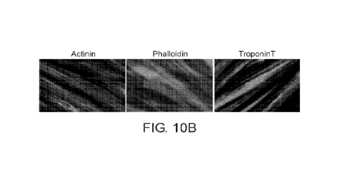
FIGs. 10A-10B show that ERKi induces a more robust induction of chicken muscle
fiber
differentiation compared to conventional DM. (10A) qRT-PCR analysis of the
gene expression of
the transcription factor narf4 and sarcomeric genes myosin heavy chains (myh
1, myh2) and
troponin (tnnt3) demonstrates significantly elevated expression following
treatment with ERKi
compared to DM. (10B) Immunoflourescent staining of ERKi treated chicken
myoblasts at 48
hours post treatment for sarcomeric proteins including alpha-actinin,
filamentous actin
(phalloidin) and troponinT demonstrating the classical striation of mature
sarcomere. No
comparison can be made to DM fibers at this timepoint as they had not yet
formed (attesting to
the early phenotype obtained by ERKi).
FIGs. 11A-11D show a quantitative analysis of ERKi impact on yield of muscle
tissue.
(11A). ERKi treated fibers cover significantly more surface area compared to
fibers induced in
DM. (11B) Evaluation of the relative mass of the muscle product at 72 hours
post-treatment with
111M SCH772984 (ERKi) compared to DM. Briefly, identical number of cells were
treated with
either condition. Following 72 hours, tissue culture plates were scraped and
cells were collected
and centrifuged. Wet weight of the pellet was measured. ERKi
treatment results in
approximately 40% increase in product mass at 72 hours post treatment. (11C)
The number of
starting cells needed to reach a final product of 1 kilogram at 72 hours post
treatment with ERKi
or DM was determined based ion the cell pellet data from 11A. (11D) The
relative protein yield
of the product of ERKi or DM treatment was determined at 72 hours post-
treatment,
demonstrating that ERKi induced myogenesis results in 4-fold increase in total
protein yield.
FIG. 12 shows a conserved phenotype achieved upon ERKi treatment in bovine
myoblasts
compared to conventional differentiation medium. Immunoflourescence images and
quantification of fusion index for bovine derived myoblasts following 72 hours
of treatment in
proliferation medium (PM), Differentiation medium (DM) or treatment with 0.5
uM SCH 772984
(ERKi). ERKi results in nearly 8-fold increase in fusion compared to DM.
FIG. 13 demonstrates that ERKi induced bovine myotubes show earlier maturation
compared to those derived by treatment with DM. Shown is immunofluorescence
staining of the
CA 03216903 2023- 10- 26
WO 2022/234586
PCT/IL2022/050474
18
sarcomeric components of myosin heavy chain (MyHC), alpha-actinin, and
Tropoinin T at 96
hours post treatment either with proliferation media (PM), differentiation
media (DM), or with
luM SCH 772984 (ERKi). Despite the presence of myotubes under treatment with
DM at 96
hours, ERKi induced myotubes have significantly higher levels of these
sarcomeric markers as
demonstrated by quantification of the relative intensity of the fluorescent
signal.
FIGs. 14A and 14B are a series of images and graphs showing the induction of
robust
myoblast fusion by multiple ERK inhibitors. Representative images (Figure 14A)
and fusion
indexes (Figure 1413) of primary bovine myoblasts treated with ERK inhibitors
SCH772984,
AZD0364, BVD523, DEL22379, FR180204, GDC0994, K0947, and LY3214996 (all at
luM)
in proliferation media show similar levels of myoblast differentiation and
fusion for all the ERK
inhibitors. Samples were fixed at 72 hours after treatment and immunostained
for sarcomeric
alpha-actinin (red) and nuclei were stained with DAPI (cyan). Error bars
represent SEM. Scale
bars are 100um.
FIGs. 15A and 15B are a series of images and graphs showing the effect of
calcium
ionophores on ERK-inhibitor-induced myoblast fusion. Representative images
(Figure 15A) and
fusion indexes (Figure 15B) of primary chicken myoblasts treated either with
ERK inhibitor
alone (SCH772984 luM, SCH) or in combination with various calcium ionophores
(Ionomycin-
2uM, and Calcymicin-luM, and Calcium ionophore I-2uM) in proliferation media
demonstrate
the synergy of combined ERK inhibitor and calcium ionophore administration.
Samples were
fixed at 48 hours after treatment and immunostained for Myosin heavy chain
(MF20, red) and
nuclei were stained with DAPI (cyan). Error bars represent SEM. Scale bars are
100um.
FIGs. 16A and 16B are a series of images and graphs showing the effect of
Retinoid X
receptor (RXR)/Ryanodinc (RAR) agonists on ERK-inhibitor-induced myoblast
fusion.
Representative images (Figure 16A) and fusion indexes (Figure 1611) of primary
chicken
myoblasts treated either with ERK inhibitor alone (SCH772984 luM, SCH) or in
combination
with various RXR/RYR agonists (9-cis retinoic acid, 9-cis RA-200nM, AM80-
200nM, AM580-
100nM, and CH55-200nM. TTNPB 200nM, and Fenretinide 200nM) in proliferation
media
demonstrate the synergy of combined ERK inhibitor and RXR/RYR agonist
administration.
Samples were fixed at 48 hours after treatment and immunostained for Myosin
heavy chain
(MF20, red) and nuclei were stained with DAPI (cyan). Error bars represent
SEM. Scale bars are
100um.
FIGs. 17A and 17B are a series of images and graphs showing the effect of
Ryanodine
(RYR) agonists on ERK-inhibitor-induced myoblast fusion. Representative images
(Figure
17A) and fusion indexes (Figure 17B) of primary chicken myoblasts treated
either with ERK
CA 03216903 2023- 10- 26
WO 2022/234586
PCT/IL2022/050474
19
inhibitor alone (SCH772984 luM, SCH) or in combination with various RYR
agonists (Caffeine
-2mM, and Suramin-10 M) in proliferation media demonstrate the synergy of
combined ERK
inhibitor and RYR agonist administration. Samples were fixed at 48 hours after
treatment and
immunostained for Myosin heavy chain (MF20, red) and nuclei were stained with
DAPI (cyan).
Error bars represent SEM. Scale bars are 100um.
FIGs. 18A and 18B are a series of images and graphs showing the superior
effect of ERK
inhibition compared to MEK inhibition on myoblast fusion phenotype.
Representative images
(Figure 18A) and fusion indexes (Figure 18B) of primary chicken myoblasts
treated either with
ERK inhibitor alone (SCH772984 1 or 10uM) compared to myoblasts treated with
MEK
inhibitor (U0126 1 or 10uM) in either proliferation medium (PM) or
differentiation medium
(DM) demonstrate the superior myoblast fusion achieved by ERK inhibition, in
particular in the
proliferation medium (PM). Samples were fixed at 48 hours after treatment and
immunostained
for Myosin heavy chain (MF20, red) and nuclei were stained with DAPI (cyan).
Error bars
represent SEM. Scale bars are 100um.
DESCRIPTION OF SPECIFIC EMBODIMENTS OF THE INVENTION
The present invention, in some embodiments thereof, relates to methods for
differentiating myogenic progenitor cells and, more particularly, but not
exclusively, to cultured
meat and cultured meat products.
Before explaining at least one embodiment of the invention in detail, it is to
be understood
that the invention is not necessarily limited in its application to the
details set forth in the
following description or exemplified by the Examples. The invention is capable
of other
embodiments or of being practiced or carried out in various ways.
Current methods for culturing muscle cells for producing cultured meat (e.g.
"in-vitro
meat", "lab meat", "laboratory meat") require a lengthy (up to 14 days for
bovine species)
differentiation step for myotube induction from expanded muscle
stem/progenitor cells,
increasing production cost and duration. The present inventors have uncovered
methods for
significantly enhancing the degree and rate of myoblast-multinucleate myotube
transition,
increasing efficiency and reducing cost of cultured meat production.
The present inventors have shown that cultured myogenic precursors can be
induced to
form large multinucleated myotubes by inhibition or reduction of ERK1/2 (see,
for example Figs.
1A, 1B), and that myogenic precursor-myotube transition, and asymmetrical
fusion is associated
with increased intracellular Ca 2+ (see, for example, Figs. 3E and 3F).
Further, the present
inventors have shown that enhancement of myoblast differentiation and fusion
can be achieved
CA 03216903 2023- 10- 26
WO 2022/234586
PCT/IL2022/050474
with a variety of ERK inhibitors (Example 10), and that manipulation of
factors downstream of
ERK1/2, by Calcium ionophores (Example 11), RXR/RAR agonists (Example 12) and
by RYR
agonists (Example 13) can effectively augment the potency of ERK inhibition.
The present inventors demonstrate the superiority of ERK inhibition (ERKi)
compared to
5 conventional methods (referred to herein as "DM" in some embodiments of
the invention) for
the purposes of cultured meat. Specifically. as demonstrated on chicken
myogenesis in tissue
culture: ERKi strengthens the differentiation transcriptional program leading
to earlier myotube
initiation; ERKi enhances fusion leading to significantly larger myotubcs; and
ERKi enhances
the maturation of myofibers through increased expression of maturation
markers, leading to
10 earlier formation of sarcomeric structures (see, for example, Example
7). Moreover, the present
inventors demonstrate that the effect is conserved and evident in at least 2
more additional
species, bovine and ovine. Similarly, data from bovine myoblasts demonstrates
that ERKi
induced fibers reach maturation faster than those achieved with DM. Taken
together, the earlier
differentiation and more robust fusion achieved by myoblast treatment with
ERKi results in
15 earlier maturation of myotubes ultimately contributing to increased
production efficiency of
cultured meat by increasing the of total mass of the meat product, area
coverage, and finally
increase in total protein yield.
Thus, in some embodiments, there is provided a method of inducing
multinucleated
myotube formation, the method comprising contacting myogenic precursor cells
from a farmed
20 animal with an Extracellular Regulated Signaling Kinase (ERK1/2) inhibitor
and/or an
upregulator of intracellular Ca 2+.
In other embodiments, there is provided a method of inducing multinucleated
myotube
formation, the method comprising contacting myogenic precursor cells from a
farmed animal
with an Extracellular Regulated Signaling Kinase (ERK1/2) inhibitor and/or an
upregulator of
intracellular Ca 2+, wherein when the myogenic precursor cells are of chicken
the contacting is
performed in the presence of Extracellular Regulated Signaling Kinase (ERK1/2)
inhibitor and an
upregulator of intracellular Ca 2+.
As used herein, the term "myogenic precursor" or "myogenic precursor cell"
refers to any
cell which can differentiate into a muscle cell. Myogenic precursors are
critical for muscle
regeneration. Although the most naturally abundant animal myogenic precursors
are the satellite
cells, which are found on the plasmalemmal surface of the muscle fiber, other
cells with
myogenic potential have been identified and may be suitable for use with the
methods of the
invention. These include mesodermally derived myoblasts, interstitially
located muscle side
population (mSP) cells, muscle derived stem cells (MDSC) and myo-endothelial
cells from
CA 03216903 2023- 10- 26
WO 2022/234586
PCT/IL2022/050474
21
endothelial-associated myofibers, mesodermal pericytes and mesoangioblasts and
mesodermal
CD133+ progenitors.
The different myogenic precursor cells may be characterized by cellular marker
profiles,
for example, MyoD+ and Desmin+ for myoblasts, CD34 +/-, Ckit- and CD45- for
mSPs, CD56+
and CD29+ for muscle precursors, CD133+ and CD34 +/- for CD133+ mesodermal
progenitors.
As used herein, the term "multinucleated myotube" refers to fused myogenic
precursors
(e.g. fused myoblasts) having 3 or more nuclei. Mono- or bi- nucleated
myogenic precursors,
even if expressing myogenic differentiation markers, arc not considered -
multinucleated
myotubes".
As used herein, the term "multinucleated myotube" is equivalent to the terms
"multinucleated myoblast", "multinucleated muscle fibers", "multinucleate
muscle fibers",
"multinucleated syncitia", "multinucleate syncitia", "multinucleated muscle
syncitium",
-multinucleate muscle syncitium", "multinucleated muscle syncitiuin",
"multinucleate muscle
syncitium", and may be used interchangeably herein.
In some embodiments, the multinucleated myotubes have in the range of 4-
10,000, 10-
8,000, 20-500, 15-250, 50-1000, 100-800, 60-2000, 70-4000, 80-6000, 90-5000
nuclei per
myotube. In specific embodiments, the multinucleated myotubes have between 10
and 100
between 10 and 500, or between 10 and 1000 nuclei. Thus, in some embodiments,
the
multinucleated myotubes comprise at least 3 nuclei, at least 10 nuclei, at
least 50 nuclei or at least
100 nuclei.
Cell nuclei can be identified and quantified by a number of techniques,
including, but not
limited to immunofluorescence, flow cytometry and immunohistological
techniques. Common
nuclear stains include DAPI (fluorescent), hematoxylin (cytological stain),
Hoechst 33258 and
33342 (fluorescent), methyl blue (cytological stain). safranin (cytological).
In specific
embodiments, the nuclei are labelled with either Hoechst 3342 (Thermo-Fisher)
or DAN
(Sigma), and visualized by fluorescent microscopy. In some embodiments,
multinucleated
myotube formation is quantified by stratification of the cells into mono- and
bi nucleated cells as
opposed to the multinucleated myotubes with four (3) or more nuclei.
In addition to developing multiple nuclei, myogenic precursor cells induced to
form
multinucleated myotubes enlarge by fusion with differentiating myogenic cells.
While reducing
the invention to practice, the present inventors have shown that the myogenic
precursor-myotube
formation includes -asymmetric fusion", that is, rather than enhanced fusion
of myoblast to
myoblast (-primary fusion"), fusion according to the methods of the present
invention is
predominately fusion of myoblast-to-myotube fusion ("secondary fusion",
"asymmetric fusion").
CA 03216903 2023- 10- 26
WO 2022/234586
PCT/IL2022/050474
22
Thus, according to some embodiments of the invention, multinucleated myotube
formation
comprises mononucleated myoblast-myotube fusion and/or expansion of bi-and tri-
nucleated
myotubes into large multinucleated fibers.
Additionally, in some embodiments, the myogenic precursor cells can be
embryonic stem
cells (ESCs, totipotent cells) and Induced Pluripotent Stem Cells (iPSCs).
iPSCs can be created
by from adult fibroblasts by induced expression of reprogramming factors. have
limitless
replicative capacity in vitro and can differentiate into myoblast-like cells
(see, for example, Roca
et al, J. Clin. Med 2015).
The phrase "embryonic stem cells" refers to embryonic cells which are capable
of
differentiating into cells of all three embryonic germ layers (i.e., endoderm,
ectoderm and
mesoderm), or remaining in an undifferentiated state. The phrase "embryonic
stem cells" may
comprise cells which are obtained from the embryonic tissue formed after
gestation (e.g.,
blastocyst) before implantation of the embryo (i.e., a pre-implantation
blastocyst), extended
blastocyst cells (EBCs) which are obtained from a post-implantation/pre-
gastrulation stage
blastocyst (see W02006/040763), embryonic germ (EG) cells which are obtained
from the
genital tissue of a fetus, and cells originating from an unfertilized ova
which are stimulated by
parthenogenesis (parthenotes).
Induced pluripotent stem cells (iPS; embryonic-like stem cells), are cells
obtained by de-
differentiation of adult somatic cells which are endowed with pluripotency
(i.e., being capable of
differentiating into the three embryonic germ cell layers, i.e., endoderm,
ectoderm and
mesoderm). According to some embodiments of the invention, such cells are
obtained from a
differentiated tissue (e.g., a somatic tissue such as skin) and undergo de-
differentiation by genetic
manipulation which re-program the cell to acquire embryonic stem cells
characteristics.
In some embodiments, the myogenic precursor cells can be induced muscle
progenitor
cells obtained by transdifferentiation of non-muscle tissue (e.g. fibroblasts)
directly into muscle
progenitors by manipulation of small molecules in the medium, and/or forced
expression of
MyoD in the non-muscle cells. US Patent Application No. 2019/061731 to
Hochedlinger et al
discloses methods for producing induced muscle progenitor cells (iMPCs) having
a satellite cell
phenotype from fibroblasts, without passage through the iPS cell stage. Bin Xu
et al (Nature
Research, Scientific Reports DOI: 10.1038/s41598-020-78987-8, 2020) discloses
transdifferentiation of fibroblasts by forced induction of MyoD.
As used herein,
"transdifferentiation" refers to a process in which a somatic cell transforms
into another somatic
cell without undergoing an intermediate pluripotent state or progenitor cell
type.
CA 03216903 2023- 10- 26
WO 2022/234586
PCT/IL2022/050474
23
The phrase "adult stem cells" (also called "tissue stem cells" or a stem cell
from a somatic
tissue) refers to any stem cell derived from a somatic tissue [of either a
postnatal or prenatal
animal (especially the human)]. The adult stem cell is generally thought to be
a multipotent stem
cell, capable of differentiation into multiple cell types. Adult stem cells
can be derived from any
adult, neonatal or fetal tissue such as adipose tissue, skin, kidney, liver,
prostate, pancreas,
intestine, bone marrow and placenta.
Hematopoietic stem cells, which may also be referred to as adult tissue stem
cells, include
stem cells obtained from blood or bone marrow tissue of an individual at any
age or from cord
blood of a newborn individual. Placental and cord blood stem cells may also be
referred to as
"young stem cells".
Mesenchymal stem cells are multipotent strom al cells that can differentiate
into a variety
of cell types, including osteoblasts (bone cells), chondrocytes (cartilage
cells), myocytes (muscle
cells) and adipocytes (fat cells which give rise to marrow adipose tissue).
The term encompasses
multipotent cells derived from the marrow as well as other non-marrow tissues,
such as placenta,
umbilical cord blood, adipose tissue, adult muscle, corneal stroma or the
dental pulp of deciduous
baby teeth. The cells do not have the capacity to reconstitute an entire
organ.
The myogenic precursor cells can be freshly isolated cells, cells cultured in
primary
culture from live tissue, or cells of isolated myogenic cell lines developed
from repeated serial
passages of primary muscle cells. Exemplary animal cell lines suitable for
foods containing
cultured animal cells are disclosed US Patent Application Publication
2021/037870 to Kreiger, et
al. In some embodiments, the myogenic precursor cells can be genetically
modified, for
example, for enhanced proliferation or for expression of tissue-specific
factors (see, for example,
US Patent Application Publication 2020/0140821 to Elfenbein et al).
According to some embodiments of the invention, when taken freshly from a
tissue
biopsy or a primary culture, an initial stage of enrichment for myoblasts is
performed.
Specifically, the cells are cultured on non-coated dishes which allow for
preferential adherence of
fibroblasts. Myoblasts which predominantly remain in the suspension are
collected and plated
again so as to remove the fibroblasts and obtain an enriched culture of
myoblasts. This process is
termed "preplating". The process may be repeated as needed (e.g., 2-4 times).
The presence of
fibroblasts on the dish can be monitored by microscopy.
Thus, in some embodiments, the myogenic precursor cells are selected from the
group
consisting of myoblasts, satellite cells, muscle side population (mSP) cells,
muscle-derived stem
cells (MDSCs), mesenchymal stem cells (MSCs), muscle-derived pericytes,
embryonic stem cells
(ESCs) and Induced Pluripotent Stem cells (iPSCs).
CA 03216903 2023- 10- 26
WO 2022/234586
PCT/IL2022/050474
24
Recent reports have shown the establishment of stem-cell lines from
domesticated
ungulate animals e.g. (Challenges and prospects for the establishment of
embryonic stem cell
lines of domesticated ungulates. Anim Reprod Sci. 2007; 98(1-2):147-168. doi:
10.1016/j.anireprosci.2006.10.009., which is hereby incorporated by
reference). Bach et al.
(Engineering of muscle tissue. Clin Plast Surg. 2003; 30(4):589-599. doi:
10.1016/S0094-
1298(03)00077-4.) suggested myosatellite cells as the preferred source of
primary myoblasts
because they recapitulate myogenesis more closely than immortal myogenic cell
lines.
Myosatellite cells have been isolated and characterized from the skeletal
muscle tissue of cattle
(Dodson et al. Optimization of bovine satellite cell derived myotube formation
in vitro. Tissue
Cell. 1987; 19(2):159-166. doi: 10.1016/0040-8166(87)90001-2.), chicken
(Yablonka-Reuveni et
al. Dev Biol. 1987; 119(1):252-259. doi: 10.1016/0012-1606(87)90226-0.), fish
(Powell et al.
Cultivation and differentiation of satellite cells from skeletal muscle of the
rainbow trout Salmo
gairdneri. J Exp Zool. 1989; 250(3):333-338), lambs (Dodson et al. Isolation
of satellite cells
from ovine skeletal muscles. J Tissue Cult Methods. 1986; 10(4):233-237. doi:
10.1007/BF01404483), pigs (Blanton Blanton et al. Isolation of two populations
of myoblasts
from porcine skeletal muscle. Muscle Nerve. 1999; 22(1):43-50. doi:
10.1002/(SICI)1097-
4598(199901)22:1, Wilschut et al. Isolation and characterization of porcine
adult muscle-derived
progenitor cells. J Cell Biochem. 2008; 105(5):1228-1239.), and turkeys
(McFarland et al.
Proliferation of the turkey myogenic satellite cell in a serum-free medium.
Comp Biochem
Physiol. 1991; 99(1-2):163-167. doi: 10.1016/0300-9629(91)90252-8). Porcine
muscle progenitor
cells have the potential for multilineage differentiation into adipogenic.
osteogenic and
chondrogenic lineages, which may play a role in the development of co-cultures
(Wilschut et al.
2008, supra).
Alternatively, as mentioned, adult stem cells from farmed animal species can
be used.
For instance, myosatellite cells are an adult stem-cell type with multilineage
potential (Asakura et
al. Differentiation. 2001; 68(4-5):245-253. doi: 10.1046/j.1432-
0436.2001.680412). These cells
also have the capacity to differentiate into skeletal muscle cells. A rare
population of multipotent
cells found in adipose tissue known as adipose tissue-derived adult stem cells
(ADSCs) is another
relevant cell type for in vitro meat production (Gimble et al. Adipose-derived
stem cells for
regenerative medicine. Circ Res. 2007; 100(9):1249-1260. doi:
10.1161/01.RES.0000265074.83288.09) which can be obtained from subcutaneous
fat and
subsequently transdifferentiated to myogenic, osteogenic, chondrogenic or
adipogenic cell
lineages (Kim et al. Muscle regeneration by adipose tissue-derived adult stem
cells attached to
CA 03216903 2023- 10- 26
WO 2022/234586
PCT/IL2022/050474
injectable PLGA spheres. Biochem Biophys Res Commun. 2006; 348(2):386-392.
doi:
10.1016/j.bbrc.2006.07.063).
Matsumoto et al. (J Cell Physiol. 2007; 215(1):210-222.) reported that mature
adipocytes
can be dedifferentiated in vitro into a multipotent preadipocyte cell line
known as dedifferentiated
5 fat (DFAT) cells, reversion of a terminally differentiated cell into a
multipotent cell type. These
DFAT cells are capable of being transdifferentiated into skeletal myocytes
(Kazama et al. Mature
adipocyte-derived dedifferentiated fat cells can transdifferentiate into
skeletal myocytes in vitro.
Biochem Biophys Res Commun. 2008; 377(3):780-785. doi:
10.1016/j.bbrc.2008.10.046) and arc
an attractive alternative to the use of stem cells.
10 In specific embodiments, the myogenic precursors are myoblasts.
Myogenic precursors may be characterized by levels of expression of certain
cellular
markers, such as, but not limited to ATP binding cassette transporter G2
(ABCG2),
MCadherin/Cadherin15, Caveolin-1, CD34, FoxKl, Integrin a1pha7, Integrin alpha
7 beta 1,
MYF-5, MyoD (MYF3), Myogenin (MYF4), neural cell adhesion molecule 1 [NCAM1
(CD56)],
15 CD82, CD318 Pax3 and Pax7. In some embodiments, the myogenic precursor
cells are cells
expressing significant levels of at least one of MyoD, Pax3 and Pax7, or
corresponding, species-
appropriate orthologs thereof. In other specific embodiments, the myogenic
precursor cells
express MyoD and at least one of Pax3 and Pax7, or corresponding, species-
appropriate orthologs
thereof. In particular embodiments, the myogenic precursor cells express all
of MyoD, Pax3 and
20 Pax7 or corresponding, species-appropriate orthologs thereof.
Once the myogenic precursor cells are obtained, they can be grown in culture
to expand
their mass, then form multinucleated myotubes, which can be later be formed
into a cultured meat
composition. Culturing the cells includes providing a culture system,
transferring basal medium
or basal medium supplemented with serum, serum-replacement and/or growth
factors and other
25 components as might be needed for the efficient growth of cells, into
culturing vessels, adding
cells and culturing the cells. The basal medium (e.g. Dulbecco's Modified
Eagle Medium;
DMEM) may include water, salts, vitamins, minerals, amino acids and a carbon
source such as
glucose. In some embodiments of the invention, the basal medium includes
animal-derived
growth factors. In other embodiments, the basal medium includes non-animal-
derived growth
factors.
In some embodiments of the invention, the basal medium includes an animal
derived
serum. In other embodiment of the invention, the basal medium of the current
invention does not
include animal derived serum such as fetal bovine serum, calf serum or horse
serum. As used
herein, by "does not include animal serum" or "animal serum-free" is meant
that the medium
CA 03216903 2023- 10- 26
WO 2022/234586
PCT/IL2022/050474
26
contains less than about 1% or less than about 0.5% or less than about 0.1% or
less than about
0.01% or zero animal derived serum by total weight of the medium. It is
envisioned within some
embodiments of the invention that a serum-free medium may contain growth
factors and other
substances, but nothing derived from an animal.
According to some embodiments, culturing is effected in the presence of serum
at a level
which is not considered starvation conditions that prevent cell proliferation.
For example, above
2 % serum (e.g., 3-25 %). According to some embodiments, the conditions
comprise 5-25 %, 10-
25 % scrum e.g., 15-25 % scrum, about 20 % serum. Such conditions are provided
in the
Examples section which follows. Thus, according to an embodiment, the medium
is B10-
AMFTm-2 medium (e.g., available from Biological Industries), which comprises a
basal medium
supplemented with fetal calf serum (FCS), steroids, basic fibroblast growth
factor, insulin,
glutamine, and antibiotics.
According to some embodiments, culture of myogenic precursors or progenitors,
and
culture of multinucleated myotubes is effected in medium having ingredients
and components
which are Generally Regarded As Safe (GRAS) and/or "xeno-free". In some
embodiments, the
medium comprises ingredients and/or components certified GRAS and or xeno-
free. In other
embodiments, the medium comprises ingredients and/or components certified GRAS
and xeno-
free. In other embodiments, the medium consists of ingredients and/or
components certified
GRAS and/or xeno-free. In still other embodiments, the medium consists of
ingredients and/or
components certified GRAS and xeno-free.
A list of media components used in meat production along with their worst-case
exposure
estimates and relevant authoritative limits or published toxicological/safety
data supporting their
use is presented in Table 1.
Table I: Exemplary List of Cell Culture Media Ingredients Used During Meat
Production,
Risk Categorization, and Safety Information
maArkasm
See Re-fere twei
MFAt Am:5547.merft tVkiliat
Clatt etsretp6tIftd CAS C
few ktwalml DItzUry
at:ego/if Expestirezp.r
Ãntaikele,gõULOSO
g swving"
,
N/A*
LAR,G1311NE-HCL _jL NIA*
Arnim, Adds
21}57-94,1,3-4S 2 MA' NIA.4`
Aci 5:(i-44-43 ,
N/A"
NM.*
CA 03216903 2023- 10- 26
WO 2022/234586
PCT/IL2022/050474
27
rag.;oertmor
Soft ftteferersot Lektoi
MsIr Assesment Estkoated
tliog Cesrespzsurpd tA5 0
fa,r HUSTRAII Dt.qtairy
Cateigtrox Exposure per MO
kr-N*4r io...g,..,.Llt, OM.)
g sowing**
. , t-
A148-N, 06 2 a: IA' U:L ,-,
NIA'
.......... ....... ..... .. ......... . ... .. .........
. ...... ............ . ... .......... .. ... .......... ..
.
CYST14 E 21-1C.L. --3 a 2 WA' 0_
t.trArA,:r.; ACID 61.f.1.6-N-0."i. 2 Ni'A LI i
-.zIONC*7..3.1 Li tv.1
=?.
..,mocoomemenomoomi
,r. N/A*
E -HC L.- 511134.1-2'W.:1 -.
..t. NA' LiL
'') 0
'...4-5=0 ..F.:i.),CINE 73-37-i 2 NIA' U.
-------------------------------------
2
-1 1 -.
2
63-6g.-2 2 iNiA* u,,, NIA
YLALAM N 9.1,2 2 ______ IMIA' UL -. NIA'
-
_______________________________________________________________________________
__
.1,PR-ouN., 14745- a 2 . ti/A ' UL -,-
NIA'
=.,--sii- P, IN E 5-E,- 4.'5-1. ....-1
N/A' :.1 Z. -,--. N/A.'
.................................................................. ,
_____________
L -I H RE.ONME. 72.1 ',..5. 2 . NIA" U. NIA'
1 r ,
RNFTOP -=AN 73,22,3 2
-
_______________________________________________________________________________
__
---I'VRt.',35gNE-2.14 A- 112:666-874.t a NFA" U:..'õ -,- NIA
2H 20
------------1-4 ',1=J NA' V: =--
NIA*
.----------i
Cabor$ sowNK '''=,C1C..,-k: 110-1- 6 2 21).0
n't 1 zokes for a 65 kg
N,3-3t15Lrate.s adcAt
far) 257-.-frAti
'r.-44.9...wo NeltiEL Unlirn 2
Itki.is3- Kai:
CA 03216903 2023- 10- 26
WO 2022/234586
PCT/IL2022/050474
28
.-1 ____________________________________________ .
, Ma*Imurn =
.=
. ,
Szife nttemott Leve:i :
' ,= Rh* Ame ,nivrst EVtili:AtecE
Cl.aAA = Compound 1
CAS.* -fur s-tu man Dteretv :
Categoq btpostire per He
, MU:14e ge4, IA, OSL) :
,= g EOM p-kg.-*
,
. so- L-.,,i., um 1 F.-f: I 21.4 2
41 mg ftlg.akt.1 for E6 kg eth..z1L-:
Ss:._ICCSNATE-W.Ne i .
:7gv -113.M1 kikl be,'Jmot, i
NDAEL trorrt :2 yeas- tat :
,=
,
..1 3m1v,
,
, 1
: SOLV.A4 PYRINATE 1113-24-E: 3 I
lo.,1.0k=::.: .1.1,4th:;:,3 .
, , the. r.i-:,:sv..t of OSL
...,
,= vowed mcoomed irs ,
, ,=
, no mail inmeskgation
.=
.,
.=
...
,=
. .1
' ,=
,= = .= , =
.=
..,
, .
,= = ,= = = . ,=
=
' ..,
. ..,
, , .
' ..,
,=
= ,
,=
,
. ,
=
. ,=
.
,= =
. .=
=
.=
.=
.-1
,
'
' Et3EX ACID .59-,EE)-.g 1 CiO. rkg
I... = t;-.10 to IMO ug Id
, i',0c.i0g,r1;n7.it not
.=
,
,g,':',,i=Ji;:hett a*A4
to:xidw, iNg to prell
.,
,
..," nfig.lpfe wito don't
,=
,
' ..." know ti=-
g2. Pk4ve 6
. ,=
,
= .=
, vitamin a EsfAcieozy i
..,
,
. ,
,= fn.-on consuming MD .
. .-1
CsS,-,n LP HA- i'.;769!:i=-91-Z 1 g. rn
UL= IMO en gjil = :
. = V5ti'arnim
:TeCf-Vi-lEROL. :. = AC_ TA ..,
, = =
.= = ..=
,=
,= .
.=
. ,
. ,
= ,= .
.=
,=
.=
.=
,= ,
. .
: .=
, =
, = .= .
.., :
,
..,
,=
, ,=
J. 2.4 ug ui...,.
I00 wid
ERGOCALartROL.:,} 1. :
:
,. =
,=
, ,
,= ]
_
CA 03216903 2023- 10- 26
WO 2022/234586
PCT/IL2022/050474
29
...
_______________________________________________________________________________
__
Z 1 Maximum
'; AUR Aut5547tient Evamated
Safe Refeillket :Level
CLase Conepouraa0 CAS 0 :. 'tor
Rusnail Dietaty
Z.' Ca teigun Expaseme per Ma-
1 z..
=q Otaikie te4ov LI1L, OA)
Z "4, g eamlifte*
, a ¨
..s; ....
D-ALP6A- 'IS-4:12-g, '1- 1 6 -&,y,... UL
=,--- ItXX) .rneiti
T-000P11 EL ..;
:.
1
1 Z;
1 1
1 1
1
:.
,1 Z
1
Z
=4
-1 - = . _ _
Z
I , 4 rr.g. 0 L -z
NIA.
1 1
1 ..;
....
1 "4
1 1
Z. .4
:
1 k .
.'.. .
MYL..-kl,P4LJSZIOL. ;L'Af -:A3-.L.,1 11 4.3u
rIti, oc-L-Azeo'l
1 :
1 1
1
&: .
WACENAMME ;'3,:ai,',2.0 ;1 25 mg
ti L ID to 35 ,.-alg,
k
0110-)TNAMIDE) 1 1
,ist.vplemeRts)
1 =:i
k
1
1 i.-
: ,
fs'f'RLEsOxi:n.F.-HcL 1s8:56--o 1 25 rf.4,1, :..IL
,, 50 to atx) mg
1 1
:.
1 Z
1 :..
:.
1 Z
1
1 ..=
1 Z:
:.
Z Z
0 L ,:, 50 Loll:Xi cog
1 Z
1
;..
Z
..'
=1
Z
?õ
CA 03216903 2023- 10- 26
WO 2022/234586
PCT/IL2022/050474
Maxtruurn
Saft!. illefervoce Level
AWse-woRm E:stkFtatt
Cliass Compound CAS* for
Human: Dietary
Cat:tem-if Exposure pee 16:Ek
Unake (o4,L OR)
g M!?roimg"
F.U80:NJVA 183-8Et-5
I
THIAWNEFICL &)3- JLNS
VITAM EN -312 E'A,14-41 2 mg kiLL=
NS
KYAMIXOS:A.IAM
LCRM
....... .
D- {370 1
PANTOTHEKAIE
=.`=
CH LON 7- 4-1. 5:2 mg 3,s
ed
CA 03216903 2023- 10- 26
WO 2022/234586
PCT/IL2022/050474
31
rkaaitramt
Refemoce toed'
sk es sment Est r,stated
Class Coertptm.mi CAS 4 fur
Rianan D64eary
CategaNty Exp;surg 1C10
Mtaim
OS.11
gi.entirkg!"
2
-..40=It to ;MOO ;
zismt-t*
COM4rItrntiont
kr
gr:-.irss'
teS :ON:1
concentrations
by en rdbr of
enO are,
expescreil
pre4ent
levels mil bolow
WI-
,
SO CALP41 2647-14-S 2.3 ed
luteta
,
(ADM UM I2'(.J te 3
gjki
Cn LORI DE CAN
,MANCIAN ESE 10034-96-5 1 3 uzg 2 L.
IIFsIld
SUS-Ark H,20
FOTAS.SUM 7447-40-7 4 ;',=1iiL: MS
CnUDRIrpf
MAGNK11 ,JM. 1. ir66
:4=50 erig
Cn LORI DE (011,1i-ril
C PPE:9 SULFAT:E- 3 1 te
=
SO MUM 13E124-3 1 1.6
ItO.t4eis tor a Ci6
METAStLICATE,- ad,*
ee3iiXi feW
.'3F1.20
rat. N'LlikEl
CA 03216903 2023- 10- 26
WO 2022/234586
PCT/IL2022/050474
32
1 kiLaxItrwm
1 . .ffl.s* Ames'snie.nt
Egtmated Safe 8*-fefstmt tAVki
Cass tostvataid ; CAS:RI
for i4tAinan: Dktaty
.L! Ca:V2g6.-ie
Ekprzits...ir8. ptr EDO
Make fcg, EL11.õ0-SL).
... g mrsAtg"
.U..ttM:M. SE L.t:WTE 1 WW2, t g,..8 = 2 1) ag h
''.'&-= ibr 65 k;g adi.tit
, -
õ
(WM = 4r0
13.3-fplo tAWeleg 13
,
....
1 =
k Ne.110M1
, .
:IMICISULP:ATE-7KAI74)1:6-2.0-0. :1 13
1
I '
1
1
1
1
I =
1
I
1 '
i.
1
NIAGNESiUM ='N;137,-.M4'-'3 '1 49.3
il=-'3-1.g: N:a UL
SULEATEIAIV-irt õw-
omes; VA, --tt 65 to axi
1 =
.
skipp.W.nctrilli.
= .., ¨il = .
URRK:NMATE- ;77-$244.--Ã a i.-
3.,a mg .rio!: u,,,, q) 3::1' 45
53i-rn 1 Witatv:
AD1 =11.7
=:';
. = ,,.:--
:gh:g baviday Kbvak
1
1 olf
iic.lw$: wxlnitralf? am
==1 = P.
rd:,,, of. nv.gritwie
'.i .
b:r.ouv Ote W. and ADi)
1 :
1 =
1 =
P.E PAWS SULFATE- 11h:1243,0
1
1 .
1
.1
1 :
1
1 '
:;.; =
i=
1 =
CA 03216903 2023- 10- 26
WO 2022/234586
PCT/IL2022/050474
33
1 MAKIrrium
1 . fa.ak amies'salailt
ER'irear ell S4fe RefefitixelAVO
Cltass Corrpiund CAS :N
for .t4tirriart Oktaty ,
Ciitagow Ekpom..ira ogr EDO
i
I Mtake
fa,g, ELIEL,..0-St.).
g mrvIrtg"
1
i
1 .
1
i: =
Vnasent in a
rei,i,idue of wed-
itt.-s:ir.i "i4.iales-op arktii
1
]
rt-tn t.i.ae4 aa
i: rw.triert worm of
t; i rµr.i el
i
..r;.--..R.Ric ;CITRATE 1I.2.,:i_,D-7
a'r..;:in, UiL ,41.;0 M 45 r.;:% .
. 1
1 .
1
1
1 .
1 CPreser t al; a
'2 residi.N.; .0 sfaed-
il izrain I=e-kip
amii
E . o.t.A iliwil &la
ffP,R47, En,zi;lier4
,1
AMMONIUM
Ca:1MTIron,
4.5 ri-w
. .. .
1.:,.-);,,ci:rv9 TransfRrrin 11 li.t))16,,,17.1:1 '3 1.,5i)
rrq:,i N la
i.-;aili Carrier
t
...,. l'.
SO m.0 k.1 tilo.44.-.21.-:s . t
]
al-I,OSPHATE il
t'
MON01:1-kia', fl,20 .1
ii '
=
t;TA:)I;at.i.foli 17;al--"4-4 zl 43g NR
Piii;OSP4ATE il
ii
...
'ON0';.EC ACED- I -60-334 ' 2 1 p--.,g;:
,,'?E,. ..- 7 -a'E,1:2: glil
ELipats
MONF.FarmAm.mA 114143- 2 a6o imkI
Dilnitil, EnE:i4-Eer_.-EiE. tor W,':.
",=-_'-ti--1:.g2ElifteiE, MINE =1
4-E!. -,;Ecluit =Arr 12.8 fad
ii
=:'
= 0,;;,1 r'aft NI:MEI, i
-- -- - -- -- -- ,.. .
- P;iV'N 'W 19em-k.S--6 2 4 rrq., AN =:--
10 n-laja ";ald
...111'zifier 1 :
i
.=.;
I,- M,LiTAP:-t ION E ITO-la-a :2
1
i
1
'=. =
An kiaddant. '1 '
i
1 .
.
=
t:
i.= ._
CA 03216903 2023- 10- 26
WO 2022/234586
PCT/IL2022/050474
34
NIA ximt/m
5Oft flefe-rente teveT
CI'mvs Compound A_Ik Amos= ent EVaroated
for Roman: Dietary
tategory Expo-sure per lel
iintakt fcg, tiii4 05-14
g 5:0MR,K,"
OiCK.EN .SERMI www:thennofi.: NR
14,v.VOLI/
hornvjafe-
tAirnarrirfl
totbsficsal
rwi
BOVNE SEfWM ,,A,,ww,-khermos 3
N3-=r-,u0nitxclerl
,rorripgiego,n. Q5mlat?.siPr&lcizAct
.11-6170CO3A1?Mi)
70060 ?5;:t$=5-t-ch
-sxp-1.617C.00
0,4 E '3ERUM N.R
ALtAJIME i=-vr.c.frwrnicyR,).1erj
ra
/110200214n 1
Ci,Igk:ED2
8YR-FP 2 YEAST R113-01-02 N
EXTRACT
F2 BROBLAST NS
GROWTH ACr'OP-
.kk5K'3
glXif -WS {PLATELET N,FA NR
r owth DERMED geAkft
FACTOR-138)
ONSULifisti K L.: NIA 3- NR
65R MTh FACTO
- -
Rs.s.pt-cj15.-$ W=.',.'"k2.6-B (Prclw:>:.
NtA
:reeqlue ssed-
tr4in
diretay
:00c.W.to cr gin
mductitql
'
CA 03216903 2023- 10- 26
WO 2022/234586 PCT/IL2022/050474
Miaxirmith
Saft Itefe-feftt tevei
Ms:1.c Asiessimegt W1trated
Class torngatrumi US*k kumN1tMtar
Calftm-10
ExtittRts.av pt.e
Intake
OSL)
g
MSR, 'I IMO-64-2 0.3 t1g. Prest;nt.
vain st:ak,up. ne
IJaly added to
meo mductton
media)
Al = Adequate Intake
.ADI = Acceptable Daily Intake
FINTB-IOM = Food and Nutrition Board of the Institute of Medicine
5 FIDT = Highest Dose Tested
N/A = Not applicable
NR = Not reported
NS = not specified as no evidence of toxicity from excess intake known
NOAEL = No Observed Adverse Effect Level
10 UL = Tolerable Upper Limit
OSL = Observed Safe Level
* No upper limit when provided as dietary protein
**Maximum dietary exposures estimated using conservative assumption of
complete transfer of
media components to the finished product on a wt/wt basis.
15 Category 1: These cell culture media components are food
ingredients/additives that are
GRAS or permitted by federal regulation without limitation on use. Exemplary
compounds in
this category include innocuous ingredients such as sugars, pH buffers, water
soluble vitamins,
and common antioxidants such as tocopherols.
Category 2: These cell culture media components are common dietary nutrients
and are
20 anticipated to have GRAS status for food use or be permitted by
regulation for addition to food.
Examples of such compounds include most of the inorganic salts and
macronutrients that are
present within the cell culture media. Where these compounds are permitted for
direct addition
to food at use levels comparable to anticipated concentrations that might
reasonably be expected
in the cell-based meat product, no safety concerns are anticipated. Majority
of nutrients present
25 within the poultry cell-based meat may be readily measured using common
validated methods
for food composition testing. Batch analyses of multiple lots of the finished
product may be
obtained to validate the above assumptions. In some instances, consideration
of established safe
levels (e.g.. AD1, UL) derived from a relevant authoritative body (e.g., U.S.
FDA. EFSA,
JECEA, FSANZ, U.S. EPA) may be leveraged to support safety. If comparisons of
anticipated
30 dietary intakes relative to an authoritative reference intake value is
used, consideration of
additive intakes from all dietary sources may be considered. In the absence of
an authoritative
reference intake value, published NOAELs from animal toxicology studies may be
used to
CA 03216903 2023- 10- 26
WO 2022/234586
PCT/IL2022/050474
36
evaluate safety using standard scientific procedures for food safety
evaluation. A margin of
exposure (MoE) of 100-fold or greater between the NOAEL and estimated dietary
intakes from
food exposures is typically considered adequate to support safety. In
situations where the MoE is
< 100-fold. additional measures for further reduction of the media component
may be necessary,
or further characterization of intraspecies/interspecies differences in
metabolism may be
necessary. These situations also require careful consideration of the
regulatory status on a case-
by-case basis (e.g., premarket approval as a food additive or GRAS evaluation
required).
Category 3: These cell culture media components have not been previously used
in food
production (e.g., no federal regulations or previous GRAS status) but with
sufficient information
to conclude that the compounds do not present risk for intended use in food
production. For
example, situations where the compound is not detectable in finished product
or is present at
equivalent levels in comparator foods, compounds that are thermo-labile and
will be digested
during cooking, and/or compounds that are expected to be digested to innocuous
compounds
following ingestion. Examples of compounds meeting the aforementioned
conditions would
include recombinant growth factors and serum components. For Category 3
substances a final
consideration in the safety assessment process may involve hazard
characterization of the
potential for a substance to produce toxic biological effects outside of the
endpoints measured in
a sub-chronic rat toxicity study. Substances with biological activity may
require additional
hazard characterization related to reproductive and developmental toxicity, or
immunotoxicity.
Considerations for allergenicity, biological effects in humans (e.g., effects
on blood pressure),
and synergistic effects with other media components also may be evaluated.
Such investigations
may preferably be evidence-based (i.e., availability of a clinical trial
demonstrating that a
substance affects blood pressure), rather than theoretical (i.e., based on
presumptive mechanisms
of action). Similar to category 2 substances, the regulatory status of
ingredients in category 3
will require case-by-case evaluation of the regulatory status of the compound
(e.g., need for
premarket approval or GRAS evaluation). Examples of category 3 components
include
recombinant proteins and animal serum.
According to some embodiments, the ingredients of the culture medium are
certified
"Generally Regarded As Safe" (GRAS) ingredients (e.g. category 1 and some of
category 2 of
Table 1). As used herein, certification of GRAS status is conferred by a
recognized regulatory
agency such as the USFDA. FDA GRAS certification can be granted (or declined)
either on the
basis of the use in food prior to 1958, or, for other substances, on the basis
of documentation of a
safety analysis conducted by the manufacturer, and reviewed by the FDA.
According to some
embodiments, the ingredients of the culture medium are certified "xeno-free"
ingredients. As
CA 03216903 2023- 10- 26
WO 2022/234586
PCT/IL2022/050474
37
used herein, the term -xeno-free" medium refers to a cell culture medium which
is devoid of
components originating from species other than those of the cultured cells. In
some
embodiments, the term "xeno-free" relates to "non-human-free", or the absence
of components
originating from species other than humans.
Cells for expansion in the cell culture may be obtained by biopsy from a live
farmed
animal, for example, from fish, pig, cows, chicken, turkey, sheep, goat and
the like.
As used herein, the term "farmed animal" refers to any animals which are grown
(cultivated) for agricultural purposes, and, in particular, for provision of
meat for consumption.
Thus, farmed animals include, but are not limited to avian species, mammalian
species,
invertebrates (e.g. shellfish), reptiles (e.g. alligators, crocodiles, snakes,
turtles, etc.) and
amphibians (e.g. frogs). Farmed animals include domesticated species (e.g.
cows, chickens, pigs,
ducks, sheep, etc.) and non-domesticated species (trout, salmon, lobster,
shrimp, etc.). Examples
of avian species suitable for use with the methods of the invention include,
but are not limited to
geese, ducks, chicken, Cornish hen, pheasants, turkeys, Guinea hen, quails,
partridge, pigeons,
emu, ostrich, capons, grouse, swan, doves, woodcocks, chukars and snipes.
Examples of farmed
aquatic species suitable for use with the methods of the invention include,
but are not limited to
carp, tilapia, salmon, milkfish, trout, bream, snakehead, eel, catfish, rohu,
halibut, seabass, cod,
rabbitfish, shrimp, crayfish, prawns, lobster, crab, oyster and clams.
Examples of farmed
mammalian species suitable for use with the methods of the invention include,
but are not limited
to cattle, bison, buffalo, yak, dromedary, llama, goats, sheep, elk, deer,
moose, reindeer, cats,
dogs, donkey, horse, rabbit, kangaroo. guinea pig. pigs and boars. In specific
embodiments, the
myogenic precursor cells are from farmed animals selected from pigs, cows,
sheep, fish, chicken,
ducks and shellfish. As used herein, the term -animal cells" refers to -non-
human cells".
In some embodiments, the myogenic precursor cells are obtained by biopsy of
muscle, for
example, the gastrocnemius muscle of a mammal or the pectoralis muscle of an
avian species.
Biopsied tissue can then be dissociated into cells by enzymatic and/or
mechanical means.
Enzymatic dissociation can be effected by protein digestion (e.g. trypsin,
pronase
digestion), alone or in combination with collagenase and/or DNase treatment
(for combination
protocols, see, for example, Miersch et al, In Vit. Cell and Dev Biol-Animal,
54: 406-412, 2018).
In specific embodiments, the biopsied tissue is dissociated by incubation with
trypsin (e.g.
Trypsin B), 0.25%. The trypsinized tissue can then be further dissociated by
mechanical means.
In specific embodiments, the enzymatically dissociated tissue is subjected to
mechanical
dissociation with a blunt instrument, such as a serological pipette.
Individual cells can be
obtained by straining the supernatants, gentle centrifugation to pellet the
dissociated cells and
CA 03216903 2023- 10- 26
WO 2022/234586
PCT/IL2022/050474
38
resuspension of the pellets in proliferation (e.g. growth) medium.
In some embodiments,
dissociated muscle tissue is "pre-plated" on uncoated plates in order to
reduce the number of
fibroblasts.
Prior to induction of formation of multinucleate myotubes, the myogenic
precursor cells
(whether dissociated myogenic precursor cells from biopsy, or other, for
example, embryonic
stem cells or iPSCs/iMSCs) are typically cultured in proliferation medium,
without inducing
differentiation, to greatly increase the number of cells available for methods
of the invention.
Culturing of the myogcnic precursors may include utilizing gases to optimize
growth conditions
independently in each culturing vessel or throughout the entirety of the
system. Suitable gases
include but are not limited to oxygen, carbon dioxide and the like. In
addition, salts are used to
optimize growth conditions for cells. Suitable salts include but are not
limited to those of sodium,
potassium, calcium and the like. The amount of salt used is consistent with
ranges known in the
art of tissue or cell culture. Cells need nutrients to grow; nutrients provide
a source of carbon.
Suitable carbon sources include but are not limited to glucose, glycerol,
galactose, hexose,
fructose, pyruvate, glutamine and the like. The amount of carbon source used
is consistent with
ranges known in the art of tissue or cell culture. The basal medium may also
include buffer such
as phosphate-buffered saline (PBS), tris(hydroxymethyl)aminomethane (TRIS),
phosphate-citrate
buffer, sorensen's phosphate butler, sodium citrate buffer, 4-(2-hydroxyethyl)-
1-
piperazineethanesulfonic acid (HEPES) and the like. Alternatively, carbon
dioxide can be fed
into the medium to control the pH. The pH is maintained at about 5.5 to about
7.5. Vitamins are
used to optimize growth conditions for cells. Suitable vitamins include but
are not limited to folic
acid, nicotinamide, riboflavin, B12 and the like. The number and concentration
of vitamins used
is consistent with ranges known in the art of tissue or cell culture.
Therefore, as stated above, the
localized culture conditions can be independently controlled to optimize the
growth of the cells
within the respective culturing vessels.
Culture conditions can be further controlled by temperature. Even though
mammalian
cells are typically cultured at body temperature, that is at 37 degrees C,
sometimes deviation from
this temperature might be desirable, depending on cell type. Thus, the
culturing vessels may be
individually temperature controlled in the range of 20-38 degrees C. (e.g.
from room temperature
to near body temperature). Culture temperature may also be adjusted according
to the source of
the myogenic precursor cells (mammalian, reptilian, avian, etc.).
Further control and
optimization of culturing can be achieved by the adjustment of the perfusion,
its speed, pressure
and, in case of pulsatile flow, its pulse frequency and strength.
CA 03216903 2023- 10- 26
WO 2022/234586
PCT/IL2022/050474
39
In some embodiments, the proliferation medium contains basal medium, with or
without
additional growth/development factors. In specific embodiments, the
proliferation medium is a
basal medium supplemented with antibiotics (e.g. Gentamycin), mammalian serum
(e.g. Bovine
or Fetal Bovine Serum) and L-Glutamine, for example, BioAmf-2 medium
(Biological Industries,
Israel). In some embodiments, the proliferation medium is a basal medium
supplemented with
growth factors which allow continued growth of the cells in culture without
transition to
differentiation (e.g. "differentiationless proliferation" through at least 3-5
passages). Growth
factors useful for proliferation medium include, but arc not limited to FGF2,
IL-6, IGF1, VEGF,
HGF, PDGF-BB, Somatotropin, TGF-betal, Nodal collagenase, MMP1 and Forskolin.
Thus, in
some embodiments, the proliferation medium comprises one or more of FGF2, IL-
6, IGF1,
VEGF, HGF, PDGF-BB, Somatotropin, TGF-betal , Nodal collagenase, MMP1 and
Forskolin
According to a preferred embodiment, the medium comprises serum or serum-
replacement or other defined factors which can be used to facilitate cell
proliferation.
As used herein the phrase "serum replacement" refers to a defined formulation,
which
substitutes the function of serum by providing cells with components needed
for growth and
viability.
Various serum replacement formulations are known in the art and are
commercially
available.
For example, GIBCOTM KnockoutTM Serum Replacement (Gibco-Invitrogen
Corporation, Grand Island, NY USA, Catalogue No. 10828028) is a defined serum-
free
formulation optimized to grow cells in culture. It should be noted that the
formulation of
GIBCOTM KnockoutTM Serum Replacement includes Albumax (Bovine serum albumin
enriched
with lipids) which is from an animal source (International Patent Publication
No. WO 98/30679
to Price, P.J. et al). However, a recent publication by Crook et al., 2007
(Crook .1 M ., et al., 2007,
Cell Stem Cell, 1: 490-494) describes six clinical-grade hESC lines generated
using FDA-
approved clinical grade foreskin fibroblasts in cGMP-manufactured KnockoutTM
Serum
Replacement (Invitrogen Corporation, USA, Catalogue No. 04-0095).
According to some embodiments of the invention, the concentration of GIBCOTM
KnockoulTm Serum Replacement in the culture medium is in the range of from
about 3 %
[volume/volume (v/v)] to about 50 % (v/v), e.g., from about 5 % (v/v) to about
40 % (v/v), e.g.,
from about 5 % (v/v) to about 30 % (v/v), e.g., from about 10 % (v/v) to about
30 % (v/v), e.g.,
from about 10 % (v/v) to about 25 % (v/v), e.g., from about 10 % (v/v) to
about 20 % (v/v), e.g.,
about 10 % (v/v), e.g., about 15 % (v/v), e.g., about 20 % (v/v), e.g., about
30 % (v/v).
CA 03216903 2023- 10- 26
WO 2022/234586
PCT/IL2022/050474
Another commercially available serum replacement is the B27 supplement without
vitamin A which is available from Gibco-Invitrogen, Corporation, Grand Island,
NY USA,
Catalogue No. 12587-010. The B27 supplement is a serum-free formulation which
includes d-
biotin, fatty acid free fraction V bovine serum albumin (BSA), catalase, L-
carnitine HC1,
5 corticosterone, ethanolamine HCl, D-galactose (Anhyd.), glutathione
(reduced), recombinant
human insulin, linoleic acid, linolenic acid. progesterone, putrescine-2-HC1,
sodium selenite,
superoxide dismutase, T-3/albumin complex, DL alpha-tocopherol and DL alpha
tocopherol
acetate.
Thus, in some embodiments, the myogenic precursor cells are undifferentiated
myogenic
10 precursor cells cultured in proliferation medium prior to inducing
multinucleated myotube
formation. In specific embodiments, the proliferation medium lacks factors
active in inducing
formation of the multinucleated myotubes. In some embodiments, the
proliferation medium
lacks one or more of EGF1, p38 agonists and TGFB inhibitors.
The present inventors have shown that addition of one or more of at least one
molecule
15 selected from the group consisting of an Extracellular Regulated
Signaling Kinase (ERK1/2)
inhibitor, a Mitogen-Activated Protein Kinase Kinase 1 (MEK1) inhibitor, a
Fibroblast Growth
Factor (FGF) inhibitor, a Transforming Growth Factor-Beta (TGF-B eta)
inhibitor, a Retinoid-X
Receptor (RXR) agonist, a Retinoid-X Receptor (RXR) activator, a Retinoid Acid
Receptor
(RAR) agonist, a Retinoid Acid Receptor (RAR) activator, a Ryanodine Receptor
(RYR1, RYR3)
20 agonist. a Ryanodine Receptor (RYR1, RYR3) activator, an upregulator of
intracellular Ca 2+, a
Calmodulin-dependent Protein Kinase II (CaMKII) agonist, a Calcium ionophore
and a
Calmodulin-dependent Protein Kinase II (CaMKII) activator to myogenic
precursors in
differentiation medium can also greatly enhance the transition to
multinucicated myotubcs (see,
for example, Figs. 18A and 18B). Thus, in some embodiments, the method
comprises contacting
25 myogenic precursor cells which have been, or are being cultured in
differentiation medium
(which may or may not have been cultured previously in proliferation medium)
with one or more
of at least one molecule of the invention, thereby inducing or enhancing
multinucleated myotube
formation.
Expansion of the myogenic precursor cells can be performed in culture plates
(e.g. petri
30 dishes, "2D culture"), in culture vessels, in bioreactors and the like.
In some embodiments, the
myogenic precursor cells are expanded on coated plates or vessels, for
example, coated with a
reconstituted basement membrane (e.g. Matrigel). In other embodiments, the
myogenic precursor
cells are expanded on a substrate or scaffold, for "3D culture-.
CA 03216903 2023- 10- 26
WO 2022/234586
PCT/IL2022/050474
41
In some cases, cells are cultured in suspension in cell culture flasks. The
cell culture
flasks are optionally stacked and/or arranged side-by-side as with the 2D
surface cell culture.
Cells cultured in suspension are usually non-adherent cells. In some cases,
however, adherent
cells are cultured on scaffolds in a suspension. Scaffolds provide structural
support and a physical
environment for cells to attach, grow, and migrate. In addition, scaffolds
usually confer
mechanical properties such as elasticity and tensile strength. Oftentimes, 3D
scaffolds are used
to culture adherent cells so as to enable 3D growth of the cells. Scaffolds
sometimes have
specific shapes or sizes for guiding the growth of the cultured cells. In some
cases, scaffolds are
composed of one or more different materials. Some scaffolds are solid
scaffolds, while others are
porous. Porous scaffolds allow cell migration or infiltration into the pores.
Scaffolds are
typically composed of a biocompatible material to induce the proper
recognition from cells. In
addition, the scaffold is made of a material with suitable mechanical
properties and degradation
kinetics for the desired tissue type that is generated from the cells. In some
cases, a scaffold
comprises a hydrogel, a biomaterial such as extracellular matrix molecule
(ECM) or chitosan, or
biocompatible synthetic material (e.g. polyethylene terephthalate). ECM
molecules are typically
proteoglycans, non-proteoglycan polysaccharides, or proteins. Potential ECM
molecules for use
in scaffolding include collagen, elastin, heparan sulfate, chondroitin
sulfate, keratan sulfate,
hyaluronic acid, laminin, and fibronectin. Sometimes, plant-based scaffolds
are used for 3D
culturing.
Normal cells in culture tend to proliferate until confluence, at which point
contact
inhibition blocks further divisions.
Thus, in some embodiments, cells are cultured in
proliferation medium until confluence. In other embodiments, expansion is
prolonged by partial
depletion of the cells, transfer to more spacious culture vessels or by
prevention of confluence,
e.g. spinner flasks, bioreactors.
In some embodiments expansion is performed for about 0.5, 1.0, 1.5, 2.0, 2.5,
3.0, 3.5,
4.0, 4.5, 5.0, 5.5, 6.0, 6.5, 7.0 days, 1.5 weeks, 2.0 weeks or more. In
specific embodiments, the
myogenic precursor cells are expanded in proliferation medium for 24 hours.
The mode of operation, be it batch, fed-batch or continuous will impact the
bioreactor
size and media requirements. From a large-scale perspective, typically fed-
batch or continuous
supply of media is favored. Several configurations can operate in these modes.
Agitated vessels
are currently the most common in biotech industries. They provide a time-
averaged
homogenous, well-mixed environment through convective mixing initiated by
mechanical,
pneumatic or hydraulic agitation such as impeller driven stirred tank
bioreactors (STRs), rotating
wall bioreactors (RWBs), and rocking motions as seen with wave bioreactors.
Other bioreactor
CA 03216903 2023- 10- 26
WO 2022/234586
PCT/IL2022/050474
42
configurations enable continuous, perfusion operation such as packed bed
bioreactors (PBBs),
fluidized bed bioreactors (FBBs) and membrane bioreactors such as hollow fiber
bioreactors
(HFBs). For non-perfusion reactors, such as STRs, continuous (perfusion)
operation requires the
coupling of the bioreactor with an internal or external cell retention device
on a recycle line, by
centrifugation, sedimentation, ultrasonic separation or microfiltration with
spin-filters,
alternating tangential flow (ATF) filtration or tangential flow filtration
(TFF). See also:
Clincke, M. F., Molleryd, C., Samani, P. K., Lindskog, E., Fdldt, E., Walsh,
K., et al.
(2013a). Very high density of Chinese hamster ovary cells in perfusion by
alternating tangential
flow or tangential flow filtration in WAVE bioreactorTm-part 11: Applications
for antibody
production and cryopreservation. Biotechnol. Frog. 29, 768-777. doi:
10.1002/btpr.1703;
Clincke, M. F., Molleryd, C., Zhang, Y., Lindskog, E., Walsh, K., and
Chotteau, V.
(2013b). Very high density of CHO cells in perfusion by ATF or TFF in WAVE
bioreactorTM:
Part I: Effect of the cell density on the process. Biotechnol. Prog. 29, 754-
767. doi:
10.1002/btpr.1704.
Bioreactors that are typically used for the expansion of muscle cells are
described in
Allen et al. ront. Sustain. Food Syst., 12
June 2019
www(dot)doi(dot)org/10(dot)3389/fsufs(dot)2019(dot)00044.
Decisions related to the type, size and number of bioreactors are influenced
by a number
of factors including passaging, which are within the skills of the ordinary
artisan and are
described in a non-limiting manner by Allen et al. ront. Sustain. Food Syst.,
12 June 20 19
www(dot)doi(dot)org/10(dot)3389/ fsufs(dot)2019(dot)00044. Passaging, in the
form of
sequential transference to reactors of increasing size, as seen in seed
trains, is required to satisfy
the minimum and maximum cell densities. Microcarrier culture and bead-to-bead
transfer
capability of a cell-line (Verbruggen et al., 2017 ytotechnology, 1-10. doi:
10.1007/s10616-017-
0101-8) may enable passaging through the addition of microcarriers to increase
surface area
without increasing vessel size. Bioreactor comparisons are typically made
based on final cell
density achievable and not on the volume, an arbitrary concept without context
such as the
seeding density and final cell number or density and passaging steps. The
achievable cell density
will differ for suspension systems that use microcarriers for anchorage-
dependent cells vs.
single-cell suspension.
Following expansion of the myogenic precursor cells, the cells are washed and
the
proliferation medium is replaced with a medium having reduced amounts of
proliferation-
inducing factors compared to their concentrations in the proliferation media,
and comprising
CA 03216903 2023- 10- 26
WO 2022/234586
PCT/IL2022/050474
43
factors supporting myogenesis, or, in other embodiments, the medium is
supplemented with
molecules for inducing formation of multinucleated myotubes from the myogenic
precursors.
The present inventors have shown that ERK1/2 is a critical factor in
maintaining the
myogenic precursor character of the precursor cells, and that inhibition of
ERK1/2 can induce
formation of multinucleated myotubes from cultured myogenic precursors (see,
for example,
Figs. IA and 1E, and, in particular, Fig. 10). Further, the present inventors
have shown that
additional factors constitute regulatory influences in the transition of
myogenic precursors to
fused, multinucleated myotubcs. Typically, additional factors which can be
added to induce
transition of the myogenic precursors to fusion into fused, multinucleated
myotubes include
inhibitors of regulatory functions upstream of ERK1/2, and activators/agonists
of regulatory
functions downstream of ERK 1 /2.
Thus, in some embodiments, there is provided a method of inducing
multinucleated
myotube formation, the method comprising contacting myogenic precursor cells
from a farmed
animal with at least one molecule selected from the group consisting of an
Extracellular
Regulated Signaling Kinase (ERK1/2) inhibitor, a Mitogen-Activated Protein
Kinase Kinase 1
(MEK1) inhibitor, a Fibroblast Growth Factor (FGF) inhibitor, a Transforming
Growth Factor-
Beta (TGF-Beta) inhibitor, a Retinoid-X Receptor (RXR) agonist, a Retinoid-X
Receptor (RXR)
activator, a Retinoic Acid Receptor (RAR) agonist, a Retinoic Acid Receptor
(RAR) activator, a
Ryanodine Receptor (RYR1, RYR3) agonist, a Ryanodine Receptor (RYR1, RYR3)
activator, an
upregulator of intracellular Ca 2+, a Calmodulin-dependent Protein Kinase II
(CaMKII) agonist
and a Calmodulin-dependent Protein Kinase II (CaMKII) activator.
In other embodiments, there is provided a method of inducing multinucleated
myotube
formation, the method comprising contacting myogenic precursor cells from a
farmed animal
with at least one molecule selected from the group consisting of an
Extracellular Regulated
Signaling Kinase (ERK1/2) inhibitor. a Mitogen-Activated Protein Kinase Kinase
1 (MEK1)
inhibitor, a Fibroblast Growth Factor (FGF) inhibitor, a Transforming Growth
Factor-Beta
(TGF-Beta) inhibitor, a Retinoid-X Receptor (RXR) agonist, a Retinoid-X
Receptor (RXR)
activator, a Retinoic Acid Receptor (RAR) agonist, a Retinoic Acid Receptor
(RAR) activator, a
Ryanodine Receptor (RYR1, RYR3) agonist, a Ryanodine Receptor (RYR1, RYR3)
activator,
an upregulator of intracellular Ca 2+, a Calmodulin-dependent Protein Kinase
II (CaMKII)
agonist and a Calmodulin-dependent Protein Kinase II (CaMKII) activator
wherein when the
myogenic precursor cells are of chicken the contacting is performed in the
presence of
Extracellular Regulated Signaling Kinase (ERK1/2) inhibitor and an upregulator
of intracellular
Ca 2+.
CA 03216903 2023- 10- 26
WO 2022/234586
PCT/IL2022/050474
44
Myogensis is the entire process which includes differentiation and fusion, and
muscle
fiber maturation. Differentiation is a distinct phase of the process.
Undifferentiated and
quiescent muscle progenitor/satellite cell express markers such as Pax3/Pax7.
Progenitors
become activated and begin to proliferate, signaling early differentiation
into myoblasts.
Myoblasts may express remnants of Pax3/Pax7, but characteristically begin to
express the
markers MyoD and Myf5. Late differentiation, when a myoblast exits the cell
cycle, is marked
by expression of markers such as MEF2 proteins and Myogenin (MyoG), and MRF4.
The
positive expression of these specific proteins/RNAs indicates their activation
and stage of
commitment. Classically, the marker used to evaluate commitment and therefore
differentiation,
is MyoG. Once differentiated into MyoG positive myoblasts, the cells also
begin to express
myosin heavy chain (MyHC) prior to undergoing fusion with other myoblasts and
at this stage
are considered "differentiated" and "fusion competent". However,
differentiated cells may also
remain unfused as mononucleated myocytes (myosin heavy chain positive and MyoG
positive).
One will appreciate that cell cultures are a population of cells, and that
maturation is a dynamic
process- thus, although expression of markers in a culture is subject to a
statistical distribution,
and you may find cells in cultures that are in transitionary phases that
express markers of
different stages simultaneously, culture conditions such as those of the
invention can
reproducibly provide cell populations rich in multinucleated myotubes
expressing
characteristically high levels of maturation markers compared to those found
in only
differentiated cells.
In some embodiments, the at least one molecule is an ERK1/2 inhibitor.
Inhibitors of
ERK1/2 suitable for use with the methods of the present invention include, but
are not limited to
MK-8353 (SCH900353), SCH772984, CC-90003, Corynoxeine, ERK1/2 inhibitor 1.
magnolin,
ERK 1N-1, ERK 1N-2, ERK 1N-3, LY3214996, Ravoxertinib, Ravoxertinib
hydrochloride, VX-
lie, FR 180204, Ulixertinib, Ulixertinib hydrochloride, ADZ0364, K0947, FR1-20
(ON-01060),
Bromacetoxycalcidiol (B3CD), BVD523, DEL22379, FR180204, GDC0994, K0947, AEZ-
131(AEZS-131), AEZS-136, AZ-13767370, BL-EI-001, LTT, ASTX-029, TCS ERK lie
and
CAY10561. In some embodiments the ERK inhibitors are selected from the peptide
inhibitors
EPE, ERK Activation Inhibitor Peptide I (ERK inhibitor IV) and ERK Activation
Inhibitor
Peptide II (ERK inhibitor V). In specific embodiments, the ERK1/2 inhibitor is
SCH772984.
In some embodiments, the at least one molecule is an inhibitor of ERK1/2
upstream
regulators, including but not limited to MEK1 inhibitors. Inhibitors of MEK1
suitable for use
with the methods of the present invention include, but are not limited to
Trametinib. PD98059,
U0126 (U0126-Et0H), PD0325901, Selumetinib (AZD6244), Cobimetinib (GDC-0973,
CA 03216903 2023- 10- 26
WO 2022/234586
PCT/IL2022/050474
RG7420), Binimetinib (MEK162), CI-1040 (PD 184352), Refametinib (BAY 869766;
RDEA119), Pimasertib (AS703026), Selumetinib (AZD6244), Cobimetinib
hemifumarate, GDC-
0623 (RG
7421), R04987655, AZD8330(ARRY-424704), SL327, MEK
inhibitor, PD318088, Cobimetinib racemate (GDC-0973 racemate; XL518 racemate),
PD198306,
5 AS-703026, ADZ8330 and EBI-1051.
In some embodiments, the at least one molecule is an inhibitor of mitogens
such as FGF1
and its receptors, including but not limited to FGF inhibitors. Inhibitors of
FGF suitable for use
with the methods of the present invention include, but are not limited to
Derazantinib, PD
161570, SSR 128129E, CH5183284, PD 166866 and Pemigatinib.
10 In some embodiments, the at least one molecule is an inhibitor of
TGF beta and its
receptors, including but not limited to TGF beta inhibitors. Inhibitors of TGF
beta suitable for
use with the methods of the present invention include, but are not limited to
SD208, LY364947, RepSox, SB 525334, R 268712 and GW 788388.
In some embodiments, the at least one molecule is a molecule which activates
or acts as
15 an agonist to Retinoid-X Receptors (RXR) and/or Retinoic Acid
Receptors (RAR), including but
not limited to RXR/RAR agonists. Agonists of RXR/RAR suitable for use with the
methods of
the present invention include, but are not limited to CD3254_ Docosahexaenoic
acid, LG100268,
SR11237, AC261066, AC55649, Adapalene, BMS961, CD1530, CD2314, CD437, BMS453,
EC23, all-trans retinoic acid, all-trans-4-hydroxy retinoic acid, all-trans
retinoic acid-d5,
20 cyantraniliprole, Vitamin A, all-trans retinol, LG100754, Beta
Carotene, beta-apo-13 carotene,
lycopene, all-trans-5,6-epoxy retinoic acid, all-transe-13,14-Dihydroretinol,
Retinyl Acetate,
Hanokiol, Valerenic acid, HX630, HX600, LG101506, 9cUAB30, AGN194204,
LG101305,
UVI3003, Net-41B, CBt-PMN, XCT0135908, PA024, methoprene acid, 9-cis retinoic
acid,
AM80, AM580, and CH55, TTNPB, and Fenretinide, LG-100064, Fluorobexarotene
(compound
25 20), Bexarotene (LGD1069), Bexarotene D4, NBD-125 (B-12), LGD1069 D4
and 9-cis-Retinoic
acid (ALRT1057).
In some embodiments, the at least one molecule is a molecule which increases
expression
of or acts as an agonist to Ryanodine Receptors (RYR1 and RYR3), including but
not limited to
RYR agonists. Agonists of RYR suitable for use with the methods of the present
invention
30 include, but are not limited to
Caffeine,
Chlorocresol, CHEB 1:67113 ,chlorantraniliprole, S107hydrochloride, JTV519,
Trifluop erazine(T
FP), Xanthines, Suramin, Suramin sodium, NAADP tetrasodium salt, S100A1,
Cyclic ADP-
Ribose (ammonium salt), pentifylline, 4-chloro-3-methylphenol (4-chloro-m-
cresol),
tetraniliprole, trifluoperazine (TFP), cyclaniliprole and Cyantraniliprole.
In specific
CA 03216903 2023- 10- 26
WO 2022/234586
PCT/IL2022/050474
46
embodiments, the RYR agonist is a methyl xanthine. In particular embodiments,
the RYR
agonist is caffeine.
In some embodiments, the at least one molecule is a molecule which activates
or acts as
an agonist of or upregulates cytoplasmic levels of Ca 2+, including but not
limited to
upregulators of intracellular calcium 2+. Upregulators of Ca 2+ suitable for
use with the methods
of the present invention include, but are not limited to NAADP tetrasodium
salt, Cyclic ADP-
Ribose, 4-bromo A23187, Ionomycin, A23187 and isoproterenol.
In some embodiments, the at least one molecule is a molecule which activates
or acts as
an agonist of CaMK11, including but not limited to agonists of CaMKII.
Agonists of CaMKII
suitable for use with the methods of the present invention include, but are
not limited to Calcium,
Calmodulin, CALP1 and CALP3. In other embodiments, the at least one molecule
is a molecule
which regulates downstream targets of CaMKII. Such molecules which regulate
downstream
targets of CaMKII and are suitable for use with the present invention include
but are not limited
to IRSP53, RAC1, CDC42, SRF, CREB, Actin, Kalirin-7, SynGap, Myomaker and
Tiaml.
In still other embodiments, the at least one molecule is a molecule which
upregulates
cytoplasmic levels of Ca 2+ is a calcium ionophore. Calcium ionophores
suitable for use with the
present invention include but are not limited to ionomycin, calcimycin,
calcium ionophore I
(CA1001; ETH1002), Beauvericin, Laidlomycon, Lasalocid, Salinomycin and
Semduramycin.
Sarcoendoplasmic calcium-ATPase is an intracellular membrane transporter that
actively
transports Ca 2+ ions from the cytosol to the lumen of the sarco(endo)plasmic
reticulum.
Inhibiting the SERCA channel activity may enhance cytosolic calcium retention.
Thus. in still
other embodiments, the at least one molecule is a sarcoendoplasmic calcium-
ATPase (SERCA)
inhibitor. SERCA inhibitors suitable for use with the present invention
include, but are not
limited to cyclopiazonic acid, 2,5-Di-tert-butylhydroquinone, (DBHQ),
Ruthenium red, t-
Butylhyroquinone, Gingerol, CPG 37157, Thapsigargin and Paxilline.
The molecules may be contacted with the myogenic precursors individually, or
in
combination with other suitable molecules. In specific embodiments, the
myogenic precursors
are contacted with ERK1/2 inhibitors, or upregulators of intracellular Ca 2+,
or both ERK1/2
inhibitors and upregulators of intracellular Ca 2+. In specific embodiments,
when the myogenic
precursors are from (e.g. derived from) chicken, the contacting is performed
in the presence of
ERK1/2 inhibitor and an upregulator of intracellular Ca 2+.
Culture of the myogenic precursors for induction of formation of the
multinucleated
myotubes can be carried out in vessels or plates or bioreactors as described
for expansion of the
myogenic precursor cells with proliferation medium. Briefly, induction of
multinucleated
CA 03216903 2023- 10- 26
WO 2022/234586
PCT/IL2022/050474
47
myotube formation can be performed in culture plates (e.g. petri dishes, -2D
culture-), in culture
vessels, in bioreactors and the like. In some embodiments, the myogenic
precursor cells are
expanded on coated plates or vessels, for example, coated with a reconstituted
basement
membrane (e.g. Matrigel). In other embodiments, the myogenic precursor cells
are expanded on
a substrate or scaffold, for "3D culture".
It is important to note that, since the cultured multinucleated myotubes can
be
incorporated in a cultured meat or cultured muscle composition, the use of 3D
scaffolds can be
effective. Scaffolds sometimes have specific shapes or sizes for guiding the
growth of the
cultured cells. The scaffold is made of a material with suitable mechanical
properties and
degradation kinetics for the desired tissue type that is generated from the
cells. In certain
instances, a scaffold comprises a degradable material to enable remodeling
and/or elimination of
the scaffold in the cultured food product. For example, in some cases, a 3D
scaffold that shapes
cultured myotubes into the shape of a meat patty biodegrades after the
myotubes expand to fill up
the interior space of the scaffold. In other instances, the scaffold comprises
a material that
remains in the cultured food product. For example, sometimes, at least a
portion of a collagen
scaffold providing support to cultured myocytes remains to provide texture and
continuing
structural support in the cultured food product. In some cases, a scaffold
comprises a hydrogel, a
biomaterial such as extracellular matrix molecule (ECM) or chitosan, or
biocompatible synthetic
material (e.g. polyethylene terephthalate).
Multinucleated myotube formation can be accompanied by increased expression or
activity of differentiation-related factors. Skeletal muscle markers include,
but are not limited to
alpha-, beta- and epsilon- Sarcoglycan, Calpain inhibitors, Creatine kinase
MM/CKMM, elF5A,
Enolasc2/Neuron- specific Enolase, FABP3/H-FABP, GDF-8/Myoststin, GDF-11/GDF8,
MCAM/CD146, MyoD, Myogenin, Myosin light chain Kinase Inhibitors, Troponin 1,
Troponinl/Tnn13. Thus, in some embodiments, culturing of the myogenic
precursors in medium
comprising at least one of Extracellular Regulated Signaling Kinase (ERK1/2)
inhibitor, a
Mitogen-Activated Protein Kinase Kinase 1 (MEK1) inhibitor, a Fibroblast
Growth Factor (FGF)
inhibitor, a Transforming Growth Factor-Beta (TGF-Beta) inhibitor, a Retinoid-
X Receptor
(RXR) agonist, a Retinoid-X Receptor (RXR) activator. a Retinoic Acid Receptor
(RAR) agonist,
a Retinoic Acid Receptor (RAR) activator, a Ryanodine Receptor (RYR1, RYR3)
agonist, a
Ryanodine Receptor (RYR1, RYR3) activator, an upregulator of intracellular Ca
2+, a
Calmodulin-dependent Protein Kinase II (CaMKII) agonist, SERCA inhibitor and a
Calmodulin-
dependent Protein Kinase II (CaMKII) activator results in increased expression
of myogenic
differentiation factors including, but not limited to MyoD, MyoG, Mymk, Mymx,
troponin
CA 03216903 2023- 10- 26
WO 2022/234586
PCT/IL2022/050474
48
(Tnnt), Myosin heavy chain 1 and 2 (MyHC 1, MyHC 2) and Actinin. In some
embodiments,
inducing multinucleated myotubes results in an increased fraction of MYOG-
positive nuclei in
the cultured myogenic precursors, as compared to nuclei of myogenic precursors
cultured in
serum-depleted differentiation medium lacking or devoid of the at least one of
Extracellular
Regulated Signaling Kinase (ERK1/2) inhibitor, a Mitogen-Activated Protein
Kinase Kinase 1
(MEK1) inhibitor, a Fibroblast Growth Factor (FGF) inhibitor, a Transforming
Growth Factor-
Beta (TGF-Beta) inhibitor, a Retinoid-X Receptor (RXR) agonist, a Retinoid-X
Receptor (RXR)
activator, a Retinoic Acid Receptor (RAR) agonist, a Retinoic Acid Receptor
(RAR) activator, a
Ryanodine Receptor (RYR1, RYR3) agonist, a Ryanodine Receptor (RYR1, RYR3)
activator, an
upregulator of intracellular Ca 2+, a Calmodulin-dependent Protein Kinase II
(CaMKII) agonist
and a Calmodulin-dependent Protein Kinase II (CaMKII) activator.
Thus, according to some aspects of the invention, there is provided a cultured
meat
composition originating from myogenic precursor or progenitor cells,
characterized by enhanced
myogenic markers, compared with identical cells cultured for the same duration
without the at
least one of Extracellular Regulated Signaling Kinase (ERK1/2) inhibitor, a
Mitogen-Activated
Protein Kinase Kinase 1 (MEK1) inhibitor, a Fibroblast Growth Factor (FGF)
inhibitor, a
Transforming Growth Factor-Beta (TGF-Beta) inhibitor, a Retinoid-X Receptor
(RXR) agonist, a
Retinoid-X Receptor (RXR) activator, a Retinoic Acid Receptor (RAR) agonist, a
Retinoic Acid
Receptor (RAR) activator, a Ryanodine Receptor (RYR1, RYR3) agonist, a
Ryanodine Receptor
(RYR1, RYR3) activator, an upregulator of intracellular Ca 2+, a Calmodulin-
dependent Protein
Kinase II (CaMKII) agonist and a Calmodulin-dependent Protein Kinase II
(CaMKII) activator of
the invention.
In some embodiments, the cultured meat composition is characterized by
abundant mature
muscle fibers and presence of characteristic striation of the actin, troponin
and phalloidin signals,
already after as few as 24 hours in culture according to the methods of the
invention. Such
striation is indicative of organization of the multinucleated myotubes into
sarcomeric
architecture. In other embodiments, the cultured meat composition is
characterized by increased
expression and activation of CaMKII and Ryodine receptors (RYR), compared with
identical
cells cultured, for the same duration, without the at least one of
Extracellular Regulated Signaling
Kinase (ERK1/2) inhibitor, a Mitogen-Activated Protein Kinase Kinase 1 (MEK1)
inhibitor, a
Fibroblast Growth Factor (FGF) inhibitor, a Transforming Growth Factor-Beta
(TGF-Beta)
inhibitor, a Retinoid-X Receptor (RXR) agonist, a Retinoid-X Receptor (RXR)
activator, a
Retinoic Acid Receptor (RAR) agonist, a Retinoic Acid Receptor (RAR)
activator, a Ryanodine
Receptor (RYR1, RYR3) agonist, a Ryanodine Receptor (RYR1, RYR3) activator, an
CA 03216903 2023- 10- 26
WO 2022/234586
PCT/IL2022/050474
49
upregulator of intracellular Ca 2+, a Calmodulin-dependent Protein Kinase II
(CaMKII) agonist
and a Calmodulin-dependent Protein Kinase II (CaMKII) activator of the
invention.
In still other embodiments, the cultured meat composition is characterized by
increased
expression of myogenic markers including, but not limited to myosin heavy
chain (MyHC),
MyoG, desmin, dystrophin and laminin, compared with identical cells cultured
for the same
duration without the at least one of Extracellular Regulated Signaling Kinase
(ERK1/2) inhibitor,
a Mitogen-Activated Protein Kinase Kinase 1 (MEK1) inhibitor, a Fibroblast
Growth Factor
(FGF) inhibitor, a Transforming Growth Factor-Beta (TGF-Beta) inhibitor, a
Retinoid-X
Receptor (RXR) agonist, a Retinoid-X Receptor (RXR) activator, a Retinoic Acid
Receptor
(RAR) agonist, a Retinoic Acid Receptor (RAR) activator, a Ryanodine Receptor
(RYR1, RYR3)
agonist, a Ryanodine Receptor (RYR1, RYR3) activator, an upregulator of
intracellular Ca 2+, a
Calmodulin-dependent Protein Kinase II (CaMKII) agonist and a Calmodulin-
dependent Protein
Kinase II (CaMKII) activator of the invention.
In still other embodiments, the cultured meat composition is characterized by
increased
presence of multinucleated myotubes (indicating a greater fusion index),
compared with identical
cells cultured for the same duration without the at least one of Extracellular
Regulated Signaling
Kinase (ERK1/2) inhibitor, a Mitogen-Activated Protein Kinase Kinase 1 (MEK1)
inhibitor, a
Fibroblast Growth Factor (FGF) inhibitor, a Transforming Growth Factor-Beta
(TGF-Beta)
inhibitor, a Retinoid-X Receptor (RXR) agonist, a Retinoid-X Receptor (RXR)
activator, a
Retinoic Acid Receptor (RAR) agonist, a Retinoic Acid Receptor (RAR)
activator, a Ryanodine
Receptor (RYR1, RYR3) agonist, a Ryanodine Receptor (RYR1, RYR3) activator, an
upregulator of intracellular Ca 2+, a Calmodulin-dependent Protein Kinase II
(CaMKII) agonist
and a Calmodulin-dependent Protein Kinase II (CaMKII) activator of the
invention.
In some embodiments, the duration of culture (e.g. before comparison of muscle
maturation characteristics) is 2,4, 6, 8, 10, 12, 14, 16, 18, 20, 22, 24, 26,
28, 30, 32, 34, 36, 38,
40, 42, 44, 46, 48, 50, 52, 54, 56, 58, 60, 62, 64, 66, 68, 70 or 72 hours or
more, one, two, three,
four, five, six, seven, eight, ten, twelve, fourteen or more days. In some
embodiments, cultures
can be compared for myogenic characteristics after 1, 2, 4, 6, 8, 12, 16, 20,
24, 36, 48 or 72
hours.
The present inventors have uncovered that culturing the myogenic precursors
with the
indicated molecules results in rapid and robust fusion, efficiently producing
multinucleated
myotubes in a surprisingly short time (see, for example, Figs. 4A-4E), in a
matter of hours, rather
than days. Thus, in some embodiments, the contacting of the myogenic
precursors with the at
least one molecule of the invention is effected for 10-96 hours, 12-72 hours,
12-48 hours, 18-48
CA 03216903 2023- 10- 26
WO 2022/234586
PCT/IL2022/050474
hours, 18-36 hours, 24-28 hours, 16-48 hours or 16-24 hours. In specific
embodiments,
contacting the myogenic precursors with the at least one molecule of the
invention is effected for
12-48 hours, or 16-24 hours.
It will be noted that the methods of the invention result in synchronization
of the
5 transition from mono- and bi-nucleated myogenic precursors to
multinucleated myotubes, as well
as significantly increasing the efficiency of formation of multi-nucleated
myotubes from the
myogenic precursors. Thus, in some embodiments, contacting the myogenic
precursors with the
at least one molecule of the invention is effected until 30%. 40%, 50%, 60% or
more of the nuclei
in the culture are from multinucleated myogenic precursors. In other
embodiments, contacting
10 the myogenic precursors with the at least one molecule of the invention
is effected until at least
50% of the nuclei in the culture are from multinucleated myogenic precursors.
It will be appreciated that the myogenic precursors or progenitors cultured
according to
the methods disclosed herein can have characteristic and/or unique gene
expression patterns or
temporal patterns, which may be distinct from those of myogenic precursors or
progenitors not
15 cultured according to the methods of the invention.
Thus, in some embodiments, the cultured myogenic precursor or progenitor cells
cultured
according to the methods of the invention are characterized by at least one of
a gene expression
profile, an RNA profile (e.g. transcriptosome) and/or a protein profile (e.g.
proteasome) distinct
from that of those of myogenic precursors or progenitors not cultured
according to the methods of
20 the invention. Such profiles can be produced using commercially
available (e.g. Affymetrix
Gene Chips 0) or custom arrays.
The methods of the present invention can be used to produce multinucleated
myotubes
suitable for use as cultured meat.
The present teachings are particularly valuable for the meat industry where
large amount
25 of cells are required at minimal commodity costs.
As mentioned hereinabove, and further described in the Examples section which
follows,
the present inventors were able to demonstrate enhanced yield in terms of
fiber yield, protein
yield and cell-weight yield (see Figures 11A-D). Ultimately this implies that
less growth
medium (and resources in general) would be required to produce the same amount
of product in
30 the same amount of time; therefore, reducing overall costs of the
production process.
According to some embodiments of the invention, an exemplary process for
obtaining
myotubes is described in the Examples section which follows. Briefly, a muscle
biopsy is
obtained. A primary culture is subjected to 1 or more (e.g., 2-3) steps of
preplating to remove
fibroblasts and enrich for myoblasts in the presence of a proliferation medium
(a medium which
CA 03216903 2023- 10- 26
WO 2022/234586
PCT/IL2022/050474
51
allows proliferation as known to those of skills in the art). Then molecule(s)
as described herein,
e.g., ERKi, RXR/RAR agonist, Ryanodine receptor agonist (RYR), CaMKII
inhibitors, calcium
ionophores or combinations thereof is added to the culture for a predetermined
period of time
after which the cells are washes and cultured again in the presence of a
proliferation medium in
the absence of the molecules.
It will be appreciated that the molecules as described herein ("at least one
of...") can also
be used for obtaining multinucleated myotubes from myogenic precursors or
progenitors while
being cultured in differentiation medium, in order to enhance (e.g. increase
or quicken) fusion
and development of the multinucleated myotubes.
When sufficient amount of cells is obtained, cultures of multinucleated
myogenic
precursors can be harvested to provide biomass for cultured meat compositions.
In some
embodiments the cultures are harvested before "maturity" (fewer than 100% of
the cells are
multinucleated), and in other embodiments, the cultures are harvested at
"maturity", i.e.
substantially all of the cells are multinucleated.
At any stage (i.e., myoblast to myotube) the cells can be harvested and banked
for further
use.
Thus, according to some embodiments there is provided a cultured meat
composition
comprising the multinucleated myotubes produced by the methods of the
invention described
herein.
In producing the cultured meat composition, the desired biomass of
multinucleated
myotubes may be a biomass reached once the cells are no longer able to
proliferate or may be the
maximum biomass the cells can reach in a given culture size and culture
conditions. In some
embodiments, the biomass of multinucicated myotubes is that at which at least
50%, 60% or
more of the nuclei in the culture are from multinucleated myogenic precursors.
Alternatively, the
desired biomass may be the biomass at which sufficient cells have been
produced to form
a cultured meat composition.
Cultured meat compositions, cultured meat products, manufactured meat
compositions or
products, and cultivated meat compositions or products refer to meat
compositions or products
that contain animal cells grown outside the animal in plates, vessels, flasks,
bioreactor systems or
other similar production systems. Cultured meat compositions or products can
take numerous
forms and be used in different ways. Manufactured or cultured animal cells can
be used as
ingredients to foods containing a high percentage of vegetable material, or
they can be produced
in enough biomass to be the primary ingredient in the food. Cultured meat
compositions or
CA 03216903 2023- 10- 26
WO 2022/234586
PCT/IL2022/050474
52
products may also contain other ingredients or additives, including but not
limited to
preservatives.
Thus, in some embodiments, there is provided a comestible comprising the
cultured meat
composition of the invention. As used herein, the term "comestible" refers to
an item of food, an
edible item. In specific embodiments, the cultured meat composition or
comestible comprising
the cultured meat composition is suitable for human or animal consumption.
The cultured meat compositions or comestibles of the invention may comprise
tissue
engineered products, cultured animal cells blended with plant-based protein,
or pure animal cell
products. In some embodiments, cultured meat compositions or comestibles
include cultured
animal cells that may or may not be combined with plant-based protein or other
food additives or
ingredients, may result in unstructured ground meat products, such as ground
beef, or may be
tissue engineered/synthesized into structured tissue such as bacon or steak.
In some
embodiments, in addition to the multinucleated inyotubes, the cultured meat or
comestibles can
comprise additional cells including, but not limited to adipocytes, muscle
cells, blood cells,
cartilage cells, bone cells, connective tissue cells, fibroblasts and/or
cardiomyocytes, and/or
additional plant- or animal originated foodstuffs. Cultivated meat
compositions can be structured
into living tissue that can be matured in a bioreactor, or nonliving tissue as
the end product.
In some cases, comestibles of the invention may be combined with or
substantially
composed of vegetable matter. Sources of vegetable matter which may be used
include, without
limitation, peas, chickpeas, mung beans, kidney beans, fava beans, soy,
cowpeas, pine nuts, rice,
corn, potato, and sesame. Exemplary methods for producing hybrid comestible
compositions
comprising cultured meat compositions and plant-based or plant-derived
components (e.g. plant
protein) arc detailed in US Patent Application Publication 20200100525.
A comestible comprising the cultured meat composition of the invention may
have an
increased meat-like flavor, aroma, or color, compared to a cultured meat
product comprising a
same number of unmodified cells of the same type. A comestible of the
invention comprising
both a plant-based product and the cultured meat composition of the present
invention may have
an increased meat-like flavor, aroma, or color, compared to a plant based
product without the
cultured meat composition of the invention.
The cultured meat composition and comestible can be enriched to some degree,
when
required, with additives to protect or modify its flavor or color, to improve
its tenderness,
juiciness or cohesiveness, or to aid with its preservation. Cultured meat
additives hence
potentially include, inter alia, salt and other means to impart flavor and
inhibits microbial growth,
extends the product's shelf life and helps emulsifying finely processed
products, such as sausages.
CA 03216903 2023- 10- 26
WO 2022/234586
PCT/IL2022/050474
53
Nitrite is utilizable in curing meat to stabilize the meat's color and flavor,
and inhibits the growth
of spore-forming microorganisms such as C. botulinum. Phosphates used in meat
processing are
normally alkaline polyphosphates such as sodium tripolyphosphate. Erythorbate
or its equivalent
ascorbic acid (vitamin C) is utilizable to stabilize the color of cured meat.
Sweeteners such as
sugar or corn syrup impart a sweet flavor, bind water and assist surface
browning during cooking
in the Maillard reaction. Seasonings impart or modify flavor. They include
spices or oleoresins
extracted from them, herbs, vegetables and essential oils. Flavorings such as
monosodium
glutamate impart or strengthen a particular flavor. Tenderizers break down
collagens to make the
meat more palatable for consumption. They include proteolytic enzymes, acids,
salt and
phosphate. Dedicated antimicrobials include lactic, citric and acetic acid,
sodium diacetate,
acidified sodium chloride or calcium sulfate, cetylpyridinium chloride,
activated lactoferrin,
sodium or potassium lactate, or bacteriocins such as nisin. Antioxidants
include a wide range of
chemicals that limit lipid oxidation, which creates an undesirable "off
flavor", in precooked meat
products. Acidifiers, most often lactic or citric acid, can impart a tangy or
tart flavor note, extend
shelf-life, tenderize fresh meat or help with protein denaturation and
moisture release in dried
meat. They substitute for the process of natural fermentation that acidifies
some meat products
such as hard salami or prosciutto.
It is thus within the scope of the invention wherein the comestible or
cultured meat
composition additionally comprises Acidity regulators, Alkalinity regulators,
Anticaking agents,
Antic aking agents, Antifoaming agents, Antifoaming agents, natural and other
Antioxidants,
Bulking agents, Food coloring agents, color retention agents, Emulsifiers,
Flavors, Flavor
enhancers, Flour treatment agents, Glazing agents, Humectants, Tracer gas,
Preservatives,
Probiotic microorganisms, Stabilizers, Sweeteners, Thickeners and any mixtures
thereof. In
particular embodiments, the additives are certified GRAS additives.
In another embodiment of the present invention, the comestible or cultured
meat
composition has the final organoleptic properties of a meat product, and
especially product(s)
selected from the group consisting of Beef, Beef heart, Beef liver, Beef
tongue, Bone soup from
allowable meats, Buffalo, Bison, Calf liver, Caribou, Goat, Ham, Horse,
Kangaroo, Lamb,
Marrow soup, Moose, Mutton, Opossum, Organ Meats, Pork, Bacon, Rabbit, Snake,
Squirrel,
Sweetbreads, Tripe, Turtle, Veal, Venison, Chicken, Chicken Liver, Cornish
Game Hen, Duck,
Duck Liver, Emu, Gizzards, Goose, Goose Liver, Grouse, Guinea Hen, Liver,
Ostrich, Partridge,
Pheasant, Quail, Squab, and Turkey.
According to an embodiment, the comestible or cultured meat composition has
enhanced
a
meat organoleptic property or meat nutritional property, greater than
cultured
CA 03216903 2023- 10- 26
WO 2022/234586
PCT/IL2022/050474
54
meat compositions devoid of the multinucleated myoblasts cultured according to
the disclosed
methods. As used herein, organoleptic properties refer to the aspects of food
(or other
substances) as experienced by the senses, including taste, sight, smell and
touch. Exemplary
organoleptic properties include, but are not limited to taste, odor, texture
and color. Methods of
organoleptic assaying are well known in the art, some of which are described
infra.
Organoleptic (sensory) evaluation is a common and very useful tool in quality
assessment
of processed food (e.g., meat, cultured meat) products. It makes use of the
senses to evaluate the
general acceptability and quality attributes of the products. The assays
typically make use of
dedicated panelists and/or artificial means.
Common test methods used in sensory evaluation are: 1. Paired comparison test
for
simple difference where two coded samples are presented to the panelists for
evaluation on
simple difference. 2. Triangle test where three coded samples are presented at
the same time, two
are identical and the third is odd and the panelist is asked to identify the
odd sample. 3. Hedonic
scale rating test or acceptability test where samples are tested to determine
their acceptability or
preference.
Sensory testing (chewing) is normally sufficient to test tenderness/toughness
or
homogenous/fibrous structure of meat and meat products. If more objective
results are desired,
special instruments for texture measurement can be employed. Such a device
typically measures
the shear-force necessary to cut through meat/meat products. Comparative
texture measurements
are usually taken from same tissues or products which were submitted to
different treatments
such as ripening, cooking etc.
The list of relevant sensory attributes includes three main groups, adjusted
individually
per type of product, as follows: Appearance: surface color, internal color,
texture (coarseness,
uniformity), overall rating with relevance to the type of product tested.
Texture:
hardness/softness, juiciness/dryness, cohesiveness, chewiness, fatty/oily
mouthfeel, overall
rating. Taste and flavor (possible list of positive and negative
characteristics of aroma and taste):
meaty, cooked chicken, roasted chicken, bouillon-like (brothy), greasy,
burned, sweet, bitter,
rancid, overall rating.
The present invention further provides a method of producing a food or a food
product,
comprising steps of: a. providing a cultured meat composition or comestible as
described herein
and b. forming the cultured meat composition or comestible into a desired
form. Further steps
can include the addition of components for nutrition, flavor, taste, texture,
color, odor, shelf life,
etc.
CA 03216903 2023- 10- 26
WO 2022/234586
PCT/IL2022/050474
According to some embodiments, the food or food product of the invention
comprises
cultured meat composition or comestible disclosed herein in the range from
about 1% to about
99%, from about 2% to about 95%, from about 3% to about 92%, from 4% to about
90%, from
about 5% to about 87%, from about 6% to about 85%, from about 7% to about 82%,
from about
5 8% to about 80%. from about 9% to about 77%, from about 10% to about 75%,
from about 12%
to about 70%, from about 13% to about 65%, from about 15% to about 60%, from
about 18% to
about 55%, from about 20% to about 50%, from about 23% to about 45%, from
about 25% to
about 43%, from about 30% to about 40%. In other embodiments, the food or food
product of
the invention comprises cultured meat composition or comestible disclosed
herein in the range
10 from about 1% to about 10%, from about 10% to about 20%, from about 20%
to about 30%, from
30% to about 40%, from about 40% to about 50%, from about 50% to about 60%,
from about
60% to about 70%, from about 70% to about 80%, from about 80% to about 90%,
from about
90% to about 99%, or 100%.
It is included in any of the methods, known in the art steps of cooking,
sterilizing,
15 pasteurizing, packaging and storing the food or food product.
Also provided is a method of providing nutrition to a subject in need thereof.
The method
comprising providing the subject with a food comprising cultured meat
composition or
comestible in an amount so as to enhance the nutrition of the subject.
According to a specific
embodiment, the subject is at risk of nutritional deficiency. According to a
specific embodiment,
20 the subject is a healthy subject (e.g., not suffering from a disease
associated with
nutrition/absorption).
According to a specific embodiment, the subject suffers from malnutrition.
According to
a specific embodiment, the subject suffers from a disease associated with
nutrition/absorption
e.g., hypocobalaminemia, iron deficiency anemia, zinc deficiency and vitamin D
deficiency, fatty
25 acid deficiency.
As mentioned, formation of multinucleated myotubes from myogenic precursors is
a
critical stage in regeneration of muscle tissue. As such, the methods
disclosed herein for
enhancing formation of the multinucleated myotubes can be used for treating
muscle injury,
where muscle regeneration is desirable.
30 Thus, according to some embodiments, there is provided a method of
treating a muscle
injury in a farmed animal, the method comprising contacting injured muscle
tissue of the farmed
animal with at least one molecule selected from the group consisting of an
Extracellular
Regulated Signaling Kinase (ERK1/2) inhibitor, a Mitogen-Activated Protein
Kinase Kinase 1
(MEK1) inhibitor, a Fibroblast Growth Factor (FGF) inhibitor, a Transforming
Growth Factor-
CA 03216903 2023- 10- 26
WO 2022/234586
PCT/IL2022/050474
56
Beta (TGF-Beta) inhibitor, a Retinoid-X Receptor (RXR) agonist, a Retinoid-X
Receptor (RXR)
activator, a Retinoic Acid Receptor (RAR) agonist, a Retinoic Acid Receptor
(RAR) activator, a
Ryanodine Receptor (RYR1, RYR3) agonist, a Ryanodine Receptor (RYR1, RYR3)
activator, an
upregulator of intracellular Ca 2+, a Calmodulin-dependent Protein Kinase II
(CaMKII) agonist
and a Calmodulin-dependent Protein Kinase II (CaMKII) activator, thereby
inducing myotube
regeneration and treating the muscle injury, wherein when the myogenic
precursor cells are of
chicken the contacting is performed in the presence of Extracellular Regulated
Signaling Kinase
(ERK1/2) inhibitor and an upregulator of intracellular Ca 2+.
In specific embodiments, the method comprises contacting the injured muscle
tissue with
an ERK1/2 inhibitor and an upregulator of intracellular Ca 2+.
In some embodiments, the muscle injury can be, but is not limited to a bruise,
a
laceration, a contusion, pathological degenerative process, inflammation,
ischemic injury, auto-
immune injury or bacterial, parasitic or viral infection.
As bused herein, the term "treating" refers to inhibiting, preventing or
arresting the
development of a pathology (disease, disorder or condition) and/or causing the
reduction,
remission, or regression of a pathology. Those of skill in the art will
understand that various
methodologies and assays can be used to assess the development of a pathology,
and similarly,
various methodologies and assays may be used to assess the reduction,
remission or regression
of a pathology.
As used herein, the term "preventing" refers to keeping a disease, disorder or
condition
from occurring in a subject who may be at risk for the disease. but has not
yet been diagnosed as
having the disease.
As used herein, the term -subject" includes any farmed animals, at any age
which suffer
from the pathology. Preferably, this term encompasses individuals who are at
risk to develop the
pathology. The methods of treating as disclosed are exclusively for farmed
animals, and
treatment of humans is explicitly excluded.
As used herein the phrase "treatment regimen" refers to a treatment plan that
specifies the
type of treatment, dosage, schedule and/or duration of a treatment provided to
a subject in need
thereof (e.g., a subject diagnosed with a pathology). The selected treatment
regimen can be an
aggressive one which is expected to result in the best clinical outcome (e.g.,
complete cure of the
pathology) or a more moderate one which may relief symptoms of the pathology
yet results in
incomplete cure of the pathology.
It will be appreciated that in certain cases the more
aggressive treatment regimen may be associated with some discomfort to the
subject or adverse
side effects (e.g., a damage to healthy cells or tissue). The type of
treatment can include a
CA 03216903 2023- 10- 26
WO 2022/234586
PCT/IL2022/050474
57
surgical intervention (e.g., removal of lesion, diseased cells, tissue, or
organ), a cell replacement
therapy, an administration of a therapeutic drug (e.g., receptor agonists,
antagonists, hormones,
chemotherapy agents) in a local or a systemic mode, an exposure to radiation
therapy using an
external source (e.g., external beam) and/or an internal source (e.g.,
brachytherapy) and/or any
combination thereof. The dosage, schedule and duration of treatment can vary,
depending on
the severity of pathology and the selected type of treatment, and those of
skills in the art are
capable of adjusting the type of treatment with the dosage, schedule and
duration of treatment.
It is expected that during the life of a patent maturing from this application
many relevant
methods for culturing myogenic precursors will be developed and the scope of
the term cultured
meat is intended to include all such new technologies a priori.
As used herein the term "about" refers to 10%.
The terms "comprises", "comprising", "includes", "including", "having" and
their
conjugates mean "including but not limited to".
The term -consisting of" means "including and limited to".
The term "consisting essentially of" means that the composition, method or
structure may
include additional ingredients, steps and/or parts, but only if the additional
ingredients, steps
and/or parts do not materially alter the basic and novel characteristics of
the claimed
composition, method or structure.
As used herein, the singular form "a", "an" and "the" include plural
references unless the
context clearly dictates otherwise. For example, the term "a compound" or "at
least one
compound" may include a plurality of compounds, including mixtures thereof.
Throughout this application, various embodiments of this invention may be
presented in
a range format. It should be understood that the description in range format
is merely for
convenience and brevity and should not be construed as an inflexible
limitation on the scope of
the invention. Accordingly, the description of a range should be considered to
have specifically
disclosed all the possible subranges as well as individual numerical values
within that range. For
example, description of a range such as from 1 to 6 should be considered to
have specifically
disclosed subranges such as from 1 to 3, from 1 to 4, from 1 to 5, from 2 to
4, from 2 to 6, from
3 to 6 etc., as well as individual numbers within that range, for example, 1,
2, 3, 4, 5, and 6. This
applies regardless of the breadth of the range.
Whenever a numerical range is indicated herein, it is meant to include any
cited numeral
(fractional or integral) within the indicated range. The phrases
"ranging/ranges between" a first
indicate number and a second indicate number and -ranging/ranges from" a first
indicate
CA 03216903 2023- 10- 26
WO 2022/234586
PCT/IL2022/050474
58
number -to- a second indicate number are used herein interchangeably and are
meant to include
the first and second indicated numbers and all the fractional and integral
numerals therebetween.
As used herein the term "method" refers to manners, means, techniques and
procedures
for accomplishing a given task including, but not limited to, those manners,
means, techniques
and procedures either known to, or readily developed from known manners,
means, techniques
and procedures by practitioners of the chemical, pharmacological, biological,
biochemical and
medical arts.
As used herein, the term -treating" includes abrogating, substantially
inhibiting, slowing
or reversing the progression of a condition, substantially ameliorating
clinical or aesthetical
symptoms of a condition or substantially preventing the appearance of clinical
or aesthetical
symptoms of a condition.
It is appreciated that certain features of the invention, which are, for
clarity, described in
the context of separate embodiments, may also be provided in combination in a
single
embodiment. Conversely, various features of the invention, which are, for
brevity, described in
the context of a single embodiment, may also be provided separately or in any
suitable
subcombination or as suitable in any other described embodiment of the
invention. Certain
features described in the context of various embodiments are not to be
considered essential
features of those embodiments, unless the embodiment is inoperative without
those elements.
Various embodiments and aspects of the present invention as delineated
hereinabove and
as claimed in the claims section below find experimental support in the
following examples.
EXAMPLES
Reference is now made to the following examples, which together with the above
descriptions illustrate some embodiments of the invention in a non limiting
fashion.
MATERIALS AND METHODS
Mice. All experiments were approved by the Animal Care and Use Committee of
the
Weizmann Institute of Science (IACUC application # 00720120-4). To generate
satellite cell
specific and tamoxifen inducible CaMK2o/y double KO mice were, Pax7-CreERT
mice(Murphy
et al., 2011) The Jackson laboratory, stock no. 017763) with double foxed
CaMK2611414-8/11 mice
reER
(Kreusser et al., 2014). Pax7C T4 ; CaMK2L51' vfl/yfufl (scDKO) or Pax7+/ ;
CaMK2E5M/yfufl (WT)
littermates were used. Wildtype c57/b16 mice were purchased from ENVIGO.
Nuclear and
membrane reporter mice were bred inhouse by crossing nTnG+/+ and mTmG+/+ mice
(The
Jackson laboratory, stock no 023537, 007576 respectively). Actin/nuclear
reporter mice were
CA 03216903 2023- 10- 26
WO 2022/234586
PCT/IL2022/050474
59
bred inhouse by crossing LifeAct-GFP mice(Riedl et al., 2008) with nTnG+4
mice, calcium
reporter mice were bred inhouse by crossing Pax7-CreERT+/+ (The Jackson
laboratory, stock no.
017763) with GCaMP6sfls"ilfist012 mice (The Jackson laboratory, stock no. and
028866), tdTomato
reporter mice were bred inhouse by crossing Pax7-CreERT+/+ with
tdTomatoflstop/fistop. Genotyping
was performed on every litter.
Isolation of and treatment of primary myoblasts. Primary mouse myoblasts were
isolated from gastrocnemius muscle using trypsin-tissue dissociation. Briefly,
muscle tissues
were incubated in Trypsin B (0.25%, Biological Industries) and subjected to
mechanical
dissociation with a serological pipet. Supernatants were strained and
centrifuged. Pellets were
grown resuspended in BioAmf-2 media (Biological Industries, Israel) and plated
on 10 %
Matrigel (BD Biosciences) -coated plates at 37 and 5 % CO?. For all in vitro
experiments,
proliferation medium was Bio-Amf2 (Biological Industries, Israel) and
Differentiation medium
(DM) was DMEM 2 % Horse Serum (HS) with 1 % Pen/Strep. For fusion assays,
cells were
trypsinized with Trypsin C (0.05 %, Biological Industries) and subjected to
two rounds of
preplating on uncoated plates to reduce the number of fibroblasts. Cells were
plated at a density
of 8x101 per well in 10 % Matrigel-coated 96-well plates in proliferation
medium for 24 hours.
The following day, proliferation media was replaced either with proliferation
media or with DM
containing DMSO (Ctrl) or 1 M ERK1/2 inhibitor (ERKi; SCH772984, Cayman
Chemicals),
201_tM RXR antagonist (RXRi; HX-531, Cayman Chemicals), 501_tM Ryanodine
receptor
antagonist (RYRi; Dantrolene, Cayman Chemicals), 5 M CaMKII inhibitor
(CaMKIIi; KN93,
Cayman Chemicals), or with DM for controls.
lmmunofluorescence staining. First passage primary myoblasts isolated from
various
strains (indicated in figure legends) were plated in 96-well plates or chamber
slides and treated
as previously described. The cells were fixed with ice cold 4 % PFA in PBS for
10 minutes,
permeabilized with 0.5 % Triton X-100 in PBS for 6 minutes, and blocked in PBS
with 0.025 %
tween, 10 % normal horse serum and 10 % normal goat serum for 1 hour at room
temperature.
Primary antibody incubation was done in blocking buffer overnight at 4
degrees, with the
following antibodies: Myosin Heavy Chain (MyHC, MF20, DSHB hybridoma
supernatant 1:10,
or MY-32 ABCAM ab51263 1:400), Myogenin (MYOG sc-13137 SCBT 1:200), pHistone 3
(PH3, ab47297 ABCAM 1:1000), Ki-67 (Cell Marque #275R), RYR (ab2868 ABCAM
1:100),
and pCaMKII (SIGMA SAB4504356 1:100). Cells were washed 3 times in PBS with
0.025 %
tween and then incubated with appropriate secondary antibodies in PBS I hour.
Where indicated,
nuclei were either labeled with DAPI (SIGMA D9542, 5ug/m1) or Hoechst 33342
(Thermo
scientific #62249, 1:2000). Fixed cells at 24 hours post treatment with
indicated inhibitors were
CA 03216903 2023- 10- 26
WO 2022/234586
PCT/IL2022/050474
imaged using the Nikon Eclipse Ti2 microscope (further described in microscopy
section). All
analysis was performed on at least 1000 nuclei. For fixed cells following the
timecourse with
ERKi or DM (Figure 1B), images were captured with an inverted Olympus IX83
microscope
(further details in microscopy section). All imaging analysis were performed
on at least 1000
5 cells.
Generation of retroviruses and transduction for live-cell imaging. 24hrs prior
to
transfection, 3x 10⁶ cells Platinum E Cells (Cell Biolabs) were seeded in 100-
mm culture dish.
10 p..g of retroviral plasmid DNA was transfected using FuGENE 6 (Roche). The
viral
suspension was collected from the conditioned media 48 hrs post transfection.
The medium was
10 centrifuged (250ORPM/10mins) to remove cell debris. The clarified viral
suspension was used to
transduce primary myoblasts. Briefly, 30,000 first passage primary myoblasts
were seeded per
well of a 6-well plate, 48 hrs prior to transduction using Polybrene (6ug/mL)
(Merck: #TR 1003-
G) as a transduction reagent. 1.5hrs after infection, viral suspension was
removed, cells were
washed with PBS, and fresh Bioamf-2 culture media was added to cells. 24hrs
following
15 transfection, cells were trypsinized and seeded in 8-chamber slide
(Ibidi #80826) at a density of
20,000/well and allowed to attach. The following day, proliferation media was
replaced with the
appropriate treatment condition and imaging commenced (time of initiation and
duration are
shown in figure legends).
Microscopy
20 Spinning-disc confocal microscopy: Live cell imaging (37 C, with 5 %
CO2) was
performed using Olympus IX83 fluorescence microscope controlled via VisiView
software
(Visitron Systems GmbH) and equipped with CoolLED pE-4000 light source
(CoolLED Ltd.,
UK), an PLAPON6OXOSC2 NA 1.4 oil immersion objective, and a Prime 95B sCMOS
camera
(Photometrics). Fluorescence excitation and emission were detected using
filter-sets 488 nm and
25 525/50 nm for GFP, 561 nm and 609/54 nm for mCherry.
Cell Discoverer 7-Zeiss: Fixed samples (Figure 1B) were imaged using Cell
discoverer
7-Zaiss inverted in widefield mode with s CMOS 702 camera Carl Zeiss Ltd.
Images were
acquired using a ZEISS Plan-APOCHROMAT 20x / 0.95 Autocorr Objective. ZEN blue
software 3.1 was used for image acquisition using AF647 for the acquisition of
the MyHC signal
30 and DAPI for the nuclei. If necessary, linear adjustments to brightness
and contrast were applied
using ImageJ v1.52 software (Schneider et al., 2012).
Nikon Eclipse Ti2 microscope: Fixed samples (Figures 2A-J and Figures 3A-M)
were
imaged using the Nikon Eclipse Ti2 microscope and NIS-Elements imaging
software
ver.5.11.00. using a 10x objective for the acquisition of MyHC, MYOG, KI-67,
pH3 and DAPI
CA 03216903 2023- 10- 26
WO 2022/234586
PCT/IL2022/050474
61
staining. If necessary, linear adjustment to brightness and contrast were
applied using Photoshop.
Live-imaging of tdTomato expressing myoblasts (not shown) were imaged using
the Nikon
Eclipse Ti2 microscope and NIS-elements software, using a 10x objective.
linear adjustments to
brightness and contrast were applied using ImageJ v1.52 software (Schneider et
al., 2012).
Quantification of fusion index. Following immunostaining and imaging, a fusion
index
was quantified by manually identifying nuclei found in a MyHC positive cell
with at least 2
nuclei. Then the values were expressed as a percentage of the total nuclei per
field. Briefly, in
Figures where fusion index is stratified into subgroups of fiber size, the
nuclei number in MyHC
positive cell was manually quantified in a given field and stratified into
groups of
mononucleated, bi-nucleated myotubes, myotubes with 3-10 nuclei and myotubes
with greater
than 10 nuclei. For myotube growth curves, LifeAct-EGFP; nTnG reporter primary
myoblasts
underwent time-lapse imaging beginning at 8hours after treatment and followed
until 23 hours.
Fields were analyzed hourly, and nuclei per cell was quantified and stratified
into
mononucleated, bi-nucleated, tri-nucleated and cells with >4 nuclei.
Data-driven cell fusion simulations. For each experiment we defined a matched
-shadow" simulation that compared the experimental fusion dynamics to a
scenario where cell-
cell fusion occurred randomly. The input for the "shadow" simulation was the
observed
distribution of multinucleated cells in each time frame. This included the
number of cells with a
single, pair, triplet or quartette-or-more nuclei that were manually annotated
with a time
resolution of 60 minutes intervals between consecutive measurements. The
estimated number of
fusion events per time interval was calculated as the difference between the
weighted
accumulated number of multinucleated cells 7: ÷[(C,(1) (0)3, (1 ¨
where i is the
number of nuclei in a multinucleated cell, t is the time interval and Ct(i) is
the number of cells
with i nuclei at time interval t. We assumed that the number of cells remain
constant throughout
the experiment. The input for the simulation included (1) N - the number of
nuclei determined at
the onset of the experiment, where each of the cells had exactly one nucleus.
And (2) N fusion -
the list of estimated fusion events per time interval. For each time interval
t, we simulated
N fusion(t) fusion events by randomly selecting two cells and fusing them,
generating one cell
with the joint number of nuclei for the next simulation round. For each time
interval, we
recorded the probability of a nucleus to be part of a 4-nuclei cell, i.e.,
what is the fraction of
nuclei in a multinucleated cell that contains 4 or more nuclei. This fraction
was used as a
measure to compare experiments to simulations. Due to annotation limitations,
we considered
multinucleated cells that contained 4 nuclei. This means that a multinucleated
cell with more
than 4 nuclei was annotated as a 4-nuclei cell. On the one hand, this
limitation had implications
CA 03216903 2023- 10- 26
WO 2022/234586
PCT/IL2022/050474
62
in the calculations of the estimated number of fusions - which was a lower
bound to the true
number of fusion events. On the other hand, the calculated probability for a
nucleus to take part
in a 4-nucleated cell was also a lower bound to the true probability. This
double lower bound
effect is expected to cancel each other and also takes place only in the later
stages of an
experiment.
Statistical significance for each experiment was calculated using a
Bootstrapping
approach. For each experiment we performed 1000 simulations. For each time
interval in each
simulation, we recorded whether the probability of a nucleus to be in a 4-
multinucleated cell was
equal or exceeded the experimental observation. The p-value was defined as the
probability for a
simulation to exceed the experiment with this measure. We used a cutoff
threshold < 0.05 (50
simulations out of 1000 for each experiment) to reject the null hypothesis of
random fusions.
Importantly, this assessment provides a p-value for each time interval in each
experiment. As a
more realistic scenario we considered the possibility that the probability of
selecting a cell for
fusion was proportional to the number of nuclei within it. This followed the
simplistic
assumption that the area of an n-nucleated cell is n times the size of a
single-nucleated cell.
Thus, simulating the situation where a cell fuses randomly, but its chance of
bumping-and-fusing
into another cell is dependent on its area.
Quantitative real-time PCR (qRT-PCR). Total RNA was isolated using Tr-Reagent
(SIGMA) according to the manufacturer's instructions. cDNA was synthesized
with the High
Capacity cDNA Reverse Transcription Kit (Applied Biosystems according to the
manufacturer's
instructions. qRT-PCR was performed with SYBR green PCR Master Mix (Applied
Biosystems)
using the StepOnePlus Real-time PCR system (Applied Biosystems). Values for
specific genes
were normalize to either Gapdh or Hprt housekeeping control as indicated in
Figure legend.
Expression was calculated using the ddCT method.
Western Blot analysis. Cultured cells and whole tissues extracts were prepared
with
RIPA buffer supplemented with protease inhibitor cocktail (SIGMA P8340), and
phosphatase
inhibitor cocktails (SIGMA P5726 and P0044). Western blotting was performed
using the Mini-
PROTEAN Tetra Cell electrophoresis system, and transferred to PVDF membranes.
The
following primary antibodies concentrations were used p-CAMKII 1:1000 (Abeam
ab182647),
CaMKII 1:1000 (Cell Signaling 3362), GAPDH 1:10,000 (Abeam ab181602), p-ERK1/2
1:20,000 (SIGMA M9692), ERK1/2 1:40,000 (SIGMA), Myosin Heavy Chain (MF-20
DSHB
hybridoma supernatant 1:100) Horseradish peroxidase conjugated secondary anti-
mouse, anti-
rabbit or anti-goat was used to detect proteins (Jackson Immunology). Western
blots were
imaged using the Chemidoc Multiplex system (Bio-rad).
CA 03216903 2023- 10- 26
WO 2022/234586
PCT/IL2022/050474
63
Co-immunoprecipitation (Co-IP). Primary myoblasts derived from gastrocnemius
muscle were pooled from 10 mice and plated on 15cm dishes and allowed to
adhere for 24 hours.
The following day, Bio-Amf2 media was replaced supplemented either with DMSO
or luM
SCH772984. Cells were treated for 4 hours, and then nuclear lysates were
prepared according to
the instructions of the Universal Magnetic Co-IP KIT (Active Motif cat#54002).
lmg of protein
was used to immunoprecipitate ERK1/2using 2ug of ERK1/2Antibody (Sigma M7927).
Rabbit
IgG was used as a control. Reactions were resuspended in 2x Sample buffer with
DTT and
loaded onto a 12 % Tris-glycinc SDS-page gel. 1 % of original volume of lysatc
loaded into lP
reaction was loaded into the gel as input control. Membranes were blotted with
RXR antibody
(SCBT sc-553).
Cloning and expression of CaMKH adenovirus for fusion assay. CaMKII-S cDNA was
PCR amplified from mouse primary myoblasts using primers, CAMK2D-F and CAMK2D-
R,
designed against published CaMKII-6 sequences, and ligated into the PGEM-T-
easy cloning
system (Promega), and sequence validated. The T287V mutation was introduced by
PCR
assembly. A 909bp upstream PCR fragment was amplified with primer sequences
designed to
incorporate a XhoI site and FLAG tag at the N-terminus of CAMK2D and a the
T287V
mutation, using primers XhoI- FLA G-CAMK2D -F and CAMK2D-T287V-IN-R . The
640bp
downstream PCR fragment was similarly amplified with a primer to introduce the
T287V
mutation and a BamHI site using the primers CAMK2D-T287V-IN-F: and CAMK2D-
BamHI-R.
Both PCR fragments were used as template for an assembly PCR reaction with
XhoI-FLAG-
CAMK2D-F and CAMK2D-BamHI-R primers to generate a 1525 bp product, which was
ligated
back into PGEM. Similarly, the WT CAMK2D was amplified with the same primers
to
incorporate the FLAG-tag and ligated back into PGEM. The 1525bp FLAG-CAMK2DwT
and
FLAG-CAMK2DT287v fragments were digested out of PGEM with BaMHI and XhoI and
ligated
into pEGFP-C1 (Clontech). A 2865 bp product EGFP-FLAG-CAMK2DwT or EGFP-FLAG-
CAMK2DT287v was digested out using KPNI and ECORV and inserted into
RedTrackCMV
(addgene plasmid #50957). RedTrack-CMV-EGFP-FLAG-CAMK2DwT (Ad-CaMK2DwT),
RedTrack-CMV-EGFP-FLAG-CAMK2DT287v(Ad-CaMK2DT287v), and empty RedTrack-CMV
(Ad-Ctrl), vector were used as template to grow adenovirus using the Adcasy
system as
previously described (Luo et al., 2007). Myoblasts were infected with crude
adenoviral lysate at
an MOT of 100 at the time of plating (reverse infection) in BioAmf2 media.
Following overnight
incubation, the cells were washed once with warm DM and were incubated for 72
hours in DM
and number of nuclei per fiber was quantified.
CA 03216903 2023- 10- 26
WO 2022/234586
PCT/IL2022/050474
64
Myomaker plasmid construct and overexpression fusion assay. The pBabe-puro and
pBabe-GFPfarn plasmids were purchased from Addgene (Plasmid #1764 and #21836,
respectively). The pBabe-CFPnls was constructed by replacing the PuroR from
the pBabe-Puro,
with DNA sequence encoding CFP fused to two tandem repeats of a nuclear import
signal.
pBabe-dsRed plasmid was constructed in a similar manner. To generate pBabe-
Mymk-CFPnls,
the CDS sequence of murine MYMK (Millay et al., 2013) was subcloned in the MCS
region of
pBabe-CFPnls plasmid using restriction free cloning. Retroviruses were
generated as described
above. Myoblasts were seeded at 7x10³ per well of 96 well. The following
morning cells were
infected with viral prep supernatants together with polybrene (6ng4t1_,) for 1
hour, then replaced
with fresh growth media, then after 8 hours the media was changed according to
indicated
conditions. Cells were fixed and stained at 18 hours post treatment.
CTX induced injuries. All experiments were approved by the Animal Care and Use
Committee of the Weizmann Institute of Science (IACUC application # 00720120-
4).
pax7CreERT/
; CaMK23t"fl//167 (scDKO) or Pax7 /+; CaMK2c5fvfl/ylvfl (WT) received
intraperitoneal
tamoxifen administration beginning at weaning (4 weeks of age) for 6
consecutive days,
followed by weekly boosters until 12 weeks of age. Mice were anesthetized with
isoflurane and
injected in the right gastrocnemius muscle with CTX dissolved in PBS (latoxan)
at 10 sites (3u1
per site) at 10uM, using a Hamilton syringe. All injuries were performed on
female mice. For
mice that received a repeat injury: following the first injury, mice were
maintained for an
additional 8 weeks and then injured again in the right gastrocnemius, as
described above.
Histology and CSA quantification. 14 days post CTX induced reinjury, muscles
were
excised and fixed in 4 % PFA, embedded in paraffin, and sectioned. Muscles
were cut
transversely in the center and cut into serial sections at 0.3mm intervals.
For analysis of muscle
fiber cross-sectional area (CSA), sections were permeabilized and stained with
WGA and DAPI.
The entire muscle transverse section of WT and scDKO mice taken at identical
locations within
the muscle were imaged using the Nikon at 10x. CSA was quantified using the
Open-CSAM,
semi-automated analysis tool with FIJI (Desgeorges et al., 2019). Each field
was evaluated for
accuracy and manually corrected. At least 9,000 fibers/mouse were measured.
Statistics. Sample size was chosen empirically following previous experience
in the
assessment of experimental variability. Generally, all experiments were
carried out with n>3
biological replicates. The analyzed animal numbers or cells per groups are
described in the
respective figure legends. All animals were matched by age and gender, and
cells harvested from
mice of similar age. Animals were genotyped before and after completion of the
experiment and
were caged together and treated in the same way. Statistical analysis was
carried out using Prism
CA 03216903 2023- 10- 26
WO 2022/234586
PCT/IL2022/050474
software. Whenever comparing between two conditions, data was analyzed with
two tailed
student's t-test. If comparing more than two conditions, ANOVA analysis with
multiple
comparisons was executed. In all Figures, measurements are reported as mean of
multiple
biological repeats, and the error bars denote SEM, unless otherwise specified
in the figure
5 legend. Throughout the study, threshold for statistical significance was
considered for p-
va1ues<0.05, denoted by one asterisk (*), two (**) if P<0.01, three (***) if P
<0.001 and four
(****) if P<0.001.
EXAMPLE 1
10 ERK1/2 inhibition induces myoblast differentiation and hyper-
fusion
The present inventors hypothesized that ERK1/2 prevents myogenesis not only
through
maintenance of myoblast proliferation but also through the active repression
of pro-myogenic
processes, by inhibiting gene expression through various nuclear targets
(Michailovici et al.,
2014; Yohe et al., 2018). In order to examine the role of ERK1/2 in myoblast
differentiation and
15 fusion, the specific ERK1/2 inhibitor SCH772984 (ERKi) was applied to
first passage mouse-
derived primary myoblasts in growth medium, and resulted in the robust
formation of myotubes
(Figure 1A-B) as compared to conventional serum-reduced differentiation medium
(DM) (90.5
% in ERKi after 24 hours vs. 11.6 % in DM). The differentiation and fusion
factors MyoD,
MyoG, Mymk, and Mymx were upregulated and the fraction of MYOG positive nuclei
was
20 significantly higher at 24 hours in cells treated with ERKi compared to
DM (Figure 1C-E).
Moreover, immunofluorescence staining of ERKi treated cultures with the
proliferation markers
KI-67 (Figure 1F and 1G) and phosphorylated Histone 3 (pH3, Figure 1H and 11),
demonstrated
that myoblasts undergo cell-cycle arrest. Taken together, these results
suggest that ERKi induces
a more robust differentiation and fusion response as compared to myoblasts
cultured in common
25 DM, leading to hypertrophic myotubes.
EXAMPLE 2
ERK1/2 inhibition initiates an RXR/RYR-dependent fusion response
The present inventors hypothesized that ERK1/2 represses a downstream target,
which
30 drives the fusion process leading to myofiber growth. In cancer cell
lines, ERK1/2
phosphorylates RXR (nuclear retinoid-X receptor), leading to inhibition of its
transactivation
potential (Macoritto et al., 2008; Matsushima-Nishiwaki et al., 2001). RXR
activity promotes
myogenesis mainly through regulation of Myod expression and as a MYOG co-
factor (Alric et
al., 1998; Froeschle et al., 1998; Khilji et al., 2020; Le May et al., 2011;
Zhu et al., 2009). Thus,
CA 03216903 2023- 10- 26
WO 2022/234586
PCT/IL2022/050474
66
it was hypothesized that RXR is a nuclear ERK1/2 target in myoblasts. RXR
immunoprecipitated with ERK1/2 in myoblasts, and this interaction was
attenuated upon
treatment with ERKi (Figure 2A). Additionally, co-treatment of myoblasts with
ERKi and the
specific RXR antagonist HX531 (20uM, RXRi) suppressed fusion by 47 % at 24
hours (Figure
2B and 2C), without affecting differentiation, as measured by the percent of
nuclei which stained
positive for MYOG (Figure 2B and 2D). These results suggest that upon ERK1/2
inhibition,
RXR becomes activated and promotes myoblast fusion.
Next, it was shown that ERKi-treated myoblast cultures upregulated the
expression of
Ryrl and Ryr3, as well as the Ca2+ sensing channels such as SERCA1/2 (Atp2a1
and Atp2a2) and
Orail 12 and STIM112 (Figure 2E). Interestingly, co-treatment of myoblasts
with ERKi and RXRi
resulted in the downregulation of Ryr1 and Ryr3 mRNA expression 24 hours post
treatment
(Figure 2F). Ryanodine receptors (RYR1-3) are channels which mediate release
of Ca2+ stores
from the SR into the cytoplasm during excitation-contraction coupling in both
cardiac and
skeletal muscle cells. However, since myoblast fusion precedes muscle-
contraction in the
hierarchy of molecular events, we wondered whether elevated cytosolic Ca2+
levels have a role in
myoblast fusion as previously suggested (Shainberg et al., 1969). Co-treatment
of cultures with
ERKi and the RYR specific antagonist Dantrolene (50uM, RYRi) reduced fusion by
60 %
(Figure 2B and 2G) without affecting differentiation, measured by MYOG
staining (Figure 2B
and 2H). Along the same line, myoblasts co-treated with ERKi and the calcium
chelator
BAPTA-AM exhibited reduced fusion by 81 % (Figure 2B and 21), without
affecting early
differentiation (Figure 2B and 2J). Taken together, these results demonstrate
that Ca2+ is
essential for myoblast fusion and that upon ERK1/2 inhibition, RXR-
transactivates Ryrl and
Ryr3 expression, which likely promotes Ca2 release from the SR, resulting in
myofiber fusion
and growth.
EXAMPLE 3
Calcium-dependent CaMKII activation promotes myoblast fusion with the growing
myotube.
Next, it was determined if Ca2+-dependent phosphorylation and activation of
cellular
kinases might be involved in the observed myofiber growth via myoblast fusion.
By monitoring
the levels of the Ca2+-dependent enzyme p-CaMKII T287, it was found that
CaMKII is activated
upon treatment of myoblasts with ERKi (Figure 3A), and that its activation is
dependent on the
upstream activity of RXR, RYRs and Ca2+ (Figure 3B-D). Strikingly, co-
treatment with the
CaMKII inhibitor KN93 (5uM; CaMKIIi) suppressed CaMKII activation (Figure 7A)
and the
formation of polynucleated myotubes, while maintaining bi- and tri-nucleated
MyHC+ cells
CA 03216903 2023- 10- 26
WO 2022/234586
PCT/IL2022/050474
67
(Figure 3E and 3F). Co-treatment of ERKi with CaMKIIi did not affect cell
cycle arrest as
measured by pH3 staining (Figure 7B) or expression of the cell-cycle
inhibitors p21 and p27,
compared to ERKi alone (Figure 3H). Similarly, co-treatment with CaMKIIi did
not affect the
initiation of the myogenic program, as both the percentage of MYOG positive
nuclei (Figure 3E
and 3G) and the expression of differentiation markers remained unaffected
(Figure 3H). Cell
motility upon co-treatment with ERKi and CaMKIIi was unchanged compared to
ERKi-treated
cells, demonstrating that fusion failure is not due to an effect on cell
migration (not shown).
These results suggest that CaMKII activation is essential for myoblast to
myotube fusion but not
for myoblast-to-myoblast fusion. Therefore, bi- and tri-nucleated myotubes
form in the presence
of CaMKIIi but these fail to expand into large multinucleated fibers.
The expression of both Mymk and Mymx was elevated upon treatment with ERKi;
however, only the elevation of Mymk expression, but not of Mymx, was partially
suppressed
upon co-treatment with ERKi and CaMKIIi (Figure 3H). It was therefore examined
whether
reduced fusion upon CaMKII inhibition could be attributed to decreased Mymk
expression. To
assess this, MYMK was over-expressed by retroviral transduction in primary
myoblasts and
subjected to treatment with ERKi and CaMKIIi. It was found that ERKi-dependent
fusion was
enhanced upon overexpression of MYMK as the average number of nuclei per
myotube was
nearly doubled. Interestingly, this effect was completely dependent on CaMKII
activity as large
myotubes were lost upon co-treatment with CaMKIIi and the accumulation of mono-
nucleated,
and bi-nucleated cells was similar to that of cells transduced with control
retrovirus. These
experiments show that CaMKII functions either downstream or in parallel to
MYMK.
To examine whether CaMKII activation is sufficient to induce myoblast-to-
myotube
fusion, primary myoblasts were transduccd with either empty adenovirus vector
(Ad-Ctrl), Ad-
CaMK26wT, or phospho-null Ad-CAMK26T287v, and induced to differentiate in DM
for 72
hours. It was found that exogenous CAMK26wT was activated by phosphorylation
in DM, yet
CAMK26T287\' failed to undergo activation (Figure 31). Moreover, it was
observed that while
expression of CAMK26w1 enhanced formation of bi- and poly-nucleated MyHC
positive cells,
expression of CAMK261-287v did not, but rather suppressed growth of multi-
nucleated cells
compared to the control (Figure 3J). This result suggests that CaMKII
activation is sufficient to
promote secondary (myoblast-to myotube) fusion.
Activation of CaMKII is a late event occurring by 16 hours post ERKi
treatment,
coinciding with an elevation in MyHC and MEF2C levels (Figure 3K).
Interestingly, both RYRs
and activated CaMKII are primarily localized to myotubes rather than to mono-
nucleated
MyHC+ cells, following ERK inhibition (Figure 3L and 3M). Taken together these
results
CA 03216903 2023- 10- 26
WO 2022/234586
PCT/IL2022/050474
68
suggest that Ca2+-dependent CaMKII activation is a downstream event to the
activation of RXR
and RYR, and that CaMKII activity is essential in myotubes for their expansion
by mediating
myoblast-to-myotube fusion.
EXAMPLE 4
Myotubes grow asymmetrically through recruitment of mono-nucleated myoblasts
at a
fusogenic synapse
As the present inventors observed that cotreatment of ERKi with CaMKIIi did
not exhibit
a total loss of fusion, and moreover, that RYR and activated CaMK11 proteins
were exclusively
located in myotubes and not in differentiated MyHC+ myoblasts under ERKi
treatment, it was
hypothesized whether myoblast fusion occurs predominantly between
mononucleated myoblasts
and myofibers as previously described in Drosophila (Onel and Renkawitz-Pohl,
2009). To
explore this notion further, live-cell imaging was performed with calculated
hourly fusion index
for a period of 8-23 hours post-treatment with ERKi. It was found that after
the initial formation
of bi-and tri- nucleated cells, these cells expand rapidly by growing at the
expense of the
mononucleated cells (Figure 4A and not shown). To verify that the expansion of
the fibers is a
regulated phenomenon, data driven simulation was performed. The present
inventors considered
the higher probability of the larger, multinucleated cells to interact and
fuse with their neighbors,
suggesting that myotubes attract neighboring myoblasts to fuse (Figure 4B).
None of the
simulations recapitulate the present results, implying that fiber growth is a
regulated process.
This behavior was also apparent in high resolution time-lapse microscopy of
myoblasts
expressing a membrane-targeted GFP and cytoplasmic dsRed. Fibers were shown
expanding
rapidly through several fusion events, which occurred nearly simultaneously
(Figure 4C and not
shown). The present inventors also observed that myoblasts display
concerted collective
movement and increased actin-rich membrane protrusions after ERKi treatment
(not shown).
Moreover, live-cell imaging revealed that fusion occurs at protrusions
extending from only one
of the fusing partners (observed in 85 % of fusion events; n=46; Figure 4D and
not shown). To
visualize Ca2+ dynamics during ERKi-induced myogenesis, imaged myoblasts were
harvested
from GCaMP6 Ca2+ reporter mice. These experiments revealed that a pulse of
Ca2+ in nascent
myotubes precedes the phase of rapid myotube growth, supporting the notion
that Ca2+ released
from the SR in early myotubes is responsible for CamKII activation, which
mediates an
asymmetric fusion reaction between the myoblasts and myotubes (Figure 4E and
not shown).
Taken together, these results suggest that myoblast fusion in mammals is
initiated by the
generation of multinucleated founder cells (2-3 nuclei) that expand by fusion
of the "attacking"
CA 03216903 2023- 10- 26
WO 2022/234586
PCT/IL2022/050474
69
myoblasts to the -receiving- myotube, and that this phenomenon is mediated by
CaMKII activity
in myotubes.
EXAMPLE 5
CaMKII is required for efficient muscle regeneration
To examine the role of CaMKII during muscle regeneration in vivo, wildtype
mice were
subjected to cardiotoxin (CTX) induced injuries, and tissues were collected on
the day of
injection, and consecutively on days 2-8 post injury. An acute activation of
ERK1/2 was evident
two days post CTX injury, likely associated with an increased proliferation of
myoblasts (Figure
5A). By the third day post injury, levels of CaMKII increased in regenerating
muscle and
remained elevated throughout the 8 days examined; this was accompanied by a
peak in CaMMI
activation at 5 days post injury (Figure 5A). Following these promising
results, the present
inventors sought to examine the requirement for CaMKII during muscle
regeneration. To
accomplish this, the present inventors generated a tamoxifen inducible and
satellite cell specific
conditional double knockout mouse of the CaMKII 6 and y isoforms (Figure 5B
and 5C).
It was found that CaMKII protein levels in quiescent satellite cells are
highly stable and
not efficiently reduced even 3 months following tamoxifen administration. To
overcome this
issue, the present inventors implemented a repeat injury model. It was
reasoned that an initial
round of regeneration would be needed to substantially reduce the levels of
the highly stable
CaMKII protein in the satellite cell pool (in order to assess function in
vitro), as well as allow for
partial knockdown in the regenerated muscle fibers, as the DNA content of the
fusing myoblasts
would now be integrated. Hence, at four weeks old, satellite-cell double
knockout (scDKO)
Pax7cleERT4; ¨(1
MK26fllyika or Pax7+4; CaMK2olvflefi (WT) mice were given tamoxifen to
induce Cre/Lox based gene disruption. When the mice were twelve weeks of age,
cardiotoxin
(CTX) was administered, and the mice were allowed to fully regenerate for 8
weeks. At 8 weeks
post injury, mice were either sacrificed to harvest primary myoblasts from the
injured leg (to
assess function in vitro), or subjected to a second CTX injury, and sacrificed
14 days post injury
for histological analysis. Reduction in CaMKII levels were indeed validated in
scDKO
myoblasts harvested 8 weeks following the first injury (Figure 5D). A fusion
index demonstrated
that such scDKO myoblasts exhibited a significant defect in ERKi-induced
fusion compared to
those isolated from their wildtype littermates (Figure 5E and 5F).
Specifically. scDKO myoblasts
exhibited a loss of the hyperfused myotubes observed in the WT cultures, and
instead
accumulated mono-nucleated MyHC+ cells and nascent myotubes (Figure 5E and
F5). These
results match and recapitulate the observations made on myoblast cultures
treated with CaMKIIi.
CA 03216903 2023- 10- 26
WO 2022/234586
PCT/IL2022/050474
Furthermore, scDKO mice which received repeated injuries, had significantly
smaller fiber
cross-sectional area (851.4vM2 37.5) compared to their WT counterparts
(9751.1M2 25) (Figure
5G and 5H), and a trend towards more centrally located nuclei (Figure 51).
Taken together, the
genetic loss of CaMK2o/y is sufficient to impair myoblast fusion and muscle
regeneration.
5 Overall, and without being bound by theory, the present inventors
have described a
signaling pathway leading to activation of CaMKII, which mediates myotube
driven asymmetric
myoblast fusion. Upon ERK1/2 inhibition, myoblast proliferation is arrested,
and the
differentiation program is initiated. ERK1/2 inhibition results in RXR
activation and induction of
RYR expression in nascent myotubes, leading to Ca2+-dependent activation of
CaMK11 in the
10 myotube, and ultimately CaMKII-dependent asymmetric myoblast -to-myotube
fusion (Figure
6A-6C).
EXAMPLE 6
ERK inhibition improves maturation of muscle fibers from mouse myoblasts
First passage primary mouse derived myoblast were seeded in proliferation
media, in
15 equal number in 12-well tissue culture plates that were precoated with
10% Matrigel solution.
Following 24 hours to allow for complete attachment, culture media was washed
and replaced
either with fresh proliferation media (PM), PM supplemented with luM SCH
772984 (ERKi) or
differentiation media (DM). After 24 hrs, cells were lysed and RNA was
collected. Gene
expression analysis was carried out using SYBR green qRT-PCR analysis.
20 Results
As shown in Figure 8, ERKi-induced mouse myotubes have stronger expression of
the
maturation markers myosin heavy chain and troponin, components of the
sarcomeric machinery
necessary for muscle contraction as compared to DM, suggesting that it is
possible that ERKi
induced fibers reach maturation earlier and may have the ability to contract
prior to those fibers
25 obtained using DM.
EXAMPLE 7
ERK inhibition promotes early induction of differentiation, fusion, and
myotube formation in
chicken myoblasts
30 Chicken primary derived myoblasts were used as a model to evaluate the
efficiency of
SCH772984 as the ERKi, compared to conventional differentiation medium (DM)
for the
purpose of producing muscle (meat) in tissue culture conditions. Chicken
myoblasts were
isolated from chicken embryos and expanded in proliferation medium. When
sufficient cell
numbers were acquired, cells were seeded on tissue culture plates and allowed
to adhere for 24
CA 03216903 2023- 10- 26
WO 2022/234586
PCT/IL2022/050474
71
hours. Then medium was changed to either PM supplemented with ERKi or DM. At
24-hours
post treatment, the cells that received ERKi were replenished with fresh PM
(without the
addition of more ERKi), and DM treated cells were replenished with fresh DM.
Media was
replaced daily over a period of 72 hours.
Evaluation of muscle fiber formation was achieved by fixing the cells and
staining for
expression of Myosin Heavy Chain (Figure 9A). The time-course demonstrated the
significant
enhancement of fiber formation following treatment with ERKi; early myotubes
consisting of 2-
3 nuclei arc apparent by 24-hrs post treatment which continue to grow
throughout the remainder
of the 72-hour time-course. Yet, treatment with conventional DM only began to
form fibers
beginning at 72-hours post treatment. A fusion index was quantified at 72
hours post treatment
(Figure 9B), in which the percentage of total nuclei present in a myotube
(MyHC positive cell
with 2 or more nuclei). While conventional DM demonstrated a fusion index of
15%, ERKi
however, induced a fusion index of 62%.
Taken together, the results indicate that ERKi is significantly more effective
than DM
treatment on chicken myoblasts to induce myogenesis. This is supported by the
fact that muscle
fibers begin to form 48 hours earlier than with DM, and at 72-hours post
treatment when ERKi
reaches its maximum effect, it demonstrates a 4-fold increase in fusion index
compared to DM.
Through the evaluation of gene expression of various transcription factors
which are
indicative of processes of myoblast differentiation, it was observed that both
ERKi and DM
treatment induce a differentiation transcriptional program as evident by the
reduction in Pax7
RNA expression. While the effect of ERKi on the upregulation of MyoD
expression likely
occurred prior to the 24-hour timepoint that was collected, its eventual
dovvnregulation occurs
prior to that of DM, suggesting its earlier regulation. The effect of ERKi
treatment is more
pronounced on the downregulation of Myt5 expression. Although both DM and ERKi
treatment
resulted in down regulation of Myf5, the effect of ERKi is stronger across all
time points.
Similar to MyoD, the maximum effect of ERKi on Myog expression likely occurred
prior to 24
hours, as both DM and ERKi increased Myog expression at 24 hours, although in
DM its
expression continued to rise through 72 hours while ERKi levels of Myog fall
by 48 hours post
treatment corresponding with the massive formation of fibers (Figure 9C).
Interestingly, the earlier induction of differentiation, fusion, and myotube
formation
achieved upon treatment of myoblasts with ERKi as compared with DM is
accompanied by
earlier maturation of the muscle fibers as evident by the elevated gene
expression of various
maturation/differentiation markers including the transcription factor mrf4,
and myosin heavy
chain 1 and 2 (myhl and myh2) and troponin 3 (tnnt3) (Figure 10A). At 48 hours
post treatment,
CA 03216903 2023- 10- 26
WO 2022/234586
PCT/IL2022/050474
72
sarcomeric structure is already apparent by immunofluorescence staining of
ERKi induced
myotubes as is evident by the classical ladder-like striation of the actinin
and troponin signals as
well as a phalloidin stain representing actin filaments (Figure 10B).
EXAMPLE 8
ERK inhibition improves yield of the produced fibers
For surface area coverage - equal number of chicken primary myoblasts were
seeded in
96 well plates and the following day treated with PM supplemented with luM SCH
772984
(ERKi) or differentiation media (DM). media was replenished daily either with
fresh PM or DM
(ERKi was not added again). At 72 hours post treatment plates were fixed, and
immunostained
for myosin heavy chain. Images were captured and analysed for area coverage of
the red signal
compared to total area per field using ImageJ software.
For cell pellet weights - equal number of chicken primary myoblasts were
seeded in
10cm dishes and the following day treated with PM supplemented with luM SCH
772984
(ERKi) or differentiation media (DM). media was replenished daily either with
fresh PM or DM
(ERKi was not added again). At 72 hours post treatment media was aspirated and
1 ml of PBS
was put in each dish and the cells were scrapped with a rubber policeman
scraper. Prior to
collection each individual collection tube was weighed on an analytical scale
while empty. Then,
cell suspensions were collected and spun down in a cooled centrifuge.
Supernatant solution was
gently aspirated by handheld pipet, and the wet pellet and collection tube was
weighed again.
The weight of the wet pellet was determined by subtracting the original weight
for the relevant
tube while empty. This was repeated for 6 replicates per treatment.
For protein yield - equal number of chicken primary myoblasts were seeded in
12-well
plates and the following day treated with PM supplemented with luM SCH 772984
(ERKi) or
differentiation media (DM). Media was replenished daily either with fresh PM
or DM (ERKi
was not added again). At 72 hours post treatment media was aspirated and 200u1
of RIPA buffer
supplemented with protease inhibitor cocktail was added per well, cells were
scraped and lysates
were incubated at 4 degrees for 30 minutes. Lysates were then centrifuged to
remove unsoluble
material and the supernatants were evaluated for total protein by the BCA
method.
Results
In order to evaluate parameters of overall yield, meaning the overall amount
of total
product following the procedure, several methods are used in the industry to
date: first, is to
evaluate either volume or surface area coverage taken by the muscle fibers
produced; second, is
CA 03216903 2023- 10- 26
WO 2022/234586
PCT/IL2022/050474
73
to evaluate the mass (weight) of product produced; and third, is to evaluate
total protein
contained within the final product either by Bradford or BCA assays.
By evaluating the total surface area coverage by myofibers obtained either by
ERKi or
DM treatment, it was observed that by 48-hours post treatment, ERKi-derived
fibers covered
45% of the surface area, while DM fibers only 7%, a 6-fold increase (Figure
11A). When
comparing the overall cell pellet weight achieved 72 hours post treatment,
ERKi yielded a 40%
increase in cell pellet weight compared to DM (Figure 11B).
Prior to the experiment, the cell population underwent several rounds of pre-
plating to
eliminate as many fibroblasts as possible; however, over the 72-hour time-
course, the few
fibroblasts that remained in starting culture do proliferate and contribute to
the overall mass and
protein yield at the endpoint. However, despite this fact, it can be estimated
that number of
starting cells needed to produce lkg of product by 72 hours post treatment
with ERKi is -97
million cells, while DM requires -130mi11ion cells (Figure 11C). And finally,
ERKi treated cells
had a nearly 4-fold increase in the amount of total protein as compared to
cells treated with DM
(Figure 11D).
EXAMPLE 9
ERK inhibition improves fiber formation in bovine and sheep myoblasts
Bovine myoblasts were seeded in equal number in 96-well plates and the
following day
treated either with PM, PM supplemented with 0.5uM SCH 772984 (ERKi) or
differentiation
media (DM). Media was replenished daily either with fresh PM or DM (ERKi was
not added
again). At 72 hours post treatment, cells were fixed and stained for myosin
heavy chain, and
several fields per condition were imaged. Nuclei per fiber was quantified per
field to result in a
fusion index. For ovine myoblasts - equal number of cells were seeded in an 8
well chamber
slide for 24 hours. The following day cells were treated with either with 1 uM
SCH 772984
(ERKi) or differentiation media (DM), supplemented with 100nM of Sir-Actin
reagent, then
incubated for 4 hours and then the slide was transferred to a heated and CO2
chambered
microscope unit. Several fields of view were selected and live imaging was
performed by
capturing images of each field every 7 minutes. The series of images for each
treatment over a
period of 31 hours was stacked into a movie file using ImageJ.
Results
In addition, the present inventors sought to demonstrate the applicability of
ERKi on
myoblasts from an additional species. Bovine myoblasts were harvested and
grown. The effect
of ERKi was found to be conserved across species. Following treatment with a
single
CA 03216903 2023- 10- 26
WO 2022/234586
PCT/IL2022/050474
74
administration of 0.5uM of ERKi, bovine myoblasts show a 6-fold increase in
fusion index as
compared to DM at 72 hours post treatment.
As suggested from both the mouse and chicken data, maturation markers were
increased
in ERKi induced myotubes compared to DM at 96 hours post-treatment. Despite
the fact that
myotubes were present in DM by 96 hours, the expression of sarcomeric proteins
MyHC, actinin
and tropoinin were significantly less than those induced by a ERKi treatment,
as evident by the
evaluation of the relative signal intensity of the immunofluorescent staining
(Figure 13).
Finally, the present inventors demonstrate that ERKi is similarly effective on
sheep
derived myoblasts as treatment with 1 M induce significantly more fusion and
myotube
formation as compared to treatment with DM (not shown).
Materials and Methods for EXAMPLES 10-15
Primary chicken myoblast isolation and treatment: chicken myoblasts were
isolated from
broiler chicken embryos at day 18 from breast and leg muscles by using Trypsin
B. Following
tissue dissociation, the cell suspension was grown for 3-4 days on 10%
Matrigel coated plates.
At the first passage, the cells were lifted and pre-plated twice for 30
minutes to enrich for
myoblasts and reduce the number of fibroblasts. Then cells were seeded at
8,000/well of optical-
96 well plates in proliferation medium. 24 hours after plating, media was
aspirated and replaced
with the indicated treatment conditions in proliferation media or
differentiation media (as
indicated). After 24 hours with treatment, the media was aspirated and all
wells were replenished
with fresh proliferation media or differentiation media without any treatment.
This was repeated
daily. Cells were fixed at 48 hours after treatment. Compounds were purchased
from Cayman
Chemicals.
Primary bovine myoblast isolation and treatment: bovine myoblasts were
isolated from
freshly slaughtered muscle cow muscle with collagenase type TT. Following
tissue dissociation,
the cell suspension was grown for 3-4 days on 10% Matrigel coated plates. At
the first passage,
the cells were lifted and pre-plated twice for 30 minutes to enrich for
myoblasts and reduce the
number of fibroblasts. Then cells were seeded at 8,000/well of optical-96 well
plates in
proliferation medium. 24 hours after plating, media was aspirated and replaced
with the
indicated treatment conditions in proliferation media. After 24 hours with
treatment, the media
was aspirated and all wells were replenished with fresh proliferation without
any treatment. This
was repeated daily. Cells were fixed at 72 hours after treatment. All ERK
inhibitors were
purchased from Cayman Chemicals.
CA 03216903 2023- 10- 26
WO 2022/234586
PCT/IL2022/050474
Microscopy: Fixed samples were imaged using the Nikon Eclipse Ti2 microscope
and
NIS-Elements imaging software ver.5.11.00. using a 10x objective for the
acquisition of MyHC,
alpha sarcomeric actinin, and DAPI staining. If necessary, linear adjustment
to brightness and
contrast were applied using Photoshop.
5
Immunafluorescent staining: Cells were fixed with ice cold 4%PFA in PBS for
10
minutes, permeabilized with 0.5% Triton X-100 in PBS for 6 minutes, and
blocked in PBS with
0.025% tween, 10% normal horse serum and 10% normal goat serum for 1 hour at
room
temperature. Primary antibody incubation was done in blocking buffer overnight
at 4 degrees,
with the following antibodies: Myosin Heavy Chain (MyHC, MF20, DSHB hybridoma
10
supernatant 1:10, alpha-actinin (SIGMA A7811). Cells were washed 3 times in
PBS with
0.025% tween and then incubated with appropriate secondary antibodies in PBS 1
hour. Nuclei
were labeled with DAPI (SIGMA D9542, 5ug/m1). All fusion indexes and imaging
analysis were
performed on at least 1000 per technical repeat.
15 EXAMPLE 10
Various ERK inhibitors induce differentiation and fusion in primary bovine
myoblasts
Several ERK inhibitors other than SCH772984 (AZD0364, BVD523, DEL22379,
FR180204, GDC0994, K0947, and LY3214996) were compared for their ability to
induce
differentiation and fusion in primary isolated bovine myoblasts and compared
to treatment with
20
luM SCH772984. Quantification of fusion indexes (Fig. 14B) of cells fixed
and stained for
alpha sarcomeric actinin and DAPI at 72 hours post-treatment (Fig. 14A),
indicated that all of
the ERK inhibitors tested, when added to proliferation medium at the same
concentration as
SCH772984 (luM), had a similar ability to induce differentiation and fusion in
the primary
myobl asts
25 EXAMPLE 11
Calcium ionophores enhance ERK-inhibitor induced differentiation and fusion in
primary
chicken myoblasts
Calcium ionophores can be employed to increase the cytosolic calcium, which is
required
for CaMK11 activation. Thus, the effect of calcium ionophores on ERKi-induced
differentiation
30
and fusion phenotype was investigated. Addition of three different calcium
ionophores
(ionomycin, calcimycin, and calcium ionophore I (AKA CA1001 or ETH1002)) to
ERKi
administration significantly increased the fusion index of primary chicken
myoblasts (in
CA 03216903 2023- 10- 26
WO 2022/234586
PCT/IL2022/050474
76
proliferation medium), from 62% in the ERKi treatment alone to 89%, 94%, and
89%
respectively for the ionophores (Figs. 15A and 15B).
EXAMPLE 12
RXR/RAR agonists enhance ERK-inhibitor induced differentiation and fusion in
primary
chick myoblasts
Retinoid X Receptor (RXR) activation is implicated in the CaMKII signaling
pathway.
The effect of RXR and related Retinoic Acid receptor (RAR) agonists on the
ERKi-induced
differentiation and fusion phenotype in myoblasts was investigated. Primary
chicken myoblasts
were treated either with ERK inhibitor alone (SCH772984 luM, SCH) or in
combination with
various RXR/RAR agonists (9-cis retinoic acid, 9-cis RA-200nM, AM80-200nM,
AM580-
100nM, and CH55-200nM, TTNPB 200nM, and Fenretinide 200nM) in proliferation
media.
The combination of the RXRJRAR agonists with the ERKi inhibitors significantly
increased the
fusion index of primary myoblasts. (Figs. 16A and 16B).
EXAMPLE 13
RYR agonists enhance ERK-inhibitor induced differentiation and fusion in
primary chick
myoblasts
Ryanodine Receptor (RYR) activation is implicated in the CaMKII signaling
pathway.
The effect of RYR agonists on the ERKi-induced differentiation and fusion
phenotype in
myoblasts was investigated. Primary chicken myoblasts were treated either with
ERK inhibitor
alone (SCH772984 luM, SCH) or in combination with RYR agonists (Caffeine 2 mM
and
Suramin 10 M) in proliferation media. The combination of the RYR agonists with
the ERKi
inhibitors significantly increased the fusion index of primary myoblasts.
(Figs. 17A and 17B).
EXAMPLE 14
ERK vs. MEK inhibition- Effect on differentiation and fusion in primary
chicken myoblasts
Both Mitogen activated protein kinase (MEK) and ERK are important components
of the
MAPK pathway. Comparing the effects of 1 and 10uM of both SCH772984 and the
MEK
inhibitor (U0126, MEKi) on primary chicken myoblasts either in proliferation
or in
differentiation media, the superior effect of ERK inhibition (SCH772984) over
MEK inhibition
(U0126) on chick myoblast differentiation and fusion was clearly observed: In
Proliferation
media, at 48 hours post treatment the luM dose of SCH772984 induced 59% fusion
while the
similar dose of the MEKi only induced 16% fusion, and the 10uM dose of
SCH772984 induces
CA 03216903 2023- 10- 26
WO 2022/234586
PCT/IL2022/050474
77
69% fusion while 10uM of the MEKi induced less fusion (7%) than luM of the
MEKi (16%). In
Differentiation media, luM of SCH772984 induced 46% fusion while luM of the
MEKi induced
only 29% fusion. At 10uM, SCH772984 induced 61% fusion while the MEKi induces
only 47%
fusion (see Figures 18A and 18B).
EXAMPLE 15
Combination of ERK inhibitor, ryanodine receptor agonist, RXR/RAR agonist, and
calcium
ionophore: Effect on induction of fusion phenotype in myoblasts compared to
treatment with
ERK inhibitor alone.
Interaction of various agents (i.e. RXR/RAR agonists, RYR agonists and Calcium
ionophores) capable of modulating the effect of ERK inhibition on myoblast
fusion is
investigated for. Combination treatments with the various identified molecules
from each class
is tested for ability to either further enhance myoblast fusion, or shorten
the time required to
reach comparable level of fusion.
Methods:
Treatment of myoblasts with combinations of molecules: bovine and chicken
myoblasts
are isolated and cultured as described above. Cells are seeded at 8,000/well
of optical-96 well
plates in proliferation medium. 24 hours after plating, media is aspirated and
replaced with the
indicated treatment conditions in proliferation media or differentiation
media. Using RXR/RAR,
RYR agonists, and Calcium ionophores identified to enhance fusion upon co-
treatment with
ERK inhibitor, various combinations of 3 to 4 different compounds are tested
at different doses.
After 24 hours with treatment, the media is aspirated and all wells are
replenished with fresh
proliferation media without any treatment. This protocol is repeated daily.
Cells are fixed for
evaluation 72 hours after treatment.
Microscopy: Fixed samples are imaged using the Nikon Eclipse Ti2 microscope
and NIS-
Elements imaging software ver.5.11.00 using a 10x objective for the
acquisition of MyHC, alpha
sarcomeric actinin, and DAPI staining. If necessary, linear adjustment to
brightness and contrast
are applied using Photoshop.
Immunofluorescent staining: Cells are fixed with ice cold 4%PFA in PBS for 10
minutes,
permeabilized with 0.5% Triton X-100 in PBS for 6 minutes, and blocked in PBS
with 0.025%
tween, 10% normal horse serum and 10% normal goat serum for 1 hour at room
temperature.
Primary antibody incubation is effected in blocking buffer overnight at 4
degrees, with the
following antibodies: Myosin Heavy Chain (MyHC, MF20, Dev Stud Hyridoma Bank
CA 03216903 2023- 10- 26
WO 2022/234586
PCT/IL2022/050474
78
hybridoma supernatant 1:10), alpha-actinin (SIGMA A7811). Cells are washed 3
times in PBS
with 0.025% Tween and then incubated with appropriate secondary antibodies in
PBS 1 hour.
Nuclei are labeled with DAPI (SIGMA D9542, 5ug/m1). All fusion indexes and
imaging
analyses are performed on at least 1000 per technical repeat.
Results: Combinations of agents which provide significant increases in the
fusion index
at a particular time following exposure, or can significantly reduce the time
of incubation for
achieving designated level[s] of myoblast fusion are chosen. Synergic
combinations are of
particular interest.
EXAMPLE 16
Effect of sequential treatment of bovine myoblasts with ERKi, RXR/RAR
agonists, RYR
agonists and calcium ionophores on fusion phenotype
The significance of chronology and timing of the addition of agents enhancing
ERK-
inhibitor-induced myoblast differentiation and fusion is investigated. As the
natural timeline of
bovine myoblast differentiation and fusion is longer than chicken or mouse
myoblasts, sequential
administration of RXR/RAR agonists, RYR agonists and calcium ionophores with
ERKi is
tested. ERK inhibitor is administered at to, and then either individual
RXR/RAR agonists, RYR
agonists, and Calcium ionophores, or combinations thereof are administered to
the myoblasts at
24, 48, or 72 hours following the initial ERK inhibitor treatment.
Methods:
Primary bovine myoblast isolation and treatment: bovine myoblasts are isolated
from
freshly slaughtered muscle cow muscle with collagenase type II. Following
tissue dissociation,
the cell suspension is grown for 3-4 days on 10% Matrigel coated plates. At
the first passage, the
cells are lifted and pre-plated twice for 30 minutes to enrich for myoblasts
and reduce the
number of fibroblasts. Then cells are seeded at 8,000/well of optical-96 well
plates in
proliferation medium. 24 hours after plating, medium is aspirated and replaced
with ERKI
treatment in proliferation media or differentiation media. Using RXR/RAR, RYR
agonists, and
Calcium ionophores identified to enhance fusion upon co-treatment with ERK
inhibitor, various
combinations of 3 to 4 different compounds are tested at different doses, at
24, 48, and 72 hours
after initial treatment with ERK inhibitor. All wells are replenished with
fresh proliferation
media or differentiation media daily with the indicated treatment where
indicated. This protocol
is repeated daily, with the cells being fixed at 72 hours after treatment.
CA 03216903 2023- 10- 26
WO 2022/234586
PCT/IL2022/050474
79
Microscopy: Fixed samples are imaged using the Nikon Eclipse Ti2 microscope
and NIS-
Elements imaging software ver.5.11.00, using a 10x objective for the
acquisition of MyHC,
alpha sarcomeric actinin, and DAPI staining images. Where necessary, linear
adjustment to
brightness and contrast are applied using Photoshop.
Immunafluorescent staining: Cells are fixed with ice cold 4%PFA in PBS for 10
minutes,
permeabilized with 0.5% Triton X-100 in PBS for 6 minutes, and blocked in PBS
with 0.025%
tween, 10% normal horse serum and 10% normal goat serum for 1 hour at room
temperature.
Primary antibody incubation is effected in blocking buffer overnight at 4
degrees, with the
following antibodies: Myosin Heavy Chain (MyHC, MF20, DSHB hybridoma
supernatant 1:10),
alpha-actinin (SIGMA A7811). Cells are washed 3 times in PBS with 0.025% Tween
and then
incubated with appropriate secondary antibodies in PBS for 1 hour. Nuclei are
labeled with
DAPI (SIGMA D9542, 5ug/m1). All imaging analyses are performed on at least
1000 per
technical repeat.
Results: Sequences of combining agents, which provide significant increases in
the
fusion index at a particular time following exposure, or can significantly
reduce the time of
incubation for achieving designated level[s] of myoblast fusion are chosen.
Indication of
differences of effective chronology of combinations, for the different
protocols, are of particular
interest.
EXAMPLE 17
Effect of inhibition of SERCA channels or other calcium regulators on myoblast
fusion:
combination with ERK-inhibitors and/or with RXR/RAR agonists, RYR agonist, or
Calcium
ionoph ores.
Previous results indicate that ERKi treatment in myoblasts, in addition to
activating of
RYRs and inducing Calcium release and CaMK11 activation, also increases flux
through SERCA
channels, and activates other calcium regulators. Without wishing to be
limited to one particular
hypothesis, it is considered possible that their upregulation is a
compensation mechanism within
the cells, in an effort to balance the amount of available Calcium and return
Calcium to the ER.
Further inhibition of the affected channels may facilitate a prolonged
accumulation of
intracellular calcium, a stronger activation of CaMK11 and enhance fusion to
even a greater
degree than treatment with ERKi alone, or ERKi in combination with RXR/RAR
agonists,
and/or RYR agonists. and/or Calcium ionophores.
CA 03216903 2023- 10- 26
WO 2022/234586
PCT/IL2022/050474
Methods:
Primary chicken myoblast isolation and treatment: Chicken myoblasts are
isolated from
broiler chicken embryos at day 18 from breast and leg muscles by using Trypsin
B. Following
tissue dissociation, the cell suspension is grown for 3-4 days on Matrigel
coated plates. At the
5 first passage, the cells are lifted and pre-plated twice for 30 minutes
to enrich for myoblasts and
reduce the number of fibroblasts. Then cells are seeded at 8,000/well of
optical-96 well plates in
proliferation medium. 24 hours after plating, medium is aspirated and replaced
with the either
proliferation media or differentiation media with SCH772984 alone, or
SCH772984 in
combination with SERCA inhibitors/or other calcium reuptake modulators, or the
latter in
10 combination with RXR/RAR agonists, and/or RYR agonists, and/or Calcium
ionophores. After
24 hours with treatment, the medium is aspirated and all wells are replenished
with fresh
proliferation media or differentiation media without any treatment. This is
repeated daily. Cells
are fixed at 24, 48, 72 and 96 hours after treatment.
Primary bovine inyoblast isolation and treatment: Bovine myoblasts are
isolated from
15 freshly slaughtered muscle cow muscle with collagenase type II.
Following tissue dissociation,
the cell suspension is grown for 3-4 days on Matrigel coated plates. At the
first passage, the cells
are lifted and pre-plated twice for 30 minutes to enrich for myoblasts and
reduce the number of
fibroblasts. Then cells are seeded at 8,000/well of optical-96 well plates in
proliferation
medium. 24 hours after plating, medium is aspirated and replaced with the
indicated treatment
20 conditions in proliferation media or differentiation media. Using
RXR/RAR, RYR agonists, and
Calcium ionophores previously identified to enhance fusion upon co-treatment
with ERK
inhibitor, various combinations of 3 to 4 different compounds are tested at
different doses, at 24,
48, and 72 hours after initial treatment with ERKi as indicated. All wells arc
replenished with
fresh proliferation media or differentiation media daily with the indicated
treatment where
25 indicated. The protocol is repeated daily, and the cells are fixed at
24, 48, 72 and 96 hours after
completion of the treatment.
Microscopy: Fixed samples are imaged using the Nikon Eclipse Ti2 microscope
and NIS-
Elements imaging software ver.5.11.00, using a 10x objective for the
acquisition of MyHC,
alpha sarcomeric actinin, and DAPI staining. If necessary, linear adjustments
to brightness and
30 contrast are applied using Photoshop.
Immunofluores cent staining: Cells are fixed with ice cold 4%PFA in PBS for 10
minutes,
permeabilized with 0.5% Triton X-100 in PBS for 6 minutes, and blocked in PBS
with 0.025%
Tween, 10% normal horse serum and 10% normal goat serum for 1 hour at room
temperature.
Primary antibody incubation is done in blocking buffer overnight at 4 degrees,
with the
CA 03216903 2023- 10- 26
WO 2022/234586
PCT/IL2022/050474
81
following antibodies: Myosin Heavy Chain (MyHC, MF20, DSHB hybridoma
supernatant 1:10),
alpha-actinin (SIGMA A7811). Cells are washed 3 times in PBS with 0.025% Tween
and then
incubated with appropriate secondary antibodies in PBS 1 hour. Nuclei are
labeled with DAPI
(SIGMA D9542, 5ug/m1). All imaging analyses is performed on at least 1000 per
technical
repeat.
EXAMPLE 18
Effect of ERK inhibition, alone or in combination with RXR/RAR or RYR
agonists, calcium
ionophores, and inhibitors of SERCA/cakium reuptake channels, on teleost
(fish)-derived
myoblast differentiation and fusion.
The ability of ERK inhibitors, alone and in combination with other molecules
affecting
the ERK-CaMKII signaling pathway to induce fusion in teleost (fish) myoblasts
is investigated,
and compared to/contrasted with their effect on mouse, chicken, and bovine
myoblast
development.
Methods:
Primary trout and zebrafish m_yoblast isolation and treatment: trout and
zebrafish
myoblasts are isolated from freshly sacrificed mature fish either using
Trypsin B or collagenase
digestion. Following tissue dissociation, the cell suspension is grown for 3-4
days on Matrigel
coated plates. At the first passage, the cells are lifted and pre-plated twice
for 30 minutes to
enrich for myoblasts and reduce the number of fibroblasts. Then the cells are
seeded at
8,000/well of optical-96 well plates in proliferation medium. 24 hours after
plating, medium is
aspirated and replaced with the either proliferation media or differentiation
media with
SCI-177s984 alone, or SCI-1772984 in co-treatment with various
combinations/doses of SERCA
inhibitors/or calcium reuptake modulators, RXR/RAR agonists, and/or RYR
agonists, and/or
Calcium ionophores (as indicated). After 24 hours with treatment, the media is
aspirated and all
wells replenished with fresh proliferation media or differentiation media
without any treatment.
This protocol is repeated daily, and the cells are fixed 24, 48, 72 and 96
hours after treatment.
Microscopy: Fixed samples are imaged using the Nikon Eclipse Ti2 microscope
and NIS-
Elements imaging software ver.5.11.00 using a 10x objective for the
acquisition of MyHC, alpha
sarcomeric actinin, and DAPI staining. If necessary, linear adjustment to
brightness and contrast
is applied using Photoshop.
CA 03216903 2023- 10- 26
WO 2022/234586
PCT/IL2022/050474
82
Immunofluores cent staining: Cells are fixed with ice cold 4%PFA in PBS for 10
minutes,
permeabilized with 0.5% Triton X-100 in PBS for 6 minutes, and blocked in PBS
with 0.025%
tween, 10% normal horse serum and 10% normal goat serum for 1 hour at room
temperature.
Primary antibody incubation is done in blocking buffer overnight at 4 degrees,
with the
following antibodies: Myosin Heavy Chain (MyHC, MF20, DSHB hybridoma
supernatant 1:10),
alpha-actinin (SIGMA A7811). Cells are washed 3 times in PBS with 0.025% Tween
and then
incubated with appropriate secondary antibodies in PBS 1 hour. Nuclei are
labeled with DAPI
(SIGMA D9542, 5ug/m1). All imaging analyses are performed on at least 1000 per
technical
repeat.
Although the invention has been described in conjunction with specific
embodiments
thereof, it is evident that many alternatives, modifications and variations
will he apparent to those
skilled in the art. Accordingly, it is intended to embrace all such
alternatives, modifications and
variations that fall within the spirit and broad scope of the appended claims.
It is the intent of the Applicant(s) that all publications, patents and patent
applications
referred to in this specification are to be incorporated in their entirety by
reference into the
specification, as if each individual publication, patent or patent application
was specifically and
individually noted when referenced that it is to be incorporated herein by
reference. In addition,
citation or identification of any reference in this application shall not be
construed as an
admission that such reference is available as prior art to the present
invention. To the extent that
section headings are used, they should not be construed as necessarily
limiting. In addition, any
priority document(s) of this application is/are hereby incorporated herein by
reference in its/their
entirety.
CA 03216903 2023- 10- 26