Note: Descriptions are shown in the official language in which they were submitted.
WO 2022/241368
PCT/US2022/071768
SYSTEMS AND METHODS TO PROCESS ELECTRONIC IMAGES TO ADJUST STAINS IN
ELECTRONIC IMAGES
RELATED APPLICATION(S)
[001] This application claims priority to U.S. Provisional Application No.
63/187,685 filed May 12, 2021, the entire disclosure of which is hereby
incorporated
herein by reference in its entirety.
FIELD OF THE DISCLOSURE
[002] Various embodiments of the present disclosure pertain generally to
image processing methods. More specifically, particular embodiments of the
present
disclosure relate to systems and methods for adjusting attributes of digital
whole
slide images.
BACKGROUND
[003] When pathologists review an image of a pathology slide on a
microscope, they cannot adjust attributes (e.g., the global or local
properties) of that
image beyond magnification. With digital pathology, a pathologist may be given
tools
to alter semantically meaningful, attributes of a digital whole slide image,
including
one or more stains used to prepare the slide.
[004] The background description provided herein is for the purpose of
generally presenting the context of the disclosure. Unless otherwise indicated
herein,
the materials described in this section are not prior art to the claims in
this
application and are not admitted to be prior art, or suggestions of the prior
art, by
inclusion in this section.
SUMMARY
[005] According to certain aspects of the present disclosure, systems and
methods are disclosed for adjusting one or more attributes of whole slide
images,
including stain adjustment.
CA 03216960 2023- 10- 26
WO 2022/241368
PCT/US2022/071768
[006] A system for adjusting stains in whole slide images may comprise at
least a data store storing a plurality of machine-learned transformations
associated
with a plurality of stain types, a processor, and a memory coupled to the
processor
and storing instructions. The instructions, when executed by the processor,
may
cause the system to perform operations including: receiving a portion of a
whole
slide image comprised of a plurality of pixels in a first color space and
including one
or more stains, identifying a stain type of the one or more stains,
retrieving, from the
plurality of stored machine-learned transformations, a machine-learned
transformation associated with the identified stain type, identifying a subset
of pixels
from the plurality of pixels to be transformed, applying the machine-learned
transformation to the subset of pixels to convert the subset of pixels from
the first
color space to a second color space specific to the identified stain type,
adjusting
one or more attributes of the one or more stains in the second color space to
generate a stain-adjusted subset of pixels, converting the stain-adjusted
subset of
pixels from the second color space to the first color space using an inverse
of the
machine-learned transformation, and providing, as output, a stain-adjusted
portion of
the whole slide image including at least the stain-adjusted subset of pixels.
[007] A method for adjusting stains in whole slide images may include:
receiving a portion of a whole slide image comprised of a plurality of pixels
in a first
color space and including one or more stains, identifying a stain type of the
one or
more stains, retrieving, from a plurality of stored machine-learned
transformations
associated with a plurality of stain types, a machine-learned transformation
associated with the identified stain type, identifying a subset of pixels from
the
plurality of pixels to be transformed, applying the machine-learned
transformation to
the subset of pixels to convert the subset of pixels from the first color
space to a
2
CA 03216960 2023- 10- 26
WO 2022/241368
PCT/US2022/071768
second color space specific to the identified stain type, adjusting one or
more
attributes of the one or more stains in the second color space to generate a
stain-
adjusted subset of pixels, converting the stain-adjusted subset of pixels from
the
second color space to the first color space using an inverse of the machine-
learned
transformation, and providing, as output, a stain-adjusted portion of the
whole slide
image including at least the stain-adjusted subset of pixels.
[008] A non-transitory computer-readable medium may store instructions
that, when executed by a processor, cause the processor to perform operations
for
adjusting stains in whole slide images. The operations may include: receiving
a
portion of a whole slide image comprised of a plurality of pixels in a first
color space
and including one or more stains, identifying a stain type of the one or more
stains,
retrieving, from a plurality of stored machine-learned transformations
associated with
a plurality of stain types, a machine-learned transformation associated with
the
identified stain type, identifying a subset of pixels from the plurality of
pixels to be
transformed, applying the machine-learned transformation to the subset of
pixels to
convert the subset of pixels from the first color space to a second color
space
specific to the identified stain type, adjusting one or more attributes of the
one or
more stains in the second color space to generate a stain-adjusted subset of
pixels,
converting the stain-adjusted subset of pixels from the second color space to
the first
color space using an inverse of the machine-learned transformation, and
providing,
as output, a stain-adjusted portion of the whole slide image including at
least the
stain-adjusted subset of pixels.
[009] It is to be understood that both the foregoing general description and
the following detailed description are exemplary and explanatory only and are
not
restrictive of the disclosed embodiments, as claimed.
3
CA 03216960 2023- 10- 26
WO 2022/241368
PCT/US2022/071768
BRIEF DESCRIPTION OF THE DRAWINGS
[0010] The accompanying drawings, which are incorporated into and
constitute a part of this specification, illustrate various exemplary
embodiments and
together with the description, serve to explain the principles of the
disclosed
embodiments.
[0011] FIG. 1A illustrates an exemplary block diagram of a system and
network to adjust attributes of whole slide images, according to an exemplary
embodiment of the present disclosure.
[0012] FIG. 1B illustrates an exemplary block diagram of an image adjustment
platform, according to an exemplary embodiment of the present disclosure.
[0013] FIG. 10 illustrates an exemplary block diagram of a slide analysis
tool,
according to an exemplary embodiment of the present disclosure.
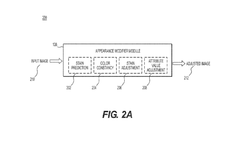
[0014] FIG. 2A is a block diagram illustrating an appearance modifier module
of a slide analysis tool for adjusting attributes of whole slide images,
according to an
exemplary embodiment of the present disclosure.
[0015] FIG. 2B is a block diagram illustrating a stain prediction module
trained
to predict a stain type of one or more stains present in a whole slide image,
according to an exemplary embodiment of the present disclosure.
[0016] FIG. 20 is a block diagram illustrating a color constancy module
trained
to provide template-based attribute matching to adjust a whole slide image,
according to an exemplary embodiment of the present disclosure.
[0017] Fig. 2D is a block diagram illustrating a stain adjustment module
trained to adjust stain-specific, attributes of a whole slide image, according
to an
exemplary embodiment of the present disclosure.
4
CA 03216960 2023- 10- 26
WO 2022/241368
PCT/US2022/071768
[0018] Fig. 2E is a block diagram illustrating an attribute value adjustment
module for adjusting values of one or more attributes of a whole slide image
based
on user input, according to an exemplary embodiment of the present disclosure.
[0019] FIG. 3 is a flowchart illustrating an exemplary method for adjusting
attributes of a whole slide image, according to an exemplary embodiment of the
present disclosure.
[0020] FIG. 4A is a flowchart illustrating an exemplary method for training a
stain prediction module, according to an exemplary embodiment of the present
disclosure.
[0021] FIG. 4B is a flowchart illustrating an exemplary method for deploying a
trained stain prediction module to predict a stain type of one or more stains
present
in a whole slide image, according to an exemplary embodiment of the present
disclosure.
[0022] FIG. 5 is a flowchart illustrating an exemplary method for template-
based color adjustment of a whole slide image, according to an exemplary
embodiment of the present disclosure.
[0023] FIG. 6 is a flowchart illustrating an exemplary method for adjusting
one
or more stains present in a whole slide image, according to an exemplary
embodiment of the present disclosure.
[0024] FIG. 7 is a flowchart illustrating an exemplary method for adjusting
values of one or more attributes of a whole slide image based on user input,
according to an exemplary embodiment of the present disclosure.
[0025] FIG. 8 illustrates an example system that may execute techniques
presented herein.
DESCRIPTION OF THE EMBODIMENTS
CA 03216960 2023- 10- 26
WO 2022/241368
PCT/US2022/071768
[0026] Reference will now be made in detail to the exemplary embodiments of
the present disclosure, examples of which are illustrated in the accompanying
drawings. Wherever possible, the same reference numbers will be used
throughout
the drawings to refer to the same or like parts.
[0027] The systems, devices, and methods disclosed herein are described in
detail by way of examples and with reference to the figures. The examples
discussed
herein are examples only and are provided to assist in the explanation of the
apparatuses, devices, systems, and methods described herein. None of the
features
or components shown in the drawings or discussed below should be taken as
mandatory for any specific implementation of any of these devices, systems, or
methods unless specifically designated as mandatory.
[0028] Also, for any methods described, regardless of whether the method is
described in conjunction with a flow diagram, it should be understood that
unless
otherwise specified or required by context, any explicit or implicit ordering
of steps
performed in the execution of a method does not imply that those steps must be
performed in the order presented but instead may be performed in a different
order
or in parallel.
[0029] As used herein, the term "exemplary" is used in the sense of
"example," rather than "ideal." Moreover, the terms "a" and "an" herein do not
denote
a limitation of quantity, but rather denote the presence of one or more of the
referenced items.
[0030] In human and animal pathology, visual examination of tissues
(histology) and cells (cytology) under a microscope may be a vital element of
diagnostic medicine. For example, histology and cytology may be performed to
diagnose cancer, facilitate drug development, and assess toxicity, etc. For
histology,
6
CA 03216960 2023- 10- 26
WO 2022/241368
PCT/US2022/071768
tissue samples undergo multiple preparation steps so that different tissue
structures
can be differentiated visually by the human eye when viewing under the
microscope.
For example, tissue preparation may consist of the following steps: (i)
preserving the
tissue using fixation; (ii) embedding the tissue in a paraffin block; (iii)
cutting the
paraffin block into thin sections (3-5 micrometers (pm)); (iv) mounting the
sections on
glass slides; and/or (v) staining mounted tissue sections to highlight
particular
components or structures. Tissue preparation may be done manually and hence
may
introduce large variability into the images observed.
[0031] Staining aids in creating visible contrast of the different tissue
structures for differentiation by a pathologist. During this process, one or
more types
of chemical substances (e.g., stains or dyes) are attached to different
compounds in
the tissue delineating different cellular structures. Different types of
stains may
highlight different structures. Therefore, pathologists may interpret or
analyze the
stains differently. Depending on a disease and its underlying behavior, one
stain or a
combination of stains may be preferable over others for use in diagnostic
detection.
Although standard protocols for using these stains are often in place,
protocols vary
per institution and overstaining or understaining of tissue may occur, which
may
potentially cause diagnostic information or indicators to be obscured. For
example,
color variations resulting from non-uniform staining between slides may cause
one
image to look pinker among other images that a pathologist has been reviewing
during a day. Such out of distribution images might be hard for the
pathologist to
investigate as separating different structures might be confusing. For
instance, a
main characteristic of lymphocytes in Hematoxylin and Eosin (H&E) stained
images
is their dark purple color; however, in some poorly stained images they might
have
similar color as other cells. Moreover, multiple stains are commonly used
together for
7
CA 03216960 2023- 10- 26
WO 2022/241368
PCT/US2022/071768
highlighting several structures of interest in the tissue, e.g., tissue that
is stained with
both hematoxylin and eosin, which may further exacerbate potential problems
caused by overstaining or understaining.
[0032] When pathologists view slides with a traditional microscope, they do
not have the ability to alter attributes (e.g., characteristics or properties)
of the image
produced by the microscope beyond magnification_ However, when whole slide
imaging is used to scan images of the slides for generating digital whole
slide
images, image processing and Al-enabled tools may be utilized for adjusting a
color,
an amount of a particular stain, a brightness, a sharpness, and/or a contrast,
among
other attribute adjustments to the whole slide images. Such adjustments may
enable
pathologists to better analyze tissue samples from human or animal patients by
allowing them to adjust the image attributes in semantically meaningful ways
(e.g., to
normalize color across a population of slides being viewed, correct for
overstaining
or understaining, enhance differentiation of structures, remove artifacts,
etc.).
[0033] Techniques discussed herein may use Al technology, machine
learning, and image processing tools to enable pathologists to adjust digital
images
according to their needs. Techniques presented herein may be used as part of a
visualization software that pathologists use to view the digital whole slide
images in
their routine workflow. Techniques discussed herein provide methods for
enabling
adjustments of semantically meaningful image attributes in pathology images,
including methods for automatically predicting stain types for use as input in
adjustment processes, color normalization methods to enable template-based
attribute matching, methods for automatically converting images to particular
color
spaces in which the semantically meaningful adjustments can be made, and user-
interface based methods for enabling attribute value adjustments.
8
CA 03216960 2023- 10- 26
WO 2022/241368
PCT/US2022/071768
[0034] FIG. 1A illustrates an exemplary block diagram of a system and
network to adjust attributes of whole slide images, according to an exemplary
embodiment of the present disclosure.
[0035] Specifically, FIG. 1A illustrates an electronic network 120 that may be
connected to servers at hospitals, laboratories, and/or doctor's offices, etc.
For
example, physician servers 121, hospital servers 122, clinical trial servers
123,
research lab servers 124, and/or laboratory information systems 125, etc., may
each
be connected to an electronic network 120, such as the Internet, through one
or
more computers, servers and/or handheld mobile devices. According to an
exemplary embodiment of the present application, the electronic network 120
may
also be connected to server systems 110, which may include processing devices
that are configured to implement an image adjustment platform 100, which
includes
a slide analysis tool 101 for using machine learning and/or image processing
tools to
identify and adjust one or more attributes of whole slide images, according to
an
exemplary embodiment of the present disclosure. The slide analysis tool 101
may
allow automatic and/or manual adjustments to color, including template-based
color
matching, an amount of a particular stain, a brightness, a sharpness, and a
contrast,
among other adjustments.
Image Attribute Adjustments
[0036] Examples of whole slide images may include digitized images of
histology or cytology slides stained with a variety of stains, such as, but
not limited
to, hematoxylin and eosin, hematoxylin alone, toluidine blue, alcian blue,
Giemsa,
trichrome, acid-fast, Nissl stain, etc. Non-limiting and non-exhaustive uses
of each
stain or combination of stains and implementation of the image adjustment
platform
9
CA 03216960 2023- 10- 26
WO 2022/241368
PCT/US2022/071768
100 for enhancing the viewing and analysis of whole slide images including
these
stain(s) are described briefly below.
Adiustments of colors in image stained with Hematoxvlin and Eosin
[0037] Hematoxylin and Eosin are the most commonly used stains for
morphological analysis of tissue. Hematoxylin binds to deoxyribonucleic acid
(DNA)
and stains the nuclei dark blue or purple, whereas eosin stains the
extracellular
matrix and cytoplasm pink. The image adjustment plafform 100 may be used for
adjustment (e.g., correction) of over-staining or under-staining of
hematoxylin or
eosin.
Adjustment of blue and purple color in Toluidine blue stained image
[0038] Toluidine blue is a polychromatic dye which may absorb different colors
depending on how it binds chemically with various tissue components. In
diagnostic
labs, toluidine blue may be used by pathologists to highlight mast cell
granules,
particularly when evaluating patients with pathological conditions that
involve mast
cells (including cancers), allergic inflammatory diseases, and
gastrointestinal
diseases such as irritable bowel syndrome. Toluidine blue may also be used to
highlight tissue components such as cartilage or certain types of mucin.
Further,
toluidine blue may be used as part of the screening process for certain
cancers, such
as oral cancer, as it binds the DNA of dividing cells causing precancerous and
cancerous cells to take up more of the dye than healthy cells.
Adiustments of blue and pink color in Alcian blue stained images
[0039] The alcian blue stain may cause acid mucins and mucosubstances to
appear blue, and nuclei to appear reddish pink when a counterstain of neutral
red is
used. The blue and pink colors of the stain may be adjusted using the image
CA 03216960 2023- 10- 26
WO 2022/241368
PCT/US2022/071768
adjustment platform 100 for better visualization of nuclei and other features
in the
image.
Adiustments of purple and pink in Giemsa stained images
[0040] A Giemsa stain is a blood stain that may be used histopathologically to
observe composition and structure. Additionally, Giemsa has high-quality
staining
capabilities of chromatin and nuclear membranes. Human and pathogenic cells
may
be stained differently, where human cells may be stained purple and bacterial
cells
pink for differentiation. The image adjustment platform 100 may be used to
adjust the
pink and purple colors to enhance the contrast between human cells and
bacterial
cells.
Adjustment of colors in images with Trichrome stain
[0041] Trichome stains may use three dyes to produce different coloration of
different tissue types. Typically, trichrome stains may be used to demonstrate
collagen, often in contrast to smooth muscle, but may also be used to
highlight fibrin
in contrast to red blood cells. The image adjustment platform 100 may be used
to
adjust green and blue colors to enhance a contrast for collagen and bone. Red
and
black colors also may be modified by the image adjustment platform 100 to
adjust
the appearance of nuclei.
Further, contrast for nuclei, Musin, fibrin and/or cytoplasm may be changed by
adjusting red and yellow colors.
Adiustment of colors in images with Acid-fast stain
[0042] Acid-fast is a differential stain used to identify acid-fast bacterial
organisms, such as members of the genus Mycobacterium and Nocardia. The stain
colors bacterial organisms as red-pink and other matter as bluish. The image
11
CA 03216960 2023- 10- 26
WO 2022/241368
PCT/US2022/071768
adjustment platform 100 may be used to adjust colors, including stain colors,
and
contrast to enhance the visibility of bacteria in the images.
Adiustment of colors in imacies with Nissl stain
[0043] Nissl staining is used to visualize Nissl substance (e.g., clumps of
rough endoplasmic reticulum and free polyribosomes) found in neurons. This
stain
may distinguish neurons from glia and the cytoarchitecture of neurons may be
more
thoroughly studied with the help of this stain. A loss of Nissl substance may
signify
abnormalities, such as cell injury or degeneration, which in turn may indicate
disease. The image adjustment platform 100 may be used to adjust pink and blue
colors produced by the stain to better visualize the difference between
various types
of neurons.
The Environment
[0044] The physician servers 121, hospital servers 122, clinical trial servers
123, research lab servers 124 and/or laboratory information systems 125 may
create
or otherwise obtain images of one or more patients' cytology specimen(s),
histopathology specimen(s), slide(s) of the cytology specimen(s), digitized
images of
the slide(s) of the histopathology specimen(s), or any combination thereof.
The
physician servers 121, hospital servers 122, clinical trial servers 123,
research lab
servers 124 and/or laboratory information systems 125 may also obtain any
combination of patient-specific information, such as age, medical history,
cancer
treatment history, family history, past biopsy or cytology information, etc.
The
physician servers 121, hospital servers 122, clinical trial servers 123,
research lab
servers 124 and/or laboratory information systems 125 may transmit digitized
slide
images and/or patient-specific information to server systems 110 over the
electronic
network 120. Server systems 110 may include one or more storage devices 109
for
12
CA 03216960 2023- 10- 26
WO 2022/241368
PCT/US2022/071768
storing images and data received from at least one of the physician servers
121,
hospital servers 122, clinical trial servers 123, research lab servers 124,
and/or
laboratory information systems 125. Server systems 110 may also include
processing devices for processing images and data stored in the one or more
storage devices 109. Server systems 110 may further include one or more
machine
learning tool(s) or capabilities. For example, the processing devices may
include one
or more machine learning tools for the image adjustment platform 100,
according to
one embodiment. Alternatively or in addition, the present disclosure (or
portions of
the system and methods of the present disclosure) may be performed on a local
processing device (e.g., a laptop).
[0045] The physician servers 121, hospital servers 122, clinical trial servers
123, research lab servers 124 and/or laboratory information systems 125 refer
to
systems used by pathologists for reviewing the images of the slides. In
hospital
settings, tissue type information may be stored in a laboratory information
system
125. Additionally, information related to stains used for tissue preparation,
including
stain type, may be stored in the laboratory information systems 125.
[00461 FIG. 1B illustrates an exemplary block diagram of the image
adjustment platform 100. The image adjustment platform 100 may include a slide
analysis tool 101, a data ingestion tool 102, a slide intake tool 103, a slide
scanner
104, a slide manager 105, a storage 106, and a viewing application tool 108.
[0047] The slide analysis tool 101, as described below, refers to a process
and system for identifying and adjusting one or more attributes of whole slide
images. Machine learning may be used to predict a stain type of one or more
stains
present in a whole slide image, according to an exemplary embodiment. Machine
learning may also be used for color normalization processes to map color
13
CA 03216960 2023- 10- 26
WO 2022/241368
PCT/US2022/071768
characteristics of a template to the whole slide image for adjusting a color
thereof to
enable color constancy among images viewed, according to another exemplary
embodiment. Machine learning may further be used to convert an original color
space of the whole slide image to a color space that is specific to a stain
type of one
or more stains identified in the whole slide image to enable a brightness or
an
amount of the one or more stains to be adjusted, according to another
exemplary
embodiment. The slide analysis tool 101 may also provide graphical user
interface
(GUI) control elements (e.g., slider bars) for display in conjunction with the
whole
slide image through a user interface of the viewing application tool 108 to
allow user-
input based adjustment of attribute values for color, brightness, sharpness,
and
contrast, among other similar examples, as described in the embodiments below.
[0048] The data ingestion tool 102 may facilitate a transfer of the whole
slide
images to the various tools, modules, components, and devices that are used
for
classifying and processing the whole slide images, according to an exemplary
embodiment. In some examples, if the whole slide image is adjusted utilizing
one or
more features of the slide analysis tool 101, only the adjusted whole slide
image may
be transferred. In other examples, both the original whole slide image and the
adjusted whole slide image may be transferred.
[0049] The slide intake tool 103 may scan pathology slides and convert them
into a digital form, according to an exemplary embodiment. The slides may be
scanned with slide scanner 104, and the slide manager 105 may process the
images
on the slides into digitized whole slide images and store the digitized whole
slide
images in storage 106.
[0050] The viewing application tool 108 may provide a user (e.g., pathologist)
a user interface that displays the whole slide images throughout various
stages of
14
CA 03216960 2023- 10- 26
WO 2022/241368
PCT/US2022/071768
adjustment. The user interface may also include the GUI control elements of
the
slide analysis tool 101 that may be interacted with to adjust the whole slide
images,
according to an exemplary embodiment. The information may be provided through
various output interfaces (e.g., a screen, a monitor, a storage device and/or
a web
browser, etc.).
[0051] The slide analysis tool 101, and one or more of its components, may
transmit and/or receive digitized whole slide images and/or patient
information to
server systems 110, physician servers 121, hospital servers 122, clinical
trial servers
123, research lab servers 124, and/or laboratory information systems 125 over
an
electronic network 120. Further, server systems 110 may include storage
devices for
storing images and data received from at least one of the slide analysis tool
101, the
data ingestion tool 102, the slide intake tool 103, the slide scanner 104, the
slide
manager 105, and viewing application tool 108. Server systems 110 may also
include processing devices for processing images and data stored in the
storage
devices. Server systems 110 may further include one or more machine learning
tool(s) or capabilities, e.g., due to the processing devices. Alternatively,
or in
addition, the present disclosure (or portions of the system and methods of the
present disclosure) may be performed on a local processing device (e.g., a
laptop).
[0052] Any of the above devices, tools and modules may be located on a
device that may be connected to an electronic network such as the Internet or
a
cloud service provider, through one or more computers, servers and/or handheld
mobile devices.
[0053] FIG. 1C illustrates an exemplary block diagram of a slide analysis tool
101, according to an exemplary embodiment of the present disclosure. The slide
CA 03216960 2023- 10- 26
WO 2022/241368
PCT/US2022/071768
analysis tool 101 may include a training image platform 131 and/or a target
image
platform 136.
[0054] According to one embodiment, the training image platform 131 may
include a plurality of software modules, including a training image intake
module 132,
a stain type identification module 133, a color normalization module 134, and
a color
space transformation module 135.
[0055] The training image platform 131, according to one embodiment, may
create or receive one or more datasets of training images used to generate and
train
one or more machine learning models that, when implemented, facilitate
adjustments
to various attributes of whole slide images. For example, the training images
may
include whole slide images received from any one or any combination of the
server
systems 110, physician servers 121, hospital servers 122, clinical trial
servers 123,
research lab servers 124, and/or laboratory information systems 125. Images
used
for training may come from real sources (e.g., humans, animals, etc.) or may
come
from synthetic sources (e.g., graphics rendering engines, 3D models, etc.).
Examples of whole slide images may include digitized histology or cytology
slides
stained with a variety of stains, such as, but not limited to, Hematoxylin and
eosin,
hematoxylin alone, toluidine blue, alcian blue, Giemsa, trichrome, acid-fast,
Nissl
stain, etc.
[0056] The training image intake module 132 of the training image platform
131 may create or receive the one or more datasets of training images. For
example,
the datasets may include one or more datasets corresponding to stain type
identification, one or more datasets corresponding to color normalization, and
one or
more datasets corresponding to stain-specific color space transformation. In
some
examples, a subset of training images may overlap between or among the various
16
CA 03216960 2023- 10- 26
WO 2022/241368
PCT/US2022/071768
datasets for stain type identification, color normalization, and stain-
specific color
space transformation. The datasets may be stored on a digital storage device
(e.g.,
one of storages devices 109).
[0057] The stain type identification module 133 may generate, using at least
the datasets corresponding to stain type identification as input, one or more
machine
learning systems capable of predicting a stain type of one or more stains
present in a
whole slide image. The color normalization module 134 may generate, using at
least
the datasets corresponding to color normalization as input, one or more
machine
learning systems capable of mapping color characteristics of one whole slide
image
(e.g., a template) to another whole slide image to provide color constancy
between
the two whole slide images. The color space transformation module 135 may
generate, using at least the datasets corresponding to stain-specific color
space
transformation as input, one or more machine learning systems capable of
identifying transformations for converting a whole slide image in an original
color
space to a new color space that is specific to a stain type of one or more
stains
present in the whole slide image to facilitate stain adjustments. In some
examples, a
machine learning system may be generated for each of the different stain types
to
learn a corresponding transformation. In other examples, one machine learning
system may be generated that is capable of learning transformations for more
than
one stain type.
[0058] According to one embodiment, the target image platform 136 may
include software modules, such as a target image intake module 137 and an
appearance modifier module 138, in addition to an output interface 139. The
target
image platform 136 may receive a target whole slide image as input and provide
the
image to the appearance modifier module 138 to adjust one or more attributes
of the
17
CA 03216960 2023- 10- 26
WO 2022/241368
PCT/US2022/071768
target whole slide image. For example, the target whole slide image may be
received
from any one or any combination of the server systems 110, physician servers
121,
hospital servers 122, clinical trial servers 123, research lab servers 124,
and/or
laboratory information systems 125. The appearance modifier module 138 may be
comprised of one or more sub-modules, described in detail with reference to
FIGs.
2A through 2E below. The sub-modules may execute the various machine learning
models generated by the training image platform 131 to facilitate the
adjustments to
the attributes of whole slide images. In some aspects, the adjustments may be
customizable based on user input.
[0059] The output interface 139 may be used to output the adjusted target
whole slide image (e.g., to a screen, monitor, storage device, web browser,
etc.).
[0060] FIG. 2A through FIG. 2E are block diagrams illustrating the appearance
modifier module 138 and software sub-modules thereof for adjusting various
attributes of a whole slide image. FIG. 2A is a block diagram 200 illustrating
the
appearance modifier module 138. The appearance modifier module 138 may include
one or more software sub-modules, including a stain prediction module 202, a
color
constancy module 204, a stain adjustment module 206, and an attribute value
adjustment module 208. A whole slide image may be received as input (e.g.,
input
image 210) to the appearance modifier module 138. The input image 210 may
include a histology whole slide image or a cytology whole slide image, where
the
whole slide image may be a digitized image of a slide-mounted and stained
histology
or cytology specimen, for example. Upon receipt of the input image 210, at
least one
of the sub-modules 202, 204, 206, 208 may be executed, and an adjusted image
212 may be provided as output of the appearance modifier module 138.
18
CA 03216960 2023- 10- 26
WO 2022/241368
PCT/US2022/071768
[0061] The adjusted image 212 may include an adjusted color, an adjusted
amount of a particular stain, an adjusted brightness, an adjusted sharpness,
and/or
adjusted contrast, among other adjustments. In some examples, indications of
one
or more regions of the input image 210 to be adjusted may also be received as
input
and only those one or more regions (e.g., rather than the entire image) may be
adjusted in the adjusted image 212. Further inputs utilized by (e.g., specific
to) one
or more of the modules 202, 204, 206, 208, described in detail in FIGs. 2B
through
2E below, may be received and applied to adjust the attributes of the input
image
210 accordingly.
[0062] FIG. 2B is a block diagram 220 illustrating the stain prediction module
202. The stain prediction module 202 may execute a trained machine learning
system for predicting stain types, such as the trained machine learning system
generated by the stain type identification module 133. The input image 210
received
at the appearance modifier module 138 and subsequently at the stain prediction
module 202 may include one or more stains of a particular stain type. In some
examples, the input image 210 may be provided without an indication of the
stain
type (e.g., an input stain type is not received). In such examples, the stain
prediction
module 202 may execute the trained machine learning system to predict the
stain
type of the one or more stains present in the input image 210. The predicted
stain
type 222 output by the trained machine learning system may be provided as
output
of the stain prediction module 202.
[0063] In other examples, an input stain type of the one or more stains may be
received along with the input image 210 (e.g., as additional input) to the
stain
prediction module 202. Nonetheless, the stain prediction module 202 may
execute
the trained machine learning system to predict the stain type as part of a
validation
19
CA 03216960 2023- 10- 26
WO 2022/241368
PCT/US2022/071768
process. For example, the predicted stain type 222 may be compared to the
input
stain type to determine whether the input stain type is erroneous. In some
examples,
when the input stain type is determined to be erroneous, a notification or an
alert
may be provided to a user (e.g., via the viewing application tool 108).
[0064] The predicted stain type 222 may be stored in association with the
image 210 in a storage device (e.g., one of storage devices 109) at
temporarily
throughout the attribute adjustment process. In some aspects, the predicted
stain
type 222 may be used as input to one or more other sub-modules of the
appearance
modifier module 138, such as the stain adjustment module 206.
[0065] FIG. 20 is a block diagram 230 illustrating the color constancy module
204. The color constancy module 204 may adjust at least color characteristics
of the
input image 210 received at the appearance modifier module 138 based on a
template 232 comprised of at least a portion of one or more whole slide images
that
is received as further input. In some examples, the template 232 may be a
population of whole slide images, including the image 210, provided as
collective
input to the appearance modifier module 138. In other examples, the template
232
may include a reference set of whole slide images. In some examples, the input
image 210 to be adjusted may be referred to as a source input image and the
template 232 may be referred to as a target input image as it is the color
characteristics of the template 232 that are the target for mapping onto the
input
image 210. The color constancy module 204 may use one or more color
normalization techniques to enable mapping of the color characteristics from
the
template 232 to the input image 210 to output a normalized image 234. The
color
constancy module 204 may execute a trained machine learning system for
performing the color normalization, such as the trained machine learning
system
CA 03216960 2023- 10- 26
WO 2022/241368
PCT/US2022/071768
generated by the color normalization module 134. Additionally and/or
alternatively,
further adjustments to the color characteristics of the input image 210 may be
made
based on user-specified information received in addition to the input image
210 and
the template 232 as input. In some examples, the attribute value adjustment
module
208 may facilitate these further adjustments.
[0066] The normalized image 234 having adjusted color characteristics
corresponding to the color characteristics of the template 232 and/or user-
specified
information may be provided as output of the color constancy module 204. In
some
examples, the normalized image 234 may be provided as input into one or more
other sub-modules of the appearance modifier module 138 to cause further
adjustments to be made to the normalized image 234. In other examples, the
normalized image 234 may be the adjusted image 212 output by the appearance
modifier module 138.
[0067] Fig. 2D is a block diagram 240 illustrating the stain adjustment module
206. The stain adjustment module 206 may receive an image 242 and a stain type
244 of the image 242 as input. In some examples, the image 242 may be the
input
image 210 originally received at the appearance modifier module 138. In other
examples, the image 242 may be a previously adjusted version of the input
image
210 that was output by another one of the sub-modules of the appearance
modifier
module 138. For instance, the normalized image 234 output by the color
constancy
module 204. The stain type 244 may be a stain type input by a user (e.g., the
pathologist) or otherwise associated with the image 242. Additionally or
alternatively,
the stain type 244 may be the predicted stain type 222 output by the stain
prediction
module 202.
21
CA 03216960 2023- 10- 26
WO 2022/241368
PCT/US2022/071768
[0068] The stain adjustment module 206 may adjust properties of the one or
more stains present in the image 242 for output as a stain-adjusted image 246.
For
example, a brightness and/or an amount of the one or more stains may be
adjusted.
In some aspects, graphical user interface (GUI) control elements, such as
slider
bars, may be provided to the user to allow the user to interactively define
the
configuration for controlling the particular stain adjustments. In other
aspects, the
stains may be adjusted to correspond to a defined configuration for stains
within a
template. The template may include a population of whole slide images,
including the
input image 210, provided collectively as input to the appearance modifier
module
138. In other examples, the template may include a reference set of whole
slide
images.
[0069] To enable the stain adjustments, the stain adjustment module 206 may
convert the image 242 in an original color space (e.g., a red, green, blue
(RGB) color
space) to a new color space that is specific to the stain type of one or more
stains
present in the image 242. For example, the stain adjustments according to the
defined configuration may be made to the image 242 in the stain-specific color
space
and then converted back to the original color space for output as the stain-
adjusted
image 246. To convert the image 242 to the new, stain-specific color space, a
transformation learned by a machine learning system, such as one or more of
the
machine learning systems generated by the color space transformation module
135,
may be identified, retrieved, and applied to the image 242.
[0070] The stain-adjusted image 246 having the defined configuration may be
provided as output of the stain adjustment module 206. In some examples, the
stain-
adjusted image 246 may be provided as input to one or more other modules, such
as
the attribute value adjustment module 208. In other examples, the stain-
adjusted
22
CA 03216960 2023- 10- 26
WO 2022/241368
PCT/US2022/071768
image 246 may be the adjusted image 212 provided as output of the appearance
modifier module 138. As previously discussed, in some examples, the image 242
is
the normalized image 234 output by the color constancy module 204 (e.g.,
rather
than the input image 210) and thus the stain-adjusted image 246 output by the
stain
adjustment module 206 may be a normalized, stain-adjusted image.
[0071] Fig. 2E is a block diagram 250 illustrating the attribute value
adjustment module 208. The attribute value adjustment module 208 may receive
an
image 252 as input. In some examples, the image 252 may be the input image 210
received as input to the appearance modifier module 138. In other examples,
the
image 252 may be an image output by another one or more of the sub-modules of
the appearance modifier module 138. For instance, the image 252 may be the
normalized image 234 output by the color constancy module 204 or the stain-
adjusted image 246 output by the stain adjustment module 206, where the stain-
adjusted image 246 may further be a normalized, stain-adjusted image (e.g., an
image previously adjusted by both the color constancy module 204 and the stain
adjustment module 206).
[0072] The attribute value adjustment module 208 may adjust values of one or
more attributes of the image 252 based on user input 254 to generate a user
input-
adjusted image 256. The adjustable attributes may include color (including hue
and
saturation), brightness, sharpness, and contrast, among other similar
attributes. The
user input 254 may be received as user interactions with the plurality of GUI
control
elements provided in conjunction with the image 252 through the viewing
application
tool 108. As one specific but non-limiting example, a slider bar may be
provided for
each of one or more attributes, where user input to or interaction with a
given slider
bar (e.g., movement from one end to another end) may increase or decrease
values
23
CA 03216960 2023- 10- 26
WO 2022/241368
PCT/US2022/071768
associated with the respective attribute. Other control elements that allow
incremental increases and decreases of value, similar to a slider bar, may be
used in
addition or alternatively to a slider bar. In some examples, the user input-
adjusted
image 256 may be displayed and updated in real-time through the viewing
application tool 108 as the user input 254 is received and applied. The user
input-
adjusted image 256 may be the adjusted image 212 output by the appearance
modifier module 138. In other examples, the user input-adjusted image 256 can
be
provided as input to the other submodules previously discussed.
[0073] FIG. 3 is a flowchart illustrating an exemplary method 300 for
adjusting
one or more attributes of a whole slide images, according to an exemplary
embodiment of the present disclosure. The exemplary method 300 (e.g., steps
302-
306) may be performed by the slide analysis tool 101 of the image adjustment
platform 100 automatically and/or in response to a request from a user (e.g.,
pathologist, patient, oncologist, technician, administrator, etc.). The
exemplary
method 300 may include one or more of the following steps.
[0074] In step 302, the method 300 may include receiving a whole slide image
as input (e.g., input image 210). The whole slide image may be a digitized
image of a
slide-mounted histology or cytology specimen, for example. The whole slide
image
may include one or more stains that were added to the slides to allow
differentiation
of various tissue or cellular structures by the human eye when imaged. The
types of
stains added may be dependent on which type of structures are desired to be
differentiated. In some examples, only a portion (e.g., one or more regions)
of the
whole slide image may be received as input. The portion may include one or
more
regions or areas of interest. In such examples, the remaining steps 304 and
306 may
24
CA 03216960 2023- 10- 26
WO 2022/241368
PCT/US2022/071768
be performed on the portion of the whole slide image rather than an entirety
of the
whole slide image.
[0075] In step 304, the method 300 may include adjusting one or more
attributes of the whole slide image. The attributes may be visual attributes
including
color, hue, saturation, brightness, or sharpness associated with the image and
a
brightness and/or amount of the one or more stains present in the whole slide
image.
Depending on the specific types of attributes to be adjusted and/or additional
inputs
provided by the user, one or more of the stain prediction module 202, the
color
constancy module 204, the stain adjustment module 206, and the attribute value
adjustment module 208 may be implemented to perform the adjustments.
[0076] In step 306, the method 300 may include providing the adjusted whole
slide image (e.g., adjusted image 212) as output.
Stain Prediction Module
[0077] FIG. 4A is a flowchart illustrating an exemplary method 400 for
training
a machine learning system to predict a stain type of one or more stains
present in a
whole slide image, according to an exemplary embodiment of the present
disclosure.
The whole slide image may be a digitized image of a slide-mounted pathology
specimen, for example. There are numerous types of stains or combination of
stains
that may be used in the preparation of the pathology specimen. Identifying a
stain
type of one or more stains used in the preparation may enable or facilitate
various
types of attribute adjustments to the whole slide image that may be stain
specific,
including adjustment of a brightness and/or amount of the one or more stains
in the
whole slide image. The exemplary method 400 (e.g., steps 402-408) may be
performed by the training image platform 131 (e.g., by stain type
identification
CA 03216960 2023- 10- 26
WO 2022/241368
PCT/US2022/071768
module 133) of the slide analysis tool 101. The exemplary method 400 may
include
one or more of the following steps.
[0078] In step 402, the method 400 may include receiving, as training data,
one or more whole slide images and a stain type for each of one or more stains
present in the one or more whole slide images. The received whole slide images
may be training images, whereas the stain type for the stains present in each
received whole slide image may form a label corresponding to the respective
training
image. For example, a first training image may be a whole slide image that
includes
two stains of a first and second stain type. Therefore, the label
corresponding to the
respective training image may indicate the first and second stain types.
[0079] The whole slide images may be digitized images of stained pathology
slides. There are numerous types of stains or combinations of stains that may
be
used when preparing the slides. To generate a representative dataset of
training
images, the received whole slide images at 402 may include one or more images
having each stain type that may be used in preparation. In some examples, one
or
more of the whole slide images received as training images may be thumbnails
or
macro-images.
[0080] In step 404, the method 400 may include extracting one or more
feature vectors from each of the one or more whole slide images. In some
examples,
the feature vectors may be extracted from particular regions of the whole
slide
images corresponding to non-background pixels of the whole slide images. For
example, each whole slide image may be comprised of a plurality of tiles,
where the
tiles include one or more of background pixels and non-background pixels. In
one
aspect, prior to extracting the feature vectors, the background pixels of the
whole
slide images may be removed using Otsu's method (e.g., a type of automatic
image
26
CA 03216960 2023- 10- 26
WO 2022/241368
PCT/US2022/071768
thresholding that separates pixels into two classes, foreground and
background) or
by removing tiles, and thus the pixels comprising the tiles, with low variance
from the
whole slide image. Accordingly, the non-background pixels of the whole slide
images
remain for feature extraction. In another aspect, prior to extracting the
feature
vectors, the whole slide images may be converted into a reduced summary form.
The reduced summary form may include a collection of non-background RGB pixels
of a whole slide image or a set of neighboring non-background pixel patches
(or
tiles) of a whole slide image. Accordingly, the non-background pixels of the
whole
slide images remain for feature extraction. In some examples, for obtaining
the
reduced summary form, the whole slide images may be spitted into a collection
image tile or a set of distinct pixels.
[0081] The type or format of the feature vector extracted may vary. In one
example, the extracted feature vectors may be vectors of RGB pixel values for
non-
background tiles of the whole slide images. In another example, the extracted
feature vectors may be one or more embeddings (e.g., for a convolutional
neural
network (CNN)) from non-background tiles of the whole slide images.
Additionally or
alternatively, if one or more of the whole sale images received is a thumbnail
(e.g.,
macro-image), the extracted feature vectors may be a CNN embedding from the
thumbnail. In a further example, image classification-based feature generation
techniques, such as bag-of-visual words or Vector of Locally Aggregated
Descriptors
(VLAD), may be applied to convert descriptors from one or more regions of the
whole slide image into vectors. The descriptors may include a color scale-
invariant
feature transform (SIFT) descriptor, an Oriented FAST and rotated BRIEF (ORB)
feature, a histogram of oriented gradients (HOG) descriptor, a radiant-
invariant
27
CA 03216960 2023- 10- 26
WO 2022/241368
PCT/US2022/071768
feature transform RIFT descriptor and/or a speeded up robust features (SURF)
descriptor.
[0082] In step 406, the method 400 may include to generate and train a
machine learning system for predicting stain type using the extracted feature
vectors
as input. The machine learning system may include a Naïve Bayes classifier, a
random forest model, a convolutional neural network (CNN), a recurrent neural
network (RNN) such as a simple RNN, a long short-term memory (LSTM) network, a
gated recurrent unit (GRU) or the like, a transformer neural network, and/or a
support vector machine, among other similar systems.
[0083] As one non-limiting example, extracted feature vectors of a training
image may be input to the machine learning system. The machine learning system
may predict a stain type for one or more stains present in the training image,
and
provide the predicted stain type as output. In some examples, for each
training
image more than one predicted stain type for a given stain may be output by
the
machine learning system, where each predicted stain type may be associated
with a
probability or score that represents a likelihood of the respective stain type
being the
actual stain type for the given stain. For example, for a first stain of a
first training
image, the machine learning system may output a first stain type associated
with an
80% probability of being the stain type and a second stain type associated
with a
20% probability of being the stain type.
[0084] In one example, to train the machine learning system, the predicted
stain type(s) may be compared to the label corresponding to the training image
provided as input to determine a loss or error. For example, a predicted stain
type for
a first stain of a first training image may be compared to the known stain
type for the
first stain of the first training image identified by the corresponding label.
The
28
CA 03216960 2023- 10- 26
WO 2022/241368
PCT/US2022/071768
machine learning system may be modified or altered (e.g., weights and/or bias
may
be adjusted) based on the error to improve an accuracy of the machine learning
system. This process may be repeated for each training image or at least until
a
determined loss or error is below a predefined threshold. In some examples,
some of
the training images may with withheld and used to further validate or test the
trained
machine learning system.
[0085] In step 408, the method 400 may include to store the trained machine
learning system for subsequent deployment by the stain prediction module 202
of
the appearance modifier module 138 described below with reference to FIG. 4B.
[0086] FIG. 4B is a flowchart illustrating an exemplary method 420 for
predicting a stain type of one or more stains present in a whole slide image,
according to an exemplary embodiment of the present disclosure. The exemplary
method 420 (e.g., steps 422-428) may be performed by the target image platform
136 of the slide analysis tool 101, and particularly by the stain prediction
module
202, automatically and/or in response to a request from a user (e.g.,
pathologist,
patient, oncologist, technician, administrator, etc.). The exemplary method
400 may
include one or more of the following steps.
[0087] In step 422, the method 420 may include receiving a whole slide image
as input (e.g., input image 210). In some examples, the whole slide image may
be a
portion of a whole slide image (e.g., one or more regions of interest) or a
thumbnail
of the whole slide image. The whole slide image may be a digitized image of a
pathology slide for which one or more stains were used in the preparation
thereof.
Accordingly, the one or more stains may be present in the whole slide image.
In
some examples, the stain type of the one or more stains may be unknown. In
other
examples, an input stain type for the stains may be received along with the
whole
29
CA 03216960 2023- 10- 26
WO 2022/241368
PCT/US2022/071768
slide image. However, it may nonetheless be beneficial to validate or confirm
that the
input stain type provided is in fact a correct stain type.
[0088] In step 424, the method 420 may include extracting one or more
feature vectors from the whole slide image. In some examples, the feature
vectors
may be extracted from non-background pixels of the whole slide image using the
same or similar processes described above in conjunction with step 404 of the
method 400. In step 426, the method may include providing the one or more
feature
vectors as input to a trained machine learning system, such as the trained
machine
learning system described in FIG. 4A, to predict a stain type of the one or
more
stains present in the whole slide image.
[0089] In step 428, the method 400 may include receiving the predicted stain
type for the one or more stains of the whole slide image (e.g., predicted
stain type
222) as output from the trained machine learning system. In some examples, the
predicted stain type may be provided for display in conjunction with the whole
slide
image through the viewing application tool 108. If more than one predicted
stain type
is received as output of the trained machine learning system, the predicted
stain type
having a highest associated probability or score may be selected for display.
However, if a probability or score associated with one or more of the
predicted stain
types output by the trained machine learning system is below a pre-defined
threshold, then a notification or alert may be generated and provided to the
user to
indicate that the stain type is unknown or the stain is of poor quality.
Additionally, in
instances where an input stain type is received along with the whole slide
image, a
comparison between the predicted stain type and the input stain type may be
performed. If, based on the comparison, a determination is made that the input
stain
CA 03216960 2023- 10- 26
WO 2022/241368
PCT/US2022/071768
type was erroneous, a notification or alert may be generated and provided for
display
through the viewing application tool 108.
[0090] In step 430, the method 420 includes storing the predicted stain type
in
association with the whole slide image (e.g., in one of storage devices 109).
The
predicted stain type may be subsequently retrieved from storage and used as
input
for one or more other sub-modules of the appearance modifier module 138, such
as
the stain adjustment module 206 implemented to adjust the one or more stains.
Color Constancy Aqainst Reference or Population of Slides
[0091] FIG. 5 is a flowchart illustrating an exemplary method 500 of template-
based color adjustment of a whole slide image, according to an exemplary
embodiment of the present disclosure. Color variations among whole slide
images
within a set or population being viewed and analyzed by a pathologist in one
sitting
may be problematic for the pathologist as their eyes may become used to a
specific
color distribution. For example, one whole slide image might look pinker in
color
among other images that the pathologist has been reviewing, which may cause
differentiation between structures to be less clear. Color variations among
whole
slide images may result from using different scanners to scan the slides or
may arise
from a variety of factors related to slide preparation. To address the issue
of color
variation, the exemplary method 500 (e.g., steps 502-508) may be performed by
the
slide analysis tool 101, and particularly the color constancy module 204,
automatically and/or in response to a request from a user (e.g., pathologist,
patient,
oncologist, technician, administrator, etc.). The exemplary method 500 may
include
one or more of the following steps.
[0092] In step 502, the method 500 may include receiving a whole slide image
for template-based color adjustment. The whole slide image may be a source
image
31
CA 03216960 2023- 10- 26
WO 2022/241368
PCT/US2022/071768
input received by the color constancy module 204 of the appearance modifier
module 138. The whole slide image may be an original whole slide image
received
as input to the appearance modifier module 138 (e.g., input image 210). For
simplicity and clarity, one whole slide image is discussed. However, in other
examples, a plurality of whole slide images to be viewed by a user may be
received
as input in step 502.
[0093] In step 504, the method 500 may include receiving a template having a
set of color characteristics (e.g., template 232). The template may be a
target image
received as additional input to the color constancy module 204. As previously
discussed, the whole slide image may include a plurality of tiles. The
template may
include a tile of a whole slide image, a set of tiles of a whole slide image,
an entirety
of a whole slide image, or a set of two or more whole slide images. The
template
may be one of a set of predefined templates stored by the image adjustment
platform 100 (e.g., in one of storage devices 109) and selected by the user.
In other
examples, the template may be uploaded by the user.
[0094] In step 506, the method 500 may include executing a color
normalization process to map the set of color characteristics of the template
to the
whole slide image to generate a normalized image of the whole slide image
(e.g.,
normalized image 234). For example, one or more of the machine learning
systems,
such as the machine learning systems generated by the color normalization
module
134, may be deployed or run by the color constancy module 204 to perform the
color
normalization process based on the source image input and target image input
received in steps 502 and 504, respectively. The normalized image may include
an
adjusted whole slide image having color characteristics that correspond to the
color
characteristics of the template. In some examples, the template and/or the
whole
32
CA 03216960 2023- 10- 26
WO 2022/241368
PCT/US2022/071768
slide image may be in a first color space (e.g., an RGB color space) and the
color
normalization process may include a conversion of the template and/or the
whole
slide image to a second color space prior to mapping the set of the color
characteristics of the template to the whole slide image. Example second color
spaces may include a HSV (hue, saturation, value) color space, a HIS (hue,
intensity, saturation) color space, and a L*a*b color space, among other
examples.
In some examples, one or more regions of the whole slide image(s) received as
the
template, such as a tissue region, may be segmented out to assure the second
color
space is constructed based on the stained tissue. That is, the segmented out
regions
(e.g., the tissue region) may be included as part of the template that is used
for color
characteristics mapping.
[0095] Various types of color normalization processes may be executed by
one or more machine learning systems to map or otherwise transfer the color
characteristics of the template to the whole slide image. Example color
normalization
processes may include histogram specification, Reinhard method, Macenko
method,
stain color descriptor (SCD), complete color normalization, and structure
preserving
color normalization (SPCN), among other similar processes discussed in turn
below.
[0096] For implementation of histogram specification, the whole slide image
may be converted from a first, RGB color space to a second, L*a*b color space.
In
the second, Lab color space, a histogram of the whole slide image (e.g., a
source
image histogram) may be matched to a histogram of the template (e.g. a target
image histogram). Following the mapping, the whole slide image may be
reconverted
back to the first, RGB color space. For implementation of the Reinhard method,
the
whole slide image and template may be converted from a first, RGB color space
to a
lap color space, and a linear transformation may be used to match the mean and
33
CA 03216960 2023- 10- 26
WO 2022/241368
PCT/US2022/071768
standard deviations of each color channel in the whole slide image to those of
the
template prior to reconverting the whole slide image back to the RGB color
space.
For implementation of the Macenko method, the whole slide image may be
converted from a first, RGB color space to an optical density (OD) space.
Within the
OD space a singular value decomposition (SVD) may be identified and a plane
corresponding to its two largest singular values may be created. Data may be
projected onto that plane, and corresponding angles may be found. The maximum
and minimum angle may be estimated, and those extreme values may then be
projected back to the OD space.
[0097] For implementation of SOD, the whole slide image may be converted
from a first, RGB color space to a second, OD space. A stain color appearance
matrix (S) may be empirically found by measuring a relative color proportion
for R, G
and B channels, and a stain depth matrix may be estimated by taking the
inverse of
S, multiplied with intensity values in OD, similar to the Ruifrok method. For
implementation of SPCN, the whole slide image (e.g., a source image) and
template
(e.g., a target image) may be factorized into a color appearance matrix (S)
and a
stain depth matrix (C) by non-negative matrix factorization (NMF), where at
least
multiple co-efficients of S and C are positive. The stain depth matrix of the
source
image may be combined with the color appearance matrix of the target image to
generate a normalized source image.
[0098] Alternative color normalization process implemented may further
include the following processes discussed in turn below. Joint Approximate
Diagonalization of Eigenmetrices (JADE) may be implemented to recover an
independent component for independent component analysis (ICA) decomposition.
Blind color decomposition may be implemented to separate intensity information
34
CA 03216960 2023- 10- 26
WO 2022/241368
PCT/US2022/071768
from color information. For example, the images may be converted from a first,
RGB
color space to a second, Maxwellian color space to estimate a color
distribution of
separate stains. Reference color vectors may be identified, and, by linear
decomposition, stain absorption vectors may be estimated and used to adjust
color
variation. A hue-saturation-density (HSD) model for stain recognition and
mapping
may be implemented. Initially the whole slide image may be converted from a
first,
RGB color space to a second, hue-saturation-intensity (HIS) model, where the
HSD
model may be defined as the RGB to HIS transform. HSI data has two chromatic
components and a density component. Different objects that correspond to
different
stains (e.g., nuclei, background) may be segmented before obtaining the
chromatic
and density distribution of hematoxylin, eosin and background. The
contribution of
stain for every pixel may be weighted as needed. The HSD model may then be
transformed back to the RGB color space. Style transfer models may
alternatively be
implemented to transfer color characteristics of one image to another.
[0099] Additionally, the color normalization processes may be implemented
by one or more types of generative adversarial network (GAN)-based machine
learning systems. As one example, an Information Maximizing Generative
Adversarial Network (InfoGAN) and learning control variables automatically
learned
by the model may be implemented, where the control variables may be used to
mimic color characteristics in the template. As another example, histoGAN, a
color
histogram-based method for controlling GAN-generated images' colors and
mapping
each color to the color of a target image (e.g., the template) may be
implemented. As
a further example, CycleGAN may be implemented to learn a style of a group of
images (e.g., learn style of the template).
CA 03216960 2023- 10- 26
WO 2022/241368
PCT/US2022/071768
[00100] In step 508, the method 500 may include providing
the
normalized image (e.g., the normalized image 234) as output of the color
constancy
module 204. The normalized image may be an adjusted whole slide image having
color characteristics corresponding to the set of color characteristics of the
template
as a result of the color normalization process. In some examples the
normalized
image may be the adjusted image 212 output by the appearance modifier module
138. In other examples, the normalized image may be provided as input into
other
sub-modules of the appearance modifier module 138, including the stain
adjustment
module 206 or the attribute value adjustment module 208.
Semantically Meaningful Stain Adjustment
[00101] As exemplified by the above discussion with
reference to FIG. 5,
adjustment of color attributes of a whole slide image, such as brightness,
hue, and
saturation, may not be done in a sensible manner using an original RGB color
space
of the whole slide image. Therefore, the whole slide image may be converted to
alternative color spaces in which the adjustments can be made. Similarly, for
adjusting one or more color properties of a stain, which may be particularly
important
if overstaining or understaining has occurred, the image may need to first be
converted from the original RGB color space to a color space specific to a
stain type
(e.g., a stain-specific color space). However, unlike hue, saturation, and
brightness
attributes of the whole slide image, it may not be possible to simply define
the stain-
based quantifications up front to perform such conversion. Instead, as part of
the
training image platform 131, one or more machine learning systems may be built
(e.g., by the color space transformation module 135) for learning a
transformation
that enables conversion of the whole slide image from the original, RGB color
space
to the stain-specific color space.
36
CA 03216960 2023- 10- 26
WO 2022/241368
PCT/US2022/071768
[00102] Various types of machine learning systems may be
utilized to
learn the transformation. The transformation may include linear and non-linear
transformations. Transformations may be learned for a plurality of different
stain
types. For example, a transformation may be learned for each stain type or
combination of stain types that may be utilized for staining pathology slides.
In some
examples, a machine learning system specific to each stain type or combination
may
be built. In other examples, one machine learning system may be capable of
learning
transformations for more than one stain type or combination. The learned
transformations may then be stored in a data store (e.g., in one of storage
devices
109) in association with the specific stain type or combination of stain types
for
subsequent retrieval and application when adjusting one or more stain
properties of
a whole slide image, as described in detail with reference to FIG. 6.
[00103] One specific but non-limiting example of a
learned
transformation may include a learned, invertible linear transformation of a
whole slide
image from an RGB color space to a stain color space. The whole slide image
may
include two stains of hematoxylin and eosin. This example transformation may
be
described by a stain matrix. For example, assuming the whole slide image is in
an
RGB color space, the learned transformation may be given by the matrix
(R
multiplication, p = T G . R, G, and B may be non-background pixels of the
whole
B
slide image in the RGB color space, and T may be an invertible or
pseudoinvertible c
x 3 matrix that converts from the RGB color space to the c-dimensional stain
space
(p) . A number of channels (c) in the c-dimensional stain space (p) may be
based on
the one or more stain types present in the whole slide image. For example,
when the
two stains include hematoxylin and eosin, c = 3, with a first channel
indicating
37
CA 03216960 2023- 10- 26
WO 2022/241368
PCT/US2022/071768
intensity, a second channel indicating hematoxylin, and a third channel
indicating
eosin. As a further example, if saffron staining is used in conjunction with
hematoxylin and eosin for staining, as is common in France, c = 4. The rows of
T,
referred to as vectors or stain vectors, may describe how to convert the pixel
values
of the whole slide image in the RGB color space (e.g., from red, green and
blue
channels) to channels in a stain-specific color space. When a whole slide
image
stained with hematoxylin and eosin is received as input to the stain
adjustment
module 206, T may be retrieved as the machine-learned transformation for
application to at least a portion of the whole slide image to convert the
portion of the
whole slide image from the RGB color space to the color space specific to
hematoxylin and eosin. Conversion to this stain-specific color space may then
facilitate adjustments to one or more of the brightness or amount of
hematoxylin
and/or eosin.
[00104] Various types of machine learning systems may be
utilized to
learn the transformation. In some examples, a principal component analysis
(PCA),
zero-phase analysis (ZCA), non-negative matrix factorization (NMF), and/or
independent components analysis (ICA) may be applied to a subset of non-
background RGB pixels from a training set of whole image slides having a given
stain or combination of stains to acquire at least the transformation matrix T
for the
given stain or combination of stains. Subsequently, semantic labels may be
applied
to one or more rows (e.g., vectors) of the matrix T. Often the first vector
may include
brightness and the other two vectors may include other stains. The semantic
meaning of each vector may be determined via analog introspection, or by
comparison with a reference set of vectors determined by using a training set
of
whole image slides stained with only a single stain or by using a small set of
38
CA 03216960 2023- 10- 26
WO 2022/241368
PCT/US2022/071768
annotations for tissues that are predisposed to absorb specific stains. These
learning
methods may be applicable if there are three input pixel channels as input and
one
or a combination of two stains. If there are more than two stains,
underdetermined
variants of these learning methods may be used.
[00105] In other examples, a clustering approach may be
applied to a
subset of non-background RGB pixels from a training set of whole image slides
having a given stain or combination of stains to learn at least the
transformation
matrix T. For example, k-means clustering identifies k prototypes within the
data,
where k may be set to the number of vectors desired. Alternatives to k-means
clustering may include Gaussian mixture models, mean-shift clustering, density-
based spatial clustering of applications with noise (DBSCAN), or the like. The
semantic meaning of each vector may be determined via manual introspection or
comparing to a reference set of vectors determined using slides stained with
only a
single stain, or by using a small set of annotations for tissues that are
predisposed to
absorb specific stains.
[00106] In further examples, a regression-based machine
learning
system (e.g., with supervised learning) may be trained to infer the matrix T
transformation. For example, a training dataset of whole slide images and
labels
identifying pixels determined to be canonical (e.g., canonical pixels) for a
given stain
may be provided as input to build and train the machine learning system.
Canonical
pixels may be pixels identified as having structures predisposed to bind with
each of
one or more stains (e.g., a pixel having DNA for which hematoxylin bins to).
[00107] FIG. 6 is a flowchart illustrating an exemplary
method 600 for
performing stain adjustments, according to an exemplary embodiment of the
present
disclosure. The exemplary method 600 (e.g., steps 602-616) may be performed by
39
CA 03216960 2023- 10- 26
WO 2022/241368
PCT/US2022/071768
the slide analysis tool 101, and particularly the stain adjustment module 206,
automatically and/or in response to a request from a user (e.g., pathologist,
patient,
oncologist, technician, administrator, etc.). The exemplary method 600 may
include
one or more of the following steps.
[00108] In step 602, the method 600 may include receiving
a whole slide
image (e.g., image 242) as input. The whole slide image may be received as
input to
the stain adjustment module 206 of the appearance modifier module 138. In some
examples, the whole slide image may be the original whole slide image (e.g.,
input
image 210) received by the appearance modifier module 138. In other examples,
the
whole slide image received as input may be an adjusted version of the original
whole
slide image output by one or more other modules of the appearance modifier
module
138, such as the normalized image output by the color constancy module 204
(e.g.,
normalized image 234).
[00109] An entirety of the whole slide image may be
received as input.
Alternatively, a portion of the whole slide image may be received as input.
The
portion may indicate a defined region of interest to which the stain
adjustment is to
be applied. The defined region of interest may be a region that is specified
manually
by the user by drawing or setting a boundary box or the like using the viewing
application tool 108. As another example, the defined region of interest may
be a
region in a field of view (e.g., that the user is zoomed in on) on the viewing
application tool 108.
[00110] In instances where an entirety of the whole slide
image is
received and the stain adjustment is to be implemented to the entirety of the
whole
slide image, a thumbnail image (e.g., a reduced size version of lower
resolution
based on a color sampling of the whole slide image) of the whole slide image
may be
CA 03216960 2023- 10- 26
WO 2022/241368
PCT/US2022/071768
utilized for subsequent processing steps. However, use of the thumbnail image
may
be less optimal as small structures with specific stains have a potential of
being
missed. Therefore, a majority of pixels corresponding to those particular
stains may
be removed prior to performing subsequent processing on the thumbnail image.
As
an alternative option, if the stain adjustment is to be implemented to an
entirety of
the whole slide image, random patches from different locations of the whole
slide
image may be selected. The randomly selected patches may be uniformly
distributed
across the whole slide image to ensure enough color information has been
obtained.
Pixels included within the randomly selected patches may be used for
subsequent
processing steps.
[00111] In step 604, a stain type for a stain present in
the whole slide
image may be identified. In some examples, the stain type is provided as input
along
with the whole slide image (e.g., an input stain type). In other examples, the
stain
type identified may be the predicted stain type output by the stain prediction
module
202 (e.g., predicted stain type 222). To provide an illustrative example,
hematoxylin
and eosin may be the identified combination of stain types present in the
whole slide
image. Hematoxylin binds to DNA and stains the nuclei a dark blue or purple,
whereas eosin stains the extracellular matrix and cytoplasm pink.
[00112] In step 606, the method 600 may include
retrieving, based on
the stain type, a machine-learned transformation. The machine-learned
transformation may be retrieved in order to convert the whole slide image from
a first
color space (e.g., a RGB color space in which the whole slide image was
received)
to a second color space that is specific to the stain type (e.g., a second,
stain-
specific color space). For example, a machine-learned transformation that is
associated with the stain type may be retrieved from among the plurality of
machine-
41
CA 03216960 2023- 10- 26
WO 2022/241368
PCT/US2022/071768
learned transformations stored in the data store (e.g., in one of storage
devices 109),
the transformations having been learned by one or more machine learning
systems
generated as part of the training image platform 131 (e.g., by color space
transformation module 135). For example, when the whole slide image is
identified
as being stained with hematoxylin and eosin, the matrix T may be the machine-
learned transformation retrieved from the data store.
[00113] In step 608, the method 600 may include
identifying at least a
subset of pixels of the whole slide image to be transformed. The subset of
pixels to
be transformed may include non-background pixels and non-artifact pixels
(e.g., the
pixels may include the stain that is being adjusted). Pixels of the whole
slide image
(or portion thereof) may be classified into background pixels and non-
background
pixels. Pixels can be determined as background pixels and excluded from the
subset
using Otsu's method, by analyzing the variance of tiles, or by identifying if
a pixel is
sufficiently close to a reference white background pixel by fitting a
distribution to
pixels identified as the background, among other similar techniques.
Additionally,
some pixels (whether background or non-background) may represent artifacts,
such
as bubbles, ink, hair, tissue folds, and other unwanted aspects, present on
the whole
slide image. Artifact pixels may be identified using semantic segmentation,
among
other similar techniques, and excluded from the subset (e.g., such that non-
artifact
pixels remain).
[00114] In step 610, the method may include applying the
machine-
learned transformation to the subset of pixels to convert the subset of pixels
from a
first color space to the second, stain-specific color space. Continuing the
example
where the whole slide image is stained with hernatoxylin and eosin, when the
matrix
T is retrieved and applied to the subset of pixels, one or more intensities
present for
42
CA 03216960 2023- 10- 26
WO 2022/241368
PCT/US2022/071768
a red, green and/or blue channel in the original RGB color space may now be
represented as a linear combination of the stains present (e.g., as stain
vectors). For
example, the stain vectors may include a first channel indicating intensity, a
second
channel indicating hematoxylin, and a third channel indicating eosin.
[00115] In step 612, the method 600 may include adjusting
one or more
attributes of the one or more stains in the second, stain-specific color
space. The
adjustable attributes may include a brightness (e.g., by adjusting pixel value
intensity) and/or an amount of each of the one or more stains (e.g., by
adjusting
values of one or more dimensions in the second, stain-specific color space).
Brightness may be increased or decreased. Similarly, the amount of a stain may
be
increased or decreased. These amount-based stain adjustments may correct for
overstaining or understaining resulting from the slide preparation. In some
examples,
the adjustments may be made automatically, where templates or other similar
reference images may be used for the adjustment. In other examples, the
adjustments may be made based on user input from interactions with GUI control
elements provided for display in conjunction with the whole slide image
through the
viewing application tool 108. Continuing the example where the whole slide
image is
stained with hematoxylin and eosin and the matrix T is retrieved and applied
to the
subset of pixels for the transformation, the GUI control elements may
correspond to
each channel represented by the stain vectors. For example, the GUI control
elements may include a slider bar for adjusting brightness, a slider bar for
adjusting
an amount of hematoxylin, and a slider bar for adjusting an amount of eosin.
Other
control elements that allow incremental increases and decreases of value,
similar to
a slider bar, may be used in addition or alternatively to a slider bar. The
adjustments
may be made uniformly (e.g., increasing the second channel by 10%, etc.).
43
CA 03216960 2023- 10- 26
WO 2022/241368
PCT/US2022/071768
[00116] In step 614, the method may include converting the
stain-
adjusted subset of pixels from the second color space back to the first color
space
using an inverse of the machine-learned transformation (e.g., an inverse
matrix T
may be applied continuing with the example above). In step 616, the method 600
may include providing a stain-adjusted whole slide image, including at least
the
stain-adjusted subset of pixels, as output (e.g., stain-adjusted image 246).
In some
examples, at least the background pixels (and in some instances the background
pixels and the artifact pixels) that were previously removed may be added to
the
stain-adjusted subset of pixels to form the stain-adjusted image for output by
the
stain adjustment module 206. In other examples, the stain-adjusted subset of
pixels
alone may be output by the stain adjustment module 206.
[00117] In some aspects the stain-adjusted image (or
normalized-stain
adjusted image if previously adjusted by color constancy module 204) may be
the
adjusted whole slide image output by the appearance modifier module 138 (e.g.,
adjusted image 212). In other aspects, the stain-adjusted image may be input
into
one or more other modules of the appearance modifier module 138 for further
adjustments. For example, in addition to the automatic template-based
attribute
adjustments discussed above with reference to FIG. 5 and stain-specific
attribute
adjustments discussed above with reference to FIG. 6, a user may desire to
manually adjust one or more attributes to better understand or visualize a
whole slide
image.
Attribute Value Adjustment Module
[00118] FIG. 7 is a flowchart illustrating an exemplary
method 700 for
enabling attribute value adjustments to a whole slide image based on user
input,
according to an exemplary embodiment of the present disclosure. The exemplary
44
CA 03216960 2023- 10- 26
WO 2022/241368
PCT/US2022/071768
method 700 (e.g., steps 702-712) may be performed by the slide analysis tool
101,
and particularly the attribute value adjustment module 208, automatically
and/or in
response to a request from a user (e.g., pathologist, patient, oncologist,
technician,
administrator, etc.). The exemplary method 600 may include one or more of the
following steps.
[00119] In step 702, the method 700 may include receiving
a whole slide
image (e.g., image 252) as input to the attribute value adjustment module 208.
In
some examples, the whole slide image may be the original whole slide image
received by the appearance modifier module 138 (e.g., input image 210). In
other
examples, the whole slide image may be an adjusted version of the original
whole
slide image. For instance, the whole slide image may be the normalized image
234
output by the color constancy module 204 and/or stain-adjusted image 246
output
the stain adjustment module.
[00120] The whole slide image may be displayed through the
viewing
application tool 108 to allow the user to interact with the whole slide image.
The
whole slide image may be comprised of a large number of pixels. The whole
slide
image be partitioned into a plurality tiles, each of the tiles including a
subset of the
pixels. In some examples, one or more of the tiles may be selected or
otherwise
identified through the viewing application tool 108, and the attribute value
adjustment
module 208 may receive an indication of the selection. For example, the user
may
draw a bounding box around the one or more tiles (e.g., associated with a
magnification and size). Alternatively, the one or more tiles may be
identified based
on a field of view (e.g., a zoomed-in region) including the one or more tiles.
In such
examples, the various attribute value adjustments described in detail below
may be
CA 03216960 2023- 10- 26
WO 2022/241368
PCT/US2022/071768
carried out on those one or more selected or identified tiles (e.g., at that
magnification and size or in the zoomed in region).
[00121] In step 704, the whole slide image or at least a
portion thereof
(e.g., the one or more tiles) may be converted from a first color space in
which the
image was received (e.g., an RGB color space) to at least one other second
color
space. The second color space may be an alternative color space in which
adjustments to one or more attribute values of the whole slide image may be
made.
One example second color space may include a Hue-Saturation-Value (HSV) color
space. Hue may be used for adjusting color attributes and saturation may be
used as
a tool to change how much color attributes are diluted with white. In some
examples,
the whole slide image may be converted into two or more different color spaces
(e.g., second, third, and/or fourth color spaces, etc.) to allow a user to
make
adjustments in more than one alternative color space.
[00122] In step 706, the whole slide image in the second
color space (or
any other alternative color space) and one or more GUI control elements for
adjusting values of one or more attributes may be provided for display in a
user
interface of the viewing application tool 108, for example. The attributes for
adjustment may include brightness, sharpness, contrast, and color, among other
similar image attributes or properties. Accordingly, GUI control elements
associated
with brightness, sharpness, contrast, and color may be provided for display.
The GUI
control elements may be elements, such as slider bars, allowing the user to
incrementally adjust values associated with each of the attributes.
[00123] Example methods for adjusting sharpness and
contrast may
include, but are not limited to the use of unsharp masking, highboost
filtering,
gradients (e.g., first order derivatives), Laplacian (e.g., second order
derivatives),
46
CA 03216960 2023- 10- 26
WO 2022/241368
PCT/US2022/071768
fuzzy techniques, bilateral and/or trilateral filters, edge preserving
decompositions for
multiscale tone and detail manipulation, blind deconvolution (e.g.,
convolution
without a known kernel, median sharpen, non-local means sharpen, contrast
enhancement, contrast stretching, intensity level slicing, and histogram
Equalization.
Adjusting brightness may include changing intensity values, and example
methods
for such adjustment may include multiplying and/or adding some value to the
intensity values. Brightness adjustment may also be performed on a specific
stain
after obtaining stain channels (e.g., after converting the image to a stain-
specific
color space as described with reference to FIG. 6).
[00124] In step 708, in response to receiving user input
associated with
one or more of the GUI control elements, the method 700 may include adjusting
corresponding values of one or attributes of the whole slide image in the
second
color space (or other alternative color space) to adjust the whole slide image
based
on the user input. Steps 706 and 708 may be continuously repeated until the
user
has completed desired adjustments (e.g., until no further input is received).
[00125] In step 710, the method 700 may include converting
the user
input-adjusted whole slide image from the second color space (or other
alternative
color space) back to the first color space. In step 712, the method 700 may
include
providing the user input-adjusted whole slide image as output of the attribute
value
adjustment module 208 (e.g., user input-adjusted image 256). In some examples,
the user input-adjusted whole slide image may be the adjusted image output by
the
attribute value adjustment module 208 and/or of the appearance modifier module
138 (e.g., adjusted image 212). In other examples, the user input-adjusted
whole
slide image may be provided as input to one or more other sub-modules of the
appearance modifier module 138
47
CA 03216960 2023- 10- 26
WO 2022/241368
PCT/US2022/071768
[00126] FIG. 8 illustrates an example system or device 800
that may
execute techniques presented herein. Device 800 may include a central
processing
unit (CPU) 820. CPU 820 may be any type of processor device including, for
example, any type of special purpose or a general-purpose microprocessor
device.
As will be appreciated by persons skilled in the relevant art, CPU 820 also
may be a
single processor in a multi-core/multiprocessor system, such system operating
alone, or in a cluster of computing devices operating in a cluster or server
farm. CPU
820 may be connected to a data communication infrastructure 810, for example a
bus, message queue, network, or multi-core message-passing scheme.
[00127] Device 800 may also include a main memory 840, for
example,
random access memory (RAM), and also may include a secondary memory 830.
Secondary memory 830, e.g. a read-only memory (ROM), may be, for example, a
hard disk drive or a removable storage drive. Such a removable storage drive
may
comprise, for example, a floppy disk drive, a magnetic tape drive, an optical
disk
drive, a flash memory, or the like. The removable storage drive in this
example reads
from and/or writes to a removable storage unit in a well-known manner. The
removable storage may comprise a floppy disk, magnetic tape, optical disk,
etc.,
which is read by and written to by the removable storage drive. As will be
appreciated by persons skilled in the relevant art, such a removable storage
unit
generally includes a computer usable storage medium having stored therein
computer software and/or data.
[00128] In alternative implementations, secondary memory
830 may
include similar means for allowing computer programs or other instructions to
be
loaded into device 800. Examples of such means may include a program cartridge
and cartridge interface (such as that found in video game devices), a
removable
48
CA 03216960 2023- 10- 26
WO 2022/241368
PCT/US2022/071768
memory chip (such as an EPROM or PROM) and associated socket, and other
removable storage units and interfaces, which allow software and data to be
transferred from a removable storage unit to device 800.
[00129] Device 800 also may include a communications
interface
("COM") 860. Communications interface 860 allows software and data to be
transferred between device 800 and external devices. Communications interface
860
may include a modem, a network interface (such as an Ethernet card), a
communications port, a PCMCIA slot and card, or the like. Software and data
transferred via communications interface 860 may be in the form of signals,
which
may be electronic, electromagnetic, optical or other signals capable of being
received by communications interface 860. These signals may be provided to
communications interface 860 via a communications path of device 800, which
may
be implemented using, for example, wire or cable, fiber optics, a phone line,
a
cellular phone link, an RF link or other communications channels.
[00130] The hardware elements, operating systems, and
programming
languages of such equipment are conventional in nature, and it is presumed
that
those skilled in the art are adequately familiar therewith. Device 800 may
also
include input and output ports 850 to connect with input and output devices
such as
keyboards, mice, touchscreens, monitors, displays, etc. Of course, the various
server functions may be implemented in a distributed fashion on a number of
similar
platforms, to distribute the processing load. Alternatively, the servers may
be
implemented by appropriate programming of one computer hardware platform.
[00131] Throughout this disclosure, references to
components or
modules generally refer to items that logically may be grouped together to
perform a
function or group of related functions. Like reference numerals are generally
49
CA 03216960 2023- 10- 26
WO 2022/241368
PCT/US2022/071768
intended to refer to the same or similar components. Components and/or modules
may be implemented in software, hardware, or a combination of software and/or
hardware.
[00132] The tools, modules, and/or functions described
above may be
performed by one or more processors. "Storage" type media may include any or
all
of the tangible memory of the computers, processors or the like, or associated
modules thereof, such as various semiconductor memories, tape drives, disk
drives
and the like, which may provide non-transitory storage at any time for
software
programming.
[00133] Software may be communicated through the Internet,
a cloud
service provider, or other telecommunication networks. For example,
communications may enable loading software from one computer or processor into
another. As used herein, unless restricted to non-transitory, tangible
"storage" media,
terms such as computer or machine "readable medium" refer to any medium that
participates in providing instructions to a processor for execution.
[00134] The foregoing general description is exemplary and
explanatory
only, and not restrictive of the disclosure. Other embodiments may be apparent
to
those skilled in the art from consideration of the specification and practice
of the
invention disclosed herein. It is intended that the specification and examples
be
considered as exemplary only.
CA 03216960 2023- 10- 26