Note: Descriptions are shown in the official language in which they were submitted.
WO 2022/241135
PCT/US2022/029023
1
MULTIPLEXED UNBIASED NUCLEIC ACID AMPLIFICATION METHOD
RELATED APPLICATION
[0001] The present application claims priority under 35 U.S.C.
119(e) to U.S.
Provisional Application No. 63/189,021, filed on May 14, 2021 and U.S.
Provisional Application No.
63/243,449, filed on September 13, 2021. The content of these related
applications is incorporated
herein by reference in its entirety.
REFERENCE TO SEQUENCE LISTING
[0002] The present application is being filed along with a
Sequence Listing in electronic
format. The Sequence Listing is provided as a file entitled 68EB 317327 WO
Sequence Listing,
created May 5, 2022, which is 56.0 kilobytes in size. The information in the
electronic format of the
Sequence Listing is incorporated herein by reference in its entirety.
BACKGROUND
Field
[0003] The present disclosure relates generally to the field
of molecular biology, for
example methods, compositions, kits and systems for multiplexed unbiased
nucleic acid
amplification
Description of the Related Art
[0004] Polymerase Chain reaction (PCR) is a molecular biology
technique for
exponentially amplifying small, specific sections of DNA amplicons through the
use of target-specific
primers. Multiplex-PCR is the exponential amplification of more than one DNA
target
simultaneously, and conventional multiplex PCR requires a unique primer pair
for each target, and
typically maxes out at 5-10 targets, e.g., 10 target DNA strands, 20 primers.
The use of multiple
primers creates a number of problems, such as, for example, limited
flexibility of target regions due
to PCR thermodynamics, formation of primer-primer dimers, bias due to primer-
induced variability,
complexity of primer design, expense and long lead time associated with
synthesis of many custom
oligonucleotide primers, and hands-on procedural complexity. There is a need
for methods,
compositions, kits and systems for multiplexed unbiased nucleic acid
amplification. There is a need
for methods, composition, kits and systems enabling the use of a single primer
or a single primer pair
for amplifying multiple nucleic acid targets.
CA 03217426 2023- 10- 31
WO 2022/241135 PCT/US2022/029023
2
SUMMARY
100051 Disclosed herein include compositions. In some
embodiments, the composition
comprises: a first protein complex and a second protein complex. In some
embodiments, the first
protein complex comprises a transposome and a first programmable DNA binding
unit capable of
specifically binding to a first binding site on a target double-stranded DNA
(dsDNA). In some
embodiments, the second protein complex comprises the transposome and a second
programmable
DNA binding unit capable of specifically binding to a second binding site on
the target dsDNA. In
some embodiments, the transposome comprises a transposase and two copies of an
adaptor.
100061 Disclosed herein include compositions. In some
embodiments, the composition
comprises: a plurality of protein complex pairs, wherein each of the plurality
of protein complex pairs
comprises a first protein complex and a second protein complex. In some
embodiments, the first
protein complex comprises a transposome and a first programmable DNA binding
unit capable of
specifically binding to a first binding site on a target dsDNA. In some
embodiments, the second
protein complex comprises the transposome and a second programmable DNA
binding unit capable
of specifically binding to a second binding site on the target dsDNA. In some
embodiments, the
transposome comprises a transposase and two copies of an adaptor. In some
embodiments, the first
binding site for each of the plurality of protein complex pairs is different
from each other and/or the
second binding site for each of the plurality of protein complex pairs is
different from each other. In
some embodiments, all of the plurality of protein complex pairs has the same
transposome.
100071 In some embodiments, the target dsDNA for two or more
of the plurality of protein
complex pairs are different. In some embodiments, the plurality of protein
complex pairs comprises
at least 5 protein complex pairs. In some embodiments, the plurality of
protein complex pairs
comprises about 5 to about 3000 protein complex pairs. In some embodiments,
the adaptor is a
dsDNA or a DNA/RNA duplex. In some embodiments, the adaptor is about 5 to
about 200 base pairs
in length. In some embodiments, the transposase is Tn5 transposase, Tn7
transposase, mariner Tcl -
like transposase, Himar1C9 transposase, or Sleeping Beauty transposase. In
some embodiments, the
transposase is a hyperactive transposase.
100081 In some embodiments, the first programmable DNA binding
unit comprises a
nuclease-deficient CRISPR associated protein (dCAS protein) and a first guide
RNA (gRNA) capable
of specifically binding to the first binding site of the target dsDNA, and the
second programmable
DNA binding unit comprises the dCAS protein and a second gRNA capable of
specifically binding
to the second binding site on the target dsDNA. In some embodiments, the
transposome is associated
with the first programmable DNA binding unit, the second programmable DNA
binding unit, or both
CA 03217426 2023- 10- 31
WO 2022/241135 PCT/US2022/029023
3
via a linker connecting the transposase and the dCAS protein. In some
embodiments, the linker
comprises a peptide linker, a chemical linker, or both. In some embodiments,
the transposase is
present in a fusion protein with the dCAS protein of the first programmable
DNA binding unit, the
dCAS protein of the second programmable DNA binding unit, or both The dCAS
protein can be
dCAS9, dCAS12, dCAS13, or dCAS14. In some embodiments, the dCAS13 protein is
dCAS13a,
dCAS13b, dCAS13c, or dCAS13d
100091 In some embodiments, the first programmable DNA binding
unit comprises an
first endonuclease-deficient zinc finger nuclease (ZFN) or a first
endonuclease-deficient transcription
activator-like effector nuclease (TALEN) capable of specifically binding to
the first binding site of
the target dsDNA; and the second programmable DNA binding unit comprises a
second
endonuclease-deficient ZFN or a second endonuclease-deficient TALEN capable of
specifically
binding to the second binding site on the target dsDNA. In some embodiments,
the transposome is
linked with the first programmable DNA binding unit, the second programmable
DNA binding unit,
or both via a linker connecting the transposase and the ZFN or the TALEN. In
some embodiments,
the linker comprises a peptide linker, a chemical linker, or both. In some
embodiments, the
transposase is present in a fusion protein with the ZFN or the TALEN of the
first programmable DNA
binding unit, the ZFN or the TALEN of the second programmable DNA binding
unit, or both.
100101 In some embodiments, the first programmable DNA binding
unit comprises an
first endonuclease-deficient meganuclease capable of specifically binding to
the first binding site of
the target dsDNA; and the second programmable DNA binding unit comprises a
second
endonuclease-deficient meganuclease capable of specifically binding to the
second binding site on
the target dsDNA. In some embodiments, the transposome is linked with the
first programmable DNA
binding unit, the second programmable DNA binding unit, or both via a linker
connecting the
transposase and the endonuclease-deficient meganuclease. In some embodiments,
the linker
comprises a peptide linker, a chemical linker, or both. In some embodiments,
the transposase is
present in a fusion protein with the endonuclease-deficient meganuclease of
the first programmable
DNA binding unit, the endonuclease-deficient meganuclease of the second
programmable DNA
binding unit, or both.
100111 In some embodiments, the second binding site is 1 to
about 50000 nucleotides
upstream or downstream of the first binding site on the target dsDNA. In some
embodiments, the
second binding site is 100-500 nucleotides upstream or downstream of the first
binding site on the
target dsDNA. In some embodiments, the distance between the first binding site
and the second
CA 03217426 2023- 10- 31
WO 2022/241135 PCT/US2022/029023
4
binding site on each target dsDNA is substantially the same. In some
embodiments, the distance
between the first binding site and the second binding site on at least two
target dsDNAs are different.
[0012] In some embodiments, the composition comprises: a third
protein complex,
wherein the third protein complex comprises the transposome and a third
programmable DNA
binding unit capable of specifically binding to a third binding site on the
target dsDNA. In some
embodiments, the third binding site is. (i) 1-50000 nucleotides upstream or
downstream of the first
binding site on the target dsDNA, (ii) 1-50000 nucleotides upstream or
downstream of the second
binding site on the target dsDNA, and/or (iii) situated between the first
binding site on the target
dsDNA and the second binding site on the target dsDNA.
[0013] Disclosed herein include reaction mixtures. In some
embodiments, the reaction
mixture comprises: a composition disclosed herein, and sample nucleic acids
suspected of comprising
the target dsDNA. In some embodiments, the reaction mixture comprises: a DNA
polymerase; and a
plurality of dNTPs.
[0014] In some embodiments, the reaction mixture comprises:
one or more of a plurality
of oligonucleotide probes, a buffer, and MgCl2. In some embodiments, the
adaptor is covalently
attached to the target dsDNA or a fragment thereof. In some embodiments, the
reaction mixture
comprises: a plurality of dsDNA fragments comprising the adaptor at both
termini. In some
embodiments, the sample nucleic acids comprises bacterial DNA, viral DNA,
fungal DNA, protozoa
DNA, or a combination thereof. In some embodiments, the target dsDNA is
genomic DNA,
mitochondria DNA, plasmid DNA, or a combination thereof. In some embodiments,
the sample
nucleic acids are from a biological sample, optionally the biological sample
comprises stool, sputum,
peripheral blood, plasma, serum, lymph nodes, respiratory tissue, exudates, or
a combination thereof
[0015] Disclosed herein include methods for simultaneous
detection of a plurality of
target nucleic acids. In some embodiments, the method comprises: contacting
sample nucleic acids
suspected of comprising a plurality of target dsDNA with a plurality of
protein complex pairs to form
a reaction mixture, wherein each of the plurality of target dsDNA comprises a
target sequence flanked
by a first binding site on the target dsDNA and a second binding site on the
target dsDNA, and wherein
each of the protein complex pairs comprises a first protein complex and a
second protein complex. In
some embodiments, the first complex comprises a transposome and a first
programmable DNA
binding unit capable of specifically binding to a first binding site on a
target dsDNA. In some
embodiments, the second complex comprises the transposome and a second
programmable DNA
binding unit capable of specifically binding to a second binding site on the
target dsDNA. In some
embodiments, the transposome comprises a transposase and two copies of an
adaptor. In some
CA 03217426 2023- 10- 31
WO 2022/241135
PCT/US2022/029023
embodiments, the first binding site for each of the plurality of protein
complex pairs is different from
each other, the second binding site for each of the plurality of protein
complex pairs is different from
each other, or both. In some embodiments, all of the plurality of protein
complex pairs comprise the
same transposome. In some embodiments, the method comprises: incubating the
reaction mixture to
generate a plurality of dsDNA fragments each comprising the adaptor on both
ends and a target
sequence. In some embodiments, the method comprises: amplifying the plurality
of dsDNA fragments
with a primer capable of binding to one strand of the adaptor to generate
amplification products. In
some embodiments, the method comprises: detecting the presence of target
sequences in amplified
products as an indication of the presence of the plurality of target dsDNA. In
some embodiments,
detecting the presence of target sequences in amplified products comprises
contacting the amplified
products with oligonucleotide probes each capable of specifically binding to
the target sequences.
100161 In some embodiments, the second binding site is about 1
to 50000 base pairs
upstream or downstream of the first binding site. In some embodiments, the
adaptor is a dsDNA or a
DNA/RNA duplex. In some embodiments, the adaptor is about 5-200 base pairs in
length. In some
embodiments, the primer is about 5-80 nucleotides in length. In some
embodiments, the plurality of
target dsDNA comprises genomic DNA, mitochondrial DNA, plasmid DNA, or a
combination
thereof. In some embodiments, the plurality of target dsDNA are from one or
more organisms, from
one or more genes, or a combination thereof. The plurality of target dsDNA can
comprise bacterial
DNA, viral DNA, fungal DNA, protozoa DNA, or a combination thereof. In some
embodiments, the
plurality of target dsDNA comprises genomic DNA from at least 2 different
organisms. In some
embodiments, the plurality of target dsDNA comprises DNA from at least 5
different genes.
100171 The method can comprise: generating the plurality of
target dsDNA from a
plurality of target RNA with a reverse transcriptase. In some embodiments,
contacting the plurality
of target dsDNA with the plurality of protein complex pairs is carried out at
about 25 C to about 80 C.
In some embodiments, incubating the reaction mixture comprises incubating the
reaction mixture at
about 37 C to about 55 C. In some embodiments, the plurality of protein
complex pairs and the
plurality of target dsDNA are present in the reaction mixture at a molecular
ratio of about 2:1 to about
2,000:1. In some embodiments, the plurality of protein complex pairs and the
plurality of target
dsDNA are present in the reaction mixture at a molecular ratio of about 2:1 to
about 200:1.
100181 In some embodiments, amplifying the plurality of dsDNA
fragments with the
primer is carried out using polymerase chain reaction (PCR). In some
embodiments, the PCR is loop-
mediated isothermal Amplification (LAW), helicase-dependent Amplification (1-
1DA), recombinase
polymerase amplification (RPA), strand displacement amplification (SDA),
nucleic acid sequence-
CA 03217426 2023- 10- 31
WO 2022/241135 PCT/US2022/029023
6
based amplification (NASBA), transcription mediated amplification (TMA),
nicking enzyme
amplification reaction (NEAR), rolling circle amplification (RCA), multiple
displacement
amplification (MDA), Ramification (RAM), circular helicase dependent
amplification (cHDA),
single primer isothermal amplification (SPIA), signal mediated amplification
of RNA technology
(SMART), self-sustained sequence replication (3SR), genome exponential
amplification reaction
(GEAR), or isothermal multiple displacement amplification (I1VIDA) In some
embodiments, the PCR
is real-time PCR or quantitative real-time PCR (QRT-PCR).
100191 In some embodiments, the method comprises: labeling one
or both ends of one or
more of the plurality of dsDNA fragments. In some embodiments, the method
comprises: labeling the
two ends of one or more of the plurality of dsDNA fragments differently. In
some embodiments, the
labeling comprises labeling with anionic labels, cationic labels, neutral
labels, electrochemical labels,
protein labels, fluorescent labels, magnetic labels, or a combination thereof
100201 In some embodiments, the sample nucleic acids are from
a biological sample. In
some embodiments, the biological sample comprises stool, sputum, peripheral
blood, plasma, serum,
lymph nodes, respiratory tissue, exudates, or a combination thereof. In some
embodiments, the
transposase is Tn5 transposase, Tn7 transposase, mariner Tel-like transposase,
Himar1C9
transposase, or Sleeping Beauty transposase. In some embodiments, the first
programmable DNA
binding unit comprises a nuclease-deficient CRISPR associated protein (dCAS
protein) and a first
guide RNA (gRNA) capable of specifically binding to the first binding site of
the target dsDNA; and
the second programmable DNA binding unit comprises the dCAS protein and a
second gRNA capable
of specifically binding to the second binding site on the target dsDNA. In
some embodiments, the
transposome is linked with the first programmable DNA binding unit, the second
programmable DNA
binding unit, or both via a linker connecting the transposase and the dCAS
protein. In some
embodiments, the linker comprises a peptide linker, a chemical linker, or
both. In some embodiments,
the transposase is present in a fusion protein with the dCAS protein of the
first programmable DNA
binding unit, the dCAS protein of the second programmable DNA binding unit, or
both. In some
embodiments, the dCAS protein is dCAS9, dCAS12, dCAS13, or dCAS14. In some
embodiments,
amplifying the plurality of dsDNA fragments does not use any primer other than
the primer capable
of binding to one strand of the adaptor.
BRIEF DESCRIPTION OF THE DRAWINGS
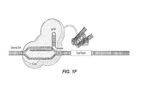
100211 FIG. 1A-FIG. 1G depict non-limiting exemplary
embodiments showing a highly
multiplexed unbiased DNA amplification method using a universal primer. FIG.
lA depicts a non-
CA 03217426 2023- 10- 31
WO 2022/241135 PCT/US2022/029023
7
limiting exemplary embodiment showing dCAS9 protein linked to a Tn5
Transposase. Fusion protein:
dCAS9 linked with Tn5 transposase (dCAS9-Tn5). FIG. 1B depicts a non-limiting
exemplary
embodiment showing a guide RNA complementary to a specific DNA target is
attached to the dCAS9.
The dCAS9 portion of a dCAS9-Tn5 is bound to a customized gRNA which is
specific to its DNA
target. FIG. 1 C depicts a non-limiting exemplary embodiment showing DNA
adaptors are attached
to the linked Tn5 transposase FIG 1D depicts a non-limiting exemplary
embodiment showing the
dCAS9 binds to the complementary sequence in the target's genomic DNA that is
specified by the
guide RNA. Then the Tn5 Transposase cuts the DNA and covalently attaches the
adaptors at the cut
site. The linked dCAS9 protein binds to its target; then, the Tn5 transposase
makes a site-specific
double-strand cut on the targeted DNA and attaches the bound adaptors to the
cut site. FIG. lE depicts
a non-limiting exemplary embodiment showing the resulting modified DNA region
has the adaptor
covalently bonded at each cut site. FIG. 1F depicts a non-limiting exemplary
embodiment showing a
second dCAS9-Tn5 molecule with a guide RNA targeting an area downstream of the
initial cut site
binds to the targeted DNA. The Tn5 transposase again cuts the DNA and
covalently attaches the
adaptors to the cut site. A second dCAS9-Tn5 unit targets a region at a
desired number of base pairs
upstream or downstream from the first cut site; it binds to the targeted area
and the Tn5 makes its
double-strand cut of the targeted DNA and attaches the bound adaptors to this
cut site. FIG. 1G depicts
a non-limiting exemplary embodiment showing the result is a piece of DNA with
the same primer
sequence at each end of the molecule. The isolated targeted DNA segment is cut
to the specific length
as programmed and is bonded on either end by identical primers. This same
process occurs
simultaneously for each unique target, resulting in multiple unique target DNA
segments each bonded
with the universal primer sequences.
100221 FIG. 2 is a non-limiting exemplary schematic of
Customized Loci-specific Library
Preparation (CLLP).
100231 FIG. 3 depicts a non-limiting exemplary embodiment
showing targeted sequencing
using genome editing tool (Cas9).
100241 FIG. 4A-FIG. 4B depict non-limiting exemplary
embodiments showing a highly
multiplexed unbiased DNA amplification method. FIG. 4A depicts a non-limiting
exemplary
embodiment showing a single tube reaction can have several Cas9-Tn5 molecules
each targeting
unique regions in one or more genomes. FIG. 4B depicts a non-limiting
exemplary embodiment
showing the result of this reaction is several DNA molecules from the targeted
regions all with the
same primer sequence at both ends of the molecules. The simultaneous DNA PCR
amplification of
the targets illustrated herein is easy to perform using a single primer pair.
CA 03217426 2023- 10- 31
WO 2022/241135 PCT/US2022/029023
8
100251 FIG. 5 depicts a non-limiting exemplary schematic of a
plasmid construct (3XFlag-
Cas9-F126-Tn5; SEQ ID NO: 1) for use in generating protein complexes provided
herein.
[0026] FIG. 6 depicts a non-limiting exemplary schematic of a
plasmid construct (3XFlag-
Cas9-xTen-Tn5; SEQ ID NO: 2) for use in generating protein complexes provided
herein.
[0027] FIG. 7 depicts a non-limiting exemplary schematic of a
plasmid construct (pET-
Tn5-xTen-dCas9; SEQ ID NO: 3) for use in generating protein complexes provided
herein.
[0028] FIG. 8 depicts the relative binding sites of exemplary
sgRNAs for S. enterica InvA
gene.
[0029] FIG. 9 depicts the relative binding sites of exemplary
sgRNAs for S. enterica FliC
gene.
[0030] FIG. 10 shows a graph of exemplary bioanalyzer data
showing that cuts in the
genomic DNA were specific to the expected size, demonstrating that the guide
RNAs for Salmonella
enterica are functional. Also, see Table 3.
[0031] FIG. 11 depicts a graph of tape station analysis
showing amplification of Tn5-
generated fragments using Adaptor A as a primer. This indicates that the
adaptor was added to the 5'
and 3' end of the cut molecules.
[0032] FIG. 12 depicts a graph of tape station analysis
showing amplification of Tn5-
generated fragments using Adaptor B as primer. This indicates that the adaptor
was added to the 5'
and 3' end of the cut molecules.
100331 FIG. 13 depicts an exemplary SDS-PAGE gel analysis of
recombinantly expressed
and purified dCas9-F126-Tn5 fusion protein. Arrow points to fusion protein
band.
100341 FIG. 14 depicts bioanalyzer analysis of an exemplary
electrophoresis gel of
recombinantly expressed and purified dCas9-F126-Tn5 fusion protein.
100351 FIG. 15 depicts an exemplary SDS-PAGE gel analysis of
recombinantly expressed
and purified dCas9-xTen-Tn5 fusion protein. Arrow points to fusion protein
band.
100361 FIG. 16 depicts bioanalyzer data from exemplary
electrophoresis analysis of
recombinantly expressed and purified dCas9-xTen-Tn5 fusion protein.
100371 FIG. 17 depicts an exemplary SDS-PAGE gel analysis of
recombinantly expressed
and purified Tn5-F126-dCas9 fusion protein. Arrow points to fusion protein
band.
100381 FIG. 18 depicts an exemplary SDS-PAGE gel analysis of
recombinantly expressed
and purified Tn5-xTen-dCas9 fusion protein. Arrow points to fusion protein
band.
100391 FIG. 19 depicts tape station analysis of amplification
reactions using catalytically
active Cas9 only (no fusion protein). No amplification is observed, suggesting
that Cas9 itself cannot
CA 03217426 2023- 10- 31
WO 2022/241135 PCT/US2022/029023
9
add adaptors to the 5' and 3' ends of digested fragments. The visible signal
is from sample incubated
with Cas9, but not subjected to PCR. The lower peak is a 100 bp size marker,
and the upper peak is
genomic DNA.
[0040] FIG. 20 depicts tape station analysis of amplification
reactions following
tagmentation reactions with dCas9-F126-Tn5. Arrow indicates signal from a
reaction subjected to
PCR conditions following incubation with Cas9-Tn5 fusion protein No gRNAs were
included in this
reaction, resulting in a broad peak indicative of random tagmentation. The
lower peak is a 100 bp size
marker, and the upper peak is genomic DNA.
[0041] FIG. 21 depicts exemplary tape station analysis of
amplification reactions
following tagmentation reactions with dCas9-xTen-Tn5. Arrow indicates signal
from a reaction
subjected to PCR conditions following incubation with Cas9-Tn5 fusion protein.
No gRNAs were
included in this reaction, resulting in a broad peak indicative of random
tagmentation. The lower peak
is a 100 bp size marker, and the upper peak is genomic DNA.
[0042] FIG. 22 depicts tape station analysis of amplification
reactions following
tagmentation with 100 nM dCas9-F126-Tn5 fusion protein. Arrow indicates signal
from reactions
subjected to PCR conditions following incubation with Cas9-Tn5 fusion protein.
The lower peak is a
100 bp size marker, and the upper peak is genomic DNA.
[0043] FIG. 23 depicts tape station analysis of amplification
reactions following
tagmentation reactions with 1 nM dCas9-F126-Tn5 fusion protein. Arrow
indicates signal from
reactions subjected to PCR conditions following incubation with Cas9-Tn5
fusion protein. The lower
peak is a 100 bp size marker, and the upper peak is genomic DNA.
[0044] FIG. 24 depicts tape station analysis of amplification
reactions following
tagmentation reactions with 100 pM dCas9-F126-Tn5 fusion protein. Arrow
indicates signal from
reactions subjected to PCR conditions following incubation with Cas9-Tn5
fusion protein. The lower
peak is a 100 bp size marker, and the upper peak is genomic DNA.
[0045] FIG. 25 depicts tape station analysis of amplification
reactions following
tagmentation reactions with 100 pM dCas9-F126-Tn5 fusion protein from FIG. 24
zoomed in.
[0046] FIG. 26 depicts tape station analysis of amplification
reactions following
tagmentation reactions with 100 pM dCas9-xTen-Tn5 fusion protein. Lower, lower
100 bp marker.
[0047] FIG. 27 depicts tape station analysis of amplification
reactions following
tagmentation reactions with 10 pM dCas9-xTen-Tn5 fusion protein. Lower, lower
100 bp marker.
[0048] FIG. 28 depicts tape station analysis of amplification
reactions following
tagmentation reactions with 1 pM dCas9-xTen-Tn5 fusion protein.
CA 03217426 2023- 10- 31
WO 2022/241135 PCT/US2022/029023
100491 FIG. 29 depicts bioanalyzer analysis of amplification
from libraries prepared by
Tn5 only tagmentation, loaded with only one adaptor (Adaptor B).
100501 FIG. 30 depicts bioanalyzer analysis of amplification
from libraries prepared by
dCas9-F126-Tn5 guided tagmentation, loaded with only one adaptor (Adaptor B).
In this experiment,
the shorter incubation protocol was used.
100511 FIG. 31 depicts bioanalyzer analysis of amplification
from libraries prepared by
dCas9-F126-Tn5 guided tagmentation, loaded with only one adaptor (Adaptor B).
In this experiment,
the longer incubation protocol was used.
100521 FIG. 32 depicts exemplary bioanalyzer analysis of
amplification from libraries
prepared by dCas9-F126-Tn5 guided tagmentation, loaded with both Adaptors A
and B. In this
experiment, the longer incubation protocol was used.
100531 FIG. 33 depicts exemplary bioanalyzer analysis of
amplification from libraries
prepared by dCas9-F126-Tn5 guided tagmentation, loaded with both Adaptors A
and B. In this
experiment, the shorter incubation protocol was used.
100541 FIG. 34 depicts an exemplary embodiment of DNA
fragments labeled with NGS
sequences adaptors using the CasTn-NEBNext Ligation based Library methods
disclosed herein.
100551 FIG. 35 depicts exemplary tape station analysis of PCR
amplification from S.
enterica genomic DNA samples incubated with dCas9-xTen-Tn5 loaded with S.
enterica sgRNAs.
Lower, lower 100 bp marker.
100561 FIG. 36 depicts exemplary tape station analysis of PCR
amplification from S.
enterica samples incubated with dCas9-xTen-Tn5 without sgRNA. Lower, lower 100
bp marker.
100571 FIG. 37 shows an illustration of a dCas9-Tn5 generated
fragment using a single
adaptor (e.g., Adaptor B).
100581 FIG. 38 depicts an illustration of a dCas9-Tn5
generated fragment from a reaction
in which the Tn5 was loaded with two different adaptors (e.g., Adaptor A and
Adaptor B).
100591 FIG. 39A-FIG. 39B depict an illustration of NEBNext
Ligation-based library
preparation for next generation sequencing. Symbols as shown in the key label
portions of adaptor
and primer sequences. Fragments generated by dCas9-Tn5 tagmentation with
NEBNext library
preparation are shown in FIG. 34.
100601 FIG. 40 depicts an illustration of tagmentation based
Nextera library preparation
for next generation sequencing.
100611 FIG. 41 depicts tagmentation based library preparation
using dCas9-Tn5 guided
tagmentati on.
CA 03217426 2023- 10- 31
WO 2022/241135 PCT/US2022/029023
11
DETAILED DESCRIPTION
[0062] In the following detailed description, reference is
made to the accompanying
drawings, which form a part hereof. In the drawings, similar symbols typically
identify similar
components, unless context dictates otherwise. The illustrative embodiments
described in the detailed
description, drawings, and claims are not meant to be limiting. Other
embodiments may be utilized,
and other changes may be made, without departing from the spirit or scope of
the subject matter
presented herein. It will be readily understood that the aspects of the
present disclosure, as generally
described herein, and illustrated in the Figures, can be arranged,
substituted, combined, separated,
and designed in a wide variety of different configurations, all of which are
explicitly contemplated
herein and made part of the disclosure herein.
[0063] All patents, published patent applications, other
publications, and sequences from
GenBank, and other databases referred to herein are incorporated by reference
in their entirety with
respect to the related technology.
[0064] Disclosed herein include compositions. In some
embodiments, the composition
comprises: a first protein complex and a second protein complex. In some
embodiments, the first
protein complex comprises a transposome and a first programmable DNA binding
unit capable of
specifically binding to a first binding site on a target double-stranded DNA
(dsDNA). In some
embodiments, the second protein complex comprises the transposome and a second
programmable
DNA binding unit capable of specifically binding to a second binding site on
the target dsDNA. In
some embodiments, the transposome comprises a transposase and two copies of an
adaptor.
[0065] Disclosed herein include compositions. In some
embodiments, the composition
comprises: a plurality of protein complex pairs, wherein each of the plurality
of protein complex pairs
comprises a first protein complex and a second protein complex. In some
embodiments, the first
protein complex comprises a transposome and a first programmable DNA binding
unit capable of
specifically binding to a first binding site on a target double-stranded DNA
(dsDNA). In some
embodiments, the second protein complex comprises the transposome and a second
programmable
DNA binding unit capable of specifically binding to a second binding site on
the target dsDNA. In
some embodiments, the transposome comprises a transposase and two copies of an
adaptor. In some
embodiments, the first binding site for each of the plurality of protein
complex pairs is different from
each other and/or the second binding site for each of the plurality of protein
complex pairs is different
from each other. In some embodiments, all of the plurality of protein complex
pairs has the same
transposome.
CA 03217426 2023- 10- 31
WO 2022/241135 PCT/US2022/029023
12
[0066] Disclosed herein include reaction mixtures. In some
embodiments, the reaction
mixture comprises: a composition disclosed herein; and sample nucleic acids
suspected of comprising
the target dsDNA. In some embodiments, the reaction mixture comprises: a DNA
polymerase; and a
plurality of dNTPs.
[0067] Disclosed herein include methods for simultaneous
detection of a plurality of
target nucleic acids In some embodiments, the method comprises. contacting
sample nucleic acids
suspected of comprising a plurality of target dsDNA with a plurality of
protein complex pairs to form
a reaction mixture, wherein each of the plurality of target dsDNA comprises a
target sequence flanked
by a first binding site on the target dsDNA and a second binding site on the
target dsDNA, and wherein
each of the protein complex pairs comprises a first protein complex and a
second protein complex. In
some embodiments, the first complex comprises a transposome and a first
programmable DNA
binding unit capable of specifically binding to a first binding site on a
target dsDNA. In some
embodiments, the second complex comprises the transposome and a second
programmable DNA
binding unit capable of specifically binding to a second binding site on the
target dsDNA. In some
embodiments, the transposome comprises a transposase and two copies of an
adaptor. In some
embodiments, the first binding site for each of the plurality of protein
complex pairs is different from
each other, the second binding site for each of the plurality of protein
complex pairs is different from
each other, or both. In some embodiments, all of the plurality of protein
complex pairs comprise the
same transposome. In some embodiments, the method comprises: incubating the
reaction mixture to
generate a plurality of dsDNA fragments each comprising the adaptor on both
ends and a target
sequence. In some embodiments, the method comprises: amplifying the plurality
of dsDNA fragments
with a primer capable of binding to one strand of the adaptor to generate
amplification products. In
some embodiments, the method comprises: detecting the presence of target
sequences in amplified
products as an indication of the presence of the plurality of target dsDNA. In
some embodiments,
detecting the presence of target sequences in amplified products comprises
contacting the amplified
products with oligonucleotide probes each capable of specifically binding to
the target sequences.
Definitions
[0068] Unless defined otherwise, technical and scientific
terms used herein have the same
meaning as commonly understood by one of ordinary skill in the art to which
the present disclosure
belongs. See, e.g. Singleton et al., Dictionary of Microbiology and Molecular
Biology 2nd ed., J.
Wiley & Sons (New York, NY 1994); Sambrook et al., Molecular Cloning, A
Laboratory Manual,
Cold Spring Harbor Press (Cold Spring Harbor, NY 1989). For purposes of the
present disclosure,
the following terms are defined below.
CA 03217426 2023- 10- 31
WO 2022/241135 PCT/US2022/029023
13
100691 As used herein, the term "adaptor" can mean a sequence
capable of facilitating
amplification or sequencing of associated nucleic acids. The associated
nucleic acids can comprise
target nucleic acids. The associated nucleic acids can comprise one or more of
spatial labels, target
labels, sample labels, indexing label, or barcode sequences (e.g., molecular
labels). The adaptors can
be linear. The adaptors can be pre-adenylated adaptors. The adaptors can be
double- or single-
stranded One or more adaptor can be located on the 5' or 3' end of a nucleic
acid When the adaptors
comprise known sequences on the 5' and 3' ends, the known sequences can be the
same or different
sequences. An adaptor located on the 5' and/or 3' ends of a polynucleotide can
be capable of
hybridizing to one or more oligonucleotides immobilized on a surface. An
adaptor can, in some
embodiments, comprise a universal sequence. A universal sequence can be a
region of nucleotide
sequence that is common to two or more nucleic acid molecules. The two or more
nucleic acid
molecules can also have regions of different sequence. Thus, for example, the
5' adaptors can
comprise identical and/or universal nucleic acid sequences and the 3' adaptors
can comprise identical
and/or universal sequences. A universal sequence that may be present in
different members of a
plurality of nucleic acid molecules can allow the replication or amplification
of multiple different
sequences using a single universal primer that is complementary to the
universal sequence. Similarly,
at least one, two (e.g., a pair) or more universal sequences that may be
present in different members
of a collection of nucleic acid molecules can allow the replication or
amplification of multiple
different sequences using at least one, two (e.g., a pair) or more single
universal primers that are
complementary to the universal sequences. Thus, a universal primer includes a
sequence that can
hybridize to such a universal sequence. The target nucleic acid sequence-
bearing molecules may be
modified to attach universal adaptors (e.g., non-target nucleic acid
sequences) to one or both ends of
the different target nucleic acid sequences. The one or more universal primers
attached to the target
nucleic acid can provide sites for hybridization of universal primers. The one
or more universal
primers attached to the target nucleic acid can be the same or different from
each other.
100701 As used herein the term "associated" or "associated
with" can mean that two or
more species are identifiable as being co-located at a point in time. An
association can mean that two
or more species are or were within a similar container. An association can be
an informatics
association. For example, digital information regarding two or more species
can be stored and can be
used to determine that one or more of the species were co-located at a point
in time. An association
can also be a physical association. In some embodiments, two or more
associated species are
"tethered", "attached", or "immobilized" to one another or to a common solid
or semisolid surface.
An association may refer to covalent or non-covalent means for attaching
labels to solid or semi-solid
CA 03217426 2023- 10- 31
WO 2022/241135 PCT/US2022/029023
14
supports such as beads. An association may be a covalent bond between a target
and a label. An
association can comprise hybridization between two molecules (such as a target
molecule and a label).
100711 As used herein, the term "complementary" can refer to
the capacity for precise
pairing between two nucleotides. For example, if a nucleotide at a given
position of a nucleic acid is
capable of hydrogen bonding with a nucleotide of another nucleic acid, then
the two nucleic acids are
considered to be complementary to one another at that position Complementarity
between two single-
stranded nucleic acid molecules may be "partial," in which only some of the
nucleotides bind, or it
may be complete when total complementarity exists between the single-stranded
molecules. A first
nucleotide sequence can be said to be the "complement" of a second sequence if
the first nucleotide
sequence is complementary to the second nucleotide sequence. A first
nucleotide sequence can be
said to be the "reverse complement" of a second sequence, if the first
nucleotide sequence is
complementary to a sequence that is the reverse (i.e., the order of the
nucleotides is reversed) of the
second sequence. As used herein, a "complementary- sequence can refer to a
"complement- or a
"reverse complement" of a sequence. It is understood from the disclosure that
if a molecule can
hybridize to another molecule it may be complementary, or partially
complementary, to the molecule
that is hybridizing.
100721 As used herein, the term "digital counting" can refer
to a method for estimating a
number of target molecules in a sample. Digital counting can include the step
of determining a number
of unique labels that have been associated with targets in a sample. This
methodology, which can be
stochastic in nature, transforms the problem of counting molecules from one of
locating and
identifying identical molecules to a series of yes/no digital questions
regarding detection of a set of
predefined labels.
100731 As used herein, the term "nucleic acid" refers to a
polynucleotide sequence, or
fragment thereof A nucleic acid can comprise nucleotides. A nucleic acid can
be exogenous or
endogenous to a cell. A nucleic acid can exist in a cell-free environment. A
nucleic acid can be a gene
or fragment thereof. A nucleic acid can be DNA. A nucleic acid can be RNA. A
nucleic acid can
comprise one or more analogs (e.g., altered backbone, sugar, or nucleobase).
Some non-limiting
examples of analogs include: 5-bromouracil, peptide nucleic acid, xeno nucleic
acid, morpholinos,
locked nucleic acids, glycol nucleic acids, threose nucleic acids,
dideoxynucleotides, cordycepin, 7-
deaza-GTP, fluorophores (e.g., rhodamine or fluorescein linked to the sugar),
thiol containing
nucleotides, biotin linked nucleotides, fluorescent base analogs, CpG islands,
methyl-7-guanosine,
methylated nucleotides, inosine, thiouri dine, pseudouridine, dihydrouridine,
queuosine, and wyosine.
"Nucleic acid", "polynucleotide, "target polynucleotide", and "target nucleic
acid" can be used
CA 03217426 2023- 10- 31
WO 2022/241135 PCT/US2022/029023
interchangeably.
100741 A nucleic acid can comprise one or more modifications
(e.g., a base modification,
a backbone modification), to provide the nucleic acid with a new or enhanced
feature (e.g., improved
stability). A nucleic acid can comprise a nucleic acid affinity tag. A
nucleoside can be a base-sugar
combination. The base portion of the nucleoside can be a heterocyclic base.
The two most common
classes of such heterocyclic bases are the purines and the pyrimidines
Nucleotides can be nucleosides
that further include a phosphate group coyalently linked to the sugar portion
of the nucleoside. For
those nucleosides that include a pentofuranosyl sugar, the phosphate group can
be linked to the 2',
the 3', or the 5' hydroxyl moiety of the sugar. In forming nucleic acids, the
phosphate groups can
covalently link adjacent nucleosides to one another to form a linear polymeric
compound. In turn, the
respective ends of this linear polymeric compound can be further joined to
form a circular compound;
however, linear compounds are generally suitable. In addition, linear
compounds may have internal
nucleotide base complementarity and may therefore fold in a manner as to
produce a fully or partially
double-stranded compound. Within nucleic acids, the phosphate groups can
commonly be referred to
as forming the internucleoside backbone of the nucleic acid. The linkage or
backbone can be a 3' to
5' phosphodiester linkage.
100751 A nucleic acid can comprise a modified backbone and/or
modified internucleoside
linkages. Modified backbones can include those that retain a phosphorus atom
in the backbone and
those that do not have a phosphorus atom in the backbone. Suitable modified
nucleic acid backbones
containing a phosphorus atom therein can include, for example,
phosphorothioates, chiral
phosphorothioates, phosphorodithioates, phosphotriesters, aminoalkyl
phosphotriesters, methyl and
other alkyl phosphonate such as 3'-alkylene phosphonates, 5'-alkylene
phosphonates, chiral
phosphonates, phosphinates, phosphoramidates including 3' -amino
phosphoramidate and aminoalkyl
phosphoramidates, phosphorodiamidates, thionophosphoramidates,
thionoalkylphosphonates,
thi onoalkylphosphotri esters, selenophosphates, and boranophosphates having
normal 3'-5' linkages,
2'-5' linked analogs, and those having inverted polarity wherein one or more
internucleotide linkages
is a 3' to 3', a 5' to 5' or a 2' to 2' linkage.
100761 A nucleic acid can comprise polynucleoti de backbones
that are formed by short
chain alkyl or cycloalkyl internucleoside linkages, mixed heteroatom and alkyl
or cycloalkyl
internucleoside linkages, or one or more short chain heteroatomic or
heterocyclic internucleoside
linkages. These can include those having morpholino linkages (formed in part
from the sugar portion
of a nucleoside); siloxane backbones; sulfide, sulfoxide and sulfone
backbones; formacetyl and
thioformacetyl backbones; methylene formacetyl and thioformacetyl backbones;
riboacetyl
CA 03217426 2023- 10- 31
WO 2022/241135 PCT/US2022/029023
16
backbones; alkene containing backbones; sulfamate backbones; methyleneimino
and
methylenehydrazino backbones; sulfonate and sulfonamide backbones; amide
backbones; and others
having mixed N, 0, S and CH2 component parts.
100771 A nucleic acid can comprise a nucleic acid mimetic. The
term "mimetic" can be
intended to include polynucleotides wherein only the furanose ring or both the
furanose ring and the
internucleotide linkage are replaced with non-furanose groups, replacement of
only the furanose ring
can also be referred as being a sugar surrogate. The heterocyclic base moiety
or a modified
heterocyclic base moiety can be maintained for hybridization with an
appropriate target nucleic acid.
One such nucleic acid can be a peptide nucleic acid (PNA). In a PNA, the sugar-
backbone of a
polynucleotide can be replaced with an amide containing backbone, in
particular an
aminoethylglycine backbone. The nucleotides can be retained and are bound
directly or indirectly to
aza nitrogen atoms of the amide portion of the backbone. The backbone in PNA
compounds can
comprise two or more linked aminoethylglycine units which gives PNA an amide
containing
backbone. The heterocyclic base moieties can be bound directly or indirectly
to aza nitrogen atoms
of the amide portion of the backbone.
100781 A nucleic acid can comprise a morpholino backbone
structure. For example, a
nucleic acid can comprise a 6-membered morpholino ring in place of a ribose
ring. In some of these
embodiments, a phosphorodiamidate or other non-phosphodiester internucleoside
linkage can replace
a phosphodiester linkage.
100791 A nucleic acid can comprise linked morpholino units
(e.g., morpholino nucleic
acid) having heterocyclic bases attached to the morpholino ring. Linking
groups can link the
morpholino monomeric units in a morpholino nucleic acid. Non-ionic morpholino-
based oligomeric
compounds can have less undesired interactions with cellular proteins.
Morpholino-based
polynucleotides can be nonionic mimics of nucleic acids. A variety of
compounds within the
morpholino class can be joined using different linking groups. A further class
of polynucleotide
mimetic can be referred to as cyclohexenyl nucleic acids (CeNA). The furanose
ring normally present
in a nucleic acid molecule can be replaced with a cyclohexenyl ring. CeNA DMT
protected
phosphoramidite monomers can be prepared and used for oligomeric compound
synthesis using
phosphoramidite chemistry. The incorporation of CeNA monomers into a nucleic
acid chain can
increase the stability of a DNA/RNA hybrid. CeNA oligoadenylates can form
complexes with nucleic
acid complements with similar stability to the native complexes. A further
modification can include
Locked Nucleic Acids (LNAs) in which the 2'-hydroxyl group is linked to the 4'
carbon atom of the
sugar ring thereby forming a 2'-C, 4' -C-oxymethylene linkage thereby forming
a bicyclic sugar
CA 03217426 2023- 10- 31
WO 2022/241135
PCT/US2022/029023
17
moiety. The linkage can be a methylene (-CH2), group bridging the 2' oxygen
atom and the 4' carbon
atom wherein n is 1 or 2. LNA and LNA analogs can display very high duplex
thermal stabilities with
complementary nucleic acid (Tm=+3 to +10 C), stability towards 3'-
exonucleolytic degradation and
good solubility properties.
[0080] A nucleic acid may also include nucleobase (often
referred to simply as "base")
modifications or substitutions As used herein, "unmodified" or "natural"
nucleobases can include the
purine bases, (e.g., adenine (A) and guanine (G)), and the pyrimidine bases,
(e.g., thymine (T),
cytosine (C) and uracil (U)). Modified nucleobases can include other synthetic
and natural
nucleobases such as 5-methylcytosine (5-me-C), 5-hydroxymethyl cytosine,
xanthine, hypoxanthine,
2-aminoadenine, 6-methyl and other alkyl derivatives of adenine and guanine, 2-
propyl and other
alkyl derivatives of adenine and guanine, 2-thiouracil, 2-thiothymine and 2-
thiocytosine, 5-halouracil
and cytosine, 5-propynyl (¨C=C¨CH3) uracil and cytosine and other alkynyl
derivatives of
pyrimidine bases, 6-azo uracil, cytosine and thymine, 5-uracil (pseudouracil),
4-thiouracil, 8-halo, 8-
amino, 8-thiol, 8-thioalkyl, 8-hydroxyl and other 8-substituted adenines and
guanines, 5-halo
particularly 5-bromo, 5-trifluoromethyl and other 5-substituted uracils and
cytosines, 7-
methylguanine and 7-methyladenine, 2-F-adenine, 2-aminoadenine, 8-azaguanine
and 8-azaadenine,
7-deazaguanine and 7-deazaadenine and 3-deazaguanine and 3-deazaadenine.
Modified nucleobases
can include tricyclic pyrimidines such as phenoxazine cytidine(1H-pyrimido(5,4-
b)(1,4)benzoxazin-
2(3H)-one), phenothiazine cytidine (1H-pyrimido(5,4-b)(1,4)benzothiazin-2(3H)-
one), G-clamps
such as a substituted phenoxazine cytidine (e.g., 9-(2-aminoethoxy)-H-
pyrimido(5,4-(b)
(1,4)benzoxazin-2(3H)-one), phenothiazine cytidine (1H-pyrimido(5,4-
b)(1,4)benzothiazin-2(3H)-
one), G-clamps such as a substituted phenoxazine cytidine (e.g., 9-(2-
aminoethoxy)-H-pyrimido(5,4-
(b) (1,4)benzoxazin-2(3H)-one), carbazole cytidine (2H-pyrimido(4,5-b)indo1-2-
one), pyridoindole
cyti dine (H-pyrido(3 ',2' :4, 5)pyrrolo[2, 3 -d]pyrimidin-2-one).
[0081] As used here, the term "target" can refer to a nucleic
acid of interest (e.g., target
dsDNA). In some embodiments, targets can be associated with an adaptor and/or
a barcode.
Exemplary suitable targets for analysis by the disclosed methods, devices, and
systems include
oligonucleotides, DNA, RNA, mRNA, microRNA, tRNA, and the like. Targets can be
single or
double stranded. In some embodiments, targets can be proteins, peptides, or
polypeptides. In some
embodiments, targets are lipids. As used herein, "target" can be used
interchangeably with "species."
[0082] As used herein, the term "reverse transcriptases" can
refer to a group of enzymes
having reverse transcriptase activity (i.e., that catalyze synthesis of DNA
from an RNA template). In
general, such enzymes include, but are not limited to, retroviral reverse
transcriptase, retrotransposon
CA 03217426 2023- 10- 31
WO 2022/241135 PCT/US2022/029023
18
reverse transcriptase, retroplasmid reverse transcriptases, retron reverse
transcriptases, bacterial
reverse transcriptases, group II intron-derived reverse transcriptase, and
mutants, variants or
derivatives thereof. Non-retroviral reverse transcriptases include non-LTR
retrotransposon reverse
transcriptases, retroplasmid reverse transcriptases, retron reverse
transciptases, and group II intron
reverse transcriptases. Examples of group II intron reverse transcriptases
include the factococcus
lactic LI_LtrB intron reverse transcriptase, the Thermosynechococcits elongatu
s TeI4c intron reverse
transcriptase, or the Geobac,llus stearothermophillis GsI-IIC intron reverse
transcriptase. Other
classes of reverse transcriptases can include many classes of non-retroviral
reverse transcriptases (i.e.,
retrons, group II introns, and diversity-generating retroelements among
others).
100831 As used herein, the term "isolate nucleic acids" can
refer to the purification of
nucleic acids from one or more cellular components. One of skill in the art
will appreciate that samples
processed to "isolate nucleic acids- therefrom can include components and
impurities other than
nucleic acids. Samples that comprise isolated nucleic acids can be prepared
from specimens using
any acceptable method known in the art. For example, cells can be lysed using
known lysis agents,
and nucleic acids can be purified or partially purified from other cellular
components. Suitable
reagents and protocols for DNA and RNA extractions can be found in, for
example, US20100009351,
and US20090131650, respectively (each of which is incorporated herein by
reference in its entirety).
100841 As used herein, "template" can refer to all or part of
a polynucleotide containing
at least one target nucleotide sequence.
100851 As used herein, a "primer" can refer to a
polynucleotide that can serve to initiate a
nucleic acid chain extension reaction. The length of a primer can vary, for
example, from about 5 to
about 100 nucleotides, from about 10 to about 50 nucleotides, from about 15 to
about 40 nucleotides,
or from about 20 to about 30 nucleotides. The length of a primer can be about
10 nucleotides, about
20 nucleotides, about 25 nucleotides, about 30 nucleotides, about 35
nucleotides, about 40
nucleotides, about 50 nucleotides, about 75 nucleotides, about 100
nucleotides, or a range between
any two of these values. In some embodiments, the primer has a length of 10 to
about 50 nucleotides,
i.e., 10, 11, 12, 13, 14, 15, 16, 17, 18, 19, 20, 21, 22, 23, 24, 25 26, 27,
28, 29, 30, 31, 32, 33, 34, 35,
36, 37, 38, 39, 40, 41, 42, 43, 44, 45, 46, 47, 48, 49, 50, or more
nucleotides. In some embodiments,
the primer has a length of 18 to 32 nucleotides.
100861 As used herein, a "probe" can refer to an
polynucleotide that can hybridizes (e.g.,
specifically) to a target sequence in a nucleic acid, under conditions that
allow hybridization, thereby
allowing detection of the target sequence or amplified nucleic acid. A probe's
"target" generally refers
to a sequence within or a subset of an amplified nucleic acid sequence which
hybridizes specifically
CA 03217426 2023- 10- 31
WO 2022/241135 PCT/US2022/029023
19
to at least a portion of a probe oligomer by standard hydrogen bonding (i.e.,
base pairing). A probe
may comprise target-specific sequences and other sequences that contribute to
three-dimensional
conformation of the probe. Sequences are -sufficiently complementary" if they
allow stable
hybridization in appropriate hybridization conditions of a probe oligom er to
a target sequence that is
not completely complementary to the probe's target-specific sequence. The
length of a probe can vary,
for example, from about 5 to about 100 nucleotides, from about 10 to about 50
nucleotides, from
about 15 to about 40 nucleotides, or from about 20 to about 30 nucleotides.
The length of a probe can
be about 10 nucleotides, about 20 nucleotides, about 25 nucleotides, about 30
nucleotides, about 35
nucleotides, about 40 nucleotides, about 50 nucleotides, about 100
nucleotides, or a range between
any two of these values. In some embodiments, the probe has a length of 10 to
about 50 nucleotides.
For example, the primers and or probes can be at least 10, 11, 12, 13, 14, 15,
16, 17, 18, 19, 20, 21,
22, 23, 24, 25 26, 27, 28, 29, 30, 31, 32, 33, 34, 35, 36, 37, 38, 39, 40, 41,
42, 43, 44, 45, 46, 47, 48,
49, 50, or more nucleotides. In some embodiments, the probe can be non-
sequence specific.
[0087] Preferably, the primers and/or probes can be between 8
and 45 nucleotides in
length. For example, the primers and or probes can be at least 8, 9, 10, 11,
12, 13, 14, 15, 16, 17, 18,
19, 20, 21, 22, 23, 24, 25, 26, 27, 28, 29, 30, 31, 32, 33, 34, 35, 36, 37,
38, 39, 40, 41, 42, 43, 44, 45,
or more nucleotides in length. The primer and probe can be modified to contain
additional nucleotides
at the 5' or the 3' terminus, or both. One of skill in the art will appreciate
that additional bases to the
3' terminus of amplification primers (not necessarily probes) are generally
complementary to the
template sequence. The primer and probe sequences can also be modified to
remove nucleotides at
the 5' or the 3' terminus. One of skill in the art will appreciate that in
order to function for
amplification, the primers or probes will be of a minimum length and annealing
temperature as
disclosed herein.
[0088] Primers and probes can bind to their targets at an
annealing temperature, which is
a temperature less than the melting temperature (Tm). As used herein, "Tm" and
"melting temperature"
are interchangeable terms which refer to the temperature at which 50% of a
population of double-
stranded polynucleotide molecules becomes dissociated into single strands. The
formulae for
calculating the Tm of polynucleotides are well known in the art. For example,
the Tm may be calculated
by the following equation. Tm = 69.3+0.41 > (G+C)%-6- SOIL, wherein L is the
length of the probe
in nucleotides. The Tm of a hybrid polynucleotide may also be estimated using
a formula adopted
from hybridization assays in 1 M salt, and commonly used for calculating Tm
for PCR primers:
[(number of A+T) 2 C + (number of G+C) x 4 C]. See, e.g., C. R. Newton et al.
PCR, 2nd ed.,
Springer-Verlag (New York: 1997), p.24 (incorporated by reference in its
entirety, herein). Other
CA 03217426 2023- 10- 31
WO 2022/241135 PCT/US2022/029023
more sophisticated computations exist in the art, which take structural as
well as sequence
characteristics into account for the calculation of Tm. The melting
temperature of an oligonucleotide
can depend on complementarity between the oligonucleotide primer or probe and
the binding
sequence, and on salt conditions. In some embodiments, an oligonucleotide
primer or probe provided
herein has a Tm of less than about 90 C in 50mM KC1, 10 mM Tris-HC1 buffer,
for example about
89 C, 88, 87, 86, 85, 84, 83, 82, 81, 80 79, 78, 77, 76, 75, 74, 73, 72, 71,
70, 69, 68, 67, 66, 65, 64,
63, 62, 61, 60, 59, 58, 57, 56, 55, 54, 53, 52, 50, 49, 48, 47, 46, 45, 44,
43, 42, 41, 40, 39 C, or less,
including ranges between any two of the listed values.
100891 In some embodiments, the primers disclosed herein,
e.g., amplification primers,
can be provided as an amplification primer pair, e.g., comprising a forward
primer and a reverse
primer (first amplification primer and second amplification primer).
Preferably, the forward and
reverse primers have Tm's that do not differ by more than 10 C, e.g., that
differ by less than 10 C,
less than 9 C, less than 8 C, less than 7 C, less than 6 C, less than 5 C,
less than 4 C, less than 3 C,
less than 2 C, or less than 1 C.
100901 The primer and probe sequences may be modified by
having nucleotide
substitutions (relative to the target sequence) within the oligonucleotide
sequence, provided that the
oligonucleotide contains enough complementarity to hybridize specifically to
the target nucleic acid
sequence. In this manner, at least 1, 2, 3, 4, or up to about 5 nucleotides
can be substituted. As used
herein, the term "complementary" can refer to sequence complementarity between
regions of two
polynucleotide strands or between two regions of the same polynucleotide
strand. A first region of a
polynucleotide is complementary to a second region of the same or a different
polynucleotide if, when
the two regions are arranged in an antiparallel fashion, at least one
nucleotide of the first region is
capable of base pairing with a base of the second region. Therefore, it is not
required for two
complementary polynucleotides to base pair at every nucleotide position.
"Fully complementary" can
refer to a first polynucleotide that is 100% or -fully" complementary to a
second polynucleotide and
thus forms a base pair at every nucleotide position. "Partially complementary"
also can refer to a first
polynucleotide that is not 100% complementary (e.g_, 90%, or 80% or 70%
complementary) and
contains mismatched nucleotides at one or more nucleotide positions. In some
embodiments, an
oligonucleotide includes a universal base.
100911 As used herein, the term "sufficiently complementary"
can refer to a contiguous
nucleic acid base sequence that is capable of hybridizing to another base
sequence by hydrogen
bonding between a series of complementary bases. Complementary base sequences
can be
CA 03217426 2023- 10- 31
WO 2022/241135 PCT/US2022/029023
21
complementary at each position in the oligomer sequence by using standard base
pairing (e.g., G:C,
A:T or A: U) or can contain one or more residues that are not complementary
(including abasic
positions), but in which the entire complementary base sequence is capable of
specifically hybridizing
with another base sequence in appropriate hybridization conditions. Contiguous
bases can be at least
about 80%, at least about 85%, at least about 90%, at least about 95%, at
least about 99%, or 100%
complementary to a sequence to which an oligomer is intended to hybridize
Substantially
complementary sequences can refer to sequences ranging in percent identity
from 100, 99, 98, 97, 96,
95, 94, 93, 92, 91, 90, 89, 88, 87, 86, 85, 84, 83, 82, 81, 80, 75, 70 or
less, or any number in between,
compared to the reference sequence. A skilled artisan can readily choose
appropriate hybridization
conditions which can be predicted based on base sequence composition, or be
determined by using
routine testing (see e.g., Green and Sambrook, Molecular Cloning, A Laboratory
Manual, 4th ed.
(Cold Spring Harbor Laboratory Press, Cold Spring Harbor, N.Y., 2012)).
100921 As used herein, the term "multiplex PCR- refers to a
type of PCR where more than
one set of primers is included in a reaction allowing one single target, or
two or more different targets,
to be amplified in a single reaction vessel (e.g., tube). The multiplex PCR
can be, for example, a real-
time PCR.
100931 Provided herein includes a nucleic acid amplification
method that enables a highly-
multiplexed single-primer PCR reaction that is unbiased and highly sensitive
and specific. In some
embodiments, the methods, compositions, kits and systems disclosed herein can
enable a single-tube
reaction with unlimited DNA targets. In some embodiments, a fusion protein
composed of a dead
CRISPR-associated protein (dCAS) linked with a transposase (Tn5) can be used
to generate many
unique, custom, ready-to-PCR-amplify DNA targets with a single universal
primer, and thus
eliminates many limitations of conventional multiplexed PCR. PCR is a
molecular biology technique
for exponentially amplifying small, specific sections of DNA amplicons through
the use of target-
specific primers. Multiplex-PCR is the exponential amplification of more than
one DNA target
simultaneously, and conventional multiplex PCR requires a unique primer pair
for each target, and
typically maxes out at 5-10 targets, e.g., 10 target DNA strands, 20 primers.
The use of multiple
primers creates a number of problems, as discussed below.
100941 The present disclosure describes methods, compositions,
kits and system for
highly multiplexed PCR reactions that occur in a way that completely
eliminates most of the
limitations of conventional multiplex-PCR. By utilizing a single primer pair
for unlimited targets, this
method enables the optimized, highly-multiplexed PCR reaction through using,
for example, a fusion
protein composed of a dCAS protein linked to a primer-preloaded Tn5
Transposase. In some
CA 03217426 2023- 10- 31
WO 2022/241135 PCT/US2022/029023
22
embodiments, the methods, compositions, kits and systems can be used for
amplifying, 2, 5, 10, 15,
20, 30, 35, or a number or a range between any two of these numbers, DNA
targets using only one
primer pair.
100951 In some embodiments, the dCAS9 protein binds to the
target DNA, and its attached
Tn5 transposase makes a DNA double-strand cut and binds both cut ends with the
preloaded primer.
Instead of paired primers floating around until they encounter a match to bind
with, the method
disclosed herein has the advantage of high specificity and sensitivity of a
dCAS9 protein programmed
with guide RNA rapidly seeking their specific DNA targets. For example, when
the dCAS9 identifies
its target, it binds at that specific site; the transposase, now activated,
efficiently makes the specific
cut of the targeted point in the DNA strand and attaches the primers to both
cut ends. All the targets
are bound with the same primer; dCAS9 pairs can be programmed to identify and
bind with as many
unique targets as desired. Since the primers are bound to the Tn5 transposase,
the limitations posed
by dimers (primer-primer binding) are eliminated. Since all the targets are
bound with a single primer,
amplification can be truly optimized and uniform.
100961 For amplification of any nucleic acid (e.g., DNA)
target, two dCAS9-Tn5 bonded
proteins can be configured to identify, cut and apply primers to that target.
The first bonded protein
unit is programmed with a specific gRNA to identify and bind the desired DNA
target. The second
dCAS9-Tn5 bonded protein unit is programmed with a complementary gRNA, which
targets the same
area as the first gRNA but is programmed to bond to its target at a space of
several base pairs (e.g., <
300bp) upstream or downstream from the first dCAS9-Tn5 bonded unit. Regardless
of how the
dCAS9 protein pairs are programmed for their unique targets, all the Tn5
transposase molecules can
be loaded identically with two short DNA primers. The end result of the
actions of the bonded protein
unit will be the selected DNA segment with primers attached at each end. (FIG.
1A-FIG. 1G).
TABLE 1: COMPARISON OF CONVENTIONAL MULTIPLEX PCR AND MULTIPLEX PCR
METHODS DISCLOSED HEREIN
Conventional Multiplex PCR
Methods
Multiplex PCR disclosed
herein
Ability to target all genomic regions No Yes
Formation of Primer Dimers Yes No
Biased amplification Yes No
Complex Primer Design Yes No
Hands-On Complexity Yes No
Low amplification efficiency Yes No
Chance of Failed Reactions Yes Very low
Self-Inhibition Yes Very low
CA 03217426 2023- 10- 31
WO 2022/241135 PCT/US2022/029023
23
100971 Challenges and limitations for conventional multiplex
PCR are well known in the
art, and have been described in, for example
https://www.lcsciences.com/discovery/overcome-
common-challenges-to-multipl ex-per-with-innovative-relay-per-and-omega-primer-
technol ogi es/.
Non-limiting examples of advantages of the multiplex PCR methods disclosed
herein, including by
dCAStellaTn5 (Constellations) method, are described below.
Limited flexibility of target regions due to PCR thermodynamics
100981 This is a problem related to the widely varying optimal
function of each specific
primer when using multiple primers simultaneously. This challenge under
current practice makes it
necessary to maintain similar melting temperatures across all primers, avoid
complementary or
similar DNA target sequences, and to minimize cross-hybridization in target
selection due to primer
non-specificity. As the methods disclosed herein allow the use of a single
primer, in some
embodiments, the user can select for any number of targets, and any target
whatsoever due to the high
sensitivity and specificity of the dCAS9.
Formation of primer-primer dimers
100991 This is a challenging PCR limitation, as dimers clog
the gears of PCR in a number
of ways. Primer-primer dimers are when primers bind to each other instead of
their DNA targets.
When primers bind to each other, they're not available to for amplification of
their target. With
amplification, the dimers (primer-primer chains) are amplified while the
uncaptured targets are not,
contributing to uneven amplification. Additionally, the presence of these
dimers in an amplified
sample creates "noise," sort of like static that can muddy the picture of
results. As the primers used
in the methods disclosed herein are not free-floating (e.g., floating alone in
the reaction solution) but
are bound to a protein (e.g., a transposase like Tn5), no dimers are formed.
In some embodiments, the
dCAS9 protein binds only to its nucleic acid (e.g., DNA) target; once bound,
the complex (e.g.,
dCAS9-Tn5) units stay bound, so no additional "noise" is introduced.
Bias due to primer induced variability
101001 The methods disclosed herein require, in some
embodiments, only one primer or
one primer pair, so the thermodynamics of the reaction can be optimized to the
single primer (or
primer pair). Again, this is important because conventional multiplexing
requires that many unique
and finicky primers be subject to a single temperature, resulting in uneven
functionality and
contributing to uneven amplification, or even the inability to target specific
regions whose primers
require conditions outside of the preset range. With dCAStellaTn5, any region
can be targeted and
the reaction conditions can be set to optimize that one single primer's
function. While PCR is
CA 03217426 2023- 10- 31
WO 2022/241135 PCT/US2022/029023
24
considered to produce quantifiable results, the inherent primer-induced
variability limits this capacity.
With a universal primer, the consistent reliability of results enhances the
capacity for quantification.
Complexity of primer design
[0101] The methods disclosed herein allow the users to pick a
favorite primer, and also
choose any and as many targets as desired without compromise to allow multiple
primers to work
under the same condition
Expense and long lead time associated with synthesis of many custom oligos
[0102] Again, with the compositions disclosed herein (e.g.,
dCAStellaTn5) the user can
pick the simplest, cheapest, favorite primer. While the guide RNAs that
program the dCAS9 proteins
do need to be synthesized, customizable guide RNAs are readily and widely
available commercially.
Hands-on procedure complexity
[0103] Without primers in solution, the cleanup steps that
often are part of current
multiplexing PCR are eliminated.
[0104] The highly multiplexed PCR methods disclosed herein and
without many
limitations of the conventional multiplex PCR currently available opens the
door to countless
embodiments and would be widely transformative across many industries. The
method is generally
applicable to many fields, e.g., medical diagnostics in view of its broad
applicability, high sensitivity
and specificity, and its capacity for detection of large numbers of targets.
The method easily evaluates
a broad menu of specimen types for infectious disease (ID), for any pathogen
that has DNA (bacteria,
fungus, protozoa, and DNA viruses) and is compatible with specimen types that
are used for major
ID syndromes. This method, as with similar platforms, can be used with DNA
extraction,
amplification, and/or detection steps. Currently, the entire process from
sample to
amplification/detection results can take approximately 90 minutes. The method
disclosed herein can
be utilized with any PCR amplification and detection platform, making its
benefits easily accessible
to the acute care customer but with unmatched quality and reliability
alongside limitless DNA targets
in any range. Instrumentation is not a barrier in terms of space or cost
because existing platforms are
compatible. This method can be integrated into a platform to optimize end-to-
end user experience. Its
wide accessibility to existing and potentially customized platforms combined
with the inherent
improvement in specificity, sensitivity, and the elimination of the
limitations of conventional
multiplex PCR, offers diagnostic capacity and reliability.
[0105] The nucleic acid amplification techniques that the
methods disclosed herein for the
amplification of multiple DNA targets can use can vary, including but not
limited to isothermal DNA
amplification techniques, such as LAMP, RPA, SDA, and HDA. Various
transposases (including the
CA 03217426 2023- 10- 31
WO 2022/241135 PCT/US2022/029023
hyperactive transposases) can be used in the method disclosed herein, for
example Tn5 transposase,
mariner Tel-like transposon, Himar1C9 transposase, Sleeping Beauty
transposase, the Tn7
transposon, and a combination thereof. The inserted primer regions can be
labeled, for example, with
one or more anionic, cationic, neutral, fluorescent, optical, or magnetic
particles. The labelled
molecules can, for example, have two different tags per molecule created
(e.g., a magnetic tag in one
end of the DNA molecule and a fluorescent tag in the other end: for separation
and visualization of
single molecules). As another example, an avidin tag can be present in one end
of the DNA molecule
and a fluorescent tag can be present in the other end: for capture and imaging
of single molecules. Or
similarly, an avidin tag in one end of the DNA molecule and a ferrocene
molecule in the other end:
for capture and chemical imaging. Adding different tags to each side of a
molecule can lead to many
different variations and alternatives. The molecules can be separated via, gel
or capillary
electrophoresis and the color detected like qPCR. Alternatives to the dCas9
protein can be used for
the programmable DNA binding activity, including Zinc Fingers that are not
bound to the FOK1
nuclease can be used. In some embodiments, a TALEN molecule without the FOK1
nuclease can be
used. Additional non-limiting examples of CAS proteins that can be used in the
methods,
compositions, kits and systems disclosed herein include, but are not limited
to, CAS12, CAS13 and
CAS14. A recombinase in conjunction with a sequence specific primer can, in
some embodiments,
be used as a programmable DNA binding molecule. The method can comprise use of
a genome editing
tool as a programmable tool to target specific areas of a genome and the use
of a transposase to cut
and paste adaptors needed for creating a sequencing library (See, FIG. 2 for
illustration of an
exemplary embodiment using the Oxford Nanopore system). In some embodiments,
targeted
sequencing method can be used by using the genome editing tools (e.g., Cas
proteins, Zinc Finger
Nucleases (ZFN), Transcription activator-like effector nucleases (TALEN),
Argonaute proteins)
without the assistance of the transposase. This can result in a programmable
fragmentation method of
nucleic acids (FIG. 3), that can further be used for making a loci-specific
sequencing library.
101061 Provided herein include a DNA amplification method for
an unbiased highly
multiplexed single primer reaction enabled by using a fusion protein that is
composed of a dead
CRISPR-associated (dCAS) protein linked to a Tn5 transposase to generate
custom, ready-to-PCR-
amplify DNA targets with a single universal primer.
101071 Each of the following patent application publications
and references is hereby
incorporated by reference in its entirety: US20200377881A1, US20200190487A1,
US20190093090A1, and US20180305683A1; Sway P. Chen and Harris H. Wang, An
Engineered
Cas-Transposon System for Programmable and Site-Directed DNA Transpositions,
The CRISPR
CA 03217426 2023- 10- 31
WO 2022/241135 PCT/US2022/029023
26
Journal Volume 2, Number 6, 2019; Hatice S. Kaya-Okur Et Al, CUT&Tag for
efficient epigenomic
profiling of small samples and single cells, Nature Communications, (2019)
10:1930; Simone Picelli
Et Al, Tn5 transposase and tagmentation procedures for massively scaled
sequencing projects,
Genome Research, 24:2033-2040.
101081 There are provided, in some embodiments, compositions.
In some embodiments,
the composition comprises. a first protein complex and a second protein
complex In some
embodiments, the first protein complex comprises a transposome and a first
programmable DNA
binding unit capable of specifically binding to a first binding site on a
target double-stranded DNA
(dsDNA). In some embodiments, the second protein complex comprises the
transposome and a
second programmable DNA binding unit capable of specifically binding to a
second binding site on
the target dsDNA. In some embodiments, the transposome comprises a transposase
and two copies
of an adaptor.
101091 There are provided, in some embodiments, compositions.
In some embodiments,
the composition comprises: a plurality of protein complex pairs, wherein each
of the plurality of
protein complex pairs comprises a first protein complex and a second protein
complex. In some
embodiments, the first protein complex comprises a transposome and a first
programmable DNA
binding unit capable of specifically binding to a first binding site on a
target dsDNA. In some
embodiments, the second protein complex comprises the transposome and a second
programmable
DNA binding unit capable of specifically binding to a second binding site on
the target dsDNA. In
some embodiments, the transposome comprises a transposase and two copies of an
adaptor. In some
embodiments, the first binding site for each of the plurality of protein
complex pairs is different from
each other and/or the second binding site for each of the plurality of protein
complex pairs is different
from each other. In some embodiments, all of the plurality of protein complex
pairs has the same
transposome.
101101 Some embodiments provide reaction mixtures. In some
embodiments, the reaction
mixture comprises: a composition disclosed herein; and sample nucleic acids
suspected of comprising
the target dsDNA. In some embodiments, the reaction mixture comprises: a DNA
polymerase; and a
plurality of dNTPs. In some embodiments, the reaction mixture comprises: one
or more of a plurality
of oligonucleotide probes, a buffer, and MgCl2. In some embodiments, the
reaction mixture
comprises: a plurality of dsDNA fragments comprising the adaptor at both
termini.
101111 There are provided, in some embodiments, methods for
simultaneous detection of
a plurality of target nucleic acids. In some embodiments, the method
comprises: contacting sample
nucleic acids suspected of comprising a plurality of target dsDNA with a
plurality of protein complex
CA 03217426 2023- 10- 31
WO 2022/241135
PCT/US2022/029023
27
pairs to form a reaction mixture, wherein each of the plurality of target
dsDNA comprises a target
sequence flanked by a first binding site on the target dsDNA and a second
binding site on the target
dsDNA, and wherein each of the protein complex pairs comprises a first protein
complex and a second
protein complex. In some embodiments, the first complex comprises a
transposome and a first
programmable DNA binding unit capable of specifically binding to a first
binding site on a target
dsDNA. In some embodiments, the second complex comprises the transposome and a
second
programmable DNA binding unit capable of specifically binding to a second
binding site on the target
dsDNA. In some embodiments, the transposome comprises a transposase and two
copies of an
adaptor. In some embodiments, the first binding site for each of the plurality
of protein complex pairs
is different from each other, the second binding site for each of the
plurality of protein complex pairs
is different from each other, or both. In some embodiments, all of the
plurality of protein complex
pairs comprise the same transposome. In some embodiments, the method
comprises: incubating the
reaction mixture to generate a plurality of dsDNA fragments each comprising
the adaptor on both
ends and a target sequence. In some embodiments, the method comprises:
amplifying the plurality of
dsDNA fragments with a primer capable of binding to one strand of the adaptor
to generate
amplification products. In some embodiments, the method comprises: detecting
the presence of target
sequences in amplified products as an indication of the presence of the
plurality of target dsDNA.
101121 Contacting the plurality of target dsDNA with the
plurality of protein complex
pairs can be carried out at about 25 C to about 85 C (e.g., 25 C, 26 C, 27 C,
28 C, 29 C, 30 C, 31 C,
32 C, 33 C, 34 C, 35 C, 36 C, 37 C, 38 C, 39 C, 40 C, 41 C, 42 C, 45 C, 50 C,
55 C, 60 C, 65 C,
70 C, 75 C, 80 C, 85 C, or a number or a range between any two of these
values). Incubating the
reaction mixture can comprise incubating the reaction mixture at about 37 C to
about 55 C (e.g., 37 C,
38 C, 39 C, 40 C, 41 C, 42 C, 43 C, 44 C, 45 C, 46 C, 47 C, 48 C, 49 C, 50 C,
51 C, 52 C, 53 C,
54 C, 55 C, or a number or a range between any two of these values).
101131 The plurality of protein complex pairs and the
plurality of target dsDNA can be
present in the reaction mixture at a molecular ratio of about 2:1 to about
2,000:1 (e.g., 2:1, 2.5:1, 3:1,
4:1, 5:1, 6:1, 7:1, 8:1, 9:1, 10:1, 11:1, 12:1, 13:1, 14:1, 15:1, 16:1, 17:1,
18:1, 19:1, 20:1, 21:1, 22:1,
23:1, 24:1, 25:1, 26:1, 27:1, 28:1, 29:1, 30:1, 31:1, 32:1, 33:1, 34:1, 35:1,
36:1, 37:1, 38:1, 39:1, 40:1,
41:1, 42:1, 43:1, 44:1, 45:1, 46:1, 47:1, 48:1, 49:1, 50:1, 51:1, 52:1, 53 :1,
54:1, 55:1, 56:1, 57:1, 58:1,
59:1, 60:1, 61:1, 62:1, 63:1, 64:1, 65:1, 66:1, 67:1, 68:1, 69:1, 70:1, 71:1,
72:1, 73:1, 74:1, 75:1, 76:1,
77:1, 78:1, 79:1, 80:1, 81:1, 82:1, 83:1, 84:1, 85:1, 86:1, 87:1, 88:1, 89:1,
90:1, 91:1, 92:1, 93:1, 94:1,
95:1, 96:1, 97:1, 98:1, 99:1, 100:1, 200:1, 300:1, 400:1, 500:1, 600:1, 700:1,
800:1, 900:1, 1000:1,
2000:1, or a number or a range between any two of these values). In some
embodiments, the plurality
CA 03217426 2023- 10- 31
WO 2022/241135 PCT/US2022/029023
28
of protein complex pairs and the plurality of target dsDNA are present in the
reaction mixture at a
molecular ratio of about 2:1 to about 200:1 (e.g., 2:1, 2.5:1, 3:1, 4:1, 5:1,
6:1, 7:1, 8:1, 9:1, 10:1, 11:1,
12:1, 13:1, 14:1, 15:1, 16:1, 17:1, 18:1, 19:1, 20:1, 21:1, 22:1, 23:1, 24:1,
25:1, 26:1, 27:1, 28:1, 29:1,
30:1, 31:1, 32:1, 33:1, 34:1, 35:1, 36:1, 37:1, 38:1, 39:1,40:1, 41:1, 42:1,
43:1, 44:1, 45:1, 46:1, 47:1,
48:1, 49:1, 50:1, 51:1, 52:1, 53:1, 54:1, 55:1, 56:1, 57:1, 58:1, 59:1, 60:1,
61:1, 62:1, 63:1, 64:1, 65:1,
66:1, 67:1, 68:1, 69:1, 70:1, 71:1, 72:1, 73:1, 74:1, 75:1, 76:1, 77:1, 78:1,
79:1, 80:1, 81:1, 82:1, 83:1,
84:1, 85:1, 86:1, 87:1, 88:1, 89:1, 90:1, 91:1, 92:1, 93:1, 94:1, 95:1, 96:1,
97:1, 98:1, 99:1, 100:1,
200:1, or a number or a range between any two of these values).
101141 The binding sites of at least two of the plurality of
protein complexes can be on
the same target dsDNA. The binding sites of at least two of the plurality of
protein complexes can be
about 1 to about 50000 nucleotides apart on the same target dsDNA. In some
embodiments, the
binding sites of at least two of the plurality of protein complexes can be, or
can be about, 1, 2, 3, 4,
5, 6, 7, 8, 9, 10, 11, 12, 13, 14, 15, 16, 17, 18, 19, 20, 21, 22, 23, 24, 25,
26, 27, 28, 29, 30, 31, 32,
33, 34, 35, 36, 37, 38, 39, 40, 41, 42, 43, 44, 45, 46, 47, 48, 49, 50, 51,
52, 53, 54, 55, 56, 57, 58, 59,
60, 61, 62, 63, 64, 65, 66, 67, 68, 69, 70, 71, 72, 73, 74, 75, 76, 77, 78,
79, 80, 81, 82, 83, 84, 85, 86,
87, 88, 89, 90, 91, 92, 93, 94, 95, 96, 97, 98, 99, 100, 200, 300, 400, 500,
600, 700, 800, 900, 1000,
2000, 3000, 4000, 5000, 6000, 7000, 8000, 9000, 10000, 20000, 30000, 40000,
50000, 60000, 70000,
80000, 90000, 100000, or a number or a range between any two of these values,
nucleotides apart on
the same target dsDNA. In some embodiments, the binding sites of at least two
of the plurality of
protein complexes can be at least, or can be at most, 1, 2, 3, 4, 5, 6, 7, 8,
9, 10, 11, 12, 13, 14, 15, 16,
17, 18, 19, 20, 21, 22, 23, 24, 25, 26, 27, 28, 29, 30, 31, 32, 33, 34, 35,
36, 37, 38, 39, 40, 41, 42, 43,
44, 45, 46, 47, 48, 49, 50, 51, 52, 53, 54, 55, 56, 57, 58, 59, 60, 61, 62,
63, 64, 65, 66, 67, 68, 69, 70,
71, 72, 73, 74, 75, 76, 77, 78, 79, 80, 81, 82, 83, 84, 85, 86, 87, 88, 89,
90, 91, 92, 93, 94, 95, 96, 97,
98, 99, 100, 200, 300, 400, 500, 600, 700, 800, 900, 1000, 2000, 3000, 4000,
5000, 6000, 7000, 8000,
9000, 10000, 20000, 30000, 40000, 50000, 60000, 70000, 80000, 90000, or 100000
nucleotides apart
on the same target dsDNA. The distance between the binding sites of a pair of
the plurality of protein
complexes can be substantially the same as the distance between the binding
sites of another pair of
the plurality of protein complexes. The distance between the binding sites of
a pair of the plurality of
protein complexes can be different as the distance between the binding sites
of another pair of the
plurality of protein complexes. The distance between the first binding site
and the second binding site
on each target dsDNA can be substantially the same. The distance between the
first binding site and
the second binding site on at least two target dsDNAs can be different.
CA 03217426 2023- 10- 31
WO 2022/241135 PCT/US2022/029023
29
101151 The first binding site and the second binding site can
be on the same strand of the
target dsDNA. The first binding site and the second binding site can be on
different strands of the
target dsDNA. In some embodiments, the second binding site can be at least, or
can be at most, 1, 2,
3, 4, 5, 6, 7, 8, 9, 10, 11, 12, 13, 14, 15, 16, 17, 18, 19, 20, 21, 22, 23,
24, 25, 26, 27, 28, 29, 30, 31,
32, 33, 34, 35, 36, 37, 38, 39, 40, 41, 42, 43, 44, 45, 46, 47, 48, 49, 50,
51, 52, 53, 54, 55, 56, 57, 58,
59, 60, 61, 62, 63, 64, 65, 66, 67, 68, 69, 70, 71, 72, 73, 74, 75, 76, 77,
78, 79, 80, 81, 82, 83, 84, 85,
86, 87, 88, 89, 90, 91, 92, 93, 94, 95, 96, 97, 98, 99, 100, 200, 300, 400,
500, 600, 700, 800, 900,
1000, 2000, 3000, 4000, 5000, 6000, 7000, 8000, 9000, 10000, 20000, 30000,
40000, 50000, 60000,
70000, 80000, 90000, or 100000 nucleotides upstream or downstream of the first
binding site on the
target dsDNA.
101161 In some embodiments, the composition comprises: a third
protein complex. The
third protein complex can comprise the transposome and a third programmable
DNA binding unit
capable of specifically binding to a third binding site on the target dsDNA.
In some embodiments,
the third binding site can be at least, or can be at most, 1, 2, 3, 4, 5, 6,
7, 8, 9, 10, 11, 12, 13, 14, 15,
16, 17, 18, 19, 20, 21, 22, 23, 24, 25, 26, 27, 28, 29, 30, 31, 32, 33, 34,
35, 36, 37, 38, 39, 40, 41, 42,
43, 44, 45, 46, 47, 48, 49, 50, 51, 52, 53, 54, 55, 56, 57, 58, 59, 60, 61,
62, 63, 64, 65, 66, 67, 68, 69,
70, 71, 72, 73, 74, 75, 76, 77, 78, 79, 80, 81, 82, 83, 84, 85, 86, 87, 88,
89, 90, 91, 92, 93, 94, 95, 96,
97, 98, 99, 100, 200, 300, 400, 500, 600, 700, 800, 900, 1000, 2000, 3000,
4000, 5000, 6000, 7000,
8000, 9000, 10000, 20000, 30000, 40000, 50000, 60000, 70000, 80000, 90000, or
100000 nucleotides
upstream or downstream of the first binding site on the target dsDNA and/or
upstream or downstream
of the second binding site on the target dsDNA. In some embodiments, the third
binding site is situated
between the first binding site on the target dsDNA and the second binding site
on the target dsDNA.
101171 The number of protein complex pairs can be different in
different embodiments.
In some embodiments, the number of protein complex pairs can comprise 1, 2, 3,
4, 5, 6, 7, 8, 9, 10,
11, 12, 13, 14, 15, 16, 17, 18, 19, 20, 21, 22, 23, 24, 25, 26, 27, 28, 29,
30, 31, 32, 33, 34, 35, 36, 37,
38, 39, 40, 41, 42, 43, 44, 45, 46, 47, 48, 49, 50, 51, 52, 53, 54, 55, 56,
57, 58, 59, 60, 61, 62, 63, 64,
65, 66, 67, 68, 69, 70, 71, 72, 73, 74, 75, 76, 77, 78, 79, 80, 81, 82, 83,
84, 85, 86, 87, 88, 89, 90, 91,
92, 93, 94, 95, 96, 97, 98, 99, 100, 200, 300, 400, 500, 600, 700, 800, 900,
1000, 2000, 3000, 4000,
5000, 6000, 7000, 8000, 9000, 10000, or a number or a range between any two of
these values, protein
complex pairs.
101181 The binding sites of at least two of the plurality of
protein complexes can be on
the different strand of a target dsDNA. At least two of the plurality of
protein complexes can be
capable of specifically binding to different target dsDNA. The plurality of
protein complex can be
CA 03217426 2023- 10- 31
WO 2022/241135 PCT/US2022/029023
capable of specifically binding to about 2-5000 target dsDNA. In some
embodiments, the plurality of
protein complexes is capable of specifically binding to about, 1, 2, 3, 4, 5,
6, 7, 8, 9, 10, 11, 12, 13,
14, 15, 16, 17, 18, 19, 20, 25, 30, 35, 40, 45, 50, 60, 70, 80, 90, 100, 110,
120, 128, 130, 140, 150,
160, 170, 180, 190, 200, 210, 220, 230, 240, 250, 260, 270, 280, 290, 300,
310, 320, 330, 340, 350,
360, 370, 380, 390, 400, 410, 420, 430, 440, 450, 460, 470, 480, 490, 500,
510, 520, 530, 540, 550,
560, 570, 580, 590, 600, 610, 620, 630, 640, 650, 660, 670, 680, 690, 700,
710, 720, 730, 740, 750,
760, 770, 780, 790, 800, 810, 820, 830, 840, 850, 860, 870, 880, 890, 900,
910, 920, 930, 940, 950,
960, 970, 980, 990, 1000, 1100, 1200, 1300, 1400, 1500, 1600, 1700, 1800,
1900, 2000, 2100, 2200,
2300, 2400, 2500, 2600, 2700, 2800, 2900, 3000, 3250, 3500, 3750, 4000, 4250,
4500, 4750, 5000,
5500, 6000, 6500, 7000, 7500, 8000, 8500, 9000, 9500, 10000, or a number or a
range between any
two of these values, target dsDNA.
Transposomes
101191 In some embodiments, the transposome comprises a
transposase and two copies of
an adaptor. At least two of the plurality of protein complexes can comprise
the same transposome.
All of the plurality of protein complexes can comprise the same transposome.
All of the plurality of
protein complexes can comprise the same transposase. The transposase can be
Tn5 transposase, Tn7
transposase, mariner Tel-like transposase, Himarl C9 transposase, or Sleeping
Beauty transposase.
The transposase can be a hyperactive transposase.
101201 In some embodiments, the transposase is a Tn5, Tn7,
MuA, or Vibrio
harveyi transposase, or an active mutant thereof. In other embodiments, the
transposase is
a Tn5 transposase or a mutant thereof In some embodiments, the Tn5 transposase
is
a hyperactive Tn5 transposase, or an active mutant thereof. In some
embodiments,
the Tn5 transposase is a Tn5 transposase as described in W02015/160895, which
is incorporated
herein by reference. In some embodiments, the Tn5 transposase is a hyperactive
Tn5 with mutations
at positions 54, 56, 372, 212, 214, 251, and 338 relative to wild-type Tn5
transposase. In some
embodiments, the Tn5 transposase is a hyperactive Tn5 with the following
mutations relative to wild-
type Tn5 transposase: E54K, M56A, L372P, K212R, P214R, G251R, and A338V. In
some
embodiments, the Tn5 transposase is a fusion protein. In some embodiments, the
Tn5 transposase
fusion protein comprises a fused elongation factor Ts (Tsf) tag. In some
embodiments,
the Tn5 transposase is a hyperactive Tn5 transposase comprising mutations at
amino acids 54, 56,
and 372 relative to the wild type sequence. In some embodiments, the
hyperactive Tn5 transposase is
a fusion protein. In some embodiments, the recognition site is a Tn5-type
transposase recognition site
(Goryshin and Reznikoff, J. Biol. Chem., 273:7367, 1998).
CA 03217426 2023- 10- 31
WO 2022/241135 PCT/US2022/029023
31
101211 The transposase may comprise a single protein or
comprise multiple protein sub-
units. A transposase may be an enzyme capable of forming a functional complex
with a transposon
end or transposon end sequences. In some embodiments, the transposase complex
comprises a
transposase (e.g., a Tn5 transposase) dimer comprising a first and a second
monomer. In some
embodiments, the transposome complex comprises a dimer of two molecules of a
transposase.
101221 The transposase and/or transposome can vary depending
on the embodiment. The
transposase can comprise a Tn5 transposase. The transposase can be a Tn
transposase (e.g., Tn3, Tn5,
Tn7, Tn10, Tn552, Tn903), a MuA transposase, a Vibhar transposase (e.g., from
Vibrio harveyi), Ac-
Ds, Ascot-1, Bsl, Cin4, Copia, En/Spm, F element, hobo, Hsmarl, Hsmar2, IN
(HIV), IS1, IS2, IS3,
IS4, IS5, IS6, IS10, IS21, IS30, IS50, IS51, IS150, IS256, IS407, IS427,
IS630, IS903, IS911, IS982,
IS1031, ISL2, Li, Mariner, P element, Tam3, Tcl, Tc3, Tel, THE-1, Tn/O, TnA,
Tn3, Tn5, Tn7,
Tn10, Tn552, Tn903, Toll, To12, Tnl 0, Tyl, any prokaryotic transposase, or
any transposase related
to and/or derived from those listed above. In some embodiments, a transposase
related to and/or
derived from a parent transposase can comprise a peptide fragment with at
least about 50%, about
55%, about 60%, about 65%, about 70%, about 75%, about 80%, about 85%, about
90%, about 91%,
about 92%, about 93%, about 94%, about 95%, about 96%, about 97%, about 98%,
or about 99%
amino acid sequence homology to a corresponding peptide fragment of the parent
transposase. The
peptide fragment can be at least about 10, 15, 20, 25, 30, 35, 40, 45, 50, 60,
70, 80, 90, 100, 150, 200,
250, 300, 400, or 500 amino acids in length. For example, a transposase
derived from Tn5 can
comprise a peptide fragment that is 50 amino acids in length and about 80%
homologous to a
corresponding fragment in a parent Tn5 transposase. In some cases, the
insertion can be facilitated
and/or triggered by addition of one or more cations. The cations can be
divalent cations such as, for
example, Ca2+, Mg2+ and Mn2+.
Adaptors
101231 The transposome can comprise a transposase and two
copies of an adaptor. The
adaptor can be a dsDNA or a DNA/RNA duplex. The adaptor can be about 3-200
(e.g., about 3, 4, 5,
6, 7, 8,9, 10, 11, 12, 13, 14, 15, 16, 17, 18, 19, 20, 25, 30, 35, 40, 45, 50,
60, 70, 80, 90, 100, 150,
200, or a number or a range between any two of these values, nucleotides in
length) base pairs in
length. In some embodiments, the adaptors can be 3-500 (e.g., about 3, 4, 5,
6, 7, 8, 9, 10, 11, 12, 13,
14, 15, 16, 17, 18, 19, 20, 25, 30, 35, 40, 45, 50, 60, 70, 80, 90, 100, 150,
200, 300, 400, 500 or a
number or a range between any two of these values, nucleotides in length) base
pairs in length. In
some embodiments, the adaptor comprises a barcode (e.g., a stochastic
barcode). In some
embodiments, the adaptor comprises a universal sequence. In some embodiments,
the adaptor has a
CA 03217426 2023- 10- 31
WO 2022/241135 PCT/US2022/029023
32
single-stranded portion and/or a double-stranded portion. In some embodiments,
an adaptor
comprises a transposon end sequence that binds to the transposase. The
transposon end sequence can
be double-stranded. In some embodiments, the transposon end sequence is a
mosaic end (ME)
sequence. In particular embodiments, the transposon ends are mosaic ends, or
hyperactive versions
of transposon ends. An adaptor sequence can be attached to one of the two
transposon end sequences.
Thus, in some embodiments, an adaptor can comprise an ME sequence or an ME'
sequence. In some
embodiments, the adaptor can have a structure that can enhance suppression
(suppressive structure of
adaptor) or inhibit suppression (permissive structure of adaptor). For
example, the adaptor can be
tuned, e.g., by changes in its sequence, to affect the level of suppression.
The level of suppression can
be related to the amount of amplification of artifacts in the sample. The
adaptor can comprise a
palindromic sequence.
101241 The methods provided herein can generate a plurality of
dsDNA fragments each
comprising an adaptor on both ends of a target sequence. The adaptor can be
covalently attached to
the target dsDNA or a fragment thereof in the methods provided herein. The
plurality of dsDNA
fragments can comprise the adaptor at both termini. The adaptor at each
terminus can be the same or
different (e.g., different sequence, different linker functional group,
different detectable moiety, etc.).
In some embodiments of the compositions and methods provided herein, protein
complex pairs each
comprise the same adaptor, while in other embodiments, protein complex pairs
comprise different
adaptors. The transposome of the first protein complex and the transposome of
the second protein
complex of a protein complex pair can comprise the same adaptors or different
adaptors. A
transposome can comprise identical adaptors or adaptors that differ with
regards to at least one
property (e.g., different sequence, different linker functional group,
different detectable moiety, etc.).
101251 The adaptor can comprise a detectable moiety (e.g., a
detectable label) as provided
herein. The adapter can comprise can be labeled, for example, with one or more
anionic, cationic,
neutral, fluorescent, optical, or magnetic particles. An adaptor can comprise
one or more nucleotides
(or analogs thereof) that are modified or otherwise non-naturally occurring.
For example, an adaptor
can include one or more nucleotide analogs (e.g., LNA, FANA, 2'-0-Me RNA, 2'-
fluoro RNA, or the
like), linkage modifications (e.g., phosphorothioates, 3'-3' and 5'-5'
reversed linkages), 5' and/or 3'
end modifications (e.g., 5' and/or 3' amino, biotin, DIG, phosphate, thiol,
dyes, quenchers, etc.), one
or more fluorescently labeled nucleotides, or any other feature that provides
a desired functionality.
The method can comprise: labeling one or both ends of one or more of the
plurality of dsDNA
fragments (e.g., with a detectable label). The method can comprise: labeling
the two ends of one or
more of the plurality of dsDNA fragments differently. The labeling can
comprise labeling with
CA 03217426 2023- 10- 31
WO 2022/241135 PCT/US2022/029023
33
detectable labels (e.g., anionic labels, cationic labels, neutral labels,
electrochemical labels, protein
labels, fluorescent labels, magnetic labels, or a combination thereof). The
method can comprise:
enriching the labeled dsDNA fragments, capturing the labeled dsDNA fragments,
isolating the labeled
dsDNA fragments, and/or visualizing the labeled dsDNA fragments. The method
can comprise
monitoring (e.g., chemical monitoring) of the detectable label(s). Adaptors
can comprise a linker
functional group individually selected from the group consisting of biotin,
streptavidin, primary
amine(s), aldehyde(s), ketone(s), and any combination thereof. There are
provided in, some
embodiments, solid supports (e.g., a synthetic particle and/or a planar
surface). The solid support can
have a magnetic property. The solid supports can comprise a support functional
group individually
selected from the group consisting of C6, biotin, streptavidin, primary
amine(s), aldehyde(s),
ketone(s), and any combination thereof In some embodiments, an adaptor and a
solid support are
associated with each other. In some embodiments, the support functional group
and the linker
functional group are associated with each other.
[0126] Some embodiments of the methods provided herein
comprise generating a
plurality of dsDNA fragments which comprise a distinct adaptor at each
terminus (e.g., at opposite
ends). The plurality of dsDNA fragments can comprise a plurality of labeled
dsDNA fragments,
wherein labeled dsDNA fragments comprise a label on the adaptor at one or both
ends. Labeled
dsDNA fragments comprising different target sequences can be distinguished on
the basis of size
(first level of multiplexing) andlabel(s) (second level of multiplexing)
(e.g., the size/label profile). In
some such embodiments, labeled dsDNA fragments of specific size (first level
of multiplexing) and
specific tags (e.g., chemical and fluorescent, second level of multiplexing)
are generated for specific
target sequences. For example, if a fluorescent label is used as an example of
the second level of
multiplexing, the method can comprise generating a first labeled dsDNA
fragment of 100 base pairs
labeled with a blue fluorophore, a second labeled dsDNA fragment of 200 base
pairs labeled with a
yellow fluorophore, a third labeled dsDNA fragment of 300 base pairs with a
red fluorophore, a fourth
labeled dsDNA fragment of 400 base pairs with a green fluorophore, a fifth
labeled dsDNA fragment
of 500 base pairs with a blue fluorophore, etc. Accordingly, the first labeled
dsDNA fragment, second
labeled dsDNA fragment, third labeled dsDNA fragment, fourth labeled dsDNA
fragment, and fifth
labeled dsDNA fragment can be distinguished from one another based on
size/label profile. In the
case of this embodiment, the first level of multiplexing comprises increasing
size of 100 bp and
alternating the 4 fluorophores (second level of multiplexing), though both
levels of multiplexing can
be adjusted depending on the needs of the user. The number of labeled dsDNA
fragments comprising
different target sequences that can be distinguished from each other can be
different in different
CA 03217426 2023- 10- 31
WO 2022/241135 PCT/US2022/029023
34
embodiments. In some embodiments, the number of labeled dsDNA fragments with a
distinctive
size/label profile can be, or can be about, 1,2, 3,4, 5, 6, 7, 8,9, 10, 11,
12, 13, 14, 15, 16, 17, 18, 19,
20, 21, 22, 23, 24, 25, 26, 27, 28, 29, 30, 31, 32, 33, 34, 35, 36, 37, 38,
39, 40, 41, 42, 43, 44, 45, 46,
47, 48, 49, 50, 51, 52, 53, 54, 55, 56, 57, 58, 59, 60, 61, 62, 63, 64, 65,
66, 67, 68, 69, 70, 71, 72, 73,
74, 75, 76, 77, 78, 79, 80, 81, 82, 83, 84, 85, 86, 87, 88, 89, 90, 91, 92,
93, 94, 95, 96, 97, 98, 99, 100,
200, 300, 400, 500, 600, 700, 800, 900, 1000, or a number or a range between
any two of these values.
Labeled dsDNA fragments can be detected using the methods and compositions
provided herein and
are known to one of skill in the art. In some embodiments, detecting labeled
dsDNA fragments
comprises use of electrophoresis (e.g., gel electrophoresis, capillary
electrophoresis). Gel
electrophoresis involves the separation of nucleic acids through a matrix,
generally a cross-linked
polymer, using an electromotive force that pulls the molecules through the
matrix. Molecules move
through the matrix at different rates causing a separation between products
that can be visualized and
interpreted via a number of methods including but not limited to;
autoradiography, phosphorimaging,
and staining with nucleic acid chelating dyes. Capillary-gel electrophoresis
(CGE) is a combination
of traditional gel electrophoresis and liquid chromatography that employs a
medium such as
polyacrylamide in a narrow bore capillary to generate fast, high-efficient
separations of nucleic acid
molecules with up to single base resolution. CGE can be combined with laser
induced fluorescence
(LIF) detection where as few as six molecules of stained DNA can be detected.
CGE/LIF detection
generally involves the use of fluorescent DNA intercalating dyes including
ethidium bromide, YOYO
and SYBR Green 1, and also may involve the use of fluorescent DNA derivatives
where fluorescent
dye is covalently bound to DNA. Simultaneous identification of several
different target sequences
(e.g., products from a multiplex reaction) may be made using this method.
101271 Adapters provided herein can comprise a barcode, for
example a stochastic
barcode, and can comprise one or more labels. Barcoding, such as stochastic
barcoding, has been
described in, for example, Fu et al., Proc Natl Acad Sci U.S.A., 2011 May
31,108(22):9026-31; U.S.
Patent Application Publication No. US2011/0160078; Fan et al., Science, 2015
February 6,
347(6222):1258367; US2015/0299784; and PCT Application Publication No.
W02015/031691; the
content of each of these, including any supporting or supplemental information
or material, is
incorporated herein by reference in its entirety. In some embodiments, the
barcode disclosed herein
can be a stochastic barcode which can be a polynucleotide sequence that may be
used to stochastically
label (e.g., barcode, tag) a target. Barcodes can be referred to stochastic
barcodes if the ratio of the
number of different barcode sequences of the stochastic barcodes and the
number of occurrence of
any of the targets to be labeled can be, or be about, 1:1, 2:1, 3:1, 4:1, 5:1,
6:1, 7:1, 8:1, 9:1, 10:1, 11:1,
CA 03217426 2023- 10- 31
WO 2022/241135 PCT/US2022/029023
12:1, 13:1, 14:1, 15:1, 16:1, 17:1, 18:1, 19:1, 20:1, 30:1, 40:1, 50:1, 60:1,
70:1, 80:1, 90:1, 100:1, or
a number or a range between any two of these values. A target can be an mRNA
species comprising
mRNA molecules with identical or nearly identical sequences. Barcodes can be
referred to as
stochastic barcodes if the ratio of the number of different barcode sequences
of the stochastic barcodes
and the number of occurrence of any of the targets to be labeled is at least,
or is at most, 1:1, 2:1, 3:1,
4:1, 5:1, 6:1, 7:1, 8:1, 9:1, 10:1, 11:1, 12:1, 13:1, 14:1, 15:1, 16:1, 17:1,
18:1, 19:1, 20:1, 30:1, 40:1,
50:1, 60:1, 70:1, 80:1, 90:1, or 100:1. Barcode sequences of stochastic
barcodes can be referred to as
molecular labels.
101281 An adaptor and/or barcode can comprise one or more
universal labels. In some
embodiments, the one or more universal labels can be the same for all barcodes
and/or adaptors. In
some embodiments, a universal label can comprise a nucleic acid sequence that
is capable of
hybridizing to a sequencing primer. Sequencing primers can be used for
sequencing barcodes
comprising a universal label. Sequencing primers (e.g., universal sequencing
primers) can comprise
sequencing primers associated with high-throughput sequencing platforms. In
some embodiments, a
universal label can comprise a nucleic acid sequence that is capable of
hybridizing to a PCR primer.
In some embodiments, the universal label can comprise a nucleic acid sequence
that is capable of
hybridizing to a sequencing primer and a PCR primer. The nucleic acid sequence
of the universal
label that is capable of hybridizing to a sequencing or PCR primer can be
referred to as a primer
binding site. A universal label can comprise a sequence that can be used to
initiate transcription of
the barcode. A universal label can comprise a sequence that can be used for
extension of the barcode
or a region within the barcode. A universal label can be, or be about, 1, 2,
3, 4, 5, 10, 15, 20, 25, 30,
35, 40, 45, 50, or a number or a range between any two of these values,
nucleotides in length. For
example, a universal label can comprise at least about 10 nucleotides. A
universal label can be at
least, or be at most, 1, 2, 3, 4, 5, 10, 15, 20, 25, 30, 35, 40, 45, 50, 100,
200, or 300 nucleotides in
length.
101291 A barcode, for example a stochastic barcode, can
comprise one or more labels.
Exemplary labels can include a universal label, a cell label, a barcode
sequence (e.g., a molecular
label), a sample label, a plate label, a spatial label, and/or a pre-spatial
label. The barcode can comprise
a universal label, a dimension label, a spatial label, a cell label, and/or a
molecular label. The order
of different labels (including but not limited to the universal label, the
dimension label, the spatial
label, the cell label, and the molecule label) in the barcode can vary. For
example, the universal label
may be the 5'-most label, and the molecular label may be the 3'-most label.
The spatial label,
dimension label, and the cell label may be in any order. In some embodiments,
the universal label,
CA 03217426 2023- 10- 31
WO 2022/241135 PCT/US2022/029023
36
the spatial label, the dimension label, the cell label, and the molecular
label are in any order. In some
embodiments, the labels of the barcode (e.g., universal label, dimension
label, spatial label, cell label,
and barcode sequence) may be separated by 1, 2, 3, 4, 5, 6, 7, 8, 9, 10, 11,
12, 13, 14, 15, 16, 17, 18,
19, or 20 or more nucleotides.
[0130] A label, for example the cell label, can comprise a
unique set of nucleic acid sub-
sequences of defined length, e g , seven nucleotides each (equivalent to the
number of bits used in
some Hamming error correction codes), which can be designed to provide error
correction capability.
The set of error correction sub-sequences comprising seven nucleotide
sequences can be designed
such that any pairwise combination of sequences in the set exhibits a defined
"genetic distance" (or
number of mismatched bases), for example, a set of error correction sub-
sequences can be designed
to exhibit a genetic distance of three nucleotides. In this case, review of
the error correction sequences
in the set of sequence data for labeled target nucleic acid molecules
(described more fully below) can
allow one to detect or correct amplification or sequencing errors. In some
embodiments, the length of
the nucleic acid sub-sequences used for creating error correction codes can
vary, for example, they
can be, or be about 1, 2, 3, 4, 5, 6, 7, 8, 9, 10, 15, 20, 30, 31, 40, 50, or
a number or a range between
any two of these values, nucleotides in length. In some embodiments, nucleic
acid sub-sequences of
other lengths can be used for creating error correction codes.
CRISPR-associated Proteins
[0131] The programmable DNA binding unit can comprise a
nuclease-deficient CRISPR
associated protein (dCAS protein) and a guide RNA (gRNA) capable of
specifically binding to the
binding site of the target dsDNA. The dCAS protein can be dCAS9, dCAS12,
dCAS13, dCAS14, or
SpRY dCAS. The dCASI3 protein can be dCASI3a, dCASI3b, dCASI3c, or dCAS13d.
[0132] In some embodiments, a Cas9 protein has an inactive
(e.g., an inactivated) DNA
cleavage domain. A nuclease-inactivated Cas9 protein can interchangeably be
referred to as a
"dCas9" protein (for nuclease-dead Cas9). Methods for generating a Cas9
protein (or a fragment
thereof) having an inactive DNA cleavage domain are known (See e.g., Jinek et
al., Science.337:816-
821 (2012); Qi et al., Ce11.28; 152(5): 1173-83 (2013), the entire content of
each is incorporated herein
by reference). For example, the DNA cleavage domain of Cas9 is known to
include two subdomains,
the HNH nuclease subdomain and the RuvC1 subdomain. The HNH subdomain cleaves
the strand
complementary to the gRNA, whereas the RuvC1 subdomain cleaves the non-
complementary strand.
Mutations within these subdomains can silence the nuclease activity of Cas9.
For example, the
mutations DlOA and H840A completely inactivate the nuclease activity of S.
pyogenes Cas9 (Jinek
et al., and Qi et al.).
CA 03217426 2023- 10- 31
WO 2022/241135
PCT/US2022/029023
37
101331 The programmable DNA binding unit can comprise a
suitable nuclease-deficient
Cas Protein that can still bind a guide RNA. The programmable DNA binding unit
can comprise a
Class 2 Type II Cas protein. The Class 2 Type II Cas protein may be a mutated
Cas protein as
compared to a wildtype counterpart. The mutated Cas protein can be nuclease-
deficient. The mutated
Cas protein may be mutated Cas9. The mutated Cas9 may be Cas9D10A. Other
examples of
mutations in Cas9 include H820A, D839A, H840A, N863A, or any combination
thereof, e g ,
D1OA/H820A, DI OA, D1OA/D839A/H840A, and D10A/D839A/H840A/N863A. The mutations
described here are with reference to SpCas9 and also include an analogous
mutation in a CRISPR
protein other than SpCas9. The programmable DNA binding unit can comprise
Streptococcus
pyogertes Cas9 (SpCas9), Staphylococcus aureus Cas9 (SaCas9), Casl, Cas1B,
Cas2, Cas3, Cas4,
Cas5, Cas6, Cas7, Cas8, Cas9, Cas100, Csy I, Csy2, Csy3, Csel, Cse2, Cscl,
Csc2, Csa5, Csn2,
Csm2, Csm3, Csm4, Csm5, Csm6, Cmrl, Cmr3, Cmr4, Cmr5, Cmr6, Csb 1 , Csb2,
Csb3, Csx17,
Csx14, Csx10, Csx16, CsaX, Csx3, Csxl, Csx15, Csfl, Csf2, Csf3, Csf4, Cpfl,
C2c1, C2c3, Cas12a,
Cas12b, Cas12c, Cas12d, Cas12e, Cas13a, Cas13b, Cas13c, derivatives thereof,
or any combination
thereof. Cas9 molecules of a variety of species can be used in the methods and
compositions described
herein. While S. pyogenes and S. aureus Cas9 molecules are the subject of much
of the disclosure
herein, Cas9 molecules of, derived from, or based on the Cas9 proteins of
other species listed herein
can be used as well. These include, for example, Cas9 molecules from
Acidovorax avenae,
Actinobacillus pleuropneumoniae, Actinobacillus succinogenes, Actinobacillus
suis, Actinomyces
sp., cycliphilus denitrificans, Aminomonas paucivorans, Bacillus cereus,
Bacillus smithii, Bacillus
thuringiensis, Bacteroides sp., Blastopirellula marina, Bradyrhizobium sp.,
Brevibacillus
laterosporus, Campylobacter coli, Campylobacter jejuni, Campylobacter tan,
Candidatus
Puniceispirillum, Clostridium cellulolyti cum, Clostridium perfringens,
Corynebacterium accolens,
Corynebacterium diphtheria, Corynebacterium matruchotii, Dinoroseobacter
shibae, Eubacterium
dolichum, gamma proteobacterium, Gluconacetobacter diazotrophicus, Haemophilus
parainfluenzae,
Haemophilus sputorum, Helicobacter canadensis, Helicobacter cinaedi,
Helicobacter mustelae,
Ilyobacter polytropus, Kingella kingae, Lactobacillus crispatus, Listeria
ivanovii, Listeria
monocytogenes, Li steriaceae bacterium, Methyl ocysti s sp., Methyl osinus
trichosporium, Mobiluncus
mulieris, Neisseria bacilliformis, Neisseria cinerea, Neisseria flavescens,
Neisseria lactamica,
Nei sseria meningitidis, Neisseria sp., Neisseria wadsworthii, Nitrosomonas
sp., Parvibaculum
lavamentivorans, Pasteurella multocida, Phascolarctobacterium succinatutens,
Ralstonia syzygii,
Rhodopseudomonas palustris, Rhodovulum sp., Simonsiella muelleri, Sphingomonas
sp.,
Sporolactobacillus vineae, Staphylococcus lugdunensis, Streptococcus sp.,
Subdoligranulum sp.,
CA 03217426 2023- 10- 31
WO 2022/241135 PCT/US2022/029023
38
Tistrella mobilis, Treponema sp., or Verminephrobacter eiseniae. Catalytically
inactivating mutations
and means of assessing the nuclease activity of said mutants are known to
those of skill in the art.
101341 Cas proteins can require recognition of a short
sequence motif adjacent to target
sites, e.g., a protospacer adjacent motif (PAM). This requirement can
disadvantageously limit target
site recognition to a subset of sequences. In some embodiments provided
herein, the Cas protein has
been engineered to have reduced or eliminated PAM recognition requirements In
some embodiments
of the compositions and methods disclosed herein, the programmable DNA binding
unit comprises a
near-PAMless SpCas9 variant named SpRY, or a variant or derivative thereof.
The engineering of
near-PAMless Cas9 variants has been described in Walton et al. ("Unconstrained
genome targeting
with near-PAMless engineered CRISPR-Cas9 variants." Science 368.6488 (2020):
290-296.), the
contents of which are hereby incorporated by reference in its entirety.
101351 The programmable DNA binding unit can comprise a guide
molecule. A guide
RNA molecule (sgRNA or gRNA) can be composed of two separate molecules; a
crRNA which is
target specific and tracrRNA which binds to Cas molecule. In some embodiments,
the crRNA and
tracrRNA are provided as separate molecules and one has to anneal it to make
into a functional
sgRNA. As used herein, the term "guide sequence" and "guide molecule" in the
context of a CRISPR-
Cas system, comprises any polynucleotide sequence having sufficient
complementarity with a
selected binding site to hybridize with the selected binding site and direct
sequence-specific binding
of a programmable DNA binding unit to the selected binding site. A gRNA
molecule can refer to a
nucleic acid that promotes the specific targeting or homing of a gRNA
molecule/Cas9 molecule
complex to a target binding site. gRNA molecules can be unimolecular (having a
single RNA
molecule) (e.g., chimeric) or modular (comprising more than one, and typically
two, separate RNA
molecules). The guide sequences made using the methods disclosed herein may be
a full-length guide
sequence, a truncated guide sequence, a full-length sgRNA sequence, a
truncated sgRNA sequence,
or an E+F sgRNA sequence. In some embodiments, the degree of complementarity
of the guide
sequence to a given binding site, when optimally aligned using a suitable
alignment algorithm, is
about or more than about 50%, 60%, 75%, 80%, 85%, 90%, 95%, 97.5%, 99%, or
more. In certain
example embodiments, the guide molecule comprises a guide sequence that may be
designed to have
at least one mismatch with the binding site, such that an RNA duplex formed
between the guide
sequence and the binding site. Accordingly, the degree of complementarity is
preferably less than
99%. For instance, where the guide sequence consists of 24 nucleotides, the
degree of
complementarity is more particularly about 96% or less. In particular
embodiments, the guide
sequence is designed to have a stretch of two or more adjacent mismatching
nucleotides, such that
CA 03217426 2023- 10- 31
WO 2022/241135 PCT/US2022/029023
39
the degree of complementarity over the entire guide sequence is further
reduced. For instance, where
the guide sequence consists of 24 nucleotides, the degree of complementarity
is more particularly
about 96% or less, more particularly, about 92% or less, more particularly
about 88% or less, more
particularly about 84% or less, more particularly about 80% or less, more
particularly about 76% or
less, more particularly about 72% or less, depending on whether the stretch of
two or more
mismatching nucleotides encompasses 2, 3, 4, 5, 6 or 7 nucleotides, etc In
some embodiments, aside
from the stretch of one or more mismatching nucleotides, the degree of
complementarity, when
optimally aligned using a suitable alignment algorithm, is about or more than
about 50%, 60%, 75%,
80%, 85%, 90%, 95%, 97.5%, 99%, or more. Optimal alignment may be determined
with the use of
any suitable algorithm for aligning sequences, non-limiting example of which
include the Smith-
Waterman algorithm, the Needleman-Wunsch algorithm, algorithms based on the
Burrows- Wheeler
Transform (e.g., the Burrows Wheeler Aligner), Clustal W, Clustal X, Clustal
Omega, BLAT,
Novoalign (Novocraft Technologies; available at www.novocraft.com), ELAND
(IIlumina, San
Diego, CA), SOAP (available at soap.genomics.org.cn), and Maq (available at
maq.sourceforge.net).
The ability of a guide sequence (within a nucleic acid-targeting guide RNA) to
direct sequence-
specific binding of a programmable DNA binding unit to a selected binding site
may be assessed by
any suitable assay. In some embodiments, the guide sequence is an RNA sequence
of between 10 to
50 nt in length, but more particularly of about 20-30 nt advantageously about
20 nt, 23-25 nt or 24 nt.
The guide sequence can be selected so as to ensure that it hybridizes to the
selected binding site.
Dead Guide Sequences
101361 The programmable DNA binding unit can comprise a CRISPR
associated protein
(CAS protein) and a guide RNA (gRNA or sgRNA) capable of specifically binding
to the binding
site of the target dsDNA. In some embodiments, guide sequences are modified in
a manner which
allows for formation of the CRISPR Cas complex and successful binding to the
binding site, while at
the same time, not allowing for successful nuclease activity. Such modified
guide sequences are
referred to as "dead guides" or "dead guide sequences". These dead guides or
dead guide sequences
can be thought of as catalytically inactive or conformationally inactive with
regard to nuclease
activity. The programmable DNA binding unit can comprise a functional Cos
protein and guide RNA
(gRNA) or crRNA wherein the gRNA or crRNA comprises a dead guide sequence
whereby the gRNA
is capable of hybridizing to a selected binding site such that the Cas protein
is directed to a selected
binding site without detectable cleavage activity of a non-mutant Cas protein.
The ability of a dead
guide sequence to direct sequence-specific binding of a CRISPR complex to an
binding site may be
assessed by any suitable assay. Dead guide sequences can be typically shorter
than respective guide
CA 03217426 2023- 10- 31
WO 2022/241135 PCT/US2022/029023
sequences which result in active cleavage. In particular embodiments, dead
guides are 5%, 10%, 20%,
30%, 40%, or 50% shorter than respective guides directed to the same.
Protein Components
101371 The programmable DNA binding unit can comprise a
protein component capable
of specifically binding to the binding site on the target dsDNA. The protein
component can comprise
an endonuclease-deficient zinc finger nuclease (ZEN), an endonuclease-
deficient transcription
activator-like effector nuclease (TALEN), Argonaute protein, an endonuclease-
deficient
meganuclease, a recombinase, or a combination thereof In some embodiments, the
programmable
DNA binding unit does not have a nuclease domain. In some embodiments, the
programmable DNA
binding unit has a nuclease domain that has been rendered catalytically
inactive via one or more
mutations. Catalytically inactivating mutations and means of assessing the
nuclease activity of said
mutants are known to those of skill in the art.
Transcription Activator-like Effectors (TALEs)
101381 The programmable DNA binding unit can comprise an
endonuclease-deficient
transcription activator-like effector nuclease (TALEN), a functional fragment
thereof, or a variant
thereof. Transcription activator-like effectors (TALEs) can be engineered to
bind practically any
desired DNA sequence. Exemplary methods of targeting using the TALEN systems
can be found for
example in Cermak T. Doyle EL. Christian et al. Efficient design and assembly
of custom TALEN
and other TAL effector-based constructs for DNA targeting. Nucleic Acids Res.
2011;39: e82; Zhang
et al. Efficient construction of sequence-specific TAL effectors for
modulating mammalian
transcription. Nat Biotechnol. 2011;29:149-153 and US Patent Nos. 8,450,471,
8,440,431 and
8,440,432, all of which are specifically incorporated by reference.
101391 The programmable DNA binding unit can comprise a TALE
polypeptide. TALEs
are transcription factors from the plant pathogen Xanthomonas that may be
readily engineered to bind
new DNA targets. In some embodiments provided herein, TALEs are not linked to
the catalytic
domain of an endonuclease (e.g., Fok1). In some embodiments provided herein,
the programmable
DNA binding unit can comprises a TALEN wherein the endonuclease domain is
catalytically inactive.
TALE polypeptides contain a nucleic acid binding domain composed of tandem
repeats of highly
conserved monomer polypeptides that are predominantly 33, 34 or 35 amino acids
in length and that
differ from each other mainly in amino acid positions 12 and 13. As used
herein, the term "polypeptide
monomers", or "TALE monomers" will be used to refer to the highly conserved
repetitive polypeptide
sequences within the TALE nucleic acid binding domain and the term "repeat
variable di-residues"
or "RVD" will be used to refer to the highly variable amino acids at positions
12 and 13 of the
CA 03217426 2023- 10- 31
WO 2022/241135 PCT/US2022/029023
41
polypeptide monomers. The TALE monomers have a nucleotide binding affinity
that is determined
by the identity of the amino acids in its RVD. For example, polypeptide
monomers with an RVD of
Ni preferentially bind to adenine (A), polypeptide monomers with an RVD of NG
preferentially bind
to thymine (T), polypeptide monomers with an RVD of HD preferentially bind to
cytosine (C) and
polypeptide monomers with an RVD of NN preferentially bind to both adenine (A)
and guanine (G).
In yet another embodiment provided herein, polypeptide monomers with an RVD of
IG preferentially
bind to T. Thus, the number and order of the polypeptide monomer repeats in
the nucleic acid binding
domain of a TALE determines its nucleic acid target specificity. In still
further embodiments provided
herein, polypeptide monomers with an RVD of NS recognize all four base pairs
and may bind to A,
T, G or C. The structure and function of TALEs is further described in, for
example, Moscou et al.,
Science 326:1501 (2009); Boch et al., Science 326:1509-1512 (2009); and Zhang
et al., Nature
Biotechnology 29: 149-153 (2011), each of which is incorporated by reference
in its entirety. The
programmable DNA binding unit can comprise polypeptide monomer repeats that
are designed to
target specific nucleic acid sequences.
101401 As described in Zhang et al., Nature Biotechnology
29:149-153 (2011), TALE
polypeptide binding efficiency may be increased by including amino acid
sequences from the
"capping regions" that are directly N-terminal or C-terminal of the DNA
binding region of naturally
occurring TALEs into the engineered TALEs at positions N-terminal or C-
terminal of the engineered
TALE DNA binding region. Thus, in some embodiments, the TALE polypeptides
described herein
further comprise an N-terminal capping region and/or a C- terminal capping
region.
[0141] As used herein the predetermined "N-terminus" to "C
terminus" orientation of the
N-terminal capping region, the DNA binding domain comprising the repeat TALE
monomers and the
C-terminal capping region, provide structural basis for the organization of
different domains in the
TALEs or polypeptides provided herein.
[0142] The entire N-terminal and/or C-terminal capping regions
are not necessary to
enhance the binding activity of the DNA binding region. Therefore, in some
embodiments, fragments
of the N-terminal and/or C-terminal capping regions are included in the TALE
polypeptides described
herein.
[0143] In some embodiments, the TALE polypeptides contain a N-
terminal capping
region fragment that included at least 10, 20, 30, 40, 50, 54, 60, 70, 80, 87,
90, 94, 100, 102, 110,
117, 120, 130, 140, 147, 150, 160, 170, 180, 190, 200, 210, 220, 230, 240,
250, 260 or 270 amino
acids of an N-terminal capping region. In some embodiments, the N-terminal
capping region fragment
amino acids are of the C-terminus (the DNA-binding region proximal end) of an
N-terminal capping
CA 03217426 2023- 10- 31
WO 2022/241135 PCT/US2022/029023
42
region. As described in Zhang etal., Nature Biotechnology 29:149-153 (2011), N-
terminal capping
region fragments that include the C- terminal 240 amino acids enhance binding
activity equal to the
full length capping region, while fragments that include the C-terminal 147
amino acids retain greater
than 80% of the efficacy of the full length capping region, and fragments that
include the C-terminal
117 amino acids retain greater than 50% of the activity of the full-length
capping region.
101441 In some embodiments, the TALE polypeptides contain a C-
terminal capping
region fragment that included at least 6, 10, 20, 30, 37, 40, 50, 60, 68, 70,
80, 90, 100, 110, 120, 127,
130, 140, 150, 155, 160, 170, 180 amino acids of a C-terminal capping region.
In some embodiments,
the C-terminal capping region fragment amino acids are of the N-terminus (the
DNA-binding region
proximal end) of a C-terminal capping region. As described in Zhang et al.,
Nature Biotechnology
29: 149-153 (2011), C-terminal capping region fragments that include the C-
terminal 68 amino acids
enhance binding activity equal to the full length capping region, while
fragments that include the C-
terminal 20 amino acids retain greater than 50% of the efficacy of the full
length capping region.
Zinc-finger (ZF) Proteins
101451 The programmable DNA binding unit can comprise a Zn-
finger (ZF) nuclease, a
functional fragment thereof, or a variant thereof. The programmable DNA
binding unit can comprise
an endonuclease-deficient ZF nuclease, a functional fragment thereof, or a
variant thereof, wherein
the domain of an endonuclease (e.g., Fokl) is catalytically inactive or
absent. The programmable DNA
binding unit can comprise a ZF protein (ZFP). The ZFP can engineered to bind
to a target site of
choice. See, for example, Beerli etal. (2002) Nature Biotechnol. 20: 135-141;
Pabo et al. (2001) Ann.
Rev. Biochem. 70:313-340; Isalan et al. (2001) Nature Biotechnol. 19:656-660;
Segal et al. (2001)
Curr. Opin. Biotechnol. 12:632-637; Choo etal. (2000) Curr. Opin. Struct Biol.
10:411-416; U.S.
Patent Nos. 6,453,242; 6,534,261; 6,599,692; 6,503,717; 6,689,558; 7,030,215;
6,794,136;
7,067,317; 7,262,054; 7,070,934; 7,361,635; 7,253,273; and U.S. Patent
Publication Nos.
2005/0064474; 2007/0218528; 2005/0267061. ZFPs can comprise arrays of ZF
modules to target
desired DNA-binding sites. Each finger module in a ZF array can target three
DNA bases. A
customized array of individual zinc finger domains can be assembled into a
ZFP.
Meganucleases
101461 The programmable DNA binding unit can be an
endonuclease-deficient
meganuclease, a functional fragment thereof, or a variant thereof. The DNA-
binding domain of
meganucleases may have a double-stranded DNA target sequence of 12 to 45 bp.
In some
embodiments, the meganuclease is either a dimeric enzyme, where each
meganuclease domain is on
a monomer, or a monomeric enzyme comprising the two domains on a single
polypeptide. Not only
CA 03217426 2023- 10- 31
WO 2022/241135 PCT/US2022/029023
43
wild-type meganucleases but also various meganuclease variants have been
generated by protein
engineering to cover a myriad of unique sequence combinations. In some
embodiments, chimeric
meganucleases with a recognition site composed of a half-site of meganuclease
A and a half-site of
protein B can also be used. Specific examples of such chimeric meganucleases
comprise the protein
domains of I-DmoI and I-CreI. Examples of meganucleases include homing
endonucleases from
the LAGLIDADG family_ The "LAGLIDADG meganuclease" refers to a homing
endonuclease from
the LAGLIDADG family, or an engineered variant comprising a polypeptide
sharing at least 80%,
85%, 90%, 95%, 97.5%, 99% or more identity or similarity with said natural
homing endonuclease.
Such engineered LAGLIDADG meganucleases can be derived from monomeric or
dimeric
meganucleases. When derived from dimeric
meganucleases, such
engineered LAGLIDADG meganucleases can be single-chain or dimeric
endonucleases.
Meganucleases may be targeted to specific sequences by modifying their
recognition sequence using
techniques well known to those skilled in the art. See e.g., Epinat et al.,
2003, Nuc. Acid Res., 31(1
1).2952-62 and Stoddard, 2005, Quarterly Review of Biophysics, pp. 1-47.
101471
The LAGLIDADG meganuclease can be I-SceI, I-Chul, I-CreI, I-CsmI, PI-
SceI,
PI-TliI, PI-MtuI, I-CeuI, I-SceII, I-SceIII, HO, PI-CivI, PI-Ctrl, PI-AaeI, PI-
BsuI, PI-DhaI, PI-DraI,
PI-MavI, PI-MchI, PI-MfuI, PI-MflI, PI-MgaI, PI-MgoI, P1-Mini, PI-MkaI, PI-
MleI, PI-MmaI, PI-
MshI, PI-MsmI, PI-MthI, PI-MtuI, PI-MxeI, PI-NpuI, PI-PfuI, PI-RmaI, PI-SpbI,
PI-SspI, PI-FacI,
PI-MjaI, PI-PhoI, PI-TagI, PI-Thyl, PI-TkoI, PI-TspI, or I-MsoI; or can be a
functional mutant or
variant thereof, whether homodimeric, heterodimeric or monomeric. In some
embodiments,
the LAGLIDADG meganuclease is a I-CreI derivative. In
some embodiments,
the LAGLIDADG meganuclease shares at least 80% similarity with the natural I-
CreI LAGLIDADG meganuclease. In some embodiments, the LAGLIDADG meganuclease
shares
at least 80% similarity with residues 1-152 of the natural I-CreI LAGLIDADG
meganuclease. In
some embodiments, the LAGLIDADG meganuclease may consists of two monomers
sharing at least
80% similarity with residues 1-152 of the natural I-CreI LAGLIDADG
meganuclease linked
together, with or without a linker peptide.
Argonaute proteins
101481
In some embodiments, the programmable DNA binding unit comprises a
nuclease
inactive Argonaute. In some embodiments, the programmable DNA binding unit
comprises an
Argonaute protein from Natronobacterium gregoryi (NgAgo), a functional
fragment thereof, or a
variant thereof. NgAgo is a ssDNA-guided endonuclease. NgAgo binds 5'
phosphorylated ssDNA of
roughly 24 nucleotides (gDNA) to guide it to its target site and will make DNA
double-strand breaks
CA 03217426 2023- 10- 31
WO 2022/241135 PCT/US2022/029023
44
at gDNA site. In some embodiments, the programmable DNA binding unit comprises
a nuclease
inactive NgAgo (dNgAgo). The characterization and use of NgAgo have been
described in Gao et al,
Nat Biotechnol. Epub 2016 May 2. PubMed PMID: 27136078; Swarts et al, Nature.
507(7491)
(2014):258-61; and Swarts et al, Nucleic Acids Res. 43(10) (2015):5120-9, each
of which are
incorporated herein by reference. A NgAgo-based programmable DNA binding unit
can comprise at
least one guide DNA element or a nucleic acid comprising a nucleic acid
sequence(s) encoding the
guide DNA element, and achieves specific targeting or recognition of a binding
site(s) via base-pairs
directly with the DNA of the binding site(s). Prokaryotic homologs of
Argonaute proteins are known
and have been described, for example, in Makarova K., et al., "Prokaryotic
homologs of Argonaute
proteins are predicted to function as key components of a novel system of
defense against mobile
genetic elements", Bud. Direct. 2009 Aug. 25; 4:29. doi: 10.1186/1745-6150-4-
29, which is
incorporated herein by reference. In some embodiments, the programmable DNA
binding unit is
a Marinitoga piezophila Argunaute (MpAgo) protein, a functional fragment
thereof, or a variant
thereof.
Recombinases
101491 In some embodiments, the programmable DNA binding unit
comprises a
recombinase configured to bind binding site(s) on target dsDNA. Site-specific
recombinases are well
known in the art, and may be generally referred to as invertases, resolvases,
or integrases. Non-
limiting examples of site-specific recombinases include, but are not limited
to: lambda integrase, Cre,
Int, II-IF, Xis, Flp, Fis, Hin, Gin, phiC31, Cin, Tn3 resolvase, TndX, XerC,
XerD, TnpX, Hjc, Gin,
SpCCE1, and ParA.
Linkers
101501 The transposome can be associated with the programmable
DNA binding unit via
a linker connecting the transposase and the dCAS protein. The linker can
comprise a peptide linker,
a chemical linker, or both. The transposase can be present in a fusion protein
comprising the dCAS
protein. The transposome can be associated with the programmable DNA binding
unit via a linker
connecting the transposase and the protein component. The peptide linker can
comprise multiple
glycine, serine, threonine, alanine, lysine, glutamine, or a combination
thereof. The peptide linker can
comprise a GS linker. The peptide linker can be a XTEN linker. The protein
component can be present
in a fusion protein comprising the transposase. The term "linker", as used
herein, refers to a molecule
which facilitates an interaction between molecules or parts of molecules. In
one embodiment, a linker
is a polypeptide linker. In another embodiment, a linker is a chemical linker.
The term "peptide linker"
or "polypeptide linker" as used herein means a peptide or polypeptide
comprising two or more amino
CA 03217426 2023- 10- 31
WO 2022/241135 PCT/US2022/029023
acids residues joined by peptide bonds. Such peptide or polypeptide linkers
are well known in the art.
Linkers can comprise naturally occurring and/or non-naturally occurring
peptides or polypeptides.
The linker can be associated with the C-terminus and/or the N-terminus of a
transposase and/or a
programmable DNA binding unit.
[0151] The linker may be a chemical linker or a peptide
linker. Thus, embodiments relate
to polypeptides conjugated to other molecules through a peptide bond and
polypeptides conjugated
to other molecules through chemical conjugation.
[0152] Peptide linkers with a degree of flexibility can be
used. The peptide linkers may
have virtually any amino acid sequence, bearing in mind that suitable peptide
linkers will have a
sequence that results in a generally flexible peptide. The use of small amino
acids, such as glycine
and alanine, are of use in creating a flexible peptide. The creation of such
sequences is routine to
those of skill in the art.
[0153] Suitable linkers can be readily selected and can be of
any suitable length, such as
from 1 amino acid (e.g., Gly) to 50 amino acids, for example 1, 2, 3, 4, 5, 6,
7, 8, 9, 10, 11, 12, 13,
14, 15, 16, 17, 18, 19, 20, 21, 22, 24, 25, 26, 27, 28, 29, 30, 31, 32, 33,
34, 35, 36, 37, 38, 39, 40, 41,
42, 43, 44, 45, 46, 47, 48, 49, 50, or a number or a range between any two of
these values, amino
acids (or any derivable range therein).
[0154] Preferred peptide linker sequences adopt a flexible
extended conformation and do
not exhibit a propensity for developing an ordered secondary structure. In
some embodiments, the
linker can be a chemical moiety which can be monomeric, dimeric, multimeric or
polymeric.
Preferably, the linker comprises amino acids. Typical amino acids in flexible
linkers include Gly, Asn
and Ser. Accordingly, in particular embodiments, the linker comprises a
combination of one or more
of Gly, Asn and Ser amino acids. Other near neutral amino acids, such as Thr
and Ala, also may be
used in the linker sequence. Examples of flexible linkers include glycine
polymers (G)n (SEQ ID NO:
32), glycine-serine polymers (including, for example, (GS)n (SEQ ID NO: 33),
(GSGGS)n (SEQ ID
NO: 34), (G4S)n (SEQ ID NO: 35) and (GGGS)n (SEQ ID NO: 36), where n is an
integer of at least
one. In some embodiments, n is at least, at most, or exactly 1, 2, 3, 4, 5, 6,
7, 8, 9, or 10 (or any
derivable range therein). Glycine-alanine polymers, alanine-serine polymers,
and other flexible
linkers known in the art. Glycine and glycine-serine polymers can be used;
both Gly and Ser are
relatively unstructured, and therefore can serve as a neutral tether between
components. Glycine
polymers can be used; glycine accesses significantly more phi-psi space than
even alanine, and is
much less restricted than residues with longer side chains. Exemplary spacers
can comprise amino
acid sequences including, but not limited to, GGSG (SEQ ID NO: 37), GGSGG (SEQ
ID NO: 38),
CA 03217426 2023- 10- 31
WO 2022/241135 PCT/US2022/029023
46
GSGSG (SEQ ID NO: 39), GSGGG (SEQ ID NO:40), GGGSG (SEQ ID NO: 41), GSSSG (SEQ
ID
NO: 42), and the like. Other near neutral amino acids, such as Thr and Ala,
may also be used in the
linker sequence. The length of the linker sequence can vary without
significantly affecting the
function or activity of the fusion protein (see, e.g., U.S. Pat. No.
6,087,329). In some embodiments,
the linker can be at least, at most, or exactly 4, 5, 6, 7, 8,9, 10, 11, 12,
13, 14, 15, 16, 17, 18, 19, 20,
21, 22, 23, 24, 25, 26, 27, 28, 29, 30, 31, 32, 33, 34, 35, 36, 37, 38, 39,
40, 41, 42, 43, 44, 45, 46, 47,
48, 49, 50, 55, 60, 65, 70, 75, 80, 85, 90, 95, or 100 amino acid residues (or
any range derivable
therein).
101551 In some embodiments, the polypeptide linker is an XTEN
linker. In some
embodiments, the linker is an XTEN linker or a variation of an XTEN linker
such as
SGSETPGTSESA (SEQ ID NO: 43), SGSETPGTSESATPES (SEQ ID NO: 44), or
SGSETPGTSESATPEGGSGGS (SEQ ID NO: 45). XTEN linkers are described in, for
example,
Schellenberger et al. (2009), Nature Biotechnology 27: 1186-1190, the entire
contents of which are
incorporated herein by reference.
101561 Suitable linkers for use in the methods provided herein
are well known to those of
skill in the art and include, but are not limited to, straight or branched-
chain carbon linkers,
heterocyclic carbon linkers, or peptide linkers. However, as used herein the
linker may also be a
covalent bond (carbon-carbon bond or carbon-heteroatom bond). In particular
embodiments, the
linker is used to separate the transposome and the programmable DNA binding
unit by a distance
sufficient to ensure that each protein retains its required functional
property.
101571 Linkers may be used to fuse two protein partners to
form a fusion protein. A
"linker" can be a chemical group or a molecule linking two molecules or
moieties, e.g., two domains
of a fusion protein. Typically, the linker is positioned between (flanked by)
two groups, molecules,
domains, or other moieties and connected to each one via a covalent bond, thus
connecting the two.
In some embodiments, the linker is an amino acid or a plurality of amino acids
(e.g., a peptide or
protein). In some embodiments, the linker is an organic molecule, group,
polymer (e.g. a non-natural
polymer, non-peptidic polymer), or chemical moiety. In another embodiment,
linkers can comprise a
direct bond or an atom such as, e.g., an oxygen (0) or sulfur (S), a unit such
as -NR- wherein R is
hydrogen or alkyl, -C(0)-, -C(0)0-, -C(0)NH-, SO, S02, -SO2NH- or a chain of
atoms, such as
substituted or unsubstituted alkyl, substituted or unsubstituted alkenyl,
substituted or unsubstituted
alkynyl, arylalkyl, heteroarylalkyl. In some embodiments, one or more
methylenes in the chain of
atoms can be replaced with one or more of 0, S, S(0), S02, -SO2NH-, -NR-, -
NR2, -C(0)-, - C(0)0-
, -C(0)NH-, a cleavable linking group, substituted or unsubstituted aryl,
substituted or unsubstituted
CA 03217426 2023- 10- 31
WO 2022/241135
PCT/US2022/029023
47
heteroaryl, and substituted or unsubstituted heterocyclic. Examples of linkers
may also include
chemical moieties and conjugating agents, such as sulfo-succinimidyl
derivatives (sulfo-SMCC,
sulfo-SMPB), disuccinimidyl suberate (DSS), disuccinimidyl glutarate (DSG) and
disuccinimidyl
tartrate (DST). Examples of linkers further comprise a linear carbon chain,
such as CN (where N = 1-
100 carbon atoms) In some embodiments, the linker can be a dipeptide linker,
such as a valine-
citrulline (val-cit), a phenylalanine-ly sine (phe-lys) linker, or
maleimidocapronic-valine-citruline-p-
aminobenzyloxy carbonyl (vc) linker. In some embodiments, the linker is
sulfosuccinimidy1-44N-
maleimidomethyl]cyclohexane-l-carboxylate (smcc). Sulfo-smcc conjugation
occurs via a maleimide
group which reacts with sulfhydryls (thiols, -SH), while its sulfo-NHS ester
is reactive toward primary
amines (as found in lysine and the protein or peptide N-terminus). Further,
the linker may be
maleimidocaproyl (me). In some embodiments, the covalent linkage may be
achieved through the use
of Traut's reagent.
Amplification
101581 The methods provided herein can comprise amplifying the
plurality of dsDNA
fragments with a primer capable of binding to one strand of the adaptor to
generate amplification
products. The amplification can generate amplification products. The primer
can bind to all or a
portion of an adaptor strand. The primer can comprise a 5' overhang (e.g., a
sequence that does not
hybridize to the adaptor and/or dsDNA fragment). In some embodiments,
amplifying the plurality of
dsDNA fragments does not use any primer other than the primer capable of
binding to one strand of
the adaptor. In some embodiments, the amplification step comprises the use of
a single primer. In
some embodiments, the amplification step comprises the use of a single primer
pair. Primers provided
herein can be about 5-80 (e.g., about 5, 6, 7, 8, 9, 10, 11, 12, 13, 14, 15,
16, 17, 18, 19, 20, 25, 30, 35,
40, 45, 50, 60, 70, 80, or a number or a range between any two of these
values, nucleotides in length)
nucleotides in length. Amplifying the plurality of dsDNA fragments with the
primer can be carried
out using PCR. The PCR can be loop-mediated isothermal Amplification (LAMP),
helicase-
dependent Amplification (HDA), recombinase polymerase amplification (RPA),
strand displacement
amplification (SDA), nucleic acid sequence-based amplification (NASBA),
transcription mediated
amplification (TMA), nicking enzyme amplification reaction (NEAR), rolling
circle amplification
(RCA), multiple displacement amplification (MDA), Ramification (RAM), circular
helicase
dependent amplification (cHDA), single primer isothermal amplification (SPIA),
signal mediated
amplification of RNA technology (SMART), self-sustained sequence replication
(3 SR), genome
exponential amplification reaction (GEAR), ligase chain reaction (LCR), self-
sustained sequence
replication (3SR), rolling circle amplification, transcription-mediated
amplification (TMA), or
CA 03217426 2023- 10- 31
WO 2022/241135 PCT/US2022/029023
48
isothermal multiple displacement amplification (IMDA). The PCR can be real-
time PCR or
quantitative real-time PCR (QRT-PCR).
101591 For example, LCR amplification uses at least four
separate oligonucleotides to
amplify a target and its complementary strand by using multiple cycles of
hybridization, ligation, and
denaturation. SDA amplifies by using a primer that contains a recognition site
for a restriction
endonuclease which nicks one strand of a hemimodified DNA duplex that includes
the target
sequence, followed by amplification in a series of primer extension and strand
displacement steps.
101601 PCR is a method well-known in the art for amplification
of nucleic acids. PCR
involves amplification of a target sequence using two or more extendable
sequence-specific
oligonucleotide primers that flank the target sequence. The nucleic acid
containing the target sequence
of interest is subj ected to a program of multiple rounds of thermal cycling
(denaturation, annealing
and extension) in the presence of the primers, a thermostable DNA polymerase
(e.g., Taq polymerase)
and various dNTPs, resulting in amplification of the target sequence. PCR uses
multiple rounds of
primer extension reactions in which complementary strands of a defined region
of a DNA molecule
are simultaneously synthesized by a thermostable DNA polymerase. At the end of
each cycle, each
newly synthesized DNA molecule acts as a template for the next cycle. During
repeated rounds of
these reactions, the number of newly synthesized DNA strands increases
exponentially such that after
20 to 30 reaction cycles, the initial template DNA will have been replicated
several thousand-fold or
million-fold.
101611 PCR can generate double-stranded amplification products
suitable for post-
amplification processing. If desired, amplification products can be detected
by visualization with
agarose gel electrophoresis, by an enzyme immunoassay format using probe-based
colorimetric
detection, by fluorescence emission technology, or by other detection means
known in the art.
101621 Examples of PCR methods include, but are not limited
to, Real-Time PCR, End-
Point PCR, Amplified fragment length polymorphism PCR (AFLP-PCR), Alu-PCR,
Asymmetric
PCR, Colony PCR, DD-PCR, Degenerate PCR, Hot-start PCR, In situ PCR, Inverse
PCR Long-PCR,
Multiplex PCR, Nested PCR, PCR-ELISA, PCR-RFLP, PCR-single strand conformation
polymorphism (PCR-SSCP), quantitative competitive PCR (QC-PCR), rapid
amplification of cDNA
ends-PCR (RACE-PCR), Random Amplification of Polymorphic DNA-PCR (RAPD-PCR),
Real-
Time PCR, Repetitive extragenic palindromic-PCR (Rep-PCR), reverse
transcriptase PCR (RT-
PCR), TAIL-PCR, Touchdown PCR and Vectorette PCR.
101631 Real-time PCR, also called quantitative real time
polymerase chain reaction (QRT-
PCR), can be used to simultaneously quantify and amplify a specific part of a
given nucleic acid
CA 03217426 2023- 10- 31
WO 2022/241135 PCT/US2022/029023
49
molecule. It can be used to determine whether a specific sequence is present
in the sample; and if it
is present, the number of copies of the sequence that are present. The term -
real-time" can refer to
periodic monitoring during PCR. Certain systems such as the ABI 7700 and
7900HT Sequence
Detection Systems (Applied Biosystems, Foster City, CA.) conduct monitoring
during each thermal
cycle at a pre-determined or user-defined point. Real-time analysis of PCR
with fluorescence
resonance energy transfer (FRET) probes measures fluorescent dye signal
changes from cycle-to-
cycle, preferably minus any internal control signals. The real-time procedure
follows the general
pattern of PCR, but the nucleic acid is quantified after each round of
amplification. Two examples of
method of quantification are the use of fluorescent dyes (e.g., SYBRGreen)
that intercalate into
double-stranded DNA, and modified DNA oligonucleotide probes that fluoresce
when hybridized
with a complementary DNA. Intercalating agents have a relatively low
fluorescence when unbound,
and a relatively high fluorescence upon binding to double-stranded nucleic
acids. As such,
intercalating agents can be used to monitor the accumulation of double
strained nucleic acids during
a nucleic acid amplification reaction. Examples of such non-specific dyes
useful in the embodiments
disclosed herein include intercalating agents such as SYBR Green I (Molecular
Probes), propidium
iodide, ethidium bromide, and the like.
101641 FIG. 5-FIG. 7 depict non-limiting exemplary schematics
of plasmid constructs
3XFlag-Cas9-F126-Tn5 (SEQ ID NO: 1), 3XFlag-Cas9-xTen-Tn5 (SEQ ID NO: 2), and
pET-Tn5-
xTen-dCas9 (SEQ ID NO: 3), respectively, for use in generating protein
complexes provided herein.
A protein complex, linker, programmable DNA binding unit, and/or transposase
disclosed herein can
be encoded by a nucleotide sequence at least about 50%, 51%, 52%, 53%, 54%,
55%, 56%, 57%,
58%, 59%, 60%, 61%, 62%, 63%, 64%, 65%, 66%, 67%, 68%, 69%, 70%, 71%, 72%,
73%, 74%,
75%, 76%, 77%, 78%, 79%, 80%, 81%, 82%, 83%, 84%, 85%, 86%, 87%, 88%, 89%,
90%, 91%,
92%, 93%, 94%, 95%, 96%, 97%, 98%, 99%, 100%, or a number or a range between
any two of these
values, identical to the protein complex, linker, programmable DNA binding
unit, and/or transposase
encoded in SEQ ID NOS: 1-3.
Oligonucleotide Probes
101651 Detecting the presence of target sequences in amplified
products can comprise
contacting the amplified products with oligonucleotide probes each capable of
specifically binding to
the target sequences. Oligonucleotide probes can, in some embodiments, include
a detectable moiety.
For example, the oligonucleotide probes disclosed herein can comprise a
radioactive label. Non-
limiting examples of radioactive labels include 311, 14C, 32-rs,
and 35S. In some embodiments,
oligonucleotide probes can include one or more non-radioactive detectable
markers or moieties,
CA 03217426 2023- 10- 31
WO 2022/241135 PCT/1152022/029023
including but not limited to ligands, fluorophores, chemiluminescent agents,
enzymes, and antibodies.
Other detectable markers for use with probes, which can enable an increase in
sensitivity of the
method of the invention, include biotin and radio-nucleotides. It will become
evident to the persons
of skill in the art that the choice of a particular label dictates the manner
in which it is bound to the
probe. For example, oligonucleotide probes labeled with one or more dyes, such
that upon
hybridization to a template nucleic acid, a detectable change in fluorescence
is generated While non-
specific dyes may be desirable for some applications, sequence-specific probes
can provide more
accurate measurements of amplification. One configuration of sequence-specific
probe can include
one end of the probe tethered to a fluorophore, and the other end of the probe
tethered to a quencher.
When the probe is unhybridized, it can maintain a stem-loop configuration, in
which the fluorophore
is quenched by the quencher, thus preventing the fluorophore from fluorescing.
When the probe is
hybridized to a template nucleic sequence, it is linearized, distancing the
fluorophore from the
quencher, and thus permitting the fluorophore to fluoresce. Another
configuration of sequence-
specific probe can include a first probe tethered to a first fluorophore of a
FRET pair, and a second
probe tethered to a second fluorophore of a FRET pair. The first probe and
second probe can be
configured to hybridize to sequences of an amplicon that are within sufficient
proximity to permit
energy transfer by FRET when the first probe and second probe are hybridized
to the same amplicon.
101661 In some embodiments, the probe is a TaqMan probe.
TaqMan probes can comprise
a fluorophore and a quencher. The quencher molecule can quench the
fluorescence emitted by the
fluorophore when excited by the cycler's light source via Forster resonance
energy transfer (FRET).
As long as the fluorophore and the quencher are in proximity, quenching can
inhibit any detectable
(e.g., fluorescence) signals. TaqMan probes provided herein can designed such
that they anneal within
a DNA region amplified by primers provided herein. Without being bound by any
particular theory,
in some embodiments, as a PCR polymerase (e.g., Taq) extends the primer and
synthesizes a nascent
strand on a single-strand template, the 5' to 3' exonuclease activity of the
PCR polymerase degrades
the probe that has annealed to the template. Degradation of the probe can
release the fluorophore from
it and break the proximity to the quencher, thereby relieving the quenching
effect and allowing
fluorescence of the fluorophore. Hence, fluorescence detected in the
quantitative PCR thermal cycler
can, in some embodiments, be directly proportional to the fluorophore released
and the amount of
DNA template present in the PCR.
101671 In some embodiments, the sequence specific probe
comprises an oligonucleotide
as disclosed herein conjugated to a fluorophore. In some embodiments, the
probe is conjugated to two
or more fluorophores. Examples of fluorophores include: xanthene dyes, e.g.,
fluorescein and
CA 03217426 2023- 10- 31
WO 2022/241135 PCT/1152022/029023
51
rhodamine dyes, such as fluorescein isothiocyanate (FITC), 2-[ethylamino)-3-
(ethylimino)-2-7-
dimethy1-3H-xanthen-9-ylThenzoic acid ethyl ester monohydrochloride
(R6G)(emits a response
radiation in the wavelength that ranges from about 500 to 560 nm),
1,1,3,3,3',3'-
Hexamethylindodicarbocyanine iodide (HIDC) (emits a response radiation in the
wavelength that
ranged from about 600 to 660 nm), 6-carboxyfluorescein (commonly known by the
abbreviations
FAM and F), 6-carboxy-2',4',7',4,7-hexachlorofluorescein (HEX), 6-carboxy-
4',5'-dichloro-2',7'-
dimethoxyfluorescein (JOE or J), N,N,N',N-tetramethy1-6-carboxyrhodamine
(TAN/IRA or T), 6-
carboxy-X-rhodamine (ROX or R), 5-carboxyrhodamine-6G (R6G5 or G5), 6-
carboxyrhodamine-6G
(R6G6 or G6), and rhodamine 110; cyanine dyes, e.g. Cy3, Cy5 and Cy7 dyes;
coumarins, e.g.,
umbelliferone, benzimide dyes, e.g. Hoechst 33258; phenanthridine dyes, e.g.
Texas Red; ethidium
dyes; acridine dyes; carbazole dyes; phenoxazine dyes; porphyrin dyes;
polymethine dyes, e.g.
cyanine dyes such as Cy3 (emits a response radiation in the wavelength that
ranges from about 540
to 580 nm), Cy5 (emits a response radiation in the wavelength that ranges from
about 640 to 680 nm),
etc; BODIPY dyes and quinoline dyes. Specific fluorophores of interest
include: Pyrene, Coumarin,
Diethylaminocoumarin, FAM, Fluorescein Chlorotriazinyl, Fluorescein, R110,
Eosin, JOE, R6G,
HIDC, Tetramethylrhodamine, TAIV1RA, Lissamine, ROX, Napthofluorescein, Texas
Red,
Napthofluorescein, Cy3, and Cy5, CAL fluor orange, and the like. Other
examples of fluorescein dyes
include 6-carboxyfluorescein (6-FAM), 2',4',1,4,-tetrachlorofluorescein (TET),
2',4',5',7',1,4-
hexachlorofluorescein (HEX), 21,7'-dimethoxy-4',5'-dichloro-6-carboxyrhodamine
(JOE), 2'-chloro-
5'-fluoro-7',8'-fused pheny1-1,4-dichloro-6-carboxyfluorescein (NED), and 2'-
chloro-7'-pheny1-1,4-
dichloro-6-carboxyfluorescein (VIC). Probes can comprise SpC6, or functional
equivalents and
derivatives thereof. Probes can comprise a spacer moiety. A spacer moiety can
comprise an alkyl
group of at least 2 carbons to about 12 carbons. A probe can comprise a spacer
comprising an abasic
unit. A probe can comprise a spacer selected from the group comprising of
idSp, iSp9, iS18, iSpC3,
iSpC6, iSpC12, or any combination thereof.
101681 In some
embodiments, the probe is conjugated to a quencher. A quencher can
absorb electromagnetic radiation and dissipate it as heat, thus remaining
dark. Example quenchers
include Dabcyl, NFQ's, such as BHQ-1 or BHQ-2 (Biosearch), IOWA BLACK FQ
(IDT), and IOWA
BLACK RQ (IDT) In some embodiments, the quencher is selected to pair with a
fluorophore so as
to absorb electromagnetic radiation emitted by the fluorophore.
Fluorophore/quencher pairs useful in
the compositions and methods disclosed herein are well-known in the art, and
can be found, e.g.,
described in Marras, "Selection of Fluorophore and Quencher Pairs for
Fluorescent Nucleic Acid
Hybridization Probes" available at
www.molecular-
CA 03217426 2023- 10- 31
WO 2022/241135 PCT/1152022/029023
52
beacons.org/download/marras,mmb06%28335%293.pdf. Examples of quencher moieties
include,
but are not limited to: a dark quencher, a Black Hole Quencher (BHQ8) (e.g.,
BHQ-0, BHQ-
1, BHQ-2, BHQ-3), a Qxl quencher, an ATTO quencher (e.g., ATTO 540Q, ATTO
580Q, and ATTO
612Q), dimethylaminoazobenzenesulfonic acid (Dabsyl), Iowa Black RQ, Iowa
Black FQ, IRDye
QC-1, a QSY dye (e.g., QSY 7, QSY 9, QSY 21), AbsoluteQuencher, Eclipse, and
metal clusters
such as gold nanoparticles, and the like_ Examples of an ATTO quencher
include, but are not limited
to: ATTO 540Q, ATTO 580Q, and ATTO 612Q. Examples of a Black Hole Quencher
(BHQC)
include, but are not limited to: BHQ-0 (493 nm), BHQ-1 (534 nm), BHQ-2 (579
nm) and BHQ-3
(672 nm).
101691 In some embodiments, a detectable label is a
fluorescent label selected from: an
Alexa Fluor dye (e.g., Alexa Fluor 350, Alexa Fluor 405, Alexa Fluor 430,
Alexa Fluor 488,
Alexa Fluor 500, Alexa Fluor 514, Alexa Fluor 532, Alexa Fluor 546, Alexa
Fluor 555,
Alexa Fluor 568, Alexa Fluor 594, Alexa Fluor 610, Alexa Fluor 633, Alexa
Fluor 635,
Alexa Fluor 647, Alexa Fluor 660, Alexa Fluor 680, Alexa Fluor 700, Alexa
Fluor 750,
Alexa Fluor 790), an ATTO dye (e.g., ATTO 390, ATTO 425, ATTO 465, ATTO 488,
ATTO 495,
ATTO 514, ATTO 520, ATTO 532, ATTO Rho6G, ATTO 542, ATTO 550, ATTO 565, ATTO
Rho3B, ATTO Rhol 1, ATTO Rhol2, ATTO Thiol 2, ATTO Rhol01, ATTO 590, ATTO 594,
ATTO
Rhol3, ATTO 610, ATTO 620, ATTO Rhol4, ATTO 633, ATTO 647, ATTO 647N, ATTO
655,
ATTO 0xa12, ATTO 665, ATTO 680, ATTO 700, ATTO 725, ATTO 740), a DyFight dye,
a cyanine
dye (e.g., Cy2, Cy3, Cy3.5, Cy3b, Cy5, Cy5.5, Cy7, Cy7.5), a FluoProbes dye, a
Sulfo Cy dye, a Seta
dye, an IRIS Dye, a SeTau dye, an SRfluor dye, a Square dye, fluorescein
(FITC),
tetramethylrhodamine (TRITC), Texas Red, Oregon Green, Pacific Blue, Pacific
Green, Pacific
Orange, a quantum dot, and a tethered fluorescent protein.
101701 In some embodiments, a fluorophore is attached to a
first end of the probe, and a
quencher is attached to a second end of the probe. In some embodiments, a
probe can comprise two
or more fluorophores. In some embodiments, a probe can comprise two or more
quencher moieties.
In some embodiments, a probe can comprise one or more quencher moieties and/or
one or more
fluorophores. A quencher moiety or a fluorophore can be attached to any
portion of a probe (e.g., on
the 5' end, on the 3' end, in the middle of the probe). Any probe nucleotide
can comprise a fluorophore
or a quencher moiety, such as, for example, BHQ1dT. Attachment can include
covalent bonding, and
can optionally include at least one linker molecule positioned between the
probe and the fluorophore
or quencher. In some embodiments, a fluorophore is attached to a 5' end of a
probe, and a quencher
is attached to a 3' end of a probe. In some embodiments, a fluorophore is
attached to a 3' end of a
CA 03217426 2023- 10- 31
WO 2022/241135 PCT/1152022/029023
53
probe, and a quencher is attached to a 5' end of a probe. Examples of probes
that can be used in
quantitative nucleic acid amplification include molecular beacons, SCORPIONTM
probes (Sigma),
TAQMANTm probes (Life Technologies) and the like. Other nucleic acid detection
technologies that
are useful in the embodiments disclosed herein include, but are not limited to
nanoparticle probe
technology (See, Elghani an, et al. (1997) Science 277:1078-1081.) and
Amplifluor probe technology
(See, U.S. Patent Nos. 5,866,366; 6,090,592; 6,117,635; and 6,117,986)
Labels
101711 The method can comprise: labeling one or both ends of
one or more of the plurality
of dsDNA fragments (e.g., with a detectable label). The method can comprise:
labeling the two ends
of one or more of the plurality of dsDNA fragments differently. The labeling
can comprise labeling
with detectable labels (e.g., anionic labels, cationic labels, neutral labels,
electrochemical labels,
protein labels, fluorescent labels, magnetic labels, or a combination
thereof). The method can
comprise: enriching the labeled dsDNA fragments, capturing the labeled dsDNA
fragments, isolating
the labeled dsDNA fragments, and/or visualizing the labeled dsDNA fragments.
The method can
comprise monitoring (e.g., chemical monitoring) of the detectable label(s).
101721 In some embodiments, the detectable moiety (e.g.,
detectable label) comprises an
optical moiety, a luminescent moiety, an electrochemically active moiety, a
nanoparticle, or a
combination thereof In some embodiments, the luminescent moiety comprises a
chemiluminescent
moiety, an electroluminescent moiety, a photoluminescent moiety, or a
combination thereof In some
embodiments, the photoluminescent moiety comprises a fluorescent moiety, a
phosphorescent
moiety, or a combination thereof. In some embodiments, the fluorescent moiety
comprises a
fluorescent dye. In some embodiments, the nanoparticle comprises a quantum
dot. In some
embodiments, the methods comprise performing a reaction to convert the
detectable moiety precursor
into the detectable moiety. In some embodiments, performing a reaction to
convert the detectable
moiety precursor into the detectable moiety comprises contacting the
detectable moiety precursor
with a substrate. In some such embodiments, contacting the detectable moiety
precursor with a
substrate yields a detectable byproduct of a reaction between the two
molecules.
Detection and Quantification of Target Sequences in Amplification Products
101731 Some of the methods provided herein comprise amplifying
the plurality of dsDNA
fragments to generate nucleic acid amplification products. The methods
described herein may further
comprise detecting and/or quantifying a nucleic acid amplification product, or
a product thereof.
Detecting the presence of target sequences in amplified products can comprise
contacting the
amplified products with oligonucleotide probes each capable of specifically
binding to the target
CA 03217426 2023- 10- 31
WO 2022/241135 PCT/US2022/029023
54
sequences. An amplification product, or a product thereof, may be detected
and/or quantified by any
suitable detection and/or quantification method including, for example, any
detection method or
quantification method described herein. Non-limiting examples of detection
and/or quantification
methods include molecular beacon (e.g., real-time, endpoint), lateral flow,
fluorescence resonance
energy transfer (FRET), fluorescence polarization (FP), surface capture, 5' to
3' exonuclease
hydrolysis probes (e g , TAQMAN), intercalating/binding dyes, absorbance
methods (e g ,
colorimetric, turbidity), electrophoresis (e.g., gel electrophoresis,
capillary electrophoresis), mass
spectrometry, nucleic acid sequencing, digital amplification, a primer
extension method (e.g.,
iPLEXTm), Molecular Inversion Probe (MIP) technology from Affymetrix,
restriction fragment length
polymorphism (RFLP analysis), allele specific oligonucleotide (ASO) analysis,
methylation-specific
PCR (MSPCR), pyrosequencing analysis, acycloprime analysis, Reverse dot blot,
GeneChip
microarrays, Dynamic allele-specific hybridization (DASH), Peptide nucleic
acid (PNA) and locked
nucleic acids (LNA) probes, AlphaScreen, SNPstream, genetic bit analysis
(GBA), Multiplex
minisequencing, SNaPshot, GOOD assay, Microarray miniseq, arrayed primer
extension (APEX),
Microarray primer extension, Tag arrays, Coded microspheres, Template-directed
incorporation
(TDI), colorimetric oligonucleotide ligation assay (OLA), sequence-coded OLA,
microarray ligation,
ligase chain reaction, padlock probes, invader assay, hybridization using at
least one probe,
hybridization using at least one fluorescently labeled probe, cloning and
sequencing, the use of
hybridization probes and quantitative real time polymerase chain reaction (QRT-
PCR), nanopore
sequencing, chips and combinations thereof. In some embodiments, detecting a
nucleic acid
amplification product comprises use of a real-time detection method (i.e.,
product is detected and/or
continuously monitored during an amplification process). In some embodiments,
detecting a nucleic
acid amplification product comprises use of an endpoint detection method
(i.e., product is detected
after completing or stopping an amplification process). Nucleic acid detection
methods may also
employ the use of labeled nucleotides incorporated directly into a target
sequence or into probes
containing complementary sequences to a target. Such labels may be radioactive
and/or fluorescent
in nature and can be resolved in any of the manners discussed herein. In some
embodiments,
quantification of a nucleic acid amplification product may be achieved using
one or more detection
methods described below. In some embodiments, the detection method can be used
in conjunction
with a measurement of signal intensity, and/or generation of (or reference to)
a standard curve and/or
look-up table for quantification of a nucleic acid amplification product.
101741 Detecting a nucleic acid amplification product can
comprise use of molecular
beacon technology. The term molecular beacon generally refers to a detectable
molecule, where the
CA 03217426 2023- 10- 31
WO 2022/241135 PCT/US2022/029023
detectable property of the molecule is detectable under certain conditions,
thereby enabling the
molecule to function as a specific and informative signal. Non-limiting
examples of detectable
properties include optical properties (e.g., fluorescence), electrical
properties, magnetic properties,
chemical properties and time or speed through an opening of known size.
Molecular beacons for
detecting nucleic acid molecules may be, for example, hair-pin shaped
oligonucleotides containing a
fluorophore on one end and a quenching dye on the opposite end The loop of the
hair-pin may contain
a probe sequence that is complementary to a target sequence and the stem is
formed by annealing of
complementary arm sequences located on either side of the probe sequence. A
fluorophore and a
quenching molecule can be covalently linked at opposite ends of each arm.
Under conditions that
prevent the oligonucleotides from hybridizing to its complementary target or
when the molecular
beacon is free in solution, the fluorescent and quenching molecules are
proximal to one another
preventing FRET. When the molecular beacon encounters a target molecule (e.g.,
a nucleic acid
amplification product), hybridization can occur, and the loop structure is
converted to a stable more
rigid conformation causing separation of the fluorophore and quencher
molecules leading to
fluorescence. Due to the specificity of the probe, the generation of
fluorescence generally is
exclusively due to the synthesis of the intended amplified product. In some
embodiments, a molecular
beacon probe sequence hybridizes to a sequence in an amplification product
that is identical to or
complementary to a sequence in a target nucleic acid. In some embodiments, a
molecular beacon
probe sequence hybridizes to a sequence in an amplification product that is
not identical to or
complementary to a sequence in a target nucleic acid (e.g., hybridizes to a
sequence added to an
amplification product by way of a tailed amplification primer or ligation).
Molecular beacons also
can be synthesized with different colored fluorophores and different target
sequences, enabling
simultaneous detection of several products in the same reaction (e.g., in a
multiplex reaction). For
quantitative amplification processes, molecular beacons can specifically bind
to the amplified target
following each cycle of amplification, and because non-hybridized molecular
beacons are dark, it is
not necessary to isolate the probe-target hybrids to quantitatively determine
the amount of amplified
product. The resulting signal is proportional to the amount of amplified
product. Detection using
molecular beacons can be done in real time or as an end-point detection
method.
101751 Detecting a nucleic acid amplification product can
comprise use of lateral flow.
Lateral flow devices generally include a solid phase fluid permeable flow path
through which fluid
flows through by capillary force. Example devices include, but are not limited
to, dipstick assays and
thin layer chromatographic plates with various appropriate coatings.
Immobilized on the flow path
are various binding reagents for the sample, binding partners or conjugates
involving binding partners
CA 03217426 2023- 10- 31
WO 2022/241135 PCT/1152022/029023
56
for the sample and signal producing systems. Detection can be achieved in
several manners including,
for example, enzymatic detection, nanoparticle detection, colorimetric
detection, and fluorescence
detection.
101761 In some embodiments, detecting a nucleic acid
amplification product comprises
use of FRET which is an energy transfer mechanism between two chromophores: a
donor and an
acceptor molecule Briefly, a donor fluorophore molecule is excited at a
specific excitation
wavelength. The subsequent emission from the donor molecule as it returns to
its ground state may
transfer excitation energy to the acceptor molecule through a long range
dipole-dipole interaction.
The emission intensity of the acceptor molecule can be monitored and is a
function of the distance
between the donor and the acceptor, the overlap of the donor emission spectrum
and the acceptor
absorption spectrum and the orientation of the donor emission dipole moment
and the acceptor
absorption dipole moment. FRET can be useful for quantifying molecular
dynamics, for example, in
DNA-DNA interactions as described for molecular beacons. For monitoring the
production of a
specific product, a probe can be labeled with a donor molecule on one end and
an acceptor molecule
on the other. Probe-target hybridization brings a change in the distance or
orientation of the donor
and acceptor and FRET change is observed.
101771 In some embodiments, detecting a nucleic acid
amplification product comprises
use of FP techniques which generally are based on the principle that a
fluorescently labeled compound
when excited by linearly polarized light will emit fluorescence having a
degree of polarization
inversely related to its rate of rotation. Therefore, when a molecule such as
a tracer-nucleic acid
conjugate, for example, having a fluorescent label is excited with linearly
polarized light, the emitted
light remains highly polarized because the fluorophore is constrained from
rotating between the time
light is absorbed and emitted. When a free tracer compound (i.e., unbound to a
nucleic acid) is excited
by linearly polarized light, its rotation is much faster than the
corresponding tracer-nucleic acid
conjugate and the molecules are more randomly oriented, therefore, the emitted
light is depolarized.
Thus, fluorescence polarization provides a quantitative means for measuring
the amount of tracer-
nucleic acid conjugate produced in an amplification reaction.
101781 In some embodiments, detecting a nucleic acid
amplification product comprises
use of surface capture. This may be accomplished by the immobilization of
specific oligonucleotides
to a surface producing a biosensor that is both highly sensitive and
selective.
101791 In some embodiments, detecting a nucleic acid
amplification product comprises
use of 5' to 3' exonuclease hydrolysis probes (e.g., TAQMAN). TAQMAN probes,
for example, are
hydrolysis probes that can increase the specificity of a quantitative
amplification method (e.g.,
CA 03217426 2023- 10- 31
WO 2022/241135
PCT/1152022/029023
57
quantitative PCR). The TAQMAN probe principle relies on 1) the 5' to 3'
exonuclease activity of
Taq polymerase to cleave a dual-labeled probe during hybridization to a
complementary target
sequence and 2) fluorophore-based detection. A resulting fluorescence signal
permits quantitative
measurements of the accumulation of amplification product during the
exponential stages of
amplification.
101801 In some embodiments, detecting a nucleic acid
amplification product comprises
use of intercalating and/or binding dyes. In some embodiments, detecting a
nucleic acid amplification
product comprises use of dyes that specifically stain nucleic acid. For
example, intercalating dyes
exhibit enhanced fluorescence upon binding to DNA or RNA. Dyes may include DNA
or RNA
intercalating fluorophores and may include for example, SYTOC 82, acridine
orange, ethidium
bromide, Hoechst dyes, PicoGreene, propidium iodide, SYBR I (an asymmetrical
cyanine dye),
SYBR II, TOTO (a thiaxole orange dimer) and YOYO (an oxazole yellow dimer).
101811 In some embodiments, detecting a nucleic acid
amplification product comprises
use of absorbance methods (e.g., colorimetric, turbidity). In some
embodiments, detection and/or
quantitation of nucleic acid can be achieved by directly converting absorbance
(e.g., UV absorbance
measurements at 260 nm) to concentration, for example. Direct measurement of
nucleic acid can be
converted to concentration using the Beer Lambert law which relates absorbance
to concentration
using the path length of the measurement and an extinction coefficient.
101821 In some embodiments, detecting a nucleic acid
amplification product comprises
use of electrophoresis (e.g., gel electrophoresis, capillary electrophoresis),
mass spectrometry, nucleic
acid sequencing, digital amplification (e.g., digital PCR), or any combination
thereof.
Target Nucleic Acids
101831 Target nucleic acids detected with the compositions and
methods provided herein
can comprise genetic signatures of interest, for example mutations of interest
(e.g., biomarkers). The
plurality of target dsDNA can comprise a genetic signature of interest (e.g.,
biomarker signature). The
genetic signature of interest can comprise one or more mutations of interest
(e.g., biomarkers). The
one or more mutations of interest can comprise point mutation, inversion,
deletion, insertion,
translocation, duplication, copy number variation, or a combination thereof.
The one or more
mutations of interest can comprise nucleotide substitution, deletion,
insertion, or a combination
thereof. The genetic signature of interest can be indicative of antibiotic
resistance or antibiotic
susceptibility of the organism from which the target dsDNA is derived. The
genetic signature of
interest can be indicative of cancer status of the organism from which the
target dsDNA is derived.
The genetic signature of interest can be indicative of the status of a genetic
disease of the organism
CA 03217426 2023- 10- 31
WO 2022/241135 PCT/1152022/029023
58
from which the target dsDNA is derived. The genetic disease can be single-gene
disorder. The genetic
disease can be cystic fibrosis, Huntington's disease, sickle cell anemia,
hemophilia, Duchenne
muscular dystrophy, Thalassemia, Fragile X syndrome, familial
hypercholesterolemia, polycystic
kidney disease, neurofibromatosis type I, hereditary spherocytosis, Marfan
syndrome, Tay-Sachs
disease, phenylketonuri a, mucopolysacchari doses, lysosomal acid lipase
deficiency, glycogen storage
diseases, galactosemia, or hemochromatosis Genetic signatures of interest (e g
, biomarker
signatures) can be detected with the methods and compositions provided herein.
Diagnostic
assessments can be performed using the methods and compositions provided
herein.
101841 Diagnostic assessments, as described herein, are made,
either alone or in
combination with other evaluations or factors, based on a biomarker signature
(e.g., a genetic
signature of interest). Provided herein are compositions and methods for
assessing the risk of
developing a disease or condition, prognosing said disease, diagnosing said
disease or condition,
monitoring said disease or condition progression or regression, assessing the
efficacy of a treatment,
or identifying a compound capable of ameliorating or treating said disease or
condition, based on a
biomarker signature (e.g., a genetic signature of interest).
Diseases and Conditions
101851 The methods provided herein can be applied to various
diseases or conditions
based on a biomarker signature (e.g., a genetic signature of interest)
associated therewith. Exemplary
diseases or conditions with a genetic signature of interest subject to the
disclosed compositions and
methods include a cardiovascular disease or condition, a kidney-associated
disease or condition, a
prenatal or pregnancy-related disease or condition, a neurological or
neuropsychiatric disease or
condition, an autoimmune or immune-related disease or condition, a cancer, an
infectious disease or
condition, a pediatric disease, disorder, or condition, a mitochondrial
disorder, a respiratory-
gastrointestinal tract disease or condition, a reproductive disease or
condition, an ophthalmic disease
or condition, a musculoskeletal disease or condition, or a dermal disease or
condition.
Samples
101861 The sample can comprise eukaryotic DNA, bacterial DNA,
viral DNA, fungal
DNA, protozoa DNA, or a combination thereof The plurality of target dsDNA can
comprise genomic
DNA, mitochondrial DNA, plasmid DNA, or a combination thereof. The plurality
of target dsDNA
can be from one or more organisms, from one or more genes, or a combination
thereof. The sample
can be, or can be derived from, a biological sample, a clinical sample, an
environmental sample, or a
combination thereof. The plurality of target dsDNA can comprise DNA from at
least 2 (e.g., 2, 3, 4,
5, 6, 7, 8, 9, 10, 20, 30, 40, 50, 60, 70, 80, 90, 100, or a number or a range
between any of these
CA 03217426 2023- 10- 31
WO 2022/241135 PCT/1152022/029023
59
values) different organisms. The plurality of target dsDNA can comprise DNA
from at least 2 (e.g.,
2, 3, 4, 5, 6, 7, 8, 9, 10, 20, 30, 40, 50, 60, 70, 80, 90, 100, or a number
or a range between any of
these values) different genes. The method can comprise: generating the
plurality of target dsDNA
from a plurality of target RNA with a reverse transcriptase. The plurality of
target dsDNA can
comprise target dsDNA generated from target RNA with a reverse transcriptase.
The sample nucleic
acids can comprise eukaryotic DNA, bacterial DNA, viral DNA, fungal DNA,
protozoa DNA, or a
combination thereof. The target dsDNA can be genomic DNA, mitochondrial DNA,
plasmid DNA,
or a combination thereof. The sample nucleic acids can be from a biological
sample, a clinical sample,
an environmental sample, or a combination thereof. The biological sample can
comprise stool,
sputum, peripheral blood, plasma, serum, lymph nodes, respiratory tissue,
exudates, bodily fluid, or
a combination thereof.
[0187] Nucleic acid utilized in methods described herein may
be obtained from any
suitable biological specimen or sample, and often is isolated from a sample
obtained from a subject.
A subject can be any living or non-living organism, including but not limited
to a human, a non-
human animal, a plant, a bacterium, a fungus, a virus, or a protist. Any human
or non-human animal
can be selected, including but not limited to mammal, reptile, avian,
amphibian, fish, ungulate,
ruminant, bovine (e.g., cattle), equine (e.g., horse), caprine and ovine
(e.g., sheep, goat), swine (e.g.,
pig), camelid (e.g., camel, llama, alpaca), monkey, ape (e.g., gorilla,
chimpanzee), ursid (e.g., bear),
poultry, dog, cat, mouse, rat, fish, dolphin, whale and shark. A subject may
be a male or female, and
a subject may be any age (e.g., an embryo, a fetus, infant, child, adult).
[0188] A sample or test sample can be any specimen that is
isolated or obtained from a
subject or part thereof. Non-limiting examples of specimens include fluid or
tissue from a subject,
including, without limitation, blood or a blood product (e.g., serum, plasma,
or the like), umbilical
cord blood, bone marrow, chorionic villi, amniotic fluid, cerebrospinal fluid,
spinal fluid, lavage fluid
(e.g., bronchoalveolar, gastric, peritoneal, ductal, ear, arthroscopic),
biopsy sample, celocentesis
sample, cells (e.g., blood cells) or parts thereof (e.g., mitochondrial,
nucleus, extracts, or the like),
washings of female reproductive tract, urine, feces, sputum, saliva, nasal
mucous, prostate fluid,
lavage, semen, lymphatic fluid, bile, tears, sweat, breast milk, breast fluid,
hard tissues (e.g., liver,
spleen, kidney, lung, or ovary), the like or combinations thereof The term
blood encompasses whole
blood, blood product or any fraction of blood, such as serum, plasma, buffy
coat, or the like as
conventionally defined. Blood plasma refers to the fraction of whole blood
resulting from
centrifugation of blood treated with anticoagulants. Blood serum refers to the
watery portion of fluid
remaining after a blood sample has coagulated. Fluid or tissue samples often
are collected in
CA 03217426 2023- 10- 31
WO 2022/241135 PCT/US2022/029023
accordance with standard protocols hospitals or clinics generally follow. For
blood, an appropriate
amount of peripheral blood (e.g., between 3-40 milliliters) often is collected
and can be stored
according to standard procedures prior to or after preparation.
101891 A sample or test sample can include samples containing
spores, viruses, cells,
nucleic acid from prokaryotes or eukaryotes, or any free nucleic acid. For
example, a method
described herein may be used for detecting nucleic acid on the outside of
spores (e g , without the
need for lysis). A sample can be isolated from any material suspected of
containing a target sequence,
such as from a subject described above. In some embodiments, a target sequence
is present in air,
plant, soil, or other materials suspected of containing biological organisms.
101901 Nucleic acid may be derived (e.g., isolated, extracted,
purified) from one or more
sources by methods known in the art. Any suitable method can be used for
isolating, extracting and/or
purifying nucleic acid from a biological sample, non-limiting examples of
which include methods of
DNA preparation in the art, and various commercially available reagents or
kits, such as Qiagen's
QIAamp Circulating Nucleic Acid Kit, QiaAmp DNA Mini Kit or QiaAmp DNA Blood
Mini Kit
(Qiagen, Hilden, Germany), GenomicPrepTM Blood DNA Isolation Kit (Promega,
Madison, Wis.),
GFXTM Genomic Blood DNA Purification Kit (Amersham, Piscataway, N.J.), and the
like or
combinations thereof.
101911 In some embodiments, a cell lysis procedure is
performed. Cell lysis may be
performed prior to initiation of a reaction provided herein. Cell lysis
procedures and reagents are
known in the art and may generally be performed by chemical (e.g., detergent,
hypotonic solutions,
enzymatic procedures, and the like, or combination thereof), physical (e.g.,
French press, sonication,
and the like), or electrolytic lysis methods. Any suitable lysis procedure can
be utilized. For example,
chemical methods generally employ lysing agents to disrupt cells and extract
nucleic acids from the
cells, followed by treatment with chaotropic salts. In some embodiments, cell
lysis comprises use of
detergents (e.g., ionic, nonionic, anionic, zwitterionic). In some
embodiments, cell lysis comprises
use of ionic detergents (e.g., sodium dodecyl sulfate (SDS), sodium lauryl
sulfate (SLS),
deoxycholate, cholate, sarkosyl). Physical methods such as freeze/thaw
followed by grinding, the use
of cell presses and the like also may be useful. High salt lysis procedures
also may be used. For
example, an alkaline lysis procedure may be utilized. The latter procedure
traditionally incorporates
the use of phenol-chloroform solutions, and an alternative phenol-chloroform-
free procedure
involving three solutions may be utilized. In the latter procedures, one
solution can contain 15 mM
Tris, pH 8.0; 10 mM EDTA and 100 ps/m1RNase A; a second solution can contain
0.2 N NaOH and
CA 03217426 2023- 10- 31
WO 2022/241135 PCT/US2022/029023
61
1% SDS; and a third solution can contain 3 M KOAc, pH 5.5, for example. In
some embodiments, a
cell lysis buffer is used in conjunction with the methods and components
described herein.
101921
Nucleic acid can be provided for conducting methods described herein
without
processing of the sample(s) containing the nucleic acid. For example, in some
embodiments, nucleic
acid is provided for conducting amplification methods described herein without
prior nucleic acid
purification In some embodiments, a target sequence is amplified directly from
a sample (e g ,
without performing any nucleic acid extraction, isolation, purification and/or
partial purification
steps). In some embodiments, nucleic acid is provided for conducting methods
described herein after
processing of the sample(s) containing the nucleic acid. For example, a
nucleic acid can be extracted,
isolated, purified, or partially purified from the sample(s). The term
"isolated" generally refers to
nucleic acid removed from its original environment (e.g., the natural
environment if it is naturally
occurring, or a host cell if expressed exogenously), and thus is altered by
human intervention (e.g.,
"by the hand of man-) from its original environment. The term "isolated
nucleic acid- can refer to a
nucleic acid removed from a subject (e.g., a human subject). An isolated
nucleic acid can be provided
with fewer non-nucleic acid components (e.g., protein, lipid, carbohydrate)
than the amount of
components present in a source sample. A composition comprising isolated
nucleic acid can be about
50% to greater than 99% free of non-nucleic acid components. A composition
comprising isolated
nucleic acid can be about 90%, 91%, 92%, 93%, 94%, 95%, 96%, 97%, 98%, 99% or
greater than
99% free of non-nucleic acid components. The term "purified" generally refers
to a nucleic acid
provided that contains fewer non-nucleic acid components (e.g., protein,
lipid, carbohydrate) than the
amount of non-nucleic acid components present prior to subjecting the nucleic
acid to a purification
procedure. A composition comprising purified nucleic acid may be about 80%,
81%, 82%, 83%, 84%,
85%, 86%, 87%, 88%, 89%, 90%, 91%, 92%, 93%, 94%, 95%, 96%, 97%, 98%, 99% or
greater than
99% free of other non-nucleic acid components.
101931
Nucleic acid may be provided for conducting methods described herein
without
modifying the nucleic acid. Modifications may include, for example,
denaturation, digestion, nicking,
unwinding, incorporation and/or ligation of heterogeneous sequences, addition
of epigenetic
modifications, addition of labels (e.g., radiolabels such as 32P, 3313,
1 or 35S; enzyme labels such as
alkaline phosphatase; fluorescent labels such as fluorescein isothiocyanate
(FITC); or other labels
such as biotin, avidin, digoxigenin, antigens, haptens, fluorochromes), and
the like. Accordingly, in
some embodiments, an unmodified nucleic acid is amplified.
101941
A method of the present disclosure for detecting a target nucleic
acid sequence
(single-stranded or double-stranded DNA and/or RNA) in a sample can detect a
target nucleic acid
CA 03217426 2023- 10- 31
WO 2022/241135 PCT/US2022/029023
62
sequence (e.g., DNA or RNA) with a high degree of sensitivity. In some
embodiments, a method of
the present disclosure can be used to detect a target RNA/DNA present in a
sample comprising a
plurality of RNAs/DNAs (including the target RNA/DNA and a plurality of non-
target RNAs/DNAs),
where the target RNA/DNA is present at one or more copies per 107 non-target
RNAs/DNAs (e.g.,
one or more copies per 106 non-target RNAs/DNAs, one or more copies per 105
non-target
RNAs/DNAs, one or more copies per 104 non-target RNAs/DNAs, one or more copies
per 103 non-
target RNAs/DNAs, one or more copies per 102 non-target RNAs/DNAs, one or more
copies per 50
non-target RNAs/DNAs, one or more copies per 20 non-target RNAs/DNAs, one or
more copies per
non-target RNAs/DNAs, or one or more copies per 5 non-target RNAs/DNAs). In
some
embodiments, a method of the present disclosure can be used to detect a target
RNA/DNA present in
a sample comprising a plurality of RNAs/DNAs (including the target RNA/DNA and
a plurality of
non-target RNAs/DNAs), where the target RNA/DNA is present at one or more
copies per 1018 non-
target RNAs/DNAs (e.g., one or more copies per 1015 non-target RNAs/DNAs, one
or more copies
per 1012 non-target RNAs/DNAs, one or more copies per 109 non-target
RNAs/DNAs, one or more
copies per 106 non-target RNAs/DNAs, one or more copies per 105 non-target
RNAs/DNAs, one or
more copies per 104 non-target RNAs/DNAs, one or more copies per 103 non-
target RNAs/DNAs,
one or more copies per 102 non-target RNAs/DNAs, one or more copies per 50 non-
target
RNAs/DNAs, one or more copies per 20 non-target RNAs/DNAs, one or more copies
per 10 non-
target RNAs/DNAs, or one or more copies per 5 non-target RNAs/DNAs). As used
herein, the terms
"RNA/DNA" and "RNAs/DNAs" shall be given their ordinary meaning, and shall
also refer to DNA,
or RNA, or a combination of DNA and RNA.
101951 In some embodiments, a method of the present disclosure
can detect a target
RNA/DNA present in a sample, where the target RNA/DNA is present at from one
copy per 107 non-
target RNAs/DNAs to one copy per 10 non-target RNAs/DNAs (e.g., from 1 copy
per 107 non-target
RNA s/DNA s to 1 copy per 102 non-target RNA s/DNA s, from 1 copy per 1 07 non-
target RNAs/DNAs
to 1 copy per 103 non-target RNAs/DNAs, from 1 copy per 107 non-target
RNAs/DNAs to 1 copy per
104 non-target RNAs/DNAs, from 1 copy per 107 non-target RNAs/DNAs to 1 copy
per 105 non-target
RNA s/DNA s, from 1 copy per 107 non-target RNAs/DNAs to 1 copy per 106 non-
target RNAs/DNAs,
from 1 copy per 106 non-target RNAs/DNAs to 1 copy per 10 non-target
RNAs/DNAs, from 1 copy
per 106 non-target RNAs/DNAs to 1 copy per 107 non-target RNAs/DNAs, from 1
copy per 106 non-
target RNAs/DNAs to 1 copy per 101 non-target RNAs/DNAs, from 1 copy per 106
non-target
RNAs/DNAs to 1 copy per 104 non-target RNAs/DNAs, from 1 copy per 106 non-
target RNAs/DNAs
to 1 copy per 105 non-target RNAs/DNAs, from 1 copy per 105 non-target
RNAs/DNAs to 1 copy per
CA 03217426 2023- 10- 31
WO 2022/241135 PCT/US2022/029023
63
non-target RNAs/DNAs, from 1 copy per 1 05 non-target RNAs/DNAs to 1 copy per
102 non-target
RNAs/DNAs, from 1 copy per 105 non-target RNAs/DNAs to 1 copy per 1 03 non-
target RNAs/DNAs,
or from 1 copy per 1 05 non-target RNAs/DNAs to 1 copy per 104 non-target
RNAs/DNAs).
101961 In some embodiments, a method of the present disclosure
can detect a target
RNA/DNA present in a sample, where the target RNA/DNA is present at from one
copy per 1 018 non-
target RNAs/DNAs to one copy per 10 non-target RNAs/DNAs (e g , from 1 copy
per 1 018 non-target
RNAs/DNAs to 1 copy per 102 non-target RNAs/DNAs, from 1 copy per 10's non-
target
RNAs/DNAs to 1 copy per 1 02 non-target RNAs/DNAs, from 1 copy per 1 012 non-
target
RNAs/DNAs to 1 copy per 102 non-target RNAs/DNAs, from 1 copy per i09 non-
target RNAs/DNAs
to 1 copy per 102 non-target RNAs/DNAs, from 1 copy per 1 07 non-target
RNAs/DNAs to 1 copy per
102 non-target RNAs/DNAs, from 1 copy per 1 07 non-target RNAs/DNAs to 1 copy
per 1 03 non-target
RNAs/DNAs, from 1 copy per 107 non-target RNAs/DNAs to 1 copy per 1 04 non-
target RNAs/DNAs,
from 1 copy per 107 non-target RNAs/DNAs to 1 copy per i05 non-target
RNAs/DNAs, from 1 copy
per 1 07 non-target RNAs/DNAs to 1 copy per 106 non-target RNAs/DNAs, from 1
copy per 106 non-
target RNAs/DNAs to 1 copy per 10 non-target RNAs/DNAs, from 1 copy per 106
non-target
RNAs/DNAs to 1 copy per 102 non-target RNAs/DNAs, from 1 copy per 106 non-
target RNAs/DNAs
to 1 copy per 103 non-target RNAs/DNAs, from 1 copy per 106 non-target
RNAs/DNAs to 1 copy per
1 04 non-target RNAs/DNAs, from 1 copy per 106 non-target RNAs/DNAs to 1 copy
per i05 non-target
RNAs/DNAs, from 1 copy per 105 non-target RNAs/DNAs to 1 copy per 10 non-
target RNAs/DNAs,
from 1 copy per 105 non-target RNAs/DNAs to 1 copy per 102 non-target
RNAs/DNAs, from 1 copy
per 105 non-target RNAs/DNAs to 1 copy per 103 non-target RNAs/DNAs, or from 1
copy per
1 05 non-target RNAs/DNAs to 1 copy per 104 non-target RNAs/DNAs).
101971 In some embodiments, a method of the present disclosure
can detect a target
RNA/DNA (e.g., target nucleic acid sequence) present in a sample, where the
target RNA/DNA is
present at from one copy per 1 07 non-target RNAs/DNAs to one copy per 100 non-
target RNAs/DNAs
(e.g., from 1 copy per i07 non-target RNAs/DNAs to 1 copy per 102 non-target
RNAs/DNAs, from 1
copy per 107 non-target RNAs/DNAs to 1 copy per 1 03 non-target RNAs/DNAs,
from 1 copy per
1 07 non-target RNA s/DNA s to 1 copy per 1 04 non-target RNA s/DNA s, from 1
copy per 1 07 non-target
RNAs/DNAs to 1 copy per 105 non-target RNAs/DNAs, from 1 copy per 1 07 non-
target RNAs/DNAs
to 1 copy per 106 non-target RNAs/DNAs, from 1 copy per 106 non-target
RNAs/DNAs to 1 copy per
100 non-target RNAs/DNAs, from 1 copy per 106 non-target RNAs/DNAs to 1 copy
per 102 non-
target RNAs/DNAs, from 1 copy per 106 non-target RNAs/DNAs to 1 copy per 1 03
non-target
RNAs/DNAs, from 1 copy per 106 non-target RNAs/DNAs to 1 copy per 1 04 non-
target RNAs/DNAs,
CA 03217426 2023- 10- 31
WO 2022/241135 PCT/US2022/029023
64
from 1 copy per 106 non-target RNAs/DNAs to 1 copy per i05 non-target
RNAs/DNAs, from 1 copy
per 105 non-target RNAs/DNAs to 1 copy per 100 non-target RNAs/DNAs, from 1
copy per 105 non-
target RNAs/DNAs to 1 copy per 102 non-target RNAs/DNAs, from 1 copy per 105
non-target
RNAs/DNAs to 1 copy per 103 non-target RNAs/DNAs, or from 1 copy per 105non-
target
RNAs/DNAs to 1 copy per 104 non-target RNA s/DNA s).
101981 In some embodiments, the threshold of detection, for a
subject method of detecting
a target RNA/DNA (e.g., target nucleic acid sequence) in a sample, is 10 nM or
less. The term
"threshold of detection" is used herein to describe the minimal amount of
target RNA/DNA that must
be present in a sample in order for detection to occur. Thus, as an
illustrative example, when a
threshold of detection is 10 nM, then a signal can be detected when a target
RNA/DNA is present in
the sample at a concentration of 10 nM or more. In some embodiments, a method
of the present
disclosure has a threshold of detection of 5 nM or less. In some embodiments,
a method of the present
disclosure has a threshold of detection of 1 nM or less. In some embodiments,
a method of the present
disclosure has a threshold of detection of 0.5 nM or less. In some
embodiments, a method of the
present disclosure has a threshold of detection of 0.1 nM or less. In some
embodiments, a method of
the present disclosure has a threshold of detection of 0.05 nM or less. In
some embodiments, a method
of the present disclosure has a threshold of detection of 0.01 nM or less. In
some embodiments, a
method of the present disclosure has a threshold of detection of 0.005 nM or
less. In some
embodiments, a method of the present disclosure has a threshold of detection
of 0.001 nM or less. In
some embodiments, a method of the present disclosure has a threshold of
detection of 0.0005 nM or
less. In some embodiments, a method of the present disclosure has a threshold
of detection of 0.0001
nM or less. In some embodiments, a method of the present disclosure has a
threshold of detection of
0.00005 nM or less. In some embodiments, a method of the present disclosure
has a threshold of
detection of 0.00001 nM or less. In some embodiments, a method of the present
disclosure has a
threshold of detection of 10 pM or less. In some embodiments, a method of the
present disclosure has
a threshold of detection of 1 pM or less. In some embodiments, a method of the
present disclosure
has a threshold of detection of 500 I'M or less. In some embodiments, a method
of the present
disclosure has a threshold of detection of 250 fM or less. In some
embodiments, a method of the
present disclosure has a threshold of detection of 100 fM or less. In some
embodiments, a method of
the present disclosure has a threshold of detection of 50 fM or less. In some
embodiments, a method
of the present disclosure has a threshold of detection of 500 aM (attomolar)
or less. In some
embodiments, a method of the present disclosure has a threshold of detection
of 250 aM or less. In
some embodiments, a method of the present disclosure has a threshold of
detection of 100 aM or less.
CA 03217426 2023- 10- 31
WO 2022/241135 PCT/US2022/029023
In some embodiments, a method of the present disclosure has a threshold of
detection of 50 aM or
less. In some embodiments, a method of the present disclosure has a threshold
of detection of 10 aM
or less. In some embodiments, a method of the present disclosure has a
threshold of detection of 1
aM or less.
101991 In some embodiments, the threshold of detection (for
detecting the target RNA
and/or DNA in a subject method), is in a range of from 500 fM to 1 nM (e.g.,
from 500 fM to 500
pM, from 500 fM to 200 pM, from 500 fM to 100 pM, from 500 fM to 10 pM, from
500 fM to 1 pM,
from 800 fM to 1 nM, from 800 fM to 500 pM, from 800 fM to 200 pM, from 800 fM
to 100 pM,
from 800 fM to 10 pM, from 800 fM to 1 pM, from 1 pM to 1 nM, from 1 pM to 500
pM, from 1 pM
to 200 pM, from 1 pM to 100 pM, or from 1 pM to 10 pM) (where the
concentration refers to the
threshold concentration of target RNA/DNA at which the target RNA/DNA can be
detected). In some
embodiments, a method of the present disclosure has a threshold of detection
in a range of from 800
fM to 100 pM. In some embodiments, a method of the present disclosure has a
threshold of detection
in a range of from 1 pM to 10 pM. In some embodiments, a method of the present
disclosure has a
threshold of detection in a range of from 10 fM to 500 fM, e.g., from 10 f1\4
to 50 fM, from 50 fM to
100 f1\4, from 100 fM to 250 fM, or from 250 fM to 500 fM.
102001 In some embodiments, the minimum concentration at which
a target RNA/DNA
(e.g., target nucleic acid sequence) can be detected in a sample is in a range
of from 500 fM to 1 nM
(e.g., from 500 fM to 500 pM, from 500 fM to 200 pM, from 500 fM to 100 pM,
from 500 fM to 10
pM, from 500 fM to 1 pM, from 800 fM to 1 nM, from 800 fM to 500 pM, from 800
fM to 200 pM,
from 800 fM to 100 pM, from 800 fM to 10 pM, from 800 fM to 1 pM, from 1 pM to
1 nM, from 1
pM to 500 pM, from 1 pM to 200 pM, from 1 pM to 100 pM, or from 1 pM to 10
pM). In some
embodiments, the minimum concentration at which a target RNA/DNA can be
detected in a sample
is in a range of from 800 fM to 100 pM. In some embodiments, the minimum
concentration at which
a target RNA/DNA can be detected in a sample is in a range of from 1 pM to 10
pM.
102011 In some embodiments, the threshold of detection (for
detecting the target
RNA/DNA in a subject method), is in a range of from 1 aM to 1 nM (e.g., from 1
aM to 500 pM,
from 1 aM to 200 pM, from 1 aM to 100 pM, from 1 aM to 10 pM, from 1 aM to 1
pM, from 100 aM
to 1 nM, from 100 aM to 500 pM, from 100 aM to 200 pM, from 100 aM to 100 pM,
from 100 aM
to 10 pM, from 100 aM to 1 pM, from 250 aM to 1 nM, from 250 aM to 500 pM,
from 250 aM to
200 pM, from 250 aM to 100 pM, from 250 aM to 10 pM, from 250 aM to 1 pM, from
500 aM to 1
nM, from 500 aM to 500 pM, from 500 aM to 200 pM, from 500 aM to 100 pM, from
500 aM to 10
pM, from 500 aM to 1 pM, from 750 aM to 1 nM, from 750 aM to 500 pM, from 750
aM to 200 pM,
CA 03217426 2023- 10- 31
WO 2022/241135 PCT/US2022/029023
66
from 750 aM to 100 pM, from 750 aM to 10 pM, from 750 aM to 1 pM, from 1 fIVI
to 1 nM, from 1
fM to 500 pM, from 1 fM to 200 pM, from 1 fM to 100 pM, from 1 fM to 10 pM,
from 1 fM to 1 pM,
from 500 fM to 500 pM, from 500 fM to 200 pM, from 500 fl\A to 100 pM, from
500 fIVI_ to 10 pM,
from 500 fM to 1 pM, from 800 fM to 1 nM, from 800 fM to 500 pM, from 800 fM
to 200 pM, from
800 fM to 100 pM, from 800 fM to 10 pM, from 800 fM to 1 pM, from 1 pM to 1
nM, from 1 pM to
500 pM, from 1 pM to 200 pM, from 1 pM to 100 pM, or from 1 pM to 10 pM)
(where the
concentration refers to the threshold concentration of target RNA/DNA at which
the target
RNA/DNA can be detected). In some embodiments, a method of the present
disclosure has a threshold
of detection in a range of from 1 aM to 800 aM. In some embodiments, a method
of the present
disclosure has a threshold of detection in a range of from 50 aM to 1 pM. In
some embodiments, a
method of the present disclosure has a threshold of detection in a range of
from 50 aM to 500 NI.
102021 In some embodiments, the minimum concentration at which
a target RNA/DNA
(e.g., target nucleic acid sequence) can be detected in a sample is in a range
of from 1 aM to 1 nM
(e.g., from 1 aM to 500 pM, from 1 aM to 200 pM, from 1 aM to 100 pM, from 1
aM to 10 pM, from
1 aM to 1 pM, from 100 aM to 1 nM, from 100 aM to 500 pM, from 100 aM to 200
pM, from 100
aM to 100 pM, from 100 aM to 10 pM, from 100 aM to 1 pM, from 250 aM to 1 nM,
from 250 aM
to 500 pM, from 250 aM to 200 pM, from 250 aM to 100 pM, from 250 aM to 10 pM,
from 250 aM
to 1 pM, from 500 aM to 1 nM, from 500 aM to 500 pM, from 500 aM to 200 pM,
from 500 aM to
100 pM, from 500 aM to 10 pM, from 500 aM to 1 pM, from 750 aM to 1 nM, from
750 aM to 500
pM, from 750 aM to 200 pM, from 750 aM to 100 pM, from 750 aM to 10 pM, from
750 aM to 1
pM, from 1 fM to 1 nM, from 1 fIVI to 500 pM, from 1 fM to 200 pM, from 1 fM
to 100 pM, from 1
NI to 10 pM, from 1 B4 to 1 pM, from 500 fIV1 to 500 pM, from 500 fM to 200
pM, from 500 NI to
100 pM, from 500 fIVI to 10 pM, from 500 fIVI to 1 pM, from 800 fIV1 to 1 nM,
from 800 fM to 500
pM, from 800 fM to 200 pM, from 800 fA4 to 100 pM, from 800 fIVI to 10 pM,
from 800 fM to 1 pM,
from 1 pM to 1 nM, from 1 pM to 500 pM, from 1 pM to 200 pM, from 1 pM to 100
pM, or from 1
pM to 10 pM). In some embodiments, the minimum concentration at which a target
RNA/DNA can
be detected in a sample is in a range of from 1 aM to 500 pM. In some
embodiments, the minimum
concentration at which a target RNA/DNA can be detected in a sample is in a
range of from 100 aM
to 500 pM.
102031 In some embodiments, a disclosed composition or method
exhibits an attomolar
(aM) sensitivity of detection. In some embodiments, a disclosed composition or
method exhibits a
femtomolar (fM) sensitivity of detection. In some embodiments, a disclosed
composition or method
CA 03217426 2023- 10- 31
WO 2022/241135
PCT/US2022/029023
67
exhibits a picomolar (pM) sensitivity of detection. In some embodiments, a
disclosed composition or
method exhibits a nanomolar (nM) sensitivity of detection.
102041 A disclosed sample includes sample nucleic acids (e.g.,
a plurality of sample
nucleic acids). The term "plurality" is used herein to mean two or more. Thus,
in some embodiments,
a sample includes two or more (e.g., 3 or more, 5 or more, 10 or more, 20 or
more, 50 or more, 100
or more, 500 or more, 1,000 or more, or 5,000 or more) sample nucleic acids
(e.g., RNAs). A disclosed
method can be used as a very sensitive way to detect a target nucleic acid
present in a sample (e.g.,
in a complex mixture of nucleic acids such as RNAs). In some embodiments the
sample includes 5
or more DNAs (e.g., 10 or more, 20 or more, 50 or more, 100 or more, 500 or
more, 1,000 or more,
or 5,000 or more DNAs) that differ from one another in sequence. In some
embodiments, the sample
includes 10 or more, 20 or more, 50 or more, 100 or more, 500 or more, 103 or
more, 5x10³ or more,
104 or more, 5x 10⁴ or more, 105 or more, 5x 10⁵ or more, 106 or more 5 x10⁶
or more, or 107 or more,
DNAs. In some embodiments, the sample comprises from 10 to 20, from 20 to 50,
from 50 to 100,
from 100 to 500, from 500 to 103, from 10 to 5 x 10³, from 5 x10³ to 104, from
104 to 5x10, from
x104to 105, from 105 to 5x10⁵, from 5 x 10⁵to 106, from 106to 5 x 10⁶, or from
5 x 10⁶to 107, or more
than 107, DNAs. In some embodiments, the sample comprises from 5 to 107RNAs
(e.g., that differ
from one another in sequence)(e.g., from 5 to 106, from 5 to 105, from 5 to
50,000, from 5 to 30,000,
from 10 to 106, from 10 to 105, from 10 to 50,000, from 10 to 30,000, from 20
to 106, from 20 to 105,
from 20 to 50,000, or from 20 to 30,000 DNAs). In some embodiments the sample
includes 20 or
more RNAs that differ from one another in sequence. In some embodiments, the
sample includes
RNAs from a cell lysate (e.g., a eukaryotic cell lysate, a mammalian cell
lysate, a human cell lysate,
a prokaryotic cell lysate, a plant cell lysate, and the like). For example, in
some embodiments, the
sample includes DNA from a cell such as a eukaryotic cell, e.g., a mammalian
cell such as a human
cell.
102051 The term "sample" is used here shall be given its
ordinary meaning and shall
include any sample that includes RNA and/or DNA (e.g., in order to determine
whether a target DNA
and/or target RNA is present among a population of RNAs and/or DNAs). The
sample can be derived
from any source, e.g., the sample can be a synthetic combination of purified
DNAs and/or RNAs; the
sample can be a cell lysate, an DNA/RNA-enriched cell lysate, or DNAs/RNAs
isolated and/or
purified from a cell lysate. The sample can be from a patient (e.g., for the
purpose of diagnosis). The
sample can be from permeabilized cells. The sample can be from crosslinked
cells. The sample can
be in tissue sections. The sample can be from tissues prepared by crosslinking
followed by
delipidation and adjustment to make a uniform refractive index.
CA 03217426 2023- 10- 31
WO 2022/241135 PCT/US2022/029023
68
[0206]
Suitable samples include but are not limited to saliva, blood, serum,
plasma, urine,
aspirate, and biopsy samples. Thus, the term -sample" with respect to a
patient encompasses blood
and other liquid samples of biological origin, solid tissue samples such as a
biopsy specimen or tissue
cultures or cells derived therefrom and the progeny thereof. The definition
also includes samples that
have been manipulated in any way after their procurement, such as by treatment
with reagents;
washed; or enrichment for certain cell populations, such as cancer cells_ The
definition also includes
sample that have been enriched for particular types of molecules, e.g., RNAs
The term "sample"
encompasses biological samples such as a clinical sample such as blood,
plasma, serum, aspirate,
cerebral spinal fluid (CSF), and also includes tissue obtained by surgical
resection, tissue obtained by
biopsy, cells in culture, cell supernatants, cell lysates, tissue samples,
organs, bone marrow, and the
like. A "biological sample" includes biological fluids derived therefrom
(e.g., cancerous cell, infected
cell, etc.), e.g., a sample comprising RNAs that is obtained from such cells
(e.g., a cell lysate or other
cell extract comprising RNAs).
[0207]
In some embodiments, the source of the sample is a (or is suspected
of being a)
diseased cell, fluid, tissue, or organ. In some embodiments, the source of the
sample is a normal (non-
diseased) cell, fluid, tissue, or organ. In some embodiments, the source of
the sample is a (or is
suspected of being a pathogen-infected cell, tissue, or organ. For example,
the source of a sample can
be an individual who may or may not be infected¨and the sample could be any
biological sample
(e.g., blood, saliva, biopsy, plasma, serum, bronchoalveolar lavage, sputum, a
fecal sample,
cerebrospinal fluid, a fine needle aspirate, a swab sample (e.g., a buccal
swab, a cervical swab, a nasal
swab), interstitial fluid, synovial fluid, nasal discharge, tears, buffy coat,
a mucous membrane sample,
an epithelial cell sample (e.g., epithelial cell scraping), etc.) collected
from the individual. In some
embodiments, the sample is a cell-free liquid sample. In some embodiments, the
sample is a liquid
sample that can comprise cells. Pathogens include viruses, fungi, helminths,
protozoa, malarial
parasites, Plasmodium parasites, Toxoplasma parasitesõS'chistosoma parasites,
and the like.
"Helminths" include roundworms, heartworms, and phytophagous nematodes
(Nematoda), flukes
(Tematoda), Acanthocephala, and tapeworms (Cestoda). Protozoan infections
include infections from
Giardia spp., Trichomonas spp., African trypanosomiasis, amoebic dysentery,
babesiosis, balantidial
dysentery, Chaga's disease, coccidiosis, malaria and toxoplasmosis. Examples
of pathogens such as
parasitic/protozoan pathogens include, but are not limited to: Plasmodium
falciparurn, Plasmodium
vivax, Trypcmosoma cruzi and Toxoplasma gondii . Fungal pathogens include, but
are not limited
to. Cryptococcus neoformans, Histoplasma capsulatum, Coccidioides immitis,
Blastomyces
dermatitidis, Chlamydia trachomatis, and Candida albicans. Pathogenic viruses
include, e.g,
CA 03217426 2023- 10- 31
WO 2022/241135
PCT/US2022/029023
69
immunodeficiency virus (e.g., HIV); influenza virus; dengue; West Nile virus;
herpes virus; yellow
fever virus; Hepatitis Virus C; Hepatitis Virus A; Hepatitis Virus B;
papillomavirus; and the like.
Pathogenic viruses can include DNA viruses such as: a papovavirus (e.g., human
papillomavirus
(HPV), polyomavirus); a hepadnavirus (e.g., Hepatitis B Virus (HBV)); a
herpesvirus (e.g., herpes
simplex virus (HSV), varicella zoster virus (VZV), epstein-barr virus (EBV),
cytomegalovirus
(CMV), herpes lymphotropic virus, Pityriasis Rosva, kaposi's sarcoma-
associated herpesvirus); an
adenovirus (e.g., atadenovirus, aviadenovirus, ichtadenovirus, mastadenovirus,
siadenovirus); a
poxvirus (e.g., smallpox, vaccinia virus, cowpox virus, monkeypox virus, orf
virus, pseudocowpox,
bovine papular stomatitis virus; tanapox virus, yaba monkey tumor virus,
molluscum contagiosum
virus (MCV)); a parvovirus (e.g., adeno-associated virus (AAV), Parvovirus
B19, human bocavirus,
bufavirus, human pary4 G1); Geminiviridae; Nanoviridae; Phycodnaviridae; and
the like. Pathogens
can include, e.g., DNAviruses [e.g.: a papovavirus (e.g., human papillomavirus
(HPV),
polyomavirus); a hepadnavirus (e.g., Hepatitis B Virus (HBV)); a herpesvirus
(e.g., herpes simplex
virus (HSV), varicella zoster virus (VZV), epstein-barr virus (EBV),
cytomegalovirus (CMV), herpes
lymphotropic virus, Pityriasis Rosea, kaposi's sarcoma-associated
herpesvirus); an adenovirus (e.g.,
atadenovirus, aviadenovirus, ichtadenovirus, mastadenovirus, siadenovirus); a
poxvirus (e.g.,
smallpox, vaccinia virus, cowpox virus, monkeypox virus, orf virus,
pseudocowpox, bovine papular
stomatitis virus; tanapox virus, yaba monkey tumor virus; molluscum
contagiosum virus (MCV)); a
parvovirus (e.g., adeno-associated virus (AAV), Parvovirus B19, human
bocavirus, bufavirus, human
pary4 G1); Geminiviridae; Nanoviridae; Phycodnaviridae; and the like],
Mycobacterium
tuberculosis, Streptococcus agalactiae, methicillin-resistant Staphylococcus
aureus, Legionella
pneumophila, Streptococcus pyogenes, Escherichia coil, Neisseria gonorrhoeae,
Neisseria
meningitidis, Pneumococcus, Cryptococcus neoformans, Histoplasma capsulatum,
Hemophilus
influenzae B, Treponema pallidum, Lyme disease spirochetes, Pseudomonas
aeruginosa,
Mycobacterium leprae, Bruce/la abortus, rabies virus, influenza virus,
cytomegalovirus, herpes
simplex virus I, herpes simplex virus II, human serum parvo-like virus,
respiratory syncytial virus,
varicella-zoster virus, hepatitis B virus, hepatitis C virus, measles virus,
adenovirus, human T-cell
leukemia viruses, Epstein-Barr virus, murine leukemia virus, mumps virus,
vesicular stomatitis virus,
Sindbis virus, lymphocytic choriomeningitis virus, wart virus, blue tongue
virus, Sendai virus, feline
leukemia virus, Reovirus, polio virus, simian virus 40, mouse mammary tumor
virus, dengue virus,
rubella virus, West Nile virus, Plasmodium falciparum, Plasmodium vivax,
Toroplasma gondii,
Trypanosoma rangeli, Trypanosoma cruzi, Trypanosoma rhodesiense, Trypanosoma
brucei,
Schistosorna mansoni, Sehistosoma japonicum, Babesia bovis, Eimeria tette lla,
Onchocerca volvulus,
CA 03217426 2023- 10- 31
WO 2022/241135 PCT/US2022/029023
Leishmania tropica, Mycobacterium tuberculosis, Trichinella spiralis,
Theileria parva, Taenia
hydatigena, Taenia ovis, Taenia saginata, Echinococcus granulosus,
Mesocestoides corti,
Mycoplasma arthritidis, M hyorhinis, M orale, M. arginini, Acholeplasma
M
salivarium and M. pneumoniae. Pathogenic viruses can comprise one or more of
SARS-CoV-2,
Influenza A, Influenza B, and/or Influenza C.
102081
The sample can be a biological sample, for example a clinical sample.
In some
embodiments, the sample is taken from a biological source, such as vagina,
urethra, penis, anus,
throat, cervix, fermentation broths, cell cultures, and the like. The sample
can comprise, for example,
fluid and cells from stool samples. The biological sample can be used (i)
directly as obtained from
the subject or source, or (ii) following a pre-treatment to modify the
character of the sample. Thus,
the test sample can be pre-treated prior to use, for example, by disrupting
cells or viral particles,
preparing liquids from solid materials, diluting viscous fluids, filtering
liquids, concentrating liquids,
inactivating interfering components, adding reagents, purifying nucleic acids,
and the like.
Accordingly, a "biological sample" as used herein includes nucleic acids (DNA,
RNA or total nucleic
acids) extracted from a clinical or biological specimen. Sample preparation
can also include using a
solution that contains buffers, salts, detergents, and/or the like which are
used to prepare the sample
for analysis. In some embodiments, the sample is processed before molecular
testing. In some
embodiments, the sample is analyzed directly, and is not pre-processed prior
to testing. The sample
can be, for example, a stool sample In some embodiments, the sample is a stool
sample from a patient
with clinical symptoms of acute gastroenteritis.
[0209]
In some embodiments, a sample to be tested is processed prior to
performing the
methods disclosed herein. For example, in some embodiments, the sample can be
isolated,
concentrated, or subjected to various other processing steps prior to
performing the methods disclosed
herein. For example, in some embodiments, the sample can be processed to
isolate nucleic acids from
the sample prior to contacting the sample with the oligonucleotides, as
disclosed herein. In some
embodiments, the methods disclosed herein are performed on the sample without
culturing the sample
in vitro. In some embodiments, the methods disclosed herein are performed on
the sample without
isolating nucleic acids from the sample prior to contacting the sample with
oligonucleotides as
disclosed herein.
[0210]
A sample can comprise one or more nucleic acids (e.g., a plurality of
nucleic
acids). The term "plurality" as used herein can refer to two or more. Thus, in
some embodiments, a
sample includes two or more (e.g., 3 or more, 5 or more, 10 or more, 20 or
more, 50 or more, 100 or
more, 500 or more, 1,000 or more, or 5,000 or more) nucleic acids (e.g., gDNA,
mRNA). A disclosed
CA 03217426 2023- 10- 31
WO 2022/241135 PCT/US2022/029023
71
method can be used as a very sensitive way to detect a target nucleic acid
present in a sample (e.g.,
in a complex mixture of nucleic acids such as gDNAs). In some embodiments, the
sample includes 5
or more nucleic acids (e.g., 10 or more, 20 or more, 50 or more, 100 or more,
500 or more, 1,000 or
more, or 5,000 or more nucleic acids) that differ from one another in
sequence. In some embodiments,
the sample includes 10 or more, 20 or more, 50 or more, 100 or more, 500 or
more, 103 or more, 5 x
103 or more, 104 or more, 5 x 10⁴ or more, 10 or more, 5 x 10⁵ or more, 106 or
more 5 x 10⁶ or more,
or 107 or more, nucleic acids.
102111 In some embodiments, the sample comprises from 10 to
20, from 20 to 50, from
50 to 100, from 100 to 500, from 500 to 103, from 103 to 5 x 10³, from 5 x 10³
to 104, from 104 to 5 x
104, from 5 x 10⁴ to 105, from 105 to 5 x 10⁵, from 5 x 10⁵ to 106, from 106
to 5 x 10⁶, or from 5 x
106 to 107, or more than 107, nucleic acids. In some embodiments, the sample
comprises from 5 to
107 nucleic acids (e.g., that differ from one another in sequence)(e.g., from
5 to 106, from 5 to 105,
from 5 to 50,000, from 5 to 30,000, from 10 to 106, from 10 to 105, from 10 to
50,000, from 10 to
30,000, from 20 to 106, from 20 to 105, from 20 to 50,000, or from 20 to
30,000 nucleic acids, or a
number or a range between any two of these values). In some embodiments, the
sample includes 20
or more nucleic acids that differ from one another in sequence.
102121 A sample can be any sample that includes nucleic acid
(e.g., in order to determine
whether a target nucleic acid is present among a population of nucleic acids).
The sample can be
derived from any source, e.g., the sample can be a synthetic combination of
purified nucleic acids;
the sample can be a cell lysate, a DNA-enriched cell lysate, or nucleic acids
isolated and/or purified
from a cell lysate. The sample can be from a patient (e.g., for the purpose of
diagnosis). The sample
can be from permeabilized cells. The sample can be from crosslinked cells. The
sample can be in
tissue sections. The sample can be from tissues prepared by crosslinking
followed by delipidation and
adjustment to make a uniform refractive index.
102131 A sample can include a target nucleic acid and a
plurality of non-target nucleic
acids. In some embodiments, the target nucleic acid is present in the sample
at one copy per 10 non-
target nucleic acids, one copy per 20 non-target nucleic acids, one copy per
25 non-target nucleic
acids, one copy per 50 non-target nucleic acids, one copy per 100 non-target
nucleic acids, one copy
per 500 non-target nucleic acids, one copy per 103 non-target nucleic acids,
one copy per 5 x 10³ non-
target nucleic acids, one copy per 104 non-target nucleic acids, one copy per
5 x 10⁴ non-target nucleic
acids, one copy per 105 non-target nucleic acids, one copy per 5 x 10⁵ non-
target nucleic acids, one
copy per 106 non-target nucleic acids, less than one copy per 106 non-target
nucleic acids, or a number
or a range between any two of these values. In some embodiments, the target
nucleic acid is present
CA 03217426 2023- 10- 31
WO 2022/241135 PCT/US2022/029023
72
in the sample at from one copy per 10 non-target nucleic acids to 1 copy per
20 non-target nucleic
acids, from 1 copy per 20 non-target nucleic acids to 1 copy per 50 non target
nucleic acids, from 1
copy per 50 non-target nucleic acids to 1 copy per 100 non-target nucleic
acids, from 1 copy per 100
non-target nucleic acids to 1 copy per 500 non-target nucleic acids, from 1
copy per 500 non target
nucleic acids to 1 copy per 103 non-target nucleic acids, from 1 copy per 103
non-target nucleic acids
to 1 copy per 5 x 10³ non-target nucleic acids, from 1 copy per 5 x 10³ non-
target nucleic acids to 1
copy per 104 non target nucleic acids, from 1 copy per 104 non-target nucleic
acids to 1 copy per
non-target nucleic acids, from 1 copy per 105 non-target nucleic acids to 1
copy per 106 non-target
nucleic acids, or from 1 copy per 106 non target nucleic acids to 1 copy per
107 non-target nucleic
acids, or a number or a range between any two of these values.
102141 Suitable samples include but are not limited to saliva,
blood, serum, plasma, urine,
aspirate, and biopsy samples. Thus, the term "sample- with respect to a
patient encompasses blood
and other liquid samples of biological origin, solid tissue samples such as a
biopsy specimen or tissue
cultures or cells derived therefrom and the progeny thereof. The definition
also includes samples that
have been manipulated in any way after their procurement, such as by treatment
with reagents;
washed; or enrichment for certain cell populations, such as cancer cells. The
definition also includes
samples that have been enriched for particular types of molecules, e.g.,
nucleic acids. The term
"sample" encompasses biological samples such as a clinical sample such as
blood, plasma, serum,
aspirate, cerebral spinal fluid (CSF), and also includes tissue obtained by
surgical resection, tissue
obtained by biopsy, cells in culture, cell supernatants, cell lysates, tissue
samples, organs, bone
marrow, and the like. A "biological sample" includes biological fluids derived
therefrom (e.g.,
cancerous cell, infected cell, etc.), e.g., a sample comprising nucleic acids
that is obtained from such
cells (e.g., a cell lysate or other cell extract comprising nucleic acids).
102151 Appropriate samples for use in the methods disclosed
herein include any
conventional biological sample obtained from an organism or a part thereof,
such as a plant, animal,
bacteria, and the like. In particular embodiments, the biological sample is
obtained from an animal
subject, such as a human subject. A biological sample is any solid or fluid
sample obtained from,
excreted by or secreted by any living organism, including, without limitation,
single celled organisms,
such as bacteria, yeast, protozoans, and amoebas among others, multicellular
organisms (such as
plants or animals, including samples from a healthy or apparently healthy
human subject or a human
patient affected by a condition or disease to be diagnosed or investigated,
such as an infection with a
pathogenic microorganism, such as a pathogenic bacteria or virus). For
example, a biological sample
can be a biological fluid obtained from, for example, blood, plasma, serum,
urine, stool, sputum,
CA 03217426 2023- 10- 31
WO 2022/241135 PCT/US2022/029023
73
mucous, lymph fluid, synovial fluid, bile, ascites, pleural effusion, seroma,
saliva, cerebrospinal fluid,
aqueous or vitreous humor, or any bodily secretion, a transudate, an exudate
(for example, fluid
obtained from an abscess or any other site of infection or inflammation), or
fluid obtained from a joint
(for example, a normal joint or a joint affected by disease, such as
rheumatoid arthritis, osteoarthritis,
gout or septic arthritis), or a swab of skin or mucosal membrane surface.
102161 A sample can be a sample obtained from any organ or
tissue (including a biopsy
or autopsy specimen, such as a tumor biopsy) or can include a cell (whether a
primary cell or cultured
cell) or medium conditioned by any cell, tissue or organ. Exemplary samples
include, without
limitation, cells, cell lysates, blood smears, cytocentrifuge preparations,
cytology smears, bodily
fluids (e.g., blood, plasma, serum, saliva, sputum, urine, bronchoalveolar
lavage, semen, etc.), tissue
biopsies (e.g., tumor biopsies), fine-needle aspirates, and/or tissue sections
(e.g., cryostat tissue
sections and/or paraffin-embedded tissue sections). In other examples, the
sample includes circulating
tumor cells (which can be identified by cell surface markers). In particular
examples, samples are
used directly (e.g., fresh or frozen), or can be manipulated prior to use, for
example, by fixation (e.g.,
using formalin) and/or embedding in wax (such as formalin-fixed paraffin-
embedded (FFPE) tissue
samples). It will be appreciated that any method of obtaining tissue from a
subject can be utilized,
and that the selection of the method used will depend upon various factors
such as the type of tissue,
age of the subject, or procedures available to the practitioner. Standard
techniques for acquisition of
such samples are available in the art.
102171 The sample can be an environmental sample, such as
water, soil, or a surface such
as industrial or medical surface.
102181 Owing to the increased sensitivity of the embodiments
disclosed herein, in certain
example embodiments, the assays and methods may be run on crude samples or
samples where the
target molecules to be detected are not further fractionated or purified from
the sample.
102191 Cells can be lysed to liberate the target molecules
(e.g., target dsDNA) Cell lysis
can be accomplished by any of a variety of means, for example, by chemical or
biochemical means,
by osmotic shock, or by means of thermal lysis, mechanical lysis, or optical
lysis. Cells can be lysed
by addition of a cell lysis buffer comprising a detergent (e.g., SDS, Li
dodecyl sulfate, Triton X-100,
Tween-20, or NP-40), an organic solvent (e.g., methanol or acetone), or
digestive enzymes (e.g.,
proteinase K, pepsin, or trypsin), or any combination thereof. To increase the
association of a target
and a barcode, the rate of the diffusion of the target molecules can be
altered by for example, reducing
the temperature and/or increasing the viscosity of the lysate.
102201 In some embodiments, the sample can be lysed using a
filter paper. The filter paper
CA 03217426 2023- 10- 31
WO 2022/241135
PCT/US2022/029023
74
can be soaked with a lysis buffer on top of the filter paper. The filter paper
can be applied to the
sample with pressure which can facilitate lysis of the sample and
hybridization of the targets of the
sample to the substrate.
102211 In some embodiments, lysis can be performed by
mechanical lysis, heat lysis,
optical lysis, and/or chemical lysis. Chemical lysis can include the use of
digestive enzymes such as
proteinase K, pepsin, and trypsin_ Lysis can be performed by the addition of a
lysis buffer to the
substrate. A lysis buffer can comprise Tris HC1. Alysis buffer can comprise at
least about 0.01, 0.05,
0.1, 0.5, or 1 M or more Tris HC1. A lysis buffer can comprise at most about
0.01, 0.05, 0.1, 0.5, or 1
M or more Tris HCL. A lysis buffer can comprise about 0.1 M Tris HC1. The pH
of the lysis buffer
can be at least about 1, 2, 3, 4, 5, 6, 7, 8, 9, 10, or more. The pH of the
lysis buffer can be at most
about 1, 2, 3, 4, 5, 6, 7, 8, 9, 10, or more. In some embodiments, the pH of
the lysis buffer is about
7.5. The lysis buffer can comprise a salt (e.g., LiC1). The concentration of
salt in the lysis buffer can
be at least about 0.1, 0.5, or 1 M or more. The concentration of salt in the
lysis buffer can be at most
about 0.1, 0.5, or 1 M or more. In some embodiments, the concentration of salt
in the lysis buffer is
about 0.5 M. The lysis buffer can comprise a detergent (e.g., SDS, Li dodecyl
sulfate, triton X, tween,
NP-40). The concentration of the detergent in the lysis buffer can be at least
about 0.0001%, 0.0005%,
0.001%, 0.005%, 0.01%, 0.05%, 0.1%, 0.5%, 1%, 2%, 3%, 4%, 50,/0,
6%, or 7%, or more. The
concentration of the detergent in the lysis buffer can be at most about
0.0001%, 0.0005%, 0.001%,
0.005%, 0.01%, 0.05%, 0.1%, 0.5%, 1%, 2%, 3%, 4%, 5%, 6c,,/0,
or 7%, or more. In some
embodiments, the concentration of the detergent in the lysis buffer is about
1% Li dodecyl sulfate.
The time used in the method for lysis can be dependent on the amount of
detergent used. In some
embodiments, the more detergent used, the less time needed for lysis. The
lysis buffer can comprise
a chelating agent (e.g., EDTA, EGTA). The concentration of a chelating agent
in the lysis buffer can
be at least about 1, 5, 10, 15, 20, 25, or 30 mM or more. The concentration of
a chelating agent in the
lysis buffer can be at most about 1, 5, 10, 15, 20, 25, or 30 mM or more. In
some embodiments, the
concentration of chelating agent in the lysis buffer is about 10 mM. The lysis
buffer can comprise a
reducing reagent (e.g., beta-mercaptoethanol, DTT). The concentration of the
reducing reagent in the
lysis buffer can be at least about 1, 5, 10, 15, or 20 mM or more. The
concentration of the reducing
reagent in the lysis buffer can be at most about 1, 5, 10, 15, or 20 mM or
more. In some embodiments,
the concentration of reducing reagent in the lysis buffer is about 5 mM. In
some embodiments, a lysis
buffer can comprise about 0.1M TrisHC1, about pH 7.5, about 0.5M LiC1, about
1% lithium dodecyl
sulfate, about 10mM EDTA, and about 5mM DTT.
102221 Lysis can be performed at a temperature of about 4, 10,
15, 20, 25, or 30 C. Lysis
CA 03217426 2023- 10- 31
WO 2022/241135 PCT/US2022/029023
can be performed for about 1, 5, 10, 15, or 20 or more minutes. A lysed cell
can comprise at least
about 100000, 200000, 300000, 400000, 500000, 600000, or 700000 or more target
nucleic acid
molecules. A lysed cell can comprise at most about 100000, 200000, 300000,
400000, 500000,
600000, or 700000 or more target nucleic acid molecules.
Kits
102231 The kit described herein can comprise. a plurality of
protein complexes In some
embodiments, each of the plurality of protein complexes comprises a
transposome and a
programmable DNA binding unit capable of specifically binding to a binding
site on a target double-
stranded DNA (dsDNA). In some embodiments, the transposome comprises a
transposase and two
copies of an adaptor. In some embodiments, the binding site for each of the
plurality of protein
complexes is different from each other. In some embodiments, the kit
comprises. at least one
component providing real-time detection activity for a nucleic acid
amplification product. The real-
time detection activity can be provided by a molecular beacon. The kit can
comprise a reverse
transcriptase and/or a reverse transcription primer. The kit can comprise one
or more primers capable
of binding to one strand of the adaptor.
102241 Kits can comprise, for example, one or more polymerases
and one or more primers,
and optionally one or more reverse transcriptases and/or reverse transcription
primers, as described
herein. Where one target is amplified, a pair of primers (forward and reverse)
can be included in the
kit. Where multiple target sequences are amplified, a plurality of primer
pairs can be included in the
kit. A kit can include a control polynucleotide, and where multiple target
sequences are amplified, a
plurality of control polynucleotides can be included in the kit.
102251 Kits can also comprise one or more of the components in
any number of separate
vessels, chambers, containers, packets, tubes, vials, microtiter plates and
the like, or the components
can be combined in various combinations in such containers. Components of the
kit can, for example,
be present in one or more containers. In some embodiments, all of the
components are provided in
one container. In some embodiments, the enzymes (e.g., polymerase(s) and/or
reverse
transcriptase(s)) can be provided in a separate container from the primers.
The components can, for
example, be lyophilized, heat dried, freeze dried, or in a stable buffer. In
some embodiments,
polymerase(s) and/or reverse transcriptase(s) are in lyophilized form or heat
dried form in a single
container, and the primers are either lyophilized, heat dried, freeze dried,
or in buffer, in a different
container. In some embodiments, polymerase(s) and/or reverse transcriptase(s),
and the primers are,
in lyophilized form or heat dried form, in a single container.
CA 03217426 2023- 10- 31
WO 2022/241135
PCT/US2022/029023
76
[0226]
Kits can further comprise, for example, dNTPs used in the reaction,
or modified
nucleotides, vessels, cuvettes or other containers used for the reaction, or a
vial of water or buffer for
re-hydrating lyophilized or heat-dried components. The buffer used can, for
example, be appropriate
for both polymerase and primer annealing activity.
[0227]
Kits can also comprise instructions for performing one or more
methods described
herein and/or a description of one or more components described herein.
Instructions and/or
descriptions can be in printed form and can be included in a kit insert. A kit
also can include a written
description of an internet location that provides such instructions or
descriptions.
[0228]
Kits can further comprise reagents used for detection methods, for
example,
reagents used for FRET, lateral flow devices, dipsticks, fluorescent dye,
colloidal gold particles, latex
particles, a molecular beacon, or polystyrene beads.
[0229]
FIG. 1A-FIG. 1G, FIG. 2, FIG. 3, and FIG. 4A-FIG. 4B of the present
disclosure
were created with BioRender.com.
EXAMPLES
[0230]
Some aspects of the embodiments discussed above are disclosed in
further detail
in the following examples, which are not in any way intended to limit the
scope of the present
disclosure.
Example 1
Detection of Bacterial Vaginosis in vaginal swab samples
[0231]
The example shows the detection of Bacterial Vaginosis (BV) in
vaginal swab
samples using a non-limiting exemplary detection method described herein.
[0232]
Vaginal swab samples are collected from women with clinical symptoms
of
vaginosis. The samples are lysed and contacted with five protein complex pairs
each is specific for
each of the five BV-related pathogens: (1) Atopohiuni vaginae, (2) BVAB-2, (3)
Megasphaera type
1, (4) Gardnerella vagina/is, and (5) Lactobacillus species (Lactobacillus
crispatus and Lactobacillus
jensenii) to form a reaction mixture:
[0233]
(1) The A. vaginae specific protein complex pair comprises: (a) a
first Tn5-dCAS9
fusion protein complex in which the Tn5 is associated with two copies of an
adaptor A and dCas9 is
associated with an sgRNA so that the dCas9 is capable of binding to a first
binding site on the 16S
rRNA gene of A. vaginae; and (b) a second Tn5-dCAS9 fusion protein complex in
which the Tn5 is
associated with two copies of the adaptor A and the dCas9 is associated with
another sgRNA so that
the dCas9 is capable of binding to a second binding site about 100-500
nucleotides downstream of
CA 03217426 2023- 10- 31
WO 2022/241135
PCT/US2022/029023
77
the first binding site on the A. vaginae genome. The region between the first
and second binding sites
is the target sequence of A. vaginae;
102341 (2) The BVAB-2 specific protein complex pair
comprises: (a) a first Tn5-dCAS9
fusion protein complex in which the Tn5 is associated with two copies of the
adaptor A and dCas9 is
associated with an sgRNA so that the dCas9 is capable of binding to a first
binding site on the 16S
rRNA gene of BVAB-2; and (b) a second Tn5-dCAS9 fusion protein complex in
which the Tn5 is
associated with two copies of the adaptor A and the dCas9 is associated with
another sgRNA so that
the dCas9 is capable of binding to a second binding site about 100-500
nucleotides downstream of
the first binding site on the BVAB-2 genome. The region between the first and
second binding sites
is the target sequence of BVAB-2;
102351 (3) The Megasphaera type 1 specific protein complex
pair comprises: (a) a first
Tn5-dCAS9 fusion protein complex in which the Tn5 is associated with two
copies of the adaptor A
and dCas9 is associated with an sgRNA so that the dCas9 is capable of binding
to a first binding site
on the 16S rRNA gene ofMegasphaera type 1; and (b) a second Tn5-dCAS9 fusion
protein complex
in which the Tn5 is associated with two copies of the adaptor A and the dCas9
is associated with
another sgRNA so that the dCas9 is capable of binding to a second binding site
about 100-500
nucleotides downstream of the first binding site on the Megasphaera type 1
genome. The region
between the first and second binding sites is the target sequence
ofMegasphaera type 1;
102361 (4) The G. vagina/is specific protein complex pair
comprises: (a) a first Tn5-
dCAS9 fusion protein complex in which the Tn5 is associated with two copies of
the adaptor A and
dCas9 is associated with an sgRNA so that the dCas9 is capable of binding to a
first binding site on
the vly gene of G. vagina/is; and (b) a second Tn5-dCAS9 fusion protein
complex in which the Tn5
is associated with two copies of the adaptor A and the dCas9 is associated
with another sgRNA so
that the dCas9 is capable of binding to a second binding site about 100-500
nucleotides downstream
of the first binding site on the G. vagina/is genome. The region between the
first and second binding
sites is the target sequence of (7. vagina/is; and
102371 (5) The Lactobacillus species specific protein complex
pair comprises: (a) a first
Tn5-dCAS9 fusion protein complex in which the Tn5 is associated with two
copies of the adaptor A
and dCas9 is associated with an sgRNA so that the dCas9 is capable of binding
to a first binding site
on the 16S rRNA gene ofLactobacilhts species, and (b) a second Tn5-dCAS9
fusion protein complex
in which the T5 is associated with two copies of the adaptor A and the dCas9
is associated with
another sgRNA so that the dCas9 is capable of binding to a second binding site
about 100-500
CA 03217426 2023- 10- 31
WO 2022/241135 PCT/US2022/029023
78
nucleotides downstream of the first binding site on the Lactobacillus species
genome. The region
between the first and second binding sites is the target sequence of
Lactobacillus species.
102381 The reaction mixture is incubated to generate dsDNA
fragments each comprising
the adaptor A on both ends of the target sequence from (1) Atopohium vaginae,
(2) BVAB-2, (3)
Alegasphaera type 1, (4) Gardnerella vagina/is, or (5) Lactobacillus species
(Lactobacillus cri.spatus
and Lactobacillus jensenii); and the dsDNA fragments are amplified using a
primer capable of
binding to one strand of the adaptor A to generate amplification products.
Probes capable of
specifically binding to the target sequence from (1) Atopobium vaginae, (2)
BVAB-2, (3)
Megasphaera type 1, (4) Gardnerella vagina/is, or (5) Lactobacillus species
(Lactobacillus crispatus
and Lactobacillusjensenii) are used to detect the target sequences of each of
these BV-related species
in amplified products. The presence of the target sequences of the BV-related
species in the amplified
products is used for diagnosis of BV.
Example 2
Design and validation of fusion proteins and guideRNAs (sgRNAs)
102391 Four constructs for generation of fusion proteins were
designed: dCAS9-F126-Tn5,
dCAS9-xTen-Tn5, Tn5-F126-dCas9, Tn5-xTen-dCas9 (See, e.g., FIG. 5-FIG. 7).
These constructs
have either the dCas9 or the Tn5 sequence at the N-terminal end of the fusion
protein separated by a
F126 linker or xTen linker. Plasmid design was based on, in some embodiments:
"Chen, S.P. & Wang,
H.H. (2019). An Engineered Cas-Transposon System for Programmable and Site-
Directed DNA
Transposition. The CRISPR Journal. Vol 2, Number 6. DOI:
10.1089/crispr.2019.0030 and Picelli
S., Bjorklund, A.K., Reinius, B., Sgasser, S., Wingerb, G., & Sandbert, R.
(2014)"; and "Tn5
transposase and tagmentation procedures for massively scaled sequencing
projects. Genome
Research. 24:2033-2040. ISSN 1088-9051/14."
sgRNA design
102401 sgRNAs targeting the InvA and FliC genes of Salmonella
Enterica were designed.
Sequence from S. enterica strain ATCC 13311 was used. sgRNAs were designed
using tools from
integrated DNA technologies (IDT) (Table 2). Relative positions of the sgRNAs
for InvA and FliC
genes are shown in FIG. 8 and FIG. 9, respectively.
TABLE 2: S. ENTERICA sgRNAS
Tube u
SEQ ID
Sample Name Sequence
label 1-niVII Species Gene
NO
Salmonella GCTATTTTGACCATTTC 10
CD .Cas9.RNXS0617.AA Sgl 10 InvA
enterica AAT
CA 03217426 2023- 10- 31
WO 2022/241135
PCT/US2022/029023
79
CD.Cas9.RNXS0617.AB Sg2 10
Salmonella CGGAGGACAAATCCAT 11
enterica InvA ACCA
CD.Cas9.RNXS0617.AC Sg3 10
Salmonella CAGTTTATCGTTATTAC 12
enterica InvACAA
CD .Cas9.RNXS0617.AD Sg4 10
Salmonella ACTTATACCATGCTGAC 13
enterica InvA CAT
CD.Cas9.RZZJ8230.AA Sg5 10 Salmonella FliC GGACAACACCCTGACC 14
enterica ATCC
CD.Cas9.RZZJ8230.AC Sg6 10
Salmonella GTCTGACCTCGACTCC 15
enterica FliC ATCC
CD.Cas9.RZZJ8230.AD Sg7 10
Salmonella GAACATCAAAGGTCTG 16
enterica FliCACTC
AC to AA 79 bp from PAM to 5' sgRNA. AD to AC 127 bp from PM/Ito 5' sgRNA.
[0241] 264 bp, 8 bp, 148 bp, 292 bp, 458 bp and 195 bp
fragments were expected for
InvA. About 130 bp and 82 bp and 232 bp fragments were expected for FliC.
Validation of Salmonella Enterica sgRNAs
[0242] To validate the specificity of the sgRNAs, genomic
samples were cut with Cas9.
Adaptors were ligated to the DNA cut by Cas9, and PCR-amplified fragments were
visualized by
bioanalyzer.
TABLE 3: BIOANALYZER ANALYSIS OF sgRNA ACTIVITY
Bioanalyzer Bioanalyzer
Universal Barcode Bioanalyzer
Cas 9 Expected Adaptor Adaptor expected Expected sizeExpected size
Actual
Fragments [bp] low range low range [bp]
size [bp] size [bp] size [bp]
[bp] [bp]2
8 57 63 128 115.2 140.8 125
82 57 63 202 181.8 222.2 225
130 57 63 250 225 275 266
148 57 63 268 241.2 294.8 272
195 57 63 315 283.5 346.5 296
232 57 63 352 316.8 387.2 323
264 57 63 384 345.6 422.4 382
292 57 63 412 370.8 453.2 425
458 57 63 578 520.2 635.8
[0243] FIG. 10 and Table 3 show that cuts in the gDNA were
specific to the expected size
(compare "Bioanalyzer expected size [bp]' column to "Actual [bpi" column in
Table 3), therefore
demonstrating that the guide RNAs for Salmonella enterica are functional.
[0244] Next, sgRNAs targeting human genes EXT1, BCL9, HOXA13,
HOXD11, and
OLIG2 were designed for 10 total sgRNAs (Table 4A-Table 4C). sgRNAs were
designed using
GenScript's Tools.
CA 03217426 2023- 10- 31
WO 2022/241135 PCT/US2022/029023
TABLE 4A: HUMAN sgRNA TARGETS
. Total
Cut size Adapter size gRNA
Gene Fragment
Design Tumor Type
[bp] [bp]
Size [bp]
EXT1 255 34 323 1,4 Exostoses,
osteosarcoma
BCL9 66 34 134 2, 4 B-cell acute
lymphocytic
leukaemia
HOXA13 445 34 513 1,4 acute
myelogenous
leukaemia
HOXD11 225 34 293 1,3 acute
myelogenous
leukaemia
OLIG2 114 34 182 1,2 T-cell acute
lymphoblastic
leukaemia
TABLE 4B: HUMAN sgRNA TARGETS
Gene On On On Overall On
Target Target Overall Overall Target Score Target
Score I Score 2 Score 1 Score 2 AVG AVG
Score 1
EXT1 85.69 84.4 82 58 85.045 70
85.69
BCL9 88.35 80.6 76 46 84.475 61
88.35
HOXA13 91.27 73.1 77 60 82.185 68.5
91.27
HOXD11 88.1 74.58 75 66 81.34 70.5 88.1
OLIG2 64.69 96.08 65 57 80.385 61
64.69
TABLE 4C: HUMAN gRNA TARGETS
Name Input Sequence SEQ ID NO:
EXT1 -1 ATATCACGTCCATAACGGGG 17
EXT1 -4 CACTTGGCCTGACTACACCG 18
BCL9-2 GGGTTGGCATCGGAACCACG 19
BCL9-4 GATGCCCTCTCCAAATGCCG 20
H0XA13-1 GTAGCCATAGGGCAGCGCCG 21
HOXA13-4 TTTCTCTACGACAACGGCGG 22
HOXD11-1 GGGCTTCGACCAGTTCTACG 23
HOXD11-3 GGGCTACGCTCCCTACTACG 24
OLIG2-1 ACTGGTGAGCGAGATCTACG 25
OLIG2-2 GCACGCCGCACATCACCCCG 26
102451 sgRNAs were also designed to target Chlamydia
trachomatis gene polymorphic
membrane protein A (pmp A) (Table 5). 5 sgRNAs in total were designed using
tools from IDT.
CA 03217426 2023- 10- 31
WO 2022/241135 PCT/US2022/029023
81
TABLE 5: C. TRACHOMATIS sgRNA TARGETS
On-
Gene SEQ ID
Fragment
Design ID Position Strand Sequence PAM Target
Symbol NO
Size [bp]
Score
CD.Cas9.CVSJ pmpA 45 GAAATTAATG
0588.AF GTTTAAGCTT 4 TGG 76
161
CD.Cas9.CVSJ pmpA 206 AGGTGAGCAA
TGG 100 169
0588 .AA
GATTTCCATT
CD.Cas9.CVSJ pmpA 375 TCAAGGACAT
0588.AC ATTCTCCTGT 6 TGG 85
48
CD.Cas9.CVSJ pmp A 423 AATGTGCTCCA
0588.AJ TAAGGAATT 7 AGG 71
140
CD.Cas9.CVSJ pmpA 563 AACTTTCTTCT
0588.AG TCTGAGGAG 8 TGG 75
260
CD.Cas9.CVSJ pmpA 823 AAGATCACAC
0588.AB CTATGGGAAA 9 TGG 100
Validation of Transposase Tn5
102461 FIG. 11-FIG. 12 show that Tn5 can ligate designed
Adaptor A (5'-
TCGTCGGCAGCGTCAGATGTGTATAAGAGACAG-3', SEQ ID NO: 27) and Adaptor B (5'-
GTCTCGTGGGCTCGGAGATGTGTATAAGAGACAG-3', SEQ ID NO: 28) to DNA fragments
for PCR amplification, demonstrating functionality. First, Tn5 was used to cut
and paste gDNA from
S. enter/ca with custom adaptors. Labeled fragments were then amplified by
PCR. Data in FIG. 11-
FIG. 12 show that the Tn5 transposase was loaded with custom adaptors.
Validation of Fusion Proteins
102471 dCA S9-F126-Tn5, dC A S9-xTen-Tn5, Tn5-F126-dCas9, Tn5-
xTen-dCas9 were
recombinantly expressed and then purified. In some embodiments, the
recombinant protein was
isolated using a self-cleaving moiety (intein) on a chitin column. Purified
fusion proteins were
analyzed for predicted size and purity on SDS-PAGE gels (FIG. 13-FIG. 18).
102481 SDS-PAGE analysis of dCAS9-F126-Tn5 is shown in FIG.
13. The sample was
observed to have a purity: > 80%. In some embodiments, the fusion protein may
also comprise intein
domain. Bioanalyzer analysis in FIG. 14 shows a portion of the protein made
(peak in 44.91) is the
correct size (contains no intein).
102491 SDS-PAGE analysis of dCAS9-xTen-Tn5 is shown in FIG.
15. The sample was
observed to have a purity: > 70%. In some embodiments, the fusion protein may
also comprise intein
CA 03217426 2023- 10- 31
WO 2022/241135 PCT/US2022/029023
82
domain, resulting in a larger than expected size. Bioanalyzer analysis in FIG.
16 shows that a portion
of the protein made (peak in 44.62) is the correct size (contains no intein).
102501 FIG. 17 depicts SD S-PAGE analysis of recombinantly expressed and
purified Tn5-
F126-dCas9. FIG. 18 depicts SDS-PAGE analysis of recombinantly expressed and
purified Tn5-xTen-
dCas9. The sample was observed to have a purity: > 65%.
Testing Fusion Proteins for Functionality
102511 dCas9-F126-Tn5 and dCas9-xTen-Tn5 were tested for functionality. The
protocol
was a follows: (1) Load sgRNA and Adaptors into fusion proteins (used Human
sgRNAs unless
otherwise indicated), (2) Guided Tagmentation, (3) Clean-up, (4) PCR
amplification, (5) quality
control (QC), and (6) results analysis.
Load sgRNA and Adaptors into fusion proteins
102521 Fusion protein was loaded at a ratio of 1:1:2 (1 molecule dCas9-Tn5
to 1 of sgRNA
to 2 of adaptor. Mixture was incubated at 24 C for 30 minutes.
Guided Taginentation
102531 100 mM dCas9-Tn5 (6.02 el0 molecules) and 500 ng human gDNA (1.52 e5
molecules) were combined for a 1 to 3 .95e5 ratio of gDNA to dCas9-Tn5.
Mixture was incubated for:
37 C for 60 mins and 55 C for 60 mins to generate tagged fragments. Several
incubation methods
were tried, and in some embodiments dCas9 can be functional in the range of 25
C to 42 C and Tn5
can be functional in the range of 37 C to 60 C. PCR amplification program is
shown in Table 6.
TABLE 6: PCR AMPLIFICATION
Temp Time
95 30 seconds
72 3 minutes
95 10 seconds
64 30 seconds X 35 cycles
72 30 seconds
72 5 minutes
4 Forever
102541 FIG. 19 depicts data related to Cas9 only control reactions. Visible
line shows tape
station analysis of DNA after Cas9 digestion. Analysis of sample after PCR
amplification reaction
shows no signal. This data indicated that the Cas9 by itself is unable to add
the adaptors to the 5' or
3' end of DNA fragments.
102551 FIG. 20-FIG. 21 show results of PCR amplification following
digestion and
ligation of adaptors with dCas9-1126-Tn5 or dCas9-xTen-Tn5, respectively.
Arrows in graphs point
CA 03217426 2023- 10- 31
WO 2022/241135 PCT/US2022/029023
83
to signal from samples following PCR. PCR amplification was detected, which is
only possible if
both fusion proteins (dCas9-F126-Tn5 and dCas9-xTen-Tn5) can perform
transposition (e.g., adding
adaptors (Adaptor B) to the 5' end and 3' end of a DNA molecules).
Results
[0256] Results show that the Tn5 was able to add the custom
adaptors to the Human
gDNA. The Cas9 only control showed that the process needs the Tn5 to amplify.
These results showed
functionality of the Tn5 fused to dCas9.
Fusion Protein to DNA Ratio Test
[0257] Next, effects of lowering the gDNA to Cas9-Tn5 ratio
was tested. The DNA
concentration was kept constant while lowering Cas-Tn fusion protein
concentration: 100 nM
(194,071 molecules of dCas9-Tn5 to 1 genomic copy of DNA), 1 nM (1,940: 1),
100 pM (194: 1),
pM (19.4 : 1), 1 pM (1.94: 1). Results are shown in FIG. 22-FIG. 28. FIG. 22
depicts results of
PCR amplification following guided tagmentation reactions using 194,071:1
ratio of dCas9-Tn5, and
shows a broad peak following PCR, indicating non-specific tagmentation.
Reducing the amount of
dCas9-Tn5 (FIG. 23-FIG. 28) results in production of detectable peaks from the
PCR reaction,
indicating that decreasing the ratio of fusion protein to DNA adds specificity
to tagmentation.
Results
[0258] Results show that the Tn5 was able to add the custom
adaptors to the Human
gDNA. The Cas9-only control showed that the process needs the Tn5 to amplify
DNA. The Tn5 was
shown to be functional and there was evidence of guided transposition.
Therefore, there is evidence
for the fusion protein comprising both dCas9 and Tn5 activities.
Fusion protein and sgR_NAs on S. enterica
[0259] FIG. 35-FIG. 36 depict guided tagmentation using S.
enterica sgRNA on dCas9-
xTen-Tn5. This data shows that the addition of sgRNA adds specificity. FIG. 36
shows that guided
tagmentation with no sgRNA is random. FIG. 35 shows that adding sgRNA confers
specificity.
Example 3
Sample library preparation
Guided Tagmentation Libraries
[0260] Described herein are methods and compositions for
generating libraries for
sequencing on the Illumina NextSeq.
[0261] Three libraries were made with ligation-based method
(FIG. 34, FIG. 37, FIG.
39A-FIG. 39B), in which NEBNext sequencing adaptors were added following the
tagmentation step
using a single adaptor (e.g., Adaptor B, using either Tn5 alone or dCas9-Tn5
fusion) and two libraries
CA 03217426 2023- 10- 31
WO 2022/241135 PCT/US2022/029023
84
were made with guided tagmentation based method (FIG. 38, FIG. 40-FIG. 41), in
which the
sequences required for NGS are included in the guided tagmentation step on
Adaptors A and B. All
libraries were prepared using Human sgRNA. dCas9-F126-Tn5 fusion protein was
used for guided
tagmentation. In these experiments, DNA was incubated with dCas-Tn5 under long
or short
incubation protocols. For the short protocol, reactions were incubated for 30
minutes at 30 C and then
for 30 minutes at 37 C For the long protocol, reactions were incubated at 30 C
for 30 minutes,
followed by 38 C for 60 minutes and then 55 C for 60 minutes.
102621 FIG. 29 shows Highly Multiplexed Single Primer DNA
Amplification using Tn5
only. Bioanalyzer analysis indicates Nonspecific DNA amplification by PCR
showing that DNA can
be amplified using only 1 primer (Adaptor B).
102631 Evidence to support Highly Multiplexed Single Primer
DNA Amplification using
dCas9-Tn fusion protein is shown in FIG. 30 (short incubation protocol) and
FIG. 31 (longer
incubation protocol). Bioanalyzer analysis of PCR amplification showed
simultaneous specific
amplification of several DNA fragments using only 1 primer (Adaptor B).
102641 Evidence to support Customized Loci-Specific Sequencing
Library Preparation is
shown in FIG. 32 (longer incubation protocol) and FIG. 33 (shorter incubation
protocol). Bioanalyzer
analysis shows that a Sequencing Library can be created. Adding adaptors A and
B, which are
required for sequencing in the Illumina platform, shows that a Sequencing
Library can be created
using guided tagmentation.
102651 In at least some of the previously described
embodiments, one or more elements
used in an embodiment can interchangeably be used in another embodiment unless
such a replacement
is not technically feasible. It will be appreciated by those skilled in the
art that various other omissions,
additions and modifications may be made to the methods and structures
described above without
departing from the scope of the claimed subject matter. All such modifications
and changes are
intended to fall within the scope of the subject matter, as defined by the
appended claims.
102661 With respect to the use of substantially any plural
and/or singular terms herein,
those having skill in the art can translate from the plural to the singular
and/or from the singular to
the plural as is appropriate to the context and/or application. The various
singular/plural permutations
may be expressly set forth herein for sake of clarity. As used in this
specification and the appended
claims, the singular forms "a," "an," and "the" include plural references
unless the context clearly
dictates otherwise. Any reference to "or" herein is intended to encompass
"and/or" unless otherwise
stated.
CA 03217426 2023- 10- 31
WO 2022/241135 PCT/US2022/029023
102671 It will be understood by those within the art that, in
general, terms used herein, and
especially in the appended claims (e.g., bodies of the appended claims) are
generally intended as
-open" terms (e.g., the term -including" should be interpreted as -including
but not limited to," the
term "having" should be interpreted as "having at least," the term "includes"
should be interpreted as
"includes but is not limited to," etc.). It will be further understood by
those within the art that if a
specific number of an introduced claim recitation is intended, such an intent
will be explicitly recited
in the claim, and in the absence of such recitation no such intent is present.
For example, as an aid to
understanding, the following appended claims may contain usage of the
introductory phrases "at least
one" and "one or more" to introduce claim recitations. However, the use of
such phrases should not
be construed to imply that the introduction of a claim recitation by the
indefinite articles "a" or "an"
limits any particular claim containing such introduced claim recitation to
embodiments containing
only one such recitation, even when the same claim includes the introductory
phrases "one or more"
or "at least one- and indefinite articles such as "a" or "an- (e.g., "a"
and/or "an" should be interpreted
to mean "at least one" or "one or more"); the same holds true for the use of
definite articMarles used
to introduce claim recitations. In addition, even if a specific number of an
introduced claim recitation
is explicitly recited, those skilled in the art will recognize that such
recitation should be interpreted to
mean at least the recited number (e.g., the bare recitation of "two
recitations," without other modifiers,
means at least two recitations, or two or more recitations). Furthermore, in
those instances where a
convention analogous to "at least one of A, B, and C, etc." is used, in
general such a construction is
intended in the sense one having skill in the art would understand the
convention (e.g.," a system
having at least one of A, B, and C" would include but not be limited to
systems that have A alone, B
alone, C alone, A and B together, A and C together, B and C together, and/or
A, B, and C together,
etc.). In those instances where a convention analogous to "at least one of A,
B, or C, etc." is used, in
general such a construction is intended in the sense one having skill in the
art would understand the
convention (e.g.," a system having at least one of A, B, or C" would include
but not be limited to
systems that have A alone, B alone, C alone, A and B together, A and C
together, B and C together,
and/or A, B, and C together, etc.). It will be further understood by those
within the art that virtually
any disjunctive word and/or phrase presenting two or more alternative terms,
whether in the
description, claims, or drawings, should be understood to contemplate the
possibilities of including
one of the terms, either of the terms, or both terms.
102681 In addition, where features or aspects of the
disclosure are described in terms of
Markush groups, those skilled in the art will recognize that the disclosure is
also thereby described in
terms of any individual member or subgroup of members of the Markush group.
CA 03217426 2023- 10- 31
WO 2022/241135 PCT/US2022/029023
86
102691 As will be understood by one skilled in the art, for
any and all purposes, such as in
terms of providing a written description, all ranges disclosed herein also
encompass any and all
possible sub-ranges and combinations of sub-ranges thereof. Any listed range
can be easily
recognized as sufficiently describing and enabling the same range being broken
down into at least
equal halves, thirds, quarters, fifths, tenths, etc. As a non-limiting
example, each range discussed
herein can be readily broken down into a lower third, middle third and upper
third, etc_ As will also
be understood by one skilled in the art all language such as "up to," "at
least," "greater than," "less
than," and the like include the number recited and refer to ranges which can
be subsequently broken
down into sub-ranges as discussed above. Finally, as will be understood by one
skilled in the art, a
range includes each individual member. Thus, for example, a group having 1-3
articles refers to
groups having 1, 2, or 3 articles. Similarly, a group having 1-5 articles
refers to groups having 1, 2,
3, 4, or 5 articles, and so forth.
102701 While various aspects and embodiments have been
disclosed herein, other aspects
and embodiments will be apparent to those skilled in the art. The various
aspects and embodiments
disclosed herein are for purposes of illustration and are not intended to be
limiting, with the true scope
and spirit being indicated by the following claims.
CA 03217426 2023- 10- 31