Note: Descriptions are shown in the official language in which they were submitted.
CA 03218052 2023-10-27
WO 2022/226640
PCT/CA2022/050635
SYSTEM FOR SIMULATING THORACIC
CAGE AND LUMBAR SPINE REGION
CROSS-REFERENCE TO RELATED APPLICATION
[0001] The present application claims the priority of United States Patent
Application
No. 63/180,136, filed on April 27, 2021 and incorporated herein by reference.
TECHNICAL FIELD
[0002] The present disclosure pertains to a system for emulating a chest
portion, a lumbar
spine region, and a lower body portion in a mannequin (a.k.a., manikin) or
dummy, for
simulating reanimation and maneuvering techniques.
BACKGROUND OF THE ART
[0003] Mannequins, also known as dummies or manikins, are commonly used for
training
purposes or for research. Mannequins emulate a human body and interventions
made on the
mannequin can be evaluated, to train maneuvering personnel. It remains a
challenge for the
mannequins to be as realistic as possible, to allow trained personnel to
translate practice skills
to real-life situations, for actions such as cardiopulmonary resuscitation
(CPR), transfer
maneuvers, etc. The lack of realism may also be found in the weight
distribution, center of
mass, and freedom of movements of limbs of the mannequin, with known
mannequins failing
to provide biomechanic fidelity.
SUMMARY
[0004] It is an aim of the present disclosure to provide a novel system for
simulating lumbar
spine motions.
[0005] It is a further aim of the present disclosure to provide a system
for simulating a chest
in cardiopulmonary resuscitation maneuvers.
[0006] Therefore, in accordance with a first aspect of the present
disclosure, there is
provided a lumbar spine mechanism for a mannequin comprising: at least three
joint units
serially connected to provide joints for at least three rotational degrees of
freedom (DOF), with
a rotational axis of a first DOF configured to be aligned with a lateral axis
of the mannequin, a
1
CA 03218052 2023-10-27
WO 2022/226640
PCT/CA2022/050635
rotational axis of a second DOF configured to be aligned with an anterior-
posterior axis of the
mannequin, and a rotational axis of a third DOF configured to be aligned with
a cranial-caudal
axis of the mannequin, sensors to measure movements at each of the at least
three joint units
to detect lumbar spine orientation changes, and wherein a bottom one of the at
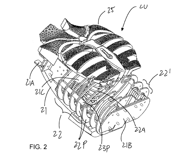
least three
joint units is adapted to be connected to a pelvis of the mannequin, and a top
one of the at
least three joint units is adapted to be connected to a torso section of the
mannequin.
[0007] Still further in accordance with the first aspect, for example, the
bottom one of the at
least three joint units is adapted to be connected to the pelvis of the
mannequin with the third
DOF.
[0008] Still further in accordance with the first aspect, for example, four
of said joint units
may provide concurrently four rotational DOFs, wherein a rotational axis of a
fourth DOF is
configured to be aligned with the lateral axis of the mannequin.
[0009] Still further in accordance with the first aspect, for example, the
joint units for the first
DOF and for the second DOF are defined by a pair of U-shaped brackets
pivotally connected
at ends of the U, with cavities of the U facing each other.
[0010] Still further in accordance with the first aspect, for example, the
U-shaped brackets
include three welded plates forming the U.
[0011] Still further in accordance with the first aspect, for example, the
sensors associated
with the first DOF and with the second DOF are within the cavities of the U.
[0012] Still further in accordance with the first aspect, for example, a
pair of adjacent ones
of the joint units share a base of the respective U-shaped brackets.
[0013] Still further in accordance with the first aspect, for example, a
base of the respective
U-shaped brackets for at least one of the joint units defines a bore used as a
foramen for
cables of the sensors.
[0014] In accordance with the first aspect of the present disclosure, there
is provided a
system for simulating lumbar spine motions, the system comprising: a mannequin
having a
lumbar spine mechanism as described above between a torso and a pelvis; a
processing unit
having an orientation calculator module to quantify the lumbar spine
orientation changes from
readings of the sensors, and a performance assessor module to assess the
lumbar spine
2
CA 03218052 2023-10-27
WO 2022/226640
PCT/CA2022/050635
motions using the quantified lumbar spine orientation changes; and an output
for outputting an
assessment and/or the lumbar spine orientation changes.
[0015] In accordance with a second aspect of the present disclosure, there
is provided a
spine mechanism for a mannequin comprising: at least three joint units
serially connected to
provide joints for at least three rotational degrees of freedom (DOF), with a
rotational axis of a
first DOF configured to be aligned with a lateral axis of the mannequin, a
rotational axis of a
second DOF configured to be aligned with an anterior-posterior axis of the
mannequin, and a
rotational axis of a third DOF configured to be aligned with a cranial-caudal
axis of the
mannequin, and sensors to measure movements at each of the at least three
joint units, and
wherein the joint units for the first DOF and for the second DOF are defined
by a pair of U-
shaped brackets pivotally connected at ends of the U, with cavities of the U
facing each other,
the U-shaped brackets include three welded plates forming the U.
[0016] Further in accordance with the second aspect, for example, four of
said joint units
may provide concurrently four rotational DOFs, wherein a rotational axis of a
fourth DOF is
configured to be aligned with the lateral axis of the mannequin.
[0017] Still further in accordance with the second aspect, for example, the
sensors
associated with the first DOF and with the second DOF are within the cavities
of the U.
[0018] Still further in accordance with the second aspect, for example, a
pair of adjacent
ones of the joint units share a base of the respective U-shaped brackets.
[0019] Still further in accordance with the second aspect, for example, a
base of the
respective U-shaped brackets for at least one of the joint units defines a
bore used as a
foramen for cables of the sensors.
[0020] In accordance with a third aspect of the present disclosure, there
is provided a torso
system for a mannequin comprising: a structural member at a back of the
mannequin; ribs
connected to the structural member to define a ribcage, at least a first set
of the ribs having
pivotable portions relative to the structural member; a chest plate assembly
operatively
connected to the ribcage for relative movement therebetween; and a shock unit
between the
chest plate assembly and the structural member.
[0021] Further in accordance with the third aspect, for example, the ribs
of the first set each
have a posterior rib member connected to the structural member, and an
anterior rib member
3
CA 03218052 2023-10-27
WO 2022/226640
PCT/CA2022/050635
pivotally connected to the respective posterior rib member, the anterior rib
members being
connected to the chest plate assembly.
[0022] Still further in accordance with the third aspect, for example, the
anterior rib
members are connected to the chest plate assembly by joints including one
translational DOF
and one rotation DOF.
[0023] Still further in accordance with the third aspect, for example, the
anterior rib member
and the posterior rib member of a left subset of the first set share a common
pivot, and the
anterior rib member and the posterior rib member of a right subset of the
first set share a
common pivot.
[0024] Still further in accordance with the third aspect, for example, the
anterior rib
members of the left subset share a common pivot for connection to the chest
plate assembly,
the anterior rib members of the right subset share a common pivot for
connection to the chest
plate assembly.
[0025] Still further in accordance with the third aspect, for example, the
chest plate
assembly has a frame member operatively connected to the ribcage.
[0026] Still further in accordance with the third aspect, for example, the
frame member
covered by a membrane emulating soft tissue.
[0027] Still further in accordance with the third aspect, for example, the
structural member
includes an arched beam to which the ribcage is connected.
[0028] Still further in accordance with the third aspect, for example, the
ribs of a second set
are fixed.
[0029] Still further in accordance with the third aspect, for example, an
airway simulator
apparatus may have at least one tube defining at least one opening at a level
of a face of the
mannequin, and being in fluid communication with at least one airbag in the
chest plate
assembly.
[0030] Still further in accordance with the third aspect, for example, a
pressure sensor may
be in the airway simulator apparatus.
4
CA 03218052 2023-10-27
WO 2022/226640
PCT/CA2022/050635
[0031] Still further in accordance with the third aspect, for example,
rotational axes may be
connected to the structural member, and configured for connection of arms to
the torso
system.
[0032]
DESCRIPTION OF THE DRAWINGS
[0033] Fig. 1 is a perspective view, partly transparent, of a mannequin in
accordance with
embodiments of the present disclosure;
[0034] Fig. 2 is a perspective view of a torso system in accordance with
the present
disclosure;
[0035] Fig. 3 is an assembly view of the torso system of Fig. 2;
[0036] Fig. 4 is a side perspective view of the torso system of Fig. 2;
[0037] Fig. 5 is a bottom view of the torso system of Fig. 2;
[0038] Fig. 6 is a perspective view of the torso system of Fig. 2, with a
chest plate assembly
removed;
[0039] Fig. 7 is a perspective view of the torso system of Fig. 6, with
ribs collapsed;
[0040] Fig. 8 is a side elevation view of the torso system of Fig. 6;
[0041] Fig. 9 is a front view of the torso system of Fig. 2 as connected to
a lumbar spine
system in accordance with a variant of the present disclosure;
[0042] Fig. 10A is a perspective view of the lumbar spine system of Fig. 9;
[0043] Fig. 10B is an assembly view of the lumbar spine system of Fig. 9;
[0044] Fig. 11A is an assembly view of a vertebra portion of the lumbar
system of Fig. 9;
[0045] Fig. 11B is a front elevation view of the vertebra portion of Fig.
13;
[0046] Fig. 12 is a perspective view of a lumbar spine system or cervical
spine system of in
accordance with another variant of the present disclosure;
[0047] Fig. 13A is an assembly view of a vertebra portion of the lumbar
system of Fig. 12;
[0048] Fig. 13B is an elevation view of the vertebra portion of the lumbar
system of Fig. 12;
CA 03218052 2023-10-27
WO 2022/226640
PCT/CA2022/050635
[0049] Fig. 13C is an assembly view of a vertebra portion of the lumbar
system of Fig. 12;
[0050] Fig. 14A is a perspective view of a leg and pelvis of the mannequin
of Fig. 1 in
accordance with another variant of the present disclosure;
[0051] Fig. 14B is an exploded view of the pelvis at a hip region;
[0052] Fig. 15A is a perspective view of a leg in accordance with another
variant of the
present disclosure
[0053] Fig. 15B is an assembly view of a knee joint of the leg of Fig. 15A;
[0054] Fig. 16 is a display view of an exemplary GUI used with the
mannequin of the
present disclosure;
[0055] Fig. 17 is a display view of another exemplary GUI used with the
mannequin of the
present disclosure; and
[0056] Fig. 18 is a schematic view of an airway simulator apparatus used
with the
mannequin of the present disclosure.
DETAILED DESCRIPTION
[0057] Referring to the drawings and more particularly to Fig. 1, a
mannequin in
accordance with the present disclosure is generally shown at 10. The mannequin
10 may also
be known as a dummy, a manikin, etc. The mannequin 10 may have various
portions, some
of which are described herein in further detail as part of the present
disclosure. The present
disclosure focuses on some skeletal parts of the mannequin 10, whereby soft-
tissue
surrounding the skeletal parts is only shown in transparent layers in Fig. 1.
However, as
observed in Fig. 1, numerous of the skeletal parts are embedded in soft-tissue
emulating
muscles, tendons, and/or skin. The mannequin 10 may have a head 11, a neck 12
and a
torso 13. The head and the neck may be as described in PCT Application
Publication
No. WO 2019/075582, incorporated herein by reference, as a possibility among
others.
However, the neck 12 may have a spine configuration using a spine portion
described herein,
in a variant. One arm 14 is shown projecting from the torso 13. A similar
other arm may be on
the other side, though removed from Fig. 1 for simplicity. A lumbar spine
portion 15 projects
downwardly from the torso 13 and interfaces a pelvis 16 to the lumbar torso
13. Legs 17
project downwardly from the pelvis 16, though a single one is shown in the
figures for
6
CA 03218052 2023-10-27
WO 2022/226640
PCT/CA2022/050635
simplicity. The various parts of the mannequin body may be articulated so as
to emulate the
human body.
[0058] Referring now to Figs. 2-8, a torso system in accordance with the
present disclosure
is generally shown at 20. The torso system 20 is configured to allow simulated
reanimation
procedures, such as CPR, for example, when chest compresses are effected on
the torso
system 20, with artificial ventilation. The torso system 20 may be used to
provide a user with
the appropriate force-feedback feeling of a human body exposed to such
maneuvers. The
torso system 20 may also be equipped with sensors to quantify the
interventions on the
mannequin 10, and hence provide feedback based on the actual maneuvers by the
operators
of the torso system 20. Hence, the torso system 20 is configured to simulate a
reaction of a
human body, with elasticity and resilience, weight distribution and center of
mass positioning.
[0059] The torso system 20 has a main structural member 21. The main
structural
member 21 may be a beam, with a slight curvature as an option. The main
structural member
21 is positioned in the mannequin 10 where the spine would be, i.e., generally
centered in the
back of the torso system 20. The structural member 21 is shown as being a
single structural
component. In an embodiment, the structural member could be constituted of
separate
vertebrae, in a similar manner as described below for the lumbar spine system.
The structural
member 21 is described as being structural, in that it is load bearing, in
supporting various
other parts of the torso system 20, and as it interconnects to other parts of
the mannequin 10,
as described herein.
[0060] At an upper end of the structural member 21, connector portions 21A
are provided
for interfacing the neck 12 and arms 14 to the torso system 20. The connector
portions 21A
may include plates, brackets, struts, beams, fasteners, etc, and have suitable
structural
integrity to preserve their shape in spite of forces applied to the torso
system 20 and of
transfer maneuvers of the mannequin 10. There may be multiple connector
portions 21A of
various types, in various arrangements, to connect the neck 12 and the arms 14
to the torso
system 20.
[0061] At an opposite end of the structural member 21, a base plate 21 B is
provided. The
base plate 21B is one possible structural component that may be used to
connect the torso
system 20 to an adjacent system. Other base components could include beams,
brackets,
blocks, rods, beam, etc. In an embodiment, adjacent systems (e.g., lumbar
spine system) are
7
CA 03218052 2023-10-27
WO 2022/226640
PCT/CA2022/050635
continuously connected to the beam or spine of the torso system 20, such as
defined by the
structural member 21, instead of being interfaced to the base plate 21B.
Weights 21C may
optionally be distributed near the top of the structural member 21 and near
the bottom of the
structural member 21 (i.e., in the cranial-caudal direction) as two options
among others.
Indeed, it is contemplated to have the mannequin 10 replicate the weight and
mass distribution
of a human body, whereby the weights 21C may be provided, for instance, in
reproducing a
mannequin having adult male proportions. It would be contemplated to size a
torso system 20
for infant, child, teenager and/or female mannequins as well.
[0062] The structural member 21 is configured to support a ribcage formed
of ribs 22 and,
optionally, ribs 22'. The ribs shown as 22 are of the type that may collapse
inwardly and have
rotational capacity, while lower ribs 22' may not have this capacity.
Typically, when
reanimation procedures are performed, chest compressions are conventionally
applied at the
location of the collapsible ribs 22. It is nevertheless considered to allow
the ribs 22' to be
collapsible as well. In an embodiment, the ribcage is collapsible by up to
2.75 inches, as depth
of compression or excursion.
[0063] The ribs 22 and 22' are shown as pairs of mirrored C-shaped
components with a
gap between them, and cantilevered from the structural member 21. The
collapsible ribs 22
may each have a posterior rib member 22P rigidly connected to the structural
member 21.
Anterior rib members 22A are connected to the free ends of the posterior rib
members 22P by
way of common pivots 23P, which may be rods, i.e., one left-side rod and one
right-side rod.
Individual pivots are also an option, with each anterior rib member 22A
pivotally connected to
a respective posterior rib member 22P. Other arrangements are possible, such
as two or
more anterior rib members 22A sharing a pivot rod. As a result, the anterior
rib members 22A
may pivot or move relative to the posterior rib members 22P. As an alternative
to pivots, a
flexure member may be provided between anterior rib member 22A and respective
posterior
rib member 22P.
[0064] In an embodiment, all anterior ends of the anterior rib members 22A
on one side of
the torso system 20 (i.e. left or right side) are interconnected by a common
pivot 23A, in
similar fashion as the pivots 23P. Accordingly, the ribs 22 may move jointly
when a pressure
is applied thereon. Individual movement is also considered for the ribs 22.
The anterior ends
of the anterior rib members 22A may also be connected to a chest plate in
other ways.
8
CA 03218052 2023-10-27
WO 2022/226640
PCT/CA2022/050635
[0065] A shock unit 24 has a first end connected to the structural member
21. The shock
unit 24 projects anteriorly to a region adjacent to the anterior ends of the
anterior rib
members 22A. The shock unit 24 may be made of a spring and of a damper, or
other system,
to offer both resistance and compliance to compressive forces, to then return
to an initial
position. Stated differently, the shock unit 24 is configured to be compressed
to then return to
its original shape. As an alternative to the spring and damper, a pad or
member of a resilient
material may be used, or the spring could be smaller than as illustrated.
Moreover, the shock
unit 24 may be optional, or there may be more than one shock unit 24. Another
expression to
define the shock unit 24 is a resilient unit or member, or spring-back unit or
member.
[0066] Referring to Figs. 2 and 3, a chest plate assembly 25 is shown being
interconnected
to the ribcage made of the ribs 22 in a manner described below, in addition to
be mounted
onto the shock unit 24. Other arrangements are possible, such as by having the
chest plate
assembly 25 connected solely to the ribs 22 with the ribs 22 connected to the
shock unit 24, or
by having the chest plate assembly 25 connected solely to the shock unit 24,
with the ribs 22
connected to the shock unit 24 as well. In other embodiment, the spring-back
effect is
provided by springs or like biasing members in the rib cage, such as at the
pivots 23P. The
chest plate assembly 25 may include various components to emulate a human
chest. A male
chest is shown but a female chest could also be provided. The chest plate
assembly may
include a rigid structural member 25A by which it is connected to the shock
unit 24. For
instance, the structural member 25A is in the form of a plate, a grill, a
frame, that has the
necessary structural integrity to resist to compression forces. As best shown
in Figs. 4 and 5,
the structural member 25A may have brackets 25B by which it is connected to
the pivots 23A
at the free ends of the anterior rib members 22A. For example, the brackets
25B of the
structural member 25A may have slots so as to form joints allowing the
movement
therebetween. Springs could be in such slots to oppose a biasing force and
could hence be
an alternative to the shock unit 24. The pivots 23A may translate and rotate
relative to the
brackets 25B, i.e., two degrees of freedom of movement may be possible, though
fewer or
more are also contemplated. The structural plate 25A may also have slots 25C
(Fig. 3),
aligned with the anterior rib members 22A. This arrangement is such that
constraints are
imposed on the movement of the structural member 25A, when compressions are
made, as
the structural member 25A is interconnected with the pivots 23A on both left
and right side of
the ribcage. Moreover, the structural plate 25A may move below a top surface
of the anterior
9
CA 03218052 2023-10-27
WO 2022/226640
PCT/CA2022/050635
rib members 22A, via the slots 25C. It may thus allow an operator to manually
feel the ribs 22,
when applying a compressive force on the torso system 20. Moreover, the
arrangement of the
ribs 22 with the position of the pivots 23A relative to the structural plate
25A is such that a
misplaced pressure may result in the ribs 22 blocking a compressive movement.
In an
embodiment, the ribs 22 may project anteriorly beyond the chest member 26 only
if an
excessive pressure is applied to a chest member 26.
[0067] The structural plate 25A may then support the soft-tissue chest
member 26. The
soft-tissue chest member 26 has the shape and feel of human soft-tissue, by a
soft-tissue
membrane. The chest member 26 may be constituted of different layers to be
rigidly
connected to the structural plate or frame 25A, and to anteriorly expose
equivalents of soft
tissue. For example, the chest member 26 may be a combination of urethane and
PLA (e.g.,
printed), as two of numerous possible materials that may be used. Silicone,
polyurethane
foam is another material that can form part of the layers of the chest member
26, while a metal
or high density polymer may be used for the skeleton members. Accordingly, the
elastic
deformation of the chest member 26, if present, may allow a user to fell the
ribs 22 during
chest compresses. An upper torso portion is shown, separate from the chest
member 26, but
contributing to forming the anatomical-like chest of the mannequin 10.
[0068] One or more sensors may be provided in the chest plate assembly 25,
to quantify
human maneuvers on the torso system 20. For example, a sensor 26A may be
located in a
region where CPR compressions are typically applied. In an embodiment, the
sensor 26A is a
force-sensing resistor at hand placement on the chest member 26. Other types
of sensors
may be used, such as inertial sensors (e.g., accelerometers), infrared
sensors, strain gauges,
etc, to measure accelerations and/or forces associated with the maneuvers made
on the chest
of the torso system 20, as well as depth and rhythm of compressions. For
example, the
sensors may have the capacity to detect excessive compressive forces applied
in a CPR
simulation procedure.
[0069] In an embodiment, an airbag 27A or airbags 27A of an airway
simulator apparatus
27 may be located between the structural plate 25A and the chest member 26.
The airbag 27
may be provided with a pair of inflatable vessels, to emulate both right and
left lungs. The
positioning of the airbag(s) 27A is such that when it inflates, it allows
chest rises and
contractions. The airbag 27A may be in fluid communication with an airway
simulator
CA 03218052 2023-10-27
WO 2022/226640
PCT/CA2022/050635
apparatus, such as described in PCT Application Publication No. WO
2019/075582, with the
airway simulator apparatus being accessible through a mouth or nostrils of the
mannequin 10.
For example, as shown in Fig. 18, the airway simulator apparatus 27 may
emulate anatomical
airways, with a mouthpiece 27B, a throat tube, a pharyngeal tube, nose
tube(s), and/or
tracheal tube(s) 27C, in fluid communication with the airbag 27A or like lung
emulating
vessel(s). The tube 27C is sized to allow intubation in one embodiment. In an
embodiment, a
pharyngeal tube extends into the torso system 20. Though it bears the moniker
"pharyngeal",
the pharyngeal tube may be longer than an anatomical pharynx. In the variant
of Fig. 18, the
airway simulator apparatus 27 is shown having a pair or airbags 27 connected
to a tube 27C,
by a tee 27D or equivalent connector. A pressure sensor 27E may be present, to
measure the
pressure in the airway simulator apparatus 27. Accordingly, reanimation
techniques may be
quantified by way of pressure measurements. The pressure sensor 27E may be
located at
other locations as well, with signals sent to the processor 100 (Fig. 1).
[0070] Referring to Fig. 9, the torso system 20 is shown relative to the
arm 14. The arm 14
may be connected to one of the connectors 21A of the structural member 21 and
be arranged
to pivot inwardly when pressure is applied to the torso system 20. Any
appropriate joints may
be provided at the junction between the torso system 20 and the arms 14, to
emulate the
rotational capabilities of the shoulder joint relative to the torso system 20.
[0071] Referring now to Figs. 9 to 11B, one variant of the lumbar spine
system 30 is
connected to the torso system 20, for instance at the base plate 21B. The
lumbar spine
system 30 includes a lumbar spine mechanism configured to emulate a reaction
of the lumbar
region of the spine, and its freedoms of movement.
[0072] In an embodiment, the lumbar spine system 30 may have a sequence of
rotational
joints 30ML, 30CC, and 30AP with ML referring to mediolateral, CC referring to
cranial caudal
and AP referring to anterior posterior. In Fig. 10A, the ML (flexion), the AP
and the CC axes
are shown with the lumbar spine system 30 having two joints 30ML and one of
each of the
joints 30CC and 30AP. Each joint may provide one rotational degree of freedom,
about their
related axes. Other arrangements are however contemplated. Additional segments
could be
used, for instance to reproduce all of the vertebral segments of the spine.
Sensors other than
encoders could be added to the spine system, such as sensors to capture
pressures and
11
CA 03218052 2023-10-27
WO 2022/226640
PCT/CA2022/050635
forces applied to a vertebral system during manual adjustment and/or
manipulation of the
mannequin.
[0073] Referring concurrently to Figs. 10B--11A, the joints 30ML and 30AP
have a similar
construction, whereby only one of these will be detailed. However, similar
reference numerals
are used to indicate like parts. The joints 30ML and 30AP have connector
brackets 31 and 32
pivotally connected by pivot assembly 33, best seen in Fig. 11B. As observed,
the pivot
assembly 33 may include one or more bearings 33A, supports for the bearings,
and a shaft
33B (e.g., bolt and nut, screw, etc). In the arrangement of Fig. 11B, the
shaft features a pair of
fasteners 33B interconnected by a coupling nut 33C, as one possible
arrangement among
others. The pivot assembly 33 is aligned with either the mediolateral axis ML
(for joint 30ML)
or the anterior posterior axis AP (for joint 30AP), thus depending on the
nature of the joint. The
connector brackets 31 and 32 have fastener holes so as to be secured to the
adjacent joint or
to the adjacent component such as the base plate 21A or pelvis 40. A sensor
assembly 34
may be provided for one or more of the joints 30ML, 30AP, 30CC, with the
possibility of having
each said joint equipped with a sensor assembly 34 to measure a rotation in
the joints. The
sensor assembly 34 may be an encoder type sensor as an assembly among others.
Hence, in
an embodiment shown in Fig. 11A, the sensor assembly 34 may include a PCB
board 34A, a
holder 34B, a magnet holder 34C featuring magnets and a screw (shaft 33B), as
one possible
configuration. Thus, the sensor assembly 34 may be provided as one possible
sensor
arrangement to determine the amount of rotation occurring when the lumbar
spine system 30
is displaced.
[0074] Referring to Figs. 9-11B, the joint 30CC is of different
configuration as its rotational
axis is longitudinally aligned with the lumbar spine system 30, contrary to
the joints 30 ML and
30AP that have their axes transversely arranged. The joint 30CC has a
connector plate 35 and
a connector plate 36, interconnected by a pivot assembly 37. A bearing 37A and
shaft 37B
may also be present to facilitate movement of the connector plates 35 and 36
relative to one
another. In order to connect the connector plates 35 or 36 to the adjacent
connector brackets
31,32 of other joints, struts 38 may be used. The struts 38 may be bolts,
fasteners, posts, etc.
A sensor assembly 39, which may be the same as the encoder assembly 34 but
arranged for
the orientation of the joint 30CC, encoder 39 is connected to the pivot
assembly 37 as one
possible type of sensor to determine the amount of rotation sensed between the
connector
plates 35 and 36.
12
CA 03218052 2023-10-27
WO 2022/226640
PCT/CA2022/050635
[0075] Therefore, in the illustrated embodiment of Figs. 10A-11B, the
lumbar spine
system 30 has four different joints allowing four different rotational degrees
of freedom, two in
flexion, one cranial-caudal and one anterior-posterior, though many more
degrees of freedom
could be present. There may be fewer degrees of freedom, such as three degrees
of freedom
with the possibility to cover all three rotational axes, i.e., flexion,
cranial-caudal and anterior-
posterior. Other configurations are considered as well.
[0076] Referring now to Figs. 12 to 13C, another variant of the lumbar
spine system is
shown at 30, and may be connected to the torso system 20, for instance at the
base plate 21B
in a similar fashion as the lumbar spine system of Figs. 10A-11B. The lumbar
spine system 30
includes a lumbar spine mechanism configured to emulate a reaction of the
lumbar region of
the spine, and its freedoms of movement. The lumbar spine system 30 could be
used as a
cervical spine system as well.
[0077] In an embodiment, the lumbar spine system 30 may have a sequence of
rotational
joints 130ML, 130CC, and 130AP with ML referring to mediolateral, CC referring
to cranial
caudal and AP referring to anterior posterior. In Fig. 12, the ML (flexion),
the AP and the CC
axes are shown with the lumbar spine system 30 having two joints 30ML and one
of each of
the joints 30CC and 30AP. Each joint may provide one rotational degree of
freedom, about
their related axes. Other arrangements are however contemplated, e.g. one
joint for each of
the CC, AP, ML axes, more than one joint for any of the CC, AP, ML axes,
different sequences
as opposed to the one shown in the figures. Additional segments could be used,
for instance
to reproduce all of the vertebral segments of the spine. Sensors other than
encoders could be
added to the spine system, such as sensors to capture pressures and forces
applied to a
vertebral system during manual adjustment and/or manipulation of the
mannequin.
[0078] Referring concurrently to Figs. 12, 13A, 13B, the joints 130ML and
130AP have a
similar construction, whereby only one of these will be detailed. However,
similar reference
numerals are used to indicate like parts. The joints 130ML and 130AP have
connector
brackets 131 and 132 pivotally connected by pivots 133, best seen in Fig. 13A
and 13B. The
connector brackets 131 and 132 may be U-shaped (i.e., two walls projecting
from one base)
with the connector bracket 131 being inverted. As the brackets 131 and 132
have their
cavities facing each other, a volume is defined to receive components. The
brackets 131 and
132 may optionally have bores therein, as shown as 132A. In an embodiment, the
brackets
13
CA 03218052 2023-10-27
WO 2022/226640
PCT/CA2022/050635
131 and 132 are made of plates that may be welded to each other, so as to
define integral
brackets. Other arrangements are contemplated, with adhesives, force fits or
like mechanical
interference. Moreover, a base may be shared by two brackets, such as the base
132B
shared between the joints 130AP and 130ML in Fig. 12. It is also contemplated
to have two
bases back to back, such as between the joints 130ML in Fig. 12.
[0079] As observed, the pivots 133 may be paired with one or more bearings
133A,
supports for the bearings, shown as being a bushing that may act as a plain
bearing. Other
arrangements are possible (e.g., bolt and nut, screw, etc). The pivots 133 are
aligned with
either the mediolateral axis ML (for joint 130ML) or the anterior posterior
axis AP (for joint
130AP), thus depending on the nature of the joint. The connector brackets 131
and 132 may
have fastener holes so as to be secured to the adjacent joint (e.g., for the
joints 130ML) or to
the adjacent component such as the base plate 21A or pelvis 40, but welding or
other
connection arrangements are possible. A sensor assembly 134 may be provided
for one or
more of the joints 130ML, 130AP, 130CC, with the possibility of having each
said joint
equipped with a sensor assembly 134 to measure a rotation in the joints. The
sensor
assembly 134 may be an encoder type sensor as an assembly among others. Hence,
in an
embodiment shown in Figs. 13A and 13B, the sensor assembly 134 may include a
PCB board
134A, a holder 134B, a magnet 134C fixed to the pivot 133, as one possible
configuration.
Other sensors may be used as well, including optical sensors, etc. Thus, the
sensor assembly
134 may be provided as one possible sensor arrangement to determine the amount
of rotation
occurring when the lumbar spine system 130 is displaced.
[0080] Referring to Figs. 12 and 13C, the joint 130CC is of different
configuration as its
rotational axis is longitudinally aligned with the lumbar spine system 130,
contrary to the
joints 130ML and 130AP that have their axes transversely arranged. The joint
130CC has a
connector plate 135 and a connector plate 136, interconnected by a shaft 137.
The shaft 137
may be fixed to the connection plate 136 in any appropriate manner (e.g.,
welding, soldering,
mechanical interference, keyway, etc). A bearing 137A, for example a disc(s),
may also be
present to facilitate movement of the connector plates 135 and 136 relative to
one another, in
a planar rotation relative to one another. In order to connect the connector
plate 135 to the
adjacent connector brackets 132 of other joints (such as to the joint 130ML in
Fig. 12),
walls 138 may be used to form a U-shaped arrangement, or struts (e.g., bolts,
fasteners,
posts, etc). In the shown arrangement, the joint 130CC has a U-shaped
arrangement, with the
14
CA 03218052 2023-10-27
WO 2022/226640
PCT/CA2022/050635
connector plate 135 and the walls 138, and is connected to a base of the
adjacent joint, i.e., of
the joint 130ML (though it could be connected to a joint 130AP as well. A
sensor assembly
139 may be provided, and may be the same as the encoder assembly 134 but
arranged for
the orientation of the joint 130CC. Accordingly, the encoder 139 is connected
to the shaft 137
as one possible type of sensor to determine the amount of rotation sensed
between the
connection plates 135 and 136.
[0081] The brackets 131, 132, and the one formed by the connection plate
135 and walls
138, as well as the back-to-back brackets 131 and 132 sharing base 132B, and
thus forming a
H-shaped bracket, may be without bending or bolts, and may thus simplify the
assembly of the
spine system 30.
[0082] Therefore, in the illustrated of Figs. 12-13C, the lumbar spine
system 30 has four
different joints allowing four different rotational degrees of freedom, two in
flexion, one cranial-
caudal and one anterior-posterior, though more degrees of freedom could be
present. There
may be fewer degrees of freedom, such as three degrees of freedom with the
possibility to
cover all three rotational axes, i.e., flexion, cranial-caudal and anterior-
posterior.
Other configurations are considered as well. The spine system of Figs. 12-13C
may define a
well or like passage via its bores 132A (131A) that may emulate the vertebral
foramen.
Accordingly, wires for the various encoders and sensors of the mannequin 10
may pass
through this foramen. The foramen of the spine system 30 of Figs. 12-13C may
prevent the
damaging of such wires and cables. Moreover, the positioning of the encoder
assemblies 134
within the cavities of the brackets 131 and 132 ensures that the cables or
wires are closed to a
center of the spine system 30.
[0083] Referring to Figs. 14A and 14B, the pelvis 40 may have a U-shaped
structure 41
having a pair of rotational blocks 42. The lumbar spine system 30 is received
in the cavity of
the U-shaped structure 41. The rotational blocks 42 may serve as rotational
pillows for joints
between the pelvis 40 and the femur 50. In order to emulate the hip joint, a
universal joint 43
may extend from each the blocks 42 and hence provide two rotational degrees of
freedom of
movement. Other possible joint arrangements include a ball and socket, etc.
[0084] The leg 50 may be constituted of a femur 51 and of a tibia 52 (or
shank)
interconnected by a single rotational degree of freedom via joint 53. More
complex joints, with
CA 03218052 2023-10-27
WO 2022/226640
PCT/CA2022/050635
additional degrees of freedom, may be used, if additional functionalities are
contemplated for
the mannequin 10.
[0085] Referring to Figs. 15A and 15B, another embodiment of the leg 50 is
shown with
femur 51 and tibia 52 (a.k.a., shank). The femur 51 is made of a central
square-section tube
51A with optional peripheral square section tubes 51B. Likewise, the tibia 52
is made of a
central square-section tube 52A with optional peripheral square section tubes
52B emulating a
calf muscle. The tubes may be welded, bolted, adhered, soldered to one
another. The tubes
51A, 51B, 52A and/or 52B may be made of a metal (e.g., steel) to give the leg
the inertial of an
anatomic leg, as a possibility among others.
[0086] The knee joint 53 of Figs. 15B may have a single rotation, or more
(e.g., with a
universal joint). The size of the knee joint 53 is such that pinch surfaces
are minimized. The
knee joint 53 may include a plate with tab 53A received in a gap between a
pair of plates with
tabs 53B, resulting from a shim 53C as an example, with a shaft 53D between
these
components. The plates with tabs 53A and 53B are received inside a cavity of
the tubes 51A
and 52A, respectively, with additional hardware to secure these components
into a fixed
assembly. Circlips may be used on the shaft 53D to facilitate assembly of the
knee joint 53.
[0087] Referring to Fig. 1, the mannequin 10 is of the type having or used
with a processing
unit 100 used to quantify the manipulations of a mannequin 10, though the
mannequin 10 may
be a standalone device (e.g., with audio and/or vibratory feedback). The
processing unit 100 is
of the type having a non-transitory computer-readable memory communicatively
coupled to it
and comprising computer-readable program instructions executable by the
processing unit 100
to perform numerous functions related to the simulation of chest maneuvers,
and lumbar spine
motions, in addition to functions optionally performed relative to other parts
of the mannequin
10. The processing unit 100 receives data from sensors described herein, to
measure various
movements of the mannequin 10 and other parameters such as forces applied to
mannequin
10. An interface may be operatively connected to the processing unit 100 to
output
quantitative data representative of the transfer manipulations, or CPR
maneuvers, and may
communicate with the operators of the mannequin 10 to warn or alarm them of
excessive or
improper manipulations. The interface may be a monitor, screen, tablet, may be
embedded
into the mannequin, etc. Figs. 16 and 17 provide exemplary GUIs, that can
provide some or
all of the following information: CPR rhythm, CPR depth ventilation rhythm,
all with respect to a
16
CA 03218052 2023-10-27
WO 2022/226640
PCT/CA2022/050635
time scale, in addition to all spine rotations based on the sensors in the
lumbar spine system
30 and neck, if applicable, for instance as graphs shown angular variations
over time (Fig. 17).
The GUI may also provide a video display of maneuvers on the mannequin 10, and
a list of
events.
[0088] With respect to the lumbar spine system 30, the processing unit 100
may have an
orientation calculator module receiving the data from the sensor assemblies 34
and 39. The
orientation calculator module may determine orientation variations sustained
by the lower back
of the mannequin 10 during manipulations. For example, the orientation
calculator module
may quantify variations in flexion angle values about one or more flexion
axes, about lateral
axes of the mannequin 10. The orientation calculator module may also quantify
lateral flexions
as well, i.e., about an anterior-posterior axis of the mannequin 10. All of
these variations of
angle values may be in the form of angular rates of change about various axes.
[0089] The orientation calculator module may provide an output as a visual
display on the
interface, or in the form of a data file for any given training session. In
accordance with an
embodiment, the output is in the form of the graph to indicate the angle or
angular rates of
change for a user to get a quantitative assessment of manipulations being
performed. The
graph may have a timescale which can be matched with data pertaining to the
various
manipulations such that a user may see the angles and angular rates of change
resulting from
various manipulations.
[0090] The processing unit 100 may be programmed with an orientation
threshold database
so as to determine what constitutes permitted versus excessive manipulations.
Hence, the
orientation calculator module may provide measured angular rates of change and
receive
threshold values from the orientation threshold database. A performance
assessor may then
determine whether the movements performed exceed the values programmed into
the
orientation threshold database, in which case it may be determined that an
excessive or
improper transfer manipulation of the mannequin 10 has been performed. The
performance
assessor module may also be programmed to evaluate CPR parameters, such as
excessive
or insufficient force, improper CPR rhythm, misplaced forces.
[0091] The performance assessor module, when identifying an excessive or
improper
manipulation by measured values exceeding beyond those expected, may alert the
operator of
the system 10 via the alert of the interface, or may provide quantitative data
relative to
17
CA 03218052 2023-10-27
WO 2022/226640
PCT/CA2022/050635
accepted values. This is an advantage over methods in which the quantitative
data is provided
at a later point, in that corrective measures may be taken right away to
practice by re-
manipulating the mannequin 10 for a proper manipulation. Moreover, the
processing unit 100
may have various thresholds to provide more than a binary "proper" vs
"excessive"
assessment. For example, preliminary signals may be emitted to warn the
operators of an
impending excessive manipulation, or excessive force, for the operators to
correct their
movements, for instance by slowing down manipulations, applying lower
compressions and/or
by reorienting the mannequin 10, accelerating the pace of compressions. Such
system
interventions may provide real time feedback to the operators during training,
for the operators
to be capable of understanding the manipulations that are not done correctly.
[0092] The processor unit 100 may also have a force evaluating module
receiving signals
from the sensors to calculate the forces to which the mannequin 10 is exposed.
For example,
the sensors may include inertial sensors (e.g., accelerometers) producing data
indicative of
the forces sustained by the mannequin 10 during the CPR procedure. The sensors
may
include pressure sensors (e.g., manometers) for an airway simulator apparatus.
18