Note: Descriptions are shown in the official language in which they were submitted.
COMPOSITIONS AND KITS FOR MOLECULAR COUNTING
10001] FIELD OF THE INVENTION
[0002] Methods and uses of molecular counting are disclosed. Molecules
can be counted
by sequencing and tracking thc number of occurrences of a target molecule.
Molecules can
also be counted by hybridization of the molecule to a solid support and
detection of thc
hybridized molecules. In some instances, the molecules to be counted are
labeled. The
molecules to be counted may also be amplified.
BACKGROUND OF THE INVENTION
[00031 Accurate determination of the quantity of nucleic acids is
necessary in a wide
variety of clinical and research measurements. When dissolved in solution, the
average
concentration of nucleic acids (RNA or DNA) can be determined by UV light
absorbance
spectrophotometry or by fluorescent DNA-binding stains. However, the
measurement
required is often not just for the total amount of nucleic acids present, but
specifically for one
or more species of interest contained and mixed with all of the other nucleic
acids within the
sample. In these cases, the nucleic acid molecule of interest is usually
distinguished from all
of the other nucleic acids through a defined sequence of nucleotides that is
unique to the
species of interest. A short synthetic ribo- or deoxyribo- oligonucleotide
with a
complementary sequence to the nucleic acid of interest can be used for its
detection and
identification. For instance, the Polymerase Chain Reaction (PCR) uses a pair
of these
oligonucleotides to serve as annealing primers for repeated cycles of DNA
polymerization
mediated by DNA polyrnerase enzymes. DNA microarrays are another common
detection
method where oligonucleotides are immobilized on solid supports to hybridize
to DNA
molecules bearing complementary sequences. Although both PCR and microarray
methods
are capable of specific detection, accurate determination of the quantity of
the detected
molecules is difficult (especially when it is present in low abundance or when
contained
within a large background of other nucleic acids). In the case of PCR (also
sometimes
referred to as quantitative-PCR, qPCR, TaqMan, or real-time PCR), the amount
of amplified
DNA molecules represents an estimate of its concentration in the starting
solution. In the case
of microarrays, the amount of DNA hybridized is an estimate of its
concentration in solution.
1
Date Recue/Date Received 2023-10-31
In both cases, only relative measurements of concentration can be made, and
the absolute
number of copies of nucleic acid in the sample cannot be precisely determined.
However,
when reference nucleic acids of pre-determined concentrations are included in
the test,
relative comparisons can be made to this standard reference to estimate the
absolute number
of copies of nucleic acids being detected.
[0004] Digital PCR is one method that can be used to determine the
absolute number of
DNA molecules of a particular nucleotide sequence (Sykes et al. Biotechniques
13: 444-449
(1992), Vogelstein et al. Digital PCR. Proc Natl. Acad Sci USA 96: 9236-9241
(1999)). In
this method, the nucleic acid solution is diluted and stochastically
partitioned into individual
containers so that there is on average less than one molecule in every two
containers. PCR is
then used to detect the presence of the nucleic acid molecule of interest in
each container. If
quantitative partitioning is assumed, the dynamic range is governed by the
number of
containers available for stochastic separation. Micro fabrication and pico
liter-sized emulsion
droplets can be used to increase the number of containers available thereby
extending the
measurement dynamic range (Fan et al. Am J Obstet Gynecol 200: 543 e541-547
(2009),
Kalinina et al. Nucleic Acids Res 25: 1999-2004 (1997)). Due to the physical
constraints of
manufacturing large numbers of separate containers and in carrying out these
larger numbers
of reactions, in practice the digital PCR method is limited to investigations
on only a small
number of different DNA molecules at a time.
[0005] Recently, a new method to determine the absolute quantity of DNA
molecules has
been demonstrated where identical copies of individual DNA molecules can be
counted after
the stochastic attachment of a set of diverse nucleic acid labels (Fu et al.
Proc Natl Acad Sci
USA 108: 9026-9031(2011)). Unlike digital PCR, this is a highly parallel
method capable of
counting many different DNA molecules simultaneously. In this method, each
copy of a
molecule randomly attaches to a short nucleic acid label by choosing from a
large, non-
depleting reservoir of diverse labels. The subsequent diversity of the labeled
molecules is
governed by the statistics of random choice, and depends on the number of
copies of identical
molecules in the collection compared to the number of kinds of labels. Once
the molecules
are labeled, they can be amplified so that simple present/absent threshold
detection methods
can be used for each. Counting the number of distinctly labeled targets
reveals the original
number of molecules of each species. Unlike digital PCR, which stochastically
expands
identical molecules into physical space, the method of stochastic labeling
expands identical
molecules into chemical space. An important distinction from digital PCR is
that the
stochastic labeling method does not require the challenging physical
separation of identical
2
Date Recue/Date Received 2023-10-31
molecules into individual physical containers. The approach is practical, and
after labeling, a
simple detector device such as a microarray with complementary probe sequences
to the
labels can be used to identify and count the number of labels present. In
addition, when
stochastic labels are attached to DNA molecules that are prepared for DNA
sequencing
readouts, the labeling sequence can serve as discreet counting tags for
absolute quantitation,
or as unique identifiers to distinguish each originally tagged template from
its amplified
daughter molecules (Kinde et al. Proc Natl Acad Sci USA 108: 9530-9535
(2011)).
SUMMARY OF THE INVENTION
[0006] In some embodiments is a digital reverse transcription method
comprising: a)
contacting a sample comprising a plurality of RNA molecules with a plurality
of
oligonucleotide tags to produce a labeled-RNA molecule, wherein: the plurality
of RNA
molecules comprise at least 2 mRNA molecules of different sequences; the
plurality of
oligonucleotide tags comprises at least 2 oligonucleotide tags of different
sequences; and the
plurality of oligonucleotide tags comprises an oligodT sequence; b)conducting
a first strand
synthesis reaction by contacting the labeled-RNA molecules with a reverse
transcriptase
enzyme to produce a labeled-cDNA molecule; and c) detecting the labeled-cDNA
molecule
by hybridizing the labeled-cDNA molecule to a solid support.
100071 In some embodiments is a stochastic label-based hybridization
chain reaction
method comprising stochastically labeling one or more nucleic acid molecules
with a
plurality of hairpin oligonucleotide tags, wherein the hairpin oligonucleotide
tag comprises an
overhang; and the one or more nucleic acid molecules act as initiators for a
hybridization
chain reaction.
[0008] At least a portion of the hairpin oligonucleotide tag may
hybridize to at least a
portion of the one or more nucleic acid molecules. The hairpin oligonucleotide
tag may
comprise an oligodT sequence. The one or more nucleic acid molecules may
comprise one or
more adapters. At least a portion of the hairpin oligonucleotide tag may
hybridize to at least a
portion of the one or more adapters. At least one hairpin oligonucleotide tag
of the plurality
of hairpin oligonucleotide tags may comprise one or more labels. At least one
hairpin
oligonucleotide tag of the plurality of hairpin oligonucleotide tags may
comprise two or more
labels.
[0009] Each hairpin oligonucleotide tag of the plurality of hairpin
oligonucleotide tags
may comprise one or more labels. Each hairpin oligonucleotide tag of the
plurality of hairpin
oligonucleotide tags may comprise two or more labels. In some instances, the
hairpin
oligonucleotide tag does not comprise a label.
3
Date Recue/Date Received 2023-10-31
100101 The plurality of hairpin oligonucleotide tags may comprise one or
more hairpin
oligonucleotide tags with a 5' overhang, hairpin oligonucleotide tags with a
3' overhang, or a
combination thereof.
[0011] The stem portion of the hairpin oligonucleotide tag can be one or
more
nucleotides in length. The stem portion of the hairpin oligonucleotide tag can
be two or more
nucleotides in length. The stem portion of the hairpin oligonucleotide tag can
be three or
more nucleotides in length. The stem portion of the hairpin oligonucleotide
tag can be thur or
more nucleotides in length. The stem portion of the hairpin oligonucleotide
tag can be five or
more nucleotides in length. The stem portion of the hairpin oligonucleotide
tag can be six or
more nucleotides in length. The stem portion of the hairpin oligonucleotide
tag can be seven
or more nucleotides in length. The stem portion of the hairpin oligonucleotide
tag can be
eight or more nucleotides in length. The stem portion of the hairpin
oligonucleotide tag can
be nine or more nucleotides in length. The stem portion of the hairpin
oligonucleotide tag can
be ten or more nucleotides in length. The stem portion of the hairpin
oligonucleotide tag can
be 15, 20, 25, 30, 35, 40, 45, 50, 60, 70, 80, 90, 100 or more nucleotides in
length.
[0012] The loop portion of the hairpin oligonucleotide tag can be one or
more nucleotides
in length. The the loop portion of the hairpin oligonucleotide tag can be two
or more
nucleotides in length. The loop portion of the hairpin oligonucleotide tag can
be three or more
nucleotides in length. The loop portion of the hairpin oligonucleotide tag can
be four or more
nucleotides in length. The loop portion of the hairpin oligonucleotide tag can
be five or more
nucleotides in length. The loop portion of the hairpin oligonucleotide tag can
be six or more
nucleotides in length. The loop portion of the hairpin oligonucleotide tag can
be seven or
more nucleotides in length. The loop portion of the hairpin oligonucleotide
tag can be eight or
more nucleotides in length. The loop portion of the hairpin oligonucleotide
tag can be nine or
more nucleotides in length. The loop portion of the hairpin oligonucleotide
tag can be ten or
more nucleotides in length. The loop portion of the hairpin oligonucleotide
tag can be 15, 20,
25, 30, 35, 40, 45, 50, 60, 70, 80, 90, 100 or more nucleotides in length.
[0013] The hairpin oligonucleotide tag may comprise a unique identifier
region. The
unique identifier region can be in the loop portion of the hairpin
oligonucleotide tag. The
unique identifier region can be in the stem portion of the hairpin
oligonucleotide tag. The
unique identifier region can be in the overhang portion of the hairpin
oligonucleotide tag.
[0014] The label may comprise a unique identifier region.
[0015] In some embodiments the oligonucleotide tag further comprises a
unique identifier
region. In some embodiments the unique identifier region is at least one
nucleotide in length.
4
Date Recue/Date Received 2023-10-31
In some embodiments the oligonucleotide tag further comprises a universal
primer binding
site. In some embodiments the oligonucleotide tag is at least one nucleotide
in length.
[0016] In some embodiments the solid support is an array. In some
embodiments the
solid support is an addressable array. In some embodiments the solid support
is an
Affymetrix 3K tag array, Arrayjet non-contact printed array, or Applied
Microarrays Inc
(AMI) array. In some embodiments the solid support is a bead.
[0017] Further disclosed herein is cell analysis method comprising: a)
contacting a
sample comprising a plurality of molecules with a plurality of oligonucleotide
tags to produce
a labeled-molecule, wherein: the plurality of molecules comprise at least 2
molecules of
different sequences; the plurality of oligonucleotide tags comprises at least
2 oligonucleotide
tags of different sequences; and the sample is from at least one cell; and b)
detecting the
labeled-molecule by hybridizing the labeled-molecule to a solid support.
[0018] In some embodiments is a clonal amplification method comprising:
a)
stochastically labeling a plurality of molecules with a plurality of
oligonucleotide tags to
produce a labeled-molecule, wherein: the plurality of molecules comprise at
least 2 molecules
of different sequences; and the plurality of oligonucleotide tags comprises at
least 2
oligonucleotide tags of different sequences; b) amplifying the labeled-
molecules to produce a
labeled-amplicon; and c) detecting the labeled-amplicon.
[0019] Further disclosed herein is a kit comprising: a) a plurality of
oligonucleotide tags,
wherein the oligonucleotide tag of the plurality of oligonucleotide tags
comprises: a target
specific region; and a unique identifier region; and b) an enzyme.
[0020] In some embodiments the enzyme is a reverse transcriptase enzyme.
In some
embodiments the enzyme is a ligase. In some embodiments the enzyme is a
polymerase. In
some embodiments the enzyme is an RNase. In some embodiments the enzyme is a
DNase.
In some embodiments the enzyme is an endonuclease.
[0021] In some embodiments the oligonucleotide tag is at least 25
nucleotides in length.
In some embodiments the unique identifier region is at least 10 nucleotides in
length. In some
embodiments the target specific region is at least 10 nucleotides in length.
In some
embodiments the target specific region comprises an oligodT sequence. In some
embodiments the oligonucleotide tag further comprises a universal primer
binding site.
[0022] In some embodiments the kit further comprises a support. In some
embodiments
the support is a semi-solid support. In some embodiments the support is a
solid support. In
some embodiments the solid support is an array. In some embodiments the
support is an
addressable array. In some embodiments the support is an Affymetrix 3K tag
array, Arrayjet
Date Recue/Date Received 2023-10-31
non-contact printed array, or Applied Microarrays Inc (AMI) array. In some
embodiments the
support is a bead.
[0023] In somc embodiments the kit further comprises a primer. In some
embodiments
the primer is a universal primer. In some embodiments the primer binds to the
oligonucleotide tag. In some embodiments the primer binds to the universal
primer binding
site of the oligonucleotide tag.
[0024] In some embodiments the kit further comprises a control oligo. In
some
embodiments the control oligo comprises at least 15 nucleotides. In some
embodiments the
control oligo is a bright hybridization control oligo. In some embodiments the
control oligo is
a spike-in template control. In some embodiments the oligonucleotide tag
further comprises a
label.
[0025] In some embodiments the primer further comprises a label. In some
embodiments
the control oligo further comprises a label. In some embodiments the label is
a dye label. In
some embodiments the label is a Cy3 dye. In some embodiments the label is a
Tye563 dye.
[0026] In some embodiments the kit further comprises a buffer.
[0027] In some embodiments the kit further comprises a carrier.
[0028] In some embodiments the kit further comprises a detergent.
[0029] Further disclosed herein is a system for determining the absolute
quantity of a
plurality of nucleic acid molecules. The system may comprise a) a plurality of
oligonucleotide tags; and b) a detector for detecting at least a portion of
the oligonucleotide
tags.
[0030] The detector may comprise an array detector, fluorescent reader,
non-fluorescent
detector, CR reader, or scanner. In some embodiments the method further
comprises the
fluorescent reader is a Sensovation or AG fluorescent reader. In some
embodiments the
method further comprises the scanner is a flatbed scanner.
[0031] The system may further comprise a thermal cycler. In some
embodiments the
system further comprises a sequencer. In some embodiments the system further
comprises a
hybridization chamber.
[0032] The system may further comprise a computer. In some embodiments
the computer
comprises a memory device. In some embodiments the memory device is capable of
storing
data. In some embodiments the system further comprises a software program. In
some
embodiments the system further comprises a computer-readable program.
[0033] In some embodiments the oligonucleotide tag further comprises a
unique identifier
region. In some embodiments the unique identifier region is at least 10
nucleotides in length.
6
Date Recue/Date Received 2023-10-31
In some embodiments the unique identifier region cannot hybridize to the
molecule. In some
embodiments the oligonucleotide tag further comprises a universal primer
binding site. In
some embodiments the oligonucleotide tag is at least 20 nucleotides in length.
In some
embodiments the oligonucleotide tag further comprises a target specific
region. In some
embodiments the target specific region comprises an oligodT sequence. In some
embodiments the target specific region is at least 10 nucleotides in length.
In some
embodiments the method further comprises conducting a first strand synthesis
reaction to
produce a labeled-cDNA molecule.
[0034] In some embodiments the amplifying the labeled-molecule comprises
conducting
a polymerase chain reaction. Alternatively, amplifying the labeled-molecule
may comprise
conducting a non-PCR based amplification reaction. Amplifying the labeled-
molecule may
comprise exponential amplification of the labeled-molecule. Amplifying the
labeled-
molecule may comprise linear amplification of the labeled molecule. Amplifying
the labeled-
molecule may comprise hybridization chain reaction (HCR) based amplification
method.
[0035] Amplifying the labeled-molecule may comprise amplifying at least
the label
portion of the labeled molecule, the molecule portion of the labeled molecule,
or a
combination thereof
[0036] In some embodiments the method further comprises conducting a
polymerase
chain reaction on the labeled-molecule or any product thereof to produce a
double-stranded
labeled-molecule. In some embodiments conducting the polymerase chain reaction
comprises annealing a first target specific primer to the labeled-molecule or
any product
thereof. In some embodiments conducting the polymerase chain reaction further
comprises
annealing a universal primer to the universal primer binding site of the
oligonucleotide tag. In
some embodiments the polymerase chain reaction comprises absolute PCR, HD-PCR,
Next
Gen PCR, digital RTA, or any combination thereof. In some embodiments the
method
comprises conducting a nested PCR reaction on the double-stranded labeled-cDNA
molecule.
In some embodiments conducting the nested PCR reaction comprises denaturing
the labeled-
molecule or any product thereof to produce a denatured single-stranded labeled-
molecule or
any product thereof In some embodiments conducting the nested PCR reaction
further
comprises annealing a second target specific primer to the denatured single-
stranded labeled-
molecule or any product thereof In some embodiments conducting the nested PCR
reaction
further comprises annealing a universal primer to the universal primer binding
site of the
oligonucleotide tag.
[0037] In some embodiments the method further comprises conducting a
sequencing
7
Date Recue/Date Received 2023-10-31
reaction to determine the sequence of at least a portion of the
oligonucleotide tag, at least a
portion of the labeled-molecule, a product thereof, a complement thereof, a
reverse
complement thereof, or any combination thereof.
[0038] In some embodiments detecting the labeled-molecules or any
products thereof
comprises an array detector, fluorescent reader, non-fluorescent detector, CR
reader, or
scanner. In some embodiments the molecule is a nucleic acid molecule.
[0039] In some embodiments the nucleic acid molecule is a DNA molecule.
In some
embodiments the nucleic acid molecule is an RNA molecule. In some embodiments
the
molecule is a peptide. In some embodiments the peptide is a polypeptide.
[0040] In some embodiments the plurality of molecules is from a cell. In
some
embodiments the sample is from a single cell. In some embodiments the sample
is from less
than about 100 cells. In some embodiments the sample is from less than about
50 cells. In
some embodiments the sample is from less than about 20 cells. In some
embodiments the
sample is from less than about 10 cells. In some embodiments the sample is
from less than
about 5 cells. In some embodiments the cell is a mammalian cell. In some
embodiments the
cell is a human cell. In some embodiments the cell is from a subject suffering
from a disease
or condition. In some embodiments the disease or condition is cancer. In some
embodiments
the disease or condition is a pathogenic infection. In some embodiments the
disease or
condition is a genetic disorder. In some embodiments the cell is from a
healthy subject. In
some embodiments the cell is a diseased cell. In some embodiments the diseased
cell is a
cancerous cell. In some embodiments the cell is a healthy cell. In some
embodiments the cell
is not a diseased or infected cell. In some embodiments the labeled-molecules
are produced
by stochastic labeling.
BRIEF DESCRIPTION OF THE DRAWINGS
[0041] The skilled artisan will understand that the drawings described
below are for
illustration purposes only. The drawings are not intended to limit the scope
of the present
teachings in any way.
[0042] FIG. 1 shows a schematic of labeling and detection of a target
molecule
[0043] FIG. 2 shows signals for the detection of labels in hybridized
molecules
[0044] FIG. 3 shows signals for the detection of labels in hybridized
molecules
[0045] FIG. 4 shows signals for the detection of labels in hybridized
molecules
[0046] FIG. 5 shows signals for the detection of labels in hybridized
molecules
[0047] FIG. 6 shows signals for the detection of labels in hybridized
molecules
[0048] FIG. 7 shows a schematic of detection of a labeled molecule by an
array detector
8
Date Recue/Date Received 2023-10-31
[0049] FIG. 8 shows a schematic of stochastic labeling of a plurality of
molecules
[0050] FIG. 9 Exemplary PCR primer consisting of a universal PCR
sequence, a short
label sequence and a target or gene-specific sequence.
[0051] FIG. 10 shows a schematic for the synthesis of of oligonucleotide
tags
[0052] FIG. 11A shows a schematic for the synthesis of of
oligonucleotide tags without
target-specific sequence
[0053] FIG. 11B-D shows a schematic for the synthesis of of
oligonucleotide tags
[0054] FIG. 12A-B depict degenerate oligonucleotide tags
[0055] FIG. 13 Additional Examples of Labeled Primers. A) Labeled Primer
without
generic primer sequence. B) Labeled Primer with universal target sequence
[0056] FIG. 14 Absolute PCR Protocol
100571 FIG. 15 Formation of Primer Dimers
[0058] FIG. 16 Method to prevent the formation of primer artifacts
100591 FIG. 17 Differences between a standard array and a digital array
[0060] FIG. 18 Digital microarray probes ¨ detection using a combination
of gene and
label sequences
[0061] FIG. 19 Absolute quantitation of mRNA molecules by counting
individual DNA
molecules
[0062] FIG. 20 Digital microarray for RNA expression
[0063] FIG. 21 Digital microarray for DNA copy number
[0064] FIG. 22 Digital microarray for microRNAs
[0065] FIG. 23A Digital microarray for single cell pre-implantation
genetic diagnosis
(PGD) (a) cycle 0; (b) cycle 5; (c) cycle 10; (d) cycle 15
100661 FIG. 23B shows a schematic of a method for single cell pre-
implantation genetic
diagnosis (PGD)
[0067] FIG. 24 Digital microarray for measuring fetal aneuploidy in
maternal circulating
nucleic acids ¨e.g., Trisomy 21
[0068] FIG. 25 Absolute quantitation of mRNA molecules by counting
individual DNA
molecules
[0069] FIG. 26 Labeling with an "inert" primer
[0070] FIG. 27 Emulsion PCR to prevent artifacts from out-competing
cDNAs during
amplification
[0071] FIG. 28 A method that does not rely on homopolymer tailing
[0072] FIG. 29 Linear amplification methods
9
Date Recue/Date Received 2023-10-31
100731 FIG. 30 Labeling with strand switching
100741 FIG. 31 Labeling by random priming
[00751 FIG. 32A-B show the results for the optimization of cDNA
synthesis
100761 FIG. 33 Schematic of stochastic labeling followed by HCR
detection of nucleic
acid molecules
100771 FIG. 34 Schematic of stochastic labeling of hairping HCR
oligonucicotides
10078] FIG. 35 Schematic of the serial dilution scheme for the
titration experiment with
serial dilutions of kanamycin RNA
100791 FIG. 36A-H Shows the scatter plots of results for the
titration experiment with
serial dilutions of kanamycin RNA
100801 FIG. 37 Shows the Correlation graph for the titration
experiment with serial
dilutions of kanamycin RNA
100811 FIG. 38 Schematic of the serial dilution scheme for the
titration experiment with
serial dilutions of human liver RNA to measure GAPDH expression
[00821 FIG. 39A-H Shows the scatter plots of results for the
titration experiment with
serial dilutions of human liver RNA to measure GAPDH expression
100831 FIG. 40 Shows the correlation graph for the titration
experiment with serial
dilutions of human liver RNA to measure GAPDH expression
[00841 FIG. 41A-D Shows the scatter plots of results for the accurate
measurements of
control bacterial genes
100851 FIG. 42 Shows the scatter plot for the validation of kanamycin
counts by digital
PCR experiment
[00861 FIG. 43 Schematic of the method for absolute quantitation of
mRNA molecules
directly from cell lysates
DETAILED DESCRIPTION OF THE INVENTION
100871 Reference will now be made in detail to exemplary embodiments
of the invention.
While the invention will be described in conjunction with the exemplary
embodiments, it will
be understood that they are not intended to limit the invention to these
embodiments. On the
contrary, the invention is intended to cover alternatives, modifications and
equivalents, which
may be included within the spirit and scope of the invention.
100881 The invention has many preferred embodiments and relics on
many patents,
applications and other references for details known to those of the art.
Date Recue/Date Received 2023-10-31
100891 An individual is not limited to a human being, but may also be
other organisms
including, but not limited to, mammals, plants, bacteria, or cells derived
from any of the
above.
100901 Throughout this disclosure, various aspects of this invention
can be presented in a
range format. It should be understood that the description in range format is
merely for
convenience and brevity and should not be construed as an inflexible
limitation on the scope
of the invention. Accordingly, the description of a range should be considered
to have
specifically disclosed all the possible subrangcs as well as individual
numerical values within
that range. For example, description of a range such as from 1 to 6 should be
considered to
have specifically disclosed subranges such as from 1 to 3, from 1 to 4, from 1
to 5, from 2 to
4, from 2 to 6, from 3 to 6 etc., as well as individual numbers within that
range, for example,
1, 2, 3, 4, 5, and 6. This applies regardless of the breadth of the range.
[00911 Disclosed herein arc methods, kits, and systems for detecting
and/or quantifying
molecules in a sample. In some instances, methods, kits, and systems for
individually
counting molecules in a sample are provided. Alternatively, methods, kits, and
systems for
determining the expression level of a gene or gene produce are provided. In
some instances,
the methods comprise the attachment of an oligonucicotidc tag to a molecule
(e.g., RNA,
DNA, protein) to form a labeled molecule. The oligonucleotide tag can comprise
a target
specific region, unique identifier region, universal primer binding region,
detectable label
region, or any combination thereof. In some instances, the attachment of the
oligonucleotide
tag to the molecule results in the formation of a unique junction comprising
at least a portion
of the oligonucleotide tag and at least a portion of the molecule. An
expression level of a
gene or gene product can be determined by detecting and/or quantifying at
least a portion of
the labeled molecule (e.g., unique junction, oligonucleotide tag, molecule).
The absolute
quantity of a target molecule can also be determined by detecting the number
of unique
oligonucicotide tags of the labeled molecules and/or the number of unique
junctions in the
labeled molecules.
100921 Further disclosed herein are absolute PCR methods for
amplifying and/or
quantifying one or more molecules. A schematic of the absolute PCR protocol is
depicted in
FIG. 14. As shown in Step I of FIG. 14, an oligonucicotide tag (1404)
comprising a universal
primber binding site (1401), unique identifier region (1402) and a target
specific region
(1403) is hybridized to a target molecule (1405). As shown in Step 2 of FIG.
14, the
oligonucleotide tag (1404) may act as a primer and a copy of the target
molecule (1405) can
11
Date LC", V 1
be synthesized by primer extension by a polymerase (e.g., DNA polymerase) to
produce an
amplicon (1406). The amplicon (1406) may comprise a universal primber binding
site (1401),
unique identifier region (1402) and a complement of target molecule (1411). As
shown in
Step 3 of FIG. 14, a reverse primer (1407) can anneal to the the amplicon
(1406). As shown
in Step 4 of FIG. 14, the amplicon (1406) can act as a template for
synthesizing second
amplicon (1408). The second amplicon (1408) can comprise a copy of the target
molecule
(1411') and a complement of the universal primer binding site (1401') and a
complement of
the unique identifier region (1402'). As shown in Step 5 of FIG. 14, the
amplicons (1406,
1408) can act as templates for subsequent amplification with a forward primer
(1409)
comprising the universal primer binding site and a reverse primer (1410)
comprising a target
specific sequence. Each subsequent amplicon comprises the unique identifier
region (1402).
By incorporating the unique identifier region into each amplicon, the
amplification efficiency
and/or amplification bias can be determined. In addition, the quantitity of
the target
molecules can be determined by counting the number of different unique
identifier regions
that are associated with each target molecule. The absolute PCR method can be
used for
subsequent analysis of the target molecules (Step 6 of FIG. 14). For example,
the amplicons
produced by the absolute PCR method can be used to detect and/or quantify one
or more
target molecules. Unincorporated oligonucleotide tags can be removed by
purification of the
amplicons.
[0093] I. Labeling of molecules with oligonucleotide tags
[0094] A. Stochastic labeling of molecules
[0095] The methods disclosed herein comprise the attachment of
oligonucleotide tags to
molecules in a sample. In some instances, attachment of the oligonucleotide
tags to the
molecules comprises stochastic labeling of the molecules. Methods for
stochastically labeling
molecules can be found, for example, in U.S. Serial Numbers 12/969,581 and
13/327,526.
Generally, the stochastic labeling method comprises the random attachment of a
plurality of
oligonucleotide tags to one or more molecules. The plurality oligonucleotide
tags are
provided in excess of the one or more molecules to be labeled. In stochastic
labeling, each
individual molecule to be labeled has an individual probability of attaching
to the plurality of
oligonucleotide tags. The probability of each individual molecule to be
labeled attaching to
a particular tag can be about the same as any other individual molecule to be
labeled.
Accordingly, in some instances, the probability of any of the molecules in a
sample finding
any of the tags is assumed to be equal, an assumption that can be used in
mathematical
calculations to estimate the number of molecules in the sample. In some
circumstances the
12
Date Recue/Date Received 2023-10-31
probability of attaching can be manipulated by, for example electing tags with
different
propertics that would increase or decrease the binding efficiency of that tag
with a individual
molecule, The oligonucicotidc tags can also be varied in numbers to alter the
probability that
a particular tag will find a binding partner during the stochastic labeling.
For example one
tag may be overrepresented in a pool of tags, thereby increasing the chances
that the
overrepresented tag finds at least one binding partner.
100961 B. Methods for attaching an oligonucleotide tag to a molecule
100971 Attachment of an oligonucleotide tag to a molecule can occur
by a variety of
methods, including, but not limited to, hybridization of the oligonucleotide
tag to the
molecule. In some instances, the oligonucleotide tag comprises a target
specific region. The
target specific region can comprise a sequence that is complementary to at
least a portion of
the molecule to be labeled. The target specific region can hybridize to the
molecule, thereby
producing a labeled molecule.
100981 Attachment of the oligonucicotide tag to a molecule can occur
by ligation.
Ligation techniques comprise blunt-end ligation and sticky-end ligation.
Ligation reactions
can include DNA ligases such as DNA ligase I, DNA ligase III, DNA ligase IV,
and T4 DNA
ligasc. Ligation reactions can include RNA ligascs such as T4 RNA ligase I and
14 RNA
ligase II.
100991 Methods of ligation are described, for example in Sambrook et
al. (2001) and thc
New England BioLabs catalog.
Methods include using T4 DNA Ligase which catalyzes the formation of a
phosphodicster bond between juxtaposed 5' phosphate and 3' hydroxyl termini in
duplex
DNA or RNA with blunt and sticky ends; Taq DNA Ligasc which catalyzes the
formation of
a phosphodiester bond between juxtaposed 5' phosphate and 3' hydroxyl termini
of two
adjacent oligonucleotides which are hybridized to a complementary target DNA;
E. coli DNA
ligase which catalyzes the formation of a phosphodiester bond between
juxtaposed 5'-
phosphate and 3'-hydroxyl termini in duplex DNA containing cohesive ends; and
T4 RNA
ligase which catalyzes ligation of a 5' phosphoryl-terminated nucleic acid
donor to a 3'
hydroxyl-terminated nucleic acid acceptor through the formation of a 3'
phosphodiester
bond, substrates include single-stranded RNA and DNA as well as dinucleoside
pyrophosphates; or any other methods described in the art. Fragmented DNA may
be treated
with one or more enzymes, for example, an endonuclease, prior to ligation of
adaptors to one
or both ends to facilitate ligation by generating ends that are compatible
with ligation.
1001001 In some instances, both ends of the oligonucleotide tag are
attached to the
13
Date Recue/Date Received 2023-10-31
=
molecule. For example, both ends of the oligonucleotide tag can be hybridized
and/or ligatcd
to one or more ends of the molecule. In some instances, attachment of both
ends of the
oligonucleotide tag to both ends of the molecule results in the formation of a
circularized
labeled-molecule. Both ends of the oligonucicotide tag can also be attached to
the same end
of the molecule. For example, the 5' end of the oligonucleotide tag is ligatcd
to the 3' end of
the molecule and the 3' end of the oligonucleotide tag is hybridized to the
3'end of the
molecule, resulting in a labeled-molecule with a hairpin structure at one end.
In some
instances the oligonucleotide tag is attached to the middle of the molecule.
[001011 In some instances, attachment of the oligonucleotide tag to
the molecule
comprises the usc of one or more adaptors. Adaptors can comprise a target
specific region on
one end, which allows the attachment of the adaptor to the molecule, and an
oligonucleotide
tag specific region on the other end, which allows attachment of the
oligonucleotide tag to the
adaptor. Adaptors can be attached to the molecule and/or oligonucleotide by
methods
including, but not limited to, hybridization and/or ligation.
[00102] Methods for ligating adaptors to fragments of nucleic acid arc well
known.
Adaptors may be double-stranded, single-stranded or partially single-stranded.
In preferred
aspects adaptors arc formed from two oligonucicotides that have a region of
complementarity, for example, about 10 to 30, or about 15 to 40 bases of
perfect
complementarity, so that when the two oligonucicotides arc hybridized together
they form a
double stranded region. Optionally, either or both of the oligonucleotides may
have a region
that is not complementary to the other oligonucleotide and forms a single
stranded overhang
at one or both ends of the adaptor. Single-stranded overhangs may preferably
by about 1 to
about 8 bases, and most preferably about 2 to about 4. The overhang may be
complementary
to the overhang created by cleavage with a restriction enzyme to facilitate
"sticky-end"
ligation. Adaptors may include other features, such as primer binding sites
and restriction
sites. In some aspects the restriction site may be for a Type IIS restriction
enzyme or another
enzyme that cuts outside of its recognition sequence, such as EcoP151 (see,
Muckc etal. J
Mol Biol 2001, 312(4):687-698 and US 5,710,000).
100103.1 The oligonucleotide tag can be attached to any region of a molecule.
For example,
the oligonucleotide can be attached to the 5' or 3' end of a polynucleotidc
(e.g., DNA, RNA).
For example, the target-specific region of the oligonucleotide tag comprises a
sequence that is
complementary to a sequence in the 5' region of the molecule. The target-
specific region of
the oligonucleotide tag can also comprise a sequence that is complementary to
a sequence in
14
Date xecue/Date xecetvea tuts- tu-s
the 3' region of the molecule. In some instances, the oligonucleotide tag is
attached a region
within a gene or gene product. For example, genomic DNA is fragmented and an
oligonucleotide tag is attached to the fragmented DNA. In other instances, an
RNA molecule
is alternatively spliced and the oligonucleotide tag is attached to the
alternatively spliced
variants. In another example, the polynucleotide is digested and the
oligonucleotide tag is
attached to the digested polynucleotide. In another example, the target-
specific region of the
oligonucleotide tag comprises a sequence that is complementary to a sequence
within the
molecule.
[00104] II. Reverse Transcription
[00105] In some instances, the methods disclosed herein comprise attachment of
an
oligonucleotide tag to an RNA molecule to produce a labeled-RNA molecule. The
methods
disclosed herein can further comprise reverse transcription of the labeled-RNA
molecule to
produce a labeled-cDNA molecule. In some instances, at least a portion of the
oligonucleotide tag acts as a primer for the reverse transcription reaction.
For example, as
shown in FIG. 1, Steps 1A-B, an oligonucleotide tag comprising an oligodT
sequence
hybridizes to the polyA tail of an mRNA molecule. The oligodT portion of the
oligonucleotide tag acts as a primer for first strand synthesis of the cDNA
molecule.
[00106] In some instances the labeled cDNA molecule can be used as a molecule
for a
new stochastic labeling reaction. The labeled cDNA can have a first tag or set
of tags from
attachment to the RNA prior to reverse transcription and a second tag or set
of tags attached
to the cDNA molecule. These multiple labeling reactions can, for example, be
used to
determine the efficiency of events that occur between the attachment of the
first and second
tags, e.g. an optional amplification reaction or the reverse transcription
reaction.
[00107] In another example, an oligonucleotide tag is attached to the 5' end
of an RNA
molecule to produce a labeled-RNA molecule. Reverse transcription of the
labeled-RNA
molecule can occur by the addition of a reverse transcription primer. In some
instances, the
reverse transcription primer is an oligodT primer, random hexanucleotide
primer, or a target-
specific oligonucleotide primer. Generally, oligo(dT) primers are 12-18
nucleotides in length
and bind to the endogenous poly(A)+ tail at the 3' end of mammalian mRNA.
Random
hexanucleotide primers can bind to mRNA at a variety of complementary sites.
Target-
specific oligonucleotide primers typically selectively prime the mRNA of
interest.
[00108] In some instances, the method comprises repeatedly reverse
transcribing the
labeled-RNA molecule to produce multiple labeled-cDNA molecules. The methods
disclosed
herein can comprise conducting at least about 1, 2, 3, 4, 5, 6, 7, 8, 9, 10,
11, 12, 13, 14, 15,
Date Recue/Date Received 2023-10-31
16, 17, 18, 19, or 20 reverse transcription reactions. The method can comprise
conducting at
least about 25, 30, 35, 40, 45, 50, 55, 60, 65, 70, 75, 80, 85, 90, 95, or 100
reverse
transcription reactions.
[00109] 111. Amplification of labeled molecules
[00110] The methods disclosed herein may comprise amplification of the labeled
molecules to produce labeled amplicons. Amplification of the labeled molecules
can
comprise PCR-based methods or non-PCR based methods. Amplification of the
labeled
molecules may comprise exponential amplification of the labeled molecules.
Amplification
of the labeled molecules may comprise linear amplification of the labeled
molecules.
[00111] In some instances, amplification of the labeled molecules comprises
non-PCR
based methods. Examples of non-PCR based methods include, but are not limited
to, multiple
displacement amplification (MDA), transcription-mediated amplification (TMA),
nucleic
acid sequence-based amplification (NASBA), strand displacement amplification
(SDA), real-
time SDA, rolling circle amplification, or circle-to-circle amplification.
[00112] Amplification of the labeled molecules may comprise hybridization
chain reaction
(HCR) based methods (Dirks and Pierce, PNAS, 2004; Zhang et al., Anal Chem,
2012). HCR
based methods may comprise DNA-based HCR. HCR based methods may comprise one
or
more labeled probes. The one or more labeled probes may comprise one or more
oligonucleotide tags disclosed herein.
[00113] In some instances, the methods disclosed herein further comprise
conducting a
polymerase chain reaction on the labeled-molecule (e.g., labeled-RNA, labeled-
DNA,
labeled-cDNA) to produce a labeled-amplicon. The labeled-amplicon can be
double-stranded
molecule. The double-stranded molecule can comprise a double-stranded RNA
molecule, a
double-stranded DNA molecule, or a RNA molecule hybridized to a DNA molecule.
One or
both of the strands of the double-stranded molecule can comprise the
oligonucleotide tag.
Alternatively, the labeled-amplicon is a single-stranded molecule. The single-
stranded
molecule can comprise DNA, RNA, or a combination thereof. The nucleic acids of
the
present invention can comprise synthetic or altered nucleic acids.
[00114] The polymerase chain reaction can be performed by methods such as PCR,
HD-
PCR, Next Gen PCR, digital RTA, or any combination thereof Additional PCR
methods
include, but are not limited to, allele-specific PCR, Alu PCR, assembly PCR,
asymmetric
PCR, droplet PCR, emulsion PCR, helicase dependent amplification HDA, hot
start PCR,
inverse PCR, linear-after-the-exponential (LATE)-PCR, long PCR, multiplex PCR,
nested
PCR, hemi-nested PCR, quantitative PCR, RT-PCR, real time PCR, single cell
PCR, and
16
Date Recue/Date Received 2023-10-31
touchdown PCR.
10011.51 In some instances, conducting a polymerase chain reaction comprises
annealing a
first target specific primer to the labeled-molecule. Alternatively or
additionally, conducting a
polymerase chain reaction further comprises annealing a universal primer to a
universal
primer binding site region of the oligonucleotide tag, wherein the
oligonucicotidc tag is on a
labeled-molecule or labeled-amplicon. The methods disclosed herein can further
comprise
annealing a second target specific primer to the labeled-molecule and/or
labeled-amplicon.
1001161 In some instances, the method comprises repeatedly amplifying the
labeled-
molecule to produce multiple labeled-amplicons. The methods disclosed herein
can comprise
conducting at least about 1, 2, 3,4, 5, 6, 7, 8, 9, 10, 11, 12, 13, 14, 15,
16, 17, 18, 19, or 20
amplification reactions. Alternatively, the method comprises conducting at
least about 25, 30,
35, 40, 45, 50, 55, 60, 65, 70, 75, 80, 85, 90, 95, or 100 amplification
reactions.
1001171 Other suitable amplification methods include the ligase chain
reaction (LCR) (for
example, Wu and Wallace, Genomics 4, 560 (1989), Landegren ct al., Science
241, 1077
(1988) and Barringer et at. Gene 89:117 (1990)), transcription amplification
(Kwoh ct al.,
Proc. NatL Acad. Sci. USA 86, 1173 (1989) and W088/10315), self-sustained
sequence
replication (Guatclli et al., Proc. Nat. Acad. Sci. USA, 87, 1874 (1990) and
W090/06995),
selective amplification of target polynucleotide sequences (U.S. Patent No.
6,410,276),
consensus sequence primed polymerase chain reaction (CP-PCR) (U.S. Patent No.
4,437,975), arbitrarily primed polymerase chain reaction (AP-PCR) (U.S. Patent
Nos.
5,413,909, 5,861,245), rolling circle amplification (RCA) (for example, Fire
and Xu, PNAS
92:4641 (1995) and Liu ct al., J. Am. Chem. Soc. 118:1587 (1996)) and U.S.
Pat. No.
5,648,245, strand displacement amplification (see Laskcn and Egholm, Trends
BiotechnoL
2003 2I(12):531-5; Barker et al. Genotne Res. 2004 May;14(5):901-7; Dean etal.
Proc. Nat!
Acad Sci USA. 2002; 99(8):5261-6; Walker et al. 1992, Nucleic Acids Res.
20(7):1691-6,
1992 and Paez, etal. Nucleic Acids Res. 2004; 32(9):e71), Qbeta Replicase,
described in PCT
Patent Application No. PCT/US87/00880 and nucleic acid based sequence
amplification
(NABSA). (See, U.S. Patent Nos. 5,409,818, 5,554,517, and 6,063,603).
Other amplification methods that may be used are
described in, U.S. Patent Nos. 6,582,938, 5,242,794, 5,494,810, 4,988,617, and
US Pub. No.
20030143599. DNA may also
be amplified
by multiplex locus-specific PCR or using adaptor-ligation and single primer
PCR (See
Kinzler and Vogelstein, NAR (1989) 17:3645-53. Other available methods of
amplification,
such as balanced PCR (Makrigiorgos, et al. (2002), Nat Biotechnol, Vol. 20,
pp.936-9), may
17
also be used.
[00118] Molecular inversion probes ("MIPs") may also be used for amplification
of
selected targets. MIPs may be generated so that the ends of the pre-circle
probe arc
complementary to regions that flank the region to be amplified. The gap can be
closed by
extension of the end of the probe so that the complement of the target is
incorporated into the
MIP prior to ligation of the ends to foim a closed circle. The closed circle
can be amplified
and detected by sequencing or hybridization as previously disclosed in
Hardenbol et al.,
Genonze Res. 15:269-275 (2005) and in U.S. Patent No. 6,858,412.
[00119] Amplification of the labeled molecule may comprise the use of one or
more
primers. FIG. 9 shows an examplary forward and reverse primers. The forward
primer (901)
may comprise a a universal PCR sequence (902), unique identifier sequence
(903) and target
sequence (904). The reverse primer (905) may comprise a target sequence.
[00120] Primers used in the method can be designed with the use of the Primer
3, a
computer program which suggests primer sequences based on a user defined input
sequence.
Other primer designs may also be used, or primers may be selected by eye
without the aid of
computer programs. There are many options available with the program to tailor
the primer
design to most applications. Primer3 can consider many factors, including, but
not limited to,
oligo melting temperature, length, GC content, 3' stability, estimated
secondary structure, the
likelihood of annealing to or amplifying undesirable sequences (for example
interspersed
repeats) and the likelihood of primer¨dimer formation between two copies of
the same
primer. In the design of primer pairs, Primer3 can consider product size and
melting
temperature, the likelihood of primer¨ dimer formation between the two primers
in the pair,
the difference between primer melting temperatures, and primer location
relative to particular
regions of interest to be avoided.
[00121] IV. Sequencing
[00122] In some aspects, the methods disclosed herein further comprise
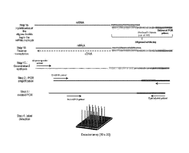
determining the
sequence of the labeled-molecule or any product thereof (e.g., labeled-
amplicons, labeled-
cDNA molecules). Determining the sequence of the labeled-molecule or any
product thereof
can comprise conducting a sequencing reaction to determine the sequence of at
least a portion
of the oligonucleotide tag, at least a portion of the labeled-cDNA molecule, a
complement
thereof, a reverse complement thereof, or any combination thereof. In some
instances only
the tag or a portion of the tag is sequenced. Determining the sequence of the
labeled-
molecule or any product thereof can be performed by sequencing methods such as
HelioscopeTM single molecule sequencing, Nanopore DNA sequencing, Lynx
Therapeutics'
18
Date Recue/Date Received 2023-10-31
Massively Parallel Signature Sequencing (MPSS), 454 pyrosequencing, Single
Molecule real
time (RNAP) sequencing, Illumina (Solexa) sequencing, SOLID sequencing, Ion
TorrentTm,
Ion semiconductor sequencing, Single Molecule SMRT(TM) sequencing, Polony
sequencing,
DNA nanoball sequencing, and VisiGen Biotechnologies approach. Alternatively,
determining the sequence of the labeled-molecule or any product thereof can
use sequencing
platforms, including, but not limited to, Genome Analyzer 'Ix, HiSeq, and
MiSeq offered by
Illumina, Single Molecule Real Time (SMRTTm) technology, such as the PacBio RS
system
offered by Pacific Biosciences (California) and the Solexa Sequencer, True
Single Molecule
Sequencing (tSMSTm) technology such as the HeliScopeTM Sequencer offered by
Helicos Inc.
(Cambridge, MA).
[00123] In some instances, determining the sequence of the labeled-molecule or
any
product thereof comprises paired-end sequencing, nanopore sequencing, high-
throughput
sequencing, shotgun sequencing, dye-terminator sequencing, multiple-primer DNA
sequencing, primer walking, Sanger dideoxy sequencing, Maxim-Gilbert
sequencing,
pyrosequencing, true single molecule sequencing, or any combination thereof
Alternatively,
the sequence of the labeled-molecule or any product thereof can be determined
by electron
microscopy or a chemical-sensitive field effect transistor (chemFET) array.
[00124] In another example, determining the sequence of labeled-molecules or
any
product thereof comprises RNA-Seq or microRNA sequencing. Alternatively,
determining
the sequence of labeled-molecules or any products thereof comprises protein
sequencing
techniques such as Edman degradation, peptide mass fingerprinting, mass
spectrometry, or
protease digestion.
[00125] The sequencing reaction can, in certain embodiments, occur on a solid
or semi-
solid support, in a gel, in an emulsion, on a surface, on a bead, in a drop,
in a continuous
follow, in a dilution, or in one or more physically separate volumes.
[00126]
Sequencing may comprise sequencing at least about 10, 20, 30, 40, 50, 60, 70,
80,
90, 100 or more nucleotides or base pairs of the labeled molecule. In some
instances,
sequencing comprises sequencing at least about 200, 300, 400, 500, 600, 700,
800, 900, 1000
or more nucleotides or base pairs of the labeled molecule. In other instances,
sequencing
comprises sequencing at least about 1500; 2,000; 3,000; 4,000; 5,000; 6,000;
7,000; 8,000;
9,000; or 10,000 or more nucleotides or base pairs of the labeled molecule.
[00127] Sequencing may comprise at least about 200, 300, 400, 500, 600, 700,
800, 900,
1000 or more sequencing reads per run. In some instances, sequencing comprises
sequencing
at least about 1500; 2,000; 3,000; 4,000; 5,000; 6,000; 7,000; 8,000; 9,000;
or 10,000 or more
19
Date Recue/Date Received 2023-10-31
sequencing reads per run.
[00128] V. Detection Methods
[00129] The methods disclosed herein can further comprise detection of thc
labeled-
molecules and/or labeled-amplicons. Detection of the labeled-molecules and/or
labeled-
amplicons can comprise hybridization of the labeled-molecules to surface, e.g.
a solid
support. Alternatively, or additionally, detection of the labeled-molecules
comprises
contacting the labeled-molecules and/or labeled-amplicons with surface, e.g. a
solid support.
In some instances, the method further comprises contacting the labeled-
molecules ancUor
labeled-amplicons with a detectable label to produce a detectable-label
conjugated labeled-
molecule. The methods disclosed herein can further comprise detecting the
detectable-label
conjugated labeled-molecule. Detection of the labeled-molecules or any
products thereof
(e.g., labeled-amplicons, detectable-label conjugated labeled-molecule) can
comprise
detection of at least a portion of the oligonucleotide tag, molecule,
detectable label, a
complement of the oligonucleotide tag, a complement of the molecule, or any
combination
thereof
[00130] Detection of the labeled-molecules or any products thereof can
comprise an
emulsion. For example, the labeled-molecules or any products thereof can be in
an emulsion.
Alternatively, detection of the labeled-molecules or any products thereof
comprises one or
more solutions. In other instances, detection of the labeled-molecules
comprises one or more
containers.
[00131] Detection of the labeled-molecules or any products thereof (e.g.,
labeled-
amplicons, detectable-label conjugated labeled-molecule) can comprise
detecting each
labeled-molecule or products thereof For example, the methods disclosed herein
comprise
sequencing at least a portion of each labeled-molecule, thereby detecting each
labeled-
molecule.
[00132] In some instances, detection of the labeled-molecules and/or labeled-
amplicons
comprises electrophoresis, spectroscopy, microscopy, chemiluminescence,
luminescence,
fluorescence, immuno fluorescence, colorimetry, or electrochemiluminescence
methods. For
example, the method comprises detection of a fluorescent dye. Detection of the
labeled-
molecule or any products thereof can comprise colorimetric methods. For
example, the
colorimetric method comprises the use of a colorimeter or a colorimetric
reader. A non-
limiting list of colorimeters and colorimetric readers include Sensovation's
Colorimetric
Array Imaging Reader (CLAIR), ESEQuant Lateral Flow Immunoassay Reader,
SpectraMax
340PC 38, SpectraMax Plus 384, SpectraMax 190, VersaMax, VMax, and EMax.
Date Recue/Date Received 2023-10-31
[00133] Additional methods used alone or in combination with other methods to
detect the
labeled-molecules and/or amplicons can comprise the use of an array detector,
fluorescence
reader, non-fluorescent detector, CR reader, luminotncter, or scanner. In some
instances,
detecting the labeled-molecules and/or labeled-amplicons comprises the use of
an array
detector. Examples of array detectors include, but are not limited to, diode-
array detectors,
photodiode array detectors, HLPC photodiode array detectors, pixel array
detectors,
Germanium array detectors, CMOS and CCD array detectors, Gated linear CCD
array
detectors, InGaAs photodiode array systems, and TE cooled CCD systems. The
array
detector can be a microarray detector. Non-limiting examples of microarray
detectors include
microelectrode array detectors, optical DNA microarray detection platforms,
DNA
microarray detectors, RNA microarray detectors, and protein microarray
detectors.
[00134] In some instances, a fluorescence reader is used to detect the labeled-
molecule
and/or labeled-amplicons. The fluorescence reader can read 1, 2, 3, 4, 5, or
more color
fluorescence microarrays or other structures on biochips, on slides, or in
microplates. In some
instances, the fluorescence reader is a Sensovation Fluorescence Array imaging
Reader
(FLAIR). Alternatively, the fluorescence reader is a fluorescence microplate
reader such as
the Gemini XPS Fluorescence microplatc reader, Gemini EM Fluorescence
microplatc
reader, Finstruments Fluoroskan filter based fluorescence microplate reader,
PHERAstar
microplate reader, FLUOstar microplate reader, POLARstar Omega microplate
reader,
FLUOstar OPTIMA multi-mode microplate reader and POLARstar OPTIMA multi-mode
microplate reader. Additional examples of fluorescence readers include
PharosFXTM and
PharosFX Plus systems.
[00135] In some instances, detection of the labeled-molecule and/or labeled-
amplicon
comprises the use of a microplate reader. In some instances, the microplate
reader is an
xMarkTm microplate absorbance spectrophotometer, iMark microplate absorbance
reader,
EnSpire Multimode plate reader, EnVision Multilabel plate reader, VICTOR X
Multilabel
plate reader, FlexStation, SpectraMax Paradigm, SpectraMax M5e, SpectraMax M5,
SpectraMax M4, SpectraMax M3, SpectraMax M2-M2e, FilterMax F series,
Fluoroskan
Ascent FL Microplate Fluoremeter and LumMometer, Fluoroskan Ascent Microplate
Fluoremeter, Luminoskan Ascent Microplate Luminometer, Multiskan EX Microplate
Photometer, Muliskan FC Microplate Photometer, and Muliskan GO Microplate
Photometer.
In some instances, the microplate reader detects absorbance, fluorescence,
luminescence,
time-resolved fluorescence, light scattering, or any combination thereof In
some
embodiments, the microplate reader detects dynamic light scattering. The
microplate reader,
21
Date Recue/Date Received 2023-10-31
can in some instances, detect static light scattering. In some instances,
detection of the
labeled-molecules and/or labeled-amplicons comprises the usc of a microplate
imager. In
some instances, the microplate imager comprises ViewLux uHTS microplate imager
and
BioRad microplate imaging system.
[00136] Detection of labeled-molecules and/or products thereof can comprise
the use of a
luminometer. Examples of luminometers include, but are not limited to,
SpectraMax L,
GloMax*-96 microplate luminometer, GloMax -20/20 single-tube luminometer,
GloMaxe-
Multi+ with InstinctTM software, GloMax0-Multi Jr single tube multimode
reader, LUMIstar
OPTIMA, LEADER HC + luminometer, LEADER 450i luminometer, and LEADER 50i
luminometer.
[00137] In some instances, detection of the labeled-molecules and/or labeled-
amplicons
comprises the use of a scanner. Scanners include flatbed scanners such as
those provided by
Cannon, Epson, HP, Fujitsu, and Xerox. Additional examples of flatbed scanners
include the
FMBIO fluorescence imaging scanners (e.g., FMBIO II, III, and III Plus
systems).
Scanners can include microplate scanners such as the Arrayit ArrayPixTM
microarray
microplate scanner. In some instances, the scanner is a Personal Molecular
ImagerTM (PMI)
system provided by Bio-rad.
[00138] Detection of the labeled-molecule can comprise the use of an
analytical technique
that measures the mass-to-charge ratio of charged particles, e.g. mass
spectrometry. In some
embodiments the mass-to-charge ratio of charged particles is measured in
combination with
chromatographic separation techniques. In some embodiments sequencing
reactions are used
in combination with mass-to-charge ratio of charged particle measurements. In
some
embodiments the tags comprise isotopes. In some embodiments the isotope type
or ratio is
controlled or manipulated in the tag library.
[00139] Detection of the labeled-molecule or any products thereof comprises
the use of
small particles and/or light scattering. For example, the amplified molecules
(e.g., labeled-
amplicons) are attached to haptens or directly to small particles and
hybridized to the array.
The small particles can be in the nanometer to micrometer range in size. The
particles can be
detected when light is scattered off of its surface.
[00140] A colorimetric assay can be used where the small particles are
colored, or haptens
can be stained with colorimetric detection systems. In some instances, a
flatbed scanner can
be used to detect the light scattered from particles, or the development of
colored materials.
The methods disclosed herein can further comprise the use of a light absorbing
material. The
light absorbing material can be used to block undesirable light scatter or
reflection. The light
22
Date Recue/Date Received 2023-10-31
absorbing material can be a food coloring or other material. In some
instances, detection of
the labeled-molecule or any products thereof comprises contacting the labeled-
molecule with
an off-axis white light.
[00141] Detection of the labeled-molecule may comprise hybridization chain
reaction
(HCR). As depicted in FIG. 33, a sample comprising a plurality of nucleic acid
molecules
(3340) is stochastically labeled with a plurality of oligonucleotide tags
(3330). The
oligonucleotide tags (3330) comprise a unique identifier region (3310) and an
adapter region
(3320). Stochastically labeling the nucleic acid molecules can comprise
attachment of one or
more oligonucleotide tags (3330) to one or more ends of the nucleic acid
molecule (3340) to
produce one or more labeled-molecules (3345). The one or more labeled
molecules can be
contacted with a plurality of HCR probes (3350). The plurality of HCR probes
(3350) may
comprise a hairpin molecules with an overhang and one or more labels (3360,
3390). The
plurality of HCR probes (3350) may comprise a mixture of hairpin molecules
with 5'
overhangs and hairpin molecules with 3' overhangs. The plurality of HCR probes
may
comprise a stem (3370, 3380). The sequence of the stem (3370, 3380) may be
complementary to at least a portion of the oligonucleotide tag. The sequence
of the stem
(3370, 3380) may be complementary to the adapter region (3320) of the
oligonucleotide tag.
The adapter region (3320) of the oligonucleotide may act as an initiator for a
hybridization
chain rection. As shown in FIG. 33, the stem (3370) of the HCR probe (3350)
can hybridize
to the adapter region (3320) of the labeled molecule (3345). Hybridization of
the stem (3370)
of the HCR probe (3350) to the adapter region (3320) of the labeled molecule
(3345) can
result in opening of the stem (e.g., 3370 and 3380 of the stem are no longer
annealed) and
linearization of the HCR probe (3350), which results in the formation of a
labeled molecule
hybridized to a HCR probe (3355). The linearized HCR probe can then act as an
initiator for
subsequent hybridization of another HCR probe. The stem of a second HCR probe
can
hybridize to the linearized HCR probe that has hybridized to the labeled
molecule, resulting
in linearization of the second HCR probe and the formation of a labeled-
molecule containing
two linearized HCR probes. The linearized second HCR probe can act as an
initiator for
another hybridization reaction. This process can be repeated multiple times to
produce a
labeled molecule with multiple linearized HCR probes (3375). The labels (3360,
3390) on the
HCR probe can enable detection of the labeled molecule. The labels (3360,
3390) may be any
type of label (e.g., fluorphore, chromophore, small molecule, nanoparticle,
hapten, enzyme,
antibody, magnet). The labels (3360 and 3390) may comprise fragments of a
single label. The
labels (3360, 3390) may generate a detectable signal when they are in close
proximity. When
23
Date Recue/Date Received 2023-10-31
the HCR probe is a hairpin, the labels (3360 and 3390) may be too far away to
produce a
detectable signal. When the HCR probe is linearized and multiple linearized
HCR probes are
hybridized together, the labels (3360, 3390) may be in close enough proximity
to generate a
detectable signal. For example, a HCR probe (3350) may comprise two pyrenc
moieties as
labels (3360, 3390). Alternatively, the labels may be nanoparticles. The HCR
can enable
attachment of multiple HCR probes to a labeled molecule, which can result in
signal
amplification. Stoachastic labeling followed by HCR may increase the
sensitivity of
detection, analysis and/or quantification of the nucleic acid molecules.
Stochastic labeling
followed by HCR may increase the accuracy of detection, analysis, and/or
quantification of
one or more nucleic acid molecules.
1001421 Additional methods and apparatus for signal detection and processing
of intensity
data are disclosed in, for example, U.S. Patents Nos. 5,143,854, 5,547,839,
5,578,832,
5,631,734, 5,800,992, 5,834,758, 5,856,092, 5,902,723, 5,936,324, 5,981,956,
6,025,601,
6,090,555, 6,141,096, 6,185,030, 6,201,639; 6,218,803; and 6,225,625, in U.S.
Patent Pub.
Nos. 20040012676 and 20050059062 and in PCT Application PCT/US99/06097
(published
as W099/47964).
1001431 Detection and/or quantification of the labeled molecules may comprise
the use of
computers or computer software. Computer software products may comprise a
computer
readable medium having computer-executable instructions for performing the
logic steps of
the method of the invention. Suitable computer readable medium include floppy
disk, CD-
ROMIDVD/DVD-ROM, hard-disk drive, flash memory, ROM/RAM, magnetic tapes, etc.
The computer-executable instructions may be written in a suitable computer
language or
combination of several languages. Basic computational biology methods are
described in, for
example, Setubal and Meidanis et al., Introduction to Computational Biology
Methods (PWS
Publishing Company, Boston, 1997); Salzberg, Searles, Kasif, (Ed.),
Computational Methods
in Molecular Biology, (Elsevier, Amsterdam, 1998); Rashidi and Buehler,
Bioinformatics
Basics: Application in Biological Science and Medicine (CRC Press, London,
2000) and
Ouelette and Bzevanis Bioinformatics: A Practical Guide for Analysis of Gene
and Proteins
(Wiley & Sons, Inc., 2nd ed., 2001). See also US 6,420,108.
1001441 Computer program products and software may be used for a variety of
purposes,
such as probe design, management of data, analysis, and instrument operation.
See, U.S.
Patent Nos. 5,593,839, 5,795,716, 5,733,729, 5,974,164, 6,066,454, 6,090,555,
6,185,561,
6,188,783, 6,223,127, 6,229,911 and 6,308,170. Computer methods related to
genotyping
24
Date Recue/Date Received 2023-10-31
using high density microarray analysis may also be used in the present
methods, see, for
example, US Patent Pub. Nos. 20050250151, 20050244883, 20050108197,
20050079536 and
20050042654. Additionally, the present disclosure may have preferred
embodiments that
include methods for providing genetic information over networks such as the
Internet as
shown in U.S. Patent Pub. Nos. 20030097222, 20020183936, 20030100995,
20030120432,
20040002818, 20040126840, and 20040049354.
[00145] Detection and/or quantification of the labeled-molecules or any
products thereof
can comprise the use of one or more algorithms. Alternatively, or
additionally, the methods,
kits and compositions can further comprise a computer, software, printer,
and/or electronic
data or information.
[00146] The methods disclosed herein can further comprise the transmission of
data/information. For example, data/information derived from the detection
and/or
quantification of the labeled-molecule or any products thereof are transmitted
to another
device and/or instrument. In some instances, the information obtained from an
algorithm can
also be transmitted to another device and/or instrument. Transmission of the
data/information
can comprise the transfer of data/information from a first source to a second
source. The first
and second sources can be in the same approximate location (e.g., within the
same room,
building, block, campus). Alternatively, first and second sources are in
multiple locations
(e.g., multiple cities, states, countries, continents, etc). In some
embodiments a non-
transitory computable readable media is used to store or analyze data
generated using
methods described herein.
[00147] Transmission of the data/information can comprise digital transmission
or analog
transmission. Digital transmission can comprise the physical transfer of data
(a digital bit
stream) over a point-to-point or point-to-multipoint communication channel.
Examples of
such channels are copper wires, optical fibres, wireless communication
channels, and storage
media. The data can be represented as an electromagnetic signal, such as an
electrical
voltage, radiowave, microwave, or infrared signal.
[00148] Analog transmission can comprise the transfer of a continuously
varying analog
signal. The messages can either be represented by a sequence of pulses by
means of a line
code (baseband transmission), or by a limited set of continuously varying wave
forms
(passband transmission), using a digital modulation method. The passband
modulation and
corresponding demodulation (also known as detection) can be carried out by
modem
equipment. According to the most common definition of digital signal, both
baseband and
passband signals representing bit-streams are considered as digital
transmission, while an
Date Recue/Date Received 2023-10-31
alternative definition only considers the baseband signal as digital, and
passband transmission
of digital data as a form of digital-to-analog conversion.
[00149] The applications and uses of the systems and methods described herein
can
produce one or more result useful to diagnose a disease state of an
individual, for example, a
patient. In one embodiment, a method of diagnosing a disease comprises
reviewing or
analyzing data relating to the presence and/or the concentration level of a
target in a sample.
A conclusion based review or analysis of the data can be provided to a
patient, a health care
provider or a health care manager. In one embodiment the conclusion is based
on the review
or analysis of data regarding a disease diagnosis. It is envisioned that in
another embodiment
that providing a conclusion to a patient, a health care provider or a health
care manager
includes transmission of the data over a network.
[00150] Accordingly, business systems and methods using the systems and
methods
described herein are provided.
[00151] One aspect of the invention is a business method comprising screening
patient test
samples for the presence or absence of a biologically active analyte to
produce data regarding
the analyte, collecting the analyte data, providing the analyte data to a
patient, a health care
provider or a health care manager for making a conclusion based on review or
analysis of the
data regarding a disease diagnosis. In one embodiment the conclusion is
provided to a
patient, a health care provider or a health care manager includes transmission
of the data over
a network.
[00152] Accordingly FIG. 8 is a block diagram showing a representative example
logic
device through which reviewing or analyzing data relating to the present
invention can be
achieved. Such data can be in relation to a disease, disorder or condition in
an individual.
FIG. 8 shows a computer system (or digital device) 800 connected to an
apparatus 820 for
use with the scanning sensing system 824 to, for example, produce a result.
The computer
system 800 may be understood as a logical apparatus that can read instructions
from media
811 and/or network port 805, which can optionally be connected to server 809
having fixed
media 812. The system shown in FIG. 8 includes CPU 801, disk drives 803,
optional input
devices such as keyboard 815 and/or mouse 816 and optional monitor 807. Data
communication can be achieved through the indicated communication medium to a
server
809 at a local or a remote location. The communication medium can include any
means of
transmitting and/or receiving data. The communication medium can comprise a
non-
transitory computer readable media. For example, the communication medium can
be a
network connection, a wireless connection or an internet connection. Such a
connection can
26
Date Recue/Date Received 2023-10-31
provide for communication over the World Wide Web. It is envisioned that data
can be
transmitted over such networks or connections for reception and/or review by a
party 822.
The receiving party 822 can bc but is not limited to a patient, a health care
provider or a
health care manager.
[00153] In one embodiment, a computer-readable medium includes a medium
suitable for
transmission of a result of an analysis of an environmental or biological
sample. The medium
can include a result regarding a disease condition or state of a subject,
wherein such a result is
derived using the methods described herein. The computer readable media can be
non-
transitory.
[00154] Data Analysis: In some embodiments the scanner instrument produces the
raw
intensity values for each position on the array as well as the background
intensity. Many
methods can be used to calculate the number of molecules in the sample. For
example, the
values for the control positions on the array are removed from the dataset and
a scatter plot is
generated to provide an image of the data. This may occur with or without the
background
intensity subtracted from the raw data. A threshold intensity value can be
established in order
to classify the positive spots and the negative spots. All of the positive
spots are summed to
provide a total count of unique stochastic labels. This process can be
automated in Microsoft
excel or another computer software program.
[00155] An alternative to this strategy is the use of clustering algorithms
such as k-means
clustering. K-means clustering is a method of cluster analysis which aims to
partition all of
the observations into clusters in which each observation belongs to the
cluster with the
nearest mean. The data can be split into 2 or 3 clusters (or more, 3 clusters
seems to produce
the cleanest numbers so far) and the number of data points can be added up to
determine the
counts.
[00156] VI. Target molecules
[00157] The methods, kits, and systems disclosed herein can be used in the
stochastic
labeling of molecules. Such molecules include, but are not limited to,
polynucleotides and
polypeptides. As used herein, the terms "polynucleotide" and "nucleic acid
molecule" refers
to a polymeric form of nucleotides of any length, either ribonucleotides,
deoxyribonucleotides, locked nucleic acids (LNA) or peptide nucleic acids
(PNAs), that
comprise purine and pyrimidine bases, or other natural, chemically or
biochemically
modified, non-natural, or derivatized nucleotide bases. A -polynucleotide" or -
nucleic acid
molecule" can consist of a single nucleotide or base pair. Alternatively, the
"polynucleotide"
or "nucleic acid molecule" comprises two or more nucleotides or base pairs.
For example, the
27
Date Recue/Date Received 2023-10-31
"polynucleotide" or "nucleic acid molecule" comprises at least about 2, 3, 4,
5, 6, 7, 8, 9, 10,
11, 12, 13, 14, 15, 16, 17, 18, 19, 20, 30, 40, 50, 60, 70, 80, 90, 100, 200,
300, 400, 500, 600,
700, 800, 900, or 1000 nucleotides or base pairs. In another example, the
polynucleotide
comprises at least about 1500, 2000, 2500, 3000, 3500, 4000, 4500, 5000, 5500,
6000, 6500,
7000, 7500, 8000, 8500, 9000, 9500, or 10000 nucleotides or base pairs. The
backbone of the
polynucleotide can comprise sugars and phosphate groups, as may typically be
found in RNA
or DNA, or modified or substituted sugar or phosphate groups. A polynucleotide
may
comprise modified nucleotides, such as methylated nucleotides and nucleotide
analogs. The
sequence of nucleotides may be interrupted by non-nucleotide components. Thus
the terms
nucleoside, nucleotide, deoxynucleoside and deoxynucleotide generally include
analogs such
as those described herein. These analogs are those molecules having some
structural features
in common with a naturally occurring nucleoside or nucleotide such that when
incorporated
into a nucleic acid or oligonucleoside sequence, they allow hybridization with
a naturally
occurring nucleic acid sequence in solution. Typically, these analogs are
derived from
naturally occurring nucleosides and nucleotides by replacing and/or modifying
the base, the
ribose or the phosphodiester moiety. The changes can be tailor made to
stabilize or
destabilize hybrid formation or enhance the specificity of hybridization with
a
complementary nucleic acid sequence as desired. In some instances, the
molecules are DNA,
RNA, or DNA-RNA hybrids. The molecules can be single-stranded or double-
stranded. In
some instances, the molecules are RNA molecules, such as rnRNA, rRNA, tRNA,
ncRNA,
lncRNA, siRNA, or miRNA. The RNA molecules can be polyadenylated.
Alternatively, the
mRNA molecules are not polyadenylated. Alternatively, the molecules are DNA
molecules.
The DNA molecules can be genomic DNA. The DNA molecules can comprise exons,
introns, untranslated regions, or any combination thereof
[00158] In some instances, the molecules are polypeptides. As used herein, the
term
"polypeptide" refers to a molecule comprising at least one peptide. In some
instances, the
polypeptide consists of a single peptide. Alternatively, the polypeptide
comprises two or
more peptides. For example, the polypeptide comprises at least about 2, 3, 4,
5, 6, 7, 8, 9, 10,
11, 12, 13, 14, 15, 16, 17, 18, 19, 20, 30, 40, 50, 60, 70, 80, 90, 100, 200,
300, 400, 500, 600,
700, 800, 900, or 1000 peptides. Examples of polypeptides include, but are not
limited to,
amino acids, proteins, peptides, hormones, oligosaccharides, lipids, glyco
lipids,
phospholipids, antibodies, enzymes, kinases, receptors, transcription factors,
and ligands.
[00159] The methods, kits, and systems disclosed herein can be used to
stochastically label
individual occurrences of identical or nearly identical molecules and/or
different molecules.
28
Date Recue/Date Received 2023-10-31
In some instances, the methods, kits, and systems disclosed herein can be used
to
stochastically label identical or nearly identical molecules (e.g., molecules
comprise identical
or nearly identical sequences). For example, the molecules to be labeled
comprise at least
about 80%, 85%, 90%, 95%, 96%, 97%, 98%, 99%, or 100% sequence identity. The
nearly
identical molecules may differ by less than about 100, 90, 80, 70, 60, 50, 40,
30, 25, 20, 25,
10, 9, 8, 7, 6, 5, 4, 3, 2, or 1 nucleotides or base pairs. In some instances,
the molecules to be
labeled are variants of each other. For example, the molecules to be labeled
may contain
single nucleotide polymorphisms or other types of mutations. In another
example, the
molecules to be labeled are splice variants. In some instances, at least one
molecule is
stochastically labeled. In other instances, at least 2, 3, 4, 5, 6, 7, 8, 9,
or 10 identical or nearly
identical molecules are stochastically labeled. Alternatively, at least 20,
30, 40, 50, 60, 70,
80, 90, 100, 200, 300, 400, 500, 600, 700, 800, 900, or 1000 identical or
nearly identical
molecules are stochastically labeled. In other instances, at least 1500;
2,000; 2500; 3,000;
3500; 4,000; 4500; 5,000; 6,000; 7,000; 8,000; 9,000; or 10000 identical or
nearly identical
molecules are stochastically labeled. In other instances; at least 15,000;
20,000; 25,000;
30,000; 35,000; 40,000; 45,000; 50,000; 60,000; 70,000; 80,000; 90,000; or
100,000 identical
or nearly identical molecules are stochastically labeled.
[00160] In other instances, the methods, kits, and systems disclosed herein
can be used to
stochastically label different molecules. For example, the molecules to be
labeled comprise
less than 75%, 70%, 65%, 60%, 55%, 50%, 45%, 40%, 35%, 30%, 25%, 20%, 15%,
10%,
5%, 4%, 3%, 2%, 1% sequence identity. The different molecules may differ by at
least about
1, 2, 3, 4, 5, 6, 7, 8, 9, 10, 15, 20, 25, 30, 40, 50, 60, 70, 80, 90, 100 or
more nucleotides or
base pairs. In some instances, at least one molecule is stochastically
labeled. In other
instances, at least 2, 3, 4, 5, 6, 7, 8, 9, or 10 different molecules are
stochastically labeled.
Alternatively, at least 20, 30, 40, 50, 60, 70, 80, 90, 100, 200, 300, 400,
500, 600, 700, 800,
900, or 1000 different molecules are stochastically labeled. In other
instances, at least 1500;
2,000; 2500; 3,000; 3500; 4,000; 4500; 5,000; 6,000; 7,000; 8,000; 9,000; or
10000 different
molecules are stochastically labeled. In other instances; at least 15,000;
20,000; 25,000;
30,000; 35,000; 40,000; 45,000; 50,000; 60,000; 70,000; 80,000; 90,000; or
100,000 different
molecules are stochastically labeled.
[00161] The different molecules to be labeled can be present in the sample at
different
concentrations or amounts. For example, the concentration or amount of one
molecule is
greater than the concentration or amount of another molecule in the sample. In
some
instances, the concentration or amount of at least one molecule in the sample
is at least about
29
Date Recue/Date Received 2023-10-31
L5, 2, 3, 4, 5, 6, 7, 8, 9, 10, 11, 12, 13, 14, 15, 20, 25, 30, 35, 40, 45,
50, 60, 70, 80, 90, or
100 or more times greater than the concentration or amount of at least one
other molecule in
the sample. In another example, the concentration or amount of one molecule is
less than the
concentration or amount of another molecule in the sample. The concentration
or amount of
at least one molecule in the sample can be at least about 1.5, 2, 3, 4, 5, 6,
7, 8, 9, 10, 11, 12,
13, 14, 15, 20, 25, 30, 35, 40, 45, 50, 60, 70, 80, 90, or 100 or more times
less than the
concentration or amount of at least one other molecule in the sample.
[00162] In some instances, the molecules to be labeled are in one or more
samples. The
molecules to be labeled can be in two or more samples. The two or more samples
can contain
different amounts or concentrations of the molecules to be labeled. In some
instances, the
concentration or amount of one molecule in one sample can be greater than the
concentration
or amount of the same molecule in a different sample. For example, a blood
sample might
contain a higher amount of a particular molecule than a urine sample.
Alternatively, a single
sample is divided into two or more subsamples. The subsamples can contain
different
amounts or concentrations of the same molecule. The concentration or amount of
at least one
molecule in one sample can be at least about 1.5, 2, 3, 4, 5, 6, 7, 8, 9, 10,
11, 12, 13, 14, 15,
20, 25, 30, 35, 40, 45, 50, 60, 70, 80, 90, or 100 or more times greater than
the concentration
or amount of the same molecule in another sample. Alternatively, the
concentration or
amount of one molecule in one sample can be less than the concentration or
amount of the
same molecule in a different sample. For example, a heart tissue sample might
contain a
higher amount of a particular molecule than a lung tissue sample. The
concentration or
amount of at least one molecule in one sample can be at least about 1.5, 2, 3,
4, 5, 6, 7, 8, 9,
10, 11, 12, 13, 14, 15, 20, 25, 30, 35, 40, 45, 50, 60, 70, 80, 90, or 100 or
more times less than
the concentration or amount of the same molecule in another sample. In some
instances, the
different concentrations or amounts of a molecule in two or more different
samples is referred
to as sample bias.
[00163] VII. Oligonucleotide tags
[00164] In some embodiments, the methods, kits, and systems disclosed herein
comprise a
plurality of oligonucleotide tags. The oligonucleotide tags can comprise a
target specific
region, a unique identifier region, an adapter region, a universal primer
binding site region, or
any combination thereof. FIG. 10-13 shows examplary oligonucleotide tags.
[00165] As shown in FIG. 10, the oligonucleotide tag (1004) may comprise a
universal
primer binding site (1001), unique identifier region (1002) and a target
specific region
(1003).
Date Recue/Date Received 2023-10-31
[00166] As shown in FIG. 11A, the oligonucleotide tag (1107) can comprise a
universal
primer binding site (1102), a unique identifier region (1103) and a target
specific region
(1105). The universal primer binding site (1102) may comprise a
phosphorothioate linkage,
as depicted by an "*" in FIG. 11A. As shown in FIG. 11B, the oligonucleotide
tag (1128) can
comprise a universal primer binding site (1122), a unique identifier region
(1123), bridge
splint (1129), and a target specific region (1126). As shown in FIG. 11C, the
oligonucleotide
tag (1158) may comprise a universal primer binding site (1151), unique
identifier region
(1152), ligation sequence (1153), and a target specific sequence (1157). As
shown in FIG.
11D, the oligonucleotide tag (1177) may comprise a universal primer binding
site (1171),
unique identifier region (1172), ligation sequence (1173), and a DNA target
specific sequence
(1178).
[00167] As shown in FIG. 12A, an oligonucleotide tag (1201) may comprise a
universal
primer binding site (1202), a unique identifier region comprising a degenerate
sequence
(1203) and a target specific region (1204). As shown in FIG. 12B, an
oligonucleotide tag
(1210) may comprise a universal primer binding site (1211), a unique
identifier region (1215)
comprising a degenerate sequence (1213) flanked by two flanking sequences
(1212 and 1214)
and a target specific region (1216).
[00168] The oligonucleotide tag may be comprise one or more secondary
structures. As
shown in FIG. 13A, the oligonucleotide tag (1301) comprises a hairpin
structure. The
oligonucleotide tag (1301) can comprise a target specific region (1302), a
cleavable stem
(1303, 1304), and a unique identifier region (1305).
[00169] The oligonucleotide tag may comprise a target specific region that can
hybridize
to a plurality of different target molecules. For example, as shown in FIG.
13B, the
oligonucleotide tag (1310) comprises a universal primer binding site (1311),
unique identifier
region (1312), and a universal target specific region (1313). The universal
target specific
region (1313) may comprise an oligodT sequence that enables hybridization to
target
molecules comprising a polyA or polyU sequence.
[00170] A method for synthesizing a plurality of oligonucleotdie tags is
depicted in FIG.
10. As shown in FIG. 10, oligonucleotide tags (1004) can be synthesized
separately. The
oligonucleotide tags (1004) can comprise a universal primer binding site
(1001), a unique
identifier region (1002), and a target specific region. The individual
oligonucleotide tags can
be pooled to produce a plurality of oligonucleotide tags (1005) comprising a
plurality of
different unique identifier regions.
[00171] A method for synthesizing a plurality of oligonucleotdie tags is
depicted in FIG.
31
Date Recue/Date Received 2023-10-31
HA. As shown in FIG. 11A, oligonucleotide fragments (1101) can be synthesized
separately.
The oligonucleotide fragments (1101) can comprise a universal primer binding
site (1102)
and a unique identifier region (1103). The universal primer binding site
(1102) may comprise
a phosphorothioate linkage, as depicted by an "*" in FIG. HA. As shown in Step
1 of FIG.
11A, the individual oligonucleotide fragments (1101) may be mixed to produce a
plurality of
oligonucleotide fragments (1104). The plurality of oligonucleotide fragments
(1104) can be
attached to a target specific region (1105). As shown in Step 2 of FIG. 11A,
the target
specific region can be ligated to the oligonucleotide tag to produce an
oligonucleotide tag
comprising a target specific region (1105). 5' and 3' exonucleases may be
added to the
reaction to remove non-ligated products (1105, 1101). The oligonucleotide tag
(1106)
comprising the universal primer binding site (1102), unique identifier region
(1103) and
target specific region (1105) may be resistant to 5' and 3' exonucleases. As
shown in Step 3
of FIG. 11A, the 3' phosphate group from the ligated oligonucleotide tag
(1106) can be
removed to produce an oligonucleotide tag (1107) without a 3' phosphate group.
The 3'
phosphate group can be removed enzymatically. For example, a T4 polynucleotide
kinase can
be used to remove the 3' phosphate group.
[00172] Another method of synthesizing oligonucleotide tags is depicted in
FIG. 11B. As
shown in FIG. 11B, an oligonucleotide tag (1128) can be synthesized by
ligating two
oligonucleotide fragments (1121 and 1127). One oligonucleotide fragment (1121)
may
comprise a universal primer binding site (1122), unique identifier region
(1123) and a left
splint (1123). The other oligonucleotide fragment (1128) may comprise a right
splint (1125)
and a target specific region (1126). A ligase (e.g., T4 DNA ligase) can be
used to join the two
oligonucleotide fragments (1121 and 1127) to produce an oligonucleotide tag
(1128). Double
stranded ligation of the left splint (1124) and right splint (1125) can
produce an
oligonucleotide tag (1128) with a bridge splint (1129).
[00173] An alternative method of synthesizing an oligonucleotide tag by
ligating two
oligonucleotide fragments is depicted in FIG. 11C. As shown in FIG. 11C, an
oligonucleotide
tag (1158) is synthesized by ligating two oligonucleotide fragments (1150 and
1158). One
oligonucleotide fragment (1150) may comprise a universal primer binding site
(1151), unique
identifier region (1152), and a ligation sequence (1153). The other
oligonucleotide fragment
(1158) may comprise a ligation sequence (1154) that is complementary to the
ligation
sequence (1153) of the first oligonucleotide fragment (1150), a complement of
a target
specific region (1155), and a label (1156). The oligonucleotide fragment
(1159) may also
comprise a 3' phosphate which prevents extension of the oligonucleotide
fragment. As shown
32
Date Recue/Date Received 2023-10-31
in Step 1 of FIG. 11C, the ligation sequences (1153 and 1154) of the two
oligonucleotide
fragments may anneal and a polymerase can be used to extend the 3' end of the
first
oligonucleotide fragment (1150) to produce an oligonucicotidc tag (1158). The
oligonucleotide tag (1158) may comprise a universal primer binding site
(1151), unique
identifier region (1152), ligation sequence (1153), and a target specific
sequence (1157). The
target specific sequence (1157) of the oligonucleotide tag (1158) may be the
complement of
the complement of the target specific region (1155) of the second
oligonucleotide fragment
(1159). The oligonucleotide fragment comprising the label (1156) can be
removed from the
oligonucleotide tags (1158). For example, the label (1156) may comprise biotin
and
oligonucleotide fragments (1159) comprising the biotin label (1156) can be
removed via
streptavidin capture. In another example, the label (1156) may comprise a 5'
phosphate and
oligonucleotide fragments (1159) comprising the 5' phosphate (1156) can be
removed via an
exonuclease (e.g., Lambda exonuclease).
[00174] As depicted in FIG. 11D, a first oligonucleotide fragment (1170)
comprising a
universal primer binding site (1171), unique identifier region (1172), a first
ligation sequence
(1173) is annealed to a second oligonucleotide fragment (1176) comprising a
second ligation
sequence (1174) and an RNA complement of the target sequence (1175). Step 1
may
comprise annealing the first and second ligation sequences (1173 and 1174)
followed by
reverse transcription of the RNA complement of the target sequence (1175) to
produce an
oligonucleotide tag (1177) comprising a universal primer binding site (1171),
unique
identifier region (1172), a first ligation sequence (1173), and a target
specific region (1178).
The oligonucleotide fragments comprising the RNA complement of the target
sequence can
be selectively degraded by RNAse treatment.
[00175] The
oligonucleotide tag can comprise at least about 1, 2, 3, 4, 5, 6, 7, 8, 9, 10,
11,
12, 13, 14, 15, 16, 17, 18, 19, 20, 30, 40, 50, 60, 70, 80, 90, 100, 200, 300,
400, 500, 600,
700, 800, 900, or 1000 nucleotides or base pairs. In another example, the
oligonucleotide tag
comprises at least about 1500, 2,000; 2500, 3,000; 3500, 4,000; 4500, 5,000;
5500, 6,000;
6500, 7,000; 7500, 8,000; 8500, 9,000; 9500, or 10,000 nucleotides or base
pairs.
[00176] The tags can be hexamers, e.g.random hexamers. The tags can be
randomly
generated from a set of mononucleotides. The tags can be assembled by randomly
incorporating mononucleotides.
[00177] The tags can also be assembled without randomness, to generate a
library of
different tags which are not randomly generated but which includes sufficient
numbers of
different tags to practice the methods.
33
Date Recue/Date Received 2023-10-31
[00178] In some embodiments an oligonucleotide tag can comprise a cutback in a
target
molecule. The cutback can be, for example, a enzymatic digestion of one or
both ends of a
target molecule. The cutback can be used in conjunction with the addition of
added
oligonucleotide tags. The combination of the cutback and the added tags can
contain
information related to the particular starting molecule. By adding a random
cutback to the
tag a smaller diversity of the added tags may be necessary for counting the
number of target
molecules when detection allows a determination of both the random cutback and
the added
oligonucleotides.
[00179] The oligonucleotide tag can comprise a target specific region. The
target specific
region can comprise a sequence that is complementary to the molecule. In some
instances, the
molecule is an mRNA molecule and the target specific region comprises an
oligodT sequence
that is complementary to the polyA tail of the mRNA molecule. The target
specific region
can also act as a primer for DNA and/or RNA synthesis. For example, the
oligodT sequence
of the target specific region can act as a primer for first strand synthesis
of a cDNA copy of
the mRNA molecule. Alternatively, the target specific region comprises a
sequence that is
complementary to any portion of the molecule. In other instances, the target
specific region
comprises a random sequence that can be hybridized or ligated to the molecule.
The target
specific region can enable attachment of the oligonucleotide tag to the
molecule. Attachment
of the oligonucleotide tag can occur by any of the methods disclosed herein
(e.g.,
hybridization, ligation). In some instances, the target specific region
comprises a sequence
that is recognized by one or more restriction enzymes. The target specific
region can
comprise at least about 1, 2, 3, 4, 5, 6,7, 8,9, 10, 11, 12, 13, 14, 15, 16,
17, 18, 19, 20, 30,
40, 50, 60, 70, 80, 90, 100, 200, 300, 400, 500, 600, 700, 800, 900, or 1000
nucleotides or
base pairs. In another example, the target specific region comprises at least
about 1500, 2000,
2500, 3000, 3500, 4000, 4500, 5000, 5500, 6000, 6500, 7000, 7500, 8000, 8500,
9000, 9500,
or 10000 nucleotides or base pairs. Preferably, the target specific region
comprises at least
about 5-10, 10-15, 10-20, 10-30, 15-30, or 20-30 nucleotides or base pairs.
[00180] In some instances, the target specific region is specific for a
particular gene or
gene product. For example, the target specific region comprises a sequence
complementary to
a region of a p53 gene or gene product. Therefore, the oligonucleotide tags
can only attach to
molecules comprising the p53-specific sequence. Alternatively, the target
specific region is
specific for a plurality of different genes or gene products. For example, the
target specific
region comprises an oligodT sequence. Therefore, the oligonucleotide tags can
attach to any
molecule comprising a polyA sequence. In another example, the target specific
region
34
Date Recue/Date Received 2023-10-31
comprises a random sequence that is complementary to a plurality of different
genes or gene
products. Thus, the oligonucleotide tag can attach to any molecule with a
sequence that is
complementary to the target specific region. In other instances, the target
specific region
comprises a restriction site overhang (e.g., EcoR1 sticky-end overhang). The
oligonucleotide
tag can ligate to any molecule comprising a sequence complementary to the
restriction site
overhang.
[00181] The oligonucleotide tag disclosed herein often comprises a unique
identifier
region. The unique identifier region may be used to uniquely identify
occurrences of target
species thereby marking each species with an identifier that can be used to
distinguish
between two otherwise identical or nearly identical targets. The unique
identifier region of
the plurality of oligonucleotide tags can comprise a collection of different
semiconductor
nanocrystals, metal compounds, peptides, oligonucleotides, antibodies, small
molecules,
isotopes, particles or structures having different shapes, colors, barcodes or
diffraction
patterns associated therewith or embedded therein, strings of numbers, random
fragments of
proteins or nucleic acids, different isotopes, or any combination thereof The
unique identifier
region can comprise a degenerative sequence. The unique identifier region can
comprise at
least about 1, 2, 3, 4, 5, 6, 7, 8,9, 10, 11, 12, 13, 14, 15, 16, 17, 18, 19,
20, 30, 40, 50, 60, 70,
80, 90, 100, 200, 300, 400, 500, 600, 700, 800, 900, or 1000 nucleotides or
base pairs. In
another example, the unique identifier region comprises at least about 1500;
2,000; 2500,
3,000; 3500, 4,000; 4500, 5,000; 5500, 6,000; 6500, 7,000; 7500, 8,000; 8500,
9,000; 9500,
or 10,000 nucleotides or base pairs. Preferably, the unique identifier region
comprises at least
about 10-30, 15-40, or 20-50 nucleotides or base pairs.
[00182] In some instances, the oligonucleotide tag comprises a universal
primer binding
site. The universal primer binding site allows the attachment of a universal
primer to the
labeled-molecule and/or labeled-amplicon. Universal primers are well known in
the art and
include, but are not limited to, -47F (M13F), alfaMF, A0X3', A0X5', BGH_r,
CMV_-30,
CMV -50, CVM f, LACrmt, lamgda gt10F, lambda gt 10R, lambda gt11F, lambda
gt11R,
M13 rev, M13Forward(-20), M13Reverse, male, plOSEQP_pQE, pA_-120, pet_4, pGAP
Forward, pGL_RVpr3, pGLpr2 R, pl(LAC1_4, pQE_FS, pQE_RS, puc Ul, puc_U2,
revers_A, seq_IRES_tam, seq_IRES_zpet, seq_ori, seq_PCR, seq_pIRES-, seq
pIRES+,
seq_pSecTag, seq pSecTag+, seq_retro+PSI, SP6, T3-prom, T7-prom, and T7-term
Inv.
Attachment of the universal primer to the universal primer binding site can be
used for
amplification, detection, and/or sequencing of the labeled-molecule and/or
labeled-amplicon.
The universal primer binding site can comprise at least about 1, 2, 3, 4, 5,
6, 7, 8, 9, 10, 11,
Date Recue/Date Received 2023-10-31
12, 13, 14, 15, 16, 17, 18, 19, 20, 30, 40, 50, 60, 70, 80, 90, 100, 200, 300,
400, 500, 600,
700, 800, 900, or 1000 nucleotides or base pairs. In another example, the
universal primer
binding site comprises at least about 1500; 2,000; 2500, 3,000; 3500, 4,000;
4500, 5,000;
5500, 6,000; 6500, 7,000; 7500, 8,000; 8500, 9,000; 9500, or 10,000
nucleotides or base
pairs. Preferably, the universal primer binding site comprises 10-30
nucleotides or base pairs.
[00183] The oligonucleotide tag may comprise an adapter region. The adapter
region may
enable hybridization of one or more probes. The adapter region may enable
hybridization of
one or more HCR probes.
[00184] The oligonucleotide tag may comprise one or more labels.
[00185] The oligonucleotide tag may act as an initiator for a hybridization
chain reaction
(HCR). The adapter region of the oligonucleotide tag may act as an initiation
for HCR. The
universal primer binding site may act as an initiator for HCR.
[00186] In some instances, the oligonucleotide tag is single-stranded. In
other instances,
the oligonucleotide tag is double-stranded. The oligonucleotide tag can be
linear.
Alternatively, the oligonucleotide tag comprises a secondary structure. As
used herein,
"secondary structure" includes tertiary, quaternary, etc...structures. In some
instances, the
secondary structure is a hairpin, a stem-loop structure, an internal loop, a
bulge loop, a
branched structure or a pseudoknot, multiple stem loop structures, cloverleaf
type structures
or any three dimensional structure. In some instances, the secondary structure
is a hairpin.
The hairpin can comprise an overhang sequence. The overhang sequence of the
hairpin can
act as a primer for a polymerase chain reaction and/or reverse transcription
reaction. The
overhang sequence comprises a sequence that is complementary to the molecule
to which the
oligonucleotide tag is attached and the overhang sequence hybridizes to the
molecule. The
overhang sequence can be ligated to the molecule and acts as a template for a
polymerase
chain reaction andlor reverse transcription reaction. In some embodiments the
tag comprises
nucleic acids and/or synthetic nucleic acids and/or modified nucleic acides.
1001871 An oligonucleotide tag comprising a hairpin may act as a probe for a
hybrization
chain reaction. Further disclosed herein is a stochastic label-based
hybridization chain
reaction (HCR) method comprising stochastically labeling one or more nucleic
acid
molecules with an oligonucleotide tag, wherein the oligonucleotide tag is a
hairpin and the
one or more nucleic acid molecules act as initiators for a hybridization chain
reaction. A
schematic of a stochastic label-based hybridization reaction is depicted in
FIG. 34. As shown
in FIG. 34, one or more nucleic acid molecules (3480) are stochastically
labeled with a
plurality of hairpin oligonucleotide tags (3490) by initiating a hybridization
chain reaction.
36
Date Recue/Date Received 2023-10-31
The hairpin oligonucleotide tags may comprise one or more labels (3410, 3470),
an overhang
(3420, 3420), a stem (3430, 3460), and a loop (3450). The overhang region
(3420) of the
hairpin oligonucleotide tag (3490) may comprise a target specific region. The
overhang
region (3420) may comprise an oligodT sequence. The sample comprising the one
or more
nucleic acid molecules may be treated with one or more restriction nucleases
prior to
stochastic labeling.The overhang region (3420) may comprise a restriction
enzyme
recognition sequence. The sample comprising the one or more nucleic acid
molecules may be
contacted with one or more adapters prior to stochastic labeling to produce an
adapter-nucleic
acid molecule hybrid. The overhang region (3420) and the stem (3430) may be
complementary to the one or more adapters. The loop (3450) of the
oligonucleotide tag may
comprise a unique identifier region. Hybridization of a first hairpin
oligonucleotide tag
(3490) to the nucleic acid molecules (3480) may result in the formation of a
labeled molecule
(3415), wherein the first hairpin oligonucleotide tag is linearized to produce
a first linearized
oligonucleotide tag. The first linearized oligonucleotide tag of labeled
molecule (3415) can
act as an initiator for hybrization of a second hairpin oligonucleotide tag to
the labeled
molecule (3415) to produce a labeled molecule with two linearized
oligonucleotide tags
(3425). The second linearized oligonucleotide tag can act as an initiator for
another
hybridization reaction. This process can be repeated multiple times to produce
a labeled
molecule with multiple linearized HCR probes (3435). The labels (3410, 3470)
on the HCR
probe can enable detection of the labeled molecule. The labels (3410, 3470)
may be any type
of label (e.g., fluorphore, chromophore, small molecule, nanoparticle, hapten,
enzyme,
antibody, magnet). The labels (3360 and 3390) may comprise fragments of a
single label. The
labels (3410, 3470) may generate a detectable signal when they are in close
proximity. When
the oligonucleotide tag is a hairpin, the labels (3360 and 3390) may be too
far away to
produce a detectable signal. When the hairpin oligonucleotide tag is
linearized and multiple
linearized hairpin oligonucleotide tags are hybridized together, the labels
(3410, 3470) may
be in close enough proximity to generate a detectable signal. For example, a
hairpin
oligonucleotide tag (3350) may comprise two pyrene moieties as labels (3410,
3470).
Alternatively, the labels may be nanoparticles. The stochastic label-based HCR
can enable
attachment of multiple hairpin oligonucleotide tags to a labeled molecule,
which can result in
signal amplification. Stoachastic label-based HCR may increase the sensitivity
of detection,
analysis and/or quantification of the nucleic acid molecules. Stochastic label-
based HCR may
increase the accuracy of detection, analysis, and/or quantification of one or
more nucleic acid
molecules.
37
Date Recue/Date Received 2023-10-31
[00188] In some instances, the plurality of oligonucleotide tags comprises at
least about 2,
3, 4, 5, 6, 7, 8, 9, 10, 15, 20, 25, 30, 35, 40, 45, 50, 55, 60, 65, 70, 75,
80, 85, 90, 95, or 100
different oligonucicotidc tags. In other instances, the plurality of
oligonucicotidc tags
comprises at least about 200; 300; 400; 500; 600; 700; 800; 900; 1,000; 2,000;
3,000; 4,000;
5,000; 6,000; 7,000; 8,000; 9,000; or 10000 different oligonucleotide tags.
Alternatively; the
plurality of oligonucleotide tags comprises at least about 20,000; 30,000;
40,000; 50,000;
60,000; 70,000; 80,000; 90,000; or 100,000 different oligonucleotide tags.
[00189] The number of oligonucleotide tags in the plurality of oligonucleotide
tags is often
in excess of the number of molecules to be labeled. In some instances, the
number of
oligonucleotide tags in the plurality of oligonucleotide tags is at least
about 2, 3, 4, 5, 6, 7, 8,
9, 10, 15, 20, 30, 40, 50, 60, 70, 80, 90, or 100 times greater than the
number of molecules to
be labeled.
[00190] The number of different oligonucleotide tags in the plurality of
oligonucleotide
tags is often in excess of the number of different molecules to be labeled. In
some instances,
the number of different oligonucleotide tags in the plurality of
oligonucleotide tags is at least
about 1, 1.5, 2, 2.5, 3, 3.5, 4, 4.5, 5, 6, 7, 8, 9, 10, 15, 20, 30, 40, 50,
60, 70, 80, 90, or 100
times greater than the number of different molecules to be labeled.
[00191] In some instances, stochastic labeling of a molecule comprises a
plurality of
oligonucleotide tags, wherein the concentration of the different
oligonucleotide tags in the
plurality of oligonucleotide tags is the same. In such instances, the
plurality of
oligonucleotide tags comprises equal numbers of each different oligonucleotide
tag.
Additionally, the relative ratio of the different oligonucleotide tags in the
plurality of
oligonucleotide is 1:1:1...1.
[00192] In some instances, stochastic labeling of a molecule comprises a
plurality of
oligonucleotide tags, wherein the concentration of the different
oligonucleotide tags in the
plurality of oligonucleotide tags is different. In such instances, the
plurality of
oligonucleotide tags comprises different numbers of each different
oligonucleotide tag.
Additionally, the relative ratio of the different oligonucleotide tags in the
plurality of
oligonucleotide is not 1:1:1...1. In some instances, some oligonucleotide tags
are present at
higher concentrations than other oligonucleotide tags in the plurality of
oligonucleotide tags.
In some instances, stochastic labeling with different concentrations of
oligonucleotide tags
extends the sample measurement dynamic range without increasing the number of
different
labels used. For example, consider stochastically labeling 3 nucleic acid
sample molecules
with 10 different oligonucleotide tags all at equal concentration. We expect
to observe 3
38
Date Recue/Date Received 2023-10-31
different labels. Now instead of 3 nucleic acid molecules, consider 30 nucleic
acid molecules,
and we expect to observe all 10 labels. In contrast, if we still used 10
different stochastic
labels and alter the relative ratios of the labels to 1:2:3:4...10, then with
3 nucleic acid
molecules, we would expect to observe between 1-3 labels, but with 30
molecules we would
expect to observe only approximately 5 labels thus extending the range of
measurement with
the same number of stochastic labels.
[00193] The relative ratios of the different oligonucleotide tags in the
plurality of
oligonucleotide tags can be 1:X, where X is at least about 1, 2, 3, 4, 5, 6,
7, 8, 9, 10, 15, 20,
25, 30, 35, 40, 45, 50, 55, 60, 65, 70, 75, 80, 85, 90, 95, or 100.
Alternatively, the relative
ratios of "n" different oligonucleotide tags in the plurality of
oligonucleotide tags is
1:A:B:C:...Z11, where A, B, C. ..Z11 is at least about 1, 2, 3, 4, 5, 6, 7, 8,
9, 10, 15, 20, 25, 30,
35, 40, 45, 50, 55, 60, 65, 70, 75, 80, 85, 90, 95, or 100.
[00194] In some instances, the concentration of two or more different
oligonucleotide tags
in the plurality of oligonucleotide tags is the same. For "n" different
oligonucleotide tags, the
concentration of at least 2, 3, 4, ...n different oligonucleotide tags is the
same. Alternatively,
the concentration of two or more different oligonucleotide tags in the
plurality of
oligonucleotide tags is different. For "n" different oligonucleotide tags, the
concentration of
at least 2, 3, 4,...n different oligonucleotide tags is different. In some
instances, for "n"
different oligonucleotide tags, the difference in concentration for at least
2, 3, 4,...n different
oligonucleotide tags is at least about 0.1, 0.2, 0.3, 0.4, 0.5, 0.6, 0.7, 0.8,
0.9, I, 1.25, 1.5, 1.75,
2, 2.25, 2.5, 2.75, 3, 4, 5, 6, 7, 8, 9, 10, 15, 20, 30, 40, 50, 60, 70, 80,
90, 100, 200, 300, 400,
500, 600, 700, 800, 900, or 1000-fold.
[00195] In some instances, at least about 1%, 2%, 3%, 4%, 5%, 6%, 7%, 8%, 9%,
10%,
15%, 20%, 25%, 30%, 35%, 40%, 45%, 50%, 55%, 60%, 65%, 70%, 75%, 80%, 85%,
90%,
95%, 97%, or 100% of the different oligonucleotide tags in the plurality of
oligonucleotide
tags have the same concentration. Alternatively, at least about 1%, 2%, 3%,
4%, 5%, 6%, 7%,
8%, 9%, 10%, 15%, 20%, 25%, 30%, 35%, 40%, 45%, 50%, 55%, 60%, 65%, 70%, 75%,
80%, 85%, 90%, 95%, 97%, or 100% of the different oligonucleotide tags in the
plurality of
oligonucleotide tags have a different concentration.
[00196] The sequences of the oligonucleotide tags may be optimized to minimize
dimerization of oligonucleotide tags. FIG. 15 depicts the formation of
oligonucleotide tag
dimers when the oligonucleotide tag sequences are not optimized. As shown in
FIG. 15,
when oligonucleotide tag sequences are not optimized, a first oligonucleotide
tag (1507)
comprising a universal primer binding site (1501), a first unique identifier
region (1502) and
39
Date Recue/Date Received 2023-10-31
a first target specific region (1503) can anneal to a second oligonucleotide
tag (1508)
comprising a universal primer binding site (1501), a second unique identifier
region (1504)
and a second target specific region (1505). The oligonucleotide tag dimer can
be amplified
and result in the formation of an amplicon (1506) comprising two universal
primer binding
sites on each end of the amplicon and a target specific region and a unique
identifier region.
Because the concentration of the oligonucleotide tags are far greater that the
number of DNA
templates, these oligonucleotide tag dimers can outcompete the labeled DNA
molecules in an
amplification reaction. Unamplified DNAs lead to false negatives, and
amplified
oligonucleotide tag dimers lead to high false positives. Thus, the
oligonucleotide tags can be
optimized to minimize oligonucleotide tag dimer formation. Alternatively,
oligonucleotide
tags that dimerize are discarded, thereby eliminating oligonucleotide tag
dimer formation.
[00197] Alternatively, as depicted in FIG. 16, oligonucleotide tag dimer
formation can be
eliminated or reduced by incorporating one or more modifications into the
oligonucleotide
tag sequence. As shown in FIG. 16, an oligonucleotide tag (1610) comprising a
universal
primer binding site (1611), unique identifier region (1612), and target
specific region (1613)
comprising uracils and a 3' phosphate group is annealed to a target molecule
(1616). The
target molecule (1616) may be a restriction endonuclease digested fragment.
The restriction
endonuclease may recognize the recognition site depicted in FIG. 16. PCR
amplification may
comprise one or more forward primers (1618 and 1618) and one or more reverse
primers
(1614 and 1615). PCR amplification may comprise nested PCR with a forward
primer (1618)
specific for the universal primer binding site (1611) of the oligonucleotide
tag and a forward
primer (1617) specific for the target specific region (1613) of the
oligonucleotide tag and
reverse primers (1614 and 1615) that are specific for the target molecule. The
target molecule
can be amplified using a Pfu DNA polymerase, which cannot amplify template
comprising
one or more uracils. Thus, any dimerized oligonucleotide tags cannot be
amplified by Pfu
DNA polymerase.
[00198] VIII. Detectable labels
[00199] The methods, kits, and systems disclosed herein can further comprise a
detectable
label. The terms "detectable label" or "label" can be used interchangeabley
and refer to any
chemical moiety attached to a nucleotide, nucleotide polymer, or nucleic acid
binding factor,
wherein the attachment may be covalent or non-covalent. Preferably, the label
is detectable
and renders the nucleotide or nucleotide polymer detectable to the
practitioner of the
invention. Detectable labels that may be used in combination with the methods
disclosed
herein include, for example, a fluorescent label, a chemiluminescent label, a
quencher, a
Date Recue/Date Received 2023-10-31
radioactive label, biotin, pyrene moiety, gold, or combinations thereof. Non-
limiting
example of detectable labels include luminescent molecules, fluorochromes,
fluorescent
quenching agents, colored molecules, radioisotopes or scintillants.
[00200] In some instances, the methods disclosed herein further comprise
attaching one or
more detectable labels to the labeled-molecule or any product thereof (e.g.,
labeled-
amplicon). The methods can comprise attaching two or more detectable labels to
the labeled-
molecule. Alternatively, the method comprises attaching at least about 3, 4,
5, 6, 7, 8, 9, or 10
detectable labels to a labeled-molecule. In some instances, the detectable
label is a CyTM
label. The CyTM label is a Cy3 label. Alternatively, or additionally, the
detectable label is
biotin. In some embodiments the detectable label is attached to a probe which
binds the
molecule or labeled molecule. This can occur, for example, after the molecule
or labeled
molecule has been hybridized to an array. In one example the molecule is bound
to partners
on an array. After the binding a probe with can bind the molecule is bound to
the molecules
on the array. This process can be repeated with multiple probes and labeles to
decrease the
likelihood that a signal is the result of nonspecific binding of a label or
nonspecific binding of
the molecule to the array.
[00201] In some instances a donor acceptor pair can be used as the detectable
labels.
Either the donor or acceptor can be attached to a probe that binds a nucleic
acid. The probe
can be, for example, a nucleic acid probe that can bind to a the molecule or
the labeled
molecule. The corresponding donor or acceptor can be added to cause a signal.
[00202] In some instances, the detectable label is a Freedom dye, Alexa Fluor
dye, CyTM
dye, fluorescein dye, or LI-COR IRDyes . In some instances, the Freedom dye is
fluorescein
(6-FAMTm, 6-carboxyfluoroscein), MAX (NHS Ester), TYETm 563, TEX 615, TYETm
665,
TYE 705. The detectable label can be an Alexa Fluor dye. Examples of Alexa
Fluor dyes
include Alexa Fluor 488 (NHS Ester), Alexa Fluor 532 (NHS Ester), Alexa
Fluor 546
(MIS Ester), Alexa Fluor 594 (NHS Ester), Alexa Fluor 647 (NHS Ester), Alexa
Fluor
660 (NHS Ester), or Alexa Fluor 750 (NHS Ester). Alternatively, the
detectable label is a
CYI'm dye. Examples of Cyrm dyes include, but are not limited to, Cy2, Cy3,
Cy3B, Cy3.5,
Cy5, Cy5.5, and Cy7. In some instances, the detectable label is a fluorescein
dye. Non-
limiting examples of fluorescein dyes include 6-FAMTm (Azide), 6-FAMTm (NHS
Ester),
Fluorescein dT, JOE (NHS Ester), TETTm, and HEXTM. In some instances, the
detectable
label is a LI-COR IRDyes , such as 5' IRDyet 700, 5' IRDye 800, or IRDyet
800CW
(NHS Ester). In some instances, the detectable label is TYETN4 563.
Alternatively, the
detectable label is Cy3.
41
Date Recue/Date Received 2023-10-31
[00203] The detectable label can be Rhodamine dye. Examples of rhodamine dyes
include,
but arc not limited to, Rhodamine GreenTm-X (NHS Ester), TAMRArm, TAMRArm (NHS
Ester), Rhodamine Red.rm-X(NHS Ester), ROXTM (NHS Ester), and5'TAMRAim
(Azide). In
other instances, the detectable label is a WellRED Dye. WelIRED Dyes include,
but are not
limited to, WellRED D4 dye, WellRED D3 dye, and WellRED D2 dye. In some
instances,
the detectable label is Texas Red -X (NHS Ester), Lightcycler 640 (NHS
Ester), or Dy 750
(NHS Ester).
[00204] In some instances, detectable labels include a linker molecule.
Examples of linker
molecules include, but are not limited to, biotin, avidin, streptavidin, HRP,
protein A, protein
G, antibodies or fragments thereof, Grb2, polyhistidine, Ni2+, FLAG tags, myc
tags.
Alternatively, detectable labels include heavy metals, electron
donors/acceptors, acridinium
esters, dyes and calorimetric substrates. In other instances, detectable
labels include enzymes
such as alkaline phosphatase, peroxidase and luciferase.
[00205] A change in mass can be considered a detectable label, as is the case
of surface
plasmon resonance detection. The skilled artisan would readily recognize
useful detectable
labels that are not mentioned herein, which may be employed in the operation
of the present
invention.
[00206] In some instances, detectable labels are used with primers. For
example, the
universal primer is a labeled with the detectable label (e.g., Cy3 labeled
universal primer,
fluorophore labeled universal primer). Alternatively, the target specific
primer is labeled with
the detectable label (e.g., TYE 563-labeled target specific primer). In other
instances,
detectable labels are used with the oligonucleotide tags. For example, the
oligonucleotide tag
is labeled with a detectable label (e.g., biotin-labeled oligonucleotide tag).
In other instances,
detectable labels are used with the nucleic acid template molecule. Detectable
labels can be
used to detect the labeled-molecules or labeled-amplicons. Alternatively,
detectable labels are
used to detect the nucleic acid template molecule.
1002071 In some instances, the detectable label is attached to the primer,
oligonucleotide
tag, labeled-molecule, labeled-amplicon, probe, HCR probe, and/or non-labeled
molecule.
Methods for attaching the detectable label to the primer, oligonucleotide tag,
labeled-
molecule, labeled-amplicon, and/or non-labeled molecule include, but are not
limited to,
chemical labeling and enzymatic labeling. In some instances, the detectable
label is attached
by chemical labeling. In some embodiments, chemical labeling techniques
comprise a
chemically reactive group. Non-limiting examples of reactive groups include
amine-reactive
succinimidyl esters such as NHS-fluorescein or NHS-rhodamine, amine-reactive
42
Date Recue/Date Received 2023-10-31
isothiocyanate derivatives including FITC, and sulfhydryl-reactive maleimide-
activated
fluors such as fluorescein-5-maleimide. In some embodiments, reaction of any
of these
reactive dyes with another molecule results in a stable covalent bond formed
between a
fluorophore and the linker and/or agent. In some embodiments, the reactive
group is
isothiocyanates. In some embodiments, a label is attached to an agent through
the primary
amines of lysine side chains. In some embodiments, chemical labeling comprises
a NHS-ester
chemistry method.
[00208] Alternatively, the detectable label is attached by enzymatic labeling.
Enzymatic
labeling methods can include, but are not limited to, a biotin acceptor
peptide/biotin ligase
(AP/Bir A), acyl carrier protein/phosphopantetheine transferase (ACP/PPTase),
human 06-
alkylguanine transferase (hAGT), Q-tag/transglutaminase (TGase), aldehyde
tag/formylglycine-generating enzyme, mutated prokaryotic dehalogenase
(HaloTagTM), and
farnesylation motif/protein farnesyltransferase (PFTase) methods. Affinity
labeling can
include, but is not limited to, noncovalent methods utilizing dihydrofolate
reductase (DHFR)
and Phe36Val mutant of FK506-binding protein 12 (FKBP12(F36V)) , and metal-
chelation
methods.
[00209] Crosslinking reagents can be used to attach a detectable label to the
primer,
oligonucleotide tag, labeled-molecule, labeled-amplicon, and/or non-labeled
molecule. In
some instances, the crosslinking reagent is glutaraldehyde. Glutaraldehyde can
react with
amine groups to create crosslinks by several routes. For example, under
reducing conditions,
the aldehydes on both ends of glutaraldehyde couple with amines to form
secondary amine
linkages.
[00210] In some instances, attachment of the detectable label to the primer,
oligonucleotide tag, labeled-molecule, labeled-amplicon, and/or non-labeled
molecule
comprises periodate-activation followed by reductive amination. In some
instances, Sulfo-
SMCC or other heterobifunctional crosslinkers are used to conjugate the
detectable to the
primer, oligonucleotide tag, labeled-molecule, labeled-amplicon, and/or non-
labeled
molecule. For example, Sulfo-SMCC is used to conjugate an enzyme to a drug. In
some
embodiments, the enzyme is activated and purified in one step and then
conjugated to the
drug in a second step. In some embodiments, the directionality of crosslinking
is limited to
one specific orientation (e.g., amines on the enzyme to sulfhydryl groups on
the antibody).
[00211] IX. Supports
[00212] In some instances, the methods, kits, and systems disclosed herein
comprise a
support. The term "support" and "substrate" as used herein are used
interchangeably and refer
43
Date Recue/Date Received 2023-10-31
to a material or group of materials having a rigid or semi-rigid surface or
surfaces. The
support or substrate can be a solid support. Alternatively, the support is a
non-solid support.
The support or substrate can comprise a membrane, paper, plastic, coated
surface, flat
surface, glass, slide, chip, or any combination thereof. In many embodiments,
at least one
surface of the solid support will be substantially flat, although in some
embodiments it may
be desirable to physically separate synthesis regions for different compounds
with, for
example, wells, raised regions, pins, etched trenches, or the like. According
to other
embodiments, the solid support(s) will take the form of beads, resins, gels,
microsphercs, or
other geometric configurations. Alternatively, the solid support(s) comprises
silica chips,
microparticics, nanoparticics, plates, and arrays. Methods and techniques
applicable to
polymer (including protein) array synthesis have been described in U.S. Patent
Pub. No.
20050074787, WO 00/58516, U.S, Patent Nos, 5,143,854, 5,242,974, 5,252,743,
5,324,633,
5,384,261, 5,405,783, 5,424,186, 5,451,683, 5,482,867, 5,491,074, 5,527,681,
5,550,215,
5,571,639, 5,578,832, 5,593,839, 5,599,695, 5,624,711, 5,631,734, 5,795,716,
5,831,070,
5,837,832, 5,856,101, 5,858,659, 5,936,324, 5,968,740, 5,974,164, 5,981,185,
5,981,956,
6,025,601, 6,033,860, 6,040,193, 6,090,555, 6,136,269, 6,269,846 and
6,428,752, in PCT
Publication No. WO 99/36760 and WO 01/58593.
Patents that describe synthesis techniques in
specific embodiments include U.S. Patent Nos. 5,412,087, 6,147,205, 6,262,216,
6,310,189,
5,889,165, and 5,959,098. Nucleic acid arrays arc described in many of the
above patents,
but many of the same techniques may be applied to polypeptide arrays.
Additional exemplary
substrates arc disclosed in U.S. Patent No. 5,744,305 and US Patent Pub. Nos.
20090149340
and 20080038559.
1_002131 In some
instances, the solid support is a bead. Examples of beads include, but are
not limited to, streptavidin beads, agarose beads, magnetic beads, Dynabeads ,
MACS
microbeads, antibody conjugated beads (e.g., anti-immunoglobulin microbead),
protein A
conjugated beads, protein G conjugated beads, protein A/G conjugated beads,
protein L
conjugated beads, oligo-dT conjugated beads, silica beads, silica-like beads,
anti-biotin
microbead, anti-fluorochrome microbead, and BcMagTm Carboxy-Terminated
Magnetic
Beads.
1002141 The solid support can be an array or microarray. The solid support can
comprise
discrete regions. The solid support can be an addressable array. In some
instances, the array
comprises a plurality of probes fixed onto a solid surface. The plurality of
probes enables
hybridization of the labeled-molecule and/or labeled-amp licon to the solid
surface. The
44
,
plurality of probes comprises a sequence that is complementary to at least a
portion of the
labeled-molecule and/or labeled-amplicon. In some instances, the plurality of
probes
comprises a sequence that is complementary to the oligonucleotide tag portion
of the labeled-
molecule and/or labeled-amplicon. In other instances, the plurality of probes
comprises a
sequence that is complementary to the junction formed by the attachment of the
oligonucleotide tag to the molecule.
1002151 The array can comprise one or more probes. The probes can be in a
variety of
formats as depicted in FIG. 18. As shown in FIG. 18A-18C, 18G and 18H, the
array (1801,
1806, 1811, 1828, 1832) can comprise a probe (1804, 1809, 1814, 1836, 1835)
comprising a
sequence that is complementary to at least a portion of the target molecule
(1802, 1807, 1813,
1829, 1833) and a sequence that is complementary to the unique identifier
region of an
oligonucleotide tag (1803, 1808, 1812, 1830, 1834). As shown FIG. 18A-18B, 18G
and 18H,
the sequence that is complementary to at least a portion of the target
molecule (1802, 1807,
1829, 1833) can be attached to the array. As shown in FIG. 18C, the sequence
that is
complementary to the unique identifier region (1812) can be attached to the
array. As shown
in FIG. 18D-18F, the array (1816, 1820, 1824) can comprise a first probe
(1817, 1821, 1825)
comprising a sequence that is complementary to at least a portion of the
target molecule and a
second probe (1819, 1823, 1827) that is complementary to the unique identifier
region. FIG.
18A-18H also depict the various ways in which a stochastically labeled
molecule (1805,
1810, 1815, 1818, 1822, 1826, 1831, 1837) can hybridize to the arrays. For
example, as
shown in FIG. 18A and 18C, the junction of the unique identifier region and
the target
molecule of the stochastically labeled molecule (1805, 1815) can hybridize to
the probe
(1804, 1814) on the array. As shown in FIG 18B, 18D-18H, there can be a gap in
the regions
of the stochastically labeled molecule (1810, 1818, 1822, 1826, 1831, 1837)
that can
hybridize to the probe on the array. As shown in FIG. 18D-18F and 18H,
different regions of
the stochastically labeled molecule (1818, 1822, 1826, 1837) can hybridize to
two or more
probes on the array. Thus, the array probes can be in many different formats.
The array
probes can comprise a sequence that is complementary to a unique identifier
region, a
sequence that is complementary to the target molecule, or a combination
thereof
Hybridization of the stochastically labeled molecule to the array can occur by
a variety of
ways. For example, two or more nucleotides of the stochastically labeled
molecule can
hybridize to one or more probes on the array. The two or more nucleotides of
the
stochastically labeled molecule that hybridize to the probes may be
consecutive nucleotides,
non-consecutive nucleotides, or a combination thereof The stochastically
labeled molecule
Date Recue/Date Received 2023-10-31
that is hybridized to the probe can be detected by any method known in the
art. For example,
the stochastically labeled molecules can be directly detected. Directly
detecting the
stochastically labeled molecule may comprise detection of a fluorophorc,
haptcn, or
detectable label. The stochastically labeled moelcules can be indirectly
detected. Indirect
detection of the stochastically labeled molecule may comprise ligation or
other enzymatic or
non-enzymatic methods.
[00216] The array can be in a variety of formats. For example, the array can
be in a 16-,
32-, 48-, 64-, 80-, 96-, 112-, 128-, 144-, 160-, 176-, 192-, 208-, 224-, 240-,
256-, 272-, 288-,
304-, 320-, 336-, 352-, 368-, 384-, or 400-format. Alternatively, the array is
in an 8x60K,
4x180IC, 2x400IC, lx1M format. In other instances, the array is in an 8x15IC,
4x441C,
2x10⁵K, 1x244K format.
[00217] The array can comprise a single array. The single array can be on a
single
substrate. Alternatively, the array is on multiple substrates. The array can
comprise multiple
formats. The array can comprise a plurality of arrays. The plurality of arrays
can comprise
two or more arrays. For example, the plurality of arrays can comprise at least
about 2, 3, 4, 5,
6, 7, 8, 9, 10, 11, 12, 13, 14, 15, 16, 17, 18, 19, 20, 25, 30, 35, 40, 45,
50, 55, 60, 65, 70, 75,
80, 85, 90, 95, or 100 arrays. In some instances, at least two arrays of the
plurality of arrays
are identical. Alternatively, at least two arrays of the plurality of arrays
are different.
[00218] In some instances, the array comprises symmetrical chambered areas.
For
example, the array comprises 0.5 x 0.5 mm, 1 x 1 mm, 1.5 x 1.5 mm, 2 x 2 mm,
2.5 x 2.5
mm, 3 x 3 mm, 3.5 x 3.5 mm, 4 x 4 mm, 4.5 x 4.5 mm, 5 x 5 mm, 5.5 x 5.5 mm, 6
x 6 mm,
6.5 x 6.5 mm, 7 x 7 mm, 7.5 x 7.5 mm, 8 x 8 mm, 8.5 x 8.5 nun, 9 x 9 mm, 9.5 x
9.5 mm, 10
x 10 mm, 10.5 x 10.5 mm, 11 x 11 mm, 11.5 x 11.5 mm, 12 x 12 mm, 12.5 x 12.5
mm, 13 x
13 mm, 13.5 x 13.5 mm, 14 x 14 mm, 14.5 x 14.5 mm, 15 x 15 mm, 15.5 x 15.5 mm,
16 x 16
mm, 16.5 x 16.5 mm, 17 x 17 mm, 17.5 x 17.5 mm, 18 x 18 mm, 18.5 x 18.5 mm, 19
x 19
mm, 19.5 x 19.5 mm, or 20 x 20 mm chambered areas. In some instances, the
array
comprises 6.5 x 6.5 mm chambered areas. Alternatively, the array comprises
asymmetrical
chambered areas. For example, the array comprises 6.5 x 0.5 mm, 6.5 x 1 mm,
6.5 x 1.5 mm,
6.5 x 2 mm, 6.5 x 2.5 mm, 6.5 x 3 mm, 6.5 x 3.5 mm, 6.5 x 4 mm, 6.5 x 4.5 mm,
6.5 x 5 mm,
6.5 x 5.5 mm, 6.5 x 6 mm, 6.5 x 6.5 mm, 6.5 x 7 mm, 6.5 x 7.5 mm, 6.5 x 8 mm,
6.5 x 8.5
mm, 6.5 x 9 mm, 6.5 x 9.5 mm, 6.5 x 10 mm, 6.5 x 10.5 mm, 6.5 x 11 mm, 6.5 x
11.5 mm,
6.5 x 12 mm, 6.5 x 12.5 mm, 6.5 x 13 mm, 6.5 x 13.5 mm, 6.5 x 14 mm, 6.5 x
14.5 mm, 6.5 x
15 rum, 6.5 x 15.5 mm, 6.5 x 16 mm, 6.5 x 16.5 mm, 6.5 x 17 mm, 6.5 x 17.5 mm,
6.5 x 18
mm, 6.5 x 18.5 mm, 6.5 x 19 mm, 6.5 x 19.5 mm, or 6.5 x 20 mm chambered areas.
46
Date Recue/Date Received 2023-10-31
[00219] The array can comprise at least about 1 gm, 2 gm, 3 gm, 4 gm, 5 gm, 6
gm, 7
gm, 8 p.m, 9 gm, 10 gm, 15 gm, 20 gm, 25 gm, 30 gm, 35 gm, 40 gm, 45 gm, 50
j.tm, 55
gm, 60 gm, 65 gm, 70 gm, 75 gm, 80 gm, 85 gm, 90 gm, 95 gm, 100 p.m, 125 gm,
150 gm,
175 gm, 200 gm , 225 gm, 250 gm, 275 gm, 300 gm, 325 gm, 350 gm, 375 gm, 400
pm ,
425 gm, 450 gm, 475 p.m, or 500 gm spots. In some instances, the array
comprises 70 gm
spots.
[00220] The array can comprise at least about 1 gm, 2 gm, 3 gm, 4 gm, 5 gm, 6
gm, 7
gm, 8 gm, 9 gm, 10 p.m, 15 gm, 20 gm, 25 gm, 30 gm, 35 gm, 40 gm, 45 gm, 50
gm, 55
gm, 60 gm, 65 gm, 70 gm, 75 p.m, 80 gm, 85 gm, 90 gm, 95 gm, 100 gm, 125 gm,
150 gm,
175 gm, 200 gm , 225 gm, 250 gm, 275 gm, 300 gm, 325 gm, 350 gm, 375 gm, 400
gm ,
425 gm, 450 gm, 475 gm, 500 gm, 525 gm, 550 gm, 575 gm, 600 gm , 625 gm, 650
gm,
675 gm, 700 gm, 725 gm, 750 gm, 775 gm, 800 gm , 825 ,um, 850 gm, 875 gm, 900
gm,
925 gm, 950 gm, 975 gm, 1000 gm feature pitch. In some instances, the array
comprises 161
gm feature pitch.
[00221] The array can comprise one or more probes. In some instances, the
array
comprises at least about 5, 10, 15, 20, 25, 30, 40, 50, 60, 70, 80, 90, or 100
probes.
Alternatively, the array comprises at least about 200, 300, 400, 500, 600,
700, 800, 900,
1000, 1100, 1200, 1300, 1400, 1500, 1600, 1700, 1800, 1900, 2000, 2100, 2200,
2300, 2400,
2500, 2600, 2700, 2800, 2900, or 3000 probes. The array can comprise at least
about 3500,
4000, 4500, 5000, 5500, 6000, 6500, 7000, 7500, 8000, 8500, 9000, 9500, or
10000 probes.
In some instances, the array comprises at least about 960 probes.
Alternatively, the array
comprises at least about 2780 probes. The probes can be specific for the
plurality of
oligonucleotide tags. The probes can be specific for at least a portion of the
plurality of
oligonucleotide tags. The probes can be specific for at least about 5%, 10%,
20%, 30%, 40%,
50%, 60%, 70%, 80%, 90%, 95%, 97% or 100% of the total number of the plurality
of
oligonucleotide tags. Alternatively, the probes are specific for at least
about 5%, 10%, 20%,
30%, 40%, 50%, 60%, 70%, 80%, 90%, 95%, 97% or 100% of the total number of
different
oligonucleotide tags of the plurality of oligonucleotide tags. In other
instances, the probes are
non-specific probes. For example, the probes can be specific for a detectable
label that is
attached to the labeled-molecule. The probe can be streptavidin.
[00222] The array can be a printed array. In some instances, the printed array
comprises
one or more oligonucleotides attached to a substrate. For example, the printed
array
comprises 5' amine modified oligonucleotides attached to an epoxy silane
substrate.
[00223] Alternatively, the array comprises a slide with one or more wells. The
slide can
47
Date Recue/Date Received 2023-10-31
comprise at least about 2, 3, 4, 5, 6, 7, 8,9, 10, 11, 12, 13, 14, 15, 16, 17,
18, 19, 20, 25, 30,
35, 40, 45, 50, 55, 60, 65, 70, 75, 80, 85, 90, 95, or 100 wells.
Alternatively, the slide
comprises at least about 125, 150, 175, 200, 250, 300, 350, 400, 450, 500,
650, 700, 750, 800,
850, 900, 950, or 1000 wells. In some instances, the slide comprises 16 wells.
Alternatively,
the slide comprises 96 wells. In other instances, the slide comprises at least
about 80, 160,
240, 320, 400, 480, 560, 640, 720, 800, 880, or 960 wells.
[00224] In some instances, the solid support is an Affymetrix 3K tag array,
Arrayjet non-
contact printed array, or Applied Microarrays Inc (AMI) array. Alternatively,
the support
comprises a contact printer, impact printer, dot printer, or pin printer.
[00225] The solid support can comprise the use of beads that self assemble in
microwells.
For example, the solid support comprises Illumina's BeadArray Technology.
Alternatively,
the solid support comprises Abbott Molecular's Bead Array technology, and
Applied
Mieroarray's FlexiPlexTm system.
[00226] In other instances, the solid support is a plate. Examples of plates
include, but are
not limited to, MSD multi-array plates, MSD Multi-Spot(' plates, microplate,
ProteOn
microplate, AlphaPlate, DELFIA plate, IsoPlate, and LumaPlate.
[00227] X. Enzymes
[00228] The methods, kits, and systems disclosed herein comprise one or more
enzymes.
Examples of enzymes include, but are not limited to, ligases, reverse
transcriptases,
polymerases, and restriction nucleases. In some instances, attachment of the
oligonucleotide
tag to the molecules comprises the use of one or more ligases. Examples of
ligases include,
but are not limited to, DNA ligases such as DNA ligase I, DNA ligase III, DNA
ligase IV,
and T4 DNA ligase, and RNA ligases such as T4 RNA ligase I and T4 RNA ligase
II.
[00229] The methods, kits, and systems disclosed herein can further comprise
the use of
one or more reverse transcriptases. In some instances, the reverse
transcriptase is a HIV-1
reverse transcriptase, M-MLV reverse transcriptase, AMV reverse transcriptase,
and
telomerase reverse transcriptase. In some instances, the reverse transcriptase
is M-MLV
reverse transcriptase.
[00230] In some instances, the methods, kits, and systems disclosed herein
comprise the
use of one or more polymerases. Examples of polymerases include, but are not
limited to,
DNA polymerases and RNA polymerases. In some instances, the DNA polymerase is
a DNA
polymerase 1, DNA polymerase 11, DNA polymerase III holoenzyme, and DNA
polymerase
IV. Commercially available DNA polymerases include, but are not limited to,
Bst 2.0 DNA
Polymerase, Bst 2.0 WarmStartTM DNA Polymerase, Bst DNA Polymerase, Sulfolobus
DNA
48
Date Recue/Date Received 2023-10-31
Polymerase IV, Taq DNA Polymerase, 9 NTMm DNA Polymerase, Deep VentRTM (exo-)
DNA Polymerase, Deep VentRTM DNA Polymerase, Hem KlenTaem, LongAmp Taq
DNA Polymerase, OneTaq DNA Polymerase, Phusion DNA Polymerase, Q5TM High-
Fidelity DNA Polymerase, TherminatorTm y DNA Polymerase, TherminatorTm DNA
Polymerase, TherminatorTm H DNA Polymerase, TherminatorTm ITI DNA Polymerase,
VentRO DNA Polymerase, VentR (exo-) DNA Polymerase, Bsu DNA Polymerase, phi29
DNA Polymerase, T4 DNA Polymerase, T7 DNA Polymerase, and Terminal
Transferase.
Alternatively, the polymerase is an RNA polymerases such as RNA polymerase I,
RNA
polymerase II, RNA polymerase III, E. coil Poly(A) polymerase, phi6 RNA
polymerase
(RdRP), Poly(U) polymerase, SP6 RNA polymerase, and T7 RNA polymerase.
[00231] In some instances, the methods, kits, and systems disclosed herein
comprise one
or more restriction enzymes. Restriction enzymes include type I, type II, type
III, and type IV
restriction enzymes. In some instances, Type I enzymes are complex,
multisubunit,
combination restriction-and-modification enzymes that cut DNA at random far
from their
recognition sequences. Generally, type II enzymes cut DNA at defined positions
close to or
within their recognition sequences. They can produce discrete restriction
fragments and
distinct gel banding patterns. Type III enzymes are also large combination
restriction-and-
modification enzymes. They often cleave outside of their recognition sequences
and can
require two such sequences in opposite orientations within the same DNA
molecule to
accomplish cleavage; they rarely give complete digests. In some instances,
type IV enzymes
recognize modified, typically methylated DNA and can be exemplified by the
McrBC and
Mrr systems of E. coll.
[00232] XI. Miscellaneous Components
[00233] The methods, kits, and systems disclosed herein can comprise the use
of one or
more reagents. Examples of reagents include, but are not limited to, PCR
reagents, ligation
reagents, reverse transcription reagents, enzyme reagents, hybridization
reagents, sample
preparation reagents, and reagents for nucleic acid purification and/or
isolation.
[00234] The methods, kits, and systems disclosed herein can comprise the use
of one or
more buffers. Examples of buffers include, but are not limited to, wash
buffers, ligation
buffers, hybridization buffers, amplification buffers, and reverse
transcription buffers. In
some instances, the hybridization buffer is a commercially available buffer,
such as TMAC
Hyb solution, SSPE hybridization solution, and ECONOTM hybridization buffer.
The buffers
disclosed herein can comprise one or more detergents.
[00235] The methods, kits, and systems disclosed herein can comprise the use
of one or
49
Date Recue/Date Received 2023-10-31
more carriers. Carriers can enhance or improve the efficiency of one or more
reactions
disclosed herein (e.g., ligation reaction, reverse transcription,
amplification, hybridization).
Carriers can decrease or prevent non-specific loss of the molecules or any
products thereof
(e.g., labeled-molecule, labeled-cDNA molecule, labeled-amplicon). For
example, the carrier
can decrease non-specific loss of a labeled-molecule through absorption to
surfaces. The
carrier can decrease the affinity of the molecule, labeled-molecule, or any
product thereof to a
surface or substrate (e.g., container, eppendorf tube, pipet tip).
Alternatively, the carrier can
increase the affinity of the molecule or any product thereof to a surface or
substrate (e.g.,
bead, array, glass, slide, chip). Carriers can protect the molecule or any
product thereof from
degradation. For example, carriers can protect an RNA molecule or any product
thereof from
ribonucleases. Alternatively, carriers can protect a DNA molecule or any
product thereof
from a DNase. Examples of carriers include, but are not limited to, nucleic
acid molecules
such as DNA and/or RNA, or polypeptides. Examples of DNA carriers include
plasmids,
vectors, polyadenylated DNA, and DNA oligonucleotides. Examples of RNA
carriers include
polyadenylated RNA, phage RNA, phage MS2 RNA, E.coli RNA, yeast RNA, yeast
tRNA,
mammalian RNA, mammalian tRNA, short polyadenylated synthetic ribonucleotides
and
RNA oligonucleotides. The RNA carrier can be a polyadenylated RNA.
Alternatively, the
RNA carrier can be a non-polyadenylated RNA. In some instances, the carrier is
from a
bacteria, yeast, or virus. For example, the carrier can be a nucleic acid
molecule or a
polypeptide derived from a bacteria, yeast or virus. For example, the carrier
is a protein from
Bacillus subtilis. In another example, the carrier is a nucleic acid molecule
from Escherichia
coll. Alternatively, the carrier is a nucleic acid molecule or peptide from a
mammal (e.g.,
human, mouse, goat, rat, cow, sheep, pig, dog, or rabbit), avian, amphibian,
or reptile.
[00236] The methods, kits, and systems disclosed herein can comprise the use
of one or
more control agents. Control agents can include control oligos, inactive
enzymes, non-
specific competitors. Alternatively, the control agents comprise bright
hybridization, bright
probe controls, nucleic acid templates, spike-in templates, PCR amplification
controls. The
PCR amplification controls can be positive controls. In other instances, the
PCR
amplification controls are negative controls. The nucleic acid template
controls can be of
known concentrations. The control agents can comprise one or more labels.
[00237] Spike-in templates can be templates that are added to a reaction or
sample. For
example, a spike-in template can be added to an amplification reaction. The
spike-in template
can be added to the amplification reaction any time after the first
amplification cycle. In some
instances, the spike-in template is added to the amplification reaction after
the 2nd, 3rd, 4th, 5th,
Date Recue/Date Received 2023-10-31
6Th, 7th, 86, -th,
9 10th, ith, 12, 13th, 14th, 15, 20th, 25th,
30th, 35th, 40th, 45th, or 50th
amplification cycle. The spike-in template can be added to the amplification
reaction any
time before the last amplification cycle. The spike-in template can comprise
one or more
nucleotides or nucleic acid base pairs. The spike-in template can comprise
DNA, RNA, or
any combination thereof The spike-in template can comprise one or more labels.
[00238] The methods, kits, and systems disclosed herein can comprise the use
of one or
more pipet tips and/or containers (e.g., tubes, vials, multiwell plates). In
some instances, the
pipet tips are low binding pipet tips. Alternatively, or additionally, the
containers can be low
binding containers. Low binding pipet tips and low binding containers can have
reduced
leaching and/or subsequent sample degradation associated with silicone-based
tips and non-
low binding containers. Low binding pipet tips and low binding containers can
have reduced
sample binding as compared to non-low binding pipet tips and containers.
Examples of low
binding tips include, but are not limited to, Coming DeckWorksTm low binding
tips and
Avant Premium low binding graduated tips. A non-limiting list of low-binding
containers
include Coming Costar low binding microcentrifuge tubes and Cosmobrand low
binding
PCR tubes and micro centrifuge tubes.
[00239] X111. indications
[00240] The methods disclosed herein may be used in gene expression
monitoring,
transcript profiling, library screening, genotyping, epigenetic analysis,
methylation pattern
analysis, tumor typing, pharmacogenomics, agrigenetics, pathogen profiling and
detection
and diagnostics. Gene expression monitoring and profiling methods have been
shown in U.S.
Patent Nos. 5,800,992, 6,013,449, 6,020,135, 6,033,860, 6,040,138, 6,177,248
and 6,309,822.
Genotyping and uses therefore are shown in U.S. Patent Publication Nos.
20030036069 and
20070065816 and U.S. Patent Nos. 5,856,092, 6,300,063, 5,858,659, 6,284,460,
6,361,947,
6,368,799 and 6,333,179. Other uses are embodied in U.S. Patent Nos.
5,871,928, 5,902,723,
6,045,996, 5,541,061, and 6,197,506.
1002411 Disclosed herein are methods, kits and compositions for detection,
monitoring,
and/or prognosis of a disease or condition in a subject. Generally, the method
comprises (a)
stochastically labeling a molecule to produce a stochastically-labeled
molecule; and (b)
detecting and/or quantifying the stochastically-labeled molecule, thereby
detecting,
monitoring, and/or prognosing a disease or condition in a subject. Detecting a
disease or
condition can comprise diagnosing a disease or condition.
[00242] Monitoring a disease or condition in a subject can further comprise
monitoring a
therapeutic regimen. Monitoring a therapeutic regimen can comprise determining
the efficacy
51
Date Recue/Date Received 2023-10-31
of a therapeutic regimen. In some instances, monitoring a therapeutic regimen
comprises
administrating, terminating, adding, or altering a therapeutic regimen.
Altering a therapeutic
regimen can comprise increasing or reducing the dosage, dosing frequency, or
mode of
administration of a therapeutic regimen. A therapeutic regimen can comprise
one or more
therapeutic drugs. The therapeutic drugs can be an anticancer drug, antiviral
drug,
antibacterial drug, antipathogenic drug, or any combination thereof.
[00243] A. Cancer
[00244] In some instances, the disease or condition is a cancer. The molecules
to be
stochastically labeled can be from a cancerous cell or tissue. In some
instances, the cancer is
a sarcoma, carcinoma, lymphoma or leukemia. Sarcomas are cancers of the bone,
cartilage,
fat, muscle, blood vessels, or other connective or supportive tissue. Sarcomas
include, but are
not limited to, bone cancer, fibrosarcoma, chondrosarcoma, Ewing's sarcoma,
malignant
hemangioendothelioma, malignant schwannoma, bilateral vestibular schwannoma,
osteosarcoma, soft tissue sarcomas (e.g. alveolar soft part sarcoma,
angiosarcoma,
cystosarcoma phylloides, dermatofibrosarcoma, desmoid tumor, epithelioid
sarcoma,
extraskeletal osteosarcoma, fibrosarcoma, hemangiopericytoma, hemangiosarcoma,
Kaposi's
sarcoma, Iciomyosarcoma, liposarcoma, lymphangiosarcoma, lymphosarcoma,
malignant
fibrous histiocytoma, neurofibrosarcoma, rhabdomyosarcoma, and synovial
sarcoma).
[00245] Carcinomas are cancers that begin in the epithelial cells, which are
cells that cover
the surface of the body, produce hormones, and make up glands. By way of non-
limiting
example, carcinomas include breast cancer, pancreatic cancer, lung cancer,
colon cancer,
colorectal cancer, rectal cancer, kidney cancer, bladder cancer, stomach
cancer, prostate
cancer, liver cancer, ovarian cancer, brain cancer, vaginal cancer, vulvar
cancer, uterine
cancer, oral cancer, penile cancer, testicular cancer, esophageal cancer, skin
cancer, cancer of
the fallopian tubes, head and neck cancer, gastrointestinal stromal cancer,
adenocarcinoma,
cutaneous or intraocular melanoma, cancer of the anal region, cancer of the
small intestine,
cancer of the endocrine system, cancer of the thyroid gland, cancer of the
parathyroid gland,
cancer of the adrenal gland, cancer of the urethra, cancer of the renal
pelvis, cancer of the
ureter, cancer of the endometrium, cancer of the cervix, cancer of the
pituitary gland,
neoplasms of the central nervous system (CNS), primary CNS lymphoma, brain
stem glioma,
and spinal axis tumors. In some instances, the cancer is a skin cancer, such
as a basal cell
carcinoma, squamous, melanoma, nonmelanoma, or actinic (solar) keratosis.
[00246] In some instances, the cancer is a lung cancer. Lung cancer can start
in the
airways that branch off the trachea to supply the lungs (bronchi) or the small
air sacs of the
52
Date Recue/Date Received 2023-10-31
lung (the alveoli). Lung cancers include non-small cell lung carcinoma
(NSCLC), small cell
lung carcinoma, and mcsothcliomia. Examples of NSCLC include squamous cell
carcinoma,
adcnocarcinoma, and large cell carcinoma. The mesothelioma may be a cancerous
tumor of
the lining of the lung and chest cavitity (pleura) or lining of the abdomen
(peritoneum). The
mesothelioma may be due to asbestos exposure. The cancer may be a brain
cancer, such as a
glioblastoma.
[00247] Alternatively, the cancer may be a central nervous system (CNS) tumor.
CNS
tumors may be classified as gliomas or nongliomas. The glioma may be malignant
glioma,
high grade glioma, diffuse intrinsic pontine glioma. Examples of gliomas
include
astrocytomas, oligodendrogliomas (or mixtures of oligodendroglioma and
astocytoma
elements), and ependymomas. Astrocytomas include, but are not limited to, low-
grade
astrocytomas, anaplastic astrocytomas, glioblastoma multiforme, pilocytic
astrocytoma,
pleomorphic xanthoastrocytoma, and subependymal giant cell astrocytoma.
Oligodendrogliomas include low-grade oligodendrogliomas (or oligoastrocytomas)
and
anaplastic oligodendriogliomas. Nongliomas include meningiomas, pituitary
adenomas,
primary CNS lymphomas, and medulloblastomas. In some instances, the cancer is
a
mcningioma.
[00248] The leukemia may be an acute lymphocytic leukemia, acute myelocytic
leukemia,
chronic lymphocytic leukemia, or chronic myelocytic leukemia. Additional types
of
leukemias include hairy cell leukemia, chronic myelomonocytic leukemia, and
juvenile
myelomonocytic leukemia.
[00249] Lymphomas are cancers of the lymphocytes and may develop from either B
or T
lymphocytes. The two major types of lymphoma are Hodgkin's lymphoma,
previously
known as Hodgkin's disease, and non-Hodgkin's lymphoma. Hodgkin's lymphoma is
marked
by the presence of the Reed-Sternberg cell. Non-Hodgkin's lymphomas are all
lymphomas
which are not Hodgkin's lymphoma. Non-Hodgkin lymphomas may be indolent
lymphomas
and aggressive lymphomas. Non-Hodgkin's lymphomas include, but are not limited
to,
diffuse large B cell lymphoma, follicular lymphoma, mucosa-associated
lymphatic tissue
lymphoma (MALT), small cell lymphocytic lymphoma, mantle cell lymphoma,
Burkitt's
lymphoma, mediastinal large B cell lymphoma, Waldenstrom macroglobulinemia,
nodal
marginal zone B cell lymphoma (NMZL), splenic marginal zone lymphoma (SMZL),
extranodal marginal zone B cell lymphoma, intravascular large B cell lymphoma,
primary
effusion lymphoma, and lymphomatoid granulomatosis.
[00250] B. Pathogenic Infection
53
Date Recue/Date Received 2023-10-31
[00251] In some instances, the disease or condition is a pathogenic infection.
The
molecules to be stochastically labeled can be from a pathogen. The pathogen
can be a virus,
bacterium, fungi, or protozoan. In some instances, the pathogen may be a
protozoan, such as
Acanthamoeba (e.g., A. astronyxis, A. castellanii, A. culbertsoni, A.
hatchetti, A. polyphaga,
A. rhysodes, A. healyi, A. divionensis), Brachiola (e.g., B connori, B.
vesicularum),
Cryptosporidium (e.g., C. parvum), Clyclospora (e.g., C. cayetanensis),
Encephalitozoon
(e.g., E. cuniculi, E. hellem, E. intestinalis), Entamoeba (e.g., E.
histolytica), Enterocytozoon
(e.g., E. bieneusi), Giardia (e.g., G. lamblia), Isospora (e.g, I. belli),
Microsporidium (e.g.,
M. africanum, M. ceylonensis), Naegleria (e.g., N. fowleri), Noserna (e.g., N.
algerae, N.
ocularum), Pleistophora, Trachipleistophora (e.g., T. anthropophthera, T.
hotninis), and
Vittqforma (e.g., V. corneae). The pathogen may be a fungus, such as, Candida,
Aspergillus,
Cryptococcus, Histoplasma, Pneutnocystis, and Stachyboo:vs.
[00252] The pathogen can be a bacterium. Exemplary bacteria include, but are
not limited
to, Bordetella, Borrelia, Brucella, Campylobacter, Chlamydia, Chlamydophila,
Clostridium,
Corynebacterium, Enterococcus, Escherichia, Francisella, Haemophilus,
Helicobacter,
Legionella, Leptospira, Listeria, Mycobacterium, Mycoplasma, Neisseria,
Pseudomonas,
Rickettsia, Salmonella, Shigella, Staphylococcus, Streptococcus, Treponema,
Vibrio, or
Yersinia.
[00253] The virus can be a reverse transcribing virus. Examples of reverse
transcribing
viruses include, but are not limited to, single stranded RNA-RT (ssRNA-RT)
virus and
double-stranded DNA ¨RT (dsDNA-RT) virus. Non-limiting examples of ssRNA-RT
viruses
include retroviruses, alpharetrovirus, betaretrovirus, gammaretrovirus,
deltaretrovirus,
epsilom-etrovirus, lentivirus, spuma virus, metavirirus, and pseudoviruses.
Non-limiting
examples of dsDNA-RT viruses include hepadenovirus and caulimovirus.
Alternatively, the
virus is a DNA virus or RNA virus. The DNA virus can be a double-stranded DNA
(dsDNA)
virus. In some instances, the dsDNA virus is an adenovirus, herpes virus, or
pox virus.
Examples of adenoviruses include, but are not limited to, adenovirus and
infectious canine
hepatitis virus. Examples of herpes viruses include, but are not limited to,
herpes simplex
virus, varicella-zoster virus, cytomegalovinis, and Epstein-Barr virus. A non-
limiting list of
pox viruses includes smallpox virus, cow pox virus, sheep pox virus, monkey
pox virus, and
vaccinia virus. The DNA virus can be a single-stranded DNA (ssDNA) virus. The
ssDNA
virus can be a parvovirus. Examples of parvoviruses include, but are not
limited to,
parvovirus B19, canine parvovirus, mouse parvovirus, porcine parvovirus,
feline
panleukopenia, and Mink enteritis virus.
54
Date Recue/Date Received 2023-10-31
[00254] Alternatively, the virus is a RNA virus. The RNA virus can be a double-
stranded
RNA (dsRNA) virus, (+) sense single-stranded RNA virus ((+)ssRNA) virus, or (-
) sense
single-stranded ((-)ssRNA) virus. A non-limiting list of dsRNA viruses include
rcovirus,
orthoreovirus, cypovirus, rotavirus, bluetongue virus, and phytoreovirus.
Examples of (+)
ssRNA viruses include, but are not limited to, picornavirus and togavirus.
Examples of
picornaviruses include, but are not limited to, enterovirus, rhinovirus,
hepatovirus,
cardiovirus, aphthovinis, poliovirus, parechovinis, erbovirus, kobuvirus,
teschovirus, and
coxsackie. In some instances, the togavirus is a rubella virus, Sindbis virus,
Eastern equine
encephalitis virus, Western equine encephalitis virus, Venezuelan equine
encephalitis virus,
Ross River virus, O'nyonglnyong virus, Chikungunya, or Semliki Forest virus. A
non-limiting
list of (-) ssRNA viruses include orthomyxovirus and rhabdovirus. Examples of
orthomyxoviruses include, but are not limited to, influenzavirus a,
influenzavirus B,
influenzavirus C, isavirus, and thogotovirus. Examples of rhabdoviruses
include, but are not
limited to, cytorhabdovirus, dichorhabdovirus, ephemerovirus, lyssavirus,
novirhabdovirus,
and vesiculovirus.
[00255] C. Fetal disorders
[00256] In some instances, the disease or condition is pregnancy. The methods
disclosed
herein can comprise diagnosing a fetal condition in a pregnant subject. The
methods
disclosed herein can comprise identifying fetal mutations or genetic
abnormalities. The
molecules to be stochastically labeled can be from a fetal cell or tissue.
Alternatively, or
additionally, the molecules to be labeled can be from the pregnant subject.
[00257] The methods, kits, and systems disclosed herein can be used in the
diagnosis,
prediction or monitoring of autosomal trisomies (e.g., Trisomy 13, 15, 16, 18,
21, or 22). In
some cases the trisomy may be associated with an increased chance of
miscarriage (e.g.,
Trisomy 15, 16, or 22). In other cases, the trisomy that is detected is a
liveborn trisomy that
may indicate that an infant will be born with birth defects (e.g., Trisomy 13
(Patau
Syndrome), Trisomy 18 (Edwards Syndrome), and Trisomy 21 (Down Syndrome)). The
abnormality may also be of a sex chromosome (e.g., XXY (Klinefelter's
Syndrome), XYY
(Jacobs Syndrome), or XXX (Trisomy X). The molecule(s) to be labeled can be on
one or
more of the following chromosomes: 13, 18, 21, X, or Y. For example, the
molecule is on
chromosome 21 and/or on chromosome 18, and/or on chromosome 13.
1002581 Further fetal conditions that can be determined based on the methods,
kits, and
systems disclosed herein include monosomy of one or more chromosomes (X
chromosome
monosomy, also known as Turner's syndrome), trisomy of one or more chromosomes
(13, 18,
Date Recue/Date Received 2023-10-31
21, and X), tetrasomy and pentasomy of one or more chromosomes (which in
humans is most
commonly observed in the sex chromosomes, e.g. XXXX, XXYY, XXXY, XYYY,
XXXXX, XXXXY, XXXYY, XYYYY and XXYYY), monoploidy, triploidy (three of every
chromosome, e.g. 69 chromosomes in humans), tetraploidy (four of every
chromosome, e.g.
92 chromosomes in humans), pentaploidy and multiploidy.
EXEMPLARY EMBODIMENTS
[00259] Disclosed herein, in some embodiments, are methods, kits, and systems
for digital
reverse transcription of an RNA molecule. In some instances, the method
comprises (a)
producing a labeled-RNA molecule by contacting a sample comprising a plurality
of RNA
molecules with a plurality of oligonucleotide tags, wherein (i) the plurality
of RNA molecules
comprises two or more RNA molecules comprising at least two different
sequences; and (ii)
the plurality of oligonucleotide tags comprises oligonucleotide tags
comprising two or more
different unique identifier sequences; (b) conducting a first strand synthesis
reaction by
contacting the labeled-RNA molecules with a reverse transcriptase enzyme to
produce a
labeled-cDNA molecule; and (c) detecting the labeled-cDNA molecule by
hybridizing the
labeled-cDNA molecule to a solid support.
[00260] Producing a labeled-RNA molecule can comprise attaching the
oligonucleotide
tag to the RNA molecule. In some instances, the oligonucleotide tag is
attached to the RNA
molecule by hybridization. In other instances, the oligonucleotide tag is
attached to the RNA
molecule by ligation. The attachment of the oligonucleotide tag can comprise
the use of a
ligase enzyme. The oligonucleotide tag can be attached to any portion of the
RNA molecule.
For example, the oligonucleotide tag can be attached to the 5' end of the RNA
molecule.
Alternatively, the oligonucleotide tag is attached to the 3' end of the RNA
molecule. In other
instances, the oligonucleotide tag is attached to an internal region of the
RNA molecule.
Attachment of the oligonucleotide tag to the RNA molecule can comprise the use
of one or
more adaptor molecules.
[00261] In some instances, the oligonucleotide tag comprises a target specific
region. The
target specific region can enable attachment of the plurality of
oligonucleotide tags to at least
one RNA molecule. The target specific region can enable attachment of the
plurality of
oligonucleotide tags to two more different RNA molecules. In some instances,
the target
specific region enables attachment of the plurality of oligonucleotide tags to
at least about 3,
4, or 5 different RNA molecules. Alternatively, the target specific region
enables attachment
of the plurality of oligonucleotide tags to at least about 6, 7, 8, 9, or 10
different RNA
56
Date Recue/Date Received 2023-10-31
molecules. In other instances, the target specific region enables attachment
of the plurality of
oligonucleotide tags to at least about 11, 12, 13, 14, or 15 different RNA
molecules. The
target specific region can comprise an oligodT sequence. Alternatively, the
target specific
region comprises a random sequence that can attach to any portion of the RNA
molecule.
[00262] In some instances, the oligonucleotide tag further comprises a
universal primer
region. The unique identifier region can be placed between the universal
primer region and
the target specific region. The oligonucleotide tag can be at least one
nucleotide in length.
The unique identifier region can be at least one nucleotides in length. The
target specific
region can be at least one nucleotide in length. The universal primer region
can be at least one
nucleotide in length. The oligonucleotide tag can comprise one or more
nucleotide moieties.
Alternatively, or additionally, the nucleotide tag comprises one or more non-
nucleotide
moieties.
[00263] In some instances, producing the labeled-RNA molecule further
comprises a
dNTP mix, annealing buffer, ligase, ligation buffer, or any combination
thereof Conducting
the first strand synthesis reaction can further comprise a first strand
buffer, dithiothreitol
(DTT), RNase inhibitor, DNA polymerase, or any combination thereof
[00264] The first strand synthesis reaction can further comprise a thermal
cycler. The first
strand synthesis reaction can further comprise a thermal cycler program
comprising 1 cycle
of 50 C for 60 minutes, followed by 3 cycles of 94 C for 2 minutes, 58 C for
2 minutes, and
68 C for 2 minutes, followed by 1 cycle of 4 C for at least 2 minutes. The
methods
disclosed herein can further comprise contacting the labeled-cDNA molecule
with a target
specific primer. The target specific primer can be a uracil-containing DNA
primer. The target
specific primer can hybridize to the labeled-cDNA molecule and a polymerase
chain reaction
can be conducted to produce a double-stranded labeled-eDNA molecule.
[00265] The sample can be further treated with one or more enzymes to remove
or degrade
RNA molecules, labeled-RNA molecules, unbound oligonucleotide tags, and/or
unbound
target specific primers. For example, the sample can be treated with an RNase
enzyme to
remove the RNA molecules (labeled and/or unbound RNA molecules) from the
sample.
Alternatively, the sample can be treated with a uracil DNA glycosylase (UDG)
to hydrolyze
the uracil from the DNA.
[00266] The method can further comprise conducting a polymerase chain reaction
(PCR)
to produce labeled-amplicons. In some instances, the polymerase chain reaction
is a nested
PCR. The nested PCR can comprise conducting a first PCR comprising mixing the
double-
stranded labeled-cDNA molecule with a first PCR mixture comprising a first
target specific
57
Date Recue/Date Received 2023-10-31
PCR primer, universal PCR primer, polymerase buffer, DNA polymerase, dNTP mix,
or any
combination thereof The first PCR can be conducted in thermal cycler. The
first PCR can
comprise a thermal cycler program comprising 1 cycle of 94 C for 2 minutes,
followed by 30
cycles of 94 C for 20 seconds, 58 C for 20 seconds, and 68 C for 20
seconds, followed by 1
cycle of 68 C for 4 minutes and 1 cycle of 4 C for at least 2 minutes. The
nested PCR can
comprise conducting a second PCR comprising mixing at least a portion of the
amplicons
produced in the first PCR reaction with a second PCR mixture comprising a
second target
specific PCR primer, labeled-universal PCR primer, polymerase buffer, DNA
polymerase,
dNTP mix, or any combination thereof The second target specific primer can
hybridize to a
region in the labeled molecule that is downstream of the first target specific
primer. The
labeled-universal PCR primer is labeled with a detectable label. In some
instances, the
labeled-universal PCR primer is a Cy3-labeled universal PCR primer.
Alternatively, the
labeled-universal PCR primer is a TYE 563-labeled universal PCR primer. The
second PCR
can be conducted in thermal cycler. The second PCR can comprise a thermal
cycler program
comprising 1 cycle of 94 C for 2 minutes, followed by 30 cycles of 94 C for
20 seconds, 58
C for 20 seconds, and 68 C for 20 seconds, followed by 1 cycle of 68 C for 4
minutes and 1
cycle of 4 C for at least 2 minutes. The second PCR of the nested PCR can
produce a
labeled-amplicon comprising the cDNA molecule, oligonucleotide tag and the
detectable
label. In some instances, the labeled-cDNA molecule of step lc is the labeled-
amplicon
produced by the second PCR of the nested PCR.
[00267] In some instances, detecting the labeled-cDNA molecule comprises
hybridizing at
least a portion of the sample comprising the labeled-amplicons comprising the
cDNA
molecule, oligonucleotide tag and the detectable label to a solid support.
Hybridizing at least
a portion of the sample comprising the labeled-amplicons can comprise a
hybridization
mixture comprising at least a portion of the sample comprising the labeled-
amplicons
produced in the second PCR of nested PCR, control oligo, hybridization buffer,
or any
combination thereof The control oligo can comprise the detectable label
conjugated to an
oligonucleotide. The detectable label is the same as the detectable label in
the labeled-
amplicon. For example, the labeled-amplicon comprises a Cy3 label and the
control oligo
comprises a Cy3-labeled oligonucleotide. The labeled-amplicons in the
hybridrization
mixture are denatured. In some instances, denaturing the labeled-amplicons
comprises
incubating the hybridization mixture at 95 C. In some instances, the
hybridization mixture is
incubated at 95 C for at least about 1, 2, 3, 4, or 5 minutes. After
denaturation of the labeled-
amplicons, the hybridization mixture is incubated at 4 C for at least 2
minutes. Hybridization
58
Date Recue/Date Received 2023-10-31
of the labeled-amplicon to the support can comprise adding at least a portion
of the
hybridization mixture to the solid support. In some instances, hybridization
of the labeled-
amplicon to the solid support comprises adding at least a portion of the
hybridization mixture
to a well of an AMI array slide. The labeled-amplicon can be hybridized to the
support for at
least about 1, 2, 3, 4, 5, 6, 7, 8, 9, 10, 11, 12, 13, 14, 15, 16, 17, 18, 19,
20, 21, 22, 23, 24, 25,
26, 27, 28, 29, 30, 31, 32, 33, 34, 35, 36, 37, 38, 39, 40, 41, 42, 43, 44,
45, 46, 47, or 48
hours. The labeled-amplicon can be hybridized to the support for at least
about 4 hours. The
labeled-amp [icon can be hybridized to the support overnight. Alternatively,
the labeled-
amplicon is hybridized to the support for about 12-14 hours. In other
instances, the labeled-
amplicon is hybridized to the support for about 3-5 hours, 4-6 hours, 6-8
hours, 8-10 hours, 9-
11 hours, 13-15 hours, 14-16 hours, 17-19 hours, or 18-20 hours. Hybridization
of the
labeled-amplicon to the support can comprise contacting the support with the
labeled-
amplicon and incubating the labeled-amplicon and support at a hybridization
temperature. In
some instances, the hybridization temperature is at least about 20 C, 21 C,
22 C, 23 C,
24 C, 25 C, 26 C, 27 C, 28 C, 29 C, 30 C, 32 C, 34 C, 36 C, 38 C,
40 C, 45
C, 50 C, 55 C, 60 C, or 65 C.
[00268] The solid support can comprise a plurality of probes. The plurality of
probes can
comprise a sequence that is complementary to at least a portion of the labeled-
cDNA
molecule or labeled-amplicon. The plurality of probes can be arranged on the
solid support in
discrete regions, wherein a discrete region on the solid support comprises
probes of identical
or near-identical sequences. In some instances, two or more discrete regions
on the solid
support comprise two different probes comprising sequences complementary to
the sequence
of two different unique identifier regions of the oligonucleotide tag.
[00269] The method further comprise covering the array slide with an adhesive
to produce
a sealed array slide. The sealed array slide can be incubated at 37 C. The
sealed array slide
can be incubated at 37 C overnight. In some instances, the sealed array is
incubated at 37 C
for at least about 12-14 hours. After incubating the sealed array at 37 C,
the method can
further comprise removing the sealed array from 37 C. The hybridization
mixture can be
removed from each well. The hybridization mixture can be stored at -20 C.
Alternatively, the
hybridization mixture is discarded.
[00270] The method can further comprise washing the wells with a first wash
buffer.
Washing the wells comprises adding a wash buffer to the well and then
aspirating the wash
buffer. Additionally, a second wash can be performed with the same or a second
wash buffer.
Once the wash buffers have been aspirated from the wells, the array slide can
be scanned. In
59
Date Recue/Date Received 2023-10-31
some instances, the array slide is scanned dry (e.g., fluid is removed from
the wells).
Alternatively, the array slide is scanned wet (e.g., fluid is in the wells).
The array slide can be
scanned by a scanner.
[00271] The method can comprise fragmentation of the amplification products
(e.g.,
labeled amplicons) to produce fragmented labeled-amplicons. The fragmented
labeled-
amplicons can be attached to the solid support. The methods disclosed herein
can further
comprise attaching a detectable label to the labeled-molecules, labeled-
amplicons, or
fragmented-labeled amplicons. The detectable label can be attached to the
labeled-molecules,
labeled-amplicons, or fragmented-labeled amplicons prior to attachment of the
labeled-
molecules, labeled-amplicons, or fragmented-labeled amplicons to the solid
support.
Alternatively, the detectable label is attached to the labeled-molecules,
labeled-amplicons, or
fragmented-labeled amplicons after attachment of the labeled-molecules,
labeled-amplicons,
or fragmented-labeled amplicons to the solid support. The methods disclosed
herein can
comprise attaching two or more detectable labels to the labeled-molecules,
labeled-
amplicons, or fragmented-labeled amplicons. In some instances, a detectable
label is the
labeled-cDNA molecule and the detectable label is incorporated into the
labeled-amplicon.
For example, a Cy3 universal PCR primer is annealed to the labeled-cDNA
molecule.
Amplication of the labeled-cDNA molecule with Cy3 universal PCR primer can
produce a
Cy3-labeled amplicons. The methods disclosed herein can further comprise
attaching a
second detectable label to the first-detectable labeled-molecule. For example,
The methods
disclosed herein can comprise attaching biotin to the Cy3-labeled amplicons to
produce
biotin/Cy3-labeled amplicons.
[00272] In some instances, detecting the labeled-cDNA molecule comprises a
fluorescent
reader. The fluorescent reader can be a Sensovation FLAIR instrument.
[00273] In some instances, the data from the scanner is stored on a computer.
Alternatively, or additionally, the data from the scanner is exported. In some
instances, the
data from the scanner is transmitted electronically. Exportation and/or
transmission of the
data can comprise one or more computer networks.
[00274] Further disclosed herein are methods, kits, and systems for
stochastically labeling
a molecule. Generally, the method comprises contacting a sample comprising a
plurality of
molecules with a plurality of oligonucleotide tags and randomly attaching one
or more
oligonucleotide tags from the plurality of oligonucleotide tags to one or more
molecules in
the sample. The plurality of oligonucleotide tags comprises oligonucleotide
tags comprising
two or more different unique identifier regions.
Date Recue/Date Received 2023-10-31
[00275] In some instances, the methods, kits, and systems comprise
concentrations of the
different oligonucleotide tags in the plurality of oligonucicotide tags. For
example, the
different oligonucleotide tags arc present in the plurality of oligonucleotidc
tags in the same
concentration. Alternatively, the concentration of at least one
oligonucleotide tag in the
plurality of oligonucleotide tags is greater than the concentration of at
least one other
oligonucleotide tag in the plurality of oligonucleotide tags. The
concentration of the at least
one oligonucleotide tag in the plurality of oligonucleotide tags is at least
about 0.1, 0.2, 0.3,
0.4, 0.5, 0.6, 0.7, 0.8, 0.9, 1, 1.5, 2, 2.5, 3, 3.5, 4, 4.5, 5, 6, 7, 8, 9,
10, 15, 20, 25, 30, 35, 40,
45, 50, 60, 70, 80, 90, or 100 times greater than the concentration of the at
least one other
oligonucleotide tag in the plurality of oligonucleotide tags. In some
instances, the
concentration of at least one oligonucleotide tag in the plurality of
oligonucleotide tags is less
than the concentration of at least one other oligonucleotide tag in the
plurality of
oligonucleotide tags. The concentration of the at least one oligonucleotide
tag in the plurality
of oligonucleotide tags is at least about 0.1, 0.2, 0.3, 0.4, 0.5, 0.6, 0.7,
0.8, 0.9, 1, 1.5, 2, 2.5,
3, 3.5, 4, 4.5, 5, 6, 7, 8, 9, 10, 15, 20, 25, 30, 35, 40, 45, 50, 60, 70, 80,
90, or 100 times less
than the concentration of the at least one other oligonucleotide tag in the
plurality of
oligonucleotide tags. In some instances, at least about 1%, 2%, 3%, 4%, 5%,
10%, 15%,
20%, 25%, 30%, 40%, 50%, 60%, 70%, 80%, 90%, 95%, 97%, or 100% of the
different
oligonucleotide tags in the plurality of oligonucleotide tags are present in
the plurality of
oligonucleotide tags in the same or similar concentration. Alternatively, at
least about 1%,
2%, 3%, 4%, 5%, 10%, 15%, 20%, 25%, 30%, 40%, 50%, 60%, 70%, 80%, 90%, 95%,
97%,
or 100% of the different oligonucleotide tags in the plurality of
oligonucleotide tags are
present in the plurality of oligonucleotide tags in different concentrations.
[00276] The oligonucleotide tags can further comprise a target specific
region, universal
primer binding site, or any combination thereof. In some instances, the unique
identifier
region is between the target specific region and the universal primer binding
site. The
oligonucleotide tags can be attached to the molecules by hybridization,
ligation, or any
combination thereof In some instances, one or more oligonucleotide tags are
attached to a
molecule. The oligonucleotide tag can be attached to the 5' end of the
molecule, 3' end of the
molecule, an internal site within the molecule, or any combination thereof One
or both ends
of the oligonucleotide tag can be attached to the molecule.
[00277] The molecule can be a polynucleotide. The polynucleotide can comprise
RNA,
DNA, or any combination thereof The molecule can be an RNA molecule. The RNA
molecule can be an mRNA. The molecule can be polyadenylated. Alternatively,
the molecule
61
Date Recue/Date Received 2023-10-31
is not polyadenylated.
[00278] Further disclosed herein are digital pre-amplification methods for
increasing the
quantity of a nucleic acid molecule in a sample. Generally, the method
comprises (a)
stochastically labeling a nucleic acid molecule in a sample by any of the
methods disclosed
herein to produce a labeled-nucleic acid molecule, wherein the labeled-nucleic
acid molecule
comprises an oligonucleotide tag attached to the nucleic acid molecule; and
(b) amplifying
the labeled-nucleic acid molecule to produce a plurality of labeled-amplicons,
wherein a
labeled-amplicon in the plurality of labeled-amplicons is a copy of the
labeled-nucleic acid
molecule. The labeled-nucleic acid molecule of step (a) can be repeatedly
amplified to
increase the quantity of the nucleic acid molecule in the sample. The
oligonucleotide tag
comprises a unique identifier region that can be used to distinguish identical
or nearly
identical nucleic acid molecules.
[00279] Stochastic labeling of the nucleic acid molecule prior to
amplification can enable
the identification of clonally replicated molecules originating from the
sample template
parent molecule. Stochastic labeling of the nucleic acid molecule prior to
amplification can
allow for controlled amplification of the nucleic acid molecule, wherein the
amplification of
an individual nucleic acid molecule can be tracked and monitored by the
oligonucleotide
label. The digital pre-amplification method can account for the true abundance
levels of
nucleic acid molecules in a sample. This method can be particularly useful for
samples
comprising limited quantities of a nucleic acid molecule. For example, this
method can be
used to increase the quantity of a nucleic acid molecule from a single cell.
Stochastic labeling
of the nucleic acid molecule sin the cell followed by amplification of the
labeled-nucleic acid
molecules can allow for more precise quantitative measurements of the nucleic
acid
molecules.
[00280] In some instances, the labeled-nucleic acid molecules in the sample
are amplified
at least about 1, 2, 3, 4, 5, 6, 7, 8, 9, or 10 times. Alternatively, the
labeled-nucleic acid
molecules in the sample are amplified at least about 11, 12, 13, 14, 15, 20,
25, 30, 35, 40, 45,
50, 60, 70, 80, 90, or 100 times.
[00281] Digital pre-amplification of the nucleic acid molecules can enable
repeated
sampling of the nucleic acid molecules in the sample without depletion of the
original
sample. Repeated sampling of the nucleic acid molecules in the sample can
comprise
conducting one or more measurements andlor experiments on the labeled-
amplicons
produced from the amplification or repeated amplification reactions conducted
on the
labeled-nucleic acid molecules. Repeated sampling of the nucleic acid
molecules in the
62
Date Recue/Date Received 2023-10-31
sample can comprise measurements for detecting and/or quantifying a nucleic
acid molecule.
Repeated sampling of the nucleic acid molecule in the sample can comprise
conducting
additional experimentation on the nucleic acid molecules in the sample.
[00282] In some embodiments, methods, kits, and systems for gene-specific
detection of
labeled molecules are disclosed. The methods, kits, and systems can be used to
increase the
detection specificity for one or more genes of interest. A schematic of the
method is depicted
in FIG. 7. Generally, the method comprises: a) hybridizing at least one target
molecule to a
solid support; and b) hybridizing a labeled gene-specific oligo to the target
molecule to
produce a labeled-target molecule.
[00283] Further disclosed herein are methods, kits and systems for the
absolute
quantification of one or more molecules. FIG. 17 depicts a comparison of the
quantification
of two genes (gene A and gene B). Quantification of the two genes by a
standard array
readout can provide a relative quantification of genes A and B. In the
standard array readout,
the genes are amplified and the amplicons are hybridized to an array. The
relative amounts of
genes A and B can be detected by fluorescence and the intensity (e.g.,
brightness) of the
signal can be used to determine that the quantity of gene B is greater than
the quantity of gene
A. The digital amplification method disclosed hcrien can be used to provide an
absolute
quantification of genes A and B. The absolute quantification method can
comprise (a)
stochastically labeling two or more genes with a plurality of oligonucleotide
tags to produce a
stochastically labeled molecule, wherein the plurality of oligonucleotide tags
comprises two
or more different unique identifier region; (b) amplifying the stochastically
labeled molecule
to produce one or more stochastically labeled amplicons; and (c) detecting the
number of
different unique identifier regions associated with each stochastically
labeled amplicons,
thereby determining the absolute quantity of two or more molecules. As shown
in FIG. 17B,
detecting the unique identifier regions comprises hybridizing the
stochastically labeled
amplicons to a solid support (e.g., array). The stochastically labeled
amplicons can hybridize
to discrete locations on the solid support and the number of different unique
identifier regions
can be determined by counting the number of discrete locations as detected by
fluorescence.
[00284] FIG. 19 depicts a shematic of an absolute quantification method of one
or more
RNA molecules. As shown in Step 1 of FIG. 19, cDNA synthesis of one or more
target RNA
molecules comprises annealing the oligodT sequence (e.g., target specific
region, 1920) of an
oligonucleotide tag (1920) to the polyA tail of a mRNA molecule (1910). The
oligonucleotide tag (1920) further comprises a unique identifier region (1940)
and a universal
primer binding site (1950). The unique identifier region (1940) may comprise a
63
Date Recue/Date Received 2023-10-31
predetermined sequence. Alternatively, the unique identifier region (1940)
comprises a
random sequence. The resulting cDNA molecule (1960) comprises a copy of the
mRNA
molecule, the unique identifier region (1940) and the universal primer binding
site (1950). As
shown in Step 2, the cDNA molecule (1960) can be amplified by nested PCR
comprising a
first forward primer (1980), a second forward primer (1990) and a reverse
primer comprising
universal primer (1970) to produce one or more labeled amplicons (e.g.,
amplicons
comprising the unique identifier region). The forward primers (1980, 1990) may
be gene-
specific primers. The labeled amplicons can be detected by any method known in
the art.
Absolute quantitation of mRNA molecules can occur by the detection and
counting of
different unique identifier regions.
[00285] FIG. 20 depicts another method for quantifying one or more molecules.
The
method may comprise (a) reverse transcribing one or more RNA molecules using a
plurality
of oligonucleotide tags (2030) comprising two or more oligonucleotide tags
(2020)
comprising a target specific region (2050), a unique identifier region (2060)
and a universal
primer binding site (2070) to produce one or more stochastically labeled cDNA
copies,
wherein the stochastically labeled cDNA copies comprise the unique identifier
region. The
unique identifier region may comprise a random sequence. The method may
further
comprising amplifying the stochastically labeled cDNA copies to produce one or
more
stochastically labeled amplicons. Amplifying may comprise PCR and T7
amplification. The
stochastically labeled amplicons may comprise the unique identifier region.
The method may
further comprise detecting the stochastically labeled cDNA copies or
stochastically labeled
amplicons. Detecting the stochastically labeled molecules can comprise
hybridizing the
stochastically labeled molecules to one or more digital arrays to determine
the number of
distinct labels for each gene of interest. Hybridization may require both the
presence of the
mRNA sequence, most likely a segment on the 3'exon of the gene, and the unique
identifier
region. The array may comprise 7 million features. The one or more molecules
may be in a
sample. The sample may comprise 20,000 different mRNA sequences. The method
may
comprise determining the number of copies of each mRNA present in the sample.
The
plurality of oligonucleotide tags may comprise 350 or more oligonucleotide
tags. In some
instances, a subset of the 350 oligonucleotide tags may be applied at a lower
concentration to
increase the effective dynamic range of measurement.
[00286] FIG. 25 depicts another method of absolulte quantitation of mRNA
molecules. As
shown in FIG. 25, the method comprises (a) conducting a reverse transcription
reaction with
an oligonucleotide tag (2560) to produce a stochastically labeled cDNA
molecule (2520),
64
Date Recue/Date Received 2023-10-31
wherein the stochastically labeled cDNA molecule comprises a cDNA copy of an
mRNA
molecule (2510), a unique identifier region (2540) and a universal primer
binding site (2550);
and (b) detecting the stochastically labeled cDNA molecule. The
oligonucicotidc tag (2560)
can serve as a primer for the reverse transcription reaction. The
oligonucleotide tag (2560)
may comprise a target specific region (2530), unique identifier region (2540)
and a universal
primer binding site (2550). The method may further comprise absolutely
quantifying the
mRNA molecules based on the detection of the stochastically labeled cDNA
molecules.
Detection of the stochastically labeled cDNA molecules may comprise counting
the number
of different unique identifier regions that are associated with each type of
cDNA molecule.
The method may further comprise amplifying the stochastically labeled cDNA
molecule prior
to said detecting to produce one or more stochastically labeled amplicons.
[00287] Further disclosed herein are methods, kits and systems for determining
the DNA
copy number. A general schematic of the method is depicted in FIG. 21. As
shown in step 1
of FIG. 21, a genomic DNA (2110) can be fragmented to produce a DNA fragment
(2130).
Fragmentation of the genomic DNA may occur by any method known in the art. For
example, fragmentation may comprise mechanical shearing. Alternatively,
fragmentation
may comprise digestion of the genomic DNA with one or more restriction
nuclease. As
shown in Step 2 of FIG. 21, the DNA fragments (2120) can be stochastically
labeled with a
plurality of oligonucleotide tags (2140) to produce a stochastically labeled
molecule (2170).
The oligonucleotide tag (2140) may comprise an adapter sequence (2150) and a
unique
identifier region (2160). The adapter sequence (2150) may enable attachment of
the
oligonucleotide tag (2140) to the DNA fragments. The adapter sequence (2150)
may
comprise one or more nucleotides that can anneal to the DNA fragments. Each
stochastically
labeled molecule (2170) may comprise one or more oligonucleotide tags (2150).
The method
may further comprise amplifying the stochastically labeled molecules (2170) to
produce one
or more stochastically labeled amplicons. The method may further comprise
removing one or
more DNA fragments prior to amplification. Removing the one or more DNA
fragments may
comprise digesting the DNA fragments with one or more restriction enzymes
prior to
amplification to prevent the replication of certain fragments. The method may
further
comprise detecting the stochastically labeled molecules. Detection may
comprise
hybridization to digital arrays detects the number of distinct unique
identifier regions ligated
to each DNA fragment.
[00288] Further disclosed herein are methods, kits and systems for analyzing
one or more
RNA molecules. The RNA molecules may be a small RNA molecule. The small RNA
Date Recue/Date Received 2023-10-31
molecule may be a microRNA. FIG. 22 depicts a general method for analyzing a
small RNA
molecule. As shown in Step 1 of FIG. 22, one or more miRNA molecules (2210)
are
stochastically labeled with a first plurality of oligonucleotide tags (2230).
The
oligonucleotide tags (2230) may comprise an adapter sequence (2240) and a
unique identifier
region (2250). The adapter sequence (2240) may enable attachment of the
oligonucleotide tag
(2230) to the miRNA molecule (2220) to produce a 3'-stochastically labeled
miRNA (2260).
As shown in Step 2 of FIG. 22, the method may further comprise stochastically
labeling the
3'-stochastically labeled mieroRNA (2260) with a second plurality of
oligonucleotide tags
(2270). The second plurality of oligonucleotide tags (2270) may comprise an
adapter
sequence (2290) and a unique identifier region (2280). The adapter sequence
(2290) may
enable attachment of the oligonucleotide tag (2270) to the 3'-stochastically
labeled miRNA
molecule (2260) to produce a 5' and 3'-stochastically labeled miRNA (2295).
The method
may further comprise reverse transcribing the stochastically labeled miRNA,
amplifying the
stochastically labeled miRNA, detecting the stochastically labeled miRNA,
quantifying the
miRNA by detecting the stochastically labeled miRNA, hybridizing the
stochastically labeled
miRNA to an array, or a combination thereof. The array may be a digital
array.The miRNA
molecule may comprise any of the miRNA sequences. For example, the miRNA
molecule
may comprise a sequence disclosed in miRBase 18 http://www.mirbase.orgi, which
was
released Nov. 2011 and lists 1921 unique mature human miRNAs. An array of 2
million
features can adequately detect 1000 labels ligated to the 1921 miRNAs.
[00289] The methods, kits and systems disclosed herein can be used for
genetical
diagnosis. For example, the methods, kits and systems disclosed herein can be
used for single
cell pre-implantation genetic diagnosis (PGD). Primary challenges with single-
cell genomic
DNA amplification assays can be from allele dropout and replication bias. As
shown in the
computation modeling analysis depicted in FIG. 23A where every molecule has a
0.8
probability of replication, molecules of 1:1 initial copy ratios can easily be
distorted to 1:10
or greater just after a few replication cycles. However, when labels are first
attached prior to
amplification, counting labels to determine copy number is unaffected by
replication bias, so
long as replication occurs. Aneuploidy determination and large regions of
deletion or
amplification can be easily and accurately determined by the stochastic
labeling method
disclosed herein. FIG. 23B depicts a schematic of the general method. As shown
in Step 1 of
FIG. 23B, the method may comprise fragmenting a genomic DNA (gDNA, 2310) to
produce
one or more fragmented molecules (2320). Fragmetation of the gDNA (2310) may
comprise
any method known in the art. For example, fragementation may comprise
conducting a
66
Date Recue/Date Received 2023-10-31
restriction digest reaction. As shown in Step 2 of FIG. 23B, the fragmented
DNA (2320) can
be stochastically labeled with a plurality of oligonucleotide tags (2380) to
produce one or
more stochastically labeled molecules (2330). The stochastically labeled
molecule (2330)
may comprise one or more oligonucleotide tags (2380). The oligonucleotide tags
(2380) may
comprise unique identifier sequence (2350) and a universal primer binding site
(2340). The
stochastically labeled molecule (2380) may be amplified using one or more
primers (2360,
2370) that can hybridize to the universal primer binding site (2340) to
produce one or more
stochastically labeled amplicons. As shown in Step 3 of FIG. 23B, the
stochastically labeled
molecules (2330) can be detected by a GeneChip detector (2395). The
stochastically labeled
molecule (2330) can hybridize to a probe (2390) on the GeneChip detector
(2395).
[00290] The methods, kits, and systems disclosed herein can be used in fetal
diagnostics.
The method may comprise (a) fragmenting a nucleic acid molecule in a sample to
produce
one or more nucleic acid fragments; (b) stochastically labeling the one or
more nucleic acid
fragments with a plurality of oligonucleotide tags comprising a unique
identifier region to
produce one or more stochastically labeled molecules; and (c) detecting the
stochastically
labeled molecules by counting the number of unique identifier regions. The
method may
further comprise diagnosing a fetal genetic disorder based on the detection of
the
stochastically labeled molecules.
[00291] FIG. 24 depicts a general schematic for using the stochastic labeling
method in
fetal diagnostics. In 100 nanograms of circulating DNA there may be about
10,000 genome
equivalents, the first trimester of maternal plasma, the total concentration
of the fetal DNA
can be about 10% of the total DNA in the maternal plasma sample. The method,
as depicted
in FIG. 24, may comprise fragmenting the DNA molecules (2410). Fragmentation
may
comprise the use of a 4-base restriction enzyme cutter. The fragmented DNA
molecules may
be stochastically labeled with a plurality of oligonucleotide tags (2420).
Stochastic labeling
may comprise ligating one or more oligonucleotide tags to the fragmented DNA
molecules to
produce one or more stochastically labeled molecules. The stochastically
labeled molecules
may be amplified in a multiplex reaction (2430) to produce one or more
stochastically labeled
amplicons. The stochastically labeled amplicons may be detected on an array
(2440). The
array may comprise 5 million features. Diagnosis of a fetal genetic disorder
(e.g., trisomy 21)
can be based on the detection of the stochastically labeled amplicons (2450,
2460). The
100,000 oligonucleotide tags may be synthesized as described in: Methods for
screening
factorial chemical libraries, Stephen P. A. Fodor et al, US Patent number:
5541061, issued
7/30/1996.
67
Date Recue/Date Received 2023-10-31
[00292] FIG. 26 depicts a schematic for stochastic labeling of one or more
molecules with
an inert primer. The method may comprise (a) reverse transcribing an mRNA
molecule
(2610) with a primer (2620) comprising an oligodU sequence to produce a cDNA
copy of the
mRNA molecule (2630), wherein the eDNA copy comprises a 3' polyA tail and a 5'
oligodT
sequence; and (b) stochastically labeling the cDNA copy (2620) with an
oligonucleotide tag
(2640) comprising a universal primer binding site (2650), unique identifier
region (2660) and
an oligodU sequence (2670) to produce stochastically labeled cDNA molecule
(2680). The
method may further comprise a second stochastic labeling step to produce a
stochastically
labeled cDNA molecule, wherein both ends of the cDNA molecule are
stochastically labeled
with an oligonucleotide tag. The method may further comprise treating the
sample with uracil
DNA glycosylase (UDG) to remove the oligodU primer (2620) and the
oligonucleotide tags
comprising the oligodU sequence. The method may further comprise amplifying
the
stochastically labeled cDNA molecule to produce one or more stochastically
labeled
amp licons.
[00293] FIG. 27 depicts a schematic for analyzing one or more molecules. The
method
may comprise (a) reverse transcribing an mRNA molecule (2710) with an
oligonucleotide tag
(2720) comprising an oligodU sequence (2730), unique identifier region (2740),
and a
universal primer binding site (2750) to produce a cDNA copy (2760) of the mRNA
molecule,
wherein the cDNA copy (2760) comprises the unique identifier region (2740) and
the
universal primer binding site (2750); and (b) amplifying the cDNA copy with a
first primer
(2790) comprising an oligodU sequence and a second primer (2780) comprising
the universal
primer sequence to produce stochastically labeled amplicons. The method may
comprise
treating the molecules with one or more restriction enzymes. The method may
further
comprise conductig an emulsion PCR reaction on the stochastically labeled
molecules.
[00294] The methods depicted in FIG. 26-27 may rely on homopolymer tailing.
FIG. 28
depicts a method that does not rely on homopolymer tailing. As depicted in
FIG. 28, the
method may comprise reverse transcribing an mRNA molecule to produce a cDNA
copy.
Reverse transcription of the mRNA molecule may be carried out on a bead
surface. The
method may comprise RNAse H digestion of the mRNA molecule. The method may
comprise stochasticaly labeling the cDNA copy with a plurality of
oligonucleotide tags to
produce one or more stochastically labeled cDNA molecules. The oligonucleotide
tag may
comprise a secondary structure. The secondary structure may be a hairpin. The
oligonucleotide tag may comprise a universal primer binding site, unique
identifier region,
restriction enzyme recognition site, target specific region, or any
combination thereof The
68
Date Recue/Date Received 2023-10-31
loop portion of the hairpin oligonucleotide tag may comprise a universal
primer binding
sequence. The loop portion of the hairpin oligonucleotide tag may comprise a
unique
identifier region. The loop portion of the hairpin oligonucleotide tag may
further comprise a
restriction enzyme recognition site. The oligonucleotide tag may be single
stranded. The
oligonucleotide tag may be double stranded. The method may further comprise
amplifying
the stochastically labeled cDNA molecule to produce one or more stochastically
labeled
amplicons. The method may further comprise digesting the stochastically
labeled amplicons
with a restriction nuclease to produce a digested stochastically labeled
amplicon. The method
may further comprise ligating one or more primers to the digested
stochastically labeled
amplicon to produce a primer-stochastically labeled amplicon. The primer may
be a
sequencing primer. The method may further comprise sequencing the primer-
stochastically
labeled amplicon. This method may reduce or prevent un-intended incorporation
of
oligonucleotide tags during PCR amplification. This method may improve
sequencing of the
stochastically labeled molecules compared to the sequenceing of the
stochastially labeled
molecules from a reaction based on homopolymer tails. This method may reduce
or prevent
sequencing errors. The oligonucleotide tag may comprise a 3' phosphate. The 3'
phosphate
can prevent extension of the 3' end during a PCR reaction, thereby reducing or
preventing
non-specific amplification.
[00295] FIG. 29 depicts a linear amplification method. The method may comprise
reverse
transcribing one or more mRNA molecules by stochastically labeling the one or
more RNA
molecules with a plurality of oligonucleotide tags to produce one or more cDNA
copies of
the mRNA molecules, wherein the cDNA copies comprise the oligonucleotide tag.
The
oligonucleotide tag may comprise a universal primer binding site, unique
identifier region
and an oligodT sequence. The method may further comprise synthesizing a DNA
copy of the
mRNA molecule by second strand synthesis. The method may comprise linear
amplification
of the stochastically labeled cDNA molecule. Linear amplification may comprise
amplifying
the stochastically labeled cDNA molecule by T7 RNA polymerase, nicking enzyme
strand
displacement synthesis or RiboSPIA (NuGEN). The method may further comprise
attaching
one or more sequencing primes to the stochastically labeled molecule. The
method may
further comprise amplifying the stochastically labeled molecule to produce one
or more
stochastically labeled amplicons. The method may further comprise sequencing
the
stochastically labeled amplicons. This method may comprise a low level of
initial
amplification followed by exponential PCR. This method may be independent of
ligation.
This method may reduce or prevent artifacts generated by PCR.
69
Date Recue/Date Received 2023-10-31
[00296] FIG. 30 depicts a method of stochastically labeling one or more
molecules by
strand switching. The method may comprise reverse transcribing a first strand
synthesis in the
presence of a strand-switch oligonucleotide to produce a stochastically
labeled cDNA
molecule. The method may further comprise amplifying the stochastically
labeled cDNA
molecule.
[00297] FIG. 31 depicts a method of stochastically labeling one or more
molecules by
random priming. The method may comprise reverse transcribing an mRNA molecule
to
produce a stochastically labeled cDNA copy. Reverse transcribing may comprise
stochastically labeling one or more molecules with a plurality of
oligonucleotide tags,
wherein the oligonucleotide tag comprises an oligodU sequence, a unique
identifier sequence
and a universal primer sequence. The oligonucleotide tag may further comprise
a restriction
enzyme recognition site. The method may further comprise removing the mRNA
molecules
with RNAse H. The method may further comprise conducting a second strand
synthesis
reaction with a second set of oligonucleotide tags. The second set of
oligonucletotide tags
may comprise a universal primer binding site, a restriction enzyme recognition
site, and a
unique identifier region. The method may further comprise treating the sample
with UDG to
remove oligonucleotide tags comprising one or more uracils. The method may
furthe
comprise amplifying the stochastically labeled molecules. The method may
further comprise
attaching one or more adapters to the stochastically labeled molecules. The
oligonucleotide
tag may comprise any three nucleotides (e.g., C, G, T ¨no A; C, G, A ¨no T).
The
oligonucleotide tag may comprise any two nucleotides (e.g., G, T ¨no A, C; A,
C ¨no G, T).
As shown in FIG. 31, the method may comprise first strand cDNA synthesis with
an-oligo dT
(or dU for subsequent removal with UDG) oligonucleotide tag bearing 12
variable label
nucleotides (C/G/T ¨ A was excluded to prevent spurious self-priming to the
T/U string).
However, instead of TdT tailing to generate the second PCR priming site, an
oligonucleotide
tag containing a quasi-random string and a PCR sequence is used.
1002981 FIG. 43 depicts a schematic of a method for absolute quantitation of
one or more
molecules directly from one or more cell lysates. As shown in FIG. 43, an
intact cell (4310)
comprising one or more DNA molecules (4320), RNA molecules (4330), proteins
(4340), or
a combination thereof is lysed to produce a lysed cell (4350). The one or more
DNA
molecules (4320), RNA molecules (4330) and/or proteins (4340) can be released
from the
cell. The quantity of one or more mRNA molecules (4330) can be determined by
stochastically labeling the mRNA molecules with a plurality of oligonucleotide
tags (4390).
The oligonucleotide tag may comprise a target specific region (4360), unique
identifier
Date Recue/Date Received 2023-10-31
region (4370) and a universal primer binding site (4380).
[00299] In some instances, the target molecule is a DNA molecule.
Alternatively, the
target molecule is an RNA molecule. In some instances, the methods disclosed
herein further
comprise reverse transcribing the RNA molecule. The labeled gene-specific
oligo can
comprise one or more nucleotides. The one or more nucleotides can be a
deoxynucleotide.
Alternatively, or additionally, the one or more nucleotides are a
deoxyribonucleotide. The
one or more nucleotides can be a synthetic nucleotide. The labeled gene-
specific oligo can
comprise at least about 5 nucleotides. Alternatively, the labeled gene-
specific oligo comprises
at least about 10 nucleotides. Alternatively, the labeled gene-specific oligo
comprises at least
about 12 nucleotides. The labeled gene-specific oligo can comprise at least
about 15
nucleotides. The labeled gene-specific oligo can comprise at least about 17
nucleotides. The
labeled gene-specific oligo can comprise at least about 20 nucleotides. In
some instances, the
labeled gene-specific oligo comprises at least about 25, 30, 35, 40, 45, 50,
55, 60, 65, 70, 75,
80, 85, 90, 95, or 100 nucleotides.
[00300] The labeled gene-specific oligo can comprise a target specific region.
The target
specific region of the labeled gene-specific oligo can be at least partially
complementary to at
least a portion of the target molecule. In some instances, the target specific
region comprises
at least about 5 nucleotides that are complementary to at least a portion of
the target
molecule. Alternatively, the target specific region comprises at least about
10 nucleotides that
are complementary to at least a portion of the target molecule. In other
instances, the target
specific region comprises at least about 12 nucleotides that are complementary
to at least a
portion of the target molecule. The target specific region can comprise at
least about 15
nucleotides that are complementary to at least a portion of the target
molecule. The target
specific region can comprise at least about 17 nucleotides that are
complementary to at least a
portion of the target molecule. The target specific region can comprise at
least about 20
nucleotides that are complementary to at least a portion of the target
molecule. The target
specific region can comprise at least about 25, 30, 35, 40, 45, 50, 55, 60,
65, 70, 75, 80, 85,
90, 96, or 100 nucleotides that are complementary to at least a portion of the
target molecule.
The target specific region can comprise a sequence that is at least about 60%
complementary
to at least a portion of the target molecule. Alternatively, the target
specific region comprises
a sequence that is at least about 70% complementary to at least a portion of
the target
molecule. The target specific region can comprise a sequence that is at least
about 80%
complementary to at least a portion of the target molecule. The target
specific region can
comprise a sequence that is at least about 85% complementary to at least a
portion of the
71
Date Recue/Date Received 2023-10-31
target molecule. The target specific region can comprise a sequence that is at
least about 90%
complementary to at least a portion of the target molecule. The target
specific region can
comprise a sequence that is at least about 95% complementary to at least a
portion of the
target molecule. The target specific region can comprise a sequence that is at
least about 97%
complementary to at least a portion of the target molecule. The target
specific region can
comprise a sequence that is at least about 98% complementary to at least a
portion of the
target molecule.
[00301] The labeled gene-specific oligo can comprise any label disclosed
herein. In some
instances, the label is a fluorophore. Alternatively, the label is a cyanine
dye (e.g., Cy3, Cy5).
[00302] The solid support can be any solid support disclosed herein. In some
instances, the
solid support is a detector array. The detector array can comprise a plurality
of probes. The
target molecule can be hybridized to one or more probes of the plurality of
probes on the
detector array.
[00303] The method can further comprise amplifying the target molecule prior
to
hybridization to the solid support. The methods disclosed herein can further
comprise
sequencing the target molecules hybridized to the solid support. The methods
disclosed
herein can be used to prevent false-positive detection of PCR amplified DNAs
that do not
contain the gene of interest.
[00304] The method can further comprise detecting the labeled-target
molecules. Methods
to detect the labeled-target molecule can comprise any of the detection
methods and
instruments disclosed herein. In some instances, detecting the labeled-target
molecule
comprises detecting the label. Detecting the labeled-target molecule can
comprise a
fluorometer. Alternatively, detecting the labeled-target molecule comprises a
luminometer. In
other instances, detecting the labeled-target molecule comprises a plate
reader.
[00305] Further disclosed herein are methods, kits, and systems for capturing
and/or
enriching a population of target molecules. FIG. 8 shows a schematic of the
method.
Generally, the method comprises: a) stochastically labeling one or more
nucleic acid
molecules in a sample to produce a stochastically labeled molecule; and b)
capturing one or
more stochastically labeled molecules to produce a captured molecule, wherein
the captured
molecule comprise a target molecule.
[00306] Capturing the stochastically labeled molecule can comprise the use of
one or more
gene-specific oligos. The gene-specific oligos can attach to a specific
stochastically labeled
molecule to produce an oligo linked molecule. In some instances, the methods
disclosed
herein further comprise isolating the oligo linked molecule from the sample.
The gene-
72
Date Recue/Date Received 2023-10-31
specific oligo can comprise a label or tag. The label or tag can enable
isolation of the oligo
linked molecule.
[00307] Alternatively, capturing the stochastically labeled molecule can
comprise
contacting the sample comprising the stochastically labeled molecules with a
solid support. In
some instances, the stochastically labeled molecule comprising the target
molecule hybridizes
to the solid support, thereby capturing the stochastically labeled molecule.
Alternatively, the
stochastically labeled molecule hybridized to the solid support does not
comprise the target
molecule and capturing the stochastically labeled molecule comprises
collecting any unbound
stochastically labeled molecules (e.g., stochastically labeled molecules that
are not hybridized
to the solid support). The solid support can be any of the solid supports
disclosed herein. In
some instances, the solid support is an array. In other instances, the solid
support is a bead.
The bead can be a magnetic bead. In some instances, capturing the
stochastically labeled
molecule comprises the use of a magnet.
[00308] The method can further comprise amplification of the stochastically
labeled
molecule and/or captured molecule. Amplification of the stochastically labeled
molecule
and/or captured molecule can comprise any of the amplification methods
disclosed herein. In
some instances, amplification of the stochastically labeled molecule and/or
captured molecule
comprises PCR.
[00309] The methods disclosed herein can further comprise sequencing of the
captured
molecule. Sequencing can comprise any of the sequencing methods disclosed
herein. In some
instances, the captured molecules are directly sequenced on the solid support.
[00310] Further disclosed herein are methods, kits, and systems for digital
detection and/or
quantification of a nucleic acid molecule. Generally, the methods, kits, and
systems comprise
(a) stochastically labeling a nucleic acid molecule with a plurality of
oligonucleotide tags to
produce a stochastically labeled-nucleic acid molecule; and (b) detecting
and/or quantifying
the stochastically labeled-nucleic acid molecule. The nucleic acid molecule
can be a DNA
molecule. The nucleic acid molecule can be from a cell. Alternatively, the
nucleic acid
molecule is a cell-free molecule. The nucleic acid molecule can be derived
from a subject.
Alternatively, the nucleic acid molecule can be derived from a foreign
subject. The foreign
subject can be a pathogen (e.g., virus, bacteria, fungus).
[00311] The method can further comprise amplifying the stochastically labeled-
nucleic
acid molecules to produce stochastically-labeled nucleic acid molecule
amplicons. The
stochastically labeled-nucleic acid molecules or any products thereof (e.g.,
stochastically-
labeled nucleic acid molecule amplicons) can be repeatedly amplified.
73
Date Recue/Date Received 2023-10-31
[00312] In some instances, the method further comprises attaching one or more
detectable
labels to the stochastically labeled-nucleic acid molecules or products
thereof In some
instances, at least one detectable label is attached to the stochastically
labeled-nucleic acid
molecules or products thereof Alternatively, at least two detectable labels
are attached to the
stochastically labeled-nucleic acid molecules or products thereof The
detectable label can be
biotin. Alternatively, the detectable label is a fluorescent dye. The
fluorescent dye can be a
CyTM dye or a TYE 563 dye. The CyTM dye can be Cy3.
[00313] The method can further comprise hybridization of the stochastically
labeled-
nucleic acid molecules or any products thereof to a solid support. The solid
support can be a
bead. Alternatively, the solid support is an array.
[00314] The method can further comprise conducting a sequencing reaction to
determine
the sequence of at least a portion of the stochastically labeled-nucleic acid
molecule or
product thereof In some instances, at least a portion of the oligonucleotide
tag of the
stochastically labeled-nucleic acid molecule or product thereof is sequences.
For example, at
least a portion of the unique identifier region of the oligonucleotide tag is
sequenced. In
another example, at least a portion of the target specific region of the
oligonucleotide tag is
sequenced. Alternatively, or additionally, at least a portion of the nucleic
acid molecule of the
stochastically labeled-nucleic acid molecule is sequenced.
[00315] Detection and/or quantification of the stochastically labeled-nucleic
acid
molecules can comprise detection and/or quantification of the stochastically-
labeled cDNA
copies and/or the stochastically-labeled nucleic acid molecule amplicons.
Detection and/or
quantification of the stochastically labeled-nucleic acid molecules can
further comprise
detection of one or more detectable labels attached to the stochastically
labeled-nucleic acid
molecules or products thereof Detection and/or quantification of the
stochastically labeled-
nucleic acid molecules or products thereof can comprise any of the detection
and/or
quantification methods disclosed herein. For example, a fluorescence reader
can be used to
detect and/or quantify the stochastically labeled-nucleic acid molecules or
products thereof.
Alternatively, a microarray reader can be used to detect and/or quantify the
stochastically
labeled-nucleic acid molecules or products thereof
[00316] Further disclosed herein are methods, kits, and systems for digital
detection and/or
digital quantification of viral molecules. Generally, the methods, kits, and
systems comprise
(a) stochastically labeling one or more viral molecules with a plurality of
oligonucleotide tags
to produce a stochastically labeled-viral molecule; and (b) detecting and/or
quantifying the
stochastically labeled-viral molecule. In some instances, the viral molecules
are nucleic acid
74
Date Recue/Date Received 2023-10-31
molecules. The nucleic acid molecules can be DNA or RNA.
1003171 The method can further comprise conducting a reverse transcription
reaction to
produce a stochastically-labeled cDNA copy of the stochastically-labeled viral
molecule
(e.g., stochastically-labeled viral RNA molecule). The stochastically-labeled
viral molecule
can be repeatedly reverse transcribed to produce multiple stochastically-
labeled cDNA copies
of the stochastically-labeled viral molecule. The methods can further comprise
amplifying the
stochastically labeled-viral molecules or any products thereof (e.g.,
stochastically-labeled
cDNA copy) to produce stochastically-labeled viral amplicons. The
stochastically labeled-
viral molecules can be repeatedly amplified. Alternatively, the products of
the stochastically-
labeled viral molecules can be repeatedly amplified. In some instances, the
products of the
stochastically-labeled viral molecules are the stochastically-labeled cDNA
copies of the
stochastically-labeled viral molecule. Alternatively, the products of the
stochastically-labeled
viral molecules are the stochastically-labeled viral amplicons.
[00318] In some instances, the method further comprises attaching one or more
detectable
labels to the stochastically labeled-viral molecules or products thereof In
some instances, at
least one detectable label is attached to the stochastically labeled-viral
molecules or products
thereof Alternatively, at least two detectable labels are attached to the
stochastically labeled-
viral molecules or products thereof The detectable label can be biotin.
Alternatively, the
detectable label is a fluorescent dye. The fluorescent dye can be a CyTM dye
or a TYE 563
dye. The CyTM dye can be Cy3.
[00319] The method can further comprise hybridization of the stochastically
labeled-viral
molecules or any products thereof to a solid support. The solid support can be
a bead.
Alternatively, the solid support is an array.
[00320] The method can further comprise conducting a sequencing reaction to
determine
the sequence of at least a portion of the stochastically labeled-viral
molecule or product
thereof In some instances, at least a portion of the oligonucleotide tag of
the stochastically
labeled-viral molecule or product thereof is sequences. For example, at least
a portion of the
unique identifier region of the oligonucleotide tag is sequenced. In another
example, at least a
portion of the target specific region of the oligonucleotide tag is sequenced.
Alternatively, or
additionally, at least a portion of the viral molecule of the stochastically
labeled-viral
molecule is sequenced.
[00321] Detection and/or quantification of the stochastically labeled-viral
molecules can
comprise detection and/or quantification of the stochastically-labeled cDNA
copies and/or the
stochastically-labeled viral amplicons. Detection and/or quantification of the
stochastically
Date Recue/Date Received 2023-10-31
labeled-viral molecules can further comprise detection of one or more
detectable labels
attached to the stochastically labeled-viral molecules or products thereof.
Detection and/or
quantification of the stochastically labeled-viral molecules or products
thereof can comprise
any of the detection and/or quantification methods disclosed herein. For
example, a
fluorescence reader can be used to detect and/or quantify the stochastically
labeled-viral
molecules or products thereof. Alternatively, a microarray reader can be used
to detect and/or
quantify the stochastically labeled-viral molecules or products thereof.
[00322] In some instances, digital detection and/or digital
quantification of the viral
molecules can be used to determine the viral load in a subject suffering from
a viral infection.
Alternatively, digital detection and/or digital quantification of the viral
molecules can be used
in the diagnosis and/or prognosis of a viral infection. In some instances,
digital detection
and/or digital quantification of the viral molecules can be used in monitoring
an antiviral
therapeutic regimen.
[00323] Further disclosed herein are methods, kits, and systems for digital
detection and/or
quantification of a biomarker. The methods, kits, and systems can be used to
quantify a
biomarker. Generally, the methods, kits, and systems comprise (a)
stochastically labeling a
biomarkcr with a plurality of oligonucleotide tags to produce a stochastically
labeled-
biomarker; and (b) detecting and/or quantifying the stochastically labeled-
biomarker. The
biomarker can be a cancer biomarker. The biomarker can be a nucleic acid
molecule or a
protein. The nucleic acid molecule can be a DNA molecule. Alternatively, the
nucleic acid
molecule can be a RNA molecule. The biomarker can be derived from a subject.
Alternatively, the biomarker can be derived from a foreign subject. The
foreign subject can
be a pathogen (e.g., virus, bacteria, fungus).
[00324] The method can further comprise conducting a reverse transcription
reaction to
produce a stochastically-labeled cDNA copy of the stochastically-labeled
biomarker (e.g.,
stochastically-labeled biomarker RNA molecule). The stochastically-labeled
biomarker can
be repeatedly reverse transcribed to produce multiple stochastically-labeled
cDNA copies of
the stochastically-labeled biomarker. The methods can further comprise
amplifying the
stochastically labeled-biomarkers or any products thereof (e.g.,
stochastically-labeled cDNA
copy) to produce stochastically-labeled biomarker amplicons. The
stochastically labeled-
biomarkers can be repeatedly amplified. Alternatively, the products of the
stochastically-
labeled biomarkers can be repeatedly amplified. In some instances, the
products of the
stochastically-labeled biomarkers are the stochastically-labeled cDNA copies
of the
stochastically-labeled biomarker. Alternatively, the products of the
stochastically-labeled
76
Date Recue/Date Received 2023-10-31
biomarkers are the stochastically-labeled biomarker amplicons.
[00325] In some instances, the method further comprises attaching one or more
detectable
labels to the stochastically labeled-biomarkers or products thereof In some
instances, at least
one detectable label is attached to the stochastically labeled-biomarkers or
products thereof
Alternatively, at least two detectable labels are attached to the
stochastically labeled-
biomarkers or products thereof The detectable label can be biotin.
Alternatively, the
detectable label is a fluorescent dye. The fluorescent dye can be a CyTM dye
or a TYE 563
dye. The CyTm dye can be Cy3.
[00326] The method can further comprise hybridization of the stochastically
labeled-
biomarkers or any products thereof to a solid support. The solid support can
be a bead.
Alternatively, the solid support is an array.
[00327] The method can further comprise conducting a sequencing reaction to
determine
the sequence of at least a portion of the stochastically labeled-biomarker or
product thereof
In some instances, at least a portion of the oligonucleotide tag of the
stochastically labeled-
biomarker or product thereof is sequences. For example, at least a portion of
the unique
identifier region of the oligonucleotide tag is sequenced. In another example,
at least a
portion of the target specific region of the oligonucleotide tag is sequenced.
Alternatively, or
additionally, at least a portion of the biomarker of the stochastically
labeled-biomarker is
sequenced.
[00328] Detection and/or quantification of the stochastically labeled-
biomarkers can
comprise detection and/or quantification of the stochastically-labeled cDNA
copies and/or the
stochastically-labeled biomarker amplicons. Detection and/or quantification of
the
stochastically labeled-biomarkers can further comprise detection of one or
more detectable
labels attached to the stochastically labeled-biomarkers or products thereof.
Detection and/or
quantification of the stochastically labeled-biomarkers or products thereof
can comprise any
of the detection and/or quantification methods disclosed herein. For example,
a fluorescence
reader can be used to detect and/or quantify the stochastically labeled-
biomarkers or products
thereof. Alternatively, a microarray reader can be used to detect and/or
quantify the
stochastically labeled-biomarkers or products thereof.
[00329] In some instances, digital detection and/or digital quantification of
the biomarkers
can be used to diagnose or prognose a condition in a subject in need thereof.
In some
instances, digital detection and/or digital quantification of the biomarkers
can be used to
monitor a therapeutic regimen.
[00330] The condition can be a cancer. The cancer can be a sarcoma, carcinoma,
leukemia,
77
Date Recue/Date Received 2023-10-31
or lymphoma.
[00331] Alternatively, the condition is a pathogenic infection. The pathogenic
infection
can be a bacterial or viral infection.
[00332] Further disclosed herein are methods, kits and systems for counting or
determining a number of nucleic acid molecules in a sample. The method may
comprise: (a)
providing a plurality of oligonucleotide tags wherein a oligonucleotide tag
comprises a
unique identifier sequence, a target sequence, and an optional PCR primer
sequence; (b)
combining a sample comprising nucleic acid molecules with the plurality of
labeled primers
to form a labeled nucleic acid molecule, wherein each target nucleic acid
molecule is capable
of attaching to a oligonucleotide tag with a unique identifier sequence; and
(c) detecting (i)
the nucleic acid molecule, a complement of the nucleic acid molecule, a
reverse complement
of the nucleic acid molecule, or a portion thereof, and (ii) the
oligonucleotide tag, a
complement of the oligonucleotide tag, a reverse complement of the
oligonucleotide tag, or a
portion thereof to determine the count or number of different labeled nucleic
acid molecules,
thereby counting or determining a number of nucleic acid molecules in the
sample. The
method may comprise counting or determining a number of 10 or more different
nucleic acid
molecules. The method may comprise counting or determining a number of 20 or
more
different nucleic acid molecules. The different nucleic acid molecules may
differ by 1 or
more nucleotides or base pairs. The different nucleic acids may be counted
simultaneously.
Alternatively, the different nucleic acid molecules may be counted
sequentially.
[00333] The method of counting or determining a number of nucleic acid
molecules in a
sample may comprise: (a) providing a plurality of oligonucleotide tags wherein
a
oligonucleotide tag comprises a unique identifier sequence, a target sequence,
and an optional
PCR primer sequence; (b) combining a sample comprising nucleic acid molecules
with the
plurality of labeled primers to form a labeled nucleic acid molecule, wherein
the attachment
of the nucleic acid molecule to the oligonucleotide tag forms a unique
molecule-tag junction;
and (c) detecting the unique molecule-tag junction, a complement of the unique
molecule-tag
junction, a reverse complement of the unique molecule-tag junction, or a
portion thereof to
determine the count or number of different labeled nucleic acid molecules,
thereby counting
or determining a number of nucleic acid molecules in the sample. The method
may comprise
counting or determining a number of 10 or more different nucleic acid
molecules. The
method may comprise counting or determining a number of 20 or more different
nucleic acid
molecules. The different nucleic acid molecules may differ by 1 or more
nucleotides or base
pairs. The different nucleic acids may be counted simultaneously.
Alternatively, the different
78
Date Recue/Date Received 2023-10-31
nucleic acid molecules may be counted sequentially.
[00334] The method of counting or determining a number of nucleic acid
molecules in a
sample may comprise: (a) providing a plurality of oligonucleotide tags,
wherein the
oligonucleotide tag comprises a target-specific sequence, a unique identifier
sequence
comprising a ribonucleic acid, and an optional PCR primer sequence; (b)
combining a sample
comprising nucleic acid molecules with the plurality of oligonucleotide tags
to form a labeled
nucleic acid molecule, wherein a target nucleic acid molecule is capable of
attaching to
oligonucleotide tags with different unique identifier sequences; (c)
synthesizing a copy of the
labeled nucleic acid molecule, wherein the copy of the labeled nucleic acid
molecule
comprises a copy of the nucleic acid molecule and a copy of the
oligonucleotide tag and the
ribonucleic acid of the unique identifier sequence comprises replaced with a
deoxyribonucleic acid; and (d) detecting the copy of the labeled nucleic acid
molecule, a
complement of the copy of the labeled nucleic acid molecule, a reverse
complement of the
copy of the labeled nucleic acid molecule, or a portion thereof to determine a
count of the
copy of the labeled nucleic acid molecule, thereby counting or determining a
number of
nucleic acid molecules in the sample. The method may comprise counting or
determining a
number of 10 or more different nucleic acid molecules. The method may comprise
counting
or determining a number of 20 or more different nucleic acid molecules. The
different nucleic
acid molecules may differ by 1 or more nucleotides or base pairs. The
different nucleic acids
may be counted simultaneously. Alternatively, the different nucleic acid
molecules may be
counted sequentially.
[00335] The method of counting or determining a number of RNA molecules in a
sample
may comprise: (a) combining a sample comprising RNA molecules with a plurality
of
oligonucleotide tags, wherein the oligonucleotide tag comprises an RNA-
specific sequence, a
unique identifier sequence, and an optional PCR primer sequence; (b)
synthesizing a copy of
an RNA molecule by attaching a oligonucleotide tag to the RNA molecule to form
a labeled
DNA molecule, wherein each RNA molecule is capable of attaching to
oligonucleotide tags
with different unique identifier sequences and each labeled DNA molecule
comprises a copy
of the RNA molecule and a copy of the oligonucleotide tag; and (c) detecting
the labeled
DNA molecule, a complement of the labeled DNA molecule, a reverse complement
of the
labeled DNA molecule, or a portion thereof to determine a count of the labeled
DNA
molecule, thereby counting or determining a number of RNA molecules in the
sample.
[00336] The method of counting or determining a number of RNA molecules in a
sample
may comprise: (a) providing a plurality of oligonucleotide tags, wherein the
oligonucleotide
79
Date Recue/Date Received 2023-10-31
tag comprises an RNA-specific sequence, a unique identifier sequence
comprising a
ribonucleic acid, and an optional PCR primer sequence; (b) combining a sample
comprising
RNA molecules with the plurality of oligonucicotidc tags to form a labeled RNA
molecule,
wherein a target RNA molecule is capable of attaching to oligonucleotide tags
with different
unique identifier sequences; (c) synthesizing a copy of the labeled RNA
molecule to form a
labeled DNA molecule, wherein the labeled DNA molecule comprises a copy of the
RNA
molecule and a copy of the oligonucleotide tag and the ribonucleic acid of the
unique
identifier sequence comprises replaced with a deoxyribonucleic acid; and (d)
detecting the
labeled DNA molecule, a complement of the labeled DNA molecule, a reverse
complement
of the labeled DNA molecule, or a portion thereof to determine a count of the
labeled DNA
molecule, thereby counting or determining a number of RNA molecules in the
sample.
[00337] The method of counting or determining a number of RNA molecules in a
sample
may comprise: (a) combining a sample comprising RNA molecules with a plurality
of
oligonucleotide tags to form a labeled RNA molecule, wherein each target RNA
molecule is
capable of attaching to a different label; (b) optionally attaching a second
oligonucleotide tag
to the labeled RNA molecule to form a dual-labeled RNA molecule; (c)
synthesizing a copy
of the labeled RNA molecule or dual-labeled RNA molecule to form a labeled DNA
molecule or dual-labeled DNA molecule, wherein the labeled DNA molecule and
the dual-
labeled DNA molecule comprise a copy of the oligonucleotide tag and a copy of
the RNA
molecule; and (d) detecting the labeled DNA molecule, a complement of the
labeled DNA
molecule, a reverse complement of the labeled-DNA molecule, the dual-labeled
DNA
molecule, a complement of the dual-labeled DNA molecule, a reverse complement
of the
dual-labeled DNA molecule, or a portion thereof to count or determine the
number of
different labeled DNA molecules or different dual-labeled DNA molecules,
thereby counting
or determining a number of RNA molecules in the sample.
[00338] The method of counting or determining a number of RNA molecules in a
sample
may comprise: (a) combining a sample comprising RNA molecules with a plurality
of labels
to form a labeled RNA molecule, wherein each target RNA molecule is capable of
attaching
to a different label; (b) optionally attaching a second label to the labeled
RNA molecule to
form a dual-labeled RNA molecule; and (c) detecting the labeled RNA molecule,
a
complement of the labeled RNA molecule, a reverse complement of the labeled-
RNA
molecule, the dual-labeled RNA molecule, a complement of the dual-labeled RNA
molecule,
a reverse complement of the dual-labeled RNA molecule, or a portion thereof to
count or
determine the number of different labeled RNA molecules or different dual-
labeled RNA
Date Recue/Date Received 2023-10-31
molecules, thereby counting or determining a number of RNA molecules in the
sample.
[00339] The method of counting or determining a number of mRNA molecules in a
sample may comprise: (a) providing a plurality of oligonucleotide tags,
wherein the
oligonucleotide tag comprises a target-specific sequence, a unique identifier
sequence, and an
optional PCR primer sequence; (b) combining a sample comprising mRNA molecules
with
the plurality of oligonucleotide tags to form a labeled mRNA molecule, wherein
each target
mRNA molecule is capable of attaching to a different oligonucleotide tag; (b)
synthesizing a
copy of the labeled mRNA molecule to form a labeled DNA molecule, wherein the
labeled
DNA molecule comprises a copy of the mRNA molecule and a oligonucleotide tag
or a copy
of the oligonucleotide tag; and (c) detecting the labeled DNA molecule, a
complement of the
labeled DNA molecule, a reverse complement of the labeled DNA molecule, or a
portion
thereof to determine a count of different labeled DNA molecules, thereby
counting or
determining a number of mRNA molecules in the sample.
[00340] In one aspect, polyadenylated RNA from a single cell is analyzed by
the methods
disclosed herein. After cell lysis the polyA RNA may be enriched by capture on
a solid
support, such as a bead, having oligo dT attached or the amplification can be
performed on
the lysate. A labeled-cDNA copy of the RNA is made by hybridizing a primer
that has an
oligo dT region and a label-tag region. The label-tag region being 5' of the
oligo dT region.
Preferably there is an amplification sequence that is 5' of the label-tag
region so that the label-
tag region, which is variable between primers, is between a 5' common
amplification primer
sequence and a 3' oligo dT region. Second strand cDNA is then synthesized
using standard
methods, for example use of RNaseH and DNA polymerase. The resulting dsDNA can
then
be linearly amplified depending on the amplification primer sequence. For
example, if the
amplification primer sequence is a T7 RNA polymerase promoter sequence,
antisense RNA
can be generated by IVT using T7 RNA pol. If the amplification prime sequence
includes a
site for s nicking enzyme (e.g. Nt. BspQ1), nicking enzyme strand displacement
can be used
to generate DNA copies of the RNA targets. The copies can then be modified to
include
sequencing primers at one or both ends and the products can be sequenced.
Sequence
information is collected for the tag and enough of the adjacent sequence to
provide an
identification of the target.
[00341] In some instances, the oligonucleotide tag comprises a ribonucleic
acid. The
oligonucleotide tag may comprise a ribonucleic acid that is uracil. The
oligonucleotide tag
may comprise a ribonucleic acid that is cytosine. The oligonucleotide tag may
comprise a
ribonucleic acid that is adenine. The oligonucleotide tag may comprise a
ribonucleic acid that
81
Date Recue/Date Received 2023-10-31
is guanosine.
[00342] The unique identifier sequence may comprise a predetermined sequence.
The
unique identifier sequence may comprise a random sequence.
[00343] The target-specific sequence of the oligonucleotide tag may be
specific for a
plurality of targets. In some aspects, the target-specific sequence of the
oligonucleotide tag
comprises an oligo dT sequence. In some aspects, the target-specific sequence
of the
oligonucleotide tag may comprise an oligo dU sequence. In some instances, the
target-
specific sequence does not comprise an oligo dT nor oligo dU sequence.
1003441 The copy of the labeled DNA molecule may be synthesized by a reverse
transcriptase enzyme. The reverse transcriptase enzyme may be selected from a
retroviral
reverse transcriptase, a phage DNA polymerase, or a DNA polymerase.
[00345] The method may further comprise synthesizing a copy of the labeled
nucleic acid
molecule to replace a ribonucleic acid with a deoxyribonucleic acid.
[00346] In some aspects, the detecting step comprises detecting the copy of
the labeled
nucleic acid molecule, a complement of the copy of the labeled nucleic acid
molecule, a
reverse complement of the copy of the labeled nucleic acid molecule, or a
portion thereof. In
some aspects, the detection step may comprise hybridization of the nucleic
acid molecule
portion of the labeled nucleic acid molecule, a complement of the nucleic acid
molecule
portion of the labeled nucleic acid molecule, a reverse complement of the
nucleic acid
molecule portion of the labeled nucleic acid molecule, the oligonucleotide tag
of the labeled
nucleic acid molecule, a complement of the oligonucleotide tag of the labeled
nucleic acid
molecule, a reverse complement of the oligonucleotide tag of the labeled
nucleic acid
molecule, a portion thereof, or any combination thereof to a solid support. In
some aspects,
the detection step may comprise hybridization of the nucleic acid molecule
portion of the
copy of the labeled nucleic acid molecule, the oligonucleotide tag portion of
the copy of the
labeled nucleic acid molecule, a complement thereof, a reverse complement
thereof, a portion
thereof, or any combination thereof to a solid support.
[00347] In some aspects, the detecting step comprises detecting the copy of
the
oligonucleotide tag, a complement of the copy of the oligonucleotide tag, a
reverse
complement of the copy of the oligonucleotide tag, or a portion thereof.
[00348] The detection step may comprise hybridization of the unique molecule-
tag
junction, a complement of the unique molecule-tag junction, a reverse
complement of the
unique molecule-tag junction, or a portion thereof to a solid support. The
detection step may
comprise hybridization of a copy of the unique molecule-tag junction, a
complement of the
82
Date Recue/Date Received 2023-10-31
copy of the unique molecule-tag junction, a reverse complement of the copy of
the unique
molecule-tag junction, or a portion thereof to a solid support.
[00349] In some aspects, the solid support comprises an array. The array may
comprise
probes attached to the surface. The array may further comprise a probe feature
for each
possible labeled nucleic acid molecule combination. In another aspect, the
solid support may
comprise a bead.
[00350] In some aspects, the detection step comprises sequencing of (i) the
nucleic acid
molecule portion of the labeled nucleic acid molecule, a complement thereof, a
reverse
complement thereof, or a portion thereof, and (ii) the oligonucleotide tag
portion of the
labeled nucleic acid molecule, a complement thereof, a reverse complement
thereof; or a
portion thereof In some aspects, the detection step comprises sequencing of
(i) the nucleic
acid molecule portion of the copy of the labeled nucleic acid molecule, a
complement thereof,
a reverse complement thereof, or a portion thereof, and (ii) the
oligonucleotide tag portion of
the copy of the labeled nucleic acid molecule, a complement thereof, a reverse
complement
thereof, or a portion thereof.
[00351] In some aspects, the detection step may comprise sequencing the unique
oligonucleotide tag-DNA junction, a complement of the unique oligonucleotide
tag-DNA
junction, a reverse complement of the unique oligonucleotide tag-DNA junction,
or a portion
thereof In some aspects, the detection step may comprise sequencing the copy
of the unique
oligonucleotide tag-DNA junction, a complement of the copy of the unique
oligonucleotide
tag-DNA junction, a reverse complement of the copy of the unique
oligonucleotide tag-DNA
junction, or a portion thereof.
[00352] In another aspect, the labeled nucleic acid molecule is
amplified. In another
aspect, the copy of the labeled nucleic acid sequence is amplified. The
amplification of the
labeled nucleic acid molecule or the copy of the labeled nucleic acid molecule
may comprise
a PCR-based method. The PCR-based method may comprise qPCR. The PCR-based
method
may comprise RT-PCR. The PCR-based method may comprise emulsion PCR. The
amplification of the nucleic acid molecule-labeled conjugate may comprise a
non-PCR-based
method. The non-PCR-based method may comprise multiple displacement
amplification. The
non-PCR-based method may comprise random priming by a strand displacement
polymerase.
[00353] In another aspect, the sample is from at least one single cell.
Alternatively, the
sample is from a plurality of cells. The sample may be from less than about
100 cells.
[00354] In some aspects, the nucleic acid molecule is a DNA molecule. In
another aspect,
the nucleic acid molecule is an RNA molecule. The nucleic acid molecule may be
an mRNA
83
Date Recue/Date Received 2023-10-31
molecule. The nucleic acid molecule may a noncoding RNA molecule. The
noncoding RNA
molecule may be a small noncoding RNA molecule. Thc noncoding RNA molecule may
be a
long noncoding RNA molecule. The noncoding RNA molecule may be a microRNA
molecule. In some aspects, the oligonucleotide tag is attached to the nucleic
acid molecule by
ligation. In another aspect, the oligonucleotide tag is attached to the
nucleic acid molecule by
hybridization.
[00355] In another aspect is a method of counting or determining a number of
DNA
molecules in a sample comprising: (a) providing a plurality of oligonucleotide
tags wherein a
oligonucleotide tag comprises a unique identifier sequence, a target sequence,
and an optional
PCR primer sequence; (b) combining a sample comprising DNA molecules with the
plurality
of labeled primers to form a labeled DNA molecule, wherein the labeled DNA
molecule
comprises a DNA molecule and a oligonucleotide tag and each target DNA
molecule is
capable of attaching to a different oligonucleotide tag; and (c) detecting (i)
the DNA
molecule, a complement of the DNA molecule, a reverse complement of the DNA
molecule,
or a portion thereof, and (ii) the oligonucleotide tag, a complement of the
oligonucleotide tag,
a reverse complement of the oligonucleotide tag, or a portion thereof to
determine the count
or number of different labeled DNA molecules, thereby counting or determining
a number of
DNA molecules in the sample.
[00356] In another aspect is a method of counting or determining a number of
DNA
molecules in a sample comprising: (a) providing a plurality of oligonucleotide
tags wherein a
oligonucleotide tag comprises a unique identifier sequence, a target sequence,
and an optional
PCR primer sequence; (b) combining a sample comprising DNA molecules with the
plurality
of labeled primers to form a labeled DNA molecule, wherein the attachment of
the DNA
molecule to the oligonucleotide tag forms a unique molecule-tag junction; and
(c) detecting
the unique molecule-tag junction, a complement of the unique molecule-tag
junction, a
reverse complement of the unique molecule-tag junction, or a portion thereof
to determine the
count or number of different labeled DNA molecules, thereby counting or
determining a
number of DNA molecules in the sample.
[00357] In another aspect is a method for determining a copy number of a
target DNA in a
sample comprising: (a) providing a plurality of adaptors, wherein the adaptors
comprise a
unique identifier sequence and each adaptor is capable of attaching to a
plurality of different
DNA molecules; (b) fragmenting a sample comprising genomic DNA to produce a
sample
comprising DNA fragments; (c) combining a plurality of adaptors with the
sample
comprising DNA fragments to form an adaptor-DNA fragment conjugate, wherein
84
Date Recue/Date Received 2023-10-31
substantially all of the DNA fragments are randomly attached to an adaptor
with a unique
identifier sequence; and (d) detecting the adaptor, a complement of the
adaptor, a reverse
complement of the adaptor or a portion thereof to determine the number of
different adaptor-
DNA fragment conjugates, thereby determining a copy number of a target DNA.
[00358] In another aspect is a method determining a copy number of a target
DNA
molecule in a sample comprising: (a) providing a plurality of adaptors,
wherein the adaptors
comprise a unique identifier sequence and the adaptors are capable of
attaching to a plurality
of different DNA molecules; (b) fragmenting a sample comprising genomic DNA to
produce
a sample comprising DNA fragments; (c) attaching adaptors to the DNA
fragments, wherein
substantially all of the DNA fragments capable of being randomly attached to
an adaptor with
a unique identifier sequence and the attachment of the adaptor to the DNA
fragment forms a
unique adaptor-DNA junction; and (d) detecting the unique adaptor-DNA
junction, a
complement of the unique adaptor-DNA junction, a reverse complement of the
unique
adaptor-DNA junction, or a portion thereof to determine the count or number of
different
unique adaptor-DNA junctions, thereby determining a copy number of a target
DNA.
[00359] In some aspects, the adaptor comprises a ribonucleic acid. In
some aspects, the
ribonucleic acid is uracil. In some aspects, the ribonucleic acid is cytosine.
In some aspects,
the ribonucleic acid is adenine. In some aspects, the ribonucleic acid is
guanine.
[00360] In some aspects, the method further comprises synthesizing a copy of
the adaptor-
DNA fragment conjugate to replace a ribonucleic acid sequence in the adaptor
with a
deoxyribonucleic acid sequence.
[00361] In some aspects, the detecting step comprises detecting the copy of
the unique
adaptor-DNA junction, a complement of the copy of the unique adaptor-DNA
junction, a
reverse complement of the copy of the unique adaptor-DNA junction, or a
portion thereof In
some aspects, the detecting step comprises detecting the copy of the adaptor,
a complement
of the copy of the adaptor, a reverse complement of the copy of the adaptor,
or a portion
thereof
[00362] In some aspects, the detection step comprises hybridization of the
unique adaptor-
DNA junction, a complement of the unique adaptor-DNA junction, a reverse
complement of
the unique adaptor-DNA junction, or a portion thereof to a solid support. In
another aspect,
the detection step comprises hybridization of the copy of the unique adaptor-
DNA junction, a
complement of the copy of the unique adaptor-DNA junction, a reverse
complement of the
copy of the unique adaptor-DNA junction, or a portion thereof to a solid
support.
[00363] In some aspects, solid support comprises an array. In some aspects,
the array
Date Recue/Date Received 2023-10-31
comprises probes attached to the surface. In some aspects, the array comprises
a probe feature
for each unique-adaptor DNA junction. In some aspects, the array comprises a
probe feature
for each copy of the unique-adaptor DNA junction. In another aspect, the solid
support
comprises a bead. In some aspects, the detection step comprises sequencing the
unique
adaptor-DNA junction, a complement of the unique adaptor-DNA junction, a
reverse
complement of the unique adaptor-DNA junction, or a portion thereof. In some
aspects, the
detection step comprises sequencing the copy of the unique adaptor-DNA
junction, a
complement of the copy of the unique adaptor-DNA junction, a reverse
complement of the
copy of the unique adaptor-DNA junction, or a portion thereof
[00364] In some aspects, the detection step comprises sequencing the copy of
the adaptor,
a complement of the copy of the adaptor, a reverse complement of the copy of
the adaptor, or
a portion thereof In some aspects, the adaptor-DNA fragment conjugate is
amplified.
[00365] In some aspects is a method of determining the presence or absence of
genetic
abnormalities comprising: (a) providing a plurality of oligonucleotide tags
wherein a
oligonucleotide tag comprises a unique identifier sequence, a target sequence,
and an optional
PCR primer sequence; (b) combining a sample comprising genomic DNA with the
plurality
of labeled primers to form a gcnomic DNA-oligonucicotide tag conjugate,
wherein each
genomic DNA is capable of attaching to a oligonucleotide tag with a unique
identifier
sequence; and (c) detecting the genomic DNA-oligonucleotide tag conjugate, a
complement
of genomic DNA-oligonucleotide tag conjugate, a reverse complement of the
genomic DNA-
oligonucleotide tag conjugate, or a portion thereof to count or determine a
number of
different genomic DNA-oligonucleotide tag conjugates, thereby determining the
presence or
absence of genetic abnormalities.
[00366] In some aspects, the detecting step comprises detecting the genomic
DNA, a
complement of the genomic DNA, a reverse complement of the genomic DNA, or a
portion
thereof In some aspects, the detecting step comprises detecting the
oligonucleotide tag, a
complement of the oligonucleotide tag, a reverse complement of the
oligonucleotide tag, or a
portion thereof
[00367] In some aspects, the genetic abnormality comprises an aneupoloidy. The
aneuploidy may be monosomy. The monosomy may be monosomy of the sex
chromosome.
The aneupoloidy may be trisomy. The trisomy may be trisomy 21. The trisomy may
be
trisomy 18. The trisomy may be trisomy 13. The aneuploidy may be tetrasomy.
The
aneuploidy may be pentasomy. In some aspects, the method further comprises
diagnosing a
genetic abnormality. In some aspects, the method may further comprise
diagnosing Turner
86
Date Recue/Date Received 2023-10-31
syndrome. In some aspects, the method may further comprise diagnosing Down
syndrome. In
some aspects, the method may further comprise diagnosing Edwards syndrome. In
some
aspects, the method may further comprise diagnosing Patau syndrome. In some
aspects, the
genetic abnormality comprises a deletion in the genomic DNA. In some aspects,
the genetic
abnormality comprises a polymorphism. In some aspects, the genetic abnormality
comprises
a single gene disorders. In some aspects, the genetic abnormality comprises a
chromosome
translocation.
[00368] In some aspects, the sample is from an embryo. In some aspects, the
sample
comprises at least one cell from the embryo.
[00369] In some aspects, the method further comprises determining an
implantation status
of the embryo based on the detecting step. In some aspects, the genomic DNA is
fragmented
prior to attachment of the oligonucleotide tags.
[00370] In some aspects, the genomic DNA is fragmented by a restriction
enzyme. In
some aspects, the genomic DNA is fragmented by an allele-specific restriction
enzyme.
[00371] In some aspects, the oligonucleotide tag comprises a ribonucleic acid.
In some
aspects, the ribonucleic acid is uracil. In some aspects, the ribonucleic acid
is cytosine. In
some aspects, the ribonucleic acid is adenine. In some aspects, the
ribonucleic acid is
guanine. In some aspects, the method further comprises synthesizing a copy of
the genomic
DNA-oligonucleotide tag conjugate to replace a ribonucleic acid sequence in
the
oligonucleotide tag with a deoxyribonucleic acid sequence.
[00372] In some aspects, the detecting step comprises detecting the copy of
the genomic
DNA-oligonucleotide tag conjugate, a complement of the copy of the genomic DNA-
oligonucleotide tag conjugate, a reverse complement of the copy of the genomic
DNA-
oligonucleotide tag conjugate, or a portion thereof.
[00373] In some aspects, the copy of the genomic DNA-oligonucleotide tag
conjugate is
synthesized by a reverse transcriptase enzyme.
1003741 In some aspects, the detection step comprises hybridization of the
genomic DNA-
oligonucleotide tag conjugate, a complement of the genomic DNA-oligonucleotide
tag
conjugate, a reverse complement of the genomic DNA-oligonucleotide tag
conjugate, or a
portion thereof to a solid support. In some aspects, the detection step
comprises hybridization
of the genomic DNA, a complement of the genomic DNA, a reverse complement of
the
genomic DNA, or a portion thereof to a solid support. In some aspects, the
detection step
comprises hybridization of the oligonucleotide tag, a complement of the
oligonucleotide tag,
a reverse complement of the oligonucleotide tag, or a portion thereof to a
solid support. In
87
Date Recue/Date Received 2023-10-31
some aspects, the detection step comprises hybridization of the copy of the
genomic DNA-
oligonucleotide tag conjugate, a complement of the copy of the genomic DNA-
oligonucleotide tag conjugate, a reverse complement of the copy of the genomic
DNA-
oligonucleotide tag conjugate, or a portion thereof to a solid support. In
some aspects, the
detection step comprises hybridization of the copy of the genomic DNA, a
complement of the
copy of the genomic DNA, a reverse complement of the copy of the genomic DNA,
or a
portion thereof to a solid support. In some aspects, the detection step
comprises hybridization
of the copy of the oligonucleotide tag, a complement of the copy of the
oligonucleotide tag, a
reverse complement of the copy of the oligonucleotide tag, or a portion
thereof to a solid
support.
[00375] In some aspects, the detection step comprises sequencing of the
genomic DNA-
oligonucleotide tag conjugate, a complement of the genomic DNA-oligonucleotide
tag
conjugate, a reverse complement of the genomic DNA-oligonucleotide tag
conjugate, or a
portion thereof. In some aspects, the detection step comprises sequencing of
the genomic
DNA, a complement of the genomic DNA, a reverse complement of the genomic DNA,
or a
portion thereof. In some aspects, the detection step comprises sequencing of
the
oligonucleotide tag, a complement of the oligonucleotide tag, a reverse
complement of the
oligonucleotide tag, or a portion thereof. In some aspects, the detection step
comprises
sequencing of the copy of the genomic DNA-oligonucleotide tag conjugate, a
complement of
the copy of the genomic DNA-oligonucleotide tag conjugate, a reverse
complement of the
copy of the genomic DNA-oligonucleotide tag conjugate, or a portion thereof In
some
aspects, the detection step comprises sequencing of the copy of the genomic
DNA, a
complement of the copy of the genomic DNA, a reverse complement of the copy of
the
genomic DNA, or a portion thereof. In some aspects, the detection step
comprises sequencing
of the copy of the oligonucleotide tag, a complement of the copy of the
oligonucleotide tag, a
reverse complement of the copy of the oligonucleotide tag, or a portion
thereof
1003761 In some aspects, the genomic DNA-oligonucleotide tag conjugate is
amplified. In
some aspects, the copy of the genomic DNA-oligonucleotide tag conjugate is
amplified.
[00377] Further disclosed herein are kits and compositions for stochastically
labeling a
molecule (e.g., nucleic acids such as DNA and RNA molecules, or polypeptides
such as
proteins and enzymes). In some instances, the kits and compositions are used
for
stochastically labeling a polyadenylated molecule. The polyadenylated molecule
can be a
polyadenylated RNA molecule. Alternatively, the kits and compositions are used
for
stochastically labeling a DNA molecule.
88
Date Recue/Date Received 2023-10-31
[00378] In some instances, the kits comprise a stochastic label primer,
universal PCR
primer, dye-labeled primer, reverse transcriptase, UDG enzyme, polymcrase,
buffers, dNTP,
array, gene specific primers, target specific primers, control oligo, or any
combination
thereof Alternatively, the kits comprise a) a universal PCR primer; b) a Cy3
labeled
universal PCR primer; c) a Cy3 TrueTag Grid; and d) an array. The array can be
a 2x8 array.
The kits disclosed herein can further comprise a stochastic label primer,
carrier, control oligo,
reverse transcriptase, UDG enzyme, polymerase, gene specific primers, target
specific
primers, dNTP, or any combination thereof
1003791 The stochastic label primer can comprise a primer attached to an
oligonucleotide
tag, wherein the oligonucleotide tag comprises an oligo dT sequence, a unique
identifier
region, and a universal primer binding site, and wherein the universal primer
binding site can
enable annealing of the universal PCR primer of the kit to the stochastic
label primer. In
some instances, a stochastic label oligo dT primer is an oligonucleotide tag
attached to an
oligo dT primer.
[00380] The dye-labeled primer can comprise a primer labeled with a dye. The
primer can
be a universal PCR primer. Alternatively, the primer is a target-specific
primer. The dye can
be a fluorescent dye. In some instances, the dye is a CyTM dye. In some
instances, the CyTM
dye is a Cy3 dye.
[00381] The kits and compositions disclosed herein can further comprise a
plurality of
probes. In some instances, the plurality of probes is hybridized to the array.
The plurality of
probes can allow hybridization of the labeled-molecule to the array. The
plurality of probes
can comprise a sequence that is complementary to the stochastic label oligo
dT.
Alternatively, or additionally, the plurality of probes comprises a sequence
that is
complementary to the molecule.
[00382] The kits and compositions disclosed herein can further comprise one or
more
reagents to remove non-labeled molecules, excess primers, or excess
oligonucleotide tags (or
stochastic label primers) from the sample comprising labeled-molecules.
[00383] In some instances, the kits and compositions comprise a reverse
transcriptase
enzyme. The reverse transcriptase can be MMLV reverse transcriptase.
[00384] The kits and compositions can comprise a polymerase enzyme. The
polymerase
can be a Taq polymerase. For example, the Taq polymerase is a Titatium Taq
polymerase.
[00385] In some instances, the kits and compositions comprise an enzyme. The
enzyme
can be an RNase enzyme. Alternatively, the enzyme is UDG. In other instances,
the enzyme
is a restriction enzyme. The enzyme can be a protease. In some instances, the
enzyme is a
89
Date Recue/Date Received 2023-10-31
DNase enzyme. Alternatively, the enzyme is a ligase. The kits and compositions
can
comprise one or more reagents that can deactivate an enzyme disclosed herein.
[00386] In some instances, the kit further comprises a carrier substance. The
carrier
substance can increase the efficiency of a reaction (e.g., amplification,
reverse transcription,
ligation, hybridization). The carrier substance can be a nucleic acid
molecule. The nucleic
acid molecule can be an RNA molecule. The RNA molecule can be a polyadenylated
RNA or
phage RNA. The phage RNA can be RNA from a MS2 phage. Alternatively, the
nucleic acid
molecule is a plasmid.
[00387] The kit can further comprise a solid support. The solid support can be
a bead. The
bead can hybridize to the labeled-molecule. The bead can enable detection of
the labeled
molecule. The bead can be a streptavidin bead or biotin-labeled bead.
[00388] The kit can further comprise an algorithm for detecting and/or
quantifying the
labeled-molecule. Alternatively, or additionally, the kit comprises a software
program for
detecting/and or quantifying the labeled-molecule. In some instances, the kits
further
comprise a thermal cycler. The kits can further comprise one or more
components for
sequencing the labeled-molecule. The one or more components for sequencing can
comprise
a sequencer, one or more primers for sequencing, beads for sequencing, or any
combination
thereof The kit can further comprise one or more components for detecting
and/or
quantifying the labeled-molecule. The one or more components for detecting
and/or
quantifying the labeled-molecule can comprise an array detector, array reader,
bead detector,
scanner, fluorometer, or any of the instruments or components disclosed
herein.
EXAMPLES
[00389] Example 1. Absolute Counting Protocol
[00390] Part 1. Reverse Transcription and Stochastic Labeling
[00391] In this step, the stochastic labels are annealed to the poly A RNA. To
increase the
overall efficiency of the subsequent reverse transcription reaction, a large
amount of carrier
RNA is also added to the sample.
[00392] In some instances, tips with low nucleic acid binding properties are
used when
pipetting extremely low concentrations of RNA. These special tips can be used
for pipetting
the RNA sample into the annealing master mix. If a dilution of the RNA is
needed, low
binding tubes can be used as well. Once the RNA has been added to the
annealing master
mix, regular tubes/tips can be used.
[00393] Make a master mix by combining the reagents listed below:
Water 7.81.11
Date Recue/Date Received 2023-10-31
K562 Total RNA (1 g/g1) 1p.1
10mM dNTP ipi
Gene Specific dUTP Primer (10 M) 0.4 I
Stochastic Labels (101.tM)* 0.4111
Total 10.6p1
[00394] Add 2111 of the RNA sample to be analyzed.
[00395] Mix well by pipetting and spin briefly
[00396] Incubate at 65 for 5 minutes (Program 1), and then place the tubes on
ice for at
least 1 minute.
[00397] In this step, double stranded cDNA is created for the specific gene of
interest.
Each cDNA molecule will now contain a primer site for the subsequent PCR step.
Combine
the following to make a master mix for reverse transcription:
5X First Strand Buffer 4)11
0.1M DTT 1)11
SuperRNaseIn (20U/ 1) 1 ul
MMLV RT 1)11
NEB Taq Polymerase 0.4111
The use MMLV RT and NEB Tag Polymerase instead of Superscript III and Titanium
Taq
can alternatively be used
[00398] Add 7.4 1 of master mix to each tube and mix by pipetting gently. Spin
briefly.
[00399] Run the following program (Program 2) on the thermal cycler:
37 for 60 minutes
3 cycles of:
94 for 2 minutes
55 for 2 minutes
68 for 2 minutes
Then 4 forever
[00400] After the PCR reaction, it is necessary to digest the sample with
Uracil DNA
Glycosylase (UDG) to prevent the unincorporated primer from being amplified in
the gene
specific PCR.
[00401] To each reaction, add 0.5 1 of UDG. Mix very well by pipetting.
Transfer all
liquid to a new PCR tube to ensure that there is no carryover of unmixed
sample.
[00402] Incubate at 37 for 30 minutes, then 4 .
91
Date Recue/Date Received 2023-10-31
[00403] Part 2. Initial Gene Specific PCR
[00404] Combine the following reagents to make a master mix for PCR:
Nuclease-free water 10.90
10X NEB Taq Buffer 1.50
10mM dNTP 0.3111
Gene Specific Primer (104) 111
Universal PCR primer (1 M)* 1111
NEB Taq Polymerase 0.30
Total 15 1
[00405] Final concentration of 0.05uM primer increases specificity of products
[00406] Add 5 I of labeled product from the previous step to a new PCR tube.
Add 15 1
PCR master mix to each sample.
[00407] Mix well by pipetting and spin briefly.
[00408] Run the following program (Program 4) on the thermal cycler:
94 for 2 minutes
30cyc1es of:
94 for 2 minutes
55 for 2 minutes
68 for 2 minutes
Then 68 for 4 minutes
4 forever
[00409] Part 3. Second, Nested PCR
[00410] Prepare the master mix for the second, nested, PCR in the pre-PCR
area.
Nuclease-free water 39.5111
10X NEB Taq Buffer 5 1
10mM dNTP 1 1
Gene Specific Nested Primer (10 M) 1 1
5Tye563 Labeled Universal PCR primer (10 M)* 1 1
NEB Taq Polymerase 0.50
Total 48 1
[00411] Aliquot 48 1 of master mix to a new PCR tube.
[00412] Add 2111 from the first PCR reaction to the tube in a separate room
designated for
post amplification processing to avoid contamination of the pre-PCR area.
Perform all
92
Date Recue/Date Received 2023-10-31
subsequent steps in this area.
[00413] Mix well by pipetting and spin briefly.
[00414] Run the following program (Program 4) on the thermal cycler:
94 for 2 minutes
30cyc1es of:
94 for 2 minutes
55 for 2 minutes
68 for 2 minutes
Then 68 for 4 minutes
4 forever
[00415] Optional Step: Run 4u1 of PCR product on a polyacrylamide 4-20%
gradient TBE
gel to assess size and purity
[00416] Part 4. Target Hybridization
[00417] Turn hyb oven on at 37 .
[00418] Prepare samples for hybridization to an Applied Microarray Inc. array
slide. Add
the following in a 0.2mL PCR tube:
Wash A (6X SSPE+0.01% Triton X-100) 550
Cy3 Control Oligo (760pM)*
PCR product 200
Total 760
[00419] Mix by pipetting and spin briefly.
[00420] Incubate tubes at 95 to denature and then place on ice.
[00421] Remove adhesive seal from AMI array slide. Pipet each hybridization
cocktail
into a well of the AMI array slide. Make a note of the order in which the
targets are added.
Cover slide with second strip of adhesive (included)
[00422] Place sealed array slide into humidity chamber and put into
hybridization oven.
Incubate at 37 overnight.
[00423] Part 5. Array Wash and Scan
[00424] After the overnight hybridization, take the array slide out of the
hybridization
oven and remove adhesive cover. Pipet out remaining hybridization cocktail and
save at -20
if desired.
[00425] Dispense 1500 Wash A to each used well. Aspirate liquid and dispense
1500
Wash B (0.6X SSPE+0.01% Triton X-100) to each well. Aspirate liquid and bring
array slide
93
Date Recue/Date Received 2023-10-31
to scanner as the arrays will be scanned dry.
[00426] Turn on the Sensovation FLAIR instrument. Wait 10 minutes for the
machine to
warm up.
[00427] Open the software and click, "Tray Open". Place the array slide into
the 4-slide
holder. Be sure to seat the slide properly. In the software, click "Tray
Close".
[00428] Click the "Scan" Icon. A window appears with information about the
scan to be
performed. Modify the name of the scan if desired and select the appropriate
wells to be
scanned by clicking the "..." icon in the "scan positions" field. Click each
well that is to be
scanned. The software will circle each selected well in yellow. Click "ok".
[00429] The Plate Overview window will appear showing the progress of the
scan. Once a
well is scanned, the color on the screen will turn from grey to green if the
reference pattern
has been detected and the grid has been positioned. If the reference pattern
has not been
detected, the well will be colored red. If any of the scans do not detect the
reference, the grid
may be manually aligned by clicking the "reanalyze" button at the top of the
screen. This will
display the grid, which can be positioned properly. Click the green "accept
analysis" button at
the top of the screen.
[00430] Once all of the grids have been aligned, the data can be exported. To
obtain
windows functionality, press the "windows" key on the keyboard and "D"
simultaneously.
Locate the scan results in the "my documents" folder under
ArrayReader/sensovation/arrayreader.scanresults. Open the appropriate scan
folder and copy
the TIFF images and the result .csv files to a flash drive or transfer through
the network.
[00431] Proceed to data analysis either manually or with a computer software
package.
[00432] Example 2. Four experiments where 120 RNA molecules were added to a
sample of background total RNA
[00433] 240 copies of a polyadenylated nucleic acid fragment was added to a 10
L
reaction containing IX titanium Taq DNA polymerase buffer, 0.2 uM dNTPs, 0.2
NI of a
pool of 960 oligo (dT) stochastic labels, 0.2 M of a second strand cDNA
primer and 0.2 uL
of Taq DNA polymerase. In some reactions, an additional number of
polyadenylated DNA
fragments with sequences unrelated to the 240 copies of test nucleic acid
fragment were also
added. In reaction A, lx1019 background polyadenylated DNA molecules were
added to the
reaction. In reaction B, 1x10⁹ background polyadenylated DNA molecules were
added to the
reaction. In reaction C, 1x10⁶ background polyadenylated DNA molecules were
added to the
reaction. And, in reaction D, no background polyadenylated DNA molecules were
added to
the reaction. 10 ng, lng or 1pg of randomly fragmented and polyadenylated
human genomic
94
Date Recue/Date Received 2023-10-31
DNA was tested. After 3 cycles of incubation at 94 C for 2 min, 45 C for 2
min and 65 C
for 5 min, 1 unit of Uracil DNA glycosylasc is added and the reaction is
incubated for 30 min
at 37 C. Half of the reaction is then added into a 20 jiL PCR reaction
consisting of 1X
Titanium buffer, 0.2 p.M dNTP, 0.2 aM gene-specific forward primer, 0.2 ittM
universal
reverse primer and 0.3 pL Titanium Taq polymerase. PCR conditions were 94 C
for 2 min
followed by 30 cycles of 94 C for 20 sec, 58 C for 20 sec and 68 C for 20 sec.
A final
incubation at 68 C for 4 min was performed. A nested PCR is performed
following the same
conditions as the first PCR, except that a nested Forward primer was used. 2
aL of a 1:25
dilution of the initial PCR was used as template for the nested PCR. PCR
products were
randomly fragmented with DNase, biotin-labeled with Terminal transferase
enzyme and then
hybridized to a detector array for 12 hours at 37 C. Signals from hybridized
DNAs were
detected via staining with Streptavidin conjugated Phycoerytherin and imaging
on a
microarray scanner. FIG. 2A-D shows the signals from hybridized DNAs for
reactions A-D,
respectively. The number of labels present in the hybridized DNA is counted
and used to
determine the number of original copies of nucleic acid fragments.
Reaction # of labels # of original copies
A 122 130
B 116 124
109 114
115 122
[004341 Example 3. Comparison with digital PCR
[00435] The concentration of an in vitro transcribed RNA was determined using
an
Agilent bioanalyzer instrument. 0.5 jig of the RNA was mixed with 2 ps of a
K562 cell line
total RNA which was used as a carrier. The RNA mixture in 3 L was added to 1
,uL of a 10
mM dNTP solution and 2 L of a 10 M pool of 960 oligo (dT) labels and 7 pi of
water.
This mixture was incubated at 65 C for 5min and immediately chilled on ice. 4
L of a first
strand reaction buffer (250mM Tris-HC1 (pH 8.3 at 25 C), 375mM KCl), 1 pi of
0.1 M
DTT, 1 al_ of RNase inhibitor (20 units) and 1 aL of superscript II reverse
transcriptase (200
units) was added and the reaction was incubated at 50 C for 60min and then at
70 C for
15min. 1 AL of RNase H (2 units) was added and the reaction was incubated at
37 C for
20min. Digital PCR was used to quantitate the number of copies of cDNA
synthesized from
the in vitro transcribed RNA. The sample was also test by stochastic labeling
PCR. 90 copies
Date Recue/Date Received 2023-10-31
of the cDNA (as determined by digital PCR) was added to a 10 gL reaction
containing IX
titanium PCR buffer, 0.2 gM dNTPs and 0.2 p,L of titanium taq polymcrasc. The
reaction was
incubated for 3 cycles at 94 C for 2min, 55 C for 2min and 68 C for 2 min. 1
unit of uracil
DNA glycosylase was added and the reaction was incubated at 37 C for 30min.
First and
nested PCRs, fragmentation, biotin-labeling and array detection were performed
as described
in Example 2. FIG. 3 shows the signals from the labels in the hybridized DNA.
The number
of labels present in the hybridized DNA is counted and used to determine the
number of
original copies of nucleic acid fragments. 40 labels were present in the
hybridized DNA and
41 copies were determined by stochastic labeling, as compared to 43 copies as
determined by
digital PCR. These results demonstrate that stochastic labeling is an
effective method for
determining the count of a molecule and its accuracy is comparable to digital
PCR.
[00436] Example 4. RT yield increased with reaction carriers
[00437] To test the effectiveness of carrier RNAs on improving the reverse
transcription
yield and as a means to reduce non-specific RNA or cDNA losses during
reactions, copies of
an in vitro transcribed polyadenylated RNA was tested with stochastic labeling
following the
protocol described in example 2. Additionally, total RNA isolated from
mammalian cells,
yeast, or E. coli , short polyadenylated synthetic ribonucicotidc, yeast tRNA,
or MS2 phagc
RNA were added to the reaction mixture. Each reaction used anywhere between
0.5 gg to 2
jig of carrier RNA. The number of RNA molecules reverse transcribed to cDNA
was
determined by the number of observed labels detected on the array, and in each
case, the
effectiveness of each carrier RNA tested could be easily determined. FIG. 4A-D
shows the
observed labels for reactions A-D, respectively.
Reaction # of Input molecules Carrier RNA RNase H # of
labels
A 188 Total RNA from X 158
188 MS2 phage RNA 165
188 Yeast tRNA 1
188 RNase H 154
[00438] Example 5. Comparison of MML V and RNase H minus MML Vreverse
transcriptase
[00439] The performance of the wild type MMLV reverse transcriptase was
compared
with the RNase H minus mutant version (Superscript 111) of the enzyme. 375
copies of an in
96
Date Recue/Date Received 2023-10-31
vitro transcribed polyadenylated RNA was added to a carrier of 1 lug of a K562
cell line total
RNA. The RNAs were added into a 12.64 reaction containing 1 1.11_, of a 10mM
dNTP
solution, 0.4 uL of a 101..iM second strand primer, 0.4 AL of a 10gM pool of
960 oligo(dT)
labels. The reaction was incubated at 65 C for 5 min to denature the RNA, and
then quickly
chilled on ice. 4 [II of a 5X first strand buffer, 1 [11_, of a 0.1M DTT, 1
jiL of superase RNase
inhibitor (20 units) and 0.4 iAL of Taq DNA polymerase (2 units) was added.
Additionally, in
reaction A, 1 fiL (200 units) of the RNase H minus mutant (Superscript III)
was added. And,
in reaction B, 14 (200 units) of the wild type MMLV reverse transcriptase was
added. The
reactions were incubated at 42 C for 60min, followed by 3 cycles of 94 C for
2min, 55 C
for 2 min and 68 C for 2min. 1 unit of uracil DNA glycosylase was added and
the reaction
was mixed and moved to a new tube and incubated at 37 C for 30min. 5 juL of
the reaction
was then added to a 20 iut PCR reaction consisting of 1X Titanium buffer, 0.2
iuM dNTP, 0.2
uM gene-specific forward primer, 0.2 M universal reverse primer and 0.3 pi
Titanium Taq
polymerase. PCR conditions were 94 C for 2 min followed by 30 cycles of 94 C
for 20 sec,
58 C for 20 sec and 68 C for 20 sec. A final incubation at 68 C for 4 min was
performed. A
nested PCR is performed following the same conditions as the first PCR, except
that a nested
Forward primer was used. 2 ftL of a 1:25 dilution of the initial PCR was used
as template for
the nested PCR. PCR products were randomly fragmented with DNase, biotin-
labeled with
Terminal transferase ezyme and then hybridized to a detector array for 12
hours at 37 C.
Signals from hybridized DNAs were detected via staining with Streptavidin
conjugated
Phycoerytherin and imaging on a microarray scanner. FIG. 5A-B show the labels
present in
the hybridized DNA in reactions A and B, respectively. The number of labels
present in the
hybridized DNA is counted and used to determine the number of original copies
of nucleic
acid fragments.
Reaction # Input RNA Reverse Transcriptase # of labels
molecules
A 188 Superscript III 159
188 MMLV 124
1004401 Example 6. Comparison of polymerases for second strand synthesis
1004411 The performance of Taxi polymerase was compared to Titanium Taq
polymerase.
1875 copies of an in vitro transcribed polyadenylated RNA was added to
reaction A. 188
copies of an in vitro transcribed polyadenylated RNA was added to reaction B.
1875 copies
97
Date Recue/Date Received 2023-10-31
of an in vitro transcribed polyadenylated RNA was added to reaction C. And,
188 copies of
an in vitro transcribed polyadenylated RNA was added to reaction D. 1 ug of
carrier RNA
from a K562 cell line was added to each of the reaction mixtures. The RNAs
were added into
a 12.6 pt reaction containing 1 iaL of a 10mM dNTP solution, 0.4 p.L of a 10
p.M second
strand primer, 0.4 111_, of a 101.tM pool of 960 oligo(dT) labels. The
reactions were incubated
at 65 C for 5 min to denature the RNA, and then quickly chilled on ice. 4 ftI_
of a 5X first
strand buffer, 1 pit of a 0.1M DTT, 1 uL of superase RNase inhibitor (20
units), reverse
transcriptase, and 0.4 uL of Taq DNA polymerase (2 units) were added to each
reaction. The
reactions were incubated at 42 C for 60min, followed by 3 cycles of 94 C for
2min, 55 C
for 2 min and 68 C for 2min. 1 unit of uracil DNA glycosylase was added and
the reaction
was mixed and moved to a new tube and incubated at 37 C for 30min. 5 uL of
reactions A
and B were mixed with a 20 iL PCR reaction consisting of 1X Taq buffer, 0.2
iuM dNTP, 0.2
uM gene-specific forward primer, 0.2 M universal reverse primer and 0.3 pi
Taq
polymerase. 5 uL of reactions C and D were mixed with a 20 ut PCR reaction
consisting of
1X Titanium buffer, 0.2 1.1M dNTP, 0.2 uM gene-specific forward primer, 0.211M
universal
reverse primer and 0.3 ut Titanium Taq polymerase. PCR conditions were 94 C
for 2 min
followed by 30 cycles of 94 C for 20 sec, 58 C for 20 sec and 68 C for 20 sec.
A final
incubation at 68 C for 4 min was performed. A nested PCR is performed
following the same
conditions as the first PCR, except that a nested Forward primer was used. 2
pI of a 1:25
dilution of the initial PCR was used as template for the nested PCR. PCR
products were
randomly fragmented with DNase, biotin-labeled with Terminal transferase ezyme
and then
hybridized to a detector array for 12 hours at 37 C. Signals from hybridized
DNAs were
detected via staining with Streptavidin conjugated Phycoerytherin and imaging
on a
microarray scanner. FIG. 6A-D shows the labels present in the hybridized DNA
in reactions
A-D, respectively. The number of labels present in the hybridized DNA is
counted and used
to determine the number of original copies of nucleic acid fragments.
Reaction # Input RNA Polymerase # of labels
molecules
A 1875 Taq
B 188 Taq 157
1875 Titanium Taq
188 Titanium Taq 129
98
Date Recue/Date Received 2023-10-31
[00442] Example 7. Absolute quantitation of mRNA by counting individual DNA
molecules
[00443] mRNA molecules can be quantitated by the addition of labels prior to
amplification
of cDNA molecules (FIG. 19). Labeled cDNA molecules are formed by cDNA
synthesis of
an mRNA molecule by the addition of a deoxy-oligonucleotide primer with (1) an
oligo dT
sequence to anneal to the poly-A RNA tail; (2) a collection of predetermined
or random
sequence label tags; and (3) a common or universal PCR primer sequence. The
labeled cDNA
molecules are amplified using gene-specific primers and a common or universal
PCR primer.
After amplification, the number of labels of different sequence composition
can be readily
detected by hybridization, sequencing or other detection methods. The
difficult task of
counting the number of individual mRNA molecules in solution is transformed
into the
simple task of determining the number of types of different labels, each being
present at high
concentrations following amplification, provided that the initial label
sequence diversity is
sufficiently greater than the number of molecules present. Any other suitable
method can also
be used to incorporate labels into the RNA or cDNA molecules before or during
amplification. Any other PCR or non-PCR based methods can also be used to
amplify the
RNA or cDNA molecules. Although helpful in these examples, amplification of
the labeled
molecules may not be required for detection.
[00444] Example 8. Digital mieroarray for RNA expression
[00445] The mRNA is reverse transcribed using a pool of n oligo-dT label
primers (random
primers with labels may also be used) (FIG. 20). The cDNA can be optionally
amplified with
methods such as PCR and T7 amplification. The labels are amplified along with
each cDNA
molecule. cDNAs are hybridized to digital arrays to determine the number of
distinct labels
for each gene of interest. Hybridization requires both presence of the gene
sequence, most
likely a segment on the 3'exon of the gene, and one of the label sequences. An
array with 7
million features is sufficient to detect a collection of 350 labels applied to
a sample with
20,000 different mRNA sequences to determine the number of copies of each mRNA
present
in the sample. A subset of the 350 label primers may be applied at a lower
concentration to
increase the effective dynamic range of measurement. This method is
particularly
advantageous for sampling limiting amounts of starting material, such as in
single cells.
[00446] Example 9. Digital microarray for DNA copy number
[00447] Genomic DNA is digested into small fragments in one or more reactions
using one
or more restriction enzymes. Adaptors with label sequences are ligated to the
DNA fragments
(FIG. 21). The ligated fragments are optionally amplified. Ligated fragments
may optionally
99
Date Recue/Date Received 2023-10-31
be digested with one or more restriction enzymes prior to amplification to
prevent the
replication of certain fragments, which is useful in the selective
amplification of only
fragments of interest. Hybridization to digital arrays detects the number of
distinct labels
ligated to each restricted fragment. Using 350 label sequences, an array of 7
million features
can assay 20,000 fragments in the genome, which represents average intervals
of 150kb in
humans. Additionally, some allele specific fragments may be assayed by
choosing restriction
enzymes (e.g., 4 base cutters) specific for an allele of interest.
[00448] Example 10. Digital microarray for microRNAs
[00449] Labels are attached to the 3' and 5' ends of microRNA by ligation or
other means
(FIG. 22). The label-microRNA complex is reverse transcribed to generate label-
DNA
products. The label-DNA products are optionally amplified. The label-DNA
products are
hybridized on digital array to detect the number of labels per microRNA.
miRBase 18
(http://www.mirbase.org/) was released in November 2011 and lists 1921 unique
mature
human rniRNAs. An array of 2 million features can adequately detect 1000
labels higated to
the 1921 miRNAs.
[00450] Example 11. Digital microarray for single cell pre-implantation
genetic diagnosis
(PG'D)
[00451] Primary challenge with single-cell genomic DNA amplification assays is
from allele
dropout and replication bias. As shown in the computation modeling analysis in
FIG. 43
where every molecule has a 0.8 probability of replication, molecules of 1:1
initial copy ratios
can easily be distorted to 1:10 or greater just after a few replication
cycles.
[00452] However, when labels are first attached prior to amplification,
counting labels to
determine copy number is unaffected by replication bias, so long as
replication occurs.
Although, this does not solve the problem of allele dropouts, aneuploidy
determination and
large regions of deletion or amplification can be easily and accurately
determined. This is
particularly useful for PGD applications.
[00453] Example 12. Digital microarray for measuring fetal aneuploidy in
maternal
circulating nucleic acids
[00454] Digital microarray can be used to measure fetal aneuploidy in maternal
circulating
nucleic acids. A sample comprising maternal circulating nucleic acids is
provided. The DNA
is fragmented using a 4 base cutter. Labels are attached to the fragmented
DNA. Circulate
and multiplex PCR to amplify 40 chromosome 21 markers and 10 control
chromosome
markers. Detect amplified label-DNA products on an array of 5 million
features. The number
100
Date Recue/Date Received 2023-10-31
of copies of chromosome 21 can be used to determine the occurrence of fetal
aneuploidy
(FIG. 24).
[00455] Example 13. Absolute quantitation of mRNA by counting individual DNA
molecules
[00456] mRNA molecules can be quantitated by the incorporation of labels
during first-
strand cDNA synthesis (FIG. 25). Labeled cDNA molecules are formed by cDNA
synthesis
of an mRNA molecule by the addition of a deoxy-oligonucleotide primer with (1)
an oligo dT
sequence to anneal to the poly-A RNA tail; (2) a collection of predetermined
or random
sequence label tags; and (3) a common or universal PCR primer sequence. After
first-strand
cDNA synthesis, the number of labels of different sequence composition can be
readily
detected by hybridization, sequencing or other detection methods. The
difficult task of
counting the number of individual mRNA molecules in solution is transformed
into the
simple task of determining the number of types of different labels, each being
present at high
concentrations following amplification, provided that the initial label
sequence diversity is
sufficiently greater than the number of molecules present. Any other suitable
method can also
be used to incorporate labels into the RNA or cDNA molecules before or during
first-strand
cDNA synthesis.
[00457] Example 14: Titration experiment with serial dilutions of kanamycin
RNA
[00458] A titration curve was generated by performing serial dilutions of
kanamycin RNA to
illustrate the broad dynamic range of the absolute counting protocol. Each of
9 serial
dilutions was normalized to a concentration of 0.25 fg/ 1 from 2.5 pg/ I, 1.25
pg/ 1, 0.25
pg/ 1, 0.125 pg/1'1, 0.025 pg/ 1, 12.5 fg/ 1, 2.5 fg/ 1, 1.25 fg/ 1 and 0.25
fg/ 1. All of the
dilutions were made using a dilution solution of lng/ 1 E. Coll total RNA in
tubes pre-rinsed
with a solution of 10ng/ 1 yeast RNA to hinder the sticking of the sample RNA
to the walls
of the tube. The samples were added to a 12.6 I reaction containing 1 ug E.
Coli total RNA,
1 I of a 10 mM solution of dNTP's, 0.4 gl of a 10 uM dU primer specific for
kanamycin and
0.4 1 of a 10 M pool of 960 dT oligo labels. The reaction was incubated at
65 C for 5 min
to denature the RNA, and then quickly chilled on ice. 4 !IL of a 5X first
strand buffer, 1 1_, of
a 0.1M DTT, 1 1tL of superase RNase inhibitor (20 units), 1 iLiL (200 units)
of the wild type
MMLV reverse transcriptase and 0.4 pi of Taq DNA polymerase (2 units) were
added. The
reactions were incubated at 37 C for 60 min, followed by 3 cycles of 94 C for
2min, 55 C
for 2 min and 72 C for 2 mm. 1 unit of uracil DNA glycosylase was added and
the reaction
was mixed and moved to a new tube and incubated at 37 C for 30min. 5 fit of
the reaction
was then added to a 20 L PCR reaction consisting of 1X Taq Reaction buffer,
0.2 M
101
Date Recue/Date Received 2023-10-31
dNTP, 0.05 uM gene-specific forward primer, 0.05 p,M universal reverse primer
and 0.3
Taq polymerasc. PCR conditions were 94 C for 2 min followed by 30 cycles of 94
C for 20
sec, 58 C for 20 sec and 72 C for 20 sec. A final incubation at 72 C for 4 min
was
performed. A nested PCR was performed using a nested forward primer and the
universal
reverse primer with a Cy3 label attached. 0.5 pi of the initial PCR was used
as template for
the nested PCR. PCR conditions were the same as for the first PCR except that
the 58 C step
was performed at 55 C. The samples were hybridized to a detector array at 37 C
overnight
and scanned the following day using a fluorescence reader to detect which
positions on the
array contained the Cy3 label. The number of positive spots was used to
determine the initial
concentration of sample. FIG. 35 shows the dilution scheme. FIG. 36A-14 shows
the scatter
plots of results and Table 1 shows the results. FIG. 37 shows the correlation
graph.
[00459] Table 1
FIG Initial Concentration Dilution Factor Expected
Count Actual Count
36A 2.5 pgALL 10000 130 199
36B 1.25 pg/ L 5000 130 178
36C 0.25 pg4aL 1000 130 170
36D 0.125 pg/ L 500 130 153
36E 1.025 p L 50 130 154
36F 12.5 fg/ L 10 130 117
36G 2.5 fg/ 1_, 5 130 95
36H 1.25 fg/ L 1 130 137
[00460] Example 15: Titration experiment with serial dilutions of human liver
RNA to
measure GAPDH expression
1004611 A titration curve was generated by performing serial dilutions of
human liver total
RNA to illustrate the ability of the stochastic labeling protocol to detect
levels of gene
expression. Each of 8 serial dilutions was normalized to a concentration of
1.25 pg/ 1 from
5000 pg/ 1, 1250 pg/ 1, 500 pg/pl, 125 pg/ 1, 50 pg/ 1, 12.5 pg/ 1, 5 pg/ 1
and 1.25 pg/ 1.
All of the dilutions were made using a dilution solution of 1 ng/ 1E. Coli
total RNA in tubes
pre-rinsed with a solution of 10 ng/ l yeast RNA to hinder the sticking of the
sample RNA to
the walls of the tube. The samples were added to a 12.6 pl reaction containing
1 lug E. Coli
total RNA, 1 pi of a 10 mM solution of dNTP's, 0.4 pl of a 10 uM dU primer
specific for
GAPDH and 0.4 I of a 10 M pool of 960 dT oligo labels. The reaction was
incubated at 65
102
Date Recue/Date Received 2023-10-31
C for 5 min to denature the RNA, and then quickly chilled on ice. 4 AL of a 5X
first strand
buffer, 1 AL of a 0.1 M DTT, 1 AL of superase RNasc inhibitor (20 units), 1
p.L (200 units) of
the wild type MMLV reverse transcriptase and 0.4 AL of Taq DNA polymerase (2
units) was
added. The reactions were incubated at 37 C for 60 min, followed by 3 cycles
of 94 C for
2min, 55 C for 2 min and 72 C for 2 min. 1 unit of uracil DNA glycosylase
was added and
the reaction was mixed and moved to a new tube and incubated at 37 C for
30min. 5 AL of
the reaction was then added to a 20 AL PCR reaction consisting of 1X Taq
Reaction buffer,
0.2 AM dNTP, 0.05 uM gene-specific forward primer, 0.05 AM universal reverse
primer and
0.3 AL Taq polymerase. PCR conditions were 94 C for 2 min followed by 30
cycles of 94 C
for 20 sec, 58 C for 20 sec and 72 C for 20 sec. A final incubation at 72 C
for 4 min was
performed. A nested PCR was performed using a nested forward primer and the
universal
reverse primer with a Cy3 label attached. 0.5 1 of the initial PCR was used as
template for
the nested PCR. PCR conditions were the same as for the first PCR except that
the 58 C step
was performed at 55 C. The samples were hybridized to a detector array at 37 C
overnight
and scanned the following day using a fluorescence reader to detect which
positions on the
array contained the Cy3 label. The number of positive spots was used to
determine the initial
concentration of sample. FIG. 38 shows the dilution scheme. FIG. 39 shows the
scatter plots
of results and Table 2 shows the results. FIG. 40 shows and correlation graph.
[00462] Table 2.
FIG Initial Concentration Dilution Factor
Actual Count
39A 5000 pg/AL 4000 73
39B 1250 pg/AL 1000 63
39C 500 pg/pt 400 69
39D 125 pg/pt 100 46
39E 50 pg/AL 40 65
39F 12.5 pg/AL 10 38
39G 5 pg/pt 4 53
39H 1.25 pg/pt 1 37
[00463] Example 16: Measurements of control bacterial genes
[00464] The protocol was validated using Poly A bacterial control RNAs (Lys,
Thr, Dap and
Phe), as well as RNA from the Kanamycin resistance gene. 4 different dilutions
of each
control were used to validate the accuracy of the counts. The samples were
added to a 12.6 p.1
reaction containing liag E. Coli total RNA, 1 I of a 10 mM solution of dNTP's,
0.4 I of a
103
Date Recue/Date Received 2023-10-31
uM gene specific dU primer and 0.4 p.1 of a 10 gM pool of 960 dT oligo labels.
The
reaction was incubated at 65 C for 5 min to denature the RNA, and then quickly
chilled on
ice. 4 IA of a 5X first strand buffer, 1 p.L of a 0.1 M DTT, 1 p.L of superase
RNasc inhibitor
(20 units), 1 p.L (200 units) of the wild type MMLV reverse transcriptase and
0.4 j.iL of Taq
DNA polymerase (2 units) was added. The reactions were incubated at 37 C for
60min,
followed by 3 cycles of 94 C for 2min, 55 C for 2 min and 72 C for 2 min. 1
unit of uracil
DNA glycosylase was added and the reaction was mixed and moved to a new tube
and
incubated at 37 C for 30min. 5 pL of the reaction was then added to a 20 IA
PCR reaction
consisting of 1X Taq Reaction buffer, 0.2 M dNTP, 0.05 uM gene-specific
forward primer,
0.05 iitM universal reverse primer and 0.3 itiL Taq polymerase. PCR conditions
were 94 C for
2 mm followed by 30 cycles of 94 C for 20 sec, 58 C for 20 sec and 72 C for 20
sec. A final
incubation at 72 C for 4 min was performed. A nested PCR was performed using a
nested
forward primer and the universal reverse primer with a Cy3 label attached.
0.5111 of the initial
PCR was used as template for the nested PCR. PCR conditions were the same as
for the first
PCR except that the 58 C step was performed at 55 C. The samples were
hybridized to a
detector array at 37 C overnight and scanned the following day using a
fluorescence reader
to detect which positions on the array contained the Cy3 label. The number of
positive spots
was used to determine the initial concentration of sample. FIG. 41 shows
scatter plots of the
results from the lowest concentration dilutions and Table 3 displays a summary
table of the
results.
1004651 Table 3.
FIG. Gene Copies in reaction Copies measured Copies
(manufacturer) (CR) measured
(digital PCR)
41A Lys (B. subtilis) 190 195
41B Dap (B. subtilis) 137 119
41C Phe (B. subtilis) 162 116
41D Thr (B. subtilis) 189 108
42 Kanamycin 750 608 520
resistance gene
(plasmid)
104
Date Recue/Date Received 2023-10-31
[00466] Example 17: Comparison of quantification of kanamycin RNA by
stochastic
labeling and digital PCR
[00467] The counts of kanamycin RNA generated by stochastic labeling were
compared to
the counts obtained from digital PCR as another example of validation. 5 pig
of kanamycin
RNA was added to a 13 I reaction containing 2 ug of E. Coil total RNA, 1 1
of a 10 mM
solution of dNTP's and 2 il of a 10 uM solution of 960 dT oligo labels. The
sample was
heated to 65 C for 5 minutes, then chilled on ice. 4 tiL of a 5X first strand
buffer, 1 tit of a
0.1M DTT, 1 1_, of superase RNase inhibitor (20 units), 1 L (200 units) of
Superscript III
reverse transcriptase was added to the reaction. The sample was incubated at
50 C for 60
minutes, then heated to 70 C for 15 minutes, then cooled to 4 C. 2 units of
RNase H were
added and the sample was incubated at 37 C for 20 minutes. 29 I of TE was
added after the
final incubation. A 50 million-fold serial dilution was performed and 1 ul was
used in
seventy-five 15 ul digital PCR reactions. Each of these reactions contained
7.5 1.11 of a 2X
SYBR PCR master mix, 0.13 glof a 10 uM kanamycin forward primer and 0.13 I of
a 10
uM kanamycin reverse primer. PCR conditions included an initial incubation at
95 C for 30
seconds followed by 45 cycles of 95 C for 15 seconds and 58 C for 60 seconds.
A melting
curve program followed the PCR for the purpose of validating the results. FIG.
42 shows the
scatter plot of results and Table 3 shows the summary of the counts for
kanamycin. FIG. 42
shows the dPCR results of 0.0002pg Kanamycin RNA using SYBR green qPCR
reagents. As
shown in FIG. 42, 50 positive wells were observed out of 75 reactions, n=104
molecules
present in 0.0002pg (520 molecules present in 0.001pg).
[00468] Example 18: Gene expression measurements in Liver RNA
[00469] The gene expression values of targets of varying abundance were
measured using
stochastic labeling. Based on previous assumptions of transcript abundance,
differing
concentrations of human liver total RNA were used to test each of 9 genes;
GAPDH, B2M,
RPL19, SDHA, GUSB, TUBB, ABCF1, G6PD, and TBP. The RNA quantities used in each
reaction were designed to target the ideal counting range of 1-300 molecules
and they were
0.625 pg, 1.25 pg, 1.25 pg, 125 pg, 12.5 pg, 12.5 pg, 2500 pg, 650 pg and 650
pg,
respectively. The samples were added to a 12.6 111 reaction containing li.tg
E. Coli total RNA,
1 1.11 of a 10 mM solution of dNTP's, 0.4 ul of a 10 uM gene specific dU
primer and 0.4 1 of
a 10 ,M pool of 960 dT oligo labels. The reaction was incubated at 65 C for 5
min to
denature the RNA, and then quickly chilled on ice. 4 L of a 5X first strand
buffer, 1 1 of a
0.1 M D11, 1 L of superase RNase inhibitor (20 units), 1 piL (200 units) of
the wild type
105
Date Recue/Date Received 2023-10-31
MMLV reverse transcriptase and 0.4 AL of Taq DNA polymerase (2 units) was
added. The
reactions were incubated at 37 C for 60 min, followed by 3 cycles of 94 C for
2min, 55 C
for 2 min and 72 C for 2 min. 1 unit of uracil DNA glycosylasc was added and
the reaction
was mixed and moved to a new tube and incubated at 37 C for 30min. 5 pt of the
reaction
was then added to a 20 IA, PCR reaction consisting of 1X Taq Reaction buffer,
0.2 uM
dNTP, 0.05 uM gene-specific forward primer, 0.05 uM universal reverse primer
and 0.3 ?AL
Taq polymerase. PCR conditions were 94 C for 2 min followed by 30 cycles of 94
C for 20
sec, 58 C for 20 sec and 72 C for 20 sec. A final incubation at 72 C for 4 min
was
performed. A nested PCR was performed using a nested forward primer and the
universal
reverse primer with a Cy3 label attached. 0.5 1 of the initial PCR was used as
template for
the nested PCR. PCR conditions were the same as for the first PCR except that
the 58 C step
was performed at 55 C. The samples were hybridized to a detector array at 37 C
overnight
and scanned the following day using a fluorescence reader to detect which
positions on the
array contained the Cy3 label. The number of positive spots was used to
determine the initial
concentration of sample. Table 4 shows a summary of the counts for all 9
genes.
[00470] Table 4.
Liver RNA Gene Copies measured by CR
picograms 1 cell) B2M 304
RPL19 200
GAPDH 376
10 picograms (- 1 cell) SDHA 82
GUSB 19
TUBB 34
100 picograms (-= 10 cells) GP6D 30
ABCF1 3
TBP 15
[00471] Example 19: Absolute quantitation of mRNA molecules directly from cell
lysates
[00472] This example describes a method to generate transcript counts directly
from cell
lysates. A range of 40-100 cells from the Ramos (RA1) cell line washed in PBS
were placed
in a PCR tube with the following reagents: lj.il Triton X-100 5%, lug E. Coli
total RNA, 1 1
of a 10mM solution of dNTP's, 0.4 1 of a gene specific dU primer and 0.4 I of
a 10uM pool
of 960 dT Oligos. The samples were heated to 70 C for 10 minutes and chilled
on ice to lyse
106
Date Recue/Date Received 2023-10-31
the cells and allow the primers to anneal. 4 L of a 5X first strand buffer, I
jut of a 0.1M
DTT, 1 pi of superasc RNase inhibitor (20 units), 1 pi (200 units) of the wild
type MMLV
reverse transcriptasc and 0.4 L of Taq DNA polymerase (2 units) was added.
Control
samples were also performed for the same cell numbers without the reverse
transcriptase. The
reactions were incubated at 37 C for 60 min, followed by 3 cycles of 94 C for
2min, 55 C
for 2 min and 72 C for 2 min. 1 unit ofuracil DNA glycosylase was added and
the reaction
was mixed and moved to a new tube and incubated at 37 C for 30min. 5 pµL of
the reaction
was then added to a 20 IA, PCR reaction consisting of 1X Taq Reaction buffer,
0.2 M
dNTP, 0.05 uM gene-specific forward primer, 0.05 11M universal reverse primer
and 0.3 pi
Taq polymerase. PCR conditions were 94 C for 2 min followed by 30 cycles of 94
C for 20
sec, 58 C for 20 sec and 72 C for 20 sec. A final incubation at 72 C for 4 min
was
performed. A nested PCR was performed using a nested forward primer and the
universal
reverse primer with a Cy3 label attached. 0.5 I of the initial PCR was used as
template for
the nested PCR. PCR conditions were the same as for the first PCR except that
the 58 C step
was performed at 55 C. The samples were hybridized to a detector array at 37C
overnight and
scanned the following day using a fluorescence reader to detect which
positions on the array
contained the Cy3 label. The number of positive spots was used to determine
the initial
concentration of the RPL19 transcript in the cells. FIG. 43 shows a diagram
summarizing the
adaptation of the stochastic labeling protocol directly to cells.
[00473] Example 20. Optimization of cDNA synthesis
[00474] Three cDNA synthesis reactions were conducted. The composition of the
three
reactions arc described below.
[00475] Reaction 1: Std = control RNA + lOnM dT24 + Reverse Transcriptase
[00476] Reaction 2: Chum= control RNA +10ng poly A carrier RNA + lOnM dT24 +
Reverse Transcriptase
[00477] Reaction 3: Bead = control RNA + 1 x 101'6 dT40 beads + Reverse
Transcriptase
[00478] The reactions were incubated for 1 hour at 42 C, then diluted to the
indicated
number of input RNA copies for 35 cycles of PCR. The PCR products for each
reaction are
shown in FIG. 32. As shown in FIG. 32A, the RNA conversion to cDNA is higher
on beads
than in-solution
[00479] Having now fully described the present invention in some detail by way
of
illustration and example for purposes of clarity of understanding, it will be
obvious to one of
ordinary skill in the art that the same can be performed by modifying or
changing the
107
Date Recue/Date Received 2023-10-31
invention within a wide and equivalent range of conditions, formulations and
other
parameters without affecting the scope of the invention or any specific
embodiment thereof,
and that such modifications or changes arc intended to be encompassed within
the scope of
the appended claims.
1004801 All publications,
patents and patent applications mentioned in this specification
are indicative of the level of skill of those skilled in the art to which this
invention pertains.
108