Note: Descriptions are shown in the official language in which they were submitted.
WO 2022/245990
PCT/US2022/029898
SANDWICH IMMUNOASSAY DEVICES USING ANTIBODIES SPECIFIC TO THE
EXOSOMES CONTAINING TARGET ANALYTES
CLAIM OF BENEFIT TO PRIOR APPLICATIONS
[0001] This application claims the benefit of U.S. Provisional
Patent Application Serial
No. 63/189,682, filed on May 18, 2021. The contents of U.S. Provisional Patent
Application
63/189,682 are hereby incorporated by reference.
BACKGROUND
[0002] An immunoassay device is a device used for performing
tests that detect the
presence (or absence) of a target analyte in a sample fluid. The immunoassay
devices include,
for example, enzyme-linked immunosorbent assay (ELISA) devices, lateral flow
assay (LFA)
devices, etc. The immunoassay devices may have different formats. A sandwich
format
immunoassay device uses two sets of antibodies to capture and detect a target
analyte. A
competitive format immunoassay device may be used for detecting analytes that
cannot
simultaneously bind to two antibodies.
[0003] Sandwich format ELISA devices include microplates with a
group of wells, for
example, 96 wells, 384 wells, 1536 wells, etc. The capture antibody is bound
to the bottom of
the microplate's wells and binds to one epitope of the target analyte (if
any). The detection
antibody then binds to the target analyte at a different epitope and is
conjugated to an enzyme
that enables detection. Enzymes on the detection antibody may interact with a
substrate to
produce a color change.
[0004] An LFA (also referred to as lateral flow
immunochromatographic assay or
lateral flow dipstick immunoassay) device typically includes a series of
capillary pads for
transporting fluid The prior art sandwich format LFA devices are used for
detecting analytes
that can bind to at least two different antibodies. In the prior art sandwich
format LFA devices,
a sample pad may be used to receive a quantity of fluid (referred to as the
sample fluid) that
may include the target analyte. The sample fluid is then transported to an
adjacent conjugate
pad by capillary action. The conjugate pad may contain a solubilized antibody
labeled with a
detector such as colloidal gold nanoparticles. The antibody is specific to the
target analyte of
interest in the sample fluid. As the sample fluid flows through the conjugate
pad, the analyte
(if any) in the sample fluid binds with the labeled antibody on the conjugate
pad and forms an
immunocomplex.
1
CA 03218733 2023- 11- 10
WO 2022/245990
PCT/US2022/029898
100051 The immunocomplex then flows from the conjugate pad into
an adjacent
membrane (or membrane pad). The membrane has a test area, or test line, that
contains an
immobilized unlabeled antibody. As the immunocomplex moves over the test area,
the
immunocomplex binds with the immobilized antibody on the test area, resulting
in a colored
test line. When the sample fluid does not include the target analyte, no
immunocomplex is
formed on the conjugate pad and no immunocomplex binds with the immobilized
antibody on
the test area. As a result, the test line does not change color.
100061 An LFA device may also include a control line on the
membrane. In a sandwich
assay format, the control line may contain an immobilized antibody that binds
to the free
antibodies labeled with the detector resulting in a colored control line,
which confirms that the
test has operated correctly regardless of whether or not the target analyte
has been present in
the sample.
100071 In a competitive format ELISA device, a reference target
analyte is bound to the
bottom of microplate wells. Sample and antibody are then added to the wells,
and if there is
target analyte present in the sample, it competes with reference target
analyte for binding to the
antibody. Unbound material is then washed away. The more target analyte in the
sample, the
less antibody ends up bound to the bottom of the wells by the reference target
analyte, and the
lower the signal.
100081 The sample pad and the conjugate pad in a competitive
format LFA device are
similar to the sample pad and the conjugate pad in the sandwich format LFA
device. In the
competitive assay format, the test line contains immobilized analyte
molecules. If the sample
liquid does not contain the analyte, the labeled antibody flows from the
conjugate pad into the
test line and binds to the analyte at the test line, resulting in a colored
test line that indicates
the lack of the target analyte in the sample liquid. If, on the other hand,
the target analyte is
present in the sample liquid, the analyte binds to the labeled antibodies on
the conjugate pad
and prevents the labeled antibody to bind to the analyte at the test line,
resulting in the lack of
color on the test line. In a competitive assay format, the control line may
contain an
immobilized analyte that binds to the free antibodies labeled with the
detector resulting in a
colored control line, which confirms that the test has operated correctly
regardless of whether
or not the target analyte has been present in the sample.
2
CA 03218733 2023- 11- 10
WO 2022/245990
PCT/US2022/029898
BRIEF DESCRIPTION OF THE DRAWINGS
100091 The various embodiments of the present sandwich
immunoassay devices using
antibodies specific to the exosomes containing target analytes now will be
discussed in detail
with an emphasis on highlighting the advantageous features. These embodiments
depict the
novel and non-obvious sandwich immunoassay devices using antibodies specific
to the
exosomes containing target analytes shown in the accompanying drawings, which
are for
illustrative purposes only. These drawings include the following figures, in
which like
numerals indicate like parts:
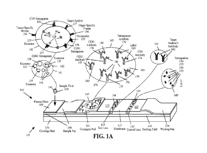
100101 FIGS. 1A-1D are functional diagrams illustrating an LFA
device and a method
that that uses one or more antibodies specific to an exosome containing the
target analyte as
the detection antibodies and an antibody specific to the target analyte as the
capture antibody,
according to various aspects of the present disclosure;
100111 FIG. 2 illustrates examples of the exosome proteins that
are specific to certain
tumors according to prior art;
100121 FIGS. 3A-3F are functional diagrams illustrating an LFA
device and a method
that that uses one or more antibodies specific to an exosome containing the
target analyte as
the detection antibodies and includes multiple test lines for capturing a set
of one or more
organ-specific or tumor-specific proteins and a target analyte, according to
various aspects of
the present disclosure;
[00131 FIGS. 4A-4D are functional diagrams illustrating an LFA
device and a method
that that uses one or more antibodies specific to an exosome containing the
target analyte as
the detection antibodies and includes multiple strips with test lines for
capturing a set of one or
more organ-specific or tumor-specific proteins and a target analyte, according
to various
aspects of the present disclosure;
100141 FIGS. 5A-5D are functional diagrams illustrating an LFA
device and a method
that that uses an antibody specific to the target analyte as the detection
antibody and one or
more antibodies specific to an exosome containing the target analyte as
capture antibodies,
according to various aspects of the present disclosure;
100151 FIGS. 6A-6F are functional diagrams illustrating an LFA
device and a method
that that uses an antibody specific to the target analyte as the detection
antibody and includes
multiple test lines for capturing a set of one or more organ-specific or tumor-
specific proteins
and a target analyte, according to various aspects of the present disclosure;
CA 03218733 2023- 11- 10
WO 2022/245990
PCT/US2022/029898
100161 FIGS. 7A-7D are functional diagrams illustrating an LFA
device and a method
that that uses an antibody specific to the target analyte as the detection
antibody and includes
multiple test strips with test lines for capturing a set of one or more organ-
specific or tumor-
specific proteins and a target analyte, according to various aspects of the
present disclosure;
100171 FIGS. 8A-8I are functional diagrams illustrating an ELISA
device and a method
that detects and captures a target analyte by using antibodies specific to
exosomes containing
the target analyte and an immobilized antibody specific to the target analyte,
according to
various aspects of the present disclosure; and
100181 FIGS. 9A-9I are functional diagrams illustrating an ELISA
device and a method
that detects and captures a target analyte by using immobilized antibodies
specific to exosomes
containing the target analyte and an antibody specific to the target analyte,
according to various
aspects of the present disclosure.
DETAILED DESCRIPTION
100191 One aspect of the present embodiments includes the
realization that a sandwich
format immunoassay requires two antibodies that are specific to the target
analyte such that the
antibodies, to a great extent, attach to the target analyte and do not attach
to other molecules.
Otherwise, the other molecules that also attach to the antibodies may become
sources of error.
For some target analytes, however, there may only be one specific antibody.
One technique to
detect the presence (or absence) of these target analytes is to use a
competitive format assay
device. The competitive format assay devices are, however, not as accurate as
the sandwich
format assay devices. Another drawback of the competitive format assay devices
is the need
to have the physical target analyte material itself in order to use it as the
reference target analyte
on the bottom of the plates (for ELISA devices) and to use it on the test line
(for LFA devices).
100201 Some of the present embodiments solve the aforementioned
problems by using
an antibody to capture exosomes in the sample liquid. Exosomes are
extracellular vesicles that
are released from cells. The exosomes may contain different proteins depending
on their host
cell. The most common exosome marker proteins include tetraspanin proteins,
such as CD9,
CD63, CD81, and CD82, which are present on the surface of the exosomes. The
exosomes
may also carry markers from the cells that release them. For some target
analytes, such as, for
example, and without limitations, cancer cells' proteins, the exosomes
released by the cells
may include the markers for the proteins that are the targets of an assay.
4
CA 03218733 2023- 11- 10
WO 2022/245990
PCT/US2022/029898
100211 Some of the present embodiments provide a method and an
immunoassay
device that receive a quantity of fluid comprising a quantity of exosomes and
detect the
presence of a target analyte on the surface of the exosomes. The immunoassay
device
comprises a detection site and a capture site. The method and the immunoassay
device perform
a fluid transfer between the detection site and the capture site. The
mechanism of the transfer
of the fluid between the site where the detection action takes place and the
site where the
capture action takes place may be by capillary action (e.g., an LFA device or
a microfluidic
device), a microfluidic chip or medium, an automated liquid handling system
(e.g., the liquid
handling used in an automated ELISA device), an automated liquid handling
system in
combination with a microfluidic device, or manual transfer such as pipetting
procedures used
in standard ELISA. In some of these immunoassay devices the detection action
and the capture
action are performed on different sites on the device. For example, in the LFA
devices, the
detection action is performed on the conjugate pad and the capture action is
performed on one
or more test lines. In some of these immunoassay devices the detection action
and the capture
action may be performed on the same site of the device. For example, in the
ELISA devices,
the detection action and the capture action may be performed in the same well
of the ELISA
device.
100221 The immunoassay devices of some of the present
embodiments perform the
detection action by using binding reagents (e.g., antibodies) to the
tetraspanin, such as, CD9
protein, CD63 protein, CD81 protein, CD82 protein, etc., to detect exosomes in
a sample liquid.
These immunoassay devices may use one or more exosome binding reagents.
Different
exosomes may bind to one or more binding reagents for the CD9, CD63, CD81,
CD82, etc.,
proteins. These immunoassay devices perform the capture action by using a
second binding
reagent (e.g., an antibody) that is specific to the target analyte, which is
used to immobilize and
capture the exosomes that carry the target analyte.
100231 The immunoassay devices of some of the present
embodiments perform the
detection action by using a binding reagent (e.g., an antibody) specific to
the target analyte to
detect the target analyte in a sample liquid. These immunoassay devices
perform the capture
action by using one or more binding reagents (e.g., antibodies) to the
tetraspanin, such as, CD9
protein, CD63 protein, CD81 protein, CD82 protein, etc., to immobilize and
capture the
exosomes that carry the target analyte. Different exosomes may bind to one or
more antibodies
for the CD9, CD63, CD81, CD82, etc., proteins.
CA 03218733 2023- 11- 10
WO 2022/245990
PCT/US2022/029898
[0024] Several non-limiting examples of these methods and
immunoassay devices are
described in Section I with reference to LFA devices and in Section II with
reference to ELISA
devices. The remaining detailed description describes the present embodiments
with reference
to the drawings. In the drawings, reference numbers label elements of the
present
embodiments. These reference numbers are reproduced below in connection with
the
discussion of the corresponding drawing features.
I. LFA DEVICE THAT CAPTURES A TARGET ANALYTE BY USING
AN ANTIBODY SPECIFIC TO AN EXOSOME CONTAINING THE
TARGET ANALYTE
[0025] FIGS. 1A-1D are functional diagrams illustrating an LFA
device 100 and a
method that uses one or more antibodies specific to an exosome containing the
target analyte
as the detection antibodies and an antibody specific to the target analyte as
the capture antibody,
according to various aspects of the present disclosure. The LFA device 100 may
be a portable
device (e.g., a handheld device or benchtop device) that is used to analyze a
sample fluid 190
(also referred to as matrix) to determine the presence and/or the amount of
one or more analytes
(referred to as target analytes). The term analyte refers to the molecule
detected by the
immunoassay device.
[0026] With reference to FIGS. 1A-1D, the LFA device 100 may
include a replaceable
cartridge that may be intended for single use. For example, the components
shown in FIGS.
1A-1D may be part of a disposable cartridge of the LFA device 100. The LFA
device 100 may
include a backing card 140 that may be used to assemble different portions of
the sample pad
150, the conjugate pad 110, the membrane 115, and/or the wicking pad 120.
[0027] The backing card 140, in some embodiments, may be a
continuous piece that
may go under the pads 150, 110, 115, and 120. In other embodiments, each pad
may have a
separate backing card. For example, during the manufacturing of the device, a
roll or sheet of
backing material may be used such that the width of the roll or the sheet is
the same as (or is
cut to be the same as) the length of the lateral flow assay cartridge (i.e.,
in the pictured
orientation, from the left end of the sample pad 150 to the right end of the
wicking pad 120).
The pads 115, 110, 150, and 120 are then placed on the backing card with the
proper overlaps
(e.g., as shown in FIGS. 1A-1D). The pads may, for example, be connected to
the backing
card with a two sided tape or a glue. The pads and the attached backing card
may then be cut
into separate strips and each strip may be used to make a different LFA
device.
6
CA 03218733 2023- 11- 10
WO 2022/245990
PCT/US2022/029898
100281 Alternatively, each pad may be separately connected to a
corresponding backing
card. The pads with the corresponding backing cards may then be assembled over
each other
with the proper overlaps to make a LFA device. The LFA device 100 may include
a housing.
In FIGS. 1A-1D, only a portion of the housing that includes the cartridge bed
170 is shown for
clarity.
100291 The sample fluid 190 applied to the LFA device 100 may
include human or
animal bodily fluid, such as, for example, and without limitations, one or
more of blood, urine,
serum, plasma, saliva, sweat, milk, mucous, semen, vaginal or urethral
secretions,
cerebrospinal fluid, etc. The sample may naturally be a liquid, may be a
liquid diluted with
another liquid, such as water, or may have originally been in a solid form
(e.g., a tissue sample)
and is treated to be in liquid form for application to the LFA device 100. The
target analytes,
in some of the present embodiments, may be substances such as, for example,
and without
limitations, proteins, haptens, enzymes, hormones, infectious disease agents,
immunoglobulins, polynucleotides, steroids, drugs, nucleic acids, markers for
gene mutations,
antigens, simple organic molecules, etc.
100301 The LFA device 100 may include a sample pad (also
referred to as sample strip
or sample receiving member) 150. The sample pad 150 may be made of natural
and/or
synthetic porous, microporous, mesoporous, or macroporous materials capable of
receiving a
sample fluid and laterally conducting the sample fluid toward the conjugate
pad 110 by
capillary action. The sample pad 150 may be made of a material such as, for
example, and
without limitations, cellulose, nitrocellulose, paper, silica, cotton, glass
(e.g., glass fiber), or
synthetic material (e.g., polyester, polyethylene, polymers, rayon, nylon,
etc.). Depending on
the type of the sample (e.g., urine, saliva, blood, serum, plasma, sweat,
milk, mucous, semen,
vaginal or urethral secretions, cerebrospinal fluid, etc.), the sample pad 150
may be treated by
a buffer (e.g., an organic compound such as tris or tris(hydroxymethyl)aminom
ethane) to
mitigate sample variabilities (pH, protein concentration, viscosity, salt
concentration, etc.).
During the manufacture of the sample pad 150, the buffer compound may be
coated,
impregnated, or otherwise applied or deposited on the sample pad 150 and then
dried. The
embodiments that the sample fluid includes blood may include a plasma filter
195.
100311 The LFA device 100 may include a conjugate pad 110 that
is fluidically
connected (i.e., capable of receiving fluid by capillary action) to the sample
pad 150. In the
depicted embodiment, the sample pad 150 is in contact with and partially
covers the conjugate
pad 110. In other embodiments, the sample pad 150 may be in more contact or
less contact
7
CA 03218733 2023- 11- 10
WO 2022/245990
PCT/US2022/029898
with the conjugate pad 110 in order to provide slower or faster binding
reagent and/or conjugate
release respectively. A sample fluid that is applied to the sample pad 150 may
be laterally
transferred from the sample pad 150 to the conjugate pad 110 by capillary
action.
100321 The conjugate pad 110 may be made of natural and/or
synthetic porous,
microporous, mesoporous, or macroporous materials capable of receiving the
sample fluid
from the sample pad 150. The conjugate pad 110 may be made of material such
as, for
example, and without limitations, glass (e.g., glass fiber), cellulose,
nitrocellulose, paper, silica,
cotton, or synthetic material (e.g., polyester, polyethylene, polymers, rayon,
nylon, etc.).
100331 In the example of FIGS. 1A-1D, the sample fluid 190 may
be a fluid such as,
for example, and without limitations, blood, urine, serum, plasma, saliva,
sweat, milk, mucous,
semen, vaginal or urethral secretions, cerebrospinal fluid. As shown in the
expanded view 141
of FIG. 1A, the sample fluid 190 may include, among other components, the
exosomes 135.
When the sample fluid 190 includes blood, the other components 198 may
include, for
example, red blood cells, white blood cells, platelets, plasma, etc.
100341 The exosomes 135 are extracellular vesicles that are
produced by most cells.
The exosomes may be found in blood, urine, or cerebrospinal fluid. The
exosomes may also
be released in vitro by cultured cells into their growth medium. The exosomes
are typically
between 30 to 150 nanometers (nm) in diameter. In malignancies, such as
cancer, the exosomes
released by the cancerous cells may include proteins that may be used as
target analytes to
diagnose cancer.
100351 As shown in the expanded view 142 of FIG. 1A, an exosome
135 may contain
a vast array of different proteins depending on the host cell. The components
of the exosome
is further modulated by the cellular state (e.g., stress, activation, or
inhibition of specific
pathways). Tetraspanins, such as CD9 156, CD63 157, CD81 158, and CD82 (not
shown),
which are the most common canonical exosome marker proteins, and/or other type
of
tetraspanins may be present on the surface of the exosome. There are 34
tetraspanins in
mammals, 33 of which have been identified in humans. Tetraspanin 159 shown in
the
expanded view 142 refers to any of the 34 tetraspanins in mammals, including,
but not limited
to CD9 156, CD63 157, CD81 158, and CD82.
100361 Other components 138 of an exosome may include different
enzymes, lipids,
transcription factors, cytoskeletons, etc. If an exosome is released by a
malignant tumor or an
infected cell, the exosome may also contain proteins 137 that may be used as
general markers
to identify malignancies. The exosome released by a malignant tumor, or an
infected cell, may
8
CA 03218733 2023- 11- 10
WO 2022/245990
PCT/US2022/029898
also contain tumor-specific proteins 194 that may be used to identify specific
tumors. The
exosome released by an organ may contain organ-specific proteins 196 that may
be used to
identify the organ.
100371 Multiple tetraspanins of the same type (e.g., multiple
CD9 marker proteins 156,
multiple CD63 marker proteins 157, multiple CD81 marker proteins 158, multiple
CD82
marker proteins, etc.), multiple tetraspanins of the different types, and/or
multiple target analyte
proteins 137 may be present on a single exosome 135. As described herein, the
immunoassays
of the present embodiments may use the exosomes to capture and identify the
target analytes
137 such as the proteins (or other markers) related to different malignancies.
100381 As shown in the expanded view 143 of FIG. 1A, the
conjugate pad 110 may
contain one or more different types of binding reagents 176-179 that are
capable of binding to
different types of tetraspanins 156-159 on the surface of exosomes 135 in the
sample fluid 190.
The binding reagents may be, for example, and without limitations, different
types of
tetraspanin antibodies. Although several examples of tetraspanin antibodies,
target analyte
antibodies, tumor-specific protein antibodies, and organ-specific protein
antibodies are used
herein, it should be noted that some of the LFA devices of the present
embodiments may use
binding reagents on the conjugate pads, on the test lines, on/or on the
control lines that are not
antibodies.
100391 In addition to, or in lieu of, the antibodies 176-178 for
the three types of
tetraspanins 156-158, the conjugate pad 110 may include antibodies for other
types of exosome
tetraspanins. The tetraspanin antibody 179 shown in the expanded view 143
refers to the
antibody of a tetraspanin, including, but not limited to the CD9 antibody, the
CD63 antibody,
the CD81 antibody, the CD82 antibody, etc.
100401 The conjugate pad 110 may include antibodies for one or
more types of
tetraspanins. For example, depending on the type of test performed by the LFA
100, the
conjugate pad 110 may include antibodies for one or more of CD9, CD63, CD81,
CD82, or
other types of tetraspanins. Accordingly, the present embodiments do not
necessarily use
antibodies for all types of exosome' s tetraspanins and may use a single
antibody or a
combination of any number of the tetraspanins antibodies, depending on the
test being
performed.
100411 The binding reagent 176-179 may be coupled to a label 180
(also referred to as
conjugate, detection conjugate, probe, detector nanoparticle, or tag) which,
in its natural state,
is readily visible either to the naked eye or with the aid of an optical
filter. The label 180 may
9
CA 03218733 2023- 11- 10
WO 2022/245990
PCT/US2022/029898
be made of small particles (e.g., nanoparticles), such as, for example, and
without limitations,
metallic sols (e.g., colloidal gold or gold sol), dye sols, colored latex
particles, carbon,
fluorescent particles, europium labels, etc. During the manufacture of the
conjugate pad 110,
the labeled binding reagent may be coated, impregnated, or otherwise applied
or deposited on
the conjugate pad 110 and then dried.
100421 After the sample fluid 190 flows from the sample pad 150
into the conjugate
pad 110, the sample fluid 190 may solubilize the labeled binding reagent. If
the sample fluid
contains the exosomes and the exosomes contain at least one of the
tetraspanins 159 (e.g., the
tetraspanins 156-158 or other tetraspanins, which are not shown) for which the
conjugate pad
100 includes an antibody, the exosomes may bind with the labeled binding
reagents (e.g., the
binding reagents 176-179) and may form an immunocomplex. The labeled binding
reagents
that do not bind with the exosomes (e.g., when the sample fluid does not
include exosomes
with tetraspanins for which the conjugate pad includes antibodies or there is
excess labeled
binding reagent) flow downstream toward the membrane 115 by capillary action.
The sample
fluid and any other material in the flow path (e.g., unbound labeled binding
reagents, wash
fluid, etc.) are herein referred to as fluid material.
100431 Depending on the type of test performed by the LFA
device, the device may not
include separate sample and conjugate pads, and may only include the conjugate
pad 110 in
some embodiments. Although the sample pad 150 is shown to go over the
conjugate pad 110,
in some embodiments, the conjugate pad 110 may go over the sample pad 150.
100441 The LFA device 100 may include a membrane 115 and one or
more test lines
(only one test line 125 is shown for simplicity) that may be embedded in the
membrane. The
LFA device 100 may optionally include a control line 130 that may be embedded
in the
membrane 115. The membrane 115 may be made of a material such as, for example,
and
without limitations, cellulose, nitrocellulose, paper, silica, cotton, glass
(e.g., glass fiber), or
synthetic material (e.g., polyester, polyethylene, polymers, rayon, nylon,
etc.) that allow the
fluid material to flow downstream from the conjugate pad 110 into the membrane
115 and from
the membrane 115 toward the wicking pad 120 by capillary action. Although the
conjugate
pad 110 is shown to go over the membrane 115, in some embodiments, the
membrane 115 may
go over the conjugate pad 110.
100451 The test line 125 may be made of a porous material such
as, for example, and
without limitations, cellulose, nitrocellulose, paper, silica, cotton, glass
(e.g., glass fiber), or
synthetic material (e.g., polyester, polyethylene, polymers, rayon, nylon,
etc.). The test line
CA 03218733 2023- 11- 10
WO 2022/245990
PCT/US2022/029898
125, in a sandwich assay format, may contain an unlabeled binding reagent that
is immobilized
on the test line 125 and does not flow downstream when porous material of the
test line is
moistened (e.g., by the fluid material). As shown in the expanded view 144,
the unlabeled
binding reagent that is immobilized on the test line 125 is the target
analyte's antibody 185.
[0046] The LFA device 100 may optionally include a control line
130 that may be
embedded in the membrane 115. The control line 130 may be made of a porous
material such
as, for example, and without limitation, cellulose, nitrocellulose, paper,
silica, cotton, glass
(e.g., glass fiber), or synthetic material (e.g., polyester, polyethylene,
polymers, rayon, nylon,
etc.). In a sandwich assay format, the control line 130 may contain an
immobilized antibody
that binds to the free labeled binding reagents (e.g., the free labeled
tetraspanins antibodies
176-179) resulting in a colored control line 130, which confirms that the test
has operated
correctly regardless of whether or not the target analyte has been present in
the sample.
[0047] As shown in the expanded view 145, the immobilized
antibody on the control
line 130 may be an immunocomplex that includes one or more types of exosome's
tetraspanins
(e.g., CD9 156, C63 157, CD81 158, CD82, etc.). In general, the control line
of the LFA
devices of the present embodiments may contain an immobilized binding reagent
(e.g., an
immobilized antibody) against the class of the binding reagents (e.g.,
antibodies) that are
included on the conjugate pad 110. For example, when the antibodies on the
conjugate pad are
of Immunoglobin G (IgG) class, the control line may include an immobilized
anti-IgG
antibody. In addition to, or in lieu of the immunocomplex that includes one or
more types of
exosome's tetraspanins, the control line 130 may include antibodies against
the class of the
tetraspanins that are included on the conjugate pad 110.
[0048] The fluid material that do not bind to the test line 125
or the control line 130
may continue to flow from the membrane 115 into the wicking pad 120 to absorb
the fluid
material that are not taken up by the test line 125 and the control line 130
while maintaining
the capillary flow from the membrane 125 into the wicking pad 120. The wicking
pad 120
may be made of a porous material such as, for example, and without
limitations, cellulose,
nitrocellulose, paper, silica, cotton, glass (e.g., glass fiber), or synthetic
material (e.g.,
polyester, polyethylene, polymers, rayon, nylon, etc.). Depending on the type
of test performed
by the LFA device, the device may not include a wicking pad 120. Although the
wicking pad
120 is shown to go over the membrane 115, in some embodiments, the membrane
115 may go
over the wicking pad 120.
11
CA 03218733 2023- 11- 10
WO 2022/245990
PCT/US2022/029898
100491 FIGS. 1A-1D, as shown, include four stages 101-104. In
stage 101 (FIG.1A),
the sample fluid 190 is applied on the sample pad 150. When the sample fluid
190 includes
blood, the LFA 100 may have the plasma filter 195 and the sample fluid 190 may
be applied
to the plasma filter 195. In the embodiments that do not include a sample pad
150, the sample
fluid 190 may be applied to the conjugate pad 110 (or applied to a plasma
filter located on the
conjugate pad when the sample fluid includes blood).
100501 In stage 102 (FIG. 1B), the fluid material may have
reached the conjugate pad
110. As shown in the expanded view 146, the tetraspanins (e.g., the
tetraspanins 156-159) on
the surface of the exosomes 135 in the fluid material may bind with the
corresponding
tetraspanin antibodies (e.g., the tetraspanin antibodies 176-179). In stage
102, some of the
exosomes 135 (e.g., as shown in the expanded view 147) may contain the target
analyte 137
on their surface while some of the exosomes 135 (e.g., as shown in the
expanded view 148)
may not contain the target analyte 137. It should be noted that, depending on
the condition of
the subject (e.g., a person or an animal) from which the sample fluid 190
(FIG. 1A) is drawn,
there may or may not be any exosome with the target analyte 137 in the sample
fluid.
100511 The exosomes 135 that are bound with the corresponding
tetraspanin antibodies
176-179 may form immunocomplexes. The immunocomplexes and the rest of the
fluid
material 198 may continue to move, by capillary action, from the conjugate pad
110 to the
membrane 115.
100521 In stage 103 (FIG. 1C), the fluid material may have
reached the test line 125.
As shown in the expanded view 149, the target analyte's antibodies 185 that
are immobilized
on the test line may bind with the target analyte 137 on the exosomes 135 that
have been bound
to the tetraspanin antibodies (e.g., the tetraspanin antibodies 176-179)
through one of their
tetraspanins (e.g., the tetraspanins 156-159). The binding results in a second
immunocomplex
(the immunocomplex shown in the expanded view 149). The label 180 on the
immobilized
second immunocomplex colors the test line 125.
100531 The intensity of the colored test line is correlated with
the density of the target
analyte 137 on the surface of the exosomes 135 in the sample fluid. The second
immunocomplex includes the exosomes 135 that are bound (through the target
analyte 137 on
their surface) with the immobilized target analyte's antibody 185, and
(through one of the
tetraspanins 156-159 on their surface) with one of the labelled tetraspanins
antibodies 176-179.
When the sample fluid does not include the target analyte, no immunocomplex
binds with the
immobilized antibody on the test line 125. As a result, the test line 125 does
not change color.
12
CA 03218733 2023- 11- 10
WO 2022/245990
PCT/US2022/029898
100541
The exosomes that lack the target analyte 137 on their surface may not
bind to
the immobilized tetraspanin antibodies 176-179 on the test line 125 and may
continue to move,
with the rest of the fluid material, toward the control line 130 and the
wicking pad 120.
100551
In stage 104 (FIG. 1D), the fluid material may have reached the control
line
130. As shown in the expanded view 151, the free labeled tetraspanins
antibodies (e.g., the
176-179 tetraspanins antibodies) in the fluid material may bind to the
immobilized tetraspanins
(e.g., the corresponding tetraspanins 156-159). This binding may result in a
colored control
line, which confirms that the test has operated correctly regardless of
whether or not the target
analyte has been present in the sample fluid.
100561
Some embodiments of the LFA device may include multiple test lines. In
some
of these embodiments, some of the test lines may be used to detect exosome
proteins that are
specific to certain tumors and/or specific to certain organs. An organ-
specific protein is defined
as a protein whose expression is significantly elevated in one or more
specific human organs.
The organ-specific proteins may be implicated in human diseases related to the
corresponding
organs. A tumor-specific protein is defined as a protein whose expression is
significantly
elevated in one or more specific tumors.
100571
FIG. 2 illustrates examples of the exosome proteins that are specific to
certain
tumors according to prior art. With reference to FIG. 2, the exosome proteins
column 211 of
table 200 identifies the name of the exosome proteins. The tumor column 212
identifies the
type of tumor(s) that correspond(s) to the exosome proteins of column 211. The
body fluid
column identifies the body fluid where the exosome proteins have been found.
100581
With reference to FIG. 2, some of the exosome proteins 211, such as
Glypican-
1 220 may be indicative of a tumor in more than one organ (in this example
breast, pancreas,
colon, and rectum). Some of the exosome proteins 211, such as PSA 230, may
also be an
organ-specific protein that may be present in the exosomes released from
healthy prostate cells.
100591
FIGS. 3A-3F are functional diagrams illustrating an LFA device 300 and a
method that uses one or more antibodies specific to an exosome containing the
target analyte
as the detection antibodies and includes multiple test lines for capturing a
set of one or more
organ-specific or tumor-specific proteins and a target analyte, according to
various aspects of
the present disclosure. The LFA device 300 may be a portable device (e.g., a
handheld device
or benchtop device) that is used to analyze a sample fluid 190 to determine
the presence and/or
the amount of one or more analytes, one or more tumor -specific proteins,
and/or one or more
organ-specific proteins.
13
CA 03218733 2023- 11- 10
WO 2022/245990
PCT/US2022/029898
100601 With reference to FIGS. 3A-3F, the LFA device 300 may
include a test line 125
to detect a target analyte 137. The target analyte 137, in some embodiments,
may be a protein
that is a general marker that identifies malignancies. The test line 125 of
the LFA device 300
may be similar to the test line 125 of the LFA device 100, described above. In
addition to the
test line 125, the LFA device 300 may include n (where n is an integer greater
than or equal to
1) test lines 321-322 to detect organ-specific and/or tumor-specific proteins.
The test lines
321-322 may be made of a porous material, as described above, with reference
to the test line
125 of the LFA device 100.
100611 FIGS. 3A-3F, as shown, include six stages 301-307. In
stage 301 (FIG.3A), the
sample fluid 190 may be applied to the sample pad 150. When the sample fluid
190 includes
blood, the LFA 300 may have the plasma filter 195 and the sample fluid 190 may
be applied
to the plasma filter 195. In the embodiments that do not include a sample pad
150, the sample
fluid 190 may be applied to the conjugate pad 110 (or applied to a plasma
filter located on the
conjugate pad when the sample fluid includes blood). The expanded views 141-
145 of FIG.
3A illustrate similar items as the corresponding expanded views of FIG. 1A.
100621 The unlabeled binding reagent that is immobilized on each
test lines 321-322
may be an antibody 371-372 to an organ-specific protein, such as the organ-
specific protein
196, or a tumor-specific protein, such as the tumor-specific protein 194. As
described above,
some proteins may act both as tumor-specific and organ-specific proteins. The
LFA device
300 may be configured such that the organ-specific proteins or the tumor-
specific proteins that
bind to the immobilized antibodies 371-372 are different proteins.
100631 With reference to FIG. 3A, a quantity of an antibody 371
to an organ-specific
protein or a tumor-specific protein may be immobilized on the test line 321
(as shown in the
expanded view 331), a quantity of an antibody 372 to an organ-specific protein
or a tumor-
specific protein may be immobilized on the test line 322 (as shown in the
expanded view 332),
etc. Although two test lines 321-322 are shown for capturing the tumor-
specific or organ-
specific proteins, different embodiments of the LFA device 300 may include any
number of
one or more test lines similar to the test lines 321-322 for capturing the
tumor-specific or organ-
specific proteins. Other components of the LFA device 300 may be similar to
the
corresponding components of the LFA device 100 described above with reference
to FIGS.
1A-1D).
100641 In stage 302 (FIG. 3B), the fluid material may have
reached the conjugate pad
110. As shown in the expanded view 335, the tetraspanins (e.g., the
tetraspanins 156-159) on
14
CA 03218733 2023- 11- 10
WO 2022/245990
PCT/US2022/029898
the surface of the exosomes 135 in the fluid material may bind with the
corresponding
tetraspanin antibodies (e.g., the tetraspanin antibodies 176-179). In stage
302, some of the
exosomes 135 (e.g., as shown in the expanded views 336 and 337) may contain
the target
analyte 137 on their surface while some of the exosomes 135 (e.g., as shown in
the expanded
view 338) may not contain the target analyte 137. Furthermore, some of the
exosomes 135
(e.g., as shown in the expanded views 337 and 338) may contain one or more
tumor-specific
proteins 194 and/or one or more organ-specific proteins 196 on their surface
while some of the
exosomes 135 (e.g., as shown in the expanded view 336) may not contain any
tumor-specific
proteins or organ-specific proteins on their surface. It should be noted that,
depending on the
condition of the subject (e.g., a person or an animal) from which the sample
fluid 190 (FIG.
1A) is drawn, the sample fluid may or may not include any exosome with the
target analyte
137, a tumor-specific protein 194, or an organ-specific protein 196 on its
surface.
100651 In stage 302 (FIG. 3B) the exosomes 135 that are bound
with the corresponding
tetraspanin antibodies 176-179 may form immunocomplexes. The immunocomplexes
and the
rest of the fluid material 198 may continue to move, by capillary action, from
the conjugate
pad 110 to the membrane 115.
100661 In stage 303 (FIG. 3C), the fluid material may have
reached the test line 321.
As shown in the expanded view 339, the tumor-specific or organ-specific
antibodies 371 that
are immobilized on the test line 321 may bind with the corresponding tumor-
specific protein
194 or organ-specific protein 196 on the exosomes 135 that have been bound to
the tetraspanin
antibodies (e.g., the tetraspanin antibodies 176-179) through one of their
tetraspanins (e.g., the
tetraspanins 156-159). The binding results in a second immunocomplex (the
immunocomplex
shown in the expanded view 339). The label 180 on the immobilized second
immunocomplex
colors the test line 321.
100671 The intensity of the colored test line 321 is correlated
with the density of the
tumor-specific or organ-specific protein on the surface of the exosomes 135 in
the sample fluid
that correspond to the immobilized antibody 371. The second immunocomplex
includes the
exosomes 135 that are bound (through the tumor-specific protein 194 or the
organ-specific
protein 196 on their surface) with the immobilized antibodies 371, and are
bound (through one
of the tetraspanins 156-159 on their surface) with one of the labelled
tetraspanins antibodies
176-179. When the sample fluid does not include the tumor-specific protein 194
or the organ-
specific protein 196 that corresponds to the immobilized antibodies 371, no
immunocomplex
CA 03218733 2023- 11- 10
WO 2022/245990
PCT/US2022/029898
binds with the immobilized antibody 371 on the test line 321. As a result, the
test line 321 does
not change color.
100681 The exosomes that lack the tumor-specific protein 194 or
the organ-specific
protein 196 that corresponds to the immobilized antibodies 371on their surface
may not bind
to the immobilized antibodies 371 on the test line 321 and may continue to
move, with the rest
of the fluid material, toward the test line 342. It should be noted that some
embodiments of
the LFA device 300 may only include the test lines 341 and 125. These
embodiments may not
include stage 304. In these embodiments, the unbound material may move from
the list line
321 toward the test line 125, as described below with reference to stage 305.
100691 In stage 304 (FIG. 3D), the fluid material may have
reached the test line 322.
As shown in the expanded view 340, the tumor-specific or organ-specific
antibodies 372 that
are immobilized on the test line 322 may bind with the corresponding tumor-
specific protein
194 or organ-specific protein 196 on the exosomes 135 that have been bound to
the tetraspanin
antibodies (e.g., the tetraspanin antibodies 176-179) through one of their
tetraspanins (e.g., the
tetraspanins 156-159). It should be noted that the tumor-specific protein 194
or organ-specific
protein 196 that correspond to the antibody 372 that is immobilized on the
test line 322 is
different than the tumor-specific protein 194 or organ-specific protein 196
that correspond to
the antibody 371 that is immobilized on the test line 321. The binding on the
test line 342
results in a third immunocomplex (the immunocomplex shown in the expanded view
340).
The label 180 on the immobilized third immunocomplex colors the test line 322.
100701 The intensity of the colored test line 322 is correlated
with the density of the
tumor-specific or organ-specific protein on the surface of the exosomes 135 in
the sample fluid
that correspond to the immobilized antibody 372. The third immunocomplex
includes the
exosomes 135 that are bound (through the tumor-specific protein 194 or the
organ-specific
protein 196 on their surface) with the immobilized antibodies 372, and are
bound (through one
of the tetraspanins 156-159 on their surface) with one of the labelled
tetraspanins antibodies
176-179. When the sample fluid does not include the tumor-specific protein 194
or the organ-
specific protein 196 that corresponds to the immobilized antibodies 372, no
immunocomplex
binds with the immobilized antibody 372 on the test line 322. As a result, the
test line 322 does
not change color.
100711 The exosomes that lack the tumor-specific protein 194 or
the organ-specific
protein 196 that corresponds to the immobilized antibodies 372 on their
surface may not bind
to the immobilized antibodies 372 on the test line 322 and may continue to
move, with the rest
16
CA 03218733 2023- 11- 10
WO 2022/245990
PCT/US2022/029898
of the fluid material, toward the test line 125. It should be noted that some
embodiments of
the LFA device 300 may include more than two (e.g., three or more) test lines
341-342 to
capture tumor-specific and/or organ-specific proteins. These embodiments may
include
additional stages similar to the 304.
[0072] In stage 305 (FIG. 3E), the fluid material may have
reached the test line 125.
As shown in the expanded view 149, the target analyte's antibodies 185 that
are immobilized
on the test line may bind with the target analyte 137 on the exosomes 135 that
have been bound
to the tetraspanin antibodies (e.g., the tetraspanin antibodies 176-179)
through one of their
tetraspanins (e.g., the tetraspanins 156-159). The binding results in an
immunocomplex (the
immunocomplex shown in the expanded view 149). The label 180 on the
immobilized second
immunocomplex colors the test line 125.
100731 The intensity of the colored test line is correlated with
the density of the target
analyte 137 on the surface of the exosomes 135 in the sample fluid. The
immunocomplex in
the expanded view 149 includes the exosomes 135 that are bound (through the
target analyte
137 on their surface) with the immobilized target analyte' s antibody 185, and
(through one of
the tetraspanins 156-159 on their surface) with one of the labelled
tetraspanins antibodies 176-
179. When the sample fluid does not include the target analyte, no
immunocomplex binds with
the immobilized antibody on the test line 125. As a result, the test line 125
does not change
color.
[0074] The exosomes that lack the target analyte 137 on their
surface may not bind to
the immobilized tetraspanin antibodies 176-179 on the test line 125 and may
continue to move,
with the rest of the fluid material, toward the control line 130 and the
wicking pad 120.
[0075] In stage 306 (FIG. 3F), the fluid material may have
reached the control line 130.
As shown in the expanded view 151, the free labeled tetraspanins antibodies
(e.g., the 176-179
tetraspanins antibodies) in the fluid material may bind to the immobilized
tetraspanins (e.g.,
the corresponding tetraspanins 156-159). This binding may result in a colored
control line,
which confirms that the test has operated correctly regardless of whether or
not the target
analyte has been present in the sample fluid. The fluid material that do not
bind to the control
line 130 may continue to flow from the membrane 115 into the wicking pad 120
to absorb the
fluid material that are not taken up by the test line 125 and the control line
130 while
maintaining the capillary flow from the membrane 125 into the wicking pad 120.
[0076] With reference to FIGS. 3A-3F, the results of a test on
the LFA device 300 may
be interpreted as follows. When none of the test lines 321-322 and 125 are
colored at the end
17
CA 03218733 2023- 11- 10
WO 2022/245990
PCT/US2022/029898
of a test, neither the target analyte 137, nor the tumor-specific proteins,
nor the organ-specific
proteins whose antibodies where immobilized on the test lines 321-322 were
present in the
sample fluid 190 in detectable amounts. When the test line 125 is colored at
the end of a test
but none of the test lines 321-322 are colored, the target analyte 137 (e.g.,
and without
limitation, a general marker of a malignancy such as cancer) is detected in
the sample fluid but
the malignancy may not be attributed to any specific tumor or specific organ.
100771 When the test line 125 and at least one of the test lines
321-322 are colored at
the end of a test, the target analyte 137 (e.g., and without limitation, a
general marker of a
malignancy such as cancer) is detected in the sample fluid. In addition, the
malignancy may
be attributed with a high probability to the specific tumor(s) or the specific
organ(s) whose
exosome protein(s) 194/196 was/were captured on the colored test line(s) 321-
322.
100781 When the test line 125 is not colored but at least, one
of the test lines 321-322
is colored at the end of a test, the target analyte 137 attributed to a
malignancy is not detected.
In this scenario, the test line(s) 321-322 may have been colored either due to
the presence of
the organ-specific proteins on the surface of exosomes released from a healthy
organ or due to
the detected exosome proteins being released by a tumor that does not have the
general marker
(i.e., the target analyte protein).
100791 In the embodiment of FIGS. 3A-3F, multiple test lines 321-
322 and 125 were
placed on the same test strip that also includes the sample pad 150, the
conjugate pad 110, the
membrane 115, the control line 130, and the wicking pad 120. Some embodiments
may place
each of the test lines 321-322 and 125 on a separate test strip where each
test strip may include
a sample pad 150, a conjugate pad 110, a membrane 115, a control line 130, and
a wicking pad
120. The multiple strips may be placed inside the same test cartridge.
100801 FIGS. 4A-4D are functional diagrams illustrating an LFA
device 400 and a
method that uses one or more antibodies specific to an exosome containing the
target analyte
as the detection antibodies and includes multiple strips with test lines for
capturing a set of one
or more organ-specific or tumor-specific proteins and a target analyte,
according to various
aspects of the present disclosure. The LFA device 400 may be a portable device
(e.g., a
handheld device or benchtop device) that is used to analyze a sample fluid 190
to determine
the presence and/or the amount of one or more analytes, one or more tumor -
specific proteins,
and/or one or more organ-specific proteins.
100811 With reference to FIGS. 4A-4D, the LFA device 400 may
include several test
strips 481-483. Each two adjacent test strips may be separated by a gap 470
that may prevent
18
CA 03218733 2023- 11- 10
WO 2022/245990
PCT/US2022/029898
any fluids from flowing from one test strip into the other. For clarity, the
gaps 470 are shown
proportionally wider that the other components of the LFA device 400.
100821 The multiple test strip LFA device 400 may include a
cartridge (only the
cartridge's bed 170 is shown for clarity) that encompasses the test strips 481-
483. Each of the
test strips 481-483 may include a separate sample pad 150, a separate membrane
115, a separate
test line 321-322 and 125, a separate control line 130, and a separate wicking
pad 120.
100831 The test line 125 may be located on the strip 483 and may
be used to detect a
target analyte 137. The target analyte 137, in some embodiments, may be a
protein that is a
general marker that identifies malignancies. The test line 125 of the LFA
device 400 may be
similar to the test line 125 of the LFA devices 100 and 300, described above.
100841 In addition to the test line 125 located on the test
strip 483, the LFA device 400
may include n (where n is an integer greater than or equal to 1) test lines
321-322 located on
the corresponding test strips 481-482 to detect organ-specific and/or tumor-
specific proteins.
The test lines 321-322 may be made of a porous material, as described above,
with reference
to the test line 125 of the LFA devices 100 and 300.
100851 FIGS. 4A-4D, as shown, include four stages 401-404. In
stage 401 (FIG.4A),
the sample fluid 190 may be applied to the sample pads 150 of each test strip
481-483. When
the sample fluid 190 includes blood, the LFA 400 may have the plasma filters
195 on each test
strip 481-483 and the sample fluid 190 may be applied to the plasma filters
195. In the
embodiments that do not include a sample pad 150, the sample fluid 190 may be
applied to the
conjugate pads 110 (or applied to plasma filters located on the conjugate pads
when the sample
fluid includes blood). The expanded views 141-145 of FIG. 4A illustrate
similar items as the
corresponding expanded views of FIGS. lA and 3A.
100861 The unlabeled binding reagent that is immobilized on each
test line 321-322
may be an antibody 371-372 to an organ-specific protein, such as the organ-
specific protein
196, or a tumor-specific protein, such as the tumor-specific protein 194. As
described above,
some proteins may act both as tumor-specific and organ-specific proteins. The
LFA device
400 may be configured such that the organ-specific proteins or the tumor-
specific proteins that
bind to the immobilized antibodies 371-372 are different proteins.
100871 With reference to FIG. 4A, a quantity of an antibody 371
to an organ-specific
protein or a tumor-specific protein may be immobilized on the test line 321
located on the test
strip 481(as shown in the expanded view 331), a quantity of an antibody 372 to
an organ-
specific protein or a tumor-specific protein may be immobilized on the test
line 322 located on
19
CA 03218733 2023- 11- 10
WO 2022/245990
PCT/US2022/029898
the test strip 482 (as shown in the expanded view 332), etc. Although two test
strips 481-482
and the corresponding test lines 321-322 are shown for capturing the tumor-
specific or organ-
specific proteins, different embodiments of the LFA device 400 may include any
number of
one or more test strips and the corresponding test lines similar to the test
strips 481-482 and
the test lines 321-322 for capturing the tumor-specific or organ-specific
proteins. Other
components of the LFA device 400 may be similar to the corresponding
components of the
LFA devices 100 and 300 described above with reference to FIGS. 1A- ID and
FIGS. 3A-3F).
[0088] In stage 402 (FIG. 4B), the fluid material may have
reached the conjugate pads
110 of the test strips 481-483. As shown in the expanded view 335, the
tetraspanins (e.g., the
tetraspanins 156-159) on the surface of the exosomes 135 in the fluid material
may bind with
the corresponding tetraspanin antibodies (e.g., the tetraspanin antibodies 176-
179).
100891 In stage 402, some of the exosomes 135 (e.g., as shown in
the expanded views
336 and 337) may contain the target analyte 137 on their surface while some of
the exosomes
135 (e.g., as shown in the expanded view 338) may not contain the target
analyte 137.
Furthermore, some of the exosomes 135 (e.g., as shown in the expanded views
337 and 338)
may contain one or more tumor-specific proteins 194 and/or one or more organ-
specific
proteins 196 on their surface while some of the exosomes 135 (e.g., as shown
in the expanded
view 336) may not contain any tumor-specific proteins or organ-specific
proteins on their
surface. It should be noted that, depending on the condition of the subject
(e.g., a person or an
animal) from which the sample fluid 190 (FIG. 1A) is drawn, the sample fluid
may or may not
include any exosome with the target analyte 137, a tumor-specific protein 194,
or an organ-
specific protein 196 on its surface.
[0090] In stage 402 (FIG. 4B) the exosomes 135 that are bound
with the corresponding
tetraspanin antibodies 176-179 may form immunocomplexes. The immunocomplexes
and the
rest of the fluid material 198 may continue to move, by capillary action, from
the conjugate
pad 110 of each test strip to the membrane 115 of the test strip.
[0091] In stage 403 (FIG. 4C), the fluid material may have
reached the test lines 321-
322 and 125. As shown in the expanded view 339, the tumor-specific or organ-
specific
antibodies 371 that are immobilized on the test line 321 may bind with the
corresponding
tumor-specific protein 194 or organ-specific protein 196 on the exosomes 135
that have been
bound to the tetraspanin antibodies (e.g., the tetraspanin antibodies 176-179)
through one of
their tetraspanins (e.g., the tetraspanins 156-159). The binding results in an
immunocomplex
CA 03218733 2023- 11- 10
WO 2022/245990
PCT/US2022/029898
(the immunocomplex shown in the expanded view 339). The label 180 on the
immobilized
immunocomplex colors the test line 321.
100921 The intensity of the colored test line 321 is correlated
with the density of the
tumor-specific or organ-specific protein on the surface of the exosomes 135 in
the sample fluid
that correspond to the immobilized antibody 371. The immunocomplex shown in
the expanded
view 339 includes the exosomes 135 that are bound (through the tumor-specific
protein 194 or
the organ-specific protein 196 on their surface) with the immobilized
antibodies 371, and are
bound (through one of the tetraspanins 156-159 on their surface) with one of
the labelled
tetraspanins antibodies 176-179. When the sample fluid does not include the
tumor-specific
protein 194 or the organ-specific protein 196 that corresponds to the
immobilized antibodies
371, no immunocomplex binds with the immobilized antibody 371 on the test line
321. As a
result, the test line 321 does not change color.
100931 As shown in the expanded view 340, the tumor-specific or
organ-specific
antibodies 372 that are immobilized on the test line 322 may bind with the
corresponding
tumor-specific protein 194 or organ-specific protein 196 on the exosomes 135
that have been
bound to the tetraspanin antibodies (e.g., the tetraspanin antibodies 176-179)
through one of
their tetraspanins (e.g., the tetraspanins 156-159). It should be noted that
the tumor-specific
protein 194 or organ-specific protein 196 that correspond to the antibody 372
that is
immobilized on the test line 322 is different than the tumor-specific protein
194 or organ-
specific protein 196 that correspond to the antibody 371 that is immobilized
on the test line
321. The binding on the test line 342 results in an immunocomplex (the
immunocomplex
shown in the expanded view 340). The label 180 on the immobilized
immunocomplex colors
the test line 322.
100941 The intensity of the colored test line 322 is correlated
with the density of the
tumor-specific or organ-specific protein on the surface of the exosomes 135 in
the sample fluid
that correspond to the immobilized antibody 372. The immunocomplex shown in
the expanded
view 340 includes the exosomes 135 that are bound (through the tumor-specific
protein 194 or
the organ-specific protein 196 on their surface) with the immobilized
antibodies 372, and are
bound (through one of the tetraspanins 156-159 on their surface) with one of
the labelled
tetraspanins antibodies 176-179. When the sample fluid does not include the
tumor-specific
protein 194 or the organ-specific protein 196 that corresponds to the
immobilized antibodies
372, no immunocomplex binds with the immobilized antibody 372 on the test line
322. As a
result, the test line 322 does not change color.
21
CA 03218733 2023- 11- 10
WO 2022/245990
PCT/US2022/029898
100951 As shown in the expanded view 149, the target analyte' s
antibodies 185 that are
immobilized on the test line may bind with the target analyte 137 on the
exosomes 135 that
have been bound to the tetraspanin antibodies (e.g., the tetraspanin
antibodies 176-179) through
one of their tetraspanins (e.g., the tetraspanins 156-159). The binding
results in an
immunocomplex (the immunocomplex shown in the expanded view 149). The label
180 on
the immobilized second immunocomplex colors the test line 125.
100961 The intensity of the colored test line 125 is correlated
with the density of the
target analyte 137 on the surface of the exosomes 135 in the sample fluid. The
immunocomplex
shown in the expanded view 149 includes the exosomes 135 that are bound
(through the target
analyte 137 on their surface) with the immobilized target analyte's antibody
185, and (through
one of the tetraspanins 156-159 on their surface) with one of the labelled
tetraspanins
antibodies 176-179. When the sample fluid does not include the target analyte
137, no
immunocomplex binds with the immobilized antibody on the test line 125. As a
result, the test
line 125 does not change color.
100971 The exosomes that lack the tumor-specific protein 194 or
the organ-specific
protein 196 that corresponds to the immobilized antibodies 371on their surface
may not bind
to the immobilized antibodies 371 on the test line 321 and may continue to
move, with the rest
of the fluid material, toward the control line 130 on the test strip 481.
100981 The exosomes that lack the tumor-specific protein 194 or
the organ-specific
protein 196 that corresponds to the immobilized antibodies 372 on their
surface may not bind
to the immobilized antibodies 372 on the test line 322 and may continue to
move, with the rest
of the fluid material, toward the control line 130 on the test strip 482. It
should be noted that
some embodiments of the LFA device 400 may include more than two test lines
341-342 to
capture tumor-specific and/or organ-specific proteins. These embodiments may
include
additional test strips similar to the test strips 481-482. Some embodiments of
the LF A device
400 may include one test line 341 to capture tumor-specific and/or organ-
specific proteins.
These embodiments may not include the test strip 482.
100991 In stage 404 (FIG. 4D), the fluid material may have
reached the control lines
130 of the test strips 481-483. As shown in the expanded view 151, the free
labeled tetraspanins
antibodies (e.g., the 176-179 tetraspanins antibodies) in the fluid material
may bind to the
immobilized tetraspanins (e.g., the corresponding tetraspanins 156-159). This
binding may
result in a colored control lines 130, which confirms that the test has
operated correctly
regardless of whether or not the target analyte has been present in the sample
fluid. The fluid
22
CA 03218733 2023- 11- 10
WO 2022/245990
PCT/US2022/029898
material that do not bind to the control lines 130 may continue to flow from
the membranes
115 of the test strips 481-483 into the wicking pads 120 of the test strips
481-483 to absorb the
fluid material that are not taken up by the test line 125 and the control line
130 while
maintaining the capillary flow from the membrane 125 into the wicking pad 120.
1001001 With reference to FIGS. 4A-4D, the results of a test on the LFA device
400 may
be interpreted as follows. When none of the test lines 321-322 and 125 are
colored at the end
of a test, neither the target analyte 137, nor the tumor-specific proteins,
nor the organ-specific
proteins whose antibodies where immobilized on the test lines 321-322 were
present in the
sample fluid 190 in detectable amounts. When the test line 125 is colored at
the end of a test
but none of the test lines 321-322 are colored, the target analyte 137 (e.g.,
and without
limitation, a general marker of a malignancy such as cancer) is detected in
the sample fluid but
the malignancy may not be attributed to any specific tumor or specific organ.
1001011 When the test line 125 and at least one of the test lines
are colored 321-322 at
the end of a test, the target analyte 137 (e.g., and without limitation, a
general marker of a
malignancy such as cancer) is detected in the sample fluid. In addition, the
malignancy may
be attributed with a high probability to the specific tumor(s) or the specific
organ(s) whose
exosome protein(s) 194/196 was/were captured on the colored test line(s) 321-
322.
1001021 When the test line 125 is not colored but at least one of the test
lines are colored
321-322 at the end of a test, the target analyte 137 attributed to a
malignancy is not detected.
In this scenario, the test line(s) 321-322 may have been colored either due to
the presence of
the organ-specific proteins on the surface of exosomes released from a healthy
organ or due to
the detected exosome proteins being released by a tumor that does not have the
general marker
(i.e., the target analyte protein).
1001031 In the embodiments of FIGS. 1A-1D, the conjugate pad 110 contains the
labelled antibodies (e.g., the labelled antibodies 176-179) that act as
detection antibodies and
test line 130 contains the immobilized target analyte' s antibody 185 that
acts as the capture
antibody. In other embodiments, such as the embodiments described below with
reference to
FIGS. 5A-5D, the roles of the capture and detection antibodies may be
switched.
1001041 FIGS. 5A-5D are functional diagrams illustrating an LFA device 500 and
a
method that uses an antibody specific to the target analyte as the detection
antibody and one or
more antibodies specific to an exosome containing the target analyte as
capture antibodies,
according to various aspects of the present disclosure. The LFA device 500 may
be a portable
23
CA 03218733 2023- 11- 10
WO 2022/245990
PCT/US2022/029898
device (e.g., a handheld device or benchtop device) that is used to analyze a
sample fluid 190
to determine the presence and/or the amount of one or more target analytes.
[00105] The LFA device 500 of FIGS. 5A-5D may include similar components and
configuration as the LFA device 100 of FIGS. 1A-1D, except that the conjugate
pad 110, the
test line 125, and the control line 130 of the LFA device 500 contains
different antibodies than
the LFA device 100.
[00106] With reference to FIG. 5A, the sample fluid 190 may be similar to the
sample
fluid 190, the exosome 135 may be similar to the exosome 135, and the target
analyte may be
similar to the target analyte 137 described above with reference to FIGS. 1A-
1D. As shown in
the expanded view 543 of FIG 5A, the conjugate pad 110 may contain the target
analyte's
antibody 185 as the binding reagent The target analyte's antibody 185 may be
coupled to a
label 180 which, in its natural state, is readily visible either to the naked
eye or with the aid of
an optical filter. The label 180 may be made of small particles (e.g.,
nanoparticles), such as,
without limitations, metallic sols (e.g., colloidal gold or gold sol), dye
sols, colored latex
particles, carbon, fluorescent particles, europium labels, etc. During the
manufacture of the
conjugate pad 110, the labeled binding reagent may be coated, impregnated, or
otherwise
applied or deposited on the conjugate pad 110 and then dried.
[00107] After the sample fluid 190 flows from the sample pad 150 into the
conjugate
pad 110, the sample fluid 190 may solubilize the labeled target analyte's
antibody 185. If the
sample fluid contains the exosomes 135 and the exosomes 135 contain the target
analyte 137
as shown in the expanded view 142 of FIG. 5A, the target analytes 137 may bind
to the labeled
target analyte's antibody 185.
[00108] As shown in the expanded view 544, the test line 125 may contain the
immobilized antibodies (e.g., the antibodies 176-179, etc.) for one or more
corresponding
tetraspanins (e.g., the CD9 tetraspanin 0156, the CD63 tetraspanin 157, the
CD81 tetraspanin
158, etc.). The test line may include immobilized antibodies for other types
of tetraspanins,
which are not shown for clarity. Tetraspanin 159 shown in the expanded view
142 refers to
any of the 34 tetraspanins in mammals, including, but not limited to CD9 156,
CD63 157,
CD81 158, and CD82. The tetraspanin antibody 179 shown in the expanded view
143 refers
to the antibody of a tetraspanin, including, but not limited to the CD9
antibody, the CD63
antibody, the CD81 antibody, the CD82 antibody, etc.
[00109] As shown in the expanded view 545, the control line may contain the
immobilized target analyte 137 in order to bind to the free labelled target
analyte antibodies.
24
CA 03218733 2023- 11- 10
WO 2022/245990
PCT/US2022/029898
In general, the control line of the LFA devices of the present embodiments may
contain an
immobilized antibody against the class of the antibodies that are included on
the conjugate pad
110. For example, when the antibodies to the target analyte is of IgG class,
the control line
130 of may include an immobilized anti-IgG antibody. In the example of the LFA
device 500,
in addition to, or in lieu of the immobilized target analyte, the control line
130 of the LFA
device 500 may include antibodies against the IgG class of antibodies.
[00110] FIGS. 5A-5D, as shown, include four stages 501-504. In stage 501
(FIG.2A),
the sample fluid 190 is applied on the sample pad 150. When the sample fluid
190 includes
blood, the LFA 500 may have the plasma filter 195 and the sample fluid 190 may
be applied
to the plasma filter 195. In the embodiments that do not include a sample pad
150, the sample
fluid 190 may be applied to the conjugate pad 110 (or applied to a plasma
filter located on the
conjugate pad when the sample fluid includes blood).
[00111] In stage 502 (FIG 5B), the fluid material may have reached the
conjugate pad
110. As shown in the expanded views 546 and 547, the target analyte 137 on the
surface of
the exosomes 135 in the fluid material may bind with the target analyte
antibody 185. It should
be noted that, depending on the condition of the subject (e.g., a person or an
animal) from
which the sample fluid 190 (FIG. 5A) is drawn, there may or may not be any
exosome with the
target analyte 137 in the sample fluid.
[00112] The exosomes 135 with the target analyte 137 on their surface that are
bound
with the target analyte antibody 185 may form immunocomplexes. The
immunocomplexes
and the rest of the fluid material 198 may continue to move, by capillary
action, from the
conjugate pad 110 to the membrane 115.
[00113] In stage 503 (FIG. 5C), the fluid material may have reached the test
line 125.
As shown in the expanded view 549, the tetraspanin antibodies (e.g., the
tetraspanin antibodies
176-179) that are immobilized on the test line 125 may bind with the
tetraspanins 156-159 on
the exosomes 135 that have been bound to the target analyte's antibodies 185.
[00114] The binding results in a second immunocomplex (the immunocomplex shown
in the expanded view 549). The label 180 on the immobilized second
immunocomplex colors
the test line 125. Since the tetraspanins' antibodies 176-179 are immobilized
on the test line
125, the exosomes that do not have the target analyte 137, and are not bound
to a labelled target
analyte's antibody, may also bind with the tetraspanins' antibodies 176-179
and become
immobilized on the test line 125. The test line may, therefore, immobilize
some particles that
CA 03218733 2023- 11- 10
WO 2022/245990
PCT/US2022/029898
do not have a label. These unlabeled particles bind to, and consume, some of
the immobilized
tetraspanin antibodies 176-179 on the test line 125.
1001151 Accordingly, the test line 125 of the LFA device 500 of the present
embodiment
is designed such that enough tetraspanin antibodies 176-179 are immobilized on
the test line
125 to allow for a percentage of the immobilized tetraspanin antibodies 176-
179 to be
consumed by the exosomes that do not carry a label and the test line still
changes color when
the sample fluid includes the target analyte. The amount of the immobilized
tetraspanin
antibodies 176-179 on the test line, in some embodiments, may be determined by
a series of
experimental tests to ensure the test line changes color when the target
analyte is present in the
sample fluid. The labeled target analyte' s antibodies 185 that do not bind to
the target analyte
137 may continue to move, with the rest of the fluid material, toward the
control line 130 and
the wicking pad 120.
1001161 In stage 504 (FIG. 5D), the fluid material may have reached the
control line
130. As shown in the expanded view 551, the free labeled target analyte's
antibodies 185 in
the fluid material may bind to the immobilized target analyte 137 on the
control line 130. This
binding may result in a colored control line 130, which confirms that the test
has operated
correctly regardless of whether or not the target analyte has been present in
the sample fluid.
1001171 As described above, some embodiments of the LFA device may include
multiple test lines. In some of these embodiments, some of the test lines may
be used to detect
exosome proteins that are specific to certain tumors and/or specific to
certain organs. FIGS.
6A-6F are functional diagrams illustrating an LFA device 600 and a method that
uses an
antibody specific to the target analyte as the detection antibody and includes
multiple test lines
for capturing a set of one or more organ-specific or tumor-specific proteins
and a target analyte,
according to various aspects of the present disclosure. The LFA device 600 may
be a portable
device (e.g., a handheld device or benchtop device) that is used to analyze a
sample fluid 190
to determine the presence and/or the amount of one or more analytes, one or
more tumor -
specific proteins, and/or one or more organ-specific proteins.
1001181 With reference to FIGS. 6A-6F, the LFA device 600 may include a test
line 125
to detect a target analyte 137. The target analyte 137, in some embodiments,
may be a protein
that is a general marker that identifies malignancies. The test line 125 of
the LFA device 600
may be similar to the test line 125 of the LFA devices 100, 300, and 500,
described above. In
addition to the test line 125, the LFA device 600 may include n (where n is an
integer greater
than or equal to 1) test lines 321-322 to detect organ-specific and/or tumor-
specific proteins.
26
CA 03218733 2023- 11- 10
WO 2022/245990
PCT/US2022/029898
The test lines 321-322 of the LFA device 600 may be similar to the test lines
321-322 of LFA
device 300 of FIGS. 3A-3F.
1001191 FIGS. 6A-6F, as shown, include six stages 601-607. In stage 601
(FIG.6A), the
sample fluid 190 may be applied to the sample pad 150. When the sample fluid
190 includes
blood, the LFA device 600 may have the plasma filter 195 and the sample fluid
190 may be
applied to the plasma filter 195. In the embodiments that do not include a
sample pad 150, the
sample fluid 190 may be applied to the conjugate pad 110 (or applied to a
plasma filter located
on the conjugate pad when the sample fluid includes blood). The expanded views
141-145 of
FIG. 6A illustrate similar items as the corresponding expanded views of FIGS.
1A, 3A, and
5A.
1001201 The unlabeled binding reagent that is immobilized on each
test line 321-322
may be an antibody 371-372 to an organ-specific protein, such as the organ-
specific protein
196, or a tumor-specific protein, such as the tumor-specific protein 194. As
described above,
some proteins may act both as tumor-specific and organ-specific proteins. The
LFA device
600 may be configured such that the organ-specific proteins or the tumor-
specific proteins that
bind to the immobilized antibodies 371-372 are different proteins.
1001211 With reference to FIG. 6A, a quantity of an antibody 371 to an organ-
specific
protein or a tumor-specific protein may be immobilized on the test line 321
(as shown in the
expanded view 331), a quantity of an antibody 372 to an organ-specific protein
or a tumor-
specific protein may be immobilized on the test line 322 (as shown in the
expanded view 332),
etc. Although two test lines 321-322 are shown for capturing the tumor-
specific or organ-
specific proteins, different embodiments of the LFA device 600 may include any
number of
one or more test lines similar to the test lines 321-322 for capturing the
tumor-specific or organ-
specific proteins. Other components of the LFA device 600 may be similar to
the
corresponding components of the LFA devices 100, 300, and 500 described above.
1001221 In stage 602 (FIG 6B), the fluid material may have reached the
conjugate pad
110. As shown in the expanded views 546 and 547, the target analyte 137 on the
surface of
the exosomes 135 in the fluid material may bind with the target analyte
antibody 185. It should
be noted that, depending on the condition of the subject (e.g., a person or an
animal) from
which the sample fluid 190 (FIG. 5A) is drawn, there may or may not be any
exosome with the
target analyte 137 in the sample fluid.
1001231 The exosomes 135 with the target analyte 137 on their surface that are
bound
with the target analyte antibody 185 may form immunocomplexes. The
immunocomplexes
27
CA 03218733 2023- 11- 10
WO 2022/245990
PCT/US2022/029898
and the rest of the fluid material 198 may continue to move, by capillary
action, from the
conjugate pad 110 to the membrane 115.
1001241 In stage 603 (FIG. 6C), the fluid material may have reached the test
line 321.
As shown in the expanded view 339, the tumor-specific or organ-specific
antibodies 371 that
are immobilized on the test line 321 may bind with the corresponding tumor-
specific protein
194 or organ-specific protein 196 on the exosomes 135 that have been bound to
the tetraspanin
antibodies (e.g., the tetraspanin antibodies 176-179) through one of their
tetraspanins (e.g., the
tetraspanins 156-159). The binding results in a second immunocomplex (the
immunocomplex
shown in the expanded view 339). The label 180 on the immobilized second
immunocomplex
colors the test line 321.
1001251 The intensity of the colored test line 321 is correlated
with the density of the
tumor-specific or organ-specific protein on the surface of the exosomes 135 in
the sample fluid
that correspond to the immobilized antibody 371. The second immunocomplex
includes the
exosomes 135 that are bound (through the tumor-specific protein 194 or the
organ-specific
protein 196 on their surface) with the immobilized antibodies 371, and are
bound (through one
of the tetraspanins 156-159 on their surface) with one of the labelled
tetraspanins antibodies
176-179. When the sample fluid does not include the tumor-specific protein 194
or the organ-
specific protein 196 that corresponds to the immobilized antibodies 371, no
immunocomplex
binds with the immobilized antibody 371 on the test line 321. As a result, the
test line 321 does
not change color.
1001261 The exosomes that lack the tumor-specific protein 194 or the organ-
specific
protein 196 that corresponds to the immobilized antibodies 371on their surface
may not bind
to the immobilized antibodies 371 on the test line 321 and may continue to
move, with the rest
of the fluid material, toward the test line 342. It should be noted that some
embodiments of
the LFA device 600 may only include the test lines 341 and 125. These
embodiments may not
include stages 604. In these embodiments, the unbound material may move from
the list line
321 toward the test line 125, as described below with reference to stage 305.
1001271 In stage 604 (FIG. 6D), the fluid material may have reached the test
line 322.
As shown in the expanded view 340, the tumor-specific or organ-specific
antibodies 372 that
are immobilized on the test line 322 may bind with the corresponding tumor-
specific protein
194 or organ-specific protein 196 on the exosomes 135 that have been bound to
the tetraspanin
antibodies (e.g., the tetraspanin antibodies 176-179) through one of their
tetraspanins (e.g., the
tetraspanins 156-159). It should be noted that the tumor-specific protein 194
or organ-specific
28
CA 03218733 2023- 11- 10
WO 2022/245990
PCT/US2022/029898
protein 196 that correspond to the antibody 372 that is immobilized on the
test line 322 is
different than the tumor-specific protein 194 or organ-specific protein 196
that correspond to
the antibody 371 that is immobilized on the test line 321. The binding on the
test line 342
results in a third immunocomplex (the immunocomplex shown in the expanded view
340).
The label 180 on the immobilized third immunocomplex colors the test line 322.
[00128]
The intensity of the colored test line 322 is correlated with the
density of the
tumor-specific or organ-specific protein on the surface of the exosomes 135 in
the sample fluid
that correspond to the immobilized antibody 372. The third immunocomplex
includes the
exosomes 135 that are bound (through the tumor-specific protein 194 or the
organ-specific
protein 196 on their surface) with the immobilized antibodies 372, and are
bound (through one
of the tetraspanins 156-159 on their surface) with one of the labelled
tetraspanins antibodies
176-179. When the sample fluid does not include the tumor-specific protein 194
or the organ-
specific protein 196 that corresponds to the immobilized antibodies 372, no
immunocomplex
binds with the immobilized antibody 372 on the test line 322. As a result, the
test line 322 does
not change color.
1001291
The exosomes that lack the tumor-specific protein 194 or the organ-
specific
protein 196 that corresponds to the immobilized antibodies 372 on their
surface may not bind
to the immobilized antibodies 372 on the test line 322 and may continue to
move, with the rest
of the fluid material, toward the test line 343. It should be noted that some
embodiments of
the LFA device 600 may include more than two test lines 341-342 to capture
tumor-specific
and/or organ-specific proteins. These embodiments may include additional
stages similar to
the 304.
[00130] In stage 605 (FIG. 6E), the fluid material may have reached the test
line 125.
As shown in the expanded view 549, the tetraspanin antibodies (e.g., the
tetraspanin antibodies
176-179) that are immobilized on the test line 1 25 may bind with the
tetraspanins 156-159 on
the exosomes 135 that have been bound to the target analyte's antibodies 185
The binding
results in an immunocomplex (the immunocomplex shown in the expanded view
549). The
label 180 on the immobilized immunocomplex colors the test line 125.
[00131]
Since the tetraspanins' antibodies 176-179 are immobilized on the test
line
125, the exosomes that do not have the target analyte 137, and are not bound
to a labelled target
analyte's antibody, may also bind with the tetraspanins' antibodies 176-179
and become
immobilized on the test line 125. The test line may, therefore, immobilize
some particles that
29
CA 03218733 2023- 11- 10
WO 2022/245990
PCT/US2022/029898
do not have a label. These unlabeled particles bind to, and consume, some of
the immobilized
tetraspanin antibodies 176-179 on the test line 125.
1001321 Accordingly, the test line 125 of the LFA device 600 of the present
embodiment
is designed such that enough tetraspanin antibodies 176-179 are immobilized on
the test line
125 to allow for a percentage of the immobilized tetraspanin antibodies 176-
179 to be
consumed by the exosomes that do not carry a label and the test line still
changes color when
the sample fluid includes the target analyte. The amount of the immobilized
tetraspanin
antibodies 176-179 on the test line, in some embodiments, may be determined by
a series of
experimental tests to ensure the test line changes color when the target
analyte is present in the
sample fluid.
1001331 When the sample fluid does not include the target analyte, no
immunocomplex
binds with the immobilized antibody on the test line 125. As a result, the
test line 125 does not
change color. The labeled target analyte' s antibodies 185 that do not bind to
the target analyte
137 may continue to move, with the rest of the fluid material, toward the
control line 130 and
the wicking pad 120.
1001341 In stage 606 (FIG. 6F), the fluid material may have reached the
control line 130.
As shown in the expanded view 551, the free labeled target analyte' s
antibodies 185 in the fluid
material may bind to the immobilized target analyte 137 on the control line
130. This binding
may result in a colored control line 130, which confirms that the test has
operated correctly
regardless of whether or not the target analyte has been present in the sample
fluid. The fluid
material that do not bind to the control line 130 may continue to flow from
the membrane 115
into the wicking pad 120 to absorb the fluid material that are not taken up by
the test line 125
and the control line 130 while maintaining the capillary flow from the
membrane 125 into the
wicking pad 120.
1001351 With reference to FIGS. 6A-6D, the results of a test on the LFA device
600 may
be interpreted as follows. When none of the test lines 321-322 and 125 are
colored at the end
of a test, neither the target analyte 137 nor the tumor-specific proteins, nor
the organ-specific
proteins whose antibodies where immobilized on the test lines 321-322 were
present in the
sample fluid 190 in detectable amounts. When the test line 125 is colored at
the end of a test
but none of the test lines 321-322 are colored, the target analyte 137 (e.g.,
and without
limitation, a general marker of a malignancy such as cancer) is detected in
the sample fluid but
the malignancy may not be attributed to any specific tumor or specific organ.
CA 03218733 2023- 11- 10
WO 2022/245990
PCT/US2022/029898
1001361 When the test line 125 and at least one of the test lines are colored
321-322 at
the end of a test, the target analyte 137 (e.g., and without limitation, a
general marker of a
malignancy such as cancer) is detected in the sample fluid. In addition, the
malignancy may
be attributed with a high probability to the specific tumor(s) or the specific
organ(s) whose
exosome protein(s) 194/196 was/were captured on the colored test line(s) 321-
322.
1001371 When the test line 125 is not colored but at least, one
of the test lines are colored
321-322 at the end of a test, the target analyte 137 attributed to a
malignancy is not detected.
In this scenario, the test line(s) 321-322 may have been colored either due to
the presence of
the organ-specific proteins on the surface of exosomes released from a healthy
organ or due to
the detected exosome proteins being released by a tumor that does not have the
general marker
(i.e., the target analyte protein).
1001381 In the embodiment of FIGS. 6A-6F, multiple test lines 321-322 and 125
were
placed on the same test strip that also includes the sample pad 150, the
conjugate pad 110, the
membrane 115, the control line 130, and the wicking pad 120. Some embodiments
may place
each test lines 321-322 and 125 on a separate strip where each strip may
include a sample pad
150, a conjugate pad 110, a membrane 115, a control line 130, and a wicking
pad 120. The
multiple strips may be placed inside the same test cartridge.
1001391 FIGS. 7A-7D are functional diagrams illustrating an LFA device 700 and
a
method that uses an antibody specific to the target analyte as the detection
antibody and
includes multiple test strips with test lines for capturing a set of one or
more organ-specific or
tumor-specific proteins and a target analyte, according to various aspects of
the present
disclosure. The LFA device 700 may be a portable device (e.g., a handheld
device or benchtop
device) that is used to analyze a sample fluid 190 to determine the presence
and/or the amount
of one or more analytes, one or more tumor -specific proteins, and/or one or
more organ-
specific proteins.
1001401 With reference to FIGS. 7A-7D, the LFA device 700 may include several
test
strips 781-783. Each two adjacent test strips may be separated by a gap 770
that may prevent
any fluids from flowing from one test strip into the other. The multiple strip
LFA device 700
may include a cartridge (only the cartridge's bed 170 is shown for clarity)
that encompasses
both of the test strips 781-783. Each of the test strips 781-783 may include
separate sample
pads 150, separate membranes 115, separate test lines 321-322 and 125,
separate control lines
130, and separate wicking pads 120.
31
CA 03218733 2023- 11- 10
WO 2022/245990
PCT/US2022/029898
1001411 The test line 125 may be located on the strip 783 and may be used to
detect a
target analyte 137. The target analyte 137, in some embodiments, may be a
protein that is a
general marker that identifies malignancies. The test line 125 of the LFA
device 700 may be
similar to the test line 125 of the LFA devices 100 and 300, described above.
1001421 In addition to the test line 125 located on the test
strip 783, the LFA device 700
may include n (where n is an integer greater than or equal to 1) test lines
321-322 located on
the corresponding test strips 781-782 to detect organ-specific and/or tumor-
specific proteins.
The test lines 321-322 may be made of a porous material, as described above,
with reference
to the test line 125 of the LFA devices 100, 300, 400, and 500.
1001431 FIGS. 7A-7D, as shown, include four stages 701-704. In stage 701
(FIG.7A),
the sample fluid 190 may be applied on the sample pads 150 of each test strip
781-783. When
the sample fluid 190 includes blood, the LFA 700 may have the plasma filters
195 on each test
strip 781-783 and the sample fluid 190 may be applied to the plasma filters
195. In the
embodiments that do not include a sample pad 150, the sample fluid 190 may be
applied to the
conjugate pads 110 (or applied to plasma filters located on the conjugate pads
when the sample
fluid includes blood). The expanded views 141-145 of FIG. 7A illustrate
similar items as the
corresponding expanded views of FIGS. 1A, 3A, 5A, and 6A.
1001441 The unlabeled binding reagent that is immobilized on each test line
321-323
may be an antibody 371-372 to an organ-specific protein, such as the organ-
specific protein
196, or a tumor-specific protein, such as the tumor-specific protein 194. As
described above,
some proteins may act both as tumor-specific and organ-specific proteins. The
LFA device
700 may be configured such that the organ-specific proteins or the tumor-
specific proteins that
bind to the immobilized antibodies 371-372 are different proteins.
1001451 With reference to FIG. 7A, a quantity of an antibody 371 to an organ-
specific
protein or a tumor-specific protein may be immobilized on the test line 321
located on the test
strip 781(as shown in the expanded view 331), a quantity of an antibody 372 to
an organ-
specific protein or a tumor-specific protein may be immobilized on the test
line 322 located on
the test strip 782 (as shown in the expanded view 332), etc. Although two test
strips 781-782
and the corresponding test lines 321-322 are shown for capturing the tumor-
specific or organ-
specific proteins, different embodiments of the LFA device 700 may include any
number of
one or more test strips and the corresponding test lines similar to the test
strips 781-782 and
the test lines 321-322 for capturing the tumor-specific or organ-specific
proteins. Other
components of the LFA device 700 may be similar to the corresponding
components of the
32
CA 03218733 2023- 11- 10
WO 2022/245990
PCT/US2022/029898
LFA devices 100, 300, 500, and 600 described above with reference to FIGS. 1A-
1D and FIGS.
3A-3F).
1001461 In stage 702 (FIG. 7B), the fluid material may have reached the
conjugate pads
110 of the test strips 781-783. As shown in the expanded views 546 and 547,
the target analyte
137 on the surface of the exosomes 135 in the fluid material may bind with the
target analyte
antibody 185. It should be noted that, depending on the condition of the
subject (e.g., a person
or an animal) from which the sample fluid 190 (FIG. 5A) is drawn, there may or
may not be
any exosome with the target analyte 137 in the sample fluid.
1001471 The exosomes 135 with the target analyte 137 on their surface that are
bound
with the target analyte antibody 185 may form immunocomplexes. The
immunocomplexes
and the rest of the fluid material 198 may continue to move, by capillary
action, from the
conjugate pad 110 to the membrane 115.
1001481 In stage 703 (FIG. 7C), the fluid material may have reached the test
lines 321-
322 and 125. As shown in the expanded view 339, the tumor-specific or organ-
specific
antibodies 371 that are immobilized on the test line 321 may bind with the
corresponding
tumor-specific protein 194 or organ-specific protein 196 on the exosomes 135
that have been
bound to the tetraspanin antibodies (e.g., the tetraspanin antibodies 176-179)
through one of
their tetraspanins (e.g., the tetraspanins 156-159). The binding results in an
immunocomplex
(the immunocomplex shown in the expanded view 339). The label 180 on the
immobilized
second immunocomplex colors the test line 321.
1001491 The intensity of the colored test line 321 is correlated
with the density of the
tumor-specific or organ-specific protein on the surface of the exosomes 135 in
the sample fluid
that correspond to the immobilized antibody 371. The immunocomplex shown in
the expanded
view 339 includes the exosomes 135 that are bound (through the tumor-specific
protein 194 or
the organ-specific protein 196 on their surface) with the immobilized
antibodies 371, and are
bound (through one of the tetraspanins 156-159 on their surface) with one of
the labelled
tetraspanins antibodies 176-179. When the sample fluid does not include the
tumor-specific
protein 194 or the organ-specific protein 196 that corresponds to the
immobilized antibodies
371, no immunocomplex binds with the immobilized antibody 371 on the test line
321. As a
result, the test line 321 does not change color.
1001501 As shown in the expanded view 340, the tumor-specific or organ-
specific
antibodies 372 that are immobilized on the test line 322 may bind with the
corresponding
tumor-specific protein 194 or organ-specific protein 196 on the exosomes 135
that have been
33
CA 03218733 2023- 11- 10
WO 2022/245990
PCT/US2022/029898
bound to the tetraspanin antibodies (e.g., the tetraspanin antibodies 176-179)
through one of
their tetraspanins (e.g., the tetraspanins 156-159). It should be noted that
the tumor-specific
protein 194 or organ-specific protein 196 that correspond to the antibody 372
that is
immobilized on the test line 322 is different than the tumor-specific protein
194 or organ-
specific protein 196 that correspond to the antibody 371 that is immobilized
on the test line
321. The binding on the test line 342 results in an immunocomplex (the
immunocomplex
shown in the expanded view 340). The label 180 on the immobilized
immunocomplex colors
the test line 322.
1001511
The intensity of the colored test line 322 is correlated with the
density of the
tumor-specific or organ-specific protein on the surface of the exosomes 135 in
the sample fluid
that correspond to the immobilized antibody 372. The immunocomplex shown in
the expanded
view 340 includes the exosomes 135 that are bound (through the tumor-specific
protein 194 or
the organ-specific protein 196 on their surface) with the immobilized
antibodies 372, and are
bound (through one of the tetraspanins 156-159 on their surface) with one of
the labelled
tetraspanins antibodies 176-179. When the sample fluid does not include the
tumor-specific
protein 194 or the organ-specific protein 196 that corresponds to the
immobilized antibodies
372, no immunocomplex binds with the immobilized antibody 372 on the test line
322. As a
result, the test line 322 does not change color.
1001521 As shown in the expanded view 549, the tetraspanin antibodies (e.g.,
the
tetraspanin antibodies 176-179) that are immobilized on the test line 125 may
bind with the
tetraspanins 156-159 on the exosomes 135 that have been bound to the target
analyte's
antibodies 185. The binding results in an immunocomplex (the immunocomplex
shown in the
expanded view 549). The label 180 on the immobilized immunocomplex colors the
test line
125.
1001531
Since the tetraspanins' antibodies 176-179 are immobilized on the test
line
125, the exosomes that do not have the target analyte 137, and are not bound
to a labelled target
analyte's antibody, may also bind with the tetraspanins' antibodies 176-179
and become
immobilized on the test line 125. The test line may, therefore, immobilize
some particles that
do not have a label. These unlabeled particles bind to, and consume, some of
the immobilized
tetraspanin antibodies 176-179 on the test line 125.
1001541 Accordingly, the test line 125 of the LFA device 700 of the present
embodiment
is designed such that enough tetraspanin antibodies 176-179 are immobilized on
the test line
125 to allow for a percentage of the immobilized tetraspanin antibodies 176-
179 to be
34
CA 03218733 2023- 11- 10
WO 2022/245990
PCT/US2022/029898
consumed by the exosomes that do not carry a label and the test line still
changes color when
the sample fluid includes the target analyte. The amount of the immobilized
tetraspanin
antibodies 176-179 on the test line, in some embodiments, may be determined by
a series of
experimental tests to ensure the test line changes color when the target
analyte is present in the
sample fluid.
1001551 When the sample fluid does not include the target analyte, no
immunocomplex
binds with the immobilized antibody on the test line 125. As a result, the
test line 125 does not
change color. The labeled target analyte' s antibodies 185 that do not bind to
the target analyte
137 may continue to move, with the rest of the fluid material, toward the
control line 130 and
the wicking pad 120.
1001561 The exosomes that lack the tumor-specific protein 194 or
the organ-specific
protein 196 that corresponds to the immobilized antibodies 371on their surface
may not bind
to the immobilized antibodies 371 on the test line 321 and may continue to
move, with the rest
of the fluid material, toward the control line 130 on the test strip 781.
1001571 The exosomes that lack the tumor-specific protein 194 or the organ-
specific
protein 196 that corresponds to the immobilized antibodies 372 on their
surface may not bind
to the immobilized antibodies 372 on the test line 322 and may continue to
move, with the rest
of the fluid material, toward the control line 130 on the test strip 782. It
should be noted that
some embodiments of the LFA device 700 may include more than two test lines
341-342 to
capture tumor-specific and/or organ-specific proteins. These embodiments may
include
additional test strips similar to the test strips 781-782. Some embodiments of
the LFA device
700 may include one test line 341 to capture tumor-specific and/or organ-
specific proteins.
These embodiments may not include the test strip 782.
1001581 In stage 704 (FIG. 7D), the fluid material may have reached the
control lines
130 of the test strips 781-783. As shown in the expanded view 551, the free
labeled target
analyte's antibodies 185 in the fluid material may bind to the immobilized
target analyte 137
on the control line 130. This binding may result in a colored control line
130, which confirms
that the test has operated correctly regardless of whether or not the target
analyte has been
present in the sample fluid.
1001591 The fluid material that do not bind to the control lines 130 may
continue to flow
from the membranes 115 of the test strips 781-783 into the wicking pads 120 of
the test strips
781-783 to absorb the fluid material that are not taken up by the test line
125 and the control
CA 03218733 2023- 11- 10
WO 2022/245990
PCT/US2022/029898
line 130 while maintaining the capillary flow from the membrane 125 into the
wicking pad
120.
1001601 With reference to FIGS. 7A-7D, the results of a test on the LFA device
700 may
be interpreted as follows. When none of the test lines 321-322 and 125 are
colored at the end
of a test, neither the target analyte 137 nor the tumor-specific proteins, nor
the organ-specific
proteins whose antibodies where immobilized on the test lines 321-322 were
present in the
sample fluid 190 in detectable amounts. When the test line 125 is colored at
the end of a test
but none of the test lines 321-322 are colored, the target analyte 137 (e.g.,
and without
limitation, a general marker of a malignancy such as cancer) is detected in
the sample fluid but
the malignancy may not be attributed to any specific tumor or specific organ.
1001611 When the test line 125 and at least one of the test lines
are colored 321-322 at
the end of a test, the target analyte 137 (e.g., and without limitation, a
general marker of a
malignancy such as cancer) is detected in the sample fluid. In addition, the
malignancy may
be attributed with a high probability to the specific tumor(s) or the specific
organ(s) whose
exosome protein(s) 194/196 was/were captured on the colored test line(s) 321-
322.
1001621 When the test line 125 is not colored but at least, one of the test
lines are colored
321-322 at the end of a test, the target analyte 137 attributed to a
malignancy is not detected.
In this scenario, the test line(s) 321-322 may have been colored either due to
the presence of
the organ-specific proteins on the surface of exosomes released from a healthy
organ or due to
the detected exosome proteins being released by a tumor that does not have the
general marker
(i.e., the target analyte protein).
ELISA DEVICE THAT DETECTS AND CAPTURES A TARGET
ANALYTE BY -USING AN ANTIBODY SPECIFIC TO AN EXOSOME
CONTAINING THE TARGET ANALYTE
1001631 FIGS. 8A-8I are functional diagrams illustrating an ELISA device 800
and a
method that detects and captures a target analyte by using antibodies specific
to exosomes
containing the target analyte and an immobilized antibody specific to the
target analyte,
according to various aspects of the present disclosure. The ELISA device 800
may be a
portable device (e.g., a handheld device or benchtop device) that is typically
used in a lab
environment to analyze a sample fluid to determine the presence and/or the
amount of one or
more target analytes.
1001641 The ELISA device 800 may include a microplate 810. The microplate 810
may
include different numbers of wells (or cavities) 820. For example, the
microplate in a sandwich
36
CA 03218733 2023- 11- 10
WO 2022/245990
PCT/US2022/029898
format ELISA device may include, 96, 384, 1536, etc., wells. In the example of
FIGS. 8A-8I,
the ELISA device 800 includes 96 wells arranged in 8 rows and 12 columns.
1001651 The microplate 810 may be made of plastic material, such as, for
example, and
without limitations, polystyrene, a derivative of polystyrene, polyvinyl
chloride (PVC), etc.
Different wells 820 may include samples from the same or different subjects.
For example,
two or more wells may be used to include sample fluids taken from the same
person at different
times and/or may include different concentration of samples taken from the
same person. Two
or more wells may include sample fluids from different persons. The same test
may be
performed in parallel on the samples in all wells 820.
1001661 FIGS. 8A-8I, as shown, include nine stages 801-809. As shown in the
expanded
view 841 in stage 801 (FIG. 8A), the target analyte's antibody 185 may be
immobilized on the
bottom of one or more of the wells 820. The target analyte's antibody 185 may
be applied as
a suitably diluted coating buffer in stage 801 to one or more wells 820 that
may be used for a
test. The coating buffer may be incubated until adsorbed to the surface of the
wells 820.
Adsorption may occur passively as the result of hydrophobic interactions
between the amino
acids side chains on the antibody 185 and the plastic surface of the wells
820. The adsorption
may be dependent on time, temperature, and the pH of the coating buffer, as
well as the
concentration of the antibody 185.
1001671 In stage 802 (FIG. 8B), the sample fluid 190 may be added to the
well(s) 820
that is/are used in the test. As shown in the expanded view 842, the sample
fluid may have a
similar composition as the sample fluid of FIG. 1A. As shown in the expanded
view 843, the
exosomes 135 may have similar composition as the exosomes described above with
reference
to FIG. 1A.
1001681 In stage 803 (FIG. 8C), the exosomes in the fluid sample that include
the target
analyte 137 on their surface may bind with the immobilized target analyte
antibodies 185 in
the well(s) 820. As shown in the expanded view 844, exosomes, such as the
exosome 835, that
do not include the target analyte on their surface, may not bind with the
immobilized target
analyte antibodies 185. The fluid sample may be left a suitable time in the
well(s) 820 in order
for the target analyte 137 to bind with the immobilized target analyte
antibodies 185.
1001691 In stage 804 (FIG. 8D), the unbound material 198 and 835 (FIG. 8C) may
be
washed away. In some embodiments, washing may be performed one or more times
by
applying a washing buffer to thoroughly remove the unbound material. As shown
in the
expanded view 845, only the exosomes 135 with the target analyte 137 bound to
the
37
CA 03218733 2023- 11- 10
WO 2022/245990
PCT/US2022/029898
immobilized target analyte's antibodies 185 may be left in the well(s) 820 at
the end of stage
804.
[00170] In stage 805 (FIG. 8E), a quantity of fluid 860 containing the
tetraspanin
antibodies may be added to the well(s) 820. As shown in the expanded view 846,
the
tetraspanin antibodies (e.g., the tetraspanin antibodies 876-879) may include
antibodies to bind
to the corresponding tetraspanins (e.g., the tetraspanins 156-159) on the
surface of the
exosomes that may be immobilized in the well(s) 820 in stage 804 (FIG. 8D).
The tetraspanin
antibodies 876-879 may include enzymes that may change color after interacting
with substrate
material that may be added to the well(s) at a later stage of the test.
[00171] In stage 806 (FIG. 8F), the tetraspanin antibodies may bind with the
corresponding tetraspanins that are on the surface of the immobilized
exosomes. As shown in
the expanded view 847, the tetraspanin antibodies 876-879 may bind with the
corresponding
tetraspanins 156-159 that are on the surface of the immobilized exosomes 135
Some of the
tetraspanin antibodies 870 may not bind with any exosomes either because there
may be excess
tetraspanin antibodies in the fluid 860 (FIG. 8E) containing the tetraspanin
antibodies or there
may not be a matching tetraspanin on the surface of the exosomes 135.
[00172] In stage 807 (FIG. 8G), the unbound tetraspanin antibodies 870 (FIG.
8F) may
be washed away. In some embodiments, washing may be performed one or more
times by
applying a washing buffer to thoroughly remove the unbound material. As shown
in the
expanded view 848, only the tetraspanin antibodies 876-879 that bind with the
corresponding
tetraspanins 156-159 that are on the surface of the immobilized exosomes 135
may be left in
the well(s) 820 at the end of stage 807.
[00173] In stage 808 (FIG. 8H), a liquid 880 containing a quantity of
substrate 885 may
be added to the well(s) 820. The substrate 885, shown in the expanded view
849, may be
converted by the enzyme on the tetraspanin antibodies 876-879.
[00174] As shown in the expanded view 851 in stage 809 (FIG. 8I), the
substrate 885
may be converted by the enzyme on the tetraspanin antibodies 876-878 to
produce a color
change 895, with intensity proportional to the amount of target analyte 137
present. Depending
on the enzyme and substrate used, the readout may also be fluorescent or
luminescent. The
colored end product may be read in a spectrophotometer as absorbance values,
representing the
analyte concentration. If the sample fluid did not contain exosomes with the
target analyte, the
color of the end product may not change, indicating the lack of the target
analyte in the sample
fluid.
38
CA 03218733 2023- 11- 10
WO 2022/245990
PCT/US2022/029898
1001751 In the embodiments of FIGS. 8A-8I, the target analyte' s antibody 185
is
immobilized in ELISA device well(s) 820. In other embodiments, such as the
embodiments
described below with reference to FIGS. 9A-9I, the tetraspanin antibody may be
immobilized
in ELISA device well(s) 820.
1001761 FIGS. 9A-9I are functional diagrams illustrating an ELISA device 900
and a
method that detects and captures a target analyte by using immobilized
antibodies specific to
exosomes containing the target analyte and an antibody specific to the target
analyte, according
to various aspects of the present disclosure. The structure of the ELISA
device 900 may be
similar to the structure of the ELISA device 800 of FIGS. 8A-8I.
1001771 FIGS. 9A-9I, as shown, include nine stages 901-909. As shown in the
expanded
view 941 in stage 901 (FIG. 9A), the antibody 179 of one or more types of
tetraspanin proteins
may be immobilized on the bottom of one or more of the wells 820. The antibody
179 may be
the antibody to tetraspanin proteins 159 (FIG. 1A), such as, for example and
without
limitations, one or more of CD9 tetraspanin protein, CD63 tetraspanin protein,
CD81
tetraspanin protein, and CD82 tetraspanin protein, etc., which are present on
the surface of the
exosomes.
1001781 The tetraspanin(s) antibody 179 may be applied as a suitably diluted
coating
buffer in stage 901 to one or more wells 820 that may be used for a test. The
coating buffer
may be incubated until adsorbed to the surface of the wells 820. Adsorption
may occur
passively as the result of hydrophobic interactions between the amino acids
side chains on the
antibody 179 and the plastic surface of the wells 820. The adsorption may be
dependent on
time, temperature, and the pH of the coating buffer, as well as the
concentration of the antibody
179.
1001791 In stage 902 (FIG. 9B), the sample fluid 190 may be added to the
well(s) 820
that is/are used in the test. As shown in the expanded view 842, the sample
fluid may have a
similar composition as the sample fluid of FIGS. lA and 8B. As shown in the
expanded view
843, the exosomes 135 may have similar composition as the exosomes described
above with
reference to FIG. 1A.
1001801 In stage 903 (FIG. 9C), the tetraspanins on the surface of the
exosomes 135 may
bind with the immobilized tetraspanin antibodies 179 in the well(s) 820.
1001811 As shown in the expanded view 944, the tetraspanins (e.g., the
tetraspanins 156-
159) on the surface of the exosomes 135 in the sample fluid material may bind
with the
corresponding tetraspanin antibodies (e.g., the tetraspanin antibodies 179).
In stage 903, some
39
CA 03218733 2023- 11- 10
WO 2022/245990
PCT/US2022/029898
of the exosomes 135 (e.g., as shown in the expanded view 945) may contain the
target analyte
137 on their surface while some of the exosomes 135 (e.g., as shown in the
expanded view
946) may not contain the target analyte 137. It should be noted that,
depending on the condition
of the subject (e.g., a person or an animal) from which the sample fluid 190
(FIG. 9C) is drawn,
there may or may not be any exosome with the target analyte 137 in the sample
fluid.
1001821 The exosomes 135 that are bound with the corresponding tetraspanin
antibodies
179 may form immunocomplexes. The fluid sample may be left a suitable time in
the well(s)
820 in order for the tetraspanins on the surface of the exosomes 135 to bind
with the
immobilized corresponding tetraspanins antibodies 179.
1001831 In stage 904 (FIG. 9D), the unbound material 198 and 835 (FIG. 9C) may
be
washed away. In some embodiments, washing may be performed one or more times
by
applying a washing buffer to thoroughly remove the unbound material. As shown
in the
expanded view 947, only the exosomes 135 with tetraspanins bound to the
immobilized
tetraspanin antibodies 179 may be left in the well(s) 820 at the end of stage
904.
1001841 In stage 905 (FIG. 9E), a quantity of fluid 960 containing the target
analyte's
antibodies 185 (as shown in the expanded view 948) may be added to the well(s)
820. The
target analyte's antibodies 185 may include enzymes that may change color
after interacting
with substrate material that may be added to the well(s) at a later stage of
the test.
1001851 In stage 906 (FIG. 9F), the target analyte's antibodies 185 may bind
(as shown
in the expanded view 949) with the target analyte 137 that are on the surface
of the exosomes
that are bound to the immobilized tetraspanin antibodies 179. Some of the
target analyte
antibodies 185 may not bind with any target analyte 137, for example, because
there may be
excess target analyte's antibodies 185 in the fluid 860 (FIG. 9E) containing
the target analyte's
antibodies 185.
1001861 In stage 907 (FIG 9G), the unbound target analyte's antibodies 185
(FIG. 9F)
may be washed away. In some embodiments, washing may be performed one or more
times
by applying a washing buffer to thoroughly remove the unbound material. As
shown in the
expanded view 950, only the target analyte antibodies 185 that bind with the
target analytes
137 that are on the surface of the immobilized exosomes 135 may be left in the
well(s) 820 at
the end of stage 907.
1001871 In stage 908 (FIG. 9H), a liquid 880 containing a quantity of
substrate 885 may
be added to the well(s) 820. The substrate 885, shown in the expanded view
951, may be
converted by the enzyme on the tetraspanin antibodies 876-878.
CA 03218733 2023- 11- 10
WO 2022/245990
PCT/US2022/029898
[00188] As shown in the expanded view 952 in stage 909 (FIG. 91), the
substrate 885
may be converted by the enzyme on the target analyte antibodies 185 to produce
a color change
895, with intensity proportional to the amount of target analyte 137 present.
Depending on the
enzyme and substrate used, the readout may also be fluorescent or luminescent.
The colored
end product may be read in a spectrophotometer as absorbance values,
representing the analyte
concentration. If the sample fluid did not contain exosomes with the target
analyte, the color
of the end product may not change, indicating the lack of the target analyte
in the sample fluid.
[00189] In a first aspect, a lateral flow assay device is
provided. The lateral flow assay
device comprises a test strip that is configured to receive a quantity of
fluid comprising a
quantity of exosomes and detect a presence of a target analyte on a surface of
the exosomes.
The test strip comprises a conjugate pad. The conjugate pad is configured to
contain a set of
one or more types of tetraspanin binding reagents conjugated with a label.
Each type of
tetraspanin binding reagent is configured to bind with a corresponding type of
exosome
tetraspanin and form an immunocomplex comprising an exosome. The conjugate pad
is
configured to receive the fluid after a start of a test and move the fluid by
capillary action. The
test strip comprises a membrane that is fluidly connected to the conjugate
pad. The membrane
is configured to move the fluid by capillary action. The membrane comprises a
test line
comprising an immobilized binding reagent to the target analyte. The
immobilized binding
reagent to the target analyte is configured to bind to a protein of the target
analyte on the surface
of an exosome in an immunocomplex comprising the exosome.
[00190] In an embodiment of the first aspect, where the test line
is a first test line, the
membrane further comprises a second test line comprising an immobilized
binding reagent to
a first type of protein. The first type of protein is one of a tumor-specific
protein and an organ-
specific protein. The binding reagent to the first type of protein is
configured to bind to the
first type of protein on the surface of the exosomes in the immunocomplexes
comprising
exosomes.
[00191] In another embodiment of the first aspect, the binding regent to the
target
analyte is an antibody of the target analyte. Each type of tetraspanin binding
reagent is a type
of tetraspanin antibody. The binding reagents to the first type of protein is
an antibody to the
first type of protein.
[00192] In another embodiment of the first aspect, where the test
line is a first test line,
the membrane further comprises a plurality of test lines other than the first
test line, wherein
each test line in the plurality of test lines comprises an immobilized binding
reagent to one of
41
CA 03218733 2023- 11- 10
WO 2022/245990
PCT/US2022/029898
a corresponding plurality of types of proteins, wherein each type of protein
in the plurality of
types of proteins is one of a tumor-specific protein and an organ-specific
protein, wherein the
binding reagent on each test line in the plurality of test lines is configured
to bind to the
corresponding type of protein on the surface of the exosomes in the
immunocomplexes
comprising exosomes.
1001931 In another embodiment of the first aspect, where the test
strip is a first test strip,
the lateral flow assay device further comprises a second test strip. The
second test strip
comprises a conjugate pad. The conjugate pad of the second test strip is
configured to contain
the set of one or more types of tetraspanin antibodies conjugated with the
label. The conjugate
pad of the second test strip is configured to receive the fluid after the
start of the test and move
the fluid by capillary action. The second test strip comprises a membrane
fluidly connected to
the conjugate pad of the second test strip. The membrane of the second test
strip is configured
to move the fluid by capillary action. The membrane of the second test strip
comprises a test
line comprising immobilized binding reagents to a first type of protein. The
first type of protein
is one of a tumor-specific protein and an organ-specific protein. The binding
reagent to the
first type of protein is configured to bind to the first type of protein on
the surface of the
exosomes in the immunocomplexes comprising exosomes.
1001941 In another embodiment of the first aspect, where the test
strip is a first test strip,
the lateral flow assay device further comprises a plurality of test strips
other than the first test
strip. Each test strip in the plurality of test strips comprises a conjugate
pad. The conjugate
pad of each test strip in the plurality of test strips is configured to
contain the set of one or more
types of tetraspanin antibodies conjugated with the label. The conjugate pad
of each test strip
in the plurality of test strips is configured to receive the fluid after the
start of the test and move
the fluid by capillary action. Each test strip in the plurality of test strips
comprises a membrane
fluidly connected to the conjugate pad of the corresponding test strip. The
membrane of each
test strip in the plurality of test strips is configured to move the fluid by
capillary action The
membrane of each test strip in the plurality of test strips comprises a test
line comprising
immobilized binding reagents to one of a corresponding plurality of types of
proteins. Each
type of protein in the plurality of types of proteins is one of a tumor-
specific protein and an
organ-specific protein. The binding reagents on the test line of each test
strip in the plurality
of test strips is configured to bind to the corresponding type of protein on
the surface of the
exosomes in the immunocomplexes comprising exosomes.
42
CA 03218733 2023- 11- 10
WO 2022/245990
PCT/US2022/029898
1001951 In another embodiment of the first aspect, the membrane comprises a
control
line comprising an immobilized binding reagent against a class of the
tetraspanin binding
reagents that the conjugate pad contains.
1001961 In another embodiment of the first aspect, the label is a detector
comprising at
least one of metallic sols comprising colloidal gold, dye sols, colored latex
particles, carbon,
fluorescent particles, europium labels, etc.
1001971 An embodiment of the first aspect further comprises a wicking pad
configured
to maintain a capillary flow from the membrane into the wicking pad; and a
sample pad
configured to receive the fluid and transfer the sample fluid by capillary
action to the conjugate
pad.
1001981 An embodiment of the first aspect further comprises a plasma filter
configured
to receive the fluid and transfer the fluid to one of the conjugate pad and a
sample pad of the
lateral flow assay device.
1001991 In a second aspect, a lateral flow assay device is
provided. The lateral flow
assay device comprises a test strip configured to receive a quantity of fluid
comprising a
quantity of exosomes and detect a presence of a target analyte on a surface of
the exosomes.
The test strip comprises a conjugate pad. The conjugate pad is configured to
contain a binding
reagent to the target analyte conjugated with a label. The binding reagent to
the target analyte
is configured to bind to a protein of the target analyte on the surface of an
exosome and form
an immunocomplex comprising an exosome. The conjugate pad is configured to
receive the
fluid after a start of a test and move the fluid by capillary action. The test
strip comprises a
membrane fluidly connected to the conjugate pad. The membrane is configured to
move the
fluid by capillary action. The membrane comprises a test line comprising a set
of one or more
types of tetraspanin binding reagents immobilized on the test line. Each type
of tetraspanin
binding reagent is configured to bind with a corresponding type of exosome
tetraspanin in an
immunocomplex comprising the exosome.
1002001 In an embodiment of the second aspect, where the test
line is a first test line, the
membrane further comprises a second test line comprising an immobilized
binding reagent to
a first type of protein The first type of protein is one of a tumor-specific
protein and an organ-
specific protein. The binding reagent to the first type of protein is
configured to bind to the
first type of protein on the surface of the exosomes in the immunocomplexes
comprising
exosomes.
43
CA 03218733 2023- 11- 10
WO 2022/245990
PCT/US2022/029898
1002011 In another embodiment of the second aspect, the binding regent to the
target
analyte is an antibody of the target analyte. Each type of tetraspanin binding
reagent is a type
of tetraspanin antibody. The binding reagents to the first type of protein is
an antibody to the
first type of protein.
1002021 In another embodiment of the second aspect, where the test line is a
first test
line, the membrane further comprises a plurality of test lines other than the
first test line. Each
test line in the plurality of test lines comprises an immobilized binding
reagent to one of a
corresponding plurality of types of proteins. Each type of protein in the
plurality of types of
proteins is one of a tumor-specific protein and an organ-specific protein. The
binding reagent
on each test line in the plurality of test lines is configured to bind to the
corresponding type of
protein on the surface of the exosomes in the immunocomplexes comprising
exosomes.
1002031 In another embodiment of the second aspect, where the test strip is a
first test
strip, the lateral flow assay device further comprises a second test strip.
The second test strip
comprises a conjugate pad. The conjugate pad of the second test strip is
configured to contain
the binding reagent to the target analyte conjugated with the label. The
conjugate pad of the
second test strip is configured to receive the fluid after the start of the
test and move the fluid
by capillary action. The second test strip comprises a membrane fluidly
connected to the
conjugate pad of the second test strip. The membrane of the second test strip
is configured to
move the fluid by capillary action. The membrane of the second test strip
comprises a test line
comprising immobilized binding reagents to a first type of protein. The first
type of protein is
one of a tumor-specific protein and an organ-specific protein. The binding
reagent to the first
type of protein is configured to bind to the first type of protein on the
surface of the exosomes
in the immunocomplexes comprising exosomes.
1002041 In another embodiment of the second aspect, where the test strip is a
first test
strip, the lateral flow assay device further comprises a plurality of test
strips other than the first
test strip. Each test strip in the plurality of test strips comprises a
conjugate pad. The conjugate
pad of each test strip in the plurality of test strips is configured to
contain the binding reagent
to the target analyte conjugated with the label. The conjugate pad of each
test strip in the
plurality of test strips is configured to receive the fluid after the start of
the test and move the
fluid by capillary action. Each test strip in the plurality of test strips
comprises a membrane
fluidly connected to the conjugate pad of the corresponding test strip. The
membrane of each
test strip in the plurality of test strips is configured to move the fluid by
capillary action. The
membrane of each test strip in the plurality of test strips comprises a test
line comprising
44
CA 03218733 2023- 11- 10
WO 2022/245990
PCT/US2022/029898
immobilized binding reagents to one of a corresponding plurality of types of
proteins. Each
type of protein in the plurality of types of proteins is one of a tumor-
specific protein and an
organ-specific protein. The binding reagents on the test line of each test
strip in the plurality
of test strips is configured to bind to the corresponding type of protein on
the surface of the
exosomes in the immunocomplexes comprising exosomes.
1002051 In another embodiment of the second aspect, the membrane comprises a
control
line comprising an immobilized binding reagent against a class of the binding
reagent to the
target analyte that the conjugate pad contains.
1002061 In another embodiment of the second aspect, the label is a detector
comprising
at least one of metallic sols comprising colloidal gold, dye sols, colored
latex particles, carbon,
fluorescent particles, europium labels, etc.
1002071 An embodiment of the second aspect further comprises a wicking pad
configured to maintain a capillary flow from the membrane into the wicking
pad; and a sample
pad configured to receive the fluid and transfer the sample fluid by capillary
action to the
conjugate pad.
1002081 Another embodiment of the second aspect further comprises a plasma
filter
configured to receive the fluid and transfer the fluid to one of the conjugate
pad and a sample
pad of the lateral flow assay device.
1002091 In a third aspect, a method and an immunoassay device are provided
that receive
a quantity of fluid comprising a quantity of exosomes and detect the presence
of a target analyte
on the surface of the exosomes. The immunoassay device comprises a detection
site and a
capture site. The method and the immunoassay device perform a fluid transfer
between the
detection site and a capture site. The detection site is configured to contain
a set of one or more
types of tetraspanin binding reagents conjugated with a label. Each type of
tetraspanin binding
reagent is configured to bind with a corresponding type of exosome tetraspanin
and form an
immunocomplex comprising an exosome. The capture site includes an immobilized
binding
reagent to the target analyte. The immobilized binding reagent to the target
analyte is
configured to bind to a protein of the target analyte on the surface of an
exosome in an
immunocomplex comprising the exosome.
1002101 In an embodiment of the third aspect, the detection site and the
capture site are
different areas of the immunoassay device.
CA 03218733 2023- 11- 10
WO 2022/245990
PCT/US2022/029898
[00211] In an embodiment of the third aspect, where the detection site and the
capture
site are different areas of the immunoassay device, the immunoassay device is
a lateral flow
assay device.
[00212] In an embodiment of the third aspect, the detection site and the
capture site are
the same area of the immunoassay device.
[00213] In an embodiment of the third aspect, where the detection site and the
capture
site are the same area of the immunoassay device, the immunoassay device is an
ELISA device.
[00214] In an embodiment of the third aspect, the fluid transfer between the
detection
site and a capture site is performed by capillary action.
[00215] In an embodiment of the third aspect, where the fluid transfer between
the
detection site and a capture site is performed by capillary action, the
immunoassay device is
one of an LFA device and a microfluidic device.
[00216] In an embodiment of the third aspect, the fluid transfer between the
detection
site and a capture site is performed by a microfluidic chip or medium.
[00217] In an embodiment of the third aspect, the fluid transfer between the
detection
site and a capture site is performed by an automated liquid handling system.
[00218] In an embodiment of the third aspect, where the fluid transfer between
the
detection site and a capture site is performed by an automated liquid handling
system, the
immunoassay device is an ELISA device.
[00219] In an embodiment of the third aspect, the fluid transfer between the
detection
site and a capture site is performed by an automated liquid handling system in
combination
with a microfluidic device.
[00220] In an embodiment of the third aspect, the fluid transfer between the
detection
site and a capture site is performed by manual transfer.
[00221] In an embodiment of the third aspect, the fluid transfer between the
detection
site and a capture site is performed by manual transfer, the immunoassay
device is an ELISA
device.
1002221 In a fourth aspect, a method and an immunoassay device are provided
that
receive a quantity of fluid comprising a quantity of exosomes and detect the
presence of a target
analyte on the surface of the exosomes. The immunoassay device comprises a
detection site
and a capture site. The method and the immunoassay device perform a fluid
transfer between
the detection site and a capture site. The detection site is configured to
contain a binding
reagent to the target analyte conjugated with a label. The binding reagent to
the target analyte
46
CA 03218733 2023- 11- 10
WO 2022/245990
PCT/US2022/029898
is configured to bind to a protein of the target analyte on the surface of an
exosome and form
an immunocomplex comprising an exosome. The capture site includes a set of one
or more
types of tetraspanin binding reagents immobilized on the capture site. Each
type of tetraspanin
binding reagent is configured to bind with a corresponding type of exosome
tetraspanin in an
immunocomplex comprising the exosome.
[00223] In an embodiment of the fourth aspect, the detection site and the
capture site are
different areas of the immunoassay device.
[00224] In an embodiment of the fourth aspect, where the detection site and
the capture
site are different areas of the immunoassay device, the immunoassay device is
a lateral flow
assay device.
[00225] In an embodiment of the fourth aspect, the detection site and the
capture site are
the same area of the immunoassay device.
[00226] In an embodiment of the fourth aspect, where the detection site and
the capture
site are the same area of the immunoassay device, the immunoassay device is an
ELISA device.
[00227] In an embodiment of the fourth aspect, the fluid transfer between the
detection
site and a capture site is performed by capillary action.
[00228] In an embodiment of the fourth aspect, where the fluid transfer
between the
detection site and a capture site is performed by capillary action, the
immunoassay device is
one of an LFA device and a microfluidic device.
[00229] In an embodiment of the fourth aspect, the fluid transfer between the
detection
site and a capture site is performed by a microfluidic chip or medium.
[00230] In an embodiment of the fourth aspect, the fluid transfer between the
detection
site and a capture site is performed by an automated liquid handling system.
[00231] In an embodiment of the fourth aspect, where the fluid transfer
between the
detection site and a capture site is performed by an automated liquid handling
system, the
immunoassay device is an ELISA device.
[00232] In an embodiment of the fourth aspect, the fluid transfer between the
detection
site and a capture site is performed by an automated liquid handling system in
combination
with a microfluidic device.
[00233] In an embodiment of the fourth aspect, the fluid transfer between the
detection
site and a capture site is performed by manual transfer.
47
CA 03218733 2023- 11- 10
WO 2022/245990
PCT/US2022/029898
1002341 In an embodiment of the fourth aspect, the fluid transfer between the
detection
site and a capture site is performed by manual transfer, the immunoassay
device is an ELISA
device.
1002351 The above description presents the best mode contemplated for carrying
out the
present embodiments, and of the manner and process of practicing them, in such
full, clear,
concise, and exact terms as to enable any person skilled in the art to which
they pertain to
practice these embodiments. The present embodiments are, however, susceptible
to
modifications and alternate constructions from those discussed above that are
fully equivalent.
Consequently, the present invention is not limited to the particular
embodiments disclosed. On
the contrary, the present invention covers all modifications and alternate
constructions coming
within the spirit and scope of the present disclosure.
48
CA 03218733 2023- 11- 10