Note: Descriptions are shown in the official language in which they were submitted.
WO 2022/246433
PCT/US2022/072413
ANTIBODY-PEPTIDE FUSION PROTEINS FOR TREATING AMYLOID
DISORDERS
CROSS-REFERENCE TO RELATED APPLICATIONS
[0001] This application claims priority from U.S. provisional
application No. 63/190,191,
filed May 18, 2021 the contents of which are incorporated by reference in
their entirety.
SUBMISSION OF SEQUENCE LISTING ON ASCII TEXT FILE
[0002] The content of the following submission on ASCII text file
is incorporated herein
by reference in its entirety: a computer readable form (CRF) of the Sequence
Listing (file
name: 165992000640SEQLI5T.TXT, date recorded: May 18, 2022, size: 69,104
bytes).
FIELD OF THE INVENTION
[0003] This application relates to antibody-peptide fusion proteins
that bind to human
amyloid fibrils, and methods of using the same.
BACKGROUND
[0004] Amyloidosis is a fatal protein-folding disorder
characterized by the aggregation
and deposition of proteinaceous fibrils and heparan sulfate proteoglycan in
vital organs and
tissues (Merlini, G. et al. (2003) N. Engl. J. Med. 349, 583-596; Merlini, G.
et al. (2004) J.
Intern. Med. 255, 159-178; De Lorenzi, E. etal. (2004) Curr. 1VIed. Chem. 11,
1065-1084;
Merlini, G (2004) Meth. J. Med. 62, 104-105). The unrelenting accumulation of
amyloid
invariably leads to organ dysfunction and severe morbidity or death. The
deposits can be
cerebral, as in patients with Alzheimer's, Huntington's or prion diseases, or
peripheral such
as seen in patients with light chain-associated (AL) amyloidosis,
transthyretin-associated
(ATTR) amyloidosis, and type 2 diabetes. Further sub-grouping into localized
or systemic
indicates whether the precursor protein is produced locally (at the site of
deposition) or
circulates in the blood stream and deposits at distant anatomic sites,
respectively
(Westermark, P. et al. (2007) Amyloid 14, 179-183). Amyloid can affect any
organ or tissue
but the kidneys, pancreas, liver, spleen, nervous tissue and heart constitute
the major sites of
deposition in patients with familial or sporadic forms of systemic
amyloidosis. Alzheimer's
disease currently affects more than 4 million Americans and this figure is
estimated to
increase to more than 16 million by the year 2050. It is by far the most
common form of
amyloidosis generally considered orphan disorders but are widely
underdiagnosed with
estimates of more than 200,000 cases in the US.
1
CA 03219124 2023- 11- 15
WO 2022/246433
PCT/US2022/072413
[0005] Of these, the major peripheral amyloidosis is transthyretin-
associated (ATTR)
amyloidosis followed by light chain-associated (AL) amyloidosis. The former
results from
the deposition of wild type (sporadic) or variant transthyretin (hereditary)
and clinically
manifests predominantly in peripheral nerves and heart; however
musculoskeletal
involvement is common and may precede organ deposition by decades. The latter
is, a
sporadic monoclonal plasma cell dyscrasia resulting in the deposition of
fibrils composed of
immunoglobulin light chain proteins. AL accounts for approximately two thirds
of all
peripheral amyloid cases and has a calculated incidence of ¨ 1.4 per 100,000
persons per year
in the USA, which is comparable to that of acute lymphocytic and chronic
myeloid leukemia
(Group, U. S. C. S. W. (2007) United States Cancer Statistics: 1999-2003
Incidence and
Mortality Web-Based Report, U.S. Department of Health and Human Services
Centers for
Disease Control and Prevention National Cancer Institute, Atlanta). Although
AL is one fifth
as common as the related plasma cell dyscrasia multiple myeloma it is arguably
more
devastating with a median survival of only 13.2 months due partly to the
rapidly progressive
nature of the organ destruction, the lack of effective anti-amyloid
therapeutics and the
inability to effectively diagnose the disease before organ failure occurs.
Fewer than 5% of all
AL patients survive 10 years or more from the time of diagnosis (Comenzo, R.
L. et at.
(2002) Blood 99, 4276-4282). Moreover, in patients with cardiac AL amyloidosis
the median
survival is less than 5 months.
[0006] ATTR is a form of systemic amyloidosis. 25% of patients with
ATTR
amyloidosis dies within 24 months of diagnosis. (Gertz and Dispenzieri JA1114
324(1)79-89
(2002).) Current therapies do not prevent organ damage. ATTR amyloidosis is
caused by
transtheryretin (TTR) fibrils. Transthyretin is a protein made by the liver
that helps carry
thyroid hormone and vitamin A in the blood. Normally, TTR is a tetramer made
up of 4
single-chain monomers. In hereditary ATTR amyloidosis, TTR gene mutations are
thought to
destabilize the protein and cause tetramer dissociation into monomers, which
aggregate into
amyloid fibrils. In wild-type ATTR amyloidosis, the normal TTR protein becomes
unstable,
misfolds, and forms amyloid fibrils.
[0007] These amyloid fibrils then accumulate in multiple organs
throughout the body For
example, the wrist, in a narrow pathway called the carpal tunnel This can
cause carpal tunnel
syndrome, which causes your hand and arm to become numb and tingle. The spinal
canal,
which can cause narrowing of the spinal column (spinal stenosis).The heart,
which can cause
heart failure and/or an irregular heart rhythm called atrial fibrillation.
2
CA 03219124 2023- 11- 15
WO 2022/246433
PCT/US2022/072413
[0008] Another prevalent form of peripheral amyloidosis in the U.S.
is inflammation-
associated (AA) amyloidosis, which is associated with chronic inflammatory
disorders such
as arthritis, tuberculosis and Familial Mediterranean Fever. The incidence of
AA is greatest
in certain regions of Europe and the frequency varies among ethnic groups
(Buck, F. S. et at.
(1989)Mod. Pathol. 2, 372-377). In areas where Familial Mediterranean Fever is
prevalent
and goes untreated, the incidence of AA can be 100%. In Europe the incidence,
based on
autopsy studies performed in the Denmark, is estimated to be 0.86% (Lofberg,
H. et at.
(1987) Acta pathologica, microbiologica, et immunologica Scandinavica 95, 297-
302);
however, in patients with rheumatoid or psoriatic arthritis the occurrence of
AA can be as
high as 26%. Such a high prevalence may warrant a screening program to detect
the disease
earlier. Deposition of amyloid is associated with a sustained increase in the
plasma
concentration of serum amyloid protein A (sA A), the precursor of the amyloid
fibrils
(Rocken, C. et al. (2002) Virchows Arch. 440, 111-122). AA differs from AL in
the type of
precursor protein that is deposited but both share common mechanistic features
associated
with fibril formation and deposition (Rocken, C. et at. (2006) J. Pathol. 210,
478-487;
Rocken, C. et al. (2001)Am. J. Pathol. 158, 1029-1038).
[0009] In addition to the disorders in which the etiopathology of
amyloid is well
established, fibrillar deposits with the structural and tinctorial properties
of amyloid have
been identified in other syndromes although their relevance to the disease
state has yet to be
established. In type 2 diabetes for example, islet amyloid precursor protein
(IAPP) deposits as
amyloid in the Islets of Langerhans (Jaikaran, E. T. et al. (2001) Biochim.
Biophys. Acta
1537, 179-203). The aggregation of IAPP results in oligomeric structures that
are toxic to
pancreatic cells (Lin, C. Y. et al. (2007) Diabetes 56, 1324-1332). Thus, it
is suggested that
the formation of IAPP amyloid in type 1 diabetic patients contributes to 13
cell destruction and
ushers in the transition to insulin dependence (Jaikaran, E. T. et al. (2001)
Biochim. Biophys.
Acta 1537, 179-203). In another example, plaques containing amyloid fibrils
composed of
apolipoprotein A-I have been identified in over half of patients with
atherosclerotic carotid
arteries (Westermark, P. et at. (1995)Am. J. Pathol. 147, 1186-1192;
Mucchiano, G. I. et al.
(2001)1. Pathol. 193, 270-275). The deposition of these fibrils was more
common in older
patients but apoA-I is undoubtedly present early in plaque development
(Vollmer, E. et al.
(1991) Virchows Arch. A. Pathol. Anat. Histopathol. 419, 79-88). As a final
example, Apo-
A-I amyloid was also recently identified in knee joint menisci obtained from
patients having
3
CA 03219124 2023- 11- 15
WO 2022/246433
PCT/US2022/072413
knee replacement surgery and may contribute to the physical deterioration of
the joint
(Solomon, A. et al (2006) Arthritis Rheum. 54, 3545-3550).
100101 In total, more than 29 proteins have been chemically or
serologically identified as
constituents of fibrils in amyloid deposits. It is the nature of these
proteins that differentiate
the diseases, determine the treatment, and establish the prognosis. Although
amyloid fibrils
are associated with a clinically heterogeneous group of diseases and can form
from
structurally distinct and functionally diverse precursor proteins, the
deposits themselves share
a number of remarkably similar characteristics including fibril structure,
fibril epitopes and
accrual of similar accessory molecules including heparan sulfate proteoglycans
(HSPGs).
Amyloid is a heterogeneous complex that includes, in addition to fibrils,
glycosaminoglycans
(GAGs) and in particular the perlecan HSPG (Ancsin, J. B. (2003) Amy/old 10,
67-79; Ailles,
L. etal. (1993) Lab. Invest. 69, 443-448; Kisilevsky, R. (1994) Mol.
Neurobiol. 9, 23-24;
Kisilevsky, R. (1990) Lab. Invest. 63, 589-591; Snow, A. D. etal. (1987) Lab.
Invest. 56,
120-123; Li, J. P. etal. (2005) Proc. Natl. Acad. Sci. USA 102, 6473-6477).
100111 To date, the most effective therapeutic intervention for
removing amyloid
deposits, which may promote recovery of organ function and lead to an improved
prognosis,
involves the use of amyloid-reactive antibodies as a means of immunotherapy.
Several
immunotherapies (antibodies) have been developed for amyloid-related diseases,
including
monoclonal antibody 11-1F4 for the treatment of AL amyloidosis, NEOD001 for
patients
with AL amyloidosis, G5K2398852 (anti-SAP monoclonal antibody) for
amyloidosis,
Solanezumab for Alzheimer's disease, intravenous IgG (IVIG) for Alzheimer's
disease, and
Bapineuzumab for Alzheimer's Alzheimer's disease. And Aducamumab for
Alzheimer's
disease. Each of these approaches has limitations or did not meet primary
outcomes in late
stage clinical trials (Phase 2/3).
100121 Accordingly, there is a need for effective treatments for
amyloidosis and amyloid
related diseases.
SUMMARY OF THE INVENTION
100131 Provided herein are antibody-peptide fusion proteins
comprising an amyloid-
reactive peptide linked to an antibody, as well as methods of making and using
the like.
100141 In one aspect, provided herein is an antibody-peptide fusion
protein, comprising:
an amyloid-reactive peptide; and an antibody that binds to amyloid fibrils,
wherein the
antibody comprises a heavy chain comprising a heavy chain variable region (VH)
and a light
4
CA 03219124 2023- 11- 15
WO 2022/246433
PCT/US2022/072413
chain comprising a light chain variable region (VL), wherein the amyloid-
reactive peptide
and the antibody are linked at the N-terminal end or the C-terminal end of the
heavy chain or
the light chain, wherein the amyloid-reactive peptide is linked to the
antibody via a spacer
comprising an amino acid sequence selected from the group consisting of SEQ ID
NOs: 23-
24, 27, 83-86. In some embodiments, the antibody binds to human amyloid
100151 In some embodiments, the amyloid-reactive peptide and the
antibody are linked at
the C-terminal end of the light chain.
100161 In some embodiments, the spacer is selected from the group
consisting of SEQ ID
NO:83 and SEQ ID NO:86.
100171 In some embodiments, the light chain further comprises a
light chain constant
region, and the heavy chain comprises a heavy chain constant region.
100181 In some embodiments, the amyloid-reactive peptide comprises
an amino acid
sequence having at least 85%, 90%, 95%, 96%, 97%, 98%, 99%, 99.5%, or 100%
sequence
identity to any one of the amino acid sequences set forth as SEQ ID NOs: 1-13.
100191 In some embodiments, the antibody-peptide fusion protein
comprises two heavy
chains and two light chains and wherein each light chain is linked at its C-
terminus with an
amyloid-reactive peptide.
100201 In some embodiments, the antibody is a chimeric antibody or
humanized
antibody.
100211 In some embodiments, the VL comprises a CDR-LI comprising
the amino acid
sequence set forth in SEQ ID NOs: 64-70, a CDR-L2 comprising the amino acid
sequence set
forth in SEQ ID NO:21, and a CDR-L3 comprising the amino acid sequence set
forth in SEQ
ID NO:22, and the VI-1 comprises a CDR-H1 comprising the amino acid sequence
set forth in
SEQ ID NO.17, a CDR-H2 comprising the amino acid sequence set forth in SEQ ID
NO:18,
and a CDR-H3 comprising the amino acid sequence set forth in SEQ ID NO:19. In
some
embodiments, the VL comprises a CDR-L1 comprising the amino acid sequence set
forth in
SEQ ID NO 20; a CDR-L2 comprising the amino acid sequence set forth in SEQ ID
NO:21,
and a CDR-L3 comprising the amino acid sequence set forth in SEQ ID NO:22, and
the VH
comprises a CDR-H1 comprising the amino acid sequence set forth in SEQ ID
NO:17, a
CDR-H2 comprising the amino acid sequence set forth in SEQ ID NOs: 71-81; and
a CDR-
H3 comprising the amino acid sequence set forth in SEQ ID NO: 19. In some
embodiments,
the VL comprises a CDR-L1 comprising the amino acid sequence set forth in SEQ
ID NOs:
CA 03219124 2023- 11- 15
WO 2022/246433
PCT/US2022/072413
64-70, a CDR-L2 comprising the amino acid sequence set forth in SEQ ID NO:21,
and a
CDR-L3 comprising the amino acid sequence set forth in SEQ ID NO:22, and the
VH
comprises a CDR-H1 comprising the amino acid sequence set forth in SEQ ID
NO:17, a
CDR-H2 comprising the amino acid sequence set forth in SEQ ID NOs: 71-81; and
a CDR-
H3 comprising the amino acid sequence set forth in SEQ ID NO: 19.
[0022] In some embodiments, the VL comprises a CDR-L1 comprising
the amino acid
sequence set forth in SEQ ID NO:64, a CDR-L2 comprising the amino acid
sequence set
forth in SEQ ID NO:21, and a CDR-L3 comprising the amino acid sequence set
forth in SEQ
ID NO:22, and the VH comprises a CDR-Ill comprising the amino acid sequence
set forth in
SEQ ID NO:17, a CDR-H2 comprising the amino acid sequence set forth in SEQ ID
NO:73,
and a CDR-H3 comprising the amino acid sequence set forth in SEQ ID NO:19.
[0023] In some embodiments, the VL comprises an amino acid sequence
set forth in SEQ
ID NO:34, and the VI-I comprises an amino acid sequence set forth in SEQ ID
NO:48. In
some embodiments, the VL comprises an amino acid sequence set forth in SEQ ID
NO:35,
and the VH comprises an amino acid sequence set forth in SEQ ID NO:51. In some
embodiments, the VL comprises an amino acid sequence set forth in SEQ ID
NO.36, and the
VH comprises an amino acid sequence set forth in SEQ ID NO:55. In some
embodiments, the
VL comprises an amino acid sequence set forth in SEQ ID NO:35, and the VH
comprises an
amino acid sequence set forth in SEQ ID NO:52. In some embodiments, the VL
comprises an
amino acid sequence set forth in SEQ ID NO:35, and the VH comprises an amino
acid
sequence set forth in SEQ ID NO:50. In some embodiments, the VL comprises an
amino acid
sequence set forth in SEQ ID NO:35, the VH comprises an amino acid sequence
set forth in
SEQ ID NO:49.
[0024] In some embodiments, the VL comprises an amino acid sequence
set forth in SEQ
ID NO:36, and the VH comprises an amino acid sequence set forth in SEQ ID
NO:55.
[0025] In some embodiments, the antibody is a full-length antibody.
In some
embodiments, the antibody comprises an Fc region. In some embodiments, the Fc
region is of
an IgGl, IgG2, IgG3, or IgG4 isotype. In some embodiments the antibody is an
IgG1 isotype.
100261 In another aspect, provided herein is an antibody-peptide
fusion protein,
comprising an antibody that binds to amyloid fibrils comprising a first
polypeptide and a
second polypeptide each comprising a light chain of the antibody, and a third
and a fourth
polypeptide each comprising a heavy chain of the antibody, and an amyloid-
reactive peptide
6
CA 03219124 2023- 11- 15
WO 2022/246433
PCT/US2022/072413
that is linked to the N-terminus or the C-terminus of the light chain or the
heavy chain,
wherein the first polypeptide and second polypeptide comprise the amino acid
set forth in
SEQ ID NO:87, and the third and fourth polypeptide comprise the amino acid
sequence set
forth in SEQ ID NO:91. In some embodiments, the first polypeptide and second
polypeptide
comprise the amino acid set forth in SEQ ID NO:88, and the third and fourth
polypeptide
comprise the amino acid sequence set forth in SEQ ID NO:92. In some
embodiments, the
first polypeptide and second polypeptide comprise the amino acid set forth in
SEQ ID NO:89,
and the third and fourth polypeptide comprise the amino acid sequence set
forth in SEQ ID
NO:91. In some embodiments, the first polypeptide and second polypeptide
comprise the
amino acid set forth in SEQ ID NO:90, and the third and fourth polypeptide
comprise the
amino acid sequence set forth in SEQ ID NO:91.
100271 In another aspect, provided herein is an antibody-peptide
fusion protein,
comprising an amyloid-reactive peptide comprising the amino acid sequence set
forth in SEQ
ID NO:1 or SEQ ID NO:2; and an antibody that binds to a human amyloid fibrils
wherein the
antibody comprises a variable heavy chain (VH) and a variable light chain (VL)
wherein the
VH comprises a CDR-H1 comprising the amino acid sequence set forth in SEQ ID
NO:17, a
CDR-H2 comprising the amino acid sequence set forth in SEQ ID NO:73, and a CDR-
H3
comprising the amino acid sequence set forth in SEQ ED NO: 19, and the VL
comprises a
CDR-L1 comprising the amino acid sequence set forth in SEQ ID NO:64, a CDR-L2
comprising the amino acid sequence set forth in SEQ NO:21, and a CDR-L3
comprising
the amino acid sequence set forth in SEQ ID NO:22; wherein the amyloid-
reactive peptide
and antibody are linked at the C-terminal end of the light chain, wherein the
amyloid-reactive
peptide is linked to the antibody via a spacer comprising an amino acid
sequence selected
from the group consisting of SEQ ID NOs: 23-24, 27, 83-86.
[0028] In another aspect, provided herein is an antibody-peptide
fusion protein,
comprising an amyloid-reactive peptide comprising the amino acid sequence set
forth in SEQ
ID NO:2; and an antibody that binds to a human amyloid fibrils wherein the
antibody
comprises a variable heavy chain (VH) and a variable light chain (VL) wherein
the VH
comprises a CDR-H1 comprising the amino acid sequence set forth in SEQ ID
NO:17, a
CDR-H2 comprising the amino acid sequence set forth in SEQ 1D NO:73, and a CDR-
H3
comprising the amino acid sequence set forth in SEQ ID NO: 19, and the VL
comprises a
CDR-L1 comprising the amino acid sequence set forth in SEQ ID NO:64, a CDR-L2
comprising the amino acid sequence set forth in SEQ ID NO:21, and a CDR-L3
comprising
7
CA 03219124 2023- 11- 15
WO 2022/246433
PCT/US2022/072413
the amino acid sequence set forth in SEQ ID NO:22; wherein the amyloid-
reactive peptide
and antibody are linked at the C-terminal end of the light chain, wherein the
amyloid-reactive
peptide is linked to the antibody via a spacer comprising the amino acid
sequence set forth in
SEQ ID NO:83.
100291 In some embodiments, the antibody-peptide fusion protein
exhibits an EC50 less
than 1.5 nM for an amyloid substrate.
100301 In some embodiments, the antibody-peptide fusion protein is
conjugated to a
detectable label wherein the detectable label comprises a fluorescent label or
a radiolabel. In
some embodiments, the radiolabel is 1-123, 1-124, F-18, ZR-89, or Tc-99m.
100311 In some embodiments, the antibody-peptide fusion protein
exhibits one or more in
vivo features selected from among improved biodistribution, pan amyloid
reactivity, and
enhanced phagocytosis compared to a reference IgG antibody.
100321 In some embodiments, the antibody-peptide fusion protein
binds to rVX6Wil, A13,
Af3(1-40), IAAP, ALK4, Al21, ATTR, a-synuclein, or Tau 441 fibrils.
100331 In another aspect, provided herein is a composition
comprising an antibody-
peptide fusion protein, comprising i) an amyloid-reactive peptide; and ii) an
antibody,
wherein the antibody comprises a heavy chain comprising a heavy chain variable
region
(VH) and a light chain comprising a light chain variable region (VL), wherein
the amyloid-
reactive peptide and antibody are linked at the C-terminal end of the light
chain, wherein the
amyloid-reactive peptide is linked to the antibody via a spacer, or without a
spacer; and
wherein at least 90% of the antibody-peptide fusion protein is intact. In some
embodiments,
the antibody is a full length antibody.
100341 In some embodiments, the composition comprises no more than
10% of a
cleavage product, wherein the cleavage product comprises a VH lacking one or
more amino
acid residues from the N-terminus or C-terminus compared to the amino acid
sequence set
forth by SEQ ID NO.89 and a VL lacking one or more amino acid residues from
the N-
terminus or C-terminus compared to the amino acid sequence set forth in SEQ ID
NO.91.
100351 In some embodiments, the antibody-peptide fusion protein
exhibits an EC50
binding affinity for one or more amyloid substrate, wherein the EC50 binding
affinity is less
than 1.5 nM.
8
CA 03219124 2023- 11- 15
WO 2022/246433
PCT/US2022/072413
100361 In some embodiments, the composition further comprises a
pharmaceutically
acceptable carrier.
100371 In another aspect, provided herein is a polynucleotide
encoding the antibody-
peptide fusion protein. In another aspect, provided herein is a vector
comprising the
polynucleotide. In another aspect, provided herein is a host cell comprising
the vector of. In
some embodiments, the host cell is a mammalian cell, optionally a Chinese
hamster ovary
(CHO) cell.
100381 In another aspect, provided herein is a method of producing
an antibody-peptide
fusion protein comprising a) culturing a host cell comprising a vector
encoding an antibody-
peptide fusion protein under perfusion cell culture conditions suitable for
expression of the
antibody-peptide fusion protein; and b) recovering the antibody-peptide fusion
protein about
every 12-36 hours; wherein the antibody-peptide fusion protein comprises i) an
amyloid-
reactive peptide; and ii) an antibody, wherein the antibody comprises a heavy
chain
comprising a heavy chain variable region (VH) and a light chain comprising a
light chain
variable region (VL), wherein the amyloid-reactive peptide and antibody are
linked at the C-
terminal end of the light chain, wherein the amyloid-reactive peptide is
linked to the antibody
via a spacer, or without a spacer. In some embodiments, the amyloid-reactive
peptide is
linked to C terminus of the constant domain of the antibody light chain.
100391 In some embodiments, the method further comprises applying
the antibody-
peptide fusion recovered in step b) to a cation exchange chromatography column
and eluting
the antibody-peptide fusion protein from the cation exchange chromatography
column.
100401 In some embodiments, the antibody-peptide fusion protein is
eluted separately
from a truncated antibody-peptide fusion protein.
100411 In some embodiments, the host cell is a CHO cell.
100421 In some embodiments, the method further comprising
determining the purity of
the antibody-peptide fusion protein, wherein the purity of the antibody-
peptide fusion protein
is determined using one or more analytical methods comprising sodium dodecyl
sulfate
capillary electrophoresis (CE-SDS), liquid chromatography (LC), mass
spectrometry (MS),
or a combination thereof
100431 In some embodiments, the antibody-peptide fusion protein is
purified to at least
90% intact antibody-peptide fusion protein.
9
CA 03219124 2023- 11- 15
WO 2022/246433
PCT/US2022/072413
[0044] In another aspect, provided herein is an antibody-peptide
fusion protein produced
by the method described herein.
[0045] In some aspects, provided herein is a method of treating a
subject having an
amyloid related disorder comprising an amyloid deposit, comprising
administering to the
subject a therapeutically effective amount of the antibody-peptide fusion
protein. In some
embodiments, the amyloid related disorder is systematic or localized
amyloidosis. In some
embodiments, the amyloid related disorder is selected from the group
consisting of AL, AH,
Af32M, ATTR, transthyretin, AA, AApoAI, AApoAII, AGel, ALys, ALEct2, AFib,
ACys,
ACal, AMed, AIAPP, APro, AIns, APrP, Parkinson's disease, Alzheimer's disease,
or A13
amyloidosis.
[0046] In some embodiments, the amyloid deposit is opsonized by the
antibody-peptide
fusion protein In some embodiments, treating the subject with the antibody-
peptide fusion
protein causes phagocytosis of the amyloid deposit. In some embodiments,
treating the
subject with the antibody-peptide fusion protein results in the improvement of
one or more
clinical features selected from the group comprising swelling of lower
extremities, severe
fatigue, severe weakness, shortness of breath, difficulty breathing, numbness,
pain in your
hands, wrists, or feet, diarrhea, constipation, unintentional weight loss, an
enlarged tongue,
skin changes, an irregular heartbeat, difficulty swallowing.
[0047] In another aspect, provided herein is a method of treating a
subject having an
amyloid-based disease or suspected of having an amyloid-based disease,
comprising a)
determining whether the subject has an amyloid deposit by i) administering the
antibody-
peptide fusion protein to the subject, wherein the antibody-peptide fusion
protein comprises a
detectable label, and ii) determining whether a signal associated with the
detectable label can
be detected from the subject; and b) if the signal is detected, administering
to the subject an
amyloidosis treatment.
[0048] In some embodiments, if a signal is not detected, monitoring
the subject for a later
development of an amyloid deposit. In some embodiments, the method further
comprises
determining the intensity of the signal and comparing the signal to a
threshold value, above
which the subject is determined to possess an amyloid deposit. In some
embodiments, the
antibody-peptide fusion protein is detected by SPECT/CT imaging, PET/CT
imagining,
gamma scintigraphy, or optical imaging.
CA 03219124 2023- 11- 15
WO 2022/246433
PCT/US2022/072413
[0049] In some embodiments, the amyloidosis treatment comprises
administering the
antibody-peptide fusion protein to the subject. In some embodiments,
administration of the
antibody-peptide fusion protein causes phagocytosis of amyloid deposit in the
subject. In
some embodiments, administration of the antibody-peptide fusion protein
results in clearance
of the amyloid deposit in the subject.
[0050] In another aspect, provided herein is a method of
identifying an amyloid deposit in
a subject, comprising administering the antibody-peptide fusion protein to the
subject,
wherein the antibody-peptide fusion protein comprises a detectable label, and
detecting a
signal from the antibody peptide fusion protein.
[0051] In another aspect, provided herein is a method of monitoring
amyloid clearance in
a subject comprising contacting the amyloid substrate in the subject with the
antibody-peptide
fusion protein, wherein the antibody-peptide fusion protein comprises a
detectable label, and
wherein the peptide of the antibody-peptide fusion protein has binding
affinity for an amyloid
substrate; and determining a signal from the detectable label, thereby
detecting the amyloid
clearance.
[0052] In some embodiments, the subject is a human.
[0053] In another aspect, provided herein is a kit comprising the
antibody-peptide fusion
protein, for use in a method provided herein.
BRIEF DESCRIPTION OF THE DRAWINGS
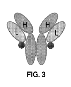
[0054] FIG. 1 shows a schematic diagram of an antibody-peptide
fusion protein with the
peptide fused to the N-terminus of the light chain via a short, rigid spacer
[0055] FIG. 2 shows a schematic diagram of an antibody-peptide
fusion protein with the
peptide fused to the C-terminus of the heavy chain via a short, rigid spacer.
[0056] FIG. 3 shows a schematic diagram of an antibody-peptide
fusion protein with the
peptide fused to the C-terminus of the light chain via a short, rigid spacer.
[0057] FIG. 4 shows a schematic diagram of an antibody-peptide
fusion protein with the
peptide fused to the C-terminus of the light chain via a long, flexible
spacer.
[0058] FIG. 5A shows data from a euripoium-linked immunosorbent
assay (EuLISA)
measuring binding of chimeric (c) 11-1F4, and humanized variants, VH10/VL4,
VH9/VL4 ,
VH8/VL4, VH7/VL4, or VH6/VL3 to synthetic rV2.6Wil light chain amyloid-like
FIG. 5B shows data from an EuLISA measuring binding of VH6/VL3-p5 (6-3-p5),
11
CA 03219124 2023- 11- 15
WO 2022/246433
PCT/US2022/072413
VH6/VL3-p5R (6-3-p5R), cl 1-1F4, or VH6/VL3 to rV26Wil fibrils. FIG. SC shows
data
from an EuLISA measuring binding of VH9/VL/4-p5R to rVX6Wil fibrils, Per125
wtATTR
extract, Ken ATTR extract, SHI ALX liver extract, or TAL ALK liver extract.
FIG. SD shows
data from an EuLISA measuring binding of VH9/VL/4-p5 to rVX6Wil fibrils,
Per125
wtATTR extract, Ken ATTR extract, SHI ALX, liver extract, or TAL ALK liver
extract. FIG.
SE shows data from an EuLISA measuring binding of c11-1F4, m11-1F4, or VH9/VL4
to
rV26Wil fibrils. FIG. SF shows data from an EuLISA measuring binding of VI-
16/VL3-p5 to
Sno ATTR extract (dark gray circles) or Ken ATTR extract (light gray circles),
and c11-1F4
binding to Sno ATTR extract (black squares). FIG. 5G shows data from an EuLISA
measuring binding of VH6/VL3-p5R to Pen l 25 wtATTR (gray circles, see label),
Sno ATTR
extract (dark gray circles), or Ken ATTR extract (light gray circles), and c11-
1F4 binding to
Sno ATTR extract (black squares). The log-transformed molar concentration of
monoclonal
antibody (-log(M)) is shown on the x-axis, and the level of binding
(femtomoles europium) is
shown on the y-axis.
[0059] FIG. 6 shows the results of 1-251-m1gp5 binding to rV26Wil
amyloid-like fibrils
and human amyloid extracts, obtained from tissues in a pulldown assay. The y-
axis shows the
percentage of bound 125I-mIgG-p5, and the percentage bound for each sample is
indicated
above the bars of the histograms. The x-axis shows the type of amyloid extract
tested
including rV2,6Wil fibrils, SNO hereditary (h) ATTR, KEN hATTR, Per 125
wtATTR,
Per253 wild type (wt) ATTR, ALK HIG extract, ALI( TAL extract, ALX SHI
extract, and ALX
TYL extract. The error bars represent the standard deviation.
[0060] FIG. 7A shows pHrodo red-labeled rV26Wil fibril uptake by
human THP-1
macrophages alone, or in the presence of human (h) IgG control, ch11-1F4,
muIgp5
(produced in the expillEK293 cell line), VH6/VL3-p5, or VH6/VL3-p5R, as
indicated from
left to right on the x-axis. The y-axis shows the level of rV26Wil fibril
uptake (measured in
fluorescent units), and the error bars represent the standard deviation. FIG.
7B shows
phagocytosis of pHrodo red-labeled rVX6Wil fibrils by macrophages in the
presence of a
THP-1 alone or with, hIgG control, c11-1F4, mIgp5, VH9/VL4-p5, or VH9/VL4-p5R,
as
indicated from left to right on the x-axis. The y-axis shows the level of
phagocytosis
(fluorescent units), and the error bars represent the standard deviation. FIG.
7C shows
phagocytosis of pHrodo red-labeled rVX6Wil fibrils by macrophages in the
presence of a
hIgG control, 5 Rituxan (a chimeric mAb as a negative control), 5 mg
c11-1F4, 5 tig
12
CA 03219124 2023- 11- 15
WO 2022/246433
PCT/US2022/072413
VH6/VL3, 5 tg VH9/VL4, VH6/VL3-p5R, or VH6/VL3-p5, as indicated from left to
right
on the x-axis. The y-axis shows the level of phagocytosis (pHrodo
fluorescence), and the
error bars represent the standard deviation.
100611 FIG. 8A shows fed-batch and perfusion workflows used to
produce intact VH9-
D54E/VL4-N33S-VSPSV-p5R Purity (% intact fusion protein) is indicated for each
production method. FIG. 8B shows the results of a binding experiment testing
the affinity of
fed-batch purified FB.1 and perfusion purified PF.1 on rVXWIL. The left y-axis
scale is used
for fed-batch purified FB.1 and the right y-axis scale is used for perfusion
purified PF.1.
100621 FIG. 9A shows the gel analysis of radiolabeled (1251) PF. I
antibody-peptide
fusion protein, in comparison to the radiolabeled (1251) antibody hIgG1
control. Reduced
(Red.) and not reduced (NR) samples are shown, and the positions of the IgG,
IgH, and IgL
for each protein are indicated. Free radioiodide (1251) is also labeled. FIG.
9B shows single
photon emission computed tomography (SPECT) and computed tomography (CT)
imaging of
systemic AA amyloidosis mice after 24 hours post-injection with either PF .1
or 125I-hIgGl.
FIG. 9C shows the biodistribution of 125I-PF.1 and 1251- hIgG1 among different
tissues in
AA mice after 24 hours post-injection. FIG. 9D shows microautoradiography of
liver (left),
spleen (center), and heart (right) tissues 24 hours post injection of AA mice
with either 1251-
PF.1 or 1251-hIgGl.
100631 FIGS. 10A-10E shows immunohistochemical staining of
different human tissues
(heart, kidney, spleen, and brain) containing ATTR, AL, ALETC2 or Af3 amyloid
with
biotinylated PF.1 and Congo red. Black arrows highlight biotinylated PF.1
bound to the
tissue sample and white arrows highlight the presence of amyloid in the tissue
sample (Congo
red staining).
100641 FIG. 11A shows quantified fluorescence emission from the
pHrodo red in mice
post injection with pHrodo red-labeled amyloid, wherein the injected amyloid
was either
preincubated with PF.1 or alone. Increased fluorescence emission is indicative
of amyloid
phagocytosis. FIG. 11B shows the fluorescence emission of the PF.1-treated and
control-
treated mice at 12 days post injection.
100651 FIGS. 12A-12D show the results of an er vivo phagocytosis
assay performed with
PF.1 and human IgG1 (hIgG1) control for ALK, ALX,, ATTRv, and ATTRwt amyloid
extracts. Phagocytosis is detected by labeling with the pH sensitive dye
succinimidyl-pHrodo
red fluorophore where increasing fluorescence emission is indicative of
enhanced
13
CA 03219124 2023- 11- 15
WO 2022/246433
PCT/US2022/072413
phagocytosis. ATTRwt is a wild type transthyretin associated amyloidosis.
ATTRy is a
variant transthyretin associated amyloidosis.
[0066] FIG. 13A shows the results of a binding experiment testing
the affinity of PF.1 on
rVkWIL A13(1-40), ATTRwt, ATTRV, ALk, and ALI< amyloid extracts. ATTRwt is a
wild
type transthyretin associated amyloidosis. ATTRy is a variant transthyretin
associated
amyloidosis. FIG. 13B shows results of a binding experiment testing the
affinity of human
IgG1 control for the same amyloid extract panel used in FIG. 12A.
[0067] FIG. 14 shows the results of a binding experiment testing
the affinity of PF.7 on
synthetic amyloid-like fibrils ot-synuclein, Tau 441, and A13(1-40). PF.7 is
VH9-D54E/VL4-
N33S-VSPSV-p5R antibody-peptide fusion protein collected after 7 days of
perfusion
culturing.
[0068] FIG. 15 shows the results of an ex vivo phagocytosis assay
performed with PR 1
on rVkWIL (WIL) and ALk (TAL) fibrils in the presence or absence of 20% human
serum as
a source of human complement. +C indicates the presence of human serum
complement.
DETAILED DESCRIPTION
[0069] Provided herein are antibody-peptide fusion proteins that
bind amyloids.
I. Definitions
[0070] As used herein, the singular forms "a,- "an,- and "the,-
refer to both the singular
as well as plural, unless the context clearly indicates otherwise. The
abbreviation, "e.g." is
derived from the Latin exempli gratia, and is used herein to indicate a non-
limiting example.
Thus, the abbreviation "e.g." is synonymous with the term "for example." As
used herein, the
term "comprises" means "includes."
[0071] Ranges can be expressed herein as from "about" one
particular value, and/or to
"about" another particular value. When such a range is expressed, another
aspect includes
from the one particular value of the range and/or to the other particular
value of the range. It
will be further understood that the endpoints of each of the ranges are
significant both in
relation to the other endpoint, and independently of the other endpoint.
Similarly, when
values are expressed as approximations, by use of the antecedent "about," it
will be
understood that the particular value forms another aspect In certain example
embodiments,
the term "about" is understood as within a range of normal tolerance in the
art, for example
14
CA 03219124 2023- 11- 15
WO 2022/246433
PCT/US2022/072413
within 2 standard deviations of the mean. About can be understood as within
10%, 9%, 8%,
7%, 6%, 5%, 4%, 3%, 2%, 1%, 0.5%, 0.1%, 0.05%, or 0.01% of the stated value.
Unless
otherwise clear from context, all numerical values provided herein can be
modified by the
term about Further, terms used herein such as "example," "exemplary," or
"exemplified," are
not meant to show preference, but rather to explain that the aspect discussed
thereafter is
merely one example of the aspect presented.
[0072] It is further to be understood that all base sizes or amino
acid sizes, and all
molecular weight or molecular mass values, given for nucleic acids or
polypeptides are
approximate, and are provided for description. Although methods and materials
similar or
equivalent to those described herein can be used in the practice or testing of
this disclosure,
suitable methods and materials are described below. In case of conflict, the
present
specification, including explanations of terms, will control. In addition, the
materials,
methods, and examples are illustrative only and not intended to be limiting.
[0073] To facilitate review of the various embodiments of this
disclosure, the following
explanations of specific terms are provided:
[0074] The terms amyloids, amyloid deposits, amyloid fibrils, and
amyloid fibers refer
to insoluble fibrous protein aggregates sharing specific structural traits.
The protein
aggregates have a tertiary structure, for example, that is formed by
aggregation of any of
several different proteins and that consists of an ordered arrangement of 13
sheets stacked
perpendicular to a fiber axis. See Sunde et al., J. Mol. Biol. (1997) 273:729-
39. Abnormal
accumulation of amyloids in organs may lead to amyloidosis. Although they are
diverse in
their occurrence, all amyloids have common morphologic properties in that they
stain with
specific dyes such as Congo red and have a characteristic red-green
birefringent appearance
in polarized light after staining. Amyloids also share common ultrastructural
features and
common x-ray diffraction and infrared spectra.
[0075] Amyloidosis refers to a pathological condition or disease
characterized by the
presence of amyloids, such as the presence of amyloid deposits. "Amyloid
diseases" or
-amyloidosis" are diseases associated with the formation, deposition,
accumulation or
persistence of amyloid fibrils. Such diseases include, but are not limited to,
Alzheimer's
disease, Down's syndrome, hereditary cerebral hemorrhage with amyloidosis of
the Dutch
type, and cerebral beta-amyloid angiopathy. Other amyloid diseases such as
systemic AA
CA 03219124 2023- 11- 15
WO 2022/246433
PCT/US2022/072413
amyloidosis, AL amyloidosis, ATTR amyloidosis, ALect2 amyloidosis, and IAPP
amyloidosis of type II diabetes are also amyloid diseases.
100761 Amyloidogenic refers to producing or tending to produce
amyloid deposits. For
example, certain soluble monomeric proteins can undergo extensive
conformational changes
leading to their aggregation into well-ordered, unbranching, 8- to 10-nm wide
fibrils, which
culminate in the formation of amyloid aggregates. More than thirty proteins,
for example,
have been found to form amyloid deposits (or amyloids) in man. Not all
proteins within the
class of diverse proteins, such as immunoglobulin light chains, are capable of
forming
amyloid, i.e., some proteins are non-amyloidogenic, meaning that they do not
tend to form
amyloids. Other proteins of the class, however, can form amyloid deposits and
are thus
amyloidogenic. Furthermore, within the class of light chain protein, some may
be deemed
more "amyloidogenic" than others based upon the ease with which they form
amyloid fibrils.
Certain light chain proteins are deemed non-amyloidogenic or less
amyloidogenic because of
their inability to readily form amyloid fibrils in patients or in vitro.
100771 Animal: Living multi-cellular vertebrate organisms, a
category that includes, for
example, mammals and birds. The term mammal includes both human and non-human
mammals. Similarly, the term "subject" includes both human and veterinary
subjects. In
some examples a subject is a subject, such as a subject suffering from an
amyloid disease.
100781 Clearance: The terms "clear" or "clearance" refer to
reducing or removing by a
measurable degree. For example, the clearance of an amyloid deposit as
described herein
relates to reducing or removing the deposit to a measurable or discernable
degree. Clearance
may result in 100% removal, but is not required to. Rather, clearance may
result in less than
100% removal, such as about 10%, 20%, 30%, 40%, 50%, 60% or more removal.
100791 Conjugate: As used herein, the term "conjugate" refers to
the product of coupling
or joining of two or more materials, the resulting product having at least two
distinct
elements, such as at least two domains. The coupled materials may be the same
or may be
different. Such a coupling may be via one or more linking groups. A "protein
conjugate," for
example, results from the coupling of two or more amino acid sequences. A
conjugate of two
proteins, for example, results in a single protein that has a domain
corresponding to each of
the individually joined proteins.
100801 The term "antibody" herein is used in the broadest sense and
specifically covers
monoclonal antibodies (including full length monoclonal antibodies),
polyclonal antibodies,
16
CA 03219124 2023- 11- 15
WO 2022/246433
PCT/US2022/072413
multispecific antibodies (e.g., bispecific antibodies), and antibody fragments
so long as they
exhibit the desired biological activity.
100811 An "isolated" antibody is one which has been identified and
separated and/or
recovered from a component of its natural environment. Contaminant components
of its
natural environment are materials which would interfere with research,
diagnostic or
therapeutic uses for the antibody, and may include enzymes, hormones, and
other
proteinaceous or nonproteinaceous solutes. In some embodiments, an antibody is
purified (1 )
to greater than 95% by weight of antibody as determined by, for example, the
Lowry method,
and in some embodiments, to greater than 99% by weight; (2) to a degree
sufficient to obtain
at least 15 residues of N- terminal or internal amino acid sequence by use of,
for example, a
spinning cup sequenator, or (3) to homogeneity by SDS-PAGE under reducing or
nonreducing conditions using, for example, Coomassie blue or silver stain,
Isolated antibody
includes the antibody in situ within recombinant cells since at least one
component of the
antibody's natural environment will not be present. Ordinarily, however,
isolated antibody
will be prepared by at least one purification step.
100821 "Native antibodies" are usually heterotetrameric
glycoproteins of about 150,000
daltons, composed of two identical light (L) chains and two identical heavy
(H) chains. Each
light chain is linked to a heavy chain by one covalent disulfide bond, while
the number of
disulfide linkages varies among the heavy chains of different immunoglobulin
isotypes. Each
heavy and light chain also has regularly spaced intrachain disulfide bridges.
Each heavy chain
has at one end a variable domain (VH) followed by a number of constant
domains. Each light
chain has a variable domain at one end (VL) and a constant domain at its other
end; the
constant domain of the light chain is aligned with the first constant domain
of the heavy
chain, and the light chain variable domain is aligned with the variable domain
of the heavy
chain. Particular amino acid residues are believed to form an interface
between the light chain
and heavy chain variable domains,
100831 The term "constant region" refers to the portion of an
immunoglobulin molecule
having a more conserved amino acid sequence relative to the other portion of
the
immunoglobulin, the variable domain, which contains the antigen binding site.
The constant
region contains the CR1, CR2 and CR3 domains (also termed CHL CH2, and CH3;
collectively, CH) of the heavy chain and the CHL (or CL or CL1) domain of the
light chain.
17
CA 03219124 2023- 11- 15
WO 2022/246433
PCT/US2022/072413
[0084] The "variable region" or "variable domain" of an antibody
refers to the amino-
terminal domains of the heavy or light chain of the antibody. The variable
domain of the
heavy chain may be referred to as "VH." The variable domain of the light chain
may be
referred to as "VL." These domains are generally the most variable parts of an
antibody and
contain the antigen-binding sites.
[0085] The term "variable" refers to the fact that certain portions
of the variable domains
differ extensively in sequence among antibodies and are used in the binding
and specificity of
each particular antibody for its particular antigen. However, the variability
is not evenly
distributed throughout the variable domains of antibodies. It is concentrated
in three segments
called complementarity-determining regions (CDRs) both in the light-chain and
the heavy-
chain variable domains. The more highly conserved portions of variable domains
are called
the framework regions (FR). The variable domains of native heavy and light
chains each
comprise four FR regions, largely adopting a beta-sheet configuration,
connected by three
CDRs, which form loops connecting, and in some cases forming part of, the beta-
sheet
structure. The CDRs in each chain are held together in close proximity by the
FR regions and,
with the CDRs from the other chain, contribute to the formation of the antigen-
binding site of
antibodies (see Kabat et al., Sequences of Proteins of Immunological Interest,
Fifth Edition,
National Institute of Health, Bethesda, Md, (1991)). The constant domains are
not involved
directly in the binding of an antibody to an antigen, but exhibit various
effector functions,
such as participation of the antibody in antibody-dependent cellular toxicity.
[0086] The "light chains" of antibodies (immunoglobulins) from any
mammalian species
can be assigned to one of two clearly distinct types, called kappa ("x") and
lambda ("X"),
based on the amino acid sequences of their constant domains,
[0087] The term IgG "isotype" or "subclass" as used herein is meant
any of the
subclasses of immunoglobulins defined by the chemical and antigenic
characteristics of their
constant regions.
[0088] Depending on the amino acid sequences of the constant
domains of their heavy
chains, antibodies (immunoglobulins) can be assigned to different classes.
There are five
major classes of immunoglobulins: IgA, IgD, IgE, IgG, and IgM, and several of
these may be
further divided into subclasses (isotypes), e.g., IgGl, IgG2, IgG3, IgG4,
IgAl, and IgA2. The
heavy chain constant domains that correspond to the different classes of
immunoglobulins are
called a, 6, E, y, and la, respectively. The subunit structures and three-
dimensional
18
CA 03219124 2023- 11- 15
WO 2022/246433
PCT/US2022/072413
configurations of different classes of immunoglobulins are well known and
described
generally in, for example, Abbas el al. Cellular and Mol. Immunology, 4th ed.
(W.B.
Saunders, Co., 2000). An antibody may be part of a larger fusion molecule,
formed by
covalent or non-covalent association of the antibody with one or more other
proteins or
peptides.
[0089] The terms "full length antibody," "intact antibody" and
"whole antibody" are
used herein interchangeably to refer to an antibody in its substantially
intact form, not
antibody- fragments as defined below. The terms particularly refer to an
antibody with heavy
chains that contain an Fc region.
[0090] A "naked antibody" for the purposes herein is an antibody
that is not conjugated
to a cytotoxic moiety or radiolabel.
100911 "Antibody fragments" comprise a portion of an intact
antibody, preferably
comprising the antigen binding region thereof. In some embodiments, the
antibody fragment
described herein is an antigen-binding fragment. Examples of antibody
fragments include
Fab, Fab', F(a1302, and FAT fragments; diabodies; linear antibodies; single-
chain antibody
molecules; and multispecific antibodies formed from antibody fragments.
[0092] Papain digestion of antibodies produces two identical
antigen-binding fragments,
called "Fab" fragments, each with a single antigen- binding site, and a
residual "Fc"
fragment, whose name reflects its ability to crystallize readily. Pepsin
treatment yields an
F(ab1)2 fragment that has two antigen-combining sites and is still capable of
cross-linking
antigen.
[0093] "Fv" is the minimum antibody fragment which contains a
complete antigen-
binding site. In one embodiment, a two-chain FIT species consists of a dimer
of one heavy-
and one light-chain variable domain in tight, non-covalent association, In a
single-chain Fv
(scFv) species, one heavy- and one light-chain variable domain can be
covalently linked by a
flexible peptide linker such that the light and heavy chains can associate in
a "dimeric"
structure analogous to that in a two-chain FAT species. It is in this
configuration that the three
CDRs of each variable domain interact to define an antigen- binding site on
the surface of the
VH-VL dimer. Collectively, the six CDRs confer antigen-binding specificity to
the antibody.
However, even a single variable domain (or half of an FAT comprising only
three CDRs
specific for an antigen) has the ability to recognize and bind antigen,
although at a lower
affinity than the entire binding site.
19
CA 03219124 2023- 11- 15
WO 2022/246433
PCT/US2022/072413
100941 The Fab fragment contains the heavy- and light-chain
variable domains and also
contains the constant domain of the light chain and the first constant domain
(CHI.) of the
heavy chain. Fab' fragments differ from Fab fragments by the addition of a few
residues at
the carboxy terminus of the heavy chain CHI domain including one or more
cysteines from
the antibody hinge region. Fab'-SH is the designation herein for Fab' in which
the cysteine
residue(s) of the constant domains bear a free thiol group. F(ab')2 antibody
fragments
originally were produced as pairs of Fab' fragments which have hinge cysteines
between
them. Other chemical couplings of antibody fragments are also known.
100951 "Single-chain Fv" or "scFv" antibody fragments comprise the
VH and VL
domains of antibody, wherein these domains are present in a single polypeptide
chain.
Generally, the scFv polypeptide further comprises a polypeptide linker between
the VH and
VL domains which enables the scFv to form the desired structure for antigen
binding. For a
review of scFv, see, e.g., Pluckfhun, in The Pharmacology of Monoclonal
Antibodies, vol.
113, Rosenburg and Moore eds., (Springer- Verlag, New York, 1994), pp. 269-
315.
100961 The term "monoclonal antibody" as used herein refers to an
antibody obtained
from a population of substantially homogeneous antibodies, e.g., the
individual antibodies
comprising the population are identical except for possible mutations, e.g.,
naturally
occurring mutations, that may be present in minor amounts. Thus, the modifier
"monoclonal."
indicates the character of the antibody as not being a mixture of discrete
antibodies. In certain
embodiments, such a monoclonal antibody typically includes an antibody
comprising a
polypeptide sequence that binds a target, wherein the target-binding
polypeptide sequence
was obtained by a process that includes the selection of a single target
binding polypeptide
sequence from a plurality of polypeptide sequences. For example, the selection
process can
be the selection of a unique clone from a plurality of clones, such as a pool
of hybridoma
clones, phage clones, or recombinant DNA clones. It should be understood that
a selected
target binding sequence can be further altered, for example, to improve
affinity for the target,
to humanize the target binding sequence, to improve its production in cell
culture, to reduce
its immunogeni city in vivo, to create a multi specific antibody, etc., and
that an antibody
comprising the altered target binding sequence is also a monoclonal antibody
of this
invention. In contrast to polyclonal antibody preparations, which typically
include different
antibodies directed against different, determinants (epitopes), each
monoclonal antibody of a
monoclonal antibody preparation is directed against a single determinant on an
antigen. In
CA 03219124 2023- 11- 15
WO 2022/246433
PCT/US2022/072413
addition to their specificity, monoclonal antibody preparations are
advantageous in that they
are typically uncontaminated by other immunoglobulins.
[0097] The modifier "monoclonal" indicates the character of the
antibody as being
obtained from a substantially homogeneous population of antibodies, and is not
to be
construed as requiring production of the antibody by any particular method.
For example, the
monoclonal antibodies to be used in accordance with the invention may be made
by a variety
of techniques, including, for example, the hybridoma method (e.g., Kohler and
Milstein,
Nature, 256:495-97 (1975); Hongo et al, Hybridoma, .1.4 (3): 253-260 (.1995),
Harlow et al.,
Antibodies: A Laboratory Manual, (Cold Spring Harbor Laboratory Press, 2nd ed.
1988);
Hammerling et at, in: Monoclonal Antibodies and T-Cell Hybridomas 563-681
(Elsevier,
N.Y., 1981 )), recombinant DNA methods (see, e.g.,U U.S. Pat. No. 4,8.16,567),
phage-display
technologies (see, e.g., Clackson et at, Nature, 352: 624-628 (1991); Marks et
at, J. Mol.
Biol. 222: 581-597 ( 1992); Sidhu eta!, J. Mol. Biol. 338(2): 299-310 (2004);
Lee etal., J.
Mol. Biol. 340(5): 1073- 1093 (2004); Fellouse, Proc. Natl. Acad. Sci. USA
101(34): 12467-
12472 (2004); and Lee et at., J. Immunol. Methods 284(1-2): 119-132 (2004),
and
technologies for producing human or human-like antibodies in animals that have
parts or all
of the human immunoglobulin loci or genes encoding human immunoglobulin
sequences
(see, e.g., WO 1998/24893; WO 1996/34096; WO 1996/33735; WO 1991/10741;
Jakobovits
11 et at, Proc. .Natl. Acad. Sci. USA 90: 2551 (1993); Jakobovits et al,
Nature 362: 255-258
(1993) ; Bruggemann et al, Year in Immunol 7:33 (1993); U.S. Pat. Nos.
5,545,807;
5,545,806; 5,569,825; 5,625,126; 5,633,425; and 5,661,016; Marks eta!,
Bio/Technology 10:
779-783 (1992); Lonberg eta!, Nature 368: 856-859 (1994); Morrison, Nature
368: 812-813
(1994) ; Fishwild eta!, Nature Biotechnol. 14: 845-851 (1996); Neuberger,
Nature
Biotechnol 14: 826 ( 1996); and Lonberg and Huszar, Intern. Rev. Irmnunol 13:
65-93 (1995).
[0098] The monoclonal antibodies herein specifically include
"chimeric" antibodies in
which a portion of the heavy and/or light chain is identical with or
homologous to
corresponding sequences in antibodies derived from a particular species or
belonging to a
particular antibody class or subclass, while the remainder of the chain(s) is
identical with or
homologous to corresponding sequences in antibodies derived from another
species or
belonging to another antibody class or subclass, as well as fragments of such
antibodies, so
long as they exhibit the desired biological activity (see, e.g., U.S. Pat. No.
4,816,567; and
Morrison c/a/, Proc. Nall Acad. Sci. USA 81:6851-6855 (1984)). Chimeric
antibodies
include primatized antibodies wherein the antigen-binding region of the
antibody is derived
21
CA 03219124 2023- 11- 15
WO 2022/246433
PCT/US2022/072413
from an antibody produced by, e.g-., immunizing macaque monkeys with the
antigen of
interest.
100991 "Humanized" forms of non-human (e.g., murine) antibodies are
chimeric
antibodies that contain minimal sequence derived from non-human
immunoglobulin. In one
embodiment, a humanized antibody is a human immunoglobulin (recipient
antibody) in
which residues from a CDR of the recipient are replaced by residues from a CDR
of a non-
human species (donor antibody) such as mouse, rat, rabbit, or nonhuman primate
having the
desired specificity, affinity, and/or capacity. In some instances, FR residues
of the human
immunoglobulin are replaced by corresponding non-human residues. Furthermore,
humanized antibodies may comprise residues that are not found in the recipient
antibody or in
the donor antibody. These modifications may be made to further refine antibody
performance. In general, a humanized antibody will comprise substantially all
of at least one,
and typically two, variable domains, in which all or substantially all of the
hypervariable
loops correspond to those of a non-human immunoglobulin, and all or
substantially all of the
FRs are those of a human immunoglobulin sequence. The humanized antibody
optionally will
also comprise at least a portion of an immunoglobulin constant region (Fc),
typically that of a
human immunoglobulin. For further details, see, e.g., Jones et al, Nature
321:522-525
(1986); Riechmann et al, Nature 332:323-329 (1988); and Presta, Curr. Op.
Struct. Biol
2:593-596 (1992), See also, e.g., Vaswani and Hamilton, Ann. Allergy, Asthma &
Immunol. 1
: 105-115 (1998); Harris, Biochem. Soc. Transactions 23: 1035-1038 (1995);
Hurl e and
Gross, Curr. Op. Biotech. 5:428-433 (1994): and U.S. Pat. Nos. 6,982,321 and
7,087,409.
101001 A "human antibody" is one which possesses an amino acid
sequence which
corresponds to that of an antibody produced by a human and/or has been made
using any of
the techniques for making human antibodies as disclosed herein. This
definition of a human
antibody specifically excludes a humanized antibody comprising non-human
antigen-binding
residues. Human antibodies can be produced using various techniques known in
the art,
including phage-display libraries. Hoogenboom and Winter, J. Mol. Biol.,
227:381 (1991);
Marks eta!, .1. Mol. Biol., 222:581 (1991). Also available for the preparation
of human
monoclonal antibodies are methods described in Cole et al, Monoclonal
Antibodies and
Cancer Therapy, Alan R. Liss, p. 77(1985); Boerner et al, J. _Minim/of .,
147(0:86-95 (1991).
See also van Dijk and van de Winkel, C'urr. Opin. Pharmacol., 5: 368-74
(2001), Human
antibodies can be prepared by administering the antigen to a transgenic animal
that has been
modified to produce such antibodies in response to antigenic challenge, but
whose
22
CA 03219124 2023- 11- 15
WO 2022/246433
PCT/US2022/072413
endogenous loci have been disabled, e.g., immunized xenomice (see, e.g., U.S.
Pat. Nos.
6,075,181 and 6,150,584 regarding XENOMOUSETm technology). See also, for
example, Li
eta!, Proc. Natl. Acad. Sci. USA, 103:3557-3562 (2006) regarding human
antibodies
generated via a human B-cell hybridoma technology.
101011 The term "complementarity-determining region" or "CDR," when
used herein
refers to the regions of an antibody-variable domain that bind to an epitope,
such as human
amyloid fibrils. Generally, antibodies comprise six CDRs; three in the VH (H1,
H2, H3), and
three in the VL (L1, L2, L3). In native antibodies, H3 and L3 display the most
diversity of the
six CDRs, and H3 in particular is believed to play a unique role in conferring
fine specificity
to antibodies. See, e.g.,Xu etal., Immunity 13:37-45 (2000); Johnson and Wu in
Methods in
Molecular Biology 248:1-25 (Lo, ed., Human Press, Totowa, NJ, 2003)). Indeed,
naturally
occurring camelid antibodies consisting of a heavy chain only are functional
and stable in the
absence of light chain. See, e.g., Hamers-Casterman et al., Nature 363:446-448
(1993) and
Sheriff et al., Nature Struct. Biol. 3:733-736 (1996).
101021 A number of CDR delineations are in use and are encompassed
herein. In some
embodiments, the CDRs may be Kabat CDRs, which are based on sequence
variability and
are the most commonly used (Kabat et al., supra). In some embodiments, the
CDRs may be
Chothia CDRs. Chothia refers instead to the location of the structural loops
(Chothia and
Lesk J. Mol. Biol. 196:901-917 (1987)). In some embodiments, the CDRs may be
AbM
CDRs. The AbM CDRs represent a compromise between the Kabat CDRs and Chothia
structural loops, and are used by Oxford Molecular's AbM antibody-modeling
software. In
some embodiments, the CDRs may be "contact" CDRs. The "contact" CDRs are based
on an
analysis of the available complex crystal structures. The residues from each
of these CDRs
are noted below.
Loop Kabat AbM Chothia Contact
Li L24-L34 L24-L34 L26-L32 L30-L36
L2 L50-L56 L50-L56 L50-L52 L46-L55
L3 L89-L97 L89-L97 L91-L96 L89-L96
H1 H31-H35B H26-H35B H26-H32 H30-H35B (Kabat
numbering)
H1 H31-H35 H26-H35 H26-H32 H30-H35 (Chothia
numbering)
H2 H50-H65 H50-H58 H53-H55 H47-H58
H3 H95-H102 H95-H102 H96-H101 H93-H101
101031 CDRs may comprise "extended CDRs" as follows: 24-36 or 24-34
(L1), 46-56 or
50-56 (L2), and 89-97 or 89-96 (L3) in the VL, and 26-35 (H1), 50-65 or 49-65
(a preferred
23
CA 03219124 2023- 11- 15
WO 2022/246433
PCT/US2022/072413
embodiment) (H2), and 93-102, 94-102, or 95-102 (H3) in the VII. The variable-
domain
residues are numbered according to Kabat et cll., supra, for each of these
extended-CDR
definitions.
[0104] "Framework" or "FR" residues are those variable domain
residues other than the
CDR residues as herein defined.
[0105] As use herein, the term "specifically binds to" or is
"specific for" refers to
measurable and reproducible interactions such as binding between a target and
an antibody,
which is determinative of the presence of the target in the presence of a
heterogeneous
population of molecules including biological molecules. For example, an
antibody that
specifically binds to a target (which can be an epitope) is an antibody that
binds this target
with greater affinity, avidity, more readily, and/or with greater duration
than it binds to other
targets In one embodiment, the extent of binding of an antibody to an
unrelated target is less
than about 10% of the binding of the antibody to the target as measured, e.g.,
by a
radioimmunoassay (RIA). In certain embodiments, an antibody that specifically
binds to a
target has a dissociation constant (Kd) of < 1 M, < 100 1.1M, < 10 nM, < 1 nM,
or < 0.1 nM.
In certain embodiments, an antibody specifically binds to an epitope on a
protein that is
conserved among the protein from different species. In another embodiment,
specific binding
can include, but does not require exclusive binding.
[0106] Effective amount or Therapeutically effective amount: The
amount of agent
that is sufficient to prevent, treat (including prophylaxis), reduce and/or
ameliorate the
symptoms and/or underlying causes of any of a disorder or disease, for example
to prevent,
inhibit, and/or amyloidosis. In some embodiments, an "effective amount" is
sufficient to
reduce or eliminate a symptom of a disease. An effective amount can be
administered one or
more times. For example, an effective amount of a peptide is an amount that is
sufficient to
bind an amyloid. A peptide may be effective, for example, when parenterally
administered in
amounts above about 1 jig per kg of body weight to about 30 mg/kg.
[0107] Inhibit: To reduce by a measurable degree. Inhibition does
not, for example,
require complete loss of function or complete cessation of the aspect being
measured. For
example, inhibiting plaque formation can mean stopping further growth of the
plaque,
slowing further growth of the plaque, or reducing the size of the plaque.
[0108] Inhibiting or treating a disease: Inhibiting the full
development of a disease or
condition, for example, inhibiting amyloidosis. "Treatment" refers to a
therapeutic
24
CA 03219124 2023- 11- 15
WO 2022/246433
PCT/US2022/072413
intervention that ameliorates a sign or symptom of a disease or pathological
condition after it
has begun to develop. The term "ameliorating," with reference to a disease or
pathological
condition, refers to any observable beneficial effect of the treatment The
beneficial effect can
be evidenced, for example, by a delayed onset of clinical symptoms of the
disease in a
susceptible subject, a reduction in severity of some or all clinical symptoms
of the disease, a
slower progression of the disease, an improvement in the overall health or
well-being of the
subject, or by other parameters well known in the art that are specific to the
particular
disease. A "prophylactic" treatment is a treatment administered to a subject
who does not
exhibit signs of a disease or exhibits only early signs for the purpose of
decreasing the risk of
developing pathology.
101091 With regard to amyloid deposit formation, "inhibition"
refers to the prevention of
reduction in the formation of the amyloid deposit, such as when compared to a
control. For
example, inhibition may result in a reduction of about 10%, 20%, 30%, 40%,
50%, 60% or
more of an amyloid deposit as compared to a control.
101101 Label refers to any detectable compound or composition that
is conjugated
directly or indirectly to another molecule to facilitate detection of that
molecule. Specific,
non-limiting examples of labels include fluorescent tags, chemiluminescent
tags, haptens,
enzymatic linkages, and radioactive isotopes. A protein that is "detectably-
labeled," for
example, means that the presence of the protein can be determined by a label
associated with
the protein.
101111 Isolated: An "isolated" biological component, such as a
peptide (for example one
or more of the peptides disclosed herein), cell, nucleic acid, or serum
samples has been
substantially separated, produced apart from, or purified away from other
biological
components in the cell of the organism in which the component naturally
occurs, for instance,
other chromosomal and extrachromosomal DNA and RNA, and proteins. Nucleic
acids,
peptides and proteins that have been "isolated- thus include nucleic acids and
proteins
purified by standard purification methods. The term also embraces nucleic
acids, peptides and
proteins prepared by recombinant expression in a cell as well as chemically
synthesized
peptide and nucleic acids. The term "isolated" or "purified" does not require
absolute purity;
rather, it is intended as a relative term. Thus, for example, an isolated
peptide preparation is
one in which the peptide or protein is more enriched than the peptide or
protein is in its
natural environment within a cell. Preferably, a preparation is purified such
that the protein or
peptide represents at least 50% of the total peptide or protein content of the
preparation, such
CA 03219124 2023- 11- 15
WO 2022/246433
PCT/US2022/072413
as at least 50%, at least 60%, at least 70%, at least 80%, at least 90%, at
least 95%, or even at
least 99% of the peptide or protein concentration.
[0112] Join: As used herein, the term -join," "joined," -link," or -
linked" refers to any
method known in the art for functionally connecting proteins and/or protein
domains. For
example, one protein domain may be linked to another protein domain via a
covalent bond,
such as in a recombinant fusion protein, with or without intervening sequences
or domains.
Joined also includes, for example, the integration of two sequences together,
such as placing
two nucleic acid sequences together in the same nucleic acid strand so that
the sequences are
expressed together.
[0113] Nucleic acid: A polymer composed of nucleotide units
(ribonucleotides,
deoxyribonucleotides, related naturally occurring structural variants, and
synthetic non-
naturally occurring analogs thereof) linked via phosphodiester bonds, related
naturally
occurring structural variants, and synthetic non-naturally occurring analogs
thereof, Thus, the
term includes nucleotide polymers in which the nucleotides and the linkages
between them
include non-naturally occurring synthetic analogs, such as, for example and
without
limitation, phosphorothioates, phosphoramidates, methyl phosphonates, chiral-
methyl
phosphonates, 2-0-methyl ribonucleotides, peptide- nucleic acids (PNAs), and
the like. Such
polynucleotides can be synthesized, for example, using an automated DNA
synthesizer. The
term "oligonucleotide" typically refers to short polynucleotides, generally no
greater than
about 50 nucleotides. It will be understood that when a nucleotide sequence is
represented by
a DNA sequence (i.e., A, T, G, C), this also includes an RNA sequence (i.e.,
A, U, G, C) in
which "U" replaces "T."
[0114] Nucleotide includes, but is not limited to, a monomer that
includes a base linked
to a sugar, such as a pyrimidine, purine or synthetic analogs thereof, or a
base linked to an
amino acid, as in a peptide nucleic acid (PNA). A nucleotide is one monomer in
a
polynucleotide. A nucleotide sequence refers to the sequence of bases in a
polynucleotide.
[0115] Conventional notation is used herein to describe nucleotide
sequences. the left-
hand end of a single-stranded nucleotide sequence is the 5 `-end; the left-
hand direction of a
double-stranded nucleotide sequence is referred to as the 5'-direction. The
direction of 5' to
3' addition of nucleotides to nascent RNA transcripts is referred to as the
transcription
direction. The DNA strand having the same sequence as an mRNA is referred to
as the
"coding strand," sequences on the DNA strand having the same sequence as an
mRNA
26
CA 03219124 2023- 11- 15
WO 2022/246433
PCT/US2022/072413
transcribed from that DNA and which are located 5' to the 5'-end of the RNA
transcript are
referred to as "upstream sequences,- sequences on the DNA strand having the
same sequence
as the RNA and which are 3' to the 3' end of the coding RNA transcript are
referred to as
"downstream sequences."
101161 cDNA refers to a DNA that is complementary or identical to
an mRNA, in either
single stranded or double stranded form.
101171 Encoding refers to the inherent property of specific
sequences of nucleotides in a
polynucleotide, such as a gene, a cDNA, or an mRNA, to serve as templates for
synthesis of
other polymers and macromolecules in biological processes having either a
defined sequence
of nucleotides (for example, rRNA, tRNA and mRNA) or a defined sequence of
amino acids
and the biological properties resulting therefrom. Thus, a gene encodes a
protein if
transcription and translation of mRNA produced by that gene produces the
protein in a cell or
other biological system. Both the coding strand, the nucleotide sequence of
which is identical
to the mRNA sequence and is usually provided in sequence listings, and non-
coding strand,
used as the template for transcription, of a gene or cDNA can be referred to
as encoding the
protein or other product of that gene or cDNA. Unless otherwise specified, a
"nucleotide
sequence encoding an amino acid sequence" includes all nucleotide sequences
that are
degenerate versions of each other and that encode the same amino acid
sequence. Nucleotide
sequences that encode proteins and RNA may include introns.
101181 Pharmaceutically acceptable carriers: The pharmaceutically
acceptable
carriers of use are conventional. Remington's Pharmaceutical Sciences, by E.
W. Martin,
Mack Publishing Co., Easton, PA, 19th Edition (1995), describes compositions
and
formulations suitable for pharmaceutical delivery of the fusion proteins
herein disclosed.
101191 In general, the nature of the carrier will depend on the
particular mode of
administration being employed. For instance, parenteral formulations usually
comprise
injectable fluids that include pharmaceutically and physiologically acceptable
fluids such as
water, physiological saline, balanced salt solutions, aqueous dextrose,
glycerol or the like as a
vehicle. For solid compositions (e.g., powder, pill, tablet, or capsule
forms), conventional
non-toxic solid carriers can include, for example, pharmaceutical grades of
mannitol, lactose,
starch, or magnesium stearate. In addition to biologically neutral carriers,
pharmaceutical
compositions to be administered can contain minor amounts of non-toxic
auxiliary
27
CA 03219124 2023- 11- 15
WO 2022/246433
PCT/US2022/072413
substances, such as wetting or emulsifying agents, preservatives, and pH
buffering agents and
the like, for example sodium acetate or sorbitan monolaurate.
[0120] Polypeptide: A polymer in which the monomers are amino acid
residues that are
joined together through amide bonds. When the amino acids are alpha-amino
acids, either the
L-optical isomer or the D-optical isomer can be used, the L-isomers being
preferred. The
terms "polypeptide" or "protein" as used herein is intended to encompass any
amino acid
sequence and include modified sequences such as glycoproteins. The term
"polypeptide" is
specifically intended to cover naturally occurring proteins, as well as those
that are
recombinantly or synthetically produced. In some examples, a peptide is one or
more of the
peptides disclosed herein.
[0121] Purified: The term -purified" does not require absolute
purity; rather, it is
intended as a relative term. Thus, for example, a purified protein preparation
is one in which
the protein referred to is more pure than the protein in its natural
environment within a cell or
within a production reaction chamber (as appropriate).
[0122] Recombinant: A recombinant nucleic acid is one that has a
sequence that is not
naturally occurring or has a sequence that is made by an artificial
combination of two
otherwise separated segments of sequence. This artificial combination is often
accomplished
by chemical synthesis or, more commonly, by the artificial manipulation of
isolated segments
of nucleic acids, e.g., by genetic engineering techniques.
[0123] Sequence identity: The similarity between two nucleic acid
sequences, or two
amino acid sequences, is expressed in terms of the similarity between the
sequences,
otherwise referred to as sequence identity. Sequence identity is frequently
measured in terms
of percentage identity (or similarity or homology); the higher the percentage,
the more similar
the two sequences are.
[0124] Methods of alignment of sequences for comparison are well
known in the art.
Various programs and alignment algorithms are described in: Smith & Waterman
Adv. App!.
Math. 2: 482, 1981; Needleman & Wunsch./ Mal. Biol. 48: 443, 1970; Pearson &
Lipman
Proc. Natl. Acad. Sci. USA 85: 2444, 1988; Higgins & Sharp Gene 73: 237-244,
1988;
Higgins & Sharp CABIOS 5: 151-153, 1989; Corpet et al Nue. Acids Res. 16,
10881-90,
1988; Huang etal. Computer Appls. In the Biosciences 8, 155-65, 1992; and
Pearson etal.
Meth. Mol. Bio. 24, 307-31, 1994. Altschul etal. (J. Mol. Biol. 215:403-410,
1990), presents a
detailed consideration of sequence alignment methods and homology
calculations.
28
CA 03219124 2023- 11- 15
WO 2022/246433
PCT/US2022/072413
101251 The NCBI Basic Local Alignment Search Tool (BLAST) (Altschul
et at. J. Mol.
Biol. 215:403-410, 1990) is available from several sources, including the
National Center for
Biotechnology Information (NCBI, Bethesda, MD) and on the Internet, for use in
connection
with the sequence analysis programs blastp, blastn, blastx, tblastn and
tblastx.
101261 Operably linked: A first nucleic acid sequence is operably
linked with a second
nucleic acid sequence when the first nucleic acid sequence is placed in a
functional
relationship with the second nucleic acid sequence. For instance, a promoter
is operably
linked to a coding sequence if the promoter affects the transcription or
expression of the
coding sequence. Generally, operably linked DNA sequences are contiguous and,
where
necessary to join two protein-coding regions, in the same reading frame.
101271 Pharmaceutical agent: A chemical compound or composition
capable of
inducing a desired therapeutic or prophylactic effect when properly
administered to a subject
or a cell.
101281 Vector: A nucleic acid molecule as introduced into a host
cell, thereby producing
a transformed host cell. Recombinant DNA vectors are vectors having
recombinant DNA. A
vector can include nucleic acid sequences that permit it to replicate in a
host cell, such as an
origin of replication. A vector can also include one or more selectable marker
genes and other
genetic elements known in the art. Viral vectors are recombinant DNA vectors
having at least
some nucleic acid sequences derived from one or more viruses. The term vector
includes
plasmids, linear nucleic acid molecules, and as described throughout
adenovirus vectors and
adenovi ruses.
101291 A subject or an individual refers to a mammal, for example,
a human. The
subject may be a human patient A subject may be a patient suffering from or
suspected of
suffering from a disease or condition and may be in need of treatment or
diagnosis or may be
in need of monitoring for the progression of the disease or condition. The
patient may also be
in on a treatment therapy that needs to be monitored for efficacy. In some
example
embodiments, a subject includes an individual suffering from amyloidosis, such
as
Alzheimer's, Huntington' s or prion diseases, or peripheral amyloidosis such
as seen in
patients with light chain (AL) amyloidosis and type 2 diabetes.
101301 Preferably, residue positions which are not identical differ
by conservative amino
acid substitutions. The term "conservative amino acid substitutions" refer to
the
interchangeability of residues having similar side chains. For example, a
group of amino
29
CA 03219124 2023- 11- 15
WO 2022/246433
PCT/US2022/072413
acids having aliphatic side chains is glycine, alanine, valine, leucine, and
isoleucine; a group
of amino acids having aliphatic-hydroxyl side chains is serine and threonine,
a group of
amino acids having amide- containing side chains is asparagine and glutamine;
a group of
amino acids having aromatic side chains is phenylalanine, tyrosine, and
tryptophan; a group
of amino acids having basic side chains is lysine, arginine, and histidine;
and a group of
amino acids having sulfur- containing side chains is cysteine and methionine.
Preferred
conservative amino acids substitution groups are: valine-leucine-isoleucine,
phenylalanine-
tyrosine, lysine-arginine, alanine valine, glutamic- aspartic, and asparagine-
glutamine.
101311 As discussed herein, minor variations in the amino acid
sequences of antibodies or
immunoglobulin molecules are contemplated as being encompassed by the present
invention,
providing that the variations in the amino acid sequence maintain at least
75%, more
preferably at least 80%, 90%, 95%, and most preferably 99%. In particular,
conservative
amino acid replacements are contemplated. Conservative replacements are those
that take
place within a family of amino acids that are related in their side chains.
Genetically encoded
amino acids are generally divided into families: (1) acidic amino acids are
aspartate,
glutamate; (2) basic amino acids are lysine, arginine, histidine; (3) non-
polar amino acids are
alanine, valine, leucine, isoleucine, proline, phenylalanine, methionine,
tryptophan, and (4)
uncharged polar amino acids are glycine, asparagine, glutamine, cysteine,
serine, threonine,
tyrosine. The hydrophilic amino acids include arginine, asparagine, aspartate,
glutamine,
glutamate, histidine, lysine, serine, and threonine. The hydrophobic amino
acids include
alanine, cysteine, isoleucine, leucine, methionine, phenylalanine, proline,
tryptophan, tyrosine
and valine. Other families of amino acids include (i) serine and threonine,
which are the
aliphatic-hydroxy family; (ii) asparagine and glutamine, which are the amide
containing
family; (iii) alanine, valine, leucine and isoleucine, which are the aliphatic
family; and (iv)
phenylalanine, tryptophan, and tyrosine, which are the aromatic family. For
example, it is
reasonable to expect that an isolated replacement of a leucine with an
isoleucine or valine, an
aspartate with a glutamate, a threonine with a serine, or a similar
replacement of an amino
acid with a structurally related amino acid will not have a major effect on
the binding or
properties of the resulting molecule, especially if the replacement does not
involve an amino
acid within a framework site. Whether an amino acid change results in a
functional peptide
can readily be determined by assaying the specific activity of the polypeptide
derivative
assays are described in detail herein. Fragments or analogs of antibodies or
immunoglobulin
molecules can be readily prepared by those of ordinary skill in the art.
Preferred amino- and
CA 03219124 2023- 11- 15
WO 2022/246433
PCT/US2022/072413
carboxy-termini of fragments or analogs occur near boundaries of functional
domains.
Structural and functional domains can be identified by comparison of the
nucleotide and/or
amino acid sequence data to public or proprietary sequence databases.
Preferably,
computerized comparison methods are used to identify sequence motifs or
predicted protein
conformation domains that occur in other proteins of known structure and/or
function.
Methods to identify protein sequences that fold into a known three-dimensional
structure are
known. (Bowie et al. Science 253:164 (1991). Thus, the foregoing examples
demonstrate that
those of skill in the art can recognize sequence motifs and structural
conformations that may
be used to define structural and functional domains in accordance with the
invention.
101321 Preferred amino acid substitutions are those which: (1)
reduce susceptibility to
proteolysis, (2) reduce susceptibility to oxidation, (3) alter binding
affinity for forming
protein complexes, (4) alter binding affinities, and (4) confer or modify
other
physicochemical or functional properties of such analogs. Analogs can include
various
muteins of a sequence other than the naturally-occurring peptide sequence. For
example,
single or multiple amino acid substitutions (preferably conservative amino
acid substitutions)
may be made in the naturally- occurring sequence (preferably in the portion of
the
polypeptide outside the domain(s) forming intermolecular contacts. A
conservative amino
acid substitution should not substantially change the structural
characteristics of the parent
sequence (e.g., a replacement amino acid should not tend to break a helix that
occurs in the
parent sequence, or disrupt other types of secondary structure that
characterizes the parent
sequence). Examples of art-recognized polypeptide secondary and tertiary
structures are
described in Proteins, Structures and Molecular Principles (Creighton, Ed., W.
H. Freeman
and Company, New York (1984)); Introduction to Protein Structure (C. Branden
and J.
Tooze, eds., Garland Publishing, New York, N.Y. (1991)); and Thornton et al.
Nature
354:105 (1991).
101331 With the exception of CDR1 in VH, CDRs generally comprise
the amino acid
residues that form the hypervariable loops. CDRs also comprise "specificity
determining
residues," or "SDRs," which are residues that contact antigen. SDRs are
contained within
regions of the CDRs called abbreviated-CDRs, or a-CDRs. Exemplary a-CDRs (a-
CDR-L1, a-
CDR-L2, a- CDR-L3, a-CDR-H1, a-CDR-H2, and a-CDR-H3) occur at amino acid
residues
31-34 of Li, 50-55 of L2, 89-96 of L3, 31-35B of HI, 50-58 of H2, and 95-102
of H3. (See
Almagro and Fransson, Front. Biosci. 13: 1619-1633 (2008).)
Antibody-peptide fusion proteins
31
CA 03219124 2023- 11- 15
WO 2022/246433
PCT/US2022/072413
101341 Provided herein are antibody-peptide fusion proteins that
target amyloids. Such
antibody-peptide fusion proteins include, for example, amyloid-reactive
peptides that are
linked to an antibody, such as through extension of the N-terminus or C-
terminus of the light
chain protein of the antibody, fragment thereof, antigen binding (Fab) region,
or via or the N-
terminus or C-terminus of the heavy chain of the antibody, thereby forming a
peptide-
antibody fusion, wherein the amyloid-reactive peptide is linked to the
antibody via a spacer
comprising an amino acid sequence selected from the group consisting of SEQ ID
NOs: 23-
24, 27, 83-86. In some embodiments, the amyloid-reactive peptide is linked to
the antibody
via a spacer comprising an amino acid sequence selected from the group
consisting of SEQ
ID NOs: 83-86. The antibody-peptide fusion proteins can be used to treat a
subject having
amyloidosis or suspected of having amyloidosis, for example, such as by
administering the
antibody-peptide fusion proteins to the subject.
101351 In some embodiments, provided herein is an antibody-peptide
fusion protein,
comprising: an amyloid-reactive peptide; and an antibody that induces
phagocytosis or acts as
an opsonin. In some embodiments, an opsonin is a protein that binds to a
target and induces
phagocytosis of that target. In some embodiments, the opsonin comprises
antibody or an
antibody-peptide fusion protein. In some embodiments, the antibody-peptide
fusion proteins
provided herein act as opsonins by binding to amyloid and promoting
phagocytosis of the
amyloid. In some embodiments, the antibody comprises a heavy chain comprising
a heavy
chain variable region (VET) and a light chain comprising a light chain
variable region (VL). In
some embodiments, the amyloid-reactive peptide and the antibody are linked at
the N- and/or
C-terminal end of the light chain and/or the N- and/or C-terminal end of the
heavy chain. In
some embodiments, the antibody-peptide fusion protein comprises more than one
amyloid-
reactive peptide linked to the antibody. In some embodiments, the amyloid-
reactive peptide is
linked to the antibody via a spacer. In some embodiments, the amyloid-reactive
peptide is
linked to the antibody via a peptide spacer. In some embodiments, the amyloid-
reactive
peptide is linked to the antibody via a spacer comprising the amino acid
sequence selected
from the group consisting of SEQ ID NOs: 23-24, 27, 83-86. In some
embodiments, the
amyloid-reactive peptide is linked to the antibody via a spacer comprising an
amino acid
sequence selected from the group consisting of SEQ ID NOs: 83-86. In some
embodiments,
the amyloid-reactive peptide and antibody are linked at the N-terminal end of
the light chain.
In some embodiments, the amyloid-reactive peptide and antibody are linked at
the C-terminal
end of the light chain. In some embodiments, the amyloid-reactive peptide and
antibody are
32
CA 03219124 2023- 11- 15
WO 2022/246433
PCT/US2022/072413
linked at the N-terminal end of the light chain. In some embodiments, the
amyloid-reactive
peptide and antibody are linked at the C-terminal end of the heavy chain. In
some
embodiments, the antibody is a full length antibody. In some embodiment, the
amyloid-
reactive peptide comprises an amino acid sequence as shown in Table 1, below.
101361
In some embodiments, the amyloid-reactive peptide antibody fusion
comprises a
heavy chain in N-to C-terminal direction comprising in order an amyloid
reactive peptide, a
spacer, a VH, a CHI, a CH2, and a CH3. In some embodiments, the amyloid-
reactive
peptide antibody fusion comprises a heavy chain in N- to C- terminal direction
in order a VH,
a CH1, a CH2, a CH3, a spacer, and an amyloid reactive peptide. In some
embodiments, the
amyloid-reactive peptide antibody fusion comprises a light chain in N- to C-
terminal
direction in order an amyloid reactive peptide, a spacer, a VL, and a CL. In
some
embodiments, the amyloid-reactive peptide antibody fusion comprises a light
chain in N- to
C- terminal direction in order a VL, and a CL, a spacer, and an amyloid
reactive peptide.
Table 1. Exemplary Amyloid-Reactive Peptide Sequences
Peptide Primary sequence SEQ ID
NO
P5 KAQKA QAKQA KQAQK AQKAQ AKQAK Q SEQ ID
NO: 1
P5R RAQRA QARQA RQAQR AQRAQ ARQAR Q SEQ ID
NO: 2
P8 KAKAK AKAKA KAKAK SEQ ID
NO: 3
P9 KAQAK AQAKA QAKAQ AKAQA KAQAK AQAK SEQ ID
NO: 4
P19 KAQQA QAKQA QQAQK AQQAQ AKQAQ Q SEQ ID
NO: 5
P20 QAQKA QAQQA KQAQQ AQKAQ AQQAK Q SEQ ID
NO: 6
P31 KAQKA QAKQA KQAQK AQKAQ AKQAK Q SEQ ID
NO: 7
P37 KTVKT VTKVT KVTVK TVKTV TKVIK V SEQ ID
NO: 8
P42 VYKVK TKVKT KVKTK VKT SEQ ID
NO: 9
P43 AQAYS KAQKA QAKQA KQAQK AQKAQ AKAK Q SEQ ID
NO: 10
P44 AQAYA RAQRA QARQA RQAQR AQRAQ ARQAR Q SEQ ID
NO: 11
KAQKA QAKQA KQAQK AQKAQ AKQAK QAQKA
P5+14 SEQ ID NO: 12
QKAQA KQAKQ
RAQRA QARQA RQAQR AQRAQ ARQAR QAQRA
P5R-F14 SEQ ID NO: 13
QRAQA RQARQ
33
CA 03219124 2023- 11- 15
WO 2022/246433
PCT/US2022/072413
101371 Without wishing to be bound by any particular theory, it is
believed that the
amyloid-reactive peptide of the antibody-peptide fusion protein, when
administered to a
subject, targets the antibody-peptide fusion protein to the amyloid deposits.
The Fc domain
then triggers an immune response at the site of the amyloid, thereby resulting
in removal of
the amyloid, such as by opsonization. In addition, the antibody-peptide fusion
protein is
believed to have a longer half-life than the amyloid-reactive peptides alone.
For example, the
circulating half-life of an IgG in humans is approximately 21 days whereas the
half-life of the
amyloid-reactive peptide alone in humans is approximatively, 11 hours. Thus,
the Ig
enhances the half-life of the antibody-peptide fusion protein in circulation.
In some
embodiments, the half-life of the antibody-peptide fusion protein is increased
by about 10%,
20%, 30%, 40%, 50%, 60%, 70%, 80% or more as compared to the amyloid-reactive
peptide
alone. As such, the antibody-peptide fusion protein, when administered to a
subject, can exert
its immunostimulatory effects longer at the site of the amyloid deposit,
thereby increasing the
immune response at the site of the amyloid deposit
101381 In some embodiments, the amyloid-reactive peptides of the
antibody-peptide
fusion proteins described herein comprises an amino acid sequence that is at
least 80%, 85%,
90% or more identical to the amino acid sequence set forth as any one of SEQ
ID NOS: 1-13,
such as at least 95%, at least 96%, at least 97%, at least 98%, at least 99%,
or 100% identical
to the amino acid sequence set forth as any one of SEQ ID NOS: 1-13. In some
embodiments,
the amyloid-reactive peptide is linked to the antibody via a spacer comprising
an amino acid
sequence selected from the group consisting of SEQ ID NOs: 23-24, 27, 83-86.
In some
embodiments, the amyloid-reactive peptide is linked to the antibody via a
spacer comprising
an amino acid sequence selected from the group consisting of SEQ ID NOs: 83-
86. In some
embodiments, the amyloid-reactive peptides linked to the antibody or
functional fragments
thereof may comprise or consist of from about 10 to about 55 amino acids. The
amyloid-
reactive peptides of the present invention may, for example, comprise or
consist of 10, 11, 12,
13, 14, 15, 16, 17, 18, 19, 20, 21, 22, 23, 24, 25, 26, 27, 28, 29, 30, 31,
32, 33, 34, 35, 36, 37,
38, 39, 40, 41, 42, 43, 44, 45, 46, 47, 48, 49, 50, 51, 52, 53, 54, or 55
amino acids. Such
peptides are described, for example, in international patent application
W02016032949 and
Wall et al (PLoS One. 2013 Jun 4,8(6):e66181), which are hereby incorporated
herein in
their entirety. In some embodiments, the amyloid-reactive peptide comprises an
amino acid
sequence having at least 80%, 85%, 90%, 95% or more sequence identity to any
one of the
amino acid sequences set forth as SEQ NOs: 1-13. In some embodiments, the
amyloid-
34
CA 03219124 2023- 11- 15
WO 2022/246433
PCT/US2022/072413
reactive peptide comprises an amino acid sequence having at least 80%, 85%,
90%, 95% or
more sequence identity to any one of the amino acid sequences set forth as SEQ
ID NOs: 1-
13. In some embodiments, the amyloid-reactive peptide comprises the amino acid
sequences
set forth as SEQ ID NOs: 1-13 comprising one or more amino acid substitutions.
In some
embodiments, the amyloid-reactive peptide comprises an amino acid sequence set
forth in
SEQ ID NO: 1. In some embodiments, the amyloid-reactive peptide comprises an
amino acid
sequence set forth in SEQ ID NO: 2. In some embodiments, the amyloid-reactive
peptide
comprises an amino acid sequence set forth in SEQ ID NO: 12. In some
embodiments, the
amyloid-reactive peptide comprises an amino acid sequence set forth in SEQ ID
NO:13. In
some embodiments, the amyloid-reactive peptide comprises the amino acid
sequence set forth
in any one or SEQ ID NOs:1-13.
101391 The amino acids forming all or a part of the amyloid-
reactive peptides bound to
the antibody or fragment thereof may be stereoisomers and modifications of
naturally
occurring amino acids, non-naturally occurring amino acids, post-
translationally modified
amino acids, enzymatically synthesized amino acids, derivatized amino acids,
constructs or
structures designed to mimic amino acids, and the like. The amino acids
forming the peptides
of the present invention may be one or more of the 20 common amino acids found
in
naturally occurring proteins, or one or more of the modified and unusual amino
acids. The
antibody-peptide fusion protein may be made by any technique known to those of
skill in the
art, including chemical synthesis or recombinant means using standard
molecular biological
techniques.
101401 In some embodiments, the antibody-peptide fusion protein
comprises an antibody
linked to an amyloid-reactive peptide. In some embodiments, the amyloid-
reactive peptide is
linked to the antibody via a spacer comprising an amino acid sequence selected
from the
group consisting of SEQ ID NOs: 23-24, 27, 83-86. In some embodiments, the
amyloid-
reactive peptide is linked to the antibody via a spacer comprising an amino
acid sequence
selected from the group consisting of SEQ ID NOs: 83-86. In some embodiments,
the
antibody that comprises one, two, three, four, five, or six CDRs of an
antibody as shown in
Table 2.
Table 2. Amino acid sequences of m11-1F4 CDRs
11-1F4 CDR Amino Acid Sequence SEQ ID NO
CDR-H1 GFSLSSYGVS 17
CA 03219124 2023- 11- 15
WO 2022/246433
PCT/US2022/072413
CDR-H2 VIWGDGSTNYHPNLMS 18
CDR-H3 LDY 19
CDR-L1 RSSQSLVFIRNGNTYLH 20
CDR-L2 KVSNRFS 21
CDR-L3 FQTTYVPNT 22
101411 In a particular embodiment, the antibody-peptide fusion
protein comprises an
antibody, wherein the antibody comprises a VH that comprises (a) a CDR-H1
comprising the
amino acid sequence of SEQ ID NO:17, (b) a CDR-H2 comprising the amino acid
sequence
of SEQ ID NO: 18, and (c) a CDR-H3 comprising the amino acid sequence of SEQ
ID
NO:19, wherein the antibody is linked to a amyloid-reactive peptide via a
spacer comprising
an amino acid sequence selected from the group consisting of SEQ ID NOs: 23-
24, 27, 83-86.
In some embodiments, the amyloid-reactive peptide is linked to the antibody
via a spacer
comprising an amino acid sequence selected from the group consisting of SEQ ID
NOs: 83-
86.
101421 In a particular embodiment, the antibody-peptide fusion
protein comprises an
antibody, wherein the antibody comprises a VL that comprises (a) a CDR-L1
comprising the
amino acid sequence of SEQ ID NO:20; (b) a CDR-L2 comprising the amino acid
sequence
of SEQ ID NO:21; and (c) a CDR-L3 comprising the amino acid sequence of SEQ ID
NO:22,
wherein the antibody is linked to a amyloid-reactive peptide via a spacer
comprising an
amino acid sequence selected from the group consisting of SEQ ID NOs: 23-24,
27, 83-86.
101431 In one embodiment, the antibody-peptide fusion protein
comprises an antibody
that comprises a VL comprising the amino acid sequence of SEQ ID NO: 16 and a
VH
comprising the amino acid sequence of SEQ ID NO: 15, wherein the antibody is
linked to a
amyloid-reactive peptide via a spacer comprising an amino acid sequence
selected from the
group consisting of SEQ ID NOs: 23-24, 27, 83-86. In some embodiments, the
amyloid-
reactive peptide is linked to the antibody via a spacer comprising an amino
acid sequence
selected from the group consisting of SEQ ID NOs: 83-86. The amino acid
sequences of SEQ
ID NO:15 and SEQ ID NO:16 are provided below.
m11-1F4 VII SEQ ID NO:15
QVQLKESGPGLVAPS QSLS I TCTVSGESLSSYGVSWVRQPPGEGLEWLGVIWGDGS TNYHPN
LMSRLS I SKDI SKS QVLFKLNSLQTDDTATYYCVTLDYWGQGT SVTVS S
m11-1F4 VL SEQ ID NO:16
36
CA 03219124 2023- 11- 15
WO 2022/246433
PCT/US2022/072413
DVVMTQTPLSLPVSLGDQASISCRSSQSLVHRNGNTYLHWYLQKPGQSPKLLIYKVSNRFSG
VPDRFSGSGSGTDFILKI SRVEAEDLGLYFCFQTTYVPNTFGGGTKLEIK
101441 In another aspect, the antibody-peptide fusion protein
comprises an antibody,
wherein the antibody comprises a VH comprising a CDR-H1 comprising the amino
acid
sequence of SEQ ID NO: 17, a CDR-H2 comprising the amino acid sequence of SEQ
ID
NO:18, and a CDR-H3 comprising the amino acid sequence of SEQ ID NO:19; and a
VL
comprising a CDR-L1 comprising the amino acid sequence of SEQ ID NO:20, a CDR-
L2
comprising the amino acid sequence of SEQ ID NO:21, and a CDR-L3 comprising
the amino
acid sequence of SEQ ID NO:22, and wherein the antibody is linked to an
amyloid-reactive
peptide via a spacer comprising an amino acid sequence selected from the group
consisting of
SEQ ID NOs: 23-24, 27, 83-86. In some embodiments, the amyloid-reactive
peptide is linked
to the antibody via a spacer comprising an amino acid sequence selected from
the group
consisting of SEQ ID NOs: 83-86. In some embodiments, the antibody-peptide
fusion protein
comprises an antibody linked to an amyloid-reactive peptide comprising any of
the amino
acid sequences listed in Table 1. In some embodiments, the antibody-peptide
fusion protein
comprises an antibody linked to an amyloid-reactive peptide comprising the
amino acid
sequence of SEQ ID NO: 1. In some embodiments, the antibody-peptide fusion
protein
comprises an antibody linked to an amyloid-reactive peptide comprising the
amino acid
sequence of SEQ ID NO:2. In some embodiments, the antibody-peptide fusion
protein
comprises an antibody linked to an amyloid-reactive peptide comprising the
amino acid
sequence of any one or SEQ ID NOs: 1-13.
101451 In another aspect, the antibody-peptide fusion protein
comprises an antibody,
wherein the antibody comprises a VH CDR1, a VH CDR2, and a VH CDR3 of a VH
having
the sequence set forth in SEQ ID NO: 15 and a VL CDR1, a VL CDR2, and a VL of
a VL
having the sequence set forth in SEQ ID NO:16; and wherein the antibody is
linked to an
amyloid-reactive peptide via a spacer comprising an amino acid sequence
selected from the
group consisting of SEQ ID NOs: 23-24, 27, 83-86. In some embodiments, the
amyloid-
reactive peptide is linked to the antibody via a spacer comprising an amino
acid sequence
selected from the group consisting of SEQ ID NOs: 83-86. In some embodiments,
the
antibody-peptide fusion protein comprises an antibody linked to an amyloid-
reactive peptide
comprising any of the amino acid sequences listed in Table 1. In some
embodiments, the
antibody-peptide fusion protein comprises an antibody linked to an amyloid-
reactive peptide
comprising the amino acid sequence of SEQ ID NO: 1. In some embodiments, the
antibody-
37
CA 03219124 2023- 11- 15
WO 2022/246433
PCT/US2022/072413
peptide fusion protein comprises an antibody linked to an amyloid-reactive
peptide
comprising the amino acid sequence of SEQ ID NO:2. In some embodiments, the
antibody-
peptide fusion protein comprises an antibody linked to an amyloid-reactive
peptide
comprising the amino acid sequence of any of SEQ ID NOs: 1-13.
101461 In some embodiments, the antibody-peptide fusion protein
comprises an antibody
that comprises a heavy chain comprising a VH comprising the amino acid
sequence of SEQ
ID NO:15 and a light chain comprising a VL comprising the amino acid sequence
of SEQ ID
NO.16, wherein the light chain is linked to an amyloid-reactive peptide via a
spacer
comprising an amino acid sequence selected from the group consisting of SEQ ID
NOs: 23-
24, 27, 83-86. In some embodiments, the amyloid-reactive peptide is linked to
the antibody
via a spacer comprising an amino acid sequence selected from the group
consisting of SEQ
ID NOs: 83-86. In some embodiments, the antibody-peptide fusion protein
comprises a
heavy chain comprising a VH comprising the amino acid sequence of SEQ ID NO:15
without
the C-terminal lysine residue, and a VL comprising the amino acid sequence of
SEQ ID
NO: 16, wherein the antibody is linked to an amyloid-reactive peptide.
101471 In another aspect, the antibody-peptide fusion protein
comprises an antibody
linked to an amyloid-reactive peptide. In some embodiments, the antibody-
peptide fusion
protein comprises an antibody linked to an amyloid-reactive peptide comprising
any of the
amino acid sequences listed in Table 1. In some embodiments, the antibody-
peptide fusion
protein comprises an antibody linked to an amyloid-reactive peptide comprising
the amino
acid sequence of SEQ ID NO: 1. In some embodiments, the antibody-peptide
fusion protein
comprises an antibody linked to an amyloid-reactive peptide comprising the
amino acid
sequence of SEQ ID NO:2. In some embodiments, the antibody-peptide fusion
protein
comprises an antibody linked to an amyloid-reactive peptide comprising the
amino acid
sequence of any of SEQ ID NOs: 1-13. In some embodiments, the amyloid-reactive
peptide
is linked to the N-terminus or C-terminus of the antibody light chain or the N-
or C-terminus
of the heavy chain. In some embodiments, the antibody also comprises a spacer
amino acid
sequence between the amyl oi d-reactive peptide and the N-terminus or C-
terminus of the
antibody light chain or the N- or C-terminus of the heavy chain. In some
embodiments, the
spacer is a peptide spacer. In some embodiments, the spacer is flexible or
rigid. In some
embodiments, the spacer comprises the amino acid sequence selected from the
group
consisting of SEQ ID NOs: 23-24, 27, 83-86. In some embodiments, the amyloid-
reactive
peptide is linked to the antibody via a spacer comprising an amino acid
sequence selected
38
CA 03219124 2023- 11- 15
WO 2022/246433
PCT/US2022/072413
from the group consisting of SEQ ID NOs: 83-86. In some embodiments, the
spacer
comprises an amino acid sequence selected from the group consisting of SEQ ID
NO:83 and
SEQ ID NO:86. In some embodiments, the spacer comprises an amino acid sequence
set
forth in SEQ ID NO:83. In some embodiments, the amyloid-reactive peptide is
linked to the
C-terminus of the light chain. In some embodiments, the amyloid-reactive
peptide set forth in
SEQ ID NO:2 is linked to the C-terminus of the light chain via the spacer
comprising an
amino acid sequence set forth in SEQ ID NO:83.
101481 In some embodiments, the antibody-peptide fusion protein
comprises an antibody
that comprises a heavy chain comprising a VH comprising the amino acid
sequence of SEQ
ID NO:15, wherein the heavy chain is linked to a amyloid-reactive peptide
comprising any of
the amino acid sequences of Table 1. In some embodiments, the antibody-peptide
fusion
protein comprise an antibody comprising a heavy chain comprising a VH
comprising the
amino acid sequence of SEQ ID NO:15, wherein the heavy chain is linked to a
amyloid-
reactive peptide comprising the amino acid sequence of SEQ ID NO: 1. In some
embodiments, the antibody-peptide fusion protein comprises an antibody
comprising a heavy
chain comprising a VH comprising the amino acid sequence of SEQ ID NO:15,
wherein the
heavy chain is linked to a amyloid-reactive peptide comprising the amino acid
sequence of
SEQ ID NO:2. In some embodiments, the antibody-peptide fusion protein
comprises an
antibody comprising a heavy chain comprising a VH comprising the amino acid
sequence of
SEQ TD NO.15, wherein the heavy chain is linked to a amyloid-reactive peptide
comprising
the amino acid sequence of any of SEQ ID NOs: 1-13. In some embodiments, the
amyloid-
reactive peptide is linked to the antibody via a spacer comprising an amino
acid sequence
selected from the group consisting of SEQ ID NOs: 23-24, 27, 83-86. In some
embodiments,
the amyloid-reactive peptide is linked to the antibody via a spacer comprising
an amino acid
sequence selected from the group consisting of SEQ ID NOs: 83-86.
1111491 In some embodiments, the antibody-peptide fusion protein
comprises an antibody
that comprises a light chain comprising a VL comprising the amino acid
sequence of SEQ ID
NO.16, wherein the light chain is linked to a amyl oid-reactive peptide
comprising any of the
amino acid sequences of Table 1. In some embodiments, the antibody-peptide
fusion protein
comprises an antibody comprising a light chain comprising a VL comprising the
amino acid
sequence of SEQ ID NO:16, wherein the light chain is linked to a amyloid-
reactive peptide
comprising the amino acid sequence of SEQ ID NO: 1. In some embodiments, the
antibody-
peptide fusion protein comprises an antibody a light chain comprising a VL
comprising the
39
CA 03219124 2023- 11- 15
WO 2022/246433
PCT/US2022/072413
amino acid sequence of SEQ ID NO:16, wherein the light chain is linked to a
amyloid-
reactive peptide comprising the amino acid sequence of SEQ ID NO:2. In some
embodiments, the antibody-peptide fusion protein comprises an antibody a light
chain
comprising a VL comprising the amino acid sequence of SEQ ID NO:16, wherein
the light
chain is linked to a amyloid-reactive peptide comprising the amino acid
sequence of any of
SEQ ID NOs: 1-13. In some embodiments, the amyloid-reactive peptide is linked
to the
antibody via a spacer comprising an amino acid sequence selected from the
group consisting
of SEQ ID NOs: 23-24, 27, 83-86. In some embodiments, the amyloid-reactive
peptide is
linked to the antibody via a spacer comprising an amino acid sequence selected
from the
group consisting of SEQ ID NOs: 83-86.
101501 In some embodiments, the antibody-peptide fusion protein
comprises an antibody
comprising a heavy chain comprising a VH comprising the amino acid sequence of
SEQ ID
NO:15 and a light chain comprising a VL comprising the amino acid sequence of
SEQ ID
NO:16, wherein the light chain is linked to a amyloid-reactive peptide
comprising any of the
amino acid sequences of Table 1. In some embodiments, the amyloid-reactive
peptide is
linked to the antibody via a spacer comprising an amino acid sequence selected
from the
group consisting of SEQ ID NOs: 23-24, 27, 83-86. In some embodiments, the
amyloid-
reactive peptide is linked to the antibody via a spacer comprising an amino
acid sequence
selected from the group consisting of SEQ ID NOs: 83-86.
101511 In some embodiments, the antibody-peptide fusion protein
comprises an antibody
comprising heavy chain comprising a VH comprising the amino acid sequence of
SEQ ID
NO:15 and a light chain comprising a VL comprising the amino acid sequence of
SEQ ID
NO:16, wherein the light chain is linked to an amyloid-reactive peptide
comprising the amino
acid sequence of SEQ ID NO: 1. In some embodiments, the amyloid-reactive
peptide is linked
to the light chain at the N-terminus. In some embodiments, the amyloid-
reactive peptide is
linked to the light chain at the C-terminus. In some embodiments, the amyloid-
reactive
peptide is linked to the antibody via a spacer comprising an amino acid
sequence selected
from the group consisting of SEQ ID NOs: 23-24, 27, 83-86. In some
embodiments, the
amyloid-reactive peptide is linked to the antibody via a spacer comprising an
amino acid
sequence selected from the group consisting of SEQ ID NOs: 83-86.
101521 In some embodiments, the antibody-peptide fusion protein
comprises an antibody
a heavy chain comprising a VH comprising the amino acid sequence of SEQ ID
NO:15 and a
light chain comprising a VL comprising the amino acid sequence of SEQ ID
NO:16, wherein
CA 03219124 2023- 11- 15
WO 2022/246433
PCT/US2022/072413
the light chain is linked to an amyloid-reactive peptide comprising the amino
acid sequence
of SEQ ID NO:2. In some embodiments, the amyloid-reactive peptide is linked to
the light
chain at the N-terminus. In some embodiments, the amyloid-reactive peptide is
linked to the
light chain at the C-terminus. In some embodiments, the amyloid-reactive
peptide is linked to
the antibody via a spacer comprising an amino acid sequence selected from the
group
consisting of SEQ ID NOs: 23-24, 27, 83-86. In some embodiments, the amyloid-
reactive
peptide is linked to the antibody via a spacer comprising an amino acid
sequence selected
from the group consisting of SEQ ID NOs: 83-86.
101531 In some embodiments, the antibody-peptide fusion protein
comprises an antibody
a heavy chain comprising a VH comprising the amino acid sequence of SEQ ID
NO:15 and a
light chain comprising a VL comprising the amino acid sequence of SEQ ID
NO:16, wherein
the light chain is linked to an amyloid-reactive peptide comprising the amino
acid sequence
of any of SEQ ID NOs: 1-13. In some embodiments, the amyloid-reactive peptide
is linked to
the light chain at the N-terminus. In some embodiments, the amyloid-reactive
peptide is
linked to the light chain at the C-terminus. In some embodiments, the amyloid-
reactive
peptide is linked to the antibody via a spacer comprising an amino acid
sequence selected
from the group consisting of SEQ ID NOs: 23-24, 27, 83-86. In some
embodiments, the
amyloid-reactive peptide is linked to the antibody via a spacer comprising an
amino acid
sequence selected from the group consisting of SEQ ID NOs: 83-86.
101541 Also provided herein are antibody-peptide fusion proteins
comprising a
humanized antibody that binds to human amyloid fibrils fused to an amyloid-
reactive peptide.
In some embodiments, the antibody-peptide fusion protein comprises a humanized
antibody
as described herein. In some embodiments, the humanized antibody comprises a
humanized
VH and/or VL sequence derived from m11-1F4. In some embodiments, the antibody-
peptide
fusion protein comprises a humanized antibody as described in International
Application No.
PCT/US2020/060596, which is hereby incorporated by reference in its entirety.
Exemplary
amino acid sequences of humanized VH and VL regions are provided below in
Tables 3-4. In
Tables 3-4, CDR sequences are underlined, and back mutated residues and
further mutations
that were introduced into the humanized variants VL4 and VH9 are bolded, and
italicized.
Further mutations that were introduced into VL4 and VH9 are listed in the IgG
column of
Tables 3-4; these mutations are numbered relative to the N-terminus of the VL
or VH. CDR
amino acid sequences for variants of VL4 and VH9 with modified CDRs are
presented in
Table 5 and Table 6, as compared to VL4 and VH9, below.
41
CA 03219124 2023- 11- 15
WO 2022/246433
PCT/US2022/072413
Table 3. Amino acid sequences of humanized light chain variable region
sequences
SEQ
IgG VL Amino Acid Sequence
ID NO
DVVMT QS PL SL PVTLGQPAS I SCRSSQSLVHRNGNT YLHW FQQRPGQ SPRRL
VLI IYKVSNRFSGVPDRFSGSGSGTDFTLKISRVEAEDVGVYYCFQTTYVPNT FG 32
GGTKL E I K
DVVMT QS PL SL PVTLGQPAS I SCRSSQSLVHRNGNT YLHW YLQRPGQ SPRRL
VL2 IYKVSNRFSGVPDRFSGSGSGTDFTLKISRVEAEDVGVYYCFQTTYVPNT FG 33
GGTKL E K
DVVMT QS PL SL PVTLGQPAS I SCRSSQSLVHRNGNT YLHW YLQRPGQ SPRLL
VL3 IYKVSNRFSGVPDRFSGSGSGTDFTLKISRVEAEDVGLYFCFQTTYVPNTFG 34
GGTKL E I K
DVVMT QS PL SL PVTLGQPAS I SCRSSQSLVHRNGNT YLHW FQQRPGQ SPRLL
VL4 IYKVSNRFSGVPDRFSGSGSGTDFTLKISRVEAEDVGVYFCFQTTYVPNTFG 35
GGTKL E I K
DVVMT QS PL SL PVTLGQPAS I SCRSSQSLVHRSGNT YLHW FQQRPGQ SPRLL
VL4-N33S IYKVSNRFSGVPDRFSGSGSGTDFTLKISRVEAEDVGVYFCFQTTYVPNT FG 36
GGTKL E I K
DVVMT QS PL SL PVTLGQPAS I SCRSSQSLVHRQGNT YLHW FQQRPGQ SPRLL
VL4-N33Q IYKVSNRFSGVPDRFSGSGSGTDFTLKISRVEAEDVGVYFCFQTTYVPNT FG 37
GGTKL E I K
DVVMT QS PL SLPVTLGQPAS I SCRSSQSLVHREGNT YLHWFQQRPGQ SPRLL
VL4-N33E IYKVSNRFSGVPDRFSGSGSGTDFTLKISRVEAEDVGVYFCFQTTYVPNT FG 38
GGTKL E I K
DVVMT QS PL SL PVTLGQPAS I SCRSSQSLVHRAGNT YLHW FQQRPGQ SPRLL
VL4-N33A IYKVSNRFSGVPDRFSGSGSGT DFTL KI SRVEAEDVGVY FCFQTTYVPNT FG 39
GGTKL E I K
DVVMT QS PL SL PVTLGQPAS I SCRSSQSLVHRHGNT YLHW FQQRPGQ SPRLL
VL4-N33H IYKVSNRFSGVPDRFSGSGSGTDFTLKISRVEAEDVGVYFCFQTTYVPNT FG 40
GGTKL E I K
DVVMT QS PL SL PVTLGQPAS I SCRSSQSLVHRAGNT YLHW FQQRPGQ SPRLL
VL4-G34A IYKVSNRFSGVPDRFSGSGSGTDFTLKISRVEAEDVGVYFCFQTTYVPNT FC 41
GGTKL E I K
DVVMT QS PL SL PVTLGQRAS I SCRSSQSLVHRVGNT YLHW FQQRPGQ SPRLL
VL4-G34V IYKVSNRFSGVPDRFSGSGSGTDFTLKISRVEAEDVGVYFCFQTTYVPNT FG 42
GGTKL E I K
42
CA 03219124 2023- 11- 15
WO 2022/246433
PCT/US2022/072413
Table 4. Amino acid sequences of humanized heavy chain variable region
sequences
SEQ
IgG VH Amino Acid Sequence
ID NO
QVQLQESGPGLVKPS ETLSLTCTVSG FSLS SYGVSWIRQPPGKGLEWIGVIW
VH1 GDGSTNYHPNLMSRVT I SVDT S E.N.Q F SLKL S SVTAA DTAVYY
CARLDYWGQG 43
TSVTVSS
QVQLQESGPGLVEPSETLSLTCTVSGFSLSSYGVSWVRQFPGKGLEWLGVIW
_
VH2 GDGSTNYHPNLMSRVT I SVDT S MNIQ F SLKL S SVTAADTAVYY
CARLDYWGQG 44
_
TSVTVSS
QVQLQESGPGLVKPSETLSLTCTVSGFSLSSYGVSWIRQPPGKGLEWIGVIW
VH3 GDGSTNYHPNLMSRLSISVDTSKNQFSLKLSSVTAADTATYYCVTLDYWGQG 45
TSVTVSS
QVQLQESGPGLVKPSETLSLTCTVSGFSLSSYGVSWVRQPPGKGLEWLGVIW
VH4 GDGSTNYHPNLMSRLSISVDTSKNQFSLKLSSVTAADTAVYYCARLDYWGQG 46
_
TSVTVSS
QVQLQESGPGLVKPSETLSLTCTVSGFSLSSYGVSWVRQPPGKGLEWLGVIW
VH5 GDGSTNYHPNLMSRLSISVDTSKNQFSLKLSSVTAADTAVYYCVTLDYWGQG 47
TSVTVSS
QVQLQESGPGLVKPSETLSLTCTVSGFSLSSYGVSWVRQPPGKGLEWLGVIW
VH6 GDGSTNY HPNLMSRLSI SKDT S KNQ F SLKL S SVTAA DTATYYC
VTLDYWGQG 48
_
TSVTVSS
QVQLQESGPGLVKPSETLSLTCTVSGFSLSSYGVSWIRQPPGKGLEWIGVIW
VH7 GDGSTNYHPNLMSRVT I SKDT S KNQ VLLKL
SSVTAADTAVYYCVTLDYWGQG 49
_
TSVTVSS
QVQLQESGPGLVKPSETLSLTCTVSGFSLSSYGVSWIRQPPGKGLEWIGVIW
VH8 GDGSTNYHPNLMSRVT I SKDT S KSQ F SLKL S SVTAADTAVYY
CVTLDYWGQG 50
_
TSVTVSS
QVQLQESGPGLVKPSETLSLTCTVSGFSLSSYGVSWIRQPPGKGLEWLGVIW
_
VH9 GDGSTNYHPNLMSRVT I SVDT S KSQ VLFKL
SSVTAADTAVYYCATLDYWGQG 51
_
TSVTVSS
QVQLQESGPGLVKPS ETLSLTCTVSG FSLS SYGVSWIRQPPGKGLEWLGVIW
VH10 GDGSTNYHPNLMSRLSISKDTSKSQVLLKLSSVTAADTAVYYCVTLDYWGQG 52
_
TSVTVSS
QVQLQESGPGLVEPSETLSLTCTVSGFSLSSYGVSWIRQFPGKGLEWLGVIW
VH9-D54S GSGSTNYHPNLMSRVT I SVDT S KSQ VLFKL SSVTAADTAVYYCATLDYWGQG 53
_
TSVTVSS
QVQLQESGPGLVKPSETLSLTCTVSGFSLSSYGVSWIRQPPGKGLEWLGVIW
VH9-D54Q GQGSTNYHPNLMSRVT I SVDT S KSQ VLFKL SSVTAADTAVYYCATLDYWGQG 54
TSVTVSS
43
CA 03219124 2023- 11- 15
WO 2022/246433
PCT/US2022/072413
QVQLQESGPGLVKPS ETLSLTCTVSG FSLS SYGVSWIRQPPGKGLEWLGVIW
VH9-D54E GEGSTNYHPNLMSRVT I SVDT S KSQ VLFKL SSVTAADTAVYYCATLDYWGQG 55
TSVTVSS
QVQLQESGPGLVKPSETLSLTCTVSGFSLSSYGVSWIRQPPGKGLEWLGVIW
VH9-D54A GAGSTNYHPNLMSRVT ISVDTSKSQVLFKLSSVTAADTAVYYCATLDYWGQG 56
TSVTVSS
QVQLQESGPGLVKPSETLSLTCTVSGFSLSSYGVSWIRQPPGKGLEWLGVIW
VH9-D54H GHGSTNYHPNLMSRVT I SVDT S KSQ VLFKL SSVTAADTAVYYCATLDYWGQG 57
TSVTVSS
QVQLQESGPGLVKPS ETLSLTCTVSG FSLS SYGVSWIRQPPGKGLEWLGVIW
VH9-G55A GDASTNYHPNLMSRVT ISVDTSKSQVLFKLSSVTAADTAVYYCATLDYWGQG 58
TSVTVSS
QVQLQESGPGLVKPSETLSLTCTVSGFSLSSYGVSWIRQPPGKGLEWLGVIW
VH9-G55V GDVSTNYHPNLMSRVT I SVDT S KSQ VLFKL SSVTAADTAVYYCATLDYWGQG 59
TSVTVSS
QVQLQESGPGLVKPSETLSLTCTVSGFSLSSYGVSWIRQPPGKGLEWLGVIW
VH9-M64V GDGSTNYHPNLVSRVT I SVDT S KSQ VLFKL SSVTAADTAVYYCATLDYWGQG 60
TSVTVSS
QVQLQESGPGLVKPSETLSLTCTVSG FSLS SYGVSWIRQPPGKGLEWLGVIW
VH9-M641 GDGSTNYHPNLISRVT I SVDT S KSQ VLFKL SSVTAADTAVYYCATLDYWGQG 61
TSVTVSS
QVQLQESGPGLVKPSETLSLTCTVSGFSLSSYGVSWIRQPPGKGLEWLGVIW
VH9-M64L GDGSTNYHPNLLSRVT ISVDTSKSQVLFKLSSVTAADTAVYYCATLDYWGQG 62
TSVTVSS
QVQLQESGPGLVKPSETLSLTCTVSGFSLSSYGVSWIRQPPGKGLEWLGVIW
VH9-M64A GDGSTNYHPNLASRVT I SVDT S KSQ VLFKL SSVTAADTAVYYCATLDYWGQG 63
TSVTVSS
Table 5. Amino acid sequences of VL4 CDRs
CDR-L1 CDR-L2 CDR-L3
IgG SEQ Amino
SEQ Amino Acid SEQ
Amino Acid Sequence Acid
ID NO ID NO Sequence ID NO
Sequence
VL4 RSSQSLVHRNGNTYLH 20 KVSNRFS 21 FQTTYVPNT 22
VL4-
RSSQSLVHRSGNTYLH 64 KVSNRFS 21 FQTTYVPNT 22
N33S
VL4-
RSSQSLVHRQGNTYLH 65 KVSNRFS 21 FQTTYVPNT 22
N33Q
VL4-
RSSQSLVHREGNTYLH 66 KVSNRFS 21 FQTTYVPNT 22
N33E
VL4-
RSSQSLVHRAGNTYLH 67 KVSNRFS 21 FQTTYVPNT 22
N33A
44
CA 03219124 2023- 11- 15
WO 2022/246433
PCT/US2022/072413
VL4-
RSSQSLVHRHGNTYLH 68 KVSNRFS 21
FQTTYVPNT 22
N33H
VL4-
RSSQSLVHRNANTYLH 69 KVSNRFS 21
FQTTYVPNT 22
G34A
VL4-
RSSQSLVHRNVNTYLH 70 KVSNRFS 21
FQTTYVPNT 22
G34V
Table 6. Amino acid sequences of VH9 CDRs
CDR-H1 CDR-H2
CDR-H3
SEQ
SEQ Amino SEQ
IgG Amino Acid
ID Amino Acid
Sequence ID Acid ID
Sequence
NO
NO Sequence NO
VH9 GFSLSSYGVS 17 VIWGDGSTNYHPNLMS 18 LDY 19
VH9-
GFSLSSYGVS 17 VIWGSGSTNYHPNLMS 71 LDY 19
D54S
D54Q VH9-
GFSLSSYGVS 17 VIWGQGSTNYHPNLMS 72 LDY 19
D54E VH9-
GFSLSSYGVS 17 VIWGEGSTNYHPNLMS 73 LDY 19
VH9-
GFSLSSYGVS 17 VIWGAGSTNYHPNLMS 74 LDY 19
D54A
VH9-
GFSLSSYGVS 17 VIWGHGSTNYHPNLMS 75 LDY 19
D54H
VH9-
GFSLSSYGVS 17 VIWGDASTNYHPNLMS 76 LDY 19
G55A
VH9-
GFSLSSYGVS 17 VIWGDVSTNYHPNLMS 77 LDY 19
G55V
VH9-
GFSLSSYGVS 17 VIWGDGSTNYHPNLVS 78 LDY 19
M64V
VH9-
M64I GFSLSSYGVS 17 VIWGDGSTNYHPNLIS 79 LDY 19
VH9-
GFSLSSYGVS 17 VIWGDGSTNYHPNLLS 80 LDY 19
M64L
VH9-
GFSLSSYGVS 17 VIWGDGSTNYHPNLAS 81 LDY 19
M64A
101551 In some embodiments, the antibody-peptide fusion protein
comprises a humanized
antibody comprising a light chain variable region (VL) and a heavy chain
variable region
(VH), wherein the VL comprises a CDR-L1 comprising the amino acid sequence set
forth in
SEQ ID NO:20, a CDR-L2 comprising the amino acid sequence set forth in SEQ ID
NO:21,
and a CDR-L3 comprising the amino acid sequence set forth in SEQ ID NO.22, and
the VH
comprises a CDR-H1 comprising the amino acid sequence set forth in SEQ ID
NO:17, a
CDR-H2 comprising the amino acid sequence set forth in SEQ ID NO:18, and a CDR-
H3
comprising the amino acid sequence set forth in SEQ ID NO: 19. In some
embodiments, the
humanized antibody comprises one, two, three, four, five, or six CDRs of an
antibody as
shown in Table 2. In some embodiments, the humanized antibody comprises a CDR-
H1, a
CA 03219124 2023- 11- 15
WO 2022/246433
PCT/US2022/072413
CDR-H2, and a CDR-H3, respectively comprising the amino acid sequences of a
CDR-Ell, a
CDR-H2, and a CDR-H3 of a VH having the sequence set forth in SEQ ID NO:15;
and a
CDR-L1, a CDR-L2, and a CDR-L3, respectively comprising the amino acid
sequences of a
CDR-L1, a CDR-L2, and a CDR-L3 of a VL having the sequence set forth in SEQ ID
NO:16.
In some embodiments, the antibody-peptide fusion protein comprises an antibody
linked to
an amyloid-reactive peptide via a spacer comprising an amino acid sequence
selected from
the group consisting of SEQ ID NOs: 23-24, 27, 83-86. In some embodiments, the
amyloid-
reactive peptide is linked to the antibody via a spacer comprising an amino
acid sequence
selected from the group consisting of SEQ ID NOs: 83-86.
[0156] In some embodiments, the antibody-peptide fusion protein
comprises a humanized
antibody comprising a light chain variable region (VL) and a heavy chain
variable region
(VH), wherein the VL comprises a CDR-L1 comprising the amino acid sequence set
forth in
SEQ ID NO.20 with one or more conservative amino acid substitutions, a CDR-L2
comprising the amino acid sequence set forth in SEQ ID NO:21 with one or more
conservative amino acid substitutions, and a CDR-L3 comprising the amino acid
sequence set
forth in SEQ ID NO:22 with one or more conservative amino acid substitutions,
and the VH
comprises a CDR-H1 comprising the amino acid sequence set forth in SEQ ID
NO:17 with
one or more conservative amino acid substitutions, a CDR-H2 comprising the
amino acid
sequence set forth in SEQ ID NO: 18 with one or more conservative amino acid
substitutions,
and a CDR-H3 comprising the amino acid sequence set forth in SEQ ID NO:19 with
one or
more conservative amino acid substitutions. In some embodiments, the humanized
antibody
comprises one, two, three, four, five, or six CDRs of an antibody as shown in
Table 2, with
one or more conservative amino acid substitutions. In some embodiments, the
humanized
antibody comprises a CDR-H1, a CDR-H2, and a CDR-H3, respectively comprising
the
amino acid sequences of a CDR-H1, a CDR-H2, and a CDR-H3 of a VH having the
sequence
set forth in SEQ ID NO:15 with one or more conservative amino acid
substitutions; and a
CDR-L1, a CDR-L2, and a CDR-L3, respectively comprising the amino acid
sequences of a
CDR-L1, a CDR-L2, and a CDR-L3 of a VL having the sequence set forth in SEQ ID
NO:16
with one or more conservative amino acid substitutions In some embodiments,
the antibody-
peptide fusion protein comprises an antibody linked to an amyloid-reactive
peptide via a
spacer comprising an amino acid sequence selected from the group consisting of
SEQ ID
NOs: 23-24, 27, 83-86. In some embodiments, the amyloid-reactive peptide is
linked to the
46
CA 03219124 2023- 11- 15
WO 2022/246433
PCT/US2022/072413
antibody via a spacer comprising an amino acid sequence selected from the
group consisting
of SEQ ID NOs: 83-86.
101571 In some embodiments, antibody-peptide fusion protein
comprises a humanized
antibody comprising a VL comprising a CDR-L1 comprising the amino acid
sequence set
forth in SEQ ID NOs:64-70, a CDR-L2 comprising the amino acid sequence set
forth in SEQ
ID NO:21, and a CDR-L3 comprising the amino acid sequence set forth in SEQ ID
NO:22,
and a VH comprising a CDR-H1 comprising the amino acid sequence set forth in
SEQ ID
NO.17, a CDR-H2 comprising the amino acid sequence set forth in SEQ ID NO:18,
and a
CDR-H3 comprising the amino acid sequence set forth in SEQ ID NO:19. In some
embodiments, the antibody-peptide fusion protein comprises a humanized
antibody, wherein
the humanized antibody comprises a VL comprising a CDR-L1 comprising the amino
acid
sequence set forth in SEQ ID NO: 20; a CDR-L2 comprising the amino acid
sequence set
forth in SEQ ID NO:21, and a CDR-L3 comprising the amino acid sequence set
forth in SEQ
ID NO:22, and a VH comprising a CDR-H1 comprising the amino acid sequence set
forth in
SEQ ID NO: 17, a CDR-H2 comprising the amino acid sequence set forth in SEQ ID
NOs:
71-81; and a CDR-H3 comprising the amino acid sequence set forth in SEQ ID
NO:19. In
some embodiments, the antibody-peptide fusion protein comprises an antibody
linked to an
amyloid-reactive peptide via a spacer comprising an amino acid sequence
selected from the
group consisting of SEQ ID NOs: 23-24, 27, 83-86. In some embodiments, the
amyloid-
reactive peptide is linked to the antibody via a spacer comprising an amino
acid sequence
selected from the group consisting of SEQ ID NOs: 83-86.
101581 In some embodiments, antibody-peptide fusion protein
comprises a humanized
antibody comprising a VL comprising a CDR-L1 comprising the amino acid
sequence
selected from the group consisting of SEQ ID NOs: 64-70, a CDR-L2 comprising
the amino
acid sequence set forth in SEQ ID NO:21, and a CDR-L3 comprising the amino
acid
sequence set forth in SEQ ID NO:22, and a VH comprising a CDR-H1 comprising
the amino
acid sequence set forth in SEQ ID NO:17, a CDR-H2 comprising the amino acid
sequence set
forth in SEQ ID NO:71, and a CDR-H3 comprising the amino acid sequence set
forth in SEQ
ID NO:19. In some embodiments, the antibody-peptide fusion protein comprises
an antibody
linked to an amyloid-reactive peptide via a spacer comprising an amino acid
sequence
selected from the group consisting of SEQ ID NOs: 23-24, 27, 83-86. In some
embodiments,
the amyloid-reactive peptide is linked to the antibody via a spacer comprising
an amino acid
sequence selected from the group consisting of SEQ ID NOs: 83-86.
47
CA 03219124 2023- 11- 15
WO 2022/246433
PCT/US2022/072413
[0159] In some embodiments, antibody-peptide fusion protein
comprises a humanized
antibody comprising a VL comprising a CDR-L1 comprising the amino acid
sequence
selected from the group consisting of SEQ ID NOs: 64-70, a CDR-L2 comprising
the amino
acid sequence set forth in SEQ ID NO:21, and a CDR-L3 comprising the amino
acid
sequence set forth in SEQ ID NO:22, and a VH comprising a CDR-H1 comprising
the amino
acid sequence set forth in SEQ ID NO:17, a CDR-H2 comprising the amino acid
sequence set
forth in SEQ ID NO:72, and a CDR-H3 comprising the amino acid sequence set
forth in SEQ
ID NO:19. In some embodiments, the antibody-peptide fusion protein comprises
an antibody
linked to an amyloid-reactive peptide via a spacer comprising an amino acid
sequence
selected from the group consisting of SEQ ID NOs: 23-24, 27, 83-86. In some
embodiments,
the amyloid-reactive peptide is linked to the antibody via a spacer comprising
an amino acid
sequence selected from the group consisting of SEQ ID NOs: 83-86.
[0160] In some embodiments, antibody-peptide fusion protein
comprises a humanized
antibody comprising a VL comprising a CDR-L1 comprising the amino acid
sequence
selected from the group consisting of in SEQ ID NOs: 64-70, a CDR-L2
comprising the
amino acid sequence set forth in SEQ ID NO:21, and a CDR-L3 comprising the
amino acid
sequence set forth in SEQ ID NO:22, and a VH comprising a CDR-HI comprising
the amino
acid sequence set forth in SEQ ID NO:17, a CDR-H2 comprising the amino acid
sequence set
forth in SEQ ID NO:73, and a CDR-H3 comprising the amino acid sequence set
forth in SEQ
ID NO:19. In some embodiments, the antibody-peptide fusion protein comprises
an antibody
linked to an amyloid-reactive peptide via a spacer comprising an amino acid
sequence
selected from the group consisting of SEQ ID NOs: 23-24, 27, 83-86. In some
embodiments,
the amyloid-reactive peptide is linked to the antibody via a spacer comprising
an amino acid
sequence selected from the group consisting of SEQ ID NOs: 83-86.
[0161] In some embodiments, antibody-peptide fusion protein
comprises a humanized
antibody comprising a VL comprising a CDR-L1 comprising the amino acid
sequence
selected from the group consisting of SEQ ID NOs: 64-70, a CDR-L2 comprising
the amino
acid sequence set forth in SEQ ID NO:21, and a CDR-L3 comprising the amino
acid
sequence set forth in SEQ ID NO:22, and a VH comprising a CDR-H1 comprising
the amino
acid sequence set forth in SEQ ID NO:17, a CDR-H2 comprising the amino acid
sequence set
forth in SEQ ID NO:74, and a CDR-H3 comprising the amino acid sequence set
forth in SEQ
ID NO:19. In some embodiments, the antibody-peptide fusion protein comprises
an antibody
linked to an amyloid-reactive peptide via a spacer comprising an amino acid
sequence
48
CA 03219124 2023- 11- 15
WO 2022/246433
PCT/US2022/072413
selected from the group consisting of SEQ ID NOs: 23-24, 27, 83-86. In some
embodiments,
the amyloid-reactive peptide is linked to the antibody via a spacer comprising
an amino acid
sequence selected from the group consisting of SEQ ID NOs: 83-86.
101621 In some embodiments, antibody-peptide fusion protein
comprises a humanized
antibody comprising a VL comprising a CDR-L1 comprising the amino acid
sequence
selected from the group consisting of SEQ ID NOs: 64-70, a CDR-L2 comprising
the amino
acid sequence set forth in SEQ ID NO:21, and a CDR-L3 comprising the amino
acid
sequence set forth in SEQ ID NO:22, and a VH comprising a CDR-H1 comprising
the amino
acid sequence set forth in SEQ ID NO:17, a CDR-F12 comprising the amino acid
sequence set
forth in SEQ ID NO:75, and a CDR-H3 comprising the amino acid sequence set
forth in SEQ
ID NO:19. In some embodiments, the antibody-peptide fusion protein comprises
an antibody
linked to an amyloid-reactive peptide via a spacer comprising an amino acid
sequence
selected from the group consisting of SEQ ID NOs: 23-24, 27, 83-86. In some
embodiments,
the amyloid-reactive peptide is linked to the antibody via a spacer comprising
an amino acid
sequence selected from the group consisting of SEQ ID NOs: 83-86.
101631 In some embodiments, antibody-peptide fusion protein
comprises a humanized
antibody comprising a VL comprising a CDR-L1 comprising the amino acid
sequence
selected from the group consisting of SEQ ID NOs: 64-70, a CDR-L2 comprising
the amino
acid sequence set forth in SEQ ID NO:21, and a CDR-L3 comprising the amino
acid
sequence set forth in SEQ ID NO:22, and a VH comprising a CDR-H1 comprising
the amino
acid sequence set forth in SEQ ID NO:17, a CDR-H2 comprising the amino acid
sequence set
forth in SEQ ID NO:76, and a CDR-H3 comprising the amino acid sequence set
forth in SEQ
ID NO:19. In some embodiments, the antibody-peptide fusion protein comprises
an antibody
linked to an amyloid-reactive peptide via a spacer comprising an amino acid
sequence
selected from the group consisting of SEQ ID NOs: 23-24, 27, 83-86. In some
embodiments,
the amyloid-reactive peptide is linked to the antibody via a spacer comprising
an amino acid
sequence selected from the group consisting of SEQ ID NOs: 83-86.
101641 In some embodiments, antibody-peptide fusion protein
comprises a humanized
antibody comprising a VL comprising a CDR-L1 comprising the amino acid
sequence
selected from the group consisting of SEQ ID NOs: 64-70, a CDR-L2 comprising
the amino
acid sequence set forth in SEQ ID NO:21, and a CDR-L3 comprising the amino
acid
sequence set forth in SEQ ID NO:22, and a VH comprising a CDR-H1 comprising
the amino
acid sequence set forth in SEQ ID NO:17, a CDR-H2 comprising the amino acid
sequence set
49
CA 03219124 2023- 11- 15
WO 2022/246433
PCT/US2022/072413
forth in SEQ ID NO:77, and a CDR-H3 comprising the amino acid sequence set
forth in SEQ
ID NO:19. In some embodiments, the antibody-peptide fusion protein comprises
an antibody
linked to an amyloid-reactive peptide via a spacer comprising an amino acid
sequence
selected from the group consisting of SEQ ID NOs: 23-24, 27, 83-86. In some
embodiments,
the amyloid-reactive peptide is linked to the antibody via a spacer comprising
an amino acid
sequence selected from the group consisting of SEQ ID NOs: 83-86.
101651 In some embodiments, antibody-peptide fusion protein
comprises a humanized
antibody comprising a VL comprising a CDR-L1 comprising the amino acid
sequence
selected from the group consisting of SEQ ID NOs: 64-70, a CDR-L2 comprising
the amino
acid sequence set forth in SEQ ID NO:21, and a CDR-L3 comprising the amino
acid
sequence set forth in SEQ ID NO:22, and a VH comprising a CDR-H1 comprising
the amino
acid sequence set forth in SEQ ID NO:17, a CDR-H2 comprising the amino acid
sequence set
forth in SEQ ID NO:78, and a CDR-H3 comprising the amino acid sequence set
forth in SEQ
ID NO:19. In some embodiments, the antibody-peptide fusion protein comprises
an antibody
linked to an amyloid-reactive peptide via a spacer comprising an amino acid
sequence
selected from the group consisting of SEQ ID NOs: 23-24, 27, 83-86. In some
embodiments,
the amyloid-reactive peptide is linked to the antibody via a spacer comprising
an amino acid
sequence selected from the group consisting of SEQ ID NOs: 83-86.
101661 In some embodiments, antibody-peptide fusion protein
comprises a humanized
antibody comprising a VL comprising a CDR-L1 comprising the amino acid
sequence
selected from the group consisting of SEQ ID NOs: 64-70, a CDR-L2 comprising
the amino
acid sequence set forth in SEQ ID NO:21, and a CDR-L3 comprising the amino
acid
sequence set forth in SEQ ID NO:22, and a VH comprising a CDR-H1 comprising
the amino
acid sequence set forth in SEQ ID NO: 17, a CDR-H2 comprising the amino acid
sequence set
forth in SEQ ID NO:79, and a CDR-H3 comprising the amino acid sequence set
forth in SEQ
ID NO:19. In some embodiments, the antibody-peptide fusion protein comprises
an antibody
linked to an amyloid-reactive peptide via a spacer comprising an amino acid
sequence
selected from the group consisting of SEQ ID NOs: 23-24, 27, 83-86 In some
embodiments,
the amyloid-reactive peptide is linked to the antibody via a spacer comprising
an amino acid
sequence selected from the group consisting of SEQ ID NOs: 83-86.
101671 In some embodiments, antibody-peptide fusion protein
comprises a humanized
antibody comprising a VL comprising a CDR-L1 comprising the amino acid
sequence
selected from the group consisting of SEQ ID NOs: 64-70, a CDR-L2 comprising
the amino
CA 03219124 2023- 11- 15
WO 2022/246433
PCT/US2022/072413
acid sequence set forth in SEQ ID NO:21, and a CDR-L3 comprising the amino
acid
sequence set forth in SEQ ID NO:22, and a VH comprising a CDR-H1 comprising
the amino
acid sequence set forth in SEQ ID NO:17, a CDR-H2 comprising the amino acid
sequence set
forth in SEQ ID NO:80, and a CDR-H3 comprising the amino acid sequence set
forth in SEQ
ID NO:19. In some embodiments, the antibody-peptide fusion protein comprises
an antibody
linked to an amyloid-reactive peptide via a spacer comprising an amino acid
sequence
selected from the group consisting of SEQ ID NOs: 23-24, 27, 83-86. In some
embodiments,
the amyloid-reactive peptide is linked to the antibody via a spacer comprising
an amino acid
sequence selected from the group consisting of SEQ ID NOs: 83-86.
101681 In some embodiments, antibody-peptide fusion protein
comprises a humanized
antibody comprising a VL comprising a CDR-L1 comprising the amino acid
sequence
selected from the group consisting of SEQ ID NOs: 64-70, a CDR-L2 comprising
the amino
acid sequence set forth in SEQ ID NO:21, and a CDR-L3 comprising the amino
acid
sequence set forth in SEQ ID NO:22, and a VH comprising a CDR-H1 comprising
the amino
acid sequence set forth in SEQ ID NO: 17, a CDR-H2 comprising the amino acid
sequence set
forth in SEQ ID NO:81, and a CDR-H3 comprising the amino acid sequence set
forth in SEQ
ID NO:19. In some embodiments, the antibody-peptide fusion protein comprises
an antibody
linked to an amyloid-reactive peptide via a spacer comprising an amino acid
sequence
selected from the group consisting of SEQ ID NOs: 23-24, 27, 83-86. In some
embodiments,
the amyloid-reactive peptide is linked to the antibody via a spacer comprising
an amino acid
sequence selected from the group consisting of SEQ ID NOs: 83-86.
101691 In some embodiments, antibody-peptide fusion protein
comprises a humanized
antibody comprising a VL comprising a CDR-L1 comprising the amino acid
sequence set
forth in SEQ ID NO:64, a CDR-L2 comprising the amino acid sequence set forth
in SEQ ID
NO:21, and a CDR-L3 comprising the amino acid sequence set forth in SEQ ID
NO:22, and a
VH comprising a CDR-H1 comprising the amino acid sequence set forth in SEQ ID
NO:17, a
CDR-H2 comprising the amino acid sequence selected from the group consisting
of SEQ ID
NOs. 71-81, and a CDR-H3 comprising the amino acid sequence set forth in SEQ
ID NO:19.
In some embodiments, the antibody-peptide fusion protein comprises an antibody
linked to
an amyloid-reactive peptide via a spacer comprising an amino acid sequence
selected from
the group consisting of SEQ ID NOs: 23-24, 27, 83-86. In some embodiments, the
amyloid-
reactive peptide is linked to the antibody via a spacer comprising an amino
acid sequence
selected from the group consisting of SEQ ID NOs: 83-86.
51
CA 03219124 2023- 11- 15
WO 2022/246433
PCT/US2022/072413
[0170] In some embodiments, antibody-peptide fusion protein
comprises a humanized
antibody comprising a VL comprising a CDR-L1 comprising the amino acid
sequence set
forth in SEQ ID NO:65, a CDR-L2 comprising the amino acid sequence set forth
in SEQ ID
NO:21, and a CDR-L3 comprising the amino acid sequence set forth in SEQ ID
NO:22, and a
VH comprising a CDR-H1 comprising the amino acid sequence set forth in SEQ ID
NO:17, a
CDR-H2 comprising the amino acid sequence selected from the group consisting
of SEQ ID
NOs: 71-81, and a CDR-H3 comprising the amino acid sequence set forth in SEQ
ID NO:19.
In some embodiments, the antibody-peptide fusion protein comprises an antibody
linked to
an amyloid-reactive peptide via a spacer comprising an amino acid sequence
selected from
the group consisting of SEQ ID NOs: 23-24, 27, 83-86. In some embodiments, the
amyloid-
reactive peptide is linked to the antibody via a spacer comprising an amino
acid sequence
selected from the group consisting of SEQ ID NOs: 83-86.
[0171] In some embodiments, antibody-peptide fusion protein
comprises a humanized
antibody comprising a VL comprising a CDR-L1 comprising the amino acid
sequence set
forth in SEQ ID NO:66, a CDR-L2 comprising the amino acid sequence set forth
in SEQ ID
NO:21, and a CDR-L3 comprising the amino acid sequence set forth in SEQ ID
NO:22, and a
VH comprising a CDR-H1 comprising the amino acid sequence set forth in SEQ ID
NO:17, a
CDR-H2 comprising the amino acid sequence selected from the group consisting
of SEQ ID
NOs: 71-81, and a CDR-H3 comprising the amino acid sequence set forth in SEQ
ID NO:19.
In some embodiments, the antibody-peptide fusion protein comprises an antibody
linked to
an amyloid-reactive peptide via a spacer comprising an amino acid sequence
selected from
the group consisting of SEQ ID NOs: 23-24, 27, 83-86. In some embodiments, the
amyloid-
reactive peptide is linked to the antibody via a spacer comprising an amino
acid sequence
selected from the group consisting of SEQ ID NOs: 83-86.
[0172] In some embodiments, antibody-peptide fusion protein
comprises a humanized
antibody comprising a VL comprising a CDR-L1 comprising the amino acid
sequence set
forth in SEQ ID NO:67, a CDR-L2 comprising the amino acid sequence set forth
in SEQ ID
NO:21, and a CDR-L3 comprising the amino acid sequence set forth in SEQ ID
NO:22, and a
VH comprising a CDR-H1 comprising the amino acid sequence set forth in SEQ ID
NO:17, a
CDR-H2 comprising the amino acid sequence selected from the group consisting
of SEQ ID
NOs: 71-81, and a CDR-H3 comprising the amino acid sequence set forth in SEQ
ID NO:19.
In some embodiments, the antibody-peptide fusion protein comprises an antibody
linked to
an amyloid-reactive peptide via a spacer comprising an amino acid sequence
selected from
52
CA 03219124 2023- 11- 15
WO 2022/246433
PCT/US2022/072413
the group consisting of SEQ ID NOs: 23-24, 27, 83-86. In some embodiments, the
amyloid-
reactive peptide is linked to the antibody via a spacer comprising an amino
acid sequence
selected from the group consisting of SEQ ID NOs: 83-86.
101731 In some embodiments, antibody-peptide fusion protein
comprises a humanized
antibody comprising a VL comprising a CDR-L1 comprising the amino acid
sequence set
forth in SEQ ID NO:68, a CDR-L2 comprising the amino acid sequence set forth
in SEQ ID
NO:21, and a CDR-L3 comprising the amino acid sequence set forth in SEQ ID
NO:22, and a
VII comprising a CDR-111 comprising the amino acid sequence set forth in SEQ
ID NO:17, a
CDR-H2 comprising the amino acid sequence selected from the group consisting
of SEQ ID
NOs: 71-81, and a CDR-H3 comprising the amino acid sequence set forth in SEQ
ID NO:19.
In some embodiments, the antibody-peptide fusion protein comprises an antibody
linked to
an amyloid-reactive peptide via a spacer comprising an amino acid sequence
selected from
the group consisting of SEQ ID NOs: 23-24, 27, 83-86. In some embodiments, the
amyloid-
reactive peptide is linked to the antibody via a spacer comprising an amino
acid sequence
selected from the group consisting of SEQ ID NOs: 83-86.
101741 In some embodiments, antibody-peptide fusion protein
comprises a humanized
antibody comprising a VL comprising a CDR-L1 comprising the amino acid
sequence set
forth in SEQ ID NO:69, a CDR-L2 comprising the amino acid sequence set forth
in SEQ ID
NO:21, and a CDR-L3 comprising the amino acid sequence set forth in SEQ ID
NO:22, and a
VII comprising a CDR-H1 comprising the amino acid sequence set forth in SEQ ID
NO:17, a
CDR-H2 comprising the amino acid sequence selected from the group consisting
of SEQ ID
NOs: 71-81, and a CDR-H3 comprising the amino acid sequence set forth in SEQ
ID NO:19.
In some embodiments, the antibody-peptide fusion protein comprises an antibody
linked to
an amyloid-reactive peptide via a spacer comprising an amino acid sequence
selected from
the group consisting of SEQ ID NOs: 23-24, 27, 83-86. In some embodiments, the
amyloid-
reactive peptide is linked to the antibody via a spacer comprising an amino
acid sequence
selected from the group consisting of SEQ ID NOs: 83-86.
101751 In some embodiments, antibody-peptide fusion protein
comprises a humanized
antibody comprising a VL comprising a CDR-L1 comprising the amino acid
sequence set
forth in SEQ ID NO:70, a CDR-L2 comprising the amino acid sequence set forth
in SEQ ID
NO:21, and a CDR-L3 comprising the amino acid sequence set forth in SEQ ID
NO:22, and a
VII comprising a CDR-H1 comprising the amino acid sequence set forth in SEQ ID
NO: 17, a
CDR-H2 comprising the amino acid sequence selected from the group consisting
of SEQ ID
53
CA 03219124 2023- 11- 15
WO 2022/246433
PCT/US2022/072413
NOs: 71-81, and a CDR-H3 comprising the amino acid sequence set forth in SEQ
ID NO:19.
In some embodiments, the antibody-peptide fusion protein comprises an antibody
linked to
an amyloid-reactive peptide via a spacer comprising an amino acid sequence
selected from
the group consisting of SEQ ID NOs: 23-24, 27, 83-86. In some embodiments, the
amyloid-
reactive peptide is linked to the antibody via a spacer comprising an amino
acid sequence
selected from the group consisting of SEQ ID NOs: 83-86.
101761 In some embodiments, antibody-peptide fusion protein
comprises a humanized
antibody comprising a VL comprising a CDR-L1 comprising the amino acid
sequence set
forth in SEQ ID NO:64, a CDR-L2 comprising the amino acid sequence set forth
in SEQ ID
NO:21, and a CDR-L3 comprising the amino acid sequence set forth in SEQ ID
NO:22, and a
VET comprising a CDR-H1 comprising the amino acid sequence set forth in SEQ ID
NO:17, a
CDR-H2 comprising the amino acid sequence set forth in SEQ ID NO:73, and a CDR-
H3
comprising the amino acid sequence set forth in SEQ ID NO: 19. In some
embodiments, the
antibody-peptide fusion protein comprises an antibody linked to an amyloid-
reactive peptide
via a spacer comprising an amino acid sequence selected from the group
consisting of SEQ
ID NOs: 23-24, 27, 83-86. In some embodiments, the amyloid-reactive peptide is
linked to
the antibody via a spacer comprising an amino acid sequence selected from the
group
consisting of SEQ ID NOs: 83-86. In some embodiments, the amyloid-reactive
peptide is
linked to the antibody via a spacer comprising an amino acid sequence selected
from the
group consisting of SEQ ID NO:83 and SEQ ID NO:86. In some embodiments, the
amyloid-
reactive peptide is linked to the antibody via a spacer comprising an amino
acid sequence set
forth in SEQ ID NO:83. In some embodiments, the amyloid-reactive peptide
comprises the
amino acid sequence set forth in SEQ ID NO:1 or SEQ ID NO:2. In some
embodiments, the
amyloid-reactive peptide comprises the amino acid sequence set forth in SEQ ID
NO:2. In
some embodiments, the antibody-peptide fusion protein comprises an antibody
linked to an
amyloid-reactive peptide set forth in SEQ ID NO:2 via a spacer comprising an
amino acid
sequence set forth in SEQ ID NO:83. In some embodiments, the antibody-peptide
fusion
protein is a full length antibody. In some embodiments, the antibody-peptide
fusion protein
has an IgG1 isotype. In some embodiments, the antibody peptide fusion protein
comprises a
light chain comprising from N- to C-terminus in order a VL, a CL, a spacer,
and an amyloid
reactive peptide. In some embodiments, the amyloid-reactive peptide is fused
to the C-
terminus of the light chain via a spacer.
54
CA 03219124 2023- 11- 15
WO 2022/246433
PCT/US2022/072413
101771 In some embodiments, the antibody-peptide fusion protein
comprises a humanized
antibody comprising the amino acid sequence of a VL as shown in Table 3. In
some
embodiments, the antibody-peptide fusion protein comprises a humanized
antibody, wherein
the humanized antibody comprises a VL selected from the group consisting of
VL2, VL3,
VL4, VL4-N33S, VL4-N33Q, VL4-N33E, VL4-N33A, VL4-N33H, VL4-G34A, or VL4-
G34V, as shown in Table 3. In some embodiments, the VL comprises an amino acid
sequence set forth in the group consisting of SEQ ID NOs: 32-42. In some
embodiments, the
antibody-peptide fusion protein comprises an antibody linked to an amyloid-
reactive peptide
via a spacer comprising an amino acid sequence selected from the group
consisting of SEQ
ID NOs: 23-24, 27, 83-86. In some embodiments, the amyloid-reactive peptide is
linked to
the antibody via a spacer comprising an amino acid sequence selected from the
group
consisting of SEQ ID NOs: 83-86. In some embodiments, the amyloid-reactive
peptide
comprises the amino acid sequence selected from the group consisting of SEQ ID
NOs: 1-13.
101781 In some embodiments, the antibody-peptide fusion protein
comprises a humanized
antibody comprising the amino acid sequence of a VH as shown in Table 4. In
some
embodiments, the antibody-peptide fusion protein comprises a humanized
antibody, wherein
the humanized antibody comprises a VH selected from the group consisting of
VH2, VH3,
VH4, VH5, VH6, VH7, VH8, VH9, VH10, VH9-D545, VH9-D54Q, VH9-D54E, VH9-
D54A, VH9-D54H, VH9-G55A, VH9-G55V, VH9-M64V, VH9-M641, VH9-M64L, or
VT19-M64A, as shown in Table 4. In some embodiments, the VH comprises an amino
acid
sequence set forth in the group consisting of SEQ ID NOs: 43-63. In some
embodiments, the
antibody-peptide fusion protein comprises an antibody linked to an amyloid-
reactive peptide
via a spacer comprising an amino acid sequence selected from the group
consisting of SEQ
ID NOs: 23-24, 27, 83-86. In some embodiments, the amyloid-reactive peptide is
linked to
the antibody via a spacer comprising an amino acid sequence selected from the
group
consisting of SEQ ID NOs: 83-86. In some embodiments, the amyloid-reactive
peptide
comprises the amino acid sequence selected from the group consisting of SEQ ID
NOs: 1-13.
101791 In some embodiments, the antibody-peptide fusion protein
comprises a humanized
antibody comprising a VL comprising an amino acid sequence set forth in SEQ ID
NO:32,
and a VH comprising an amino acid sequence selected from the group consisting
of SEQ ID
NOs:43-63. In some embodiments, the antibody-peptide fusion protein comprises
an antibody
linked to an amyloid-reactive peptide via a spacer comprising an amino acid
sequence
selected from the group consisting of SEQ ID NOs: 23-24, 27, 83-86. In some
embodiments,
CA 03219124 2023- 11- 15
WO 2022/246433
PCT/US2022/072413
the amyloid-reactive peptide is linked to the antibody via a spacer comprising
an amino acid
sequence selected from the group consisting of SEQ ID NOs: 83-86. In some
embodiments,
the amyloid-reactive peptide comprises the amino acid sequence selected from
the group
consisting of SEQ ID NOs: 1-13.
101801 In some embodiments, the antibody-peptide fusion protein
comprises a humanized
antibody comprising a VL comprising an amino acid sequence set forth in SEQ ID
NO:33,
and a VH comprising an amino acid sequence selected from the group consisting
of SEQ ID
NOs:43-63. In some embodiments, the antibody-peptide fusion protein comprises
an antibody
linked to an amyloid-reactive peptide via a spacer comprising an amino acid
sequence
selected from the group consisting of SEQ ID NOs: 23-24, 27, 83-86. In some
embodiments,
the amyloid-reactive peptide is linked to the antibody via a spacer comprising
an amino acid
sequence selected from the group consisting of SEQ ID NOs: 83-86. In some
embodiments,
the amyloid-reactive peptide comprises the amino acid sequence selected from
the group
consisting of SEQ ID NOs: 1-13.
101811 In some embodiments, the antibody-peptide fusion protein
comprises a humanized
antibody comprising a VL comprising an amino acid sequence set forth in SEQ ID
NO:34,
and a VH comprising an amino acid sequence selected from the group consisting
of SEQ ID
NOs:43-63. In some embodiments, the antibody-peptide fusion protein comprises
an antibody
linked to an amyloid-reactive peptide via a spacer comprising an amino acid
sequence
selected from the group consisting of SEQ ID NOs: 23-24, 27, 83-86. In some
embodiments,
the amyloid-reactive peptide is linked to the antibody via a spacer comprising
an amino acid
sequence selected from the group consisting of SEQ ID NOs: 83-86. In some
embodiments,
the amyloid-reactive peptide comprises the amino acid sequence selected from
the group
consisting of SEQ ID NOs: 1-13.
101821 In some embodiments, the antibody-peptide fusion protein
comprises a humanized
antibody comprising a VL comprising an amino acid sequence set forth in SEQ ID
NO:35,
and a VH comprising an amino acid sequence selected from the group consisting
of SEQ ID
NOs:43-63. In some embodiments, the antibody-peptide fusion protein comprises
an antibody
linked to an amyl oid-reactive peptide via a spacer comprising an amino acid
sequence
selected from the group consisting of SEQ ID NOs: 23-24, 27, 83-86. In some
embodiments,
the amyloid-reactive peptide is linked to the antibody via a spacer comprising
an amino acid
sequence selected from the group consisting of SEQ ID NOs: 83-86. In some
embodiments,
56
CA 03219124 2023- 11- 15
WO 2022/246433
PCT/US2022/072413
the amyloid-reactive peptide comprises the amino acid sequence selected from
the group
consisting of SEQ ID NOs: 1-13.
101831 In some embodiments, the antibody-peptide fusion protein
comprises a humanized
antibody comprising a VL comprising an amino acid sequence set forth in SEQ ID
NO:36,
and a VH comprising an amino acid sequence selected from the group consisting
of SEQ ID
NOs:43-63. In some embodiments, the antibody-peptide fusion protein comprises
an antibody
linked to an amyloid-reactive peptide via a spacer comprising an amino acid
sequence
selected from the group consisting of SEQ ID NOs: 23-24, 27, 83-86. In some
embodiments,
the amyloid-reactive peptide is linked to the antibody via a spacer comprising
an amino acid
sequence selected from the group consisting of SEQ ID NOs: 83-86. In some
embodiments,
the amyloid-reactive peptide comprises the amino acid sequence selected from
the group
consisting of SEQ ID NOs: 1-13.
101841 In some embodiments, the antibody-peptide fusion protein
comprises a humanized
antibody comprising a VL comprising an amino acid sequence set forth in SEQ ID
NO:37,
and a VH comprising an amino acid sequence selected from the group consisting
of SEQ ID
NOs:43-63. In some embodiments, the antibody-peptide fusion protein comprises
an antibody
linked to an amyloid-reactive peptide via a spacer comprising an amino acid
sequence
selected from the group consisting of SEQ ID NOs: 23-24, 27, 83-86. In some
embodiments,
the amyloid-reactive peptide is linked to the antibody via a spacer comprising
an amino acid
sequence selected from the group consisting of SEQ ID NOs: 83-86. In some
embodiments,
the amyloid-reactive peptide comprises the amino acid sequence selected from
the group
consisting of SEQ ID NOs: 1-13.
101851 In some embodiments, the antibody-peptide fusion protein
comprises a humanized
antibody comprising a VL comprising an amino acid sequence set forth in SEQ ID
NO:38,
and a VH comprising an amino acid sequence selected from the group consisting
of SEQ ID
NOs:43-63. In some embodiments, the antibody-peptide fusion protein comprises
an antibody
linked to an amyloid-reactive peptide via a spacer comprising an amino acid
sequence
selected from the group consisting of SEQ ID NOs: 23-24, 27, 83-86. In some
embodiments,
the amyloid-reactive peptide is linked to the antibody via a spacer comprising
an amino acid
sequence selected from the group consisting of SEQ ID NOs: 83-86. In some
embodiments,
the amyloid-reactive peptide comprises the amino acid sequence selected from
the group
consisting of SEQ ID NOs: 1-13.
57
CA 03219124 2023- 11- 15
WO 2022/246433
PCT/US2022/072413
101861 In some embodiments, the antibody-peptide fusion protein
comprises a humanized
antibody comprising a VL comprising an amino acid sequence set forth in SEQ ID
NO:39,
and a VH comprising an amino acid sequence selected from the group consisting
of SEQ ID
NOs:43-63. In some embodiments, the antibody-peptide fusion protein comprises
an antibody
linked to an amyloid-reactive peptide via a spacer comprising an amino acid
sequence
selected from the group consisting of SEQ ID NOs: 23-24, 27, 83-86. In some
embodiments,
the amyloid-reactive peptide is linked to the antibody via a spacer comprising
an amino acid
sequence selected from the group consisting of SEQ ID NOs: 83-86. In some
embodiments,
the amyloid-reactive peptide comprises the amino acid sequence selected from
the group
consisting of SEQ ID NOs: 1-13.
101871 In some embodiments, the antibody-peptide fusion protein
comprises a humanized
antibody comprising a VL comprising an amino acid sequence set forth in SEQ ID
NO:40,
and a VH comprising an amino acid sequence selected from the group consisting
of SEQ ID
NOs:43-63. In some embodiments, the antibody-peptide fusion protein comprises
an antibody
linked to an amyloid-reactive peptide via a spacer comprising an amino acid
sequence
selected from the group consisting of SEQ ID NOs: 23-24, 27, 83-86. In some
embodiments,
the amyloid-reactive peptide is linked to the antibody via a spacer comprising
an amino acid
sequence selected from the group consisting of SEQ ID NOs: 83-86. In some
embodiments,
the amyloid-reactive peptide comprises the amino acid sequence selected from
the group
consisting of SEQ ID NOs: 1-13.
101881 In some embodiments, the antibody-peptide fusion protein
comprises a humanized
antibody comprising a VL comprising an amino acid sequence set forth in SEQ ID
NO:41,
and a VH comprising an amino acid sequence selected from the group consisting
of SEQ ID
NOs:43-63. In some embodiments, the antibody-peptide fusion protein comprises
an antibody
linked to an amyloid-reactive peptide via a spacer comprising an amino acid
sequence
selected from the group consisting of SEQ ID NOs: 23-24, 27, 83-86. In some
embodiments,
the amyloid-reactive peptide is linked to the antibody via a spacer comprising
an amino acid
sequence selected from the group consisting of SEQ ID NOs: 83-86_ In some
embodiments,
the amyloid-reactive peptide comprises the amino acid sequence selected from
the group
consisting of SEQ ID NOs: 1-13.
101891 In some embodiments, the antibody-peptide fusion protein
comprises a humanized
antibody comprising a VL comprising an amino acid sequence set forth in SEQ ID
NO:42,
and a VH comprising an amino acid sequence selected from the group consisting
of SEQ ID
58
CA 03219124 2023- 11- 15
WO 2022/246433
PCT/US2022/072413
NOs:43-63. In some embodiments, the antibody-peptide fusion protein comprises
an antibody
linked to an amyloid-reactive peptide via a spacer comprising an amino acid
sequence
selected from the group consisting of SEQ ID NOs: 23-24, 27, 83-86. In some
embodiments,
the amyloid-reactive peptide is linked to the antibody via a spacer comprising
an amino acid
sequence selected from the group consisting of SEQ ID NOs: 83-86. In some
embodiments,
the amyloid-reactive peptide comprises the amino acid sequence selected from
the group
consisting of SEQ ID NOs: 1-13.
101901 In some embodiments, the antibody-peptide fusion protein
comprises a humanized
antibody comprising a VL comprising an amino acid sequence selected from the
group
consisting of SEQ ID NOs:32-42, and a VH comprising an amino acid sequence set
forth in
SEQ ID NO:43. In some embodiments, the antibody-peptide fusion protein
comprises an
antibody linked to an amyloid-reactive peptide via a spacer comprising an
amino acid
sequence selected from the group consisting of SEQ ID NOs: 23-24, 27, 83-86.
In some
embodiments, the amyloid-reactive peptide is linked to the antibody via a
spacer comprising
an amino acid sequence selected from the group consisting of SEQ ID NOs: 83-
86. In some
embodiments, the amyloid-reactive peptide comprises the amino acid sequence
selected from
the group consisting of SEQ NOs: 1-13.
101911 In some embodiments, the antibody-peptide fusion protein
comprises a humanized
antibody comprising a VL comprising an amino acid sequence selected from the
group
consisting of SEQ ID NOs:32-42, and a VH comprising an amino acid sequence set
forth in
SEQ ID NO:44. In some embodiments, the antibody-peptide fusion protein
comprises an
antibody linked to an amyloid-reactive peptide via a spacer comprising an
amino acid
sequence selected from the group consisting of SEQ ID NOs: 23-24, 27, 83-86.
In some
embodiments, the amyloid-reactive peptide is linked to the antibody via a
spacer comprising
an amino acid sequence selected from the group consisting of SEQ ID NOs: 83-
86. In some
embodiments, the amyloid-reactive peptide comprises the amino acid sequence
selected from
the group consisting of SEQ ID NOs: 1-13.
101921 In some embodiments, the antibody-peptide fusion protein
comprises a humanized
antibody comprising a VL comprising an amino acid sequence selected from the
group
consisting of SEQ ID NOs:32-42, and a VH comprising an amino acid sequence set
forth in
SEQ ID NO:45. In some embodiments, the antibody-peptide fusion protein
comprises an
antibody linked to an amyloid-reactive peptide via a spacer comprising an
amino acid
sequence selected from the group consisting of SEQ ID NOs: 23-24, 27, 83-86.
In some
59
CA 03219124 2023- 11- 15
WO 2022/246433
PCT/US2022/072413
embodiments, the amyloid-reactive peptide is linked to the antibody via a
spacer comprising
an amino acid sequence selected from the group consisting of SEQ ID NOs: 83-
86. In some
embodiments, the amyloid-reactive peptide comprises the amino acid sequence
selected from
the group consisting of SEQ ID NOs: 1-13.
101931 In some embodiments, the antibody-peptide fusion protein
comprises a humanized
antibody comprising a VL comprising an amino acid sequence selected from the
group
consisting of SEQ ID NOs:32-42, and a VH comprising an amino acid sequence set
forth in
SEQ ID NO.46. In some embodiments, the antibody-peptide fusion protein
comprises an
antibody linked to an amyloid-reactive peptide via a spacer comprising an
amino acid
sequence selected from the group consisting of SEQ ID NOs: 23-24, 27, 83-86.
In some
embodiments, the amyloid-reactive peptide is linked to the antibody via a
spacer comprising
an amino acid sequence selected from the group consisting of SEQ ID NOs: 83-
86. In some
embodiments, the amyloid-reactive peptide comprises the amino acid sequence
selected from
the group consisting of SEQ ID NOs: 1-13.
101941 In some embodiments, the antibody-peptide fusion protein
comprises a humanized
antibody comprising a VL comprising an amino acid sequence selected from the
group
consisting of SEQ ID NOs.32-42, and a VH comprising an amino acid sequence set
forth in
SEQ ID NO:47. In some embodiments, the antibody-peptide fusion protein
comprises an
antibody linked to an amyloid-reactive peptide via a spacer comprising an
amino acid
sequence selected from the group consisting of SEQ ID NOs: 23-24, 27, 83-86.
In some
embodiments, the amyloid-reactive peptide is linked to the antibody via a
spacer comprising
an amino acid sequence selected from the group consisting of SEQ ID NOs: 83-
86. In some
embodiments, the amyloid-reactive peptide comprises the amino acid sequence
selected from
the group consisting of SEQ ID NOs: 1-13.
101951 In some embodiments, the antibody-peptide fusion protein
comprises a humanized
antibody comprising a VL comprising an amino acid sequence selected from the
group
consisting of SEQ ID NOs:32-42, and a VH comprising an amino acid sequence set
forth in
SEQ ID NO:48. In some embodiments, the antibody-peptide fusion protein
comprises an
antibody linked to an amyl oid-reactive peptide via a spacer comprising an
amino acid
sequence selected from the group consisting of SEQ ID NOs: 23-24, 27, 83-86.
In some
embodiments, the amyloid-reactive peptide is linked to the antibody via a
spacer comprising
an amino acid sequence selected from the group consisting of SEQ ID NOs: 83-
86. In some
CA 03219124 2023- 11- 15
WO 2022/246433
PCT/US2022/072413
embodiments, the amyloid-reactive peptide comprises the amino acid sequence
selected from
the group consisting of SEQ ID NOs: 1-13.
101961 In some embodiments, the antibody-peptide fusion protein
comprises a humanized
antibody comprising a VL comprising an amino acid sequence selected from the
group
consisting of SEQ ID NOs:32-42, and a VH comprising an amino acid sequence set
forth in
SEQ ID NO:49. In some embodiments, the antibody-peptide fusion protein
comprises an
antibody linked to an amyloid-reactive peptide via a spacer comprising an
amino acid
sequence selected from the group consisting of SEQ ID NOs: 23-24, 27, 83-86.
In some
embodiments, the amyloid-reactive peptide is linked to the antibody via a
spacer comprising
an amino acid sequence selected from the group consisting of SEQ ID NOs: 83-
86. In some
embodiments, the amyloid-reactive peptide comprises the amino acid sequence
selected from
the group consisting of SEQ ID NOs: 1-13.
101971 In some embodiments, the antibody-peptide fusion protein
comprises a humanized
antibody comprising a VL comprising an amino acid sequence selected from the
group
consisting of SEQ ID NOs:32-42, and a VH comprising an amino acid sequence set
forth in
SEQ ID NO.50. In some embodiments, the antibody-peptide fusion protein
comprises an
antibody linked to an amyloid-reactive peptide via a spacer comprising an
amino acid
sequence selected from the group consisting of SEQ ID NOs: 23-24, 27, 83-86.
In some
embodiments, the amyloid-reactive peptide is linked to the antibody via a
spacer comprising
an amino acid sequence selected from the group consisting of SEQ ID NOs: 83-
86. In some
embodiments, the amyloid-reactive peptide comprises the amino acid sequence
selected from
the group consisting of SEQ ID NOs: 1-13.
101981 In some embodiments, the antibody-peptide fusion protein
comprises a humanized
antibody comprising a VL comprising an amino acid sequence selected from the
group
consisting of SEQ ID NOs:32-42, and a VH comprising an amino acid sequence set
forth in
SEQ ID NO.51. In some embodiments, the antibody-peptide fusion protein
comprises an
antibody linked to an amyloid-reactive peptide via a spacer comprising an
amino acid
sequence selected from the group consisting of SEQ ID NOs: 23-24, 27, 83-86.
In some
embodiments, the amyloid-reactive peptide is linked to the antibody via a
spacer comprising
an amino acid sequence selected from the group consisting of SEQ ID NOs: 83-
86. In some
embodiments, the amyloid-reactive peptide comprises the amino acid sequence
selected from
the group consisting of SEQ ID NOs: 1-13.
61
CA 03219124 2023- 11- 15
WO 2022/246433
PCT/US2022/072413
101991 In some embodiments, the antibody-peptide fusion protein
comprises a humanized
antibody comprising a VL comprising an amino acid sequence selected from the
group
consisting of SEQ ID NOs:32-42, and a VH comprising an amino acid sequence set
forth in
SEQ ID NO:52. In some embodiments, the antibody-peptide fusion protein
comprises an
antibody linked to an amyloid-reactive peptide via a spacer comprising an
amino acid
sequence selected from the group consisting of SEQ ID NOs: 23-24, 27, 83-86.
In some
embodiments, the amyloid-reactive peptide is linked to the antibody via a
spacer comprising
an amino acid sequence selected from the group consisting of SEQ ID NOs: 83-
86. In some
embodiments, the amyloid-reactive peptide comprises the amino acid sequence
selected from
the group consisting of SEQ ID NOs: 1-13.
102001 In some embodiments, the antibody-peptide fusion protein
comprises a humanized
antibody comprising a VL comprising an amino acid sequence selected from the
group
consisting of SEQ ID NOs:32-42, and a VH comprising an amino acid sequence set
forth in
SEQ ID NO:53. In some embodiments, the antibody-peptide fusion protein
comprises an
antibody linked to an amyloid-reactive peptide via a spacer comprising an
amino acid
sequence selected from the group consisting of SEQ ID NOs: 23-24, 27, 83-86.
In some
embodiments, the amyloid-reactive peptide is linked to the antibody via a
spacer comprising
an amino acid sequence selected from the group consisting of SEQ ED NOs: 83-
86. In some
embodiments, the amyloid-reactive peptide comprises the amino acid sequence
selected from
the group consisting of SEQ ID NOs: 1-13.
102011 In some embodiments, the antibody-peptide fusion protein
comprises a humanized
antibody comprising a VL comprising an amino acid sequence selected from the
group
consisting of SEQ ID NOs:32-42, and a VH comprising an amino acid sequence set
forth in
SEQ ID NO:54. In some embodiments, the antibody-peptide fusion protein
comprises an
antibody linked to an amyloid-reactive peptide via a spacer comprising an
amino acid
sequence selected from the group consisting of SEQ ID NOs: 23-24, 27, 83-86.
In some
embodiments, the amyloid-reactive peptide is linked to the antibody via a
spacer comprising
an amino acid sequence selected from the group consisting of SEQ ID NOs: 83-86
In some
embodiments, the amyloid-reactive peptide comprises the amino acid sequence
selected from
the group consisting of SEQ NOs: 1-13.
102021 In some embodiments, the antibody-peptide fusion protein
comprises a humanized
antibody comprising a VL comprising an amino acid sequence selected from the
group
consisting of SEQ ID NOs:32-42, and a VH comprising an amino acid sequence set
forth in
62
CA 03219124 2023- 11- 15
WO 2022/246433
PCT/US2022/072413
SEQ ID NO:55. In some embodiments, the antibody-peptide fusion protein
comprises an
antibody linked to an amyloid-reactive peptide via a spacer comprising an
amino acid
sequence selected from the group consisting of SEQ ID NOs: 23-24, 27, 83-86.
In some
embodiments, the amyloid-reactive peptide is linked to the antibody via a
spacer comprising
an amino acid sequence selected from the group consisting of SEQ ID NOs: 83-
86. In some
embodiments, the amyloid-reactive peptide comprises the amino acid sequence
selected from
the group consisting of SEQ ID NOs: 1-13.
102031 In some embodiments, the antibody-peptide fusion protein
comprises a humanized
antibody comprising a VL comprising an amino acid sequence selected from the
group
consisting of SEQ ID NOs:32-42, and a VH comprising an amino acid sequence set
forth in
SEQ ID NO:56. In some embodiments, the antibody-peptide fusion protein
comprises an
antibody linked to an amyloid-reactive peptide via a spacer comprising an
amino acid
sequence selected from the group consisting of SEQ ID NOs: 23-24, 27, 83-86.
In some
embodiments, the amyloid-reactive peptide is linked to the antibody via a
spacer comprising
an amino acid sequence selected from the group consisting of SEQ ID NOs: 83-
86. In some
embodiments, the amyloid-reactive peptide comprises the amino acid sequence
selected from
the group consisting of SEQ ID NOs: 1-13.
102041 In some embodiments, the antibody-peptide fusion protein
comprises a humanized
antibody comprising a VL comprising an amino acid sequence selected from the
group
consisting of SEQ ID NOs:32-42, and a VH comprising an amino acid sequence set
forth in
SEQ ID NO:57. In some embodiments, the antibody-peptide fusion protein
comprises an
antibody linked to an amyloid-reactive peptide via a spacer comprising an
amino acid
sequence selected from the group consisting of SEQ ID NOs: 23-24, 27, 83-86.
In some
embodiments, the amyloid-reactive peptide is linked to the antibody via a
spacer comprising
an amino acid sequence selected from the group consisting of SEQ ID NOs: 83-
86. In some
embodiments, the amyloid-reactive peptide comprises the amino acid sequence
selected from
the group consisting of SEQ ID NOs: 1-13.
102051 In some embodiments, the antibody-peptide fusion protein
comprises a humanized
antibody comprising a VL comprising an amino acid sequence selected from the
group
consisting of SEQ ID NOs:32-42, and a VH comprising an amino acid sequence set
forth in
SEQ ID NO:58. In some embodiments, the antibody-peptide fusion protein
comprises an
antibody linked to an amyloid-reactive peptide via a spacer comprising an
amino acid
sequence selected from the group consisting of SEQ ID NOs: 23-24, 27, 83-86.
In some
63
CA 03219124 2023- 11- 15
WO 2022/246433
PCT/US2022/072413
embodiments, the amyloid-reactive peptide is linked to the antibody via a
spacer comprising
an amino acid sequence selected from the group consisting of SEQ ID NOs: 83-
86. In some
embodiments, the amyloid-reactive peptide comprises the amino acid sequence
selected from
the group consisting of SEQ ID NOs: 1-13.
102061 In some embodiments, the antibody-peptide fusion protein
comprises a humanized
antibody comprising a VL comprising an amino acid sequence selected from the
group
consisting of SEQ ID NOs:32-42, and a VH comprising an amino acid sequence set
forth in
SEQ ID NO.59. In some embodiments, the antibody-peptide fusion protein
comprises an
antibody linked to an amyloid-reactive peptide via a spacer comprising an
amino acid
sequence selected from the group consisting of SEQ ID NOs: 23-24, 27, 83-86.
In some
embodiments, the amyloid-reactive peptide is linked to the antibody via a
spacer comprising
an amino acid sequence selected from the group consisting of SEQ ID NOs: 83-
86. In some
embodiments, the amyloid-reactive peptide comprises the amino acid sequence
selected from
the group consisting of SEQ ID NOs: 1-13.
102071 In some embodiments, the antibody-peptide fusion protein
comprises a humanized
antibody comprising a VL comprising an amino acid sequence selected from the
group
consisting of SEQ ID NOs.32-42, and a VH comprising an amino acid sequence set
forth in
SEQ ID NO:60. In some embodiments, the antibody-peptide fusion protein
comprises an
antibody linked to an amyloid-reactive peptide via a spacer comprising an
amino acid
sequence selected from the group consisting of SEQ ID NOs: 23-24, 27, 83-86.
In some
embodiments, the amyloid-reactive peptide is linked to the antibody via a
spacer comprising
an amino acid sequence selected from the group consisting of SEQ ID NOs: 83-
86. In some
embodiments, the amyloid-reactive peptide comprises the amino acid sequence
selected from
the group consisting of SEQ ID NOs: 1-13.
102081 In some embodiments, the antibody-peptide fusion protein
comprises a humanized
antibody comprising a VL comprising an amino acid sequence selected from the
group
consisting of SEQ ID NOs:32-42, and a VH comprising an amino acid sequence set
forth in
SEQ ID NO:61. In some embodiments, the antibody-peptide fusion protein
comprises an
antibody linked to an amyl oid-reactive peptide via a spacer comprising an
amino acid
sequence selected from the group consisting of SEQ ID NOs: 23-24, 27, 83-86.
In some
embodiments, the amyloid-reactive peptide is linked to the antibody via a
spacer comprising
an amino acid sequence selected from the group consisting of SEQ ID NOs: 83-
86. In some
64
CA 03219124 2023- 11- 15
WO 2022/246433
PCT/US2022/072413
embodiments, the amyloid-reactive peptide comprises the amino acid sequence
selected from
the group consisting of SEQ ID NOs: 1-13.
102091 In some embodiments, the antibody-peptide fusion protein
comprises a humanized
antibody comprising a VL comprising an amino acid sequence selected from the
group
consisting of SEQ ID NOs:32-42, and a VH comprising an amino acid sequence set
forth in
SEQ ID NO:62. In some embodiments, the antibody-peptide fusion protein
comprises an
antibody linked to an amyloid-reactive peptide via a spacer comprising an
amino acid
sequence selected from the group consisting of SEQ ID NOs: 23-24, 27, 83-86.
In some
embodiments, the amyloid-reactive peptide is linked to the antibody via a
spacer comprising
an amino acid sequence selected from the group consisting of SEQ ID NOs: 83-
86. In some
embodiments, the amyloid-reactive peptide comprises the amino acid sequence
selected from
the group consisting of SEQ ID NOs: 1-13.
102101 In some embodiments, the antibody-peptide fusion protein
comprises a humanized
antibody comprising a VL comprising an amino acid sequence selected from the
group
consisting of SEQ ID NOs:32-42, and a VH comprising an amino acid sequence set
forth in
SEQ ID NO.63. In some embodiments, the antibody-peptide fusion protein
comprises an
antibody linked to an amyloid-reactive peptide via a spacer comprising an
amino acid
sequence selected from the group consisting of SEQ ID NOs: 23-24, 27, 83-86.
In some
embodiments, the amyloid-reactive peptide is linked to the antibody via a
spacer comprising
an amino acid sequence selected from the group consisting of SEQ ID NOs: 83-
86. In some
embodiments, the amyloid-reactive peptide comprises the amino acid sequence
selected from
the group consisting of SEQ ID NOs: 1-13.
102111 In some embodiments, the antibody-peptide fusion protein
comprises a humanized
antibody comprising a VL comprising an amino acid sequence set forth in SEQ ID
NO:34,
and a VH comprising an amino acid sequence set forth in SEQ ID NO:48. In some
embodiments, the antibody-peptide fusion protein comprises an antibody linked
to an
amyloid-reactive peptide via a spacer comprising an amino acid sequence
selected from the
group consisting of SEQ ID NOs: 23-24, 27, 83-86. In some embodiments, the
amyloid-
reactive peptide is linked to the antibody via a spacer comprising an amino
acid sequence
selected from the group consisting of SEQ ID NOs: 83-86. In some embodiments,
the
amyloid-reactive peptide comprises the amino acid sequence selected from the
group
consisting of SEQ ID NOs: 1-13.
CA 03219124 2023- 11- 15
WO 2022/246433
PCT/US2022/072413
102121 In some embodiments, the antibody-peptide fusion protein
comprises a humanized
antibody comprising a VL comprising an amino acid sequence set forth in SEQ ID
NO:35,
and a VH comprising an amino acid sequence set forth in SEQ NO:51. In some
embodiments, the antibody-peptide fusion protein comprises an antibody linked
to an
amyloid-reactive peptide via a spacer comprising an amino acid sequence
selected from the
group consisting of SEQ ID NOs: 23-24, 27, 83-86. In some embodiments, the
amyloid-
reactive peptide is linked to the antibody via a spacer comprising an amino
acid sequence
selected from the group consisting of SEQ ID NOs: 83-86. In some embodiments,
the
amyloid-reactive peptide comprises the amino acid sequence selected from the
group
consisting of SEQ ID NOs: 1-13.
102131 In some embodiments, the antibody-peptide fusion protein
comprises a humanized
antibody comprising a VL comprising an amino acid sequence set forth in SEQ ID
NO:35,
and a VH comprising an amino acid sequence set forth in SEQ ID NO:52. In some
embodiments, the antibody-peptide fusion protein comprises an antibody linked
to an
amyloid-reactive peptide via a spacer comprising an amino acid sequence
selected from the
group consisting of SEQ ID NOs: 23-24, 27, 83-86. In some embodiments, the
amyloid-
reactive peptide is linked to the antibody via a spacer comprising an amino
acid sequence
selected from the group consisting of SEQ ID NOs: 83-86. In some embodiments,
the
amyloid-reactive peptide comprises the amino acid sequence selected from the
group
consisting of SEQ ID NOs: 1-13.
102141 In some embodiments, the antibody-peptide fusion protein
comprises a humanized
antibody comprising a VL comprising an amino acid sequence set forth in SEQ ID
NO:35,
and a VH comprising an amino acid sequence set forth in SEQ ID NO:50. In some
embodiments, the antibody-peptide fusion protein comprises an antibody linked
to an
amyloid-reactive peptide via a spacer comprising an amino acid sequence
selected from the
group consisting of SEQ ID NOs: 23-24, 27, 83-86. In some embodiments, the
amyloid-
reactive peptide is linked to the antibody via a spacer comprising an amino
acid sequence
selected from the group consisting of SEQ ID NOs: 83-86 In some embodiments,
the
amyloid-reactive peptide comprises the amino acid sequence selected from the
group
consisting of SEQ ID NOs: 1-13.
102151 In some embodiments, the antibody-peptide fusion protein
comprises a humanized
antibody comprising a VL comprising an amino acid sequence set forth in SEQ ID
NO:35,
and a VH comprising an amino acid sequence set forth in SEQ ID NO:49. In some
66
CA 03219124 2023- 11- 15
WO 2022/246433
PCT/US2022/072413
embodiments, the antibody-peptide fusion protein comprises an antibody linked
to an
amyloid-reactive peptide via a spacer comprising an amino acid sequence
selected from the
group consisting of SEQ ID NOs: 23-24, 27, 83-86. In some embodiments, the
amyloid-
reactive peptide is linked to the antibody via a spacer comprising an amino
acid sequence
selected from the group consisting of SEQ ID NOs: 83-86. In some embodiments,
the
amyloid-reactive peptide comprises the amino acid sequence selected from the
group
consisting of SEQ ID NOs: 1-13.
102161 In some embodiments, the antibody-peptide fusion protein
comprises a humanized
antibody comprising a VL comprising an amino acid sequence set forth in SEQ ID
NO:36,
and a VH comprising an amino acid sequence set forth in SEQ ID NO:55. In some
embodiments, the antibody-peptide fusion protein comprises an antibody linked
to an
amyloid-reactive peptide via a spacer. In some embodiments, the amyloid-
reactive peptide is
fused to the C-terminus of the light chain via a spacer. In some embodiments,
the antibody-
peptide fusion protein comprises an antibody linked to an amyloid-reactive
peptide via a
spacer comprising an amino acid sequence selected from the group consisting of
SEQ ID
NOs: 23-24, 27, 83-86. In some embodiments, the amyloid-reactive peptide is
linked to the
antibody via a spacer comprising an amino acid sequence selected from the
group consisting
of SEQ ID NOs: 83-86. In some embodiments, the amyloid-reactive peptide is
linked to the
antibody via a spacer comprising an amino acid sequence selected from the
group consisting
of SEQ ID NO:83 and SEQ ID NO:86. In some embodiments, the amyl oid-reactive
peptide is
linked to the antibody via a spacer comprising an amino acid sequence set
forth in SEQ ID
NO:83. In some embodiments, the amyloid-reactive peptide comprises the amino
acid
sequence selected from the group consisting of SEQ ID NOs: 1-13. In some
embodiments,
the amyloid-reactive peptide comprises the amino acid sequence set forth in
SEQ ID NO:1 or
SEQ ID NO:2. In some embodiments, the amyloid-reactive peptide comprises the
amino acid
sequence set forth in SEQ ID NO:2. In some embodiments, the antibody-peptide
fusion
protein comprises an antibody linked to an amyloid-reactive peptide set forth
in SEQ ID
NO:2 via a spacer comprising an amino acid sequence set forth in SEQ ID NO:83.
102171 In some embodiments, the antibody-peptide fusion protein
comprises a humanized
antibody comprising the VL of VL4 as shown in Table 3, and the VII of VH9 as
shown in
Table 4. In some embodiments, the antibody-peptide fusion protein comprises a
humanized
antibody comprising a VL comprising an amino acid sequence set forth in SEQ ID
NO:35,
and a VII comprising an amino acid sequence set forth in SEQ ID NO:51. In some
67
CA 03219124 2023- 11- 15
WO 2022/246433
PCT/US2022/072413
embodiments, the antibody-peptide fusion protein comprises an antibody linked
to an
amyloid-reactive peptide via a spacer comprising an amino acid sequence
selected from the
group consisting of SEQ ID NOs: 23-24, 27, 83-86. In some embodiments, the
amyloid-
reactive peptide is linked to the antibody via a spacer comprising an amino
acid sequence
selected from the group consisting of SEQ ID NOs: 83-86. In some embodiments,
the
amyloid-reactive peptide comprises the amino acid sequence selected from the
group
consisting of SEQ ID NOs: 1-13.
102181 In some embodiments, the antibody-peptide fusion protein
comprises a humanized
antibody comprising the VL of VL4-N33S as shown in Table 3, and the VII of VH9-
D54E
as shown in Table 4. In some embodiments, the antibody-peptide fusion protein
comprises a
humanized antibody comprising a VL comprising an amino acid sequence set forth
in SEQ
ID NO:36, and a VH comprising an amino acid sequence set forth in SEQ ID
NO:55. In some
embodiments, the antibody-peptide fusion protein comprises an antibody linked
to an
amyloid-reactive peptide via a spacer comprising an amino acid sequence
selected from the
group consisting of SEQ ID NOs: 23-24, 27, 83-86. In some embodiments, the
amyloid-
reactive peptide is linked to the antibody via a spacer comprising an amino
acid sequence
selected from the group consisting of SEQ ID NOs: 83-86. In some embodiments,
the
amyloid-reactive peptide comprises the amino acid sequence selected from the
group
consisting of SEQ ID NOs: 1-13.
102191 In some embodiments, the antibody-peptide fusion protein
comprises a humanized
antibody comprising the VL of VL3 as shown in Table 3, and the VII of VH6 as
shown in
Table 4. In some embodiments, the antibody-peptide fusion protein comprises a
humanized
antibody comprising a VL comprising an amino acid sequence set forth in SEQ ID
NO:34,
and a VII comprising an amino acid sequence set forth in SEQ ID NO:48. In some
embodiments, the antibody-peptide fusion protein comprises an antibody linked
to an
amyloid-reactive peptide via a spacer comprising an amino acid sequence
selected from the
group consisting of SEQ ID NOs: 23-24, 27, 83-86. In some embodiments, the
amyloid-
reactive peptide is linked to the antibody via a spacer comprising an amino
acid sequence
selected from the group consisting of SEQ ID NOs: 83-86. In some embodiments,
the
amyloid-reactive peptide comprises the amino acid sequence selected from the
group
consisting of SEQ ID NOs: 1-13.
102201 In some embodiments, the antibody-peptide fusion protein
comprises a humanized
antibody comprising the VL of VL4 as shown in Table 3, and the VII of VH10 as
shown in
68
CA 03219124 2023- 11- 15
WO 2022/246433
PCT/US2022/072413
Table 4. In some embodiments, the antibody-peptide fusion protein comprises a
humanized
antibody comprising a VL comprising an amino acid sequence set forth in SEQ ID
NO:35,
and a VH comprising an amino acid sequence set forth in SEQ ID NO:52. In some
embodiments, the antibody-peptide fusion protein comprises a humanized
antibody
comprising the VL of VL4 as shown in Table 3, and the VH of VH8 as shown in
Table 4. In
some embodiments, the antibody-peptide fusion protein comprises a humanized
antibody
comprising a VL comprising an amino acid sequence set forth in SEQ ID NO:35,
and a VH
comprising an amino acid sequence set forth in SEQ ID NO:50. In some
embodiments, the
antibody-peptide fusion protein comprises a humanized antibody comprising the
VL of VL4
as shown in Table 3, and the VH of VH7 as shown in Table 4. In some
embodiments, the
antibody-peptide fusion protein comprises a humanized antibody comprising a VL
comprising an amino acid sequence set forth in SEQ ID NO:35, and a VH
comprising an
amino acid sequence set forth in SEQ ID NO.69 In some embodiments, the
antibody-peptide
fusion protein comprises an antibody linked to an amyloid-reactive peptide via
a spacer
comprising an amino acid sequence selected from the group consisting of SEQ ID
NOs: 23-
24, 27, 83-86. In some embodiments, the amyloid-reactive peptide is linked to
the antibody
via a spacer comprising an amino acid sequence selected from the group
consisting of SEQ
ID NOs: 83-86. In some embodiments, the amyloid-reactive peptide comprises the
amino
acid sequence selected from the group consisting of SEQ ID NOs: 1-13.
[0221] In some embodiments, the antibody-peptide fusion protein
comprising a
humanized antibody comprises an amyloid-reactive peptide. In some embodiments,
the
amyloid-reactive peptide comprises one or more of the peptides shown in Table
1. In certain
some embodiments, the amyloid-reactive peptide comprises an amino acid
sequence selected
from the group consisting of SEQ ID NOs: 1-13. In some embodiments, the
amyloid-reactive
peptide comprises the amino acid sequence set forth in SEQ ID NO: 1. In some
embodiments,
the amyloid-reactive peptide comprises the amino acid sequence set forth in
SEQ ID NO:2.
[0222] In some embodiments, the antibody-peptide fusion protein
comprises a humanized
antibody, wherein the humanized antibody comprises a light chain In some
embodiments,
the amyloid-reactive peptide is fused to the N-terminus of the light chain. In
some
embodiments, the amyloid-reactive peptide is fused to the C-terminus of the
light chain. In
some embodiments, the antibody-peptide fusion protein comprises a humanized
antibody,
wherein the humanized antibody comprises a light chain, wherein the amyloid-
reactive
peptide is fused to the N-terminus of the light chain by a spacer. In some
embodiments, the
69
CA 03219124 2023- 11- 15
WO 2022/246433
PCT/US2022/072413
antibody-peptide fusion protein comprises a humanized antibody, wherein the
humanized
antibody comprises a light chain, wherein the amyloid-reactive peptide is
fused to the C-
terminus of the light chain by a spacer. In some embodiments, the spacer is a
peptide spacer.
In some embodiments, the spacer is a flexible spacer. In some embodiments, the
space
comprises glycine and serine residues. In some embodiments, the spacer
comprises the amino
acid sequence GGGYS. In some embodiments, the spacer comprises the amino acid
sequence
set forth in SEQ ID NO:27. In some embodiments, the spacer is a rigid spacer.
In some
embodiments, the spacer is uncharged. In some embodiments, the spacer
comprises an amino
acid sequence set forth in SEQ ID NOs: 83-86.
102231 In some embodiments, the antibody-peptide fusion protein
comprises a humanized
antibody, wherein the humanized antibody comprises a light chain. In some
embodiments,
the amyloid-reactive peptide is fused to the N-terminus of the heavy chain. In
some
embodiments, the amyloid-reactive peptide is fused to the C-terminus of the
heavy chain. In
some embodiments, the antibody-peptide fusion protein comprises a humanized
antibody,
wherein the humanized antibody comprises a heavy chain, wherein the amyloid-
reactive
peptide is fused to the N-terminus of the heavy chain by a spacer. In some
embodiments, the
antibody-peptide fusion protein comprises a humanized antibody, wherein the
humanized
antibody comprises a heavy chain, wherein the amyloid-reactive peptide is
fused to the C-
terminus of the heavy chain by a spacer. In some embodiments, the spacer is a
peptide spacer.
In some embodiments, the spacer is a flexible spacer. In some embodiments, the
spacer is a
rigid spacer. In some embodiments, the spacer is uncharged. In some
embodiments, the
antibody-peptide fusion protein comprises an antibody linked to an amyloid-
reactive peptide
via a spacer comprising an amino acid sequence selected from the group
consisting of SEQ
ID NOs: 23-24, 27, 83-86. In some embodiments, the amyloid-reactive peptide is
linked to
the antibody via a spacer comprising an amino acid sequence selected from the
group
consisting of SEQ ID NOs: 83-86.
102241 In certain embodiments, the antibody-peptide fusion protein
may include spacer
sequences of amino acids between the C- or N-terminus of the light chain or C-
or N-
terminus of the heavy and the amyloid-reactive peptide. In certain
embodiments, the peptide-
Ig conjugates may include spacer sequences of amino acids between the N-
terminal of the
peptide and a leader sequence required for secretion of the Ig-peptide from
cells expressing
the reagent. In some embodiments, the spacer is a flexible spacer. In some
embodiments, the
spacer is a rigid spacer peptide. In some embodiments, the spacer is
uncharged. In some
CA 03219124 2023- 11- 15
WO 2022/246433
PCT/US2022/072413
embodiments a spacer peptide may comprise or consist of from about 3 to about
55 amino
acids. The spacer peptides of the present invention may comprise or consist of
3, 4, 5, 6, 7, 8,
9, 10, 11, 12, 13, 14, 15, 16, 17, 18, 19, 20, 21, 22, 23, 24, 25, 26, 27, 28,
29, 30, 31, 32, 33,
34, 35, 36, 37, 38, 39, 40, 41, 42, 43, 44, 45, 46, 47, 48, 49, 50, 51, 52,
53, 54, or 55 amino
acids. As used herein, a nucleic acid sequence or amino acid sequence is
"adjacent" to
another nucleic acid sequence or amino acid sequence if such nucleic acid
sequences or
amino acid sequences are close to each other in sequence. For example, two
nucleic acid
sequences can be adjacent to each other as described herein but still include
an intervening
spacer sequence. In some embodiments, the spacer peptide comprises an amino
acid sequence
as set forth in Table 9, below. In some embodiments, the spacer comprises an
amino acid
sequence set forth in SEQ ID NOs: 83-86.
Table 7. Exemplary Spacer Sequences
Description Amino Acid Sequence SEQ ID NO
Short, rigid spacer VSPSV SEQ ID
NO: 83
Long, rigid spacer VSPSVVSPSV SEQ ID
NO: 84
Flexible, short spacer GGSGG SEQ ID
NO: 85
Flexible, long spacer GGGGSGGGGS SEQ ID
NO: 86
102251 In some embodiments, one or more of the peptides shown in
Table 1 can be
linked to a humanized antibody or functional fragment thereof through the C-
or N-terminus
of the light chain protein or the C- or N-terminus of the heavy chain, thereby
forming an
antibody-peptide fusion protein comprising a humanized antibody. That is, any
of the
sequences identified below in Table 1 can be linked to the heavy or light
chain of the
humanized antibody or functional fragment thereof independently or
simultaneously to form
an antibody-peptide fusion protein. For example, two of the amyloid-reactive
peptides can be
linked with a single antibody, such by linking the amyloid-reactive peptide
amino acid
sequences to the N-terminus of a humanized antibody light chain, or joining
the amyloid-
reactive peptide amino acid sequences to the C-terminus of a humanized
antibody light chain.
102261 In some embodiments, the antibody-peptide fusion protein
comprises a light chain
further comprising a light chain constant region (e.g., comprising a CL1), and
a heavy chain
comprising a heavy chain constant region (e.g., comprising a CH1, a CH2, and a
CH3). In
some embodiments, the antibody-peptide fusion protein comprises two light
chains and two
heavy chains.
71
CA 03219124 2023- 11- 15
WO 2022/246433
PCT/US2022/072413
102271 In some embodiments, the antibody-peptide fusion protein
comprises a light chain
comprising, from N- to C-terminus, an amyloid-reactive peptide and a light
chain. In some
embodiments, the light chain comprises, from N- to C-terminus, a VL and a CL1.
In some
embodiments, the VL is any one of the VLs described herein. In some
embodiments, the
antibody-peptide fusion protein comprises a heavy chain comprising, from N- to
C-terminus,
a VH, a CH1, a CH2, and a CH3. In some embodiments, the VH is any one of the
VHs
described herein. In some embodiments, the antibody-peptide fusion protein
comprises a first
and second light chain comprising, from N-terminal to C-terminal direction an
amyloid
reactive peptide, a spacer, a variable light chain region, and a constant
light chain region, and
a first and second heavy chain comprising, from N- to C-terminus, a VH, a CH1,
a CH2, and
a CH3, wherein the CH2 and the CH3 of the first and second heavy chain form a
dimer.
102281 In some embodiments, the antibody-peptide fusion protein
comprises a light chain
comprising, from N- to C-terminus, a light chain and an amyloid-reactive
peptide. In some
embodiments, the light chain comprises, from N- to C-terminus, a VL and a CL1.
In some
embodiments, the VL is any one of the VLs described herein. In some
embodiments, the
antibody-peptide fusion protein comprises a heavy chain comprising, from N- to
C-terminus,
a VH, a CHI, a CH2, and a CH3. In some embodiments, the VH is any one of the
VHs
described herein. In some embodiments, the antibody-peptide fusion protein
comprises a first
and second light chain comprising, from the N-terminal to C-terminal direction
a variable
light chain region, a constant light chain region, a spacer, and an amyl oid
reactive peptide and
a first and second heavy chain comprising, from N- to C-terminus, a VH, a CH1,
a CH2, and
a CH3, wherein the CH2 and the CH3 of the first and second heavy chain form a
dimer.
102291 In some embodiments, the antibody-peptide fusion protein
comprises a light chain
comprising, from N- to C-terminus, an amyloid-reactive peptide, a spacer
peptide, and a light
chain. In some embodiments, the spacer peptide comprises the amino acid
sequence of SEQ
ID NO:23. In some embodiments, the spacer peptide comprises the amino acid
sequence of
SEQ ID NO:27. In some embodiments, the spacer peptide comprises an amino acid
sequence
of SEQ ID NOs. 83-86 In some embodiments, the light chain comprises, from N-
to C-
terminus, a VL and a CL1 In some embodiments, the VL is any one of the VLs
described
herein. In some embodiments, the antibody-peptide fusion protein comprises a
heavy chain
comprising, from N- to C-terminus, a VH, a CH1, a CH2, and a CH3. In some
embodiments,
the VH is any one of the VHs described herein. In some embodiments, the
antibody-peptide
fusion protein comprises a first and second light chain comprising, from N- to
C-terminus, an
72
CA 03219124 2023- 11- 15
WO 2022/246433
PCT/US2022/072413
amyloid-reactive peptide, a spacer, a VL, and a CL1, and a first and second
heavy chain
comprising, from N- to C-terminus, a VH, a CH1, a CH2, and a CH3, wherein the
CH2 and
the CH3 of the first and second heavy chain form a dimer.
102301 In some embodiments, the antibody-peptide fusion protein
comprises a light chain
comprising, from N- to C-terminus, a light chain, a spacer peptide, and an
amyloid-reactive
peptide. In some embodiments, the spacer peptide comprises the amino acid
sequence of SEQ
ID NO:23. In some embodiments, the spacer peptide comprises the amino acid
sequence of
SEQ ID NO.27. In some embodiments, the spacer peptide comprises an amino acid
sequence
of SEQ ID NOs: 83-86. In some embodiments, the light chain comprises, from N-
to C-
terminus, a VL and a CL1. In some embodiments, the VL is any one of the VLs
described
herein. In some embodiments, the antibody-peptide fusion protein comprises a
heavy chain
comprising, from N- to C-terminus, a VH, a CH1, a CH2, and a CH3. In some
embodiments,
the VH is any one of the VHs described herein. In some embodiments, the
antibody-peptide
fusion protein comprises a first and second light chain comprising, from N- to
C-terminus, a
VL, a CL1, a spacer, and an amyloid-reactive peptide, and a first and second
heavy chain
comprising, from N- to C-terminus, a VH, a CHI, a CH2, and a CH3, wherein the
CH2 and
the CH3 of the first and second heavy chain form a dimer.
102311 In some embodiments, the antibody-peptide fusion protein
comprises, from N- to
C-terminus, a secretory leader peptide, a first spacer peptide, an amyloid-
reactive peptide, a
second spacer peptide, and a light chain. In some embodiments, the first
spacer peptide
comprises the amino acid sequence of SEQ ID NO:23. In some embodiments, the
first spacer
peptide comprises the amino acid sequence of SEQ ID NO:27. In some
embodiments, the
second spacer peptide comprises the amino acid sequence of SEQ ID NO:24. In
some
embodiments, the first and/or second spacer peptide comprises an amino acid
sequence of
SEQ ID NOs: 83-86. In some embodiments, the light chain comprises, from N- to
C-
terminus, a VL and a CL1. In some embodiments, the VL is any one of the VLs
described
herein. In some embodiments, the antibody-peptide fusion protein comprises a
heavy chain
comprising, from N- to C-terminus, a VH, a CH1, a CH2, and a CH3. In some
embodiments,
the VH is any one of the VHs described herein In some embodiments, the VH is
any one of
the VHs described herein.
102321 In some embodiments, the antibody-peptide fusion protein
comprises, from N- to
C-terminus, a secretory leader peptide, a first spacer peptide, a light chain,
a second spacer
peptide, and an amyloid-reactive peptide. In some embodiments, the first
spacer peptide
73
CA 03219124 2023- 11- 15
WO 2022/246433
PCT/US2022/072413
comprises the amino acid sequence of SEQ ID NO:23. In some embodiments, the
first spacer
peptide comprises the amino acid sequence of SEQ ID NO.27. In some
embodiments, the
second spacer peptide comprises the amino acid sequence of SEQ ID NO:24. In
some
embodiments, the first and/or second spacer peptide comprises an amino acid
sequence of
SEQ ID NOs: 83-86. In some embodiments, the light chain comprises, from N- to
C-
terminus, a VL and a CL1. In some embodiments, the VL is any one of the VLs
described
herein. In some embodiments, the antibody-peptide fusion protein comprises a
heavy chain
comprising, from N- to C-terminus, a VII, a CH1, a CH2, and a CH3. In some
embodiments,
the VH is any one of the VHs described herein. In some embodiments, the VH is
any one of
the VHs described herein.
102331 In some embodiments, the antibody-peptide fusion protein
comprises a light chain
comprising, from N- to C-terminus, a VL and a CL1. In some embodiments, the VL
is any
one of the VLs described herein. In some embodiments, the antibody-peptide
fusion protein
comprises a heavy chain comprising, from N- to C-terminus, an amyloid-reactive
peptide,
VH, a CHL a CH2, and a CH3. In some embodiments, the VH is any one of the VHs
described herein. . In some embodiments, the antibody-peptide fusion protein
comprises a
first and second light chain comprising, from N- to C-terminus, a VL, and a
CL1, and a first
and second heavy chain comprising, from N- to C-terminus, an amyloid-reactive
peptide, a
VH, a CHI, a CH2, and a CH3, wherein the CH2 and the CH3 of the first and
second heavy
chain form a dimer.
102341 In some embodiments, the antibody-peptide fusion protein
comprises a light chain
comprising, from N- to C-terminus, a VL and a CL1. In some embodiments, the VL
is any
one of the VLs described herein. In some embodiments, the antibody-peptide
fusion protein
comprises a heavy chain comprising, from N- to C-terminus, a VH, a CHL a CH2,
a CH3,
and an amyloid-reactive peptide. In some embodiments, the VH is any one of the
VHs
described herein. In some embodiments, the antibody-peptide fusion protein
comprises a first
and second light chain comprising, from N- to C-terminus, a VL, and a CL1, and
a first and
second heavy chain comprising, from N- to C-terminus, a VH, a Cl-I1, a CH2, a
CH3, and an
amyloid-reactive peptide, wherein the CH2 and the CH3 of the first and second
heavy chain
form a dimer.
102351 In some embodiments, the antibody-peptide fusion protein
comprises a light chain
comprising, from N- to C-terminus, a VL and a CL1. In some embodiments, the VL
is any
one of the VLs described herein. In some embodiments, the antibody-peptide
fusion protein
74
CA 03219124 2023- 11- 15
WO 2022/246433
PCT/US2022/072413
comprises a heavy chain comprising, from N- to C-terminus, an amyloid-reactive
peptide, a
spacer peptide, a VH, a CH1, a CH2, and a CH3. In some embodiments, the spacer
peptide
comprises the amino acid sequence of SEQ ID NO:23. In some embodiments, the
spacer
peptide comprises the amino acid sequence of SEQ ID NO:27. In some
embodiments, the
spacer peptide comprises an amino acid sequence of SEQ ID NOs: 83-86. In some
embodiments, the VH is any one of the VHs described herein. In some
embodiments, the
antibody-peptide fusion protein comprises a first and second light chain
comprising, from N-
to C-terminus, a VL, and a CL1, and a first and second heavy chain comprising,
from N- to
C-terminus, an amyloid-reactive peptide, a spacer, a VH, a CH1, a CH2, and a
CH3, wherein
the CH2 and the CH3 of the first and second heavy chain form a dimer.
102361 In some embodiments, the antibody-peptide fusion protein
comprises a light chain
comprising, from N- to C-terminus, a VL and a CL1. In some embodiments, the VL
is any
one of the VLs described herein. In some embodiments, the antibody-peptide
fusion protein
comprises a heavy chain comprising, from N- to C-terminus, a VH, a CH1, a CH2,
a CH3, a
spacer peptide, and an amyloid-reactive peptide. In some embodiments, the
spacer peptide
comprises the amino acid sequence of SEQ ID NO:23. In some embodiments, the
spacer
peptide comprises the amino acid sequence of SEQ lD NO:27. In some
embodiments, the
spacer peptide comprises an amino acid sequence of SEQ ID NOs: 83-86. In some
embodiments, the VH is any one of the VHs described herein. In some
embodiments, the
antibody-peptide fusion protein comprises a first and second light chain
comprising, from N-
to C-terminus, a VL, and a CL1, and a first and second heavy chain comprising,
from N- to
C-terminus, a VH, a CH1, a CH2, a CH3, a spacer, and an amyloid-reactive
peptide, wherein
the CH2 and the CH3 of the first and second heavy chain form a dimer.
102371 In some embodiments, the antibody-peptide fusion protein
comprises a light chain
comprising, from N- to C-terminus, an amyloid-reactive peptide, a spacer
peptide, and a light
chain. In some embodiments, the amyloid-reactive peptide comprises the amino
acid
sequence of SEQ ID NO:2. In some embodiments, the spacer comprises the amino
acid
sequences of SEQ ID NO:83: In some embodiments, the light chain comprises a VL
comprising the amino acid sequence of SEQ lD NO:36. In some embodiments, the
antibody-
peptide fusion protein comprises a heavy chain comprising, from N- to C-
terminus, a VH, a
CH1, a CH2, and a CH3. In some embodiments, the heavy chain comprises a VH
comprising
the amino acid sequence of SEQ ID NO:55. In some embodiments, the antibody-
peptide
fusion protein comprises a light chain comprises an amino acid set forth in
SEQ ID NO:87,
CA 03219124 2023- 11- 15
WO 2022/246433
PCT/US2022/072413
and a heavy chain comprises an amino acid sequence set forth in SEQ ID NO:91.
In some
embodiments, the antibody-peptide fusion protein comprises a first polypeptide
and a second
polypeptide comprising an amyloid-reactive peptide linked to the N-terminus of
a light chain
of an antibody that binds to a human amyloid fibrils, and a third and a fourth
polypeptide
comprising a heavy chain of an antibody that binds to a human amyloid fibrils,
wherein the
first polypeptide and second polypeptide comprise the amino acid set forth in
SEQ ID NO:87,
and the third and fourth polypeptide comprise the amino acid sequence set
forth in SEQ ID
NO:91. In some embodiments, the antibody-peptide fusion protein comprises a
structure as
shown in FIG. 1.
102381 In some embodiments, the antibody-peptide fusion protein
comprises a heavy
chain comprising, from N- to C-terminus, a heavy chain, a spacer peptide, and
an amyloid-
reactive peptide. In some embodiments, the amyloid-reactive peptide comprises
the amino
acid sequence of SEQ ID NO:2. In some embodiments, the spacer comprises the
amino acid
sequences of SEQ ID NO:83. In some embodiments, the light chain comprises a VL
comprising the amino acid sequence of SEQ ID NO:36. In some embodiments, the
heavy
chain comprises a VH comprising the amino acid sequence of SEQ ID NO:55. In
some
embodiments, the antibody-peptide fusion protein comprises a light chain
comprises an
amino acid set forth in SEQ ID NO:88, and a heavy chain comprises an amino
acid sequence
set forth in SEQ ID NO:92. In some embodiments, the antibody-peptide fusion
protein
comprises a first polypeptide and a second polypeptide comprising a light
chain of an
antibody that binds to a human amyloid fibrils, and a third and a fourth
polypeptide
comprising an amyloid-reactive peptide linked to the C-terminus of a heavy
chain of an
antibody that binds to a human amyloid fibrils, wherein the first polypeptide
and second
polypeptide comprise the amino acid set forth in SEQ ID NO:88, and the third
and fourth
polypeptide comprise the amino acid sequence set forth in SEQ ID NO:92.1n some
embodiments, the antibody-peptide fusion protein comprises a structure as
shown in FIG. 2.
102391 In some embodiments the antibody peptide fusion comprises a
light chain
comprising in N-terminal to C-terminal direction a variable light chain
region, a constant
light chain region, a spacer, and an amyloid reactive peptide. In some
embodiments, the
amyloid-reactive peptide comprises the amino acid sequence of SEQ ID NO:2. In
some
embodiments, the spacer comprises the amino acid sequences of SEQ ID NO:83. In
some
embodiments, the light chain comprises a VL comprising the amino acid sequence
of SEQ ID
NO.36. In some embodiments, the heavy chain comprises a VH comprising the
amino acid
76
CA 03219124 2023- 11- 15
WO 2022/246433
PCT/US2022/072413
sequence of SEQ ID NO:55. In some embodiments, the antibody-peptide fusion
protein
comprises a light chain comprises an amino acid set forth in SEQ ID NO:89, and
a heavy
chain comprises an amino acid sequence set forth in SEQ ID NO:91. In some
embodiments,
the antibody-peptide fusion protein comprises a first polypeptide and a second
polypeptide
comprising an amyloid-reactive peptide linked to the C-terminus of a light
chain of an
antibody that binds to a human amyloid fibrils, and a third and a fourth
polypeptide
comprising a heavy chain of an antibody that binds to a human amyloid fibrils,
wherein the
first polypeptide and second polypeptide comprise the amino acid set forth in
SEQ ID NO:89,
and the third and fourth polypeptide comprise the amino acid sequence set
forth in SEQ ID
NO:91. In some embodiments, the antibody-peptide fusion protein comprises a
structure as
shown in FIG. 3.
102401 In some embodiments, the antibody-peptide fusion protein
comprises a light chain
comprising, from N- to C-terminus, a light chain, a spacer peptide, and an
amyloid-reactive
peptide. In some embodiments, the amyloid-reactive peptide comprises the amino
acid
sequence of SEQ ID NO:2. In some embodiments, the spacer comprises the amino
acid
sequences of SEQ ID NO:86. In some embodiments, the light chain comprises a VL
comprising the amino acid sequence of SEQ ID NO:36. In some embodiments, the
heavy
chain comprises a VH comprising the amino acid sequence of SEQ ID NO:55. In
some
embodiments, the antibody-peptide fusion protein comprises a light chain
comprises an
amino acid set forth in SEQ ID NO:90, and a heavy chain comprises an amino
acid sequence
set forth in SEQ ID NO:91. In some embodiments, the antibody-peptide fusion
protein
comprises a first polypeptide and a second polypeptide comprising an amyloid-
reactive
peptide linked to the C-terminus of a light chain of an antibody that binds to
human amyloid
fibrils, and a third and a fourth polypeptide comprising a heavy chain of an
antibody that
binds to human amyloid fibrils, wherein the first polypeptide and second
polypeptide
comprise the amino acid set forth in SEQ ID NO:90, and the third and fourth
polypeptide
comprise the amino acid sequence set forth in SEQ ID NO:91. In some
embodiments, the
antibody-peptide fusion protein comprises a structure as shown in FIG. 4.
102411 In some embodiments, the antibody-peptide fusion protein
comprises an antibody
that binds to amyloid fibrils comprising a first polypeptide and a second
polypeptide each
comprising a light chain of the antibody, and a third and a forth polypeptide
each comprising
a heavy chain of the antibody. In some embodiments, the antibody-peptide
fusion protein
comprises an amyloid-reactive peptide that is linked to the N-terminus or the
C-terminus of
77
CA 03219124 2023- 11- 15
WO 2022/246433
PCT/US2022/072413
the light chain or the heavy chain. In some embodiments, the first polypeptide
and second
polypeptide comprise the amino acid set forth in SEQ ID NO:87, and the third
and fourth
polypeptide comprise the amino acid sequence set forth in SEQ ID NO:91. In
some
embodiments, the first polypeptide and second polypeptide comprise the amino
acid set forth
in SEQ ID NO:88, and the third and fourth polypeptide comprise the amino acid
sequence set
forth in SEQ ID NO:92. In some embodiments, the first polypeptide and second
polypeptide
comprise the amino acid set forth in SEQ ID NO:89, and the third and fourth
polypeptide
comprise the amino acid sequence set forth in SEQ ID NO:91. In some
embodiments, the
first polypeptide and second polypeptide comprise the amino acid set forth in
SEQ ID NO:90,
and the third and fourth polypeptide comprise the amino acid sequence set
forth in SEQ ID
NO:91. In some embodiments, the antibody-peptide fusion protein comprises an
antibody
linked to an amyl oid-reactive peptide via a spacer comprising an amino acid
sequence
selected from the group consisting of SEQ ID NOs: 23-24, 27, 83-86. In some
embodiments,
the amyloid-reactive peptide is linked to the antibody via a spacer comprising
an amino acid
sequence selected from the group consisting of SEQ ID NOs: 83-86. In some
embodiments,
the amyloid-reactive peptide comprises the amino acid sequence selected from
the group
consisting of SEQ ID NOs: 1-13. In some embodiments, the amyloid-reactive
peptide
comprises the amino acid sequence set forth in SEQ ID NO:1 or SEQ ID NO:2. In
some
embodiments, the amyloid-reactive peptide comprises the amino acid sequence
set forth in
SEQ ID NO:2. In some embodiments, the antibody-peptide fusion protein
comprises an
antibody linked to an amyloid-reactive peptide set forth in SEQ ID NO:2 via a
spacer
comprising an amino acid sequence set forth in SEQ ID NO:83.
102421 In some embodiments, the antibody-peptide fusion protein
comprises an amyloid-
reactive peptide comprising the amino acid sequence set forth in SEQ ID NO:1
or SEQ ID
NO:2. In some embodiments, the antibody-peptide fusion protein comprises an
antibody that
binds to human amyloid fibrils. In some embodiments, the antibody comprises a
variable
heavy chain (VH) and a variable light chain (VL) wherein the VH comprises a
CDR-H1
comprising the amino acid sequence set forth in SEQ ID NO: 17, a CDR-H2
comprising the
amino acid sequence set forth in SEQ ID NO:73, and a CDR-H3 comprising the
amino acid
sequence set forth in SEQ ID NO: 19, and the VL comprises a CDR-L1 comprising
the amino
acid sequence set forth in SEQ ID NO:64, a CDR-L2 comprising the amino acid
sequence set
forth in SEQ ID NO:21, and a CDR-L3 comprising the amino acid sequence set
forth in SEQ
ID NO:22. In some embodiments, the amyloid-reactive peptide and antibody are
linked at the
78
CA 03219124 2023- 11- 15
WO 2022/246433
PCT/US2022/072413
C-terminal end of the light chain. In some embodiments, the amyloid-reactive
peptide is
linked to the antibody via a spacer. In some embodiments the antibody peptide
fusion
comprises a light chain comprising in N-terminal to C-terminal direction a
variable light
chain region, a constant light chain region, a spacer, and an amyloid reactive
peptide. In some
embodiments, the spacer comprises an amino acid sequence selected from the
group
consisting of SEQ ID NOs: 23-24, 27, 83-86. In some embodiments, the spacer
comprises an
amino acid sequence selected from the group consisting of SEQ ID NOs: 83-86.
In some
embodiments, the spacer comprises an amino acid sequence set forth in SEQ ID
NO:83.
102431 In some embodiments, the antibody-peptide fusion proteins
described herein bind
to amyloid deposits or fibrils. In some embodiments, the antibody-peptide
fusion protein
binds to one or more amyloidogenic peptides in amyloids. In some embodiments,
amyloids
bound by the antibody-peptide fusion proteins comprise an amyloidogenic 26
variable
domain protein (VX6Wil) or an amyloidogenic immunoglobulin light chain (AL),
A13(1-40)
amyloid-like fibril or an amyloidogenic A13 precursor protein, or serum
amyloid protein A
(AA). In other embodiments, the amyloids bound by the antibody-peptide fusion
protein
comprise amyloidogenic forms of immunoglobulin heavy chain (AH), I32-
microglobulin
(A132M), transthyretin variants (ATTR), apolipoprotein Al (AApoAI),
apolipoprotein All
(AApoAII), gelsolin (AGel), lysozyme (ALys), leukocyte chemotactic factor
(ALect2),
fibrinogen a variants (AFib), cystatin variants (ACys), calcitonin (ACal),
lactadherin (AMed),
islet amyloid polypeptide (AIAPP), prolactin (APro), insulin (AIns), prior
protein (APrP); a-
synuclein (AaSyn), tau (ATau), atrial natriuretic factor (AANF), or IAAP,
ALK4, ALX1 other
amyloidogenic peptides. The amyloidogenic peptides bound by the antibody-
peptide fusion
proteins can be a protein, a protein fragment, or a protein domain. In some
embodiments, the
amyloid deposits or amyloid fibrils comprise recombinant amyloidogenic
proteins. In some
embodiments, the amyloids are part of the pathology of a disease.
102441 In some embodiments, the antibodies provided herein bind
specifically to amyloid
light chain fibrils. In some embodiments, the amyloid-reactive peptide binds
to various
amyloid fibrils such as amyloidogenic X6 variable domain protein (V2,6Wil) or
an
amyloidogenic immunoglobulin light chain (AL), A13(1-40) amyloid-like fibril
or an
amyloidogenic Af3 precursor protein, or serum amyloid protein A (AA). In other
embodiments, the amyloids bound by the antibody-peptide fusion protein
comprise
amyloidogenic forms of immunoglobulin heavy chain (AR), 132-microglobulin
(A132M),
transthyretin variants (ATTR), apolipoprotein AT (AApoAI), apolipoprotein All
(AApoAII),
79
CA 03219124 2023- 11- 15
WO 2022/246433
PCT/US2022/072413
gelsolin (AGel), lysozyme (ALys), leukocyte chemotactic factor (ALect2),
fibrinogen a
variants (AFib), cystatin variants (ACys), calcitonin (ACal), lactadherin
(AMed), islet
amyloid polypeptide (AIAPP), prolactin (APro), insulin (AIns), prior protein
(APrP); a-
synuclein (AaSyn), tau (ATau), atrial natriuretic factor (AANF), or IAAP,
ALK4, ALX1 other
amyloidogenic peptides. In some embodiments, the amyloid-reactive peptide
binds to
heparan sulfate glycosaminoglycans
102451 In some embodiments, the antibody-peptide fusion proteins
described herein bind
to amyloid deposits or fibrils. In some embodiments, the amyloid deposits or
fibrils are
located in one or more organ. In some embodiments, the amyloid deposits are
located in one
or more tissue type. In some embodiments, the amyloid deposits or fibrils are
located in one
or more of the liver, spleen, heart, kidney, brain, muscle, pancreas, stomach,
upper intestine,
lower intestine, and blood. In some embodiments, the antibody-peptide fusion
proteins bind
to amyloid deposits or fibrils located in at least 1, 2, 3, 4, 5, 6, 7, 8, 9,
or 10 organs or tissue
types. In some embodiments, the antibody-peptide fusion proteins exhibit pan
amyloid
reactivity. In some embodiments, the antibody-peptide fusion proteins exhibit
reactivity
toward amyloid deposits or fibrils located in the liver, spleen, heart,
kidney, brain, muscle,
pancreas, stomach, upper intestine, lower intestine, and/or blood.
102461 In some embodiments, the antibody-peptide fusion protein
comprises an amyloid-
reactive peptide comprising the amino acid sequence set forth in SEQ ID NO:1
or SEQ ID
NO:2. In some embodiments the antibody peptide fusion comprises a light chain
comprising
in N-terminal to C-terminal direction a variable light chain region, a
constant light chain
region, a spacer, and an amyloid reactive peptide. In some embodiments, the
antibody-
peptide fusion protein comprises an antibody that binds to human amyloid
fibrils. In some
embodiments, the antibody comprises a variable heavy chain (VH) and a variable
light chain
(VL) wherein the VH comprises a CDR-H1 comprising the amino acid sequence set
forth in
SEQ ID NO.17, a CDR-H2 comprising the amino acid sequence set forth in SEQ ID
NO.73,
and a CDR-H3 comprising the amino acid sequence set forth in SEQ ID NO:19, and
the VL
comprises a CDR-L1 comprising the amino acid sequence set forth in SEQ ID
NO:64, a
CDR-L2 comprising the amino acid sequence set forth in SEQ ID NO:21, and a CDR-
L3
comprising the amino acid sequence set forth in SEQ ID NO:22. In some
embodiments, the
amyloid-reactive peptide and antibody are linked at the C-terminal end of the
light chain. In
some embodiments, the amyloid-reactive peptide is linked to the antibody via a
spacer. In
some embodiments, the spacer comprises an amino acid sequence selected from
the group
CA 03219124 2023- 11- 15
WO 2022/246433
PCT/US2022/072413
consisting of SEQ ID NOs: 23-24, 27, 83-86. In some embodiments, the spacer
comprises an
amino acid sequence selected from the group consisting of SEQ ID NOs: 83-86.
In some
embodiments, the spacer comprises an amino acid sequence set forth in SEQ ID
NO:83. In
some embodiments, the antibody-peptide fusion protein exhibits pan amyloid
reactivity. In
some embodiments, the antibody-peptide fusion protein exhibits reactivity
toward amyloid
deposits or fibrils located in the liver, spleen, heart, kidney, brain,
muscle, pancreas, stomach,
upper intestine, lower intestine, and/or blood.
102471 In some embodiments, the antibody-peptide fusion proteins
described herein bind
to amyloid deposits or fibrils with a high binding affinity. In some
embodiments, the
antibody-peptide fusion proteins described herein bind to amyloid substrates
with a high
binding affinity. In some embodiments, the binding affinity is less than 500
nM, 400 nM, 300
nM, 200 nM, 100 nM, 50 nM, 40, nM, 30 nM, 20 nM, 10 nM, 5 nM, 4 nM, 3 nM, 2
nM, or
1.5 nM. In some embodiments, the binding affinity is less than 500 nM. In some
embodiments, the binding affinity is less than 100 nM. In some embodiments,
the binding
affinity is less than 10 nM. In some embodiments, the binding affinity is less
than 1.5 nM. In
some embodiments, the binding affinity is between 0.05 nM and 100 nM, between
0.1 nM
and 50 nM, between 0.2 nM and 25 nM, between 0.3 nM and 10 nM, between 0.4 nM
and 5
nM, between 0.5 nM and 2 nM, between 0.6 nM and 1 nM, or between 0.2 nM and
1.5 nM.
In some embodiments, the binding affinity is the same or different for
different amyloid
substrates. In some embodiments, the binding affinity is the same or different
for human
amyloid substrates. In some embodiments, the binding affinity is the same or
different for
synthetic amyloid substrates.
102481 In some embodiments, the antibody-peptide fusion protein
comprises an amyloid-
reactive peptide comprising the amino acid sequence set forth in SEQ ID NO:1
or SEQ ID
NO:2. In some embodiments, the antibody-peptide fusion protein comprises an
antibody that
binds to human amyloid fibrils. In some embodiments, the antibody comprises a
variable
heavy chain (VH) and a variable light chain (VL) wherein the VH comprises a
CDR-H1
comprising the amino acid sequence set forth in SEQ ID NO:17, a CDR-H2
comprising the
amino acid sequence set forth in SEQ ID NO:73, and a CDR-H3 comprising the
amino acid
sequence set forth in SEQ ID NO: 19, and the VL comprises a CDR-L1 comprising
the amino
acid sequence set forth in SEQ ID NO:64, a CDR-L2 comprising the amino acid
sequence set
forth in SEQ ID NO:21, and a CDR-L3 comprising the amino acid sequence set
forth in SEQ
ID NO:22. In some embodiments, the amyloid-reactive peptide and antibody are
linked at the
81
CA 03219124 2023- 11- 15
WO 2022/246433
PCT/US2022/072413
C-terminal end of the light chain. In some embodiments, the amyloid-reactive
peptide is
linked to the antibody via a spacer. In some embodiments, the spacer comprises
an amino
acid sequence selected from the group consisting of SEQ ID NOs: 23-24, 27, 83-
86. In some
embodiments, the spacer comprises an amino acid sequence selected from the
group
consisting of SEQ ID NOs: 83-86. In some embodiments, the spacer comprises an
amino acid
sequence set forth in SEQ ID NO:83. In some embodiments, the antibody-peptide
fusion
proteins described herein bind to amyloid deposits or fibrils with a binding
affinity described
by an EC50 binding affinity. In some embodiments, the antibody-peptide fusion
proteins
described herein bind to amyloid substrates with a binding affinity described
by an EC50
binding affinity. In some embodiments, the EC50 binding affinity is less than
500 nM, 400
nM, 300 nM, 200 nM, 100 nM, 50 nM, 40, nM, 30 nM, 20 nM, 10 nM, 5 nM, 4 nM, 3
nM, 2
nM, or 1.5 nM. In some embodiments, the EC50 binding affinity is less than 10
nM. In some
embodiments, the EC50 binding affinity is less than 1.5 nM In some
embodiments, the EC50
binding affinity is the same or different for different amyloid substrates. In
some
embodiments, the EC50 binding affinity is the same or different for human
amyloid
substrates. In some embodiments, the EC50 binding affinity is the same or
different for
synthetic amyloid substrates.
[0249] As those skilled in the art will appreciate, the fragment
antigen binding (or Fab
region) is the head of an antibody that naturally interacts with target
antigen. Components of
the Fab region, for example, allow antibodies to bind to specific ligands and,
through that
interaction, to further activate the immune system. For IgG, IgA, IgD, IgE,
and IgM antibody
isotypes, the Ig is composed of two proteins, the heavy chain and light chain
that interact in
pairs to form an intact Ig comprising 2 heavy chains and 2 light chains. Both
the heavy and
light chains are further divided into variable domains and constant domains ¨
the light and
heavy variable domains comprising the Fab functional region and the heavy
chains forming
the fragment crystallizable (Fe) domains that interact with cell receptors and
complement
The Fe regions of Ig bears a highly conserved N-glycosylation site.
[0250] In certain example embodiments, one or more of the peptides
shown in Table 1
below can be linked to an antibody or functional fragment thereof through the
C- or N-
terminus of the light chain protein or the C- or N-terminus of the heavy
chain, thereby
forming an antibody-peptide fusion protein. That is, any of the sequences
identified below in
Table 1 can be linked to the heavy or light chain of the antibody or
functional fragment
thereof independently or simultaneously to form a peptide-antibody conjugate.
For example,
82
CA 03219124 2023- 11- 15
WO 2022/246433
PCT/US2022/072413
two of the amyloid-reactive peptides can be linked with a single antibody,
such by joining the
amyloid-reactive peptide amino acid sequences to the N-terminal of the Ig
light chain
proteins.
102511 In certain example embodiments, recombinant DNA technology
may be employed
wherein a nucleotide sequence that encodes a peptide of the invention is
cloned, fused to an
Ig light chain, into an expression vector, transformed or transfected into an
appropriate host
cell, and cultivated under conditions suitable or expression. The peptide-Ig
light chain fusion
is then isolated. Advantageously, and as those skilled in the art will
appreciate in view of this
disclosure, the methods described herein can be used to join any peptide
sequence to the
antibody. That is, while amyloid-reactive peptides are used as an example of a
peptide linked
to the antibody, the method of linking a peptide to an antibody ¨ such as to
the N and/or C-
terminal end of the light chain protein and/or the N and/or C-terminal end of
the Ig heavy
chain protein ¨ can be used for a variety of different peptides to join the
peptide to the
antibodies.
102521 In certain example embodiments, multiple of the same or
different peptides can be
linked to a single antibody or functional fragment thereof For example, a
first expression
vector can include a light chain nucleic acid sequence that is integrated with
a nucleic acid
sequence encoding Peptide A, with the nucleic acid sequence for Peptide A
positioned in the
vector such that the Peptide A is expressed as linked to the N-terminal of the
light chain
protein. Further, a second expression vector can include a heavy chain nucleic
acid sequence
that is integrated with a nucleic acid sequence encoding Peptide B, with the
nucleic acid
sequence for Peptide B positioned in the vector such that Peptide B is
expressed as linked to
the N-terminal of the light chain protein.
102531 In such example embodiments, when both expression vectors
are expressed within
the same cell, the resulting Ig protein can have one Peptide A sequence on the
N-terminal of
each light chain (for a total of two Peptide As) and a Peptide B on the N-
terminal of the
heavy chain. In certain example embodiments, the vector may include a Peptide
C on the C-
terminal end, thereby resulting in an antibody having two Peptide A sequences
(one on each
light chain), a Peptide B sequence on the N-terminal end of the heavy chain,
and a Peptide C
sequence linked to the C-terminal end of the heavy chain. As such, and as one
skilled in the
art will appreciate based on this disclosure, the expression vectors can be
tailored to modify
the immunoglobulin to have the same or different combinations of proteins. As
a specific
example using an amyloid-reactive peptide, an antibody-peptide fusion protein
may include
83
CA 03219124 2023- 11- 15
WO 2022/246433
PCT/US2022/072413
two p5 proteins sequences (SEQ ID NO:1), i.e., one on each light chain N-
terminal end. In
other example embodiments, the peptides linked to the immunoglobulin may have
an affinity
to a ligand, and hence can be used to detect the ligand.
[0254] In some embodiments, the antibody-peptide fusion protein
comprising a
humanized antibody of the present disclosure comprises an Fc region In some
embodiments,
the Fc is of an IgGl, IgG2, IgG3, or IgG4 isotype. In some embodiments, the
antibody-
peptide fusion protein comprising a humanized antibody promotes an Fc-mediated
antibody
effector function. In some embodiments, the antibody-peptide fusion protein
comprising a
humanized antibody promotes antibody-dependent cellular phagocytosis.
[0255] In some embodiments, the antibody-peptide fusion protein
binds to human
amyloid fibrils with a dissociation constant (Kd) that is less than about 100,
10, 1, 0.1, 0.01
utM In some embodiments, the antibody-peptide fusion protein binds to human
amyloid
fibrils with a Kd that is about 0.01, 0.05, 0.1, 0,2, 0.3, 0.4, 0.5, 0.6, 0.7,
0.8, 0.9, 1, 2, 3, 4, 5,
6, 7, 8, 9, 10, 15, 20, 25, 50, 75, or 100 'LEM including any value or range
between these
values. In some embodiments, the antibody-peptide fusion protein binds to
human amyloid
fibrils with a Kd that is less than 500, 100, 10, or 1 nM. In some
embodiments, the antibody-
peptide fusion protein binds to human amyloid fibrils with a Kd that is less
than about 5, 10,
20, 30, 40, 50, 60, 70, 80, 90, 100, 250, 500, 750, 1000, 2000, or 2200 nM. In
some
embodiments, the antibody-peptide fusion protein binds to human amyloid
fibrils with a Kd
that is about 5, 10, 20, 30, 40, 50, 60, 70, 80, 90, 100, 250, 500, 750, 1000,
2000, or 2200
nM, including any value or range between these values. In some embodiments,
the antibody-
peptide fusion protein binds to human amyloid fibrils with a Kd that is about
40-50 nM. hi
some embodiments, the antibody-peptide fusion protein binds to human amyloid
fibrils with
a Kd that is 40-50 nM. In some embodiments, the antibody-peptide fusion
protein binds to
human amyloid fibrils with a Kd that is less than 50 nM. In some embodiments,
the antibody-
peptide fusion protein binds to human amyloid fibrils with a Kd that is less
than the Kd of
c11-1F4 binding to human amyloid fibrils.
102561 In some embodiments, the antibody-peptide fusion protein
binds to human
amyloid fibrils with half-maximal binding at a concentration of antibody
(EC50) that is less
than about 0.01, 0.1, or 1 ti.M. In some embodiments, the antibody-peptide
fusion protein
binds to human amyloid fibrils with half-maximal binding at a concentration of
antibody
(EC50) that is about 0.001, 0.01, 0.05, 0.1, 0.2, 0.3, 0.4, 0.5, 0.6, 0.7,
0.8, 0.9, 1, 2, 3, 4, 5, 6,
7, 8, 9, or 10 [iM, including any value or range between these values. In some
embodiments,
84
CA 03219124 2023- 11- 15
WO 2022/246433
PCT/US2022/072413
the antibody-peptide fusion protein binds to human amyloid fibrils with half-
maximal
binding at a concentration of antibody (EC50) that is less than about 1, 10,
100, or 1000 nM.
In some embodiments, the antibody-peptide fusion protein binds to human
amyloid fibrils
with half-maximal binding at a concentration of antibody (EC50) that is about
1, 5, 10, 15, 20,
25, 30, 35, 40, 45, 50, 55, 60, 65, 70, 75, 80, 85, 90, 95, 100, 100, 250,
500, 750, or 1000 nM,
including any value or range between these values. In some embodiments, the
antibody-
peptide fusion protein binds to human amyloid fibrils with half-maximal
binding at a
concentration of antibody (EC50) that is about 17 nM, 7 nM, 16 nM, 75 nM, or
95 nM. In
some embodiments, the antibody-peptide fusion protein binds to human amyloid
fibrils with
half-maximal binding at a concentration of antibody (EC50) that is less than
about 10 nM, 20
nM, 80 nM, or 100 nM. In some embodiments, the antibody-peptide fusion protein
binds to
human amyloid fibrils with half-maximal binding at a concentration of antibody
(EC50) that is
less than the EC50 of c11-1F4 binding to human amyloid fibrils
102571 Methods for calculating dissociation constants and EC5os are
known in the art, and
include, for example, surface plasmon resonance and europium-linked
immunosorbant assays
(EuLISAs). In some embodiments, the dissociation constant is determined by
measuring
binding to a Len(1-22) monomer peptide, for example, using surface plasmon
resonance. In
some embodiments, the ECr) is determined using a EuLISA. In some embodiments,
the EC50
is determined using a EuLISA to measure the level of binding to rVX6Wil
fibrils, Per125
wtATTR extract, Ken ATTR extract, SHI AU liver extract, or TAL ALT( liver
extract.
102581 In some embodiments, the antibody-peptide fusion protein is
conjugated to a
detectable label. In some embodiments, the detectable label is selected from
the group
consisting of radionuclides (e.g., 1-125, L123, 1_124, -1_131, Zr-89, Tc-
99m,Cu-64,Br-76, F28),
enzymes (horse radish peroxidase); biotin; and fluorophores, etc. Any means
known in the art
for detectably labeling a protein can be used and/or adapted for use with the
methods
described herein. For example, the antibody-peptide fusion proteins, can be
radiolabeled with
a radioisotope, or labeled with a fluorescent tag or a chemiluminescent tag.
Example
radioisotopes include, for example, 18F, in-rn,
99mTc, and 1231, and 125I These and other
radioisotopes can be attached to the antibody-peptide fusion protein using
well known
chemistry that may or not involve the use of a chelating agent, such as DTPA
or DOTA
covalently linked to the light chain protein of the antibody-peptide fusion
protein, for
example. Example fluorescent or chemiluminescent tags include fluorescein,
Texas red,
rhodamine, Alexa dyes, and luciferase that can be conjugated to the antibody-
peptide fusion
CA 03219124 2023- 11- 15
WO 2022/246433
PCT/US2022/072413
protein by reaction with lysine, cysteine, glutamic acid, and aspartic acid
side chains. In one
example embodiment, the label is detected using a fluorescent microplate
reader, or
fluorimeter, using the excitation and emission wavelengths appropriate for the
tag that is
used. Radioactive labels can be detected, for example, using a gamma or
scintillation counter
depending on the type of radioactive emission and by using energy windows
suitable for the
accurate detection of the specific radionuclide. However, any other suitable
technique for
detection of radioisotopes can also be used to detect the label. In some
embodiments, the
detectable label is 1251.
102591 In some embodiments, the antibody-peptide fusion protein
binds to rV26Wil
fibrils, Per125 wtATTR extract, KEN hATTR extract, SHI ALX liver extract,
and/or TAL
ALK liver extract. In some embodiments, the antibody-peptide fusion proteins
described
herein bind to amyloid deposits or fibrils. In some embodiments, the antibody-
peptide fusion
protein binds to one or more amyloidogenic peptides in amyloids. In some
embodiments,
amyloids bound by the antibody-peptide fusion protein comprise an
amyloidogenic X6
variable domain protein (V26Wil) or an amyloidogenic immunoglobulin light
chain (AL),
A13(1-40) amyloid-like fibril or an amyloidogenic A13 precursor protein, or
serum amyloid
protein A (AA). In other embodiments, the amyloids bound by the antibody-
peptide fusion
protein comprise amyloidogenic forms of immunoglobulin heavy chain (AH), 132-
microglobulin (A132M), transthyretin variants (ATTR), apolipoprotein AT
(AApoAI),
apolipoprotein All (AApoAII), gelsolin (AGel), lysozyme (ALys), leukocyte
chemotactic
factor (ALect2), fibrinogen a variants (AFib), cystatin variants (ACys),
calcitonin ((ACal),
lactadherin (AMed), islet amyloid polypeptide (AIAPP), prolactin (APro),
insulin (AIns),
prior protein (APrP); ot-synuclein (AaSyn), tau (ATau), atrial natriuretic
factor (AANF), or
IAAP, AL-k4, A1X1 other amyloidogenic peptides. The amyloidogenic peptides
bound by the
antibody-peptide fusion protein can be a protein, a protein fragment, or a
protein domain. In
some embodiments, the amyloid deposits or amyloid fibrils comprise recombinant
amyloidogenic proteins. In some embodiments, the amyloids are part of the
pathology of a
disease.
102601 In some embodiments, binding of the antibody-peptide fusion
protein to human
amyloid promotes the phagocytosis of human amyloid fibrils In some
embodiments, the
antibody-peptide fusion protein opsonizes human amyloid fibrils. In some
embodiments, the
antibody-peptide fusion protein opsonizes rVX6Wil fibrils. In some
embodiments, contacting
human amyloid fibrils with an antibody-peptide fusion protein of the present
disclosure in the
86
CA 03219124 2023- 11- 15
WO 2022/246433
PCT/US2022/072413
presence of macrophages promotes the uptake of the human amyloid fibrils by
the
macrophages. In some embodiments, contacting human amyloid fibrils with an
antibody-
peptide fusion protein of the present disclosure in the presence of
macrophages promotes the
opsonization of the human amyloid fibrils. In some embodiments, binding of the
antibody-
peptide fusion protein to human amyloid promotes the phagocytosis of human
amyloid fibrils
to an equal or greater extent than a control antibody (e.g., hIgG and/or c11-
1F4). In some
embodiments, the antibody-peptide fusion protein promotes antibody-dependent
cellular
phagocytosis.
102611 In some embodiments, the antibody-peptide fusion protein
exhibits one or more in
vivo features selected from among improved biodistribution, pan amyloid
reactivity, and
enhanced phagocytosis compared to a reference antibody. In some embodiments,
the
antibody-peptide fusion protein exhibits improved biodistribution compared to
reference
antibody, wherein the antibody-peptide fusion protein is detectable in organs
across the body.
In some embodiments, the antibody-peptide fusion protein exhibits improved
biodistribution,
wherein the antibody-peptide fusion protein is detectable in one or more of
the liver, spleen,
heart, kidney, brain, muscle, pancreas, stomach, upper intestine, lower
intestine, and blood. In
some embodiments, the antibody-peptide fusion protein exhibits pan amyloid
reactivity
compared to reference antibody, wherein the antibody-peptide fusion protein is
reactive
towards one or more distinct amyloid substrates in vivo. In some embodiments,
the antibody-
peptide fusion protein is reactive towards amyloid substrates in the liver,
spleen, heart,
kidney, brain, muscle, pancreas, stomach, upper intestine, lower intestine,
and blood, In some
embodiments, the antibody-peptide fusion protein exhibits enhanced
phagocytosis compared
to reference antibody, wherein contacting an amyloid substrate in vivo with
the antibody-
peptide fusion protein results in increased levels of phagocytosis. In some
embodiments,
contacting an amyloid substrate in vivo with the antibody-peptide fusion
protein results in
clearance of the amyloid substrate. In some embodiments, contacting an amyloid
substrate in
vivo with the antibody-peptide fusion protein results in enhanced phagocytosis
and clearance
of the amyloid substrate. In some embodiments, contacting an amyloid substrate
in vivo with
the antibody-peptide fusion protein provides therapeutic benefit for an
individual having an
amyloid related disorder. In some embodiments, the reference antibody is not
engineered to
bind amyloid substrates. In some embodiments, the reference antibody does not
comprise an
amyloid-reactive peptide. In some embodiments, the reference antibody is not
fused to an
amyloid-reactive peptide. In some embodiments, the reference antibody serves
as a negative
87
CA 03219124 2023- 11- 15
WO 2022/246433
PCT/US2022/072413
control. In some embodiments, the reference antibody is an IgG antibody. In
some
embodiments, the reference antibody is an IgGl, IgG2, IgG3, or IgG4 isotype.
In some
embodiments, the reference antibody is an IgG1 isotype.
102621 Also provided herein are compositions comprising an antibody-
peptide fusion
protein comprising an amyloid reactive-peptide and an antibody that binds to
human amyloid
fibrils. In some embodiments, the antibody comprises a heavy chain comprising
a heavy
chain variable region (VH) and a light chain comprising a light chain variable
region (VL). In
some embodiments, the amyloid-reactive peptide and antibody are linked at the
N-terminal
end of the heavy chain. In some embodiments, the amyloid-reactive peptide and
antibody are
linked at the C-terminal end of the heavy chain. In some embodiments, the
amyloid-reactive
peptide and antibody are linked at the N-terminal end of the light chain. In
some
embodiments, the amyloid-reactive peptide and antibody are linked at the C-
terminal end of
the light chain. In some embodiments, the amyloid-reactive peptide is linked
to the antibody
via a spacer, or without a spacer. In some embodiments, the composition
comprises the
antibody-peptide fusion protein, wherein at least 80% of the antibody-peptide
fusion protein
is intact. In some embodiments, the composition comprises the antibody-peptide
fusion
protein, wherein at least 80%, 85%, 90%, 91%, 92%, 93%, 94%, 95%, 96%, 97%,
98%, 99%,
or 99.5% of the antibody-peptide fusion protein is intact. In some
embodiments, an intact
antibody-peptide fusion protein is one that has not been subject to
proteolytic degradation. In
some embodiments, the intact antibody-peptide fusion protein consists of the
full length
antibody-peptide fusion protein. In some embodiments, the intact antibody-
peptide fusion
protein comprises the full length amino acid sequence selected from the group
consisting of
SEQ ID NOs:87-92. In some embodiments, the antibody is a full length antibody.
102631 In some embodiments, the composition has a purity that is
defined by the amount
of intact antibody-peptide fusion protein present in the composition. In some
embodiments,
an intact antibody-peptide fusion protein is one that has not been subject to
proteolytic
degradation. In some embodiments, the intact antibody-peptide fusion protein
consists of the
full length antibody-peptide fusion protein_ In some embodiments, the intact
antibody-peptide
fusion protein comprises the full length amino acid sequence selected from the
group
consisting of SEQ ID NOs:87-92. In some embodiments, the composition has a
purity of at
least 80%. For example, the composition having a purity of 80% comprises 80%
intact
antibody-peptide fusion protein. In some embodiments, the composition has a
purity of at
least 80%, 85%, 90%, 91%, 92%, 93%, 94%, 95%, 96%, 97%, 98%, 99%, or 99.5%. In
some
88
CA 03219124 2023- 11- 15
WO 2022/246433
PCT/US2022/072413
embodiments, the intact antibody-peptide fusion protein consists of the full
length antibody-
peptide fusion protein. In some embodiments, the intact antibody-peptide
fusion protein
comprises an amino acid sequence selected from the group consisting of SEQ ID
NOs:87-92.
102641 In some embodiments, the composition comprises an antibody-
peptide fusion
protein comprising an amyloid reactive-peptide and an antibody that binds to
human amyloid
fibrils. In some embodiments, the antibody comprises a heavy chain comprising
a heavy
chain variable region (VH) and a light chain comprising a light chain variable
region (VL). In
some embodiments, the amyloid-reactive peptide and antibody are linked at the
N-terminal
end of the heavy chain. In some embodiments, the amyloid-reactive peptide and
antibody are
linked at the C-terminal end of the heavy chain. In some embodiments, the
amyloid-reactive
peptide and antibody are linked at the N-terminal end of the light chain. In
some
embodiments, the amyloid-reactive peptide and antibody are linked at the C-
terminal end of
the light chain. In some embodiments, the amyloid-reactive peptide is linked
to the antibody
via a spacer, or without a spacer.
102651 In some embodiments, the composition described herein
comprises no more than
20% of a cleavage product. In some embodiments, the cleavage product comprises
a light
chain lacking one or more amino acid residues from the N-terminus or C-
terminus. In some
embodiments the cleavage produce comprises a light chain comprising in N-
terminal to C-
terminal direction a variable light chain region, a constant light chain
region, a spacer, and an
amyloid reactive peptide. In some embodiments, the cleavage product comprises
an amyloid-
reactive peptide lacking one or more amino acid residues from the C-terminus.
In some
embodiments, the cleavage product comprises an antibody linked to an amyloid-
reactive
peptide, wherein the antibody or the amyloid-reactive peptide lacks one or
more residues at
the N or C terminus. In some embodiments the cleavage produce comprises a
light chain
comprising in N-terminal to C-terminal direction an amyloid-reactive peptide,
a spacer, a
variable light chain region, and a constant light chain region. In some
embodiments, the
cleavage product comprises an amyloid-reactive peptide lacking one or more
amino acid
residues from the N-terminus In some embodiments, the cleavage product
comprises a heavy
chain lacking one or more amino acid residues from the N-terminus or C-
terminus. In some
embodiments, the composition described herein comprises no more than 20%, 15%,
12%,
10%, 9%, 8%, 7%, 6%, 5%, 4%, 3%, 2%, 1%, or 0.5% of the cleavage product.
102661 In some embodiments, the composition described herein
comprises no more than
20% of a cleavage product. In some embodiments, the cleavage product comprises
a light
89
CA 03219124 2023- 11- 15
WO 2022/246433
PCT/US2022/072413
chain lacking one or more amino acid residues from the N-terminus compared to
the amino
acid sequence set forth by SEQ ID NOs. 87-90. In some embodiments, the
cleavage product
comprises a light chain comprising an amyloid-reactive peptide lacking one or
more amino
acid residues from the N-terminus compared to the amino acid sequence set
forth by SEQ ID
NO:87. In some embodiments, the cleavage product comprises a light chain
lacking one or
more amino acid residues from the C-terminus compared to the amino acid
sequence set forth
by SEQ ID NO: 87-90. In some embodiments, the cleavage product comprises a
light chain
comprising an amyloid-reactive peptide lacking one or more amino acid residues
from the C-
terminus compared to the amino acid sequence set forth by SEQ ID NOs: 89-90.
In some
embodiments, the cleavage product comprises a heavy chain lacking one or more
amino acid
residues from the N-terminus compared to the amino acid sequence set forth by
SEQ ID
NOs: 91-92. In some embodiments, the cleavage product comprises a heavy chain
lacking
one or more amino acid residues from the C-terminus compared to the amino acid
sequence
set forth by SEQ ID NO: 91-92. In some embodiments, the cleavage product
comprises a
heavy chain comprising an amyloid-reactive peptide lacking one or more amino
acid residues
from the C-terminus compared to the amino acid sequence set forth by SEQ ID
NO:92. In
some embodiments, the cleavage product comprises a light chain or a heavy
chain comprising
an amyloid-reactive peptide lacking one or more amino acid residues from the N-
terminus or
C-terminus. In some embodiments, the composition described herein comprises no
more than
20%, 15%, 12%, 10%, 9%, 8%, 7%, 6%, 5%, 4%, 3%, 2%, 1%, or 0.5% of the
cleavage
product.
102671 In some embodiments, the composition described herein
comprises no more than
20% of a cleavage product. In some embodiments, the cleavage product comprises
a light
chain lacking one or more amino acid residues from the N-terminus compared to
the amino
acid sequence set forth by SEQ ID NO:89. In some embodiments, the cleavage
product
comprises a light chain lacking one or more amino acid residues from the C-
terminus
compared to the amino acid sequence set forth by SEQ ID NO:89. In some
embodiments, the
cleavage product comprises an amyloid-reactive peptide lacking one or more
amino acid
residues from the C-terminus. In some embodiments, the cleavage product
comprises an
antibody fused to an amyloid reactive peptide, wherein the amyloid reactive
peptide is
truncated by 1, 2, 3, 4, 5, 6, 7, 8, 9, 10, or 15 amino acids. In some
embodiments, the
cleavage product comprises a heavy chain lacking one or more amino acid
residues from the
N-terminus compared to the amino acid sequence set forth in SEQ ID NO:91. In
some
CA 03219124 2023- 11- 15
WO 2022/246433
PCT/US2022/072413
embodiments, the cleavage product lacks at least 1, 2, 3, 4, 5, 6, 7, 8, 9,
10, or 15 amino acids
at the N or C terminus compared to the intact fusion protein. In some
embodiments, the
cleavage product comprises a heavy chain lacking one or more amino acid
residues from the
C-terminus compared to the amino acid sequence set forth in SEQ ID NO:91. In
some
embodiments, the composition described herein comprises no more than 20%, 15%,
12%,
10%, 9%, 8%, 7%, 6%, 5%, 4%, 3%, 2%, 1%, or 0.5% of the cleavage product.
102681 In some embodiments, the composition described herein
comprises no more than
20% of a cleavage product. In some embodiments, the cleavage product comprises
a light
chain lacking one or more amino acid residues from the N-terminus compared to
the amino
acid sequence set forth by SEQ ID NO:87. In some embodiments, the cleavage
product
comprises an amyloid-reactive peptide lacking one or more amino acid residues
from the N-
terminus. In some embodiments, the cleavage product comprises a light chain
lacking one or
more amino acid residues from the C-terminus compared to the amino acid
sequence set forth
by SEQ ID NO:87. In some embodiments, the cleavage product comprises a heavy
chain
lacking one or more amino acid residues from the N-terminus compared to the
amino acid
sequence set forth in SEQ ID NO:91. In some embodiments, the cleavage product
comprises
a heavy chain lacking one or more amino acid residues from the C-terminus
compared to the
amino acid sequence set forth in SEQ ID NO:91. In some embodiments, the
composition
described herein comprises no more than 20%, 15%, 12%, 10%, 9%, 8%, 7%, 6%,
5%, 4%,
3%, 2%, 1%, or 0.5% of the cleavage product.
102691 In some embodiments, the composition described herein
comprises no more than
20% of a cleavage product. In some embodiments, the cleavage product comprises
a light
chain lacking one or more amino acid residues from the N-terminus compared to
the amino
acid sequence set forth by SEQ ID NO:88. In some embodiments, the cleavage
product
comprises a light chain lacking one or more amino acid residues from the C-
terminus
compared to the amino acid sequence set forth by SEQ ID NO:88. In some
embodiments, the
cleavage product comprises a heavy chain lacking one or more amino acid
residues from the
N-terminus compared to the amino acid sequence set forth in SEQ ID NO:92 In
some
embodiments, the cleavage product comprises a heavy chain lacking one or more
amino acid
residues from the C-terminus compared to the amino acid sequence set forth in
SEQ ID
NO.92. In some embodiments, the cleavage product comprises an amyloid-reactive
peptide
lacking one or more amino acid residues from the C-terminus. In some
embodiments, the
91
CA 03219124 2023- 11- 15
WO 2022/246433
PCT/US2022/072413
composition described herein comprises no more than 20%, 15%, 12%, 10%, 9%,
8%, 7%,
6%, 5%, 4%, 3%, 2%, 1%, or 0.5% of the cleavage product.
102701 In some embodiments, the composition described herein
comprises no more than
20% of a cleavage product. In some embodiments, the cleavage product comprises
a light
chain lacking one or more amino acid residues from the N-terminus compared to
the amino
acid sequence set forth by SEQ ID NO:90. In some embodiments, the cleavage
product
comprises a light chain lacking one or more amino acid residues from the C-
terminus
compared to the amino acid sequence set forth by SEQ ID NO:90. In some
embodiments, the
cleavage product comprises an amyloid-reactive peptide lacking one or more
amino acid
residues from the C-terminus. In some embodiments, the cleavage product
comprises a
heavy chain lacking one or more amino acid residues from the N-terminus
compared to the
amino acid sequence set forth in SEQ ID NO:91. In some embodiments, the
cleavage product
comprises a heavy chain lacking one or more amino acid residues from the C-
terminus
compared to the amino acid sequence set forth in SEQ ID NO:91. In some
embodiments, the
composition described herein comprises no more than 20%, 15%, 12%, 10%, 9%,
8%, 7%,
6%, 5%, 4%, 3%, 2%, 1%, or 0.5% of the cleavage product.
102711 In some embodiments, the composition comprises an antibody-
peptide fusion
protein, wherein the antibody-peptide fusion protein exhibits an EC50 binding
affinity for
one or more amyloid substrate. In some embodiments, the EC50 binding affinity
is less than
500 nM, 400 nM, 300 nM, 200 nM, 100 nM, 50 nM, 40, nM, 30 nM, 20 nM, 10 nM, 5
nM, 4
nM, 3 nM, 2 nM, or 1.5 nM. In some embodiments, the EC50 binding affinity is
about or less
than 100 nM. In some embodiments, the EC50 binding affinity is about or less
than 10 nM. In
some embodiments, the EC50 binding affinity is about or less than 1.5 nM. In
some
embodiments, the EC50 binding affinity is the same or different for different
amyloid
substrates. In some embodiments, the EC50 binding affinity is the same or
different for
human amyloid substrates. In some embodiments, the EC50 binding affinity is
the same or
different for synthetic amyloid substrates.
102721 In some embodiments, the composition comprises an antibody-
peptide fusion
protein, wherein the antibody-peptide fusion protein exhibits one or more in
vivo features
selected from among improved biodistribution, pan amyloid reactivity, and
enhanced
phagocytosis compared to reference antibody. In some embodiments, the antibody-
peptide
fusion protein exhibits improved biodistribution compared to reference
antibody, wherein the
antibody-peptide fusion protein is detectable in organs across the body. In
some
92
CA 03219124 2023- 11- 15
WO 2022/246433
PCT/US2022/072413
embodiments, the antibody-peptide fusion protein exhibits improved
biodistribution, wherein
the antibody-peptide fusion protein is detectable in one or more of the liver,
spleen, heart,
kidney, brain, muscle, pancreas, stomach, upper intestine, lower intestine,
and blood. In some
embodiments, the antibody-peptide fusion protein exhibits pan amyloid
reactivity compared
to reference antibody, wherein the antibody-peptide fusion protein is reactive
towards one or
more distinct amyloid substrates in vivo. In some embodiments, the antibody-
peptide fusion
protein is reactive towards amyloid substrates in the liver, spleen, heart,
kidney, brain,
muscle, pancreas, stomach, upper intestine, lower intestine, and blood, In
some embodiments,
the antibody-peptide fusion protein exhibits enhanced phagocytosis compared to
reference
antibody, wherein contacting an amyloid substrate in vivo with the antibody-
peptide fusion
protein results in increased levels of phagocytosis. In some embodiments,
contacting an
amyloid substrate in vivo with the antibody-peptide fusion protein results in
clearance of the
amyloid substrate In some embodiments, contacting an amyloid substrate in vivo
with the
antibody-peptide fusion protein results in enhanced phagocytosis and clearance
of the
amyloid substrate. In some embodiments, contacting an amyloid substrate in
vivo with the
antibody-peptide fusion protein provides therapeutic benefit for an individual
having an
amyloid related disorder. In some embodiments, the reference antibody does not
bind
amyloid substrates. In some embodiments, the reference antibody does not
comprise an
amyloid-reactive peptide. In some embodiments, the reference antibody is an
IgG antibody.
In some embodiments, the reference antibody is an IgGl, IgG2, IgG3, or IgG4
isotype. In
some embodiments, the reference antibody is an IgG1 isotype.
102731 In some embodiments, the composition further comprises a
pharmaceutically
acceptable carrier. In some embodiments, the pharmaceutically acceptable
carrier may be a
liquid or solid filler, diluent, excipient, solvent, or encapsulating
material, or a combination
thereof.
102741 In various embodiments, the compositions according to the
disclosure may be
formulated for delivery via any route of administration. This may include
e.g., aerosol, nasal,
oral, transmucosal, transdermal, parenteral or enteral In some embodiments,
administration
is intravenous or subcutaneous.
102751 Provided are pharmaceutical formulations including the
antibody-peptide fusion
protein antibody. The pharmaceutical compositions and formulations generally
include one
or more optional pharmaceutically acceptable carrier or excipient. In some
embodiments, the
composition includes at least one additional therapeutic agent.
93
CA 03219124 2023- 11- 15
WO 2022/246433
PCT/US2022/072413
[0276] The term "pharmaceutical formulation" refers to a
preparation which is in such
form as to permit the biological activity of an active ingredient contained
therein to be
effective, and which contains no additional components which are unacceptably
toxic to a
subject to which the formulation would be administered.
[0277] A "pharmaceutically acceptable carrier" refers to an
ingredient in a
pharmaceutical formulation, other than an active ingredient, which is nontoxic
to a subject.
A pharmaceutically acceptable carrier includes, but is not limited to, a
buffer, excipient,
stabilizer, or preservative.
[0278] Buffering agents in some aspects are included in the
compositions. Suitable
buffering agents include, for example, citric acid, sodium citrate, phosphoric
acid, potassium
phosphate, and various other acids and salts. In some aspects, a mixture of
two or more
buffering agents is used. The buffering agent or mixtures thereof are
typically present in an
amount of about 0.001% to about 4% by weight of the total composition. Methods
for
preparing administrable pharmaceutical compositions are known. Exemplary
methods are
described in more detail in, for example, Remington: The Science and Practice
of Pharmacy,
Lippincott Williams & Wilkins; 21st ed. (May 1, 2005).
[0279] Also provided herein are pharmaceutical compositions
comprising any of the
antibody-peptide fusion proteins described herein. In some embodiments, the
pharmaceutical
composition further comprises a pharmaceutically acceptable carrier.
[0280] In certain example embodiments, the antibody-peptide fusion
protein may be
obtained by isolation or purification. Protein purification techniques
involve, at one level, the
homogenization and crude fractionation of cells, tissue, or organ to peptide
and non-peptide
fractions. Other protein purification techniques include, for example,
precipitation with
ammonium sulfate, polyethylene glycol (PEG), antibodies and the like, or by
heat
denaturation, followed by: centrifugation; chromatography steps such as ion
exchange, gel
filtration, reverse phase, hydroxylapatite and affinity chromatography;
isoelectric focusing;
gel electrophoresis, for example polyacrylamide gel electrophoresis; and
combinations of
these and other techniques.
102811 Various chromatographic techniques include but are not
limited to ion-exchange
chromatography, gel exclusion chromatography, affinity chromatography, immuno-
affinity
chromatography, and reverse phase chromatography. A particularly efficient
method of
purifying peptides is fast performance liquid chromatography (FPLC) or even
high-
94
CA 03219124 2023- 11- 15
WO 2022/246433
PCT/US2022/072413
performance liquid chromatography (HPLC). In certain example embodiments, the
Fc
domain may be linked to the amyloid-reactive peptide via a GGGYS linker
sequence (SEQ
ID NO:27).
III. Diagnostic and detection methods
[0282] Also provided herein are methods of identifying an amyloid
deposit in a subject.
[0283] In some embodiments, provided herein is a method of
identifying an amyloid
deposit in a subject, comprising administering any one of the antibody-peptide
fusion
proteins described herein to the subject, wherein the antibody-peptide fusion
protein
comprises a detectable label, and detecting a signal from the antibody peptide
fusion protein.
Any one of the detectably-labeled antibody-peptide fusion proteins described
herein may be
used. In some embodiments, the subject is determined to be amyloid free or
suffering from
monoclonal gammopathy of unknown significance (MGUS), multiple myeloma (MM),
or
one or more related plasma cell diseases. In some embodiments, the antibody-
peptide fusion
protein comprises an amyloid-reactive peptide comprising the amino acid
sequence set forth
in SEQ ID NO:1 or SEQ ID NO:2. In some embodiments, the antibody-peptide
fusion protein
comprises an antibody that binds to human amyloid fibrils. In some
embodiments, the
antibody comprises a variable heavy chain (VH) and a variable light chain (VL)
wherein the
VH comprises a CDR-H1 comprising the amino acid sequence set forth in SEQ ID
NO:17, a
CDR-H2 comprising the amino acid sequence set forth in SEQ ID NO:73, and a CDR-
143
comprising the amino acid sequence set forth in SEQ ID NO: 19, and the VL
comprises a
CDR-L1 comprising the amino acid sequence set forth in SEQ ID NO:64, a CDR-L2
comprising the amino acid sequence set forth in SEQ ID NO:21, and a CDR-L3
comprising
the amino acid sequence set forth in SEQ ID NO:22. In some embodiments, the
amyloid-
reactive peptide and antibody are linked at the N-terminal end or the C-
terminal end of the
light chain. In some embodiments, the amyloid-reactive peptide and antibody
are linked at
the N-terminal end or the C-terminal end of the heavy chain. In some
embodiments, the
amyloid-reactive peptide is linked to the antibody via a spacer. In some
embodiments, the
spacer comprises an amino acid sequence selected from the group consisting of
SEQ ID NOs:
23-24, 27, 83-86. In some embodiments, the spacer comprises an amino acid
sequence
selected from the group consisting of SEQ ID NOs: 83-86. In some embodiments,
the spacer
comprises an amino acid sequence set forth in SEQ ID NO:83. In some
embodiments, the
antibody is a full length antibody.
CA 03219124 2023- 11- 15
WO 2022/246433
PCT/US2022/072413
102841 In some embodiments, provided herein is a method of
detecting a ligand,
comprising: contacting the ligand with any one of the antibody-peptide fusion
proteins
described herein, wherein the antibody-peptide fusion protein comprises a
detectable label,
wherein the peptide of the antibody-peptide fusion protein has binding
affinity to the ligand
and, determining a signal from the detectable label, thereby detecting the
ligand. Any one of
the detectably-labeled antibody-peptide fusion proteins described herein may
be used.
102851 In some embodiments, the antibody-peptide fusion proteins
can be labeled with
various agents to allow their detection in vivo and in in vitro assays, such
as after the fusion
peptides are purified. Without being limited this may include radionuclides
(e.g. ,I-125 1_123 j.
131,
", Tc-"rn, Cu-", Br-76, F-18); enzymes (horse radish peroxidase); biotin;
fluorophores,
etc. Any means known in the art for detectably labeling a protein can be used
and/or adapted
for use with the methods described herein. For example, the antibodies or
fragments thereof,
and/or the amyloid-reactive peptides, can be radiolabeled with a radioisotope,
or labeled with
a fluorescent tag or a chemiluminescent tag. Example radioisotopes include,
for example, 18F,
99mTc, and 1231, and 125I. These and other radioisotopes can be attached to
the isolated
immunoglobulin light chain using well known chemistry that may or not involve
the use of a
chelating agent, such as DTPA or DOTA covalently linked to the light chain
protein of the
antibody, for example. Example fluorescent or chemiluminescent tags include
fluorescein,
Texas red, rhodamine, Alexa dyes, and luciferase that can be conjugated to the
protein by
reaction with lysine, cysteine, glutamic acid, and aspartic acid side chains.
In one example
embodiment, the label is detected using a fluorescent microplate reader, or
fluorimeter, using
the excitation and emission wavelengths appropriate for the tag that is used.
Radioactive
labels can be detected, for example, using a gamma or scintillation counter
depending on the
type of radioactive emission and by using energy windows suitable for the
accurate detection
of the specific radionuclide. However, any other suitable technique for
detection of
radioisotopes can also be used to detect the label.
102861 With regard to amyloidosis, such labeling, for example, can
be used to diagnose
the presence of amyloid, to determine the amyloid protein load, to monitor the
ability of the
antibody-peptide fusion proteins to bind amyloid in a particular subject, to
monitor the
progression of amyloidosis, and/or to monitor a subject's response to an
amyloid treatment
(including treatments associated with the administration of the antibody-
peptide fusion
proteins to the subject). For example, antibody-peptide fusion proteins are
labeled with a
detectable label as described herein and thereafter administered to a subject
that is suffering
96
CA 03219124 2023- 11- 15
WO 2022/246433
PCT/US2022/072413
from, or suspected to be suffering from, an amyloid-based disease (e.g.,
amyloidosis,
monoclonal gammopathy of unknown significance (MGUS), multiple myeloma (MM),
or
related plasma cell diseases). Thereafter, the subject can be imaged, for
example, to detect the
presence of the antibody-peptide fusion proteins.
102871 In certain example embodiments, the signals from the
detectably-labeled
antibody-peptide fusion proteins can be quantified, thereby providing an
indication of the
level of amyloid deposit in the subject For example, the signal intensity may
be compared to
a standard signal threshold, above which amyloidosis is present but below
which amyloidosis
is absent or at a low level. The subject can be diagnosed as having amyloid,
in which case a
treatment can be administered, such as such as chemotherapy, corticosteroid
medicines
(lenalidomide or thalidomide) and/or bortezomib (Velcade). Additionally or
alternatively, the
antibody-peptide fusion proteins described herein can be administered to the
subject in an
effort to treat the subject as described herein. In certain example
embodiments, the subject
may be stratified into one or more groups, such as a low amyloid load, medium
amyloid load,
or high amyloid load, and then treated accordingly. To monitor treatment
progress, the
subject may be re-administered the antibody-peptide fusion proteins, and hence
reassessed for
their amyloid load.
IV. Methods of treatment
102881 Also provided herein are methods of treating a subject
having an amyloid related
disorder, comprising administering to the subject an effective amount of an
antibody-peptide
fusion protein of the present disclosure.
102891 In some embodiments, provided a method of treating an
amyloid disease (e.g., an
amyloidosis) comprising administering a therapeutically effective amount of
any one of the
antibody-peptide fusion proteins described herein to a subject in need
thereof.
102901 In other embodiments, the amyloidosis is a systemic
amyloidosis. In some
embodiments, the amyloidosis is a familial amyloidosis. In other embodiments,
the
amyloidosis is a sporadic amyloidosis. In some embodiments, the amyloidosis or
amyloid-
related disease is AA amyloidosis, AL amyloidosis, AH amyloidosis, A13
amyloidosis, ATTR
amyloidosis, hATTR amyloidosis, ALect2 amyloidosis, and IAPP amyloidosis of
type II
diabetes, Alzheimer's disease, Down's syndrome, hereditary cerebral hemorrhage
with
amyloidosis of the Dutch type, cerebral beta-amyloid angiopathy, spongiform
encelohalopathy, thyroid tumors, Parkinson's disease, dementia with Lewis
bodies, a
97
CA 03219124 2023- 11- 15
WO 2022/246433
PCT/US2022/072413
tauopathy, Huntington's disease, senile systemic amyloidosis, familial
hemodialysis, senile
systemic aging, aging pituitary disorder, iatrogenic syndrome, spongiform
encephalopathies,
reactive chronic inflammation, thyroid tumors, myeloma or other forms of
cancer. In some
embodiments, the amyloid related disease is selected from the group consisting
of AL, AH,
A132M, ATTR, transthyretin, AA, AApoAI, AApoAII, AApoAIV, AApoCII, AApoCII,
AGel, ALys, ALEct2, AFib, ACys, ACal, AMed, AIAPP, APro, AIns, APrP, ASPC,
AGa17,
ACor, Aker, ALac, AOAPP, ASem I, AEnf, or A13 amyloidosis. In some
embodiments,
treatment with the antibody-peptide fusion protein results in the clearance of
amyloid. In
some embodiments, the antibody-peptide fusion protein binds to amyloids
associated with
normal aging. In other embodiments, the antibody-peptide fusion protein is
used in the
diagnosis, treatment, or prognosis of an amyloidosis or amyloid-related
disease in a subject.
102911 In some embodiments, the amyloid related disease is
localized amyloidosis.
102921 In some embodiments, the antibody-peptide fusion protein is
administered via an
intradermal, subcutaneous, intramuscular, intracardiac, intravascular,
intravenous, intra-
ocular, intra-arterial, epidural, intraspinal, extracorporeal, intrathecal,
intraperitoneal,
intrapleural, intraluminal, intravitreal, intracavernous, intraventricular,
intra-bone, infra-
articular, intracellular, or pulmonary route.
102931 In some embodiments, the antibody-peptide fusion protein is
administered in
sufficient amounts to induce phagocytosis of the amyloid by cells of the
immune system
(e.g., macrophages).
102941 In some embodiments, the subject is a mammal such as
primate, bovine, rodent, or
pig. In some embodiments, the subject is a human.
102951 Also provided herein are methods of targeting an amyloid
deposit for clearance. In
some embodiments, the method comprises contacting an amyloid deposit with an
antibody-
peptide fusion protein of the present disclosure.
102961 In some embodiments, the amyloid deposits may contribute to
the pathology of a
disease. In other embodiments, the amyloid deposits may be indicative of
amyloidosis or an
amyloid-related disease in a subject. In some embodiments, the antibody-
peptide fusion
protein binds to amyloids in a subject with an amyloidosis. In some
embodiments, the
amyloidosis is localized to a specific tissue or organ system, such as the
liver, the heart, or the
central nervous system.
102971 In some embodiments, the amyloid deposit is removed. In
some embodiments, the
amyloid deposit is cleared. In some embodiments, the amyloid deposit is
opsonized by the
98
CA 03219124 2023- 11- 15
WO 2022/246433
PCT/US2022/072413
antibody-peptide fusion protein. In some embodiments, binding of the antibody-
peptide
fusion protein to human amyloid fibrils promotes the phagocytosis of the human
amyloid
fibrils and the removal of the amyloid deposit. In some embodiments, the
antibody-peptide
fusion protein opsonizes human amyloid fibrils, thereby removing of the
amyloid deposit. In
some embodiments, the antibody-peptide fusion protein opsonizes rY26Wil
fibrils. In some
embodiments, binding of the antibody-peptide fusion protein to human amyloid
fibrils
promotes the phagocytosis and/or opsonization of human amyloid fibrils to an
equal or
greater extent than a control antibody (e.g., mIgp5 and/or c11-1F4).
102981 In some embodiments, provided herein is a method of treating
an amyloid-related
disorder comprising a administering an antibody-peptide fusion protein
conjugated to a
detectable label, detecting the label, and administering to the subject an
amyloidosis
treatment if the signal is detected. In some embodiments, the detectable label
is a radio label.
In some embodiments the detectable label is an I', Tc" label. In some
embodiments, the
detectable label is a fluorescent label. In some embodiments the detectable
label is an
enzymatic label. In some embodiments, the label is horseradish peroxidase or
alkaline
phosphatase. Labels further include chemical moieties such as biotin, which
may be detected
via binding to a specific cognate detectable moiety, e.g., labeled avidin. In
some
embodiments, the amyloid deposit is identified in the liver, spleen, or blood
of the subject. In
some embodiments, the amyl oidosi s treatment comprises an antibody-peptide
fusion protein
provided herein.
102991 Also provided herein is a method of identifying an amyloid
deposit in a subject
comprising administering an antibody-peptide fusion protein, wherein the
antibody-peptide
fusion protein is conjugated to a detectable label. In some embodiments, the
method
comprises detecting a signal from the antibody-peptide fusion protein. In some
embodiments, the detectable label is a radio label. In some embodiments the
detectable label
is an 1125, Tc99 label. In some embodiments, the detectable label is a
fluorescent label. In
some embodiments the detectable label is an enzymatic label. In some
embodiments, the
label is horseradish peroxidase or alkaline phosphatase. Labels further
include chemical
moieties such as biotin, which may be detected via binding to a specific
cognate detectable
moiety, e.g., labeled avidin. In some embodiments, the amyloid deposit is
identified in the
liver, spleen, or blood of the subject.
103001 In some embodiments, provided herein is a method of
detecting a ligand
comprising contacting the ligand with an antibody-peptide fusion conjugated to
a detectable
99
CA 03219124 2023- 11- 15
WO 2022/246433
PCT/US2022/072413
label, and determining a signal from the detectable label. In some
embodiments, the
detectable label is a radio label. In some embodiments the detectable label is
an I', Te99
label. In some embodiments, the detectable label is a fluorescent label. In
some embodiments
the detectable label is an enzymatic label. In some embodiments, the label is
horseradish
peroxidase or alkaline phosphatase. Labels further include chemical moieties
such as biotin,
which may be detected via binding to a specific cognate detectable moiety,
e.g., labeled
avidin. In some embodiments, the detection is in vitro. In some embodiments,
the detection
is in vivo.
V. Nucleic acids, vectors, host cells, and methods of making
peptide-antibody fusion
proteins
[0301] Also provided herein are nucleic acid(s) encoding an
antibody-peptide fusion
protein of the present disclosure. The antibody-peptide fusion protein may be
any of the
antibody-peptide fusion proteins described herein.
[0302] In some embodiments, the nucleic acid provided herein are in
one or more
vectors. For example, in some embodiments, provided herein is a vector
comprising a heavy
chain and light chain of an antibody, wherein the light chain is linked to a
peptide. In some
embodiments, the heavy chain and the light chain linked to a peptide are in
different vectors.
[0303] In some embodiments, the vector comprises the nucleic
acid(s) encoding an
antibody-peptide fusion protein of the present disclosure.
[0304] For antibody production, the heavy chain and light chain
linked to a peptide
expression vectors may be introduced into appropriate production cell lines
know in the art.
Introduction of the expression vectors may be accomplished by co-transfection
via
electroporation or any other suitable transformation technology available in
the art. Antibody
producing cell lines can then be selected and expanded and antibodies
purified. The purified
antibodies can then be analyzed by standard techniques such as SDS-PAGE.
[0305] Also provided is a host cell comprising a nucleic acid
encoding any of the
antibody-peptide fusion proteins described herein. Suitable host cells for
cloning or
expression of antibody-encoding vectors include prokaryotic or eukaryotic
cells described
herein. For example, the antibody-peptide fusion protein may be produced in
bacteria, in
particular when glycosylation and Fc effector function are not needed. For
expression of
antibody fragments and polypeptides in bacteria, see, e.g., U.S. Patent Nos.
5,648,237,
5,789,199, and 5,840,523. (See also Charlton, Methods in Molecular Biology,
Vol. 248
100
CA 03219124 2023- 11- 15
WO 2022/246433
PCT/US2022/072413
(B.K.C. Lo, ed., Humana Press, Totowa, NJ, 2003), pp. 245-254, describing
expression of
antibody fragments in E. coll.) After expression, the antibody linked to a
peptide may be
isolated from the bacterial cell paste in a soluble fraction and can be
further purified.
103061 In some embodiments, the host cell comprising a vector
comprising a nucleic
acid(s) encoding an antibody-peptide fusion protein of the present disclosure.
103071 Suitable host cells for the expression of glycosylated
antibody are also derived
from multicellular organisms (invertebrates and vertebrates). Examples of
invertebrate cells
include plant and insect cells. Numerous baculoviral strains have been
identified which may
be used in conjunction with insect cells, particularly for transfection of
Spodoptera
frugiperda cells.
103081 Plant cell cultures can also be utilized as hosts. See,
e.g., US Patent Nos.
5,959,177, 6,040,498, 6,420,548, 7,125,978, and 6,417,429 (describing
PLANTIBODIESTm
technology for producing antibodies in transgenic plants).
103091 Vertebrate cells may also be used as hosts. For example,
mammalian cell lines
that are adapted to grow in suspension may be useful. Other examples of useful
mammalian
host cell lines are monkey kidney CV1 line transformed by SV40 (COS-7); human
embryonic
kidney line (293 or 293 cells as described, e.g., in Graham etal., J. Gen
Virol. 36:59 (1977));
baby hamster kidney cells (BHK); mouse Sertoli cells (TM4 cells as described,
e.g., in
Mather, Biol. Reprod. 23:243-251 (1980)); monkey kidney cells (CV1); African
green
monkey kidney cells (VER0-76); human cervical carcinoma cells (HELA); canine
kidney
cells (MDCK; buffalo rat liver cells (BRL 3A); human lung cells (W138); human
liver cells
(Hep G2); mouse mammary tumor (MMT 060562); TRI cells, as described, e.g., in
Mather et
al., Annals N Y. Acad. Sci. 383:44-68 (1982); MRC 5 cells; and FS4 cells.
Other useful
mammalian host cell lines include Chinese hamster ovary (CHO) cells, including
DHFR-
CHO cells (Urlaub etal., PrOC. Natl. Acad. Sci. USA 77:4216 (1980)); and
myeloma cell
lines such as YO, NSO and Sp2/0. For a review of certain mammalian host cell
lines suitable
for antibody production, see, e.g., Yazaki and Wu, Methods in Molecular
Biology, Vol. 248
(B.K.C. Lo, ed., Humana Press, Totowa, NJ), pp. 255-268 (2003).
VI. Purification Methods
103101 Also provided herein are methods of producing an antibody-
peptide fusion protein
of the present disclosure In some embodiments, the method comprises culturing
a host cell of
101
CA 03219124 2023- 11- 15
WO 2022/246433
PCT/US2022/072413
the present disclosure under conditions suitable for expression of the vector
encoding the
antibody-peptide fusion protein and recovering the antibody-peptide fusion
protein.
103111 In some embodiments, the method of producing an antibody-
peptide fusion
protein comprises i) culturing a host cell comprising a vector encoding an
antibody-peptide
fusion protein under perfusion cell culture conditions suitable for expression
of the antibody-
peptide fusion protein; and ii) recovering the antibody-peptide fusion protein
about every 12-
36 hours. In some embodiments, the antibody-peptide fusion protein comprises
an amyloid-
reactive peptide and an antibody that binds to human amyloid fibrils, wherein
the antibody
comprises a heavy chain comprising a heavy chain variable region (VII) and a
light chain
comprising a light chain variable region (VL). In some embodiments, the
amyloid-reactive
peptide and antibody are linked at the N-terminal end or the C-terminal end of
the light chain,
wherein the amyloid-reactive peptide is linked to the antibody via a spacer,
or without a
spacer. In some embodiments, the amyloid-reactive peptide and antibody are
linked at the N-
terminal end or the C-terminal end of the heavy chain, wherein the amyloid-
reactive peptide
is linked to the antibody via a spacer, or without a spacer. In some
embodiments, the
amyloid-reactive peptide and antibody are linked at the C-terminal end of the
light chain,
wherein the amyloid-reactive peptide is linked to the antibody via a spacer,
or without a
spacer. In some embodiments, the antibody is a full length antibody.
103121 In some embodiments, the method comprises culturing a host
cell using a fed-
batch culture method. Fed batch culture refers to a method of culturing cells,
wherein the cell
culture is supplemented with fresh medium, i.e., the cells are "fed" with new
medium while
spent medium is not removed. Typically, a "fed-batch" culture process is
performed in a
bioreactor and additional components (e.g., nutritional supplements) are added
to the culture
at some time after initiation of the culture process. The controlled addition
of nutrients
directly affects the growth rate of the culture and allows for avoidance of
the buildup of
overflow metabolites (see, for example, Wlaschin, K. F. et al., "Fedbatch
culture and
dynamic nutrient feeding," Cell Culture Engineering, 101 :43-74 (2006) and
Lee, J. et al.,
"Control of fed-batch fermentations," Biotechnol Adv., 17.29-48 (1999)) A fed-
batch
culture is typically terminated at some point and the cells and/or components
in the medium
are harvested and optionally purified.
103131 In some embodiments, the method comprises culturing a host
cell using a
perfusion culture method. Perfusion refers to a method of culturing cells,
wherein additional
fresh medium is provided to the culture and spent medium is removed from the
culture.
102
CA 03219124 2023- 11- 15
WO 2022/246433
PCT/US2022/072413
Perfusion is initiated after the culture is seeded and can occur either
continuously or
intermittently, as desired, over a period of time. The fresh medium added
during perfusion
typically provides nutritional supplements for the cells that have been
depleted during the
culturing process. Perfusion also allows for removal of cellular waste
products and toxic
byproducts from the cell culture. Perfusion is performed during the growth
phase of the cells,
but can also be continued after the cells have been transferred to a fed-batch
cell culture.
[0314] In some embodiments, the method of producing an antibody-
peptide fusion
protein comprises culturing a host cell comprising a vector encoding an
antibody-peptide
fusion protein under perfusion cell culture conditions suitable for expression
of the antibody-
peptide fusion protein. In some embodiments, the method comprises using a fed-
batch culture
method for the production of the antibody-peptide fusion protein. In some
embodiments, the
method comprises using a perfusion culture method for the production of the
antibody-
peptide fusion protein. In some embodiments, the antibody-peptide fusion
protein comprises
an amyloid-reactive peptide and an antibody that binds to a human amyloid
fibrils, wherein
the antibody comprises a heavy chain comprising a heavy chain variable region
(VH) and a
light chain comprising a light chain variable region (VL). In some
embodiments, the
amyloid-reactive peptide and antibody are linked at the N-terminal end or the
C-terminal end
of the light chain, wherein the amyloid-reactive peptide is linked to the
antibody via a spacer,
or without a spacer. In some embodiments, the amyloid-reactive peptide and
antibody are
linked at the N-terminal end or the C-terminal end of the heavy chain, wherein
the amyloid-
reactive peptide is linked to the antibody via a spacer, or without a spacer.
In some
embodiments, the amyloid-reactive peptide and antibody are linked at the C-
terminal end of
the light chain, wherein the amyloid-reactive peptide is linked to the
antibody via a spacer, or
without a spacer. In some embodiments, the antibody is a full length antibody.
[0315] In some embodiments, the method of producing an antibody-
peptide fusion
protein further comprises a continuous cell culture method. In some
embodiments, the
method comprises culturing cells under perfusion conditions. In some
embodiments, the
method comprises recovering the antibody-peptide fusion protein about every 12-
36 hours In
some embodiments, the antibody-peptide fusion protein is recovered about every
12-24
hours, wherein the antibody-peptide fusion protein is recovered about every
12, 13, 14, 15,
16, 17, 18, 19, 20, 21, 22, 23, or 24 hours. In some embodiments, the antibody-
peptide fusion
protein is recovered about every 24-36 hours, wherein the antibody-peptide
fusion protein is
recovered about every 24, 25, 26, 27, 28, 29, 30, 31, 32, 33, 34, 35, or 36
hours. In some
103
CA 03219124 2023- 11- 15
WO 2022/246433
PCT/US2022/072413
embodiments, the antibody-peptide fusion protein is recovered about every 12
to 16 hours, 14
to 18 hours, 16 to 20 hours, 16 to 24 hours, 20 to 32 hours, 18 to 36 hours,
24 to 32 hours, or
16 to 28 hours. In some embodiments, the antibody-peptide fusion protein is
recovered after
no more than 12, 16, 20, 24, 28, 32, or 36 hours. In some embodiments, the
antibody-peptide
fusion protein is recovered about every day. In some embodiments, the antibody-
peptide
fusion protein is recovered about every 1, 2, 3, 4, 5, 6, or 7 days. In some
embodiments, the
antibody-peptide fusion protein is recovered about every 1-4 days, 2-5 days, 3-
6 days, 1-5
days, 3-7 days, or 1-7 days. In some embodiments, the antibody-peptide fusion
protein is
recovered after no more than 1, 2, 3, 4, 5, 6, or 7 days. In some embodiments,
the antibody-
peptide fusion protein comprises an amyloid-reactive peptide and an antibody
that binds to
human amyloid fibrils, wherein the antibody comprises a heavy chain comprising
a heavy
chain variable region (VET) and a light chain comprising a light chain
variable region (VL). In
some embodiments, the amyloid-reactive peptide and antibody are linked at the
N-terminal
end or the C-terminal end of the light chain, wherein the amyloid-reactive
peptide is linked to
the antibody via a spacer, or without a spacer. In some embodiments, the
amyloid-reactive
peptide and antibody are linked at the N-terminal end or the C-terminal end of
the heavy
chain, wherein the amyloid-reactive peptide is linked to the antibody via a
spacer, or without
a spacer. In some embodiments, the amyloid-reactive peptide and antibody are
linked at the
C-terminal end of the light chain, wherein the amyloid-reactive peptide is
linked to the
antibody via a spacer, or without a spacer. In some embodiments, the antibody
is a full length
antibody.
103161 In some embodiments, the method of producing an antibody-
peptide fusion
protein further comprises applying the antibody-peptide fusion recovered in
the recovering
step to a cation exchange chromatography column. In some embodiments, the
cation
exchange chromatography column is used to isolate intact antibody-peptide
fusion protein. In
some embodiments, the cation exchange chromatography column is used to
separate
truncated antibody-peptide fusion protein away from the intact antibody-
peptide fusion
protein. In some embodiments, the recovered antibody-peptide fusion is applied
to the cation
exchange chromatography column. In some embodiments, the cation exchange
chromatography column is washed with one or more buffers comprising a
dissolved salt. In
some embodiments, the cation exchange chromatography column is washed with a
low-salt
buffer. In some embodiments, the cation exchange chromatography column is
washed with a
high-salt buffer. In some embodiments, the cation exchange chromatography
column is first
104
CA 03219124 2023- 11- 15
WO 2022/246433
PCT/US2022/072413
washed with a low-salt buffer, followed by application of a high-salt buffer
elution buffer. In
some embodiments, the low-salt buffer elutes the truncated antibody-peptide
fusion protein
from the cation exchange chromatography column. In some embodiments, the high-
salt
buffer elutes the intact antibody-peptide fusion protein from the cation
exchange
chromatography column. In some embodiments, the method comprises applying the
antibody-peptide fusion protein to the cation exchange chromatography column,
then
washing the column with a first buffer containing low-salt concentrations,
then eluting the
intact antibody-peptide fusion protein with a second buffer containing high-
salt
concentrations. In some embodiments, the intact antibody-peptide fusion
protein is eluted
separately from a truncated antibody-peptide fusion protein. In some
embodiments, the
method results in enrichment of the intact antibody-peptide fusion protein. In
some
embodiments, the method results in removal of the truncated antibody-peptide
fusion protein
from the isolated intact antibody-peptide fusion protein In some embodiments,
the method
comprises applying the antibody-peptide fusion protein to the cation exchange
chromatography column, then washing the column with a first buffer containing
low-salt
concentrations, then applying a second buffer containing high-salt
concentrations results in
the isolation of an intact antibody-peptide fusion protein.
[0317] In some embodiments, the method comprises applying the
antibody-peptide fusion
protein to the cation exchange chromatography column, then washing the column
with a first
buffer containing low-salt concentrations, then washing with a second buffer
containing high-
salt concentrations results in the isolation of an intact antibody-peptide
fusion protein. In
some embodiments, washing the column with a first buffer containing low-salt
concentrations
elutes the truncated antibody-peptide fusion protein from the cation exchange
chromatography column. In some embodiments, the first buffer comprising a low-
salt
concentration comprises a concentration of salt no higher than 200 mM, 210 mM,
220 mM,
230 mM, 240 mM, 250 mM, 260 mM, 270 mM, 280 mM, 290 mM, or 300 mM. In some
embodiments, the first buffer comprising a low-salt concentration comprises a
concentration
of salt between 200 mM and 300 mM, 210 mM and 280 mM, 220 mM and 260 mM, 210
mM
and 290 mM, 240 mM and 270 mM, or 230 mM and 260 mM. In some embodiments, the
first buffer comprising a low-salt concentration comprises a concentration of
salt that is about
220 mM, 240 mM, 260 mM, or 280 mM or any range between these values. In some
emvodimetns, the furst buffer comprises a concentration of salt from about 200
mM to about
300 mM, or about 220 mM to about 280 mM. In some embodiments, the first buffer
105
CA 03219124 2023- 11- 15
WO 2022/246433
PCT/US2022/072413
comprising a low-salt concentration comprises a concentration of salt that is
about 260 mM.
In some embodiments, washing the column with a second buffer containing high-
salt
concentrations after washing the column with the first buffer continuing low-
salt
concentration elutes the intact antibody-peptide fusion protein from the
cation exchange
chromatography column. In some embodiments, the second buffer comprising a
high-salt
concentration comprises a concentration of salt no less than 350 mM, 360 mM,
370 mM, 380
mM, 390 mM, 400 mM, 410 mM, 420 mM, 430 mM, 440 mM, or 450 mM. In some
embodiments, the second buffer comprising a high-salt concentration comprises
a
concentration of salt between 350 mM and 450 mM, 360 mM and 430 mM, 370 mM and
410
mM, 360 mM and 440 mM, 390 mM and 420 mM, or 380 mM and 410 mM or any range
between these points. In some embodiments, the second buffer comprises a
concentration of
salt of between 350 mM and 450 mM or between 380 mM and 420 mM. In some
embodiments, the first buffer comprising a low-salt concentration comprises a
concentration
of salt that is about 380 mM, 400 mM, 420 mM, or 440 mM. In some embodiments,
the first
buffer comprising a low-salt concentration comprises a concentration of salt
that is about 400
mM. hi some embodiments, the pH of the first and second buffers are between
4.5 and 6.5,
4.7 and 6.3, 5.1 and 5.7, 4.7 and 6.1, 5.5 and 5.9, or 5.3 and 5.7. In some
embodiments, the
pH of the first and second buffers are about 5.0, 5.1, 5.2, 5.3, 5.4, 5.5,
5.6, 5.7, 5.8, 5.9, or
6Ø In some embodiments, the pH of the first and second buffers are about
5.5. In some
embodiments, the pH of the first and second buffers are the same. In some
embodiments, the
pH of the first and second buffers are the different.
103181 In some embodiments, the method of producing comprises
applying the antibody-
peptide fusion protein to the cation exchange chromatography column, then
washing the
column with a first buffer containing low-salt concentrations, then eluting
with a second
buffer containing high-salt concentrations results in the isolation of an
intact antibody-peptide
fusion protein. In some embodiment, the method comprises applying the antibody-
peptide
fusion protein to the cation exchange chromatography column with a loading
density,
wherein the loading density is defined as grams antibody-peptide fusion
protein per liter of
resin (g/L). In some embodiments, the loading density is no more than 20, 25,
30, 35, 40, 45,
50, or 55 g/L, any range between these points. In some embodiments, the
loading density is at
least 15, 20, 25, 30, 35, 40, 45, or 50 g/L. In some embodiments, the loading
density is
between 15 and 55, 20 and 45, 25 and 50, 20 and 35, 30 and 40, 35 and 50, or
20 and 50 g/L.
In some embodiments, the loading density is 50 g/L.
106
CA 03219124 2023- 11- 15
WO 2022/246433
PCT/US2022/072413
103191 In some embodiments, the method of producing an antibody-
peptide fusion
protein comprises culturing a host cell, wherein the host cell is a mammalian
cell. In some
embodiments, the host cell is a CHO cell. In some embodiments, the host cell
comprises a
vector encoding an antibody-peptide fusion protein, including any of the
antibody-peptide
fusion proteins described herein.
103201 In some embodiments, the method of producing an antibody-
peptide fusion
protein further comprises determining the purity of the antibody-peptide
fusion protein. For
example, sodium dodecyl sulfate capillary electrophoresis (CE-SDS) is an
analytical method
used to assess the purity of proteins, including the quantitative analysis of
monoclonal
antibodies. Antibody samples are mixed with a replaceable SDS-gel buffer and
then
electrophoresed through an SDS-gel filled capillary. Samples are injected into
the capillary
inlets capillary using high voltage. Protein migration through the separation
matrix occurs in
an anodic direction, and quantitative detection occurs near the distal end of
the capillary
using a UV absorbance detection system. In some embodiments, the purity of the
antibody-
peptide fusion protein is determined using one or more analytical methods
comprising
sodium dodecyl sulfate capillary electrophoresis (CE-SDS), liquid
chromatography (LC),
mass spectrometry (MS), or a combination thereof. In some embodiments, the
purity of the
antibody-peptide fusion protein is determined using sodium dodecyl sulfate
capillary
electrophoresis (CE-SDS).
103211 In some embodiments, the method further comprises
determining the purity,
wherein the antibody-peptide fusion protein is purified to at least 80% intact
antibody-peptide
fusion protein. In some embodiments, the antibody-peptide fusion protein has a
purity that is
defined by the amount of the intact antibody-peptide fusion protein present.
In some
embodiments, the intact antibody-peptide fusion protein consists of the full
length antibody-
peptide fusion. In some embodiments, the antibody-peptide fusion protein has a
purity of at
least 80%. In some embodiments, the antibody-peptide fusion protein has a
purity of at least
80%, 85%, 90%, 91%, 92%, 93%, 94%, 95%, 96%, 97%, 98%, 99%, or 99.5%. In some
embodiments, the antibody-peptide fusion protein has a purity between 80% and
100%, 82%
and 96%, 85% and 90%, 84% and 92%, 80% and 88%, 90% and 98%, or 95% and 100%.
In
some embodiments, the antibody-peptide fusion protein purity is positively
correlated to
amyloid substrate binding.
103221 In some embodiments, the method further comprises
determining the purity,
wherein the antibody-peptide fusion protein is purified to at least 80% intact
antibody-peptide
107
CA 03219124 2023- 11- 15
WO 2022/246433
PCT/US2022/072413
fusion protein. In some embodiments, the antibody-peptide fusion protein has a
purity that is
defined by the amount of the intact antibody-peptide fusion protein present.
In some
embodiments, the intact antibody-peptide fusion protein consists of the full
length antibody-
peptide fusion. In some embodiments, the antibody-peptide fusion protein
comprises no more
than 20% of a cleavage product. In some embodiments, the cleavage product
comprises a
light chain comprising a light chain lacking one or more amino acid residues
from the N-
terminus or the C-terminus. In some embodiments, the cleavage product
comprises a heavy
chain comprising a heavy chain lacking one or more amino acid residues from
the N-terminus
or the C-terminus. In some embodiments, the antibody-peptide fusion protein
comprises no
more than 20%, 15%, 12%, 10%, 9%, 8%, 7%, 6%, 5%, 4%, 3%, 2%, 1%, or 0.5% of
the
cleavage product. In some embodiments, the antibody-peptide fusion protein
comprises
between 20% and 0%, 15% and 2%, 12% and 4%, 15% and 8%, 10% and 1%, 5% and 2%,
or
2% and 0% cleavage product
VII. Kits
103231 The present disclosure also provides kits containing an
antibody-peptide fusion
protein of the present disclosure. Kits of the present disclosure may include
one or more
containers comprising a purified antibody-peptide fusion protein. In some
embodiments, the
kits further include instructions for use in accordance with the methods of
this disclosure. In
some embodiments, these instructions comprise a description of administration
of the
antibody-peptide fusion protein as described herein to treat an amyloid
disease, according to
any methods of this disclosure.
103241 In some embodiments, the instructions comprise a description
of how to detect an
amyloid deposit, for example in a subject, in a tissue sample, or in a cell.
The kit may further
comprise a description of selecting an individual suitable for treatment based
on identifying
whether that individual has an amyloid disease, as described herein.
103251 The label or package insert indicates that the composition
is used for treating, e.g.,
a disease of the present disclosure. Instructions may be provided for
practicing any of the
methods described herein.
VIII. EXAMPLES
108
CA 03219124 2023- 11- 15
WO 2022/246433
PCT/US2022/072413
103261 The following examples further illustrate the invention but
should not be
construed as in any way limiting its scope. In light of the present disclosure
and the general
level of skill in the art, those of skill will appreciate that the following
Examples are intended
to be exemplary only and that numerous changes, modifications, and alterations
can be
employed without departing from the scope of the presently disclosed subject
matter. The
attached figures are meant to be considered as integral parts of the
specification and
description of the disclosure.
Example 1. Design of antibody-peptide fusion proteins
103271 The following example describes the design of exemplary
amyloid-reactive
peptide-antibody fusion protein constructs. The structures of exemplary
constructs are
provided in FIGS. 1-4. Amino acids sequences of the constructs are provided in
Table El
and Table E2, below. In Tables El-E2, the amino acid sequence of the amyloid-
reactive
peptide p5R shown in bold, and spacer sequences are underlined and italicized.
Table El. Antibody-peptide fusion protein light chain sequences
SEQ
Figure with
Description Amino Acid Sequence ID
exemplary
NO
structure
AP GGGRAQRAQARQARQAQRAQRAQARQ
-
ARQVSPSVDVVMTQS PLSLPVTLGQPAS
N33 S.
VH9-D54E/VL4
I S CRS S QSLVHRS GNTYLHWFQQRPGQS
PRLL I YKVSNRFS GVPDRFS GS GS GTDF
TLKISRVEAEDVGVYFCFQTTYVPNTFG
p5R fused to N- 87 FIG. 1
GGTKLE IKRTVAAPSVFIFPPSDEQLKS
terminus of light
GTASVVCLLNNFYPREAKVQWKVDNALQ
chain via short rigid
SGNSQESVTEQDSKDSTYSLSSTLTLSK
spacer (VSPSV). ADYEKHKVYACEVTHQGLS S PVT KS FNR
GEC
VH9-D54E/VL4-
DVVMTQS PLSLPVTLGQPAS I S CRS SQS
N33 S.
LVHRSGNTYLHWFQQRPGQSPRLL IYKV
SNRFS GVPDRFS GS GS GTDFTLKI SRVE
AEDVGVYFCFQTTYVPNTFGGGTKLEIK
p5R fused to C- 88 FIG. 2
RTVAAPSVFIFPPSDEQLKSGTASVVCL
terminus of heavy
LNNFYPREAKVQWKVDNALQS GNS QESV
chain via short rigid
TEQDSKDS TYSLSS TLTLSKADYEKHKV
spacer (VSPSV).
YACEVTHQGLS S PVTKS FNRGEC
VH9-D54E/VL4- DVVMTQS PLSLPVTLGQPAS I S CRS SQS
N33 S. LVHRSGNTYLHWFQQRPGQSPRLL IYKV
SNRFS GVPDRFS GS GS GTDFTLKI SRVE
p5R fused to C- AEDVGVYFCFQTTYVPNTFGGGTKLEIK 89 FIG. 3
terminus of light RTVAAPSVFIFPPSDEQLKSGTASVVCL
chain via short rigid LNNFYPREAKVQWKVDNALQS GNS QESV
spacer (VSPSV). TEQDSKDS TYSLSS TLTLSKADYEKHKV
109
CA 03219124 2023- 11- 15
WO 2022/246433
PCT/US2022/072413
YACEVT HQGL S S PVT KS FNRGEC VSPSV
RAQRAQARQARQAQRAQRAQARQARQ
DVVMTQS PLSLPVTLGQPAS ISCRSSQS
VH9-D54E/VL4- LVHRSGNTYLHWFQQRPGQSPRLL IYKV
N33S. SNRFS GVPDRFS GS GS GTDFTLKI SRVE
AEDVGVYFCFQTTYVPNTFGGGTKLEIK
p5R fused to C- RTVAAPSVFI FPPSDEQLKSGTASVVCL
90 FIG. 4
terminus of light LNNFYPREAKVQWKVDNALQS GNS QESV
chain via long TEQDSKDS TYSLSS TLTLSKADYEKHKV
spacer YACEVT HQGL S S PVT KS FNRGECGGGGS
(GGGGSGGGGS). GGGGSRAQRAQARQARQAQRAQRAQARQ
ARQ
Table E2. Antibody-peptide fusion protein heavy chain sequences
SEQ
Figure with
Description Amino Acid Sequence ID
exemplary
NO
structure
QVQLQESGPGLVKPSE TLSLTCTVSGFS
LS SYGVSW IRQPPGKGLEWLGVI WGEGS
TNYHPNLMSRVT I SVDT SKS QVL FKLSS
VTAADTAVYYCAT LDYWGQGT SVTVS SA
-
S TKGPSVFPLAPS SKS TSGGT.AALGCLV
N33 S.
VH9-D54E/VL4
KDYFPE PVTVSWNS GAL T S GVHT FP.AVL
QSSGLYSLSSVVTVPS SSLGTQTYICNV
NHKPSNTKVDKKVEPKSCDKTHTCPPCP
p5R fused to N- 91 FIG. 1
APELLGGPSVFLFPPKPEDTLMI SRTPE
terminus of light
. VT CVVVDVS HE D PEVK FNWYVDGVEVHN
chain via short rigid
AKTKPREEQYNS TYRVVSVLTVLHQDWL
spacer (VSPSV).
NGKE YKCKVSNKAL PAP I EKT I SKAKGQ
PRE PQVY TLP PS RIDE L TKNQVS L T CLVK
GFYPSDIAVEWESNGQPENNYKT T PPVL
DSDGS FFLYSKLIVDKSRWQQGNVESCS
VMHEALHNHYTQKS L S LS PGK
QVQLQESGPGLVKPSE TLSL TCTVS GFS
LS SYGVSW IRQPPGKGLEWLGVI WGEGS
TNYHPNLMSRVT I SVDT SKS QVL FKLSS
VTAADTAVYYCAT LDYWGQGT SVTVS SA
VH9-D54E/VL4- S TKGPSVFPLAPS SKS TSGGT.AALGCLV
N33 S. KDYFPE PVTVSWNS GAL T S GVHT FPAVL
QSSGLYSLSSVVIVPSSSLGTQTYICNV
p5R fused to C- NHKPSNTKVDKKVEPKSCDKTHTCPPCP 92 FIG. 2
terminus of heavy APELLGGPSVFL FPPKPKDTLMI SRTPE
chain via short rigid VTCVVVDVSHEDPEVKFNWYVDGVEVHN
spacer (VSPSV). AKTKPREEQYNS TYRVVSVL TVLHQDWL
NGKE YKCKVSNKAL PAP I EKT I SKAKGQ
PRE PQVY TLP PS RDE L TKNQVS L T CLVK
GFYPSDIAVEWESNGQPENNYKT T PPVL
DSDGS FFLYSKLIVDKSRWQQGNVESCS
110
CA 03219124 2023- 11- 15
WO 2022/246433
PCT/US2022/072413
VMHEALHNHYTQKSLSLSPGKVSPSVRA.
QRAQARQARQAQRAQRAQARQARQ
VH9-D54E/VL4-
N33 S.
p5R fused to C- See amino acid sequence above. 91 FIG. 3
terminus of light
chain via short rigid
spacer (VSPSV).
VI-19-D54E/VL4-
N33 S.
p5R fused to C-
See amino acid sequence above. 91 FIG. 4
terminus of light
chain via long
spacer
(GGGGSGGGGS).
Example 2. Binding affinities of humanized anti-amyloid antibodies to Len(1-
22)
monomer peptide
1113281 Binding affinities of humanized anti-amyloid antibodies have
been determined,
for example, as described in International Application No. PCT/US2020/060596.
Table E3,
below, provides the results of surface plasmon resonance (SPR) assays
measuring the binding
of the humanized anti-amyloid antibodies to a Len(1-22) Monomer peptide.
Specifically, the
humanized VH9 and VL4 sequences were tested with or without additional amino
acid
substitutions, as indicated in the "Ligand" column of Table E3.
Table E3. Exemplary SPR analysis of binding of humanized antibodies to Len(1-
22)
monomer peptide
Chi2 kd KD
Rmax
Ligand Analyte Icc, (1/Ms)
(RU2) (Vs) (M)
(RU)
VH9- Len 1-22
.2 29E-02 2.30E+03 1.39E- 6.05E-
9.8
D54S+VL4 Monomer peptide 04 08
VH9- Len 1-22
.5 00E-03 3.90E+03 9.97E- 2.56E-
21.8
D54Q+VL4 Monomer peptide 04 07
VH9- Len 1-22
1.04E-02 8.28E+03 1'35E- 1.63E-
26.8
D54E+VL4 Monomer peptide 03 07
Len 1-22 778E- 2 18E-
3.06E-02 3.56E+02 ..
10.7
D54A+VL4 Monomer peptide 04 06
VH9- Len 1-22
1.97E-02 5.93E+03 1.39E- 2.35E-
15.1
D54H+VL4 Monomer peptide 03 07
VH9- Len 1-22
7.41E-02 2.82E+04 4.84E- 1.72E-
21.3
G55A+VL4 Monomer peptide 04 08
VH9- Len 1-22 8.29E-02 3.38E+03 1.63E- 4.83E- 9.8
111
CA 03219124 2023- 11- 15
WO 2022/246433
PCT/US2022/072413
G55V+VL4 Monomer peptide 03 07
VH9- Len 1-22
6.24E-02 2.38E+04 5'20E- 2. 18E-
12.8
M64V+VL4 Monomer peptide 04 08
VH9- Len 1-22
1.07E-01 9.29E+04 1.92E- 2.07E-
6.5
M64I+VL4 Monomer peptide 03 08
VH9- Len 1-22
1.16E-01 9.30E+04 2.08E- 2.24E-
7.8
M64L+VL4 Monomer peptide 03 08
VH9- Len 1-22
1.50E-01 7.79E+04 1.04E- 1.33E-
10.5
M64A+VL4 Monomer peptide 03 08
VH9+VL4- Len 1-22
1.10E-01 1.43E+04 9'54E- 6.69E-
23.8
N33S Monomer peptide 04 08
VH9+VL4- Len 1-22
6.49E-02 1.33E+04 1.04E- 7.84E-
21.7
N33Q Monomer peptide 03 08
VH9+VL4- Len 1-22
5.27E-02 1.23E+04 1.31E- 1.06E-
15.4
N33E Monomer peptide 03 07
VH9+VL4- Len 1-22 1 00E- 8.85E-
5.56E-02 1.13E+04 =
17.4
N33A Monomer peptide 03 08
VH9+VL4- Len 1-22 7.43E- 5.66E-
7.20E-02 1.31E+04
17.6
N33H Monomer peptide 04 08
VH9+VL4- Len 1-22 1.81E- 3.04E-
9.04E-01 5.95E+04
24.9
G34A Monomer peptide 03 08
VH9+VL4- Len 1-22
5.83E-01 4.18E+04 1.46E- 3.50E-
29.7
G34V Monomer peptide 03 08
Len 1-22
VH9+VT A 1 88E+00 6 04E+04 2.60E- 4. 30E-
47 2
Monomer peptide 03 08
Example 3. Binding affinities of humanized anti-amyloid antibodies to rVI.6Wil
fibrils
[0329] The ability of humanized anti-amyloid antibodies to bind
rV26Wil fibrils has been
tested, for example, by euripoium-linked immunosorbent assay (Fun-SA),
compared to the
chimeric antibody c11-1F4 (FIG. SA). Based on that data presented in FIG. SA,
c11-1F4
bound with an EC50 value of -72 nM, VH10/VL4 bound with an EC50 value of 17
nM,
VH9/VL4 bound with an EC50 value of 7 nM, VH8/VL4 bound with an EC50 value of
16 nM,
VH7/VL4 bound with an EC50 value of 75 nM and VH6/VL3 bound with an EC50 value
of 95
nM. VH9/VL4 exhibited increased binding relative to c11-1F4 (FIG. SA). The
VH9/VL4
antibody bound amyloid fibrils to a greater extent than VH6/VL3 did (FIG. SA).
[0330]
As further described in PCT/US2020/060596, which is hereby incorporated by
reference in its entirety, the peptides p5 and p5R were added to the N-
terminal of the light
chain of humanized antibodies with VH9/VL4 and VH6/VL3. As shown in FIG. 5B,
addition
of peptides p5 and p5R to the N-terminal of the light chain of VH6/VL3
enhanced the
binding to rVX6Wil fibrils by -30-fold (based on EC50). Based on that data
presented in FIG.
112
CA 03219124 2023- 11- 15
WO 2022/246433
PCT/US2022/072413
5B, VH6/VL3-p5 bound with an EC50 value of 3 nM, VH6/VL3-p5R bound with an
EC50
value of 3 nM, c11-1F4 bound with an EC50 value of ¨100 nM, and VH6/VL3 bound
with an
EC50 value of 95 nM.
[0331] In general, variants with the arginine variant of p5 (p5R)
were superior to the p5
variants (FIG. 5C and FIG. 5D).
[0332] As shown in FIG. 5E, VH9/VL4 had the same reactivity to
rVk6Wil fibrils as the
murine parent.
[0333] As shown in FIG. 5E and FIG. 5F, both VH6/VL3-p5 and VH6/VL3-
p5R
exhibited binding to hATTR amyloid extracts. Based on the data in FIG. SE and
FIG. SF,
VH6/VL3-p5 bound to Sno hATTR extract with an EC50 value of 50 nM, and to Ken
ATTR
extract with an EC50 value of 90 nM, and VH6/VL3-p5R bound to Sno ATTR extract
with an
EC50 value of 47 nM, Ken ATTR extract with an EC50 value of 70 nM, and Per125
wtATTR
with an EC50 value of 85 nM.
[0334] Table E4, below, provides the results of the EuLISAs
measuring the ability of
mIgp5, hIgGl, c11-1F4, m11-1F4, VH6/VL3-p5, VH9/VL4-p5, and VH9/VL4-p5R to
bind
rV2.6Wil fibrils, Per125 wtATTR extract, KEN hATTR extract, SHI AU, liver
extract, and
TAL ALI< liver extract. For each combination of antibody and substrate, the
Log-transformed
EC50, EC50, and maximal level of binding in the assay is shown. Conditions
labeled "na"
were not tested. As shown in Table E4, the humanized anti-amyloid antibodies
fused to p5 or
p5R were able to bind various amyloid fibrils and amyloid extracts. VH6/VL3-
p5, VH9/VL4-
p5, and VH9/VL4-p5R bound all fibrils and extracts tested with higher affinity
(based on
EC50 measurements) than m11-1F4 and all other control antibodies. VI-19/VL4-
p5R generally
exhibited lower EC5os than VH9/VL4-p5 did, and VH9/VL4-p5 generally exhibited
lower
EC 50 s than VH6/VL3-p5.
Table E4. Exemplary EuLISA data
mIgp5
Substrate LogECso ECso Max
rVA,6Wil 8.541 2.88E-09 58.5
Per125 wtATTR 8.205 6.24E-09 31.7
KEN hATTR 8.448 3.56E-09 19.9
SHI ALX, liver 8.532 2.94E-09 24.0
TAL ALI( liver 8.472 3.37E-09 24.6
hIgG1
113
CA 03219124 2023- 11- 15
WO 2022/246433
PCT/US2022/072413
Substrate LogECso ECso Max
rV26Wil 5.894 1.28E-06
39.3
Per125 wtATTR - 7.917 na - 8.378
KEN hATTR - 9.061 na -0.8
SHI ALX, liver - 7.878 na 2.5
TAL ALI( liver - 3.001 na - 2217
c11-1F4
Substrate LogECso ECso Max
rVA,6Wil 6.536 2.91E-07
63.9
Per125 wtATTR 6.419 3.81E-07
71.12
KEN hATTR - -1.360 na - 19728
SHI AU, liver 6.592 2.56E-07
51.31
TAL ALK liver - 0.5994 na - 17788
ml 1-1 F4
Substrate LogECso ECso Max
rVA,6Wil 6.265 5.43E-07
183.6
Per125 wtATTR 5.725 1.88E-06
208.8
KEN hATTR - 2.386 na - 294290
SHI ALk liver - 4.447 na - 1506
TAL ALI( liver - 3.028 na 35143
VH6NL3-p5
Substrate LogECso ECso Max
rV26Wil 8.457 3.49E-09
65.3
Per125 wtATTR 7.404 3.94E-08
33.9
KEN hATIR 7.18 6.61E-08 16.9
SHI AU, liver 7.303 4.98E-08
29.8
TAL ALI< liver 7.314 4.85E-08
29.5
VH9NL4-p5
Substrate LogECso ECso Max
rV2,6Wil 8.393 4.05E-09
71.02
Per125 wtATTR 7.611 2.45E-08
58.06
KEN hATTR 7.357 4.40E-08
37.79
SHI AU, liver 7.709 1.95E-08
41.93
TAL ALK liver 7.613 2.44E-08
60.05
VH9NL4-p5R
Substrate LogECso ECso Max
rV26Wil 8.408 3.91E-09
82.68
Per125 wtATTR 7.855 1.40E-08
55.74
KEN hATTR 7.683 2.07E-08
26.64
SHI ALk liver 7.823 1.50E-08 40
TAL ALI< liver 7.901 1.26E-08
45.28
114
CA 03219124 2023- 11- 15
WO 2022/246433 PCT/US2022/072413
Example 4. Wil fibrils substrate pull down by humanized anti-amyloid
antibodies
193351
The ability of anti-amyloid antibodies to pull down substrates was
examined, as
described in PCT/US2020/060596. mIgG-p5 yielded excellent binding to Wil
fibrils and
amyloid extracts in the pull down assay (FIG. 6). The VH9/VL4 parent and
variants ability to
pulldown substrates was significantly decreased relative to mIgp5 as shown in
Table E5,
below. In Table ES, the values shown are the percent bound, and cells without
data represent
antibody/substrate combinations that were not tested.
Table E5. Summary of exemplary pulldown experiments
VH9/VL4- VH9/VL4- VH6NL3-
Substrate mIgp5 V119NL4
P5 p5R P5
rVk6Wil synthetic 71 24.73 22.62 10.45
22.53
fibrils
A13(1-40) fibrils 7.38
20.57
hIAPP fibril 2.46
4.82
Vic4(LEN(1-22) beads 54.44 50.17 57.44
54.87
HIG AL-k1 10 0,19
0,51
TAL ALk 37 0.72
0.94
SHI ALX 34 1.97 1.22 0.68
0.60
TYL ALX 21 0.56
0.69
CAB ALK4 2.43
2.80
SNO hATTR 12 0.41
0.49
KEN hATIR 15 0.61
0.79
wtATTR - PER125 31 1.33 1.12 1.36
1.00
wtATTR - PER253 17 0.80
1.58
103361
The ability of humanized anti-amyloid antibodies to act as opsonins for
amyloid
fibrils (i.e., promote the phagocytosis of amyloid fibrils) was tested, as
described in
PCT/US2020/060596. VH9/VL4-p5 and VH9/VL4-p5R promoted rVX6Wil fibril uptake
better than VH6/VL3-p5 and VH6/VL3-p5R did, which was consistent with the
difference in
ELISA binding data described above (see FIG. 7A and FIG. 7B). VH9/VL4 without
peptide
was approximately as good as VH6/VL3 with p5 or p5R attached and many fold
better than
\7H6/V13 without peptide, as shown in FIG. 7C. VH9/VL4 alone was a better
opsonin than
c11-1F4 (FIG. 7B). VH6/VL3-p5 and VH6/VL3-p5R promoted equivalent levels of
fibril
uptake to the mIgp5, and performed better than c11-1F4 (FIG. 7A). VH9/VL4-p5
and
VH9/VL4-p5R also promoted equivalent levels of fibril uptake to mIgp5, and
performed
better than c11-1F4 (FIG. 7B). Surprisingly, the humanized anti-amyloid
antibodies
conjugated to the p5 or p5R peptides provided significantly better
opsonization than c11-1F4.
115
CA 03219124 2023- 11- 15
WO 2022/246433
PCT/US2022/072413
Example 5. Fed-batch and perfusion production of VH9-D54E/VL4-N33S-p5R;
binding
of amyloid substrate
103371 Fed-batch and other semi-continuous culturing methods are
often used for
production of recombinant proteins and antibodies due to their high culturing
density, high
product yield, and flexible productivity applications. However, high density
cultures can
influence the activity of inhibitory enzymes and toxins that may be
detrimental to the overall
production yield or strategy. Evaluation of the expression of early antibody-
peptide fusion
protein constructs by mammalian cells indicated that the fusion protein is
highly susceptible
to truncation, presumably by host-cell derived proteases. Perfusion is a type
of continuous
culturing method that avoids issues associated with high density culturing
methods (e.g., fed-
batch), but perfusion suffers from lower production yields and increased
complexity. In this
example, VH9-D54E/VL4-N33S-p5R (with VSPSV spacer) was isolated and assessed
for
purity (percent intact fusion protein) using exemplary fed-batch and perfusion
mode culturing
methods, overviewed in FIG. 8A. The binding affinity of antibody-peptide
fusion protein,
produced using a fed-batch and perfusion mode culturing methods, was tested,
wherein
substrate was amyloid like fibril rVX6WIL (FIG. 8B).
103381 Chinese hamster ovary cells were used for protein production
by fed-batch mode
and perfusion mode culturing methods. Cells were plated on 3 ¨ 15 cm plates at
a density of
0.1-2 x 10⁶ cells/mL with fresh culture medium (chemically define, protein-
free) with
swirling at 120 RPM and incubated at 37 C (5% CO2). For production of VH9-
D54E/VL4-
N33S-VSPSV-p5R, 50-200 x 106 cells were passaged in fresh medium at 1 x 10⁶
cells/mL the
day prior to transfection with a plasmid encoding an antibody heavy chain
comprising VH9-
D54E and another plasmid encoding an antibody light chain comprising VL4-N33S-
VSPSV-
p5R. Cells were washed and transferred to fresh medium for either fed-batch
mode culturing
or perfusion mode culturing. Secreted antibody-peptide fusion protein (VH9-
D54E/VL4-
N33S-VSPSV-p5R) was collected from the fed-batch culture after about 7 days.
Collected
antibody peptide fusion protein was then applied to a protein A chromatography
column,
followed by anion exchange chromatography column. Eluate from the anion
exchange
column was collected and the isolated antibody-peptide fusion protein from
this fed-batch
culturing method was designated FB.1 (fed-batch 1).
103391 Table E6 shows the mass spectrometry purity analysis of the
antibody-peptide
fusion protein using an alternative fed-batch culturing method.
116
CA 03219124 2023- 11- 15
WO 2022/246433
PCT/US2022/072413
Table E6. Mass spectrometry (MS) purity analysis of antibody-peptide fusion
protein
after an alternative fed-batch mode culturing method
Antibody light chain % (Day 10) % (Day 14)
Truncated 27.0 39.9
Full-length 73.0 60.1
103401 To remedy the truncation observed during fed-batch cell
culture, perfusion cell
culture was evaluated. Perfusion cells were grown in a bioreactor, wherein the
cell culture
was passed through a membrane cartridge which retained cells but allowed
passage of
proteins away from cells and cellular debris. Secreted antibody-peptide fusion
protein (VH9-
D54E/VL4-N33S-VSPSV-p5R) was collected, after 1 or 7 days, and was immediately
staged
for sample processing. Secreted antibody peptide fusion protein was then
applied to a protein
A chromatography column, followed by anion exchange chromatography column.
Eluate
from the anion exchange column was collected and further subjected to cation
exchange
(CEX) chromatography, wherein the sample was loaded at a loading density
between 20.0
and 50.0 g/L resin, washed with wash buffer comprising 260 mM NaC1 (pH 5.5)
and then
eluted with elution buffer comprising 400 mM NaCL (pH 5.5). Application of the
antibody-
peptide fusion protein to the CEX column allowed for separation of truncated
forms of the
antibody-peptide fusion protein from the intact antibody-peptide fusion
protein. Truncated
forms of the antibody-peptide fusion protein were removed during the wash step
with the
wash buffer_ Intact antibody-peptide fusion protein was eluted with elution
buffer_ Eluate
from the CEX column was collected and analyzed sodium dodecyl sulfate
capillary
electrophoresis (CE-SDS) to determine the purity of the isolated antibody-
peptide fusion
protein. Over 99% purity was achieved using the perfusion method with the CEX
column
chromatography (Table E7). This eluate was designated PF.1 (perfusion 1) and
was prepared
for amyloid substrate binding analysis.
Table E7. Purity analysis of antibody-peptide fusion protein after CEX
chromatography
Peak Wash buffer CE-SDS
Load Material Yield (%)
No. (NaClmM) (1)/0, purity)
1 200 42.2 12.1
CEX Load
2 260 18.1 62.6
(47.5% purity)
3 400 34.5 99.9
103411 To assess the functional consequence of antibody-peptide
fusion protein purity,
obtain via fed-batch or perfusion mode production schemes, FB.1 and PF.1
antibody-peptide
117
CA 03219124 2023- 11- 15
WO 2022/246433
PCT/US2022/072413
fusion proteins were prepared for amyloid substrate binding analysis. Amyloid
like fibril
rV2,6WIL was used as the substrate for investigating binding affinities of
FB.1 or PF.1. The
FB.1 antibody-peptide fusion protein was added to the wells in 2-fold serial
dilution starting
at 100 nM, and the PF.1 antibody-peptide fusion protein was added to the wells
in 2-fold
serial dilution starting at 5 nM. Detection of bound FB.1 or PF.1 was assessed
by measuring
time-resolved fluorescence, following addition of a bic-ytinylated goat anti-
human Fc-reactive
secondary antibody and streptavidin-europium conjugate. The mean and standard
deviation
(SD) of three replicates were calculated and the potency (EC50) was determined
following
fitting with a sigmoidal four parameter logistic (4PL) equation with
logarithmic x-axis
(Prism) (FIG. 8B).
103421 The estimated potency (EC50) value for the binding of FB.1
to rV26WIL was
13.4 nM, while the EC50 value for the binding of PF.1 to rV2,6WIL was 0.15 nM.
These data
demonstrate that the highly pure intact antibody-peptide fusion protein
produced using the
perfusion mode culturing and cation exchange chromatography method (PF.1) has
a
significantly higher binding affinity for the r\26WIL amyloid substrate than
the antibody-
peptide fusion protein produced using the fed-batch culturing method (FB.1).
For all
examples hereafter, antibody-peptide fusion protein was produced using the
perfusion
culturing and cation exchange chromatography method describe in this example.
Example 6. Biodistribution of PF.1 (VH9-D54ENL4-N33S-p5R) in mice
103431 For this study, PF.1 (VH9-D54E/VL4-N33S-p5R with VSPSV
spacer) was
expressed by stably transfected CHO cells and produced by the perfusion tissue
culture
method for 1 day. The PF.1 antibody-peptide fusion was purified by Protein A
and cation
exchange chromatography as described in Example 5. The PF.1 antibody-peptide
fusion and
control hIgG1 were radiolabeled with iodine-125 by oxidative incorporation
into tyrosine
side chains. The free radioiodide was separated by size exclusion
chromatography and the
radiopurity assessed by SDS-PAGE and autoradiography (FIG. 9A). Both the heavy
and
light chain proteins of the reagents were radiolabeled and there was no
evidence of free
radioiodide. The light chain of the PF.1 was uniform (single species) and had
a visibly higher
molecular weight as compared to the control IgG1 due to the presence of the
p5R peptide.
103441 Approximately 100 p..Ci (10 jig reagent) was injected
intravenously (IV) into the
lateral tail vein of mice with systemic AA amyloidosis, principally involving
the liver and
118
CA 03219124 2023- 11- 15
WO 2022/246433
PCT/US2022/072413
spleen. At 24 hours post-injection the mice (n=3 per group) were euthanized by
isoflurane
overdose and the SPECT/CT images acquired (FIG. 9B). The 125I-PF.1 images
revealed
retention of radioactivity in the liver, spleen and GI tract of the AA mice,
which contrasted to
images of 1251-hIgG1 in AA mice at 24 hours post-injection. In addition, there
was visually
more radioactivity in the atrial region of the mouse heart in animals
administered 125I-PF.1
as compared to the control reagent, which may indicate greater blood pool
radioactivity or
binding to the scant cardiac amyloid deposits in this mouse model.
103451 Immediately thereafter, samples or organs and blood were
collected for
measurement of tissue associated radioactivity as a measure of the
biodistribution of the
reagents in the organs, wherein biodistribution was defined as a percentage of
injected dose
measured per gram tissue. Three mice per group were examined and the mean
biodistribution
with standard deviation (SD) was calculated. Organ analysis included muscle,
liver, pancreas,
spleen, left kidney, right kidney, stomach, upper intestine, lower intestine,
heart, lung, and
blood. This analysis revealed significantly higher retention of the 125I-PF.1
in the liver,
spleen, kidneys, and stomach compared to the control hIgG1 (FIG. 9C). This
suggests that
the PF.1 reagent is selectively retained by amyloid in these organs.
103461 Then distribution of the radiolabeled reagents in the mouse
organs was assessed
using microautoradiography. Briefly, samples of tissue were fixed for 24 hours
in 10%
buffered formalin, embedded in paraffin blocks and 6 tim-thick sections of
tissue prepared on
glass slides. The slides were exposed to photographic emulsion for three days
and then
counterstained with hematoxylin and eosin (H&E) stain. The presence of
radioactivity in the
tissues was evidenced by the deposition of black silver grains. In all organs
evaluated the
binding of 125I-PF.1 was observed in associated with amyloid deposits in the
tissue,
indicative of specific binding to the pathology (FIG. 9D) showing
representative sections of
liver, spleen and heart (bar = 500 m). In contrast, no retention of 1251-
hIgG1 control was
seen.
Example 7. Binding of PF.1 to amyloid in human tissues
103471 Formalin-fixed paraffin embedded sections were prepared from
several human
tissues containing ATTR (FIG. 10A), AL (FIGS. 10B and 10C), or ALETC2 (FIG.
10D)
amyloid. An additional sample of brain tissue from a patient with Alzheimer's
disease was
also evaluated (FIG. 10E). The tissues were stained with biotinylated PF.1
(VH9-
119
CA 03219124 2023- 11- 15
WO 2022/246433
PCT/US2022/072413
D54E/VL4-N33S-p5R with VSPSV spacer; 2 1.tg/mL in PBS) using standard
immunohistochemical methods and visualized following addition of
diaminobenzidine (black
arrows). The presence of amyloid in slides from the same tissues was
visualized by Congo
red fluorescence following staining of the tissues with a solution of alkaline
Congo red (white
arrows).
103481 PF.1 bound specifically to the cardiac amyloid deposits
surrounding the
cardiomyocytes in two samples of ATTRy amyloidosis (T60A KEN and T60A SNO)
(FIG.
10A) and two samples of AL amyloidosis (ALK TAL and ALI< BAB) (FIG. 10B).
Similarly,
specific binding with amyloid was observed with AL amyloid deposits (ALI( HUN
and ALI(
JON) in the kidney (FIG. 10C). Specific binding of PF.1 to ALECT2 amyloid was
observed
in renal and splenic tissue samples (FIG. 10D). Finally, the diffuse and core
plaques
composed of A13 amyloid in the brain of a patient with Alzheimer's disease
were positively
stained with PF.1 (FIG. 10E).
103491 These data demonstrate the specific reactivity of PF.1 with
tissue amyloid deposits
of varied types in diverse tissues. Thus, the pan amyloid reactivity of PF.1,
mediated by the
p5R peptide, is evidenced by immunohistochemical staining using tissues from
the four most
common forms of amyloid-related diseases.
Example 8. Phagocytosis of human AL amyloid extract by PF.1 in vivo
103501 A sample of human AL (BAL) amyloid extract was labeled with
the pH sensitive
dye succinimidyl-pHrodo red fluorophore. pHrodo red exhibits weak fluorescence
at pH 7.5
but is enhanced as the pH becomes more acidic, as is found in the
phagolysosome of
macrophages following phagocytosis. Human AL (BAL) amyloid extract (roughly
10% w/w
pHrodo red-labeled material) was preincubated with PF.1 (VH9-D54E/VL4-N335-p5R
with
VSPSV spacer; 500 lig PF.1 per 2 mg of AL extract) for roughly 30 minutes. As
a control,
AL extract was incubated in a similar volume of PBS without PF.1.
Immunocompromised
(NU/NU) mice (n=4 per group) were administered 2 mg of the PF.1-coated AL
extract
subcutaneously in the back. The fluorescence emission from the pHrodo red-
labeled amyloid
was detected by optical imaging of the mice under isoflurane (1-2% in air)
anesthesia. Mice
were imaged 1 day, 5 days, 7 days, 10 days, and 12 days post injection of the
amyloid and the
fluorescence emission quantified.
120
CA 03219124 2023- 11- 15
WO 2022/246433
PCT/US2022/072413
103511 Preincubation of the amyloid with PF.1 resulted in a rapid
and sustained increase
in phagocytosis of the amyloid over 12 days post-injection as evidenced by the
increase in
pHrodo red fluorescence emission (FIG. 11A). In contrast, the fluorescence
emission from
the amyloid extract alone (control) was significantly lower (FIG. 11A). The
difference in
fluorescence emission between the PF.1-treated and control groups at 12 days
post injection
was visibly enhanced in the treated group (representative mice shown in FIG.
11B).
103521 These data demonstrate that amyloid bound PF.1 can enhance
the phagocytosis of
amyloid by phagocytic cells, principally macrophages in vivo.
Example 9. Phagocytosis of human AL amyloid extract by PF.1 in vitro
103531 Human AL extracts (ALI< or AU) and human ATTR (v or wt)
amyloid extracts
were labeled with the pH sensitive dye succinimidyl-pHrodo red fluorophore,
for use in an ex
vivo phagocytosis assay. Human THP-1 cells were activated by addition of
phorbol myristate
acetate (PMA) and seeded onto the wells of a 24-well tissue culture plate. A
20-[ig mass of
amyloid extract was added to the wells with increasing amounts of PF.1 (VH9-
D54E/VL4-
N33S-p5R with VSPSV spacer) or control hIgG1 antibody (6 nM, 20 nM, 60 nM, and
200
nM) and the plates were incubated for 1 hour at 37 C. The wells were viewed
using an
inverted fluorescence microscope (Keyance BZ X800) and four digital images (4x
objective)
captured for each well. The fluorescence in each image was quantified using
spectral
segmentation and the mean and standard deviation (SD) of the four images
determined
(FIGS. 12A-12D).
103541 The results demonstrate that PF.1 enhances phagocytosis of
diverse amyloid
extracts by activated human THP-1 macrophages in a dose-dependent manner with
maximum
effect in these assay conditions observed at approximately 60 nM PF.1 for ALI(
extracts
(FIG. 12A), ALX, extracts (FIG. 12B), ATTRy extracts (FIG. 12C), and ATTRwt
extracts
(FIG. 12D). The enhancement of fluorescence emission due to increased
phagocytosis of the
amyloid substrates was significantly greater than the control hIgG1 in all
instances.
103551 These data demonstrate that opsonization of human amyloid by
PF.1 results in
significant phagocytosis of the material by human macrophages.
121
CA 03219124 2023- 11- 15
WO 2022/246433
PCT/US2022/072413
Example 10. Potent binding of diverse amyloid substrates by PF.1
103561 Synthetic amyloid like fibrils (rV26WIL and A13(1-40)) as
well as human AL
extracts (AU, or ALK) and human ATTRV and ATTRwt amyloid extracts were used as
the
substrate for investigating binding affinities of PF.1. The antibody-peptide
conjugate was
added to the wells in 2-fold serial dilution starting at 100 nM. For wells
containing rV26WIL,
the antibody-peptide conjugate was added to the wells in 2-fold serial
dilution starting at 5
nM. Detection of bound PF.1 (VH9-D54E/VL4-N33S-p5R with VSPSV spacer) was
assessed
by measuring time-resolved fluorescence, following addition of a biotinylated
goat anti-
human Fe-reactive secondary antibody and streptavidin-europium conjugate. The
mean and
standard deviation (SD) of three replicates were calculated and the potency
(EC50) was
determined following fitting with a sigmoidal four parameter logistic (4PL)
equation with
logarithmic x-axis (Prism) (FIG. 13A). A non-reactive hIgG1 was used as a
negative control
in a parallel assay (FIG. 13B).
103571 The estimated potency (EC50) values for the binding of PF.1
to the amyloid
substrates ranged from 0.15 nM for synthetic fibrils to 0.6 nM for the
ALK(GRA) amyloid
extract (Table E8). These data demonstrate that the high affinity binding of
PF.1 for synthetic
fibrils and human AL and ATTR amyloid extracts.
Table E8. EC50 values for PF.1 binding of diverse amyloid substrates
Amyloid Substrate EC50 (nM)
rVX6WIL 0.15
A13(1-40) 0.15
ATTRwt (125) 0.18
ATTRv (T60A, KEN) 0.45
ALk (Sill) 0.32
ALI< (TAL) 0.40
ALX (BAL) 0.36
ALk (GRA) 0.60
A13(1-40), 40 amino acid version of Af3; wt, wild-type; v, variant type; T60A,
mutation in
transthyretin. 125, KEN, SHI, TAL, BAL, and GRA, are patient sample
designations for Alic and
ALX fibrils.
Example 11. Binding of synthetic amyloid substrates by PF.7 in vitro
103581 For this study V119-D54E/VL4-N33S-p5R (with VSPSV spacer)
was expressed
by CHO cells and produced by the perfusion tissue culture method for 7 days,
using a similar
method as described for PF.1. The antibody-peptide fusion was purified by
Protein A and
122
CA 03219124 2023- 11- 15
WO 2022/246433
PCT/US2022/072413
cation exchange chromatography ¨ the isolated material was designated PF.7.
Synthetic
amyloid like fibrils (Tau 441, a-synuclein, and Al3(1-40)) were used as the
substrate for PF.7
binding analysis. The PF.7 was added to the wells in 2-fold serial dilution
starting at 100 nM.
Detection of bound PF.7 was assessed by measuring time-resolved fluorescence,
following
addition of a biotinylated goat anti-human Fc-reactive secondary antibody and
streptavidin-
europium conjugate. The mean and standard deviation (SD) of three replicates
were
calculated and the potency (EC50) was determined following fitting with a
sigmoidal four
parameter logistic (4PL) equation with logarithmic x-axis (Prism) (FIG. 14). A
non-reactive
hIgG1 was used as a negative control in a parallel assay (FIG. 13B).
103591 The estimated potency (EC50) values for the binding of PF.7
to the different fibril
substrates were 127 nM, 1.3 uM, and 0.4 nM for a-synuclein, Tau 441, and Al3(1-
40) (Table
E9).
Table E9. EC50 values for PF.7 for synthetic fibril substrates.
Synthetic fibril substrate EC50 (nM
u-synuclein 127 nM
Tau 441 1300 nM
A13(1-40) 0.4 nM
Example 12. Phagocytosis of human AL amyloid extract by PF.1 in vitro is
enhanced by
human serum.
103601 Synthetic rV26WIL (Wil) fibrils and human ALI< (TAL) amyloid
extract were
labeled with the pH sensitive dye succinimidyl-pHrodo red fluorophore, for use
in an ex vivo
phagocytosis assay. Human THP-1 cells were activated by addition of phorbol
myristate
acetate (PMA) and seeded onto the wells of a 24-well tissue culture plate. A
20-ug mass of
amyloid extract was added to the wells with 60 nM PF.1 (VH9-D54E/VL4-N33S-p5R
with
VSPSV spacer) in the presence or absence of 20% human serum as a source of
complement.
The plates were incubated for 1 hour at 37 C. The wells were viewed using an
inverted
fluorescence microscope (Keyance BZ X800) and four digital images (4x
objective) captured
for each well. The fluorescence in each image was quantified using spectral
segmentation and
the mean and standard deviation (SD) of the four images determined (FIG. 15).
103611 The results demonstrate that human serum as a source of
complement
significantly enhanced the phagocytosis of amyloid substrates following
opsonization by
PF.1 by activated human THP-1 macrophages.
123
CA 03219124 2023- 11- 15
WO 2022/246433
PCT/US2022/072413
103621 These data demonstrate that opsonization of human amyloid by
PF.1 and
phagocytosis by human macrophages can be significantly enhanced by human
serum, as a
source of complement.
124
CA 03219124 2023- 11- 15
WO 2022/246433
PCT/US2022/072413
IX. EXEMPLARY EMBODIMENTS
1. An antibody-peptide fusion protein, comprising: an amyloid-reactive
peptide; and an
antibody that binds to human amyloid fibrils, wherein antibody comprises a
heavy chain
comprising a heavy chain variable region (VH) and a light chain comprising a
light chain
variable region (VL), wherein the amyloid-reactive peptide and antibody are
linked at the C-
terminal end of the light chain via a spacer, or without a spacer.
2. The antibody-peptide fusion protein of embodiment 1, wherein the amyloid-
reactive
peptide is linked to the C-terminal end of the light chain via a spacer.
3. The antibody-peptide fusion protein of embodiment 1 or embodiment 2,
wherein the spacer
is a peptide spacer.
4. The antibody-peptide fusion protein of any one of the preceding
embodiments, wherein the
light chain further comprises a light chain constant region, and the heavy
chain comprises a
heavy chain constant region.
5. The antibody-peptide fusion protein of any one of the preceding
embodiments, wherein the
antibody-peptide fusion protein comprises two light chains and two heavy
chains.
6. The antibody-peptide fusion protein of any one of the preceding
embodiments, wherein the
spacer comprises an amino acid sequence selected from the group consisting of
SEQ ID
NOS: 23-24, 27, and 83-86.
7. An antibody-peptide fusion protein, comprising. an amyloid-reactive
peptide; and an
antibody that binds to human amyloid fibrils, wherein antibody comprises a
heavy chain
comprising a heavy chain variable region (VH) and a light chain comprising a
light chain
variable region (VL), wherein the amyloid-reactive peptide and the antibody
are linked at the
N- and/or C-terminal end of the light chain and/or the N- and/or C-terminal
end of the heavy
chain, wherein the amyloid-reactive peptide is linked to the antibody via a
spacer, wherein
the spacer comprises an amino acid sequence selected from the group consisting
of SEQ ID
NOS: 83-86.
8. The antibody-peptide fusion protein of embodiment 7, wherein the amyloid-
reactive
peptide and the antibody are linked at the N-terminal end of the light chain
or the N-terminal
end of the heavy chain.
125
CA 03219124 2023- 11- 15
WO 2022/246433
PCT/US2022/072413
9. The antibody-peptide fusion protein of embodiment 7, wherein the amyloid-
reactive
peptide and the antibody are linked at the C-terminal end of the light chain
or the C-terminal
end of the heavy chain.
10. The antibody-peptide fusion protein of any one of embodiments 7-9, wherein
the light
chain further comprises a light chain constant region, and the heavy chain
comprises a heavy
chain constant region.
11. The antibody-peptide fusion protein of any one of embodiments 7-10,
wherein the
antibody-peptide fusion protein comprises two light chains and two heavy
chains.
12. The antibody-peptide fusion protein of any one of the preceding
embodiments, wherein
the amyloid-reactive peptide comprises an amino acid sequence having at least
85% sequence
identity to any one of the amino acid sequences set forth as SEQ ID NOS:1-13.
13. The antibody-peptide fusion protein of any one of the preceding
embodiments, wherein
the antibody-peptide fusion protein comprises at least two amyloid-reactive
peptides and
wherein the amyl oid-reactive peptides are the same peptide or different
peptides
14. The antibody-peptide fusion protein of any one of the preceding
embodiments, wherein
the VL comprises a CDR-L1 comprising the amino acid sequence set forth in SEQ
ID NO:20,
a CDR-L2 comprising the amino acid sequence set forth in SEQ ID NO:21, and a
CDR-L3
comprising the amino acid sequence set forth in SEQ ID NO:22, and the VH
comprises a
CDR-H1 comprising the amino acid sequence set forth in SEQ ID NO:17, a CDR-H2
comprising the amino acid sequence set forth in SEQ lD NO: 18, and a CDR-H3
comprising
the amino acid sequence set forth in SEQ ID NO: 19.
15. The antibody-peptide fusion protein of any one of the preceding
embodiments, wherein
the antibody is a chimeric antibody or humanized antibody.
16. The antibody-peptide fusion protein of embodiments 1-13 and 15, wherein a)
the VL
comprises a CDR-L1 comprising the amino acid sequence set forth in SEQ ID
NO:64-70, a
CDR-L2 comprising the amino acid sequence set forth in SEQ ID NO:21, and a CDR-
L3
comprising the amino acid sequence set forth in SEQ lD NO:22, and the VH
comprises a
CDR-H1 comprising the amino acid sequence set forth in SEQ ID NO:17, a CDR-H2
comprising the amino acid sequence set forth in SEQ ID NO: 18, and a CDR-H3
comprising
the amino acid sequence set forth in SEQ ID NO: 19; b) the VL comprises a CDR-
L1
comprising the amino acid sequence set forth in SEQ ID NO: 20; a CDR-L2
comprising the
amino acid sequence set forth in SEQ ID NO:21, and a CDR-L3 comprising the
amino acid
126
CA 03219124 2023- 11- 15
WO 2022/246433
PCT/US2022/072413
sequence set forth in SEQ ID NO:22, and the VH comprises a CDR-H1 comprising
the amino
acid sequence set forth in SEQ ID NO:17, a CDR-H2 comprising the amino acid
sequence set
forth in SEQ ID NO: 71-81; and a CDR-H3 comprising the amino acid sequence set
forth in
SEQ ID NO:19; or c) the VL comprises a CDR-L1 comprising the amino acid
sequence set
forth in SEQ ID NO:64-70, a CDR-L2 comprising the amino acid sequence set
forth in SEQ
ID NO:21, and a CDR-L3 comprising the amino acid sequence set forth in SEQ ID
NO:22,
and the VH comprises a CDR-H1 comprising the amino acid sequence set forth in
SEQ ID
NO:17, a CDR-H2 comprising the amino acid sequence set forth in SEQ ID NO: 71-
81; and a
CDR-H3 comprising the amino acid sequence set forth in SEQ ID NO:19.
17. The antibody-peptide fusion protein of any one of embodiment s14-16,
wherein the VL
comprises an amino acid sequence set forth in the group consisting of SEQ ID
NOs:32-42.
18. The antibody-peptide fusion protein of any one of embodiments 14-17,
wherein the VH
comprises an amino acid sequence set forth in the group consisting of SEQ ID
NOs:43-63.
19. The antibody-peptide fusion protein of any one of embodiments 14-18,
wherein a) the VL
comprises an amino acid sequence set forth in SEQ ID NO:34, and the VH
comprises an
amino acid sequence set forth in SEQ ID NO:48; b) the VL comprises an amino
acid
sequence set forth in SEQ ID NO:35, and the VH comprises an amino acid
sequence set forth
in SEQ ID NO:51; c) the VL comprises an amino acid sequence set forth in SEQ
ID NO:36,
and the VH comprises an amino acid sequence set forth in SEQ ID NO:55; d) the
VL
comprises an amino acid sequence set forth in SEQ ID NO:35, and the VH
comprises an
amino acid sequence set forth in SEQ ID NO:52; e) the VL comprises an amino
acid
sequence set forth in SEQ ID NO:35, and the VH comprises an amino acid
sequence set forth
in SEQ ID NO:50; or f) the VL comprises an amino acid sequence set forth in
SEQ ID
NO:35, the VH comprises an amino acid sequence set forth in SEQ ID NO:69.
20. The antibody-peptide fusion protein of any one of embodiments 14-19,
wherein the VL
comprises an amino acid sequence set forth in SEQ ID NO:34, and the VH
comprises an
amino acid sequence set forth in SEQ ID NO:48.
21. The antibody-peptide fusion protein of any one of embodiments 14-19,
wherein the VL
comprises an amino acid sequence set forth in SEQ ID NO:35, and the VH
comprises an
amino acid sequence set forth in SEQ ID NO:51.
127
CA 03219124 2023- 11- 15
WO 2022/246433
PCT/US2022/072413
22. The antibody-peptide fusion protein of any one of embodiments 14-19,
wherein the VL
comprises an amino acid sequence set forth in SEQ ID NO:36, and the VH
comprises an
amino acid sequence set forth in SEQ ID NO:55.
23. The antibody-peptide fusion protein of any one of the preceding
embodiments, wherein
the antibody is a full-length antibody, a Fab fragment, or a scFv.
24. The antibody-peptide fusion protein of any one of the preceding
embodiments, wherein
the antibody comprises an Fc region.
25. The antibody-peptide fusion protein of embodiment 24, wherein the Fc
region is of an
IgGl, IgG2, IgG3, or IgG4 isotype.
26. An antibody-peptide fusion protein, comprising a) a first polypeptide and
a second
polypeptide comprising an amyloid-reactive peptide linked to the N-terminus of
a light chain
of an antibody that binds to human amyloid fibrils, and a third and a fourth
polypeptide
comprising a heavy chain of an antibody that binds to human amyloid fibrils,
wherein the
first polypeptide and second polypeptide comprise the amino acid set forth in
SEQ ID NO:87,
and the third and fourth polypeptide comprise the amino acid sequence set
forth in SEQ ID
NO:91; b) a first polypeptide and a second polypeptide comprising a light
chain of an
antibody that binds to human amyloid fibrils, and a third and a fourth
polypeptide comprising
an amyloid-reactive peptide linked to the C-terminus of a heavy chain of an
antibody that
binds to human amyloid fibrils, wherein the first polypeptide and second
polypeptide
comprise the amino acid set forth in SEQ ID NO:88, and the third and fourth
polypeptide
comprise the amino acid sequence set forth in SEQ ID NO:92; c) a first
polypeptide and a
second polypeptide comprising an amyloid-reactive peptide linked to the C-
terminus of a
light chain of an antibody that binds to human amyloid fibrils, and a third
and a fourth
polypeptide comprising a heavy chain of an antibody that binds to human
amyloid fibrils,
wherein the first polypeptide and second polypeptide comprise the amino acid
set forth in
SEQ ID NO:89, and the third and fourth polypeptide comprise the amino acid
sequence set
forth in SEQ ID NO:91; or d) a first polypeptide and a second polypeptide
comprising an
amyloid-reactive peptide linked to the C-terminus of a light chain of an
antibody that binds to
a human amyloid fibrils, and a third and a fourth polypeptide comprising a
heavy chain of an
antibody that binds to human amyloid fibrils, wherein the first polypeptide
and second
polypeptide comprise the amino acid set forth in SEQ ID NO:90, and the third
and fourth
polypeptide comprise the amino acid sequence set forth in SEQ ID NO:91.
128
CA 03219124 2023- 11- 15
WO 2022/246433
PCT/US2022/072413
27. The antibody-peptide fusion protein of any one of the preceding
embodiments, wherein
the antibody-peptide fusion protein is conjugated to a detectable label.
28. The antibody-peptide fusion protein of any one of the preceding
embodiments, wherein
the antibody-peptide fusion protein binds to rVX6Wil, A13, A13(1-40), IAAP,
ALK4, Alkl, or
ATTR fibrils.
29. A pharmaceutical composition comprising the antibody-peptide fusion
protein of any one
of embodiments 1-28.
30. Nucleic acid(s) encoding the antibody-peptide fusion protein of any one of
embodiments
1-28.
31. A vector comprising the nucleic acid(s) of embodiment 30.
32. A host cell comprising the vector of embodiment 31.
33. The host cell of embodiment 32, wherein the host cell is a mammalian cell,
optionally a
Chinese hamster ovary (CHO) cell.
34. A method of making an antibody-peptide fusion protein comprising culturing
the host cell
of embodiment 32 or embodiment 33 under conditions suitable for expression of
the vector
encoding the antibody-peptide fusion protein.
35. The method of embodiment 34, wherein the method further comprises
recovering the
antibody-peptide fusion protein.
36 A method of treating a subject having an amyloid related disorder,
comprising
administering to the subject a therapeutically effective amount of the
antibody-peptide fusion
protein of any one of embodiments 1-28.
37. The method of embodiment 36, wherein the amyloid related disorder is
systematic or
localized amyloidosis.
38. The method of embodiment 36, wherein the amyloid related disorder is
selected from the
group consisting of AL, AH, A132M, ATTR, AA, AApoAI, AApoAII, AGel, ALys,
ALECT2, AFib, ACys, ACal, AMed, AIAPP, APro, AIns, APrP, or Al3 amyloidosis.
39. The method of any one of embodiments 36-38, wherein treatment with the
antibody-
peptide fusion protein results in the clearance of amyloid.
40. The method of any one of embodiments 36-39, wherein the subject is a
human.
129
CA 03219124 2023- 11- 15
WO 2022/246433
PCT/US2022/072413
41. A method of targeting an amyloid deposit for clearance, comprising
contacting an
amyloid deposit with the antibody-peptide fusion protein of any one of
embodiments 1-28.
42. The method of embodiment 41, wherein the amyloid deposit is removed.
43. The method of embodiment 41 or 42, wherein the amyloid deposit is
opsonized by the
antibody-peptide fusion protein.
44. A method of treating a subject suffering from, or suspected to be
suffering from, an
amyloid-based disease, comprising a) determining whether the subject has an
amyloid deposit
by i) administering the antibody-peptide fusion protein of any one of
embodiments 1-28 to
the subject, wherein the antibody-peptide fusion protein comprises a
detectable label, and ii)
determining whether a signal associated with the detectable label can be
detected from the
subject; and b) if the signal is detected, administering to the subject an
amyloidosis treatment.
45. The method of embodiment 44, wherein, if a signal is not detected,
monitoring the subject
for a later development of an amyloid deposit.
46. The method of embodiment 45, further comprising determining the intensity
of the signal
and comparing the signal to a threshold value, above which the subject is
determined to
possess an amyloid deposit.
47. The method of any of embodiments 44-46, wherein the amyloidosis treatment
comprises
administering the antibody-peptide fusion protein of any one of embodiments 1-
28 to the
subject.
48. The method of embodiment 47, wherein administration of the antibody-
peptide fusion
protein results in clearance of the amyloid deposit in the subject.
49. A method of identifying an amyloid deposit in a subject, comprising
administering the
antibody-peptide fusion protein of any one of embodiments 1-28 to the subject,
wherein the
antibody-peptide fusion protein comprises a detectable label, and detecting a
signal from the
antibody peptide fusion protein.
50. The method of any of embodiments 44-49, wherein the subject is determined
to be
amyloid free or suffering from monoclonal gammopathy of unknown significance
(MGUS),
multiple myeloma (MM), or one or more related plasma cell diseases.
51. A method of detecting a ligand, comprising contacting the ligand with the
antibody-
peptide fusion protein of any one of embodiments 1-28, wherein the antibody-
peptide fusion
protein comprises a detectable label, wherein the peptide of the antibody-
peptide fusion
130
CA 03219124 2023- 11- 15
WO 2022/246433
PCT/US2022/072413
protein has binding affinity to the ligand and, determining a signal from the
detectable label,
thereby detecting the ligand.
52. A kit comprising the antibody-peptide fusion protein of any one of
embodiments 1-28, for
use in the method of any one of embodiments 36-51.
1A. An antibody-peptide fusion protein, comprising:
an amyloid-reactive peptide; and
an antibody capable of inducing phagocytosis and serving as an opsonin,
wherein the
antibody comprises a heavy chain comprising a heavy chain variable region (VH)
and a light
chain comprising a light chain variable region (VL), wherein the amyloid-
reactive peptide
and the antibody are linked at the N-terminal end or the C-terminal end of the
heavy chain or
the light chain,
wherein the amyloid-reactive peptide is linked to the antibody via a spacer
comprising
an amino acid sequence selected from the group consisting of SEQ ID NOs: 23-
24, 27, 83-86.
2A. The antibody-peptide fusion protein of embodiment 1A, wherein
the light chain
comprises a light chain constant region, and the heavy chain comprises a heavy
chain
constant region.
3A. The antibody-peptide fusion protein of embodiment lA or 2A,
wherein the amyloid-
reactive peptide and the antibody are linked at the C-terminal end of the
light chain.
4A. The antibody-peptide fusion protein of embodiment lA or 2A,
wherein the spacer is
selected from the group consisting of SEQ ID NO:83 and SEQ ID NO:86.
5A. The antibody-peptide fusion protein of any one of embodiments
1A-4A, wherein the
amyloid-reactive peptide comprises an amino acid sequence having at least 85%,
90%, 95%,
96%, 97%, 98%, 99%, 99.5%, or 100% sequence identity to any one of the amino
acid
sequences set forth as SEQ ID NOs: 1-13.
6A. The antibody-peptide fusion protein of any one of embodiments
1A-5A, wherein the
antibody-peptide fusion protein comprises two heavy chains and two light
chains and wherein
each light chain is linked at the C-terminal end to the amyloid-reactive
peptide.
131
CA 03219124 2023- 11- 15
WO 2022/246433
PCT/US2022/072413
7A. The antibody-peptide fusion protein of any one of embodiments
1A-6A, wherein the
antibody is a chimeric antibody or humanized antibody.
8A. The antibody-peptide fusion protein of any one of embodiments
1A-7A, wherein the
antibody binds to human amyloid fibrils.
9A. The antibody-peptide fusion protein of any one of embodiments
1A-8A, wherein
a) the VL comprises a CDR-L1 comprising the amino acid sequence set forth in
SEQ ID
NOs: 64-70, a CDR-L2 comprising the amino acid sequence set forth in SEQ ID
NO:21, and a CDR-L3 comprising the amino acid sequence set forth in SEQ ID
NO:22, and the VH comprises a CDR-H1 comprising the amino acid sequence set
forth in SEQ ID NO: 17, a CDR-H2 comprising the amino acid sequence set forth
in
SEQ ID NO: 18, and a CDR-H3 comprising the amino acid sequence set forth in
SEQ
ID NO:19;
b) the VL comprises a CDR-L1 comprising the amino acid sequence set forth in
SEQ ID
NO: 20; a CDR-L2 comprising the amino acid sequence set forth in SEQ ID NO:21,
and a CDR-L3 comprising the amino acid sequence set forth in SEQ ID NO:22, and
the VH comprises a CDR-H1 comprising the amino acid sequence set forth in SEQ
ID
NO:17, a CDR-H2 comprising the amino acid sequence set forth in SEQ ID NOs: 71-
81; and a CDR-H3 comprising the amino acid sequence set forth in SEQ ID NO:19;
or
c) the VL comprises a CDR-L1 comprising the amino acid sequence set forth in
SEQ ID
NOs: 64-70, a CDR-L2 comprising the amino acid sequence set forth in SEQ ID
NO:21, and a CDR-L3 comprising the amino acid sequence set forth in SEQ ID
NO:22, and the VH comprises a CDR-H1 comprising the amino acid sequence set
forth in SEQ ID NO:17, a CDR-H2 comprising the amino acid sequence set forth
in
SEQ ID NOs: 71-81; and a CDR-H3 comprising the amino acid sequence set forth
in
SEQ NO:19.
10A. The antibody-peptide fusion protein of any one of embodiments 1A-8A,
wherein the VL comprises a CDR-L1 comprising the amino acid sequence set forth
in SEQ
ID NO:64, a CDR-L2 comprising the amino acid sequence set forth in SEQ ID
NO:21, and a
CDR-L3 comprising the amino acid sequence set forth in SEQ ID NO:22, and the
VH
comprises a CDR-H1 comprising the amino acid sequence set forth in SEQ ID
NO:17, a
132
CA 03219124 2023- 11- 15
WO 2022/246433
PCT/US2022/072413
CDR-H2 comprising the amino acid sequence set forth in SEQ ID NO:73, and a CDR-
H3
comprising the amino acid sequence set forth in SEQ ID NO: 19.
1 1 A . The antibody-peptide fusion protein of any one of embodiments 1A-8A,
wherein:
a) the VL comprises an amino acid sequence set forth in SEQ ID NO:34, and the
VH
comprises an amino acid sequence set forth in SEQ ID NO:48;
b) the VL comprises an amino acid sequence set forth in SEQ ID NO:35, and the
VH
comprises an amino acid sequence set forth in SEQ ID NO:51;
c) the VL comprises an amino acid sequence set forth in SEQ ID NO:36, and the
VH
comprises an amino acid sequence set forth in SEQ ID NO:55;
d) the VL comprises an amino acid sequence set forth in SEQ ID NO:35, and the
VH
comprises an amino acid sequence set forth in SEQ ID NO:52;
e) the VL comprises an amino acid sequence set forth in SEQ ID NO:35, and the
VH
comprises an amino acid sequence set forth in SEQ ID NO:50; or
f) the VL comprises an amino acid sequence set forth in SEQ ID NO:35, the VH
comprises an amino acid sequence set forth in SEQ ID NO:49.
12A. The antibody-peptide fusion protein of any one of embodiments 1A-11A,
wherein the
VL comprises an amino acid sequence set forth in SEQ ID NO:36, and the VH
comprises an
amino acid sequence set forth in SEQ ID NO:55.
13A. The antibody-peptide fusion protein of any one of embodiments 1A-12A,
wherein the
antibody is a full-length antibody.
14A. The antibody-peptide fusion protein of embodiment 13A, wherein the Fe
region is of
an IgG1 isotype.
15A. An antibody-peptide fusion protein, comprising:
an antibody that binds to amyloid fibrils comprising a first polypeptide and a
second
polypeptide each comprising a light chain of the antibody, and a third and a
fourth
polypeptide each comprising a heavy chain of the antibody, and
an amyloid-reactive peptide that is linked to the N-terminus or the C-terminus
of the
light chain or the heavy chain, wherein
a) the first polypeptide and second polypeptide comprise the amino acid set
forth in SEQ
ID NO:87, and the third and fourth polypeptide comprise the amino acid
sequence set
forth in SEQ ID NO:91;
133
CA 03219124 2023- 11- 15
WO 2022/246433
PCT/US2022/072413
b) the first polypeptide and second polypeptide comprise the amino acid set
forth in SEQ
ID NO:88, and the third and fourth polypeptide comprise the amino acid
sequence set
forth in SEQ ID NO:92;
c) the first polypeptide and second polypeptide comprise the amino acid set
forth in SEQ
ID NO:89, and the third and fourth polypeptide comprise the amino acid
sequence set
forth in SEQ NO:91; or
d) the first polypeptide and second polypeptide comprise the amino acid set
forth in SEQ
ID NO:90, and the third and fourth polypeptide comprise the amino acid
sequence set
forth in SEQ NO:91.
16A. An antibody-peptide fusion protein, comprising:
an amyloid-reactive peptide comprising the amino acid sequence set forth in
SEQ ID
NO.1 or SEQ ID NO:2; and
an antibody that binds to a human amyloid fibrils wherein the antibody
comprises a
variable heavy chain (VH) and a variable light chain (VL) wherein the VH
comprises a CDR-
H1 comprising the amino acid sequence set forth in SEQ ID NO: 17, a CDR-H2
comprising
the amino acid sequence set forth in SEQ ID NO:73, and a CDR-H3 comprising the
amino
acid sequence set forth in SEQ ID NO:19, and the VL comprises a CDR-L1
comprising the
amino acid sequence set forth in SEQ ID NO:64, a CDR-L2 comprising the amino
acid
sequence set forth in SEQ ID NO:21, and a CDR-L3 comprising the amino acid
sequence set
forth in SEQ ID NO:22; wherein the amyloid-reactive peptide and antibody are
linked at the
C-terminal end of the light chain,
wherein the amyloid-reactive peptide is linked to the antibody via a spacer
comprising
an amino acid sequence selected from the group consisting of SEQ ID NOs: 23-
24, 27, 83-86.
17A. An antibody-peptide fusion protein, comprising:
an amyloid-reactive peptide comprising the amino acid sequence set forth in
SEQ ID
NO:2; and
an antibody that binds to human amyloid fibrils wherein the antibody comprises
a
variable heavy chain (VH) and a variable light chain (VL) wherein the VH
comprises a CDR-
H1 comprising the amino acid sequence set forth in SEQ ID NO: 17, a CDR-H2
comprising
the amino acid sequence set forth in SEQ ID NO:73, and a CDR-H3 comprising the
amino
acid sequence set forth in SEQ ID NO:19, and the VL comprises a CDR-L1
comprising the
amino acid sequence set forth in SEQ ID NO:64, a CDR-L2 comprising the amino
acid
134
CA 03219124 2023- 11- 15
WO 2022/246433
PCT/US2022/072413
sequence set forth in SEQ ID NO:21, and a CDR-L3 comprising the amino acid
sequence set
forth in SEQ ID NO:22; wherein the amyloid-reactive peptide and antibody are
linked at the
C-terminal end of the light chain,
wherein the amyloid-reactive peptide is linked to the antibody via a spacer
comprising
the amino acid sequence set forth in SEQ ID NO:83.
18A. The antibody-peptide fusion protein of any one of embodiments 1A-17A,
wherein the
antibody-peptide fusion protein exhibits an EC50 less than 1.5 nIVI for an
amyloid substrate.
19A. The antibody-peptide fusion protein of any one of embodiments 1A-18A,
wherein the
antibody-peptide fusion protein is conjugated to a detectable label wherein
the detectable
label comprises a fluorescent label or a radiolab el.
20A. The method of embodiment 19A, wherein the radiolabel is 1-123, 1-124, F-
18, ZR-89,
or Tc-99m.
21A. The antibody-peptide fusion protein of any one of embodiments 1A-20A,
wherein the
antibody-peptide fusion protein exhibits one or more in vivo features selected
from among
improved biodistribution, pan amyloid reactivity, and enhanced phagocytosis
compared to a
reference IgG antibody.
22A. The antibody-peptide fusion protein of any one of embodiments 1A-21A,
wherein the
antibody-peptide fusion protein binds to r\a6Wil, A13, A13(1-40), IAAP, ALK4,
A1X1, ATTR,
a-synuclein, or Tau 441 fibrils.
23A. A composition comprising
an antibody-peptide fusion protein, comprising:
i) an amyloid-reactive peptide; and
ii) an antibody that is capable of inducing phagocytosis and serving as an
opsonin, wherein the antibody comprises a heavy chain comprising a heavy chain
variable
region (VH) and a light chain comprising a light chain variable region (VL),
wherein the
amyloid-reactive peptide and antibody are linked at the N-terminal end or the
C-terminal end
of the heavy chain or the light chain, wherein the amyloid-reactive peptide is
linked to the
antibody via a spacer, or without a spacer; and
wherein at least 90% of the antibody-peptide fusion protein is intact.
135
CA 03219124 2023- 11- 15
WO 2022/246433
PCT/US2022/072413
24A. The composition of embodiment 23A, wherein the intact antibody-peptide
fusion
protein comprises the antibody-peptide fusion protein of any one of
embodiments 1A-22A.
25A. The composition of embodiment 23A, wherein the composition comprises no
more
than 10% of a cleavage product, wherein the cleavage product comprises a heavy
chain
lacking one or more amino acid residues from the N-terminus or C-terminus
compared to the
amino acid sequence set forth by SEQ ID NO:89 or a light chain lacking one or
more amino
acid residues from the N-terminus or C-terminus compared to the amino acid
sequence set
forth in SEQ ID NO:91.
26A. The composition of any one of embodiments 23A-25A, wherein antibody-
peptide
fusion protein exhibits an EC50 binding affinity for one or more amyloid
substrate, wherein
the EC50 binding affinity is less than 1.5 nM.
27A. The composition of any one of embodiments 23A-26A, further comprising a
pharmaceutically acceptable carrier.
28A. A polynucleotide encoding the antibody-peptide fusion protein of any one
of
embodiments 1A-22A
29A. A vector comprising the polynucleotide of embodiment 28A.
30A. A host cell comprising the vector of embodiment 29A.
31A. The host cell of embodiment 30A, wherein the host cell is a mammalian
cell,
optionally a Chinese hamster ovary (CT-TO) cell.
32A. A method of producing an antibody-peptide fusion protein comprising
a) culturing a host cell comprising a vector encoding an antibody-peptide
fusion
protein under perfusion cell culture conditions suitable for expression of the
antibody-peptide
fusion protein, and
b) recovering the antibody-peptide fusion protein about every 12-36 hours;
wherein the antibody-peptide fusion protein comprises
i) an amyloid-reactive peptide; and
ii) an antibody that is capable of inducing phagocytosis and serving as an
opsonin, wherein the antibody comprises a heavy chain comprising a heavy chain
variable region (VH) and a light chain comprising a light chain variable
region (VL),
136
CA 03219124 2023- 11- 15
WO 2022/246433
PCT/US2022/072413
wherein the amyloid-reactive peptide and antibody are linked at the C-terminal
end of
the light chain, wherein the amyloid-reactive peptide is linked to the
antibody via a
spacer, or without a spacer.
33A. The method of embodiment 32A, further comprising applying the antibody-
peptide
fusion recovered in step b) to a cation exchange chromatography column and
eluting the
antibody-peptide fusion protein from the cation exchange chromatography
column.
34A. The method of embodiment 33A, wherein the antibody-peptide fusion protein
is
eluted separately from a truncated antibody-peptide fusion protein.
35A. The method of any one of embodiments 32A-34A, wherein the antibody-
peptide
fusion protein comprises the antibody-peptide fusion protein of any one of
embodiments 1A-
22A.
36A. The method of any one of embodiments 32A-35A, wherein the host cell is a
CHO
cell.
37A. The method of any of embodiments 32A-36A, further comprising determining
the
purity of the antibody-peptide fusion protein, wherein the purity of the
antibody-peptide
fusion protein is determined using one or more analytical methods comprising
sodium
dodecyl sulfate capillary electrophoresis (CE-SDS), liquid chromatography
(LC), mass
spectrometry (MS), or a combination thereof.
38A. The method of any one of embodiments 32A-37A, wherein the antibody-
peptide
fusion protein is purified to at least 90% intact antibody-peptide fusion
protein.
39A. An antibody-peptide fusion protein produced by the method of any of
embodiments
32A-38A.
40A. A method of treating a subject having an amyloid related disorder
comprising an
amyloid deposit, comprising administering to the subject a therapeutically
effective amount
of the antibody-peptide fusion protein of any one of embodiments 1A-22A and
39A or the
composition of any one of embodiments 23A-27A.
41A. The method of embodiment 40A, wherein the amyloid related disorder is
systematic
or localized amyloidosis.
137
CA 03219124 2023- 11- 15
WO 2022/246433
PCT/US2022/072413
42A. The method of embodiment 40A or 41A, wherein the amyloid related disorder
is
selected from the group consisting of AL, AH, A132M, ATTR, transthyretin, AA,
AApoAI,
AApoAII, AGel, ALys, ALEct2, AFib, ACys, ACal, AMed, AIAPP, APro, AIns, APrP,
Parkinson's disease, Alzheimer's disease, or AP amyloidosis.
43A. The method of any one of embodiments 40A-42A, wherein the amyloid deposit
is
opsonized by the antibody-peptide fusion protein.
44A. The method of any one of embodiments 40A-43A, wherein treating the
subject with
the antibody-peptide fusion protein causes phagocytosis of the amyloid
deposit.
45A. A method of treating a subject having an amyloid-based disease or
suspected of having
an amyloid-based disease, comprising:
a) determining whether the subject has an amyloid deposit by:
i) administering the antibody-peptide fusion protein of any one of embodiments
1A-
22A and 39A or the composition of any one of embodiments 23A-27A to the
subject,
wherein the antibody-peptide fusion protein comprises a detectable label, and
ii) determining whether a signal associated with the detectable label can be
detected
from the subject; and
b) if the signal is detected, administering to the subject an amyloidosis
treatment.
46A. The method of embodiment 45A, wherein, if a signal is not detected,
monitoring the
subject for a later development of an amyloid deposit.
47A. The method of embodiment 45A or 46A, further comprising determining the
intensity
of the signal and comparing the signal to a threshold value, above which the
subject is
determined to possess an amyloid deposit.
48A. The method of any one of embodiments 45A-47A, wherein the antibody-
peptide
fusion protein is detected by SPECT/CT imaging, PET/CT imagining, gamma
scintigraphy,
or optical imaging.
49A. The method of any of embodiments 45A-48A, wherein the amyloidosis
treatment
comprises administering the antibody-peptide fusion protein of any one of
embodiments 1A-
22A and 39A or the composition of any one of embodiments 23A-27A to the
subject.
138
CA 03219124 2023- 11- 15
WO 2022/246433
PCT/US2022/072413
50A. A method of identifying an amyloid deposit in a subject, comprising
administering the
antibody-peptide fusion protein of any one of embodiments 1A-22A and 39A or
the
composition of any one of embodiments 23A-27A, wherein the antibody-peptide
fusion
protein comprises a detectable label, and detecting a signal from the antibody
peptide fusion
protein.
51A. A method of monitoring amyloid clearance in a subject comprising
contacting the amyloid substrate in the subject with the antibody-peptide
fusion
protein of any one of embodiments 1A-22A and 39A or the composition of any one
of
embodiments 23A-27A, wherein the antibody-peptide fusion protein comprises a
detectable
label, and wherein the peptide of the antibody-peptide fusion protein has
binding affinity for
an amyloid substrate; and
determining a signal from the detectable label, thereby detecting the amyloid
clearance.
52A The method of any one of embodiments 40A-51A, wherein the
subject is a human
53A. A kit comprising the antibody-peptide fusion protein of any one of
embodiments 1A-
22A and 39A or the composition of any one of embodiments 23A-27A, for use in
the method
of any one of embodiments 40A-52A.
EXEMPLARY SEQUENCES
All polynucleotide sequences are depicted in the 5'43 direction. All
polypeptide sequences
are depicted in the N-terminal to C-terminal direction.
11-1F4 VII sequence (SEQ ID NO:15)
QVQLKESGPGLVAPSQSLSITCTVSGFSLSSYGVSWVRQPPGKGLEWLGVIWGDGS
TNYHPNLMSRLSISKDISKSQVLFKLNSLQTDDTATYYCVTLDYWGQGTSVTVSS
11-1F4 VL sequence (SEQ ID NO:16)
DVVMTQTPLSLPVSLGDQASISCRSSQSLVHRNGNTYLHWYLQKPGQSPKLLIYKV
SNRFSGVPDRFSGSGSGTDFTLKISRVEAEDLGLYFCFQTTYVPNTFGGGTKLEIK
139
CA 03219124 2023- 11- 15
WO 2022/246433
PCT/US2022/072413
11-1F4 CDR-H1 sequence (SEQ ID NO:17)
GFSLSSYGVS
11-1F4 CDR-H2 sequence (SEQ ID NO:18)
VIW GD GS TN YHPNLMS
11-1F4 CDR-H3 sequence (SEQ ID NO:19)
LDY
11-1F4 CDR-L1 sequence (SEQ ID NO:20)
RS SQ SLVHRNGNTYLH
11-1F4 CDR-L2 sequence (SEQ ID NO:21)
KV SNRF S
11-1F4 CDR-L3 sequence (SEQ ID NO:22)
FQTTYVPNT
5' spacer sequence (SEQ ID NO:23)
AQAGQAGQAQGGGYS
3' spacer sequence (SEQ ID NO:24)
VTP TV
Igp5 light chain construct (SEQ ID NO:25)
AQAGQAGQAQGGGYSKAQKAQAKQAKQAQKAQKAQAKQAKQVTP TVDVVNITQ
TPL SLPV S LGD QA S IS CRS SQ SLVHRNGNTYLHWYLQKPGQ SPKLLIYKVSNRF SGVP
DRF S GS GS GTDF TLKISRVEAEDLGLYF CFQTTYVPNTFGGGTKLEIK
p5-3'spacer-11-1F4 VL sequence (SEQ ID NO:26)
KAQKAQAKQAK QAQKAQKAQAKQAK QVTP TVDVVMT QTPL S LPV S LGD QAS IS CR
S SQ SLVEIRNGNTYLHWYLQKPGQ SPKLLIYKVSNRF SGVPDRF S GS GS GTDFTLKISR
VEAEDLGLYFCFQTTYVPNTFGGGTKLEIK
Linker sequence (SEQ ID NO:27)
GGGYS
140
CA 03219124 2023- 11- 15
WO 2022/246433
PCT/US2022/072413
IGKV2-30*02- Human germline sequence (SEQ ID NO:28)
DVVMTQSPLSLPVTLGQPASISCRSSQSLVHSDGNTYLNWFQQRPGQSPRRLIYKVSN
RD S GVPDRF SGS GS GTDF TLKISRVEAEDVGVYYCMQGTHWPP
Human VL acceptor sequence (SEQ ID NO:29)
DVVMTQSPLSLPVTLGQPASISCRSSQSLVHSDGNTYLNWFQQRPGQSPRRLIYKVSN
RD S GVPDRF SGS GS GTDF TLKISRVEAEDVGVYYCF QT T YVPNTFGGGTKLEIK
IGHV4-4O8- Human germline sequence (SEQ ID NO:30)
QVQLQE SGPGLVKP SETLSLTC TVSGGSIS SYYW SWIRQPPGKGLEWIGYIYTSGSTN
YNPSLKSRVTISVDTSKNQFSLKLS SVTAADTAVYYCAR
Human VL acceptor sequence (SEQ ID NO:31)
QVQLQESGPGLVKPSETLSLTCTVSGGSISSYYWSWIRQPPGKGLEWIGYIYTSGSTN
YNPSLKSRVTISVDTSKNQFSLKLS SVTAADTAVYYCARLDYWGQGTSVTVSS
VL1 (SEQ ID NO:32)
DVVMTQSPLSLPVTLGQPASISCRSSQSLVHRNGNTYLEIWFQQRPGQSPRRLIYKVS
NRF S GVPDRF S GS GSGTDF TLKISRVEAEDVGVYYCF Q T TYVPNTF GGGTKLEIK
VL2 (SEQ ID NO:33)
DV VMTQ SPLSLPVTLGQPAS ISCRS S Q SLVHRNGN T YLHW YLQRPGQ SPRRLIYKV S
NRF SGVPDRF S GS GS GTDF TLK I SRVEAED VGVYYCF Q T TYVPNTF GGGTKLEIK
VL3 (SEQ ID NO:34)
DVVMTQSPLSLPVTLGQPAS ISCRSSQSLVHRNGNTYLHWYLQRPGQSPRLLIYKVS
NRF S GVPDRF S GS GSGTDF TLKISRVEAEDVGLYFCFQT TYVPNTF GGGTKLEIK
VL4 (SEQ ID NO:35)
DVVMTQSPLSLPVTLGQPASISCRSSQSLVHRNGNTYLI-IWFQQRPGQSPRLLIYKVS
NRF S GVPDRF S GS GSGTDF TLKISRVEAEDVGVYF CFQ TTYVPNTF GGGTKLEIK
VL4-N335 (SEQ ID NO:36)
DVVMTQSPLSLPVTLGQPASISCRSSQSLVHRSGNTYLHWFQQRPGQSPRLLIYKVSN
RFSGVPDRFSGSGSGTDFTLKISRVEAEDVGVYFCFQTTYVPNTFGGGTKLEIK
141
CA 03219124 2023- 11- 15
WO 2022/246433
PCT/US2022/072413
VL4-N33Q (SEQ ID NO:37)
DVVMTQ SPL SLPVTLG QPAS IS CRS S Q SLVHRQGNTYLHWFQQRPGQ SPRLLIYKVS
NRF SGVPDRF S GS GS GTDF TLKISRVEAEDVGVYF CFQTTYVPNTFGGGTKLEIK
VL4-N33E (SEQ ID NO:38)
DVVMTQ SPL SLPVTL GQPA S IS C RS S Q SLVHREGNTYLHWFQQRPGQ SPRLLIYKVSN
RF SGVPDRF S GS GS GTDF TLKISRVEAEDVGVYF CF Q TTYVPNTF GGGTKLEIK
VL4-N33A (SEQ ID NO:39)
DVVMTQ SPL SLPVTLGQPA S IS CRS S Q SLVHRAGNTYLHWFQQRPGQ SPRLLIYKVS
NRF SGVPDRF S GS GS GTDF TLKISRVEAEDVGVYF CFQTTYVPNTFGGGTKLEIK
VL4-N3311 (SEQ ID NO:40)
DVVMTQ SPL SLPVTLGQPA S IS CRS S Q SLVHRHGNTYLHWFQQRPGQ SPRLLIYKVS
NRF SGVPDRF S GS GS GTDF TLKISRVEAEDVGVYF CFQTTYVPNTFGGGTKLEIK
VL4-G34A (SEQ ID NO:41)
DVVMTQ SPL SLPVTLGQPA S IS CRS S Q SLVHRAGNTYLHWFQQRPGQ SPRLLIYKVS
NRF SGVPDRF S GS GS GTDF TLKISRVEAEDVGVYF CFQTTYVPNTFGGGTKLEIK
VL4-G34V (SEQ ID NO:42)
DVVMTQ SPL SLPVTLGQPA S IS CRS S Q SLVHRVGNTYLHWFQQRPGQ SPRLLIYKVS
NRF SGVPDRF S GS GS GTDF TLKISRVEAEDVGVYF CFQTTYVPNTFGGGTKLEIK
VHI (SEQ ID NO:43)
QVQLQESGPGLVKP SETLSLTC TVS GF SLS SYGVSWIRQPPGKGLEWIGVIWGDGS TN
YHPNLM SRVTIS VD T SKNQF SLKL S S VTAAD TAVYYC ARLDYW GQ GT SVTVSS
VH2 (SEQ ID NO:44)
QVQLQESGPGLVKP SETLSLTC TVS GF SLS SYGVSWVRQPPGKGLEWLGVIWGDGS T
NYHPNLM SRVTIS VDT SKNQF SLKL S S VTAAD TAVYYC ARLDYW GQ GT S VT V S S
VH3 (SEQ ID NO:45)
QVQLQESGPGLVKP SETLSLTC TVS GF SLS SYGVSWIRQPPGKGLEWIGVIWGDGS TN
YHPNLM SRL S IS VD T SKNQF SLKLS S VT AAD TATYYCVTLDYWGQ GT S VTV S S
142
CA 03219124 2023- 11- 15
ST -TT -co VZI6TZ0 VD
Et I
SSAIASIDODANACIIIVOAAAVICIVVIASSINTIAOSNSICIASIIAIISWINdHAN
ISDODMIAMA1HIDNDclaMIMSADAS d9SAIDEIS-II1S cD1A19d9SHO-16A0
(rs :ON (11 OHS) OtSCI-6HA
SSAIASIDODANACIIIVDAAAVICIVVIASSINTIAOSNSIGASIIAIISTATINdHAN
IS9SDAMAD'IMHID)19cido-WMSADAS SISADSAIDEIS'ITASdNAI9d9SJOI6A0
(:OM CR OHS) StSCI-6HA
SSAIASIDODANACHIADAAAVICEVVIASSINTIAOSXSICDISIsmisWINdlHAN
ISOCIDAVIADIM119)1941d0111MSADASSISADSAIDIISIJASdNAIDdDSAOIOAO
(ZS:ON CH OHS) 0 MA
SSAIASIDODANACIIIVDAAAVICIVVIASSINTIAOSNSICIASIIAIISWINdHAN
ISOC9AUA9'IM119)19c1d0211MSAOAS dNA-19d9SHO'IOAO
(IS:ON at Ms) 6HA
SSAIASIDODAVACIIIADAAAVICIVVIASS'INISJOSNSICINSIIANSTATINdiTA
NI S9COMIADIMHIDNDck16111MSADAS d)1A19d9SHO'16AO
(0g:0 NI CR OHS) SHA
SSAIASIDODMAGIIADAAAVICIVVIASSINTIAONNSICINSIIAIISTATINWHA
NI SOCOAMADIMHIDNDdd6111MSADAS ADSAI DEIS '1IgS dNAI9d9SNYI6AO
(617:0N CR OHS) LHA
SSAIASIOODMACHIADAAIVICEVVIASS'INISIONDISICDISISMISTATINdHAN
S91:19ANIADIMTIONDddOXAMSADAS .. d>11V19d9SHO'IOAO
(817:0N CR OHS) 9HA
SSAIASIDODAUCHIADAAAVICEVVIASS'INISJONDISICEASISMISWINdHAN
I SOUDANIADIA1HIDNOddOXAMSADAS S TS d9SAIDEISIIHS d>LVIDd9SHO-16A0
(2,17:oNt m Oas) sHA
SSAIASIDODMACIIIIVDAAAVICEVVIASS'INISJONDISICEASISIIISTATINdHAN
ISDCEDAVADIMHIDNDddollAMSADASSISJOSAIDE-N-uasdNAIDdDSHO'16A0
(917:0N CR OHS) 17HA
ETtZLO/ZZOZSI1II3d t9tZ/ZZ0Z
ST -TT -co VZI6TZ0 VD
17171
SSAIASIDODMACIIIVOAAAVICWVIASS'INTIAOSNSICEASIIAIISVINdl-IAN
ISDCIDMIAMMTIDNDcidOIMSADAS SISJOSAIDEIS-IIHSdNAIDdDSHMOAO
(9:0N aiCols) V1791A1-6HA
SSAIASIDODMXCHIVJAAAVICEVVIASSINJIAOSNSICIASIIAIISTINdlIAN
ISDCEDMIADIMHID)IDdeMEMSADAS S'ISADSAIDEISIIASdNAIDdDSAUIOAO
(9:0N atCIJS)11791A1-6HA
S SAIASID MACIIIVJAAAVICEVVIAS
IS 9CIDAVIADIM119)19dd011IMSADAS S'ISADSAIDIISTIASdNAIDdDSAOIOAO
(19:0N GI Ws) 11791A1-6HA
S S AIA S D ODANACIIIVDAAAVICEVVIA S S'INTIAO S NS ICU SIIAIIS AINc11-1AN
ISDC9MTA9JM'1I9)ID4I(1621IMSADAS SISADSAIDE-ISTIHSdNAIDdDS'AMOAO
(09:0K (II (3-S) At9W-6HA
S SAIASIDODANACTIIVDAAAVICIVVIASSINTIAOS)ISICIASIIA)ISTATINdlIAN
ISACEDMIAD'IMTIONDfIcIOIIIMSADAS SISIDSAIDIIS'IIHSdNAIDdDSHO'IOAO
(6g:ON ai OIS) ASSO-6HA
S SAIASIDODANACIIIVDAAAVICIVVIASSINTIAOSNSICIASIIAIISIATINdRAN
ISVCEDMIAD'IMTIDNDcklOIIIMSADAS SISADSAIDIIS'lIgScINAIDcIDSAMOAO
(8S:ON at Otis) VSSD-611A
S SAIASIDODMACIIIVDAAAVICIVVIASSINTIAOSNSICIASIIAIISTATINdRAN
ISDHDMIAD'IMTID)IDclaMIMSADAS S'ISIDSAIDIIS'IIHSdNAIDdDSHO'IOAO
(LS:ON at OIS) litga-6HA
S SAIASIDODANAGIIVDAAAVICIVVIASSINTIAOSNSICIASIIAIISIVINdinAN
ISDVDMIADMTIDNDdclOILASADAS S TS dDSAI aLIS TLSDL VIDdDS HOIOAO
OS :OK al CMS) V17S11-6HA
S SAIASIDODAUCLIIVDAAAVICIVVIASSINTIAOSNSIGASIIAIISIATINdinAN
IS DIDMIAMMTIOND=IdOIHMSADAS S'ISJOSAIDEISTIASdNAIDdDSHMOAO
(SS :ON CR OHS) Ira-6HA
ETtZLO/ZZOZSI1II3d t9tZ/ZZ0Z
WO 2022/246433
PCT/US2022/072413
VL4-N33S CDR-L1 (SEQ ID NO: 64)
RS SQ SLVHRSGNTYLH
VL4-N33Q CDR-L1 (SEQ ID NO: 65)
RS SQSLVHRQGNTYLH
VL4-N33E CDR-L1 (SEQ ID NO: 66)
RS SQ SLVHREGNTYLH
VL4-N33A CDR-L1 (SEQ ID NO: 67)
RS SQ SLVHRAGNTYLH
VL4-N3311 CDR-L1 (SEQ ID NO: 68)
RS SQ SLVHRHGNTYLH
VL4-G34A CDR-L1 (SEQ ID NO: 69)
RS SQ SLVHRNANTYLH
VL4-G34V CDR-L1 (SEQ ID NO: 70)
RS SQ SLVHRNVNTYLH
VI19-D545 CDR-I12 (SEQ ID NO: 71)
VIWGS GS TNYHPNLMS
VH9-D54Q CDR-I12 (SEQ ID NO: 72)
VIWGQ GS TNYHPNLMS
VH9-D54E CDR-I12 (SEQ ID NO: 73)
VIWGEGS TNYHPNLMS
V119-D54A CDR-H2 (SEQ ID NO: 74)
VIWGAGS TNYHPNLMS
VH9-D5411 CDR-I12 (SEQ ID NO: 75)
VIWGHGS TNYHPNLMS
145
CA 03219124 2023 11 15
WO 2022/246433
PCT/US2022/072413
VH9-G55A CDR-I12 (SEQ ID NO: 76)
V1WGD A S TNYHPNLM S
V119-G55V CDR-112 (SEQ ID NO: 77)
VIW GD V S TN YHPNLMS
VH9-M64V CDR-H2 (SEQ ID NO: 78)
V1WGDGSTNYHPNLVS
VI19-M641 CDR-112 (SEQ ID NO: 79)
V1WGDGSTNYHPNLIS
VH9-M64L CDR-H2 (SEQ ID NO: 80)
V1WGDGSTNYHPNLL S
VH9-M64A CDR-H2 (SEQ ID NO: 81)
V1WGDGSTNYHPNLAS
N-terminus of Ig light chain (SEQ ID NO: 82)
DVVMTQTP
Short, rigid spacer (SEQ ID NO: 83)
V SP S V
Long, rigid spacer (SEQ ID NO: 84)
VSPSVVSPSV
Short, flexible spacer (SEQ ID NO: 85)
GGSGG
Long, flexible spacer (SEQ ID NO: 86)
GGGGSGGGGS
146
CA 03219124 2023- 11- 15