Note: Descriptions are shown in the official language in which they were submitted.
[DESCRIPTION]
[Invention Title]
CHIMERIC ANTIGEN RECEPTOR SPECIFICALLY BINDING TO CD300C
ANTIGEN OR RECEPTOR THEREOF
[Technical Field]
The present disclosure relates to a chimeric antigen receptor specifically
binding to a CD300c antigen or a receptor thereof, immune cells expressing the
same, uses thereof, and the like.
[Background Art]
Cancer is one of the diseases that account for the largest share of the causes
of death in modern people. This disease is caused by changes in normal cells
due
to genetic mutations that result from various causes and refers to a malignant
tumor
that does not follow differentiation, proliferation, growth pattern, or the
like of normal
cells. Cancer is characterized by "uncontrolled cell growth", and this
abnormal cell
growth causes the formation of a cell mass called a tumor, which infiltrates
the
surrounding tissues and, in severe cases, may metastasize to other organs of
the
body. Cancer is an intractable chronic disease that is not fundamentally cured
in
many cases even if it is treated with surgery, radiotherapy, chemotherapy, and
the
like, causes pain to patients, and ultimately leads to death. In particular,
in recent
years, the global cancer incidence rate is increasing by 5% or higher every
year due
to increased elderly population, environmental deterioration, or the like.
According
to the WHO report, it is estimated that within the next 25 years, the number
of cancer
patients will increase to 30 million, of which 20 million will die from
cancer.
Cancer drug treatments, that is, anticancer agents are generally cytotoxic
compounds and treat cancer by attacking and killing cancer cells. However,
these
anticancer agents exhibit high adverse effects since they damage not only
cancer
cells but also normal cells. Thus, targeted therapeutic agents have been
developed
1
CA 03220226 2023- 11- 23
to decrease adverse effects. However, these targeted therapeutic agents could
exhibit decreased adverse effects, but had a limitation in that resistance
occurs with
a high probability. Therefore, in recent years, interest in immunotherapeutic
agents,
which use the body's immune system to decrease problems due to toxicity and
resistance, is rapidly increasing. As an example of such immunotherapeutic
agents,
immune checkpoint inhibitors have been developed that specifically bind to PD-
Li on
the surface of cancer cells and inhibit the binding of T cells to PD-1 so that
T cells
are activated to attack cancer cells. However, even these immune checkpoint
inhibitors are not effective in various types of cancer. Therefore, there is
an urgent
need to develop novel immunotherapeutic agents that exhibit an equivalent
therapeutic effect in various cancers.
Meanwhile, chimeric antigen receptors (CARs) are artificial receptors
designed to deliver antigen specificity to T cells, and are composed of an
antigen-specific domain that activates T cells and provides specific immunity,
a
transmembrane domain, an intracellular domain, and the like. Recently, studies
are
actively conducted on cancer immunotherapy using cells into which a gene
encoding
such a chimeric antigen receptor has been introduced, that is, a method for
treating
cancer through a therapy in which T cells are collected from a patient, a gene
encoding a chimeric antigen receptor is introduced into these T cells and
amplified,
and transferred back into the patient.
[Prior Art Document]
[Patent Document]
(Patent Document 1) Korean Patent Publication No. 10-2016-0016725
[Disclosure]
[Technical Problem]
The present disclosure has been made to solve the problems of the prior art
as described above.
An aspect of the present disclosure is to provide a chimeric antigen receptor
2
CA 03220226 2023- 11- 23
for preventing or treating cancer, including a binding domain specifically
binding to a
CD300c antigen or a receptor thereof.
Another aspect of the present disclosure is to provide an immune cell
expressing the chimeric antigen receptor.
Still another aspect of the present disclosure is to provide an isolated
nucleic
acid molecule encoding the chimeric antigen receptor.
Still another aspect of the present disclosure is to provide a vector
including
the nucleic acid molecule encoding the chimeric antigen receptor.
Still another aspect of the present disclosure is to provide an anticancer
therapy using the chimeric antigen receptor or the immune cell including the
same.
Still another aspect of the present disclosure is to provide a method for
preventing or treating cancer, the method using the chimeric antigen receptor
or the
immune cells including the same.
Still another aspect of the present disclosure is to provide use of the
chimeric
antigen receptor or the immune cells including the same for the prevention or
treatment of cancer.
Still another aspect of the present disclosure is to provide use of the
chimeric
antigen receptor or the immune cells including the same for the manufacture of
a
medicament for preventing or treating cancer.
An aspect of the present disclosure is not limited to the aspects as mentioned
above. The aspect of the present disclosure will become clearer from the
following
description, and will be realized by the means as described in the claims and
combinations thereof.
[Technical Solution]
Representative features of the present disclosure for achieving the
above-mentioned aspects are as follows.
In accordance with an aspect of the present disclosure, there is provided a
chimeric antigen receptor including a binding domain specifically binding to a
3
CA 03220226 2023- 11- 23
CD300c antigen or a receptor thereof.
In accordance with another aspect of the present disclosure, there is
provided an immune cell including the chimeric antigen receptor.
In accordance with still another aspect of the present disclosure, there is
provided a nucleic acid encoding the chimeric antigen receptor.
In accordance with still another aspect of the present disclosure, there is
provided a vector expressing the chimeric antigen receptor.
In accordance with still another aspect of the present disclosure, there is
provided a pharmaceutical composition containing the chimeric antigen receptor
or
the immune cell including the same.
In accordance with still another aspect of the present disclosure, there is
provided a method for preventing or treating cancer, the method including
administering to a subject the chimeric antigen receptor or the immune cell
including
the same.
In accordance with still another aspect of the present disclosure, there is
provided an anticancer therapy using the chimeric antigen receptor or the
immune
cells including the same.
In accordance with still another aspect of the present disclosure, there is
provided use of the chimeric antigen receptor or the immune cell including the
same
for the prevention or treatment of cancer.
In accordance with still another aspect of the present disclosure, there is
provided use of the chimeric antigen receptor or the immune cell including the
same
for the manufacture of a medicament for preventing or treating cancer.
[Advantageous Effects]
The chimeric antigen receptor specifically binding to a CD300c antigen or a
receptor thereof, according to the present disclosure, can specifically
recognize
cancer cells expressing the CD300c antigen or the CD300c receptor, and thus
can
inhibit the growth, metastasis, development, and the like of cancer in a
direct and
4
CA 03220226 2023- 11- 23
effective manner and can be effectively used for the treatment of various
cancers
expressing the CD300c antigen or the CD300c receptor on the surface thereof.
[Brief Description of Drawings]
FIGS. la to ly illustrate heavy chain and light chain variable region
sequences (nucleic acid and amino acid sequences) of 25 types of anti-CD300c
monoclonal antibodies according to the present disclosure, respectively. In
each
drawing, CDR regions (CDR1, CDR2, and CDR3) appear sequentially.
FIG. 2 is a schematic diagram briefly illustrating a mechanism whereby an
anti-CD300c monoclonal antibody and/or CD300c siRNA of the present disclosure
exhibits an anticancer effect.
FIG. 3 is a schematic diagram briefly illustrating mechanisms whereby an
anti-CD300c monoclonal antibody of the present disclosure acts on a monocyte,
a T
cell, and a cancer cell, separately.
FIG. 4 illustrates the SDS-PAGE results of anti-CD300c monoclonal
antibodies under non-reducing conditions according to an embodiment of the
present
disclosure.
FIG. 5 illustrates the SDS-PAGE results of the anti-CD300c monoclonal
antibodies under reducing conditions according to an embodiment of the present
disclosure.
FIG. 6 illustrates the results obtained by comparing the expression of
CD300c in normal cells, immune cells, and a cancer cell line, according to an
embodiment of the present disclosure.
FIG. 7 illustrates the results obtained by examining the binding affinity, to
a
CD300c antigen, of the anti-CD300c monoclonal antibodies according to an
embodiment of the present disclosure.
FIG. 8 illustrates the results obtained by examining the anticancer effect of
an
anti-CD300c monoclonal antibody through T cell activation according to an
embodiment of the present disclosure.
CA 03220226 2023- 11- 23
FIGS. 9 and 10 illustrate the results obtained by examining the effect of
anti-CD300c monoclonal antibodies on differentiation into M1 macrophages
according to an embodiment of the present disclosure.
FIGS. 11 and 12 illustrate the results obtained by examining the
concentration-dependent effect of anti-CD300c monoclonal antibodies on the
differentiation into M1 macrophages according to an embodiment of the present
disclosure.
FIG. 13 illustrates the results obtained by examining the effect of an
anti-CD300c monoclonal antibody on the differentiation into M1 macrophages
according to an embodiment of the present disclosure.
FIG. 14 illustrates the results obtained by re-examining whether an
anti-CD300c monoclonal antibody promotes the differentiation of human
monocytes
into M1 macrophages according to an embodiment of the present disclosure.
FIGS. 15 to 18 illustrate the results obtained by comparing, using [LISA, the
differentiation ability into M1 macrophages between anti-CD300c monoclonal
antibodies and conventional immunotherapeutic agents according to an
embodiment
of the present disclosure.
FIG. 19 illustrates the results obtained by comparing, using [LISA, the
differentiation ability from MO macrophages into M1 macrophages between an
anti-CD300c monoclonal antibody and an immunotherapeutic agent according to an
embodiment of the present disclosure.
FIG. 20 illustrates the results obtained by comparing, using [LISA, the
differentiation ability into M1 macrophages between an anti-CD300c monoclonal
antibody and an immunotherapeutic agent according to an embodiment of the
present disclosure.
FIGS. 21 to 23 illustrate the results obtained by examining, using [LISA, the
redifferentiation(repolarization) ability of an anti-CD300c monoclonal
antibody from
M2 macrophages into M1 macrophages according to an embodiment of the present
disclosure.
6
CA 03220226 2023- 11- 23
FIG. 24 illustrates the results obtained by examining the differentiation
ability
and redifferentiation(repolarization) ability, into M1 macrophages, of an anti-
CD300c
monoclonal antibody according to an embodiment of the present disclosure.
FIGS. 25 to 27 illustrate the results obtained by examining the signaling of
MAPK (FIG. 25), NF-kB (FIG. 26), and IkB (FIG. 27), which are signals of M1
macrophage differentiation in the co-treatment with an anti-CD300c monoclonal
antibody and an immunotherapeutic agent according to an embodiment of the
present disclosure.
FIG. 28 illustrates the results obtained by examining the cancer cell growth
inhibitory effect of anti-CD300c monoclonal antibodies under 0% FBS conditions
according to an embodiment of the present disclosure.
FIG. 29 illustrates the results obtained by examining the cancer cell growth
inhibitory effect of anti-CD300c monoclonal antibodies under 0.1% FBS
conditions
according to an embodiment of the present disclosure.
FIG. 30 illustrates the results obtained by examining the cancer (lung cancer)
cell growth inhibitory effect of anti-CD300c monoclonal antibodies and an
immunotherapeutic agent according to an embodiment of the present disclosure.
FIG. 31 illustrates the results obtained by examining the cancer (breast
cancer) cell growth inhibitory effect of anti-CD300c monoclonal antibodies and
an
immunotherapeutic agent according to an embodiment of the present disclosure.
FIG. 32 illustrates the results obtained by examining the cancer cell growth
inhibitory effect of an anti-CD300c monoclonal antibody depending on its
concentration according to an embodiment of the present disclosure.
FIG. 33 illustrates the results obtained by examining the change in the
apoptosis signal in the co-treatment with an anti-CD300c monoclonal antibody
and
an immunotherapeutic agent according to an embodiment of the present
disclosure.
FIGS. 34 and 35 illustrate the results obtained by examining the cancer cell
growth inhibitory effects in the co-treatment with an anti-CD300c monoclonal
antibody and immunotherapeutic agents according to an embodiment of the
present
7
CA 03220226 2023- 11- 23
disclosure.
FIG. 36 illustrates binding [LISA results according to an embodiment of the
present disclosure.
FIG. 37 illustrates the results obtained by examining whether anti-CD300c
monoclonal antibodies can promote the differentiation from mouse macrophages
into
M1 macrophages according to an embodiment of the present disclosure.
FIG. 38 illustrates the results obtained by examining whether anti-CD300c
monoclonal antibodies exhibit an anticancer effect in a mouse cancer cell line
according to an embodiment of the present disclosure.
FIG. 39 schematically illustrates an experimental method used in an
embodiment of the present disclosure.
FIG. 40 illustrates the cancer growth inhibitory effects in vivo observed when
mice implanted with a colorectal cancer cell line were administered with an
anti-CD300c monoclonal antibody and an anti-PD-1 antibody alone or in
combination
according to an embodiment of the present disclosure.
FIG. 41 illustrates the results obtained by examining whether an anti-CD300c
monoclonal antibody stimulates the CD8+ T cell immunity in mouse tumor models
according to an embodiment of the present disclosure.
FIG. 42 illustrates the results obtained by examining whether an anti-CD300c
monoclonal antibody increases M1 macrophages in cancer tissues in mouse models
according to an embodiment of the present disclosure.
FIG. 43 briefly illustrates a gene arrangement for constructing a chimeric
antigen receptor specifically binding to a CD300c antigen or a receptor
thereof
according to an embodiment of the present disclosure.
FIG. 44 briefly illustrates a vector map for constructing a chimeric antigen
receptor specifically binding to a CD300c antigen or a receptor thereof
according to
an embodiment of the present disclosure.
FIG. 45 illustrates the results obtained by examining, using Western blotting,
J urkat cell line expressing a chimeric antigen receptor specifically binding
to a
8
CA 03220226 2023- 11- 23
CD300c antigen or a receptor thereof according to an embodiment of the present
disclosure.
FIG. 46 illustrates the results obtained by examining the anticancer effects
of
J urkat cell line expressing a chimeric antigen receptor specifically binding
to a
CD300c antigen or a receptor thereof according to an embodiment of the present
disclosure.
[Best Mode for Carrying Out the Invention]
The following detailed description of the present disclosure will be
described,
with reference to specific drawings, for specific embodiments in which the
present
disclosure may be practiced. However, the present disclosure is not limited
thereto,
and the scope of the present disclosure is defined only by the appended
claims,
appropriately interpreted, along with the full range of equivalents to which
the claims
are entitled. It is to be understood that various embodiments of the present
disclosure, although different from each other, are not necessarily mutually
exclusive.
For example, a particular feature, structure, or characteristic described
herein may
vary from one embodiment to another or be implemented as a combination of
embodiments without departing from the spirit and scope of the present
disclosure.
Technical and scientific terms used herein have the same meanings as commonly
used in the art to which the present disclosure belongs, unless otherwise
defined.
For the purpose of interpreting this specification, the following definitions
will apply,
and the singular forms include plural referents and vice versa unless the
context
clearly dictates otherwise.
Definitions
As used herein, the term "about" means within an acceptable error range for
the particular value which is known to one of ordinary skill in the art.
The term "(antigen-) binding domain" refers to a portion of a protein which
binds to an antigen. The antigen-binding domain may be a synthetic
polypeptide,
9
CA 03220226 2023- 11- 23
an enzymatically obtainable polypeptide, or a genetically engineered
polypeptide,
and may be an immunoglobulin (for example, an antibody) or a portion thereof
(for
example, an antigen-binding fragment) which binds to an antigen.
The term "antibody" is used broadly and includes monoclonal antibodies
(including full length antibodies) of any isotype such as IgG, IgM, IgA, IgD,
and IgE,
polyclonal antibodies, multispecific antibodies (for example, bispecific
antibodies),
antibody fusions (for example, a fusion of an antibody with a (poly)peptide or
a fusion
of an antibody with a compound), and antibody fragments (including antigen-
binding
fragments). As used herein, the prefix "anti-", when in conjunction with an
antigen,
indicates that the given antibody is reactive with the given antigen. An
antibody
reactive with a specific antigen can be generated, without limitation, by
synthetic
and/or recombinant methods such as selection of libraries of recombinant
antibodies
in phage or similar vectors, or by immunizing an animal with the antigen or an
antigen-encoding nucleic acid. A typical IgG antibody is composed of two
identical
heavy chains and two identical light chains that are joined by disulfide
bonds. Each
of the heavy and light chains contains a constant region and a variable
region.
Heavy chain variable regions (HVRs) and light chain variable regions (LVRs)
contain
three segments, referred to as "complementarity determining regions" ("CDRs")
or
"hypervariable regions", respectively, which are primarily responsible for
binding an
epitope of an antigen. These are usually referred to as CDR1, CDR2, and CDR3,
numbered sequentially from the N-terminus. The more highly conserved portions
of
the variable regions outside of the CDRs are called the "framework regions"
("FRs").
An antibody herein may be, for example, an animal antibody, a chimeric
antibody, a
humanized antibody, or a human antibody.
The term "single domain antibody" is an antibody specific for the CD300c
antigen, in which a CDR is a portion of a single domain polypeptide, and may
be
generally produced using only two heavy chains and an antigen-binding site.
However, the single domain antibody may include all of antibodies naturally
devoid
of light chains, single-domain antibodies derived from conventional 4-chain
CA 03220226 2023- 11- 23
antibodies, engineered antibodies, and single domain scaffolds other than
those
derived from antibodies.
The term "single-chain variable fragment (scFv)" refers to a protein in which
light chain and heavy chain variable regions of an antibody are linked to each
other
via a linker consisting of a peptide sequence having about 15 amino acid
residues.
The scFv may be in an order of light chain variable domain-linker-heavy chain
variable region, or an order of heavy chain variable region-linker-light chain
variable
region, and has the same or similar antigen specificity as its original
antibody. The
linking site is a hydrophilic flexible peptide chain (SG linker) mainly
composed of
glycine and serine. The 15-amino acid sequence of "(Gly-Gly-Gly-Gly-Ser)3" or
a
sequence similar thereto is mainly used. The antibody refers to an
immunoglobulin
molecule that is immunologically reactive with a specific antigen, and
includes all of
polyclonal antibodies, monoclonal antibodies, and functional fragments
thereof. In
addition, the term may include forms produced by genetic engineering, such as
chimeric antibodies (for example, humanized murine antibodies) and
heterologous
antibodies (for example, bispecific antibodies). Among these, the monoclonal
antibodies are antibodies that exhibit single binding specificity and affinity
for a single
antigenic site (epitope).
Unlike polyclonal antibodies including antibodies that
exhibit specificity for different epitopes, the monoclonal antibodies exhibit
binding
specificity and affinity for a single epitope on an antigen, which allows for
easy
quality control as a therapeutic. In particular, the anti-CD300c monoclonal
antibody
of the present disclosure not only exhibits anticancer activity by itself by
specifically
binding to CD300c-expressing cancer cells, but also exhibits maximized cancer
cell-dependent anticancer activity by stimulating immune cells. The antibody
includes
variable region(s) of a heavy chain and/or a light chain in terms of the
constitution,
wherein the variable region includes, as a primary structure thereof, a
portion that
forms an antigen-binding site of the antibody molecule. The antibody of the
present
disclosure may be composed of a partial fragment containing the variable
region.
The term "humanization" (also called reshaping or CDR-grafting) includes a
11
CA 03220226 2023- 11- 23
well-established technique for reducing the immunogenicity of monoclonal
antibodies
from xenogeneic sources (commonly rodent) and for improving their affinity or
effector function (ADCC, complement activation, Clq binding).
The term "monoclonal antibody" as used herein refers to an antibody
obtained from a population of substantially homogeneous antibodies, that is,
the
individual antibodies constituting the population are identical except for
possible
naturally occurring mutations and/or post-translation modifications (for
example,
isomerization or amidation) that may be present in minor amounts. Monoclonal
antibodies are highly specific and are directed for a single antigenic site.
The
monoclonal antibody is obtained from a substantially homogeneous population of
antibodies, displays the nature of an antibody, and is not to be construed as
requiring production of the antibody by any particular method.
For example,
monoclonal antibodies to be used according to the present disclosure may be
constructed by a variety of techniques, including but not limited to,
hybridoma
methods, recombinant DNA methods, phage-display methods, and methods utilizing
transgenic animals containing all or part of the human immunoglobulin loci.
The term "antigen-binding fragment" refers to a portion of an antibody having
specific binding ability to an antigen or a polypeptide including the same.
The terms
"antibody" and "antigen-binding fragment" may be used interchangeably except
for a
case where it is understood in the context that the "antibody" specifically
excludes
the "antigen-binding fragment," and the "antibody" may be construed as
including the
"antigen-binding fragment". Examples of the antigen-binding fragment include,
but
are not limited to, Fv, Fab, Fab', Fab'-SH, F(ab')2, diabodies, triabodies,
tetrabodies,
cross-Fab fragments, linear antibodies, single chain antibody molecules (for
example,
scFv), and multispecific antibodies formed of antibody fragments and single
domain
antibodies.
The term "chimeric antigen receptor" or "CAR" is defined as a cell surface
receptor that includes an extracellular target-binding domain, a transmembrane
domain, and an intracellular signaling domain. The chimeric antigen receptor
of the
12
CA 03220226 2023- 11- 23
present disclosure is intended primarily for use with lymphocytes, such as T
cells
and natural killer (NK) cells.
The term "anticancer agent" collectively refers to known medications used in
conventional cancer treatment which act on various metabolic pathways of cells
and
exhibit cytotoxic or cytostatic effects on cancer cells.
The anticancer agent
encompasses chemotherapeutic agents, targeted therapeutic agents, and
immunotherapeutic agents.
The term "immunotherapeutic agent" refers to a medication which activates
immune cells to kill cancer cells.
The term "subject" is used interchangeably with "patient" and may be a
mammal, which is in need of prevention or treatment of cancer, such as a
primate
(for example, a human), a companion animal (for example, a dog, a cat, etc.),
livestock (for example, a cow, a pig, a horse, sheep, a goat, etc.), and a
laboratory
animal (for example, a rat, a mouse, a guinea pig, etc.). In an embodiment of
the
present disclosure, the subject is a human.
The term "treatment" generally means obtaining a desired pharmacological
and/or physiological effect. The effect may be therapeutic in terms of
partially or
completely curing a disease and/or an adverse effect attributed to the
disease.
Desirable therapeutic effects include, but are not limited to, prevention of
onset or
recurrence of disease, alleviation of symptoms, diminishment of any direct or
indirect
pathological consequences of the disease, prevention of metastasis, decreasing
the
rate of disease progression, amelioration or slowing of the disease state, and
remission or improved prognosis. Preferably, the "treatment" may refer to
medical
intervention of a disease or disorder that has already developed.
The term "prevention" is directed to a prophylactic treatment, that is, to a
measure or procedure, the purpose of which is to prevent, rather than to cure
a
disease. The term "prevention" means that a desired pharmacological and/or
physiological effect is obtained which is prophylactic in terms of completely
or
partially preventing a disease or symptom thereof.
13
CA 03220226 2023- 11- 23
The term "administration" means providing a substance (for example, an
anti-CD300c antibody or an antigen-binding fragment thereof and other
anticancer
agents) to a subject to achieve a prophylactic or therapeutic purpose (for
example,
prevention or treatment of cancer).
The term "biological sample" encompasses a variety of sample types
obtained from a subject and may be used in diagnostic or monitoring assays.
The
biological sample includes, but is not limited to, blood and other liquid
samples of
biological origin, and solid tissue samples such as a biopsy specimen or
tissue
cultures or cells derived therefrom and the progeny thereof. Therefore, the
biological sample encompasses a clinical sample, and also includes cells in
culture,
cell supernatants, cell lysates, serum, plasma, biological fluid, and tissue
samples,
particularly, tumor samples.
Chimeric Antigen Receptor
According to an aspect of the present disclosure, there is provided a chimeric
antigen receptor including a binding domain specifically binding to a CD300c
antigen
or a receptor thereof.
The chimeric antigen receptor (CAR) is an artificially
constructed hybrid protein or polypeptide which contains an antigen-binding
domain
of an antibody (for example, scFv) linked to a T-cell signaling domain. The
chimeric
antigen receptor can induce T-cell specificity and reactivity towards a
selected target
in a non-MHC-restricted manner by exploiting the antigen-binding ability of a
monoclonal antibody. The chimeric antigen receptor may include an
(extracellular)
antigen-binding domain, a transmembrane domain, and an intracellular signaling
domain. In addition, the chimeric antigen receptor may further include a GS
linker.
In addition, the chimeric antigen receptor may further include a signal
peptide. In
an embodiment, the chimeric antigen receptor may include an (extracellular)
antigen-binding domain, a GS linker, a transmembrane domain, and an
intracellular
signaling domain. In another embodiment, the chimeric antigen receptor may
include an (extracellular) antigen-binding domain, a signal peptide, a GS
linker, a
transmembrane domain, and an intracellular signaling domain. In addition to
the
14
CA 03220226 2023- 11- 23
components listed above, the chimeric antigen receptor of the present
disclosure
may include any component of chimeric antigen receptors commonly known in the
art.
In an embodiment, the binding domain may include any one or more selected
from the group consisting of an antibody, a single domain antibody, and a
single
chain variable fragment, each of which specifically binds to the CD300c
antigen or a
receptor thereof, and an antigen.
Specifically, the binding domain may be a CD300c antigen. The CD300c
antigen may include the entire CD300c antigen sequence or an extracellular
domain
([CD) of the CD300c antigen sequence, for binding to a receptor thereof. The
extracellular domain sequence of the CD300c antigen may include or consist of
the
amino acid sequence represented by SEQ ID NO: 402. In addition, the
extracellular
domain sequence may include an amino acid sequence that has 70% or more, 75%
or more, 80% or more, 85% or more, 90% or more, 95% or more, or 98% or more
sequence identity to SEQ ID NO: 402.
In addition, the binding domain may be an anti-CD300c antibody (preferably
an anti-CD300c monoclonal antibody) or an antigen-binding fragment thereof.
However, the binding domain may include any substance as long as it can
specifically bind to the CD300c antigen or a receptor thereof. In this regard,
in a
case where the binding domain is an antibody or an antigen-binding fragment
thereof,
such binding domain may be prepared by any antibody production technique known
in the art.
In an embodiment, the binding domain may include:
(i) a heavy chain variable region including: CDR1 including or consisting of
an amino acid sequence selected from the group consisting of SEQ ID NO: 7, SEQ
ID NO: 19, SEQ ID NO: 31, SEQ ID NO: 43, SEQ ID NO: 55, SEQ ID NO: 67, SEQ
ID NO: 79, SEQ ID NO: 91, SEQ ID NO: 103, SEQ ID NO: 115, SEQ ID NO: 127,
SEQ ID NO: 139, SEQ ID NO: 151, SEQ ID NO: 163, SEQ ID NO: 175, SEQ ID NO:
187, SEQ ID NO: 199, SEQ ID NO: 211, SEQ ID NO: 223, SEQ ID NO: 235, SEQ ID
CA 03220226 2023- 11- 23
NO: 247, SEQ ID NO: 259, SEQ ID NO: 271, SEQ ID NO: 283, and SEQ ID NO:
295;
CDR2 including or consisting of an amino acid sequence selected from the
group consisting of SEQ ID NO: 8, SEQ ID NO: 20, SEQ ID NO: 32, SEQ ID NO: 44,
SEQ ID NO: 56, SEQ ID NO: 68, SEQ ID NO: 80, SEQ ID NO: 92, SEQ ID NO: 104,
SEQ ID NO: 116, SEQ ID NO: 128, SEQ ID NO: 140, SEQ ID NO: 152, SEQ ID NO:
164, SEQ ID NO: 176, SEQ ID NO: 188, SEQ ID NO: 200, SEQ ID NO: 212, SEQ ID
NO: 224, SEQ ID NO: 236, SEQ ID NO: 248, SEQ ID NO: 260, SEQ ID NO: 272,
SEQ ID NO: 284, and SEQ ID NO: 296; and
CDR3 including or consisting of an amino acid sequence selected from the
group consisting of SEQ ID NO: 9, SEQ ID NO: 21, SEQ ID NO: 33, SEQ ID NO: 45,
SEQ ID NO: 57, SEQ ID NO: 69, SEQ ID NO: 81, SEQ ID NO: 93, SEQ ID NO: 105,
SEQ ID NO: 117, SEQ ID NO: 129, SEQ ID NO: 141, SEQ ID NO: 153, SEQ ID NO:
165, SEQ ID NO: 177, SEQ ID NO: 189, SEQ ID NO: 201, SEQ ID NO: 213, SEQ ID
NO: 225, SEQ ID NO: 237, SEQ ID NO: 249, SEQ ID NO: 261, SEQ ID NO: 273,
SEQ ID NO: 285, and SEQ ID NO: 297; and
(ii) a light chain variable region including: CDR1 including or consisting of
an
amino acid sequence selected from the group consisting of SEQ ID NO: 10, SEQ
ID
NO: 22, SEQ ID NO: 34, SEQ ID NO: 46, SEQ ID NO: 58, SEQ ID NO: 70, SEQ ID
NO: 82, SEQ ID NO: 94, SEQ ID NO: 106, SEQ ID NO: 118, SEQ ID NO: 130, SEQ
ID NO: 142, SEQ ID NO: 154, SEQ ID NO: 166, SEQ ID NO: 178, SEQ ID NO: 190,
SEQ ID NO: 202, SEQ ID NO: 214, SEQ ID NO: 226, SEQ ID NO: 238, SEQ ID NO:
250, SEQ ID NO: 262, SEQ ID NO: 274, SEQ ID NO: 286, and SEQ ID NO: 298;
CDR2 including or consisting of an amino acid sequence selected from the
group consisting of SEQ ID NO: 11, SEQ ID NO: 23, SEQ ID NO: 35, SEQ ID NO:
47, SEQ ID NO: 59, SEQ ID NO: 71, SEQ ID NO: 83, SEQ ID NO: 95, SEQ ID NO:
107, SEQ ID NO: 119, SEQ ID NO: 131, SEQ ID NO: 143, SEQ ID NO: 155, SEQ ID
NO: 167, SEQ ID NO: 179, SEQ ID NO: 191, SEQ ID NO: 203, SEQ ID NO: 215,
SEQ ID NO: 227, SEQ ID NO: 239, SEQ ID NO: 251, SEQ ID NO: 263, SEQ ID NO:
16
CA 03220226 2023- 11- 23
275, SEQ ID NO: 287, and SEQ ID NO: 299; and
CDR3 including or consisting of an amino acid sequence selected from the
group consisting of SEQ ID NO: 12, SEQ ID NO: 24, SEQ ID NO: 36, SEQ ID NO:
48, SEQ ID NO: 60, SEQ ID NO: 72, SEQ ID NO: 84, SEQ ID NO: 96, SEQ ID NO:
108, SEQ ID NO: 120, SEQ ID NO: 132, SEQ ID NO: 144, SEQ ID NO: 156, SEQ ID
NO: 168, SEQ ID NO: 180, SEQ ID NO: 192, SEQ ID NO: 204, SEQ ID NO: 216,
SEQ ID NO: 228, SEQ ID NO: 240, SEQ ID NO: 252, SEQ ID NO: 264, SEQ ID NO:
276, SEQ ID NO: 288, and SEQ ID NO: 300.
In another embodiment, (i) the heavy chain variable region may include:
CDR1 including or consisting of the amino acid sequence represented by
SEQ ID NO: 7, SEQ ID NO: 67, SEQ ID NO: 79, SEQ ID NO: 115, or SEQ ID NO:
211;
CDR2 including or consisting of the amino acid sequence represented by
SEQ ID NO: 8, SEQ ID NO: 68, SEQ ID NO: 80, SEQ ID NO: 116, or SEQ ID NO:
212; and
CDR3 including or consisting of the amino acid sequence represented by
SEQ ID NO: 9, SEQ ID NO: 69, SEQ ID NO: 81, SEQ ID NO: 117, or SEQ ID NO:
213, and
(ii) the light chain variable region may include:
CDR1 including or consisting of the amino acid sequence represented by
SEQ ID NO: 10, SEQ ID NO: 70, SEQ ID NO: 82, SEQ ID NO: 118, or SEQ ID NO:
214;
CDR2 including or consisting of the amino acid sequence represented by
SEQ ID NO: 11, SEQ ID NO: 71, SEQ ID NO: 83, SEQ ID NO: 119, or SEQ ID NO:
215; and
CDR3 including or consisting of the amino acid sequence represented by
SEQ ID NO: 12, SEQ ID NO: 72, SEQ ID NO: 84, SEQ ID NO: 120, or SEQ ID NO:
216.
In still another embodiment, the heavy chain variable region may include the
17
CA 03220226 2023- 11- 23
amino acid sequence represented by SEQ ID NO: 303, SEQ ID NO: 323, SEQ ID
NO: 327, SEQ ID NO: 339 or SEQ ID NO: 371, and the light chain variable region
may include the amino acid sequence represented by SEQ ID NO: 304, SEQ ID NO:
324, SEQ ID NO: 328, SEQ ID NO: 340, or SEQ ID NO: 372. Preferably, the heavy
chain variable region may include the amino acid sequence represented by SEQ
ID
NO: 303 and the light chain variable region may include the amino acid
sequence
represented by SEQ ID NO: 304; the heavy chain variable region may include the
amino acid sequence represented by SEQ ID NO: 323 and the light chain variable
region may include the amino acid sequence represented by SEQ ID NO: 324; the
heavy chain variable region may include the amino acid sequence represented by
SEQ ID NO: 327 and the light chain variable region may include the amino acid
sequence represented by SEQ ID NO: 328; the heavy chain variable region may
include the amino acid sequence represented by SEQ ID NO: 339 and the light
chain
variable region may include the amino acid sequence represented by SEQ ID NO:
340; the heavy chain variable region may include the amino acid sequence
represented by SEQ ID NO: 371 and the heavy chain variable region may include
the amino acid sequence represented by SEQ ID NO: 372.
In still another embodiment, the binding domain of the chimeric antigen
receptor may include a heavy chain variable region including CDR1 to CDR3
including or consisting of amino acid sequences represented by Formulas (1) to
(3),
respectively, and a light chain variable region including CDR1 to CDR3
including or
consisting of amino acid sequences represented by Formulas (4) to (6),
respectively
(each amino acid sequence is shown in N¨>C direction):
FTFSX1YX2MX3WVR (1) (SEQ ID NO: 403)
wherein,
X1= R, 5, or D
X2= A, G, or H
X3= T, H, or 5
X1X25X3X4GGX5TYYAX6 (2) (SEQ ID NO: 404)
18
CA 03220226 2023- 11- 23
wherein,
X1= 5, A, or T
X2= M or I
X3= G or 5
X4= T or 5
X5= T, 5, or Y
X6= D or E
YCAX1X2X3X4X5X6X7X8X9X10X11W
(3) (SEQ ID NO: 405)
wherein,
X1= R, V, or 5
X2= G or 5
X3= A, G, 5, Y, or I
X4= Y, A, Q, G, or R
X5= G or L
X6= F, R, I, M, or P
X7= D, G, F, or L
X8= H, F, D, or V
X9= F, I, Y or not present
X10= D or not present
X11= Y or not present
CX1X2X3X4X5X6X7X8X9X10X11X12X13W (4) (SEQ ID NO: 406)
wherein,
X1= R, 5, or T
X2= A, G, or R
X3= 5 or N
X4= Q, 5, or N
X5= 5, I, or G
X6= I, N, or G
X7= G, I, T, or 5
19
CA 03220226 2023- 11- 23
X8= N, G, R, A, or K
X9= Y, 5, R, or G
X10= N or not present
X11= Y or not present
X12= L or V
X13= N, Y, H, or Q
X1X2X3X4X5X6X7GX8X9 (5) (SEQ ID NO: 407)
wherein,
X1= D, E, 5, or R
X2= A, D, K, or N
X3= 5 or N
X4= N, K, or Q
X5= L or R
X6= E or P
X7= T or 5
X8= I or V
X9= P or R
YCX1X2X3X4X5X6X7X8X9X10X11F
(6) (SEQ ID NO: 408)
wherein,
X1= Q, 5, or A
X2= Q, 5, or A
X3= 5, Y, or W
X4= 5, T, D, or A
X5= A, 5, D, or G
X6= I, 5, N, or T
X7= P, 5, L, N, or K
X8= Y, T, 5, N, or G
X9= V, G, L or not present
X10 = P or not present
CA 03220226 2023- 11- 23
X11= T, I, or V.
In a specific embodiment, the binding domain may be a single chain variable
segment (scFv) and may include or consist of an amino acid sequence selected
from
the group consisting of SEQ ID NOs: 412, 414, 416, 418, 420, 422, 424, 426,
428,
430, 432, 434, 436, 438, and 440. Preferably, the binding domain may include
or
consist of SEQ ID NO: 412, 414, 416, 418, or 440.
The binding domain may include a sequence having 80% or more, preferably
90% or more, more preferably 95% or more, and most preferably 98% or more
sequence identity to any of the above-described amino acid sequences.
In a specific embodiment, amino acid sequence variants of the antibodies of
the present disclosure are contemplated. For example, it may be desirable to
improve the binding affinity and/or other biological properties of the
antibody.
Amino acid sequence variants of the antibody may be prepared by introducing
appropriate modifications into the nucleotide sequences encoding molecules, or
by
peptide synthesis. Such modifications include, for example, deletions of
residues
from the amino acid sequence of the antibody, and/or insertions of residues
into
such amino acid sequences, and/or substitutions of residues within such amino
acid
sequences. Any combination of various modifications including deletion,
insertion,
and substitution can be made to arrive at final constructs, provided that the
final
constructs possess desired characteristics, for example, antigen-binding
characteristics. Sites of interest for substitutional mutagenesis include
heavy chain
variable regions (HVRs) and framework regions (FRs). Conservative
substitutions
are provided in Table 1 under the heading "Preferred substitution" and further
described below in reference to amino acid side chain classes (1) to (6).
Amino
acid substitutions may be introduced into the molecule of interest and the
products
screened for desired activity, for example, retained/improved antigen binding,
decreased immunogenicity, or improved ADCC or CDC.
[TABLE 1]
21
CA 03220226 2023- 11- 23
Original Preferred
Exemplary substitution
residue substitution
Ala(A) Val; Leu; Ile Val
Arg(R) Lys; Gin; Asn Lys
Asn(N) Gin; His; Asp; Lys; Arg Gin
Asp(D) Glu; Asn Glu
Cys(C) Ser; Ala Ser
Gin(Q) Asn; Glu Asn
Glu(E) Asp; Gin Asp
Gly(G) Ala Ala
His(H) Asn; Gin; Lys; Arg Arg
Ile(1) Leu; Val; Met; Ala; Phe; Norleucine Leu
Leu(L) Norleucine; Ile; Val; Met; Ala; Phe Ile
Lys(K) Arg; Gin; Asn Arg
Met(M) Leu; Phe; Ile Ile
Phe(F) Trp; Leu; Val; Ile; Ala; Tyr Tyr
Pro(P) Ala Ala
Ser(S) Thr Thr
Thr(T) Val; Ser Ser
Trp(W) Tyr; Phe Tyr
Tyr(Y) Trp; Phe; Thr; Ser Phe
Val(V) Ile; Leu; Met; Phe; Ala; Norleucine Leu
Amino acids may be grouped according to common side-chain properties: (1)
hydrophobic: norleucine, Met, Ala, Val, Leu, Ile;
(2) neutral hydrophilic: Cys, Ser, Thr, Asn, Gin;
(3) acidic: Asp, Glu;
(4) basic: His, Lys, Arg;
(5) residues that influence chain orientation: Gly, Pro;
(6) aromatic: Trp, Tyr, Phe.
Non-conservative substitutions will entail exchanging a member of one of
these classes for another class.
As used herein, the term "amino acid sequence variant" includes substantial
variants wherein there are amino acid substitution(s) in one or more
hypervariable
region residues of a parent antigen binding molecule (for example, a humanized
or
human antibody). In general, the resultant variant(s) selected for further
study will
22
CA 03220226 2023- 11- 23
have modifications, for example, improvements (for example, increased affinity
or
reduced immunogenicity) in certain biological properties relative to the
parent
antigen binding molecule and/or will have substantially retained certain
biological
properties of the parent antigen binding molecule. An exemplary substitutional
variant is an affinity matured antibody, which may be conveniently generated,
for
example, using phage display-based affinity maturation techniques known in the
art.
Briefly, one or more HVR residues are mutated and the variant antigen binding
molecules are displayed on phage and screened for a particular biological
activity
(for example, binding affinity). In a specific embodiment, substitution(s),
insertion(s),
or deletion(s) may occur within one or more HVRs so long as such alterations
do not
substantially reduce the ability of the antigen binding molecule to bind
antigen. For
example, conservative alterations (for example, conservative substitutions as
provided herein) that do not substantially reduce binding affinity may be made
in
HVRs.
In addition, there are provided variants of the antibody or an antigen-binding
fragment thereof of the present disclosure, which have improved affinity for
the
CD300c antigen or a receptor thereof. Such variants may be obtained by a
number
of affinity maturation protocols including CDR mutation (Yang et al., J . Mol.
Biol., 254,
392-403, 1995), chain shuffling (Marks et al., Bio/Technology, 10, 779-783,
1992),
use of mutator strains of E. coli (Low et al., J . Mol. Biol., 250, 359-368,
1996), DNA
shuffling (Patten et al., Curr. Opin. Biotechnol., 8, 724-733, 1997), phage
display
(Thompson et al., J . Mol. Biol., 256, 77-88, 1996), and sexual PCR (Crameri
et al.,
Nature, 391, 288-291, 1998). These methods of affinity maturation are
discussed in
Vaughan et al. (Science, 239, 1534-1536, 1988).
The anti-CD300c monoclonal antibody or an antigen-binding fragment
thereof may have inter-species cross-reactivity. Specifically, the anti-CD300c
monoclonal antibody or an antigen-binding fragment thereof may exhibit
cross-reactivity between human and mouse CD300c antigens. Such
cross-reactivity is identified in Experimental Examples 6.3 and 6.4.
23
CA 03220226 2023- 11- 23
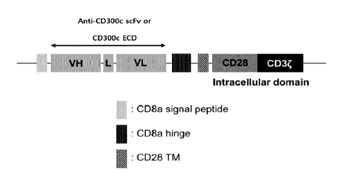
In a specific embodiment, the signal peptide may be or include a CD8a signal
peptide.
In a specific embodiment, the GS linker may be a 5 to 15-peptide consisting
of glycine and serine. Specifically, the GS linker may include or consist of
the
amino acid sequence represented by SEQ ID NO: 422.
In a specific embodiment, the transmembrane domain may be or include a
CD8 hinge (hinge of cluster of differentiation 8) and/or a CD28 transmembrane
domain. The CD8 hinge may include or consist of the amino acid sequence
represented by SEQ ID NO: 424. The CD28 transmembrane domain may include
or consist of the amino acid sequence represented by SEQ ID NO: 426.
In a specific embodiment, the intracellular signaling domain may be or
include a CD28 intracellular domain and/or a CD3 intracellular domain. The
CD28
intracellular domain may include or consist of the amino acid sequence
represented
by SEQ ID NO: 428. The CD3 intracellular domain may include or consist of the
amino acid sequence represented by SEQ ID NO: 430.
Polynucleotide, Vector, and Immune Cell
According to another aspect of the present disclosure, there are provided a
polynucleotide including a nucleic acid sequence encoding the chimeric antigen
receptor, a vector (for example, expression vector) including the
polynucleotide, and
an immune cell expressing the chimeric antigen receptor.
The polynucleotide of the present disclosure may include any nucleic acid
sequence encoding an amino acid sequence that constitutes or is included in
the
chimeric antigen receptor, and may also include a nucleic acid sequence having
80% or more, preferably 90% or more, more preferably 95% or more, and most
preferably 98% or more identity thereto.
The "vector" refers to a nucleic acid molecule capable of transporting another
nucleic acid linked thereto. One type of vector is a "plasmid," which refers
to a
circular double stranded DNA loop into which an additional DNA segment can be
inserted. Another type of vector is a viral vector wherein virally-derived DNA
or
24
CA 03220226 2023- 11- 23
RNA sequences are present in the vector for packaging into a virus. Specific
vectors are capable of autonomous replication in a host cell into which they
are
introduced (for example, bacterial vectors having a bacterial origin of
replication and
episomal mammalian vectors).
Other vectors (for example, non-episomal
mammalian vectors) are integrated into the genome of a host cell upon the
introduction into the host cell, and thereby are replicated along with the
host genome.
Moreover, specific vectors are capable of inducing the expression of genes to
which
they are operatively-linked. Such vectors are referred to herein as
"expression
vectors". Common expression vectors of utility in recombinant DNA techniques
are
often in the form of a plasmid. As used herein, the terms "plasmid" and
"vector"
may be used interchangeably as the plasmid is the most commonly used form of
vector. However, the present disclosure includes such other forms of
expression
vectors, such as viral vectors (for example, lentiviruses, replication
defective
retroviruses, adenoviruses, and adeno-associated viruses), which serve
equivalent
functions.
The immune cells expressing the chimeric antigen receptor of the present
disclosure may be produced by transforming immune cells with the vector. For
example, the immune cells may be produced by introducing into immune cells a
lentiviral vector including a nucleic acid sequence that encodes a desired
chimeric
antigen receptor.
In an embodiment, the immune cell(s) may be any one or more selected from
the group consisting of monocyte(s), macrophage(s), T cell(s), natural killer
cell(s)
(NK cell(s)), and dendritic cell(s). In addition, any immune cells may be
included
therein as long as they can be used for the prevention or treatment of cancer.
Preferably, the immune cells of the present disclosure may be T cells.
For
purposes of the present disclosure, the T cell may be any T cell, such as a
cultured T
cell, for example, a primary T cell, or a T cell from a cultured T cell line,
for example,
J urkat, SupT1 or the like, or a T cell obtained from a mammal. The T cell,
when
obtained from a mammal, can be obtained from a number of sources including,
but
CA 03220226 2023- 11- 23
not limited to, bone marrow, blood, lymph nodes, thymus, or other tissues or
body
fluids. The T cell may also be enriched or purified. The T cell may be a human
T
cell. The T cell may be a T cell isolated from a human. The T cell may be any
type of T cell and may be of any developmental stage, including but not
limited to,
CD4+/CD8+ double positive T cells, CD8+ T cells (for example, cytotoxic T
cells),
CD4+ helper T cells, for example, Thl and Th2 cells, peripheral blood
mononuclear
cells (PBMCs), peripheral blood leukocytes (PBLs), tumor infiltrating
lymphocytes
(TILs), memory T cells, naive T cells, and the like. The T cell may be a CD8+
T cell
or a CD4+ T cell.
Method for Prevention or Treatment of Cancer
According to still another aspect of the present disclosure, there is provided
a
method for preventing or treating cancer, alleviating or decreasing the
severity of at
least one symptom or sign of cancer, inhibiting metastasis, or inhibiting
growth of
cancer, in a subject, by using the immune cells of the present disclosure. As
used
herein, "preventing or treating cancer" may include inhibiting proliferation,
survival,
metastasis, recurrence, or anticancer agent resistance of cancer. Such a
method
may include administering the immune cells of the present disclosure to a
subject in
need of prevention or treatment of cancer. Accordingly, there is provided use
of a
composition including the immune cell(s) as an active ingredient, for
preventing or
treating cancer.
As used herein, the term "cancer" refers to a physiological condition that is
typically characterized by unregulated cell growth in mammals. The cancer to
be
prevented or treated in the present disclosure may include, depending on the
site of
occurrence, colorectal cancer, small intestine cancer, rectal cancer, colon
cancer,
thyroid cancer, endocrine adenocarcinoma, oral cancer, tongue cancer,
pharyngeal
cancer, laryngeal cancer, esophageal cancer, cervical cancer, uterine cancer,
fallopian tube cancer, ovarian cancer, brain cancer, head and neck cancer,
lung
cancer, lymph gland cancer, gallbladder cancer, bladder cancer, kidney cancer,
liver
cancer, pancreatic cancer, prostate cancer, skin cancer (or melanoma), breast
26
CA 03220226 2023- 11- 23
cancer, stomach cancer, bone cancer, blood cancer, and the like. However, any
cancer can be included therein as long as it expresses a CD300c protein on the
surface of cancer cells. In an embodiment, the cancer may include at least any
one
selected from the group consisting of colorectal cancer, rectal cancer, colon
cancer,
thyroid cancer, oral cancer, pharyngeal cancer, laryngeal cancer, cervical
cancer,
brain cancer, lung cancer, ovarian cancer, bladder cancer, kidney cancer,
liver
cancer, pancreatic cancer, prostate cancer, skin cancer, tongue cancer, breast
cancer, uterine cancer, stomach cancer, bone cancer, and blood cancer. In
another
embodiment, the cancer may be a solid cancer.
In an embodiment, the method may further include administering one or more
anticancer agents (for example, immunotherapeutic agents). In a case where (i)
the
immune cells of the present disclosure are used in combination with (ii) one
or more
immunotherapeutic agents, (i) and (ii) may be administered simultaneously or
sequentially.
The wording "administered sequentially" means that one ingredient is first
administered and another ingredient is administered immediately or at a
predetermined interval after the first administration, wherein the ingredients
may be
administered in any order. That is, one or more immunotherapeutic agents may
be
administered immediately or at a predetermined interval after the immune cells
are
administered, or vice versa. In addition, any of the one or more
immunotherapeutic
agents may be first administered first, followed by the immune cells, and then
another of the one or more immunotherapeutic agents.
Immunotherapeutic agents have a novel mechanism by which immune cells
in the body are activated to kill cancer cells, and thus are advantageous in
that they
can be widely used for most cancers without specific genetic mutations. In
addition,
the immunotherapeutic agents have fewer adverse effects in that they treat
cancer
by strengthening the patient's own immune system, and have effects of
improving
the patient's quality of life and significantly extending the survival. These
immunotherapeutic agents include immune checkpoint inhibitors, and may be
27
CA 03220226 2023- 11- 23
manufactured by known methods or commercially available products. Examples of
the immunotherapeutic agents include, but are not limited to, anti-PD-1, anti-
PD-L1,
anti-CTLA-4, anti-CD47, anti-KIR, anti-LAG3, anti-CD137, anti-0X40, anti-
CD276,
anti-CD27, anti-GITR, anti-TI M3, anti-41BB, anti-CD226, anti-CD40, anti-CD70,
anti-ICOS, anti-CD4OL, anti-BTLA, anti-TCR, and anti-TIGIT antibodies. In
addition,
examples of the immunotherapeutic agents include, but are not limited to,
durvalumab (Imfinzie), atezolizumab (Tecentriq), avelumab (Bavencio0),
pembrolizumab (Keytruda ), nivolumab (Opdivo ), aCD47, cemiplimab (Libtayo ),
magrolimab (Hu5F9-G4), and ipilimumab (Yervoye).
In an embodiment, the immunotherapeutic agent may include at least any
one selected from the group consisting of anti-PD-1, anti-PD-L1, anti-CTLA-4,
anti-CD47, anti-KIR, anti-LAG3, anti-CD137, anti-0X40, anti-CD276, anti-CD27,
anti-GITR, anti-TI M3, anti-41BB, anti-CD226, anti-CD40, anti-CD70, anti-ICOS,
anti-CD4OL, anti-BTLA, anti-TCR, and anti-TIGIT antibodies. In one example,
the
immunotherapeutic agent may include at least any one selected from the group
consisting of anti-PD-1, anti-PD-L1, anti-CTLA-4, and anti-CD47 antibodies.
In another embodiment, the immunotherapeutic agent may include at least
any one selected from the group consisting of durvalumab (Imfinzi),
atezolizumab
(Tecentriq), pembrolizumab (Keytruda), nivolumab (Opdivo), aCD47, and
ipilimumab
(Yervoy).
The immune cells according to the present disclosure and optionally one or
more additional anticancer agents in each case may be administered in several
ways
depending on whether local or systemic treatment is desired and the area to be
treated.
Methods of administering these ingredients to a subject may vary
depending on the purpose of administration, the site of the disease, the
subject's
condition, and the like. The route of administration may be oral, parenteral,
inhalation, local or topical (for example, intralesional administration).
Examples of
parenteral administration may include, but are not limited to, intravenous,
subcutaneous, intraperitoneal, intrapulmonary, intraarterial, intramuscular,
rectal,
28
CA 03220226 2023- 11- 23
vaginal, intraarticular, intraprostatic, intranasal, intraocular,
intravesical, intrathecal,
or intraventricular administration (for example, intracerebroventricular
administration).
In addition, when used in combination, the immune cells and the additional
anticancer agent may be administered by the same route or may be administered
by
different routes.
In the method, the number of the immune cells according to the present
disclosure may vary depending on the age, sex, and body weight of an
individual
(patient). The immune cells may be included at about 1 to about 10 times the
number of tumor cells in the individual. In addition, the effective amount of
at least
one additional anticancer agent may vary depending on the age, sex, and body
weight of an individual (patient). In general, administration may be performed
in an
amount of about 0.01 mg to 100 mg, or 5 mg to about 50 mg, per kg of body. The
amount may be administrated once a day or several times a day in divided
doses.
However, the effective amount may be increased or decreased depending on route
and period of administration, severity of disease, sex, body weight, age, and
the like.
Thus, the range of the present disclosure is not limited thereto.
Pharmaceutical Composition
According to still another aspect of the present disclosure, there is provided
a
pharmaceutical composition for preventing or treating cancer, including the
immune
cells according to the present disclosure as an active ingredient. In
addition, there
is provided use of the immune cell(s) according to the present disclosure for
the
manufacture of a medicament for preventing or treating cancer.
The immune cells may be included in the composition in a prophylactically or
therapeutically effective amount. The pharmaceutical composition may be
administered to a subject to inhibit proliferation, survival, metastasis,
recurrence, or
anticancer agent resistance of cancer.
In an embodiment, the pharmaceutical composition may further include at
least one additional anticancer agent (for example, immunotherapeutic agent).
Specifically, the immune cells and optionally the additional immunotherapeutic
agent
29
CA 03220226 2023- 11- 23
may be included in the same composition or may be included in separate
compositions. When included in separate compositions, the immune cells and the
additional immunotherapeutic agent may be formulated separately, and may be
administered simultaneously or sequentially.
To prepare the pharmaceutical composition of the present disclosure, the
immune cells and optionally the additional immunotherapeutic agent may be
mixed
with a pharmaceutically acceptable carrier and/or excipient. The
pharmaceutical
composition may be prepared in the form of a lyophilized preparation or an
aqueous
solution.
For example, see Remington's Pharmaceutical Sciences and U.S.
Pharmacopeia: National Formulary, Mack Publishing Company, Easton, PA (1984).
Acceptable carriers and/or excipients (including stabilizers) are nontoxic to
recipients at the dosages and concentrations employed, and include, but are
not
limited to, buffers (for example,
phosphate, citrate, or other organic acids);
antioxidants (for example, ascorbic acid or methionine); preservatives (for
example,
octadecyldimethylbenzyl ammonium chloride; hexamethonium
chloride;
benzalkonium chloride or benzethonium chloride; phenol, butyl, or benzyl
alcohol;
alkyl parabens, such as methyl or propyl paraben; catechol; resorcinol;
cyclohexanol;
3-pentanol; and m-cresol); low-molecular weight (less than about 10 residues)
polypeptides; proteins (for example, serum albumin, gelatin, or
immunoglobulin);
hydrophilic polymers (for example, polyvinylpyrrolidone); amino acids (for
example,
glycine, glutamine, asparagine, histidine, arginine, or lysine);
monosaccharides,
disaccharides, and other carbohydrates, such as glucose, mannose, or dextrins;
chelating agents (for example, EDTA); sugars (for example, sucrose, mannitol,
trehalose or sorbitol); salt-forming counter-ions (for example, sodium); metal
complexes (for example, Zn-protein complexes); and (or) non-ionic surfactants
(for
example, TWEENTm, PLURONICSTM or polyethylene glycol (PEG)).
The pharmaceutical composition of the present disclosure may be formulated
in a suitable form known in the art depending on the route of administration.
As used herein, the term "prophylactically or therapeutically effective
amount"
CA 03220226 2023- 11- 23
or "effective amount" refers to an amount of an active ingredient in a
composition,
which is effective for preventing or treating cancer in a subject. Also, this
amount is
sufficient for preventing or treating cancer at a reasonable benefit/risk
ratio
applicable to medical treatment and does not cause adverse effects. The level
of
the effective amount may be determined depending on the patient's health
status,
type of disease, severity of disease, activity of the drug, sensitivity to the
drug,
method of administration, frequency of administration, route of administration
and
rate of excretion, duration of treatment, drugs used in combination or
coincidentally
therewith, and other factors well known in the medical field. It is important
to
administer a minimum amount that allows the maximum effect to be achieved with
minimal or no adverse effects in consideration of all of the above factors,
which can
be easily determined by those skilled in the art.
For the effective amount of each active ingredient in the pharmaceutical
composition of the present disclosure, refer to the description in the section
on the
method for preventing or treating cancer.
In another embodiment, the pharmaceutical composition can inhibit
proliferation, survival, metastasis, recurrence, or anticancer agent
resistance of
cancer.
[Mode for Carrying Out the Invention]
Hereinafter, the present disclosure will be described in more detail by way of
examples. However, the following examples are only for illustrating the
present
disclosure, and the scope of the present disclosure is not limited thereto.
EXAMPLES
I. Production of Anti-CD300c Monoclonal Antibodies
Example 1. Production of Anti-CD300c Monoclonal Antibodies
Example 1.1. Construction of Anti-CD300c Monoclonal Antibody Library
31
CA 03220226 2023- 11- 23
In order to select anti-CD300c monoclonal antibodies, biopanning was
performed using a lambda phage library, a kappa phage library, a VH3VL1 phage
library, and an OPALTL phage library. Specifically, a CD300c antigen was added
at
a concentration of 5 pg/mL to an immunotube and reacted for 1 hour to allow
the
antigen to be adsorbed on the surface of the immunotube. Thereafter, 3% skim
milk was added to inhibit non-specific reactions, and then, 1012 PFU of each
antibody
phage library dispersed in 3% skim milk was added to each immunotube for
antigen
binding. After washing was performed three times using Tris buffered saline-
Tween
20 (TBST) solution to remove non-specifically bound phages, single-chain
variable
fragment (scFv) phage antibodies, which are specifically bound to the CD300c
antigen, were eluted using 100 mM triethylamine solution. The eluted phages
were
neutralized using 1.0 M Tris-HCI buffer (pH 7.8), and then infected at 37 C
for 1 hour
by treatment to E. coli ER2537. The infected E. coil was applied onto LB agar
medium containing carbenicillin, and then cultured at 37 C for 16 hours. Then,
the
formed E. coli colonies were suspended using 3 mL of super broth (SB)-
carbenicillin
culture medium. Some of the suspension was stored at ¨80 C until use with the
addition of 15% glycerol, and the remaining portion was re-inoculated into
SB-carbenicillin-2% glucose solution and cultured at 37 C. The obtained
culture
was centrifuged, and biopanning was repeated three times again using the
supernatant containing phage particles to obtain and concentrate antigen-
specific
antibodies.
After repeating the biopanning three times, E. coil containing the antibody
gene was applied onto LB agar medium containing carbenicillin and cultured at
37 C
for 16 hours. The formed E. coil colonies were inoculated again into
SB-carbenicillin-2% glucose solution and cultured at 37 C until the absorbance
(at
OD600nm) reached 0.5. Then, IPTG was added and further cultured at 30 C for 16
hours. Thereafter, periplasmic extraction was performed. From the results, a
library pool of antibodies, which specifically bind to the CD300c antigen, was
primarily obtained.
32
CA 03220226 2023- 11- 23
Example 1.2. Selection of Anti-CD300c Monoclonal Antibodies
In order to select anti-CD300c monoclonal antibodies specifically binding,
with high binding affinity, to the CD300c antigen, [LISA was performed using
the
library pool obtained in the same manner as in Example 1.1. More specifically,
CD300c and CD300a antigens in a coating buffer (0.1 M sodium carbonate, pH
9.0)
were separately dispensed into an ELISA plate at a concentration of 5 pg/mL
per
well and then incubated at room temperature for 3 hours to allow the antigen
to be
bound to the plate. Thereafter, the plate was washed three times with
phosphate
buffered saline-Tween 20 (PBST) to remove unbound antigen, and then 350 pL of
PBST supplemented with 2% bovine serum albumin (BSA) was added to each well,
followed by incubation at room temperature for 1 hour, and the plate was again
washed with PBST. Then, 25 pg of a periplasmic extract containing scFv
obtained
in the same manner as in Example 1.1 was added thereto and incubated at room
temperature for 1 hour for antigen binding. After 1 hour, washing was
performed
three times using PBST to remove unbound scFv, and then 4 pg/mL of an antibody
for detection was added, followed by again incubation at room temperature for
1
hour. Subsequently, the unbound antibody for detection was removed using PBST.
Then, anti-rabbit IgG to which HRP was bound was added, followed by incubation
at
room temperature for 1 hour, and the unbound antibody was removed again using
PBST. Subsequently, TMB solution was added, followed by incubation for 10
minutes for development. Then, 2 N sulfuric acid solution was added to
terminate
the development reaction, and the absorbance was measured at 450 nm to
identify
the antibodies specifically binding to the CD300c antigen.
Example 1.3. Identification of Anti-CD300c Monoclonal Antibody
Sequences
The nucleotide sequences of the anti-CD300c monoclonal antibodies, which
were selected using the same method as in Example 1.2, were identified. More
specifically, for each of the selected antibody clones, plasmid DNA was
extracted
therefrom using a plasmid miniprep kit, and then DNA sequencing was performed
to
33
CA 03220226 2023- 11- 23
sequence complementarity-determining regions (CDRs). As a result, 25 types of
anti-CD300c monoclonal antibodies having different amino acid sequences were
obtained. The heavy chain and light chain variable regions of these 25 types
of
anti-CD300c monoclonal antibodies are shown in Tables 2 and 3.
[TABLE 2]
Antibody Origin Heavy chain Light chain Heavy chain Light
chain
name (phage variable region variable region variable region
variable region
library) (nucleic acid) (nucleic acid) (amino
acid) (amino acid)
CK1 Kappa FIG. laa FIG. lab FIG. lac FIG. lad
(SEQ ID NO: (SEQ ID NO: (SEQ ID NO: (SEQ ID NO:
301) 302) 303) 304)
CK2 Kappa FIG. lba FIG. lbb FIG. lbc FIG. lbd
(SEQ ID NO: (SEQ ID NO: (SEQ ID NO: (SEQ ID NO:
305) 306) 307) 308)
CK3 Kappa FIG. lca FIG. lcb FIG. lcc FIG. lcd
(SEQ ID NO: (SEQ ID NO: (SEQ ID NO: (SEQ ID NO:
309) 310) 311) 312)
CL4 Lambda FIG. lda FIG. ldb FIG. ldc FIG. ldd
(SEQ ID NO: (SEQ ID NO: (SEQ ID NO: (SEQ ID NO:
313) 314) 315) 316)
CL5 Lambda FIG. lea FIG. leb FIG. lec FIG. led
(SEQ ID NO: (SEQ ID NO: (SEQ ID NO: (SEQ ID NO:
317) 318) 319) 320)
CL6 VH3VL1 FIG. lfa FIG. lfb FIG. lfc FIG. ltd
(SEQ ID NO: (SEQ ID NO: (SEQ ID NO: (SEQ ID NO:
321) 322) 323) 324)
CL7 VH3VL1 FIG. lga FIG. lgb FIG. lgc FIG. lgd
(SEQ ID NO: (SEQ ID NO: (SEQ ID NO: (SEQ ID NO:
325) 326) 327) 328)
CL8 VH3VL1 FIG. lha FIG. lhb FIG. lhc FIG. lhd
(SEQ ID NO: (SEQ ID NO: (SEQ ID NO:
(SEQ ID NO:
329) 330) 331) 332)
CL9 VH3VL1 FIG. ha FIG. lib FIG. lic FIG. lid
(SEQ ID NO: (SEQ ID NO: (SEQ ID NO: (SEQ ID NO:
333) 334) 335) 336)
CLIO VH3VL1 FIG. lja FIG. ljb FIG. ljc FIG. ljd
(SEQ ID NO: (SEQ ID NO: (SEQ ID NO: (SEQ ID NO:
337) 338) 339) 340)
SK11 Kappa FIG. lka FIG. lkb FIG. lkc FIG. lkd
(SEQ ID NO: (SEQ ID NO: (SEQ ID NO: (SEQ ID NO:
341) 342) 343) 344)
5K12 Kappa FIG. lla FIG. llb FIG. 11c FIG. lid
(SEQ ID NO: (SEQ ID NO: (SEQ ID NO: (SEQ ID NO:
345) 346) 347) 348)
5K13 Kappa FIG. lma FIG. lmb FIG. lmc FIG. lmd
34
CA 03220226 2023- 11- 23
(SEQ ID NO: (SEQ ID NO: (SEQ ID NO: (SEQ ID NO:
349) 350) 351) 352)
5K14 Kappa FIG. ma FIG. lnb FIG. inc FIG. lnd
(SEQ ID NO: (SEQ ID NO: (SEQ ID NO: (SEQ ID NO:
353) 354) 355) 356)
5K15 Kappa FIG. loa FIG. lob FIG. loc FIG. lod
(SEQ ID NO: (SEQ ID NO: (SEQ ID NO: (SEQ ID NO:
357) 358) 359) 360)
5K16 Kappa FIG. 1pa FIG. 1pb FIG. 1pc FIG. 1pd
(SEQ ID NO: (SEQ ID NO: (SEQ ID NO: (SEQ ID NO:
361) 362) 363) 364)
5K17 Kappa FIG. lqa FIG. lqb FIG. lqc FIG. lqd
(SEQ ID NO: (SEQ ID NO: (SEQ ID NO: (SEQ ID NO:
365) 366) 367) 368)
[TABLE 3]
Antibody name Origin Heavy chain Light chain Heavy chain
Light chain
(phage variable variable variable
variable
library) region region region region
(nucleic acid) (nucleic acid) (amino
acid) (amino acid)
5L18 Lambda FIG. lra FIG. lrb FIG. lrc
FIG. 1rd
(SEQ ID NO: (SEQ ID NO: (SEQ ID NO: (SEQ ID NO:
369) 370) 371) 372)
CB301 H3L1 A10 VH3VL1 FIG. lsa FIG. lsb FIG. lsc FIG.
lsd
(SEQ ID NO: (SEQ ID NO: (SEQ ID NO: (SEQ ID NO:
373) 374) 375) 376)
CB301 H3L1 Al2 VH3VL1 FIG. lta FIG. ltb FIG. ltc FIG.
ltd
(SEQ ID NO: (SEQ ID NO: (SEQ ID NO: (SEQ ID NO:
377) 378) 379) 380)
CB301 H3L1 E6 VH3VL1 FIG. lua FIG. lub FIG. luc FIG.
lud
(SEQ ID NO: (SEQ ID NO: (SEQ ID NO: (SEQ ID NO:
381) 382) 383) 384)
CB301 H3L1 F4 VH3VL1 FIG. lva FIG. lvb FIG. lvc FIG.
lvd
(SEQ ID NO: (SEQ ID NO: (SEQ ID NO: (SEQ ID NO:
385) 386) 387) 388)
CB301 H3L1 Gll VH3VL1 FIG. lwa FIG. lwb FIG. lwc FIG.
lwd
(SEQ ID NO: (SEQ ID NO: (SEQ ID NO: (SEQ ID NO:
389) 390) 391) 392)
CB301 OPALTL B5 OPALTL FIG. lxa FIG. lxb FIG. lxc FIG.
lxd
(SEQ ID NO: (SEQ ID NO: (SEQ ID NO: (SEQ ID NO:
393) 394) 395) 396)
CB301 OPALTL E6 OPALTL FIG. lya FIG. lyb FIG. lyc FIG.
lyd
(SEQ ID NO: (SEQ ID NO: (SEQ ID NO: (SEQ ID NO:
397) 398) 399) 400)
In each of the drawings stated in Tables 2 and 3, the CDR regions (CDR1,
CDR2, and CDR3) are underlined and appear sequentially (that is, CDR1 appears,
CA 03220226 2023- 11- 23
followed by CDR2, and then CDR3). In addition, the CDR regions included in
each
drawing are represented by the sequence numbers as shown in Table 4:
[TABLE 4]
Antibody Heavy chain Amino acid CDR1 CDR2
CDR3
/Light chain /DNA
FIG. laa CK1 Heavy chain DNA SEQ ID NO: SEQ ID NO:
SEQ ID NO:
1 2 3
FIG. lab Light chain DNA SEQ ID NO: SEQ
ID NO: SEQ ID NO:
4 5 6
FIG. lac Heavy chain Amino acid SEQ ID NO: SEQ ID NO:
SEQ ID NO:
7 8 9
FIG. lad Light chain Amino acid SEQ ID NO: SEQ ID NO:
SEQ ID NO:
11 12
FIG. lba CK2 Heavy chain DNA SEQ ID NO: SEQ ID NO:
SEQ ID NO:
13 14 15
FIG. lbb Light chain DNA SEQ ID NO: SEQ
ID NO: SEQ ID NO:
16 17 18
FIG. lbc Heavy chain Amino acid SEQ ID NO: SEQ ID NO:
SEQ ID NO:
19 20 21
FIG. lbd Light chain Amino acid SEQ ID NO: SEQ ID NO:
SEQ ID NO:
22 23 24
FIG. lca CK3 Heavy chain DNA SEQ ID NO: SEQ ID NO:
SEQ ID NO:
25 26 27
FIG. lcb Light chain DNA SEQ ID NO: SEQ
ID NO: SEQ ID NO:
28 29 30
FIG. lcc Heavy chain Amino acid SEQ ID NO: SEQ ID NO:
SEQ ID NO:
31 32 33
FIG. lcd Light chain Amino acid SEQ ID NO: SEQ ID NO:
SEQ ID NO:
34 35 36
FIG. lda CL4 Heavy chain DNA SEQ ID NO: SEQ ID NO:
SEQ ID NO:
37 38 39
FIG. ldb Light chain DNA SEQ ID NO: SEQ
ID NO: SEQ ID NO:
40 41 42
FIG. ldc Heavy chain Amino acid SEQ ID NO: SEQ ID NO:
SEQ ID NO:
43 44 45
FIG. ldd Light chain Amino acid SEQ ID NO: SEQ ID NO:
SEQ ID NO:
46 47 48
FIG. lea CL5 Heavy chain DNA SEQ ID NO: SEQ ID NO:
SEQ ID NO:
49 50 51
FIG. leb Light chain DNA SEQ ID NO: SEQ
ID NO: SEQ ID NO:
52 53 54
FIG. lec Heavy chain Amino acid SEQ ID NO: SEQ ID NO:
SEQ ID NO:
55 56 57
FIG. led Light chain Amino acid SEQ ID NO: SEQ ID NO:
SEQ ID NO:
58 59 60
FIG. lfa CL6 Heavy chain DNA SEQ ID NO: SEQ ID NO:
SEQ ID NO:
36
CA 03220226 2023- 11- 23
61 62 63
FIG. lfb Light chain DNA SEQ ID NO: SEQ
ID NO: SEQ ID NO:
64 65 66
FIG. lfc Heavy chain Amino acid SEQ ID NO: SEQ ID NO:
SEQ ID NO:
67 68 69
FIG. ltd Light chain Amino acid SEQ ID NO: SEQ ID NO:
SEQ ID NO:
70 71 72
FIG. lga CL7 Heavy chain DNA SEQ ID NO: SEQ ID NO:
SEQ ID NO:
73 74 75
FIG. lgb Light chain DNA SEQ ID NO: SEQ
ID NO: SEQ ID NO:
76 77 78
FIG. lgc Heavy chain Amino acid SEQ ID NO: SEQ ID NO:
SEQ ID NO:
79 80 81
FIG. lgd Light chain Amino acid SEQ ID NO: SEQ ID NO:
SEQ ID NO:
82 83 84
FIG. lha CL8 Heavy chain DNA SEQ ID NO: SEQ ID NO:
SEQ ID NO:
85 86 87
FIG. lhb Light chain DNA SEQ ID NO: SEQ
ID NO: SEQ ID NO:
88 89 90
FIG. lhc Heavy chain Amino acid SEQ ID NO: SEQ ID NO:
SEQ ID NO:
91 92 93
FIG. lhd Light chain Amino acid SEQ ID NO: SEQ ID NO:
SEQ ID NO:
94 95 96
FIG. ha CL9 Heavy chain DNA SEQ ID NO: SEQ ID NO:
SEQ ID NO:
97 98 99
FIG. lib Light chain DNA SEQ ID NO: SEQ
ID NO: SEQ ID NO:
100 101
102
FIG. lic Heavy chain Amino acid SEQ ID NO: SEQ ID NO:
SEQ ID NO:
103 104
105
FIG. lid Light chain Amino acid SEQ ID NO: SEQ ID
NO: SEQ ID NO:
106 107
108
FIG. lja CLIO Heavy chain DNA SEQ ID NO: SEQ ID NO:
SEQ ID NO:
109 110
111
FIG. ljb Light chain DNA SEQ ID NO: SEQ
ID NO: SEQ ID NO:
112 113
114
FIG. ljc Heavy chain Amino acid SEQ ID NO: SEQ ID NO:
SEQ ID NO:
115 116
117
FIG. ljd Light chain Amino acid SEQ ID NO: SEQ ID
NO: SEQ ID NO:
118 119
120
FIG. lka SK11 Heavy chain DNA SEQ ID NO: SEQ ID NO:
SEQ ID NO:
121 122
123
FIG. lkb Light chain DNA SEQ ID NO: SEQ
ID NO: SEQ ID NO:
124 125
126
FIG. lkc Heavy chain Amino acid SEQ ID NO: SEQ ID NO:
SEQ ID NO:
127 128
129
FIG. lkd Light chain Amino acid SEQ ID NO: SEQ ID NO:
SEQ ID NO:
130 131
132
FIG. ha 5K12 Heavy chain DNA SEQ ID NO: SEQ ID NO:
SEQ ID NO:
133 134
135
FIG. lib Light chain DNA SEQ ID NO: SEQ
ID NO: SEQ ID NO:
37
CA 03220226 2023- 11- 23
136 137
138
FIG. 11c Heavy chain Amino acid SEQ ID NO: SEQ ID NO:
SEQ ID NO:
139 140
141
FIG. lid Light chain
Amino acid SEQ ID NO: SEQ ID NO: SEQ ID NO:
142 143
144
FIG. lma 5K13 Heavy chain DNA SEQ ID NO: SEQ ID NO:
SEQ ID NO:
145 146
147
FIG. lmb Light chain DNA SEQ ID NO: SEQ ID NO:
SEQ ID NO:
148 149
150
FIG. lmc Heavy chain Amino acid SEQ ID NO: SEQ ID NO:
SEQ ID NO:
151 152
153
FIG. lmd Light chain Amino acid SEQ ID NO: SEQ ID NO:
SEQ ID NO:
154 155
156
FIG. ma 5K14 Heavy chain DNA SEQ ID NO: SEQ ID NO:
SEQ ID NO:
157 158
159
FIG. lnb Light chain DNA
SEQ ID NO: SEQ ID NO: SEQ ID NO:
160 161
162
FIG. inc Heavy chain Amino acid SEQ ID NO: SEQ ID NO:
SEQ ID NO:
163 164
165
FIG. lnd Light chain Amino acid SEQ ID NO: SEQ ID NO:
SEQ ID NO:
166 167
168
FIG. loa 5K15 Heavy chain DNA SEQ ID NO: SEQ ID NO:
SEQ ID NO:
169 170
171
FIG. lob Light chain DNA
SEQ ID NO: SEQ ID NO: SEQ ID NO:
172 173
174
FIG. loc Heavy chain Amino acid SEQ ID NO: SEQ ID NO:
SEQ ID NO:
175 176
177
FIG. lod Light chain Amino acid SEQ ID NO: SEQ ID NO:
SEQ ID NO:
178 179
180
FIG. 1pa 5K16 Heavy chain DNA SEQ ID NO: SEQ ID NO:
SEQ ID NO:
181 182
183
FIG. 1pb Light chain DNA
SEQ ID NO: SEQ ID NO: SEQ ID NO:
184 185
186
FIG. 1pc Heavy chain Amino acid SEQ ID NO: SEQ ID NO:
SEQ ID NO:
187 188
189
FIG. 1pd Light chain Amino acid SEQ ID NO: SEQ ID NO:
SEQ ID NO:
190 191
192
FIG. lqa 5K17 Heavy chain DNA SEQ ID NO: SEQ ID NO:
SEQ ID NO:
193 194
195
FIG. lqb Light chain DNA
SEQ ID NO: SEQ ID NO: SEQ ID NO:
196 197
198
FIG. lqc Heavy chain Amino acid SEQ ID NO: SEQ ID NO:
SEQ ID NO:
199 200
201
FIG. lqd Light chain Amino acid SEQ ID NO: SEQ ID NO:
SEQ ID NO:
202 203
204
FIG. lra 5L18 Heavy chain DNA SEQ ID NO: SEQ ID NO:
SEQ ID NO:
205 206
207
FIG. lrb Light chain DNA
SEQ ID NO: SEQ ID NO: SEQ ID NO:
208 209
210
FIG. lrc Heavy chain Amino acid SEQ ID NO: SEQ ID NO:
SEQ ID NO:
38
CA 03220226 2023- 11- 23
211 212 213
FIG. 1rd Light chain Amino acid SEQ ID NO: SEQ ID NO:
SEQ ID NO:
214 215 216
FIG. lsa CB301_H Heavy chain DNA SEQ ID NO: SEQ ID NO:
SEQ ID NO:
3L1 A10 217 218 219
FIG. lsb Light chain DNA SEQ ID NO: SEQ
ID NO: SEQ ID NO:
220 221 222
FIG. lsc Heavy chain Amino acid SEQ ID NO: SEQ ID NO:
SEQ ID NO:
223 224 225
FIG. lsd Light chain Amino acid SEQ ID NO: SEQ ID NO:
SEQ ID NO:
226 227 228
FIG. lta CB301_H Heavy chain DNA SEQ ID NO: SEQ ID NO:
SEQ ID NO:
3L1 Al2 229 230 231
FIG. ltb Light chain DNA SEQ ID NO: SEQ
ID NO: SEQ ID NO:
232 233 234
FIG. ltc Heavy chain Amino acid SEQ ID NO: SEQ ID NO:
SEQ ID NO:
235 236 237
FIG. ltd Light chain Amino acid SEQ ID NO: SEQ ID NO:
SEQ ID NO:
238 239 240
FIG. lua CB301_H Heavy chain DNA SEQ ID NO: SEQ ID NO:
SEQ ID NO:
3L1 E6 241 242 243
FIG. lub Light chain DNA SEQ ID NO: SEQ
ID NO: SEQ ID NO:
244 245 246
FIG. luc Heavy chain Amino acid SEQ ID NO: SEQ ID NO:
SEQ ID NO:
247 248 249
FIG. lud Light chain Amino acid SEQ ID NO: SEQ ID NO:
SEQ ID NO:
250 251 252
FIG. lva CB301_H Heavy chain DNA SEQ ID NO: SEQ ID NO:
SEQ ID NO:
3L1 F4 253 254 255
FIG. lvb Light chain DNA SEQ ID NO: SEQ
ID NO: SEQ ID NO:
256 257 258
FIG. lvc Heavy chain Amino acid SEQ ID NO: SEQ ID NO:
SEQ ID NO:
259 260 261
FIG. lvd Light chain Amino acid SEQ ID NO: SEQ ID NO:
SEQ ID NO:
262 263 264
FIG. lwa CB301_H Heavy chain DNA SEQ ID NO: SEQ ID NO:
SEQ ID NO:
3L1 Gil 265 266 267
FIG. lwb Light chain DNA SEQ ID NO: SEQ
ID NO: SEQ ID NO:
268 269 270
FIG. lwc Heavy chain Amino acid SEQ ID NO: SEQ ID NO:
SEQ ID NO:
271 272 273
FIG. lwd Light chain Amino acid SEQ ID NO: SEQ ID NO:
SEQ ID NO:
274 275 276
FIG. lxa CB301 0 Heavy chain DNA SEQ ID NO: SEQ ID NO:
SEQ ID NO:
PALTL B 277 278 279
FIG. lxb 5 Light chain DNA SEQ ID NO: SEQ
ID NO: SEQ ID NO:
280 281 282
FIG. lxc Heavy chain Amino acid SEQ ID NO: SEQ ID NO:
SEQ ID NO:
283 284 285
FIG. lxd Light chain Amino acid SEQ ID NO: SEQ ID NO:
SEQ ID NO:
39
CA 03220226 2023- 11- 23
286 287 288
FIG. lya CB301_0 Heavy chain DNA SEQ ID NO: SEQ ID NO:
SEQ ID NO:
PALTL_E 289 290 291
FIG. lyb 6 Light chain DNA SEQ ID NO: SEQ
ID NO: SEQ ID NO:
292 293 294
FIG. lyc Heavy chain Amino acid SEQ ID NO: SEQ ID NO:
SEQ ID NO:
295 296 297
FIG. lyd Light chain Amino acid SEQ ID NO: SEQ ID NO:
SEQ ID NO:
298 299 300
As described above, 25 types of anti-CD300c monoclonal antibodies were
identified that specifically bind, with high binding affinity, to the CD300c
antigen and
can be used for the prevention or treatment of cancer.
Example 1.4. Production and Purification of Anti-CD300c Monoclonal
Antibodies
By using the nucleotide sequences of the anti-CD300c monoclonal
antibodies identified in Example 1.3, expression vectors having, separately,
heavy
and light chains capable of expressing antibodies were prepared. More
specifically,
the expression vectors were prepared by inserting genes into pCIW3.3 vectors
using
the analyzed CDR sequences so that the vectors can express the heavy and light
chains, respectively. The prepared expression vectors for heavy and light
chains
were mixed with polyethylenimine (PEI) at a mass ratio of 1:1. The mixture was
transfected into 293T cells to induce antibody expression, and on day 8, the
culture
was centrifuged to remove the cells and obtain the resultant culture. The
obtained
culture was filtered, and then resuspended using a mixed solution of 0.1 M
NaH2PO4
and 0.1 M Na2HPO4 (pH 7.0). The resuspended solution was purified through
affinity chromatography using protein A beads (GE Healthcare), followed by
elution
using an elution buffer (Thermofisher).
In order to identify the produced antibodies, each of reducing sample buffer
and non-reducing sample buffer was added to 5 pg of purified antibody, and
electrophoresis was performed using pre-made SDS-PAGE (Invitrogen). Then, the
proteins were stained using Coomassie Blue. The results under non-reducing
conditions are show in FIG. 4, and the results under reducing conditions are
shown
CA 03220226 2023- 11- 23
in FIG. 5.
As shown in FIGS. 4 and 5, it was identified that the anti-CD300c monoclonal
antibodies having a high purity were produced and purified.
II. Expression of CD300c in Cancer Cells and Binding of Anti-CD300c
Monoclonal Antibodies to CD300c Antigen
Experimental Example 1. Identification of Expression of CD300c in
Cancer cell lines
In order to evaluate whether CD300c is expressed in various cancer cells,
various cancer cell lines, such as MKN45 (human gastric cancer cell line),
IM95
(human gastric cancer cell line), HT-29 (human colorectal cancer cell line),
A549
(human lung cancer cell line), HCT116 (human colorectal cancer cell line),
MDA-MB-231 (human breast cancer cell line), and HepG2 (human liver cancer cell
line) were cultured and evaluated for the expression of CD300c at the mRNA and
protein levels. In addition, THP-1 cells (human monocyte cell line), which are
immune cells, were also evaluated. Specifically, HEK293T (normal cell line)
was
used as a control.
Meanwhile, the expression of proteins was identified by Western blot and
flow cytometry (FACS) using fluorescently labeled cells. Specifically, each of
the
cultured cell lines was fixed with 4% formaldehyde, and then blocked using 5%
normal bovine serum albumin. Then, staining was performed with 0.5 pg of
eFluor660-labeled anti-CD300c antibody (Invitrogen). Subsequently, the
fluorescently labeled cells were identified using flow cytometry (FACS).
As a result, it was identified that the CD300c antigen was expressed at the
mRNA and protein levels in various cancer cells, such as colorectal cancer,
lung
cancer, and breast cancer. In addition, as shown in FIG. 6, according to the
results
of analysis using flow cytometry (FACS), it was identified that significantly
high
expression of CD300c was observed in the human lung cancer cell line (A549)
and
the human monocyte cell line (THP-1) as compared with the normal cell line
41
CA 03220226 2023- 11- 23
(HE K293T).
Experimental Example 2. Identification of Antigen-Binding Affinity of
Anti-CD300c Monoclonal Antibodies
In order to identify the antigen-binding ability of the anti-CD300c monoclonal
antibodies produced in Example 1, binding ELISA was performed. Specifically,
each of the CD300c antigen (11832-H08H, Sino Biological) or CD300a antigen
(12449-H08H, Sino Biological) in a coating buffer solution (0.1 M sodium
carbonate,
pH 9.0) was dispensed into an ELISA plate at a concentration of 8 pg/mL per
well
and then incubated at room temperature for 3 hours to allow the antigen to be
bound
to the plate. Thereafter, the plate was washed three times with PBST to remove
unbound antigen, and then 300 pL of PBST supplemented with 5% bovine serum
albumin (BSA) was added to each well, followed by incubation at room
temperature
for 1 hour, and the plate was again washed with PBST. Then, the anti-CD300c
monoclonal antibody was diluted four times and added, and incubated at room
temperature for 1 hour for antigen binding. After 1 hour, washing was
performed
three times using PBST to remove unbound anti-CD300c monoclonal antibody, and
then 4 pg/mL of an antibody for detection (HRP conjugated anti-Fc IgG) was
added,
followed by again incubation at room temperature for 1 hour. Subsequently, the
unbound antibody for detection was removed using PBST, and then TMB solution
was added, followed by incubation for 10 minutes for development. Then, 2 N
sulfuric acid solution was added to terminate the development reaction, and
the
absorbance was measured at 450 nm to identify the antibodies specifically
binding to
the CD300c antigen. The results are shown in Table 5 below and FIG. 7.
[TABLE 5]
42
CA 03220226 2023- 11- 23
B301 antibody EC50 gimL)
CK1 0.056
CK2 0.033
CK3 0.793
CL4 0.031
CL5 0.032
CL6 0.148
CL7 0.047
CL8 49.7
CL9 0.094
CLIO 0.039
SK11 0.052
SK12 0.067
=
SK13 0.044
SK14 0.065
SK15 14.74
SK16 2.42
SK17 0.054
SL18 0.17
As shown in Table 5, as a result of measuring the EC50 (effective
concentration of drug that causes 50% of the maximum response) values of the
anti-CD300c monoclonal antibodies, it was identified that the remaining all 14
clones
except for 4 clones (CK3, CL8, SK15, and SK16) exhibited high binding affinity
of 0.2
pg/mL or lower. Additionally, as shown in FIG. 7, it was identified that the
anti-CD300c monoclonal antibodies of the present disclosure bound to the
CD300c
antigen with high binding affinity even in the sigmoid curves for the results
of the
binding [LISA.
Experimental Example 3. Identification of Binding Specificity of
Anti-CD300c Monoclonal Antibody to CD300c Antigen
In order to identify the specificity of the anti-CD300c monoclonal antibody
43
CA 03220226 2023- 11- 23
CL7 for the CD300c antigen, it was further examined whether CL7 exhibited
cross-reactivity to the CD300a antigen, which was known to antagonize the
CD300c
antigen and had a similar protein sequence thereto. More specifically, the
CD300a
antigen (available from Sino Biological) was used at concentrations of 0.039,
0.63,
and 10 pg/mL, and then binding ELISA was performed in the same manner as in
Experimental Example 2. The results are shown in FIG. 36.
As shown in FIG. 36, it was found that the anti-CD300c monoclonal antibody
did not bind to antigens other than CD300c and showed high binding specificity
only
to the CD300c antigen.
Ill. Preparation of Cells Expressing Chimeric Antigen Receptor(s)
Specifically Binding to CD300c Antigen or Receptor Thereof and Identification
of Anticancer Effects Thereof
Example 2. Construction of Expression Vectors for Chimeric Antigen
Receptors Specifically Binding to CD300c Antigen or Receptor Thereof
In order to produce chimeric antigen receptors including binding domains
specifically binding to a CD300c antigen or a receptor thereof, the following
sequences were sequentially inserted into the pLVX-Puro vector (Addgene) to
construct an expression vector for a chimeric antigen receptor (pLVX-
Puro/aCD300c
scFv or pLVX-Puro/CD300c ECD-CAR) specifically binding to a CD300c antigen or
a
receptor thereof: a CD8a signal peptide (the DNA sequence being represented by
SEQ ID NO: 301) including the amino acid sequence represented by SEQ ID NO:
302 which allows a synthesized protein to pass through the cell membrane and
move to a correct position; an anti-CD300c single chain variable fragment of
each of
CK1, CL6, CL7, CL10, and 5L18 including the amino acid sequences represented
by
SEQ ID NOs: 412, 414, 416, 418, and 420, respectively, and specifically
binding to a
CD300c antigen or a receptor thereof (aCD300c scFvs, the DNA sequences being
represented by SEQ ID NO: 411, 413, 415, 417, and 419, respectively) or a
CD300c
extracellular domain (ECD) antigen including the amino acid sequence
represented
44
CA 03220226 2023- 11- 23
by SEQ ID NO: 402 (the DNA sequence being represented by SEQ ID NO: 401); a
GS linker including the amino acid sequence represented by SEQ ID NO: 422 (the
DNA sequence being represented by SEQ ID NO: 421); a CD8 hinge including the
amino acid sequence represented by SEQ ID NO: 424 (hinge of cluster of
differentiation 8, the DNA sequence being represented by SEQ ID NO: 423) and a
CD28 transmembrane domain including the amino acid sequence represented by
SEQ ID NO: 426 (the DNA sequence being represented by SEQ ID NO: 425) as
transmembranes domains; and a CD28 intracellular domain including the amino
acid
sequence represented by SEQ ID NO: 428 (the DNA sequence being represented
by SEQ ID NO: 427) and a CD3 intracellular domain including the amino acid
sequence represented by SEQ ID NO: 430 (the DNA sequence being represented
by SEQ ID NO: 429) as an intracytoplasmic domain for macrophage activation
signaling. The gene and protein sequence combinations for the
respective
constructed respective vectors are shown in Table 6. A schematic diagram of
the
gene arrangement is shown in FIG. 43, and an example of the constructed vector
map is shown in FIG. 44.
[TABLE 6]
CAR name DNA sequence combination Protein sequence
combination SEQ ID
NOs
CK1 CAR Combination of SEQ ID NOs: 409, Combination of SEQ ID
NOs: 410, 431 and
411, 421, 423, 425, 427, and 429 412, 422, 424, 426, 428,
and 430 432
CL6 CAR Combination of SEQ ID NOs: 409, Combination of SEQ ID
NOs: 410, 433 and
413, 421, 423, 425, 427, and 429 414, 422, 424, 426, 428,
and 430 434
CL7 CAR Combination of SEQ ID NOs: 409, Combination of SEQ ID
NOs: 410, 435 and
307, 421, 423, 425, 427, and 429 416, 422, 424, 426, 428,
and 430 436
CLIO CAR Combination of SEQ ID NOs: 409, Combination of SEQ ID NOs: 410, 437
and
309, 421, 423, 425, 427, and 429 418, 422, 424, 426, 428,
and 430 438
5L18 CAR Combination of SEQ ID NOs: 409, Combination of SEQ ID NOs: 410, 439
and
311, 421, 423, 425, 427, and 429 420, 422, 424, 426, 428,
and 430 440
CD300c Combination of SEQ ID NOs: 409, Combination of SEQ ID
NOs: 410, 441 and
ECD CAR 401, 421, 423, 425, 427, and 429
402, 422, 424, 426, 428, and 430 442
Example 3. Preparation of Recombinant Lentiviruses for Expressing
Chimeric Antigen Receptors Specifically Binding to CD300c Antigen or
CA 03220226 2023- 11- 23
Receptor Thereof
HEK293T cell line (ATCC) required for preparation of recombinant
lentiviruses for expressing the chimeric antigen receptors specifically
binding to a
CD300c antigen or a receptor thereof was prepared as follows. A complete
medium used for cell culture was prepared by adding heat-treated fetal bovine
serum
(FBS) to fresh DMEM (Gibco) to a concentration of 10%, adding lx
Penicillin-Streptomycin (Gibco) thereto, and then performing uniform mixing by
inverting up and down. The resultant complete medium was preheated to 37 C
before use. The HEK293T cell line was rapidly thawed for 2 to 3 minutes before
use by quick transfer of its cryopreserved cell stock to a constant-
temperature water
bath at 37 C, inoculated into 30 mL of the complete medium, and cultured in a
5%
CO2 incubator at 37 C. When the cell confluency reached 80% or higher, the
cells
were maintained by subculture. For lentiviral transfection, the HEK293T cell
line
was inoculated into 10 mL of the complete medium at a concentration of 1 to
2x10⁶
cells, cultured for 16 hours, and then subjected to lentiviral transfection.
LENTI-XTm Expression System (Takara) kit was used for lentiviral
transfection for expression of the chimeric antigen receptor specifically
binding to a
CD300c antigen or a receptor thereof. To 7 pg of the expression vector
pLVX-Puro/aCD300C scFv or pLVX-Puro/CD300c ECD-CAR constructed in the
same manner as in Example 2, sterile water (Invitrogen) was added to make 600
pL.
Then, the resultant product was placed in a tube of Lenti-X Packaging Single
Shots
in LENTI-XTm Expression System and mixing was performed to prepare a
nanoparticle complex solution. The nanoparticle complex solution obtained by
reaction at room temperature for 10 minutes was added dropwise to the
previously
prepared HEK293T cells, and then mixing was performed by shaking side to side.
The mixture was cultured for 4 hours in an incubator, followed by further
addition of 6
mL of the fresh complete medium, and cultured for 48 hours. After culture, the
supernatant was collected, centrifuged to remove cell debris, filtered through
a 0.45
pm filter, and stored at ¨80 C until use.
46
CA 03220226 2023- 11- 23
After 20 pL of the obtained lentivirus supernatant was added to the GoStix
cassette sample well(s) in the LENTI-XTm Expression System, three drops of
Chase
solution were added to the sample wells, followed by incubation at room
temperature
for 10 minutes. Subsequently, the presence or absence of a band confirmed
whether recombinant lentiviruses at an effective dose of 5x10⁵ IFU/mL or
higher
were obtained.
As a result, it was identified that recombinant lentiviruses expressing the
chimeric antigen receptors specifically binding to a CD300c antigen or a
receptor
thereof were prepared. It was identified that the prepared lentiviruses
expressed
chimeric antigen receptors, which include the amino acid sequences of single
chain
variable fragments of CK1, CL6, CL7, CL10, and 5L18, respectively.
Example 4. Production of J urkat Cells Expressing Chimeric Antigen
Receptors Specifically Binding to CD300c Antigen or Receptor Thereof
In order to produce J urkat cells expressing the chimeric antigen receptors
specifically binding to a CD300c antigen or a receptor thereof, the
recombinant
lentiviruses prepared in the same manner as in Example 2 were transfected into
a
J urkat cell line. More specifically, a recombinant lentivirus at 0.1 to 10
MOI was
inoculated in the complete medium supplemented with 8 pg/mL polybrene (Merk)
and then uniformly mixed by inverting up and down. In addition, 1 mL of the
mixture
was added to the J urkat cell line prepared in a 6-well plate for each well,
centrifuged
at 1,800 rpm for 45 to 90 minutes, and cultured in a 5% CO2 incubator at 37 C
for 24
hours. 24 hours later, subculture was performed at a concentration of 5x10⁵
cells/m L.
In order to identify whether the chimeric antigen receptor specifically
binding
to a CD300c antigen or a receptor thereof is normally expressed in the
transfected
J urkat cell line, total proteins of the cultured J urkat cells were obtained
using
PRO-PREP solution (iNtRON). The concentration of the obtained proteins was
measured using a microBCA protein assay kit (Thermo Fisher), and then Western
blotting was performed using an equal amount of protein. More specifically,
the
47
CA 03220226 2023- 11- 23
equal amount of protein was electrophoresed by SDS-PAGE (Invitrogen), and then
the electrophoresed protein was transferred to a nitrocellulose membrane
(Invitrogen). The protein-bound nitrocellulose membrane was blocked using a 5%
skim milk (BD) solution to block non-specific antibody reactions. An anti-CD3
antibody and an anti-GAPDH antibody (Cell Signaling Technologies, USA) as
primary antibodies were separately diluted to a concentration of 1:1,000 using
5%
skim milk, and used to treat the nitrocellulose membranes. After the
incubation,
unbound antibodies were removed. A horseradish peroxidase (HRP)-conjugated
secondary antibody (Cell Signaling Technologies) as a secondary antibody was
diluted to a concentration of 1:2,000 and used to treat the nitrocellulose
membrane.
The membrane was treated with [CL solution (Thermo Fisher) to induce color
development, and then the protein was quantified using the luminescent image
analyzer iBright1500 (Invitrogen). The Western blotting results are shown in
FIG.
45.
As shown in FIG. 45, the CD3 intracellular domain, to which the anti-CD3
antibody was bound, was observed, indicating that a J urkat cell line
expressing the
chimeric antigen receptor specifically binding to a CD300c antigen or a
receptor
thereof was produced.
Additionally, the expression level of the chimeric antigen receptor
specifically
binding to a CD300c antigen or a receptor thereof was examined using a flow
cytometer (FACS).
More specifically, the J urkat cell line was treated with a
PE-fusion anti-IgG antibody (J ackson ImmunoResearch), cultured at 4 C for 30
minutes, and then examined using a flow cytometer.
As a result of flow cytometry, it was identified that a J urkat cell line
expressing the chimeric antigen receptor specifically binding to a CD300c
antigen or
a receptor thereof was produced.
Experimental Example 4. Anticancer Effects of J urkat Cells Expressing
Chimeric Antigen Receptors Specifically Binding to CD300c Antigen or
Receptor Thereof
48
CA 03220226 2023- 11- 23
In order to identify anticancer effects of J urkat cells expressing chimeric
antigen receptors specifically binding to a CD300c antigen or a receptor
thereof,
cancer cell killing effects thereof were examined using A549 cell line.
More
specifically, the A549 cell line was inoculated into a 96-well plate at a
concentration
of 1x10⁵ cells/mL.
The J urkat cells expressing chimeric antigen receptors
specifically binding to a CD300c antigen or a receptor thereof, as produced in
the
same manner as in Example 4, were applied to the cancer cells to a
concentration of
20:1, and co-cultured. J urkat cells receiving no lentivirus transfection were
used as
a control. After co-culture for 24 hours, the co-cultured J urkat cell line
was removed
from each well, followed by incubation using CCK-8 (DOJ INDO) for 1 hour, and
then
the absorbance was measured at OD450 nm to examine the degree of cancer cell
death. The results are shown FIG. 46.
As shown in FIG. 46, it was identified that the J urkat cell lines expressing
chimeric antigen receptors specifically binding to a CD300c antigen or a
receptor
thereof exhibited anticancer effects against the A549 cell line, and these
anticancer
effects were higher than those of the J urkat cell line receiving no
lentivirus
transfection.
The above results could identify that the use of immune cell lines expressing
chimeric antigen receptors specifically binding to a CD300c antigen or a
receptor
thereof increased cancer therapeutic effects using the immune cell lines and
thus
can effectively treat cancer. It was also found that the immune cell lines
specifically
respond only to cancer cells expressing a CD300c antigen on the surface and
thus
can maximize therapeutic effects with decreased adverse effects.
IV. Anticancer Effects of Anti-CD300c Monoclonal Antibodies
Experimental Example 5. Identification of Anticancer Effects by
Administration of Anti-CD300c Monoclonal Antibodies
Experimental Example 5.1. Identification of T Cell Activation Effect
In order to identify whether the anti-CD300c monoclonal antibody produced
49
CA 03220226 2023- 11- 23
in Example 1 can exhibit an anticancer effect by activating T cells, the
production
level of interleukin-2 (IL-2) according to the treatment with the anti-CD300c
monoclonal antibody in human T cells was examined. IL-2 is an immune factor
that
helps growth, proliferation, and differentiation of T cells, and an increased
production
level of IL-2 means the activation of T cells due to an increase in
stimulation that
induces increased differentiation, proliferation, and growth of T cells.
Specifically,
each of an anti-CD3 monoclonal antibody and an anti-CD28 monoclonal antibody
was added to a 96-well plate at a concentration of 2 pg/well and fixed for 24
hours.
Then, the wells were co-treated with J urkat T cells (human T lymphocyte cell
line) at
1x10⁵ cells/well and the anti-CD300c monoclonal antibody at 10 pg/well.
Subsequently, the production level of IL-2 was measured using an ELISA kit (IL-
2
Qua ntikine kit, R&D Systems), and then compared with the control group not
treated
with the anti-CD300c monoclonal antibody. The results are shown FIG. 8.
As shown in FIG. 8, it was identified that the production level of IL-2
increased when the J urkat T cells activated by treatment with the anti-CD3
monoclonal antibody and the anti-CD28 monoclonal antibody were treated with
the
anti-CD300c monoclonal antibody.
The above results identified that the
anti-CD300c monoclonal antibody can activate T cells and induce the anticancer
immune action to inhibit the growth of cancer tissue.
Experimental Example 5.2. Identification of Promotion of Differentiation
into M1 Macrophages (I): Measurement of Production Level of Macrophage
Differentiation Marker (TNF-a)
In order to identify whether the anti-CD300c monoclonal antibodies selected
in Example 1 can promote the differentiation of monocytes into M1 macrophages,
THP-1 (human monocyte cell line) was dispensed into a 96-well plate at 1.5x10⁴
cells/well, and treated with 10 pg/mL of the anti-CD300c monoclonal antibodies
and/or 100 ng/mL of LPS. After the incubation for 48 hours, the production
level of
tumor necrosis factor-a (TNF-a), which is a differentiation marker of M1
macrophages, was measured using an ELISA kit (Human TNF-a Quantikine kit, R&D
CA 03220226 2023- 11- 23
Systems). The results are shown in FIGS. 9 and 10.
As shown in FIG. 9, it was identified that the anti-CD300c monoclonal
antibodies CL4, CL7, CL10, and 51_18 exhibited an increase in the production
level
of TNF-a by about 2 times or higher compared with the control group (Con)
treated
with LPS alone.
In addition, as shown in FIG. 10, it was identified that all the experimental
groups treated with the anti-CD300c monoclonal antibodies alone without LPS
treatment exhibited an increase in the production level of TNF-a compared with
the
control group (Con) treated with LPS alone.
Experimental Example 5.3. Identification of
Antibody
Concentration-Dependent Increase in Differentiation Ability into M1
Macrophages
In order to identify that the induction of differentiation into M1 macrophages
by the anti-CD300c monoclonal antibodies increases with concentrations of the
anti-CD300c monoclonal antibodies, the production level of TNF-a was examined
in
the same manner as in Experimental Example 5.2. The anti-CD300c monoclonal
antibodies were used at concentrations of 10, 1, and 0.1 pg/mL. The results
are
shown in FIG. 11.
As shown in FIG. 11, it was identified that the production level of TNF-a
increased as the treatment concentration of the anti-CD300c monoclonal
antibody
(CL7, CL10, or 5L18) increased.
In order to identify under more specific concentrations, the anti-CD300c
monoclonal antibody CL7 was used at concentrations of 10, 5, 2.5, 1.25, 0.625,
0.313, 0.157, and 0.079 pg/mL, and the production level of TNF-a was examined.
The results are shown in FIG. 12.
As shown in FIG. 12, it was identified that the production level of TNF-a
increased as the concentration of the anti-CD300c monoclonal antibody used for
treatment increased.
Experimental Example 5.4. Identification of Promotion of Differentiation
51
CA 03220226 2023- 11- 23
Ability into M1 Macrophages (II): Cell Morphology Observation
In order to identify the differentiation pattern into M1 macrophages through
cell morphology when monocytes were treated with an anti-CD300c monoclonal
antibody, THP-1 were treated with the anti-CD300c monoclonal antibody 10
pg/mL,
cultured for 48 hours, and then observed for cell morphology under a
microscope.
The results are shown in FIG. 13.
As shown in FIG. 13, it was identified that as for the experimental group
(CL7) treated with the anti-CD300c monoclonal antibody, the morphology of THP-
1
cells was changed from suspension cells to circular adherent cells, which are
in the
form of M1 macrophages. The above results identified that the differentiation
of
monocytes into M1 macrophages was promoted by treatment with the anti-CD300c
monoclonal antibody.
Experimental Example 5.5. Re-identification of Promotion of
Differentiation Ability into M1 Macrophages
In order to re-identify whether the anti-CD300c monoclonal antibody CL7
promoted the differentiation of human monocytes into M1 macrophages, the
secretion levels of TNF-a, interleukin-113 (IL-113), and interleukin-8 (IL-8)
were
measured using an [LISA kit. More specifically, THP-1 was dispensed into a
96-well plate at 1.5x10⁴ cells/well, and treated with 10 pg/mL of the anti-
CD300c
monoclonal antibody. After the incubation for 48 hours, the production levels
of
TNF-a, IL-113, and IL-8, which are markers for differentiation into M1
macrophages,
were measured using an ELISA kit (Human TNF-a Quantikine kit, R&D Systems).
The results are shown in FIG. 14.
As shown in FIG. 14, it was identified that all three types of markers for
differentiation into M1 macrophages increased in the experimental group (CL7)
treated with the anti-CD300c monoclonal antibody, compared with the control
group
(Con) not treated with the anti-CD300c monoclonal antibody.
Experimental Example 5.6. Identification of Repolariation Ability of M2
Macrophages into M1 Macrophages
52
CA 03220226 2023- 11- 23
In order to identify whether the anti-CD300c monoclonal antibody can
redifferentiate(repolarization) M2 macrophages into M1 macrophages, THP-1 was
dispensed into a 96-well plate at 1.5x10⁴ cells/well, pre-treated for 6 hours
by
treatment with 320 nM of PMA, and then treated with 20 ng/mL of interleukin-4
(IL-4)
and interleukin-13 (IL-13), and with 10 pg/mL of the anti-CD300c monoclonal
antibody, followed by incubation for 18 hours. The production levels of TNF-a,
IL-113, and IL-8 were examined using an ELISA kit. The results are shown in
FIGS.
21 to 23.
As shown in FIGS. 21 to 23, it was identified that the experimental groups not
pre-treated with PMA and co-treated with IL-4, IL-13, and the anti-CD300c
monoclonal antibody exhibited increased production levels of TNF-a, IL-18, and
IL-8,
and the experimental groups pre-treated with PMA and co-treated with IL-4, IL-
13,
and the anti-CD300c monoclonal antibody also similarly exhibited increased
production levels of TNF-a, IL-113, and IL-8.
The results identified that the
anti-CD300c monoclonal antibody could effectively
redifferentiate(repolarization) M2
macrophages into M1 macrophages.
Experimental Example 5.7. Identification of Differentiation Ability and
Repolarization Ability into M1 Macrophages
In order to identify the differentiation
ability and
redifferentiation(repolarization) ability of the anti-CD300c monoclonal
antibody into
M1 macrophages, THP-1 was dispensed into a 96-well plate at 1.5x10⁴
cells/well,
pre-treated with 10 pg/mL of the anti-CD300c monoclonal antibody for 48 hours,
and
treated with 100 ng/mL of PMA, 100 ng/mL of LPS, and 20 ng/mL of IL-4 and IL-
13,
followed by incubation for 24 hours. The production level of TNF-a was
examined
using an [LISA kit. The results are shown in FIG. 24.
As shown in FIG. 24, it was identified that all the experimental groups
pre-treated with the anti-CD300c monoclonal antibody exhibited a significant
increase in the production level of TNF-a, compared with the MO macrophage
control
group treated with PMA alone, the M1 macrophage control group treated with LPS
53
CA 03220226 2023- 11- 23
alone, and the M2 macrophage control group treated with IL-4 and IL-13 alone.
The results identified that the anti-CD300c monoclonal antibody had excellent
abilities to differentiate MO macrophages into M1 macrophages, differentiate
THP-1
into M1 macrophages, and redifferentiate(repolarization) M2 macrophages into
M1
macrophages.
Experimental Example 6. Identification of Inter-Species Cross-Reactivity
of Anti-CD300c Monoclonal Antibodies by Anticancer Effect Observation
Experimental Example 6.1 Identification of Human Cancer Cell Growth
Inhibitory Effect
In order to identify the effect of CD300c-targeting monoclonal antibodies on
the growth of cancer cells, cell proliferation assay was performed using A549
(human lung cancer cell line). More specifically, 2x10⁴ cells were dispensed
into a
96-well plate under the conditions of 0% fetal bovine serum (FBS), and 6x10³
cells
were dispensed under the conditions of 0.1% fetal bovine serum. Then, the
cells
were treated with 10 pg/mL of the anti-CD300c monoclonal antibodies, followed
by
incubation for 5 days. The cells were treated with CCK-8 (DOJ INDO), and the
absorbance was measured at OD45onm to examine the cancer cell growth
inhibitory
effect of the anti-CD300c monoclonal antibodies. The results are shown in
FIGS.
28 and 29.
As shown in FIG. 28, it was identified that all the antibodies except for SK11
and 5K17 had a cancer cell growth inhibitory effect under the conditions of 0%
FBS.
As illustrated in FIG. 29, it was identified that all the anti-CD300c
monoclonal
antibodies used in the experiment had a cancer cell growth inhibitory effect
under
0.1% FBS conditions.
Experimental Example 6.2. Identification of Cancer Cell Growth
Inhibitory Effect According to Concentration of Anti-CD300c Monoclonal
Antibody
In order to identify the cancer cell growth inhibitory effect according to the
concentration of the anti-CD300c monoclonal antibody, 2x10⁴ A549 cells were
54
CA 03220226 2023- 11- 23
dispensed into a 96-well plate under the conditions of 0% fetal bovine serum
(FBS),
and treated with the anti-CD300c monoclonal antibody at 10 pg/mL, followed by
incubation for 5 days. Subsequently, the cells were treated with CCK-8 (DOJ
INDO),
followed by incubation for 3 hours, and then the absorbance was measured at
OD45onm to examine the cancer cell growth inhibitory effect of the anti-CD300c
monoclonal antibody. The results are shown in FIG. 32.
As shown in FIG. 32, it was identified that the growth of cancer cells was
inhibited as the concentration of the anti-CD300c monoclonal antibody
increased.
Experimental Example 6.3. Identification of Increased differentiation
ability into M1 Macrophages in Mice
In order to identify whether the anti-CD300c monoclonal antibodies could
promote the differentiation from mouse macrophages to M1 macrophages, mouse
macrophages (Raw264.7) were dispensed into a 96-well plate at a concentration
of
1x10⁴ cells/well, and then treated with the anti-CD300c monoclonal antibodies
at 10
pg/mL, followed by incubation. The production level of TNF-a was examined
using
an [LISA kit. The results are shown in FIG. 37.
As shown in FIG. 37, it was identified that the production level of TNF-a
increased in the experimental groups treated with the anti-CD300c monoclonal
antibodies.
The above results indicated that the anti-CD300c monoclonal
antibodies acted equally in mice as well as humans, and thus had cross-
reactivity of
promoting the differentiation into M1 macrophages.
Experimental Example 6.4. Identification of Cancer Cell Growth
Inhibitory Effect in Mice
In order to identify whether the anti-CD300c monoclonal antibodies CL7,
CL10, and 5L18 exhibit an anticancer effect, CT26 (mouse colorectal cancer
cell
line) was dispensed into a 96-well plate at a concentration of 1x10⁴
cells/well and
treated with 10 ug/mL of the monoclonal antibodies, followed by incubation for
5
days. Then, cell proliferation assay was performed through CCK-8 detection.
As shown in FIG. 38, it was identified that the anti-CD300c monoclonal
CA 03220226 2023- 11- 23
antibodies exerted cancer cell proliferation inhibitory effects of 66% (CL7),
15%
(CL10), and 38% (SL18), respectively, which were higher than that of the
control
group, indicating that the anti-CD300c monoclonal antibodies exhibited a
cancer
therapeutic effect in mice.
Therefore, it can be seen that the anti-CD300c
monoclonal antibodies acted equally in humans as well as mice, and thus has
cross-reactivity showing an anticancer effect.
Experimental Example 7. Comparison of In Vitro Anticancer Effect
Between Anti-CD300c Monoclonal Antibodies and Conventional
lmmunotherapeutic Agent
Cancer immunotherapeutic agents used in the following experimental
examples below are as follows: Imfinzie (AstraZeneca) and Keytruda (Merck
Sharp
& Dohme).
Experimental Example 7.1. Comparison of Differentiation Ability into M1
Macrophages Between Anti-CD300c Monoclonal Antibodies and Conventional
lmmunotherapeutic Agent: Measurement of Production Levels of Three
Differentiation Markers (TNF-a, IL-10, and IL-8)
In order to compare the differentiation ability into M1 macrophages between
anti-CD300c monoclonal antibodies and a conventional immunotherapeutic agent,
the production level of TNF-a was examined using an ELISA kit in the same
manner
as in Experimental Example 5.2. As the conventional immunotherapeutic agent
Imfinzi was used at a concentration of 10 pg/mL. The results are shown in FIG.
15.
As shown in FIG. 15, it was identified that the anti-CD300c monoclonal
antibody significantly increased the production level of TNF-a compared with
the
control group treated with Imfinzi (Imf) alone. The above results identified
that the
anti-CD300c monoclonal antibodies significantly increased the differentiation
ability
into M1 macrophages compared with the conventionally known immunotherapeutic
agent.
For the comparison with other immunotherapeutic agents, Imfinzi, which is an
anti-PD-Li immunotherapeutic agent, Keytruda, which is an anti-PD-1
56
CA 03220226 2023- 11- 23
immunotherapeutic agent, and an isotype control (immunoglobulin G) antibody
were
used at a concentration of 10 pg/mL for each, and the production levels of TNF-
a,
IL-113, and IL-8 were examined using an ELISA kit. The results are shown in
FIGS.
16 to 18.
As shown in FIGS. 16 to 18, it was identified that the anti-CD300c
monoclonal antibody significantly increased the production levels of TNF-a, IL-
113,
and IL-8 compared with Imfinzi, Keytruda, and the IgG antibody. The above
results
identified that the anti-CD300c monoclonal antibodies could significantly
increase the
promotion of differentiation into M1 macrophages compared with the
conventional
immunotherapeutic agents.
Experimental Example 7.2. Comparison of Differentiation Ability from
MO Macrophages into M1 Macrophages Between Anti-CD300c Monoclonal
Antibody and Conventional Immunotherapeutic agent
In order to compare the differentiation ability of MO macrophages into M1
macrophages between the anti-CD300c monoclonal antibody and an
immunotherapeutic agent, THP-1 was dispensed into a 96-well plate at 1.5x10⁴
cells/well and treated with the anti-CD300c monoclonal antibody 10 pg/mL,
Imfinzi at
pg/mL, and/or 200 nM phorbol-12-myristate-13-acetate (PMA). After the
incubation for 48 hours, the production level of TNF-a was measured using an
ELISA
kit. The results are shown in FIG. 19.
As shown in FIG. 19, it was identified that TNF-a was not produced in the
comparison group treated with the immunotherapeutic agent Imfinzi alone, but
the
production level of TNF-a increased in the experimental group treated with the
anti-CD300c monoclonal antibody alone. In addition, it was identified that
even
when THP-1 cells were differentiated into MO macrophages by treatment with
PMA,
the experimental group treated with the anti-CD300c monoclonal antibody showed
a
significantly high production level of TNF-a compared with the experimental
group
treated with Imfinzi. The above results identified that the anti-CD300c
monoclonal
antibody promoted the differentiation of MO macrophages into M1 macrophages
57
CA 03220226 2023- 11- 23
compared with the conventional immunotherapeutic agent.
Experimental Example 7.3. Comparison of Differentiation Ability into M1
Macrophages Between Anti-CD300c Monoclonal Antibody and Conventional
Immunotherapeutic Agent
In order to compare the differentiation ability into M1 macrophages between
an anti-CD300c monoclonal antibody and a conventional immunotherapeutic agent,
the production level of TNF-a was examined in the same manner as in
Experimental
Example 5.2. The results are shown in FIG. 20.
As can be shown in FIG. 20, it was identified that when monocytes were
differentiated into M1 macrophages by treatment with LPS, the experimental
group
co-treated with Imfinzi and LPS did not exhibit a significant difference in
the
production level of TNF-a, but the experimental group co-treated with the
anti-CD300c monoclonal antibody and LPS exhibited a significant increase in
the
production level of TNF-a compared with the experimental group treated with
the
anti-CD300c monoclonal antibody alone.
Experimental Example 7.4. Comparison of Cancer Cell Growth
Inhibitory Effect Between Anti-CD300c Monoclonal Antibodies and
Conventional Immunotherapeutic Agent
In order to compare the cancer cell growth inhibitory effect between an
anti-CD300c monoclonal antibody and an immunotherapeutic agent, the cell
growth
inhibitory effect was examined using A549 (human lung cancer cell line) and
MDA-MB-231 (human breast cancer cell line). More specifically, 2x10⁴ cells
were
dispensed into a 96-well plate under the conditions of 0% fetal bovine serum
(FBS)
and 6x10³ cells were dispensed under the conditions of 0.1% fetal bovine
serum.
Then, the cells were treated with an anti-CD300c monoclonal antibody at 10
pg/mL,
followed by incubation for 5 days, and then observed under an optical
microscope.
The results are shown in FIGS. 30 and 31.
As shown in FIG. 30, it was identified that the anti-CD300c monoclonal
antibodies more effectively inhibited the proliferation of cancer cells than
the
58
CA 03220226 2023- 11- 23
immunotherapeutic agent Imfinzi in the A549 cell line.
As shown in FIG. 31, it was identified that the anti-CD300c monoclonal
antibodies more effectively inhibited the proliferation of cancer cells than
the
immunotherapeutic agent Imfinzi in the MDA-MB-231 cell line.
V. Combination of Anti-CD300c Monoclonal Antibody and
Immunotherapeutic Agent
Example 5. Co-Administration of Anti-CD300c Monoclonal Antibody
(CL7) and Immunotherapeutic Agent
The anti-CD300c monoclonal antibody (CL7) produced in Example 1 was
used in combination with other immunotherapeutic agents, for example, the
anti-PD-Li antibodies Imfinzie and Opdivoe, and the anti-PD-1 antibody
Keytruda 8,
an anti-CD47 antibody (aCD47), and an anti-CTLA-4 antibody.
The respective immunotherapeutic agents were accessible from the
following: IMFINZI (AstraZeneca); Opdivo and the anti-CTLA-4 antibody (Bristol
Myers Squibb Company); Keytruda (Merck Sharp & Dohme); and the anti-CD47
antibody (Abcam).
Experimental Example 8. Identification of (Synergistic) Increase in
Macrophage Activity by Combination
Experimental Example 8.1. Identification of Increase in Differentiation
Ability into M1 Macrophages
In order to identify that the differentiation ability of monocytes into M1
macrophages increased when the monocytes are treated with the anti-CD300c
monoclonal antibody CL7 in combination with an immunotherapeutic agent, such
as
an anti-PD-1 antibody, an anti-PD-Li antibody, an anti-CTLA-4 antibody, and an
anti-CD47 antibody, the signaling of mitogen-activated protein kinase (MAPK),
IkB,
and NF-kB, which are representative signals of M1 macrophage differentiation
was
examined.
Specifically, THP-1 was dispensed into a 6-well plate at 8.8x10⁵
cells/well, and treated with 10 pg/mL of the anti-CD300c monoclonal antibody,
10
59
CA 03220226 2023- 11- 23
pg/mL of Imfinzi, and/or 10 pg/mL of Keytruda. For a control group, an equal
amount of phosphate buffer solution (PBS) was used for treatment. After
incubation
for 48 hours, phosphorylated SAPK/J NK, phosphorylated ERK, and phosphorylated
p38 for MAPK signal, phosphorylated NF-kB for NF-kB signal, and phosphorylated
IkB for IkB signal were examined through Western blotting. The results are
shown
in FIGS. 25 to 27.
FIGS. 25, 26, and 27 illustrate the results of signaling of MAPK, NF-Kb, and
IkB, respectively. It was identified that the levels of phosphorylated MAPK,
IkB, and
NF-kB increased when the cells were treated with the anti-CD300c monoclonal
antibody in combination with an immunotherapeutic agent, such as an anti-PD-1
antibody, an anti-PD-Li antibody, an anti-CTLA-4 antibody, and an anti-CD47
antibody, compared with when the cells were treated with the anti-CD300c
monoclonal antibody alone. The above results identified that the cell
signaling
representing the differentiation into M1 macrophages increased when the cells
were
treated with the anti-CD300c monoclonal antibody in combination with an
immunotherapeutic agent, such as an anti-PD-1 antibody, an anti-PD-Li
antibody,
an anti-CTLA-4 antibody, and an anti-CD47 antibody, compared with when the
cells
were treated with the anti-CD300c monoclonal antibody alone.
Experimental Example 9. Identification of (Synergistic) Increase in
Cancer Cell Growth Inhibitory Effect by Combination (In Vitro)
Experimental Example 9.1. Identification of Apoptosis Signals
It was identified whether the apoptosis signal increased when the
anti-CD300c monoclonal antibody CL7 was used in combination with an
immunotherapeutic agent, such as an anti-PD-1 antibody, an anti-PD-Li
antibody,
an anti-CTLA-4 antibody, and an anti-CD47 antibody. Specifically, A549 cells
were
dispensed into a 6-well plate at 8x10⁵ cells/well and treated with 10 pg/mL of
the
anti-CD300c monoclonal antibody, and 10 pg/mL of Imfinzi, Keytruda, Opdivo,
and
an anti-CD47 antibody, alone or in combination. After incubation for 48 hours,
the
apoptosis signal or cell cycle signal was examined through Western blotting.
CA 03220226 2023- 11- 23
Cleaved caspase-9, caspase-3, caspase-2, and caspase-8 were checked as
markers for the apoptosis signal, and cyclin D1, CDK2, p27kip1, CDK6, cyclin
D3,
P21 Wafl, Cipl, and the like were checked as markers for the cell cycle
signal.
As shown in FIG. 33, the apoptosis signal increased when the cells were
co-treated with the anti-CD300c monoclonal antibody and the anti-PD-1 antibody
Imfinzi compared with when the cells were treated with the anti-CD300c
monoclonal
antibody alone, and the levels of cleaved-caspase9 and p21 increased and the
level
of cyclin D1 decreased when the cells were co-treated with the anti-CD300c
monoclonal antibody in combination with an immunotherapeutic agent, such as an
anti-PD-1 antibody, an anti-PD-L1 antibody, an anti-CTLA-4 antibody, or an
anti-CD47 antibody. The above results identified that the apoptosis of cancer
cells
was better induced when the cells were treated with the anti-CD300c monoclonal
antibody in combination with an immunotherapeutic agent, such as an anti-PD-1
antibody, an anti-PD-Li antibody, an anti-CTLA-4 antibody, or an anti-CD47
antibody, compared with when the cells were treated with the anti-CD300c
monoclonal antibody alone.
Experimental Example 9.2. Identification of Growth Inhibitory Effect on
Cancer Cell Lines
In order to identify the cancer cell growth inhibitory effect by co-
administration
of the anti-CD300c monoclonal antibody CL7 and an immunotherapeutic agent, the
comparison of the cancer cell growth inhibitory effect was performed using
A549
(human lung cancer cell line) and MDA-MB-231 (human breast cancer cell line).
Specifically, 2x10⁴ cells (A549) or 3x10⁴ cells (MDA-MB-231) were dispensed
into a
96-well plate in the absence of fetal bovine serum (FBS) conditions, and at
6x10³
cells (A549) or 1x10⁴ cells (MDA-MB-231) were dispensed under the conditions
of
0.1% fetal bovine serum. Subsequently, the cells were treated with 10 pg/mL of
the
anti-CD300c monoclonal antibody and Imfinzi alone or in combination and
incubated
for 5 days. For a control group, an equal amount of phosphate buffer solution
(PBS) was used for treatment. Then, the cells were treated with CCK-8 (DOJ
INDO)
61
CA 03220226 2023- 11- 23
and the absorbance was measured at OD45onm. The results are shown in FIG. 34
(A549) and FIG. 35 (MDA-MB-231).
As shown in FIG. 34, it was identified that as for the A549 cell line in the
absence of FBS, compared with the control group, the cell growth inhibitory
effect
was 17% higher in the treatment with the anti-CD300c monoclonal antibody alone
and 34% higher in the treatment with the anti-CD300c monoclonal antibody and
Imfinzi in combination.
As shown in FIG. 35, as for the MDA-MB-231 cell line under the conditions of
0% FBS, compared with the control group, the cancer cell growth inhibitory
effect
was observed to be 19% higher in the treatment with the anti-CD300c monoclonal
antibody alone, 45% higher in the treatment with the anti-CD300c monoclonal
antibody and the anti-CD47 antibody in combination, and 51% higher in the
treatment with the anti-CD300c monoclonal antibody, the anti-CD47 antibody,
and
Imfinzi in combination.
Under 0.1% FBS conditions, the cancer cell growth
inhibitory effect was observed to be 19% higher in the treatment with the
anti-CD300c monoclonal antibody, 22% higher in the treatment with the anti-
CD300c
monoclonal antibody and the anti-CD47 antibody in combination, and 32% higher
in
the treatment with the anti-CD300c monoclonal antibody, the anti-CD47
antibody,
and Imfinzi in combination.
The above results identified that the cancer cell growth was further inhibited
in the treatment with the anti-CD300c monoclonal antibody in combination with
an
immunotherapeutic agent, such as an anti-PD-1 antibody, an anti-PD-Li
antibody,
an anti-CTLA-4 antibody, or an anti-CD47 antibody, compared with the treatment
with the anti-CD300c monoclonal antibody alone.
Experimental Example 10. Identification of In Vivo (Synergistic)
Increase by Combination
Experimental Example 10.1. Identification of In Vivo Cancer Cell Growth
Inhibitory Effect
In order to identify the anticancer effect in vivo of the anti-CD300c
62
CA 03220226 2023- 11- 23
monoclonal antibody CL7, allograft mouse tumor models were constructed by
implanting a colorectal cancer cell line (CT26) at 2x10⁵ cells into 8-week-old
BALB/c
mice through subcutaneous injection. Animal breeding and experiments were all
carried out in a specific pathogen free (SPF) facility. On day 12 (D12) after
implantation of the colorectal cancer cell line, the mice with a tumor size of
50 to 100
mm3 were administered with the anti-CD300c monoclonal antibody and an anti-PD-
1
antibody purchased from BioXcell alone or in combination, and administered
with an
equal amount of phosphate buffered saline (PBS) for a control group. A
schematic
experimental method is shown in FIG. 39. Specifically, the mice were
intraperitoneally injected with the respective antibodies (CL7: 10 mg/kg; and
anti-PD-1 antibody: 10 mg/kg) alone or in combination, twice a week for two
weeks
(a total of 4 times on D12, D15, D19, and D22). The tumor volume was measured
for 25 days. The results are shown in FIG. 40.
As can be seen from FIG. 40, it was identified that the cancer growth was
inhibited in the experimental group administered with the anti-CD300c
monoclonal
antibody alone compared with the control group, but the cancer growth was more
effectively inhibited in the treatment with the anti-CD300c monoclonal
antibody and
an immunotherapeutic agent such as the anti-PD-1 antibody in combination
compared with the treatment with the anti-CD300c monoclonal antibody alone.
Experimental Example 10.2. Identification of M1 Macrophage Increase
Effect In Vivo
In order to identify whether an anti-CD300c monoclonal antibody increases
M1 macrophages in the cancer tissue of mouse models, the mice experimented in
the same manner as in Experimental Example 10.1 were euthanized on day 25, and
1% para-formaldehyde (PFA) was intravascularly injected and perfused into the
mice,
and then cancer tissue was obtained. The obtained cancer tissue was fixed
using
1% PFA, and sequentially dehydrated using 10%, 20%, and 30% sucrose solution.
The dehydrated cancer tissue was frozen in the optimal cutting temperature
(OCT)
compound, and then the cancer tissue was sectioned to a thickness of 50 pm by
63
CA 03220226 2023- 11- 23
using a cryotome. The tissue was incubated for 1 hour at 37 C in a mixed
solution
of 20 mg/ml collagenase D and 2 mg/ml DNase I, and then filtered through a 70
pm
cell strainer. The red blood cells were lyzed, and the cells were again
filtered
through a nylon mesh to become single cells. To inhibit non-specific reactions
in
the single cell suspension, the cells were incubated with a CD16/32 antibody
(Invitrogen) for 1 hour, and the cell viability was examined. The resultant
product
was stained with antibodies to the M1 macrophage marker iNOS and the M2
macrophage marker CD206 and analyzed by FACS.
As a result, as shown in FIG. 42, it was identified that M1 macrophages
partially increased in the experimental group treated with the anti-PD-1
antibody
compared with the control group, but in the experimental group treated with
the
anti-CD300c monoclonal antibody, M1 macrophages significantly increased and M2
macrophages were hardly observed.
In addition, it was identified that M1
macrophages further increased in the experimental group co-administered with
the
anti-CD300c monoclonal antibody and the anti-PD-1 antibody. The above results
identified that the differentiation into M1 macrophages can be effectively
promoted in
the treatment with the anti-CD300c monoclonal antibody in combination with an
immunotherapeutic agent, such as an anti-PD-1 antibody, an anti-PD-Li
antibody,
an anti-CTLA-4 antibody, and an anti-CD47 antibody, compared with the
treatment
with the anti-CD300c monoclonal antibody alone.
Experimental Example 10.3. Identification of CD8+ T Cell Immunity
Stimulating Effect In Vivo
In order to identify whether the anti-CD300c monoclonal antibody CL7
stimulated CD8+ T cell immunity in mouse tumor models, the mice experimented
in
the same manner as in Experimental Example 10.1 were euthanized on day 25, and
then 1% para-formaldehyde (PFA) was intravascularly injected and perfused into
the
mice, and then cancer tissue was obtained. The obtained cancer tissue was
fixed
using 1% PFA, and sequentially dehydrated using 10%, 20%, and 30% sucrose
solution.
The dehydrated cancer tissue was frozen in the optimal cutting
64
CA 03220226 2023- 11- 23
temperature (OCT) compound, and then the cancer tissue was sectioned to a
thickness of 50 pm by using a cryotome. Then, staining was performed using
CD8+
and iNOS.
As shown in FIG. 41, it was identified that the CD8+ T cells partially
increased in the experimental group treated with the anti-PD-1 antibody
compared
with the control group, but the CD8+ T cells significantly increased in the
experimental group treated with the anti-CD300c monoclonal antibody. It was
also
identified that the CD8+ T cells further increased in the experimental group
administered with the anti-CD300c monoclonal antibody and the anti-PD-1
antibody
in combination compared with the anti-PD-1 alone treatment group. The above
results identified that the anti-CD300c monoclonal antibody increased the
number of
CD8+ T cells more effectively when the anti-CD300c monoclonal antibody was
used
in combination with the conventional immunotherapeutic agent.
Through the results as above, it was identified that the anti-CD300c
monoclonal antibodies of the present disclosure can bind with high specificity
to the
CD300c antigen and can be used for various individuals due to their inter-
species
cross-reactivity, for example, with mice. It was also identified both in vitro
and in
vivo that the anti-CD300c monoclonal antibodies can effectively inhibit the
proliferation, metastasis, and the like of cancer cells by acting as
immunotherapeutic
agents through the activation of T cells and the promotion of differentiation
into M1
macrophages, and it was identified that the anti-CD300c monoclonal antibodies
further increase their therapeutic effects by co-administration with a
conventional
immunotherapeutic agent.
Accordingly, it was found that the anti-CD300c
monoclonal antibodies can be effectively used for immunotherapeutic agent
against
various cancers expressing CD300c antigen.
The description of the present disclosure as described above is provided for
illustration, and those of ordinary skill in the art to which the present
disclosure
pertains would be able to understand that the embodiments disclosed herein can
be
easily modified into other specific forms without changing the technical
spirit or
CA 03220226 2023- 11- 23
essential features of the present disclosure. Therefore, the embodiments and
examples described above should be understood as being illustrative but not
limitative in all aspects.
66
CA 03220226 2023- 11- 23