Note: Descriptions are shown in the official language in which they were submitted.
WO 2022/253604
PCT/EP2022/063820
Title
Biomarkers and uses thereof
Technical field
The present invention provides methods for classifying, diagnosing, and
monitoring a
subject having a cancer through the measurement of novel biomarkers which co-
localize. Also provided are kits and arrays for diagnosing cancer,
specifically aggressive
cancer; differential diagnosis; and monitoring the progression of cancer.
Background
Transforming growth factor 13 (TGF13) is overexpressed in several advanced
cancers and
promotes tumor progression. How cancer cells evade TGFI3-induced growth
inhibition
and escape normal homeostasis is unclear. In the canonical TGFp-Smad signaling
pathway, cellular responses depend on the kinase activity of TGFp receptor I
(TpRI),
leading to the formation of Smad2, Snnad3, and Smad4 complexes that regulate
the
transcription of certain genes, including SERPINE1, Snail1, and
metalloproteinase
protein 2. T13RI is cleaved in its extracellular domain by TNF-a converting
enzyme
(TACE/ADAM17), resulting in a loss of growth inhibitory effects mediated by
TGFI3
mediated by Smad-proteins (Liu C etal. Mol Cell 2009;35(1):26-36).
In contrast, in non-canonical TGFI3-induced signaling pathways, cellular
responses are
often regulated by the E3-ligase tumor necrosis factor receptor-associated
factor 6
(TRAF6). This protein associates with TBRI and is activated upon ligand
binding to
receptors, promoting activation of the MAP kinase kinase kinase TGFB-activated
kinase
1 (TAK1). TRAF6 promotes activation of the phosphatidylinosito1-3' -kinase
(PI3K)-AKT
pathway in response to insulin stimulation through K63-linked
polyubiquitination of the
endosomal protein Adaptor Protein, Phosphotyrosine Interacting With PH Domain
And
Leucine Zipper 1 (APPL1) on K160 11' 13-15, and in response to TGFB
stimulation, by
K63-linked polyubiquitination of the regulatory subunit p85a in the PI3K
complex
(Hannidi A, etal. Sci Signal 2017;10(486)). TRAF6 also activates proteolytic
enzymes,
such as ADAM17/TACE and presenilin 1 in the y-secretase complex, to cleave off
the
intracellular domain (ICD) of T13RI, allowing soluble TBRI-ICD to enter the
nucleus,
after ubiquitination of K178 by TRAF6, to promote transcription of pro-
invasive genes
and TGFBR1.
The inventors have recently shown that the endosonnal adaptor proteins APPL1
and
APPL2 associate with TpRI-ICD and enhance nuclear accumulation of TI3RI-ICD in
response to TGFI3 stimulation of cells, promoting invasiveness of prostate
cancer cells
CA 03221184 2023- 12-1
WO 2022/253604
PCT/EP2022/063820
in vitro and showing a strong correlation with aggressiveness of human
prostate
cancers (Song 3, Mu Y, Li C, Bergh A, Miaczynska M, He!din C-H, et al. APPL
proteins
promote TGFI3-induced nuclear transport of the TGFI3 type I receptor
intracellular
domain. Oncotarget. 2016;7:279-92).
WO 2012/125623 discloses the use of cleavage inhibitors of TORI and uses
thereof in
cancer therapy, and a diagnostic method, wherein nuclear localization of the
TORT-ICD
indicates presence of cancer cells in the sample, and the likelihood of cancer
invasiveness/metastasis in the subject.
The TGF13 signaling pathway has dual and pivotal roles in tumor progression.
In normal
cells and at early stages of tunnorigenesis, it acts as a tumor suppressor by
inhibiting
proliferation and inducing differentiation and apoptosis. TGF13 inhibits
proliferation of
several cell types, including epithelial and endothelial cells, keratinocytes,
and
leukocytes. In most normal cell types, TGFI3 stimulation arrests cell cycle
progression
in G1 by downregulating expression of MYC and upregulating the expression of
cyclin-
dependent kinase inhibitors, including pi5INK4B and p21 (Sintich SM, Lamm ML,
Sensibar 3 a, Lee C. Transforming growth factor-Ill-induced proliferation of
the
prostate cancer cell line, TSU-Prl: the role of platelet-derived growth
factor.
Endocrinology. 1999;140:3411-5). However, in advanced cancers, when cancer
cells
evade the suppressive responses of TGF13, the cytokine becomes a tumor
promoter (i.e.
TGFI3 promotes tunnorigenesis) by inducing epithelial-nnesenchynnal
transition,
facilitating tumor invasion and metastasis, and suppressing the immune system
(Bane
E, Massague J. Transforming Growth Factor-I3 Signaling in Immunity and Cancer.
Immunity 2019; 50: 924-940.).
Despite these findings, little is known about the role of TGF13 in mitosis.
TGF13 can
promote proliferation of certain mesenchymal and cancer cells, but its role in
the
mechanism of growth stimulation is poorly understood. As a stimulator of
proliferation,
TGFfi induces expression of fibroblast growth factor 2 in human renal
fibroblasts, and
platelet-derived growth factor in glioma and osteosarcoma cells. In normal
prostatic
epithelial cells, TGFI3 acts as a growth suppressor by inhibiting
proliferation and
inducing apoptosis, whereas in prostate cancer cells, which have lost
sensitivity to
TGFI3-induced growth arrest, TGFI3 may promote tumor cell growth. For example,
TGFI3
stimulates cell proliferation in the prostate cancer cell line TSU-Prl
(Sintich SM, Lamm
ML, Sensibar 3 a, Lee C. Transforming growth factor-131-induced proliferation
of the
prostate cancer cell line, TSU-Prl: the role of platelet-derived growth
factor.
Endocrinology. 1999;140:3411-5), and causes only transient proliferation
inhibition in
2
CA 03221184 2023- 12-1
WO 2022/253604
PCT/EP2022/063820
the DU145 and PC-3 cell lines, while having no effect on proliferation of
LNCaP prostate
carcinoma cells (Wilding G, Zugmeier G, Knabbe C, Flanders K, Gelmann E.
Differential
effects of transforming growth factor 13 on human prostate cancer cells in
vitro. Mol
Cell Endocrinol. 1989;62:79-87).
Aurora kinases are serine/threonine kinases that are essential for cell
proliferation.
They are phosphotransferase enzymes that help the dividing cell dispense its
genetic
materials to its daughter cells. More specifically, Aurora kinases play a
crucial role in
cellular division by controlling chromatid segregation. Aurora kinases, such
as Aurora
kinase A (AURKA) and Aurora kinase B (AURKB), are overexpressed in many
tumors,
including breast, lung, pancreatic, ovarian, and prostate tumors. Aurora
kinase B
(AURKB) is a component of the chromosomal passenger complex (CPC), which
contains
three regulatory components, i.e. the inner centromere protein (INCENP),
survivin,
and borealin. AURKB binds to the conserved C-terminal IN-box region of INCENP
(Adams RR, etal. Curr Biol 2000;10(17):1075-8), where a Thr-Ser-Ser motif is
located,
which is phosphorylated by AURKB (Bishop JD, Schumacher JM. I Biol Chem
2002;277(31):27577-80), contributing to AURKB activation and stabilization of
the
complex. The AURKB:INCENP complex has also been suggested to favor
autophosphorylation of AURKB in trans, as AURKB was found to form dimers in a
study
of its crystal structure (Elkins JM, etal. J Med Chem 2012;55(17):7841-8).
In interphase, CPC localizes in the heterochromatin, and after a cell enters
mitosis,
AURKB phosphorylation of histone H3 at Ser10 (H3S10) facilitates removal of
CPC from
the chromosome arms to the inner centronnere. At anaphase onset, CPC releases
from
the chromosomes and re-localizes to the spindle midzone, where a
phosphorylation
gradient of AURKB is formed. During cytokinesis, CPC targets to the cleavage
furrow
and midbody. AURKB regulates abscission timing by controlling the localization
and
function of vacuolar protein sorting-associated protein 4 (VPS4) (5). Briefly,
chromatin-
modifying protein/charged multivesicular body protein (Chmp) 4c interacts with
borealin and is phosphorylated by AURKB at several residues in a motif in the
C-
terminus which is missing in the Chmp4a and Chmp4b paralogs. In the midbody,
Abscission/NoCut checkpoint regulator (ANCHR) interacts with Chmp4c and VPS4
to
form a ternary complex. The kinase activity of AURKB is required to sustain
this
complex because treatment with an inhibitor of the AURKB kinase leads to the
dissociation of VPS4 from Chmp4c (5). VPS4 is involved in the endosonnal
sorting
complexes required for transport-III-mediated constriction and final scission.
However,
the regulation of the activity of VPS4 in abscission is still unknown. Because
of their
association with several different cancer types, inhibitors of Aurora kinases
are being
3
CA 03221184 2023- 12-1
WO 2022/253604
PCT/EP2022/063820
tested in clinical trials (Keen N, Taylor S. Aurora-kinase inhibitors as
anticancer agents.
Nat Rev Cancer. 2004;4:927-36).
US 2016/0153052 relates to diagnostic assays useful in classification of
patients for
selection of cancer therapy with one or more Aurora kinase B inhibitors,
either as
monotherapy or as part of combination therapy, and monitoring patient response
to
such therapy, and CN110261612A relates to use of Aurora B and Survivin in
preparing
a colorectal cancer diagnostic kit.
Prostate cancer is the most common cancer in men worldwide, particularly in
the
Western countries, associated with around 375,000 deaths each year (Esfahani
M,
Ataei N, Panjehpour M. Bionnarkers for Evaluation of Prostate Cancer
Prognosis.
2015;16:2601-11 and Sung H et al. CA Cancer J Clin 2021;71(3):209-49).
Transforming growth factor p (TGFp) is a potent determinant of cell fate
because of its
contextual regulation of cell homeostasis and differentiation during
embryogenesis and
in several types of malignancies.
There are today no bionnarkers available in tissues or body liquids, such as
blood or
urine for screening and detection of aggressive cancer. In prostate cancer,
PSA
(prostate specific antigen) is commonly used as a marker, but it is not
reliable nor
specific for prostate cancer. Prostate and renal (RCC) biopsies are assessed
visually by
pathologists and assigned a Gleason Score grade (prostate) or a Fuhrnnan grade
in
RCC. Both scores are subjective and dependent on the pathologists' experience.
Moreover, there are currently no available tissue-based markers that can
distinguish
between a prostate cancer classified as Gleason score >7 and Gleason score <7.
This
is important as Gleason score (GS) >7 has worse prognosis than below 7 (Zhu et
al.,
Front. Oncol., 16 July 2019). Bionnarkers are needed for patient
selection/classification
(to include only subjects able to respond to a specific treatment),
verification of therapy
mode of action and effectiveness, patient monitoring and assessing dose
titration and
product efficacy. This will accelerate the drug development process and reduce
the
number of patients needed in clinical trials, saving costs.
In view of the above, there is a need for novel bionnarkers for diagnosing
cancer in
which the non-canonical TGFB signaling pathways is involved, and thus
classifying
patients that would benefit for an anti-cancer treatment with an agent
preventing this
mechanism. There is also a need of biomarkers for predicting aggressive cancer
at an
early stage of the disease.
4
CA 03221184 2023- 12-1
WO 2022/253604
PCT/EP2022/063820
Summary
By knocking down expression of APPL1 and APPL2, the inventors surprisingly
identified
AURKB as a target gene for the APPL1/APPL2 regulated pathway in castration-
resistant
prostate cancer cells (CRPC). The inventors surprisingly found that TRAF6 was
auto-
ubiquitinated during mitotic progression and contributed to AURKB activity
through
K63-linked polyubiquitination of AURKB on K85 and K87. Moreover, the inventors
surprisingly found that AURKB formed a complex with APPL1 and the
intracellular
domains of TpRI (TpRI-ICD) during mitosis and cytokinesis in CRPC cells, and
in
neuroblastoma cells a colocalization of AURKB and TpRI was observed by
confocal
imaging as well. The inventors surprisingly found APPL1 and TpRI were required
for
proliferation of CRPC cells.
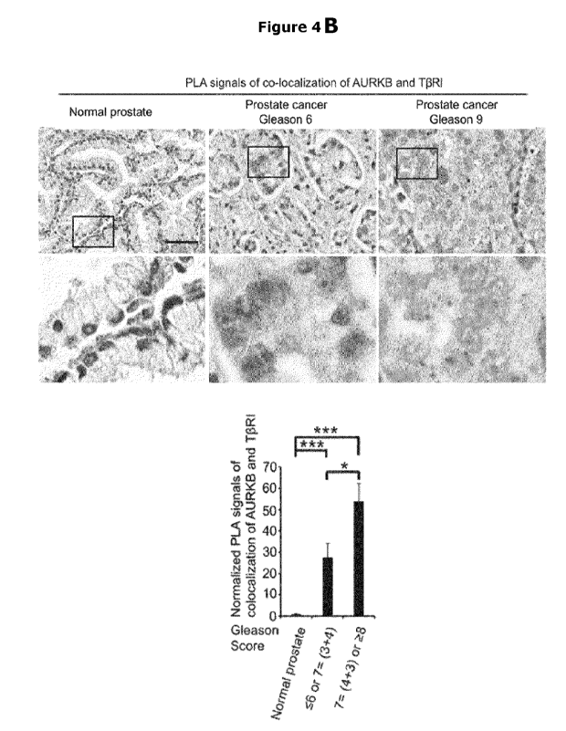
Moreover, high expression of AURKB and TpRI-ICD
complexes visualized by in situ PLA technique was present in clinical prostate
cancer
material and correlated to poor prognosis. The inventors surprisingly found
that the
expression of AURKA and AURKB was higher in CRPC of neuroendocrine type than
in
CRPC adenocarcinoma, consistent with the poor prognosis for patients with CRPC
of
neuroendocrine type.
The present invention provides biomarkers for classifying, diagnosing, and
monitoring
a treatment of cancer in a subject. The biomarkers are also useful for
identifying and
predicting aggressive cancer forms.
Transforming growth factor fi (TGFp) is frequently overexpressed in several
cancers,
causing tumor progression. In-depth characterization of the functional
significance of
TpRI in mitosis demonstrates a newly identified, important role during
cytokinesis.
A first object of the present invention provides a method for diagnosing
cancer in a
subject, the method comprising the steps of:
a) providing a biological test sample from the subject; and
b) determining the presence or absence of a first biomarker, a second
bionnarker, and a third bionnarker, wherein said biomarkers are: Aurora
kinase B (AURKB), Adaptor Protein, Phosphotyrosine Interacting With PH
Domain And Leucine Zipper 1 (APPL1), and TGFp receptor type 1 (TpR1),
in the test sample;
wherein the co-localization of all three biomarkers in the biological test
sample is
indicative of cancer in the subject.
Thus, it will be appreciated that step (b) may involve determining the co-
localization
of the first, second, and third biomarkers within the test sample. Examples of
5
CA 03221184 2023- 12-1
WO 2022/253604
PCT/EP2022/063820
techniques that can be used to determine whether two proteins are co-localized
include
those described herein and include immunohistochemistry, in situ
hybridization,
innnnunoprecipitation, innnnunofluorescence, confocal microscopy, many of
which are
exemplified in the Examples.
For the avoidance of doubt, the co-localization of biomarkers does not require
the
biomarkers to be in a complex with each other, but merely that the biomarkers
are
spatially close to each other. For example, two proteins may be co-localized
if they
are observed as being spatially close to each other (for example, by
innnnunofluorescence and digital imaging using z-stack), and a direct
interaction
between the biomarkers is not necessary. However, biomarkers may be co-
localized
because they do directly interact, and therefore both situations are
encompassed by
the term "co-localization".
In an embodiment of all of the methods of the invention, the co-localization
of the
biomarkers to a cellular structure is indicative of cancer in the subject.
Thus, it will be
appreciated that step (b) may involve determining the co-localization of the
first,
second, and third biomarkers in a cellular structure within the test sample.
By "cellular structure" we include the meaning of any defined compartment or
sub-
compartment of a cell such as an organelle, including a sub-part of an
organelle.
Cellular structures include the nucleus, ribosonnes, endoplasnnic reticulunn
(ER), Golgi
apparatus, cytoplasm and mitochondria. For example, the organelle may be the
nucleus and the sub-part of the nucleus may be the nnidbody. Examples of
techniques
that can be used to determine whether two proteins are co-localized to a
cellular
structure are known in the art.
For example, using immunofluorescence or
innnnunohistochemistry, a marker for the nucleus may be used in addition to
markers
for the particular biomarkers, enabling the skilled person to assess whether
these
separate markers are all observed in the nucleus and thus whether the
biomarkers are
co-localized to the nucleus. Similarly, a population of cells may be
fractionated and an
immunoprecipitation may be carried out to determine whether the biomarkers are
in a
complex within, for example, the nuclear fraction.
In an embodiment of all of the methods of the invention, the cellular
structure is the
nucleus. In a further embodiment of all the methods of the invention, the
cellular
structure is a cytokinesis structure.
6
CA 03221184 2023- 12-1
WO 2022/253604
PCT/EP2022/063820
In an embodiment, the present invention provides a method for diagnosing
cancer in
a subject, the method comprising the steps of:
a) providing a biological test sample from the subject; and
b) determining the presence or absence of a first biomarker, a second
biomarker, and a third biomarker, wherein said biomarkers are: Aurora
kinase B (AURKB), Adaptor Protein, Phosphotyrosine Interacting With PH
Domain And Leucine Zipper 1 (APPL1), and TGFp receptor type 1 (T13R1),
in the test sample;
wherein the presence of all three biomarkers co-localized to a cytokinesis
structure in
the biological test sample is indicative of cancer in the subject.
Thus, it will be appreciated that step (b) may involve determining the
presence or
absence of the first, second, and third biomarkers in a cytokinesis structure
within the
test sample.
Thus, co-localization of three biomarkers to a cytokinesis structure includes
the
meaning of each of the three biomarkers being identifiable in one or more
cytokinesis
structures. In a particularly preferred embodiment, the cytokinesis structure
is the
midbody and so colocalization of the three biomarkers in a cytokinesis
structure is
colocalization of each of the three biomarkers to the midbody. For the
avoidance of
doubt, by co-localizing of biomarkers to a cytokinesis structure, it is not a
requirement
for the biomarkers to be in a complex with each other, but merely that the
biomarkers
are co-localized to a cytokinesis structure. For example, two proteins may be
colocalized if they are observed as being close to each other by
immunofluorescence
and digital imaging using z-stack.
In an embodiment, the method further comprises determining the presence or
absence
of a fourth biomarker in the biological test sample, wherein said biomarker is
TNF
receptor associated factor 6 (TRAF6), and wherein the co-localization of all
four
biomarkers in the biological test sample is indicative of cancer in the
subject.
In an embodiment of the methods of the invention, the co-localization of the
biomarkers to a cellular structure is indicative of cancer in the subject.
Thus, it will be
appreciated that step (b) may involve determining the co-localization of the
first,
second, and third biomarkers in a cellular structure within the test sample.
In an embodiment, the method further comprises determining the presence or
absence
of a fourth biomarker in the biological test sample, wherein said biomarker is
TNF
7
CA 03221184 2023- 12-1
WO 2022/253604
PCT/EP2022/063820
receptor associated factor 6 (TRAF6), wherein the presence of all four
biomarkers co-
localized to a cytokinesis structure in the biological test sample is
indicative of cancer
in the subject.
Thus, co-localization of four biomarkers to a cytokinesis structure includes
the meaning
of each of the four biomarkers being identifiable in one or more cytokinesis
structures.
In a particularly preferred embodiment, the cytokinesis structure is the
midbody and
so colocalization of the four biomarkers in a cytokinesis structure is
colocalization of
each of the four biomarkers to the midbody.
In an embodiment the TGFI3 receptor type 1 (T13R1) is the intracellular domain
(T13R1-
ICD). The term "TGFI3 receptor type 1" may be used interchangeably with "TGFI3
receptor type I" "Ti3R1", "TGFI3R1", "Ti3RI" and "TGFI3RI" herein.
Methods for assessing the presence and/or intracellular localization of
biomarkers are
well known in the art and any suitable method can be used. For example, the
cytokinesis structure may be isolated and the presence of the biomarker in the
cytokinesis structure assessed, or the cytokinesis structure may be identified
by a
detectable moiety and localization of a biomarker within that cytokinesis
structure may
be assessed by assessing whether the biomarker localizes to the same
detectable
moiety. Examples of techniques that can be used include those described herein
and
include innnnunohistochennistry, in situ hybridization, innnnunoprecipitation,
innnnunofluorescence, confocal microscopy, many of which are exemplified in
the
Examples.
In some embodiments, diagnosing the cancer includes determining the malignancy
of
the cancer. In some embodiments, diagnosing the cancer includes determining
the
stage of the cancer. In some embodiments, diagnosing the cancer includes
assessing
the risk of cancer recurrence. In some embodiments, diagnosing the cancer
includes
assessing the grade of the cancer.
The invention also includes a method comprising the steps of:
- providing a biological test sample from a subject;
- determining the presence or absence of a first biomarker, a second
biomarker, a third biomarker, and a fourth biomarker, the biomarkers are:
Aurora kinase B (AURKB), Adaptor Protein, Phosphotyrosine Interacting
With PH Domain And Leucine Zipper 1 (APPL1), TGFR receptor type 1 (-113R1)
and TNF receptor associated factor 6 (TRAF6), in the test sample; and
8
CA 03221184 2023- 12-1
WO 2022/253604
PCT/EP2022/063820
wherein the co-localization of all four biomarkers in the biological test
sample is
indicative of cancer in the subject.
The invention also includes a method comprising the steps of:
- providing a biological test sample from a subject;
- determining the presence or absence of a first biomarker, a second
biomarker, a third biomarker, and a fourth biomarker, the biomarkers are:
Aurora kinase B (AURKB), Adaptor Protein, Phosphotyrosine Interacting
With PH Domain And Leucine Zipper 1 (APPL1), TGFB receptor type 1 (T13R1)
and TNF receptor associated factor 6 (TRAF6), in the test sample; and
wherein the presence of all four biomarkers co-localized to a cytokinesis
structure in
the biological test sample is indicative of cancer in the subject.
Thus, it will be appreciated that the second step may involve determining the
co-
localization of the biomarkers within the test sample.
The intracellular domain (ICD) of Tl3R1 is not cleaved off from the TI3R1 in a
healthy
cell, thereby not detectable in the nucleus, which means that the three or
four
biomarkers (AURKB, APPL1, T13R1 (or TI3R1-ICD) and TRAF6) co-localized during
cytokinesis is not detectable in healthy cells.
Methods for determining the presence of biomarkers and/or whether biomarkers
co-
localize during cytokinesis and/or mitosis are known in the art. For example,
to
examine the innnnunofluorescence of proteins at each mitotic stage, cells can
be
synchronized (at the G1-S transition) by double-thymidine block and release,
in order
to enrich cytokinetic cells. A staging system can be used to identify the
different
phases of mitosis and cytokinesis based on the DNA and spindle morphology and
extent
of chromosome alignment and separation. Synchronization of mammalian cells in
cytokinesis can also be achieved by releasing cells from pre-metaphase arrest.
Pre-
metaphase synchronization can be achieved using microtubule
polymerizing/depolymerizing agents (such as nocodazole and taxol), as well as
kinesin
inhibitors (such as monastrol and S-trityl-L-cysteine).
It is a second object of the invention to provide a method for diagnosing
and/or
prognosing aggressive cancer in a subject, the method comprising the steps of:
a) providing a biological test sample from the subject;
b) determining the presence or absence of a first biomarker, a second
biomarker, and a third biomarker, wherein said biomarkers are: Aurora
9
CA 03221184 2023- 12-1
WO 2022/253604
PCT/EP2022/063820
kinase B (AURKB), Adaptor Protein, Phosphotyrosine Interacting With PH
Domain And Leucine Zipper 1 (APPL1), and TGFI3 receptor type 1 (T13R1),
in said test sample; and
wherein the co-localization of all three biomarkers in the biological sample
is indicative
of aggressive cancer in the subject.
It is a further object of the invention to provide a method for diagnosing
and/or
prognosing aggressive cancer in a subject, the method comprising the steps of:
a) providing a biological test sample from the subject;
b) determining the presence or absence of a first biomarker, a second
bionnarker, and a third bionnarker, wherein said biomarkers are: Aurora
kinase B (AURKB), Adaptor Protein, Phosphotyrosine Interacting With PH
Domain And Leucine Zipper 1 (APPL1), and TGFI3 receptor type 1 (Ti3R1),
in said test sample; and
wherein the presence of all three biomarkers co-localized to a cytokinesis
structure in
the biological sample is indicative of aggressive cancer in the subject.
An aggressive cancer form includes the meaning of high risk for metastasis. By
aggressive cancer, we include a cancer comprising or consisting of stage III
and/or
stage IV cancer, for example as determined by the American Joint Committee on
Cancer (AJCC) TNM system American Joint Committee on Cancer and the
International
Union Against Cancer.
Preferably, the cytokinesis structure is the midbody or the midzone of a cell.
In an embodiment the TGFI3 receptor type 1 (T13R1) is the intracellular domain
(T13R1-
ICD).
Midbodies can be detected by using a molecule that binds to the midbody, such
as a
molecule that binds to a protein that is known to localize to the midbody,
e.g., an
antibody that specifically binds to a midbody polypeptide or an antigenic
fragment
thereof, e.g., Mitotic Kinesin-Like Protein-1 (MKLP-1), kinesin family member
4 (KIF4),
and/or 13-tubulin. MKLP-1 localizes to the spindle equator and is believed to
participate
in the separation of spindle poles during anaphase B of mitosis, by
crosslinking
antiparallel microtubules at the spindle midzone. A number of antibodies
suitable for
use in the methods described herein are known in the art and/or are
commercially
available. For example, anti-MKLP1 is available from BD Biosciences (San Jose,
CA)
and Santa Cruz Biotechnology Inc. (Santa Cruz, CA). Methods for isolating
nnidbodies
CA 03221184 2023- 12-1
WO 2022/253604
PCT/EP2022/063820
are known in the art (Science. 2004 Jul 2; 305(5680): 61-66). Proteins present
in the
nnidbody preparations can then be identified by tandem liquid chromatography
and
tandem mass spectrometry.
In an embodiment, the method further comprises determining the presence or
absence
of a fourth biomarker in the biological test sample, wherein said biomarker is
TNF
receptor associated factor 6 (TRAF6), and wherein the co-localization of all
four
biomarkers in the biological test sample is indicative of aggressive cancer in
the subject.
In an embodiment, the method further comprises determining the presence or
absence
of a fourth biomarker in the biological test sample, wherein said biomarker is
TNF
receptor associated factor 6 (TRAF6), wherein the presence of all four
biomarkers co-
localized to a cytokinesis structure in the biological test sample is
indicative of
aggressive cancer in the subject.
Further, the invention provides a method for diagnosing and/or prognosing
aggressive
cancer in a subject, the method comprising the steps of:
a) providing a biological test sample from the subject;
b) determining the presence or absence of a first biomarker, a second
biomarker, a third biomarker, and a fourth biomarker, wherein said biomarkers
are: Aurora kinase B (AURKB), Adaptor Protein, Phosphotyrosine Interacting
With PH Domain And Leucine Zipper 1 (APPL1), and TGFB receptor type 1 (T8R1)
and TNF receptor associated factor 6 (TRAF6), in said test sample; and
wherein the co-localization of all four biomarkers in the biological sample is
indicative
of aggressive cancer in the subject.
Further, the invention provides a method for diagnosing and/or prognosing
aggressive
cancer in a subject, the method comprising the steps of:
a) providing a biological test sample from the subject;
b) determining the presence or absence of a first biomarker, a second
biomarker, a third biomarker, and a fourth biomarker, wherein said biomarkers
are: Aurora kinase B (AURKB), Adaptor Protein, Phosphotyrosine Interacting
With PH Domain And Leucine Zipper 1 (APPL1), and TGF8 receptor type 1 (T8R1)
and TNF receptor associated factor 6 (TRAF6), in said test sample; and
wherein the presence of all four biomarkers co-localized to a cytokinesis
structure in
the biological sample is indicative of aggressive cancer in the subject.
11
CA 03221184 2023- 12-1
WO 2022/253604
PCT/EP2022/063820
Preferably, AURKB is ubiquitinated. In an embodiment, the methods of the
invention
comprise detecting the presence of ubiquitinated AURKB in the biological test
sample.
Methods for detecting the presence of ubiquitinated AURKB are known in the art
and
disclosed herein.
Protein ubiquitination is a post-translational modification catalyzed by a
cascade of
enzymatic reactions involving a ubiquitin (Ub)-activating enzyme (El), a Ub-
conjugating enzyme (E2), and a Ub ligase (E3). Ub is conjugated onto protein
substrates by formation of an isopeptide bond between the carboxyl group of
the C-
terminal glycine residue of Ub and the c-amino group of a lysine residue in
the
substrate. Furthermore, a polyubiquitin (polyUb) chain is formed by
conjugating the
carboxyl group of the C-terminal glycine residue of Ub to the E-amino group of
one of
the seven internal lysines in the preceding Ub.
In other words, polyUbs are linked through the E-amino group of the Lys-48
and/or
Lys-63 residues of the preceding Ub. In an embodiment, AURKB comprises the
consensus sequence -(hydrophobic)-K-(hydrophobic)-K-X-(hydrophobic)-(polar)-
(hydrophobic)-(polar)-(hydrophobic), in which at least one K is ubiquitinated.
As
shown in Figure 31, this motif is conserved in human, pig, cow, dog, mouse and
rat
AURKB. In an embodiment, AURKB comprises the *K*KX*8(.*&* consensus sequence,
wherein *=hydrophobic, &=polar, X=any amino acid, K= acceptor lysine, and at
least
one of the lysine residues therein is ubiquitinated.
In an embodiment, AURKB
comprises the GKGKFGNVYL (SEQ ID NO: 23) consensus sequence and at least one
of
the lysine residues therein is ubiquitinated. In other words, in an embodiment
AURKB
is ubiquitinated at one or both lysine residues corresponding to Lysine 85
(K85) and/or
Lysine 87 (K87) of human AURKB (SEQ ID NO: 1). In an embodiment, AURKB is
ubiquitinated at a lysine residue corresponding to Lysine 85 (K85) of human
AURKB
(SEQ ID NO: 1). In an embodiment, AURKB is ubiquitinated at a lysine residue
corresponding to Lysine 87 (K87) of human AURKB (SEQ ID NO: 1). In an
embodiment,
AURKB is ubiquitinated at both lysine residues corresponding to Lysine 85
(K85) and
Lysine 87 (K87) of human AURKB (SEQ ID NO: 1).
By "corresponding to" we include the meaning of the lysine residue in another
AURKB
(such as an orthologue or variant of human AURKB) which aligns to K85 in human
AURKB (SEQ ID NO: 1 and/or to K87 in human AURKB (SEQ ID NO: 1) when the
sequence of human AURKB and the sequence of a different AURKB are compared,
such
as are aligned using MacVector, ClustalOrnega, or ClustalW2, or are aligned as
shown
12
CA 03221184 2023- 12-1
WO 2022/253604
PCT/EP2022/063820
in Figure 1 of Brown et al., Evolutionary Biology volume 4, Article number: 39
(2004),
incorporated by reference.
SEQ ID NO Sequence
AURKB amino
1 macikensypw pygrqtapsg istipqrvir kepvtpsalv
add sequence imsrsnvqpt aapgqkvmen
(SEQ ID NO: 1) 61 ssgtpdiltr hftiddfeig rpigkgkfgn vylarekksh
fivalkvlfk sqle<egveh
121 glrrpiRiga 1-111-1hpni1r1 ynyfydrrri ylilpyaprg
elykelqksc tfdeqrtati
181 meeladalmy chgkkvihrd ikpenii1gi kgelkiadfg
wsvhapsirr ktmugtidyi
241 ppemiegrmh nekvdiwcig vicyelivgn ppfesashne
tyrrivkvd1 kfpasvpmga
301 qdliskilrh npseriplaq vsahpwvran srrvlppsal qsva
AU RKB coding ATGGCCCAGAAGGAGAACTCCTACCCCTGGCCCTACGGCCGACAGACGG
sequence
CTCCATCTGGCCTGAGCACCCTGCCCCAGCGAGTCCTCCGGAAAGAGCC
(SEQ ID NO: 2) TGTCACCCCATCTGCACTTGTCCTCATGAGCCGCTCCAATGTCCAGCCCA
CAGCTGCCCCTGGCCAGAAGGTGATGGAGAATAGCAGTGGGACACCCGA
CATCTTAACGCGGCACTTCACAATTGATGACTTTGAGATTGGGCGTCCTCT
GGGCAAAGGCAAGTTTGGAAACGTGTACTTGGCTCGGGAGAAGAAAAGC
CATTTCATCGTGGCGCTCAAGGTCCTCTTCAAGTCCCAGATAGAGAAGGA
GGGCGTGGAGCATCAGCTGCGCAGAGAGATCGAAATCCAGGCCCACCTG
CACCATCCCAACATCCTGCGTCTCTACAACTATTTTTATGACCGGAGGAG
GATCTACTTGATTCTAGAGTATGCCCCCCGCGGGGAGCTCTACAAGGAGC
TGCAGAAGAGCTGCACATTTGACGAGCAGCGAACAGCCACGATCATGGA
GGAGTTGGCAGATGCTCTAATGTACTGCCATGGGAAGAAGGTGATTCACA
GAGACATAAAGCCAGAAAATCTGCTCTTAGGGCTCAAGGGAGAGCTGAA
GATTGCTGACTTCGGCTGGTCTGTGCATGCGCCCTCCCTGAGGAGGAAG
ACAATGTGTGGCACCCTGGACTACCTGCCCCCAGAGATGATTGAGGGGC
GCATGCACAATGAGAAGGTGGATCTGTGGTGCATTGGAGTGCTTTGCTAT
GAGCTGCTGGTGGGGAACCCACCCTTTGAGAGTGCATCACACAACGAGA
CCTATCGCCGCATCGTCAAGGTGGACCTAAAGTTCCCCGCTTCCGTGCCC
ATGGGAGCCCAGGACCTCATCTCCAAACTGCTCAGGCATAACCCCTCGGA
ACGGCTGCCCCTGGCCCAGGTCTCAGCCCACCCTTGGGTCCGGGCCAAC
TCTCGGAGGGTGCTGCCTCCCTCTGCCCTTCAATCTGTCGCCTGA
In an embodiment, AURKB is Lys48-linked and/or Lys63-linked polyubiquitinated.
13
CA 03221184 2023- 12-1
WO 2022/253604
PCT/EP2022/063820
In the accompanying Examples, the inventors surprisingly found that AURKB
contains
at least one acceptor lysine residue that serves as the recognition site for
ubiquitination
by TRAF6, and that TRAF6-mediated ubiquitination of AURKB on K85 and/or K87 in
the
consensus sequence contributes to its activity and controls the localization
of WI in
the midbody during cell division. Methods for
determining whether a protein is
ubiquitinated are known in the art and include an in vivo ubiquitination
assay, or an in
situ PLA assay with two antibodies (AURKB and K63 antibodies) as described in
the
Examples.
The method(s) disclosed in the present specification is/are suitable for
cancer types
associated with and/or mediated by proteolytic cleavage of transforming growth
factor
13 type I receptor (T13RI).
By a cancer "associated with and/or mediated by the proteolytic cleavage of
transforming growth factor 13 type I receptor (T13RI)" we include the meaning
of a
cancer in which the intracellular domain (ICD) of T13RI has been
proteolytically cleaved
and enters the nucleus to promote transcription of pro-invasive genes. Methods
of
detecting the localization of T13RI and T13RI-ICD are described herein.
The cancer is for example a solid tumour. The tumour may be selected from the
group
consisting of prostate cancer, renal carcinoma, lung cancer, kidney cancer,
gastric
cancer, bladder carcinoma, breast cancer, endonnetrial cancer, ovarian cancer,
and
colorectal cancer.
Preferably, the cancer is prostate cancer. In a further embodiment, the
prostate cancer
is castration-resistant prostate cancer (CRPC). By "castration resistant
prostate cancer
(CRPC)" we include the meaning of a form of prostate cancer wherein the cancer
is no
longer stopped by low testosterone levels (less than 50 ng/nnL). Castration-
resistant
prostate cancer is defined by a rising PSA level and/or worsening symptoms
and/or
growing cancer verified by scans. In an
embodiment, the CRPC is of the
neuroendocrine type. In an embodiment, the biological test sample comprises
CRPC
cells. As shown in the accompanying Examples, the inventors surprisingly found
that
during mitosis and cytokinesis, a T13RI-AURKB complex was formed in nnidbody
in CRPC
cells and neuroblastonna KELLY cells.
Preferably, the biological test sample is a tissue sample, such as a biopsy
from a
tumour.
14
CA 03221184 2023- 12-1
WO 2022/253604
PCT/EP2022/063820
The "sample to be tested", "biological test sample", "test sample" or "control
sample"
may be a tissue or fluid sample taken or derived from a subject.
Preferably the test sample is provided from a mammal. The mammal may be any
domestic or farm animal. Preferably, the mammal is a rat, mouse, guinea pig,
cat,
dog, horse or a primate. Most preferably, the mammal is human.
A sample as used herein includes any relevant biological sample that can be
used for
molecular profiling, e.g., sections of tissues such as biopsy or tissue
removed during
surgical or other procedures, bodily fluids (e.g. liquid biopsy), autopsy
samples, and
frozen sections taken for histological purposes, a sample comprising cells.
Such
samples include blood or blood fractions or products (e.g. serum, buffy coat,
plasma,
platelets, red blood cells, and the like), sputum, malignant effusion, cheek
cells tissue,
cultured cells (e.g., primary cultures, explants, and transformed cells),
stool, urine,
other biological or bodily fluids (e.g., prostatic fluid, gastric fluid,
intestinal fluid, renal
fluid, lung fluid, cerebrospinal fluid, and the like), etc. The sample can
comprise
biological material that is a fresh frozen & formalin fixed paraffin embedded
(FFPE)
block, fornnalin-fixed paraffin embedded, or is within an RNA preservative and
fornnalin
fixative. More than one sample of more than one type can be used for each
subject.
Preferably the sample is a cell or tissue sample (or derivative thereof), for
example
one comprising or consisting of cancer cells. In a preferred embodiment, the
sample
comprises a fixed tumor sample. The sample used in the methods described
herein can
be a fornnalin fixed paraffin embedded (FFPE) sample. The FFPE sample can be
one or
more of fixed tissue, unstained slides, bone marrow core or clot, core needle
biopsy,
malignant fluids and fine needle aspirate (FNA). In an embodiment, the fixed
tissue
comprises a tumor containing fornnalin fixed paraffin embedded (FFPE) block
from a
surgery or biopsy.
A sample may be processed according to techniques understood by those in the
art. A
sample can be without limitation fresh, frozen or fixed cells or tissue. In
some
embodiments, a sample comprises formalin-fixed paraffin-embedded (FFPE)
tissue,
fresh tissue or fresh frozen (FF) tissue. A sample can comprise cultured
cells, including
primary or immortalized cell lines derived from a sample from a subject. A
sample can
also refer to an extract from a sample from a subject. For example, a sample
can
comprise DNA, RNA or protein extracted from a tissue or a bodily fluid. Many
techniques and commercial kits are available for such purposes. The fresh
sample from
the subject can be treated with an agent to preserve RNA prior to further
processing,
e.g., cell lysis and extraction. Samples can include frozen samples collected
for other
CA 03221184 2023- 12-1
WO 2022/253604
PCT/EP2022/063820
purposes. Samples can be associated with relevant information such as age,
gender,
and clinical symptoms present in the subject; source of the sample; and
methods of
collection and storage of the sample.
A biopsy comprises the process of removing a tissue sample for diagnostic or
prognostic evaluation, and to the tissue specimen itself. Any biopsy technique
known
in the art can be applied to the methods of the present invention. The biopsy
technique
applied can depend on the tissue type to be evaluated (e.g., colon, prostate,
kidney,
bladder, lymph node, liver, bone marrow, blood cell, lung, breast, etc.), the
size and
type of the tumor (e.g., solid or suspended, blood or ascites), among other
factors.
Representative biopsy techniques include, but are not limited to, excisional
biopsy,
incisional biopsy, needle biopsy, surgical biopsy, and bone marrow biopsy. An
"excisional biopsy" refers to the removal of an entire tumor mass with a small
margin
of normal tissue surrounding it. An "incisional biopsy" refers to the removal
of a wedge
of tissue that includes a cross-sectional diameter of the tumor. The method
may use a
"core-needle biopsy" of the tumor mass, or a "fine-needle aspiration biopsy"
which
generally obtains a suspension of cells from within the tumor mass. Biopsy
techniques
are discussed, for example, in Harrison's Principles of Internal Medicine,
Kasper, et al.,
eds., 16th ed., 2005, Chapter 70, and throughout Part V.
Preferably test and control samples are derived from the same species.
Preferably test
and control samples are matched for age, gender and/or lifestyle.
In an embodiment the tissue sample is tumour tissue, such as a biopsy. In an
embodiment, the cell sample is a sample of cancer cells.
Preferably, the method further comprises the steps of:
c) providing one or more control sample from:
i. an individual not afflicted with cancer; and/or
ii. an individual afflicted with cancer, wherein the control sample is of a
different stage of cancer to that of the test sample, or wherein the
control sample is derived from healthy tissue from an individual afflicted
with cancer;
d) determining the presence or absence of a first bionnarker, a second
bionnarker, and a third bionnarker, wherein said bionnarkers are: Aurora
kinase
B (AURKB), Adaptor Protein, Phosphotyrosine Interacting With PH Domain And
Leucine Zipper 1 (APPL1), and TGFB receptor type 1 (T13R1), in the control
sample;
16
CA 03221184 2023- 12-1
WO 2022/253604
PCT/EP2022/063820
wherein cancer is diagnosed in the event that all three biomarkers measured in
step
(b) are co-localized in the test sample, and not all three biomarkers measured
in step
(d) are co-localized in the control sample.
Preferably, the method further comprises the steps of:
c) providing one or more control sample from:
i. an individual not afflicted with cancer; and/or
ii. an individual afflicted with cancer, wherein the control sample was of
a different stage of cancer to that of the test sample, or wherein the
control sample is derived from healthy tissue from an individual afflicted
with cancer;
d) determining the presence or absence of a first biomarker, a second
biomarker, and a third biomarker, wherein said biomarkers are: Aurora kinase
B (AURKB), Adaptor Protein, Phosphotyrosine Interacting With PH Domain And
Leucine Zipper 1 (APPL1), and TGFI3 receptor type 1 (TI3R1), in the control
sample;
wherein cancer is diagnosed in the event that all three biomarkers measured in
step
(b) are co-localized to a cytokinesis structure in the test sample, and not
all three
biomarkers measured in step (d) are co-localized to a cytokinesis structure in
the
control sample.
For example, if the cancer is strictly localized to one lobe of the prostate
it may be
possible to use healthy (i.e. non-cancerous) tissue in another lobe from the
same
individual as control.
In an embodiment, the method further comprises (d) determining the presence or
absence of a fourth biomarker in the control sample, wherein said biomarker is
TNF
receptor associated factor 6 (TRAF6), wherein cancer is diagnosed in the event
that all
four biomarkers measured in step (b) are co-localized in the test sample, and
not all
four biomarkers measured in step (d) are co-localized in the control sample.
In an embodiment, the method further comprises (d) determining the presence or
absence of a fourth biomarker in the control sample, wherein said biomarker is
TNF
receptor associated factor 6 (TRAF6), wherein cancer is diagnosed in the event
that all
four biomarkers measured in step (b) are co-localized to a cytokinesis
structure in the
test sample, and not all four biomarkers measured in step (d) are co-localized
to a
cytokinesis structure in the control sample.
17
CA 03221184 2023- 12-1
WO 2022/253604
PCT/EP2022/063820
Thus, preferably, the method further comprises the steps of:
c) providing one or more control sample from:
i. an individual not afflicted with cancer; and/or
ii. an individual afflicted with cancer, wherein the control sample is of a
different stage of cancer to that of the test sample;
d) determining the presence or absence of a first biomarker, a second
biomarker, a third biomarker, and a fourth biomarker, wherein said biomarkers
are: Aurora kinase B (AURKB), Adaptor Protein, Phosphotyrosine Interacting
With PH Domain And Leucine Zipper 1 (APPL1), TGFB receptor type 1 (TI3R1)
and TNF receptor associated factor 6 (TRAF6), in the control sample;
wherein cancer is diagnosed in the event that all four biomarkers measured in
step (b)
are co-localized in the test sample, and not all four biomarkers measured in
step (d)
are co-localized in the control sample.
Thus, preferably, the method further comprises the steps of:
c) providing one or more control sample from:
i. an individual not afflicted with cancer; and/or
ii. an individual afflicted with cancer, wherein the control sample was of
a different stage of cancer to that of that the test sample;
d) determining the presence or absence of a first biomarker, a second
biomarker, a third biomarker, and a fourth biomarker, wherein said biomarkers
are: Aurora kinase B (AURKB), Adaptor Protein, Phosphotyrosine Interacting
With PH Domain And Leucine Zipper 1 (APPL1), TGFB receptor type 1 (TI3R1)
and TNF receptor associated factor 6 (TRAF6), in the control sample;
wherein cancer is diagnosed in the event that all four biomarkers measured in
step (b)
are co-localized to a cytokinesis structure in the test sample, and not all
four
biomarkers measured in step (d) are co-localized to a cytokinesis structure in
the
control sample.
Preferably, the AURKB is ubiquitinated.
By "wherein the control sample was of a different stage of cancer to that of
that the
test sample" we include the meaning that the control sample is derived from an
individual afflicted with cancer, but the cancer comprised within the control
sample is
less advanced (i.e. lower grade or score) than the cancer in the test sample.
The
cancer may be diagnosed in the individual afflicted with cancer using
conventional
clinical methods known in the art.
18
CA 03221184 2023- 12-1
WO 2022/253604
PCT/EP2022/063820
By "wherein the control sample is derived from healthy tissue from an
individual
afflicted with cancer", we include the meaning that the control sample may be
derived
from healthy, non-cancerous tissue that is adjacent to the cancerous tissue.
As exemplified in the accompanying examples, the presence of Aurora kinase B
(AURKB), Adaptor Protein, Phosphotyrosine Interacting With PH Domain And
Leucine
Zipper 1 (APPL1), TGFB receptor type 1 (T13R1) and TNF receptor associated
factor 6
(TRAF6) in a cytokinesis structure is indicative of cancer in a subject.
Preferably, the individual not afflicted with cancer was not, at the time the
sample was
obtained, afflicted with any disease or condition. Preferably, the individual
not afflicted
with cancer is a healthy individual.
Preferably, the presence or absence of biomarkers Aurora kinase B (AURKB),
Adaptor
Protein, Phosphotyrosine Interacting With PH Domain And Leucine Zipper 1
(APPL1),
TGFB receptor type 1 (T3R1) and/or TNF receptor associated factor 6 (TRAF6),
preferably co-localized to a cellular structure such as a cytokinesis
structure, is
determined by detecting the biomarker protein; and/or detecting a biological
activity
of the biomarker protein.
In an embodiment the TGFB receptor type 1 (T13R1) is the intracellular domain
(T13R1-
ICD).
By detecting the biomarker protein we include the meaning of detecting whether
the
biomarker protein is present directly, for example by using a binding partner
that
specifically binds to the biomarker protein. By detecting a biological
activity of the
biomarker protein we include the meaning of assaying for a biological activity
of the
biomarker protein, for example an enzymatic activity. It will be appreciated
that
detecting a biological activity of the biomarker protein may be used to
indirectly
determine the presence or absence of the biomarker.
The presence and/or absence of said biomarkers, preferably co-localized to a
cellular
structure such as a cytokinesis structure may be determined by
innnnunohistochennistry, innnnunocytochennistry, innnnunoprecipitation (IP),
ELISA
techniques (single or mulitplex), radioimmunoassay (RIA), immunoradiometric
assays
(IRMA) and innnnunoenzynnatic assays (IEMA), including sandwich assays using
monoclonal and/or polyclonal antibodies, in situ proximity ligation assay
(PLA),
enzymatic methods, image analysis, mass spectrometry, aptanners, Bio-Layer
19
CA 03221184 2023- 12-1
WO 2022/253604
PCT/EP2022/063820
Interferonnetry (BLI), Surface plasmon resoncance (SPR), Multiplex assay (MSD,
Mesoscale discovery), or by indicator substances that bind to Aurora kinase B
(AURKB),
Adaptor Protein, Phosphotyrosine Interacting With PH Domain And Leucine Zipper
1
(APPL1), TGF13 receptor type 1 intracellular domain (Ti3R1-ICD) and TNF
receptor
associated factor 6 (TRAF6).
Immunohistochemistry (IHC) is a process of localizing antigens (e.g.,
proteins) in cells
of a tissue binding antibodies specifically to antigens in the tissues. The
antigen-binding
antibody can be conjugated or fused to a tag that allows its detection, e.g.,
via
visualization. In some embodiments, the tag is an enzyme that can catalyze a
color-
producing reaction, such as alkaline phosphatase or horseradish peroxidase.
The
enzyme can be fused to the antibody or non-covalently bound, e.g., using a
biotin-
avadin system. Alternatively, the antibody can be tagged with a fluorophore,
such as
fluorescein, rhodannine, DyLight Fluor or Alexa Fluor. The antigen-binding
antibody can
be directly tagged or it can itself be recognized by a detection antibody that
carries the
tag. Using IHC, one or more proteins may be detected. The expression of a gene
product can be related to its staining intensity compared to control levels.
In some
embodiments, the gene product is considered differentially expressed if its
staining
varies at least 1.2, 1.3, 1.4, 1.5, 1.6, 1.7, 1.8, 1.9, 2.0, 2.2, 2.5, 2.7,
3.0, 4, 5, 6, 7,
8, 9 or 10-fold in the sample versus the control.
IHC comprises the application of antigen-antibody interactions to
histochennical
techniques. In an illustrative example, a tissue section is mounted on a slide
and is
incubated with antibodies (polyclonal or monoclonal) specific to the antigen
(primary
reaction). The antigen-antibody signal is then amplified using a second
antibody
conjugated to a complex of peroxidase antiperoxidase (PAP), avidin-biotin-
peroxidase
(ABC) or avidin-biotin alkaline phosphatase. In the presence of substrate and
chronnogen, the enzyme forms a colored deposit at the sites of antibody-
antigen
binding.
Immunofluorescence is an alternate approach to visualize target proteins. In
this
technique, the primary target-antibody signal is amplified using a second
antibody
conjugated to a fluorochronne. On UV light absorption, the fluorochronne emits
its own
light at a longer wavelength (fluorescence), thus allowing localization of
antibody-
antigen complexes.
Protein-based techniques for detecting the presence and/or amount of a
biomarker
also include innnnunoaffinity assays based on antibodies selectively
innnnunoreactive for
CA 03221184 2023- 12-1
WO 2022/253604
PCT/EP2022/063820
the protein encoding the biomarker. These techniques include without
limitation
innnnunoprecipitation, Western blot analysis, molecular binding assays, enzyme-
linked
innnnunosorbent assay (ELISA), enzyme-linked innnnunofiltration assay (ELIFA),
fluorescence activated cell sorting (FACS) and the like. For example, an
optional
method of detecting the presence and/or absence of a biomarker in a sample
comprises
contacting the sample with an antibody against the biomarker, or an
immunoreactive
fragment of the antibody thereof, or a recombinant protein containing an
antigen
binding region of an antibody against the biomarker under conditions
sufficient for an
antibody-biomarker complex to form; and then detecting said complex. Methods
for
producing such antibodies are known in the art. ELISA methods are well known
in the
art, for example see The ELISA Guidebook (Methods in Molecular Biology), 2000,
Crowther, Humana Press, ISBN-13: 978-0896037281 (the disclosures of which are
incorporated by reference. A wide range of immunoassay techniques using such
an
assay format are available, see, e.g., U.S. Pat. Nos. 4,016,043, 4,424,279 and
4,018,653. These include both single-site and two-site or "sandwich" assays of
the
non-competitive types, as well as in the traditional competitive binding
assays. These
assays also include direct binding of a labelled antibody to a target
biomarker. Suitable
binding agents (also referred to as binding molecules) can be selected from a
library,
based on their ability to bind a given protein.
Antibodies can be used to immunoprecipitate specific proteins from solution
samples
or to innnnunoblot proteins separated by, e.g., polyacrylannide gel
electrophoresis.
Preferably, step (b) and/or (d) is performed by labelling the one or more
bionnarkers
in the test sample(s) with a detectable moiety.
Preferably, step (b) and/or (d) is performed by labelling the one or more
biomarkers
in the control sample(s) with a detectable moiety.
By a "detectable moiety" we include the meaning that the moiety is one which
may be
detected, such as visualized, qualified as being present or not, and/or
quantitated. By
a moiety being detectable, the relative amount and/or location of the moiety
may be
determined. Suitable detectable moieties are well known in the art.
Thus, the detectable moiety may be a fluorescent and/or luminescent and/or
chennilunninescent moiety which, when exposed to specific conditions, may be
detected.
For example, a fluorescent moiety may need to be exposed to radiation (i.e.
light) at
a specific wavelength and intensity to cause excitation of the fluorescent
moiety,
21
CA 03221184 2023- 12-1
WO 2022/253604
PCT/EP2022/063820
thereby enabling it to emit detectable fluorescence at a specific wavelength
that may
be detected.
Alternatively, the detectable moiety may be an enzyme which is capable of
converting
a (preferably undetectable) substrate into a detectable product that can be
visualized
and/or detected. Examples of suitable enzymes are discussed in more detail
below in
relation to, for example, ELISA assays.
Alternatively, the detectable moiety may be a radioactive atom which is useful
in
imaging. Suitable radioactive atoms include 99nnTc and 1231 for scintigraphic
studies.
Other readily detectable moieties include, for example, spin labels for
magnetic
resonance imaging (MRI) such as 1231 again, 1311, 111In, 19F, 13C, 15N, 170,
gadolinium, manganese or iron. Clearly, the agent to be detected (such as, for
example, biomarkers in the test sample and/or control sample described herein
and/or
an antibody molecule for use in detecting a selected protein) must have
sufficient of
the appropriate atomic isotopes in order for the detectable moiety to be
readily
detectable.
The radio- or other labels may be incorporated into the agents of the
invention (i.e.
the proteins present in the samples of the methods of the invention and/or the
binding
agents of the invention) in known ways. For example, if the binding moiety is
a
polypeptide it may be biosynthesized or may be synthesized by chemical amino
acid
synthesis using suitable amino acid precursors involving, for example,
fluorine-19 in
place of hydrogen. Labels such as 99nnTc, 1231, 186Rh, 188Rh and 111In can,
for
example, be attached via cysteine residues in the binding moiety. Yttrium-90
can be
attached via a lysine residue. The IODOGEN method (Fraker et al (1978)
Biochem.
Biophys. Res. Comm. 80, 49-57) can be used to incorporate 1231. Reference
("Monoclonal Antibodies in Innnnunoscintigraphy", J-F Chatal, CRC Press, 1989)
describes other methods in detail. Methods for conjugating other detectable
moieties
(such as enzymatic, fluorescent, luminescent, chemiluminescent or radioactive
moieties) to proteins are well known in the art.
Preferably, step (b) and/or (d) is performed using one or more first binding
agent
capable of binding to said bionnarker. It will be appreciated by persons
skilled in the
art that the first binding agent may comprise or consist of a single species
with
specificity for one of the biomarkers or a plurality of different species,
each with
specificity for a different protein bionnarker.
22
CA 03221184 2023- 12-1
WO 2022/253604
PCT/EP2022/063820
Preferably, step (b) and/or (d) is performed using an assay comprising a
second
binding agent capable of binding to said first binding agent, the second
binding agent
comprising a detectable moiety.
At least one type of the binding agents, and more typically all of the types,
may
comprise or consist of an antibody or antigen-binding fragment of the same, or
a
variant thereof.
Preferably, the first binding agent and/or the second binding agent comprises
or
consists of an antibody or an antigen-binding fragment thereof.
The antibody or antigen binding fragment thereof may be a scFv; Fab; or a
binding
domain of an innnnunoglobulin molecule.
Preferably, the detectable moiety is selected from the group consisting of: a
fluorescent
moiety; a luminescent moiety; a chemiluminescent moiety; a radioactive moiety;
an
enzymatic moiety.
In yet another embodiment the presence and/or absence of Aurora kinase B
(AURKB),
Adaptor Protein, Phosphotyrosine Interacting With PH Domain And Leucine Zipper
1
(APPL1), TGFB receptor type 1 (T13R1) and/or TNF receptor associated factor 6
(TRAF6)
is determined by measuring the presence and/or expression of a nucleic acid
molecule
encoding the biomarker.
Preferably, the nucleic acid molecule is a cDNA molecule or an mRNA molecule.
Any method of detecting and/or quantitating the nucleic acid molecule encoding
the
biomarker can in principle be used to determine the presence and/or absence of
the
biomarker. The nucleic acid molecule encoding the biomarker can be directly
detected
and/or quantitated (such as by RNA sequencing), or may be copied and/or
amplified
to allow detection of amplified copies of the nucleic acid molecule encoding
the
biomarker or its complement.
Preferably, determining the presence and/or absence of the biomarkers in step
(b), (d)
and/or (f) is performed using a method selected from the group consisting of
Southern
hybridization, Northern hybridization, polynnerase chain reaction (PCR),
reverse
transcriptase PCR (RT PCR), quantitative real-time PCR (qRT-PCR), nanoarray,
nnicroarray, macroarray, autoradiography and in situ hybridization.
23
CA 03221184 2023- 12-1
WO 2022/253604
PCT/EP2022/063820
Reverse transcription can be performed by any method known in the art. For
example,
reverse transcription may be performed using the Omniscript kit (Qiagen,
Valencia,
CA), Superscript III kit (Invitrogen, Carlsbad, CA), for RT-PCR. Target-
specific priming
can be performed in order to increase the sensitivity of detection of target
sequences
and generate target-specific cDNA. RT-PCR can be performed using for eample
Applied
Biosystems Prism (ABI) 7900 HT instruments, or Thermo Fisher QuantStudio Real
Time
PCR instruments or any other thermocycler with fluorescent real time detection
of the
amplification, in a volume with target sequence-specific cDNA or messenger RNA
equivalent to 1 ng total RNA or more. Primers and probes concentrations for
TaqMang
analysis are added to amplify fluorescent annplicons using PCR cycling
conditions such
as 95 C for 10 minutes for one cycle, 95 C for 20 seconds, and 60 C for 45
seconds
for 40 cycles. The amplification reaction can also be performed as a one-step
qRT-PCR
using either one single thermostable DNA polynnerase capable of performing
both the
reverse transcription and the DNA polymerisation such as the Tth Polynnerase
originally
isolated from Thermus thermophilus. It is also feasible to perform a one-step
qPCR
with a mixture of reverse transcriptase and thermostable DNA polymerase. PCR
products can also be labelled with a fluorescent dye, such as SYBR Green or
any other
fluorescent dye detected by the instrument.
The amplification can be designed to determine the presence and/or absence of
all the
biomarkers in step (b), (d) and/or (f) either as single entities or in
combination such
as in multiplex PCR or digital PCR (dPCR) A reference sample can be assayed to
ensure
reagent and process stability. The reference sample can be obtained from a
cell line
expressing the target messenger RNA or be obtained as synthetized messenger
RNA.
A reference sample can be assayed to ensure reagent and process stability.
Negative
controls (e.g., no template) should be assayed to monitor any exogenous
nucleic acid
contamination.
In situ hybridization assays are well known and are generally described in
Angerer et
al., Methods Enzymol. 152:649-660 (1987). In an in situ hybridization assay,
cells,
e.g., from a biopsy, are fixed to a solid support, typically a glass slide. If
DNA is to be
probed, the cells are denatured with heat or alkali. The cells are then
contacted with a
hybridization solution at a moderate temperature to permit annealing of
specific probes
that are labeled. The probes are preferably labeled, e.g., with radioisotopes
or
fluorescent reporters, or enzymatically. FISH (fluorescence in situ
hybridization) uses
fluorescent probes that bind to only those parts of a sequence with which they
show a
high degree of sequence similarity. CISH (chromogenic in situ hybridization)
uses
24
CA 03221184 2023- 12-1
WO 2022/253604
PCT/EP2022/063820
conventional peroxidase or alkaline phosphatase reactions visualized under a
standard
bright-field microscope.
In situ hybridization can be used to detect specific gene sequences in tissue
sections
or cell preparations by hybridizing the complementary strand of a nucleotide
probe to
the sequence of interest. Fluorescent in situ hybridization (FISH) uses a
fluorescent
probe to increase the sensitivity of in situ hybridization.
FISH is a cytogenetic technique used to detect and localize specific
polynucleotide
sequences in cells. For example, FISH can be used to detect DNA sequences on
chromosomes. FISH can also be used to detect and localize specific RNAs, e.g.,
nnRNAs,
within tissue samples. In FISH uses fluorescent probes that bind to specific
nucleotide
sequences to which they show a high degree of sequence similarity.
Fluorescence
microscopy can be used to find out whether and where the fluorescent probes
are
bound. In addition to detecting specific nucleotide sequences, e.g.,
translocations,
fusion, breaks, duplications and other chromosomal abnormalities, FISH can
help
define the spatial-temporal patterns of specific gene copy number and/or gene
expression within cells and tissues.
In an embodiment, determining the presence and/or absence of the biomarkers in
step
(b) and/or (d) is performed using one or more binding moieties, each
individually
capable of binding selectively to a nucleic acid molecule encoding one of the
bionnarkers.
Preferably, the one or more binding moieties each comprise or consist of a
nucleic acid
molecule.
Preferably, the one or more binding moieties each comprise or consist of DNA,
RNA,
PNA, LNA, GNA, TNA or PMO.
Preferably, the one or more binding moieties comprises a detectable moiety.
Preferably, the detectable moiety is selected from the group consisting of: a
fluorescent
moiety; a luminescent moiety; a chemiluminescent moiety; a radioactive moiety
(for
example, a radioactive atom); or an enzymatic moiety.
The radioactive atom may be technetium-99m, iodine-123, iodine 125, iodine-
131,
indium-111, fluorine-19, carbon-13, nitrogen-15, oxygen-17, phosphorus-32,
sulphur-
35, deuterium, tritium, rhenium-186, rhenium-188 and yttrium-90.
Preferably, the detectable moiety of the binding moiety is a fluorescent
moiety
CA 03221184 2023- 12-1
WO 2022/253604
PCT/EP2022/063820
It is a further object of the invention to provide a method for diagnosing
cancer in a
subject comprising the steps of:
a) providing a biological test sample from a subject; and
b) determining the presence and/or amount of a first biomarker, a second
biomarker, and a third biomarker, wherein said biomarkers are: Aurora kinase
B (AURKB), Adaptor Protein, Phosphotyrosine Interacting With PH Domain And
Leucine Zipper 1 (APPL1), and TGFB receptor type 1 (T13R1),
C) providing one or more control sample from:
i. an individual not afflicted with cancer; and/or
ii. an individual afflicted with cancer, wherein the control sample was of
a different stage of cancer to that of that the test sample, or wherein
the control sample is derived from healthy tissue from an individual
afflicted with cancer;
d) determining the presence and/or amount of a first biomarker, a second
biomarker, and a third biomarker, wherein said biomarkers are: Aurora kinase
B (AURKB), Adaptor Protein, Phosphotyrosine Interacting With PH Domain And
Leucine Zipper 1 (APPL1), and TGFI3 receptor type 1 (T13R1) in the control
sample;
wherein cancer is diagnosed in the event that all three biomarkers are present
in the
test sample, and not all three biomarkers are present in the control sample;
and/or
wherein the cancer is diagnosed in the event that the amount of the three
biomarkers
in the test sample in step (b) is increased relative to the amount of the
three
biomarkers in the control sample measured in step (d).
It is a further object of the invention to provide a method for diagnosing
cancer in a
subject comprising the steps of:
a) providing a biological test sample from a subject; and
b) determining the presence and/or amount of a first biomarker, a second
biomarker, a third biomarker, and a fourth biomarker, wherein said biomarkers
are: Aurora kinase B (AURKB), Adaptor Protein, Phosphotyrosine Interacting
With PH Domain And Leucine Zipper 1 (APPL1), TGFB receptor type 1 (TBR1)
and TNF receptor associated factor 6 (TRAF6),
c) providing one or more control sample from:
i. an individual not afflicted with cancer; and/or
ii. an individual afflicted with cancer, wherein the control sample was of
a different stage of cancer to that of that the test sample, or wherein
the control sample is derived from healthy tissue from an individual
afflicted with cancer;
26
CA 03221184 2023- 12-1
WO 2022/253604
PCT/EP2022/063820
d) determining the presence and/or amount of a first biomarker, a second
bionnarker, a third bionnarker, and a fourth bionnarker, wherein said
biomarkers
are: Aurora kinase B (AURKB), Adaptor Protein, Phosphotyrosine Interacting
With PH Domain And Leucine Zipper 1 (APPL1), TGFI3 receptor type 1 (TI3R1)
and TNF receptor associated factor 6 (TRAF6), in the control sample;
wherein cancer is diagnosed in the event that all four biomarkers are present
in the
test sample, and not all four biomarkers are present in the control sample;
and/or
wherein the cancer is diagnosed in the event that the amount of the four
biomarkers
in the test sample in step (b) is increased relative to the amount of the four
biomarkers
in the control sample measured in step (d).
Preferably, the cancer is prostate cancer. In a further embodiment, the
prostate cancer
is castration-resistant prostate cancer (CRPC). In an embodiment, the CRPC is
of the
neuroendocrine type.
This method of the invention comprises expression profiling, which includes
assessing
differential expression of the biomarkers disclosed herein. Differential
expression can
include overexpression and/or underexpression of a biological product, e.g., a
gene,
mRNA or protein, compared to a control (or a reference). Determining the
presence
and/or amount of said biomarkers can be performed by any of the proteins or
nucleic
acid-based techniques described herein. The control sample can include similar
cells
to the test sample but without the disease (e.g., expression profiles obtained
from
samples from healthy individuals). A control can be a previously determined
level that
is indicative of a drug target efficacy associated with the particular disease
and the
particular drug target. The control can be derived from the same subject,
e.g., a normal
adjacent portion of the same organ as the diseased cells, the control can be
derived
from healthy tissues (i.e. non-cancerous tissues) from other individuals, or
previously
determined thresholds that are indicative of a disease responding or not-
responding to
a particular drug target. The control can also be a control found in the same
sample,
e.g. a housekeeping gene or a product thereof (e.g., mRNA or protein). For
example,
a control nucleic acid can be one which is known not to differ depending on
the
cancerous or non-cancerous state of the cell. The expression level of a
control nucleic
acid can be used to normalize signal levels in the test and reference
populations.
Illustrative control genes include, but are not limited to, e.g., 3-actin,
glyceraldehyde
3-phosphate dehydrogenase and ribosomal protein P1. Multiple controls or types
of
controls can be used. The source of differential expression can vary. For
example, a
gene copy number may be increased in a cell, thereby resulting in increased
expression
of the gene. Alternately, transcription of the gene may be modified, e.g., by
chromatin
27
CA 03221184 2023- 12-1
WO 2022/253604
PCT/EP2022/063820
remodeling, differential nnethylation, changes in promoter or enhancer
regions,
differential expression or activity of transcription factors, etc. Translation
may also be
modified, e.g., by differential expression of factors that degrade nnRNA,
translate
nnRNA, or silence translation, e.g., nnicroRNAs or siRNAs or changes due to
alternative
splicing. In some embodiments, differential expression comprises differential
activity.
For example, a protein may carry a mutation that increases the activity of the
protein,
such as constitutive activation, thereby contributing to a diseased state.
Molecular
profiling that reveals changes in activity can be used to guide treatment
selection.
The level of expression of Aurora kinase B (AURKB), Adaptor Protein,
Phosphotyrosine
Interacting With PH Domain And Leucine Zipper 1 (APPL1), TGF13 receptor type 1
(T13R1)
and/or TNF receptor associated factor 6 (TRAF6) may be determined by measuring
DNA, nnRNA or cDNAs coding for said respective bionnarker (Aurora kinase B
(AURKB),
Adaptor Protein, Phosphotyrosine Interacting With PH Domain And Leucine Zipper
1
(APPL1), TGFI3 receptor type 1 intracellular domain (TI3R1-ICD) and TNF
receptor
associated factor 6 (TRAF6)) and/or fragments thereof.
In the context of the present invention, an increased level of said
biomarkers: Aurora
kinase B (AURKB), Adaptor Protein, Phosphotyrosine Interacting With PH Domain
And
Leucine Zipper 1 (APPL1), TGFI3 receptor type 1 (TI3R1) and TNF receptor
associated
factor 6 (TRAF6) in the test sample compared to the level of biomarkers in the
control
sample is indicative of cancer in the subject. For example, when the level of
Aurora
kinase B (AURKB) is increased in the test sample relative to the level of
Aurora kinase
B (AURKB) in the control sample, when the level of Adaptor Protein,
Phosphotyrosine
Interacting With PH Domain And Leucine Zipper 1 (APPL1) is increased in the
test
sample relative to the level of Adaptor Protein, Phosphotyrosine Interacting
With PH
Domain And Leucine Zipper 1 (APPL1) in the control sample, when the level
TGF13
receptor type 1 (T13R1) is increased in the test sample relative to the level
of TGFI3
receptor type 1 (Ti3R1) in the control sample, and when the level TNF receptor
associated factor 6 (TRAF6) is increased in the test sample relative to the
level of TNF
receptor associated factor 6 (TRAF6) in the control sample, the test sample is
indicative
of cancer in the subject.
By "is increased relative to the amount in a control sample" we include the
meaning of
the amount of the biomarkers in the test sample is increased from that of the
one or
more control sample (or to predefined reference values representing the same).
Preferably the amount in the test sample is increased relative to the amount
in the one
or more control sample (or mean of the control samples) by at least 5%, for
example,
28
CA 03221184 2023- 12-1
WO 2022/253604
PCT/EP2022/063820
at least 6%, 7%, 8%, 9%, 10%, 11%, 12%, 13%, 14%, 15%, 16%, 17%, 18%, 19%,
20%, 21%, 22%, 23%, 24%, 25%, 26%, 27%, 28%, 29%, 30%, 31%, 32%, 33%,
34%, 35%, 36%, 37%, 38%, 39%, 40%, 41%, 42%, 43%, 44%, 45%, 41%, 42%,
430/o, 44%, 55%, 60%, 65%, 66%, 67%, 68%, 69%, 70%, 71%, 72%, 73%, 74%,
75%, 76%, 77%, 78%, 79%, 80%, 81%, 82%, 83%, 84%, 85%, 86%, 87%, 88%,
89%, 90%, 91%, 92%, 93%, 94%, 95%, 96%, 97%, 98%, 99%, 100%, 125%, 150%,
175%, 200%, 225%, 250%, 275%, 300%, 350%, 400%, 500% or at least 1000% of
the one or more control sample (e.g., the negative control sample).
The amount in the test sample may be increased relative to the amount in the
control
sample in a statistically significant manner. Any suitable means for
determining p-
value known to the skilled person can be used, including z-test, t-test,
Student's t-test,
f-test, Mann-Whitney U test, Wilcoxon signed-rank test and Pearson's chi-
squared test.
Preferably, the individual not afflicted with cancer was not, at the time the
sample was
obtained, afflicted with any disease or condition. Preferably, the individual
not afflicted
with cancer is a healthy individual.
Alternatively or additionally, the methods of the invention further comprise
or consist
of the steps of:
providing one or more control sample from;
(e) an individual afflicted with cancer (i.e., a positive control); and/or
(f) an individual afflicted with cancer, wherein the sample was of the same
stage
to that of that the test sample, or wherein the control sample is derived from
healthy tissue from an individual afflicted with cancer;
determining a biomarker signature of the control sample by measuring the
presence
and/or amount in the control sample of the all three biomarkers measured in
step (b);
wherein cancer is diagnosed or detected in the event that the presence and/or
amount
in the test sample of the biomarkers measured in step (b) corresponds to the
presence
and/or amount in the positive control sample of the all three biomarkers
measured in
step (f).
In an embodiment, alternatively or additionally, the methods of the invention
further
comprise or consist of the steps of:
providing one or more control sample from;
(e) an individual afflicted with cancer (i.e., a positive control); and/or
(f) an individual afflicted with cancer, wherein the sample was of the same
stage
to that of that the test sample;
29
CA 03221184 2023- 12-1
WO 2022/253604
PCT/EP2022/063820
determining a biomarker signature of the control sample by measuring the
presence
and/or amount in the control sample of the all four biomarkers measured in
step (b);
wherein cancer is diagnosed or detected in the event that the presence and/or
amount
in the test sample of the biomarkers measured in step (b) corresponds to the
presence
and/or amount in the positive control sample of the all four biomarkers
measured in
step (f).
Alternatively or additionally, the sample(s) provided in step (a), (c) and/or
(e) are
provided before treatment of the cancer (e.g., resection, chemotherapy,
radiotherapy).
By "corresponds to the presence and/or amount in a positive control sample" we
mean
or include the presence and/or amount is identical to that of a positive
control sample;
or closer to that of one or more positive control sample than to one or more
negative
control sample (or to predefined reference values representing the same).
Preferably
the presence and/or amount is within + 40 % of that of the one or more control
sample
(or mean of the control samples), for example, within +39%, 38%, +37%, +36%,
35%, 34%, 33%, +32%, 31%, +30%, +29%, +28%, +27%, +26%, 25%,
24%, 23%, 22%, 21%, 20%, +19%, +18%, 17%, +16%, +15%, +14%,
13%, 12%, +11%, 10%, 9%, 8%, 7%, 6%, 5%, 4%, 3%, 2%, 1%,
0.05% or within 0% of the one or more control sample (e.g., the positive
control
sample).
The difference in the presence or amount in the test sample may be 5 standard
deviation from the mean presence or amount in the control samples, for
example, 4.5,
1.5, (17,
D2I.5, D3.4, 13.3, D3.2, or 0 standard
deviations from the from the mean
presence or amount in the control samples, provided that the standard
deviation
ranges for differing and corresponding biomarker expressions do not overlap
(e.g.,
abut, but no not overlap).
By "corresponds to the presence and/or amount in a positive control sample" we
include the meaning that that the presence or amount in the test sample
correlates
with the amount in the control sample in a statistically significant manner.
For example,
the presence or amount in the test sample may correlate with that of the
control
sample with a p value of 0.05, for example, (:).04,
0.004, 003, 0.002, 0.001, 0.0005 or 0.0001.
Differential expression (up-regulation or down regulation) of biomarkers, or
lack
thereof, can be determined by any suitable means known to a skilled person.
CA 03221184 2023- 12-1
WO 2022/253604
PCT/EP2022/063820
Differential expression is determined to a p value of a least less than 0.05
(p = 0.05),
for example, at least (3.04, (102,
0.0001,
0.00001 or at least C).000001. Alternatively or additionally, differential
expression
is determined using a support vector machine (SVM).
In an embodiment, the presence and/or amount in the test sample of the one or
more
biomarkers measured in step (b) are compared against predetermined reference
values representative of the measurements in steps (d) and/or (f).
The one or more individual afflicted with cancer may be an individual
afflicted with a
cancer selected from the group consisting of prostate cancer (such as
castration-
resistant prostate cancer), renal carcinoma, lung cancer, kidney cancer
gastric cancer,
bladder carcinoma, breast cancer, endonnetrial cancer, ovarian cancer, and
colorectal
cancer. Preferably, the individual afflicted with cancer is one who is known
to have the
same type of cancer as the cancer that is to be diagnosed or detected. The one
or
more individual afflicted with cancer may be afflicted with a cancer
associated with
and/or mediated by proteolytic cleavage of transforming growth factor 13 type
I
receptor (Tr3RI).
In an embodiment, in the event that the subject is diagnosed with cancer, the
method
further comprises the step of:
- providing the subject with cancer therapy.
Preferably, the cancer therapy is selected from the group consisting of
surgery,
chemotherapy, immunotherapy, chemoimmunotherapy and thermochemotherapy.
Accordingly, in one embodiment, where the presence of cancer is indicated, the
method
comprises treating the subject for cancer according to current recommendations
(e.g.,
surgical removal of cancer cells, radiotherapy and/or chemotherapy).
In an embodiment, the cancer therapy is selected from the group consisting of
surgery,
chemotherapy, immunotherapy, chemoimmunotherapy and thermochemotherapy
(e.g., AC chemotherapy; Capecitabine and docetaxel chemotherapy (Taxotere 0);
CMF chemotherapy; Cyclophosphannide; EC chemotherapy; ECF chemotherapy; E-CMF
chemotherapy (Epi-CMF); Eribulin (HalavenC)); FEC chemotherapy; FEC-T
chemotherapy; Fluorouracil (5FU); GemCarbo chemotherapy; Gemcitabine (Gemzar
CD); Gemcitabine and cisplatin chemotherapy (GemCis or GemCisplat); GemTaxol
chemotherapy; Idarubicin (Zavedos 0); Liposonnal doxorubicin (DaunoXonne 0);
M itonnycin (M itonnycin C Kyowa CD) ; M itoxa ntrone; MM chemotherapy; M MM
31
CA 03221184 2023- 12-1
WO 2022/253604
PCT/EP2022/063820
chemotherapy; Paclitaxel (Taxol 0); TAC chemotherapy; Taxote re and
cyclophosphannide (TC) chemotherapy; Vinblastine (Velbe 0); Vincristine
(Oncovin 0);
Vindesine (Eldisine C)); and Vinorelbine (Navelbine C))).
In an embodiment, the anti-cancer agent is an agent that prevents cleavage of
TI3RI,
preferably so that the intracellular domain is incapable of translocating to
the nucleus,
such as an antibody or antigen-binding fragment thereof, or a small molecule
that
prevents cleavage of TI3RI.
Alternatively or additionally, the method is repeated.
Alternatively or additionally, the method is repeated wherein, in step (a),
the sample
to be tested is one that has been obtained from the subject at a different
time to the
sample in the previous method repetition.
It will be understood that the method is repeated using a test sample taken at
a
different time period to the previous test sample(s) used.
Alternatively or additionally, the method is repeated using a test sample
taken between
1 day to 104 weeks to the previous test sample(s) used, for example, between 1
week
to 100 weeks, 1 week to 90 weeks, 1 week to 80 weeks, 1 week to 70 weeks, 1
week
to 60 weeks, 1 week to 50 weeks, 1 week to 40 weeks, 1 week to 30 weeks, 1
week
to 20 weeks, 1 week to 10 weeks, 1 week to 9 weeks,1 week to 8 weeks, 1 week
to
7 weeks, 1 week to 6 weeks, 1 week to 5 weeks, 1 week to 4 weeks, 1 week to 3
weeks, or 1 week to 2 weeks.
Alternatively or additionally, the method is repeated using a test sample
taken every
period from the group consisting of: 1 day, 2 days, 3 day, 4 days, 5 days, 6
days, 7
days, 10 days, 2 weeks, 3 weeks, 4 weeks, 5 weeks, 6 weeks, 7 weeks, 8 weeks,
9
weeks, 10 weeks, 15 weeks, 20 weeks, 25 weeks, 30 weeks, 35 weeks, 40 weeks,
45
weeks, 50 weeks, 55 weeks, 60 weeks, 65 weeks, 70 weeks, 75 weeks, 80 weeks,
85
weeks, 90 weeks, 95 weeks, 100 weeks, 104, weeks, 105 weeks, 110 weeks, 115
weeks, 120 weeks, 125 weeks and 130 weeks.
Alternatively or additionally, the method is repeated at least once, for
example, 2
times, 3 times, 4 times, 5 times, 6 times, 7 times, 8 times, 9 times, 10
times, 11 times,
12 times, 13 times, 14 times, 15 times, 16 times, 17 times, 18 times, 19
times, 20
times, 21 times, 22 times, 23, 24 times or 25 times.
32
CA 03221184 2023- 12-1
WO 2022/253604
PCT/EP2022/063820
In a further object, the invention provides a method for determining the
Gleason score
(GS) in a subject suffering from, or suspected to be suffering from prostate
cancer, as
being either (i) GS < 6 or 7 (3+4); or (ii) GS 7 (4+3) or 8, the method
comprising
the steps of:
a) providing a biological test sample from the subject;
b)
assessing the amount of a complex comprising Aurora kinase B (AURKB)
and TGFp receptor type 1 (T13R1);
C)
comparing the amount of the complex in (b) with the amount of a
complex comprising Aurora kinase B (AURKB) and TGFp receptor type 1 (T13R1)
from a reference sample that is known to have a GS of either (i) GS 6 or 7
(3+4); or (ii) GS 7 (4+3) or 8;
wherein the comparison allows the determination of the GS in the subject as
being
either (i) GS < 6 or 7 (3+4); or (ii) GS 7 (4+3) or > 8.
By a "complex comprising Aurora kinase B (AURKB) and TGFp receptor type 1
(TpR1)",
we include the meaning of a collection of two or more proteins that interact
with each
other to form a multiprotein structure at the same location, which two or more
proteins
comprise Aurora kinase B (AURKB) and TGFp receptor type 1 (T13R1). Preferably,
the
proteins in the complex interact with each other by means of non-covalent
interactions.
Methods of detecting protein complexes are well known in the art and include
but are
not limited to, immunoprecipitation and in situ proximity ligation assays
(PLA),
innnnunofluorescence and confocal microscopy. Such protein complexes can then
be
quantified using methods known in the art and those described in the
accompanying
Examples.
In an embodiment of any of the methods described herein, the TGFO receptor
type 1
(T13R1) is the intracellular domain (T13R1-ICD).
Currently, the most common grading system for prostate cancer is the Gleason
grading
system, which is used to indicate how likely it is that a tumor will spread
based on its
microscopic appearance (Gleason and Mellinger, 1974, Iczkowski KA. Gleason
grading.
PathologyOutlines.com
website.
https://www.pathologyoutlines.conn/topic/prostategrading.html.). The tissue
can be
stained with antibodies against a-methylacyl-CoA racennase (AMACR), p63 and
cytokeratin (CK) 5 and investigated using light microscopy. This system uses a
scale
from 1 to 5, where 5 represents the more aggressive tumor pattern. Two grades
are
given, one to the most common area and the other to the second most common
area,
respectively. Then the pathologist adds together the two grades to obtain the
"Gleason
33
CA 03221184 2023- 12-1
WO 2022/253604
PCT/EP2022/063820
Score" (GS). The GS ranges from 2 to 10 and has a very strong prognostic value
as
a predictor of death from prostate cancer. Patients with a high GS (8-10) have
worse
survival outcomes.
Because of the different proportion of Gleason pattern 3 and Gleason pattern 4
leading
to various prognosis, in 2014, a new grading system was proposed that
separated a
GS of 7 into two different groups: GS 3+4=7 (prognostic grade group II) and GS
4+3=7(prognostic grade group III), (Pierorazio PM, et al. BJU Int. (2013)
111:753-
60). Being able to distinguish between GS 3+4=7 and GS 4+3=7, is important as
there are different radiation therapy approaches for GS 3+4=7 (Grade Group II)
versus
4+3=7 (Grade Group III) (Zhu etal. Front. Oncol., 16 July 2019).
In an embodiment, the method is capable of distinguishing a test sample from a
subject
having a Gleason score of
6 or 7 (3+4) from a test sample from a subject having a
Gleason score of 7 (4+3) or 8. As shown
in the accompanying Examples, the
inventors surprisingly found that a high number of AURKB-TFRI-ICD complexes
were
found in clinical material of prostate cancer patients with high Gleason Score
(7= (4+3)
or 8) compared to clinical material of prostate cancer patients with lower
Gleason
Score (7= (3+4) or Et) (Figure 5B).
In an embodiment, the complex further comprises Adaptor Protein,
Phosphotyrosine
Interacting With PH Domain And Leucine Zipper 1 (APPL1).
In an embodiment, the complex further comprises TNF receptor associated factor
6
(TRAF6).
In an embodiment, the complex is localised to a cellular structure, such as a
cytokinesis
structure.
Preferably, the AURKB is ubiquitinated.
In an embodiment, Aurora kinase B (AURKB) is ubiquitinated at one or both
lysine
residues corresponding to Lysine 85 (K85) and/or Lysine 87 (K87) of human
AURKB
(SEQ ID NO: 1).
In an embodiment, the prostate cancer is castration-resistant prostate cancer
(CRPC).
In a further aspect, the invention provides an array for determining the
presence of
cancer in a subject comprising:
34
CA 03221184 2023- 12-1
WO 2022/253604
PCT/EP2022/063820
(i) a binding agent capable of binding to Aurora kinase B (AURKB) as described
herein and/or a binding moiety capable of binding selectively to a nucleic
acid
molecule encoding capable of binding to Aurora kinase B (AURKB) as described
herein;
(ii) a binding agent capable of binding to Adaptor Protein, Phosphotyrosine
Interacting With PH Domain And Leucine Zipper 1 (APPL1) as described herein
and/or a binding moiety capable of binding selectively to a nucleic acid
molecule
encoding capable of binding to Adaptor Protein, Phosphotyrosine Interacting
With PH Domain And Leucine Zipper 1 (APPL1) as described herein; and
(iii) a binding agent capable of binding to TGFI3 receptor type 1 (TI3R1) as
described herein and/or a binding moiety capable of binding selectively to a
nucleic acid molecule encoding capable of binding to TGFI3 receptor type 1
(T13R1)
as described herein.
In an embodiment, the array further comprises (iv) a binding agent capable of
binding
to TNF receptor associated factor 6 (TRAF6) as described herein and/or a
binding
moiety capable of binding selectively to a nucleic acid molecule encoding
capable of
binding to TNF receptor associated factor 6 (TRAF6) as described herein.
In a further aspect, the invention provides an array for determining the
presence of
cancer in a subject comprising:
(i) a binding agent capable of binding to Aurora kinase B (AURKB) as described
herein and/or a binding moiety capable of binding selectively to a nucleic
acid
molecule encoding capable of binding to Aurora kinase B (AURKB) as described
herein;
(ii) a binding agent capable of binding to Adaptor Protein, Phosphotyrosine
Interacting With PH Domain And Leucine Zipper 1 (APPL1) as described herein
and/or a binding moiety capable of binding selectively to a nucleic acid
molecule
encoding capable of binding to Adaptor Protein, Phosphotyrosine Interacting
With PH Domain And Leucine Zipper 1 (APPL1) as described herein;
(iii) a binding agent capable of binding to TGFI3 receptor type 1 (TI3R1) as
described herein and/or a binding moiety capable of binding selectively to a
nucleic acid molecule encoding capable of binding to TGFI3 receptor type 1
(TI3R1)
as described herein; and
(iv) a binding agent capable of binding to TNF receptor associated factor 6
(TRAF6) as described herein and/or a binding moiety capable of binding
selectively to a nucleic acid molecule encoding capable of binding to TNF
receptor associated factor 6 (TRAF6) as described herein.
CA 03221184 2023- 12-1
WO 2022/253604
PCT/EP2022/063820
The cancer may be an aggressive cancer.
Preferably, the binding agent is capable of binding to the TGF8 receptor type
1
intracellular domain (T8R1-ICD).
In an embodiment, the binding agent in (i) is capable of binding to
ubiquitinated Aurora
kinase B (AURKB) as described herein. In an embodiment, the binding agent in
(i) is
capable of distinguishing ubiquitinated Aurora kinase B (AURKB) from AURKB
that is
not ubiquitinated.
Once suitable binding molecules (discussed above) have been identified and
isolated,
the skilled person can manufacture an array using methods well known in the
art of
molecular biology. An array is typically formed of a linear or two-dimensional
structure
having spaced apart (i.e. discrete) regions ("spots"), each having a finite
area, formed
on the surface of a solid support. An array can also be a bead structure where
each
bead can be identified by a molecular code or colour code or identified in a
continuous
flow. Analysis can also be performed sequentially where the sample is passed
over a
series of spots each adsorbing the class of molecules from the solution. The
solid
support is typically glass or a polymer, the most commonly used polymers being
cellulose, polyacrylamide, nylon, polystyrene, polyvinyl chloride or
polypropylene. The
solid supports may be in the form of tubes, beads, discs, silicon chips,
nnicroplates,
polyvinylidene difluoride (PVDF) membrane, nitrocellulose membrane, nylon
membrane, other porous membrane, non-porous membrane (e.g. plastic, polymer,
perspex, silicon, amongst others), a plurality of polymeric pins, or a
plurality of
microtitre wells, or any other surface suitable for immobilizing proteins,
polynucleotides and other suitable molecules and/or conducting an immunoassay.
The
binding processes are well known in the art and generally consist of cross-
linking
covalently binding or physically adsorbing a protein molecule, polynucleotide
or the
like to the solid support. By using well-known techniques, such as contact or
non-
contact printing, masking or photolithography, the location of each spot can
be defined.
For reviews see Jenkins, R.E., Pennington, S.R. (2001, Proteomics, 2,13-29)
and Lal
et al (2002, Drug Discov Today 15;7(18 Suppl):5143-9).
Typically, the array is a microarray. By "microarray" we include the meaning
of an
array of regions having a density of discrete regions of at least about
100/cm2, and
preferably at least about 1000/cm2. The regions in a microarray have typical
dimensions, e.g., diameters, in the range of between about 10-250 pm, and are
36
CA 03221184 2023- 12-1
WO 2022/253604
PCT/EP2022/063820
separated from other regions in the array by about the same distance. The
array may
also be a nnacroarray or a nanoarray.
Once suitable binding molecules (discussed above) have been identified and
isolated,
the skilled person can manufacture an array using methods well known in the
art of
molecular biology.
In an embodiment, the array comprises one or more antibodies, or antigen-
binding
fragments thereof, capable (individually or collectively) of binding said
bionnarkers
Aurora kinase B (AURKB), Adaptor Protein, Phosphotyrosine Interacting With PH
Domain And Leucine Zipper 1 (APPL1), TGFp receptor type 1 (T13R1) or its
intracellular
domain (I-13RI-1CD) and TNF receptor associated factor 6 (TRAF6) at the
protein level.
For example, the array may comprise scFv molecules capable (collectively) of
binding
to all bionnarkers Aurora kinase B (AURKB), Adaptor Protein, Phosphotyrosine
Interacting With PH Domain And Leucine Zipper 1 (APPL1), TGF13 receptor type 1
(T13R1) or its intracellular domain (T13R1-ICD) and TNF receptor associated
factor 6
(TRAF6) at the protein level.
It will be appreciated that the array may comprise one or more positive and/or
negative
control samples, such as the control samples described herein.
It is a further object to provide a kit for the diagnosis and/or prognosis of
in a subject
said kit comprising:
(i) a binding agent capable of binding to Aurora kinase B (AURKB) as described
herein and/or a binding moiety capable of binding selectively to a nucleic
acid
molecule encoding Aurora kinase B (AURKB) as described herein;
(ii) a binding agent capable of binding to Adaptor Protein, Phosphotyrosine
Interacting With PH Domain And Leucine Zipper 1 (APPL1) as described herein
and/or a binding moiety capable of binding selectively to a nucleic acid
molecule
encoding Adaptor Protein, Phosphotyrosine Interacting With PH Domain And
Leucine Zipper 1 (APPL1) as described herein; and
(iii) a binding agent capable of binding to TGFP receptor type 1 (TPRI) or its
intracellular domain (ICD) as described herein and/or a binding moiety capable
of binding selectively to a nucleic acid molecule encoding TGFp receptor type
1
(-113R1) or its intracellular domain (ICD) as described herein.
In an embodiment, the kit further comprises (iv) a binding agent capable of
binding to
TNF receptor associated factor 6 (TRAF6) as described herein and/or a binding
moiety
37
CA 03221184 2023- 12-1
WO 2022/253604
PCT/EP2022/063820
capable of binding selectively to a nucleic acid molecule encoding capable of
binding to
TNF receptor associated factor 6 (TRAF6) as described herein.
It is a further object to provide a kit for the diagnosis and/or prognosis of
in a subject
said kit comprising:
(i) a binding agent capable of binding to Aurora kinase B (AURKB) and/or a
binding moiety capable of binding selectively to a nucleic acid molecule
encoding Aurora kinase B (AURKB) as described herein;
(ii) a binding agent capable of binding to Adaptor Protein, Phosphotyrosine
Interacting With PH Domain And Leucine Zipper 1 (APPL1) and/or a binding
moiety capable of binding selectively to a nucleic acid molecule encoding
Adaptor Protein, Phosphotyrosine Interacting With PH Domain And Leucine
Zipper 1 (APPL1) as described herein;
(iii) a binding agent capable of binding to TGFI3 receptor type 1 (TI3R1)
and/or
a binding moiety capable of binding selectively to a nucleic acid molecule
encoding TGFI3 receptor type 1 (TI3R1) as described herein; and
(iv) a binding agent capable of binding to TNF receptor associated factor 6
(TRAF6) and/or a binding moiety capable of binding selectively to a nucleic
acid
molecule encoding TNF receptor associated factor 6 (TRAF6) as described
herein.
Optionally, the kit further comprises instruction for use.
The kit for example is suitable for the diagnosis and/or prognosis of cancer.
The cancer
may be a solid tumour. The tumour may for example be selected from the group
consisting of prostate cancer, renal carcinoma, lung cancer, gastric cancer,
bladder
carcinoma, breast cancer, endonnetrial cancer, ovarian cancer and colorectal
cancer.
The cancer may be an aggressive cancer.
In an embodiment the TGFI3 receptor type 1 (TI3R1) is the intracellular domain
(TI3R1-
ICD).
In an embodiment, the binding agent capable in (i) is capable of binding to
ubiquitinated Aurora kinase B (AURKB) as described herein. In an embodiment,
the
binding agent in (i) is capable of distinguishing ubiquitinated Aurora kinase
B (AURKB)
from AURKB that is not ubiquitinated.
38
CA 03221184 2023- 12-1
WO 2022/253604
PCT/EP2022/063820
As with the array, it will be appreciated that the kit may comprise one or
more positive
and/or negative control samples, for example as described herein.
It is a further object to provide, Aurora kinase B (AURKB), Adaptor Protein,
Phosphotyrosine Interacting With PH Domain And Leucine Zipper 1 (APPL1), and
TGFP
receptor type 1 (T13R1) for use as biomarkers in the diagnosis and/or
prognosis of a
disease or condition involving proteolytic cleavage of TGFp receptor type 1,
wherein
the co-localization of all three markers to a cytokinesis structure in a cell
is indicative
of said disease or condition.
In an embodiment, Aurora kinase B (AURKB), Adaptor Protein, Phosphotyrosine
Interacting With PH Domain And Leucine Zipper 1 (APPL1), and TGFp receptor
type 1
(TpR1) for use as biomarkers, further comprises TNF receptor associated factor
6
(TRAF6) for use as a biomarker in the diagnosis and/or prognosis of a disease
or
condition involving proteolytic cleavage of TGFp receptor type 1, wherein the
co-
localization of all four biomarkers to a cytokinesis structure in a cell is
indicative of said
disease or condition.
It is a further object to provide, Aurora kinase B (AURKB), Adaptor Protein,
Phosphotyrosine Interacting With PH Domain And Leucine Zipper 1 (APPL1), TGFp
receptor type 1 (T13R1) and TNF receptor associated factor 6 (TRAF6) for use
as
biomarkers in the diagnosis a disease or condition involving proteolytic
cleavage of
TGFp receptor type 1, wherein the co-localization of all four markers to a
cytokinesis
structure in a cell is indicative of said disease or condition.
In an embodiment, the disease or condition involving proteolytic cleavage of
TGFp
receptor type 1 is cancer. In an embodiment, the cancer is any of the cancers
described herein. Preferably, AURKB is ubiquitinated. In an embodiment the
TGFp
receptor type 1 (TPR1) is the intracellular domain (T13R1-ICD).
It is a further object to provide, use of Aurora kinase B (AURKB), Adaptor
Protein,
Phosphotyrosine Interacting With PH Domain And Leucine Zipper 1 (APPL1), and
TGFp
receptor type 1 (TpR1) as biomarkers for the diagnosis and/or prognosis of a
disease
or condition involving proteolytic cleavage of TGFp receptor type 1.
In an embodiment, the use further comprises the use of TNF receptor associated
factor
6 (TRAF6) as a biomarker for the diagnosis and/or prognosis of a disease or
condition
involving proteolytic cleavage of TGFp receptor type 1.
39
CA 03221184 2023- 12-1
WO 2022/253604
PCT/EP2022/063820
It is a further object to provide, use of Aurora kinase B (AURKB), Adaptor
Protein,
Phosphotyrosine Interacting With PH Domain And Leucine Zipper 1 (APPL1), TGF13
receptor type 1 (Ti3R1) and TNF receptor associated factor 6 (TRAF6) as
biomarkers
for determining the presence of cancer in a subject.
In an embodiment, the use comprises providing a biological test sample from a
subject
to be tested, and optionally a control sample, as described herein.
It is a further object to provide, a complex comprising Aurora kinase B
(AURKB),
Adaptor Protein, Phosphotyrosine Interacting With PH Domain And Leucine Zipper
1
(APPL1), and TGF13 receptor type 1 (T13R1), wherein AURKB is ubiquitinated.
In an embodiment, the complex further comprises TNF receptor associated factor
6
(TRAF6).
It is a further object to provide, a complex comprising Aurora kinase B
(AURKB),
Adaptor Protein, Phosphotyrosine Interacting With PH Domain And Leucine Zipper
1
(APPL1), TGF13 receptor type 1 (Ti3R1) and TNF receptor associated factor 6
(TRAF6),
wherein AURKB is ubiquitinated.
In an embodiment the TGFI3 receptor type 1 (T13R1) is the intracellular domain
(T13R1-
ICD). Preferably, AURKB is ubiquitinated.
In an embodiment, wherein in the event that the subject is diagnosed with
cancer
and/or aggressive cancer, the method further comprises the step of:
- administering a cancer therapy to the subject.
It is a further object to provide, a method for treating cancer in a subject,
which subject
has been diagnosed as having a cancer according to the methods described
herein, the
method comprising administering a cancer therapy to the subject. Suitable
cancer
therapies are known in the art and are discussed herein. In an embodiment, the
anti-
cancer agent is an antibody or antigen-binding fragment thereof or a small
molecule
that prevents cleavage of WI.
Preferably, the method comprises the following steps:
(a) diagnosing a subject as having a cancer using a method of the
invention;
and
(b) treating the subject so diagnosed with a cancer therapy.
CA 03221184 2023- 12-1
WO 2022/253604
PCT/EP2022/063820
In an embodiment, the anticancer agent is administered in combination with
another
cancer therapy, either simultaneously or sequentially.
In an embodiment, the subject may be administered an effective amount of a
cancer
therapy and/or anticancer agent. By "effective amount" we include the meaning
of an
amount of a pharmaceutical compound or composition which is effective to
achieve an
improvement in a subject, including but not limited to, improved survival
rate, more
rapid recovery, or improvement or elimination of symptoms, and/or other
indicators
as selected by those skilled in the art.
Further, there is provided a method for monitoring a treatment of a subject
having
cancer. The method is suitable for cancers mediated by proteolytic cleavage of
transforming growth factor 13 type I receptor (TfiRI). The method comprises
the steps
of:
- providing a first biological sample si from the subject to be tested;
- determining a first value V, representing the expression level of a first
biomarker, a second biomarker, and a third biomarker, wherein said bionnarkers
are: Aurora kinase B (AURKB), Adaptor Protein, Phosphotyrosine Interacting
With PH Domain And Leucine Zipper 1 (APPL1), and TGFI3 receptor type 1 (TI3R1)
in the first biological sample at a first time point ti of a treatment;
- starting or continuing the treatment;
- obtaining a second biological sample 52 from said subject after a
predetermined time t2 of treatment, and
- determining a
second value v2 representing the expression level of a first
biomarker, a second biomarker, and a third biomarker, wherein said bionnarkers
are: Aurora kinase B (AURKB), Adaptor Protein, Phosphotyrosine Interacting
With PH Domain And Leucine Zipper 1 (APPL1), and TGF13 receptor type 1 (T13R1)
in the second biological sample at time t2 of treatment, and
- if the level of
v > v2 the subject is responding to the treatment, if Vi < Vz
the subject is not responding to the treatment.
Further, there is provided a method for monitoring a treatment of a subject
having
cancer. The method is suitable for cancers mediated by proteolytic cleavage of
transforming growth factor 13 type I receptor (TpRI). The method comprises the
steps
of:
- providing a first biological sample si from the subject to be tested;
- determining a first value Vi representing the expression level of a first
biomarker, a second biomarker, a third biomarker, and a fourth biomarker, the
41
CA 03221184 2023- 12-1
WO 2022/253604
PCT/EP2022/063820
bionnarkers are: Aurora kinase B (AURKB), Adaptor Protein, Phosphotyrosine
Interacting With PH Domain And Leucine Zipper 1 (APPL1), TGF13 receptor type
1 (T13R1) and TNF receptor associated factor 6 (TRAF6) in the first biological
sample at a first time point ti of a treatment;
- starting or continuing the treatment;
- obtaining a second biological sample 52 from said subject after a
predetermined time t2 of treatment, and
- determining a second value v2 representing the expression level of a
first
biomarker, a second biomarker, a third biomarker, and a fourth biomarker, the
bionnarkers are: Aurora kinase B (AURKB), Adaptor Protein, Phosphotyrosine
Interacting With PH Domain And Leucine Zipper 1 (APPL1), TGFI3 receptor type
1 (T13R1) and TNF receptor associated factor 6 (TRAF6) in the second
biological
sample at time t2 of treatment, and
- if the level of vi > v2 the subject is responding to the treatment, if vi
< v2
the subject is not responding to the treatment.
A decreased expression level of said biomarkers compared to a reference value
is
indicative of a reduced number of cancer cells.
Further, there is provided a method for monitoring a treatment of a subject
having
cancer. The method is suitable for cancers mediated by proteolytic cleavage of
transforming growth factor 13 type I receptor (T13RI). The method comprises
the steps
of:
- providing a first biological sample Si from the subject to be tested;
- determining a first value vi representing the co-localization of a first
biomarker, a second biomarker, and a third biomarker, wherein said bionnarkers
are: Aurora kinase B (AURKB), Adaptor Protein, Phosphotyrosine Interacting
With PH Domain And Leucine Zipper 1 (APPL1), and TGF13 receptor type 1 (T13R1)
in the first biological sample at a first time point ti of a treatment;
- starting or continuing the treatment;
- obtaining a second biological sample s2 from said subject after a
predetermined time t2 of treatment, and
- determining a second value v2 representing the co-localization of a first
biomarker, a second biomarker, and a third biomarker, wherein said bionnarkers
are: Aurora kinase B (AURKB), Adaptor Protein, Phosphotyrosine Interacting
With PH Domain And Leucine Zipper 1 (APPL1), and TG93 receptor type 1 (T13R1)
in the second biological sample at time t2 of treatment , and
42
CA 03221184 2023- 12-1
WO 2022/253604
PCT/EP2022/063820
-
if the level of vi > vz the subject is responding to the treatment, if vi
< v2
the subject is not responding to the treatment.
Further, there is provided a method for monitoring a treatment of a subject
having
cancer. The method is suitable for cancers mediated by proteolytic cleavage of
transforming growth factor 13 type I receptor (T13RI). The method comprises
the steps
of:
¨ providing a first biological sample 51 from the subject to be tested;
¨ determining a first value vi representing the co-localization of a of a
first
biomarker, a second biomarker, a third biomarker, and a fourth biomarker, the
bionnarkers are: Aurora kinase B (AURKB), Adaptor Protein, Phosphotyrosine
Interacting With PH Domain And Leucine Zipper 1 (APPL1), TGFB receptor type
1 (Tpl) and TNF receptor associated factor 6 (TRAF6) in the first biological
sample at a first time point ti of a treatment;
¨ starting or continuing the treatment;
¨ obtaining a second biological sample 52 from said subject after a
predetermined
time t2 of treatment, and
¨ determining a second value v2 representing the co-localization of a of a
first
biomarker, a second biomarker, a third biomarker, and a fourth biomarker, the
bionnarkers are: Aurora kinase B (AURKB), Adaptor Protein, Phosphotyrosine
Interacting With PH Domain And Leucine Zipper 1 (APPL1), TGFB receptor type
1 (TBR1) and TNF receptor associated factor 6 (TRAF6) in the second biological
sample at time t2 of treatment , and
¨ if the level of vi > v2 the subject is responding to the treatment, if vi
< v2 the
subject is not responding to the treatment.
A decreased level of colocalization of said biomarkers compared to a reference
value
is indicative of a reduced number of cancer cells.
The reference value may be for example before start of a treatment, after a
change of
a treatment or any change that may be of interest to monitor, i.e., start
value to.
The second, third, fourth, fifth etc. value may be set at a predetermined time
point
after the start point to or change of a treatment, at predetermined time
points during
a treatment or other interesting events that are to be monitored.
The method(s) and kit(s) described above are not limited to be used only in
view of
cancer diseases, the methods and kits may be useful for any other disease or
condition
43
CA 03221184 2023- 12-1
WO 2022/253604
PCT/EP2022/063820
associated with and/or mediated by the proteolytic cleavage of transforming
growth
factor 13 type I receptor (T13RI).
The present invention will now be described by reference to the following
Figures and
Examples.
Brief description of the drawings
Fig. 1 APPL1 and 2 promote AURKB, BIRC5, CDCA8, and KIF2C expression.
(A) Human prostate cancer PC-3U cells were transfected with control or No. 1
APPL1
and APPL2 siRNA. RNA was extracted from cells, and nnicroarray analysis was
performed. (B(i) and (ii)) qRT-PCR analysis of the genes shown in panel a of
cells
treated with or without No. 1 APPL1 and APPL2 siRNA. Inhibition by siRNA was
overcome by expressing siRNA-resistant constructs; N=4, data presented as
mean SEM [Student's t-test, * P<0.05, ** P<0.01, ***P<0.001]. (B (hi) and
(iv))
qRT-PCR was performed to validate the microarray results of Figure lb using a
second
pair of siRNAs (No. 2; N=3). Data are presented as mean SD [Student's t-test,
***P<0.001].
(C(I)) Expression of survivin and AURKB was evaluated by
immunoblotting in PC-3U cells treated or not with No.1 APPL1/2 siRNA and
TGF[3. (C(II))
PC-3U cells were synchronized with a double thymidine block and treated with
No. 1
APPL1 and APPL2 siRNA. Cells were released and cell lysates were prepared at
different
times, and subjected to immunoblotting. (D) PC-3U cells were transfected with
or
without No. 1 APPL1 and APPL2 siRNA, incubated with nocodazole for 12 h, and
analyzed by immunoblotting. (E) Innnnunofluorescence and confocal imaging
showing
co-localization of AURKB (green) and APPL1 (red) during telophase and
cytokinesis.
(F-K) Orthogonal views (XY, XZ and YZ) of two Z-stack images of panel e. (F,
I) XY
view (z-projection). (G, 3) XZ view. (H, K) YZ view. Scale bar, 20 pm. (L) PC-
3U cells
were treated with TGF13 for different time periods; cell lysates were then
subjected to
innnnunoprecipitation using anti-survivin antibodies and immunoblotting using
antibodies against APPL1 and T13RI. IB, immunoblot; TCL, total cell lysates.
(M) PC-3U
cells were transfected with full-length GFP-APPL1, yellow fluorescent protein
(YFP)-
APPL1-AN, or GFP-APPL1-1C and then stained with AURKB (red). The green channel
was selected to show both GFP and YFP. Scale bar, 20 pm. A schematic
representation
of the APPL1 protein and mutants is included. (N) Schematic representation of
the
APPL1 protein and mutants. (0) PC-3U cells transiently transfected with HA-
AURKB
and different APPL1 domains as indicated, were synchronized and then subjected
to
innnnunoprecipitation with an antibody against HA and immunoblotting using a
GFP
antibody. Non-transfected (NT).
44
CA 03221184 2023- 12-1
WO 2022/253604
PCT/EP2022/063820
Figure 2. T13RI co-localizes with AURKB during mitosis.
(A, B and D) Innnnunofluorescence experiments showing co-localization of AURKB
(green) and TpRI (V22, red) during mitosis in human prostate cancer (PC-3U)
(A) and
human neuroblastonna (KELLY) (B) cells, and of TI3RI (V22, green) and p-
tubulin (red)
throughout the PC-3U mitosis (D). Scale bar, 20 pm. (C) Localization of
survivin
(green) and T13RI (V22, red) in PC-3U cells throughout the mitosis (E)
Decreased co-
localization of T13RI and AURKB after treatment of PC-3U cells on ice for 30
min. Scale
bar, 20 pm. (F) Representative confocal images showing the localization of
green
fluorescent protein (GFP)-VPS4A (green) and p-tubulin (red) with or without
knockdown of TI3RI by siRNA, or treatment with the TI3RI kinase inhibitor
5B505124.
Scale bar, 5 pm. (G) Multinucleated cells were counted after knockdown of
TGFBR1.
Data presented as mean SEM, N=3 [Student's t-test, *P<0.05]. Scale bar, 20 pm.
(H)
Gene Set Enrichment Analysis (GSEA) of genes ranked by their correlation with
TGFBRI
expression yielded 34 significantly enriched gene sets (adjusted p-value
0.05 and
the p-values are adjusted using the Benjamini-Hochberg procedure). The ridge-
plot
shows the distribution of correlation coefficients of the core enriched genes,
i.e., genes
which contribute most to the enrichment of the gene set. The gene sets are
ordered
by normalized enrichment score. Color indicates the adjusted p-value. (I) GSEA
plots
of the hallmark mitotic spindle (left) and G2/M checkpoint (right) gene sets
show their
strong association with TGFBR/-correlated genes. The upper panels show the
correlation coefficients and position of the gene set genes within the ranked
list of all
genes, and the lower panels show the running enrichment score. (J) PC-3U cells
were
treated or not with SB505124 and TGF13 for 30 minutes, after which cell
lysates were
analyzed by immunoblotting. (K) In vitro kinase assay showing that AURKB can
phosphorylate TI3RI. (L) PC-3U cells were stained with antibodies against p-
Smad2
(red) and AURKB (green). Red and green scale bar, 5 pm; white scale bar, 20
pm.
Figure 3. TRAF6 mediates K63-linked polyubiquitination of AURKB and the
colocalization between AURKB and T13RI is dependent on TRAF6 and
characteristics of
mutants AURKB.
(A-B) PC-3U cells were treated with or without TRAF6 siRNA, synchronized with
a
double thymidine block and subjected to analysis by immunoblotting (IS) after
different time periods (A), with or without incubation for 12 h with
nocodazole (B).
(C) Lysates of synchronized PC-3U cells were innmunoprecipitated (IP) with an
AURKB
antibody, followed by immunoblotting with antibodies against TI3RI, APPL1 and
TRAF6.
(D(i) and D(ii)) Lysates of synchronized PC-3U cells transfected with Flag-
AURKB
and HA-tagged wild-type (WT) or mutated ubiquitin, were subjected to
innnnunoprecipitation using a Flag antibody, followed by immunoblotting using
an HA
CA 03221184 2023- 12-1
WO 2022/253604
PCT/EP2022/063820
antibody. Arrow points to heavy immunoglobulin chain. (E) PC-3U cells were
synchronized with a double thynnidine block, released to fresh media with 10%
FBS,
harvested at the indicated times, and then subjected to in vivo ubiquitination
assay. S
is short for starvation. (F) Lysates of synchronized PC-3U cells treated with
or without
TRAF6 siRNA were subjected to immunoprecipitation using a Flag antibody,
followed
by innnnunoblotting using an HA antibody. Arrow points to heavy
innmunoglobulin chain.
Data presented as mean SEM, N=3 [Student's t-test, ** P<0.011
(G-H)
Innnnunofluorescence showing reduced co-localization of AURKB and TI3RI during
mitosis when TRAF6 expression was decreased in PC-3U and MEF cells. (I) The
consensus motif for ubiquitination by TRAF6 is present in AURKB in several
species.
Amino acids of the same type are labeled as (*) hydrophobic, (8) polar, (X)
any amino
acid residue. (K) is the acceptor lysine residue. (J((i) and (ii)) Lysates of
synchronized PC-3U cells transfected with HA-tagged WT ubiquitin and Flag-
tagged WT
or mutant AURKB, were subjected to immunoprecipitation using a Flag antibody,
followed by innnnunoblotting using an HA rabbit antibody. Data presented as
mean SEM, N=3 [Student's t-test, * P<0.05]. (K) Lysates of PC-3U cells
transfected
with Flag-tagged WT and mutant AURKB were innnnunoprecipitated with a Flag
antibody
and then subjected to innnnunoblotting with a TRAF6 antibody, as indicated.
(1(i) and
(ii)) PC-3U cells were transfected with WT or mutant GFP-AURKB, and then
subjected
to immunoblotting with an antibody against H3pS10. Data presented as mean SEM,
N=3 [Student's t-test, ** P<0.01[. (M(i) and (ii)) Immunoprecipitated Flag-
AURKB
or its mutants were subjected to the in vitro kinase assays. The expression of
Flag-
AURKB and its mutants and equal loading was controlled by innmunoblotting
aliquots
of the Flag immunoprecipitates or total cell lysate (TCL), as indicated.
Incorporated
radioactivity was detected by a phosphorimager. Migration positions of
phosphorylated
proteins and total proteins detected after staining of gels with Coomassie
Brilliant Blue
are shown by arrows (M(i)). Histone H3 was used as substrate and H3pS10 was
detected by innnnunoblotting (M(ii)). (N) PC-3U cells were transfected with WT
or
mutant GFP-AURKB, then stained with TRRI (red). n=20, N=3, data presented as
mean SEM [Student's t-test, ** P<0.01, ***P<0.0011. (0) PC-3U cells were
transfected with WT or mutant GFP-AURKB, then stained with Hoechst 33342. N=3
[Student's t-test, * P<0.05].
Figure 4. The expression of AURKB correlates with poor prognosis in different
cancers
and the relation between RB1, and AURKB expression in prostate cancer and
correlation between the expression of APPL1, AURKA and TGFBR1 in CRPC, and
AURKB
is ubiquitinated in different cancers and forms a complex with Tl3RT in
prostate cancer.
46
CA 03221184 2023- 12-1
WO 2022/253604
PCT/EP2022/063820
(A(i)) In situ PLA was performed on TMA to show the colocalization of AURKB
and
Lys63-linked polyubiquitin (brown dots). (A(ii)) In situ PLA was performed on
TMAs
to investigate the co-localization of AURKB and K63-linked ubiquitin (brown
dots). The
numbers of normal prostates, kidneys, and lungs were 22, 24, and 23,
respectively.
The numbers of prostate cancers, ccRCC, and lung adenocarcinoma were 41, 38,
and
32, respectively. Quantification shows mean SEM [Student's t-test, **P<0.01,
***P<0.001] (B) The association between AURKB and TORI in prostate cancer TMA
of patient materials (brown dots) was determined by in situ PLA. A total of 29
patients
with low Gleason scores and 28 patients with high Gleason scores were
included. The
numbers of normal prostates were 23. Quantification shows nnean SEM [Student's
t-
test, *P<0.05, ***P<0.001]. Scale bar, 50 ptm. (C) Lack of association between
AURKB
and T13RI in normal prostate tissue (brown dots), as determined by in situ
PLA, serving
as a negative control (no primary antibody was added). Scale bar, 50 pm. (D(i)
and
(ii)) Expression of eight genes of interest across 49 CRPC samples, with 15
CRPC-NE
samples and 34 CPPC-Adeno samples, including both primary tumors and
metastases.
Samples are grouped first by their subtype and then by the tumor location.
(D(iii))
The expression of AURKA and AURKB in CRPC-NE and CRPC-Adeno [Mann-Whitney U
test, ***P<0.00].1. (D(iv)) The expression of AURKB and TGFBR1 was correlated
in
both CRPC-NE and CRPC-Adeno. Pearson correlation analysis was used for data
analysis. (D(v)) Correlation of APPL1, AURKA and TGFBRI expression in CRPC-NE
and
CRPC-Adeno. Pearson correlation analysis are used for data analysis. (E) RBI
mutations in prostate cancer. (F) Negative correlation between expression of
nnRNA
for AURKB and Rat in prostate cancer; Pearson correlation coefficient (r) is
presented.
Data were obtained from cBioPortal TCGA PanCancer Atlas databases. (G)
Expression
of AURKB in the primary prostate tumors in TCGA differed between Gleason
groups
[Student's t-test, ***P<0.001]. Tumors were grouped based on their Gleason
scores.
(H-)) Kaplan¨Meier plots illustrating the effects on the survival of patients
of low vs.
high expression of AURKB in prostate cancer, ccRCC, or lung adenocarcinonna.
Representative images were obtained from the Human Protein Atlas, based on
data
from the TCGA Pan Cancer Atlas database.
Figure 5. Effects of APPLI/2, TGFBRI and TRAF6 on cell proliferation and
survival.
(A) PC-3U cells were treated with control (ctrl) siRNA or No.1 APPLI/2 siRNA
and
subjected to MTT assay after different number of days in culture. (B)
Apoptotic cells
were counted among cells transfected with the different siRNAs. (C-D) Effects
of
silencing of the APPL1/2 genes in PC-3U cells on EGF stimulated cell growth
(C), and
of silencing of the TRAF6 or TGFBRI genes with siRNA on cell number stimulated
by
47
CA 03221184 2023- 12-1
WO 2022/253604
PCT/EP2022/063820
10% FBS (D). N=3, Quantification shows mean SEM [Student's t-test, * P<0.05,
**
P<0.01, ***P<0.001].
Figure 6. Schematic illustration of the T8RI-ICD signaling pathway and its
involvement
in mitotic progression.
The non-canonical pathway in which TOR' undergoes proteolytic cleavage by
TACE/ADAM17 and presenilin 1 in the activated y-secretase complex, generates
an
intracellular domain (T8RI-ICD). The endosonnal protein APPL1/2 and intact
microtubules are required for the nuclear translocation of -WI-1CD. In the
nucleus,
T8RI-ICD forms a complex with the transcriptional co-activator p300 and
promotes
expression of pro-invasive genes, TGFBR1, as well as AURKB and BIRC5 (encoding
survivin). During cell division, T8RI-ICD and APPL1 form a complex with AURKB.
TRAF6
promotes K63-linked polyubiquitination of AURKB on K85 and K87 during mitosis,
which together with T8RI-ICD are required for proper cytokinesis.
It is to be understood that the present invention is not limited to the
particular
materials and methods described or equipment, as these may vary. It is also to
be
understood that the terminology used herein is for the purpose of describing
particular
embodiments only and is not intended to limit the scope of the present
invention, which
will be limited only by the appended claims.
It should be noted that as used herein and in the appended claims, the
singular forms
"a," "an," and "the" include plural reference unless the context clearly
dictates
otherwise. Thus, for example, a reference to "an antibody" is a reference to
one or
more antibodies and derivatives thereof known to those skilled in the art, and
so forth.
By "bionnarker" we include the meaning of a naturally-occurring biological
molecule, or
component or fragment thereof, the measurement of which can provide
information
useful in the prognosis and/or diagnosis of cancer. For example, the
bionnarker may
be a naturally-occurring protein or carbohydrate moiety, or an antigenic
component or
fragment thereof.
By "diagnosis" we include the meaning of determining the presence or absence
of a
disease state in an individual (e.g., determining whether an individual is or
is not
suffering from cancer, including an aggressive cancer).
The terms "cancer" and "cancerous" refer to or describe the physiological
condition in
mammals is typically characterized by unregulated cell growth. Examples of
cancer
include but are not limited to, carcinoma, prostate cancer, small cell lung
cancer,
48
CA 03221184 2023- 12-1
WO 2022/253604
PCT/EP2022/063820
kidney cancer, endometrial cancer, ovarian cancer, skin cancer and colorectal
cancer.
The grade score (numerical: G1 up to G4) increases with the lack of cellular
differentiation - it reflects how much the tumor cells differ from the cells
of the normal
tissue they have originated from. Tumors may be graded on four-tier, three-
tier, or
two-tier scales, depending on the institution and the tumor type. The
histologic tumor
grade score along with the metastatic (whole-body-level cancer-spread) staging
are
used to evaluate each specific cancer subject, develop their individual
treatment
strategy and to predict their prognosis. The most commonly used system of
grading is
as per the guidelines of the American Joint Commission on Cancer. As per their
standards, the following are the grading categories: GX Grade cannot be
assessed; G1
Well differentiated (Low grade); G2 Moderately differentiated (Intermediate
grade);
G3 Poorly differentiated (High grade) and G4 Undifferentiated (High grade).
The terms "neoplasm" or "tumour" may be used interchangeably and refer to an
is
abnormal mass of tissue wherein the growth of the mass surpasses and is not
coordinated with the growth of normal tissue. A neoplasm or tumour may be
defined
as "benign" or "malignant" depending on the following characteristics: degree
of
cellular differentiation including morphology and functionality, rate of
growth, local
invasion and metastasis. A "benign" neoplasm is generally well differentiated,
has
characteristically slower growth than a malignant neoplasm and remains
localized to
the site of origin. In addition, a benign neoplasm does not have the capacity
to infiltrate,
invade or metastasize to distant sites.
A "malignant" neoplasm is generally poorly differentiated (anaplasia), has
characteristically rapid growth accompanied by progressive infiltration,
invasion, and
destruction of the surrounding tissue. Furthermore, a malignant neoplasm has
the
capacity to metastasize to distant sites.
The term "prostate cancer" refers to a malignant neoplasm of the prostate
within a
given subject, wherein the neoplasm is of epithelial origin and is also
referred to as a
carcinoma of the prostate. Prostate cancer can be defined according to its
type, stage
and/or grade. Typical staging systems include the Jewett-Whitmore system and
the
TNM system (the system adopted by the American Joint Committee on Cancer and
the
International Union Against Cancer). A typical grading system is the Gleason
Score
which is a measure of tumour aggressiveness based on pathological examination
of
tissue biopsy.
49
CA 03221184 2023- 12-1
WO 2022/253604
PCT/EP2022/063820
The Gleason system is used to grade the adenocarcinoma cells in prostate
cancer. This
system uses a grading score ranging from 2 to 10, but scores below 6 are
rarely used.
A Gleason score is given to prostate cancer based upon its microscopic
appearance.
Cancers with a higher Gleason score are more aggressive and have a worse
prognosis.
Since prostate cancers often have areas with different grades, a grade is
assigned to
the two areas that make up most of the cancer. The Gleason score is based on
the
sum of two numbers: the first number is the grade of the most common tumor
pattern;
the second number is the grade of the second most common pattern. A
pathologist
examines the biopsy specimen and attempts to give a final Gleason score to the
two
patterns. Cancers with a Gleason score of 6 or less may be called well-
differentiated
or low-grade. Cancers with a Gleason score of 7 may be called moderately-
differentiated or intermediate-grade. Cancers with Gleason scores of 8 to 10
may be
called poorly-differentiated or high-grade.
The term "prostate cancer", when used without qualification, includes both
localized
and metastasized prostate cancer. The term "prostate cancer" can be qualified
by the
terms "localized" or "metastasized" to differentiate between different types
of tumour
as those words are defined herein. The terms "prostate cancer" and "malignant
disease
of the prostate" are used interchangeably herein.
The term "differentiation'' refers to the extent to which parenchymal cells
resemble
comparable normal cells both morphologically and functionally.
The term "metastasis" refers to spread or migration of cancerous cells from a
primary
(original) tumour to another organ or tissue and is typically identifiable by
the presence
of a "secondary tumour" or "secondary cell mass" of the tissue type of the
primary
(original) tumour and not of that of the organ or tissue in which the
secondary
(metastatic) tumour is located. For example, a prostate cancer that has
migrated to
bone is said to be metastasized prostate cancer and consists of cancerous
prostate
cancer cells in the prostate as well as cancerous prostate cancer cells
growing in bone
tissue.
The terms "a non-malignant disease of the prostate", "non-prostate cancer
state" and
"benign prostatic disease" may be used interchangeably and refer to a disease
state of
the prostate that has not been classified as prostate cancer according to
specific
diagnostic methods including but not limited to rectal palpitation, PSA
scoring,
transrectal ultrasonography and tissue biopsy. Such diseases include, but are
not
CA 03221184 2023- 12-1
WO 2022/253604
PCT/EP2022/063820
limited to, an inflammation of prostatic tissue (i.e., chronic bacterial
prostatitis, acute
bacterial prostatitis, chronic abacterial prostatitis) and benign prostate
hyperplasia.
The term "healthy" refers to an absence of any malignant or non-malignant
disease;
thus, a "healthy individual" may have other diseases or conditions that would
normally
not be considered "healthy". A "healthy" individual demonstrates an absence of
any
malignant or non-malignant disease.
In the context of prostate cancer, the term "healthy" refers to an absence of
any
malignant or non-malignant disease of the prostate; thus, a "healthy
individual" may
have other diseases or conditions that would normally not be considered
"healthy". A
"healthy" individual demonstrates an absence of any malignant or non-malignant
disease of the prostate.
By "cytokinesis" we include the meaning of the physical process of cell
division during
which the cytoplasm of a parental eukaryotic cell divides into two daughter
cells. It
occurs concurrently with two types of nuclear division called mitosis and
meiosis, which
occur in animal cells. Mitosis result in two separate nuclei contained within
a single cell.
Cytokinesis performs an essential process to separate the cell in half and
ensure that
one nucleus ends up in each daughter cell. Cytokinesis starts during the
nuclear
division phase called anaphase and continues through telophase. A ring of
protein
filaments called the contractile ring forms around the equator of the cell
just beneath
the plasma membrane. The contractile ring shrinks at the equator of the cell,
pinching
the plasma membrane inward, and forming what is called a cleavage furrow.
Eventually, the contractile ring shrinks to the point that there are two
separate cells
each bound by its own plasma membrane. Abscission, the process through which
the
membrane connecting the two newly generated cells is severed resulting in
physical
separation of the siblings, concludes cytokinesis.
By "midbody" we include the meaning of a transient structure that connects two
daughter cells at the end of cytokinesis, with the principal function being to
localize
the site of abscission, which physically separates two daughter cells. The
midbody
forms from the midzone, which is a bipolar microtubule array that assembles
between
separating sister chromatids during anaphase. After the cleavage furrow is
formed, the
central spindle midzone is reconstructed to form a midbody. The midbody
provides an
important platform for recruiting and organizing crucial proteins that
regulate the
detachment of two daughter cells Conversion of midzones to midbodies
correlates
positively with furrow ingression.
51
CA 03221184 2023- 12-1
WO 2022/253604
PCT/EP2022/063820
By the term "co-localized" we include the meaning of the presence of two or
more
rnolecules/proteins/connpounds/bionnarkers at the same cellular location, for
example
to the midbody. As used herein by the terms "associated" and "co-localized" we
include
that these molecules/compounds/proteins/biomarkers compounds are spatially and
temporally localized to the same region of a cell, but not necessarily in a
complex in
which each component directly interacts with each other.
Co-localization of biomarkers in a cytokinesis structure can be determined by
methods
known in the art and includes those described herein.
The term "presence," "expression", "level", "amount", and "expression level"
may
relate to the amount of a nucleic acid molecule, such as DNA and nnRNA, and/or
a
protein of a defined biomolecule, such as for example APPL1, AURKB, T3RI, T3RI-
ICD
and TRAF6. AURKB may also be ubiquitinated. The level of each biomolecule is
determined at a specific and predetermined site in a cell, such as for example
the
nucleus, the cytosol, cell-membrane, cytokinesis structure etc.
AURKB can be non-ubiquitinated, ubiquitinated, polyubiquitinated.
The term event means any change in the method of treatment, such as start,
change
of medication and finalizing a treatment. A change in the levels of the
bionnarkers, i.e.,
Aurora kinase B (AURKB), Adaptor Protein, Phosphotyrosine Interacting With PH
Domain And Leucine Zipper 1 (APPL1), TGF3 receptor type 1 (T3R1) and TNF
receptor
associated factor 6 (TRAF6), co-localized in the cytokinesis structure which
provides
information regarding a disease or condition involving abnormal cleavage of
transforming growth factor 13 type I receptor (T3RI) or a treatment of said
related
disease or condition.
In this specification, an antibody (V22) binding to (for detecting and/or
visualizing) the
intracellular domain of TGF3 receptor type 1 (T3R1-ICD) was used. However,
even if
T3R1-ICD (about 34 kDa) is detected and visualized in a sample it does not
mean that
only this domain is present, it can be, but V22 binds also to the intact full-
length protein
(55.96 kDa), hence also recognized by V22. To evaluate if only the ICD or for
example
the full-length protein is present, the respective molecular weight may for
example be
determined.
52
CA 03221184 2023- 12-1
WO 2022/253604
PCT/EP2022/063820
This means that the term "TGF13 receptor type 1 (T13R1)" as a biomarker may in
some
embodiments mean the intracellular domain (WI-1CD).
A reference value means a value representing an expression level, such as the
amount
(for example mRNA or protein) or intensity (e.g. immunofluorescence and other
imaging methods, Western blot) of respective biomarker, such as APPL1, AURKB,
T13RI-ICD and TRAF6 in a biological sample. The sample may be a biopsy taken
from a
solid tumour, benign or malign, for defining a start/reference value to for
use as a
reference point and detect how time and/or a change in for example medication,
dose,
time, addition of medication (and combinations) etc. affects the reference
value t .1,2,3etc,
positively or negatively. A non-cancerous tissue exhibits a reference value of
0, i.e.,
the markers (APPLI, AURKB, WI, and TRAF6) are not co-localized during
cytokinesis.
The term inhibitor or blocker means an agent or compound that binds to a
protein/enzyme and thereby decrease the protein/enzyme activity, or physically
blocks
a site on a protein, membrane, cell etc. thereby sterically hinder other
agents to reach
that site.
The present invention provides reliable bionnarkers for selecting/classifying
subjects
suffering from a cancer associated with the non-canonical TGF13-induced
signaling
pathways involving cleavage of transforming growth factor 13 type I receptor
(TWO,
predicting response to treatments, monitoring the outcome of treatments with
an
inhibitor/blocker for cleavage of WI provides valuable tools for successful
cancer
treatments.
Moreover, the present invention is also responding to the unmet high medical
need for
identifying invasive cancer growth and thereby preventing metastasis in an
early phase
of the disease.
Material and Methods
Cell culture
The human prostate cancer cell line PC-3U (RRID:CVCL 0482) and the human
neuroblastoma cell line KELLY which were purchased from Sigma (RRID: CVCL
2092)
were grown in RPMI-1640 supplemented with 10% fetal bovine serum (FBS), 2 mM L-
glutamine, and 100 units/ml penicillin and 0.1 nng/nnl streptomycin.
Immortalized wild-
type mouse embryonic fibroblasts (MEFs) or MEFs deficient in TRAF6 (from Jun-
ichiro
Inoue) were grown in Dulbecco's modified Eagle's medium containing 10% FBS, 4
nnM
L-glutannine, and 100 units/ml penicillin and 0.1 nng/nnl streptomycin. For
TGF13
stimulation experiments, TGF13 (5 ng/nnL) was prepared in medium containing 1%
FBS
53
CA 03221184 2023-12-1
WO 2022/253604
PCT/EP2022/063820
and added to cells that had been starved for 18 h in RPMI medium supplemented
with
1% FBS. Transient transfection was performed with FuGENE HD (Roche) according
to
the manufacturer's instructions. The cell lines have been validated by IDEXX
BioAnalytics.
Antibodies and reagents used for immunoblotting
Antibodies against the following proteins were used for immunoblotting: APPL1
(Cell
Signaling Technology Cat# 3858, RRID:AB 2056989), p-Aurora kinases (Thr288 in
AURKA; Thr232 in AURKB; Thr198 in AURKC; the molecular masses of the proteins
are
48 kDa, 40 kDa, and 35 kDa, respectively) (Cell Signaling Technology Cat#
2914,
RRID:AB 2061631), cyclin B1 (Cell
Signaling Technology Cat# 4135,
RRID:AB 2233956), HA (Cell Signaling Technology Cat# 3724, RRID:AB 1549585 and
Cell Signaling Technology Cat# 2367, RRID:AB 10691311), GFP (Cell Signaling
Technology Cat # 2956, RRID:AB 1196615), GAPDH (Cell Signaling Technology Cat#
5174, RRID:AB 10622025), p38 (Cell Signaling Technology Cat# 8690,
RRID:AB 10999090), survivin (Cell Signaling
Technology Cat# 2808,
RRID:AB 2063948), APPL2 (Santa Cruz Biotechnology Cat# sc-67403,
RRID:AB 2056383), AURKB (Abcam Cat# ab2254, RRID:AB 302923), TRAF6 (Abcann
Cat# ab40675, RRID:AB 778573), Flag
(Sigma-Aldrich Cat# F9291,
RRID:AB 439698), I3-actin (Sigma-Aldrich Cat# A5441, RRID:AB 476744), I3-
tubulin
(Sigma-Aldrich Cat# T0198, RRID:AB 477556 and Cell Signaling Technology Cat#
2146, RRID:AB 2210545), H3pS10 (Millipore Cat# 06-570, RRID:AB 310177), and
TI3RI (V22; Santa Cruz Biotechnology Cat# sc-398, RRID: AB 632493; this
antibody
specifically recognizes the ICD of TI3RI, as described before13). Horseradish
peroxidase-coupled secondary antibodies were purchased from Dako and Protein-G
Sepharose and ECL immunoblotting detection reagents from GE Healthcare.
Pefabloc
was obtained from Roche, PageRuler Prestained Protein Ladder was from Thermo
Fisher Scientific.
Protein analysis
Cells were washed twice with ice-cold phosphate-buffered saline (PBS) and
lysed in
ice-cold lysis buffer [150 mM NaCI, 50 mM Tris, pH 8.0, 0.5 A) (v/v)
deoxycholate, 1 A)
(v/v) NP40, 10 A) (v/v) glycerol and protease inhibitors]. After
centrifugation, the
supernatants were collected, and protein concentrations determined using the
BCA
Protein Assay Kit (ThermoFisher Scientific). Equal amounts of protein from
each total
cell lysate were used for immunoprecipitation. Immunoprecipitated proteins
were
resolved by sodium dodecylsulfate (SDS)-polyacrylaminde electrophoresis (PAGE)
on
Mini-PROTEAN TGX gels (Bio-Rad) blotted onto nitrocellulose membranes, and
54
CA 03221184 2023- 12-1
WO 2022/253604
PCT/EP2022/063820
subjected to immunoblotting as described previously (Song 3, Mu Y, Li C, Bergh
A,
Miaczynska M, He!din C-H, et al. APPL proteins promote TGFI3-induced nuclear
transport of the TGFI3 type I receptor intracellular domain. Oncotarget.
2016;7:279-
92).
In vivo ubiquitination assay
PC-3U cells were washed once in ice-cold PBS, collected in 1 ml ice-cold PBS,
and then
centrifuged at 300 xg for 5 min at 4 C. Noncovalent protein interactions were
dissociated in fresh-made 1 A) SDS in PBS and by boiling for 10 min. Samples
were
diluted in 1.5 ml lysis buffer containing 0.5 % NP-40 with protease inhibitors
in PBS.
The samples were subjected to innmunoprecipitation, followed by
immunoblotting, as
described previously (Hannidi A, Song 3, Thakur N, et al. TGF-0 promotes PI3K-
AKT
signaling and prostate cancer cell migration through the TRAF6-mediated
ubiquitylation of p850. Sc! Signal 2017; 10: eaaI4186).
Immuno fluorescence and microscope image acquisition
Other primary antibodies against the following proteins were used in
innnnunofluorescence experiments: AURKB (Novus, Cat# NBP2-50039,
RRID:AB 2895237), and p-Smad2 (Cell Signaling Technology Cat# 3108,
RRID:AB 490941). Secondary antibodies were: donkey anti-rabbit Alexa Fluor 555
(Thermo Fisher Scientific Cat# A-31572, RRID:AB 162543), donkey anti- mouse
Alexa
Fluor 555 (Thermo Fisher Scientific Cat# A-31570, RID:AB 2536180), and goat
anti-
mouse Alexa Fluor 488 (Thermo Fisher Scientific Cat# A-11029, RRID:AB
2534088)õ
and goat anti-rabbit Alexa Fluor 488 (Thermo Fisher Scientific Cat# A32731,
RRID:AB 2633280).
Imnnunofluorescence assays were performed as described
previously (Song 3, et al. Oncotarget 2016; 7: 279-292). Briefly, cells were
plated on
coverslips, fixed in 4 % parafornnaldehyde for 30 min, and then treated with
0.2 %
Triton X-100 in PBS for 5 min and blocked with 10 nnM glycine. Incubation with
primary
antibodies was performed for 1 h at room temperature, followed by washing in
PBS
and incubation with secondary antibodies. Photomicrographs were obtained using
a
confocal microscope LSM 710 (Carl Zeiss) with a 63x/1.4 NA objective lens
(Carl Zeiss).
The images were acquired under oil immersion at room temperature, using Zen
2011
software.
Plasmids and site-directed mutagenesis
pCR3-Flag-AURKB K106R kinase dead (KD) was a kind gift from Susanne Lens
(Addgene Plasnnid #108488; http://n2t. net/addgene:
108488; RRID:
Addgene 108488)44 and was used for context optimization and to generate a
construct
CA 03221184 2023- 12-1
WO 2022/253604
PCT/EP2022/063820
expressing the Flag-tagged wild-type AURKB protein by QuickChange Lightning
MultiSite-Directed Mutagenesis kit (Agilent Technologies). The primers for
mutagenesis
were 0355, 0358, 03517, and o3S18 (Table 2). Plasnnids expressing altered Flag-
AURKB
(i.e., K85R, K87R, and K85R/K87R double mutant) were generated by PCR
mutagenesis, using oligo o3S9, o3S10, and o3S11, respectively. Similar
approaches
were employed to construct plasmids expressing the enhanced green fluorescent
protein (EGFP)-fused to the wild-type AURKB, as well as the K85R, K87R, and
K85R/K87R mutants, using pEGFP-AURKB K106R (KD) as the template for
mutagenesis (Addgene Plasmid #108493; http://n2t.net/addgene:108493; RRID:
Addgene 108493).44 Integration of tags and alterations of AURKB sequences were
confirmed by DNA sequencing of the individual plasmids.
Plasnnids carrying 6His-APPL1 and 6His-APPL2 (purchased from Thermo Fisher
Scientific), were used as templates for mutagenesis to generate constructs
producing
transcripts that were tolerant of siRNA-induced gene silencing. The sequence
of siRNA-
resistance construct of APPL1 was 5'-AGAGAGATGGATTCAGACATA-3' (SEQ ID NO:3),
and the sequence of siRNA-resistance construct of APPL2 was 5'-
CAGATTTATCTCACAGATAAC-3'(SEQ ID NO:4). Alterations in APPL1 and APPL2
sequences were confirmed by DNA sequencing. YFP-APPL1-AN and GFP-APPL1-AC
were kind gifts from Marta Miaczynska.46 pEGFPC1-human APPL1 was a gift from
Pietro
De Ca milli (Addgene plasmid
#22198; http://n2t. net/a ddgene : 22198;
RRID:Addgene 22198)46 and was used to generate constructs harboring BAR
domain,
PH domain, and PTB domain respectively, by QuickChange Lightning MultiSite-
Directed
Mutagenesis kit (Agilent Technologies). The primers for mutagenesis were
oYZ86,
oYZ87, oYZ88, oYZ91 and oYZ92 (Table 2). Alterations of APPL1 sequences were
confirmed by DNA sequencing of the individual plasmids.
Table 2. The primers used in this study to generate AURKB and APPL1 plasmids
are
shown.
Name Sequences
SEQ ID
NO
o3 S5 CCCCGGGAATCAAAACGAATTCGCCACCATGG
5
o358 CGAATTCGCCACCATGGACTACAAAGACGATGACGACAAGGCCCAGAAG 6
GAGAACT
o] S17 TGAAGAGGACCTTGAGCGCCACGATGAAATGGC
7
03518 CGCTTCTGTGCCCATGGGAGCCCAGG
8
o359 CAAACTTGCCTCTGCCCAGAGGACGCCC
9
o3S10 GTACACGTTTCCAAACCTGCCTTTGCCCAGAGG
10
56
CA 03221184 2023- 12-1
WO 2022/253604
PCT/EP2022/063820
03511 ACACGTTTCCAAACCTGCCTCTGCCCAGAGGACGC
11
oYZ8 ACCCCACCAAATTTCCTGAATTCTGCAGTCGACG
12
6
oYZ8 CGGACTCAGATCTCGAGTGGTTAATCGAAATTTAACCCG
13
7
oYZ8 CTGTACAATAAACAACATATCTAAACAAATAGAATTCTGCAGTCGACGG 14
8
oYZ9 GTCCGGACTCAGATCTCGAGTGATTCTTCATCAGTTATTTATTGT
15
1
oYZ9 GGATCGTAGGGCATCAGAATTCTGCAGTCGAC
16
2
siRNA transfection
On TARGET plus APPL1 (No. 1: target sequence, 5'-GGAAAUGGACAGUGAUAUA-3' (SEQ
ID NO: 17); No. 2: target sequence, 5'-GAUCUGAGUCUACAAAUUU-3') (SEQ ID NO:
18),
APPL2 (No. 1: target sequence, 5'-AGAUCUACCUGACCGACAA-3' (SEQ ID NO:19); No.
2: target sequence, 5'-GCGGAAAAGAUGCGGGUGU-3') (SEQ ID NO:20), TORI siRNA
(target sequence, 5'-CAUAUUGCUGCAACCAGGA-3') (SEQ ID NO:21), SMART pool
TRAF6 siRNA, and siGENOME non-targeting control siRNA #1 (target sequence, 5'-
UAGCGACUAAACACAUCAA-3') (SEQ ID NO:22)were obtained from Dharnnacon
Research. siRNA was transfected into cells using Oligofectamine Transfection
Reagent
(ThermoFisher Scientific), according to the manufacturer's protocol.
Total RNA extraction and microarray assay
After knockdown of APPL1 and APPL2, total RNA was extracted from PC-3U cells
using
the RNeasy Mini Kit (Qiagen). RNA purity and integrity were evaluated with the
Agilent
RNA 6000 Nano Kit and Agilent 2100 Bioanalyzer (Agilent Technologies). Total
RNA
(500 ng) was used to generate a biotin-labeled antisense RNA target with the
TargetAnnp7m-Nano Labeling Kit for Illunnina Expression Beadchip (Epicenter)
following
the manufacturer's protocol. RNA (750 ng) was hybridized to an Illumina Human
HT-
12 Beadchip array for 17 h. The chips were washed and stained with Cy3-
streptavidin
according to the manufacturer's instructions. Image data were acquired using
the iScan
system (Illunnina). Microarray data were analyzed using GenonneStudio and
DAVID
Bioinfornnatics Resources 6.7 and verified by qRT-PCR.
In vitro kinase Assay
For in vitro kinase assay, HEK293T cells were transfected with vectors for
Flag-tagged
AURKB or its mutants K85R, K87R, and K85/87R, or the control empty pcDNA3
vector,
57
CA 03221184 2023- 12-1
WO 2022/253604
PCT/EP2022/063820
using FuGENE HD (Pronnega). Proteins were extracted in RIPA lysis buffer (150
mM
NaCI, 0.1% Triton X-100, 0.5% sodium deoxycholate, 0.1% SDS, 50 mM Tris-HCI,
pH
8.0, protease inhibitors (Roche)) and innnnunoprecipitated with anti-Flag
antibody
(Sigma-Aldrich Cat# F1804, RRID:AB 262044) and protein G Sepharose
(Invitrogen).
The beads were washed four times in RIPA buffer, and equilibrated in kinase
buffer (15
mM MOPS, pH 7.2, 7.5 mM glycerol 2-phosphate, 15 mM MgCl2, 3 mM EGTA, 0.15 mM
dithiothreitol).
The phosphorylation reaction was initiated by addition of substrate, histone
H3 (1 pg)
and ATP. In non-radioactive kinase assays, the concentration of ATP was 0.5
mM, while
it was 5 pM in assays with 0.5 pCi [y-32P] ATP (Perkin Elmer). For analyses on
SDS-
PAGE, reactions were stopped by addition of one-fifth volume of 6x SDS sample
buffer,
heated at 96 C for 5 min and applied onto SDS-PAGE.
Phosphorylation of histone H3 was detected by immunoblotting with anti-phospho-
histone H3 (Serb) antibody (Millipore Cat# 06-570, RRID:AB 310177). Equal
expression and loading were controlled by innnnunoblotting of the membranes
with anti-
histone H3 antibody (Cell Signaling Technology Cat# 4499, RRID:AB 10544537)
and
with anti-Flag antibody (Sigma-Aldrich Cat# F1804, RRID:AB 262044).
Evaluation of cell number and death
Cell number was measured using the Cell Proliferation Kit I (MTT) from Roche
or
automated cell counter Countess-- from Thermo Fisher Scientific. Cell
apoptosis was
analyzed using ArthurTM after staining with the Tali-rm apoptosis kit
(ThernnoFisher
Scientific).
In situ proximity Ligation Assay (PLA)
For PLA brightfield, the prostate cancer tissue nnicroarray (TMA; BioCat) was
first
deparaffinized, and then subjected to antigen retrieval, and permeabilization.
PLA was
performed using antibodies against AURKB (Novus Biologicals Cat# NBP2-50039,
RRID:AB 2895237)), K63-linked polyubiquitin (Abcam
Cat# ab179434,
RRID:AB 2895239) and TOR' (V22, Santa Cruz Biotechnology Cat# Sc- 398, RRID:
AB 632493) with Duolink Detection for Brightfield (Sigma). Images were
acquired with
Pannorannic 250 Flash, and PLA signals were analyzed using Duolink Image Tool
software.
Bioinforma tics
58
CA 03221184 2023- 12-1
WO 2022/253604
PCT/EP2022/063820
Genes correlating with TGF8R1 in prostate cancer were identified by
calculating
Pearson's correlation coefficients using 10g2 CPM normalized expression data
of the
TCGA PRAD cohort. All genes were ranked by their correlation to TGFBR1, and
Gene
Set Enrichment Analysis (GSEA) was performed using the R package
clusterProfiler47
with the Hallmark gene sets of the Molecular Signatures Database (MSigDB) 49.
34
gene sets were enriched with an adjusted p-value of 0.05 or below.
RNA-seq expression data and clinical metadata from The Cancer Genome Atlas
were
downloaded using the Genomic Data Commons 49 and the R package TCGAbio1inks50,
v. 2.16.4. The primary and secondary Gleason grades for each prostate tumor
were
obtained from the file PRAD clindata.xls. Tumors were grouped based on their
Gleason
scores. The 10g2 CPM (counts-per-million) normalized expression values of each
gene
of interest were plotted per Gleason group using the R package ggpubr-51. The
statistical
significance of the expression difference was calculated using t-tests.
RNA-seq expression data and copy-number data for samples from 49 castration-
resistant prostate cancer (CRPC) samples from a published study 52 were
downloaded
from The cBio Cancer Genonnics Portal (http://cbioportal.org). Clinical data
were
obtained from the file data clinical
sample.txt and expression data
fromdata RNA Seq expression median.txt and the copy-number data from
data log2CNA.txt. The data were read and subjected to all further analysis
using R,
v. 4Ø2 53. The expression data were 10g2-transformed, and a row-normalized
heatnnap was plotted with the samples sorted by subtype and tumor location,
and
genes hierarchically clustered by their expression profile. The RB1 copy-
number status
was defined as gain for an RB1 10g2 copy-number value of 0.4 or above, as a
loss for
a value of -0.4 or below, and copy neutral otherwise. Copy-number data was
unavailable for three adenocarcinonna samples. The expression difference of
the genes
of interest in neuroendocrine vs adenocarcinonna CRPC groups along with Mann-
Whitney U test p-values were visualized with box plots generated by the
ggboxplot
function of the R package ggpubr51. Pearson's correlation between the
expression of
TGFBR1 and other genes were calculated within neuroendocrine CRPC samples and
adenocarcinoma CRPC samples.
Statistical analysis
The Student's t-test or Mann-Whitney U test were used to analyze differences
between
two independent groups as indicated in the figure legends. Values are
expressed as
the mean standard error of the mean (SEM) or standard deviation (SD) of at
least
59
CA 03221184 2023- 12-1
WO 2022/253604
PCT/EP2022/063820
three independent experiments. P values less than 0.05 were considered
statistically
significant. . * P<0.05, ** P<0.01, ***P<0.001.
Examples
Example 1. APPL proteins regulate genes involved in proliferation and
apoptosis
The inventors found that the endosomal adaptor proteins Adaptor Protein,
Phosphotyrosine Interacting With PH Domain And Leucine Zipper 1 (APPL1) and
APPL2
are required for the nuclear accumulation of T8RI-ICD in response to TGF8
stimulation
of cells 16. To investigate the target genes of the nuclear TI3RI-ICD-APPL1
complex, the
inventors performed microarray analyses to assess the effect on gene
expression of
knocking down APPL1/2 (Table 1 and Figure la).
Among the affected genes in APPL1/2 knockdown cells, it was observed decreased
expression of genes encoding proteins involved in cell proliferation and
apoptosis, i.e.,
components of the CPC [AURKB, survivin (encoded by BIRC5), and borealin
(encoded
by CDCA8)] and their downstream substrate, mitotic centromere-associated
kinesin
(encoded by KIF2C) (Table 1 and Figure la ).333454. No effect was observed on
expression of INCENP.
Table 1. Regulated genes in siControl vs siAPPL1+ 2
Gene name Gene ID Fold change
aurora kinase B AURKB 1.726
baculoviral TAP repeat containing 5 BIRC (survivin) 1.451
cell division cycle associated 8 CDCA8 (borealin) 1.235
Kinesin family member 2C KIF2C (MCAK) 1.466
The inventors verified the microarray data using quantitative real-time PCR
(qRT-PCR).
Specifically, it was confirmed that the expression of AURKB, BIRC5, CDCA8, and
KIF2C
was decreased in cells transfected with two different APPL1/2 small
interfering (si)RNAs
(Fig. 1B(i), (ii), (iii) and (iv)). Re-expression of the wild-type APPL1/2
protein from
siRNA-resistant constructs overcame the inhibition by APPL1/2 siRNA to a
significant
extent (Fig. 1B).
Since AURKB functions in the CPC complex, we determined the level of proteins
and
protein-protein interactions during mitotic progression of cells grown in 10%
FBS or as
specified below. Using immunoblotting, it was observed reduced AURKB and
survivin
protein levels in the APPL1/2 knockdown cells (Fig. 1C(i)). To examine whether
the
CA 03221184 2023- 12-1
WO 2022/253604
PCT/EP2022/063820
AURKB expression level is related to APPL1/2 proteins, a double thymidine
block was
used to synchronize PC-3U cells and then release them into the normal medium
with
10% FBS to follow cell cycle progression. When the cells were treated with
APPL1/2
siRNA, expression of AURKB and phosphorylation of its substrate, histone H3 at
Ser10
(H3pS10), was dramatically decreased in the cell cycle (Fig. 1C(ii)). The
expression of
cyclin B1 and T13RI was notably decreased after silencing the expression of
APPL1/2
(Fig 1C(ii)). The reduced expression of Ti3RI in cells treated with APPL1/2
siRNA is
consistent with previous reports that nuclear TpRI-ICD promotes its own
expression1-455. In confirmation of these findings, cells that were arrested
at the G2/M
phase by nocodazole treatment also showed decreased levels of AURKB and H3pS10
(Fig. 1F). Interestingly, using innnnunofluorescence microscopy and z-stack
imaging
analyses, it was observed that APPL1 co-localized with AURKB in the
cytokinetic
structure (e.g. midbody) (Fig. 1E-K). Furthermore, a co-innnnunoprecipitation
assay
showed that APPL1 formed a complex with survivin in a TGFp-dependent manner,
peaking at 48 h (Fig. 1L).
The inventors previously reported that APPL1 is required for nuclear
accumulation of
T13RI¨ICD and that the C-terminal part of APPL1 binds to WI 16. On the basis
of these
findings, the inventors investigated the effect of N- and C-terminal deletions
of APPL1
on the levels of AURKB. Indeed, the expression of C-terminal deletion APPL1
mutant
suppressed the level of AURKB, whereas an N-terminal deletion mutant did not
have
such an effect (Fig 1M). Moreover, by co-innnnunoprecipitation experiments, we
found
that AURKB associated with all three domains of APPL1, including BAR domain,
PH
domain and PTB domain (Figure 1N-0).45,56 Taken together, these data support
the
notion that APPL1 associates with and regulates the expression of AURKB and
that the
expression of TGFBR1, which is dependent on nuclear T13RI-ICD 14'55, is cell
cycle
dependent.
Example 2. TfiRI associates with AURKB in the cytokinetic structure during
mitosis
The inventors observed that APPL1 interacts with AURKB (Fig. 1E-K and 10) and
forms
a complex with TpRI. Since the expression of TI3RI is cell cycle dependent, it
was
investigated whether TpRI also associates with CPC during mitosis and
cytokinesis.
Innnnunostaining experiments performed in PC-3U prostate cancer cells and
KELLY
neuroblastoma cells revealed that TpRI co-localized with AURKB in a
cytokinetic
structure (the midzone as well as in the midbody) (Figs. 2A and B). A partial
co-
localization was detected between TpRI and survivin during telophase (Fig.
2C), and
TpRI and p-tubulin clearly co-localized in the cytokinetic structure (midbody)
(Fig. 2D).
61
CA 03221184 2023- 12-1
WO 2022/253604
PCT/EP2022/063820
APPL1 has been reported to transport TpRI-ICD from endosomes to the nucleus
via
microtubules. Therefore, it was investigated whether intact microtubules are
important
for the TpRI localization; no interaction between AURKB and TpRI was seen in
the
cytokinetic structure when microtubules were depolymerized by cold treatment
(Fig.
2E). Dynamic microtubules were also important for localization of AURKB during
anaphase, which is consistent with a previous report58. Of note, silencing the
expression of TpRI resulted in abnormal abscission in around 42 `3/0 of
cytokinesis cells,
but inhibition of the kinase activity of TpRI by 55505124 did not affect the
abscission
(Fig. 2F). Furthermore, knockdown of TfiRI led to nnultinucleation (Fig. 2G),
giving
further support to the possibility of an important function for T13RI during
cell division.
Moreover, TpRI (expression of TGFBR1) was strongly correlated with mitotic
spindle
and G2/M checkpoint gene sets in prostate cancer (Fig. 2H, I).
No p-5mad2 was found to localize in the midbody (Figure 2L), indicating that
the
canonical TGF13 signaling pathway is not active there. Taken together, these
results
suggest that T13RI and AURKB co-localize in the nnidbody and that this co-
localization
depends on an intact microtubule cytoskeleton.
It was also observed that inhibition of the kinase activity of TOR' by
SB505124
suppressed AURKB phosphorylation (Fig. 2L, 2J), suggesting that TOR' kinase
activity
is important for AURKB activity. Reciprocally, it was observed that His-AURKB
phosphorylated glutathione-S-transferase (GST)-TpRI in an in vitro kinase
assay (Fig.
2K). There was not found any pSmad2 localization in the cytokinetic structure
(e.g.
nnidbody) (Fig. 2L), indicating that the canonical TGFp signaling pathway is
not active
there. Taken together, these results suggest that TpRI and AURKB co-localize
in the
cytokinetic structure (e.g. nnidbody) and that this co-localization depends on
the intact
microtubule cytoskeleton.
Example 3. TRAF6 promotes polyubiquitination of AURKB on Lys85 and Lys87
Next, the role of the ubiquitin E3-ligase TRAF6 for the expression of AURKB
was
investigated. It was observed that TRAF6 knockdown by siRNA led to decreased
expression of both H3pS10 and AURKB during the cell cycle, as demonstrated by
immunoblotting (Figs. 3A, B). AURKB was found to associate (precipitate) with
TI3RI,
APPL1 and TRAF6, as determined by a co-immunoprecipitation assay (Fig. 3C).
AURKB has been reported to undergo ubiquitination, which is important for its
re-
localization from centromeres to microtubules89 and for its involvement in
chromatin
62
CA 03221184 2023- 12-1
WO 2022/253604
PCT/EP2022/063820
de-condensation and nuclear envelope formation60. The inventors found that
AURKB
underwent both Lys48-linked (K48-linked) and Lys63-linked (K63-linked)
polyubiquitination when PC-3U cells were arrested in mitosis (Fig. 3D(i) and
3D(ii)).
The inventors also investigated if TRAF6 could be autoubiquitinated and
activated
during mitotic progression after release from double thymidine block. The
endogenous
TRAF6 was autoubiquitinated 10- 12 h after PC-3U cells were released from
double
thymidine block, i.e., at the time when AURKB is active (Fig. 3E), consistent
with
current knowledge that autoubiquitination of TRAF6 is enabling its catalytic
activity62.
Knockdown of TRAF6 by siRNA in PC-3U cells suppressed polyubiquitination of
AURKB
(Fig. 3F). Imnnunostaining also revealed that endogenous TpRI co-localized
with AURKB
in a TRAF6-dependent manner in both PC-3U and MEF cell lines (Figs. 3G, 3H).
The consensus pattern of ubiquitination by TRAF6, i.e. -(hydrophobic)-K-
(hydrophobic)-X-X-(hydrophobic)-(polar)-(hydrophobic)-(polar)-(hydrophobic),
in
which K is the ubiquitinated site and X is any other amino acid 63 is found in
AURKB
(84GKGKFGNVYL) (SEQ ID NO: 23), and is conserved among different species (Fig.
31). To investigate if K85 and/or K87 in AURKB is/are ubiquitinated and, if
so, its
functional consequence(s), the inventors generated mutants in which Lys85
and/or
Lys87 were mutated to arginine residues, the inventors were able to show that
the
ubiquitination of AURKB was indeed decreased in these mutants (Fig. 3J(i) and
3J(ii)).
Interactions between TRAF6 and the AURKB mutants K85, K85/K87, and to lesser
extent K87, were also decreased compared to the interaction with wild-type
AURKB,
as determined by a co-innnnunoprecipitation assay (Fig. 3K). Moreover, the
phosphorylation of H3 at S10 by AURKB was decreased in cells overexpressing
the
AURKB K85/87R double mutant (Fig. 3L(i) and 3L(ii)), suggesting that
ubiquitination
of AURKB affects its kinase activity. As K85 and K87 are localized in the
glycine-rich
loop of AURKB, which binds ATP, the inventors investigated the AURKB mutants
in an
in vitro kinase assay. Both the single mutants and the double K85/87R mutant
were
found to incorporate radioactive phosphate (Figure 2M(i)) demonstrating that
these
mutations did not interfere with binding of ATP. To investigate whether AURKB
mutants are intrinsically defective in kinase activity, an in vitro kinase
assay using
recombinant histone H3 as substrate was performed. All AURKB wild-type and
mutants,
except the kinase dead K106R which served as control for the experiment, could
phosphorylate histone H3 at Ser10, thereby demonstrating conserved intrinsic
activity
of AURKB mutants (Figure 3M(ii)).
Of interest, TpRI did not localize to cytokinetic structures when cells
overexpressed the
AURKB mutants (Fig. 3N), suggesting that ubiquitination of AURKB is required
for the
63
CA 03221184 2023- 12-1
WO 2022/253604
PCT/EP2022/063820
recruitment of TI3RI in cytokinetic structure (midbodies). Double AURKB mutant
(K85/87R)¨expressing cells showed less 4N DNA content, compared to wild-type,
supporting the biological relevance of polyubiquitination of AURKB on K85 and
K87
during replication of the cells (Figure 30). Overall, these results support
the notion
that TRAF6 is autoubiquitinated during mitotic progression and that TRAF6-
mediated
ubiquitination of AURKB on K85/K87 contributes to its activity and controls
the
localization of TfIRI in the cytokinetic structure during cell division.
Example 4. Expression of AURKB, and AURKB-TpRI complex formation
correlate with poor prognosis in several tumor types
Of note, high expression of AURKB nnRNA also correlated with poor prognosis in
prostate cancer, ccRCC, and lung adenocarcinonna (Fig. 4H, I, 3). AURKB
expression
correlated with the degree of malignancy of prostate cancer, as determined by
the
Gleason score, based on histopathological scoring in prostate cancer samples
(a higher
Gleason score indicates more aggressive disease) (Fig. 4G).
To investigate the importance of AURKB and TfiRI for cancer progression, the
inventors
next determined their activity, expression, and complex formation in
clinically derived
samples. By using an in situ proximity ligation assay (PLA), it was
investigated whether
Lys63-linked K63-linked) polyubiquitination of AURKB could be visualized in
tissues
from patients with prostate cancer, clear cell renal cancer (ccRCC) or lung
cancer
(adenocarcinonna). They observed a high number of Lys63-linked
polyubiquitinated
AURKB molecules in all three cancer types compared with corresponding normal
tissues
(Fig. 4A(i) and 4A(ii)). Moreover, by in situ PLA they also identified a
significantly
higher number of AURKB and TI3RI complexes in sections from patients with
aggressive
prostate cancer compared to those from patients with less aggressive disease
(Figs.
4B) in normal prostate tissues almost no signals were observed (Fig. 4C).
To further investigate expression of genes of interest in different prostate
cancer types,
bioinformatics analysis was performed using a public database (Fig. 4D(i)-
(v)). The
expression of both AURKA and AURKB was higher in CRPC-neuroendocrine (CRPC-NE)
than in CRPC-adenocarcinoma (CRPC-Adeno) consistent with the observation that
CRPC-NE patients have a poor prognosis (Fig. 4D(iii). Furthermore, the
expression of
AURKB, correlated to the expression of TGFBR1 in both CRPC-NE and CRPC-Adeno
(Fig.
4D(iv)). The relative expression of TGFBR1, AURKA, AURKB, TRAF6, VPS4A/B and
APPL1/2 in CRPC-NE and CRPC-Adeno including both primary tumors and metastases
is also shown (Fig 4D(i) and (ii). Interestingly, the expression of APPL1 and
AURKA
correlated with TGFBR1 in CRPC-NE but not in CRPC-Adeno (Fig. S4D(v)).
64
CA 03221184 2023- 12-1
WO 2022/253604
PCT/EP2022/063820
It has been reported that in several lung cancer and breast cancer cell lines,
loss of
RBI makes cells hyper-dependent on AURKB for their survival65. The inventors
therefore investigated the expression of Ral and AURKB in prostate cancer
tissues.
The inventors found that RBI is deleted in 10% of prostate cancers (Fig. 4E)
and
intriguingly, that expression of AURKB is negatively correlated with RBI in
prostate
cancer (Fig. 4F), including in neuroendocrine prostate cancer (Fig. 4D(iv)).
The
expression of both AURKA and AURKB was higher in CRPC-neuroendocrine (CRPC-NE)
than in CRPC-adenocarcinoma (CRPC-Adeno), consistent with the observation that
CRPC-NE patients have a poor prognosis (Figure 4D(iii)). Furthermore, the
expression
of AURKB, correlated to the expression of TGFBR1 in both CRPC-NE and CRPC-
Adeno
(Figure 4D(iii)). The relative expression of TGFBR1, AURKA, AURKB, TRAF6,
VPS4A/B
and APPL1/2 in CRPC-NE and CRPC-Adeno including both primary tumors and
metastases is also shown (Figure D(i) and (ii)). Interestingly, the expression
of APPL1
and AURKA correlated with TGFBR1 in CRPC-NE but not in CRPC-Adeno (Figure
4D(v)).
Example 5. APPL proteins, TI3RI and TRAF6 affects cell growth and survival
TI3RI associates with the endocytic adaptor protein APPL1, which has a role in
cell
proliferation and survival. Because the interaction between APPL1 and Tl3RI is
important during cancer progression, whether APPL proteins affect
proliferation or
survival of PC-3U cells was investigated. For this purpose, the MTT (3-(4,5-
dirnethylthiazol-2-y1)-2,5-diphenyltetrazoliurn bromide) assay, which measures
relative cell number, was used. The results showed that knockdown of APPL1/2
led to
a decrease in cell numbers, suggesting that APPL1/2 is needed for cell
proliferation or
survival (viability) (Fig. 5A).
To further investigate the possible role of APPL1/2 in cell survival, the
apoptotic cells
was quantified and more of them was found to be in TfiRI and APPL1/2 knockdown
cell
cultures than in controls (Fig. 513). For comparison, the role of APPL1/2 in
the cellular
response to epidermal growth factor (EGF) was investigated, which promotes
cell
proliferation and facilitates nuclear translocation of APPL proteins45.
Knockdown of
APPL1/2 with siRNA resulted in reduced cell numbers when compared with PC-3U
control cells treated with EGF (Fig. 5C), suggesting that APPL proteins are
important
for proliferation or survival of EGF-stimulated cells, consistent with the
observation of
increased APPL1 gene expression and protein expression during the initiation
and
progression of prostate cancer. Reduced cell numbers in TRAF6 and TfiRI
knockdown
cultures of PC-3U cells was also observed, suggesting that TRAF6 and Ti3RI are
required
for cell proliferation or survival (Fig. 5D).
CA 03221184 2023- 12-1
WO 2022/253604
PCT/EP2022/063820
In summary, the inventors provide a method for identifying patients having a
cancer
type associated with the non-canonical TGF8-induced signaling pathways
involving
cleavage of transforming growth factor 13 type I receptor (T8RI).
Discussion
The inventors have previously identified a cancer-specific signaling pathway
in which
TpRI undergoes proteolytic cleavage in a TRAF6-dependent manner, generating -
WI-
ICD which enters the nucleus when T8RI is polyubiquitinated by TRAF6 on
residue
K17813-15. They have also reported that APPL1 interacts with TORI-ICD via its
C-
terminus and that the complex traffics to the nucleus via nnicrotubules in a
TRAF6-
dependent nnanner16 . Once in the nucleus, TpRI-ICD induces the expression of
TpRI
and other genes by binding to their promoter regions14.
Here, AURKB was identified as a target gene for the APPL1/APPL2-dependent
pathway
in CRPC cells in vitro. TRAF6 was found to be autoubiquitinated in CRPC cells
during
mitotic progression and to contribute to AURKB kinase activity through K63-
linked
polyubiquitination on K85/K87 in a conserved glycine-rich part of AURKB.
Moreover,
the inventors surprisngly found that APPL1 and T8RI-ICD formed a complex with
AURKB during mitosis and cytokinesis in CRPC cells. In addition, knockdown of
APPL1,
TRAF6 or TGFBR1 inhibited proliferation or survival of CRPC cells, suggesting
that they
are required for growth of CRPC in vitro.
Mitosis is an extraordinarily complex and highly controlled biological
process, in which
members of the Aurora kinase family have been shown to be required for
chromosomal
segregation41,76,76. Without being bound by theory, the inventors hypothesize
that
T8RI-ICD acts together with AURKB to take part in regulation of mitosis and
cytokinesis
in a TRAF6-dependent manner, involving polyubiquitination of AURKB on K85 and
K87.
Double mutation of K85 and K87 suppressed the kinase activity of AURKB,
suggesting
that ubiquitination of these residues contributes to its kinase activity.
However,
mutation of the two lysine residues did not prevent autophosphorylation of
AURKB.
These lysine residues are located in the conserved glycine-rich motif G-X-G-X-
X-X-G
in subdomain I of the AURKB kinase, K85 being located after the first glycine
residue
and K87 after the second77. Importantly, TRAF6 was found to be
autoubiquitinated,
which is consistent with its activation during mitotic progression at the same
time as
AURKB is active, in agreement with our hypothesis that active TRAF6 has an
impact on
AURKB to regulate proliferation of cancer cells.
66
CA 03221184 2023- 12-1
WO 2022/253604
PCT/EP2022/063820
By confocal imaging we found that APPL1 and AURKB, as well as AURKB and -URI,
colocalized in nnidbodies during mitosis and cytokinesis. The co-localization
of AURKB
and WI is dependent on TRAF6. Moreover, by co-innnnunoprecipitation AURKB was
shown to interact with APPL1, WI and TRAF6 (Figure 3C). AURKB was found to
bind
to all three domains of APPL1 (Figure 10), while T8RI binds to the PTB-domain
of APPL1.
It is possible that these interactions are dynamic during mitotic progression
and
cytokinesis, and the precise constitution of these complexes over time remains
to be
determined. However, our data suggest that AURKB and TRAF6 associated during
mitotic progression to contribute to AURKB activity, and that during late
telophase and
cytokinesis APPL1, AURKB and T8RI localized in nnidbodies. Moreover, -MR'
localization
to midbodies was dependent on K63-linked polyubiquitination on K85 and K87 of
AURKB, suggesting that WI associated with ubiquitinated AURKB (Figure 6).
Earlier work has shown that knockdown of AURKB in LnCaP, a human androgen-
dependent prostate cancer cell line, does not affect tumor cell survival. In
contrast,
knockdown of AURKB in the more aggressive, androgen-independent PC3 cells
results
in apoptosis in vitro and reduced tumor growth in a xenograft nude mouse model
in
vivo81, suggesting an important role for AURKB in androgen-independent
prostate
cancer cells. A previous study described AURKB-related tumor-promoting and pro-
survival effects in CPRC82. With this result and the current findings that the
T8R1-APPL1
pathway controls AURKB expression and that WI interacts with AURKB, the
inventors
hypothesise that TpRI promotes cell proliferation in part through its role
during
cytokinesis and cell division. Thus, the growth-inhibitory effect transduced
by the
canonical TI3RI-Snnad signaling pathway in normal epithelial cells is distinct
from the
role of T13RI-ICD in complex with AURKB during mitotic progression and
cytokinesis, as
reported herein. The observation that knockdown of TI3RI led to
multinucleation of
cancer cells underscores the functional role of TpRI in cytokinesis of cancer
cells.
AURKB is frequently overexpressed in various cancers, including prostate
cancer.
Errors in mitosis can lead to genome instability, which is an important
hallmark of
tumorigenesis 83. As noted, Aurora kinases are involved in multiple steps of
mitosis,
including centrosonne maturation, bipolar spindle assembly, chromosome
condensation,
alignment, and cytokinesis. Because of their specific roles in regulating
mitosis, they
are target candidates in cancer treatment, with inhibitors being tested in
clinical
trials30,31,41. Higher expression of AURKB also indicated more aggressiveness
of
prostate cancer and poorer patient survival (Fig. 4). Although TGFp inhibits
cell
proliferation and induces apoptosis in normal epithelial cells, it often
promotes the
67
CA 03221184 2023- 12-1
WO 2022/253604
PCT/EP2022/063820
growth of advanced cancers and TORI kinase inhibitors have been found to block
growth of different cancer cell lines. Furthermore, in prostate cancer
patients TGF8RI
expression was highly correlated with mitotic spindle and G2/M checkpoint
(Fig. 2).
Moreover, the expression of AURKA and AURKB was higher in CRPC of the
neuroendocrine type than in CRPC adenocarcinoma, consistent with the poor
prognosis
for patients with CRPC of the neuroendocrine type (Fig. 4). The amount of TOR'
and
AURKB complexes were more frequently observed in sections from prostate cancer
patients with higher Gleason score, which indicates more aggressive disease
(Fig 4).
In summary, the present data supports the hypothesis that AURKB and TpRI forms
a
functional complex during cell mitosis and cytokinesis to take part in cell
proliferation
and that TRAF6-induced ubiquitination of AURKB plays an important role, since
the
AURKB K85 and K87R mutants did not recruit TpRI to nnidbodies (Fig. 3).
Taken together, the findings presented here demonstrate a previously unknown
function of TpRI in regulating cancer cell proliferation, i.e., through
interaction with
AURKB when the cells enter mitosis. This function is clearly distinct from the
well-
known function of TpRI as an upstream regulator of transcriptional responses
via the
canonical TGFp-Smad signaling pathway, in response to TGFp.
TRAF6, which
associates with TOR', causes polyubiquitination of AURKB on specific residues
(Lys85
and Lys87) (K85 and K87), thereby contributing to AURKB activity as measured
by
H3pS10 (Fig. 6). The identification of a key function for the TpRI-TRAF6-
APPL1¨AURKB
complex in the cytokinesis of cancer cells provides a basis for developing
novel
bionnarkers and treatment strategies for aggressive cancers that depend on
this
pathway.
References
1
Esfahani M, Ataei N, Panjehpour M. Bionnarkers for Evaluation of Prostate
Cancer Prognosis. Asian Pac J Cancer Prey 2015; 16: 2601-2611.
2
Sung H, Ferlay 3, Siegel RL, et al. Global Cancer Statistics 2020:
GLOBOCAN
Estimates of Incidence and Mortality Worldwide for 36 Cancers in 185
Countries. CA
Cancer J Clin 2021; 71: 209-249.
3 Massague J. TGF13 in Cancer. Cell 2008; 134: 215-230.
4
Heldin CH, Moustakas A. Signaling Receptors for TGF-13 Family Members.
Cold
Spring Harb Perspect Biol 2016; 8: a022053.
5 Bane E,
Massague J. Transforming Growth Factor-13 Signaling in Immunity and
Cancer. Immunity 2019; 50: 924-940.
6
Liu S, Ren 3, ten Dijke P. Targeting TGF13 signal transduction for cancer
therapy.
Signal Transduct Target Ther 2021; 6: 8.
68
CA 03221184 2023- 12-1
WO 2022/253604
PCT/EP2022/063820
7
Liu C, Xu P, Lamouille S, et al. TACE-Mediated Ectodomain Shedding of the
Type
I TGF-8 Receptor Downregulates TGF-8 Signaling. Mol Cell 2009; 35: 26-36.
8
Sorrentino A, Thakur N, Grimsby S, et al. The type I TGF-8 receptor
engages
TRAF6 to activate TAK1 in a receptor kinase-independent manner. Nat Cell Biol
2008;
10: 1199-1207.
9
Yamashita M, Fatyol K, Jin C, et al. TRAF6 Mediates Smad-Independent
Activation of INK and p38 by TGF-8. Mol Cell 2008; 31: 918-924.
Landstronn M. The TAK1-TRAF6 signalling pathway. Int J Biochem Cell Biol 2010;
42: 585-589.
10 11 Cheng
KK, Lam KS, Wang Y, et al. TRAF6-mediated ubiquitination of APPL1
enhances hepatic actions of insulin by promoting the membrane translocation of
Akt.
Biochem J 2013; 455: 207-216.
12
Hannidi A, Song 3, Thakur N, et al. TGF-8 promotes PI3K-AKT signaling and
prostate cancer cell migration through the TRAF6-mediated ubiquitylation of
p850. Sc!
Signal 2017; 10: eaaI4186.
13
Mu Y, Sundar R, Thakur N, et al. TRAF6 ubiquitinates TGFB type I receptor
to
promote its cleavage and nuclear translocation in cancer. Nat Commun 2011; 2:
330.
14
Gudey SK, Sundar R, Mu Y, et al. TRAF6 Stimulates the Tumor-Promoting
Effects of TGFB Type I Receptor Through Polyubiquitination and Activation of
Presenilin
1. Sci Signal 2014; 7: ra2.
15
Sundar R, Gudey SK, Heldin CH, et al. TRAF6 promotes TGF8-induced invasion
and cell-cycle regulation via Lys63-linked polyubiquitination of Lys178 in
TGF8 type I
receptor. Cell Cycle 2015; 14: 554-565.
16
Song 3, Mu Y, Li C, et al. APPL proteins promote TGF8-induced nuclear
transport
of the TGFI3 type I receptor intracellular domain. Oncotarget 2016; 7: 279-
292.
17
Sitarann RT, Mallikarjuna P. Landstronn M, et al. Transforming growth
factor-8
promotes aggressiveness and invasion of clear cell renal cell carcinoma.
Oncotarget
2016; 7: 35917-35931.
18
David C3, Massague 3. Contextual determinants of TGF8 action in
development,
immunity and cancer. Nat Rev Mol Cell Biol 2018; 19: 419-435,
19
Datto MB, Li Y, Panus 3F, et al. Transforming growth factor 13 induces the
cyclin-
dependent kinase inhibitor p21 through a p53-independent mechanism. Proc Nat!
Acad
Sci U S A 1995; 92: 5545-5549.
20
Seoane 3, Pouponnot C, Staller P, et al. TGF8 influences Myc, Miz-1 and
Smad
to control the CDK inhibitor p15INK4b. Nat Cell Biol 2001; 3: 400-408.
21
Derynck R, Turley S3, Akhurst R3. TGF8 biology in cancer progression and
innnnunotherapy. Nat Rev Clin Oncol 2021; 18: 9-34.
69
CA 03221184 2023- 12-1
WO 2022/253604
PCT/EP2022/063820
22
Moustakas A, He!din CH. Mechanisms of TGF13-Induced Epithelial-
Mesenchynnal
Transition. 3 Clin Med 2016; 5: 63.
23
Dongre A, Weinberg RA. New insights into the mechanisms of epithelial-
mesenchymal transition and implications for cancer. Nat Rev Mol Cell Biol
2019; 20:
69-84.
24
Strutz F, Zeisberg M, Renziehausen A, et al. TGF-131 induces proliferation
in
human renal fibroblasts via induction of basic fibroblast growth factor (FGF-
2). Kidney
Int 2001; 59: 579-592.
25
Matsuyama S, Iwadate M, Kondo M, et al. SB-431542 and Gleevec Inhibit
Transforming Growth Factor-I3-Induced Proliferation of Human Osteosarcoma
Cells.
Cancer Res 2003; 63: 7791-7798.
26
Ikushinna H, Miyazono K. TGF13 signalling: a complex web in cancer
progression.
Nat Rev Cancer 2010; 10: 415-424.
27
Sintich SM, Lamm ML, Sensibar JA, et al. Transforming Growth Factor-I31-
Induced Proliferation of the Prostate Cancer Cell Line, TSU-Prl: The Role of
Platelet-
Derived Growth Factor. Endocrinology 1999; 140: 3411-3415.
28
Wilding G, Zugmeier G, Knabbe C, et al. Differential effects of
transforming
growth factor 13 on human prostate cancer cells in vitro. Mol Cell Endocrinol
1989; 62:
79-87.
29 Mallikarjuna
P, Raviprakash TS, Aripaka K, et al. Interactions between TGF-I3
type I receptor and hypoxia-inducible factor-a mediates a synergistic
crosstalk leading
to poor prognosis for patients with clear cell renal cell carcinoma. Cell
Cycle 2019; 18:
2141-2156.
Borah NA, Reddy MM. Aurora Kinase B Inhibition: A Potential Therapeutic
25 Strategy for Cancer. Molecules 2021; 26.
31
Mou PK, Yang EJ, Shi C, et al. Aurora kinase A, a synthetic lethal target
for
precision cancer medicine. Exp Mol Med 2021; 53: 835-847.
32
Yoo S, Sinha A, Yang D, et al. Integrative network analysis of early-stage
lung
adenocarcinoma identifies aurora kinase inhibition as interceptor of invasion
and
30 progression. Nat Commun 2022; 13: 1592.
33
Carmena M, Wheelock M, Funabiki H, et al. The chromosomal passenger
complex (CPC): from easy rider to the godfather of mitosis. Nat Rev Mol Cell
Biol 2012;
13: 789-803.
34
Trivedi P, Stukenberg PT. A Condensed View of the Chromosome Passenger
Complex. Trends Cell Biol 2020; 30: 676-687.
35
Adams RR, Wheatley SP, Gouldsworthy AM, et al. INCENP binds the Aurora-
related kinase AIRK2 and is required to target it to chromosomes, the central
spindle
and cleavage furrow. Curr Biol 2000; 10: 1075-1078.
CA 03221184 2023- 12-1
WO 2022/253604
PCT/EP2022/063820
36
Bishop JD, Schumacher 3M. Phosphorylation of the Carboxyl Terminus of
Inner
Centronnere Protein (INCENP) by the Aurora B Kinase Stimulates Aurora B Kinase
Activity. J Biol Chem 2002; 277: 27577-27580.
37
Elkins 3M, Santaguida S, Musacchio A, et al. Crystal structure of human
Aurora
B in complex with INCENP and VX-680. J Med Chem 2012; 55: 7841-7848.
38
Abdul Azeez KR, Chatterjee S, Yu C, et al. Structural mechanism of
synergistic
activation of Aurora kinase B/C by phosphorylated INCENP. Nat Commun 2019; 10:
3166.
39
Goldenson B, Crispino JD. The aurora kinases in cell cycle and leukemia.
Oncogene 2015; 34: 537-545.
40
Fuller BG, Lampson MA, Foley EA, et al. Midzone activation of aurora B in
anaphase produces an intracellular phosphorylation gradient. Nature 2008; 453:
1132-1136.
41
Tang A, Gao K, Chu L, et al. Aurora kinases: novel therapy targets in
cancers.
Oncotarget 2017; 8: 23937-23954.
42
Fransson S, Hansson M, Ruuth K, et al. Intragenic anaplastic lymphoma
kinase
(ALK) rearrangements: translocations as a novel mechanism of ALK activation in
neuroblastonna tumors. Genes Chromosomes Cancer 2015; 54: 99-109.
43
Franzen P, Ichijo H, Miyazono K. Different signals mediate transforming
growth
factor-Ill-induced growth inhibition and extracellular matrix production in
prostatic
carcinoma cells. Exp Cell Res 1993; 207: 1-7.
44
Hengeveld RC, Hertz NT, Vronnans MJ, et al. Development of a chemical
genetic
approach for human Aurora B kinase identifies novel substrates of the
chromosomal
passenger complex. Mol Cell Proteomics 2012; 11: 47-59.
45 Miaczynska M,
Christoforidis S, Giner A, et al. APPL proteins link Rab5 to nuclear
signal transduction via an endosomal compartment. Cell 2004; 116: 445-456.
46
Erdnnann KS, Mao Y, McCrea HJ, et al. A role of the Lowe syndrome protein
OCRL in early steps of the endocytic pathway. Dev Cell 2007; 13: 377-390.
47
Yu G, Wang LG, Han Y, et al. clusterProfiler: an R package for comparing
biological themes among gene clusters. OM/CS 2012; 16: 284-287.
48
Liberzon A, Birger C, Thorvaldsdottir H, et al. The Molecular Signatures
Database Hallmark Gene Set Collection. Cell Syst 2015; 1: 417-425.
49
Grossman RL, Heath AP, Ferretti V, et al. Toward a Shared Vision for
Cancer
Genonnic Data. N Engl J Med 2016; 375: 1109-1112.
50 Colaprico A,
Silva TC, Olsen C, et al. TCGAbiolinks: an R/Bioconductor package
for integrative analysis of TCGA data. Nucleic Acids Res 2016; 44: e71.
51
Kassambara A. ggpubr: 'ggp10t2' Based Publication Ready Plots. R package
version 0.4Ø 2020. https://CRAN.R-project.org/package=ggpubr.
71
CA 03221184 2023- 12-1
WO 2022/253604
PCT/EP2022/063820
52
Beltran H, Prandi D, Mosquera 3M, et al. Divergent clonal evolution of
castration-
resistant neuroendocrine prostate cancer. Nat Med 2016; 22: 298-305.
53
R Core Team. R: A language and environment for statistical computing. R
Foundation for Statistical Computing. 2013. http://www.R-project.org/.
54 Rafatmanesh
A, Behjati M, Mobasseri N, et al. The survivin molecule as a
double-edged sword in cellular physiologic and pathologic conditions and its
role as a
potential biomarker and therapeutic target in cancer. J Cell Physiol 2020;
235: 725-
744.
55
Gudey SK, Sundar R, He!din CH, et al. Pro-invasive properties of Snall1
are
regulated by sumoylation in response to TGF13 stimulation in cancer.
Oncotarget 2017;
8: 97703-97726.
56
Liu Z, Xiao T, Peng X, et al. APPLs: More than just adiponectin receptor
binding
proteins. Cell Signal 2017; 32: 76-84.
57
Fabbro M, Zhou BB, Takahashi M, et al. Cdkl/Erk2- and Plkl-dependent
phosphorylation of a centrosome protein, Cep55, is required for its
recruitment to
midbody and cytokinesis. Dev Cell 2005; 9: 477-488.
58
Mora-Bermudez F, Gerlich D, Ellenberg 3. Maximal chromosome compaction
occurs by axial shortening in anaphase and depends on Aurora kinase. Nat Cell
Biol
2007; 9: 822-831.
59 Afonso 0,
Figueiredo AC, Maiato H. Late mitotic functions of Aurora kinases.
Chromosoma 2017; 126: 93-103.
60
Ramadan K, Bruderer R, Spiga FM, et al. Cdc48/p97 promotes reformation of
the nucleus by extracting the kinase Aurora B from chromatin. Nature 2007;
450:
1258-1262.
61 Honda R,
Korner R, Nigg EA. Exploring the functional interactions between
Aurora B, INCENP, and survivin in mitosis. Mol Biol Cell 2003; 14: 3325-3341.
62
Wang C, Deng L, Hong M, et al. TAK1 is a ubiquitin-dependent kinase of MKK
and IKK. Nature 2001; 412: 346-351.
63
Jadhav T, Geetha T, Jiang 3, et al. Identification of a consensus site for
TRAF6/p62 polyubiquitination. Biochem Biophys Res Commun 2008; 371: 521-524.
64
Kim W, Bennett EJ, Huttlin EL, et al. Systematic and quantitative
assessment
of the ubiquitin-modified proteome. Mol Cell 2011; 44: 325-340.
65
Oser MG, Fonseca R, Chakraborty AA, et al. Cells Lacking the RB1 Tumor
Suppressor Gene Are Hyperdependent on Aurora B Kinase for Survival. Cancer
Discov
2019; 9: 230-247.
66
Alanee S, Moore A, Nutt M, et al. Contemporary Incidence and Mortality
Rates
of Neuroendocrine Prostate Cancer. Anticancer Res 2015; 35: 4145-4150.
72
CA 03221184 2023- 12-1
WO 2022/253604
PCT/EP2022/063820
67
Tsaur I, Heidegger I, Kretschmer A, et al. Aggressive variants of prostate
cancer
- Are we ready to apply specific treatment right now? Cancer Treat Rev 2019;
75: 20-
26.
68
Deepa SS, Dong LQ. APPL1: role in adiponectin signaling and beyond. Am J
Physiol Endocrinol Metab 2009; 296: E22-36.
69
Johnson IRD, Parkinson-Lawrence EJ, Keegan H, et al. Endosomal gene
expression: a new indicator for prostate cancer patient prognosis? Oncotarget
2015;
6: 37919-37929.
70
Starczynowski DT, Lockwood WW, Delehouzee S. et al. TRAF6 is an amplified
oncogene bridging the RAS and NF-k13 pathways in human lung cancer. Journal of
Clinical Investigation 2011; 121: 4095-4105.
71
Aripaka K, Gudey SK, Zang G, et al. TRAF6 function as a novel co-regulator
of
Wnt3a target genes in prostate cancer. EBioMedicine 2019; 45: 192-207.
72 The Human Protein Atlas. TGFBR1 in
breast cancer.
https://www.proteinatlas.org/ENSG00000106799-TGFBR1/pathology/breast+cancer
(accessed May 16 2022).
73
Yang WL, Wang J, Chan CH, et al. The E3 ligase TRAF6 regulates Akt
ubiquitination and activation. Science 2009; 325: 1134-1138.
74
Rong Y, Wang D, Wu W, et al. TRAF6 is over-expressed in pancreatic cancer
and promotes the tumorigenicity of pancreatic cancer cells. Med Oncol 2014;
31: 260.
75
Krenn V, Musacchio A. The Aurora B kinase in chromosome bi-orientation and
spindle checkpoint signaling. Front Oncol 2015; 5: 225.
76
Willems E, Dedobbeleer M, Digregorio M, et al. The functional diversity of
Aurora
kinases: a comprehensive review. Cell Div 2018; 13: 7.
77 Hanks SK,
Hunter T. The eukaryotic protein kinase superfamily: kinase
(catalytic) domain structure and classification. FASEB J 1995; 9: 576-596.
78
Bose A, Sudevan S, Rao VJ, et al. Haploinsufficient tumor suppressor Tip60
negatively regulates the oncogenic Aurora B kinase. J Biosci 2019; 44: 147.
79
Cheetham GM, Knegtel RM, Coll JT, et al. Crystal structure of Aurora-2, an
oncogenic serine/threonine kinase. J Biol Chem 2002; 277: 42419-42422.
80
Yasui Y, Urano T, Kawajiri A, et al. Autophosphorylation of a newly
identified
site of Aurora-B is indispensable for cytokinesis. J Biol Chem 2004; 279:
12997-13003.
81
Addepalli MK, Ray KB, Kumar B, et al. RNAi-mediated knockdown of AURKB and
EGFR shows enhanced therapeutic efficacy in prostate tumor regression. Gene
Ther
2010; 17: 352-359.
82
Chieffi P, Cozzolino L, Kisslinger A, et al. Aurora B expression directly
correlates
with prostate cancer malignancy and influence prostate cell proliferation.
Prostate 2006;
66: 326-333.
73
CA 03221184 2023- 12-1
WO 2022/253604
PCT/EP2022/063820
83
Hanahan D, Weinberg RA. Hallmarks of cancer: the next generation. Cell
2011;
144: 646-674.
74
CA 03221184 2023- 12-1