Note: Descriptions are shown in the official language in which they were submitted.
WO 2022/112426
PCT/EP2021/083022
- 1 -
ILLUMINATION SOURCES AND METHOD FOR DENTAL TRANSILLUMINATION
The present disclosure relates to a dental transillumination device and system
for home
use. Such a system may be used by a person who is not a dental practitioner,
to be able to
identify dental issues (such as caries, for example) without needing to visit
a dentist.
It is common for dental patients to feel anxiety associated with visiting a
dentist. This
can lead to people avoiding a visit to their dentist, thereby missing the
opportunity to identify
dental issues early, whilst they are easy to treat. In some areas, it is
difficult to access dental
care, for example because there is no dentist close by, or because the cost of
visiting a dentist
is prohibitive. It is therefore desirable to provide some means to monitor
dental health at home.
If dental issues are identified at home, this can provide the motivation for
people to visit the
dentist, especially if they would otherwise be reluctant to do so. On the
other hand, if a dental
patient is able to determine at home that there are no issues with their
teeth, they can defer a
trip to the dentist.
According to the Centres for Disease Control and Prevention, 91%% of adults
aged 20
to 64 in the US have dental caries (commonly referred to as tooth decay or
cavities). If caries is
identified early, preventative measures can be suggested by a dentist in order
to prevent further
deterioration in the condition of the teeth. Without such preventative
measures, the caries may
worsen, necessitating more invasive treatments (such as drilling the tooth and
filling the cavity).
Caries can be categorised visually by a dentist as one of category 1 to
category 6
according to the International Caries Detection and Assessment System (ICDAS).
The visual
appearance associated with each code is set out below:
1 - White/brown spot in dry enamel.
2 - White/brown spot in wet enamel.
3 - Micro-cavity in dry enamel <0.5mm without visible dentin.
4 - Dark dentine shadow seen through wet enamel with or without micro-cavity.
5 - Dentin exposure in cavity > 0.5 mm to half the dental surface.
6 - Dentin exposure in cavity greater than half of the dental surface.
Caries categorised as code 3 or above should be seen by a dentist, for
treatment and/or
ongoing monitoring.
As well as visual examination, a method traditionally used by dentist in order
to identify
caries is the use of x-rays to produce a radiograph of the patient's teeth.
However, since x-rays
are ionising radiation, there is understandable reluctance to use x-ray
imaging too frequently.
Typically, even those at high risk of dental problems will not have x-rays
taken more frequently
CA 03221312 2023- 12-4
WO 2022/112426
PCT/EP2021/083022
- 2 -
than once every 6 months. X-ray imaging must of course be carried out by a
dental
professional, and cannot be done at home by a patient themselves.
Dental transillumination is another technique which also allows for the
identification of
caries. In this technique, a tooth is illuminated by a bright light source
which illuminates from
one side of the tooth or both sides, typically with near infra-red (NIR)
light. The light passes
through the tooth and the tooth is imaged in the occlusal/incisal direction
(the direction toward
the biting surface of the teeth). Due to the optical properties of caries
tissue, caries appear as a
dark shadow on the image. Transillumination facilitates detection of caries in
real-time, and
since the light used is non-ionising, it can be carried out frequently.
Existing transillumination devices are very expensive and unsuitable for home
use. In
particular, existing device require the expertise of a dentist to capture
images of sufficient
quality to identify caries. Typically, such devices comprise a head portion
with two parallel
flexible wing portions which fit snugly either side of a tooth to be imaged.
Light is incident onto
the tooth from one or both of the wing portions, and the flexibility of the
wing portions should
allow for the light source to be maintained close to the tooth surface.
However, such devices
have been found to have shortcomings ¨ for example, it is common for images of
canines and
incisors to be overexposed (due to too much light entering the tooth) whereas
images of molars
may be underexposed (due to insufficient light entering the tooth). It
therefore requires the
expertise of a dentist to position the device in such a way that satisfactory
images are captured.
In view of the foregoing, it is desirable to provide a dental
transillumination system for
home use which would allow a person which is not a dental practitioner to be
able to identify
dental issues without needing to visit a dentist.
According to a first aspect of the present invention, there is provided a head
portion for a
device for transillumination of a tooth, the head portion comprising: a
central portion, arranged
to receive light from the tooth for imaging an occlusal/incisal view of the
tooth, and first and
second flexible wing portions extending from the central portion, wherein the
first flexible wing
portion is configured to contact a first side of the tooth, and the second
flexible wing portion is
configured to contact a second side of the tooth, opposite to the first side,
wherein the first
flexible wing portion comprises a first plurality of illumination sources for
illuminating the tooth
from the first side, and the second flexible wing portion comprises a second
plurality of
illumination sources for illuminating the tooth from the second side, and
wherein the first and
second plurality of illumination sources are controllable to produce multiple
different lighting
conditions.
Here, different lighting conditions correspond to different combinations of
illumination
sources being turned on to illuminate the tooth.
CA 03221312 2023- 12-4
WO 2022/112426
PCT/EP2021/083022
- 3 -
The first and second sides of the tooth may be the facial and lingual/palatal
sides of the
tooth. Here, the facial side of the tooth is the labial surface (the side
nearest the lips) or the
buccal surface (the side nearest the cheeks) ¨ depending on the tooth. The
lingual/palatal
surface of the tooth is the side of the tooth closest to the tongue or palate,
respectively.
Whether the image is an occlusal view or incisal view of the tooth depends on
the tooth
¨ the incisal surface is the biting edge of the canines and incisors, while
the occlusal surface is
the biting edge the molars and premolars. So images of the canines and
incisors are termed
incisal views and images of the molars and premolars are termed occlusal
views.
The multiple lighting conditions may differ from one another in terms of the
total light
intensity incident into the tooth, and/or the angle(s) from which light is
incident into the tooth.
The multiple lighting conditions may be achieved by controlling how many
illumination sources
are illuminating the tooth, and/or by controlling which of the illumination
sources, each in a
different position, are illuminating the tooth.
Each of the illumination sources (i.e. each of the illumination sources of the
first and
second plurality of illumination sources) may be independently controllable.
That is, every one
of the illumination sources present in the head portion may be controllable
independent of the
status of any of the other illumination sources.
The illumination sources of the first and second plurality of illumination
sources may be
controllable in pairs. The pairings may be pairs on the same flexible wing
portion, or the pairs
may each comprise one illumination source from one flexible wing portion, and
one illumination
source from the other flexible wing portion.
Each flexible wing portion may comprise two groups of two illumination
sources. A first
group may comprise two illumination sources located so as to be closer to the
tip of the tooth
when the head portion is positioned on a tooth, and a second group may
comprise two
illumination sources located so as to be further from the tip of the tooth
when the head portion is
positioned on a tooth. Each of the first and second group may be independently
controllable of
the other group, and corresponding first and second groups on the second wing
portion are also
independently controllable of each other and of the first and second groups on
the first wing.
The illumination sources of the first and second plurality of illumination
sources may be
controllable in groups of greater than two (but less than the total number of
illumination
sources).
Optionally, the plurality of illumination sources are LEDs.
The plurality of illumination sources may be configured to emit near-IR light.
Here, near-
IR light is defined as light having a wavelength in the range of 780nm to
3000nm (in accordance
CA 03221312 2023- 12-4
WO 2022/112426
PCT/EP2021/083022
- 4 -
with the ISO 20473 standard). The plurality of illumination sources may be
configured to emit
light having a wavelength of between 780nm and 1000nm.
In one example, the illumination sources are LEDs having a peak wavelength at
approximately 850nm.
Optionally, the plurality of illumination sources all have the same nominal
spectral
characteristics (including the same peak wavelength and peak width).
Alternatively, one or
more illumination sources could have different spectral characteristics from
the others (with
peak wavelengths still within the near-IR range) to allow for images to be
captured under a
plurality of near-IR wavelengths.
The first plurality of illumination sources may comprise four illumination
sources, and/or
the second plurality of illumination sources may comprise four illumination
sources.
In the case that four illumination sources are provided on a flexible wing
portion, the four
illumination sources of the first plurality of illumination sources may be
arranged at the corners
of a notional quadrilateral, for example a square or rectangle or a rhombus or
parallelogram,
facing the side of the tooth.
On a flexible wing portion, two or more illumination sources may be located so
as to be
further from the tip of the tooth when the head portion is in place on a
tooth, and two or more
illumination sources may be located so as to be positioned closer to the tip
of the tooth when
the head portion is in place on a tooth. The aim is for at least one of the
illumination sources to
be located adjacent to the gum-line when in use, no matter which tooth is
being imaged. Here,
adjacent to the gum-line means close to, but slightly vertically offset from
the gum-line, so that
light is still directed into the tooth rather than into the gum. This is
advantageous because (as
discussed later) a camera captures images from the tip of the tooth (i.e.
facing the biting surface
of the tooth), so to image the greatest possible volume of the tooth, and in
particular to capture
as much as possible of the shadows indicative of caries, it is advantageous to
have light
entering the tooth from near the gum-line. If the tooth is illuminated far
from the gum-line, the
shadows closer to the gum-line will be hidden from the camera. It depends on
the tooth as to
which of the illumination sources is in the best position; on a molar in the
lower jaw for example
the best-positioned illumination source may be the one positioned lowermost
down the tooth
when the head portion is in place on a tooth. On an incisor or canine in the
lower jaw, for
example, that same illumination source may be located below the gum-line, in
which case it is
not best-positioned to illuminate the tooth. In such a case, the best
positioned illumination
source may be one located closest to the tip of the tooth when the head
portion is in place on
the tooth.
CA 03221312 2023- 12-4
WO 2022/112426
PCT/EP2021/083022
- 5 -
The illumination sources positioned closer to the tip of the tooth in use are
optionally
positioned at a distance of 0.5 to 3 mm (for example, 1mm to 2mm, and
optionally 1.75mm)
from the illumination sources located so as to be further from the tip of the
tooth in use,
measured as a perpendicular distance between the closest edges of the
respective illumination
sources.
The two or more illumination sources located so as to be closer to the tip of
the tooth
may be spaced apart at intervals of 3 to 7mm, for example 5mnn (giving a
corresponding
spacing across the lateral direction of the tooth), measured as a
perpendicular distance
between the closest edges of the illumination sources. The two or more
illumination sources
located so as to be closer to the tip of the tooth may be positioned so that
they are located at
the same height along the flexible wing portion (so that a notional line
joining their centres is
approximately parallel to the gum-line when the head portion is positioned on
a tooth), or they
may be slightly vertically offset from one another. They may be vertically
offset by
approximately 1mm for example, measured as the perpendicular distance between
a line joining
their centres.
Similarly, the two or more illumination sources located so as to be positioned
further
from the tip of the tooth may be spaced apart at intervals of 3 to 7mm, for
example 5mm (giving
a corresponding spacing across the lateral direction of the tooth), measured
as a perpendicular
distance between the closest edges of the illumination sources. The two or
more illumination
sources located so as to be further from the tip of the tooth may be
positioned so that they are
located at the same height along the flexible wing portion (so that a notional
line joining their
centres is approximately parallel to the gum-line when the head portion is
positioned on a
tooth), or they may be slightly vertically offset from one another. They may
be vertically offset
by approximately 1mm for example, measured as the perpendicular distance
between a line
joining their centres.
Where four illumination sources are provided, two of the four illumination
sources may
be located so as to be further from the tip of the tooth when the head portion
is in place on a
tooth, and two of the four illumination sources may be located so as to be
positioned closer to
the tip of the tooth when the head portion is in place on a tooth. The two
illumination sources
located so as to be closer to the tip of the tooth may be positioned so that
they are located at
the same height along the flexible wing portion (so that a notional line
joining their centres is
approximately parallel to the gum-line when the head portion is positioned on
a tooth), or they
may be slightly vertically offset from one another (for example, by 1mm,
measured as the
perpendicular distance between a line joining their centres). The two or more
illumination
sources located so as to be further from the tip of the tooth may be
positioned so that they are
CA 03221312 2023- 12-4
WO 2022/112426
PCT/EP2021/083022
- 6 -
located at the same height along the flexible wing portion (so that a notional
line joining their
centres is approximately parallel to the gum-line when the head portion is
positioned on a
tooth), or they may be slightly vertically offset from one another (for
example, by lmm,
measured as the perpendicular distance between a line joining their centres).
As noted above, the illumination sources are controllable. At least the on/off
status of
the illumination sources may be controllable. Optionally, the intensity of
illumination of the
illumination sources may be controllable.
The illumination sources may be controllable in such a way as to illuminate
the first and
second sides of the tooth simultaneously.
The head portion may comprise a flex PCB on which the plurality of
illumination sources
are mounted. The flex PCB may comprise two wings (one for each flexible wing
portion), and
each wing may be mounted onto a more rigid projection extending from the
central portion. Of
course, the rigid projection may be still flexible enough to allow the
flexible wing portion to flex,
so that the head portion can be slid onto a tooth.
The flexible wing portions may be covered by a soft covering which both
protects the flex
PCB and its components, and protects the user's teeth from damage by the head
portion.
Each of the first and second flexible wing portions may comprise a projection
(for
example formed by the soft covering) arranged to space apart the illumination
sources from the
tooth. This may allow for the light from the illumination sources to be more
evenly spread,
compared to the case where the illumination sources are closer to the tooth.
When the LEDs
are too close to the tooth, there may be an uneven spread of light, and a risk
of partial
overexposure in the captured images.
Each of the first and second flexible wing portions may comprise a projection
for
example formed by the soft covering) arranged to prevent or reduce light from
the illumination
sources directly falling into a window in the central portion.
The central portion may comprise a plurality of alignment projections (for
example,
formed by the soft covering) bracketing the first and second flexible wing
portions and arranged
substantially perpendicular to the first and second flexible wing portions.
The head portion may comprise a chip for controlling the illumination sources.
The
illumination sources may each be separately connected to the chip to allow for
independent
control of each illumination source by the chip.
The control of the illumination sources may be carried out without input from
the user,
i.e. control of the illumination sources may be carried out automatically.
CA 03221312 2023- 12-4
WO 2022/112426
PCT/EP2021/083022
- 7 -
The invention extends to a transillumination device comprising the head
portion of the
first aspect (optionally incorporating any of the optional features of the
head portion as
described in the foregoing).
The head portion may be removably attached to the transillumination device.
This
allows a plurality of users to use the same transillumination device, but for
each user to have
their own head portion. Alternatively, the head portion may be an integral
part of the
transillumination device.
The head portion may comprise an RFID tag or NFC tag. This may allow for
identification of a head portion as belonging to a particular user.
The transillumination device may comprise a camera. The camera may be a CMOS
or
CCD camera, for example. The camera may be a colour camera, or a monochromatic
camera.
The camera may be capable of capturing still images, and/or video.
The transillumination device may comprise an optical assembly. The optical
assembly
may comprise a prism and one or more lenses. The prism may receive light from
the tooth via a
window in the central portion of the head portion, and may bend the light down
a neck portion of
the transillumination device. Further optical components (optionally including
an aperture and
three piano-convex lenses) in the optical assembly may shape the light beam
and focus it onto
the imaging area of the camera.
In an alternative arrangement, the camera may be positioned directly opposite
the
window in the central portion of the head portion, obviating the need for a
prism. An aperture
and a piano-convex lenses may be positioned between the window in the central
portion of the
head portion and the camera.
The transillumination device may comprise a controller for controlling the
illumination
sources.
The transillumination device may comprise a wireless transceiver.
According to a second aspect of the invention, there is provided a method of
imaging a
tooth by transillumination comprising: controlling a plurality of illumination
sources to apply a
first lighting condition to the tooth, and imaging the tooth under the first
lighting condition to
acquire a first image; and controlling the plurality of illumination sources
to apply a second
lighting condition to the tooth, and imaging the tooth under the second
lighting condition to
acquire a second image, wherein the second lighting condition is different to
the first lighting
condition.
Here, the first and second lighting conditions are lighting conditions which
allow for
transillumination of the tooth to acquire first and second transillumination
images of the tooth.
CA 03221312 2023- 12-4
WO 2022/112426
PCT/EP2021/083022
- 8 -
Optionally, the method may comprise analysing the first and second images to
determine if the first and second images meet predetermined criteria for
further analysis.
A plurality of further lighting conditions may also be applied, to allow for a
further plurality
of transillumination images of the tooth to be acquired.
According to a third aspect of the invention, there is provided a method of
imaging a
tooth by transillumination comprising: controlling a plurality of illumination
sources to apply a
first lighting condition to the tooth, wherein the first lighting condition is
applied on the basis of
knowledge of the type of tooth that is being imaged, and imaging the tooth
under the first
lighting condition to acquire a first image.
Optionally, knowledge regarding the type of the tooth includes information
about the size
and/or location of the tooth, and/or whether the tooth is an incisor, canine,
pre-molar or molar. A
shape recognition algorithm may be used to determine the type of tooth.
Optionally, the method comprises analysing the first image to determine if the
first image
meets predetermined criteria for further analysis, and if not, controlling a
plurality of illumination
sources to apply a second lighting condition to the tooth, and imaging the
tooth under the
second lighting condition.
According to a fourth aspect of the invention, there is provided a method of
imaging a
tooth by transillumination comprising: controlling a plurality of illumination
sources to apply a
first lighting condition to the tooth, wherein the first lighting condition is
pre-set based on a prior
calibration process carried out by the user, and imaging the tooth under the
first lighting
condition to acquire a first image.
In the calibration process, a plurality of lighting conditions may be
sequentially applied to
the tooth, and an image may be acquired under each lighting condition. Then,
the images are
analysed to determine the highest quality image. The lighting condition under
which the highest
quality image was acquired is then pre-set as the first lighting condition.
The highest quality image may for example be one with minimal reflections from
stray
light, and/or an image with the correct exposure (not overexposed by putting
too much light into
the tooth, or underexposed by putting too little light into the tooth), and/or
an image with
appropriate brightness and contrast.
The following optional features apply to any of the second, third or fourth
aspects of the
invention (and may also be combined with optional features described above in
respect of those
aspects).
The foregoing methods may include the use of the head portion of the first
aspect
(optionally incorporating any of the optional features described above in
respect of the first
aspect) or the use of the transillumination device described above.
CA 03221312 2023- 12-4
WO 2022/112426
PCT/EP2021/083022
- 9 -
The method may include imaging the tooth while the head portion is held
stationary on
the tooth, or whilst the head portion is being moved across the surface of the
tooth, for example
in a sweeping motion. Such a sweeping motion may comprise smoothly moving
slowly from
tooth to tooth.
The predetermined criteria for further analysis may include one or more
selected from:
the positioning of the tooth within the image, the uniformity of lighting in
the image, the
brightness and the contrast in the image.
The methods may comprise controlling the on/off status of the illumination
sources to
change the lighting condition. The methods may comprise controlling the
intensity of
illumination of the illumination sources to change the lighting condition.
The methods may comprise capturing multiple images of a tooth under
illumination by
near-IR at different wavelengths. The method may comprise combining the
multiple images of
the same tooth to make a combined image. Each image of the multiple images in
the combined
image may be combined with the others with a weighting factor of between 0 and
1, where 0
means no contribution and 1 means total contribution.
The methods may comprise illuminating the first and second sides of the tooth
simultaneously.
The methods may comprise illuminating only one of the first or second sides of
the tooth
at a time.
The methods may comprise controlling the plurality of illumination sources
individually, in
pairs, or in groups of greater than two (but less than the total number of
illumination sources).
For example, each flexible wing portion may comprise two groups of two
illumination sources.
A first group may comprise two illumination sources located so as to be closer
to the tip of the
tooth when the head portion is positioned on a tooth, and a second group may
comprise two
illumination sources located so as to be further from the tip of the tooth
when the head portion is
positioned on a tooth.
Certain preferred embodiments of the invention will now be described by way of
example
only and with reference to the accompanying drawings, in which:
Figure 1 shows an exemplary transillumination device;
Figure 2 shows a tip of the exemplary transillumination device;
Figure 3 shows an exploded view of the tip of the exemplary transillumination
device,
along with a neck portion of a main body portion of the exemplary
transillumination device;
Figure 4 shows an optical assembly within the neck portion of a main body
portion of the
exemplary transillumination device;
CA 03221312 2023- 12-4
WO 2022/112426
PCT/EP2021/083022
- 10 -
Figure 5 shows an exploded view of components in the interior of the
transillumination
device;
Figure 6 shows components in the interior of the transillumination device in
their installed
position;
Figure 7 shows a schematic of the system comprising a transillumination
device, a
user's smart device, and a remote server;
Figure 8 shows an image of an app interface with the location of caries
identified to the
user.
Figure 9 shows the layout of light sources in the transillumination device;
Figures 10a to 10n show exemplary lighting schemes demonstrating sets of light
sources that can be illuminated or switched off, to give different lighting
conditions;
Figures 11 a and llb show an alternative configuration of the tip of the
exemplary
transillumination device;
Figures 12a and 12b show images produced by transillumination with caries
categorised
as code 3;
Figure 13a shows an exploded view of components in the interior of a
transillumination
device, including an inductive charging coil for charging a rechargeable
battery;
Figure 13b shows a schematic view of an inductive charging arrangement;
Figures 14a to 14c show an exemplary transillumination device comprising an
optical
assembly;
Figures 15a to 15c show the internal components of an exemplary
transillumination
device; and
Figures 16a to 16c show an exemplary head portion of a transillumination
device.
Figure 1 shows an exemplary transillumination device 100. It will be
appreciated that the
device and method described herein could be used by a user on their own teeth
(as described
below) or by a first person on a second person's teeth.
The transillumination device comprises a head portion 110 and a main body
portion 150.
The head portion 110 comprises a tip 112 which is shaped so as to fit over a
user's tooth.
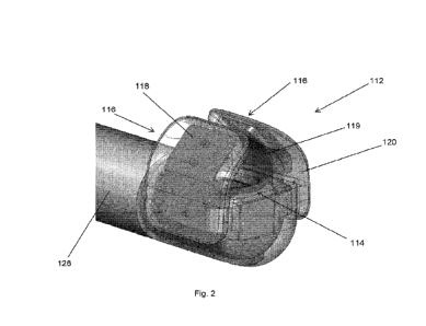
Figure 2 shows the tip 112 in more detail. The tip 112 comprises a central
portion 114
with two flexible wing portions 116 extending therefrom. When the tip 112 is
positioned on the
user's tooth, the central portion 114 sits at or near the occlusal/incisal
surface of the user's
tooth, and the flexible wings contact either side of the tooth; one of the
flexible wings sits
against the buccal/facial side of the tooth, and the other sits against the
lingual/palatal side of
the tooth.
CA 03221312 2023- 12-4
WO 2022/112426
PCT/EP2021/083022
- 11 -
As shown in Figures 2 and 3, each of the flexible wing portions 116 comprises
a support
arm 119 which supports a flex PCB 118, both of which are over-moulded with a
soft and flexible
material 120 (in this case, a UV-curable elastomer is used but other materials
may be used
instead of the UV-curable elastomer; for example a thermoplastic elastomer
(TPE) may be
used). Each of the flexible wing portions 116 comprises four LEDs 122 attached
to the flex PCB
118 and the four LEDs 122 are arranged in a rectangle configuration. By
utilising a flex PCB
118, the flexible wings 116 can be made flexible to ensure that the LEDs 122
remain close to
the tooth in use.
As discussed in more detail below, the transillumination device 100 is capable
of
applying a number of different lighting conditions to the tooth. This is
achievable by having a
plurality of LEDs in the tip 112.
Arranging the LEDs in a rectangular/square configuration or a
parallelogram/rhombus
configuration (or in any configuration where one or more LEDs are closer to
the gum-line than
one or more other LEDs, and where one or more LEDs are laterally offset from
one or more
other LEDs) on each side of the tooth ensures that one or more of the LEDs on
each side is
close enough to the tooth to transilluminate the tooth. Additionally, such an
arrangement allows
for the provision of light from several different angles (for example, close
to the gum-line, and
further up the tooth, and from two lateral directions) so that all evidence of
caries within the
tooth (seen as shadows on a transillumination image) can be imaged. Providing
a plurality of
LEDs with different positions provides a range of options for LED placement
relative to the
tooth, thus providing a technical solution to the problem of finding optimal
LED placement for
transillumination for a user at home (which normally requires extensive
training for a dentist to
realise in a clinic setting using a single light source). Providing a
plurality of LEDs also allows
for variable levels of illumination by turning on different numbers of LEDs.
This allows to get an
increased amount of light into a large tooth (for example, a molar), whereas a
lesser amount of
light is needed for transillumination of a small tooth (for example, an
incisor).
Each LED 122 is configured to emit near-IR light. Here, near-IR light is
defined as light
having a wavelength in the range of 780nm to 3000nm (in accordance with the
ISO 20473
standard). In this case, the LEDs have a peak wavelength at approximately
850nm and a
spectral bandwidth of approximately 35 nm. The radiant intensity (at a forward
current of 100
mA) is approximately 9 to 18 mW/sr, and typically is around 13 mVV/sr. The
flexible wing
portions 116 are configured to flex outwardly slightly when positioned over
the tooth, but a
returning force acts against this outward flexing, so that the tip 112 grips
onto the sides of the
tooth. The flexing of the flexible wing portions 116 ensures that there is
minimal clearance
between each LED 122 and the surface of the tooth; this reduces stray light
which could be
CA 03221312 2023- 12-4
WO 2022/112426
PCT/EP2021/083022
- 12 -
detrimental to imaging, and also ensures that an appropriate amount of light
penetrates into the
tooth to allow for imaging.
The LEDs 122 may also be configured to emit light in the visible spectrum ¨
this allows a
user to visualise that the device is working.
The head portion 110 also comprises a sleeve portion 126 comprising a hollow,
broadly
cylindrical portion. The sleeve portion 126 is configured to be received over
a neck portion 160
of the main body portion 150, such that the head portion 110 and the main body
portion 150 clip
together.
The removable nature of the head portion 110 allows for several different
users, e.g.
members of the same family to use the transillumination device 100, with each
family member
using a different head portion 110. To allow for identification of each head
portion 110 with a
particular user, the head portion comprises an RFID tag (not shown). The RFID
tag is read by a
user's smart device (e.g. a mobile telephone 200 having a display screen, as
shown in Figure
7). Alternatively near-field communication (NFC) between the transillumination
device 100 or
head portion 110 and the user's smart device could be used to identify each
head portion 110.
The central portion 114 of the tip 112 comprises a window 124 in the over-
moulding 120.
The window 124 is configured to face the occlusal/incisal surface of the
user's tooth . When the
head portion 110 is clipped onto the main body portion 150, the window 124 in
the central
portion 114 of the tip 112 aligns with a corresponding window 162 in the neck
portion 160 of the
main body portion 150.
In use, light from the tooth passes through the window 124 in the central
portion 114,
through the corresponding window 162 in the neck portion 160, and into the
neck portion 160.
The neck portion 160 holds an optical assembly for directing the light to a
camera 170. The
optical assembly (shown in Figure 4) comprises a prism 164 which receives
light from the
window 162 and bends it to direct it along the direction of the axis of the
neck portion 160.
Further optical components (an aperture 166 and three piano-convex lenses 168)
in the optical
assembly shape the light beam and focus it onto the imaging area of the camera
170. It will be
appreciated that the optical arrangement may be adapted for use with different
wavelengths of
light from the LEDS.
In this case, the camera 170 is a low-voltage CMOS device. The camera outputs
colour
images, which are converted to greyscale for further analysis. The
transillumination device 100
may carry out such a conversion or this can be performed by user's smart
device (in this case a
mobile telephone 200). As an alternative, a monochromatic camera could be
used. There may
be filters, for example a software filter, coupled to the camera to filter out
different parts of the
spectrum. Image processing of captured images may be performed by the
transillumination
CA 03221312 2023- 12-4
WO 2022/112426
PCT/EP2021/083022
- 13 -
device 100 and/or the user's smart device 200 and/or by a remote server 300.
Such image
processing may include filtering by wavelength, exposure control, white
balance, colour
saturation, hue control, white pixel cancelling, and/or noise cancelling.
Figure 5 shows other internal components in the main body portion 150 of the
transillumination device 100 in an exploded view. These components are shown
in their
installed position in Figure 6. The transillumination device 100 is battery
powered, and
comprises a rechargeable battery 172. The battery 172 is inductively charged
by an inductor
174 which interacts with an inductive charging station, of the kind known in
the art. Similar
inductive charging stations are for example commonly used to charge electrical
toothbrushes.
Alternative power and charging systems are shown in Figures 13a and 13b,
discussed in more
detail below.
The transillumination device 100 comprises means for providing feedback to the
user.
This takes the form of a vibration motor (not shown) and a plurality of user-
feedback LEDs 176
(in this case four) radially spaced around an annular portion 178 of a flex
PCB 184, and which
emit visible light through the material forming main body portion 150. Each
LED 176 is an RGB
LED (i.e. comprising a combination of 3 LEDs: one red; one green; and one blue
in just a single
package). In this way many colors of light can be produced by each LED. The
feedback
comprises an indication to the user that suitable images were or were not
captured by the
device in use (as discussed in more detail below). Alternatively or
additionally, similar feedback
can be provided by the user's smart device.
Control of the operation of the transillumination device 100 is by a
controller 180 in the
form of a system-on-a-chip microcontroller. The controller 180 comprises
integrated Wi-
Fi/Bluetooth capability. The controller may perform any of the image
processing mentioned
above. The controller 180 is located on a main PCB 182 which is connected to
the battery 172.
The main PCB 182 is also connected to the flex PCB 184. The flex PCB 184
comprises the
annular portion 178 and an LED-interface portion 186 located at the end of the
neck portion
160, as well as connector portions of the flex PCB 184 which run between the
main PCB to the
annular portion 178 and from the annular portion 178 to the LED-interface
portion 186.
All connections between the controller 180 and the LEDs 176 on the annular
portion 178
run along the flex PCB 184 (and onto the main PCB 182), and similarly all
connections between
the controller 180 and the connectors on the LED-interface portion 186 run
along the flex PCB
184 (and onto the main PCB 182).
Forming the annular portion 178 and LED-interface portion 186 as portions of a
flex PCB
reduces the cost and complexity of assembly; if the annular portion 178 and
LED-interface
portion 186 were instead formed from rigid PCBs, then additional cable
connections to the main
CA 03221312 2023- 12-4
WO 2022/112426
PCT/EP2021/083022
- 14 -
PCB 182 would be needed. Moreover, using a flex PCB 184 enables the device to
be kept
compact; a flex PCB is very flat which enables a thinner neck portion 160 to
be provided. Since
cable connectors are not needed, this also allows to keep the size down.
A flex PCB is also used because its flexibility can be utilised to bend the
PCB into a
suitable shape ¨ so the annular portion 178 and LED-interface portion 186 are
arranged in a
transverse direction to the axial extent of the main body portion 150.
When the head portion 110 is clipped onto the main body portion 150, the LED-
interface
portion 186 is electrically connected to the flex PCB 118 in the head portion
110. This
connection is achieved via three pogo connectors 127 (see Figure 3b) which are
connected to
the flex PCB 118 in the head portion 110. These pogo connectors 127 pass
through small
holes 128 (see Figure 3a) in the top of the sleeve portion 126 and through
corresponding small
holes 163 in the top of the neck portion 126, and contact the LED-interface
portion 186 of the
flex PCB 184. Whilst two small holes 128 and two corresponding small holes 163
are shown in
Figure 3a, three of each would be provided ¨ one for each of the three pogo
connectors 127.
The flex PCB 118 comprises an LED controller chip 125 for controlling each of
the LEDs
122 individually (i.e. each LED 122 can be separately controlled independently
of the status of
any of the other LEDs 122). The control of the LEDs 122 is done automatically
by the device,
without any input from the user.
The main body portion 150 comprises an outer housing 152 (shown in Figure 6)
and an
inner cradle 154. The inner cradle 154 comprises a series of resilient
projections designed to
form areas into which the various components can be clipped. Therefore, the
inductor 174,
battery 172, main PCB 182, flex PCB 184 (including the annular portion 178 and
LED-interface
portion 186), and camera 170 are all secured within the inner cradle 154
without use of any
screws. This facilitates ease of assembly/maintenance and reduces
manufacturing costs.
The transillumination device 100 may be turned on/off using a user's smart
device 200.
This control (and any other communication between the smart device 200 and
transillumination
device 100) can occur via a Bluetooth connection. Alternatively, one or more
buttons may be
present on the main body portion 150 for this purpose.
The prism 164, aperture 166 and three piano-convex lenses 168 are held within
a
portion of the inner cradle 154 extending into the neck portion 160. The
lenses are held in
cylindrical spacers to ensure that the correct optical path is maintained.
Again, no screws are
needed for securing the components of the optical assembly.
The outer housing 152 is formed from a durable and waterproof plastic, such as
acrylonitrile butadiene styrene (ABS) plastic.
CA 03221312 2023- 12-4
WO 2022/112426
PCT/EP2021/083022
- 15 -
Figure 7 shows a system for enabling detection of caries by a user at home, of
which the
transillumination device 100 is a part. Also part of the system are a user's
smart device (in this
case a mobile telephone 200 having a display screen but it will be appreciated
that any suitable
smart device could be used including a tablet, a laptop, desktop or a purpose-
built smart device)
and a remote server, e.g. a cloud-based server 300 As noted above, the
transillumination
device 100 comprises a system-on-a-chip microcontroller with integrated Wi-
Fi/Bluetooth
capability. This allows communication between the transillumination device 100
and the mobile
telephone 200. The mobile telephone 200 comprises a communications interface
(as is known
in the art) capable of communicating with the cloud-based server 300 via the
internet.
In brief, the mobile telephone 200 runs a software application (an app) which
receives
images from the transillumination device 100, performs processing based on
those images, and
then transmits the images to the cloud-based server 300. The cloud-based
server 300 performs
any further necessary processing of the images which has not already been
carried out by the
mobile telephone, and sends the results back to the mobile telephone 200, to
be displayed to
the user on the display screen of the mobile telephone 200.
Before using the transillumination device 100 for the first time, the user
opens the app
and is taken through a process whereby they register.
Registration of the user comprises the setting of a username and password,
registration
of an email address, acceptance of terms and conditions, selection of a
subscription package if
applicable, entering of payment details, and connection to the user's smart
device. These
registration steps can later be amended.
Registration may be performed for other members of the same household to use
the
same device.
The user also registers with the app if the user has any missing teeth,
fillings in any of
their teeth, implants, prosthetics or any other know conditions. Information
about the user's diet
and/or health conditions may optionally also be collected.
The image capturing process is now described in greater detail. The app
running on the
mobile telephone 200 guides the user during the image capturing process. The
user is directed
to move the transillumination device 100 around one quadrant (i.e. quarter) of
the jaw at a time,
as described below. Each quadrant is defined between the rearmost tooth to the
central front
incisor, for the upper and lower jaw and for the left and right sides of each
¨ i.e. there is an
upper-left quadrant, a lower-left quadrant, an upper-right quadrant and a
lower-right quadrant.
For each quadrant, the user is instructed where to start (e.g. from the
rearmost tooth of
one of the four quadrants) and accordingly places the head portion 110 of the
transillumination
device 100 over the rearmost tooth (or other designated starting point) of the
selected quadrant
CA 03221312 2023- 12-4
WO 2022/112426
PCT/EP2021/083022
- 16 -
and slides it into place so that the central portion 114 is facing the
occlusal/incisal surface of the
tooth, with the flexible wing portions 116 contacting either side of the
tooth. Then the user turns
on the image capturing, for example by clicking a button on the device or the
app and, under the
direction of the app, moves the head portion 110 from tooth to tooth until all
teeth in the selected
quadrant are imaged Under the direction of the app, the user briefly pauses
(e.g. for 1 to 3
seconds) over each tooth to be imaged so that the head portion 110 is
temporarily held
stationary relative to the tooth being imaged. Whilst held stationary,
different lighting conditions
are applied to the tooth (as discussed below), and a plurality of imaged are
captured. Each
image is sent to the mobile telephone 200 for processing by the app.
Alternatively, the transillumination device may perform a pre-selection of the
images,
based on image quality, so that only images meeting certain quality criteria
are sent to the
mobile telephone 200. The quality criteria include one or more of
focus/sharpness, acceptable
lighting (i.e. not over/under exposed) and the presence of a tooth within the
image.
The app comprises an algorithm that detects if a tooth is present within an
image. In this
algorithm the tooth image is segmented, and shape recognition is used to
determine the type of
tooth. Together with the given quadrant, and the known sequence of the teeth,
the imaged tooth
is identified and labelled according to the international standard numbering
system. Based on
the position of the tooth, and the brightness and the contrast in the image,
the app selects the
best image for each tooth from the series of images that are captured for that
tooth. The
selection may be based on quality of the images such as uniformity of
lighting, contrast and
alignment of the image. When/if the quality of the image is approved (i.e. it
meets
predetermined criteria such as those outlined above), visual feedback is
provided to the user via
their smart device to indicate which teeth have successfully been imaged. For
example, the
corresponding tooth will be marked on the model of the teeth presented on the
app (for
example, the corresponding tooth may be shaded green), and the user is
instructed to move to
the next tooth. If no images are approved for one or more teeth because of
insufficient quality
for analysis, the app indicates to the user that the tooth should be re-
imaged.
As an alternative to the user holding the head portion 110 stationary on each
tooth to
acquire images, the user may sweep the head portion 110 across the quadrant
with a slow,
continuous sweeping motion. A series of images are taken in the sweep,
including images
under different lighting conditions for each tooth. If one or more of the
teeth is not successfully
imaged, the app directs the user to perform the sweep again.
Once an image of each tooth is approved and labelled, the images are then sent
from
the mobile telephone 200 to the cloud-based server 300. Such images can be
anonymised prior
to communication and the communication can be end-to-end encrypted to protect
a user's
CA 03221312 2023- 12-4
WO 2022/112426
PCT/EP2021/083022
- 17 -
privacy. The cloud-based server 300 comprises a machine learning algorithm,
which in this
case is a deep learning neural network (running on a processor 320) with the
capability of
identifying caries and categorising the identified caries in transillumination
images of the teeth.
The processor 320 returns a user report regarding the status of each tooth
(i.e. no
caries, or caries, and the classification of caries if they are present) to
the mobile telephone 200
and this is displayed to the user as visual feedback on a picture of the
arrangement of the teeth
(as shown in Figure 8), showing the mapping of teeth and their status.
The user report generated by the processor 320 is encrypted prior to sending
to the
mobile telephone 200. The report does not include any information which would
allow the user
to be identified, i.e. the data is anonymised.
The app displays informative and/or motivational text (for example, "Well
done!") if no
caries are identified. If caries code 1 or 2 are identified, helpful
information regarding
appropriate oral care (for example, "Ensure that you brush for at least two
minutes in the
morning and the evening") is displayed to the user.
If any of the caries are identified as code 3, 4 or 5, the user is advised to
visit a dentist.
The cloud-based server 300 comprises a database of dental clinics, and the app
presents a list
of such clinics which are local to the user (and/or a map showing the location
of these), based
on the user's location data or the user selecting a locality. Alternatively
such a database may be
included in the app or it may be a separate database in its entirety, which
can be accessed by
the cloud-based server or smart device. The user can choose one or more dental
clinics 400a,
400b from this list to be given access to a detailed dental report, so that
the dental clinic(s) can
provide a treatment plan and/or cost estimate to the user for treatment of the
identified caries.
The detailed dental report is a standardised report including images of all of
the user's teeth
captured by the transillumination device 100. The detailed dental report
comprises a clickable
model of the teeth so that the dental professional can select each individual
tooth to see the
corresponding transillumination image and labelled caries. The detailed dental
report does not
include any information which would allow the user to be identified, i.e. the
data is anonymised.
The user is also given the option to add an email address for a dental clinic
(not already
in the database) that they would like to send the report to. A link to
download the report is sent
to that email address, and the dental clinic would then have to sign up and
log in to access to
the report.
To facilitate the dental clinics accessing the user's report, as well as
sending the user's
report to the mobile telephone 200, the processor 320 sends the detailed
dental report to a safe
server 340. The server is safe in that all communication with the safe server
340 is end-to-end
encrypted, and two-step authentication is required to access data from the
safe server 340.
CA 03221312 2023- 12-4
WO 2022/112426
PCT/EP2021/083022
- 18 -
When a user chooses one or more dental clinics to be given access to their
report, a link
is sent from the safe sever 340 to the dental clinic(s) 400a, 400b to enable
them to access the
report from the safe server 340.
As noted above, communication between the dental clinic(s) 400a, 400b and safe
server
340 is end-to-end encrypted, and the dental clinic(s) 400a, 400b are
authenticated by two-factor
authentication. Once authenticated, the dental clinic is able to download the
user's report from
the safe server 340.
A dental practitioner at the dental clinic reviews the images and the
identified caries, and
determines a treatment plan and/or cost estimate and may suggest an
appointment
time/location. This information is then sent back to the safe server 340. The
safe server 340
receives the cost estimate and/or treatment plan and delivers this via the app
to the user. The
user can then contact a dental clinic 400a, 400b to arrange treatment.
The lighting features of the transillumination device 100 will now be
described in greater
detail. As noted above, the first and second flexible wing portions 116 each
comprise a plurality
of LEDs. The LEDs 122 are individually controllable, and each is connected to
the controller
180 via the flex PCB 184 in the main body portion 150 (comprising the LED-
interface portion
186), the pogo connectors, and the flex PCB 118 in the head portion 110.
Because the LEDs
122 are individually controllable, the transillumination device 100 has the
capability of
illuminating teeth with different sets of LEDs illuminated, so as to create a
plurality of different
lighting conditions.
Figures 9 and 10 give further details regarding the positioning and control of
the LEDs
122 in the tip 112.
Each flexible wing portion 116 is defined by four edges. These are labelled in
Figure 9.
Firstly, there is a base edge 117a, where the flexible wing portion 116 meets
the central portion
114, and an opposed opening edge 117b, where the flexible wing portion defines
the slot for
receiving a tooth. Then, transverse to the base edge and opening edge are the
proximal edge
117c (nearest to the main body portion 150 of the device) and the opposed
distal edge 117d
(furthest from the main body portion 150 of the device).
Each flexible wing portion 116 comprises four LEDs 122. As shown in Figure 9,
these
are arranged so as to be positioned at the corners of a rectangle. Here, two
sides of the
rectangle are broadly parallel to the base edge 117a and the opening edge
117b, and two sides
of the rectangle are broadly parallel to the proximal edge 117c and the distal
edge 117d. The
LEDs 122 are spaced apart by 1.75mm (the perpendicular distance from edge-to-
edge) along
the sides of the rectangle parallel to the proximal edge 117c and the distal
edge 117d, and are
CA 03221312 2023- 12-4
WO 2022/112426
PCT/EP2021/083022
- 19 -
spaced apart by 5mm (the perpendicular distance from edge-to-edge) along the
sides of the
rectangle parallel to the base edge 117a and the opening edge 117b.
The LEDs are individually controllable (such that each can be turned on or off
independently of the status of the others), and this allows for multiple
different lighting
conditions to be achieved.
Figures 10a-10n show schematically different lighting conditions that can be
obtained.
The arrangements show are not exhaustive or limiting in any way, but are
simply illustrative of
some of the many lighting conditions which could be achieved. In these
figures, an illuminated
LED is represented as a shaded circle. In these figures, the distances between
the LEDs have
been exaggerated for ease of illustration.
In general, at least one LED is illuminated on each side. This is
advantageous, but not
limiting on the invention. Similarly, it is generally advantageous light is
illuminated further from
the tip of the tooth (i.e. closer to the gum-line). The camera captures images
from near the tip
of the tooth, so to image the greatest possible volume of the tooth, and in
particular to capture
as much as possible of the shadows indicative of caries, it is advantageous to
have light
entering the tooth further from the tip of the tooth, near the gum-line. If
the tooth is illuminated
too far from the gum-line, the shadows nearer the gum-line will be hidden.
Figures 10a-10c show lighting conditions with two LEDs illuminating the tooth
at the
same time. Figures 10d and be show lighting conditions with three LEDs
illuminating the tooth
at the same time. Figures 10f -10k show lighting conditions with four LEDs
illuminating the
tooth at the same time. Figure 101 shows a lighting condition with five LEDs
illuminating the
tooth at the same time, and Figures 10m-10n show lighting conditions with six
LEDs illuminating
the tooth at the same time. Seven, or even all eight of the LEDs could also be
lit at the same
time.
As discussed above, the LEDs can each be controlled such that each is either
on, or off.
However, instead of an LED being off, it could instead be illuminated at a
reduced intensity.
Whilst eight LEDs 112 have been described above, a greater number of (or
fewer) LEDs
could instead be provided. Additional lighting conditions would be available
for a greater
number of LEDs.
Whilst the LEDs described above all have the same nominal spectral
characteristics
(including the same peak wavelength and peak width), it will be appreciated
that one or more of
the LEDs could have different spectral characteristics from the others (with
peak wavelengths
still within the near-IR range) to allow for images to be captured under a
plurality of near-IR
wavelengths.
CA 03221312 2023- 12-4
WO 2022/112426
PCT/EP2021/083022
- 20 -
The plurality of lighting conditions can be cycled through as the
transillumination device
100 is moving over the teeth, so that for each tooth, at least one image is
taken under each of
the plurality of lighting conditions. At least one of the plurality of
lighting conditions should result
in the capture of a suitable image for caries analysis, but the particular
lighting condition which
will achieve this will vary depending on the tooth being imaged. For example,
for a molar which
has had a filling, a suitable lighting condition could be that all the LEDs
122 should be
illuminated. However, when an incisor is imaged under the same lighting
condition, it is likely
that the resulting image would be over-exposed (because too much light was
incident into the
tooth). In that case, a suitable lighting condition would include only a
subset of the LEDS 122
(i.e. not all of the LEDs 122) begin illuminated. For example, half of the
LEDs could be
illuminated.
As an alternative to cycling through all lighting conditions for each tooth,
the lighting
conditions may be applied based on knowledge of the particular tooth that is
being imaged, with
a predetermined lighting condition being applied for that particular tooth.
Knowledge of the
particular tooth that is being imaged may be obtained through shape-
recognition of the tooth (for
example using machine learning techniques and based on geometrical patterns),
and/or
through knowing the expected sequence of teeth to be imaged, for example. If
the resulting
image is not acceptable (because the lighting condition does not allow the
tooth to be imaged
correctly), then a different lighting condition may be applied.
The first lighting condition to be applied to a given tooth may be pre-set
based on a prior
calibration process carried out by the user on first use of the device. In the
calibration process, a
plurality of lighting conditions may be sequentially applied to the tooth, and
an image may be
acquired under each lighting condition. Then, the images are analysed to
determine the highest
quality image. The lighting condition under which the highest quality image
was acquired is
then pre-set as the first lighting condition. The highest quality image may
for example be one
with minimal reflections from stray light, and/or an image with the correct
exposure (not
overexposed by putting too much light into the tooth, or underexposed by
putting too little light
into the tooth), and/or an image with appropriate brightness and contrast.
It will be appreciated that rather than using a single captured image, it is
possible to
capture and select multiple images of the same tooth and use parts of each
image, or multiple
overlaid images of the same tooth in the trained neural network of the remote
server.
For example, it is possible to capture multiple images of a tooth under
illumination by
near-IR at different wavelengths (for example where one or more LEDs are
provided having
different wavelength characteristics from the others). The multiple images of
the same tooth
can then be combined to make a combined image as an input to the machine
learning
CA 03221312 2023- 12-4
WO 2022/112426
PCT/EP2021/083022
-21 -
algorithm. Each image of the multiple images in the combined image can be
combined with the
others with a weighting factor of between 0 and 1, where 0 means no
contribution and 1 means
complete contribution.
Figures 11 a and llb show the tip 112a of a head portion of a
transillumination device,
similar to the tip shown in Figure 2. The tip 112a comprises a central portion
114a with two
flexible wing portions 116a extending from the central portion 114a. When the
tip 112a is
positioned on the user's tooth, the central portion 114a sits at or near the
occlusal/incisal
surface of the user's tooth, and the flexible wing portions contact either
side of the tooth; one of
the flexible wing portions sits against the buccal/facial side of the tooth,
and the other sits
against the lingual/palatal side of the tooth. Each of the flexible wing
portions 116a comprises
four LEDs 122a. These are attached to a flex PCB (not shown) supported by a
support arm
(not shown) forming part of each of the flexible wing portions 116a.
The flexible wing portions 116a and central portion 114 comprise a soft and
flexible over-
moulding. In this case, a UV-curable elastomer is used for the majority of the
over-moulding,
with an IR-transparent silicone material being used for the over-moulding over
the LEDs 122a.
Other materials may be used instead of the UV-curable elastomer; for example a
thermoplastic elastomer (TPE) may be used. The central portion 114a of the tip
112a
comprises a window (i.e. an aperture) 124a in the over-moulding. The window
124a is
configured to face the occlusal/incisal surface of the user's tooth.
The flexible wing portions 116a are flexible to ensure that the LEDs 122a
remain close to
the tooth in use. The flexible wing portions 116a are configured to flex
outwardly slightly when
positioned over the tooth, but a returning force acts against this outward
flexing, so that the
flexible wings 116a grip onto the sides of the tooth. The flexing of the
flexible wing portions
116a ensures that there is minimal clearance between each LED 122a and the
surface of the
tooth; this reduces stray light which could be detrimental to imaging, and
also ensures that an
appropriate amount of light penetrates into the tooth to allow for imaging.
Each LED 122a is configured to emit near-IR light, having a peak wavelength at
approximately 850nm and a spectral bandwidth of approximately 35 nm. The
radiant intensity
(at a forward current of 100 mA) is approximately 9 to 18 mW/sr, and typically
is around 13
mW/sr. The LEDs 122a may also be configured to emit light in the visible
spectrum ¨ this
allows a user to visualise that the device is working.
Each flexible wing portion 116a is defined by four edges. These are labelled
in Figure
lib. Firstly, there is a base edge 117a, where the flexible wing portion 116a
meets the central
portion 114a, and an opposed opening edge 117b, where the flexible wing
portion defines the
slot for receiving a tooth. Then, transverse to the base edge and opening edge
are the proximal
CA 03221312 2023- 12-4
WO 2022/112426
PCT/EP2021/083022
- 22 -
edge 117c (nearest to the main body portion 150 of the device) and the opposed
distal edge
117d (furthest from the main body portion 150 of the device).
As shown in Figure 11a, two LEDs are located so as to be further from the tip
of the
tooth when the head portion is in place on a tooth (the upper two LEDs as
illustrated in Figure
11a, closest to the base edge 117a), and two LEDs are located so as to be
positioned closer to
the tip of the tooth when the head portion is in place on a tooth (the lower
two LEDs as
illustrated in Figure 11a, furthest from base edge 117a). The two LEDs located
so as to be
further from the tip of the tooth when the head portion is in place on a tooth
are vertically offset
from one another. The two LEDs located so as to be closer to the tip of the
tooth when the
head portion is in place on a tooth are vertically offset from one another.
The vertically offset
position allows to cover a slightly larger extent of the tooth in the vertical
direction, compared to
the configurations shown in previous figures, with side-by-side LEDs.
That is, the four LEDs 122a on a flexible wing portion 116a sit at the corners
of a
rhombus/parallelogram. Two sides of the rhombus/parallelogram are parallel to
the proximal
edge and the distal edge 117d, and the other two sides are non-parallel to the
base edge 117a.
At least one LED 122a should be located adjacent to the gum-line when in use,
no
matter which tooth is being imaged. Here, adjacent to the gum-line means close
to, but slightly
vertically offset from the gum-line, so that light is still directed into the
tooth rather than into the
gum. To image the greatest possible volume of the tooth, and in particular to
capture as much
as possible of the shadows indicative of caries, it is advantageous to have
light entering the
tooth from near the gum-line. It depends on the tooth as to which of the LEDs
122a is in the
best position; on a molar in the lower jaw for example the best-positioned LED
122a may be the
one positioned lowermost down the tooth when the head portion is in place on a
tooth. On an
incisor or canine in the lower jaw, for example, that same LED 122a may be
located below the
gum-line, in which case it is not best-positioned to illuminate the tooth. In
such a case, the best
positioned LED 122a may be one located closest to the tip of the tooth when
the head portion is
in place on the tooth.
The flexible wing portions 116a shown in Figures 11 a and lib each comprise
two
projections facing towards the opposite flexible wing portion 116a (i.e.
facing inwardly towards
the slot between the flexible wing portions 116a which receives the tooth in
use). A first
projection 116b on a flexible wing portion 116a runs along the opening edge
117b of the flexible
wing portion 116a. This first projection 116b is in order to position the LEDs
at a small distance
from the surface of the tooth. This allows for the LED light to be more evenly
spread, compared
to the case where the LEDs were closer to the tooth. When the LEDs are too
close to the tooth,
CA 03221312 2023- 12-4
WO 2022/112426
PCT/EP2021/083022
- 23 -
there may be an uneven spread of light, and a risk of partial overexposure in
the captured
images.
The second projection 116c runs from the proximal edge 117c (nearest to the
main body
portion 150 of the device) to the opposed distal edge 117d (furthest from the
main body portion
150 of the device), and is located between the LEDs 122a and the base edge
117a. This
second projection 116c should prevent or reduce light from being transmitted
directly from the
LEDs 122a into the camera. In the embodiment shown, the second projection 116c
slopes
down towards the base edge 117a in the direction from the distal edge 117d to
the proximal
edge 117c.
The central portion 114a comprises alignment projections 125a and 125b. The
first
alignment projection 125a projects away from the central portion 114 and runs
across the top of
the tip 112a (where the top of the tip is the end of the tip furthest from the
main body portion
150). The second alignment projection 125b is broadly parallel to the first
alignment projection
125a, but is located the other side of the flexible wing portions 116a than
the first alignment
projection 125a. The first and second alignment projections therefore bracket
the flexible wing
portions 116a. The first and second alignment projections are broadly
perpendicular to the
flexible wing portions 116a. The first and second alignment projections aid
the user to hold the
device parallel to the row of teeth.
As shown in Figure 11 b, in this embodiment, on each flexible wing portion
116a there
are two groups of two LEDs ¨ a base group (the two LEDs closest to the base
edge 117a) and
a top group (the two LEDs furthest from the base edge 117a). Each group is
independently
controllable, so there are a total of four independently controllable groups.
Labelling one
flexible wing portion 116a as the "left-hand" wing, and one as the "right-
hand" wing, we have:
- a right-hand base group;
- a right-hand top group;
- a left-hand base group; and
- a left-hand top group.
Some exemplary lighting conditions (combinations 1 to 10) which can be
achieved with such
a configuration are listed below. Here, only the groups which are illuminated
are mentioned.
The other groups which are not mentioned are not illuminated.
¨ Combination 1 ¨ both base groups (left-hand and right-hand) lit
¨ Combination 2 ¨ both top groups (left-hand and right-hand) lit
¨ Combination 3 ¨ right-hand base group lit
¨ Combination 4 ¨ right-hand top group lit
¨ Cornmination 5 ¨ left-hand base group lit
CA 03221312 2023- 12-4
WO 2022/112426
PCT/EP2021/083022
- 24 -
¨ Combination 6 ¨ left-hand top group lit
¨ Combination 7 ¨ right-hand top and right-hand base groups lit
¨ Combination 8 ¨ left-hand top and left-hand base groups lit
¨ Combination 9 ¨ right-hand base group and left-hand top group lit
¨ Combination 10¨ right-hand top group and left-hand base group lit
In these combinations, the maximum number of LEDs lit simultaneously is four,
but more
may be used. Using four or fewer LEDs at any one time reduces the risk of
reflection of the LED
light, which can be detrimental to image quality.
For a given tooth, a pre-set combination (or combinations) is used. These may
be
identified in a calibration process carried out by the user where on first
usage of the device, all
of the combinations will be cycled through for each tooth, and an appropriate
condition/conditions will be identified for each tooth. The appropriate
combination(s) depends
on tooth size (height and thickness) and position.
Combinations 3 to 6 provide light coming from only one side of the teeth.
These
combinations are particularly suitable for imaging incisors. The light should
then be directed
from the labial (facial) side of the incisor, through the tooth, and out of
the lingual/palatal side of
the tooth. This is because the lingual/palatal side of an incisor has a
sloping shape, which can
create strong reflections of the LED light, leading to poor imaging The
incisors are thinner than
other teeth, and images of sufficient quality can be obtained with light
coming from only one
side. Whether a base group of top group is used for a given tooth depends on
the height of the
tooth.
Combinations 7 to 9 might be used for example when imaging very large molars,
where
additional light is needed to obtain images of sufficient quality.
The groups of LEDs can each be controlled such that every LED in a group is
either on,
or off. However, instead of a group being off, the LEDs within that group
could instead be
illuminated at a reduced intensity.
Whilst eight LEDs 112a have been described above, a greater number of (or
fewer)
LEDs could instead be provided.
Whilst the LEDs 122a described above all have the same nominal spectral
characteristics (including the same peak wavelength and peak width), it will
be appreciated that
one or more of the LEDs could have different spectral characteristics from the
others (with peak
wavelengths still within the near-IR range) to allow for images to be captured
under a plurality of
near-IR wavelengths.
CA 03221312 2023- 12-4
WO 2022/112426
PCT/EP2021/083022
- 25 -
Rather than using a single captured image, it is possible to capture and
select multiple
images of the same tooth and use parts of each image, or multiple overlaid
images of the same
tooth in the trained neural network of the remote server.
For example, it is possible to capture multiple images of a tooth under
illumination by
near-IR at different wavelengths (for example where one or more LEDs are
provided having
different wavelength characteristics from the others). The multiple images of
the same tooth
can then be combined to make a combined image as an input to the machine
learning
algorithm. Each image of the multiple images in the combined image can be
combined with the
others with a weighting factor of between 0 and 1, where 0 means no
contribution and 1 means
complete contribution.
The control scheme identified above (with four groups of two LEDs, operable in
some or
all of the combinations listed above) is not limited to usage with the tip
112a shown in Figures
11 a and 11 b, but could instead be implemented with the tip 112 discussed in
relation to the
earlier Figures.
Figures 12a and 12b show images produced by transillumination with a caries
categorised as code 3. The image on the left shows the caries identified by a
dentist as
category 3 caries, whereas the image on the right shows the regions where the
machine
learning model identified the location of caries, which were also categorized
as code 3 by the
machine learning model.
The machine learning algorithm is trained on a training set of images
comprising 1000
images of each category of caries (category 1 to category 5), as identified by
a qualified dentist.
Whilst one configuration for a transillumination device has been described
above, other
variations are possible. Some possible alternative configurations are
described below.
As discussed above, one configuration of the transillumination device
comprises a
rechargeable battery 172 which is inductively charged by an inductor 174. The
inductor 174 sits
in the base of the main body portion, and interacts with an external inductive
charging station, of
the kind known in the art. Similar inductive charging stations are for example
commonly used to
charge electrical toothbrushes. An alternative configuration is discussed
below.
It is advantageous for the transillumination device to be able to communicate
wirelessly
(via Bluetooth for example) with the user's smart device (e.g. mobile phone)
whilst the
transillumination device is in use, i.e. being held by the user in their hand.
Since water in the
user's body (e.g. in their hand/mouth) absorbs Wi-Fi signals (e.g. 2.45GHz
radio waves), it is
advantageous to position the Wi-Fi antenna at a position at which the user's
hand or mouth
does not surround the Wi-Fi antenna. A suitable position is the base of the
main body portion of
the transillumination device. However, the inductor (comprising ferrite
material) forming part of
CA 03221312 2023- 12-4
WO 2022/112426
PCT/EP2021/083022
- 26 -
the indicative charging circuit also absorbs Wi-Fi signals (e.g. 2.45GHz radio
waves). For this
reason, to allow the Wi-Fi antenna to be positioned in the base, the
indicative charging circuit
must be moved away from the conventional position in the base.
Figure 13a shows an exploded view of components in the interior of a
transillumination
device 500. In the configuration shown, the inductor 574 for charging the
rechargeable battery
572 is located in a middle portion 500a of the transillumination device 500.
There is also a Wi-
Fi antenna (not shown) positioned in the base portion 500b of the device.
Thus, the Wi-Fi
antenna is in a position where it is unobstructed by the user or the inductor
574.
It will be appreciated that with the inductor 574 located in a middle portion
500a of the
device 200, an inductive charging station for charging the device 500 will
have to take a
different form compared to that previously discussed. For example, the
inductive charging
station may sit next to or wrap around the middle portion 500a of the device,
rather than
interacting with the base.
Figure 13b shows a schematic view of an inductive charging arrangement 600
used to
charge the battery in the devices described herein. The charging arrangement
600 comprises
two ferrite rods 601 (with a diameter of approximately 4mm and a length of
approximately
30mm). One of the two rods 601 is mounted on a PCB of the transillumination
device and the
other is positioned in a separate inductive charging device. Each of the rods
is surrounded (in
part) by a copper coil 602a, 602b with a thickness of 0.25mm, this forming an
inductor in each
of the handheld device and charging device. When assembled in the devices (the
handheld
transillumination device and the charging device respectively) a 2.5mm
tolerance gap is left
between the outside of the coils 602a, 602b of the respective inductors to
allow for walls of the
respective devices and tolerance between them during charging.
During charging, the coils 602a, 602b are each part of a resonant circuit, the
two
resonant circuits are loosely magnetically coupled. The coil 602a is arranged
to receive energy
through inductive coupling with the coil 602b of the charging station.
Alternating current passes
through the induction coil 602b in the charging station. The moving electric
charge from this
creates a magnetic field, which fluctuates in strength because the electric
current's amplitude is
fluctuating. This changing magnetic field, enhanced by the rods 601, creates
an alternating
electric current in the coil 602a of the device, which in turn passes through
a rectifier to convert
it to direct current. The direct current is then used to charge the
rechargeable battery of the
transillumination device.
Figures 14a and 14b show the interior of a transillumination device 700. The
device
comprises a main body portion 701 including an outer housing 702 having a base
portion 703, a
middle portion 704 and a neck portion 705. On the neck portion there is
mounted a head portion
CA 03221312 2023- 12-4
WO 2022/112426
PCT/EP2021/083022
- 27 -
800, in the form of a tip as previously described. Inside the housing 702
there can be seen a
battery 707 arranged for inductive charging as described above, along with
rigid printed circuit
boards 708, 709 (PCBs) orthogonal to one another for holding various
electronic components.
This maximises the space available for mounting components.
In the neck portion 705, seen more clearly in Figures 14b and 14c, there is a
transparent
window 710 made of sapphire glass that allows the passage of light into the
interior of the neck
portion 705. Inside of the neck portion an optical assembly 711 can be seen.
This window is
resistant to scratching, which is important as it is exposed during use of the
device, in a user's
mouth. The optical assembly comprises a camera 712 that is positioned opposite
(i.e. behind)
the transparent window 710. Thus, the transparent window protects the optical
assembly. The
optical assembly also comprises a plastic housing 713 above the camera, an
aperture film 714,
a lens 715, a metal frame 716 for holding the transparent window 710 in place
and a rubber
gasket 717 for holding the optical assembly together. The camera 712 is
mounted on a flexible
PCB 718 within the neck portion 705 of the main body portion of the device.
The camera 712 is
also secured to the rigid PCB 709 (along with the other components of the
optical assembly) to
provide support and reduce relative movement of these components.
A heater (not shown) is arranged to heat the metal frame 716, which in turn
conducts
heat to the transparent window 710 and prevents fogging/condensation in use.
To do so the
window may be heated to 40 C within 20 seconds, for example. In use, with a
tip 800 mounted
as shown, transillumination of a tooth is performed and the camera 712
captures images
through the transparent window 710. In use the transparent window 710 is
exposed to the
conditions (heat and humidity) of a user's month.
Figures 15a to 15c show the internal arrangement of PCBs 708, 709 and
electronic
components more clearly. Figure 15a shows a first rigid PCB 708 and a second
rigid PCB 709.
These are oriented substantially at a right angle to one another to allow
maximum space for
mounting electronic components inside of the housing. The first PCB 708 has an
antenna 719
(positioned in the base of the device) and a cradle 720 mounted upon it; the
cradle holds the
battery 707. The second PCB 709 has a connection point 721 mounted on it, in
the neck portion
of the device. The connection point 721 is electrically connected to a pogo
connector that
extends through the housing of the main body to which a corresponding pogo
connector of a
head portion can be connected. When the tip 800 is mounted on the neck portion
(as shown in
figure 14c) this connection point 721 therefore provides an electrical
connection point between
the main body portion of the device and the head portion. This makes is easy
for the tip to be
mounted and removed from the device, for example to swap it with another. Also
attached to
the second PCB 209 is a flexible PCB 718 on which the optical assembly 711 is
mounted.
CA 03221312 2023- 12-4
WO 2022/112426
PCT/EP2021/083022
- 28 -
Figures 16a and 16b show the tip arrangement in more detail. The flexible PCB
801 in
the tip extends into first and second flexible wing portions 802, 803
extending from a central
portion of the tip. On each of these portions are mounted LEDs 804 for
illuminating a tooth and
performing the transillumination method previously described. The optical
assembly 711 within
the neck portion of the device can also be seen in this figure. The
transparent window 710 of
the optical assembly aligns with an aperture 805 in the tip.
Figure 16b shows an exploded view of the tip portion 800, including the
flexible PCB
801, and a flexible over-moulding 806 which encapsulates the flexible PCB 801.
It will be appreciated that the devices and methods described herein could be
adapted
for use on the teeth of animals. In such a case, the device would be more
robust that a device
made for human-use (comprising a stronger casing to withstand chewing by an
animal, for
example). The user of the device would then be a veterinary professional or
possibly the
animal's owner. Different sized/shaped head portions 110 for the
transillumination device 100
would also be provided, with a size and shape appropriate for use with
different animals ¨ for
example, dogs, cats and horses. The LEDs 122 and lens/prism arrangements could
also be
adapted accordingly.
CA 03221312 2023- 12-4