Note: Descriptions are shown in the official language in which they were submitted.
WO 2022/261448
PCT/US2022/033024
THROMBUS REMOVAL SYSTEMS AND ASSOCIATED METHODS
CROSS REFERENCE TO RELATED APPLICATIONS
[0001] This application claims the benefit of priority to U.S.
Provisional Application Nos.
63/209,257, filed June 10, 2021, 63/250.089, filed September 29, 2021,
63/285,054, filed
December 1, 2021, 63/335,656, filed April 27, 2022, each of which is herein
incorporated by
reference in its entirety.
INCORPORATION BY REFERENCE
[0002] All publications and patent applications mentioned in this
specification are herein
incorporated by reference to the same extent as if each individual publication
or patent
application was specifically and individually indicated to be incorporated by
reference.
FIELD
[0003] The present technology generally relates to medical devices and, in
particular, to
systems including aspiration and fluid delivery mechanisms and associated
methods for
removing a thrombus from a mammalian blood vessel.
BACKGROUND
[0004] Thrombotic material may lead to a blockage in fluid flow within the
vasculature of a
mammal. Such blockages may occur in varied regions within the body, such as
within the
pulmonary system, peripheral vasculature, deep vasculature, or brain.
Pulmonary embolisms
typically arise when a thrombus originating from another part of the body
(e.g., a vein in the
pelvis or leg) becomes dislodged and travels to the lungs. Anticoagulation
therapy is the current
standard of care for treating pulmonary embolisms, but may not be effective in
some patients.
Additionally, conventional devices for removing thrombotic material may not be
capable of
navigating the tortuous vascular anatomy, may not be effective in removing
thrombotic material,
and/or may lack the ability to provide sensor data or other feedback to the
clinician during the
thrombectomy procedure. Existing thrombectomy devices operate based on simple
aspiration
which works sufficiently for certain clots but is largely ineffective for
difficult, organized clots.
Many patients presenting with deep vein thrombus (DVT) are left untreated as
long as the risk of
limb ischemia is low. In more urgent cases, they are treated with catheter-
directed thrombolysis
or lytic therapy to break up a clot over the course of many hours or days.
More recently other
tools like clot retrievers have been developed to treat DVT and pulmonary
embolism (PE), but
these tools are not being widely adopted because of their limited
effectiveness and additional
1
CA 03221894 2023- 12- 7
WO 2022/261448
PCT/US2022/033024
costs versus aspiration or the standard of case. Other recent developments
focus on slicing or
macerating the clot, but these mechanisms are designed to reduce the risk of
the catheter
clogging and do not address the problem of tough, large, organized clots.
There remains the
need for a device to address these and other problems with existing venous
thrombectomy
including, but not limited to, a fast, easy-to-use, and effective device for
removing a variety of
clot morphologies.
BRIEF DESCRIPTION OF THE DRAWINGS
[0005] The novel features of the invention are set forth with
particularity in the claims that
follow. A better understanding of the features and advantages of the present
invention will be
obtained by reference to the following detailed description that sets forth
illustrative
embodiments, in which the principles of the invention are utilized, and the
accompanying
drawings of which:
[0006] FIGS. 1-1L illustrate various views of a portion of a
thrombus removal system
including a distal portion of an elongated catheter configured in accordance
with an embodiment
of the present technology.
[0007] FIGS. 2A-2D illustrate plan views of various configurations
of irrigation ports and
fluid streams of a thrombus removal system according to embodiments of the
present
technology.
[0008] FIGS. 3A-3H illustrate an elevation view of various configurations
of irrigation ports
of a thrombus removal system according to embodiments of the present
technology.
[0009] FIGS. 4A-4P illustrate an elevation view of various
configurations of irrigation ports
and fluid streams of a thrombus removal system according to embodiments of the
present
technology.
[0010] FIGS. 5A-5G illustrate various configurations of irrigation ports of
a thrombus
removal system according to embodiments of the present technology.
[0011] FIGS. 6A-6C illustrate various embodiments of a thrombus
removal system including
a saline source, an aspiration system, and one or more controls for
controlling irrigation and/or
aspiration of the system.
[0012] FIGS. 7A-7D illustrate various configurations of clog detection
and/or clog removal
features of a thrombus removal system.
[0013] FIGS. 8A-8C illustrate one embodiment of controlling various
irrigation ports of a
thrombus removal system.
[0014] FIG. 9A is a system schematic diagram of a thrombus removal
system.
2
CA 03221894 2023- 12- 7
WO 2022/261448
PCT/US2022/033024
[0015] FIG. 9B is one embodiment of a thrombus removal system
including one or more
sensors configured to detect a clot.
[0016] FIG. 10 is a table showing various system states of a
thrombus removal system.
[0017] FIG. 11 is a procedure flow chart of various system states
of a thrombus removal
system.
[0018] FIGS. 12A-12B illustrate pressure waveform graphs during a
clot engagement state.
[0019] FIG. 13 is a simplified system schematic of a thrombus
removal system.
[0020] FIG. 14 is one embodiment of a flow waveform of a thrombus
removal system.
[0021] FIG. 15 illustrates an aspiration scheme of a thrombus
removal system.
[0022] FIGS. 16A-16D illustrate one embodiment of a thrombus removal
system.
[0023] FIG. 17 illustrates various irrigation pump cycles of a
thrombus removal system.
[0024] FIG. 18 illustrates a thrombus removal system with a valve
near the aspiration source.
[0025] FIGS. 19A-19B illustrate a thrombus removal system with a
plurality of struts in the
funnel.
[0026] FIGS. 20A-20B illustrate a thrombus removal system with a
hemispherical funnel.
[0027] FIG. 21 is a flowchart describing a method of assessing a
volume of clot removed
during treatment.
[0028] FIG. 22 is a flowchart describing various mechanisms of
action of the fluid streams
disclosed herein.
SUMMARY OF THE DISCLOSURE
[0029] A thrombus removal is provided, comprising an elongate shaft
comprising a working
end, at least one fluid lumen in the elongate shaft, and two or more apertures
disposed at or near
the working end, the two or more apertures in fluid communication with the
least one fluid
lumen and configured to generate two or more fluid streams that at least
partially collide at an
interaction region, the two or more fluid streams having a flow rate
sufficient to create cavitation
in the interaction region that is configured to mechanically fractionate a
target thrombus.
[0030] A thrombus removal device is also provided, comprising an
elongate shaft
comprising a working end, at least one fluid lumen in the elongate shaft, and
two or more
apertures disposed at or near the working end, the two or more apertures in
fluid communication
with the least one fluid lumen and configured to generate two or more fluid
streams that interact
within or near the working end at an interaction region, the two or more fluid
streams having a
flow rate and proximity sufficient to induce cavitation at the interaction
region that is configured
to mechanically morcellate a target thrombus.
3
CA 03221894 2023- 12- 7
WO 2022/261448
PCT/US2022/033024
[0031] In some embodiments, the two or more fluid streams each have
a flow rate ranging
between 50m/s and 90m/s.
[0032] In other embodiments, the two or more fluid streams each
have a flow rate of at least
50nVs.
[0033] In some examples, fluid flowing within the at least one fluid lumen
at a lumen flow
rate of 3m/s results in the two or more fluid streams having a flow rate of at
least 50m/s.
[0034] In other embodiments, fluid flowing within the at least one
fluid lumen at a lumen
flow rate of 4m/s results in the two or more fluid streams having a flow rate
of at least 70m/s.
[0035] In some examples, fluid flowing within the at least one
fluid lumen at a lumen flow
rate of 5m/s results in the two or more fluid streams having a flow rate of at
least 90m/s.
[0036] In one embodiment, the interaction region comprises a focal
point of the two or more
fluid streams.
[0037] In some embodiments, the two or more fluid streams are
generally orthogonal to a
longitudinal axis of the elongate shaft.
[0038] In some examples, the two or more fluids streams are directed
distally such that the
focal point is distal relative to the two or more apertures.
[0039] In one embodiment, the distally directed two or more fluid
streams are further
configured to generate a cavitation column that extends distally from the
focal point.
[0040] In some embodiments, the two or more fluids streams are
directed proximally such
that the focal point is proximal relative to the two or more apertures.
[0041] In one embodiment, the proximally directed two or more fluid
streams are further
configured to generate a cavitation column that extends proximally from the
focal point.
[0042] In some examples, a cavitation detection sensor is disposed
on or within the thrombus
removal device.
[0043] In some embodiments, the cavitation detection sensor is disposed on
or within a
funnel at the working end of the thrombus removal device.
[0044] In another embodiment, the cavitation detection sensor is
disposed on or within an
aspiration lumen at the working end of the thrombus removal device.
[0045] In some examples, the cavitation detection sensor comprises
an ultrasound transducer
element.
[0046] In other embodiments, the cavitation detection sensor
comprises a hydrophone.
[0047] In some examples, the cavitation detection sensor comprises
a laser.
[0048] In other embodiments, the cavitation detection sensor
comprises a microphone.
[0049] Another embodiment includes a real-time imaging device
configured to image the
cavitation in real-time. In some embodiments, the real-time imaging device
comprises an
4
CA 03221894 2023- 12- 7
WO 2022/261448
PCT/US2022/033024
ultrasound imaging device. In some embodiments, the ultrasound imaging device
comprises an
external ultrasound imaging probe. In other embodiments, the ultrasound
imaging device
comprises a catheter-based ultrasound imaging device.
[0050] A method for removing a thrombus from a blood vessel of a
patient is provided, the
method comprising introducing a distal portion of an elongate catheter to a
thrombus location in
a blood vessel, drawing at least a section of the thrombus into the distal
portion, and generating
two or more fluid streams having a flow rate of at least 20 m/s that interact
at an interaction
region to create cavitation within the thrombus.
[0051] A method for removing a thrombus from a blood vessel of a
patient is also provided,
the method comprising introducing a distal portion of an elongate catheter to
a thrombus location
in a blood vessel, drawing at least a section of the thrombus into the distal
portion, and
generating two or more fluid streams having a flow rate of at least 50 m/s
that interact at an
interaction region to create cavitation within the thrombus.
[0052] A method for removing a thrombus from a blood vessel of a
patient is provided, the
method comprising introducing a distal portion of an elongate catheter to a
thrombus location in
a blood vessel, drawing at least a section of the thrombus into the distal
portion, and generating
two or more fluid streams that interact within or near the distal portion at
an interaction region,
wherein the two or more fluid streams are configured to apply at least four
distinct breaking
forces to the thrombus including: 1) a slicing force as the two or more fluid
streams initially cut
through the thrombus prior to meeting at the interaction region; 2) a
cavitation force at the
interaction region when the two or more fluid streams interact to generate
cavitation; 3) a
shearing force caused by the two or more fluid streams moving against each
other to generate
shearing cavitation; and 4) a rotational fluid motion force caused by the
shearing force and the
cavitation force.
[0053] In some embodiments, the drawing is by suction applied via an
aspiration lumen of
the elongate catheter.
[0054] In one embodiment, generating the two or more fluid streams
further comprises
directing the two or more fluid streams proximally relative to fluid stream
apertures of the
elongate catheter.
[0055] In some embodiments, generating the two or more fluid streams
further comprises
directing the two or more fluid streams distally relative to fluid stream
apertures of the elongate
catheter.
[0056] In one embodiment, generating the two or more fluid streams
further comprises
directing the two or more fluid streams generally orthogonal to a longitudinal
axis of the
elongate catheter.
5
CA 03221894 2023- 12- 7
WO 2022/261448
PCT/US2022/033024
[0057] In some examples, only a portion of the two or more fluid
streams interact at the
interaction region.
[0058] In other embodiments, a second portion of the two or more
fluid streams that does not
interact at the interaction region generates at least one shearing cavitation
stream in the
thrombus.
[0059] In some embodiments, a second portion of the two or more
fluid streams that does not
interact at the interaction region generates at least one halo cavitation
stream in the thrombus.
[0060] In one embodiment, the flow rate ranges from 20m/s to 90m/s.
[0061] In some embodiments, the flow rate ranges from 50m/s to
90m/s.
[0062] A method for removing a thrombus from a blood vessel of a patient,
the method
comprising introducing a distal portion of an elongate catheter to a thrombus
location in a blood
vessel, drawing at least a portion of the thrombus into the distal portion,
directing two or more
fluid streams into the thrombus to cut or partially cut the thrombus, removing
at least a portion of
the thrombus from the distal portion, continuing directing the two or more
fluid streams into the
thrombus until the two or more streams meet and interact with another in an
interaction region
within the thrombus, maintaining a flow rate of the two or more fluid streams
sufficient to
generate cavitation in the interaction region, and removing at least a portion
of the thrombus
from the distal portion.
[0063] In some embodiments, the flow rate is at least 20m/s.
[0064] In other embodiments, the flow rate is at least 50m/s.
[0065] In some embodiments, the flow rate is between 20m/s and
90m/s.
[0066] In some embodiments, the method further includes detecting
the cavitation with a
cavitation sensor.
[0067] In one example, during the directing step, the method
includes determining that there
is no cavitation.
[0068] In some embodiments, the method further comprises indicating
to the user that there
is no cavitation.
[0069] A method for removing a thrombus from a blood vessel of a
patient with a thrombus
removal device is provided, the method comprising introducing a distal portion
of an elongate
catheter to a thrombus location in a blood vessel, expanding a funnel of the
elongate catheter at
the thrombus location, operating an aspiration source of the elongate catheter
at a first vacuum
level, capturing at least a section of the thrombus into the funnel of the
distal portion,
determining that at least the section of the thrombus has been captured into
the funnel, directing
fluid toward the thrombus from at least two different jet ports of the
elongate catheter, and
6
CA 03221894 2023- 12- 7
WO 2022/261448
PCT/US2022/033024
operating the aspiration source at a second vacuum level higher than the first
vacuum level to
remove the thrombus from the patient.
[0070] In some embodiments, the drawing is by suction applied via
an aspiration lumen of
the elongate catheter.
[00711 In one embodiment, the fluid has an average velocity of at least 20
meters/second
(m/s).
[0072] In some embodiments, determining that at least the section
of the thrombus has been
captured into the funnel further comprises identifying a pressure change
associated with a
thrombus capture with at least one jet port of the thrombus removal device.
[0073] In one example, the pressure change comprises a pressure drop below
a pressure
threshold.
[0074] In another embodiment, the pressure change comprises a rate
of change greater than a
pressure threshold.
[0075] In some embodiments, the pressure change comprises
identifying pressure
fluctuations that fall below a threshold value.
[0076] In some examples, the pressure change comprises a pressure
increase above the
second vacuum level.
[0077] In some embodiments, directing fluid further comprises
directing fluid streams that
interact with another in an interaction region.
[0078] In other embodiments, directing fluid further comprises causing the
fluid streams to
intersect.
[0079] In some examples, fluid streams are orthogonal to a
longitudinal axis of the elongate
catheter.
[0080] In another example, the fluid streams are proximally
directed.
[0081] In some examples, determining that at least the section of the
thrombus has been
captured into the funnel further comprises detecting a change in impedance
with a sensor
positioned at a distal portion of the thrombus removal device.
[0082] In another embodiment, the method includes determining when
the thrombus has
been removed.
[0083] A method for removing a thrombus from a blood vessel of a patient
with a thrombus
removal device is provided, the method comprising introducing a distal portion
of an elongate
catheter to a thrombus location in a blood vessel, expanding a funnel of the
elongate catheter at
the thrombus location, operating an aspiration lumen at a first suction level
prior to engagement
with a thrombus, capturing at least a section of the thrombus into the funnel
of the distal portion,
determining that at least the section of the thrombus has been captured into
the funnel, directing
7
CA 03221894 2023- 12- 7
WO 2022/261448
PCT/US2022/033024
fluid toward the thrombus from at least two different jet ports of the
elongate catheter, and
operating the aspiration lumen at a second suction level that is higher than
the first suction level
to remove the thrombus from the patient.
[0084] In some embodiments, the method includes determining if the
thrombus has been
fully removed from the patient.
[0085] In another embodiment, the method includes operating the
aspiration lumen at the
first suction level and stopping directing the fluid.
[0086] A method for removing a thrombus from a blood vessel of a
patient with a thrombus
removal device is provided, the method comprising introducing a distal portion
of an elongate
catheter to a thrombus location in a blood vessel, expanding a funnel of the
elongate catheter at
the thrombus location, operating an aspiration source of the elongate
catheter, measuring a flow
rate of the aspiration source, capturing at least a section of the thrombus
into the funnel of the
distal portion, determining that at least the section of the thrombus has been
captured into the
funnel based on the flow rate, directing fluid toward the thrombus from at
least two different
points along respective fluid paths, and removing the thrombus from the
patient with the
aspiration source.
[0087] In some embodiments, the drawing is by suction applied via
an aspiration lumen of
the elongate catheter.
[0088] In another embodiment, the method includes determining a
rate of change of the flow
rate.
[0089] In some examples, the method includes determining that at
least the section of the
thrombus has been captured into the funnel when the rate of change is above a
predetermined
threshold.
[0090] In some embodiments, the method further comprises
determining that the thrombus is
fully captured into the funnel when the flow rate reaches zero.
[0091] In one implementation, the method includes indicating to the
user that the thrombus is
fully captured.
[0092] In some embodiments, the directing fluid step is performed
only after it is determined
that at least a section of the thrombus has been captured into the funnel.
[0093] In another embodiment, the method includes directing fluid towards
the thrombus a
lower flow rate for a first time period.
[0094] A thrombus removal device, comprising an elongate catheter,
a hemispherical funnel
disposed on a distal end of the catheter, an aspiration source coupled to the
hemispherical funnel
with an aspiration lumen, a plurality of jets disposed within or near the
hemispherical funnel, and
8
CA 03221894 2023- 12- 7
WO 2022/261448
PCT/US2022/033024
a fluid source coupled to the plurality of jets and configured to direct fluid
toward a common
intersection point.
[0095] A thrombus removal device is provided, comprising an
elongate shaft comprising a
working end, an aspiration lumen disposed in the elongate shaft, extending to
the working end,
and coupled to an aspiration source, at least one fluid lumen in the elongate
shaft, two or more
apertures disposed at or near the working end, the two or more apertures in
fluid communication
with the least one fluid lumen and configured to generate two or more fluid
streams, at least one
aperture disposed in the aspiration lumen and in fluid communication with the
at least one fluid
lumen, the at least one aperture configured to generate an aspiration fluid
stream, and an
electronic controller configured to control the aspiration source and to
direct a flow of fluid into
the at least on fluid lumen.
[0096] In some embodiments, the aspiration fluid stream is
configured to be directed
proximally into the aspiration lumen.
[0097] In another implementation, the device includes a valve
disposed within the aspiration
lumen and being operatively coupled to the electronic controller.
[0098] In some embodiments, in a normal operation mode, the
electronic controller is
configured to open the valve and direct a flow of fluid into the two or more
apertures but not the
at least one aperture in the aspiration lumen.
[0099] In another implementation, in a clog removal mode, the
electronic controller is
configured to close the valve and direct a flow of fluid into the at least one
aperture in the
aspiration lumen.
DETAILED DESCRIPTION
[0100] This application is related to disclosure in International
Application No.
PCT/US2021/020915, filed March 4, 2021 (the '915 application), the disclosure
of which is
incorporated by reference herein for all purposes. The '915 application
describes general
mechanisms for capturing and removing a clot. By example, the catheter may
include a capture
element such as an auger to break up and draw in a clot material into an
aspiration lumen. In
another example, multiple fluid streams are directed toward the clot to
fragment the material.
[0101] The present technology is generally directed to thrombus removal
systems and
associated methods. A system configured in accordance with an embodiment of
the present
technology can include, for example, an elongated catheter having a distal
portion configured to
be positioned within a blood vessel of the patient, a proximal portion
configured to be external to
the patient, a fluid delivery mechanism configured to fragment the thrombus
with pressurized
9
CA 03221894 2023- 12- 7
WO 2022/261448
PCT/US2022/033024
fluid, an aspiration mechanism configured to aspirate the fragments of the
thrombus, and one or
more lumens extending at least partially from the proximal portion to the
distal portion..
[0102] The terminology used in the description presented below is
intended to be interpreted
in its broadest reasonable manner, even though it is being used in conjunction
with a detailed
description of certain specific embodiments of the present technology. Certain
terms may even
be emphasized below; however, any terminology intended to be interpreted in
any restricted
manner will be overtly and specifically defined as such in this Detailed
Description section.
Additionally, the present technology can include other embodiments that are
within the scope of
the examples but are not described in detail with respect to the figures.
[0103] Reference throughout this specification to one embodiment" or "an
embodiment"
means that a particular feature, structure, or characteristic described in
connection with the
embodiment is included in at least one embodiment of the present technology.
Thus, the
appearances of the phrases "in one embodiment" or "in an embodiment" in
various places
throughout this specification are not necessarily all referring to the same
embodiment.
Furthermore, the particular features or characteristics may be combined in any
suitable manner in
one or more embodiments.
[0104] Reference throughout this specification to relative terms
such as, for example,
"generally," "approximately," and "about" are used herein to mean the stated
value plus or minus
10%.
[0105] Although some embodiments herein are described in terms of thrombus
removal, it
will be appreciated that the present technology can be used and/or modified to
remove other
types of emboli that may occlude a blood vessel, such as fat, tissue, or a
foreign substance.
Additionally, although some embodiments herein are described in the context of
thrombus
removal from a pulmonary artery (e.g., pulmonary embolectomy), the technology
may be applied
to removal of thrombi and/or emboli from other portions of the vasculature
(e.g., in
neurovascular, coronary, or peripheral applications). Moreover, although some
embodiments are
discussed in terms of maceration of a thrombus with a fluid, the present
technology can be
adapted for use with other techniques for breaking up a thrombus into smaller
fragments or
particles (e.g., ultrasonic, mechanical, enzymatic, etc.).
[0106] The headings provided herein are for convenience only and do not
interpret the scope
or meaning of the claimed present technology.
Systems for Thrombus Removal
[0107] As provided above, the present technology is generally
directed to thrombus removal
systems. Such systems include an elongated catheter having a distal portion
positionable within a
blood vessel of the patient (e.g., an artery or vein), a proximal portion
positionable outside the
CA 03221894 2023- 12- 7
WO 2022/261448
PCT/US2022/033024
patient's body, a fluid delivery mechanism configured to fragment the thrombus
with pressurized
fluid, an aspiration mechanism configured to aspirate the fragments of the
thrombus, and one or
more lumens extending at least partially from the proximal portion to the
distal portion. In some
embodiments, the systems herein are configured to engage a thrombus in a
patient's blood vessel,
break the thrombus into small fragments, and aspirate the fragments out of the
patient's body.
The pressurized fluid streams (e.g., jets) function to cut or macerate
thrombus, before, during,
and/or after at least a portion of the thrombus has entered the aspiration
lumen or a funnel of the
system. Fragmentation helps to prevent clogging of the aspiration lumen and
allows the
thrombus removal system to macerate large, firm clots that otherwise could not
be aspirated. As
used herein, "thrombus" and "embolism" are used somewhat interchangeably in
various respects.
It should be appreciated that while the description may refer to removal of
"thrombus," this
should be understood to encompass removal of thrombus fragments and other
emboli as
provided herein.
[0108] According to embodiments of the present technology, a fluid
delivery mechanism can
provide a plurality of fluid streams (e.g., jets) to fluid apertures of the
thrombus removal system
for macerating, cutting, fragmenting, pulverizing and/or urging thrombus to be
removed from a
proximal portion of the thrombus removal system. The thrombus removal system
can include an
aspiration lumen extending at least partially from the proximal portion to the
distal portion of the
thrombus removal system that is adapted for fluid communication with an
aspiration pump (e.g.,
vacuum source). In operation, the aspiration pump may generate a volume of
lower pressure
within the aspiration lumen near the proximal portion of the thrombus removal
system, urging
aspiration of thrombus from the distal portion.
[0109] FIG. 1 illustrates a distal portion 10 of a thrombus removal
system according to an
embodiment of the present technology. FIG. IA Section A-A illustrates an
elevation sectional
view of the distal portion. The example section A-A in FIG. 1A depicts a
funnel 20 that is
positioned at the distal end of the distal portion 10, the funnel adapted to
engage with thrombus
and/or a tissue (e.g., vessel) wall to aid in thrombus fragmentation and/or
removal. The funnel
can have a variety of shapes and constructions as would be understood by one
of skill from the
description herein. The example section A-A in FIG. lA depicts a double walled
thrombus
removal device construction having an outer wall/tube 40 and an inner
wall/tube 50. An
aspiration lumen 55 is formed by the inner wall 50 and is centrally located. A
generally annular
volume forms at least one fluid lumen 45 between the outer wall 40 and the
inner wall 50. The
fluid lumen 45 is adapted for fluid communication with the fluid delivery
mechanism. One or
more apertures (e.g., nozzles, orifices, or ports) 30 are positioned in the
thrombus removal
system to be in fluid communication with the fluid lumen 45 and an irrigation
manifold 25. In
11
CA 03221894 2023- 12- 7
WO 2022/261448
PCT/US2022/033024
operation, the ports 30 are adapted to direct (e.g., pressurized) fluid toward
thrombus that is
engaged with the distal portion 10 of the thrombus removal system.
[0110] In various embodiments, the system can have an average flow
velocity within the
fluid lumen of up to 20 tn/s to achieve consistent and successful aspiration
of clots. In some
embodiments, the fluid source itself can be delivered in a pulsed sequence or
a preprogrammed
sequence that includes some combination of pulsatile flow and constant flow to
deliver fluid to
the jets. In these embodiments, while the average pulsed fluid velocity may be
up to 20 m/s, the
peak fluid velocity in the lumen may be up to 30 na/s or more during the
pulsing of the fluid
source. In some embodiments, the jets or apertures are no smaller than 0.0100"
or even as small
as 0.008" to avoid undesirable spraying of fluid. In some embodiments, the
system can have a
minimum vacuum or aspiration pressure of 15 inHg, to remove target clots after
they have been
macerated or broken up with the jets described above.
[0111] The thrombus removal system can be sized and configured to
access and remove
thrombi in various locations or vessels within a patient's body. It should be
understood that
while the dimensions of the system may vary depending on the target location,
generally similar
features and components described herein may be implemented in the thrombus
removal system
regardless of the application. For example, a thrombus removal system
configured to remove
pulmonary embolism (PE) from a patient may have an outer wall/tube with a size
of
approximately 11-13 Fr, or preferably 12 Fr, and an inner wall/tube with a
size of 7-9 Fr, or
preferably 8 Fr. A deep vein thrombosis (DVT) device, on the other hand, may
have an outer
wall/tube with a size of approximately 9-11 Fr, or preferably 10 Fr, and an
inner wall/tube with a
size of 6-9 Fr, or preferably 7.5 Fr. Applications are further provided for
ischemic stroke and
peripheral embolism applications.
[0112] Section B-B of FIG. 1B illustrates in plan view a portion of
the thrombus removal
system that is proximal to the funnel and irrigation manifold. Section B-B
depicts an outer wall
140, an inner wall 150, an aspiration lumen 155 and a fluid lumen 145. In some
embodiments,
in cross-section the aspiration lumen 155 is generally circular and the fluid
lumen 145 is
generally annular in shape (e.g., cross-section 70). It will be appreciated
that alternative
constructions and/or arrangements of the inner wall 150 and the outer wall 140
produce
variations in cross-sectional shape of the aspiration and fluid lumens 155 and
145. For example,
the inner wall 150 can be shaped to form an aspiration lumen 155 that, in
cross-section, is
generally oval, circular, rectilinear, square, pentagonal, or hexagonal. The
inner and outer walls
150 and 140 can be shaped and arranged to form a fluid lumen 145 that, in
cross-section, is
generally crescent-shaped, diamond shaped, or irregularly shaped. For example,
referring to
FIG. 1C Section B-B, the region between the inner wall 150 and the outer wall
140 can include
12
CA 03221894 2023- 12- 7
WO 2022/261448
PCT/US2022/033024
one or more wall structures 165 that form respective fluid lumens 145 (e.g.,
as in cross-section
80). The wall structures 165 can be formed by lamination between the outer and
inner walls 140
and 150, or by a multi-lumen extrusion that forms a plurality of the wall
structures.
[0113] Section B-B of FIGS. 1D-1H illustrate additional examples of
a portion of the
thrombus removal system that is proximal to the funnel and irrigation
manifold. Similar to the
embodiments described above, the portion in these examples can include an
outer wall 140, an
inner wall 150, and an aspiration lumen 155. Additionally, the illustrated
portion of the
thrombus removal system can include a middle wall 170 disposed between the
outer wall 140
and the inner wall 150. The middle wall 170 enables further segmentation of
the annular space
between the inner wall and outer wall into a plurality of distinct fluid
lumens and/or auxiliary
lumens. For example, referring to FIG. 1D, the middle wall can be generally
hexagon shaped,
and the annular space can include a plurality of fluid lumens 145a-141 and a
plurality of auxiliary
lumens 175a-175f. As shown in FIG. 1D, the fluid lumens can be formed by some
combination
of the outer wall 140 and the middle wall 170, or between the middle wall 170,
the inner wall
150, and two of the auxiliary lumens. For example, fluid lumen 145a is formed
in the space
between outer wall 140 and middle wall 170. However, fluid lumen 145g is
formed in the space
between middle wall 170, inner wall 150, auxiliary lumen 175a, and auxiliary
lumen 175b.
Generally, the fluid lumens are configured to carry a flow of fluid such as
saline from a saline
source of the system to one or more ports/apertures/orifices of the system.
The auxiliary lumens
can be configured for a number of functions. In some embodiments, the
auxiliary lumens can be
coupled to the fluid/saline source and to the apertures to be used as
additional fluid lumens. In
other embodiments, the auxiliary lumens can be configured as steering ports
and can include a
guide wire or steering wire within the lumen for steering of the thrombus
removal system.
Additionally, in other embodiments, the auxiliary lumens can be configured to
carry electrical,
mechanical, or fluid connections to one or more sensors. For example, the
system may include
one or more electrical, optical, or fluid based sensors disposed along any
length of the system.
The sensors can be used during therapy to provide feedback for the system
(e.g., sensors can be
used to detect clogs to initiate a clog removal protocol, or to determine the
proper therapy mode
based on sensor feedback such as jet pulse sequences, aspiration sequences,
etc.). The auxiliary
ports can therefore be used to connect to the sensors, e.g., by electrical
connection, optical
connection, mechanical/wire connection, and/or fluid connection. It is also
contemplated that the
fluid and auxiliary lumens can be configured to carry and deliver other
fluids, such as
thrombolytics or radio-opaque contrast injections to the target tissue site
during treatment.
[0114] It should be understood that in some embodiments, all the
fluid lumens are fluidly
connected to all of the jets or apertures of the thrombus removal device.
Therefore, when a flow
13
CA 03221894 2023- 12- 7
WO 2022/261448
PCT/US2022/033024
of fluid is delivered from the fluid lumen(s) to the jets, all jets are
activated with a jet of fluid at
once. However, it should also be understood that in some embodiments, the
fluid lumens are
separate or distinct, and these distinct fluid lumens may be fluidly coupled
to one or more jets
but not to all jets of the device. In these embodiments, a subset of the jets
can be controlled by
delivering fluid only to the fluid lumens that are coupled to that subset of
jets. This enables
additional functionality in the device, in which specific jets can be
activated in a user defined or
predetermined order.
[0115] In various embodiments, the fluid pressure is generated at
the pump (in the console or
handle). The fluid is accelerated as it exits the ports at the distal end and
is directed to the target
clot. In this way a wider variety of cost-effective components can be used to
form the catheter
while still maintaining a highly-effective device for clot removal. Additional
details are
provided below.
[0116] Section B-B of FIG. lE illustrates another embodiment of the
portion of the thrombus
removal system that is proximal to the funnel and irrigation manifold. Similar
to the
embodiment of FIG. 1D, this embodiment also includes a middle wall 170.
However, the middle
wall in this example is generally square shaped, facilitating the formation of
fluid lumens 145a-
145k and auxiliary lumens 175a-175d. The example illustrated in section B-B of
FIG. 1F is
similar to that of the embodiment of FIG. 1E, however this embodiment includes
only fluid
lumens 145a-145d. The fluid lumens 145e-145k from the embodiment of FIG. lE
are not used
as fluid lumens in this embodiment. They can be, for example, empty lumens,
vacuum, filled
with an insulative material, and/or filled with a radio-opaque material or any
other material that
may help visualize the thrombus removal system during therapy. The embodiment
1F includes
the same four auxiliary reports as illustrated and described in the embodiment
of FIG. 1E.
[0117] Section B-B of FIG. 1G illustrates another example of a
portion of the thrombus
removal system that is proximal to the funnel and irrigation manifold. Similar
to the
embodiments described above, the illustrated portion of the thrombus removal
system can
include a middle wall 170 disposed between the outer wall 140 and the inner
wall 150.
However, this embodiment includes four distinct fluid lumens 145a-145d formed
by wall
structures 165. As with the embodiment of FIG. 1C, the wall structures 165 can
be formed by
lamination between the outer and inner walls 140 and 150, or by a multi-lumen
extrusion that
forms a plurality of the wall structures. As shown, this embodiment can
include a pair of
auxiliary lumens 175a and 175b, which can be used, for example, for steering
or for sensor
connections as described above.
[0118] Section B-B of FIG. 1H is another similar embodiment in
which the middle wall and
outer wall can be used to form fluid lumens 145a and 145b. Auxiliary lumens
175a and 175b
14
CA 03221894 2023- 12- 7
WO 2022/261448
PCT/US2022/033024
can be formed in the space between the middle wall and the inner wall. It
should be understood
that the middle wall can contact the outer wall to create independent fluid
lumens 145a and 145b.
However, in other embodiments, it should be understood that the middle wall
may not contact
the outer wall, which would facilitate a single annular fluid lumen, such as
is shown by fluid
lumen 145 in Section B-B of FIG. 11. In another embodiment, as shown in
Section B-B of FIG.
1J, the inner wall 150 and the outer wall 140 may not be concentric, which
facilitates formation
of an annular space and/or fluid lumen 145 that is thicker or wider on one
side of the device
relative to the other side. As shown in FIG. 1J, a distance between the
exemplary outer wall 140
and inner wall at the top (e.g., 12 o'clock) portion of the device is larger
than a distance between
the outer wall and inner wall at the bottom (e.g., 6 o'clock) portion of the
device.
[0119] Section C-C of FIG. 1K illustrates in plan view a portion of
the thrombus removal
system comprising an irrigation manifold 225. Section C-C depicts an outer
wall 240, an inner
wall 250, a fluid lumen 245, an aspiration lumen 255, and ports 230 for
directing respective fluid
streams 210.
[0120] Detail View 101 of FIG. 1L illustrates a section view in elevation
of a portion of the
irrigation manifold 25 that includes a plurality of ports 230 that are formed
within an inner wall
250. In some embodiments, a thickness of one or more walls of the thrombus
removal system
may be varied along its axial length and/or its circumference. As shown in
Detail View 101,
inner wall 250 has a first thickness 265 in a region 250 that is proximal to
the irrigation manifold
25, and a second thickness 270 in a region 235 that includes the ports 230. In
some
embodiments, the second thickness 270 is greater than the first thickness 265.
The first thickness
265 can correspond to a general wall thickness of the inner wall 50 and/or of
the outer wall 40,
which can be from about 0.10 mm to about 0.60 mm, or any value within the
aforementioned
range. The second thickness 270 can be from about 0.20 mm to about 0.70 mm,
from about 0.70
mm to about 0.90 mm, or from about 0.90 mm to about 1.20 mm. The second
thickness 270 can
be any value within the aforementioned range. The dimension of the second
thickness 270 can be
selected to provide a fluid path through the ports 230 that produces a
generally laminar flow for a
fluid stream that is directed therethrough, when the fluid delivery mechanism
supplies fluid via
the fluid lumen 245 at a typical operating pressure. Such operating pressure
can be from about 10
psi to about 60 psi, from about 60 psi to about 100 psi, or from about 100 psi
to about 150 psi.
The operating pressure of the fluid delivery mechanism can be any value within
the
aforementioned range of values. In some embodiments, the fluid delivery
mechanism is operated
in a high pressure mode, having a pressure from about 150 psi to about 250
psi, from about 250
psi to about 350 psi, from about 350 psi to about 425 psi, or from about 425
psi to about 500 psi.
CA 03221894 2023- 12- 7
WO 2022/261448
PCT/US2022/033024
The operating pressure of the fluid delivery mechanism in the high pressure
mode can be any
value within the aforementioned range of values.
[0121] The manifold is configured to increase a fluid pressure
and/or flow rate of the fluid.
When fluid is provided by the fluid delivery mechanism to the fluid lumen(s)
at a first pressure
and/or a first flow rate, the manifold is configured to increase the pressure
of the fluid to a
second pressure and/or is configured to increase the flow rate of the fluid to
a second flow rate.
The second pressure and/or second fluid rate can be higher than the first
pressure and/or first
flow rate. As a result, the manifold can be configured to increase the
relatively low operating
pressures and/or flow rates generated by the fluid delivery mechanism to the
relatively high
pressures and/or high flow rates generated by the ports/fluid streams.
[0122] In some embodiments, a profile (cross-sectional dimension)
of a port 230 varies along
its length (e.g., is non-cylindrical). A variation in the cross-sectional
dimension of the port may
alter and/or adjust a characteristic of fluid flow along the port 230. For
example, a reduction in
cross-sectional dimension may accelerate a flow of fluid through the port 230
(for a given
volume of fluid). In some embodiments, a port 230 may be conical along its
length (e.g.,
tapered), such that its smallest dimension is positioned at the distal end of
the port 230, where
distal is with respect to a direction of fluid flow.
[0123] In some embodiments, the port 230 is formed to direct the
fluid flow along a selected
path. FIGS. 2A-2E illustrate various embodiments of arrangements of ports 230
for directing
respective fluid streams 210. In some embodiments, such as those shown in
FIGS. 2A and 2B, at
least two ports 230 are arranged to produce (e.g., respective) fluid streams
210 that intersect at an
intersection region 237 of the thrombus removal system. An intersection region
237 can be a
region of increased fluid momentum and/or energy transfer, which multiply with
respect to
individual fluid streams that are not directed to combine at the intersection.
The increased fluid
momentum and/or energy transfer at an intersection may advantageously fragment
thrombus
more efficiently and/or quickly. As described above, the fluid streams can be
configured to
accelerate and cause cavitation and/or other effects to further add to
breaking up of the target
clot. In some embodiments, an intersection region can be formed from at least
2, at least 3, at
least 4, at least 5, at least 6, at least 7, at least 8, at least 9, or at
least 10 fluid streams 210. An
intersection region can be generally near a central axis 290 of the thrombus
removal system (e.g.,
237), or away from the central axis (e.g., 238 and 239 in the embodiment of
FIG. 2D). In some
embodiments, at least two intersection regions (e.g., 238 and 239) are formed.
In some
embodiments, one or more ports 230 are arranged to direct a fluid stream 210
along an oblique
angle with respect to the central axis of the thrombus removal system. An
operating pressure of
the fluid delivery mechanism may be selected to approach a minimum targeted
fluid velocity for
16
CA 03221894 2023- 12- 7
WO 2022/261448
PCT/US2022/033024
a fluid stream 210 that is delivered from a port 230. The targeted fluid
velocity for a fluid stream
210 can be about 5 meters/second (m/s), about 8 m/s, about 10 m/s, about 12
m/s, or about 15
m/s. Additionally, the targeted fluid velocities in some embodiments can be in
the range above
15nVs to up to150 m/s. At these higher velocities (e.g. above 15m/s, or
alternatively above
20m/s), the fluid streams may be configured to generate cavitation in a target
thrombus or tissue.
It has been found that with fluid exiting from the ports to these flow rates a
cavitation effect can
be created in the focal area of the intersecting or colliding fluid streams,
or additionally at a
boundary of one or more of the fluid streams. While the exact specifications
may change based
on the catheter size, in general, at least one of the fluid streams should be
accelerated to such a
high velocity to create cavitation as described in detail below. The targeted
fluid velocity for
fluid stream 210 can be any value within the range of aforementioned values.
In some
embodiments, at least two ports 230 are adapted to deliver respective fluid
streams at different
fluid velocities (i.e. speed and direction), for a given pressure of the fluid
delivery mechanism.
In some embodiments, at least two ports 230 are adapted to deliver respective
fluid streams at the
substantially the same fluid velocities, for a given pressure of the fluid
delivery mechanism. In
some embodiments, one port is adapted to deliver fluid at high velocity and
the respective one or
more other ports is adapted to deliver fluid at relatively lower velocities.
Advantageously, an
increased cross-sectional area of the fluid lumen 145 reduces a required
operating pressure of the
fluid delivery mechanism to achieve a targeted fluid velocity of the fluid
streams.
[0124] In some embodiments, the fluid streams are configured to create
angular momentum
that is imparted to a thrombus. In some examples, angular momentum is imparted
on the
thrombus by application of a) at least one fluid stream 210 that is directed
at an oblique angle
from a port 230, and/or b) at least two fluid streams 210 that have different
fluid velocities. For
example, fluid streams that cross near each other but do not necessarily
intersect may create a
-swirl" or rotational energy on the clot material. Advantageously, angular
momentum produced
in a thrombus may impart a (e.g., centrifugal) force that assists in
fragmentation and removal of
the thrombus. Rotating of the clot may enhance delivery of the clot material
to the jets. By
example, with a large, amorphous clot the soft material may be easily
aspirated or broken up by
the fluid streams whereas tough fibrin may be positioned away from the fluid
streams. Rotating
or swirling of the clot moves the material around so the harder clot material
is presented to the
jets. The swirling may also further break up the clot as it is banged inside
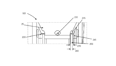
the funnel.
[0125] Referring to FIGS. 3A-311, ports 330 can be arranged along
various axial positions of
the thrombus removal system. The thrombus removal system can include a flow
axis 305 that is
aligned with a general direction (e.g., distal-to-proximal) of flow for fluid
that is aspirated
therein. In some embodiments, a position of a port 330 comprises a) near a
base of, b) in a
17
CA 03221894 2023- 12- 7
WO 2022/261448
PCT/US2022/033024
middle portion of, c) in a distal portion of, or d) proximal to, a funnel
portion 320 of the
thrombus removal system. In some embodiments, at least two ports 330 are
aligned along flow
axis 305. In some embodiments, at least two ports 330 are arranged at a
different axial and/or
angular positions along the flow axis 305. In some embodiments, at least two
ports 330 are
arranged (e.g., along a perimeter of the thrombus removal system) along a
given axial position of
the flow axis 305.
[0126] FIGS. 4A-4H depict various configurations of fluid streams
410 that are directed
from respective ports 430. A fluid stream 410 can be directed along a path
that is substantially
orthogonal, proximal, and/or distal to the flow axis 405 (which is like to
flow axis 305). In some
embodiments, at least two fluid streams are directed in different directions
with respect to the
flow axis 405. In some embodiments, at least two fluid streams are directed in
a same direction
(e.g., proximally) with respect to the flow axis 405. In some embodiments, at
least a first fluid
stream is directed orthogonally, at least a second fluid stream is directed
proximally, and at least
a third fluid stream is directed distally with respect to the flow axis 405.
An angle a may
characterize an angle that a fluid stream 410 is directed with respect to an
axis that is orthogonal
to the flow axis 405 (e.g., as shown in section D-D of FIGS. 4G and 4H). An
intersection region
of fluid streams can be within an interior portion of the thrombus removal
system, and/or
exterior (e.g., distal) to the thrombus removal system. In some embodiments, a
fluid stream that
is directed by a port 430 in a nominal direction (e.g., distally) is deflected
along an altered path
(e.g., proximally) by (e.g., suction) pressure generated by the aspiration
mechanism during
operation.
[0127] Cavitation Generation
[0128] The exemplary system includes fluidic jets configured in a
particular manner to
enhance removal of clot. The exemplary fluid streams or jets have been shown
in bench studies
to dramatically improve removal of clot through various mechanisms of action
including, but not
limited to, cavitation and water cutting. In contrast to conventional fluid
mechanisms for
thrombectomy, in some embodiments herein, fluid streams 410 from respective
ports 430 are
delivered at sufficient flow rates (and patterns) to create cavitation and/or
other preferential
effects to improve removal of clot. In certain examples, the cavitation effect
is created by large
pressure drops and deceleration at the focal point and/or intersection point
of at least two fluid
streams. The cavitation may provide a source of turbulent kinetic energy that
can be used to
mechanically fractionate and/or liquefy thrombi or other target tissue
structures. When the fluid
velocity is sufficiently high, the material accumulates impact energy, which
can cause
deformation and fragmentation. This also may modify the surface properties of
the clot to allow
the material to be penetrated to enable cavitation within the clot. Collision
or interaction of the
18
CA 03221894 2023- 12- 7
WO 2022/261448
PCT/US2022/033024
high-speed jets creates hydrodynamic cavitation whereby a pressure drop below
the vapor
pressure of the liquid creates bubbles which eventually collapse with great
mechanical energy in
the cavitation field, causing a kind of implosion in the clot material.
Further, with multiple jets
directed towards a focal point or sufficiently near respective streams, the
closing speed of the
fluid particles is significantly higher (up to double) that of a single jet
stream. This also forces
fluid and/or particles out from the space between the fluid jets at high
speed. The speed of the
fluid jets is sufficiently high to create a pressure drop below the vapor
pressure such that the
fluid vaporizes. When pressure rises again the bubble collapses, which causes
the cavitation. It
has been found that the power of the exemplary system and cavitation effect
significantly
exceeds conventional fluid jet(s) and mechanical tools like rotating screws.
In some examples,
the collapse of the bubbles may generate heat in or around the target tissue,
which may further
promote breaking up of the clot. In bench studies systems in accordance with
various
embodiments were able to remove certain clot material that simple aspiration
or water jetting
were not. In other studies, the exemplary systems were able to remove clot
material in a fraction
of the time of conventional systems.
[0129] FIGS. 4I-4K illustrate examples of generation of cavitation
420 at the intersection,
collision, or interaction of two or more fluid streams 410. Referring to FIG.
41, fluid streams 410
from at least two ports 430 are directed generally parallel to another and
orthogonal to flow axis
405 of the thrombus removal device. As shown in the embodiment of FIG. 41, the
cavitation 420
is generally confined to the region of interaction (e.g., the focal point)
between the fluid streams
410. As illustrated, the cavitation 420 can comprise a plurality of
microbubbles. When a
thrombus is engaged with a funnel of the thrombus removal device, the fluid
streams 410 and/or
cavitation 420 can be used to break up, fractionate, liquefy, and/or dissolve
the thrombus to
facilitate aspiration and removal of the thrombus with the device.
[0130] In the embodiment of FIG. 4J, the fluid streams 410 from at least
two ports 430 are
not directed orthogonal to the flow axis 405, but instead are directed
slightly distally from the
ports to create cavitation 420 in an interaction region that is distal to the
ports 430. In some
embodiments, depending on the velocity/flow rate of the fluid streams, the
resulting collision of
the distally directed fluid streams can additionally create a cavitation
column 422 that propagates
and/or resides distally to cavitation 420 in the intersection region. When a
thrombus is engaged
with a funnel of the thrombus removal device in this embodiment, the fluid
streams 410 and/or
cavitation can be used to break up, fractionate, liquefy, and/or dissolve the
thrombus to facilitate
aspiration and removal of the thrombus with the device. Additionally,
cavitation column 422
can provide additional kinetic energy to break up, fractionate, liquefy,
and/or dissolve portions of
the thrombus distal to the cavitation 420. Although the embodiment of FIG. 4J
is shown as
19
CA 03221894 2023- 12- 7
WO 2022/261448
PCT/US2022/033024
including a funnel to assist with thrombus engagement and aspiration, it
should be understood
that in other embodiments, the device may not include a funnel. In these
embodiments, the
cavitation 420 and cavitation column 422 can be used to break up, fractionate,
liquefy, and/or
dissolve a thrombus located distally to the device and ports 430.
[0131] In the embodiment of FIG. 4K, the fluid streams 410 from at least
two ports 430 are
not directed orthogonal to the flow axis 405, but instead are directed
slightly proximally from the
ports to create cavitation 420 in an interaction region that is proximal to
the ports 430. In some
embodiments, depending on the velocity/flow rate of the fluid streams, the
resulting collision of
the proximally directed fluid streams can additionally create a cavitation
column 422 that
propagates and/or resides proximally to cavitation 420 in the interaction
region and in the same
direction as aspiration of the thrombus removal device. When a thrombus is
engaged with a
funnel of the thrombus removal device in this embodiment, the fluid streams
410 and/or
cavitation can be used to break up, fractionate, liquefy, and/or dissolve the
thrombus to facilitate
aspiration and removal of the thrombus with the device. Additionally,
cavitation column 422
can provide additional kinetic energy to break up, fractionate, liquefy,
and/or dissolve portions of
the thrombus proximal to the cavitation 420 which may further assist in
aspiration of the
thrombus into the device.
[0132] FIG. 4L illustrates a top down view of a thrombus removal
device. In this
embodiment, the device includes a total of four intersecting or interacting
fluid streams 410. As
described above, the interaction between the fluid streams and/or the flow
rates of the fluid
streams can create conditions sufficient to generate cavitation 420 at the
interaction region of the
fluid streams. While this embodiment shows four fluid streams, it should be
understood that any
number of fluid streams can be implemented to achieve cavitation, including
two fluid streams,
three fluid streams, or more than four fluid streams.
[0133] As described above, the thrombus removal device can include one or
more fluid
lumens (e.g., fluid lumen 45 in FIG. 1A) configured to provide fluid to one or
more apertures
(e.g. apertures 30 in FIG. lA or ports 430 in FIGS. 4A-4K). According to one
aspect of this
disclosure, cavitation can be formed at the interaction region between at
least two fluid streams
when the flow rate of the fluid streams is high enough to create appropriate
pressure drops and
deceleration at and around the focal point and/or intersection point of the
streams. In one
embodiment, a flow rate of approximately 3na/s within the fluid lumen(s) of
the thrombus
removal device results in a fluid stream exiting the ports with a flow rate of
at least 50m/s. In
this embodiment, two or more fluid streams, each having a flow rate of at
least 50m/s, can be
configured to generate cavitation at an interaction region of the fluid
streams. In another
embodiment, a flow rate of approximately 41n/s within the fluid lumen(s) of
the thrombus
CA 03221894 2023- 12- 7
WO 2022/261448
PCT/US2022/033024
removal device results in a fluid stream exiting the ports with a flow rate of
at least 70m/s. In
this embodiment, two or more fluid streams, each having a flow rate of at
least 70m/s, can be
configured to generate cavitation at an interaction region of the fluid
streams. In yet another
embodiment, a flow rate of approximately 5m/s within the fluid lumen(s) of the
thrombus
removal device results in a fluid stream exiting the ports with a flow rate of
at least 90m/s. In
this embodiment, two or more fluid streams, each having a flow rate of at
least 90m/s, can be
configured to generate cavitation at an interaction region of the fluid
streams. Generally, the
thrombus removal device of the present disclosure is configured to provide
fluid in the one or
more fluid lumens at a flow rate of 3-5m/s which correlates to fluid streams
exiting the jets,
ports, or apertures at a flow rate of 50-90m/s. Fluid streams at these flow
rates arc configured to
create the appropriate pressure drops and deceleration at the focal point or
interaction region of
the fluid streams to generate cavitation.
[0134] In another embodiment, cavitation at the focal point or
interaction region of the fluid
streams can be characterized not by the flow rate of the fluid streams, but
instead by the pressure
drop at the intersection or passing/shearing of the fluid streams. When the
pressure drop exceeds
a cavitation threshold, cavitation is formed at that location. In one
embodiment, this pressure
drop can be at least 20MPa. In other embodiments, the pressure drop can be any
pressure drop
greater than 25MPa. Since the pressure drop is dependent on the fluid shear it
is possible to
create cavitation at the boundary of a single jet (e.g., halo cavitation).
Therefore, in some
embodiments, two fluid streams passing along some common boundary becomes a
variation
where the shear creating the cavitation can be created by two streams moving
in opposite
directions of lower velocities, as shown in FIGS. 4N and 40 and described in
more detail below.
[0135] In another embodiment, the ports can be arranged in a
slightly offset configuration
such that crossing or intersecting fluid streams only partially collide at the
interaction region. In
this embodiment, at least four distinct breaking forces can be applied to the
target thrombi,
including 1) a "cutting" or slicing force as the individual fluid streams
initially cut through the
thrombus prior to meeting at a focal point or interaction region, 2)
cavitation at the focal point or
interaction region when the fluid streams intersect, partially intersect,
collide, and/or partially
collide, 3) shearing from the jet streams moving against each other on either
side of the jets
streams, the focal point, and/or the interaction region, and 4) swirling or
halo rotational fluid
motion caused by shearing and cavitation forces.
[0136] FIG. 4M shows a cross-sectional view of such a
configuration, with ports 430a and
430b being disposed generally opposite each other across a shaft, funnel, or
lumen of the
thrombus removal device, but offset in a manner that prevents the entirety of
the fluid streams
from colliding with another. It should be understood that while this
embodiment shows the ports
21
CA 03221894 2023- 12- 7
WO 2022/261448
PCT/US2022/033024
generally on opposite sides of the lumen, funnel, or shaft of the device, any
configuration of
ports illustrated herein can be used as long as the ports are slightly offset
so as to enable only
partial collisions of the crossing or intersecting fluid streams.
[0137] Referring still to FIG. 4M, the at least four distinct
breaking forces enabled by this
configuration will now be described. During the initial activation or "turn
on" of the fluid
streams from ports 430a and 430b, the fluid streams will generally travel from
the thrombus
removal device through the thrombus towards an intersection point. As the
fluid streams are
moving in this direction, but before collision, the fluid streams provide a
"cutting" or slicing
force to the thrombus that is engaged with the device. When the fluid streams
finally collide or
intersect, as shown, since ports 430a and 430b are partially offset, only
first portion 431a of the
fluid stream from port 430a directly intersects or collides with first portion
431b of the fluid
stream from port 431b. This collision or intersection of the fluid stream
portions causes
cavitation 420 at the intersection point when the flow rate of the fluid
streams are sufficient to
cause cavitation, as described above. As also shown in FIG. 4L, second
portion(s) 432a of the
fluid stream from port 430a does not collide or intersect with second
portion(s) 432b of the fluid
stream from port 430b. As such, these second portion(s) of the fluid streams
continue past the
intersection point and past the cavitation 420. However, the fluid streams
moving past each
other in opposite, opposing, or different directions causes shearing streams
or shearing cavitation
433a and 433b to form within and/or around the thrombus, applying another type
of breaking
force on the thrombus. Additionally, the cavitation, the shearing streams,
and/or the interaction
between the partially offset ports further results in swirling streams or halo
cavitation 434a and
434b, applying a fourth distinct breaking force to the thrombus engaged with
the device.
[0138] FIGS. 4N and 40 illustrate additional views of a thrombus
removal device that can
include some or all of the breaking forces described above. FIG. 4N is a cross-
sectional view of
a thrombus removal device, and FIG. 40 is a longitudinal slice cutting across
the fluid streams as
represented by plane 440 in FIG. 4N. In FIG. 4N, the thrombus removal device
can include a
plurality of ports 430. In this embodiment, the ports are offset such that
none of the fluid
streams from the respective ports intersect or cross any of the other fluid
streams. However, the
ports are arranged in a manner that allows the fluid streams to pass closely
next to adjacent fluid
streams. In this example, first fluid stream 441 passes closely next to
adjacent second fluid
stream 442, which passes closely next to adjacent third fluid stream 443,
which passes closely
next to adjacent fourth fluid stream 444. The passing of close or adjacent
fluid streams creates
shearing streams or shearing cavitation 433 in between adjacent fluid streams,
as shown.
Additionally, as described above, the passing of close or adjacent fluid
streams additionally
creates swirling streams or halo cavitation 434. It should be understood that
the steady state
22
CA 03221894 2023- 12- 7
WO 2022/261448
PCT/US2022/033024
scenarios described herein will likely vary over time as the fluid flow
resultant from the
interactions impacts the velocities/directions of the fluid streams.
[0139] FIG. 40 is a view of a slice cutting across the fluid
streams along plane 440 in FIG.
4N, showing fluid streams 441 and 442, shearing streams or shearing cavitation
433, and
swirling streams or halo cavitation 434. It can be seen that the halo
cavitation 434 that is caused
by the passing streams can swirl or flow in a circular manner around the
respective fluid streams,
and even pass or converge into the shearing streams or shearing cavitation 433
at the center of
the opposing streams. In combination, all of these breaking forces can provide
additional
breaking energy to act on, break up, cut up, and mechanically fractionate a
thrombus engaged
with the device.
[0140] Cavitation Detection
[0141] With the ability of the thrombus removal device to generate
cavitation at the
intersection region of two or more fluid streams, the thrombus removal device
can further
include cavitation detection capabilities to detect if and when cavitation is
generated within or
near a target thrombus. In some embodiments, the cavitation detection
capabilities can detect the
location and/or intensity of the cavitation. Cavitation detection further
provides additional
functionality in the operation of the device, providing an additional
mechanism for detecting
when the device is engaged with a thrombus.
[0142] In some embodiments, cavitation detection can be used to
determine the interaction
between the jets or fluid streams and the target thrombus. For example, when a
thrombus is first
engaged in a funnel of the device (e.g., with aspiration), the jets or fluid
streams can be activated
to provide two or more fluid streams inward towards a focal point or
intersection point of the two
or more fluid streams. However, during this initial activation of the jets or
fluid streams, the
thrombus may be positioned or located in between the two or more fluid
streams, thereby
preventing collision or intersection of the fluid streams. At this point in
the therapy, as the fluid
streams may not yet be intersecting, they first must -cut" or drive through
the thrombus.
Depending on the flow rate of the fluid streams as they initially "cut"
through the thrombus,
there may be no cavitation present.
[0143] Cavitation detection can be used to identify scenarios in
which 1) a clot is engaged in
the funnel, 2) aspiration is activated, 3) the jets or fluid streams are
activated but cavitation is not
present, and/or 4) the jets or fluid streams are activated and cavitation is
present. For example, a
pressure or flow measurement in the aspiration lumen while aspiration is
activated can be used to
determine if the clot is engaged in the funnel. Then, if cavitation is
simultaneously detected, the
system can indicate to the user that the clot is engaged and the jets or fluid
streams are producing
cavitation in the clot. If no cavitation is detected, then the system can
indicate to the user that the
23
CA 03221894 2023- 12- 7
WO 2022/261448
PCT/US2022/033024
clot is engaged and the jets or fluid streams are cutting the clot. In some
embodiments, whether
or not cavitation is detected can be displayed or indicated to the user.
Therefore, the indication
to the user on if cavitation is present or not present can provide useful
information to the user
regarding the state or status of the therapy (e.g., whether a thrombus is
engaged, whether cutting
is occurring, or whether cavitation is occurring).
[0144] As the treatment progresses, the jets or fluid streams will
eventually cut through the
thrombus in the funnel, causing the two or more jets to intersect at the focal
point. When this
event occurs, if the fluid streams have a sufficient flow rate (e.g., 20-90m/s
or more, as described
above), the two or more intersecting fluid streams can be configured to
generate cavitation at the
focal point. It should be understood that in many situations, with the
thrombus still engaged in
the funnel of the thrombus removal device, this cavitation can further provide
mechanical
fractionation and/or liquefaction of the thrombus at the focal point. In some
embodiments, the
therapy includes alternating cycles of "cutting" and cavitation. As the
thrombus moves around in
the funnel and is broken up into smaller pieces or sections and aspirated into
the thrombus
removal device, there will be instances in which the fluid streams are
intersecting, and therefore
creating cavitation, and there will be instances in which the fluid streams
are not intersecting
(e.g., perhaps due to the thrombus preventing intersection) and rely instead
on the "cutting"
nature of the jets to break up the clot.
[0145] In some embodiments, the ability to detect cavitation can be
used to direct the jet
and/or aspiration control schemes of the thrombus removal device. For example,
it may be
beneficial to alternate between -cutting" and -cavitation" modes of the
thrombus removal
device. In one example, the jets are activated to "cut" through an engaged
thrombus until
cavitation is detected. Once cavitation is detected, the jets can remain
active for a preset period
of time. Next, the jets can be temporarily pulsed or turned off, with
aspiration remaining on, to
allow the thrombus to shift or move deeper into the funnel. Then the jets can
be activated again,
restarting a cycle of a "cutting" mode followed by a "cavitation" mode. In
some embodiments, it
may be desirable to avoid cavitation and instead rely only on the cutting
mechanism of action.
In this instance, cavitation detection can be used to alert or indicate to a
user that cavitation has
formed. In some embodiments, the device can automatically pause or pulse the
jets when
cavitation is detected, to allow the clot to fill the funnel and restart the
cutting process with the
jets.
[0146] Referring back to FIG. 41, in some embodiments the thrombus
removal device can
include a cavitation detection sensor 424. The cavitation detection sensor can
comprise, for
example, an ultrasound transducer element or a hydrophone. The sensor may
detect cavitation
by monitoring for cavitation directly and/or indirectly. In the case of
indirect monitoring, the
24
CA 03221894 2023- 12- 7
WO 2022/261448
PCT/US2022/033024
sensor monitors characteristics of the fluid stream and identifies the desired
cavitation based on
known correlations. The correlations may vary based on the size and shape of
the catheter end
(or funnel), orientation of the jets and focal point, etc. In the embodiment
of FIG. 41, the device
is shown with a cavitation detection sensor 424 in the funnel and a second
cavitation detection
sensor 424 in the shaft/aspiration lumen of the device. While only the
embodiment of FIG. 41 is
illustrated to include a cavitation sensor(s), it should be understood that
any embodiment or jet
configuration described herein can further include one or more cavitation
sensors. Generally
these cavitation detection sensors can be directed or pointed towards the
intersection point of the
two or more fluid streams. It should be understood that in other embodiments,
these devices can
include one, or more than two cavitation detection sensors. The sensors may be
located only in
the funnel, only in the shaft/aspiration lumen, or a combination of both as
shown. Generally, the
cavitation detection sensors can he positioned anywhere within or on the
device that provides an
acoustic path between the sensor and the target cavitation region. Although
only the
embodiment of FIG. 41 shows a device with a cavitation detection sensor, it
should be
understood that any thrombus removal device described herein can include such
functionality,
including the embodiments of FIGS. 4J and 4K. Since those embodiments include
distally and
proximally directed fluid streams, respectively, thereby enabling formation of
a cavitation
column, it should be understood that in those embodiments, the cavitation
detection sensors can
be configured to sense and/or detect both the cavitation 420 and the
cavitation column 422.
[0147] Other types of sensors are proposed, including a microphone
configured to detect
cavitation, or a laser configured to detect temperature changes at the
intersection point when
cavitation occurs.
[0148] In addition to cavitation detection with a sensor disposed
on or in the device, in other
embodiments the thrombus removal device can be used in conjunction with a
separate cavitation
detection device, such as a real-time imaging device. For example, cavitation
can be identified
as a hyper-echoic region in real-time B-mode ultrasound imaging. Therefore, in
one
embodiment, an ultrasound imaging device can be directed towards the target
thrombi and be
used to identify when cavitation occurs in real-time, providing real-team
feedback to a physician
or surgeon during a thrombus removal procedure. The ultrasound imaging device
can comprise,
for example, an external ultrasound imaging probe (e.g., placed in contact
with the skin of the
patient). Alternatively, the ultrasound imaging device can comprise an
internal or catheter-based
ultrasound imaging probe configured to be advanced along with or within the
thrombus removal
device to the target thrombus location.
[0149] FIG. 4P is a photograph of a benchtop experiment showing the
formation of
cavitation at an interaction region of four interacting or intersecting jets
or fluid streams. In this
CA 03221894 2023- 12- 7
WO 2022/261448
PCT/US2022/033024
experiment, the fluid source (e.g., a water pump) was pulsed to have an
operating pressure
ranging from peak 200 psi to 750 psi. The fluid source was then able to
produce a flow rate in
the fluid lumen of the device having an average velocity ranging between 2 m/s
and 10 m/s.
That flow rate in the fluid lumen resulted in an average velocity out of the
jet apertures ranging
between 50 m/s and 200 m/s. With the same setup, the fluid source was operated
at a pulsed
pressure to produce an average velocity out of the jet apertures below 10 m/s
and no cavitation
was observed.
[0150] FIGS. 5A-5G illustrate a variety of exit aperture geometries
with which ports 530 can
be configured in accordance with embodiments of the present technology.
Aperture geometries
can comprise an oval, circular, cross ("x" shape), "t" shape, rectangle, or
square shape. A fluid
stream that is delivered from the port 530 can comprise substantially laminar
flow (e.g., at the
aperture), or a turbulent flow (e.g., that fans outward). The size of the
ports 530 can be adjusted
to achieve the appropriate exit velocity and acceleration of the fluid
streams. In some
embodiments, these port sizes can be optimized to achieve a flow rate of 50-
90m/s so as to create
cavitation at the intersection point of the two or more fluid streams.
Generally, smaller ports
create a higher velocity fluid stream, at the expense of transmitting less
kinetic energy due to
lower volume of fluid exiting the ports.
[0151] FIGS. 6A-6C illustrate various configurations of a thrombus
removal system 600,
including a thrombus removal device, 602, a vacuum source and cannister 604,
and a fluid
source 606. In some embodiments, the vacuum source and cannister and the fluid
source are
housed in a console unit that is detachably connected to the thrombus removal
device. A fluid
pump can be housed in the console, or alternatively, in the handle of the
device. The console can
include one or more CPUs, electronic controllers, or microcontrollers
configured to control all
functions of the system. The thrombus removal device 602 can include a funnel
608, a flexible
shaft 610, a handle 612, and one or more controls 614 and 616. For example, in
the embodiment
shown in FIG. 6A, the device can include a finger switch or trigger 614 and a
foot pedal or
switch 616. These can be used to control aspiration and irrigation,
respectively. Alternatively,
as shown in the embodiment of FIG. 6B, the device can include only a foot
switch 614, which
can be used to control both functions, or in FIG. 6C, the device can include
only an overpedal
616, also used to control both functions. It is also contemplated that an
embodiment could
include only a finger switch to control both aspiration and irrigation
functions. As shown in
FIG. 6A, the vacuum source can be coupled to the aspiration lumen of the
device with a vacuum
line 618. Any clots or other debris removed from a patient during therapy can
be stored in the
vacuum cannister 604. Similarly, the fluid source (e.g., a saline hag) can be
coupled to the fluid
lumens of the device with a fluid line 620.
26
CA 03221894 2023- 12- 7
WO 2022/261448
PCT/US2022/033024
[0152] Still referring to FIG. 6A, electronics line 622 can couple
any electronics/sensors, etc.
from the device to the console/controllers of the system. The system console
including the
CPUs/electronic controllers can be configured to monitor fluid and pressure
levels and adjust
them automatically or in real-time as needed. In some embodiments, the
CPUs/electronic
controllers are configured to control the vacuum and irrigation as well as
electromechanically
stop and start both systems in response to sensor data, such as pressure data,
flow data, etc.
[0153] As is described above, aspiration occurs down the central
lumen of the device and is
provided by a vacuum pump in the console. The vacuum pump can include a
container that
collects any thrombus or debris removed from the patient.
Clog detection and clog removal
[0154] In some situations, the device may become clogged with a
thrombus or with other
debris during therapy. Many clog detection and clog removal schemes can be
implemented in
the thrombus removal system. Generally, clogs in the system or device can be
detected with any
number of sensors disposed in or around the device. For example, pressure
sensors can be
disposed on or in the funnel, on or in the fluid lumens, or on or in the
aspiration lumen of the
device or system at any number of locations. The sensor data can then be used
to monitor the
operation of the device. For example, pressure sensors in the aspiration lumen
can provide an
indication if the device is clogged with a clot or other debris. The system
can monitor the
pressure in the aspiration lumen, and significant changes in pressure from the
normal operating
pressure can indicate a problem with the device or the therapy. For example, a
pressure sensor
reading that drastically drops from the normal operating pressure range could
indicate that a clot
or other debris is clogging the device or system proximal to the pressure
sensor. Similarly, a
pressure sensor reading that drastically increases from the nottnal operating
pressure range could
indicate that a clot or other debris is clogging the device or system distal
to the pressure sensor.
Pressure sensors disposed along a length of the device can therefore be used
in this manner to
determine if the device is clogged and even identify where along the length of
the device the clog
is located based on which pressure sensors have higher than normal pressure
readings and which
pressure sensors have lower than normal pressure readings. Similarly, flow
meters or sensors
can be used to monitor the flow of fluid in the fluid lumens and/or the flow
of debris, blood, and
clots in the aspiration lumen. These flow sensor readings can be used to
determine if the
aspiration lumen or a flow lumen (and potentially a jet or aperture) is
clogged or blocked.
[0155] In one embodiment, the system can be configured to produce
vacuum suction with a
large volume piston pump that can be selectively controlled. This enables
automatically
stopping vacuum when pressure and/or other sensors in the thrombus removal
device detect a
sharp change in vacuum pressure as a result of a clog. Once detected, the
system can be
27
CA 03221894 2023- 12- 7
WO 2022/261448
PCT/US2022/033024
configured to automatically halt irrigation jetting and the vacuum piston
resulting vacuum in
instant removal of vacuum pressure to reduce blood loss and prevent over
irrigating the patient.
[0156] In another embodiment, when the system detects a clogged
device, the system can be
configured to automatically holt irrigation and aspiration, then run a
declogging routine that
rapidly cycles vacuum pressure to induce a "fluid hammer" effect to remove the
clot or clog.
[0157] Additional embodiments are provided for removing clogs or
clots from the device.
Referring to FIG. 7A, the thrombus removal device can include a plurality of
jets 730 disposed
along a length of the device, including along the shaft of the device. In some
embodiments, the
jets can be pointed in different angles to assist in moving the clot or debris
proximally along the
device. For example, the jets can be aimed generally proximally along the
shaft of the device to
push or force clots in that direction.
[0158] In another embodiment, referring to FIGS. 7B-7C, the
thrombus removal device can
include a valve 732 disposed on or within the aspiration lumen. The valve can
comprise a
flapper valve, a shunt valve, a duckbill valve, or the like. FIG. 7B shows the
valve in the open
position, and FIG. 7C shows the valve in the closed position. During normal
operation of the
system, the valve can remain in the open position to allow clots and other
debris to be removed
from the patient. In the event that a clog is detected by the system, the
valve can be closed, as
shown in FIG. 7C, to seal the aspiration lumen of the device. With the valve
closed, irrigation
jets 734 that are positioned proximal to the valve within the aspiration lumen
can be activated to
generate pressure behind (e.g., distal to) the clot, thereby forcing the clot
out of the device and
into the vacuum cannister.
[0159] Similarly, referring to FIG. 7D, another embodiment of the
device includes a distal
balloon 736 that can be inflated when a clot is detected to seal the inner
lumen of the device.
The system can then be configured to irrigate the clogged sealed lumen with
jets 734, generating
pressure behind the clot as described above.
[0160] In some embodiments, a conventional vacuum pump via
peristaltic or diagram pump
is used, and another method of preventing blood loss by reducing vacuum
pressure can be to
purge or shunt the vacuum chamber when a clot or clog is removed.
Jet control schemes
[0161] As described above, in some embodiments, fluid lumens can be
distinct and separate,
thereby enabling individual jets to be controlled to deliver a stream of fluid
while other jets are
inactive or not delivering fluid. The system can be configured to respond to
pressure sensing and
volume of fluid infused and removed. These control schemes can vary the amount
of irrigation
and aspiration as well as sequence or pulse the individual lumens to provide
different cutting or
declogging results. This facilitates many novel jet control schemes to be used
by the thrombus
28
CA 03221894 2023- 12- 7
WO 2022/261448
PCT/US2022/033024
removal device to assist in breaking up/macerating clots and/or removing those
clots from the
patient. For example, referring to FIGS. 8A-8C, a cross section of the
thrombus removal device
is shown with one example of a jet control scheme. In this embodiment, jets
830a-830d can each
be fluidly coupled to a fluid source with an independent or distinct fluid
lumen. Thus, in FIG.
8A, only jet 830d can be activated, allowing a stream or jet of fluid to be
delivered by jet 830d
into the aspiration lumen of the device. Similarly, in FIG. 8B, only jet 830c
is activated, and in
FIG. 8C, only jet 830b is activated.
[0162] It should be understood then that any number of jet control
schemes can be
incorporated into the treatment by the thrombus removal device when the jets
are fed by
independent fluid lumens. For example, referring to FIG. 8A, in one
embodiment, the device
could rapidly cycle sequentially from delivering jets of fluid from each of
the jets (e.g., first from
jet 830a for a preset time, then jet 830b, then jet 830c, then jet 830d, and
so forth). Similarly,
pairs or groupings of jets can be activated while other jets are inactive. For
example, the jet
sequence could cycle between activating only opposing pairs of jets 830a and
830c and then
activating only opposing pairs of jets 830b and 830d.
[0163] While the embodiments above describe activating one or more
jets in radial patterns
around a circumference of the device, it should be understood that jet control
schemes can also
be used longitudinally along the device. For example, recall that the
embodiment of FIG. 7A
included a plurality of jets disposed along a length of the device. In one
embodiment, a jet
control scheme can be implemented in the system to rapidly cycle between jets
in a distal to
proximal direction (e.g., first activating the distal most set of jets, then
the next most distal, and
so forth until the proximal most jets are activated). It is contemplated that
a control scheme in
this fashion could move or urge along difficult, large, or stubborn clots to
remove them from the
device.
[0164] Aspiration of the system can also be pulsed or timed with the
irrigation bursts to
maximize effectiveness and to reduce blood loss. For example, in some
embodiments, the
aspiration is pulsed to coincide with jet irrigation. In other embodiments,
the aspiration is pulsed
or activated in between bursts of jets.
[0165] FIGS. 9A-9B are schematic diagrams of the thrombus removal
system and the
thrombus removal device, respectively. Referring to FIG. 9A, the system can
include pulmonary
artery pressure (Ppa), pressure vacuum source (Pvs), pressure jet source
(Pjs), fluid resistance of
vacuum system (Rvs) and fluid capacitance (Cvs) of the aspiration/vacuum
portion of the device,
fluid resistance (Rjs) and capacitance (Cjs) of the jet portion of the device,
and multiple test
points Ti -T7 for testing pressure or flow of the system. Any number or type
of pressure and/or
flow sensors can be implemented in the system. Additionally, other types of
sensors can be
29
CA 03221894 2023- 12- 7
WO 2022/261448
PCT/US2022/033024
used. For example, electrodes or impedance sensors can be used to measure an
impedance at the
distal end of the system (e.g., to characterize changes in electrical
impedance associated with
clots vs. blood). In other embodiments, temperature sensors (e.g., one or more
thermistors) may
be used to sense a temperature of the device or target tissue. In additional
embodiments, the
vacuum source or jet source can be configured as sensors. such as using back
emf or a fluid
column sensor connected to the aspiration lumen or jet lumen.
[0166] The pressure vacuum source (Pvs) can be a vacuum source (a
trap in which a low
pressure gas is maintained above the aspirant) or a positive displacement
source, both of which
induce a negative pressure distal to the CNTs (when present as it may not be
required with a
positive displacement pump). Engagement, such as with a clot, can be
characterized by either
the difference between an expected flow or rate in change of flow and a
measured flow where
that difference is of great enough magnitude.
[0167] Referring to FIG. 9B, CNTs represents the junction or
connection between the
pressure vacuum source and the thrombus removal device, and CNTj represents
the junction or
connection between the pressure jet source and the thrombus removal device. A
valve in CNTs
and CNTj can isolate the capacitance of the vacuum/jet source from the rest of
the system such
that the amount of blood drawn into the system when the vacuum system is
stopped or shut down
is minimized. Referring to FIG. 9A, testing points Ti and T2 can represent
pressure or flow
sensor locations configured to provide pressure/flow readings at a location
between the pressure
vacuum source and the device, and between the pressure jet source and the
device, respectively.
Testing points T4 and T3, similarly, can represent pressure or flow sensor
locations configured
to provide pressure/flow readings at a location near the junction or
connection between the
device and the pressure vacuum source and pressure jet source, respectively.
Additionally,
testing points T5, T6, and T7 can represent pressure or flow sensor locations
configured to
provide pressure/flow readings at a location near a distal end of the device.
For example, testing
point T5 can provide flow/pressure readings at or near where the jet fluid
exits the jet ports or
nozzles at the distal end of the device. Similarly, testing points T6 and T7
can provide
pressure/flow readings of the aspiration system at or near the distal end of
the device, such as
within the funnel (T6 in FIG. 9B) or within the thrombus removal device just
proximal to the
funnel (T7 in FIG. 9B). The locations of the test points within the system
schematic illustrate
potential test/sensor locations for pressure sensors, flow sensors, or other
sensors that could be
used in real time to control operation of the device, detect system operating
parameters, detect
clogs, etc.
[0168] Referring to FIG. 9B, an embodiment of the thrombus removal
device is shown
which includes test points T6 and T7 for sensing flow and/or pressure in the
funnel and the
CA 03221894 2023- 12- 7
WO 2022/261448
PCT/US2022/033024
aspiration lumen of the device, and also haptic sensors H1 and/or H2 for
detecting contact with a
clot or other debris in the patient. In some embodiments, the haptic sensors
can comprise
pressure sensors (positive or negative), optical sensors, electrical impedance
sensors (dc, single
frequency, or spectral) or other sensors useful for the operation of the
system.
[0169] Procedure and System Controls
[0170] FIGS. 10-12 illustrate procedural flow and system control
schematics for a thrombus
removal system according to various embodiments, including clot detection,
clot engagement,
and clot removal.
[0171] FIG. 10 is a table that generally describes the various
states of a thrombus removal
system including the associated sensor reading(s) found in each state.
Generally, the thrombus
removal system can include a searching for clot/no clot engaged state, an
engaging clot state, a
clot engaged state, a clogged jet lumen state, a clogged aspiration lumen
state, and a clot initially
engaged/leak state. As described above, sensors can be disposed throughout the
system,
including at or near a distal end of the device (e.g., at or within the funnel
and/or within the
aspiration lumen), at or near a proximal end of the device, and/or at or
within the pressure and/or
jet/fluid source outside of the device. The readings of these groupings of
sensors can generally
be used to determine which state the thrombus removal system is in, and can be
further used to
inform and control the device into subsequent states throughout the therapy.
Referring to the
table in FIG. 10, when the sensors are in a nominal or (+) state it reflects a
signal indicative of
engagement with a clot, and when the sensors are in a non-nominal or (-) state
it reflects a signal
indicative that the device is not in engagement with a clot. In some
embodiments, the nominal
state can correspond to a sensed parameter within a given range (e.g., a
specific pressure or flow
rate range) and the non-nominal state can correspond to a sensed parameter
(e.g., pressure or
flow rate) beyond a threshold pressure. In one embodiment, a nominal range for
pressure can be
between approximately +4 to -25 inHg.
[0172] For example, referring to the table of FIG. 10, when the
thrombus removal system is
in a searching for clot/no clot engaged state, all of the sensors including
the distal sensors, the
proximal sensors, and the source sensors can be in a non-nominal state.
However, when the
system is engaging a clot, both the source sensors and the distal sensor(s)
can be in a nominal
state, with the proximal sensor(s) remaining in a non-nominal state. When the
thrombus removal
system is in a clot engaged state, all of the sensors will be in a nominal
state, as shown in FIG.
10.
[0173] The sensors can also inform errors in the system, including
clogged lumens
(aspiration or jet lumens) as well as leaks in the system. For example, still
referring to FIG. 10,
source and proximal sensors in a nominal state and distal sensors in a non-
nominal state can
31
CA 03221894 2023- 12- 7
WO 2022/261448
PCT/US2022/033024
indicate that one or more of the jet lumens is clogged. Similarly, source
sensors in a nominal
state and proximal/distal sensors in a non-nominal state can indicate a
clogged aspiration lumen
or lumens. Finally, proximal and distal sensors in a nominal state and the
source sensor(s) in a
non-nominal state can indicate a leak in the system, or that a clot has
initially been engaged.
Further details on the sensors, their measurements, and how the system
determines the system
state based on measurements will be discussed below.
[0174] FIG. 11 is a flowchart that describes the various system
states that a thrombus
removal system may cycle through during a thrombus removal procedure.
Referring to step
1102 of the flowchart, the thrombus removal system or device can be inserted
into a patient's
vasculaturc and a distal end of the device can be advanced and delivered to
the target tissue site
that includes one or more thrombi. At this point, the user of the device can
actuate, press, or
initiate a clot searching routine in the thrombus removal system at step 1104
(e.g., such as by
pressing a button on a handle or on a generator of the system). In some
embodiments, the
system can initiate the clot searching routine automatically.
[0175] When the thrombus removal system is actively in the clot searching
routine of step
1104, the system is monitoring various sensors (such as flow or pressure
sensors) to determine
if/when the thrombus removal system has engaged with a thrombus or thrombi at
the target
tissue location. While in this clot searching state, the system can operate
the aspiration source to
pull vacuum and assist in capturing clots in the funnel of the device. In some
embodiments, the
aspiration can run at a normal level (e.g., the same level of aspiration that
runs when a clot is
being removed) and in other embodiments the aspiration can run at a lower
level or some
minimal level. In this state, the jets can be completely off or can also run
at a lower or minimal
level to assist with clot capture. As described above, the system can include
any number of
pressure and/or flow sensors located at several locations on or within the
system. The system
can also use the jet ports/jet lumens as sensors, which can inform the system
about the particular
state and guide the therapy process.
[0176] FIG. 12A illustrates a pressure waveform Pw of distal
sensors of a thrombus removal
system, such as distal sensors located on or within a funnel or distal lumen
of the device, or
alternatively, using the jet ports or lumens as distal pressure sensors (under
negative pressure
relative to local pressure at the jet aperture/no aspiration). This allows for
the measurement with
lower flows than required for the aspiration lumen. Referring to the diagram
of FIG. 12A, Ppa is
the pressure of the pulmonary artery, Pab is the pressure at ambient or
atmospheric, and Pt is a
predefined pressure threshold. Various regions of the pressure wave Pw are
shown, including:
a, which indicates that the device is not engaged with a clot and is measuring
heart induced
pulmonary artery fluctuations, b, which indicates that the sensed pressure is
dropping as a
32
CA 03221894 2023- 12- 7
WO 2022/261448
PCT/US2022/033024
function of engagement with a clot, c, which indicates that pressure is below
the predefined
threshold Pt where fluctuations are masked, and d, which indicates either the
time at which the
pressure source is activated and/or the time at which the device begins to
interface with a clot.
[0177] Many features of the pressure wave Pw in FIG. 12A can be
used or identified to
indicate that the device has engaged with a clot. Typically the Ppa is
measured prior to
engagement with a clot, to provide a baseline for pressures at the target
tissue location. In one
embodiment, when the pressure wave Pw drops below the predefined pressure
threshold Pt, it
can indicate to the system that a clot has been engaged. This can result in
the system state
moving to the engaged state 1108 in FIG. 11. Additionally, the system state
can move to clot
engaged if pressure fluctuations disappear or fall below a threshold value as
in some percentage
of region a of the pressure waveform. In another embodiment, if the rate of
change in region b
of the pressure waveform is greater than a threshold level, the system state
can move to engaged.
The rate of change in region b can provide information on the quality of
engagement such as;
fewer or more ports engaged at the level of the ports, essentially if all
ports are engaged by clot
the IDP/dtl as the capacitance of the system will be smaller. Any combination
of the above
conditions can result in the system identifying or determining that a clot is
engaged in the funnel
at the distal end of the device.
[0178] FIG. 12B illustrates a pressure waveform Pw of distal
sensors of a thrombus removal
system, such as distal sensors located on or within a funnel or distal lumen
of the device, or
alternatively, using the jet ports or lumens as distal pressure sensors (under
low positive
pressure/aspiration on). Once again, the diagram includes various regions of
the pressure wave
Pw, including: a, which indicates that the device is not engaged with a clot
and is measuring
heart induced pulmonary artery fluctuations, b, which indicates that the
sensed pressure is rising
as a function of engagement with a clot, c, which indicates that pressure is
above the predefined
threshold Pt where fluctuations are masked, and d, which indicates either the
time at which the
pressure source is activated and/or the time at which the device begins to
interface with a clot. In
FIG. 12B, engagement is determined when the pressure of the pressure waveform
Pw increases
above the aspiration pressure as the clot presses against the distal pressure
sensors (e.g., jet ports
or purpose-built ports or sensors not used for jetting).
[0179] In addition to the pressure changes measured by the sensors and
described above in
FIGS. 12A-12B. engagement of a clot can be identified when the pressure change
is mediated by
one or any combination of the following scenarios:
[0180] 1) a flow induced in the aspiration line either towards or
away from the pressure
vacuum source (Pvs). As described above, the Pvs may be configured as pressure
source such
as a vacuum trap or as a positive displacement pump or a combination thereof.
33
CA 03221894 2023- 12- 7
WO 2022/261448
PCT/US2022/033024
[0181] 2) a flow induced in one or more of the jet lines, either
towards or away from the
pressure jet source (Pjs). The Pjs may be a configured as pressure source such
as a volume of
fluid maintained under pressure or periodically pressurized or as a positive
displacement pump
or a combination thereof.
[0182] 3) a change in electrical impedance measured by distal electrodes or
impedance
sensors.
[0183] 4) a change in any combination of the above.
[0184] The flows described above in 1) and 2) may be induced by a
number of conditions,
including a relatively constant delta P across the line or a pulsating
pressure across the line. In
some embodiments, the pulsations are configured to minimize the total volume
of fluid displaced
into or out of the system. For example, a small volume of fluid (e.g., 1-10
mL) can be drawn in
and out of the aspiration line with the pulsations. In this example, the dQ/dt
is larger for the
inflow than the outflow to enhance fluid drag to coax the clot into the
funnel. An engagement is
indicated by an abrupt increase in pressure and/or flow. In another example, a
smaller volume of
fluid (e.g., 0.1-1 mL) can be drawn into the jet line or port. In this
example, an engagement is
indicated by a decrease in the pressure and/or in flow. In yet another
embodiment, engagement
of a clot can be determined by discharging a volume of fluid at a constant
flow rate through the
jet while the aspiration is drawing fluid into the system and noting a
pressure increase on the
pressure line, wherein the engagement is indicated by an increase in the
pressure and/or flow.
[0185] When engagement with a clot is sensed, as described above, the
system can move to
the engaged routine/clot removal state as shown in FIG. 11. First, to confirm
engagement, the
system can turn off jet flow (if active) and decrease the aspiration pressure
to less than Pa on
(e.g., 10 in Hg absolute) and test the rate of IdPa/dtl. If this tested rate
is IdPa/dt1 >1= Pa on/lsec
the system can begin aspiration and jetting to remove the clot. However, if
that condition is not
met, the testing can continue. If repeating the testing does not result in
confirmed engagement,
then the system can be removed.
[0186] During the clot removal/engaged state, the system can
continue to sense maintained
engagement with a clot, as indicated by maintenance of Pa < Pa on. With
maintained clot
engagement, the jets of the thrombus removal system can be activated to
provide an average
fluid velocity Vjet of greater than 10 m/s (and optionally greater than 20 m/s
or greater than 40
m/s). The jet flow from the jets can be any combination of pulsatile with a
non-zero minimum,
pulsatile with a zero minimum, constant, or negative minimum.
[0187] The Pa can continue to be monitored during clot removal. If
Pa>Pa_on, system
changes can include either turning off the jets and returning to clot
engagement functions (as
described above), or alternatively, incrementally decreasing jet average
velocity Vjet. If the
34
CA 03221894 2023- 12- 7
WO 2022/261448
PCT/US2022/033024
Vjet<Vjet minimum, the system can return to clot engagement. If during
continued monitoring
Pa<Pa on, then the clot removal process can continue.
[0188] In some embodiments the system can monitor Qasp (flow in the
aspiration line) and
Qjet (flow in the jet line) and or calculate Q' s from system resistances and
capacitances. In this
embodiment, if Qjet>/=Qasp the system can return to engagement or else can
continue with clot
removal.
[0189] After the engaged routine has progressed, referring to FIG.
11, at step 1110 the
system can move to determine if the clot has been cleared. If the system
determines that the clot
has been cleared at step 1112, then the process flow chart can revert to the
pre-search routine
state in which the system is neither actively looking for a clot or actively
attempting to
engage/remove a clot. In general, however, the cleared state or occlusion
testing procedure
includes assessing or monitoring blood flow past the distal end of the
thrombus removal system
to assess improvement in blood flow (as a result of clot removal). In addition
to using flow or
pressure sensors to identify an increase in flow, other techniques can be used
by the system. For
example, in some embodiments, flow monitoring can be accomplished using
thermal dilution
and or time of flight. For example, a volume of cold fluid (e.g., colder than
body temperature)
can be delivered into the target tissue location and the temperature can be
monitored at another
sensor location. For example, the cold fluid can be delivered at testing
location T7 (Fig. 9B) and
temperature can also be measured at testing location T6. Alternatively, a
heated fluid can be
delivered at T7 and temperature can be monitored at T6. In another embodiment,
a contrast
agent can be injected by the system into the target location through the
jetting system or through
purpose build lumens, and the contrast agent can be visualized to determine
whether or not the
clot was removed.
[0190] If, however, the system determines that the device is either
clogged or the clot has not
been cleared, at step 1114 the system can engage in a clogged or clearing
routine to attempt to
de-clog the device or remove the clot. The clearing/clogging protocols have
been previously
described in this disclosure, but in general the system can use any number of
procedures
including continuing to run aspiration/jets, reversing the pressure of the
aspiration and/or jets,
running aspiration without jets or jets without aspiration, or any other
number of clearing or
clogging routines. If the system determines that the clot has been cleared at
step 1116, then the
process flow chart can revert to the pre-search routine state in which the
system is neither
actively looking for a clot or actively attempting to engage/remove a clot.
[0191] FIG. 13 is a simplified system schematic that describes the
system elements of a
thrombus removal system that are required to implement the procedures and
methods described
above. In general, the system can include an electronic controller configured
to control operation
CA 03221894 2023- 12- 7
WO 2022/261448
PCT/US2022/033024
of both a vacuum/aspiration source and a fluid (jet) source of the system.
Sensors can be located
throughout the system, including within the vacuum/fluid source and within the
device (both
proximal and distal). As described above, the sensors can include pressure,
flow, impedance,
etc. sensors. Sensor measurements can be input back into the controller along
with error signals
to control operation of the device. Vessel blood flow can also be monitored to
assist in
determining when a clot has been cleared or engaged.
[0192] In contrast to the embodiments described above which use
pressure sensing or
pressure waveforms to assist in control schemes of the device, in other
embodiments the device
can control system states based on flow measurements within the system, such
as aspiration flow
rates or irrigation flow rates. Additionally, any of the control schemes
described herein can be
combined with another. For example the pressure control schemes can be
combined with the
flow control schemes. As described in FIGS. 9A-9B and above, multiple test
points Tl-T7 can
be provided in the system for testing flow within the system. Any number or
type of flow
sensors can be implemented in the system at the testing points, or at other
points in the system,
particularly in the funnel and in the aspiration lumen(s) of the device.
[0193] FIG. 14 illustrates an aspiration flow (Q) waveform that can
be sensed by one or
more flow sensors located within the system, such as at test points T1-T7, but
specifically
sensors associated with aspiration flow (e.g., test points Ti. T4, T6, and
T7). The waveform of
FIG. 14 shows the flow of aspiration over time as the system hunts or looks
for a new clot,
engages the clot, and begins treatment/removal of the clot. Many features of
the flow wave Q in
FIG. 14 can be used or identified to indicate that the device has engaged with
a clot and provide
insight into the clot behavior within the device including within the funnel.
Any determinations
that the system makes as a result of the measured flow wave Q can be indicated
to the user. For
example, the system can indicate to the user (e.g., with a display, an
indicator, or an audio signal)
that the system is partially engaged, fully engaged, or not engaged with a
clot. Typically the Q is
measured prior to engagement with a clot, when aspiration is activated either
at a clot
engagement level or at some aspiration flow level lower than a clot engagement
level (e.g., a clot
seeking level), to provide a baseline for flow at the target tissue location.
It should be noted that
as shown in the embodiment of FIG. 14, aspiration is activated but the water
jets have not yet
been activated. In other embodiments, however, the water jets may be activated
during any of
the phases of the curve illustrated in FIG. 14. The flow Q while the system is
looking for a clot
is shown in region a of the waveform in FIG. 14.
[0194] In FIG. 14, when the flow wave Q begins to drop, as shown in
region b, the measured
flow can indicate to the system that a clot has been engaged. In some
embodiments, the slope
dQ/dt of the flow waveform can be used by the system to determine if there is
clot engagement.
36
CA 03221894 2023- 12- 7
WO 2022/261448
PCT/US2022/033024
In some embodiments, the rate or slope of the flow waveform can be indicative
of the "quality"
of engagement with a clot. For example, the greater the rate of decline, the
larger the resistance
(decrease in flow path around the clot) induced by the interface between clot
and funnel. Actual
rates will be dependent on system parameters such as component volumes, and
dimensions,
source flow rates, component capacitances, and or pressures. Eventually, the
flow wave Q will
go to approximately zero (or some non-zero minimum), as shown in region c of
FIG. 14,
indicating that the clot is fully engaged within or seated within the funnel
of the device. This can
result in the system state moving to the engaged state 1108 in FIG. 11.
Additionally, the system
state can move to clot engaged if pressure fluctuations disappear or fall
below a threshold value
as in some percentage of the flow waveform. For example, section d of the wave
in FIG. 14 is
slightly above zero, but is below a threshold that indicates to the system
that the clot is engaged
or partially engaged. This above zero flow can also he a result of turning on
the jets, or can also
be caused by the clot moving around not being fully engaged within the funnel
of the device.
[0195] The rate of change in region b can provide information on
the quality of engagement.
Any combination of the above conditions can result in the system identifying
or determining that
a clot is engaged in the funnel at the distal end of the device.
[0196] Now referring to FIG. 15, in some embodiments the aspiration
can be pulsed while
looking for a clot (or prior to clot engagement) to reduce the amount of blood
drawn into the
system. An aspiration waveform at the aspiration source can also be monitored
and used to
determine when the system has engaged with a clot. FIG. 15 illustrates two
aspiration pulsing
schemes that can be used with the thrombus removal system. Positive flow (+)
in this diagram
indicates positive flow in the direction of the aspiration source. In
Aspiration Scheme 1, shown
on the left of FIG. 15, the aspiration can be pulsed or cycled between 0 flow
Q and positive flow
Q, resulting in the illustrated square wave as shown. In this example, the
square wave begins to
deteriorate or slope towards zero in the third pulse, indicating to the system
that a clot has been
engaged. Therefore, in this embodiment, pulsing the aspiration and monitoring
the resulting
aspiration flow waveform can allow the system to determine when a clot is
engaged by the
thrombus removal device. In Aspiration Scheme 2, shown on the right side of
FIG. 15, the
aspiration is still pulsed, but instead of pulsing between 0 flow and positive
flow as in Aspiration
Scheme 1, instead in Aspiration Scheme 2 the pulsing sequence transitions from
positive flow to
0 flow to negative flow and back to 0 flow, as shown. In this embodiment, a
clot can be detected
as engaged in the same manner as described above in Aspiration Scheme 1. The
function of the
negative flow waveform is to push fluid back out of the device. When the
device is hunting or
searching for a clot, this negative pulsing waveform can result in less or
limited blood being
aspirated into the system and removed from the patient. The illustrated
waveforms are on
37
CA 03221894 2023- 12- 7
WO 2022/261448
PCT/US2022/033024
example, however it should be understood that other waveforms can be used such
as triangular
waveforms, sinusoidal waveforms, or "purpose built" waveforms.
[0197] The injection of fluid into the system from the jets when a
clot is engaged in the
funnel of the thrombus removal device creates additional challenges for
maintaining clot
engagement in the funnel. For example, if a clot is fully engaged in the
funnel and an injection
of fluid or water is added to the system with jets 30 (in FIG. 16B), the clot
can be permanently or
temporarily dislodged from the funnel if the aspiration system is incapable of
maintaining the
negative pressure across the clot or if the momentum of the jet fluid
impacting the clot is great
enough to overcome the pressure gradient retaining the clot. FIG. 16B shows
that when the jets
30 are activated with a main clot in the funnel, a small piece of the main
clot can be broken,
macerated, or severed from the main clot and aspirated into the aspiration
lumen of the device.
FIG. 16C shows a partially engaged clot.
[0198] Referring still to FIGS. 16A-16C, the device when engaged
with a clot can be
schematically illustrated as having a resistance Rciot in the funnel distal to
the jets and a
resistance Rcath in the catheter proximal to the jets. The resistance Rciot
varies as a function of
engagement, so this resistance is higher when the clot is fully engaged and
lower when the clot is
partially or not engaged with the funnel. This resistance, and therefore clot
engagement, can be
detected by the system using the aspiration/flow controls described above.
[0199] In some embodiments, referring to FIG. 16D, one or more
compliant sections or
easily deformed sections 1701 can be added to the funnel or to the catheter at
or near where the
jets inject fluid into the device. These compliant section(s) 1701 can be of a
more compliant
material than the surrounding portions of the device including the funnel. The
compliant
section(s) 1701 can be specifically designed and configured to expand when the
bolus of fluid is
injected by the jets into a fully contained or engaged clot. The compliant
sections allow the jets
to be turned on with fully engaged clots without dislodging the clot from the
funnel thereby
minimizing the chance that the clot becomes partially of full disengaged.
[0200] Control schemes for injecting fluid into the clots with the
device are also provided
that advantageously assist with clot engagement. Referring to FIG. 17, an
irrigation/jet pump
cycle can include a plurality of different pumping sequences. For example, a
given pump cycle
Pc or irrigation cycle may include a pump cycle a, pump cycle b, and pump
cycle c, with Pc = a
+ b + c. When the irrigation is turned on, pump cycle b can be implemented in
which water or
fluid is injected from the jets towards an engaged clot at a velocity of
greater than 10m/s and up
to 40-75m/s or higher. This initial bolus of fluid from the jets at a high
velocity (e.g., 10m/s to
40m/s) is intended to penetrate and break off a small portions of the main
clot such that they can
more easily be aspirated into the device with the aspiration system. Still
referring to FIG. 17,
38
CA 03221894 2023- 12- 7
WO 2022/261448
PCT/US2022/033024
once the small portion of clot has been broken off from the main clot with the
initial bolus of
fluid, pump cycle c can be implemented wherein the jets inject fluid at a
lower flow rate than in
pump cycle b to assist with aspiration/transportation of the broken-off
portions of clot into the
aspiration system. In one embodiment, the flow rate of irrigation from the
jets in pump cycle c
can be less than 10m/s. The duration of the various pump cycles can be fine
tuned and adjusted
based on the specific treatment, including the clot size, clot type, clot
hardness, etc. In some
embodiments, it may be desirable to irrigate at the higher flow rate of pump
cycle b for a longer
period of time to break off large or stubborn/hard clots. However, this
results in adding more
volume of fluid into the system, so the system must include sufficient
compliance built-in to
avoid dislodging the clots from the funnel. In other embodiments, pump cycle b
is only run for a
short period of time to break off a portion of clot, and then the system can
cycle to pump cycle c
to assist in aspirating the pieces or portions of clot.
[0201] In some embodiments, referring to FIG. 18, a valve can be
added to the aspiration
system to allow for large capacitances at the vacuum/aspiration source and
full pressuring at the
application of aspiration in the funnel. FIG. 18 schematically illustrates
this configuration, in
which the valve is added near the aspiration source.
[0202] Additional funnel designs are also provided. In one
embodiment, referring to FIGS.
19A-19B, the funnel 20 can include expandable struts 2001 that surround
compliant funnel
section 85 configured to provide additional compliance into the funnel. The
compliant funnel
section can be similar in function to 1701 described in FIG. 16D, which can
prevent clot
disengagement when a bolus of fluid from the jets is added into the funnel.
FIG. 19B is a top
down view of the funnel 20 with the struts. In some embodiments, the struts
and/or funnel can
include strain sensors configured to sense engagement with a clot.
Alternatively, the strain
sensors can determine when a clot is partially engaged or is becoming
dislodged from the funnel,
such as by detecting when the clot moves from a fully engaged situation to a
partially engaged
situation. Additionally, the strain gauges can be configured to sense
deformations within the
funnel or the struts indicative of clot interface resistance changes.
[0203] In FIGS. 20A-20B, an alternative funnel design is provided.
In contrast to the
conically shaped funnels described above, the funnel of FIGS. 20A-20B provides
a
hemispherical shape. In some examples, this configuration is configured to
enhance clot
engagement during jet/aspiration and/or minimize collapse of the distal
engagement or capture
portion that can be encountered in conically shaped funnels.
[0204] Assessing the effectiveness/completion of treatment
[0205] Systems and methods are provided herein for assessing the
effectiveness and/or
completion progress of thrombectomy treatment. In some embodiments, the
methods can be
39
CA 03221894 2023- 12- 7
WO 2022/261448
PCT/US2022/033024
implemented entirely in software that resides on the thrombectomy device
itself or is in
communication with the device. In other embodiments, the methods can be
implemented in
combination with hardware disposed on or in the device that provides
additional information to
the system/device on treatment progress.
[0206] In one embodiment, a method of assessing the effectiveness or
monitoring the
progress of treatment can include assessing or determining the volume of clot
removal based on
pre-treatment imaging (e.g., CT). Referring to the flowchart of FIG. 21, the
method can include,
at step 2102, obtaining pre-treatment images of the clot to be removed or
treated. In some
embodiments, this can include obtaining CT i""ges, ultrasound images, MR1
images, or any other
high resolution or high quality images of the target clot.
[0207] At step 2104, the method can then include performing a
thrombectomy procedure on
a targeted clot or clots using any of the devices and methods described
herein.
[0208] Next, at step 2106, the method can include determining or
calculating the volume of
clot removed from the patient during the thrombectomy procedure. In some
embodiments, this
determination is done entirely in software, such as with algorithms that
compare pre-treatment
imaging to post-treatment imaging, determine the volume of pre-treatment clot
to post-treatment
clot, and identify the volume or percentage of clot removed.
[0209] In other embodiments, the determination can be based on
sensor feedback from the
thrombectomy device. For example, flow and/or pressure sensors outside the
thrombectomy
device or alternatively inside the aspiration lumen of the device can be used
to measure or
estimate the amount of clot removed in real-time. Alternatively, contrast
agent can be delivered
into the target region during treatment, such as with the jets or
alternatively with a separate
contrast agent lumen to allow for real-time imaging of the clot removal. In
some embodiments,
the contrast agent can be delivered from or near the funnel of the device. In
some
embodiments, additives can be added to the contrast agent which can adhere to
the clot(s) and
show when the clots are removed under real-time imaging. This can then enable
software or
image processing solutions to estimate or detet
_____________________________________ -nine the amount of clot removed during
therapy.
[0210] In some embodiments, completion of the treatment can be
determined or assessed
based on a scoring system that is a composite of performance parameters (e.g.,
volume removed
per step 2106 above) and/or physiological parameters (Sp02 increase/decrease,
HR, respiratory
rate, etc. recovering to normal ranges).
[0211] Referring to FIG. 22, a chart is provided that illustrates
the relationship between the
exit velocity or flow rate (avg.) of the jet(s) and the mechanism of action
with one or more
thrombi engaged with the thrombus removal device (i.e., engaged in the funnel
or with the
aspiration lumen). Generally, at lower jet flow rates (e.g., below 10 m/s
depending on different
CA 03221894 2023- 12- 7
WO 2022/261448
PCT/US2022/033024
parameters like the formulation of the clot and the jet configuration), the
jets serve to assist with
purging of the thrombus or thrombi into the aspiration lumen (especially when
the funnel is
occluded or partially occluded by the clot). This purging can include the
function of pushing the
thrombus into and or through the aspiration lumen and also providing fluid
into the funnel and
into the aspiration lumen to assist with clot removal. The purging may also
assist with breaking
up soft, loose material on the surface of the clot but it will not be able to
break through harder
material. However, once the jet flow rate begins to exceed a cutting threshold
2202, in addition
to purging, the jets begin to cut the thrombus or thrombi surface to break the
thrombus into small
fragments which can then be more easily aspirated into the aspiration lumen of
the thrombus
removal device. It has also been found that at sufficiently high velocity the
jet(s) will pierce the
clot surface and penetrate through to the inner part of the clot. In some
embodiments, the
threshold comprises a jet flow rate that ranges from 10-12 m/s. in other
embodiments, an ideal
cutting or piercing flow rate of the jets range from 10-15 m/s, or
alternatively, from 12-15 m/s.
When the jet flow rate begins to exceed a cavitation threshold 2204, in
addition to purging and
cutting, the jets, either individually or due to the interaction with one or
more other jets, can be
configured to produce cavitation within the clot or within the funnel of the
device, as described
above. As described herein, in some embodiments, cavitation is formed with jet
flow rates over
15m/s, over 20m/2, from 15-90m/s, from 20-90m/s, or from 50-90m/s. Flow rates
higher than
90m/s can also be used to generate cavitation.
[0212] While the embodiments herein have been described as being intended
to remove
thrombi from a patient's vasculature, other applications of this technology
are provided. For
example, the devices described herein can be used for breaking up and removing
hardened stool
from the digestive tract of a patient, such as from the intestines or colon of
a patient. In one
embodiment, the device can be inserted into a colon or intestine of the
patient (such as through
the anus) and advanced to the site of hardened stool. Next, the aspiration
system can be
activated to engage the hardened stool with an engagement member (e.g.,
funnel) of the device.
Finally, the jets or irrigation can be activated to break off pieces of the
hardened stool and
aspirate them into the system. Any of the techniques described above with
respect to controlling
the system or removing clots can be applied to the removal of hardened stool.
[0213] As one of skill in the art will appreciate from the disclosure
herein, various
components of the thrombus removal systems described above can be omitted
without deviating
from the scope of the present technology. As discussed previously, for
example, the present
technology can be used and/or modified to remove other types of emboli that
may occlude a
blood vessel, such as fat, tissue, or a foreign substance. Further, although
some embodiments
herein are described in the context of thrombus removal from a pulmonary
artery, the disclosed
41
CA 03221894 2023- 12- 7
WO 2022/261448
PCT/US2022/033024
technology may be applied to removal of thrombi and/or emboli from other
portions of the
vasculature (e.g., in neurovascular, coronary, or peripheral applications).
Likewise, additional
components not explicitly described above may be added to the thrombus removal
systems
without deviating from the scope of the present technology. Accordingly, the
systems described
herein are not limited to those configurations expressly identified, but
rather encompasses
variations and alterations of the described systems.
Conclusion
[0214] The above detailed description of embodiments of the
technology arc not intended to
be exhaustive or to limit the technology to the precise forms disclosed above.
Although specific
embodiments of, and examples for, the technology arc described above for
illustrative purposes,
various equivalent modifications are possible within the scope of the
technology as those skilled
in the relevant art will recognize. For example, although steps are presented
in a given order,
alternative embodiments may perform steps in a different order. The various
embodiments
described herein may also be combined to provide further embodiments.
[0215] From the foregoing, it will be appreciated that specific embodiments
of the
technology have been described herein for purposes of illustration, but well-
known structures
and functions have not been shown or described in detail to avoid
unnecessarily obscuring the
description of the embodiments of the technology. Where the context permits,
singular or plural
terms may also include the plural or singular term, respectively.
[0216] Unless the context clearly requires otherwise, throughout the
description and the
examples, the words "comprise," "comprising," and the like are to be construed
in an inclusive
sense, as opposed to an exclusive or exhaustive sense; that is to say, in the
sense of "including,
but not limited to." As used herein, the terms "connected," "coupled," or any
variant thereof,
means any connection or coupling, either direct or indirect, between two or
more elements; the
coupling of connection between the elements can be physical, logical, or a
combination thereof.
Additionally, the words "herein," "above," "below," and words of similar
import, when used in
this application, shall refer to this application as a whole and not to any
particular portions of this
application. Where the context permits, words in the above Detailed
Description using the
singular or plural number may also include the plural or singular number
respectively. As used
herein, the phrase "and/or" as in "A and/or B" refers to A alone, B alone, and
A and B.
Additionally, the term "comprising" is used throughout to mean including at
least the recited
feature(s) such that any greater number of the same feature and/or additional
types of other
features are not precluded. It will also be appreciated that specific
embodiments have been
described herein for purposes of illustration, but that various modifications
may be made without
deviating from the technology. Further, while advantages associated with some
embodiments of
42
CA 03221894 2023- 12- 7
WO 2022/261448
PCT/US2022/033024
the technology have been described in the context of those embodiments, other
embodiments
may also exhibit such advantages, and not all embodiments need necessarily
exhibit such
advantages to fall within the scope of the technology. Accordingly, the
disclosure and associated
technology can encompass other embodiments not expressly shown or described
herein.
43
CA 03221894 2023- 12- 7