Note: Descriptions are shown in the official language in which they were submitted.
WO 2018/148445
PCT/US2018/017470
MULTI-SPECIFIC BINDING PROTEINS FOR ACTIVATION OF NATURAL
KILLER CELLS AND THERAPEUTIC USES THEREOF TO TREAT CANCER
CROSS-REFERENCE TO RELATED APPLICATIONS
[0001] This application claims the benefit of and priority to U.S.
Provisional Patent
Application No. 62/456,535, filed February 08, 2017.
SEQUENCE LISTING
[0002] The instant application contains a Sequence Listing which has been
submitted
electronically in ASCII format. Said
ASCII copy, created on February 6, 2018, is named DFY-001PC_SL.txt and is
71,169 bytes
in size.
FIELD OF THE INVENTION
[0003] The invention relates to multi-specific binding proteins that
bind a tumor-
.. associated antigen, the NKG2D receptor and CD16.
BACKGROUND
[0004] Cancer continues to be a significant health problem despite the
substantial
research efforts and scientific advances reported in the literature for
treating this disease.
Some of the most frequently diagnosed cancers include prostate cancer, breast
cancer, and
lung cancer. Prostate cancer is the most common form of cancer in men. Breast
cancer
remains a leading cause of death in women. Current treatment options for these
cancers are
not effective for all patients and/or can have substantial adverse side
effects. Other types of
cancer also remain challenging to treat using existing therapeutic options.
[0005] Cancer immunotherapies are desirable because they are highly
specific and can
facilitate destruction of cancer cells using the patient's own immune system.
Fusion proteins
such as bi-specific T-cell engagers are cancer immunotherapies described in
the literature that
bind to tumor cells and T-cells to facilitate destruction of tumor cells.
Antibodies that bind to
certain tumor-associated antigens and to certain immune cells have been
described in the
literature. See, for example WO 2016/134371 and WO 2015/095412.
1
Date Recue/Date Received 2023-12-05
WO 2018/148445
PCT/US2018/017470
[0006] Natural killer (NK) cells are a component of the innate immune
system and make
up approximately 15% of circulating lymphocytes. NK cells infiltrate virtually
all tissues and
were originally characterized by their ability to kill tumor cells effectively
without the need
for prior sensitization. Activated NK cells kill target cells by means similar
to cytotoxic T
cells ¨ i.e., via cytolytic granules that contain perforin and granzymes as
well as via death
receptor pathways. Activated NK cells also secrete inflammatory cytokines such
as IFN-
gamma and chemokines that promote the recruitment of other leukocytes to the
target tissue.
[0007] NK cells respond to signals through a variety of activating and
inhibitory
receptors on their surface. For example, when NK cells encounter healthy self-
cells, their
activity is inhibited through activation of the killer-cell immunoglobulin-
like receptors
(KIRs). Alternatively, when NK cells encounter foreign cells or cancer cells,
they are
activated via their activating receptors (e.g., NKG2D, NCRs, DNAM1). NK cells
are also
activated by the constant region of some immunoglobulins through CD16
receptors on their
surface. The overall sensitivity of NK cells to activation depends on the sum
of stimulatory
and inhibitory signals.
SUMMARY
[0008] The invention provides multi-specific binding proteins that bind
to a tumor-
associated antigen on a cancer cell and the NKG2D receptor and CD16 receptor
on natural
killer cells to activate the natural killer cells, pharmaceutical compositions
comprising such
multi-specific binding proteins, and therapeutic methods using such multi-
specific proteins
and pharmaceutical compositions, including for the treatment of cancer. Such
proteins can
engage more than one kind of NK activating receptor, and may block the binding
of natural
ligands to NKG2D. In certain embodiments, the protein can agonize NK cells in
humans,
and in other species such as rodents and cynomolgus monkeys. Various aspects
and
embodiments of the invention are described in further detail below.
[0009] In some embodiments, the multi-specific binding protein can
incorporate a first
antigen-binding site that binds NKG2D; a second antigen-binding site that
binds a tumor-
associated antigen; and an antibody Fc domain, a portion thereof sufficient to
bind CD16, or
a third antigen-binding site that binds CD16.
[0010] In some embodiments, the multi-specific binding protein is
trivalent, which
includes a first and a second antigen binding site that both bind the same
tumor-associated
2
Date Recue/Date Received 2023-12-05
WO 2018/148445
PCT/US2018/017470
antigen; a third antigen binding site that binds NKG2D; and an antibody Fe
domain, a portion
thereof sufficient to bind CD16.
[0011] In some embodiments, the multi-specific binding protein is
tetravalent, which
includes a first and a second antigen binding site that both bind the same
tumor-associated
antigen; a third and fourth antigen binding site that both bind NKG2D; and an
antibody Fe
domain, a portion thereof sufficient to bind CD16.
[0012] The antigen-binding sites may each incorporate an antibody heavy
chain variable
domain and an antibody light chain variable domain (e.g., arranged as in an
antibody, or
fused together to from an scFv), or one or more of the antigen-binding sites
may be a single
domain antibody, such as a VHH antibody like a camelid antibody or a VNAR
antibody like
those found in cartilaginous fish. In some instances, the tumor-associated
antigen can be
selected from the group consisting of HER2, CD20, CD33, B-cell maturation
antigen
(BCMA), EpCAM, CD2, CD19, CD30, CD38, CD40, CD52, CD70, EGER/ERBB1, IGF1R,
HER3/ERBB3, HER4/ERBB4, MUC1, cMET, SLAMF7, PSCA, MICA, MICB, TRAILR1,
TRAILR2, MAGE-A3, B7.1, B7.2, CTLA4, and PD1.
[0013] Another aspect of the invention provides a method of treating
cancer in a patient.
The method comprises administering to a patient in need thereof a
therapeutically effective
amount of a multi-specific binding protein described herein to treat the
cancer. Exemplary
cancers for treatment using the multi-specific binding proteins include, for
example, a
carcinoma that expresses HER2.
BRIEF DESCRIPTION OF THE DRAWINGS
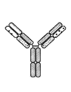
[0014] FIG. 1 is a representation of a multi-specific binding protein
that contains an
NKG2D-binding domain (right arm), a tumor associated antigen-binding domain
(left arm)
and an Fe domain or a portion thereof that binds to CD16.
[0015] FIG. 2 is a representation of a multi-specific binding protein that
contains an
NKG2D-binding domain in a scFv format (right arm), a tumor associated antigen-
binding
domain (left arm) and an Fc domain or a portion thereof that binds to CD16.
[0016] FIG. 3 is a representation of a TriNKET in the Triomab form,
which is a
trifunctional, bispecific antibody that maintains an IgG-like shape. This
chimera consists of
two half antibodies, each with one light and one heavy chain, that originate
from two parental
antibodies. Triomab form may be an heterodimeric construct containing 1/2 of
rat antibody
and '/2 of mouse antibody.
3
Date Recue/Date Received 2023-12-05
WO 2018/148445
PCT/US2018/017470
[0017] FIG. 4 is a representation of a TriNKET in the KiH Common Light
Chain (LC)
form, which involves the knobs-into-holes (KIHs) technology. KiH is a
heterodimer
containing 2 Fabs binding to target 1 and 2, and an Fc stabilized by
heterodimerization
mutations. TriNKET in the KiH format may be an heterodimeric construct with 2
fabs
binding to target 1 and target 2, containing 2 different heavy chains and a
common light chain
that pairs with both HC.
[0018] FIG. 5 is a representation of a TriNKET in the dual-variable
domain
immunoglobulin (DVD-IgTM) form, which combines the target binding domains of
two
monoclonal antibodies via flexible naturally occurring linkers, and yields a
tetravalent IgG -
like molecule. DVD-IgTM is an homodimeric construct where variable domain
targeting
antigen 2 is fused to the N terminus of variable domain of Fab targeting
antigen 1 Construct
contains normal Fc.
[0019] FIG. 6 is a representation of a TriNKET in the Orthogonal Fab
interface (Ortho-
Fab) form, which is an heterodimeric construct that contains 2 Fabs binding to
targetl and
target 2 fused to Fc. LC-HC pairing is ensured by orthogonal interface.
Heterodimerization is
ensured by mutations in the Fc.
[0020] FIG. 7 is a representation of a TrinKET in the 2 mug format.
[0021] FIG. 8 is a representation of a TriNKET in the ES form, which is
an
heterodimeric construct containing 2 different Fobs binding to target 1 and
target 2 fused to
the Fc. Heterodimerization is ensured by electrostatic steering mutations in
the Fc.
[0022] FIG. 9 is a representation of a TriNKET in the Fab Arm Exchange
form:
antibodies that exchange Fab arms by swapping a heavy chain (HC) and attached
light chain
(LC) (half-molecule) with a heavy-light chain pair from another molecule,
resulting in
bispecific antibodies. Fab Arm Exchange form (cFae) is a heterodimer
containing 2 Fabs
binding to target 1 and 2, and an Fc stabilized by heterodimerization
mutations.
[0023] FIG. 10 is a representation of a TriNKET in the SEED Body form,
which is an
heterodimer containing 2 Fabs binding to target 1 and 2, and an Fc stabilized
by
heterodimerization mutations.
[0024] FIG. 11 is a representation of a TriNKET in the LuZ-Y form, in
which leucine
zipper is used to induce heterodimerization of two different HCs. Lu7.-Y form
is a
heterodimer containing 2 different scFabs binding to target 1 and 2, fused to
Fc.
Heterodimerization is ensured through leucine zipper motifs fused to C-
terminus of Fc.
[0025] FIG. 12 is a representation of a TriNKET in the Cov-X-Body form.
4
Date Recue/Date Received 2023-12-05
WO 2018/148445
PCT/US2018/017470
[0026] FIGs. 13A-13B are representations of TriNKETs in the i&.-Body
forms, which are
an heterodimeric constructs with 2 different Fabs fused to Fc stabilized by
heterodimerization
mutations: Fabl targeting antigen 1 contains kappa LC, while second Fab
targeting antigen 2
contains lambda LC. FIG. 13A is an exemplary representation of one form of a
la-Body;
FIG. 13B is an exemplary representation of another la-Body.
[0027] FIG. 14 is a graph demonstrating the binding affinity of NKG2D-
binding domains
(listed as clones) to human recombinant NKG2D in an ELISA assay.
[0028] FIG. 15 is a graph demonstrating the binding affinity of NKG2D-
binding domains
(listed as clones) to cynomolgus recombinant NKG2D in an ELISA assay.
[0029] FIG. 16 is a graph demonstrating the binding affinity of NKG2D-
binding domains
(listed as clones) to mouse recombinant NKG2D in an ELISA assay.
[0030] FIG. 17 is a graph demonstrating the binding of NKG2D-binding
domains (listed
as clones) to EL4 cells expressing human NKG2D by flow cytometry showing mean
fluorescence intensity (MFI) fold over background.
[0031] FIG. 18 is a graph demonstrating the binding of NKG2D-binding
domains (listed
as clones) to EL4 cells expressing mouse NKG2D by flow cytometry showing mean
fluorescence intensity (MFI) fold over background.
[0032] FIG. 19 is a graph demonstrating specific binding affinity of
NKG2D-binding
domains (listed as clones) to recombinant human NKG2D-Fc by competing with
natural
ligand ULBP-6.
[0033] FIG. 20 is a graph demonstrating specific binding affinity of
NKG2D-binding
domains (listed as clones) to recombinant human NKG2D-Fc by competing with
natural
ligand MICA.
[0034] FIG. 21 is a graph demonstrating specific binding affinity of
NKG2D-binding
domains (listed as clones) to recombinant mouse NKG2D-Fc by competing with
natural
ligand Rae-1 delta.
[0035] FIG. 22 is a graph showing activation of human NKG2D by NKG2D-
binding
domains (listed as clones) by quantifying the percentage of TNF-alpha positive
cells which
express human NKG2D-CD3 zeta fusion proteins.
[0036] FIG. 23 is a graph showing activation of mouse NKG2D by NKG2D-
binding
domains (listed as clones) by quantifying the percentage of TNF-alpha positive
cells which
express mouse NKG2D-CD3 zeta fusion proteins.
[0037] FIG. 24 is a graph showing activation of human NK cells by NKG2D-
binding
domains (listed as clones).
5
Date Recue/Date Received 2023-12-05
WO 2018/148445
PCT/US2018/017470
[0038] FIG. 25 is a graph showing activation of human NK cells by NKG2D-
binding
domains (listed as clones).
[0039] FIG. 26 is a graph showing activation of mouse NK cells by NKG2D-
binding
domains (listed as clones).
[0040] FIG. 27 is a graph showing activation of mouse NK cells by NKG2D-
binding
domains (listed as clones).
[0041] FIG. 28 is a graph showing the cytoto)dc effect of NKG2D-binding
domains
(listed as clones) on tumor cells.
[0042] FIG. 29 is a graph showing the melting temperature of NKG2D-
binding domains
.. (listed as clones) measured by differential scanning fluorirnetry.
[0043] FIG. 30 is a graph showing enhanced activation of human NK cells
by multi-
specific binding proteins.
[0044] FIG. 31 is a graph showing multi-specific binding proteins
induced higher levels
of cytotoxicity towards tumor target cells by human NK cells.
[0045] FIG. 32 is a graph showing multi-specific binding proteins induced
higher levels
of cytotoxicity towards tumor target cells by human NK cells.
[0046] FIG. 33 is a graph showing multi-specific binding proteins
induced higher levels
of cytotoxicity towards tumor target cells by human NK cells.
[0047] FIG. 34 is a graph showing multi-specific binding proteins
induced higher levels
of cytotoxicity towards tumor target cells by human NK cells.
[0048] FIG. 35 is a graph showing multi-specific binding proteins
induced higher levels
of cytotoxicity towards tumor target cells by mouse NK cells.
[0049] FIG. 36 is a graph showing multi-specific binding proteins
induced higher levels
of cytotoxicity towards tumor target cells by mouse NK cells.
[0050] FIG. 37 is a binding profile of CD33-targeting TriNKETs to NKG2D
expressed
on EL4 cells. FIG. 37 shows binding of the two TriNKETs when a CD33-binding
domain is
used as the second targeting arm.
[0051] FIG. 38 is a binding profile of HER2-targeting TriNKETs to NKG2D
expressed
on EL4 cells. FIG. 38 shows the same two NKG2D-binding domains now paired with
a
HER2 second targeting arm.
[0052] FIG. 39 is a binding profile of BCMA-targeting TriNKETs to NKG2D
expressed
on EL4 cells.
6
Date Recue/Date Received 2023-12-05
WO 2018/148445
PCT/US2018/017470
[0053] FIG. 40 is a histogram of CD20-targeting TriNKETs that bind to
NKG2D
expressed on EL4 cells. Unstained EL4 cells were used a negative control for
fluorescence
signal. Unstained: filled; CD2O-TriNKET-F04: solid line; CD2O-TriNKET-C26:
dashed line.
[0054] FIG. 41 is a binding profile of CD33-targeting TriNKETs to CD33
expressed on
MV4-11 human AML cells.
[0055] FIG. 42 is a binding profile of HER2-targeting TriNKETs to HER2
expressed on
human 786-0 renal cell carcinoma cells.
[0056] FIG. 43 is a binding profile of BCMA-targeting TriNKETs to BCMA
expressed
on MM.1S human myeloma cells.
[0057] FIG. 44 is a histogram of CD20-targeting TriNKETs that bind to CD20
expressed
on Raji human lymphoma cells. Unstained cells were used a negative control for
fluorescence
signal. Unstained: filled; CD2O-TriNKET-F04: solid line; CD2O-TriNKET-C26:
dashed line.
[0058] FIGs. 45A-45C are bar graphs of synergistic activation of NK
cells using CD16
and NKG2D. FIG. 45A demonstrates levels of CD107a; FIG. 45B demonstrates
levels of
IFNy; FIG. 45C demonstrates levels of CD107a and IFNy. Graphs indicate the
mean (n = 2)
SD. Data are representative of five independent experiments using five
different healthy
donors.
[0059] FIG. 46 is a bar graph showing activation of NK cells using
TriNKETs targeting
NKG2D and CD16. Antibodies tested were of human IgG1 isotypes. Graphs indicate
the
mean (n = 2) SD.
[0060] FIGs. 47A ¨ 47C are bar graphs demonstrating that TriNKETs and
trastuzumab
were able to activate primary human NK cells in co-culture with HER2-positive
human
tumor cells, indicated by an increase in CD107a degranulation and IFNy
cytokine production.
Compared to the monoclonal antibody trastuzumab, both TriNKETs showed superior
activation of human NK cells with a variety of human HER2 cancer cells. FIG.
47A shows
that human NK cells are activated by TriNKETs when cultured with SkBr-3 cells.
FIG. 47B
shows that human NK cells are activated by TriNKETs when cultured with Colo201
cells.
FIG. 47C shows that human NK cell are activated by TriNKETs when cultured with
HCC1954 cells.
[0061] FIGs. 48A ¨ 48B are line graphs demonstrating TriNKET-mediated
activation of
rested or IL-2-activated human NK cells in co-culture with the CD33-expressing
human
AML cell line MV4-11. FIG. 48A shows TriNKET-mediated activation of resting
human NK
cells. FIG. 48B shows TriNKET-mediated activation of IL-2-activated human NK
cells from
the same donor.
7
Date Recue/Date Received 2023-12-05
WO 2018/148445
PCT/US2018/017470
[0062] FIGs. 49A ¨ 49B are graphs demonstrating TriNKET enhancement of
cytotoxic
activity using IL-2-activated and rested human NK cells. FIG. 49A shows
percent specific
lysis of SkBr-3 tumor cells by rested human NK cells. FIG. 49B shows percent
specific lysis
of SkBr-3 tumor cells by IL-2-activated human NK cells.
[0063] FIGs. 50A-50B are graphs demonstrating TriNKETs provide the greater
advantage against HER2 medium and low cancers compared to trastuzumab. FIG.
50A shows
activated human NK cell killing of HER2 high-SkBr-3 tumor cells. FIG. 50B
shows human
NK cell killing of HER2 low-786-0 tumor cells. TriNKETs provide a greater
advantage
compared to trastuzumab against cancer cells with low HER2 expression.
[0064] FIGs. 51A ¨ 51C are histograms showing that the expression of the
high-affinity
Fc1271 (CD64) on three human AML cells lines, Molm-13 cell line (FIG. 51A),
Mv4-11 cell
line (FIG. 51B), and THP-1 cell line (FIG. 51C).
[0065] FIGs. 52A-52B are line graphs of monoclonal antibody or TriNKET
mediated
activation of human NK cells in co-culture with either Molm-13 (FIG. 52B) or
THP-1 (FIG.
52A) cells.
[0066] FIGs. 53A ¨ 53C are line graphs of human NK cytotoxicity assays
using the three
human AML cell lines as targets. FIG. 53A shows that Mv4-11 cells, which
express CD64,
but at a lower level than THP-1, showed reduced efficacy with the monoclonal
anti-CD33.
FIG. 53B demonstrates that a monoclonal antibody against CD33 shows good
efficacy
against Molm-13 cells, which do not express CD64. FIG. 53C demonstrates that
THP-1 cells
showed no effect with monoclonal anti-CD33 alone. The identities of the line
graphs noted
in FIG. 53C are also applicable to the line graphs in FIGs. 53A-53B.
[0067] FIGs. 54A & 54B are bar graphs showing B cells from a health
donor are
sensitive to TriNKET-mediated lysis.
[0068] FIGs. 54C & 54D are bar graphs showing myeloid cells are resistant
to
TriNKET-mediated lysis.
[0069] FIG. 55 are line graphs of TriNKETs-mediated hPBMC killing of
SkBr-3 tumor
cells in long-term co-cultures.
[0070] FIG. 56 is a flowchart of study design of RMA/S-HER2 subcutaneous
SC2.2
efficacy.
[0071] FIG. 57 are line graphs showing that SC2.2 has no effect on
subcutaneous
RMA/S-HER2 tumor growth.
[0072] FIGs. 58A ¨ 58B are graphs showing in vitro binding by mcFAE-
C26.99
TriNKET. 60 mg/mL of indicated antibodies with four-fold dilutions were added
to 2x10⁵
8
Date Recue/Date Received 2023-12-05
WO 2018/148445
PCT/US2018/017470
B16F10 tumor cells (FIG. 58A) or EL4-mNKG2D cells (FIG. 58B). Binding was
assessed
using a goat anti-mouse PE secondary antibody followed by flow cytometric
analysis.
[0073] FIG. 59 is a graph showing increased NK cytotoxicity mediated by
mcFAE-
C26.99 TriNKET.
[0074] FIGs. 60A ¨ 60B show the anti-tumor efficacy of mcFAE-C26.99 TriNKET
in
Bl6F10 s.c. models. Mice were treated intraperitoneally with (FIG. 60A)
isotype control
mouse IgG2a mab C1.18.4 and mouse anti-Tyrp-1 monoclonal antibody or (FIG.
60B)
isotype control mouse IgG2a mab C1.18.4 and mcFAE-C26.99 TriNKET, injected at
a dose
of 150 lig (days 6, 8, 10, 12, 14, 16, and 21). Tumor growth was assessed for
28 days. Graphs
show tumor growth curves of individual mice.
[0075] FIGs. 61A ¨ 61B show anti-tumor efficacy of mcFAE-C26.99 TriNKET
in
Bl6F10 i.v. models. FIG. 61A represents tumor burden when antibodies were
administered at
a 150-jig dose (days 4, 6, 8, 11, 13, 15). FIG. 61B represents tumor burden
when antibodies
were administered at a 150-gg dose (days 7, 9, 11, 13, 15). 18 days after
tumor challenge,
mice were euthanized and surface lung metastases were scored.
[0076] FIG. 62 is bar graph showing that human NK cells are activated by
TriNKETs
when cultured with CD20+ Raji cells.
[0077] FIG. 63 is a bar graph showing that human NK activation in
culture with BCMA
positive MM. 1S human myeloma cells.
[0078] FIG. 64 is a graph showing that TriNKETs enhance human NK cell lysis
of
KMS12-PE myeloma cells.
[0079] FIG. 65 is a graph showing that BCMA-targeting TriNKETs with
different
NKG2D-binding domains enhance human NK cell lysis of KMS12-PE myeloma cells.
[0080] FIG. 66 is a line graph showing tri-specific binding in one
molecule is important
for maximal NK cell activity.
[0081] FIG. 67 is an Oasc-Fab heterodimeric construct that includes Fab
binding to
target 1 and scFab binding to target 2 fused to Fc. Heterodimerization is
ensured by mutations
in the Fc.
[0082] FIG. 68 is a DuetMab, which is an heterodimeric construct
containing 2 different
Fabs binding to antigen 1 and 2 and Fc stabilized by heterodimerization
mutations. Fab 1 and
2 contain differential S-S bridges that ensure correct LC and HC pairing.
[0083] FIG. 69 is a CrossmAb, which is an heterodimeric construct with 2
different Fabs
binding to target 1 and 2 fused to Fc stabilized by heterodimerization. CL and
CH1 domains
9
Date Recue/Date Received 2023-12-05
WO 2018/148445
PCT/US2018/017470
and Vh and VL domains are switched, e.g., CH1 is fused in-line with VL, while
CL is fused
in-line with VH.
[0084] FIG. 70 is a Fit-Ig, which is an homodimeric constructs where Fab
binding to
antigen 2 is fused to the N terminus of HC of Fab that binds to antigen 1. The
construct
contains wild-type Fe.
DETAILED DESCRIPTION
[0085] The invention provides multi-specific binding proteins that bind
a tumor-
associated antigen on a cancer cell and the NKG2D receptor and CD16 receptor
on natural
killer cells to activate the natural killer cell, pharmaceutical compositions
comprising such
.. multi-specific binding proteins, and therapeutic methods using such multi-
specific proteins
and pharmaceutical compositions, including for the treatment of cancer.
Various aspects of
the invention are set forth below in sections; however, aspects of the
invention described in
one particular section are not to be limited to any particular section.
[0086] To facilitate an understanding of the present invention, a number
of terms and
phrases are defined below.
[0087] The terms "a" and "an" as used herein mean "one or more" and
include the plural
unless the context is inappropriate.
[0088] As used herein, the term "antigen-binding site" refers to the
part of the
immunoglobulin molecule that participates in antigen binding. In human
antibodies,
the antigen binding site is formed by amino acid residues of the N-terminal
variable ("V")
regions of the heavy ("H") and light ("L") chains. Three highly divergent
stretches within the
V regions of the heavy and light chains are referred to as "hypervariable
regions" which are
interposed between more conserved flanking stretches known as "framework
regions," or
"1-Rs". Thus the term "FR" refers to amino acid sequences which are naturally
found between
and adjacent to hypervariable regions in immunoglobulins. In a human antibody
molecule,
the three hypervariable regions of a light chain and the three hypervariable
regions of a heavy
chain are disposed relative to each other in three dimensional space to form
an antigen-
binding surface. The antigen-binding surface is complementary to the three-
dimensional
surface of a bound antigen, and the three hypervariable regions of each of the
heavy and light
chains are referred to as "complementarity-determining regions," or "CDRs." In
certain
animals, such as camels and cartilaginous fish, the antigen-binding site is
formed by a single
antibody chain providing a "single domain antibody." Antigen-binding sites can
exist in an
Date Recue/Date Received 2023-12-05
WO 2018/148445
PCT/US2018/017470
intact antibody, in an antigen-binding fragment of an antibody that retains
the antigen-
binding surface, or in a recombinant polypeptide such as an scFv, using a
peptide linker to
connect the heavy chain variable domain to the light chain variable domain in
a single
polypeptide.
[0089] The term "tumor associated antigen" as used herein means any antigen
including
but not limited to a protein, glycoprotein, ganglioside, carbohydrate, lipid
that is associated
with cancer. Such antigen can be expressed on malignant cells or in the tumor
microenvironment such as on tumor-associated blood vessels, extracellular
matrix,
mesenchymal stroma, or immune infiltrates.
[0090] As used herein, the terms "subject" and "patient" refer to an
organism to be
treated by the methods and compositions described herein. Such organisms
preferably
include, but are not limited to, mammals (e.g., murines, simians, equines,
bovines, porcines,
canines, felines, and the like), and more preferably include humans.
[0091] As used herein, the term "effective amount" refers to the amount
of a compound
(e.g., a compound of the present invention) sufficient to effect beneficial or
desired results.
An effective amount can be administered in one or more administrations,
applications or
dosages and is not intended to be limited to a particular formulation or
administration route.
As used herein, the term "treating" includes any effect, e.g., lessening,
reducing, modulating,
ameliorating or eliminating, that results in the improvement of the condition,
disease,
disorder, and the like, or ameliorating a symptom thereof.
[0092] As used herein, the term "pharmaceutical composition" refers to
the combination
of an active agent with a carrier, inert or active, making the composition
especially suitable
for diagnostic or therapeutic use in vivo or ex vivo.
[0093] As used herein, the term "pharmaceutically acceptable carrier"
refers to any of the
standard pharmaceutical carriers, such as a phosphate buffered saline
solution, water,
emulsions (e.g., such as an oil/water or water/oil emulsions), and various
types of wetting
agents. The compositions also can include stabilizers and preservatives. For
examples of
carriers, stabilizers and adjuvants, see e.g., Martin, Remington's
Pharmaceutical Sciences,
15th Ed., Mack Publ. Co., Easton, PA [1975].
[0094] As used herein, the term "pharmaceutically acceptable salt" refers
to any
pharmaceutically acceptable salt (e.g., acid or base) of a compound of the
present invention
which, upon administration to a subject, is capable of providing a compound of
this invention
11
Date Recue/Date Received 2023-12-05
WO 2018/148445
PCT/US2018/017470
or an active metabolite or residue thereof. As is known to those of skill in
the art, "salts" of
the compounds of the present invention may be derived from inorganic or
organic acids and
bases. Exemplary acids include, but are not limited to, hydrochloric,
hydrobromic, sulfuric,
nitric, perchloric, fumaric, maleic, phosphoric, glycolic, lactic, salicylic,
succinic, toluene-p-
sulfonic, tartaric, acetic, citric, methanesulfonic, ethanesulfonic, formic,
benzoic, malonic,
naphthalene-2-sulfonic, benzenesulfonic acid, and the like. Other acids, such
as oxalic, while
not in themselves pharmaceutically acceptable, may be employed in the
preparation of salts
useful as intermediates in obtaining the compounds of the invention and their
pharmaceutically acceptable acid addition salts.
[0095] Exemplary bases include, but are not limited to, alkali metal (e.g.,
sodium)
hydroxides, alkaline earth metal (e.g., magnesium) hydroxides, ammonia, and
compounds of
formula NW, wherein W is C1_4 alkyl, and the like.
[0096] Exemplary salts include, but are not limited to: acetate,
adipate, alginate,
aspartate, benzoate, benzenesulfonate, bisulfate, butyrate, citrate,
camphorate,
camphorsulfonate, cyclopentanepropionate, digluconate, dodecylsulfate,
ethanesulfonate,
fumarate, flucoheptanoate, glycerophosphate, hemisulfate, heptanoate,
hexanoate,
hydrochloride, hydrobromide, hydroiodide, 2-hydroxyethanesulfonate, lactate,
maleate,
methanesulfonate, 2-naphthalenesulfonate, nicotinate, oxalate, palmoate,
pectinate,
persulfate, phenylpropionate, picrate, pivalate, propionate, succinate,
tartrate, thiocyanate,
tosylate, undecanoate, and the like. Other examples of salts include anions of
the compounds
of the present invention compounded with a suitable cation such as Nat, NH4,
and NW4+
(wherein W is a C1 _4 alkyl group), and the like.
[0097] For therapeutic use, salts of the compounds of the present
invention are
contemplated as being pharmaceutically acceptable. However, salts of acids and
bases that
are non-pharmaceutically acceptable may also find use, for example, in the
preparation or
purification of a pharmaceutically acceptable compound.
[0098] Throughout the description, where compositions are described as
having,
including, or comprising specific components, or where processes and methods
are described
as having, including, or comprising specific steps, it is contemplated that,
additionally, there
are compositions of the present invention that consist essentially of, or
consist of, the recited
components, and that there are processes and methods according to the present
invention that
consist essentially of, or consist of, the recited processing steps.
12
Date Recue/Date Received 2023-12-05
WO 2018/148445
PCT/US2018/017470
[0099] As a general matter, compositions specifying a percentage are by
weight unless
otherwise specified. Further, if a variable is not accompanied by a
definition, then the
previous definition of the variable controls.
PROTEINS
[00100] The invention provides multi-specific binding proteins that bind a
tumor-
associated antigen on a cancer cell and the NKG2D receptor and CD16 receptor
on natural
killer cells to activate the natural killer cell. The multi-specific binding
proteins are useful in
the pharmaceutical compositions and therapeutic methods described herein.
Binding of the
multi-specific binding protein to the NKG2D receptor and CD16 receptor on
natural killer
cell enhances the activity of the natural killer cell toward destruction of a
cancer cell.
Binding of the multi-specific binding protein to a tumor-associated antigen on
a cancer cell
brings the cancer cell into proximity to the natural killer cell, which
facilitates direct and
indirect destruction of the cancer cell by the natural killer cell. Further
description of
exemplary multi-specific binding proteins are provided below.
[00101] The first component of the multi-specific binding proteins binds to
NKG2D
receptor-expressing cells, which can include but are not limited to NK cells,
7.5 T
cells and CD8+ 43 T cells. Upon NKG2D-binding, the multi-specific binding
proteins may
block natural ligands, such as ULBP6 and MICA, from binding to NKG2D.
[00102] The second component of the multi-specific binding proteins binds to
one or more
tumor-associated antigens, which can include, but are not limited to HER2,
CD20, CD33,
BCMA, EpCAM, CD2, CD19, CD30, CD38, CD40, CD52, CD70, EGFR/ERBB1, IGF1R,
HER3/ERBB3, HER4/ERBB4, MUC1, cMET, SLAM14/, PSCA, MICA, MICB, TRAILR1,
IRAILR2, MAGE-A3, B7.1, B7.2, CTLA4, and PD1.
[00103] The third component for the multi-specific binding proteins binds to
cells
expressing CD16, an Pc receptor on the surface of leukocytes including natural
killer cells,
macrophages, neutrophils, eosinophils, mast cells, and follicular dendritic
cells.
[00104] The multi-specific binding proteins can take several formats as shown
in but not
limited to the examples below. One format is a heterodimeric, multi-specific
antibody
including a first immunoglobulin heavy chain, a second immunoglobulin heavy
chain and an
immunoglobulin light chain. The first immunoglobulin heavy chain includes a
first Fc (hinge-
CH2-CH3) domain, a first variable heavy chain domain and an optional first CH1
heavy
chain domain. The immunoglobulin light chain includes a variable light chain
domain and a
13
Date Recue/Date Received 2023-12-05
WO 2018/148445
PCT/US2018/017470
constant light chain domain; together with the first immunoglobulin heavy
chain, the
immunoglobulin light chain forms an antigen-binding site that binds NKG2D. The
second
immunoglobulin heavy chain comprises a second Fc (hinge-CH2-CH3) domain, a
second
variable heavy chain domain and a second optional CH1 heavy chain domain that
may pair
with an immunoglobulin light chain identical to the one that pairs with the
first
immunoglobulin heavy chain, except that when immunoglobulin light chain is
paired with the
second immunoglobulin heavy chain, the resulting antigen binding site binds to
a tumor
antigen. The first Fc domain and second Fc domain together are able to bind to
CD16
(FIG.1).
[00105] Another exemplary format involves a heterodimeric, multi-specific
antibody
which includes a first immunoglobulin heavy chain, an immunoglobulin light
chain and a
second immunoglobulin heavy chain. The first immunoglobulin heavy chain
includes a first
Fc (hinge-CH2-CH3) domain fused via either a linker or an antibody hinge to a
single chain
Fv (scFv) that binds NKG2D. A variety of linkers could be used for linking the
scFv to the
.. first Fc domain or within the scFv itself. In addition, the scFv can
incorporate mutations that
enable the formation of a disulfide bond to stabilize the overall scFv
structure. The scFv can
also incorporate mutations to modify the isoelectric point of the overall
first immunoglobulin
heavy chain and/or to enable more facile downstream purification. The second
immunoglobulin heavy chain includes a second Fc (hinge-CH2-CH3) domain and a
second
variable heavy chain domain and a second optional CH1 heavy chain domain. The
immunoglobulin light chain includes a variable light chain domain and a
constant light chain
domain. The second immunoglobulin heavy chain pairs with the immunoglobulin
light chain
and binds to a tumor antigen. The first Fc domain and the second Fc domain
together are able
to bind to CD16 (FIG. 2).
[00106] An alternative format of the heterodimeric multi-specific proteins
includes a first
immunoglobulin heavy chain, a second immunoglobulin heavy chain, a first
immunoglobulin
light chain and a second immunoglobulin light chain. The first immunoglobulin
heavy chain
includes a first Fc (hinge-CH2-CH3) domain, a first variable heavy chain
domain and an
optional first CHI heavy chain domain. The first immunoglobulin light chain
includes a
variable light chain domain and a constant light chain domain. Together with
the first
immunoglobulin heavy chain, the first immunoglobulin light chain forms an
antigen-binding
site that binds a tumor antigen. The second immunoglobulin heavy chain
comprises a second
Fc (hinge-CH2-CH3) domain, a second variable heavy chain domain and a second
optional
14
Date Recue/Date Received 2023-12-05
WO 2018/148445
PCT/US2018/017470
CH1 heavy chain domain. The second immunoglobulin light chain includes a
variable light
chain domain and a constant light chain domain. Together with the second
immunoglobulin
heavy chain, the immunoglobulin light chain forms an antigen-binding site that
binds to the
same tumor antigen. The second immunoglobulin heavy chain may pair with an
immunoglobulin light chain, which may be identical to the immunoglobulin light
chain that
pairs with the first immunoglobulin heavy chain, except that when
immunoglobulin light
chain is paired with the second immunoglobulin heavy chain, the resulting
antigen binding
site is a second antigen-binding site that binds to a tumor antigen. In
certain embodiments,
the first Fc domain and second Fc domain together are able to bind to CD16
(FIG.1).
[00107] One or more additional binding motifs may be fused to the C-terminus
of the
constant region CH3 domain, optionally via a linker sequence. In certain
embodiments, the
antigen-binding site could be a single-chain or disulfide-stabilized variable
region (ScFv) or
could form a tetravalent or trivalent molecule.
[00108] In some embodiments, the multi-specific binding protein is in the
Triomab form,
which is a trifunctional, bispecific antibody that maintains an IgG-like
shape. This chimera
consists of two half antibodies, each with one light and one heavy chain, that
originate from
two parental antibodies.
[00109] In some embodiments, the multi-specific binding protein is the KiH
Common
Light Chain (LC) form, which involves the knobs-into-holes (KIHs) technology.
The KIH
involves engineering CH3 domains to create either a "knob" or a "hole" in each
heavy chain
to promote heterodimerization. The concept behind the "Knobs-into-Holes (KiH)"
Fc
technology was to introduce a "knob" in one CH3 domain (CH3A) by substitution
of a small
residue with a bulky one (i.e., T366WcH3A in EU numbering). To accommodate the
"knob," a
complementary "hole" surface was created on the other CH3 domain (CH3B) by
replacing
the closest neighboring residues to the knob with smaller ones (i.e.,
T366S/L368A/Y407VcH3B). The "hole" mutation was optimized by structured-guided
phage
library screening (Atwell S, Ridgway JB, Wells JA, Carter P. Stable
heterodimers from
remodeling the domain interface of a homodimer using a phage display library.
J Mol
Biol (1997) 270(1):26-35). X-ray crystal structures of KiH Fc variants
(Elliott JM, Ultsch M,
Lee J, Tong R, Takeda K, Spiess C, et al., Antiparallel conformation of knob
and hole
aglycosylated half-antibody homodimers is mediated by a CH2-CH3 hydrophobic
interaction. J Mol Biol (2014) 426(9):1947-57; Mimoto F, Kadono S, Katada H,
Igawa T,
Kamikawa T, Hattori K. Crystal structure of a novel asymmetrically engineered
Fc variant
with improved affinity for FcgammaRs. Mol Immunol (2014) 58(1):132-8)
demonstrated
Date Recue/Date Received 2023-12-05
WO 2018/148445
PCT/US2018/017470
that heterodimerization is thermodynamically favored by hydrophobic
interactions driven by
steric complementarity at the inter-CH3 domain core interface, whereas the
knob¨knob and
the hole¨hole interfaces do not favor homodimerization owing to steric
hindrance and
disruption of the favorable interactions, respectively.
.. [00110] In some embodiments, the multi-specific binding protein is in the
dual-variable
domain immunoglobulin (DVD-IgTM) form, which combines the target binding
domains of
two monoclonal antibodies via flexible naturally occurring linkers, and yields
a tetravalent
IgG - like molecule.
[00111] In some embodiments, the multi-specific binding protein is in the
Orthogonal Fab
interface (Ortho-Fab) form. In ortho-Fab IgG approach (Lewis SM, Wu X,
Pustilnik A,
Sereno A, Huang F, Rick HL, et al. Generation of bispecific IgG antibodies by
structure-
based design of an orthogonal Fab interface. Nat. Biotechnol. (2014) 32(2):191-
8), structure-
based regional design introduces complementary mutations at the LC and
HCvii_ciii interface
in only one Fab, without any changes being made to the other Fab.
[00112] In some embodiments, the multi-specific binding protein is in the 2
inlIg format.
In some embodiments, the multi-specific binding protein is in the ES form,
which is an
heterodimeric construct containing 2 different Fabs binding to target 1 and
target 2 fused to
the Fc. Heterodimerization is ensured by electrostatic steering mutations in
the Fc. In some
embodiments, the multi-specific binding protein is in the la-Body form, which
is an
heterodimeric constructs with 2 different Fabs fused to Fc stabilized by
heterodimerization
mutations: Fabl targeting antigen 1 contains kappa LC, while second Fab
targeting antigen 2
contains lambda LC. FIG. 13A is an exemplary representation of one form of a
Ick-Body;
FIG. 13B is an exemplary representation of another ick-Body.
[00113] In some embodiments, the multi-specific binding protein is in Fab Arm
Exchange
form (antibodies that exchange Fab arms by swapping a heavy chain and attached
light chain
(half-molecule) with a heavy-light chain pair from another molecule, which
results in
bispecific antibodies). In some embodiments, the multi-specific binding
protein is in the
SEED Body form (The strand-exchange engineered domain (SEED) platform was
designed
to generate asymmetric and bispecific antibody-like molecules, a capability
that expands
.. therapeutic applications of natural antibodies. This protein engineered
platform is based on
exchanging structurally related sequences of immunoglobulin within the
conserved CH3
domains. The SEED design allows efficient generation of AG/GA heterodimers,
while
disfavoring homodimerization of AG and GA SEED CH3 domains. (Muda M. et al.,
Protein
16
Date Recue/Date Received 2023-12-05
WO 2018/148445
PCT/US2018/017470
Eng. Des. Se!. (2011, 24(5):447-54)). In some embodiments, the multi-specific
binding
protein is in the LuZ-Y foiiii, in which leucine zipper is used to induce
heterodimerization of
two different HCs. (Wranik, BJ. et al., J. Biol. Chem. (2012), 287:43331-9).
[00114] In some embodiments, the multi-specific binding protein is in the Cov-
X-Body
form (In bispecific CovX-Bodies, two different peptides are joined together
using a branched
azetidinone linker and fused to the scaffold antibody under mild conditions in
a site-specific
manner. Whereas the pharmacophores are responsible for functional activities,
the antibody
scaffold imparts long half-life and Ig-like distribution. The pharmacophores
can be
chemically optimized or replaced with other pharmacophores to generate
optimized or unique
.. bispecific antibodies. (Doppalapudi VR etal., PNAS (2010), 107(52);22611-
22616).
[00115] In some embodiments, the multi-specific binding protein is in an Oasc-
Fab
heterodimeric form that includes Fab binding to target 1 and scFab binding to
target 2 fused
to Fc. Heterodimerization is ensured by mutations in the Fc.
[00116] In some embodiments, the multi-specific binding protein is in a
DuetMab form,
which is an heterodimeric construct containing 2 different Fabs binding to
antigen 1 and 2
and Fc stabilized by heterodimerization mutations. Fab 1 and 2 contain
differential S-S
bridges that ensure correct LC and HC pairing.
[00117] In some embodiments, the multi-specific binding protein is in a
CrossmAb form,
which is an heterodimeric construct with 2 different Fabs binding to Target 1
and 2 fused to
Fc stabilized by heterodimerization. CL and CH1 domains and VH and VL domains
are
switched, e.g., CH1 is fused in-line with VL, while CL is fused in-line with
VH.
[00118] In some embodiments, the multi-specific binding protein is in a Fit-Ig
form, which
is an homodimeric constructs where Fab binding to antigen 2 is fused to the N
terminus of
HC of Fab that binds to antigen 1. The construct contains wild-type Fc.
[00119] Table 1 lists peptide sequences of heavy chain variable domains and
light chain
variable domains that, in combination, can bind to NKG2D.
17
Date Recue/Date Received 2023-12-05
WO 2018/148445 PCT/US2018/017470
Table 1
Clones Heavy chain variable region amino acid Light chain variable
region amino acid
sequence sequence
ADI-27705 QVQLQQWGAGLLKPSETLSLTCAVY DIQMTQSPSTLSASVGDRVTITCR
GGSFSGYYWSWIRQPPGKGLEWIGEI ASQSISSWLAWYQQKPGKAPKLL
DHSGSTNYNPSLKSRVTISVDTSKNQ IYKASSLESGVPSRFSGSGSGTEFT
FSLKLSSVTAADTAVYYCARARGPW LTISSLQPDDFATYYCQQYNSYPI
SFDPWGQGTLVTVSS TFGGGTKVEIK
(SEQ ID NO:1) (SEQ ID NO:2)
ADI-27724 QVQLQQWGAGLLKPSETLSLTCAVY EIVLTQSPGTLSLSPGERATLSCRA
GGSFSGYYWSWIRQPPGKGLEWIGEI SQSVSSSYLAWYQQKPGQAPRLL
DHSGSTNYNPSLKSRVTISVDTSKNQ IYGASSRATGIPDRFSGSGSGTDFT
FSLKLSSVTAADTAVYYCARARGPW LTISRLEPEDFAVYYCQQYGSSPIT
SFDPWGQGTLVTVSS FGGGTKVEIK
(SEQ ID NO:3) (SEQ ID NO:4)
ADI-27740 QVQLQQWGAGLLKPSETLSLTCAVY DIQMTQSPSTLSASVGDRVTITCR
(A40) GGSFSGYYWSWIRQPPGKGLEWIGEI ASQSIGSWLAWYQQKPGKAPKLL
DHSGSTNYNPSLKSRVTISVDTSKNQ IYKASSLESGVPSRFSGSGSGTEFT
FSLKLSSVTAADTAVYYCARARGPW LTISSLQPDDFATYYCQQYHSFYT
SFDPWGQGTLVTVSS FGGGTKVEIK
(SEQ ID NO:5) (SEQ ID NO:6)
ADI-27741 QVQLQQWGAGLLKPSETLSLTCAVY DIQMTQSPSTLSASVGDRVTITCR
GGSFSGYYWSWIRQPPGKGLEWIGEI ASQSIGSWLAWYQQKPGKAPKLL
DHSGSTNYNPSLKSRVTISVDTSKNQ IYKASSLESGVPSRFSGSGSGTEFT
FSLKLSSVTAADTAVYYCARARGPW LTISSLQPDDFATYYCQQSNSYYT
SFDPWGQGTLVTVSS FGGGTKVEIK
(SEQ ID NO:7) (SEQ ID NO:8)
ADI-27743 QVQLQQWGAGLLKPSETLSLTCAVY DIQMTQSPSTLSASVGDRVTITCR
GGSFSGYYWSWIRQPPGKGLEWIGEI ASQSISSWLAWYQQKPGKAPKLL
DHSGSTNYNPSLKSRVTISVDTSKNQ IYKASSLESGVPSRFSGSGSGTEFT
FSLKLSSVTAADTAVYYCARARGPW LTISSLQPDDFATYYCQQYNSYPT
18
Date Recue/Date Received 2023-12-05
50-ZI -EZOZ PAP 3W 31.21:1/3115311 PIE
6T
N3111A1CIDASVS-1I,SdSOITAIOIG AAVALTS-112Sd.311-1DVDMOUTOAO 0-176Z-IGV
(OZ:ON GI OHS) (61:0N CU OHS)
31I3AXI000d SSAINILDODAAdoadS
IdAIGAOODAAIVJGGIO-IsSI,1:1 MdD21V2IVDAAAVICIVVIASSI)FISA
JAH,LOSOSDSJWSdADSHISSV31A1 ONNSIGASLIAIISNISdNANISDSHO
T-Dmvxodx0Ofkmv-imsoisOsv IHDIMH-IMIDdd0211MSMAADSHSOD
11DIL1AIICID A S Sd sOnnibia AAVD,E1S-112Sd31-1-
1DVDMOO-IOAO I 0176Z1GV
(81:0N GI OHS) (LT :ON CU OHS)
NI3AN,I909.4 SSAINI,LDODMdiadS
JAMSNAOODALLVJGCMOISSIII McIDIIVITVDAAAVIGVVIASSINIS4
IdHIDSOSDSAITSdADSHISSVNAT ONNSIGASHAITSNISdNANISDSHG
-1-1)1dV31Dc13100AMVIMSSISOSV IHDVAH'IMIDddOIIIMSMAADSJSDD
2131I1ANGDASVSIISdSOITAIOIG AAVD,LISII1Sc1)11-1DVDMOO-10A0 666Z-1GV
(9I :ON GI OHS) (ST :ON GI OHS)
NianN,LoOodi SSAINTLOODMdCHS
MdA3NSOODAL1VdClUclUISSI,1:1 MdDI1V2IVDAAAVICIVVIASS-131-1S4
IdaLDSDSDSANSdADSHISSVNAI ONNSIGASTIAITSNISdNANISDSHG
-n)mvxoct3100Amv-vAssisOsv I39IM3IDN9ddOIIIMSMAADSdSD9
11DIL1ANGDASVSIISdSOITAIOIG Anvauls-u1scDrnovomOO-10A0 ts i SZ-IGV
(-17T :ON GI OHS) (I :ON cu Oas)
)1IHAN,L900H SSAINI.LOODMdladS
IlddSDAOODAAIVAGC1c1O-ISSIE1 MdDIIVIIVDAAAVICIVVIASS-131-1S3
id31OS9SOSAUSdADSH-ISSVNAI ONNSIGASILANSMISdNANISOSHG
1-1NdV3IDdNOOAMVIMSSISOSV I3DIM3-IDNDddONIMSAAAADSASOD (9ZD)
IIDILLAIICIDASVSMSdSO,LIATOKI AAVDEIS'I2Sc131-1-1DVDMOCTIOAO 9.ZZ8Z-IGV
(ZI:ON GI OHS) (T 1 :ON CR OHS)
31IH-131,1,900,4,1, SSAINIIDODMcIa4D
AdIGASOODAAIVSGHdOISSIIII MdDHVIIVDAAAVICIVVIASSIXIS4
da1DSOSOSH210dADSHI11SVAU ON3ISIGASI1AIIS31-1SdNANISOSHO
rmadOodx00Ankt\nAssisOsi I301M3-1WIDdc10111MSMAADSJSDD
IIDIL1ANCIDASVS-ISSdSOITAI.O1H AAVD,EISII2Sd.3171DVDMOU1OAO STSZ-IGV
(OT :ON GI OHS) (6:0N GI OHS)
31I3AXI000d SSAINILDODMcICHS
OLJ1JO/8IOZSI1/Iad
irt81'I/8I07 OM
50-ZI -EZOZ PAP 3W 31.21:1/3115311 3).21a
OZ
dildSCUOODAKINJCIMIOISSIII AVIDNVIIVDAAAVIUVVIASSINIS4
1daLDS9S9S.d21SdADS3ISSVNAI ONNS.LUASILAIISNISdNANISDSHU
TINdV3IDd)100AMVIMSSISOSV IHDIMHIDNOddOIIIMSMAADSdSOD
Ila1LIA2109ASVSIISdSO1IAIOIG AAVD,LISIIHSd)ITIDVDMOOIOAO tZ176Z-IGV
(OE:ON CH OHS) (6Z:ON cu OHs)
3IIHANI000d SSAINTIDOOMdiadS
ISASHAOODALINdCIUdOISSIII MdDIIV2IVDAXAVICIVVIASSINISd
JAH,LOSDSOSAIISdADSHISSVNAI ONNS.LUASIIANSNISdNANISDSHU
1131dV3IDd)100AMVIMSSISOSV IHDIMHIMIDddONIMSMAADSJSDD
IIDILLANUDASVSIISdSO,LIATOICI AAVD.LISIIHSdN'TIDVDMOOIOAO IZt6Z-IGV
(VON oi Oas) (woN cn Oas)
3IIHANIDDD SSAINIIDODMdCHS
d.I.S4SSAOODAAIVHCICHOISSIII MdMIV2IVDAAAVIOVV1ASSINIS4
IdaIDSDSDSAIISdADSHISSVMAI ONNSICIASIIAIISMISdNIANISOSHCI
113IcIV,IDdNOOAMVIMSSISOSV IHDIMHID)19ddOIIIMSMAADSHSD9
2131I1AIKIDASVSILSdSO1IATOIU AAVD,EISIIHSd3ITIDVDMOO-TOAO 6I-176Z-IUV
(9:0N UI OHS) (SZ:ON UI Oas)
)1I3AXI000.4 SSAINIIDOOMdUdS
IddSOAOODAA,LVdiaadOlssul MdDHVIIVDAAAVIOVVIASSINIS4
JAH,LOSOSDS.4NSdADSHISSV)IAI ONNSIGASI,LAIISMISdNANISOSHO
1131dV3IDd)100AMVIMSSISOSIV I3DIM3IMIDddOHIMSMAADSdS9D
IIDIIIAIICIDASVSlisdsOnniOpa AAVaLISIIHSd3ITIDVDMOOIOAO LO-176Z-ICIV
(17VON cu Oas) (:ON UI Ogs)
NI3ANI009.4 SSAINILDODMcICHS
IcIdS9A.00DAKINJUUdOISSI1I AVIDIIVIIVDAAAVIUVVIASSINIS4
IdHIDSOS9S.411SdADSHISSVNAI ONNSIGASI,LAIISMISdNANISOSHO
'11)IdV3IDcI)100AMVIMSSISOSV IHDIAkHIMIDddOIIIMSMAADSJSOD
21D1I1ANGDASVSIISdSO1IAIOIU AAVD,LISIIHSd)11-IDVDMOOIOAO g0t6Z-IGV
(ZZ:ON jj OHS) (IZ:ON UI OHS)
3II3AMID99d SSAINILOODMdUdS
,LdASUAOODAL1VAUCklOISSI,L1 MdDIIV2IVDAAAVICIVVIASSINIS4
1daLDS9S9SdllSdADSHISSVNAI ONNSIGASILAIISNISdNANISDSHU
TINdV3IDd)100AMVIMSSISOSV IHDIMHIDNOddOIIIMSMAADSdSOD
OLJ1JO/8IOZSI1/Iad irt81'I/8I07 OM
WO 2018/148445 PCT/US2018/017470
SFDPWGQGTLVTVS S GGGTKVEIK
(SEQ ID NO:31) (SEQ ID NO:32)
ADI-29425 QVQLQQWGAGLLKPSETLS LTCAVY DIQMTQSPSTLSASVGDRVTITCR
GGS FS GYYWSWIRQPPGKGLEWIGEI AS QS IS SWLAWYQQKPGKAPKLL
DHS GS TNYNPS LKSRVTIS VDTS KNQ IYKA SS LES GYPS RFS GS GS GTEFT
FSLKLS SVTAADTAVYYCARARGPW LTISS LQPDDFATYYCQQYQSYPT
SFDPWGQGTLVTVS S FGGGTKVEIK
(SEQ ID NO:33) (SEQ ID NO:34)
ADI-29426 QVQLQQWGAGLLKPSETLSLTCAVY DIQMTQSPSTLSASVGDRVTITCR
GGSFS GYYWSVVIRQPPGKGLEWIGEI AS QS IG SWLAVVYQQKPGKAPKLL
DHS GS TNYNPSLKSRVTIS VDTS KNQ IYKA SS LES GVPS RFS GS GS GTEFT
FS LKLS SVTAADTAVYYCARARGPW LTISS LQPDDFATYYCQQYHSF1PT
SFDPWGQGTLVTVS S FGGGTKVEIK
(SEQ ID NO:35) (SEQ ID NO:36)
ADI-29429 QVQLQQWGAGLLKPSETLS LTCAVY DIQMTQSPSTLSASVGDRVTITCR
GGSFS GYYWSWIRQPPGKGLEWIGEI AS QS IG SWLAWYQQKPGKAPKLL
DHS GS TNYNPSLKSRVTIS VDTS KNQ IYKA SS LES GVPS RFS GS GS GTEFT
FSLKLS S VTAADTAVYYCARARGPW LTISS LQPDDFATYYCQQYELYSY
SFDPWGQGTLVTVS S TFGGGTKVEIK
(SEQ ID NO:37) (SEQ ID NO:38)
ADI-29447 QVQLQQWGAGLLKPSETLS LTCAVY DIQMTQSPSTLSASVGDRVTITCR
(F47) GGSFS GYrIVSWIRQPPGKGLEWIGEI AS QS IS SWLAWYQQKPGKAPKLL
DHS GS TNYNPSLKSRVTIS VDTS KNQ WKA SS LES GVPS RFS GS GS GTEFT
FSLKLS SVTAADTAVYYCARARGPW LTISS LQPDDFATYYCQQYDTFITF
SFDPWGQGTLVTVS S GGGTKVEIK
(SEQ ID NO:39) (SEQ ID NO:40)
ADI-27727 QVQLVQS GAEVKKPGSSVKVSCKAS DIVMTQS PDS LAVS LGERATINC K
GGTFS SYAISWVRQAPGQGLEWMGG SS QS VLYS SNNKNYLAWYQQKP
IIPIFGTANYAQKFQGRVTITADESTS GQPPKLLIYWASTRESGVPDRFSG
TAYMELS SLRSEDTAVYYCARGD S SI SGS GTDFTLTIS SLQAEDVAVYYC
RHAYYYYGMDVWGQGTTVTVSS QQYYSTPITFGGGTKVEIK
(SEQ ID NO:41) (SEQ ID NO:42)
ADI-29443 QLQLQESGPGLVKPSETLSLTCTV SG EIVLTQSPATLSLSPGERATLSCRA
21
Date Recue/Date Received 2023-12-05
WO 2018/148445 PCT/US2018/017470
(F43) GS IS SS SYYWGWIRQPPGKGLEWIGSI SQSVSRYLAWYQQKPGQAPRLLI
YYS GS TYYNPSLKSRVTIS VDTS KNQ YDASNRATGIPARFS GS GS GTDFT
FSLKLSS VTAADTAVYYCARGSDRF LTISSLEPEDFAVYYCQQFDTWPP
HPYFDYVVGQGTLVT VS S TFGGGTKVEIK
(SEQ ID NO:43) (SEQ ID NO:44)
ADI-27744 EVQLLES GGGLVQPGGSLRLSCAAS G DIQMTQS PS S VS AS VGDRVTITCR
(A44) FTFS SYAMSWVRQAPGKGLEWVSAI AS QGIDSWLAWYQQKPGKAPKL
S GS GGSTYYAD SVKGRFTIS RDNS KN LIYAAS SLQS GVPSRFS GS GS GTD
TLYLQMNSLRAEDTAVYYCAKDGG El LTISSLQPEDFATYYCQQGVSY
YYDSGAGDYWGQGTLVTVSS PRTFGGGTKVEIK
(SEQ ID NO:45) (SEQ ID NO:46)
CDR1 (SEQ ID NO:51) - FTFSSYAMS CDR1 (SEQ ID NO:54) -
CDR2 (SEQ ID NO:52) - RAS QGIDSWLA
AISGSGGSTYYADSVKG CDR2 (SEQ ID NO:55) - AASSLQS
CDR3 (SEQ ID NO:53) - CDR3 (SEQ ID NO:56) -
AKDGGYYDSGAGDY QQGVSYPRT
ADI-27749 EVQLVESGGGLVKPGGSLRLSCAAS DIQMTQSPSSVSASVGDRVTITCR
(A49) GFTFS S YSMNWVRQAPGKGLEWVSS AS QGIS SWLAWYQQKPGKAPKLL
ISS SSSYIYYADS VKGRFTISRDNAKN IYAASS LQS GVPSRFS GSGSGTDF
SLYLQMNSLRAEDTAVYYCARGAP TLTIS S LQPEDFATYYC QQGVS FP
MGAAAGWFDPWGQGTLVTVSS R GGGTKVEIK
(SEQ ID NO:47) (SEQ ID NO:48)
CDR1 (SEQ ID NO:57) - FTFSSYSMN CDR1 (SEQ ID NO:60) -
CDR2 (SEQ ID NO:58) - RAS QGIS SWLA
SISSSSSYIYYADSVKG CDR2 (SEQ ID NO:61) - AASSLQS
CDR3 (SEQ ID NO:59) - CDR3 (SEQ ID NO:62) -
ARGAPMGAAAGWFDP QQGVSFPRT
ADI-29463 QVQLVQS GAEVKKPGASVKVSCKAS EIVLTQSPGTLSLSPGERATLSCRA
GYTFTGYYMHWVRQAPGQGLEWM SQS VS S NLAWYQQ KPGQAPRLLI
22
Date Recue/Date Received 2023-12-05
WO 2018/148445 PCT/US2018/017470
(F63) GWINPNSGGTNYAQKFQGRVTMTR YGASTRAT GIPARFS GS GS GTEFT
DTSISTAYMELSRLRSDDTAVYYCAR LTISSLQSEDFAVYYCQQDDYWP
DTGEYYDTDDHGMDVWGQGTTVTV PT'FGGGTKVEIK
SS (SEQ ID NO:50)
(SEQ ID NO:49)
CDR1 (SEQ ID NO:66) -
CDR1 (SEQ ID NO:63) - YTFTGYYMH RASQSVSSNLA
CDR2 (SEQ ID NO:64) - CDR2 (SEQ ID NO:67) - GASTRAT
WINPNSGGTNYAQKFQG CDR3 (SEQ ID NO:68) -
CDR3 (SEQ ID NO:65) - QQDDYWPPT
ARDTGEYYDTDDHGMDV
ADI-29404 QVQLQQWGAGLLKPSETLSLTCAVY DIQMTQSPSTLSASVGDRVTITCR
(F04) GGS FS GYYWSWIRQPPGKGLEWIGEI AS QS IS SWLAWYQQKPGKAPKLL
DHS GS TNYNPS LKSRVTIS VDTS KNQ IYKA SS LES GYPS RFS GS GS GTEFT
FSLKLSSVTAADTAVYYCARARGPW LTISSLQPDDFATYYCEQYDS YPT
SFDPWGQGTLVTVSS (SEQ ID NO:78) FGGGTKVEIK (SEQ ID NO:79)
ADI-28200 QVQLVQSGAEVKKPGSSVKVSCKAS DIVMTQS PDS LAVS LGERATTNCE
GGTFSSYAISVVVRQAPGQGLENVMGG SS QSLLN SGNQKNYLTWYQQKPG
IIPIFGTANYA QKFQGRVTITADES TS QPPKPLIYWAS TRES GVPDRFS GS
TAYMELSSLRSEDTAVYYCARRGRK GS GTD FTLTIS S LQAEDVAVYYC Q
AS GSFYYYYGMDVWGQGTTVTV S S NDYSYPYTFGQGTKLEIK
(SEQ ID NO:80) (SEQ ID NO:81)
[00120] Alternatively, a heavy chain variable domain defined by SEQ ID NO:69
can be
paired with a light chain variable domain defined by SEQ ID NO:70 to form an
antigen-
binding site that can bind to NKG2D, as illustrated in US 9,273,136.
SEQ ID NO:69
QVQLVESGGGLVKPGGSLRLSCAASGFTFSS YGMHWVRQAPGKGLEWVAFIRYD GS
NKYYADSVKGRFTISRDNSKNTLYLQMNSLRAEDTAVYYCAKDRGLGDGTYFDYW
GQGTTVTVSS
23
Date Recue/Date Received 2023-12-05
WO 2018/148445
PCT/US2018/017470
SEQ ID NO:70
QSALTQPASVSGSPGQSITISCSGSSSNIGNNAVNWYQQLPGKAPKLLIYYDDLLPSG
VSDRFSGS KSGTSAFLAISGLQSEDEADYYCAAWDDSLNGPVFGGGTKLTVL
[00121] Alternatively, heavy chain variable domain defined by SEQ ID NO:71 can
be
paired with light chain variable domain defined by SEQ ID NO:72 to form an
antigen-
binding site that can bind to NKG2D, as illustrated in US 7,879,985.
SEQ ID NO:71
QVHLQESGPGLVKPSETLSLTCTVSDDSISSYYWSWIRQPPGKGLEWIGHISYSGSAN
YNPSLKSRVTISVDTSKNQFSLKLSSVTAADTAVYYCANWDDAFNIWGQGTMVTVS
S
SEQ ID NO:72
EIVLTQSPGTLSLSPGERATLSCRASQSVSSSYLAWYQQKPGQAPRLLIYGASSRATGI
PDRFSGSGSGTDFTLTISRLEPEDFAVYYCQQYGSSPWTFGQGTKVEIK
[00122] Within the Pc domain, CD16 binding is mediated by the hinge region and
the CH2
domain. For example, within human IgGl, the interaction with CD16 is primarily
focused on
amino acid residues Asp 265 ¨ Glu 269, Asn 297 ¨ Thr 299, Ala 327 ¨ Ile 332,
Leu 234 ¨
Ser 239, and carbohydrate residue N-acetyl-D-glucosamine in the CH2 domain
(see,
Sondermann et al, Nature, 406(6793):267-273). Based on the known domains,
mutations can
be selected to enhance or reduce the binding affinity to CD16, such as by
using phage-
displayed libraries or yeast surface-displayed cDNA libraries, or can be
designed based on
the known three-dimensional structure of the interaction.
[00123] The assembly of heterodimeric antibody heavy chains can be
accomplished by
expressing two different antibody heavy chain sequences in the same cell,
which may lead to
the assembly of homodimers of each antibody heavy chain as well as assembly of
heterodimers. Promoting the preferential assembly of heterodimers can be
accomplished by
incorporating different mutations in the CH3 domain of each antibody heavy
chain constant
region as shown in US13/494870, US16/028850, US11/533709, US12/875015,
US13/289934, US14/773418, US12/811207, US13/866756, US14/647480, US14/830336.
For example, mutations can be made in the CH3 domain based on human IgG1 and
incorporating distinct pairs of amino acid substitutions within a first
polypeptide and a second
polypeptide that allow these two chains to selectively heterodimerize with
each other. The
positions of amino acid substitutions illustrated below are all numbered
according to the EU
index as in Kabat.
24
Date Recue/Date Received 2023-12-05
WO 2018/148445
PCT/US2018/017470
[00124] In one scenario, an amino acid substitution in the first polypeptide
replaces the
original amino acid with a larger amino acid, selected from arginine (R),
phenylalanine (F),
tyrosine (Y) or tryptophan (W), and at least one amino acid substitution in
the second
polypeptide replaces the original amino acid(s) with a smaller amino acid(s),
chosen from
alanine (A), serine (S), threonine (T), or valine (V), such that the larger
amino acid
substitution (a protuberance) fits into the surface of the smaller amino acid
substitutions (a
cavity). For example, one polypeptide can incorporate a T366W substitution,
and the other
can incorporate three substitutions including T366S, L368A, and Y407V.
[00125] An antibody heavy chain variable domain of the invention can
optionally be
coupled to an amino acid sequence at least 90% identical to an antibody
constant region, such
as an IgG constant region including hinge, CH2 and CH3 domains with or without
CH1
domain. In some embodiments, the amino acid sequence of the constant region is
at least
90% identical to a human antibody constant region, such as an human IgG1
constant region,
an IgG2 constant region, IgG3 constant region, or IgG4 constant region. In
some other
embodiments, the amino acid sequence of the constant region is at least 90%
identical to an
antibody constant region from another mammal, such as rabbit, dog, cat, mouse,
or horse.
One or more mutations can be incorporated into the constant region as compared
to human
IgG1 constant region, for example at Q347, Y349, L351, S354, E356, E357, K360,
Q362,
S364, T366, L368, 1(370, N390, 1(392, T394, D399, S400, D401, F405, Y407,
1(409, T411
and/or 1(439. Exemplary substitutions include, for example, Q347E, Q347R,
Y349S,
Y349K, Y349T, Y349D, Y349E, Y349C, T350V, L351K, L351D, L351Y, S354C, E356K,
E357Q, E357L, E357W, K360E, K360W, Q362E, S364K, S364E, S364H, S364D, T366V,
T366I, T366L, T366M, T366K, T366W, T366S, L368E, L368A, L368D, K370S, N390D,
N390E, K392L, K392M, K392V, K392F, K392D, K392E, T394F, T394W, D399R, D399K,
D399V, S400K, S400R, D401K, F405A, F405T, Y407A, Y407I , Y407V, K409F, K409W,
K409D, T411D, T411E, K439D, and K439E.
[00126] In certain embodiments, mutations that can be incorporated into the
CH1 of a
human IgG1 constant region may be at amino acid V125, F126, P127, T135, T139,
A140,
F170, P171, and/or V173. In certain embodiments, mutations that can be
incorporated into
the Cic of a human IgG1 constant region may be at amino acid E123, F116, S176,
V163,
S174, and/or T164.
[00127] Alternatively, amino acid substitutions could be selected from the
following sets
of substitutions shown in Table 2.
Date Recue/Date Received 2023-12-05
WO 2018/148445
PCT/US2018/017470
Table 2
First Polypeptide Second Polypeptide
Set 1 5364E/F405A Y349K/T394F
Set 2 S364H/D401K Y349T/T411E
Set 3 S364H/T394F Y349T/F405A
Set 4 S364E/T394F Y349K/F405A
Set 5 5364E/T411E Y349K/D401K
Set 6 5364D/T394F Y349K/F405A
Set 7 S364H/F405A Y349T/T394F
Set 8 S364K/E357Q L368D/K370S
Set 9 L368D/K370S S364K
Set 10 L368E/K3705 S364K
Set 11 K360E/Q362E D401K
Set 12 L368D/K3705 5364K/E357L
Set 13 K370S 5364K/E357Q
Set 14 F405L K409R
Set 15 K409R F405L
[00128] Alternatively, amino acid substitutions could be selected from the
following sets
of substitutions shown in Table 3.
Table 3
First Polypeptide Second Polypeptide
Set 1 K409W D399V/F405T
Set 2 Y349S E357W
Set 3 K360E Q347R
Set 4 K360E/K409W Q347R/D399V/F405T
Set 5 Q347E/K360E/K409W Q347R/D399V/F405T
Set 6 Y3495/K409W E357W/D399V/F405T
[00129] Alternatively, amino acid substitutions could be selected from the
following set of
substitutions shown in Table 4.
Table 4
First Polypeptide Second Polypeptide
Set 1 T366K/L351K L351D/L368E
26
Date Recue/Date Received 2023-12-05
WO 2018/148445
PCT/US2018/017470
Set 2 T366K/L351K L351D/Y349E
Set 3 T366K/L351K L351D/Y349D
Set 4 T366K/L351K L351D/Y349E/L368E
Set 5 T366K/L351K L351D/Y349D/L368E
Set 6 E356K/D399K K392D/K409D
[00130] Alternatively, at least one amino acid substitution in each
polypeptide chain could
be selected from Table 5.
Table 5
First Polypeptide Second Polypeptide
L351Y, D399R, D399K, S400K, S400R, T366V, T366I, T366L, T366M, N390D,
Y407A, Y4071, Y407V N390E, K392L, K392M, K392V, K392F
K392D, K392E, K409F, K409W, T411D and
T4 11E
[00131] Alternatively, at least one amino acid substitutions could be selected
from the
following set of substitutions in Table 6, where the position(s) indicated in
the First
Polypeptide column is replaced by any known negatively-charged amino acid, and
the
position(s) indicated in the Second Polypeptide Column is replaced by any
known positively-
charged amino acid.
Table 6
First Polypeptide Second Polypeptide
K392, 1(370, 1(409, or K439 D399, E356, or E357
[00132] Alternatively, at least one amino acid substitutions could be selected
from the
following set of in Table 7, where the position(s) indicated in the First
Polypeptide column is
replaced by any known positively-charged amino acid, and the position(s)
indicated in the
Second Polypeptide Column is replaced by any known negatively-charged amino
acid.
Table 7
First Polypeptide Second Polypeptide
D399, E356, or E357 K409, K439, K370, or 1(392
27
Date Recue/Date Received 2023-12-05
WO 2018/148445
PCT/US2018/017470
[00133] Alternatively, amino acid substitutions could be selected from the
following set of
in Table 8.
Table 8
First Polypeptide Second Polypeptide
T350V, L351Y, F405A, and Y407V T350V, T366L, K392L, and T394W
[00134] Alternatively, or in addition, the structural stability of a
heteromultimer protein
may be increased by introducing S354C on either of the first or second
polypeptide chain,
and Y349C on the opposing polypeptide chain, which forms an artificial
disulfide bridge
within the interface of the two polypeptides.
[00135] The multi-specific proteins described above can be made using
recombinant DNA
technology well known to a skilled person in the art. For example, a first
nucleic acid
sequence encoding the first immunoglobulin heavy chain can be cloned into a
first expression
vector; a second nucleic acid sequence encoding the second immunoglobulin
heavy chain
can be cloned into a second expression vector; a third nucleic acid sequence
encoding the
immunoglobulin light chain can be cloned into a third expression vector; the
first, second,
and third expression vectors can be stably transfected together into host
cells to produce the
multimeric proteins.
[00136] To achieve the highest yield of the multi-specific protein, different
ratios of the
first, second, and third expression vector can be explored to determine the
optimal ratio for
transfection into the host cells. After transfection, single clones can be
isolated for cell bank
generation using methods known in the art, such as limited dilution, ELISA,
FACS,
microscopy, or Clonepix.
[00137] Clones can be cultured under conditions suitable for bio-reactor scale-
up and
maintained expression of the multi-specific protein. The multi-specific
proteins can be
isolated and purified using methods known in the art including centrifugation,
depth
filtration, cell lysis, homogenization, freeze-thawing, affinity purification,
gel filtration, ion
exchange chromatography, hydrophobic interaction exchange chromatography, and
mixed-
mode chromatography.
H. Characteristics of TriNKETs
[00138] In certain embodiments, TriNKETs described herein, which include an
NKG2D-
binding domain and a binding domain for a tumor associated antigen, bind to
cells expressing
28
Date Recue/Date Received 2023-12-05
WO 2018/148445
PCT/US2018/017470
human NKG2D. In certain embodiments, TriNKETs, which include an NKG2D-binding
domain and a binding domain for a tumor associated antigen, bind to the tumor
associated
antigen at a comparable level to that of a monoclonal antibody having the same
tumor
associated antigen-binding domain. For example, TriNKETs that include an NKG2D-
binding
domain and a HER2-binding domain from Trastuzumab can bind to HER2 expressed
on cells
at a level comparable to that of Trastuzumab.
[00139] However, the TriNKETs described herein are more effective in reducing
tumor
growth and killing cancer cells. For example, a TriNKET of the present
disclosure that
targets HER2 expressing tumor/cancer cells is more effective than SC2.2 ¨ a
single chain
bispecific molecule built from an scFv derived from trastuzumab linked to ULBP-
6, a ligand
for NKG2D. SC2.2 binds HER2+ cancer cells and NKG2D+ NK cells simultaneously.
Therefore, effectiveness of SC2.2 in reducing HER2+ cancer cell number was
investigated.
in vitro activation and cytotoxity assays demonstrated that SC2.2 was
effective in activating
and killing NK cells. However, SC2.2 failed to demonstrate efficacy in the
RMA/S-HER2
subcutaneous tumor model. The efficacy of SC2.2 was also tested in vivo using
an RMA/S-
HER2 overexpressing syngeneic mouse model. In this mouse model, SC2.2 failed
to
demonstrate control of tumor growth compared to vehicle control. Thus,
although SC2.2 was
able to activate and kill NK cells, and binds to HER2+ cancer cells, these
properties were
insufficient to effectively control HER2+ tumor growth.
[00140] In certain embodiments, TriNKETs described herein, which include an
NKG2D-
binding domain and a binding domain for tumor associated antigen, activate
primary human
NK cells when culturing with tumor cells expressing the antigen. NK cell
activation is
marked by the increase in CD107a degranulation and IFNy cytokine production.
Furthermore, compared to a monoclonal antibody that includes the tumor
associated antigen-
binding domain, TriNKETs show superior activation of human NK cells in the
presence of
tumor cells expressing the antigen. For example, compared to the monoclonal
antibody
trastuzumab, TriNKETs of the present disclosure having a HER2-binding domain,
have a
superior activation of human NK cells in the presence of HER2-expressing
cancer cells.
[00141] In certain embodiments, TriNKETs described herein, which include an
NKG2D-
binding domain and a binding domain for a tumor associated antigen, enhance
the activity of
rested and IL-2-activated human NK cells in the presence of tumor cells
expressing the
antigen. Rested NK cells showed less background IFNy production and CD107a
degranulation than IL-2-activated NK cells. In certain embodiments, rested NK
cells show a
greater change in IFNy production and CD107a degranulation compared to IL-2-
activated
29
Date Recue/Date Received 2023-12-05
WO 2018/148445
PCT/US2018/017470
NK cells. In certain embodiments, IL-2-activated NK cells show a greater
percentage of cells
becoming IFN7+; CD107a+ after stimulation with TriNKETs.
[00142] In certain embodiments, TriNKETs described herein, which include an
NKG2D-
binding domain and a binding domain for a tumor associated antigen (non-
limiting examples
of tumor associated antigens including CD20, BCMA, and HER2), enhance the
cytotoxic
activity of rested and IL-2-activated human NK cells in the presence of tumor
cells
expressing the antigen. Furthermore, TriNKETs (e.g., A40-TriNKET, A44-
TriNICET, A49-
TriNKET, C26-TriNKET, F04-TriNKET, F43-TriNKET, F47-TriNKET, and F63-
TriNKET), which include a binding domain for a tumor associated antigen (non-
limiting
examples of tumor associated antigens including CD20, BCMA, and HER2) more
potently
direct activated and rested NK cell responses against the tumor cells,
compared to a
monoclonal antibody that includes the same tumor associated antigen binding
site. In certain
embodiments, TriNKETs offer advantage against tumor cells expressing medium
and low
tumor antigens compared to monoclonal antibodies that include the same tumor
antigen
binding site. Therefore, a therapy including TriNKETs can be superior to a
monoclonal
antibody therapy.
[00143] In certain embodiments, compared to monoclonal antibodies, TriNKETs
described
herein (e.g., A40-TriNKET, A44-TriNKET, A49-TriNKET, C26-TriNKET, 1704-
TriNKET,
F43-TriNKET, F47-TriNKET, and F63-TriNKET), which include a binding domain for
a
tumor associated antigen (non-limiting examples of tumor associated antigens
including
CD20, BCMA, and HER2) are advantageous in treating cancers with high
expression of Fc
receptor (FcR), or cancers residing in a tumor microenvironment with high
levels of FcR.
Monoclonal antibodies exert their effects on tumor growth through multiple
mechanisms
including ADCC, CDC, phagocytosis, and signal blockade amongst others. Amongst
FcyRs,
CD16 has the lowest affinity for IgG Fc; FcyRI (CD64) is the high-affinity
FcR, which binds
about 1000 times more strongly to IgG Fc than CD16. CD64 is normally expressed
on many
hematopoietic lineages such as the myeloid lineage, and can be expressed on
tumors derived
from these cell types, such as acute myeloid leukemia (AML). Immune cells
infiltrating into
the tumor, such as MDSCs and monocytes, also express CD64 and are known to
infiltrate the
tumor microenvironment. Expression of CD64 by the tumor or in the tumor
microenvironment can have a detrimental effect on monoclonal antibody therapy.
Expression
of CD64 in the tumor microenvironment makes it difficult for these antibodies
to engage
CD16 on the surface of NK cells, as the antibodies prefer to bind the high-
affinity receptor.
TriNKETs, through targeting two activating receptors on the surface of NK
cells, can
Date Recue/Date Received 2023-12-05
WO 2018/148445
PCT/US2018/017470
overcome the detrimental effect of CD64 expression (either on tumor or tumor
microenvironment) on monoclonal antibody therapy. Regardless of CD64
expression on the
tumor cells, TriNKETs are able to mediate human NK cell responses against all
tumor cells,
because dual targeting of two activating receptors on NK cells provides
stronger specific
binding to NK cells.
[00144] In some embodiments, TriNKETs described herein (e.g., A40-TriNKET, A44-
TriNKET, A49-TriNICET, C26-TriNKET, F04-TriNKET, F43-TriNKET, F47-TriNKET, and
F63-TriNKET), which include a binding domain for a tumor associated antigen
(non-limiting
examples of tumor associated antigens including CD20, BCMA, and HER2) provide
a better
safety profile through reduced on-target off-tumor side effects. Natural
killer cells and CD8 T
cells are both able to directly lyse tumor cells, although the mechanisms
through which NK
cells and CD8 T cell recognize normal self from tumor cells differ. The
activity of NK cells
is regulated by the balance of signals from activating (NCRs, NKG2D, CD16,
etc.) and
inhibitory (KIRs, NKG2A, etc.) receptors. The balance of these activating and
inhibitory
signals allow NK cells to determine healthy self-cells from stressed, virally
infected, or
transformed self-cells. This 'built-in' mechanism of self-tolerance will help
protect normal
heathy tissue from NK cell responses. To extend this principle, the self-
tolerance of NK cells
will allow TriNKETs to target antigens expressed both on self and tumor
without off tumor
side effects, or with an increased therapeutic window. Unlike natural killer
cells, T cells
require recognition of a specific peptide presented by MHC molecules for
activation and
effector functions. T cells have been the primary target of immunotherapy, and
many
strategies have been developed to redirect T cell responses against the tumor.
T cell
bispecifics, checkpoint inhibitors, and CAR-T cells have all been approved by
the FDA, but
often suffer from dose-limiting toxicities. T cell bispecifics and CAR-T cells
work around the
TCR-MHC recognition system by using binding domains to target antigens on the
surface of
tumor cells, and using engineered signaling domains to transduce the
activation signals into
the effector cell. Although effective at eliciting an anti-tumor immune
response these
therapies are often coupled with cytokine release syndrome (CRS), and on-
target off-tumor
side effects. TriNKETs are unique in this context as they will not 'override'
the natural
systems of NK cell activation and inhibition. Instead, TriNKETs are designed
to sway the
balance, and provide additional activation signals to the NK cells, while
maintaining NK
tolerance to healthy self.
[00145] In some embodiments, TriNKETs described herein including an NKG2D-
binding
domain (e.g., A40-TriNKET, A44-TriNKET, A49-TriNICET, C26-TriNKET, F04-
TriNKET,
31
Date Recue/Date Received 2023-12-05
WO 2018/148445
PCT/US2018/017470
F43-TriNKET, F47-TriNKET, and F63-TriNKET), which include a binding domain for
a
tumor associated antigen (non-limiting examples of tumor associated antigens
including
CD20, BCMA, and HER2) delay progression of the tumor more effectively than
monoclonal
antibodies that include the same tumor antigen-binding domain. In some
embodiments,
TriNKETs including an NKG2D-binding domain and a tumor antigen-binding domain
are
more effective against cancer metastases than monoclonal antibodies that
include the same
tumor antigen-binding domain.
[00146] The description above describes multiple aspects and embodiments of
the
invention. The patent application specifically contemplates all combinations
and
permutations of the aspects and embodiments.
ILL THERAPEUTIC APPLICATIONS
[00147] The invention provides methods for treating cancer using a multi-
specific binding
protein described herein (e.g., A40-TriNKET, A44-TriNKET, A49-TriNKET, C26-
TriNKET, F04-TriNKET, F43-TriNKET, F47-TriNKET, and F63-TriNKET), which
include
a binding domain for a tumor associated antigen (non-limiting examples of
tumor associated
antigens including CD20, BCMA, and HER2) and/or a pharmaceutical composition
described
herein. The methods may be used to treat a variety of cancers, including a
solid tumor, a
lymphoma, and a leukemia. The type of cancer to be treated is desirably
matched with the
type of cancer cell to which the multi-specific binding protein binds. For
example, treatment
of a cancer expressing epithelial cell adhesion molecule (EpCAM), such as a
colon cancer
expressing EpCAM, is desirably treated using a multi-specific binding protein
described
herein that binds to multi-specific binding protein. Additional aspects and
embodiments of
the therapeutic methods are described below.
[00148] Accordingly, one aspect of the invention provides a method of treating
cancer in a
patient, wherein the method comprises administering to a patient in need
thereof a
therapeutically effective amount of a multi-specific binding protein described
herein (e.g.,
A40-TriNKET, A44-TriNKET, A49-TriNKET, C26-TriNKET, F04-TriNKET, F43-
TriNKET, F47-TriNKET, and F63-TriNKET), which include a binding domain for a
tumor
associated antigen (non-limiting examples of tumor associated antigens
including CD20,
BCMA, and HER2) to treat the cancer. Exemplary cancers for treatment include a
solid
tumor, leukemia, and lymphoma.
32
Date Recue/Date Received 2023-12-05
WO 2018/148445
PCT/US2018/017470
[00149] The therapeutic method can be characterized according to the cancer to
be treated.
For example, in certain embodiments, the cancer is a solid tumor. In certain
other
embodiments, the cancer is brain cancer, bladder cancer, breast cancer,
cervical cancer, colon
cancer, colorectal cancer, endometrial cancer, esophageal cancer, leukemia,
lung cancer, liver
cancer, melanoma, ovarian cancer, pancreatic cancer, prostate cancer, rectal
cancer, renal
cancer, stomach cancer, testicular cancer, or uterine cancer. In yet other
embodiments, the
cancer is a vascularized tumor, squamous cell carcinoma, adenocarcinoma, small
cell
carcinoma, melanoma, glionaa, neuroblastoma, sarcoma (e.g., an angiosarcoma or
chondrosarcoma), larynx cancer, parotid cancer, bilary tract cancer, thyroid
cancer, acral
lentiginous melanoma, actinic keratoses, acute lymphocytic leukemia, acute
myeloid
leukemia, adenoid cycstic carcinoma, adenomas, adenosarcoma, adenosquamous
carcinoma,
anal canal cancer, anal cancer, anorectum cancer, astrocytic tumor, bartholin
gland
carcinoma, basal cell carcinoma, biliary cancer, bone cancer, bone marrow
cancer, bronchial
cancer, bronchial gland carcinoma, carcinoid, cholangiocarcinoma,
chondosarcoma, choriod
plexus papilloma/carcinoma, chronic lymphocytic leukemia, chronic myeloid
leukemia, clear
cell carcinoma, connective tissue cancer, cystadenoma, digestive system
cancer, duodenum
cancer, endocrine system cancer, endodermal sinus tumor, endometrial
hyperplasia,
endometrial stromal sarcoma, endometrioid adenocarcinoma, endothelial cell
cancer,
ependymal cancer, epithelial cell cancer, Ewing's sarcoma, eye and orbit
cancer, female
genital cancer, focal nodular hyperplasia, gallbladder cancer, gastric antrum
cancer, gastric
fundus cancer, gastrinoma, glioblastoma, glucagonoma, heart cancer,
hemangiblastomas,
hemangioendothelioma, hemangiomas, hepatic adenoma, hepatic adenomatosis,
hepatobiliary
cancer, hepatocellular carcinoma, Hodgkin's disease, ileum cancer, insulinoma,
intaepithelial
neoplasia, interepithelial squamous cell neoplasia, intrahepatic bile duct
cancer, invasive
squamous cell carcinoma, jejunum cancer, joint cancer, Kaposi's sarcoma,
pelvic cancer,
large cell carcinoma, large intestine cancer, leiomyosarcoma, lentigo maligna
melanomas,
lymphoma, male genital cancer, malignant melanoma, malignant mesothelial
tumors,
medulloblastoma, medulloepithelioma, meningeal cancer, mesothelial cancer,
metastatic
carcinoma, mouth cancer, mucoepidermoid carcinoma, multiple myeloma, muscle
cancer,
nasal tract cancer, nervous system cancer, neuroepithelial adenocarcinoma
nodular
melanoma, non-epithelial skin cancer, non-Hodgkin's lymphoma, oat cell
carcinoma,
oligodendroglial cancer, oral cavity cancer, osteosarcoma, papillary serous
adenocarcinoma,
penile cancer, pharynx cancer, pituitary tumors, plasmacytoma, pseudosarcoma,
pulmonary
blastoma, rectal cancer, renal cell carcinoma, respiratory system cancer,
retinoblastoma,
33
Date Recue/Date Received 2023-12-05
WO 2018/148445
PCT/US2018/017470
rhabdomyosarcoma, sarcoma, serous carcinoma, sinus cancer, skin cancer, small
cell
carcinoma, small intestine cancer, smooth muscle cancer, soft tissue cancer,
somatostatin-
secreting tumor, spine cancer, squamous cell carcinoma, striated muscle
cancer,
submesothelial cancer, superficial spreading melanoma, T cell leukemia, tongue
cancer,
undifferentiated carcinoma, ureter cancer, urethra cancer, urinary bladder
cancer, urinary
system cancer, uterine cervix cancer, uterine corpus cancer, uveal melanoma,
vaginal cancer,
verrucous carcinoma, VIPoma, vulva cancer, well differentiated carcinoma, or
Wilms tumor.
[00150] In certain other embodiments, the cancer is non-Hodgkin's lymphoma,
such as a
B-cell lymphoma or a T-cell lymphoma. In certain embodiments, the non-
Hodgkin's
lymphoma is a B-cell lymphoma, such as a diffuse large B-cell lymphoma,
primary
mediastinal B-cell lymphoma, follicular lymphoma, small lymphocytic lymphoma,
mantle
cell lymphoma, marginal zone B-cell lymphoma, extranodal marginal zone B-cell
lymphoma,
nodal marginal zone B-cell lymphoma, splenic marginal zone B-cell lymphoma,
Burkitt
lymphoma, lymphoplasmacytic lymphoma, hairy cell leukemia, or primary central
nervous
system (CNS) lymphoma. In certain other embodiments, the non-Hodgkin's
lymphoma is a
T-cell lymphoma, such as a precursor T-lymphoblastic lymphoma, peripheral T-
cell
lymphoma, cutaneous T-cell lymphoma, angioimmunoblastic T-cell lymphoma,
extranodal
natural killer/T-cell lymphoma, enteropathy type T-cell lymphoma, subcutaneous
panniculitis-like T-cell lymphoma, anaplastic large cell lymphoma, or
peripheral T-cell
lymphoma.
[00151] The cancer to be treated can be characterized according to the
presence of a
particular antigen expressed on the surface of the cancer cell. In certain
embodiments, the
cancer cell expresses one or more of the following: HER2, CD20, CD33, BCMA,
EpCAM,
CD2, CD19, CD30, CD38, CD40, CD52, CD70, EGFR/ERBB1, IGF1R, HER3/ERBB3,
HER4/ERBB4, MUC1, cMET, SLAMF7, PSCA, MICA, MICB, TRAILR1, 1RAILR2,
MAGE-A3, B7.1, B7.2, CTLA4, and PD1.
[00152] In certain embodiments, a protein of the present disclosure is used in
a method of
enhancing tumor cell death (synonymous with lysis, killing, ablation, reducing
survival or
cell proliferation, and the like) directly or indirectly, or manufacture of a
medicament for use
in a method of enhancing tumor cell death (synonymous with lysis, killing,
ablation, reducing
survival or cell proliferation, and the like) directly or indirectly, by
exposing a tumor or
cancer cell and natural killer cells to a protein of the present disclosure
(e.g., A40-TriNKET,
A44-TriNKET, A49-TriNICET, C26-TriN10ET, F04-TriNICET, F43-TriNKET, F47-
34
Date Recue/Date Received 2023-12-05
WO 2018/148445
PCT/US2018/017470
TriNICET, and F63-TriNICET), which include a binding domain for a tumor
associated
antigen (non-limiting examples of tumor associated antigens including CD20,
BCMA, and
HER2). The tumor cell that is responsive to a protein, as described above,
expresses the
tumor-associated antigen to which the second antigen-binding site of the
protein binds. For
example, in an exemplary embodiment the C26-TriNICET-CD20 is used to target a
CD20-
expressing tumor or cancer cell (either rested or activated); in another
exemplary
embodiment, C26-TriNKET-BMCA is used to target a BMCA-expressing tumor or
cancer
cell (either rested or activated).
[00153] In certain embodiments, a protein of the present disclosure is used in
a method of
treating a cancer in a subject in need thereof, or manufacture of a medicament
for use in a
method of treating a cancer in a subject in need thereof, which involves
administration to the
subject a protein or a formulation including the protein of the present
disclosure (e.g., A40-
TriNKET, A44-TriNKET, A49-TriNKET, C26-TriNICET, F04-TriNKET, F43-TriNKET,
F47-TriNKET, and F63-TriNICET), which include a binding domain for a tumor
associated
antigen (non-limiting examples of tumor associated antigens including CD20,
BCMA, and
HER2). The cancer cell responsive to a protein, as described above, expresses
the tumor-
associated antigen to which the second antigen-binding site of the protein
binds. For
example, in an exemplary embodiment the C26-TriNKET-CD20 is used to target a
CD20-
expressing cancer cell (either rested or activated); in another exemplary
embodiment, C26-
TriNKET-BMCA is used to target a BMCA-expressing tumor or cancer cell (either
rested or
activated).
IV. COMBINATION THERAPY
[00154] Another aspect of the invention provides for combination
therapy. Multi-
specific binding proteins described herein be used in combination with
additional therapeutic
agents to treat the cancer.
[00155] Exemplary therapeutic agents that may be used as part of a
combination
therapy in treating cancer, include, for example, radiation, mitomycin,
tretinoin, ribomustin,
gemcitabine, vincristine, etoposide, cladribine, mitobronitol, methotrexate,
doxorubicin,
carboquone, pentostatin, nitracrine, zinostatin, cetrorelix, letrozole,
raltitrexed, daunorubicin,
fadrozole, fotemustine, thymalfasin, sobuzoxane, nedaplatin, cytarabine,
bicalutamide,
vinorelbine, vesnarinone, aminoglutethimide, amsacrine, proglumide,
elliptinium acetate,
ketanserin, doxifluridine, etretinate, isotretinoin, streptozocin, nimustine,
vindesine,
Date Recue/Date Received 2023-12-05
WO 2018/148445
PCT/US2018/017470
flutamide, drogenil, butocin, carmofur, razoxane, sizofilan, carboplatin,
mitolactol, tegafur,
ifosfamide, prednimustine, picibanil, levamisole, teniposide, improsulfan,
enocitabine,
lisuride, oxymetholone, tamoxifen, progesterone, mepitiostane, epitiostanol,
formestane,
interferon-alpha, interferon-2 alpha, interferon-beta, interferon-gamma,
colony stimulating
factor-1, colony stimulating factor-2, denileukin diftitox, interleukin-2,
luteinizing hormone
releasing factor and variations of the aforementioned agents that may exhibit
differential
binding to its cognate receptor, and increased or decreased serum half-life.
[00156] An additional class of agents that may be used as part of a
combination therapy in
treating cancer is immune checkpoint inhibitors. Exemplary immune checkpoint
inhibitors
include agents that inhibit one or more of (i) cytotoxic T-lymphocyte-
associated antigen 4
(CTLA4), (ii) programmed cell death protein 1 (PD1), (iii) PDL1, (iv) LAG3,
(v) B7-H3, (vi)
B7-H4, and (vii) TIM3. The CTLA4 inhibitor ipilimumab has been approved by the
United
States Food and Drug Administration for treating melanoma.
[00157] Yet other agents that may be used as part of a combination therapy in
treating
cancer are monoclonal antibody agents that target non-checkpoint targets
(e.g., herceptin) and
non-cytotoxic agents (e.g., tyrosine-kinase inhibitors).
[00158] Yet other categories of anti-cancer agents include, for example: (i)
an inhibitor
selected from an ALK Inhibitor, an ATR Inhibitor, an A2A Antagonist, a Base
Excision
Repair Inhibitor, a Bcr-Abl Tyrosine Kinase Inhibitor, a Bruton's Tyrosine
Kinase Inhibitor, a
CDC7 Inhibitor, a CHK1 Inhibitor, a Cyclin-Dependent Kinase Inhibitor, a DNA-
PK
Inhibitor, an Inhibitor of both DNA-PK and mTOR, a DNMT1 Inhibitor, a DNMT1
Inhibitor
plus 2-chloro-deoxyadenosine, an HDAC Inhibitor, a Hedgehog Signaling Pathway
Inhibitor,
an IDO Inhibitor, a JAK Inhibitor, a mTOR Inhibitor, a MEK Inhibitor, a MELK
Inhibitor, a
MTH1 Inhibitor, a PARP Inhibitor, a Phosphoinositide 3-Kinase Inhibitor, an
Inhibitor of
both PARP1 and DHODH, a Proteasome Inhibitor, a Topoisomerase-II Inhibitor, a
Tyrosine
Kinase Inhibitor, a VEGFR Inhibitor, and a WEE1 Inhibitor; (ii) an agonist of
0X40, CD137,
CD40, GITR, CD27, HVEM, TNFRSF25, or ICOS; and (iii) a cytolcine selected from
IL-12,
IL-15, GM-CSF, and G-CSF.
[00159] Proteins of the invention can also be used as an adjunct to surgical
removal of the
primary lesion.
[00160] The amount of multi-specific binding protein and additional
therapeutic agent and
the relative timing of administration may be selected in order to achieve a
desired combined
36
Date Recue/Date Received 2023-12-05
WO 2018/148445
PCT/US2018/017470
therapeutic effect. For example, when administering a combination therapy to a
patient in
need of such administration, the therapeutic agents in the combination, or a
pharmaceutical
composition or compositions comprising the therapeutic agents, may be
administered in any
order such as, for example, sequentially, concurrently, together,
simultaneously and the like.
Further, for example, a multi-specific binding protein may be administered
during a time
when the additional therapeutic agent(s) exerts its prophylactic or
therapeutic effect, or vice
versa.
V. PHARMACEUTICAL COMPOSITIONS
[00161] The present disclosure also features pharmaceutical compositions that
contain a
therapeutically effective amount of a protein described herein (e.g., A40-
TriNKET, A44-
TriNKET, A49-TriNKET, C26-TriNKET, F04-TriNKET, F43-TriNKET, F47-TriNKET, and
F63-TriNKET), which include a binding domain for a tumor associated antigen
(non-limiting
examples of tumor associated antigens including CD20, BCMA, and HER2). The
composition can be formulated for use in a variety of drug delivery systems.
One or more
.. physiologically acceptable excipients or carriers can also be included in
the composition for
proper formulation. Suitable formulations for use in the present disclosure
are found in
Remington's Pharmaceutical Sciences, Mack Publishing Company, Philadelphia,
Pa., 17th
ed., 1985. For a brief review of methods for drug delivery, see, e.g., Langer
(Science
249:1527-1533, 1990).
[00162] The intravenous drug delivery formulation of the present disclosure
may be
contained in a bag, a pen, or a syringe. In certain embodiments, the bag may
be connected to
a channel comprising a tube and/or a needle. In certain embodiments, the
formulation may
be a lyophilized formulation or a liquid formulation. In certain embodiments,
the formulation
may freeze-dried (lyophilized) and contained in about 12-60 vials. In certain
embodiments,
the formulation may be freeze-dried and 45 mg of the freeze-dried formulation
may be
contained in one vial. In certain embodiments, the about 40 mg ¨ about 100 mg
of freeze-
dried formulation may be contained in one vial. In certain embodiments, freeze
dried
formulation from 12, 27, or 45 vials are combined to obtained a therapeutic
dose of the
protein in the intravenous drug formulation. In certain embodiments, the
formulation may be
a liquid formulation and stored as about 250 mg/vial to about 1000 mg/vial. In
certain
embodiments, the formulation may be a liquid formulation and stored as about
600 mg/vial.
In certain embodiments, the formulation may be a liquid formulation and stored
as about 250
mg/vial.
37
Date Recue/Date Received 2023-12-05
WO 2018/148445
PCT/US2018/017470
[00163] This present disclosure could exist in a liquid aqueous pharmaceutical
formulation
including a therapeutically effective amount of the protein in a buffered
solution forming a
formulation.
[00164] These compositions may be sterilized by conventional sterilization
techniques, or
may be sterile filtered. The resulting aqueous solutions may be packaged for
use as-is, or
lyophilized, the lyophilized preparation being combined with a sterile aqueous
carrier prior to
administration. The pH of the preparations typically will be between 3 and 11,
more
preferably between 5 and 9 or between 6 and 8, and most preferably between 7
and 8, such as
7 to 7.5. The resulting compositions in solid form may be packaged in multiple
single dose
units, each containing a fixed amount of the above-mentioned agent or agents.
The
composition in solid form can also be packaged in a container for a flexible
quantity.
[00165] In certain embodiments, the present disclosure provides a formulation
with an
extended shelf life including the protein of the present disclosure, in
combination with
mannitol, citric acid monohydrate, sodium citrate, disodium phosphate
dihydrate, sodium
dihydrogen phosphate dihydrate, sodium chloride, polysorbate 80, water, and
sodium
hydroxide.
[00166] In certain embodiments, an aqueous formulation is prepared including
the protein
of the present disclosure in a pH-buffered solution. The buffer of this
invention may have a
pH ranging from about 4 to about 8, e.g., from about 4.5 to about 6.0, or from
about 4.8 to
about 5.5, or may have a pH of about 5.0 to about 5.2. Ranges intermediate to
the above
recited pH's are also intended to be part of this disclosure. For example,
ranges of values
using a combination of any of the above recited values as upper and/or lower
limits are
intended to be included. Examples of buffers that will control the pH within
this range
include acetate (e.g. sodium acetate), succinate (such as sodium succinate),
gluconate,
histidine, citrate and other organic acid buffers.
[00167] In certain embodiments, the formulation includes a buffer system which
contains
citrate and phosphate to maintain the pH in a range of about 4 to about 8. In
certain
embodiments the pH range may be from about 4.5 to about 6.0, or from about pH
4.8 to about
5.5, or in a pH range of about 5.0 to about 5.2. In certain embodiments, the
buffer system
includes citric acid monohydrate, sodium citrate, disodium phosphate
dihydrate, and/or
sodium dihydrogen phosphate dihydrate. In certain embodiments, the buffer
system includes
about 1.3 mg/ml of citric acid (e.g., 1.305 mg/me, about 0.3 mg/ml of sodium
citrate (e.g.,
38
Date Recue/Date Received 2023-12-05
WO 2018/148445
PCT/US2018/017470
0.305 mg/ml), about 1.5 mg/ml of disodium phosphate dihydrate (e.g. 1.53
mg/ml), about 0.9
mg/ml of sodium dihydrogen phosphate dihydrate (e.g., 0.86), and about 6.2
mg/ml of
sodium chloride (e.g., 6.165 mg/ml). In certain embodiments, the buffer system
includes 1-
1.5 mg/ml of citric acid, 0.25 to 0.5 mg/ml of sodium citrate, 1.25 to 1.75
mg/ml of disodium
phosphate dihydrate, 0.7 to 1.1 mg/ml of sodium dihydrogen phosphate
dihydrate, and 6.0 to
6.4 mg/ml of sodium chloride. In certain embodiments, the pH of the
formulation is adjusted
with sodium hydroxide.
[00168] A polyol, which acts as a tonicifier and may stabilize the antibody,
may also be
included in the formulation. The polyol is added to the formulation in an
amount which may
vary with respect to the desired isotonicity of the formulation. In certain
embodiments, the
aqueous formulation may be isotonic. The amount of polyol added may also be
altered with
respect to the molecular weight of the polyol. For example, a lower amount of
a
monosaccharide (e.g. mannitol) may be added, compared to a disaccharide (such
as
trehalose). In certain embodiments, the polyol which may be used in the
formulation as a
.. tonicity agent is mannitol. In certain embodiments, the mannitol
concentration may be about
5 to about 20 mg/ml. In certain embodiments, the concentration of mannitol may
be about 7.5
to 15 mg/ml. In certain embodiments, the concentration of mannitol may be
about 10-14
mg/ml. In certain embodiments, the concentration of mannitol may be about 12
mg/ml. In
certain embodiments, the polyol sorbitol may be included in the formulation.
[00169] A detergent or surfactant may also be added to the formulation.
Exemplary
detergents include nonionic detergents such as polysorbates (e.g. polysorbates
20, 80 etc..) or
poloxamers (e.g., poloxamer 188). The amount of detergent added is such that
it reduces
aggregation of the formulated antibody and/or minimizes the formation of
particulates in the
formulation and/or reduces adsorption. In certain embodiments, the formulation
may include
a surfactant which is a polysorbate. In certain embodiments, the formulation
may contain the
detergent polysorbate 80 or Tween 80. Tween 80 is a term used to describe
polyoxyethylene
(20) sorbitanmonooleate (see Fiedler, Lexikon der Hifsstoffe, Editio Cantor
Verlag
Aulendorf, 4th ed., 1996). In certain embodiments, the formulation may contain
between
about 0.1 mg/mL and about 10 mg/mL of polysorbate 80, or between about 0.5
mg/mL and
about 5 mg/mL. In certain embodiments, about 0.1% polysorbate 80 may be added
in the
formulation.
[00170] In embodiments, the protein product of the present disclosure is
formulated as a
liquid formulation. The liquid formulation may be presented at a 10 mg/mL
concentration in
39
Date Recue/Date Received 2023-12-05
WO 2018/148445
PCT/US2018/017470
eithera USP / Ph Eur type I 50R vial closed with a rubber stopper and sealed
with an
aluminum crimp seal closure. The stopper may be made of elastomer complying
with USP
and Ph Eur. In certain embodiments vials may be filled with 61.2 mL of the
protein product
solution in order to allow an extractable volume of 60 mL. In certain
embodiments, the
liquid formulation may be diluted with 0.9% saline solution.
[00171] In certain embodiments, the liquid formulation of the disclosure may
be prepared
as a 10 mg/mL concentration solution in combination with a sugar at
stabilizing levels. In
certain embodiments the liquid formulation may be prepared in an aqueous
carrier. In certain
embodiments, a stabilizer may be added in an amount no greater than that which
may result
.. in a viscosity undesirable or unsuitable for intravenous administration. In
certain
embodiments, the sugar may be disaccharides, e.g., sucrose. In certain
embodiments, the
liquid formulation may also include one or more of a buffering agent, a
surfactant, and a
preservative.
[00172] In certain embodiments, the pH of the liquid formulation may be set by
addition
of a pharmaceutically acceptable acid and/or base. In certain embodiments, the
pharmaceutically acceptable acid may be hydrochloric acid. In certain
embodiments, the
base may be sodium hydroxide.
[00173] In addition to aggregation, deamidation is a common product variant of
peptides
and proteins that may occur during fermentation, harvest/cell clarification,
purification, drug
substance/drug product storage and during sample analysis. Deamidation is the
loss of NH3
from a protein forming a succinimide intermediate that can undergo hydrolysis.
The
succinimide intermediate results in a 17 daltons mass decrease of the parent
peptide. The
subsequent hydrolysis results in an 18 daltons mass increase. Isolation of the
succinimide
intermediate is difficult due to instability under aqueous conditions. As
such, deamidation is
typically detectable as a 1 dalton mass increase. Deamidation of an asparagine
results in
either aspartic or isoaspartic acid. The parameters affecting the rate of
deamidation include
pH, temperature, solvent dielectric constant, ionic strength, primary
sequence, local
polypeptide conformation and tertiary structure. The amino acid residues
adjacent to Asn in
the peptide chain affect deamidation rates. Gly and Ser following an Asn in
protein sequences
results in a higher susceptibility to deamidation.
[00174] In certain embodiments, the liquid formulation of the present
disclosure may be
preserved under conditions of pH and humidity to prevent deamination of the
protein product.
Date Recue/Date Received 2023-12-05
WO 2018/148445
PCT/US2018/017470
[00175] The aqueous carrier of interest herein is one which is
pharmaceutically acceptable
(safe and non-toxic for administration to a human) and is useful for the
preparation of a liquid
formulation. Illustrative carriers include sterile water for injection (SWFI),
bacteriostatic
water for injection (BWFI), a pH buffered solution (e.g. phosphate-buffered
saline), sterile
saline solution, Ringer's solution or dextrose solution.
[00176] A preservative may be optionally added to the formulations herein to
reduce
bacterial action. The addition of a preservative may, for example, facilitate
the production of
a multi-use (multiple-dose) formulation.
[00177] Intravenous (IV) formulations may be the preferred administration
route in
particular instances, such as when a patient is in the hospital after
transplantation receiving all
drugs via the IV route. In certain embodiments, the liquid formulation is
diluted with 0.9%
Sodium Chloride solution before administration. In certain embodiments, the
diluted drug
product for injection is isotonic and suitable for administration by
intravenous infusion.
[00178] In certain embodiments, a salt or buffer components may be added in an
amount
of 10 mM - 200 mM. The salts and/or buffers are pharmaceutically acceptable
and are
derived from various known acids (inorganic and organic) with "base forming"
metals or
amines. In certain embodiments, the buffer may be phosphate buffer. In certain
embodiments, the buffer may be glycinate, carbonate, citrate buffers, in which
case, sodium,
potassium or ammonium ions can serve as counterion.
[00179] A preservative may be optionally added to the formulations herein to
reduce
bacterial action. The addition of a preservative may, for example, facilitate
the production of
a multi-use (multiple-dose) formulation.
[00180] The aqueous carrier of interest herein is one which is
pharmaceutically acceptable
(safe and non-toxic for administration to a human) and is useful for the
preparation of a liquid
formulation. Illustrative carriers include sterile water for injection (SWFI),
bacteriostatic
water for injection (BWFI), a pH buffered solution (e.g. phosphate-buffered
saline), sterile
saline solution, Ringer's solution or dextrose solution.
[00181] This present disclosure could exist in a lyophilized formulation
including the
proteins and a lyoprotectant. The lyoprotectant may be sugar, e.g.,
disaccharides. In certain
embodiments, the lycoprotectant may be sucrose or maltose. The lyophilized
formulation
may also include one or more of a buffering agent, a surfactant, a bulking
agent, and/or a
preservative.
41
Date Recue/Date Received 2023-12-05
WO 2018/148445
PCT/US2018/017470
[00182] The amount of sucrose or maltose useful for stabilization of the
lyophilized drug
product may be in a weight ratio of at least 1:2 protein to sucrose or
maltose. In certain
embodiments, the protein to sucrose or maltose weight ratio may be of from 1:2
to 1:5.
[00183] In certain embodiments, the pH of the formulation, prior to
lyophilization, may be
set by addition of a pharmaceutically acceptable acid and/or base. In certain
embodiments the
pharmaceutically acceptable acid may be hydrochloric acid. In certain
embodiments, the
pharmaceutically acceptable base may be sodium hydroxide.
[00184] Before lyophilization, the pH of the solution containing the protein
of the present
disclosure may be adjusted between 6 to 8. In certain embodiments, the pH
range for the
lyophilized drug product may be from 7 to 8.
[00185] In certain embodiments, a salt or buffer components may be added in an
amount
of 10 mM - 200 mM. The salts and/or buffers are pharmaceutically acceptable
and are
derived from various known acids (inorganic and organic) with "base forming"
metals or
amines. In certain embodiments, the buffer may be phosphate buffer. In certain
embodiments, the buffer may be glycinate, carbonate, citrate buffers, in which
case, sodium,
potassium or ammonium ions can serve as counterion.
[00186] In certain embodiments, a "bulking agent" may be added. A "bulking
agent" is a
compound which adds mass to a lyophilized mixture and contributes to the
physical structure
of the lyophilized cake (e.g., facilitates the production of an essentially
uniform lyophilized
cake which maintains an open pore structure). Illustrative bulking agents
include mannitol,
glycine, polyethylene glycol and sorbitol. The lyophilized formulations of the
present
invention may contain such bulking agents.
[00187] A preservative may be optionally added to the formulations herein to
reduce
bacterial action. The addition of a preservative may, for example, facilitate
the production of
a multi-use (multiple-dose) formulation.
[00188] In certain embodiments, the lyophilized drug product may be
constituted with an
aqueous carrier. The aqueous carrier of interest herein is one which is
pharmaceutically
acceptable (e.g., safe and non-toxic for administration to a human) and is
useful for the
preparation of a liquid formulation, after lyophilization. Illustrative
diluents include sterile
.. water for injection (SWFI), bacteriostatic water for injection (BWFI), a pH
buffered solution
(e.g. phosphate-buffered saline), sterile saline solution, Ringer's solution
or dextrose solution.
42
Date Recue/Date Received 2023-12-05
WO 2018/148445
PCT/US2018/017470
[00189] In certain embodiments, the lyophilized drug product of the current
disclosure is
reconstituted with either Sterile Water for Injection, USP (SWFI) or 0.9%
Sodium Chloride
Injection, USP. During reconstitution, the lyophilized powder dissolves into a
solution.
[00190] In certain embodiments, the lyophilized protein product of the instant
disclosure is
constituted to about 4.5 mL water for injection and diluted with 0.9% saline
solution (sodium
chloride solution).
[00191] Actual dosage levels of the active ingredients in the pharmaceutical
compositions
of this invention may be varied so as to obtain an amount of the active
ingredient which is
effective to achieve the desired therapeutic response for a particular
patient, composition, and
mode of administration, without being toxic to the patient.
[00192] The specific dose can be a uniform dose for each patient, for example,
50-5000
mg of protein. Alternatively, a patient's dose can be tailored to the
approximate body weight
or surface area of the patient. Other factors in determining the appropriate
dosage can include
the disease or condition to be treated or prevented, the severity of the
disease, the route of
administration, and the age, sex and medical condition of the patient. Further
refinement of
the calculations necessary to determine the appropriate dosage for treatment
is routinely made
by those skilled in the art, especially in light of the dosage information and
assays disclosed
herein. The dosage can also be determined through the use of known assays for
determining
dosages used in conjunction with appropriate dose-response data. An individual
patient's
dosage can be adjusted as the progress of the disease is monitored. Blood
levels of the
targetable construct or complex in a patient can be measured to see if the
dosage needs to be
adjusted to reach or maintain an effective concentration. Pharmacogenomics may
be used to
determine which targetable constructs and/or complexes, and dosages thereof,
are most likely
to be effective for a given individual (Schmitz et al., Clinica Chimica Acta
308: 43-53, 2001;
Steimer et al., Clinica Chimica Acta 308: 33-41, 2001).
[00193] In general, dosages based on body weight are from about 0.01 jig to
about 100 mg
per kg of body weight, such as about 0.01 jig to about 100 mg/kg of body
weight, about 0.01
jig to about 50 mg/kg of body weight, about 0.01 jig to about 10 mg/kg of body
weight, about
0.01 jig to about 1 mg/kg of body weight, about 0.01 jig to about 100 jig/kg
of body weight,
about 0.01 jig to about 50 jig/kg of body weight, about 0.01 jig to about 10
jig/kg of body
weight, about 0.01 jig to about 1 jig/kg of body weight, about 0.01 jig to
about 0.1 jig/kg of
body weight, about 0.1 jig to about 100 mg/kg of body weight, about 0.1 lag to
about 50
mg/kg of body weight, about 0.1 jig to about 10 mg/kg of body weight, about
0.1 jig to about
1 mg/kg of body weight, about 0.1 jig to about 100 Kg/kg of body weight, about
0.1 jig to
43
Date Recue/Date Received 2023-12-05
WO 2018/148445
PCT/US2018/017470
about 10 jig/kg of body weight, about 0.1 jig to about 1 jig/kg of body
weight, about 1 jig to
about 100 mg/kg of body weight, about 1 jig to about 50 mg/kg of body weight,
about 1 jig to
about 10 mg/kg of body weight, about 1 jig to about 1 mg/kg of body weight,
about 1 jig to
about 100 jig/kg of body weight, about 1 jig to about 50 jig/kg of body
weight, about 1 jig to
about 10 jig/kg of body weight, about 10 jig to about 100 mg/kg of body
weight, about 10 jig
to about 50 mg/kg of body weight, about 10 mg to about 10 mg/kg of body
weight, about 10
jig to about 1 mg/kg of body weight, about 10 jig to about 100 jig/kg of body
weight, about
jig to about 50 jig/kg of body weight, about 50 jig to about 100 mg/kg of body
weight,
about 50gg to about 50 mg/kg of body weight, about 50 jig to about 10 mg/kg of
body
10 weight, about 50 jig to about 1 mg/kg of body weight, about 50 jig to
about 100 g/kg of
body weight, about 100 jig to about 100 mg/kg of body weight, about 100 jig to
about 50
mg/kg of body weight, about 100 jig to about 10 mg/kg of body weight, about
100 jig to
about 1 mg/kg of body weight, about 1 mg to about 100 mg/kg of body weight,
about 1 mg to
about 50 mg/kg of body weight, about 1 mg to about 10 mg/kg of body weight,
about 10 mg
to about 100 mg/kg of body weight, about 10 mg to about 50 mg/kg of body
weight, about 50
mg to about 100 mg/kg of body weight.
[00194] Doses may be given once or more times daily, weekly, monthly or
yearly, or even
once every 2 to 20 years. Persons of ordinary skill in the art can easily
estimate repetition
rates for dosing based on measured residence times and concentrations of the
targetable
construct or complex in bodily fluids or tissues. Administration of the
present invention could
be intravenous, intraarterial, intraperitoneal, intramuscular, subcutaneous,
intrapleural,
intrathecal, intracavitary, by perfusion through a catheter or by direct
intralesional injection.
This may be administered once or more times daily, once or more times weekly,
once or
more times monthly, and once or more times annually.
[00195] The description above describes multiple aspects and embodiments of
the
invention. The patent application specifically contemplates all combinations
and
permutations of the aspects and embodiments.
EXAMPLES
[00196] The invention now being generally described, will be more readily
understood by
reference to the following examples, which are included merely for purposes of
illustration of
certain aspects and embodiments of the present invention, and is not intended
to limit the
invention.
44
Date Recue/Date Received 2023-12-05
WO 2018/148445
PCT/US2018/017470
Example 1¨ NKG2D-binding domains bind to NKG2D
NKG2D-binding domains bind to purified recombinant NKG2D
[00197] The nucleic acid sequences of human, mouse or cynomolgus NKG2D
ectodomains were fused with nucleic acid sequences encoding human IgG1 Fc
domains and
introduced into mammalian cells to be expressed. After purification, NKG2D-Fc
fusion
proteins were adsorbed to wells of microplates. After blocking the wells with
bovine serum
albumin to prevent non-specific binding, NKG2D-binding domains were titrated
and added to
the wells pre-adsorbed with NKG2D-Fc fusion proteins. Primary antibody binding
was
detected using a secondary antibody which was conjugated to horseradish
peroxidase and
specifically recognizes a human kappa light chain to avoid Fc cross-
reactivity. 3,3',5,5'-
Tetramethylbenzidine (TMB), a substrate for horseradish permddase, was added
to the wells
to visualize the binding signal, whose absorbance was measured at 450 nM and
corrected at
540 nM. An NKG2D-binding domain clone, an isotype control or a positive
control (selected
from SEQ ID NO: 45-48, or anti-mouse NKG2D clones MI-6 and CX-5 available at
eBioscience) was added to each well.
[00198] The isotype control showed minimal binding to recombinant
NKG2D-Fc
proteins, while the positive control bound strongest to the recombinant
antigens. NKG2D-
binding domains produced by all clones demonstrated binding across human (FIG.
14),
mouse (FIG. 16), and cynomolgus (FIG. 15) recombinant NKG2D-Fc proteins,
although with
varying affinities from clone to clone. Generally, each anti-NKG2D clone bound
to human
(FIG. 14) and cynomolgus (FIG. 15) recombinant NKG2D-Fc with similar affinity,
but with
lower affinity to mouse (FIG. 16) recombinant NKG2D-Fc.
NKG2D-binding domains bind to cells expressing NKG2D
[00199] EL4 mouse lymphoma cell lines were engineered to express human or
mouse
NKG2D - CD3 zeta signaling domain chimeric antigen receptors. An NKG2D-binding
clone,
an isotype control or a positive control was used at a 100 nM concentration to
stain
extracellular NKG2D expressed on the EL4 cells. The antibody binding was
detected using
fluorophore-conjugated anti-human IgG secondary antibodies. Cells were
analyzed by flow
cytometry, and fold-over-background (FOB) was calculated using the mean
fluorescence
intensity (MFI) of NKG2D-expressing cells compared to parental EL4 cells.
[00200] NKG2D-binding domains produced by all clones bound to EL4
cells
expressing human and mouse NKG2D. Positive control antibodies (selected from
SEQ ID
NO:45-48, or anti-mouse NKG2D clones MI-6 and CX-5 available at eBioscience)
gave the
Date Recue/Date Received 2023-12-05
WO 2018/148445
PCT/US2018/017470
best FOB binding signal. The NKG2D binding affinity for each clone was similar
between
cells expressing human (FIG. 17) and mouse (FIG. 18) NKG2Ds (FIGs. 17-18,
respectively).
Example 2¨ NKG2D-binding domains block natural ligand binding to NKG2D
Competition With ULBP-6
[00201] Recombinant human NKG2D-Fc proteins were adsorbed to wells of
a
microplate, and the wells were blocked with bovine serum albumin reduce non-
specific
binding. A saturating concentration of ULBP-6-His-biotin was added to the
wells, followed
by addition of the NKG2D-binding domain clones. After a 2-hour incubation,
wells were
washed and ULBP-6-His-biotin that remained bound to the NKG2D-Fc coated wells
was
detected by streptavidin conjugated to horseradish peroxidase and TMB
substrate.
Absorbance was measured at 450 nM and corrected at 540 nM. After subtracting
background,
specific binding of NKG2D-binding domains to the NKG2D-Fc proteins was
calculated from
the percentage of ULBP-6-His-biotin that was blocked from binding to the NKG2D-
Fc
proteins in wells. The positive control antibody (selected from SEQ ID NO:45-
48) and
various NKG2D-binding domains blocked ULBP-6 binding to NKG2D, while isotype
control
showed little competition with ULBP-6 (FIG. 19).
Competition With MICA
[00202] Recombinant human MICA-Fc proteins were adsorbed to wells of a
microplate, and the wells were blocked with bovine serum albumin to reduce non-
specific
binding. NKG2D-Fc-biotin was added to wells followed by NKG2D-binding domains.
After
incubation and washing, NKG2D-Fc-biotin that remained bound to MICA-Fc coated
wells
was detected using streptavidin-HRP and TMB substrate. Absorbance was measured
at 450
nM and corrected at 540 nM. After subtracting background, specific binding of
NKG2D-
binding domains to the NKG2D-Fc proteins was calculated from the percentage of
NKG2D-
Fc-biotin that was blocked from binding to the MICA-Fc coated wells. The
positive control
antibody (selected from SEQ ID NO: 45-48) and various NKG2D-binding domains
blocked
MICA binding to NKG2D, while isotype control showed little competition with
MICA (FIG.
20).
Competition With Rae-1 delta
[00203] Recombinant mouse Rae-ldelta-Fc (purchased from R&D Systems)
was
adsorbed to wells of a microplate, and the wells were blocked with bovine
serum albumin to
46
Date Recue/Date Received 2023-12-05
WO 2018/148445
PCT/US2018/017470
reduce non-specific binding. Mouse NKG2D-Fc-biotin was added to the wells
followed by
NKG2D-binding domains. After incubation and washing, NKG2D-Fc-biotin that
remained
bound to Rae-ldelta-Fc coated wells was detected using streptavidin-HRP and
TMB
substrate. Absorbance was measured at 450 nM and corrected at 540 nM. After
subtracting
background, specific binding of NKG2D-binding domains to the NKG2D-Fc proteins
was
calculated from the percentage of NKG2D-Fc-biotin that was blocked from
binding to the
Rae-ldelta-Fc coated wells. The positive control (selected from SEQ ID NO:45-
48, or anti-
mouse NKG2D clones MI-6 and CX-5 available at eBioscience) and various NKG2D-
binding domain clones blocked Rae-ldelta binding to mouse NKG2D, while the
isotype
control antibody showed little competition with Rae-ldelta (FIG. 21).
Example 3¨ NKG2D -binding domain clones activate NKG2D
[00204] Nucleic acid sequences of human and mouse NKG2D were fused to
nucleic
acid sequences encoding a CD3 zeta signaling domain to obtain chimeric antigen
receptor
(CAR) constructs. The NKG2D-CAR constructs were then cloned into a retrovirus
vector
using Gibson assembly and transfected into expi293 cells for retrovirus
production. EL4 cells
were infected with viruses containing NKG2D-CAR together with 81.tg/mL
polybrene. 24
hours after infection, the expression levels of NKG2D-CAR in the EL4 cells
were analyzed
by flow cytometry, and clones which express high levels of the NKG2D-CAR on
the cell
surface were selected.
[00205] To determine whether NKG2D-binding domains activate NKG2D, they
were
adsorbed to wells of a microplate, and NKG2D-CAR EL4 cells were cultured on
the antibody
fragment-coated wells for 4 hours in the presence of brefeldin-A and monensin.
Intracellular
TNF-alpha production, an indicator for NKG2D activation, was assayed by flow
cytometry.
The percentage of TNF-alpha positive cells was normalized to the cells treated
with the
positive control. All NKG2D-binding domains activated both human (FIG. 22) and
mouse
(FIG. 23) NKG2Ds.
Example 4¨ NKG2D-binding domains activate NK cells
Primary human NK cells
[00206] Peripheral blood mononuclear cells (PBMCs) were isolated from human
peripheral blood buffy coats using density gradient centrifugation. NK cells
(CD3- CD56+)
were isolated using negative selection with magnetic beads from PBMCs, and the
purity of
the isolated NK cells was typically >95%. Isolated NK cells were then cultured
in media
47
Date Recue/Date Received 2023-12-05
WO 2018/148445
PCT/US2018/017470
containing 100 ng/mL IL-2 for 24-48 hours before they were transferred to the
wells of a
microplate to which the NKG2D-binding domains were adsorbed, and cultured in
the media
containing fluorophore-conjugated anti-CD107a antibody, brefeldin-A, and
monensin.
Following culture, NK cells were assayed by flow cytometry using fluorophore-
conjugated
.. antibodies against CD3, CD56 and IFN-gamma. CD107a and IFN-gamma staining
were
analyzed in CD3-CD56+ cells to assess NK cell activation. The increase in
CD107a/IFN-
gamma double-positive cells is indicative of better NK cell activation through
engagement of
two activating receptors rather than one receptor. NKG2D-binding domains and
the positive
control (selected from SEQ ID NO:45-48) showed a higher percentage of NK cells
becoming
CD107a and IFN-gamma+ than the isotype control (FIG. 24 & FIG. 25 represent
two
independent experiments each using a different donor's PBMC for NK cell
preparation).
Primary mouse NK cells
[00207] Spleens were obtained from C57B1/6 mice and crushed through a 70 i.tm
cell
.. strainer to obtain single cell suspension. Cells were pelleted and
resuspended in ACK lysis
buffer (purchased from Thermo Fisher Scientific #A1049201; 155mM ammonium
chloride,
10mM potassium bicarbonate, 0.01mM EDTA) to remove red blood cells. The
remaining
cells were cultured with 100 ng/mL hIL-2 for 72 hours before being harvested
and prepared
for NK cell isolation. NK cells (CD3-NK1.1+) were then isolated from spleen
cells using a
.. negative depletion technique with magnetic beads with typically >90%
purity. Purified NK
cells were cultured in media containing 100 ng/mL mIL-15 for 48 hours before
they were
transferred to the wells of a microplate to which the NKG2D-binding domains
were
adsorbed, and cultured in the media containing fluorophore-conjugated anti-
CD107a
antibody, brefeldin-A, and monensin. Following culture in NKG2D-binding domain-
coated
wells, NK cells were assayed by flow cytometry using fluorophore-conjugated
antibodies
against CD3, NK1.1 and IFN-gamrna. CD107a and IFN-gamma staining were analyzed
in
CD3- NK1.1+ cells to assess NK cell activation. The increase in CD107a/IFN-
gamma double-
positive cells is indicative of better NK cell activation through engagement
of two activating
receptors rather than one receptor. NKG2D-binding domains and the positive
control
(selected from anti-mouse NKG2D clones MI-6 and CX-5 available at eBioscience)
showed a
higher percentage of NK cells becoming CD107a+ and IFN-gamma+ than the isotype
control
(FIG. 26 & FIG. 27 represent two independent experiments each using a
different mouse for
NK cell preparation).
48
Date Recue/Date Received 2023-12-05
WO 2018/148445
PCT/US2018/017470
Example 5 ¨ NKG2D-binding domains enable cytotoxicity of target tumor cells
[00208] Human and mouse primary NK cell activation assays demonstrate
increased
cytotoxicity markers on NK cells after incubation with NKG2D-binding domains.
To address
whether this translates into increased tumor cell lysis, a cell-based assay
was utilized where
each NKG2D-binding domain was developed into a monospecific antibody. The Fc
region
was used as one targeting arm, while the Fab region (NKG2D-binding domain)
acted as
another targeting arm to activate NK cells. THP-1 cells, which are of human
origin and
express high levels of Fc receptors, were used as a tumor target and a Perkin
Elmer DELFIA
Cytotoxicity Kit was used. THP-1 cells were labeled with BATDA reagent, and
resuspended
at 105/mL in culture media. Labeled THP-1 cells were then combined with NKG2D
antibodies and isolated mouse NK cells in wells of a microtiter plate at 37 C
for 3 hours.
After incubation, 20 1 of the culture supernatant was removed, mixed with 200
I of
Europium solution and incubated with shaking for 15 minutes in the dark.
Fluorescence was
measured over time by a PheraStar plate reader equipped with a time-resolved
fluorescence
module (Excitation 337nm, Emission 620nm) and specific lysis was calculated
according to
the kit instructions.
[00209] The positive control, ULBP-6 - a natural ligand for NKG2D, showed
increased
specific lysis of THP-1 target cells by mouse NK cells. NKG2D antibodies also
increased
specific lysis of THP-1 target cells, while isotype control antibody showed
reduced specific
lysis. The dotted line indicates specific lysis of THP-1 cells by mouse NK
cells without
antibody added (FIG. 28).
Example 6¨ NKG2D antibodies show high thermostability
[00210] Melting temperatures of NKG2D-binding domains were assayed using
differential
scanning fluorimetry. The extrapolated apparent melting temperatures are high
relative to
typical IgG1 antibodies (FIG. 29).
Example 7 ¨ Multi-specific binding proteins display enhanced ability to
activate NK
cells
[00211] Peripheral blood mononuclear cells (PBMCs) were isolated from human
peripheral blood buffy coats using density gradient centrifugation. NK cells
(CD3- CD56+)
were isolated using negative selection with magnetic beads from PBMCs, and the
purity of
the isolated NK cells was typically >95%. Isolated NK cells were then cultured
in media
containing 100 ng/mL IL-2 for 24-48 hours before they were transferred to the
wells of a
49
Date Recue/Date Received 2023-12-05
WO 2018/148445
PCT/US2018/017470
microplate to which multi-specific and bispecific binding proteins were
adsorbed
respectively, and cultured in the media containing fluorophore-conjugated anti-
CD107a
antibody, brefeldin-A, and monensin. Following culture, NK cells were assayed
by flow
cytometry using fluorophore-conjugated antibodies against CD3, CD56 and IFN-
gamma.
.. CD107a and IFN-gamma staining were analyzed in CD3- CD56+ cells to assess
NK cell
activation. The increase in CD107a/IFN-gamma double-positive cells is
indicative of better
NK cell activation. AL2.2 is a multi-specific binding protein containing HER2-
binding
domain (trastuzumab), NKG2D-binding domain (ULBP-6) and a human IgG1 Fe
domain. It
was made through a controlled Fab-arm exchange reaction (cFAE) starting from
trastuzumab
homodimer and ULBP-6-Fc homodimer (see Labrijn et al. Nature Protocols 9, 2450-
2463).
SC2.2 is single chain protein including an scFv derived from trastuzumab, and
ULBP-6 (SEQ
ID NO:73).
SEQ ID NO: 73
MAAAAIPALLLCLPLLFLLFGWSRARRDDPHSLCYDITVIPKFRPGPRWCAVQGQVD
EKTFLHYDCGNKTVTPVSPLGKKLNVTMAWKAQNPVLREVVDILTEQLLDIQLENY
TPKEPLTLQARMSCEQKAEGHSSGSWQFSIDGQTFLLFDSEKRMWTTVHPGARKMK
EKWENDKDVAMSFHYISMGDCIGWLEDFLMGMDSTLEPSAGAPLAMSSGTTQLRA
TATTLILCCLLIILPCFILPGI
[00212] Analysis of CD107a and IFN-gamma staining indicated that isotype
control IgG
showed no activation of NK cells, while a higher percentage of NK cells
becoming CD107a+
and IFN-gamma+ after stimulation with a multi-specific binding protein
compared with a
bispecific protein, demonstrating stronger NK cell activation through
engagement of two
activating receptors (NKG2D and CD16) rather than just one (NKG2D) (FIG. 30).
This
increase in NK cell activation is expected to translate into more potent tumor
cell killing.
Example 8 ¨ Multi-specific binding proteins display enhanced cytotoxicity
towards
target tumor cells
Primary human NK cell cytotoxicity assay
[00213] Peripheral blood mononuclear cells (PBMCs) were isolated from human
peripheral blood buffy coats using density gradient centrifugation. NK cells
(CD3- CD56+)
were isolated using negative selection with magnetic beads from PBMCs, and the
purity of
the isolated NK cells was typically >95%. NK cells were then cultured
overnight in media
containing 100ng/mL IL-2 before used in cytotoxicity assays. The following day
NK cells
Date Recue/Date Received 2023-12-05
WO 2018/148445
PCT/US2018/017470
were resuspended at 5x10⁵/mL in fresh culture media. Human breast cancer cell
SkBr-3 cells
were labeled with BATDA reagent according to Perkin Elmer DELFIA Cytotoxicity
Kit and
resuspended at 5x10⁴/mL in culture media. Various dilution of the multi-
specific binding
proteins were made into culture media. NK cells, the labeled SkBr-3 cells and
the multi-
specific binding proteins were then combined in wells of a microtiter plate
and incubated at
37 C for 3 hours. After incubation, 20 1 of the culture supernatant was
removed, mixed with
200 gl of Europium solution and incubated with shaking for 15 minutes in the
dark.
Fluorescence was measured over time by a PheraStar plate reader equipped with
a time-
resolved fluorescence module (Excitation 337nm, Emission 620nm) and specific
lysis was
calculated according to the kit instructions. AL0.2 is a multi-specific
binding protein
containing HER2-binding domain (trastuzumab), NKG2D-binding domain (selected
from
SEQ ID NO: 1-44)) and a human IgG1 Fc domain. It was made through a controlled
Fab-arm
exchange reaction (cFAE) starting from trastuzumab homodimer and anti-NKG2D
homodimer. AL0.2si is based on AL0.2 and contains an additional D265A mutation
in Fc
domain which abrogates CD16 binding. Trastuzumab-si is based on Trastuzumab
and
contains an additional D265A mutation in Fc domain which abrogates CD16
binding.AL2.2
is a multi-specific binding protein containing HER2-binding domain
(trastuzumab), NKG2D-
binding domain (ULBP-6) and a human IgG1 Fc domain. SC2.2 is single chain
protein
including an scFv derived from trastuzumab, and ULBP-6.
[00214] AL0.2 showed enhanced lysis of SkBr-3 target cells by human NK cells
than
trastuzumab in a does dependent manner, with a p value of 0.0311 in EC50 (FIG.
31).
AL0.2si (FIG. 32) and trastuzumab-si (FIG. 33) showed reduction in both
potency and
maximum specific lysis of SkBr-3 cells compared to AL0.2, with a p-value of
0.0002, and
0.0001 in EC50, respectively (FIGs. 32-33). In addition, AL0.2 showed enhanced
lysis of
SlcBr-3 cells than AL2.2 in a dose-dependent manner (FIG. 34). Isotype control
IgG showed
no increase in specific lysis at any of the concentrations tested. Together
the data have
demonstrated that multi-specific binding proteins engaging 2 activating
receptors on NK cells
and one tumor antigen, induce more potent killing of tumor cells by human NK
cells
compared to bispecific proteins engaging one activating receptor on NK cells
and one tumor
antigen.
Primary mouse NK cell cytotoxicity assay
[00215] Spleens were obtained from C57B1/6 mice and crushed through a 70 gm
cell
strainer to obtain single cell suspension. Cells were pelleted and resuspended
in ACK lysis
51
Date Recue/Date Received 2023-12-05
WO 2018/148445
PCT/US2018/017470
buffer (purchased from Thermo Fisher Scientific #A1049201; 155mM ammonium
chloride,
10mNI potassium bicarbonate, 0.01mM EDTA) to remove red blood cells. The
remaining
cells were cultured with 100 ng/mL hIL-2 for 72 hours before being harvested
and prepared
for NK cell isolation. NK cells (CD3-NK1.1+) were then isolated from spleen
cells using a
negative depletion technique with magnetic beads with typically >90% purity.
Purified NK
cells were cultured in media containing 100 ng/mL mIL-15 for 48 hours before
resuspended
in culture media at 106/mL for cytotoxic assays. RMA-HER2-dTomato, a mouse
tumor cell
line engineered to express HER2 and dTomato, and its control counterpart, RMA
cells
expressing zsGreen were used as targets. They were resuspended at 2x10⁵/mL in
culture
media and seeded into wells of a micro plate at 1:1 ratio. Dilutions of multi-
specific protein
were made into culture media, and added to the RMA cells together with the NK
cells. After
incubation overnight at 37 C with 5% CO2, the percentage of RMA-HER2-dTomato
and
RMA-zsGreen cells were determined by flow cytometry using the fluorescent
reporter to
identify the two cells types. Specific target cell death = (1- ((% RMA-Ca2T-
dTomato cells in
treatment group * % RMA-zsGreen cells in control group)/(% RMA-Ca2T-dTomato
cells in
control group * % RMA-zsGreen cells in treatment group))) * 100%.
[00216] AL2.2 is more potent in redirecting NK cell responses to tumor targets
than SC2.2
(FIG. 36) and Trastuzumab (FIG. 35). Control protein showed little impact on
specific target
death. These data demonstrate the multi-specific binding proteins engaging 2
activating
receptors on NK cells and one tumor antigen, induce more potent killing of
tumor cells by
mouse NK cells compared to bispecific proteins engaging one activating
receptor on NK cells
and one tumor antigen.
Example 9¨ Multi-specific binding proteins bind to NKG2D
[00217] EL4 mouse lymphoma cell lines were engineered to express human NKG2D
trispecific binding proteins (TriNKETs) that each contain an NKG2D-binding
domain, a
tumor-associated antigen binding domain (such as a CD33-, a HER2-, a CD20-, or
a BCMA-
binding domain), and an Fc domain that binds to CD16 as shown in FIG. 1, were
tested for
their affinity to extracellular NKG2D expressed on EL4 cells. The binding of
the multi-
specific binding proteins to NKG2D was detected using fluorophore-conjugated
anti-human
IgG secondary antibodies. Cells were analyzed by flow cytometry, and fold-over-
background
(FOB) was calculated using the mean fluorescence intensity (WI) of NKG2D-
expressing
cells compared to parental EL4 cells.
52
Date Recue/Date Received 2023-12-05
WO 2018/148445 PCT/US2018/017470
[00218] TriNKETs tested include CD33-TriNKET-C26 (ADI-28226 and a CD33-binding
domain), CD33-TriNKET-F04 (ADI-29404 and a CD33-binding domain), HER2-TriNKET-
C26 (ADI-28226 and a HER2-binding domain), HER2-TriNKET-F04 (ADI-29404 and a
HER2-binding domain), CD2O-TriNKET-C26 (ADI-28226 and a CD20-binding domain),
CD2O-TriNKET-F04 (ADI-29404 and a CD20-binding domain), BCMA-TriNKET-C26
(ADI-28226 and a BCMA-binding domain), BCMA-TriNKET-F04 (ADI-29404 and a
BCMA-binding domain), BCMA-TriNKET-F43 (ADI-29443 and a BCMA-binding domain),
and BCMA-TriNKET-F47 (ADI-29447 and a BCMA-binding domain). The HER2-binding
domain used in the tested molecules was composed of a heavy chain variable
domain and a
light chain variable domain of Trastuzumab. The CD33-binding domain was
composed of a
heavy chain variable domain and a light chain variable domain listed below.
SEQ ID NO:74:
QVQLVQSGAEVKKPGASVKVSCKASGYTFTDYVVHWVRQAPGQGLEWMGYINPY
ND
CDR1
GTKYNEKFKGRVTMTRDTSISTAYMELSRLRSDDTAVYYCARDYRYEVYGMDYWG
CDR2 CDR3
GTLVTVSS
SEQ ID NO:75:
DIVLTQSPASLAVSPGQRATITCTASSSVNYIHWYQQKPGQPPKLLIYDTSKVASGVP
AR
CDR1 CDR1
FSGSGSG __ l'DF1LTINPVEANDTANYYCOOWRSYPLTFGQGTKLEIK
CDR3
The CD20-binding domain used in the tested molecules was composed of a heavy
chain
variable domain and a light chain variable domain. The BCMA-binding domain
used in the
tested molecules was composed of a heavy chain variable domain and light chain
variable
domain as listed below.
EM-801 heavy chain variable domain (SEQ ID NO:82):
EVQLLESGGGLVQPGGSLRLSCAASGFTFSS YAMSWVRQAPGKGLEWVSAISGSGG
CDR1 CDR2
STYYADSVKGRFTISRDNS KNTLYLQMNSLRAEDTAVYYCAKVLGWFDYWGQGTL
VTVSS CDR3
53
Date Recue/Date Received 2023-12-05
WO 2018/148445
PCT/US2018/017470
EM-801 light chain variable domain (SEQ ID NO:83):
EIVLTQSPGTLSLSPGERATLSCRASOSVSSSYLAWYQQKPGQAPRLLIYGASSRATGI
CDR1 CDR2
PDRFSGSGSGTDFTLTISRLEPEDFAVYYCQQYGYPPDFTFGQGTKVEIK
CDR3
EM-901 heavy chain variable domain (SEQ ID NO:76)
EVQLLESGGGLVQPGGSLRLSCAASGFTFSDNAMGWVRQAPGKGLEWVSAISGPGS
ST
CDR1 CDR2
YYADSVKGRFTISRDNS KNTLYLQMNSLRAEDTAVYYCAKVLGWFDYWGQGTLVT
VSS CDR3
EM-901 light chain variable domain (SEQ ID NO:77)
EIVLTQSPGTLSLSPGERATLSCRASQSVSDEYLSWYQQKPGQAPRLLIHSASTRATGI
PD
CDR1 CDR2
RFSGSGSGTDFILAISRLEPEDFAVYYCQQYGYPPDFIFGQGTKVEIK
CDR3
[00219] The data show that a TriNKET of the present disclosure binds to NKG2D
when
the protein includes a tumor antigen-binding domain such as CD33, HER2, CD20,
and
BCMA.
Example 10¨ Multi-specific binding proteins bind to human tumor antigens
Trispecific-binding proteins bind to CD33, HER2, CD20 and BCMA
[00220] Human AML cell line MV4-11 expressing CD33 was used to assay the
binding of
TriNKETs to the tumor associated antigen. TriNKETs and the parental anti-CD33
monoclonal antibody were incubated with the cells, and the binding was
detected using
fluorophore-conjugated anti-human IgG secondary antibodies. Cells were
analyzed by flow
cytometry, and fold-over-background (FOB) was calculated using the mean
fluorescence
intensity (MFI) from TriNKETs and the parental monoclonal anti-CD33 antibody
normalized
to secondary antibody controls. CD33-TriNKET-C26, and CD33-TriNKET-F04 show
comparable levels of binding to CD33 as compared with the parental anti-CD33
antibody
(FIG. 41).
[00221] Human cancer cell lines expressing HER2 were used to assay the binding
of
TriNKETs to the tumor associated antigen. Renal cell carcinoma cell line 786-0
expresses
54
Date Recue/Date Received 2023-12-05
WO 2018/148445
PCT/US2018/017470
low levels of HER2. TriNKETs and optionally the parental anti-HER2 monoclonal
antibody
(Trastuzumab) were incubated with the cells, and the binding was detected
using
fluorophore-conjugated anti-human IgG secondary antibodies. Cells were
analyzed by flow
cytometry, and fold-over-background (FOB) was calculated using the mean
fluorescence
intensity (MFI) from TriNKETs and Trastuzumab normalized to secondary antibody
controls.
HER2-TriNKET-C26, and HER2-TriNKET-F04 show comparable levels of binding to
HER2
expressed on 786-0 cells as compared with Trastuzumab (FIG. 42).
[00222] MM.1S human myeloma cells expressing BCMA were used to assay the
binding
of TriNKETs to the tumor associated antigen BCMA. TriNKETs and optionally the
parental
anti-BCMA monoclonal antibody (EM-801) were incubated with the cells, and the
binding
was detected using fluorophore-conjugated anti-human IgG secondary antibodies.
Cells were
analyzed by flow cytometry, and fold-over-background (FOB) was calculated
using the mean
fluorescence intensity (MFI) from TriNKETs and EM-801 normalized to secondary
antibody
controls. C26--TriNKET-BCMA, F04-TriNKET-BCMA, F43-TriNKET-BCMA, and F47-
TriNKET-BCMA show comparable levels of binding to BCMA expressed on MM. 1S
cells as
compared with EM-801 (FIG. 43).
[00223] Raji human lymphoma cells expressing CD20 were used to assay the
binding of
TriNKETs to the tumor associated antigen CD20. TriNKETs were incubated with
the cells,
and the binding was detected using fluorophore-conjugated anti-human IgG
secondary
antibodies. Cells were analyzed by flow cytometry and histogram was plot. As
shown in FIG.
44, CD2O-TriNKET-C26 and CD2O-TriNKET-F04 bind to CD20 equally well.
Example 11 ¨ Multi-specific binding proteins activate NK cells
[00224] Peripheral blood mononuclear cells (PBMCs) were isolated from human
peripheral blood buffy coats using density gradient centrifugation. NK cells
(CD3- CD56+)
were isolated using negative selection with magnetic beads from PBMCs, and the
purity of
the isolated NK cells was typically >90%. Isolated NK cells were cultured in
media
containing 10Ong/mL IL-2 for activation or rested overnight without cytokine.
IL-2-activated
NK cells were used within 24-48 hours after activation.
[00225] Human cancer cells expressing a tumor antigen were harvested and
resuspended
in culture media at 2x10⁶/mL. Monoclonal antibodies or TriNKETs targeting the
tumor
antigen were diluted in culture media. Activated NK cells were harvested,
washed, and
resuspended at 2x10⁶/mL in culture media. Cancer cells were then mixed with
monoclonal
Date Recue/Date Received 2023-12-05
WO 2018/148445
PCT/US2018/017470
antibodies/TriNKETs and activated NK cells in the presence of IL-2. Brefeldin-
A and
monensin were also added to the mixed culture to block protein transport out
of the cell for
intracellular cytokine staining. Fluorophore-conjugated anti-CD107a was added
to the mixed
culture and the culture was incubated for 4 hours before samples were prepared
for FACS
analysis using fluorophore-conjugated antibodies against CD3, CD56 and IFN-
gamma.
CD107a and IFN-gamma staining was analyzed in CD3- CD56+ cells to assess NK
cell
activation. The increase in CD107a/IFN-gamma double-positive cells is
indicative of better
NK cell activation through engagement of two activating receptors rather than
one receptor.
[00226] TriNKETs mediate activation of human NK cells co-cultured with HER2-
expressing SkBr-3 cells (FIG. 47A), Colo201 cells (FIG. 47B), and HCC1954
cells (FIG.
47C) respectively as indicated by an increase of CD107a degranulation and IFN-
gamma
production. SkBr-3 cells and HCC1954 cells have high levels of surface HER2
expression,
and Colo201 has medium HER2 expression. Compared to the monoclonal antibody
trastuzumab, TriNKETs show superior activation of human NK cells in the
presence of
human cancer cells. NK cells alone, NK cells plus SkBr-3 cells are used as
negative controls.
[00227] TriNKETs (C26-TriNKET-HER2 and F04-TriNKET-HER2) mediate activation
of human NK cells co-cultured with CD33-expressing human AML Mv4-11 cells
showed an
increase of CD107a degranulation and IFN-gamma production. Compared to the
monoclonal
anti-CD33 antibody, TriNKETs (C26-TriNKET-HER2 and F04-TriNKET-HER2) showed
superior activation of human NK cells in the presence of human cancer cells
expressing
HER2 (FIGs. 47A-47C).
Primary human NK cells are activated by TriNKETs in co-culture with target
expressing human cancer cell lines
[00228] Co-culturing primary human NK cells with CD20-positive human cancer
cells
resulted in TriNKET-mediated activation of primary human NK cells (FIG. 62).
TriNKETs
targeting CD20 (e.g., C26-TriNKET-CD20 and F04-TriNKET-CD20), mediated
activation of
human NK cells co-cultured with CD20-positive Raji cells, as indicated by an
increase in
CD107a degranulation and IFNy cytokine production (FIG. 62). Compared to the
monoclonal
antibody Rituximab, both TriNKETs (e.g., C26-TriNKET-CD20 and F04-TriNKET-
CD20)
showed superior activation of human NK cells (FIG. 62).
56
Date Recue/Date Received 2023-12-05
WO 2018/148445 PCT/US2018/017470
Rituximab vH
QVQLQQPGAELVKPGASVKMSCKASGYTFTSYNMHWVKQTPGRGLEWIGAIYPGN
CDR1 CDR2
GDTSYNOKFKGKATLTADKSSSTAYMQLSSLTSEDSAVYYCARSTYYGGDWYFNV
WGAGTTVTVSA (SEQ ID NO:84) CDR3
Rituximab_vL
QIVLSQSPAILSASPGEKVTMTCRASSSVSYIHWFQQKPGSSPKPWIYATSNLASGVP
CDR1 CDR2
VRFSGSGSGTSYSLTISRVEAEDAATYYCQOWTSNPPTFGGGTKLEIK (SEQ ID
NO:85) CDR3
[00229] Co-culturing primary human NK cells with BCMA-positive MM. 1S myeloma
cells resulted in TriNKET-mediated activation of the primary human NK cells.
TriNKETs
targeting BCMA (e.g., C26-TriNKET-BMCA and F04-TriNKET-BMCA) mediated
activation of human NK cells co-cultured with MM. IS myeloma cells, as
indicated by an
increase in CD107a degranulation and IFNy cytokine production (FIG. 63).
Compared to
isotype TriNKET, TriNKETs targeting BCMA (e.g., A44-TriNKET-BMCA, A49-TriNKET-
BMCA, C26-TriNKET-BMCA, F04-TriNKET-BMCA, F43-TriNKET-BMCA, F43-
TriNKET-BMCA, F47-TriNKET-BMCA, and F63-TriNKET-BMCA) showed increased NK
cell activity (FIG. 63).
Example 12 ¨ Trispecific-binding proteins enable cytotoxicity of target cancer
cells
[00230] Peripheral blood mononuclear cells (PBMCs) were isolated from human
peripheral blood buffy coats using density gradient centrifugation. NK cells
(CD3- CD56+)
were isolated using negative selection with magnetic beads from PBMCs, and the
purity of
the isolated NK cells was typically >90%. Isolated NK cells were cultured in
media
containing 10Ong/mL IL-2 for activation or rested overnight without cytokine.
IL-2-activated
or rested NK cells were used the following day in cytotoxicity assays.
[00231] In order to test the ability of human NK cells to lyse cancer cells in
the presence
of TriNKETs, a cyto Tox 96 non-radioactive cytotoxicity assay from Promega
(G1780) was
used according to the manufacturer's instructions. Briefly, human cancer cells
expressing a
tumor antigen were harvested, washed, and resuspended in culture media at 1-
2x10⁵/mL.
Rested and/or activated NK cells were harvested, washed, and resuspended at
105-
57
Date Recue/Date Received 2023-12-05
WO 2018/148445
PCT/US2018/017470
2.0x10⁶/mL in the same culture media as that of the cancer cells. In each well
of a 96 well
plate, 50 pl of the cancer cell suspension was mixed with 50 pl of NK cell
suspension with or
without TriNKETs targeting the tumor antigen expressed on the cancer cells.
After
incubation at 37 C with 5% CO-, for 3 hours and 15 minutes, 10x lysis buffer
was added to
wells containing only cancer cells, and to wells containing only media for the
maximum lysis
and negative reagent controls, respectively. The plate was then placed back
into the incubator
for an additional 45 minutes to reach a total of 4 hours incubation. Cells
were then pelleted,
and the culture supernatant was transferred to a new 96 well plate and mixed
with a substrate
for development. The new plate was incubated for 30 minutes at room
temperature, and the
absorbance was read at 492nm on a SpectraMax i3x. Percentage of specific lysis
of the
cancer cells was calculated as follows: % Specific lysis = ((experimental
lysis ¨ spontaneous
lysis from NK cells alone ¨ spontaneous lysis from cancer cells alone) /
(Maximum lysis ¨
negative reagent control)) x 100%.
[00232] TriNKETs mediate cytotoxicity of human NK cells against the CD33-
positive
Molm-13 human AML cell line. As shown in FIG. 53B, rested human NK cells were
mixed
with Molm-13 cancer cells, and TriNKETs (e.g., C26-TriNKET-CD33 and F04-
TriNKET-
CD33) are able to enhance the cytotoxic activity of rested human NK cells in a
dose-
responsive manner against the cancer cells. The dotted line indicates
cytotoxic activity of
rested NK cells without TriNKETs. Activated human NK cells were mixed with
Molm-13
cancer cells, and TriNKETs enhance the cytotoxic activity of activated human
NK cells even
further, compared to an anti-CD33 antibody, in a dose-responsive manner
against the cancer
cells (FIG. 53B).
[00233] TriNKETs enhance NK cell cytotoxicity against targets with low surface
expression compared to the cytotoxic activity of trastuzumab, an anti-HER2
monoclonal
antibody. Rested human NK cells were mixed with high HER2-expressing SkBr
tumor cells
and low HER2-expressing 786-0 cancer cells, and TriNKETs' ability to enhance
the
cytotoxic activity of rested human NK cells against the high and low HER2-
expressing
cancer cells in a dose-responsive manner was assayed. Dotted lines in FIG. 50A
and FIG.
50B indicate the cytotoxic activity of rested NK cells against the cancer
cells in the absence
of TriNKETs. As shown in FIG. 50B, upon mixing activated human NK cells with
low
HER2-expressing 786-0 cells, and TriNKET (e.g., CD26-TriNKET-HER2 and F04-
TriNKET-HER2) dose-responsive cytotoxic activity of activated human NK cells
against the
cancer cells was observed.
58
Date Recue/Date Received 2023-12-05
WO 2018/148445
PCT/US2018/017470
[00234] TriNKET-mediated lysis of BCMA positive myeloma cells was assayed.
FIG. 64
shows-TriNKET-mediated lysis of BCMA-positive KMS12-PE myeloma cells by rested
human NK effector cells. Two TriNKETs (cFAE-A49.801 and cFAE-A49.901) using
the
same NKG2D-binding domain (A49), but different BCMA targeting domains were
tested for
efficacy in vitro. Both TriNKETs enhanced NK cell lysis of KMS12-PE cells to a
similar
extent, but TriNKETs using the EM-901 targeting domain provided increased
potency.
[00235] FIG. 65 shows cytotoxic activity of several TriNKETs using different
NKG2D-
binding domains (A40, A44, A49, C26, and F47), but the same BCMA targeting
domain.
Changing the NKG2D-binding domain of the BCMA-targeted TriNKET produced
variations
in maximal killing as well as potency of the TriNKETs. All TriNKETs
demonstrated
increased killing of KIVIS12-PE target cells compared to EM-901 monoclonal
antibody (FIG.
65).
Example 13
[00236] Synergistic activation of human NK cells by cross-linking NKG2D and CD
16
was investigated.
Primary human NK cell activation assay
[00237] Peripheral blood mononuclear cells (PBMCs) were isolated from
peripheral
human blood buffy coats using density gradient centrifugation. NK cells were
purified from
PBMCs using negative magnetic beads (StemCell # 17955). NK cells were >90% CD3-
.. CD56+ as determined by flow cytometry. Cells were then expanded 48 hours in
media
containing 100 ng/mL hIL-2 (Peprotech #200-02) before use in activation
assays. Antibodies
were coated onto a 96-well flat-bottom plate at a concentration of 2
g/m1(anti-CD16,
Biolegend # 302013) and 5 g/inL (anti-NKG2D, R&D #MAB139) in 100 I sterile
PBS
overnight at 4 C followed by washing the wells thoroughly to remove excess
antibody. For
the assessment of degranulation IL-2-activated NK cells were resuspended at
5x10⁵ cells/ml
in culture media supplemented with 100 ng/mL hIL2 and 1 ps/mL APC-conjugated
anti-
CD107a mAb (Biolegend # 328619). lx i05 cells/well were then added onto
antibody coated
plates. The protein transport inhibitors Brefeldin A (BFA, Biolegend # 420601)
and
Monensin (Biolegend # 420701) were added at a final dilution of 1:1000 and
1:270
respectively. Plated cells were incubated for 4 hours at 37 C in 5% CO2. For
intracellular
staining of IFN-y NK cells were labeled with anti-CD3 (Biolegend #300452) and
anti-CD56
mAb (Biolegend # 318328) and subsequently fixed and permeabilized and labeled
with anti-
59
Date Recue/Date Received 2023-12-05
WO 2018/148445
PCT/US2018/017470
IFN-y mAb (Biolegend # 506507). NK cells were analyzed for expression of
CD107a and
IFN-y by flow cytometry after gating on live CD56 CD3-cells.
[00238] To investigate the relative potency of receptor combination,
crosslinking of
NKG2D or CD16 and co-crosslinking of both receptors by plate-bound stimulation
was
performed. As shown in Figure 45 (FIGs. 45A-45C), combined stimulation of CD16
and
NKG2D resulted in highly elevated levels of CD107a (degranulation) (FIG. 45A)
and/or
IFN-y production (FIG. 45B). Dotted lines represent an additive effect of
individual
stimulations of each receptor.
[00239] CD107a levels and intracellular IFN-y production of IL-2-activated NK
cells
were analyzed after 4 hours of plate-bound stimulation with anti-CD16, anti-
NKG2D or a
combination of both monoclonal antibodies. Graphs indicate the mean (n = 2)
SD. FIG.
45A demonstrates levels of CD107a; FIG. 45B demonstrates levels of IFNy; FIG.
45C
demonstrates levels of CD107a and IFN-y production. Data shown in FIGs. 45A-
45C are
representative of five independent experiments using five different healthy
donors.
[00240] CD107a degranulation and intracellular IFN-y production of IL-2-
activated NK
cells were analyzed after 4 hours of plate-bound stimulation with trastuzumab,
anti-NKG2D,
or a TriNKET derived from the binding domains of trastuzumab and the anti-
NKG2D
antibody (FIG. 46). In all cases antibodies tested were of the human IgG1
isotype. Graphs
indicate the mean (n = 2) SD.
Example 14
Assessment of TriNKET binding to cell-expressed human NKG2D
[00241] EL4 cells transduced with human NKG2D were used to test binding to
cell-
expressed human NKG2D. TriNKETs were diluted to 20 g/mL, and then diluted
serially.
The mAb or TriNKET dilutions were used to stain cells, and binding of the
TriNKET or mAb
was detected using a fluorophore-conjugated anti-human IgG secondary antibody.
Cells were
analyzed by flow cytometry, binding MFI was normalized to secondary antibody
controls to
obtain fold over background values.
Assessment of TriNKET binding to cell-expressed human cancer antigens
[00242] Human cancer cell lines expressing either CD33 or HER2 were used to
assess
tumor antigen binding of TriNKETs derived from different NKG2D targeting
clones. The
human AML cell line MV4-11 was used to assess binding of TriNKETs to cell-
expressed
Date Recue/Date Received 2023-12-05
WO 2018/148445
PCT/US2018/017470
CD33. The human renal cell carcinoma cell line 786-0 expresses low levels of
HER2 and
was used to assess TriNKET binding to cell-expressed HER2. TriNKETs were
diluted to 20
ug/mL, and were incubated with the respective cells. Binding of the TriNKET
was detected
using a fluorophore-conjugated anti-human IgG secondary antibody. Cells were
analyzed by
flow cytometry, binding MFI to cell expressed CD33 and HER2 was normalized to
secondary
antibody controls to obtain fold over background values.
Determination of antibody binding capacity of human HER2-positive cancer cell
lines
[00243] Antibody binding capacity (ABC) of HER2-positive human cancer cell
lines was
measured. The Quantum Simply Cellular kit from Bangs Lab was used (#815), and
the
manufacturer instructions were followed for the preparation of antibody
labeled beads.
Briefly, each of the four populations of beads were stained with a saturating
amount of anti-
HER2 antibody, and the cell populations were also stained with a saturating
amount of the
same antibody. Sample data was acquired for each bead population, as well as
the cell
populations. The QuickCal worksheet, provided with the kit, was used for the
generation of a
standard curve and extrapolation of ABC values for each of the cell lines.
Activation of primary NK cells by TriNKETs
[00244] PBMCs were isolated from human peripheral blood buffy coats using
density
gradient centrifugation. Isolated PBMCs were washed and prepared for NK cell
isolation. NK
cells were isolated using a negative selection technique with magnetic beads;
the purity of
isolated NK cells was typically >90% CD3-CD56+. Isolated NK cells were
cultured in
media containing 10Ong/mL IL-2 for activation or rested overnight without
cytokine. IL-2-
activated NK cells were used 24-48 hours later; rested NK cells were always
used the day
after purification.
[00245] Human cancer cell lines expressing a cancer target of interest were
harvested from
culture, and cells were adjusted to 2x10⁶/mL. Monoclonal antibodies or
TriNKETs targeting
the cancer target of interest were diluted in culture media. Rested and/or
activated NK cells
were harvested from culture, cells were washed, and were resuspended at
2x10⁶/mL in
culture media. IL-2, and fluorophore-conjugated anti-CD107a were added to the
NK cells for
the activation culture. Brefeldin-A and monensin were diluted into culture
media to block
protein transport out of the cell for intracellular cytokine staining. Into a
96-well plate 50 pl
of tumor targets, mAbs/TriNKETs, BFA/monensin, and NK cells were added for a
total
61
Date Recue/Date Received 2023-12-05
WO 2018/148445
PCT/US2018/017470
culture volume of 200 pi The plate was cultured for 4 hours before samples
were prepared
for FACS analysis.
[00246] Following the 4 hour activation culture, cells were prepared for
analysis by flow
cytometry using fluorophore-conjugated antibodies against CD3, CD56 and IFNy.
CD107a
and IFNy staining was analyzed in CD3-CD56+ populations to assess NK cell
activation.
Primary human NK cell cytotoxicity assay
[00247] PBMCs were isolated from human peripheral blood buffy coats using
density
gradient centrifugation. Isolated PBMCs were washed and prepared for NK cell
isolation. NK
cells were isolated using a negative selection technique with magnetic beads,
purity of
isolated NK cells was typically >90% CD3-CD56+. Isolated NK cells were
cultured in
media containing 10Ong/mL IL-2 or were rested overnight without cytokine. IL-2-
activated
or rested NK cells were used the following day in cytotoxicity assays.
Cyto Tax 96 LHD release assay:
[00248] The ability of human NK cells to lyse tumor cells was measured with or
without
the addition of TriNKETs using the cyto Tox 96 non-radioactive cytotoxicity
assay from
Promega (G1780). Human cancer cell lines expressing a cancer target of
interest were
harvested from culture, cells were washed with PBS, and were resuspended in
growth media
at 1-2x10⁵/mL for use as target cells. 50 pl of the target cell suspension
were added to each
well. Monoclonal antibodies or TriNKETs targeting a cancer antigen of interest
were diluted
in culture media, 50 p1 of diluted mAb or TriNKET were added to each well.
Rested and/or
activated NK cells were harvested from culture, cells were washed, and were
resuspended at
105-2.0x10⁶/mL in culture media depending on the desired E:T ratio. 50 I of
NK cells were
added to each well of the plate to make a total of 150 I culture volume. The
plate was
incubated at 37 C with 5% CO2 for 3 hours and 15 minutes. After the
incubation, 10x lysis
buffer was added to wells of target cells alone, and to wells containing media
alone, for
maximum lysis and volume controls. The plate was then placed back into the
incubator for
an additional 45 minutes, to make to total of 4 hours of incubation before
development.
[00249] After incubation, the plate was removed from the incubator and the
cells were
pelleted by centrifugation at 200g for 5 minutes. 50 pl of culture supernatant
were
transferred to a clean microplate and 50 I of substrate solution were added
to each well. The
plate was protected from the light and incubated for 30 minutes at room
temperature. 50 pl
of stop solution were added to each well, and absorbance was read at 492nm on
a
62
Date Recue/Date Received 2023-12-05
WO 2018/148445
PCT/US2018/017470
SpectraMax i3x. % Specific lysis was calculated as follows: % Specific lysis =
((Experimental release ¨ Spontaneous release from effector ¨ Spontaneous
release from
target) / (Maximum release ¨ Spontaneous release)) * 100%.
DELFIA cytotoxicity assay:
[00250] Human cancer cell lines expressing a target of interest were harvested
from
culture, cells were washed with PBS, and were resuspended in growth media at
106/mL for
labeling with BATDA reagent (Perkin Elmer AD0116). Manufacturer instructions
were
followed for labeling of the target cells. After labeling cells were washed 3x
with PBS, and
were resuspended at 0.5-1.0x10⁵/mL in culture media. To prepare the background
wells an
aliquot of the labeled cells was put aside, and the cells were spun out of the
media. 100 vtl of
the media were carefully added to wells in triplicate to avoid disturbing the
pelleted cells.
100 vtl of BATDA labeled cells were added to each well of the 96-well plate.
Wells were
saved for spontaneous release from target cells, and wells were prepared for
max lysis of
target cells by addition of 1% Triton-X. Monoclonal antibodies or TriNKETs
against the
tumor target of interest were diluted in culture media and 50 IA of diluted
mAb or TriNKET
were added to each well. Rested and/or activated NK cells were harvested from
culture, cells
were washed, and were resuspended at 105-2.0x10⁶/mL in culture media depending
on the
desired E:T ratio. 50 vtl of NK cells were added to each well of the plate to
make a total of
200 pl culture volume. The plate was incubated at 37 C with 5% CO2 for 2-3
hours before
developing the assay.
[00251] After culturing for 2-3 hours, the plate was removed from the
incubator and the
cells were pelleted by centrifugation at 200g for 5 minutes. 20 pl of culture
supernatant was
transferred to a clean microplate provided from the manufacturer, 200 pl of
room temperature
europium solution was added to each well. The plate was protected from the
light and
incubated on a plate shaker at 250rpm for 15 minutes. Plate was read using
either Victor 3 or
SpectraMax i3X instruments. % Specific lysis was calculated as follows: %
Specific lysis =
((Experimental release ¨ Spontaneous release) / (Maximum release ¨ Spontaneous
release)) *
100%.
Long term human PBMC cytotoxicity assay:
[00252] SkBr-3 target cells were labeled with BacMam 3.0 NucLight Green
(#4622) to
allow for tracking of the target cells. The manufacturer's protocol was
followed for labeling
of SkBr-3 target cells. Annexin V Red (Essen Bioscience #4641) was diluted and
prepared
63
Date Recue/Date Received 2023-12-05
WO 2018/148445
PCT/US2018/017470
according to the manufacturer's instructions. Monoclonal antibodies or
TriNKETs were
diluted into culture media. 50 pl of mAbs or TriNKETs, Annexin V, and rested
NK cells
were added to wells of a 96 well plate already containing labeled SkBr-3
cells; 50u1 of
complete culture media was added for a total of 200 pl culture volume.
[00253] Image collection was setup on the IncuCyte S3. Images for the phase,
green, and
red channels were collected every hour, with 2 images per well. Image analysis
was done
using the IncuCyte S3 software. Masks for the green and red channels were
created to count
the number of tumor cells, and annexin V positive cells respectively. To
calculate the %
annexin V positive Mv4-11 target cells the following formula was used. %
Annexin V
positive SlcBr-3 cells = ((overlap object count) / (green object count)) *
100%.
Comparing a TriNKET that targets HER+ cancer Cells with SC2.2
[00254] A TriNKET targeting HER2 is more effective than Trastuzumab at
reducing
SkBr-3 cell number, and only 60% of the cells from time zero were left after
60 hours.A
TriNKET of the present disclosure that targets HER2 expressing tumor/cancer
cells is more
effective than SC2.2 ¨ a single chain bispecific molecule built from an scFv
derived from
trastuzumab linked to ULBP-6, a ligand for NKG2D. SC2.2 binds HER2+ cancer
cells and
NKG2D+ NK cells simultaneously. Therefore, effectiveness of SC2.2 in reducing
HER2+
cancer cell number was investigated. In vitro activation and cytotoxity assays
demonstrated
that SC2.2 was effective in activating and killing NK cells. However, SC2.2
failed to
demonstrate efficacy in the RMA/S-HER2 subcutaneous tumor model. The efficacy
of
SC2.2 was also tested in vivo using an RMA/S-HER2 overexpressing syngeneic
mouse
model. In this mouse model, SC2.2 failed to demonstrate control of tumor
growth compared
to vehicle control. Thus, although SC2.2 was able to activate and kill NK
cells, and binds to
HER2+ cancer cells, these properties were insufficient to effectively control
HER2+ tumor
growth.
Assessment of SC2.2 serum half-life in C57B1/6 mice
[00255] To determine the serum half-life of SC2.2 in C57B1/6 mice, SC2.2 was
labeled
with a fluorescent tag to track its concentration in vivo. SC2.2 was labeled
with IRDye
800CW (Licor #929-70020). The labeled protein was injected intravenously into
3 C57B1/6
mice, blood was taken from each mouse at the indicated time points. After
collection blood
was centrifuged at 1000g for 15 mins and serum was collected from each sample
and stored
at 4C until all time points were collected.
64
Date Recue/Date Received 2023-12-05
WO 2018/148445
PCT/US2018/017470
[00256] Serum was imaged using an Odyssey CLx infrared imaging system, the
fluorescent signal from the 800 channel was quantified using Image J software.
Image
intensities were normalized to the first time point, and the data was fit to a
biphasic decay
equation. In this experimental system the beta half-life of SC2.2 was
calculated to be around
7 hours.
In vivo testing of SC2.2 against RMA/S-HER2 subcutaneous tumors
[00257] An in vivo study was designed according to FIG. 56 to test the
efficacy of SC2.2
against subcutaneous RMA/S-HER2 tumors. 106 RMA/S cells transduced with human
HER2
were injected subcutaneously into the flank of 20 C57B1/6 mice. Starting day 2
after tumor
innoculation SC2.2 was dosed daily via IP injection. 5C2.2 was dosed at a high
and a low
concentrations along with a vehicle control. Starting day 4 after tumor
innoculation tumors
were measured Monday, Wednesday, and Friday for the duration of the study.
Tumor volume
was calculated using the following formula: Tumor volume = Length x width x
height
TriNKETs bind to cells expressing human NKG2D
[00258] The ability of a TriNKET to bind cells expressing human NKG2D was
determined. FIG. 37 and FIG. 38 show dose responsive binding of two TriNKETs
containing
different NKG2D-binding domains. FIG. 37 shows binding of the two TriNKETs
when a
CD33-binding domain is used as the second targeting arm. FIG. 38 shows the
same two
NKG2D-binding domains now paired with a HER2 second targeting arm. The six
NKG2D-
binding domains retain the same binding profile with both tumor targeting
domains.
TriNKETs bind to cells expressing human cancer antigens
[00259] The ability of a TriNKET to bind cells expressing human cancer
antigens was
determined. FIG. 41 and FIG. 42 show binding of TriNKETs to cell-expressed
CD33 (FIG.
41) and HER2 (FIG. 42). TriNKET binding to cell-expressed antigen was
consistent between
NKG2D-binding domains. TriNKETs bound to comparable levels as the parental
monoclonal antibody.
Antibody binding capacity of human HER2-positive cancer cell lines
[00260] Table 9 shows the results of HER2 surface quantification. SkBr-3 and
HCC1954
cells were identified to have high (+++) levels of surface HER2. ZR-75-1 and
Colo201
showed medium levels (++) of surface HER2, and 786-0 showed the lowest level
of HER2
Date Recue/Date Received 2023-12-05
WO 2018/148445
PCT/US2018/017470
[00261] Table 9: ABC of HER2-positive cancer cell lines
Cell Line HER2 expression ABC
786-0 Low 28,162
Colo201 Medium 273,568
ZR-75-1 Medium 281,026
SkBr-3 High 6,820,532
HCC1954 High 10,569,869
Primary human NK cells are activated by TriNKETs in co-culture with human
cancer
lines expressing varying levels of HER2
[00262] FIGs. 47A ¨ 47C show that TriNKETs and trastuzumab were able to
activate
primary human NK cells in co-culture with HER2-positive human tumor cells,
indicated by
an increase in CD107a degranulation and IFNy cytokine production. Compared to
the
monoclonal antibody trastuzumab, both TriNKETs showed superior activation of
human NK
cells with a variety of human HER2 cancer cells.
[00263] FIG. 47A shows that human NK cells are activated by TriNKETs when
cultured
with SkBr-3 cells. FIG. 47B shows that human NK cells are activated by
TriNKETs when
cultured with Colo201 cells. FIG. 47C shows that human NK cell are activated
by TriNKETs
when cultured with HCC1954 cells.
TriNKETs enhance activity of rested and IL-2-activated human NK cells
[00264] FIGs. 48A ¨ 48B show TriNKET-mediated activation of rested or IL-2-
activated
human NK cells in co-culture with the CD33-expressing human AML cell line MV4-
11. FIG.
48A shows TriNKET-mediated activation of resting human NK cells. FIG. 48B
shows
TriNKET-mediated activation of IL-2-activated human NK cells from the same
donor.
Rested NK cells showed less background IFNy production and CD107a
degranulation, than
IL-2-activated NK cells. Rested NK cells showed a greater change in IFNy
production and
CD107a degranulation compared to IL-2-activated NK cells. IL-2-activated NK
cells showed
a greater percentage of cells becoming IFNy+; CD107a+ after stimulation with
TriNKETs.
66
Date Recue/Date Received 2023-12-05
WO 2018/148445
PCT/US2018/017470
TriNKETs enhance cytotoxicity of rested and IL-2-activated human NK cells
[00265] FIGs. 49A ¨ 49B show TriNKET enhancement of cytotoxic activity using
IL-2-
activated and rested human NK cells. FIG. 49A shows percent specific lysis of
SkBr-3 tumor
cells by rested human NK cells. FIG. 49B shows percent specific lysis of SkBr-
3 tumor cells
by IL-2-activated human NK cells. IL-2-activated and rested NK cell
populations came from
the same donor. Compared to trastuzumab, TriNKETs more potently direct
responses against
SkBr-3 cells by either activated or rested NK cell populations.
TriNKETs enhance NK cell cytotoxicity against targets with low surface
expression
[00266] FIGs. 50A-50B show TriNKETs provide a greater advantage against HER2-
medium and low cancers compared to trastuzumab. FIG. 50A shows activated human
NK
cell killing of HER2-high SkBr-3 tumor cells. FIG. 50B shows human NK cell
killing of
HER2-low 786-0 tumor cells. TriNKETs provide a greater advantage compared to
trastuzumab against cancer cells with low HER2 expression. TriNKETs provide
the greatest
advantage against targets with low surface expression.
The advantage of TriNKETs in treating cancers with high expression of FcR, or
in
tumor microenvironments with high levels of FcR
[00267] Monoclonal antibody therapy has been approved for the treatment of
many cancer
types, including both hematological and solid tumors. While the use of
monoclonal
antibodies in cancer treatment has improved patient outcomes, there are still
limitations.
Mechanistic studies have demonstrated monoclonal antibodies exert their
effects on tumor
growth through multiple mechanisms including ADCC, CDC, phagocytosis, and
signal
blockade amongst others.
[00268] Most notably, ADCC is thought to be a major mechanism through which
monoclonal antibodies exert their effect. ADCC relies on antibody Fc
engagement of the
low-affinity FcyRIII (CD16) on the surface of natural killer cells, which
mediate direct lysis
of the tumor cell. Amongst FcyR, CD16 has the lowest affinity for IgG Fc,
FcyRI (CD64) is
the high-affinity FcR, and binds about 1000 times stronger to IgG Fc than
CD16.
[00269] CD64 is normally expressed on many hematopoietic lineages such as the
myeloid
lineage, and can be expressed on tumors derived from these cell types, such as
acute myeloid
leukemia (AML). Immune cells infiltrating into the tumor, such as MDSCs and
monocytes,
also express CD64 and are known to infiltrate the tumor microenvironment.
Expression of
CD64 by the tumor or in the tumor microenvironment can have a detrimental
effect on
67
Date Recue/Date Received 2023-12-05
WO 2018/148445
PCT/US2018/017470
monoclonal antibody therapy. Expression of CD64 in the tumor microenvironment
makes it
difficult for these antibodies to engage CD16 on the surface of NK cells, as
the antibodies
prefer to bind the high-affinity receptor. Through targeting two activating
receptors on the
surface of NK cells, TriNKETs may be able to overcome the detrimental effect
of CD64
expression on monoclonal antibody therapy.
FcRyI (CD64) expression on three AML cell lines
[00270] An in vitro culture system was developed to test the activity of
TriNKETs and
monoclonal antibodies against tumors with high and low levels of CD64 surface
expression.
Molm-13 and THP-1 are two human AML cell lines which have similar expression
of surface
CD33, but Molm-13 cells do not express CD64, while THP-1 cells express CD64 on
their
surface (FIGs. 51A ¨ 51C). Using monoclonal antibodies or TriNKETs directed to
target
CD33, the effect of CD64 expression by the tumor on monoclonal antibody or
TriNKET
therapy was tested. FIGs. 51A ¨ 51C show the expression of the high-affinity
FcRyI (CD64)
on three human AML cells lines, Molm-13 cell line (FIG. 51A), Mv4-11 cell line
(FIG. 51B),
and THP-1 cell line (FIG. 51C). Molm-13 cells do not express CD64, while Mv4-
11 cells
have a low level, and THP-1 have a high level of cell surface CD64.
TriNKETs have an advantage in targeting tumor cells with high surface
expression of
FcRs
[00271] FIGs. 52A-52B show monoclonal antibody or TriNKET mediated activation
of
human NK cells in co-culture with either Molm-13 (FIG. 52B) or THP-1 (FIG.
52A) cells. A
monoclonal antibody against human CD33 demonstrated good activation of human
NK cells,
in the Molm-13 co-culture system as evidenced by increased CD107a
degranulation and IFNy
production. The monoclonal antibody has no effect in the THP-1 co-culture
system, where
high levels of CD64 are present on the tumor. Interestingly, TriNKETs were
effective
against both Molm-13 (FIG. 52B) and THP-1 (FIG. 52A) cells, while monoclonal
antibodies
fail to activate NK cells in culture with FcR-Hi THP-1 cells, indicating
TriNKETs are able to
overcome binding to CD64 on the tumor, and effectively target NK cells for
activation. Dual
targeting of two activating receptors on NK cells provided stronger specific
binding to NK
cells. Monoclonal antibodies, which only target CD16 on NK cells, can be bound
by other
high-affinity FcRs, and prevent engagement of CD16 on NK cells.
[00272] Human NK cell cytotoxicity assays using the Molm-13 and THP-1 co-
culture
systems provide additional evidence to support the efficacy of TriNKETs in the
presence of
68
Date Recue/Date Received 2023-12-05
WO 2018/148445
PCT/US2018/017470
high-levels of CD64. In these cytotoxicity assays a third human AML cell line
was used,
Mv4-11. Mv4-11 cells (FIG. 51B) express low levels of CD64, and fall in
between THP-1
(FIG. 51C) and Molm-13 (FIG. 51A) cells for the levels of CD64 on their
surface.
TriNKETs demonstrate efficacy on AML cell lines regardless of FcyRI expression
[00273] FIGs. 53A ¨ 53C show human NK cytotoxicity assays using the three
human
AML cell lines as targets. A monoclonal antibody against CD33 shows good
efficacy against
Molm-13 cells (FIG. 53B), which do not express CD64. Mv4-11 cells (FIG. 53A),
which
express CD64, but at a lower level than THP-1, showed reduced efficacy with
the
monoclonal anti-CD33. THP-1 cells (FIG. 53C) showed no effect with monoclonal
anti-
CD33 alone. Regardless of CD64 expression on the tumor cells, TriNKETs were
able to
mediate human NK cell responses against all tumor cells tested here.
[00274] FIGs. 53A ¨ 53C show that THP-1 cells were protected against
monoclonal
antibody therapy, due to high levels of high-affinity FcR expression on their
surface.
TriNKETs circumvented this protection by targeting two activating receptors on
the surface
of NK cells. Cytotoxicity data correlated directly to what was seen in the co-
culture
activation experiments. TriNKETs were able to circumvent protection from mAb
therapy
seen with THP-1 cells, and induce NK cell mediated lysis despite high levels
of FcR.
Killing of normal myeloid and normal B cells in PBMC cultures: TriNKETs
provide
better safety profile through less on-target off-tumor side effects
[00275] Natural killer cells and CD8 T cells are both able to directly lyse
tumor cells,
although the mechanisms through which NK cells and CD8 T cell recognize normal
self from
tumor cells differ. The activity of NK cells is regulated by the balance of
signals from
activating (NCRs, NKG2D, CD16, etc.) and inhibitory (KIRs, NKG2A, etc.)
receptors. The
balance of these activating and inhibitory signals allow NK cells to determine
healthy self-
cells from stressed, virally infected, or transformed self-cells. This 'built-
in' mechanism of
self-tolerance, will help protect normal heathy tissue from NK cell responses.
To extend this
principle, the self-tolerance of NK cells will allow TriNKETs to target
antigens expressed
both on self and tumor without off tumor side effects, or with an increased
therapeutic
window.
[00276] Unlike natural killer cells, T cells require recognition of a specific
peptide
presented by MHC molecules for activation and effector functions. T cells have
been the
primary target of immunotherapy, and many strategies have been developed to
redirect T cell
69
Date Recue/Date Received 2023-12-05
WO 2018/148445
PCT/US2018/017470
responses against the tumor. T cell bispecifics, checkpoint inhibitors, and
CAR-T cells have
all been approved by the FDA, but often suffer from dose-limiting toxicities.
T cell
bispecifics and CAR-T cells work around the TCR-MHC recognition system by
using
binding domains to target antigens on the surface of tumor cells, and using
engineered
signaling domains to transduce the activation signals into the effector cell.
Although
effective at eliciting an anti-tumor immune response these therapies are often
coupled with
cytokine release syndrome (CRS), and on-target off-tumor side effects.
TriNKETs are unique
in this context as they will not 'override' the natural systems of NK cell
activation and
inhibition. Instead, TriNKETs are designed to sway the balance, and provide
additional
activation signals to the NK cells, while maintaining NK tolerance to healthy
self.
[00277] PBMCs were isolated from whole blood by density gradient
centrifugation. Any
contaminating red blood cells were lysed by incubation in ACK lysis buffer.
PBMCs were
washed 3x in PBS, and total PBMCs were counted. PBMCs were adjusted to 106/mL
in
primary cell culture media. lmL of PBMCs were seeded into wells of a 24 well
plate, the
indicated TriNKETs or mAbs were added to the PBMC cultures at lOug/mL. Cells
were
cultured overnight at 37C with 5% CO2. The following day (24hrs later) PBMCs
were
harvested from culture and prepared for FACS analysis. The percentage of
CD45+; CD19+
B cells and CD45+; CD33+; CD1 lb+ myeloid cells was analyzed over the
different treatment
groups.
[00278] FIGs. 54B & 54D show that autologous myeloid cells are protected from
TriNKET mediated NK cell responses. FIGs. 54A & 54B shows B cells from a
health donor
are sensitive to TriNKET mediated lysis, while myeloid cells are resistant to
TriNKET lysis.
PBMCs treated with TriNKETs targeting CD20 showed reduced frequency of CD19+ B
cells
with the CD45+ lymphocyte population (FIG. 54A), but no effect in CD45+, CDD3-
, CD56-
lymphocyte population (FIG. 54C). In these cultures the frequency of CD45+,
CD19+
myeloid cells (FIG. 54B), or the frequency of CD33+, CD 33+, CD11b+ myeloid
cells (FIG.
54D) were unchanged.
TriNKETs mediate hPBMC killing of SkBr-3 tumor cells in long-term co-cultures
Primary human PBMC cytotoxicity assay
[00279] FIG. 55 shows long term killing of SkBr-3 cells in culture with human
PBMCs.
When cultured alone SkBr-3 cells proliferate and almost double in 60 hours.
When human
PBMCs are added to SkBr-3 cells in culture the rate of proliferation is
slowed, and when an
Date Recue/Date Received 2023-12-05
WO 2018/148445
PCT/US2018/017470
isotype control TriNKET targeting CD33 is added proliferation is also slowed,
but to a lesser
extent. When cultures are treated with Trastuzumab SkBr-3 no longer
proliferate, and after
60 hours only 80% of the cells from time zero are left. Since SkBr-3 cells are
sensitive to
HER2 signal blockade the effect on SkBr-3 cell growth could be mediated by
HER2 signal
.. blockade or through Fc effector functions such as ADCC.
Example 15
Anti-tumor efficacy of mcFAE-C26.99 TriNKETs in vitro
[00280] To verify binding activities of the murine cFAE-C26.99 TriNKET, direct
binding
was measured in comparison to its monoclonal antibodies by flow cytometry
assays against
Tyrp-l-positive B16F10 melanoma cells (FIG. 58A) and the EL4 line
overexpressing murine
NKG2D (EL4-mNKG2D, FIG. 58B).
[00281] To test whether mcFAE-C26.99 TriNKETs retained the ability to mediate
cytotoxicity, killing of Tyrp-l-positive B 16F10 tumor targets by murine IL-2-
activated NK
cells was measured. As shown in FIG. 59, murine NK cells increased their
cytotoxic activity
in the presence of mcFAE-C26.99. Importantly, the anti-Tyrp-1 monoclonal
antibody TA99
exhibited only marginal effects.
Increased NK cytotoxicity mediated by mcFAE-C26.99 TriNKET
[00282] About 5x10³ B16F10 melanoma cells per well were seeded two days prior
to
assay. On the day of the experiment 5x10⁴ murine IL-2-activated NK cells were
added in the
.. presence of TA99 mab or mcFAE-C26.99 TriNKET (mcFAE-C26.99 is a heterodimer
of
mC26 and TA99 with mouse IgG2c as the Fc. Gm mutations refer to
heterodimerization
mutations used to generate heterodimer). 20 [ig/mL of antibodies with four-
fold dilutions
were used. After 4 hours of co-culture, percentage of cytotoxicity was
assessed using
CytoTox96 kit for LDH release. Dotted line represents baseline cytotoxicity in
the absence of
antibodies.
mC26 hvL mCL (bolded section) (italicized underlined amino acids are the
heterodimerization mutations used to generate heterodimer):
DIQMTQSPSTLSASVGDRVTITCRASOSISSWLAWYQQKPGKAPKWYKASSLESGV
PSRFSGSGSGTEFTLTISSLQPDDFATYYCOOYGSFYITFGGGTKVEIKRADAAPTVSI
71
Date Recue/Date Received 2023-12-05
WO 2018/148445
PCT/US2018/017470
FPPSSEQLTSGGASVVCFLNNFYPKDINVKWKIDGSERQNGVLNSWTDQDSKDS
TYSMSSTLTLTKDEYERHNSYTCEATHKTSTSPIVKSFNRNEC (SEQ ID NO: 86)
mC26_hvH_IgG2CGmB
QVQLQQWGAGLLKPSETLS LTCAVYGGS FS GYYWSWIRQPPGKGLEWIGEIDHS GST
NYNPSLKSRVTISVDTSKNQFSLKLSSVTAADTAVYYCARARGPWSFDPWGQGTLV
TVS SAKTTAPSVYPLAPVC GGTTGSSVTLGCLVKGYFPEPVTLTWNS GSLS SGVH
TFPALLQSGLYTLSSSVTVTSNTWPSQTITCNVAHPASSTKVDKKIEPRVPITQNP
CPPLKECPPCAAPDLLGGPSVFIFPPKIKDVLMISLSPMVTCVVVDVSEDDPDVQI
SWFVNNVEVIITAQTQTHREDYNSTLRVVSALPIQHQDWMSGKEFKCKVNNRA
LPSPIEKTISKPRGPVRAPQVYVLPPPAEEMTKKEFSLTCMIKGFLPAEIAVDWTS
NGRTEQNYKNTATVLDSDGSYFMYSRLRVQKSTWERGSLFACSVVHEGLHNHL
TTKTISRSLGK (SEQ ID NO:87)
TA99 mvL mCL
DIQMS QSPAS LS ASVGETVTITCRAS GNIYNYLAWYQQKQGKSPHLLVYDAKTLAD
GVPSRFS GS GS GTQYS LKIS SLQTED S GNYYCQHFWS LPH FGS GT KLEIKRADAAPT
VSIFPPSSEQLTSGGA SVVCFLNNFYPKDINVKWKIDGSERQNGVLNSWTDQDSK
DSTYSMSSTLTLTKDEYERHNSYTCEATHKTSTSPIVKSFNRNEC (SEQ ID NO: 88)
TA99_mvH_IgG2CGmA
EVQLQQSGAELVRPGALVKLSCKTSGFNIKDYFLHWVRQRPDQGLEWIGWINPDNG
NTVYDPKFQGTASLTADTSSNTVYLQLSGLTSEDTAVYFCTRRDYTYEKAALDYWG
QGASVIVSSAKTTAPSVYPLAPVCGGTTGSSVTLGCLVKGYFPEPVTLTWNSGSL
SSGVHTFPALLQSGLYTLSSSVTVTSNTWPSQTITCNVAHPASSTKVDKKIEPRVP
ITQNPCPPLKECPPCAAPDLLGGPSVFIFPPKIKDVLMISLSPMVT CVVVDV SEDD
PDVQISWFVNNVEVHTAQTQTHREDYNSTLRVVSALPIQHQDWMSGKEFKCKV
NNRALPSPIEKTISKPRGPVRAPQVYVLPPPAEEMTKKEFSLTCMITGFLPAEIAV
DWTSNGRTEQNYKNTATVLDSDGSYLMYSKLTVQKSTWERGSLFACSVVHEGL
HNHLTTKTISRSLGK (SEQ ID NO:89)
Example 16
Anti-tumor efficacy of mcFAE-C26.99 TriNKETs in vivo
[00283] To test whether mcFAE-C26.99 elicits antitumor functions in vivo,
C57BL/6 mice
were injected subcutaneously with 2x10⁵ B16F10 tumor cells. Mice were treated
either with
the isotype control, monoclonal TA99 antibody or with the mcFAE-C26.99
TriNKET.
Treatment with the monoclonal TA99 antibody showed similar tumor progression
as in the
control group treated with the isotype. However, administration of the mcFAE-
C26.99
TriNKET resulted in delayed tumor progression compared to the isotype-treated
group.
About 2x10⁵ B16F10 melanoma cells were injected subcutaneously into the flank
of
72
Date Recue/Date Received 2023-12-05
WO 2018/148445
PCT/US2018/017470
C57BL/6 mice. On Day 6 after tumor inoculation mice were randomized (n=10 per
group).
Mice were treated intraperitoneally with (FIG. 60A) isotype control mouse
IgG2a mab
C1.18.4 and mouse anti-Tyrp-1 monoclonal antibody or (FIG. 60B) isotype
control mouse
IgG2a mab C1.18.4 and mcFAE-C26.99 TriNKET, injected at a dose of 150 pg (days
6, 8,
10, 12, 14, 16, and 21). Tumor growth was assessed for 28 days. Graphs show
tumor growth
curves of individual mice.
[00284] In addition to the subcutaneous B16F10 tumor model, the mcFAE-C26.99
TriNKET was also tested for its tumor efficacy in a disseminated tumor
setting. lx i05
Bl6F10 cells were intravenously injected into mice. Treatment started either
on day 4 or day
.. 7 with a low (300 1g/injection) and high (600 pg/injection) antibody dose.
On day 18 after
tumor inoculation, lung metastases were counted. Treatment started at day 4
and 7 after
tumor inoculation resulted in reduced numbers of lung metastases when TA99
monoclonal
antibody or mcFAE-C26.99 TriNKET was used at high concentration compared to
the
isotype-treated control group. At low concentrations only mcFAE-C26.99 TriNKET
diminished tumor burden (FIG. 61A). Similar effects were seen when antibodies
were
administered starting on day 7 after tumor inoculation. Overall, mcFAE-C26.99
TriNKET
therapy resulted in lower numbers of lung metastases compared to the
monoclonal TA99
antibody in all tested conditions. About lx105B16F10 melanoma cells were
injected
intravenously into the tail vein of C57BL/6 mice (n=8 per group). Mice were
either left
untreated or treated intraperitoneally with control mab (isotype, clone
C1.18.4), monoclonal
TA99 antibody or TA99 TriNKET (mcFAE-C26.99). FIG. 61A represents tumor burden
when antibodies were administered at a 150-41g dose (days 4, 6, 8, 11, 13,
15). FIG. 61B
represents tumor burden when antibodies were administered at a 150-pg dose
(days 7, 9, 11,
13, 15). 18 days after tumor challenge, mice were euthanized and surface lung
metastases
were scored (FIG. 61B).
Example 17
Cytotoxic activity of rested human NK cells mediated by TriNKETs, monoclonal
antibodies, or bispecific antibodies against HER2-positive cells
[00285] PBMCs were isolated from human peripheral blood huffy coats
using density
gradient centrifugation. Isolated PBMCs were washed and prepared for NK cell
isolation. NK
cells were isolated using a negative selection technique with magnetic beads;
the purity of the
isolated NK cells was typically >90% CD3-CD56+. Isolated NK cells were
cultured in
73
Date Recue/Date Received 2023-12-05
WO 2018/148445
PCT/US2018/017470
media containing 100 ng/mL IL-2 or were rested overnight without cytokine. IL-
2-activated
or rested NK cells were used the following day in cytotoxicity assays.
[00286] DELFIA cytotoxicity assay:
[00287] Human cancer cell lines expressing a target of interest were
harvested from
culture, cells were washed with HBS, and were resuspended in growth media at
106/mL for
labeling with BATDA reagent (Perkin Elmer AD0116). Manufacturer instructions
were
followed for labeling of the target cells. After labeling, cells were washed
3x with HBS, and
were resuspended at 0.5-1.0x10⁵/mL in culture media. To prepare the background
wells an
aliquot of the labeled cells was put aside, and the cells were spun out of the
media. 100 1 of
the media was carefully added to wells in triplicate to avoid disturbing the
pelleted cells. 100
1 of BATDA labeled cells were added to each well of the 96-well plate. Wells
were saved
for spontaneous release from target cells, and wells were prepared for maximal
lysis of target
cells by addition of 1% Triton-X. Monoclonal antibodies or TriNKETs against
the tumor
target of interest were diluted in culture media and 50 I of diluted mAb or
TriNKET was
added to each well. Rested and/or activated NK cells were harvested from
culture, the cells
were washed and were resuspended at 105-2.0x10⁶/mL in culture media depending
on the
desired E:T ratio. 50 1 of NK cells were added to each well of the plate to
make a total 200
1 culture volume. The plate was incubated at 37 C with 5% CO2 for 2-3 hours
before
developing the assay.
[00288] After culturing for 2-3 hours, the plate was removed from the
incubator and
the cells were pelleted by centrifugation at 200g for 5 minutes. 20 1 of
culture supernatant
was transferred to a clean microplate provided from the manufacturer and 200
1 of room
temperature europium solution was added to each well. The plate was protected
from the
light and incubated on a plate shaker at 250rpm for 15 minutes. The plate was
read using
either Victor 3 or SpectraMax i3X instruments. % Specific lysis was calculated
as follows: %
Specific lysis = ((Experimental release ¨ Spontaneous release) / (Maximum
release ¨
Spontaneous release)) * 100%.
Combination of monoclonal antibody and bispecifc NK cell engager does not
recapitulate TriNKET activity:
[00289] FIG. 66 shows the cytotoxic activity of rested human NK cells
mediated by
TriNKETs, monoclonal antibodies, or bispecific antibodies against the HER2-
positive Colo-
201 cell line. A TriNKET (ADI-29404 (F04)) targeting HER2 induced maximum
lysis of
Colo-201 cells by rested human NK cells. The D265A mutation was introduced
into the CH2
74
Date Recue/Date Received 2023-12-05
WO 2018/148445
PCT/US2018/017470
domain of the TriNKET to abrogate FcR binding. The HER2-TriNKET (ADI-29404
(F04))-
D265A fails to mediate lysis of Colo-201 cells, demonstrating the importance
of dual
targeting of CD16 and NKG2D on NK cells. To further demonstrate the importance
of dual
targeting on NK cells the monoclonal antibody Trastuzumab was used to target
HER2 and
mediate ADCC by NK cells, Trastuzumab alone was able to increase NK cell lysis
of Colo-
201 cells, but maximum lysis achieved by Trastuzumab alone was about 4x lower
compared
to the TriNKET. To understand the importance of having CD16 and NKG2D
targeting on the
same molecule, TriNKET (ADI-29404 (F04)) activity was compared to the activity
of a
bispecific antibody targeting HER2 and NKG2D combined with Trastuzumab. When
used at
.. equimolar concentrations the combination of bispecific and Trastuzumab was
not able to
mediate maximal lysis of Colo-201 cells by rested human NK cells. The failure
of
Trastuzumab + bispecific combination demonstrates the importance of containing
the
trispecific-binding of TriNKETs in one molecule.
[00290]
EQUIVALENTS
.. [00291] The invention may be embodied in other specific forms without
departing from
the spirit or essential characteristics thereof. The foregoing embodiments are
therefore to be
considered in all respects illustrative rather than limiting the invention
described herein.
Scope of the invention is thus indicated by the appended claims rather than by
the foregoing
description, and all changes that come within the meaning and range of
equivalency of the
claims are intended to be embraced therein.
Date Recue/Date Received 2023-12-05