Note: Descriptions are shown in the official language in which they were submitted.
CA 03222012 2023-11-29
WO 2022/271473
PCT/US2022/033193
IMPLANT AND CONTRAST DELIVERY WITH STAGNATION DEVICE
RELATED APPLICATION
[0001] This application claims priority to U.S. Provisional Application
No.
63/215,389, filed June 25, 2021, and entitled IMPLANT AND CONTRAST DELIVERY
WITH STAGNATION DEVICE, the disclosure of which is hereby incorporated by
reference
in its entirety.
BACKGROUND
Field
[0002] The present disclosure generally relates to the field of medical
implant
devices.
Description of Related Art
[0003] Various medical procedures involve the implantation of medical
implant
devices within the anatomy of the heart. Certain physiological parameters
associated with
such anatomy, such as fluid pressure, can have an impact on patient health
prospects.
SUMMARY
[0004] Described herein are one or more methods and/or devices to
facilitate
placement and/or visualizing of medical implant devices.
[0005] For purposes of summarizing the disclosure, certain aspects,
advantages
and novel features have been described. It is to be understood that not
necessarily all such
advantages may be achieved in accordance with any particular example. Thus,
the disclosed
examples may be carried out in a manner that achieves or optimizes one
advantage or group
of advantages as taught herein without necessarily achieving other advantages
as may be
taught or suggested herein.
BRIEF DESCRIPTION OF THE DRAWINGS
[0006] Various examples are depicted in the accompanying drawings for
illustrative purposes and should in no way be interpreted as limiting the
scope of the
inventions. In addition, various features of different disclosed examples can
be combined to
form additional examples, which are part of this disclosure. Throughout the
drawings,
reference numbers may be reused to indicate correspondence between reference
elements.
1
CA 03222012 2023-11-29
WO 2022/271473
PCT/US2022/033193
[0007] Figure 1 illustrates several access pathways for maneuvering
guidewires
and catheters in and around the heart to deploy compressible medical implants
(e.g., frames)
of the present application.
[0008] Figure 2 depicts an example method for deploying the medical
implants
described herein, wherein a guidewire and/or catheter is/are introduced
through the
subclavian or jugular vein, through the superior vena cava (SVC) and into the
coronary sinus.
[0009] Figure 3 illustrates an example shunt/anchor structure in
accordance with
one or more examples.
[0010] Figure 4 illustrates how a guidewire may initially be advanced
from the
right atrium into the coronary sinus through its ostium or opening in
accordance with one or
more examples.
[0011] Figure 5 illustrates introduction of a shunt deployment or
delivery
catheter having a soft, tapered distal tip advancing along the guidewire that
remains bridging
the tissue wall between the coronary sinus and the left atrium according to
one or more
examples.
[0012] Figure 6 illustrates the delivery catheter advanced through the
puncture in
the tissue wall into the left atrium, which passage is facilitated by widening
of the puncture
and the soft, tapered distal tip in accordance with one or more examples.
[0013] Figure 7 depicts an initial deployment of the shunt, wherein a
pair of distal
flanges (e.g., anchoring arms) expands within the left atrium into contact
with the tissue wall
in accordance with one or more examples.
[0014] Figure 8 illustrates further deployment of the expandable shunt
just before
a pair of proximal flanges expands within the coronary sinus into contact with
the wall in
accordance with one or more examples.
[0015] Figure 9 is an enlarged view from the perspective of the coronary
sinus to
better illustrate deployment of the proximal flanges in accordance with one or
more
examples.
[0016] Figure 10 shows distal advancement of a first control or
actuating rod from
within the inner sheath in accordance with one or more examples.
[0017] Figure 11 then shows release of the first proximal flange by the
actuating
rod, thus permitting the flange to resiliently contact the tissue wall (or at
least the luminal
surface of the coronary sinus) in accordance with one or more examples.
[0018] Figure 12 illustrates retraction of the actuating rod into the
side opening in
accordance with one or more examples.
2
CA 03222012 2023-11-29
WO 2022/271473
PCT/US2022/033193
[0019] Figure 13 illustrates the delivery catheter being retracted along
the
guidewire such that the shunt is fully deployed between the left atrium and
the coronary sinus
in accordance with one or more examples.
[0020] Figure 14 illustrates an example stagnation device that can be
configured
to at least partially occlude a blood flow path pathway within a heart, in
accordance with one
or more examples.
[0021] Figure 15 illustrates another example stagnation device which may
be
configured to at least partially occlude a blood flow pathway within a heart,
in accordance
with one or more examples.
[0022] Figure 16 illustrates another example stagnation device which may
be
configured to at least partially occlude a blood flow pathway within a heart,
in accordance
with one or more examples.
[0023] Figures 17A and 17B illustrate another example stagnation device
that
may be configured to at least partially occlude blood flow within a blood flow
pathway of a
heart, in accordance with one or more examples.
[0024] Figures 18-1, 18-2, 18-3, and 18-4 provide a flow diagram
illustrating a
process for stagnating blood flow and/or visualizing one or more implants in
accordance with
or more examples.
[0025] Figures 19-1, 19-2, 19-3, and 19-4 are images of cardiac anatomy
and
certain devices/systems corresponding to operations of the process of Figures
18-1, 18-2,
18-3, and 18-4 in accordance with one or more examples of the present
disclosure.
DETAILED DESCRIPTION
[0026] The headings provided herein are for convenience only and do not
necessarily affect the scope or meaning of the claimed invention.
[0027] Although certain preferred examples and examples are disclosed
below,
inventive subject matter extends beyond the specifically disclosed examples to
other
alternative examples and/or uses and to modifications and equivalents thereof.
Thus, the
scope of the claims that may arise herefrom is not limited by any of the
particular examples
described below. For example, in any method or process disclosed herein, the
acts or
operations of the method or process may be performed in any suitable sequence
and are not
necessarily limited to any particular disclosed sequence. Various operations
may be described
as multiple discrete operations in turn, in a manner that may be helpful in
understanding
certain examples; however, the order of description should not be construed to
imply that
3
CA 03222012 2023-11-29
WO 2022/271473
PCT/US2022/033193
these operations are order dependent. Additionally, the structures, systems,
and/or devices
described herein may be embodied as integrated components or as separate
components. For
purposes of comparing various examples, certain aspects and advantages of
these examples
are described. Not necessarily all such aspects or advantages are achieved by
any particular
example. Thus, for example, various examples may be carried out in a manner
that achieves
or optimizes one advantage or group of advantages as taught herein without
necessarily
achieving other aspects or advantages as may also be taught or suggested
herein.
[0028] Certain reference numbers are re-used across different figures of
the figure
set of the present disclosure as a matter of convenience for devices,
components, systems,
features, and/or modules having features that may be similar in one or more
respects.
However, with respect to any of the examples disclosed herein, re-use of
common reference
numbers in the drawings does not necessarily indicate that such features,
devices,
components, or modules are identical or similar. Rather, one having ordinary
skill in the art
may be informed by context with respect to the degree to which usage of common
reference
numbers can imply similarity between referenced subject matter. Use of a
particular reference
number in the context of the description of a particular figure can be
understood to relate to
the identified device, component, aspect, feature, module, or system in that
particular figure,
and not necessarily to any devices, components, aspects, features, modules, or
systems
identified by the same reference number in another figure. Furthermore,
aspects of separate
figures identified with common reference numbers can be interpreted to share
characteristics
or to be entirely independent of one another.
[0029] Certain standard anatomical terms of location are used herein to
refer to
the anatomy of animals, and namely humans, with respect to the preferred
examples.
Although certain spatially relative terms, such as "outer," "inner," "upper,"
"lower," "below,"
"above," "vertical," "horizontal," "top," "bottom," and similar terms, are
used herein to
describe a spatial relationship of one device/element or anatomical structure
to another
device/element or anatomical structure, it is understood that these terms are
used herein for
ease of description to describe the positional relationship between
element(s)/structures(s), as
illustrated in the drawings. It should be understood that spatially relative
terms are intended
to encompass different orientations of the element(s)/structures(s), in use or
operation, in
addition to the orientations depicted in the drawings. For example, an
element/structure
described as "above" another element/structure may represent a position that
is below or
beside such other element/structure with respect to alternate orientations of
the subject patient
or element/structure, and vice-versa.
4
CA 03222012 2023-11-29
WO 2022/271473
PCT/US2022/033193
[0030] The present disclosure relates to systems, devices, and methods
for
delivery and/or confirmation of delivery of various cardiac shunts and/or
other medical
implant devices. In some implementations, the present disclosure relates to
blood flow
occlusion devices that incorporate and/or are associated with cardiac shunts
and/or other
cardiac implant devices. The term "associated with" is used herein according
to its broad and
ordinary meaning. For example, where a first feature, element, component,
device, or
member is described as being "associated with" a second feature, element,
component,
device, or member, such description should be understood as indicating that
the first feature,
element, component, device, or member is physically coupled, attached, or
connected to,
integrated with, embedded at least partially within, or otherwise physically
related to the
second feature, element, component, device, or member, whether directly or
indirectly.
Certain examples are disclosed herein in the context of cardiac implant
devices. However,
although certain principles disclosed herein are particularly applicable to
the anatomy of the
heart, it should be understood that devices in accordance with the present
disclosure may be
implanted in, or configured for implantation in, any suitable or desirable
anatomy.
Cardiac Physiology
[0031] Heart failure is a common and potentially lethal condition
affecting
humans, with sub-optimal clinical outcomes often resulting in symptoms,
morbidity and/or
mortality, despite maximal medical treatment. In particular, "diastolic heart
failure" refers to
the clinical syndrome of heart failure occurring in the context of preserved
left ventricular
systolic function (ejection fraction) and in the absence of major valvular
disease. This
condition is characterized by a stiff left ventricle with decreased compliance
and impaired
relaxation, which leads to increased end-diastolic pressure. Approximately one
third of
patients with heart failure have diastolic heart failure and there are very
few, if any, proven
effective treatments.
[0032] Symptoms of diastolic heart failure are due, at least in a large
part, to an
elevation in pressure in the left atrium. Elevated Left Atrial Pressure (LAP)
is present in
several abnormal heart conditions, including Heart Failure (HF). In addition
to diastolic heart
failure, a number of other medical conditions, including systolic dysfunction
of the left
ventricle and valve disease, can lead to elevated pressures in the left
atrium. Both Heart
Failure with Preserved Ejection Fraction (HFpEF) and Heart Failure with
Reduced Ejection
Fraction (HFrEF) can exhibit elevated LAP. It has been hypothesized that both
subgroups of
HF might benefit from a reduction in LAP, which in turn reduces the systolic
preload on the
CA 03222012 2023-11-29
WO 2022/271473
PCT/US2022/033193
left ventricle, Left Ventricular End Diastolic Pressure (LVEDP). It could also
relieve pressure
on the pulmonary circulation, reducing the risk of pulmonary edema, improving
respiration
and improving patient comfort.
[0033] The following includes a general description of human cardiac
anatomy
that is relevant to certain inventive features and examples disclosed herein
and is included to
provide context for certain aspects of the present disclosure. In humans and
other vertebrate
animals, the heart is a hollow muscular organ having four pumping chambers:
the left and
right atria and the left and right ventricles, each provided with its own one-
way valve. The
natural heart valves are identified as the aortic, mitral (or bicuspid),
tricuspid and pulmonary,
and are each mounted in an annulus comprising dense fibrous rings attached
either directly or
indirectly to the atrial and ventricular muscle fibers. Each annulus defines a
flow orifice. The
four valves ensure that blood does not flow in the wrong direction during the
cardiac cycle;
that is, to ensure that the blood does not back flow through the valve. Blood
flows from the
venous system and right atrium through the tricuspid valve to the right
ventricle, then from
the right ventricle through the pulmonary valve to the pulmonary artery and
the lungs.
Oxygenated blood then flows through the mitral valve from the left atrium to
the left
ventricle, and finally from the left ventricle through the aortic valve to the
aorta/arterial
system.
[0034] Heart failure is a common and potentially lethal condition
affecting
humans, with sub-optimal clinical outcomes often resulting in symptoms,
morbidity and/or
mortality, despite maximal medical treatment. In particular, "diastolic heart
failure" refers to
the clinical syndrome of heart failure occurring in the context of preserved
left ventricular
systolic function (ejection fraction) and in the absence of major valvular
disease. This
condition is characterized by a stiff left ventricle with decreased compliance
and impaired
relaxation, which leads to increased end-diastolic pressure. Approximately one
third of
patients with heart failure have diastolic heart failure and there are very
few, if any, proven
effective treatments.
[0035] Symptoms of diastolic heart failure are due, at least in a large
part, to an
elevation in pressure in the left atrium. Elevated Left Atrial Pressure (LAP)
is present in
several abnormal heart conditions, including Heart Failure (HF). In addition
to diastolic heart
failure, a number of other medical conditions, including systolic dysfunction
of the left
ventricle and valve disease, can lead to elevated pressures in the left
atrium. Both Heart
Failure with Preserved Ejection Fraction (HFpEF) and Heart Failure with
Reduced Ejection
Fraction (HFrEF) can exhibit elevated LAP. It has been hypothesized that both
subgroups of
6
CA 03222012 2023-11-29
WO 2022/271473
PCT/US2022/033193
HF might benefit from a reduction in LAP, which in turn reduces the systolic
preload on the
left ventricle, Left Ventricular End Diastolic Pressure (LVEDP). It could also
relieve pressure
on the pulmonary circulation, reducing the risk of pulmonary edema, improving
respiration
and improving patient comfort.
[0036] Pulmonary hypertension (PH) is defined as a rise in mean pressure
in the
main pulmonary artery. PH may arise from many different causes, but, in all
patients, has
been shown to increase mortality rate. A deadly form of PH arises in the very
small branches
of the pulmonary arteries and is known as Pulmonary Arterial Hypertension
(PAH). In PAH,
the cells inside the small arteries multiply due to injury or disease,
decreasing the area inside
of the artery and thickening the arterial wall. As a result, these small
pulmonary arteries
narrow and stiffen, causing blood flow to become restricted and upstream
pressures to rise.
This increase in pressure in the main pulmonary artery is the common
connection between all
forms of PH regardless of underlying cause. Despite previous attempts, there
is a need for an
improved way to reduce elevated pressure in the left atrium, as well as other
susceptible heart
chambers such as the pulmonary artery.
[0037] The present disclosure provides methods and devices for
delivering
implants, occlusion devices, and/or similar devices to desired locations
within a human body.
The term "implant" is used herein according to its plain and ordinary meaning
and may refer
to any medical implant, frame, valve, shunt, stent, anchor, and/or similar
devices for use in
treating various conditions in a human body. The terms "stagnation device,"
"means for
stagnating," and/or "means for stagnating blood flow" are used herein
according to its plain
and ordinary meaning and may refer to any device that may be configured to
occlude, slow,
impede, block, stagnate, and/or otherwise impact flow of blood and/or various
other fluids
(e.g., contrast solution) within one or more blood flow pathways of a heart.
Implants and/or
stagnation devices may be delivered via catheter (i.e., transcatheter) for
various medical
procedures and may have a generally sturdy and/or flexible structure. The term
"catheter" is
used herein according to its broad and ordinary meaning and may include any
tube, sheath,
steerable sheath, steerable catheters, and/or any other type of elongate
tubular delivery device
comprising an inner lumen configured to slidably receive instrumentation, such
as for
positioning within an atrium or coronary sinus, including for example delivery
catheters
and/or cannulas. In some cases, implants and/or stagnation devices may be
composed of a
shape-memory alloy (e.g., Nitinol) and/or may have pre-defined shapes and/or
structures.
Implants and/or stagnation devices described herein may be configured to be
shaped and/or
compressed to fit into a catheter.
7
CA 03222012 2023-11-29
WO 2022/271473
PCT/US2022/033193
[0038] Some methods for delivery of medical implants (e.g., shunt
implants) can
involve injection of a contrast solution at or near a delivered implant. For
example, after
distal and/or proximal arms of an implant have been anchored to and/or placed
against a
tissue wall, a contrast injection may be performed. The contrast injection can
provide means
of visualizing the implant and/or an area around the implant to determine if
the tissue wall
has been adequately captured by the distal and/or proximal arms of the
implant. However,
due at least in part to rapid blood flow around the implant, the concentration
of the contrast
can diminish quickly, making discernment of the implant configuration highly
difficult.
Moreover, this difficulty can be compounded by poor fluoroscopy systems.
[0039] In some cases, an ancillary device may be utilized in conjunction
with a
delivery catheter (e.g., may be delivered separately from the delivery
catheter) to impede
flow within a blood flow pathway. However, limited space within the blood flow
pathway
can make it difficult or impossible to accommodate the catheter and the
ancillary device.
[0040] Anchoring arms (e.g., flanges) of medical implant can be
configured to
extend into various areas of the heart, including the left atrium and/or
coronary sinus. Blood
flow through such areas can cause displacement of such implants and/or may
make it difficult
to visualize the implants using contrast. A contrast solution may be used in
certain procedures
to visualize anatomy and/or devices within a body. The contrast solution may
be visualized
using X-ray and/or other systems and/or may allow physicians to determine
positions of
medical implants through analysis of empty space around the contrast solution.
[0041] Some examples of the present disclosure provide methods and/or
systems
for at least partially stagnating and/or impeding fluid flow through a blood
flow pathway
(e.g., the coronary sinus) before, during, and/or after injection of a
contrast (e.g., iodine
radiopaque material) solution at or near a delivered implant. With the flow
impeded, the
contrast can remain highly concentrated, thereby providing means to properly
visualize
and/or assess implant positioning and/or configuration.
[0042] One or more stagnation devices may be configured for delivery to
an
implant site (e.g., the tissue wall separating the coronary sinus and the left
atrium) together
with one or more implants (e.g., shunt implants) via a catheter/sheath (e.g.,
an atrial shunt
delivery catheter (ASDC)). During delivery, the stagnation device and/or one
or more
implants may be at least partially enclosed by an outer sheath and/or may be
crimped onto an
inner sheath. The outer sheath may be retracted until at least a first portion
of an implant
(e.g., the distal arms of the implant are exposed. The catheter/inner sheath
may be retracted to
allow the distal arms to be seated against a first (e.g., left atrium) side of
the tissue wall. The
8
CA 03222012 2023-11-29
WO 2022/271473
PCT/US2022/033193
outer sheath can then be retracted until a second portion (e.g., the proximal
arms) and/or the
entire implant is exposed. An actuating rod and/or similar device attached to
at least a portion
of the implant (e.g., to a proximal arm) may be extended to position the
portion of the implant
against a second (e.g., coronary sinus) side of the tissue wall. The term
"actuating rod" is
used herein in accordance with its plain and ordinary meaning and may include
any delivery
device, delivery arm, delivery rod, control device/arm/rod, and/or other
device configured to
attach to and/or detach from a medical implant and/or to move and/or maneuver
as needed to
facilitate placement and/or delivery of at least a portion of the medical
implant. The outer
sheath can be further retracted to expose at least a portion of the stagnation
device. Exposing
the stagnation device may be configured to cause expansion of the stagnation
device,
however the stagnation device may additionally or alternatively be configured
to be manually
expanded. A contrast solution may then be injected at or near the implant. In
an expanded
form, the stagnation device may be configured to at least partially stagnate
blood and/or
contrast flow through the flow pathway. As a result, the stagnation device may
be configured
to cause a higher concentration of contrast solution at the region of
assessment, allowing the
physician to make better judgements regarding the position and/or
configuration of the
implant.
[0043] Because visualizing the implant device is most effective when the
contrast
solution is situated around a delivered implant device, it can be important to
retain the
contrast solution around the implant device. However, blood flow through an
opening at
which a medical implant may be anchored can make it very difficult to contain
the contrast
solution. It may therefore be beneficial for a stagnation device to be
position near and/or
downstream of the medical implant to provide effective viewing of the medical
implant. In
some cases, stagnating the blood flow downstream of the implant device can
cause some
blood and/or contrast solution to leak upwards from the coronary sinus and
into the left
atrium, where it can pool along the left atrium wall and provide effective
imaging of distal
anchoring arms of the medical implant along the left atrium wall.
[0044] Stagnation devices may advantageously be configured for delivery
and/or
use in combination with and/or simultaneously with one or more medical
implants. For
example, a stagnation device and a medical implant may be configured to be
crimped onto
the same inner sheath and/or enclosed by the same outer sheath. Stagnation
devices may
advantageously have relatively small profiles and/or may be configured to
assume collapsed
forms to accommodate the medical implant and/or various delivery systems.
9
CA 03222012 2023-11-29
WO 2022/271473
PCT/US2022/033193
[0045] Figure 1 illustrates several access pathways for maneuvering
guidewires
and catheters in and around the heart 1 to deploy compressible medical
implants (e.g.,
frames) of the present application. For instance, access may be from above via
either the
subclavian vein or jugular vein into the superior vena cava (SVC) 15, right
atrium (RA) 5 and
from there into the coronary sinus (CS) 19. Alternatively, the access path may
start in the
femoral vein and through the inferior vena cava (IVC) 14 into the heart 1.
Other access routes
may also be used, and each typically utilizes a percutaneous incision through
which the
guidewire and catheter are inserted into the vasculature, normally through a
sealed introducer,
and from there the physician controls the distal ends of the devices from
outside the body.
[0046] Figure 2 depicts an example method for deploying the medical
implants 10
described herein, wherein a guidewire and/or catheter 16 is/are introduced
through the
subclavian or jugular vein, through the SVC 15 and into the coronary sinus 19.
In some
instances, a guidewire may be used to provide a path, after which an
introducer sheath (not
shown) may be routed along the guidewire and into the patient's vasculature,
typically with
the use of a dilator. Figure 2 shows a deployment catheter 16 extending from
the SVC 15 to
the coronary sinus 19 of the heart 1, the deployment catheter 16 having been
passed through
the introducer sheath which provides a hemostatic valve to prevent blood loss.
[0047] In one example, the deployment catheter 16 may be about 30 cm
long, and
the guidewire may be somewhat longer for ease of use. In some examples, the
deployment
catheter may function to form and prepare an opening in the wall of the left
atrium 2, and a
separate placement or delivery catheter will be used for delivery of an
expandable implant 10.
In other examples, the deployment catheter may be used as both the puncture
preparation and
implant placement catheter with full functionality. In the present
application, the terms
"deployment catheter" or "delivery catheter" will be used to represent a
catheter or introducer
with one or both of these functions.
[0048] Since the coronary sinus 19 is largely contiguous around the left
atrium 2,
there are a variety of possible acceptable placements for implants 10. The
site selected for
placement of the stent, may be made in an area where the tissue of the
particular patient is
less thick or less dense, as determined beforehand by non-invasive diagnostic
means, such as
a CT scan or radiographic technique, such as fluoroscopy or intravascular
coronary echo
(IVUS).
[0049] Some methods to reduce LAP involve utilizing an implant 10
between the
left atrium 2 and the right atrium 5, through the interatrial septum
therebetween. This is a
convenient approach, as the two structures are adjacent and transseptal access
is common
CA 03222012 2023-11-29
WO 2022/271473
PCT/US2022/033193
practice. However, there may be a possibility of emboli travelling from the
right side of the
heart to the left, which presents a stroke risk. This event should only happen
if the right
atrium pressures go above left atrium pressures; primarily during discrete
events like
coughing, sneezing, Valsalva maneuver, or bowel movements. The anatomical
position of the
septum would naturally allow emboli to travel freely between the atria if an
implant 10 was
present and the pressure gradient flipped. This can be mitigated by a valve or
filter element in
the implant 10, but there may still be risk that emboli will cross over.
[0050] Implanting to the coronary sinus 19 offers some distinct
advantages,
primarily that the coronary sinus 19 is much less likely to have emboli
present for several
reasons. First, the blood draining from the coronary vasculature into the
right atrium 5 has
just passed through capillaries, so it is essentially filtered blood. Second,
the ostium of the
coronary sinus 19 in the right atrium 5 is often partially covered by a pseudo-
valve called the
Thebesian Valve. The Thebesian Valve is not always present, but some studies
show it is
present in >60% of hearts and it would act as a natural "guard dog" to the
coronary sinus to
prevent emboli from entering in the event of a spike in right atrium pressure.
Third, pressure
gradient between the coronary sinus 19 and the right atrium 5 into which it
drains is very low,
meaning that emboli in the right atrium 5 is likely to remain there. Fourth,
in the event that
emboli do enter the coronary sinus 19, there will be a much greater gradient
between the right
atrium 5 and the coronary vasculature than between the right atrium 5 and the
left atrium 2.
Most likely emboli would travel further down the coronary vasculature until
right atrium
pressure returned to normal and then the emboli would return directly to the
right atrium S.
[0051] Some additional advantages to locating the implant 10 between the
left
atrium 2 and the coronary sinus 19 is that this anatomy is less mobile than
the septum (it is
more stable), it thus preserves the septum for later transseptal access for
alternate therapies,
and it could potentially have other therapeutic benefits. By diverting left
atrial blood into the
coronary sinus 19, sinus pressures may increase by a small amount. This would
cause blood
in the coronary vasculature to travel more slowly through the heart,
increasing perfusion and
oxygen transfer, which would be more efficient and also could help a dying
heart muscle to
recover. The preservation of transseptal access also is a very significant
advantage because
HF patients often have a number of other comorbidities like Atrial
Fibrillation (AF) and
Mitral Regurgitation (MR) and several of the therapies for treating these
conditions require a
transseptal approach.
[0052] An implant 10 may also be positioned within chambers and/or
vessels
and/or between other cardiac chambers, such as between the pulmonary artery
and right
11
CA 03222012 2023-11-29
WO 2022/271473
PCT/US2022/033193
atrium 5. The implant 10 may be desirably implanted within the wall of the
pulmonary artery
using the deployment tools described herein, with the catheters approaching
from above and
passing through the pulmonary artery. As explained above, pulmonary
hypertension (PH) is
defined as a rise in mean pressure in the main pulmonary artery. Blood flows
through the
implant 10 from the pulmonary artery into the right atrium 5 if the pressure
differential
causes flow in that direction, which attenuates pressure and reduces damage to
the pulmonary
artery. The purpose is to attenuate pressure spikes in the pulmonary artery.
The implant 10
may also extend from the pulmonary artery to other heart chambers (e.g., left
atrium 2) and/or
blood vessels. In some examples, the implant 10 may further contain a one-way
valve for
preventing backflow, or a check valve for allowing blood to pass only above a
designated
pressure.
[0053] Some implants 10 described herein may be at least partially
compressible
and/or expandable. Moreover, in some examples, an implant 10 may have various
features
and/or may be used in combination with devices having various barriers for
preventing,
inhibiting, and/or containing tissue growth. The implant 10 may be configured
to at least
partially prevent, inhibit, reduce, contain, and/or otherwise alter tissue
growth and/or in-
growth of tissue at and/or around the implant 10 and/or within an opening in a
tissue wall.
Implants 10 described herein may have various features to simplify and/or
improve delivery
procedures for surgeons. For example, an implant 10 may be at least partially
flexible,
compressible, and/or elastic to allow the implant 10 to be shaped and/or
molded as
necessary/desired to fit into delivery catheters having various sizes and/or
shapes.
[0054] Moreover, an implant 10 may be configured to maintain various
openings
created in tissue walls having various sizes and/or shapes. A tissue wall may
be situated
between a first anatomical chamber (e.g., the coronary sinus) and a second
anatomical
chamber (e.g., the left atrium). In some examples, an opening may be created
through the
tissue wall and/or the implant 10 (e.g., a central flow portion and/or central
flow portion of
the implant 10) may be configured to fit at least partially within the
opening. The opening
may represent a blood flow path between the first anatomical chamber and the
second
anatomical chamber. In some examples, the implant 10 may be configured to
maintain the
opening and/or the blood flow path from the first anatomical chamber to the
second
anatomical chamber.
Cardiac Shunt Implants
12
CA 03222012 2023-11-29
WO 2022/271473
PCT/US2022/033193
[0055] Figure 3 illustrates an example shunt/anchor structure 150 (e.g.,
a medical
implant) in accordance with one or more examples. The shunt structure 150 may
represent an
example of a cardiac implant that may be configured for delivery in
combination with one or
more stagnation devices in accordance with certain examples disclosed herein.
The shunt
structure 150 may be an expandable shunt. When expanded, a central flow
channel 166 of the
shunt 150 may define a generally circular or oval opening. The channel 166 may
be
configured to hold the sides of a puncture opening in a tissue wall to form a
blood flow path
between chamber(s) or vessel(s) of the heart that are separated by the tissue
wall. For
example, the shunt 150 may be configured to be implanted in the wall
separating the coronary
sinus and the left atrium. The central flow channel 166 may be partly formed
by a pair of side
walls 170a, 170b defined by a generally parallel arrangement of thin struts
179 that forms an
array of parallelogram-shaped cells or openings 180. In some examples,
substantially the
entire shunt 150 is formed by super-elastic struts that are configured to be
compressed and fit
into a catheter (not shown) and subsequently expanded back to the relaxed
shape as shown in
Figure 3.
[0056] Formation of the shunt 150 using a plurality of interconnected
struts
forming cells therebetween may serve to at least partially increase the
flexibility of the shunt,
thereby enabling compression thereof and expansion at the implant site. The
interconnected
struts around the central flow channel 166 advantageously provide a cage
having sufficient
rigidity and structure to hold the tissue at the puncture in an open position.
End walls 172a,
172b of the central flow channel 166 can serve to connect the side walls 170a,
170b and
extend between distal and proximal flanges, or arms, 152, 154 on each side.
The side walls
170a, 170b and end walls 172a, 172b together may define a tubular lattice, as
shown. The end
walls 172a, 172b can comprise thin struts 179 extending at a slight angle from
a central flow
axis of the shunt 150.
[0057] Although the illustrated shunt 150 comprises struts that define a
tubular or
circular lattice of open cells forming the central flow channel 166, in some
examples, the
structure that makes up the channel forms a substantially contiguous wall
surface through at
least a portion of the channel 166. In the illustrated example, the tilt of
the shunt structure 150
may facilitate collapse of the shunt into a delivery catheter (not shown), as
well as the
expansion of the flanges/arm 152, 154 on both sides of a target tissue wall.
The central flow
channel 166 may remain essentially unchanged between the collapsed and
expanded states of
the shunt 150, whereas the flanges/arms 152, 154 may transition in and out of
alignment with
the angled flow channel.
13
CA 03222012 2023-11-29
WO 2022/271473
PCT/US2022/033193
[0058] The shunt 150 may comprise a terminal end 160a of a leading or
first distal
flange 152a which can be configured to emerge distally from a catheter
followed by the rest
of the first distal flange 152a. A second distal flange 152b can comprise a
terminal end 160b.
The first and second distal flanges 152a, 152b can be fully expelled from a
catheter and may
extend generally in opposite directions. In some examples, the first distal
flange 152a may be
longer than the second distal flange 152b.
[0059] The flanges 152a, 152b may be configured to be expanded into
contact
with a tissue wall (e.g., on a left atrial side of a left atrial wall).
Subsequently, the catheter can
be retracted until the distal tip of the catheter is located in the coronary
sinus, and then the
proximal flanges 154 may be deployed. One or more delivery devices (e.g., a
first actuating
rod and/or a second actuating rod) can be configured to engage different
locations on the
expandable shunt 150 and control its expulsion from the catheter. For example,
a first
actuating rod can engage a terminal end 164a of a leading or first proximal
flange 154a, while
a second actuating rod can engage a terminal end 164b of a trailing or second
proximal
flange 154b. The first and second actuating rods may be configured to slide
axially within the
catheter independently of one another. The first actuating rod may continue to
advance the
terminal end 164a of the first proximal flange 154a, but the second actuating
rod may halt so
as to stop advancement of the terminal end 164b of the second proximal flange
154b. This
permits the two flanges 154a, 154b to separate so as to allow the shunt 150 to
assume its
relaxed, expanded configuration. More particularly, a central flow tube 166
(or "barrel"
portion) gradually opens until the fully expanded state is reached. The two
actuating
rods may carry thin elongated release rods which may be retracted to release
the rods from
engagement with the proximal flanges 154a, 154b.
[0060] Although certain examples of shunts disclosed herein comprise
flow
channels having substantially circular cross-sections, in some examples, shunt
structures in
accordance with the present disclosure have oval-shaped, rectangular, diamond-
shaped, or
elliptical flow channel configuration. For example, relatively elongated side
walls compared
to the illustrated configuration of Figure 3 may produce a rectangular or oval-
shaped flow
channel. Such shapes of shunt flow channels may be desirable for larger
punctures, while still
being configured to collapse down to a relatively small delivery profile.
[0061] In some examples, each of the distal and proximal flanges/arms
152, 154 is
configured to curl outward from the end walls 172a, 172b and be set to point
approximately
radially away from the central flow channel 166 in the expanded configuration.
The expanded
flanges/arms may serve to secure the shunt 150 to a target tissue wall.
Additional aspects and
14
CA 03222012 2023-11-29
WO 2022/271473
PCT/US2022/033193
features of shunt, implant, and/or anchor structures that may be utilized in
combination with
stagnation devices of examples of the present disclosure are disclosed in U.S.
Pat. No.
9,789,294, entitled "Expandable Cardiac Shunt," issued on October 17, 2017,
the disclosure
of which is hereby expressly incorporated by reference in its entirety.
Although certain
examples are disclosed herein in the context of shunt structures similar to
that shown in
Figure 3 and described above, it should be understood that shunt structures or
other implant
devices delivered and/or utilized in combination with stagnation devices in
accordance with
examples of the present disclosure may have any type, form, structure,
configuration, and/or
may be used or configured to be used for any purpose, whether for shunting or
other purpose
or functionality.
[0062] Figures 4-13 are schematic views of steps in making a puncture
hole
through a wall of the coronary sinus and placement of a shunt between the
coronary sinus and
left atrium, as seen looking down on a section of the heart with the posterior
aspect down.
[0063] As shown in Figure 4, a guidewire 36 may initially be advanced
from the
right atrium 5 into the coronary sinus 19 through its ostium or opening. A
puncture
catheter 22 (e.g., an inner sheath) is then advanced over the guidewire 36.
The puncture
catheter 22 can be introduced into the body through a proximal end of an
introducer sheath
(not shown). The introducer sheath can provide access to a particular vascular
pathway (e.g.,
jugular or subclavian vein) and/or may have a hemostatic valve therein. While
holding the
introducer sheath at a fixed location, the surgeon can manipulate the puncture
catheter 22 to
the implant site.
[0064] Figure 5 illustrates introduction of a shunt deployment or
delivery
catheter 50 having a soft, tapered distal tip 52 advancing along the guidewire
36 that remains
bridging the tissue wall 30 between the coronary sinus and the left atrium 2.
Figure 6
illustrates the delivery catheter 50 advanced through the puncture in the
tissue wall 30 into
the left atrium, which passage is facilitated by widening of the puncture and
the soft, tapered
distal tip 52. The delivery catheter 50 is shown in section in these views to
illustrate a desired
position of an expandable shunt 150 therein, just proximal to the distal tip
52. The
expandable shunt 150 is shown in a collapsed, generally tubular configuration,
which
facilitates passage through the lumen of the catheter 50. Actuating rods
extending through the
lumen and/or connected to the expandable shunt 150 are not shown in some of
these
illustrations for clarity, but are also described below.
[0065] Figure 7 depicts an initial deployment of the shunt 150, wherein
a pair of
distal flanges 152 (e.g., anchoring arms) expands within the left atrium 2
into contact with the
CA 03222012 2023-11-29
WO 2022/271473
PCT/US2022/033193
tissue wall 30. This expansion is initiated by retraction of the outer sheath
of the delivery
catheter 50 relative to an inner sheath/catheter 54. The shunt 150 is located
in the annular
space between the inner sheath 54 and outer sheath 50. The inner sheath 54
passes through a
central flow passage of the shunt 150. Typically, the shunt 150 collapses (is
crimped) into a
generally tubular configuration between the two sheaths with the flanges
straightened, and its
flanges spring open as seen in Figures 7 and 8 when the restraining outer
sheath 50 retracts.
As will be described below, the flanges 152 expand generally in opposite
directions in a
common plane to form a T-shape, as opposed to expanding in a circular fashion
which would
form an annular flange. Radiopaque markers on the flanges 152 may be provided
to facilitate
positioning immediately within the left atrium.
[0066] Figure 8 illustrates further deployment of the expandable shunt
150 just
before a pair of proximal flanges 154 expands within the coronary sinus 19
into contact with
the wall 30. More particularly, the physician retracts the entire inner sheath
54 and
shunt 150 until the two distal flanges 152 come into contact with the tissue
wall 30. This can
be felt by tactile feedback, or by once again confirming the position of the
distal
flanges 152 by radiopaque visualization. The outer sheath 50 is also shown
retracted farther
proximally to expose a pair of proximal flanges 154. At this stage in
deployment of the
shunt 150, the proximal flanges 154 are retained by actuating rods and
prevented from
expanding within the coronary sinus 19.
[0067] Figure 9 is an enlarged view from the perspective of the coronary
sinus to
better illustrate deployment of the proximal flanges 154. As described above,
the inner sheath
54 is retracted so that the distal flanges 152 are in intimate engagement with
the tissue
wall 30 on the left atrial side. The proximal flanges 154 remain constrained
generally aligned
with the inner sheath 54.
[0068] Figure 10 shows distal advancement of a first control or
actuating rod 162
(e.g., delivery device) from within the inner sheath 54. The actuating rod 162
emerges from a
side opening in the inner sheath 54, and is coupled to a leading or first
proximal
flange 154a such that the flange is permitted to expand into its relaxed
position as shown.
Once the flange 154 is believed to be positioned within the puncture wound,
but prior to its
release from the delivery catheter 50 and/or actuating rod 162, a contrast
injection can be
made in the vicinity to see whether the shunt is properly positioned. That is,
contrast media
visible in the gaps between the opposed flanges 152, 154 indicates that the
flanges are not on
opposite sides of the tissue wall 30. The shunt 150 can thus be further
manipulated to change
position.
16
CA 03222012 2023-11-29
WO 2022/271473
PCT/US2022/033193
[0069] Figure 11 then shows release of the first proximal flange 154a by
the
actuating rod 162, thus permitting the flange to resiliently contact the
tissue wall 30 (or at
least the luminal surface of the coronary sinus). The physician then causes
retraction of the
actuating rod 162 into the side opening, as seen in Figure 12. Subsequently, a
second control
or actuating rod (not shown) can release the trailing or second proximal
flange 154b, which
also permits it to resiliently contact the tissue wall 30. At this point, the
shunt 150 is entirely
free from the delivery catheter 50, though the inner sheath 54 remains
extending through the
central flow passage of the shunt. The opposed leading flanges 152a, 154a form
a clamping
pair of flanges, as do the opposed trailing flanges 152b, 154b. As will be
explained, the
clamping pairs of flanges apply a small compressive force to the tissue wall
30 hold the
shunt 150 in place, though the gaps separating the clamping pairs of flanges
is desirably
calibrated to avoid excessive clamping or necrosis of the tissue.
[0070] The delivery catheter 50 is shown being retracted along the
guidewire 36 in Figure 13, such that the shunt 150 is fully deployed between
the left atrium
and the coronary sinus. The guidewire 36 is then retracted as well.
[0071] Shunting to the coronary sinus offers some distinct advantages,
primarily
that the coronary sinus is much less likely to have emboli present for several
reasons. First,
the blood draining from the coronary vasculature into the right atrium has
just passed through
capillaries, so it is essentially filtered blood. Second, the ostium of the
coronary sinus in the
right atrium is often partially covered by a pseudo-valve called the Thebesian
Valve. The
Thebesian Valve is not always present, but some studies show it is present in
>60% of hearts
and it would act as a natural "guard dog" to the coronary sinus to prevent
emboli from
entering in the event of a spike in right atrium pressure. Third, pressure
gradient between the
coronary sinus and the right atrium into which it drains is very low, meaning
that emboli in
the right atrium is likely to remain there. Fourth, in the event that emboli
do enter the
coronary sinus, there will be a much greater gradient between the right atrium
and the
coronary vasculature than between the right atrium and the left atrium. Most
likely emboli
would travel further down the coronary vasculature until right atrium pressure
returned to
normal and then the emboli would return directly to the right atrium.
[0072] Some additional advantages to locating the shunt between the left
atrium
and the coronary sinus is that this anatomy is less mobile than the septum (it
is more stable),
it thus preserves the septum for later transseptal access for alternate
therapies, and it could
potentially have other therapeutic benefits. By diverting left atrial blood
into the coronary
sinus, sinus pressures may increase by a small amount. This would cause blood
in the
17
CA 03222012 2023-11-29
WO 2022/271473
PCT/US2022/033193
coronary vasculature to travel more slowly through the heart, increasing
perfusion and
oxygen transfer, which would be more efficient and also could help a dying
heart muscle to
recover. There is a device designed to do this very thing, the Neovasc
Reducer. The
preservation of transseptal access also is a very significant advantage
because HF patients
often have a number of other comorbidities like Atrial Fibrillation (AF) and
Mitral
Regurgitation (MR) and several of the therapies for treating these conditions
require a
transseptal approach.
[0073] The shunt 150 may also be positioned between other cardiac
chambers,
such as between the pulmonary artery and right atrium. The shunt 150 is
desirably implanted
within the wall of the pulmonary artery using the deployment tools described
herein, with the
catheters approaching from above and passing through the pulmonary artery. As
explained
above, pulmonary hypertension (PH) is defined as a rise in mean pressure in
the main
pulmonary artery. Blood flows through the shunt 150 from the pulmonary artery
into the right
atrium if the pressure differential causes flow in that direction, which
attenuates pressure and
reduces damage to the pulmonary artery. The purpose is to attenuate pressure
spikes in the
pulmonary artery. The shunt 150 may also extend from the pulmonary artery to
other heart
chambers (e.g., left atrium) and/or blood vessels. Although not preferred or
shown, the
shunt 150 may further contain a one-way valve for preventing backflow, or a
check valve for
allowing blood to pass only above a designated pressure.
Stagnation Devices
[0074] Figure 14 illustrates an example stagnation device that can be
configured
to at least partially occlude a blood flow path pathway within a heart, in
accordance with one
or more examples. The stagnation device may be configured for delivery via an
inner sheath
54 (e.g., a catheter and/or puncture catheter), an outer sheath 50 (e.g., a
catheter), and/or
similar devices. In some examples, the stagnation device may be at least
partially expandable
such that the stagnation device may be configured to expand from a compressed
profile (e.g.,
while at least partially enclosed within the outer sheath 50) to the expanded
profile shown in
Figure 14. The stagnation device may be configured to at least partially
encircle the inner
sheath 54.
[0075] In some examples, the stagnation device may comprise an
infrastructure
and/or skeleton that may comprise one or more cords 1402. The term "cord" is
used herein in
accordance with its plain and ordinary meaning and may refer to any rigid
and/or flexible line
of material, and may include a wire, suture, string, bar, and/or rod. A cord
may be composed
18
CA 03222012 2023-11-29
WO 2022/271473
PCT/US2022/033193
of one or more materials, which can include metals (e.g., Nitinol) and/or
plastics. The one or
more cords 1402 can have a generally flexible structure. For example, a cord
1402 may be at
least partially composed of a shape-memory alloy (e.g., Nitinol) and/or other
material
configured to bend and/or otherwise flex in response to outside forces. In
some examples, the
one or more cords 1402 may be shape-set into a desired form. In the example
shown in
Figure 14, the stagnation device may comprise a first cord 1402a that may be
oval-shaped
and/or may form a partial oval. The first cord 1402a may be configured to join
with end
portions of other cords 1402 (e.g., a second cord 1402b) which may be
configured to extend
generally longitudinally along the inner sheath 54.
[0076] One or more cords 1402 (e.g., the second cord 1402b) may have a
generally linear and/or curved form. For example, the second cord 1402b and/or
other cords
1402 may be configured to extend to some degree away from the inner sheath 54
and/or from
a longitudinal axis of the inner sheath 54 and/or stagnation device. In this
way, the stagnation
device may have a generally conical (e.g., at least partially cone shaped)
and/or umbrella
shape, in which the first cord 1402a forms an oval-shaped opening into the
stagnation device.
The second cord 1402b and/or other cords may extend generally longitudinally
along a
longitudinal axis of the inner sheath 54 and/or stagnation device and/or the
second cord
1402b and/or other cords may attach to the first cord 1402a at endpoints of
the second cord
1402b and/or other cords. The second cord 1402b and/or other cords may extend
generally
perpendicularly to the first cord 1402a.
[0077] A diameter of the oval-shaped opening formed by the first cord
1402a may
be greater than a diameter of the outer sheath 50 and/or the diameter of the
stagnation device
at other portions of the stagnation device. In some examples, the stagnation
device and/or the
first cord 1402a may be configured to expand to a diameter that is
approximately equal to
and/or greater than a diameter of the blood flow pathway (e.g., the coronary
sinus). In this
way, at least a portion of the stagnation device including the first cord
1402a may be
configured to expand until it contacts the walls of the blood flow pathway,
thereby effectively
occluding at least a portion of the blood flow pathway.
[0078] When the stagnation device is situated within the outer sheath
50, the one
or more cords 1402 (e.g., the second cord 1402b) may have a relatively linear
form in
response to external pressure from the outer sheath 50. When the outer sheath
50 is retracted
to expose at least a portion of the stagnation device, one or more cords 1402
(e.g., the second
cord 1402b) may be configured to assume a more curved form and/or may be
configured to
19
CA 03222012 2023-11-29
WO 2022/271473
PCT/US2022/033193
expand at or near end portions of the cords 1402 (e.g., where the second cord
1402b attaches
to and/or extends into the first cord 1402a).
[0079] In some examples, the stagnation device may comprise one or more
coverings 1405 (e.g., skirts) configured to form at least a partial barrier
around an outer
portion of the stagnation device. In some examples, the covering 1405 may form
a fluid tight
barrier around an outer portion of the stagnation device and/or may comprise a
porous netting
and/or porous mesh network of wires and or similar devices having gaps to
allow some blood
flow through the covering 1405. The covering 1405 may be composed of any
suitable
material, which can include polytetrafluoroethylene (PTFE) and/or similar
materials.
[0080] While the stagnation device is shown including the first cord
1402a, the
stagnation device may not necessarily include an oval-shaped cord at an end
portion of the
stagnation device. For example, one or more longitudinally extending cords
(e.g., the second
cord 1402b and/or additional cords 1402) may be configured to individually
and/or
independently extend outwardly and/or to press against walls of the blood flow
pathway.
[0081] In some examples, the covering 1405 may be configured to have an
at least
partially folded form while the stagnation device is in a compressed form
while within the
outer sheath 50. As the stagnation device expands, the covering 1405 may also
be configured
to expand such that folded portions of the covering may be stretched to remove
folds.
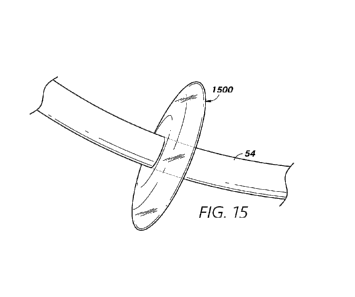
[0082] Figure 15 illustrates another example stagnation device 1500
comprising
an inflatable and/or expandable balloon which may be configured to at least
partially occlude
a blood flow pathway within a heart, in accordance with one or more examples.
The
stagnation device 1500 may be configured to be inflated using air and/or fluid
to form an
expanded shape. In some examples, the stagnation device 1500 may be configured
to form a
ring-shaped extension around at least a portion of an inner sheath 54. For
example, the
stagnation device 1500 may be attached to an outer surface of the inner sheath
54 and/or may
be configured to fit into an outer sheath. The stagnation device 1500 may be
configured to
have a reduced profile while situated within an outer sheath (see, e.g., the
outer sheath 50 of
Figure 16). When the stagnation device 1500 is exposed (e.g., by retraction of
the outer
sheath), the stagnation device 1500 may be inflated to expand in all or some
directions
around the inner sheath 54. The stagnation device 1500 may be configured to
extend and/or
expand to a diameter that is approximately equal to or greater than a blood
flow pathway
(e.g., the coronary sinus) of the heart. The stagnation device 1500 may
further be configured
to be deflated following inflation of the stagnation device 1500. For example,
the stagnation
device 1500 may only temporarily be inflated to at least partially occlude the
blood flow
CA 03222012 2023-11-29
WO 2022/271473
PCT/US2022/033193
pathway. Following application of a visualizing contrast into the blood flow,
the stagnation
device 1500 may be deflated to allow normal blood flow through the blood flow
pathway.
[0083] The stagnation device 1500 may have a generally small profile to
allow the
stagnation device 1500 to be used in combination with various other devices
and/or implants.
For example, the stagnation device 1500 may have a minimal width and/or may be
configured to extend along a relatively small portion of the inner catheter
54. The stagnation
device 1500 may have a diameter that is greater than a diameter of the inner
sheath.
[0084] Figure 16 illustrates another example stagnation device 1600
comprising
an inflatable and/or expandable balloon which may be configured to at least
partially occlude
a blood flow pathway within a heart, in accordance with one or more examples.
The
stagnation device 1600 may be configured to be inflated using air and/or fluid
to form an
expanded shape. In some examples, the stagnation device 1600 may be configured
to form a
ring-shaped extension around at least a portion of an outer sheath 50. For
example, the
stagnation device 1600 may be attached to an outer surface of the outer sheath
50. The
stagnation device 1600 may be configured to have a reduced profile during
delivery. Upon
and/or after delivery to a target location, the stagnation device 1600 may be
inflated to
expand in all or some directions around the outer sheath 50. The stagnation
device 1600 may
be configured to extend and/or expand to a diameter that is approximately
equal to or greater
than a blood flow pathway (e.g., the coronary sinus) of the heart. The
stagnation device 1600
may further be configured to be deflated following inflation of the stagnation
device 1600.
For example, the stagnation device 1600 may only temporarily be inflated to at
least partially
occlude the blood flow pathway. Following application of a visualizing
contrast into the
blood flow, the stagnation device 1600 may be deflated to allow normal blood
flow through
the blood flow pathway.
[0085] The stagnation device 1600 may have a generally small profile to
allow the
stagnation device 1600 to be used in combination with various other devices
and/or implants.
For example, the stagnation device 1600 may have a minimal width and/or may be
configured to extend along a relatively small portion of the outer sheath 50.
The stagnation
device 1600 may have a diameter that is greater than a diameter of the inner
sheath 54 and/or
outer sheath 50.
[0086] Figures 17A and 17B illustrate another example stagnation device
1700
that may be configured to at least partially occlude blood flow within a blood
flow pathway
of a heart, in accordance with one or more examples. Figure 17A illustrates a
compressed
form of the stagnation device 1700 and Figure 17B illustrates an expanded form
of the
21
CA 03222012 2023-11-29
WO 2022/271473
PCT/US2022/033193
stagnation device 1700. The stagnation device 1700 may be configured for use
as an outer
sheath and/or may be configured to extend at least partially along an inner
sheath 54. The
stagnation device 1700 may have a generally tubular form to approximate the
tubular shape
of the inner sheath 54.
[0087] At an end portion of the stagnation device 1700, the stagnation
device
1700 may comprise one or more expandable and/or movable petals 1706 which may
be
formed by slits and/or severed portions of the stagnation device 1700. The one
or more petals
1706 may at least partially overlap each other while in the compressed form
illustrated in
Figure 17A. One or more wires 1702 may be configured to extend from one or
more of the
petals 1706. For example, a first wire 1702a may be configured to attach to,
engage, and/or
actuate a first pedal 1706a of the stagnation device 1700. For example, the
first wire 1702a
may be configured to extend along and/or within the stagnation device 1700
and/or may be
accessible to a surgeon. The first wire 1702a may be configured to be pulled
backward (e.g.,
away from the end portion of the stagnation device 1700) to apply backward
pulling force to
the first pedal 1706a. In some examples, multiple cords 1702 may be configured
to be pulled
simultaneously to cause simultaneous movement of multiple petals 1706. As
shown in Figure
17B, the petals 1706 may be configured to be pulled backwards to increase a
diameter of the
stagnation device 1700 at the end portion of the stagnation device 1700.
[0088] In some examples, the one or more petals 1706 may be connected
via one
or more webbings 1707 and/or membranes. A webbing 1707 may have a generally
flexible
structure and/or may be configured to stretch as the petals 1706 expand. In
some examples,
the webbing 1707 may be configured to be collapsed (e.g., folded) while the
stagnation
device 1700 is in an unexpanded form and/or may be configured to expand (e.g.,
unfurl)
when the stagnation device 1700 is in an expanded form. The webbings 1707 may
be
configured to close gaps between the petals 1706 to prevent fluid from
escaping between the
petals 1706.
[0089] The stagnation device 1700 may be configured to at least
partially prevent
fluid (e.g., blood and/or contrast solution) from escaping into the stagnation
device 1700. In
some examples, the stagnation device 1700 may comprise one or more occluders
configured
to be activated in response to expansion of the stagnation device 1700. For
example, the
stagnation device 1700 may comprise a flap configured to extend across at
least a portion of
an inner lumen of the stagnation device in response to separation and/or
expansion of the
petals 1706. The flap may represent a barrier to prevent fluid flow beyond the
flap to allow
for effective flow stagnation at or near a distal end of the stagnation device
1700. In response
22
CA 03222012 2023-11-29
WO 2022/271473
PCT/US2022/033193
to the stagnation device 1700 returning to an unexpanded form, the flap may be
configured to
compress and/or be pressed against an inner wall of the stagnation device
1700.
Flow Stagnation Processes
[0090] Figures 18-1, 18-2, 18-3, and 18-4 provide a flow diagram
illustrating a
process 1800 for stagnating blood flow and/or visualizing one or more implants
in
accordance with or more examples. Figures 19-1, 19-2, 19-3, and 19-4 are
images of cardiac
anatomy and certain devices/systems corresponding to operations of the process
1800 of
Figures 18-1, 18-2, 18-3, and 18-4 in accordance with one or more examples of
the present
disclosure.
[0091] At block 1802, the process 1800 involves delivering distal
anchoring arms
152 (e.g., flanges), including a first distal arm 152a and/or a second distal
arm of an implant
device through an opening in a tissue wall 30 and/or to a distal side of the
tissue wall 30, as
shown in image 1902 of Figure 19. Although a particular example of a delivery
system is
shown in Figure 19-1, it should be understood that implant devices in
accordance with
aspects of the present disclosure may be delivered and/or implanted using any
suitable or
desirable delivery system and/or delivery system components. Moreover, while a
shunt
implant device is shown, steps of the process 1800 of Figure 18 may be
applicable to other
types of implant devices.
[0092] The illustrated delivery system includes an inner catheter 54,
which may
be disposed at least partially within an outer sheath 50 (e.g., outer
catheter) during one or
more portions of the process 1800. In some examples, the shunt structure may
be disposed at
least partially around the inner catheter 54, wherein the shunt structure is
disposed at least
partially within the outer sheath 50 during one or more portions of the
process 1800. For
example, the inner catheter 54 may be disposed within the barrel portion of
the shunt
structure, as shown.
[0093] The implant device (e.g., medical implant) may be delivered to
the tissue
wall together with or separately from a delivery device 162, which can include
an actuating
rod. The delivery device 162 may be attached to at least a portion of the
medical implant
(e.g., to a first proximal anchoring arm 154a) during delivery of the medical
implant through
the body and/or to the tissue wall 30. In some examples, the medical implant,
delivery device
162, and/or one or more stagnation devices may be configured for delivery via
the inner
catheter 54 and/or outer sheath 50. The medical implant and/or a stagnation
device may be
crimped in series along the inner catheter 54 and/or may be positioned in
series within the
23
CA 03222012 2023-11-29
WO 2022/271473
PCT/US2022/033193
outer sheath 50. For example, retracting the outer sheath 50 a first amount
may expose the
medical implant (but not the stagnation device) and/or further retracting the
outer sheath 50 a
second amount that is greater than the first amount may expose the stagnation
device 1900
and the medical implant.
[0094] In some examples, the delivery system may be configured such that
a
guidewire may be disposed at least partially therein. For example, the
guidewire may run in
the area of an axis of the sheath and/or inner catheter 54, such as within the
inner catheter 54.
The delivery system may be configured to be advanced over the guidewire to
guide the
delivery system to a target implantation site.
[0095] In some examples, the delivery system includes a tapered nosecone
feature
52, which may be associated with a distal end of the sheath 50, catheter 54,
and/or delivery
system. In some implementations, the nosecone feature 52 may be utilized to
dilate the
opening in a tissue wall into which the implant device is to be implanted, or
through which
the delivery system is to be advanced. The nosecone feature 52 may facilitate
advancement of
the distal end of the delivery system through the tortuous anatomy of the
patient and/or with
an outer delivery sheath or other conduit/path. The nosecone 52 may be a
separate component
from the catheter 54 or may be integrated with the catheter 54. In some
examples, the
nosecone 52 is adjacent to and/or integrated with a distal end of the outer
sheath 50. In some
examples, the nosecone 52 may comprise and/or be formed of multiple flap-type
forms that
can be urged/spread apart when the implant device and/or any portions thereof,
the interior
catheter 54, or other device(s) are advanced therethrough.
[0096] The outer sheath 50 may be used to transport the implant device
and/or
one or more stagnation devices to the target implantation site. That is, the
implant device
and/or stagnation device may be advanced to the target implantation site at
least partially
within a lumen of the outer sheath 50, such that the implant device and/or
stagnation device
is/are held and/or secured at least partially within a distal portion of the
outer sheath 50. In
some examples, the medical implant may be removed from the outer sheath 50 by
at least
partially retracting the outer sheath 50 to expose at least a portion of the
medical implant.
[0097] In some implementations, accessing the cardiac anatomy with the
delivery
system may be performed following one or more procedures or steps to place the
guidewire
and form and/or dilate an opening between the left atrium and coronary sinus
of the patient's
heart, the details of which are omitted for convenience and clarity.
[0098] Access to the target wall 30 and left atrium 2 via the coronary
sinus 19
may be achieved using any suitable or desirable procedure. For example,
various access
24
CA 03222012 2023-11-29
WO 2022/271473
PCT/US2022/033193
pathways may be utilized in maneuvering guidewires and catheters in and around
the heart to
deploy one or more implants and/or stagnation devices in accordance with
examples of the
present disclosure. In some examples, access may be achieved through the
subclavian or
jugular veins into the superior vena cava (not shown), right atrium, and from
there into the
coronary sinus 19. Alternatively, the access path may start in the femoral
vein and through
the inferior vena cava (not shown) into the heart. Other access routes may
also be used, each
of which may typically utilize a percutaneous incision through which the
guidewire and
catheter are inserted into the vasculature, normally through a sealed
introducer, and from
there the system may be designed or configured to allow the physician to
control the distal
ends of the devices from outside the body.
[0099] In some implementations, a guidewire is introduced through the
subclavian
or jugular vein, through the superior vena cava, and into the coronary sinus
19 via the right
atrium. The guidewire can be disposed in a spiral configuration within the
left atrium 2,
which may help to secure the guidewire in place. Once the guidewire provides a
path, an
introducer sheath may be routed along the guidewire and into the patient's
vasculature, such
as with the use of a dilator. The delivery catheter may be advanced through
the superior vena
cava to the coronary sinus of the heart, wherein the introducer sheath may
provide a
hemostatic valve to prevent blood loss. In some examples, a deployment
catheter may
function to form and prepare an opening in the wall of the left atrium 2, and
a separate
placement delivery system, as shown, is used for delivery of the implant
device. In other
examples, the deployment system may be used as the both the puncture
preparation and
implant delivery catheter with full functionality. In the present application,
the term "delivery
system" is used to represent a catheter or introducer with one or both of
these functions.
[0100] The guidewire may be disposed as running through an opening in
the
tissue wall 30 prior to penetration thereof by the nosecone 52. The opening
may originally be
formed using a needle (not shown) associated with the delivery system or other
delivery
system implemented prior to block 1802. In some implementations, the nosecone
feature 52
may be used to at least partially dilate the opening, which may have been
previously dilated
using a balloon dilator or other instrument.
[0101] At block 1804, the process 1800 involves extending, advancing,
and/or
maneuvering an actuating rod 162 attached to a first proximal arm 154a to
attach and/or
anchor at least a portion of the medical implant (e.g., the first proximal arm
154a) at a
proximal side of the tissue wall 30, as shown in image 1904 of Figure 19. In
some examples,
the actuating rod 162 may be manually and/or electronically controlled and/or
maneuvered to
CA 03222012 2023-11-29
WO 2022/271473
PCT/US2022/033193
control advancement of one or more portions of the implant device. While only
a single
actuating rod 162 is shown, additional actuating rods 162 may be used for
delivery of other
arms/flanges of the implant.
[0102] The actuating rod 162 may be configured to place the first
anchoring arm
154a at a desired position on the tissue wall 30. If the first anchoring arm
154a is not properly
positioned on the tissue wall 30, the actuating rod 162 can pull the first
anchoring arm 154a
back to re-attempt attachment of the first anchoring arm 154a at the tissue
wall. However,
after the actuating rod 162 and/or additional actuating rods is/are detached
from the first
anchoring arm 154a and/or additional anchoring arms, it can be difficult or
impossible to re-
engage the anchoring arms 154 and/or to re-position the anchoring arms 154.
Thus, prior to
disengaging the actuating rod 162 from the first anchoring arm 154a, a
contrast solution may
be injected and/or one or more stagnation devices may be activated to assist
in determining
correct placement of the implant device.
[0103] At block 1806, the process 1800 involves retracting the outer
sheath 50 at
least partially to expose at least a portion of an expandable stagnation
device 1900 proximate
to at least a portion of the medical implant, as shown in image 1906 of Figure
19. In some
examples, the stagnation device 1900 may be configured to at least partially
occlude, inhibit,
stagnate, and/or obstruct blood flow through the coronary sinus 19 and/or
other blood flow
pathway. The stagnation device 1900 may have any of a variety of forms,
including a
network of wires forming the wire "umbrella" form shown in Figure 19, one or
more
inflatable balloons and/or an expandable tube/sheath. In some examples, the
stagnation
device 1900 may have a generally conical form in which a circular base and/or
opening of the
stagnation device 1900 is situated proximate to the medical implant. The
stagnation device
1900 may be configured to be crimped onto the inner sheath 54 and/or to assume
a
compressed form while at least partially enclosed by the outer sheath 50. In
response to the
outer sheath 50 being retracted to expose at least a portion of the stagnation
device 1900, the
stagnation device 1900 and/or the exposed portion of the stagnation device
1900 may be
configured to at least partially expand to form an increased diameter. In some
examples, the
stagnation device 1900 may be configured to be manually expanded at least in
part. For
example, the stagnation device 1900 may be expanded by inflation and/or by
actuating and/or
pulling on one or more wires attached to at least portions of the stagnation
device 1900.
[0104] The stagnation device 1900 may be configured to at least
partially occlude
and/or stagnate blood flow within the coronary sinus 19. In some examples, the
stagnation
device 1900 may be fluid-tight and/or may be configured to completely and/or
greatly
26
CA 03222012 2023-11-29
WO 2022/271473
PCT/US2022/033193
stagnate blood flow. However, because total blood flow stagnation may not be
required, the
stagnation device 1900 may be at least partially porous to allow some blood
flow through the
stagnation device 1900.
[0105] With the stagnation device 1900 in the expanded form, a contrast
solution
may be injected into the coronary sinus 19. In some examples, the contrast
solution may be
configured to be injected from a side opening 159 of the inner sheath 54. The
side opening
159 may be the same opening used for delivering at least a portion of the
implant device (e.g.,
the first anchoring arm 154a) from the inner sheath 54. In some examples, the
contrast
solution may be injected between at least a portion of the implant device and
the stagnation
device 1900 and/or approximately below the opening and/or below the implant
device. With
the flow stagnation provided by the stagnation device 1900, the contrast
solution around the
implant device may be relatively slow-moving and/or may remain at a relatively
high
concentration to allow for effective visualizing of the implant device.
[0106] In some examples, the contrast solution may be injected
approximately
between at least a portion of the medical implant and at least a portion of
the stagnation
device 1900. The stagnation device 1900 may be situated downstream of the
medical implant.
For example, blood may naturally flow downward through the opening in the
tissue wall 30
and/or toward the stagnation device 1900 and/or outer sheath 50. The contrast
solution may
be configured to be injected upstream of the stagnation device 1900 and/or
downstream of the
medical implant. In some cases, flow stagnation caused by the stagnation
device 1900 may
cause blood and/or contrast solution to pass upward through the opening in the
tissue wall 30
and/or to pool along a distal side of the tissue wall 30, which may
advantageously allow
viewing of distal portions (e.g., the first distal arm 152a) of the medical
implant.
[0107] At block 1808, the process 1800 involves extending the outer
sheath 50
and/or retracting the stagnation device 1900 to collapse the stagnation device
1900 and/or
enclose the stagnation device 1900 within the outer sheath 50, as shown in
image 1908 of
Figure 19. The stagnation device 1900 may be collapsed after injection of the
contrast
solution and/or after the positioning and/or orientation of the implant device
has been viewed
by a physician. In some cases, the stagnation device 1900 may only need to be
in an
expanded form for a few seconds to provide sufficient stagnation for viewing
the placement
of the medical implant. However, the stagnation device 1900 may be maintained
in the
expanded form longer if necessary. In some examples, the stagnation device
1900 may be
configured to cause only partial stagnation of flow (e.g., due at least in
part to a porous
27
CA 03222012 2023-11-29
WO 2022/271473
PCT/US2022/033193
structure of the stagnation device 1900) and may be maintained in the expanded
form for
extended periods with minimal risk of harm to the patient.
[0108] At block 1810, the process 1800 involves detaching the actuating
rod 162
from the implant device, as shown in image 1910 of Figure 19. The actuating
rod 162 may be
detached only after the contrast injection and/or expansion of the stagnation
device 1900. At
block 1812, the process 1800 involves retracting the actuating rod 162 into
the outer sheath
50, as shown in image 1912 of Figure 19. At block 1814, the process 1800
involves removing
the delivery systems, including the inner sheath 54 and/or outer sheath 50,
from the coronary
sinus 19 and/or from the body, as shown in image 1914 of Figure 19.
[0109] Additional aspects and features of processes for delivering shunt
structures
that may be delivered and/or utilized in combination with stagnation devices
in accordance
with examples of the present disclosure for implantation in the wall between
the coronary
sinus and the left atrium are disclosed in U.S. Pat. No. 9,789,294, entitled
"Expandable
Cardiac Shunt," issued on October 17, 2017, the disclosure of which is hereby
expressly
incorporated by reference in its entirety. Although the implant device is
shown in the left
atrium/coronary sinus wall, the implant device may be positioned between other
cardiac
chambers, such as between the left and right atria.
[0110] Some implementations of the present disclosure relate to a method
comprising delivering a medical implant and a delivery device to a tissue wall
within a heart
of a patient. The delivery device is attached to the medical implant. The
method further
comprises maneuvering the delivery device to place at least a portion of the
medical implant
at a desired position with respect to the tissue wall and delivering an
expandable stagnation
device proximate to the medical implant. The expandable stagnation device is
configured to
at least partially inhibit blood flow. The method further comprises injecting
a contrast
solution between at least a portion of the medical implant and at least a
portion of the
expandable stagnation device, collapsing the expandable stagnation device, and
detaching the
delivery device from the medical implant.
[0111] The method can further comprise delivering the medical implant
and the
expandable stagnation device via a first catheter. In some examples, the
method further
comprises retracting the first catheter to remove the medical implant from the
first catheter
and further retracting the first catheter to remove the expandable stagnation
device from the
first catheter.
[0112] In some examples, removing the expandable stagnation device from
the
first catheter causes the expandable stagnation device to expand.
28
CA 03222012 2023-11-29
WO 2022/271473
PCT/US2022/033193
[0113] The expandable stagnation device can comprise a network of wires
configured to expand to a conical form. In some examples, the expandable
stagnation device
comprises an inflatable balloon.
[0114] In some examples, the method further comprises delivering the
expandable
stagnation device via an inner sheath. The expandable stagnation device can
extend from the
inner sheath. The method can further comprise delivering the expandable
stagnation device
via an inner sheath and an outer sheath, wherein the expandable stagnation
device extends
from the outer sheath.
[0115] The expandable stagnation device can comprise a tubular sheath
having
one or more expandable petals at an end portion of the expandable stagnation
device. In some
examples, the one or more expandable petals are attached to pull wires
configured to expand
the one or more expandable petals.
[0116] In some examples, the expandable stagnation device comprises one
or
more webbings between the one or more expandable petals. The expandable
stagnation
device can comprise one or more flaps configured to at least partially inhibit
blood flow
through an inner lumen of the expandable stagnation device.
[0117] The medical implant can comprise a first anchoring arm. The
delivery
device may be attached to the first anchoring arm.
[0118] In some implementations of the present disclosure, a delivery
system
comprises an expandable stagnation device configured to expand to at least
partially inhibit
blood flow within a blood flow pathway of a heart and compress following
injection of a
contrast solution within a blood flow pathway of a heart, an inner sheath
configured to deliver
a medical implant and the expandable stagnation device through the blood flow
pathway of
the heart and proximate to a tissue wall of the heart, and a delivery device
configured to
attach to the medical implant and maneuver at least a portion of the medical
implant to a
desired position with respect to the tissue wall of the heart and detach from
the medical
implant following injection of the contrast solution within the blood flow
pathway of the
heart.
[0119] The delivery system can further comprise an outer sheath
configured to at
least partially enclose the expandable stagnation device and the medical
implant and retract to
remove the medical implant and the expandable stagnation device from the outer
sheath.
[0120] In some examples, removing the expandable stagnation device from
the
outer sheath causes the expandable stagnation device to expand. The expandable
stagnation
device can comprise a network of wires configured to expand to a conical form.
29
CA 03222012 2023-11-29
WO 2022/271473
PCT/US2022/033193
[0121] The expandable stagnation device can comprise an inflatable
balloon. In
some examples, the expandable stagnation device extends from the inner sheath.
[0122] In some examples, the expandable stagnation device comprises a
tubular
sheath at least partially enclosing the inner sheath and having one or more
expandable petals
at an end portion of the expandable stagnation device.
[0123] In some aspects, the techniques described herein relate to a
method
including: delivering a medical implant and a delivery device via an inner
catheter to a tissue
wall within a heart of a patient; maneuvering the delivery device to place at
least a portion of
the medical implant at a desired position with respect to the tissue wall;
delivering a
stagnation device proximate to the medical implant, the stagnation device
configured to at
least partially inhibit blood flow; and injecting a contrast solution between
at least a portion
of the medical implant and at least a portion of the stagnation device.
[0124] In some aspects, the techniques described herein relate to a
method,
wherein the delivery device is attached to the medical implant.
[0125] In some aspects, the techniques described herein relate to a
method, further
including, after injection of the contrast solution, detaching the delivery
device from the
medical implant.
[0126] In some aspects, the techniques described herein relate to a
method,
wherein the stagnation device is in a compressed form prior to delivery
proximate to the
medical implant.
[0127] In some aspects, the techniques described herein relate to a
method,
wherein the stagnation device is configured to expand upon delivery proximate
to the medical
implant.
[0128] In some aspects, the techniques described herein relate to a
method, further
including, after injection of the contrast solution, collapsing the stagnation
device.
[0129] In some aspects, the techniques described herein relate to a
method, further
including delivering the medical implant and the stagnation device via an
outer sheath.
[0130] In some aspects, the techniques described herein relate to a
method, further
including: retracting the outer sheath to remove the medical implant from the
outer sheath;
and further retracting the outer sheath to remove the stagnation device from
the outer sheath.
[0131] In some aspects, the techniques described herein relate to a
method,
wherein removing the stagnation device from the outer sheath causes the
stagnation device to
expand.
CA 03222012 2023-11-29
WO 2022/271473
PCT/US2022/033193
[0132] In some aspects, the techniques described herein relate to a
method,
wherein the stagnation device includes a network of wires.
[0133] In some aspects, the techniques described herein relate to a
method,
wherein the network of wires has an at least partial cone shape.
[0134] In some aspects, the techniques described herein relate to a
method,
wherein the network of wires includes an oval-shaped wire and one or more
curved wires.
[0135] In some aspects, the techniques described herein relate to a
method,
wherein each curved wire of the one or more curved wires attaches to the oval-
shaped wire at
an endpoint of the curved wire.
[0136] In some aspects, the techniques described herein relate to a
method,
wherein the one or more curved wires extend generally perpendicularly to the
oval-shaped
wire.
[0137] In some aspects, the techniques described herein relate to a
method,
wherein the stagnation device is delivered via an outer sheath, and wherein
the oval-shaped
wire is configured to expand to a greater diameter than the outer sheath.
[0138] In some aspects, the techniques described herein relate to a
method,
wherein the one or more curved wires are configured to extend outwardly from
the inner
catheter in response to removal from the outer sheath.
[0139] In some aspects, the techniques described herein relate to a
method,
wherein the stagnation device is configured to encircle at least a portion of
the inner catheter.
[0140] In some aspects, the techniques described herein relate to a
method,
wherein the stagnation device includes a covering extending between the wires.
[0141] In some aspects, the techniques described herein relate to a
method,
wherein the covering is fluid tight.
[0142] In some aspects, the techniques described herein relate to a
method,
wherein the covering is porous.
[0143] In some aspects, the techniques described herein relate to a
method,
wherein the stagnation device includes an inflatable balloon.
[0144] In some aspects, the techniques described herein relate to a
method,
wherein the stagnation device extends from an outer surface of the inner
catheter.
[0145] In some aspects, the techniques described herein relate to a
method, further
including delivering the stagnation device via an outer sheath, wherein the
stagnation device
extends from an outer surface of the outer sheath.
31
CA 03222012 2023-11-29
WO 2022/271473
PCT/US2022/033193
[0146] In some aspects, the techniques described herein relate to a
method,
wherein the stagnation device is ring-shaped.
[0147] In some aspects, the techniques described herein relate to a
method,
wherein a diameter of the stagnation device is greater than a width of the
stagnation device.
[0148] In some aspects, the techniques described herein relate to a
method,
wherein the stagnation device includes a tubular sheath having one or more
expandable petals
at an end portion of the tubular sheath.
[0149] In some aspects, the techniques described herein relate to a
method,
wherein the one or more expandable petals are attached to pull wires
configured to expand
the one or more expandable petals.
[0150] In some aspects, the techniques described herein relate to a
method,
wherein the stagnation device includes one or more webbings between the one or
more
expandable petals.
[0151] In some aspects, the techniques described herein relate to a
method,
wherein the stagnation device includes one or more flaps configured to at
least partially
inhibit blood flow through an inner lumen of the stagnation device.
[0152] In some aspects, the techniques described herein relate to a
method,
wherein the medical implant includes a first anchoring arm, and the delivery
device is
attached to the first anchoring arm.
[0153] In some aspects, the techniques described herein relate to a
delivery
system including: a stagnation device configured to expand to at least
partially inhibit blood
flow within a blood flow pathway of a heart and compress following injection
of a contrast
solution within a blood flow pathway of a heart; an inner catheter configured
to deliver a
medical implant and the stagnation device through the blood flow pathway of
the heart and
proximate to a tissue wall of the heart; and a delivery device configured to
maneuver at least
a portion of the medical implant to a desired position with respect to the
tissue wall of the
heart.
[0154] In some aspects, the techniques described herein relate to a
delivery
system, wherein the delivery device is further configured to detach from the
medical implant
following injection of the contrast solution within the blood flow pathway of
the heart.
[0155] In some aspects, the techniques described herein relate to a
delivery
system, further including an outer sheath configured to at least partially
enclose the
stagnation device and the medical implant, and retract to remove the medical
implant and the
stagnation device from the outer sheath.
32
CA 03222012 2023-11-29
WO 2022/271473
PCT/US2022/033193
[0156] In some aspects, the techniques described herein relate to a
delivery
system, wherein removing the stagnation device from the outer sheath causes
the stagnation
device to expand.
[0157] In some aspects, the techniques described herein relate to a
delivery
system, wherein the stagnation device includes a network of wires.
[0158] In some aspects, the techniques described herein relate to a
delivery
system, wherein the network of wires has an at least partial cone shape.
[0159] In some aspects, the techniques described herein relate to a
delivery
system, wherein the network of wires includes an oval-shaped wire and one or
more curved
wires.
[0160] In some aspects, the techniques described herein relate to a
delivery
system, wherein each curved wire of the one or more curved wires attaches to
the oval-
shaped wire at an endpoint of the curved wire.
[0161] In some aspects, the techniques described herein relate to a
delivery
system, wherein the one or more curved wires extend generally perpendicularly
to the oval-
shaped wire.
[0162] In some aspects, the techniques described herein relate to a
delivery
system, wherein the stagnation device is delivered via an outer sheath, and
wherein the oval-
shaped wire is configured to expand to a greater diameter than the outer
sheath.
[0163] In some aspects, the techniques described herein relate to a
delivery
system, wherein the one or more curved wires are configured to extend
outwardly from the
inner catheter in response to removal from the outer sheath.
[0164] In some aspects, the techniques described herein relate to a
delivery
system, wherein the stagnation device is configured to encircle at least a
portion of the inner
catheter.
[0165] In some aspects, the techniques described herein relate to a
delivery
system, wherein the stagnation device includes a covering extending between
the wires.
[0166] In some aspects, the techniques described herein relate to a
delivery
system, wherein the covering is fluid tight.
[0167] In some aspects, the techniques described herein relate to a
delivery
system, wherein the covering is porous.
[0168] In some aspects, the techniques described herein relate to a
delivery
system, wherein the stagnation device includes an inflatable balloon.
33
CA 03222012 2023-11-29
WO 2022/271473
PCT/US2022/033193
[0169] In some aspects, the techniques described herein relate to a
delivery
system, wherein the stagnation device extends from an outer surface of the
inner catheter.
[0170] In some aspects, the techniques described herein relate to a
delivery
system, further including delivering the stagnation device via an outer
sheath, wherein the
stagnation device extends from an outer surface of the outer sheath.
[0171] In some aspects, the techniques described herein relate to a
delivery
system, wherein the stagnation device is ring-shaped.
[0172] In some aspects, the techniques described herein relate to a
delivery
system, wherein a diameter of the stagnation device is greater than a width of
the stagnation
device.
[0173] In some aspects, the techniques described herein relate to a
delivery
system, wherein the stagnation device includes a tubular sheath having one or
more
expandable petals at an end portion of the tubular sheath.
[0174] In some aspects, the techniques described herein relate to a
delivery
system, wherein the one or more expandable petals are attached to pull wires
configured to
expand the one or more expandable petals.
[0175] In some aspects, the techniques described herein relate to a
delivery
system, wherein the stagnation device includes one or more webbings between
the one or
more expandable petals.
[0176] In some aspects, the techniques described herein relate to a
delivery
system, wherein the stagnation device includes one or more flaps configured to
at least
partially inhibit blood flow through an inner lumen of the stagnation device.
[0177] In some aspects, the techniques described herein relate to a
delivery
system, wherein the medical implant includes a first anchoring arm, and the
delivery device is
attached to the first anchoring arm.
[0178] In some aspects, the techniques described herein relate to a
delivery
system including: means for stagnating blood flow within a blood flow pathway
of a heart;
means for delivering a medical implant and the means for stagnating through
the blood flow
pathway of the heart and proximate to a tissue wall of the heart; and means
for maneuvering
at least a portion of the medical implant to a desired position with respect
to the tissue wall of
the heart.
[0179] In some aspects, the techniques described herein relate to a
delivery
system, wherein the means for maneuvering is further configured to detach from
the medical
implant following injection of a contrast solution within the blood flow
pathway of the heart.
34
CA 03222012 2023-11-29
WO 2022/271473
PCT/US2022/033193
[0180] In some aspects, the techniques described herein relate to a
delivery
system, further including an outer sheath configured to at least partially
enclose the means for
stagnating and the medical implant, and retract to remove the medical implant
and the means
for stagnating from the outer sheath.
[0181] In some aspects, the techniques described herein relate to a
delivery
system, wherein the means for stagnating includes a network of wires.
[0182] In some aspects, the techniques described herein relate to a
delivery
system, wherein the means for stagnating includes an inflatable balloon.
[0183] In some aspects, the techniques described herein relate to a
delivery
system, wherein the means for stagnating includes a tubular sheath having one
or more
expandable petals at an end portion of the tubular sheath.
[0184] In some aspects, the techniques described herein relate to a
delivery
system, wherein the medical implant includes a first anchoring arm and the
means for
maneuvering is attached to the first anchoring arm.
Additional Examples
[0185] Depending on the example, certain acts, events, or functions of
any of the
processes or algorithms described herein can be performed in a different
sequence, may be
added, merged, or left out altogether. Thus, in certain examples, not all
described acts or
events are necessary for the practice of the processes.
[0186] Conditional language used herein, such as, among others, "can,"
"could,"
"might," "may," "e.g.," and the like, unless specifically stated otherwise, or
otherwise
understood within the context as used, is intended in its ordinary sense and
is generally
intended to convey that certain examples include, while other examples do not
include,
certain features, elements and/or steps. Thus, such conditional language is
not generally
intended to imply that features, elements and/or steps are in any way required
for one or more
examples or that one or more examples necessarily include logic for deciding,
with or
without author input or prompting, whether these features, elements and/or
steps are included
or are to be performed in any particular example. The terms "comprising,"
"including,"
"having," and the like are synonymous, are used in their ordinary sense, and
are used
inclusively, in an open-ended fashion, and do not exclude additional elements,
features, acts,
operations, and so forth. Also, the term "or" is used in its inclusive sense
(and not in its
exclusive sense) so that when used, for example, to connect a list of
elements, the term "or"
means one, some, or all of the elements in the list. Conjunctive language such
as the phrase
CA 03222012 2023-11-29
WO 2022/271473
PCT/US2022/033193
"at least one of X, Y and Z," unless specifically stated otherwise, is
understood with the
context as used in general to convey that an item, term, element, etc. may be
either X, Y or Z.
Thus, such conjunctive language is not generally intended to imply that
certain examples
require at least one of X, at least one of Y and at least one of Z to each be
present.
[0187] It should be appreciated that in the above description of
examples, various
features are sometimes grouped together in a single example, Figure, or
description thereof
for the purpose of streamlining the disclosure and aiding in the understanding
of one or more
of the various inventive aspects. This method of disclosure, however, is not
to be interpreted
as reflecting an intention that any claim require more features than are
expressly recited in
that claim. Moreover, any components, features, or steps illustrated and/or
described in a
particular example herein can be applied to or used with any other example(s).
Further, no
component, feature, step, or group of components, features, or steps are
necessary or
indispensable for each example. Thus, it is intended that the scope of the
inventions herein
disclosed and claimed below should not be limited by the particular examples
described
above, but should be determined only by a fair reading of the claims that
follow.
[0188] It should be understood that certain ordinal terms (e.g., "first"
or "second")
may be provided for ease of reference and do not necessarily imply physical
characteristics or
ordering. Therefore, as used herein, an ordinal term (e.g., "first," "second,"
"third," etc.) used
to modify an element, such as a structure, a component, an operation, etc.,
does not
necessarily indicate priority or order of the element with respect to any
other element, but
rather may generally distinguish the element from another element having a
similar or
identical name (but for use of the ordinal term). In addition, as used herein,
indefinite articles
("a" and "an") may indicate "one or more" rather than "one." Further, an
operation performed
"based on" a condition or event may also be performed based on one or more
other
conditions or events not explicitly recited.
[0189] Unless otherwise defined, all terms (including technical and
scientific
terms) used herein have the same meaning as commonly understood by one of
ordinary skill
in the art to which example examples belong. It be further understood that
terms, such as
those defined in commonly used dictionaries, should be interpreted as having a
meaning that
is consistent with their meaning in the context of the relevant art and not be
interpreted in an
idealized or overly formal sense unless expressly so defined herein.
[0190] The spatially relative terms "outer," "inner," "upper," "lower,"
"below,"
"above," "vertical," "horizontal," and similar terms, may be used herein for
ease of
description to describe the relations between one element or component and
another element
36
CA 03222012 2023-11-29
WO 2022/271473
PCT/US2022/033193
or component as illustrated in the drawings. It be understood that the
spatially relative terms
are intended to encompass different orientations of the device in use or
operation, in addition
to the orientation depicted in the drawings. For example, in the case where a
device shown in
the drawing is turned over, the device positioned "below" or "beneath" another
device may
be placed "above" another device. Accordingly, the illustrative term "below"
may include
both the lower and upper positions. The device may also be oriented in the
other direction,
and thus the spatially relative terms may be interpreted differently depending
on the
orientations.
[0191] Unless otherwise expressly stated, comparative and/or
quantitative terms,
such as "less," "more," "greater," and the like, are intended to encompass the
concepts of
equality. For example, "less" can mean not only "less" in the strictest
mathematical sense, but
also, "less than or equal to.
37