Note: Descriptions are shown in the official language in which they were submitted.
CA 03225736 2023-12-28
WO 2023/004098 PCT/US2022/037983
HEART VALVE REPAIR DEVICES
RELATED APPLICATIONS
[0001] The present application claims the benefit of US Provisional
Application No. 63/225,387
filed on July 23, 2021, titled "Heart Valve Repair Devices and Delivery
Devices Therefor," and
the benefit of US Provisional Application No. 63/307,589 filed on February 7,
2022, titled "Heart
Valve Repair Devices and Delivery Devices Therefor," which are incorporated
herein by
reference in their entireties.
BACKGROUND
[0002] The native heart valves (i.e., the aortic, pulmonary, tricuspid, and
mitral valves) serve
critical functions in assuring the forward flow of an adequate supply of blood
through the
cardiovascular system. These heart valves may be damaged, and thus rendered
less effective, for
example, by congenital malformations, inflammatory processes, infectious
conditions, disease,
etc. Such damage to the valves may result in serious cardiovascular compromise
or death.
Damaged valves can be surgically repaired or replaced during open heart
surgery. However, open
heart surgeries are highly invasive, and complications may occur.
Transvascular techniques can
be used to introduce and implant prosthetic devices in a manner that is much
less invasive than
open heart surgery. As one example, a transvascular technique useable for
accessing the native
mitral and aortic valves is the trans-septal technique. The trans-septal
technique comprises
advancing a catheter into the right atrium (e.g., inserting a catheter into
the right femoral vein, up
the inferior vena cava and into the right atrium). The septum is then
punctured, and the catheter
passed into the left atrium. A similar transvascular technique can be used to
implant a prosthetic
device within the tricuspid valve that begins similarly to the trans-septal
technique but stops
short of puncturing the septum and instead turns the delivery catheter toward
the tricuspid valve
in the right atrium.
[0003] A healthy heart has a generally conical shape that tapers to a lower
apex. The heart is
four-chambered and comprises the left atrium, right atrium, left ventricle,
and right ventricle. The
left and right sides of the heart are separated by a wall generally referred
to as the septum. The
native mitral valve of the human heart connects the left atrium to the left
ventricle. The mitral
valve has a very different anatomy than other native heart valves. The mitral
valve includes an
annulus portion, which is an annular portion of the native valve tissue
surrounding the mitral
valve orifice, and a pair of cusps, or leaflets, extending downward from the
annulus into the left
ventricle. The mitral valve annulus may form a "D"-shaped, oval, or otherwise
out-of-round
1
CA 03225736 2023-12-28
WO 2023/004098 PCT/US2022/037983
cross-sectional shape having major and minor axes. The anterior leaflet may be
larger than the
posterior leaflet, forming a generally "C"-shaped boundary between the
abutting sides of the
leaflets when they are closed together.
[0004] When operating properly, the anterior leaflet and the posterior leaflet
function together as
a one-way valve to allow blood to flow only from the left atrium to the left
ventricle. The left
atrium receives oxygenated blood from the pulmonary veins. When the muscles of
the left atrium
contract and the left ventricle dilates (also referred to as "ventricular
diastole" or "diastole"), the
oxygenated blood that is collected in the left atrium flows into the left
ventricle. When the
muscles of the left atrium relax and the muscles of the left ventricle
contract (also referred to as
"ventricular systole" or "systole"), the increased blood pressure in the left
ventricle urges the
sides of the two leaflets together, thereby closing the one-way mitral valve
so that blood cannot
flow back to the left atrium and is instead expelled out of the left ventricle
through the aortic
valve. To prevent the two leaflets from prolapsing under pressure and folding
back through the
mitral annulus toward the left atrium, a plurality of fibrous cords called
chordae tendineae tether
the leaflets to papillary muscles in the left ventricle.
[0005] Valvular regurgitation involves the valve improperly allowing some
blood to flow in the
wrong direction through the valve. For example, mitral regurgitation occurs
when the native
mitral valve fails to close properly and blood flows into the left atrium from
the left ventricle
during the systolic phase of heart contraction. Mitral regurgitation is one of
the most common
forms of valvular heart disease. Mitral regurgitation may have many different
causes, such as
leaflet prolapse, dysfunctional papillary muscles, stretching of the mitral
valve annulus resulting
from dilation of the left ventricle, more than one of these, etc. Mitral
regurgitation at a central
portion of the leaflets can be referred to as central jet mitral regurgitation
and mitral regurgitation
nearer to one commissure (i.e., location where the leaflets meet) of the
leaflets can be referred to
as eccentric jet mitral regurgitation. Central jet regurgitation occurs when
the edges of the
leaflets do not meet in the middle and thus the valve does not close, and
regurgitation is present.
Tricuspid regurgitation may be similar, but on the right side of the heart.
SUMMARY
[0006] This summary is meant to provide some examples and is not intended to
be limiting of
the scope of the invention in any way. For example, any feature included in an
example of this
summary is not required by the claims, unless the claims explicitly recite the
features. Also, the
2
CA 03225736 2023-12-28
WO 2023/004098 PCT/US2022/037983
features, components, steps, concepts, etc. described in examples in this
summary and elsewhere
in this disclosure can be combined in a variety of ways. Various features and
steps as described
elsewhere in this disclosure can be included in the examples summarized here.
[0007] Devices for repairing and/or treating a native valve of a patient are
disclosed. The
devices can be valve repair devices, implantable devices, valve treatment
devices, implants, etc.
While sometimes described as an implantable device for illustration purposes
in various
examples herein, similar configurations can be used on other devices, e.g.,
valve repair devices,
etc., that are not necessarily implanted and may be removed after treatment.
[0008] The devices can include an indicator (these can be the same as or
similar to other
indicators described anywhere herein) and a gripping member or clasp and
(these can be the
same as or similar to other gripping members, gripper arms, clasps, and clasp
arms described
anywhere herein). The devices can also include a paddle (the paddle can be the
same as or
similar to other paddles described anywhere herein). The paddle and/or the
gripping
member/clasp (e.g., a clasp arm of the clasp, a gripper arm, etc.) are movable
to form an opening
or capture region for receiving a leaflet. In some implementations, the
opening or capture region
is formed between the gripper member/clasp (e.g., a clasp arm of the clasp,
etc.) and the paddle
(e.g., a portion of the paddle, etc.). The indicator is configured to indicate
whether a leaflet of
the native valve is inserted into the opening or capture region to at least a
minimum insertion
depth or engagement depth. The minimum insertion depth or engagement depth can
be
preselected and/or configured to a particular depth as desired.
[0009] The indicators herein can be configured in a variety of shapes, sizes,
and materials. In
some implementations, the indicators can comprise an undulating shape, an S-
shape, a C-shape,
a U-shape, a V-shape, a hook shape, a check-mark shape, a swoosh shape, etc.
[0010] In some implementations, a valve repair device (or valve treatment
device, etc.) includes
a clasp and/or a clasp arm and an indicator (e.g., leaflet depth indicator,
indicator arm, marker,
sensor, electrode, etc.). The device can also include a paddle. The indicator
can be configured as
an indicator arm and/or can be configured such that is movable (e.g., through
the clasp, paddle
and/or another portion of the device) to indicate whether a leaflet of the
native valve is inserted
into the opening or capture region to at least a minimum insertion depth. The
minimum insertion
depth can be preselected and/or configured to a particular depth as desired.
3
CA 03225736 2023-12-28
WO 2023/004098 PCT/US2022/037983
[0011] In some implementations, the indicator can comprise an indicator arm,
and the indicator
arm can be coupled to the valve repair device at a first end of the indicator
arm and at a second
end of the indicator arm. The indicator arm can be coupled to an optional
coaptation element of
the valve repair device. The indicator arm can be compressible and can be
configured to engage
the leaflet of the native valve. The indicator arm can comprise one or more
protrusions
extending from the indicator arm. The clasp and the indicator arm can each
comprise a marker
comprising a radiopaque material. The capture region can be formed between a
portion of the
paddle and an arm of the clasp. The paddle can comprise an outer paddle and an
inner paddle.
[0012] In some implementations, the indicator or indicator arm can be
configured to pass
through a channel, slot, gap, and/or opening of the clasp. In some
implementations, the indicator
or indicator arm can be configured to pass through a channel, slot, gap,
and/or opening of the
paddle. In some implementations, the indicator or indicator arm can be
configured to pass
through channel, slot, gap, and/or opening of a moveable arm of the clasp.
[0013] In some implementations, the clasp can optionally include a fixed arm.
In some
implementations, the fixed arm of the clasp can comprise a first beam, a
second beam, and/or an
engaging member between the first beam and the second beam.
[0014] In some implementations, an indicator marker can be attached to the
indicator arm. The
indicator arm can comprise a fixed end and a moving end. The fixed end of the
indicator arm
can be coupled to the clasp. The fixed end of the indicator arm can be coupled
to a movable arm
of the clasp. The moving end can comprise an indicator marker comprising a
radiopaque
material. The fixed end and the moving end can be disposed on a first side of
a movable arm of
the clasp.
[0015] In some implementations, the indicator or indicator arm includes a
leaflet engaging
member (e.g., an extension, protrusion, arm, edge, bump, dip, swoop, U-shaped
portion, V-
shaped portion, triangular-shaped portion, curved portion, circular portion,
rectangular portion,
etc.) between the fixed end and the moving end. The leaflet-engaging member
can be configured
to pass through at least one of a movable arm of the clasp and the paddle.
[0016] In some implementations, the leaflet-engaging member is disposed on a
second side of a
movable arm of the clasp. In some implementations, the leaflet-engaging member
can comprise
one or more protrusions extending from the leaflet-engaging member.
4
CA 03225736 2023-12-28
WO 2023/004098 PCT/US2022/037983
[0017] In some implementations, an indicator can comprise a first arm and a
second arm. The
first arm and the second arm can be coupled with the moving end and can be
connected at a
connection point at the fixed end.
[0018] In some implementations, the indicator arm is formed from a portion of
the clasp. The
indicator arm can be formed between outer beams of a movable arm of the clasp
and/or outside
outer beams of the clasp (or clasp arm of the clasp).
[0019] In some implementations, the indicator arm can comprise a twisted
portion. The twisted
portion can comprise one or more twists between 0 degrees and 180 degrees.
[0020] In some implementations, the indicator arm can comprise a first arm
portion and a second
arm portion. At least one of the first arm portion and a second arm portion
can be formed
between outer beams of the clasp and/or outside outer beams of the clasp. At
least one of the
first arm portion and a second arm portion can be formed from a portion of
first beam of the
clasp. In some implementations, the first arm portion can comprise a twisted
portion. The twisted
portion of the first arm portion can comprise one or more twists between 0
degrees and 180
degrees clockwise.
[0021] In some implementations, a second arm portion can comprise a twisted
portion. The
twisted portion of the second arm portion can comprise one or more twists
between 0 degrees
and 180 degrees counterclockwise.
[0022] In some implementations, the first arm portion and the second arm
portion are coupled
with the moving end at a connection point. The connection point can comprise
an indicator
marker comprising a radiopaque material press fit into at least one of the
first arm portion and
the second arm portion.
[0023] In some implementations, a valve repair system for repairing a native
valve of a patient
includes a delivery system and a valve repair device coupled to the delivery
system. The valve
repair device can include a paddle, an indicator (e.g., leaflet depth
indicator, indicator arm,
sensor, etc.), and a gripping member or clasp. The gripping member/clasp
and/or the paddle can
be movable to form an opening or capture region to receive a leaflet of the
native valve. The
indicator is coupled to the valve repair device. In some implementations, the
indicator is
configured as an indicator arm and/or is movable to indicate whether the
leaflet of the native
CA 03225736 2023-12-28
WO 2023/004098 PCT/US2022/037983
valve is inserted into the opening or capture region to at least a minimum
insertion depth. The
device can be configured to have different minimum insertion depths as desired
(e.g., a minimum
depth of one or more of 3mm, 4mm, 5mm, 6mm, 7 mm, 8mm, etc.). The indicator or
indicator
arm can be configured to pass through one or more of the paddle and the
gripping member/clasp.
[0024] In some implementations, a valve repair device includes a gripping
member or clasp
(e.g., a clasp arm, gripper arm, etc.) and a leaflet depth indicator. The
leaflet depth indicator
includes at least a first electrode and a second electrode. The first
electrode and the second
electrode provide electrical signals to indicate whether a leaflet of the
native valve is inserted
into an opening or capture region to at least a minimum insertion depth. The
minimum insertion
depth can be preselected and/or configured to a particular depth as desired.
The device can also
include a paddle.
[0025] In some implementations, a valve repair device for repairing a native
heart valve includes
a gripping member or clasp (e.g., a clasp arm, gripper arm, etc.) and a
leaflet depth indicator.
The clasp (or a clasp arm/gripper arm of the clasp) can be movable to form an
opening or capture
region for receiving a native leaflet of the native valve. The leaflet depth
indicator can comprise
a first electrode and a second electrode. The first electrode and the second
electrode can provide
electrical signals to indicate whether a leaflet of the native valve is
inserted into the opening to a
particular insertion depth.
[0026] In some implementations, the electrical signals comprises an
intracardiac
electrocardiogram signal or a bioimpedance signal. The first electrode and the
second electrode
can be coupled to the gripping member/clasp (or clasp arm, gripper arm, etc.).
In some
implementations, the gripping member/clasp comprises a movable arm, and the
first electrode
and the second electrode are coupled to the movable arm. In some
implementations, the first
electrode and the second electrode are coupled to an indicator arm. The
indicator arm can be
coupled to the valve repair device and is movable in an opening or capture
region.
[0027] In some implementations, a valve repair system for repairing a native
heart valve includes
a delivery system and valve repair device. The valve repair device is
releasably coupled to the
delivery system. The valve repair device includes a gripping member or clasp
(e.g., a clasp arm,
gripper arm, etc.) and a leaflet depth indicator. The gripping member/clasp
(e.g., a portion, clasp
arm, gripper arm, etc. thereof) is movable to form an opening or capture
region for receiving a
native leaflet of the native valve. The leaflet depth indicator comprises a
first electrode and a
6
CA 03225736 2023-12-28
WO 2023/004098 PCT/US2022/037983
second electrode. The first electrode and the second electrode provide
electrical signals to
indicate whether a leaflet of the native valve is inserted into the opening or
capture region to at
least a minimum insertion depth. The minimum insertion depth can be
preselected and/or
configured to a particular depth as desired.
[0028] In some implementations, a leaflet depth indicator can be integrally
formed with the
gripping member/clasp. For example, the leaflet depth indicator can be formed
from the same
material as the gripping member/clasp. In some implementations, the gripping
member/clasp
and the leaflet depth indicator can be cut from a single piece of sheet
material.
[0029] In some implementations, the material of the leaflet depth indicator
can be bent, twisted
and/or shape set relative to the material of the gripping member/clasp such
that the leaflet depth
indicator is positioned in a plane such that it can contact a native leaflet
and determine whether
the gripping member/clasp has properly engaged the native leaflet. The leaflet
depth indicator
can extend from a movable arm of the clasp, aa hinge portion of the clasp,
and/or a fixed arm of
the clasp.
[0030] In some implementations, a valve repair device includes a gripping
member or clasp
(and/or clasp arm, gripper arm, etc. thereof) and an indicator. The device can
also include a
paddle. The valve repair device can also include an insulator disposed between
at least a portion
of the gripping member/clasp/clasp arm and the indicator. The indicator
includes one or more
electrically conductive indicator contacts which can connect to a sensor to
indicate whether a
leaflet of the native valve is inserted into the opening or capture region to
at least a minimum
insertion depth. The minimum insertion depth can be preselected and/or
configured to a
particular depth as desired.
[0031] In some implementations, the signal can be passed to the sensors by
electrical wiring
from the valve repair device to the sensors. The signal can be passed to the
sensors by electrical
conductance from the indicator through a portion of the valve repair device.
In some
implementations, the signal is passed to the sensors by electrical conductance
from the indicator
through at least one of an electrically conductive fixed arm, an electrically
conductive coaptation
element, an electrically conductive collar, an electrically conductive
catheter coupler, and an
electrically conductive actuation line
7
CA 03225736 2023-12-28
WO 2023/004098 PCT/US2022/037983
[0032] In some implementations, a gripping member or clasp can include movable
arm and a
fixed arm, as well as a first indicator plate coupled to the fixed arm, and a
second indicator plate
coupled to the movable arm.
[0033] In some implementations the valve repair device can include a bar
coupled to the clasp
wherein the bar includes a leaflet engaging portion and a device engaging
portion. The leaflet
engaging portion can reinforce the paddle and can prevent or inhibit a leaflet
from bunching
around the indicator or between portions of the indicator.
[0034] In some implementations, a valve repair device for repairing a native
valve of a patient
includes a gripping member or clasp (and/or clasp arm, gripper arm, etc.) and
an indicator. The
gripping member/clasp (or a portion, arm, etc. of the gripping member/clasp)
is movable to form
an opening or capture region for capturing a leaflet of the native valve. The
indicator is coupled
to the valve repair device. The indicator can comprise one or more
electrically conductive
indicator contacts. The indicator can indicate whether the leaflet of the
native valve is inserted
into the opening or capture region to at least a minimum insertion depth.
[0035] In some implementations, the indicator can comprise two electrically
conductive
indicator contacts. The two electrically conductive indicator contacts can be
bridged when the
gripping member/clasp is in a closed position and leaflet tissue is not
inserted to the minimum
insertion depth. Or the two electrically conductive indicator contacts can be
electrically isolated
when the gripping member/clasp is in a closed position and leaflet tissue is
inserted to the
minimum insertion depth. The one or more electrically conductive indicator
contacts can be
disposed on a paddle of the valve repair device.
[0036] In some implementations, a valve repair device for repairing a native
heart valve includes
an electrically conductive clasp (or other gripping member), an electrically
conductive paddle,
and an insulator. The insulator is disposed between a portion of the
electrically conductive clasp
and the electrically conductive paddle. The electrically conductive clasp is
configured to move
to form a capture region for capturing a leaflet of the native valve. The
electrically conductive
clasp contacts the electrically conductive paddle when the clasp is in a
closed position and leaflet
tissue is not inserted to a minimum insertion depth.
8
CA 03225736 2023-12-28
WO 2023/004098 PCT/US2022/037983
[0037] In some implementations, the clasp is electrically isolated from the
electrically
conductive paddle when the clasp is in a closed position and leaflet tissue is
inserted to the
minimum insertion depth.
[0038] In some implementations, the electrically conductive paddle is coupled
to an electrically
conductive collar. The electrically conductive paddle can be coupled to the
electrically
conductive collar by an electrically conductive coaptation element.
[0039] In some implementations, a valve repair system includes a valve repair
device and a
delivery device. The valve repair device includes an electrically conductive
clasp (or other
gripping member), an electrically conductive paddle, an insulator, and an
electrically conductive
collar. The insulator is disposed between a portion of the electrically
conductive clasp and the
electrically conductive paddle. The electrically conductive collar is
electrically coupled to the
electrically conductive paddle. The delivery device includes a catheter, and
electrically
conductive coupler, and an electrically conductive actuation line. The
electrically conductive
coupler is releasably coupled to the electrically conductive collar. The
electrically conductive
actuation line is connected to the electrically conductive clasp configured to
move the clasp to
form a capture region for capturing a leaflet of the native valve. The
electrically conductive
clasp contacts the electrically conductive paddle when the clasp is in a
closed position and leaflet
tissue is not inserted to a minimum insertion depth.
[0040] In some implementations, the electrically conductive paddle is coupled
to the electrically
conductive collar by an electrically conductive coaptation element. The clasp
can be electrically
isolated from the electrically conductive paddle when the clasp is in a closed
position and leaflet
tissue is inserted to the minimum insertion depth.
[0041] In some implementations, a valve repair device for repairing a native
heart valve includes
an electrically conductive clasp (or other gripping member), an electrically
conductive leaflet
depth indicator, and an insulator. The insulator is disposed between a portion
of the electrically
conductive clasp and the electrically conductive leaflet depth indicator. The
electrically
conductive clasp (or clasp arm) is configured to move to form a capture region
for capturing a
leaflet of the native valve.
9
CA 03225736 2023-12-28
WO 2023/004098 PCT/US2022/037983
[0042] In some implementations, the electrically conductive leaflet depth
indicator contacts the
electrically conductive clasp when the clasp is in a closed position and
leaflet tissue is not
inserted to a minimum insertion depth.
[0043] In some implementations, the clasp is electrically isolated from the
electrically
conductive leaflet depth indicator when the clasp is in a closed position and
leaflet tissue is
inserted to the minimum insertion depth.
[0044] In some implementations, the electrically conductive leaflet depth
indicator moves
relative to the clasp (or clasp arm) when the clasp is in a closed position
and leaflet tissue is
inserted to the minimum insertion depth.
[0045] In some implementations, a valve repair device includes a clasp (or
clasp arm), an
indicator, and a sensor. The clasp includes a movable arm and a fixed arm. The
clasp (or
movable arm thereof) is movable to form an opening or capture region for
capturing a leaflet of
the native valve. The indicator comprises a first indicator plate coupled to
the fixed arm and a
second indicator plate coupled to the movable arm. The indicator is configured
to detect one or
more electrical characteristics of blood or tissue. The sensor is coupled to
the indicator.
[0046] In some implementations, the sensor is configured to measure one or
more of resistance,
inductance, capacitance, voltage, current, and impedance. The sensor can be
configured to
measure impedance. The sensor can be configured to compare the sensed one or
more electrical
characteristics to previously measured electrical characteristics that
correspond to known tissue
and blood samples. The sensor can be configured to determine whether tissue is
engaged. The
sensor is configured to differentiate between leaflet tissue and chordae
tendinea tissue.
[0047] In some implementations a first impedance value is measured in a method
of identifying
a clasp condition (or gripper member condition). The first impedance value is
compared to
previously measured impedance values. One or more of the condition or location
of the clasp
based is determined or estimated based on the comparison. The method(s) can be
performed on a
living animal or on a simulation, such as on a cadaver, cadaver heart,
simulator (e.g., with the
body parts, heart, leaflet, tissue, etc. being simulated), etc.
[0048] In some implementations, a valve repair device for repairing a native
valve of a patient
includes a paddle, an indicator, a bar, and a gripping member or clasp. The
gripping
CA 03225736 2023-12-28
WO 2023/004098 PCT/US2022/037983
member/clasp (or a portion or movable arm thereof) is movable to form a
capture region for
capturing a leaflet of the native valve. In some implementations, the
indicator is coupled to the
gripping member/clasp. The indicator can be configured as an indicator arm
and/or configured to
be movable to indicate whether the leaflet of the native valve is inserted
into the opening or
capture region to at least a minimum insertion depth. The minimum insertion
depth can be
preselected and/or configured to a particular depth as desired. The bar is
coupled to the paddle.
The bar reinforces the paddle and reduces a space in the capture region.
[0049] In some implementation. The bar can comprise a leaflet engaging portion
and a device
engaging portion. The leaflet engaging portion can comprise a one or more
crests positioned to
make contact with the leaflet. The crest can overlap the indicator when viewed
from the side.
[0050] A further understanding of the nature and advantages of the present
invention are set forth
in the following description and claims, particularly when considered in
conjunction with the
accompanying drawings in which like parts bear like reference numerals.
BRIEF DESCRIPTION OF THE DRAWINGS
[0051] To further clarify various aspects of examples in the present
disclosure, a more particular
description of certain examples and implementations will be made by reference
to various
aspects of the appended drawings. It is appreciated that these drawings depict
only example
implementations of the present disclosure and are therefore not to be
considered limiting of the
scope of the disclosure. Moreover, while the figures can be drawn to scale for
some examples,
the figures are not necessarily drawn to scale for all examples. Examples and
other features and
advantages of the present disclosure will be described and explained with
additional specificity
and detail through the use of the accompanying drawings in which:
[0052] Figure 1 illustrates a cutaway view of the human heart in a diastolic
phase;
[0053] Figure 2 illustrates a cutaway view of the human heart in a systolic
phase;
[0054] Figure 3 illustrates a cutaway view of the human heart in a systolic
phase showing valve
regurgitation;
[0055] Figure 4 is the cutaway view of Figure 3 annotated to illustrate a
natural shape of mitral
valve leaflets in the systolic phase;
11
CA 03225736 2023-12-28
WO 2023/004098 PCT/US2022/037983
[0056] Figure 5 illustrates a healthy mitral valve with the leaflets closed as
viewed from an atrial
side of the mitral valve;
[0057] Figure 6 illustrates a dysfunctional mitral valve with a visible gap
between the leaflets as
viewed from an atrial side of the mitral valve;
[0058] Figure 7 illustrates a tricuspid valve viewed from an atrial side of
the tricuspid valve;
[0059] Figures 8-14 show an example of an implantable device or implant, in
various stages of
deployment;
[0060] Figure 15 shows an example of an implantable device or implant that is
similar to the
device illustrated by Figures 8-14, but where the paddles are independently
controllable;
[0061] Figures 16-21 show the example implantable device or implant of Figures
8-14 being
delivered and implanted within a native valve;
[0062] Figure 22 shows a perspective view of an example implantable device or
implant in a
closed position;
[0063] Figure 23 shows a front view of the implantable device or implant of
Figure 22;
[0064] Figure 24 shows a side view of the implantable device or implant of
Figure 22;
[0065] Figure 25 shows a front view of the implantable device or implant of
Figure 22 with a
cover covering the paddles and a coaptation element or spacer;
[0066] Figure 26 shows a top perspective view of the implantable device or
implant of Figure 22
in an open position;
[0067] Figure 27 shows a bottom perspective view of the implantable device or
implant of
Figure 22 in an open position;
[0068] Figure 28 shows a clasp for use in an implantable device or implant;
[0069] Figure 29 shows a portion of native valve tissue grasped by a clasp;
12
CA 03225736 2023-12-28
WO 2023/004098 PCT/US2022/037983
[0070] Figure 30 shows a side view of an example implantable device or implant
in a partially-
open position with clasps in a closed position;
[0071] Figure 31 shows a side view of an example implantable device or implant
in a partially-
open position with clasps in an open position;
[0072] Figure 32 shows a side view of an example implantable device or implant
in a half-open
position with clasps in a closed position;
[0073] Figure 33 shows a side view of an example implantable device or implant
in a half-open
position with clasps in an open position;
[0074] Figure 34 shows a side view of an example implantable device or implant
in a three-
quarters-open position with clasps in a closed position;
[0075] Figure 35 shows a side view of an example implantable device or implant
in a three-
quarters-open position with clasps in an open position;
[0076] Figure 36 shows a side view of an example implantable device in a fully
open or full
bailout position with clasps in a closed position;
[0077] Figure 37 shows a side view of an example implantable device in a fully
open or full
bailout position with clasps in an open position;
[0078] Figures 38-49 show the example implantable device or implant of Figures
30-38,
including a cover, being delivered and implanted within a native valve;
[0079] Figure 50 shows a schematic view illustrating a path of native valve
leaflets along each
side of a coaptation element or spacer of an example valve repair device or
implant;
[0080] Figure 51 shows a top schematic view illustrating a path of native
valve leaflets around a
coaptation element or spacer of an example valve repair device or implant;
[0081] Figure 52 shows a coaptation element or spacer in a gap of a native
valve as viewed from
an atrial side of the native valve;
13
CA 03225736 2023-12-28
WO 2023/004098 PCT/US2022/037983
[0082] Figure 53 shows a valve repair device or implant attached to native
valve leaflets with the
coaptation element or spacer in the gap of the native valve as viewed from a
ventricular side of
the native valve;
[0083] Figure 54 shows a perspective view of a valve repair device or implant
attached to native
valve leaflets with the coaptation element or spacer in the gap of the native
valve shown from a
ventricular side of the native valve;
[0084] Figure 55 shows a perspective view of an example implantable device or
implant in a
closed position;
[0085] Figure 56 shows a perspective view of an example clasp of an example
implantable
device or implant in a closed position;
[0086] Figure 57 illustrates a valve repair device with paddles in an open
position;
[0087] Figure 58 illustrates the valve repair device of Figure 57, in which
the paddles are in the
open position and gripping members (e.g., gripping arms, clasp arms, etc.) are
moved to create a
wider gap between the gripping members and paddles;
[0088] Figure 59 illustrates the valve repair device of Figure 57, in which
the valve repair device
is in the position shown in Figure 57 with valve tissue placed between the
gripping members and
the paddles;
[0089] Figure 60 illustrates the valve repair device of Figure 57, in which
the gripping members
are moved to lessen the gap between the gripping members and the paddles;
[0090] Figures 61A-61B illustrate the movement of the paddles of the valve
repair device of
Figure 57 from the open position to a closed position;
[0091] Figure 62 illustrates the valve repair device of Figure 57 in a closed
position, in which the
gripping members are engaging valve tissue;
[0092] Figure 63 illustrates the valve repair device of Figure 57 after being
disconnected from a
delivery device and attached to valve tissue, in which the valve repair device
is in a closed and
locked condition;
14
CA 03225736 2023-12-28
WO 2023/004098 PCT/US2022/037983
[0093] Figures 64-67 show an example clasp or leaflet capture portion being
deployed to engage
with a leaflet of a native valve;
[0094] Figures 68-77 show a device having clasps with indicator arms being
delivered and
deployed within a native valve;
[0095] Figures 78-84 illustrate an example valve repair device with paddles in
an open position;
[0096] Figures 85-87 show a device having clasps with indicator arms;
[0097] Figures 88-93 illustrate an example of a clasp having an indicator arm
with a shaped end;
[0098] Figures 94 and 95A-95G illustrate an example of a clasp having an
indicator arm with a
shaped portion in a closed position;
[0099] Figures 96A and 96B illustrate the clasp having an indicator arm with a
shaped portion of
Figure 94 in an open position;
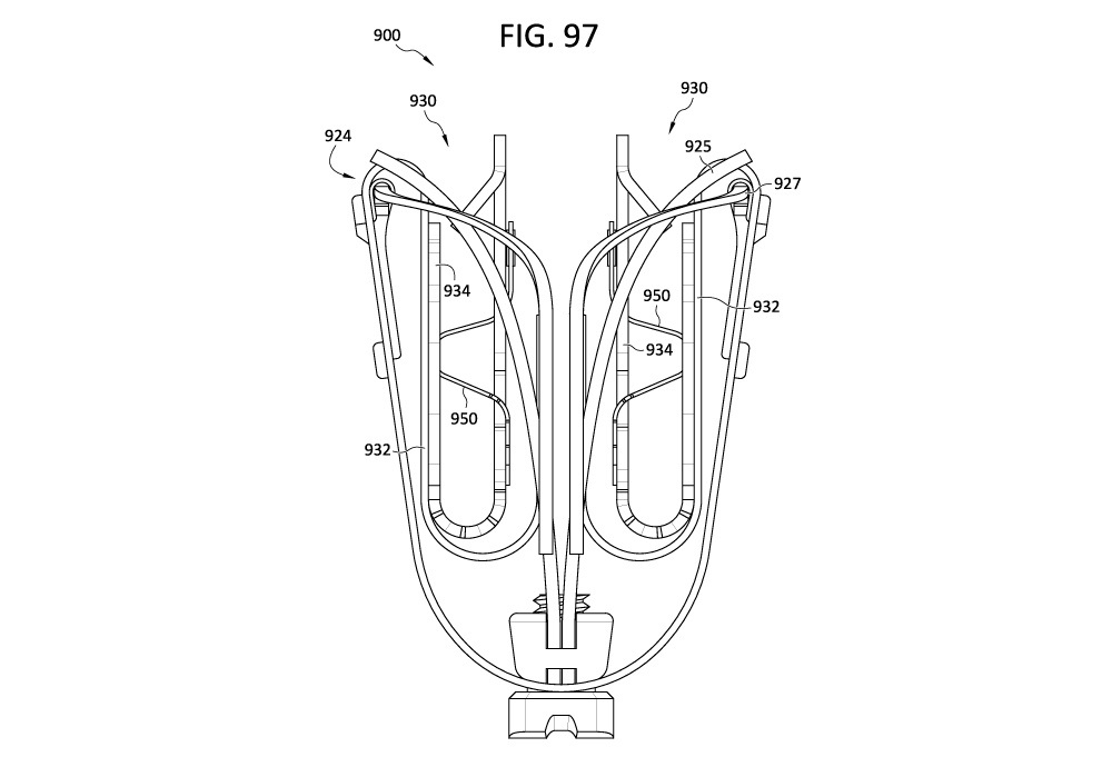
[0100] Figures 97-98 illustrate an example valve repair device having clasps
with leaflet depth
indicators;
[0101] Figures 99-101 illustrate example clasps having leaflet depth
indicators;
[0102] Figures 102A and 102B illustrate a valve repair device having clasps
with leaflet depth
indicators;
[0103] Figures 103-109 illustrate example clasps having leaflet depth
indicators;
[0104] Figure 110 illustrates a fixed end of a leaflet depth indicator;
[0105] Figures 111-114 illustrate example clasps having leaflet depth
indicators;
[0106] Figures 115-116 illustrate a device having clasps with leaflet depth
indicators;
[0107] Figures 117-118 illustrate example leaflet paths between the clasps and
the leaflet depth
indicators;
CA 03225736 2023-12-28
WO 2023/004098 PCT/US2022/037983
[0108] Figures 119-120 illustrate example leaflet depth indicators for clasps
and/or capture
devices;
[0109] Figures 121-126 illustrate example clasps having leaflet depth
indicators;
[0110] Figures 127-128 illustrate an example implantable device with clasps
having leaflet depth
indicators;
[0111] Figure 129 illustrates an example clasp having a leaflet depth
indicator;
[0112] Figure 130 illustrates an example device with clasps having leaflet
depth indicators;
[0113] Figure 131 illustrates an example clasp having a leaflet depth
indicator;
[0114] Figure 132 illustrates an example implantable device with clasps having
leaflet depth
indicators;
[0115] Figure 133 illustrates an example clasp having a leaflet depth
indicator;
[0116] Figure 134 illustrates an example implantable device with clasps having
leaflet depth
indicators;
[0117] Figure 135 illustrates an example clasp having a leaflet depth
indicator;
[0118] Figures 136, 137A, and 137B illustrate intracardiac electrocardiogram
(IECG) signals
measured by electrodes of example leaflet depth indicators;
[0119] Figures 137C-137F illustrate bipolar IECG signals measured from
electrodes of example
leaflet depth indicators;
[0120] Figure 138 illustrates an example clasp having a leaflet depth
indicator;
[0121] Figure 139 illustrates an example clasp having an integral leaflet
depth indicator;
[0122] Figure 140A illustrates an example clasp having arms that can be formed
into an integral
leaflet depth indicator;
16
CA 03225736 2023-12-28
WO 2023/004098 PCT/US2022/037983
[0123] Figure 140B illustrates an example clasp having an integral leaflet
depth indicator made
from the arms shown in Figure 140A;
[0124] Figure 140C illustrates an example clasp having an integral leaflet
depth indicator made
from the arms shown in Figure 140A;
[0125] Figure 141A illustrates an example clasp having arms that can be formed
into a movable
arm of the clasp and arms that can be formed into an integral leaflet depth
indicator;
[0126] Figure 141B illustrates an example clasp having arms that can be formed
into a movable
arm of the clasp and arms that can be formed into an integral leaflet depth
indicator;
[0127] Figure 141C illustrates an example clasp having a movable arm and
integral leaflet depth
indicator made from the arms shown in Figure 141 A or Figure 141B;
[0128] Figure 141D illustrates an example clasp having arms that can be formed
into a movable
arm of the clasp and arms that can be formed into an integral leaflet depth
indicator;
[0129] Figure 142A illustrates an example clasp having an integral leaflet
depth indicator where
a valve leaflet is not inserted to a depth that causes displacement of the
leaflet depth indicator;
[0130] Figure 142B illustrates an example clasp having an integral leaflet
depth indicator where
a valve leaflet is inserted to a depth that causes displacement of the leaflet
depth indicator;
[0131] Figure 143A illustrates an example clasp having an integral leaflet
depth indicator where
a valve leaflet is not inserted to a depth that causes displacement of the
leaflet depth indicator;
[0132] Figure 143B illustrates an example clasp having an integral leaflet
depth indicator where
a valve leaflet is inserted to a depth that causes displacement of the leaflet
depth indicator;
[0133] Figures 144-147 illustrate example devices with clasps having an
electrical leaflet depth
indicator;
[0134] Figures 148-155 illustrate example clasps with leaflet depth indicators
that are configured
to visually and electrically indicate leaflet insertion;
17
CA 03225736 2023-12-28
WO 2023/004098 PCT/US2022/037983
[0135] Figures 156, 156A, 156B, 156C, and 156D illustrate clasps having
different sensing plate
configurations;
[0136] Figures 157-158 illustrate example clasps having electrical leaflet
depth indicators;
[0137] Figure 159 illustrates an example clasp having an electrical leaflet
depth indicator of one
of Figures 157-158 sensing blood;
[0138] Figure 160 illustrates an example clasp having an electrical leaflet
depth indicator of one
of Figures 157-158 sensing a valve leaflet;
[0139] Figure 161 illustrates an example clasp having an electrical leaflet
depth indicator of one
of Figures 157-158 sensing chordae tendineae;
[0140] Figure 162 illustrates a circuit used to measure impedance in
accordance with some
implementations of clasps with electrical indicators;
[0141] Figure 163 illustrates examples of calculations of components of
impedance;
[0142] Figure 164 illustrates an implementation of a method of identifying a
clasp condition
based on an electrical measurement;
[0143] Figure 165-169 illustrate example devices and/or portions of devices
with clasps having a
leaflet depth indicator.
DETAILED DESCRIPTION
[0144] The following description refers to the accompanying drawings, which
illustrate example
implementations of the present disclosure. Other implementations having
different structures and
operation do not depart from the scope of the present disclosure.
[0145] Example implementations of the present disclosure are directed to
systems, devices,
methods, etc. for repairing a defective heart valve. For example, various
implementations of
valve repair devices, implantable devices, implants, and systems (including
systems for delivery
thereof) are disclosed herein, and any combination of these options can be
made unless
specifically excluded. In other words, individual components of the disclosed
devices and
systems can be combined unless mutually exclusive or otherwise physically
impossible. Further,
the techniques and methods herein can be performed on a living animal or on a
simulation, such
18
CA 03225736 2023-12-28
WO 2023/004098 PCT/US2022/037983
as on a cadaver, cadaver heart, simulator (e.g., with the body parts, heart,
tissue, etc. being
simulated), etc.
[0146] As described herein, when one or more components are described as being
connected,
joined, affixed, coupled, attached, or otherwise interconnected, such
interconnection can be
direct as between the components or can be indirect such as through the use of
one or more
intermediary components. Also as described herein, reference to a "member,"
"component," or
"portion" shall not be limited to a single structural member, component, or
element but can
include an assembly of components, members, or elements. Also as described
herein, the terms
"substantially" and "about" are defined as at least close to (and includes) a
given value or state
(preferably within 10% of, more preferably within 1% of, and most preferably
within 0.1% of).
The terms "clasp" and "clasp arm" are often used herein with respect to
specific examples, but
the terms "gripping member" and/or "gripper arm" can be used in place of and
function in the
same or similar ways, even if not configured in the same way as a typical
clasp.
[0147] Figures 1 and 2 are cutaway views of the human heart H in diastolic and
systolic phases,
respectively. The right ventricle RV and left ventricle LV are separated from
the right atrium RA
and left atrium LA, respectively, by the tricuspid valve TV and mitral valve
MV; i.e., the
atrioventricular valves. Additionally, the aortic valve AV separates the left
ventricle LV from the
ascending aorta AA, and the pulmonary valve PV separates the right ventricle
from the
pulmonary artery PA. Each of these valves has flexible leaflets (e.g.,
leaflets 20, 22 shown in
Figures 3-6 and leaflets 30, 32, 34 shown in Fig. 7) extending inward across
the respective
orifices that come together or "coapt" in the flow stream to form the one-way,
fluid-occluding
surfaces. The native valve repair systems of the present application are
frequently described
and/or illustrated with respect to the mitral valve MV. Therefore, anatomical
structures of the left
atrium LA and left ventricle LV will be explained in greater detail. However,
the devices
described herein can also be used in repairing other native valves, e.g., the
devices can be used in
repairing the tricuspid valve TV, the aortic valve AV, and the pulmonary valve
PV.
[0148] The left atrium LA receives oxygenated blood from the lungs. During the
diastolic phase,
or diastole, seen in Figure 1, the blood that was previously collected in the
left atrium LA (during
the systolic phase) moves through the mitral valve MV and into the left
ventricle LV by
expansion of the left ventricle LV. In the systolic phase, or systole, seen in
Figure 2, the left
ventricle LV contracts to force the blood through the aortic valve AV and
ascending aorta AA
19
CA 03225736 2023-12-28
WO 2023/004098 PCT/US2022/037983
into the body. During systole, the leaflets of the mitral valve MV close to
prevent the blood from
regurgitating from the left ventricle LV and back into the left atrium LA and
blood is collected in
the left atrium from the pulmonary vein. In some implementations, the devices
described by the
present application are used to repair the function of a defective mitral
valve MV. That is, the
devices are configured to help close the leaflets of the mitral valve to
prevent or inhibit blood
from regurgitating from the left ventricle LV and back into the left atrium
LA. Many of the
devices described in the present application are designed to easily grasp and
secure the native
leaflets around a coaptation element or spacer that beneficially acts as a
filler in the regurgitant
orifice to prevent or inhibit back flow or regurgitation during systole,
though this is not
necessary.
[0149] Referring now to Figures 1-7, the mitral valve MV includes two
leaflets, the anterior
leaflet 20 and the posterior leaflet 22. The mitral valve MV also includes an
annulus 24, which is
a variably dense fibrous ring of tissues that encircles the leaflets 20, 22.
Referring to Figures 3
and 4, the mitral valve MV is anchored to the wall of the left ventricle LV by
chordae tendineae
CT. The chordae tendineae CT are cord-like tendons that connect the papillary
muscles PM (i.e.,
the muscles located at the base of the chordae tendineae CT and within the
walls of the left
ventricle LV) to the leaflets 20, 22 of the mitral valve MV. The papillary
muscles PM serve to
limit the movements of leaflets 20, 22 of the mitral valve MV and prevent the
mitral valve MV
from being reverted. The mitral valve MV opens and closes in response to
pressure changes in
the left atrium LA and the left ventricle LV. The papillary muscles PM do not
open or close the
mitral valve MV. Rather, the papillary muscles PM support or brace the
leaflets 20, 22 against
the high pressure needed to circulate blood throughout the body. Together the
papillary muscles
PM and the chordae tendineae CT are known as the subvalvular apparatus, which
functions to
keep the mitral valve MV from prolapsing into the left atrium LA when the
mitral valve closes.
As seen from a Left Ventricular Outflow Tract (LVOT) view shown in Figure 3,
the anatomy of
the leaflets 20, 22 is such that the inner sides of the leaflets coapt at the
free end portions and the
leaflets 20, 22 start receding or spreading apart from each other. The
leaflets 20, 22 spread apart
in the atrial direction, until each leaflet meets with the mitral annulus.
[0150] Various disease processes can impair proper function of one or more of
the native valves
of the heart H. These disease processes include degenerative processes (e.g.,
Barlow's Disease,
fibroelastic deficiency, etc.), inflammatory processes (e.g., Rheumatic Heart
Disease), and
infectious processes (e.g., endocarditis, etc.). In addition, damage to the
left ventricle LV or the
CA 03225736 2023-12-28
WO 2023/004098 PCT/US2022/037983
right ventricle RV from prior heart attacks (i.e., myocardial infarction
secondary to coronary
artery disease) or other heart diseases (e.g., cardiomyopathy, etc.) can
distort a native valve's
geometry, which can cause the native valve to dysfunction. However, the
majority of patients
undergoing valve surgery, such as surgery to the mitral valve MV, suffer from
a degenerative
disease that causes a malfunction in a leaflet (e.g., leaflets 20, 22) of a
native valve (e.g., the
mitral valve MV), which results in prolapse and regurgitation.
[0151] Generally, a native valve may malfunction in different ways: including
(1) valve stenosis;
and (2) valve regurgitation. Valve stenosis occurs when a native valve does
not open completely
and thereby causes an obstruction of blood flow. Typically, valve stenosis
results from buildup of
calcified material on the leaflets of a valve, which causes the leaflets to
thicken and impairs the
ability of the valve to fully open to permit forward blood flow. Valve
regurgitation occurs when
the leaflets of the valve do not close completely thereby causing blood to
leak back into the prior
chamber (e.g., causing blood to leak from the left ventricle to the left
atrium).
[0152] There are three main mechanisms by which a native valve becomes
regurgitant¨or
incompetent¨which include Carpentier's type I, type II, and type III
malfunctions. A Carpentier
type I malfunction involves the dilation of the annulus such that normally
functioning leaflets are
distracted from each other and fail to form a tight seal (i.e., the leaflets
do not coapt properly).
Included in a type I mechanism malfunction are perforations of the leaflets,
as are present in
endocarditis. A Carpentier's type II malfunction involves prolapse of one or
more leaflets of a
native valve above a plane of coaptation. A Carpentier's type III malfunction
involves restriction
of the motion of one or more leaflets of a native valve such that the leaflets
are abnormally
constrained below the plane of the annulus. Leaflet restriction can be caused
by rheumatic
disease (Ma) or dilation of a ventricle (nth).
[0153] Referring to Figure 5, when a healthy mitral valve MV is in a closed
position, the anterior
leaflet 20 and the posterior leaflet 22 coapt, which prevents blood from
leaking from the left
ventricle LV to the left atrium LA. Referring to Figures 3 and 6, mitral
regurgitation MR occurs
when the anterior leaflet 20 and/or the posterior leaflet 22 of the mitral
valve MV is displaced
into the left atrium LA during systole so that the edges of the leaflets 20,
22 are not in contact
with each other. This failure to coapt causes a gap 26 between the anterior
leaflet 20 and the
posterior leaflet 22, which allows blood to flow back into the left atrium LA
from the left
ventricle LV during systole, as illustrated by the mitral regurgitation MR
flow path shown in
21
CA 03225736 2023-12-28
WO 2023/004098 PCT/US2022/037983
Figure 3. Referring to Figure 6, the gap 26 can have a width W between about
2.5 mm and about
17.5 mm, between about 5 mm and about 15 mm, between about 7.5 mm and about
12.5 mm, or
about 10 mm. In some situations, the gap 26 can have a width W greater than 15
mm. As set
forth above, there are several different ways that a leaflet (e.g., leaflets
20, 22 of mitral valve
MV) may malfunction which can thereby lead to valvular regurgitation.
[0154] In any of the above-mentioned situations, a valve repair device or
implant is desired that
is capable of engaging the anterior leaflet 20 and the posterior leaflet 22 to
close the gap 26 and
prevent or inhibit regurgitation of blood through the mitral valve MV. As can
be seen in Figure 4,
an abstract representation of a valve repair device, implantable device, or
implant 10 is shown
implanted between the leaflets 20, 22 such that regurgitation does not occur
during systole
(compare Figure 3 with Figure 4). In some implementations, the coaptation
element (e.g., spacer,
coaption element, gap filler, membrane, sheet, plug, wedge, balloon, etc.) of
the device 10 has a
generally tapered or triangular shape that naturally adapts to the native
valve geometry and to its
expanding leaflet nature (toward the annulus). In this application, the terms
spacer, coaption
element, coaptation element, and gap filler are used interchangeably and refer
to an element that
fills a portion of the space between native valve leaflets and/or that is
configured such that the
native valve leaflets engage or "coapt" against (e.g., such that the native
leaflets coapt against the
coaption element, coaptation element, spacer, etc. instead of only against one
another).).
[0155] Although stenosis or regurgitation can affect any valve, stenosis is
predominantly found
to affect either the aortic valve AV or the pulmonary valve PV, and
regurgitation is
predominantly found to affect either the mitral valve MV or the tricuspid
valve TV. Both valve
stenosis and valve regurgitation increase the workload of the heart H and may
lead to very
serious conditions if left un-treated; such as endocarditis, congestive heart
failure, permanent
heart damage, cardiac arrest, and ultimately death. Because the left side of
the heart (i.e., the left
atrium LA, the left ventricle LV, the mitral valve MV, and the aortic valve
AV) are primarily
responsible for circulating the flow of blood throughout the body.
Accordingly, because of the
substantially higher pressures on the left side heart dysfunction of the
mitral valve MV or the
aortic valve AV is particularly problematic and often life threatening.
[0156] Malfunctioning native heart valves can either be repaired or replaced.
Repair typically
involves the preservation and correction of the patient's native valve.
Replacement typically
involves replacing the patient's native valve with a biological or mechanical
substitute. Typically,
22
CA 03225736 2023-12-28
WO 2023/004098 PCT/US2022/037983
the aortic valve AV and pulmonary valve PV are more prone to stenosis. Because
stenotic
damage sustained by the leaflets is irreversible, treatments for a stenotic
aortic valve or stenotic
pulmonary valve can be removal and replacement of the valve with a surgically
implanted heart
valve, or displacement of the valve with a transcatheter heart valve. The
mitral valve MV and the
tricuspid valve TV are more prone to deformation of leaflets and/or
surrounding tissue, which, as
described above, prevents the mitral valve MV or tricuspid valve TV from
closing properly and
allows for regurgitation or back flow of blood from the ventricle into the
atrium (e.g., a deformed
mitral valve MV may allow for regurgitation or back flow from the left
ventricle LV to the left
atrium LA as shown in Figure 3). The regurgitation or back flow of blood from
the ventricle to
the atrium results in valvular insufficiency. Deformations in the structure or
shape of the mitral
valve MV or the tricuspid valve TV are often repairable. In addition,
regurgitation can occur due
to the chordae tendineae CT becoming dysfunctional (e.g., the chordae
tendineae CT may stretch
or rupture), which allows the anterior leaflet 20 and the posterior leaflet 22
to be reverted such
that blood is regurgitated into the left atrium LA. The problems occurring due
to dysfunctional
chordae tendineae CT can be repaired by repairing the chordae tendineae CT or
the structure of
the mitral valve MV (e.g., by securing the leaflets 20, 22 at the affected
portion of the mitral
valve).
[0157] The devices and procedures disclosed herein often make reference to
repairing the
structure of a mitral valve. However, it should be understood that the devices
and concepts
provided herein can be used to repair any native valve, as well as any
component of a native
valve. Such devices can be used between the leaflets 20, 22 of the mitral
valve MV to prevent or
inhibit regurgitation of blood from the left ventricle into the left atrium.
With respect to the
tricuspid valve TV (Figure 7), any of the devices and concepts herein can be
used between any
two of the anterior leaflet 30, septal leaflet 32, and posterior leaflet 34 to
prevent or inhibit
regurgitation of blood from the right ventricle into the right atrium. In
addition, any of the
devices and concepts provided herein can be used on all three of the leaflets
30, 32, 34 together
to prevent or inhibit regurgitation of blood from the right ventricle to the
right atrium. That is, the
valve repair devices or implants provided herein can be centrally located
between the three
leaflets 30, 32, 34.
[0158] An example implantable device (e.g., implantable prosthetic device,
etc.) or implant can
optionally have a coaptation element (e.g., spacer, coaption element, gap
filler, etc.) and at least
one anchor (e.g., one, two, three, or more). In some implementations, an
implantable device or
23
CA 03225736 2023-12-28
WO 2023/004098 PCT/US2022/037983
implant can have any combination or sub-combination of the features disclosed
herein without a
coaptation element. When included, the coaptation element (e.g., coaption
element, spacer, etc.)
is configured to be positioned within the native heart valve orifice to help
fill the space between
the leaflets and form a more effective seal, thereby reducing or preventing or
inhibiting
regurgitation described above. The coaptation element can have a structure
that is impervious to
blood (or that resists blood flow therethrough) and that allows the native
leaflets to close around
the coaptation element during ventricular systole to block blood from flowing
from the left or
right ventricle back into the left or right atrium, respectively. The device
or implant can be
configured to seal against two or three native valve leaflets; that is, the
device can be used in the
native mitral (bicuspid) and tricuspid valves. The coaptation element is
sometimes referred to
herein as a spacer because the coaptation element can fill a space between
improperly
functioning native leaflets (e.g., mitral valve leaflets 20, 22 or tricuspid
valve leaflets 30, 32, 34)
that do not close completely.
[0159] The optional coaptation element (e.g., spacer, coaption element, etc.)
can have various
shapes. In some implementations, the coaptation element can have an elongated
cylindrical shape
having a round cross-sectional shape. In some implementations, the coaptation
element can have
an oval cross-sectional shape, an ovoid cross-sectional shape, a crescent
cross-sectional shape, a
rectangular cross-sectional shape, or various other non-cylindrical shapes. In
some
implementations, the coaptation element can have an atrial portion positioned
in or adjacent to
the atrium, a ventricular or lower portion positioned in or adjacent to the
ventricle, and a side
surface that extends between the native leaflets. In some implementations
configured for use in
the tricuspid valve, the atrial or upper portion is positioned in or adjacent
to the right atrium, and
the ventricular or lower portion is positioned in or adjacent to the right
ventricle, and the side
surface that extends between the native tricuspid leaflets.
[0160] In some implementations, the anchor can be configured to secure the
device to one or
both of the native leaflets such that the coaptation element is positioned
between the two native
leaflets. In some implementations configured for use in the tricuspid valve,
the anchor is
configured to secure the device to one, two, or three of the tricuspid
leaflets such that the
coaptation element is positioned between the three native leaflets. In some
implementations, the
anchor can attach to the coaptation element at a location adjacent the
ventricular portion of the
coaptation element. In some implementations, the anchor can attach to an
actuation element,
such as a shaft or actuation wire, to which the coaptation element is also
attached. In some
24
CA 03225736 2023-12-28
WO 2023/004098 PCT/US2022/037983
implementations, the anchor and the coaptation element can be positioned
independently with
respect to each other by separately moving each of the anchor and the
coaptation element along
the longitudinal axis of the actuation element (e.g., actuation shaft,
actuation rod, actuation tube,
actuation wire, etc.). In some implementations, the anchor and the coaptation
element can be
positioned simultaneously by moving the anchor and the coaptation element
together along the
longitudinal axis of the actuation element, e.g., shaft, actuation wire, etc.
The anchor can be
configured to be positioned behind a native leaflet when implanted such that
the leaflet is
grasped by the anchor.
[0161] The device or implant can be configured to be implanted via a delivery
system or other
means for delivery. The delivery system can comprise one or more of a
guide/delivery sheath, a
delivery catheter, a steerable catheter, an implant catheter, tube,
combinations of these, etc. The
coaptation element and the anchor can be compressible to a radially compressed
state and can be
self-expandable to a radially expanded state when compressive pressure is
released. The device
can be configured for the anchor to be expanded radially away from the still-
compressed
coaptation element initially in order to create a gap between the coaptation
element and the
anchor. A native leaflet can then be positioned in the gap. The coaptation
element can be
expanded radially, closing the gap between the coaptation element and the
anchor and capturing
the leaflet between the coaptation element and the anchor. In some
implementations, the anchor
and coaptation element are optionally configured to self-expand. The
implantation methods for
various implementations can be different and are more fully discussed below
with respect to each
implementation. Additional information regarding these and other delivery
methods can be found
in U.S. Pat. No. 8,449,599 and U.S. Patent Application Publication Nos.
2014/0222136,
2014/0067052, 2016/0331523, and PCT patent application publication Nos.
W02020/076898,
each of which is incorporated herein by reference in its entirety for all
purposes. These method(s)
can be performed on a living animal or on a simulation, such as on a cadaver,
cadaver heart,
simulator (e.g., with the body parts, heart, tissue, etc. being simulated),
etc. mutatis mutandis.
[0162] The disclosed devices or implants can be configured such that the
anchor is connected to
a leaflet, taking advantage of the tension from native chordae tendineae to
resist high systolic
pressure urging the device toward the left atrium. During diastole, the
devices can rely on the
compressive and retention forces exerted on the leaflet that is grasped by the
anchor.
CA 03225736 2023-12-28
WO 2023/004098 PCT/US2022/037983
[0163] Referring now to Figures 8-15, a schematically illustrated device or
implant 100 (e.g., a
prosthetic spacer device, valve repair device implantable device, etc.) is
shown in various stages
of deployment. The device or implant 100 and other similar devices/implants
are described in
more detail in PCT patent application publication Nos. W02018/195215,
W02020/076898, and
WO 2019/139904, which are incorporated herein by reference in their entirety.
The device 100
can include any other features for another device or implant discussed in the
present application
or the applications cited above, and the device 100 can be positioned to
engage valve tissue (e.g.,
leaflets 20, 22, 30, 32, 34) as part of any suitable valve repair system
(e.g., any valve repair
system disclosed in the present application or the applications cited above).
[0164] The device or implant 100 is deployed from a delivery system or other
means for delivery
102. The delivery system 102 can comprise one or more of a catheter, a sheath,
a guide
catheter/sheath, a delivery catheter/sheath, a steerable catheter, an implant
catheter, a tube, a
channel, a pathway, combinations of these, etc. The device or implant 100
includes a coaptation
portion 104 and an anchor portion 106.
[0165] In some implementations, the coaptation portion 104 of the device or
implant 100
includes a coaptation element 110 (e.g., spacer, plug, filler, foam, sheet,
membrane, coaption
element, etc.) that is adapted to be implanted between leaflets of a native
valve (e.g., a native
mitral valve, native tricuspid valve, etc.) and is slidably attached to an
actuation element 112
(e.g., actuation wire, shaft, tube, hypotube, line, suture, braid, etc.). The
anchor portion 106
includes one or more anchors 108 that are actuatable between open and closed
conditions and
can take a wide variety of forms, such as, for example, paddles, gripping
elements, or the like.
Actuation of the means for actuating or actuation element 112 opens and closes
the anchor
portion 106 of the device 100 to grasp the native valve leaflets during
implantation. The means
for actuating or actuation element 112 (as well as other means for actuating
and actuation
elements herein) can take a wide variety of different forms (e.g., as a wire,
rod, shaft, tube,
screw, suture, line, strip, combination of these, etc.), be made of a variety
of different materials,
and have a variety of configurations. As one example, the actuation element
can be threaded such
that rotation of the actuation element moves the anchor portion 106 relative
to the coaptation
portion 104. Or, the actuation element can be unthreaded, such that pushing or
pulling the
actuation element 112 moves the anchor portion 106 relative to the coaptation
portion 104.
26
CA 03225736 2023-12-28
WO 2023/004098 PCT/US2022/037983
[0166] The anchor portion 106 and/or anchors of the device 100 include outer
paddles 120 and
inner paddles 122 that are, in some implementations, connected between a cap
114 and the
coaptation element 110 by portions 124, 126, 128. The portions 124, 126, 128
can be jointed
and/or flexible to move between all of the positions described below. The
interconnection of the
outer paddles 120, the inner paddles 122, the coaptation element 110, and the
cap 114 by the
portions 124, 126, and 128 can constrain the device to the positions and
movements illustrated
herein.
[0167] In some implementations, the delivery system 102 includes a steerable
catheter, implant
catheter, and means for actuating or actuation element 112 (e.g., actuation
wire, actuation shaft,
etc.). These can be configured to extend through a guide catheter/sheath
(e.g., a transseptal
sheath, etc.). In some implementations, the means for actuating or actuation
element 112 extends
through a delivery catheter and the coaptation element 110 to the distal end
(e.g., a cap 114 or
other attachment portion at the distal connection of the anchor portion 106).
Extending and
retracting the actuation element 112 increases and decreases the spacing
between the coaptation
element 110 and the distal end of the device (e.g., the cap 114 or other
attachment portion),
respectively. In some implementations, a collar or other attachment element
(e.g., clamp, clip,
lock, sutures, friction fit, buckle, snap fit, lasso, etc.) removably attaches
the coaptation element
110 to the delivery system 102, either directly or indirectly, so that the
means for actuating or
actuation element 112 slides through the collar or other attachment element
and, in some
implementations, through a coaptation element 110 during actuation to open and
close the
paddles 120, 122 of the anchor portion 106 and/or anchors 108.
[0168] In some implementation, the anchor portion 106 and/or anchors 108 can
include
attachment portions or gripping members. The illustrated gripping members can
comprise clasps
130 that include a base or fixed arm 132, a movable arm 134, optional barbs,
friction-enhancing
elements, or other means for securing 136 (e.g., protrusions, ridges, grooves,
textured surfaces,
adhesive, etc.), and a joint portion 138. The fixed arms 132 are attached to
the inner paddles 122.
In some implementations, the fixed arms 132 are attached to the inner paddles
122 with the joint
portion 138 disposed proximate a coaptation element 110. In some
implementations, the clasps
(e.g., barbed clasps, barbed gripping members, etc.) have flat surfaces and do
not fit in a recess
of the inner paddle. Rather, the flat portions of the clasps are disposed
against the surface of the
inner paddle 122. The joint portion 138 provides a spring force between the
fixed and movable
arms 132, 134 of the clasp 130. The joint portion 138 can be any suitable
joint, such as a flexible
27
CA 03225736 2023-12-28
WO 2023/004098 PCT/US2022/037983
joint, a spring joint, a pivot joint, or the like. In some implementations,
the joint portion 138 is a
flexible piece of material integrally formed with the fixed and movable arms
132, 134. The fixed
arms 132 are attached to the inner paddles 122 and remain stationary or
substantially stationary
relative to the inner paddles 122 when the movable arms 134 are opened to open
the clasps 130
and expose the optional barbs, friction-enhancing elements, or means for
securing 136.
[0169] In some implementations, the clasps 130 are opened by applying tension
to actuation
lines 116 attached to the movable arms 134, thereby causing the movable arms
134 to articulate,
flex, or pivot on the joint portions 138. The actuation lines 116 extend
through the delivery
system 102 (e.g., through a steerable catheter and/or an implant catheter).
Other actuation
mechanisms are also possible.
[0170] The actuation line 116 can take a wide variety of forms, such as, for
example, a line, a
suture, a wire, a rod, a catheter, or the like. The clasps 130 can be spring
loaded so that in the
closed position the clasps 130 continue to provide a pinching force on the
grasped native leaflet.
This pinching force remains constant regardless of the position of the inner
paddles 122.
Optional barbs, friction-enhancing elements, or other means for securing 136
of the clasps 130
can grab, pinch, and/or pierce the native leaflets to further secure the
native leaflets.
[0171] During implantation, the paddles 120, 122 can be opened and closed, for
example, to
grasp the native leaflets (e.g., native mitral valve leaflets, etc.) between
the paddles 120, 122
and/or between the paddles 120, 122 and a coaptation element 110. The clasps
130 can be used
to grasp and/or further secure the native leaflets by engaging the leaflets
with optional barbs,
friction-enhancing elements, or means for securing 136 and pinching the
leaflets between the
movable and fixed arms 134, 132. The optional barbs, friction-enhancing
elements, or other
means for securing 136 (e.g., protrusions, ridges, grooves, textured surfaces,
adhesive, etc.) of
the clasps 130 increase friction with the leaflets or can partially or
completely puncture the
leaflets. The actuation lines 116 can be actuated separately so that each
clasp 130 can be opened
and closed separately. Separate operation allows one leaflet to be grasped at
a time, or for the
repositioning of a clasp 130 on a leaflet that was insufficiently grasped,
without altering a
successful grasp on the other leaflet. The clasps 130 can be opened and closed
relative to the
position of the inner paddle 122 (as long as the inner paddle is in an open or
at least partially
open position), thereby allowing leaflets to be grasped in a variety of
positions as the particular
situation requires.
28
CA 03225736 2023-12-28
WO 2023/004098 PCT/US2022/037983
[0172] Referring now to Figure 8, the device 100 is shown in an elongated or
fully open
condition for deployment from an implant delivery catheter of the delivery
system 102. The
device 100 is disposed at the end of the catheter in the fully open position,
because the fully open
position takes up the least space and allows the smallest catheter to be used
(or the largest device
to be used for a given catheter size). In the elongated condition the cap 114
is spaced apart from
the coaptation element 110 such that the paddles 120, 122 are fully extended.
In some
implementations, an angle formed between the interior of the outer and inner
paddles 120, 122 is
approximately 180 degrees. The clasps 130 are kept in a closed condition
during deployment
through the delivery system 102 so that the optional barbs, friction-enhancing
elements, or other
means for securing 136 (Figure 9) do not catch or damage the delivery system
102 or tissue in
the patient's heart.
[0173] Referring now to Figure 9, the device 100 is shown in an elongated
detangling condition,
similar to Figure 8, but with the clasps 130 in a fully open position, ranging
from about 140
degrees to about 200 degrees, from about 170 degrees to about 190 degrees, or
about 180 degrees
between fixed and movable arms 132, 134 of the clasps 130. Fully opening the
paddles 120, 122
and the clasps 130 has been found to improve ease of detanglement or
detachment from anatomy
of the patient, such as the chordae tendineae CT, during implantation of the
device 100.
[0174] Referring now to Figure 10, the device 100 is shown in a shortened or
fully closed
condition. The compact size of the device 100 in the shortened condition
allows for easier
maneuvering and placement within the heart. To move the device 100 from the
elongated
condition to the shortened condition, the means for actuating or actuation
element 112 is
retracted to pull the cap 114 towards the coaptation element 110. The
connection portion(s) 126
(e.g., joint(s), flexible connection(s), etc.) between the outer paddle 120
and inner paddle 122 are
constrained in movement such that compression forces acting on the outer
paddle 120 from the
cap 114 being retracted towards the coaptation element 110 cause the paddles
or gripping
elements to move radially outward. During movement from the open to closed
position, the outer
paddles 120 maintain an acute angle with the means for actuating or actuation
element 112. The
outer paddles 120 can optionally be biased toward a closed position. The inner
paddles 122
during the same motion move through a considerably larger angle as they are
oriented away from
the coaptation element 110 in the open condition and collapse along the sides
of the coaptation
element 110 in the closed condition. In some implementations, the inner
paddles 122 are thinner
and/or narrower than the outer paddles 120, and the connection portions 126,
128 (e.g., joints,
29
CA 03225736 2023-12-28
WO 2023/004098 PCT/US2022/037983
flexible connections, etc.) connected to the inner paddles 122 can be thinner
and/or more
flexible. For example, this increased flexibility can allow more movement than
the connection
portion 124 connecting the outer paddle 120 to the cap 114. In some
implementations, the outer
paddles 120 are narrower than the inner paddles 122. The connection portions
126, 128
connected to the inner paddles 122 can be more flexible, for example, to allow
more movement
than the connection portion 124 connecting the outer paddle 120 to the cap
114. In some
implementations, the inner paddles 122 can be the same or substantially the
same width as the
outer paddles
[0175] Referring now to Figures 11-13, the device 100 is shown in a partially
open, grasp-ready
condition. To transition from the fully closed to the partially open
condition, the means for
actuating or actuation element (e.g., actuation wire, actuation shaft, etc.)
is extended to push the
cap 114 away from the coaptation element 110, thereby pulling on the outer
paddles 120, which
in turn pull on the inner paddles 122, causing the anchors or anchor portion
106 to partially
unfold. The actuation lines 116 are also retracted to open the clasps 130 so
that the leaflets can be
grasped. In some implementations, the pair of inner and outer paddles 122, 120
are moved in
unison, rather than independently, by a single means for actuating or single
actuation element
112. Also, the positions of the clasps 130 are dependent on the positions of
the paddles 122, 120.
For example, referring to Figure 10 closing the paddles 122, 120 also closes
the clasps. In some
implementations, the paddles 120, 122 can be independently controllable. For
example, the
device 100 can have two actuation elements and two independent caps (or other
attachment
portions), such that one independent actuation element (e.g., wire, shaft,
etc.) and cap (or other
attachment portion) are used to control one paddle, and the other independent
actuation element
and cap (or other attachment portion) are used to control the other paddle.
[0176] Referring now to Figure 12, one of the actuation lines 116 is extended
to allow one of the
clasps 130 to close. Referring now to Figure 13, the other actuation line 116
is extended to allow
the other clasp 130 to close. Either or both of the actuation lines 116 can be
repeatedly actuated
to repeatedly open and close the clasps 130.
[0177] Referring now to Figure 14, the device 100 is shown in a fully closed
and deployed
condition. The delivery system or means for delivery 102 and means for
actuating or actuation
element 112 are retracted and the paddles 120, 122 and clasps 130 remain in a
fully closed
position. Once deployed, the device 100 can be maintained in the fully closed
position with a
CA 03225736 2023-12-28
WO 2023/004098 PCT/US2022/037983
mechanical latch or can be biased to remain closed through the use of spring
materials, such as
steel, other metals, plastics, composites, etc. or shape-memory alloys such as
Nitinol. For
example, the connection portions 124, 126, 128, the joint portions 138, and/or
the inner and outer
paddles 122, and/or an additional biasing component (not shown) can be formed
of metals such
as steel or shape-memory alloy, such as Nitinol¨produced in a wire, sheet,
tubing, or laser
sintered powder¨and are biased to hold the outer paddles 120 closed around the
coaptation
element 110 and the clasps 130 pinched around native leaflets. Similarly, the
fixed and movable
arms 132, 134 of the clasps 130 are biased to pinch the leaflets. In some
implementations, the
attachment or connection portions 124, 126, 128, joint portions 138, and/or
the inner and outer
paddles 122, and/or an additional biasing component (not shown) can be formed
of any other
suitably elastic material, such as a metal or polymer material, to maintain
the device 100 in the
closed condition after implantation.
[0178] Figure 15 illustrates an example where the paddles 120, 122 are
independently
controllable. The device 101 illustrated by Figure 15 is similar to the device
100 illustrated by
Figure 11, except the device 101 of Figure 15 includes an actuation element
that is configured as
two independent actuation elements 111, 113 that are coupled to two
independent caps 115, 117.
To transition a first inner paddle 122 and a first outer paddle 120 from the
fully closed to the
partially open condition, the means for actuating or actuation element 111 is
extended to push the
cap 115 away from the coaptation element 110, thereby pulling on the outer
paddle 120, which in
turn pulls on the inner paddle 122, causing the first anchor 108 to partially
unfold. To transition a
second inner paddle 122 and a second outer paddle 120 from the fully closed to
the partially
open condition, the means for actuating or actuation element 113 is extended
to push the cap 115
away from the coaptation element 110, thereby pulling on the outer paddle 120,
which in turn
pulls on the inner paddle 122, causing the second anchor 108 to partially
unfold. The
independent paddle control illustrated by Figure 15 can be implemented on any
of the devices
disclosed by the present application. For comparison, in the example
illustrated by Figure 11, the
pair of inner and outer paddles 122, 120 are moved in unison, rather than
independently, by a
single means for actuating or actuation element 112.
[0179] Referring now to Figures 16-21, the device 100 of Figures 8-14 is shown
being delivered
and deployed within the native mitral valve MV of the heart H. Referring to
Figure 16, a delivery
sheath/catheter is inserted into the left atrium LA through the septum and the
implant/device 100
is deployed from the delivery catheter/sheath in the fully open condition as
illustrated in Figure
31
CA 03225736 2023-12-28
WO 2023/004098 PCT/US2022/037983
16. The means for actuating or actuation element 112 is then retracted to move
the
implant/device into the fully closed condition shown in Figure 17.
[0180] As can be seen in Figure 18, the implant/device is moved into position
within the mitral
valve MV into the ventricle LV and partially opened so that the leaflets 20,
22 can be grasped.
For example, a steerable catheter can be advanced and steered or flexed to
position the steerable
catheter as illustrated by Figure 18. The implant catheter connected to the
implant/device can be
advanced from inside the steerable catheter to position the implant as
illustrated by Figure 18.
[0181] Referring now to Figure 19, the implant catheter can be retracted into
the steerable
catheter to position the mitral valve leaflets 20, 22 in the clasps 130. An
actuation line 116 is
extended to close one of the clasps 130, capturing a leaflet 20. Figure 20
shows the other
actuation line 116 being then extended to close the other clasp 130, capturing
the remaining
leaflet 22. Lastly, as can be seen in Figure 21, the delivery system 102
(e.g., steerable catheter,
implant catheter, etc.), means for actuating or actuation element 112 and
actuation lines 116 are
then retracted and the device or implant 100 is fully closed and deployed in
the native mitral
valve MV.
[0182] Referring now to Figures 22-27, an example of an implantable device or
implant or
implant 200 is shown. The implantable device 200 is one of the many different
configurations
that the device 100 that is schematically illustrated in Figures 8-14 can
take. The device 200 can
include any other features for an implantable device or implant discussed in
the present
application, and the device 200 can be positioned to engage valve tissue 20,
22 as part of any
suitable valve repair system (e.g., any valve repair system disclosed in the
present application).
The device/implant 200 can be a prosthetic spacer device, valve repair device,
or another type of
implant that attaches to leaflets of a native valve.
[0183] In some implementations, the implantable device or implant 200 includes
a coaptation
portion 204, a proximal or attachment portion 205, an anchor portion 206, and
a distal portion
207. In some implementations, the coaptation portion 204 of the device
optionally includes a
coaptation element 210 (e.g., a spacer, coaption element, plug, membrane,
sheet, etc.) for
implantation between leaflets of a native valve. In some implementations, the
anchor portion 206
includes a plurality of anchors 208. The anchors can be configured in a
variety of ways. In some
implementations, each anchor 208 includes outer paddles 220, inner paddles
222, paddle
extension members or paddle frames 224, and clasps 230. In some
implementations, the
32
CA 03225736 2023-12-28
WO 2023/004098 PCT/US2022/037983
attachment portion 205 includes a first or proximal collar 211 (or other
attachment element) for
engaging with a capture mechanism 213 (see e.g., Figures 43-49) of a delivery
system 202
(Figures 38-42 and 49). Delivery system 202 can be the same as or similar to
delivery system
102 described elsewhere and can comprise one or more of a catheter, a sheath,
a guide
catheter/sheath, a delivery catheter/sheath, a steerable catheter, an implant
catheter, a tube, a
channel, a pathway, combinations of these, etc. The capture mechanism can be
configured in a
variety of ways and, in some implementations, can comprise one or more of a
clamp, clip, pin,
suture, line, lasso, noose, snare, buckle, lock, latch, etc.
[0184] In some implementations, the coaptation element 210 and paddles 220,222
are formed
from a flexible material that can be a metal fabric, such as a mesh, woven,
braided, or formed in
any other suitable way or a laser cut or otherwise cut flexible material. The
material can be cloth,
shape-memory alloy wire¨such as Nitinol¨to provide shape-setting capability,
or any other
flexible material suitable for implantation in the human body.
[0185] An actuation element 212 (e.g., actuation shaft, actuation rod,
actuation tube, actuation
wire, actuation line, etc.) extends from the delivery system 202 to engage and
enable actuation of
the implantable device or implant 200. In some implementations, the actuation
element 212
extends through the capture mechanism 213, proximal collar 211, and coaptation
element 210 to
engage a cap 214 of the distal portion 207. The actuation element 212 can be
configured to
removably engage the cap 214 with a threaded connection, or the like, so that
the actuation
element 212 can be disengaged and removed from the device 200 after
implantation.
[0186] The coaptation element 210 extends from the proximal collar 211 (or
other attachment
element) to the inner paddles 222. In some implementations, the coaptation
element 210 has a
generally elongated and round shape, though other shapes and configurations
are possible. In
some implementations, the coaptation element 210 has an elliptical shape or
cross-section when
viewed from above (e.g., Figure 51) and has a tapered shape or cross-section
when seen from a
front view (e.g., Figure 23) and a round shape or cross-section when seen from
a side view (e.g.,
Figure 24). A blend of these three geometries can result in the three-
dimensional shape of the
illustrated coaptation element 210 that achieves the benefits described
herein. The round shape of
the coaptation element 210 can also be seen, when viewed from above, to
substantially follow or
be close to the shape of the paddle frames 224.
33
CA 03225736 2023-12-28
WO 2023/004098 PCT/US2022/037983
[0187] The size and/or shape of the coaptation element 210 can be selected to
minimize the
number of implants that a single patient will require (preferably one), while
at the same time
maintaining low transvalvular gradients. In some implementations, the anterior-
posterior distance
at the top of the coaptation element is about 5 mm, and the medial-lateral
distance of the
coaptation element at its widest is about 10 mm. In some implementations, the
overall geometry
of the device 200 can be based on these two dimensions and the overall shape
strategy described
above. It should be readily apparent that the use of other anterior-posterior
distance anterior-
posterior distance and medial-lateral distance as starting points for the
device will result in a
device having different dimensions. Further, using other dimensions and the
shape strategy
described above will also result in a device having different dimensions.
[0188] In some implementations, the outer paddles 220 are jointably attached
to the cap 214 of
the distal portion 207 by connection portions 221 and to the inner paddles 222
by connection
portions 223. The inner paddles 222 are jointably attached to the coaptation
element by
connection portions 225. In this manner, the anchors 208 are configured
similar to legs in that the
inner paddles 222 are like upper portions of the legs, the outer paddles 220
are like lower
portions of the legs, and the connection portions 223 are like knee portions
of the legs.
[0189] In some implementations, the inner paddles 222 are stiff, relatively
stiff, rigid, have rigid
portions and/or are stiffened by a stiffening member or a fixed arm 232 of the
clasps 230. The
stiffening of the inner paddle allows the device to move to the various
different positions shown
and described herein. The inner paddle 222, the outer paddle 220, the
coaptation can all be
interconnected as described herein, such that the device 200 is constrained to
the movements and
positions shown and described herein.
[0190] In some implementations, the paddle frames 224 are attached to the cap
214 at the distal
portion 207 and extend to the connection portions 223 between the inner and
outer paddles 222,
220. In some implementations, the paddle frames 224 are formed of a material
that is more rigid
and stiff than the material forming the paddles 222, 220 so that the paddle
frames 224 provide
support for the paddles 222, 220.
[0191] The paddle frames 224 provide additional pinching force between the
inner paddles 222
and the coaptation element 210 and assist in wrapping the leaflets around the
sides of the
coaptation element 210 for a better seal between the coaptation element 210
and the leaflets, as
can be seen in Figure 51. That is, the paddle frames 224 can be configured
with a round three-
34
CA 03225736 2023-12-28
WO 2023/004098 PCT/US2022/037983
dimensional shape extending from the cap 214 to the connection portions 223 of
the anchors 208.
The connections between the paddle frames 224, the outer and inner paddles
220, 222, the cap
214, and the coaptation element 210 can constrain each of these parts to the
movements and
positions described herein. In particular the connection portion 223 is
constrained by its
connection between the outer and inner paddles 220, 222 and by its connection
to the paddle
frame 224. Similarly, the paddle frame 224 is constrained by its attachment to
the connection
portion 223 (and thus the inner and outer paddles 222, 220) and to the cap
214.
[0192] Configuring the paddle frames 224 in this manner provides increased
surface area
compared to the outer paddles 220 alone. This can, for example, make it easier
to grasp and
secure the native leaflets. The increased surface area can also distribute the
clamping force of the
paddles 220 and paddle frames 224 against the native leaflets over a
relatively larger surface of
the native leaflets in order to further protect the native leaflet tissue.
Referring again to Figure
51, the increased surface area of the paddle frames 224 can also allow the
native leaflets to be
clamped to the implantable device or implant 200, such that the native
leaflets coapt entirely
around the coaptation member or coaptation element 210. This can, for example,
improve sealing
of the native leaflets 20, 22 and thus prevent or further reduce mitral
regurgitation.
[0193] In some implementations the clasps comprise a movable arm coupled to
the anchors. In
some implementations, the clasps 230 include a base or fixed arm 232, a
movable arm 234,
optional barbs 236, and a joint portion 238. The fixed arms 232 are attached
to the inner paddles
222, with the joint portion 238 disposed proximate the coaptation element 210.
The joint portion
238 is spring-loaded so that the fixed and movable arms 232, 234 are biased
toward each other
when the clasp 230 is in a closed condition. In some implementations, the
clasps 230 include
friction-enhancing elements or means for securing, such as optional barbs,
protrusions, ridges,
grooves, textured surfaces, adhesive, etc.
[0194] In some implementations, the fixed arms 232 are attached to the inner
paddles 222
through holes or slots 231 with sutures (not shown). The fixed arms 232 can be
attached to the
inner paddles 222 with any suitable means, such as screws or other fasteners,
crimped sleeves,
mechanical latches or snaps, welding, adhesive, clamps, latches, or the like.
The fixed arms 232
remain substantially stationary relative to the inner paddles 222 when the
movable arms 234 are
opened to open the clasps 230 and expose the optional barbs or other friction-
enhancing elements
236. The clasps 230 are opened by applying tension to actuation lines 216
(e.g., as shown in
CA 03225736 2023-12-28
WO 2023/004098 PCT/US2022/037983
Figures 43-48) attached to holes 235 in the movable arms 234, thereby causing
the movable
arms 234 to articulate, pivot, and/or flex on the joint portions 238.
[0195] Referring now to Figure 29, a close-up view of one of the leaflets 20,
22 grasped by a
clasp such as clasp 230 is shown. The leaflet 20, 22 is grasped between the
movable and fixed
arms 234, 232 of the clasp 230. The tissue of the leaflet 20, 22 is not
pierced by the optional
barbs or friction-enhancing elements 236, though in some implementations the
optional barbs
236 can partially or fully pierce through the leaflet 20, 22. The angle and
height of the optional
barbs or friction-enhancing elements 236 relative to the movable arm 234 helps
to secure the
leaflet 20, 22 within the clasp 230. In particular, a force pulling the
implant off of the native
leaflet 20, 22 will encourage the optional barbs or friction-enhancing
elements 236 to further
engage the tissue, thereby ensuring better retention. Retention of the leaflet
20, 22 in the clasp
230 is further improved by the position of fixed arm 232 near the optional
barbs/friction-
enhancing elements 236 when the clasp 230 is closed. In this arrangement, the
tissue is formed
by the fixed arms 232 and the movable arms 234 and the optional barbs/friction-
enhancing
elements 236 into an S-shaped torturous path. Thus, forces pulling the leaflet
20, 22 away from
the clasp 230 will encourage the tissue to further engage the optional
barbs/friction-enhancing
elements 236 before the leaflets 20, 22 can escape. For example, leaflet
tension during diastole
can encourage the optional barbs 236 to pull toward the end portion of the
leaflet 20, 22. Thus,
the S-shaped path can utilize the leaflet tension during diastole to more
tightly engage the leaflets
20, 22 with the optional barbs/friction-enhancing elements 236.
[0196] Referring to Figure 25, the device or implant 200 can also include a
cover 240. In some
implementations, the cover 240 can be disposed on the coaptation element 210,
the outer and
inner paddles 220, 222, and/or the paddle frames 224. The cover 240 can be
configured to
prevent or reduce blood-flow through the device or implant 200 and/or to
promote native tissue
ingrowth. In some implementations, the cover 240 can be a cloth or fabric such
as PET, velour,
or other suitable fabric. In some implementations, in lieu of or in addition
to a fabric, the cover
240 can include a coating (e.g., polymeric) that is applied to the implantable
device or implant
200.
[0197] During implantation, the paddles 220, 222 of the anchors 208 are opened
and closed to
grasp the native valve leaflets 20, 22 between the paddles 220, 222 and the
coaptation element
210. The anchors 208 are moved between a closed position (Figures 22-25) to
various open
36
CA 03225736 2023-12-28
WO 2023/004098 PCT/US2022/037983
positions (Figures 26-37) by extending and retracting the actuation element
212. Extending and
retracting the actuation element 212 increases and decreases the spacing
between the coaptation
element 210 and the cap 214, respectively. The proximal collar 211 (or other
attachment element)
and the coaptation element 210 slide along the actuation element 212 during
actuation so that
changing of the spacing between the coaptation element 210 and the cap 214
causes the paddles
220, 220 to move between different positions to grasp the mitral valve
leaflets 20, 22 during
implantation.
[0198] As the device 200 is opened and closed, the pair of inner and outer
paddles 222, 220 are
moved in unison, rather than independently, by a single actuation element 212.
Also, the
positions of the clasps 230 are dependent on the positions of the paddles 222,
220. For example,
the clasps 230 are arranged such that closure of the anchors 208
simultaneously closes the clasps
230. In some implementations, the device 200 can be made to have the paddles
220, 222 be
independently controllable in the same manner (e.g., the device 101
illustrated in Figure 15).
[0199] In some implementations, the clasps 230 further secure the native
leaflets 20, 22 by
engaging the leaflets 20, 22 with optional barbs and/or other friction-
enhancing elements 236
and/or pinching the leaflets 20, 22 between the movable and fixed arms 234,
232. In some
implementations, the clasps 230 are barbed clasps that include barbs that
increase friction with
and/or can partially or completely puncture the leaflets 20, 22. The actuation
lines 216 (Figures
43-48) can be actuated separately so that each clasp 230 can be opened and
closed separately.
Separate operation allows one leaflet 20, 22 to be grasped at a time, or for
the repositioning of a
clasp 230 on a leaflet 20, 22 that was insufficiently grasped, without
altering a successful grasp
on the other leaflet 20, 22. The clasps 230 can be fully opened and closed
when the inner paddle
222 is not closed, thereby allowing leaflets 20, 22 to be grasped in a variety
of positions as the
particular situation requires.
[0200] Referring now to Figures 22-25, the device 200 is shown in a closed
position. When
closed, the inner paddles 222 are disposed between the outer paddles 220 and
the coaptation
element 210. The clasps 230 are disposed between the inner paddles 222 and the
coaptation
element 210. Upon successful capture of native leaflets 20, 22 the device 200
is moved to and
retained in the closed position so that the leaflets 20, 22 are secured within
the device 200 by the
clasps 230 and are pressed against the coaptation element 210 by the paddles
220, 222. The outer
paddles 220 can have a wide curved shape that fits around the curved shape of
the coaptation
37
CA 03225736 2023-12-28
WO 2023/004098 PCT/US2022/037983
element 210 to more securely grip the leaflets 20, 22 when the device 200 is
closed (e.g., as can
be seen in Figure 51). The curved shape and rounded edges of the outer paddle
220 also prohibits
or inhibits tearing of the leaflet tissue.
[0201] Referring now to Figures 30-37, the implantable device or implant 200
described above
is shown in various positions and configurations ranging from partially open
to fully open. The
paddles 220, 222 of the device 200 transition between each of the positions
shown in Figures 30-
37 from the closed position shown in Figures 22-25 up extension of the
actuation element 212
from a fully retracted to fully extended position.
[0202] Referring now to Figures 30-31, the device 200 is shown in a partially
open position. The
device 200 is moved into the partially open position by extending the
actuation element 212.
Extending the actuation element 212 pulls down on the bottom portions of the
outer paddles 220
and paddle frames 224. The outer paddles 220 and paddle frames 224 pull down
on the inner
paddles 222, where the inner paddles 222 are connected to the outer paddles
220 and the paddle
frames 224. Because the proximal collar 211 (or other attachment element) and
coaptation
element 210 are held in place by the capture mechanism 213, the inner paddles
222 are caused to
articulate, pivot, and/or flex in an opening direction. The inner paddles 222,
the outer paddles
220, and the paddle frames all flex to the position shown in Figures 30-31.
Opening the paddles
222, 220 and frames 224 forms a gap between the coaptation element 210 and the
inner paddle
222 that can receive and grasp the native leaflets 20, 22. This movement also
exposes the clasps
230 that can be moved between closed (Figure 30) and open (Figure 31)
positions to form a
second gap for grasping the native leaflets 20, 22. The extent of the gap
between the fixed and
movable arms 232, 234 of the clasp 230 is limited to the extent that the inner
paddle 222 has
spread away from the coaptation element 210.
[0203] Referring now to Figures 32-33, the device 200 is shown in a laterally
extended or open
position. The device 200 is moved into the laterally extended or open position
by continuing to
extend the actuation element 212 described above, thereby increasing the
distance between the
coaptation element 210 and the cap 214 of the distal portion 207. Continuing
to extend the
actuation element 212 pulls down on the outer paddles 220 and paddle frames
224, thereby
causing the inner paddles 222 to spread apart further from the coaptation
element 210. In the
laterally extended or open position, the inner paddles 222 extend horizontally
more than in other
positions of the device 200 and form an approximately 90-degree angle with the
coaptation
38
CA 03225736 2023-12-28
WO 2023/004098 PCT/US2022/037983
element 210. Similarly, the paddle frames 224 are at their maximum spread
position when the
device 200 is in the laterally extended or open position. The increased gap
between the
coaptation element 210 and inner paddle 222 formed in the laterally extended
or open position
allows clasps 230 to open further (Figure 33) before engaging the coaptation
element 210,
thereby increasing the size of the gap between the fixed and movable arms 232,
234.
[0204] Referring now to Figures 34-35, the example device 200 is shown in a
three-quarters
extended position. The device 200 is moved into the three-quarters extended
position by
continuing to extend the actuation element 212 described above, thereby
increasing the distance
between the coaptation element 210 and the cap 214 of the distal portion 207.
Continuing to
extend the actuation element 212 pulls down on the outer paddles 220 and
paddle frames 224,
thereby causing the inner paddles 222 to spread apart further from the
coaptation element 210. In
the three-quarters extended position, the inner paddles 222 are open beyond 90
degrees to an
approximately 135-degree angle with the coaptation element 210. The paddle
frames 224 are less
spread than in the laterally extended or open position and begin to move
inward toward the
actuation element 212 as the actuation element 212 extends further. The outer
paddles 220 also
flex back toward the actuation element 212. As with the laterally extended or
open position, the
increased gap between the coaptation element 210 and inner paddle 222 formed
in the laterally
extended or open position allows clasps 230 to open even further (Figure 35),
thereby increasing
the size of the gap between the fixed and movable arms 232, 234.
[0205] Referring now to Figures 36-37, the example device 200 is shown in a
fully extended
position. The device 200 is moved into the fully extended position by
continuing to extend the
actuation element 212 described above, thereby increasing the distance between
the coaptation
element 210 and the cap 214 of the distal portion 207 to a maximum distance
allowable by the
device 200. Continuing to extend the actuation element 212 pulls down on the
outer paddles 220
and paddle frames 224, thereby causing the inner paddles 222 to spread apart
further from the
coaptation element 210. The outer paddles 220 and paddle frames 224 move to a
position where
they are close to the actuation element. In the fully extended position, the
inner paddles 222 are
open to an approximately 180-degree angle with the coaptation element 210. The
inner and outer
paddles 222, 220 are stretched straight in the fully extended position to form
an approximately
180-degree angle between the paddles 222, 220. The fully extended position of
the device 200
provides the maximum size of the gap between the coaptation element 210 and
inner paddle 222,
and, in some implementations, allows clasps 230 to also open fully to
approximately 180 degrees
39
CA 03225736 2023-12-28
WO 2023/004098 PCT/US2022/037983
(Figure 37) between the fixed and movable arms 232, 234 of the clasp 230. The
position of the
device 200 is the longest and the narrowest configuration. Thus, the fully
extended position of
the device 200 can be a desirable position for bailout of the device 200 from
an attempted
implantation or can be a desired position for placement of the device in a
delivery catheter, or the
like.
[0206] Configuring the device or implant 200 such that the anchors 208 can
extend to a straight
or approximately straight configuration (e.g., approximately 120-180 degrees
relative to the
coaptation element 210) can provide several advantages. For example, this
configuration can
reduce the radial crimp profile of the device or implant 200. It can also make
it easier to grasp
the native leaflets 20, 22 by providing a larger opening between the
coaptation element 210 and
the inner paddles 222 in which to grasp the native leaflets 20, 22.
Additionally, the relatively
narrow, straight configuration can prevent or reduce the likelihood that the
device or implant 200
will become entangled in native anatomy (e.g., chordae tendineae CT shown in
Figures 3 and 4)
when positioning and/or retrieving the device or implant 200 into the delivery
system 202.
[0207] Referring now to Figures 38-49, an example device 200 is shown being
delivered and
deployed within the native mitral valve MV of the heart H. As described above,
the device 200
shown in Figures 38-49 includes the optional covering 240 (e.g., Figure 25)
over the coaptation
element 210, clasps 230, inner paddles 222 and/or the outer paddles 220. The
device 200 is
deployed from a delivery system 202 (e.g., which can comprise an implant
catheter that is
extendable from a steerable catheter 241 and/or a guide sheath) and is
retained by a capture
mechanism 213 (see e.g., Figures 43 and 48) and is actuated by extending or
retracting the
actuation element 212. Fingers of the capture mechanism 213 removably attach
the collar 211 to
the delivery system 202. In some implementations, the capture mechanism 213 is
held closed
around the collar 211 by the actuation element 212, such that removal of the
actuation element
212 allows the fingers of the capture mechanism 213 to open and release the
collar 211 to
decouple the capture mechanism 213 from the device 200 after the device 200
has been
successfully implanted.
[0208] Referring now to Figure 38, the delivery system 202 (e.g., a delivery
catheter/sheath
thereof) is inserted into the left atrium LA through the septum and the
device/implant 200 is
deployed from the delivery system 202 (e.g., an implant catheter retaining the
device/implant can
be extended to deploy the device/implant out from a steerable catheter) in the
fully open
CA 03225736 2023-12-28
WO 2023/004098 PCT/US2022/037983
condition for the reasons discussed above with respect to the device 100. The
actuation element
212 is then retracted to move the device 200 through the partially closed
condition (Figure 39)
and to the fully closed condition shown in Figures 40-41. Then the delivery
system or catheter
maneuvers the device/implant 200 towards the mitral valve MV as shown in
Figure 41. Referring
now to Figure 42, when the device 200 is aligned with the mitral valve MV, the
actuation
element 212 is extended to open the paddles 220, 222 into the partially opened
position and the
actuation lines 216 (Figures 43-48) are retracted to open the clasps 230 to
prepare for leaflet
grasp. Next, as shown in Figures 43-44, the partially open device 200 is
inserted through the
native valve (e.g., by advancing an implant catheter from a steerable
catheter) until leaflets 20,
22 are properly positioned in between the inner paddles 222 and the coaptation
element 210 and
inside the open clasps 230.
[0209] Figure 45 shows the device 200 with both clasps 230 closed, though the
optional barbs
236 of one clasp 230 missed one leaflet 22. As can be seen in Figures 45-47,
the out of position
clasp 230 is opened and closed again to properly grasp the missed leaflet 22.
When both leaflets
20, 22 are grasped properly, the actuation element 212 is retracted to move
the device 200 into
the fully closed position shown in Figure 48. With the device 200 fully closed
and implanted in
the native valve, the actuation element 212 is disengaged from the cap 214 and
is withdrawn to
release the capture mechanism 213 from the proximal collar 211 (or other
attachment element)
so that the capture mechanism 213 can be withdrawn into the delivery system
202 (e.g., into a
catheter/sheath), as shown in Figure 49. Once deployed, the device 200 can be
maintained in the
fully closed position with a mechanical means such as a latch or can be biased
to remain closed
through the use of spring material, such as steel, and/or shape-memory alloys
such as Nitinol.
For example, the paddles 220, 222 can be formed of steel or Nitinol shape-
memory alloy¨
produced in a wire, sheet, tubing, or laser sintered powder¨and are biased to
hold the outer
paddles 220 closed around the inner paddles 222, coaptation element 210,
and/or the clasps 230
pinched around native leaflets 20, 22.
[0210] Referring to Figures 50-54, once the device 200 is implanted in a
native valve, the
coaptation element 210 functions as a gap filler in the valve regurgitant
orifice, such as the gap
26 in the mitral valve MV illustrated by Figure 6 or a gap in another native
valve. In some
implementations, when the device 200 has been deployed between the two
opposing valve
leaflets 20, 22, the leaflets 20, 22 no longer coapt against each other in the
area of the coaptation
element 210, but instead coapt against the coaptation element 210. This
reduces the distance the
41
CA 03225736 2023-12-28
WO 2023/004098 PCT/US2022/037983
leaflets 20, 22 need to be approximated to close the mitral valve MV during
systole, thereby
facilitating repair of functional valve disease that may be causing mitral
regurgitation. A
reduction in leaflet approximation distance can result in several other
advantages as well. For
example, the reduced approximation distance required of the leaflets 20, 22
reduces or minimizes
the stress experienced by the native valve. Shorter approximation distance of
the valve leaflets
20,22 can also require less approximation forces which can result in less
tension experienced by
the leaflets 20, 22 and less diameter reduction of the valve annulus. The
smaller reduction of the
valve annulus¨or none at all¨can result in less reduction in valve orifice
area as compared to a
device without a coaptation element or spacer. In this way, the coaptation
element 210 can
reduce the transvalvular gradients.
[0211] To adequately fill the gap 26 between the leaflets 20, 22, the device
200 and the
components thereof can have a wide variety of different shapes and sizes. For
example, the outer
paddles 220 and paddle frames 224 can be configured to conform to the shape or
geometry of the
coaptation element 210 as is shown in Figures 50-54. As a result, the outer
paddles 220 and
paddle frames 224 can mate with both the coaptation element 210 and the native
valve leaflets
20, 22. In some implementations, when the leaflets 20, 22 are coapted against
the coaptation
element 210, the leaflets 20, 22 fully surround or "hug" the coaptation
element 210 in its entirety,
thus small leaks at lateral and medial aspects 201, 203 of the coaptation
element 210 can be
prevented or inhibited. The interaction of the leaflets 20, 22 and the device
200 is made clear in
Figure 51, which shows a schematic atrial or surgeon's view that shows the
paddle frame 224
(which would not actually be visible from a true atrial view, e.g., Figure
52), conforming to the
coaptation element 210 geometry. The opposing leaflets 20, 22 (the ends of
which would also not
be visible in the true atrial view, e.g., Figure 52) being approximated by the
paddle frames 224,
to fully surround or "hug" the coaptation element 210.
[0212] This coaptation of the leaflets 20, 22 against the lateral and medial
aspects 201, 203 of
the coaptation element 210 (shown from the atrial side in Figure 52, and the
ventricular side in
Figure 53) would seem to contradict the statement above that the presence of a
coaptation
element 210 minimizes the distance the leaflets need to be approximated.
However, the distance
the leaflets 20, 22 need to be approximated is still minimized if the
coaptation element 210 is
placed precisely at a regurgitant gap 26 and the regurgitant gap 26 is less
than the width (medial¨
lateral) of the coaptation element 210.
42
CA 03225736 2023-12-28
WO 2023/004098 PCT/US2022/037983
[0213] Figure 50 illustrates the geometry of the coaptation element 210 and
the paddle frame
224 from an LVOT perspective. As can be seen in this view, the coaptation
element 210 has a
tapered shape being smaller in dimension in the area closer to where the
inside surfaces of the
leaflets 20, 22 are required to coapt and increase in dimension as the
coaptation element 210
extends toward the atrium. Thus, the depicted native valve geometry is
accommodated by a
tapered coaptation element geometry. Still referring to Figure 50, the tapered
coaptation element
geometry, in conjunction with the illustrated expanding paddle frame shape
(toward the valve
annulus) can help to achieve coaptation on the lower end of the leaflets,
reduce stress, and
minimize transvalvular gradients.
[0214] Referring to Figure 54, the shape of the coaptation element 210 and the
paddle frames
224 can be defined based on an Intra-Commissural view of the native valve and
the device 200.
Two factors of these shapes are leaflet coaptation against the coaptation
element 210 and
reduction of stress on the leaflets due to the coaptation. Referring to
Figures 54 and 24, to both
coapt the valve leaflets 20, 22 against the coaptation element 210 and reduce
the stress applied to
the valve leaflets 20, 22 by the coaptation element 210 and/or the paddle
frames 224, the
coaptation element 210 can have a round or rounded shape and the paddle frames
224 can have a
full radius that spans nearly the entirety of the paddle frame 224. The round
shape of the
coaptation element 210 and/or the illustrated fully rounded shape of the
paddle frames 224
distributes the stresses on the leaflets 20, 22 across a large, curved
engagement area 209. For
example, in Figure 54, the force on the leaflets 20, 22 by the paddle frames
is spread along the
entire rounded length of the paddle frame 224, as the leaflets 20 try to open
during the diastole
cycle.
[0215] Referring now to Figure 55, an example of an implantable device or
implant 300 is
shown. The implantable device 300 is one of the many different configurations
that the device
100 that is schematically illustrated in Figures 8-14 can take. The device 300
can include any
other features for an implantable device or implant discussed in the present
application, and the
device 300 can be positioned to engage valve tissue 20, 22 as part of any
suitable valve repair
system (e.g., any valve repair system disclosed in the present application).
[0216] The implantable device or implant 300 includes a proximal or attachment
portion 305, an
anchor portion 306, and a distal portion 307. In some implementations, the
device/implant 300
includes a coaptation portion 304, and the coaptation portion 304 can
optionally include a
43
CA 03225736 2023-12-28
WO 2023/004098 PCT/US2022/037983
coaptation element 310 (e.g., spacer, plug, membrane, sheet, etc.) for
implantation between the
leaflets 20, 22 of the native valve. In some implementations, the anchor
portion 306 includes a
plurality of anchors 308. In some implementations, each anchor 308 can include
one or more
paddles, e.g., outer paddles 320, inner paddles 322, paddle extension members
or paddle frames
324. The anchors can also include and/or be coupled to clasps 330. In some
implementations,
the attachment portion 305 includes a first or proximal collar 311 (or other
attachment element)
for engaging with a capture mechanism (e.g., a capture mechanism such as the
capture
mechanism 213 shown in Figures 43-49, or another capture mechanism described
herein or
otherwise known) of a delivery system (e.g., a delivery system such as the
system shown in
Figures 38-42 and 49).
[0217] The anchors 308 can be attached to the other portions of the device
and/or to each other
in a variety of different ways (e.g., directly, indirectly, welding, sutures,
adhesive, links, latches,
integrally formed, a combination of some or all of these, etc.). In some
implementations, the
anchors 308 are attached to a coaptation member or coaptation element 310 by
connection
portions 325 and to a cap 314 by connection portions 321.
[0218] The anchors 308 can comprise first portions or outer paddles 320 and
second portions or
inner paddles 322 separated by connection portions 323. The connection
portions 323 can be
attached to paddle frames 324 that are hingeably attached to a cap 314 or
other attachment
portion. In this manner, the anchors 308 are configured similar to legs in
that the inner paddles
322 are like upper portions of the legs, the outer paddles 320 are like lower
portions of the legs,
and the connection portions 323 are like knee portions of the legs.
[0219] In implementations with a coaptation member or coaptation element 310,
the coaptation
member or coaptation element 310 and the anchors 308 can be coupled together
in various ways.
For example, as shown in the illustrated example, the coaptation element 310
and the anchors
308 can be coupled together by integrally forming the coaptation element 310
and the anchors
308 as a single, unitary component. This can be accomplished, for example, by
forming the
coaptation element 310 and the anchors 308 from a continuous strip 301 of a
braided or woven
material, such as braided or woven nitinol wire. In the illustrated example,
the coaptation
element 310, the outer paddle portions 320, the inner paddle portions 322, and
the connection
portions 321, 323, 325 are formed from the continuous strip of fabric 301.
44
CA 03225736 2023-12-28
WO 2023/004098 PCT/US2022/037983
[0220] Like the anchors 208 of the implantable device or implant 200 described
above, the
anchors 308 can be configured to move between various configurations by
axially moving the
distal end of the device (e.g., cap 314, etc.) relative to the proximal end of
the device (e.g.,
proximal collar 311 or other attachment element, etc.) and thus the anchors
308 move relative to
a midpoint of the device. This movement can be along a longitudinal axis
extending between the
distal end (e.g., cap 314, etc.) and the proximal end (e.g., collar 311 or
other attachment element,
etc.) of the device. For example, the anchors 308 can be positioned in a fully
extended or straight
configuration (e.g., similar to the configuration of device 200 shown in
Figure 36) by moving the
distal end (e.g., cap 314, etc.) away from the proximal end of the device.
[0221] In some implementations, in the straight configuration, the paddle
portions 320, 322 are
aligned or straight in the direction of the longitudinal axis of the device.
In some
implementations, the connection portions 323 of the anchors 308 are adjacent
the longitudinal
axis of the coaptation element 310 (e.g., similar to the configuration of
device 200 shown in
Figure 36). From the straight configuration, the anchors 308 can be moved to a
fully folded
configuration (e.g., Figure 55), e.g., by moving the proximal end and distal
end toward each
other and/or toward a midpoint or center of the device. Initially, as the
distal end (e.g., cap 314,
etc.) moves toward the proximal end and/or midpoint or center of the device,
the anchors 308
bend at connection portions 321, 323, 325, and the connection portions 323
move radially
outwardly relative to the longitudinal axis of the device 300 and axially
toward the midpoint
and/or toward the proximal end of the device (e.g., similar to the
configuration of device 200
shown in Figure 34). As the cap 314 continues to move toward the midpoint
and/or toward the
proximal end of the device, the connection portions 323 move radially inwardly
relative to the
longitudinal axis of the device 300 and axially toward the proximal end of the
device (e.g.,
similar to the configuration of device 200 shown in Figure 30).
[0222] In some implementations, the clasps comprise a movable arm coupled to
an anchor. In
some implementations, the clasps 330 (as shown in detail in Figure 56) include
a base or fixed
arm 332, a movable arm 334, optional barbs/friction-enhancing elements 336,
and a joint portion
338. The fixed arms 332 are attached to the inner paddles 322, with the joint
portion 338
disposed proximate the coaptation element 310. The joint portion 338 is spring-
loaded so that the
fixed and movable arms 332, 334 are biased toward each other when the clasp
330 is in a closed
condition.
CA 03225736 2023-12-28
WO 2023/004098 PCT/US2022/037983
[0223] The fixed arms 332 are attached to the inner paddles 322 through holes
or slots 331 with
sutures (not shown). The fixed arms 332 can be attached to the inner paddles
322 with any
suitable means, such as screws or other fasteners, crimped sleeves, mechanical
latches or snaps,
welding, adhesive, or the like. The fixed arms 332 remain substantially
stationary relative to the
inner paddles 322 when the movable arms 334 are opened to open the clasps 330
and expose the
optional barbs 336. The clasps 330 are opened by applying tension to actuation
lines (e.g., the
actuation lines 216 shown in Figures 43-48) attached to holes 335 in the
movable arms 334,
thereby causing the movable arms 334 to articulate, pivot, and/or flex on the
joint portions 338.
[0224] In short, the implantable device or implant 300 is similar in
configuration and operation
to the implantable device or implant 200 described above, except that the
coaptation element
310, outer paddles 320, inner paddles 322, and connection portions 321, 323,
325 are formed
from the single strip of material 301. In some implementations, the strip of
material 301 is
attached to the proximal collar 311, cap 314, and paddle frames 324 by being
woven or inserted
through openings in the proximal collar 311, cap 314, and paddle frames 324
that are configured
to receive the continuous strip of material 301. The continuous strip 301 can
be a single layer of
material or can include two or more layers. In some implementations, portions
of the device 300
have a single layer of the strip of material 301 and other portions are formed
from multiple
overlapping or overlying layers of the strip of material 301.
[0225] For example, Figure 55 shows a coaptation element 310 and inner paddles
322 formed
from multiple overlapping layers of the strip of material 301. The single
continuous strip of
material 301 can start and end in various locations of the device 300. The
ends of the strip of
material 301 can be in the same location or different locations of the device
300. For example, in
the illustrated example of Figure 55, the strip of material 301 begins and
ends in the location of
the inner paddles 322.
[0226] As with the implantable device or implant 200 described above, the size
of the coaptation
element 310 can be selected to minimize the number of implants that a single
patient will require
(preferably one), while at the same time maintaining low transvalvular
gradients. In particular,
forming many components of the device 300 from the strip of material 301
allows the device 300
to be made smaller than the device 200. For example, in some implementations,
the anterior-
posterior distance at the top of the coaptation element 310 is less than 2 mm,
and the medial-
46
CA 03225736 2023-12-28
WO 2023/004098 PCT/US2022/037983
lateral distance of the device 300 (i.e., the width of the paddle frames 324
which are wider than
the coaptation element 310) at its widest is about 5 mm.
[0227] Figures 57-63 illustrate another example of one of the many valve
repair systems 400 for
repairing a native valve of a patient that the concepts of the present
application can be applied to.
The valve repair system 400 includes a delivery device 401 and a valve repair
device 402.
[0228] The valve repair device 402 includes a base assembly 404, a pair of
paddles 406, and a
pair of gripping members 408 (e.g., clasps, clasp arms, grippers, gripping
arms, latches, etc.). In
some implementations, the paddles 406 can be integrally formed with the base
assembly. For
example, the paddles 406 can be formed as extensions of links of the base
assembly. In the
illustrated example, the base assembly 404 of the valve repair device 402 has
a shaft 403, a
coupler 405 configured to move along the shaft, and a lock 407 configured to
lock the coupler in
a stationary position on the shaft. The coupler 405 is mechanically connected
to the paddles 406,
such that movement of the coupler 405 along the shaft 403 causes the paddles
to move between
an open position and a closed position. In this way, the coupler 405 serves as
a means for
mechanically coupling the paddles 406 to the shaft 403 and, when moving along
the shaft 403,
for causing the paddles 406 to move between their open and closed positions.
[0229] In some implementations, the gripping members 408 are pivotally
connected to the base
assembly 404 (e.g., the gripping members 408 can be pivotally connected to the
shaft 403, or any
other suitable member of the base assembly), such that the gripping members
can be moved to
adjust the width of the opening 414 between the paddles 406 and the gripping
members 408.
The gripping member 408 can include an optional barbed portion 409 for
attaching the gripping
members to valve tissue when the valve repair device 402 is attached to the
valve tissue. The
gripping member 408 forms a means for gripping the valve tissue (in particular
tissue of the
valve leaflets) with a sticking means or portion such as the optional barbed
portion 409. When
the paddles 406 are in the closed position, the paddles engage the gripping
members 408, such
that, when valve tissue is attached to the optional barbed portion 409 of the
gripping members,
the paddles act as holding or securing means to hold the valve tissue at the
gripping members
and to secure the valve repair device 402 to the valve tissue. In some
implementations, the
gripping members 408 are configured to engage the paddles 406 such that the
optional barbed
portion 409 engages the valve tissue member and the paddles 406 to secure the
valve repair
device 402 to the valve tissue member. For example, in certain situations, it
can be advantageous
47
CA 03225736 2023-12-28
WO 2023/004098 PCT/US2022/037983
to have the paddles 406 maintain an open position and have the gripping
members 408 move
outward toward the paddles 406 to engage valve tissue and the paddles 406.
[0230] While the examples shown in Figures 57-63 illustrate a pair of paddles
406 and a pair of
gripping members 408, it should be understood that the valve repair device 402
can include any
suitable number of paddles and gripping members.
[0231] In some implementations, the valve repair system 400 includes a
placement shaft 413 that
is removably attached to the shaft 403 of the base assembly 404 of the valve
repair device 402.
After the valve repair device 402 is secured to valve tissue, the placement
shaft 413 is removed
from the shaft 403 to remove the valve repair device 402 from the remainder of
the valve repair
system 400, such that the valve repair device 402 can remain attached to the
valve tissue, and the
delivery device 401 can be removed from a patient's body.
[0232] The valve repair system 400 can also include a paddle control mechanism
410, a gripper
control mechanism 411, and a lock control mechanism 412. The paddle control
mechanism 410
is mechanically attached to the coupler 405 to move the coupler along the
shaft, which causes the
paddles 406 to move between the open and closed positions. The paddle control
mechanism 410
can take any suitable form, and can comprise, for example, a shaft, wire,
tube, hypotube, rod,
suture, line, etc. For example, the paddle control mechanism can comprise a
hollow shaft, a
catheter tube or a sleeve that fits over the placement shaft 413 and the shaft
403 and is connected
to the coupler 405.
[0233] The gripper control mechanism 411 is configured to move the gripping
members 408
such that the width of the opening 414 between the gripping members and the
paddles 406 can
be altered. The gripper control mechanism 411 can take any suitable form, such
as, for example,
a line, a suture, a wire, a rod, a catheter, a tube, a hypotube, etc.
[0234] The lock control mechanism 412 is configured to lock and unlock the
lock. The lock 407
serves as a locking means for locking the coupler 405 in a stationary position
with respect to the
shaft 403 and can take a wide variety of different forms and the type of lock
control mechanism
412 can be dictated by the type of lock used. In some implementations, the
lock 407 includes a
pivotable plate having a hole, in which the shaft 403 of the valve repair
device 402 is disposed
within the hole of the pivotable plate. In this example, when the pivotable
plate is in the tilted
position, the pivotable plate engages the shaft 403 to maintain a position on
the shaft 403, but,
48
CA 03225736 2023-12-28
WO 2023/004098 PCT/US2022/037983
when the pivotable plate is in a substantially non-tilted position, the
pivotable plate can be
moved along the shaft (which allows the coupler 405 to move along the shaft
403). In other
words, the coupler 405 is prevented or inhibited from moving in the direction
Y (as shown in
Figure 61A) along the shaft 403 when the pivotable plate of the lock 407 is in
a tilted (or locked)
position, and the coupler is allowed to move in the direction Y along the
shaft 403 when the
pivotable plate is in a substantially non-tilted (or unlocked) position. In
examples in which the
lock 407 includes a pivotable plate, the lock control mechanism 412 is
configured to engage the
pivotable plate to move the plate between the tilted and substantially non-
tilted positions. The
lock control mechanism 412 can be, for example, a rod, a suture, a wire, or
any other member
that is capable of moving a pivotable plate of the lock 407 between a tilted
and substantially non-
tilted position. In some implementations, the pivotable plate of the lock 407
is biased in the
tilted (or locked) position, and the lock control mechanism 412 is used to
move the plate from
the tilted position to the substantially non-tilted (or unlocked) position. In
some
implementations, the pivotable plate of the lock 407 is biased in the
substantially non-tilted (or
unlocked) position, and the lock control mechanism 412 is used to move the
plate from the
substantially non-tilted position to the tilted (or locked) position.
[0235] Figures 61A-61B illustrate the valve repair device 402 moving from an
open position (as
shown in Figure 61A) to a closed position (as shown in Figure 61B). The base
assembly 404
includes a first link 1021 extending from point A to point B, a second link
1022 extending from
point A to point C, a third link 1023 extending from point B to point D, a
fourth link 1024
extending from point C to point E, and a fifth link 1025 extending from point
D to point E. The
coupler 405 is movably attached to the shaft 403, and the shaft 403 is fixed
to the fifth link 1025.
The first link 1021 and the second link 1022 are pivotally attached to the
coupler 405 at point A,
such that movement of the coupler 405 along the shaft 403 moves the location
of point A and,
consequently, moves the first link 1021 and the second link 1022. The first
link 1021 and the
third link 1023 are pivotally attached to each other at point B, and the
second link 1022 and the
fourth link 1024 are pivotally attached to each other at point C. One paddle
406a is attached to
first link 1021 such that movement of first link 1021 causes the paddle 406a
to move, and the
other paddle 406b is attached to the second link 1022 such that movement of
the second link
1022 causes the paddle 406b to move. In some implementations, the paddles
406a, 406b can be
connected to links 1023, 1024 or be extensions of links 1023, 1024.
49
CA 03225736 2023-12-28
WO 2023/004098 PCT/US2022/037983
[0236] In order to move the valve repair device from the open position (as
shown in Figure 61A)
to the closed position (as shown in Figure 61B), the coupler 405 is moved
along the shaft 403 in
the direction Y, which moves the pivot point A for the first link 1021 and the
second link 1022 to
a new position. Movement of the coupler 405 (and pivot point A) in the
direction Y causes a
portion of the first link 1021 near point A to move in the direction H, and
the portion of the first
link 1021 near point B to move in the direction J. The paddle 406a is attached
to the first link
1021 such that movement of the coupler 405 in the direction Y causes the
paddle 406a to move
in the direction Z. In addition, the third link 1023 is pivotally attached to
the first link 1021 at
point B such that movement of the coupler 405 in the direction Y causes the
third link 1023 to
move in the direction K. Similarly, movement of the coupler 405 (and pivot
point A) in the
direction Y causes a portion of the second link 1022 near point A to move in
the direction L, and
the portion of the second link 1022 near point C to move in the direction M.
The paddle 406b is
attached to the second link 1022 such that movement of the coupler 405 in the
direction Y causes
the paddle 406b to move in the direction V. In addition, the fourth link 1024
is pivotally attached
to the second link 1022 at point C such that movement of the coupler 405 in
the direction Y
causes the fourth link 1024 to move in the direction N. Figure 61B illustrates
the final position
of the valve repair device 402 after the coupler 405 is moved as shown in
Figure 61A.
[0237] Referring to Figure 58, the valve repair device 402 is shown in the
open position (similar
to the position shown in Figure 61A), and the gripper control mechanism 411 is
shown moving
the gripping members 408 to provide a wider gap at the opening 414 between the
gripping
members and the paddles 406. In the illustrated example, the gripper control
mechanism 411
includes a line, such as a suture, a wire, etc. that is threaded through an
opening in an end of the
gripper members 408. Both ends of the line extend through the delivery opening
516 of the
delivery device 401. When the line is pulled through the delivery opening 516
in the direction Y,
the gripping members 408 move inward in the direction X, which causes the
opening 414
between the gripping members and the paddles 406 to become wider.
[0238] Referring to Figure 59, the valve repair device 402 is shown such that
valve tissue 20, 22
is disposed in the opening 414 between the gripping members 408 and the
paddles 406.
Referring to Figure 60, after the valve tissue 20, 22 is disposed between the
gripping members
408 and the paddles 406, the gripper control mechanism 411 is used to lessen
the width of the
opening 414 between the gripping members and the paddles. That is, in the
illustrated example,
the line of the gripper control mechanism 411 is released from or pushed out
of the opening 516
CA 03225736 2023-12-28
WO 2023/004098 PCT/US2022/037983
of the delivery member in the direction H, which allows the gripping members
408 to move in
the direction D to lessen the width of the opening 414. While the gripper
control mechanism 411
is shown moving the gripping members 408 to increase the width of the opening
414 between the
gripping members and the paddles 406 (Figure 59), it should be understood that
the gripping
members may not need to be moved in order to position valve tissue in the
opening 414. In
certain circumstances, however, the opening 414 between the paddles 406 and
the gripping
members 408 can be wider in order to receive the valve tissue.
[0239] Referring to Figure 62, the valve repair device 402 is in the closed
position and secured
to valve tissue 20, 22. The valve repair device 402 is secured to the valve
tissue 20 by the
paddles 406a, 406b and the gripping members 408a, 408b. In particular, the
valve tissue 20,22
is attached to the valve repair device 402 by the optional barbed portion 409
of the gripping
members 408a, 408b, and the paddles 406a, 406b engage the gripping members 408
to secure the
valve repair device 402 to the valve tissue 20, 22.
[0240] In order to move the valve repair device 402 from the open position to
the closed
position, the lock 407 is moved to an unlocked condition (as shown in Figure
62) by the lock
control mechanism 412. Once the lock 407 is in the unlocked condition, the
coupler 405 can be
moved along the shaft 403 by the paddle control mechanism 410. In the
illustrated example, the
paddle control mechanism 410 moves the coupler 405 in a direction Y along the
shaft, which
causes one paddle 406a to move in a direction X and the other paddle 406b to
move in a
direction Z. The movement of the paddles 406a, 406b in the direction X and the
direction Z,
causes the paddles to engage the gripping members 408a, 408b and secure the
valve repair
device 402 to the valve tissue 20, 22.
[0241] Referring to Figure 63, after the paddles 406 are moved to the closed
position to secure
the valve repair device 402 to the valve tissue 20, 22 (as shown in Figure
62), the lock 407 is
moved to the locked condition by the locking control mechanism 412 (Figure 62)
to maintain the
valve repair device 402 in the closed position. After the valve repair device
402 is maintained in
the locked condition by the lock 407, the valve repair device 402 is removed
from the delivery
device 401 by disconnecting the shaft 403 from the placement shaft 413 (Figure
62). In addition,
the valve repair device 402 is disengaged from the paddle control mechanism
410 (Figure 62),
the gripper control mechanism 411 (Figure 62), and the lock control mechanism
412. Removal
51
CA 03225736 2023-12-28
WO 2023/004098 PCT/US2022/037983
of the valve repair device 402 from the delivery device 401 allows the valve
repair device to
remain secured to valve tissue 20, 22 while the delivery device 401 is removed
from a patient.
[0242] The concepts disclosed by the present application can be used with a
wide variety of
different valve repair devices. For example, the concepts disclosed by the
present application
can be used with any of the different valve repair devices disclosed herein.
The concepts
disclosed by the present application can be used with valve repair devices
having paddles,
spacers, and other components that can be narrowed and widened, such as the
valve repair
devices disclosed by U.S. Provisional Application No. 63/278,037, which is
incorporated herein
by reference in its entirety.
[0243] In many of the examples disclosed herein, native valve leaflets are
positioned in a gap
between components, such as movable and fixed arms of a clasp or between a
clasp arm and a
paddle, that will be secured to the leaflets. Once the leaflet is positioned
within the gap, the
component(s) are actuated to pinch the leaflet tissue, thereby securing the
leaflet. When the
device includes clasps, positioning the leaflet further into the opening
between the arms of the
clasp before actuating the movable arm to pinch the leaflet allows the movable
arm to engage
more of the leaflet tissue. Not only is more of the tissue then engaged by the
clasp, but any
optional barbs or other securing members arranged at the distal ends of the
movable or fixed
arms are positioned to engage thicker portions of the native leaflet tissue as
the tissue is disposed
further within the gap. Engaging more and thicker tissue with the clasps
ensures a more secure
grip on the native leaflet by the clasp.
[0244] Determining the depth of native leaflet engagement within the gap
between the movable
and fixed arms is a challenge using current imaging technology. In particular,
the leaflet tissue
moves with each beat of the heart and may be translucent or be visually hard
to distinguish from
surrounding tissue. In contrast, the clasps (e.g., optionally barbed clasps,
etc.) formed from
materials, such as metal, are easier to see with imaging devices. Therefore, a
surgeon can look at
the position of the movable arm and one or more indicators to determine
whether the clasp has
properly engaged the native leaflet.
[0245] Example valve repair devices can include an indicator used to determine
whether the
native leaflet is sufficiently engaged by or within the clasp or optionally
barbed clasp during
implantation, deployment, or other use of the valve repair device. In some
implementations, the
indicators are visible via imaging devices during implantation. In some
implementations, the
52
CA 03225736 2023-12-28
WO 2023/004098 PCT/US2022/037983
indicators generate an electrical signal that indicates leaflet insertion or
capture. The indicator
can be configured to show or otherwise indicate to the user that the leaflet
is inserted in the
opening to a desired capture depth and/or that the leaflet has not reached the
desired capture
depth. Using an indicator allows the user to observe the indicator and/or
signals therefrom to
determine that the leaflet is properly engaged.
[0246] The various indicators herein can be configured in a variety of shapes,
sizes, and
materials. In some implementations, the indicators can comprise a curved
shape, an undulating
shape, an S-shape, a C-shape, a U-shape, a V-shape, a hook shape, a check-mark
shape, a swoosh
shape, a linear shape, a planar shape, a circular shape, a rectangular shape,
a triangular shape,
etc.
[0247] Referring now to FIGS. 64-67, the example clasp 500 (which can be a
barbed clasp or
include other friction or grip enhancing features) is shown being deployed
within a native
valve 40, such as the mitral valve, the tricuspid valve, the aortic valve, or
the pulmonary valve, to
couple a device (not shown), such as any of the devices, valve repair devices,
valve treatment
devices, implantable devices, implants, etc. described herein, to one of the
native leaflets 42, 44.
The leaflets 42, 44 can be the mitral valve leaflets 20, 22 or the leaflets of
the tricuspid valve, the
aortic valve, or the pulmonary valve. Referring now to FIG. 64 the clasp 500
is shown in an open
condition with a native leaflet 42, 44 partially inserted into an opening of
the clasp 500 formed
between the fixed and movable arms 510, 530. To determine whether the leaflet
42, 44 has
reached the desired engagement depth, the indicator arm 550 can be actuated
via an actuation
line (not shown), e.g., an actuation element, actuation suture, actuation
wire, etc.). The indicator
arm has an optional barb 555 to further secure the leaflet in place. Referring
now to FIG. 65, the
clasp is shown in a closed configuration, closed on the leaflet 42, 44. The
indicator arm 550 has
not yet been actuated.
[0248] Referring now to FIG. 66, the indicator arm 550 is shown in an actuated
condition. The
illustrated optional barb 540 on the movable arm 530 has pierced the native
leaflet. When the
leaflet 42, 44 is inserted into the opening of the clasp 500 halfway or less
than halfway between
the optional barbed portion 540 and the jointed, flexible, or hinged portion
520 and/or is not
inserted far enough into the clasp to overlap with the length of the indicator
arm 550, the
indicator arm 550 does not engage the leaflet 42, 44. Instead, the indicator
arm swings toward
53
CA 03225736 2023-12-28
WO 2023/004098 PCT/US2022/037983
the fixed arm 510. The indicator arm's position is visible via imaging devices
used to monitor
implantation and deployment of the device.
[0249] Referring now to FIG. 67, the clasp is closed on the leaflet 42, 44,
and the leaflet is
positioned deep enough into the clasp 500 such that it overlaps with the
indicator arm 550. The
optional barb 540 on the movable arm 530 has pierced the native leaflet. The
indicator arm rests
on the leaflet tissue, and the leaflet prevents or inhibits the indicator arm
from moving all the
way toward the fixed arm 510 of the clasp. The indicator arm as illustrated in
FIG. 67 has an
optional barb 540 to further secure the leaflet in place. In examples without
a barb on the
indicator arm, the indicator arm can bounce with the pulse of the heartbeat,
which pulses the
leaflet. This pulsing is visible by imaging techniques described above and can
be used to indicate
to the operator that the leaflet is positioned sufficiently deep into the
clasp. Any of the indicators
disclosed herein can be configured to pulse or bounce with the leaflets as the
heart beats.
[0250] Referring now to FIGS. 68-77, example clasps 500 are shown attached to
paddles of a
device, such as any of the devices, valve repair devices, valve treatment
devices, implantable
devices, implants, etc. disclosed herein, and being deployed within a native
valve 40 and coupled
to one or more of the native leaflets 42, 44. The clasps 500 are attached to
paddles 122 of the
device 100 that can be moved between opened and closed positions to capture
and secure the
native leaflets 42, 44 within the device 100, as described above.
[0251] Referring now to FIG. 69, the device 100 is shown at the native valve
40 with the
paddles 122 opened. The clasps 500 are then opened by applying tension to
actuating
lines 502, 504 attached to the ends of the movable arms 530 and the indicator
arms 550,
respectively. The indicator arms disclosed herein can be active (e.g., opened
and closed by an
active step, such as pulling on the lines 504) or passive (e.g., no addition
action in addition to
opening and closing of the clasps is needed for operation of the indicator
arms). Opening the
clasps 500 and the paddles 122 as shown in FIG. 69 allows the device 100 to be
maneuvered
such that the leaflets 42, 44 are at least partially disposed in the opening
506 formed between the
fixed and movable arms 510, 530 of the clasps to facilitate the capture of the
leaflets 42, 44 by
the clasps 500.
[0252] Referring now to FIG. 69, the paddles 122 and clasps 500 are partially
closed to position
the leaflets for detection by the indicating arms 550 and eventual capture by
the clasps 500. The
partially closed position of the paddles 122 and clasps 500 allows the
optional barbed
54
CA 03225736 2023-12-28
WO 2023/004098 PCT/US2022/037983
portions 540 of the movable arms 530 to pinch the leaflets 42, 44 against the
fixed
arms 510 without stretching or moving the leaflets 42, 44 so far that the
leaflets 42, 44 are
pushed aside by the movable arms 530 or slip off of the optional barbed
portions 540 during an
attempted leaflet capture.
[0253] Referring now to FIG. 70, both indicator arms 550 are actuated by
releasing tension on
the actuation lines 504 (e.g., actuation wires, actuation sutures, etc.),
which can be the same as or
similar to other actuation lines described elsewhere herein. Both indicator
arms 550 miss or slip
off of the leaflets 42, 44 and move to a fully actuated position that is
beyond the fixed
arms 510 of the clasps 500. The indicator arms 550 crossing the fixed arms 510
forms an X-
shape that is visible via imaging devices used to monitor implantation and
deployment of the
device.
[0254] Referring now to FIG. 71, the indicator arms 550 are retracted by
applying tension to the
actuation lines 504 (e.g., actuation wires, actuation sutures, etc.) and the
device 100 is
repositioned so that the leaflets 42, 44 are more deeply inserted into the
openings 506 of the
clasps 500. One of the indicator arms 550 is then allowed to close by
releasing tension on one of
the actuating lines 504, as can be seen in FIG. 72. The indicator arm 550
engages the
leaflet 42 and pinches the leaflet 42 against the fixed arm 510 and paddle
122. FIG. 73 shows the
same with the other indicator arm 550 being actuated to engage the other
leaflet 44 and pinch the
leaflet 44 against the other fixed arm 510 and paddle 122. Engagement with the
leaflets 42, 44 prevents or inhibits the indicator arms 550 from moving past
the fixed arms 510 of
the clasps 500 to form the X-shape shown in FIG. 70. Thus, the indicator arms
550 indicate to an
observer observing the installation via an imaging device that the leaflets
42, 44 are inserted into
the openings 506 beyond the minimum engagement depth or minimum insertion
depth that is
determined by the length of the indicator arms 550.
[0255] While the terms minimum engagement depth or minimum insertion depth are
often used
in this disclosure, other similar terms can be used in their place such as an
insertion depth, an
engagement depth, a selected insertion depth, a selected engagement depth, a
preselected
insertion depth, a preselected engagement depth, a predetermined insertion
depth, a
predetermined engagement depth, etc.
[0256] Referring now to FIGS. 74-77, once the indicator arms 550 indicate that
the
leaflets 42, 44 are sufficiently inserted into the openings 506, the movable
arms 530 are actuated
CA 03225736 2023-12-28
WO 2023/004098 PCT/US2022/037983
by releasing tension on the actuating lines 502 so that the leaflets 42, 44
are pinched between the
optional barbed portions 540 and fixed arms 510 of each clasp 500. The paddles
122 are then
moved to a fully closed position, shown in FIG. 76, to secure the leaflets
firmly within the
device 100. The indicators 550 can be monitored in any of the positions
illustrated by FIGS. 72-
76. For example, the indicators 550 will pulse or jump as the heart beats.
This pulsing or jumping
can be visualized to confirm that the valve repair device is correctly
positioned. Since the
indicators 550 are flexible enough to flex or jump as the heart beats, the
movable arms 530 can
be made stiff and/or close with a high enough force that closed movable arms
530 do not pulse or
jump as the heart beats. Any of the indicators disclosed herein can be
flexible enough to flex or
jump as the heart beats.
[0257] Referring now to FIG. 77, one of the leaflets 44 is shown partially
withdrawn from the
device 100, which may occur because of movement of the leaflets 42, 44 during
the beating of
the heart. As can be seen in FIG. 77, the leaflet 44 remains partially secured
by the optional
barbed portion 540. However, the leaflet 44 is no longer secured at or beyond
the minimum
engagement depth as determined by the length of the indicator arm 550.
Withdrawal of the
leaflet 44 allows the indicator arm 550 to move beyond the fixed arm 510,
thereby forming an X-
shape that is visible to an observer using an imaging device. In addition, or
instead, the indicator
arm 550 that does not contact the valve leaflet does not pulse or jump as the
heart beats. Thus,
insufficient retention or slippage of the leaflets 42, 44 from the device 100
can be detected before
the device 100 is detached from a delivery device (not shown). Once the
slipped leaflet is
detected, the clasps 500 and paddles 122 can be opened and repositioned to
better secure the
slipped leaflet. Any of the indicator arms disclosed herein can be configured
to detect a slipped
leaflet. In some implementations, a single actuation line can be used to raise
and lower the
movable arm of a clasp and allow the indicator to move to a leaflet detecting
position.
[0258] Any of the features of any of the leaflet depth indicators disclosed in
PCT patent
application publication No. 2020/168,081, which is incorporated herein by
reference in its
entirety, can be combined with the leaflet depth indicators disclosed herein.
The leaflet depth
indicators can be used with a variety of devices that grasp leaflets as well.
For example, the
leaflet depth indicators can be used with the valve repair devices, implants,
etc. shown and
described in US 2019/0290260, WO 2018167388, as well as chordae repair devices
that require
grasping the end of the leaflet (see e.g., US 2019/0290260, WO 2018167388).
56
CA 03225736 2023-12-28
WO 2023/004098 PCT/US2022/037983
[0259] Referring now to Figures 78-87, examples devices 600 (e.g., which can
be the same as or
similar to other devices, valve repair devices, valve treatment devices,
implants, etc. described
herein) are shown in various positions and configurations ranging from
partially open to closed.
[0260] As illustrated in Figure 78, an example device 600 includes a
coaptation portion 604, a
proximal or attachment portion 605, an anchor portion 606, and a distal
portion 607. In some
implementations, the coaptation portion 604 of the device optionally includes
a coaptation
element 610 (e.g., a spacer, coaption element, plug, membrane, sheet, etc.)
for implantation
between leaflets of a native valve. In some implementations, the anchor
portion 606 includes a
plurality of anchors 608. The anchors can be configured in a variety of ways.
In some
implementations, each anchor 608 includes outer paddles 620, inner paddles
622, paddle
extension members or paddle frames (not shown), and clasps 630. In some
implementations, the
clasps 630 include a base or fixed arm 632, a movable arm 634, optional barbs
636, and a joint
portion 638. In some implementations, the attachment portion 605 includes a
first or proximal
collar 611.
[0261] Referring to Figure 78, the device 600 is shown in a laterally extended
or open position.
The device 600 is moved into the laterally extended or open position by
continuing to extend the
actuation element 612 described above, thereby increasing the distance between
the coaptation
element 610 and the cap 614 of the distal portion 607. In the laterally
extended or open position,
the inner paddles 622 extend horizontally more than in other positions of the
device 600 and
form an approximately 90-degree angle with the coaptation element 610.
Similarly, the paddle
frames (not shown) are at their maximum spread position when the device 600 is
in the laterally
extended or open position. The increased gap between the coaptation element
610 and inner
paddle 622 formed in the laterally extended or open position allows clasps 630
to open further
before engaging the coaptation element 610, thereby increasing the size of the
gap between the
fixed and movable arms 632, 634.
[0262] To determine whether a leaflet has reached the engagement depth, the
device 600 can
include an indicator arm 650. The indicator arm 650 can be a variety of shapes
and sizes and can
be made of a variety of materials. In some implementations, the indicator arm
650 is a wire. The
indicator arm 650 can be attached to the device 600 in a variety of locations.
In some
implementations, a first end 652 of the indicator arm 650 is fixedly attached
to the coaptation
element 610.
57
CA 03225736 2023-12-28
WO 2023/004098 PCT/US2022/037983
[0263] With reference to Figure 79, in accordance with some implementations,
the fixed arm
632, the movable arm 634, the outer paddle 620, the inner paddle 622, and the
paddle frames
(not shown) can each include one or more channels or slots, through which the
indicator arm 650
can be disposed. For example, as shown in Figure 79, the indicator arm 650 can
be disposed
through the movable arm channel or slot 660 of the movable arm 634, fixed arm
channel or slot
662 of the fixed arm 632, the inner paddle channel or slot 664 of the inner
paddle 622, and the
outer paddle channel or slot 666 of the outer paddle 620.
[0264] The second end 654 of the indicator arm 650 can terminate in various
locations. In some
implementations, the second end 654 of the indicator arm 650 can terminate
distal to the outer
paddles 620, while in other implementations, the second end 654 of the
indicator arm 650 can
terminate between the outer paddle 620 and the inner paddle 622, or between
the fixed arm 632
and the movable arm 634. The second end 654 of the indicator arm 650 can also
terminate within
any of the movable arm channel or slot 660, fixed arm channel or slot 662,
inner paddle channel
or slot 664, or the outer paddle channel or slot 666. As the device 600 moves
and progresses
from open to closed, the second end 654 of the indicator arm 650 will move as
well. For
example, the indicator arm 650 will flatten, align with, and/or be pressed
against the device 600
when the device is closed. As a result, the indicator arm 650 does not
increase the size or does
not significantly increase the size of the device 600.
[0265] In some implementations, the indicator arm 650 can include any number
of loops, turns,
bends, or twists between the first end 652 and the second end 654. With
reference to Figure 80,
the indicator arm 650 can include a bend 658 between the first end 652 and the
second end 654.
The bend 658 as shown in Figure 80 is disposed distal to the outer paddle 620,
but the bend can
also be disposed between the movable arm 634 and fixed arm 632, or between the
fixed arm 632
and the outer paddle 620. Distal to the bend 658, the second end 654 of the
indicator arm 650 can
be positioned towards, or attached to, the coaptation element 610.
[0266] With reference to Figures 81 and 82, the indicator arm 650 is attached
to the opposing
indicator arm 650 of the device 600. For example, the two indicator arms can
be formed from a
single wire. The single wire can be thin and flexible such that the wire is
compressed inside the
device 600 when the device is closed. As a result, the indicator arm 650 does
not increase the
size or does not significantly increase the size of the device 600. In the
example illustrated by
Figure 81, the portion that connects the indicator arms is disposed inside the
paddles. In the
58
CA 03225736 2023-12-28
WO 2023/004098 PCT/US2022/037983
example illustrated by Figure 82, the portion that connects the indicator arms
650 extends past
and/or through the paddles.
[0267] The indicator arm 650 can include an indicator marker 656 and/or the
indicator arm itself
can act as a marker or include portions that act as a marker. The indicator
marker 656 can be a
radiopaque material that can be printed or attached as a separate piece of
material to the indicator
marker 656. For example, the radiopaque material can be a coil made of
platinum or another
radiopaque material. The indicator marker 656 can be visible with fluoroscopy
and/or other
imaging techniques and can assist the user in determining whether the leaflet
is properly
positioned in the clasp 630. The indicator marker need not be a separate
component. For
example, in some implementations, the indicator marker is integral with the
indicator arm, e.g.,
the indicator marker can be portion of the indicator arm that comprises
radiopaque material
and/or is thicker or has a larger surface area (which can help increase
visibility).
[0268] The indicator arm 650 can be separately moved relative to the movable
arm 634 to
facilitate detection of the depth of engagement of the native leaflet between
the movable arm 634
and the fixed arm 632 of the clasp 630. In an example, the indicator arm 650
is more resilient
and/or flexible than the movable arm 634. This increased resilience and/or
flexibility allows the
indicator arm to bounce, pulse or jump, while the movable arm 634 provides a
firm grip on the
leaflet tissue and does not bounce, pulse, or jump. The bounce, pulse or jump
of the indicator
arm 650 can be viewed using standard imaging equipment to determine that the
clasp is correctly
engaged with the leaflet tissue.
[0269] When viewing with fluoroscopy and/or other imaging techniques, the
distance that the
indicator arm 650 and the indicator marker 656 move can assist the user in
determining whether
the leaflet is properly positioned in the clasp 630. If the leaflets 42, 44
positioned within the
clasp 630 engage or otherwise actuate the indicator arm 650, then the
indicator arm 650 and the
indicator marker 656 will move a distance that can be measured using various
techniques. The
sufficient distance that indicates proper alignment of the leaflets 42, 44 in
the clasp 630 can be
predetermined by the user. On the other hand, if the leaflets 42, 44
positioned within the clasp
630 do not engage or otherwise actuate the indicator arm 650 the sufficient
distance, then
adjusting the device 600 can be necessary until the proper alignment is
achieved.
[0270] The relative positioning of the indicator arm 650 and the indicator
marker 656 can assist
in determining a minimum engagement depth of the leaflets 42, 44 as measured
from the end of
59
CA 03225736 2023-12-28
WO 2023/004098 PCT/US2022/037983
the movable arm 634 of the clasp 630 has been achieved. Positioning the
indicator marker 656
closer to or further away from the first end 652 of the indicator arm 650 can
change the distance
that the indicator marker 656 moves when it is engaged by the leaflets 42, 44.
For example, when
engaged by the leaflets 42, 44, an indicator marker 656 positioned closer to
the first end 652 of
the indicator arm 650 will not move as great a distance as an indicator marker
656 positioned
further from the first end 652 of the indicator arm 650.
[0271] Referring now to FIGS. 83-84, the clasp 630 is shown in an open
configuration, with
leaflet 42, 44 engaging the indicator arm 650. The leaflets 42, 44 push the
marker 656 on the
indicator arm closer to the movable arm 634 of the clasp and closer to the
coaptation element
610. This movement indicates that the leaflet is positioned at an acceptable
depth. Once the user
sees that the leaflet is positioned at an acceptable depth, the movable arms
634 and/or the inner
and outer paddles 622, 620 are closed to capture the leaflets.
[0272] Referring to FIGS. 85-87, example clasps 700 are shown attached to
paddles of an
example device 702, which is similar in many regards to other devices, valve
repair devices,
valve treatment devices, implantable devices, implants, etc. disclosed herein,
and being deployed
within a native valve 40 and to secure the device to the native leaflets 42,
44. Further, the
devices illustrated by Figures 85-87 are similar to the devices illustrated by
Figures 75-84,
except the indicator arms 750 are connected to the movable arms, instead of
the spacer. As such,
any of the features of the devices illustrated by Figures 85-87 can be used in
the devices
illustrated by Figures 49-64.
[0273] Referring now to FIG. 85, the device 702 is shown at the native valve
40 with the paddles
722 opened. The clasps 700 are then opened by applying tension to actuating
lines 704 attached
to the ends of the movable arm 730. The tension flexes the hinge portion 720
of the clasps to
open the clasps. Opening the clasps 700 and the paddles 722 as shown in FIG.
85 allows the
device 702 to be maneuvered such that the leaflets 42, 44 are at least
partially disposed in the
opening 706 formed between the fixed and movable arms 710, 730 of the clasps
to facilitate the
capture of the leaflets 42, 44 by the clasps 700.
[0274] Referring now to FIG. 86, the paddles 722 and clasps 700 are partially
closed to position
the leaflets for detection by the indicator arms 750 and eventual capture by
the clasps 700. The
partially closed position of the paddles 722 and clasps 700 allows the
optional barbed portions
740 of the movable arms 730 to pinch the leaflets 42, 44 against the fixed
arms 710 without
CA 03225736 2023-12-28
WO 2023/004098 PCT/US2022/037983
stretching or moving the leaflets 42, 44 so far that the leaflets 42, 44 are
pushed aside by the
movable arms 730 or slip off of the optional barbed portions 740 during an
attempted leaflet
capture.
[0275] Once the indicator arms 750 indicate that the leaflets 42, 44 are
sufficiently inserted into
the openings 706, the movable arms 730 are actuated by releasing tension on
the actuating lines
704 so that the leaflets 42, 44 are pinched between the optional barbed
portions 740 and fixed
arms 710 of each clasp 700. The paddles 722 are moved to a fully closed
position, shown in FIG.
86, to secure the leaflets firmly within the device 702.
[0276] Referring now to FIG. 87, one of the leaflets 44 is shown partially
withdrawn from the
device 702, which may occur for various reasons, including because of movement
of the leaflets
42, 44 during the beating of the heart. The leaflet 44 remains partially
secured by the optional
barbed portion 740. However, the leaflet 44 is no longer secured at or beyond
the minimum
engagement depth as determined by the position of the indicator arm 750 and
the indicator
marker 756. Insufficient retention or slippage of the leaflets 42, 44 from the
device 702 can be
detected before the device 702 is detached from a delivery device (not shown).
Once the slipped
leaflet is detected, the clasps 700 and paddles 722 can be opened and
repositioned to better
secure the slipped leaflet.
[0277] With reference to Figure 88 a valve repair device clasp 830 includes an
indicator arm 850
with a shaped portion 852 that can be used with a valve repair device (see,
for example, the valve
repair devices disclosed by WO 2020/168081). The clasp 830 includes a fixed
arm 832, a flex or
hinge portion 838, a movable arm 834 having an optional barbed portion 836,
and an indicator
850 connected to the movable arm 834 via an indicator flex or hinge portion
854. The indicator
850 is used to indicate whether the leaflet has reached a minimum depth. The
movable arm 834
can have at least one opening 860 in it, through which the indicator passes
through. Thus, the
shaped portion 852 of the indicator arm 850 will not indicate that the native
leaflet has reached a
minimum engagement depth until the leaflet is inserted at or beyond the
location of the shaped
portion 852. Once the leaflet 42, 44 has reached the desired engagement depth,
the indicator arm
850 is pressed toward the movable arm 834 by the leaflet 42, 44, causing the
shaped portion 852
of the indicator arm to pass through the opening 860 of the movable arm 834.
This can be
viewed under fluoroscopy because the shaped portion is on the atrial side of
the valve. Thus, the
shaped portion 852 positioned on the exterior of the movable arm, as opposed
to the interior
61
CA 03225736 2023-12-28
WO 2023/004098 PCT/US2022/037983
space between the movable and fixed arms 834, 832 of the clasp, indicates that
the leaflet 42, 44
has reached a sufficient depth. The shaped portion can be configured in a
variety of shapes, e.g.,
as a circle, square, triangle, rectangle, D-shaped, P-shaped, S-shaped, oval,
ovoid, coiled, etc.
[0278] Figures 89-90 illustrate the clasp 830 in an open position where the
indicator 850 having
a shaped portion 852 is in a normal or non-engaged configuration. Figures 91-
93 illustrate the
clasp 830 being deployed within a native valve to secure at least one of the
leaflets 42, 44. In
Figure 91, the clasp 830 is shown in an open condition with a native leaflet
42, 44 partially
inserted into an opening of the clasp 830 formed between the fixed and movable
arms 832, 834.
To determine whether the leaflet 42, 44 has reached the desired engagement
depth, the movable
arm 834 is actuated to close the clasp, such that the movable arm and the
fixed arm move closer
together. The indicator arm is free to flex, move, or pivot about the
indicator flex or hinge
portion 854 when pressure is applied to the indicator arm, either by leaflet
42, 44 or the fixed
arm 832.
[0279] In Figure 92, when the movable arm 834 is actuated to close the clasp
on the leaflet 42,
44, the indicator arm will not be forced out of its resting configuration if
the leaflet is not
sufficiently deep within the clasp. That is, when the leaflet is not
positioned sufficiently deep
within the clasp, the indicator arm and shaped portion 852 will remain in its
resting configuration
between the movable arm and the fixed arm.
[0280] In Figure 93, the movable arm 834 has been actuated to close the clasp
on the leaflet 42,
44, when the leaflet is positioned sufficiently deep within the clasp. The
indicator arm 850 and
its shaped portion 852 indicate to the operator that the leaflet 42, 44 is
sufficiently deep. When
the leaflet is sufficiently deep and the movable arm 834 is actuated, the
leaflet applies pressure to
the indicator arm. This pressure moves the indicator arm 850 towards the
movable arm such that
the shaped portion 852 of the indicator arm 850 passes through the opening 860
of the movable
arm, to the side of the movable arm facing away from the fixed arm (i.e., into
open space on the
atrial side of the valve leaflets.
[0281] With reference to Figure 94-98, the clasp 930 includes a fixed arm 932,
a flex or hinge
portion 938, a movable arm 934 having an optional barbed portion 936, and an
indicator arm 950
connected to the movable arm 934. The movable arm 934 can have at least one
opening 960
(e.g., aperture, channel, slot, etc.) in it, which the indicator arm 950
passes through. In some
implementations, instead of an opening, the indicator arm moves adjacent to
the movable arm or
62
CA 03225736 2023-12-28
WO 2023/004098 PCT/US2022/037983
through a notch in the side thereof. The indicator arm 950 can include an
optional indicator
marker 956 and/or the movable clasp arm 934 can include an optional indicator
marker 957.
Any of the implementations disclosed herein can include the optional indicator
marker 956
and/or the optional indicator marker 957. In some implementations, the
indicator marker 956
and/or the indicator marker 957 comprise a radiopaque material that can be
printed or attached as
a separate piece of material to the indicator marker 956 and/or the indicator
marker 957. For
example, the radiopaque material can be a coil made of platinum or another
radiopaque material.
The indicator marker is not necessarily a separate component. For example, in
some
implementations, the indicator marker 956 is integral with the indicator arm,
e.g., the indicator
marker 956 can be a portion of the indicator arm that comprises radiopaque
material and/or is
thicker or has a larger surface area (which can help increase visibility)
and/or the indicator
marker 957 is integral with the movable clasp arm, e.g., the indicator marker
957 can be a
portion of the clasp arm that comprises radiopaque material and/or is thicker
or has a larger
surface area (which can help increase visibility).
[0282] The indicator marker 956 and/or the indicator marker 957 can be visible
with fluoroscopy
and/or other imaging techniques and can assist the user in determining whether
the leaflet is
properly positioned in the clasp 930. The indicator arm 950 can be used with a
suitable valve
treatment device or valve repair device (see, for example, the devices
disclosed by WO
2020/168081, which is incorporated by reference herein). While some valve
repair devices or
valve treatment devices may be shown or described herein as implantable
devices for illustration
purposes, the concepts and configurations described herein (e.g., indicator
portions, etc.) can be
adapted for use on a variety of devices that are not necessarily implanted and
may be removed
after treatment.
[0283] The indicator arm 950 includes a fixed end 954 and a moving end 952.
The fixed end
954 of the indicator arm 950 can be coupled to the movable arm 934 in a
variety of ways and at a
variety of locations along the movable arm 934. The indicator arm 950 can be
coupled to the
movable arm 934 at any point between the hinge portion 938 and the optional
barbed portion
936.
[0284] As shown in Figures 94-98, the indicator arm 950 is coupled to the
movable arm 934 of
the clasp 930 at a first side F of the clasp 930. The indicator arm 950 can
pass around or through
the movable arm 934 of the clasp 930 such that a portion of the indicator arm
950 is disposed in
63
CA 03225736 2023-12-28
WO 2023/004098 PCT/US2022/037983
a second side G of the clasp 930 (opposite the first side F). In some
implementations, the
indicator arm 950 includes a shaped leaflet-engaging member or leaflet-
engaging portion 958
located at least partially on the second side G that can contact a native
leaflet when inserted into
the clasp 930. Optionally, the leaflet-engaging member or leaflet-engaging
portion 958 extends
into a space between portions of the fixed arm or a cutout in the fixed arm.
This extra extension
of the leaflet-engaging member or leaflet-engaging portion 958 facilitates
more movement of the
marker and/or end portion than would be possible if the leaflet-engaging
member or leaflet-
engaging portion 958 stopped at the surface of the fixed arm.
[0285] The indicator arm includes one or more arms that extend from the moving
end to the
fixed end. In some implementations, as illustrated in Figures 95A-95G and 96A-
96B, the
indicator arm 950 can include a first arm 972 and a second arm 974 that extend
from the moving
end 952 to the fixed end 954 of the indicator arm 950. The first arm 972 and
the second arm 974
are connected to the moving end 952 on the first side F of the clasp 930. In
some
implementations, between the moving end 952 and the leaflet-engaging member or
leaflet-
engaging portion 958 of the indicator arm 950, the first arm 972 extends
through a first opening
962 disposed in the surface of the movable arm 934, and the second arm 974
extends through a
second opening 964 disposed in the surface of the movable arm 934 (though
other arrangements,
e.g., adjacent to or through side notches, etc. are also possible). The
movable arm 934 of the
clasp includes a first beam 990 and a second beam 992, which extend
perpendicular to one
another and define the first opening 962 and the second opening 964. The beams
990, 992 define
the size of the openings 962, 964 and the path of travel of the indicator arm
950. The beam 990
prevents or inhibits twisting of the indicator. The beam 992 causes the
indicator to move
substantially orthogonally into the space F when the indicator engages the
leaflet tissue.
[0286] Between the leaflet-engaging member or leaflet-engaging portion 958 and
the fixed end
of the indicator arm 950, the first arm 972 and the second arm 974 of the
indicator arm wrap
around the movable arm 934 of the clasp 130 back to the first side F of the
clasp 130. At the
fixed end 954, the first arm 972 and the second arm 974 can be connected to
each other at
connection point 976 (see also Figure 110). At the fixed end 954, the
indicator arm 950 is also
connected to the movable arm 934 of the clasp 130.
[0287] The indicator arm 950 is used to indicate whether the leaflet has
reached a desired depth
in the clasp 930. Once the leaflet has reached the desired engagement depth in
the clasp 930, the
64
CA 03225736 2023-12-28
WO 2023/004098 PCT/US2022/037983
leaflet engages the indicator arm 950 on the second side G of the clasp 930.
For example, the
leaflet can engage one or more of the first arm 972 and the second arm 974 at
the leaflet
engaging portion 958 of the indicator arm 950. The leaflet-engaging member or
leaflet-engaging
portion 958 is pressed towards the movable arm 934 of the clasp 930, which
causes the moving
end 952 and the optional indicator marker 956 (when included) to move away
from the movable
arm 934 of the clasp 930 and the optional indicator marker 957 (when
included). The movement
of the indicator marker 956 away from the movable arm 934 of the clasp 930 can
be viewed and
or measured under fluoroscopy to determine if the leaflet is engaged at the
proper position in the
clasp 930.
[0288] In implementations when both indicator markers 956, 957 are included,
an image (e.g.,
fluoroscopy image) that shows only a single marker (i.e., the two markers 956,
957 are adjacent
to one another or abutting one another and only a single mass can be seen on
the image) indicates
that tissue, such as valve leaflet tissue is not disposed in the clasp 930 to
a sufficient depth.
Conversely, when both indicator markers 956, 957 are included, an image (e.g.,
fluoroscopy
image) that shows two separate markers (i.e., the two markers 956, 957 are
spaced apart)
indicates that tissue, such as valve leaflet tissue is disposed in the clasp
930 to a sufficient depth.
[0289] Referring to Figures 95A-95G and 96A-96B, the indicator arm 950 can be
pushed such
that the moving end 952 is moved perpendicular away from the movable arm 934
(Figures 95F-
95G), or parallel to the movable arm 934 towards the optional barbed portion
936. For example,
when the device is partially open and the indicator arm 950 is engaged by a
valve leaflet 42, 44
the indicator arm will take the position illustrated by Figures 95F-95G to
clearly indicate that the
leaflet tissue is at a sufficient depth. Referring to Figures 96A-96B, when
the device is fully
closed, the indicator will push against a spacer, central component, and/or
actuation element, etc.
and be pressed to a flattened condition (see Figure 101). As a result, a
device having the
indicator 950 takes up no additional space or very little additional space
compared to the same
device that does not include the indicator. Optionally, a line or suture can
be connected to the
indicator 950 to move the indicator to the flattened configuration during the
process of capturing
the valve leaflets 42, 44. As a result, the indicator would not take up space
between the fixed and
movable arms 932, 934 during the leaflet capture process.
[0290] Figures 97-98 illustrate a valve repair device 900 that includes the
clasps 930 illustrated
by Figures 95A-95G and 96A-96B. The valve repair device is in the closed
configuration in
CA 03225736 2023-12-28
WO 2023/004098 PCT/US2022/037983
Figures 97 and 98. The valve repair device 900 can be operated in
substantially the same manner
as the valve repair device 200 described above. The valve repair device 900
can optionally
include adjustable width paddle frame assemblies 924 instead of the paddle
frames 224 of the
valve repair device 200. The adjustable width paddle frame assemblies 924
allow the width of
the device 900 to be narrowed and widened during deployment of the valve
repair device 900.
The adjustable width paddle frame assemblies 924 can take a wide variety of
different forms. In
the illustrated example, the adjustable width paddle frame assemblies 924
include a stiff, inner
paddle frame 925, and a flexible, outer paddle frame 927. The stiff, inner
paddle frame 925 has a
fixed width. The flexible, outer paddle frame 927 has an adjustable width that
is controllable
and/or settable during deployment of the valve repair device 900.
[0291] Referring to Figures 95F and 95G, the movable arm 934 can include a
cross-bar 980.
The indicator arm 950 can contact the cross-bar 980. When the indicator arm
950 is in the
engaged position, indicator arm 950 contacts the movable arm 934 of the clasp
930 to limit the
movement of the indicator arm 950. As such, the cross-bar 980 acts as a stop
for the indicator
arm 950 in the engaged position. When the indicator arm 950 is in the
disengaged position
(Figures 95C-95E), the moving end 952 of the indicator arm 950 contacts the
movable arm 934
of the clasp 930 to set or limit the position of the indicator arm 950. As
such, the back of the
movable arm 934 and the moving end 952 act as a stop for the indicator arm 950
in the
disengaged position.
[0292] Figures 99-101 illustrate an example of a clasp 1030 that operates in a
similar fashion to
the clasp 930. Figure 99 illustrates the clasp 1030 in a disengaged position.
In some
implementations, with reference to Figures 99-101, between the fixed end 1054
and the moving
end 1052, the indicator arm 1050 extends through a single opening (e.g.,
opening 1060) of the
movable arm 1034 of the clasp 1030. In this example, the fixed arm 1032 has a
fork
configuration so that in the disengaged position (Figure 99), the moving end
1052 of the
indicator arm 1050 can be disposed parallel to or angled partially into a
second opening 1062 in
the movable arm 1034. The clasps herein can be used with a variety of valve
repair devices or
valve treatment devices whether implanted or removed after treatment.
[0293] The indicator arm 1050 can include one or more protrusions 1080 that
extend outward
from the indicator arm 1050. The protrusions 1080 can be located on the
leaflet-engaging
member or leaflet-engaging portion 1058 of the indicator arm 1050 such that
when the indicator
66
CA 03225736 2023-12-28
WO 2023/004098 PCT/US2022/037983
arm 1050 is in the engaged position (Figures 100-101), the protrusions 1080
engage the movable
arm 1034 of the clasp 1030 to prevent or inhibit the leaflet-engaging member
or leaflet-engaging
portion 1058 from traveling through the opening 1060 and to the first side F.
The indicator arm
can be pushed such that the moving end 1052 is moved perpendicular away from
the movable
arm 1034 (Figure 100), or parallel to the movable arm 1034 towards the
optional barbed portion
1064 (Figure 101). For example, when the device is partially open and the
indicator 1050 is
engaged by a valve leaflet 42, 44 the indicator arm will take the position
illustrated by Figure
100 to clearly indicate that the leaflet tissue is at a sufficient depth. When
the device is fully
closed, the indicator will push against the spacer and be pressed to the
flattened condition
illustrated by Figure 101. As a result, a device having the indicator 1050
takes up no additional
space or very little additional space compared to the same device that does
not include the
indicator. Optionally, a line or suture can be connected to the indicator 1050
to move the
indicator to the configuration illustrated by Figure 101 during the process of
capturing the valve
leaflets 42, 44. As a result, the indicator would not take up space between
the fixed and movable
arms 1032, 1034 during the leaflet capture process.
[0294] With reference to Figures 102A-102B, a device 1100 with two clasps is
illustrated.
Figure 102A illustrates the device as viewed in open space. Figure 102B
illustrates the device as
viewed under fluoroscopy. A first clasp 1130 is engaged with a leaflet, and a
second clasp 1230
is not engaged with a leaflet. The leaflet 42 engages the indicator arm 1150,
for example, at the
leaflet-engaging member or leaflet-engaging portion 1158, such that the
leaflet-engaging
member or leaflet-engaging portion 1158 is pressed towards the movable arm
1134 of the clasp
1130, which causes the moving end 1152 and the indicator marker (not shown) to
move or
extend away from the movable arm 1134 of the clasp 1130. As can be seen in
Figures 102A and
102B, a clear indication of leaflet capture is provided.
[0295] Still referring to Figures 102A-102B, the second clasp 1230 is not
engaged with a leaflet,
and thus the moving end 1252 of the second indicator arm 1250 is adjacent to
and/or coupled
with the movable arm 1234. The first and second indicator arms 1150, 1250 can
be any of the
indicators or indicator arms disclosed herein. For example, the indicator arms
1150, 1250 can
have any of the features or combinations of the features of the indicator arms
shown in Figures
94, 95A-95G, 96A, 96B, 99-101, and 103-118.
67
CA 03225736 2023-12-28
WO 2023/004098 PCT/US2022/037983
[0296] Figures 103-105 illustrate additional examples of indicator arm
configurations. The
indicator arm can be positioned along the clasp in a variety of ways. For
example, with reference
to Figure 103, the moving end 1352 of the indicator arm 1350 can be oriented
such that in the
disengaged position, it is located on the first side F of the movable arm 1334
of the clasp 1330
and bends towards the movable arm 1334. With reference to Figure 104, the
moving end 1352 of
the indicator arm 1350 can be oriented such that in the disengaged position,
it is located on the
second side G of the movable arm 1334 of the clasp 1330 and bends towards the
movable arm
1334. With reference to Figure 105, the moving end 1352 of the indicator arm
1350 can be
oriented such that in the disengaged position, it is located within the
opening of the movable arm
1334 of the clasp 1330 and extends along the plane AA of the movable arm 1334.
[0297] With reference to Figure 106, the movable arm 1434 of the clasp 1430
can include a bar
1490 extending through the opening along axis AA along the movable arm 1434.
The bar 1490
prevents or reduces twisting of the of the indicator arm 1450 by providing a
path for legs of the
indicator arm 1450 to slide along. In the disengaged position, the moving end
1452 of the
indicator arm 1450 is positioned on the first side F of the clasp 1430
adjacent to the bar 1490.
[0298] With reference to Figure 107, the movable arm 1434 of the clasp 1430
can include one or
more protrusions 1492 extending from the movable arm 1434. In addition to the
bar 1490 or
instead of the bar 1490, the protrusions 1492 prevent or reduce twisting of
the of the indicator
arm 1450 by providing a path for legs of the indicator arm 1450 to slide
along.
[0299] Figures 108-109 illustrate the clasp 1530 in a closed position.
Referring to Figure 108,
the leaflet is located within the clasp 1530, but is not located far enough
within the clasp 1530 to
engage the indicator arm 1550. The indicator marker 1556 therefore does not
move from the
movable arm 1534 of the clasp 1530. The position of the indicator marker 1556,
which can be
visible with fluoroscopy and/or other imaging techniques, can assist the user
in determining that
the leaflet 42, 44 is not properly positioned in the clasp 1530. In the Figure
108 implementation,
the indicator marker 1556 rests against a back side of the movable arm 1534
when the leaflet 42,
44 does not engage the indicator arm 1550. As such, the back side of the
movable arm 1534 acts
as a stop for the indicator arm 1550 in a "not engaged" condition.
[0300] Referring to Figure 109, the leaflet is located far enough within the
clasp 1530 to engage
the indicator arm 1550. The movement of the indicator arm 1550 results in the
movement of the
indicator marker 1556 from the movable arm 1534 of the clasp 1530. The
position of the
68
CA 03225736 2023-12-28
WO 2023/004098 PCT/US2022/037983
indicator marker 1556, which can be visible with fluoroscopy and/or other
imaging techniques,
can assist the user in determining that the leaflet 42, 44 is properly
positioned in the clasp 1530.
In the Figure 109 implementation, the indicator arm 1550 presses against a
cross-beam 1560 (see
also the similar cross-beams in the implementations illustrated by Figures 106
and 107) on the
front side of the movable arm 1534 when the leaflet 42, 44 engages the
indicator arm 1550. As
such, the cross-beam 1560 of the movable arm 1534 acts as a stop for the
indicator arm 1550 in
an "engaged" condition. As such, the "not engaged" stop of Figure 108 and the
"engaged" stop
of Figure 109 help to provide a clear indication of whether or not the leaflet
42, 44 have been
inserted a sufficient depth into the clasp.
[0301] With reference to Figure 110, at the fixed end 1654 of the indicator
arm 1650, the first
arm 1672 and the second arm 1674 can be connected to each other at connection
point 1676. The
connection of the first arm 1672 and the second arm 1674 at the connection
point 1676 can take
place in a variety of ways, for example, by welding, hinge, adhesion, link,
interconnection, etc.
At the fixed end 1654, the indicator arm 1650 can also be connected to the
movable arm of the
clasp (see Figure 96). This split connection point 1676 allows the indicator
arms to both be made
from a single piece and routed as illustrated by any of Figures 95A-95G, 96A,
96B, 97, 98, 103-
109. That is, the split connection point 1676 can be spread apart, be routed
through openings
and/or around bars of the movable arm to position the indicator arm relative
to the movable arm,
be brought back together, and be secured to the movable arm.
[0302] With reference to Figures 111-113, the movable arm 1732 of the clasp
1730 can include
one or more markers 1790 in addition to a marker on the indicator 1750. The
marker 1790 can be
a similar substance to an indicator marker 1756 on the indicator arm 1750. The
marker 1790 can
be a radiopaque material that can be printed or attached as a separate piece
of material to the
marker 1756. For example, the radiopaque material can be a coil made of
platinum or another
radiopaque material. The indicator marker need not be a separate component.
For example, in
some implementations, the indicator marker is integral with the indicator arm,
e.g., the indicator
marker can be portion of the indicator arm that comprises radiopaque material
and/or is thicker
or has a larger surface area (which can help increase visibility).
[0303] The marker 1756 can be visible with fluoroscopy and/or other imaging
techniques and
can assist the user in determining whether the leaflet is properly positioned
in the clasp 1730. For
example, when engaged with a leaflet, the indicator arm 1750 is pushed,
resulting in movement
69
CA 03225736 2023-12-28
WO 2023/004098 PCT/US2022/037983
of the indicator marker 1756 away from the marker 1790 on the movable arm 1732
of the clasp
1730. The distance between the indicator marker 1756 and the marker 1790 of
the movable arm
1732, which can both be visible with fluoroscopy and/or other imaging
techniques, can assist the
user in determining that the leaflet 42, 44 is properly positioned in the
clasp 1730 or is not
properly positioned in the clasp 1730.
[0304] In some implementations the various indicator arms herein can be
pulled, stretched,
and/or moved to open more capture space. For example, Figure 114 illustrates
that an end 1852
and/or a marker 1856 of an indicator arm 1850 can be pulled on as indicated by
arrow 1851
during the process of capturing a valve leaflet 42, 44 between the fixed and
movable arms 1832,
1834 of the clasp 1830. For example, the end 1852 of the indicator arm 1850
can be pulled on
by a line or suture when the clasp is open. As a result, the indicator arm
does not block the space
between the fixed and movable arms 1832, 1834 or takes up less of the space
between the fixed
and movable arms. In the illustrated example, the indicator arm 1850 has a
curved path when
pulled on as indicated by arrow 1851. For example, the indicator arm can
curve, as indicated by
reference character 1858 from the attachment between the movable arm and the
indicator arm
into the space G, but not all the way to the fixed arm. Then the indicator arm
1850 curves back
through the movable arm to the space F, but not to the extent that the
indicator arm extends when
engaged by a valve leaflet. Then the indicator arm 1850 extends back toward
the free end of the
movable arm 1834. Once the leaflet is positioned in the space G (or the user
thinks the leaflet is
in the space G), the indicator arm can be released to indicate whether or not
the leaflet is
disposed in the space G and closing the clasp 1830 will capture the leaflet.
[0305] With reference to Figure 115-116, in accordance with some
implementations, one or more
of a fixed arm 1932, a movable arm 1934, an outer paddle 1920, the inner
paddle 1922, and/or
the paddle frames (not shown) of the device 1900 can include openings,
channels, cutouts,
notches, etc., which the leaflet-engaging member or leaflet-engaging portion
1958 of the
indicator arm 1950 can travel through. Otherwise, the fixed arm 1932, the
movable arm 1934,
the outer paddle 1920, the inner paddle 1922, and the paddle frames (not
shown) of the device
1900 can be the same or similar to and/or operate in the same or similar
manner as the fixed arm,
the movable arm, the outer paddle, the inner paddle, and the paddle frames of
the device 200
described above or other devices herein. In some implementations, the fixed
arm 1932, the
movable arm 1934, the outer paddle 1920, and the inner paddle 1922 can be
formed from a
single sheet or ribbon of material. In some implementations, as illustrated in
Figures 115-116,
CA 03225736 2023-12-28
WO 2023/004098 PCT/US2022/037983
the leaflet-engaging member or leaflet-engaging portion 1958 of the indicator
arm 1950 can be
disposed through the movable arm channel 1960 of the movable arm 1934, the
fixed arm channel
1962 of the fixed arm 1932, and the inner paddle channel 1964 of the inner
paddle 1922. By
allowing the leaflet-engaging portion 1958 to extend through the fixed arm
channel 1962 and the
inner paddle channel 1964, the free end of the indicator arm 1950 can extend
further from the
movable arm 1934 to provide a clearer indication of leaflet engagement.
[0306] The devices, clasps, and indicator arms of various devices herein
(including, for example,
devices 900, 1100, 1900 and clasps 930, 1030, 1130, 1330, 1430, 1530, 1730,
1830, 2030, etc.)
can be configured such that, even when the device is in the closed
configuration (e.g., as shown
in Figures 97, 98, 115, and 116), if leaflet tissue is captured within the
clasp, then the indicator
arm remains extended in an extended position away from the movable arm. Thus,
the device can
provide an indication of proper capture of the leaflet at the time of capture
when the device is in
a partially open configuration or capture-ready configuration and, when the
device is transitioned
from the partially open configuration to the closed configuration, the device
can still provide an
indication that the leaflet remains properly captured and has not somehow
slipped or been torn
out of the clasp. This can give the user confidence that the device has been
properly implanted,
even in the closed configuration.
[0307] Further, the devices, clasps, and indicator arms of various devices
herein (including, for
example, devices 900, 1100, 1900 and clasps 930, 1030, 1130, 1330, 1430, 1530,
1730, 1830,
2030, etc.) can be configured such that the indicator arm can bounce, pulse,
or jump in a way that
is visible using standard imaging equipment to help determine correct
placement and
engagement with the leaflet tissue. For example, the devices, clasps, and
indicator arms can be
configured such that the indicator arm bounces, pulses, or jumps while the
leaflet tissue is within
the capture region of the clasp before the movable arm of the clasp has been
closed. This allows
the end user to be sure the leaflet tissue is deep enough to engage the
leaflet-engaging portion
and will be properly captured before releasing the movable arm to a fully
closed position (and
thus before penetrating or deeply penetrating the tissue with any optional
barbs that can be used
on the movable arm of the clasp).
[0308] With reference to Figure 117, the clasp 2030 includes an engaging
member 2090 between
the first beam 2092 and the second beam 2094 of a fixed arm 2034. The engaging
member 2090
can assist in further stabilization of the leaflet 42, 44 when the leaflet 42,
44 is engaged in the
71
CA 03225736 2023-12-28
WO 2023/004098 PCT/US2022/037983
clasp 2030 by the indicator arm 2050. Specifically, the leaflet 42, 44 is
pressed against the two
legs of the indicator arm 2050, the first beam 2092 of the fixed arm 2034, the
second beam 2094
of the fixed arm, and the engaging member 2090, leading to further
stabilization of the leaflet 42,
44. Figure 117 illustrates the undulating path of the leaflet 42, 44 when the
engaging member is
included. Figure 118 illustrates the clasp 2030 without the engaging member
2090 and the
resulting path of the leaflet.
[0309] In some implementations, the indicator arm can be coupled with an inner
paddle and/or
the fixed arm of a clasp of a valve repair device or valve treatment device.
The valve repair
device can have the configuration of any of the valve repair devices disclosed
herein, such as the
valve repair device 200. With reference to Figures 119-123, the indicator arm
2150 is configured
to attach to the inner paddle 2122 (see figure 121). This configuration can
keep the indicator arm
2150 disposed between the inner paddle and the outer paddle at all times. As
such the indicator
arms 2150 are contained within the envelope of the valve repair device 2100.
The indicator arm
2150 can include a leaflet-engaging member or leaflet-engaging portion 2158
(e.g., an extension,
protrusion, arm, edge, bump, dip, swoop, U-shaped portion, V-shaped portion,
triangular-shaped
portion, curved portion, circular portion, rectangular portion, etc.) for
engaging the leaflet and an
indicator marker 2156 for assisting the user in determining whether the
leaflet is properly
positioned in the clasp. The indicator arm 2150 can also include a coupling
member 2190 for
coupling the indicator arm 2150 to the inner paddle. The coupling member 2190
can be various
shapes and sizes and can include pins 2192 (Figure 119) and/or curves 2194
(Figure 120) for
assistance in coupling. In some implementations, the coupling member 2190 can
comprise one or
more of a joint, pivot, hinge, pin, clip, clamp, flexible connection, suture,
ribbon, bridge, sheet,
etc. The indicators (e.g., indicator arms, markers, sensors, electrodes, etc.)
herein can be used
with a variety of valve repair devices or valve treatment devices whether
implanted or removed
after treatment.
[0310] With reference to Figures 121-123, the device 2100 with a clasp 2130
including a
movable arm 2134, a fixed arm 2132, and an indicator arm 2150 coupled to the
inner paddle
2122 via the coupling member 2190. When the leaflet 42, 44 is not located far
enough within the
clasp 2130 to engage the indicator arm 2150 (see Figure 122), the indicator
marker 2156 lays
against the inner paddle 2122. When the leaflet 42, 44 is located far enough
within the clasp
2130 to engage the indicator arm 2150, the indicator marker 2156 moves away
from the inner
paddle 2122 (Figure 123). The position of the indicator marker 2156, which can
be visible with
72
CA 03225736 2023-12-28
WO 2023/004098 PCT/US2022/037983
fluoroscopy and/or other imaging techniques, can assist the user in
determining that the leaflet
42, 44 is properly positioned in the clasp 2130.
[0311] In some implementations, the device can include multiple indicators
coupled to the clasp.
Any of the clasps disclosed herein can include two or more indicators. For
example, with
reference to Figures 124-126, the device 2200 includes a first indicator arm
2202 and a second
indicator arm 2204. The first indicator arm 2202 and the second indicator arm
2204 can be
substantially similar to the indicator arm 2150 of Figures 121-123. However,
any of the indicator
configurations disclosed herein can be used and/or indicators disclosed herein
can be broken in
half or have portions that are broken in half two provide two indicating
portions. Having
multiple indicators next to each other (e.g., with leaflet-engaging portions
as similar depths or
distances from the clasp hinge and/or optional clasp barbs/friction-enhancing
features) allow a
user to determine if the leaflet is properly oriented (e.g., not significantly
angled) in the clasp, or
whether the leaflet is positioned too far off of one side or another of the
clasp. For example, with
reference to Figure 125, the leaflet 42, 44 is positioned withing the clasp
2230 such that it
engages the second indicator arm 2204, but not the first indicator arm 2202.
This can be
determined by locating the indicator marker on fluoroscopy and/or other
imaging techniques.
This can be due to the device being tilted with respect to the valve leaflets.
With reference to
Figure 126, the device 2200 can be readjusted so that the leaflet engages both
the first indicator
arm 2202 and the second indicator arm 2204, signifying a secure fit of the
leaflet 42, 44 within
the device 2200 with an acceptable orientation of the clasp on the leaflet. In
some
implementations, the multiple indicators can have leaflet-engaging portions
can be at different
depths (or different distances from the clasp hinge or optional clasp
barbs/friction-enhancing
features) such that you can tell if the leaflet is partially engaged in depth
or fully engaged.
[0312] With reference to Figures 127-135, leaflet depth can be determined by
analyzing
electrical signals from electrodes placed on a valve repair device. The
electrodes can be placed
at a wide variety of different locations of the valve repair device. For
example, electrodes can be
placed on a visual indicator, such as any of the indicators disclosed herein,
a portion of the
device, such as on a clasp, on a paddle, on a spacer, etc. When electrodes (or
other electrical
measuring components) are placed on a visual indicator, the leaflet depth can
both be determined
through imaging and by analyzing the electrical signals.
73
CA 03225736 2023-12-28
WO 2023/004098 PCT/US2022/037983
[0313] The measured signals can take a wide variety of different forms. For
example, the signals
can include intracardiac electrocardiogram (IECG) signals and bioimpedance
signals. The signals
measure the electrical activity of the heart during contraction. It has been
surprisingly discovered
that when electrical signals are measured during leaflet capture, the
amplitude and shape of the
electrical signals are distinct in instances where the electrodes make contact
with the leaflet or
other portion of the heart valve (e.g., chordae tendinea). The electrical
signals can differentiate
the type of tissue that is being contacted, and the extent of that contact
with the electrode (i.e., if
the electrode is at the edge or near the root of the leaflet). As such, by
placing electrodes on the
device, the electrical signals can assist the user in determining if the
leaflet is captured or
partially captured in the device, whether no tissue is captured by the device
and/or whether the
device is making contact with chordae tendinea or other portion of the heart
valve instead of the
leaflet.
[0314] In the example illustrated by Figure 127, an example implantable valve
repair device or
valve treatment device includes a plurality of anchors 2308. The anchors can
be configured in a
variety of ways. In some implementations, each anchor 2308 includes outer
paddles 2320, inner
paddles 2322, paddle extension members or paddle frames (not shown), and
clasps 2330 having
fixed and movable arms 2332, 2334. The device can take a wide variety of
different forms. In
some implementations, the device 2300 is the same or similar to the device 200
described herein.
While the example shown in Figure 127 is an implantable device, similar
configurations and
concepts described relative to Figure 127 can be used on other devices, e.g.,
valve repair devices,
etc., that are not necessarily implanted and may be removed after treatment.
[0315] In some implementations, to determine whether a leaflet has reached a
particular
engagement depth, the device 2300 can include an indicator arm 2350. The
indicator arm can
include one or more electrodes that measure electrical signals to assist the
user in determining if
the leaflet is captured or partially captured in the device. For example, the
indicator arm 2350
can include a first electrode 2356 and a second electrode 2358. Each of the
first electrode 2356
and the second electrode 2358 provide a signal in and/or in contact with
material inside the heart
at two different locations. For example, the electrodes can provide a signal
based on being
positioned in blood in the atrium (and not in contact with tissue), based on
being positioned in
blood in the ventricle (and not in contact with tissue), based on being in
contact with valve leaflet
tissue and/or based on being in contact with chordae tissue. In some
implementations, three,
74
CA 03225736 2023-12-28
WO 2023/004098 PCT/US2022/037983
four, five, or more electrodes are included. Any number of electrodes can be
included for each
clasp.
[0316] The electrical signals can take a wide variety of different forms and
can be processed in a
wide variety of different ways to determine the position of the device in the
heart and/or the
position of the leaflets relative to the heart. In some implementations, IECG
signals are
measured on the first and second electrodes 2356, 2358. A bipolar signal can
be calculated as the
signal from the first electrode 2356 subtracted from the signal from the
second electrode 2358.
The resulting bipolar signal and/or the original signals can provide
indications of the first and/or
second electrodes 2356, 2358 being in the atrium (and not in contact with
tissue), being
positioned in blood in the ventricle (and not in contact with tissue), being
in contact with valve
leaflet tissue and/or being in contact with chordae tissue.
[0317] When measuring the bioimpedance signals, different signal readings will
be seen for
leaflets which make contact with one electrode or with both electrodes. For
example, if a leaflet
makes contact with only the first electrode, then a higher magnitude signal
reading can result.
However, when the leaflet makes sufficient contact with both the first
electrode 2356 and a
second electrode 2358, then a lower magnitude signal reading can result,
indicating that the
device is correctly placed.
[0318] With reference to Figures 128-129, the clasps 2330 of the device 2300
can be partially
closed (Figure 128) or fully closed (Figure 129) such that the position of the
leaflets 42, 44 can
be detected by the indicating arms 2350 for eventual capture by the clasps
2330. Leaflet 42 is
partially secured within the clasp 2330 and makes contact with only the first
electrode 2356.
Leaflet 44 is partially secured within the clasp 2330 but does not make
contact with either the
first electrode 2356 or the second electrode 2358. Figure 129 illustrates the
partial capture of
leaflet 42 within the clasp 2330. The electrical signals from the first
electrode 2356 and the
second electrode 2358 can indicate to a user that the leaflets 42, 44 are in
an insufficient position
and that repositioning of the clasps 2330 is required. The device 2300 can be
detached and
reattached so that the leaflets can be recaptured within the clasps 2330.
[0319] With reference to Figures 130-131, the leaflets 42, 44 are repositioned
within the clasps
2330 so that they make contact with both the first electrode 2356 and the
second electrode 2358.
The electrical signals from the first electrode 2356 and the second electrode
2358 can indicate to
CA 03225736 2023-12-28
WO 2023/004098 PCT/US2022/037983
a user that the leaflets 42, 44 are in an acceptable position and that
repositioning of the clasps
2330 is not required.
[0320] Referring now to Figures 132-133, the clasps 2430 of the device 2400
include electrodes
on the movable arm 2434. Specifically, the first electrode 2456 and the second
electrode 2458
can be coupled at different locations along the movable arm 2434.
Alternatively or in addition,
electrodes 2456, 2458 can be positioned on a fixed arm 2432 of the clasp 2430
and/or the inner
paddle portion of the device. When the clasp is closed, the leaflets engage
the electrodes, and the
electrical signals from the electrodes can indicate to a user whether the
leaflets are in a sufficient
position or that repositioning of the clasps is required. In some
implementations, IECG signals
are measured on the first and second electrodes 2456, 2458. A bipolar signal
can be calculated as
the signal from the first electrode 2456 subtracted from the signal from the
second electrode
2458. The resulting bipolar signal and/or the original signals can provide
indications of the first
and/or second electrodes 2456, 2458 being in the atrium (and not in contact
with tissue), being
positioned in blood in the ventricle (and not in contact with tissue), being
in contact with valve
leaflet tissue and/or being in contact with chordae tissue.
[0321] The device can also include multiple indicator arms, with each
indicator arm having an
electrode for indicating whether the leaflets are in a sufficient position.
Referring now to Figures
134-135, the device 2500 has a pair of clasps 2530 that each include a first
indicator arm 2550
and a second indicator arm 2252. The first indicator arm 2550 includes a first
electrode 2556,
and the second indicator arm 2252 includes a second electrode 2558. In this
instance, when the
clasp is closed, the leaflets engage the first electrode 2556 of the first
indicator arm 2550 as well
as the second electrode 2558 of the second indicator arm 2252, and the
electrical signals from the
electrodes can indicate to a user whether the leaflets are in a sufficient
position or that
repositioning of the clasps is required.
[0322] Figure 136 illustrates an IECG signal reading. The P wave is a small
deflection wave that
represents atrial depolarization, the Q waves correspond to depolarization of
the interventricular
septum, the R wave reflects depolarization of the main mass of the ventricles,
and the S wave
signifies the final depolarization of the ventricles, at the base of the
heart.
[0323] IECG readings from electrodes on a leaflet secured at an adequate depth
in a device (e.g.,
leaflet 42 shown in Figures 130-131 or 133) are illustrated in Figures 137A-
137C. Figure 137A
illustrates the waveform signal of electrode 2358 (or 2458) alone. Figure 137B
illustrates the
76
CA 03225736 2023-12-28
WO 2023/004098 PCT/US2022/037983
waveform signal of the first electrode 2356 (or 2456) alone. Figure 137C
illustrates the bipolar
waveform signal (the waveform signal of Fig. 137A minus the waveform signal of
Fig. 137B).
[0324] Figure 137D illustrates the bipolar waveform signal of a leaflet which
contacts only the
first electrode 2356 (or 2456). For example, this can be the signal provided
by the examples
illustrated by Figures 128-129. The signal from the first electrode would be
significantly lower
than the signal illustrated by Figure 137B, because less of the leaflet is
inserted into the clasp.
This reduced insertion causes a thinner portion of the leaflet to be contacted
by the electrode,
resulting in the lower amplitude signal. The lack of contact with a leaflet by
the electrode 2358
(2458) results in a very low amplitude signal, such as the signal illustrated
by Figure 137F. The
bipolar signal illustrated by Figure 137D and/or individual signals from the
two electrodes can be
used to determine that the leaflet is inserted to the first electrode 2356 (or
2456), but not as far as
the second electrode 2358 (2458). For example, the waveform illustrated by
Figure 137E can
correspond to an expected waveform when the leaflet is inserted to the first
electrode 2356 (or
2456), but not as far as the second electrode 2358 (2458). Or the set of the
bipolar signal and
individual signals from the electrodes can correspond to an expected set of
waveforms when the
leaflet is inserted to the first electrode 2356 ( 2456), but not to the second
electrode 2358 (2458).
[0325] Figure 137E illustrates a bipolar waveform signal where a portion of
the chordae tendinea
contacts the first electrode 2356 (or 2456). The signal from the first
electrode will be different
when contacting chordae tendinea than when contacting leaflet tissue. For
example, the signal
illustrated by Figure 137E can have a higher amplitude and/or a longer
wavelength (i.e., when an
electrode contacts the chordae tendinea) than the signal illustrated by Figure
137C (i.e., when the
electrodes contact leaflet tissue). The bipolar signal illustrated by Figure
137E and/or individual
signals from the two electrodes can be used to determine that one or both of
the electrodes are in
contact with the chorea tendinea. For example, the waveform illustrated by
Figure 137E can
correspond to an expected waveform when chordae tendinea is inserted to the
first electrode
2356 (or 2456), and the second electrode 2358 (2458) does not contact tissue.
Or the set of the
bipolar signal and individual signals from the electrodes can correspond to an
expected set of
waveforms when chordae tendinea contacts the first electrode 2356 (or 2456),
but chordae
tendinea does not contact second electrode 2358 (2458).
[0326] Figure 137F illustrates the bipolar waveform signal when there is no
heart tissue
contacting the electrodes. The signal is substantially flat and/or zero,
because both sensors are
77
CA 03225736 2023-12-28
WO 2023/004098 PCT/US2022/037983
only contacting blood in the heart. The signal can have the shape illustrated
by Figure 137F
when the second electrode 2358 (2458) is deeper in the device (e.g., further
in the clasp) than the
first electrode 2356 (or 2456) and, thus, more shielded. The signal from the
electrodes will be
different when not contacting tissue (e.g., only contacting the blood in the
heart) than when
contacting leaflet tissue. For example, the signal illustrated by Figure 137F
can have a lower
amplitude and/or be flat or substantially flat. The bipolar signal illustrated
by Figure 137F and/or
individual signals from the two electrodes can be used to determine that one
or both of the
electrodes are in the blood in the heart. The signals from the electrodes will
differ when the
electrodes (and thus the device) is in the atrium versus when the electrodes
are disposed in the
ventricle. For example, the signals from each electrode can have a higher
magnitude in the
ventricle than in the atrium.
[0327] Signals from the electrodes can be used to determine a variety of
different conditions of
the device. For example, the electrodes can be used to determine and/or
confirm that the device
is disposed in the atrium, is disposed in the ventricle, whether the device is
in contact with a
leaflet, whether a leaflets is at a sufficient depth in a clasp, whether
chordae tendinea is disposed
in the device, such as in a clasp, etc.
[0328] In some implementations, a portion of the indicator can be formed from
the clasp. For
example, the indicator can be formed by cutting a portion of the movable arm
and shape-setting
and/or twisting the cut portion. The indicator can be positioned in a plane
such that it can contact
a native leaflet and determine whether the clasp has properly engaged the
native leaflet.
[0329] With reference to Figure 138, flat material 2630 that can be bent to
form a clasp and an
indicator arm for a valve repair device is illustrated. The flat clasp
material 2630 includes a fixed
arm 2632, a flex or hinge portion 2638, a movable arm 2634 having a gripping
portion 2636
(such as the optional illustrated barbed end), and an indicator arm 2650. The
movable arm 2634
can have at least one opening 2661 (e.g., aperture, channel, slot, etc.) in
it, which the indicator
arm 2650 is configured to pass through. The entire flat clasp material 2630
can be formed from a
single piece of flat material.
[0330] The indicator arm 2650 is formed from a portion of the movable arm 2634
of the flat
clasp material 2630. The indicator arm 2650 can be cut into a portion of the
movable arm 2634
by a variety of methods, including laser cutting, etc. The indicator arm 2650
includes a moving
end 2652 and a fixed end 2654. The fixed end 2654 of the indicator arm 2650
can be coupled to
78
CA 03225736 2023-12-28
WO 2023/004098 PCT/US2022/037983
the movable arm 2634 in a variety of ways and at a variety of locations along
the movable arm
2634. In the illustrated example, the movable arm and the indicator arm are
cut into the flat
clasp material such that the indicator arm remains attached to the movable arm
at the junction
2660. The indicator arm 2650 can be coupled to the movable arm 2634 at any
point between the
hinge portion 2638 and the gripping portion 2636.
[0331] The indicator arm 2650 can include an optional indicator marker 2656.
In some
implementations, the indicator marker 2656 comprises a radiopaque material
that can be printed
or attached as a separate piece of material to the indicator marker 2656. For
example, the
radiopaque material can be a coil made of platinum or another radiopaque
material. The indicator
marker is not necessarily a separate component. For example, in some
implementations, the
indicator marker 2656 is integral with the indicator arm, e.g., the indicator
marker 2656 can be
portion of the indicator arm that comprises radiopaque material and/or is
thicker or has a larger
surface area (which can help increase visibility).
[0332] The indicator marker 2656 can be visible with fluoroscopy and/or other
imaging
techniques and can assist the user in determining whether the leaflet is
properly positioned in the
clasp 2630. The indicator arm 2650 can be used with a suitable valve repair
device, such as any
of the valve repair devices disclosed herein (see also, for example, the valve
repair devices
disclosed by Published PCT application WO 2020/168081, which is incorporated
herein by
reference in its entirety).
[0333] The indicator arm 2650 meets the movable arm 2634 at a junction 2660 on
the movable
arm 2634. The junction 2660 can be located at various positions along the
movable arm 2634.
For example, the junction 2660 can be located at a base 2662 on the movable
arm 2634 at a
position near the hinge portion 2638. The junction 2660 can also be located
along a side of an
interior edge (see Figure 139 and Figures 141A-B) or exterior edge (see
Figures 140A-C) of the
movable arm at any point between the hinge portion 2638 to the gripping
portion 2636. In
another implementation, the junction 2660 can be at the gripping portion 2636
end of the
movable arm and extend toward the base 2662.
[0334] The indicator arm 2650 can be a variety of lengths. In some
implementations, the
indicator arm 2650 is cut along a length of the movable arm 2634 from the
hinge portion 2638 to
the gripping portion 2636. In other implementations, the indicator arm 2650
extends along only a
portion of the movable arm 2634 between the hinge portion 2638 to the gripping
portion 2636. In
79
CA 03225736 2023-12-28
WO 2023/004098 PCT/US2022/037983
some implementations, the length of the indicator arm is between 2.0 mm and
15.0 mm including
any subrange, including between 5.0 mm and 10.0 mm, and between 6.0 mm and 8.0
mm.
[0335] The indicator arm 2650 can have a range of thicknesses. In some
implementations, the
thickness of the indicator arm is between 0.100 mm and 0.500 mm, including
between 0.250 mm
and 0.400 mm, and between 0.320 mm and 0.380 mm. In some implementations, the
thickness of
the indicator arm is 0.380 mm. The indicator arm can have thickness in
subranges of any of
these ranges.
[0336] The indicator arm 2650 can have a range of widths. In some
implementations, the width
of the indicator arm is between 0.025 mm and 0.250 mm, including between 0.040
mm and
0.120 mm, and between 0.075 mm and 0.100 mm. In some implementations, the
thickness of the
indicator arm is 0.050 mm. The indicator arm can have widths that are in
subranges of any of
these ranges. In an example implementation, by cutting both the movable arm
end the indicator
arm from a single, single thickness, material, the relative flexibilities of
the indicator arm and the
movable arm can be controlled by selecting the relative widths of the portions
of material that
form the movable arm and the portion of material that forms the indicator arm.
[0337] In some implementations, the indicator arm can be bent to include one
or more twisted
portions between the moving end and the fixed end of the indicator arm. With
reference to Figure
138, the indicator arm 2650 can include a twisted portion 2658 between the
moving end 2652
and the fixed end 2654. The twisted portion can include one or more twists,
with each twist
ranging from 0 degrees to 180 degrees, relative to the untwisted portion of
the indicator arm. In
some implementations, the twisted portion can be twisted between 5 degrees and
170 degrees,
between 15 degrees and 145 degrees, between 30 degrees and 120 degrees, or
between 60
degrees and 90 degrees. In some implementations, the twisted portion is
twisted 90 degrees
relative to the untwisted portion. The twists can be clockwise or
counterclockwise relative to the
untwisted portion. The twist of the twisted portion can cause the moving end
of the indicator arm
to be positioned between the movable arm 2634 and the fixed arm 2632 of the
clasp 2630 (for
example, at the second side G, as illustrated in Figures 139, 140B, 140C, and
141C. The twists
cause the moving end 2652 to be positioned such that the indicator arm 2650
can contact a native
leaflet when inserted into the clasp 2630.
[0338] In some implementations, the twist of the indicator arm is configured
to make the
indicator arm flex more easily (or less easily). For example, when the
indicator arm is narrower
CA 03225736 2023-12-28
WO 2023/004098 PCT/US2022/037983
than the thickness of the indicator arm, an indicator arm that is bent 90
degrees will flex more
easily when engaged by leaflet tissue than an indicator arm that is not bent.
As a result, the
flexibility or responsiveness of the indicator arm can be controlled with the
width of the indicator
arm and by twisting the indicator arm.
[0339] Figure 139 illustrates an example implementation of a clasp 2730 with
an indicator arm
2750 that can be made from a single piece of flat material. The indicator arm
2750 of the clasp
2730 can include a first arm portion 2770 and a second arm portion 2780. Both
the first arm
portion 2770 and the second arm portion 2780 can be similar in various regards
(including
length, width, and thickness) to the indicator arm 2650 of Figure 138. In some
implementations,
the movable arm 2734 includes a center beam 2790 and two outer beams 2735
disposed between
the hinge portion 2738 and the gripping portion 2736 (such as the optional
illustrated barbed
end). The center beam 2790 and the two outer beams define the size of the
openings 2762, 2764.
[0340] The first arm portion 2770 and second arm portion 2780 are each cut
from the material
between the center beam 2790 and the two outer beams 2735 of the movable arm
2734. The
material between the center beam 2790 and the two outer beams 2735 can be
straightened,
stretched, bent, or otherwise processed or treated to make the indicator arm
2735. For example,
the material between the center beam 2790 and the two outer beams 2735 of the
indicator arm
portions 2770, 2780 can be cut in a tortuous path to extend the length of the
material that forms
the indicator arm portion and straighten, bend, or otherwise treat the
material to form the
indicator arm portions 2770, 2780 shown in Figure 139. The first arm portion
2770 includes a
bent portion 2772 adjacent to the fixed end 2754 of the first arm portion
2770. The second arm
portion 2780 also includes a bent portion 2782 adjacent to the fixed end 2755
of the second arm
portion 2780.
[0341] The bent portions 2772, 2782 cause the first and second arm portions
2770, 2780 to
extend to the second side G of the clasp 2730, where the indicator arm 2650
can contact a native
leaflet when inserted into the clasp 2730. The first arm portion 2770 and the
second arm portion
2780 can be connected at a connection point 2792 on the first side F of the
clasp 2830. The first
arm portion 2770 and the second arm portion 2780 can be connected by various
means, including
by welding, press fitting, etc. In some implementations, the indicator arm
2750 includes an
indicator marker 2756, similar in material aspects to indicator marker 2656.
In some
implementations, the first arm portion 2770 is secured to the second arm
portion 2780 with the
81
CA 03225736 2023-12-28
WO 2023/004098 PCT/US2022/037983
indicator marker 2756, which is press fit into both the first arm portion 2770
and the second arm
portion 2780. The axis of indicator marker 2756 can be press fit in the first
arm portion 2770 and
the second arm portion 2780 in a space formed by the connection at the ends of
the indicator
arm, a stacked configuration, or a mirrored configuration. The stacked
configuration can position
the axis of the indicator marker 2756 perpendicular to the plane of the
indicator arm 2750,
whereas the mirrored configuration puts the axis of the indicator marker 2756
within the plane of
the indicator arm 2750. Depending on the dimensions of the indicator marker
2756 used,
different orientations can be more visible to the available fluoroscopy
angles.
[0342] Figures 140A-140C illustrate example implementations of clasps with
integral indicator
arms 2850, where the indicator arms are formed from material that is outside
the outer beams
2835. In these examples, the first arm portion 2870 and second arm portion
2880 of the indicator
arm 2850 are formed from flat material that is disposed laterally relative to
the flat material that
the outer beams 2835 of the movable arm 2834 are made from. Both the first arm
portion 2870
and the second arm portion 2880 can be similar in various regards (including
length, width, and
thickness) to the indicator arm 2650 of Figure 138, except the arm portions
2870, 2880 are made
from material on the outside of the clasp.
[0343] With reference to Figures 140A-140C, the first arm portion 2870 can
meet the movable
clasp arm 2834 at a first junction 2860 on the movable arm 2834. The second
arm portion 2880
meets the movable arm 2834 at a second junction 2861 on the movable arm 2834.
The junctions
2860, 2861 can be located at various positions along the movable arm 2834. For
example, the
junctions 2860, 2861 can be located on the movable arm 2834 at a position near
the hinge
portion 2838. The junctions 2860, 2861 can also be located along the outer
beams 2835 of the
movable arm 2834 at any point between the hinge portion 2838 to the gripping
portion 2836
(such as the optional illustrated barbed end). Figure 140A illustrates the
first arm portion 2870
and the second arm portion 2880 of the indicator arm 2850 after they are
formed from the
material adjacent to the outer beams 2835 of the movable arm 2834, but before
they are shaped
to form the indicator arm 2850.
[0344] With reference to Figures 140B and 140C, the first arm portion 2870 and
the second arm
portion 2880 can be configured in a variety of ways. With reference to Figure
140B, the first arm
portion 2870 includes a twisted and/or bent portion 2872 adjacent to the fixed
end 2854 of the
first arm position 2870. The second arm portion 2880 also includes a twisted
and/or bent portion
82
CA 03225736 2023-12-28
WO 2023/004098 PCT/US2022/037983
2882 adjacent to the fixed end 2855 of the second arm portion 2880. In the
Figure 140B
implementation, the twisted and/or bent portions 2872, 2882 are configured
such that the first
and second indicator arm portions 2870, 2880 extend across the outer beams
2835 on the side F.
Then, the first and second indicator arm portions 2870, 2880 extend through
the spaces 2862,
2864 between the outer beams 2835 and the center beam 2890 to the second side
G of the clasp
2830, where the indicator arm 2850 can contact a native leaflet when inserted
into the clasp
2830. The first and second indicator arm portions can include additional
twists and/or bends to
allow the first arm portion 2870 and the second arm portion 2880 can be
connected at a
connection point 2892 on the first side F of the clasp 2830. The first arm
portion 2870 and the
second arm portion 2880 can be connected by various means, including by
welding, etc. In some
implementations, the indicator arm 2850 can include an indicator marker 2856,
similar in
material aspects to indicator marker 2656, 2756.
[0345] With reference to Figure 140C, the first arm portion 2870 includes a
twisted and/or bent
portion 2872 adjacent to the fixed end 2854 of the first arm position 2870.
The second arm
portion 2880 also includes a twisted and/or bent portion 2882 adjacent to the
fixed end 2855 of
the second arm portion 2880. In the Figure 140C implementation, the twisted
and/or bent
portions 2872, 2882 are configured such that the first and second indicator
arm portions 2870,
2880 extend across the outer beams 2835 on the inner side G. The first and
second indicator arm
portions 2870, 2880, are bent in a configuration where the indicator arm 2850
can contact a
native leaflet when inserted into the clasp 2830. The first and second
indicator arm portions can
include additional twists and/or bends to 2880 to extend through the spaces
2862, 2864 between
the outer beams 2835 and the center beam 2890 to the side F of the clasp 2830
and can be
connected at a connection point 2892. The first arm portion 2870 and the
second arm portion
2880 can be connected by various means, including by welding, etc. In some
implementations,
the indicator arm 2850 can include an indicator marker 2856, similar in
material aspects to
indicator marker 2656, 2756.
[0346] Clasps with integral leaflet depth indicator(s) can be made from a
single piece of flat
material in a wide variety of different ways. With reference to Figures 141A-
141B, in some
implementations, the gripping portion 2936 of the clasp 2930 can include a
first gripping
member 2910, a second gripping member 2912, and a third gripping member 2914.
The gripping
members can be the same as or similar to other gripping members, clasps, clasp
arms, etc.
described elsewhere herein). The first gripping member 2910 and second
gripping member 2912
83
CA 03225736 2023-12-28
WO 2023/004098 PCT/US2022/037983
can each connect to the third gripping member 2914 by way of connecting
members 2916. The
connecting members 2916 can take a wide variety of different forms. For
example, the
connecting members 2916 can comprise sutures, fasteners, pins, snaps, magnets
etc. In some
implementations, the connecting members can extend through openings in one or
more of a first
gripping member 2910, a second gripping member 2912, and a third gripping
member 2914.
[0347] Configuring the first gripping member 2910, the second gripping member
2912, the third
gripping member 2914, the first indicator arm portion 2970, and/or the second
indicator arm
portion 2980 in the manner illustrated by Figures 141A and 141B can facilitate
easier
manufacturing of the clasps 2930 with integral leaflet depth indicator(s). For
example, in the
implementations illustrated by Figures 141A and 141B, the first indicator arm
portion 2970 and
the second indicator arm portion 2980 extend past the first gripping member
2910, the second
gripping member 2912, and the third gripping member 2914. This can allow the
leaflet depth
indicator to be longer then would be possible if the arm or arms were formed
only from material
in a window of the movable arm.
[0348] The clasps 2930 illustrated by Figures 141A and 141B are similar,
except the indicator
arm portions 2970, 2980 of Figure 141A do not include connecting members and
the indicator
arm portions of Figure 141B include connecting members 2918. In the Figure
141A
implementation, the indicator arm portions 2970, 2980 will not be attached to
one another and
can form two independently movable leaflet depth indicators. The inclusion of
two side by side
leaflet depth indicators can provide additional information about the position
of the leaflet
relative to the clasp. For example, two side by side leaflet depth indicators
can provide an
indication of rotation and/or offset of the clasp relative to the leaflet in
addition to the depth of
the leaflet in the clasp. Two independent leaflet depth indicator arms can be
used in any of the
implementations disclosed herein.
[0349] In the Figure 141B implementation, the first indicator arm portion 2970
can connect to
the second indicator arm portion 2980 by way of connecting members or features
2918. The
connecting members or features 2918 can take a wide variety of different
forms. For example,
the connecting members or features 2918 can comprise complementary sutures,
fasteners, pins,
snaps, magnets etc. In some implementations, the connecting members can extend
through
openings in one or more of the first indicator arm portion 2970 and the second
indicator arm
portion 2980. In some implementations, the first indicator arm portion 2970
can connect to the
84
CA 03225736 2023-12-28
WO 2023/004098 PCT/US2022/037983
second indicator arm portion 2980 by other means. For example, with reference
to Figure 141C,
the first indicator arm portion 2970 can connect to the second indicator arm
portion similar to the
first indicator arm portion and second indicator arm portion of Figures 140C.
[0350] With reference to Figures 141A and 141B, the gripping portion 2936
(such as the optional
illustrated barbed end) is illustrated in a preliminary configuration, whereby
the first gripping
member 2910 and second gripping member 2912 are not yet connected to the third
gripping
member 2914. With reference to Figure 141C, the gripping portion 2936 is
illustrated in a
formed or assembled configuration, whereby the first gripping member 2910 and
second
gripping member 2912 are connected to the third gripping member 2914 by way of
the
corresponding connection members 2916.-
[0351] With reference to Figure 141C, the first arm portion 2970 includes a
twisted and/or bent
portion 2972 adjacent to the fixed end 2954 of the first arm position 2970.
The second arm
portion 2980 also includes a twisted and/or bent portion 2982 adjacent to the
fixed end 2955 of
the second arm portion 2980. The first and second indicator arm portions 2970,
2980 extend on
the second side G of the spaces 2962, 2964 between the outer beams 2835 and
the center beam
2990, where the indicator arm 2950 can contact a native leaflet when inserted
into the clasp
2930. The first and second indicator arm portions can include additional
twists and/or bends to
allow the first arm portion 2970 and the second arm portion 2980 can be
connected at a
connection point 2992 on the first side F of the clasp 2930. The first arm
portion 2970 and the
second arm portion 2980 can be connected by various means, including by
welding, etc. In some
implementations, the indicator arm 2950 can include an indicator marker 2956.
[0352] With reference to Figure 141D, the first arm portion 2970 can have a
bent portion 2972
adjacent to the fixed end 2954 of the first arm position 2970. The second arm
portion 2980 can
also include a bent portion 2982 adjacent to the fixed end 2955 of the second
arm portion 2980.
When bent, the first and second indicator arm portions 2970, 2980 extend on
the second side G
of the spaces 2962, 2964 between the outer beams 2934 and the center beam
2990, where the
indicator arm 2950 can contact a native leaflet when inserted into the clasp
2930. The first and
second indicator arm portions 2970, 2980 are integrally formed with a
transition portion 2920.
The first and second indicator arm portions 2970, 2980 can include additional
twists and/or
bends to allow the transition portion 2920 to be positioned on the first side
F of the clasp 2930.
In some implementations, the transition portion 2920 can include an indicator
marker.
CA 03225736 2023-12-28
WO 2023/004098 PCT/US2022/037983
[0353] With reference to Figures 142A-142B, a clasp 3030 is illustrated in a
closed position. In
some implementations, the clasp 3030 is the same or substantially similar to
any of clasps 2630,
2730, 2830, or 2930. Referring to Figure 142A, the leaflet is located within
the clasp 3030, but is
not located far enough within the clasp 3030 to engage the indicator arm 3050.
The optional
indicator marker 3056 (when included) therefore does not move away from the
movable arm
3034 of the clasp 3030 or the optional indicator marker 3057 (when included).
The position of
the indicator marker 3056 and/or the indicator marker 3057, which can be
visible with
fluoroscopy and/or other imaging techniques, can assist the user in
determining that the leaflet
42, 44 is not properly positioned in the clasp 3030. For example, when both
indicator markers
3056, 3057 are included, an image (e.g., fluoroscopy image) that shows only a
single marker
(i.e., the two markers 3056, 3057 are adjacent to one another or abutting one
another and only a
single mass can be seen on the image) indicates that tissue, such as valve
leaflet tissue is not
disposed in the clasp 3030 to a sufficient depth.
[0354] Referring to Figure 142B, the leaflet is located far enough within the
clasp 3030 to
engage the indicator arm 3050. The movement of the indicator arm 3050 results
in the movement
of the indicator marker 3056 from the movable arm 3034 of the clasp 3030. The
position of the
indicator marker 3056, which can be visible with fluoroscopy and/or other
imaging techniques,
can assist the user in determining that the leaflet 42, 44 is properly
positioned in the clasp 3030.
For example, when both indicator markers 3056, 3057 are included, an image
(e.g., fluoroscopy
image) that shows two separate markers (i.e., the two markers 3056, 3057 are
spaced apart)
indicates that tissue, such as leaflet tissue is disposed in the clasp 3030 to
a sufficient depth.
[0355] In the implementations illustrated by Figures 138, 139, 140A-140C, 141A-
141D, 142A-
142B, the leaflet depth indicators 2650, 2750, 2850, 2950 extend from the
movable arms of the
clasp. However, in other implementations, the leaflet depth indicator can
extend from the hinge
portion or the fixed arm portion of the clasp. For example, the clasp 3130 in
the implementation
illustrated by Figures 143A and 143B, a leaflet depth indicator 3150 extends
from the fixed arm
3132 of the clasp. The leaflet depth indicator 3150 can be integrally formed
with the clasp
3130. In the implementation illustrated by Figures 143A and 143B, the leaflet
depth indicator
3150 originates at the fixed arm 3132 As is shown in Figures 143A and 143B,
the leaflet depth
indicator 3150 includes a curved portion 3160 that extends along the hinge
portion 3138. The
leaflet depth indicator then extends along the movable arm 3134 of the clasp
3130.
86
CA 03225736 2023-12-28
WO 2023/004098 PCT/US2022/037983
[0356] Referring to Figure 143A, the leaflet is located within the clasp 3030,
but is not located
far enough within the clasp 3130 to engage the indicator arm 3150. The
indicator marker 3156
therefore does not move from the movable arm 3134 of the clasp 3130. The
position of the
indicator marker 3156, which can be visible with fluoroscopy and/or other
imaging techniques,
can assist the user in determining that the leaflet 42, 44 is not properly
positioned in the clasp
3030. For example, when both indicator markers 3156, 3157 are included, an
image (e.g.,
fluoroscopy image) that shows only a single marker (i.e., the two markers
3156, 3157 are
adjacent to one another or abutting one another and only a single mass can be
seen on the image)
indicates that tissue, such as leaflet tissue is not disposed in the clasp
3130 to a sufficient depth.
[0357] Referring to Figure 143B, the leaflet is located far enough within the
clasp 3130 to
engage the indicator arm 3150. The movement of the indicator arm 3150 results
in the movement
of the indicator marker 3156 from the movable arm 3134 of the clasp 3130. The
position of the
indicator marker 3156, which can be visible with fluoroscopy and/or other
imaging techniques,
can assist the user in determining that the leaflet 42, 44 is properly
positioned in the clasp 3030.
For example, when both indicator markers 3156, 3157 are included, an image
(e.g., fluoroscopy
image) that shows two separate markers (i.e., the two markers 3156, 3157 are
spaced apart)
indicates that tissue, such as leaflet tissue is disposed in the clasp 3130 to
a sufficient depth.
[0358] Figures 144-145 illustrate an implementation of a device 3200 with a
leaflet indicator
3250. The leaflet indicator 3250 can be used with a variety of different
devices 3200. For
example, the leaflet indicator 3250 can be used with any of the valve repair
devices disclosed
herein or any other valve repair device. In the illustrated example, the
device 3200 includes an
inner paddle 3222 and an outer paddle 3220, as well as a clasp 3230 comprising
a movable arm
3234 and a fixed arm 3232. The leaflet indicator 3250 can be coupled to
various components on
the device, such as the inner paddle 3222, the fixed arm of the clasp 3232,
and/or the movable
arm 3234 of the clasp 3230. In the illustrated example, the leaflet indicator
3250 is disposed on
the inner paddle 3222.
[0359] The indicator 3250 can take a wide variety of different forms. For
example, the indicator
3250 can comprise one or more components capable of sensing an electrical
characteristic of a
material, such as blood or tissue, which can be a valve leaflet, chordae
tendinea, papillary
muscles, heart wall tissue, etc. and/or contact by a valve repair device
component, such as a class
arm, a paddle portion, a coaptation element, etc. In the illustrated example,
the indicator 3250
87
CA 03225736 2023-12-28
WO 2023/004098 PCT/US2022/037983
can include one or more electrically conductive contacts, for example, a first
contact 3252 and a
second contact 3254. Although two indicator contacts are illustrated in
Figures 144-145, any
number of indicator contacts can be used for the indicator. The indicator
contacts can be
electrically coupled to one or more sensors. The sensors can be coupled with
the indicator
contacts through multiple ways, including electrically conductive wiring. The
sensors can
include electrical sensors which can measure one or more of resistance,
inductance, capacitance,
voltage, current and impedance.
[0360] With reference to Figure 144, a leaflet is not positioned in the clasp
3230. If the clasp
3230 closes without a leaflet positioned between the movable arm 3234 and the
fixed arm 3232,
the moveable arm 3234 can move and make contact with the indicator 3250 and
create a bridge
between the first indicator contact 3252 and the second indicator contact
3254. In this instance,
the sensor 3260 senses the lack of resistance (e.g., the circuit is closed by
the moveable clasp
arm). This information can be used to determine that the leaflet is not
present in the clasp 3230.
[0361] With reference to Figure 145, a leaflet 42, 44 is positioned in the
clasp 3230. If the clasp
3230 closes with the leaflet 42, 44 positioned between the movable arm 3234
and the fixed arm
3232, the moveable arm 3234 may not make contact with the indicator 3250 when
it bends to a
closed position. In this instance, the sensor 3260 can observe or otherwise
indicate that there is a
measurable resistance between the contacts 3252, 3254, which can be used to
determine that the
leaflet is present in the clasp 3230.
[0362] With reference to Figure 146-147, an implementation of a system 3301
having a leaflet
indicator 3350 is illustrated. In this implementation, components of the valve
repair system itself
are used as the indicator 3350. A wide variety of different configurations of
valve repair system
components can be used as a leaflet depth indicator. The illustrated device
3300 includes an inner
paddle 3322 and an outer paddle 3320 as well as a clasp 3330 comprising a
movable arm 3334
and a fixed arm 3332. In the illustrated example, and insulator 3356 is
positioned between the
inner pedal 3322 and the fixed arm 3332. The device 3300 can include any
device disclosed
herein, as well as any other valve repair device. The leaflet indicator 3350
can comprise various
components on the device that can be electrically coupled to a proximal
control handle (not
shown). In the illustrated example, a first electrical path is defined by the
control line 3362 and
the clasp 3330. A second electrical path is defined by the inner panel 3322,
the coaptation
element 3372, and the coupler 3376. The electrical paths can be formed in a
wide variety of
88
CA 03225736 2023-12-28
WO 2023/004098 PCT/US2022/037983
different ways. For example, components, portions of the components, or
auxiliary components
that run along the components can be formed from an electrically conductive
material.
[0363] The indicator 3350 can be electrically coupled to one or more sensors
3360. The sensors
can include electrical sensors which can measure one or more of resistance,
inductance,
capacitance, voltage, current, impedance, etc. The sensor 3360 can be coupled
with the indicator
3350 through multiple ways. The indicator 3350 can be electrically coupled to
the sensor 3360
through the first path defined by the control line 3362 and the clasp 3330 and
the second path
defined by the inner panel 3322, the coaptation element 3372, and the coupler
3376. In some
implementations, the device is made from electrically conductive components.
For example, the
moveable arm, the coaptation element 3372, the collar 3374, the catheter
coupler 3376, and/or
the actuation lines 3378 can be electrically conductive.
[0364] With reference to Figure 146, a leaflet is not positioned in the clasp
3330. If the clasp
3330 closes without a leaflet positioned between the movable arm 3334 and the
fixed arm 3332,
the moveable arm 3334 can move and make contact with the indicator 3350
thereby closing the
circuit between the first path, the sensor 3360, and the second path. In this
instance, the sensor
3360 indicates the lack resistance (e.g., the circuit is closed by the
moveable clasp arm), that can
be used to determine that the leaflet is not present in the clasp 3330.
[0365] With reference to Figure 147, a leaflet 42, 44 is positioned in the
clasp 3330. If the clasp
3330 closes with the leaflet 42, 44 positioned between the movable arm 3334
and the fixed arm
3332, the moveable arm 3334 does not make contact with the inner paddle 3322.
In this instance,
the circuit between the sensor 3360, the first path, and the second path is
interrupted (open) and
the sensor 3360 can determine that the leaflet is present in the clasp 3330.
[0366] With reference to Figures 148-155, in some implementations a visual
indicator 3450 is
coupled to the movable arm 3434 of the clasp 3430 and the visual indicator
3450 and the clasp
3430 act as an electrical indicator. The visual indicator 3450 and the clasp
3430 can take a
variety of different forms. For example the indicator 3450 and the clasp 3430
can be any of the
clasps and indicators disclosed in this patent application. In the
implementations illustrated by
Figures 148-155 the visual indicator 3450 can be in accordance with Figures 94-
98. A circuit can
be formed by a sensor 3460, the clasp 3430, and the visual indicator 3450, via
wiring connecting
the clasp 3430 and the visual indicator 3450 to the sensor 3460.
89
CA 03225736 2023-12-28
WO 2023/004098 PCT/US2022/037983
[0367] Referring to Figures 148-151, in some implementations an insulator 3480
insulates one or
more portions of the indicator 3450 from the clasp 3430. The insulator 3480
can take a variety
of different forms. In the example of Figures 148-151, the portions of the
indicator 3450 and the
clasp 3430 that are electrically insulated from one another are schematically
illustrated by dashed
line region 3480. The schematically illustrated insulator 3480 can be achieved
in a variety of
different ways. With reference to Figures 152-155, the indicator 3450 and the
clasp 3430 are
electrically insulated from each other through one or more insulating
components, for example
first insulator 3482 and second insulator 3484. The first insulator 3482
insulates the visual
indicator 3450 and the clasp 3430 at the connection between the clasp and the
indicator. The
second insulator 3484 insulates the crossbar of the clasp 3430 from the curved
portion of the
visual indicator when the visual indicator 3450 is in the leaflet engaged
position.
[0368] With reference to Figures 148-155, by being insulated in region 3480,
such as by one or
more insulating components, an electrical signal indicating whether or not
there is a leaflet
disposed within the clasp can be determined by the sensor 3460. When a leaflet
does not engage
the indicator 3450 within the clasp 3430, the visual indicator 3450 makes
electrical contact with
the clasp 3430 and the circuit is closed (see Figures 148, 149, 152, and 153).
When a leaflet
engages the indicator 3450 within the clasp 3430, the visual indicator 3450
does not make
electrical contact with the clasp 3430 and the circuit is open (see Figs. 150,
151, 154, and 155).
[0369] With reference to Figures 148-149, and 152-153, the indicator 3450 is
in a non-engaged
position, which can be when a leaflet is not positioned within the clasp 3430.
In the non-engaged
position, indication that there is not a leaflet can be seen visually through
the position of the
indicator marker 3456, which does not move from the movable arm 3434 of the
clasp 3430, as
well as through the closed circuit comprising the sensor 3460, the clasp 3430,
the indicator 3450,
and wiring connecting the clasp 3430 and the indicator 3450 to the sensor
3460. However, in
other implementations, the insulator or insulators can be configured such that
the circuit is open
when the visual indicator is in the non-engaged position. For example,
insulators can be
positioned at the marker 3456 and at the crossbar of the clasp to insulate the
visual indicator
from the clasp in the non-engaged position.
[0370] With reference to Figures 150-151 and 154-155, the indicator 3450 is in
an engaged
position, which can be when a leaflet is positioned within the clasp 3430. In
the engaged
position, indication that there is a leaflet can be seen visually through the
position of the
CA 03225736 2023-12-28
WO 2023/004098 PCT/US2022/037983
indicator marker 3456, which has moved a measurable distance from the movable
arm 3434 of
the clasp 3430, as well as through the open circuit comprising the sensor
3460, the clasp 3430,
the indicator 3450, and wiring connecting the clasp 3430 and the indicator
3450 to the sensor
3460. However, in other implementations, the insulator or insulators can be
configured such that
the circuit is closed when the visual indicator is in the engaged position.
For example, the
insulators can be configured such that the crossbar of the clasp is not
insulated from the curved
portion of the visual indicator such that the curved portion of the indicator
directly engages the
crossbar of the clasp in the engaged position.
[0371] With reference to Figures 156-158, implementations of clasps 3530
having electrical
indicators 3550 are illustrated. The electrical indicators 3550 can take a
variety of different
forms. For example, the indicator 3550 can comprise one or more plates. With
reference to
Figure 156, an example indicator 3550 comprises a first indicator plate 3552
and a second
indicator plate 3554. In accordance with some implementations, the first
indicator plate 3552 is
coupled to the fixed arm 3532 of the clasp 3530, and the second indicator
plate 3554 is coupled
to the moveable arm 3534 of the clasp 3530. With reference to Figure 158, the
indicator plates
can be made of one or more separate plates. The indicator plates can be made
of an electrically
conductive material.
[0372] Figures 156A-156D illustrate additional indicator plate configurations.
The
implementations illustrated by Figures 156, 156A-156D, and 158 are a few
examples of the
many configurations that can be used. In the implementation illustrated by
Figure 156A, first and
second plates 3552, 3554 are positioned near a hinge portion of the clasp
3530. In other
implementations, a plate is positioned only on the movable arm 3534 of the
clasp or only on the
fixed arm of the clasp. The plates 3552, 3554 can be positioned at or near a
minimum acceptable
leaflet insertion depth.
[0373] In the implementation illustrated by Figure 156B, first and second
plates 3552, 3554 are
positioned on the fixed arm 3532 of the clasp 3530. In other implementations,
the first and
second plates 3552, 3554 are positioned on the movable arm 3534 of the clasp.
In other
implementations, a pair of plates is disposed on the fixed arm 3532 of the
clasp and a pair of
plates are disposed on the movable arm of the clasp. In the implementation
illustrated by Figure
156B, the indicator plate 3554 can correspond to a minimum leaflet insertion
depth and the
indicator plate 3552 can correspond to a maximum leaflet insertion depth.
91
CA 03225736 2023-12-28
WO 2023/004098 PCT/US2022/037983
[0374] In the implementation illustrated by Figure 156C, first and second
plates 3552, 3554 are
positioned on the fixed arm 3532 of the clasp 3530. In other implementations,
the first and
second plates 3552, 3554 are positioned on the movable arm 3534 of the clasp,
in the
implementation illustrated by Figure 156C, the first and second plates 3552,
3554 extend along a
length of the clasp arm. The first and second plates 3552, 3554 are spaced
apart by a gap. The
implementation illustrated by Figure 156C allows the indicator 3550 detect
variations in the
presence or depth of tissue, such as leaflet tissue, across the width of the
clasp. For example, the
configuration illustrated by Figure 156C can sense that a leaflet is crooked
or askew or otherwise
improperly grasped by the clasp. The implementation illustrated by Figure 156D
is the same as
the implementation illustrated by figure 156C, except that a pair of plates is
disposed on the
fixed clasp arm 3532 and a pair of plates is disposed on the movable clasp arm
3534.
[0375] Referring to Figures 157 and 158, in some implementations an AC voltage
is applied
across the electrical indicator(s) and one or more impedance measurements are
taken and/or
derived. The applied AC voltage can be varied. Different materials can have
different impedance
characteristics for different applied AC voltages. As such, applying varying
AC voltages can
allow for enhanced differentiation between different biological materials
disposed in the clasp.
Any of the electrical indicators disclosed here in can be used with one or
more AC voltages
applied and one or more impedance measurements taken.
[0376] In some implementations, the AC voltage is applied and one or more
impedance
characteristics are measured while the clasp is closed. In other
implementations, the AC voltage
is applied and one or more impedance characteristics are measured while the
class is open,
partially open, or not fully closed. Taking the impedance measurements while
the clasp is open,
partially open, or not fully closed can have the benefit of being able to
confirm that leaflet tissue
is properly positioned in the clasp and/or to confirm that another unwanted
tissue, such as
chordae tendinea is not positioned in the clasp, before the clasp is closed.
The clasp can take a
variety of different forms. For example, the clasp can be any of the clasps
disclosed in the
present patent application. The clasps can include optional barbs or other
friction-enhancing or
securing elements. Taking the impedance measurements while the clasp is open,
partially open,
or not fully closed can prevent or inhibit the optional barbs from piercing or
penetrating the
leaflet until it is confirmed that the leaflet is properly positioned in the
clasp. Taking the
impedance measurements while the clasp is open, partially open, or not fully
closed can prevent
or inhibit chordae tendinea from being closed in the clasp.
92
CA 03225736 2023-12-28
WO 2023/004098 PCT/US2022/037983
[0377] With reference to Figures 157-162, the indicator 3550 can be included
in a circuit along
with the AC power supply, the electrical sensor 3560, and the wiring. The
sensor 3560 and the
AC power supply can be a single device or separate devices. The wiring
connects the first
indicator plate 3552 and the second indicator plate 3554 to the AC power
supply and the
electrical sensor 3560 to measure, among other things, resistance, inductance,
capacitance,
voltage, current, and/or impedance, components of impedance, etc. The sensor
3560 can measure
electrical characteristics in various locations and situations, including when
the indicator 3550 is
in the presence of blood 3590 (Figure 159), leaflets 42, 44 (Figure 160), and
chordae tendinea
3592 or other portion of the heart valve instead of the leaflet (Figure 161).
The resistance,
inductance, capacitance, voltage, impedance, and/or current readings taken by
the sensor can be
different based on the anatomy or anatomies that the indicator 3550 is in
contact with. Thus, the
electrical characteristics measured by the electrical sensor 3560 can be used
to determine the
location of the clasp and/or the anatomy that the clasp is in contact with,
based on the resistance,
inductance, capacitance, voltage, impedance and/or current readings taken by
the sensor.
[0378] With reference to Figure 162, the impedance can be measured using a
sensor 3560. The
sensor can take a variety of different forms, including an impedance meter.
Impedance is a
quantity that expresses resistance to the flow of an AC current. The magnitude
of the impedance
Z is equal to the maximum value of the potential difference, or voltage, V
(volts) across the
circuit, divided by the maximum value of the current I (amperes) through the
circuit. Thus, by
controlling the AC voltage and measuring the current for any given scenario,
the impedance can
be calculated.
[0379] With reference to Figure 163, the impedance of an ideal resistor is
purely real and is
called resistive impedance ZR, and can be measured by dividing voltage (V) by
current (I). Ideal
inductors and capacitors have a purely imaginary reactive impedance. The
impedance of
inductors increases as frequency increases, and is calculated as jwL, or the
imaginary product of
frequency and inductance. The impedance of capacitors decreases as frequency
increases, and
can be calculated as 1/(jwC) or the imaginary inverse of the product of
frequency and
capacitance.
[0380] With reference to Figure 164, a method 3600 of identifying the clasp
condition is
illustrated. The method 3600 includes the step of measuring 3610 a first
impedance value. The
impedance can be measured in a variety of different ways. The resistance
component R of
93
CA 03225736 2023-12-28
WO 2023/004098 PCT/US2022/037983
impedance, the inductance component L of impedance, and/or the capacitance
component C of
impedance can be measured or derived from the measurements. Impedance can be
measured by a
sensor used in a circuit according to Figures 144-161, and can be measured in
accordance with
the measurements described in Figures 162-163. For example, the impedance
between the plates
of the indicators 3550 illustrated by Figures 156-161 can be measured. In
other implementations,
impedance between components of any of the indicators disclosed here in can be
measured.
[0381] The method 3600 also includes the step of comparing 3620 the impedance
Z values to a
set of previously collected measured values. The previously collected
impedance values can
correspond to known conditions. For example, each of the previously collected
impedance
values can be for a type of tissue in a clasp, such as leaflet tissue or
chordae tendinea, an amount
of tissue in the clasp, a fluid, such as blood in the clasp and/or surrounding
the tissue, etc. The
previously measured impedance values and associated conditions can be
collected, analyzed,
and/or processed to predict or estimate conditions associated with future
measurements. For
example, look-up tables, predictive algorithms, and/or machine learning
strategies can be formed
using the previously measured impedance values and corresponding conditions.
These look up
tables, predictive algorithms, and/or machine learning strategies can then be
used to identify,
estimate, and/or predict a condition that corresponds to a future measured
impedance value, such
as the impedance value measured in step 3610.
[0382] The method 3600 also includes the step of identifying or estimating
3630 the condition
and/or location of the clasp. The clasp condition can be determined by
comparing the measured
impedance value to the condition associated with the corresponding values of
the previously
measured impedance values. The clasp condition can include a determination of
where the clasp
is located, what the clasp is attached to, etc. The method 3600 can determine,
for example, if the
clasp is coupled with a leaflet, and if so, the amount of insertion of said
leaflet into the clasp.
[0383] When the leaflet is captured by a valve repair device, the leaflet can
be pressed between
the indicator and the clasp. In some instances, a small or thin leaflet can be
at least partially
bunched between certain portions of the indicator, or between the indicator
and the clasp such
that the distance that the indicator is pushed is reduced. With reference to
Figures 165-169, in
some implementations, the device 3700 can include a bar coupled with at least
one of the fixed
arm 3732 of the clasp 3730 and the inner paddle 3722. The bar can reinforce
the inner paddle
3722 and can prevent or inhibit the leaflet from bunching around or between
portions of the
94
CA 03225736 2023-12-28
WO 2023/004098 PCT/US2022/037983
indicator 3750. As such, when the leaflet is captured within the clasp of the
device, the contact
between the leaflet and the bar can ensure that the contact between the
leaflet and the indicator is
sufficient to be identified by a user. The bar can be included in any device
disclosed herein, as
well as any other valve repair device.
[0384] With reference to Figure 165, the bar 3760 can include a leaflet
engaging portion 3762
and a device engaging portion 3764. The bar 3760 can be disposed in a gap
between the leaflet
engaging portions 3758 of the indicator 3750 (see Figure 166). The leaflet
engaging portion 3762
can have a variety of shapes and sizes. For example, the leaflet engaging
portion 3762 of the bar
3760 can make contact with the fixed arm 3732 of the clasp 3730 and/or be
flush with a surface
of the fixed arm of the clasp 3730 from a first end 3766 of the leaflet
engaging portion 3762 to a
second end 3768 of the leaflet engaging portion. The bar 3760 can be disposed
through or around
the fixed arm 3732 of the clasp 3730 and the inner paddle 3722 such that the
device engaging
portion 3764 is hooked onto or otherwise secured to the inner paddle 3722 at a
position between
the inner paddle 3722 and the outer paddle 3720. The bar 3760 can assist in
further stabilization
of the leaflet 42, 44 when the leaflet 42, 44 is engaged in the clasp 3730 by
the indicator arm
3750. Specifically, the leaflet 42, 44 is pressed against the two legs of the
indicator arm 2050 and
the bar 3760, leading to further stabilization of the leaflet 42, 44. The bar
3760 causes an
undulating path of the leaflet 42, 44.
[0385] The bar can have a variety of different contours. For example, the
contours can be
selected to optimize or enhance visualization of the indicator 3750 and/or
optimize or enhance
engagement or grasping of the leaflet by the clasp 3730. In the implementation
illustrated by
Figure 166, the bar 3770 can include one or more ridges 3774 on or adjacent to
the leaflet
engaging portion 3772. The bar 3767 extends substantially into the gap between
the leaflet
engaging portions 3758 of the indicator 3750. As such, the bar 3770 will
increase the movement
of the indicator 3750 when the leaflet is disposed in the clasp and/or the
leaflet will be grasped
more firmly by the closed clasp.
[0386] With reference to Figure 167, a bar 3780 can be positioned such that
the surface 3788
does not make contact with the fixed arm 3732 of the clasp 3730 and/or the
surface 3788 is
spaced apart from the fixed arm 3732 of the clasp 3730. The device engaging
portion 3784 can
include a clasp region 3786 which can be positioned around the inner paddle
3722 to secure the
bar 3780 to the device 3700.
CA 03225736 2023-12-28
WO 2023/004098 PCT/US2022/037983
[0387] With reference to Figure 168-169, a leaflet engaging portion 3792 of a
bar 3790 can
include one or more crests 3796. The crest(s) 3796 of the bar 3790 can be
configured to cause the
indicator 3750 to move substantially and provide a visual indication as soon
as the leaflet has
reached a minimum insertion depth. The crest can be configured to cause the
indicator 3750 to
move substantially as soon as the leaflet has reached the minimum insertion
depth in a variety of
different ways. In the illustrated example, the crest 3796 abuts the movable
arm 3734 and/or is
very close to the movable arm 3734. Also, when viewed from the side, as in
Figure 168, the
profile of the crest 3796 overlaps the leaflet engaging portions of the
indicator 3750. As a result,
the indicator 3750 will move substantially as soon as the leaflet has reached
the overlap between
the crest 3796 and the leaflet engaging portions of the indicator 3750. In
some implementations,
the overlap is selected to coincide with the minimum leaflet insertion depth.
[0388] In some implementations, the crest 3796 can be configured to cause the
clasp 3730 to
engage portion of the leaflet more firmly in the proximal end (toward the open
end) of the clasp
3730 than the portion of the leaflet in the distal end (toward the closed end)
of the clasp. The
crest 3796 can be configured to cause the clasp 3730 to engage the portion of
the leaflet more
firmly in the proximal end of the clasp 3730 than the portion of the leaflet
in the distal end of the
clasp in a variety of different ways. In the implementation illustrated by
Figures 168 and 169, a
crest 3796 is included near the proximal end of the clasp, but no crest is
included at the distal end
of the clasp. As such, a portion of the leaflet closer to the proximal end of
the clasp is engaged
more firmly than a portion of the leaflet near a distal end of a clasp. In
other implementations,
crests 3796 can be included at multiple locations, such as at both the
proximal end of the clasp
and at the distal end of the clasp.
[0389] Any of the various systems, devices, apparatuses, etc. in this
disclosure can be sterilized
(e.g., with heat, radiation, ethylene oxide, hydrogen peroxide, etc.) to
ensure they are safe for use
with patients, and the methods herein can comprise sterilization of the
associated system, device,
apparatus, etc. (e.g., with heat, radiation, ethylene oxide, hydrogen
peroxide, etc.).
[0390] The various inventive aspects, concepts and features of the disclosures
may be described
and illustrated herein as embodied in combination in the examples herein,
these various aspects,
concepts, and features may be used in many alternative implementations, either
individually or in
various combinations and sub-combinations thereof. Unless expressly excluded
herein all such
combinations and sub-combinations are intended to be within the scope of the
present
96
CA 03225736 2023-12-28
WO 2023/004098 PCT/US2022/037983
application. Still further, while various alternative implementations as to
the various aspects,
concepts, and features of the disclosures¨such as alternative materials,
structures,
configurations, methods, devices, and components, alternatives as to form,
fit, and function, and
so on¨may be described herein, such descriptions are not intended to be a
complete or
exhaustive list of available implementations, whether presently known or later
developed. Those
skilled in the art may readily adopt one or more of the inventive aspects,
concepts, or features
into additional implementations and uses within the scope of the present
application even if such
implementations are not expressly disclosed herein.
[0391] Additionally, even though some features, concepts, or aspects of the
disclosures may be
described herein as being a preferred arrangement or method, such description
is not intended to
suggest that such feature is required or necessary unless expressly so stated.
Still further,
example or representative values and ranges may be included to assist in
understanding the
present application, however, such values and ranges are not to be construed
in a limiting sense
and are intended to be critical values or ranges only if so expressly stated.
[0392] Moreover, while various aspects, features and concepts may be expressly
identified
herein as being inventive or forming part of a disclosure, such identification
is not intended to be
exclusive, but rather there may be inventive aspects, concepts, and features
that are fully
described herein without being expressly identified as such or as part of a
specific disclosure, the
disclosures instead being set forth in the appended claims. Descriptions of
example methods or
processes are not limited to inclusion of all steps as being required in all
cases, nor is the order
that the steps are presented to be construed as required or necessary unless
expressly so stated.
Further, the techniques, methods, operations, steps, etc. described or
suggested herein can be
performed on a living animal or on a non-living simulation, such as on a
cadaver, cadaver heart,
simulator (e.g., with the body parts, tissue, etc. being simulated), etc. The
words used in the
claims have their full ordinary meanings and are not limited in any way by the
description of the
implementations in the specification.
97