Note: Descriptions are shown in the official language in which they were submitted.
CA 03226752 2024-01-15
WO 2023/004279 PCT/US2022/073829
IMPLANT AUGMENTATION SYSTEMS AND METHODS OF USE
CROSS REFERENCE TO RELATED APPLICATIONS
[0001] This application claims priority benefit under 35 U.S.C. 119(e) of
U.S.
Provisional Application No. 63/223,640 filed July 20, 2021, and entitled
Prosthetic Extension
Stability Device and Hand Reamer, and U.S. Provisional Application No.
63/263,615 filed
November 5, 2021, and entitled Stem Augmentation Device Implant and Method of
Use, both
applications are hereby incorporated herein by reference in their entireties.
FIELD OF THE INVENTION
[0002] The present disclosure relates to surgical instruments, guides, and
methods of use
to be implemented in surgical procedures. The present disclosure relates to
podiatric and
orthopedic surgical instruments, guides, and methodology to be implemented in
various
procedures of the foot and/or ankle, for example arthroplasty. More
specifically, but not
exclusively, the present disclosure relates to surgical instruments, guides to
be implemented
in conjunction with instruments (as well as other components, for example
implants, devices,
systems, assemblies, etc.) and methods of use for performing ankle
arthroplasty procedures.
BACKGROUND OF THE INVENTION
[0003] Many currently available surgical instruments and implants, as well
as
methodology, do not completely address the needs of patients. Additionally,
many currently
available surgical instruments, implants, and methodology fail to account for
properties of the
ankle joint of various patients and accordingly can decrease favorability of
the outcome for
said patients.
SUMMARY
[0004] The present disclosure is directed toward surgical instruments,
implants, and
methods directed to arthroplasty procedures.
[0005] A first aspect of the present disclosure is a system for
augmentation of a tibial
component. The system includes an augmentation component having a first porous
structure,
a second porous structure, a top portion having a top surface, and a bottom
portion having a
bottom surface arranged opposite the augmentation component from the top
surface. The
system also includes a tibial component and an instrument. The instrument
includes an
CA 03226752 2024-01-15
WO 2023/004279 PCT/US2022/073829
engagement portion having a geometry complimentary to that of the augmentation
component.
[0006] According to the first aspect of the present disclosure, the
augmentation
component is couplable with a surface of the tibial component.
[0007] According to the first aspect of the present disclosure, the first
porous structure is
positioned proud relative to the second porous structure.
[0008] According to the first aspect of the present disclosure, the first
porous structure
includes a first porosity and the second porous structure includes a second
porosity.
[0009] According to the first aspect of the present disclosure, the first
porosity is greater
than the second porosity.
[0010] According to the first aspect of the present disclosure, the top
portion of the
augmentation component is positioned superior relative to the bottom portion
of the
augmentation component.
[0011] According to the first aspect of the present disclosure, the bottom
portion includes
at least one first lateral dimension and the top portion includes at least one
second lateral
dimension.
[0012] According to the first aspect of the present disclosure, the at
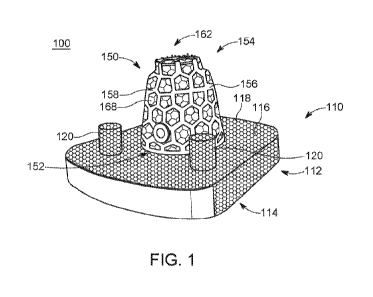
least one first lateral
dimension is a plurality of lateral dimensions, wherein each of the plurality
of lateral
dimensions are greater than the at least one second lateral dimension.
[0013] According to the first aspect of the present disclosure, the at
least one first lateral
dimension includes a plurality of lateral dimensions, wherein the plurality of
lateral
dimensions decrease in magnitude from the bottom surface of the bottom portion
toward a
top surface of the bottom portion.
[0014] According to the first aspect of the present disclosure, the
augmentation
component includes an interior volume configured to receive a protrusion
disposed on a
surface of the tibial component.
[0015] According to the first aspect of the present disclosure, the
instrument is a reaming
instrument configured to ream a volume that corresponds to a volume of the
augmentation
component.
[0016] According to the first aspect of the present disclosure, the
instrument includes a
handle and an engagement component. The engagement component is coupled with
the
2
CA 03226752 2024-01-15
WO 2023/004279 PCT/US2022/073829
handle and includes a lower engagement portion having a plurality of first
engagement
features disposed on at least a portion of at least a lateral surface and a
top surface of the
lower engagement portion, and an upper engagement portion positioned superior
to the lower
engagement portion that includes a plurality of second engagement features
disposed on at
least a portion of at least a lateral surface and a top surface of the upper
engagement portion.
[0017] According to the first aspect of the present disclosure, the
plurality of first
engagement features and the plurality of second engagement features protrude
from the
surfaces of the lower engagement portion and the upper engagement portion.
[0018] A second aspect of the present disclosure is an augmentation
component for a
tibial component. The augmentation component includes a top portion with at
least one
lateral dimension, and a bottom portion with a plurality of lateral dimensions
that decrease in
magnitude from a bottom surface of the bottom portion to a top surface of the
bottom portion.
[0019] According to the second aspect of the present disclosure, the top
portion is
positioned superior to and is integral with the bottom component.
[0020] According to the second aspect of the present disclosure,
augmentation component
also includes a central volume extending from the bottom surface of the bottom
portion into
the augmentation component, the central volume having an interior surface.
[0021] According to the second aspect of the present disclosure, the
augmentation
component also includes a bore extending from an exterior surface of the
augmentation
component through to the interior surface of the augmentation component and
providing fluid
communication from the central volume to the exterior surface of the
augmentation
component.
[0022] According to the second aspect of the present disclosure, the
augmentation
component also includes a first porous structure having a first porosity and a
second porous
structure having a second porosity, wherein the first porous structure is
positioned proud
relative to the second porous structure.
[0023] According to the second aspect of the present disclosure, the first
porosity is
greater than the second porosity.
[0024] According to the second aspect of the present disclosure, the second
engagement
element includes a retention portion comprising a post and a threading,
wherein the post is
configured to releasably couple with the second arm and the threading is
configured to
releasably couple with the actuator.
3
CA 03226752 2024-01-15
WO 2023/004279 PCT/US2022/073829
[0025] A third aspect of the present disclosure is a method of augmenting a
tibial
component. The method includes collecting imaging data from a patient,
identifying from
the imaging date a void in a distal portion of a tibia of the patient,
obtaining an augmentation
component with a volume that corresponds to a volume of the void of the
patient, coupling
the augmentation component with a tibial component, and implanting the tibial
component
such that the augmentation component occupies at least a portion of the void
of the patient.
BRIEF DESCRIPTION OF THE DRAWINGS
[0026] The accompanying drawings, which are incorporated in and constitute
a part of
the specification, illustrate embodiments of the inventions and together with
the detailed
description herein, serve to explain the principles of the inventions. It is
emphasized that, in
accordance with the standard practice in the industry, various features may or
may not be
drawn to scale. In fact, the dimensions of the various features may be
arbitrarily increased or
reduced for clarity of discussion. The drawings are only for purposes of
illustrating
embodiments of inventions of the disclosure and are not to be construed as
limiting the
inventions.
[0027] FIG. 1 is a front-right perspective view of an exemplary augmented
system for
ankle arthroplasty procedures, in accordance with the present disclosure;
[0028] FIG. 2 is a front view of the exemplary augmented system for ankle
arthroplasty
procedures of FIG. 1, in accordance with the present disclosure;
[0029] FIG. 3 is front view of a portion of the exemplary augmented system
for ankle
arthroplasty of FIG. 1, in accordance with the present disclosure;
[0030] FIG. 4 is a bottom view of the portion of FIG. 3 of the exemplary
augmented
system for ankle arthroplasty of FIG. 1, in accordance with the present
disclosure;
[0031] FIG. 5A is a rear-left perspective view of an instrument to be
implemented in
conjunction with the exemplary system for performing ankle arthroplasty of
FIG. 1, in
accordance with the present disclosure;
[0032] FIG. 5B is side cross-sectional view of an accessory for the
instrument of FIG. 5A
to be implemented in conjunction with the exemplary system for performing
ankle
arthroplasty of FIG. 1, in accordance with the present disclosure;
[0033] FIG. 6 is a top view of the instrument of FIG. 5A to be implemented
in
conjunction with the exemplary system of FIG. 1 for implementation in
performing a surgical
procedure, in accordance with the present disclosure;
4
CA 03226752 2024-01-15
WO 2023/004279 PCT/US2022/073829
[0034] FIG. 7 is a front-right perspective view of the instrument of FIG.
5A to be
implemented in conjunction with the exemplary system of FIG. 1 for
implementation in
performing a surgical procedure, in accordance with the present disclosure;
[0035] FIG. 8 is a top view of a portion of the instrument of FIG. 5A to be
implemented
in conjunction with the exemplary system of FIG. 1 for implementation in
performing a
surgical procedure, in accordance with the present disclosure;
[0036] FIG. 9 is a front view of a portion of the instrument of FIG. 5A to
be
implemented in conjunction with the exemplary system of FIG. 1 for
implementation in
performing a surgical procedure, in accordance with the present disclosure;
[0037] FIG. 10 is a front perspective view of an exemplary augmented system
for ankle
arthroplasty procedures, in accordance with the present disclosure;
[0038] FIG. 11 is an elevated front-left view of the exemplary augmented
system of FIG.
10, in accordance with the present disclosure;
[0039] FIG. 12 is front exploded view of the exemplary augmented system of
FIG. 10, in
accordance with the present disclosure;
[0040] FIG. 13 is top view of the exemplary augmented system of FIG. 10, in
accordance
with the present disclosure;
[0041] FIG. 14 is a front perspective view of a portion of the exemplary
augmented
system of FIG. 10, in accordance with the present disclosure;
[0042] FIG. 15 is atop view of a portion of the exemplary augmented system
of FIG. 10,
in accordance with the present disclosure;
[0043] FIG. 16 is a front view of the anatomy of the distal tibia and
fibula prior to
placing a tibial component of a total ankle arthroplasty system, in accordance
with the present
disclosure;
[0044] FIG. 17 is a front view of the anatomy of FIG. 15 shown adjacent an
exemplary
instrument for implementation with the augmented system of FIG. 10, in
accordance with the
present disclosure;
[0045] FIG. 18 is a front view of the anatomy of FIG. 15 after
implementation of the
exemplary instrument of FIG. 17, in accordance with the present disclosure;
[0046] FIG. 19 is a front view of the anatomy of FIG. 15 after
implementation of the
exemplary instrument of FIG. 17 and adjacent the augmented system of FIG. 10,
in
accordance with the present disclosure;
CA 03226752 2024-01-15
WO 2023/004279 PCT/US2022/073829
[0047] FIG. 20 is a front view of the anatomy of FIG. 15 after
implementation of the
exemplary instrument of FIG. 17 and the implantation of the augmented system
of FIG. 10, in
accordance with the present disclosure;
[0048] FIG. 21 is a front-left perspective view of an implant system for a
lower
extremity, in accordance with the present disclosure;
[0049] FIG. 22 is an alternate front-left perspective view of the implant
system of FIG.
21 to be implemented in conjunction with the exemplary system of FIG. 1 for
implementation
in performing a surgical procedure, in accordance with the present disclosure;
[0050] FIG. 23 is a front view of a portion of the implant system of FIG.
1, in accordance
with the present disclosure;
[0051] FIG. 24 is a side view of the portion of FIG. 23 of the implant
system of FIG. 21
in accordance with the present disclosure;
[0052] FIG. 25 is a front-left perspective view of the portion of FIG. 23
of the implant
system of FIG. 21, in accordance with the present disclosure;
[0053] FIG. 26 is an alternate front-left perspective view of the portion
of FIG. 23 of the
implant system of FIG. 21 implanted in the distal tibia, in accordance with
the present
disclosure;
[0054] FIG. 27 is a front view of an instrument which may be implemented in
conjunction with the implant system of FIG. 21, in accordance with the present
disclosure;
[0055] FIG. 28 is a front-left perspective view of the instrument of FIG.
27 which may
be implemented in conjunction with the implant system of FIG. 21, in
accordance with the
present disclosure;
[0056] FIG. 29 is a top view of an instrument which may be implemented in
conjunction
with the implant system of FIG. 21, in accordance with the present disclosure;
[0057] FIG. 30 is a front-left perspective view of the instrument of FIG.
29 which may
be implemented in conjunction with the implant system of FIG. 21, in
accordance with the
present disclosure;
[0058] FIG. 31 is a side view of an instrument which may be implemented in
conjunction
with the implant system of FIG. 21, in accordance with the present disclosure;
[0059] FIG. 32 is a front view of the instrument of FIG. 31 which may be
implemented
in conjunction with the implant system of FIG. 21, in accordance with the
present disclosure;
[0060] FIG. 33 is a top view of the instrument of FIG. 31 which may be
implemented in
conjunction with the implant system of FIG. 21, in accordance with the present
disclosure;
6
CA 03226752 2024-01-15
WO 2023/004279 PCT/US2022/073829
[0061] FIG. 34 is a side view of the instrument of FIG. 31 implemented in
conjunction
with other instruments which may be implemented in conjunction with the
implant system of
FIG. 21, in accordance with the present disclosure;
[0062] FIG. 35 is a side view of an instrument which may be implemented in
conjunction
with the implant system of FIG. 21, in accordance with the present disclosure;
[0063] FIG. 36 is a top view of the instrument of FIG. 35 which may be
implemented in
conjunction with the implant system of FIG. 21, in accordance with the present
disclosure;
[0064] FIG. 37 is a front-right perspective view of the instrument of FIG.
35 which may
be implemented in conjunction with the implant system of FIG. 21, in
accordance with the
present disclosure;
[0065] FIG. 38 is a front-left perspective view of the instruments of FIGS.
29 and 35
implemented in conjunction with one another, which may also be implemented in
conjunction with the implant system of claim 21, in accordance with the
present disclosure;
[0066] FIG. 39 is a front view of the implant system of FIG. 21, in
accordance with the
present disclosure;
[0067] FIG. 40 is a rear view of the implant system of FIG. 21, in
accordance with the
present disclosure;
[0068] FIG. 41 is a side view of the implant system of FIG. 21, in
accordance with the
present disclosure,
[0069] FIG. 42 is perspective view of the portion of the exemplary
augmented system of
FIG. 3 uncoupled from a tibial component, in accordance with the present
disclosure;
[0070] FIG. 43 is a perspective view of the portion of the exemplary
augmented system
of FIG. 3 being coupled to the tibial component of FIG. 42, in accordance with
the present
disclosure;
[0071] FIG. 44 is a top view of the assembly process of the augmented
system of FIG. 1,
in accordance with the present disclosure;
[0072] FIG. 45 is a top view of the coupling process for the augmented
system of FIG. 1,
in accordance with the present disclosure;
[0073] FIG. 46 is a side view of the assembled augmented system of FIG. 1,
in
accordance with the present disclosure; and
[0074] FIG. 47 is a front view of the assembled augmented system of FIG. 1
implanted
into a distal tibia portion, in accordance with the present disclosure.
7
CA 03226752 2024-01-15
WO 2023/004279 PCT/US2022/073829
DETAILED DESCRIPTION FOR CARRYING OUT THE INVENTION
[0075] In this detailed description and the following claims, the words
proximal, distal,
anterior or plantar, posterior or dorsal, medial, lateral, superior and
inferior are defined by
their standard usage for indicating a particular part or portion of a bone or
implant according
to the relative disposition of the natural bone or directional terms of
reference. For example,
"proximal" means the portion of a device or implant nearest the torso, while
"distal" indicates
the portion of the device or implant farthest from the torso. As for
directional terms,
"anterior" is a direction towards the front side of the body, "posterior"
means a direction
towards the back side of the body, "medial" means towards the midline of the
body, "lateral"
is a direction towards the sides or away from the midline of the body,
"superior" means a
direction above and "inferior" means a direction below another object or
structure. Further,
specifically in regards to the foot, the term "dorsal" refers to the top of
the foot and the term
"plantar" refers the bottom of the foot.
[0076] Similarly, positions or directions may be used herein with reference
to anatomical
structures or surfaces. For example, as the current implants, devices,
instrumentation, and
methods are described herein with reference to use with the bones of the foot,
the bones of
the foot, ankle and lower leg may be used to describe the surfaces, positions,
directions or
orientations of the implants, devices, instrumentation and methods. Further,
the implants,
devices, instrumentation, and methods, and the aspects, components, features
and the like
thereof, disclosed herein are described with respect to one side of the body
for brevity
purposes. However, as the human body is relatively symmetrical or mirrored
about a line of
symmetry (midline), it is hereby expressly contemplated that the implants,
devices,
instrumentation, and methods, and the aspects, components, features and the
like thereof,
described and/or illustrated herein may be changed, varied, modified,
reconfigured or
otherwise altered for use or association with another side of the body for a
same or similar
purpose without departing from the spirit and scope of the invention. For
example, the
implants, devices, instrumentation, and methods, and the aspects, components,
features and
the like thereof, described herein with respect to the right foot may be
mirrored so that they
likewise function with the left foot. Further, the implants, devices,
instrumentation, and
methods, and the aspects, components, features and the like thereof, disclosed
herein are
described with respect to the foot for brevity purposes, but it should be
understood that the
implants, devices, instrumentation, and methods may be used with other bones
of the body
having similar structures.
8
CA 03226752 2024-01-15
WO 2023/004279 PCT/US2022/073829
[0077] Surgical procedures are commonly performed to address acute or
chronic
conditions of a patient that can be addressed by placing an implant (e.g.,
plate, screw,
component of an implant system, etc.) adjacent (e.g., coupled with, abutting,
etc.). These
procedures can include arthroplasty procedures (e.g., joint replacement) such
as an ankle
arthroplasty procedure which is used herein as an exemplary arthroplasty
procedure and an
exemplary application of various implants, systems, augmentation components,
and
methodologies. Accordingly, the application of an ankle arthroplasty procedure
should not
be considered limiting but rather exemplary as the contents of this disclosure
may be applied
to alternate implants, implant systems, and procedures/applications.
[0078] Ankle arthroplasty implants commonly include at least tibial and
talar
components which couple with the distal tibia and proximal talus, typically
after at least some
resection of said bones has occurred. For the sake of this disclosure, the
tibial component of
ankle arthroplasty systems will be discussed and contemplated relative to the
systems,
components, and methods disclosed herein. However, it should be noted that
said systems,
components, and methods may also be applicable to the talar component of
various ankle
arthroplasty systems (or other components of arthroplasty systems, ankle or
otherwise).
[0079] The tibial component of ankle arthroplasty systems (including that
which is shown
and described subsequently herein) is typically impacted into a recess formed
in the distal
tibia after at least a portion of the distal tibia has been resected. In some
procedures and for
some patients, the distal tibia is healthy and accommodates this resection and
impaction of
the tibial component with sufficient amounts of the distal tibia to provide
bone purchase and
ingrowth, thus retaining the tibial component within the desired position in
the distal tibia.
However, some patients exhibit one or more of various irregularities in the
distal tibia, some
of which may be identified using preoperative imaging (e.g., CT, Mill, etc.)
while others may
become apparent intraoperatively. These irregularities may include cysts,
voids, and other
volumes within the distal tibia that prevent the tibial component from
achieving proper
integration with the distal tibia, as there is not a sufficient amount of
healthy distal tibia to
provide the necessary bone ingrowth surfaces and bone purchase to retain the
tibial
component. Standard tibial components are typically configured for placement
in a healthy
distal tibia that is absent any irregularities. However, when irregularities
become evident (or
other anatomical challenges, such as ankle arthroplasty revision procedures
standard tibial
components fail to accommodate the anatomy of the distal tibia of patients.
[0080] Accordingly, it is desirable to tailor a standard tibial component
to patients with
irregularities in the distal tibia. In some instances, custom implants can
address irregularities
9
CA 03226752 2024-01-15
WO 2023/004279 PCT/US2022/073829
but can be prohibitively expensive and carry lengthy lead times. Additionally,
custom
implants cannot account for irregularities that are identified
intraoperatively. In order to
adapt standard tibial components for various irregularities in the distal
tibia, an augment is
required that may couple with the superior (top) surface of the tibial
component and occupy
at least a portion of such irregularities in the distal tibia, thus allowing
the standard tibial
component to occupy the resected volume and contact appropriate surfaces of
the distal tibia.
Such an augment would aid in fixation of the tibial component by providing
fixation (via
bone ingrowth surfaces, bone purchase, etc.) within said irregularities of the
distal tibia and
accordingly adapt standard tibial components for patients presenting distal
tibial
irregularities.
[0081] Referring to the drawings, wherein like reference numerals are used
to indicate
like or analogous components throughout the several views, and with particular
reference to
FIGS. 1-9, there is illustrated an exemplary embodiment of an implant system
100 which may
be a portion of a larger implant system (for example, an arthroplasty system).
As shown, the
implant system 100 is a tibial component system which is a portion of a total
ankle
arthroplasty system, where the implant system 100 is coupled with the distal
tibia after a
portion of the distal tibia has been resected. The implant system 100 is shown
to include a
tibial component 110 having a base portion 112 as well as bottom and top
surfaces 114 and
116, respectively, where the top surface 114 is arranged opposite the base
portion 112 from
the bottom surface 116. The tibial component 110 is shown to include a texture
118 on the
outer surface of the base portion 112 as well as projections 120 extending
upward from the
top surface 116 of the base portion 112. In some embodiments, the tibial
component 110
may include an alternate number of projections 120, for example one
protrusion, three
protrusions, etc. As shown, the projections 120 are configured to facilitate
coupling with the
distal tibia after resection as occurred, with said projections 120 extending
into corresponding
openings in the tibia created by a physician in the process of preparing the
distal tibia for the
tibial component 110. The texture 118 may be configured to facilitate bone
ingrowth and/or
otherwise retain the tibial component in the distal tibia. It should be noted
that the tibial
component 110 is a standard tibial component (e.g., has no custom components,
is sold off
the shelf, etc.) and is shown here for exemplary purposes.
[0082] The tibial component 110 further includes a stem 122 (as shown in
FIG. 12)
arranged in a central portion of the top surface 116, with the stem 122
extending
perpendicularly upward from the top surface 116 (similar to the projections
120). The stem
122 is configured to extend upward (e.g., proximally, in a superior direction)
into the distal
CA 03226752 2024-01-15
WO 2023/004279 PCT/US2022/073829
tibia when placed in a similar fashion to the projections 122, where one or
more instruments
creates a volume in the distal tibia of which the stem 122 occupies at least a
portion. The
stem 122 as shown includes a substantially cylindrical geometry, where a
bottom end of the
stem is integral with the top surface 116 (as are the projections 120) and a
top end of the stem
122 is open. Accordingly, the stem 122 includes a central opening 126 (e.g., a
volume with
the bottom of said volume defined by the top surface 116) with a complimentary
cylindrical
geometry to that of the stem 122. The stem 122 is further shown to include
apertures 124
arranged about the lateral surface of the stem 122. As shown, each of the
apertures 124 are
substantially the same size, but in some aspects one or more of said apertures
124 may be of
an alternate size (e.g., to accommodate another component, etc.). Similarly,
the apertures 124
are all of a substantially circular geometry but may have alternate geometries
in some
embodiments. The stem 122 also includes the texture 118 on all surfaces
thereof, according
to the exemplary embodiments shown and described herein.
[0083] An augment 150 is shown to be coupled with the top surface 116 of
the tibial
component 110. With reference to FIG. 2, the augment 150 includes a central
opening 166
that is sized to receive at least a portion of the stem 122 therein when the
augment 150 is
coupled with the tibial component. In some aspects, the augment 150 may be
coupled with
the tibial component via an adhesive or cement, or may implement other
coupling methods
(e.g., fasteners, etc.). As shown, the tibial component 110 includes a single
augment 150, but
in some embodiments multiple augments 150 of various geometries and/or sizes
may be
coupled with the tibial component 110. The augment 150 is shown to have a
height greater
than that of the stem 122 such that when implanted in the distal tibia of a
patient, the augment
150 extends further upward (in a proximal, superior direction) than the stem
122 or the
projections 120. The augment 150 may be coupled with the tibial component
(e.g.,
positioned on the top surface 116) so as to provide a volume configured to
occupy at least a
portion of a complimentary volume of an irregularity in the distal tibia of
the patient.
Further, in some aspects the augment 150 may be configured to occupy the
entirety of the
volume of an irregularity or may be sized so as to have a volume greater than
said irregularity
(e.g., to facilitate a desired fit). Furthermore, and as shown and described
subsequently
herein, the augment 150 may include one or more complimentary instruments
configured to
facilitate implantation of the tibial component 110 with the augment 150
(e.g., a reamer to
ream an irregularity into a volume that will receive the augment 150, etc.).
In some aspects,
the augment may be of a custom size based on an irregularity identified and
measured via
imaging data. Further, in some aspects, the augment 150 may be selected by a
physician
11
CA 03226752 2024-01-15
WO 2023/004279 PCT/US2022/073829
from a library of augments 150 (and/or augment trials) provided in a surgical
kit, where each
augment 150 has a different size/shape/geometry so as to address various
irregularities in the
distal tibia of the patient. It should be noted that an appropriate version of
the augment 150
for an irregularity (or potentially multiple irregularities) may require
manipulation of the
geometry of the irregularity in order for the augment 150 to fit as desired.
[0084] As shown, the augment 150 has a bottom portion 152 and a top portion
154. The
geometry of the bottom portion 152 of the augment 150 is that of a frustum of
a cone shape
(e.g., where a frustum is a portion of a solid that lies between two parallel
lines cutting said
solid), and the geometry of the top portion 154 is that of a cylinder with a
diameter greater
than the height. The cylindrical geometry of the top portion 154, as described
subsequently
herein, corresponds to a complimentary cylindrical volume formed in the distal
portion of the
tibia prior to implantation. Such cylindrical geometry (both of the top
portion 154 and said
corresponding volume) is configured to facilitate coupling of the augment 150
(and other
components of the system as shown and described) with the distal tibia and
retention thereof
It should be noted that in some aspects the geometry of the augment 150 may be
variable
(e.g., the bottom portion 152 may have a lesser height/volume/surface area
than that of the
top portion 154, the bottom portion 152 and top portion 154 may each have
alternate
geometries, etc.).
[0085] The augment 150 further includes an exterior surface 156, a bottom
surface 160, a
top surface 162, and an interior surface 164. The exterior surface 156 extends
between the
bottom surface 160 and the top surface 162. As shown, the bottom surface 160
is coupled
with the top surface 116 of the tibial component 110. The top surface 162 is
arranged
opposite the augment 150 from the bottom surface and is an upper surface of
the top portion
154 of the augment 150. The interior surface 164, as shown in FIG. 4, defines
the volume of
the central opening 166 of the augment 150. The augment 150 also has a pair of
porous
structures (e.g., lattice structures, web structures, nodal structures, mesh
structures, etc.)
including a first porous structure 158 and a second porous structure 168. As
shown, the first
porous structure 158 is arranged proud relative to the second porous structure
168, where the
first porous structure 158 defines an outer surface of the augment 150 for all
of the
aforementioned surfaces. The first porous structure 158 is shown to have a
greater porosity
(e.g., more porous) than that of the second porous structure 168. As shown,
the first porous
structure is a substantially randomized structure (while still satisfying
various design inputs
and structural parameters) whereas the second porous structure has a grid-
shaped geometry
(e.g., with substantially orthogonal components). It should be understood that
the first and
12
CA 03226752 2024-01-15
WO 2023/004279 PCT/US2022/073829
second porous structures 158, 168 as shown and described herein are exemplary
and may be
modified for various embodiments of the augment 150. For example, a particular
irregularity
in the distal tibia of a patient may be conducive to one or more porous
structures with greater
or lesser porosities than those shown in the exemplary augment 150.
Ultimately, the augment
150 may include an alternate number of porous structures or alternate porous
structures, or an
alternate arrangement of two or more porous structures.
[0086] The augment 150 includes an exterior opening 170 disposed on the
exterior
surface 156 thereof as well as an interior opening 174 disposed on the
interior surface 164
thereof The exterior opening 170 and the interior opening 174 define a bore
172 extending
from the exterior surface 156 to the interior surface 164 and thus
establishing fluid
communication between said surfaces (and between the exterior of the augment
150 and the
central opening 166). In some aspects, the augment 150 may include the bore
172 to provide
optionality for a physician to provide additional fixation when coupling the
augment 150 with
the tibial component 110. For example, in addition to implementing an
adhesive/cement to
couple the bottom surface 160 of the augment 150 with the top surface 116 of
the tibial
component the physician may implement a fastener (e.g., screw, etc.). Such a
fastener may
be inserted through the exterior opening 170, into and through the bore 172
(e.g., the bore
receives the fastener), and exit the interior opening 174. The fastener, and
thus the bore 172,
may be aligned with one or more of the apertures 124 of the stem 122 such that
one or more
of the apertures 124 receive at least a portion of the fastener thus providing
additional
coupling between the augment 150 and the tibial component 110.
[0087] Referring now to FIGS. 5A-9, an instrument 200 is shown (and with
reference to
FIG. 5, relative to an exemplary tibia 302 and fibula 310 of a patient). The
instrument 200 is
shown to include a handle 202 having a first end 204 and a second end 208
which are
disposed at opposite ends of the handle 202 as shown in the exemplary
embodiment of FIGS.
5-9. The first end 204 of the handle 202 includes an opening 206 extending
through the
handle 202. The handle 200 also includes an engagement portion 210 arranged at
(or
adjacent to) the second end 208 of the handle 200, where the engagement
portion 210 is
coupled with the second end 208. In some aspects, the engagement portion 210
may be
coupled with the second end 208 through a variety of means (e.g., pivotably
coupled,
rotatably coupled, etc.). Further, in some aspects the engagement portion 210
may be at least
partially integral with the second end 208 of the handle 200.
[0088] Referring now to FIG. 5B, an accessory 250 is shown that may be
coupled with
the opening 206 disposed at the second end 208 of the handle 202. The
accessory 250 is
13
CA 03226752 2024-01-15
WO 2023/004279 PCT/US2022/073829
shown to include a stem 252 configured to facilitate rotatable and/or
pivotable coupling with
the handle 202 via the opening 206. The stem 252 may include one or more
geometries
and/or dimensions, where at least one of said geometries is a shape
complimentary to that of
the opening 206. For example, as shown the stem 252 include a substantially
cylindrical
portion having a lateral dimension lesser than that of the opening 206 (e.g.,
a diameter) such
that said cylindrical portion may be at least partially received by and/or
disposed within the
opening 206. Further, the stem 252 is shown to include geometries at opposite
ends of the
cylindrical portion with a lateral dimension greater than that of the opening
206 so as to retain
at least a portion of the cylindrical portion within the opening 206 (and thus
retain the
accessory 150 in a coupled state with the handle 202). The accessory 250 is
further shown to
include a knob 254, where the knob 254 is rotatably and/or pivotably coupled
with the stem
252. As shown, the knob 254 includes a central opening configured to engage
one of the
aforementioned geometries of the stem 252 that has a first lateral dimension
greater than that
of the cylindrical portion of the stem 252. The central opening of the knob
254 is also shown
to have a second lateral dimension where said lateral dimension is greater
than that of the
cylindrical portion of the stem 252, but lesser than that of the end geometry
of the stem 252
that is greater than that of the cylindrical portion (e.g., said second
opening of the knob 254
may have a lateral dimension the same as or similar to that of the opening
206). Accordingly,
the knob 252 may be rotated about the stem 252 independent of any movement to
the handle
202 of the instrument 200 and, as described subsequently herein, facilitate
other manipulation
of the instrument 200.
[0089] The engagement portion 210 is shown to include a first portion 212
and second
portion 222 (e.g., bottom and top portions, respectively) where the first
portion 212 and the
second portion 222 are integral with one another. The first portion 212
includes a bottom
surface 214 and a top surface 218, as well as a lateral surface 216 extending
between the
bottom surface 214 and the top surface 218. The lateral surface 216 includes a
plurality of
first engagement features 220 extending (e.g., projecting, protruding, etc.)
from the lateral
surface 216 at oblique and/or orthogonal angles relative to the lateral
surface 216. Further, as
shown the first engagement features 220 are disposed substantially equidistant
one another
and extend vertically along the lateral surface 216 from the bottom surface
214 to the top
surface 218 of the first portion 212. In some aspects, the first engagement
features 220 may
extend vertically beyond the lateral surface 216 (e.g., past the bottom
surface 214 and the top
surface 218). Each of the first engagement features 220 as shown includes a
ridge protruding
from the lateral surface 216 of the first portion 212, with said ridges each
angled in a specific
14
CA 03226752 2024-01-15
WO 2023/004279 PCT/US2022/073829
direction (e.g., as shown in FIG. 6, the first engagement features 220 are
angled in a
substantially counterclockwise direction relative to the engagement portion
210). The first
portion 212 is configured to have a geometry similar to that of the bottom
portion 152 of the
augment 150 such that implementation of the instrument 200 (and the first
portion 212) may
ream or otherwise configured a space in the distal tibia 302 of the patient to
accommodate at
least a portion of the augment 150. Accordingly, the geometry of the first
portion 212 of the
engagement portion 210 has the general geometry of the frustum of a cone as
defined
previously herein with reference to the augment.
[0090] The second portion 222 of engagement portion 210 includes a bottom
surface 224
and a top surface 228, as well as a lateral surface 226 extending between the
bottom surface
224 and the top surface 228. As shown, the second portion 222 is arranged such
that the
bottom surface 224 abuts (e.g., is integral with) the top surface 220 of the
first portion 212.
The second portion 222 is shown to have a substantially cylindrical geometry
(similar to that
of the top portion 154 of the augment 150), with the diameter of the cylinder
greater than the
height (although all geometric properties of the instrument 200 may vary based
on the
geometry of the augment 150). Said geometry of the second portion 222 is
configured to
ream a volume in the distal tibia complimentary to the geometry of the top
portion 164 of the
augment 150 (e.g., to ream a cylindrical volume). The second portion 222 is
shown to
include a plurality of second engagement features 230, which may be arranged
the same
and/or similar to those of the first portion 212. As shown, the second
engagement features
230 extend laterally (e.g., project, protrude, etc.) from the lateral surface
226 and further
extend vertically along at least the height of the lateral surface 226. As
shown, the second
engagement features 230 extend from the bottom surface 224 of the second
portion 222
vertically and terminate just beyond the top surface 228 such that that top
surface 228
includes the second engagement features 230. It should be understood that the
augment 150
may be configured such that it may be 3-D printed.
[0091] Referring to FIG. 5, the instrument 200 is shown to be engaged with
the
exemplary tibia 302 of a patient. The engagement portion 210 has been placed
such that the
top surface 228 contacts a distal surface of a recess 304 created in the
distal tibia 302 (e.g., by
resection). In the instance of a revision procedure, the engagement portion
210 may be
placed in a portion of the distal tibia 302 which previously accommodated the
stem 122 of the
tibial component 110. In such an instance, the geometry of the engagement
portion 210
allows for the engagement portion 210 to self-center when reaming a volume to
accommodate the augment 150. That is to say that the geometry of the
engagement portion
CA 03226752 2024-01-15
WO 2023/004279 PCT/US2022/073829
210 of the instrument 200 is configured to ream a volume centered about (and
in this
example, extending concentrically therefrom) the recess in the distal tibia
that previously
received the stem 122 (or other protrusions of other tibial components). In
procedures that do
not offer such a recess from a previous tibial component, the engagement
portion is
configured to center about a point identified by the physician and ultimately
ream a volume
with a geometry complimentary to that of the augment 150 such that said volume
will receive
the augment 150 (or at least a portion thereof) when implanted after coupling
of the augment
150 with the tibial component 110. The instrument 200 as shown, is a manual
use instrument
where a physician may manipulate the instrument 200 by the handle 202 about a
semi-
circular path 232 in a reciprocating nature to ream the desired volume in the
distal tibia 302.
Movement about the path 232 may be driven by a physician manipulating the
instrument 200
via the accessory 250 as shown in FIG. 5B. For example, the instrument 200 may
be
manipulated in the semi-circular path 232 by grasping the knob 254, where the
knob rotates
to facilitate such manipulation and movement of the instrument 200. In some
aspects, the
instrument 200 and/or portions thereof may be modified so as to couple with
various other
instruments (e.g., power tools/reamers). The instrument 200 may also be
configured/designed such that it may be 3-D printed, thus eliminating the
machining process
common to many instruments.
[0092] Referring now to FIGS. 11-20, alternate embodiments of an augment
450 and an
instrument 500 are shown. The augment 450 may have one or more properties the
same as
and/or similar to the augment 150 and, similarly, the instrument 500 may have
one or more
properties the same as the instrument 200. As shown in FIGS. 11-20, the
augment 450 is
shown relative to the tibial component system 100 and components thereof that
are the same
as those shown and described previously herein. For the sake of brevity, these
components
will not be described again. However, it should be understood that similar to
the augment
150 and instrument 200, the augment 450 and the instrument 500 may be
implemented in
conjunction with various implant systems (tibial and otherwise) and the tibial
component
system 100 is shown herein as an exemplary tibial component. It should also be
understood
that the applications for the augment 450 and instrument 500 include at least
those shown and
described previously here (e.g., to address one or more irregularities in the
anatomy of a
patient, whether preoperatively through custom design and/or intraoperatively
through a
trialing process).
[0093] The augment 450 is shown to be coupled with the top surface 116 of
the tibial
component 110. With reference to FIG. 15, the augment 450 includes a central
opening 466
16
CA 03226752 2024-01-15
WO 2023/004279 PCT/US2022/073829
that is sized to receive at least a portion of the stem 122 therein when the
augment 450 is
coupled with the tibial component. In some aspects, the augment 450 may be
coupled with
the tibial component via an adhesive or cement, or may implement other
coupling methods
(e.g., fasteners, etc.). As shown, the tibial component 110 includes a single
augment 450, but
in some embodiments multiple augments 450 of various geometries and/or sizes
may be
coupled with the tibial component 110. The augment 450 is shown to have a
height greater
than that of the stem 122 such that when implanted in the distal tibia of a
patient, the augment
450 extends further upward (in a proximal, superior direction) than the stem
122 or the
projections 120. The augment 450 may be coupled with the tibial component
(e.g.,
positioned on the top surface 116) so as to provide a volume configured to
occupy at least a
portion of a complimentary volume of an irregularity in the distal tibia of
the patient.
Further, in some aspects the augment 450 may be configured to occupy the
entirety of the
volume of an irregularity or may be sized so as to have a volume greater than
said irregularity
(e.g., to facilitate a desired fit). Furthermore, and as shown and described
subsequently
herein, the augment 450 may include one or more complimentary instruments
configured to
facilitate implantation of the tibial component 110 with the augment 450
(e.g., a reamer to
ream an irregularity into a volume that will receive the augment 450, etc.).
In some aspects,
the augment 450 may be of a custom size based on an irregularity identified
and measured via
imaging data. Further, in some aspects, the augment 450 may be selected by a
physician
from a library of augments 450 (and/or augment trials) provided in a surgical
kit, where each
augment 450 has a different size/shape/geometry so as to address various
irregularities in the
distal tibia of the patient. It should be noted that an appropriate version of
the augment 450
for an irregularity (or potentially multiple irregularities) may require
manipulation of the
geometry of the irregularity in order for the augment 450 to fit as desired.
[0094] As shown, the augment 450 has a body 452, with the geometry of the
body 452 of
the augment 450 is that of a frustum of a cone shape (e.g., where a frustum is
a portion of a
solid that lies between two parallel lines cutting said solid. It should be
noted that in some
aspects the geometry of the augment 450 may be variable (e.g., the body 452
may have a
diameter greater or less than the height of the body 452, the body 452 may
have alternate
geometries, etc.). The augment 450 further includes an exterior surface 456, a
bottom surface
460, a top surface 462, and an interior surface 464. The exterior surface 456
extends between
the bottom surface 460 and the top surface 462. As shown, the bottom surface
460 is coupled
with the top surface 116 of the tibial component 110. The top surface 462 is
arranged
opposite the augment 450 from the bottom surface and is an upper surface of
the body 454 of
17
CA 03226752 2024-01-15
WO 2023/004279 PCT/US2022/073829
the augment 450. The interior surface 464, as shown in FIG. 15, defines the
volume of the
central opening 466 of the augment 450. The augment 450 also has a pair of
porous
structures (e.g., lattice structures, web structures, nodal structures, mesh
structures, etc.)
including a first porous structure 458 and a second porous structure 468. As
shown, the first
porous structure 458 is arranged proud (e.g., is the outer structure relative
to an inner
structure) relative to the second porous structure 468, where the first porous
structure 458
defines an outer surface of the augment 450 for all of the aforementioned
surfaces. The first
porous structure 458 is shown to have a greater porosity (e.g., more porous)
than that of the
second porous structure 468. As shown, the first porous structure 458 is a
substantially
randomized structure (while still satisfying various design inputs and
structural parameters)
whereas the second porous structure has a grid-shaped geometry (e.g., with
substantially
orthogonal components). It should be understood that the first and second
porous structures
458, 468 as shown and described herein are exemplary and may be modified for
various
embodiments of the augment 450. For example, a particular irregularity in the
distal tibia of
a patient may be conducive to one or more porous structures with greater or
lesser porosities
than those shown in the exemplary augment 450. Ultimately, the augment 450 may
include
an alternate number of porous structures or alternate porous structures, or an
alternate
arrangement of two or more porous structures.
[0095] The augment 450 includes an exterior opening 470 disposed on the
exterior
surface 456 thereof as well as an interior opening 474 disposed on the
interior surface 464
thereof The exterior opening 470 and the interior opening 474 define a bore
472 extending
from the exterior surface 456 to the interior surface 464 and thus
establishing fluid
communication between said surfaces (and between the exterior of the augment
450 and the
central opening 466). In some aspects, the augment 450 may include the bore
472 to provide
optionality for a physician to provide additional fixation when coupling the
augment 450 with
the tibial component 110. For example, in addition to implementing an
adhesive/cement to
couple the bottom surface 460 of the augment 450 with the top surface 116 of
the tibial
component the physician may implement a fastener 476 (e.g., screw, etc.). The
fastener 476
may be inserted through the exterior opening 470, into and through the bore
472 (e.g., the
bore receives the fastener), and exit the interior opening 474. The fastener
476, and thus the
bore 472, may be aligned with one or more of the apertures 124 of the stem 122
such that one
or more of the apertures 124 receive at least a portion of the fastener 476
thus providing
additional coupling between the augment 450 and the tibial component 110.
18
CA 03226752 2024-01-15
WO 2023/004279 PCT/US2022/073829
[0096] Referring now to FIGS. 16-20, the instrument 500 is shown relative
to the
exemplary tibia 302 and fibula 310 of a patient. The instrument 500 is shown
to include a
handle 502 having a first end 504 and a second end 508 which are disposed at
opposite ends
of the handle 502 as shown in the exemplary embodiment of FIG. 17. The handle
500
includes an engagement portion 510 arranged at (or adjacent to) the second end
508 of the
handle 500, where the engagement portion 510 is coupled with the second end
508. In some
aspects, the engagement portion 510 may be coupled with the second end 508
through a
variety of means (e.g., pivotably coupled, rotatably coupled, etc.). Further,
in some aspects
the engagement portion 510 may be at least partially integral with the second
end 508 of the
handle 500.
[0097] The engagement portion 510 is shown to include a body 512. The body
512
includes a bottom surface 514 and a top surface 518, as well as a lateral
surface 516
extending between the bottom surface 514 and the top surface 518. The lateral
surface 516
includes a plurality of engagement features 520 extending (e.g., projecting,
protruding, etc.)
from the lateral surface 516 at oblique and/or orthogonal angles relative to
the lateral surface
516. Further, as shown the engagement features 520 are disposed substantially
equidistant
one another and extend vertically along the lateral surface 516 from the
bottom surface 514 to
the top surface 518 of the body 512. In some aspects, the engagement features
520 may
extend vertically beyond the lateral surface 516 (e.g., past the bottom
surface 514 and the top
surface 518). Each of the engagement features 520 as shown includes a ridge
protruding
from the lateral surface 516 of the body 512, with said ridges each angled in
a specific
direction (e.g., similar to that shown with reference to the first engagement
features 220 of
the instrument 200 as shown in FIG. 6, the engagement features 520 are angled
in a
substantially counterclockwise direction relative to the engagement portion
510). The body
512 is configured to have a geometry similar to that of the body 452 of the
augment 450 such
that implementation of the instrument 500 (and the body 512) may ream or
otherwise
configure a bore 306 within a recess 308 (from resection to accommodate the
tibial
component 110) in the distal tibia 302 of the patient to accommodate at least
a portion of the
augment 450 (as shown with reference to FIGS. 18-20). Accordingly, the
geometry of the
body 512 of the engagement portion 510 has the general geometry of the frustum
of a cone as
defined previously herein with reference to the augment. It should be
understood that the
augment 150 may be configured such that it may be 3-D printed.
[0098] Referring to FIG. 17, the instrument 500 is shown just prior to
engagement with
the exemplary tibia 302 of a patient. The engagement portion 510 has been
placed such that
19
CA 03226752 2024-01-15
WO 2023/004279
PCT/US2022/073829
the top surface 518 will contact a distal surface of the recess 304 created in
the distal tibia
302 (e.g., by resection). In the instance of a revision procedure, the
engagement portion 510
may be placed in a portion of the distal tibia 302 which previously
accommodated the stem
122 of the tibial component 110. In such an instance, the geometry of the
engagement
portion 510 allows for the engagement portion 510 to self-center when reaming
a volume to
accommodate the augment 450. That is to say that the geometry of the
engagement portion
510 of the instrument 500 is configured to ream a volume centered about (and
in this
example, extending concentrically therefrom) the recess in the distal tibia
that previously
received the stem 122 (or other protrusions of other tibial components). In
procedures that do
not offer such a recess from a previous tibial component, the engagement
portion is
configured to center about a point identified by the physician and ultimately
ream a volume
with a geometry complimentary to that of the augment 450 such that said volume
will receive
the augment 450 (or at least a portion thereof) when implanted after coupling
of the augment
450 with the tibial component 110. The instrument 500 as shown is a manual use
instrument
where a physician may manipulate the instrument 500 by the handle 502 about a
semi-
circular path 532 in a reciprocating nature to ream the desired volume in the
distal tibia 302.
In some aspects, the instrument 500 and/or portions thereof may be modified so
as to couple
with various other instruments (e.g., power tools/reamers). The instrument 500
may also be
configured/designed such that it may be 3-D printed, thus eliminating the
machining process
common to many instruments.
[0099]
Referring now to FIGS. 21-26, an implant system 600 is shown, according to an
exemplary embodiment of the present disclosure. The implant system 600 may
include at
least one of a talar component 602, an intermediate component 604, and/or a
tibial
component 610. In some aspects, the talar component 602 may be a talar spacer
or other
partial or total talar replacement implant, which may include one or more
surfaces configured
to interface with other bones/joints of the foot (e.g., articulate with and/or
facilitate fusion
with) and/or interface with (e.g., articulate with) other implant components
such as the
intermediate component 604. In some aspects, the talar component 602 may be
configured to
facilitate fusion with a calcaneus 312 of a patient, and may also be
configured to interface
with components of other implant systems (e.g., arthroplasty systems).
Further, in some
aspects the tibial component 610 may be the same as and/or similar to a tibial
component of
an ankle arthroplasty system configured to interface with the intermediate
component 604
and thus provide compatibility with various talar components including the
talar component
602 and/or talar components configured to couple with at least a portion of a
native talus of a
CA 03226752 2024-01-15
WO 2023/004279 PCT/US2022/073829
patient. It should be understood that the implant system 600 may be
implemented as shown
in FIGS. 21-26, or may be implemented in alternate configurations including
various other
components with cross-system compatibility, including but not limited to,
those shown and
described previously herein.
[0100] The tibial component 610 is shown to include a base portion 612 as
well as
bottom and top surfaces 614 and 616, respectively, where the top surface 614
is arranged
opposite the base portion 612 from the bottom surface 616. The tibial
component 610 is
shown to include a texture 618 on the outer surface of the base portion 612
(including at least
the top surface 614). The texture 118 may be configured to facilitate bone
ingrowth and/or
otherwise retain the tibial component 610 in the distal tibia. It should be
noted that the tibial
component 610 may be a standard tibial component (e.g., has no custom
components, is sold
off the shelf, etc.). The tibial component 610 is further shown to include a
recess 620
disposed on the bottom surface 616 of the base portion 612 and, when shown in
the position
of FIG. 23, extends upward into the base portion 612 from the bottom surface
616. The
recess 620 may include one or more engagement features 622 disposed on at
least one surface
therein, wherein said one or more engagement features 622 are configured to
facilitate
coupling (e.g., releasable, slidable, etc.) with another implant component. As
shown, the
engagement features 622 are configured to releasably couple with the
intermediate
component 604 where the engagement features 622 are configured as
protrusions/grooves
extending along at least a portion of the length of the recess 620 and
configured to engage
with complimentary protrusions/grooves of the intermediate component 604. In
some
aspects, the one or more engagement feature 622 may include alternate
configurations and/or
positioning within the recess 622. For example, the one or more engagement
feature 622
may include a dovetail configuration or other various interface/engagement
configurations. It
should be understood that the tibial component 610 may be releasably couplable
with various
intermediate components, including but not limited to, the intermediate
component 604,
which is to say that the one or more engagement features 622 (and/or other
features of the
tibial component 604) may be configured to interface with various other
implant components
and/or implant systems.
[0101] An augment 650 is shown to be coupled with the top surface 616 of
the tibial
component 610. As shown, the augment is integral with the base portion 612,
although in
some aspects the augment may include one or more features (e.g., a central
opening, etc.)
configured to facilitate coupling with the base portion 612. As shown, the
tibial component
610 includes a single augment 650, but in some embodiments multiple augments
650 of
21
CA 03226752 2024-01-15
WO 2023/004279 PCT/US2022/073829
various geometries and/or sizes may be coupled with the tibial component 610.
The augment
650 may be configured so as to provide a volume configured to occupy at least
a portion of a
complimentary volume of an irregularity in the distal tibia of the patient. As
shown in FIGS.
23-26, the augment 650 is shown to be positioned off-center on the top surface
614 of the
tibial component 610. In some aspects, the augment 650 may be selectively
positioned about
the tibial component 610 so as to address specific anatomy of a patient (e.g.,
a void in the
distal tibia, a preference of a physician to bias the base portion 612 more
anterior, posterior,
medial, or lateral from a traditional, centered configuration, etc.). Further,
in some aspects
the augment 650 may be positioned so as to be received by an intramedullary
canal of a tibia
302 of a patient, with the base portion 612 of the tibial component 610
positioned based on
the position of the augment 650 relative to the base portion 612. Further, in
some aspects the
augment 650 may be configured to occupy the entirety of the volume of an
irregularity or
may be sized so as to have a volume greater than the irregularity (e.g., to
facilitate a desired
fit). Furthermore, and as shown and described subsequently herein, the augment
650 may
include one or more complimentary instruments configured to facilitate
implantation of the
tibial component 610 with the augment 650 (e.g., a reamer to ream an
irregularity into a
volume that will receive the augment 650, etc.). In some aspects, the augment
may be of a
custom size based on an irregularity identified and measured via imaging data.
Further, in
some aspects, the augment 650 may be selected by a physician from a library of
augments
650 (and/or augment trials) provided in a surgical kit, where each augment 650
has a different
size/shape/geometry so as to address various irregularities in the distal
tibia of the patient. It
should be noted that an appropriate version of the augment 650 for an
irregularity (or
potentially multiple irregularities) may require manipulation of the geometry
of the
irregularity in order for the augment 650 to fit as desired.
[0102] As shown in FIGS. 23-26, the augment 650 has a bottom portion 652
and a top
portion 654. The geometry of the bottom portion 652 of the augment 650 is that
of a frustum
of a cone shape (e.g., where a frustum is a portion of a solid that lies
between two parallel
lines cutting said solid), and the geometry of the top portion 654 is that of
a cylinder with a
diameter greater than the height. The cylindrical geometry of the top portion
654, as
described subsequently herein, corresponds to a complimentary cylindrical
volume formed in
the distal portion of the tibia prior to implantation. Such cylindrical
geometry (both of the
top portion 654 and said corresponding volume) is configured to facilitate
coupling of the
augment 650 (and other components of the system as shown and described) with
the distal
tibia and retention thereof. It should be noted that in some aspects the
geometry of the
22
CA 03226752 2024-01-15
WO 2023/004279 PCT/US2022/073829
augment 650 may be variable (e.g., the bottom portion 652 may have a lesser
height/volume/surface area than that of the top portion 654, the bottom
portion 652 and top
portion 654 may each have alternate geometries, etc.).
[0103] The augment 650 further includes an exterior surface 656, a bottom
surface 660,
and a top surface 662. The exterior surface 656 extends between the bottom
surface 660 and
the top surface 662. As shown, the bottom surface 660 is integral with the top
surface 616 of
the tibial component 610. The top surface 662 is arranged opposite the augment
650 from the
bottom surface and is an upper surface of the top portion 654 of the augment
650. In some
aspects, the augment 650 may include an interior surface defining a volume of
a central
opening of the augment 650, although in other aspects the central portion of
the augment 650
may be solid. The augment 650 also has a pair of porous structures (e.g.,
lattice structures,
web structures, nodal structures, mesh structures, etc.) including a first
porous structure 658
and a second porous structure 668. As shown, the first porous structure 658 is
arranged
proud relative to the second porous structure 668, where the first porous
structure 658 defines
an outer surface of the augment 650 for all of the aforementioned surfaces.
The first porous
structure 658 is shown to have a greater porosity (e.g., more porous) than
that of the second
porous structure 668. As shown, the first porous structure is a substantially
randomized
structure (while still satisfying various design inputs and structural
parameters) whereas the
second porous structure has a grid-shaped geometry (e.g., with substantially
orthogonal
components). It should be understood that the first and second porous
structures 658, 668 as
shown and described herein, are exemplary and may be modified for various
embodiments of
the augment 650. For example, a particular irregularity in the distal tibia of
a patient may be
conducive to one or more porous structures with greater or lesser porosities
than those shown
in the exemplary augment 650. Ultimately, the augment 650 may include an
alternate
number of porous structures or alternate porous structures, or an alternate
arrangement of two
or more porous structures.
[0104] Referring now to FIGS. 27-28, an instrument 710 is shown, according
to an
exemplary embodiment of the present disclosure. The instrument 710 as shown
may be a
guide or other instrument configured to facilitate and/or guide drill holes
and/or cuts to the
distal tibia 304 of a patient in order to prepare the distal tibia 304 of a
patient for implantation
of at least a portion of the system 600 (e.g., the tibial component 610). In
some aspects, the
instrument 710 may be color-coded in order to indicate a specific step in a
procedure, or may
be one of multiple instruments 710 of various sizes included in a surgical kit
where color
coding indicates size of the instrument 710. The instrument 710 may also be a
patient
23
CA 03226752 2024-01-15
WO 2023/004279 PCT/US2022/073829
specific instrument/guide generated (e.g., 3-D printed, etc.) specifically to
fit an anatomy of a
patient based on imaging data and/or modeling. The instrument 710 as shown
includes a
vertical protrusion 712 including at least one opening (as shown, two
openings) configured to
guide a drill or other instrument and, in some aspects, placement of pins or
guidewires. The
instrument 710 further includes at least one opening in a central portion 714
thereof similarly
configured to guide a drill for the placement of pins and/or wires. A
protrusion 716 is shown
to extend from the central portion 714 in a direction substantially opposite
that the protrusion
712, where the protrusion 716 may be configured to facilitate manipulation of
the instrument
710 and/or guide various other actions by a physician. In some aspects, the
instrument 710
may be releasably couplable with a portion of the anatomy of a patient (e.g.,
via pins, slidably
coupled, etc.).
[0105] Referring now to FIGS. 29-30, an instrument 720 is shown, according
to an
exemplary embodiment of the present disclosure. Similar to the instrument 710,
the
instrument 720 may be a guide or other instrument configured to facilitate
and/or guide drill
holes and/or cuts to the distal tibia 304 of a patient in order to prepare the
distal tibia 304 of a
patient for implantation of at least a portion of the system 600 (e.g., the
tibial component
610). Further, the instrument 720 may be configured to releasably couple with
at least a
portion of the anatomy of the patient. In some aspects, the instrument 720 may
be color-
coded in order to indicate a specific step in a procedure, or may be one of
multiple
instruments 720 of various sizes included in a surgical kit where color coding
indicates size
of the instrument 720. The instrument 720 may also be a patient specific
instrument/guide
generated (e.g., 3-D printed, etc.) specifically to fit an anatomy of a
patient based on imaging
data and/or modeling. The instrument 720 includes a base portion 722 which
includes an
opening 724 positioned substantially centrally relative to the base portion
722. In some
aspects, the opening 724 may be positioned relative to a portion of the
anatomy of the patient
to which a specific operation may be performed. For example, the instrument
720 may be
generated with the opening 724 in a specific position such that, when
releasably coupled with
a patient, the opening 724 aligns with a portion of the anatomy (e.g., an
intramedullary canal
of the distal tibia) and a physician may insert at least a portion of a reamer
within the opening
724 so as to ensure reaming of the desired portion of the anatomy. In some
aspects, the
instrument 720 may be releasably couplable with a portion of the anatomy of a
patient (e.g.,
via pins, slidably coupled, etc.).
[0106] Referring now to FIGS. 31-34, an instrument 730 is shown, according
to an
exemplary embodiment of the present disclosure. Similar to the instruments
710, 720, the
24
CA 03226752 2024-01-15
WO 2023/004279 PCT/US2022/073829
instrument 730 may be a guide or other instrument configured to facilitate
and/or guide drill
holes and/or cuts to the distal tibia 304 of a patient in order to prepare the
distal tibia 304 of a
patient for implantation of at least a portion of the system 600 (e.g., the
tibial component
610). Further, the instrument 730 may be configured to releasably couple with
at least a
portion of the anatomy of the patient. In some aspects, the instrument 730 may
be color-
coded in order to indicate a specific step in a procedure, or may be one of
multiple
instruments 730 of various sizes included in a surgical kit where color coding
indicates size
of the instrument 730. The instrument 730 may also be a patient specific
instrument/guide
generated (e.g., 3-D printed, etc.) specifically to fit an anatomy of a
patient based on imaging
data and/or modeling. The instrument 730 includes a base portion 732 which
includes an
opening 738 positioned substantially centrally relative to the base portion
732 (for example,
in a central portion of the base portion 732 which may or may not be
"centered" relative to
the instrument 730). In some aspects, the opening 738 may be positioned on the
instrument
730 relative to a portion of the anatomy of the patient to which a specific
operation may be
performed. For example, the instrument 730 may be generated with the opening
738 in a
specific position such that, when releasably coupled with a patient, the
opening 738 aligns
with a portion of the anatomy (e.g., an intramedullary canal of the distal
tibia) and a
physician may insert at least a portion of a reamer within the opening 738 so
as to ensure
reaming of the desired portion of the anatomy. The instrument 730 is also
shown to include a
protrusion 734 extending from the base portion 732 in a substantially
orthogonal direction
and from an end portion of the base portion 732. The protrusion 734 is shown
to include at
least one opening 736 (as shown, two openings) configured to guide drill holes
and/or
placement of wires/pins by a physician.
[0107] As shown in FIG. 34, the instrument 730 may be positioned on the
distal tibia 304
of a patient such that the at least one opening 736 is disposed on an anterior
portion thereof.
Further, the instrument 730 may also be implemented in conjunction with other
instruments,
for example the instrument 750 which may be configured to retain the
instrument 730 and/or
one or more portions of the anatomy of a patient in a desired location during
a procedure. In
some aspects, the instrument 730 may be releasably couplable with a portion of
the anatomy
of a patient (e.g., via pins, slidably coupled, etc.).
[0108] Referring now to FIGS. 34-38, an instrument 740 is shown, according
to an
exemplary embodiment of the present disclosure. The instrument 740 is shown
relative to the
distal tibia 302 and fibula 310 of a patient. The instrument 740 is shown to
include a handle
742 and a head 744 positioned at an end of the handle 742. As shown, the
handle 742 has a
CA 03226752 2024-01-15
WO 2023/004279 PCT/US2022/073829
substantially tapered configuration with a wider lateral dimension at the end
thereof opposite
the head 744. The instrument 740 and portions thereof may be the same as
and/or similar to
the instrument 500 shown and described herein previously and, as such, may
also be
implemented in the same and/or similar fashion to that shown in at least FIG.
17 herein and
will not be repeated for brevity sake. The instruments 720, 730 may be
configured to have
one or more geometries that accommodate one or more features of the instrument
740 and/or
other similar instruments (e.g., as shown in FIG. 38). For example, the
openings 724, 738
may be configured to receive at least a portion of the head 744 of the
instrument 740 therein
so as to guide reaming of the distal tibia 304 by a physician. In some
aspects, the instrument
740 may be configured to a specific anatomy of a patient (e.g., the head 744
may be taller,
have a greater lateral dimension, etc.) to accommodate said anatomy.
[0109] Referring now to FIGS. 39-41, the implant system 600 is shown in an
implanted
state adjacent the tibia, fibula, and calcaneus 304, 310, 312 of a patient,
according to an
exemplary embodiment of the present disclosure. The tibial component 610 is
shown
coupled with the distal tibia 304 of a patient and, while not shown, the
augment 650 is
disposed at least partially within the tibia 304 (e.g., in the intramedullary
canal). The
intermediate component 604 is shown to be releasably coupled with the tibial
component 610
(via the one or more engagement features 622, although not shown here) such
that the
intermediate component 604 is retained in a position substantially
inferior/plantar relative to
the tibial component 610. The talar component 602 is shown to be positioned
inferior/plantar
relative to the intermediate component 604 such that an upper surface of the
talar component
602 may interface with a lower surface of the intermediate component. Further,
a pair of
trajectories 690 are shown extending through at least a portion of the talar
component 602
and through the calcaneus 312. The pair of trajectories 690 corresponds to
trajectories of
fixation elements (e.g., fasteners, screws, compression screws, lag screws,
etc.) configured to
facilitate placement of the talar component 604 with the calcaneus 312 (e.g.,
arthrodesis,
arthroeresis, etc.). In some aspects, the subtalar joint between the talar
component 602 and
the calcaneus 312 may be fused in performing a procedure that includes
implanting one or
more components of the implant system 600, although this may not be included
in all such
procedures.
[0110] It should be understood that the implant systems 100, 600 as well as
any
components thereof and/or instruments shown and described herein should be
considered
interchangeable and cross-compatible. That is to say that the instrument 730
may be
implemented in conjunction with the instrument 500 and the implant system 100,
or the
26
CA 03226752 2024-01-15
WO 2023/004279 PCT/US2022/073829
implant system 600 may be implemented in conjunction with the instrument 500
and the
instrument 710. Further, it should be understood that any of the components
shown and
described herein may be configured specific to an anatomy of a patient. For
example, the
augment 650 may have height and lateral dimension corresponding to dimensions
of a tibial
void or the intramedullary canal of a patient, or the head 744 of the
instrument 700 may be
sized according to similar anatomical dimensions of a patient.
[0111] FIGS. 42-47 show the implant system 100 being assembled by a
physician prior to
implantation. FIG. 42, shows a physician preparing to couple the augment 150
to the tibial
component. FIG. 43 shows the physician connecting the augment 150 to the
tibial component
110. FIG. 44 shows the assembled implant system 100 being prepared for
implantation into
the distal tibia 302 of the patient. FIG. 45 shows the physician using a tool
to secure the
augment 150 to the tibial component 110. FIG. 46 shows the implant system 100
adjacent
the distal tibia 302 prior to implantation. FIG. 47 shows the implant system
100 implanted
into the distal tibia 302.
[0112] It is understood by one skilled in the art that the implant system
100, although
shown as a two piece construct could be one piece manufactured out of titanium
or another
biocompatible metal material. The implant system 100, either as a one piece
construct or a
two piece construct (i.e., tibial component and augment) may be manufactured
using a 3-D
printer or some additive manufacturing process or machined out of bulk metal
bars. Although
the implant system 100 as described herein is directed towards an ankle
replacement implant,
it is contemplated that the augment element could be used with other joint
implants to
stabilize the implant post-operatively. For example, the augment 150 could be
using in a total
shoulder arthroplasty system, an acetabulum cup, either the femoral or tibial
component of a
total knee and virtually all other prosthetic joint devices that are inserted
within a bone of a
patient.
[0113] Although the implant system as explained in relation to its
preferred embodiment,
it is to be understood that many other possible modifications and variations
can be made
without departing from the spirit and scope of the invention.
[0114] The terminology used herein is for the purpose of describing
particular
embodiments only and is not intended to be limiting of the invention. As used
herein, the
singular forms "a", "an" and "the" are intended to include the plural forms as
well, unless the
context clearly indicates otherwise. It will be further understood that the
terms "comprise"
(and any form of comprise, such as "comprises" and "comprising"), "have" (and
any form of
have, such as "has", and "having"), "include" (and any form of include, such
as "includes"
27
CA 03226752 2024-01-15
WO 2023/004279 PCT/US2022/073829
and "including"), and "contain" (and any form of contain, such as "contains"
and
"containing") are open-ended linking verbs. As a result, a method or device
that
"comprises," "has," "includes," or "contains" one or more steps or elements
possesses those
one or more steps or elements, but is not limited to possessing only those one
or more steps
or elements. Likewise, a step of a method or an element of a device that
"comprises," "has,"
"includes," or "contains" one or more features possesses those one or more
features, but is
not limited to possessing only those one or more features. Furthermore, a
device or structure
that is configured in a certain way is configured in at least that way, but
may also be
configured in ways that are not listed.
[0115] The invention has been described with reference to the preferred
embodiments. It
will be understood that the architectural and operational embodiments
described herein are
exemplary of a plurality of possible arrangements to provide the same general
features,
characteristics, and general system operation. Modifications and alterations
will occur to
others upon a reading and understanding of the preceding detailed description.
It is intended
that the invention be construed as including all such modifications and
alterations.
28