Note: Descriptions are shown in the official language in which they were submitted.
CA 03227148 2024-01-19
WO 2023/004077 PCT/US2022/037931
COMPOSITIONS AND METHODS FOR DETECTION OF COLORECTAL CANCER
CROSS-REFERENCE TO RELATED APPLICATIONS
[1] This application claims the benefit of U.S. Provisional Application No.
63/224,378 filed July 21, 2021, the content of which is hereby incorporated
herein in its
entirety.
BACKGROUND
[2] Early detection of cancer greatly increases the chance of successful
treatment.
However, many cancers including colorectal cancer still lack either effective
screening
recommendations or patient compliance with such recommendations. Typical
challenges for
cancer-screening tests include limited sensitivity and specificity. A high
rate of false-positive
results can be of particular concern, as it can create difficult management
decisions for
clinicians and patients who would not want to unnecessarily administer (or
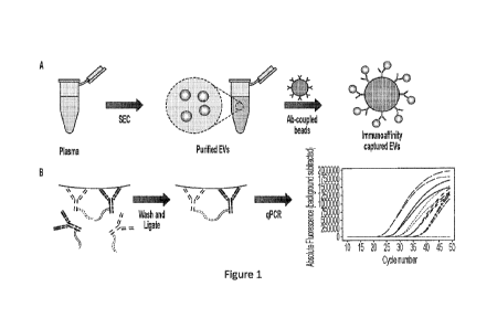
receive) anti-
cancer therapy that may potentially have undesirable side effects. Conversely,
a high rate of
false-negative results fails to satisfy the purpose of the screening test, as
patients who need
therapy are missed, resulting in a treatment delay and consequently a reduced
possibility of
success.
SUMMARY
[3] The present disclosure, among other things, provides insights and
technologies for achieving effective colorectal cancer screening from a
biological sample. In
some embodiments, such a biological sample is or comprises a bodily fluid-
derived sample,
e.g., in some embodiments a blood-derived sample. In some embodiments, the
present
disclosure, among other things, provides insights and technologies that are
particularly useful
for colorectal adenocarcinoma screening. In some embodiments, provided
technologies are
effective for detection of early-stage colorectal cancer (e.g., colorectal
adenocarcinoma). In
some embodiments, provided technologies are effective even when applied to
populations
comprising or consisting of asymptomatic individuals (e.g., due to
sufficiently high
sensitivity and/or low rates of false positive and/or false negative results).
In some
embodiments, provided technologies are effective when applied to populations
comprising or
CA 03227148 2024-01-19
WO 2023/004077 PCT/US2022/037931
consisting of individuals (e.g., asymptomatic individuals) without hereditary
risk in
developing colorectal cancer (e.g., colorectal adenocarcinoma). In some
embodiments,
provided technologies are effective when applied to populations comprising or
consisting of
symptomatic individuals (e.g., individuals suffering from one or more symptoms
of
colorectal cancer). In some embodiments, provided technologies are effective
when applied
to populations comprising or consisting of individuals at risk for colorectal
cancer (e.g.,
individuals with hereditary and/or life-history associated risk factors for
colorectal cancer). In
some embodiments, provided technologies may be or include one or more
compositions (e.g.,
molecular entities or complexes, systems, cells, collections, combinations,
kits, etc.) and/or
methods (e.g., of making, using, assessing, etc.), as will be clear to one
skilled in the art
reading the disclosure provided herein.
[4] In some embodiments, the present disclosure identifies the source
of a
problem with certain prior technologies including, for example, certain
conventional
approaches to detection and diagnosis of colorectal cancer. For example, the
present
disclosure appreciates that many conventional diagnostic assays, e.g.,
colonoscopies, stool
test, CT scanning, and/or molecular tests based on cell-free nucleic acids,
serum biomarkers,
and/or bulk analysis of extracellular vesicles, can be time-consuming, costly,
and/or lacking
sensitivity and/or specificity sufficient to provide a reliable and
comprehensive diagnostic
assessment. In some embodiments, the present disclosure provides technologies
(including
systems, compositions, and methods) that solve such problems, among other
things, by
detecting co-localization of a target biomarker signature of colorectal cancer
in individual
extracellular vesicles, which comprises at least one extracellular vesicle-
associated surface
biomarker and at least one target biomarker selected from the group consisting
of surface
biomarkers, internal biomarkers, and RNA biomarkers. In some embodiments, the
present
disclosure provides technologies (including systems, compositions, and
methods) that solve
such problems, among other things, by detecting such target biomarker
signature of
colorectal cancer using a target entity detection approach that was developed
by Applicant
and described in U.S. Application No. 16/805,637 (published as US2020/0299780;
issued as
US11,085,089), and International Application PCT/US2020/020529 (published as
W02020180741), both filed February 28, 2020 and entitled "Systems,
Compositions, and
Methods for Target Entity Detection," which are based on interaction and/or co-
localization
CA 03227148 2024-01-19
WO 2023/004077 PCT/US2022/037931
3
of at least two or more target entities (e.g., a target biomarker signature)
in individual
extracellular vesicles.
[5] In some embodiments, extracellular vesicles for detection as described
herein
can be isolated from a bodily fluid of a subject by a size exclusion-based
method. As will be
understood by a skilled artisan, in some embodiments, a size exclusion-based
method may
provide a sample comprising nanoparticles having a size range of interest that
includes
extracellular vesicles. Accordingly, in some embodiments, provided
technologies of the
present disclosure encompass detection, in individual nanoparticles having a
size range of
interest (e.g., in some embodiments about 30 nm to about 1000 nm) that
includes
extracellular vesicles, of co-localization of at least two or more surface
biomarkers (e.g., as
described herein) that forms a target biomarker signature of colorectal
cancer. A skilled
artisan reading the present disclosure will understand that various
embodiments described
herein in the context of "extracellular vesicle(s)" can be also applicable in
the context of
"nanoparticles" as described herein.
[6] In some embodiments, the present disclosure, among other things,
provides
insights that screening of asymptotic individuals, e.g., regular screening
prior to or otherwise
in absence of developed symptom(s), can be beneficial, and even important for
effective
management (e.g., successful treatment) of colorectal cancer (e.g., in some
embodiments
colorectal adenocarcinoma. In some embodiments, the present disclosure
provides colorectal
cancer screening systems that can be implemented to detect colorectal cancer
(e.g., in some
embodiments colorectal adenocarcinoma), including early-stage cancer, in some
embodiments in asymptomatic individuals. In some embodiments, provided
technologies are
implemented to achieve regular screening of asymptomatic individuals. The
present
disclosure provides, for example, compositions (e.g., reagents, kits,
components, etc.), and
methods of providing and/or using them, including strategies that involve
regular testing of
one or more individuals (e.g., symptomatic or asymptomatic individuals). The
present
disclosure defines usefulness of such systems, and provides compositions and
methods for
implementing them.
[7] In some embodiments, provided technologies achieve detection (e.g.,
early
detection, e.g., in asymptomatic individual(s) and/or population(s)) of one or
more features
(e.g., incidence, progression, responsiveness to therapy, recurrence, etc.) of
colorectal cancer,
CA 03227148 2024-01-19
WO 2023/004077
PCT/US2022/037931
4
with sensitivity and/or specificity (e.g., rate of false positive and/or false
negative results)
appropriate to permit useful application of provided technologies to single-
time and/or
regular (e.g., periodic) assessment. In some embodiments, provided
technologies are useful
in conjunction with regular medical examinations, such as but not limited to:
physicals,
general practitioner visits, cholesterol/lipid blood tests, diabetes (type 2)
screening, blood
pressure screening, thyroid function tests, prostate cancer screening,
mammograms,
HPV/Pap smears, colorectal cancer screening, and/or vaccinations. In some
embodiments,
provided technologies are useful in conjunction with treatment regimen(s); in
some
embodiments, provided technologies may improve one or more characteristics
(e.g., rate of
success according to an accepted parameter) of such treatment regimen(s).
[8] In some
aspects, provided are technologies for use in classifying a subject
(e.g., an asymptomatic subject) as having or being susceptible to colorectal
cancer (e.g., in
some embodiments colorectal adenocarcinoma). In some embodiments, the present
disclosure provides methods or assays for classifying a subject (e.g., an
asymptomatic
subject) as having or being susceptible to colorectal cancer (e.g., in some
embodiments
colorectal adenocarcinoma). In some embodiments, a provided method or assay
comprises
(a) detecting, in a bodily fluid-derived sample (e.g., but not limited to a
blood-derived
sample, a fecal-derived sample, etc.) from a subject in need thereof,
extracellular vesicles
expressing a target biomarker signature of colorectal cancer (e.g., in some
embodiments
colorectal adenocarcinoma), the target biomarker signature comprising: at
least one
extracellular vesicle-associated surface biomarker and at least one target
biomarker selected
from the group consisting of: surface biomarkers (as described herein),
intravesicular
biomarkers (as described herein), and intravesicular RNA biomarkers (as
described herein);
(b) comparing sample information indicative of level of the target biomarker
signature-
expressing extracellular vesicles in the bodily fluid-derived sample (e.g.,
but not limited to a
blood-derived sample, a fecal-derived sample, etc.) to reference information
including a
reference threshold level; and (c) classifying the subject as having or being
susceptible to
colorectal cancer (e.g., in some embodiments colorectal adenocarcinoma) when
the bodily
fluid-derived sample (e.g., but not limited to a blood-derived sample, a fecal-
derived sample,
etc.) shows an elevated level of target biomarker signature-expressing
extracellular vesicles
relative to a classification cutoff referencing the reference threshold level.
CA 03227148 2024-01-19
WO 2023/004077 PCT/US2022/037931
[9] In some embodiments, one or more surface biomarkers that can be
included in
a target biomarker signature are selected from (i) polypeptides encoded by
human genes as
follows: ACSL5, ACVR2B, ALDH18A1, ALG5, AP1M2, ATP1B1, B3GNT3, BCAP31, CASK,
CD] 33, CDH1, CDH17, CDH3, CEACAM5, CEACAM6, CFB, CFTR, CHDH, CHMP4B,
CISD2, CLIC1, COPG2, CYP2S1, DPEP1, DSG2, EDAR, EPCAM, EPHB2, EPHB3,
ERMP1, FERMT1, GALNT3, GNPNAT1, GOLIM4, GPA33, GPCR5A, HACD3, HEPH,
HKDC1, IHH, ILDR1, ITGA2, KCNQ1, KEL, KPNA2, LAD], LAMC2, LBR, LMNB1,
LMNB2, LSR, MAP7, MARCKSL1, MLEC, MUC1, MUC13, NCEH1, NDUFS6, NLN, NOX1,
NUP210, OCIAD2, PGAM5, PIGR, PIGT, PTK7, RAB25, RAP2A, RAP2B, RCC2, RNF43,
RPN1, RPN2, RPS3, RUVBL2, SlOOP, SLC12A2, SLC25A6, SLC2A1, 5MIM22, SNTB1,
SORD, 55R4, ST14, STOML2, STT3B, SYAP1, TM9SF2, TMED2, TMPO, TOMM22,
TOMM34, AMHR2, CLDN1, DLL4, EGFR, ERBB2, FAP, FGFR4, FOLR1, GUCY2C,
IGF1R, ILIA, ITGAV, KRT8, LGR5, LPR6, MET, MST1R, MUC5AC, TNFRSF10B,VEGFA,
and combinations thereof; and/or (ii) carbohydrate-dependent markers as
follows: CanAg
(glycoform of MUC1), Lewis Y/B antigen, Lewis B Antigen, Sialyltetraosyl
carbohydrate,
Tn antigen, SialylTn (sTn) antigen, Thomsen-Friedenreich (T, TF) antigen,
Lewis Y antigen
(also known as CD174), Sialyl Lewis X (sLex) antigen (also known as Sialyl
SSEA-1
(SLX)), Sialyl Lewis A antigen (also known as CA19-9), SSEA-1 (also known as
Lewis X
antigen), NeuGcGM3 (N-glycolyl GM3 ganglioside), and combinations thereof.
[10] In some embodiments, one or more surface biomarkers that can be
included in
a target biomarker signature are selected from (i) polypeptides encoded by
human genes as
follows: ACVR2B, B3GNT3, CD133, CDH17, CDH3, CEACAM5, CEACAM6, CFB, CFTR,
CYP2S1, DLL4, EDAR, EPCAM, EPHB2, EPHB3, ERBB2, FAP, GPCR5A, IHH, ILDR1,
ITGAV, KCNQ1, KEL, MARCKSL1, MST1R, MUC1, MUC5AC, NOX1, OCIAD2, RNF43,
5MIM22, and combinations thereof; and/or (ii) carbohydrate-dependent markers
as follows:
Lewis Y antigen (also known as CD174), SialylTn (sTn) antigen, Sialyl Lewis X
(sLex)
antigen (also known as Sialyl SSEA-1 (SLX)), T antigen, Tn antigen, and
combinations
thereof.
[11] In some embodiments, one or more intravesicular biomarkers that can be
included in a target biomarker signature are selected from polypeptides
encoded by human
CA 03227148 2024-01-19
WO 2023/004077 PCT/US2022/037931
6
genes as follows: AGMAT, AGR2, AGR3, ANKS4B, AP1M2, ARSE, ASCL2, BSPRY,
Cl0orf99, Cl 5orf48, Clorf106, C9orf152, CBLC, CCL24, CDCA7, CDX1, CDX2, DDC,
DSG2, EHF, ELF3, EPS8L3, ESRP1, ESRP2, ETV4, EVPL, FABP1, FAM3D, FAM83E,
FAM84A, FERMT1, FOXA2, FOXA3, FOXQ1, GPX2, GRB7, HKDC1, HMGCS2, HNF4A,
HOXB9, KCNN4, KLK1, KRT20, KRT23, KRT8, LGALS4, METTL7B, MISP, MUC2, MYB,
MYBL2, MY01A, PHGR1, PITX1, PKP3, PLAC8, PLEK2, PLS1, PPP1R14D, PRR15,
PTK6, S100A14, SlOOP, SAPCD2, SERPINB5, SPDEF, TRIM'S, TRIM31, USH1C, VIL1 ,
and combinations thereof. In some embodiments, an intravesicular biomarker
described
herein may comprise at least one post-translational modification.
[12] In some embodiments, one or more intravesicular RNAs (e.g., mRNAs)
that
can be included in a target biomarker signature are selected from RNA
transcripts (e.g.,
mRNA transcripts) encoded by human genes as follows: AGMAT, AGR2, AGR3,
ANKS4B,
AN09, AP1M2, ARSE, ASCL2, ATP10B, B3GNT3, BIK, BSPRY, Cl0orf99, Cl5orf48,
Clorf106, Clorf210, C9orf152, CA12, CBLC, CCL24, CD24, CDCA7, CDH1, CDH17,
CDH3, CDHR1, CDHR5, CDX1, CDX2, CEACAM5, CEACAM6, CEACAM7, CFTR,
CLDN2, CLDN3, CLDN4, CLDN7, CLRN3, COL17A1, CRB3, CYP2S1, DDC, DPEP1,
DSG2, EHF, ELF3, EPCAM, EPHB3, EPS8L3, ERN2, ESRP1, ESRP2, ETV4, EVPL, FA2H,
FABP1, FAM3D, FAM83E, FAM84A, FAT], FERMT1, FOXA2, FOXA3, FOXQ1, FUT2,
FUT3, FXYD3, GCNT3, GGT6, GJB1, GJB3, GPA33, GPR160, GPR35, GPX2, GRB7,
GUCY2C, HKDC1, HMGCS2, HNF4A, HOXB9, IHH, ITLN1, KCNN4, KIAA1324, KLK1,
KRT20, KRT23, KRT8, LGALS4, LGR5, LY6G6D, MEP1A, METTL7B, MISP, MUC13,
MUC2, MYB, MYBL2, MY01A, NOX1, PDZKlIP1, PHGR1, PIGR, PITX1, PKP3, PLAC8,
PLEK2, PLS1, POF1B, PPP1R14D, PROM], PRR15, PRSS8, PTK6, RAB25, RNF128,
RNF186, RNF43, S100A14, SlOOP, SAPCD2, SERPINB5, SLC26A3, SLC39A5, SLC44A4,
SLC5A1, SMIM22, SPDEF, ST6GALNAC1, TJP3, TM4SF5, TMC5, TMEM45B, TMPRSS2,
TMPRSS4, TNS4, TRABD2A, TRIM'S, TRIM31, TSPAN1, TSPAN8, UGT2B17, UGT8,
USH1C, VILl, and combinations thereof.
[13] In some embodiments, methods or assays described herein may be
performed
for one more additional target biomarker signature (including, e.g., at least
one, at least two,
at least three, or more additional target biomarker signatures). In some such
embodiments, a
CA 03227148 2024-01-19
WO 2023/004077 PCT/US2022/037931
7
classification cutoff may reference additional reference threshold level(s)
corresponding to
each additional target biomarker signature.
[14] In some embodiments, an extracellular vesicle-associated surface
biomarker
for use in a target biomarker signature of colorectal cancer used and/or
described herein may
be or comprise a tumor-specific biomarker and/or a tissue-specific biomarker
(e.g., a colon
and/or rectum tissue-specific biomarker). In some embodiments, such an
extracellular
vesicle-associated surface biomarker may be or comprise a non-specific marker,
e.g., it is
present in one or more non-target tumors, and/or in one or more non-target
tissues. In some
embodiments, such an extracellular vesicle-associated surface biomarker may be
or comprise
one or more surface proteins encoded by human genes as follows: ACSL5, ACVR2B,
ALDH18A1, ALG5, AP1M2, ATP1B1, B3GNT3, BCAP31, CASK, CD133, CDH1, CDH17,
CDH3, CEACAM5, CEACAM6, CFB, CFTR, CHDH, CHMP4B, CISD2, CLIC1, COPG2,
CYP2S1, DPEP1, DSG2, EDAR, EPCAM, EPHB2, EPHB3, ERMP1, FERMT1, GALNT3,
GNPNAT1, GOLIM4, GPA33, GPCR5A, HACD3, HEPH, HKDC1, IHH, ILDR1, ITGA2,
KCNQ1, KEL, KPNA2, LAD], LAMC2, LBR, LMNB1, LMNB2, LSR, MAP7, MARCKSL1,
MLEC, MUC1, MUC13, NCEH1, NDUFS6, NLN, NOX1, NUP210, OCIAD2, PGAM5,
PIGR, PIGT, PTK7, RAB25, RAP2A, RAP2B, RCC2, RNF43, RPN1, RPN2, RPS3, RUVBL2,
SlOOP, SLC12A2, SLC25A6, SLC2A1, 5MIM22, SNTB1, SORD, 55R4, ST14, STOML2,
STT3B, SYAP1, TM9SF2, TMED2, TMPO, TOMM22, TOMM34, AMHR2, CLDN1, DLL4,
EGFR, ERBB2, FAP, FGFR4, FOLR1, GUCY2C, IGF1R, ILIA, ITGAV, KRT8, LGR5,
LPR6, MET, MST1R, MUC5AC, TNFRSF10B, VEGFA, or any combinations thereof;
and/or
(ii) one or more carbohydrate-dependent markers as follows: CanAg (glycoform
of MUC1),
Lewis Y/B antigen, Lewis B Antigen, Sialyltetraosyl carbohydrate, Tn antigen,
SialylTn
(sTn) antigen, Thomsen-Friedenreich (T, TF) antigen, Lewis Y antigen (also
known as
CD174), Sialyl Lewis X (sLex) antigen (also known as Sialyl SSEA-1 (SLX)),
Sialyl Lewis
A antigen (also known as CA19-9), SSEA-1 (also known as Lewis X antigen),
NeuGcGM3,
and combinations thereof.
[15] In some embodiments, an extracellular vesicle-associated surface
biomarker
may be or comprise one or more of (i) a polypeptide encoded by human gene
MUC/; and/or
one or more of (ii) a carbohydrate-dependent marker as follows: Lewis Y
antigen (also
CA 03227148 2024-01-19
WO 2023/004077 PCT/US2022/037931
8
known as CD174), SialylTn (sTn) antigen, Sialyl Lewis X (sLex) antigen (also
known as
Sialyl SSEA-1 (SLX)), T antigen, Tn antigen, or combinations thereof.
[16] In some embodiments, a target biomarker signature of colorectal cancer
(e.g.,
colorectal adenocarcinoma) may comprise an extracellular vesicle-associated
surface
biomarker (e.g., ones described herein) and at least one (including, e.g., 1,
2, 3, or more)
additional target surface biomarker, which, in some embodiments, may be or
comprise one or
more polypeptides encoded by human genes as follows: ACSL5, ACVR2B, ALDH18A1,
ALG5, AP1M2, ATP1B1, B3GNT3, BCAP31, CASK, CD133, CDH1, CDH17, CDH3,
CEACAM5, CEACAM6, CFB, CFTR, CHDH, CHMP4B, CISD2, CLIC1, COPG2, CYP2S1,
DPEP1, DSG2, EDAR, EPCAM, EPHB2, EPHB3, ERMP1, FERMT1, GALNT3, GNPNAT1,
GOLIM4, GPA33, GPCR5A, HACD3, HEPH, HKDC1, IHH, ILDR1, ITGA2, KCNQ1, KEL,
KPNA2, LAD], LAMC2, LBR, LMNB1, LMNB2, LSR, MAP 7, MARCKSL1, MLEC, MUC1,
MUC13, NCEH1, NDUFS6, NLN, NOX1, NUP210, OCIAD2, PGAM5, PIGR, PIGT, PTK7,
RAB25, RAP2A, RAP2B, RCC2, RNF43, RPN1, RPN2, RPS3, RUVBL2, SlOOP, SLC12A2,
SLC25A6, SLC2A1, 5MIM22, SNTB1, SORD, 55R4, ST14, STOML2, STT3B, SYAP1,
TM9SF2, TMED2, TMPO, TOMM22, TOMM34, AMHR2, CLDN1, DLL4, EGFR, ERBB2,
FAP, FGFR4, FOLR1, GUCY2C, IGF1R, ILIA, ITGAV, KRT8, LGR5, LPR6, MET, MST1R,
MUC5AC, TNFRSF 10B, VEGFA; and/or one or more carbohydate markers as follows:
CanAg (glycoform of MUC1), Lewis Y/B antigen, Lewis B Antigen, Sialyltetraosyl
carbohydrate, Tn antigen, SialylTn (sTn) antigen, Thomsen-Friedenreich (T, TF)
antigen,
Lewis Y antigen (also known as CD174), Sialyl Lewis X (sLex) antigen (also
known as
Sialyl SSEA-1 (SLX)), Sialyl Lewis A antigen (also known as CA19-9), SSEA-1
(also
known as Lewis X antigen), NeuGcGM3, or combinations thereof.
[17] In some embodiments, a target biomarker signature of colorectal cancer
(e.g.,
colorectal adenocarcinoma) may comprise an extracellular vesicle-associated
surface
biomarker (e.g., ones described herein) and at least one (including, e.g., 1,
2, 3, or more)
additional surface biomarker, which are selected from (i) polypeptides encoded
by human
genes as follows: ACVR2B, B3GNT3, CD133, CDH17, CDH3, CEACAM5, CEACAM6,
CFB, CFTR, CYP2S1, DLL4, EDAR, EPCAM, EPHB2, EPHB3, ERBB2, FAP, GPCR5A,
IHH, ILDR1, ITGAV, KCNQ1, KEL, MARCKSL1, MST1R, MUC1, MUC5AC, NOX1,
OCIAD2, RNF43, 5MIM22, and combinations thereof; and/or (ii) carbohydrate-
dependent
CA 03227148 2024-01-19
WO 2023/004077 PCT/US2022/037931
9
markers as follows: Lewis Y antigen (also known as CD174), SialylTn (sTn)
antigen, Sialyl
Lewis X (sLex) antigen (also known as Sialyl SSEA-1 (SLX)), T antigen, Tn
antigen, and
combinations thereof.
[18] In some embodiments, a target biomarker signature of colorectal cancer
(e.g.,
colorectal adenocarcinoma) may comprise an extracellular vesicle-associated
surface
biomarker (e.g., ones described herein) and at least one target intravesicular
RNA biomarker,
which, in some embodiments, may be or comprise at least one RNA transcript
(e.g., mRNA
transcript) encoded by a human gene as follows: AGMAT, AGR2, AGR3, ANKS4B,
AN09,
AP1M2, ARSE, ASCL2, ATP10B, B3GNT3, BIK, BSPRY, Cl0orf99, Cl 5orf48, Clorf106,
Clorf210, C9orf152, CA12, CBLC, CCL24, CD24, CDCA7, CDH1, CDH17, CDH3,
CDHR1, CDHR5, CDX1, CDX2, CEACAM5, CEACAM6, CEACAM7, CFTR, CLDN2,
CLDN3, CLDN4, CLDN7, CLRN3, COL17A1, CRB3, CYP2S1, DDC, DPEP1, DSG2, EHF,
ELF3, EPCAM, EPHB3, EPS8L3, ERN2, ESRP1, ESRP2, ETV4, EVPL, FA2H, FABP1,
FAM3D, FAM83E, FAM84A, FAT], FERMT1, FOXA2, FOXA3, FOXQ1, FUT2, FUT3,
FXYD3, GCNT3, GGT6, GJB1, GJB3, GPA33, GPR160, GPR35, GPX2, GRB7, GUCY2C,
HKDC1, HMGCS2, HNF4A, HOXB9, IHH, ITLN1, KCNN4, KIAA1324, KLK1, KRT20,
KRT23, KRT8, LGALS4, LGR5, LY6G6D, MEP1A, METTL7B, MISP, MUC13, MUC2, MYB,
MYBL2, MY01A, NOX1, PDZKlIP1, PHGR1, PIGR, PITX1, PKP3, PLAC8, PLEK2, PLS1,
POF1B, PPP1R14D, PROM], PRR15, PRSS8, PTK6, RAB25, RNF128, RNF186, RNF43,
S100A14, SlOOP, SAPCD2, SERPINB5, SLC26A3, SLC39A5, SLC44A4, SLC5A1, SMIM22,
SPDEF, ST6GALNAC1, TJP3, TM4SF5, TMC5, TMEM45B, TMPRSS2, TMPRSS4, TNS4,
TRABD2A, TRIM'S, TRIM31, TSPAN1, TSPAN8, UGT2B17, UGT8, USH1C, VIL1 , or
combinations thereof.
[19] In some embodiments, a target biomarker signature of colorectal cancer
may
comprise an extracellular vesicle-associated surface biomarker (e.g., ones
described herein)
and at least one additional target intravesicular biomarker, which, in some
embodiments, may
be or comprise at least one polypeptide encoded by a human gene as follows:
AGMAT,
AGR2, AGR3, ANKS4B, AP1M2, ARSE, ASCL2, BSPRY, Cl0orf99, Cl5orf48, Clorf106,
C9orf152, CBLC, CCL24, CDCA7, CDX1, CDX2, DDC, DSG2, EHF, ELF3, EPS8L3,
ESRP1, ESRP2, ETV4, EVPL, FABP1, FAM3D, FAM83E, FAM84A, FERMT1, FOXA2,
FOXA3, FOXQ1, GPX2, GRB7, HKDC1, HMGCS2, HNF4A, HOXB9, KCNN4, KLK1,
CA 03227148 2024-01-19
WO 2023/004077 PCT/US2022/037931
KRT20, KRT23, KRT8, LGALS4, METTL7B, MISP, MUC2, MYB, MYBL2, MY01A, PHGR1,
PITX1, PKP3, PLAC8, PLEK2, PLS1, PPP1R14D, PRR15, PTK6, S100A14, SlOOP,
SAPCD2, SERPINB5, SPDEF, TRIM'S, TRIM31, USH1C, VIL1 , or combinations
thereof. In
some embodiments, an intravesicular biomarker described herein may comprise at
least one
post-translational modification.
[20] In some embodiments, a reference threshold level for use in a provided
method or assay described herein is determined by levels of target biomarker
signature-
expressing extracellular vesicles observed in comparable samples from a
population of non-
colorectal cancer subjects.
[21] In some embodiments, an extracellular vesicle-associated surface
biomarker
included in a target biomarker signature may be detected using affinity agents
(e.g., but not
limited to antibody-based agents). In some embodiments, an extracellular
vesicle-associated
surface biomarker may be detected using a capture assay comprising an antibody-
based
agent. For example, in some embodiments, a capture assay for detecting the
presence of an
extracellular vesicle-associated surface biomarker in an extracellular vesicle
may involve
contacting a bodily fluid-derived sample (e.g., but not limited to a blood-
derived sample, a
fecal-derived sample, etc.) comprising extracellular vesicles with a capture
agent directed to
such an extracellular vesicle-associated surface biomarker. In some
embodiments, such a
capture agent may comprise a binding moiety directed to an extracellular
vesicle-associated
surface biomarker (e.g., ones described herein), which may be optionally
conjugated to a
solid substrate. Without limitations, an exemplary capture agent for an
extracellular vesicle-
associated surface biomarker may be or comprising a solid substrate (e.g., a
magnetic bead)
and a binding moiety (e.g., an antibody agent) directed to an extracellular
vesicle-associated
surface biomarker.
[22] In some embodiments, a target biomarker included in a target biomarker
signature may be detected using appropriate methods known in the art, which
may vary with
types of analytes to be detected (e.g., surface analytes vs. intravesicular
analytes; and/or
polypeptides and/or glycoforms vs. carbohydrates vs. RNAs). For example, a
person skilled
in the art, reading the present disclosure, will appreciate that a surface
biomarker and/or an
intravesicular biomarker may be detected using affinity agents (e.g., antibody-
based agents)
in some embodiments, while in some embodiments, an intravesicular RNA (e.g.,
but not
CA 03227148 2024-01-19
WO 2023/004077 PCT/US2022/037931
11
limited to mRNA and noncoding RNA such as, e.g., orphan noncoding RNA, long
noncoding
RNA, piwi-interacting RNA, microRNA, circular RNA, etc.) biomarker may be
detected
using nucleic acid-based agents, e.g., using quantitative reverse
transcription PCR.
[23] For example, in some embodiments where a target biomarker is or
comprises
a surface biomarker and/or an intravesicular marker, such a target biomarker
may be detected
involving a proximity ligation assay, e.g., following a capture assay (e.g.,
ones as described
herein) to capture extracellular vesicles that display an extracellular
vesicle-associated
surface biomarker (e.g., ones as used and/or described herein). In some
embodiments, such a
proximity ligation assay may comprise contacting a bodily fluid-derived sample
(e.g., but not
limited to a blood-derived sample, a fecal-derived sample, etc.) comprising
extracellular
vesicles with a set of detection probes, each directed to a target biomarker,
which set
comprises at least two distinct detection probes, so that a combination
comprising the
extracellular vesicles and the set of detection probes is generated, wherein
the two detection
probes each comprise: (i) a binding moiety directed to a surface biomarker
and/or an
intravesicular biomarker; and (ii) an oligonucleotide domain coupled to the
binding moiety,
the oligonucleotide domain comprising a double-stranded portion and a single-
stranded
overhang portion extended from one end of the oligonucleotide domain. Such
single-stranded
overhang portions of the detection probes are characterized in that they can
hybridize with
each other when the detection probes are bound to the same extracellular
vesicle. Such a
combination comprising the extracellular vesicles and the set of detection
probes is then
maintained under conditions that permit binding of the set of detection probes
to their
respective targets on the extracellular vesicles such that their
oligonucleotide domains are in
close enough proximity to anneal to form a double-stranded complex. Such a
double-
stranded complex can be detected by contacting the double-stranded complex
with a nucleic
acid ligase to generate a ligated template; and detecting the ligated
template. In some
embodiments, a ligated template can be detected using quantitative PCR. The
presence of
such a ligated template is indicative of presence of extracellular vesicles
that are positive for
a target biomarker signature of colorectal cancer (e.g., colorectal
adenocarcinoma). While
such a proximity ligation assay may perform better, e.g., with higher
specificity and/or
sensitivity, than other existing proximity ligation assays, a person skilled
in the art reading
CA 03227148 2024-01-19
WO 2023/004077 PCT/US2022/037931
12
the present disclosure will appreciate that other forms of proximity ligation
assays that are
known in the art may be used instead.
[24] In some embodiments where a target biomarker is or comprises an
intravesicular RNA (e.g., but not limited to mRNA and noncoding RNA such as,
e.g., orphan
noncoding RNA, long noncoding RNA, piwi-interacting RNA, microRNA, circular
RNA,
etc.) marker, such a target biomarker may be detected involving a nucleic acid
detection
assay. In some embodiments, an exemplary nucleic acid detection assay may be
or comprise
reverse-transcription PCR.
[25] In some embodiments where a target biomarker is or comprises an
intravesicular biomarker and/or an intravesicular RNA (e.g., but not limited
to mRNA and
noncoding RNA such as, e.g., orphan noncoding RNA, long noncoding RNA, piwi-
interacting RNA, microRNA, circular RNA, etc.) biomarker, such a target
biomarker may be
detected involving, prior to a detection assay (e.g., a proximity ligation
assay as described
herein), a sample treatment (e.g., fixation and/or permeabilization) to expose
such
biomarker(s) within extracellular vesicles for subsequent detection.
[26] The present disclosure, among other things, recognizes that detection
of a
plurality of colorectal cancer-associated biomarkers based on a bulk sample
(e.g., a bulk
sample of extracellular vesicles), rather than at a resolution of a single
extracellular vesicle,
typically does not provide sufficient specificity and/or sensitivity in
determination of whether
a subject from whom the sample is obtained is likely to be suffering from or
susceptible to
colorectal cancer. The present disclosure, among other things, provides
technologies,
including systems, compositions, and/or methods, that solve such problems,
including for
example by specifically requiring that individual extracellular vesicles for
detection be
characterized by presence of a target biomarker signature comprising a
combination of at
least one or more extracellular vesicle-associated surface biomarkers and at
least one or more
target biomarkers. In particular embodiments, the present disclosure teaches
technologies
that require such individual extracellular vesicles be characterized by
presence (e.g., by
expression) of such a target biomarker signature of colorectal cancer (e.g.,
colorectal
adenocarcinoma), while extracellular vesicles that do not comprise the target
biomarker
signature do not produce a detectable signal (e.g., a level that is above a
reference level, e.g.,
by at least 10% or more, where in some embodiments, a reference level may be a
level
CA 03227148 2024-01-19
WO 2023/004077 PCT/US2022/037931
13
observed in a negative control sample, such as a sample in which individual
extracellular
vesicles comprising such a target biomarker signature are absent).
[27] As will be understood by a skilled artisan, in some embodiments, a
sample
comprising extracellular vesicles may also comprise nanoparticles having a
size range of
interest that includes extracellular vesicles. Thus, in some embodiments,
provided
technologies of the present disclosure in the context of extracellular
vesicles are also
applicable to detection of nanoparticles having a size range interest that
includes extracellular
vesicles. Accordingly, in some embodiments, the present disclosure, among
other things,
provides technologies for detection, in individual nanoparticles having a size
range of interest
(e.g., in some embodiments about 30 nm to about 1000 nm) that includes
extracellular
vesicles, of co-localization of at least two or more surface biomarkers (e.g.,
as described
herein) that forms a target biomarker signature of colorectal cancer.
[28] In some embodiments, the present disclosure describes a method
comprising
steps of: (a) providing or obtaining a sample comprising nanoparticles having
a size within
the range of about 30 nm to about 1000 nm, which are isolated from a bodily
fluid-derived
sample (e.g., but not limited to a blood-derived sample, a fecal-derived
sample, etc.) of a
subject; (b) detecting on surfaces of the nanoparticles co-localization of at
least two surface
biomarkers whose combined expression level has been determined to be
associated with
colorectal cancer, wherein the surface biomarkers are selected from (i)
polypeptides encoded
by human genes as follows: ACSL5, ACVR2B, ALDH18A1, ALG5, AP1M2, ATP1B1,
B3GNT3, BCAP31, CASK, CD133, CDH1, CDH17, CDH3, CEACAM5, CEACAM6, CFB,
CFTR, CHDH, CHMP4B, CISD2, CLIC1, COPG2, CYP2S1, DPEP1, DSG2, EDAR,
EPCAM, EPHB2, EPHB3, ERMP1, FERMT1, GALNT3, GNPNAT1, GOLIM4, GPA33,
GPCR5A, HACD3, HEPH, HKDC1, IHH, ILDR1, ITGA2, KCNQ1, KEL, KPNA2, LAD],
LAMC2, LBR, LMNB1, LMNB2, LSR, MAP7, MARCKSL1, MLEC, MUC1, MUC13, NCEH1,
NDUFS6, NLN, NOX1, NUP210, OCIAD2, PGAM5, PIGR, PIGT, PTK7, RAB25, RAP2A,
RAP2B, RCC2, RNF43, RPN1, RPN2, RPS3, RUVBL2, SlOOP, SLC12A2, SLC25A6,
SLC2A1, 5MIM22, SNTB1, SORD, 55R4, ST14, STOML2, STT3B, SYAP1, TM9SF2,
TMED2, TMPO, TOMM22, TOMM34, AMHR2, CLDN1, DLL4, EGFR, ERBB2, FAP,
FGFR4, FOLR1, GUCY2C, IGF1R, ILIA, ITGAV, KRT8, LGR5, LPR6, MET, MST1R,
MUC5AC, TNFRSF10B, VEGFA, and combinations thereof; and/or (ii) carbohydrate-
CA 03227148 2024-01-19
WO 2023/004077 PCT/US2022/037931
14
dependent markers as follows: CanAg (glycoform of MUC1), Lewis Y/B antigen,
Lewis B
Antigen, Sialyltetraosyl carbohydrate, Tn antigen, SialylTn (sTn) antigen,
Thomsen-
Friedenreich (T, TF) antigen, Lewis Y antigen (also known as CD174), Sialyl
Lewis X
(sLex) antigen (also known as Sialyl SSEA-1 (SLX)), Sialyl Lewis A antigen
(also known as
CA19-9), SSEA-1 (also known as Lewis X antigen), NeuGcGM3 (N-glycolyl GM3
ganglioside), and combinations thereof; (c) comparing the detected co-
localization level with
the determined level; and (d) classifying the subject as having or being
susceptible to
colorectal cancer when the detected co-localization level is at or above the
determined level.
[29] In some embodiments, the first surface biomarker and the second
surface
biomarker(s) are each independently selected from: (i) polypeptides encoded by
human genes
as follows: ACVR2B, B3GNT3, CD133, CDH17, CDH3, CEACAM5, CEACAM6, CFB,
CFTR, CYP2S1, DLL4, EDAR, EPCAM, EPHB2, EPHB3, ERBB2, FAP, GPCR5A, IHH,
ILDR1, ITGAV, KCNQ1, KEL, MARCKSL1, MST1R, MUC1, MUC5AC, NOX1, OCIAD2,
RNF43, SMIM22, and combinations thereof; and/or (ii) carbohydrate-dependent
markers as
follows: Lewis Y antigen (also known as CD174), SialylTn (sTn) antigen, Sialyl
Lewis X
(sLex) antigen (also known as Sialyl SSEA-1 (SLX)), T antigen, Tn antigen, and
combinations thereof.
[30] Accordingly, in some embodiments, technologies provided herein can be
useful for detection of incidence or recurrence of colorectal cancer in a
subject and/or across
a population of subjects. In some embodiments, a target biomarker signature
may be selected
for detection of colorectal cancer. In some embodiments, a target biomarker
signature may
be selected for detection of a specific category of colorectal cancer,
including, e.g., but not
limited to colorectal adenocarcinoma. In some embodiments, a target biomarker
signature
may be selected for detection of early-stage (e.g., stage I and/or stage II)
colorectal cancer,
including, e.g., but not limited to colorectal adenocarcinoma. In some
embodiments, a target
biomarker signature may be selected for detection of late-stage (e.g., stage
III and/or stage
IV) colorectal cancer, including, e.g., but not limited to colorectal
adenocarcinoma. In some
embodiments, technologies provided herein can be used periodically (e.g.,
every year) to
screen a human subject or across a population of human subjects for early-
stage colorectal
cancer or colorectal cancer recurrence.
CA 03227148 2024-01-19
WO 2023/004077 PCT/US2022/037931
[31] In some embodiments, a subject that is amenable to technologies
provided
herein for detection of incidence or recurrence of colorectal cancer (e.g.,
colorectal
adenocarcinoma) may be an asymptomatic human subject and/or across an
asymptomatic
population. Such an asymptomatic subject may be a subject who has a family
history of
colorectal cancer, who has a life history which places them at increased risk
for colorectal
cancer, who has been previously treated for colorectal cancer, who is at risk
of colorectal
cancer recurrence after cancer treatment, and/or who is in remission after
colorectal cancer
treatment. In some embodiments, such an asymptomatic subject may be a subject
who is
determined to have a normal medical diagnosis result from, e.g., colonoscopy,
stool test, CT
scanning, and/or molecular tests, for example, and/or based on cell-free
nucleic acids. In
some embodiments, such an asymptomatic subject may be a subject who is
determined to
have an abnormal medical diagnosis result from, e.g., colonoscopy, stool test,
CT scanning,
and/or molecular tests, for example, based on cell-free nucleic acids, when
compared to
results as typically observed in non-colorectal cancer subjects and/or normal
healthy subjects.
Alternatively, in some embodiments, an asymptomatic subject may be a subject
who has not
been previously screened for colorectal cancer, who has not been diagnosed for
colorectal
cancer, and/or who has not previously received colorectal cancer therapy.
[32] In some embodiments, a subject or population of subjects may be
selected
based on one or more characteristics such as age, race, geographic location,
genetic history,
personal and/or medical history (e.g., smoking, alcohol, drugs, carcinogenic
agents, diet,
obesity, diabetes, physical activity, sun exposure, radiation exposure,
chronic inflammation
of the colon, and/or occupational hazard).
[33] In some embodiments, technologies provided herein can be useful for
selecting surgery or therapy for a subject who is suffering from or
susceptible to colorectal
cancer (e.g., colorectal adenocarcinoma). In some embodiments, colorectal
cancer surgery,
therapy, and/or an adjunct therapy can be selected in light of findings based
on technologies
provided herein.
[34] In some embodiments, technologies provided herein can be useful for
monitoring and/or evaluating efficacy of therapy administered to a subject
(e.g., colorectal
cancer subject).
CA 03227148 2024-01-19
WO 2023/004077 PCT/US2022/037931
16
[35] In some embodiments, the present disclosure provides technologies for
managing patient care, e.g., for one or more individual subjects and/or across
a population of
subjects. To give but a few examples, in some embodiments, the present
disclosure provides
technologies that may be utilized in screening (e.g., temporally or
incidentally motivated
screening and/or non-temporally or incidentally motivated screening, e.g.,
periodic screening
such as annual, semi-annual, bi-annual, or with some other frequency). For
example, in
some embodiments, provided technologies for use in temporally motivated
screening can be
useful for screening one or more individual subjects or across a population of
subjects (e.g.,
asymptomatic subjects) who are older than a certain age (e.g., over 40, 45,
50, 55, 60, 65, 70,
or older). In some embodiments, provided technologies for use in temporally
motivated
screening can be useful for screening one or more individual subjects or
across a population
of subjects (e.g., asymptomatic subjects) who are between an age range from 40
to 90. In
some embodiments, provided technologies for use in temporally motivated
screening can be
useful for screening one or more individual subjects or across a population of
subjects (e.g.,
asymptomatic subjects) who are between an age range from 45 to 85. In some
embodiments,
provided technologies for use in incidentally motivated screening can be
useful for screening
individual subjects who may have experienced an incident or event that
motivates screening
for colorectal cancer as described herein. For example, in some embodiments,
an incidental
motivation relating to determination of one or more indicators of cancer or
susceptibility
thereto may be or comprise, e.g., an incident based on their family history
(e.g., a close
relative such as blood-related relative was previously diagnosed for
colorectal cancer),
identification of one or more risk factors associated with colorectal cancer
(e.g., life history
risk factors including, but not limited to smoking, alcohol, diet, obesity,
occupational hazard,
etc.) and/or prior incidental findings from genetic tests (e.g., genome
sequencing), and/or
imaging diagnostic tests (e.g., ultrasound, computerized tomography (CT)
and/or magnetic
resonance imaging (MRI) scans), development of one or more signs or symptoms
characteristic of colorectal cancer (e.g., abnormal medical results such as
fecal occult blood,
and/or symptoms potentially indicative of colorectal cancer etc.).
[36] In some embodiments, provided technologies for managing patient care
can
inform treatment and/or payment (e.g., reimbursement for treatment) decisions
and/or
actions. For example, in some embodiments, provided technologies can provide
CA 03227148 2024-01-19
WO 2023/004077
PCT/US2022/037931
17
determination of whether individual subjects have one or more indicators of
incidence or
recurrence of colorectal cancer, thereby informing physicians and/or patients
when to initiate
therapy in light of such findings. Additionally or alternatively, in some
embodiments,
provided technologies can inform physicians and/or patients of treatment
selection, e.g.,
based on findings of specific responsiveness biomarkers (e.g., colorectal
cancer
responsiveness biomarkers). In some embodiments, provided technologies can
provide
determination of whether individual subjects are responsive to current
treatment, e.g., based
on findings of changes in one or more levels of molecular targets associated
with colorectal
cancer, thereby informing physicians and/or patients of efficacy of such
therapy and/or
decisions to maintain or alter therapy in light of such findings.
[37] In some
embodiments, provided technologies can inform decision making
relating to whether health insurance providers reimburse (or not), e.g., for
(1) screening itself
(e.g., reimbursement available only for periodic/regular screening or
available only for
temporally and/or incidentally motivated screening); and/or for (2)
initiating, maintaining,
and/or altering therapy in light of findings by provided technologies. For
example, in some
embodiments, the present disclosure provides methods relating to (a) receiving
results of a
screening as described herein and also receiving a request for reimbursement
of the screening
and/or of a particular therapeutic regimen; (b) approving reimbursement of the
screening if it
was performed on a subject according to an appropriate schedule or response to
a relevant
incident and/or approving reimbursement of the therapeutic regimen if it
represents
appropriate treatment in light of the received screening results; and,
optionally (c)
implementing the reimbursement or providing notification that reimbursement is
refused. In
some embodiments, a therapeutic regimen is appropriate in light of received
screening results
if the received screening results detect a biomarker that represents an
approved biomarker for
the relevant therapeutic regimen (e.g., as may be noted in a prescribing
information label
and/or via an approved companion diagnostic). Alternatively or additionally,
the present
disclosure contemplates reporting systems (e.g., implemented via appropriate
electronic
device(s) and/or communications system(s)) that permit or facilitate reporting
and/or
processing of screening results, and/or of reimbursement decisions as
described herein.
CA 03227148 2024-01-19
WO 2023/004077 PCT/US2022/037931
18
[38] Some aspects provided herein relate to systems and kits for use in
provided
technologies. In some embodiments, a system or kit may comprise detection
agents for a
tumor biomarker signature of colorectal cancer (e.g., ones described herein).
[39] In some embodiments, such a system or kit may comprise a capture agent
for
an extracellular vesicle-associated surface biomarker present in extracellular
vesicles
associated with colorectal cancer (e.g., ones used and/or described herein);
and (b) at least
one or more detection agents directed to one or more target biomarkers of a
target biomarker
signature of colorectal cancer, which may be or comprise additional surface
biomarker(s)
(e.g., ones as used and/or described herein), intravesicular biomarker(s)
(e.g., ones as used
and/or described herein), and/or intravesicular RNA (e.g., but not limited to
mRNA and
noncoding RNA such as, e.g., orphan noncoding RNA, long noncoding RNA, piwi-
interacting RNA, microRNA, circular RNA, etc.) biomarker(s) (e.g., ones as
used and/or
described herein).
[40] In some embodiments, a capture agent included in a system and/or kit
may
comprise a binding moiety directed to an extracellular vesicle-associated
surface biomarker
(e.g., ones described herein). In some embodiments, such a binding moiety may
be
conjugated to a solid substrate, which in some embodiments may be or comprise
a solid
substrate. In some embodiments, such a solid substrate may be or comprise a
magnetic bead.
In some embodiments, an exemplary capture agent included in a provided system
and/or kit
may be or comprise a solid substrate (e.g., a magnetic bead) and an affinity
reagent (e.g., but
not limited to an antibody agent) directed to an extracellular vesicle-
associated surface
biomarker conjugated thereto.
[41] In some embodiments where a target biomarker includes a surface
biomarker
and/or an intravesicular biomarker, a system and/or kit may include detection
agents for
performing a proximity ligation assay (e.g., ones as described herein). In
some embodiments,
such detection agents for performing a proximity ligation assay may comprise a
set of
detection probes, each directed to a target biomarker of a target biomarker
signature, which
set comprises at least two detection probes, wherein the two detection probes
each comprise:
(i) a polypeptide-binding moiety directed to a target biomarker; and (ii) an
oligonucleotide
domain coupled to the binding moiety, the oligonucleotide domain comprising a
double-
stranded portion and a single-stranded overhang portion extended from one end
of the
CA 03227148 2024-01-19
WO 2023/004077 PCT/US2022/037931
19
oligonucleotide domain, wherein the single-stranded overhang portions of the
detection
probes are characterized in that they can hybridize to each other when the
detection probes
are bound to the same extracellular vesicle.
[42] In some embodiments, a provided system and/or kit may comprise a
plurality
(e.g., 2, 3, 4, 5, or more) of sets of detection probes, each set of which
comprises two or more
(e.g., 3, 4, 5, 6, 7, 8, 9, 10, 11, 12, 13, 14, 15, 16, 17, 18, 19, 20 or
more) detection probes. In
some embodiments, at least one set of detection probes may be directed to
detection for
colorectal cancer. For example, in some embodiments, a provided system and/kit
may
comprise at least one set for detection probes for detection of colorectal
cancer and at least
one set of detection probes for detection of a different cancer (e.g.,
pancreatic cancer). In
some embodiments, two or more detection probes maybe directed to different
categories of
colorectal cancer (including, e.g., colorectal adenocarcinoma). In some
embodiments, two or
more sets may be directed to detection of colorectal cancer of different
stages. In some
embodiments, two or more sets maybe directed to detection of colorectal cancer
of the same
stage.
[43] In some embodiments, detection probes in a provided kit may be
provided as a
single mixture in a container. In some embodiments, multiple sets of detection
probes may be
provided as individual mixtures in separate containers. In some embodiments,
each detection
probe is provided individually in a separate container.
[44] In some embodiments where a target biomarker includes an
intravesicular
RNA (e.g., but not limited to mRNA and noncoding RNA such as, e.g., orphan
noncoding
RNA, long noncoding RNA, piwi-interacting RNA, microRNA, circular RNA, etc.)
biomarker, such a system and/or kit may include detection agents for
performing a nucleic
acid detection assay. In some embodiments, such a system and/or kit may
include detection
agents for performing a quantitative reverse-transcription PCR, for example,
which may
comprise primers directed to intravesicular RNA (e.g., but not limited to mRNA
and
noncoding RNA such as, e.g., orphan noncoding RNA, long noncoding RNA, piwi-
interacting RNA, microRNA, circular RNA, etc.) target(s).
[45] A skilled artisan reading the present disclosure will understand that
a system
or kit for detection of extracellular vesicles can also be employed to detect
nanoparticles
having a size range of interest that includes extracellular vesicles.
Accordingly, in some
CA 03227148 2024-01-19
WO 2023/004077 PCT/US2022/037931
embodiments, a system or kit may comprise (i) a capture agent for a first
surface biomarker
of a colorectal cancer-associated biomarker signature (e.g., as described
herein) present on
the surface of nanoparticles having a size range of interest that includes
extracellular
vesicles; and (ii) at least one or more detection agents directed to a second
surface biomarker
of the colorectal cancer-specific biomarker signature. In some embodiments,
such
nanoparticles have a size within the range of about 30 nm to about 1000 nm.
[46] In some embodiments, the present disclosure describes a kit for
detection of
colorectal cancer comprising: (a) a capture agent comprising a target-capture
moiety directed
to a first surface biomarker; and (b) at least one set of detection probes,
which set comprises
at least two detection probes each directed to a second surface biomarker,
wherein the
detection probes each comprise: (i) a target binding moiety directed at the
second surface
biomarker; and (ii) an oligonucleotide domain coupled to the target binding
moiety, the
oligonucleotide domain comprising a double-stranded portion and a single-
stranded overhang
portion extended from one end of the oligonucleotide domain, wherein the
single-stranded
overhang portions of the at least two detection probes are characterized in
that they can
hybridize to each other when the at least two detection probes are bound to
the same
nanoparticle having a size within the range of about 30 nm to about 1000 nm;
wherein at
least the first surface biomarker and the second surface biomarker form a
target biomarker
signature determined to be associated with colorectal cancer, and wherein the
first and
second surface biomarkers are each independently selected from: (i)
polypeptides encoded by
human genes as follows: ACSL5, ACVR2B, ALDH18A1, ALG5, AP1M2, ATP1B1, B3GNT3,
BCAP31, CASK, CD133, CDH1, CDH17, CDH3, CEACAM5, CEACAM6, CFB, CFTR,
CHDH, CHMP4B, CISD2, CLIC1, COPG2, CYP2S1, DPEP1, DSG2, EDAR, EPCAM,
EPHB2, EPHB3, ERMP1, FERMT1, GALNT3, GNPNAT1, GOLIM4, GPA33, GPCR5A,
HACD3, HEPH, HKDC1, IHH, ILDR1, ITGA2, KCNQ1, KEL, KPNA2, LAD], LAMC2,
LBR, LMNB1, LMNB2, LSR, MAP7, MARCKSL1, MLEC, MUC1, MUC13, NCEH1,
NDUFS6, NLN, NOX1, NUP210, OCIAD2, PGAM5, PIGR, PIGT, PTK7, RAB25, RAP2A,
RAP2B, RCC2, RNF43, RPN1, RPN2, RPS3, RUVBL2, SlOOP, SLC12A2, SLC25A6,
SLC2A1, 5MIM22, SNTB1, SORD, 55R4, ST14, STOML2, STT3B, SYAP1, TM9SF2,
TMED2, TMPO, TOMM22, TOMM34, AMHR2, CLDN1, DLL4, EGFR, ERBB2, FAP,
FGFR4, FOLR1, GUCY2C, IGF1R, ILIA, ITGAV, KRT8, LGR5, LPR6, MET, MST1R,
CA 03227148 2024-01-19
WO 2023/004077 PCT/US2022/037931
21
MUC5AC, TNFRSF 10B, VEGFA, and combinations thereof; and/or (ii) carbohydrate-
dependent markers as follows: CanAg (glycoform of MUC1), Lewis Y/B antigen,
Lewis B
Antigen, Sialyltetraosyl carbohydrate, Tn antigen, SialylTn (sTn) antigen,
Thomsen-
Friedenreich (T, TF) antigen, Lewis Y antigen (also known as CD174), Sialyl
Lewis X
(sLex) antigen (also known as Sialyl SSEA-1 (SLX)), Sialyl Lewis A antigen
(also known as
CA19-9), SSEA-1 (also known as Lewis X antigen), NeuGcGM3 (N-glycolyl GM3
ganglioside), and combinations thereof.
[47] In some embodiments, the first surface biomarker and the second
surface
biomarker(s) are each independently selected from: (i) polypeptides encoded by
human genes
as follows: ACVR2B, B3GNT3, CD133, CDH17, CDH3, CEACAM5, CEACAM6, CFB,
CFTR, CYP2S1, DLL4, EDAR, EPCAM, EPHB2, EPHB3, ERBB2, FAP, GPCR5A, IHH,
ILDR1, ITGAV, KCNQ1, KEL, MARCKSL1, MST1R, MUC1, MUC5AC, NOX1, OCIAD2,
RNF43, SMIM22, and combinations thereof; and/or (ii) carbohydrate-dependent
markers as
follows: Lewis Y antigen (also known as CD174), SialylTn (sTn) antigen, Sialyl
Lewis X
(sLex) antigen (also known as Sialyl SSEA-1 (SLX)), T antigen, Tn antigen, and
combinations thereof.
[48] In some embodiments, a provided system and/or kit may comprise at
least one
chemical reagent, e.g., to process a sample and/or nanoparticles (including,
e.g., in some
embodiments extracellular vesicles) therein. In some embodiments, a provided
system and/or
kit may comprise at least one chemical reagent to process nanoparticles
(including, e.g., in
some embodiments extracellular vesicles) in a sample, including, e.g., but not
limited to a
fixation agent, a permeabilization agent, and/or a blocking agent. In some
embodiments, a
provided system and/or kit may comprise a nucleic acid ligase and/or a nucleic
acid
polymerase. In some embodiments, a provided system and/or kit may comprise one
or more
primers and/or probes. In some embodiments, a provided system and/or kit may
comprise
one or more pairs of primers, for example for PCR, e.g., quantitative PCR
(qPCR) reactions.
In some embodiments, a provided system and/or kit may comprise one or more
probes such
as, for example, hydrolysis probes which may in some embodiments be designed
to increase
the specificity of qPCR (e.g., TaqMan probes). In some embodiments, a provided
system
and/or kit may comprise one or more multiplexing probes, for example as may be
useful
CA 03227148 2024-01-19
WO 2023/004077 PCT/US2022/037931
22
when simultaneous or parallel qPCR reactions are employed (e.g., to facilitate
or improve
readout).
[49] In some embodiments, a provided system and/or kit can be used for
screening
(e.g., regular screening) and/or other assessment of individuals (e.g.,
asymptomatic or
symptomatic subjects) for detection (e.g., early detection) of colorectal
cancer. In some
embodiments, a provided system and/or kit can be used for screening and/or
other assessment
of individuals susceptible to colorectal cancer (e.g., individuals with a
known genetic,
environmental, or experiential risk, etc.). In some embodiments, provided
system and/or kits
can be used for monitoring recurrence of colorectal cancer in a subject who
has been
previously treated. In some embodiments, provided systems and/or kits can be
used as a
companion diagnostic in combination with a therapy for a subject who is
suffering from
colorectal cancer. In some embodiments, provided systems and/or kits can be
used for
monitoring or evaluating efficacy of a therapy administered to a subject who
is suffering
from colorectal cancer. In some embodiments, provided systems and/or kits can
be used for
selecting a therapy for a subject who is suffering from colorectal cancer. In
some
embodiments, provided systems and/or kits can be used for making a therapy
decision and/or
selecting a therapy for a subject with one or more symptoms (e.g., non-
specific symptoms)
associated with colorectal cancer.
[50] Complexes formed by performing methods described herein and/or using
systems and/or kits described herein are also within the scope of disclosure.
For example, in
some embodiments, a complex comprises: an extracellular vesicle expressing a
target
biomarker signature, which includes at least one extracellular vesicle-
associated surface
biomarker and at least one target biomarker selected from the group consisting
of: surface
biomarkers (e.g., ones described herein), intravesicular biomarkers (e.g.,
ones described
herein), and intravesicular RNA biomarkers (e.g., ones described herein),
wherein the
extracellular vesicle is immobilized onto a solid substrate comprising a
binding moiety
directed to such a extracellular vesicle-associated surface biomarker. In some
embodiments,
such a complex further comprises at least two detection probes directed to at
least one target
biomarker of a target biomarker signature present in the extracellular
vesicle, wherein each
detection probe is bound to a respective target biomarker and each comprises:
(i) a binding
moiety directed to the target biomarker; and (ii) an oligonucleotide domain
coupled to the
CA 03227148 2024-01-19
WO 2023/004077 PCT/US2022/037931
23
binding moiety, the oligonucleotide domain comprising a double-stranded
portion and a
single-stranded overhang portion extended from one end of the oligonucleotide
domain,
wherein the single-stranded overhang portions of the detection probes are
hybridized to each
other.
[51] In some embodiments, an extracellular vesicle-associated surface
biomarker
present in an extracellular vesicle that forms a complex may comprise one or
more surface
biomarkers described herein. In some embodiments, such an extracellular
vesicle-associated
biomarker may be or comprise (i) at least one polypeptide encoded by a human
gene as
follows: ACSL5, ACVR2B, ALDH18A1, ALG5, AP1M2, ATP1B1, B3GNT3, BCAP31, CASK,
CD] 33, CDH1, CDH17, CDH3, CEACAM5, CEACAM6, CFB, CFTR, CHDH, CHMP4B,
CISD2, CLIC1, COPG2, CYP2S1, DPEP1, DSG2, EDAR, EPCAM, EPHB2, EPHB3,
ERMP1, FERMT1, GALNT3, GNPNAT1, GOLIM4, GPA33, GPCR5A, HACD3, HEPH,
HKDC1, IHH, ILDR1, ITGA2, KCNQ1, KEL, KPNA2, LAD], LAMC2, LBR, LMNB1,
LMNB2, LSR, MAP7, MARCKSL1, MLEC, MUC1, MUC13, NCEH1, NDUFS6, NLN, NOX1,
NUP210, OCIAD2, PGAM5, PIGR, PIGT, PTK7, RAB25, RAP2A, RAP2B, RCC2, RNF43,
RPN1, RPN2, RPS3, RUVBL2, SlOOP, SLC12A2, SLC25A6, SLC2A1, 5MIM22, SNTB1,
SORD, 55R4, ST14, STOML2, STT3B, SYAP1, TM9SF2, TMED2, TMPO, TOMM22,
TOMM34, AMHR2, CLDN1, DLL4, EGFR, ERBB2, FAP, FGFR4, FOLR1, GUCY2C,
IGF1R, ILIA, ITGAV, KRT8, LGR5, LPR6, MET, MST1R, MUC5AC, TNFRSF10B, VEGFA,
or combinations thereof; and/or (ii) at least one carbohydrate-dependent
marker as follows:
CanAg (glycoform of MUC1), Lewis Y/B antigen, Lewis B Antigen, Sialyltetraosyl
carbohydrate, Tn antigen, SialylTn (sTn) antigen, Thomsen-Friedenreich (T, TF)
antigen,
Lewis Y antigen (also known as CD174), Sialyl Lewis X (sLex) antigen (also
known as
Sialyl SSEA-1 (SLX)), Sialyl Lewis A antigen (also known as CA19-9), SSEA-1
(also
known as Lewis X antigen), NeuGcGM3, or combinations thereof.
[52] In some embodiments, an extracellular vesicle-associated biomarker may
be
or comprise one or more of (i) a polypeptide encoded by human gene MUC/;
and/or one or
more of (ii) a carbohydrate-dependent marker as follows: Lewis Y antigen (also
known as
CD174), SialylTn (sTn) antigen, Sialyl Lewis X (sLex) antigen (also known as
Sialyl SSEA-
1 (SLX)), T antigen, Tn antigen, or combinations thereof.
CA 03227148 2024-01-19
WO 2023/004077 PCT/US2022/037931
24
[53] In some embodiments, a surface biomarker present in an extracellular
vesicle
that forms a complex may be or comprise (i) at least one polypeptide encoded
by a human
gene as follows: ACSL5, ACVR2B, ALDH18A1, ALG5, AP1M2, ATP1B1, B3GNT3,
BCAP31, CASK, CD133, CDH1, CDH17, CDH3, CEACAM5, CEACAM6, CFB, CFTR,
CHDH, CHMP4B, CISD2, CLIC1, COPG2, CYP2S1, DPEP1, DSG2, EDAR, EPCAM,
EPHB2, EPHB3, ERMP1, FERMT1, GALNT3, GNPNAT1, GOLIM4, GPA33, GPCR5A,
HACD3, HEPH, HKDC1, IHH, ILDR1, ITGA2, KCNQ1, KEL, KPNA2, LAD], LAMC2,
LBR, LMNB1, LMNB2, LSR, MAP7, MARCKSL1, MLEC, MUC1, MUC13, NCEH1,
NDUFS6, NLN, NOX1, NUP210, OCIAD2, PGAM5, PIGR, PIGT, PTK7, RAB25, RAP2A,
RAP2B, RCC2, RNF43, RPN1, RPN2, RPS3, RUVBL2, SlOOP, SLC12A2, SLC25A6,
SLC2A1, 5MIM22, SNTB1, SORD, 55R4, ST14, STOML2, STT3B, SYAP1, TM9SF2,
TMED2, TMPO, TOMM22, TOMM34, AMHR2, CLDN1, DLL4, EGFR, ERBB2, FAP,
FGFR4, FOLR1, GUCY2C, IGF1R, ILIA, ITGA V, KRT8, LGR5, LPR6, MET, MST1R,
MUC5AC, TNFRSF 10B, VEGFA, or combinations thereof; and/or (ii) at least one
carbohydrate-dependent marker as follows: CanAg (glycoform of MUC1), Lewis Y/B
antigen, Lewis B Antigen, Sialyltetraosyl carbohydrate, Tn antigen, SialylTn
(sTn) antigen,
Thomsen-Friedenreich (T, TF) antigen, Lewis Y antigen (also known as CD174),
Sialyl
Lewis X (sLex) antigen (also known as Sialyl SSEA-1 (SLX)), Sialyl Lewis A
antigen (also
known as CA19-9), SSEA-1 (also known as Lewis X antigen), NeuGcGM3, or
combinations
thereof.
[54] In some embodiments, a surface biomarker present in an extracellular
vesicle
that forms a complex may be or comprise one or more of (i) a polypeptide
encoded by a
human gene as follows: ACVR2B, B3GNT3, CD133, CDH17, CDH3, CEACAM5,
CEACAM6, CFB, CFTR, CYP2S1, DLL4, EDAR, EPCAM, EPHB2, EPHB3, ERBB2, FAP,
GPCR5A, IHH, ILDR1, ITGAV, KCNQ1, KEL, MARCKSL1, MST1R, MUC1, MUC5AC,
NOX1, OCIAD2, RNF43, 5MIM22, or combinations thereof; and/or one or more of
(ii) a
carbohydrate-dependent marker as follows: Lewis Y antigen (also known as
CD174),
SialylTn (sTn) antigen, Sialyl Lewis X (sLex) antigen (also known as Sialyl
SSEA-1 (SLX)),
T antigen, Tn antigen, or combinations thereof.
[55] In some embodiments, an intravesicular biomarker present in an
extracellular
vesicle that forms a complex may be or comprise at least one polypeptide
encoded by a
CA 03227148 2024-01-19
WO 2023/004077 PCT/US2022/037931
human gene: AGMAT, AGR2, AGR3, ANKS4B, AP1M2, ARSE, ASCL2, BSPRY, Cl0orf99,
Cl 5orf48, Clorf106, C9orf152, CBLC, CCL24, CDCA7, CDX1, CDX2, DDC, DSG2, EHF,
ELF3, EPS8L3, ESRP1, ESRP2, ETV4, EVPL, FABP1, FAM3D, FAM83E, FAM84A,
FERMT1, FOXA2, FOXA3, FOXQ1, GPX2, GRB7, HKDC1, HMGCS2, HNF4A, HOXB9,
KCNN4, KLK1, KRT20, KRT23, KRT8, LGALS4, METTL7B, MISP, MUC2, MYB, MYBL2,
MY01A, PHGR1, PITX1, PKP3, PLAC8, PLEK2, PLS1, PPP1R14D, PRR15, PTK6,
S100A14, SlOOP, SAPCD2, SERPINB5, SPDEF, TRIM'S, TRIM31, USH1C, VIL1 , or
combinations thereof. In some embodiments, an intravesicular biomarker
described herein
may comprise at least one post-translational modification.
[56] In some embodiments, an intravesicular RNA biomarker present in an
extracellular vesicle that forms a complex may be or comprise at least one RNA
transcript
(e.g., mRNA transcript) encoded by a human gene: AGMAT, AGR2, AGR3, ANKS4B,
AN09,
AP1M2, ARSE, ASCL2, ATP10B, B3GNT3, BIK, BSPRY, ClOorf99, Cl 5orf48, Clorf106,
Clorf210, C9orf152, CA12, CBLC, CCL24, CD24, CDCA7, CDH1, CDH17, CDH3,
CDHR1, CDHR5, CDX1, CDX2, CEACAM5, CEACAM6, CEACAM7, CFTR, CLDN2,
CLDN3, CLDN4, CLDN7, CLRN3, COL17A1, CRB3, CYP2S1, DDC, DPEP1, DSG2, EHF,
ELF3, EPCAM, EPHB3, EPS8L3, ERN2, ESRP1, ESRP2, ETV4, EVPL, FA2H, FABP1,
FAM3D, FAM83E, FAM84A, FAT], FERMT1, FOXA2, FOXA3, FOXQ1, FUT2, FUT3,
FXYD3, GCNT3, GGT6, GJB1, GJB3, GPA33, GPR160, GPR35, GPX2, GRB7, GUCY2C,
HKDC1, HMGCS2, HNF4A, HOXB9, IHH, ITLN1, KCNN4, KIAA1324, KLK1, KRT20,
KRT23, KRT8, LGALS4, LGR5, LY6G6D, MEP1A, METTL7B, MISP, MUC13, MUC2, MYB,
MYBL2, MY01A, NOX1, PDZKlIP1, PHGR1, PIGR, PITX1, PKP3, PLAC8, PLEK2, PLS1,
POF1B, PPP1R14D, PROM], PRR15, PRSS8, PTK6, RAB25, RNF128, RNF186, RNF43,
S100A14, SlOOP, SAPCD2, SERPINB5, SLC26A3, SLC39A5, SLC44A4, SLC5A1, SMIM22,
SPDEF, ST6GALNAC1, TJP3, TM4SF5, TMC5, TMEM45B, TMPRSS2, TMPRSS4, TNS4,
TRABD2A, TRIM'S, TRIM31, TSPAN1, TSPAN8, UGT2B17, UGT8, USH1C, VIL1 , or
combinations thereof
[57] In some embodiments, an extracellular vesicle-associated surface
biomarker
and/or a surface biomarker included in a target biomarker signature may be or
comprise a
FERMT1 polypeptide. In some embodiments, an extracellular vesicle-associated
surface
biomarker and/or a surface biomarker included in a target biomarker signature
may be or
CA 03227148 2024-01-19
WO 2023/004077 PCT/US2022/037931
26
comprise an EPCAM polypeptide. In some embodiments, an extracellular vesicle-
associated
surface biomarker and/or a surface biomarker included in a target biomarker
signature may
be or comprise an EPHB2 polypeptide. In some embodiments, an extracellular
vesicle-
associated surface biomarker and/or a surface biomarker included in a target
biomarker
signature may be or comprise a CEACAM6 polypeptide. In some embodiments, an
extracellular vesicle-associated surface biomarker and/or a surface biomarker
included in a
target biomarker signature may be or comprise a CEACAM5 polypeptide. In some
embodiments, an extracellular vesicle-associated surface biomarker and/or a
surface
biomarker included in a target biomarker signature may be or comprise a CDH17
polypeptide. In some embodiments, an extracellular vesicle-associated surface
biomarker
and/or a surface biomarker included in a target biomarker signature may be or
comprise a
MARCKSL1 polypeptide. In some embodiments, an extracellular vesicle-associated
surface
biomarker and/or a surface biomarker included in a target biomarker signature
may be or
comprise a TOMM34 polypeptide. In some embodiments, an extracellular vesicle-
associated
surface biomarker and/or a surface biomarker included in a target biomarker
signature may
be or comprise a SlOOP polypeptide. In some embodiments, an extracellular
vesicle-
associated surface biomarker and/or a surface biomarker included in a target
biomarker
signature may be or comprise an EPHB3 polypeptide. In some embodiments, an
extracellular
vesicle-associated surface biomarker and/or a surface biomarker included in a
target
biomarker signature may be or comprise a CDH1 polypeptide. In some
embodiments, an
extracellular vesicle-associated surface biomarker and/or a surface biomarker
included in a
target biomarker signature may be or comprise a MUC13 polypeptide. In some
embodiments,
an extracellular vesicle-associated surface biomarker and/or a surface
biomarker included in
a target biomarker signature may be or comprise a SLC12A2 polypeptide. In some
embodiments, an extracellular vesicle-associated surface biomarker and/or a
surface
biomarker included in a target biomarker signature may be or comprise a RAB25
polypeptide. In some embodiments, an extracellular vesicle-associated surface
biomarker
and/or a surface biomarker included in a target biomarker signature may be or
comprise a
LAMC2 polypeptide. In some embodiments, an extracellular vesicle-associated
surface
biomarker and/or a surface biomarker included in a target biomarker signature
may be or
comprise a DSG2 polypeptide. In some embodiments, an extracellular vesicle-
associated
CA 03227148 2024-01-19
WO 2023/004077 PCT/US2022/037931
27
surface biomarker and/or a surface biomarker included in a target biomarker
signature may
be or comprise a CASK polypeptide. In some embodiments, an extracellular
vesicle-
associated surface biomarker and/or a surface biomarker included in a target
biomarker
signature may be or comprise a LMNB2 polypeptide.
[58] In some embodiments, an extracellular vesicle-associated surface
biomarker
and/or a surface biomarker included in a target biomarker signature may be or
comprise a
MUC1 polypeptide. In some embodiments, an extracellular vesicle-associated
surface
biomarker and/or a surface biomarker included in a target biomarker signature
may be or
comprise a Lewis Y antigen. In some embodiments, an extracellular vesicle-
associated
surface biomarker and/or a surface biomarker included in a target biomarker
signature may
be or comprise a sTn antigen. In some embodiments, an extracellular vesicle-
associated
surface biomarker and/or a surface biomarker included in a target biomarker
signature may
be or comprise a sLex antigen. In some embodiments, an extracellular vesicle-
associated
surface biomarker and/or a surface biomarker included in a target-biomarker
signature may
be or comprise a T antigen. In some embodiments, an extracellular vesicle-
associated surface
biomarker and/or a surface biomarker included in a target-biomarker signature
may be or
comprise a Tn antigen.
[59] Also within the scope of the present disclosure is a complex
comprising: a
nanoparticle having a size range of interest that includes extracellular
vesicles, and
comprising a colorectal cancer-specific biomarker signature, which includes at
least two
surface biomarkers described herein, wherein the nanoparticle is immobilized
onto a solid
substrate comprising a binding moiety directed to a first surface biomarker of
a colorectal
cancer-specific biomarker signature. In some embodiments, such a complex is
also bound to
at least two detection probes each directed to a surface biomarker (which can
be the same or
different surface biomarker(s)) of the colorectal cancer-specific biomarker
signature, wherein
each detection probe is bound to a respective surface biomarker and each
comprises: (i) a
binding moiety directed to the surface biomarker; and (ii) an oligonucleotide
domain coupled
to the binding moiety, the oligonucleotide domain comprising a double-stranded
portion and
a single-stranded overhang portion extended from one end of the
oligonucleotide domain,
wherein the single-stranded overhang portions of the detection probes are
hybridized to each
other.
CA 03227148 2024-01-19
WO 2023/004077 PCT/US2022/037931
28
[60] In some embodiments, the present disclosure describes a complex
comprising:
(a) a nanoparticle having a size within the range of about 30 nm to about 1000
nm and
comprising at least a first surface biomarker and a second surface biomarker
on its surface,
which combination is determined to be a target biomarker signature for
colorectal cancer,
wherein the first surface biomarker and the second surface biomarker are each
independently
selected from: (i) polypeptides encoded by human genes as follows: ACSL5,
ACVR2B,
ALDH18A1, ALG5, AP1M2, ATP1B1, B3GNT3, BCAP31, CASK, CD133, CDH1, CDH17,
CDH3, CEACAM5, CEACAM6, CFB, CFTR, CHDH, CHMP4B, CISD2, CLIC1, COPG2,
CYP2S1, DPEP1, DSG2, EDAR, EPCAM, EPHB2, EPHB3, ERMP1, FERMT1, GALNT3,
GNPNAT1, GOLIM4, GPA33, GPCR5A, HACD3, HEPH, HKDC1, IHH, ILDR1, ITGA2,
KCNQ1, KEL, KPNA2, LAD], LAMC2, LBR, LMNB1, LMNB2, LSR, MAP7, MARCKSL1,
MLEC, MUC1, MUC13, NCEH1, NDUFS6, NLN, NOX1, NUP210, OCIAD2, PGAM5,
PIGR, PIGT, PTK7, RAB25, RAP2A, RAP2B, RCC2, RNF43, RPN1, RPN2, RPS3, RUVBL2,
SlOOP, SLC12A2, SLC25A6, SLC2A1, 5MIM22, SNTB1, SORD, 55R4, ST14, STOML2,
STT3B, SYAP1, TM9SF2, TMED2, TMPO, TOMM22, TOMM34, AMHR2, CLDN1, DLL4,
EGFR, ERBB2, FAP, FGFR4, FOLR1, GUCY2C, IGF1R, ILIA, ITGAV, KRT8, LGR5,
LPR6, MET, MST1R, MUC5AC, TNFRSF10B,VEGFA, and combinations thereof; and/or
(ii)
carbohydrate-dependent markers as follows: CanAg (glycoform of MUC1), Lewis
Y/B
antigen, Lewis B Antigen, Sialyltetraosyl carbohydrate, Tn antigen, SialylTn
(sTn) antigen,
Thomsen-Friedenreich (T, TF) antigen, Lewis Y antigen (also known as CD174),
Sialyl
Lewis X (sLex) antigen (also known as Sialyl SSEA-1 (SLX)), Sialyl Lewis A
antigen (also
known as CA19-9), SSEA-1 (also known as Lewis X antigen), NeuGcGM3 (N-glycolyl
GM3 ganglioside), and combinations thereof; (b) a solid substrate comprising a
target-
capture moiety directed to the first surface biomarker; wherein the target-
capture moiety
binds to the first surface biomarker of the nanoparticle such that the
nanoparticle is
immobilized on the solid substrate; and (c) at least a first detection probe
and a second
detection probe each bound to the nanoparticle, wherein each detection probe
comprises: (i) a
target binding moiety directed to the second surface biomarker; and (ii) an
oligonucleotide
domain coupled to the target binding moiety, the oligonucleotide domain
comprising a
double-stranded portion and a single-stranded overhang portion extended from
one end of the
CA 03227148 2024-01-19
WO 2023/004077 PCT/US2022/037931
29
oligonucleotide domain, wherein the single-stranded overhang portions of the
first and
second detection probes are hybridized to each other.
[61] In some embodiments, the first surface biomarker and the second
surface
biomarker(s) are each independently selected from: (i) polypeptides encoded by
human genes
as follows: ACVR2B, B3GNT3, CD133, CDH17, CDH3, CEACAM5, CEACAM6, CFB,
CFTR, CYP2S1, DLL4, EDAR, EPCAM, EPHB2, EPHB3, ERBB2, FAP, GPCR5A, IHH,
ILDR1, ITGAV, KCNQ1, KEL, MARCKSL1, MST1R, MUC1, MUC5AC, NOX1, OCIAD2,
RNF43, SMIM22, and combinations thereof; and/or (ii) carbohydrate-dependent
markers as
follows: Lewis Y antigen (also known as CD174), SialylTn (sTn) antigen, Sialyl
Lewis X
(sLex) antigen (also known as Sialyl SSEA-1 (SLX)), T antigen, Tn antigen, and
combinations thereof.
[62] These, and other aspects encompassed by the present disclosure, are
described
in more detail below and in the claims.
BRIEF DESCRIPTION OF THE DRAWINGS
[63] Figure 1 is a schematic diagram illustrating an exemplary workflow of
profiling individual extracellular vesicles (EVs). The figure shows
purification of EVs from
plasma using size exclusion chromatography (SEC) and immunoaffinity capture of
EVs
displaying a specific EV-associated surface marker (Panel A); detection of co-
localized
target markers (e.g., intravesicular biomarkers or surface biomarkers) on
captured EVs using
a target entity detection assay according to some embodiments described herein
(Panel B).
[64] Figure 2 is a schematic diagram illustrating a target entity detection
assay
according to some embodiments described herein. In some embodiments, a target
entity
detection assay uses a combination of detection probes, which combination is
specific for
detection of cancer. In some embodiments, a duplex system includes a first
detection probe
for a target biomarker 1 and a second detection probe for a target biomarker 2
are added to a
sample comprising a biological entity (e.g., extracellular vesicle). In some
embodiments,
detection probes each comprise a target binding moiety (e.g., an affinity
agent such as, e.g.,
an antibody agent against a target biomarker) coupled to an oligonucleotide
domain, which
comprises a double-stranded portion and a single-stranded overhang extended
from one end
of the oligonucleotide domain. A detection signal is generated when distinct
target binding
CA 03227148 2024-01-19
WO 2023/004077 PCT/US2022/037931
moieties (e.g., affinity agents such as, e.g., antibody agents against target
biomarker 1 and
target biomarker 2, respectively) of the first and second detection probes are
localized to the
same biological entity (e.g., an extracellular vesicle) in close proximity
such that the
corresponding single-stranded overhangs hybridize to each other, thus allowing
ligation of
their oligonucleotide domains to occur. For example, a control entity (e.g., a
biological entity
from a healthy subject sample) does not express one or both of target
biomarker 1 and target
biomarker 2, so no detection of signal can be generated. However, when a
biological entity
from a cancer sample (e.g., a colorectal cancer sample) expresses target
biomarker 1 and
target biomarker 2, and the target biomarkers are present within a short
enough distance of
each other in the same biological entity (e.g., extracellular vesicle), a
detection signal is
generated.
[65] Figure 3 is a schematic diagram illustrating a target entity detection
assay
according to some embodiments described herein. The figure shows an exemplary
triplex
target entity detection system, in which in some embodiments, three or more
detection
probes, each for a target biomarker, can be added to a sample comprising a
biological entity
(e.g., extracellular vesicle). In some embodiments, detection probes each
comprise a target
binding moiety (e.g., an affinity agent such as, e.g., an antibody agent
against a target
biomarker) coupled to an oligonucleotide domain, which comprises a double-
stranded
portion and a single-stranded overhang extended from one end of the
oligonucleotide
domain. A detection signal is generated when the corresponding single-stranded
overhangs of
all three or more detection probes hybridize to each other to form a linear
double-stranded
complex, and ligation of at least one strand of the double-stranded complex
occurs, thus
allowing a resulting ligated product to be detected.
[66] Figure 4 is a non-limiting example of a double-stranded complex
comprising
four detection probes connected to each other in a linear arrangement through
hybridization
of their respective single-stranded overhangs.
[67] Figure 5 is a schematic diagram illustrating a target entity detection
assay of
an exemplary embodiment described herein. In some embodiments, a plurality of
detection
probes, each for a distinct target, are added to a sample comprising a
biological entity (e.g.,
extracellular vesicle). In some embodiments, detection probes each comprise a
target binding
moiety (e.g., an antibody agent) coupled to an oligonucleotide domain, which
comprises a
CA 03227148 2024-01-19
WO 2023/004077 PCT/US2022/037931
31
double-stranded portion and a single-stranded overhang extended from one end
of the
oligonucleotide domain. A detection signal is generated when all detection
probes are
localized to the same biological entity (e.g., an extracellular vesicle or
analyte) in close
proximity such that the corresponding single-stranded overhangs hybridize to
form a linear
double-stranded complex, and ligation of at least one strand of the resulting
linear double-
stranded complex occurs, thereby allowing a ligated product to be detected.
[68] Figure 6 is a depiction of a bar chart showing the 5-year relative
survival rates
by stage of diagnosis of colorectal cancer taken from SEER 18 2010-2016, All
Races, Both
Sexes by SEER Summary Stage 2000.
[69] Figure 7 is a depiction of a pie chart showing at which point
diagnosis occurs
by percentage (localized, regional, distant, and unknown) for colorectal
cancer. Commonly,
diagnosis occurs in the distant stage when cancer is most lethal. SEER 18 2010-
2016, All
Races, Both Sexes by SEER Summary Stage 2000.
[70] Figure 8 shows Ct values from characterization of certain exemplary
biomarker combinations using methods and/or assays described herein (e.g.,
target entity
detection systems as described herein) in colorectal cancer-specific cell
lines that express at
least two surface biomarkers and in a negative control group. Panel A shows
biomarker
combination of BCAP31 and EPCAM, Panel B shows biomarker combination of BCAP31
and LeX antigen, Panel C shows biomarker combination of BCAP31 and sLex
antigen,
Panel D shows biomarker combination of CDH1 and sTn antigen, Panel E shows
biomarker
combination of CEACAM5 and LeX antigen, Panel F shows biomarker combination of
CEACAM5 and LEY antigen, and Panel G shows biomarker combination of CEACAM5
and sLex antigen, Panel H shows biomarker combination of CEACAM5 and sTn
antigen,
Panel I shows biomarker combination of CEACAM5 and T antigen, Panel J shows
biomarker combination of CEACAM6 and LeX antigen, Panel K shows biomarker
combination of CEACAM6 and LEY antigen, Panel L shows biomarker combination of
CEACAM6 and sLex antigen, Panel M shows biomarker combination of CEACAM6 and
sTn antigen, Panel N shows biomarker combination of EPCAM and LeX antigen,
Panel 0
shows biomarker combination of EPCAM and sLex antigen, Panel P shows biomarker
combination of LeX antigen and LeX antigen, Panel Q shows biomarker
combination of
LeX antigen and sLex antigen, Panel R shows biomarker combination of LEY
antigen and
CA 03227148 2024-01-19
WO 2023/004077 PCT/US2022/037931
32
MET, Panel S shows biomarker combination of LEY antigen and sLex antigen,
Panel T
shows biomarker combination of LEY antigen and sTn antigen, Panel U shows
biomarker
combination of LEY antigen and TNFRSF10B, Panel V shows biomarker combination
of
sLex antigen and sTn antigen, Panel W shows biomarker combination of ERBB2 and
MUC5A, Panel X shows biomarker combination of DLL4 and ITGAV, Panel Y shows
biomarker combination of ERBB2 and ITGAV, Panel Z shows biomarker combination
of
ITGAV and MUC5A, and Panel AA shows biomarker combination of DLL4 and MUC5A.
[71] Figure 9 shows MIF RT-PCR signal (45-Ct) following EPCAM-targeted
immunoaffinity capture for OVCAR-3 (positive cell line) and SK-MEL-1 (negative
cell line)
EVs. Multiple detergent (Tween-20) concentrations were evaluated, with 0%
Tween showing
greater delta Ct values.
CERTAIN DEFINITIONS
[72] Administering: As used herein, the term "administering" or
"administration"
typically refers to the administration of a composition to a subject to
achieve delivery of an
agent that is, or is included in, a composition to a target site or a site to
be treated. Those of
ordinary skill in the art will be aware of a variety of routes that may, in
appropriate
circumstances, be utilized for administration to a subject, for example a
human. For example,
in some embodiments, administration may be parenteral. In some embodiments,
administration may be oral. In some embodiments, administration may involve
only a single
dose. In some embodiments, administration may involve application of a fixed
number of
doses. In some embodiments, administration may involve dosing that is
intermittent (e.g., a
plurality of doses separated in time) and/or periodic (e.g., individual doses
separated by a
common period of time) dosing. In some embodiments, administration may involve
continuous dosing (e.g., perfusion) for at least a selected period of time.
[73] Affinity Agent: The term "affinity agent" as used herein refers to an
entity
that is or comprises a target-binding moiety as described herein, and
therefore binds to a
target of interest (e.g., molecular target of interest such as a biomarker or
an epitope). In
many embodiments, an affinity agent in accordance with the present disclosure
binds
specifically with a biomarker as described herein. In many embodiments, an
affinity agent in
accordance with the present disclosure binds specifically with a protein
biomarker as
CA 03227148 2024-01-19
WO 2023/004077 PCT/US2022/037931
33
described herein. In some embodiments, an affinity agent in accordance with
the present
disclosure binds specifically with a carbohydrate-dependent biomarker as
described herein.
In some embodiments, an affinity agent may be or comprise an antibody agent
(e.g., an
antibody or other entity that is or includes an antigen-binding portion
thereof). Alternatively
or additionally, in some embodiments, an affinity agent may selected from the
group
consisting of affimers, aptamers, lectins, sialic acid-binding immunoglobulin-
type lectins
(siglecs), and combinations thereof, and/or another binding agent that may be
considered a
ligand. In some embodiments, a target (e.g., a biomarker target) of an
affinity agent is or
comprises one or more polypeptide, nucleic acid, carbohydrate, and/or lipid
moieties and/or
entities).
[74] Agent: In general, the term "agent", as used herein, is used to
refer to an
entity (e.g., for example, a lipid, metal, nucleic acid, polypeptide,
polysaccharide, small
molecule, etc., or complex, combination, mixture or system [e.g., cell,
tissue, organism]
thereof), or phenomenon (e.g., heat, electric current or field, magnetic force
or field, etc.). In
appropriate circumstances, as will be clear from context to those skilled in
the art, the term
may be utilized to refer to an entity that is or comprises a cell or organism,
or a fraction,
extract, or component thereof. Alternatively or additionally, as context will
make clear, the
term may be used to refer to a natural product in that it is found in and/or
is obtained from
nature. In some instances, again as will be clear from context, the term may
be used to refer
to one or more entities that is man-made in that it is designed, engineered,
and/or produced
through action of the hand of man and/or is not found in nature. In some
embodiments, an
agent may be utilized in isolated or pure form; in some embodiments, an agent
may be
utilized in crude form. In some embodiments, potential agents may be provided
as
collections or libraries, for example that may be screened to identify or
characterize active
agents within them. In some cases, the term "agent" may refer to a compound or
entity that
is or comprises a polymer; in some cases, the term may refer to a compound or
entity that
comprises one or more polymeric moieties. In some embodiments, the term
"agent" may
refer to a compound or entity that is not a polymer and/or is substantially
free of any polymer
and/or of one or more particular polymeric moieties. In some embodiments, the
term may
refer to a compound or entity that lacks or is substantially free of any
polymeric moiety.
CA 03227148 2024-01-19
WO 2023/004077 PCT/US2022/037931
34
[75] Amplification: The terms "amplification" and "amplify" refers to a
template-
dependent process that results in an increase in the amount and/or levels of a
nucleic acid
molecule relative to its initial amount and/or level. A template-dependent
process is generally
a process that involves template-dependent extension of a primer molecule,
wherein the
sequence of the newly synthesized strand of nucleic acid is dictated by the
well-known rules
of complementary base pairing (see, for example, Watson, J. D. et al., In:
Molecular Biology
of the Gene, 4th Ed., W. A. Benjamin, Inc., Menlo Park, Calif. (1987); which
is incorporated
herein by reference for the purpose described herein).
[76] Antibody agent: As used herein, the term "antibody agent" refers to an
agent
that specifically binds to a particular antigen. In some embodiments, an
antibody agent refers
to a polypeptide that includes canonical immunoglobulin sequence elements
sufficient to
confer specific binding to a particular target antigen. As is known in the
art, intact antibodies
as produced in nature are approximately 150 kD tetrameric agents comprised of
two identical
heavy chain polypeptides (about 50 kD each) and two identical light chain
polypeptides
(about 25 kD each) that associate with each other into what is commonly
referred to as a "Y-
shaped" structure. Each heavy chain is comprised of at least four domains
(each about 110
amino acids long)¨ an amino-terminal variable (VH) domain (located at the tips
of the Y
structure), followed by three constant domains: CH1, CH2, and the carboxy-
terminal CH3
(located at the base of the Y's stem). A short region, known as the "switch",
connects the
heavy chain variable and constant regions. The "hinge" connects CH2 and CH3
domains to
the rest of the antibody. Two disulfide bonds in this hinge region connect the
two heavy
chain polypeptides to one another in an intact antibody. Each light chain is
comprised of two
domains ¨ an amino-terminal variable (VL) domain, followed by a carboxy-
terminal constant
(CL) domain, separated from one another by another "switch". Intact antibody
tetramers are
comprised of two heavy chain-light chain dimers in which the heavy and light
chains are
linked to one another by a single disulfide bond; two other disulfide bonds
connect the heavy
chain hinge regions to one another, so that the dimers are connected to one
another and the
tetramer is formed. Naturally-produced antibodies are also glycosylated,
typically on the
CH2 domain. Each domain in a natural antibody has a structure characterized by
an
"immunoglobulin fold" formed from two beta sheets (e.g., 3-, 4-, or 5-stranded
sheets)
packed against each other in a compressed antiparallel beta barrel. Each
variable domain
CA 03227148 2024-01-19
WO 2023/004077 PCT/US2022/037931
contains three hypervariable loops known as "complement determining regions"
(CDR1,
CDR2, and CDR3) and four somewhat invariant "framework" regions (FR1, FR2,
FR3, and
FR4). When natural antibodies fold, the FR regions form the beta sheets that
provide the
structural framework for the domains, and the CDR loop regions from both the
heavy and
light chains are brought together in three-dimensional space so that they
create a single
hypervariable antigen binding site located at the tip of the Y structure. The
Fc region of
naturally-occurring antibodies binds to elements of the complement system, and
also to
receptors on effector cells, including for example effector cells that mediate
cytotoxicity. As
is known in the art, affinity and/or other binding attributes of Fc regions
for Fc receptors can
be modulated through glycosylation or other modification. In some embodiments,
antibodies
produced and/or utilized in accordance with the present invention include
glycosylated Fc
domains, including Fc domains with modified or engineered such glycosylation.
For
purposes of the present invention, in certain embodiments, any polypeptide or
complex of
polypeptides that includes sufficient immunoglobulin domain sequences as found
in natural
antibodies can be referred to and/or used as an "antibody", whether such
polypeptide is
naturally produced (e.g., generated by an organism reacting to an antigen), or
produced by
recombinant engineering, chemical synthesis, or other artificial system or
methodology. In
some embodiments, an antibody is polyclonal; in some embodiments, an antibody
is
monoclonal. In some embodiments, an antibody has constant region sequences
that are
characteristic of rabbit, rodent (e.g., mouse, rat, hamster, etc.), camelid
(e.g., llama, alpaca),
sheep, goat, bovine, horse, chicken, donkey, shark, primate, human, or in
vitro-derived (e.g.,
yeast, phage) antibodies. In some embodiments, antibody sequence elements are
humanized,
primatized, chimeric, etc., as is known in the art. Moreover, the term
"antibody" as used
herein, can refer in appropriate embodiments (unless otherwise stated or clear
from context)
to any of the art-known or developed constructs or formats for utilizing
antibody structural
and functional features in alternative presentation. For example, in some
embodiments, an
antibody utilized in accordance with the present invention is in a format
selected from, but
not limited to, IgA, IgG, IgE or IgM antibodies; bi- or multi- specific
antibodies (e.g.,
Zybodies , etc.); antibody fragments such as Fab fragments, Fab fragments,
F(ab')2
fragments, Fd fragments, and isolated CDRs or sets thereof; single chain Fvs;
polypeptide-Fc
fusions; single domain antibodies, alternative scaffolds or antibody mimetics
(e.g., anticalins,
CA 03227148 2024-01-19
WO 2023/004077 PCT/US2022/037931
36
FN3 monobodies, Affibodies, Affilins, Affimers, Affitins, Alphabodies,
Avimers, Fynomers,
Im7, VLR, VNAR, Trimab, CrossMab, Trident); nanobodies, binanobodies, di-sdFv,
single
domain antibodies, trifunctional antibodies, diabodies, and minibodies. etc.
In some
embodiments, relevant formats may be or include: Adnectins ; Affibodies ;
Affilins ;
Anticalins ; Avimers ; BiTE s; cameloid antibodies; Centyrins ; ankyrin repeat
proteins
or DARPINsC); dual-affinity re-targeting (DART) agents; Fynomers ; shark
single domain
antibodies such as IgNAR; immune mobilizing monoclonal T cell receptors
against cancer
(ImmTACs); KALBITOR s; MicroProteins; Nanobodies minibodies; masked
antibodies
(e.g., Probodies ); Small Modular ImmunoPharmaceuticals ("SMIPsTm"); single
chain or
Tandem diabodies (TandAbC)); TCR-like antibodies; Trans-bodies ; TrimerX ;
VHHs. In
some embodiments, an antibody may lack a covalent modification (e.g.,
attachment of a
glycan) that it would have if produced naturally. In some embodiments, an
antibody may
contain a covalent modification (e.g., attachment of a glycan, a payload
[e.g., a detectable
moiety, a therapeutic moiety, a catalytic moiety, etc.], or other pendant
group [e.g., poly-
ethylene glycol, etc.]).
[77] Antigen: As used herein, the term "antigen" refers to an entity (e.g.,
a
molecule or a molecular structure such as, e.g., a peptide or protein,
carbohydrate,
lipoparticle, oligonucleotide, chemical molecule, or combinations thereof)
that includes one
or more epitopes and therefore is recognized and bound by an affinity agent
(e.g., an
antibody, affimer, or aptamer).
[78] Approximately or about: As used herein, the term "approximately" or
"about," as applied to one or more values of interest, refers to a value that
is similar to a
stated reference value. In general, those skilled in the art, familiar within
the context, will
appreciate the relevant degree of variance encompassed by "about" or
"approximately" in
that context. For example, in some embodiments, the term "approximately" or
"about" may
encompass a range of values that are within 25%, 20%, 19%, 18%, 17%, 16%, 15%,
14%,
13%, 12%, 11%, 10%,9%, 8%, 7%, 6%, 5%, 4%, 3%, 2%, 1%, or less of the referred
value.
[79] Aptamer: As used herein, the term "aptamer" typically refers to a
nucleic acid
molecule or a peptide molecule that binds to a specific target molecule (e.g.,
an epitope). In
some embodiments, a nucleic acid aptamer may be described by a nucleotide
sequence and is
typically about 15-60 nucleotides in length. A nucleic acid aptamer may be or
comprise a
CA 03227148 2024-01-19
WO 2023/004077 PCT/US2022/037931
37
single stranded and/or double-stranded structure. In some embodiments, a
nucleic acid
aptamer may be or comprise DNA. In some embodiments, a nucleic acid aptamer
may be or
comprise RNA. Without wishing to be bound by any theory, it is contemplated
that the chain
of nucleotides in an aptamer form intramolecular interactions that fold the
molecule into a
complex three-dimensional shape, and this three-dimensional shape allows the
aptamer to
bind tightly to the surface of its target molecule. In some embodiments, a
peptide aptamer
may be described to have one or more peptide loops of variable sequence
displayed by a
protein scaffold. Peptide aptamers can be isolated from combinatorial
libraries and often
subsequently improved by directed mutation or rounds of variable region
mutagenesis and
selection. Given the extraordinary diversity of molecular shapes that exist
within the universe
of all possible nucleotide and/or peptide sequences, aptamers may be obtained
for a wide
array of molecular targets, including proteins and small molecules. In
addition to high
specificity, aptamers typically have very high affinities for their targets
(e.g., affinities in the
picomolar to low nanomolar range for proteins or polypeptides). Because
aptamers are
typically synthetic molecules, aptamers are amenable to a variety of
modifications, which can
optimize their function for particular applications.
[80] Associated with: Two events or entities are "associated" with one
another, as
that term is used herein, if the presence, level and/or form of one is
correlated with that of the
other. For example, a particular biological phenomenon (e.g., expression of a
specific
biomarker) is considered to be associated with colorectal cancer (e.g., a
specific type of
colorectal cancer (e.g., colorectal adenocarcinoma) and/or stage of colorectal
cancer), if its
presence correlates with incidence of and/or susceptibility of the colorectal
cancer (e.g.,
across a relevant population).
[81] Biological entity: In appropriate circumstances, as will be clear from
context
to those skilled in the art, the term "biological entity" may be utilized to
refer to an entity or
component that is present in a biological sample, e.g., in some embodiments
derived or
obtained from a subject, which, in some embodiments, may be or comprise a cell
or an
organism, such as an animal or human, or, in some embodiments, may be or
comprise a
biological tissue or fluid. In some embodiments, a biological entity is or
comprises a cell or
microorganism, or a fraction, extract, or component thereof (including, e.g.,
intracellular
components and/or molecules secreted by a cell or microorganism). For example,
in some
CA 03227148 2024-01-19
WO 2023/004077 PCT/US2022/037931
38
embodiments, a biological entity is or comprises a cell. In some embodiments,
a biological
entity is or comprises a nanoparticle having a size within the range of about
30 nm to about
1000 nm, which in some embodiments are obtained from a bodily fluid sample
(e.g., but not
limited to a blood sample, a fecal sample, etc.) of a subject. In some
embodiments, such a
nanoparticle may be or comprise a protein aggregate, including, e.g., in some
embodiments
comprising a glycan, and/or an extracellular vesicle. In some embodiments,
such a
nanoparticle may have a size within the range of about 30 nm to about 1000 nm,
about 50 nm
to about 500 nm, or about 75 nm to about 500 nm. In some embodiments, a
biological entity
is or comprises an extracellular vesicle. In some embodiments, a biological
entity is or
comprises a biological analyte (e.g., a metabolite, carbohydrate, protein or
polypeptide,
enzyme, lipid, organelle, cytokine, receptor, ligand, and any combinations
thereof). In some
embodiments, a biological entity present in a sample is in a native state
(e.g., proteins or
polypeptides remain in a naturally occurring conformational structure). In
some
embodiments, a biological entity is processed, e.g., by isolating from a
sample or deriving
from a naturally occurring biological entity. For example, a biological entity
can be
processed with one or more chemical agents such that it is more desirable for
detection
utilizing technologies provided herein. As an example only, a biological
entity may be a cell
or extracellular vesicle that is contacted with a fixative agent (e.g., but
not limited to
methanol and/or formaldehyde) to cause proteins and/or peptides present in the
cell or
extracellular vesicle to form crosslinks. In some embodiments, a biological
entity is in an
isolated or pure form (e.g., isolated from a bodily fluid sample such as,
e.g., a blood, serum,
plasma, or fecal sample, etc.). In some embodiments, a biological entity may
be present in a
complex matrix (e.g., a bodily fluid sample such as, e.g., a blood, serum,
plasma, or fecal
sample, etc.).
[82] Biomarker: The term "biomarker" typically refers to an entity,
event, or
characteristic whose presence, level, degree, type, and/or form, correlates
with a particular
biological event or state of interest, so that it is considered to be a
"marker" of that event or
state. To give but a few examples, in some embodiments, a biomarker may be or
comprise a
marker for a particular disease state, or for likelihood that a particular
disease, disorder or
condition may develop, occur, or reoccur. In some embodiments, a biomarker may
be or
comprise a marker for a particular disease or therapeutic outcome, or
likelihood thereof. In
CA 03227148 2024-01-19
WO 2023/004077 PCT/US2022/037931
39
some embodiments, a biomarker may be or comprise a marker for a particular
tissue (e.g.,
but not limited to brain, breast, colon, ovary and/or other tissues associated
with a female
reproductive system, pancreas, prostate and/or other tissues associated with a
male
reproductive system, liver, lung, and skin). Such a marker for a particular
tissue, in some
embodiments, may be specific for a healthy tissue, specific for a diseased
tissue, or in some
embodiments may be present in a normal healthy tissue and diseased tissue
(e.g., a tumor);
those skilled in the art, reading the present disclosure, will appreciate
appropriate contexts for
each such type of biomarker. In some embodiments, a biomarker may be or
comprise a
cancer-specific marker (e.g., a marker that is specific to a particular
cancer). In some
embodiments, a biomarker may be or comprise a non-specific cancer marker
(e.g., a marker
that is present in at least two or more cancers). A non-specific cancer marker
may be or
comprise, in some embodiments, a generic marker for cancers (e.g., a marker
that is typically
present in cancers, regardless of tissue types), or in some embodiments, a
marker for cancers
of a specific tissue (e.g., but not limited to brain, breast, colon, ovary
and/or other tissues
associated with a female reproductive system, pancreas, prostate and/or other
tissues
associated with a male reproductive system, liver, lung, and skin). Thus, in
some
embodiments, a biomarker is predictive; in some embodiments, a biomarker is
prognostic; in
some embodiments, a biomarker is diagnostic, of the relevant biological event
or state of
interest. A biomarker may be or comprise an entity of any chemical class, and
may be or
comprise a combination of entities. For example, in some embodiments, a
biomarker may be
or comprise a nucleic acid, a polypeptide, a lipid, a carbohydrate, a small
molecule, an
inorganic agent (e.g., a metal or ion), or a combination thereof. In some
embodiments, a
biomarker is or comprises a portion of a particular molecule, complex, or
structure; e.g., in
some embodiments, a biomarker may be or comprise an epitope. In some
embodiments, a
biomarker is a surface marker (e.g., a surface protein marker) of an
extracellular vesicle
associated with colorectal cancer (e.g., colorectal adenocarcinoma). In some
embodiments, a
biomarker is intravesicular (e.g., a protein or RNA marker that is present
within an
extracellular vesicle). In some embodiments, a biomarker may be or comprise a
genetic or
epigenetic signature. In some embodiments, a biomarker may be or comprise a
gene
expression signature. In some embodiments, a "biomarker" appropriate for use
in accordance
with the present disclosure may refer to presence, level, and/or form of a
molecular entity
CA 03227148 2024-01-19
WO 2023/004077 PCT/US2022/037931
(e.g., epitope) present in a target marker. For example, in some embodiments,
two or more
"biomarkers" as molecular entities (e.g., epitopes) may be present on the same
target marker
(e.g., a marker protein such as a surface protein present in an extracellular
vesicle).
[83] Blood-derived sample: The term "blood-derived sample," as used herein,
refers to a sample derived from a blood sample (i.e., a whole blood sample) of
a subject in
need thereof. Examples of blood-derived samples include, but are not limited
to, blood
plasma (including, e.g., fresh frozen plasma), blood serum, blood fractions,
plasma fractions,
serum fractions, blood fractions comprising red blood cells (RBC), platelets,
leukocytes, etc.,
and cell lysates including fractions thereof (for example, cells, such as red
blood cells, white
blood cells, etc., may be harvested and lysed to obtain a cell lysate). In
some embodiments, a
blood-derived sample that is used with methods, systems, and/or kits described
herein is a
plasma sample.
[84] Cancer: The term "cancer" is used herein to generally refer to a
disease or
condition in which cells of a tissue of interest exhibit relatively abnormal,
uncontrolled,
and/or autonomous growth, so that they exhibit an aberrant growth phenotype
characterized
by a significant loss of control of cell proliferation. In some embodiments,
cancer may
comprise cells that are precancerous (e.g., benign), malignant, pre-
metastatic, metastatic,
and/or non-metastatic. The present disclosure provides technologies for
detection of
colorectal cancer (including, for example, colorectal adenocarcinoma).
[85] Capture assay: As used herein, the term "capture assay" refers to a
process of
isolating or separating a biological entity of interest from a sample (e.g.,
in some
embodiments a bodily fluid-derived sample). In some embodiments, a biological
entity of
interest is isolated or separated from a sample (e.g., in some embodiments a
bodily fluid-
derived sample) using a capture probe described herein. In some embodiments, a
biological
entity of interest that binds to a capture probe described herein is subject
to a detection assay
described herein. In some embodiments, a biological entity of interest
amenable to a capture
assay described herein is or comprises nanoparticles having a size range of
interest that
includes extracellular vesicles. In some embodiments, such a nanoparticle may
have a size
within the range of about 30 nm to about 1000 nm, about 50 nm to about 500 nm,
or about 75
nm to about 500 nm. In some embodiments, a biological entity of interest
amenable to a
CA 03227148 2024-01-19
WO 2023/004077 PCT/US2022/037931
41
capture assay described herein is or comprises extracellular vesicles (e.g.,
in some
embodiments exosomes) of interest.
[86] Capture probe: As used herein, the term "capture probe" refers to a
capture
agent for capturing a biological entity of interest from a sample (e.g., in
some embodiments a
bodily fluid-derived sample). In many embodiments described herein, a capture
agent
comprises at least one target-capture moiety that binds to a surface
polypeptide of a
biological entity of interest. In some embodiments, such a biological entity
of interest is or
comprises nanoparticles having a size range of interest that includes
extracellular vesicles. In
some embodiments, such nanoparticles may have a size within the range of about
30 nm to
about 1000 nm, about 50 nm to about 500 nm, or about 75 nm to about 500 nm. In
some
embodiments, such a biological entity of interest comprises extracellular
vesicles (e.g., in
some embodiments exosomes). In some embodiments, a capture agent comprises at
least one
target moiety that binds to a surface biomarker (e.g., ones described herein)
of nanoparticles
having a size within the range of about 30 nm to about 1000 nm, including,
e.g., extracellular
vesicles (e.g., in some embodiments exosomes). In some embodiments, a target-
capture
moiety of a capture agent is or comprises an affinity agent described herein.
In some
embodiments, a target-capture moiety of a capture agent is or comprises an
antibody agent.
In some embodiments, a target-capture moiety of a capture agent is or
comprises a lectin or a
sialic acid-binding immunoglobulin-type lectin. In some embodiments, a capture
agent may
comprise a solid substrate such that its target-capture moiety is immobilized
thereonto. In
some embodiments, an exemplary solid substrate is a bead (e.g., a magnetic
bead). In some
embodiments, a capture probe is or comprises a population of magnetic beads
comprising a
target-capture moiety that specifically binds to a surface biomarker described
herein.
[87] Classification cutoff: As used herein, the term "classification
cutoff' refers to
a level, value, or score, or a set of values, or an indicator that is used to
predict a subject's
risk for a disease or condition (e.g., colorectal adenocarcinoma), for
example, by defining
one or more dividing lines among two or more subsets of a population (e.g.,
normal healthy
subjects and subjects with inflammatory conditions vs. colorectal
adenocarcinoma subjects).
In some embodiments, a classification cutoff may be determined referencing at
least one
reference threshold level (e.g., reference cutoff) for a target biomarker
signature described
herein, optionally in combination with other appropriate variables, e.g., age,
life-history-
CA 03227148 2024-01-19
WO 2023/004077 PCT/US2022/037931
42
associated risk factors, hereditary factors, physical and/or medical
conditions of a subject. In
some embodiments where a classification is based on a single target biomarker
signature
(e.g., as described herein), a classification cutoff may be the same as a
reference threshold
(e.g., cutoff) pre-determined for the single target biomarker signature. In
some embodiments
where a classification is based on two or more (e.g., 2, 3, 4, or more) target
biomarker
signatures, a classification cutoff may reference two or more reference
thresholds (e.g.,
cutoffs) each individually pre-determined for the corresponding target
biomarker signatures,
and optionally incorporate one or more appropriate variables, e.g., age, life-
history-
associated risk factors, hereditary factors, physical and/or medical
conditions of a subject. In
some embodiments, a classification cutoff may be determined via a computer
algorithm-
mediated analysis that references at least one reference threshold level
(e.g., reference cutoff)
for a target biomarker signature described herein, optionally in combination
with other
appropriate variables, e.g., age, life-history-associated risk factors,
hereditary factors,
physical and/or medical conditions of a subject.
[88] Close proximity: The term "close proximity" as used herein, refers
to a
distance between two detection probes (e.g., two detection probes in a pair)
that is
sufficiently close enough such that an interaction between the detection
probes (e.g., through
respective oligonucleotide domains) is expected to likely occur. For example,
in some
embodiments, probability of two detection probes interacting with each other
(e.g., through
respective oligonucleotide domains) over a period of time when they are in
sufficiently close
proximity to each other under a specified condition (e.g., when detection
probes are bound to
respective targets in an extracellular vesicle is at least 50% or more,
including, e.g., at least
60%, at least 70%, at least 80%, at least 90% or more. In some embodiments, a
distance
between two detection probes when they are in sufficiently close proximity to
each other may
range between approximately 0.1-1000 nm, or 0.5-500 nm, or 1-250 nm. In some
embodiments, a distance between two detection probes when they are in
sufficiently close
proximity to each other may range between approximately 0.1-10 nm or between
approximately 0.5-5 nm. In some embodiments, a distance between two detection
probes
when they are in sufficiently close proximity to each other may be less than
100 nm or
shorter, including, e.g., less than 90 nm, less than 80 nm, less than 70 nm,
less than 60 nm,
less than 50 nm, less than 40 nm, less than 30 nm, less than 20 nm, less than
10 nm, less than
CA 03227148 2024-01-19
WO 2023/004077 PCT/US2022/037931
43
nm, less than 1 nm, or shorter. In some embodiments, a distance between two
detection
probes when they are in sufficiently close proximity to each other may range
between
approximately 40-1000 nm or 40 nm-500 nm.
[89] Comparable: As used herein, the term "comparable" refers to two or
more
agents, entities, situations, sets of conditions, etc., that may not be
identical to one another
but that are sufficiently similar to permit comparison therebetween so that
one skilled in the
art will appreciate that conclusions may reasonably be drawn based on
differences or
similarities observed. In some embodiments, comparable sets of conditions,
circumstances,
individuals, or populations are characterized by a plurality of substantially
identical features
and one or a small number of varied features. Those of ordinary skill in the
art will
understand, in context, what degree of identity is required in any given
circumstance for two
or more such agents, entities, situations, sets of conditions, etc. to be
considered comparable.
For example, those of ordinary skill in the art will appreciate that sets of
circumstances,
individuals, or populations are comparable to one another when characterized
by a sufficient
number and type of substantially identical features to warrant a reasonable
conclusion that
differences in results obtained or phenomena observed under or with different
sets of
circumstances, individuals, or populations are caused by or indicative of the
variation in
those features that are varied.
[90] Complementary: As used herein, the term "complementary" in the context
of
nucleic acid base-pairing refers to oligonucleotide hybridization related by
base-pairing rules.
For example, the sequence "C-A-G-T" is complementary to the sequence "G-T-C-
A."
Complementarity can be partial or total. Thus, any degree of partial
complementarity is
intended to be included within the scope of the term "complementary" provided
that the
partial complementarity permits oligonucleotide hybridization. Partial
complementarity is
where one or more nucleic acid bases is not matched according to the base
pairing rules.
Total or complete complementarity between nucleic acids is where each and
every nucleic
acid base is matched with another base under the base pairing rules. In the
context of
identifying biomarker combinations for detection of a particular cancer, the
term
"complementary" is used herein in reference to sets of biomarkers having
different
information content (e.g., ability to detect cancer in distinct, substantially
non-overlapping
subgroups of subjects). For example, two sets of biomarkers ¨ set 1 and set 2
¨ are said to be
CA 03227148 2024-01-19
WO 2023/004077 PCT/US2022/037931
44
"complementary" to each other if, for example, set 1 detects cancer in a group
(e.g., group A)
of subjects in a population, and set 2 detects cancer in a substantially
separate and non-
overlapping group of subjects in the same population (e.g., group B), but not
in Group A.
Similarly, set 1 does not detect cancer in a substantial number of subjects in
Group B.
[91] Detecting: The term "detecting" is used broadly herein to include
appropriate
means of determining the presence or absence of an extracellular vesicle
expressing a target
biomarker signature of colorectal cancer (e.g., colorectal adenocarcinoma) or
any form of
measurement indicative of such an extracellular vesicle. Thus, "detecting" may
include
determining, measuring, assessing, or assaying the presence or absence, level,
amount, and/or
location of an entity of interest (e.g., a surface biomarker, an
intravesicular biomarker, or an
intravesicular RNA biomarker) that corresponds to part of a target biomarker
signature in any
way. In some embodiments, "detecting" may include determining, measuring,
assessing, or
quantifying a form of measurement indicative of an entity of interest (e.g., a
ligated template
indicative of a surface biomarker and/or an intravesicular biomarker, or a PCR
amplification
product indicative of an intravesicular mRNA). Quantitative and qualitative
determinations,
measurements or assessments are included, including semi-quantitative. Such
determinations,
measurements or assessments may be relative, for example when an entity of
interest (e.g., a
surface biomarker, an intravesicular biomarker, or an intravesicular RNA
biomarker) or a
form of measurement indicative thereof is being detected relative to a control
reference, or
absolute. As such, the term "quantifying" when used in the context of
quantifying an entity
of interest (e.g., a surface biomarker, an intravesicular biomarker, or an
intravesicular RNA
biomarker) or a form of measurement indicative thereof can refer to absolute
or to relative
quantification. Absolute quantification may be accomplished by correlating a
detected level
of an entity of interest (e.g., a surface biomarker, an intravesicular
biomarker, or an
intravesicular RNA biomarker) or a form of measurement indicative thereof to
known control
standards (e.g., through generation of a standard curve). Alternatively,
relative quantification
can be accomplished by comparison of detected levels or amounts between two or
more
different entities of interest (e.g., different surface biomarkers,
intravesicular biomarkers, or
intravesicular RNA biomarkers) to provide a relative quantification of each of
the two or
more different entities of interest, i.e., relative to each other.
CA 03227148 2024-01-19
WO 2023/004077 PCT/US2022/037931
[92] Detection label: The term "detection label" as used herein refers to
any
element, molecule, functional group, compound, fragment or moiety that is
detectable. In
some embodiments, a detection label is provided or utilized alone. In some
embodiments, a
detection label is provided and/or utilized in association with (e.g., joined
to) another agent.
Examples of detection labels include, but are not limited to: various ligands,
radionuclides
(e.g., 3H, 14C, 18F, 19F, 32F), 35s, 1351, 1251, 1231, 64cu, 187Re, 1111n,
90¨,
Y 99mTc, "Mu, 89Zr, etc.),
fluorescent dyes, chemiluminescent agents (such as, for example, acridinium
esters,
stabilized dioxetanes, and the like), bioluminescent agents, spectrally
resolvable inorganic
fluorescent semiconductors nanocrystals (i.e., quantum dots), metal
nanoparticles (e.g., gold,
silver, copper, platinum, etc.) nanoclusters, paramagnetic metal ions,
enzymes, colorimetric
labels (such as, for example, dyes, colloidal gold, and the like), biotin,
digoxigenin, haptens,
and proteins for which antisera or monoclonal antibodies are available.
[93] Detection probe: The term "detection probe" typically refers to a
probe
directed to detection and/or quantification of a specific target. In some
embodiments, a
detection probe is a quantification probe, which provides an indicator
representing level of a
specific target. In accordance with the present disclosure, a detection probe
refers to a
composition comprising a target binding entity, directly or indirectly,
coupled to an
oligonucleotide domain, wherein the target binding entity specifically binds
to a respective
target (e.g., molecular target), and wherein at least a portion of the
oligonucleotide domain is
designed to permit hybridization with a portion of an oligonucleotide domain
of another
detection probe for a distinct target. In many embodiments, an oligonucleotide
domain
appropriate for use in the accordance with the present disclosure comprises a
double-stranded
portion and at least one single-stranded overhang. In some embodiments, an
oligonucleotide
domain may comprise a double-stranded portion and a single-stranded overhang
at each end
of the double-stranded portion. In some embodiments, a target binding entity
of a detection
probe is or comprises an affinity agent described herein. In some embodiments,
a target
binding entity of a detection probe is or comprises an antibody agent. In some
embodiments,
a target binding entity of a detection probe is or comprises a lectin or a
sialic acid-binding
immunoglobulin-type lectin (siglec).
[94] Double-stranded: As used herein, the term "double-stranded" in the
context
of oligonucleotide domain is understood by those of skill in the art that a
pair of
CA 03227148 2024-01-19
WO 2023/004077 PCT/US2022/037931
46
oligonucleotides exist in a hydrogen-bonded, helical arrangement typically
associated with,
for example, nucleic acid such as DNA. In addition to the 100% complementary
form
of double-stranded oligonucleotides, the term "double-stranded" as used herein
is also meant
to refer to those forms which include mismatches (e.g., partial
complementarity) and/or
structural features as bulges, loops, or hairpins.
[95] Double-stranded complex: As used herein, the term "double-stranded
complex" typically refers to a complex comprising at least two or more
(including, e.g., 2, 3,
4, 5, 6, 7, 8, 9, 10, 11, 12, 13, 14, 15, 16, 17, 18, 19, 20, or more)
detection probes (e.g., as
provided and/or utilized herein), each directed to a target (which can be the
same target or a
distinct target), connected or coupled to one another in a linear arrangement
through
hybridization of complementary single-stranded overhangs of the detection
probes. In some
embodiments, such a double-stranded complex may comprise an extracellular
vesicle,
wherein respective target binding moieties of the detection probes are
simultaneously bound
to the extracellular vesicle.
[96] Epitope: As used herein, the term "epitope" includes any moiety that
is
specifically recognized by an affinity agent (e.g., but not limited to an
antibody, affimer,
and/or aptamer). In some embodiments, an epitope is comprised of a plurality
of chemical
atoms or groups on an antigen. In some embodiments, such chemical atoms or
groups are
surface-exposed when the antigen adopts a relevant three-dimensional
conformation. In
some embodiments, such chemical atoms or groups are physically near to each
other in space
when the antigen adopts such a conformation. In some embodiments, at least
some such
chemical atoms are groups are physically separated from one another when the
antigen
adopts an alternative conformation (e.g., is linearized).
[97] Extracellular vesicle: As used herein, the term "extracellular
vesicle"
typically refers to a vesicle outside of a cell, e.g., secreted by a cell.
Examples of secreted
vesicles include, but are not limited to exosomes, microvesicles,
microparticles, ectosomes,
oncosomes, and apoptotic bodies. Without wishing to be bound by theory,
exosomes are
nanometer-sized vesicles (e.g., between 40 nm and 120 nm) of endocytic origin
that may
form by inward budding of the limiting membrane of multivesicular endosomes
(MVEs),
while microvesicles typically bud from the cell surface and their size may
vary between 50
nm and 1000 nm. In some embodiments, an extracellular vesicle is or comprises
an exosome
CA 03227148 2024-01-19
WO 2023/004077 PCT/US2022/037931
47
and/or a microvesicle. In some embodiments, a sample comprising an
extracellular vesicle is
substantially free of apoptotic bodies. In some embodiments, a sample
comprising
extracellular vesicles may comprise extracellular vesicles shed or derived
from one or more
tissues (e.g., cancerous tissues and/or non-cancerous or healthy tissues). In
some
embodiments, an extracellular vesicle in a sample may be shed or derived from
a colorectal
cancer (e.g., colorectal adenocarcinoma) tumor; in some embodiments, an
extracellular
vesicle is shed or derived from a tumor of a non-colorectal cancer (e.g., non-
colorectal
adenocarcinoma). In some embodiments, an extracellular vesicle is shed or
derived from a
healthy tissue. In some embodiments, an extracellular vesicle is shed or
derived from a
benign colorectal tumor. In some embodiments, an extracellular vesicle is shed
or derived
from a tissue of a subject with symptoms (e.g., non-specific symptoms)
associated with
colorectal cancer (e.g., colorectal adenocarcinoma).
[98] Extracellular vesicle-associated membrane-bound polypeptide: As used
herein, such a term refers to a polypeptide that is present in the membrane of
an extracellular
vesicle. In some embodiments, such a biomarker may be associated with the
extracellular
side of the membrane. In some embodiments, such a polypeptide may be tumor
specific. In
some embodiments, such a polypeptide may be tissue-specific (e.g., colon
tissue-specific or
rectal tissue-specific). In some embodiments, such a polypeptide may be non-
specific, e.g., it
is present in one or more non-target tumors, and/or in one or more non-target
tissues.
[99] Hybridization: As used herein, the term "hybridizing", "hybridize",
"hybridization", "annealing", or "anneal" are used interchangeably in
reference to pairing of
complementary nucleic acids using any process by which a strand of nucleic
acid joins with a
complementary strand through base pairing to form a hybridization complex.
Hybridization
and the strength of hybridization (e.g., strength of the association between
the nucleic acids)
is impacted by various factors including, e.g., the degree of complementarity
between the
nucleic acids, stringency of the conditions involved, the melting temperature
(T) of the
formed hybridization complex, and the G:C ratio within the nucleic acids.
[100] Intravesicular protein biomarker: As used herein, the term
"intravesicular
protein biomarker" refers to a marker indicative of the state (e.g., presence,
level, and/or
activity) of a polypeptide that is present within a biological entity (e.g., a
cell or an
extracellular vesicle). In many embodiments, an intravesicular protein
biomarker is
CA 03227148 2024-01-19
WO 2023/004077 PCT/US2022/037931
48
associated with or present within an extracellular vesicle. In many
embodiments, an
intravesicular protein biomarker may be post-translationally modified in a
reversible (e.g.
phosphorylation) or irreversible (e.g. cleavage) manner. In some embodiments,
an
intravesicular protein biomarker may be or comprise a phosphorylated
polypeptide. In some
embodiments, an intravesicular protein biomarker may be or comprise a mutated
polypeptide.
[101] Intravesicular RNA biomarker: As used herein, the term
"intravesicular RNA
biomarker" refers to a marker indicative of the state (e.g., presence and/or
level) of a RNA
that is present within a biological entity (e.g., a cell or an extracellular
vesicle). In many
embodiments, an intravesicular RNA biomarker is associated with or present
within an
extracellular vesicle. In some embodiments, an intravesicular RNA biomarker is
associated
or specific to cancer. In some embodiments, an intravesicular RNA biomarker is
or
comprises an mRNA transcript. In some embodiments, an intravesicular RNA
biomarker is
or comprises a noncoding RNA. Exemplary noncoding RNAs may include, but are
not
limited to small nuclear RNA, microRNA (miRNA), small nucleolar RNA (snoRNA),
circular RNA (circRNA), long noncoding RNA (lncRNA), small noncoding RNA, piwi-
interacting RNA, etc.). Certain RNA biomarkers for cancer are described in the
art, e.g., as
described in Xi et al. "RNA Biomarkers: Frontier of Precision Medicine for
Cancer"
Noncoding RNA (2017) 3:9, the contents of which are incorporated herein by
reference for
purposes described herein. In some embodiments, an intravesicular RNA
biomarker is or
comprise an orphan noncoding RNA (oncRNA). Certain oncRNAs that are cancer-
specific
were identified and described in the art, e.g., as described in Teng et al.
"Orphan noncoding
RNAs: novel regulators and cancer biomarkers" Ann Transl Med (2019) 7:S21;
Fish et al.
"Cancer cells exploit an orphan RNA to drive metastatic progression" Nature
Medicine
(2018) 24: 1743-1751; International Patent Publication WO 2019/094780, each of
which are
incorporated herein by reference for purposes described herein. In some
embodiments, an
intravesicular RNA biomarker is or comprises a long non-coding RNA. Certain
non-coding
RNA biomarkers for cancer are described in the art, e.g., as described in Qian
et al. "Long
Non-coding RNAs in Cancer: Implications for Diagnosis, Prognosis, and Therapy"
Front.
Med. (2020) Volume 7, Article 612393, the contents of which are incorporated
herein by
reference for purposes described herein. In some embodiments, an
intravesicular RNA
CA 03227148 2024-01-19
WO 2023/004077 PCT/US2022/037931
49
biomarker is or comprises piwiRNA. In some embodiments, an intravesicular RNA
biomarker is or comprises miRNA. In some embodiments, an intravesicular RNA
biomarker
is or comprises snoRNA. In some embodiments, an intravesicular RNA biomarker
is or
comprises circRNA.
[102] Ligase: As used herein, the term "ligase" or "nucleic acid ligase"
refers to an
enzyme for use in ligating nucleic acids. In some embodiments, a ligase is
enzyme for use in
ligating a 3'-end of a polynucleotide to a 5'-end of a polynucleotide. In some
embodiments, a
ligase is an enzyme for use to perform a sticky-end ligation. In some
embodiments, a ligase is
an enzyme for use to perform a blunt-end ligation. In some embodiments, a
ligase is or
comprises a DNA ligase.
[103] Life-history-associated risk factors: As used herein, the term "life-
history risk
factors" refers to individuals' actions, experiences, medical history, and/or
exposures in their
lives which may directly or indirectly increase such individuals' risk for a
condition, e.g.,
cancer such as, e.g., colorectal adenocarcinoma, relative to individuals who
do not have such
actions, experiences, medical history, and/or exposures in their lives. In
some embodiments,
non-limiting examples of life-history-associated risk factors include smoking,
alcohol, drugs,
carcinogenic agents, diet, obesity, diabetes, physical activity, sun exposure,
radiation
exposure, bituminous smoke exposure, exposure to infectious agents such as
viruses and
bacteria, and/or occupational hazard (Reid et al., 2017; which is incorporated
herein by
reference for the purpose described herein). One skilled in the art recognizes
that the above
list of life-history-associated risk factors contributing to cancer (e.g.,
colorectal
adenocarcinoma) susceptibility is not exhaustive but constantly evolving.
[104] Ligation: As used herein, the term "ligate", "ligating or "ligation"
refers to a
method or composition known in the art for joining two oligonucleotides or
polynucleotides.
A ligation may be or comprise a sticky-end ligation or a blunt-end ligation.
In some
embodiments, ligation involved in provided technologies is or comprises a
sticky-end
ligation. In some embodiments, ligation refers to joining a 3' end of a
polynucleotide to a 5'
end of a polynucleotide. In some embodiments, ligation is facilitated by use
of a nucleic acid
ligase.
[105] Nanoparticles: The term "nanoparticles" as used in the context of a
sample
for a detection assay (e.g., as described herein) refers to particles having a
size range of about
CA 03227148 2024-01-19
WO 2023/004077 PCT/US2022/037931
30 nm to about 1000 nm. In some embodiments, nanoparticles have a size range
of about 30
nm to about 750 nm. In some embodiments, nanoparticles have a size range of
about 50 nm
to about 750 nm. In some embodiments, nanoparticles have a size range of about
30 nm to
about 500 nm. In some embodiments, nanoparticles have a size range of about 50
nm to
about 500 nm. In some embodiments, nanoparticles are obtained from a bodily
fluid sample
of a subject, for example, in some embodiments by a size exclusion-based
method (e.g., in
some embodiments size exclusion chromatography). In some embodiments,
nanoparticles are
or comprise analyte aggregates, which in some embodiments may be or comprise
protein or
mucin aggregates. In some embodiments, nanoparticles are or comprise protein
multimers. In
some embodiments, nanoparticles are or comprise extracellular vesicles.
[106] Non-cancer subjects: As used herein, the term "non-cancer subjects"
generally refers to subjects who do not have non-benign colorectal cancer, and
more
specifically colorectal adenocarcinoma. For example, in some embodiments, a
non-cancer
subject is a healthy subject. In some embodiments, a non-cancer subject is a
healthy subject
below age 55. In some embodiments, a non-cancer subject is a healthy subject
of age 55 or
above. In some embodiments, a non-cancer subject is a subject with non-colon
related health
diseases, disorders, or conditions. In some embodiments, a non-cancer subject
is a subject
having a benign tumor in the colorectal cavity and surrounding area.
[107] Nucleic acid/ Oligonucleotide: As used herein, the term "nucleic
acid" refers
to a polymer of at least 10 nucleotides or more. In some embodiments, a
nucleic acid is or
comprises DNA. In some embodiments, a nucleic acid is or comprises RNA. In
some
embodiments, a nucleic acid is or comprises peptide nucleic acid (PNA). In
some
embodiments, a nucleic acid is or comprises a single stranded nucleic acid. In
some
embodiments, a nucleic acid is or comprises a double-stranded nucleic acid. In
some
embodiments, a nucleic acid comprises both single and double-stranded
portions. In some
embodiments, a nucleic acid comprises a backbone that comprises one or more
phosphodiester linkages. In some embodiments, a nucleic acid comprises a
backbone that
comprises both phosphodiester and non-phosphodiester linkages. For example, in
some
embodiments, a nucleic acid may comprise a backbone that comprises one or more
phosphorothioate or 5'-N-phosphoramidite linkages and/or one or more peptide
bonds, e.g.,
as in a "peptide nucleic acid". In some embodiments, a nucleic acid comprises
one or more,
CA 03227148 2024-01-19
WO 2023/004077 PCT/US2022/037931
51
or all, natural residues (e.g., adenine, cytosine, deoxyadenosine,
deoxycytidine,
deoxyguanosine, deoxythymidine, guanine, thymine, uracil). In some
embodiments, a
nucleic acid comprises on or more, or all, non-natural residues. In some
embodiments, a
non-natural residue comprises a nucleoside analog (e.g., 2-aminoadenosine, 2-
thiothymidine,
inosine, pyrrolo-pyrimidine, 3 -methyl adenosine, 5-methylcytidine, C-5
propynyl-cytidine,
C-5 propynyl-uridine, 2-aminoadenosine, C5-bromouridine, C5-fluorouridine, C5-
iodouridine, C5-propynyl-uridine, C5 -propynyl-cytidine, C5-methylcytidine, 2-
aminoadenosine, 7-deazaadenosine, 7-deazaguano sine, 8-oxoadenosine, 8-
oxoguanosine, 6-
0-methylguanine, 2-thiocytidine, methylated bases, intercalated bases, and
combinations
thereof). In some embodiments, a non-natural residue comprises one or more
modified
sugars (e.g., 2'-fluororibose, ribose, 2'-deoxyribose, arabinose, and hexose)
as compared to
those in natural residues. In some embodiments, a nucleic acid has a
nucleotide sequence
that encodes a functional gene product such as an RNA or polypeptide. In some
embodiments, a nucleic acid has a nucleotide sequence that comprises one or
more introns.
In some embodiments, a nucleic acid may be prepared by isolation from a
natural source,
enzymatic synthesis (e.g., by polymerization based on a complementary
template, e.g., in
vivo or in vitro, reproduction in a recombinant cell or system, or chemical
synthesis. In some
embodiments, a nucleic acid is at least 10, 15, 20, 25, 30, 35, 40, 45, 50,
55, 60, 65, 70, 75,
80, 85, 90, 95, 100, 110, 120, 130, 140, 150, 160, 170, 180, 190, 200, 225,
250, 275, 300,
325, 350, 375, 400, 425, 450, 475, 500, 600, 700, 800, 900, 1000, 1500, 2000,
2500, 3000,
3500, 4000, 4500, 5000, 5500, 6000, 6500, 7000, 7500, 8000, 8500, 9000, 9500,
10,000,
10,500, 11,000, 11,500, 12,000, 12,500, 13,000, 13,500, 14,000, 14,500,
15,000, 15,500,
16,000, 16,500, 17,000, 17,500, 18,000, 18,500, 19,000, 19,500, or 20,000 or
more residues
or nucleotides long.
[108] Nucleotide: As used herein, the term "nucleotide" refers to its art-
recognized
meaning. When a number of nucleotides is used as an indication of size, e.g.,
of an
oligonucleotide, a certain number of nucleotides refers to the number of
nucleotides on a
single strand, e.g., of an oligonucleotide.
[109] Patient: As used herein, the term "patient" refers to any organism
who is
suffering or at risk of a disease or disorder or condition. Typical patients
include animals
(e.g., mammals such as mice, rats, rabbits, non-human primates, and/or
humans). In some
CA 03227148 2024-01-19
WO 2023/004077 PCT/US2022/037931
52
embodiments, a patient is a human. In some embodiments, a patient is suffering
from or
susceptible to one or more diseases or disorders or conditions. In some
embodiments, a
patient displays one or more symptoms of a disease or disorder or condition.
In some
embodiments, a patient has been diagnosed with one or more diseases or
disorders or
conditions. In some embodiments, a disease or disorder or condition that is
amenable to
provided technologies is or includes cancer, or presence of one or more
tumors. In some
embodiments, a patient is receiving or has received certain therapy to
diagnose and/or to treat
a disease, disorder, or condition.
[110] Po/ypeptide: The term "polypeptide", as used herein, typically has
its art-
recognized meaning of a polymer of at least three amino acids or more. Those
of ordinary
skill in the art will appreciate that the term "polypeptide" is intended to be
sufficiently
general as to encompass not only polypeptides having a complete sequence
recited herein,
but also to encompass polypeptides that represent functional, biologically
active, or
characteristic fragments, portions or domains (e.g., fragments, portions, or
domains retaining
at least one activity) of such complete polypeptides. In some embodiments,
polypeptides
may contain L-amino acids, D-amino acids, or both and/or may contain any of a
variety of
amino acid modifications or analogs known in the art. Useful modifications
include, e.g.,
terminal acetylation, amidation, glycosylation, methylation, etc. In some
embodiments,
polypeptides may comprise natural amino acids, non-natural amino acids,
synthetic amino
acids, and combinations thereof (e.g., may be or comprise peptidomimetics).
[111] Prevent or prevention: As used herein, "prevent" or "prevention,"
when used
in connection with the occurrence of a disease, disorder, and/or condition,
refers to reducing
the risk of developing the disease, disorder and/or condition and/or to
delaying onset of one
or more characteristics or symptoms of the disease, disorder or condition.
Prevention may be
considered complete when onset of a disease, disorder or condition has been
delayed for a
predefined period of time.
[112] Primer: As used herein, the term "primer" refers to an
oligonucleotide
capable of acting as a point of initiation of synthesis when placed under
conditions in which
synthesis of a primer extension product which is complementary to a nucleic
acid strand is
induced (e.g., in the presence of nucleotides and an inducing agent such as
DNA polymerase
and at a suitable temperature and pH). A primer is preferably single stranded
for maximum
CA 03227148 2024-01-19
WO 2023/004077 PCT/US2022/037931
53
efficiency in amplification. A primer must be sufficiently long to prime the
synthesis of
extension products in the presence of the inducing agent. The exact lengths of
a primer can
depend on many factors, e.g., desired annealing temperature, etc.
[113] Reference: As used herein, "reference" describes a standard or
control
relative to which a comparison is performed. For example, in some embodiments,
an agent,
animal, individual, population, sample, sequence or value of interest is
compared with a
reference or control agent, animal, individual, population, sample, sequence,
or value. In
some embodiments, a reference or control is tested and/or determined
substantially
simultaneously with the testing or determination of interest. In some
embodiments, a
reference or control is a historical reference or control, optionally embodied
in a tangible
medium. In some embodiments, a reference or control in the context of a
reference level of a
target refers to a level of a target in a normal healthy subject or a
population of normal
healthy subjects. In some embodiments, a reference or control in the context
of a reference
level of a target refers to a level of a target in a subject prior to a
treatment. Typically, as
would be understood by those skilled in the art, a reference or control is
determined or
characterized under comparable conditions or circumstances to those under
assessment. In
some embodiments, cell-line-derived extracellular vesicles are used as a
reference or control.
Those skilled in the art will appreciate when sufficient similarities are
present to justify
reliance on and/or comparison to a particular possible reference or control.
[114] Risk: As will be understood from context, "risk" of a disease,
disorder, and/or
condition refers to a likelihood that a particular individual will develop the
disease, disorder,
and/or condition. In some embodiments, risk is expressed as a percentage. In
some
embodiments, risk is from 0, 1, 2, 3, 4, 5, 6, 7, 8, 9, 10, 20, 30, 40, 50,
60, 70, 80, 90 up to
100%. In some embodiments risk is expressed as a risk relative to a risk
associated with a
reference sample or group of reference samples. In some embodiments, a
reference sample
or group of reference samples have a known risk of a disease, disorder,
condition and/or
event. In some embodiments a reference sample or group of reference samples
are from
individuals comparable to a particular individual. In some embodiments,
relative risk is 0, 1,
2, 3, 4, 5, 6, 7, 8, 9, 10, or more.
[115] Sample: As used herein, the term "sample" typically refers to an
aliquot of
material obtained or derived from a source of interest. In some embodiments, a
sample is
CA 03227148 2024-01-19
WO 2023/004077 PCT/US2022/037931
54
obtained or derived from a biological source (e.g., a tissue or organism or
cell culture) of
interest. In some embodiments, a source of interest may be or comprise a cell
or an organism,
such as an animal or human. In some embodiments, a source of interest is or
comprises
biological tissue or fluid. In some embodiments, a biological tissue or fluid
may be or
comprise amniotic fluid, aqueous humor, ascites, bile, bone marrow, blood,
breast milk,
cerebrospinal fluid, cerumen, chyle, chime, ejaculate, endolymph, exudate,
feces, gastric
acid, gastric juice, lymph, mucus, pericardial fluid, perilymph, peritoneal
fluid, pleural fluid,
pus, rheum, saliva, sebum, semen, serum, smegma, sputum, synovial fluid,
sweat, tears,
urine, vaginal secretions, vitreous humour, vomit, and/or combinations or
component(s)
thereof. In some embodiments, a biological fluid may be or comprise an
intracellular fluid,
an extracellular fluid, an intravesicular fluid (blood plasma), an
interstitial fluid, a lymphatic
fluid, and/or a transcellular fluid. In some embodiments, a biological tissue
or sample may
be obtained, for example, by aspirate, biopsy (e.g., fine needle or tissue
biopsy), swab (e.g.,
oral, nasal, skin, or vaginal swab), scraping, surgery, washing or lavage
(e.g.,
bronchoalveolar, ductal, nasal, ocular, oral, uterine, vaginal, or other
washing or lavage). In
some embodiments, a biological sample is or comprises a bodily fluid sample or
a bodily
fluid-derived sample. Examples of a bodily fluid include, but are not limited
to an amniotic
fluid, bile, blood, breast milk, bronchoalveolar lavage fluid (BAL),
cerebrospinal fluid,
dialysate, feces, saliva, semen, synovial fluid, tears, urine, etc. In some
embodiments, a
biological sample is or comprises a liquid biopsy. In some embodiments, a
biological sample
is or comprises cells obtained from an individual. In some embodiments, a
sample is a
"primary sample" obtained directly from a source of interest by any
appropriate means. In
some embodiments, as will be clear from context, the term "sample" refers to a
preparation
that is obtained by processing (e.g., by removing one or more components of
and/or by
adding one or more agents to) a primary sample. For example, a sample is a
preparation that
is processed by using a semi-permeable membrane or an affinity-based method
such
antibody-based method to separate a biological entity of interest from other
non-target
entities. Such a "processed sample" may comprise, for example, in some
embodiments
extracellular vesicles, while, in some embodiments, nucleic acids and/or
proteins, etc.,
extracted from a sample. In some embodiments, a processed sample can be
obtained by
CA 03227148 2024-01-19
WO 2023/004077 PCT/US2022/037931
subjecting a primary sample to one or more techniques such as amplification or
reverse
transcription of nucleic acid, isolation and/or purification of certain
components, etc.
[116] Selective or specific: The term "selective" or "specific", when used
herein
with reference to an agent having an activity, is understood by those skilled
in the art to mean
that the agent discriminates between potential target entities, states, or
cells. For example, in
some embodiments, an agent is said to bind "specifically" to its target if it
binds
preferentially with that target in the presence of one or more competing
alternative
targets. In many embodiments, specific interaction is dependent upon the
presence of a
particular structural feature of the target entity (e.g., an epitope, a cleft,
a binding site). It is
to be understood that specificity need not be absolute. In some embodiments,
specificity may
be evaluated relative to that of a target-binding moiety for one or more other
potential target
entities (e.g., competitors). In some embodiments, specificity is evaluated
relative to that of
a reference specific binding moiety. In some embodiments, specificity is
evaluated relative
to that of a reference non-specific binding moiety. In some embodiments, a
target-binding
moiety does not detectably bind to the competing alternative target under
conditions of
binding to its target entity. In some embodiments, a target-binding moiety
binds with higher
on-rate, lower off-rate, increased affinity, decreased dissociation, and/or
increased stability to
its target entity as compared with the competing alternative target(s).
[117] Small molecule: As used herein, the term "small molecule" means a low
molecular weight organic and/or inorganic compound. In general, a "small
molecule" is a
molecule that is less than about 5 kilodaltons (kD) in size. In some
embodiments, a small
molecule is less than about 4 kD, 3 kD, about 2 kD, or about 1 kD. In some
embodiments, the
small molecule is less than about 800 daltons (D), about 600 D, about 500 D,
about 400 D,
about 300 D, about 200 D, or about 100 D. In some embodiments, a small
molecule is less
than about 2000 g/mol, less than about 1500 g/mol, less than about 1000 g/mol,
less than
about 800 g/mol, or less than about 500 g/mol. In some embodiments, a small
molecule is not
a polymer. In some embodiments, a small molecule does not include a polymeric
moiety. In
some embodiments, a small molecule is not a protein or polypeptide (e.g., is
not an
oligopeptide or peptide). In some embodiments, a small molecule is not a
polynucleotide
(e.g., is not an oligonucleotide). In some embodiments, a small molecule is
not a
polysaccharide. In some embodiments, a small molecule does not comprise a
polysaccharide
CA 03227148 2024-01-19
WO 2023/004077 PCT/US2022/037931
56
(e.g., is not a glycoprotein, proteoglycan, glycolipid, etc.). In some
embodiments, a small
molecule is not a lipid. In some embodiments, a small molecule is biologically
active. In
some embodiments, suitable small molecules may be identified by methods such
as screening
large libraries of compounds (Beck- Sickinger & Weber (2001) Combinational
Strategies in
Biology and Chemistry (John Wiley & Sons, Chichester, Sussex); by structure-
activity
relationship by nuclear magnetic resonance (Shuker et al. (1996) "Discovering
high-affinity
ligands for proteins: SAR by NMR." Science 274: 1531-1534); encoded self-
assembling
chemical libraries (Melkko et al. (2004) "Encoded self-assembling chemical
libraries."
Nature Biotechnol. 22: 568-574); DNA-templated chemistry (Gartner et al.
(2004) "DNA-
templated organic synthesis and selection of a library of macrocycles."
Science 305: 1601-
1605); dynamic combinatorial chemistry (Ramstrom & Lehn (2002) "Drug discovery
by
dynamic combinatorial libraries." Nature Rev. Drug Discov. 1: 26-36);
tethering (Arkin &
Wells (2004) "Small-molecule inhibitors of protein-protein interactions:
progressing towards
the dream." Nature Rev. Drug Discov. 3: 301-317); and speed screen
(Muckenschnabel et al.
(2004) "SpeedScreen: label-free liquid chromatography-mass spectrometry-based
high-
throughput screening for the discovery of orphan protein ligands." Anal.
Biochem. 324: 241-
249). In some embodiments, a small molecule may have a dissociation constant
for a target
in the nanomolar range.
[118] Specific binding: As used herein, the term "specific binding" refers
to an
ability to discriminate between possible binding partners in the environment
in which binding
is to occur. A target-binding moiety that interacts with one particular target
when other
potential targets are present is said to "bind specifically" to the target
with which it interacts.
In some embodiments, specific binding is assessed by detecting or determining
degree of
association between a target-binding moiety and its partner; in some
embodiments, specific
binding is assessed by detecting or determining degree of dissociation of a
target-binding
moiety-partner complex; in some embodiments, specific binding is assessed by
detecting or
determining ability of a target-binding moiety to compete an alternative
interaction between
its partner and another entity. In some embodiments, specific binding is
assessed by
performing such detections or determinations across a range of concentrations.
[119] Stage of cancer: As used herein, the term "stage of cancer" refers to
a
qualitative or quantitative assessment of the level of advancement of a cancer
(e.g., colorectal
CA 03227148 2024-01-19
WO 2023/004077 PCT/US2022/037931
57
adenocarcinoma). In some embodiments, criteria used to determine the stage of
a cancer
may include, but are not limited to, one or more of where the cancer is
located in a body,
tumor size, whether the cancer has spread to lymph nodes, whether the cancer
has spread to
one or more different parts of the body, etc. In some embodiments, cancer may
be staged
using the AJCC staging system. The AJCC staging system is a classification
system,
developed by the American Joint Committee on Cancer for describing the extent
of disease
progress in cancer patients, which utilizes in part the TNM scoring system:
Tumor size,
Lymph Nodes affected, Metastases. In some embodiments, cancer may be staged
using a
classification system that in part involves the TNM scoring system, according
to which T
refers to the size and extent of the main tumor, usually called the primary
tumor; N refers to
the number of nearby lymph nodes that have cancer; and M refers to whether the
cancer has
metastasized. In some embodiments, a cancer may be referred to as Stage 0
(abnormal cells
are present but have not spread to nearby tissue, also called carcinoma in
situ, or CIS; CIS is
not cancer, but it may become cancer), Stage I-III (cancer is present; the
higher the number,
the larger the tumor and the more it has spread into nearby tissues), or Stage
IV (the cancer
has spread to distant parts of the body). In some embodiments, a cancer may be
assigned to a
stage selected from the group consisting of: in situ (abnormal cells are
present but have not
spread to nearby tissue); localized (cancer is limited to the place where it
started, with no
sign that it has spread); regional (cancer has spread to nearby lymph nodes,
tissues, or
organs): distant (cancer has spread to distant parts of the body); and unknown
(there is not
enough information to figure out the stage).
[120] Subject: As used herein, the term "subject" refers to an organism
from which
a sample is obtained, e.g., for experimental, diagnostic, prophylactic, and/or
therapeutic
purposes. Typical subjects include animals (e.g., mammals such as mice, rats,
rabbits, non-
human primates, domestic pets, etc.) and humans. In some embodiments, a
subject is a
human subject, e.g., a human male or female subject. In some embodiments, a
subject is
suffering from colorectal cancer (e.g., colorectal adenocarcinoma.) In some
embodiments, a
subject is susceptible to colorectal cancer (e.g., colorectal adenocarcinoma).
In some
embodiments, a subject displays one or more symptoms or characteristics of
colorectal
cancer (e.g., colorectal adenocarcinoma). In some embodiments, a subject
displays one or
more non-specific symptoms of colorectal cancer (e.g., colorectal
adenocarcinoma). In some
CA 03227148 2024-01-19
WO 2023/004077 PCT/US2022/037931
58
embodiments, a subject does not display any symptom or characteristic of
colorectal cancer
(e.g., colorectal adenocarcinoma). In some embodiments, a subject is someone
with one or
more features characteristic of susceptibility to or risk of colorectal cancer
(e.g., colorectal
adenocarcinoma). In some embodiments, a subject is a patient. In some
embodiments, a
subject is an individual to whom diagnosis and/or therapy is and/or has been
administered.
In some embodiments, a subject is an asymptotic subject. Such an asymptomatic
subject may
be a subject at average population risk or with hereditary risk. For example,
such an
asymptomatic subject may be a subject who has a family history of cancer, who
has been
previously treated for cancer, who is at risk of cancer recurrence after
cancer treatment, who
is in remission after cancer treatment, and/or who has been previously or
periodically
screened for the presence of at least one cancer biomarker. Alternatively, in
some
embodiments, an asymptomatic subject may be a subject who has not been
previously
screened for cancer, who has not been diagnosed for cancer, and/or who has not
previously
received cancer therapy. In some embodiments, a subject amenable to provided
technologies
is an individual selected based on one or more characteristics such as age,
race, geographic
location, genetic history, medical history, personal history (e.g., smoking,
alcohol, drugs,
carcinogenic agents, diet, obesity, physical activity, sun exposure, radiation
exposure,
exposure to infectious agents such as viruses, and/or occupational hazard).
[121] Suffering from: An individual who is "suffering from" a disease,
disorder,
and/or condition has been diagnosed with and/or displays one or more symptoms
of a
disease, disorder, and/or condition.
[122] Surface analyte: As used herein, a "surface analyte" refers to an
analyte
present on the surface of a biological entity (e.g., a cell or a nanoparticle
from a biological
sample). In some embodiments, a surface analyte is or comprises a surface
polypeptide or
surface protein. In some embodiments, a surface analyte is or comprises a
glycan.
[123] Surface biomarker: As used herein, a "surface biomarker" refers to a
marker
indicative of the state (e.g., presence, level, and/or activity) of a surface
analyte (e.g., as
described herein) of a biological entity (e.g., a cell or a nanoparticle
including, e.g., in some
embodiments an analyte aggregate (e.g., a protein or mucin aggregate) and/or
an extracellular
vesicle). In some embodiments, a surface biomarker is or comprises a surface
protein
CA 03227148 2024-01-19
WO 2023/004077 PCT/US2022/037931
59
biomarker. In some embodiments, a surface biomarker is or comprises a
carbohydrate-
dependent marker.
[124] Surface polypeptide or surface protein: As used interchangeably
herein, the
terms "surface polypeptide" and "surface protein" refer to a polypeptide or
protein present in
and/or on the surface of a biological entity (e.g., a cell or a nanoparticle
including, e.g., in
some embodiments an analyte aggregate (e.g., a protein or mucin aggregate)
and/or an
extracellular vesicle, etc.) through direct or indirect interactions. As will
be understood by a
skilled artisan, a surface protein, in some embodiments, may comprise a post-
translational
modification, including, e.g., but not limited to glycosylation. In some
embodiments, a
surface polypeptide or protein may be or comprise a membrane-bound
polypeptide. In some
embodiments, a membrane-bound polypeptide refers to a polypeptide or protein
with one or
more domains or regions present in and/or on the surface of the membrane of a
biological
entity (e.g., a cell, an extracellular vesicle, etc.). In some embodiments, a
membrane-bound
polypeptide may comprise one or more domains or regions spanning and/or
associated with
the plasma membrane of a biological entity (e.g., a cell, an extracellular
vesicle, etc.). In
some embodiments, a membrane-bound polypeptide may comprise one or more
domains or
regions spanning and/or associated with the plasma membrane of a biological
entity (e.g., a
cell, an extracellular vesicle, etc.) and also protruding into the
intracellular and/or
intravesicular space. In some embodiments, a membrane-bound polypeptide may
comprise
one or more domains or regions associated with the plasma membrane of a
biological entity
(e.g., a cell, an extracellular vesicle, etc.), for example, via one or more
non-peptidic linkages
(e.g., through a glycosylphosphatidylinositol (GPI) anchor or lipidification
or through non-
covalent interaction). In some embodiments, a membrane-bound polypeptide may
comprise
one or more domains or regions that is/are anchored into either side of plasma
membrane of a
biological entity (e.g., a cell, an extracellular vesicle, etc.). In some
embodiments, a surface
protein is associated with or present on the surface of a nanoparticle (e.g.,
as described
herein). In some embodiments, a surface protein is associated with or present
within an
extracellular vesicle. In some embodiments, a surface protein may be
associated with or
present within a colorectal adenocarcinoma-associated extracellular vesicle
(e.g., an
extracellular vesicle obtained or derived from a bodily fluid-derived sample
(e.g., but not
limited to a blood-derived sample, a fecal-derived sample, etc.) of a subject
suffering from or
CA 03227148 2024-01-19
WO 2023/004077 PCT/US2022/037931
susceptible to colorectal adenocarcinoma). As will be understood by a skilled
artisan,
detection of the presence of at least a portion of a surface polypeptide or
surface protein
on/within extracellular vesicles can facilitate separation and/or isolation of
colorectal
adenocarcinoma-associated extracellular vesicles from a biological sample
(e.g., a blood or
blood-derived sample) from a subject. In some embodiments, detection of the
presence of a
surface polypeptide or surface protein may be or comprise detection of an
intravesicular
portion (e.g., an intravesicular epitope) of such a surface polypeptide or
surface protein. In
some embodiments, detection of the presence of a surface polypeptide or
surface protein may
be or comprise detection of a membrane-spanning portion of such a surface
polypeptide or
surface protein. In some embodiments, detection of the presence of a surface
polypeptide or
surface protein may be or comprise detection of an extravesicular portion of
such a surface
polypeptide or surface protein.
[125] Surface protein biomarker: As used herein, the term "surface protein
biomarker" refers to a marker indicative of the state (e.g., presence, level,
and/or activity) of
a surface protein (e.g., as described herein) of a biological entity (e.g., a
cell or a nanoparticle
including, e.g., in some embodiments an analyte aggregate (e.g., a protein or
mucin
aggregate) and/or an extracellular vesicle). In some embodiments, a surface
protein refers to
a polypeptide or protein with one or more domains or regions located in or on
the surface of
the membrane of a biological entity (e.g., a cell or an extracellular
vesicle). In some
embodiments, a surface protein biomarker may be or comprise an epitope that is
present on
the interior side (intravesicular) or the exterior side (extravesicular) of
the membrane. In
some embodiments, a surface protein biomarker is associated with or present in
an
extracellular vesicle. In some embodiments, a surface protein biomarker may be
or comprise
a mutated polypeptide. In some embodiments, a surface protein biomarker may be
post-
translationally modified (e.g., but not limited to glycosylated,
phosphorylated, etc.) In some
embodiments, a surface protein biomarker may be post-translationally processed
and present
in the form of a truncated polypeptide, for example, as a result of
proteolytic cleavage). In
some embodiments, a surface protein biomarker may be or comprise an epitope
that is
present on the exterior surface of a nanoparticle.
[126] Susceptible to: An individual who is "susceptible to" a disease,
disorder,
and/or condition is one who has a higher risk of developing the disease,
disorder, and/or
CA 03227148 2024-01-19
WO 2023/004077 PCT/US2022/037931
61
condition than does a member of the general public. In some embodiments, an
individual
who is susceptible to a disease, disorder, and/or condition may not have been
diagnosed with
the disease, disorder, and/or condition. In some embodiments, an individual
who is
susceptible to a disease, disorder, and/or condition may exhibit symptoms of
the disease,
disorder, and/or condition. In some embodiments, an individual who is
susceptible to a
disease, disorder, and/or condition may not exhibit symptoms of the disease,
disorder, and/or
condition. In some embodiments, an individual who is susceptible to a disease,
disorder,
and/or condition will develop the disease, disorder, and/or condition. In some
embodiments,
an individual who is susceptible to a disease, disorder, and/or condition will
not develop the
disease, disorder, and/or condition.
[127] Target-binding moiety: In general, the terms "target-binding moiety"
and
"binding moiety" are used interchangeably herein to refer to any entity or
moiety that binds
to a target of interest (e.g., molecular target of interest such as a
biomarker or an epitope). In
many embodiments, a target-binding moiety of interest is one that binds
specifically with its
target (e.g., a target biomarker) in that it discriminates its target from
other potential binding
partners in a particular interaction context. In general, a target-binding
moiety may be or
comprise an entity or moiety of any chemical class (e.g., polymer, non-
polymer, small
molecule, polypeptide, carbohydrate, lipid, nucleic acid, etc.). In some
embodiments, a
target-binding moiety is a single chemical entity. In some embodiments, a
target-binding
moiety is a complex of two or more discrete chemical entities associated with
one another
under relevant conditions by non-covalent interactions. For example, those
skilled in the art
will appreciate that in some embodiments, a target-binding moiety may comprise
a "generic"
binding moiety (e.g., one of biotin/avidin/streptavidin and/or a class-
specific antibody) and a
"specific" binding moiety (e.g., an antibody or aptamers with a particular
molecular target)
that is linked to the partner of the generic biding moiety. In some
embodiments, such an
approach can permit modular assembly of multiple target binding moieties
through linkage of
different specific binding moieties with a generic binding moiety partner.
[128] Target biomarker signature: The term "target biomarker signature", as
used
herein, refers to a combination of (e.g., at least 2 or more, including, e.g.,
at least 3, at least 4,
at least 5, at least 6, at least 7, at least 8, at least 9, at least 10, at
least 11, at least 12, at least
13, at least 14, at least 15, at least 16, at least 17, at least 18, at least
19, at least 20, at least
CA 03227148 2024-01-19
WO 2023/004077 PCT/US2022/037931
62
25, at least 30, or more) biomarkers, which combination correlates with a
particular
biological event or state of interest, so that one skilled in the art will
appreciate that it may
appropriately be considered to be a "signature" of that event or state. To
give but a few
examples, in some embodiments, a target biomarker signature may correlate with
a particular
disease or disease state, and/or with likelihood that a particular disease,
disorder or condition
may develop, occur, or reoccur. In some embodiments, a target biomarker
signature may
correlate with a particular disease or therapeutic outcome, or likelihood
thereof. In some
embodiments, a target biomarker signature may correlate with a specific cancer
and/or stage
thereof. In some embodiments, a target biomarker signature may correlate with
colorectal
cancer (e.g., colorectal adenocarcinoma) and/or a stage and/or a subtype
thereof. In some
embodiments, a target biomarker signature comprises a combination of (e.g., at
least 2 or
more, including, e.g., at least 3, at least 4, at least 5, at least 6, at
least 7, at least 8, at least 9,
at least 10, at least 11, at least 12, at least 13, at least 14, at least 15,
at least 16, at least 17, at
least 18, at least 19, at least 20, at least 25, at least 30, or more)
biomarkers that together are
specific for a colorectal cancer (e.g., colorectal adenocarcinoma) or a
subtype and/or a
disease stage thereof, though one or more biomarkers in such a combination may
be directed
to a target (e.g., a surface biomarker, an intravesicular biomarker, and/or an
intravesicular
RNA) that is not specific to the colorectal cancer (e.g., colorectal
adenocarcinoma). For
example, in some embodiments, a target biomarker signature may comprise at
least one
biomarker specific to a colorectal adenocarcinoma or a stage and/or subtype
thereof (i.e., a
colorectal adenocarcinoma-specific target), and may further comprise a
biomarker that is not
necessarily or completely specific for the colorectal adenocarcinoma (e.g.,
that may also be
found on some or all biological entities such as, e.g., cells, extracellular
vesicles, etc., that are
not cancerous, are not of the relevant cancer, and/or are not of the
particular stage and/or
subtype of interest). That is, as will be appreciated by those skilled in the
art reading the
present specification, so long as a combination of biomarkers utilized in a
target biomarker
signature is or comprises a plurality of biomarkers that together are specific
for the relevant
target biological entities of interest (e.g., colorectal adenocarcinoma cells
of interest or
extracellular vesicles secreted by colorectal adenocarcinoma cells) (i.e.,
sufficiently
distinguish the relevant target biological entities (e.g., colorectal
adenocarcinoma cells of
interest or extracellular vesicles secreted by colorectal adenocarcinoma
cells) for detection
CA 03227148 2024-01-19
WO 2023/004077 PCT/US2022/037931
63
from other biological entities not of interest for detection), such a
combination of biomarkers
is a useful target biomarker signature in accordance with certain embodiments
of the present
disclosure.
[129] Therapeutic agent: As used interchangeably herein, the phrase
"therapeutic
agent" or "therapy" refers to an agent or intervention that, when administered
to a subject or
a patient, has a therapeutic effect and/or elicits a desired biological and/or
pharmacological
effect. In some embodiments, a therapeutic agent or therapy is any substance
that can be
used to alleviate, ameliorate, relieve, inhibit, prevent, delay onset of,
reduce severity of,
and/or reduce incidence of one or more symptoms or features of a disease,
disorder, and/or
condition. In some embodiments, a therapeutic agent or therapy is a medical
intervention
(e.g., surgery, radiation, phototherapy) that can be performed to alleviate,
relieve, inhibit,
present, delay onset of, reduce severity of, and/or reduce incidence of one or
more symptoms
or features of a disease, disorder, and/or condition.
[130] Threshold level (e.g., cutoff): As used herein, the term "threshold
level"
refers to a level that are used as a reference to attain information on and/or
classify the results
of a measurement, for example, the results of a measurement attained in an
assay. For
example, in some embodiments, a threshold level (e.g., a cutoff) means a value
measured in
an assay that defines the dividing line between two subsets of a population
(e.g., normal
and/or non-colorectal adenocarcinoma vs. colorectal adenocarcinoma). Thus, a
value that is
equal to or higher than the threshold level defines one subset of the
population, and a value
that is lower than the threshold level defines the other subset of the
population. A threshold
level can be determined based on one or more control samples or across a
population of
control samples. A threshold level can be determined prior to, concurrently
with, or after the
measurement of interest is taken. In some embodiments, a threshold level can
be a range of
values.
[131] Treat: As used herein, the term "treat," "treatment," or "treating"
refers to any
method used to partially or completely alleviate, ameliorate, relieve,
inhibit, prevent, delay
onset of, reduce severity of, and/or reduce incidence of one or more symptoms
or features of
a disease, disorder, and/or condition. Treatment may be administered to a
subject who does
not exhibit signs of a disease, disorder, and/or condition. In some
embodiments, treatment
may be administered to a subject who exhibits only early signs of the disease,
disorder,
CA 03227148 2024-01-19
WO 2023/004077 PCT/US2022/037931
64
and/or condition, for example for the purpose of decreasing the risk of
developing pathology
associated with the disease, disorder, and/or condition. In some embodiments,
treatment may
be administered to a subject at a later-stage of disease, disorder, and/or
condition.
[132] Standard techniques may be used for recombinant DNA, oligonucleotide
synthesis, and tissue culture and transformation (e.g., electroporation,
lipofection).
Enzymatic reactions and purification techniques may be performed according to
manufacturer's specifications or as commonly accomplished in the art or as
described herein.
The foregoing techniques and procedures may be generally performed according
to
conventional methods well known in the art and as described in various general
and more
specific references that are cited and discussed throughout the present
specification. See e.g.,
Sambrook et al., Molecular Cloning: A Laboratory Manual (2d ed., Cold Spring
Harbor
Laboratory Press, Cold Spring Harbor, N.Y. (1989)), which is incorporated
herein by
reference for the purpose described herein.
DETAILED DESCRIPTION OF CERTAIN EMBODIMENTS
[133] Colorectal cancer was responsible for an estimated 53,200 deaths and
147,950
new cases in 2020 with a 64.6% 5-year relative survival rate from 2010-2016
(New cases
come from SEER 13. Deaths come from U.S. Mortality.). The majority of these
deaths are
attributable to late diagnosis. Patients with localized disease at diagnosis
had a 5-year
survival rate of 90.2%, however, the majority of patients received initial
diagnosis when
distant metastasis had already formed and those patients have a dismal 5-year
survival rate of
approximately 14.3%.
[134] The majority of colorectal cancers are typically adenocarcinomas
which start
in cells that make mucus to lubricate the colon and rectum. Colorectal cancer
(e.g., colorectal
adenocarcinoma) commonly matures from growths or polyps on the inner lining of
the colon
or rectum. Some polyps become cancerous while others do not; however, the
progression to
cancer can take many years and is dependent on the type of polyp. Adenomatous
polyps can
change into cancer and are considered pre-cancerous. The three types of
adenomas include
tubular, villous, and tubulovillous.
[135] When cancerous polyps form, they can grow into the wall of the colon
or
rectum through the many layers. This becomes problematic because when cancer
cells are in
CA 03227148 2024-01-19
WO 2023/004077 PCT/US2022/037931
the wall of the colon or rectum, they can then can grow into blood vessels or
lymph vessels
and travel to other parts of the body.
[136] The current methodology for colorectal cancer screening involves a
colonoscopy, which allows a doctor to physically examine a patient for polyps
by sedating a
patient and then using a lighted tube (e.g., colonoscope) to conduct a search.
Other methods
of screening include stool tests and CT scans. However, there is currently no
inexpensive or
widely available screening to detect colorectal cancer through use of blood
samples and/or
obtaining information at a pre-polyp stage. The ability to avoid an invasive
screen such as a
colonoscopy would save patients' time, money, and the emotional trauma of
having to be
sedated and/or going through a lengthy examination process. Additionally,
asymptomatic
screenings are simply not available.
[137] The present disclosure, among other things, identifies the source of
a problem
with certain prior technologies including, for example, certain conventional
approaches to
detection and diagnosis of colorectal cancer. For example, the present
disclosure appreciates
that many conventional diagnostic assays, e.g., colonoscopies, stool test,
and/or CT scanning,
can be time-consuming, costly, and/or lacking sensitivity and/or specificity
sufficient to
provide a reliable and comprehensive diagnostic assessment. In some
embodiments, the
present disclosure provides technologies (including systems, compositions, and
methods) that
solve such problems, among other things, by identification of biomarker
combinations that
are predicted to exhibit high sensitivity and specificity for colorectal
cancer based on
bioinformatics analysis. In some embodiments, the present disclosure provides
technologies
(including systems, compositions, and methods) that solve such problems, by
detecting co-
localization of a target biomarker signature of colorectal cancer (e.g.,
identified by
bioinformatics analysis) in individual extracellular vesicles, which comprises
at least one
extracellular vesicle-associated surface biomarker and at least one target
biomarker selected
from the group consisting of surface biomarkers, internal protein biomarkers,
and RNA
biomarkers present in extracellular vesicles associated with colorectal
cancer. In some
embodiments, the present disclosure provides technologies (including systems,
compositions,
and methods) that solve such problems, among other things, by detecting such
target
biomarker signature of colorectal cancer using a target entity detection
approach that was
developed by Applicant and described in U.S. Application No. 16/805,637
(published as
CA 03227148 2024-01-19
WO 2023/004077 PCT/US2022/037931
66
US2020/0299780; issued as US11,085,089), and International Application
PCT/US2020/020529 (published as W02020180741), both filed February 28, 2020
and
entitled "Systems, Compositions, and Methods for Target Entity Detection,"
which are based
on interaction and/or co-localization of a target biomarker signature in
individual
extracellular vesicles. The contents of each of the aforementioned disclosures
is incorporated
herein by reference in their entirety.
[138] In some embodiments, extracellular vesicles for detection as
described herein
can be isolated from a bodily fluid of a subject by a size exclusion-based
method. As will be
understood by a skilled artisan, in some embodiments, a size exclusion-based
method may
provide a sample comprising nanoparticles having a size range of interest that
includes
extracellular vesicles. Accordingly, in some embodiments, provided
technologies of the
present disclosure encompass detection, in individual nanoparticles having a
size range of
interest (e.g., in some embodiments about 30 nm to about 1000 nm) that
includes
extracellular vesicles, of co-localization of at least two or more surface
biomarkers (e.g., as
described herein) that forms a target biomarker signature of colorectal
cancer. A skilled
artisan reading the present disclosure will understand that various
embodiments described
herein in the context of "extracellular vesicle(s)" (e.g., assays for
detecting individual
extracellular vesicles and/or provided "extracellular vesicle-associated
surface biomarkers")
can be also applicable in the context of "nanoparticles" as described herein.
[139] The present disclosure, among other things, provides insights and
technologies for achieving effective colorectal cancer screening, e.g., for
early detection of
colorectal cancer, e.g., including but not limited to colorectal
adenocarcinoma. In some
embodiments, the present disclosure provides technologies for early detection
of colorectal
cancer in subjects who may be experiencing one more symptoms associated with
colorectal
cancer. In some embodiments, the present disclosure provides technologies for
early
detection of colorectal cancer in subjects who are at hereditary risks for
colorectal cancer. In
some embodiments, the present disclosure provides technologies for early
detection of
colorectal cancer in subjects who may be at hereditary risk and/or
experiencing one or more
symptoms associated with colorectal cancer. In some embodiments, the present
disclosure
provides technologies for early detection of colorectal cancer in subjects who
may have life-
history risk factors. In some embodiments, the present disclosure provides
technologies for
CA 03227148 2024-01-19
WO 2023/004077
PCT/US2022/037931
67
screening individuals, e.g., individuals with certain risks (e.g., hereditary
risk, life history
associated risk, or average risk) for early-stage colorectal cancer (e.g.,
colorectal
adenocarcinoma). Colon cancers are relatively common relative to other cancer
types, in
which 22% of cases are detected at an advanced stage, metastasized stage (SEER
18 2010-
2016, All Races, Both Sexes by SEER Summary Stage 200; see Figure 7). In some
embodiments, provided technologies are effective for detection of early-stage
colorectal
cancer (e.g., colorectal adenocarcinomas). In some embodiments, provided
technologies are
effective when applied to populations comprising or consisting of individuals
having one or
more symptoms that may be associated with colorectal cancer. In some
embodiments,
provided technologies are effective even when applied to populations
comprising or
consisting of asymptomatic or symptomatic individuals (e.g., due to
sufficiently high
sensitivity and/or low rates of false positive and/or false negative results).
In some
embodiments, provided technologies are effective when applied to populations
comprising or
consisting of individuals (e.g., asymptomatic or symptomatic individuals)
without hereditary
risk, and/or life-history related risk of developing colorectal cancer. In
some embodiments,
provided technologies are effective when applied to populations comprising or
consisting of
individuals (e.g., asymptomatic or symptomatic individuals) with hereditary
risk for
developing colorectal cancer. In some embodiments, provided technologies are
effective
when applied to populations comprising or consisting of individuals
susceptible to colorectal
cancer (e.g., individuals with a known genetic, environmental, or experiential
risk, etc.). In
some embodiments, provided technologies may be or include one or more
compositions (e.g.,
molecular complexes, systems, collections, combinations, kits, etc.) and/or
methods (e.g., of
making, using, assessing, etc.), as will be clear to one skilled in the art
reading the disclosure
provided herein.
[140] In some
embodiments, provided technologies achieve detection (e.g., early
detection, e.g., in asymptomatic individual(s) and/or population(s)) of one or
more features
(e.g., incidence, progression, responsiveness to therapy, recurrence, etc.) of
colorectal cancer,
with sensitivity and/or specificity (e.g., rate of false positive and/or false
negative results)
appropriate to permit useful application of provided technologies to single-
time and/or
regular (e.g., periodic) assessment. In some embodiments, provided
technologies are useful
in conjunction with an individual's regular medical examinations, such as but
not limited to:
CA 03227148 2024-01-19
WO 2023/004077 PCT/US2022/037931
68
physicals, general practitioner visits, cholesterol/lipid blood tests, fecal
tests, diabetes (type
2) screening, colonoscopies, blood pressure screening, thyroid function tests,
colorectal
cancer screening, mammograms, HPV/Pap smears, and/or vaccinations. In some
embodiments, provided technologies are useful in conjunction with treatment
regimen(s); in
some embodiments, provided technologies may improve one or more
characteristics (e.g.,
rate of success according to an accepted parameter) of such treatment
regimen(s).
[141] In some embodiments, the present disclosure, among other things,
provides
insights that screening of asymptotic individuals, e.g., regular screening
prior to or otherwise
in absence of developed symptom(s), can be beneficial, and even important for
effective
management (e.g., successful treatment) of colorectal cancer. In some
embodiments, the
present disclosure provides colorectal cancer screening systems that can be
implemented to
detect colorectal cancer, including early-stage cancer, in some embodiments in
asymptomatic
individuals (e.g., without hereditary, and/or life-history associated risks in
colorectal cancer).
In some embodiments, provided technologies are implemented to achieve regular
screening
of asymptomatic individuals (e.g., with or without hereditary risk(s) in
colorectal cancer). In
some embodiments, provided technologies are implemented to achieve regular
screening of
symptomatic individuals (e.g., with or without hereditary and/or life-history
associated
risk(s) in colorectal cancer). The present disclosure provides, for example,
compositions
(e.g., reagents, kits, components, etc.), and methods of providing and/or
using them,
including strategies that involve regular testing of one or more individuals
(e.g.,
asymptomatic individuals). The present disclosure defines usefulness of such
systems, and
provides compositions and methods for implementing them.
I. Colorectal Cancer Detection
[142] Today there is no colorectal cancer blood screening test of any kind
that is
CDC or United States Preventive Services Task Force (USPSTF) recommended for
screening asymptomatic individuals of average risk, while in the USA the age-
adjusted
incidence rate of colorectal cancer was 42.4 per 100,000 in men and 32.9 per
100,000 in
women per year in 2017. Colorectal cancer is one of the most common cancer
types and even
though it is less lethal than some other cancer types, it remains lethal even
in early-stage
cancer (localized stage) where the survivability over a five year period to
90.2% (Figure 6).
CA 03227148 2024-01-19
WO 2023/004077 PCT/US2022/037931
69
In 2020 alone, there was an estimated 147,950 new cases of colorectal cancer
and an
estimated 53,200 deaths. The total number of deaths and new cases is on a
slight decline over
the past several years, however, still remains a major issue. (New cases come
from SEER 13.
Deaths come from U.S. Mortality;
https://seer.cancer.gov/statfacts/html/pancreas.html is
incorporated herein by reference for the purpose described herein). The 5-year
relative
survival rates for the localized stage is 90.2%, regional stage is 71.8%, and
distant stage is
14.3%. Therefore, even with early screening about one in ten patients will die
in the first five
years. (Figure 6). Currently, detection ranges are rather dismal with 38% of
colorectal cancer
cases being detected while in the localized stage, 35% of cases being detected
in the regional
stage, and 22% of cases being detected in the distant stage (Figure 7).
[143] The Surveillance, Epidemiology and End Results (SEER) data from 2000-
2017 has reported extensively on the prevalence and epidemiology of colorectal
cancer in the
United States of America. SEER reported that in 2017 for colorectal cancer in
the United
States in ages 65+ there were 163 cases per 100,000 individuals, for 50-64
there were 70.1
cases per 100,000 individuals, and in ages <50 there were only 8.5 cases per
100,000
individuals. The rates in males were higher on average than in females. In
2017 there were
42.4 cases per 100,000 individuals for males where there were 32.9 cases per
100,000
individuals for females. This difference may be hereditary, diet related, or
related to an
unknown cause. In all cases, outcomes over a 5-year period are not promising.
Technologies
disclosed herein are designed to address the current shortcomings in screening
technologies.
[144] According to the American Cancer Society, controllable risk factors
for
colorectal cancer include, for example, weight, diet, and exercise which have
a more
pronounced impact than on other cancer types. Diets that include more grains,
fruits, and
vegetables may decrease rates of colorectal cancer in addition to having
enough vitamin D.
Alcohol and tobacco use also increases an individual's risk for developing
colorectal cancer.
Certain high risk factors include, but are not limited to chronic inflammation
and/or a
personal history of inflammatory bowel disease (IBD).
[145] The International Agency for Research on Cancer (IARC) has identified
at
least 50 known carcinogens in tobacco smoke. Examples of such carcinogens
include but are
not limited to tobacco-specific N-nitrosamines (TSNAs) formed by nitrosation
of nicotine
during tobacco processing and during smoking. The chemical 4-
(methylnitrosamino)-1(3-
CA 03227148 2024-01-19
WO 2023/004077 PCT/US2022/037931
pyridy1)-1-butanone (NNK) is known to induce colorectal cancer (e.g.,
colorectal
adenocarcinoma) in experimental animals. NNK is known to bind to DNA and
create DNA
adducts, leading to DNA damage. Failure to repair this damage can lead to
permanent
mutations. NNK is associated with DNA mutations resulting in the activation of
K-ras
oncogenes, which is detected in human colorectal cancer.
[146] In some embodiments, the present disclosure provides technologies for
effective screening of colorectal cancer in individuals at hereditary risk, or
in individuals
with life-history associated-risks. In some embodiments, the present
disclosure provides
technologies for effective screening of colorectal cancer in average-risk
individuals. In some
embodiments, the present disclosure provides technologies for effective
screening of
colorectal cancer in individuals with one or more symptoms associated with
colorectal
cancer. In some embodiments, the present disclosure provides technologies for
effective
screening of colorectal cancer in asymptomatic individuals. Despite being
relatively common
in both men and women, there is currently no recommended colorectal cancer
screening tool
that is non-invasive based on a subject's blood sample and intended for
screening
asymptomatic and/or average-risk individuals (e.g., individuals under the age
of 55 years, or
individuals over the age of 55 years). This is due, in part, to the cost,
limited availability,
potential side effects, and/or poor performance (e.g., high false positive
rate, or
ineffectualness) of existing colorectal cancer and colorectal cancer screening
technologies.
Given the incidence of colorectal cancer in average-risk individuals,
inadequate test
specificities (<99.5%) can result in false positive results that outnumber
true positives by
more than an order of magnitude. This places a significant burden on the
healthcare system
and on the individuals being screened as false positive results lead to
additional tests,
unnecessary surgeries, and emotional/physical distress (Wu et al., 2016). In
some
embodiments, the present disclosure provides an insight that a particularly
useful colorectal
cancer screening test would be characterized by: (1) ultrahigh specificity
(>99.5%) to
minimize the number of false positives, and (2) high sensitivity (>40%) for
stage I and II
colorectal cancer (i.e., when prognosis is most favorable).
[147] In some embodiments, the present disclosure provides an insight that
a
particularly useful colorectal cancer screening test may be characterized by:
(1) ultrahigh
specificity (>98%) to minimize the number of false positives, and (2) high
sensitivity (>40%)
CA 03227148 2024-01-19
WO 2023/004077 PCT/US2022/037931
71
for stage I and II colorectal cancer (i.e., when prognosis is most favorable).
For example, in
some embodiments, a particularly useful colorectal cancer screening test may
be
characterized by a specificity of >98% and a sensitivity of >50%, for example,
for stage I and
II colorectal cancer. In some embodiments, a particularly useful colorectal
cancer screening
test may be characterized by a specificity of >98% and a sensitivity of >60%,
for example,
for stage I and II colorectal cancer. In some embodiments, a particularly
useful colorectal
cancer screening test may be characterized by a specificity of >98% and a
sensitivity of
>70%, for example, for stage I and II colorectal cancer. In some embodiments,
a particularly
useful colorectal cancer screening test may be characterized by a specificity
of >99.5% and a
sensitivity of >65%, for example, for stage I and II colorectal cancer. In
some embodiments,
a particularly useful colorectal cancer screening test may be characterized by
a specificity of
>99.5% and a sensitivity of >60%, for example, for stage I and II colorectal
cancer. In some
embodiments, a particularly useful colorectal cancer screening test may be
characterized by a
specificity of 99% or higher and a sensitivity of >10% or higher (including,
e.g., >15%,
>20%, >25%). In some embodiments, a particularly useful colorectal cancer
screening test
may be characterized by a specificity of 99% or higher and a sensitivity of
50% or higher. In
some embodiments, a particularly useful colorectal cancer screening test may
be
characterized by a specificity of 90% or higher and a sensitivity of 50% or
higher.
[148] In some embodiments, the present disclosure provides an insight that
a
colorectal cancer screening test involving more than one set of biomarker
combinations (e.g.,
at least two orthogonal biomarker combinations as described herein) can
increase specificity
and/or sensitivity of such an assay, as compared to that is achieved by one
set of biomarker
combination. For example, in some embodiments, a colorectal cancer screening
test
involving at least two orthogonal biomarker combinations can achieve a
specificity of at least
98% and a sensitivity of at least 50%. In some embodiments, a colorectal
cancer screening
test involving at least two orthogonal biomarker combinations can achieve a
specificity of at
least 98% and a sensitivity of at least 60%. In some embodiments, a colorectal
cancer
screening test involving at least two orthogonal biomarker combinations can
achieve a
specificity of 99% and a sensitivity of 50% or higher.
[149] In some embodiments, the present disclosure provides an insight that
a
particularly useful colorectal cancer screening test may be characterized by
an acceptable
CA 03227148 2024-01-19
WO 2023/004077 PCT/US2022/037931
72
positive predictive value (PPV) at an economically justifiable cost. PPV is
the likelihood a
patient has the disease following a positive test, and is influenced by
sensitivity, specificity,
and/or disease prevalence. In some embodiments, assays described herein can be
useful for
early colorectal cancer detection that achieves a PPV of greater than 10% or
higher,
including, e.g., greater than 15%, greater than 20%, or greater than 25% or
higher, with a
specificity cutoff of at least 70% or higher, including, e.g., at least 75%,
at least 80%, at least
85%, or higher. In some embodiments, assays described herein are particularly
useful for
early colorectal cancer detection that achieves a PPV of greater than 10% or
higher,
including, e.g., greater than 15%, greater than 20%, or greater than 25% or
higher, with a
specificity cutoff of at least 85% or higher, including, e.g., at least 90%,
at least 95%, or
higher (e.g., a specificity cutoff of at least 98% for subjects at hereditary
risk for colorectal
cancer, or a specificity cutoff of at least 99.5% for subjects experiencing
one or more
symptoms associated with colorectal cancer).
[150] In some embodiments, assays described herein are particularly useful
as a first
screening test for early colorectal cancer detection. In some embodiments,
subjects who have
received a positive test result from assays described herein are recommended
to receive a
follow-up test, e.g., colonoscopy. In some such embodiments, assays described
herein can be
useful for early colorectal cancer detection that achieves a PPV of greater
than 2% or higher,
including, e.g., greater than 3%, greater than 4%, greater than 5%, greater
than 6% greater
than 7%, greater than 8%, greater than 9%, greater than 10%, greater than 15%,
greater than
20%, or greater than 25% or higher. In some embodiments, assays described
herein can
achieve a specificity cutoff of at least 70% or higher, including, e.g., at
least 75%, at least
80%, at least 85%, or higher. In some such embodiments, assays described
herein can
achieve a specificity cutoff of at least 85% or higher, including, e.g., at
least 90%, at least
95% or higher (e.g., a specificity cutoff of at least 98% for subjects at
hereditary risk for
colorectal cancer, or with a specificity cutoff of at least 99.5% for subjects
experiencing one
or more symptoms associated with colorectal cancer).
[151] Several different biomarker classes have been studied for a
colorectal cancer
liquid biopsy assay including circulating tumor DNA (ctDNA), circulating tumor
cells
(CTCs), bulk proteins, and extracellular vesicles (EVs). EVs are particularly
promising due
to their abundance and stability in the bloodstream relative to ctDNA and
CTCs, suggesting
CA 03227148 2024-01-19
WO 2023/004077 PCT/US2022/037931
73
improved sensitivity for early-stage cancers. Moreover, EVs contain cargo
(i.e., proteins,
RNA, metabolites) that originated from the same cell, providing superior
specificity over
bulk protein measurements. While the diagnostic utility EVs has been studied,
much of this
work has pertained to bulk EV measurements or low-throughput single-EV
analyses.
H. Provided Biomarkers and/or Target Biomarker Signatures for Detection of
Colorectal
Cancer
[152] The present disclosure, among other things, provides various target
biomarkers or combinations thereof (e.g., target biomarker signatures) for
colorectal cancer.
Such target biomarker signatures that are predicted to exhibit high
sensitivity and specificity
for colorectal cancer were discovered by a multi-pronged bioinformatics
analysis and
biological approach, which for example, in some embodiments involve
computational
analysis of a diverse set of data, e.g., in some embodiments comprising one or
more of
sequencing data, expression data, mass spectrometry, histology, post-
translational
modification data, and/or in vitro and/or in vivo experimental data through
machine learning
and/or computational modeling.
[153] In some embodiments, a target biomarker signature of colorectal
cancer
comprises at least one or more (e.g., 1, 2, 3, 4, 5, 6, 7, 8, or more) surface
biomarker (e.g., in
some embodiments surface polypeptide present in extracellular vesicles
associated with
colorectal cancer; "extracellular vesicle-associated surface biomarker") and
at least one or
more (e.g., 1, 2, 3, 4, 5, 6, 7, 8, or more) target biomarkers selected from
the group consisting
of surface biomarker(s), intravesicular biomarker(s), and intravesicular RNA
biomarker(s),
such that the combination of such surface biomarker(s) and such target
biomarker(s) present
a target biomarker signature of colorectal cancer that provides (a) high
specificity (e.g.,
greater than 98% or higher such as greater than 99%, or greater than 99.5%) to
minimize the
number of false positives, and (b) high sensitivity (e.g., greater than 40%,
greater than 50%,
greater than 60%, greater than 70%, greater than 80%) for stage I and II
colorectal cancer
when prognosis is most favorable.
[154] In some embodiments, the present disclosure recognizes that in
certain
embodiments, sensitivity and specificity rates for subjects with different
colorectal cancer
risk levels may vary depending upon the risk tolerance of the attending
physician and/or the
CA 03227148 2024-01-19
WO 2023/004077
PCT/US2022/037931
74
guidelines set forth by interested medical consortia. In some embodiments,
lower specificity
and/or sensitivity may be used for screening patients at higher risk of
colorectal cancer (e.g.,
patients with life-history-associated risk factors, symptomatic patients, or
patients with a
family history of colorectal cancer, etc.) as compared to that for patients
with lower risk for
colorectal cancer. For example, in some embodiments, biomarker combinations
described
herein that are useful for detection of colorectal cancer may provide a
specificity of at least
70% including, e.g., at least 75%, at least 80%, at least 85%, at least 90%,
at least 95%, at
least 98%, at least 99.5%, or higher. Additionally or alternatively, in some
embodiments,
biomarker combinations described herein that are useful for detection of
colorectal cancer
may provide a sensitivity of at least 50% including, e.g., at least 55%, at
least 60%, at least
65%, at least 70%, at least 75%, at least 80%, at least 85%, at least 90%, at
least 95%, at least
98%, at least 99.5%, or higher.
[155] In
certain embodiments, subjects at risk of colorectal cancer may be served
with an 85% specificity rate or higher (including, e.g., at least 90%, at
least 95% or higher
specificity rate) with 50% sensitivity or higher (including, e.g., at least
60%, at least 70%, at
least 80%, or higher sensitivity). In certain embodiments, at risk subjects
with life-history-
associated risk factors may be served with an 85% specificity rate or higher
(including, e.g.,
at least 90%, at least 95% or higher specificity rate) with 50% sensitivity or
higher
(including, e.g., at least 60%, at least 70%, at least 80%, or higher
sensitivity). In certain
embodiments, symptomatic subjects may be served with an 85% specificity rate
or higher
(including, e.g., at least 90%, at least 95% or higher specificity rate) with
50% sensitivity or
higher (including, e.g., at least 60%, at least 70%, at least 80%, or higher
sensitivity). In
certain embodiments, non-symptomatic subjects may be served with an 85%
specificity rate
or higher (including, e.g., at least 90%, at least 95% or higher specificity
rate) with 50%
sensitivity or higher (including, e.g., at least 60%, at least 70%, at least
80%, or higher
sensitivity). In certain embodiments, subjects at risk of colorectal cancer
may be served with
a 99.5% specificity rate with 70% sensitivity or a 98% specificity rate with
80% sensitivity.
In certain embodiments, at risk subjects with life-history-associated risk
factors may be
served with a 99.5% specificity rate with 70% sensitivity or a 98% specificity
rate with 80%
sensitivity. In some embodiments, an assay described herein for detection of
colorectal
cancer in at-risk subjects (e.g., with life-history-associated risk factors)
may have a set
CA 03227148 2024-01-19
WO 2023/004077 PCT/US2022/037931
sensitivity rate that is lower than 80% sensitivity, including e.g., less than
70%, less than
60%, less than 50% or lower sensitivity rate. In certain embodiments, non-
symptomatic
subjects may be served with a 99.5% specificity rate with 70% sensitivity or a
98%
specificity rate with 80% sensitivity. In some embodiments, an assay described
herein for
detection of colorectal cancer in non-symptomatic subjects may have a set
sensitivity rate
that is lower than 80% sensitivity, including e.g., less than 70%, less than
60%, less than 50%
or lower sensitivity rate. In some embodiments, technologies and/or assays
described herein
for detection of colorectal cancer in a symptomatic subject may have a lower
sensitivity
and/or specificity requirement than those for detection of colorectal cancer
in an
asymptomatic subject. In some embodiments, an assay described herein for
detection of
colorectal cancer in a symptomatic subject may have a set specificity rate
that is lower than
99.5% specificity, including e.g., less than 99% sensitivity, less than 95%,
less than 90%, or
less than 85% specificity rate. In some embodiments, an assay described herein
for detection
of colorectal cancer in a symptomatic subject may have a set sensitivity rate
that is lower
than 80% sensitivity, including e.g., less than 70%, or less than 60%
sensitivity rate.
[156] In some embodiments, the present disclosure, among other things,
appreciates
that a biomarker signature of colorectal cancer that provides a positive
predictive value
(PPV) of 2% or higher may be useful for screening individuals at risk for
colorectal cancer.
In some embodiments, a target biomarker signature of colorectal cancer
comprises at least
one surface biomarker (e.g., surface biomarker present on the surfaces of
extracellular
vesicles associated with colorectal cancer) and at least one target biomarker
selected from the
group consisting of surface biomarker(s), intravesicular biomarker(s), and
intravesicular
RNA biomarker(s), such that the combination of such surface biomarker(s) and
such target
biomarker(s) present a target biomarker signature of colorectal cancer that
provides a positive
predictive value (PPV) of at least 2% or higher, including, e.g., at least 3%,
at least 4%, at
least 5%, at least 6%, at least 7%, at least 8%, at least 9%, at least 10% or
higher, at least
15% or higher, at least 20% or higher, at least 25% or higher, and/or at least
30% or higher,
in high-risk population.
[157] In general, gene identifiers used herein refer to the Gene
Identification
catalogued by the UniProt Consortium (UniProt.org); one skilled in the art
will understand
CA 03227148 2024-01-19
WO 2023/004077 PCT/US2022/037931
76
that certain genes can be known by multiple names and will also readily
recognize such
multiple names.
[158] In general, carbohydrate identifiers used herein refer to Kegg Cancer-
associated Carbohydrates database (genome.jp/kegg/disease/br08441.html); one
skilled in the
art will understand that certain carbohydrates can be known by multiple names
and will also
readily recognize such multiple names.
[159] In some embodiments, a target biomarker included in a target
biomarker
signature of colorectal cancer is or comprises a surface biomarker selected
from the group
consisting of: Long-chain-fatty-acid--CoA ligase 5 (ACSL5) polypeptide,
Activin receptor
type-2B (ACVR2B) polypeptide, Delta- 1-pyrroline-5-carboxylate synthase
(ALDH18A1)
polypeptide, Dolichyl-phosphate beta-glucosyltransferase (ALG5) polypeptide,
AP-1
complex subunit mu-2 (AP1M2) polypeptide, Sodium/potassium-transporting ATPase
subunit beta-1 (ATP1B1) polypeptide, N-acetyllactosaminide beta-1,3-N-
acetylglucosaminyltransferase 3 (B3GNT3) polypeptide, B-cell receptor-
associated protein
31 (BCAP31) polypeptide, Peripheral plasma membrane protein CASK (CASK)
polypeptide,
Prominin-1 (CD133) polypeptide, Cadherin-1 (CDH1) polypeptide, Cadherin-17
(CDH17)
polypeptide, Cadherin-3 (CDH3) polypeptide, Carcinoembryonic antigen-related
cell
adhesion molecule 5 (CEACAM5) polypeptide, Carcinoembryonic antigen-related
cell
adhesion molecule 6 (CEACAM6) polypeptide, Complement factor B (CFB)
polypeptide,
Cystic fibrosis transmembrane conductance regulator (CFTR) polypeptide,
Choline
dehydrogenase, mitochondrial (CHDH) polypeptide, Charged multivesicular body
protein 4b
(CHMP4B) polypeptide, CDGSH iron-sulfur domain-containing protein 2 (CISD2)
polypeptide, Chloride intracellular channel protein 1 (CLIC1) polypeptide,
Coatomer subunit
gamma-2 (COPG2) polypeptide, Cytochrome P450 2S1 (CYP2S1) polypeptide,
Dipeptidase
1 (DPEP1) polypeptide, Desmoglein-2 (DSG2) polypeptide, Tumor necrosis factor
receptor
superfamily member EDAR (EDAR) polypeptide, Epithelial cell adhesion molecule
(EPCAM) polypeptide, Ephrin type-B receptor 2 (EPHB2) polypeptide, Ephrin type-
B
receptor 3 (EPHB3) polypeptide, Endoplasmic reticulum metallopeptidase 1
(ERMP1)
polypeptide, Fermitin family homolog 1 (FERMT1) polypeptide, Polypeptide N-
acetylgalactosaminyltransferase 3 (GALNT3) polypeptide, Glucosamine 6-
phosphate N-
acetyltransferase (GNPNAT1) polypeptide, Golgi integral membrane protein 4
(GOLIM4)
CA 03227148 2024-01-19
WO 2023/004077 PCT/US2022/037931
77
polypeptide, Cell surface A33 antigen (GPA33) polypeptide, Retinoic acid-
induced protein 3
(GPCR5A) polypeptide, Very-long-chain (3R)-3-hydroxyacyl-CoA dehydratase 3
(HACD3)
polypeptide, Hephaestin (HEPH) polypeptide, Hexokinase HKDC1 (HKDC1)
polypeptide,
Indian hedgehog protein (IHH) polypeptide, Immunoglobulin-like domain-
containing
receptor 1 (ILDR1) polypeptide, Integrin alpha-2 (ITGA2) polypeptide,
Potassium voltage-
gated channel subfamily KQT member 1 (KCNQ1) polypeptide, Kell blood group
glycoprotein (KEL) polypeptide, Importin subunit alpha-1 (KPNA2) polypeptide,
Ladinin-1
(LAD1) polypeptide, Laminin subunit gamma-2 (LAMC2) polypeptide, Delta(14)-
sterol
reductase LBR (LBR) polypeptide, Lamin-Bl (LMNB1) polypeptide, Lamin-B2
(LMNB2)
polypeptide, Lipolysis-stimulated lipoprotein receptor; (LSR) polypeptide,
Ensconsin
(MAP7) polypeptide, MARCKS-related protein (MARCKSL1) polypeptide, Malectin
(MLEC) polypeptide, Mucin-1 (MUC1) polypeptide, Mucin-13 (MUC13) polypeptide,
Neutral cholesterol ester hydrolase 1 (NCEH1) polypeptide, NADH dehydrogenase
[ubiquinone] iron-sulfur protein 6, mitochondrial (NDUFS6) polypeptide,
Neurolysin,
mitochondrial (NLN) polypeptide, NADPH oxidase 1 (NOX1) polypeptide, Nuclear
pore
membrane glycoprotein 210 (NUP210) polypeptide, OCIA domain-containing protein
2
(OCIAD2) polypeptide, Serine/threonine-protein phosphatase PGAM5,
mitochondrial
(PGAM5) polypeptide, Polymeric immunoglobulin receptor (PIGR) polypeptide, GPI
transamidase component PIG-T (PIGT) polypeptide, Inactive tyrosine-protein
kinase 7
(PTK7) polypeptide, Ras-related protein Rab-25 (RAB25) polypeptide, Ras-
related protein
Rap-2a (RAP2A) polypeptide, Ras-related protein Rap-2b (RAP2B) polypeptide,
Protein
RCC2 (RCC2) polypeptide, E3 ubiquitin-protein ligase RNF43 (RNF43)
polypeptide,
Dolichyl-diphosphooligosaccharide¨protein glycosyltransferase subunit 1 (RPN1)
polypeptide, Dolichyl-diphosphooligosaccharide--protein glycosyltransferase
subunit 2
(RPN2) polypeptide, 40S ribosomal protein S3 (RPS3) polypeptide, RuvB-like 2
(RUVBL2)
polypeptide, Protein S100-P (S 100P) polypeptide, Solute carrier family 12
member 2
(SLC12A2) polypeptide, ADP/ATP translocase 3 (5LC25A6) polypeptide, Solute
carrier
family 2, facilitated glucose transporter member 1 (5LC2A1) polypeptide, Small
integral
membrane protein 22 (5MIM22) polypeptide, Beta-l-syntrophin (SNTB1)
polypeptide,
Sorbitol dehydrogenase (SORD) polypeptide, Translocon-associated protein
subunit delta
(55R4) polypeptide, Suppressor of tumorigenicity 14 protein (ST14)
polypeptide, Stomatin-
CA 03227148 2024-01-19
WO 2023/004077 PCT/US2022/037931
78
like protein 2, mitochondrial (STOML2) polypeptide, Dolichyl-
diphosphooligosaccharide--
protein glycosyltransferase subunit STT3B (STT3B) polypeptide, Synapse-
associated protein
1 (SYAP1) polypeptide, Transmembrane 9 superfamily member 2 (TM9SF2)
polypeptide,
Transmembrane emp24 domain-containing protein 2 (TMED2) polypeptide, Lamina-
associated polypeptide 2, isoform alpha (TMPO) polypeptide, Mitochondrial
import receptor
subunit T0M22 homolog (TOMM22) polypeptide, Mitochondrial import receptor
subunit
T0M34 (TOMM34) polypeptide, Anti-Muellerian hormone type-2 receptor (AMHR2)
polypeptide, CanAg (glycoform of MUC1), Claudin-1 (CLDN1) polypeptide, Delta-
like
protein 4 (DLL4) polypeptide, Epidermal growth factor receptor (EGFR)
polypeptide,
Receptor tyrosine-protein kinase erbB-2 (ERBB2) polypeptide, Prolyl
endopeptidase FAP
(FAP) polypeptide, Fibroblast growth factor receptor 4 (FGFR4) polypeptide,
Folate receptor
alpha (FOLR1) polypeptide, Heat-stable enterotoxin receptor (GUCY2C)
polypeptide,
Insulin-like growth factor 1 receptor (IGF1R) polypeptide, Interleukin-1 alpha
(ILIA)
polypeptide, Integrin alpha-V (ITGAV) polypeptide, Keratin, type II
cytoskeletal 8 (KRT8)
polypeptide, Lewis Y/B antigen, Lewis B Antigen, Leucine-rich repeat-
containing G-protein
coupled receptor 5 (LGR5) polypeptide, (LPR6) polypeptide, Hepatocyte growth
factor
receptor (MET) polypeptide, Macrophage-stimulating protein receptor (MST1R)
polypeptide, Mucin-5AC (MUC5AC) polypeptide, Sialyltetraosyl carbohydrate,
Tumor
necrosis factor receptor superfamily member 10B (TNFRSF10B) polypeptide,
Vascular
endothelial growth factor A (VEGFA) polypeptide, Tn antigen, SialylTn (sTn)
antigen,
Thomsen-Friedenreich (T, TF) antigen, Lewis Y antigen (also known as CD174),
Sialyl
Lewis X (sLex) antigen (also known as Sialyl SSEA-1 (SLX))), Sialyl Lewis A
antigen (also
known as CA19-9), SSEA-1 (also known as Lewis X antigen), NeuGcGM3, and
combinations thereof.
[160] In some embodiments, a target biomarker included in a target
biomarker
signature of colorectal cancer is or comprises a surface biomarker selected
from the group
consisting of: Activin receptor type-2B (ACVR2B) polypeptide, N-
acetyllactosaminide beta-
1,3-N-acetylglucosaminyltransferase 3 (B3GNT3) polypeptide, Prominin-1 (CD133)
polypeptide, Cadherin-17 (CDH17) polypeptide, Cadherin-3 (CDH3) polypeptide,
Carcinoembryonic antigen-related cell adhesion molecule 5 (CEACAM5)
polypeptide,
Carcinoembryonic antigen-related cell adhesion molecule 6 (CEACAM6)
polypeptide,
CA 03227148 2024-01-19
WO 2023/004077
PCT/US2022/037931
79
Complement factor B (CFB) polypeptide, Cystic fibrosis transmembrane
conductance
regulator (CFTR) polypeptide, Cytochrome P450 2S1 (CYP2S1) polypeptide, Tumor
necrosis factor receptor superfamily member EDAR (EDAR) polypeptide,
Epithelial cell
adhesion molecule (EPCAM) polypeptide, Ephrin type-B receptor 2 (EPHB2)
polypeptide,
Ephrin type-B receptor 3 (EPHB3) polypeptide, Retinoic acid-induced protein 3
(GPCR5A)
polypeptide, Indian hedgehog protein (IHH) polypeptide, Immunoglobulin-like
domain-
containing receptor 1 (ILDR1) polypeptide, Potassium voltage-gated channel
subfamily KQT
member 1 (KCNQ1) polypeptide, Kell blood group glycoprotein (KEL) polypeptide,
MARCKS-related protein (MARCKSL1) polypeptide, Mucin-1 (MUC1) polypeptide,
NADPH oxidase 1 (NOX1) polypeptide, OCIA domain-containing protein 2 (OCIAD2)
polypeptide, E3 ubiquitin-protein ligase RNF43 (RNF43) polypeptide, Small
integral
membrane protein 22 (5MIM22) polypeptide, Delta-like protein 4 (DLL4)
polypeptide,
Receptor tyrosine-protein kinase erbB-2 (ERBB2) polypeptide, Prolyl
endopeptidase FAP
(FAP) polypeptide, Integrin alpha-V (ITGAV) polypeptide, Macrophage-
stimulating protein
receptor (MST1R) polypeptide, Mucin-SAC (MUC5AC) polypeptide, Lewis Y antigen,
SialylTn (sTn) antigen, Sialyl Lewis X (sLex) antigen (also known as Sialyl
SSEA-1
(SLX))), T antigen, Tn antigen, and combinations thereof.
[161] In some
embodiments, a target biomarker signature comprises one or more
extracellular vesicle-associated surface biomarkers and/or one or more surface
biomarkers,
each independently selected from a list consisting of an ACSL5 polypeptide, an
ACVR2B
polypeptide, an ALDH18A1 polypeptide, an ALG5 polypeptide, an AP1M2
polypeptide, an
ATP1B1 polypeptide, a B3GNT3 polypeptide, a BCAP31 polypeptide, a CASK
polypeptide,
a CDH1 polypeptide, a CD133 polypeptide, a CDH17 polypeptide, a CDH3
polypeptide, a
CEACAM5 polypeptide, a CEACAM6 polypeptide, a CFB polypeptide, a CFTR
polypeptide, a CHDH polypeptide, a CHMP4B polypeptide, a CISD2 polypeptide, a
CLIC1
polypeptide, a COPG2 polypeptide, a CYP2S1 polypeptide, a DPEP1 polypeptide, a
DSG2
polypeptide, an EDAR polypeptide, an EPCAM polypeptide, an EPHB2 polypeptide,
an
EPHB3 polypeptide, an ERMP1 polypeptide, a FERMT1 polypeptide, a GALNT3
polypeptide, a GNPNAT1 polypeptide, a GPCR5A polypeptide, a GOLIM4
polypeptide, a
GPA33 polypeptide, a HACD3 polypeptide, a HEPH polypeptide, a HKDC1
polypeptide, an
IHH polypeptide, an ILDR1 polypeptide, an ITGA2 polypeptide, a KCNQ1
polypeptide, a
CA 03227148 2024-01-19
WO 2023/004077
PCT/US2022/037931
KEL polypeptide, a KPNA2 polypeptide, a LAD1 polypeptide, a LAMC2 polypeptide,
a
LBR polypeptide, a LMNB1 polypeptide, a LMNB2 polypeptide, a LSR polypeptide,
a
MAP7 polypeptide, a MARCKSL1 polypeptide, a MLEC polypeptide, a MUC1
polypeptide,
a MUC13 polypeptide, a NCEH1 polypeptide, a NDUFS6 polypeptide, a NLN
polypeptide, a
NOX1 polypeptide, a NUP210 polypeptide, an OCIAD2 polypeptide, a PGAM5
polypeptide,
a PIGR polypeptide, a PIGT polypeptide, a PTK7 polypeptide, a RAB25
polypeptide, a
RAP2A polypeptide, a RAP2B polypeptide, a RCC2 polypeptide, a RNF43
polypeptide, a
RPN1 polypeptide, a RPN2 polypeptide, a RPS3 polypeptide, a RUVBL2
polypeptide, a
SlOOP polypeptide, a SLC12A2 polypeptide, a SLC25A6 polypeptide, a SLC2A1
polypeptide, a SMIM22 polypeptide, a SNTB1 polypeptide, a SORD polypeptide, a
SSR4
polypeptide, a ST14 polypeptide, a STOML2 polypeptide, a STT3B polypeptide, a
SYAP1
polypeptide, a TM9SF2 polypeptide, a TMED2 polypeptide, a TMPO polypeptide, a
TOMM22 polypeptide, a TOMM34 polypeptide, an AMHR2 polypeptide, CanAg
(glycoform of MUC1), a CLDN1 polypeptide, a DLL4 polypeptide, a EGFR
polypeptide, an
ERBB2 polypeptide, a FAP polypeptide, a FGFR4 polypeptide, a FOLR1
polypeptide, a
GUCY2C polypeptide, an IGF1R polypeptide, an ILIA polypeptide, an ITGAV
polypeptide,
a KRT8 polypeptide, a Lewis Y/B antigen, Lewis B Antigen, a LGR5 polypeptide,
a LPR6
polypeptide, a MET polypeptide, a MST1R polypeptide, a MUC5AC polypeptide, a
Sialyltetraosyl carbohydrate, a TNFRSF1OB polypeptide, a VEGFA polypeptide, a
Tn
antigen, a SialylTn (sTn) antigen, a Thomsen-Friedenreich (T, TF) antigen, a
Lewis Y
antigen (also known as CD174), a Sialyl Lewis X (sLex) antigen (also known as
Sialyl
SSEA-1 (SLX)), a Sialyl Lewis A antigen (also known as CA19-9), a SSEA-1 (also
known
as Lewis X antigen), NeuGcGM3, and combinations thereof.
[162] In some
embodiments, a target biomarker signature comprises one or more
extracellular vesicle-associated surface biomarkers and/or one or more surface
biomarkers,
each independently selected from a list consisting of an ACVR2B polypeptide, a
B3GNT3
polypeptide, a CD133 polypeptide, a CDH17 polypeptide, a CDH3 polypeptide, a
CEACAM5 polypeptide, a CEACAM6 polypeptide, a CFB polypeptide, a CFTR
polypeptide, a CYP2S1 polypeptide, an EDAR polypeptide, an EPCAM polypeptide,
an
EPHB2 polypeptide, an EPHB3 polypeptide, a GPCR5A polypeptide, an IHH
polypeptide,
an ILDR1 polypeptide, a KCNQ1 polypeptide, a KEL polypeptide, a MARCKSL1
CA 03227148 2024-01-19
WO 2023/004077 PCT/US2022/037931
81
polypeptide, a MUC1 polypeptide, a NOX1 polypeptide, a RNF43 polypeptide, a
SMIM22
polypeptide, a DLL4 polypeptide, an ERBB2 polypeptide, a FAP polypeptide, an
ITGAV
polypeptide, a MST1R polypeptide, a MUC5AC polypeptide, a Lewis Y antigen, a
SialylTn
(sTn) antigen, a Sialyl Lewis X (sLex) antigen (also known as Sialyl SSEA-1
(SLX)), a T
antigen, a Tn antigen, and combinations thereof.
[163] In some embodiments, a target biomarker signature comprises one or
more
extracellular vesicle-associated surface biomarkers and/or one or more surface
biomarkers,
each selected from a list consisting of: PIGT polypeptide, FERMT1 polypeptide,
EPCAM
polypeptide, CYP2S1 polypeptide, EPHB2 polypeptide, CEACAM6 polypeptide,
CEACAM5 polypeptide, CDH17 polypeptide, MARCKSL1 polypeptide, TOMM34
polypeptide, SlOOP polypeptide, AP1M2 polypeptide, EPHB3 polypeptide, CDH1
polypeptide, LSR polypeptide, MAP7 polypeptide, HEPH polypeptide, MUC13
polypeptide,
SLC12A2 polypeptide, RAB25 polypeptide, GALNT3 polypeptide, LAMC2 polypeptide,
PGAM5 polypeptide, RPN2 polypeptide, DSG2 polypeptide, CASK polypeptide, ALG5
polypeptide, LAD1 polypeptide, HACD3 polypeptide, LMNB2 polypeptide, and
combinations thereof.
[164] In some embodiments, a target biomarker signature comprises one or
more
extracellular vesicle-associated surface biomarkers and/or one or more surface
biomarkers,
each selected from a list consisting of: PIGT polypeptide, FERMT1 polypeptide,
EPCAM
polypeptide, CYP2S1 polypeptide, EPHB2 polypeptide, CEACAM6 polypeptide,
CEACAM5 polypeptide, CDH17 polypeptide, MARCKSL1 polypeptide, TOMM34
polypeptide, SWOP polypeptide, AP1M2 polypeptide, EPHB3 polypeptide, CDH1
polypeptide, LSR polypeptide, and combinations thereof.
[165] In some embodiments, a target biomarker signature comprises one or
more
extracellular vesicle-associated surface biomarkers and/or one or more surface
biomarkers,
each independently selected from a list consisting of ACVR2B polypeptide,
B3GNT3
polypeptide, CD133 polypeptide, CDH17 polypeptide, CDH3 polypeptide, CEACAM5
polypeptide, CEACAM6 polypeptide, CFB polypeptide, CFTR polypeptide, CYP2S1
polypeptide, EDAR polypeptide, EPCAM polypeptide, EPHB2 polypeptide, EPHB3
polypeptide, GPCR5A polypeptide, IHH polypeptide, ILDR1 polypeptide, KCNQ1
polypeptide, KEL polypeptide, MARCKSL1 polypeptide, MUC1 polypeptide, NOX1
CA 03227148 2024-01-19
WO 2023/004077 PCT/US2022/037931
82
polypeptide, RNF43 polypeptide, SMIN422 polypeptide, DLL4 polypeptide, ERBB2
polypeptide, FAP polypeptide, ITGAV polypeptide, MST1R polypeptide, MUC5AC
polypeptide, Lewis Y antigen, SialylTn (sTn) antigen, Sialyl Lewis X (sLex)
antigen (also
known as Sialyl SSEA-1 (SLX)), T antigen, Tn antigen, and combinations
thereof.
[166] In some embodiments, a target biomarker signature comprises one or
more
extracellular vesicle-associated surface biomarkers and/or one or more surface
biomarkers,
each selected from a list consisting of: FERMT1 polypeptide, EPCAM
polypeptide, EPHB2
polypeptide, CEACAM6 polypeptide, CEACAM5 polypeptide, CDH17 polypeptide,
MARCKSL1 polypeptide, TOMM34 polypeptide, SlOOP polypeptide, EPHB3
polypeptide,
CDH1 polypeptide, MUC13 polypeptide, SLC12A2 polypeptide, RAB25 polypeptide,
LAMC2 polypeptide, and combinations thereof.
[167] In some embodiments, a target biomarker in a target biomarker
signature of
colorectal cancer is or comprises an intravesicular biomarker selected from
the group
consisting of: a AGMAT polypeptide, a AGR2 polypeptide, a AGR3 polypeptide, a
ANKS4B polypeptide, a AP1M2 polypeptide, a ARSE polypeptide, a ASCL2
polypeptide, a
BSPRY polypeptide, a C lOorf99 polypeptide, a Cl5orf48 polypeptide, a C
lorf106
polypeptide, a C9orf152 polypeptide, a CBLC polypeptide, a CCL24 polypeptide,
a CDCA7
polypeptide, a CDX1 polypeptide, a CDX2 polypeptide, a DDC polypeptide, a DSG2
polypeptide, a EHF polypeptide, a ELF3 polypeptide, a EPS8L3 polypeptide, a
ESRP1
polypeptide, a ESRP2 polypeptide, a ETV4 polypeptide, a EVPL polypeptide, a
FABP1
polypeptide, a FAM3D polypeptide, a FAM83E polypeptide, a FAM84A polypeptide,
a
FERMT1 polypeptide, a FOXA2 polypeptide, a FOXA3 polypeptide, a FOXQ1
polypeptide,
a GPX2 polypeptide, a GRB7 polypeptide, a HKDC1 polypeptide, a HMGCS2
polypeptide,
a HNF4A polypeptide, a HOXB9 polypeptide, a KCNN4 polypeptide, a KLK1
polypeptide,
a KRT20 polypeptide, a KRT23 polypeptide, a KRT8 polypeptide, a LGALS4
polypeptide, a
METTL7B polypeptide, a MISP polypeptide, a MUC2 polypeptide, a MYB
polypeptide, a
MYBL2 polypeptide, a MY01A polypeptide, a PHGR1 polypeptide, a PITX1
polypeptide, a
PKP3 polypeptide, a PLAC8 polypeptide, a PLEK2 polypeptide, a PLS1
polypeptide, a
PPP1R14D polypeptide, a PRR15 polypeptide, a PTK6 polypeptide, a S100A14
polypeptide,
a SlOOP polypeptide, a SAPCD2 polypeptide, a SERPINB5 polypeptide, a SPDEF
CA 03227148 2024-01-19
WO 2023/004077
PCT/US2022/037931
83
polypeptide, a TRIM15 polypeptide, a TRIM31 polypeptide, a USH1C polypeptide,
a VIL1
polypeptide, and combinations thereof. In some embodiments, an intravesicular
biomarker
described herein may comprise at least one post-translational modification.
[168] In some
embodiments, a target biomarker signature comprises one or more
intravesicular RNA biomarkers selected from a list consisting of a AGMAT RNA,
a AGR2
RNA, a AGR3 RNA, a ANKS4B RNA, a ANO9 RNA, a AP1M2 RNA, a ARSE RNA, a
ASCL2 RNA, a ATP1OB RNA, a B3GNT3 RNA, a BIK RNA, a BSPRY RNA, a C1Oorf99
RNA, a C15orf48 RNA, a Clorf106 RNA, a Clorf210 RNA, a C9orf152 RNA, a CA12
RNA, a CBLC RNA, a CCL24 RNA, a CD24 RNA, a CDCA7 RNA, a CDH1 RNA, a
CDH17 RNA, a CDH3 RNA, a CDHR1 RNA, a CDHR5 RNA, a CDX1 RNA, a CDX2
RNA, a CEACAM5 RNA, a CEACAM6 RNA, a CEACAM7 RNA, a CFTR RNA, a
CLDN2 RNA, a CLDN3 RNA, a CLDN4 RNA, a CLDN7 RNA, a CLRN3 RNA, a
COL17A1 RNA, a CRB3 RNA, a CYP2S1 RNA, a DDC RNA, a DPEP1 RNA, a DSG2
RNA, a EHF RNA, a ELF3 RNA, a EPCAM RNA, a EPHB3 RNA, a EPS8L3 RNA, a
ERN2 RNA, a ESRP1 RNA, a ESRP2 RNA, a ETV4 RNA, a EVPL RNA, a FA2H RNA, a
FABP1 RNA, a FAM3D RNA, a FAM83E RNA, a FAM84A RNA, a FAT1 RNA, a
FERMT1 RNA, a FOXA2 RNA, a FOXA3 RNA, a FOXQ1 RNA, a FUT2 RNA, a FUT3
RNA, a FXYD3 RNA, a GCNT3 RNA, a GGT6 RNA, a GJB1 RNA, a GJB3 RNA, a
GPA33 RNA, a GPR160 RNA, a GPR35 RNA, a GPX2 RNA, a GRB7 RNA, a GUCY2C
RNA, a HKDC1 RNA, a HMGCS2 RNA, a HNF4A RNA, a HOXB9 RNA, a IHH RNA, a
ITLN1 RNA, a KCNN4 RNA, a KIAA1324 RNA, a KLK1 RNA, a KRT20 RNA, a KRT23
RNA, a KRT8 RNA, a LGALS4 RNA, a LGR5 RNA, a LY6G6D RNA, a MEP1A RNA, a
METTL7B RNA, a MISP RNA, a MUC13 RNA, a MUC2 RNA, a MYB RNA, a MYBL2
RNA, a MY01A RNA, a NOX1 RNA, a PDZK1IP1 RNA, a PHGR1 RNA, a PIGR RNA, a
PITX1 RNA, a PKP3 RNA, a PLAC8 RNA, a PLEK2 RNA, a PLS1 RNA, a POF1B RNA, a
PPP1R14D RNA, a PROM1 RNA, a PRR15 RNA, a PRSS8 RNA, a PTK6 RNA, a RAB25
RNA, a RNF128 RNA, a RNF186 RNA, a RNF43 RNA, a S100A14 RNA, a SlOOP RNA, a
SAPCD2 RNA, a SERPINB5 RNA, a SLC26A3 RNA, a SLC39A5 RNA, a SLC44A4 RNA,
a SLC5A1 RNA, a SMIM22 RNA, a SPDEF RNA, a ST6GALNAC1 RNA, a TJP3 RNA, a
TM4SF5 RNA, a TMC5 RNA, a TMEM45B RNA, a TMPRSS2 RNA, a TMPRSS4 RNA, a
TNS4 RNA, a TRABD2A RNA, a TRIM15 RNA, a TRIM31 RNA, a TSPAN1 RNA, a
CA 03227148 2024-01-19
WO 2023/004077 PCT/US2022/037931
84
TSPAN8 RNA, a UGT2B17 RNA, a UGT8 RNA, a USH1C RNA, a VIL1 RNA, a CLDN6
RNA, a CRABP2 RNA, a KLK7 RNA, a MIF RNA, a S100A1 RNA, a PRAME RNA, and
combinations thereof.
[169] In some embodiments, a target biomarker signature for colorectal
cancer
comprises at least two or more (e.g., 2, 3, 4, 5, 6, 7, 8, or more) surface
biomarkers (e.g., ones
described herein) present on the surface of nanoparticles having a size range
of interest that
includes extracellular vesicles, e.g., in some embodiments, nanoparticles
having a size within
the range of about 30 nm to about 1000 nm.) In some embodiments, the two or
more surface
biomarkers are the same. In some embodiments, the two or more surface
biomarkers are
distinct.
[170] In some embodiments, a target biomarker signature for colorectal
cancer
comprises at least one or more (e.g., 1, 2, 3, 4, 5, 6, 7, 8, or more)
extracellular vesicle-
associated surface biomarkers (e.g., ones described herein) and at least one
or more (e.g., 1,
2, 3, 4, 5, 6, 7, 8, or more) surface biomarkers (e.g., ones described
herein). In some
embodiments, at least one extracellular vesicle-associated surface biomarker
and at least one
surface biomarker are the same.
[171] In some embodiments, at least one extracellular vesicle-associated
surface
biomarker and at least one surface biomarker(s) of a target biomarker
signature for colorectal
cancer are distinct. For example, in some embodiments, a target biomarker
signature for
colorectal cancer comprises at least one extracellular vesicle-associated
surface biomarker
and at least one surface biomarker.
[172] In some embodiments, a target biomarker signature for colorectal
cancer
comprises at least one or more (e.g., 1, 2, 3, 4, 5, 6, 7, 8, or more) surface
biomarker (e.g.,
ones described herein) present on the surface of nanoparticles having a size
range of interest
that includes extracellular vesicles, e.g., in some embodiments, nanoparticles
having a size
within the range of about 30 nm to about 1000 nm.) and at least one or more
(e.g., 1, 2, 3, 4,
5, 6, 7, 8, or more) intravesicular biomarkers (e.g., ones described herein).
In some such
embodiments, the surface biomarker(s) and the intravesicular biomarker(s) can
be encoded
by the same gene, while the former is present on the surface of the
nanoparticles and the
latter is contained within the extracellular vesicle (e.g. cargo). In some
such embodiments,
CA 03227148 2024-01-19
WO 2023/004077 PCT/US2022/037931
the surface biomarker(s) and the intravesicular biomarker(s) can be encoded by
different
genes.
[173] In some embodiments, a target biomarker signature for colorectal
cancer
comprises at least one or more (e.g., 1, 2, 3, 4, 5, 6, 7, 8, or more)
extracellular vesicle-
associated surface biomarkers (e.g., ones described herein) and at least one
or more (e.g., 1,
2, 3, 4, 5, 6, 7, 8, or more) intravesicular biomarkers (e.g., ones described
herein). In some
such embodiments, the extracellular vesicle-associated surface biomarker(s)
and the
intravesicular biomarker(s) can be encoded by the same gene, while the former
is expressed
in the membrane of the extracellular vesicle and the latter is contained
within the
extracellular vesicle (e.g., cargo). In some such embodiments, the
extracellular vesicle-
associated surface biomarker(s) and the intravesicular biomarker(s) can be
encoded by
different genes.
[174] In some embodiments, a target biomarker signature for colorectal
cancer
comprises at least one or more (e.g., 1, 2, 3, 4, 5, 6, 7, 8, or more) surface
biomarkers (e.g.,
ones described herein) and at least one or more (e.g., 1, 2, 3, 4, 5, 6, 7, 8,
or more)
intravesicular RNA (e.g., mRNA) biomarkers (e.g., ones described herein). In
some such
embodiments, the surface biomarker(s) and the intravesicular RNA (e.g., but
not limited to
mRNA and noncoding RNA such as, e.g., orphan noncoding RNA, long noncoding
RNA,
piwi-interacting RNA, microRNA, circular RNA, etc.) biomarker(s) can be
encoded by the
same gene. In some such embodiments, the surface biomarker(s) and the
intravesicular RNA
(e.g., but not limited to mRNA and noncoding RNA such as, e.g., orphan
noncoding RNA,
long noncoding RNA, piwi-interacting RNA, microRNA, circular RNA, etc.)
biomarker(s)
can be encoded by different genes.
[175] In some embodiments, a target biomarker signature for colorectal
cancer
comprises at least one or more (e.g., 1, 2, 3, 4, 5, 6, 7, 8, or more)
extracellular vesicle-
associated surface biomarkers (e.g., ones described herein) and at least one
or more (e.g., 1,
2, 3, 4, 5, 6, 7, 8, or more) intravesicular RNA (e.g., mRNA) biomarkers
(e.g., ones described
herein). In some such embodiments, the extracellular vesicle-associated
surface biomarker(s)
and the intravesicular RNA (e.g., mRNA) biomarker(s) can be encoded by the
same gene. In
some such embodiments, the extracellular vesicle-associated surface
biomarker(s) and the
intravesicular RNA (e.g., mRNA) biomarker(s) can be encoded by different
genes.
CA 03227148 2024-01-19
WO 2023/004077 PCT/US2022/037931
86
[176] In some embodiments, any one of provided biomarkers can be detected
and/or
measured by protein and/or RNA (e.g., mRNA) expression levels in wild-type
form.
[177] In some embodiments, any one of provided biomarkers can be detected
and/or
measured by protein and/or RNA (e.g., mRNA) expression levels in mutant form.
Thus, in
some embodiments, mutant-specific detection of provided biomarkers (e.g.,
proteins and/or
RNA such as, e.g., mRNAs) can be included.
[178] As noted herein, in some embodiments, a biomarker is or comprises a
particular form of one or more polypeptides or proteins (e.g., a pro- form, a
truncated form, a
modified form such as a glycosylated, phosphorylated, acetylated, methylated,
ubiquitylated,
lipidated form, etc). In some embodiments, detection of such form detects a
plurality (and, in
some embodiments, substantially all) polypeptides present in that form (e.g.,
containing a
particular modification such as, for example, a particular glycosylation,
e.g., sialyl-Tn (sTn)
glycosylation, e.g., a truncated 0-glycan containing a sialic acid a-2,6
linked to GalNAc a-
O-Ser/Thr.
[179] Accordingly, in some embodiments, a surface biomarker can be or
comprise a
glycosylation moiety (e.g., an sTn antigen moiety, a Tn antigen moiety, or a T
antigen
moiety). Thompsen-nouvelle (Tn) antigen is an 0-linked glycan that is thought
to be
associated with a broad array of tumors. Tn is a single alpha-linked GalNAc
added to Ser or
Thr as the first step of a major 0-linked glycosylation pathway. A skilled
artisan will
understand that in certain embodiments, T antigen typically refers to an 0-
linked glycan with
the structure Galf31-3GalNAc-.
[180] In some embodiments, a surface protein biomarker can be or comprise a
tumor-associated post-translational modification. In some embodiments, such a
post-
translational modification can be or comprise tumor-specific glycosylation
patterns such as
mucins with glycans aberrantly truncated at the initial GalNAc (e.g., Tn), or
combinations
thereof. In some embodiments, a surface protein biomarker can be or comprise a
tumor-
specific proteoform of mucin resulting from altered splicing and/or
translation (isoforms) or
proteolysis (cancer specific protease activity resulting in aberrant cleavage
products).
[181] In some embodiments, a target biomarker signature comprises a
combination
of at least two biomarkers, which combination can be selected from the
following: a CYP2S1
polypeptide and a FERMT1 polypeptide; or a HKDC1 polypeptide and a TOMM34
CA 03227148 2024-01-19
WO 2023/004077 PCT/US2022/037931
87
polypeptide; or a CYP2S1 polypeptide and a SlOOP polypeptide; or a CEACAM6
polypeptide and a HKDC1 polypeptide; or a CYP2S1 polypeptide and a NLN
polypeptide; or
a CEACAM6 polypeptide and a HACD3 polypeptide; or a FERMT1 polypeptide and a
SlOOP polypeptide; or a CASK polypeptide and a SlOOP polypeptide; or a CYP2S1
polypeptide and a LBR polypeptide; or a CYP2S1 polypeptide and a LMNB1
polypeptide; or
a CEACAM6 polypeptide and a NLN polypeptide; or a CEACAM6 polypeptide and a
CHMP4B polypeptide; or a ALG5 polypeptide and a CYP2S1 polypeptide; or a
CEACAM6
polypeptide and a PGAM5 polypeptide; or a CEACAM6 polypeptide and a RPS3
polypeptide; or a BCAP31 polypeptide and a CEACAM6 polypeptide; or a FERMT1
polypeptide and a TOMM22 polypeptide; or a CYP2S1 polypeptide and a PGAM5
polypeptide; or a CEACAM6 polypeptide and a ITGA2 polypeptide; or a HKDC1
polypeptide and a SlOOP polypeptide; or a CYP2S1 polypeptide and a RAP2A
polypeptide;
or a CYP2S1 polypeptide and a 5LC25A6 polypeptide; or a HEPH polypeptide and a
TOMM34 polypeptide; or a DSG2 polypeptide and a TOMM34 polypeptide; or a EPHB3
polypeptide and a HKDC1 polypeptide; or a CEACAM5 polypeptide and a DPEP1
polypeptide; or a CEACAM6 polypeptide and a FERMT1 polypeptide; or a CHDH
polypeptide and a EPHB2 polypeptide; or a CHMP4B polypeptide and a CYP2S1
polypeptide; or a CEACAM6 polypeptide and a LAD1 polypeptide; or a MARCKSL1
polypeptide and a SlOOP polypeptide; or a CDH1 polypeptide and a FERMT1
polypeptide;
or a EPHB2 polypeptide and a HACD3 polypeptide; or a FERMT1 polypeptide and a
TOMM34 polypeptide; or a EPHB2 polypeptide and a LSR polypeptide; or a EPHB3
polypeptide and a FERMT1 polypeptide; or a EPHB2 polypeptide and a MARCKSL1
polypeptide; or a EPHB2 polypeptide and a LAMC2 polypeptide; or a EPHB2
polypeptide
and a SORD polypeptide; or a HKDC1 polypeptide and a LAMC2 polypeptide; or a
EPHB3
polypeptide and a SlOOP polypeptide; or a ACSL5 polypeptide and a LAMC2
polypeptide;
or a EPCAM polypeptide and a PGAM5 polypeptide; or a HKDC1 polypeptide and a
SNTB1
polypeptide; or a MAP7 polypeptide and a SlOOP polypeptide; or a DPEP1
polypeptide and a
SNTB1 polypeptide; or a CHMP4B polypeptide and a EPHB2 polypeptide; or a
FERMT1
polypeptide and a SNTB1 polypeptide; or a BCAP31 polypeptide and a EPCAM
polypeptide; or a FERMT1 polypeptide and a LAMC2 polypeptide; or a DPEP1
polypeptide
and a TOMM34 polypeptide; or a CEACAM5 polypeptide and a ITGA2 polypeptide; or
a
CA 03227148 2024-01-19
WO 2023/004077 PCT/US2022/037931
88
FERMT1 polypeptide and a RAP2A polypeptide; or a LAMC2 polypeptide and a SlOOP
polypeptide; or a GALNT3 polypeptide and a TOMM34 polypeptide; or a DPEP1
polypeptide and a MARCKSL1 polypeptide; or a ACSL5 polypeptide and a TOMM34
polypeptide; or a DSG2 polypeptide and a MARCKSL1 polypeptide; or a AP1M2
polypeptide and a SlOOP polypeptide; or a EPCAM polypeptide and a LAMC2
polypeptide;
or a BCAP31 polypeptide and a EPHB2 polypeptide; or a CASK polypeptide and a
EPHB2
polypeptide; or a ATP1B1 polypeptide and a SlOOP polypeptide; or a EPCAM
polypeptide
and a RPN2 polypeptide; or a CDH17 polypeptide and a SORD polypeptide; or a
LSR
polypeptide and a MARCKSL1 polypeptide; or a CEACAM5 polypeptide and a HACD3
polypeptide; or a EPCAM polypeptide and a SNTB1 polypeptide; or a EPCAM
polypeptide
and a TOMM34 polypeptide; or a CEACAM5 polypeptide and a SNTB1 polypeptide; or
a
CHDH polypeptide and a TOMM34 polypeptide; or a ACSL5 polypeptide and a EPHB3
polypeptide; or a ALG5 polypeptide and a EPCAM polypeptide; or a CLIC1
polypeptide and
a EPCAM polypeptide; or a ACSL5 polypeptide and a MARCKSL1 polypeptide; or a
EPCAM polypeptide and a RPS3 polypeptide; or a CEACAM5 polypeptide and a
MARCKSL1 polypeptide; or a CEACAM5 polypeptide and a RAP2A polypeptide; or a
CEACAM5 polypeptide and a STT3B polypeptide; or a CDH17 polypeptide and a
EPHB3
polypeptide; or a MARCKSL1 polypeptide and a TOMM34 polypeptide; or a CEACAM5
polypeptide and a PTK7 polypeptide; or a CEACAM5 polypeptide and a SLC12A2
polypeptide; or a EPHB3 polypeptide and a LSR polypeptide; or a CEACAM5
polypeptide
and a RCC2 polypeptide; or a DSG2 polypeptide and a LAMC2 polypeptide; or a
CHDH
polypeptide and a MARCKSL1 polypeptide; or a CDH17 polypeptide and a PTK7
polypeptide; or a CLIC1 polypeptide and a MARCKSL1 polypeptide; or a DSG2
polypeptide
and a PGAM5 polypeptide; or a DPEP1 polypeptide and a EPHB3 polypeptide; or a
EPHB3
polypeptide and a LAMC2 polypeptide; or a 5LC2A1 polypeptide and a SNTB1
polypeptide;
or a DPEP1 polypeptide and a PGAM5 polypeptide; or a CDH17 polypeptide and a
RPN2
polypeptide; or a CDH17 polypeptide and a SNTB1 polypeptide; or a HKDC1
polypeptide
and a 5LC2A1 polypeptide; or a CDH17 polypeptide and a ITGA2 polypeptide; or a
DPEP1
polypeptide and a SORD polypeptide; or a CDH17 polypeptide and a LAMC2
polypeptide;
or a CEACAM6 polypeptide and a RAP2B polypeptide; or a CEACAM6 polypeptide and
a
CLIC1 polypeptide; or a CEACAM6 polypeptide and a SYAP1 polypeptide; or a CDH1
CA 03227148 2024-01-19
WO 2023/004077 PCT/US2022/037931
89
polypeptide and a CEACAM6 polypeptide; or a EPHB2 polypeptide and a SLC12A2
polypeptide; or a FERMT1 polypeptide and a RAB25 polypeptide; or a FERMT1
polypeptide and a HKDC1 polypeptide; or a DPEP1 polypeptide and a SlOOP
polypeptide; or
a EPHB2 polypeptide and a SlOOP polypeptide; or a EPHB2 polypeptide and a
TOMM34
polypeptide; or a RCC2 polypeptide and a SlOOP polypeptide; or a EPCAM
polypeptide and
a SORD polypeptide; or a EPCAM polypeptide and a ITGA2 polypeptide; or a LSR
polypeptide and a TOMM34 polypeptide; or a HEPH polypeptide and a MARCKSL1
polypeptide; or a CEACAM5 polypeptide and a PGAM5 polypeptide; or a MARCKSL1
polypeptide and a SYAP1 polypeptide; or a DPEP1 polypeptide and a LMNB1
polypeptide;
or a CDH17 polypeptide and a SLC12A2 polypeptide; or a HEPH polypeptide and a
LAMC2
polypeptide; or a ACSL5 polypeptide and a SNTB1 polypeptide; or a DSG2
polypeptide and
a RPN2 polypeptide; or a DPEP1 polypeptide and a RUVBL2 polypeptide; or a
HACD3
polypeptide and a HEPH polypeptide; or a EPHB3 polypeptide and a SNTB1
polypeptide; or
a CEACAM6 polypeptide and a RAB25 polypeptide; or a CEACAM6 polypeptide and a
SlOOP polypeptide; or a EPHB2 polypeptide and a FERMT1 polypeptide; or a
FERMT1
polypeptide and a ITGA2 polypeptide; or a BCAP31 polypeptide and a FERMT1
polypeptide; or a SlOOP polypeptide and a 5LC12A2 polypeptide; or a FERMT1
polypeptide
and a 5LC12A2 polypeptide; or a CDH17 polypeptide and a SlOOP polypeptide; or
a SlOOP
polypeptide and a SORD polypeptide; or a EPHB2 polypeptide and a LMNB2
polypeptide;
or a LMNB2 polypeptide and a SlOOP polypeptide; or a EPHB2 polypeptide and a
RAP2B
polypeptide; or a CDH17 polypeptide and a TOMM34 polypeptide; or a CEACAM5
polypeptide and a EPHB3 polypeptide; or a CEACAM5 polypeptide and a TOMM34
polypeptide; or a 5T14 polypeptide and a TOMM34 polypeptide; or a CDH1
polypeptide and
a TOMM34 polypeptide; or a EPCAM polypeptide and a NLN polypeptide; or a EPCAM
polypeptide and a EPHB3 polypeptide; or a CASK polypeptide and a CEACAM5
polypeptide; or a CEACAM5 polypeptide and a SORD polypeptide; or a CEACAM5
polypeptide and a RPS3 polypeptide; or a CDH17 polypeptide and a 5LC2A1
polypeptide;
or a EPHB3 polypeptide and a TOMM34 polypeptide; or a BCAP31 polypeptide and a
CEACAM5 polypeptide; or a BCAP31 polypeptide and a CDH17 polypeptide; or a
CDH17
polypeptide and a RPS3 polypeptide; or a EPHB3 polypeptide and a 5LC2A1
polypeptide; or
a CLIC1 polypeptide and a DSG2 polypeptide; or a DSG2 polypeptide and a LMNB2
CA 03227148 2024-01-19
WO 2023/004077
PCT/US2022/037931
polypeptide; or a EPHB3 polypeptide and a GPA33 polypeptide; or a ATP1B1
polypeptide
and a EPHB3 polypeptide; or a CDH1 polypeptide and a EPHB3 polypeptide; or a
CASK
polypeptide and a DSG2 polypeptide; or a GPA33 polypeptide and a SLC2A1
polypeptide;
or a DSG2 polypeptide and a RUVBL2 polypeptide; or and combinations thereof.
In some
embodiments, a target biomarker in the foregoing combinations may be used as a
target of a
capture probe and/or a target of a detection probe of assays described herein.
[182] In some
embodiments, a target biomarker signature comprises at least two
biomarkers, which is or comprises a CYP2S1 polypeptide and a FERMT1
polypeptide. In
some embodiments, a target biomarker signature comprises at least two
biomarkers, which is
or comprises a HKDC1 polypeptide and a TOMM34 polypeptide. In some
embodiments, a
target biomarker signature comprises at least two biomarkers, which is or
comprises a
CYP2S1 polypeptide and a SlOOP polypeptide. In some embodiments, a target
biomarker
signature comprises at least two biomarkers, which is or comprises a CEACAM6
polypeptide
and a HKDC1 polypeptide. In some embodiments, a target biomarker signature
comprises at
least two biomarkers, which is or comprises a CYP2S1 polypeptide and a NLN
polypeptide.
In some embodiments, a target biomarker signature comprises at least two
biomarkers, which
is or comprises a CEACAM6 polypeptide and a HACD3 polypeptide. In some
embodiments,
a target biomarker signature comprises at least two biomarkers, which is or
comprises a
FERMT1 polypeptide and a SlOOP polypeptide. In some embodiments, a target
biomarker
signature comprises at least two biomarkers, which is or comprises a CASK
polypeptide and
a SlOOP polypeptide. In some embodiments, a target biomarker signature
comprises at least
two biomarkers, which is or comprises a CYP2S1 polypeptide and a LBR
polypeptide. In
some embodiments, a target biomarker signature comprises at least two
biomarkers, which is
or comprises a CYP2S1 polypeptide and a LMNB1 polypeptide. In some
embodiments, a
target biomarker signature comprises at least two biomarkers, which is or
comprises a
CEACAM6 polypeptide and a NLN polypeptide. In some embodiments, a target
biomarker
signature comprises at least two biomarkers, which is or comprises a CEACAM6
polypeptide
and a CHMP4B polypeptide. In some embodiments, a target biomarker signature
comprises
at least two biomarkers, which is or comprises a ALG5 polypeptide and a CYP2S1
polypeptide. In some embodiments, a target biomarker signature comprises at
least two
biomarkers, which is or comprises a CEACAM6 polypeptide and a PGAM5
polypeptide. In
CA 03227148 2024-01-19
WO 2023/004077 PCT/US2022/037931
91
some embodiments, a target biomarker signature comprises at least two
biomarkers, which is
or comprises a CEACAM6 polypeptide and a RPS3 polypeptide. In some
embodiments, a
target biomarker signature comprises at least two biomarkers, which is or
comprises a
BCAP31 polypeptide and a CEACAM6 polypeptide. In some embodiments, a target
biomarker signature comprises at least two biomarkers, which is or comprises a
CEACAM6
polypeptide and a RAP2B polypeptide. In some embodiments, a target biomarker
signature
comprises at least two biomarkers, which is or comprises a CEACAM6 polypeptide
and a
LAD1 polypeptide. In some embodiments, a target biomarker signature comprises
at least
two biomarkers, which is or comprises a CEACAM6 polypeptide and a CLIC1
polypeptide.
In some embodiments, a target biomarker signature comprises at least two
biomarkers, which
is or comprises a CEACAM6 polypeptide and a SYAP1 polypeptide. In some
embodiments,
a target biomarker signature comprises at least two biomarkers, which is or
comprises a
EPCAM polypeptide and a SNTB1 polypeptide. In some embodiments, a target
biomarker
signature comprises at least two biomarkers, which is or comprises a CDH1
polypeptide and
a CEACAM6 polypeptide. In some embodiments, a target biomarker signature
comprises at
least two biomarkers, which is or comprises a HKDC1 polypeptide and a SlOOP
polypeptide.
In some embodiments, a target biomarker signature comprises at least two
biomarkers, which
is or comprises a EPHB3 polypeptide and a FERMT1 polypeptide. In some
embodiments, a
target biomarker signature comprises at least two biomarkers, which is or
comprises a
FERMT1 polypeptide and a TOMM34 polypeptide. In some embodiments, a target
biomarker signature comprises at least two biomarkers, which is or comprises a
CEACAM6
polypeptide and a ITGA2 polypeptide. In some embodiments, a target biomarker
signature
comprises at least two biomarkers, which is or comprises a CEACAM6 polypeptide
and a
RAB25 polypeptide. In some embodiments, a target biomarker signature comprises
at least
two biomarkers, which is or comprises a DSG2 polypeptide and a TOMM34
polypeptide. In
some embodiments, a target biomarker signature comprises at least two
biomarkers, which is
or comprises a CEACAM6 polypeptide and a SlOOP polypeptide. In some
embodiments, a
target biomarker signature comprises at least two biomarkers, which is or
comprises a
CEACAM6 polypeptide and a FERMT1 polypeptide. In some embodiments, a target
biomarker in the foregoing combinations may be used as a target of a capture
probe and/or a
target of a detection probe of assays described herein.
CA 03227148 2024-01-19
WO 2023/004077
PCT/US2022/037931
92
[183] In some
embodiments, a target biomarker signature comprises at least two
biomarkers, which is or comprises a MUC1 polypeptide and an ACVR2B
polypeptide. In
some embodiments, a target biomarker signature comprises at least two
biomarkers, which is
or comprises a MUC1 polypeptide and a B3GNT3 polypeptide. In some embodiments,
a
target biomarker signature comprises at least two biomarkers, which is or
comprises a MUC1
polypeptide and a CD133 polypeptide. In some embodiments, a target biomarker
signature
comprises at least two biomarkers, which is or comprises a MUC1 polypeptide
and a CDH17
polypeptide. In some embodiments, a target biomarker signature comprises at
least two
biomarkers, which is or comprises a MUC1 polypeptide and a CDH3 polypeptide.
In some
embodiments, a target biomarker signature comprises at least two biomarkers,
which is or
comprises a MUC1 polypeptide and a CEACAM5 polypeptide. In some embodiments, a
target biomarker signature comprises at least two biomarkers, which is or
comprises a MUC1
polypeptide and a CEACAM6 polypeptide. In some embodiments, a target biomarker
signature comprises at least two biomarkers, which is or comprises a MUC1
polypeptide and
a CFB polypeptide. In some embodiments, a target biomarker signature comprises
at least
two biomarkers, which is or comprises a MUC1 polypeptide and a CFTR
polypeptide. In
some embodiments, a target biomarker signature comprises at least two
biomarkers, which is
or comprises a MUC1 polypeptide and a CYP2S1 polypeptide. In some embodiments,
a
target biomarker signature comprises at least two biomarkers, which is or
comprises a MUC1
polypeptide and a DLL4 polypeptide. In some embodiments, a target biomarker
signature
comprises at least two biomarkers, which is or comprises a MUC1 polypeptide
and an EDAR
polypeptide. In some embodiments, a target biomarker signature comprises at
least two
biomarkers, which is or comprises a MUC1 polypeptide and an EPCAM polypeptide.
In
some embodiments, a target biomarker signature comprises at least two
biomarkers, which is
or comprises a MUC1 polypeptide and an EPHB2 polypeptide. In some embodiments,
a
target biomarker signature comprises at least two biomarkers, which is or
comprises a MUC1
polypeptide and an EPHB3 polypeptide. In some embodiments, a target biomarker
signature
comprises at least two biomarkers, which is or comprises a MUC1 polypeptide
and an
ERBB2 polypeptide. In some embodiments, a target biomarker signature comprises
at least
two biomarkers, which is or comprises a MUC1 polypeptide and a FAP
polypeptide. In some
embodiments, a target biomarker signature comprises at least two biomarkers,
which is or
CA 03227148 2024-01-19
WO 2023/004077
PCT/US2022/037931
93
comprises a MUC1 polypeptide and a GPCR5A polypeptide. In some embodiments, a
target
biomarker signature comprises at least two biomarkers, which is or comprises a
MUC1
polypeptide and an IHH polypeptide. In some embodiments, a target biomarker
signature
comprises at least two biomarkers, which is or comprises a MUC1 polypeptide
and an
ILDR1 polypeptide. In some embodiments, a target biomarker signature comprises
at least
two biomarkers, which is or comprises a MUC1 polypeptide and an ITGAV
polypeptide. In
some embodiments, a target biomarker signature comprises at least two
biomarkers, which is
or comprises a MUC1 polypeptide and a KCNQ1 polypeptide. In some embodiments,
a
target biomarker signature comprises at least two biomarkers, which is or
comprises a MUC1
polypeptide and a KEL polypeptide. In some embodiments, a target biomarker
signature
comprises at least two biomarkers, which is or comprises a MUC1 polypeptide
and a
MARCKSL1 polypeptide. In some embodiments, a target biomarker signature
comprises at
least two biomarkers, which is or comprises a MUC1 polypeptide and a MST1R
polypeptide.
In some embodiments, a target biomarker signature comprises at least two
biomarkers, which
is or comprises a MUC1 polypeptide and a MUC5AC polypeptide. In some
embodiments, a
target biomarker signature comprises at least two biomarkers, which is or
comprises a MUC1
polypeptide and a NOX1 polypeptide. In some embodiments, a target biomarker
signature
comprises at least two biomarkers, which is or comprises a MUC1 polypeptide
and an
OCIAD2 polypeptide. In some embodiments, a target biomarker signature
comprises at least
two biomarkers, which is or comprises a MUC1 polypeptide and a RNF43
polypeptide. In
some embodiments, a target biomarker signature comprises at least two
biomarkers, which is
or comprises a MUC1 polypeptide and a SMIM22 polypeptide. In some embodiments,
a
target biomarker in the foregoing combinations may be used as a target of a
capture probe
and/or a target of a detection probe of assays described herein.
[184] In some
embodiments, a target biomarker signature comprises at least two
biomarkers, which is or comprises a sTn antigen and an ACVR2B polypeptide. In
some
embodiments, a target biomarker signature comprises at least two biomarkers,
which is or
comprises a sTn antigen and a B3GNT3 polypeptide. In some embodiments, a
target
biomarker signature comprises at least two biomarkers, which is or comprises a
sTn antigen
and a CD133 polypeptide. In some embodiments, a target biomarker signature
comprises at
least two biomarkers, which is or comprises a sTn antigen and a CDH17
polypeptide. In
CA 03227148 2024-01-19
WO 2023/004077 PCT/US2022/037931
94
some embodiments, a target biomarker signature comprises at least two
biomarkers, which is
or comprises a sTn antigen and a CDH3 polypeptide. In some embodiments, a
target
biomarker signature comprises at least two biomarkers, which is or comprises a
sTn antigen
and a CEACAM5 polypeptide. In some embodiments, a target biomarker signature
comprises at least two biomarkers, which is or comprises a sTn antigen and a
CEACAM6
polypeptide. In some embodiments, a target biomarker signature comprises at
least two
biomarkers, which is or comprises a sTn antigen and a CFB polypeptide. In some
embodiments, a target biomarker signature comprises at least two biomarkers,
which is or
comprises a sTn antigen and a CFTR polypeptide. In some embodiments, a target
biomarker
signature comprises at least two biomarkers, which is or comprises a sTn
antigen and a
CYP2S1 polypeptide. In some embodiments, a target biomarker signature
comprises at least
two biomarkers, which is or comprises a sTn antigen and a DLL4 polypeptide. In
some
embodiments, a target biomarker signature comprises at least two biomarkers,
which is or
comprises a sTn antigen and an EDAR polypeptide. In some embodiments, a target
biomarker signature comprises at least two biomarkers, which is or comprises a
sTn antigen
and an EPCAM polypeptide. In some embodiments, a target biomarker signature
comprises
at least two biomarkers, which is or comprises a sTn antigen and an EPHB2
polypeptide. In
some embodiments, a target biomarker signature comprises at least two
biomarkers, which is
or comprises a sTn antigen and an EPHB3 polypeptide. In some embodiments, a
target
biomarker signature comprises at least two biomarkers, which is or comprises a
sTn antigen
and an ERBB2 polypeptide. In some embodiments, a target biomarker signature
comprises at
least two biomarkers, which is or comprises a sTn antigen and a FAP
polypeptide. In some
embodiments, a target biomarker signature comprises at least two biomarkers,
which is or
comprises a sTn antigen and a GPCR5A polypeptide. In some embodiments, a
target
biomarker signature comprises at least two biomarkers, which is or comprises a
sTn antigen
and an IHH polypeptide. In some embodiments, a target biomarker signature
comprises at
least two biomarkers, which is or comprises a sTn antigen and an ILDR1
polypeptide. In
some embodiments, a target biomarker signature comprises at least two
biomarkers, which is
or comprises a sTn antigen and an ITGAV polypeptide. In some embodiments, a
target
biomarker signature comprises at least two biomarkers, which is or comprises a
sTn antigen
and a KCNQ1 polypeptide. In some embodiments, a target biomarker signature
comprises at
CA 03227148 2024-01-19
WO 2023/004077
PCT/US2022/037931
least two biomarkers, which is or comprises a sTn antigen and a KEL
polypeptide. In some
embodiments, a target biomarker signature comprises at least two biomarkers,
which is or
comprises a sTn antigen and a MARCKSL1 polypeptide. In some embodiments, a
target
biomarker signature comprises at least two biomarkers, which is or comprises a
sTn antigen
and a MST1R polypeptide. In some embodiments, a target biomarker signature
comprises at
least two biomarkers, which is or comprises a sTn antigen and a MUC5AC
polypeptide. In
some embodiments, a target biomarker signature comprises at least two
biomarkers, which is
or comprises a sTn antigen and a NOX1 polypeptide. In some embodiments, a
target
biomarker signature comprises at least two biomarkers, which is or comprises a
sTn antigen
and an OCIAD2 polypeptide. In some embodiments, a target biomarker signature
comprises
at least two biomarkers, which is or comprises a sTn antigen and a RNF43
polypeptide. In
some embodiments, a target biomarker signature comprises at least two
biomarkers, which is
or comprises a sTn antigen and a SMIM22 polypeptide. In some embodiments, a
target
biomarker in the foregoing combinations may be used as a target of a capture
probe and/or a
target of a detection probe of assays described herein.
[185] In some
embodiments, a target biomarker signature comprises at least two
biomarkers, which is or comprises a sLex antigen and an ACVR2B polypeptide. In
some
embodiments, a target biomarker signature comprises at least two biomarkers,
which is or
comprises a sLex antigen and a B3GNT3 polypeptide. In some embodiments, a
target
biomarker signature comprises at least two biomarkers, which is or comprises a
sLex antigen
and a CD133 polypeptide. In some embodiments, a target biomarker signature
comprises at
least two biomarkers, which is or comprises a sLex antigen and a CDH17
polypeptide. In
some embodiments, a target biomarker signature comprises at least two
biomarkers, which is
or comprises a sLex antigen and a CDH3 polypeptide. In some embodiments, a
target
biomarker signature comprises at least two biomarkers, which is or comprises a
sLex antigen
and a CEACAM5 polypeptide. In some embodiments, a target biomarker signature
comprises at least two biomarkers, which is or comprises a sLex antigen and a
CEACAM6
polypeptide. In some embodiments, a target biomarker signature comprises at
least two
biomarkers, which is or comprises a sLex antigen and a CFB polypeptide. In
some
embodiments, a target biomarker signature comprises at least two biomarkers,
which is or
comprises a sLex antigen and a CFTR polypeptide. In some embodiments, a target
biomarker
CA 03227148 2024-01-19
WO 2023/004077 PCT/US2022/037931
96
signature comprises at least two biomarkers, which is or comprises a sLex
antigen and a
CYP2S1 polypeptide. In some embodiments, a target biomarker signature
comprises at least
two biomarkers, which is or comprises a sLex antigen and a DLL4 polypeptide.
In some
embodiments, a target biomarker signature comprises at least two biomarkers,
which is or
comprises a sLex antigen and an EDAR polypeptide. In some embodiments, a
target
biomarker signature comprises at least two biomarkers, which is or comprises a
sLex antigen
and an EPCAM polypeptide. In some embodiments, a target biomarker signature
comprises
at least two biomarkers, which is or comprises a sLex antigen and an EPHB2
polypeptide. In
some embodiments, a target biomarker signature comprises at least two
biomarkers, which is
or comprises a sLex antigen and an EPHB3 polypeptide. In some embodiments, a
target
biomarker signature comprises at least two biomarkers, which is or comprises a
sLex antigen
and an ERBB2 polypeptide. In some embodiments, a target biomarker signature
comprises at
least two biomarkers, which is or comprises a sLex antigen and a FAP
polypeptide. In some
embodiments, a target biomarker signature comprises at least two biomarkers,
which is or
comprises a sLex antigen and a GPCR5A polypeptide. In some embodiments, a
target
biomarker signature comprises at least two biomarkers, which is or comprises a
sLex antigen
and an IHH polypeptide. In some embodiments, a target biomarker signature
comprises at
least two biomarkers, which is or comprises a sLex antigen and an ILDR1
polypeptide. In
some embodiments, a target biomarker signature comprises at least two
biomarkers, which is
or comprises a sLex antigen and an ITGAV polypeptide. In some embodiments, a
target
biomarker signature comprises at least two biomarkers, which is or comprises a
sLex antigen
and a KCNQ1 polypeptide. In some embodiments, a target biomarker signature
comprises at
least two biomarkers, which is or comprises a sLex antigen and a KEL
polypeptide. In some
embodiments, a target biomarker signature comprises at least two biomarkers,
which is or
comprises a sLex antigen and a MARCKSL1 polypeptide. In some embodiments, a
target
biomarker signature comprises at least two biomarkers, which is or comprises a
sLex antigen
and a MST1R polypeptide. In some embodiments, a target biomarker signature
comprises at
least two biomarkers, which is or comprises a sLex antigen and a MUC5AC
polypeptide. In
some embodiments, a target biomarker signature comprises at least two
biomarkers, which is
or comprises a sLex antigen and a NOX1 polypeptide. In some embodiments, a
target
biomarker signature comprises at least two biomarkers, which is or comprises a
sLex antigen
CA 03227148 2024-01-19
WO 2023/004077 PCT/US2022/037931
97
and an OCIAD2 polypeptide. In some embodiments, a target biomarker signature
comprises
at least two biomarkers, which is or comprises a sLex antigen and a RNF43
polypeptide. In
some embodiments, a target biomarker signature comprises at least two
biomarkers, which is
or comprises a sLex antigen and a SMIM22 polypeptide. In some embodiments, a
target
biomarker in the foregoing combinations may be used as a target of a capture
probe and/or a
target of a detection probe of assays described herein.
[186] In some embodiments, a target biomarker signature comprises a
combination
of at least three biomarkers, which combination can be selected from the
following: a
CYP2S1 polypeptide, a EPHB2 polypeptide, and a SlOOP polypeptide; or a EPHB2
polypeptide, a FERMT1 polypeptide, and a SlOOP polypeptide; or a CYP2S1
polypeptide, a
EPHB2 polypeptide, and a FERMT1 polypeptide; or a CYP2S1 polypeptide, a FERMT1
polypeptide, and a TOMM34 polypeptide; or a CYP2S1 polypeptide, a FERMT1
polypeptide, and a MARCKSL1 polypeptide; or a CEACAM6 polypeptide, a CYP2S1
polypeptide, and a FERMT1 polypeptide; or a CEACAM6 polypeptide, a FERMT1
polypeptide, and a SlOOP polypeptide; or a CEACAM5 polypeptide, a CYP2S1
polypeptide,
and a FERMT1 polypeptide; or a CYP2S1 polypeptide, a DSG2 polypeptide, and a
FERMT1
polypeptide; or a DPEP1 polypeptide, a FERMT1 polypeptide, and a SlOOP
polypeptide; or a
CEACAM6 polypeptide, a FERMT1 polypeptide, and a LAMC2 polypeptide; or a
CEACAM6 polypeptide, a CYP2S1 polypeptide, and a MARCKSL1 polypeptide; or a
CYP2S1 polypeptide, a EPHB2 polypeptide, and a TOMM34 polypeptide; or a
CEACAM6
polypeptide, a CYP2S1 polypeptide, and a EPHB2 polypeptide; or a EPHB2
polypeptide, a
EPHB3 polypeptide, and a SlOOP polypeptide; or a EPHB2 polypeptide, a MARCKSL1
polypeptide, and a SlOOP polypeptide; or a CEACAM6 polypeptide, a DPEP1
polypeptide,
and a SlOOP polypeptide; or a CEACAM6 polypeptide, a EPHB2 polypeptide, and a
SlOOP
polypeptide; or a EPHB2 polypeptide, a LAMC2 polypeptide, and a SlOOP
polypeptide; or a
CDH17 polypeptide, a EPHB3 polypeptide, and a SlOOP polypeptide; or a CEACAM6
polypeptide, a MARCKSL1 polypeptide, and a TOMM34 polypeptide; or a CDH17
polypeptide, a EPHB3 polypeptide, and a 5LC2A1 polypeptide; or a CEACAM5
polypeptide, a EPHB3 polypeptide, and a TOMM34 polypeptide; or a CDH17
polypeptide, a
EPHB3 polypeptide, and a TOMM34 polypeptide; or a CDH17 polypeptide, a 5LC2A1
polypeptide, and a SNTB1 polypeptide; or a CDH17 polypeptide, a 5LC2A1
polypeptide,
CA 03227148 2024-01-19
WO 2023/004077 PCT/US2022/037931
98
and a TOMM34 polypeptide; or a CEACAM5 polypeptide, a SNTB1 polypeptide, and a
TOMM34 polypeptide; or a EPCAM polypeptide, a EPHB3 polypeptide, and a TOMM34
polypeptide; or a CEACAM5 polypeptide, a EPHB3 polypeptide, and a LAMC2
polypeptide; or a CEACAM5 polypeptide, a EPHB3 polypeptide, and a MARCKSL1
polypeptide; or a EPCAM polypeptide, a EPHB3 polypeptide, and a MARCKSL1
polypeptide; or a CEACAM5 polypeptide, a EPHB3 polypeptide, and a SLC25A6
polypeptide; or a DPEP1 polypeptide, a MARCKSL1 polypeptide, and a SNTB1
polypeptide; or a GPA33 polypeptide, a SLC2A1 polypeptide, and a SNTB1
polypeptide; or
a DPEP1 polypeptide, a LAMC2 polypeptide, and a SNTB1 polypeptide; or a LSR
polypeptide, a SLC25A6 polypeptide, and a ST14 polypeptide; or a GPA33
polypeptide, a
HACD3 polypeptide, and a SLC2A1 polypeptide; or a DSG2 polypeptide, a LAMC2
polypeptide, and a LMNB1 polypeptide; or a DPEP1 polypeptide, a PGAM5
polypeptide,
and a SNTB1 polypeptide; or a LAMC2 polypeptide, a LMNB1 polypeptide, and a
LSR
polypeptide; or a DSG2 polypeptide, a LAMC2 polypeptide, and a SORD
polypeptide; or a
HACD3 polypeptide, a MUC13 polypeptide, and a SLC2A1 polypeptide; or a LAMC2
polypeptide, a MUC13 polypeptide, and a SNTB1 polypeptide; or a DSG2
polypeptide, a
GALNT3 polypeptide, and a LAMC2 polypeptide; or a HKDC1 polypeptide, a LSR
polypeptide, and a SLC2A1 polypeptide; or a AP1M2 polypeptide, a LSR
polypeptide, and a
RUVBL2 polypeptide; or a AP1M2 polypeptide, a LSR polypeptide, and a SLC25A6
polypeptide; or a GALNT3 polypeptide, a LAMC2 polypeptide, and a LMNB1
polypeptide;
or a DPEP1 polypeptide, a RPN2 polypeptide, and a SNTB1 polypeptide; or a
GPA33
polypeptide, a SLC12A2 polypeptide, and a SLC2A1 polypeptide; or a MUC13
polypeptide,
a SLC2A1 polypeptide, and a SNTB1 polypeptide; or a DSG2 polypeptide, a MAP7
polypeptide, and a NUP210 polypeptide; or a AP1M2 polypeptide, a LMNB2
polypeptide,
and a LSR polypeptide; or a AP1M2 polypeptide, a SLC25A6 polypeptide, and a
ST14
polypeptide; or a DSG2 polypeptide, a GALNT3 polypeptide, and a SORD
polypeptide; or a
GPA33 polypeptide, a PTK7 polypeptide, and a SLC2A1 polypeptide; or a DPEP1
polypeptide, a HACD3 polypeptide, and a SNTB1 polypeptide; or a DPEP1
polypeptide, a
RUVBL2 polypeptide, and a SLC25A6 polypeptide; or a DPEP1 polypeptide, a PGAM5
polypeptide, and a RUVBL2 polypeptide; or a DPEP1 polypeptide, a HACD3
polypeptide,
and a LMNB2 polypeptide; or a GPA33 polypeptide, a RPS3 polypeptide, and a
SLC25A6
CA 03227148 2024-01-19
WO 2023/004077 PCT/US2022/037931
99
polypeptide; or a LSR polypeptide, a RUVBL2 polypeptide, and a ST14
polypeptide; or a
DSG2 polypeptide, a GALNT3 polypeptide, and a GNPNAT1 polypeptide; or a DSG2
polypeptide, a LMNB1 polypeptide, and a SLC12A2 polypeptide; or a CHDH
polypeptide, a
LMNB2 polypeptide, and a LSR polypeptide; or a HKDC1 polypeptide, a LSR
polypeptide,
and a SLC25A6 polypeptide; or a DSG2 polypeptide, a GALNT3 polypeptide, and a
ITGA2
polypeptide; or a GPA33 polypeptide, a HACD3 polypeptide, and a SLC12A2
polypeptide;
or a CASK polypeptide, a CDH1 polypeptide, and a RPN2 polypeptide; or a GPA33
polypeptide, a PTK7 polypeptide, and a 5LC25A6 polypeptide; or a ATP1B1
polypeptide, a
LMNB1 polypeptide, and a MAP7 polypeptide; or a DSG2 polypeptide, a GALNT3
polypeptide, and a NUP210 polypeptide; or a CLIC1 polypeptide, a HEPH
polypeptide, and
a RAB25 polypeptide; or a HKDC1 polypeptide, a 5LC25A6 polypeptide, and a 5T14
polypeptide; or a CDH1 polypeptide, a GOLIM4 polypeptide, and a PGAM5
polypeptide; or
a CHMP4B polypeptide, a HKDC1 polypeptide, and a RAB25 polypeptide; or a PIGR
polypeptide, a SLC12A2 polypeptide, and a SORD polypeptide; or a CASK
polypeptide, a
CDH1 polypeptide, and a KPNA2 polypeptide; or a CDH1 polypeptide, a MLEC
polypeptide, and a RPN2 polypeptide; or a CDH1 polypeptide, a HKDC1
polypeptide, and a
RAP2B polypeptide; or a CHDH polypeptide, a PGAM5 polypeptide, and a 5T14
polypeptide; or a CDH1 polypeptide, a GOLIM4 polypeptide, and a RPN2
polypeptide; or a
CDH1 polypeptide, a HACD3 polypeptide, and a LMNB1 polypeptide; or a CLIC1
polypeptide, a HEPH polypeptide, and a 5T14 polypeptide; or a ACSL5
polypeptide, a
ATP1B1 polypeptide, and a KPNA2 polypeptide; or a ALG5 polypeptide, a CDH1
polypeptide, and a RPN2 polypeptide; or a CHDH polypeptide, a RPN2
polypeptide, and a
5T14 polypeptide; or a CHDH polypeptide, a LMNB1 polypeptide, and a RPS3
polypeptide;
or a CASK polypeptide, a ITGA2 polypeptide, and a MLEC polypeptide; or a CASK
polypeptide, a CISD2 polypeptide, and a ITGA2 polypeptide; or a CASK
polypeptide, a
ITGA2 polypeptide, and a STT3B polypeptide; or a CHDH polypeptide, a CLIC1
polypeptide, and a TMPO polypeptide; or a ACSL5 polypeptide, a COPG2
polypeptide, and
a RPN1 polypeptide; or a ALDH18A1 polypeptide, a GOLIM4 polypeptide, and a
RPN1
polypeptide; or a ACSL5 polypeptide, a CHDH polypeptide, and a PTK7
polypeptide; or a
LAD1 polypeptide, a RAP2B polypeptide, and a 55R4 polypeptide; or a ACSL5
polypeptide, a ATP1B1 polypeptide, and a RCC2 polypeptide; or a ACSL5
polypeptide, a
CA 03227148 2024-01-19
WO 2023/004077 PCT/US2022/037931
100
CHDH polypeptide, and a CLIC1 polypeptide; or a ACSL5 polypeptide, a GNPNAT1
polypeptide, and a LAD1 polypeptide; or a ALDH18A1 polypeptide, a ATP1B1
polypeptide,
and a RCC2 polypeptide; or and combinations thereof. In some embodiments, at
least one
target biomarker in the foregoing combinations may be used as a target of a
capture probe,
and at least two biomarkers may be used as targets of detection probes.
[187] In some embodiments, a target biomarker signature comprises at
least three
biomarkers, which is or comprises a CYP2S1 polypeptide, a EPHB2 polypeptide,
and a
SlOOP polypeptide. In some embodiments, a target biomarker signature comprises
at least
three biomarkers, which is or comprises a EPHB2 polypeptide, a FERMT1
polypeptide, and
a SlOOP polypeptide. In some embodiments, a target biomarker signature
comprises at least
three biomarkers, which is or comprises a CYP2S1 polypeptide, a EPHB2
polypeptide, and a
FERMT1 polypeptide. In some embodiments, a target biomarker signature
comprises at least
three biomarkers, which is or comprises a CYP2S1 polypeptide, a FERMT1
polypeptide, and
a TOMM34 polypeptide. In some embodiments, a target biomarker signature
comprises at
least three biomarkers, which is or comprises a CYP2S1 polypeptide, a FERMT1
polypeptide, and a MARCKSL1 polypeptide. In some embodiments, a target
biomarker
signature comprises at least three biomarkers, which is or comprises a CEACAM6
polypeptide, a CYP2S1 polypeptide, and a FERMT1 polypeptide. In some
embodiments, a
target biomarker signature comprises at least three biomarkers, which is or
comprises a
CEACAM6 polypeptide, a FERMT1 polypeptide, and a SlOOP polypeptide. In some
embodiments, a target biomarker signature comprises at least three biomarkers,
which is or
comprises a CEACAM5 polypeptide, a CYP2S1 polypeptide, and a FERMT1
polypeptide. In
some embodiments, a target biomarker signature comprises at least three
biomarkers, which
is or comprises a CYP2S1 polypeptide, a DSG2 polypeptide, and a FERMT1
polypeptide. In
some embodiments, a target biomarker signature comprises at least three
biomarkers, which
is or comprises a DPEP1 polypeptide, a FERMT1 polypeptide, and a SlOOP
polypeptide. In
some embodiments, a target biomarker signature comprises at least three
biomarkers, which
is or comprises a CEACAM6 polypeptide, a FERMT1 polypeptide, and a LAMC2
polypeptide. In some embodiments, a target biomarker signature comprises at
least three
biomarkers, which is or comprises a EPHB2 polypeptide, a FERMT1 polypeptide,
and a
TOMM34 polypeptide. In some embodiments, a target biomarker signature
comprises at
CA 03227148 2024-01-19
WO 2023/004077 PCT/US2022/037931
101
least three biomarkers, which is or comprises a CEACAM6 polypeptide, a FERMT1
polypeptide, and a MARCKSL1 polypeptide. In some embodiments, a target
biomarker
signature comprises at least three biomarkers, which is or comprises a EPHB2
polypeptide, a
FERMT1 polypeptide, and a LAMC2 polypeptide. In some embodiments, a target
biomarker
signature comprises at least three biomarkers, which is or comprises a EPHB2
polypeptide, a
EPHB3 polypeptide, and a SlOOP polypeptide. In some embodiments, a target
biomarker
signature comprises at least three biomarkers, which is or comprises a EPHB2
polypeptide, a
MARCKSL1 polypeptide, and a SlOOP polypeptide. In some embodiments, a target
biomarker signature comprises at least three biomarkers, which is or comprises
a EPCAM
polypeptide, a FERMT1 polypeptide, and a TOMM34 polypeptide. In some
embodiments, a
target biomarker signature comprises at least three biomarkers, which is or
comprises a
CEACAM6 polypeptide, a FERMT1 polypeptide, and a TOMM34 polypeptide. In some
embodiments, a target biomarker signature comprises at least three biomarkers,
which is or
comprises a CEACAM6 polypeptide, a DPEP1 polypeptide, and a SlOOP polypeptide.
In
some embodiments, a target biomarker signature comprises at least three
biomarkers, which
is or comprises a CEACAM6 polypeptide, a EPHB3 polypeptide, and a FERMT1
polypeptide. In some embodiments, a target biomarker signature comprises at
least three
biomarkers, which is or comprises a CEACAM6 polypeptide, a EPHB2 polypeptide,
and a
SlOOP polypeptide. In some embodiments, a target biomarker signature comprises
at least
three biomarkers, which is or comprises a CEACAM5 polypeptide, a FERMT1
polypeptide,
and a TOMM34 polypeptide. In some embodiments, a target biomarker signature
comprises
at least three biomarkers, which is or comprises a CDH17 polypeptide, a FERMT1
polypeptide, and a TOMM34 polypeptide. In some embodiments, a target biomarker
signature comprises at least three biomarkers, which is or comprises a CEACAM5
polypeptide, a EPHB3 polypeptide, and a FERMT1 polypeptide. In some
embodiments, a
target biomarker signature comprises at least three biomarkers, which is or
comprises a
CDH17 polypeptide, a EPHB3 polypeptide, and a SlOOP polypeptide. In some
embodiments,
a target biomarker signature comprises at least three biomarkers, which is or
comprises a
BCAP31 polypeptide, a CEACAM6 polypeptide, and a FERMT1 polypeptide. In some
embodiments, a target biomarker signature comprises at least three biomarkers,
which is or
comprises a EPHB2 polypeptide, a SlOOP polypeptide, and a 5LC12A2 polypeptide.
In some
CA 03227148 2024-01-19
WO 2023/004077 PCT/US2022/037931
102
embodiments, a target biomarker signature comprises at least three biomarkers,
which is or
comprises a CASK polypeptide, a EPHB2 polypeptide, and a SlOOP polypeptide. In
some
embodiments, a target biomarker signature comprises at least three biomarkers,
which is or
comprises a CEACAM6 polypeptide, a EPHB2 polypeptide, and a TOMM34
polypeptide. In
some embodiments, a target biomarker signature comprises at least three
biomarkers, which
is or comprises a CDH17 polypeptide, a EPHB3 polypeptide, and a 5LC2A1
polypeptide. In
some embodiments, a target biomarker signature comprises at least three
biomarkers, which
is or comprises a CEACAM6 polypeptide, a FERMT1 polypeptide, and a PIGT
polypeptide.
In some embodiments, a target biomarker signature comprises at least three
biomarkers,
which is or comprises a EPHB2 polypeptide, a PIGT polypeptide, and a SlOOP
polypeptide.
In some embodiments, a target biomarker signature comprises at least three
biomarkers,
which is or comprises a FERMT1 polypeptide, a MARCKSL1 polypeptide, and a PIGT
polypeptide. In some embodiments, a target biomarker signature comprises at
least three
biomarkers, which is or comprises a EPCAM polypeptide, a FERMT1 polypeptide,
and a
PIGT polypeptide. In some embodiments, a target biomarker signature comprises
at least
three biomarkers, which is or comprises a EPHB2 polypeptide, a FERMT1
polypeptide, and
a PIGT polypeptide. In some embodiments, a target biomarker signature
comprises at least
three biomarkers, which is or comprises a CYP2S1 polypeptide, a EPHB2
polypeptide, and a
PIGT polypeptide. In some embodiments, a target biomarker signature comprises
at least
three biomarkers, which is or comprises a FERMT1 polypeptide, a LSR
polypeptide, and a
PIGT polypeptide. In some embodiments, a target biomarker signature comprises
at least
three biomarkers, which is or comprises a CEACAM5 polypeptide, a CYP2S1
polypeptide,
and a PIGT polypeptide. In some embodiments, a target biomarker signature
comprises at
least three biomarkers, which is or comprises a CEACAM6 polypeptide, a CYP2S1
polypeptide, and a PIGT polypeptide. In some embodiments, a target biomarker
signature
comprises at least three biomarkers, which is or comprises a CDH17
polypeptide, a FERMT1
polypeptide, and a PIGT polypeptide. In some embodiments, a target biomarker
signature
comprises at least three biomarkers, which is or comprises a CEACAM5
polypeptide, a
FERMT1 polypeptide, and a PIGT polypeptide. In some embodiments, a target
biomarker
signature comprises at least three biomarkers, which is or comprises a ACSL5
polypeptide, a
FERMT1 polypeptide, and a PIGT polypeptide. In some embodiments, a target
biomarker
CA 03227148 2024-01-19
WO 2023/004077 PCT/US2022/037931
103
signature comprises at least three biomarkers, which is or comprises a DSG2
polypeptide, a
FERMT1 polypeptide, and a PIGT polypeptide. In some embodiments, a target
biomarker
signature comprises at least three biomarkers, which is or comprises a FERMT1
polypeptide,
a HKDC1 polypeptide, and a PIGT polypeptide. In some embodiments, a target
biomarker
signature comprises at least three biomarkers, which is or comprises a CEACAM6
polypeptide, a MARCKSL1 polypeptide, and a PIGT polypeptide. In some
embodiments, a
target biomarker signature comprises at least three biomarkers, which is or
comprises a
FERMT1 polypeptide, a HEPH polypeptide, and a PIGT polypeptide. In some
embodiments,
a target biomarker signature comprises at least three biomarkers, which is or
comprises a
EPHB2 polypeptide, a FERMT1 polypeptide, and a SlOOP polypeptide. In some
embodiments, a target biomarker signature comprises at least three biomarkers,
which is or
comprises a EPCAM polypeptide, a PIGT polypeptide, and a TOMM34 polypeptide.
In some
embodiments, a target biomarker signature comprises at least three biomarkers,
which is or
comprises a CDH17 polypeptide, a PIGT polypeptide, and a TOMM34 polypeptide.
In some
embodiments, a target biomarker signature comprises at least three biomarkers,
which is or
comprises a CEACAM6 polypeptide, a FERMT1 polypeptide, and a SlOOP
polypeptide. In
some embodiments, a target biomarker signature comprises at least three
biomarkers, which
is or comprises a EPHB2 polypeptide, a FERMT1 polypeptide, and a TOMM34
polypeptide.
In some embodiments, a target biomarker signature comprises at least three
biomarkers,
which is or comprises a EPHB2 polypeptide, a EPHB3 polypeptide, and a SlOOP
polypeptide. In some embodiments, a target biomarker signature comprises at
least three
biomarkers, which is or comprises a EPCAM polypeptide, a FERMT1 polypeptide,
and a
TOMM34 polypeptide. In some embodiments, a target biomarker signature
comprises at
least three biomarkers, which is or comprises a CEACAM6 polypeptide, a FERMT1
polypeptide, and a TOMM34 polypeptide. In some embodiments, a target biomarker
signature comprises at least three biomarkers, which is or comprises a CEACAM6
polypeptide, a EPHB3 polypeptide, and a FERMT1 polypeptide. In some
embodiments, a
target biomarker signature comprises at least three biomarkers, which is or
comprises a
CEACAM6 polypeptide, a EPHB2 polypeptide, and a SlOOP polypeptide. In some
embodiments, a target biomarker signature comprises at least three biomarkers,
which is or
comprises a CEACAM5 polypeptide, a FERMT1 polypeptide, and a TOMM34
polypeptide.
CA 03227148 2024-01-19
WO 2023/004077 PCT/US2022/037931
104
In some embodiments, a target biomarker signature comprises at least three
biomarkers,
which is or comprises a CDH17 polypeptide, a FERMT1 polypeptide, and a TOMM34
polypeptide. In some embodiments, a target biomarker signature comprises at
least three
biomarkers, which is or comprises a CEACAM5 polypeptide, a EPHB3 polypeptide,
and a
FERMT1 polypeptide. In some embodiments, at least one target biomarker in the
foregoing
combinations may be used as a target of a capture probe, and at least two
biomarkers may be
used as targets of detection probes.
M. Exemplary Methods of Detecting Provided Markers and/or Target Biomarker
Signatures
for Colorectal Cancer
[188] In general, the present disclosure provides technologies according to
which a
target biomarker signature is analyzed and/or assessed in a bodily fluid-
derived sample (e.g.,
but not limited to a blood-derived sample, a fecal-derived sample, etc.)
comprising
extracellular vesicles from a subject in need thereof; in some embodiments, a
diagnosis or
therapeutic decision is made based on such analysis and/or assessment.
[189] In some embodiments, methods of detecting a target biomarker
signature
include methods for detecting one or more provided markers of a target
biomarker signature
as proteins, glycans, or proteoglycans (including, e.g., but not limited to a
protein with a
carbohydrate or glycan moiety). Exemplary protein-based methods of detecting
one or more
provided markers include, but are not limited to, proximity ligation assay,
mass spectrometry
(MS) and immunoassays, such as immunoprecipitation; Western blot; ELISA;
immunohistochemistry; immunocytochemistry; flow cytometry; and immuno-PCR. In
some
embodiments, an immunoassay can be a chemiluminescent immunoassay. In some
embodiments, an immunoassay can be a high-throughput and/or automated
immunoassay
platform.
[190] In some embodiments, methods of detecting one or more provided
markers as
proteins, glycans, or proteoglycans (including, e.g., but not limited to a
protein with a
carbohydrate or glycan moiety) in a sample comprise contacting a sample with
one or more
antibody agents directed to the provided markers of interest. In some
embodiments, such
methods also comprise contacting the sample with one or more detection labels.
In some
embodiments, antibody agents are labeled with one or more detection labels.
CA 03227148 2024-01-19
WO 2023/004077 PCT/US2022/037931
105
[191] In some embodiments, detecting binding between a biomarker of
interest and
an antibody agent for the biomarker of interest includes determining
absorbance values or
emission values for one or more detection agents. For example, the absorbance
values or
emission values are indicative of amount and/or concentration of biomarker of
interest
expressed by extracellular vesicles (e.g., higher absorbance is indicative of
higher level of
biomarker of interest expressed by extracellular vesicles). In some
embodiments, absorbance
values or emission values for detection agents are above a threshold value. In
some
embodiments, absorbance values or emission values for detection agents is at
least 1.3, at
least 1.4, at least 1.5, at least 1.6, at least 1.7, at least 1.8, at least
1.9, at least 2.0, at least 2.5,
at least 3.0, at least 3.5 fold or greater than a threshold value. In some
embodiments, the
threshold value is determined across a population of a control or reference
group (e.g., non-
cancer subjects).
[192] In some embodiments, methods of detecting one or more provided
markers
include methods for detecting one or more provided markers as nucleic acids.
Exemplary
nucleic acid-based methods of detecting one or more provided markers include,
but are not
limited to, performing nucleic acid amplification methods, such as polymerase
chain reaction
(PCR), reverse transcription polymerase chain reaction (RT-PCR), transcription-
mediated
amplification (TMA), ligase chain reaction (LCR), strand displacement
amplification (SDA),
and nucleic acid sequence based amplification (NASBA). In some embodiments, a
nucleic
acid-based method of detecting one or more provided markers includes detecting
hybridization between one or more nucleic acid probes and one or more
nucleotide sequences
that encode a biomarker of interest. In some embodiments, the nucleic acid
probes are each
complementary to at least a portion of one of the one or more nucleotide
sequences that
encode the biomarker of interest. In some embodiments, the nucleotide
sequences that
encode the biomarker of interest include DNA (e.g., cDNA). In some
embodiments, the
nucleotide sequences that encode the biomarker of interest include RNA. In
some
embodiments, the nucleotide sequences that encode the biomarker of interest
may be or
comprise mRNA. In some embodiments, the nucleotide sequences that encode the
biomarker
of interest may be or comprise microRNA. In some embodiments, the nucleotide
sequences
that encode the biomarker of interest may be or comprise noncoding RNA. For
example, in
some embodiments, the nucleotide sequences that encode the biomarker of
interest may be or
CA 03227148 2024-01-19
WO 2023/004077 PCT/US2022/037931
106
comprise orphan noncoding RNA (oncRNA). In some embodiments, the nucleotide
sequences that encode the biomarker of interest may be or comprise long
noncoding RNA
(lncRNA). In some embodiments, the nucleotide sequences that encode the
biomarker of
interest may be or comprise piwi-interacting RNA (piwiRNA). In some
embodiments, the
nucleotide sequences that encode the biomarker of interest may be or comprise
circular RNA
(circRNA). In some embodiments, the nucleotide sequences that encode the
biomarker of
interest may be or comprise small nucleolar RNA (snoRNA).
[193] In some embodiments, methods of detecting one or more provided
markers
involve proximity-ligation-immuno quantitative polymerase chain reaction (pliq-
PCR). Pliq-
PCR can have certain advantages over other technologies to profile EVs. For
example, pliq-
PCR can have a sensitivity three orders of magnitude greater than other
standard
immunoassays, such as ELISAs (Darmanis et al., 2010; which is incorporated
herein by
reference for the purpose described herein). In some embodiments, a pliq-PCR
reaction can
be designed to have an ultra-low LOD, which enables to detect trace levels of
tumor-derived
EVs, for example, down to a thousand EVs per mL.
[194] In some embodiments, methods for detecting one or more provided
markers
may involve other technologies for detecting EVs, including, e.g., Nanoplasmic
Exosome
(nPLEX) Sensor (Im et al., 2014; which is incorporated herein by reference for
the purpose
described herein) and the Integrated Magnetic-Electrochemical Exosome (iMEX)
Sensor
(Jeong et al., 2016; which is incorporated herein by reference for the purpose
described
herein), which have reported LODs of -103 and -104 EVs, respectively (Shao et
al., 2018;
which is incorporated herein by reference for the purpose described herein).
[195] In some embodiments, methods for detecting one or more provided
biomarkers in extracellular vesicles can be based on bulk EV sample analysis.
[196] In some embodiments, methods for detecting one or more provided
biomarkers in extracellular vesicles can be based on profiling individual EVs
(e.g., single-EV
profiling assays), which is further discussed in the section entitled
"Exemplary Methods for
Profiling Individual Nanoparticles Having a Size Range of Interest that
Includes
Extracellular Vesicles (EVs)" below.
[197] A skilled artisan reading the present disclosure will understand that
the assays
described herein for detecting or profiling individual EVs can be also used to
detect
CA 03227148 2024-01-19
WO 2023/004077 PCT/US2022/037931
107
biomarker combinations on the surface of nanoparticles having a size range of
interest (e.g.,
as described herein) that includes extracellular vesicles (e.g., as described
herein).
[198] In some embodiments, nanoparticles having a size range of interest
that
includes extracellular vesicles in a sample may be captured or immobilized on
a solid
substrate prior to detecting one or more provided biomarkers in accordance
with the present
disclosure. In some embodiments, nanoparticles having a size range of interest
that includes
extracellular vesicles may be captured on a solid substrate surface by non-
specific
interaction, including, e.g., adsorption. In some embodiments, nanoparticles
having a size
range of interest that includes extracellular vesicles may be selectively
captured on a solid
substrate surface. For example, in some embodiments, a solid substrate surface
may be
coated with an agent that specifically binds to nanoparticles having a size
range of interest
that includes extracellular vesicles (e.g., an antibody agent specifically
targeting such
nanoparticles, e.g., associated with colorectal cancer). In some embodiments,
a solid
substrate surface may be coated with a member of an affinity binding pair and
an entity of
interest (e.g., extracellular vesicles) to be captured may be conjugated to a
complementary
member of the affinity binding pair. In some embodiments, an exemplary
affinity binding
pair includes, e.g., but is not limited to biotin and avidin-like molecules
such as streptavidin.
As will be understood by those of skilled in the art, other appropriate
affinity binding pairs
can also be used to facilitate capture of an entity of interest to a solid
substrate surface. In
some embodiments, an entity of interest may be captured on a solid substrate
surface by
application of a current, e.g., as described in Ibsen et al. ACS Nano., 11:
6641-6651 (2017)
and Lewis et al. ACS Nano., 12: 3311-3320 (2018), both of which are
incorporated herein by
reference for the purpose described herein, and both of which describe use of
an alternating
current electrokinetic microarray chip device to isolate extracellular
vesicles from an
undiluted human blood or plasma sample.
[199] A solid substrate may be provided in a form that is suitable for
capturing
nanoparticles having a size range of interest that includes extracellular
vesicles and does not
interfere with downstream handling, processing, and/or detection. For example,
in some
embodiments, a solid substrate may be or comprise a bead (e.g., a magnetic
bead). In some
embodiments, a solid substrate may be or comprise a surface. For example, in
some
embodiments, such a surface may be a capture surface of an assay chamber
(including, e.g., a
CA 03227148 2024-01-19
WO 2023/004077 PCT/US2022/037931
108
tube, a well, a microwell, a plate, a filter, a membrane, a matrix, etc.).
Accordingly, in some
embodiments, a method described herein comprises, prior to detecting provided
biomarkers
in a sample, capturing or immobilizing nanoparticles having a size range of
interest that
includes extracellular vesicles on a solid substrate.
[200] In some embodiments, a sample may be processed, e.g., to remove
undesirable entities such as cell debris or cells, prior to capturing
nanoparticles having a size
range of interest that includes extracellular vesicles on a solid substrate
surface. For example,
in some embodiments, such a sample may be subjected to centrifugation, e.g.,
to remove cell
debris, cells, and/or other particulates. Additionally or alternatively, in
some embodiments,
such a sample may be subjected to size-exclusion-based purification or
filtration. Various
size-exclusion-based purification or filtration are known in the art and those
skilled in the art
will appreciate that in some cases, a sample may be subjected to a spin column
purification
based on specific molecular weight or particle size cutoff. Those skilled in
the art will also
appreciate that appropriate molecular weight or particle size cutoff for
purification purposes
can be selected, e.g., based on the size of extracellular vesicles. For
example, in some
embodiments, size-exclusion separation methods may be applied to samples
comprising
extracellular vesicles to isolate a fraction of nanoparticles that include
extracellular vesicles
of a certain size (e.g., greater than 30 nm and no more than 1000 nm, or
greater than 70 nm
and no more than 200 nm). Typically, extracellular vesicles may range from 30
nm to
several micrometers in diameter. See, e.g., Chuo et al., "Imaging
extracellular vesicles:
current and emerging methods" Journal of Biomedical Sciences 25: 91(2018)
which is
incorporated herein by reference for the purpose described herein, which
provides
information of sizes for different extracellular vesicle (EV) subtypes:
migrasomes (0.5-3
iim), microvesicles (0.1-1 im), oncosomes (1-10 im), exomeres (<50 nm), small
exosomes
(60-80 nm), and large exosomes (90-120 nm). In some embodiments, nanoparticles
having a
size range of about 30 nm to 1000 nm may be isolated, for example, in some
embodiments
by one or more size-exclusion separation methods, for detection assay. In some
embodiments, specific EV subtype(s) may be isolated, for example, in some
embodiments by
one or more size-exclusion separation methods, for detection assay.
[201] In some embodiments, nanoparticles having a size range of interest
that
includes extracellular vesicles in a sample may be processed prior to
detecting one or more
CA 03227148 2024-01-19
WO 2023/004077 PCT/US2022/037931
109
provided biomarkers of a target biomarker signature for colorectal cancer.
Different sample
processing and/or preparation can be performed, e.g., to stabilize targets
(e.g., target
biomarkers) in nanoparticles having a size range of interest that includes
extracellular
vesicles to be detected, and/or to facilitate exposure of targets (e.g.,
intravesicular proteins
and/or RNA such as mRNA) to a detection assay (e.g., as described herein),
and/or to reduce
non-specific binding. Examples of such sample processing and/or preparation
are known in
the art and include, but are not limited to, crosslinking molecular targets
(e.g., fixation),
permeabilization of biological entities (e.g., cells or nanoparticles having a
size range of
interest that includes extracellular vesicles), and/or blocking non-specific
binding sites.
[202] In one aspect, the present disclosure provides a method for
detecting whether
a target biomarker signature of colorectal cancer is present or absent in a
biological sample
from a subject in need thereof, which may be in some embodiments a biological
sample (e.g.,
but not limited to a blood-derived sample, a fecal-derived sample, etc.)
comprising
nanoparticles having a size range of interest that includes extracellular
vesicles. In some
embodiments, such a method comprises (a) detecting, in a biological sample
such as a blood-
derived sample (e.g., a plasma sample) from a subject, biological entities of
interest
(including, e.g., nanoparticles having a size range of interest that includes
extracellular
vesicles) having a target biomarker signature of colorectal cancer; and (b)
comparing sample
information indicative of the level of the target biomarker signature-
expressing biological
entities of interest (e.g., nanoparticles having a size range of interest that
includes
extracellular vesicles) in the biological sample (e.g., blood-derived sample)
to reference
information including a reference threshold level. In some embodiments, a
reference
threshold level corresponds to a level of biological entities of interest
(e.g., nanoparticles
having a size range of interest that includes extracellular vesicles) that
express such a target
biomarker signature in comparable samples from a population of reference
subjects, e.g.,
non-cancer subjects. In some embodiments, exemplary non-cancer subjects
include healthy
subjects (e.g., healthy subjects of specified age ranges, such as e.g., below
age 55 or above
age 55), subjects with non-colon-related health diseases, disorders, or
conditions (including,
e.g., subjects having non-colorectal cancer such as lung cancer, ovarian
cancer, etc., or
subjects having symptoms of inflammatory bowel diseases or disorders),
subjects having a
benign colorectal tumor, and combinations thereof.
CA 03227148 2024-01-19
WO 2023/004077 PCT/US2022/037931
110
[203] In some embodiments, a sample is pre-screened for certain
characteristics
prior to utilization in an assay as described herein. In some embodiments, a
sample meeting
certain pre-screening criteria is more suitable for diagnostic applications
than a sample
failing pre-screening criteria. For example, in some embodiments samples are
visually
inspected for appearance using known standards, e.g., is the sample normal,
hemolyzed (red),
icteric (yellow), and/or lipemic (whitish/turbid). In some embodiments,
samples can then be
rated on a known standard scale (e.g., 1, 2, 3, 4, 5) and the results are
recorded. In some
embodiments, samples are visually inspected for hemolysis (e.g., heme) and
rated on a scale
from 1-5, where the visual inspection correlates with a known concentration,
e.g., where 1
denotes approximately 0 mg/dL, 2 denotes approximately 50 mg/dL, 3 denotes
approximately 150 mg/dL, 4 denotes approximately 250 mg/dL, and 5 denotes
approximately
525 mg/dL. In some embodiments, samples are visually inspected icteric levels
(e.g.,
bilirubin) and rated on a scale from 1-5, where the visual inspection
correlates with a known
concentration, e.g., where 1 denotes approximately 0 mg/dL, 2 denotes
approximately 1.7
mg/dL, 3 denotes approximately 6.6 mg/dL, 4 denotes approximately 16 mg/dL,
and 5
denotes approximately 30 mg/dL. In some embodiments, samples are visually
inspected for
turbidity (e.g. lipids) and rated on a scale from 1-5, where the visual
inspection correlates
with a known concentration, e.g., where 1 denotes approximately 0 mg/dL, 2
denotes
approximately 125 mg/dL, 3 denotes approximately 250 mg/dL, 4 denotes
approximately
500 mg/dL, and 5 denotes approximately 1000 mg/dL.
[204] In some embodiments, samples scoring lower than a certain level on
one or
more metrics, e.g., equal to or lower than a score of 4, may be utilized in an
assay as
described herein. In some embodiments, samples scoring lower than a certain
level on one or
more metrics, e.g., equal to or lower than a score of 3, may be utilized in an
assay as
described herein. In some embodiments, samples scoring lower than a certain
level on one or
more metrics, e.g., equal to or lower than a score of 2, may be utilized in an
assay as
described herein. In some embodiments, samples scoring lower than a certain
level on all
three metrics (e.g., hemolyzed, icteric, and lipemic) e.g., equal to or lower
than a score of 2,
may be utilized in an assay as described herein. In some embodiments, low
visual inspection
scores on pre-screening criteria such as hemolysis, bilirubin, and/or lipemia
(e.g., equal to or
CA 03227148 2024-01-19
WO 2023/004077 PCT/US2022/037931
111
lower than a score of 2) may have no appreciable effect (e.g., not be
correlated with) on
diagnostic properties (e.g., Ct values) produced in an assay as described
herein.
[205] In some embodiments, a sample is determined to be positive for the
presence
of a target biomarker signature (e.g., ones described herein) when it shows an
elevated level
of nanoparticles (having a size range of interest that includes extracellular
vesicles) that
present the target biomarker signature on their surface, relative to a
reference threshold level
(e.g., ones described herein). In some embodiments, a sample is determined to
be positive for
the presence of a target biomarker signature (e.g., as reflected by the level
of target
biomarker signature-expressing extracellular vesicles) if its level is at
least 30% or higher,
including, e.g., at least 40%, at least 50%, at least 60%, at least 70%, at
least 80%, at least
90%, at least 95% or higher, as compared to a reference threshold level. In
some
embodiments, a sample is determined to be positive for the presence of a
target biomarker
signature (e.g., as reflected by the level of target biomarker signature-
expressing extracellular
vesicles) if its level is at least 2-fold or higher, including, e.g., at least
3-fold, at least 4-fold,
at least 5-fold, at least 6-fold, at least 7-fold, at least 8-fold, at least 9-
fold, at least 10-fold, at
least 50-fold, at least 100-fold, at least 250-fold, at least 500-fold, at
least 750-fold, at least
1000-fold, at least 2500-fold, at least 5000-fold, or higher, as compared to a
reference
threshold level.
[206] In some embodiments, a binary classification system may be used to
determine whether a sample is positive for the presence of a target biomarker
signature. For
example, in some embodiments, a sample is determined to be positive for the
presence of a
target biomarker signature (e.g., as reflected by the level of target
biomarker signature-
expressing extracellular vesicles) if its level is at or above a reference
threshold level, e.g., a
cutoff value. In some embodiments, such a reference threshold level (e.g., a
cutoff value)
may be determined by selecting a certain number of standard deviations away
from an
average value obtained from control subjects such that a desired sensitivity
and/or specificity
of a colorectal cancer detection assay (e.g., ones described herein) can be
achieved. In some
embodiments, such a reference threshold level (e.g., a cutoff value) may be
determined by
selecting a certain number of standard deviations away from a maximum assay
signal
obtained from control subjects such that a desired sensitivity and/or
specificity of a colorectal
cancer detection assay (e.g., ones described herein) can be achieved. In some
embodiments,
CA 03227148 2024-01-19
WO 2023/004077 PCT/US2022/037931
112
such a reference threshold level (e.g., a cutoff value) may be determined by
selecting the less
restrictive of either (i) a certain number of standard deviations away from an
average value
obtained from control subjects, or (ii) a certain number of standard
deviations away from a
maximum assay signal obtained from control subjects, such that a desired
sensitivity and/or
specificity of a colorectal cancer detection assay (e.g., ones described
herein) can be
achieved. In some embodiments, control subjects for determination of a
reference threshold
level (e.g., a cutoff value) may include, but are not limited to healthy
subjects, subjects with
inflammatory conditions (e.g., Crohn's disease, ulcerative colitis,
inflammatory bowel
disease, etc.), subjects with benign tumors, and combinations thereof. In some
embodiments,
healthy subjects and subjects with inflammatory conditions (e.g., inflammatory
bowel
disease, ulcerative colitis, or Crohn's disease) are included in determination
of a reference
threshold level (e.g., a cutoff value). In some embodiments, subjects with
benign tumors are
not included in determination of a reference threshold level (e.g., a cutoff
value). In some
embodiments, a reference threshold level (e.g., a cutoff value) may be
determined by
selecting at least 1.5 standard deviations (SDs) or higher (including, e.g.,
at least 1.6, at least
1.7, at least 1.8, at least 1.9, at least 2, at least 2.1, at least 2.2, at
least 2.3, at least 2.4, at least
2.5, at least 2.6, at least 2.7, at least 2.8, at least 2.9, at least 3, at
least 3.1, at least 3.2, at least
3.3, at least 3.4, at least 3.5, at least 3.6 or higher SDs) away from (i) an
average value
obtained from control subjects, or (ii) a maximum assay signal obtained from
control
subjects, such that a desired specificity (e.g., at least 95% or higher
specificity [including,
e.g., at least 96%, at least 97%, at least 98%, at least 99%, or higher
specificity] such as in
some embodiments at least 99.8% specificity) of a colorectal cancer detection
assay (e.g.,
ones described herein) can be achieved. In some embodiments, a reference
threshold level
(e.g., a cutoff value) may be determined by selecting at least 2.9 SDs (e.g.,
at least 2.93 SDs)
away from (i) an average value obtained from control subjects, or (ii) a
maximum assay
signal obtained from control subjects, such that a desired specificity (e.g.,
at least 99%, or
higher specificity) of a colorectal cancer detection assay (e.g., ones
described herein) can be
achieved. In some embodiments, a reference threshold level (e.g., a cutoff
value) may be
determined by selecting at least 2.9 SDs (e.g., at least 2.93 SDs) away from
the less
restrictive of (i) an average value obtained from control subjects, or (ii) a
maximum assay
signal obtained from control subjects, such that a desired specificity (e.g.,
at least 99%, or
CA 03227148 2024-01-19
WO 2023/004077 PCT/US2022/037931
113
higher specificity) of a colorectal cancer detection assay (e.g., ones
described herein) can be
achieved. In some embodiments, such a reference threshold level (e.g., a
cutoff value) may
be determined based on expression level (e.g., transcript level) of a target
biomarker in
normal healthy tissues vs. in colorectal cancer samples such that the
specificity and/or
sensitivity of interest (e.g., as described herein) can be achieved. In some
embodiments, a
reference threshold level (e.g., a cutoff value) may vary dependent on, for
example,
colorectal cancer stages and/or subtypes and/or patient characteristics, for
example, patient
age, risks factors for colorectal cancer (e.g., hereditary risk vs. average
risk, life-history-
associated risk factors), symptomatic/asymptomatic status, and combinations
thereof.
[207] In some embodiments, a reference threshold level (e.g., a cutoff
value) may be
determined based on a log-normal distribution around healthy subjects (e.g.,
of specified age
ranges), and optionally subjects with inflammatory conditions (e.g.,
inflammatory bowel
disease, ulcerative colitis, or Crohn's disease) and selection of a level that
is necessary to
achieve the specificity of interest, e.g., based on prevalence of colorectal
cancer or a subtype
thereof (e.g., including but not limited to colorectal adenocarcinoma). In
some embodiments,
specificity of interest may be at least 70%, including, e.g., at least 75%, at
least 80%, at least
85%, at least 90%, at least 95%, at least 98%, at least 99%, at least 99.5% or
higher.
[208] The present disclosure, among other things, also provides
technologies for
determining whether a subject as having or being susceptible to colorectal
cancer, for
example, from a sample comprising nanoparticles with a size range of interest
that includes
extracellular vesicles. For example, in some embodiments, when a bodily fluid-
derived
sample (e.g., but not limited to a blood-derived sample, a fecal-derived
sample, etc.) from a
subject in need thereof shows a level of target biomarker signature-expressing
extracellular
vesicles that is at or above a reference threshold level, e.g., cutoff value
(e.g., as determined
in accordance with the present disclosure), then the subject is classified as
having or being
susceptible to colorectal cancer. In some such embodiments, a reference
threshold level (e.g.,
cutoff value) may be determined based on a log-normal distribution around
healthy subjects
(e.g., of specified age ranges), and optionally subjects with inflammatory
conditions (e.g.,
inflammatory bowel disease) and selection of a level that is necessary to
achieve the
specificity of interest, e.g., based on prevalence of colorectal cancer or a
subtype thereof
(e.g., colorectal adenocarcinoma). In some embodiments, specificity of
interest may be at
CA 03227148 2024-01-19
WO 2023/004077 PCT/US2022/037931
114
least 70%, including, e.g., at least 75%, at least 80%, at least 85%, at least
90%, at least 95%,
at least 98%, at least 99%, at least 99.5% or higher.
[209] In some embodiments, a reference threshold level (e.g., a cutoff
value) may be
determined based on expression level (e.g., transcript level) of individual
target biomarker(s)
of a target biomarker signature in normal healthy tissues vs. in colorectal
cancer samples
such that the specificity and/or sensitivity of interest (e.g., as described
herein) can be
achieved. In some embodiments, a reference threshold level (e.g., a cutoff
value) may vary
dependent on, for example, colorectal cancer stages and/or subtypes and/or
patient
characteristics, for example, patient age, risks factors for colorectal cancer
(e.g., hereditary
risk vs. average risk, life-history-associated risk factors),
symptomatic/asymptomatic status,
and combinations thereof.
[210] In some embodiments, when a biological sample from a subject in need
thereof shows a level of biomarker combination that satisfies a reference
threshold level, then
the subject is classified as having or being susceptible to colorectal cancer.
For example, in
some embodiments, when a bodily fluid-derived sample (e.g., but not limited to
a blood-
derived sample, a fecal-derived sample, etc.) from a subject in need thereof
shows an
elevated level of target biomarker signature-expressing extracellular vesicles
relative to a
reference threshold level, then the subject is classified as having or being
susceptible to
colorectal cancer.
[211] In some embodiments, a subject in need thereof is classified as
having or
being susceptible to colorectal cancer when the subject's bodily fluid-derived
sample (e.g.,
but not limited to a blood-derived sample, a fecal-derived sample, etc.) shows
a level of
target biomarker signature-expressing extracellular vesicles that is at least
30% or higher,
including, e.g., at least 40%, at least 50%, at least 60%, at least 70%, at
least 80%, at least
90%, at least 95% or higher, as compared to a reference threshold level. In
some
embodiments, a subject in need thereof is classified as having or being
susceptible to
colorectal cancer when the subject's bodily fluid-derived sample (e.g., but
not limited to a
blood-derived sample, a fecal-derived sample, etc.) shows a level of target
biomarker
signature-expressing extracellular vesicles that is at least 2-fold or higher,
including, e.g., at
least 3-fold, at least 4-fold, at least 5-fold, at least 6-fold, at least 7-
fold, at least 8-fold, at
least 9-fold, at least 10-fold, at least 20-fold, at least 30-fold, at least
40-fold, at least 50-fold,
CA 03227148 2024-01-19
WO 2023/004077 PCT/US2022/037931
115
at least 60-fold, at least 70-fold, at least 80-fold, at least 90-fold, at
least 100-fold, at least
250-fold, at least 500-fold, at least 750-fold, at least 1000-fold, or higher,
as compared to a
reference threshold level.
[212] When a biological sample from a subject in need thereof shows a
comparable
level to a reference threshold level, then the subject is classified as not
likely to have or as
not likely to be susceptible to colorectal cancer. In some such embodiments, a
reference
threshold level corresponds to a level of extracellular vesicles that express
a target biomarker
signature in comparable samples from a population of reference subjects, e.g.,
non-cancer
subjects. In some embodiments, exemplary non-cancer subjects include healthy
subjects
(e.g., healthy subjects of specified age ranges, such as e.g., below age 55 or
above age 55),
subjects with non-colon related health diseases, disorders, or conditions
(including, e.g.,
subjects having non-colorectal cancer such as lung cancer, ovarian cancer,
etc., or subjects
having symptoms of inflammatory bowel diseases or disorders), subjects having
benign
colorectal tumors, and combinations thereof.
IV. Exemplary Methods for Profiling Individual Nanoparticles Having a Size
Range of
Interest that Includes Extracellular Vesicles (EVs)
[213] In some embodiments, assays for profiling individual extracellular
vesicles
(e.g., single EV profiling assays) can be used to detect one or more provided
biomarkers of
one or more target biomarker signatures for colorectal cancer. For example, in
some
embodiments, such an assay may involve (i) a capture assay through targeting
one or more
provided markers of a target biomarker signature for colorectal cancer and
(ii) a detection
assay for at least one or more additional provided markers of such a target
biomarker
signature for colorectal cancer, wherein such a capture assay is performed
prior to such a
detection assay.
[214] A skilled artisan reading the present disclosure will understand that
assays
described herein for detecting or profiling individual extracellular vesicles
can also detect
surface biomarkers present on the surfaces of nanoparticles having a size of
interest (e.g., in
some embodiments a size within the range of about 30 nm to about 1000 nm) that
includes
extracellular vesicles.
CA 03227148 2024-01-19
WO 2023/004077 PCT/US2022/037931
116
[215] In some embodiments, a capture assay is performed to selectively
capture
tumor-associated nanoparticles having a size range of interest that includes
extracellular
vesicles (e.g., colorectal tumor-associated extracellular vesicles) from a
bodily fluid-derived
sample (e.g., but not limited to a blood-derived sample, a fecal-derived
sample, etc.) of a
subject in need thereof. In some embodiments, a capture assay is performed to
selectively
capture nanoparticles of a certain size range that includes extracellular
vesicles, and/or
certain characteristic(s), for example, extracellular vesicles associated with
colorectal cancer.
In some such embodiments, prior to a capture assay, a bodily fluid-derived
sample (e.g., but
not limited to a blood-derived sample, a fecal-derived sample, etc.) may be
pre-processed to
remove contaminants, including, e.g., but not limited to soluble proteins and
interfering
entities such as, e.g., cell debris. For example, in some embodiments,
nanoparticles having a
size range of interest that includes extracellular vesicles are purified from
a bodily fluid-
derived sample (e.g., but not limited to a blood-derived sample, a fecal-
derived sample, etc.)
of a subject using size exclusion chromatography. In some such embodiments,
nanoparticles
having a size range of interest that includes extracellular vesicles can be
directly purified
from a bodily fluid-derived sample (e.g., but not limited to a blood-derived
sample, a fecal-
derived sample, etc.) using size exclusion chromatography, which in some
embodiments may
remove at least 90% or higher (including, e.g., at least 93%, 95%, 97%, 99% or
higher) of
soluble proteins and other interfering agents such as, e.g., cell debris.
[216] In some embodiments, a capture assay comprises a step of contacting a
bodily
fluid-derived sample (e.g., but not limited to a blood-derived sample, a fecal-
derived sample,
etc.) with at least one capture agent comprising a target-capture moiety that
binds to at least
one or more provided biomarkers of a target biomarker signature for colorectal
cancer. In
some embodiments, a capture assay may be multiplexed, which comprises a step
of
contacting a bodily fluid-derived sample (e.g., but not limited to a blood-
derived sample, a
fecal-derived sample, etc.) with a set of capture agents, each capture agent
comprising a
target-capture moiety that binds to a distinct provided biomarker of a target
biomarker
signature for colorectal cancer. In some embodiments, a target-capture moiety
is directed to
an extracellular vesicle-associated surface biomarker or surface biomarker
(e.g., ones as
described and/or utilized herein).
CA 03227148 2024-01-19
WO 2023/004077 PCT/US2022/037931
117
[217] In some embodiments, such a target-capture moiety may be immobilized
on a
solid substrate. Accordingly, in some embodiments, a capture agent employed in
a capture
assay is or comprises a solid substrate comprising at least one or more (e.g.,
1, 2, 3, 4, 5, or
more) target-capture moiety conjugated thereto, each target-capture moiety
directed to an
extracellular vesicle-associated surface biomarker and/or surface biomarker
(e.g., ones as
described and/or utilized herein). A solid substrate may be provided in a form
that is suitable
for capturing nanoparticles having a size range of interest that includes
extracellular vesicles
and does not interfere with downstream handling, processing, and/or detection.
For example,
in some embodiments, a solid substrate may be or comprise a bead (e.g., a
magnetic bead). In
some embodiments, a solid substrate may be or comprise a surface. For example,
in some
embodiments, such a surface may be a capture surface of an assay chamber
(including, e.g., a
tube, a well, a microwell, a plate, a filter, a membrane, a matrix, etc.). In
some embodiments,
a capture agent is or comprises a magnetic bead comprising a target-capture
moiety
conjugated thereto.
[218] In some embodiments, a detection assay is performed to detect one or
more
provided biomarkers of a target biomarker signature for colorectal cancer
(e.g., ones that are
different from ones targeted in a capture assay) in nanoparticles having a
size range of
interest that includes extracellular vesicles that are captured by a capture
assay (e.g., as
described above). In some embodiments, a detection assay may comprise immuno-
PCR. In
some embodiments, an immuno-PCR may involve at least one probe targeting a
single
provided biomarker (e.g., ones described herein) of a target biomarker
signature for
colorectal cancer. In some embodiments, an immuno-PCR may involve a plurality
of (e.g.,
at least two, at least three, at least four, or more) probes directed to
different epitopes of the
same biomarker (e.g., ones described herein) of a target biomarker signature.
In some
embodiments, an immuno-PCR may involve a plurality of (e.g., at least two, at
least three, at
least four, or more) probes, each directed to a different provided biomarker
described herein.
[219] In some embodiments, a detection assay may comprise reverse
transcription
polymerase chain reaction (RT-PCR). In some embodiments, an RT-PCR may involve
at
least one primer/probe set targeting a single provided biomarker described
herein. In some
embodiments, an RT-PCR may involve a plurality of (e.g., at least two, at
least three, at least
CA 03227148 2024-01-19
WO 2023/004077 PCT/US2022/037931
118
four, or more) primer/probe sets, each set directed to a different provided
biomarker
described herein.
[220] In some embodiments, a detection assay may comprise a proximity-
ligation-
immuno quantitative polymerase chain reaction (pliq-PCR), for example, to
determine co-
localization of one or more provided biomarkers of a target biomarker
signature for
colorectal cancer within nanoparticles having a size range of interest that
includes
extracellular vesicles (e.g., captured extracellular vesicles that express at
least one
extracellular vesicle-associated surface biomarker).
[221] In some embodiments, a detection assay employs a target entity
detection
system that was developed by Applicant and described in U.S. Application No.
16/805,637
(published as US2020/0299780; issued as US11,085,089), and International
Application
PCT/US2020/020529 (published as W02020180741), both filed February 28, 2020
and
entitled "Systems, Compositions, and Methods for Target Entity Detection" (the
"089
patent" and the "529 application"; both of which are incorporated herein by
reference in
their entirety) which are, in part, based on interaction and/or co-
localization of a target
biomarker signature in individual extracellular vesicles. For example, such a
target entity
detection system (as described in the '089 patent and '529 application and
also further
described below in the section entitled "Provided Target Entity Detection
Systems and
Methods Involving the Same") can detect in a sample (e.g., in a biological,
environmental, or
other sample), in some embodiments at a single entity level, entities of
interest (e.g.,
biological or chemical entities of interest, such as extracellular vesicles or
analytes)
comprising at least one or more (e.g., at least two or more) targets (e.g.,
molecular targets).
Those skilled in the art, reading the present disclosure, will recognize that
provided target
entity detection systems are useful for a wide variety of applications and/or
purposes,
including, e.g., for detection of colorectal cancer. For example, in some
embodiments,
provided target entity detection systems may be useful for medical
applications and/or
purposes. In some embodiments, provided target entity detection systems may be
useful to
screen (e.g., regularly screen) individuals (e.g., in some embodiments which
may be
asymptomatic individuals, or in some embodiments which may be individuals
experiencing
one or more symptoms associated with colorectal cancer, or in some embodiments
which
may be individuals at risk for colorectal cancer such as, e.g., individuals
with a hereditary
CA 03227148 2024-01-19
WO 2023/004077 PCT/US2022/037931
119
risk for colorectal cancer and/or life-history-associated risk factor,
including individuals who
smoke and/or are obese) for a disease or condition (e.g., colorectal cancer).
In some
embodiments, provided target entity detection systems may be useful to screen
(e.g.,
regularly screen) individuals (e.g., in some embodiments which may be
asymptomatic
individuals, or in some embodiments which may be individuals experiencing one
or more
symptoms associated with colorectal cancer, or in some embodiments which may
be
individuals at risk for colorectal cancer such as, e.g., individuals with a
hereditary risk for
colorectal cancer and/or life-history-associated risk factor, including
individuals who smoke
and/or are obese) for different types of cancer (e.g., for a plurality of
different cancers, one of
which may be colorectal cancer). In some embodiments, provided target entity
detection
systems are effective even when applied to populations comprising or
consisting of
asymptomatic individuals (e.g., due to sufficiently high sensitivity and/or
low rates of false
positive and/or false negative results). In some embodiments, provided target
entity detection
systems may be useful as a companion diagnostic in conjunction with a disease
treatment
(e.g., treatment of colorectal cancer).
[222] In some embodiments, a plurality of (e.g., at least two or more)
detection
assays may be performed to detect a plurality of biomarkers (e.g., at least
two or more) of
one or more target biomarker signatures for colorectal cancer (e.g., ones that
are different
from ones targeted in a capture assay) in nanoparticles having a size range of
interest that
includes extracellular vesicles, e.g., ones that are captured by a capture
assay (e.g., as
described above). For example, in some embodiments, a plurality of detection
assays may
comprise (i) a provided target entity detection system or a system described
in the '089
patent and '529 application and/or described herein; and (ii) immuno-PCR. In
some
embodiments, a plurality of detection assays may comprise (i) a provided
target entity
detection system or a system described in the '637 application and '529
application and/or
described herein; and (ii) RT-PCR.
[223] For example, in some embodiments, a subject's sample comprising
extracellular vesicles may be first subjected to detection of surface
biomarkers (e.g., as
described herein) using a target entity detection system or a system described
in the '089
patent and '529 application and/or described herein and then subjected to a
lysis buffer to
release intravesicular analytes, followed by a nucleic acid assay (e.g., in
some embodiments
CA 03227148 2024-01-19
WO 2023/004077 PCT/US2022/037931
120
RT-qPCR) for detection of one or more intravesicular RNA biomarkers. In some
embodiments, one or more intravesicular RNA biomarkers may be or comprise an
mRNA
transcript encoded by a biomarker gene described herein. In some embodiments,
one or more
intravesicular RNA biomarkers may be or comprise a microRNA. In some
embodiments, one
or more intravesicular RNA biomarkers may be or comprise an orphan noncoding
RNA. In
some embodiments, one or more intravesicular RNA biomarkers may be or comprise
a long
noncoding RNA. In some embodiments, one or more intravesicular RNA biomarkers
may be
or comprise a piwi-interacting RNA. In some embodiments, one or more
intravesicular RNA
biomarkers may be or comprise a circular RNA. In some embodiments, one or more
intravesicular RNA biomarkers may be or comprise a small nucleolar RNA.
V. Provided Target Entity Detection Systems and Methods Involving the Same
[224] In some embodiments, a target entity detection system that can
be useful in a
detection assay for one or more provided biomarkers of one or more target
biomarker
signatures for colorectal cancer includes a plurality of detection probes each
for a specific
target (e.g., a provided biomarker of a target biomarker signature). In some
embodiments,
such a system may comprise at least 2, at least 3, at least 4, at least 5, at
least 6, at least 7, at
least 8, at least 9, at least 10, at least 15, at least 20, at least 25, at
least 30, at least 40, at least
50, or more detection probes each for a specific target (e.g., a provided
biomarker of a target
biomarker signature). In some embodiments, such a system may comprise 2-50
detection
probes each for a specific target (e.g., a provided biomarker of a target
biomarker signature).
In some embodiments, such a system may comprise 2-30 detection probes each for
a specific
target (e.g., a provided biomarker of a target biomarker signature). In some
embodiments,
such a system may comprise 2-25 detection probes each for a specific target
(e.g., a provided
biomarker of a target biomarker signature). In some embodiments, such a system
may
comprise 5-30 detection probes each for a specific target (e.g., a provided
biomarker of a
target biomarker signature). In some embodiments, such a system may comprise 5-
25
detection probes each for a specific target (e.g., a provided biomarker of a
target biomarker
signature). In some embodiments, at least two of such detection probes in a
set may be
directed to the same biomarker of a target biomarker signature. In some
embodiments, at
least two of such detection probes in a set may be directed to the same
epitope of the same
CA 03227148 2024-01-19
WO 2023/004077 PCT/US2022/037931
121
biomarker of a target biomarker signature. In some embodiments, at least two
of such
detection probes in a set may be directed to different epitopes of the same
biomarker of a
target biomarker signature.
[225] In some embodiments, detection probes appropriate for use in a target
entity
detection system provided herein may be used for detection of a single disease
or condition,
e.g., colorectal cancer. In some embodiments, detection probes appropriate for
use in a target
entity detection system provided herein may permit detection of at least two
or more diseases
or conditions, e.g., one of which is colorectal cancer. In some embodiments,
detection probes
appropriate for use in a target entity detection system provided herein may
permit detection
of colorectal cancer of certain subtypes including but not limited to, e.g.,
colorectal
adenocarcinoma, and other specified types of cancer as known in the art (SEER
Cancer
Statistics Review 1975-2017). In some embodiments, detection probes
appropriate for use in
a target entity detection system provided herein may permit detection of
colorectal cancer of
certain stages, including, e.g., stage I, stage II, stage III, and/or stage
IV. Accordingly, in
some embodiments, detection probes appropriate for use in a target entity
detection system
provided herein may comprise a plurality (e.g., at least 2, at least 3, at
least 4, at least 5, at
least 6, at least 7, at least 8, at least 9, at least 10, or more) of sets of
detection probes,
wherein each set is directed to detection of a different disease or a
different type of disease or
condition. For example, in some embodiments, detection probes appropriate for
use in a
target entity detection system provided herein may comprise a plurality (e.g.,
at least 2, at
least 3, at least 4, at least 5, at least 6, at least 7, at least 8, at least
9, at least 10, or more) of
sets of detection probes, wherein in some embodiments, each set is directed to
detection of a
different type of cancer, one of which is colorectal cancer, or in some
embodiments, each set
is directed to detection of colorectal cancer of various subtypes (e.g.,
colorectal
adenocarcinoma) and/or stages.
Detection probes
[226] In some embodiments, a detection probe as provided and/or utilized
herein
comprises a target-binding moiety and an oligonucleotide domain coupled to the
target-
binding moiety. In some embodiments, an oligonucleotide domain coupled to a
target-
binding moiety may comprise a double-stranded portion and a single-stranded
overhang
CA 03227148 2024-01-19
WO 2023/004077 PCT/US2022/037931
122
extended from at least one end of the oligonucleotide domain. In some
embodiments, an
oligonucleotide domain coupled to a target-binding moiety may comprise a
double-stranded
portion and a single-stranded overhang extended from each end of the
oligonucleotide
domain. In some embodiments, detection probes may be suitable for proximity-
ligation-
immuno quantitative polymerase chain reaction (pliq-PCR) and be referred to as
pliq-PCR
detection probes.
A. Target-binding moieties
[227] A target-binding moiety that is coupled to an oligonucleotide domain
is an
entity or an agent that specifically binds to a target (e.g., a provided
biomarker of a target
biomarker signature; those skilled in the art will appreciate that, where the
target biomarker
is a particular form or moiety/component, the target-binding moiety
specifically binds to that
form or moiety/component). In some embodiments, a target-binding moiety may
have a
binding affinity (e.g., as measured by a dissociation constant) for a target
(e.g., molecular
target) of at least about 104M, at least about 10-5M, at least about 10-6M, at
least about 10-
7M, at least about 10-8M, at least about 10-9M, or lower. Those skilled in the
art will
appreciate that, in some cases, binding affinity (e.g., as measured by a
dissociation constant)
may be influenced by non-covalent intermolecular interactions such as hydrogen
bonding,
electrostatic interactions, hydrophobic and Van der Waals forces between the
two molecules.
Alternatively or additionally, binding affinity between a ligand and its
target molecule may
be affected by the presence of other molecules. Those skilled in the art will
be familiar with a
variety of technologies for measuring binding affinity and/or dissociation
constants in
accordance with the present disclosure, including, e.g., but not limited to
ELISAs, surface
plasmon resonance (SPR) assays, Luminex Single Antigen (LSA) assays, bio-layer
interferometry (B LI) (e.g., Octet) assays, grating-coupled interferometry,
and spectroscopic
assays.
[228] In some embodiments, a target-binding moiety is assessed for off-
target
interactions. In some embodiments, a target-binding moiety is assessed using
immunocapture
followed by mass spectrometry (e.g., to reveal off target binding events in a
complex
sample). In some embodiments, a target-binding moiety is assessed using
protein or glycan
arrays, e.g., where many thousands of human proteins or glycans are arrayed on
a chip and an
CA 03227148 2024-01-19
WO 2023/004077 PCT/US2022/037931
123
antibody's binding is profiled across all available targets (e.g., a specific
antibody will only
bind to a target of interest). In some embodiments, a target-binding moiety is
assessed using
traditional immunoassays such as western blot. In some embodiments, a target-
binding
moiety is assessed for generic off-target non-specific binding (e.g., binding
to other
antibodies, DNA, lipids, etc.). In some embodiments, such generic off-target
non-specific
binding may be measured and identified using a negative control to identify a
false positive
signal (e.g., suggesting that one or more antibodies bind non-specifically,
and not to a target).
[229] In some embodiments, a target-binding moiety may be or comprise an
agent
of any chemical class such as, for example, a carbohydrate, a nucleic acid, a
lipid, a metal, a
polypeptide, a small molecule, etc., and/or a combination thereof. In some
embodiments, a
target-binding moiety may be or comprise an affinity agent such as an
antibody, affimer,
aptamer, lectin, siglec, etc. In some embodiments, a target-binding moiety is
or comprises an
antibody agent, e.g., an antibody agent that specifically binds to a target or
an epitope
thereof, e.g., a provided biomarker of a target biomarker signature for
colorectal cancer or an
epitope thereof. In some embodiments, a target-binding moiety is or comprises
a lectin or
siglec that specifically binds to a carbohydrate-dependent marker as provided
herein. In some
embodiments, a target-binding moiety for a provided biomarker may be a
commercially
available. In some embodiments, a target-binding moiety for a provided
biomarker may be
designed and created for the purpose of use in assays as described herein. In
some
embodiments, a target-binding moiety is or comprises an aptamer, e.g., an
aptamer that
specifically binds to a target or an epitope thereof, e.g., a provided
biomarker of a target
biomarker signature for colorectal cancer or an epitope thereof. In some
embodiments, a
target-binding moiety is or comprises an affimer molecule that specifically
binds to a target
or an epitope thereof, e.g., a provided biomarker of a target biomarker
signature for
colorectal cancer or an epitope thereof. In some embodiments, such an affimer
molecule can
be or comprise a peptide or polypeptide that binds to a target or an epitope
thereof (e.g., as
described herein) with similar specificity and affinity to that of a
corresponding antibody. In
some embodiments, a target may be or comprise a target that is associated with
colorectal
cancer. For example, in some such embodiments, a cancer-associated target can
be or
comprise a target that is associated with more than one cancer (i.e., at least
two or more
cancers). In some embodiments, a cancer-associated target can be or comprise a
target that is
CA 03227148 2024-01-19
WO 2023/004077 PCT/US2022/037931
124
typically associated with cancers. In some embodiments, a cancer-associated
target can be or
comprise a target that is associated with cancers of a specific tissue, e.g.,
colorectal cancer.
In some embodiments, a cancer-associated target can be or comprise a target
that is specific
to a particular cancer, e.g., a particular colorectal cancer and more
specifically colorectal
adenocarcinoma.
[230] In some embodiments, a target-binding moiety recognizes and
specifically
binds to a target present in a biological entity (including, e.g., but not
limited to cells and/or
extracellular vesicles). For example, in some embodiments, a target-binding
moiety may
recognize and specifically bind to a tumor-associated antigen or epitope
thereof. In some
embodiments, a tumor-associated antigen may be or comprise an antigen that is
associated
with a cancer such as, for example, skin cancer, brain cancer (including,
e.g., glioblastoma),
breast cancer, colorectal cancer (e.g., colorectal adenocarcinoma), liver
cancer, lung cancer,
ovarian cancer, pancreatic cancer, prostate cancer, and skin cancer. In some
embodiments, a
target-binding moiety may recognize a tumor antigen associated with colorectal
cancer (e.g.,
colorectal adenocarcinoma). In some embodiments, a target-binding moiety may
recognize a
tumor antigen associated with colorectal adenocarcinoma.
[231] In some embodiments, a target-binding moiety may specifically bind to
an
intravesicular target, e.g., a provided intravesicular protein or RNA (e.g.,
mRNA). In some
embodiments, a target-binding moiety may specifically bind to a surface target
that is present
on/within nanoparticles having a size range of interest that includes
extracellular vesicles,
e.g., a membrane-bound polypeptide present on colorectal cancer-associated
extracellular
vesicles.
[232] In some embodiments, a target-binding moiety is directed to a
biomarker for a
specific condition or disease (e.g., cancer), which biomarker is or has been
determined, for
example, by analyzing a population or library (e.g., tens, hundreds,
thousands, tens of
thousands, hundreds of thousands, or more) of patient biopsies and/or patient
data to identify
such a biomarker (e.g., a predictive biomarker).
[233] In some embodiments, a relevant biomarker may be one identified
and/or
characterized, for example, via data analysis. In some embodiments, for
example, a diverse
set of data (e.g., in some embodiments comprising one or more of bulk RNA
sequencing,
single-cell RNA (scRNA) sequencing, mass spectrometry, histology, post-
translational
CA 03227148 2024-01-19
WO 2023/004077 PCT/US2022/037931
125
modification data, in vitro and/or in vivo experimental data) can be analyzed
through
machine learning and/or computational modeling to identify biomarkers (e.g.,
predictive
markers) that are highly specific to a disease or condition (e.g., cancer).
[234] In some embodiments, a target-binding moiety is directed to a tissue-
specific
target, for example, a target that is associated with a specific tissue such
as, for example,
brain, breast, colon, ovary and/or other tissues associated with a female
reproductive system,
pancreas, prostate and/or other tissues associated with a male reproductive
system, liver,
lung, and skin. In some embodiments, such a tissue-specific target may be
associated with a
normal healthy tissue and/or a diseased tissue, such as a tumor. In some
embodiments, a
target-binding moiety is directed to a target that is specifically associated
with a normal
healthy condition of a subject.
[235] In some embodiments, individual target binding entities utilized in a
plurality
of detection probes (e.g., as described and/or utilized herein) are directed
to different targets.
In some embodiments, such different targets may represent different marker
proteins or
polypeptides. In some embodiments, such different targets may represent
different epitopes
of the same marker proteins or polypeptides. In some embodiments, two or more
individual
target binding entities utilized in a plurality of detection probes (e.g., as
described and/or
utilized herein) may be directed to the same target.
[236] In some embodiments, individual target binding entities utilized in a
plurality
of detection probes for detection of colorectal cancer may be directed to
different target
biomarkers of a target biomarker signature for colorectal cancer (e.g., ones
as described in
the section entitled "Provided Biornarkers and/or Target Biornarker Signatures
for Detection
of Colorectal Cancer" above).
[237] In some embodiments, individual target binding entities utilized in a
plurality
of detection probes for detection of colorectal cancer may be directed to the
same target
biomarker of a target biomarker signature for colorectal cancer (e.g., ones as
described in the
section entitled "Provided Biornarkers and/or Target Biornarker Signatures for
Detection of
Colorectal cancer" above). In some embodiments, such target binding entities
may be
directed to the same or different epitopes of the same target biomarker of
such a target
biomarker signature for colorectal cancer.
CA 03227148 2024-01-19
WO 2023/004077 PCT/US2022/037931
126
B. Oligonucleotide domains
[238] In some embodiments, an oligonucleotide domain for use in accordance
with
the present disclosure (e.g., that may be coupled to a target-binding moiety)
may comprise a
double-stranded portion and a single-stranded overhang extended from one or
both ends of
the oligonucleotide domain. In some embodiments where an oligonucleotide
domain
comprises a single-stranded overhang extended from each end, a single-stranded
overhang is
extended from a different strand of a double-stranded portion. In some
embodiments where
an oligonucleotide domain comprises a single-stranded overhang extended from
one end of
the oligonucleotide domain, the other end of the oligonucleotide domain may be
a blunt end.
[239] In some embodiments, an oligonucleotide domain may comprise
ribonucleotides, deoxyribonucleotides, synthetic nucleotide residues that are
capable of
participating in Watson-Crick type or analogous base pair interactions, and
any combinations
thereof. In some embodiments, an oligonucleotide domain is or comprises DNA.
In some
embodiments, an oligonucleotide domain is or comprises peptide nucleic acid
(PNA).
[240] In some embodiments, an oligonucleotide may have a length that is
determined, at least in part, for example, by, e.g., the physical
characteristics of an entity of
interest (e.g., biological entity such as extracellular vesicles) to be
detected, and/or selection
and localization of molecular targets in an entity of interest (e.g.,
biological entity such as
extracellular vesicles) to be detected. In some embodiments, an
oligonucleotide domain of a
detection probe is configured to have a length such that when a first
detection probe and a
second detection probe bind to an entity of interest (e.g., biological entity
such as
extracellular vesicles), the first single-stranded overhang and the second
single-stranded
overhang are in sufficiently close proximity to permit interaction (e.g.,
hybridization)
between the single-stranded overhangs. For example, when an entity of interest
(e.g.,
biological entity) is an extracellular vesicle (e.g., an exosome),
oligonucleotide domains of
detection probes can each independently have a length such that their
respective single-
stranded overhangs are in sufficiently close proximity to anneal or interact
with each other
when the corresponding detection probes are bound to the same extracellular
vesicle. For
example, in some embodiments, oligonucleotide domains of detection probes for
use in
detecting extracellular vesicles (e.g., an exosome) may each independently
have a length of
about 20 nm to about 200 nm, about 40 nm to about 500 nm, about 40 nm to about
300 nm,
CA 03227148 2024-01-19
WO 2023/004077 PCT/US2022/037931
127
or about 50 nm to about 150 nm. In some embodiments, oligonucleotide domains
of
detection probes for use in detecting extracellular vesicles (e.g., an
exosome) may each
independently have a length of about 20 nm to about 200 nm. In some
embodiments, lengths
of oligonucleotide domains of detection probes in a set can each independently
vary to
increase and/or maximize the probability of them finding each other when they
simultaneously bind to the same entity of interest. Such oligonucleotide
domains designed
for use in detection probes for detecting extracellular vesicles can also be
used in detection
probes for detecting nanoparticles having a size range of interest that
includes extracellular
vesicles.
[241] Accordingly, in some embodiments, an oligonucleotide domain for use
in
technologies provided herein may have a length in the range of about 20 up to
about 1000
nucleotides. In some embodiments, an oligonucleotide domain may have a length
in the
range of about 30 up to about 1000 nucleotides, In some embodiments, an
oligonucleotide
domain may have a length in the range of about 30 to about 500 nucleotides,
from about 30
to about 250 nucleotides, from about 30 to about 200 nucleotides, from about
30 to about 150
nucleotides, from about 40 to about 150 nucleotides, from about 40 to about
125 nucleotides,
from about 40 to about 100 nucleotides, from about 40 to about 60 nucleotides,
from about
50 to about 90 nucleotides, from about 50 to about 80 nucleotides. In some
embodiments, an
oligonucleotide domain may have a length of at least 20 or more nucleotides,
including, e.g.,
at least 30, at least 40, at least 50, at least 60, at least 70, at least 80,
at least 90, at least 100,
at least 250, at least 500, at least 750, at least 1000 nucleotides or more.
In some
embodiments, an oligonucleotide domain may have a length of no more than 1000
nucleotides or lower, including, e.g., no more than 900, no more than 800, no
more than 700,
no more than 600, no more than 500, no more than 400, no more than 300, no
more than 200,
no more than 100, no more than 90, no more than 80, no more than 70, no more
than 60, no
more than 50, no more than 40 nucleotides, no more than 30 nucleotides, no
more than 20
nucleotides or lower.
[242] In some embodiments, an oligonucleotide domain may have a length of
about
20 nm to about 500 nm. In some embodiments, an oligonucleotide domain may have
a length
of about 20 nm to about 400 nm, about 30 nm to about 200 nm, about 50 nm to
about 100
nm, about 30 nm to about 70 nm, or about 40 nm to about 60 nm. In some
embodiments, an
CA 03227148 2024-01-19
WO 2023/004077 PCT/US2022/037931
128
oligonucleotide domain may have a length of at least about 20 nm or more,
including, e.g., at
least about 30 nm, at least about 40 nm, at least about 50 nm, at least about
60 nm, at least
about 70 nm, at least about 80 nm, at least about 90 nm, at least about 100
nm, at least about
200 nm, at least about 300 nm, at least about 400 nm or more. In some
embodiments, an
oligonucleotide domain may have a length of no more than 1000 nm or lower,
including, e.g.,
no more than 900 nm, no more than 800 nm, no more than 700 nm, no more than
600 nm, no
more than 500 nm, no more than 400 nm, no more than 300 nm, no more than 200
nm, no
more than 100 nm or lower.
[243] In some embodiments, a double-stranded portion of an oligonucleotide
domain for use in technologies provided herein may have a length in the range
of about 30 up
to about 1000 nucleotides. In some embodiments, a double-stranded portion of
an
oligonucleotide domain may have a length in the range of about 30 to about 500
nucleotides,
from about 30 to about 250 nucleotides, from about 30 to about 200
nucleotides, from about
30 to about 150 nucleotides, from about 40 to about 150 nucleotides, from
about 40 to about
125 nucleotides, from about 40 to about 100 nucleotides, from about 50 to
about 90
nucleotides, from about 50 to about 80 nucleotides. In some embodiments, a
double-stranded
portion of an oligonucleotide domain may have a length of at least 30 or more
nucleotides,
including, e.g., at least 40, at least 50, at least 60, at least 70, at least
80, at least 90, at least
100, at least 250, at least 500, at least 750, at least 1000 nucleotides or
more. In some
embodiments, a double-stranded portion of an oligonucleotide domain may have a
length of
no more than 1000 nucleotides or lower, including, e.g., no more than 900, no
more than 800,
no more than 700, no more than 600, no more than 500, no more than 400, no
more than 300,
no more than 200, no more than 100, no more than 90, no more than 80, no more
than 70, no
more than 60, no more than 50, no more than 40 nucleotides or lower.
[244] In some embodiments, a double-stranded portion of an oligonucleotide
domain may have a length of about 20 nm to about 500 nm. In some embodiments,
a double-
stranded portion of an oligonucleotide domain may have a length of about 20 nm
to about
400 nm, about 30 nm to about 200 nm, about 50 nm to about 100 nm, about 30 nm
to about
70 nm, or about 40 nm to about 60 nm. In some embodiments, a double-stranded
portion of
an oligonucleotide domain may have a length of at least about 20 nm or more,
including, e.g.,
at least about 30 nm, at least about 40 nm, at least about 50 nm, at least
about 60 nm, at least
CA 03227148 2024-01-19
WO 2023/004077 PCT/US2022/037931
129
about 70 nm, at least about 80 nm, at least about 90 nm, at least about 100
nm, at least about
200 nm, at least about 300 nm, at least about 400 nm or more. In some
embodiments, a
double-stranded portion of an oligonucleotide domain may have a length of no
more than
1000 nm or lower, including, e.g., no more than 900 nm, no more than 800 nm,
no more than
700 nm, no more than 600 nm, no more than 500 nm, no more than 400 nm, no more
than
300 nm, no more than 200 nm, no more than 100 nm or lower.
[245] In some embodiments, a double-stranded portion of an oligonucleotide
domain is characterized in that when detection probes are connected to each
other through
hybridization of respective complementary single-stranded overhangs (e.g., as
described
and/or utilized herein), the combined length of the respective oligonucleotide
domains
(including, if any, a linker that links a target-binding moiety to an
oligonucleotide domain) is
long enough to allow respective target binding entities to substantially span
the full
characteristic length (e.g., diameter) of an entity of interest (e.g., an
extracellular vesicle). For
example, in some embodiments where extracellular vesicles are entities of
interest, a
combined length of oligonucleotide domains (including, if any, a linker that
links a target-
binding moiety to an oligonucleotide domain) of detection probes may be
approximately 50
to 200 nm, when the detection probes are fully connected to each other.
[246] In some embodiments, a double-stranded portion of an oligonucleotide
domain may comprise a binding site for a primer. In some embodiments, such a
binding site
for a primer may comprise a nucleotide sequence that is designed to reduce or
minimize the
likelihood for miss-priming or primer dimers. Such a feature, in some
embodiments, can
decrease the lower limit of detection and thus increase the sensitivity of
systems provided
herein. In some embodiments, a binding site for a primer may comprise a
nucleotide
sequence that is designed to have a similar annealing temperature as another
primer binding
site.
[247] In some embodiments, a double-stranded portion of an oligonucleotide
domain may comprise a nucleotide sequence designed to reduce or minimize
overlap with
nucleic acid sequences (e.g., DNA and/or RNA sequences) typically associated
with genome
and/or gene transcripts (e.g., genomic DNA and/or RNA, such as mRNA of genes)
of a
subject (e.g., a human subject). Such a feature, in some embodiments, may
reduce or
CA 03227148 2024-01-19
WO 2023/004077 PCT/US2022/037931
130
minimize interference of any genomic DNA and/or mRNA transcripts of a subject
that may
be present (e.g., as contaminants) in a sample during detection.
[248] In some embodiments, a double-stranded portion of an oligonucleotide
domain may have a nucleotide sequence designed to reduce or minimize formation
of self-
dimers, homo-dimers, or hetero-dimers.
[249] In some embodiments, a single-stranded overhang of an oligonucleotide
domain for use in technologies provided herein may have a length of about 2 to
about 20
nucleotides. In some embodiments, a single-stranded overhang of an
oligonucleotide domain
may have a length of about 2 to about 15 nucleotides, from about 2 to about 10
nucleotides,
from about 3 to about 20 nucleotides, from about 3 to about 15 nucleotides,
from about 3 to
about 10 nucleotides. In some embodiments, a single-stranded overhang can have
at least 1 to
nucleotides in length. In some embodiments, a single-stranded overhang of an
oligonucleotide domain may have a length of at least 2 or more nucleotides,
including, e.g.,
at least 3, at least 4, at least 5, at least 6, at least 7, at least 8, at
least 9, at least 10, at least 11,
at least 12, at least 13, at least 14, at least 15, at least 20 nucleotides,
or more. In some
embodiments, a single-stranded overhang of an oligonucleotide domain may have
a length of
no more than 20 nucleotides or lower, including, e.g., no more than 15, no
more than 14, no
more than 13, no more than 12, no more than 11, no more than 10, no more than
9, no more
than 8, no more than 7, no more than 6, no more than 5, no more than 4
nucleotides or lower.
[250] In some embodiments, a single-stranded overhang of an oligonucleotide
domain may have a length of about 1 nm to about 10 nm. In some embodiments, a
single-
stranded overhang of an oligonucleotide domain may have a length of about 1 nm
to about 5
nm. In some embodiments, a single-stranded overhang of an oligonucleotide
domain may
have a length of at least about 0.5 nm or more, including, e.g., at least
about 1 nm, at least
about 1.5 nm, at least about 2 nm, at least about 3 nm, at least about 4 nm,
at least about 5
nm, at least about 6 nm, at least about 7 nm, at least about 8 nm, at least
about 9 nm, at least
about 10 nm or more. In some embodiments, a single-stranded overhang of an
oligonucleotide domain may have a length of no more than 10 nm or lower,
including, e.g.,
no more than 9 nm, no more than 8 nm, no more than 7 nm, no more than 6 nm, no
more than
5 nm, no more than 4 nm, no more than 3 nm, no more than 2 nm, no more than 1
nm or
lower.
CA 03227148 2024-01-19
WO 2023/004077 PCT/US2022/037931
131
[251] A single-stranded overhang of an oligonucleotide domain is designed
to
comprise a nucleotide sequence that is complementary to at least a portion of
a single-
stranded overhang of a second detection probe such that a double-stranded
complex
comprising a first detection probe and a second detection probe can be formed
through
hybridization of the complementary single-stranded overhangs. In some
embodiments,
nucleotide sequences of complementary single-stranded overhangs are selected
for optimal
ligation efficiency in the presence of an appropriate nucleic acid ligase. In
some
embodiments, a single-stranded overhang has a nucleotide sequence
preferentially selected
for efficient ligation by a specific nucleic acid ligase of interest (e.g., a
DNA ligase such as a
T4 or T7 ligase). For example, such a single-stranded overhang may have a
nucleotide
sequence of GAGT, e.g., as described in Song et al., "Enzyme-guided DNA sewing
architecture" Scientific Reports 5: 17722 (2015), which is incorporated herein
by reference
for the purpose described herein.
[252] When two detection probes couple together through hybridization of
respective complementary single-stranded overhangs, their respective
oligonucleotide
domains comprising the hybridized single-stranded overhangs can, in some
embodiments,
have a combined length of about 90%-110% or about 95%-105% of a characteristic
length
(e.g., diameter) of an entity of interest (e.g., a biological entity). For
example, in some
embodiments when a biological entity is an exosome, the combined length can be
about 50
nm to about 200 nm, or about 75 nm to about 150 nm, or about 80 nm to about
120 nm.
C. Coupling between a target-binding moiety and an oligonucleotide domain
[253] An oligonucleotide domain and a target-binding moiety can be coupled
together in a detection probe by a covalent linkage, and/or by a non-covalent
association
(such as, e.g., a protein-protein interaction such as streptavidin-biotin
interaction and/or an
ionic interaction). In some embodiments, a detection probe appropriate for use
in accordance
with the present disclosure is a conjugate molecule comprising a target-
binding moiety and
an oligonucleotide domain, where the two components are typically covalently
coupled to
each other, e.g., directly through a bond, or indirectly through one or more
linkers. In some
embodiments, a target-binding moiety is coupled to one of two strands of an
oligonucleotide
domain by a covalent linkage (e.g., directly through a bond or indirectly
through one or more
CA 03227148 2024-01-19
WO 2023/004077 PCT/US2022/037931
132
linkers) and/or by a non-covalent association (such as, e.g., a protein-
protein interaction such
as streptavidin-biotin interaction and/or ionic interaction).
[254] Where linkers are employed, in some embodiments, linkers are chosen
to
provide for covalent attachment of a target-binding moiety to one or both
strands of an
oligonucleotide domain through selected linkers. In some embodiments, linkers
are chosen
such that the resulting covalent attachment of a target-binding moiety to one
or both strands
of an oligonucleotide domain maintains the desired binding affinity of the
target-binding
moiety for its target. In some embodiments, linkers are chosen to enhance
binding specificity
of a target-binding moiety for its target. Linkers and/or conjugation methods
of interest may
vary widely depending on a target-binding moiety, e.g., its size and/or
charges. In some
embodiments, linkers are biologically inert.
[255] A variety of linkers and/or methods for coupling a target-binding
moiety to an
oligonucleotide is known to one of ordinary skill in the art and can be used
in accordance
with the present disclosure. In some embodiments, a linker can comprise a
spacer group at
either end with a reactive functional group at either end capable of covalent
attachment to a
target-binding moiety. Examples of spacer groups that can be used in linkers
include, but are
not limited to, aliphatic and unsaturated hydrocarbon chains (including, e.g.,
C4, C5, C6, C7,
C8, C9, C10, C11, C12, C13, C14, C15, C16, C17, C18, C19, C20, or longer),
spacers
containing heteroatoms such as oxygen (e.g., ethers such as polyethylene
glycol) or nitrogen
(polyamines), peptides, carbohydrates, cyclic or acyclic systems that may
contain
heteroatoms. Non-limiting examples of a reactive functional group to
facilitate covalent
attachment include nucleophilic functional groups (e.g., amines, alcohols,
thiols, and/or
hydrazides), electrophilic functional groups (e.g., aldehydes, esters, vinyl
ketones, epoxides,
isocyanates, and/or maleimides), functional groups capable of cycloaddition
reactions,
forming disulfide bonds, or binding to metals. In some embodiments, exemplary
reactive
functional groups, but are not limited to, primary and secondary amines,
hydroxamic acids,
N- hydroxysuccinimidyl (NHS) esters, dibenzocyclooctyne (DBC0)-NHS esters,
azido-
NHS esters, azidoacetic acid NHS ester, propargyl-NHS ester, trans-cyclooctene-
NHS esters,
N-hydroxysuccinimidyl carbonates, oxycarbonylimidazoles, nitrophenylesters,
trifluoroethyl
esters, glycidyl ethers, vinylsulfones, maleimides, azidobenzoyl hydrazide,
N44-(p-
azidosalicylamino)buty1]-3'-[2'- pyridyldithio]propionamid), bis-
sulfosuccinimidyl suberate,
CA 03227148 2024-01-19
WO 2023/004077 PCT/US2022/037931
133
dimethyladipimidate, disuccinimidyltartrate, N- maleimidobutyryloxysuccinimide
ester, N-
hydroxy sulfosuccinimidy1-4- azidobenzoate, N-succinimidyl [4-azidopheny1]-
1,3'-
dithiopropionate, N- succinimidyl [4-iodoacetyl]aminobenzoate, glutaraldehyde,
and
succinimidyl 4-[N-maleimidomethyl]cyclohexane-1-carboxylate, 3-(2-
pyridyldithio)propionic acid N-hydroxysuccinimide ester (SPDP), 4-(N-
maleimidomethyl)-
cyclohexane- 1-carboxylic acid N-hydroxysuccinimide ester (SMCC), and any
combinations
thereof.
[256] In some embodiments, a target-binding moiety (e.g., a target binding
antibody
agent) is coupled or conjugated to one or both strands of an oligonucleotide
domain using N-
hydrosysuccinimide (NHS) ester chemistry. NHS esters react with free primary
amines and
result in stable covalent attachment. In some embodiments, a primary amino
group can be
positioned at a terminal end with a spacer group, e.g., but not limited to an
aliphatic and
unsaturated hydrocarbon chain (e.g., a C6 or C12 spacer group).
[257] In some embodiments, a target-binding moiety (e.g., a target-binding
affinity
agent) can be coupled or conjugated to one or both strands of an
oligonucleotide domain
using a site-specific conjugation method known in the art, e.g., to enhance
the binding
specificity of conjugated target-binding moiety (e.g., conjugated target-
binding affinity
agent). Examples of a site-specific conjugation method include, but are not
limited to
coupling or conjugation through a disulfide bond, C-terminus, carbohydrate
residue or
glycan, and/or unnatural amino acid labeling. In some embodiments where a
target-binding
moiety is or comprises an affinity agent, an oligonucleotide can be coupled or
conjugated to
the target-binding moiety via at least one or more free amine groups present
in the target-
binding moiety. In some embodiments, an oligonucleotide can be coupled or
conjugated to a
target-binding moiety that is or comprises an affinity agent via at least one
or more reactive
thiol groups present in the target-binding moiety. In some embodiments, an
oligonucleotide
can be coupled or conjugated to a target-binding moiety that is or comprises
an antibody
agent or a peptide aptamer via at least one or more carbohydrate residues
present in the
target-binding moiety.
[258] In some embodiments, a plurality of oligonucleotides (e.g., at least
2, at least
3, at least 4, at least 5, at least 6, at least 7, at least 8, at least 9, at
least ten, or more) can be
coupled or conjugated to a target-binding moiety (e.g., a target binding
antibody agent).
CA 03227148 2024-01-19
WO 2023/004077 PCT/US2022/037931
134
Exemplary duplex target entity detection system
[259] In some embodiments, a target entity detection system as provided by
the
present disclosure (and useful, for example, for detecting, e.g., at a single
entity level,
extracellular vesicles associated with colorectal cancer) may comprise a first
population of
first detection probes (e.g., as described and/or utilized herein) for a
provided target
biomarker (e.g., ones described herein) and a second population of second
detection probes
(e.g., as described and/or utilized herein) for a provided target biomarker
(e.g., ones
described herein). In some embodiments, the first detection probes and the
second detection
probes are directed to the same provided target biomarker. In some
embodiments, the first
detection probes and the second detection probes are directed to different
provided target
biomarkers.
[260] Figure 2 illustrates an exemplary duplex target entity detection
system for
detecting, at a single entity level, an entity of interest (e.g., biological
entity such as an
extracellular vesicle) comprising (i) at least one target (e.g., a provided
biomarker of a target
biomarker signature for colorectal cancer) which expression level is high
enough such that
two molecules of the same target (e.g., a provided biomarker of a target
biomarker signature
for colorectal cancer) are found in close proximity, or (ii) at least two or
more distinct targets
(e.g.,. provided biomarkers of a target biomarker signature for colorectal
cancer). A first
detection probe comprises a first target-binding moiety (e.g., directed to a
target cancer
marker 1) and a first oligonucleotide domain coupled to the first target-
binding moiety, the
first oligonucleotide domain comprising a first double-stranded portion and a
first single-
stranded overhang extended from one end of the first oligonucleotide domain.
As shown in
Figure 2, a first oligonucleotide domain may be resulted from hybridization of
a longer
strand (strand 3) and a shorter strand (strand 1), thereby forming a double-
stranded portion
and a single-stranded overhang at one end. In some embodiments, a first target-
binding
moiety (e.g., directed to target cancer marker 1) is coupled (e.g., covalently
coupled) to a 5'
end or 3' end of a strand of a first oligonucleotide domain (e.g., strand 1).
In some
embodiments, a 5' end or 3' end of a strand that is coupled to a first target-
binding moiety
may be modified with a linker (e.g., as described and/or utilized herein with
or without a
CA 03227148 2024-01-19
WO 2023/004077 PCT/US2022/037931
135
spacer group). In some embodiments, a 5' end of another strand of a first
oligonucleotide
domain (e.g., strand 3) has a free phosphate group.
[261] In the embodiment depicted in Figure 2, a second detection probe
comprises a
second target-binding moiety (e.g., directed to a target cancer marker 2) and
a second
oligonucleotide domain coupled to the second target-binding moiety, the second
oligonucleotide domain comprising a second double-stranded portion and a
second single-
stranded overhang extended from one end of the second oligonucleotide domain.
As shown
in Figure 2, a second oligonucleotide domain may be resulted from
hybridization of a longer
strand (strand 4) and a shorter strand (strand 2), thereby forming a double-
stranded portion
and a single-stranded overhang at one end. In some embodiments, a second
target-binding
moiety (e.g., directed to a target cancer marker 2) is coupled (e.g.,
covalently coupled) to a 5'
end of a strand of a second oligonucleotide domain (e.g., strand 2). In some
embodiments, a
5' end of a strand that is coupled to a second target-binding moiety may be
modified with a
linker (e.g., as described and/or utilized herein with or without a spacer
group). In some
embodiments, a 5' end of another strand of a second oligonucleotide domain
(e.g., strand 4)
has a free phosphate group.
[262] At least portions of a first single-stranded overhang and a second
single-
stranded overhang are complementary to each other such that they can hybridize
to form a
double-stranded complex when they are in sufficiently close proximity, e.g.,
when a first
detection probe and a second detection probe simultaneously bind to the same
entity of
interest (e.g., biological entity such as extracellular vesicle). In some
embodiments, a first
single-stranded overhang and a second single-stranded overhang have equal
lengths such that
when they hybridize to form a double-stranded complex, there is no gap (other
than a nick to
be ligated) between their respective oligonucleotide domains and each
respective target-
binding moiety is located at an opposing end of the double-stranded complex.
For example,
in some embodiments, a double-stranded complex forms before ligation occurs,
wherein the
double-stranded complex comprises a first detection probe and a second
detection probe
coupled to each other through direct hybridization of their respective single-
stranded
overhangs (e.g., having 4 nucleotides in length), wherein each respective
target-binding
moiety (e.g., directed to a target cancer marker 1 and a target cancer marker
2, respectively)
is present at opposing ends of the double-stranded complex. In such
embodiments, both
CA 03227148 2024-01-19
WO 2023/004077 PCT/US2022/037931
136
strands of the double-stranded complex (comprising a nick between respective
oligonucleotide domains) are ligatable, e.g., for amplification and detection.
In some
embodiments, a double-stranded complex (e.g., before ligation occurs) can
comprise an
entity of interest (e.g., a biological entity such as an extracellular
vesicle), wherein a first
target-binding moiety (e.g., directed to a target cancer marker 1) and a
second target-binding
moiety (e.g., directed to a target cancer marker 2) are simultaneously bound
to the entity of
interest.
[263] In some embodiments of a duplex target entity detection system for
detection
of colorectal cancer (e.g., colorectal adenocarcinoma), a first target-binding
moiety of a first
detection probe may be directed to a first target surface biomarker (e.g.,
ones provided in the
section entitled "Provided Biornarkers and/or Target Biornarker Signatures for
Detection of
Colorectal Cancer"), while a second target-binding moiety of a second
detection probe may
be directed to a second target surface biomarker (e.g., ones provided in the
section entitled
"Provided Biornarkers and/or Target Biornarker Signatures for Detection of
Colorectal
Cancer"). In some embodiments, a first target-binding moiety of a first
detection probe may
be directed to a first target intravesicular biomarker (e.g., ones provided in
the section
entitled "Provided Biornarkers and/or Target Biornarker Signatures for
Detection of
Colorectal Cancer"), while a second target-binding moiety of a second
detection probe may
be directed to a second target intravesicular biomarker (e.g., ones provided
in the section
entitled "Provided Biornarkers and/or Target Biornarker Signatures for
Detection of
Colorectal Cancer"). In some embodiments, the first target-binding moiety and
the second
target-binding moiety may be directed to the same or different epitopes of the
same target
surface biomarker or of the same target intravesicular biomarker. In some
embodiments, the
first target-binding moiety and the second target-binding moiety may be
directed to the
different target surface biomarkers or different target intravesicular
biomarkers. In some
embodiments, the double stranded portion of a first oligonucleotide domain and
a second
oligonucleotide domain may be the same. In some embodiments, the double-
stranded
portion of a first oligonucleotide domain and a second oligonucleotide domain
may be
different.
[264] In some embodiments, a duplex target entity detection system for
detection of
colorectal cancer (e.g., colorectal adenocarcinoma) may comprise at least two
distinct sets of
CA 03227148 2024-01-19
WO 2023/004077 PCT/US2022/037931
137
detection probes. For example, in some embodiments, each set may be directed
to a distinct
target biomarker signature comprising one or more target biomarkers (e.g.,
ones described
herein).
[265] In some embodiments, a duplex target entity detection system
comprising at
least two distinct sets of detection probes may also comprise a capture assay
comprising a
capture agent directed to an extracellular vesicle-associated surface
biomarker.
[266] In some embodiments, any combination of biomarker probes (e.g., a
biomarker signature) including capture probes or detection probes as described
herein may be
utilized in combination with any other set of biomarker probes (e.g., a
biomarker signature)
including capture probes or detection probes as described herein.
Exemplary triplex or multiplex (n>3) target entity detection system
[267] In some embodiments, a target entity detection system as provided by
the
present disclosure (and useful, for example, for detecting, e.g., at a single
entity level,
extracellular vesicles associated with colorectal cancer) may comprise n
populations of
distinct detection probes (e.g., as described and/or utilized herein), wherein
n >3. For
example, in some embodiments when n =3, a target entity detection system may
comprise a
first detection probe (e.g., as described and/or utilized herein) for a first
target, a population
of a second detection probe (e.g., as described and/or utilized herein) for a
second target, and
a population of a third detection probe (e.g., as described and/or utilized
herein) for a third
target.
[268] Figure 3 illustrates an exemplary triplex target entity detection
system for
detecting, at a single entity level, an entity of interest (e.g., a biological
entity such as an
extracellular vesicle) comprising three distinct molecular targets. A first
detection probe
comprises a first target-binding moiety (e.g., anti-cancer marker 1 antibody
agent) and a first
oligonucleotide domain coupled to the first target-binding moiety, the first
oligonucleotide
domain comprising a first double-stranded portion and a first single-stranded
overhang
extended from one end of the first oligonucleotide domain. As shown in Figure
3, a first
oligonucleotide domain may be resulted from hybridization of a longer strand
(strand 8) and
a shorter strand (strand 1), thereby forming a double-stranded portion and a
single-stranded
overhang at one end. In some embodiments, a first target-binding moiety (e.g.,
anti-cancer
CA 03227148 2024-01-19
WO 2023/004077 PCT/US2022/037931
138
marker 1 antibody agent) is coupled (e.g., covalently coupled) to a 5' end of
a strand of a first
oligonucleotide domain (e.g., strand 1). In some embodiments, a 5' end of a
strand that is
coupled to a first target-binding moiety may be modified with a linker (e.g.,
as described
and/or utilized herein with or without a spacer group). In some embodiments, a
5' end of
another strand of a first oligonucleotide domain (e.g., strand 8) has a free
phosphate group.
[269] In the embodiment depicted in Figure 3, a second detection probe
comprises a
second target-binding moiety (e.g., anti-cancer marker 3 antibody agent) and a
second
oligonucleotide domain coupled to the second target-binding moiety, the second
oligonucleotide domain comprising a second double-stranded portion and a
second single-
stranded overhang extended from one end of the second oligonucleotide domain.
As shown
in Figure 3, a second oligonucleotide domain may be resulted from
hybridization of a longer
strand (strand 4) and a shorter strand (strand 2), thereby forming a double-
stranded portion
and a single-stranded overhang at one end. In some embodiments, a second
target-binding
moiety (e.g., anti-cancer marker 3 antibody agent) is coupled (e.g.,
covalently coupled) to a
5' end of a strand of a second oligonucleotide domain (e.g., strand 2). In
some embodiments,
a 5' end of a strand that is coupled to a second target-binding moiety may be
modified with a
linker (e.g., as described and/or utilized herein with or without a spacer
group). In some
embodiments, a 5' end of another strand of a second oligonucleotide domain
(e.g., strand 4)
has no free phosphate group.
[270] A third detection probe comprises a third target-binding moiety
(e.g., anti-
cancer marker 2 antibody agent) and a third oligonucleotide domain coupled to
the third
target-binding moiety, the third oligonucleotide domain comprising a third
double-stranded
portion and a single-stranded overhang extended from each end of the third
oligonucleotide
domain. For example, a single-stranded overhang is extended from one end of a
strand of a
third oligonucleotide domain while another single-stranded overhang is
extended from an
opposing end of a different strand of the third oligonucleotide domain. As
shown in Figure
3, a third oligonucleotide domain may be resulted from hybridization of
portions of two
strands (e.g., strands 9 and 10), thereby forming a double-stranded portion
and a single-
stranded overhang at each end. For example, a single-stranded overhang (3A) is
formed at a
5' end of strand 9 of a third detection probe, wherein the 5' end of strand 9
has a free
phosphate group. Additionally, a single-stranded overhang (3B) is formed at a
5' end of
CA 03227148 2024-01-19
WO 2023/004077 PCT/US2022/037931
139
strand 10 of the same third detection probe and a third target-binding moiety
(e.g., anti-target
2 antibody agent) is also coupled (e.g., covalently coupled) to the 5' end of
strand 10. In
some embodiments, a 5' end of a strand (e.g., strand 10) that is coupled to a
third target-
binding moiety may be modified with a linker (e.g., as described and/or
utilized herein with
or without a spacer group).
[271] When all three detection probes are in sufficiently close proximity,
e.g., when
all three detection probes simultaneously bind to the same entity of interest
(e.g., biological
entity), (i) at least a portion of a single-stranded overhang (e.g., 3A) of a
third detection probe
is hybridized to a corresponding complementary portion of a single-stranded
overhang of a
second detection probe, and (ii) at least a portion of another single-stranded
overhang (e.g.,
3B) of the third detection probe is hybridized to a corresponding
complementary portion of a
single-stranded overhang of a first detection probe. As a result, a double-
stranded complex
comprising all three detection probes coupled to each other in a linear
arrangement is formed
by direct hybridization of corresponding single-stranded overhangs. See, e.g.,
Figure 3.
[272] In some embodiments involving use of at least three or more (n >3)
detection
probes in provided technologies, when single-stranded overhangs of detection
probes anneal
to each respective partner(s) to form a double-stranded complex, at least (n-
2) target-binding
moiety/moieties is/are present at internal position(s) of the double-stranded
complex. In such
embodiments, it is desirable to have internal target binding moieties present
in a single strand
of the double-stranded complex such that another strand of the double-stranded
complex is
free of any internal target binding moieties and is thus ligatable to form a
ligated template.
e.g., for amplification and detection. See, e.g., Figure 3 (using three
detection probes),
Figure 4 (using four detection probes), and Figure 5 (using n detection
probes).
[273] In some embodiments where a strand of a double-stranded complex
comprises
at least one or more internal target binding moieties, the strand comprises a
gap between an
end of an oligonucleotide strand of a detection probe to which the internal
target-binding
moiety is coupled and an end of an oligonucleotide strand of another detection
probe. The
size of the gap is large enough that the strand becomes non-ligatable in the
presence of a
nucleic acid ligase. In some embodiments, the gap may be 2-8 nucleotides in
size or 2-6
nucleotides in size. In some embodiments, the gap is 6 nucleotides in size. In
some
embodiments, the overlap (hybridization region between single-stranded
overhangs) can be
CA 03227148 2024-01-19
WO 2023/004077
PCT/US2022/037931
140
2-15 nucleotides in length or 4-10 nucleotides in length. In some embodiments,
the overlap
(hybridization region between single-stranded overhangs) is 8 nucleotides in
length. The size
of the gap and/or hybridization region are selected to provide an optimum
signal separation
from a ligated template (comprising no internal target binding moieties) and
non-ligated
template (comprising at least one internal target-binding moiety). It should
be noted that
while Figures 3-5 do not show binding of detection probes to an entity of
interest (e.g., a
biological entity), a double-stranded complex (e.g., before ligation occurs)
can comprise an
entity of interest (e.g., a biological entity such as extracellular vesicles),
wherein at least three
or more target binding moieties are simultaneously bound to the entity of
interest.
[274] In some
embodiments, selection of a combination (e.g., a set) of detection
probes (e.g., number of detection probes and/or specific biomarkers) for use
in a target entity
detection system provided herein (e.g., a duplex, triplex or multiplex target
entity detection
system described herein) is based on, for example, a desired specificity
and/or a desired
sensitivity that is deemed to be optimal for a particular application. For
example, in some
embodiments, a combination of detection probes is selected for detection of
colorectal cancer
(e.g., for stage I, II, III, or IV) such that it provides a specificity of at
least 95% or higher,
including, e.g., at least 96%, at least 97%, at least 98%, at least 99%, at
least 99.5%, at least
99.7%, at least 99.8% or higher. In some embodiments, a combination of
detection probes is
selected for detection of colorectal cancer (e.g., for stage I, II, III, or
IV) such that it provides
a sensitivity of at least 30% or higher, including, e.g., at least 40%, at
least 50%, at least
60%, at least 70%, at least 80%, at least 90%, at least 95% or higher. In some
embodiments,
a combination of detection probes is selected for detection of colorectal
cancer (e.g., for stage
I, II, III, or IV) such that it provides a positive predictive value of at
least 8% or higher,
including, e.g., at least 9%, at least 10%, at least 15%, at least 20%, at
least 25%, at least
30%, at least 40%, at least 50%, or higher. In some embodiments, a combination
of detection
probes is selected for detection of colorectal cancer (e.g. colorectal
adenocarcinoma) (e.g.,
for stage I, II, III, or IV) such that it provides a positive predictive value
of at least 2% or
higher, including, e.g., at least 3%, at least 4%, at least 5%, at least 6%,
at least 7%, at least
8%, at least 9%, at least 10%, at least 15%, at least 20%, at least 25%, at
least 30%, at least
40%, at least 50%, or higher. In some embodiments, a combination of detection
probes is
selected for detection of colorectal cancer (e.g., for stage I, II, III, or
IV) such that it provides
CA 03227148 2024-01-19
WO 2023/004077 PCT/US2022/037931
141
a limit of detection (LOD) below lx107 EV/mL sample or lower, including, e.g.,
below
7x10⁶ EV/mL sample, below 6x10⁶ EV/mL sample, below 5x10⁶ EV/mL sample, below
4x10⁶ EV/mL sample, below 3x10⁶ EV/mL sample, below 2x10⁶ EV/mL sample, below
1x10⁶ EV/mL sample, or lower. In some embodiments, such colorectal cancer
detection
assay may be used to detect different subtypes of colorectal cancer including,
e.g., colorectal
adenocarcinoma and other specified types of colorectal cancer as known in the
art (SEER
Cancer Statistics Review 1975-2017). In some embodiments, such colorectal
cancer
detection assay may be used to detect colorectal cancer of an epithelial
origin. In some
embodiments, such colorectal cancer detection assay may be used to detect
colorectal
adenocarcinoma.
[275] In some embodiments, a combination (e.g., a set) of detection
probes, rather
than individual detection probes, confers specificity to detection of a
disease, disorder, or
condition (e.g., a particular colorectal cancer (e.g., colorectal
adenocarcinoma) and/or a stage
of colorectal cancer as described herein), for example, one or more individual
probes may be
directed to a target that itself is not specific to colorectal cancer. For
example, in some
embodiments, a useful combination of detection probes in a target entity
detection system
provided herein (e.g., a duplex, triplex or multiplex target entity detection
system described
herein) may comprise at least one detection probe directed to a target
specific for the relevant
disease, disorder, or condition (i.e., a target that is specific to the
relevant disease, disorder,
or condition), and may further comprise at least one detection probe directed
to a target that
is not necessarily or completely specific for the relevant disease, disorder,
or condition (e.g.,
that may also be found on some or all cells that are healthy, are not of the
particular disease,
disorder, or condition, and/or are not of the particular disease stage of
interest). That is, as
will be appreciated by those skilled in the art reading the present
specification, so long as the
set of detection probes utilized in accordance with the present invention is
or comprises a
plurality of individual detection probes that together are specific for
detection of the relevant
disease, disorder, or condition (i.e., sufficiently distinguish biological
entities for detection
that are associated with the relevant disease, disorder, or condition from
other biological
entities not of interest for detection), the set is useful in accordance with
certain embodiments
of the present disclosure.
CA 03227148 2024-01-19
WO 2023/004077 PCT/US2022/037931
142
[276] In some embodiments, a target entity detection system provided herein
(e.g., a
duplex, triplex or multiplex target entity detection system described herein)
can comprise at
least one or more (e.g., at least 2 or more) control probes (in addition to
target-specific
detection probes, e.g., as described and/or utilized herein, for example, in
some embodiments
to recognize disease-specific biomarkers such as cancer-specific biomarkers
and/or tissue-
specific biomarkers). In some embodiments, a control probe is designed such
that its binding
to an entity of interest (e.g., a biological entity) inhibits (completely or
partially) generation
of a detection signal.
[277] In some embodiments, a control probe comprises a control binding
moiety and
an oligonucleotide domain (e.g., as described and/or utilized herein) coupled
to the control
binding moiety, the oligonucleotide domain comprising a double-stranded
portion and a
single-stranded overhang extended from one end of the oligonucleotide domain.
A control
binding moiety is an entity or moiety that bind to a control reference. In
some embodiments,
a control reference can be or comprise a biomarker that is preferentially
associated with a
normal healthy cell. In some embodiments, a control reference can be or
comprise a
biomarker preferentially associated from a non-target tissue. In some
embodiments, inclusion
of a control probe can selectively remove or minimize detectable signals
generated from false
positives (e.g., entities of interest comprising a control reference,
optionally in combination
with one or more targets to be detected). Other control probes described in
U.S. Application
No. 16/805,637 (published as US2020/0299780; issued as US11,085,089), and
International
Application PCT/U52020/020529 (published as W02020180741), both filed February
28,
2020 and entitled "Systems, Compositions, and Methods for Target Entity
Detection," the
entire contents of each application are incorporated herein by reference in
their entirety, can
be useful in provided target entity detections systems.
[278] In some embodiments, the present disclosure provides insights, among
other
things, that detection probes as described or utilized herein may non-
specifically bind to a
solid substrate surface and some of them may remain in an assay sample even
after multiple
washes to remove any excess or unbound detection probes; and that such non-
specifically
bound detection probes may come off from the solid substrate surface and
become free-
floating in a ligation reaction, thus allowing them to interact with one
another to generate a
non-specific ligated template that produces an undesirable background signal.
Accordingly,
CA 03227148 2024-01-19
WO 2023/004077 PCT/US2022/037931
143
in some embodiments, a target entity detection system provided herein (e.g., a
duplex,
triplex, or multiplex target entity detection described herein) can comprise
at least one or
more (e.g., at least 2 or more) inhibitor oligonucleotides that are designed
to capture residual
detection probes that are not bound to an entity of interest but remain as
free agents in a
ligation reaction, thereby preventing such free-floating detection probes from
interacting with
other free-floating complementary detection probes to produce an undesirable
background
signal. In some embodiments, an inhibitor oligonucleotide may be or comprise a
single-
stranded or double-stranded oligonucleotide comprising a binding domain for a
single-
stranded overhang of a detection probe (e.g., as described or utilized
herein), wherein the
inhibitor oligonucleotide does not comprise a primer binding site. The absence
of such a
primer binding site in an inhibitor oligonucleotide prevents a primer from
binding to a non-
specific ligated template resulting from ligation of a detectable probe to an
inhibitor
oligonucleotide, thereby reducing or inhibiting the non-specific ligated
template from
amplification and/or detection, e.g., by polymerase chain reaction.
[279] In some embodiments, an inhibitor oligonucleotide comprises a binding
domain for a single-stranded overhang of a detection probe (e.g., as described
or utilized
herein), wherein the binding domain is or comprises a nucleotide sequence that
is
substantially complementary to the single-stranded overhang of the detection
probe such that
a free, unbound detection probe having a complementary single-stranded
overhang can bind
to the binding domain of the inhibitor oligonucleotide. In some embodiments,
an inhibitor
oligonucleotide may have a hairpin at one end. In some embodiments, an
inhibitor
oligonucleotide may be a single-stranded oligonucleotide comprising at one end
a binding
domain for a single-stranded overhang of a detection probe, wherein a portion
of the single-
stranded oligonucleotide can self-hybridize to form a hairpin at another end.
[280] In some embodiments, a target entity detection system provided herein
(e.g., a
duplex, triplex or multiplex target entity detection system described herein)
does not
comprise a connector oligonucleotide that associates an oligonucleotide domain
of a
detection probe with an oligonucleotide domain of another detection probe. In
some
embodiments, a connector oligonucleotide is designed to bridge oligonucleotide
domains of
any two detection probes that would not otherwise interact with each other
when they bind to
an entity of interest. In some embodiments, a connector oligonucleotide is
designed to
CA 03227148 2024-01-19
WO 2023/004077
PCT/US2022/037931
144
hybridize with at least a portion of an oligonucleotide domain of a detection
probe and at
least a portion of an oligonucleotide domain of another detection probe. A
connector
oligonucleotide can be single-stranded, double-stranded, or a combination
thereof. A
connector oligonucleotide is free of any target-binding moiety (e.g., as
described and/or
utilized herein) or control binding moiety. In at least some embodiments, no
connector
oligonucleotides are necessary to indirectly connect oligonucleotide domains
of detection
probes; in some embodiments, such connector oligonucleotides are not utilized,
in part
because detection probes as provided and/or utilized herein are designed such
that their
respective oligonucleotide domains have a sufficient length to reach and
interact with each
other when they are in sufficiently close proximity, e.g., when the detection
probes
simultaneously bind to an entity of interest (e.g., a biological entity such
as an extracellular
vesicle).
Methods of using provided target entity detection systems
[281]
Provided target entity detection systems are useful in detecting an entity of
interest (e.g., a biological entity such as extracellular vesicles) in a
sample (e.g., in a
biological, environmental, or other sample) for various applications and/or
purposes
associated with detection of colorectal cancer. Accordingly, some aspects
provided herein
relate to methods of using a plurality of (e.g., at least 2, at least 3, or
more) detection probes
appropriate for use in accordance with the present disclosure. In some
embodiments, a
method comprises contacting an entity of interest (e.g., a biological entity
such as
extracellular vesicles) in a sample (e.g., a blood or blood-derived sample
from a human
subject) with a set of detection probes comprising at least 2 or more
(including, e.g., at least
3, at least 4, at least 5, at least 6, at least 7, at least 8, at least 9, at
least 10, at least 11, at least
12, at least 13, at least 14, at least 15, at least 16, at least 17, at least
18, at least 19, at least 20
or more) detection probes as described and/or utilized herein. In some
embodiments, a
method comprises subjecting a sample comprising an entity of interest (e.g., a
biological
entity such as extracellular vesicles) to a target entity detection system
(e.g., as provided
herein). A plurality of detection probes (e.g., at least two or more) can be
added to a sample
comprising an entity of interest (e.g., a biological entity such as
extracellular vesicles) at the
same time or at different times (e.g., sequentially). In some embodiments, a
method may
CA 03227148 2024-01-19
WO 2023/004077 PCT/US2022/037931
145
comprise, prior to contacting with a plurality of detection probes, contacting
a sample
comprising an entity of interest with at least one capture agent directed to
an extracellular
vesicle-associated surface biomarker.
[282] In certain embodiments, a provided target entity detection system for
use in a
method described herein may comprise a plurality of (e.g., at least 2, at
least 3, at least 4, at
least 5, at least 6, at least 7, at least 8, at least 9, at least 10, at least
11, at least 12, at least 13,
at least 14, at least 15, at least 16, at least 17, at least 18, at least 19,
at least 20 or more)
distinct sets (e.g., combinations) of detection probes (e.g., as described
herein). In some
embodiments, a method comprises contacting an entity of interest (e.g., a
biological entity
such as extracellular vesicles) in a sample (e.g., a blood or blood-derived
sample from a
human subject) with a plurality of sets of detection probes, wherein each set
may comprise at
least 2 or more (including, e.g., at least 3, at least 4, at least 5, at least
6, at least 7, at least 8,
at least 9, at least 10, at least 11, at least 12, at least 13, at least 14,
at least 15, at least 16, at
least 17, at least 18, at least 19, at least 20 or more) detection probes as
described and/or
utilized herein. In some embodiments, a method comprises subjecting a sample
comprising
an entity of interest (e.g., a biological entity such as extracellular
vesicles) to a target entity
detection system (e.g., as provided herein). A plurality of detection probes
and/or detection
probe combinations (e.g., at least two or more) can be added to a sample
comprising an entity
of interest (e.g., a biological entity such as extracellular vesicles) at the
same time or at
different times (e.g., sequentially). In some embodiments, a method may
comprise, prior to
contacting with a plurality of detection probes, contacting a sample
comprising an entity of
interest with at least one capture agent directed to an extracellular vesicle-
associated surface
biomarker.
[283] In some embodiments, the relationship between results (e.g., Ct
values and/or
relative number of ligated nucleic acid templates (e.g., ligated DNA
templates)) from
profiling one or more biomarker combinations in a sample can be combined with
clinical
information (including, e.g., but not limited to patient age, past medical
history, etc.) and/or
other information to better classify patients with or at risk for colorectal
cancer. Various
classification algorithms can be used to interpret the relationship between
multiple variables
to increase an assay's sensitivity and/or specificity. In some embodiments,
such algorithms
include, but are not limited to, logistic regression models, support vector
machines, gradient
CA 03227148 2024-01-19
WO 2023/004077 PCT/US2022/037931
146
boosting machines, random forest algorithms, Naive Bayes algorithms, K-nearest
neighborhood algorithms, and combinations thereof. In some embodiments,
performance
(e.g., accuracy) of assays described herein can be improved, e.g., by
selection of biomarker
combinations (e.g., as described herein), selection of other factors or
variables (e.g., clinical
information and/or lifestyle information) to include an algorithm, and/or
selection of the type
of algorithm itself.
[284] In certain embodiments, technologies described herein utilize a
predictive
algorithm that is trained and validated using data sets as described herein.
In certain
embodiments, technologies described herein are utilized to generate a risk
score using an
algorithm created from training samples which is designed to take into account
results from
at least two, e.g., at least two, at least 3, at least 4, at least 5, or more
than 5 separate assays
comprising biomarker signatures (e.g., as described herein). In certain
embodiments, an
algorithm-generated risk score can be generated at least in part using
diagnostic data (e.g.,
raw and/or normalized Ct values) from at least one individual assay (e.g.,
individual
biomarker signature). In certain embodiments, a reference threshold can be
included within a
risk score. In certain embodiments, multiple threshold levels denoting
multiple different
degrees of colorectal cancer risk may be included in a risk score. In some
embodiments,
separate target biomarker signature assays may be performed as individual
assays in a series
of assays, and individual assays may be weighted equally or differently in a
predictive
algorithm. In some embodiments, for example, weighting of individual assays
combined in
an algorithm (e.g., a cohort of biomarker assays) may be determined by a
number of factors
including but not limited to the sensitivity of an individual assay, the
specificity of an
individual assay, the reproducibility of an individual assay, the variability
of an individual
assay, the positive predictive value of an individual assay, and/or the lowest
limit of detection
of a specific assay. In some embodiments, a cohort of biomarker assays may be
ranked
according to a characteristic (e.g., sensitivity, specificity, lowest limit of
detection etc.) and
the biomarker assays may then be weighted based upon their relative rank.
[285] In some embodiments, a risk score generated by an algorithm (as
described
herein) can be presented in a suitable manner, e.g., on a nominal scale, e.g.,
on a scale of 0-
100 reflecting a number of likelihoods, e.g., including but not limited to the
likelihood a
subject has colorectal cancer, the likelihood a subject will develop
colorectal cancer, and/or
CA 03227148 2024-01-19
WO 2023/004077 PCT/US2022/037931
147
the likely stage of colorectal cancer. In some embodiments, a higher risk
score can
demonstrate that there is an increasing likelihood of disease pathology, e.g.,
lower to higher
values may reflect healthy controls, benign controls, stage I, stage II, stage
III, and stage IV
colorectal cancers. In some embodiments, a risk score can be utilized to
reduce the potential
of cross reactivity of technologies as described herein when compared with
other cancer
types.
[286] In some embodiments, a risk score may be generated from a combination
of
data derived from assays as described herein coupled with other applicable
diagnostic data
such as age, life history, MRI results, CT scanning, flexible sigmoidoscopy,
fecal biomarker
test results, other blood biomarker test results, or any combination thereof.
In some
embodiments, a risk score provides predictive value above and beyond that of
conventional
standard of care diagnostic assay predictive values, e.g., higher than
predictive values
provided by abdominal scans, flexible sigmoidoscopy, or other colorectal
cancer screening
assays utilized in isolation or in combination with another diagnostic assay.
In some
embodiments, a risk score may be generated that has high specificity for
colorectal cancer
(e.g., colorectal adenocarcinoma) and has low sensitivity for other cancers.
[287] In some embodiments, a risk score may have an associated clinical
cutoff for
detection of colorectal cancer. For example, in some embodiments, a risk
score's clinical
cutoff for detection may require an assay that yields at least 40%, e.g., at
least 50%, at least
60%, or greater sensitivity for detection of both early and late-stage
colorectal cancer and has
a minimum of 90% specificity, e.g., at least 91%, at least 92%, at least 93%,
at least 94%, at
least 95%, at least 96%, at least 97%, at least 98%, at least 99% or greater
specificity in a
generally healthy population of subjects (e.g., aged 40 to 85 years of age) or
in a population
of subjects with hereditary risk. In some embodiments, sensitivity and
specificity targets are
the approximate lower bounds of the two-sided 95% confidence interval for the
targeted 77%
sensitivity and 99.5% specificity.
[288] In some embodiments, a training study is performed to provide the
necessary
data required to program a risk score algorithm. In some embodiments, such a
training study
may comprise a cohort of samples from a range of suppliers, including at least
commercial
suppliers, biobanks, purpose driven studies, and/or physicians. In some
embodiments, a
training study may comprise positive samples from colorectal cancer patients
(e.g., stage I,
CA 03227148 2024-01-19
WO 2023/004077 PCT/US2022/037931
148
stage II, stage III, and/or stage IV), positive control samples from
colorectal cancer cell lines,
negative samples from benign colorectal tumor patients, negative samples from
non-
colorectal cancer patients (e.g., brain cancer, breast cancer, ovarian cancer,
endometrial
cancer, lung adenocarcinoma, melanoma, non-Hodgkin's lymphoma, pancreatic
cancer, skin
cancer, etc.), negative samples from inflammatory condition patients (e.g.,
Crohn's disease,
inflammatory bowel disease, diabetes type II, lupus, pancreatitis, rheumatoid
arthritis,
ulcerative colitis, etc.), negative samples from healthy patients, or any
combination thereof.
In some embodiments, a training study may comprise samples from patients of
any
appropriate age range, e.g., <31 years old, 31-40 years old, 41-50 years old,
51-60 years old,
61-70 years old, 71-80 years old, or >80 years old. In some embodiments, a
training study
may comprise samples from patients of any race/ethnicity/descent, (e.g.,
Caucasians,
Africans, Asians etc.).
[289] In some embodiments, a validation study is performed to provide the
necessary data required to confirm a risk score algorithm's utility. In some
embodiments,
such a validation study may comprise a cohort of samples from a range of
suppliers,
including at least commercial suppliers, biobanks, purpose driven studies,
and/or physicians.
In some embodiments, a validation study may comprise positive samples from
colorectal
cancer patients (e.g., stage I, stage II, stage III, and/or stage IV),
positive control samples
from colorectal cancer cell lines, negative samples from benign colorectal
tumor patients,
negative samples from non-colorectal cancer patients (e.g., brain cancer,
breast cancer,
ovarian cancer, endometrial cancer, lung adenocarcinoma, melanoma, non-
Hodgkin's
lymphoma, pancreatic cancer, skin cancer, etc.), negative samples from
inflammatory
condition patients (e.g., Crohn's disease, inflammatory bowel disease,
diabetes type II, lupus,
pancreatitis, rheumatoid arthritis, ulcerative colitis, etc.), negative
samples from healthy
patients, or any combination thereof. In some embodiments, a validation study
may comprise
samples from patients of any appropriate age range, e.g., <31 years old, 31-40
years old, 41-
50 years old, 51-60 years old, 61-70 years old, 71-80 years old, or >80 years
old. In some
embodiments, a validation study may comprise samples from patients of any
race/ethnicity/descent, (e.g., Caucasians, Africans, Asians, etc.).
[290] In certain embodiments, at least one target biomarker signature
comprising at
least one surface biomarker (e.g., extracellular vesicle-associated surface
biomarker) and at
CA 03227148 2024-01-19
WO 2023/004077 PCT/US2022/037931
149
least one (including, e.g., at least two, or more) target biomarker (which may
be selected
from any of surface biomarkers described herein, intravesicular biomarkers
described herein,
and/or intravesicular RNA biomarkers described herein) may be embodied in a
colorectal
cancer detection assay. In some such embodiments, at least one capture agent
is directed to
the surface biomarker, and at least one set of detection probes is directed to
one or more of
such target biomarkers described herein.
[291] In certain embodiments, at least two (including, e.g., at least three
or more)
distinct target biomarker signatures each comprising at least one surface
biomarker (e.g.,
extracellular vesicle-associated surface biomarker) and at least one
(including, e.g., at least
two, or more) target biomarker (which may be selected from any of surface
biomarkers
described herein, intravesicular biomarkers described herein, and/or
intravesicular RNA
biomarkers described herein) may be embodied in a colorectal cancer detection
assay.
[292] In some embodiments, each distinct target biomarker signature may
have a
different pre-determined cutoff value for individually determining whether a
sample is
positive for colorectal cancer. In some embodiments, a sample is determined to
be positive
for colorectal cancer if assay readout is above at least one of cutoff values
for a plurality of
(e.g., at least 2 or more) target biomarker signatures. In some embodiments, a
diagnostic
value or a risk score cutoff can be determined based on a plurality of (e.g.,
at least 2, at least
3 or more) target biomarker signatures.
[293] Accordingly, in some embodiments, a sample can be divided into
aliquots
such that a different capture agent and/or a different set of detection probes
(e.g., each
directed to detection of a distinct disease or condition) can be added to a
different aliquot. In
such embodiments, provided technologies can be implemented with one aliquot at
a time or
multiple aliquots at a time (e.g., for parallel assays to increase
throughput).
[294] In some embodiments, amount of detection probes that is added to a
sample
provides a sufficiently low concentration of detection probes in a mixture to
ensure that the
detection probes will not randomly come into close proximity with one another
in the
absence of binding to an entity of interest (e.g., biological entity), at
least not to any great or
substantial degree. As such, in many embodiments, when detection probes
simultaneously
bind to the same entity of interest (e.g., biological entity) through the
binding interaction
between respective targeting binding moieties of the detection probes and the
binding sites of
CA 03227148 2024-01-19
WO 2023/004077 PCT/US2022/037931
150
an entity of interest (e.g., a biological entity), the detection probes come
into sufficiently
close proximity to one another to form double-stranded complex (e.g., as
described herein).
In some embodiments, the concentration of detection probes in a mixture
following
combination with a sample may range from about 1 fM to 1 pM, such as from
about 1pM to
about 1 nM, including from about 1 pM to about 100 nM.
[295] In some embodiments, the concentration of an entity of interest
(e.g., a
biological entity) in a sample is sufficiently low such that a detection probe
binding to one
entity of interest (e.g., a biological entity) will not randomly come into
close proximity with
another detection probe binding to another entity of interest (e.g.,
biological entity) in the
absence of respective detection probes binding to the same entity of interest
(e.g., biological
entity), at least not to any great or substantial degree. By way of example
only, the
concentration of an entity of interest (e.g., biological entity) in a sample
is sufficiently low
such that a first target detection probe binding to a non-target entity of
interest (e.g., a non-
cancerous biological entity such as an extracellular vesicle comprising a
first target) will not
randomly come into close proximity with another different target detection
probe that is
bound to another non-target entity of interest (e.g., a non-cancerous
biological entity such as
an extracellular vesicle), at least not to any great or substantial degree, to
generate a false
positive detectable signal.
[296] Following contacting an entity of interest (e.g., biological entity)
in a sample
with a set of detection probes, such a mixture may be incubated for a period
of time sufficient
for the detection probes to bind corresponding targets (e.g., molecular
targets), if present, in
the entity of interest to form a double-stranded complex (e.g., as described
herein). In some
embodiments, such a mixture is incubated for a period of time ranging from
about 5 min to
about 5 hours, including from about 30 min to about 2 hours, at a temperature
ranging from
about 10 to about 50 C, including from about 20 C to about 37 C.
[297] A double-stranded complex (resulted from contacting an entity of
interest
such as a biological entity with detection probes) can then be subsequently
contacted with a
nucleic acid ligase to perform nucleic acid ligation of a free 3' end hydroxyl
and 5' end
phosphate end of oligonucleotide strands of detection probes, thereby
generating a ligated
template comprising oligonucleotide strands of at least two or more detection
probes. In
some embodiments, prior to contacting an assay sample comprising a double-
stranded
CA 03227148 2024-01-19
WO 2023/004077 PCT/US2022/037931
151
complex with a nucleic acid ligase, at least one or more inhibitor
oligonucleotide (e.g., as
described herein) can be added to the assay sample such that the inhibitor
oligonucleotide can
capture any residual free-floating detection probes that may otherwise
interact with each
other during a ligation reaction.
[298] As is known in the art, ligases catalyze the formation of a
phosphodiester
bond between juxtaposed 3'-hydroxyl and 5'-phosphate termini of two
immediately adjacent
nucleic acids when they are annealed or hybridized to a third nucleic acid
sequence to which
they are complementary. Any known nucleic acid ligase (e.g., DNA ligases) may
be
employed, including but not limited to temperature sensitive and/or
thermostable ligases.
Non-limiting examples of temperature sensitive ligases include bacteriophage
T4 DNA
ligase, bacteriophage T7 ligase, and E. coli ligase. Non-limiting examples of
thermostable
ligases include Taq ligase, Tth ligase, and Pfu ligase. Thermostable ligase
may be obtained
from thermophilic or hyper thermophilic organisms, including but not limited
to, prokaryotic,
eukaryotic, or archaeal organisms. In some embodiments, a nucleic acid ligase
is a DNA
ligase. In some embodiments, a nucleic acid ligase can be a RNA ligase.
[299] In some embodiments, in a ligation step, a suitable nucleic acid
ligase (e.g., a
DNA ligase) and any reagents that are necessary and/or desirable are combined
with the
reaction mixture and maintained under conditions sufficient for ligation of
the hybridized
ligation oligonucleotides to occur. Ligation reaction conditions are well
known to those of
skill in the art. During ligation, a reaction mixture, in some embodiments,
may be maintained
at a temperature ranging from about 20 C to about 45 C, such as from about
25 C to about
37 C for a period of time ranging from about 5 minutes to about 16 hours,
such as from
about 1 hour to about 4 hours. In yet other embodiments, a reaction mixture
may be
maintained at a temperature ranging from about 35 C to about 45 C, such as
from about 37
C to about 42 C, e.g., at or about 38 C, 39 C, 40 C or 41 C, for a
period of time ranging
from about 5 minutes to about 16 hours, such as from about 1 hour to about 10
hours,
including from about 2 to about 8 hours.
[300] Detection of such a ligated template can provide information as to
whether an
entity of interest (e.g., a biological entity) in a sample is positive or
negative for targets to
which detection probes are directed. For example, a detectable level of such a
ligated
template is indicative of a tested entity of interest (e.g., a biological
entity) comprising targets
CA 03227148 2024-01-19
WO 2023/004077 PCT/US2022/037931
152
(e.g., molecular targets) of interest. In some embodiments, a detectable level
is a level that is
above a reference level, e.g., by at least 10% or more, including, e.g., at
least 20%, at least
30%, at least 40%, at least 50%, at least 60%, at least 70%, at least 80%, at
least 90% or
more. In some embodiments, a reference level may be a level observed in a
negative control
sample, such as a sample in which an entity of interest comprising such
targets is absent.
Conversely, a non-detectable level (e.g., a level that is below the threshold
of a detectable
level) of such a ligated template indicates that at least one of targets
(e.g., molecular targets)
of interest is absent from a tested entity of interest (e.g., a biological
entity). Those of skill in
the art will appreciate that a threshold that separates a detectable level
from a non-detectable
level may be determined based on, for example, a desired sensitivity level,
and/or a desired
specificity level that is deemed to be optimal for each application and/or
purpose. For
example, in some embodiments, a specificity of 99.7% may be achieved using a
system
provided herein, for example by setting a threshold that is three standard
deviations above a
reference level (e.g., a level observed in a negative control sample, such as,
e.g., a sample
derived from one or more normal healthy individuals). Additionally or
alternatively, those of
skill in the art will appreciate that a threshold of a detectable level (e.g.,
as reflected by a
detection signal intensity) may be 1 to 100-fold above a reference level.
[301] In some embodiments, a method provided herein comprises, following
ligation, detecting a ligated template, e.g., as a measure of the presence
and/or amount of an
entity of interest in a sample. In various embodiments, detection of a ligated
template may be
qualitative or quantitative. As such, in some embodiments where detection is
qualitative, a
method provides a reading or evaluation, e.g., assessment, of whether or not
an entity of
interest (e.g., a biological entity) comprising at least two or more targets
(e.g., molecular
targets) is present in a sample being assayed. In other embodiments, a method
provides a
quantitative detection of whether an entity of interest (e.g., a biological
entity) comprising at
least two or more targets (e.g., molecular targets) is present in a sample
being assayed, e.g.,
an evaluation or assessment of the actual amount of an entity of interest
(e.g., a biological
entity) comprising at least two or more targets (e.g., molecular targets) in a
sample being
assayed. In some embodiments, such quantitative detection may be absolute or
relative.
[302] A ligated template formed by using technologies provided herein may
be
detected by an appropriate method known in the art. Those of skill in the art
will appreciate
CA 03227148 2024-01-19
WO 2023/004077 PCT/US2022/037931
153
that appropriate detection methods may be selected based on, for example, a
desired
sensitivity level and/or an application in which a method is being practiced.
In some
embodiments, a ligated template can be directly detected without any
amplification, while in
other embodiments, ligated template may be amplified such that the copy number
of the
ligated template is increased, e.g., to enhance sensitivity of a particular
assay. Where
detection without amplification is practicable, a ligated template may be
detected in a number
of different ways. For example, oligonucleotide domains of detection probes
(e.g., as
described and/or utilized herein) may have been directly labeled, e.g.,
fluorescently or
radioisotopically labeled, such that a ligated template is directly labeled.
For example, in
some embodiments, an oligonucleotide domain of a detection probe (e.g., as
provided and/or
utilized herein) can comprise a detectable label. A detectable label may be a
composition
detectable by spectroscopic, photochemical, biochemical, immunochemical,
electrical,
optical or chemical means. Such labels include biotin for staining with
labeled Streptavidin
conjugate, magnetic beads (e.g., Dynabeads ), fluorescent dyes (e.g.,
fluorescein, Texas red,
, s
rhodamine, green fluorescent protein, and the like), radiolabels (e.g., 3H,
1251 34, 14C, or
32P), enzymes (e.g., horse radish peroxidase, alkaline phosphatase and others
commonly used
in an ELISA), and calorimetric labels such as colloidal gold or colored glass
or plastic (e.g.,
polystyrene, polypropylene, latex, etc.) beads. In some embodiments, a
directly labeled
ligated template may be size separated from the remainder of the reaction
mixture, including
unligated directly labeled ligation oligonucleotides, in order to detect the
ligated template.
[303] In some embodiments, detection of a ligated template can include an
amplification step, where the copy number of ligated nucleic acids is
increased, e.g., in order
to enhance sensitivity of the assay. The amplification may be linear or
exponential, as
desired, where amplification can include, but is not limited to polymerase
chain reaction
(PCR); quantitative PCR, isothermal amplification, NASBA, digital droplet PCR,
etc.
[304] Various technologies for achieving PCR amplification are known in the
art;
those skilled in the art will be well familiar with a variety of embodiments
of PCR
technologies, and will readily be able to select those suitable to amplify a
ligated template
generated using technologies provided herein. For example, in some
embodiments, a reaction
mixture that includes a ligated template is combined with one or more primers
that are
employed in the primer extension reaction, e.g., PCR primers (such as forward
and reverse
CA 03227148 2024-01-19
WO 2023/004077 PCT/US2022/037931
154
primers employed in geometric (or exponential) amplification, or a single
primer employed
in a linear amplification). Oligonucleotide primers with which one or more
ligated templates
are contacted should be of sufficient length to provide for hybridization to
complementary
template DNA under appropriate annealing conditions. Primers are typically at
least 10 bp in
length, including, e.g., at least 15 bp in length, at least 20 bp in length,
at least 25 bp in
length, at least 30 bp in length or longer. In some embodiments, the length of
primers can
typically range from about 15 to 50 bp in length, from about 18 to 30 bp, or
about 20 to 35 bp
in length. Ligated templates may be contacted with a single primer or a set of
two primers
(forward and reverse primers), depending on whether primer extension, linear,
or exponential
amplification of the template DNA is desired.
[305] In addition to the above components, a reaction mixture comprising a
ligated
template typically includes a polymerase and deoxyribonucleoside triphosphates
(dNTPs).
The desired polymerase activity may be provided by one or more distinct
polymerase
enzymes. In preparing a reaction mixture, e.g., for amplification of a ligated
template,
various constituent components may be combined in any convenient order. For
example, an
appropriate buffer may be combined with one or more primers, one or more
polymerases and
a ligated template to be detected, or all of the various constituent
components may be
combined at the same time to produce the reaction mixture.
VI. Uses
[306] In some embodiments, one or more provided biomarkers of one or more
target
biomarker signatures for colorectal cancer can be detected in a sample
comprising biological
entities (including, e.g., cells, circulating tumor cells, cell-free DNA,
extracellular vesicles,
etc.), for example, using methods of detecting and/or assays as described
herein. In some
embodiments, one or more provided biomarkers of one or more target biomarker
signatures
for colorectal cancer can be detected in a sample comprising nanoparticles
having a size
range of interest that includes extracellular vesicles, for example, using
methods of detecting
and/or assays as described herein.
[307] In some embodiments, a sample may be or comprise a biological sample.
In
some embodiments, a biological sample is a bodily fluid sample of a subject
(e.g., a human
subject). In some embodiments, a biological sample can be derived from a blood
or blood-
CA 03227148 2024-01-19
WO 2023/004077 PCT/US2022/037931
155
derived sample of a subject (e.g., a human subject) in need of such an assay.
In some
embodiments, a biological sample can be or comprise a primary sample (e.g., a
tissue or
tumor sample) from a subject (e.g., a human subject) in need of such an assay.
In some
embodiments, a biological sample can be processed to separate one or more
entities of
interest (e.g., biological entity) from non-target entities of interest,
and/or to enrich one or
more entities of interest (e.g., biological entity). In some embodiments, an
entity of interest
present in a sample may be or comprise a biological entity, e.g., a cell or a
nanoparticle
having a size range of interest that includes extracellular vesicles (e.g., an
exosome). In some
embodiments, such a biological entity (e.g., extracellular vesicle) may be
processed or
contacted with a chemical reagent, e.g., to stabilize and/or crosslink targets
(e.g., provided
target biomarkers) to be assayed in the biological entity and/or to reduce non-
specific binding
with detection probes. In some embodiments, a biological entity is or
comprises a cell, which
may be optionally processed, e.g., with a chemical reagent for stabilizing
and/or crosslinking
targets (e.g., molecular targets) and/or for reducing non-specific binding. In
some
embodiments, a biological entity is or comprises an extracellular vesicle
(e.g., an exosome),
which may be optionally processed, e.g., with a chemical reagent for
stabilizing and/or
cros slinking targets (e.g., molecular targets) and/or for reducing non-
specific binding.
[308] In some embodiments, technologies provided herein can be useful
for
managing patient care, e.g., for one or more individual subjects and/or across
a population of
subjects. By way of example only, in some embodiments, provided technologies
may be
utilized in screening, which for example, may be performed periodically, such
as annually,
semi-annually, bi-annually, or with some other frequency as deemed to be
appropriate by
those skilled in the art. In some embodiments, such a screening may be
temporally
motivated or incidentally motivated. For example, in some embodiments,
provided
technologies may be utilized in temporally motivated screening for one or more
individual
subjects or across a population of subjects (e.g., asymptomatic subjects) who
are older than a
certain age (e.g., over 40, 45, 50, 55, 60, 65, 70, 75, 80, or older). As will
be appreciated by
those skilled in the art, in some embodiments, the screening age and/or
frequency may be
determined based on, for example, but not limited to prevalence of a disease,
disorder, or
condition (e.g., cancer such as colorectal cancer). In some embodiments,
provided
technologies may be utilized in incidentally-motivated screening for
individual subjects who
CA 03227148 2024-01-19
WO 2023/004077 PCT/US2022/037931
156
may have experienced an incident or event that motivates screening for a
particular disease,
disorder, or condition (e.g., cancer such as colorectal cancer). For example,
in some
embodiments, an incidental motivation relating to determination of one or more
indicators of
a disease, disorder, or condition (e.g., cancer such as colorectal cancer) or
susceptibility
thereto may be or comprise, e.g., an incident based on their family history
(e.g., a close
relative such as blood-related relative was previously diagnosed for such a
disease, disorder,
or condition such as colorectal cancer), identification of one or more life-
history associated
risk factors for a disease, disorder, or condition (e.g., colorectal cancer)
and/or prior
incidental findings from genetic tests (e.g., genome sequencing), and/or
imaging diagnostic
tests (e.g., ultrasound, computerized tomography (CT) and/or magnetic
resonance imaging
(MRI) scans), development of one or more signs or symptoms characteristic of a
particular
disease, disorder, or condition (e.g., chronic inflammatory diseases, e.g.,
Crohn's disease),
subjects having benign colorectal tumors/polyps, and combinations thereof,
and/or other
incidents or events as will be appreciated by those skilled in the art.
[309] In some embodiments, provided technologies for managing patient
care can
inform treatment and/or payment (e.g., reimbursement for treatment) decisions
and/or
actions. For example, in some embodiments, provided technologies can provide
determination of whether individual subjects have one or more indicators of
risk, incidence,
or recurrence of a disease disorder, or condition (e.g., cancer such as
colorectal cancer),
thereby informing physicians and/or patients when to provide/receive
therapeutic or
prophylactic recommendations and/or to initiate such therapy in light of such
findings. In
some embodiments, such individual subjects may be asymptomatic subjects, who
may be
temporally-motivated or incidentally-motivated to be screened at a regular
frequency (e.g.,
annually, semi-annually, bi-annually, or other frequency as deemed to be
appropriate by
those skilled in the art). In some embodiments, such individual subjects may
be experiencing
one or more symptoms that may be associated with colorectal cancer, who may be
temporally-motivated or incidentally-motivated to be screened at a regular
frequency (e.g.,
annually, semi-annually, bi-annually, or other frequency as deemed to be
appropriate by
those skilled in the art). In some embodiments, such individual subjects may
be subjects
having a benign colorectal tumor/polyp and/or a chronic inflammatory
condition, who may
be temporally-motivated or incidentally-motivated screened at a regular
frequency (e.g.,
CA 03227148 2024-01-19
WO 2023/004077 PCT/US2022/037931
157
annually, semi-annually, bi-annually, or other frequency as deemed to be
appropriate by
those skilled in the art). In some embodiments, such individual subjects may
be subjects at
hereditary risk for colorectal cancer, who may be temporally-motivated or
incidentally-
motivated to be screened at a regular frequency (e.g., annually, semi-
annually, bi-annually, or
other frequency as deemed to be appropriate by those skilled in the art). In
some
embodiments, such individual subjects may be subjects with life-history
associated risk, who
may be temporally-motivated or incidentally-motivated screened at a regular
frequency (e.g.,
annually, semi-annually, bi-annually, or other frequency as deemed to be
appropriate by
those skilled in the art). In some embodiments, such individual subjects may
be obese and/or
smoking subjects (e.g., a BMI over 30 and/or heavy smokers), who may be
temporally-
motivated or incidentally-motivated screened at a regular frequency (e.g.,
annually, semi-
annually, bi-annually, or other frequency as deemed to be appropriate by those
skilled in the
art). In some embodiments, such obese and/or smoking subjects may be
experiencing
abdominal pain.
[310] Additionally or alternatively, in some embodiments, provided
technologies
can inform physicians and/or patients of treatment selection, e.g., based on
findings of
specific responsiveness biomarkers (e.g., cancer responsiveness biomarkers).
In some
embodiments, provided technologies can provide determination of whether
individual
subjects are responsive to current treatment, e.g., based on findings of
changes in one or
more levels of molecular targets associated with a disease, thereby informing
physicians
and/or patients of efficacy of such therapy and/or decisions to maintain or
alter therapy in
light of such findings. In some embodiments, provided technologies can provide
determination of whether individual subjects are likely to be responsive to a
recommended
treatment, e.g., based on findings of molecular targets (e.g., provided
biomarkers of one or
more target biomarker signatures for colorectal cancer (e.g., colorectal
adenocarcinoma)) that
predict therapeutic effects of a recommended treatment on individual subjects,
thereby
informing physicians and/or patients of potential efficacy of such therapy
and/or decisions to
administer or alter therapy in light of such findings.
[311] In some embodiments, provided technologies can inform decision making
relating to whether health insurance providers reimburse (or not), e.g., for
(1) screening itself
(e.g., reimbursement available only for periodic/regular screening or
available only for
CA 03227148 2024-01-19
WO 2023/004077 PCT/US2022/037931
158
temporally- and/or incidentally- motivated screening); and/or for (2)
initiating, maintaining,
and/or altering therapy in light of findings by provided technologies. For
example, in some
embodiments, the present disclosure provides methods relating to (a) receiving
results of a
screening that employs provided technologies and also receiving a request for
reimbursement
of the screening and/or of a particular therapeutic regimen; (b) approving
reimbursement of
the screening if it was performed on a subject according to an appropriate
schedule (based
on, e.g., screening age such as older than a certain age, e.g., over 40, 45,
50, 55, 60, 65, 70,
75, 80, or older, and/or screening frequency such as, e.g., every 3 months,
every 6 months,
every year, every 2 years, every 3 years or at some other frequencies) or in
response to a
relevant incident and/or approving reimbursement of the therapeutic regimen if
it represents
appropriate treatment in light of the received screening results; and,
optionally (c)
implementing the reimbursement or providing notification that reimbursement is
refused. In
some embodiments, a therapeutic regimen is appropriate in light of received
screening results
if the received screening results detect a biomarker that represents an
approved biomarker for
the relevant therapeutic regimen (e.g., as may be noted in a prescribing
information label
and/or via an approved companion diagnostic).
[312] Alternatively or additionally, the present disclosure contemplates
reporting
systems (e.g., implemented via appropriate electronic device(s) and/or
communications
system(s)) that permit or facilitate reporting and/or processing of screening
results (e.g., as
generated in accordance with the present disclosure), and/or of reimbursement
decisions as
described herein. Various reporting systems are known in the art; those
skilled in the art will
be well familiar with a variety of such embodiments, and will readily be able
to select those
suitable for implementation.
Exemplary uses
A. Detection of colorectal cancer incidence or recurrence
[313] The present disclosure, among other things, recognizes that detection
of a
single cancer-associated biomarker in a biological entity (e.g., extracellular
vesicle) or a
plurality of cancer-associated biomarkers based on a bulk sample, rather than
at a resolution
of a single biological entity (e.g., individual extracellular vesicles),
typically does not provide
sufficient specificity and/or sensitivity in determination of whether a
subject from whom the
CA 03227148 2024-01-19
WO 2023/004077 PCT/US2022/037931
159
biological entity is obtained is likely to be suffering from or susceptible to
cancer (e.g.,
colorectal cancer). The present disclosure, among other things, provides
technologies,
including compositions and/or methods, that solve such problems, including for
example by
specifically requiring that an entity (e.g., a nanoparticle having a size
range of interest that
includes an extracellular vesicle) for detection be characterized by presence
of a combination
of at least two or more targets (e.g., at least two or more provided
biomarkers of a target
biomarker signature for colorectal cancer). In particular embodiments, the
present disclosure
teaches technologies that require such an entity (e.g., a nanoparticle having
a size range of
interest that includes an extracellular vesicle) be characterized by presence
(e.g., by
expression) of a combination of molecular targets that is specific to cancer
(i.e., "target
biomarker signature" of a relevant cancer, e.g., colorectal cancer), while
biological entities
(e.g., nanoparticle having a size range of interest that includes
extracellular vesicles) that do
not comprise the targeted combination (e.g., target biomarker signature) do
not produce a
detectable signal. Accordingly, in some embodiments, technologies provided
herein can be
useful for detection of risk, incidence, and/or recurrence of cancer in a
subject. In some such
embodiments, technologies provided herein are useful for detection of risk,
incidence, and/or
recurrence of colorectal cancer in a subject. For example, in some
embodiments, a
combination of two or more provided biomarkers are selected for detection of a
specific
cancer (e.g., colorectal cancer) or various cancers (one of which includes
colorectal cancer).
In some embodiments, a specific combination of provided biomarkers for
detection of
colorectal cancer can be determined by analyzing a population or library
(e.g., tens,
hundreds, thousands, tens of thousands, hundreds of thousands, or more) of
colorectal cancer
patient biopsies and/or patient data to identify such a predictive
combination. In some
embodiments, a relevant combination of biomarkers may be one identified and/or
characterized, for example, via data analysis. For example, in some
embodiments, data
analysis may comprise a bioinformatic analysis, for example, as described in
Examples 6-8.
In some embodiments, for example, a diverse set of colorectal cancer-
associated data (e.g., in
some embodiments comprising one or more of bulk RNA sequencing, single-cell
RNA
(scRNA) sequencing, mass spectrometry, histology, post-translational
modification data, in
vitro and/or in vivo experimental data) can be analyzed through machine
learning and/or
computational modeling to identify a combination of predictive markers that is
highly
CA 03227148 2024-01-19
WO 2023/004077 PCT/US2022/037931
160
specific to colorectal cancer. In some embodiments, a combination of
predictive markers to
distinguish stages of cancer (e.g., colorectal cancer) can be determined in
silico based on
comparing and analyzing diverse data (e.g., in some embodiments comprising
bulk RNA
sequencing, scRNA sequencing, mass spectrometry, histology, post-translational
modification data, in vitro and/or in vivo experimental data) relating to
different stages of
cancer (e.g., colorectal cancer). For example, in some embodiments,
technologies provided
herein can be used to distinguish colorectal cancer subjects from non-
colorectal cancer
subjects, including, e.g., healthy subjects, subjects diagnosed with benign
tumors or
abdominal masses, and subjects with non-colon-related diseases, disorders,
and/or conditions
(e.g., subjects with non-colorectal cancer, or subjects with inflammatory
conditions, e.g.,
Crohn's disease, ulcerative colitis). In some embodiments, technologies
provided herein can
be useful for early detection of colorectal cancer, e.g., detection of
colorectal cancer of stage
I or stage II. In some embodiments, technologies provided herein can be useful
for detection
of one or more colorectal cancer subtypes, including, e.g., colorectal
adenocarcinoma and
other specified types of colorectal cancer as known in the art (SEER Cancer
Statistics
Review 1975-2017). In some embodiments, technologies provided herein can be
useful for
screening individuals at hereditary risk, life-history associated risk, or
average risk for early-
stage colorectal cancer (e.g., colorectal adenocarcinoma).
[314] In some embodiments, technologies provided herein can be useful for
screening a subject for risk, incidence, or recurrence of a specific cancer in
a single assay.
For example, in some embodiments, technologies provided herein is useful for
screening a
subject for risk, incidence, or recurrence of colorectal cancer. In some
embodiments,
technologies provided herein can be used to screen a subject for risk or
incidence of a
specific cancer or a plurality of (e.g., at least 2, at least 3, or more)
cancers in a single assay.
For example, in some embodiments, technologies provided herein can be used to
screen a
subject for a plurality of cancers in a single assay, one of which includes
colorectal cancer
and other cancers to be screened can be selected from the group consisting of
brain cancer
(including, e.g., glioblastoma), breast cancer, ovarian cancer, pancreatic
cancer, prostate
cancer, liver cancer, lung cancer, and skin cancer.
[315] In some embodiments, provided technologies can be used periodically
(e.g.,
every year, every two years, every three years, etc.) to screen a human
subject for colorectal
CA 03227148 2024-01-19
WO 2023/004077 PCT/US2022/037931
161
cancer (e.g., early-stage colorectal cancer) or cancer recurrence. In some
embodiments, a
human subject amenable to such screening may be an adult or an elderly. In
some
embodiments, a human subject amenable to such screening may be older than a
specified
age, e.g., age 45 and above, age 55 and above, age 65 and above, age 70 and
above, at least
age 75 above, or age80 and above. In some embodiments, a human subject
amenable to such
screening may have an age of about 50 or above. In some embodiments, a human
subject
amenable to such screening may have an age of 50 or less. In some embodiments,
a human
subject amenable to such screening may have an age over 35. In some
embodiments, a
human subject who is determined to have a genetic predisposition to colorectal
cancer may
be screened at a younger age than a human subject who has no family history
risk.
[316] In some embodiments, a subject that is amenable to provided
technologies for
detection of incidence or recurrence of colorectal cancer may be a human
subject with a
smoking or obesity history (e.g., a heavy smoker and/or a BMI over 30), who in
some
embodiments may be experiencing one or more symptoms associated with
colorectal cancer
or a subset thereof (e.g., colorectal adenocarcinoma). In some embodiments, a
subject that is
amenable to provided technologies for detection of incidence or recurrence of
colorectal
cancer may be a human subject who is at least 45 years old and is determined
to have a
benign colon tumor and/or one or more chronic inflammatory conditions (e.g.,
inflammatory
bowel disease). In some embodiments, a subject that is amenable to provided
technologies
for detection of incidence or recurrence of colorectal cancer may be a subject
who has a
family history of colorectal cancer (e.g., subjects having one or more first-
degree relatives
with a history of colorectal cancer), who has been previously treated for
cancer (e.g.,
colorectal cancer, e.g., colorectal adenocarcinoma), who is at risk of
colorectal cancer
recurrence after cancer treatment, who is in remission after colorectal cancer
treatment,
and/or who has been previously or periodically screened for colorectal cancer,
e.g., by
screening for the presence of at least one colorectal cancer biomarker (e.g.,
as described
herein).
[317] In some embodiments, the present disclosure, among other things,
provides
insights that technologies described and/or utilized herein may be
particularly useful for
screening certain populations of subjects, e.g., subjects who are at higher
susceptibility to
developing colorectal cancer. In some embodiments, the present disclosure,
among other
CA 03227148 2024-01-19
WO 2023/004077 PCT/US2022/037931
162
things, recognizes that the resulting PPVs of technologies described and/or
utilized herein for
colorectal cancer (e.g., colorectal adenocarcinoma) detection may be higher in
colorectal
cancer prone or susceptible populations. In some embodiments, the present
disclosure,
among other things, provides insights that screening of smoking or obese
individuals, e.g.,
regular screening prior to or otherwise in absence of developed symptom(s),
can be
beneficial, and even important for effective management (e.g., successful
treatment) of
colorectal cancer. In some embodiments, the present disclosure provides
colorectal cancer
screening systems that can be implemented to detect colorectal cancer,
including early-stage
cancer, in some embodiments in obese and/or smoking individuals (e.g., with or
without
hereditary and/or life-history risks in colorectal cancer and/or with or
without symptoms
associated with colorectal cancer). In some embodiments, provided technologies
can be
implemented to achieve regular screening of obese and/or smoking individuals
(e.g., with or
without hereditary and/or life-history risks in colorectal cancer and/or with
or without
symptoms associated with colorectal cancer). In some embodiments, provided
technologies
achieve detection (e.g., early detection, e.g., in symptomatic or asymptomatic
individual(s)
and/or population(s)) of one or more features (e.g., incidence, progression,
responsiveness to
therapy, recurrence, etc.) of colorectal cancer, with sensitivity and/or
specificity (e.g., rate of
false positive and/or false negative results) appropriate to permit useful
application of
provided technologies to single-time and/or regular (e.g., periodic)
assessment. In some
embodiments, provided technologies are useful in conjunction with a subject's
periodic
physical examination (e.g., every year, every other year, or at an interval
approved by the
attending physician). In some embodiments, provided technologies are useful in
conjunction
with treatment regimen(s); in some embodiments, provided technologies may
improve one or
more characteristics (e.g., rate of success according to an accepted
parameter) of such
treatment regimen(s).
[318] In some embodiments, a subject that is amenable to provided
technologies for
detection of incidence or recurrence of colorectal cancer may be an
asymptomatic human
subject and/or across an asymptomatic population of subjects. Such an
asymptomatic subject
and/or across an asymptomatic population of subjects may be subject(s) who
has/have a
family history of cancers such as breast and/or ovarian cancer, leukemia,
and/or colorectal
cancer (e.g., individuals having one or more first-degree relatives with a
history of cancers
CA 03227148 2024-01-19
WO 2023/004077 PCT/US2022/037931
163
known to be associated with genetic risk factors), who has been previously
treated for cancer
(e.g., colorectal cancer), who is at risk of colorectal cancer recurrence
after cancer treatment,
who is in remission after colorectal cancer treatment, and/or who has been
previously or
periodically screened for colorectal cancer, e.g., by screening for the
presence of at least one
colorectal cancer biomarker via colonoscopy or other means (e.g., X-ray
imaging, low-dose
CT scanning, and/ or molecular tests based on cell-free nucleic acids, serum
biomarkers.
Alternatively, in some embodiments, an asymptomatic subject may be a subject
who has not
been previously screened for colorectal cancer, who has not been diagnosed for
colorectal
cancer, and/or who has not previously received colorectal cancer therapy. In
some
embodiments, an asymptomatic subject may be a subject with a benign colon
tumor. In some
embodiments, an asymptomatic subject may be a subject who is susceptible to
colorectal
cancer (e.g., at an average population risk, at an elevated life-history
associated risk, or with
hereditary risk for colorectal cancer).
[319] In some embodiments, a subject or population of subjects that are
amenable to
provided technologies for detection of colorectal cancer may be selected based
on one or
more characteristics such as age, race, geographic location, genetic history,
medical history,
personal history (e.g., smoking, alcohol, drugs, carcinogenic agents, diet,
obesity, physical
activity, sun exposure, radiation exposure, and/or occupational hazard). For
example, in
some embodiments, a subject or population of subjects that are amenable to
provided
technologies for detection of colorectal cancer may be a subject or a
population of subjects
determined to currently be or have been a smoker (e.g., cigarettes, cigars,
pipe, and/or
hookah) or obese.
[320] In some embodiments, a subject or population of subjects that are
amenable to
provided technologies for detection of colorectal cancer may be a subject or a
population of
subjects determined to have one or more germline mutations in genes associated
with
hereditary polyposis syndromes (APC, MUTYH, POLE, POLD1, NTHL1, BMPR1A, or
SMAD4) and/or genes associated with hereditary colon cancer syndromes (MLH1,
MSH2,
MSH6, PMS2, EPCAM, PTEN, or STK11), and combinations thereof.
[321] In some embodiments, a subject or population of subjects that are
amenable to
provided technologies for detection of colorectal cancer may be a subject or a
population of
subjects diagnosed with an imaging-confirmed colorectal mass.
CA 03227148 2024-01-19
WO 2023/004077 PCT/US2022/037931
164
[322] In some embodiments, a subject or population of subjects that are
amenable to
provided technologies for detection of colorectal cancer may be a subject or a
population of
subjects at hereditary risk or life-history associated risk before undergoing
a biopsy, a
colonoscopy, and/or a surgical procedure (e.g., colorectal resection).
[323] In some embodiments, a subject or population of subjects that are
amenable to
provided technologies for detection of colorectal cancer may be a subject or
population of
subjects determined to have inflammatory bowel disease. In some embodiments, a
subject or
population of subjects that are amenable to provided technologies for
detection of colorectal
cancer may be a subject or population of subjects with a history of chronic
bowel disease or
other digestive tract issues. In some embodiments, a subject or population of
subjects that are
amenable to provided technologies for detection of colorectal cancer may be a
subject or
population of subjects with high current or historical alcohol consumption. In
some
embodiments, a subject or population that are amenable to provided
technologies for
detection of colorectal cancer may be subject or population of subjects
consuming higher
than average quantities or red meat (e.g. people residing in the United
States). In some
embodiments, a subject or population of subjects that are amenable to provided
technologies
for detection of colorectal cancer may be a subject or population of subjects
determined to
have hereditary mutations in genes associated with hereditary polyposis
syndromes (APC,
MUTYH, POLE, POLD1, NTHL1, BMPR1A, or SMAD4), and/or genes associated with
hereditary colon cancer syndromes (MLH1, MSH2, MSH6, PMS2, EPCAM, PTEN, or
STK11). In some embodiments, a subject or population of subjects that are
amenable to
provided technologies for detection of colorectal cancer may be a subject or
population of
subjects exposed to radiation therapy and/or chemotherapy.
[324] In some embodiments, a subject or population of subjects that are
amenable to
provided technologies for detection of colorectal cancer may be a subject or a
population of
subjects with one or more non-specific symptoms of colorectal cancer. In some
embodiments, exemplary non-specific symptoms of colorectal cancer may include
symptoms
similar to those of chronic bowel disease, and/or symptoms such as bloody
stools, irritable
bowel syndrome, or chronic digestive issues. In some embodiments, exemplary
non-specific
symptoms of colorectal cancer may include blood in stool, change in bowel
habits,
CA 03227148 2024-01-19
WO 2023/004077 PCT/US2022/037931
165
constipation, narrow stools, passing excessive amounts of gas, anemia,
fatigue, abdominal
discomfort or pain, and/or unplanned weight loss.
[325] In some embodiments, a subject or population of subjects that are
amenable to
provided technologies for detection of colorectal cancer may be a subject or a
population of
subjects of diverse descendants such as Asians, African Americans, Caucasians,
Native
Hawaiians or other Pacific Islanders, Hispanics or Latinos, American Indians
or Alaska
natives, non-Hispanic blacks, or non-Hispanic whites. In some embodiments, a
subject or
population of subjects that are amenable to provided technologies for
detection of colorectal
cancer may be a subject or a population of subjects of diverse descendants
such as Asian
Pacific Islanders, Hispanics, American Indian/Alaska natives, non-Hispanic
black, or non-
Hispanic white. In some embodiments, a subject or population of subjects that
are amenable
to provided technologies for detection of colorectal cancer may be a subject
or a population
of subjects of any race and/or any ethnicity.
[326] In some embodiments, a subject or population of subjects that are
amenable to
provided technologies for detection of colorectal cancer may have been
previously subjected
to colonoscopy, low-dose CT scanning, and/or molecular tests based on cell-
free nucleic
acids and/or serum biomarkers. In some embodiments, such subjects may have
received a
negative indication of colorectal cancer (e.g., colorectal adenocarcinoma)
from such
diagnostic tests. In some embodiments, such subjects may have received a
positive indication
of colorectal cancer from such diagnostic tests.
[327] In some embodiments, technologies provided herein can be used in
combination with other diagnostics assays including, e.g., but not limited to
(i) physicals,
general practitioner visits, cholesterol/lipid blood tests, fecal tests,
diabetes (type 2)
screening, colonoscopies, blood pressure screening, thyroid function tests,
prostate cancer
screening, mammograms, HPV/Pap smears, colorectal cancer screening, and/or
vaccinations;
(ii) flexible sigmoidoscopy, abdominal CT scanning, and/or molecular tests
based on cell-
free nucleic acids from blood or feces, and/or serum biomarkers; (iii) a
genetic assay to
screen blood plasma for genetic mutations in circulating tumor DNA and/or
protein
biomarkers linked to cancer; (iv) an assay involving immunofluorescence
staining to identify
cell phenotype and marker expression, followed by amplification and analysis
by next-
generation sequencing; and (v) germline and somatic mutation assays, or assays
involving
CA 03227148 2024-01-19
WO 2023/004077 PCT/US2022/037931
166
cell-free tumor DNA, liquid biopsy, serum biomarker, cell-free DNA, fecal
biomarkers,
and/or circulating tumor cells.
B. Selection of cancer therapy (e.g., colorectal cancer therapy)
[328] In some embodiments, provided technologies can be used for
selecting an
appropriate treatment for a cancer patient (e.g., a patient suffering from or
susceptible to
colorectal cancer). For example, some embodiments provided herein relate to a
companion
diagnostic assay for classification of patients for cancer therapy (e.g.,
colorectal cancer
and/or adjunct treatment) which comprises assessment in a patient sample
(e.g., a blood or
blood-derived sample from a colorectal cancer patient) of a selected
combination of provided
biomarkers using technologies provided herein. Based on such an assay outcome,
patients
who are determined to be more likely to respond to a cancer therapy (e.g., a
colorectal cancer
therapy and/or an adjunct therapy, including, e.g., 5-Fluorouracil,
Bevacizumab,
Capecitabine, Cetuximab, Irinotecan, Oxaliplatin, Panitumumab, or Regorafenib)
can be
administered such a therapy, or patients who are determined to be non-
responsive to a
specific such therapy can be administered a different therapy.
C. Evaluation of treatment efficacy (e.g., cancer treatment efficacy)
[329] In some embodiments, technologies provided herein can be used for
monitoring and/or evaluating efficacy of an anti-cancer therapy administered
to a cancer
patient (e.g., colorectal cancer patient). For example, a bodily fluid sample
(e.g., but not
limited to a blood sample, a fecal sample, etc.) can be collected from a
colorectal cancer
patient prior to or receiving an anti-cancer therapy (e.g., 5-Fluorouracil,
Bevacizumab,
Capecitabine, Cetuximab, Irinotecan, Oxaliplatin, Panitumumab, Regorafenib) at
a first time
point to detect or measure tumor burdens, e.g., by detecting presence or
amount of
nanoparticles having a size range of interest that includes extracellular
vesicles comprising a
selected combination of biomarkers that is specific to detection of colorectal
cancer. After a
period of treatment, a second bodily fluid sample (e.g., but not limited to a
blood sample, a
fecal sample, etc.) can be collected from the same colorectal cancer patient
to detect changes
in tumor burdens, e.g., by detecting absence or reduction in amount of
nanoparticles having a
size range of interest that includes extracellular vesicles comprising a
selected combination
CA 03227148 2024-01-19
WO 2023/004077 PCT/US2022/037931
167
of biomarkers that is specific to detection of colorectal cancer. By
monitoring levels and/or
changes in tumor burdens over the course of treatment, appropriate course of
action, e.g.,
increasing or decreasing the dose of a therapeutic agent, and/or administering
a different
therapeutic agent, can be taken.
VII. Kits
[330] Also provided are kits that find use in practicing technologies as
described
above. In some embodiments, a kit comprises a plurality of detection probes
(e.g., as
described and/or utilized herein). In some embodiments, a provided kit may
comprise two or
more (e.g., 3, 4, 5, 6, 7, 8, 9, 10, 11, 12, 13, 14, 15, 16, 17, 18, 19, 20 or
more) detection
probes. In some embodiments, individual detection probes may be directed at
different
targets. In some embodiments, two or more individual detection probes may be
directed to
the same target. In some embodiments, a provided kit comprises two or more
different
detection probes directed at different targets, and optionally may include at
least one
additional detection probe also directed at a target to which another
detection probe
isdirected. In some embodiments, a provided kit comprises a plurality of
subsets of detection
probes, each of which comprises two or more detection probes directed at the
same target. In
some embodiments, a plurality of detection probes may be provided as a mixture
in a
container. In some embodiments, multiple subsets of detection probes may be
provided as
individual mixtures in separate containers. In some embodiments, each
detection probe is
provided individually in a separate container.
[331] In some embodiments, a kit for detection of colorectal cancer
comprises: (a) a
capture agent comprising a target-capture moiety directed to an extracellular
vesicle-
associated surface biomarker; and (b) a set of detection probes, which set
comprises at least
two detection probes each directed to a target biomarker of a target biomarker
signature for
colorectal cancer, wherein the detection probes each comprise:(i) a target
binding moiety
directed the target biomarker of the target biomarker signature for colorectal
cancer; and (ii)
an oligonucleotide domain coupled to the target binding moiety, the
oligonucleotide domain
comprising a double-stranded portion and a single-stranded overhang portion
extended from
one end of the oligonucleotide domain, wherein the single-stranded overhang
portions of the
CA 03227148 2024-01-19
WO 2023/004077 PCT/US2022/037931
168
at least two detection probes are characterized in that they can hybridize to
each other when
the at least two detection probes are bound to the same extracellular vesicle.
[332] In some embodiments, the present disclosure describes a kit for
detection of
colorectal cancer comprising: (a) a capture agent comprising a target-capture
moiety directed
to a first surface biomarker; and (b) at least one set of detection probes,
which set comprises
at least two detection probes each directed to a second surface biomarker,
wherein the
detection probes each comprise: (i) a target binding moiety directed at the
second surface
biomarker; and (ii) an oligonucleotide domain coupled to the target binding
moiety, the
oligonucleotide domain comprising a double-stranded portion and a single-
stranded overhang
portion extended from one end of the oligonucleotide domain, wherein the
single-stranded
overhang portions of the at least two detection probes are characterized in
that they can
hybridize to each other when the at least two detection probes are bound to
the same
nanoparticle having the size within the range of about 30 nm to about 1000 nm;
wherein at
least the first surface biomarker and the second surface biomarker form a
target biomarker
signature determined to be associated with colorectal cancer, and wherein the
first and
second surface biomarkers are each independently selected from: (i)
polypeptides encoded by
human genes as follows: ACSL5, ACVR2B, ALDH18A1, ALG5, AP1M2, ATP1B1, B3GNT3,
BCAP31, CASK, CD133, CDH1, CDH17, CDH3, CEACAM5, CEACAM6, CFB, CFTR,
CHDH, CHMP4B, CISD2, CLIC1, COPG2, CYP2S1, DPEP1, DSG2, EDAR, EPCAM,
EPHB2, EPHB3, ERMP1, FERMT1, GALNT3, GNPNAT1, GOLIM4, GPA33, GPCR5A,
HACD3, HEPH, HKDC1, IHH, ILDR1, ITGA2, KCNQ1, KEL, KPNA2, LAD], LAMC2,
LBR, LMNB1, LMNB2, LSR, MAP7, MARCKSL1, MLEC, MUC1, MUC13, NCEH1,
NDUFS6, NLN, NOX1, NUP210, OCIAD2, PGAM5, PIGR, PIGT, PTK7, RAB25, RAP2A,
RAP2B, RCC2, RNF43, RPN1, RPN2, RPS3, RUVBL2, SlOOP, SLC12A2, SLC25A6,
SLC2A1, 5MIM22, SNTB1, SORD, 55R4, ST14, STOML2, STT3B, SYAP1, TM9SF2,
TMED2, TMPO, TOMM22, TOMM34, AMHR2, CLDN1, DLL4, EGFR, ERBB2, FAP,
FGFR4, FOLR1, GUCY2C, IGF1R, ILIA, ITGAV, KRT8, LGR5, LPR6, MET, MST1R,
MUC5AC, TNFRSF 10B, VEGFA, and combinations thereof; and/or (ii) carbohydrate-
dependent markers as follows: CanAg (glycoform of MUC1), Lewis Y/B antigen,
Lewis B
Antigen, Sialyltetraosyl carbohydrate, Tn antigen, SialylTn (sTn) antigen,
Thomsen-
Friedenreich (T, TF) antigen, Lewis Y antigen (also known as CD174), Sialyl
Lewis X
CA 03227148 2024-01-19
WO 2023/004077 PCT/US2022/037931
169
(sLex) antigen (also known as Sialyl SSEA-1 (SLX)), Sialyl Lewis A antigen
(also known as
CA19-9), SSEA-1 (also known as Lewis X antigen), NeuGcGM3 (N-glycolyl GM3
ganglioside), and combinations thereof.
[333] In some embodiments, the first and second surface biomarkers are each
independently selected from: (i) polypeptides encoded by human genes as
follows: ACVR2B,
B3GNT3, CD133, CDH17, CDH3, CEACAM5, CEACAM6, CFB, CFTR, CYP2S1, DLL4,
EDAR, EPCAM, EPHB2, EPHB3, ERBB2, FAP, GPCR5A, IHH, ILDR1, ITGAV, KCNQ1,
KEL, MARCKSL1, MST1R, MUC1, MUC5AC, NOX1, OCIAD2, RNF43, SMIM22, and
combinations thereof; and/or (ii) carbohydrate-dependent markers as follows:
Lewis Y
antigen (also known as CD174), SialylTn (sTn) antigen, Sialyl Lewis X (sLex)
antigen (also
known as Sialyl SSEA-1 (SLX)), T antigen, Tn antigen, and combinations
thereof. In some
embodiments, the first and the second surface biomarkers are different. In
some
embodiments, the first and the second surface biomarkers are the same (with
the same or
different epitopes).
[334] In many embodiments described herein, a target biomarker signature
for
colorectal cancer comprises:
at least one extracellular vesicle-associated surface biomarker and at least
one target biomarker
selected from the group consisting of: surface biomarkers, intravesicular
biomarkers, and
intravesicular RNA biomarkers, wherein:
= the surface biomarkers are selected from (i) polypeptides encoded by
human genes as
follows: ACSL5, ACVR2B, ALDH18A1, ALG5, AP1M2, ATP1B1, B3GNT3, BCAP31,
CASK, CD133, CDH1, CDH17, CDH3, CEACAM5, CEACAM6, CFB, CFTR, CHDH,
CHMP4B, CISD2, CLIC1, COPG2, CYP2S1, DPEP1, DSG2, EDAR, EPCAM, EPHB2,
EPHB3, ERMP1, FERMT1, GALNT3, GNPNAT1, GOLIM4, GPA33, GPCR5A, HACD3,
HEPH, HKDC1, IHH, ILDR1, ITGA2, KCNQ1, KEL, KPNA2, LAD], LAMC2, LBR,
LMNB1, LMNB2, LSR, MAP7, MARCKSL1, MLEC, MUC1, MUC13, NCEH1, NDUFS6,
NLN, NOX1, NUP210, OCIAD2, PGAM5, PIGR, PIGT, PTK7, RAB25, RAP2A, RAP2B,
RCC2, RNF43, RPN1, RPN2, RPS3, RUVBL2, SlOOP, SLC12A2, SLC25A6, SLC2A1,
SMIM22, SNTB1, SORD, 55R4, ST14, STOML2, STT3B, SYAP1, TM9SF2, TMED2,
TMPO, TOMM22, TOMM34, AMHR2, CLDN1, DLL4, EGFR, ERBB2, FAP, FGFR4,
FOLR1, GUCY2C, IGF1R, ILIA, ITGAV, KRT8, LGR5, LPR6, MET, MST1R, MUC5AC,
CA 03227148 2024-01-19
WO 2023/004077 PCT/US2022/037931
170
TNFRSF 10B, VEGFA, and combinations thereof; and/or (ii) carbohydrate-
dependent
markers as follows: CanAg (glycoform of MUC1), Lewis Y/B antigen, Lewis B
Antigen,
Sialyltetraosyl carbohydrate, Tn antigen, SialylTn (sTn) antigen, Thomsen-
Friedenreich
(T, TF) antigen, Lewis Y antigen (also known as CD174), Sialyl Lewis X (sLex)
antigen
(also known as Sialyl SSEA-1 (SLX)), Sialyl Lewis A antigen (also known as
CA19-9),
SSEA-1 (also known as Lewis X antigen), NeuGcGM3 (N-glycolyl GM3 ganglioside),
and combinations thereof;
= the intravesicular biomarkers are selected from polypeptides encoded by
human genes as
follows: AGMAT, AGR2, AGR3, ANKS4B, AP1M2, ARSE, ASCL2, BSPRY, Cl0orf99,
Cl 5orf48, Clorf106, C9orf152, CBLC, CCL24, CDCA7, CDX1, CDX2, DDC, DSG2,
EHF, ELF3, EPS8L3, ESRP1, ESRP2, ETV4, EVPL, FABP1, FAM3D, FAM83E,
FAM84A, FERMT1, FOXA2, FOXA3, FOXQ1, GPX2, GRB7, HKDC1, HMGCS2,
HNF4A, HOXB9, KCNN4, KLK1, KRT20, KRT23, KRT8, LGALS4, METTL7B, MISP,
MUC2, MYB, MYBL2, MY01A, PHGR1, PITX1, PKP3, PLAC8, PLEK2, PLS1,
PPP1R14D, PRR15, PTK6, S100A14, SlOOP, SAPCD2, SERPINB5, SPDEF, TRIM'S,
TRIM31, USH1C, VIL1 , and combinations thereof; in some embodiments, an
intravesicular biomarker described herein may comprise at least one post-
translational
modification;
= the intravesicular RNA biomarkers are selected from: RNA transcripts
(e.g., mRNA
transcripts) encoded by human genes as follows: AGMAT, AGR2, AGR3, ANKS4B,
AN09, AP1M2, ARSE, ASCL2, ATP10B, B3GNT3, BIK, BSPRY, Cl0orf99, Cl 5orf48,
Clorf106, Clorf210, C9orf152, CA12, CBLC, CCL24, CD24, CDCA7, CDH1, CDH17,
CDH3, CDHR1, CDHR5, CDX1, CDX2, CEACAM5, CEACAM6, CEACAM7, CFTR,
CLDN2, CLDN3, CLDN4, CLDN7, CLRN3, COL17A1, CRB3, CYP2S1, DDC, DPEP1,
DSG2, EHF, ELF3, EPCAM, EPHB3, EPS8L3, ERN2, ESRP1, ESRP2, ETV4, EVPL,
FA2H, FABP1, FAM3D, FAM83E, FAM84A, FAT], FERMT1, FOXA2, FOXA3,
FOXQ1, FUT2, FUT3, FXYD3, GCNT3, GGT6, GJB1, GJB3, GPA33, GPR160, GPR35,
GPX2, GRB7, GUCY2C, HKDC1, HMGCS2, HNF4A, HOXB9, IHH, ITLN1, KCNN4,
KIAA1324, KLK1, KRT20, KRT23, KRT8, LGALS4, LGR5, LY6G6D, MEP1A,
METTL7B, MISP, MUC13, MUC2, MYB, MYBL2, MY01A, NOX1, PDZKlIP1, PHGR1,
PIGR, PITX1, PKP3, PLAC8, PLEK2, PLS1, POF1B, PPP1R14D, PROM], PRR15,
CA 03227148 2024-01-19
WO 2023/004077 PCT/US2022/037931
171
PRSS8, PTK6, RAB25, RNF128, RNF186, RNF43, S100A14, SlOOP, SAPCD2,
SERPINB5, SLC26A3, SLC39A5, SLC44A4, SLC5A1, SMIM22, SPDEF, ST6GALNAC1,
TJP3, TM4SF5, TMC5, TMEM45B, TMPRSS2, TMPRSS4, TNS4, TRABD2A, TRIM'S,
TRIM31, TSPAN1, TSPAN8, UGT2B17, UGT8, USH1C, VIL1 , and combinations thereof.
[335] In some embodiments, a kit for detection of colorectal cancer
comprises: (a) a
capture agent comprising a target-capture moiety directed to an extracellular
vesicle-
associated surface biomarker; and (b) a set of detection probes, which set
comprises at least
two detection probes each directed to a target biomarker of a target biomarker
signature for
colorectal cancer, wherein the detection probes each comprise:(i) a target
binding moiety
directed the target biomarker of the target biomarker signature for colorectal
cancer; and (ii)
an oligonucleotide domain coupled to the target binding moiety, the
oligonucleotide domain
comprising a double-stranded portion and a single-stranded overhang portion
extended from
one end of the oligonucleotide domain, wherein the single-stranded overhang
portions of the
at least two detection probes are characterized in that they can hybridize to
each other when
the at least two detection probes are bound to the same extracellular vesicle.
In these
embodiments, such a target biomarker signature for colorectal cancer comprises
at least one
extracellular vesicle-associated surface biomarker (e.g., as described herein)
and at least one
target biomarker selected from the group consisting of: surface biomarkers
(e.g., as described
herein), intravesicular biomarkers (e.g., as described herein), and
intravesicular RNA
biomarkers (e.g., as described herein). In some embodiments, one or more
surface
biomarkers utilized in a provided kit are selected from: (i) polypeptides
encoded by human
genes as follows: ACSL5, ACVR2B, ALDH18A1, ALG5, AP1M2, ATP1B1, B3GNT3,
BCAP31, CASK, CD133, CDH1, CDH17, CDH3, CEACAM5, CEACAM6, CFB, CFTR,
CHDH, CHMP4B, CISD2, CLIC1, COPG2, CYP2S1, DPEP1, DSG2, EDAR, EPCAM,
EPHB2, EPHB3, ERMP1, FERMT1, GALNT3, GNPNAT1, GOLIM4, GPA33, GPCR5A,
HACD3, HEPH, HKDC1, IHH, ILDR1, ITGA2, KCNQ1, KEL, KPNA2, LAD], LAMC2,
LBR, LMNB1, LMNB2, LSR, MAP7, MARCKSL1, MLEC, MUC1, MUC13, NCEH1,
NDUFS6, NLN, NOX1, NUP210, OCIAD2, PGAM5, PIGR, PIGT, PTK7, RAB25, RAP2A,
RAP2B, RCC2, RNF43, RPN1, RPN2, RPS3, RUVBL2, SlOOP, 5LC12A2, 5LC25A6,
5LC2A1, 5MIM22, SNTB1, SORD, 55R4, 5T14, STOML2, STT3B, SYAP1, TM9SF2,
TMED2, TMPO, TOMM22, TOMM34, AMHR2, CLDN1, DLL4, EGFR, ERBB2, FAP,
CA 03227148 2024-01-19
WO 2023/004077 PCT/US2022/037931
172
FGFR4, FOLR1, GUCY2C, IGF1R, ILIA, ITGAV, KRT8, LGR5, LPR6, MET, MST1R,
MUC5AC, TNFRSF 10B, VEGFA, and combinations thereof; and/or (ii) carbohydrate-
dependent markers as follows: CanAg (glycoform of MUC1), Lewis Y/B antigen,
Lewis B
Antigen, Sialyltetraosyl carbohydrate, Tn antigen, SialylTn (sTn) antigen,
Thomsen-
Friedenreich (T, TF) antigen, Lewis Y antigen (also known as CD174), Sialyl
Lewis X
(sLex) antigen (also known as Sialyl SSEA-1 (SLX)), Sialyl Lewis A antigen
(also known as
CA19-9), SSEA-1 (also known as Lewis X antigen), NeuGcGM3 (N-glycolyl GM3
ganglioside), and combinations thereof.
[336] In some embodiments, one or more surface biomarkers utilized in a
provided
kit are selected from: (i) polypeptides encoded by human genes as follows:
ACVR2B,
B3GNT3, CD133, CDH17, CDH3, CEACAM5, CEACAM6, CFB, CFTR, CYP2S1, DLL4,
EDAR, EPCAM, EPHB2, EPHB3, ERBB2, FAP, GPCR5A, IHH, ILDR1, ITGAV, KCNQ1,
KEL, MARCKSL1, MST1R, MUC1, MUC5AC, NOX1, OCIAD2, RNF43, SMIM22, and
combinations thereof; and/or (ii) carbohydrate-dependent markers as follows:
Lewis Y
antigen (also known as CD174), SialylTn (sTn) antigen, Sialyl Lewis X (sLex)
antigen (also
known as Sialyl SSEA-1 (SLX)), T antigen, Tn antigen, and combinations
thereof.
[337] In some embodiments, a first surface biomarker utilized in a provided
kit is
selected from: (i) a polypeptide encoded by human gene MUC/; and/or (ii)
carbohydrate-
dependent markers as follows: Lewis Y antigen (also known as CD174), SialylTn
(sTn)
antigen, Sialyl Lewis X (sLex) antigen (also known as Sialyl SSEA-1 (SLX)), T
antigen, Tn
antigen, and combinations thereof; and a second surface biomarker utilized in
a provided kit
is selected from: polypeptides encoded by human genes as follows: ACVR2B,
B3GNT3,
CD133, CDH17, CDH3, CEACAM5, CEACAM6, CFB, CFTR, CYP2S1, DLL4, EDAR,
EPCAM, EPHB2, EPHB3, ERBB2, FAP, GPCR5A, IHH, ILDR1, ITGAV, KCNQ1, KEL,
MARCKSL1, MST1R, MUC1, MUC5AC, NOX1, OCIAD2, RNF43, SMIM22, and
combinations thereof.
[338] In some embodiments, one or more intravesicular biomarkers utilized
in a
provided kit are selected from polypeptides encoded by human genes as follows:
AGMAT,
AGR2, AGR3, ANKS4B, AP1M2, ARSE, ASCL2, BSPRY, Cl0orf99, Cl 5orf48, Clorf106,
C9orf152, CBLC, CCL24, CDCA7, CDX1, CDX2, DDC, DSG2, EHF, ELF3, EPS8L3,
ESRP1, ESRP2, ETV4, EVPL, FABP1, FAM3D, FAM83E, FAM84A, FERMT1, FOXA2,
CA 03227148 2024-01-19
WO 2023/004077 PCT/US2022/037931
173
FOXA3, FOXQ1, GPX2, GRB7, HKDC1, HMGCS2, HNF4A, HOXB9, KCNN4, KLK1,
KRT20, KRT23, KRT8, LGALS4, METTL7B, MISP, MUC2, MYB, MYBL2, MY01A, PHGR1,
PITX1, PKP3, PLAC8, PLEK2, PLS1, PPP1R14D, PRR15, PTK6, S100A14, SlOOP,
SAPCD2, SERPINB5, SPDEF, TRIM'S, TRIM31, USH1C, VIL1 , and combinations
thereof.
In some embodiments, an intravesicular biomarker described herein may comprise
at least
one post-translational modification. In some embodiments, one or more
intravesicular RNA
biomarkers utilized in a provided kit are selected from: RNA transcripts
(e.g., mRNA
transcripts) encoded by human genes as follows: AGMAT, AGR2, AGR3, ANKS4B,
AN09,
AP1M2, ARSE, ASCL2, ATP10B, B3GNT3, BIK, BSPRY, ClOorf99, Cl 5orf48, Clorf106,
Clorf210, C9orf152, CA12, CBLC, CCL24, CD24, CDCA7, CDH1, CDH17, CDH3,
CDHR1, CDHR5, CDX1, CDX2, CEACAM5, CEACAM6, CEACAM7, CFTR, CLDN2,
CLDN3, CLDN4, CLDN7, CLRN3, C0L17A1, CRB3, CYP251, DDC, DPEP1, DSG2, EHF,
ELF3, EPCAM, EPHB3, EPS8L3, ERN2, ESRP1, ESRP2, ETV4, EVPL, FA2H, FABP1,
FAM3D, FAM83E, FAM84A, FAT], FERMT1, FOXA2, FOXA3, FOXQ1, FUT2, FUT3,
FXYD3, GCNT3, GGT6, GJB1, GJB3, GPA33, GPR160, GPR35, GPX2, GRB7, GUCY2C,
HKDC1, HMGCS2, HNF4A, HOXB9, IHH, ITLN1, KCNN4, KIAA1324, KLK1, KRT20,
KRT23, KRT8, LGALS4, LGR5, LY6G6D, MEP1A, METTL7B, MISP, MUC13, MUC2, MYB,
MYBL2, MY01A, NOX1, PDZKlIP1, PHGR1, PIGR, PITX1, PKP3, PLAC8, PLEK2, PLS1,
POF1B, PPP1R14D, PROM], PRR15, PRSS8, PTK6, RAB25, RNF128, RNF186, RNF43,
S100A14, SlOOP, SAPCD2, SERPINB5, SLC26A3, SLC39A5, SLC44A4, SLC5A1, SMIM22,
SPDEF, ST6GALNAC1, TJP3, TM4SF5, TMC5, TMEM45B, TMPRSS2, TMPRSS4, TNS4,
TRABD2A, TRIM'S, TRIM31, TSPAN1, TSPAN8, UGT2B17, UGT8, USH1C, VIL1 , and
combinations thereof.
[339] In some embodiments, when at least one target biomarker is selected
from one
or more of the provided surface biomarkers, the selected surface biomarker(s)
and the at least
one extracellular vesicle-associated surface biomarker are different. In some
embodiments,
when at least one target biomarker is selected from one or more of the
provided surface
biomarkers, the selected surface biomarker(s) and the at least one
extracellular vesicle-
associated surface biomarker are the same (with the same or different
epitopes).
[340] In some embodiments, a capture agent provided in a kit comprises a
target-
capture moiety directed to an extracellular vesicle-associated surface
biomarker, which is or
CA 03227148 2024-01-19
WO 2023/004077 PCT/US2022/037931
174
comprises one or more of (i) a polypeptide encoded by human genes as follows:
ACSL5,
ACVR2B, ALDH18A1, ALG5, AP1M2, ATP1B1, B3GNT3, BCAP31, CASK, CD133, CDH1,
CDH17, CDH3, CEACAM5, CEACAM6, CFB, CFTR, CHDH, CHMP4B, CISD2, CLIC1,
COPG2, CYP2S1, DPEP1, DSG2, EDAR, EPCAM, EPHB2, EPHB3, ERMP1, FERMT1,
GALNT3, GNPNAT1, GOLIM4, GPA33, GPCR5A, HACD3, HEPH, HKDC1, IHH, ILDR1,
ITGA2, KCNQ1, KEL, KPNA2, LAD], LAMC2, LBR, LMNB1, LMNB2, LSR, MAP 7,
MARCKSL1, MLEC, MUC1, MUC13, NCEH1, NDUFS6, NLN, NOX1, NUP210, OCIAD2,
PGAM5, PIGR, PIGT, PTK7, RAB25, RAP2A, RAP2B, RCC2, RNF43, RPN1, RPN2, RPS3,
RUVBL2, SlOOP, SLC12A2, SLC25A6, SLC2A1, 5MIM22, SNTB1, SORD, 55R4, ST14,
STOML2, STT3B, SYAP1, TM9SF2, TMED2, TMPO, TOMM22, TOMM34, AMHR2,
CLDN1, DLL4, EGFR, ERBB2, FAP, FGFR4, FOLR1, GUCY2C, IGF1R, ILIA, ITGAV,
KRT8, LGR5, LPR6, MET, MST1R, MUC5AC, TNFRSF10B, VEGFA, or combinations
thereof; and/or one or more of (ii) a carbohydrate-dependent marker as
follows: CanAg
(glycoform of MUC1), Lewis Y/B antigen, Lewis B Antigen, Sialyltetraosyl
carbohydrate,
Tn antigen, SialylTn (sTn) antigen, Thomsen-Friedenreich (T, TF) antigen,
Lewis Y antigen
(also known as CD174), Sialyl Lewis X (sLex) antigen (also known as Sialyl
SSEA-1
(SLX)), Sialyl Lewis A antigen (also known as CA19-9), SSEA-1 (also known as
Lewis X
antigen), NeuGcGM3 (N-glycolyl GM3 ganglioside), or combinations thereof.
[341] In some embodiments, a target binding moiety of at least two
detection probes
provided in a kit is each directed to the same target biomarker of a target
biomarker
signature. In some such embodiments, an oligonucleotide domain of such at
least two
detection probes are different
[342] In some embodiments, a target binding moiety of at least two
detection probes
provided in a kit is each directed to a distinct target biomarker of a target
biomarker
signature.
[343] In some embodiments, a target binding moiety of a detection probe may
be or
comprise an affinity agent, which in some embodiments may be or comprise an
antibody
(e.g., a monoclonal antibody). In some embodiments, a target binding moiety of
a detection
probe may be or comprise an affinity agent, which in some embodiments may be
or comprise
a lectin or siglec.
CA 03227148 2024-01-19
WO 2023/004077 PCT/US2022/037931
175
[344] In some embodiments, a kit may comprise at least one enzymatic and/or
chemical reagent such as an enzyme, a fixation agent, a permeabilization
agent, and/or a
blocking agent.
[345] In some embodiments, a kit may comprise one or more nucleic acid
ligation
reagents (e.g., a nucleic acid ligase such as a DNA ligase and/or a buffer
solution).
[346] In some embodiments, a kit may comprise at least one or more
amplification
reagents such as PCR amplification reagents. In some embodiments, a kit may
comprise one
or more nucleic acid polymerases (e.g., DNA polymerases), one or more pairs of
primers,
nucleotides, and/or a buffered solution.
[347] In some embodiments, a kit may comprise a solid substrate for
capturing an
entity (e.g., biological entity) of interest. For example, such a solid
substrate may be or
comprise a bead (e.g., a magnetic bead). In some embodiments, such a solid
substrate may be
or comprise a surface. In some embodiments, a surface may be or comprise a
capture surface
(e.g., an entity capture surface) of an assay chamber, such as, e.g., a
filter, a matrix, a
membrane, a plate, a tube, a well (e.g., but not limited to a microwell), etc.
In some
embodiments, a surface (e.g., a capture surface) of a solid substrate can be
coated with a
capture agent (e.g., affinity agent) for an entity (e.g., biological entity)
of interest.
[348] In some embodiments, a set of detection probes provided in a kit may
be
selected for diagnosis of colorectal cancer.
[349] In some embodiments, a set of detection probes provided in a kit may
be
selected for diagnosis of colorectal adenocarcinoma.
[350] In some embodiments, a kit may comprise a plurality of sets of
detection
probes, wherein each set of detection probes is directed for detection of a
specific cancer and
comprises at least 2 or more detection probes. For example, such a kit can be
used to screen a
subject for various cancers, one of which is colorectal cancer (e.g.,
colorectal
adenocarcinoma) while other cancers may be selected from skin cancer, lung
cancer, breast
cancer, ovarian cancer, pancreatic cancer, prostate cancer, brain cancer, and
liver cancer in a
single assay.
[351] In some embodiments, kits provided herein may include instructions
for
practicing methods described herein. These instructions may be present in kits
in a variety of
forms, one or more of which may be present in the kits. One form in which
these instructions
CA 03227148 2024-01-19
WO 2023/004077 PCT/US2022/037931
176
may be present is as printed information on a suitable medium or substrate,
e.g., a piece or
pieces of paper on which the information is printed, in the packaging of kits,
in a package
insert, etc. Yet another means may be a computer readable medium, e.g.,
diskette, CD, USB
drive, etc., on which instructional information has been recorded. Yet another
means that
may be present is a website address which may be used via the internet to
access instructional
information. Any convenient means may be present in the kits.
[352] In some embodiments where kits are for use as companion diagnostics,
such
kits can include instructions for identifying patients that are likely to
respond to a therapeutic
agent (e.g., identification of biomarkers that are indicative of patient
responsiveness to the
therapeutic agent). In some embodiments, such kits can comprise a therapeutic
agent for use
in tandem with the companion diagnostic test.
[353] Other features of the invention will become apparent in the course of
the
following description of exemplary embodiments, which are given for
illustration of the
invention and are not intended to be limiting thereof.
EXEMPLIFICATION
Example 1: Detection of an exemplary target biomarker signature in individual
extracellular vesicles associated with colorectal cancer
[354] The present Example describes synthesis of detection probes for
targets (e.g.,
target biomarker(s)) each comprising a target-binding moiety and an
oligonucleotide domain
(comprising a double-stranded portion and a single stranded overhang) coupled
to the target-
binding moiety. The present Example further demonstrates that use of such
detection probes
to detect the presence or absence of biological entities (e.g., extracellular
vesicles)
comprising two or more distinct targets.
[355] In some embodiments, a detection probe can comprise a double-stranded
oligonucleotide with an antibody agent specific to a target cancer biomarker
at one end and a
single stranded overhang at another end. When two or more detection probes are
bound to
the same biological entity (e.g., an extracellular vesicle), the single-
stranded overhangs of the
detection probes are in close proximity such that they can hybridize to each
other to form a
double-stranded complex, which can be subsequently ligated and amplified for
detection.
CA 03227148 2024-01-19
WO 2023/004077 PCT/US2022/037931
177
[356] This study employed at least two detection probes in a set. In some
embodiments, such at least two detection probes are directed to the same
target biomarker. In
some embodiments, such at least two detection probes directed to the same
target, which may
be directed to different epitopes of the same target or to the same epitope of
the same target.
In some embodiments, such at least two detection probes are directed to
distinct targets. A
skilled artisan reading the present disclosure will understand that two
detection probes can be
directed to different target biomarkers, or that three or more detection
probes, each directed
towards a distinct target protein, may be used. Further, compositions and
methods described
in this Example can be extended to applications in different biological
samples (e.g.,
comprising extracellular vesicles).
Overview of an exemplary assay
[357] In some embodiments, a target entity detection system described
herein is a
duplex system. In some embodiments, such a duplex system, e.g., as illustrated
in Figure 2,
utilizes two antibodies that each recognize a different epitope. Paired double-
stranded
template DNAs are also utilized in qPCR, each of which has specific four-base
5' overhangs
complementary to the 5' overhang on its partner. Each antibody may be
conjugated with one
of the two double-stranded DNA templates. When the antibodies bind their
target epitopes,
the sticky ends of the respective templates can hybridize. These sticky ends
may then be
ligated together by T7 ligase, prior to PCR amplification. For hybridization
between the two
DNA templates to occur, the two antibodies need to be bound close enough to
each other
(within 50 to 60 nm, the length of the DNA linker and antibody). Any templates
that bind but
remain unligated will not produce PCR product, as shown in Figure 2.
Exemplary Methods:
Oligonucleotides
[358] In some embodiments, oligonucleotides can have the following sequence
structure and modifications. It is noted that the strand numbers below
correspond to the
numerical values associated with strands shown in Figure 2.
Strand 1 vi
/5AzideN/CAGTCTGACACAGCAGTCGTTAATCGTCGCTGCTACCCTTGACATCCGTG
CA 03227148 2024-01-19
WO 2023/004077 PCT/US2022/037931
178
ACTGGCTAGACAGAGGTGT, where /5AzideN/ refers to an azide group linked to the 5'
oligonucleotide terminus via a NHS ester linker, or
/5AmMC12/CAGTCTGACACAGCAGTCGTTAATCGTCGCTGCTACCCTTGACATCCGT
GACTGGCTAGACAGAGGTGT, where /5AmMC12/ refers to an amine group (e.g., a
primary amino group) linked to the 5' oligonucleotide terminus via a 12-carbon
spacer, or
/5Thio1MC6/CAGTCTGACACAGCAGTCGTTAATCGTCGCTGCTACCCTTGACATCCGT
GACTGGCTAGACAGAGGTGT, where /5Thio1MC6/ refers to a thiol linked to the 5'
oligonucleotide terminus via a 6-carbon spacer.
Strand 2 vi:
/5AzideN/GACCTGACCTACAGTGACCATAGCCTTGCCTGATTAGCCACTGTCCAGTT
TGGCTCCTGGTCTCACTAG, where /5AzideN/ refers to an azide group linked to the 5'
oligonucleotide terminus via a NHS ester linker, or
/5AmMC12/GACCTGACCTACAGTGACCATAGCCTTGCCTGATTAGCCACTGTCCAGT
TTGGCTCCTGGTCTCACTAG, where /5AmMC1/ refers to an amine group (e.g., a primary
amino group) linked to the 5' oligonucleotide terminus via a 12-carbon spacer,
or
/5Thio1MC6/GACCTGACCTACAGTGACCATAGCCTTGCCTGATTAGCCACTGTCCAGT
TTGGCTCCTGGTCTCACTAG, where /5Thio1MC6/ refers to a thiol linked to the 5'
oligonucleotide terminus via a 6-carbon spacer
Strand 3 vi:
/5Phos/GAGTACACCTCTGTCTAGCCAGTCACGGATGTCAAGGGTAGCAGCGACGAT
TAACGACTGCTGTGTCAGACTG, wherein /5Phos/ refers to a phosphate group linked to
the
5' oligonucleotide terminus
Strand 4 vi:
/5Phos/ACTCCTAGTGAGACCAGGAGCCAAACTGGACAGTGGCTAATCAGGCAAGGC
TATGGTCACTGTAGGTCAGGTC, wherein /5Phos/ refers to a phosphate group linked to
the
5' oligonucleotide terminus
Strand 5 vi:
CAGTCTGACACAGCAGTCGT
Strand 6 vi:
GACCTGACCTACAGTGACCA
Strand 7 (Probe) vi:
/56-FAM/TGGCTAGAC/ZEN/AGAGGTGTACTCCTAGTGAGA/3IABkFQ/, wherein /56-
FAM/ refers to a fluorescein (e.g., 6-FAM) at the 5' oligonucleotide terminus;
and
/3IABkFQ/ refers to a fluorescein quencher at the 3' oligonucleotide terminus
CA 03227148 2024-01-19
WO 2023/004077 PCT/US2022/037931
179
[359] In some embodiments, oligonucleotides can have the following
sequence
structure and modifications. It is noted that the strand numbers below
correspond to the
numerical values associated with strands shown in Figure 2.
Strand 1 v2:
/5AzideN/CAGTCTGACTCACCACTCGTTAATCGTCGCTGCTACCCTTGACATCCGTGA
CTGGCTAGACAGAGGTGT, where /5AzideN/ refers to an azide group linked to the 5'
oligonucleotide terminus via a NHS ester linker, or
/5AmMC12/CAGTCTGACTCACCACTCGTTAATCGTCGCTGCTACCCTTGACATCCGT
GACTGGCTAGACAGAGGTGT, where /5AmMC12/ refers to an amine group (e.g., a
primary amino group) linked to the 5' oligonucleotide terminus via a 12-carbon
spacer, or
/5Thio1MC6/CAGTCTGACTCACCACTCGTTAATCGTCGCTGCTACCCTTGACATCCGT
GACTGGCTAGACAGAGGTGT, where /5Thio1MC6/ refers to a thiol linked to the 5'
oligonucleotide terminus via a 6-carbon spacer
Strand 2 v2:
/5AzideN/CACCAGACCTACGAAGTCCATAGCCTTGCCTGATTAGCCACTGTCCAGTT
TGGCTCCTGGTCTCACTAG, where /5AzideN/ refers to an azide group linked to the 5'
oligonucleotide terminus via a NHS ester linker, or
/5AmMC12/CACCAGACCTACGAAGTCCATAGCCTTGCCTGATTAGCCACTGTCCAGT
TTGGCTCCTGGTCTCACTAG, where /5AmMC1/ refers to an amine group (e.g., a primary
amino group) linked to the 5' oligonucleotide terminus via a 12-carbon spacer,
or
/5Thio1MC6/CACCAGACCTACGAAGTCCATAGCCTTGCCTGATTAGCCACTGTCCAG
TTTGGCTCCTGGTCTCACTAG, where /5Thio1MC6/ refers to a thiol linked to the 5'
oligonucleotide terminus via a 6-carbon spacer
Strand 3 v2:
/5Phos/GAGTACACCTCTGTCTAGCCAGTCACGGATGTCAAGGGTAGCAGCGACGAT
TAACGAGTGGTGAGTCAGACTG, wherein /5Phos/ refers to a phosphate group linked to
the 5' oligonucleotide terminus
Strand 4 v2:
/5Phos/ACTCCTAGTGAGACCAGGAGCCAAACTGGACAGTGGCTAATCAGGCAAGGC
TATGGACTTCGTAGGTCTGGTG, wherein /5Phos/ refers to a phosphate group linked to
the
5' oligonucleotide terminus
Strand 5 v2:
CAGTCTGACTCACCACTCGT
Strand 6 v2:
CACCAGACCTACGAAGTCCA
CA 03227148 2024-01-19
WO 2023/004077 PCT/US2022/037931
180
Strand 7 (Probe) v2:
/56-FAM/TGGCTAGAC/ZEN/AGAGGTGTACTCCTAGTGAGA/3IABkFQ/, wherein /56-
FAM/ refers to a fluorescein (e.g., 6-FAM) at the 5' oligonucleotide terminus;
and
/3IABkFQ/ refers to a fluorescein quencher at the 3' oligonucleotide terminus.
[360] In some embodiments, oligonucleotides can have the following
sequence
structure and modifications. It is noted that the strand numbers below
correspond to the
numerical values associated with strands shown in Figure 2.
Strand 1 vi-med:
/5AzideN/CAGTCTGACACAGCAGTCGTGACTGGCTAGACAGAGGTGT, where
/5AzideN/ refers to an azide group linked to the 5' oligonucleotide terminus
via a NHS ester
linker, or
/5AmMC12/CAGTCTGACACAGCAGTCGTGACTGGCTAGACAGAGGTGT, where
/5AmMC12/ refers to an amine group (e.g., a primary amino group) linked to the
5'
oligonucleotide terminus via a 12-carbon spacer, or
/5Thio1MC6/CAGTCTGACACAGCAGTCGTGACTGGCTAGACAGAGGTGT, where
/5Thio1MC6/ refers to a thiol linked to the 5' oligonucleotide terminus via a
6-carbon spacer.
Strand 2 vi-med:
/5AzideN/GACCTGACCTACAGTGACCATTGGCTCCTGGTCTCACTAG, where /5AzideN/
refers to an azide group linked to the 5' oligonucleotide terminus via a NHS
ester linker, or
/5AmMC12/GACCTGACCTACAGTGACCATTGGCTCCTGGTCTCACTAG, where
/5AmMC1/ refers to an amine group (e.g., a primary amino group) linked to the
5'
oligonucleotide terminus via a 12-carbon spacer, or
/5Thio1MC6/GACCTGACCTACAGTGACCATTGGCTCCTGGTCTCACTAG, where
/5Thio1MC6/ refers to a thiol linked to the 5' oligonucleotide terminus via a
6-carbon spacer
Strand 3 vi-med:
/5Phos/GAGTACACCTCTGTCTAGCCAGTCACGACTGCTGTGTCAGACTG, wherein
/5Phos/ refers to a phosphate group linked to the 5' oligonucleotide terminus
Strand 4 vi-med:
/5Phos/ACTCCTAGTGAGACCAGGAGCCAATGGTCACTGTAGGTCAGGTC, wherein
/5Phos/ refers to a phosphate group linked to the 5' oligonucleotide terminus
Strand 5 vi:
CA 03227148 2024-01-19
WO 2023/004077 PCT/US2022/037931
181
CAGTCTGACACAGCAGTCGT
Strand 6 vi:
GACCTGACCTACAGTGACCA
Strand 7 (Probe) vi:
/56-FAM/TGGCTAGAC/ZEN/AGAGGTGTACTCCTAGTGAGA/3IABkFQ/, wherein /56-
FAM/ refers to a fluorescein (e.g., 6-FAM) at the 5' oligonucleotide terminus;
and /3IABkFQ/
refers to a fluorescein quencher at the 3' oligonucleotide terminus.
Antibody-oligonucleotide (e.g., antibody-DNA) conjugation:
[361] Antibody aliquots ranging from 25-100 i.ig may be conjugated with
oligonucleotide strands. For example, 60 i.ig aliquots of antibodies may be
conjugated with
hybridized strands 1+3 and 2+4, for example, using copper-free click
chemistry. The first
step may be to prepare DBCO-functionalized antibodies to participate in the
conjugation
reaction with azide-modified oligonucleotide domain (e.g., DNA domain). This
may begin
with reacting the antibodies with the DBCO-PEGS-NHS heterobifunctional cross
linker. The
reaction between the NHS ester and available lysine groups may be allowed to
take place at
room temperature for 2 hours, after which unreacted crosslinker may be removed
using
centrifugal ultrafiltration. To complete the conjugation, azide-modified
oligonucleotide
domains (e.g., DNA domain) and the DBCO-functionalized antibodies may be
allowed to
react overnight at room temperature. The concentration of conjugated antibody
may be
measured, for example, using the Qubit protein assay.
Cell Culture
[362] Negative control cells (e.g., non-colorectal cancer cells such as
melanoma
cells or healthy cells) may be grown in Eagle's Minimum Essential Medium
(EMEM) with
10% exosome-free FBS and 50 units of penicillin/streptomycin per mL.
Colorectal cancer
cells may be grown in Roswell Park Memorial Institute (RPMI 1640) with 10%
exosome-
free FBS and 50 units of penicillin/streptomycin per mL. There are currently
dozens, if not
more, exemplary colorectal cancer cell lines that may be useful to develop an
assay for
detection of colorectal cancer. Cell lines may be grown in complete media
supplemented
CA 03227148 2024-01-19
WO 2023/004077 PCT/US2022/037931
182
with exosome-depleted fetal bovine serum per the recommendation of the cell
line supplier
or inventor.
Purification of extracellular vesicles from cell culture medium
[363] In some embodiments, colorectal cancer cells and negative control
cells may
be grown in their respective media until they reach -80% confluence. The cell
culture
medium may be collected and spun at 300 RCF for 5 minutes at room temperature
(RT) to
remove cells and debris. The supernatant may then be collected and used in
assays as
described herein or frozen at -80 C.
Thawing
[364] If prior to use, samples were stored at -80 C, they are thawed. In
brief, 50 mL
tubes containing frozen conditioned media placed in plastic racks, the racks
are placed in an
empty ice bucket. Room temperature (RT) water is added, and samples are
allowed to thaw,
with periodic inversion/shaking to facilitate thawing. Tubes are consolidated
such that all the
tubes for each cell line are the same volume. A typical purification volume is
approximately
200 mLs of spent medium per cell line. If larger batches are desired, this
volume can be
increased.
Clarification
[365] In some embodiments, samples are clarified prior to use.
Clarification of
media serves to remove cells and debris. In brief, 1) spin at 1300 RCF for 10
mins; transfer
supernatant to a new 50 mL conical tube using a pipette, leaving -1 cm of
medium (to avoid
disturbing the pellet), the remaining media is not decanted; 2) spin at 2000
RCF for 30 mins;
transfer supernatant to a new 50 mL conical tube using a pipette, leaving -1
cm of medium
(to avoid disturbing the pellet), the remaining media is not decanted.
Concentrate Media
[366] In some embodiments, samples are concentrated. In brief: 1) a single
15 mL
Amicon 10 kDa MWCO filter is used for approximately 100 mLs of medium (for
example,
for a 200 mL batch, two 10 kDa MWCO ultrafiltration tubes will be needed). In
some
CA 03227148 2024-01-19
WO 2023/004077 PCT/US2022/037931
183
embodiments, the same ultrafiltration column can be sequentially added to and
re-spun to
enable the concentration of large volumes of medium. In general, columns were
utilized
according to the manufacturer's protocol. Columns are spun for 10-12 minutes
each time, at
maximum speed (2500 to 4,300 RCF). 2) When each of the two tubes containing
the same
spent medium reaches -1500 uL, the two tubes are combined into one, the now
empty
Amicon tube may be utilized as a balance. 3) When removing the concentrated
medium, the
sides of the concentration chamber may be flushed to release as many entrapped
EVs as
possible, while avoiding frothing, the consolidated media may be concentrated
until there is 1
mL left. 4) The media is transferred to a 1.5 mL protein LoBind tube, with the
1 mL line
marked, if necessary, volume is corrected to 1 mL with 20 nm filtered lx PBS.
Final Clarification Spin
[367] To remove any remaining debris, the concentrated media can be
centrifuged at
10,000 RCF for 10 minutes at 21 C in a tabletop Eppendorf centrifuge.
Run Concentrated Media Through Prepared IZON Columns
[368] Izon columns are washed as described by the manufacturer, 20 nm
filtered 1X
PBS can be used to both wash the columns and recover the samples. 1 mL of
concentrated
spent medium can be run through the column and fractions can be collected
(e.g., fractions 7,
8, and 9) in 5-mL Eppendorf flip-cap tubes, following the manufacturer's
protocol.
Particle counts:
[369] Particle counts may be obtained, e.g., using a SpectraDyne particle
counting
instrument using the T5400 chips, to measure nanoparticle range between 65 and
1000 nm.
In some embodiments, a particle size that is smaller than 65 nm or larger than
1000 nm may
be desirable.
Generation of patient plasma pools:
[370] In some embodiments, pooled patient plasma pools may be utilized. In
brief, 1
mL aliquots of patient plasma may be thawed at room temperature for at least
30 minutes.
The tubes may be vortexed briefly and spun down to consolidate plasma to the
bottom of
CA 03227148 2024-01-19
WO 2023/004077 PCT/US2022/037931
184
each tube. Plasma samples from a given patient cohort may be combined in an
appropriately
sized container and mixed thoroughly by end-over-end mixing. Each plasma pool
may be
split into 1 mL aliquots in Protein Lo-bind 1.5 mL Eppendorf tubes and
refrozen at -80 C.
Whole-plasma clarification (optional):
[371] In some embodiments, prior to EVs purification, samples may be
blinded by
personnel who would not participate in sample-handling. The patient-
identification
information may only be revealed after the experiment is completed to enable
data analysis. 1
mL aliquots of whole plasma may be removed from storage at -80 C and subjected
to three
clarification spins to remove cells, platelets, and debris.
Size-exclusion chromatography purification of EVs from clarified plasma:
[372] Each clarified plasma sample (individual samples or pooled samples)
may be
run through a single-use, size-exclusion purification column to isolate the
EVs. Nanoparticles
having a size range of about 65 nm to about 1000 nm may be collected for each
sample. In
some embodiments, particle size that is smaller than 65 nm or larger than 1000
nm may be
desirable.
Capture-antibody conjugation to magnetic-capture beads:
[373] Antibodies may be conjugated to magnetic beads (e.g., epoxy-
functionalized
DynabeadsTm). Briefly, beads may be weighed in a sterile environment and
resuspended in
buffer. Antibodies may be, at approximately 8 i.ig of Ab per mg of bead, mixed
with the
functionalized beads and the conjugation reaction may take place overnight at
37 C with
end-over-end mixing. The beads may be washed several times using the wash
buffer
provided by the conjugation kit and may be stored at 4 C in the provided
storage buffer, or at
-20 C in a glycerol-based storage buffer.
Direct capture of purified plasma EVs using antibody-conjugated magnetic
beads:
[374] For biomarker capture, a diluted sample of purified plasma EVs may be
incubated with magnetic beads conjugated with respective antibodies for an
appropriate time
period at an appropriate temperature, e.g., at room temperature.
CA 03227148 2024-01-19
WO 2023/004077 PCT/US2022/037931
185
Binding of antibody-oligonucleotide conjugates to EVs bound on magnetic
capture beads:
[375] Antibody-oligonucleotide conjugates may be diluted in an appropriate
buffer
at their optimal concentrations. Antibody probes may be allowed to interact
with a sample
comprising EVs bound on magnetic capture beads.
Post-binding washes:
[376] In some embodiments, samples may be washed, e.g., multiple times, in
an
appropriate buffer.
Ligation:
[377] After the wash to remove unbound antibody-oligonucleotide conjugates,
the
beads with bound extracellular vesicles and bound antibody-oligonucleotide
conjugates may
be contacted with a ligation mix. The mixtures may then be incubated for 20
minutes at
room temperature.
PCR:
[378] Following ligation, the beads with bound extracellular vesicles and
bound
antibody-oligonucleotide conjugates may be contacted with a PCR mix. PCR may
be
performed in a 96-well plate, e.g., on the Quant Studio 3, with the following
exemplary PCR
protocol: hold at 95 C for 1 minute, perform 50 cycles of 95 C for 5 seconds
and 62 C for
15 seconds. The rate of temperature change may be chosen to be standard (e.g.,
2 C per
second). A single qPCR reaction may be performed for each experimental
replicate and ROX
may be used as the passive reference to normalize the qPCR signals. Data may
then be
downloaded from the Quant Studio 3 machine and analyzed and plotted in Python
3.7.
Data analysis:
[379] In some embodiments, a binary classification system can be used for
data
analysis. In some embodiments, signals from a detection assay may be
normalized based on a
reference signal. For example, in some embodiments, normalized signals for a
single
antibody duplex may be calculated by choosing a reference sample. In some
embodiments,
CA 03227148 2024-01-19
WO 2023/004077 PCT/US2022/037931
186
the equations used to calculate the normalized signal for an arbitrary sample
i are given
below, where Signalm is the signal from the highest concentration cell-line
EVs standard.
ACti = Ctõf ¨ Cti
Signal i = 2Acti
Signali
Norm Signal i = _________________________________
Signalma,
Discussion:
[380] The present Example describes the use of biomarker combinations in
the
assay described in Figures 1 and 2 (e.g. the biomarkers used in combination
with a duplex
assay). The assay may be capable of detecting colorectal cancer with >99%
specificity. In
some embodiments, a biomarker combination includes capture and detection
probes. In some
embodiments, use of two or more biomarker combinations in an assay may
increase the
specificity of the assay.
[381] In some embodiments, a dendron, which can add up to 16 strands of
oligonucleotide domain (e.g., DNA) per antibody, can be used instead of one or
two strands
of DNA per antibody, for example, to enhance signal-to-noise.
Example 2: Assessment of extracellular vesicle (EV) surface biomarkers as
colorectal
cancer biomarkers
[382] In some embodiments, colorectal cancer detection includes detection
of at
least EV surface biomarker(s) following immunoaffinity capture of
extracellular vesicles.
[383] In some embodiments, one or more surface biomarkers or extracellular
membrane biomarkers that are present on extracellular vesicles ("capture
biomarkers") can
be used for immunoaffinity capture of colorectal cancer-associated
extracellular vesicles.
Examples of such capture biomarkers may include, but are not limited to (i)
polypeptides
encoded by human genes as follows: ACSL5, ACVR2B, ALDH18A1, ALG5, AP1M2,
ATP1B1, B3GNT3, BCAP31, CASK, CD133, CDH1, CDH17, CDH3, CEACAM5,
CEACAM6, CFB, CFTR, CHDH, CHMP4B, CISD2, CLIC1, COPG2, CYP2S1, DPEP1,
CA 03227148 2024-01-19
WO 2023/004077 PCT/US2022/037931
187
DSG2, EDAR, EPCAM, EPHB2, EPHB3, ERMP1, FERMT1, GALNT3, GNPNAT1,
GOLIM4, GPA33, GPCR5A, HACD3, HEPH, HKDC1, IHH, ILDR1, ITGA2, KCNQ1, KEL,
KPNA2, LAD], LAMC2, LBR, LMNB1, LMNB2, LSR, MAP 7, MARCKSL1, MLEC, MUG],
MUC13, NCEH1, NDUFS6, NLN, NOX1, NUP210, OCIAD2, PGAM5, PIGR, PIGT, PTK7,
RAB25, RAP2A, RAP2B, RCC2, RNF43, RPN1, RPN2, RPS3, RUVBL2, SlOOP, 5LC12A2,
SLC25A6, 5LC2A1, SMIM22, SNTB1, SORD, SSR4, 5T14, STOML2, STT3B, SYAP1,
TM9SF2, TMED2, TMPO, TOMM22, TOMM34, AMHR2, CLDN1, DLL4, EGFR, ERBB2,
FAP, FGFR4, FOLR1, GUCY2C, IGF1R, ILIA, ITGAV, KRT8, LGR5, LPR6, MET, MST1R,
MUC5AC, TNFRSF 10B, VEGFA, and combinations thereof; and/or (ii) carbohydrate-
dependent markers as follows: CanAg (glycoform of MUC1), Lewis Y/B antigen,
Lewis B
Antigen, Sialyltetraosyl carbohydrate, Tn antigen, SialylTn (sTn) antigen,
Thomsen-
Friedenreich (T, TF) antigen, Lewis Y antigen (also known as CD174), Sialyl
Lewis X
(sLex) antigen (also known as Sialyl SSEA-1 (SLX)), Sialyl Lewis A antigen
(also known as
CA19-9), SSEA-1 (also known as Lewis X antigen), NeuGcGM3 (N-glycolyl GM3
ganglioside), and combinations thereof.
[384] In some embodiments, EV immunoassay methodology (e.g., ones described
herein such as in Example 1) and biomarker-validation process (e.g., ones
described herein
such as in Example 1) can be used to assess additional surface biomarkers as
biomarkers for
colorectal cancer. In some embodiments, an antibody directed to a capture
biomarker (e.g., a
surface biomarker present on colorectal cancer-associated EVs) is conjugated
to magnetic
beads and evaluated, optionally first on cell-line EVs then on patient
samples, for its ability
to bind the specific target biomarker. The antibody-coated bead is assessed
for its ability to
capture colorectal cancer-associated EVs and the captured EVs by the antibody-
coated bead
is read out using a target entity detection system (e.g., a duplex system as
described herein
involving a set of two detection probes (e.g., as described herein), each
directed to a target
marker that is distinct from the capture biomarker.
[385] In some embodiments, captured EVs can be read out using at least one
(e.g.,
1, 2, 3, or more) surface biomarker, which is or comprises one or more of (i)
a polypeptide
encoded by human genes as follows: ACSL5, ACVR2B, ALDH18A1, ALG5, AP1M2,
ATP1B1, B3GNT3, BCAP31, CASK, CD133, CDH1, CDH17, CDH3, CEACAM5,
CEACAM6, CFB, CFTR, CHDH, CHMP4B, CISD2, CLIC1, COPG2, CYP2S1, DPEP1,
CA 03227148 2024-01-19
WO 2023/004077 PCT/US2022/037931
188
DSG2, EDAR, EPCAM, EPHB2, EPHB3, ERMP1, FERMT1, GALNT3, GNPNAT1,
GOLIM4, GPA33, GPCR5A, HACD3, HEPH, HKDC1, IHH, ILDR1, ITGA2, KCNQ1, KEL,
KPNA2, LAD], LAMC2, LBR, LMNB1, LMNB2, LSR, MAP 7, MARCKSL1, MLEC, MUG],
MUC13, NCEH1, NDUFS6, NLN, NOX1, NUP210, OCIAD2, PGAM5, PIGR, PIGT, PTK7,
RAB25, RAP2A, RAP2B, RCC2, RNF43, RPN1, RPN2, RPS3, RUVBL2, SlOOP, SLC12A2,
SLC25A6, SLC2A1, SMIM22, SNTB1, SORD, SSR4, ST14, STOML2, STT3B, SYAP1,
TM9SF2, TMED2, TMPO, TOMM22, TOMM34, AMHR2, CLDN1, DLL4, EGFR, ERBB2,
FAP, FGFR4, FOLR1, GUCY2C, IGF1R, ILIA, ITGAV, KRT8, LGR5, LPR6, MET, MST1R,
MUC5AC, TNFRSF 10B, VEGFA, or combinations thereof; and/or one or more of (ii)
a
carbohydrate-dependent marker as follows: CanAg (glycoform of MUC1), Lewis Y/B
antigen, Lewis B Antigen, Sialyltetraosyl carbohydrate, Tn antigen, SialylTn
(sTn) antigen,
Thomsen-Friedenreich (T, TF) antigen, Lewis Y antigen (also known as CD174),
Sialyl
Lewis X (sLex) antigen (also known as Sialyl SSEA-1 (SLX)), Sialyl Lewis A
antigen (also
known as CA19-9), SSEA-1 (also known as Lewis X antigen), NeuGcGM3 (N-glycolyl
GM3 ganglioside), or combinations thereof. In some embodiments, captured EVs
can be read
out using a set of detection probes (e.g., as utilized and/or described
herein), at least two of
which are directed to one or more (e.g., 1, 2, 3, or more) surface biomarkers,
which are or
comprise (i) one or more polypeptides encoded by human genes as follows:
ACSL5,
ACVR2B, ALDH18A1, ALG5, AP1M2, ATP1B1, B3GNT3, BCAP31, CASK, CD133, CDH1,
CDH17, CDH3, CEACAM5, CEACAM6, CFB, CFTR, CHDH, CHMP4B, CISD2, CLIC1,
COPG2, CYP2S1, DPEP1, DSG2, EDAR, EPCAM, EPHB2, EPHB3, ERMP1, FERMT1,
GALNT3, GNPNAT1, GOLIM4, GPA33, GPCR5A, HACD3, HEPH, HKDC1, IHH, ILDR1,
ITGA2, KCNQ1, KEL, KPNA2, LAD], LAMC2, LBR, LMNB1, LMNB2, LSR, MAP 7,
MARCKSL1, MLEC, MUG], MUC13, NCEH1, NDUFS6, NLN, NOX1, NUP210, OCIAD2,
PGAM5, PIGR, PIGT, PTK7, RAB25, RAP2A, RAP2B, RCC2, RNF43, RPN1, RPN2, RPS3,
RUVBL2, SlOOP, SLC12A2, SLC25A6, SLC2A1, 5MIM22, SNTB1, SORD, 55R4, ST14,
STOML2, STT3B, SYAP1, TM9SF2, TMED2, TMPO, TOMM22, TOMM34, AMHR2,
CLDN1, DLL4, EGFR, ERBB2, FAP, FGFR4, FOLR1, GUCY2C, IGF1R, ILIA, ITGAV,
KRT8, LGR5, LPR6, MET, MST1R, MUC5AC, TNFRSF10B,VEGFA, or combinations
thereof; and/or (ii) one or more carbohydrate-dependent markers as follows:
CanAg
(glycoform of MUC1), Lewis Y/B antigen, Lewis B Antigen, Sialyltetraosyl
carbohydrate,
CA 03227148 2024-01-19
WO 2023/004077 PCT/US2022/037931
189
Tn antigen, SialylTn (sTn) antigen, Thomsen-Friedenreich (T, TF) antigen,
Lewis Y antigen
(also known as CD174), Sialyl Lewis X (sLex) antigen (also known as Sialyl
SSEA-1
(SLX)), Sialyl Lewis A antigen (also known as CA19-9), SSEA-1 (also known as
Lewis X
antigen), NeuGcGM3 (N-glycolyl GM3 ganglioside), or combinations thereof. In
some
embodiments, a set of detection probes comprises two detection probes each
directed to the
same surface biomarker. In some embodiments, a set of detection probes
comprises two
detection probes each directed to a distinct surface biomarker.
Example 3: Assessment of mRNA in extracellular vesicles (intravesicular mRNA)
as
colorectal cancer biomarkers
[386] In some embodiments, colorectal cancer detection includes detection
of at
least intravesicular mRNA(s) following immunoaffinity capture of extracellular
vesicles.
[387] In some embodiments, one or more surface proteins or extracellular
membrane proteins that are present on extracellular vesicles ("capture
proteins") can be used
for immunoaffinity capture of colorectal cancer-associated extracellular
vesicles. Examples
of such capture protein biomarkers may include, but are not limited to
polypeptides encoded
by human genes as described in Example 2 and carbohydrate-dependent markers as
described
in Example 2.
[388] In some embodiments, EV nucleic acid detection assay (e.g., reverse
transcription PCR using primer-probe sets) and biomarker-validation process
(e.g., ones
described herein such as in Example 1) can be used to assess mRNA biomarker
candidates
for colorectal cancer. In some embodiments, an antibody directed to a capture
biomarker
(e.g., a surface biomarker present in colorectal cancer-associated EVs) is
conjugated to
magnetic beads and evaluated, optionally first on cell-line EVs then on
patient samples, for
its ability to bind the specific target biomarker. The antibody-coated bead is
assessed for its
ability to capture colorectal cancer-associated EVs and the captured EVs by
the antibody-
coated bead is profiled for their mRNA contents, for example, using one-step
quantitative
reverse transcription PCR (RT-qPCR) master mix.
[389] In some embodiments, captured EVs can be read out by detection of at
least
one (e.g., 1, 2, 3, or more) of the following mRNA biomarkers encoded by human
genes as
follows: AGMAT, AGR2, AGR3, ANKS4B, AN09, AP1M2, ARSE, ASCL2, ATP10B,
CA 03227148 2024-01-19
WO 2023/004077 PCT/US2022/037931
190
B3GNT3, BIK, BSPRY, Cl0orf99, Cl 5orf48, Clorf106, Clorf210, C9orf152, CA12,
CBLC,
CCL24, CD24, CDCA7, CDH1, CDH17, CDH3, CDHR1, CDHR5, CDX1, CDX2,
CEACAM5, CEACAM6, CEACAM7, CFTR, CLDN2, CLDN3, CLDN4, CLDN7, CLRN3,
COL17A1, CRB3, CYP2S1, DDC, DPEP1, DSG2, EHF, ELF3, EPCAM, EPHB3, EPS8L3,
ERN2, ESRP1, ESRP2, ETV4, EVPL, FA2H, FABP1, FAM3D, FAM83E, FAM84A, FAT],
FERMT1, FOXA2, FOXA3, FOXQ1, FUT2, FUT3, FXYD3, GCNT3, GGT6, GJB1, GJB3,
GPA33, GPR160, GPR35, GPX2, GRB7, GUCY2C, HKDC1, HMGCS2, HNF4A, HOXB9,
IHH, ITLN1, KCNN4, KIAA1324, KLK1, KRT20, KRT23, KRT8, LGALS4, LGR5, LY6G6D,
MEP1A, METTL7B, MISP, MUC13, MUC2, MYB, MYBL2, MY01A, NOX1, PDZKlIP1,
PHGR1, PIGR, PITX1, PKP3, PLAC8, PLEK2, PLS1, POF1B, PPP1R14D, PROM],
PRR15, PRSS8, PTK6, RAB25, RNF128, RNF186, RNF43, S100A14, SlOOP, SAPCD2,
SERPINB5, SLC26A3, SLC39A5, SLC44A4, SLC5A1, SMIM22, SPDEF, ST6GALNAC1,
TJP3, TM4SF5, TMC5, TMEM45B, TMPRSS2, TMPRSS4, TNS4, TRABD2A, TRIM'S,
TRIM31, TSPAN1, TSPAN8, UGT2B17, UGT8, USH1C, VIL1 , and combinations thereof.
[390] In some embodiments, captured EVs can be read out by detection of at
least
one (e.g., 1, 2, 3, or more) intravesicular RNA biomarkers (e.g., mRNA
biomarkers described
above); and at least one (e.g., 1, 2, 3, or more) surface biomarkers (e.g., as
described in
Example 2). Such biomarker combination is colorectal cancer-specific. For
example, in some
embodiments, an intravesicular RNA biomarker may be or comprise an mRNA
transcript
encoded by a human gene described herein. In some embodiments, an
intravesicular RNA
biomarker may be or comprise a microRNA. In some embodiments, an
intravesicular RNA
biomarker may be or comprise long noncoding RNA. In some embodiments, an
intravesicular RNA biomarker may be or comprise piwi-interacting RNA. In some
embodiments, an intravesicular RNA biomarker may be or comprise circular RNA.
In some
embodiments, an intravesicular RNA biomarker may be or comprise small
nucleolar RNA. In
some embodiments, an intravesicular RNA biomarker may be or comprise an orphan
noncoding RNA.
[391] In some embodiments, captured EVs can be read out (i) by detection of
one or
more (e.g., 1, 2, 3, or more) intravesicular RNA biomarkers described herein
using RT-qPCR
("intravesicular biomarker detection); and (ii) by using a set of detection
probes (e.g., as
utilized and/or described herein), at least one of which are directed to one
or more (e.g., 1, 2,
CA 03227148 2024-01-19
WO 2023/004077 PCT/US2022/037931
191
3, or more) of EV surface biomarkers described in Example 2 ("surface
biomarker
detection"). In some embodiments, intravesicular biomarker detection is
performed after
surface biomarker detection. For example, in some embodiments, captured EVs
after
intravesicular biomarker detection can be contacted with a lysing agent to
release
intravascular analytes (including, e.g., intravesicular RNA biomarkers) for
detection and
analysis.
[392] In some embodiments for surface biomarker detection, a set of
detection
probes comprises at least one detection probe directed to an EV surface
biomarker.
In some such embodiments, a set of detection probes comprises at least two
detection probes
directed to the same EV surface biomarker (with the same or different
epitopes). In some
such embodiments, a set of detection probes comprises at least two detection
probes directed
to distinct EV surface biomarkers.
[393] In some embodiments, a set of detection probes comprises at least one
detection probe directed to an EV surface biomarker. In some such embodiments,
a set of
detection probes comprises at least two detection probes directed to the same
EV surface
biomarker (with the same or different epitopes). In some such embodiments, a
set of
detection probes comprises at least two detection probes directed to distinct
EV surface
biomarkers. In some embodiments, a sample comprising an EV surface biomarker
and
intravesicular mRNA can be contacted with an anti-EV surface biomarker
affinity agent
(e.g., an antibody directed to EV surface biomarker as described in Example 2)
conjugated to
a single-stranded oligonucleotide (e.g., DNA) that serves as one of two
primers in a pair for
an intravesicular mRNA biomarker (e.g., described in Example 3) such that the
anti-EV
surface biomarker affinity agent is bound to the EV surface biomarker while
the conjugated
single-stranded oligonucleotide is hybridized with the intravesicular mRNA
biomarker
present in the same sample. A second primer of the pair and an RT-qPCR probe
are then
added to perform an RT-qPCR for detection of the presence of an intravesicular
mRNA and
an EV surface biomarker in a single sample.
[394] In some embodiments, captured EVs can be read out by detection of at
least
one (e.g., 1, 2, 3, or more) mRNA biomarker described above; and at least one
(e.g., 1, 2, 3,
or more) EV intravesicular biomarkers described in Example 4. In some such
embodiments,
captured EVs can be read out (i) by detection of one or more (e.g., 1, 2, 3,
or more) mRNAs;
CA 03227148 2024-01-19
WO 2023/004077 PCT/US2022/037931
192
and (ii) by using a set of detection probes (e.g., as utilized and/or
described herein), at least
one of which are directed to one or more (e.g., 1, 2, 3, or more)
intravesicular biomarkers
described in Example 4. In some embodiments, a set of detection probes
comprises at least
one detection probe directed to an intravesicular biomarker (e.g., as
described herein). In
some embodiments, a set of detection probes comprises at least two detection
probes each
directed to the same intravesicular biomarker (e.g., with the same epitope or
different
epitopes). In some embodiments, a set of detection probes comprises at least
two detection
probes each directed to a distinct intravesicular biomarker (e.g., as
described herein). In
some embodiments, a sample comprising EV intravesicular biomarker and
intravesicular
mRNA can be contacted with an anti-EV intravesicular biomarker affinity agent
(e.g., an
antibody directed to EV intravesicular biomarker as described in Example 5)
conjugated to a
single-stranded oligonucleotide (e.g., DNA) that serves as one of two primers
in a pair for an
intravesicular mRNA biomarker (e.g., described in Example 4) such that the
anti-EV
intravesicular biomarker affinity agent is bound to the EV intravesicular
biomarker while the
conjugated single-stranded oligonucleotide is hybridized with the
intravesicular mRNA
biomarker present in the same sample. A second primer of the pair and an RT-
qPCR probe
are then added to perform an RT-qPCR for detection of the presence of an
intravesicular
mRNA and an intravesicular biomarker in a single sample.
[395] The present Example further demonstrates exemplary methods for
detection
of at least one (e.g., 1, 2, 3, or more) intravesicular RNA biomarker in
extracellular vesicles
derived from cancer cell lines. In some embodiments, such a method comprises
immunoaffinity capture of extracellular vesicles as described herein (e.g.,
via a surface-
bound protein such as a surface biomarker described herein), followed by
detection of
intravesicular RNA, for example, by reverse-transcription qPCR (RT-qPCR). In
some
embodiments, extracellular vesicles are captured by a cancer-associated
surface biomarker,
e.g., in some embodiments using antibody-functionalized solid substrate (e.g.,
magnetic
beads). In some embodiments, captured extracellular vesicles are lysed to
release their
nucleic acid cargo prior to detection of intravesicular RNA. In some
embodiments,
intravesicular RNA is or comprises mRNA.
[396] In some embodiments, cell lines were selected that originate from or
are
associated with cancer (e.g., a particular cancer type). In some embodiments,
such cell lines
CA 03227148 2024-01-19
WO 2023/004077 PCT/US2022/037931
193
were selected that originate from or are associated with colon/colorectal
cancer, leukemia,
melanoma, ovarian cancer, or sarcoma (e.g., rhabdoid tumor). In some
embodiments, G-401,
K562, NIH:OVCAR-3, SK-MEL-1, or T84 cell lines were selected.
[397] In some embodiments, extracellular vesicles were purified from
conditioned
cell culture medium, counted, immunoaffinity captured, and washed via methods
as
described herein (e.g., as described in Example 1).
[398] Each RT-qPCR reaction mixture included a PCR reaction mixture (e.g.,
50%
(volume) Luna One-Step reaction mix, 5% (volume) Luna WarmStart RT enzyme mix,
5%
(volume) primer-TaqMan probe mixture), and a variable combination of water,
captured
extracellular vesicles, and lysing agent. RT-qPCR was performed, for example,
on the Quant
Studio 3, with a suitable PCR protocol, e.g., hold at 55 C for 10 minutes,
hold at 95 C for 1
minute, perform 50 cycles of 95 C for 5 seconds and 62 C for 15 seconds, and
standard melt
curve. The rate of temperature change was chosen to be standard (2 C per
second). All
qPCRs were performed in doublets or triplets and ROX was used as the passive
reference to
normalize the qPCR signals. Data was then downloaded from the Quant Studio 3
machine
and analyzed and plotted in Python 3.7. Primers and TaqMan probes for each
gene were
purchased from Integrated DNA Technologies (IDT) as a 20X concentrate.
[399] As an initial experiment, MIF mRNA was found to be detected in 5e7
bulk
extracellular vesicles that were lysed with 1% IGEPAL. Table 1 shows MIF
expression in
transcript per million (TPM) from different cell lines.
[400] Table 1: Summary of MIF mRNA expression for cancer cell lines. MIF
RT-PCR signal (45-Ct) for a panel of bulk cell line EVs with varying gene
expression.
Samples were tested in singlicate and 5e7 EVs were used per reaction.
Cell Line MIF Expression (TPM)
SK-MEL-1 1302.5
NIH:OVCAR-3 1175.3
T84 196.8
G-401 0.8
[401] A similar experiment was performed to further demonstrate this
approach
across different intravesicular RNA biomarkers. Table 2 shows mRNA transcript
expression
CA 03227148 2024-01-19
WO 2023/004077
PCT/US2022/037931
194
levels in 5e7 bulk extracellular vesicles from different cell lines and shows
that mRNA is
detectable in cell-line EVs at levels that are dependent on cell gene
expression.
[402] Table 2: Summary of expression of four different mRNA transcripts for
cancer cell lines
mRNA Expression RT-PCR signal of 5e7 lysed EVs
Cell Line
transcript (TPM) Replicate 1 Replicate 2
OVCAR-3 203.4 34.0 33.9
CLDN6 SK-MEL-1 0.1 40.5
Undetected
No template control - Undetected
Undetected
K-562 220.1 29.9 29.7
FAM83A G-401 0.1 Undetected
Undetected
No template control - Undetected
Undetected
NCI-H1781 759.5 27.4 27.3
HMGB3 OVCAR-3 293.6 37.4 37.5
No template control - Undetected
Undetected
NCI-H1781 134.9 33.6 33.9
B3GNT3 G-401 0 Undetected
Undetected
No template control - Undetected
Undetected
[403] Additionally, an experiment was performed to detect the
colocalization of at
least one intravesicular RNA biomarkers with at least one surface biomarker
(e.g., a surface
marker that is associated with extracellular vesicles). In some embodiments,
extracellular
vesicles are captured using antibody-functionalized beads directed to a
surface biomarker
that is present on the surface of the extracellular vesicles. For example, in
the present
Example, EPCAM-targeted beads were used to capture extracellular vesicles.
Bound
extracellular vesicles were lysed and MIF mRNA content was quantified via RT-
qPCR.
Results are shown in Figure 9. In some embodiments, a positive control cell
line is selected
that expresses a surface biomarker for capture and/or an intravesicular RNA
biomarker for
detection (e.g., EPCAM+, MIF+), while a negative control cell line is selected
that does not
express a target biomarker for capture and/or detection (e.g., EPCAM-, MIF+).
NIH:OVCAR-3 was selected as a positive control cell line and SK-MEL-1 was
selected as a
negative control cell line. Multiple detergent conditions were also assessed
in this
experiment to assess the effect of detergent (e.g., Tween-20) concentration on
assay
performance. These data indicate that reducing detergent (e.g., Tween-20)
concentration can
improve assay performance. Without wishing to be bound by a particular theory,
this effect
CA 03227148 2024-01-19
WO 2023/004077 PCT/US2022/037931
195
may likely be due to preservation of membrane integrity during extracellular
vesicle capture,
as Tween-20 may permeabilize membranes.
[404] The present Example demonstrates that intravesicular RNA can be
detected
via RT-qPCR. In particular, the present Example demonstrates that
colocalization of surface
biomarkers and intravesicular RNA in extracellular vesicles can be detected by
immunoaffinity capture via a surface biomarker followed by RT-qPCR analysis of
intravascular RNA.
Example 4: Assessment of intravesicular biomarkers as colorectal cancer
biomarkers
[405] In some embodiments, colorectal cancer detection includes detection
of at
least intravesicular protein(s) following immunoaffinity capture of
extracellular vesicles.
[406] In some embodiments, one or more surface proteins or extracellular
membrane biomarkers that are present on extracellular vesicles ("capture
biomarkers") can
be used for immunoaffinity capture of colorectal cancer-associated
extracellular vesicles.
Examples of such capture biomarkers may include, but are not limited to
polypeptides
encoded by human genes as described in Example 2 and carbohydrate biomarkers
as
described in Example 2.
[407] In some embodiments, EV immunoassay methodology (e.g., ones described
herein such as in Example 1) and biomarker-validation process (e.g., ones
described herein
such as in Example 1) can be used to assess intravesicular proteins as
biomarkers for
colorectal cancer. In some embodiments, an antibody directed to a capture
biomarker (e.g., a
surface protein present in colorectal cancer-associated EVs) is conjugated to
magnetic beads
and evaluated, first on cell-line EVs then on patient samples, for its ability
to bind the
specific target protein biomarker. The antibody-coated bead is assessed for
its ability to
capture colorectal cancer-associated EVs and the captured EVs by the antibody-
coated beads
are fixed and/or permeabilized prior to being profiled for their
intravesicular proteins using a
target entity detection system (e.g., a duplex system as described herein
involving a set of
two detection probes, each directed to a target marker that is distinct from
the capture
protein).
[408] In some embodiments, captured EVs after fixation and/or
permeabilization
can be read out using at least one (e.g., 1, 2, 3, or more) intravesicular
biomarker, which is or
CA 03227148 2024-01-19
WO 2023/004077 PCT/US2022/037931
196
comprises a polypeptide encoded by a human gene as follows: AGMAT, AGR2, AGR3,
ANKS4B, AP1M2, ARSE, ASCL2, BSPRY, Cl0orf99, Cl 5orf48, Clorf106, C9orf152,
CBLC,
CCL24, CDCA7, CDX1, CDX2, DDC, DSG2, EHF, ELF3, EPS8L3, ESRP1, ESRP2, ETV4,
EVPL, FABP1, FAM3D, FAM83E, FAM84A, FERMT1, FOXA2, FOXA3, FOXQ1, GPX2,
GRB7, HKDC1, HMGCS2, HNF4A, HOXB9, KCNN4, KLK1, KRT20, KRT23, KRT8,
LGALS4, METTL7B, MISP, MUC2, MYB, MYBL2, MY01A, PHGR1, PITX1, PKP3, PLAC8,
PLEK2, PLS1, PPP1R14D, PRR15, PTK6, S100A14, SlOOP, SAPCD2, SERPINB5, SPDEF,
TRIM'S, TRIM31, USH1C, VIL1 , or combinations thereof. In some embodiments, an
intravesicular biomarker described herein may comprise at least one post-
translational
modification. In some embodiments, captured EVs after fixation and/or
permeabilization can
be read out using a set of detection probes (e.g., as utilized and/or
described herein), at least
two of which are directed to one or more (e.g., 1, 2, 3, or more)
intravesicular biomarkers
described above. In some embodiments, a set of detection probes comprises two
detection
probes each directed to the same intravesicular biomarker. In some
embodiments, a set of
detection probes comprises two detection probes each directed to a distinct
intravesicular
biomarker.
[409] In some embodiments, captured EVs after fixation and/or
permeabilization
can be read out using (i) at least one (e.g., 1, 2, 3, or more) intravesicular
marker described
above; and (ii) at least one (e.g., 1, 2, 3, or more) EV surface biomarkers
described in
Example 2. In some embodiments, captured EVs after fixation and/or
permeabilization can
be read out using a set of detection probes (e.g., as utilized and/or
described herein), which
comprises (i) a first detection probe directed to one or more (e.g., 1, 2, 3,
or more)
intravesicular markers described above; and (ii) a second detection probe
directed to one or
more (e.g., 1, 2, 3, or more) of EV surface biomarkers described in Example 2.
In some
embodiments, captured EVs after fixation and/or permeabilization can be read
out by
detecting an EV intravesicular marker and an EV intravesicular mRNA together
in a single
sample as described in Example 3 above.
Example 5: Development of a colorectal cancer liquid biopsy assay
[410] The present Example describes development of a colorectal cancer
liquid
biopsy assay, for example, for screening hereditary- and average-risk
individuals. Despite the
CA 03227148 2024-01-19
WO 2023/004077 PCT/US2022/037931
197
success of colonoscopy for diagnosis of colorectal cancer, it may be desirable
to develop a
non-invasive colorectal cancer screening test from blood that may exhibit two
features to
provide clinical utility: (1) ultrahigh specificity (>99.5%) to minimize the
number of false
positives, and (2) high sensitivity (>40%) for stage I and II colorectal
cancer when prognosis
is most favorable. The development of such a test has the potential to save
tens of thousands
of lives each year.
[411] Several different biomarker classes have been studied for a
colorectal cancer
liquid biopsy assay including circulating tumor DNA (ctDNA), circulating tumor
cells
(CTCs), bulk proteins, and extracellular vesicles (EVs). EVs are particularly
promising due
to their abundance and stability in the bloodstream relative to ctDNA and
CTCs, suggesting
improved sensitivity for early-stage cancers. Moreover, EVs contain cargo
(e.g., proteins,
RNA, metabolites) that originated from the same cell, providing superior
specificity over
bulk protein measurements. While the diagnostic utility EVs has been studied,
much of this
work has pertained to bulk EV measurements or low-throughput single-EV
analyses.
[412] This present Example describes one aspect of an exemplary approach
for
early-stage colorectal cancer detection through the profiling of individual
extracellular
vesicles (EVs) in human plasma. EVs, including exosomes and microvesicles,
contain co-
localized proteins, RNAs, metabolites, and other compounds representative of
their cell of
origin (Kosaka et al., 2019; which is incorporated herein by reference for the
purpose
described herein). The detection of strategically chosen co-localized markers
within a single
EV can enable the identification of cell type with ultrahigh specificity,
including the ability
to distinguish cancer cells from normal tissues. As opposed to other cancer
diagnostic
approaches that rely on cell death for biomarkers to enter the blood (i.e.,
cfDNA), EVs are
released at a high rate by functioning cells. Single cells have been shown to
release as many
as 10,000 EVs per day in vitro (Balaj et al., 2011; which is incorporated
herein by reference
for the purpose described herein). In addition, it is widely accepted that
cancer cells release
EVs at a higher rate than healthy cells (Bebelman et al. 2018; which is
incorporated herein
by reference for the purpose described herein).
[413] In one aspect, the present disclosure provides insights and
technologies
involving identification of genes that are upregulated in colorectal cancer
versus healthy
tissues using Applicant's proprietary bioinformatic biomarker discovery
process. From a list
CA 03227148 2024-01-19
WO 2023/004077 PCT/US2022/037931
198
of upregulated biomarkers, biomarker combinations that are predicted to
exhibit high
sensitivity and specificity for colorectal cancer are designed. Using an
exemplary individual
EV assay (see, e.g., illustrated in Figure 1 or 2 and/or described herein), co-
localization of
such biomarkers on an individual vesicle may be detected, indicating that the
grouping of
biomarkers originated from the same cell. This provides superior specificity
to bulk
biomarker measurements, including bulk EV assays, given that many upregulated
cancer
biomarkers are expressed by one or more healthy tissues. Through the careful
design of
biomarker combinations, signals from competing tissues can be reduced or
eliminated,
including those closely related to colorectal cancer. In some embodiments, the
present
disclosure provides technologies with ultrahigh specificity that is
particularly helpful as a
colorectal cancer screening test for which the prevalence of disease is low
and a high
positive-predictive value (>10%) is required (Seltzer et al., 1995; which is
incorporated
herein by reference for the purpose described herein).
Biornarker Discovery
[414] In some embodiments, a biomarker discovery process leverages
bioinformatic
analysis of large databases and an understanding of the biology of colorectal
cancer and
extracellular vesicles.
Individual Extracellular Vesicle Analysis
[415] The detection of tumor-derived EVs in the blood requires an assay
that has
sufficient selectivity and sensitivity to detect relatively few tumor-derived
EVs per milliliter
of plasma in a background of 10 billion EVs from a diverse range of healthy
tissues. The
present disclosure, among other things, provides technologies that address
this challenge. For
example, in some embodiments, an assay for individual extracellular vesicle
analysis is
illustrated in Figure 1, which is performed in three key steps as outlined
below:
1. EVs are purified from patient plasma using size-exclusion chromatography
(SEC), which
removes greater than 99% of soluble proteins and other interfering compounds.
2. Tumor-specific EVs are captured using antibody-functionalized magnetic
beads specific
to a membrane-associated surface biomarker.
CA 03227148 2024-01-19
WO 2023/004077 PCT/US2022/037931
199
3. A modified version of proximity-ligation-immuno quantitative polymerase
chain
reaction (pliq-PCR) is performed to determine the co-localization of
additional
biomarkers contained on or within the captured EVs.
[416] In many embodiments of a modified version of a pliq-PCR assay, two or
more
different antibody-oligonucleotide conjugates are added to the EVs captured by
the antibody-
functionalized magnetic bead and the antibodies subsequently bind to their
biomarker targets.
The oligonucleotides are composed of dsDNA with single-stranded overhangs that
are
complementary, and thus, capable of hybridizing when in close proximity (i.e.,
when the
corresponding biomarker targets are located on the same EV). After washing
away unbound
antibody-oligonucleotide species, adjacently bound antibody-oligonucleotide
species are
ligated using a standard DNA ligase reaction. Subsequent qPCR of the ligated
template
strands enables the detection and relative quantification of co-localized
biomarker species. In
some embodiments, two to twenty distinct antibody-oligonucleotide probes can
be
incorporated into such an assay, e.g., as described in U.S. Application No.
16/805,637
(published as U52020/0299780; issued as US11,085,089), and International
Application
PCT/U52020/020529 (published as W02020180741), both filed February 28, 2020
and
entitled "Systems, Compositions, and Methods for Target Entity Detection";
which are both
incorporated herein by reference in their entirety for any purpose.
[417] pliq-PCR has numerous advantages over other technologies to profile
EVs.
For example, pliq-PCR has a sensitivity three orders of magnitude greater than
other standard
immunoassays, such as ELISAs (Darmanis et al., 2010; which is incorporated
herein by
reference for the purpose described herein). The ultra-low LOD of a well-
optimized pliq-
PCR reaction enables detection of trace levels of tumor-derived EVs, down to a
thousand
EVs per mL. This compares favorably with other emerging EV analysis
technologies,
including the Nanoplasmic Exosome (nPLEX) Sensor (Im et al., 2014; which is
incorporated
herein by reference for the purpose described herein) and the Integrated
Magnetic-
Electrochemical Exosome (iMEX) Sensor (Jeong et al., 2016; which is
incorporated herein
by reference for the purpose described herein), which have reported LODs of -
103 and -104
EVs, respectively (Shao et al., 2018; which is incorporated herein by
reference for the
purpose described herein). Moreover, in some embodiments, a modified version
of pliq-PCR
CA 03227148 2024-01-19
WO 2023/004077 PCT/US2022/037931
200
approach does not require complicated equipment and can uniquely detect the co-
localization
of multiple biomarkers on individual EVs.
[418] In some embodiments, to further improve the sensitivity and
specificity of an
individual EV profiling assay, other classes of EV biomarkers include mRNA and
intravesicular proteins (in addition to EV surface biomarker) can be
identified and included
in an assay.
Preliminary Work
[419] Through preliminary studies, a workflow was developed in which
biomarker
candidates are validated to be present in EVs and capable of being detected by
commercially
available antibodies or mRNA primer-probe sets. For a given biomarker of
interest, one or
more cell lines expressing (positive control) and not expressing the biomarker
of interest
(negative control) can be cultured to harvest their EVs through concentrating
their cell
culture media and performing purification to isolate nanoparticles having a
size range of
interest (e.g., using SEC). Typically, extracellular vesicles may range from
30 nm to several
micrometers in diameter. See, e.g., Chuo et al., "Imaging extracellular
vesicles: current and
emerging methods" Journal of Biomedical Sciences 25: 91(2018) which is
incorporated
herein by reference for the purpose described herein, which provides
information of sizes for
different extracellular vesicle (EV) subtypes: migrasomes (0.5-3 iim),
microvesicles (0.1-1
iim), oncosomes (1-10 iim), exomeres (<50 nm), small exosomes (60-80 nm), and
large
exosomes (90-120 nm). In some embodiments, nanoparticles having a size range
of about 30
nm to 1000 nm may be isolated for detection assay. In some embodiments,
specific EV
subtype(s) may be isolated for detection assay.
[420] To further improve the performance of an exemplary single EV profile
assay
(e.g., ones described herein) for detection of colorectal cancer, in some
embodiments,
additional biomarker candidates including membrane-bound proteins and
intravesicular
mRNAs/proteins can be identified.
[421] In some embodiments, it was previously demonstrated by Applicant the
feasibility of EV- mRNA detection using purified cell-line EVs in bulk.
Through
immunoaffinity capture of a membrane bound protein marker, this approach
enables the
detection of two co-localized biomarkers. Moreover, EV-mRNA detection requires
a simpler
CA 03227148 2024-01-19
WO 2023/004077 PCT/US2022/037931
201
protocol because RT-qPCR can be performed directly after immunoaffinity
capture. In some
embodiments, mRNA detection using EVs can be performed by capturing EVs using
capture
probes (e.g., as described herein) and detecting a particular colorectal
cancer mRNA
biomarker. EVs that express both capture probe marker and colorectal cancer
mRNA
biomarker are selectively detected.
Example 6: Bioinformatically-identified biomarkers and biomarker combinations
[422] The present Example illustrates an exemplary bioinformatically driven
approach for identification of certain biomarkers and biomarker combinations
that can be
useful for colorectal cancer diagnosis.
Bioinforrnatic filtering
[423] There are more than 55,000 transcripts captured in the Genotype-
Tissue
Expression (GTEx) database (e.g., a primary data resource for normal tissue
gene expression)
and the Cancer Genome Atlas (TCGA) database (e.g., a primary data resource for
cancer
tissue gene expression). To identify biomarkers that are useful for detection
of colorectal
cancer, two filtering steps were applied to the data.
[424] In some embodiments, UniProt filter was used. Biomarkers that have a
valid
UniProt entry, which includes evidence that a biomarker protein was found to
be associated
with a membrane, were considered in the analysis (e.g., proteins with no
evidence of being
membrane associated were optionally filtered out). Such a filtering step may
optionally
distinguish between different membranes of interest or level of confidence of
the provided
evidence.
[425] In some embodiments, Vesiclepedia filter was used. Vesiclepedia (a
repository of extracellular vesicle publications) was used to filter the
results. Vesiclepedia
lists the number of EV related references published for each gene (e.g.,
Entrez). These
references were used as a proxy for presence of a given biomarker in or on
EVs. If no EV-
related publications exist for a given biomarker, it is less likely to be an
actual EV biomarker,
and was thus filtered from the list of biomarkers for further consideration.
CA 03227148 2024-01-19
WO 2023/004077 PCT/US2022/037931
202
Minimum expression and differentiation filtering
[426] In some embodiments, a minimum expression level of a biomarker is
considered. Low biomarker expression may produce stochastic noise and make
robust signal
detection difficult and unreliable. To overcome this challenge, one or more
(including all of)
of the following expression filters were applied. In particular embodiments,
four expression
filters were applied.
Minimal expression in the cancer of interest
[427] In some embodiments, a minimum number of samples were used to show
expression levels that were detectable in the cancer of interest, while
leaving room for
discovery of subtypes that potentially have differential gene expression
profiles. To achieve
this filter, in some embodiments, the 80th percentile of gene expression in
the TCGA cancer
of interest (e.g., colorectal cancer) was calculated, and in some embodiments,
biomarkers
that have a transcript per million (TPM) value of >15 at the 80t11 percentile
were considered.
Minimal expression associated cell-lines
[428] In some embodiments, positive control cell-lines were utilized for
testing of
antibodies directed towards bioinformatically-predicted biomarkers. In some
embodiments,
the Cancer Cell Line Encyclopedia (CCLE) gene expression set, which contains
>1000 cell-
line profiles, was utilized to reduce biomarker lists to those for which cell-
lines expressing a
biomarker of interest exist. In some embodiments, the 90th percentile of
expression for each
biomarker across cancer-specific cell-lines was calculated, and in some
embodiments,
biomarkers with a TPM >15 at the 90th percentile were considered.
Minimum expression in the cancer of interest on a protein level
[429] One skilled in the art will understand that not all genes that are
expressed are
ultimately translated into proteins. Accordingly, in some embodiments, mass
spectrometry
data from the Clinical Proteomic Tumor Analysis Consortium (CPTAC) were
utilized to
filter for protein-expressing genes. In some embodiments, biomarkers with a
spectral count
greater than 10 were considered to be expressed.
CA 03227148 2024-01-19
WO 2023/004077 PCT/US2022/037931
203
Minimum differentiation against normal tissue
[430] In some embodiments, assays described herein achieved superior
specificity
by requiring co-expression of at least two biomarkers, and in some
embodiments, at least
three biomarkers, on the same extracellular vesicle. Simple differential gene
expression of
normal tissues yielded too many false negative values. Instead, in some
embodiments, a
biomarker signature comprises a combination of biomarkers that may include
biomarkers
that were highly expressed in multiple tissue types, but only when they were
paired with
other biomarkers that provided additional discriminatory power (e.g., highly
tissue specific
and/or highly cancer specific). However, such an analysis could capture
housekeeping genes,
such as GAPDH, which were ubiquitously expressed, and accordingly were not
necessarily
useful as discriminatory biomarkers. To remove such markers, in some
embodiments, a z-
score comparing cancerous tissue (e.g., colorectal cancer) and every tissue
type in GTEx for
a given biomarker was calculated. In some embodiments, a biomarker with a z-
score of 5 at
the 80th percentile, in at least one normal tissue type was selected (e.g., at
least one normal
tissue was clearly excluded by a biomarker candidate).
Simulation and stochastic sampling
[431] Discriminatory power of a biomarker signature candidate or biomarker
combination candidate comprising at least two or more (including, e.g., at
least three or
more) biomarkers can be determined by simulating and comparing expression of
such a
biomarker signature candidate in normal subjects (e.g., subjects who were
determined not to
have colorectal cancer) to that in cancer subjects. Combinations of at least 2
and at least 3
biomarkers were generated based on filtered biomarker sets. An EV score, which
estimated
the number of EVs generated by a profiled tissue, was calculated for a given
combination by
multiplying TPM values of all markers in a given combination.
[432] To simulate a population of normal subjects, a cohort of 5000 plasma
samples
from 5000 "healthy individuals" was created. Individual samples were created
by randomly
selecting tissue samples from each of the 54 tissues in the GTEx database and
multiplying
the TPM values of expressed genes with the estimated weight in grams of each
organ based
on a healthy individual. EV scores were then summed for an individual across
tissues to
simulate an individual. EV scores were then summed across tissues for a
simulated
CA 03227148 2024-01-19
WO 2023/004077 PCT/US2022/037931
204
individual. In addition to a healthy cohort, 5000 samples from "cancer
individuals" were
created by repeating the "healthy" pool generation technique, but with an
added step of
adding EV scores of randomly selected colorectal cancer (e.g., colorectal
adenocarcinoma)
samples from TCGA, multiplying the sample by 1, 10, or 100, corresponding to a
lg, a 10g,
or a 100g tumor. Using these two sample pools of "healthy" and "cancer"
individuals,
sensitivity for each biomarker combination candidate at 99% specificity was
calculated. This
metric was then used to rank biomarker combination candidates.
[433] For initial biomarker signature selection, in some embodiments, 1
million
combinations of three biomarkers were randomly sampled, and in some
embodiments
simulations were conducted using a 100g tumor, and 1000 individuals in each of
the cancer
and the healthy pool. In some embodiments, biomarker combinations were then
ranked based
on their sensitivity value at 99% specificity. In some embodiments, single
biomarkers were
then ranked based on the top 0.5 percentile of their rank in the combination
list.
Example 7: Correlation of bioinformatically-identified biomarkers and
biomarker
combinations with pathways known in the art
[434] The present Example describes a gene set enrichment analysis for
determination of overlap between certain bioinformatically-predicted
biomarkers and
published gene pathways. One skilled in the art will recognize that in certain
cases, lists of
single genes can be challenging to appropriately interpret. Fortunately, there
are resources
that provide functional lists of genes, such as, for example, lists of genes
that encode proteins
that are components of the same biochemical pathway or phenomenon. Comparing a
bioinformatically-identified list of biomarkers to known gene sets and
biochemical pathways
can impose structure on a list of biomarkers.
[435] Table 3 shows an enrichment analysis of certain bioinformatically-
identified
biomarkers when compared to all gene sets in the Molecular Signature Database
Category 2 -
Cannonical pathways (v.7.4.) from the Broad Institute. This database includes,
among other
resources, KEGG, Biocarta, and Reactome data. Each p-value is a result of a
Chi-square test,
comparing a particular gene set with a list of certain bioinformatically-
identified biomarkers
against the background of all genes in MSigDB C2-CP database. Biomarkers were
ranked
CA 03227148 2024-01-19
WO 2023/004077 PCT/US2022/037931
205
with the highest overlap first, and in some embodiments, overlaps with a
nominal p-value of
0.05 were considered.
[436] Table 3 shows certain molecular pathways that are enriched in a
list of
bioinformatically identified biomarkers, following correction for multiple
testing, several
molecular pathways exhibited a false discovery rate (FDR) of less than 0.05.
Such molecular
pathways provide a biological theme for certain bioinformatically identified
biomarkers.
Table 3 - Enrichment analysis of certain bioinformatically identified
biomarkers
Gene Ontology Pathway, Source and Raw P FDR Pathway Exemplary
Biomarkers
Description value Gene # Included
REACTOME_INITIATION_OF_NUCLE 0.00E+0 0.00E+0 19 LBR, LMNB1, TMPO
AR_ENVELOPE_NE_REFORMATION 0 0
REACTOME_APOPTOTIC_CLEAVAG 0.00E+0 0.00E+0 38 BCAP31, CDH1, DSG2,
E_OF_CELLULAR_PROTEINS 0 0 LMNB1
KEGG_N_GLYCAN_BIOSYNTHESIS 0.00E+0 4.00E- 46 ALG5, RPN1, RPN2,
0 07 STT3B
REACTOME_APOPTOTIC_EXECUTIO 0.00E+0 7.20E- 52 BCAP31, CDH1, DSG2,
N_PHASE 0 06 LMNB1
BIOCARTA_NPC_PATHWAY 0.00E+0 1.89E- 11 KPNA2, NUP210
0 05
REACTOME_APOPTOTIC_CLEAVAG 0.00E+0 1.89E- 11 CDH1, DSG2
E_OF_CELL_ADHESION_PROTEINS 0 05
PID_SYNDECAN_2_PATHWAY 1.00E- 1.97E- 33 CASK, EPHB2,
ITGA2
07 04
REACTOME_DEPOLYMERISATION_ 1.00E- 2.89E- 15 LMNB1, TMPO
OF_THE_NUCLEAR_LAMINA 06 03
REACTOME_NUCLEAR_ENVELOPE_ 2.20E- 6.54E- 76 CHMP4B, LBR, LMNB1,
NE_REASSEMBLY 06 03 TMPO
REACTOME_DEFECTIVE_GALNT3_C 2.40E- 6.88E- 16 GALNT3, MUC13
AUSES_FAMILIAL_HYPERPHOSPHA 06 03
TEMIC_TUMORAL_CALCINOSIS_HF
TC
BIOCARTA_PROTEASOME_PATHWA 1.87E- 5.45E- 19 RPN1, RPN2
05 02
REACTOME_EPHRIN_SIGNALING 1.87E- 5.45E- 19 EPHB2, EPHB3
05 02
REACTOME_LDL_CLEARANCE 1.87E- 5.45E- 19 LSR, NCEH1
05 02
REACTOME_RHOD_GTPASE_CYCLE 3.25E- 9.46E- 51 LBR, LMNB1, TMPO
05 02
REACTOME_NUCLEAR_ENVELOPE_ 5.00E- 1.45E- 53 LMNB1, NUP210, TMPO
BREAKDOWN 05 01
BIOCARTA_CASPASE_PATHWAY 8.51E- 2.47E- 22 LMNB1, LMNB2
05 01
WP_NRF2ARE_REGULATION 1.29E- 3.76E- 23 EPHB2, PGAM5
04 01
REACTOME_NON_INTEGRIN_MEMB 1.53E- 4.45E- 59 CASK, ITGA2, LAMC2
RANE_ECM_INTERACTIONS 04 01
CA 03227148 2024-01-19
WO 2023/004077
PCT/US2022/037931
206
Gene Ontology Pathway, Source and Raw P FDR Pathway Exemplary
Biomarkers
Description value Gene # Included
REACTOME_SYNTHESIS_OF_VERY_ 1.90E- 5.52E- 24 ACSL5, HACD3
LONG_CHAIN_FATTY_ACYL_COAS 04 01
REACTOME_ASPARAGINE_N_LINKE 2.07E- 6.02E- 305 ALG5, COPG2,
D_GLYCOSYLATION 04 01 GNPNAT1, MLEC, RPN1,
RPN2, TMED2
REACTOME_SRP_DEPENDENT_COT 2.93E- 8.49E- 113 RPN1, RPN2, RPS3, SSR4
RANSLATIONAL_PROTEIN_TARGET 04 01
ING_TO_MEMBRANE
REACTOME_SARS_COV_2_INFECTI 5.01E- 1.00E+0 67 CHMP4B, RPN1, RPN2
ON 04 0
REACTOME_SYNDECAN_INTERACT 5.10E- 1.00E+0 27 CASK, ITGA2
IONS 04 0
BIOCARTA_TNFRl_PATHWAY 8.82E- 1.00E+0 29 LMNB1, LMNB2
04 0
REACTOME_MITOPHAGY 8.82E- 1.00E+0 29 PGAM5, TOMM22
04 0
REACTOME_MATURATION_OF_SAR 8.82E- 1.00E+0 29 RPN1, RPN2
S_COV_2_SPIKE_PROTEIN 04 0
REACTOME_CELL_CELL_COMMUNI 1.09E- 1.00E+0 130 CASK, CDH1, CDH17,
CATION 03 0 LAMC2
BIOCARTA_FAS_PATHWAY 1.13E- 1.00E+0 30 LMNB1, LMNB2
03 0
REACTOME_LAMININ_INTERACTIO 1.13E- 1.00E+0 30 ITGA2, LAMC2
NS 03 0
REACTOME_MET_ACTIVATES_PTK2 1.13E- 1.00E+0 30 ITGA2, LAMC2
_SIGNALING 03 0
REACTOME_RHOG_GTPASE_CYCLE 1.15E- 1.00E+0 74 DSG2, LBR, TMPO
03 0
REACTOME_PROGRAMMED_CELL_ 1.84E- 1.00E+0 208 BCAP31, CDH1,
DEATH 03 0 CHMP4B, DSG2, LMNB1
WP_GASTRIC_CANCER_NETWORK_ 2.17E- 1.00E+0 33 LBR, LMNB2
2 03 0
REACTOME_ADHERENS_JUNCTION 2.17E- 1.00E+0 33 CDH1, CDH17
S_INTERACTIONS 03 0
REACTOME_PLASMA_LIPOPROTEIN 2.17E- 1.00E+0 33 LSR, NCEH1
_CLEARANCE 03 0
REACTOME_SARS_COV_INFECTION 2.85E- 1.00E+0 146 ATP1B1, CHMP4B,
03 0 RPN1, RPN2
REACTOME_RAC2_GTPASE_CYCLE 4.10E- 1.00E+0 88 DSG2, LBR, TMPO
03 0
REACTOME_INTERACTIONS_OF_VP 4.42E- 1.00E+0 37 NUP210, 5LC25A6
R_WITH_HOST_CELLULAR_PROTEI 03 0
NS
REACTOME_FATTY_ACYL_COA_BI 4.42E- 1.00E+0 37 ACSL5, HACD3
OSYNTHESIS 03 0
REACTOME_FORMATION_OF_XYLU 4.74E- 1.00E+0 5 SORD
LOSE_5_PHOSPHATE 03 0
REACTOME_INFLUENZA_INFECTIO 4.92E- 1.00E+0 157 KPNA2, NUP210, RPS3,
03 0 5LC25A6
REACTOME_MITOTIC_METAPHASE 5.28E- 1.00E+0 236 CHMP4B, LBR, LMNB1,
_AND_ANAPHASE 03 0 RCC2, TMPO
REACTOME_CELL_JUNCTION_ORG 5.50E- 1.00E+0 92 CDH1, CDH17, LAMC2
ANIZATION 03 0
CA 03227148 2024-01-19
WO 2023/004077
PCT/US2022/037931
207
Gene Ontology Pathway, Source and Raw P FDR Pathway Exemplary
Biomarkers
Description value Gene # Included
REACTOME_RAC3_GTPASE_CYCLE 6.32E- 1.00E+0 94 DSG2, LBR, TMPO
03 0
PID_EPHB_FWD_PATHWAY 6.89E- 1.00E+0 40 EPHB2, EPHB3
03 0
WP_NEURAL_CREST_CELL_MIGRA 7.88E- 1.00E+0 41 EPHB2, EPHB3
TION_DURING_DEVELOPMENT 03 0
REACTOME_NSl_MEDIATED_EFFEC 7.88E- 1.00E+0 41 KPNA2, NUP210
TS_ON_HOST_PATHWAYS 03 0
REACTOME_MET_PROMOTES_CELL 7.88E- 1.00E+0 41 ITGA2, LAMC2
MOTILITY 03 0
REACTOME_SLC_TRANSPORTER_DI 8.70E- 1.00E+0 99 HEPH, NUP210, SLC2A1
SORDERS 03 0
REACTOME_EPHB_MEDIATED_FOR 8.95E- 1.00E+0 42 EPHB2, EPHB3
WARD_SIGNALING 03 0
REACTOME_RHOF_GTPASE_CYCLE 8.95E- 1.00E+0 42 LMNB1, TMPO
03 0
REACTOME_FIBRONECTIN_MATRIX 1.08E- 1.00E+0 6 CEACAM6
FORMATION 02 0
REACTOME_SENSING_OF_DNA_DO 1.08E- 1.00E+0 6 KPNA2
UBLE_STRAND_BREAKS 02 0
REACTOME_CHOLINE_CATABOLIS 1.08E- 1.00E+0 6 CHDH
02 0
REACTOME_VLDL_CLEARANCE 1.08E- 1.00E+0 6 LSR
02 0
WP_FAS_LIGAND_FASL_PATHWAY 1.14E- 1.00E+0 44 LMNB1, LMNB2
_AND_STRESS_INDUCTION_OF_HE 02 0
AT_SHOCK_PROTEINS_HSP_REGUL
ATION
WP_NEURAL_CREST_CELL_MIGRA 1.14E- 1.00E+0 44 EPHB2, EPHB3
TION_IN_CANCER 02 0
REACTOME_TRANSLATION_OF_SA 1.14E- 1.00E+0 44 RPN1, RPN2
RS_COV_2_STRUCTURAL_PROTEIN 02 0
REACTOME_APOPTOSIS 1.20E- 1.00E+0 179 BCAP31, CDH1, DSG2,
02 0 LMNB1
WP_ENVELOPE_PROTEINS_AND_TH 1.41E- 1.00E+0 46 LBR, TMPO
EIR_POTENTIAL_ROLES_IN_EDMD_ 02 0
PHYSIOPATHOLOGY
PID_A6B1_A6B4_INTEGRIN_PATHW 1.41E- 1.00E+0 46 CDH1, LAMC2
AY 02 0
REACTOME_INFECTIOUS_DISEASE 1.54E- 1.00E+0 924 AP1M2, ATP1B1, CDH1,
02 0 CHMP4B, DPEP1,
KPNA2, NUP210, RPN1,
RPN2, RPS3, 5LC25A6
REACTOME_RNA_POLYMERASE_II_ 1.78E- 1.00E+0 1374 LBR
TRANSCRIPTION 02 0
PID_ARF6_TRAFFICKING_PATHWA 1.90E- 1.00E+0 49 CDH1, ITGA2
02 0
REACTOME_CELL_SURFACE_INTER 1.97E- 1.00E+0 194 ATP 1B1, CEACAM5,
ACTIONS_AT_THE_VASCULAR_WA 02 0 CEACAM6, EPCAM
LL
WP_CHOLESTEROL_BIOSYNTHESIS 1.98E- 1.00E+0 7 LBR
WITH_SKELETAL_DYSPLASIAS 02 0
CA 03227148 2024-01-19
WO 2023/004077
PCT/US2022/037931
208
Gene Ontology Pathway, Source and Raw P FDR Pathway Exemplary
Biomarkers
Description value Gene # Included
REACTOME_ATTACHMENT_OF_GPI 1.98E- 1.00E+0 7 PIGT
_ANCHOR_TO_UPAR 02 0
REACTOME_CATION_COUPLED_CH 1.98E- 1.00E+0 7 SLC12A2
LORIDE_COTRANSPORTERS 02 0
REACTOME_FRUCTOSE_METABOLI 1.98E- 1.00E+0 7 SORD
SM 02 0
REACTOME_LTC4_CYSLTR_MEDIA 1.98E- 1.00E+0 7 DPEP1
TED_IL4_PRODUCTION 02 0
PID_CASPASE_PATHWAY 2.27E- 1.00E+0 51 LMNB1, LMNB 2
02 0
REACTOME_EPH_EPHRIN_MEDIATE 2.27E- 1.00E+0 51 EPHB2, EPHB3
D_REPULSION_OF_CELLS 02 0
WP_HIPPOMERLIN_SIGNALING_DY 2.83E- 1.00E+0 123 CDH1, CDH17, ITGA2
SREGULATION 02 0
REACTOME_EXTRACELLULAR_MA 2.84E- 1.00E+0 301 CASK, CDH1,
TRIX_ORGANIZATION 02 0 CEACAM6, ITGA2,
LAMC2
BIOCARTA_CTBPl_PATHWAY 3.13E- 1.00E+0 8 CDH1
02 0
WP_HIF1A_AND_PPARG_REGULATI 3.13E- 1.00E+0 8 SLC2A1
ON_OF_GLYCOLYSIS 02 0
WP_KETOGENESIS_AND_KETOLYSI 3.13E- 1.00E+0 8 SLC2A1
02 0
REACTOME_VITAMIN_C_AS CORB A 3.13E- 1.00E+0 8 SLC2A1
TE_METABOLISM 02 0
REACTOME_CREBl_PHOSPHORYLA 3.13E- 1.00E+0 8 KPNA2
TION_THROUGH_THE_ACTIVATION 02 0
_OF_CAMKII_CAMKK_CAMKIV_CA
SCASDE
REACTOME_SYNTHESIS_OF_UDP_N 3.13E- 1.00E+0 8 GNPNAT1
_ACETYL_GLUCOS AMINE 02 0
BIOCARTA_HIVNEF_PATHWAY 3.37E- 1.00E+0 56 LMNB1, LMNB 2
02 0
REACTOME_M_PHASE 3.56E- 1.00E+0 417 CHMP4B, LBR, LMNB1,
02 0 NUP210, RCC2, TMPO
REACTOME_HOST_INTERACTIONS_ 3.82E- 1.00E+0 131 AP1M2, NUP210,
OF_HIV_FACTORS 02 0 5LC25A6
WP_PROXIMAL_TUBULE_TRANSPO 3.87E- 1.00E+0 58 ATP1B1, SLC2A1
RT 02 0
WP_NLR_PROTEINS 4.49E- 1.00E+0 9 EPHB 2
02 0
WP_GAMMAGLUTAMYL_CYCLE_F 4.49E- 1.00E+0 9 DPEP1
OR_THE_BIOSYNTHESIS_AND_DEG 02 0
RADATION_OF_GLUTATHIONE_INC
LUDING_DISEASES
REACTOME_CHLl_INTERACTIONS 4.49E- 1.00E+0 9 ITGA2
02 0
REACTOME_INLA_MEDIATED_ENT 4.49E- 1.00E+0 9 CDH1
RY_OF_LISTERIA_MONOCYTOGENE 02 0
S_INTO_HOST_CELLS
REACTOME_O_LINKED_GLYCOSYL 4.98E- 1.00E+0 62 GALNT3, MUC13
ATION_OF_MUCINS 02 0
CA 03227148 2024-01-19
WO 2023/004077 PCT/US2022/037931
209
Example 8: Correlation of bioinformatically-identified biomarkers and
biomarker
combinations with clinical covariates and known somatic mutational drivers.
[437] The present Example illustrates potential associations between known
colorectal cancer clinical covariates and certain bioinformatically-predicted
biomarkers; and
potential associations between known colorectal cancer mutational drivers and
certain
bioinformatic ally-predicted biomarkers.
[438] In some embodiments, one or more clinical covariates were considered
in
addition to gene expression of certain bioinformatically-identified
biomarkers. Such analysis
can be useful to provide an indication on potential subgroups, including
staging, lymph node
involvement, microsatellite instability (MSI), and others.
[439] In some embodiments, clinical covariates included nodal involvement
(e.g.,
nO, nl, n2), cancer stage, and/or history of colon polyps. In some
embodiments, cancer stage
included stage I, stage II, stage III, or stage IV cancers.
[440] This clinical covariate analysis did not identify any strong
enrichments within
the TCGA sample, demonstrating that certain bioinformatically-identified
biomarker
combinations can be particularly useful to identify colorectal cancer samples
(e.g., colorectal
adenocarcinoma samples) irrespective of a particular clinical covariate (data
not shown).
[441] In some embodiments, one or more somatic mutational drivers
(including,
e.g., mutation and copy number of alteration profiles) were considered in
addition to gene
expression of certain bioinformatically-identified biomarkers. For example,
certain major
known mutational drivers of colorectal cancer include, but are not limited to
mutations in
TP53, SMAD4, PIK3CA, KRAS, APC, or combinations thereof. For each of these
drivers,
cancer-associated mutations may include copy number alterations (CNAs;
including, e.g., but
not limited to amplification and/or deletion) and/or mutations (including,
e.g., but not limited
to inframe mutation, missense mutation, splice, and/or truncating mutation). A
clustering
analysis was performed to identify associations between bioinformatically-
predicted
biomarkers, biomarker combinations, and certain major mutational drivers of
colorectal
cancer.
CA 03227148 2024-01-19
WO 2023/004077 PCT/US2022/037931
210
[442] This mutational driver analysis did not identify any strong
enrichments within
the TCGA sample, demonstrating that certain bioinformatically-predicted
biomarkers and/or
biomarker combinations can be particularly useful to identify colorectal
cancer samples (e.g.,
colorectal adenocarcinoma samples) irrespective of a particular mutational
driver (data not
shown).
Example 9: Assessment of certain surface biomarkers and combinations thereof
as targets
for capture probes and/or detection probes for target entity detection systems
described
herein
[443] The present Example describes exemplary characterization of surface
biomarkers for use in assays as described herein (e.g., for the detection of
colorectal cancer,
e.g., in some embodiments colorectal adenocarcinoma). In some embodiments, a
surface
biomarker was assessed as a target for a capture probe of assays described
herein. In some
embodiments, a surface biomarker was assessed as a target for a detection
probe of assays
described herein.
[444] In this Example where a surface biomarker was assessed as target for
a
capture probe of assays described herein, a target-capture moiety (e.g., in
some embodiments
an antibody agent) that binds to a particular surface biomarker of interest
was immobilized
on a solid substrate to form a capture probe. The capture probe was then added
to
conditioned media from a selected cell line to capture nanoparticles (i)
having a size range of
interest (e.g., about 30 nm to about 1000 nm) that included extracellular
vesicles, and (ii)
having on their surfaces the particular surface biomarker of interest.
Captured nanoparticles
that included extracellular vesicles were then read out by a set of detection
probes (as
described herein) each directed to a canonical exosome marker. For example,
CD63, CD81,
and CD9 are canonical exosome markers that are highly expressed in multiple
tissues and
cell lines (see, for example, Bobrie et al., Journal of extracellular vesicles
1.1, 2012,
incorporated herein by reference). Unconditioned media (e.g., buffer or media
which does
not contain nanoparticles having a size range of interest (e.g., about 30 nm
to about 1000 nm)
that included extracellular vesicles) was used as a negative control.
CA 03227148 2024-01-19
WO 2023/004077 PCT/US2022/037931
211
[445] In this Example where a surface biomarker was assessed as target for
a
detection probe of assays described herein, a target-capture moiety (e.g., in
some
embodiments an antibody agent) that binds to a canonical exosome marker (e.g.,
in some
embodiments CD63 or CD81) was immobilized on a solid substrate to form a
capture probe.
The capture probe was then added to conditioned media from a selected cell
line to capture
nanoparticles (i) having a size range of interest (e.g., about 30 nm to about
1000 nm) that
included extracellular vesicles, and (ii) having on their surfaces the
particular biomarker of
interest. Captured nanoparticles that included extracellular vesicles were
then read out by a
set of detection probes (as described herein) each directed to a particular
surface biomarker
of interest. Unconditioned media (e.g., buffer or media which does not contain
nanoparticles
having a size range of interest (e.g., about 30 nm to about 1000 nm) that
included
extracellular vesicles) was used as a negative control.
[446] In some embodiments, a positive cell line is selected that expresses
a target
biomarker of interest, while a negative cell line is selected that does not
express a target
biomarker of interest. In some embodiments, such positive and negative cell
lines are
selected that originate from or are associated with a particular cancer type.
In some
embodiments, such cell lines were selected that originate from or are
associated with breast
cancer, colon/colorectal cancer, lung cancer, lymphoma, ovarian cancer,
pancreatic cancer,
sarcoma (e.g., rhabdoid tumor), or skin cancer. In some embodiments, A549,
AsPC-1,
AU565, BT-20, BxPC-3, COLO 201, COR-L95, COV413A, C0V644, G-401, HCC4006,
HCT 116, HT-1080, HT-29, MCF7, NCI-H146, NCI-H1781, NCI-H1819, NCI-H441, NCI-
H520, NIH:OVCAR-3, OVKATE, SK-MEL-1, SK-MES-1, SK-OV-3, SU-DHL-1, or SW
900, cell lines were selected.
[447] Table 4 shows absolute and delta Ct values for certain surface
biomarkers
assayed individually as targets for a capture probe. Ct values were read from
qPCR where
the numeric value corresponds to the number of PCR cycles (i.e., higher values
indicate less
signal). CD63, CD81, or CD9 were used as a target for a detection probe. As
shown in
Table 4, certain surface biomarkers may be particularly useful as a target for
a capture probe
in assays as described herein. For example, surface biomarkers with high delta
Ct values
(e.g., delta Ct values greater than 2, including, e.g., greater than 3,
greater than 4, greater than
CA 03227148 2024-01-19
WO 2023/004077
PCT/US2022/037931
212
5, or higher) may be particularly useful as targets for capture probes.
Likewise, such
characterization may also be helpful in identifying target-capture moieties
that are
particularly useful as capture probes. In some embodiments, surface biomarkers
MUC1 and
other mucins (e.g., MUC4 and MUC16) are particularly useful targets for
capture probes. In
some embodiments, surface biomarkers that comprise glycosylation, e.g., sTn
antigen, sLex
antigen, are particularly useful targets for capture probes.
[448] Table 4- Characterization of certain surface biomarkers individually
as
targets for a capture probe. dCt: delta Ct between positive and negative cell-
line; abs Ct:
absolute Ct; (+): positive cell line; (-): negative cell line.
CD63 CD81 CD9
No No
No
(+) (-) EV (+) (-) EV (+) (-)
EV
Target abs abs abs abs abs abs abs abs
abs
Biomarker dCt Ct Ct Ct dCt Ct Ct Ct dCt Ct Ct Ct
CEACAM5 7.24 26.15 33.39 33.36 8.26 27.94 36.2 37.32 - - -
CEACAM6 10.12 24.01 34.13 34.77 14.6 19.69 34.29 39.72 - - - -
DLL4 - - -
-1.9 32.51 30.61 37.73 13.12 23.03 36.15 38.16
EPCAM 8.31 20.25 28.56
28.85 6.45 22.07 28.52 28.79 - - -
EGFR 7.21 26.67 33.88
34.37 11.71 22.88 34.58 35.34 - - - -
EPHB2 - - - - 9.79 31.94
41.73 45 .. - .. - .. -
ERBB2 1.81 31.74 33.55
37.46 9.79 20.82 30.62 30.83 - - - -
FOLR1 9.26 22.32 31.58
34.16 10.78 20.31 31.09 34.11 - - - -
PAP 4.12 31.83 35.95 36.32 -0.41 29.89 29.48 31.35 3.98 28.5 32.47 35.19
HACD3 0.73 35.77 36.51
40.16 3.96 32.76 36.72 39.84 - - -
IGF1R 6.94 24.24 31.18
30.68 9.71 21.98 31.69 36.96 - - - -
ITGA2 10.11 22.35 32.46
33.33 9.43 25.39 34.83 42.8 - - - -
ITGAV - - -
-2.67 24.98 22.31 45 7.99 18.16 26.15 32.31
MET 7.01 25.02 32.03
34.12 4.57 27.83 32.4 36.48 - - -
MARCKSL1 1.78 30.72 32.49 31.86 3.94 29.59 33.53 34 - - - -
PTK7 2.98 30.88 33.86
39.6 3.73 32.68 36.41 38.08 - - - -
sTn antigen 2.08 23.71 25.78 41.51 4.25 21.49 25.73
42.34 - - - -
Tn antigen 7.67 22.52 30.19 39.2 10.03 20.29 30.32
35.58 - - - -
T antigen 6.34 23.46 29.8 36.7 8.17 21.89 30.06 40.57
- - - -
TNFRSF1OB 1.36 28.7 30.07 36.53 2.18 30.07 32.25 37.06 - - - -
[449] Table 5 shows absolute and delta Ct values for certain surface
biomarkers
assayed individually as targets for a detection probe. Ct values were read
from qPCR where
the numeric value corresponds to the number of PCR cycles (i.e., higher values
indicate less
CA 03227148 2024-01-19
WO 2023/004077
PCT/US2022/037931
213
signal). CD63 or CD81 were used as a target for a capture probe. As shown in
Table 5,
certain surface biomarkers may be particularly useful as a target for a
detection probe in
assays as described herein. For example, surface biomarkers with high delta Ct
values (e.g.,
delta Ct values greater than 2, including, e.g., greater than 3, greater than
4, greater than 5, or
higher) may be particularly useful as targets for detection probes. Likewise,
such
characterization may also be helpful in identifying target-capture moieties
that are
particularly useful as detection probes. In some embodiments, surface
biomarkers shown in
Table 5 can be used as targets for detection probes.
[450] Table 5- Screening for target biomarkers individually as targets for
a
detection probe. dCt: delta Ct between positive and negative cell-line; abs
Ct: absolute Ct;
(+): positive cell line; (-): negative cell line.
CD63 CD81
Target (+) (-) No EV (+) (-) No
EV
Biomarker dCt abs Ct abs Ct abs Ct dCt
abs Ct abs Ct abs Ct
BCAP31 6.99 25.86 32.85 39.83 7.37 23.96 31.33
34.45
CEACAM5 0.53 28.58 29.12 29.05 5.54 29.89 35.43 35.93
CEACAM6 7.59 24.81 32.39 32.39 17.22 21.3 38.51
38.79
CDH1 1.24 28.24 29.48 29.78 8.85 27.19 36.04
36.23
CLDN1 11.09 27.8 38.89 42.12 15.13 26.92 42.05
42.39
DLL4 11.1 28.9 40 40
EPCAM 14.94 20.69 35.62 36.47 14.59 22.06 36.65 38.6
EGFR 2.75 26.08 28.83 28.74 12.78 23.28 36.06
37.01
ERBB2 16.91 23.09 40 40
FOLR1 6.79 22.88 29.67 29.74 15.09 20.65 35.74
35.57
HACD3 4.72 28.31 33.03 37.5 4.06 28.48 32.54
37.82
IGF1R 7.05 23 30.05 29.57 13.21 21.89 35.1
37.47
ITGA2 9.32 23.34 32.66 32.72 11.69 25.97 37.66
44.09
MET 4.85 25.6 30.45 30.63 10.33 26.05 36.38
37.21
MARCKSL1 0.31 33.57 33.88 37.49 10.83 28.02 38.84 42.15
sTn antigen 9.86 29.29 39.15 37.48 10.35 26.89 37.24
41.43
Tn antigen 3.88 32.68 36.56 37.76 6.04 30.46 36.51
37.87
T antigen 7.54 31.78 39.33 42.6 8.78 29.12 37.91
38.5
[451] Multiple canonical exosome markers were used for characterization of
each
surface biomarker as indicated herein because each canonical exosome marker
can vary in
CA 03227148 2024-01-19
WO 2023/004077 PCT/US2022/037931
214
expression level across exosomes (e.g., exosomes derived from a specific
sample). For
example, certain exosomes may express a high level of CD63, but not CD81 or
CD9, or vice
versa. Therefore, as shown in Tables 4 and 5, Ct values may vary between
canonical
exosome markers for a given surface biomarker.
[452] In some embodiments, certain surface biomarkers were characterized in
combination as a target for a capture probe (e.g., as described herein) and as
a target for a
detection probe (as described herein), of assays described herein. For
example, a biomarker
combination comprising surface biomarkers of BCAP31 and EPCAM encompasses
combinations where BCAP31 is the target for a capture probe and EPCAM is the
target for a
detection probe; and also combinations where EPCAM is the target for a capture
probe and
BCAP31 is the target for a detection probe. Such 2-biomarker combinations can
be useful
for the detection of colorectal cancer (e.g., in some embodiments colorectal
adenocarcinoma).
[453] In the present Example, certain biomarker combinations as shown in
Figure 8
were assessed in colorectal cancer-specific cell lines. Such biomarker
combinations include
at least two surface biomarkers, which are (BCAP31,EPCAM), (BCAP31, LeX
antigen),
(BCAP31, sLex antigen), (CDH1, sTn antigen), (CEACAM5, LeX antigen), (CEACAM5,
LEY antigen), (CEACAM5, sLex antigen), (CEACAM5, sTn antigen), (CEACAM5, T
antigen), (CEACAM6, LeX antigen), (CEACAM6, LEY antigen), (CEACAM6, sLex
antigen), (CEACAM6, sTn antigen), (EPCAM, LeX antigen), (EPCAM, sLex antigen),
(LeX
antigen, LeX antigen), (LeX antigen, sLex antigen), (LEY antigen, MET), (LEY
antigen,
sLex antigen), (LEY antigen, sTn antigen), (LEY antigen, TNFRSF10B), (sLex
antigen, sTn
antigen), (ERBB2, MUC5A), (DLL4, ITGAV), (ERBB2, ITGAV), (ITGAV, MUC5A), and
(DLL4, MUC5A). In some embodiments, a colorectal cancer-specific cell line
utilized for
biomarker combination characterization was originated from or associated with
colorectal
adenocarcinoma. In some embodiments, a colorectal cancer-specific cell line
utilized for
biomarker combination characterization was T84.
[454] Figure 8 shows Ct values from characterization of certain 2-biomarker
combinations in colorectal cancer-specific cell lines and in a negative
control group (e.g., no
extracellular vesicles). Ct values were read from qPCR where the numeric value
corresponds
CA 03227148 2024-01-19
WO 2023/004077 PCT/US2022/037931
215
to the number of PCR cycles (i.e., higher values indicate less signal). As
shown in Figure 8,
certain 2-biomarker combinations may be particularly useful for detection of
colorectal
cancer. For example, surface biomarkers with high delta Ct values between the
colorectal
cancer-specific cell line and the negative control group (e.g., delta Ct
values greater than 2,
including, e.g., greater than 3, greater than 4, greater than 5, or higher)
may be particularly
useful for detection of colorectal cancer. In some embodiments, biomarker
combinations as
shown in Figure 8 may be useful for detection of colorectal cancer.
CA 03227148 2024-01-19
WO 2023/004077 PCT/US2022/037931
216
References Cited
Balaj, L., Lessard, R., Dai, L., Cho, Y.J., Pomeroy, S.L., Breakefield, X.O.
and Skog, J., 2011.
Tumour microvesicles contain retrotransposon elements and amplified
oncogene sequences. Nature communications, 2, p.180.
Bebelman, M.P., Smit, M.J., Pegtel, D.M., and Baglio, S.R., 2018. Biogenesis
and function
of extracellular vesicles in cancer. Pharmacology & Therapeutics, 188, pp.1-
11.
Darmanis, S., Nong, R.Y., Hammond, M., Gu, J., Alderborn, A., Vanelid, J.,
Siegbahn,
A., Gustafsdottir, S., Ericsson, 0., Landegren, U. and Kamali-Moghaddam, M.,
2010. Sensitive plasma protein analysis by microparticle-based proximity
ligation assays. Molecular & cellular proteomics, 9(2), pp.327-335.
Howlader N, Noone AM, Krapcho M, Miller D, Brest A, Yu M, Ruhl J, Tatalovich
Z,
Mariotto A, Lewis DR, Chen HS, Feuer EJ, Cronin KA (eds). SEER Cancer
Statistics
Review, 1975-2016, National Cancer Institute. Bethesda, MD,
https://seer.cancer.govicsr/1975_2016/, based on November 2018 SEER data
submission, posted to the SEER web site, April 2019.
Im, H., Shao, H., Park, Y.I., Peterson, V.M., Castro, C.M., Weissleder, R. and
Lee, H., 2014.
Label-free detection and molecular profiling of exosomes with a nano-plasmonic
sensor.
Nature biotechnology, 32(5), p.490.
Jeong, S., Park, J., Pathania, D., Castro, C.M., Weissleder, R. and Lee, H.,
2016. Integrated
magneto¨electrochemical sensor for exosome analysis. ACS nano, 10(2), pp.1802-
1809.
Shao, H., Im, H., Castro, C.M., Breakefield, X., Weissleder, R. and Lee, H.,
2018. New
technologies for analysis of extracellular vesicles. Chemical reviews, 118(4),
pp.1917- 1950.
CA 03227148 2024-01-19
WO 2023/004077 PCT/US2022/037931
217
EQUIVALENTS
[455] Those skilled in the art will recognize, or be able to ascertain
using no more
than routine experimentation, many equivalents to the specific embodiments of
the invention
described herein. It is to be understood that the invention encompasses all
variations,
combinations, and permutations in which one or more limitations, elements,
clauses,
descriptive terms, etc., from one or more of the listed claims is introduced
into another claim
dependent on the same base claim (or, as relevant, any other claim) unless
otherwise
indicated or unless it would be evident to one of ordinary skill in the art
that a contradiction
or inconsistency would arise. Further, it should also be understood that any
embodiment or
aspect of the invention can be explicitly excluded from the claims, regardless
of whether the
specific exclusion is recited in the specification. The scope of the present
invention is not
intended to be limited to the above Description, but rather is as set forth in
the claims that
follow.