Note: Descriptions are shown in the official language in which they were submitted.
WO 2023/014806
PCT/US2022/039297
ULTRASOUND DETECTION OF CLOTS IN THE BLOODSTREAM
TECHNICAL FIELD
[0001] The present disclosure relates generally to the detection and analysis
of objects
in the bloodstream via ultrasound. In particular, some implementations may
relate to early, pre-
symptomatic detection and analysis of objections, including blood clots, using
machine learning.
BACKGROUND
[0002] Abnormalities in the bloodstream can pose serious health risks.
Irregular object
and formations in the bloodstream, including blood clots, can be particularly
dangerous. Some
objects, like clots, gradually become larger over time. As clots and other
objects in the
bloodstream become larger, blood flow becomes more and more restricted which
increases the
risk of serious health conditions such as stroke, pulmonary embolism, deep
vein thrombosis, and
other conditions. Presently, medical technology only enables detection of
clots and other objects
that are large enough to be observed directly using imaging technology such as
ultrasound or
computed tomography (CT.) By the time these objects and clots are large enough
to detect,
patients are likely already experiencing symptoms and other adverse health
outcomes. Therefore,
detection of small objects and clots is desirable because it would enable
doctors to identify and
treat conditions early, before patients begin experiencing adverse health
outcomes.
SUMMARY
[0003] Systems and methods are described herein for the detection of particles
in a
bloodstream, such as blood clots, based on the relative speed of the particles
compared to the
speed of the bloodstream. In addition to the relative speed, detection may
also be accomplished
and/or assisted by examining the relative position of particles and blood
clots within the cross
-1 -
CA 03227405 2024- 1-29
WO 2023/014806
PCT/US2022/039297
section of a blood vessel and the position of blood clots in a blood vessel
relative to each other.
Changes in both the speed and the position of particles are detectable when
solid particles are
moving in a fluid. These speed and position parameters are affected by the
size, shape, and other
properties of the particles.
[0004] An ultrasound sensor may be used to accomplish the above described
detection.
In an embodiment, the ultrasound sensor may be used with color Doppler for the
detection. "[he
color Doppler technique uses pulse-wave Doppler with short pulses to create an
image sequence
of blood flow in a target region of a blood vessel. The image may contain
information about the
presence and properties of particles, including blood clots, suspended in the
bloodstream.
[0005] Machine learning may be used to extract the relevant information from
an image
sequence of a blood flow. A machine learning model may be trained using
collected data
comprising sample image sequences of blood flows and known targets. Known
targets may be
the confirmed size or frequency of a particle or blood clot in a particular
sample. Known targets
may also be other parameters. In an embodiment, a known target may be the risk
that a clot will
cause a particular health problem within a particular time frame. In an
embodiment, the machine
learning model may be a neural network.
[0006] Other features and aspects of the disclosure will become apparent from
the
following detailed description, taken in conjunction with the accompanying
drawings, which
illustrate, by way of example, the features in accordance with various
embodiments. The
summary is not intended to limit the scope of the invention, which is defined
solely by the claims
attached hereto.
BRIEF DESCRIPTION OF THE DRAWINGS
[0007] The technology disclosed herein, in accordance with one or more various
embodiments, is described in detail with reference to the following figures.
The drawings are
provided for purposes of illustration only and merely depict typical or
example embodiments of
-2-
CA 03227405 2024- 1-29
WO 2023/014806
PCT/US2022/039297
the disclosed technology. These drawings are provided to facilitate the
reader's understanding
of the disclosed technology and shall not be considered limiting of the
breadth, scope, or
applicability thereof It should be noted that for clarity and ease of
illustration these drawings
are not necessarily made to scale.
[0008] FIG. 1 is a diagram showing an example blood vessel segment through
which a
blood clot moves at a differential speed relative to the blood flow speed.
[0009] FIG. 2 is a diagram showing an example blood vessel cross section
showing the
relative position of a blood clot within the cross section.
[0010] FIG. 3 s a diagram showing an example blood vessel cross section
showing the
relative position of blood clots to each other within the cross section.
[0011] FIG. 4 is a flow diagram of an example of blood stream anomaly
detection
method.
[0012] FIG. 5 is a flow diagram of an example of blood stream anomaly
detection
method.
[0013] FIG. 6 is a diagram showing an example of a blood stream anomaly
detection
system.
[0014] The figures are not intended to be exhaustive or to limit the invention
to the
precise form disclosed. It should be understood that the invention can be
practiced with
modification and alteration, and that the disclosed technology be limited only
by the claims and
the equivalents thereof
DETAILED DESCRIPTION
[0015] Early detection of clots and/or other abnormalities in the blood stream
can save
lives. The systems and methods disclosed herein are directed to the detection
of clots and other
particles in the bloodstream including detection of their frequency, size,
relative position within
a blood vessel, and other properties. The systems and methods employed non-
invasive methods
-3-
CA 03227405 2024- 1-29
WO 2023/014806
PCT/US2022/039297
wherein clots and other particles can be detected based on their observed
fluid mechanics
behavior. Clots and other particles having a diameter of around 90 microns may
be detected with
the systems and methods described herein.
[0016] The technology is grounded in an important fluid mechanics principle ¨
particles
traveling in a fluid do not travel at the same speed as the fluid itself under
certain conditions.
The speed of individual particles suspended within a fluid depends on the size
of the particle and
other factors. Therefore, movement of these particles within the fluid causes
a change in the
frequency of a wave relative to the speed of the fluid. These changes in wave
frequency can be
detected and mapped back to the existence, size, and frequency of particles
within a fluid. This
is known as a Doppler shift:
MAMEMM.IMEMENUMMENEMMI
Esloppk.,,r InEdEMEEMBELINEMPil44'44W1
[0017] The Navier-Stokes and Newton-Euler equations are two sets of important
equations that interact with each other and together describe the flow of a
fluid that contains
solid particles. The Navier Stokes equations describe the flow of a fluid:
[0018] V = uf = 0
[0019] d1 / + uf = Vuf = ¨ + vV'2uf
[0020] where uf pf, , p and v = pip f are the fluid velocity, density,
pressure and kinematic
viscosity and p, is the dynamic viscosity.
[0021] When solid objects or particles are suspended in and moving within a
fluid, the
Navier-Stokes must be combined with other equations that describe the motion
of the suspended
particles. The Newton-Euler equations describe the motion of such particles:
-4-
CA 03227405 2024- 1-29
WO 2023/014806
PCT/US2022/039297
au
100221 ppVp ¨dtP = gSa = n dS + (pp ¨ pf )Vpg + Fc
aVp
da)
1002.31I3= rxa=ndS+Tc
P at a v
[0024] where Vp = 47ca3/3 and Ip = 2ppVpa2/5 are the particle volume and
moment of
inertia, with a the particle radius; g is the gravitational acceleration; 6 =
¨pI+2RE is the fluid
stress, with I the identity matrix and E = (Vuf + VuTf )/2 the deformation
tensor; r is the distance
vector from the center of the sphere while n is the unit vector normal to the
particle surface aVp,
and Fc and Tc represent additional forces and torques acting on the particles.
[0025] An important parameter for these equations is the Reynolds number. The
Reynolds number associated with a fluid describes the way the fluid behaves
including
characteristics such as inertial and viscous forces in the flow. In a laminar
flow, particles do
travel at the same speed as the fluid if the Reynolds number is less than 1.
There is a differential
in speed, however, when the Reynolds number exceeds the threshold of 1. The
Reynolds number
of the fluid itself is typically denoted Re. However, the flow around a
particular particle
suspended within the fluid has its own Reynolds number, denoted Re_p. The Re_p
effects a
differential in speed between the particle and the fluid in which the particle
is suspended. This
may be refen-ed to in the art as "slip." In addition to the speed
differential, particles moving in a
fluid may also move between the center and walls of the fluid flow and may
prefer to cluster
together within the fluid flow.
[0026] The Reynolds number for an average blood flow is approximately 2,000.
Therefore, because the Reynolds number for blood is three orders of magnitude
greater than the
threshold of 1, it is possible to detect the differential speed of particles
moving within a blood
flow. This detection can be accomplished by imaging a blood flow using
ultrasound Doppler
techniques. Each particle or clot moving in the bloodstream is an "event" that
will stay within
the range of the ultrasound sensor only for a short time as the blood carries
it away. Color
-5-
CA 03227405 2024- 1-29
WO 2023/014806
PCT/US2022/039297
Doppler may be used to image a blood flow. Additionally, the magnitude and
frequency of an
observable Doppler shift would also depend on the size of the particle or
blood clot. Therefore,
a Doppler signal may reveal multiple pieces of information about particles in
the blood. For
instance, it may reveal not only their existence but also properties such as
frequency, size,
position, and other factors. These additional factors may assist in
distinguishing clots and other
factors from noise.
[0027] Clots and other particles in the bloodstream can be detected because
they behave
differently from the regular cells making up the blood stream. Clots and other
particles exhibit
distinguishable fluid mechanics behavior. Namely, clots and other particles
moving in the blood
stream travel at a different speeds than the surrounding blood stream. Clots
and other particles
also have a tendency to occupy particular positions within a blood vessel as
well as particular
positions relative to each other. These three behavior patterns, (i)
differential speed, (ii) relative
position within the blood vessel, and (iii) relative position to other
particles in the blood flow,
provide for the detection of clots and their properties.
[0028] Individual clots or other particles moving in a bloodstream will appear
as
-events" in the Doppler spectrum as they pass through the range of an
ultrasound probe.
Measuring the Doppler shift relative to the frequency of the blood flow in the
portion of the
imaged blood vessel provides a strong reference point. In other words, the
Doppler shift is
measured relative to a central frequency. Therefore, the presence of a clot or
other particle
moving in the blood flow can still be detected with a high level of confidence
even if there are
variations in the blood flow. For instance, changes in the blood flow may be
present depending
on which portion of a blood vessel is measured within a patient, whether the
patients has eaten
recently, and other factors. The clot detection is also reliable in different
patients who may have
differing blood flow baselines.
-6-
CA 03227405 2024- 1-29
WO 2023/014806
PCT/US2022/039297
[0029] Machine learning techniques may be used to identify and interpret
patterns
consistent with these events. Machine learning techniques can be used to
distinguish events from
the spectrum and thereby confirm the presence of a clot or other particle in
the bloodstream.
Machine learning techniques may further be used to interpret event patterns to
characterize the
size and frequency of different particles or clots in the bloodstream. Though
fluid mechanics
principles support the idea that there will be a detectable event when a clot
or other particle is
present in the blood stream, there is no closed-form solution for that event.
In other words,
though patterns for differential speed, preferred position of clots and other
objects, and clustering
tendencies exist, these patterns have not and cannot be determined definitely
through observation
and conventional mathematical techniques alone beyond highly simplified
example scenarios.
[0030] Though the example embodiments described below concern blood clot
detection,
the techniques described herein may be used to detect any particles or objects
in the bloodstream
that pose a health risk. For instance these techniques may allow for detection
of foreign or
abnormal bodies in the bloodstream, such as cancer cells, and events
signifying other health
conditions. Therefore, a machine learning model is important to determining
the existence of
clots or other particles and identifying their characteristics. A machine
learning model may be
trained to effectively detect and characterize clots and other particles.
[0031] FIG. 1 is a diagram showing an example blood vessel segment through
which a
blood clot moves at a differential speed relative to the blood flow speed. The
blood vessel
segment 100 contains regular blood cells 102, 104 as well as a blood clot 106.
In the segment
100, the blood cells 102, 104, and clot 106 together form a blood flow 108.
This blood flow 108
may have a blood flow speed 112. The clot 106 may be moving within the blood
flow 108. The
clot 106 may move at a different speed 110 than the blood flow speed 112.
[0032] FIG 2 is a diagram showing an example blood vessel cross section
showing the
relative position of a blood clot within the cross section. The blood vessel
cross section 206
-7-
CA 03227405 2024- 1-29
WO 2023/014806
PCT/US2022/039297
contains regular blood cells 102, 104 as well as a blood clot 106. The blood
vessel cross section
has a radius 200. The blood clot 106 may occupy a particular position relative
to the center 208
and the outer wall 210 of the blood vessel cross section 206. The position of
the blood clot 106
may he described as the distance 202 along the radius 200 at which the blood
clot 106 is relative
to the outer wall 210 of the blood vessel cross section 206. The position of
the blood clot 106
may also be described as the distance 204 along the radius 200 at which the
blood clot 106 is
relative to the center 208 of the blood vessel cross section 206.
[0033] FIG. 3 is a diagram showing an example blood vessel cross section
showing the
relative position of blood clots to each other within the cross section. The
blood vessel cross
section 206 contains regular blood cells 102, 104 as well as blood clots 106,
300. The relative
position of the blood clots to each other may be expressed by a distance 302
between the two
clots 106, 300. A blood vessel cross section 206 may also contain a plurality
of blood clots which
may each occupy positions relative to each other.
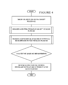
[0034] FIG. 4 is a flow diagram of an example of a blood stream anomaly
detection
method. An ultrasound probe may be used to create an image sequence of a blood
flow 400 in
a target region 402 of a blood vessel. The target region 402 may be an area of
the body in which
measurements of the blood flow are desirable. For instance the target region
402 may be in a
blood vessel in the arms or legs of a patient. The image may be created using
different types of
medical imaging technology. For example, color Doppler may be used to create
the image
sequence. From the image, a central frequency 404 of the blood flow 400 in the
target region
402 of the blood vessel may be determined. The central frequency 404 is the
Doppler frequency
shift that corresponds to the bloodstream as a whole. A differential frequency
406 of one or more
particles 408 suspended within the blood flow 400 may also be measured.
Particles moving
within the blood cell may have a different frequency than the surrounding
blood flow. This
frequency differential may be detectable from the image sequence of the blood
flow.
-8-
CA 03227405 2024- 1-29
WO 2023/014806
PCT/US2022/039297
[0035] The differential frequency 406 may be analyzed to determine properties
of the
blood flow and properties of any particle or particles suspended within the
blood flow. For
example, an analysis of the differential frequency 406 measured may reveal
that an irregular
particle is present in the blood flow 404. This irregular particle may be a
blood clot.
[0036] FIG. 5 is a flow diagram of an example of a blood stream anomaly
detection
method. An ultrasound probe may be used to create an image sequence of a blood
flow 400 in
a target region 402 of a blood vessel 412. The target region 402 may be an
area of the body in
which measurements of the blood flow 400 are desirable. For instance the
target region 402 may
be in a blood vessel 412 in the arms or legs of a patient. The image may be
created using different
types of medical imaging technology. For example, color Doppler may be used to
create the
image sequence. From the image, a central frequency 404 of the blood flow 400
in the target
region 402 of the blood vessel 412 may be determined. The central frequency
404 is the Doppler
frequency shift that corresponds to the bloodstream as a whole.
100371 Several properties of the blood flow 400 may be measured. These
properties may
reveal important information about the blood flow 400 which may reveal whether
a medical risk
is present. A differential frequency 406 of one or more particles 408
suspended within the blood
flow 400 may also be measured. Particles moving within the blood cell may have
a different
frequency than the surrounding blood flow. This frequency differential may be
detectable from
the image sequence of the blood flow.
[0038] A relative position 420 of a particle 408 within a cross section 410 of
a blood
vessel 412 may be measured. Specifically, the position of a particle 408 along
the radius 414 of
the cross section 410 of the blood vessel 412 may be measured. For instance
the distance between
the particle 408 and the wall of the blood vessel 412 may be measured.
Alternatively or
additionally, the distance between the particle 408 and the center of the
blood vessel 412 may be
measured. Irregular particles in a blood flow 400, such as blood clots, may
have a tendency to
-9-
CA 03227405 2024- 1-29
WO 2023/014806
PCT/US2022/039297
be suspended within certain areas in the blood flow 400. Therefore, the
position of a detected
particle 408 may provide information about the blood flow 400 that may
correspond to a
particular type of irregularity which may correspond to a particular type of
health risk.
[0039] A clustering factor 416 may also be measured. The clustering factor may
quantify
the relative position of a particle 408 relative to another particle 418
within the blood flow 400.
Some types of particles 408, 418 suspended within a blood flow 400 may have a
propensity to
be located close together within the blood flow 400. In one embodiment, the
type of particle 408
may be a blood clot. Blood clots tends to cluster together within blood
vessels. The measured
clustering factor may provide information about the blood flow 400 which may
correspond to a
particular type of irregularity which may correspond to a particular type of
health risk. For
example, a measurement of many blood clots grouped very close together may
reveal a
significant risk of stroke.
[0040] The differential frequency 406, relative position 420, and clustering
factor 416
may be analyzed to determine properties of the blood flow 400 and properties
of any particle
408 or particles 408, 418 suspended within the blood flow 400. For example, an
analysis of the
differential frequency 406, relative position 420, and clustering factor 416
measured may reveal
that an irregular particle is present in the blood flow 404. This irregular
particle may be a blood
clot.
[0041] The differential frequency 406, relative position 420, and clustering
factor 416
may be further analyzed to determine the properties of a particle detected in
the blood flow. For
instance, these factors may reveal the size, shape, and/or frequency of a
particle suspended in a
blood flow. These factors may also correspond to a particular medical
condition and/or the risk
of a particular adverse medical event. For example, an analysis of the
relative position of the
blood clot within the blood vessel cross section may correspond to the risk of
an adverse medical
event, such as a stroke, or may reveal that the clot has reached a certain
size. A further analysis
-10-
CA 03227405 2024- 1-29
WO 2023/014806
PCT/US2022/039297
of the clustering factor may likewise correspond to the risk of an adverse
medical event, such as
a stroke.
[0042] FIG. 6 is a diagram showing an example of a blood stream anomaly
detection
system. A blood stream anomaly detection system may include an ultrasound
sensor 600. The
ultrasound sensor 600 may create an image sequence 602 of a blood flow 604 in
a target region
606 of a blood vessel 608. A blood stream anomaly detection system may also
include a set of
anomaly detection parameters 610. A few example of relevant anomaly detection
parameters
may be relative speed 632 of a particle in the blood flow as compared to the
speed of the blood
flow itself, the relative position 634 of a particle in a blood flow, and the
relative position 636
of a particle in a blood flow relative to other particles in the blood flow.
The values of these
parameters may reveal important information about a blood flow including the
presence of an
anomaly or irregular particle in the blood flow and the properties of any
irregular particle,
including the shape, size, and frequency of the irregular particle.
[0043] All of these anomaly detection parameters 610 may be determined from a
high
quality image sequence of a blood flow through a blood vessel. In an example
embodiment, a
high quality image sequence may be created using color Doppler techniques. The
ultrasound
sensor 600 may employ pulse-wave Doppler with short pulses to create the image
sequence 602
of the blood flow 604 including the anomaly detection parameters 610.
[0044] A blood stream anomaly detection system may also include one or more
sample
datasets comprising training data 614. The sample datasets may be sample image
sequences 616,
620, 624 of sample blood flows which are mapped to known parameters of
interest 618, 622,
626. The known parameters of interest 618, 622, 626 may be known values
anomaly detection
parameters. For instance the known parameters 618, 622, 626 may correspond to
known values
for sizes, shapes, and/or frequencies of irregular particles detected in the
sample image
sequences. The irregular particles detected may be blood clots. The known
parameters 618, 622,
-11 -
CA 03227405 2024- 1-29
WO 2023/014806
PCT/US2022/039297
626 may also correspond to set risk thresholds for particular health
conditions. For example,
known parameters 618, 622, 626 may correspond to samples in which blood clots
with a greater
than 50% chance of stroke are present.
[0045] A blood stream anomaly detection system may include a machine learning
model
612. The machine learning model 612 may be trained using the training data
614. The trained
machine learning model may then apply learned patterns to analyze the image
sequence 602 of
a blood flow 604 in a target region 606 of a blood vessel 608. The machine
learning model may
determine information about the blood flow 604 including the presence of any
irregular particles
in the blood flow and the properties of any detected in-egular particles. The
machine learning
model may generate output data 642. The output data 642 may comprise values
for properties of
irregular particles detected in a blood flow. For example, the output data 642
may include a
determined size 628 of an anomaly 630 present in the blood flow 604. The
output data 642 may
include a determined shape 638 of an anomaly 630 present in the blood flow
604. The output
data 642 may include a determined frequency 640 of an anomaly 630 present in
the blood flow
604.
[0046] In an example embodiment, the blood stream anomaly detection system of
FIG.
6 may be specifically configured to detect blood clots in a blood flow. A
blood clot detection
and classification system may include an ultrasound sensor. The ultrasound
sensor may create
an image sequence of a blood flow in a target region. The blood clot detection
and classification
system may also include a set of blood clot detection parameters, including:
(i) speed, wherein
speed is the relative speed of a clot in the blood flow to the speed of the
blood flow itself; (ii)
vessel position, wherein vessel position is a relative position of the clot in
the blood flow along
the radius of the blood vessel; and (iii) clot position, wherein clot position
is a relative position
of the clot to other particles in the blood. An ultrasound sensor in a blood
clot detection and
-12-
CA 03227405 2024- 1-29
WO 2023/014806
PCT/US2022/039297
classification system may employ pulse-wave Doppler with short pulses to
create an image
sequence of the blood flow including the blood clot detection parameters.
[0047] A blood clot detection and classification system may also include one
or more
sample datasets, wherein the sample datasets include image sequences of sample
blood flows
and corresponding known values for the blood clot detection parameters. A
blood clot detection
and classification system may also include a machine learning model, wherein
the machine
learning model is trained using the one or more sample datasets to, based on
the blood clot
detection parameters, detect the presence of a blood clot in the blood flow,
determine the
frequency of the blood clot in the blood flow, determine the diameter of the
blood clot in the
blood flow, and determine the shape of the blood clot in the blood flow.
[0048] While various embodiments of the present invention have been described
above,
it should be understood that they have been presented by way of example only,
and not of
limitation. Likewise, the various diagrams may depict an example architectural
or other
configuration for the invention, which is done to aid in understanding the
features and
functionality that can be included in the invention. The invention is not
restricted to the
illustrated example architectures or configurations, but the desired features
can be implemented
using a variety of alternative architectures and configurations. Indeed, it
will be apparent to one
of skill in the art how alternative functional, logical or physical
partitioning and configurations
can be implemented to implement the desired features of the present invention.
Also, a multitude
of different constituent module names other than those depicted herein can be
applied to the
various partitions. Additionally, with regard to flow diagrams, operational
descriptions and
method claims, the order in which the steps are presented herein shall not
mandate that various
embodiments be implemented to perform the recited functionality in the same
order unless the
context dictates otherwise.
-13 -
CA 03227405 2024- 1-29
WO 2023/014806
PCT/US2022/039297
[0049] Although the invention is described above in terms of various exemplary
embodiments and implementations, it should be understood that the various
features, aspects and
functionality described in one or more of the individual embodiments are not
limited in their
applicability to the particular embodiment with which they are described, but
instead can be
applied, alone or in various combinations, to one or more of the other
embodiments of the
invention, whether or not such embodiments are described and whether or not
such features are
presented as being a part of a described embodiment. Thus, the breadth and
scope of the present
invention should not be limited by any of the above-described exemplary
embodiments.
[0050] Terms and phrases used in this document, and variations thereof, unless
otherwise
expressly stated, should be construed as open ended as opposed to limiting. As
examples of the
foregoing: the term "including" should be read as meaning "including, without
limitation" or the
like; the term "example" is used to provide exemplary instances of the item in
discussion, not an
exhaustive or limiting list thereof; the terms "a" or "an" should be read as
meaning "at least one,"
"one or more- or the like; and adjectives such as "conventional,-
"traditional,- "normal,"
"standard," "known" and terms of similar meaning should not be construed as
limiting the item
described to a given time period or to an item available as of a given time,
but instead should be
read to encompass conventional, traditional, normal, or standard technologies
that may be
available or known now or at any time in the future. Likewise, where this
document refers to
technologies that would be apparent or known to one of ordinary skill in the
art, such
technologies encompass those apparent or known to the skilled artisan now or
at any time in the
future.
[0051] The presence of broadening words and phrases such as "one or more," "at
least,"
"but not limited to- or other like phrases in some instances shall not be read
to mean that the
narrower case is intended or required in instances where such broadening
phrases may be absent.
-14-
CA 03227405 2024- 1-29
WO 2023/014806
PCT/US2022/039297
The use of the term "module" does not imply that the components or
functionality described or
claimed as part of the module are all configured in a common package. Indeed,
any or all of the
various components of a module, whether control logic or other components, can
be combined
in a single package or separately maintained and can further be distributed in
multiple groupings
or packages or across multiple locations.
100521 Additionally, the various embodiments set forth herein are described in
terms of
exemplary block diagrams, flow charts and other illustrations. As will become
apparent to one
of ordinary skill in the art after reading this document, the illustrated
embodiments and their
various alternatives can be implemented without confinement to the illustrated
examples. For
example, block diagrams and their accompanying description should not be
construed as
mandating a particular architecture or configuration.
-1 5 -
CA 03227405 2024- 1-29