Note: Descriptions are shown in the official language in which they were submitted.
CA 03229106 2024-02-12
WO 2023/039138
PCT/US2022/043014
1
EXCHANGEABLE OPTICS AND THERAPEUTICS
BACKGROUND
[0001] An
intraocular lens (IOL) is a lens that is implanted in the eye. IOLs come in
phakic, designed to be implanted without performing cataract surgery, and
pseudophakic,
designed to be implanted in conjunction with cataract surgery, varieties. A
phakic IOL has the
ability to reside in the sulcus space between the capsular bag and the iris or
alternatively can
reside in the anterior chamber, between the iris and cornea. The most commonly
employed
pseudophakic IOL is posterior chamber IOL includes haptics that enable the
lens to be held in
place in the capsular bag inside the eye. Implantation of an IOL is often
carried out by an eye
surgeon in a surgical center, but may be also be performed at an
ophthalmologist's office in an
in office surgical suite. In office procedures are particularly common with
phakic IOLs, much
in the same way laser refractive surgeries are typically in office. The field
of pseudophakic
IOLs is increasingly addressing the issue of presbyopia, which is the case
where someone is
not able to see both at distance and near. Presbyopia is not an indication for
insurance coverage
of cataract surgery currently.
[0002] As the
field matures, it is likely IOLs will be increasingly utilized to address
presbyopia, instead of glare and blurred vision even with glasses or some form
of wearable
refractive correction which is the current indication. To achieve the quality
of vision of laser
refractive surgery and to enable incremental changes to the lens as the
technology improves, a
means of fully customizable and upgradeable IOL design is sorely needed.
Refractive cataract
surgery replaces the natural eye lens with an advanced multi-focal or extended-
depth-of-focus
(EDOF) IOL. Refractive cataract surgery has not achieved the precision of
corneal refractive
surgery, such as LASIK (laser-assisted in situ keratomileusis), which can be
individualized to
high precision. Moreover, there currently is a lack of wave-front guided
precision in cataract
extraction and IOL implantation.
[0003] A
wavefront-guided approach refers to an ablation profile that considers
preoperative higher-order aberrations, where the final goal is to avoid
inducing aberrations and
to eliminate some that exist. This is commonly employed with laser refractive
surgery such as
LASIK and PRK, as all variables in the eye are known. The laser ablation
profile is computed
preoperatively according to the results of aberrometry and is transferred to a
laser system for
use, for example, during surgery. The only modification made to the eye is to
the shape of the
cornea. Currently this is an elusive task in cataract surgery for two reasons.
Principally, the
effective lens position, where the IOL ends up in the eye, is hard to
determine. Small changes
CA 03229106 2024-02-12
WO 2023/039138
PCT/US2022/043014
2
in the anterior posterior position make large changes in the total power of
the lens. In addition,
zonular weakness induced by the surgery and change in corneal astigmatism made
by the
cataract main incision can respectively change the lens position and the
corneal curvature.
Moreover, any customized, astigmatism and higher order aberration correction
is precluded a
priori on the IOL is precluded by potential shifting of the IOL within the
capsular bag in the
X,Y, Z plane.
[0004] Outside
of the inability to provide wavefront guided IOL optimization, current
IOL systems do not enable ease of correction if a non-optimal IOL is placed,
nor do they allow
for ease of upgradeability. IOL exchange is a challenging procedure that even
in the most
skilled surgeon's hands results in significant trauma to the ocular
structures. So much so that
IOL exchange is viewed as a last resort. However, repeated removal and
replacement of a
conventional IOL may not be an easy procedure and can result in complications.
For example,
IOL exchange with the conventional IOLs requires dissection of the capsular
bag and retrieval
of an unfolded lens through the cornea or sclera. Either retrieval approach
(through the cornea
or through the sclera) is highly traumatic to the eye and its delicate
structures. Instead of
exchanging IOLs, most surgeons will perform LASIK or other laser refractive
procedure to the
cornea. This also is not infinitely repeatable as corneal tissue is ablated at
each procedure.
Repeated laser correction can lead to a host of complications including
corneal ectasia and
epithelial ingrowth. It also can induce ocular surface disease in even young
patients and thus
is less than ideal in many of the older individuals undergoing cataract
surgery.
[0005] A need
exists for a system that enables relatively unlimited exchangeable optics
as well as wavefront guided lens optimization.
BRIEF SUMMARY
[0006]
Exchangeable optics and therapeutics are described that can enable progressive
application and exchanges of lenses and/or therapeutics in the eye.
[0007] An
exchangeable optics system includes an intraocular base that can be fixed
within an eye. The intraocular base includes one or more couplers and a
supporting structure.
The one or more couplers can include magnetic material or other releasable
fixation material
or structures. For example, the releasable couplers can be in the form of a
hook and loop
coupler, a memory material fixation element such as what is utilized for
tagging guns for
affixing tags to clothing, a button fastener, a screw-type fastener, a hinge-
based fastener similar
to a cuff link, a suction cup based mechanism, an adhesive, or any other means
of reversible
fixation.
CA 03229106 2024-02-12
WO 2023/039138
PCT/US2022/043014
3
[0008] Magnetic
fixation is particularly attractive as the base element to which the
secondary optic couples can be in the capsular bag and the magnetic secondary
optic can couple
through magnetic force through the anterior capsular bag without physically
directly contacting
the IOL in the bag. Magnetic attraction is also an ideal mechanism as it
allows for a secondary
optic to be disengaged from the primary optic with minimal force. Accordingly
for magnetic
and other types of releasable couplers, it can be important to consider damage
to delicate
zonules that hold the capsular bag. The supporting structure can include
haptics and a main
structure that physically supports an exchangeable optic or therapeutic that
is coupled via the
one or more couplers. In some cases, the intraocular base can include a fixed
lens within or on
the main structure.
[0009] This Summary is
provided to introduce a selection of concepts in a simplified
form that are further described below in the Detailed Description. This
Summary is not
intended to identify key features or essential features of the claimed subject
matter, nor is it
intended to be used to limit the scope of the claimed subject matter.
BRIEF DESCRIPTION OF THE DRAWINGS
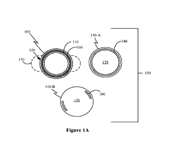
[0001] Figures 1A and
1B illustrate exchangeable optics systems suitable for an
implantable intraocular lens and application of therapeutics.
[0002] Figures 2A-2E
illustrate various locations in the eye where an exchangeable
optics system can be set.
[0003] Figures 3A and
3B illustrate a perspective view and a top view, respectively,
of an exchangeable optic with clips for coupling to an intraocular base.
[0004] Figure 4
illustrates a perspective view of an exchangeable optic with a screw
mount for coupling to an intraocular base.
[0005] Figure 5
illustrates a side view of part of an exchangeable optic system with a
post and clip coupling.
[0006] Figures 6A-6D
illustrate an exchangeable optics system with multiple stacked
lenses.
[0007] Figures 7A and
7B illustrate another exchangeable optics system with multiple
stacked lenses; Figure 7A shows another example of an intraocular base; and
Figure 7B shows
application of a second optic onto the intraocular base and first optic using
a delivery system.
(0008 Figure 8
illustrates an optic delivery system consisting of a hook that can be
drawn coaxially within a delivery sleeve.
CA 03229106 2024-02-12
WO 2023/039138
PCT/US2022/043014
4
[0009] Figures 9A and
9B show fiducial designs that can enable precise orientation of
three-dimensional rotation of an optic or haptic.
[0010] Figure 10
illustrates an example exchangeable optics system with magnetic
exchangeable intraocular lens.
[0011,1 Figures 11A-11D
illustrate another example of an exchangeable optics system
with magnetic exchangeable ocular lens.
[0012] Figures 12A-12D
illustrate example haptic designs for exchangeable optics
systems.
[0013] Figures 13A and
13B illustrate a side view and top view, respectively, of an
exchangeable optic with rotatable lens.
[00141 Figures 14A-14C
illustrate an example of an exchangeable optics system with
therapeutic delivery.
[00i 5] Figures 15A-15C
illustrate example magnetic liposomes or nanoparticles that
can be used for delivery of therapeutics on an exchangeable optics system.
DETAILED DESCRIPTION
[0010] Exchangeable
optics and therapeutics are described that can enable progressive
application and exchanges of lenses and/or therapeutics in the eye.
[0011] Figures 1A and
1B illustrate exchangeable optics systems suitable for an
implantable intraocular lens and application of therapeutics. As shown in
Figures 1A and 1B,
Exchangeable optics systems include an intraocular base 100 that can be fixed
within an eye.
The intraocular base 100 includes one or more couplers (e.g., coupler 110) and
a supporting
structure 120. The one or more couplers can include magnetic material or other
releasable
fixation material or structures. In this example, a single ring-shaped coupler
110 is shown.
[0012] Referring to
Figure 1A, an exchangeable optics system 130 can include an
intraocular base 100 that supports an exchangeable optic (e.g., 140-A, 140-B)
and can be fixed
within the eye. As mentioned above, the intraocular base 100 can include one
or more couplers
(e.g., coupler 110) and a supporting structure 120. The one or more couplers,
in this case, ring-
shaped coupler 110, are used to releasably couple the intraocular base 100 to
the exchangeable
optic 140-A, 140-B. The supporting structure 120 can include haptics 150 for
suturing or
otherwise fixing the intraocular base 100 in the eye and a main structure 160
(which may be a
circular substructure), which can be used to physically support an
exchangeable optic 140-A,
140-B directly or indirectly via the one or more couplers.
CA 03229106 2024-02-12
WO 2023/039138
PCT/US2022/043014
[0013] The
haptics 150 can be any suitable structure enabling the intraocular base 100
to be fixed within the eye. Various examples are shown in Figures 6A, 7A, 10,
11A, and 12A-
12D.
[0014] In the
illustrated scenario, the main structure 160 is open in the center such that
the exchangeable optic 140-A, 140-B rests on a proximal surface at the
perimeter of the
intraocular base 100. In other implementations, the main structure 160 has a
transparent surface
over which the exchangeable optic 140-A, 140-B rests. The supporting structure
120 can also
optionally include a lens or IOL (not shown) within or on the main structure
160. In some
cases, the supporting structure 120 can include one or more protrusions that
can be used to
extend up through a hole in the capsular bag of the eye (see e.g., extensions
222 of Figure 2B
and tension ring extensions 1125 of Figure 11A). In some of such cases, a
coupler can be
disposed at an end of a protrusion. This coupler may be the coupler for the
base or an additional
coupler for the base.
[0015] The
exchangeable optic 140-A, 140-B can include a lens 170 and one or more
corresponding couplers 180. For application of the exchangeable optics system
130, the
intraocular base 100 can be deployed in an eye. One of the exchangeable optics
140-A, 140-B
can then be deployed, oriented/aligned, and coupled to the intraocular base
100 using the
couplers 110, 180 (illustrated as magnets/magnetic material). Alignment can
involve radial
alignment with respect to either the intraocular base, the eye, or some
structure within the eye.
The one or more exchangeable optics (e.g., optic 140-A, 140-B) can include
fiducials to aid in
radial alignment, such as seen in Figures 9A and 9B. Alignment can also
involve depth
alignment with respect to either the intraocular base, the eye, or some
structure within the eye.
[0016] In some
cases, there are more or fewer "corresponding couplers" 180 than there
are couplers 110 of the intraocular base 100. For example, the couplers of the
base may be
point couplers while the couplers of the optic may be a single ring shape. In
the illustrated
scenario, one exchangeable optic 140-A is shown with a single corresponding
coupler 180,
which is in the shape of a ring; and the other exchangeable optic 140-B is
shown with two
corresponding couplers 180 that are positioned to both couple to the ring-
shaped coupler 110
of the intraocular base 100. The coupling between the intraocular base 100 and
the
exchangeable optic 140-A, 140-B can be accomplished in a variety of ways, for
example,
magnetically, using friction, or chemically. In the illustrated scenario,
magnetic coupling is
shown.
[0017] Of
course, while a ring-shape coupler 110 is one example, the one or more
couplers at the intraocular base may be two couplers formed of magnetic
material such that the
CA 03229106 2024-02-12
WO 2023/039138
PCT/US2022/043014
6
coupling is accomplished using a two-point coupling where a first of the one
or more couplers
of the intraocular base is disposed at a proximal surface (i.e., the surface
facing outward from
the eye) on one side of the intraocular base and a second of the one or more
couplers is disposed
at the proximal surface on another side of the intraocular base. The
corresponding one or more
couplers would then be disposed at the exchangeable optic in a manner to
orient and couple the
exchangeable optic to the base. For example, the corresponding one or more
couplers would
be disposed in alignment for coupling to the one or more couplers of the
intraocular base.
[0018] As
mentioned above, the one or more couplers 110 and the corresponding one
or more couplers 180 can be formed of magnetic material. The magnetic material
can be any
suitable ferromagnetic or ferrimagnetic material. The magnetic material is
sized and shaped so
as to minimize or avoid susceptibility to strong external magnetic fields such
as MRI (e.g.,
avoiding/minimizing movement or interference with imaging).
[0019] It
should be understood that although the examples contained herein make
reference to the couplers being magnets or magnetic, other types of releasable
couplers can be
used (e.g., chemical, mechanical, or friction based) in certain
implementations. The use of
magnetic couplers also enable certain therapeutics to be applied.
[0020] Indeed,
referring to Figure 1B, the same intraocular base 100 can be used to
apply therapeutics 190. In the illustrated scenario, therapeutics 190 can be
coupled to the
intraocular base 100. In some cases, the therapeutics 190 are applied once the
intraocular base
100 is deployed in the eye. In some cases, the therapeutics 190 may be applied
before original
deployment and then optionally reapplied after deployment.
[0021] Figures
2A-2E illustrate various locations in the eye where an exchangeable
optics system can be set. Figures 2A-2C show examples of an intraocular base
of an
exchangeable optics system being positioned within a capsular bag of the eye.
Referring to
Figure 2A, an intraocular base 200 of an exchangeable optics system 210 can be
positioned
within the capsular bag 212 of an eye. Through use of magnetic coupling, an
exchangeable
optic 215 (or therapeutic) can be deployed to (and even later removed from)
the sulcus space
216 of the eye. Referring to Figure 2B, an intraocular base 220 having
extensions 222 can be
positioned within the capsular bag 212 of an eye. The extensions 222 (or other
protruding
structure) can be extended into the sulcus space 216 through one or more holes
in the capsular
bag 212. For example, there may be an opening from cataract surgery through
which the
extensions 222 can protrude. In some cases, small openings may be made to
allow for the
extensions 222 to protrude through. Magnetic, mechanical, or chemical couplers
may be
provided at the end of the extensions 222 for an exchangeable optic 225 that
is deployed to
CA 03229106 2024-02-12
WO 2023/039138
PCT/US2022/043014
7
(and even later removed from) the sulcus space 216 to couple to. Referring to
Figure 2C, an
exchangeable optics system 230 can be positioned entirely within the capsular
bag 212.
[0022]
Referring to Figure 2D, in some cases, an exchangeable optics system 240 can
be positioned entirely in the sulcus 216 behind the iris 242, in front of the
capsular bag 212.
Referring to Figure 2E, in some cases, an exchangeable optics system 250 can
be positioned
in the anterior chamber behind the cornea 252, in front of the iris 242. The
examples shown in
Figures 2D and 2E could work with a patient that is phakic (with native lens).
Advantageously,
if the intraocular base is fixed in the anterior chamber (such as shown in
Figure 2E) or in the
sulcus space (such as shown in Figure 2D), cataract surgery may not be
required.
[0023] For any
of these locations, if weight of the system is ever greater than zonular
strength, an air bladder or portion of the device that floats in aqueous can
be incorporated in
the intraocular base. This buoyant component of the invention can be
permanently
incorporated, for example a compressible foam buoy that has sealed foam used
in nautical
equipment, pool toys and body boards. Alternatively, the device can have a
reservoir that acts
as a bladder that is filled with a gas or any material lighter than water.
This would enable
adjustable buoyancy based upon the degree of fill.
[0024] As
mentioned above, the one or more couplers 110 (and corresponding one or
more couplers 180) can include magnetic material or other releasable fixation
material or
structures. For example, the releasable couplers can be in the form of a hook
and loop coupler,
a memory material fixation element such as what is utilized for tagging guns
for affixing tags
to clothing, a button fastener, a screw-type fastener, a hinge-based fastener
similar to a cuff
link, a suction cup based mechanism, an adhesive, or any other means of
reversible fixation.
[0025] Figures
3A and 3B illustrate a perspective view and a top view, respectively,
of an exchangeable optic with clips for coupling to an intraocular base;
Figure 4 illustrates a
perspective view of an exchangeable optic with a screw mount for coupling to
an intraocular
base; and Figure 5 illustrates a side view of part of an exchangeable optic
system with a post
and clip coupling.
[0026]
Referring to Figures 3A and 3B, an exchangeable optic 300 can have a clip 310
that can attach to a coupler of an intraocular base (not shown). In some
cases, the exchangeable
optic 300 can include ribs 320 to assist with a secure fit, for example,
within a main structure
of the intraocular base.
[0027]
Referring to Figure 4, an exchangeable optic 400 can have a screw mount 410
for securing to a corresponding coupler at an intraocular base (not shown). In
some cases, the
CA 03229106 2024-02-12
WO 2023/039138
PCT/US2022/043014
8
exchangeable optic 400 can include prongs 420 to assist with a secure fit, for
example, within
a coupler and main structure of the intraocular base.
[0028]
Referring to Figure 5, an exchangeable optic 510 can be coupled to an
intraocular base 520 using a post 530 and nitinol clip 540.
[0029] For any
direct connection between a base and an exchangeable optic (or
between two exchangeable optics), it is desirable that the coupling mechanism
is located within
the confines of the anterior rhexis. This will enable direct connection
between the exchangeable
optic (e.g., exchangeable optic 225 of Figure 2B) outside of the capsular bag
212 and the
intraocular base (e.g., intraocular base 220 of Figure 2B) in the capsular
bag. Alternatively,
femtosecond laser or other precision surgical platform can not only make the
primary rhexis
but also make two or more small secondary opening in the anterior capsule
through which a
coupling mechanism (e.g., extensions 222) can protrude. The use of the
femtosecond laser or
other precision surgical platform to form secondary openings through which a
coupling
mechanism can protrude may serve a secondary function of aligning a lens in a
particular axis,
which is useful, for example, with toric IOLs. Indeed, the femtosecond laser
or other precision
surgical platform can be used to make two additional holes adjacent to the
rhexis at the axis
the IOL must be through.
[0030] There
are numerous coupling mechanisms that may be used instead of or in
addition to magnetic material. In some cases, the exchangeable optic can have
a fixation
element that has a shape memory material component that can be placed through
a hole at the
intraocular base through the holes made in the anterior capsule. Similar to a
tagging gun used
to attach price tags to clothing, the T arms can flex when being pushed
through the hole in the
optic haptic junction and return to an open position once through the hole.
[0031] As is
clear to one skilled in the art, this arrangement can be modified in
numerous ways. For example, in some cases, the T arm fixation element can be
incorporated
into the intraocular base and project through the capsular bag into the sulcus
space. The
exchangeable optic can have a hole in it through which the T fixation element
projects. This
may be a preferable option if capsular bag phimosis causes the capsular bag to
shift in position
in relation to the hole in the primary optic. By having the T fixation element
project beyond
the capsular bag, this helps ensure maintained access to the coupling
mechanism, even if
capsular phimosis occurs. In addition, the T-shape fixation element can be
made of a variety
of memory materials including shape memory polymers and shape memory metals.
Suitable
memory polymers for the described fixation elements include, but are not
limited to,
polynorbomene, polycaprolactone, polyenes, nylons, polycyclooctene (PCO),
blends of PCO
CA 03229106 2024-02-12
WO 2023/039138
PCT/US2022/043014
9
and styrene-butadiene rubber, polyvinyl acetate/polyvinylidinefluoride
(PVAc/PVDF), blends
of PVAc/PVDF/polymethylmethacrylate (PMMA), polyurethanes, styrene-butadiene
copolymers, polyethylene, trans-isoprene, blends of polycaprolactone and n-
butylacrylate, and
combinations thereof Suitable memory metals for the described fixation
elements include, but
are not limited to, stainless steel, cobalt, nickel, chromium, molybdenum
titanium, nitinol,
tantalum, platinum-iridium alloy, gold, magnesium, or combinations thereof
Further, it should
be understood that other end shapes may be used for the T shape fixation
element. For example,
the end shape may be a circle, triangle or any shape that is larger than the
hole it is to be fixated
through.
[0032] In some
cases, the intraocular base or the exchangeable optic can have posts that
project either through the anterior capsulotomy or through the secondary holes
created in the
anterior capsule. In one such implementation with a post projection, an
exchangeable optic
could then fit through the posts and an elastic band can be placed over the
exchangeable optic
onto the post thereby holding the exchangeable optic in place. The elastic
band that retains the
exchangeable optic can operate similar to how rubber bands hold a wire in
place to the bracket
on dental braces. In another implementation of a post projection, the post
could have a thread
on it in which a screw can mount. In another implementation, the post can
include a hole
through which a cotter pin or memory material can be placed through. In
another
implementation, the post can include a lever arm. Similar to a cuff link, the
post can either be
straight up and down or when turned at the hinge will form a T. This
arrangement does not
involve shape memory but instead just a mechanical hinge. An exchangeable
optic with a
feature similar to a shirt cuff can be threaded over the fixation element when
it is in a straight
position and then once in place the hinge can be turned so instead of straight
the post forms a
T thereby holding the exchangeable optic and the intraocular base together.
[0033] In some
cases, the intraocular base and the exchangeable optic can use a snap-
button arrangement, for example, if designed with low enough friction.
[0034] In some
cases, the intraocular base and the exchangeable optic can use a twist
on mechanism in conjunction with posts, where the posts include a T or L
shaped end and once
the posts pass through the opening in the other part, the exchangeable optic
can be rotated so
that the end of each post catches on a surface to hold the two in place. For
example, if one post
element is in the shape of a L but the slot it passes through only is slightly
larger than the
horizontal component of the L, then if the intraocular base and the
exchangeable optic are
rotated in relation to each other, the leading edge of the L moves beyond the
edge of the slot it
passes through thereby holding the intraocular based and the exchangeable
optic together. In
CA 03229106 2024-02-12
WO 2023/039138
PCT/US2022/043014
some cases, a shape memory material can be incorporated. For example, the L
shape can have
a projection at the very end (such as in the form of a very pronounced serif
L). The projection
at the end of the L can fit into a hole that is adjacent to the notch (e.g.,
similar to that employed
in some ballpoint pens). Thus, as the L shape is threaded through the notch,
the projection
portion at the end of the L abuts the edge of the notch and is bent slightly
out of the way so
rotation can continue. Once rotated far enough that the projection on the L
reaches the hole
next to the notch and falls into place thereby enabling the L to again be
coplanar with the
intraocular base and exchangeable optic. In some cases, both the exchangeable
optic and the L
shaped post can be formed of materials with memory shape properties
[0035] Figures
6A-6D illustrate an exchangeable optics system with multiple stacked
lenses. Figure 6A illustrates an exploded view of an exchangeable optics
system 600 that
includes an intraocular base 610 and a plurality of optics (including first
optic 621 and second
optic 622). The intraocular base 610 can have a supporting structure 630 with
a haptic ring 640
that can be sutured for fixed connection to an eye. One or more couplers can
be on the
supporting structure. For example, the one or more couplers can be point
sources or a ring (such
as represented by white dotted line 650) that is disposed on or goes around a
circumference of
the supporting structure (see also e.g., Figures 11A and 11B). Referring to
Figure 6B, the
intraocular base 610 can be disposed in the eye (e.g., in the sulcus space).
As shown in Figure
6C, the first optic 621 can be releasably attached to the intraocular base
610. Alternatively, in
some cases, the first optic 621 (or a third optic) is fixedly attached to the
intraocular base 610
or is built-in to the intraocular base (see e.g., lens 1060 of Figure 10).
Then, as shown in Figure
6D, the second optic 622 can be releasably attached to the intraocular base
610 over the first
optic 621. In some cases, the magnetic force from the intraocular base 610 is
sufficient to
couple both optics. In some cases, the positioning of the two optics enable at
least a portion of
the one or more couplers to be dedicated to a respective one of the two (or
more) optics. In
some cases, the first optic 621 includes one or more couplers for the second
optic 622 to couple
to. In some cases, the first optic 621 is fixedly attached to the intraocular
base 610 and the
couplers on the supporting structure are configured for attachment of the
second optic 622.
[0036] Figures
7A and 7B illustrate another exchangeable optics system with multiple
stacked lenses; Figure 7A shows another example of an intraocular base; and
Figure 7B shows
application of a second optic onto the intraocular base and first optic using
a delivery system.
[0037]
Referring to Figure 7A, an intraocular base 710 can have a supporting
structure
720 with a haptic 730 that can be sutured for fixed connection to an eye. One
or more couplers
can be on the supporting structure 720. For example, the one or more couplers
can be point
CA 03229106 2024-02-12
WO 2023/039138
PCT/US2022/043014
11
sources or a ring (such as represented by white dotted line 740) that is
disposed on or goes
around a circumference of the supporting structure (see also e.g., Figures 11A
and 11B).
[0038] Turning
to Figure 7B, a lens 750 can be easily applied to the intraocular base
710 via a tool (optic delivery system 760) through a small incision in the
sclera 770. An optic
delivery system 760 can include a hook or other fine instrument that can be
drawn coaxially,
allowing for a minimal incision that minimizes changes to corneal astigmatism
and damage to
the ocular structures after optic introduction or exchange. The optic delivery
system can
coaxially store a capsular bag containing a new optic containing, for example,
the secondary
lens 750 and enter through a minimal incision. As shown in Figure 7B, once
inside the eye
close to the location of the intraocular base 710, the optic delivery system
760 can release the
capsular bag close into the sulcus space. The hook (see Figure 8) can be used
to maneuver the
capsular bag or secondary lens to be oriented properly with respect to the
intraocular base 710.
At some point, the new optic 750 can couple to the intraocular base, at which
point the hook
can optionally be used to properly orient the new optic with respect to the
intraocular base.
Fiducial markers may be used to facilitate orientation and alignment (see
e.g., Figures 9A and
9B, which can be used under optical coherence tomography - OCT) In some cases,
the
exchangeable optics (e.g., lens 750) can include an aperture, which may be
hooked by the
instrument of the optic delivery system.
[0039] In this
illustrated scenario, the lens 750 is a second optic; however, this method
can be carried out for the first optic (e.g., optic 621) and even a
replacement second optic (e.g.,
to replace the second optic 622 after optic 622 is applied as shown in Figure
6D).
[0040] Figure 8
illustrates operation of an optic delivery system. Referring to Figure
8, an optic delivery system 810 can include a hook 815, which can be drawn
coaxially into the
eye within a delivery sleeve of the optic delivery system 810. In a first
step, the hook 815 can
be in the extended position. As illustrated in a second step, the hook 815 can
engage a hole 825
within the periphery of the optic 820 to enable extraction. As illustrated in
the third step, the
hook 815 can then be drawn coaxially back into the optic delivery system 810,
bringing the
optic 820 towards the delivery sleeve. At a certain point, the hook 815 can be
drawn entirely
within the optic delivery system 810, at which point the optic 820 can be
forced to fold inwards
and be drawn with the hook into the optic delivery system 810, such as shown
in step 4.
[0041] Figures
9A and 9B show fiducial designs that can enable precise orientation of
three-dimensional rotation of an optic or haptic. Fiducials can be placed,
etched, or drawn onto
a lens or other optic to aid in orientation of the lens or other optic once
deployed. The fiducial
markers can be of a material suitable for detection by IR, ultrasound,
fluorescent, x-ray, MRI,
CA 03229106 2024-02-12
WO 2023/039138
PCT/US2022/043014
12
etc. In one implementation, the fiducials can be detectable by an ocular
response analyzer (e.g.,
optical coherence tomography - OCT). The fiducial markers can be used to
determine precise
effective lens position (ELP). Corresponding markers can be applied to an
intraocular base at
haptics, on the optional lens, or on the supporting structure, as some
examples. In some cases,
a corresponding fiducial design may be disposed at the intraocular base (e.g.,
on main structure
region 160 of Figure 1A).
[0042]
Referring to Figure 9A, the fiducial can be L-shaped. Arms of the L shape can
vary. If the size and shape of the L-shaped fiducial is known, apparent length
can be used to
inform rotation of the lens or optic in three dimensions. Referring to Figure
9B, the fiducial
can be bulls-eye-shaped (e.g., a single dot within a circle). In particular,
use of a bulls-eye
shape can allow part of the fiducial to be printed on an opposite side of the
lens or optic. The
fiducial being on both sides of the lens or optic can create greater apparent
motion of the dot
relative to the circle, allowing a more accurate understanding of its
orientation in three-
dimensional space.
[0043] A few L
shaped fiducials printed on one side of the lens or haptic receiving
system (e.g., as shown in Figure 9A) or a circle on one side of the lens and a
dot on the other
placed within the circle when viewed anterior/posterior (e.g., providing a
bullseye such as
shown in Figure 9B) will enable a sensitive measurement of any tilt. By
visualizing the length
of the L arms or where the dot is in relation to the circle it is possible to
determine where the
lens or haptic receiver is located.
[0044] In some
implementations, fiducials are provided on both the exchangeable optic
and the intraocular base that can be read using OCT. The fiducials can be read
in relation to a
stationary feature of the eye (e.g., conjunctival vessel pattern preregistered
with corneal
topography/tomography, biometry data, etc.). The OCT can then guide placement
of the optic
on haptic. The fiducials support real time tracking of the intraocular base in
case the intraocular
base moves when the exchangeable optic is removed. When the exchangeable optic
is
repositioned or replaced, the OCT device can calculate in real time with the
fiducials what
position change is necessary.
[0045] As
mentioned above, an exchangeable optics system can include a variety of
structures for the intraocular base. In addition, the couplers of the
intraocular base can be
disposed in various locations and be configured in various shapes. The
following examples are
directed to exchangeable optics systems with intraocular bases having magnetic
coupling;
however, embodiments are not limited thereto.
CA 03229106 2024-02-12
WO 2023/039138
PCT/US2022/043014
13
[0046] Figure
10 illustrates an example exchangeable optics system with magnetic
exchangeable intraocular lens. Referring to Figure 10, an exchangeable optics
system 1000
can include an intraocular base 1010 with haptics 1020 and a circular magnet
coupler 1030;
and an exchangeable optic 1040. The exchangeable optic 1040 can be a magnetic
optic with a
corresponding circular magnet 1050 around its periphery.
[0047] In the
illustrated scenario, the haptics 1020 are in the form of a two C-loop
haptic. In some cases, the intraocular base 1010 can further include a lens
1060. For example,
the intraocular base 1010 can be similar to a conventional IOL, but further
includes the one or
more couplers (e.g., here in the form of a magnet disposed at a periphery). A
magnetic optic
1040 can then be deployed, rotated to any precise orientation, for example
aligned using
fiducials such as shown in Figures 9A and 9B, and coupled to the intraocular
base structure
1010. In some cases, the exchangeable optic 1040 may not be deployed for
potentially years
down the line and/or may be replaced years later to deploy a more precise
lens. An intraocular
base structure 1010 that allows for deployment, rotation, and coupling of a
magnetic optic (e.g.,
exchangeable optic 1040) can be advantageous, for example, in precise toric
astigmatism
correction. In addition, since it is possible to add additional lenses and/or
replace the
exchangeable optic 1040, it is possible to add a further corrective lens after
a more disruptive
surgery, add a corrective lens years after the fact, or deploy a more precise
lens, for example a
specially made or three-dimensional printed lens.
[0048] Figures
11A-11D illustrate another example of an exchangeable optics system
with magnetic exchangeable ocular lens. Referring to Figures 11A and 11D, in
exchangeable
optics system 1100, an intraocular base 1110 can include a capsular tension
ring 1120 with
optional tension ring extensions 1125 and two or more couplers 1130. As
mentioned above
with respect to Figure 2B, through use of one or more protrusions such as
tensions ring
extensions 1125, the capsular tension ring 1120 can be designed in such a way
that two or more
magnetic arms (e.g., tension ring extensions 1125 with couplers 1130) emerge
through the
anterior capsulotomy similar to an Ahmed segment thereby enabling optic
placement in the
sulcus space. Alternatively, the capsular tension ring can be designed such
that the capsular
tension ring does not rise up out of the anterior capsulotomy but instead
remains in bag. In
some cases, in addition to the couplers 1130 or as an alternative to the
couplers 1130, a
secondary magnet ring 1140 can be included, which can provide a 360-degree
docking platform
for magnetic optics 1150, as shown in Figures 11C and 11D. That is, as shown
in Figure 11C,
an optic with corresponding couplers can be deployed, and attraction between
the couplers
1130 on the arms of the capsular tension ring 1120 (and/or optionally the
secondary magnet
CA 03229106 2024-02-12
WO 2023/039138
PCT/US2022/043014
14
ring 1140) and the corresponding couplers on the optic 1150 can releasably
maintain the optic
1150 in place.
[0049] Figures
12A-12D illustrate example haptic designs for exchangeable optics
systems. A supporting structure of an intraocular base can be implemented with
haptics of a
variety of different shapes and patterns. In addition to the shapes shown in
Figures 6A and 7A,
the two-looped C shaped haptic such as shown in Figure 10 and the capsular
tension ring
configuration shown in Figure 11A, other haptic shapes may be used. For
example, Figure
12A shows an exchangeable optics system 1200 with an intraocular base 1210
design having
a cruciate haptic pattern 1215 and a magnetic coupling optic 1220. Figure 12B
shows an
exchangeable optics system 1230 with an intraocular base 1240 design having a
haptic design
1245 that can facilitate secondary scleral sutured lens similar to the Gore
Akreos lens and a
magnetic coupling optic 1220. Figure 12C shows an exchangeable optics system
1250 with an
intraocular base 1260 design with four-pronged haptic arm 1265 and a magnetic
coupling optic
1220. Figure 12D shows an exchangeable optics system 1270 with an intraocular
base 1280
design with looped haptic 1285 and a magnetic coupling optic 1220.
[0050] With
cataract surgery, the shape of the corneal as well as the optics of the lens
and the effective lens position are altered. Even if precisely positioned in
the appropriate
location, postoperative shifting of the lens is not uncommon. An exchangeable
optics system
such as described herein can address these obstacles. First, by sandwiching
the capsular bag
between the magnetic optic and magnetic haptic receiver through the bag, the
system is less
likely to rotate or shift in relation to the capsular bag. Second, in certain
embodiments, such as
3D printing of a wavefront guided custom intraocular lens, it may make more
sense to allow
an intraocular base with a lens haptic system to scar into the capsular bag.
As the capsule
contracts, the final effective lens position of the intraocular base will then
be known. By
including fiducials, a wavefront scan can calculate shape of cornea after
cataract surgery, an
effective lens position can be determined from fiducials, and this data can be
used to 3D print
a custom lens when all variables are achieved. The custom lens can then be
attached afterwards
to the determined specifications. This would enable the ability to not only
print wavefront
optimized monofocal IOLs, but also custom wavefront optimized multifocal and
extended
depth of focus intraocular lens. An intraocular base also provides a forward
compatible system
for any future iteration of lens since the lens can be replaced/exchanged with
the newest
iteration of the lens.
[0051] In some
of such cases, the lens providing the primary power can be deployed
with the intraocular base (see e.g., lens 1060 described with respect to
Figure 10) and a
CA 03229106 2024-02-12
WO 2023/039138
PCT/US2022/043014
wavefront guided optic can be delivered secondarily for attachment to the
intraocular base that
has the lens 1060. The wavefront guided optic ("second lens") can be deployed
through a far
smaller incision and similar to ICL surgery and LASIK, may be amenable to
office-based
procedures. That is, the secondary optic can be deployed through a small
enough corneal
incision or previous surgical incisions can be accessed, and the additional
variability created
by reentering cornea can be minimized. This would enable the primary lens and
haptic system
to be deployed in the bag similar to current IOLs, just with a magnetic
system. At a secondary
time period in which the capsular bag has fully contracted, the fiducials
provide effective lens
position. In addition, by using topography/tomography and wavefront
measurements of the
length of the eye, all the optical variables could be controlled for. If
necessary, the degree of
astigmatism induced by penetrating the cornea to deliver the secondary optic
can be controlled
for with custom optic design adjusted to account for the induced astigmatism.
Thus, it is
possible to a priori determine effective lens position (ELP) and determine
what custom or non-
custom lens would be ideal for an eye.
[0052]
Specialized optics can be applied to an intraocular base as part of the
described
exchangeable optics systems. Figures 13A and 13B illustrate a side view and
top view,
respectively, of an exchangeable optic with rotatable lens. A lens housing
system is provided
for a rotational design that enables rotation of a lens of intraocular base or
an exchangeable
optic. Referring to Figure 13A, a design for an exchangeable optic 1300 can
have a coupling
frame 1310 to which couplers 1320 of an intraocular base 1330 can be attached;
a stationary
body 1340 that can fit within an opening of the intraocular base 1330 and a
rotating body 1350
which can rotate in one or two dimensions, depending on coupling between the
stationary body
1340 and the rotating body 1350.
[0053] As
previously mentioned, an intraocular base can be used not just to support
delivery of exchangeable optics, but also to provide a surface for delivery of
therapeutics.
Figures 14A-14C an example of an exchangeable optics system with therapeutic
delivery.
[0054] Magnetic
liposomes or nanoparticles can be used in conjunction with magnetic
components of an exchangeable optics system.
[0055] In
addition to incorporating drug delivery polymeric implants or reservoirs
directly into the haptic or optic system of the device, the magnetic
components of the
intraocular base provide a means of coupling magnetic nanoparticles and
liposomes to the
device. The magnetic liposomes or particles may be preloaded onto the device
and
administered at the time of surgery or after surgery.
CA 03229106 2024-02-12
WO 2023/039138
PCT/US2022/043014
16
[0056] Magnetic
liposomes or nanoparticles can be coupled to a magnetic intraocular
base prior to deployment in the eye. Alternatively, or in addition, liposomes
or nanoparticles
can be introduced through an intravitreal, transzonular, intracapsular or
intracameral approach
after deployment of a magnetic intraocular base into the eye and be coupled to
the magnetic
intraocular base in the eye. The magnetic particles can be used to deliver
therapeutics including,
but not limited to antibiotics, steroids, and non-steroidal anti-inflammatory
drugs (NSAIDs).
These therapeutics can be configured such as illustrated in Figures 15A-15C to
facilitate
attachment to an intraocular base. Instead of rapidly exiting the eye through
the normal outflow
pathways, a magnetic intraocular base would enable the magnetic particles to
dwell on the
haptic system until they degraded or release ferrofluid to the point that the
magnetic attraction
is no longer sufficient to remain bound.
[0057] As
mentioned above, the magnetic particles used to deliver the therapeutics can
be applied to various forms of an intraocular base. Referring to Figure 14A,
an intraocular base
1410 in the form of a capsular tension ring can be formed of or coated with
magnetic material
that attracts the magnetic particles. In some cases, different regions can be
applied with
different therapeutics, for example, a region for antibiotics 1412, a region
for steroid 1414, and
a region for NSAID 1416. Of course, the therapeutics may be applied in a
manner that the
various therapeutics are disbursed throughout the surface of the intraocular
base 1410.
[0058]
Referring to Figure 14B, an intraocular base 1420, with or without a lens, can
include a magnetic coupler/ring 1422 that is used to attach magnetic particles
1430. The
magnetic particles 1430 can thus be deployed and attached around the ring
1422.
[0059]
Referring to Figure 14C, an intraocular base 1440 with magnetic haptics 1442
can be used to attach magnetic particles 1450.
[0060]
Referring to Figure 15A, a magnetic particle can be formed of a magnetite core
with polymer coating and polyethylene glycol shell. The magnetite cores can
cause the
magnetic particle to be attracted to the magnetic intraocular base allowing
for relatively fine
deployment. If a plurality of magnetite particles is present, attraction
between the magnetic
particle and the magnetic intraocular base is reduced. The strength of the
magnetic on the
magnetic intraocular base as well as the concentration of the magnetite, size
of polymer
particle, and rate of degradation can adjust the dwell time to further
finetune localized dosage.
In a particular embodiment, rate of polymer degradation can be tuned to drug
release rate. This
can allow the magnetic particle to disassociate after the majority - or even
all of - the drug is
delivered due to a decreased attraction.
CA 03229106 2024-02-12
WO 2023/039138
PCT/US2022/043014
17
[0061]
Referring to Figure 15B, a magnetic particle can have a plurality of magnetic
particles within a single polymer particle instead of a single magnetite core
as shown in Figure
15A.
[0062]
Referring to Figure 15C, a magnetic particle can be formed as a liposome
particle with a ferrofluid core. A therapeutic can include a liposome shell, a
magnetic ferrofluid
within the liposome shell, and a drug or therapeutic core within the liposome
shell. The
magnetic ferrofluid and drug or therapeutic core can be combined inside the
liposome shell.
Since the ferrofluid and therapeutics are combined within the liposome shell,
release of the
drug or therapeutic can coincide with release of the ferrofluid. In certain
implementations, rate
of ferrofluid release can be tuned to drug release rate so when the majority
of drug is released
the degree of attraction between the liposome and intraocular base is reduced
to the point at
which the liposome dissociates and then can freely flow through the trabecular
meshwork out
of the eye.
[0063] Since
free iron is known to be toxic to the retina, magnetic nanoparticles are
contained within a biocompatible shell much like current iron-based MRI
contrast agents such
as Ferridex 0 from Berlex Laboratories Inc. The nanoparticles are of
sufficient size in order
for them to freely egress out of the eye through the trabecular meshwork when
the extraocular
magnet is removed. The nanoparticles are then cleared by the liver like other
iron-based
nanoparticles currently used clinically.
[0064] The
biocompatible material for the biocompatible shell of the magnetic
nanoparticles can be selected from the group consisting of polyvinyl alcohol,
sodium
polyacrylate, acrylate polymers, hyaluronase polymers, collagen membrane,
Porous HA/TCP
ceramic composite, hydroxyapatite bone cement, PVP/PMMA, tricalcium phosphate,
hydroxyapatite coated collagen fibers, calcium sulphate, hydroxyapatite (HAp),
phosphorylcholine (PC), silicone, ultrahigh molecular weight polyethylene ,
polyethylene,
acrylic, nylon, Polyurethane, Polypropylene, poly(methyl methacrylate),
Teflon, Dacron,
acetal, polyester, silicone-collagen composite, polyaldehyde, polyvinyl
chloride), silicone-
acrylate, poly(tetrafluoroethylene), hydroxyethyl methacrylate (HEMA),
poly(methyl
methacrylate) (PMMA), poly(glycolide lactide), poly(glycolic acid),
tetrafluoroethylene,
hexafluoropropylene, poly(glycolic acid), poly(lactic acid),
desaminotyrosyltyrosine ethyl
ester, polydioxanone, fibrin, gelatin, hyaluronan, tricalcium phosphate,
polyglycolide (PGA),
polycaprolactone, poly (lactide-co-glycolide), polyhydroxybutyrate,
polyhydroxyvalerate,
trimethylene carbonate, polyanhydrides, polyorthoesters, poly(vinyl alcohol),
poly(N-vinyl 2-
pyrrolidone), poly( ethylene glycol), poly(hydroxyethylmethacrylate), n-vinyl-
2-pyrrolidone,
CA 03229106 2024-02-12
WO 2023/039138
PCT/US2022/043014
18
methacrylic acid, methyl methacrylate, and maleic anhydride, polycaprolactone,
poly(amino
acids), poly(L-lysine), poly(1-ornithine), poly(glutamic acid),
polycyanoacrylates,
polyphosphazenes, poly(lactic acid), poly(glycolic acid), crown ethers,
cyclodextrins,
cyclophanes, ethylene glycol, Methylacrylate, Para-xylylene, Biodegradable
Copolymers,
Copolymer Surface Coatings, Starch Polymers, Polylactic Acid, Cellophane,
Tyrosine
Polycarbonates Lactide and Glycolide Polymers, Collagen, PTFE, silicone,
Keratin-Based
Materials, Fibrous Composites - Carbon Fiber and Particles, Polymer
Composites,
Artificial/Natural Material Composites, Glass-Ceramic/Metal Composites, Glass-
Ceramic/Nonmetal Composites, Dental Composites, hydrogels, timed-release
foams, and
polymeric carriers.
[0065]
According to certain implementations, the magnetic nanoparticles can include
metal oxide and polymeric or liposomal formulations. Example liposomes include
elements
from the group consisting of fatty acids, fatty acids derivatives, mono-, di
and triglycerides,
phospholipids, sphingolipids, cholesterol and steroid derivatives, oils,
vitamins and terpenes
including but not limited to egg yolk L-- phosphatidylcholine (EPC), 1,2-
dimyristoyl-sn-
glycero-3- phosphatidylcholine (DMPC), 1,2-dipalmitoyl-sn-glycero-3-
phosphatidylcholine
(DPPC), 1 ,2-dioleoyl-sn-glycero-3-phosphatidylcholine (DOPC), 1 ,2-distearoyl-
sn-glycero-
3- phosphatidylcholine (DSPC), 1,2-dilauroyl-sn-glycero-3-phosphatidylcholine
(DLPC), 1,2-
di ol eoyl-sn-gly cero-3 -pho sphaethanol amine (DOPE),
1 -p almitoyl-ol eoy 1-sn-gly cero-3-
phosphoethanolamine (POPE), 1,2-dimyristoyl-sn-glycero-3-phosphoethanolamine
(DMPE),
1,2-dipalmitoyl-sn-glycero-3-phosphoethanolamine (DPPE), and 1,2-distearoyl-sn-
glycero-3-
phospharthanolamine (DSPE), phosphatidic acids, phosphatidyl cholines with
both saturated
and unsaturated lipids, phosphatidyl ethanolamines, phosphatidylglycerols,
phosphatidylserines, phosphatidylinositols, lysophosphatidyl derivatives,
cardiolipin, 13- acyl-
y-alkyl phospholipids, di-oleoyl phosphatidylcholine, di-myristoyl
phosphatidylcholine, di-
pentadecanoyl phosphatidylcholine, di-lauroyl
phosphatidylcholine,
dipalmitoylphosphatidylcholine, di
stearoy 1pho sphati dyl choline,
diarachidoylphosphatidylcholine,
dibehenoylphosphatidylcholine,
ditricosanoylphosphatidylcholine, dilignoceroylphatidylcholine; and
phosphatidylethanolamines.
[0066] The
polymer formulations (e.g., forming a matrix for the nanoparticles) can be
selected from the group consisting of poly(acrylamide), poly(N-
isopropylacrylamide),
polyisopropylacrylamide- co-l-vinylimidazole), poly(N,N-dimethylacrylamide),
poly(N,N-
dimethylacrylamide), poly(1- vinylimidazole), poly(sodium acrylate),
poly(sodium
CA 03229106 2024-02-12
WO 2023/039138
PCT/US2022/043014
19
methacrylate), poly(2- hydroxyethylmethacrylate) (HEMA), poly N-
dimethylaminoethyl
methacrylate) (DMAEMA), poly(N tris(hydroxymethyOmethylacrylamide), poly(1-(3-
methacryloxy)propylsulfonic acid) (sodium salt),
poly(allylamine), p oly (N-
acryloxy succinimi de), poly(N-vinylcaprolactam), p oly (1-viny1-2-pyrroli
done), p oly (2-
acrylamido-2-methyl-l-propanesulfonic acid) (sodium salt), poly((3-
acrylamidopropyl)
trimethylammonium chloride), and poly(diallyldimethylammonium chloride),
poly(hydroxy
acids), polyanhydrides, polyorthoesters, polyamides, polycarbonates,
polyalkylenes,
polyalkylene glycols, polyalkylene oxides, polyalkylene terepthalates,
polyvinyl alcohols,
polyvinyl ethers, polyvinyl esters, polyvinyl halides, polyvinylpyrrolidone,
polysiloxanes,
poly(vinyl alcohols), poly( vinyl acetate), polystyrene, polyurethanes and co-
polymers thereof,
synthetic celluloses, polyacrylic acids, poly(butyric acid), poly(valeric
acid), and poly(lactide-
co-caprolactone), ethylene vinyl acetate, copolymers and blends thereof
[0067]
Advantageously, the described intraocular base enables customization and
exchange of optics as well as delivery of therapeutics.
[0068] Although
the subject matter has been described in language specific to structural
features and/or acts, it is to be understood that the subject matter defined
in the appended claims
is not necessarily limited to the specific features or acts described above.
Rather, the specific
features and acts described above are disclosed as examples of implementing
the claims and
other equivalent features and acts are intended to be within the scope of the
claims.