Note: Descriptions are shown in the official language in which they were submitted.
CA 03229484 2024-02-15
WO 2023/021346
PCT/IB2022/056848
1
TISSUE EXPANDERS HAVING FILL PORT ASSEMBLIES AND
DRAIN PORT ASSEMBLIES THAT ARE ISOLATED FROM ONE
ANOTHER FOR PREVENTING CROSS-CONTAMINATION OF FLUIDS
CROSS-REFERENCE TO RELATED APPLICATIONS
[00011] The present patent application claims benefit of U.S. Provisional
Application Serial No.
63/234,528, filed on August 18, 2021, the disclosure of which is hereby
incorporated by reference
herein.
BACKGROUND OF THE INVENTION
Field of the Invention
[0002] The present patent application is generally related to implantable
devices and is more
specifically related to tissue expanders having integrated drainage and fluid
delivery components.
Description of the Related Art
[0003] Tissue expanders are medical devices that are implanted beneath the
skin or muscle
and then gradually inflated to stretch the overlying tissue. Tissue expanders
are commonly used
to either create a pocket for receiving a permanent prosthesis, or to generate
an increased skin
surface area in anticipation of the new skin being utilized for grafting or
reconstruction.
[0004] Tissue expanders are typically formed of a silicone polymer shell.
After implantation,
a fluid, such as saline, is periodically injected into the tissue expander to
enlarge it over time
Between injections, the surrounding skin is permitted to stretch and grow to
create the increased
skin surface and the increased tissue pocket for receipt of a permanent
implant. Typically, a
tissue expander has an injection element through which fluid can be introduced
into or withdrawn
from the shell of the tissue expander. One such injection element is an
integrated port having a
septum that can be pierced with a hypodermic needle for adding or withdrawing
fluid from the
tissue expander. Alternatively, the injection element may be a self-sealing
area on the tissue
expander which allows penetration by a hypodermic needle and self-closing
after the needle has
been withdrawn from the expander.
CA 03229484 2024-02-15
WO 2023/021346
PCT/IB2022/056848
2
[0005] Most conventional, commercially available tissue expanders have a
single port that is
used for inflating and deflating the shell of the tissue expander. They have
no means for draining
fluid (e.g., seroma) that forms around the outside of the shell of the tissue
expander after
implantation.
[0006] After surgery, patients typically have surgical drains placed to
prevent blood and
lymphatic fluid from building up under the skin, allowing for a quicker
recovery. Some patients
are sent home with drains that are implanted and connected to an external
reservoir. Emptying
these reservoirs can be traumatic because the patients must periodically
measure and empty the
reservoirs (e.g., every morning). Many patients loathe surgical drains and
look forward to having
the drains removed. In addition, surgical drains may be associated with higher
risks of
infection. Thus, having a means to remove bodily fluid without the need for an
external drain
would provide great benefits for patients.
[0007] There have been some efforts directed to integrating drains into
tissue expanders.
For example, U.S. Published Patent Application No. 2020/0129258, assigned to
Mentor
Worldwide LLC, the disclosure of which is hereby incorporated by reference
herein, teaches a
tissue expander having an integrated drain includes an outer shell having an
opening and one or
more drainage holes. An injection port is disposed in the opening of the shell
and forms a fluid-
tight seal with the shell. The injection port includes a needle guard having a
needle guard base
with a top surface, and a barrier membrane that overlies the top surface of
the needle guard base.
The barrier membrane defines an inflation chamber located between the top
surface of the needle
guard base and a bottom surface of the barrier membrane, and a drainage
chamber overlying a
top surface of the barrier membrane. The tissue expander includes one or more
inflation ports
that are in fluid communication with the inflation chamber for inflating and
deflating the outer shell
with a first fluid. A drainage conduit is in fluid communication with and
extends between the
drainage chamber and the one or more drainage holes for draining a second
fluid from outside
the shell.
[0008] U.S. Patent No. 8,454,690 to McClellan discloses a tissue expander
having an
implant shell that is filled via an inflation port. The tissue expander has a
pocket that extends
around the implant shell. The pocket contains a drainage channel that can be
used to drain
fluid. A drainage port located in a superior region of the shell of the tissue
expander is
CA 03229484 2024-02-15
WO 2023/021346
PCT/IB2022/056848
3
coupled with the drainage channel via an elongated communication channel that
extends
between the drainage channel and the drainage port In McClellan, the drainage
port is
located in a superior region of the breast, which potentially increases the
risk of infection
because the superior region of the breast that is not nom-ially exposed to
drainage fluid
because drains are typically placed in the inferior region of the breast.
[0009] In addition, in McClellan, the fill port and the drainage port are
both located in the
superior region of the breast. The proximity of the fill port and the drain
port may potentially
result in cross-contamination of the filling lumen and the drainage lumen
since the needles
are passing through the same superior breast tissue. This may lead to
contamination of
the interior of the tissue expander, which may culture and be released back
into the breast
pocket if there is inadvertent deflation and rupture of the expander. In
addition, in McClellan,
the communication channel is an elongated feature that runs a substantial
distance between
the port and the drainage delivery canal. The communication channel adds
additional bulk
to the expander and potentially additional mechanical failure modes, for
example, when
folding the expander to place the expander into the breast pocket.
[0010] In view of the above-noted problems, there is a need for improved
tissue expanders
having integrated fill port assemblies and drainage port assemblies that
minimize the risk of
infection. In addition, there is a continuing need for improved tissue
expanders having integrated
fill and drainage port assemblies that minimize the likelihood that the fluid
used to fill a tissue
expander will be mixed and/or cross-contaminate with the bodily fluids that
accumulate around
the shell of the tissue expander. Moreover, there remains a need for improved
tissue expanders
that remove seroma fluid without the need for a drain being attached 24 hours
a day to a patient.
SUMMARY OF THE INVENTION
[0011] In one embodiment, a tissue expander preferably includes two
separate ports,
namely, a fill port assembly that is used for adding and withdrawing fluids
from a shell of the
tissue expander for adjusting the size of the tissue expander and a drain port
assembly that
is used for draining bodily fluids that accumulate around the shell of the
tissue expander after
the tissue expander has been implanted in a patient.
CA 03229484 2024-02-15
WO 2023/021346
PCT/IB2022/056848
4
[0012] In one embodiment, the drain port assembly may include a drain port
hub that is
in-line with a longitudinal axis of a drain cover that is used to collect
bodily fluids.
[0013] In one embodiment, the drain port assembly may include a drain port
hub that is
off-set from a longitudinal axis of a drain cover that is used to collect
bodily fluids.
[0014] In one embodiment, the positioning of the off-set drain port hub may
shift the drain
port septum away from an inframammary fold to provide for improved access
relative to the
inframammary fold.
[0015] In one embodiment, the tissue expander may include a self-sealing
membrane that
partially surrounds and/or covers areas outside the drain. The self-sealing
membrane may
prevent deflation due to accidental puncture, and may also prevent folding of
the tissue
expander when it is in a deflated state, thereby minimizing the possibility
that a bare shell
region will be accidentally punctured by a needle if it overlaps
[0016] In one embodiment, a tissue expander has a fill port assembly and a
drain port
assembly that is isolated from the fill port assembly. In one embodiment, the
tissue expander
preferably includes a shell having an anterior wall with a superior zone and
an inferior zone. In
one embodiment, the shell desirably has one or more drainage openings formed
in the inferior
zone of the anterior wall of the shell.
[0017] In one embodiment, the fill port assembly is located within the
superior zone of the
anterior wall of the shell. In one embodiment, the drain port assembly is
located within the inferior
zone of the anterior wall of the shell. The drain port assembly is preferably
in fluid communication
with the one or more drainage openings formed in the inferior zone of the
anterior wall of the shell.
[0018] In one embodiment, the fill port assembly that is located within the
superior zone of
the anterior wall of the shell is isolated from the drain port assembly that
is located with the inferior
zone of the anterior wall of the shell, which preferably prevents mixing
and/or cross-contamination
of the bodily fluids in the drain port assembly with the fluid used to fill
the shell.
[0019] In one embodiment, the drain port assembly preferably includes a
drain cover including
an elongated body having a first end and a second end. In one embodiment, the
drain cover
includes a hub that is located between the first and second ends of the
elongated body. The drain
CA 03229484 2024-02-15
WO 2023/021346
PCT/IB2022/056848
cover preferably has a first fluid reservoir that extends from the hub to the
first end of the elongated
body and a second fluid reservoir that extends from the hub to the second end
of the elongated
body.
[0020] In one embodiment, the outer face of the drain cover preferably
surrounds the first and
second fluid reservoirs. In one embodiment, the outer face of the drain cover
is secured to an
inner surface of the anterior wall of the shell within the inferior zone of
the anterior wall. In one
embodiment, the outer face of the drain cover preferably surrounds the one or
more drainage
openings. and the first and second fluid reservoirs are aligned with the one
or more drainage
openings formed in the inferior zone of the anterior wall of the shell.
[0021] In one embodiment; the hub of the drain cover preferably includes
an annular rim
having an open outer end and an inner end that is closed by a hub end wall. In
one embodiment;
one or more fluid openings may be formed in the annular rim of the hub for
providing fluid
communication between the hub and the first and second fluid reservoirs of the
drain cover.
[0022] In embodiment, the drain port assembly preferably includes a drain
port needle guard
that is disposed within the hub of the drain cover. The drain port needle
guard desirably has an
annular rim having an open outer end and an inner end that is closed by a
needle guard end wall.
[0023] In one embodiment, one or more fluid openings are formed in the
annular rim of the
drain port needle guard, which are preferably aligned with the one or more
fluid openings formed
in the annular rim of the hub for providing fluid communication between the
drain port needle
guard and the first and second fluid reservoirs of the drain cover.
[0024] In one embodiment, a drain port septum is desirably disposed within
the open outer
end of the annular rim of the drain port needle guard.
[0025] In one embodiment, a drain port magnet may be disposed between the
needle guard
end wall and the hub end wall to aid medical personnel in locating the drain
port assembly.
[0026] In one embodiment, a drain may be disposed within the first and
second fluid
reservoirs of the drain cover. In one embodiment, the drain includes a first
drain component that
is disposed within the first fluid reservoir of the drain cover, and a second
drain component that
disposed within the second fluid reservoir of the drain cover.
CA 03229484 2024-02-15
WO 2023/021346
PCT/IB2022/056848
6
[0027] In one embodiment, the outer face of the drain cover defines a
convexly curved
surface.
[0028] In one embodiment, the first and second fluid reservoirs have
convexly curved shapes
that match the convexly curved surface of the outer face of the drain cover.
[0029] In one embodiment, the first drain component has a convexly curved
shape that
matches the convexly curved shape of the first fluid reservoir and the second
drain component
has a convexly curved shape that matches the convexly curved shape of the
second fluid
reservoir.
[0030] In one embodiment, a self-sealing membrane may cover the shell
within the inferior
zone of the anterior wall of the shell. The self-sealing membrane may be
secured to an inner
surface or an outer surface of the shell.
[0031] In one embodiment, the self-sealing membrane extends superiorly from
the drain port
assembly toward the superior zone of the anterior wall of the shell.
[0032] In one embodiment, the elongated body of the drain cover preferably
has a longitudinal
axis that extends from the first end to the second end of the elongated body.
[0033] In one embodiment, the hub of the drain cover is centrally located
between the first
and second ends of the elongated body of the drain cover and is intersected
(e.g., bisected) by
the longitudinal axis of the elongated body of the drain cover.
[0034] In one embodiment, the drain port assembly may include a drain cover
including an
elongated body having a first end, a second end, a longitudinal axis extending
from the first end
to the second end; and a hub located between the first and second ends of the
elongated body
that is off-set from the longitudinal axis of the elongated body.
[0035] In one embodiment, the drain cover preferably includes a fluid
reservoir extending from
the first end to the second end of the elongated body, and an outer face that
surrounds the fluid
reservoir.
CA 03229484 2024-02-15
WO 2023/021346
PCT/IB2022/056848
7
[0036] In one embodiment, the outer face of the drain cover is preferably
secured to an inner
surface of the anterior wall of the shell within the inferior zone of the
anterior wall.
[0037] In one embodiment, the outer face of the drain cover desirably
surrounds the one or
more drainage openings, and the fluid reservoir is aligned with the one or
more drainage openings
formed in the inferior zone of the anterior wall of the shell.
[0038] In one embodiment, the off-set hub extends superiorly from an upper
edge of the
elongated body of the drain cover.
[0039] In one embodiment. the hub preferably includes an annular rim having
an open outer
end and an inner end that is closed by a hub end wall, and one or more fluid
openings formed in
the annular rim of the hub for providing fluid communication between the hub
and the fluid
reservoir of the drain cover.
[0040] In one embodiment, the drain port assembly preferably includes a
drain disposed
within the fluid reservoir of the drain cover.
[0041] In one embodiment, the drain port assembly may include a drain port
needle guard
disposed within the hub of the drain cover, the drain port needle guard
including an annular rim
having an open outer end and an inner end that is closed by a needle guard end
wall.
[0042] In one embodiment, one or more fluid openings may be formed in the
annular rim of
the drain port needle guard, which may be aligned with the one or more fluid
openings formed in
the annular rim of the hub for providing fluid communication between the drain
port needle guard
and the fluid reservoir of the drain cover.
[0043] In one embodiment, a drain port septum may be disposed within the
open outer end
of the annular rim of the drain port needle guard.
[0044] In one embodiment, a drain port magnet is disposed between the
needle guard end
wall and the hub end wall to help medical personnel locate the drain port
assembly within the
shell of the tissue expander.
CA 03229484 2024-02-15
WO 2023/021346
PCT/IB2022/056848
8
[0045] In one embodiment, the tissue expander may include a self-sealing
membrane that
covers the shell within the inferior zone of the anterior wall of the shell.
[0046] In one embodiment, the self-sealing membrane preferably extends
superiorly from the
drain port assembly toward the superior zone of the anterior wall of the
shell.
[0047] In one embodiment, a tissue expander having a fill port assembly and
a drain port
assembly preferably includes a shell having an anterior wall having a superior
zone and an inferior
zone.
[0048] In one embodiment, one or more drainage openings are formed in the
inferior zone of
the anterior wall of the shell.
[0049] In one embodiment, the tissue expander preferably includes a fill
port assembly
located within the superior zone of the anterior wall of the shell, and a
drain port assembly located
within the inferior zone of the anterior wall of the shell. The drain port
assembly is desirably in
fluid communication with the one or more drainage openings formed in the
inferior zone of the
anterior wall of the shell.
[0050] In one embodiment, the drain port assembly may include a drain cover
having an
elongated body with a first end and a second end.
[0051] In one embodiment, the drain cover may have a drain port hub that is
centrally located
between the first and second ends of the elongated body.
[0052] In one embodiment, the drain cover desirably includes one or more
fluid reservoirs
that are located between the first and second ends of the elongated body.
[0053] The drain cover preferably has an outer face that surrounds the one
or more fluid
reservoirs.
[0054] In one embodiment, the outer face of the drain cover is preferably
secured to an inner
surface of the anterior wall of the shell within the inferior zone of the
anterior wall.
CA 03229484 2024-02-15
WO 2023/021346
PCT/IB2022/056848
9
[0055] In one embodiment, the outer face of the drain cover preferably
surrounds the one or
more drainage openings, whereby the one or more fluid reservoirs are aligned
with the one or
more drainage openings formed in the inferior zone of the anterior wall of the
shell.
[0056] In one embodiment, the fill port assembly that is located within the
superior zone of
the anterior wall of the shell is isolated from the drain port assembly that
is located within the
inferior zone of the anterior wall of the shell.
[0057] In one embodiment, a self-sealing membrane may cover the shell
within the inferior
zone of the anterior wall of the shell.
[0058] In one embodiment, the self-sealing membrane extends superiorly from
the drain port
assembly toward the superior zone of the anterior wall of the shell.
[0059] In one embodiment, the elongated body of the drain cover preferably
has a longitudinal
axis that extends from the first end to the second end of the elongated body.
In one embodiment,
the drain port hub is preferably secured to an upper edge of the elongated
body and is off-set
from the longitudinal axis of the elongated body.
[0060] In one embodiment. a tissue expander preferably has a fill port
assembly and a drain
port assembly. In one embodiment. the tissue expander desirably includes a
shell having an
anterior wall with a superior zone and an inferior zone, and one or more
drainage openings formed
in the inferior zone of the anterior wall of the shell.
[0061] In one embodiment, a fill port assembly is located within the
superior zone of the
anterior wall of the shell, and a drain port assembly is located within the
inferior zone of the
anterior wall of the shell.
[0062] The drain port assembly is preferably in fluid communication with
the one or more
drainage openings fomied in the inferior zone of the anterior wall of the
shell.
[0063] In one embodiment, the fill port assembly that is located within the
superior zone of
the anterior wall of the shell is isolated from the drain port assembly that
is located with the inferior
zone of the anterior wall of the shell.
CA 03229484 2024-02-15
WO 2023/021346
PCT/IB2022/056848
[0064] In one embodiment, the tissue expander may be implanted in breast
tissue of a patient
so that the fill port assembly is located in superior breast tissue, which is
closer to an upper end
of the patient, and the drain port assembly is located in inferior breast
tissue, which is closer to a
lower end of the patient.
BRIEF DESCRIPTION OF THE DRAWINGS
[0065] FIG. 1A is a perspective view of a topside of a tissue expander
having an integrated
fill port and an integrated drain port, in accordance with one embodiment of
the present patent
application.
[0066] FIG. 1B is a perspective view of the underside of the tissue
expander shown in FIG.
1A.
[0067] FIG. 1C is a top plan view of the tissue expander shown in FIGS. 1A
and 1B.
[0068] FIG. 1D is a bottom view of the tissue expander shown in FIGS. 1A-
1C.
[0069] FIG. lE is a right side view of the tissue expander shown in FIGS.
1A-1D.
[0070] FIG. IF is a left side view of the tissue expander shown in FIGS. 1A-
1E.
[0071] FIG. 2A is an exploded view of a tissue expander including a fill
port assembly and a
drain port assembly, in accordance with one embodiment of the present patent
application.
[0072] FIG. 2B is another exploded view of the tissue expander shown in
FIG. 2A.
[0073] FIG. 3A is a perspective view of a tissue expander having a fill
port assembly and a
drain port assembly, in accordance with one embodiment of the present patent
application.
[0074] FIG. 3B is a front view of the tissue expander shown in FIG. 3A.
[0075] FIG. 3C is a right side view of the tissue expander shown in FIGS.
3A and 3B.
[0076] FIG. 3D is a left side view of the tissue expander shown in FIGS. 3A-
3C.
CA 03229484 2024-02-15
WO 2023/021346
PCT/IB2022/056848
11
[0077] FIG. 4 is an exploded view of a fill port assembly of the tissue
expander shown in
FIGS. 3A-3D.
[0078] FIG. 5 is a cross-sectional view of the fill port assembly shown in
FIG. 3B.
[0079] FIG. 6 is an exploded view of the drain port assembly shown in FIGS.
3A-3D.
[0080] FIG. 7A is a perspective view of the drain port assembly of FIG. 6
integrated into a
tissue expander, in accordance with one embodiment of the present patent
application.
[0081] FIG. 7B is an inferior view of a tissue expander including the drain
port assembly
shown in FIGS. 6 and 7A.
[0082] FIG. 7C is another perspective view of a tissue expander having an
integrated drain
port assembly, in accordance with one embodiment of the present patent
application.
[0083] FIG. 7D is a magnified view of the drain port assembly shown in
FIGS. 7A-7C.
[0084] FIG. 8 is a cross-sectional view of the drain port assembly shown in
FIG. 3B.
[0085] FIG. 9 is a perspective view of a tissue expander having a fill port
assembly and a
drain port assembly, in accordance with one embodiment of the present patent
application.
[0086] FIG. 10A is an exploded view of a tissue expander having a fill port
assembly and a
drain port assembly, in accordance with one embodiment of the present patent
application.
[0087] FIG. 10B is another exploded view of the tissue expander shown in
FIG. 10A.
[0088] FIG. 11A is a perspective view of a tissue expander having a fill
port assembly and a
drain port assembly, in accordance with one embodiment of the present patent
application.
[0089] FIG. 11B is a front view of the tissue expander shown in FIG. 11A.
[0090] FIG. 11C is a left side view of the tissue expander shown in FIGS,
11A and 11B.
[0091] FIG. 12 is an exploded view of a drain port assembly for a tissue
expander, in
accordance with one embodiment of the present patent application.
CA 03229484 2024-02-15
WO 2023/021346
PCT/IB2022/056848
12
[0092] FIG. 13A is a perspective view of a drain port assembly integrated
with a shell of a
tissue expander, in accordance with one embodiment of the present patent
application.
[0093] FIG. 13B is an inferior view of a tissue expander incorporating the
drain port assembly
shown in FIG. 13A.
[0094] FIG. 13C is another perspective view of the drain port assembly
shown in FIGS. 13A
and 13B.
[0095] FIG. 13D is a magnified view of the drain port assembly shown in
FIGS. 13A-13C.
[0096] FIG. 13E is a cross-sectional view of the drain port assembly shown
in FIG. 13B.
[0097] FIG. 14 is a cross-sectional view of a tissue expander having an
integrated fill port
assembly and an integrated drain port assembly, in accordance with one
embodiment of the
present patent application.
[0098] FIG. 15 shows systems for creating vacuum for draining fluids that
collect around
tissue expanders, in accordance with one embodiment of the present patent
application.
DETAILED DESCRIPTION OF PREFERRED EMBODIMENTS
[0099] Referring to FIGS. 1A-1F, in one embodiment, a tissue expander 100
preferably
includes a shell 102, such as a silicone shell, having an anterior wall 104
and a posterior wall 106.
In one embodiment, the anterior wall 104 of the shell 102 desirably includes a
superior zone 108
and an inferior zone 110. In one embodiment, when the tissue expander 100 is
implanted in a
patient, the superior zone 108 of the shell 102 is preferably closer to an
upper end of the patient
(e.g., the head) and the inferior zone 110 of the shell 102 is preferably
closer to a lower end of
the patient (e.g., the feet).
[00100] In one embodiment, the tissue expander 100 preferably includes a fill
port assembly
(not shown) that is located within the superior zone 108 of the shell 102. In
one embodiment, the
fill port assembly is located inside the shell 102 and is secured to an inner
surface of the shell.
The fill port assembly may be used for adding fluid (e.g., saline) or removing
fluid from the shell
102 to expand or reduce the outer dimension or size of the tissue expander
100.
CA 03229484 2024-02-15
WO 2023/021346
PCT/IB2022/056848
13
[00101] In one embodiment, the tissue expander 100 preferably includes a drain
port assembly
(not shown) that is located within the inferior zone 110 of the shell 102. In
one embodiment, the
drain port assembly is located inside the shell 102 and is secured to an inner
surface of the shell.
The drain port assembly may be used for draining fluid that accumulates
adjacent the inferior
zone 110 of the shell 102.
[00102] In one embodiment, the tissue expander 100 desirably includes one or
more drainage
holes 180 (FIG. 9) that are formed in the anterior wall 104 of the shell 102.
The one or more
drainage holes are preferably located within the inferior zone 110 of the
anterior wall 104 of the
shell 102. The one or more drainage holes may be used to drain fluid (e.g.,
seroma fluid) that
accumulates around the tissue expander 100 following surgical implantation.
[00103] In one embodiment, the tissue expander 100 preferably includes a
stabilizing base
112 that is secured to the posterior wall 106 of the shell 102. In one
embodiment. the stabilizing
base 112 preferably includes tabs 114 that are utilized for anchoring the
tissue expander 100 to
tissue to prevent the tissue expander from shifting after it has been
implanted in a patient.
[00104] Referring to FIGS. 2A and 2B, in one embodiment, the tissue expander
100 preferably
includes the shell 102 and the stabilizing base 112 that is adapted for being
secured over the
posterior wall 106 of the shell. In one embodiment, the stabilizing base 112
may close and/or
seal a manufacturing opening located in the posterior wall 106 of the shell.
For example, the shell
102 may be formed over a mandrel that leaves the manufacturing opening and the
stabilizing
base 112 covers the manufacturing opening for closing/sealing the posterior
wall 106 of the shell.
[00105] In one embodiment, the tissue expander 100 preferably includes a fill
port assembly
116 that may be utilized for adding fluid into and/or removing fluid from the
shell 102 for adjusting
the size and/or shape of the tissue expander 100. In one embodiment, the fill
port assembly 116
desirably includes a self-sealing safety patch 118, a fill port septum 120, a
fill port needle guard
122 that is adapted to receive the fill port septum 120, and a fill port
magnet 124 that helps medical
personnel to locate the fill port assembly (e.g., by using a locator magnet)
after the fill port
assembly has been secured inside the shell 102 of the tissue expander 100. In
one embodiment,
the fill port needle guard may be made of metal or may be made of polymer
materials such as
plastic. In one embodiment. the fill port assembly 116 is 1) located in the
superior zone 108 (FIG.
CA 03229484 2024-02-15
WO 2023/021346
PCT/IB2022/056848
14
1E) of the shell 102, 2) located inside the shell 102, and 3) is secured to an
inner surface of the
shell 102.
[00106] In one embodiment; the fill port assembly 116 is secured to the shell
102 at a location
that faces toward a patient's skin surface. In one embodiment, the fill port
assembly desirably
includes the fill port septum that is preferably located within a central
region of the fill port
assembly and/or within the superior zone 108 of the anterior wall of the shell
102 of the tissue
expander 100. The fill port septum is desirably self-sealing for preventing
fluid leaks from the
tissue expander 100 after a needle is removed from the fill port assembly 116.
(00107] In one embodiment, the tissue expander 100 preferably includes a drain
port assembly
126 that is configured for draining fluid that accumulates around the shell
102 and particularly
around the inferior zone 110 of the shell 102. In one embodiment, the drain
port assembly 126
preferably includes a drain cover 128, a drain 130, a drain port septum 132, a
drain port needle
guard 134, and a drain port magnet 136. As will be described in more detail
herein; the
components of the drain port assembly 126 are preferably configured for being
assembled and
disposed inside the shell 102 of the tissue expander 100. In one embodiment,
the drain port
assembly 126 is 1) located in the inferior zone 110 (FIG. 1E) of the shell
102,2) located inside
the shell 102, and 3) secured to an inner surface of the shell 102 to form a
water-tight seal between
the drain cover and the inner surface of the shell.
[00108] In one embodiment; an appropriately sized and shaped mandrel may be
used to form
the shell 102 of the tissue expander 100. In one embodiment, the shell 102 may
be formed using
a dip molding methodology; although other methodologies may be used including
spraying a
mandrel with a shell forming solution or injection molding. During a dip
molding method, a
mandrel is dipped into silicone dispersion and then removed to allow for
partial cure and solvent
evaporation. The dipping step may be repeated several times. Once the shell
has been formed,
it is removed from the mandrel. The dip molding process results in the
formation of a partial shell
that has the manufacturing opening in the posterior wall 106 of the shell 102.
[00109] The seal-sealing safety patch 118 may be attached to the inner surface
of the shell
102 using silicone rubber or other similar biocompatible adhesives. The
completed shell can be
non-filled or partially pre-filled. After implantation, the tissue expander
100 may be filled through
the fill port assembly 116 with saline, gel, foam, or combinations of these
materials or other
CA 03229484 2024-02-15
WO 2023/021346
PCT/IB2022/056848
suitable materials known in the art to gradually expand the tissue expander
100 to the desired
dimensions. This typically takes place over the course of multiple office
visits.
[001 10] In one embodiment, a needle is utilized for inflating and deflating
the shell 102 of the
tissue expander 100. The needle preferably has a pointed tip and an opening
provided at the
pointed tip. In one embodiment, the pointed tip of the needle is passed
through the shell 102, the
seal-sealing safety patch 118; and the fill port septum 120 so that the
opening at the distal end of
the needle is located inside the fill port needle guard 122. Once the opening
of the needle is
positioned within the fill port needle guard, a fluid (e.g., saline) may be
passed through the needle
opening whereupon the injected fluid flows into the fill port needle guard,
and through fluid
openings in the outer wall of the fill port needle guard 122 for inflating the
shell 102 with the fluid.
To deflate the tissue expander 100, the needle may be used to remove fluid
from the shell by
withdrawing fluid through the fill port assembly, whereupon the fluid may be
removed from the
shell 102 via the needle. In one embodiment, the fluid is withdrawn through
the needle using
negative pressure or a vacuum.
[00111] Referring FIGS. 3A-3D, in one embodiment, the tissue expander 100
preferably
includes the fill port assembly 116, which is desirably located within the
superior zone 108 of the
anterior wall 104 of the shell 102. In one embodiment, a major superior face
138 of the self-
sealing safety patch 118 is secured to the inner surface of the shell 102,
such as by using silicone
rubber or other similar biocompatible adhesives.
[00112] In one embodiment, the tissue expander 100 preferably includes the
drain port
assembly 126, which is desirably located within the inferior zone 110 of the
anterior wall 104 of
the shell 102. In one embodiment, an outer face 140 of the drain cover 128 is
preferably secured
to the inner surface of the shell 102 to form a water-tight connection with
the shell 102.
[00113] In one embodiment; the tissue expander 100 preferably includes the
stabilizing base
112 that closes the manufacturing opening that is present in the posterior
wall 106 of the shell
102. The stabilizing base 112 preferably includes tabs 114 that may be used to
anchor the tissue
expander 100 to tissue of a patient.
[00114] Referring to FIG. 4; in one embodiment, the fill port assembly 116
preferably includes
the self-sealing safety patch 118 having the major superior surface 138 that
is adapted to be
CA 03229484 2024-02-15
WO 2023/021346
PCT/IB2022/056848
16
abutted against and secured to the inner surface of the shell 102 (FIG. 1A).
As noted above, the
self-sealing safety patch 118 is preferably located within the superior zone
108 of the anterior wall
104 of the shell 102 (FIG. 1A). The self-sealing safety patch 118 preferably
has an outer perimeter
that surrounds the outer perimeter of the fill port needle guard 122 to
provide a safety zone for
preventing a leak from forming in the shell if a filling needle is
inadvertently inserted into the shell
at a location that is outside the perimeter of the fill port needle guard 122.
[00115] In one embodiment, the fill port assembly 116 preferably includes the
fill port septum
120 that is adapted to be disposed inside the fill port needle guard 122. The
fill port septum may
be made of an elastomeric material such as rubber or a polymer material such
as silicone. The
fill port needle guard 122 preferably has a needle guard rim 142 having an
open upper end 144
that is adapted to receive the fill port septum 120. The needle guard rim 142
desirably extends
upwardly from the needle guard base 148. The needle guard rim 142 of the fill
port needle guard
122 preferably has one or more openings 146 that enable fluid to be added into
the shell 102
(FIG. 1A) of the tissue expander.
[00116] In one embodiment, the fill port magnet 124 is preferably secured to
the needle guard
base 148 of the fill port needle guard 122. When attempting to add or remove
fluid from the shell,
the fill port magnet 124 preferably helps medical personnel to locate the fill
port assembly and/or
the fill port septum 120.
[00117] In one embodiment, the fill port septum 120 may be bonded to the fill
port needle guard
122 by using one or more uncured silicone sheets or a silicone adhesive. In
one embodiment,
heat may be used for curing the one or more silicone sheets or the silicone
adhesive. In one
embodiment, the fill port septum 120 may be bonded to the fill port needle
guard 122 by
mechanical means such as by using an interference fit or mating geometrical
features.
[00118] Referring to FIGS. 4 and 5, in one embodiment, after the fill port
septum 120 is
assembled into the open upper end 144 of the needle guard rim 142, and after
the fill port magnet
124 is assembled with an underside of the needle guard base 148, a superior
face 150 of the fill
port septum 120 may be secured to a major inferior face 152 of the self-
sealing safety patch 118.
[001191 Referring to FIG. 5, in one embodiment, the fill port assembly 116 is
preferably secured
to an inner surface 105 of the anterior wall 104 of the shell 102 of the
tissue expander 100. In
CA 03229484 2024-02-15
WO 2023/021346
PCT/IB2022/056848
17
one embodiment, the fill port assembly 116 is preferably located within the
superior zone 108 of
the anterior wall 104 of the shell 102. In one embodiment, the superior face
138 of the self-sealing
safety patch 118 is preferably secured to the inner surface 105 of the shell
102 and the superior
surface 150 of the fill port septum 120 is secured to the inferior face 152 of
the self-sealing safety
patch 118. The self-sealing safety patch 118 preferably has an outer perimeter
that is larger than
the outer perimeter of the fill port needle guard 122 to provide a safety zone
that surrounds the
outer perimeters of the respective fill port septum 120 and fill port needle
guard 122 to prevent
the formation of leaks in the shell if a needle is inserted into the shell
within the outer perimeter of
the safety patch 118 but outside the outer perimeter of the fill port needle
guard 122. The one or
more openings 146 formed in the needle guard rim 142 of the fill port needle
guard 122 allow fluid
to be added into and/or removed from the shell 102 of the tissue expander 100.
The fill port
magnet 124 is preferably secured to the underside of the base 148 of the fill
port needle guard
122 to help medical personnel locate the fill port assembly 116 within the
shell 102 of the tissue
expander 100.
[00120] In one embodiment, to add fluid to the shell 102, a distal tip of a
needle (not shown)
may be advanced in the direction designated DIR1 so that the distal tip passes
through the
anterior wall 104 of the shell 102, through the self-sealing safety patch 118,
and through the fill
port septum 120 whereupon the distal tip of the needle is located inside the
fill port needle guard
122 for being in fluid communication with the inside of the shell 102. When
the distal end of the
needle has been advanced into the fill port needle guard 122, the distal tip
of the needle is
preferably bounded by the needle guard rim 142 and the needle guard base 148
of the fill port
needle guard 122. The needle guard base 148 serves as a stop to prevent the
needle from further
penetration into the tissue expander 100. In one embodiment, a syringe or pump
system may be
activated for introducing fluid inside the shell 102 and/or removing fluid
from the shell 102.
[00121] Referring to FIG. 6, in one embodiment, the drain port assembly 126
preferably
includes the drain cover 128 having an outer face 140 that is adapted to be
sealed against an
inner surface of a shell. In one embodiment, the drain port assembly is
preferably located within
the inferior zone 110 of the anterior wall 104 of the shell 102 (FIG. 1A). In
one embodiment, the
outer face 140 of the drain cover 128 defines a convex surface that preferably
conforms to (i.e.,
mirrors) the shape of the concave inner surface of the shell within the
inferior zone of the shell,
CA 03229484 2024-02-15
WO 2023/021346
PCT/IB2022/056848
18
which facilitates forming a water-tight seal between the curved drain cover
and the curved inner
surface of the shell.
[00122] In one embodiment, the drain port assembly 126 preferably includes the
drain 130 that
is adapted to be assembled with the drain cover 128. In one embodiment, the
drain port assembly
126 preferably includes the drain port septum 132, the drain port needle guard
134 that is adapted
to seat the drain port septum 132, and the drain port magnet 136 that aids
medical personnel in
identifying the location of the drain port assembly 126 within the shell of
the tissue expander.
[00123] In one embodiment, the drain cover 128 preferably includes an
elongated body 154
having a curved shape that conforms to and/or matches the curved inner surface
of the shell
within the inferior zone of the anterior wall of the shell. In one embodiment,
the drain cover 128
preferably includes a central hub 156. In one embodiment, the central hub 156
is located at the
center of the drain cover 128. In one embodiment, the central hub 156 includes
an annular rim
158 having an open outer end and an inner end that is closed by an end wall
160. The annular
rim 158 or annular wall of the central hub 156 preferably includes a first
fluid opening 162A and a
second fluid opening 162B. In one embodiment, the drain cover 128 preferably
has a first fluid
reservoir 164A that is in fluid communication with the first fluid opening
162A and a second fluid
reservoir 164B that is in fluid communication with the second fluid opening
162B of the central
hub 156. In one embodiment, under negative pressure, fluid that has
accumulated within the first
and second fluid reservoirs 164A. 164B may be drawn through the respective
first and second
fluid openings 162A, 162B and into the central hub 156 of the drain cover 128
for being removed
from the tissue expander and/or a patient.
[00124] In one embodiment, the drain 130 preferably includes a first drain
130A that is
disposed within the first fluid reservoir 164A of the drain cover 128 and a
second drain 130B that
is disposed within the second fluid reservoir 164B of the drain cover. In one
embodiment, the
drains 130A, 130B are desirably positioned within the respective fluid
reservoirs 164A, 164B for
draining bodily fluids that have accumulated around the outer perimeter of the
tissue expander.
In one embodiment, the drains are desirably aligned with the drainage holes
180 (FIG. 9) formed
in the shell 102 of the tissue expander 100.
CA 03229484 2024-02-15
WO 2023/021346
PCT/IB2022/056848
19
[00125] In one embodiment, the drains 130A, 130B may be similar to the
surgical drains
disclosed in U.S. Patent No. 4,398,910 to Blake et al., the disclosure of
which is hereby
incorporated by reference herein.
[00126] In one embodiment, the drain port needle guard 134 preferably includes
the annular
rim 166 having the open outer end 168 and the inner end that is closed by the
end wall 170. In
one embodiment, the annular rim 166 of the drain port needle guard 134
preferably includes first
and second fluid openings 172A, 172B that are configured for allowing fluid to
be drawn into the
drain port needle guard 134. In one embodiment, the drain port magnet 136 is
adapted to be
secured to an inner face of the end wall 170 of the drain port needle guard
134. In one
embodiment, the drain port septum 132 is preferably adapted to be inserted
into the open outer
end 168 of the annular rim 166 of the drain port needle guard 134.
[00127] In one embodiment. after the drain port septum 132, the drain port
needle guard 134,
and the drain port magnet 136 have been assembled, the subassembly may be
inserted into the
open end of the annular rim 158 of the central hub 156 of the drain cover 128.
The outer face
140 of the drain cover 128 is preferably secured to the inner surface of the
shell of the tissue
expander to locate the drain port assembly 126 within the inferior zone of the
shell of the tissue
expander. The outer face of the drain cover and the inner surface of the shell
preferably fon-n a
water-tight seal to isolate the bodily fluids that are collected within the
drain cover from the fluids
that are used to fill the shell.
[00128] In one embodiment, after the tissue expander 100 (FIG. 3A) has been
implanted inside
a patient, bodily fluids may accumulate around the outer perimeter of the
shell of the tissue
expander. The fluid typically pools within or adjacent the inferior zone of
the shell of the tissue
expander. In one embodiment, after passing through drain openings 180 (FIG. 9)
in the shell 102,
the bodily fluid is collected within the first and second drains 130A, 130B
that are located within
the first and second fluid reservoirs 164A. 164B of the drain cover 128. In
one embodiment, to
remove the bodily fluid from the tissue expander, a needle may be inserted
through the inferior
zone of the shell and the drain port septum 132 for locating the distal end of
the needle within the
drain port needle guard. When the distal end of the needle is located within
the drain port needle
guard, the distal end of the needle is preferably in fluid communication with
the first and second
drains 130A, 130B and the first and second fluid reservoirs 164A, 164B.
CA 03229484 2024-02-15
WO 2023/021346
PCT/IB2022/056848
[00129] In one embodiment., a syringe or pump system may be activated to
generate a
vacuum andlor negative pressure for draining the fluid that is accumulated in
the first and second
drains 130A, 130B and the first and second fluid reservoirs 164A, 164B of the
drain cover 128.
Under negative pressure and/or vacuum, the fluid that is present in the fluid
reservoirs 164A,
164B is preferably drawn through the fluid openings 162A, 162B formed in the
annular rim 158 of
the central hub 156, and through the fluid openings 172A, 172B formed in the
annular rim 166 of
the drain port needle guard 134. After the fluid has been drawn into the
distal tip of the needle,
the distal tip of the needle may be retracted from the drain port septum 132
of the drain port
assembly 126. In one embodiment, fluid openings 162A, 162B are larger than
fluid openings
172A, 172B, and may be substantially large such that the anterior rim 158 only
bounds the
superior and inferior surfaces of the annular rim 166 of the drain port needle
guard 134
[00130] Referring to FIG. 7A-713, in one embodiment the drain port assembly
126 is preferably
located inside the shell 102 of the tissue expander 100. In one embodiment,
the drain port
assembly 126 is preferably located within the inferior zone 110 of the
anterior wall 104 of the shell
102. In one embodiment, the outer face 140 of the drain cover 128 preferably
conforms to the
shape of the inner surface of the shell 102 within the inferior zone 110 of
the shell 102. The outer
face 140 of the drain cover 128 is preferably secured to the inner surface of
the shell 102 to form
a water-tight seal therewith. The drain port septum 132 is preferably located
inside the drain port
needle guard 134, which; in turn, is located inside the central hub 156 of the
drain cover 128. The
first drain 130A is disposed within the first fluid reservoir 164A of the
drain cover 128 and the
second drain 130B is disposed inside the second fluid reservoir 164B of the
drain cover 128.
[00131] In one embodiment, after the tissue expander has been implanted in a
patient, fluid
may accumulate around the inferior zone 110 of the shell 102. The fluid
preferably passes through
drain openings 180 (FIG. 9) formed in the shell. The drain openings are
preferably in alignment
with the drain cover 128 and are surrounded by the outer face of the drain
cover. The fluid that
passes through the drain openings 180 may be collected within the first and
second drains 130A,
130B disposed within the respective first and second fluid reservoirs 164A,
164B of the drain
cover 128. In one embodiment, a needle may be inserted through the anterior
wall 104 of the
shell 102 and advanced into the drain port septum 132 for positioning the
sharpened distal tip of
the needle so that it is in fluid communication with the fluid that has been
collected within the first
and second drains 130A, 130B. A syringe or pump system may be utilized for
creating a vacuum
CA 03229484 2024-02-15
WO 2023/021346
PCT/IB2022/056848
21
within the drain port needle guard 134 for draining the fluid that has
accumulated within the first
and second reservoirs 164A, 164B and the first and second drains 130A, 130B of
the drain port
assembly 126.
[00132] In one embodiment. the drain port assembly 126 is spaced away from
and/or isolated
from the fill port assembly 116, which preferably prevents cross-contamination
of the bodily fluid
that has accumulated within the drain port assembly from the fluid that is
used for filling and/or
inflating the tissue expander 100.
[00133] Referring to FIG. 8, in one embodiment, the drain port assembly 126 is
preferably
disposed inside the shell 102 of the tissue expander 100. In one embodiment,
the drain port
assembly 126 is preferably located within the inferior zone 110 of the
anterior wall 104 of the shell
102. In one embodiment, the outer face 140 of the drain cover 128 is
preferably secured to the
inner surface 105 of the shell 102 to form a water-tight seal between the
outer face of the drain
cover 128 and the inner surface 105 of the shell 102. The presence of the
water-tight seal
between the drain cover 128 and the shell 102 preferably ensures that the
fluid that is used to fill
the shell 102 does not contact bodily fluid collected by the drain port
assembly 126. In one
embodiment, the drain port needle guard 134 is disposed inside the central hub
156 of the drain
cover 128. The drain port magnet 136 is disposed between the bottom wall 170
of the drain port
needle guard 134 and the closed wall 160 of the central hub of the drain cover
128. The drain
port septum 132 is seated within the drain port needle guard 134. The fluid
openings 172, 162
formed in the respective drain port needle guard 134 and the central hub 156
of the drain cover
128 provide fluid communication with the first and second drains 130A, 130B
that are disposed
within the first and second fluid reservoirs 164A, 164B (FIG. 7D) of the drain
cover 128.
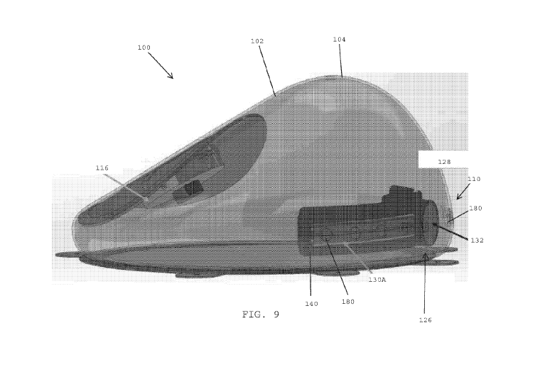
[00134] Referring to FIG. 9, in one embodiment, the shell 102 of the tissue
expander 100
preferably includes drainage openings 180 that are formed within the inferior
zone 110 of the
anterior wall 104 of the shell 102. The drainage openings 180 are preferably
aligned with the first
and second fluid reservoirs 164A, 164B (FIG. 6) of the drain cover 128 of the
drain port assembly
126. The outer face 140 of the drain cover 128 surrounds the drainage openings
180 in the shell
102 to ensure that the bodily fluids are collected inside the drain cover 128
and do not mix with
the fluid used to fill and expand the shell 102.
CA 03229484 2024-02-15
WO 2023/021346
PCT/IB2022/056848
22
[00135] In one embodiment, the bodily fluid that accumulates outside the shell
102, within
and/or adjacent the inferior zone 110 of the shell, preferably passes through
the drainage
openings 180 for being collected by the first and second drains 130A, 130B
that are disposed
within the first and second fluid reservoirs of the drain cover 128. In one
embodiment, a needle
may be inserted into the drain port septum 132 to generate a vacuum or
negative pressure for
draining the fluid that has been collected inside the drains and the fluid
reservoirs of the drain
cover 128.
[00136] In the embodiments shown in FIGS. 2A-9, the drain cover has a central
hub that is
positioned "in-line" with the longitudinal axis of the drain cover, and the
drain port septum, the
drain port needle guard, and the drain port magnet are also positioned "in-
line" with the
longitudinal axis of the drain cover. In other embodiments, the location of
the central hub of the
drain cover; the drain port septum, the drain port needle guard, and the drain
port magnet may
be "off-set" from the longitudinal axis of the drain cover.
[00137] Referring to FIGS. 10A and 10B, in one embodiment, the tissue expander
200
preferably includes a shell 202 and a stabilizing base 212 that is adapted for
being secured over
a posterior wall 206 of the shell. In one embodiment, the stabilizing base 212
may close and/or
seal a manufacturing opening located in the posterior wall 206 of the shell.
[00138] In one embodiment, the tissue expander 200 preferably includes a fill
port assembly
216 that may be utilized for adding fluid (e.g., saline) into and/or removing
fluid from the shell 202
for adjusting the size and/or shape of the tissue expander 200. In one
embodiment, the fill port
assembly 216 desirably includes a self-sealing safety patch 218; a fill port
septum 220, a fill port
needle guard 222 that is adapted to receive the fill port septum 220; and a
fill port magnet 224
that may be utilized by medical personnel for locating the fill port assembly
inside the shell 202 of
the tissue expander 200. The fill port septum is desirably self-sealing for
preventing the leaking
of fluid from the tissue expander 200 after an injection needle is removed
from the fill port
assembly 216. In one embodiment, the fill port assembly 216 is 1) located in a
superior zone 208
of the shell 202, 2) located inside the shell 202, and 3) is secured to an
inner surface of the shell
202.
[00139] In one embodiment, the tissue expander 100 preferably includes a drain
port assembly
226 that is preferably configured for draining fluid that accumulates around
the outer perimeter of
CA 03229484 2024-02-15
WO 2023/021346
PCT/IB2022/056848
23
the tissue expander and/or the inferior zone 210 of the shell 202. In one
embodiment, the drain
port assembly 226 preferably includes a drain cover 228 having an off-set hub
256, a drain 230,
a drain port septum 232, a drain port needle guard 234, and a drain port
magnet 236. The off-set
hub 256 is preferably not in alignment with the longitudinal axis of the drain
cover 228. The off-
set hub 256 may be located along an upper edge of the drain cover 228. In one
embodiment, the
drain port assembly 226 is 1) located in the inferior zone 210 of the shell
202, 2) located inside
the shell 202, and 3) secured to an inner surface of the shell 202.
[00140] Referring FIGS. 11A-11C, in one embodiment, the tissue expander 200
preferably
includes the fill port assembly 216, which is desirably located within the
superior zone 208 of the
anterior wall 204 of the shell 202. In one embodiment, a major superior face
238 of the self-
sealing safety patch 218 is secured to the inner surface 205 of the shell 202.
[00141] In one embodiment, the tissue expander 200 preferably includes the
drain port
assembly 226, which is desirably located within the inferior zone 210 of the
anterior wall 204 of
the shell 202. In one embodiment, an outer face 240 of the drain cover 228 is
preferably secured
to the inner surface 205 of the shell 202 to form a water-tight seal between
the drain cover and
the shell 202. The water-tight seal preferably isolates the bodily fluids
collected in the drain cover
from the fluid that is used to fill the shell.
[00142] The drain port assembly 226 preferably includes the drain cover 228
having the off-
set hub 256, the drain 230, a drain port septum 232, a drain port needle guard
234, and a drain
port magnet 236. The off-set hub 256 is preferably not in alignment with the
longitudinal axis of
the drain cover 228.
[00143] In one embodiment, the shell 202 preferably includes drain openings
280 (FIG. 110)
that extend through the shell 202 and that are aligned with the fluid
reservoir 264 of the drain
cover 228. The bodily fluid that passes through the drain openings 280 is
directed into the drain
cover for later removal via draining the fluid through the off-set hub and the
drain port septum.
[00144] In one embodiment, the tissue expander 200 preferably includes the
stabilizing base
212 that closes the manufacturing opening that is present in the posterior
wall 206 of the shell
202. The stabilizing base 212 preferably includes tabs 214 that may be used to
anchor the tissue
expander 200 to tissue of a patient.
CA 03229484 2024-02-15
WO 2023/021346
PCT/IB2022/056848
24
[00145] Referring to FIG. 12, in one embodiment, the drain port assembly 226
preferably
includes the drain cover 228 having the outer face 240 that is adapted to be
sealed against an
inner surface of a shell within the inferior zone 210 of the anterior wall 204
of the shell 202 (FIG.
11C). In one embodiment, the outer face 240 of the drain cover 228 defines a
convex curved
surface that preferably conforms to (i.e., mirrors) the concave curved inner
surface of the shell
within the inferior zone of the shell.
[00146] In one embodiment, the drain cover 228 preferably includes an
elongated body 254
having a curved shape that conforms to and/or matches the curved inner surface
of the shell
within the inferior zone of the anterior wall of the shell. In one embodiment,
the drain cover 228
preferably includes the off-set hub 256 having an annular rim 258 having an
open outer end and
an inner end that is closed by an end wall 260. The annular rim 258 of the off-
set hub 256
preferably includes a fluid opening 262. In one embodiment, the drain cover
228 preferably has
a fluid reservoir 264 that is in fluid communication with the fluid opening
262 of the off-set hub
256. In one embodiment, under negative pressure, fluid that has accumulated
within the fluid
reservoir 264 of the drain cover may be drawn through the fluid opening 262
and into the off-set,
hub 256 of the drain cover 228 for being removed from a patient.
[00147] In one embodiment, the drain port assembly 226 preferably includes a
drain 230 that
is adapted to be assembled with the drain cover 228. In one embodiment, the
drain 230 is
preferably disposed within the fluid reservoir 264. In one embodiment, the
drain 230 is desirably
positioned within the fluid reservoir 264 for draining fluid from around the
perimeter of the tissue
expander. In one embodiment, the drains are desirably aligned with the
drainage holes 280
formed in the shell 202 of the tissue expander 200 (FIG. 11C).
[00148] In one embodiment, the drain 230 may be similar to the surgical drains
disclosed in
U.S. Patent No. 4,398,910 to Blake et al., the disclosure of which is hereby
incorporated by
reference herein.
[00149] In one embodiment, the drain port assembly 226 preferably includes the
drain port
septum 232, the drain port needle guard 234 that is adapted to seat the drain
port septum 232,
and the drain port magnet 236 that aids medical personnel in identifying the
location of the drain
port assembly 226 within the shell of the tissue expander.
CA 03229484 2024-02-15
WO 2023/021346
PCT/IB2022/056848
[00150] Referring to FIG. 13A-13D, in one embodiment the drain port
assembly 226 is
preferably located inside the shell 202 of the tissue expander 200. In one
embodiment, the drain
port assembly 226 is preferably located within the inferior zone 210 of the
anterior wall 204 of the
shell 202. In one embodiment. the outer face 240 of the drain cover 228
preferably conforms to
the shape of the inner surface of the shell 202 within the inferior zone 210
of the shell 202. The
outer face 240 of the drain 228 is preferably secured to the inner surface of
the shell 202 to form
a water-tight seal with the shell.
[00151] The off-set hub 256 of the drain cover 228 is adapted to receive the
drain cover septum
232, the drain cover needle guard 234 (FIG. 12) and the drain port magnet 236
(FIG. 12). The
hub 256 is off-set from the longitudinal axis Al (FIG. 13B) of the elongated
body 254 of the drain
cover 228. In one embodiment, the off-set hub 256 may be located at an upper
edge of the
elongated body 254 of the drain cover 228.
[00152] The drain port septum 232 is preferably located inside the drain port
needle guard 234
(FIG. 12), which. in turn, is located inside the off-set hub 256 of the drain
cover 228. The drain
230 is disposed within the fluid reservoir 264 of the drain cover 228.
[00153] In one embodiment, after the tissue expander has been implanted in a
patient, fluid
may accumulate around the inferior zone 210 of the shell 202. The fluid
preferably passes through
drain openings 280 (FIG. 11C) formed in the shell that are in alignment with
the drain cover 228.
The fluid that passes through the drain openings 280 may be collected within
the drain 230
disposed inside the fluid reservoir 264 of the drain cover 228. In one
embodiment, a needle may
be inserted through the anterior wall 204 of the shell 202 and advanced into
the drain port septum
232 for positioning the distal tip of the needle inside the drain port needle
guard 234 so that the
opening of the needle is in fluid communication with the fluid that has been
collected within the
drain 230. A syringe or pump system may be utilized for creating a vacuum
within the drain port
needle guard 234 for draining the fluid that has accumulated within the fluid
reservoir 264 and the
drain 230 of the drain port assembly 226.
[00154] In one embodiment, the drain port assembly 226 is preferably spaced
away from
and/or isolated from the fill port assembly 216 that is located within the
superior zone 208 of the
shell 202. which preferably avoids mixing and/or cross-contamination of the
bodily fluid that has
CA 03229484 2024-02-15
WO 2023/021346
PCT/IB2022/056848
26
accumulated within the drain port assembly with the fill fluid that is used
for filling the shell of the
tissue expander 200.
[00155] Referring to FIG. 13E, in one embodiment, the drain port assembly 226
is preferably
disposed inside the shell 202 of the tissue expander 200. In one embodiment,
the drain port
assembly 226 is preferably located within the inferior zone 210 of the
anterior wall 204 of the shell
202. In one embodiment, the outer face 240 of the drain cover 228 is
preferably secured against
the inner surface 205 of the shell 202 to form a water-tight seal between the
outer face of the
drain cover 228 and the inner surface 205 of the shell 202. The presence of
the water-tight seal
between the drain cover 228 and the shell 202 preferably ensures that fluid
used to inflate the
shell 202 does not mix with the bodily fluid collected by the drain port
assembly 226 (FIG. 13A).
In one embodiment, the drain port needle guard 234 is disposed inside the off-
set hub 256 of the
drainage cover 228. The drain port magnet 236 is disposed between the bottom
wall 270 of the
drain port needle guard 234 and the closed wall 260 of the off-set hub 256 of
the drain cover 228.
The drain port septum 232 is seated within the drain port needle guard 234.
The fluid openings
272,262 formed in the respective drain port needle guard 234 and the off-set
hub 256 of the drain
cover 228 provide fluid communication with the drain 230 that is disposed
within the fluid reservoir
264 of the drain cover 228. In one embodiment, fluid opening 262 is larger
than fluid opening
272, and may be substantially large such that the annular rim 258 of the off-
set hub 256 only
bounds the superior and lateral surfaces of the annular rim of the drain port
needle guard 234.
[00159 Referring FIG. 14, in one embodiment, a tissue expander 300 has a
construction that
is similar to the tissue expander shown and described above in FIGS. 10A-14.
The tissue
expander 300 preferably includes a fill port assembly 316, located within a
superior zone 308 of
an anterior wall 304 of a shell 302, which is used for filling the shell with
a fluid, and a drain port
assembly 326, located within an inferior zone 310 of the anterior wall 304 of
the shell 302, which
is used for draining bodily fluids that accumulate outside the shell of the
tissue expander.
[00157] In one embodiment, the tissue expander 300 preferably includes a self-
sealing
membrane 390 that surrounds the drain port septum 332, the drain port needle
guard 334, and
the off-set hub 356 or central hub (not shown) of the drain cover 328 of the
drain port assembly
326. In one embodiment. an outer face 340 of the drain cover 328 is preferably
secured to the
CA 03229484 2024-02-15
WO 2023/021346
PCT/IB2022/056848
27
inner surface 305 of the shell 302 to form a water-tight seal between the
drain cover 328 and the
shell 302.
[00158] In one embodiment, the self-sealing membrane 390 may extend around the
outer
perimeter of the drain cover 328. The self-sealing membrane may overlie the
inner surface 305
or an outer surface of the shell 302.
[00150] In one embodiment, the self-sealing membrane 390 preferably extends
superiorly to
prevent the shell of a deflated tissue expander from folding over upon itself
and covering the drain
port assembly 326.
[00160] In one embodiment, the self-sealing membrane 390 preferably
incorporates one or
more of the embodiments disclosed in commonly assigned U.S. Provisional
Application No.
63/157285, filed on March 5, 2021 the disclosure of which is hereby
incorporated by reference
herein.
[00161] In one embodiment, the tissue expander may include a needle stop
patch. In one
embodiment, the needle stop patch may be positioned over the inner face 346 of
the drain cover
328. The needle stop patch may comprise plastic sheeting or one or more layers
of a textile
material, such as the needle stop patch made of layers of textile material
disclosed in commonly
assigned U.S. Provisional Application Ser. No. _______________________
(ETH6088USPSP1), the disclosure of
which is hereby incorporated by reference herein. In one embodiment, the drain
cover 328 is
made of a material that is of sufficient strength or durometer to act as a
needle stop.
[00162] In one embodiment. a first needle, referred to as an inflation needle,
may be used for
inflating and deflating the tissue expander with a fluid (e.g., saline). In
one embodiment, the first
needle may be a standard, injection needle that is used for inflation and
deflation of the tissue
expander. In one embodiment, the first needle may be used for injecting a
solution (e.g., saline
solution) into the shell to expand the size and/or change the shape of the
tissue expander. The
first needle may also be used for removing the fluid from the shell for
reducing the size and/or
changing the shape of the tissue expander.
[00163] In one embodiment. a second needle; referred to as a drainage needle,
may be used
for draining fluid (e.g., seroma) that collects around the shell of the tissue
expander following
CA 03229484 2024-02-15
WO 2023/021346
PCT/IB2022/056848
28
implantation. In one embodiment. the drainage needle is used for drainage
purposes only. The
drainage needle desirably includes a hollow, cylindrical shaft made of medical
grade material
(e.g., stainless steel). In one embodiment, the distal end of the hollow,
cylindrical shaft has a
sharpened tip and a drainage opening that desirably enables fluid, such as
bodily fluids (e.g.,
seroma fluid), to be drawn into the drainage needle. In one embodiment, the
second needle is
preferably an 18G needle to allow for ease of fluid withdrawal, and the drain
port septum and self-
sealing membrane is able to seal when punctured by this needle size.
[00164] After breast reconstruction surgery, surgical drains may be placed in
patients to
prevent blood and lymphatic fluid from building up under the skin, allowing
for a quicker recovery.
Some patients are sent home with drains that are implanted and connected to an
external
reservoir. Emptying these reservoirs can be traumatic for patients because
they must measure
and empty the reservoirs every morning. In many instances, patients cannot
wait to have the
drains removed. Having a means to remove seroma fluid without the need for a
drain being
attached 24 hours a day is a great benefit to the patient.
[00165] FIG. 15 shows various systems and devices that may be used for
creating a vacuum
to drain fluid that has accumulated around the tissue expanders disclosed
herein after the tissue
expanders have been implanted inside patients. The devices may be coupled with
the drain port
assemblies disclosed herein for drawing any fluid that has accumulated around
the outsides of
the shells of the respective tissue expanders. In one embodiment, a system for
generating a
vacuum preferably includes a compressible bulb 415. In one embodiment, vacuum
may be
created using a flexible, compressible reservoir 425 that draws a
substantially constant vacuum
to permit uniform removal of fluid from a surgical incision through a wound
drain catheter, such
as the surgical fluid evacuator disclosed in U.S. Patent No. 4,429,693 to
Blake et al., the
disclosure of which is hereby incorporated by reference herein. In one
embodiment, a system
having a metered container 435 may be used for drawing a vacuum to permit the
uniform removal
of fluid from a surgical site.
[00166] While the foregoing is directed to embodiments of the present
invention, other and
further embodiments of the invention may be devised without departing from the
basic scope
thereof, which is only limited by the scope of the claims that follow. For
example, the present
invention contemplates that any of the features shown in any of the
embodiments described
CA 03229484 2024-02-15
WO 2023/021346
PCT/IB2022/056848
29
herein, or incorporated by reference herein, may be incorporated with any of
the features shown
in any of the other embodiments described herein, or incorporated by reference
herein, and still
fall within the scope of the present invention.