Note: Descriptions are shown in the official language in which they were submitted.
SYSTEM FOR AUTOMATED REAL-TIME DETECTION, OUTLINING,
TRACKING AND CHARACTERIZATION OF BLOOD VESSELS IN ULTRASOUND
IMAGING
BACKGROUND
FIELD
[0001] Embodiments relate in general to the field of signal
processing for imaging
devices, and in particular to the field of signal processing for ultrasound
imaging devices or
probes such as ones including micromachined ultrasound transducers (MUTs),
BACKGROUND
[0002] Ultrasound imaging is widely used in the fields of
medicine and non-
destructive testing.
[0003] An ultrasound imaging probe or ultrasonic imaging device
typically includes
an array of many individual ultrasonic transducers (pixels) that are used to
emit and receive
acoustic energy relative to a target to be imaged. A reflected waveform is
received by a
transducer (for example, a micro-machined ultrasonic transducer), converted to
an electrical
signal and, with further signal processing, an image is created. Fluid
velocity and direction of
fluid flow (for example, with respect to blood flow) may also be measured or
detected by
ultrasound and presented visually to the ultrasound imaging device operator.
This
quantification and visualization of anatomical structures and movement can be
utilized in
support of a range of medical diagnostic applications and other medical
procedures..
[0004] Among the most common medical procedures is vascular
access including
procedures involving the placement of intravenous catheters, including
peripherally inserted
central catheters (PICC), central venous catheters (CVC), and peripheral
intravenous (PIV)
catheters.
[0005] However, catheter placement, which involves insertion of
a needle, may be
difficult, and may require multiple attempts. Each extra attempt can cause
unnecessary pain,
1
CA 03230241 2024- 2- 27
injury, and health risks for the patient, while creating added labor and
materials costs for the
healthcare institution. When an artery is inadvertently struck by a needle,
significant and
potentially dangerous bleeding can occur. When a nerve is inadvertently struck
by a needle, it
can cause unnecessary pain for the patient.
SUMMARY
[0006] The ultrasonic imaging device of some embodiments may
operate according to
one or more sets of instructions, including algorithms, which may be used
collectively or
individually, to assist a user of an ultrasound imaging device to identify
human or animal
anatomical features such as veins, arteries and nerves, such as for the
purpose of guiding the
placement of intravenous catheters.
BRIEF DESCRIPTION OF THE DRAWINGS
[0007] The novel features of the invention are set forth with
particularity in the
appended claims. A better understanding of the features and advantages of Some
embodiments will be obtained by reference to the following detailed
description that sets forth
illustrative embodiments, in which the principles of the invention are
utilized, and the
accompanying drawings (also "Figure" and "Fig." herein), of which:
[0008] Fig. 1 is a block diagram of an imaging device with
selectively alterable
characteristics, in accordance with disclosed embodiments.
[0009] Fig. 2 is a diagram of an imaging system with
selectively alterable
characteristics, in accordance with disclosed embodiments.
[0010] Fig. 3 is a schematic diagram of an imaging device with
selectively alterable
characteristics, in accordance with disclosed embodiments.
[0011] Fig. 4 depicts an embodiment of a touchscreen user
interface (UI) according to
Some embodiments, which displays a real-time B-mode ultrasound image sequence,
along
with various interpretive overlays, virtual indicator lights, measurements,
recommendations,
controls and parameters.
[0012] Fig. 5 depicts, in flowchart form, the operations of the
blood vessel detection,
outlining, tracing and characterization algorithm, as implemented in an
ultrasound imaging
system, according to one embodiment.
2
CA 03230241 2024- 2- 27
[0013] Fig. 6 depicts a flowchart of a process according to
some embodiment.
DETAILED DESCRIPTION
[0014] Some embodiments relate to imaging devices, and more
particularly to
ultrasound imaging devices that are electronically configurable. Ultrasound
imaging devices
may be used to image internal tissue, bones, blood flow, or organs of human or
animal bodies
in a non-invasive manner. The images can then be displayed. To perform
ultrasound
imaging, the ultrasound imaging devices transmits an ultrasonic signal into
the body and
receive a reflected signal from the body part being imaged. Such ultrasound
imaging devices
include transducers and associated electronics, which may be referred to as
transceivers or
imagers, and which may be based on photo-acoustic or ultrasonic effects. Such
transducers
may be used for imaging and may be used in other applications as well. For
example, the
transducers may be used in medical imaging; flow measurements in pipes,
speaker, and
microphone arrays; lithotripsy; localized tissue heating for therapeutic; and
highly intensive
focused ultrasound (HIFU) surgery.
[0015] Additional aspects and advantages of some embodiments
will become readily
apparent to those skilled in this art from the instant detailed description,
wherein only
illustrative embodiments are shown and described. As will be realized, some
embodiments are
capable of achieving other, different goals, and their several details are
capable
of modifications in various obvious respects, all without departing from the
disclosure.
Accordingly, the drawings and description are to be regarded as illustrative
in nature, and not
as restrictive.
[0016] Ultrasound imaging is being used increasingly to improve
outcomes in
vascular access by providing direct visualization of vessels and nerves before
and during
needle insertion. The human interpretation of ultrasound imaging is
challenging for the
clinical practitioner (i.e. clinical human practitioner) due to the difficulty
of interpretation of
the images by a human operator. Therefore, the use of ultrasound for catheter
placement has
been limited mainly to the more-demanding task of placing central lines (PICC
and CVC),
which is often done by specialists, whereas the more routine PIV performed by
nurses are
usually done without the benefit of ultrasound. However, even for experienced
human
3
CA 03230241 2024- 2- 27
practitioners, ultrasound image quality in some patients can make
interpretation of such
images for vein or artery identification unreliable.
[0017] Although the instant disclosure mentions vein
detection/identification and vein
tracking, it is to be understood and embodiments are not so limited, and
include within their
scope the identification of human or animal vessels that sustain fluid flow
(hereafter, "flow
vessel").
[0018] Once a vein is successfully found, the vein diameter
must be measured, and an
appropriate size of catheter must be determined. Vein diameter is typically
measured in a
semi-manual way by using hand-drawn "calipers" on the screen of the ultrasound
imager.
Appropriate catheter size is usually determined from the vein diameter by
applying a formula
or looking up the value in a table. These steps cost valuable time, which may
be avoided by
automating the process.
[0019] For all users, it is beneficial to confirm that the
vessel chosen for access is truly
a vein. It should also be demonstrated that the vein is compressible, to avoid
accessing a vein
affected by clotting.
[0020] Consequently, there exists a need to: 1) simplify the
process of ultrasound-
guided vascular access so that less-experienced practitioners can utilize the
technique; 2)
improve outcomes achieved by practitioners, even those who are already
experienced with
ultrasound imaging; 3) and shorten the time taken to complete the procedure.
[0021] Some embodiments fulfill these needs through the use of
computerized
algorithms for automatic interpretation of ultrasound images generated by
ultrasonic imagers.
The computerized algorithm of some embodiments are implemented in an
ultrasound imaging
system and performs identifying and delineating (outlining) or otherwise
visually indicating
veins and arteries; measuring and characterizing vessels; assessing a vein's
suitability for
access; and recommending of catheter gauge. A feature of some embodiments is
their ability
to be applied in real time during an insertion procedure involving an
insertion of a foreign
body in the vessel, allowing the practitioner to quickly identify vessels as
they appear on
screen, and to track these structures as the scan progresses.
[0022] In general, some embodiments relate to imaging devices,
and more particularly
to imaging devices having electronically configurable ultrasonic transducers.
Non-intrusive
4
CA 03230241 2024- 2- 27
imaging devices may be used to image internal tissue, bones, blood flow, or
organs of human
or animal bodies. The images can then be displayed. To perform the imaging,
the imaging
devices transmit a signal into the body and receive a reflected signal from
the body part being
imaged. Such imaging devices include transducers, which may be referred to as
transceivers
or imagers, and which may be based on photo-acoustic or ultrasonic effects.
Such transducers
may be used for imaging and may be used in other applications as well. For
example, the
transducers may be used in medical imaging; flow measurements in pipes,
speaker, and
microphone arrays; lithotripsy; localized tissue heating for therapeutic
purposes; and highly
intensive focused ultrasound (HIFU) surgery.
[0023] Traditionally, imaging devices such as ultrasound
imagers used in medical
imaging use piezoelectric (PZT) materials or other piezo ceramic and polymer
composites.
Such imaging devices may include a housing to house the transducers with the
PZT material,
as well as other electronics that form and display the image on a display
unit. To fabricate the
bulk PZT elements or the transducers, a thick piezoelectric material slab may
be cut into large
rectangular shaped PZT elements. These rectangular-shaped PZT elements may be
expensive
to build, since the manufacturing process involves precisely cutting generally
the rectangular-
shaped thick PZT or ceramic material and mounting it on substrates with
precise spacing.
Further, the impedance of the transducers is much higher than the impedance of
the
transmit/receive electronics for the transducers, which can affect
performance.
[0024] Still further, such thick bulk PZT elements can require
very high voltage
pulses, for example 100 volts (V) or more to generate transmission signals.
This high drive
voltage results in high power dissipation, since the power dissipation in the
transducers is
proportional to the square of the drive voltage. This high power dissipation
generate heat
within the imaging device such that cooling arrangements are necessitated.
These cooling
systems increase the manufacturing costs and weights of the imaging devices
which makes
the imaging devices more burdensome to operate.
[0025] Even further, the transmit/receive electronics for the
transducers may be
located far away from the transducers themselves, thus requiring micro-coax
cables between
the transducers and transmit/receive electronics. In general, the cables have
a precise length
CA 03230241 2024- 2- 27
for delay and impedance matching, and, quite often, additional impedance
matching networks
are used for efficient connection of the transducers through the cables to the
electronics.
[0026] Some embodiments may be utilized in the context of
imaging devices that
utilize either piezoelectric micromachined ultrasound transducer (pMUT) or
capacitive
micromachine ultrasonic transducer (cMUT) technologies, as described in
further detail
herein.
[0027] In general, MUTs, such as both cMUT and pMUT, include a
diaphragm (a thin
membrane attached at its edges, or at some point in the interior of the
probe), whereas a
"traditional," bulk PZT element typically consists of a solid piece of
material.
[0028] Piezoelectric micromachined ultrasound transducers
(pMUTs) may be
efficiently formed on a substrate leveraging various semiconductor wafer
manufacturing
operations. Semiconductor wafers may currently come in 6 inch, 8 inch, and 12
inch sizes
and are capable of housing hundreds of transducer arrays. These semiconductor
wafers start
as a silicon substrate on which various processing operations are performed.
An example of
such an operation is the formation of SiO2 layers, also known as insulating
oxides. Various
other operations such as the addition of metal layers to serve as
interconnects and bond pads
are performed to allow connection to other electronics. Yet another example of
a machine
operation is the etching of cavities. Compared to the conventional transducers
having bulky
piezoelectric material, pMUT elements built on semiconductor substrates are
less bulky, are
cheaper to manufacture, and have simpler and higher performance
interconnection between
electronics and transducers. As such, they provide greater flexibility in the
operational
frequency of the imaging device using the same, and potential to generate
higher quality
images.
[0029] In some embodiments, the imaging device is coupled to an
application specific
integrated circuit (ASIC) that includes transmit drivers, sensing circuitry
for received echo
signals, and control circuitry to control various operations. The ASIC may be
formed on
another semiconductor wafer. This ASIC may be placed in close proximity to
pMUT or
cMUT elements to reduce parasitic losses. As a specific example, the ASIC may
be 50
micrometers (gm) or less away from the transducer array. In a broader example,
there may be
less than 100 gm separation between the 2 wafers or 2 die, where each wafer
includes many
6
CA 03230241 2024- 2- 27
die and a die includes a transducer in the transducer wafer and an ASIC in the
ASIC wafer. In
some embodiments, the ASIC has matching dimensions relative to the pMUT or
cMUT array
and allows the devices to be stacked for wafer-to-wafer interconnection or
transducer die on
ASIC wafer or transducer die to ASIC die interconnection. Alternatively, the
transducer can
also be developed on top of the ASIC wafer using low temperature piezo
material sputtering
and other low temperature processing compatible with ASIC processing.
[0030] Wherever the ASIC and the transducer interconnect,
according to one
embodiment, the two may have similar footprints. More specifically, according
to the latter
embodiment, a footprint of the ASIC may be an integer multiple or divisor of
the MUT
footprint.
[0031] Regardless of whether the imaging device is based on
pMUT or cMUT, an
imaging device according to some embodiments may include a number of transmit
channels
and a number of receive channels. Transmit channels are to drive the
transducer elements
with a voltage pulse at a frequency the elements are responsive to. This
causes an ultrasonic
waveform to be emitted from the elements, which waveform is to be directed
towards an
object to be imaged, such as toward an organ in a body. In some examples, the
imaging
device with the array of transducer elements may make mechanical contact with
the body
using a gel in between the imaging device and the body. The ultrasonic
waveform travels
towards the object, i.e., an organ, and a portion of the waveform is reflected
back to the
transducer elements in the form of received/reflected ultrasonic energy where
the received
ultrasonic energy may converted to an electrical energy within the imaging
device. The
received ultrasonic energy may then be further processed by a number of
receive channels to
convert the received ultrasonic energy to electrical signals, and the
electrical signals may be
processed by other circuitry to develop an image of the object for display
based on the
electrical signals.
[0032] An embodiment of an ultrasound imaging device includes a
transducer array,
and control circuitry including, for example, an application-specific
integrated circuit (ASIC),
and transmit and receive beamforming circuitry, and optionally additional
control electronics.
[0033] An imaging device incorporating features of the
embodiments may
advantageously reduce or resolve issues
7
CA 03230241 2024- 2- 27
[0034] In an embodiment, an imaging device may include a
handheld casing where
transducers and associated electronic circuitries, such as a control circuitry
and optionally a
computing device are housed. The imaging device may also contain a battery to
power the
electronic circuitries.
[0035] Thus, some embodiments pertain to a portable imaging
device utilizing either
pMUT elements or cMUT elements in a 2D array. In some embodiments, such an
array of
transducer elements is coupled to an application specific integrated circuit
(ASIC) of the
imaging device.
[0036] In the following description, for purposes of
explanation, specific details are
set forth in order to provide an understanding of the disclosure. It will be
apparent, however,
to one skilled in the art that the disclosure may be practiced without these
details.
Furthermore, one skilled in the art will recognize that examples of the
present disclosure,
described below, may be implemented in a variety of ways, such as a process,
one or more
processors (processing circuitry) of a control circuitry, one or more
processors (or processing
circuitry) of a computing device, a system, a device, or a method on a
tangible computer-
readable medium.
[0037] One skilled in the art shall recognize: (1) that certain
fabrication operations
may optionally be performed; (2) that operations may not be limited to the
specific order set
forth herein; and (3) that certain operations may be performed in different
orders, including
being done contemporaneously.
[0038] Elements/components shown in diagrams are illustrative
of exemplary
embodiments and are meant to avoid obscuring the disclosure. Reference in the
specification
to "one example," "preferred example," "an example," "examples," "an
embodiment," "some
embodiments," or "embodiments" means that a particular feature, structure,
characteristic, or
function described in connection with the example is included in at least one
example of the
disclosure and may be in more than one example. The appearances of the phrases
"in one
example," "in an example," "in examples," "in an embodiment," "in some
embodiments," or
"in embodiments" in various places in the specification are not necessarily
all referring to the
same example or examples. The terms "include," "including," "comprise," and
"comprising"
shall be understood to be open terms and any lists that follow are examples
and not meant to
8
CA 03230241 2024- 2- 27
be limited to the listed items. Any headings used herein are for
organizational purposes only
and shall not be used to limit the scope of the description or the claims.
Furthermore, the use
of certain terms in various places in the specification is for illustration
and should not be
construed as limiting.
[0039] Turning now to the figures, Fig. 1 is a block diagram of
an imaging device 100
with a controller or control circuitry 106 controlling selectively alterable
channels (108, 110)
and having imaging computations performed on a computing device 112 according
to
principles described herein. As described above, the imaging device 100 may be
used to
generate an image of internal tissue, bones, blood flow, or organs of human or
animal bodies.
Accordingly, the imaging device 100 may transmit a signal into the body and
receive a
reflected signal from the body part being imaged. Such imaging devices may
include either
pMUT or cMUT, which may be referred to as transducers or imagers, which may be
based on
photo-acoustic or ultrasonic effects. The imaging device 100 may be used to
image other
objects as well. For example, the imaging device may be used in medical
imaging; flow
measurements in pipes, speaker, and microphone arrays; lithotripsy; localized
tissue heating
for therapeutic; and highly intensive focused ultrasound (HIFU) surgery.
[0040] In addition to use with human patients, the imaging
device 100 may be used to
acquire an image of internal organs of an animal as well. Moreover, in
addition to imaging
internal organs, the imaging device 100 may also be used to determine
direction and velocity
of blood flow in arteries and veins as in Doppler mode imaging and may also be
used to
measure tissue stiffness.
[0041] The imaging device 100 may be used to perform different
types of imaging.
For example, the imaging device 100 may be used to perform one-dimensional
imaging, also
known as A-Scan, two-dimensional imaging, also known as B scan, three-
dimensional
imaging, also known as C scan, and Doppler imaging (that is, the use of
Doppler ultrasound to
determine movement, such as fluid flow within a vessel). The imaging device
100 may be
switched to different imaging modes, including without limitation linear mode
and sector
mode, and electronically configured under program control.
[0042] To facilitate such imaging, the imaging device 100
includes one or more
ultrasound transducers 102, each transducer 102 including an array of
ultrasound transducer
9
CA 03230241 2024- 2- 27
elements 104. Each ultrasound transducer element 104 may be embodied as any
suitable
transducer element, such as a pMUT or cMUT element. The transducer elements
104 operate
to 1) generate the ultrasonic pressure waves that are to pass through the body
or other mass
and 2) receive reflected waves (received ultrasonic energy) off the object
within the body, or
other mass, to be imaged. In some examples, the imaging device 100 may be
configured to
simultaneously transmit and receive ultrasonic waveforms or ultrasonic
pressure waves
(pressure waves in short). For example, control circuitry 106 may be
configured to control
certain transducer elements 104 to send pressure waves toward the target
object being imaged
while other transducer elements 104, at the same time, receive the pressure
waves/ultrasonic
energy reflected from the target object, and generate electrical charges based
on the same in
response to the received waves/received ultrasonic energy/received energy.
[0043] In some examples, each transducer element 104 may be
configured to transmit
or receive signals at a certain frequency and bandwidth associated with a
center frequency, as
well as, optionally, at additional center frequencies and bandwidths. Such
multi-frequency
transducer elements 104 may be referred to as multi-modal elements 104 and can
expand the
bandwidth of the imaging device 100. The transducer element 104 may be able to
emit or
receive signals at any suitable center frequency, such as about 0.1 to about
100 megahertz.
The transducer element 104 may be configured to emit or receive signals at one
or more
center frequencies in the range from about 3.5 to about 5 megahertz.
[0044] To generate the pressure waves, the imaging device 100
may include a number
of transmit (Tx) channels 108 and a number of receive (Rx) channels 110. The
transmit
channels 108 may include a number of components that drive the transducer 102,
i.e., the
array of transducer elements 104, with a voltage pulse at a frequency that
they are responsive
to. This causes an ultrasonic waveform to be emitted from the transducer
elements 104
towards an object to be imaged.
[0045] According to some embodiments, an ultrasonic waveform
may include one or
more ultrasonic pressure waves transmitted from one or more corresponding
transducer
elements of the imaging device substantially simultaneously.
[0046] The ultrasonic waveform travels towards the object to be
imaged and a portion
of the waveform is reflected back to the transducer 102, which converts it to
an electrical
CA 03230241 2024- 2- 27
energy through a piezoelectric effect. The receive channels 110 collect
electrical energy thus
obtained, and process it, and send it for example to the computing device 112,
which develops
or generates an image that may be displayed.
[0047] In some examples, while the number of transmit channels
108 and receive
channels 110 in the imaging device 100 may remain constant, and the number of
transducer
elements 104 that they are coupled to may vary. A coupling of the transmit and
receive
channels to the transducer elements may be, in one embodiment, controlled by
control
circuitry 106. In some examples, for example as shown in Fig. 1, the control
circuitry may
include the transmit channels 108 and in the receive channels 110. For
example, the
transducer elements 104 of a transducer 102 may be formed into a two-
dimensional spatial
array with N columns and M rows. In a specific example, the two-dimensional
array of
transducer elements 104 may have 128 columns and 32 rows. In this example, the
imaging
device 100 may have up to 128 transmit channels 108 and up to 128 receive
channels 110. In
this example, each transmit channel 108 and receive channel 110 may be coupled
to multiple
or single pixels 104. For example, depending on the imaging mode (for example,
whether a
linear mode where a number of transducers transmit ultrasound waves in a same
spatial
direction, or a sector mode, where a number of transducers transmit ultrasound
waves in
different spatial directions), each column of transducer elements 104 may be
coupled to a
single transmit channel 108 and a single receive channel (110) . In this
example, the transmit
channel 108 and receive channel 110 may receive composite signals, which
composite signals
combine signals received at each transducer element 104 within the respective
column. In
another example, i.e., during a different imaging mode, each transducer
element 104 may be
coupled to its dedicated transmit channel 108 and its dedicated receive
channel 110. In some
embodiments, a transducer element 104 may be coupled to both a transmit
channel 108 and a
receive channel 110. For example, a transducer element 104 may be adapted to
create and
transmit an ultrasound pulse and then detect the echo of that pulse in the
form of converting
the reflected ultrasonic energy into electrical energy.
[0048] The control circuitry 106 may be embodied as any circuit
or circuits
configured to perform the functions described herein. For example, the control
circuitry 106
may be embodied as or otherwise include an application specific integrated
circuit (ASIC), a
11
CA 03230241 2024- 2- 27
field programmable gate array (FPGA), a system-on-a-chip, a processor and
memory, a
voltage source, a current source, one or more amplifiers, one or more digital-
to-analog
converters, one or more analog-to-digital converters, etc.
[0049] The illustrative computing device 112 may be embodied as
any suitable
computing device including any suitable components, such as a processor,
memory,
communication circuitry, battery, display, etc. In one embodiment, the
computing device 112
may be integrated with the control circuitry 106, transducers 102, etc., into
a single package
or single chip, or a single system on a chip (SoC), as suggested for example
in the
embodiment of Fig. 1. In other embodiments, some or all of the computing
devices may be in
a separate package from the control circuitry, and the transducers, etc., as
suggested for
example in the embodiment of in Fig. 2 as will be described in further detail
below.
[0050] Each transducer element may have any suitable shape such
as, square,
rectangle, ellipse, or circle. The transducer elements may be arranged in a
two dimensional
array arranged in orthogonal directions, such as in N columns and M rows as
noted herein, or
may be arranged in an asymmetric (or staggered) rectilinear array.
[0051] Transducer elements 104 may have associated transmit
driver circuits of
associated transmit channels, and low noise amplifiers of associated receive
channels. Thus,
a transmit channel may include transmit drivers, and a receive channel may
include one or
more low noise amplifiers. For example, although not explicitly shown, the
transmit and
receive channels may each include multiplexing and address control circuitry
to enable
specific transducer elements and sets of transducer elements to be activated,
deactivated or
put in low power mode. It is understood that transducers may be arranged in
patterns other
than orthogonal rows and columns, such as in a circular fashion, or in other
patterns based on
the ranges of ultrasonic waveforms to be generated therefrom.
[0052] Fig. 2 is a diagram of an imaging environment including
an imaging system
with selectively configurable characteristics, according to an embodiment. The
imaging
system of Fig. 2 may include an imaging device 202 and a computing system 222
which
includes a computing device 216 and a display 220 coupled to the computing
device, as will
be described in further detail below.
12
CA 03230241 2024- 2- 27
[0053] As depicted in Fig. 2, the computing device 216 may,
according to one
embodiment, and unlike the embodiment of Fig. 1, be physically separate from
the imaging
device 220. For example, the computing device 216 and display device 220 may
be disposed
within a separate device (in this context, the shown computing system 222,
physically
separate from imaging device 202 during operation) as compared with the
components of the
imaging device 202. The computing system 222 may include a mobile device, such
as cell
phone or tablet, or a stationary computing device, which can display images to
a user. In
another example, as shown in Fig. 1 for example, the display device, the
computing device,
and associated display, may be part of the imaging device 202 (now shown).
That is, the
imaging device 100, computing device 216, and display device 220 may be
disposed within a
single housing.
[0054] A "computing device" as referred to herein may, in some
embodiments, be
configured to generate signals to at least one of cause an image of the object
to be displayed
on a display, or cause information regarding the image to be communicated to a
user.
Causing the information regarding the image to be displayed may include
causing identifying
information regarding an identified vessel, and recommendations regarding a
foreign object
such as a catheter to be inserted into the vessel, to be communicated to a
user via a user
interface, such as by being displayed on a display, via a voice message to be
played through a
speaker, and/or text on the UI display. The generation of the signals may
include, in some
embodiments, implementing a vessel detection and tracking algorithm as will be
described
further below.
[0055] As depicted, the imaging system includes the imaging
device 202 that is
configured to generate and transmit, via the transmit channels (Fig. 1, 108),
pressure waves
210 toward an object, such as a heart 214, in a transmit mode/process. The
internal organ, or
other object to be imaged, may reflect a portion of the pressure waves 210
toward the imaging
device 202 which may receive, via a transducer (such as transducer 102 of Fig.
1), receive
channels (Fig. 1, 110), control circuitry (Fig. 1, 106), the reflected
pressure waves. The
transducer may generate an electrical signal based on the received ultrasonic
energy in a
receive mode/process. A transmit mode or receive mode may be applicable in the
context of
imaging devices that may be configured to either transmit or receive, but at
different times.
13
CA 03230241 2024- 2- 27
However, as noted previously, some imaging devices according to embodiments
may be
adapted to be in both a transmit mode and a receive mode simultaneously. The
system also
includes a computing device 216 that is to communicate with the imaging device
100 through
a communication channel, such as a wireless communication channel 218 as
shown, although
embodiments also encompass within their scope wired communication between a
computing
system and imaging device. The imaging device 100 may communicate signals to
the
computing device 216 which may have one or more processors to process the
received signals
to complete formation of an image of the object. A display device 220 of the
computing
system 222 may then display images of the object using the signals from the
computing
device. The computing system may further convey information to a user
regarding a
defective pixel as noted above.
[0056] An imaging device according to some embodiments may
include a portable
device, and/or a handheld device that is adapted to communicate signals
through a
communication channel, either wirelessly (using a wireless communication
protocol, such as
an IEEE 802.11 or Wi-Fi protocol, a Bluetooth protocol, including Bluetooth
Low Energy, a
mmWave communication protocol, or any other wireless communication protocol as
would
be within the knowledge of a skilled person) or via a wired connection such as
a cable (such
as USB2, USB 3, USB 3.1, and USB-C) or such as interconnects on a
microelectronic device,
with the computing device. In the case of a tethered or wired, connection, the
imaging device
may include a port as will be described in further detail in the context of
Fig. 3 for receiving a
cable connection of a cable that is to communicate with the computing device.
In the case of
a wireless connection, the imaging device 100 may include a wireless
transceiver to
communicate with the computing device 216.
[0057] It should be appreciated that, in various embodiments,
different aspects of the
disclosure may be performed in different components. For example, in one
embodiment, the
imaging device may include circuitry (such as the channels) to cause
ultrasound waveforms to
be sent and received through its transducers, while the computing device may
be adapted to
control such circuitry to the generate ultrasound waveforms at the transducer
elements of the
imaging device using voltage signals, and farther a processing of the received
ultrasonic
energy to determine a defective pixel dataset for one or more defective
pixels. In such an
14
CA 03230241 2024- 2- 27
embodiment, the computing device may manage/control a functioning of the
imaging device
based on the determination of the defective pixels, may construct images of
the object using
frames as discussed in more detail below, may select and configure transmit
and receive
channels, etc.
[0058] In another embodiment, the imaging device may include
control circuitry to
control a generation of the ultrasound waveforms at the transducer elements
using voltage
signals in order to cause the ultrasound waveform to be sent and received from
the transducer
elements, and may also generate electrical signals from the received
ultrasound energy, and,
in a test mode, use electrical signals corresponding to the received
ultrasound waveforms to
determine information regarding one or more defective pixels of the imaging
device. In such
an embodiment, the control circuitry of the imaging device may send the
electrical signals
generated from the received ultrasound energy to the computing device, which
may process
them in order to determine the information regarding one or more defective
pixels. More
generally, it should be appreciated that any suitable function disclosed
herein may be
performed by one or more circuitries, and that these circuitries may be housed
in one physical
device, or housed physically separately from each other, but communicatively
coupled to one
another.
[0059] Fig. 3 represents a view of an imaging device according
to some embodiments,
as will be described in further detail below.
[0060] As seen in Fig. 3, the imaging device 300 may include a
handheld casing 331
where transducers 302 and associated electronics are housed. The imaging
device may also
contain a battery 338 to power the electronics. Fig. 3 thus shows an
embodiment of a portable
imaging device capable of 2D and 3D imaging using pMUTs in a 2D array,
optionally built
on a silicon wafer. Such an array coupled to an application specific
integrated circuit (ASIC)
106 with electronic configuration of certain parameters, enables a higher
quality of image
processing at a low cost than has been previously possible. Further by
controlling certain
parameters, for example the number of channels used, power consumption may be
altered and
temperature may be changed.
[0061] The imaging device 300 according to some embodiments is
configured to
allow system configurability and adaptability in real time based on
information regarding one
CA 03230241 2024- 2- 27
or more defective pixels (defective pixel data). This is done for example by
comparing a
current pixel performance dataset of one or more pixels of a transducer array
of an imaging
device with a baseline pixel performance dataset of the same pixels as will be
explained in
further detail below.
[0062] Now addressing Fig. 3 in more detail, Fig. 3 is a
schematic diagram of an
imaging device 300 with selectively adjustable features, according to some
embodiments.
The imaging device 300 may be similar to imaging device 100 of Fig. 1, or to
imaging device
202 of Fig. 2, by way of example only. As described above, the imaging device
may include
an ultrasonic medical probe. Fig. 3 depicts transducer(s) 302 of the imaging
device 300. As
described above, the transducer(s) 302 may include arrays of transducer
elements (Fig. 1,
104) that are adapted to transmit and receive pressure waves (Fig. 2, 210). In
some examples,
the imaging device 300 may include a coating layer 322 that serves as an
impedance matching
interface between the transducers 302 and the human body, or other mass or
tissue through
which the pressure waves (Fig. 2, 210) are transmitted. In some cases, the
coating layer 322
may serve as a lens when designed with the curvature consistent with focal
length desired.
[0063] The imaging device 300 may be embodied in any suitable
form factor. In some
embodiments, part of the imaging device 300 that includes the transducers 302
may extend
outward from the rest of the imaging device 100. The imaging device 300 may be
embodied
as any suitable ultrasonic medical probe, such as a convex array probe, a
micro-convex array
probe, a linear array probe, an endovaginal probe, endorectal probe, a
surgical probe, an
intraoperative probe, etc.
[0064] In some embodiments, the user may apply gel on the skin
of a living body
before a direct contact with the coating layer 322 so that the impedance
matching at the
interface between the coating layer 322 and the human body may be improved.
Impedance
matching reduces the loss of the pressure waves (Fig. 2, 210) at the interface
and the loss of
the reflected wave travelling toward the imaging device 300 at the interface.
[0065] In some examples, the coating layer 322 may be a flat
layer to maximize
transmission of acoustic signals from the transducer(s) 102 to the body and
vice versa. The
thickness of the coating layer 322 may be a quarter wavelength of the pressure
wave (Fig. 2,
210) to be generated at the transducer(s) 102.
16
CA 03230241 2024- 2- 27
[0066] The imaging device 300 also includes a control circuitry
106, such as one or
more processors, optionally in the form of an application-specific integrated
circuit (ASIC
chip or ASIC), for controlling the transducers 102. The control circuitry 106
may be coupled
to the transducers 102, such as by way of bumps. As described above, the
transmit channels
108 and receive channels 110 may be selectively alterable or adjustable,
meaning that the
quantity of transmit channels 108 and receive channels 110 that are active at
a given time may
be altered such that, for example, one or more pixels determined to be
defective are not used.
For example, the control circuitry 106 may be adapted to selectively adjust
the transmit
channels 108 and receive channel 110 based on pixels to be tested for defects,
and/or based on
pixels determined to be defective.
[0067] In some examples, the basis for altering the channels
may be a mode of
operation, the mode of operation may in turn be chosen based on which pixels
are determined
to be defective, and optionally based on the type of defect of each defective
pixel.
[0068] The imaging device may also include one or more
processors 326 for
controlling the components of the imaging device 100. One or more processors
326 may be
configured to, in addition to control circuitry 106, at least one of control
an activation of
transducer elements, process electrical signals based on reflected ultrasonic
waveforms from
the transducer elements or generate signals to cause generation of an image of
an object being
imaged by one or more processors of a computing device, such as computing
device 112 of
Fig. 1 or 216 of Fig. 2. One or more processors 326 may further be adapted to
perform other
processing functions associated with the imaging device. The one or more
processors 326
may be embodied as any type of processors 326. For example, the one or more
processors 326
may be embodied as a single or multi-core processor(s), a single or multi-
socket processor, a
digital signal processor, a graphics processor, a neural network compute
engine, an image
processor, a microcontroller, a field programmable gate array (FPGA), or other
processor or
processing/controlling circuit. The imaging device 100 may also include
circuit(s) 328, such
as Analog Front End (AFE), for processing/conditioning signals, and an
acoustic absorber
layer 330 for absorbing waves that are generated by the transducers 102 and
propagated
towards the circuits 328. That is, the transducer(s) 102 may be mounted on a
substrate and
may be attached to an acoustic absorber layer 330. This layer absorbs any
ultrasonic signals
17
CA 03230241 2024- 2- 27
that are emitted in the reverse direction (i.e., in a direction away from
coating layer 322 in a
direction toward port 334), which may otherwise be reflected and interfere
with the quality of
the image. While Fig. 3 depicts the acoustic absorber layer 330, this
component may be
omitted in cases where other components prevent a material transmission of
ultrasound in the
reverse direction.
[0069] The analog front end 328 may be embodied as any circuit
or circuits
configured to interface with the control circuitry 106 and other components of
the imaging
device, such as the processor 326. For example, the analog front end 328 may
include, e.g.,
one or more digital-to-analog converters, one or more analog-to-digital
converters, one or
more amplifiers, etc.
[0070] The imaging device may include a communication unit 332
for communicating
data, including control signals, with an external device, such as the
computing device (Fig. 2,
216), through for example a port 334 or a wireless transceiver. The imaging
device 100 may
include memory 336 for storing data. The memory 336 may be embodied as any
type of
volatile or non-volatile memory or data storage capable of performing the
functions described
herein. In operation, the memory 336 may store various data and software used
during
operation of the imaging device 100 such as operating systems, applications,
programs,
libraries, and drivers.
[0071] In some examples, the imaging device 100 may include a
battery 338 for
providing electrical power to the components of the imaging device 100. The
battery 338
may also include battery charging circuits which may be wireless or wired
charging circuits
(not shown). The imaging device may include a gauge that indicates a battery
charge
consumed and is used to configure the imaging device to optimize power
management for
improved battery life. Additionally or alternatively, in some embodiments, the
imaging device
may be powered by an external power source, such as by plugging the imaging
device into a
wall outlet.
[0072] Some embodiments overcome disadvantages over the prior
art with respect to
identification of vessels in a body, such as a vessel within which fluid
flows, such as a vessel
of a living body within which fluid flows, that would mitigate issues with
vessel detection for
the insertion of foreign bodies into the vessel, especially where fluid may
flow within the
18
CA 03230241 2024- 2- 27
vessel. Computer algorithms according to some embodiments enable the detection
and
tracking of vessels within which a fluid flows (hereinafter "vessels") this
way facilitating the
detection of blood vessels for medical intervention, such as the insertion of
a foreign body
(e.g. a needle or catheter or the like).
[0073] According to some embodiments, the algorithm may relies
not only on A, B or
C-mode imaging by an ultrasonic device for vessel detection, but also on
imaging the allows
the determination of fluid flow within a detected vessel.
[0074] Typically, by way of example in B-mode (2 dimensional)
ultrasonic images,
veins and arteries appear as dark, oval regions on the displayed image when
seen in a short-
axis view (i.e., in a cross sectional view taken perpendicular to the
direction of blood flow).
In principle, according to some embodiments, a computer algorithm including
object
recognition may be implemented to detect or identify vessels in an A, B or C-
mode ultrasound
image sequence by detecting a presence of such oval regions.
[0075] By itself, an object detector yields inadequate vessel
detection performance
due to the presence of confounding tissue texture and imaging artifacts in the
displayed image
or image frame. Furthermore, when veins are collapsed by the force of the
imaging probe
during the procedure, they may become impossible to detect in an individual B-
mode image
frame.
[0076] A novel aspect of embodiments is the use of additional
flow information,
beyond what is seen in an individual image ultrasonic frame, such as a B-mode
frame. The
use of such additional flow information can enhance the accuracy and
computational
efficiency of vessel detection and identification. Some embodiments use two
additional
sources of information beyond a single ultrasonic image frame (such as a B-
mode image
frame): 1) flow data (such as Doppler flow data, including either color
Doppler or power
Doppler), and 2) one or more ultrasonic image frames preceding the current
ultrasonic image
frame (such as a B-mode image frame) in the time domain. In some embodiments,
flow data
refers to flow data only for flow that has a component perpendicular to a
plane of the
ultrasonic images used to detect and track a vessel.
[0077] In the context of the instant description, when
referring to an "ultrasonic
image" or "ultrasonic images" in the singular or in the plural, what is being
referred to is one
19
CA 03230241 2024- 2- 27
or more images generated as a result of using an ultrasonic device. The one or
more images
could include A-mode, B-mode or C-mode images, and preferably B-mode (two
dimensional
images).
[0078] In some instances, less-experienced ultrasound human
users may find flow
images, such as flow images obtained using Doppler imaging, confusing, since
such images
may sometimes be noisy, and may be displayed overlaid on the ultrasonic image,
thereby
obscuring the anatomical detail depicted within the ultrasonic image. A
feature of some
embodiments is that information or data acquired through flow imaging may be
acquired and
used by the computer algorithm "behind the scenes," that is, it may not be
displayed to a
human user, but consumed by the computer algorithm in order to identify a
vessel in a body.
Thus, according to one example, Doppler imaging data may be processed by an
algorithm
according to some embodiments to promote more accurate and more efficient
vessel detection
by a computing device.
[0079] Some embodiments recognize that object recognition
through ultrasound may
benefit from identifying image locations worthy of being searched further in
order to
recognize/detect/identify the object. Usually these locations are identified
from the image
itself. However, some embodiments use a separate flow image (such as either
color Doppler
or power Doppler) to identify candidate locations for finding vessels in the
corresponding
ultrasonic image (these locations are referred to herein as flow seeds), or as
confirmatory
information regarding whether an object identified as a vessel in which fluid
flows is indeed a
vessel in which fluid flows. Doppler information may be effective in this
task, because flow
detection is a good signature or indicator of the presence of a vessel.
[0080] If a spatial location exhibits flow determined based on
flow data (such as data
obtained through Doppler imaging), and strong evidence of a predetermined
shape (such as an
elliptical shape) centered near the same location in the corresponding B-mode
image, then this
suggests high confidence that a vessel is present at this location¨greater
confidence than
what is provided by either signature (i.e. flow only or ultrasonic image frame
only) considered
individually. Some embodiments exploit this idea by searching an ultrasonic
image for the
predetermined shape in proximity to flow seeds, such as Doppler seeds. The
type of flow,
such as whether pulsating based on heart rate, may indicate an artery, versus
a comparatively
CA 03230241 2024- 2- 27
more constant flow may indicate a vein. The above technique not only improves
detection
performance of a computing device, but also reduces the search space, yielding
valuable
computational efficiency. An alternative approach is to use flow information
to complement
data indicating an ultrasonic image of a vessel from an ultrasonic deep-
learning detector when
computing confidence of the presence of a vessel.
[0081] Some embodiments incorporate predictive tracking as a
further source of seed
locations to be searched (in addition to the Doppler seeds). These "tracker
seeds" are obtained
by using the shape, location, and/or apparent translational velocity of each
vessel (due to
relative motion of the vessel and the probe) detected in the current frame to
predict the shape
and location of that vessel in the next frame. Tracking improves detection
performance by
exploiting interframe consistency. If the algorithm suspects that a vessel is
present in a given
image frame, the fact that it has the expected shape and location enhances
confidence in this
conclusion.
[0082] Tracking also reduces the ranges of shapes and locations
of ellipses that must
be searched, thereby reducing computational burden.
[0083] In alternative embodiment, rather than separate object
detection and tracking, a
spatio-temporal detection method may be used which jointly analyzes recent
image frames to
make a vessel detection decision for the current frame. A spatio-temporal
detection method
involves a two dimensional spatial dimension and a third dimension based on
time. This may
be done, for example, using a multichannel implementation of the You Only Look
Once
(YOLO) algorithm (in which the current frame and a set of preceding frames
serve as the
channels) or the combination of a convolutional neural network (which performs
spatial
analysis) and long short-term memory network (which performs temporal
analysis).
[0084] In addition to their accuracy benefits, tracking and
spatio-temporal detection
allow the algorithm to keep track of which vessel is which (e.g. artery versus
vein), permitting
consistent annotation on the user interface, and allowing parameters of each
vessel to be
analyzed temporally for purposes of detection and discrimination of veins and
arteries.
[0085] In a preferred embodiment, a similar tracking approach
to that used for
ultrasound imaging may also be applied to a concurrently acquired flow data,
such as
concurrently applied Doppler flow video. Doppler tracking increases the
confidence in the
21
CA 03230241 2024- 2- 27
tracker seed locations (analogous to the confidence boost described above in
the context of
beginning with flow seeds and following up with ultrasonic imaging), and
enables each vessel
to be uniquely tracked in the Doppler sequence so that the flow information
may be analyzed
by motion-compensated temporal processing for use in vein-artery
discrimination.
[0086] In a preferred embodiment, Doppler information may be
used to produce
evidence as to a vessel's identity as a vein or artery by applying signal
processing methods to
analyze the periodic behavior of flow as evidence of pulsatility (a feature
typically associated
with arteries). In some embodiments, a determination as to pulsatility of a
vessel may be
accomplished by a machine learning classifier. The machine learning classifier
may use
ultrasonic image data, or it may use measurements of a scalar index of
pulsatility. In some
embodiments, pulsatility may be determined by a computing device by analyzing
data relating
to spatial movements in proximity to a vessel as between successive ultrasound
images. In
alternative embodiments, pulsatility may be assessed by local analysis of
anatomical motion
(e.g. of the vessel walls) in the ultrasonic imagery.
[0087] In the upper arm, where peripherally inserted central
catheter (PICC) and
central venous catheter (CVC) lines (as examples of foreign objects to be
inserted into bodily
vessels) are generally placed, there is only one major artery, the brachial
artery, which artery
is very close to brachial veins. The other two major veins¨the basilic vein
and the cephalic
vein¨are further separated from the brachial artery. Thus, in the upper arm,
if a large vessel,
imaged by the ultrasound device of some embodiments, is not immediately
adjacent to any
other large vessel, it is more likely a vein than an artery, because the only
artery will have
other large vessels close to it. An upper arm major vein not close to any
other major vessel
of the upper arm is one of two preferred bodily veins for line access. Some
embodiments may
use an flow information, along with ultrasonic images, to detect blood vessel
location, and
may, in this way, provide a strong signature for vein-artery discrimination,
such as in the
upper arm. Such signature may be provided by using data on vessel location,
pulsatility
and/or compressibility, along with flow data. This may be done by using each
of these
individually or in combination, combined using machine learning or by simple
Boolean logic.
22
CA 03230241 2024- 2- 27
[0088] Within the context of some embodiments, each vessel may
be accessed in real-
time (during detection and tracking of a blood vessel), or at the end of the
imaging session
involving detection and tracking of a blood vessel.
[0089] A blood vessel may best be accessed only if it is a vein
having certain
characteristics. The vein may best be compressible, because incompressibility
can imply that
the vein contains a clot, which may break off and travel to the lungs if the
vessel is accessed.
In standard practice, the operator of the probe observes compressibility by
using the probe to
apply pressure to the tissue, thereby squeezing the vessels. In some
embodiments,
compressibility is measured automatically. The vein may best have a sufficient
diameter to
accommodate a catheter, in accordance with vessel occupancy standards that are
generally
established by a providing healthcare institution. As described previously,
arteries may best
be avoided. In the upper arm, since the preferred veins (basilic and cephalic)
for access by a
needle or catheter are not near any artery, an isolated vessel may be detected
by an algorithm
according to some embodiments to be a vein rather than an artery, and as such
safely distant
from any artery because it is in the upper arm, and not near any other large
vessels.
[0090] In summary, for a vessel to be a good candidate to be
accessed for catheter
placement or needle placement, it may, according to some embodiments meet the
following
criteria: 1) be a vein, 2) be compressible, and 3) have a diameter should be
sufficiently large
to accommodate the foreign object to be inserted therein. In the upper arm,
there is an
additional criterion which may be applied, that the vessel should not be close
to other
comparably large vessels/may be isolated from other comparably large vessels.
[0091] In a preferred embodiment, suitability for vascular
access may be determined
by a computing device by way of accessing a logical truth table based on the
relevant criteria.
In alternative embodiments, measures of these criteria may serve as features
for use by a
machine learning algorithm run on the computing device.
[0092] Some embodiments include a system of algorithmic
processing components
and a user interface component. In a preferred embodiment, these components
may operate as
described further below.
23
CA 03230241 2024- 2- 27
[0093] Some embodiments include a novel user interface (UI)
that defines the
functionality and manner of information presentation pertaining to detection
and tracking of a
vessel using ultrasonic imaging.
[0094] Some embodiments pertain to algorithms having
algorithmic components
described in greater detail herein, executed on one or more processors of a
computing system,
such as computing system 112 or 216, to provide information displayed in the
user interface,
the information including identification of a vessel, and information
regarding its accessibility
by one or more foreign objects, such as a catheter and/or a needle.
[0095] According to some embodiments, the algorithm is to
implement detection and
tracking of a vessel, including for example providing an outline of the vessel
on the UI.
According to embodiments related to detection and track, a potential vessel
may be detected
by the Vessel Scouting Function (VSF) of an algorithm. Once a potential
vessel, or a
candidate vessel, is detected for access, the algorithm may continuously
determine and cause
displaying an outline of its boundary and the location of its center or a its
diameter, and may
also predicts its outline, apparent velocity (due to relative motion of the
tissue and probe), and
location of the vessel in a next frame, for purposes of vessel tracking.
[0096] An algorithmic component of an algorithm according to
some embodiments
may implement a discrimination as between veins and arteries. In this
component, the
algorithm may continuously seek to perform such discrimination. Discrimination
may be
implemented by using criteria such as compressibility, pulsatility and spatial
location in
conjunction with a shape on an ultrasonic image, such as an elliptical shape
in a B-mode
image.
[0097] An algorithmic component of an algorithm according to
some embodiments
may automatically determine parameters or attributes of a vessel, such as its
compressibility,
its pulsatility, its diameter, its depth (or distance from the surface of the
skin), its suitability
for access, the fluid flow (or flow rate) therein, to name a few. A
recommendation of foreign
object selection for insertion, such as a catheter, may then be displayed to
the user on the UI.
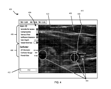
[0098] Fig. 4 depicts an embodiment of a touchscreen user
interface (UI) 400
according to some embodiments. The display of UI 400 displays a real-time B-
mode
ultrasound image sequence 402, along with various interpretive overlays,
virtual indicator
24
CA 03230241 2024- 2- 27
lights 404, vessel parameters 406, noting that a vessel parameter (or vessel
attribute) as used
herein may include any of one or more vessel dimensions (such as
diameter/radius, and
depth), vessel fluid flow rate, vessel pulsatility, vessel compressibility, or
vessel outline on an
ultrasonic image. A vessel parameter may further include vessel suitability
for access based
on the foreign object to be inserted therein. The UI may include text
communication to the
user 410, and may further include a feature 412 to allow selection, by the
user, of the type of
foreign object to be inserted, in the shown case a catheter that is a PCC.
[0099] According to some embodiments, as shown by way of
example in Fig. 4, a
series of B mode images may be displayed in real-time as time progresses.
Veins and arteries
may be caused to be shaded on the display by the algorithm using a color code
(for example,
red for artery, blue for vein). A specific candidate vessel for catheter
placement may be
outlined. Other styles of annotation, such as crosshairs, can substitute for
outlines and
shading. The algorithm may select a vein as the candidate vessel and indicate
its attributes in
an overlay 414 and in the "Vein ID" section 406 of the interface. A "Vessel
Overlays" toggle
switch may be provided to allow the user to turn off the overlays. If the user
is interested in
seeing the attributes of a different vessel, some algorithms may provide an
option to touch on
a different vessel, and the algorithm may then outline that different vessel
as the candidate
vessel and provide the new vessel's attributes. Next to the candidate vein 416
is shown its
anterior-posterior (AP) diameter (reported, for example, in millimeters), as
well as the
recommended catheter associated with that diameter (chosen based on guidelines
set by the
healthcare institution). The vertical line segment "caliper" within the
outlined candidate vein
416 depicts the path along which the AP diameter is measured. This path may be
computed
automatically by the algorithm.
[0100] An algorithm according to some embodiments may report
automated (i.e.
determined by the algorithm) findings about an outlined vessel on the UI, for
example in the
form of vein ID 406 in Fig. 4. "Vein depth" is a numeric value measured
automatically as the
distance (e.g. in cm) from the skin line (top of image) to the uppermost point
on the vein
outline. "Vein ID" may include four colored (shown by way of patterns (no
pattern, textured
pattern, and solid pattern) in Fig. 4), virtual indicator lights conveying
automatically-
determined information about the candidate vessel outlined on the image
display.
CA 03230241 2024- 2- 27
"Compressible" (green or red) indicates whether the candidate vessel may be
collapsed by
applying pressure to the tissue using the ultrasound probe (veins are
typically compressible,
while arteries are usually not). "Venous flow" (green or red) indicates
whether or not the
vessel is exhibiting a blood flow pattern that is indicative of a vein (flow
is typically more
pulsatile in an artery than in a vein; this pulsatility may be measured using
Doppler flow
information and or pulsating motions of the vessel seen in the B-mode images).
"Sufficient
diameter" (green, yellow or red) indicates whether the AP diameter is large
enough to be
targeted for catheter placement based on guidelines set by the healthcare
institution. A yellow
indication denotes that the diameter is borderline. "Suitable for access"
(green, yellow or red)
indicates whether the vein meets the criteria for catheter placement. The
"Catheter selection"
section repeats the AP diameter, and shows the corresponding recommended
catheter gauge
and French size, obtained based on permitted percent occupancy of the vessel
by the catheter,
as determined by the healthcare institution.
[0101] Other graphical elements in Fig. 4 may a user profile
button, and ultrasound
imaging parameters.
[0102] Fig. 5 depicts, in flowchart form, an exemplary process
for blood vessel
detection, outlining, tracing and characterization algorithm, as implemented
in an ultrasound
imaging system, according to one embodiment. Functional blocks or stages
implemented or
executed by the algorithm 500 are cross-referenced in the flowchart in Fig. 5.
[0103] Operation 501 performs initialization, including without
limitation, one or
more of the following algorithm parameters:
= search parameters: initial ranges of parameter values to be considered
when searching
for a best-fit vessel candidate:
o starting from a B-mode image, for example, determining
parameters to search
for and identify one or more substantially elliptical (including circular)
shapes
each within a range of predetermined parameter values; for example, aspect
ratio between 0 and 1; long-axis radius between lmm and 3mm; orientation
angle between 0 and 45 degrees; and no limit on the vessel's location within
the image.
26
CA 03230241 2024- 2- 27
o starting from flow information, determining parameters for detecting flow
seeds or Doppler seeds using Doppler or a Doppler tracker; for example, the
flow seed locations may be obtained as the local maxima of measured flow.
= vessel inventory: using a data structure containing, for each vessel
currently being
tracked by the algorithm: an identifier code, a center location, shape
parameters, and
apparent velocity (translational displacement per frame);
= seed list: data structure containing list of spatial image locations
(found as described
above) that are candidates to search for a predetermined vessel shape, such as
an
ellipse.
[0104] At operation 502, an exemplary algorithm may perform
data acquisition,
including without limitation the following:
= at operation 502-1: acquiring a B-mode image frame (Current B-mode). In
this
Operation, a B-mode image frame may be acquired in a standard way.
= At operation 502-2: acquiring a Doppler flow (color doppler or power
Doppler) image
frame (Current Doppler). In the operation 502-2, a Doppler image frame is
acquired in
the standard way. In a preferred embodiment, Current Doppler covers the same
field
of view as Current B-mode. In alternative embodiments, the Current Doppler
includes
only a subset of the image information, for example:
o full field of view initially, then switching to partial fields of view
until such
time that the probe is not in contact with the skin (which is equivalent to
beginning from scratch);
o set of all pixels within a pre-specified number of pixels of the left,
right and/or
bottom edge of Current B-mode, so as to seek vessels that were not within the
B-mode field of view in the previous frame, but are present in the current
field
of view due to motion of the probe. It is to be noted that a vessel cannot
enter
the field of view through top edge of the image, where the skin line resides.
As a result, according to some embodiments, an algorithm may apply Doppler
imaging to only image regions near these edges to detect the appearance of
vessels not yet seen;
27
CA 03230241 2024- 2- 27
o subsets of the full field of view, such as small regions
of interest or individual
scan lines, which the algorithm may request to be acquired by the probe. Such
requests may be useful, for example, in the following situation. Suppose the
B-mode Tracker has been following a given vessel in a series of image frames
through time, and predicts it to be present in the next image frame with given
location and shape parameters. Now suppose that upon searching based on
these parameters, the Quality Score (described below) does not lead to
detection of a vessel as anticipated. In this case, the algorithm may request
the
probe to interrogate, using Doppler flow imaging, a small image region
surrounding the predicted location so as to determine whether the small image
region is still a good candidate for vessel search. If so, then the B-mode
Tracker may be re-seeded.
[0105] The algorithm at operation 503 detects placement of the
ultrasonic probe. An
ultrasound scan begins when the ultrasound probe is placed in contact with the
skin (which
may coated in ultrasound gel). Therefore, the algorithms are by-passed
(dormant) until the
probe is in place. If the probe is lifted from the skin during a scan, the
algorithms are again
by-passed and the algorithm parameters are re-initialized. The condition
"probe in place" may
be detected by measuring the average image intensity of the pixels within X%
of the bottom
of the image, and comparing this value to a threshold determined T. The
percentage X and
threshold T may be determined based on example scans at various depth and gain
settings for
the specific ultrasound probe.
[0106] An exemplary algorithm at operations 504 and 505
performs Doppler seed
detection and tracking and B-mode vessel detection, tracking and outline
calculation. The
algorithm 500 contains two detector/tracker pairs: one for Doppler; one for B-
mode.
[0107] The Doppler Detector 504-1 searches the Doppler image
frame for possible
flow seeds. Its search is guided by the Doppler Tracker 504-2, which focuses
this search
based on the last known location of possible flow seeds. The Doppler Tracker
504-2 also
keeps track of the seeds (track of which current seed corresponds to which
prior seed). The
B-mode Detector 505 uses the possible seeds identified by the Doppler
Detector/Tracker as a
starting point for searching the B-mode image for vessels.
28
CA 03230241 2024- 2- 27
[0108] The B-mode Detector 505's search for vessels may be
guided by a set of
search parameters provided by the B-mode Tracker. In summary, the two
detector/tracker
pairs work together in the following way: the Doppler Detector/Tracker 504
keeps track of
flow seeds, while the B-mode Detector/Tracker 505 uses that information to
look for and keep
track of the vessels. The process may start with identifying a B-mode seed
first, and basing
Doppler detection on the region of the B-mode seed, or by identifying a
Doppler seed first,
and basing B-mode detection on the region of the Doppler seed.
[0109] An exemplary algorithm may use a Doppler Detector to
perform operation
504-1. For operation 504-1, to begin, the search window to find possible seeds
is the entire
image. In this block, possible seeds may be identified from the Current
Doppler as follows:
(1) based on Current Doppler, create a component image fl (or at least data
fl) that
contains flow information only for pixels exhibiting one of the two possible
flow directions
(toward the probe or away from the probe);
(2) generate a binary signal map 12 from fl by comparing fl against a
threshold TO (to
remove weak or noisy signal values), and eliminate regions in fl that are too
small in spatial
extent (suggesting that they depict noise or small vessels that are not of
interest);
(3) declare no detection at the UI if the total signal area in 12 exceeds a
pre-determined
threshold (which happens when the probe motion overwhelms the motion due to
blood flow)
or is smaller than a pre-determined threshold (likely corresponding to noise
or small vessels).
(4) locate a signal region RO in f2;
(5) find an interior point P in RO (e.g., the centroid of RO), and declare P
as a possible
flow seed.
[0110] An exemplary algorithm at operation 504-1(2) above
includes tuning TO for
best performance, and is dependent on how Current Doppler is scaled before
being input to
the algorithm. For example, TO may be chosen to be 0.1 times the maximum value
in Current
Doppler. Likewise, choice of the threshold values for determining a region to
be too large or
too small by the algorithm at operations 504-1(2) and (3) is dependent on the
spatial
resolution of Current Doppler. These threshold values may be set according to
the size of the
smallest or largest vessels of interest.
29
CA 03230241 2024- 2- 27
[0111] At operation 504-2, the algorithm may add possible seeds
to the seed list. If
any of the possible seeds identified in D.1 are not already in the seed list,
then add them to the
seed list. In the seed list, the location and maximum flow amplitude ("flow
score") for each
possible seed may be recorded in a memory of the computing device.
[0112] The algorithm may, at operation 504-3, update the
Doppler Tracker. The
Doppler Tracker may serves two purposes: 1) it may limits the region(s) of the
image being
searched in 504-1 for Doppler signal based on the locations of the seeds in of
the seed list, and
2) it may keep track of which seed is which.
[0113] In a preferred embodiment, when the scan begins, the
search window for
detection of flow may encompass the entire image frame. Thereafter, the
Doppler tracker may
defines a search window for the detection in 504-1, for each seed in the seed
list. The search
window for a given seed is a box centered at a point defined by the seed's
last known
location, plus a predicted motion vector for the current frame. Currently, the
box is 16x16
pixels. In other variations, the motion vector may be omitted for one or both
directions.
[0114] For each frame, the Doppler Tracker may check whether
the search for a given
seed is going outside the boundaries of the image. If so, the algorithm may
determine that
tracking of the seed has failed, and it is no longer tracked, and may indicate
the same to the
user via the UI.
[0115] Because Doppler images are noisy, it is possible that a
seed becomes
undetectable in one or more consecutive frames. To address this issue, if a
seed undetectable,
the Doppler Tracker continues to track its predicted position for some period
of time, such as
0.5 sec. If the seed re-appears, then it continues to be tracked as normal. If
not, the tracking
of that seed is discontinued.
[0116] An example algorithm may perform B-mode vessel
detection, tracking and
outline calculation at operation 505.
[0117] At operation 505-1, an exemplary algorithm may employ a
B-mode Tracker to
predict and update the seed list. In operation 505-2, the algorithm may use a
Current B-mode
to compute a best-fit vessel boundary and Quality Score (QS). In operation 505-
3, the
algorithm is to determine whether an ellipse (vessel) is present. In Operation
505-4, the
CA 03230241 2024- 2- 27
algorithm is to update the B-mode tracker by updating the vessel parameter
values of each
seed using the values in found in operation 505-2.
[0118] In the preferred embodiment, a vessel is characterized
by an ellipse which
outlines the vessel boundary. The ellipse may be determined by any well-known
method as
would be within the knowledge of a skilled person. However, alternative
approaches for
determining the ellipse could be used instead, as will be obvious to those
skilled in the art.
[0119] Quality Score is a metric that quantifies the degree to
which an ellipse
accurately describes the signature of a particular vessel in a B-mode image.
In a preferred
embodiment, the Quality Score of a vessel may be measured by the strength of
image gradient
averaged over a set of N points evenly placed at the vessel boundary. The
image gradient
represents a quantification of the contrast of the vessel boundary in B-mode.
The number of
points N may set between 20 and 50, with lower values for smaller vessels and
higher values
for larger vessels.
[0120] The Vessel Boundary may be defined as the shape
outlining the vessel in B-
mode (e.g. ellipse) that produces the largest or maximum Quality Score when
computed by
the computing device at a given image location, with determination of a
location for the
maximum Quality Score performed by the computing device using a grid search
over the
shape parameters by the algorithm at operation 505-2. For example, for an
ellipse model of
the vessel shape, the grid search may be based on the parameters of an
ellipse: center location
coordinates, long-axis radius, aspect ratio and orientation angle.
[0121] In operation 505-3, the algorithm may determine a vessel
to be present
(determination of a location for the maximum Quality Score) if the Quality
Score exceeds a
pre-defined threshold determined empirically from example images to achieve a
desired trade-
off of false-positive and false-negative ellipse detections.
[0122] The B-mode tracker (505-1 and 505-3) may be used by the
algorithm to
prescribe a search range for the ellipse parameters used to locate a vessel in
the next image
frame. For center location (x, y) of a given vessel location, the B-mode
tracker predicts a
search window based on the current vessel location and motion vector, in the
same fashion as
would be predicted by the Doppler tracker. This search window may be adjusted
in size by the
algorithm to speed up the search process when the algorithm determines that
the vessel is
31
CA 03230241 2024- 2- 27
overly compressed (for example, the aspect ratio being less than 0.2). The
tracker also
monitors whether the search for a vessel seed is going outside the boundaries
of the predicted
search window or the boundaries of the image. If so, the tracking of the seed
(either B-mode
or Doppler) may be terminated by the algorithm.
[0123] For the ellipse parameters (such as locations on the
ellipse), a small interval
centered on the current value of each parameter may be used by the algorithm
as the search
range. For example, the search interval for the long-axis radius may be given
as [r0-dr, r0+dr],
where r0 is the current radius value, and dr is a small increment, e.g., 8
pixels; the search
interval for the aspect ratio may be given as [f0-df, f0+dfl, where f0 is the
current aspect ratio
value, and df is a small increment, e.g., 0.1; the search interval for the
aspect ratio may be
given as [f0-df, f0+df], where f0 is the current aspect ratio value, and df is
a small increment,
e.g., 0.1. To accommodate the situation of a sudden change in vessel shape
when vessel
compression is applied by the user via the probe, these search intervals may
be adjusted by the
algorithm in size accordingly when a vessel is overly compressed.
[0124] Vein-artery discrimination may be performed by the
algorithm at operation
506. Arterial flow is generally pulsatile, with periodicity corresponding to
that of the heart
rate of a subject of the ultrasound. Venous flow is generally phasic, with
slower variations
that are related more closely to respiration. Therefore, veins and arteries
may be distinguished
by the algorithm via the difference in temporal behavior of the flow
magnitude.
[0125] Pulsatility estimate may be performed by the algorithm
at operation 506-1. For
a time series of flow scores for the seeds being tracked by the Doppler
Tracker, a computation
may be performed to measure a signature of pulsatility for each seed, as
evidence for
determining whether the location corresponds to a vein or artery. The
pulsatility may be
analyzed in several ways: 1) a simple pulsatility index may be computed by the
algorithm,
such as PI = (max(v)-min(v))/mean(v), where v represents the time series of
the available
flow scores from a time window leading up to the present moment, 2) a
pulsatility score may
be computed by the algorithm by detecting the periodicity of the
autocorrelation of v (using
any standard method for such detection), or 3) a pulsatility score may be
computed by the
algorithm by applying a machine learning classifier directly to v. By
thresholding the
pulsatility scores, a determination of whether or not a given vessel is
pulsatile may be made,
32
CA 03230241 2024- 2- 27
which is used to set the state of the Pulsatile indictor light shown in the
user interface. This
determination also informs the vein-artery discrimination in 506-3.
[0126] Compute distances between every pair of vessels may be
performed by the
algorithm at operation 506-2. In situations, such placement of PICC lines,
where the spatial
relationships of the vessels are fairly consistent across patients, the
distance of a vessel
relative to others provides a signature as to its identity. For example, no
arteries are generally
seen in the lateral aspect of the upper arm, and only one artery (the brachial
artery) is seen in
the medial aspect of the upper arm. Therefore, any major vessel seen in the
lateral aspect is
likely a vein. If it is near the skin surface, it is likely the cephalic vein;
if it is deep, it is likely
the ulnar vein. In the medial aspect, the brachial artery is immediately
adjacent to various
brachial veins, whereas the basilic vein is some distance away from this
grouping. Thus, a
vein that is reasonably well separated from an artery is most likely a vein
(in particular, the
basilic vein). Similarly, vessels immediately adjacent to an artery are most
likely veins (in
particular, brachial vein). Thus, by measuring the distances between vessels
detected in the
B-mode images, one can infer information that can contribute to vein-artery
discrimination.
Indeed, each vessel may be specifically named in some cases by the algorithm
on the UI.
[0127] Updating a vein-artery flag for every vessel in vessel
inventory may be
performed by the algorithm at operation 506-3. Using a voting scheme based on
the pulsatility
score from Operation 506-1 and the distance measurements defined by the
algorithm at
operation 506-2, each vessel may be identified as either a vein or artery for
purposes of
defining the color coding shown in the user interface.
[0128] At operation UI image display 506', the method may, by
way of operation
506'-1, display the B-mode image, and by way of operation 506'-2, display
every vessel
boundary overlay on the B-mode image, for example using red for artery and
blue for vein.
[0129] Final analysis may be performed by the algorithm at
operation 507.
[0130] Computation of compressibility index may be performed by
the algorithm at
operation 507-1, which may be calculated as a ratio (or difference) of maximum
and
minimum aspect ratios of the vessel during the compression phase of the
ultrasound exam, or
other similar measures of the variation in shape. If it is observed by the
algorithm that the
33
CA 03230241 2024- 2- 27
Quality Score drops below a preset threshold, indicating that the vessel
effectively disappears
from the B-mode image, this may be indicative of full compression.
[0131] Computation of a final value of pulsatility index may be
computed by the
algorithm at operation 507-2 based on an exam phase following a "Center the
Vein"
instruction by the algorithm to the user by way of the UI at operation 508.
The anterior-
posterior (AP) diameter of the vessel is obtained by the algorithm at
operation 507-3 simply
as the vertical dimension of the fitted ellipse. Vein depth is computed by the
algorithm at
operation 507-4 by measuring the distance in units of pixels from the top of
the vein to the
skin surface and converting to units of mm based on the known calibration
scale of the image.
Catheter gauge and French size may be determined by the algorithm at operation
507-5 and
are based on AP diameter using known reference values.
[0132] Referring to operations 508, the algorithm may determine
at operation 508-0
whether vessel compression is complete. If yes, the algorithm moves to
operation 508-1,
where it indicates to the user via the UI that the compression is complete,
and if no, the
algorithm moves to operation 508-3, where it determines whether a vein is
centered in the
image frame. If yes, the algorithm moves to operation 508-3, where it
indicates to the user
via the UI to compress the vein three times (or any number of times) for
example using the
probe, and if no, the algorithm moves to operation 508-4, where it determines
whether there is
a vein in the vessel inventory. If yes, the algorithm moves to operation 508-
5, where it
indicates to the user via the UI to center the vein, and if no, the algorithm
moves to operation
508-6, where it determines whether a vein is centered in the image frame. If
yes, the
algorithm moves to operation 508-7, where it indicates to the user via the UI
to locate the vein
using the probe, and if no, the algorithm moves to operation 508-8, where it
indicates to the
user via the UI to place the probe. After the probe is placed, the method may
move back to
initialization at operation 501. After operations 508-1, 508-3, 508-5 and 508-
7, the method
may move back to operation 504.
[0133] Indicator lights on the user interface are set by the
algorithm at operation 509
as follows. A Venous Flow light may be set by way of example to red (not
venous) or green
(venous) by comparison of the pulsatility index to a pre-defined threshold
value chosen for
desired trade-off of true-positive and false-positive determination within the
range of the
34
CA 03230241 2024- 2- 27
pulsatility index (for example, 0.5). Compressibility light is set similarly
based on
compressibility index. Sufficient Diameter light may be set comparing AP
Diameter to known
reference threshold used in clinical practice, with for example yellow
indicating that the AP
diameter is within some pre-defined range centered at the threshold, green
indicating that the
AP Diameter is above this range, and red indicating that the AP Diameter is
below this range.
Suitability for Access light may be for example set to red (not suitable) if
any of the Venous
Flow, Compressibility or Sufficient Diameter lights is set to red. If Venous
Flow and
Compressibility are set to green, then the status of the Suitability for
Access light (determined
by the algorithm at operation 507-6) is set the same as the Sufficient
Diameter light.
[0134] Although the above description of exemplary embodiments
may specifically
mention veins, embodiments are not so limited, and pertain to the detection
and tracking of
any vessels with a body that may be the subject of ultrasonic imaging where
the vessel is to be
accessed by a foreign object. In addition, although certain colors are
mentioned above to
indicate suitability for access or other parameters relating to a vessel,
embodiments are not so
limited, and include within their scope an indication of vessel parameters to
a user through a
UT in any manner, such as through text, visual images or codes, voice
communication.
[0135] In an example, instructions implemented by processor 326
may be provided
via the memory 336 or any other memory or storage device of the imaging
device, or the
processor 326 or any other processor of the imaging device, may be embodied as
a tangible,
non-transitory, machine-readable medium including code to direct the processor
326 to
perform electronic operations in the casing. The processor 326 may access the
non-transitory,
machine-readable medium over the an interconnect between memory 336 and
processor 326.
For instance, the non-transitory, machine-readable medium may be embodied by
memory 336
or a separate memory within processor 326, or may include specific storage
units such as
optical disks, flash drives, or any number of other hardware devices that may
be plugged into
the casing. The non-transitory, machine-readable medium may include
instructions to direct
the processor 326 to perform a specific sequence or flow of actions, for
example, as described
with respect to the flowchart(s) and block diagram(s) of operations and
functionality depicted
herein. As used herein, the terms "machine-readable medium" and "computer-
readable
medium" are interchangeable.
CA 03230241 2024- 2- 27
[0136] Fig. 6 illustrates a method 600 to be performed at a
computing device
comprising a memory, and one or more processors coupled to the memory. Method
600 at
operation 602 includes performing a vessel detection algorithm to detect, in
real time during
image generation by an ultrasound imaging device, a vessel of a living body,
the algorithm
including: determining current vessel parameters based on a current ultrasonic
image frame
on a display; determining preceding vessel parameters based on a preceding
ultrasonic image
frame on the display at a time preceding a current time during the image
generation;
determining current flow data for vessel fluid flow corresponding to the
current ultrasonic
image frame; and detecting and tracking the vessel based on the current vessel
parameters, the
preceding vessel parameters and the current flow data. The method at operation
604 includes
determining and causing to display to a user, via a user interface device that
includes the
display, information regarding a suitability of the vessel for access by a
predetermined foreign
object.
[0137] Any of the below-described examples may be combined with
any other
example (or combination of examples), unless explicitly stated otherwise.
Aspects described
herein can also implement a hierarchical application of the scheme for
example, by
introducing a hierarchical prioritization of usage for different functions
(e.g.,
low/medium/high priority, etc.).
[0138] Although implementations have been described with
reference to specific
exemplary aspects, it will be evident that various modifications and changes
may be made to
these aspects without departing from the broader scope of the present
disclosure. Many of the
arrangements and processes described herein can be used in combination or in
parallel
implementations. Accordingly, the specification and drawings are to be
regarded in an
illustrative rather than a restrictive sense. The accompanying drawings that
form a part hereof
show, by way of illustration, and not of limitation, specific aspects in which
the subject matter
may be practiced. The aspects illustrated are described in sufficient detail
to enable those
skilled in the art to practice the teachings disclosed herein. Other aspects
may be utilized and
derived therefrom, such that structural and logical substitutions and changes
may be made
without departing from the scope of this disclosure. This Detailed
Description, therefore, is
36
CA 03230241 2024- 2- 27
not to be taken in a limiting sense, and the scope of various aspects is
defined only by the
appended claims, along with the full range of equivalents to which such claims
are entitled.
[0139] Such aspects of the inventive subject matter may be
referred to herein,
individually and/or collectively, merely for convenience and without intending
to voluntarily
limit the scope of this application to any single aspect or inventive concept
if more than one is
in fact disclosed.
[0140] While preferred embodiments of the present disclosure
have been shown and
described herein, it will be obvious to those skilled in the art that such
embodiments are
provided by way of example only. It is not intended that embodiments be
limited by the
specific examples provided within the specification. While embodiments of the
disclosure
have been described with reference to the aforementioned specification, the
descriptions
and illustrations of the embodiments herein are not meant to be construed in a
limiting sense.
Numerous variations, changes, and substitutions will now occur to those
skilled in the art
without departing from the concepts of the present disclosure. Furthermore, it
shall
be understood that all aspects of the various embodiments are not limited to
the specific
depictions, configurations, or relative proportions set forth herein, which
depend upon a
variety of conditions and variables. It should be understood that various
alternatives to the
embodiments described herein may be employed. It is therefore contemplated
that the
disclosure also covers any such alternatives, modifications, variations or
equivalents.
[0141] EXAMPLES
[0142] Illustrative examples of the technologies disclosed
herein are provided below.
An embodiment of the technologies may include any one or more, and any
combination of,
the examples described below.
[0143] Example 1 includes an apparatus of a computing device
comprising a memory,
and one or more processors coupled to the memory to: perform a vessel
detection algorithm to
detect, in real time during image generation by an ultrasound imaging device,
a vessel of a
living body, the algorithm including: determining current vessel parameters
based on a current
ultrasonic image frame on a display at a current time; determining preceding
vessel
parameters based on a preceding ultrasonic image frame on the display at a
time preceding the
current time; determining current flow data for vessel fluid flow
corresponding to the current
37
CA 03230241 2024- 2- 27
ultrasonic image frame; and detecting and tracking the vessel based on the
current vessel
parameters, the preceding vessel parameters and the current flow data; and
determine and
cause to display to a user, via a user interface device that includes the
display, information
regarding a suitability of the vessel for access by a predetermined foreign
object.
[0144] Example 2 includes the subject matter of Example 1,
wherein the current flow
data corresponds to current Doppler flow data, and the ultrasonic image is a
two-dimensional
ultrasonic image.
[0145] Example 3 includes the subject matter of Example 1,
wherein the one or more
processors are to select between determining the current flow data for a same
field of view as
the current ultrasonic image frame, and determining the current flow data for
a smaller field
of view than the same field of view.
[0146] Example 4 includes the subject matter of Example 3,
wherein the one or more
processors are to select to determine the current flow data for the same field
of view at a start
of image generation, and to subsequently select to determine the current flow
data for the
smaller field of view.
[0147] Example 5 includes the subject matter of Example 4,
wherein the smaller field
of view includes a predetermined number of image pixels to a at least one of a
left, right,
bottom or top edge of the same field of view.
[0148] Example 6 includes the subject matter of Example 1,
wherein the one or more
processors are to further cause the user interface device to communicate to
the user
information on the current flow data in real time during the image generation.
[0149] Example 7 includes the subject matter of Example 1,
wherein the one or more
processors are to further determine and cause to display to the user, via the
user interface
device, information regarding the current vessel parameters.
[0150] Example 8 includes the subject matter of Example 7,
wherein the information
regarding the current vessel parameters includes an outline of a boundary of
the vessel and at
least one of a location of its center or its diameter on the current
ultrasonic image frame.
[0151] Example 9 includes the subject matter of Example 1,
wherein the one or more
processors are to cause to display to the user the information regarding the
suitability of the
38
CA 03230241 2024- 2- 27
vessel for access by the predetermined foreign object during performance of
the vessel
detection algorithm.
[0152] Example 10 includes the subject matter of Example 1,
wherein the one or more
processors are to determine a type of the predetermined foreign object by
receiving signals
from the user interface device corresponding to a selection of the type of the
predetermined
foreign object by the user.
[0153] Example 11 includes the subject matter of Example 1,
wherein the one or more
processors are to: access the memory to read information therefrom including a
correlation
between one or more of the vessel parameters with one or more attributes of
the
predetermined foreign object; and cause to communicate to the user, via the
user interface
device, information regarding the one or more attributes of the predetermined
foreign object.
[0154] Example 12 includes the subject matter of any one of
Examples 1-11, wherein
the vessel parameters include one or more of a vessel dimension, vessel fluid
flow rate, vessel
pulsatility, vessel compressibility, or vessel outline.
[0155] Example 13 includes the subject matter of any one of
Examples 1-11, wherein
determining current vessel parameters includes identifying a candidate vessel
seed location to
be searched to detect the vessel by identifying a predetermined shape in the
current ultrasonic
image frame and by determining the candidate vessel seed location based on a
location of the
predetermined shape, the one or more processors to further analyze the
candidate vessel seed
location to detect the vessel by determining the current flow data
corresponding to the
candidate vessel seed location.
[0156] Example 14 includes the subject matter of Example 1,
wherein the one or more
processors include identifying a candidate vessel seed location to be searched
to detect the
vessel by identifying vessel fluid flow corresponding to the current
ultrasonic image frame
and by determining the candidate vessel seed location based on a location of
the vessel fluid
flow, the one or more processors to further analyze the candidate vessel seed
location to
detect the vessel by determining the current vessel parameters and determining
the preceding
vessel parameters at the candidate vessel seed location.
[0157] Example 15 includes the subject matter of Example 14,
wherein determining
the preceding vessel parameters includes using a preceding vessel quality
score, the one or
39
CA 03230241 2024- 2- 27
more processors to: determine the preceding vessel quality score by:
determining a vessel
boundary in the preceding ultrasonic image frame; and determining a strength
of an image
gradient averaged over a set of N points at the vessel boundary; and detect
the vessel based on
a determination that the preceding vessel quality score exceeds a predefined
quality score
threshold.
[0158] Example 16 includes the subject matter of Example 1,
wherein the one or more
processors are to perform the tracking algorithm by: using the preceding
vessel parameters to
determine a candidate vessel seed location to be tracked in a time domain to
detect the vessel;
generating a prediction of the current vessel parameters based on the
preceding vessel
parameters; and detecting the vessel based on a determination that a
correlation exists
between the current vessel parameters and the prediction.
[0159] Example 17 includes the subject matter of Example 16,
wherein using the
preceding vessel parameters includes using a preceding vessel quality score,
the one or more
processors to: determine the preceding vessel quality score by: determining a
preceding vessel
boundary in the preceding ultrasonic image frame; and determining a strength
of an image
gradient averaged over a set of N points at the preceding vessel boundary; and
in response to a
determination that the preceding vessel quality score exceeds a predefined
quality score
threshold, identify a location of the vessel boundary as the candidate vessel
seed location.
[0160] Example 18 includes the subject matter of Example 17,
wherein determining
the current vessel parameters includes using a current vessel quality score,
the one or more
processors to: determine the current vessel quality score by: determining a
current vessel
boundary in the current ultrasonic image frame; and determining a strength of
an image
gradient averaged over a set of N points at the current vessel boundary; and
in response to a
determination that the current vessel quality score is below the predefined
quality score
threshold, determining the current flow data.
[0161] Example 19 includes the subject matter of Example 1,
wherein the one or more
processors are to perform the tracking algorithm by: using preceding flow data
based on the
preceding ultrasonic image frame to determine a candidate vessel seed location
to be tracked
in a time domain to detect the vessel; generating a prediction of the current
flow data based on
CA 03230241 2024- 2- 27
the preceding vessel parameters; and detecting the vessel based on a
determination that a
correlation exists between the current flow data and the prediction.
[0162] Example 20 includes the subject matter of any one of
Examples 14-19, the one
or more processors to cause to be stored in the memory a list of candidate
vessel seed
locations corresponding to the image generation, and of maximum flow
amplitudes for
respective ones of the candidate vessel seed locations, wherein determining
the current flow
data includes determining a respective plurality of current flow data
corresponding to at least
some of respective ones of the candidate vessel seed locations
[0163] Example 21 includes the subject matter of any one of
Examples 16-18, wherein
the one or more processors are to perform the tracking algorithm by jointly
analyzing a
plurality of preceding ultrasonic image frames to detect the vessel in the
current ultrasonic
image frame, jointly analyzing including one of using a multichannel
implementation of a
You Only Look Once (YOLO) algorithm, or using an algorithm including a
combined
convolutional neural network and long short-term memory network.
[0164] Example 22 includes the subject matter of Example 1,
wherein the one or more
processors are further to identify whether the vessel corresponds to a vein or
to an artery by
determining at least one of a compressibility of the vessel, or a pulsatility
of the vessel based
on analyzing a periodic behavior of flow within the vessel.
[0165] Example 23 includes the subject matter of Example 22,
wherein the one or
more processors are to identify whether the vessel corresponds to a vein or to
an artery by
further computing a distance between pairs of vessels in the current image
frame.
[0166] Example 24 includes the subject matter of Example 22,
wherein the one or
more processors are to determine the pulsatility by analyzing data on spatial
movements in
proximity to the vessel as between successive ultrasonic image frames, or by
performing a
local analysis of a motion of walls of the vessel.
[0167] Example 25 includes the subject matter of Example 1,
wherein the one or more
processors are to determine at least one of the current vessel parameters or
the preceding
vessel parameters by using a spatial search range based on a compression level
of the vessel.
[0168] Example 26 includes a system including; a user interface
device including a
display device; and a computing device communicatively coupled to the user
interface device,
41
CA 03230241 2024- 2- 27
the computing device comprising a memory, and one or more processors coupled
to the
memory to: perform a vessel detection algorithm to detect, in real time during
image
generation by an ultrasound imaging device, a vessel of a living body, the
algorithm
including: determining current vessel parameters based on a current ultrasonic
image frame
on the display; determining preceding vessel parameters based on a preceding
ultrasonic
image frame on the display at a time preceding a current time during the image
generation;
determining current flow data for vessel fluid flow corresponding to the
current ultrasonic
image frame; and detecting and tracking the vessel based on the current vessel
parameters, the
preceding vessel parameters and the current flow data; and determine and cause
to display to
a user, via the user interface device, information regarding a suitability of
the vessel for access
by a predetermined foreign object.
[0169] Example 27 includes the subject matter of Example 26,
wherein the current
flow data corresponds to current Doppler flow data, and the ultrasonic image
is a two-
dimensional ultrasonic image.
[0170] Example 28 includes the subject matter of Example 26,
wherein the one or
more processors are to select between determining the current flow data for a
same field of
view as the current ultrasonic image frame, and determining the current flow
data for a
smaller field of view than the same field of view.
[0171] Example 29 includes the subject matter of Example 28,
wherein the one or
more processors are to select to determine the current flow data for the same
field of view at a
start of image generation, and to subsequently select to determine the current
flow data for the
smaller field of view.
[0172] Example 30 includes the subject matter of Example 29,
wherein the smaller
field of view includes a predetermined number of image pixels to a at least
one of a left, right,
bottom or top edge of the same field of view.
[0173] Example 31 includes the subject matter of Example 26,
wherein the one or
more processors are to further cause the user interface device to communicate
to the user
information on the current flow data in real time during the image generation.
42
CA 03230241 2024- 2- 27
[0174] Example 32 includes the subject matter of Example 26,
wherein the one or
more processors are to further determine and cause to display to the user, via
the user
interface device, information regarding the current vessel parameters.
[0175] Example 33 includes the subject matter of Example 32,
wherein the
information regarding the current vessel parameters includes an outline of a
boundary of the
vessel and at least one of a location of its center or its diameter on the
current ultrasonic
image frame.
[0176] Example 34 includes the subject matter of Example 26,
wherein the one or
more processors are to cause to display to the user the information regarding
the suitability of
the vessel for access by the predetermined foreign object during performance
of the vessel
detection algorithm.
[0177] Example 35 includes the subject matter of Example 26,
wherein the one or
more processors are to determine a type of the predetermined foreign object by
receiving
signals from the user interface device corresponding to a selection of the
type of the
predetermined foreign object by the user.
[0178] Example 36 includes the subject matter of Example 26,
wherein the one or
more processors are to: access the memory to read information therefrom
including a
correlation between one or more of the vessel parameters with one or more
attributes of the
predetermined foreign object; and cause to communicate to the user, via the
user interface
device, information regarding the one or more attributes of the predetermined
foreign object.
[0179] Example 37 includes the subject matter of any one of
Examples 26-36, wherein
the vessel parameters include one or more of a vessel dimension, vessel fluid
flow rate, vessel
pulsatility, vessel compressibility, or vessel outline.
[0180] Example 38 includes the subject matter of any one of
Examples 26-36, wherein
determining current vessel parameters includes identifying a candidate vessel
seed location to
be searched to detect the vessel by identifying a predetermined shape in the
current ultrasonic
image frame and by determining the candidate vessel seed location based on a
location of the
predetermined shape, the one or more processors to further analyze the
candidate vessel seed
location to detect the vessel by determining the current flow data
corresponding to the
candidate vessel seed location.
43
CA 03230241 2024- 2- 27
[0181] Example 39 includes the subject matter of Example 26,
wherein the one or
more processors include identifying a candidate vessel seed location to be
searched to detect
the vessel by identifying vessel fluid flow corresponding to the current
ultrasonic image frame
and by determining the candidate vessel seed location based on a location of
the vessel fluid
flow, the one or more processors to further analyze the candidate vessel seed
location to
detect the vessel by determining the current vessel parameters and determining
the preceding
vessel parameters at the candidate vessel seed location.
[0182] Example 40 includes the subject matter of Example 39,
wherein determining
the preceding vessel parameters includes using a preceding vessel quality
score, the one or
more processors to: determine the preceding vessel quality score by:
determining a vessel
boundary in the preceding ultrasonic image frame; and determining a strength
of an image
gradient averaged over a set of N points at the vessel boundary; and detect
the vessel based on
a determination that the preceding vessel quality score exceeds a predefined
quality score
threshold.
[0183] Example 41 includes the subject matter of Example 26,
wherein the one or
more processors are to perform the tracking algorithm by: using the preceding
vessel
parameters to determine a candidate vessel seed location to be tracked in a
time domain to
detect the vessel; generating a prediction of the current vessel parameters
based on the
preceding vessel parameters; and detecting the vessel based on a determination
that a
correlation exists between the current vessel parameters and the prediction.
[0184] Example 42 includes the subject matter of Example 41,
wherein using the
preceding vessel parameters includes using a preceding vessel quality score,
the one or more
processors to: determine the preceding vessel quality score by: determining a
preceding vessel
boundary in the preceding ultrasonic image frame; and determining a strength
of an image
gradient averaged over a set of N points at the preceding vessel boundary; and
in response to a
determination that the preceding vessel quality score exceeds a predefined
quality score
threshold, identify a location of the vessel boundary as the candidate vessel
seed location.
[0185] Example 43 includes the subject matter of Example 42,
wherein determining
the current vessel parameters includes using a current vessel quality score,
the one or more
processors to: determine the current vessel quality score by: determining a
current vessel
44
CA 03230241 2024- 2- 27
boundary in the current ultrasonic image frame; and determining a strength of
an image
gradient averaged over a set of N points at the current vessel boundary; and
in response to a
determination that the current vessel quality score is below the predefined
quality score
threshold, determining the current flow data.
[0186] Example 44 includes the subject matter of Example 26,
wherein the one or
more processors are to perform the tracking algorithm by: using preceding flow
data based on
the preceding ultrasonic image frame to determine a candidate vessel seed
location to be
tracked in a time domain to detect the vessel; generating a prediction of the
current flow data
based on the preceding vessel parameters; and detecting the vessel based on a
determination
that a correlation exists between the current flow data and the prediction.
[0187] Example 45 includes the subject matter of any one of
Examples 39-44, the one
or more processors to cause to be stored in the memory a list of candidate
vessel seed
locations corresponding to the image generation, and of maximum flow
amplitudes for
respective ones of the candidate vessel seed locations, wherein determining
the current flow
data includes determining a respective plurality of current flow data
corresponding to at least
some of respective ones of the candidate vessel seed locations
[0188] Example 46 includes the subject matter of any one of
Examples 41-43, wherein
the one or more processors are to perform the tracking algorithm by jointly
analyzing a
plurality of preceding ultrasonic image frames to detect the vessel in the
current ultrasonic
image frame, jointly analyzing including one of using a multichannel
implementation of a
You Only Look Once (YOLO) algorithm, or using an algorithm including a
combined
convolutional neural network and long short-term memory network.
[0189] Example 47 includes the subject matter of Example 26,
wherein the one or
more processors are further to identify whether the vessel corresponds to a
vein or to an artery
by determining at least one of a compressibility of the vessel, or a
pulsatility of the vessel
based on analyzing a periodic behavior of flow within the vessel.
[0190] Example 48 includes the subject matter of Example 47,
wherein the one or
more processors are to identify whether the vessel corresponds to a vein or to
an artery by
further computing a distance between pairs of vessels in the current image
frame.
CA 03230241 2024- 2- 27
[0191] Example 49 includes the subject matter of Example 47,
wherein the one or
more processors are to determine the pulsatility by analyzing data on spatial
movements in
proximity to the vessel as between successive ultrasonic image frames, or by
performing a
local analysis of a motion of walls of the vessel.
[0192] Example 50 includes the subject matter of Example 26,
wherein the one or
more processors are to determine at least one of the current vessel parameters
or the preceding
vessel parameters by using a spatial search range based on a compression level
of the vessel.
[0193] Example 51 includes a method to be performed at a
computing device
comprising a memory, and one or more processors coupled to the memory, the
method
including: performing a vessel detection algorithm to detect, in real time
during image
generation by an ultrasound imaging device, a vessel of a living body, the
algorithm
including: determining current vessel parameters based on a current ultrasonic
image frame
on a display; determining preceding vessel parameters based on a preceding
ultrasonic image
frame on the display at a time preceding a current time during the image
generation;
determining current flow data for vessel fluid flow corresponding to the
current ultrasonic
image frame; and detecting and tracking the vessel based on the current vessel
parameters, the
preceding vessel parameters and the current flow data; and determining and
causing to
display to a user, via a user interface device that includes the display,
information regarding a
suitability of the vessel for access by a predetermined foreign object.
[0194] Example 52 includes the subject matter of Example 51,
wherein the current
flow data corresponds to current Doppler flow data, and the ultrasonic image
is a two-
dimensional ultrasonic image.
[0195] Example 53 includes the subject matter of Example 51,
the method further
including selecting between determining the current flow data for a same field
of view as the
current ultrasonic image frame, and determining the current flow data for a
smaller field of
view than the same field of view.
[0196] Example 54 includes the subject matter of Example 53,
the method further
including selecting to determine the current flow data for the same field of
view at a start of
image generation, and subsequently selecting to determine the current flow
data for the
smaller field of view.
46
CA 03230241 2024- 2- 27
[0197] Example 55 includes the subject matter of Example 54,
wherein the smaller
field of view includes a predetermined number of image pixels to a at least
one of a left, right,
bottom or top edge of the same field of view.
[0198] Example 56 includes the subject matter of Example 51,
the method further
including causing the user interface device to communicate to the user
information on the
current flow data in real time during the image generation.
[0199] Example 57 includes the subject matter of Example 51,
the method further
including determining and causing to display to the user, via the user
interface device,
information regarding the current vessel parameters.
[0200] Example 58 includes the subject matter of Example 57,
wherein the
information regarding the current vessel parameters includes an outline of a
boundary of the
vessel and at least one of a location of its center or its diameter on the
current ultrasonic
image frame.
[0201] Example 59 includes the subject matter of Example 51,
the method further
including causing to display to the user the information regarding the
suitability of the vessel
for access by the predetermined foreign object during performance of the
vessel detection
algorithm.
[0202] Example 60 includes the subject matter of Example 51,
the method further
including determining a type of the predetermined foreign object by receiving
signals from
the user interface device corresponding to a selection of the type of the
predetermined foreign
object by the user.
[0203] Example 61 includes the subject matter of Example 51,
the method further
including: accessing the memory to read information therefrom including a
correlation
between one or more of the vessel parameters with one or more attributes of
the
predetermined foreign object; and causing to communicate to the user, via the
user interface
device, information regarding the one or more attributes of the predetermined
foreign object.
[0204] Example 62 includes the subject matter of any one of
Examples 51-61, wherein
the vessel parameters include one or more of a vessel dimension, vessel fluid
flow rate, vessel
pulsatility, vessel compressibility, or vessel outline.
47
CA 03230241 2024- 2- 27
[0205] Example 63 includes the subject matter of any one of
Examples 51-61, wherein
determining current vessel parameters includes identifying a candidate vessel
seed location to
be searched to detect the vessel by identifying a predetermined shape in the
current ultrasonic
image frame and by determining the candidate vessel seed location based on a
location of the
predetermined shape, the method further including analyzing the candidate
vessel seed
location to detect the vessel by determining the current flow data
corresponding to the
candidate vessel seed location.
[0206] Example 64 includes the subject matter of Example 51,
the method including
identifying a candidate vessel seed location to be searched to detect the
vessel by identifying
vessel fluid flow corresponding to the current ultrasonic image frame and by
determining the
candidate vessel seed location based on a location of the vessel fluid flow,
the method further
including analyzing the candidate vessel seed location to detect the vessel by
determining the
current vessel parameters and determining the preceding vessel parameters at
the candidate
vessel seed location.
[0207] Example 65 includes the subject matter of Example 64,
wherein determining
the preceding vessel parameters includes using a preceding vessel quality
score, the method
further including: determining the preceding vessel quality score by:
determining a vessel
boundary in the preceding ultrasonic image frame; and determining a strength
of an image
gradient averaged over a set of N points at the vessel boundary; and detecting
the vessel based
on a determination that the preceding vessel quality score exceeds a
predefined quality score
threshold.
[0208] Example 66 includes the subject matter of Example 51,
the method further
including performing the tracking algorithm by: using the preceding vessel
parameters to
determine a candidate vessel seed location to be tracked in a time domain to
detect the vessel;
generating a prediction of the current vessel parameters based on the
preceding vessel
parameters; and detecting the vessel based on a determination that a
correlation exists
between the current vessel parameters and the prediction.
[0209] Example 67 includes the subject matter of Example 66,
wherein using the
preceding vessel parameters includes using a preceding vessel quality score,
the method
including: determining the preceding vessel quality score by: determining a
preceding vessel
48
CA 03230241 2024- 2- 27
boundary in the preceding ultrasonic image frame; and determining a strength
of an image
gradient averaged over a set of N points at the preceding vessel boundary; and
in response to a
determination that the preceding vessel quality score exceeds a predefined
quality score
threshold, identifying a location of the vessel boundary as the candidate
vessel seed location.
[0210] Example 68 includes the subject matter of Example 67,
wherein determining
the current vessel parameters includes using a current vessel quality score,
the method further
including: determining the current vessel quality score by: determining a
current vessel
boundary in the current ultrasonic image frame; and determining a strength of
an image
gradient averaged over a set of N points at the current vessel boundary; and
in response to a
determination that the current vessel quality score is below the predefined
quality score
threshold, determining the current flow data.
[0211] Example 69 includes the subject matter of Example 51,
the method further
including performing the tracking algorithm by: using preceding flow data
based on the
preceding ultrasonic image frame to determine a candidate vessel seed location
to be tracked
in a time domain to detect the vessel; generating a prediction of the current
flow data based on
the preceding vessel parameters; and detecting the vessel based on a
determination that a
correlation exists between the current flow data and the prediction.
[0212] Example 70 includes the subject matter of any one of
Examples 64-69, the
method including causing to be stored in the memory a list of candidate vessel
seed locations
corresponding to the image generation, and of maximum flow amplitudes for
respective ones
of the candidate vessel seed locations, wherein determining the current flow
data includes
determining a respective plurality of current flow data corresponding to at
least some of
respective ones of the candidate vessel seed locations
[0213] Example 71 includes the subject matter of any one of
Examples 66-68, the
method further including performing the tracking algorithm by jointly
analyzing a plurality of
preceding ultrasonic image frames to detect the vessel in the current
ultrasonic image frame,
jointly analyzing including one of using a multichannel implementation of a
You Only Look
Once (YOLO) algorithm, or using an algorithm including a combined
convolutional neural
network and long short-term memory network.
49
CA 03230241 2024- 2- 27
[0214] Example 72 includes the subject matter of Example 51,
the method further
including identifying whether the vessel corresponds to a vein or to an artery
by determining
at least one of a compressibility of the vessel, or a pulsatility of the
vessel based on analyzing
a periodic behavior of flow within the vessel.
[0215] Example 73 includes the subject matter of Example 72,
the method further
including identifying whether the vessel corresponds to a vein or to an artery
by further
computing a distance between pairs of vessels in the current image frame.
[0216] Example 74 includes the subject matter of Example 72,
the method further
including determining the pulsatility by analyzing data on spatial movements
in proximity to
the vessel as between successive ultrasonic image frames, or by performing a
local analysis of
a motion of walls of the vessel.
[0217] Example 75 includes the subject matter of Example 51,
the method further
including determining at least one of the current vessel parameters or the
preceding vessel
parameters by using a spatial search range based on a compression level of the
vessel.
[0218] Example 76 includes an apparatus comprising means for
performing the
method of any one of Examples 51-75.
[0219] Example 77 includes one or more computer-readable media
comprising a
plurality of instructions stored thereon that, when executed, cause one or
more processors to
perform the method of any one of Examples 51-75.
[0220] Example 78 includes an imaging device comprising the
apparatus of any one
of Examples 1-25, and further including the user interface device.
[0221] Example 79 includes a product comprising one or more
tangible computer-
readable non-transitory storage media comprising computer-executable
instructions operable
to, when executed by at least one computer processor, enable the at least one
processor to
perform the method of any one of Examples 51-75.
CA 03230241 2024- 2- 27