Note: Descriptions are shown in the official language in which they were submitted.
WO 2023/041041
PCT/CN2022/119334
D3-BINDING MOLECULES AND USES THEREOF
CROSS-REFERENCE TO RELATED APPLICATIONS
This application claims benefit of priority of International Patent
Application No.
PCT/CN2021/119011 filed on September 17, 2021, the disclosure of which is
incorporated by
reference herein in its entirety.
SEQUENCE LISTING
This application incorporates by reference a Sequence Listing submitted with
this application.
FIELD
This application generally relates to Delta Like Canonical Notch Ligand 3 (D3)-
binding
molecules, including anti-D3 antibodies, and their uses.
BACKGROUND
Delta Like Canonical Notch Ligand 3 (D3 or DLL3) is a type I transmembrane
protein that
belongs to the DSL family of Notch ligands. It is normally expressed
exclusively on intracellular
membranes, especially the Golgi apparatus. Additional Notch family ligands
include Delta Like
Canonical Notch Ligand 1 (D1), Delta Like Canonical Notch Ligand 4 (D4),
Jagged Canonical
Notch Ligand 1 (J1), and Jagged Canonical Notch Ligand 2 (J2). Except for D3,
the other ligands
can activate Notch signaling. D3 acts as an inhibitor of Notch signaling by
interfering with the
binding between Notch and its ligands. D3 is highly expressed on lung tumor
cell surface,
including small cell lung cancer (SCLC) and large cell neuroendocrine
carcinoma (LCNEC).
While it is normally expressed exclusively on the intracellular membranes, D3
is a potential
therapeutic tumor target for any tumors that express D3, including SCLC and
LCNEC.
Lung cancer is the most common cause of cancer death and about 2 million
people are
diagnosed with lung cancer every year in the world; about 15% of all lung
cancer cases are SCLC,
which is the most aggressive form of lung cancer with very limited therapeutic
options: surgery,
chemotherapy and radiation therapy. In 2019, FDA approved Atezolizumab (anti-
PD-L1) for first-
line treatment of SCLC, while the limited 2-month benefit highlights the need
for development of
additional therapies.
There is still a need to find new D3-targeted agents, including D3-binding
molecules such as
monoclonal antibodies for uses, including as antibody conjugates such as ADCs
or as bispecific
antibodies and for CAR-T therapy development.
1
CA 03231586 2024- 3- 12
WO 2023/041041
PCT/CN2022/119334
SUMMARY
The present disclosure is directed to compounds, methods, compositions and
articles of
manufacture that provide D3-binding molecules with improved efficacy. The
benefits provided by
the present disclosure are broadly applicable in the field of antibody
therapeutics and diagnostics
and may be used in conjunction with other therapeutics such as antibodies that
react with a variety
of targets.
The present disclosure provides D3-binding molecules, such as monoclonal
antibodies, that
can specifically bind to human D3 and are cross-reactive with cynomolgus
monkey and/or mouse
D3. Such D3-binding molecules provide certain advantages compared to the
agents, compositions
and/or methods currently used and/or known in the art. These advantages
include internalization
potency, improved therapeutic and pharmacological properties, increased
specificity, improved
safety profile, reduced immunogenicity, and other advantageous properties such
as improved ease
of preparation or reduced costs of goods, higher stability especially as
compared to candidate drugs
already known in the art.
In the present disclosure, D3-binding molecules, such as monoclonal
antibodies, against D3
which can be used to treat D3-overexpressing tumor have been developed.
The present disclosure provides D3-binding molecules, nucleic acid molecules
encoding the
same, expression vectors and host cells used for the expression of D3-binding
molecules, and
methods for using D3-binding molecules. D3-binding molecules of the present
disclosure provide
potent agents for the treatment of multiple cancers (including lung cancer)
via modulating human
immune function.
In some embodiments, the present disclosure provides a D3-binding molecule
comprising at
least one immunoglobulin single variable domain (e.g. VHH domain) that
specifically binds to D3,
such as human D3, cyno D3 and/or mouse DLL-3. In some embodiments, the single
variable
domain comprises CDR1, CDR2 and CDR3, and wherein:
the CDR1 comprises an amino acid sequence as set forth in SEQ ID NO: 1, 4, 7
or 10;
the CDR2 comprises an amino acid sequence as set forth in SEQ ID NO: 2, 5, 8
or 11; and
the CDR3 comprises an amino acid sequence as set forth in SEQ ID NO: 3, 6 or 9
In some embodiments, the single variable domain as disclosed herein comprises:
(A) a CDR1 as set forth in SEQ ID NO: 1; a CDR2 as set forth in SEQ ID NO: 2;
and a CDR3
as set forth in SEQ ID NO: 3;
(B) a CDR1 as set forth in SEQ ID NO: 4; a CDR2 as set forth in SEQ ID NO: 5;
and a CDR3
as set forth in SEQ ID NO: 6;
(C) a CDR1 as set forth in SEQ ID NO: 7; a CDR2 as set forth in SEQ ID NO: 8;
and a CDR3
as set forth in SEQ ID NO: 9; or
(D) a CDR1 as set forth in SEQ ID NO: 10; a CD12 as set forth in SEQ ID NO:
11; and a
CDR3 as set forth in SEQ ID NO: 6.
2
CA 03231586 2024- 3- 12
WO 2023/041041
PCT/CN2022/119334
In some embodiments, the single variable domain as disclosed herein comprises:
(A) a CDR1 as set forth in SEQ ID NO: 27; a CDR2 as set forth in SEQ ID NO: 28
or 56;
and a CDR3 as set forth in SEQ ID NO: 29;
(B) a CDR1 as set forth in SEQ ID NO: 38; a CDR2 as set forth in SEQ ID NO:
39; and a
CDR3 as set forth in SEQ ID NO: 40; or
(C) a CDR1 as set forth in SEQ ID NO: 49; a CDR2 as set forth in SEQ ID NO:
50; and a
CDR3 as set forth in SEQ ID NO: 51;
wherein the CDR numbering is according to Contact numbering system.
In some embodiments, the single variable domain as disclosed herein comprises.
(A) an amino acid sequence as set forth in any one of SEQ ID NOs: 12-18 and
55;
(B) an amino acid sequence at least 85%, 90%, or 95% identical to the amino
acid sequence
as set forth in any one of SEQ ID NOs: 12-18 and 55 yet the specific binding
affinity to D3 is
maintained (e.g., substantially maintained, for example, at least 50%, at
least 60%, at least 70%,
at least 80%, at least 90%, at least 95%); or
(C) an amino acid sequence with addition, deletion and/or substitution of one
or more (e.g. 1,
2 or 3) amino acids compared with the amino acid sequence as set forth in any
one of SEQ ID
NOs: 12-18 and 55 yet the specific binding affinity to D3 is maintained (e.g.,
substantially
maintained, for example, at least 50%, at least 60%, at least 70%, at least
80%, at least 90%, at
least 95%).
In some embodiments, D3-binding molecules as disclosed herein comprise one or
more
substitutions, additions, and/or deletions of amino acids in the framework
regions, e.g. FRW1,
FRW2, FRW3, and/or FRW4 of a single variable domain (e.g., VIIEI). In some
embodiments,
FRW1 at the N terminal and/or FRW4 at the C terminal of the single variable
domain is truncated,
e.g. truncated by no more than 5, 4, 3, 2, or 1 amino acid(s).
In some embodiments, a single variable domain (e.g., VFIH) comprises an amino
acid
sequence as set forth in any one of SEQ ID NOs: 12-18 and 55.
In some embodiments, a D3-binding molecule as disclosed herein further
comprises one or
more human IgG constant domains, such as one or more human IgG1 , IgG2, IgG3
or IgG4 constant
domains. In some embodiments, the IgG constant domain is a human IgG1 constant
domain or a
variant thereof. An example of an amino acid sequence of IgG1 constant domains
is as set forth in
SEQ ID NO: 19. In some embodiments, a D3-binding molecule comprises a variant
of one or more
human IgG1 constant domains, e.g. an IgG1 Fc with L234A/L235A substitutions,
according to EU
numbering
In some embodiments, a D3-binding molecule as disclosed herein has one or more
of the
following properties:
(a) binds to human D3, cyno D3 and/or mouse D3 with EC50s at nM grade, as
measured by
ELISA or FACS;
(b) shows dose-dependent internalization potency in human D3 expressing cells;
and
3
CA 03231586 2024- 3- 12
WO 2023/041041
PCT/CN2022/119334
(c) binds to human D3 with a KD of no more than 0.1 nM, as measured by SPR.
In some embodiments, a D3-binding molecule as disclosed herein is a chimeric
antibody, a
humanized antibody or a fully human antibody. In some embodiments, the D3-
binding molecule
is a dimer.
In some embodiments, a D3-binding molecule as disclosed herein comprises a
single variable
domain as set forth in any one of SEQ ID NOs: 12-18 and 55, and IgG constant
domains as set
forth in SEQ NO: 19.
In some embodiments, the present disclosure provides a nucleic acid molecule
comprising a
nucleic acid sequence encoding a D3-binding molecule as disclosed herein, such
as a D3-binding
molecule comprising a single variable domain (e g , VHI-1).
In some embodiments, the present disclosure provides a vector comprising a
nucleic acid
molecule as disclosed herein.
In some embodiments, the present disclosure provides a host cell comprising an
expression
vector or a nucleic acid molecule as disclosed herein.
In some embodiments, the present disclosure provides a pharmaceutical
composition
comprising a D3-binding molecule as disclosed herein and a pharmaceutically
acceptable carrier.
In some embodiments, the present disclosure provides a method for preparing a
D3-binding
molecule which comprises expressing the D3-binding molecule in a host cell as
disclosed herein
and isolating the D3-binding molecule from the host cell.
In some embodiments, the present disclosure provides a method of modulating a
D3-related
immune response in a subject, comprising administering a D3-binding molecule
as disclosed
herein to the subject such that the D3-related immune response in the subject
is modulated.
In some embodiments, the present disclosure provides a method for treating or
preventing a
D3 positive or D3 overexpressed cancer in a subject comprising administering
an effective amount
of a D3-binding molecule or a pharmaceutical composition as disclosed herein
to the subject. In
some embodiments, the cancer is lung cancer, including, for example, SCLC and
LCNEC
In some embodiments, the present disclosure provides use of a D3-binding
molecule as
disclosed herein in the manufacture of a medicament for diagnosing, treating
or preventing a D3
positive cancer.
In some embodiments, the present disclosure provides a D3-binding molecule as
disclosed
herein for use in diagnosing, treating or preventing a D3 positive cancer.
In some aspects, the present disclosure is directed to kits or devices and
associated methods
that employ a D3-binding molecule as disclosed herein, or pharmaceutical
compositions as
disclosed herein.
The foregoing is a summary and thus contains, by necessity, simplifications,
generalizations,
and omissions of detail; consequently, those skilled in the art will
appreciate that the summary is
illustrative only and is not intended to be in any way limiting. Other
aspects, features, and
advantages of the binding molecules, methods, compositions and/or devices
and/or other subject
4
CA 03231586 2024- 3- 12
WO 2023/041041
PCT/CN2022/119334
matter described herein will become apparent in the teachings set forth
herein. The summary is
provided to introduce a selection of concepts in a simplified form that are
further described below
in the Detailed Description. This summary is not intended to identify key
features or essential
features of the claimed subject matter, nor is it intended to be used as an
aid in determining the
scope of the claimed subject matter.
BRIEF DESCRIPTION OF THE FIGURES
Figure 1 shows exemplary binding results of antibodies with immobilized WT115-
hProl.ECD.his measured by ELISA.
Figures 2a and 2b show exemplary binding results of antibodies with WT115-
293F.hPro1.2E5 cells measured by FACS.
Figures 3a and 3b show exemplary binding results of antibodies with WT115-
Flpin293.cProl .pool cells, measured by FACS.
Figures 4a and 4b show exemplary binding results of antibodies on immobilized
WTI 15-
MBP-mProl.ECD.hFc, measured by ELISA.
Figures 5a and 5b show exemplary internalization results of antibodies by
WT115-
293F.hPro1.2E5 cells.
Figures 6a-6d show exemplary epitope binning results of antibodies on
immobilized WT115-
hProl.ECD.his by ELISA. Fig.6a, binning with WT1156-P3R2-1C2-uIgGl; Fig.6b,
binning with
WT1156-P3R2-1H6-uIgGl; Fig.6c, binning with WT1156-P8R2-1H1-uIgGl; Fig 6d,
binning
with WT115-BMK1.
Figure 7a shows an exemplary ELISA binding result of antibodies on soluble
WT115-
hProl.ECD.his or truncated proteins by ELISA. Figure 7b shows ELISA binding of
antibodies on
immobilized WT115-hProl.ECD.his or truncated proteins by ELISA. Figure 7c
shows the
diagrams of the truncated protein.
Figure 8 shows exemplary cross-family binding results of antibodies with human
D1 and
human D4, measured by ELISA.
Figure 9 shows exemplary serum stability test results of WT1156-P3R2-1C2-z109-
uIgGl.
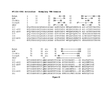
Figure 10 shows an alignment of exemplary immunoglobulin single variable
domains
WT1156-P3R2-1C2 (1C2), WT1156-P3R2-1C2-z102 (1C2-z102), WT1156-P3R2-1C2-z109
(1C2-z109), WT1156-P3R2-1C9 (1C9), WT1156-P3R2-1H6 (1H6), WT1156-P3R2-1H6-z100
(1H6-z100), and WT1156-P8R2-1H1 (1H1). Boundaries of CDRs are indicated by
Kabat, AbM,
Chothia, Contact, and EVIGT numbering.
5
CA 03231586 2024- 3- 12
WO 2023/041041 PCT/CN2022/119334
DETAILED DESCRIPTION
While the present disclosure may be embodied in many different forms,
disclosed herein are
specific illustrative embodiments thereof that exemplify the principles of the
disclosure. It should
be emphasized that the present disclosure is not limited to the specific
embodiments illustrated.
Moreover, any section headings used herein are for organizational purposes
only and are not to be
construed as limiting the subject matter described.
Unless otherwise defined herein, scientific and technical terms used in
connection with the
present disclosure shall have the meanings that are commonly understood by
those of ordinary
skill in the art. Further, unless otherwise required by context, singular
terms shall include
pluralities and plural terms shall include the singular. More specifically, as
used in this
specification and the appended claims, the singular forms "a," "an" and "the"
include plural
referents unless the context clearly dictates otherwise. Thus, for example,
reference to "a protein"
includes a plurality of proteins; reference to "a cell" includes mixtures of
cells, and the like. In
this application, the use of "or" means "and/or" unless stated otherwise.
Furthermore, the use of
the term "comprising," as well as other forms, such as "comprises" and
"comprised", is not limiting.
In addition, ranges provided in the specification and appended claims include
both end points and
all points between the end points.
Generally, nomenclature used in connection with, and techniques of, cell and
tissue culture,
molecular biology, immunology, microbiology, genetics and protein and nucleic
acid chemistry
and hybridization described herein are well known and commonly used in the
art. The methods
and techniques of the present disclosure are generally performed according to
conventional
methods well known in the art and as described in various general and more
specific references
that are cited and discussed throughout the present specification unless
otherwise indicated. See,
e.g., Abbas et al., Cellular and Molecular Immunology, 6th ed., W.B. Saunders
Company (2010);
Sambrook J. & Russell D. Molecular Cloning: A Laboratory Manual, 3rd ed., Cold
Spring Harbor
Laboratory Press, Cold Spring Harbor, N.Y. (2000); Ausubel et al., Short
Protocols in Molecular
Biology: A Compendium of Methods from Current Protocols in Molecular Biology,
Wiley, John
& Sons, Inc. (2002); Harlow and Lane Using Antibodies: A Laboratory Manual,
Cold Spring
Harbor Laboratory Press, Cold Spring Harbor, N.Y. (1998); and Coligan et al.,
Short Protocols in
Protein Science, Wiley, John & Sons, Inc. (2003). The nomenclature used in
connection with, and
the laboratory procedures and techniques of, analytical chemistry, synthetic
organic chemistry,
and medicinal and pharmaceutical chemistry described herein are well known and
commonly used
in the art
Definitions
6
CA 03231586 2024- 3- 12
WO 2023/041041
PCT/CN2022/119334
In order to better understand the disclosure, the definitions and explanations
of the relevant
terms are provided as follows.
The term "antibody" (e.g. anti-D3 antibody) and "antigen-binding molecule"
(e.g. D3-
binding molecule) are used interchangeably in the broadest sense and encompass
any form of
antibody that exhibits the desired biological or binding activity. It covers,
but is not limited to,
humanized antibodies, fully human antibodies, chimeric antibodies and single-
domain antibodies
(sdAbs, comprising just one chain, which is typically similar to a heavy
chain), as well as
fragments of any of the foregoing as long as they exhibit the desired antigen-
binding activity,
including, for example, an antibody comprising at least one VIM domain. A
conventional
antibody comprises a heavy chain(s) and a light chain(s). Heavy chains may be
classified into
y, a and a, which define isotypes of an antibody as IgM, IgD, IgG, IgA and
IgE, respectively.
A heavy chain can comprise a heavy chain variable region (VH) and a heavy
chain constant
region (CH). A heavy chain can comprise one or more constant regions, for
example, 3
constant regions (CH1, CH2 and CH3). A light chain can comprise a light chain
variable region
(VL) and a light chain constant region (CL). A VH and a VL region can further
be divided into
hypervariable regions (called complementary determining regions (CDRs)), which
are
interspaced by relatively conservative regions (called framework regions
(FRW)) A VH and
a VL can comprise 3 CDRs (Complementarity determining regions) and 4 FRs
(Framework
regions) in the following order: FRW 1, CDR1, FRW2, CDR2, FRW3, CDR3, FRW4
from
N-terminal to C-terminal. Antibodies can be of different antibody isotypes,
for example, IgG
(e.g., IgGl, IgG2, IgG3 or IgG4 subtype), IgAl , IgA2, IgD, IgE or IgM
antibody.
The term "Fc region" is used to define a C-terminal region of an
immunoglobulin heavy chain,
including, for example, native sequence Fc regions, recombinant Fc regions,
and variant Fc regions.
Although the boundaries of the Fc region of an immunoglobulin heavy chain
might vary, the
human IgG heavy chain Fc region is often defined to stretch from an amino acid
residue at position
Cys226 (according to the EU numbering system), or from Pro230 (according to
the EU numbering
system), to the carboxyl-terminus thereof. The C-terminal lysine (residue 447
according to the
EU numbering system) of the Fc region may be removed, for example, during
production or
purification of the antibody, or by recombinantly engineering the nucleic acid
encoding a heavy
chain of the antibody.
A "functional Fc region" possesses an "effector function" of a native sequence
Fc region.
Exemplary "effector functions" include Clq binding; complement dependent
cytotoxicity (CDC);
Fc receptor binding; antibody-dependent cell-mediated cytotoxicity (ADCC);
phagocytosis; down
regulation of cell surface receptors (e.g., B cell receptor; BCR), etc. Such
effector functions
generally require the Fc region to be combined with a binding region or
binding domain (e.g., an
7
CA 03231586 2024- 3- 12
WO 2023/041041
PCT/CN2022/119334
antibody variable region or domain, including a VIATI domain) and can be
assessed using various
assays as disclosed.
A "native sequence Fc region" comprises an amino acid sequence identical to
the amino acid
sequence of an Fc region found in nature, and not manipulated, modified,
and/or changed (e.g.,
isolated, purified, selected, including or combining with other sequences such
as variable region
sequences) by a human. Native sequence human Fc regions include a native
sequence human
IgG1 Fc region (non-A and A allotypes); native sequence human IgG2 Fc region;
native sequence
human IgG3 Fc region; and native sequence human IgG4 Fc region as well as
naturally occurring
variants thereof
A "variant Fc region" comprises an amino acid sequence which differs from that
of a native
sequence Fc region by virtue of at least one amino acid modification, (e.g.,
substituting, addition,
or deletion) preferably one or more amino acid substitution(s). In some
embodiments, the variant
Fc region has at least one amino acid substitution compared to a native
sequence Fc region or to
the Fc region of a parent polypeptide, for example, from about one to about
ten amino acid
substitutions, and preferably from about one to about five amino acid
substitutions in a native
sequence Fc region or in the Fc region of the parent polypeptide. A variant Fc
region can possess
at least about SO% horn ol ogy with a native sequence Fc region and/or with an
Fc region of a parent
polypeptide, or at least about 90% homology therewith, for example, at least
about 95% homology
therewith. The variant Fc region herein described herein may have a loss of
effector function (e.g.,
silent Fc).
Antibodies described herein include, but are not limited to, synthetic
antibodies, monoclonal
antibodies, recombinantly produced antibodies, multi specific antibodies
(e.g., including hi specific
antibodies), human antibodies, humanized antibodies, chimeric antibodies,
intrabodies, single-
chain Fvs (scFv) (e.g., including rnonospecific, bispecific, etc.), camelized
antibodies, Fab
fragments, F(ab') fragments, disulfide-linked Fvs (sdFv), anti -idi otypic
(anti-Id) antibodies, and
epitope-binding fragments of any of the above.
The term "immunoglobulin single variable domain" or "single variable domain"
or "VITH
domain" or "VHH" or "heavy chain only antibody variable domain" may be used
interchangeably
herein and refers to a single chain antigen binding domain that is capable of
binding to an antigen
or epitope, independently of a different variable domain. A VIM domain (e.g.
variable domain of
a heavy chain antibody) represents the smallest known antigen-binding unit
generated by adaptive
immune responses (Koch-Nolte F. et al., FASEB J. Nov; 21(13):3490-8. Epub 2007
Jun 15 (2007)).
A VHH domain may be a human domain, but also includes a single domain from
other species
such as rodent, nurse shark and Camelid VHH domains. Camelid VHH are
immunoglobulin single
variable domain polypeptides that are derived from species including camel,
llama, alpaca,
dromedary, and guanaco, which produce heavy chain antibodies naturally devoid
of light chains.
8
CA 03231586 2024- 3- 12
WO 2023/041041
PCT/CN2022/119334
Such VI-1H domains may be humanized according to standard techniques available
in the art and
are considered as "single domain antibodies'. As used herein, VHI-1 includes
camelid
domains and humanized VHH domains.
The term "humanized antibody" is intended to refer to antibodies in which CDR
sequences
derived from the germline of another mammalian species, such as a mouse, llama
or alpaca, have
been grafted onto human framework sequences. Additional framework region
modifications may
be made within the human framework sequences
The term "Ka", as used herein, is intended to refer to the association rate of
a particular
antibody-antigen interaction, whereas the term "Kd" as used herein, is
intended to refer to the
dissociation rate of a particular antibody-antigen interaction. Kd values for
antibodies can be
determined using methods well established in the art. The term "KD" as used
herein, is intended
to refer to the dissociation constant of a particular antibody-antigen
interaction, which is obtained
from the ratio of Kd to Ka (e.g., Kd/Ka) and is expressed as a molar
concentration (M). A preferred
method for determining the Kd of an antibody is by using surface plasmon
resonance, preferably
using a biosensor system such as a Biacore system.
The term "specific binding" or "specifically binds" as used herein refers to a
non-random
binding reaction between two molecules, such as for example between an
antibody and an antigen
The term "high affinity", as used herein, refers to a D3 binding molecule such
as an antibody
having a KD of 1 x 10⁻⁷M or less, more preferably 5 x 10⁻⁸M or less, even more
preferably 1x10-
M or less, even more preferably 5 x 10⁻⁹ M or less and even more preferably 1
x 10-9 M or less
for a target antigen
The term "EC50", as used herein, which is also termed as "half maximal
effective
concentration" refers to the concentration of a drug, antibody or toxicant
which induces a response
halfway between the baseline and maximum after a specified exposure time In
the context of the
present disclosure, EC50 is expressed in the unit of -nM".
The term "epitope", as used herein, refers to a portion of an antigen that an
immunoglobulin or antibody specifically binds to. "Epitope" is also known as
"antigenic
determinant". Epitope or antigenic determinant generally comprises chemically
active
surface groups of a molecule such as amino acids, carbohydrates or sugar side
chains, and
generally has a specific three-dimensional structure and a specific charge
characteristic. For
example, an epitope generally comprises at least 3, 4, 5, 6, 7, 8, 9, 10, 11,
12, 13, 14 or 15
consecutive or non-consecutive amino acids in a unique steric conformation,
which may be
"linear" or "conformational". See, for example, Epitope Mapping Protocols in
Methods in
Molecular Biology, Vol. 66, G. E. Morris, Ed. (1996). In a linear epitope, all
the interaction
sites between a protein and an interaction molecule (e.g., an antibody) are
present linearly
along the primary amino acid sequence of the protein. In a conformational
epitope, the
9
CA 03231586 2024- 3- 12
WO 2023/041041
PCT/CN2022/119334
interaction sites span over amino acid residues that are separate from each
other in a protein.
Antibodies may be screened depending on competitiveness of binding to the same
epitope
by conventional techniques known by a person skilled in the art. For example,
studies on
competition or cross-competition may be conducted to obtain antibodies that
compete or
cross-compete with each other for binding to antigens. High-throughput methods
for
obtaining antibodies binding to the same epitope, which are based on their
cross-competition,
are described in an international patent application WO 03/48731.
The term "isolated antibody", as used herein, is intended to refer to an
antibody that is
substantially free of other antibodies having different antigenic
specificities (e.g., an isolated
antibody that specifically binds a D3 protein is substantially free of
antibodies that specifically
bind antigens other than D3 proteins). An isolated antibody that specifically
binds a human D3
protein may, however, have cross- reactivity to other antigens, such as D3
proteins from other
species. Moreover, an isolated antibody can be substantially free of other
cellular material and/or
chemicals.
The term "vector", as used herein, refers to a nucleic acid vehicle which can
have a
polynucleotide inserted therein. When the vector allows for the expression of
the protein
encoded by the polynucleoti de inserted therein, the vector is called an
expression vector. The
vector can have carried genetic material elements expressed in a host cell by
transformation,
transduction, or transfection into the host cell. Vectors are well known by a
person skilled in
the art, including, but not limited to plasmids, phages, cosmids, artificial
chromosome such
as yeast artificial chromosome (YAC), bacterial artificial chromosome (BAC) or
P1-derived
artificial chromosome (PAC); phage such as X. phage or M13 phage and animal
virus. The
animal viruses that can be used as vectors, include, but are not limited to,
retrovirus
(including lentivirus), adenovirus, adeno-associated virus, herpes virus (such
as herpes
simplex virus), pox virus, baculovirus, papillomavirus, papova virus (such as
SV40). A
vector may comprise multiple elements for controlling expression, including,
but not limited
to, a promoter sequence, a transcription initiation sequence, an enhancer
sequence, a
selection element and a reporter gene. In addition, a vector may comprise an
origin of
replication.
The term "host cell", as used herein, refers to a cell into which a vector can
be introduced,
including, but not limited to, a prokaryotic cell such as E. coil or Bacillus
subtilis, a fungal
cell such as yeast cell or Aspergillus, an insect cell such as S2 Drosophila
cell or Sf9, and an
animal cell such as fibroblast, CHO cell, COS cell, NSO cell, HeLa cell, BHK
cell, HEK 293
cell or human cell.
The term "identity", as used herein, refers to a relationship between the
sequences of
two or more polypeptide molecules or two or more nucleic acid molecules, as
determined by
CA 03231586 2024- 3- 12
WO 2023/041041
PCT/CN2022/119334
aligning and comparing the sequences. "Percent identity" means the percent of
identical
residues between the amino acids or nucleotides in the compared molecules and
is calculated
based on the size of the smallest of the molecules being compared. For these
calculations,
gaps in alignments (if any) are preferably addressed by a particular
mathematical model or
computer program (e.g., an "algorithm"). Methods that can be used to calculate
the identity
of the aligned nucleic acids or polypeptides include those described in
Computational
Molecular Biology, (Lesk, A. M,, ed.), 1988, New York: Oxford University
Press;
Biocomputing Informatics and Genome Projects, (Smith, D. W., ed.), 1993, New
York:
Academic Press; Computer Analysis of Sequence Data, Part I, (Griffin, A. M.,
and Griffin,
II. G., eds.), 1994, New Jersey: Humana Press; von IIeinje, G., 1987, Sequence
Analysis in
Molecular Biology, New York: Academic Press; Sequence Analysis Primer,
(Gribskov, M.
and Devereux, J., eds.), 1991, New York: M. Stockton Press; and Carillo et al,
1988, SIAMJ.
Applied Math. 48:1073.
The term "immunogenicity", as used herein, refers to an ability to stimulate
formation
of specific antibodies or sensitized lymphocytes in organisms. It not only
refers to a property
of an antigen to stimulate a specific immunocyte to activate, proliferate and
differentiate so
as to finally generate immunologic effector substance such as antibody and
sensitized
lymphocyte, but also refers to a specific immune response that antibody or
sensitized T
lymphocyte can be formed in an immune system of an organism after stimulating
the
organism with an antigen. Immunogenicity is an important property of an
antigen. Whether
an antigen can successfully induce the generation of an immune response in a
host depends
on several factors, including properties of an antigen, reactivity of a host,
and immunization
means.
The term "transfection" or "transfect", as used herein, refers to a process by
which
nucleic acids are introduced into eukaryotic cells, particularly mammalian
cells. Protocols
and techniques for transfection include but not limited to lipid transfection
and chemical and
physical methods such as electroporation. A number of transfection techniques
are well
known in the art and are disclosed herein. See, e.g., Graham et al., 1973,
Virology 52:456;
Sambrook et al., 2001, Molecular Cloning: A Laboratory Manual, supra; Davis et
al., 1986,
Basic Methods in Molecular Biology, Elsevier; Chu et al, 1981, Gene 13:197.
The term "SPR" or "surface plasmon resonance", as used herein, refers to and
includes an
optical phenomenon that allows for an analysis of real-time biospecific
interactions by detection
of alterations in protein concentrations within a biosensor matrix, for
example using the BIAcore
system (Pharmacia Biosensor AB, Uppsala, Sweden and Piscataway, N.J.). For
further
descriptions, see Example and Jonsson, U., et al. (1993) Arm. Biol. Clin.
51:19-26; Jonsson, U., et
11
CA 03231586 2024- 3- 12
WO 2023/041041
PCT/CN2022/119334
al. (1991) Biotechnipies 11:620-627; Johnsson, B., etal. (1995)1 114o1
Recognit 8:125-131; and
Johnnson, B., et al. (1991) Anal. Biochern. 198:268-277.
The term "fluorescence-activated cell sorting" or "FACS", as used herein,
refers to a
specialized type of flow cytometry. It provides a method for sorting a
heterogeneous mixture of
biological cells into two or more containers, one cell at a time, based upon
the specific light
scattering and fluorescent characteristics of each cell (FlowMetric. "Sorting
Out Fluorescence
Activated Cell Sorting". Retrieved 2017-11-09.). Instruments for carrying out
FACS are known to
those of skill in the art and are commercially available to the public.
Examples of such instruments
include FACS Star Plus, FAC Scan and FACSort instruments from Becton Dickinson
(Foster City,
C al if.) Epics C from Coulter Epics Division (I Ii al eah, Fla.) and MoFlo
from C ytom ati on (Colorado
Springs, Colo.).
The term "subject" includes any human or nonhuman animal, preferably humans.
The term "condition associated with D3" or "condition related to D3", as used
herein, refers
to any condition that is caused by, exacerbated by, or otherwise linked to
increased or decreased
(generally increased) expression or activities of D3 (e.g. a human D3).
The term "cancer", as used herein, refers to any tumor or any malignant cell
growth or
proliferation, primary or metastasis-mediated, including solid tumors and non-
solid tumors such
as leukemia.
The term "treatment", "treating" or "treated", as used herein in the context
of treating a
condition, pertains generally to treatment or therapy, whether of a human or
an animal, in which
some desired therapeutic effect is achieved, for example, inhibition of the
progress of a condition,
and includes a reduction in the rate of progress, a halt in the rate of
progress, regression of the
condition, amelioration of the condition, and cure of the condition. Treatment
as a prophylactic
measure (e.g., prophylaxis, prevention) is also included. For cancer,
"treating" may refer to a
dampening or slowing of a tumor or malignant cell growth, proliferation, or
metastasis, or some
combination thereof. For tumors, "treatment" includes removal of all or part
of a tumor, inhibiting
or slowing tumor growth and metastasis, preventing or delaying the development
of a tumor, or
some combination thereof.
The term "therapeutically-effective amount,- as used herein, pertains to that
amount of an
active compound, or a material, composition or dosage from comprising an
active compound,
which is effective for producing some desired therapeutic effect, commensurate
with a reasonable
benefit/risk ratio, when administered in accordance with a desired treatment
regimen. For example,
a "therapeutically-effective amount," of a D3-binding molecule refers to an
amount or
concentration effective to treat a human D3-related disease or condition.
The term "host cell", as used herein, refers to a cell with the introduction
of exogenous
polynucl eoti des .
12
CA 03231586 2024- 3- 12
WO 2023/041041
PCT/CN2022/119334
The term "pharmaceutically acceptable", as used herein, means that the
vehicle, diluent,
excipient and/or salts thereof, are chemically and/or physically compatible
with other ingredients
in the formulation, and physiologically compatible with the recipient.
As used herein, the term "a pharmaceutically acceptable carrier and/or
excipient" refers to a
carrier, stabilizer, and/or excipient pharmacologically and/or physiologically
compatible with a
subject and an active agent, which is well known in the art (see, e.g.,
Remington's Pharmaceutical
Sciences. Edited by Gennaro AR, 19th ed Pennsylvania: Mack Publishing Company,
1995), and
includes, but is not limited to a pH adjuster, surfactant, adjuvant or an
ionic strength enhancer. For
example, a pH adjuster includes, but is not limited to, phosphate buffer; a
surfactant includes, but
is not limited to, cationic, anionic, or non-ionic surfactant, e.g., Tween-80;
an ionic strength
enhancer includes, but is not limited to, sodium chloride Carriers,
excipients, or stabilizers are
nontoxic to the cell or mammal being exposed thereto at the dosages and
concentrations employed.
Often the carrier is an aqueous pH buffered solution. Examples of carriers
include buffers such as
phosphate, citrate, and other organic acids; antioxidants including ascorbic
acid; low molecular
weight (e.g., less than about 10 amino acid residues) polypeptide; proteins,
such as serum albumin,
gelatin, or immunoglobulins; hydrophilic polymers such as
polyvinylpyrrolidone; amino acids
such as glycine, glutamine, asparagine, arginine or lysine; monosaccharides,
di saccharides, and
other carbohydrates including glucose, mannose, or dextrins; chelating agents
such as EDTA;
sugar alcohols such as mannitol or sorbitol; salt-forming counterions such as
sodium; and/or
nonionic surfactants such as TWEENTm, polyethylene glycol (PEG), and
PLURONICSTM. The
term "carrier" can also refer to a diluent, adjuvant (e.g., Freund's adjuvant
(complete or
incomplete)), excipient, or vehicle with which the therapeutic is
administered. Such carriers can
be sterile liquids, such as water and oils, including those of petroleum,
animal, vegetable or
synthetic origin, such as peanut oil, soybean oil, mineral oil, sesame oil and
the like. Water is a
exemplary carrier when a composition (e.g., a pharmaceutical composition) is
administered
intravenously. Saline solutions and aqueous dextrose and glycerol solutions
can also be employed
as liquid carriers, particularly for injectable solutions. Suitable excipients
(e.g., pharmaceutical
excipients) include starch, glucose, lactose, sucrose, gelatin, malt, rice,
flour, chalk, silica gel,
sodium stearate, glycerol monostearate, talc, sodium chloride, dried skim
milk, glycerol, propylene
glycol, water, ethanol and the like. The composition, if desired, can also
contain minor amounts
of wetting or emulsifying agents, or pH buffering agents. Compositions can
take the form of
solutions, suspensions, emulsion, tablets, pills, capsules, powders, sustained-
release formulations
and the like. Oral compositions, including formulations, can include standard
carriers such as
pharmaceutical grades of mannitol, lactose, starch, magnesium stearate, sodium
saccharine,
cellulose, magnesium carbonate, etc. Examples of suitable carriers are
described in Remington' s
Pharmaceutical Sciences (1990) Mack Publishing Co., Easton, PA. Compositions,
including
13
CA 03231586 2024- 3- 12
WO 2023/041041
PCT/CN2022/119334
pharmaceutical compounds, may contain a prophylactically or therapeutically
effective amount of
a D3-binding agent (e.g., an anti-D3 antibody), for example, in isolated or
purified form, together
with a suitable amount of carrier so as to provide the form for proper
administration to the subject
(e.g., patient). The formulation should suit the mode of administration.
As used herein, the term "adjuvant" refers to a non-specific
immunopotentiator, which can
enhance immune response to an antigen or change the type of immune response in
an organism
when it is delivered together with the antigen to the organism or is delivered
to the organism in
advance. There are a variety of adjuvants, including, but not limited to,
aluminium adjuvants (for
example, aluminum hydroxide), Freund's adjuvants (for example, Freund's
complete adjuvant and
Freund's incomplete adjuvant), coryne bacterium parvum, lipopolysaccharide,
cytokines, and the
like. Freund's adjuvant is the most commonly used adjuvant in animal
experiments. Aluminum
hydroxide adjuvant is more commonly used in clinical trials.
D3-binding molecules
In some aspects, the disclosure provides D3-binding molecules. A D3-binding
molecule, in a
general sense, may include any molecule that specifically binds to D3. In some
circumstances, a
"D3-binding molecule" may include a "D3 antagonist" and an "anti-D3 antibody".
"D3 antagonist"
refers to any chemical compound or biological molecule that blocks D3
activities. "Anti-D3
antibody" includes, but not limited to, a chimeric antibody, a humanized
antibody, a human
antibody or a single-domain antibody. A D3-binding molecule is not limited to
a polypeptide or a
protein and may comprise other components such as nucleotides, hybrids,
glucans and a
combination thereof As exemplified herein, a D3-binding molecule may be an
anti-D3 antibody
or anti-D3 fusion protein.
In some embodiments, D3-binding molecules as disclosed herein comprise at
least one VHH
that specifically binds to D3 Further, a D3-binding molecule may be a single-
domain antibody
and comprising one VHH. For example, a single-domain antibody is able to bind
selectively to a
specific antigen (e.g., D3). In some embodiments, a D3-binding molecule
comprises a VITH fused
to an immunoglobulin Fc region, for example, an Fc region of IgG (e.g., IgG4
or IgG1). In some
embodiments, the Fc region is an Fc region of human IgGl. By fusing a VHH to
an Fc region, it
may be more efficient to recruit effector functions. Also, fusion of a VHH to
an Fc region may
help a D3-binding molecule to form a dimer and may also help the extension of
the half life of the
D3-binding molecule in vivo.
As known in the art, VHH molecules derived from Camelidae antibodies are among
the
smallest intact antigen-binding domains known (approximately 15 kDa, or 10
times smaller than
a conventional IgG) and hence are well suited towards delivery to dense
tissues and for accessing
the limited space between macromolecules.
14
CA 03231586 2024- 3- 12
WO 2023/041041
PCT/CN2022/119334
VHEIs as disclosed herein may be made by the skilled artisan according to
methods known
in the art or any future method. For example, VHI-Is may be obtained using
methods known in the
art such as by immunizing a camel and obtaining hybridoma's therefrom, or by
cloning a library
of VEIHs of the disclosure using molecular biology techniques known in the art
and subsequent
selection by using phage display.
For example, a VHH can be obtained by immunization of llamas or alpacas with
the desired
antigen and subsequent isolation of the mRNA coding for heavy-chain
antibodies. By reverse
transcription and polymerase chain reaction, a gene library of single-domain
antibodies containing
several million clones is produced. Screening techniques like phage display
and ribosome
display help to identify the clones binding the antigen. One technique is
phage display in which a
library of (e.g., human) antibodies is synthesized on phages, the library is
screened with the antigen
of interest or an antibody-binding portion thereof, and the phage that binds
the antigen is isolated,
from which one may obtain the immunoreactive fragments. Methods for preparing
and screening
such libraries are well known in the art and kits for generating phage display
libraries are
commercially available (e.g., the Pharmacia Recombinant Phage Antibody System,
catalog no.
27-9400-01; and the Stratagene SurfZAPTm phage display kit, catalog no.
240612). There also are
other methods and reagents that can be used in generating and screening
antibody display libraries
(see, e.g., Barbas et al.,Proc. Natl. Acad. Sc!. USA 88:7978-7982 (1991)).
When potent clones have been identified, their DNA sequence is optimized, for
example, by
affinity maturation or humanization. Humanization may prevent immunological
reactions of the
human organism against the antibody.
Accordingly, the VI-Ms can be obtained (1) by isolating the VHH domain of a
naturally
occurring heavy chain antibody; (2) by expression of a nucleotide sequence
encoding a naturally
occurring VHH domain; (3) by "humanization" (as described below) of a
naturally occurring VI-TH
domain or by expression of a nucleic acid encoding a such humanized VI-1H
domain; (4) by
"camelization" of a naturally occurring VH domain from any animal species, in
particular a species
of mammal, such as from a human being, or by expression of a nucleic acid
encoding such a
camelized VH domain; (5) by "camelisation" of a "domain antibody" or "Dab" as
described by
Ward et al (supra), or by expression of a nucleic acid encoding such a cam
elized VH domain; (6)
using synthetic or semi-synthetic techniques for preparing proteins,
polypeptides or other amino
acid sequences; (7) by preparing a nucleic acid encoding a V1-111 using
techniques for nucleic acid
synthesis, followed by expression of the nucleic acid thus obtained, (8)
subjecting heavy chain
antibodies or VHHs to affinity maturation, to mutagenesis (e.g. random
mutagenesis or site-
directed mutagenesis) and/or any other technique(s) in order to increase the
affinity and/or
specificity of the VHEI; and/or (9) by any combination of the foregoing.
Suitable methods and
CA 03231586 2024- 3- 12
WO 2023/041041
PCT/CN2022/119334
techniques for performing the foregoing will be clear to the skilled person
based on the disclosure
herein and, for example, include methods and techniques described in more
detail herein.
Single-domain antibodies are usually generated by PCR cloning of variable
domain repertoire
from blood, lymph node, or spleen cDNA obtained from immunized animals into a
phage display
vector. Antigen-specific single-domain antibodies are commonly selected by
panning phase
libraries on immobilized antigen, for example, antigen coated onto the plastic
surface of a test tube,
biotinylated antigens immobilized on Streptavidin beads, or membrane proteins
expressed on the
surface of cells. The affinity of sdAbs can often been improved by mimicking
this strategy in vitro,
for example, by site directed mutagenesis of the CDR regions and further
rounds of panning on
immobilized antigen under conditions of increased stringency (higher
temperature, high or low
salt concentration, high or low pH, and low antigen concentrations)
(Wesolowski et al., Single
domain antibodies: promising experimental and therapeutic tools in infection
and immunity. Med
Microbiol Immunol (2009) 198: 157-174).
Methods for preparing a VHH specifically binding to an antigen or epitope was
described in
references, for example: R. van der Linden et al., Journal of Immunological
Methods, 240(2000)
185-195; Li et al., J Biol Chem., 287(2012)13713-13721; Deffar et al., African
Journal of
Biotechnology Vol. 8(12), pp.2645, 17 June, 2009 and WO 94/04678.
In some embodiments, a VHH may be truncated at the N-terminus or C-terminus
such that it
comprises only a partial FRW1 and/or FRW4, or lacks one or both of those
framework regions, so
long as the VHI-1 substantially maintains antigen binding and specificity
(e.g., substantially
maintained, for example, at least 50%, at least 60%, at least 70%, at least
80%, at least 90%, at
least 95%).
The present disclosure also provides D3-binding molecules with a masking
moiety and/or
cleavable moiety in which one or more of the D3-binding domains of the D3-
binding molecules
are masked (e.g., via a masking moiety) and/or activatable (e.g., via a
cleavable moiety).
Technologies for masking of a D3-binding molecule (e.g., an antibody) are well
known in the art,
including SAFE body masking technology (see, e.g., US 2019/0241886) and
Probody masking
technology (see, e.g., US 2015/0079088). Such technologies can be used to
generate a D3-binding
molecule (e.g., an antibody) that is masked and/or activatable. Such masked
and/or activatable
D3-binding molecules (e.g., antibodies) are useful for the preparation of
conjugates, including
immunoconjugates, antibody-drug conjugates (ADCs), masked ADCs and activatable
antibody-
drug conjugates (AADCs), comprising any one of the D3-binding molecules (e.g.,
antibodies) of
the present disclosure, including those directly or indirectly linked to
another agent such as a drug.
For example, D3-binding molecules of the present disclosure may be covalently
bound by a
synthetic linker to one or more agents such as drugs.
16
CA 03231586 2024- 3- 12
WO 2023/041041
PCT/CN2022/119334
If desired, a D3-binding molecule is linked or conjugated (directly or
indirectly) to a moiety
with effector function, such as cytotoxic activity (e.g., a chemotherapeutic
moiety or a radioisotope)
or immune recruitment activity. Moieties that are linked or conjugated
(directly or indirectly)
include drugs that are cytotoxic (e.g., toxins such as auristatins) or non-
cytotoxic (e.g., signal
transduction modulators such as kinases or masking moieties that mask one or
more binding
domains of a D3-binding molecule, or cleavable moieties that allow for
activating a D3-binding
molecule by cleaving of a cleavable moiety to unmask one or more binding
domains of a D3-
binding molecule in the tumor microenvironment, in the form of masked
conjugates. Moieties that
promote immune recruitment can include other antigen-binding agents, such as
viral proteins that
bind selectively to cells of the innate immune system. Alternatively or in
addition, a D3-binding
molecule is optionally linked or conjugated (directly or indirectly) to a
moiety that facilitates
isolation from a mixture (e.g., a tag) or a moiety with reporter activity
(e.g., a detection label or
reporter protein). It will be appreciated that the features of a D3-binding
molecule described herein
extend also to a polypeptide comprising a D3-binding molecule fragment.
In some embodiments, D3-binding molecules described herein may be linked or
conjugated
(directly or indirectly) to a polypeptide, which can result in the generation
of an activatable
antibody. In some embodiments, a D3-binding molecule is linked or conjugated
(directly or
indirectly) to an agent. In some embodiments, the agent is a drug, resulting
in an ADC or an
AADC when the antibody of the ADC comprises a masking moiety and a cleavable
moiety.
In some embodiments, D3-binding molecules described herein are conjugated or
recombinantly linked (directly or indirectly) to a therapeutic agent (e.g., a
cytotoxic agent) or to a
diagnostic or detectable agent. The conjugated or recombinantly linked
antibodies, including
masked or activatable conjugates, can be useful, for example, for treating or
preventing a disease,
disorder or condition, such as a cancer or a tumor.
Diagnosis and detection can be accomplished, for example, by coupling a D3-
binding
molecule to detectable substances including, for example: enzymes, including,
but not limited to,
horseradish peroxidase, alkaline phosphatase, beta-gal actosi dase, or acetyl
choli nesterase;
prosthetic groups, including, but not limited to, streptavidin/biotin or
avidin/biotin; fluorescent
materials, including, but not limited to, umbelliferone, fluorescein,
fluorescein isothiocynate,
rhodamine, dichlorotriazinylamine fluorescein, dansyl chloride, or
phycoerythrin; luminescent
materials, including, but not limited to, luminol; bioluminescent materials,
including, but not
limited to, luciferase, luciferin, or aequorin; chemiluminescent material,
including, but not limited
to, an acridinium based compound or a HALOTAG; radioactive materials,
including, but not
limited to, iodine (1311, 1251, 1231, and 1211), carbon (14C), sulfur (35S),
tritium (3H), indium
(115In, 113I11, 112In, and 111111), technetium (99Tc), thallium (201Ti),
gallium (68Ga and 67Ga),
palladium (103Pd), molybdenum (99Mo), xenon (133Xe), fluorine (18F), 153 Sm,
177Lu, 159Gd,
17
CA 03231586 2024- 3- 12
WO 2023/041041
PCT/CN2022/119334
149Pm, 140La, 175Yb, 166Ho, 90Y, 47Sc, 186Re, 188Re, 142Pr, 105Rh, 97Ru, 68Ge,
57Co,
65Zn, 85Sr, 32P, 153Gd, 169Yb, 51Cr, 54Mn, 75Se, 113Sn, or 117Sn; positron
emitting metals
using various positron emission tomographies; and non-radioactive paramagnetic
metal ions.
Conjugates of an antibody and agent, including wherein the agent is a drug for
the preparation
of ADC or an AADC, may be made using a variety of bifunctional protein
coupling agents such
as BMPS, EMCS, GMBS, HBVS, LC-SMCC, MBS, MPBH, SBAP, SIA, STAB, SMCC, SMPB,
SMPH, sulfo-EMCS, sulfo-GMBS, sulfo-KMUS, sulfo-MBS, sulfo-SIAB, sulfo-SMCC,
sulfo-
SMPB, and SVSB (succinimidy1-(4-vinylsulfone) benzoate). The present
disclosure further
contemplates that conjugates of antibodies and agents, including wherein the
agent is a drug for
the preparation of an ADC or AADC, may be prepared using any suitable methods
as disclosed in
the art (see, e.g., Bioconjugate Techniques (Hermanson ed., 2d ed. 2008)).
Conventional conjugation strategies for antibodies and agents, including
wherein the agent is
a drug for the preparation of ADC or AADC, have been based on random
conjugation chemistries
involving the c-amino group of Lys residues or the thiol group of Cys
residues, which results in
heterogeneous conjugates. Recently developed techniques allow site-specific
conjugation to
antibodies, resulting in homogeneous loading and avoiding conjugate
subpopulations with altered
antigen-binding or pharmacokinetics. These include engineering of "thiomabs"
comprising
cysteine substitutions at positions on the heavy and light chains that provide
reactive thiol groups
and do not disrupt immunoglobulin folding and assembly or alter antigen
binding (see, e.g.,
Junutula et al., 2008, J. Immunol. Meth. 332: 41-52; and Junutula et al.,
2008, Nature Biotechnol.
26:925-32). In another method, selenocysteine is cotranslationally inserted
into an antibody
sequence by recoding the stop codon UGA from termination to selenocysteine
insertion, allowing
site specific covalent conjugation at the nucleophilic selenol group of
selenocysteine in the
presence of the other natural amino acids (see, e.g., Hofer et al., 2008, Proc
Natl. Acad Sci. USA
105:12451-56; and Hofer et al., 2009, Biochemistry 48(50):12047-57).
D3-binding molecules described herein may be monospecific, bispecific,
trispecific or of
greater multispecifi city Such agents may include antibodies. Multi speci fic
antibodies, such as
bispecific antibodies, are monoclonal antibodies that have binding
specificities for at least two
different targets (e.g., antigens) or two different epitopes on the same
target (e.g., a bispecific
antibody directed to D3 with a first binding domain for a first epitope of D3,
and a second binding
domain for a second epitope of D3. In some embodiments, the multispecific
(e.g., bispecific)
antibodies can be constructed based on the sequences of the antibodies
described herein. In some
embodiments, the multispecific antibodies described herein are bispecific
antibodies. In some
embodiments, bispecific antibodies are mouse, chimeric, human or humanized
antibodies. In some
embodiments, one of the binding specificities of the multispecific antibody is
for D3 and the other
is for any other target (e.g., antigen) In some embodiments, a multispecific
(e.g., bispecific)
18
CA 03231586 2024- 3- 12
WO 2023/041041
PCT/CN2022/119334
antibody can comprise more than one target (e.g., antigen) binding domain, in
which different
binding domains are specific for different targets (e.g., a first binding
domain that binds to D3 and
a second binding domain that binds another target (e.g., antigen), such as an
immune checkpoint
regulator (e.g., a negative checkpoint regulator). In some embodiments,
multispecific (e.g.,
bispecific) antibody molecules can bind more than one (e.g., two or more)
epitopes on the same
target (e.g., antigen). In some embodiments, one of the binding specificities
is D3 and the other is
for one or more of Cytotoxic T-lymphocyte antigen-4 (CTLA-4), CD80, CD86,
Programmed cell
death 1 (PD-1), Programmed cell death ligand 1 (PD-L1), Programmed cell death
ligand 2 (PD-
L2), Lymphocyte activation gene-3 (LAG-3; also known as CD223), Galectin-3, B
and T
lymphocyte attenuator (BTLA), T-cell membrane protein 3 (T11\43), Galectin-9
(GAL9), 117-111,
B7-H3, B7-H4, T-Cell immunoreceptor with Ig and ITIM domains
(TIGIT/Vstm3/WUCAM/VSIG9), V-domain Ig suppressor of T-Cell activation
(VISTA),
Glucocorticoid-induced tumor necrosis factor receptor-related (GITR) protein,
Herpes Virus Entry
Mediator (HVEM), 0X40, CD27, CD28, CD137. CGEN-150011, CGEN-15022, CGEN-15027,
CGEN-15049, CGEN-15052, and CGEN-15092.
Methods for making multispecific antibodies are known in the art, for example,
by co-
expression of two immunoglobulin heavy chain-light chain pairs, where the two
heavy chains have
different specificities (see, e.g., Milstein and Cuello, 1983, Nature 305:537-
40). For further details
of generating multispecific antibodies (e.g., bispecific antibodies), see, for
example, Bispecific
Antibodies (Kontermann ed., 2011).
The present disclosure provides humanized antibodies that bind D3. Various
methods for
humanizing non-human antibodies are known in the art. For example, a humanized
antibody can
have one or more amino acid residues introduced into it from a source that is
non-human. These
non-human amino acid residues are often referred to as "import" residues,
which are typically
taken from an -import" variable domain Humanized antibodies that bind D3 may
be produced
using techniques known to those skilled in the art (e.g., Zhang et al.,
Molecular Immunology,
42(12): 1445-1451, 2005; Hwang et al., Methods, 36(1): 35-42, 2005; Dall'Acqua
et al., Methods,
36(1): 43-60, 2005; Clark, Immunology Today, 21(8): 397-402, 2000, and U.S.
Patent Nos.
6,180,370; 6,054,927; 5,869,619; 5,861,155; 5,712,120; and 4,816,567).
A D3-binding molecule may be described as an anti-D3 antibody in the following
sections.
Anti-D3 antibodies with functional properties
Antibodies of the disclosure including, for example, antibodies comprising at
least one VHH
domain, are characterized by particular functional features or properties of
the antibodies. In some
embodiments, the antibodies have one or more of the following properties:
19
CA 03231586 2024- 3- 12
WO 2023/041041
PCT/CN2022/119334
(a) bind to human D3, cyno D3 and mouse D3 with EC 50 at nM grade, as measured
by ELISA
or FACS;
(b) show dose-dependent internalization potency in human cells engineered to
express D3;
and
(c) bind to human D3 ECD with a KD no more than 0.1 nM, as measured by SPR.
An antibody of the disclosure binds to cell surface D3 with high affinity. The
binding of an
antibody of the disclosure to D3 can be assessed using one or more techniques
well established in
the art, for example, ELISA. The binding specificity of an antibody of the
disclosure can also be
determined by monitoring binding of the antibody to cells expressing a D3
protein, e.g., by flow
cytometry. For example, an antibody can be tested by a flow cytometry assay
(e.g., FACS) in
which the antibody is reacted with a cell line that expresses human D3, such
as CHO cells and 293
cells that have been transfected to express D3 on their cell surface.
Additionally or alternatively,
the binding of the antibody, including the binding kinetics (e.g., Kd value)
can be tested in BIAcore
binding assays. Still other suitable binding assays include ELISA assays, for
example using a
recombinant D3 protein. For example, an antibody of the disclosure binds to a
cell surface D3 (e.g.,
human D3 ECD) protein with a KT) of 1 x 10⁻⁷M or less, 5 x 10⁻⁸M or less, 2 x
10-8 M or less, 5 x
101Morless,4xlO9Morless,3x1(Y9Morless,2x109Morless,lx109Morless,5x10
10 M or less, or 1 x 10⁻¹⁰ M or less.
In some embodiments, the antibodies of the disclosure bind to cynomolgus
monkey or mouse
D3 at an EC50 of no more than or about 10 nM, 9 nM, 8 nM, 7 nM, 6 nM, 5 nM, 4
nM, 3 n1\4, 2
nM, 1 nM, 0.9 nM, 0.8 nM, 0.7 nM, 0.6 nM, 0.5 nM, 0.4 aM, 0.3 nM, 0.2 nM, 0.1
nM, 0.09 nM,
0.08 nM, 0.07 nM, 0.06 nM, 0.05 nM, 0.04 nM, 0.03 nM, 0.02 nM, or 0.01 nM, as
measured by
FACS.
Anti-D3 antibodies comprising CDRs
In some embodiments, an anti-D3 antibody as disclosed herein comprises at
least one
imrnun ogl obulin single variable domain (e.g., VIIH), wherein the VIM
comprises CDR1, CDR2
and CDR3, and wherein CDR1 comprises an amino acid sequence as set forth in
SEQ ID NO: 1,
4, 7 or 10, CDR2 comprises an amino acid sequence as set forth in SEQ ID NO:
2, 5, 8 or 11, and
CDR3 comprises an amino acid sequence as set forth in SEQ ID NO: 3, 6 or 9. In
some
embodiments, the CDR numbering are according to a combination of Kabat and AbM
numbering.
The extent of the framework region and CDRs can be precisely identified using
methodology
known in the art, for example, by the Kabat definition, the Chothia
definition, the AbM definition,
the contact definition, the IMGT definition (all of which are well known in
the art) and any
combinations thereof. See, e.g., Kabat, E.A., et al. (1991) Sequences of
Proteins of Immunological
Interest, Fifth Edition,U U.S. Department of Health and Human Services, NIH
Publication No. 91-
CA 03231586 2024- 3- 12
WO 2023/041041
PCT/CN2022/119334
3242, Chothia et al., (1989) Nature 342:877; Chothia, C. et al. (1987) J. Mol.
Biol. 196:901-917,
Al-lazikani et al (1997) J. Molec. Biol. 273:927-948; Edelman et al., Proc
Natl Acad Sci U S A.
1969 May, 63(1):78-85; and Martin and Allen, in "Handbook of Therapeutic
Antibodies", chapter
5,2007. See also hgmp.mrc.ac.uk and bioinf. org.uk/abs. Correspondence or
alignments between
numberings according to different definitions can for example be found at wA-
w.irno-,orW (see
also Giudicelli V et al. IIVIGT, the international ImMunoGeneTics database.
Nucleic Acids Res.
(1997) 25:206-11; and Lefranc MP et al., IMGT unique numbering for
irnmunoglobulin and T
cell receptor variable domains and 1g superfamily V-like domains. Dev Comp
Immunol. (2003)
27:55-77).
As will be appreciated by those in the art, the exact numbering and placement
of the CDRs
can be different among different numbering systems. However, it should be
understood that the
disclosure of a variable heavy sequence, a variable light sequence and/or a
VHFI sequence includes
the disclosure of the associated (inherent) CDRs. Accordingly, the disclosure
of each variable
region is a disclosure of the CDRs (e.g., CDR1, CDR2 and CDR3). Two antibodies
having the
same VH, VL or VHH CDRs means that their CDRs are identical when determined by
the same
approach (e.g., the Kabat, AbM, Chothia, Contact, and IIVIGT numbering
approaches as known in
the art).
Variable regions and CDRs in an antibody sequence can be identified according
to general
rules that have been developed in the art (for example, the Kabat, AbM,
Chothia, Contact, and
[MGT numbering system) or by aligning the sequences against a database of
known variable
regions. Methods for identifying these regions are described in Kontermann and
Dubel, eds.,
Antibody Engineering, Springer, New York, NY, 2001 and Dinarello et al.,
Current Protocols in
Immunology, John Wiley and Sons Inc., Hoboken, NJ, 2000. Exemplary databases
of antibody
sequences are described in, and can be accessed through, the "Abysis" website
at
www.bi oinf. org.uk/abs (maintained by A.C. Martin in the Department of
Biochemistry &
Molecular Biology University College London, London, England) and the VBASE2
website at
www.vbase2.org, as described in Retter et al., Nucl. Acids Res., 33 (Database
issue): D671 -D674
(2005). Preferably sequences are analyzed using the Abysis database, which
integrates sequence
data from Kabat, IIVIGT and the Protein Data Bank (PDB) with structural data
from the PDB. See
Dr. Andrew C. R. Martin's book chapter Protein Sequence and Structure Analysis
of Antibody
Variable Domains. In: Antibody Engineering Lab Manual (Ed.: Duebel, S. and
Kontermann, R.,
Springer-Verlag, Heidelberg, ISBN-13: 978-3540413547, also available on the
website
bioinfor2.uk/abs). The Abysis database website further includes general rules
that have been
developed for identifying CDRs which can be used in accordance with the
teachings herein. Figure
10 shows an alignment of exemplary immunoglobulin single variable domains and
boundaries of
CDRs are indicated by Kabat, AbM, Chothia, Contact, and MGT numbering.
21
CA 03231586 2024- 3- 12
WO 2023/041041
PCT/CN2022/119334
In some embodiments, a D3-binding molecule as disclosed herein comprises at
least one
immunoglobulin single variable domain (e.g., VHH), wherein the VELF1 comprises
FRW1-CDR1-
FRW2-CDR2-FRW3-CDR3-FRW4, and wherein CDR1 has an amino acid sequence as set
forth
in SEQ ID NO: 1, 4, 7 or 10, CDR2 has an amino acid sequence as set forth in
SEQ ID NO: 2, 5,
8 or ill, and CDR3 has an amino acid sequence as set forth in SEQ ID NO: 3, 6
or 9. In some
embodiments, the FRW1 and FRW4 at the N and C terminal of the VHH comprised in
a D3-
binding molecule may be truncated such that it comprise only a partial FRW1
and/or FRW4, or
the VHH lacks one or both of these framework regions, so long as the VHH
substantially maintains
antigen binding and specificity.
In some embodiments, provided herein is an anti-D3 antibody (such as an anti-
D3 single
domain antibody) comprising one, two, or all three CDRs of the amino acid
sequence as set forth
in SEQ ID NO: 12. In some embodiments, there is provided an anti-D3 antibody
(such as an anti-
D3 single domain antibody) comprising one, two, or all three CDRs of the amino
acid sequence
as set forth in SEQ ID NO: 13. In some embodiments, there is provided an anti-
D3 antibody (such
as an anti-D3 single domain antibody) comprising one, two, or all three CDRs
of the amino acid
sequence as set forth in SEQ ID NO: 14. In some embodiments, there is provided
an anti-D3
antibody (such as an anti-D3 single domain antibody) comprising one, two, or
all three CDRs of
the amino acid sequence as set forth in SEQ ID NO: 15. In some embodiments,
there is provided
an anti-D3 antibody (such as an anti-D3 single domain antibody) comprising
one, two, or all three
CDRs of the amino acid sequence as set forth in SEQ ID NO: 16. In some
embodiments, there is
provided an anti-D3 antibody (such as an anti-D3 single domain antibody)
comprising one, two,
or all three CDRs of the amino acid sequence as set forth in SEQ ID NO: 17. In
some embodiments,
there is provided an anti-D3 antibody (such as an anti-D3 single domain
antibody) comprising one,
two, or all three CDRs of the amino acid sequence as set forth in SEQ ID NO:
18. In some
embodiments, there is provided an anti-D3 antibody (such as an anti-D3 single
domain antibody)
comprising one, two, or all three CDRs of the amino acid sequence as set forth
in SEQ ID NO: 55.
In some embodiments, the anti -D3 single domain antibody is cam el i d. In
some embodiments, the
anti-D3 antibody (such as the anti-D3 single domain antibody) is humanized. In
some
embodiments, the anti-D3 antibody (such as the anti-D3 single domain antibody)
comprises an
acceptor human framework, e.g., a human immunoglobulin framework or a human
consensus
framework.
In some embodiments, the anti-D3 antibody (such as the single domain antibody)
comprises
a CDR1 having an amino acid sequence of the CDR1 as set forth in SEQ ID NO:
12. In some
embodiments, the anti-D3 antibody (such as the single domain antibody)
comprises a CDR2
having an amino acid sequence of the CDR2 as set forth in SEQ ID NO: 12. In
other embodiments,
the anti-D3 antibody (such as the single domain antibody) comprises a CDR3
having an amino
22
CA 03231586 2024- 3- 12
WO 2023/041041
PCT/CN2022/119334
acid sequence of the CDR3 as set forth in SEQ ID NO: 12. In some embodiments,
the anti-D3
antibody (such as the single domain antibody) comprises a CDR1 and a CDR2
having amino acid
sequences of the CDR1 and the CDR2 as set forth in SEQ ID NO: 12. In some
embodiments, the
anti-D3 antibody (such as the single domain antibody) comprises a CDR1 and a
CDR3 having
amino acid sequences of the CDR1 and the CDR3 as set forth in SEQ ID NO: 12.
In some
embodiments, the anti-D3 antibody (such as the single domain antibody)
comprises a CDR2 and
a CDR3 having amino acid sequences of the CDR2 and the CDR3 as set forth in
SEQ ID NO: 12.
In some embodiments, the anti-D3 antibody (such as the single domain antibody)
comprises a
CDR1, a CDR2, and a CDR3 having amino acid sequences of the CDR1, the CDR2,
and the CDR3
as set forth in SEQ ID NO: 12. CDR sequences can be determined according to
well-known
numbering systems. In some embodiments, the CDRs are according to IIVIGT
numbering. In some
embodiments, the CDRs are according to Kabat numbering. In other embodiments,
the CDRs are
according to Chothia numbering. In other embodiments, the CDRs are according
to Contact
numbering. In some embodiments, the CDRs are according to AbM numbering. In
some
embodiments, the anti-D3 single domain antibody is camelid. In some
embodiments, the anti-D3
antibody (such as the anti-D3 single domain antibody) is humanized. In some
embodiments, the
anti-D3 antibody (such as the anti -D3 single domain antibody) comprises an
acceptor human
framework, e.g., a human immunoglobulin framework or a human consensus
framework.
In some embodiments, the single domain antibody has a CDR1 haying an amino
acid
sequence of the CDR1 as set forth in SEQ ID NO: 13. In some embodiments, the
single domain
antibody has a CDR2 having an amino acid sequence of the CDR2 as set forth in
SEQ ID NO: 13.
In other embodiments, the single domain antibody has a CDR3 having an amino
acid sequence of
the CDR3 as set forth in SEQ ID NO: 13. In some embodiments, the single domain
antibody has
a CDR] and a CDR2 having amino acid sequences of the CDR1 and the CDR2 as set
forth in SEQ
1D NO: 13, In some embodiments, the single domain antibody has a CDR1 and a
CDR3 having
amino acid sequences of the CDR1 and the CDR3 as set forth in SEQ ID NO: 13.
In some
embodiments, the single domain antibody has a CDR2 and a CDR3 having amino
acid sequences
of the CDR2 and the CDR3 as set forth in SEQ ID NO: 13. In some embodiments,
the single
domain antibody has a CDR1, a CDR2, and a CDR3 having amino acid sequences of
the CDR1,
the CDR2, and the CDR3 as set forth in SEQ ID NO: 13. CDR sequences can be
determined
according to well-known numbering systems. In some embodiments, the CDRs are
according to
IIVIGT numbering. In some embodiments, the CDRs are according to Kabat
numbering. In other
embodiments, the CDRs are according to Chothia numbering. In other
embodiments, the CDRs
are according to Contact numbering. In some embodiments, the CDRs are
according to AbM
numbering. In some embodiments, the anti-D3 single domain antibody is camelid.
In some
embodiments, the anti-D3 single domain antibody is humanized. In some
embodiments, the anti-
23
CA 03231586 2024- 3- 12
WO 2023/041041
PCT/CN2022/119334
D3 single domain antibody comprises an acceptor human framework, e.g., a human
immunoglobulin framework or a human consensus framework.
In some embodiments, the single domain antibody has a CDR1 haying an amino
acid
sequence of the CDR1 as set forth in SEQ ID NO: 14. In some embodiments, the
single domain
antibody has a CDR2 haying an amino acid sequence of the CDR2 as set forth in
SEQ ID NO: 14.
In other embodiments, the single domain antibody has a CDR3 haying an amino
acid sequence of
the CDR3 as set forth in SEQ ID NO: 14. In some embodiments, the single domain
antibody has
a CDR1 and a CDR2 having amino acid sequences of the CDR1 and the CDR2 as set
forth in SEQ
ID NO: 14. In some embodiments, the single domain antibody has a CDR1 and a
CDR3 having
amino acid sequences of the CDR1 and the CDR3 as set forth in SEQ ID NO: 14.
In some
embodiments, the single domain antibody has a CDR2 and a CDR3 having amino
acid sequences
of the CDR2 and the CDR3 as set forth in SEQ ID NO. 14. In some embodiments,
the single
domain antibody has a CDR1, a CDR2, and a CDR3 haying amino acid sequences of
the CDR1,
the CDR2, and the CDR3 as set forth in SEQ ID NO: 14. CDR sequences can be
determined
according to well-known numbering systems. In some embodiments, the CDRs are
according to
IIVIGT numbering. In some embodiments, the CDRs are according to Kabat
numbering. In other
embodiments, the CDRs are according to Chothi a numbering. In other
embodiments, the CDRs
are according to Contact numbering. In some embodiments, the CDRs are
according to AbM
numbering. In some embodiments, the anti-D3 single domain antibody is camelid.
In some
embodiments, the anti-D3 single domain antibody is humanized. In some
embodiments, the anti-
D3 single domain antibody comprises an acceptor human framework, e.g., a human
immunoglobulin framework or a human consensus framework.
In some embodiments, the single domain antibody has a CDR1 having an amino
acid
sequence of the CDR1 as set forth in SEQ ID NO: 15. In some embodiments, the
single domain
antibody has a CDR2 haying an amino acid sequence of the CDR2 as set forth in
SEQ ID NO: IS.
In other embodiments, the single domain antibody has a CDR3 having an amino
acid sequence of
the CDR3 as set forth in SEQ ID NO: 15. In some embodiments, the single domain
antibody has
a CDR1 and a CDR2 having amino acid sequences of the CDR1 and the CDR2 as set
forth in SEQ
ID NO: 15. In some embodiments, the single domain antibody has a CDR1 and a
CDR3 haying
amino acid sequences of the CDR1 and the CDR3 as set forth in SEQ ID NO: 15.
In some
embodiments, the single domain antibody has a CDR2 and a CDR3 haying amino
acid sequences
of the CDR2 and the CDR3 as set forth in SEQ ID NO: 15. In some embodiments,
the single
domain antibody has a CDR1, a CDR2, and a CDR3 haying amino acid sequences of
the CDR1,
the CDR2, and the CDR3 as set forth in SEQ ID NO: 15. CDR sequences can be
determined
according to well-known numbering systems. In some embodiments, the CDRs are
according to
IIVIGT numbering. In some embodiments, the CDRs are according to Kabat
numbering. In other
24
CA 03231586 2024- 3- 12
WO 2023/041041
PCT/CN2022/119334
embodiments, the CDRs are according to Chothia numbering. In other
embodiments, the CDRs
are according to Contact numbering. In some embodiments, the CDRs are
according to AbM
numbering. In some embodiments, the anti-D3 single domain antibody is camelid.
In some
embodiments, the anti-D3 single domain antibody is humanized. In some
embodiments, the anti-
D3 single domain antibody comprises an acceptor human framework, e.g., a human
immunoglobulin framework or a human consensus framework.
In some embodiments, the single domain antibody has a CDR1 having an amino
acid
sequence of the CDR1 as set forth in SEQ ID NO: 16. In some embodiments, the
single domain
antibody has a CDR2 having an amino acid sequence of the CDR2 as set forth in
SEQ ID NO: 16.
In other embodiments, the single domain antibody has a CDR3 haying an amino
acid sequence of
the CDR3 as set forth in SEQ ID NO: 16. In some embodiments, the single domain
antibody has
a CDR1 and a CDR2 having amino acid sequences of the CDR1 and the CDR2 as set
forth in SEQ
ID NO: 16. In some embodiments, the single domain antibody has a CDR1 and a
CDR3 haying
amino acid sequences of the CDR1 and the CDR3 as set forth in SEQ ID NO: 16.
In some
embodiments, the single domain antibody has a CDR2 and a CDR3 haying amino
acid sequences
of the CDR2 and the CDR3 as set forth in SEQ ID NO: 16. In some embodiments,
the single
domain antibody has a CDR], a CDR2, and a CDR3 having amino acid sequences of
the CDR1,
the CDR2, and the CDR3 as set forth in SEQ ID NO: 16. CDR sequences can be
determined
according to well-known numbering systems. In some embodiments, the CDRs are
according to
[MGT numbering. In some embodiments, the CDRs are according to Kabat
numbering. In other
embodiments, the CDRs are according to Chothia numbering. In other
embodiments, the CDRs
are according to Contact numbering. In some embodiments, the CDRs are
according to AbM
numbering. In some embodiments, the anti-D3 single domain antibody is camelid.
In some
embodiments, the anti-D3 single domain antibody is humanized. In some
embodiments, the anti-
D3 single domain antibody comprises an acceptor human framework, e.g., a human
immunoglobulin framework or a human consensus framework.
In some embodiments, the single domain antibody has a CDR1 having an amino
acid
sequence of the CDRI as set forth in SEQ ID NO: 17. In some embodiments, the
single domain
antibody has a CDR2 having an amino acid sequence of the CDR2 as set forth in
SEQ ID NO: 17.
In other embodiments, the single domain antibody has a CDR3 haying an amino
acid sequence of
the CDR3 as set forth in SEQ ID NO: 17. In some embodiments, the single domain
antibody has
a CDR1 and a CDR2 haying amino acid sequences of the CDR1 and the CDR2 as set
forth in SEQ
ID NO: 17. In some embodiments, the single domain antibody has a CDR1 and a
CDR3 haying
amino acid sequences of the CDR1 and the CDR3 as set forth in SEQ ID NO: 17.
In some
embodiments, the single domain antibody has a CDR2 and a CDR3 haying amino
acid sequences
of the CDR2 and the CDR3 as set forth in SEQ ID NO: 17. In some embodiments,
the single
CA 03231586 2024- 3- 12
WO 2023/041041
PCT/CN2022/119334
domain antibody has a CDR1, a CDR2, and a CDR3 having amino acid sequences of
the CDR1,
the CDR2, and the CDR3 as set forth in SEQ ID NO: 17. CDR sequences can be
determined
according to well-known numbering systems. In some embodiments, the CDRs are
according to
IIVIGT numbering. In some embodiments, the CDRs are according to Kabat
numbering. In other
embodiments, the CDRs are according to Chothia numbering. In other
embodiments, the CDRs
are according to Contact numbering. In some embodiments, the CDRs are
according to AbM
numbering. In some embodiments, the anti-D3 single domain antibody is camelid.
In some
embodiments, the anti-D3 single domain antibody is humanized. In some
embodiments, the anti-
D3 single domain antibody comprises an acceptor human framework, e.g., a human
immunoglobulin framework or a human consensus framework.
In some embodiments, the single domain antibody has a CDR1 having an amino
acid
sequence of the CDR1 as set forth in SEQ ID NO: 18. In some embodiments, the
single domain
antibody has a CDR2 having an amino acid sequence of the CDR2 as set forth in
SEQ ID NO: 18.
In other embodiments, the single domain antibody has a CDR3 having an amino
acid sequence of
the CDR3 as set forth in SEQ ID NO: 18. In some embodiments, the single domain
antibody has
a CDR1 and a CDR2 having amino acid sequences of the CDR1 and the CDR2 as set
forth in SEQ
ID NO: 18. In some embodiments, the single domain antibody has a CDR1 and a
CDR3 having
amino acid sequences of the CDR1 and the CDR3 as set forth in SEQ ID NO: 18.
In some
embodiments, the single domain antibody has a CDR2 and a CDR3 haying amino
acid sequences
of the CDR2 and the CDR3 as set forth in SEQ LD NO: 18. In some embodiments,
the single
domain antibody has a CDR1, a CDR2, and a CDR3 having amino acid sequences of
the CDR1,
the CDR2, and the CDR3 as set forth in SEQ ID NO: 18. CDR sequences can be
determined
according to well-known numbering systems. In some embodiments, the CDRs are
according to
FVIGT numbering. In some embodiments, the CDRs are according to Kabat
numbering. In other
embodiments, the CDRs are according to Chothia numbering. In other
embodiments, the CDRs
are according to Contact numbering. In some embodiments, the CDRs are
according to AbM
numbering. In some embodiments, the anti-D3 single domain antibody is camelid.
In some
embodiments, the anti-D3 single domain antibody is humanized. In some
embodiments, the anti-
D3 single domain antibody comprises an acceptor human framework, e.g., a human
immunoglobulin framework or a human consensus framework.
In some embodiments, provided herein is a single domain antibody that binds to
D3
comprising the following structure: FR1-CDR1-FR2-CDR2-FR3-CDR3-FR4, wherein
(i) the
CDR1 comprises an amino acid sequence as set forth in SEQ ID NO: 1, SEQ ID NO:
4, SEQ ID
NO: 7, SEQ ID NO: 10, SEQ ID NO: 20, SEQ ID NO: 23, SEQ ID NO: 24, SEQ ID NO:
27, SEQ
ID NO: 31, SEQ ID NO: 34, SEQ ID NO: 35, SEQ ID NO: 38, SEQ ID NO: 42, SEQ ID
NO: 45,
SEQ ID NO: 46, SEQ ID NO: 49, SEQ ID NO: 53, or SEQ ID NO: 54; (ii) the CDR2
comprises
26
CA 03231586 2024- 3- 12
WO 2023/041041
PCT/CN2022/119334
an amino acid sequence as set forth in SEQ ID NO: 2, SEQ ID NO: 5, SEQ ID NO:
8, SEQ ID
NO: 11, SEQ ID NO: 21, SEQ ID NO: 25, SEQ ID NO: 28, SEQ ID NO: 56, SEQ ID NO:
30,
SEQ ID NO: 32, SEQ ID NO: 36, SEQ ID NO: 39, SEQ ID NO: 41, SEQ ID NO: 43, SEQ
ID
NO: 47, SEQ ID NO: 50, or SEQ ID NO: 52; and/or (iii) the CDR3 comprises an
amino acid
sequence as set forth in SEQ ID NO: 3, SEQ ID NO: 6, SEQ ID NO: 9, SEQ ID NO:
22, SEQ ID
NO: 26, SEQ ID NO: 29, SEQ ID NO: 33, SEQ ID NO: 37, SEQ ID NO: 40, SEQ ID NO:
44,
SEQ ID NO: 48, or SEQ ID NO: 51. In some embodiments, the anti-D3 single
domain antibody
is camelid. In some embodiments, the anti-D3 single domain antibody is
humanized. In some
embodiments, the anti-D3 single domain antibody comprises an acceptor human
framework, e.g.,
a human immunoglobulin framework or a human consensus framework.
In some embodiments, the CDR1 comprises the exemplary amino acid sequence as
set forth
in SEQ ID NO: 1; the CDR2 comprises the exemplary amino acid sequence as set
forth in SEQ
ID NO: 2; and the CDR3 comprises the exemplary amino acid sequence as set
forth in SEQ ID
NO: 3. In some embodiments, the CDR1 is according to IlVIGT numbering,
comprising the amino
acid sequence as set forth in SEQ ID NO: 20; the CDR2 is according to IlVIGT
numbering,
comprising the amino acid sequence as set forth in SEQ ID NO: 21; and the CDR3
is according to
IMGT numbering, comprising the amino acid sequence as set forth in SEQ ID NO:
22. In some
embodiments, the CDR1 is according to Kabat numbering, comprising the amino
acid sequence
as set forth in SEQ ID NO: 23; the CDR2 is according to Kabat numbering,
comprising the amino
acid sequence as set forth in SEQ ID NO: 2; and the CDR3 is according to Kabat
numbering,
comprising the amino acid sequence as set forth in SEQ ID NO: 3. In some
embodiments, the
CDR1 is according to Chothia numbering, comprising the amino acid sequence as
set forth in SEQ
ID NO: 24; the CDR2 is according to Chothia numbering, comprising the amino
acid sequence as
set forth in SEQ ID NO: 25; and the CDR3 is according to Chothia numbering,
comprising the
amino acid sequence as set forth in SEQ ID NO: 26. In some embodiments, the
CDR I is according
to Contact numbering, comprising the amino acid sequence as set forth in SEQ
ID NO: 27; the
CDR2 is according to Contact numbering, comprising the amino acid sequence as
set forth in SEQ
ID NO: 28 or 56; and the CDR3 is according to Contact numbering, comprising
the amino acid
sequence as set forth in SEQ ID NO: 29. In some embodiments, the CDR1 is
according to AbM
numbering, comprising the amino acid sequence as set forth in SEQ ID NO: 1;
the CDR2 is
according to AbM numbering, comprising the amino acid sequence as set forth in
SEQ ID NO: 30;
and the CDR3 is according to AbM numbering, comprising the amino acid sequence
as set forth
in SEQ ID NO: 3. In some embodiments, the anti-D3 single domain antibody is
camelid. In some
embodiments, the anti-D3 single domain antibody is humanized. In some
embodiments, the anti-
D3 single domain antibody comprises an acceptor human framework, e.g., a human
immunoglobulin framework or a human consensus framework.
27
CA 03231586 2024- 3- 12
WO 2023/041041
PCT/CN2022/119334
In some embodiments, the CDR1 comprises the exemplary amino acid sequence as
set forth
in SEQ ID NO: 4; the CDR2 comprises the exemplary amino acid sequence as set
forth in SEQ
ID NO: 5; and the CDR3 comprises the exemplary amino acid sequence as set
forth in SEQ ID
NO: 6. In some embodiments, the CDR1 is according to EVIGT numbering,
comprising the amino
acid sequence as set forth in SEQ ID NO: 31; the CDR2 is according to IIVIGT
numbering,
comprising the amino acid sequence as set forth in SEQ ID NO: 32; and the CDR3
is according to
IMGT numbering, comprising the amino acid sequence as set forth in SEQ ID NO:
33. In some
embodiments, the CDR1 is according to Kabat numbering, comprising the amino
acid sequence
as set forth in SEQ ID NO: 34; the CDR2 is according to Kabat numbering,
comprising the amino
acid sequence as set forth in SEQ ID NO: 5; and the CDR3 is according to Kabat
numbering,
comprising the amino acid sequence as set forth in SEQ ID NO: 6. In some
embodiments, the
CDR1 is according to Chothia numbering, comprising the amino acid sequence as
set forth in SEQ
ID NO: 35; the CDR2 is according to Chothia numbering, comprising the amino
acid sequence as
set forth in SEQ ID NO: 36; and the CDR3 is according to Chothia numbering,
comprising the
amino acid sequence as set forth in SEQ ID NO. 37. In some embodiments, the
CDR1 is according
to Contact numbering, comprising the amino acid sequence as set forth in SEQ
ID NO: 38; the
CDR2 is according to Contact numbering, comprising the amino acid sequence as
set forth in SEQ
ID NO: 39; and the CDR3 is according to Contact numbering, comprising the
amino acid sequence
as set forth in SEQ ID NO: 40. In some embodiments, the CDR1 is according to
AbM numbering,
comprising the amino acid sequence as set forth in SEQ ID NO: 4; the CDR2 is
according to AbM
numbering, comprising the amino acid sequence as set forth in SEQ ID NO: 41;
and the CDR3 is
according to AbM numbering, comprising the amino acid sequence as set forth in
SEQ ID NO: 6.
In some embodiments, the anti-D3 single domain antibody is camelid. In some
embodiments, the
anti-D3 single domain antibody is humanized. In some embodiments, the anti -D3
single domain
antibody comprises an acceptor human framework, e.g., a human immunoglobulin
framework or
a human consensus framework.
In some embodiments, the CDR1 comprises the exemplary amino acid sequence as
set forth
in SEQ ID NO: 7; the CDR2 comprises the exemplary amino acid sequence as set
forth in SEQ
ID NO: 8; and the CDR3 comprises the exemplary amino acid sequence as set
forth in SEQ ID
NO: 9. In some embodiments, the CDR1 is according to EVIGT numbering,
comprising the amino
acid sequence as set forth in SEQ ID NO: 42; the CDR2 is according to EV1GT
numbering,
comprising the amino acid sequence as set forth in SEQ ID NO: 43; and the CDR3
is according to
IIVIGT numbering, comprising the amino acid sequence as set forth in SEQ ID
NO: 44. hi some
embodiments, the CDR1 is according to Kabat numbering, comprising the amino
acid sequence
as set forth in SEQ ID NO: 45; the CDR2 is according to Kabat numbering,
comprising the amino
acid sequence as set forth in SEQ ID NO: 8; and the CDR3 is according to Kabat
numbering,
28
CA 03231586 2024- 3- 12
WO 2023/041041
PCT/CN2022/119334
comprising the amino acid sequence as set forth in SEQ ID NO: 9. In some
embodiments, the
CDR1 is according to Chothia numbering, comprising the amino acid sequence as
set forth in SEQ
ID NO: 46; the CDR2 is according to Chothia numbering, comprising the amino
acid sequence as
set forth in SEQ ID NO: 47; and the CDR3 is according to Chothia numbering,
comprising the
amino acid sequence as set forth in SEQ ID NO: 48. In some embodiments, the
CDR1 is according
to Contact numbering, comprising the amino acid sequence as set forth in SEQ
ID NO: 49; the
CDR2 is according to Contact numbering, comprising the amino acid sequence as
set forth in SEQ
ID NO: 50; and the CDR3 is according to Contact numbering, comprising the
amino acid sequence
as set forth in SEQ ID NO: 51. In some embodiments, the CDR1 is according to
AbM numbering,
comprising the amino acid sequence as set forth in SEQ ID NO: 7; the CDR2 is
according to AbM
numbering, comprising the amino acid sequence as set forth in SEQ ID NO: 52;
and the CDR3 is
according to AbM numbering, comprising the amino acid sequence as set forth in
SEQ ID NO: 9.
In some embodiments, the anti-D3 single domain antibody is camelid. In some
embodiments, the
anti-D3 single domain antibody is humanized. In some embodiments, the anti-D3
single domain
antibody comprises an acceptor human framework, e.g., a human immunoglobulin
framework or
a human consensus framework.
In some embodiments, the CDR1 comprises the exemplary amino acid sequence as
set forth
in SEQ ID NO: 10; the CDR2 comprises the exemplary amino acid sequence as set
forth in SEQ
ID NO: 11; and the CDR3 comprises the exemplary amino acid sequence as set
forth in SEQ ID
NO: 6. In some embodiments, the CDR1 is according to EVIGT numbering,
comprising the amino
acid sequence as set forth in SEQ ID NO: 53; the CDR2 is according to WIGT
numbering,
comprising the amino acid sequence as set forth in SEQ ID NO: 32; and the CDR3
is according to
1MGT numbering, comprising the amino acid sequence as set forth in SEQ ID NO:
33. In some
embodiments, the CDR1 is according to Kabat numbering, comprising the amino
acid sequence
as set forth in SEQ ID NO: 34; the CDR2 is according to Kabat numbering,
comprising the amino
acid sequence as set forth in SEQ ID NO: 11; and the CDR3 is according to
Kabat numbering,
comprising the amino acid sequence as set forth in SEQ ID NO: 6. In some
embodiments, the
CDR1 is according to Chothia numbering, comprising the amino acid sequence as
set forth in SEQ
ID NO: 54; the CDR2 is according to Chothia numbering, comprising the amino
acid sequence as
set forth in SEQ ID NO: 36; and the CDR3 is according to Chothia numbering,
comprising the
amino acid sequence as set forth in SEQ ID NO: 37. In some embodiments, the
CDR1 is according
to Contact numbering, comprising the amino acid sequence as set forth in SEQ
ID NO: 38; the
CDR2 is according to Contact numbering, comprising the amino acid sequence as
set forth in SEQ
ID NO: 39; and the CDR3 is according to Contact numbering, comprising the
amino acid sequence
as set forth in SEQ ID NO: 40. In some embodiments, the CDR1 is according to
AbM numbering,
comprising the amino acid sequence as set forth in SEQ ID NO: 10; the CDR2 is
according to
29
CA 03231586 2024- 3- 12
WO 2023/041041
PCT/CN2022/119334
AbM numbering, comprising the amino acid sequence as set forth in SEQ ID NO:
41; and the
CDR3 is according to AbM numbering, comprising the amino acid sequence as set
forth in SEQ
ID NO: 6. In some embodiments, the anti-D3 single domain antibody is camelid.
In some
embodiments, the anti-D3 single domain antibody is humanized. In some
embodiments, the anti-
D3 single domain antibody comprises an acceptor human framework, e.g., a human
immunoglobulin framework or a human consensus framework.
In some embodiments, the single domain antibody further comprises one or more
framework
regions of WT1156-P3R2-1C2, WT1156-P3R2-1C9, WT1156-P8R2-1H1, WT1156-P3R2-1H6,
WT1156-P3R2-1C2-z102, WT1156-P3R2-1 C2-z 109, and/or WT 1156 -P3R2-1H6-z100.
In some
embodiments, the single domain antibody comprises one or more framework(s)
derived from a
VHH domain comprising the sequence as set forth in SEQ ID NO: 12. In some
embodiments, the
single domain antibody comprises one or more framework(s) derived from a VHEI
domain
comprising the sequence as set forth in SEQ ID NO: 13. In some embodiments,
the single domain
antibody comprises one or more framework(s) derived from a VHH domain
comprising the
sequence as set forth in SEQ ID NO: 14. In some embodiments, the single domain
antibody
comprises one or more framework(s) derived from a VHH domain comprising the
sequence as set
forth in SEQ ID NO. 15. In some embodiments, the single domain antibody
comprises one or more
framework(s) derived from a VHH domain comprising the sequence as set forth in
SEQ ID NO:
16. In some embodiments, the single domain antibody comprises one or more
framework(s)
derived from a VIM domain comprising the sequence as set forth in SEQ ID NO:
17. In some
embodiments, the single domain antibody comprises one or more framework(s)
derived from a
VHII domain comprising the sequence as set forth in SEQ ID NO: 18. In some
embodiments, the
single domain antibody comprises one or more framework(s) derived from a VHH
domain
comprising the sequence as set forth in SEQ ID NO: 55.
In some embodiments, the single domain antibody provided herein is a humanized
single
domain antibody.
Framework regions described herein are determined based upon the boundaries of
the CDR
numbering system. In other words, if the CDRs are determined by, e.g., IMGT,
Kabat, Chothia,
Contact, or AbM, then the framework regions are the amino acid residues
surrounding the CDRs
in the variable region in the format, from the N-terminus to C-terminus: FR1-
CDR1-FR2-CDR2-
FR3-CDR3-FR4. For example, FR1 is defined as the amino acid residues N-
terminal to the CDR1
amino acid residues as defined by, e.g., the IIVIGT numbering system, the
Kabat numbering system,
the Chothia numbering system, the Contact numbering system, or the AbM
numbering system,
FR2 is defined as the amino acid residues between CDR1 and CDR2 amino acid
residues as
defined by, e.g., the "MGT numbering system, the Kabat numbering system, the
Chothia
numbering system, the Contact numbering system, or the AbM numbering system,
FR3 is defined
CA 03231586 2024- 3- 12
WO 2023/041041
PCT/CN2022/119334
as the amino acid residues between CDR2 and CDR3 amino acid residues as
defined by, e.g., the
[MGT numbering system, the Kabat numbering system, the Chothia numbering
system, the
Contact numbering system, or the AbM numbering system, and FR4 is defined as
the amino acid
residues C-terminal to the CDR3 amino acid residues as defined by, e.g., the
MGT numbering
system, the Kabat numbering system, the Chothia numbering system, the Contact
numbering
system, or the AbM numbering system.
In some embodiments, there is provided an isolated anti-D3 single domain
antibody
comprising a VHH domain having the amino acid sequence as set forth in SEQ ID
NO: 12. In
some embodiments, there is provided a polypeptide comprising the amino acid
sequence as set
forth in SEQ ID NO: 12. In some embodiments, there is provided an isolated
anti-D3 single domain
antibody comprising a
domain having the amino acid sequence as set forth in SEQ ID NO:
13. In some embodiments, there is provided a polypeptide comprising the amino
acid sequence as
set forth in SEQ ID NO: 13. In some embodiments, there is provided an isolated
anti-D3 single
domain antibody comprising a VHH domain having the amino acid sequence as set
forth in SEQ
ID NO: 14. In some embodiments, there is provided a polypeptide comprising the
amino acid
sequence as set forth in SEQ ID NO: 14. In some embodiments, there is provided
an isolated anti-
D3 single domain antibody comprising a VFIFI domain having the amino acid
sequence as set forth
in SEQ ID NO: 15. In some embodiments, there is provided a polypeptide
comprising the amino
acid sequence as set forth in SEQ ID NO: 15. In some embodiments, there is
provided an isolated
anti-D3 single domain antibody comprising a VIM domain having the amino acid
sequence as set
forth in SEQ ID NO. 16. In some embodiments, there is provided a polypeptide
comprising the
amino acid sequence as set forth in SEQ ID NO: 16. In some embodiments, there
is provided an
isolated anti-D3 single domain antibody comprising a VHH domain having the
amino acid
sequence as set forth in SEQ ID NO: 17. In some embodiments, there is provided
a polypeptide
comprising the amino acid sequence as set forth in SEQ ID NO: 17. In some
embodiments, there
is provided an isolated anti-D3 single domain antibody comprising a VHH domain
having the
amino acid sequence as set forth in SEQ ID NO: 18. In some embodiments, there
is provided a
polypeptide comprising the amino acid sequence as set forth in SEQ ID NO: 18.
In some
embodiments, there is provided an isolated anti-D3 single domain antibody
comprising a VHH
domain having the amino acid sequence as set forth in SEQ ID NO: 55. In some
embodiments,
there is provided a polypeptide comprising the amino acid sequence as set
forth in SEQ ID NO:
55.
Anti-D3 antibodies comprising VITH sequences
In some embodiments, anti-D3 antibodies comprise at least one immunoglobulin
single
variable domain (e.g., VEIH), wherein the VI-111 comprises or consists of:
31
CA 03231586 2024- 3- 12
WO 2023/041041
PCT/CN2022/119334
(A) an amino acid sequence as set forth in any one of SEQ ID NOs: 12-18 and
55;
(B) an amino acid sequence which is at least 85%, at least 90%, or at least
95% identical to
any one of SEQ ID NOs: 12-18 and 55; or
(C) an amino acid sequence with addition, deletion and/or substitution of one
or more (for
example, 1, 2, 3, 4, 5, 6, 7, 8,9 or 10) amino acids compared with any one of
SEQ ID NOs: 12-18
and 55.
The percent identity between two amino acid sequences can be determined using
the
algorithm of E. Meyers and W. Miller (Comput. Appl. Biosci., 4:11-17 (1988))
which has been
incorporated into the ALIGN program (version 2.0), using a PAM120 weight
residue table, a gap
length penalty of 12 and a gap penalty of 4. In addition, the percent identity
between two amino
acid sequences can be determined by the algorithm of Needleman and Wunsch (J.
Mol. Biol.
48:444-453 (1970)) which has been incorporated into the GAP program in the GCG
software
package (available at http://www.gcg.com), using either a Blossum 62 matrix or
a PAM250 matrix,
and a gap weight of 16, 14, 12, 10, 8, 6, or 4 and a length weight of 1, 2, 3,
4, 5, or 6.
Additionally or alternatively, protein (e.g., antibody) sequences of the
present disclosure can
further be used as a "query sequence" to perform a search against public
databases to, for example,
identify related sequences. Such searches can be performed using the )(BLAST
program (version
2.0) of Altschul, et al. (1990) J. MoI. Biol. 215:403-10. BLAST protein
searches can be performed
with the )(BLAST program, score = 50, wordlength = 3 to obtain amino acid
sequences
homologous to the antibody molecules of the disclosure. To obtain gapped
alignments for
comparison purposes, Gapped BLAST can be utilized as described in Altschul et
al, (1997)
Nucleic Acids Res. 25(17):3389-3402. When utilizing BLAST and Gapped BLAST
programs, the
default parameters of the respective programs { e.g., )(BLAST and NBLAST) can
be used. See
www.ncbi.nlm.nih.gov.
In some embodiments, the amino acid sequence of a VHH can be at least 85%,
86%, 87%,
88%, 89%, 90%, 91%, 92%, 93%, 94%, 95%, 96%, 97%, 98% or 99% identical to any
one of SEQ
ID NOs: 12-18 and 55.
In some further embodiments, anti-D3 antibodies may contain conservative
substitution
or modification of amino acids in the variable regions and/or constant
regions. It is understood
in the art that certain conservative sequence modification can be made which
do not remove
antigen binding. See, e.g., Brummell et al. (1993) Biochem 32:1180-8; de Wildt
et al. (1997) Prot.
Eng. 10:835-41; Komissarov et al. (1997) J. Biol. Chem. 272:26864- 26870; Hall
et al. (1992) J.
Immunol. 149:1605-12; Kelley and 0' Connell (1993) Biochem. 32:6862-35; Adib-
Conquy et al.
(1998) Int. Immunol. 10:341-6 and Beers et al. (2000) Clin. Can. Res. 6:2835-
43.
As described above, the term "conservative substitution", as used herein,
refers to an
amino acid substitution which would not disadvantageously affect or change the
essential
32
CA 03231586 2024- 3- 12
WO 2023/041041
PCT/CN2022/119334
properties of a protein/polypeptide comprising the amino acid sequence. For
example, a
conservative substitution may be introduced by standard techniques known in
the art such as
site-directed mutagenesis and PCR-mediated mutagenesis. Conservative amino
acid
substitutions include substitutions wherein an amino acid residue is
substituted with another
amino acid residue having a similar side chain, for example, a residue
physically or
functionally similar (such as, having similar size, shape, charge, chemical
property including
the capability of forming covalent bond or hydrogen bond, etc.) to the
corresponding amino
acid residue. The families of amino acid residues having similar side chains
have been
defined in the art. These families include amino acids having alkaline side
chains (for
example, lysine, arginine and histidine), amino acids having acidic side
chains (for example,
aspartic acid and glutamic acid), amino acids having uncharged polar side
chains (for
example, glycine, asparagine, glutamine, senile, threonine, tyrosine,
cysteine, tryptophan),
amino acids having nonpolar side chains (for example, alanine, valine,
leucine, isoleucine,
proline, phenylalanine, methionine), amino acids having 0-branched side chains
(such as
threonine, valine, isoleucine) and amino acids having aromatic side chains
(for example,
tyrosine, phenylalanine, tryptophan, histidine). Therefore, a corresponding
amino acid
residue is preferably substituted with another amino acid residue from the
same side-chain
family. Methods for identifying amino acid conservative substitutions are well
known in the
art (see, for example, Brummell et al., Biochem. 32: 1180-1187 (1993);
Kobayashi et al.,
Protein Eng. 12(10): 879-884 (1999); and Burks et al., Proc. Natl. Acad. Sci.
USA 94: 412-
417 (1997), which are incorporated herein by reference).
In some embodiments, an anti-D3 antibody comprises at least one VHI-1, and the
VHH
comprises an amino acid sequence as set forth in any one of SEQ ID NOs: 12-18.
In some
embodiments, the anti-D3 antibody comprises a VFIH which has the amino acid
sequence as set
forth in any one of SEQ ID NOs: 12-18.
In some embodiments, an anti-D3 antibody is a chimeric antibody, comprising a
VHH fused
to an Fc region of human IgG1 or IgG4, wherein the VEIN comprises an amino
acid sequence as
set forth in any one of SEQ ID NOs: 12-18 and 55. In some embodiments, an anti-
D3 antibody is
a chimeric antibody comprising a VI1:1-1 and an Fc region of human IgGl. Such
antibodies are
exemplified herein as "WT1156-P3R2-1C2-uIgGr, "WT1156-P3R2-1H6-uIgG1-, "WT1156-
P8R2-1H1-uIgGl", and "WT1156-P3R2-1C9-uIgGl". In some further embodiments, an
anti-D3
antibody is a humanized antibody comprising a VIM and an Fc region of human
IgGl. Such
antibodies are exemplified herein as "WT1156-P3R2-1C2-z102-uIgGl", "WT1156-
P3R2-1C2-
z109-uIgGl" and "WT1156-P3R2-1H6-z100-uIgGl".
In some embodiments, the addition, deletion and/or substitution of at least
one of the amino
acids in the VI-11-1 region is not in any of the CDR sequences, but in the
framework (FRW)
33
CA 03231586 2024- 3- 12
WO 2023/041041
PCT/CN2022/119334
sequences. For example, an antibody or antigen-binding portion thereof as
described above may
comprise one or more substitutions of the amino acids in the framework
sequences, e.g. FRW1,
FRW2, FRW3, and/or FRW4 of the VHH region.
In some embodiments, an antibody or antigen-binding portion thereof as
provided herein
comprises any suitable framework region (FRW) sequences, as long as the
antigen-binding
domains can specifically bind to D3.
As described above, an antibody or antigen-binding portion thereof may contain
modification of one or more amino acids in the variable regions of the heavy
chain and/or
light chain, including wherein the modification is a conservative
substitution. It is understood
in the art that certain conservative sequence modifications can be made which
do not remove
antigen binding. See, e.g., Brummell etal. (1993) Biochem 32:1180-8; de Wildt
et al. (1997) Prot.
Eng. 10:835-41; Komissarov et al. (1997) J. Biol. Chem. 272:26864- 26870; Hall
et al. (1992) J.
Immunol. 149:1605-12; Kelley and 0' Connell (1993) Biochem. 32:6862-35; Adib-
Conquy et al.
(1998) Int. Immunol. 10:341-6 and Beers et al. (2000) Clin. Can. Res. 6:2835-
43.
In some embodiments, an antibody or antigen-binding portion thereof comprises
a VHH
domain comprising an amino acid sequence as set forth in any one of SEQ ID
NOs: 12-18 and 55,
and a Fc region comprising an amino acid sequence as set forth in SEQ ID NO.
19.
An antigen-binding domain of a D3-binding molecule is not limited to the VHH
form and
may adopt a variety of other formats, such as but not limited to, a Fab, a
Fab', a F(ab')2, an Fy
fragment, a single-chain antibody molecule (scFv). In some embodiments, an
antigen-binding
domain is a FAT fragment with a VH region and a VL region in separate chains
held together by
tight, non-covalent interactions.
Fc region comprising IgG constant domains
Anti-D3 antibodies and antigen-binding fragments provided herein further
comprise an Fc
region comprising one or more human IgG constant domains. A human IgG constant
domain may
be a human IgG1 , IgG2, IgG3 or IgG4 constant domain, preferably a human IgG1
constant domain.
An example of the amino acid sequence of an Fc region comprising human IgG1
constant regions
is set forth in SEQ ID NO: 19. In some embodiments, the Fc region is a human
IgG1 Fc region,
such as a wild-type Fc region or a Fc variant comprising one or more amino
acid modifications
(e.g. Leu234A1a/Leu235Ala or LALA) that alters the antibody-dependent cellular
cytotoxicity
(ADCC) or other effector functions.
In some embodiments, the Fc modification comprises a LALA mutation, e.g.
mutations of
L234A and L235A, according to EU numbering as in Kabat et al.. The Kabat
numbering system
is often used when referring to a residue in the variable domain
(approximately residues 1-107 of
the light chain and residues 1-113 of the heavy chain) (e.g., Kabat et al.,
Sequences of
34
CA 03231586 2024- 3- 12
WO 2023/041041
PCT/CN2022/119334
Immunological Interest. 5th Ed Public Health Service, National Institutes of
Health, Bethesda,
Md. (1991)). The "EU numbering system" or "EU index" is generally used when
referring to a
residue in an immunoglobulin heavy chain constant region (e.g., the EU index
reported in Kabat
et al., supra). The "EU numbering as in Kabat" or "EU index as in Kabat"
refers to the residue
numbering of the human IgG1 EU antibody. Unless stated otherwise herein,
references to residue
numbers in the constant domain of antibodies means residue numbering by the EU
numbering
system
Nucleic Acid Molecules Encoding Antibodies of the Disclosure
In some aspects, the present disclosure provides a nucleic acid molecule
comprising a nucleic
acid sequence encoding a D3-binding molecule as disclosed herein, for example,
encoding a single
variable domain of a D3-binding molecule as disclosed herein. Nucleic acids of
the disclosure can
be obtained using standard molecular biology techniques.
A nucleic acid encoding a VHH region can be converted to a full-length heavy
chain gene by
operatively linking the VHH-encoding nucleic acid to another nucleic acid
encoding one or more
heavy chain constant regions (e.g. CH1, CH2 and CH3). The sequences of human
heavy chain
constant region genes are known in the art (see e g , Kabat et al (1991),
supra) and DNA fragments
encompassing these regions can be obtained by standard PCR amplification. A
heavy chain
constant region can be an IgGl, IgG2, IgG3, IgG4, IgA, IgE, IgM or IgD
constant region, such as
an IgG1 constant region.
Once nucleic acids encoding V1-111 segments are obtained, these nucleic acids
can be further
manipulated by standard recombinant DNA techniques, for example to convert
variable region
genes to full-length antibody chain genes. In these manipulations, a VHH-
encoding nucleic acid
is operatively linked to another nucleic acid encoding another protein, such
as an antibody constant
region or a flexible linker. The term -operatively linked", as used in this
context, is intended to
mean that two or more nucleic acids are joined such that the amino acid
sequences encoded by the
two or more nucleic acids remain in-frame.
In some embodiments, the disclosure is directed to a nucleic acid molecule,
comprising a
nucleic acid sequence encoding a single variable domain (e.g., VIM) of a D3-
binding molecule as
disclosed herein.
In some embodiments, the nucleic acid molecule comprises a nucleic acid
sequence selected
from the group consisting of:
(A) a nucleic acid sequence that encodes a VHH region as set forth in any one
of SEQ ID
NOs: 12-18 and 55;
CA 03231586 2024- 3- 12
WO 2023/041041
PCT/CN2022/119334
(B) a nucleic acid sequence with at least 80% (e.g. at least 85%, 86%, 87%,
88%, 89%, 90%,
91%, 92 43, 93%, 94%, 95%, 96%, 97%, 98%, or 99%) sequence identity to the
nucleic acid
sequence of (A); and
(C) a nucleic acid sequence that hybridizes under high stringency conditions
to the
complementary strand of the nucleic acid sequence of (A)
In some embodiments, provided herein is a nucleic acid molecule comprises a
nucleic acid
sequence encoding an anti-D3 single domain antibody comprising the amino acid
sequence as set
forth in SEQ ID NO: 12. In some embodiments, provided herein is a nucleic acid
molecule
comprises a nucleic acid sequence encoding an anti-D3 single domain antibody
comprising the
amino acid sequence as set forth in SEQ ID NO: 13. In some embodiments,
provided herein is a
nucleic acid molecule comprises a nucleic acid sequence encoding an anti-D3
single domain
antibody comprising the amino acid sequence as set forth in SEQ ID NO: 14. In
some embodiments,
provided herein is a nucleic acid molecule comprises a nucleic acid sequence
encoding an anti-D3
single domain antibody comprising the amino acid sequence as set forth in SEQ
ID NO: 15. In
some embodiments, provided herein is a nucleic acid molecule comprises a
nucleic acid sequence
encoding an anti-D3 single domain antibody comprising the amino acid sequence
as set forth in
SEQ ID NO. 16. In some embodiments, provided herein is a nucleic acid molecule
comprises a
nucleic acid sequence encoding an anti-D3 single domain antibody comprising
the amino acid
sequence as set forth in SEQ ID NO: 17. In some embodiments, provided herein
is a nucleic acid
molecule comprises a nucleic acid sequence encoding an anti-D3 single domain
antibody
comprising the amino acid sequence as set forth in SEQ ID NO: 18. In some
embodiments,
provided herein is a nucleic acid molecule comprises a nucleic acid sequence
encoding an anti-D3
single domain antibody comprising the amino acid sequence as set forth in SEQ
ID NO: 55.
In some embodiments, the percent identity is derived from the degeneracy of
the genetic code,
and the encoded protein sequences remain unchanged.
Exemplary high stringency conditions include hybridization at 45 C in 5X SSPE
and 45%
formamide, and a final wash at 65 C in 0.1 X SSC It is understood in the art
that conditions of
equivalent stringency can be achieved through variation of temperature and
buffer, or salt
concentration as described Ausubel, et al. (Eds.), Protocols in Molecular
Biology, John Wiley &
Sons (1994), pp. 6Ø3 to 6.4.10. Modifications in hybridization conditions
can be empirically
determined or precisely calculated based on the length and the percentage of
guanosine/cytosine
(GC) base pairing of the probe. The hybridization conditions can be calculated
as described in
Sambrook, et al, (Eds.), Molecular Cloning: A laboratory Manual. Cold Spring
Harbor Laboratory
Press: Cold Spring Harbor, New York (1989), pp. 9.47 to 9.51.
Host Cells
36
CA 03231586 2024- 3- 12
WO 2023/041041
PCT/CN2022/119334
Host cells as disclosed in the present disclosure may be any cell which is
suitable for
expressing the antibodies of the present disclosure, for example, yeast,
bacterial, plant and
mammalian cells. Mammalian host cells for expressing the antibodies of the
present disclosure
include Chinese Hamster Ovary (CHO cells) (including dhfr CHO cells, described
in Urlaub and
Chasin, (1980) Proc. Natl. Acad. ScL USA 77:4216-4220, used with a DHFR
selectable marker,
e.g., as described in R. J. Kaufman and P. A. Sharp (1982) J. MoI. Biol.
159:601-621), 293F cells,
NSO myeloma cells, COS cells and SP2 cells. In particular, for use with NSO
myeloma cells,
another expression system is the GS gene expression system disclosed in WO
87/04462, WO
89/01036 and EP 338,841. Also included are monkey kidney CV1 line transformed
by SV40
(COS-7, ATCC CRL 1651); human embryonic kidney line (293 or 293 cells
subcloned for growth
in suspension culture, Graham et al., J. Gen Virol. 36:59 (1977)); baby
hamster kidney cells (BEM,
ATCC CCL 10); Chinese hamster ovary cells/-DHFR (CHO, Urlaub et al., 1980,
Proc. Natl. Acad.
Sci. USA 77:4216); mouse sertoli cells (TM4, Mather, 1980, Biol. Reprod.
23:243-251); monkey
kidney cells (CV1 ATCC CCL 70); African green monkey kidney cells (VERO-76,
ATCC CRL-
1587); human cervical carcinoma cells (HELA, ATCC CCL 2); canine kidney cells
(MDCK,
ATCC CCL 34); buffalo rat liver cells (BRL 3A, ATCC CRL 1442); human lung
cells (W138,
ATCC CCL 75); human liver cells (Flep G2, HB 8065); mouse mammary tumor (MMT
060562,
ATCC CCL51); TRI cells (Mather et al., 1982, Annals N.Y. Acad. Sci. 383:44-
68); MRC 5 cells;
FS4 cells; mouse myeloma cells, such as N SO (e.g. RCB0213, 1992,
Bio/Technology 10:169) and
SP2/0 cells (e.g. SP2/0-Ag14 cells, ATCC CRL 1581); rat myeloma cells, such as
YB2/0 cells
(e.g. YB2/3F1L.P2.G11.16Ag.20 cells, ATCC CRL 1662); PER.C6 cells; and a human
hepatoma
line (Hep G2). CHO cells are one of the cell lines that can be used herein,
with CHO-K1, DUK-
B11, CHO-DP12, CHO-DG44 (Somatic Cell and Molecular Genetics 12:555 (1986)),
and Lec13
being exemplary host cell lines. In the case of CHO-K1, DUK-Bll, DG44 or CHO-
DP12 host
cells, these may be altered such that they are deficient in their ability to
fucosylate proteins
expressed therein. In some embodiments, the host cells herein are selected
from CHO, CHO-S,
HEK, HEK293, HEK-293F, Expi293F, PER.C6 or NSO cells or lyrnphocytic cells.
Suitable prokaryotes for this purpose include eubacteria, such as Gram-
negative or Gram-
positive organisms, for example, Enterobacteriaceae such as Escherichia, e.g.,
E. coli,
Enterobacter, Erwini a, Klebsiella, Proteus, Salmonella, e.g., Salmonella
typhirnurium, Serrati a,
e.g., Serratia marcescans, and Shigella, as well as Bacilli such as B.
subtilis and B. licheniformis,
Pseudomonas such as P. aeruginosa, and Streptomyces.
In addition to prokaryotes, eukaryotic microbes such as filamentous fungi or
yeast are also
suitable cloning or expression hosts for antibody-encoding vectors.
Saccharomyces cerevisiae, or
common baker's yeast, is the most commonly used among lower eukaryotic host
microorganisms.
37
CA 03231586 2024- 3- 12
WO 2023/041041
PCT/CN2022/119334
However, a number of other genera, species, and strains are commonly available
and useful herein,
such as Schizosaccharomyces pombe; Kluyveromyces hosts such as, e.g., K.
lactis, K. fragilis
(ATCC 12,424), K. bulgaricus (ATCC 16,045), K. wickeramii (ATCC 24,178), K.
waltii (ATCC
56,500), K. drosophilarum (ATCC 36,906), K. thermotolerans, and K. marxianus;
yanowia (EP
402,226), Pichia pastoris (EP 183,070); Candida; Trichoderma reesia (EP
244,234);
Neurosporacrassa; Schwanniomyces such as Schwanniomycesoccidentalis; and
filamentous fungi
such as, e.g., Neurospora, Penicillium, Tolypocladium, and Aspergillus hosts
such as A. nidulans
and A. niger.
When recombinant expression vectors encoding an antibody are introduced into
mammalian
host cells, the antibody is produced by culturing the host cells for a period
of time sufficient to
allow for expression of the antibody in the host cells or, secretion of the
antibody into the culture
medium in which the host cells are grown. Antibodies can be recovered from the
culture medium
using standard protein purification methods.
Pharmaceutical Compositions
In some aspects, the present disclosure provides a pharmaceutical composition
comprising a
D3-binding molecule as disclosed herein, for example, comprising a single
variable domain (e.g.,
VHH) of a D3-binding molecule as disclosed herein and a pharmaceutically
acceptable carrier. In
some aspects, the present disclosure provides a pharmaceutical composition
comprising a nucleic
acid encoding a D3-binding molecule as disclosed herein, for example,
comprising a single
variable domain (e.g., VHI-1) of a D3-binding molecule as disclosed herein and
a pharmaceutically
acceptable carrier. In some aspects, the present disclosure provides a
pharmaceutical composition
comprising a cell expressing a D3-binding molecule as disclosed herein, for
example, comprising
a single variable domain (e.g., VHH) of a D3-binding molecule as disclosed
herein and a
pharmaceutically acceptable carrier.
Components of the compositions
The pharmaceutical composition may optionally contain one or more additional
components,
including one or more pharmaceutically active ingredients, such as another
antibody or a drug.
The pharmaceutical compositions of the disclosure also can be administered in
a combination
therapy with, for example, another immune-stimulatory agent, anti-cancer
agent, an antiviral agent,
or a vaccine, including wherein the anti-D3 antibody enhances the immune
response. A
pharmaceutically acceptable carrier can include, for example, a
pharmaceutically acceptable liquid,
gel or solid carriers, an aqueous medium, a non-aqueous medium, an anti-
microbial agent, isotonic
agents, buffers, antioxidants, anesthetics, suspending/dispersing agent, a
chelating agent, a diluent,
adjuvant, excipient or a nontoxic auxiliary substance, other known in the art
various combinations
of components or more.
38
CA 03231586 2024- 3- 12
WO 2023/041041
PCT/CN2022/119334
Suitable components of the pharmaceutical composition may include, for
example,
antioxidants, fillers, binders, disintegrating agents, buffers, preservatives,
lubricants, flavorings,
thickening agents, coloring agents, emulsifiers or stabilizers such as sugars
and cyclodextrin.
Suitable anti-oxidants may include, for example, methionine, ascorbic acid,
EDTA, sodium
thiosulfate, platinum, catalase, citric acid, cysteine, mercapto glycerol,
thioglycolic acid, Mercapto
sorbitol, butyl methyl anisole, butylated hydroxy toluene and/or propyl
gallate. As disclosed in the
present disclosure, a composition may comprise an antibody or an antigen-
binding fragment of the
present disclosure and also comprise one or more anti-oxidants such as
methionine, to prevent or
reduce a decrease in binding affinity, thereby enhancing antibody stability
and extended shelf life
Thus, in some embodiments, the present disclosure provides a composition
comprising one or
more antibodies or antigen binding fragment thereof and one or more anti-
oxidants such as
methionine. The present disclosure further provides a variety of methods,
wherein an antibody or
antigen binding fragment thereof is mixed with one or more anti-oxidants, such
as methionine, so
that the antibody or antigen binding fragment thereof can be prevented from
oxidation, to extend
their shelf life and/or increased activity.
To further illustrate, pharmaceutical acceptable carriers may include, for
example, aqueous
vehicles such as sodium chloride injection, Ringer's injection, isotonic
dextrose injection, sterile
water inj ection, or dextrose and lactated Ringers inj ection, nonaqueous
vehicles such as fixed oils
of vegetable origin, cottonseed oil, corn oil, sesame oil, or peanut oil,
antimicrobial agents at
bacteriostatic or fungistatic concentrations, isotonic agents such as sodium
chloride or dextrose,
buffers such as phosphate or citrate buffers, antioxidants such as sodium
bisulfate, local anesthetics
such as procaine hydrochloride, suspending and dispersing agents such as
sodium
carboxymethylcelluose, hydroxypropyl methylcellulose, or polyvinylpyrrolidone,
emulsifying
agents such as Polysorbate 80 (TWEEN-80), sequestering or chelating agents
such as EDTA
(ethyl en edi am i n etetraaceti c acid) or EGT A (ethylene glycol tetraaceti
c acid), ethyl alcohol,
polyethylene glycol, propylene glycol, sodium hydroxide, hydrochloric acid,
citric acid, or lactic
acid. Antimicrobial agents utilized as carriers may be added to pharmaceutical
compositions in
multiple-dose containers that include phenols or cresols, mercurials, benzyl
alcohol, chlorobutanol,
methyl and propyl p-hydroxybenzoic acid esters, thimerosal, benzalkonium
chloride and
benzethonium chloride. Suitable excipients may include, for example, water,
saline, dextrose,
glycerol, or ethanol. Suitable non-toxic auxiliary substances may include, for
example, wetting or
emulsifying agents, pH buffering agents, stabilizers, solubility enhancers, or
agents such as sodium
acetate, sorb itan monolaurate, triethanolamine oleate, or cyclodextrin.
Administration, Formulation and Dosage
A pharmaceutical composition of the disclosure may be administered to a
subject in need
thereof, by various routes, including, but not limited to, oral, intravenous,
intra-arterial,
39
CA 03231586 2024- 3- 12
WO 2023/041041
PCT/CN2022/119334
subcutaneous, parenteral, intranasal, intramuscular, intracranial,
intracardiac, intraventricular,
intratracheal, buccal, rectal, intraperitoneal, intradermal, topical,
transdermal, and intrathecal, or
otherwise by implantation or inhalation. The subject compositions may be
formulated into
preparations in solid, semi-solid, liquid, or gaseous forms; including, but
not limited to, tablets,
capsules, powders, granules, ointments, solutions, suppositories, enemas,
injections, inhalants, and
aerosols. The appropriate formulation and route of administration may be
selected according to
the intended application and therapeutic regimen.
Suitable formulations for enteral administration include hard or soft gelatin
capsules, pills,
tablets, including coated tablets, elixirs, suspensions, syrups or inhalations
and controlled release
forms thereof.
Formulations suitable for parenteral administration (e.g., by inj ection),
include aqueous or
non-aqueous, isotonic, pyrogen-free, sterile liquids (e.g., solutions,
suspensions), in which the
active ingredient is dissolved, suspended, or otherwise provided (e.g., in a
liposome or other
microparticulate). Such liquids may additional contain other pharmaceutically
acceptable
ingredients, such as anti-oxidants, buffers, preservatives, stabilisers,
bacteriostats, suspending
agents, thickening agents, and solutes which render the formulation isotonic
with the blood (or
other relevant bodily fluid) of the intended recipient Examples of exci pi
ents include, for example,
water, alcohols, polyols, glycerol, vegetable oils, and the like. Examples of
suitable isotonic
carriers for use in such formulations include Sodium Chloride Injection,
Ringer's Solution, or
Lactated Ringer's Injection. Similarly, the particular dosage regimen,
including dose, timing and
repetition, will depend on the particular individual and that individual's
medical history, as well as
empirical considerations such as pharmacokinetics (e.g., half-life, clearance
rate, etc.).
Frequency of administration may be determined and adjusted over the course of
therapy, and
is based on reducing the number of proliferative or tumorigenic cells,
maintaining the reduction of
such neoplastic cells, reducing the proliferation of neoplastic cells, or
delaying the development
of metastasis. In some embodiments, the dosage administered may be adjusted or
attenuated to
manage potential side effects and/or toxicity. Alternatively, sustained
continuous release
formulations of a subject therapeutic composition may be appropriate.
It will be appreciated by one of skill in the art that appropriate dosages can
vary from patient
to patient. Determining the optimal dosage will generally involve the
balancing of the level of
therapeutic benefit against any risk or deleterious side effects. The selected
dosage level will
depend on a variety of factors including, but not limited to, the activity of
the particular compound,
the route of administration, the time of administration, the rate of excretion
of the compound, the
duration of the treatment, other drugs, compounds, and/or materials used in
combination, the
severity of the condition, and the species, sex, age, weight, condition,
general health, and prior
medical history of the patient. The amount of compound and route of
administration will
CA 03231586 2024- 3- 12
WO 2023/041041
PCT/CN2022/119334
ultimately be at the discretion of the physician, veterinarian, or clinician,
although generally the
dosage will be selected to achieve local concentrations at the site of action
that achieve the desired
effect without causing substantial harmful or deleterious side-effects.
In general, a D3-binding molecule of the disclosure may be administered in
various ranges
These include about 5 kg/kg body weight to about 100 mg/kg body weight per
dose; about 50
ug/kg body weight to about 5 mg/kg body weight per dose; about 100 ug/kg body
weight to about
mg/kg body weight per dose; and any values within the ranges. Other ranges
include about 100
ug/kg body weight to about 20 mg/kg body weight per dose and about 0.5 mg/kg
body weight to
about 20 mg/kg body weight per dose. In some embodiments, the dosage is at
least about 100
10
pg/kg body weight, at least about 250 ug/kg body weight, at least about 750
pg/kg body weight,
at least about 3 mg/kg body weight, at least about 5 mg/kg body weight, at
least about 10 mg/kg
body weight.
In any event, an antibody or antigen binding portion thereof of the disclosure
is preferably
administered as needed to subjects in need thereof Determination of the
frequency of
administration may be made by persons skilled in the art, such as an attending
physician based on
considerations of the condition being treated, age of the subject being
treated, severity of the
condition being treated, general state of health of the subject being treated
and the like
In some embodiments, the course of treatment involving a D3-binding molecule
of the present
disclosure will comprise multiple doses of the selected drug product over a
period of weeks or
months. For example, a D3-binding molecule of the present disclosure may be
administered once
every day, every two days, every four days, every week, every ten days, every
two weeks, every
three weeks, every month, every six weeks, every two months, every ten weeks
or every three
months. In this regard, it will be appreciated that the dosages may be altered
or the interval may
be adjusted based on patient response and clinical practices.
Dosages and regimens may also be determined empirically for the disclosed
therapeutic
compositions in individuals who have been given one or more administration(s).
For example,
individuals may be given incremental dosages of a therapeutic composition
produced as described
herein. In some embodiments, the dosage may be gradually increased or reduced
or attenuated
based respectively on empirically determined or observed side effects or
toxicity. To assess
efficacy of the selected composition, a marker of the specific disease,
disorder or condition can be
followed as described previously. For cancer, these include direct
measurements of tumor size via
palpation or visual observation, indirect measurement of tumor size by x-ray
or other imaging
techniques; an improvement as assessed by direct tumor biopsy and microscopic
examination of
the tumor sample; the measurement of an indirect tumor marker (e.g., PSA for
prostate cancer) or
a tumorigenic antigen, a decrease in pain or paralysis; improved speech,
vision, breathing or other
disability associated with the tumor; increased appetite; or an increase in
quality of life as measured
41
CA 03231586 2024- 3- 12
WO 2023/041041
PCT/CN2022/119334
by accepted tests or prolongation of survival. It will be apparent to one of
skill in the art that the
dosage will vary depending on the individual, the type of neoplastic
condition, the stage of
neoplastic condition, whether the neoplastic condition has begun to
metastasize to other location
in the individual, and the past and concurrent treatments being used.
Compatible formulations for parenteral administration (e.g., intravenous
injection) may
comprise a D3-binding molecule as disclosed herein in concentrations of from
about 10 ug/m1 to
about 100 mg/m1. In some embodiments, the concentrations of the D3-binding
molecule (e.g.,
antibody or the antigen binding portion thereof) will comprise 20 pg/ml, 40
ug/ml, 60 ug/ml, 80
ug/ml, 100 ug/ml, 200 ug/ml, 300, ug/ml, 400 ug/ml, 500 ug/ml, 600 litg/ml,
700 pg/ml, 800 ug/ml,
900 ug/m1 or 1 mg/ml. In some embodiments, the concentrations of the D3-
binding molecule (e.g.,
antibody or the antigen binding portion thereof) will comprise 2 mg/ml, 3
mg/ml, 4 mg/ml, 5
mg/ml, 6 mg/ml, 8 mg/ml, 10 mg/ml, 12 mg/ml, 14 mg/ml, 16 mg/ml, 18 mg/ml, 20
mg/ml, 25
mg/ml, 30 mg/ml, 35 mg/ml, 40 mg/ml, 45 mg/ml, 50 mg/ml, 60 mg/ml, 70 mg/ml,
80 mg,/ml, 90
mg/ml or 100 mg/ml.
Applications of the Disclosure
The antibodies, antibody compositions and methods of the present disclosure
have numerous
in vitro and in vivo utilities and uses including, for example, detection of
D3 or enhancement of
immune response. For example, these molecules can be administered to cells in
culture, in vitro or
ex vivo, or to human subjects, e.g., in vivo, to enhance immunity in a variety
of situations. The
immune response can be modulated, for example, augmented, stimulated or up-
regulated.
For example, the subjects include human patients in need of enhancement of an
immune
response. The methods are particularly suitable for treating human patients
having a disorder that
can be treated by augmenting an immune response (e.g., a T-cell mediated
immune response). In
some embodiments, the methods are particularly suitable for treatment of
cancer in vivo. To
achieve antigen-specific enhancement of immunity, anti-D3 antibodies can be
administered
together with an antigen of interest or the antigen in ay already be present
in the subject to be treated
(e.g., a tumor-bearing or virus-bearing subject) When antibodies to D3 are
administered together
with another agent, the two can be administered in either order or
simultaneously.
The present disclosure further provides methods for detecting the presence of
human D3
antigen in a sample, or measuring the amount of human D3 antigen, comprising
contacting the
sample, and a control sample, for example, with a human monoclonal antibody,
or an antigen
binding portion thereof, which specifically binds to human D3, under
conditions that allow for
formation of a complex between the antibody or portion thereof and human D3.
The formation of
a complex is then detected, wherein a difference complex formation between the
sample compared
to the control sample is indicative of the presence of human D3 antigen in the
sample. Moreover,
42
CA 03231586 2024- 3- 12
WO 2023/041041
PCT/CN2022/119334
anti-D3 antibodies of the disclosure can be used to purify human D3 via
immunoaffinity
purification.
Treatment of disorders including cancers
In some aspects, the present disclosure provides a method of treating a
disorder or a disease
in a mammal, which comprises administering to the subject (for example, a
human) in need of
treatment a therapeutically effective amount of an anti-D3 antibody or antigen-
binding portion
thereof as disclosed herein. In some aspects, the present disclosure provides
an anti-D3 antibody
or antigen-binding portion thereof as disclosed herein for use in treating a
disease or disorder. In
some aspects, provided herein is a use of an anti-D3 antibody or antigen-
binding portion thereof
as disclosed herein for the manufacture of a medicament for the treatment of a
disease or disorder.
The disorder or disease may be a cancer.
A variety of cancers where D3 is implicated, whether malignant or benign and
whether
primary or secondary, may be treated or prevented with a method provided by
the disclosure. The
cancers may include, but not limited to, lung (including various subtypes,
e.g. small cell and non-
small cell lung cancer), adrenal, liver, kidney, bladder, breast, gastric,
ovarian, cervical, uterine,
esophageal, colorectal, prostate pancreatic, thyroid, carcinomas, sarcomas,
glioblastomas and
various head and neck tumors Exemplary cancers include, for example, small
cell lung cancer,
large cell neuroendocrine carcinoma, glioblastoma, Ewing's sarcoma, and
cancers with
neuroendocrine phenotype.
Anti-D3 antibodies as disclosed herein can be used for treating lung cancers
such as
bronchogenic carcinoma, non-small cell lung cancer, squamous cell carcinoma,
small cell
carcinoma, large cell carcinoma, and adenocarcinoma, e.g. lung adenocarcinoma.
The lung cancers
may be refractory, relapsed or resistant to a platinum based agent (e.g.,
carboplatin, cisplatin,
oxaliplatin, topotecan) and/or a taxane (e.g., docetaxel, paclitaxel,
larotaxel or cab azitaxel).
Cancers to be treated by anti-D3 antibodies as disclosed herein may also be
large cell
neuroendocrine carcinoma (LCNEC), medullary thyroid cancer, glioblastoma,
neuroendocrine
prostate cancer (NEPC), high-grade gastroenteropancreatic cancer (GEP) and
malignant
melanoma. Anti-D3 antibodies as disclosed herein may be used to treat
neuroendocrine tumors
(both NET and pNET) arising in the kidney, genitourinary tract (bladder,
prostate, ovary, cervix,
and endometrium), gastrointestinal tract (colon, stomach), thyroid (medullary
thyroid cancer), and
lung (small cell lung carcinoma and large cell neuroendocrine carcinoma).
As described above, anti-D3 antibodies are especially effective at treating
lung cancer,
including the following subtypes: small cell lung cancer and non-small cell
lung cancer (e.g.
squamous cell non-small cell lung cancer or squamous cell small cell lung
cancer) and large cell
neuroendocrine carcinoma.
Stimulation of an immune response
43
CA 03231586 2024- 3- 12
WO 2023/041041
PCT/CN2022/119334
In some aspects, the disclosure also provides a method of enhancing (for
example, stimulating)
an immune response in a subject comprising administering to the subject a D3-
binding molecule,
for example, an anti-D3 antibody or an antigen binding portion thereof, of the
disclosure such that
an immune response in the subj ect is enhanced. In some aspects, the present
disclosure provides
an anti-D3 antibody or antigen-binding portion thereof as disclosed herein for
use in enhancing
(for example, stimulating) an immune response in a subject. In some aspects,
provided herein is a
use of an anti -D3 antibody or antigen-binding portion thereof as disclosed
herein for the
manufacture of a medicament for enhancing (for example, stimulating) an immune
response in a
subject For example, in some embodiments, the subject is a mammal. In some
embodiments, the
subject is a human.
The term "enhancing an immune response" or its grammatical variations, means
stimulating,
evoking, increasing, improving, or augmenting any response of a mammal's
immune system. The
immune response may be a cellular response (e.g. cell-mediated, such as
cytotoxic T lymphocyte
mediated) or a humoral response (e.g. antibody mediated response), and may be
a primary or
secondary immune response Examples of enhancement of immune response include
increased
CD4 helper T cell activity and generation of cytolytic T cells. The
enhancement of immune
response can be assessed using a number of in vitro or in vivo measurements
known to those
skilled in the art, including, but not limited to, cytotoxic T lymphocyte
assays, release of cytokines
(for example 1L-2 production or IFN-y production), regression of tumors,
survival of tumor
bearing animals, antibody production, immune cell proliferation, expression of
cell surface
markers, and cytotoxicity. For example, methods of the disclosure are useful
to enhance the
immune response by a mammal when compared to the immune response by an
untreated mammal
or a mammal not treated using the methods as disclosed herein.
A D3-binding molecule may be used alone as a monotherapy, or may be used in
combination
with chemical therapies, radi otherapi es, targeted therapies or cell
immunotherapi es etc.
Combined use with chemotherapies
A D3-binding molecule (e.g., an anti-D3 antibody) may be used in combination
with
chemotherapies, including, for example, an anti-cancer agent, a cytotoxic
agent or
chemotherapeutic agent.
The term "anti-cancer agent- or "anti-proliferative agent- means any agent
that can be used
to treat a cell proliferative disorder such as cancer, and includes, but is
not limited to. cytotoxic
agents, cytostatic agents, anti-angiogenic agents, debulking agents,
chemotherapeutic agents,
radiotherapy and radiotherapeutic agents, targeted anti-cancer agents, BRMs,
therapeutic
antibodies, cancer vaccines, cytokines, hormone therapies, radiation therapy
and anti-metastatic
agents and immunotherapeutic agents. It will be appreciated that, in some
embodiments as
discussed above, such anti-cancer agents may comprise conjugates and may be
associated with the
44
CA 03231586 2024- 3- 12
WO 2023/041041
PCT/CN2022/119334
disclosed anti-D3 antibodies prior to administration. For example, in some
embodiments selected
anti-cancer agents will be linked to the unpaired cysteines of the engineered
antibodies to provide
engineered conjugates (e.g., antibody-drug conjugates) as set forth herein.
Accordingly, such
engineered conjugates are expressly contemplated as being within the scope of
the present
disclosure. In some embodiments, the disclosed anti-cancer agents will be
given in combination
with anti-D3 conjugates comprising a different therapeutic agent as set forth
above.
As used herein the term "cytotoxic agent" means a substance that is toxic to
the cells and
decreases or inhibits the function of cells and/or causes destruction of
cells. In some embodiments,
the substance is a naturally occurring molecule derived from a living
organism. Examples of
cytotoxic agents include, but are not limited to, small molecule toxins or
enzymatically active
toxins of bacteria (e.g., Diptheria toxin, Pseudomonas endotoxin and exotoxin,
Staphylococcal
enterotoxin A), fungal (e.g., u-sarcin, restrictocin), plants (e.g., abrin,
ricin, modeccin, viscumin,
pokeweed anti-viral protein, saporin, gelonin, momoridin, trichosanthin,
barley toxin, Aleurites
fordii proteins, dianthin proteins, Phytolacca mericana proteins (PAPI, PAPII,
and PAP-S),
Momordica charantia inhibitor, curcin, crotin, saponaria officinalis
inhibitor, gelonin, mitegellin,
restrictocin, phenomycin, neomycin, and the tricothecenes) or animals, (e.g.,
cytotoxic RNases,
such as extracellular pancreatic RNases; DNase I, including fragments and/or
variants thereof)
For the purposes of the present disclosure a "chemotherapeutic agent"
comprises a chemical
compound that non-specifically decreases or inhibits the growth,
proliferation, and/or survival of
cancer cells (e.g., cytotoxic or cytostatic agents). Such chemical agents are
often directed to
intracellular processes necessary for cell growth Or division, and are thus
particularly effective
against cancerous cells, which generally grow and divide rapidly. For example,
vincristine
depolymerizes microtubules, and thus inhibits cells from entering mitosis.
In general,
chemotherapeutic agents can include any chemical agent that inhibits, or is
designed to inhibit, a
cancerous cell or a cell likely to become cancerous or generate tumorigenic
progeny (e.g., TIC).
Such agents are often administered, and are often most effective, in
combination, e.g., in regimens
such as CHOP or FOLFIRI.
Examples of anti-cancer agents that may be used in combination with D3-binding
molecules
(e.g., anti-D3 antibodies) of the present disclosure (either as a component of
a site specific
conjugate or in an unconjugated state) include, but are not limited to,
alkylating agents, alkyl
sulfonates, aziridines, ethylenimines and methylamelamines, acetogenins, a
camptothecin,
bryostatin, callystatin, CC-1065, cryptophycins, dolastatin, duocarmycin,
eleutherobin,
pancratistatin, a sarcodictyin, spongistatin, nitrogen mustards, antibiotics,
enediyne antibiotics,
dynemicin, bisphosphonates, esperamicin, chromoprotein enediyne antiobiotic
chromophores,
aclacinomysins, actinomycin, authramycin, azaserine, bleomycins, cactinomycin,
carabicin,
carminomycin, carzinophilin, chromomycinis, dactinomycin, daunorubicin,
detorubicin, 6-diazo-
CA 03231586 2024- 3- 12
WO 2023/041041
PCT/CN2022/119334
5-oxo-L-norleucine, ADRIAMYCIN doxorubicin, epirubicin, esorubicin,
idarubicin,
marcellomycin, mitomycins, mycophenolic acid, nogalamycin, olivomycins,
peplomycin,
potfiromycin, puromycin, quelamycin, rodorubicin, streptonigrin, streptozocin,
tubercidin,
ubenimex, zinostatin, zorubicin; anti-metabolites, erlotinib, vemurafenib,
crizotinib, sorafenib,
ibrutinib, enzalutamide, folic acid analogues, purine analogs, androgens, anti-
adrenals, folic acid
replenisher such as frolinic acid, aceglatone, aldophosphamide glycoside,
aminolevulinic acid,
en iluracil, am sacrine, bestrabucil, hi santrene, edatraxate, defofami ne,
dem ecolcine, di azi quone,
elfornithine, elliptinium acetate, an epothilone, etoglucid, gallium nitrate,
hydroxyurea, lentinan,
lonidainine, maytansinoids, mitoguazone, mitoxantrone, mopidanmol, nitraerine,
pentostatin,
phenamet, pirarubicin, losoxantrone, podophyllinic acid, 2- ethylhydrazide,
procarbazine, PSK
polysaccharide complex (JHS Natural Products, Eugene, OR), razoxane; rhizoxin;
sizofiran;
spirogermanium; tenuazonic acid; triaziquone; 2,2',2"-trichlorotriethylamine;
trichothecenes
(especially T-2 toxin, verracurin A, roridin A and anguidine); urethan;
vindesine, dacarbazine;
mannomustine; mitobronitol; mitolactol; pipobroman; gacytosine; arabinoside
("Ara-C");
cyclophosphamide; thiotepa, taxoids, chloranbucil; GEMZAR gemcitabine; 6-
thioguanine;
mercaptopurine; methotrexate; platinum analogs, vinblastine; platinum; etoposi
de (VP-16);
ifosfamide; mitoxantrone; vincristine; NAVELB1NE' vinorelbine; novantrone;
teniposide;
edatrexate; daunomycin; aminopterin; xeloda; ibandronate; irinotecan
(Camptosar, CPT-11),
topoisomerase inhibitor RFS 2000; difluorometlhylornithine; retinoids;
capecitabine;
combretastatin; leucovorin; oxaliplatin; inhibitors of PKC-alpha, Raf, H-Ras,
EGFR and VEGF-
A that reduce cell proliferation and pharmaceutically acceptable salts, acids
or derivatives of any
of the above. Also included in this definition are anti-hormonal agents that
act to regulate or inhibit
hormone action on tumors such as anti-estrogens and selective estrogen
receptor modulators,
aromatase inhibitors that inhibit the enzyme aromatase, which regulates
estrogen production in the
adrenal glands, and anti-androgens; as well as troxacitabine (a 1,3- dioxolane
nucleoside cytosine
analog); antisense oligonucleotides, ribozymes such as a VEGF expression
inhibitor and a F1ER2
expression inhibitor; vaccines, PROLEUKIN r1L-2; LURTOTECAN topoi somerase 1
inhibitor;
ABARELIX rmRH; Vinorelbine and Esperamicins and pharmaceutically acceptable
salts, acids
or derivatives of any of the above.
Combined use with radiotherapies
The present disclosure also provides for the combination of a D3-binding
molecule with a
radiotherapy (e.g., any mechanism for inducing DNA damage locally within tumor
cells such as
gamma-irradiation, X-rays, UV-irradiation, microwaves, electronic emissions
and the like).
Combination therapy using the directed delivery of radioisotopes to tumor
cells is also
contemplated, and the disclosed D3-binding molecules may be used in connection
with a targeted
anti-cancer agent or other targeting means. Typically, radiation therapy is
administered in pulses
46
CA 03231586 2024- 3- 12
WO 2023/041041
PCT/CN2022/119334
over a period of time from about 1 to about 2 weeks. The radiation therapy may
be administered
to subjects having head and neck cancer for about 6 to 7 weeks. Optionally,
the radiation therapy
may be administered as a single dose or as multiple, sequential doses.
Diagnosis
The disclosure provides in vitro and in vivo methods for detecting, diagnosing
or monitoring
proliferative disorders and methods of screening cells from a patient to
identify tumor cells
including tumorigenic cells. Such methods include identifying an individual
having cancer for
treatment or monitoring progression of a cancer, comprising contacting the
patient or a sample
obtained from a patient (either in vivo or in vitro) with an anti-D3 antibody
as described herein
and detecting presence or absence, or level of association, of the antibody to
bound or free target
molecules in the sample. In some embodiments, the anti-D3 antibody will
comprise a detectable
label or reporter molecule as described herein.
In some embodiments, the association of an anti-D3 antibody with particular
cells in the
sample can denote that the sample may contain tumorigenic cells, thereby
indicating that the
individual having cancer may be effectively treated with an anti-D3 antibody
as described herein.
Samples can be analyzed by numerous assays, for example, radioimmunoassays,
enzyme
immunoassays (e.g. ELISA), competitive-binding assays, fluorescent
immunoassays, immunoblot
assays, Western Blot analysis and flow cytometry assays. Compatible in vivo
theragnostic or
diagnostic assays can comprise art recognized imaging or monitoring
techniques, for example,
magnetic resonance imaging, computerized tomography (e.g. CAT scan), positron
tomography
(e.g., PET scan), radiography, ultrasound, etc., as would be known by those
skilled in the art.
Pharmaceutical packs and kits
Pharmaceutical packs and kits comprising one or more containers, comprising
one or more
doses of a D3-binding molecule are also provided. In some embodiments, a unit
dosage is
provided wherein the unit dosage contains a predetermined amount of a
composition comprising,
for example, a D3-binding molecule, with or without one or more additional
agents. In some
embodiments, such a unit dosage is supplied in single-use prefilled syringe
for injection. In some
embodiments, the composition contained in the unit dosage may comprise saline,
sucrose, or the
like; a buffer, such as phosphate, or the like; and/or be formulated within a
stable and effective pH
range. Alternatively, in some embodiments, a composition may be provided as a
lyophilized
powder that may be reconstituted upon addition of an appropriate liquid, for
example, sterile water
or saline solution. In some embodiments, the composition comprises one or more
substances that
inhibit protein aggregation, including, but not limited to, sucrose and
arginine. Any label on, or
associated with, the container(s) indicates that the enclosed composition is
used for treating the
neoplastic disease condition of choice
47
CA 03231586 2024- 3- 12
WO 2023/041041
PCT/CN2022/119334
The present disclosure also provides kits for producing single-dose or multi-
dose
administration units of a D3-binding molecule and, optionally, one or more
anti-cancer agents.
The kit comprises a container and a label or package insert on or associated
with the container.
Suitable containers include, for example, bottles, vials, syringes, etc. The
containers may be
formed from a variety of materials such as glass or plastic and contain a
pharmaceutically effective
amount of the disclosed D3-binding molecules in a conjugated or unconjugated
form. In some
embodiments, the container(s) comprise a sterile access port (for example the
container may be an
intravenous solution bag or a vial having a stopper pierceable by a hypodermic
injection needle).
Such kits will generally contain in a suitable container a pharmaceutically
acceptable formulation
of a D3-binding molecule in a conjugated or unconjugated form and, optionally,
one or more anti-
cancer agents in the same or different containers. The kits may also contain
other pharmaceutically
acceptable formulations, either for diagnosis or combined therapy. For
example, in addition to a
D3-binding molecule of the disclosure such kits may contain any one or more of
a range of anti-
cancer agents such as chemotherapeutic or radiotherapeutic drugs; anti-
angiogenic agents; anti-
metastatic agents; targeted anti-cancer agents; cytotoxic agents, and/or other
anti-cancer agents.
For example, the kits may have a single container that contains a D3-binding
molecule, with
or without additional components, or they may have distinct containers for
each desired agent
Where combined therapeutics are provided for conjugation, a single solution
may be pre-mixed,
either in a molar equivalent combination, or with one component in excess of
the other.
Alternatively, the conjugates and any optional anti-cancer agent of the kit
may be maintained
separately within distinct containers prior to administration to a patient.
The kits may also
comprise a second/third container means for containing a sterile,
pharmaceutically acceptable
buffer or other diluents such as bacteriostatic water for injection (BWFI),
phosphate-buffered
saline (PBS), Ringer's solution and dextrose solution.
When the components of the kit are provided in one or more liquid solutions,
the liquid
solution is preferably an aqueous solution, for example, a sterile aqueous or
saline solution.
However, the components of the kit may be provided as dried powder(s). When
reagents or
components are provided as a dry powder, the powder can be reconstituted by
the addition of a
suitable solvent. It is envisioned that the solvent may also be provided in
another container.
As indicated briefly above the kits may also contain a means by which to
administer a D3-
binding molecule and any optional components to a patient, e.g., one or more
needles, I.V. bags
or syringes, or even an eye dropper, pipette, or other such like apparatus,
from which the
formulation may be injected or introduced into the animal or applied to a
diseased area of the body.
The kits of the present disclosure will also typically include a means for
containing the vials, or
such like, and other component in close confinement for commercial sale, such
as, e.g., injection
48
CA 03231586 2024- 3- 12
WO 2023/041041
PCT/CN2022/119334
or blow-molded plastic containers into which the desired vials and other
apparatus are placed and
retained.
Sequence Listing Summary
Appended to the instant application is a sequence listing comprising a number
of amino acid
sequences. The following Tables A-F provide a summary of the included
sequences. The
exemplary antibodies are collectively referred to as "WBPT1156 antibodies" in
the present
disclosure.
Table A. Amino acid sequences of the CDR region
V1111 CDR1 CDR2 CDR3
WT1156-P3R2-1C2 SEQ ID NO: 1 SEQ ID NO: 2 SEQ ID
NO: 3
WT1156-P3R2-1C2-z102 GLTFSTATVG AFPAYYSTYY DDTPSPSRSPF
WT1156-P3R2-1C2-z109 ASSVKG YKH
WTI156-P3R2-1C2-z109'
WT1156-P3R2-1C9 SEQ ID NO: 4 SEQ ID NO: 5 SEQ TD
NO: 6
GRTTSRYSMV GN S AHD GR S
DTNPPYGPPW
AYADSVKG STPSEYEY
WT1156-P3R2-1H6 SEQ ID NO: 7 SEQ ID NO: 8 SEQ ID
NO: 9
WT1156-P3R2-1H6-z100 GRTFRSYAM_G AISWIGGGTY SSLLRHGHMF
YADSVKG EESDY
WT1156-P8R2-1H1 SEQ ID NO: 10 SEQ ID NO: 11 SEQ ID
NO: 6
GRIASRY SMV GNSAHDGRS DTNPPYGPPW
AYTDSVKG STPSEYEY
Table B. Amino acid sequences of the VHH region and human IgG1 heavy chain Fe
region
Amino acid sequence of human
Amino acid sequence of VHH region
IgG1 Fc region
SEQ ID No: 12 SEQ ID No:
119
EVQLVESGGGLVQTGDSLRLSCAASGL DKTHTCPPCPAPELLGGP
TFSTATVGWFRQAPGKERDLIAAIPAYY SVFLFPPKPKDTLMISRTP
WT1156-P3R2-1C2
STYYASSVKGRFTISRDNAKNTVYLQM EVTCVVVDVSHEDPEVK
NSLKPEDTGVYYCAADDTPSPSRSPFYK FNWYVDGVEVHNAKTK
HRGQGTQVTVSS
PREEQYNSTYRVVSVLTV
SEQ ID No: 13
LHQDWLNGKEYKCKVS
WT1156-P3R2-1C9 QVQLVESGGGLVQAGGSLRLSCAASGR NKALPAPIEKTISKAKGQ
TTSRYSMVWFRQAPGQEREFVGGNSA PREPQVYTLPPSREEMTK
49
CA 03231586 2024- 3- 12
WO 2023/041041
PCT/CN2022/119334
HDGRSAYADSVKGRF TF SRDNAKNTGY NQ V SL T C LVKGF YP SDIA
LQMSSLRPDDTAVYYCAADTNPPYGPP VEWESNGQPENNYKTTP
W S TP SEYEYW GHGT QVT V S S
PVLDSDGSFFLYSKLTVD
SEQ ID No: 14
KSRWQQGNVFSCSVMHE
QVQLVESGGGLVQAGGSLRLSCAASGR ALHNHYTQKSLSLSPGK
TFRSYAMGWFRQAPGKEREFVAAISWI
WT1156-P3R2-1H6
GGGTYVADSVKGRFTISGDNAKNTLYL
QMN SLKPEDTAVY YCAASSLLRRGHM
FEE SDYWGQ GT Q VTVS S
SEQ ID No: 15
EVDLVESGGGLVQPGGSLRLSCAASGR
TASRYSMVWFRQAPGQEREFVGGNSA
WT1156-P8R2-1H1
HDGRSAYTDSVKGRF TF SRDNAKNTGY
LQMNSLRPDDTAVYYCAADTNPPYGPP
W S TP SEYEYW GHGT QVT V S S
SEQ ID No: 16
EVQLVESGGGLVQPGGSLRLSCAASGLT
WT1156-P3R2-1C2- FSTATVGWFRQAPGKGRELIAAIPAYYS
zl 02 TYYASSVKGRFTISRDNAKNSVYLQMN
SLRAEDTAVYYCAADDTPSPSRSPFYK
HRGQGTMVTVS S
SEQ ID No: 17
EVQLVESGGGLVQPGGSLRLSCAASGLT
WT1156-P3R2 - 1C 2 - FSTATVGWFRQAPGKGRELVAAIPAYY
z109 STYYASSVKGRFTISRDNAKNSLYLQM
NSLRPEDTAVYYCAADDTPSPSRSPFYK
HRGQGTMVTVS S
SEQ ID No: 18
QVQLVE SGGGVVQPGGSLRL SCAASGR
WT1156-P3R2-1H6- TFRSYAMGWFRQAPGKEREFVAAISWI
z100 GGGTYYADSVKGRFTISGDNSKNTLYL
QMNSLRAEDTAVICAAS SL LRHGRM
FEE S DYW GQ GTMVT V S S
CA 03231586 2024- 3- 12
WO 2023/041041
PCT/CN2022/119334
Table C. Amino acid sequences of Antibody Clones WT1156-P3R2-1C2, WT1156-P3R2-
1C2-z102, WT1156-P3R2-1C2-z109 and WT1156-P3R2-1C2-z109'
Exemplary IMGT Kab at Chothia Contact AbM
VHH VHH GLTFSTATVG GLTFSTAT TATVG
GLTFSTA STATVG GLTFSTATVG
CDR CDR1 (SEQ ID NO: 1) (SEQ ID NO: 20) (SEQ ID NO: 23) (SEQ ID
NO: (SEQ ID NO: (SEQ ID NO: 1)
Seq. 24) 27)
VHH AIPAYYSTYY 1PAYYST A1PAYYSTYYA
AYY LIAAIPAYYST A1PAYYSTY
CDR2 ASSVKG (SEQ ID NO: 21) S SVKG
(SEQ ID NO: Y (SEQ ID NO: 30)
(SEQ ID NO: 2) (SEQ ID NO: 2) 25) (SEQ ID
NO:
28); or
LVAAIPAYYS
TY
(SEQ ID NO:
56)
Viii DDTPSPSRSPF AADDTPSPSRS DDTPSPSRSPFY DTPSPSRSPF AADDTPSPSR DDTPSPSRSPF
CDR3 YKH PFYKH KIT YK
SPFYK YKH
(SEQ ED NO: 3) (SEQ ID NO: 22) (SEQ ID NO: 3) (SEQ ID NO: (SEQ
ID NO: (SEQ ID NO: 3)
26) 29)
VHH Sequence WT1156-P3R2-1C2:
EVQLVESGGGLVQTGD SERE S CAA S GL TF S TAT VGWFRQAP GKERDL IAAIPAYY S TYYAS
SVKGRFTISRDNAKNTVYLQMN
SLIKPEDTGVYYCAADDTPSPSRSPFYKIIRGQGTQVTVSS (SEQ ID No: 12)
VIIII Sequence WT1156 -P3R2-1C2-z102
EVQLVESGGGLVQPGGSLRL S CAA S GL TF S TATVGWFRQAP GK GREL IAAIPAYY STYYA S S
VKGRFT I SRDNAKN S VYL QMN S
LRAEDTAVYYCAADDTPSPSRSPFYKHRGQGTMVTVSS (SEQ ID No: 16)
VHH Sequence WT 1156 -P3R2-1C2-z 109 :
EVQLVESGGGL VQPGGSLRLSCAASCiLTFSTATVGWFRQAPGKGREL VAA1PAY Y STY Y ASS
VKGRFT1SRDNAKN SL YL QMN
SLRPEDTAVYYCAADDTPSPSRSPFYKHRGQGTMVTVSS (SEQ ID No: 17)
VHH Sequence WT1156-P3R2-1C2-z 109 ' :
EVQLVESGGGLVQPGGSLRL S CAA S GL TF S TATVGWFRQAP GK GREL IAAIPAYY STYYA S S
VKGRFT I SRDNAKN S LYL QMNS
LRPEDTAVYYCAADDTPSPSRSPFYKHRGQGTMVTVSS (SEQ ID No: 55)
Table D. Amino acid sequences of Antibody Clone WT1156-P3R2-1C9
Exemplary IMGT Kabat Chothia
Contact AbM
VHH VHH GRTTSRYSMV GRTTSRYS RYSMV GRTTSRY SRYSMV
GRTTSRYSMV
CDR
CDR1 (SEQ ID NO: 4) (SEQ ID NO: 31) (SEQ ID NO: 34) (SEQ ID NO: 35)
(SEQ ID NO: 38) (SEQ ID NO: 4)
Seq.
VHH GNSAHDGRSA NSAHDGRS
GNSAHDGRSA AFID G FVGGNSAHDGR GNSAHDGRSA
CDR2 YADSVKG (SEQ ID NO: 32) YADSVKG
(SEQ ID NO: 36) SA (SEQ ID NO: 41)
(SEQ NO: 5) (SEQ ID NO: 5) (SEQ ID NO: 39)
VHH DTNPPYGPPW AADTNPPYGP DTNPPYGPPW TNPPYGPPWS AADTNPPYGPP DTNPPYGPPW
CDR3 STPSEYEY PWSTPSEYEY STPSEYEY TPSEYE WSTP
SEYE STPSEYEY
(SEQ ID NO: 6) (SEQ ID NO: 33) (SEQ ID NO: 6) (SEQ ID NO: 37) (SEQ ID NO: 40)
(SEQ ID NO: 6)
VHH Sequence WT1156-P3R2-1C9:
QVQLVESGGGLVQAGG SLRL S CAA S GRTTSRYSMVWFRQAPGQEREFVG GN SAI ID GRSAYAD S VK
GRFTF SRDNAKNT GYL
QMSSERPDDTAVYYCAADTNPPYGPPWSTPSEYEYWGHGTQVTVSS (SEQ ID No: 13)
Table E. Amino acid sequences of Antibody Clones WT1156-P3R2-1H6, WT1156-P3R2-
1116-z100
51
CA 03231586 2024- 3- 12
WO 2023/041041
PCT/CN2022/119334
Exemplary IMGT Kabat Chothia Contact
AbM
VIM VIIIT GRTFRSYAMG GRTFRSYA SYAMG GRTFRSY
RSYAMG GRTFRSYAMG
CDR
CDR1 (SEQ ID NO: 7) (SEQ ID NO: 42) (SEQ ID NO: (SEQ ID NO: 46) (SEQ
ID NO: 49) (SEQ ID NO: 7)
Seq. 45)
V-1111 AISWIGGGT-Y-Y ISWIGGGT AISWIGGGTY WIGG
FVAAISWIGGG AISWIGGGTY
CDR2 ADSVKG (SEQ ID NO:
43) YADSVKG (SEQ ID NO: 47) TY (SEQ ID NO: 52)
(SEQ ID NO: 8) (SEQ ID NO: 8) (SEQ ID
NO: 50)
Villi SSLLRHGHMF AASSLLRHGH SSLLRHGHM SLLRHGELVIEE AASSLLRHGH SSLLRHGHMFE
CDR3 EESDY MFEESDY FEESDY ESD VIFEESD
ESDY
(SEQ ID NO: 9) (SEQ ID NO; 44) (SEQ ID NO: 9) (SEQ TD NO: 48) (SEQ ID NO: 51)
(SEQ TD NO: 9)
VHH Sequence WT1156-P3R2-1H6:
QVQLVESGGGLVQAGGSLRL SCAAS
GRTFRSYAMGWFRQAPGKEREFVAAISWIGGGTYYADSVKGRFTISGDNAKNTLYLQ
MNSLKPEDTAVYYCAASSLLRHGHNIFEESDYWGQGTQVTVSS (SEQ ID No: 14)
VHH Sequence WT1156-P3R2-1H6-z100:
QVQLVESGGGVVQPGGSLRLSCAASGRTFRSYAMGWFRQAPGKEREFVAAISWIGGGTYYADSVKGRFTISGDNSKNTL
YLQ
MNSLRAEDTAVYYCAASSLLRHGHMFEESDYWGQ GTMVTVS S (SEQ ID No: 18)
Table F. Amino acid sequences of Antibody Clone WT1156-P8R2-11{1
Exemplary IMGT Kabat Chothia Contact
AbM
VIM Vilti GRTASRYSMV GRTASRYS RYSMV GRTASRY
SRYSMV GRTASRYSMV
CDR
CDR1 (SEQ ID NO: 10) (SEQ 1D NO: 53) (SEQ ID NO: 34) (SEQ ID NO: 54)
(SEQ ID NO: 38) (SEQ ID NO: 10)
Seq.
VHH GNSATIDGRSA NSAHDGRS GNSAHDGRSA AT-MG
FVGGNSAHDG GNSAHDGRSA
CDR2 YTDSVKG (SEQ ID NO: 32) YTDSVKG (SEQ ID NO:
36) RSA .. (SEQ ID NO: 41)
(SEQ ID NO: 11) (SEQ ID NO: 11) (SEQ ID
NO: 39)
VHH DTNPPYGPPW AADTNPPYGP DTNPPYGPPW TNPPYGPPWST AADTNPPYGP DTNPPYGPPW
CDR3 STPSEYEY PWSTPSEYEY STPSEYEY PSEYE PWSTPSEYE STPSEYEY
(SEQ ID NO: 6) (SEQ 1D NO: 33) (SEQ ID NO: 6) (SEQ ID NO: 37) (SEQ ID NO; 40)
(SEQ ID NO: 6)
VHH Sequence WT1156-P8R2-1H1:
EVDLVESGGGLVQPGGSLRL S CAA S GRT A SRYSMVWFRQAPGQEREFVGGNS AHD GR S A YTD SVK
GRFTF SRDNAKNT GYLQ
MNSLRPDDTAVYYCAADTNPPYGPPWSTPSEYEYWGHGTQVTVSS (SEQ ID No: 15)
EXAMPLES
The present disclosure, thus generally described, will be understood more
readily by reference
to the following Examples, which are provided by way of illustration and are
not intended to be
limiting of the present disclosure. The Examples are not intended to represent
that the experiments
below are all or the only experiments performed.
EXAMPLE 1
Preparation of Antigens, Benchmark Antibodies and Cell Lines
1.1 Antigen generation
DNA sequences encoding the extracellular domain (ECD) sequence of cynomolgus
D3
(Uniprot No. A0A2K5WSR4) and mouse D3 (Uniprot No. 088516), were synthesized
in Sangon
52
CA 03231586 2024- 3- 12
WO 2023/041041
PCT/CN2022/119334
Biotech (Shanghai, China), and then subcloned into modified pcDNA3.3
expression vectors with
MBP tag in N-terminal and AVI-His tag or human Fc tag in C-terminal. Human D3
(Uniprot No.
Q9NYJ7) was purchased from AcroBiosystems (Cat. DL3-H52H4).
DNA sequences (as disclosed in WO 2017/021349) encoding the truncated isoforms
of
human D3 were synthesized in Sangon Biotech (Shanghai, China), and then
subcloned into
modified pcDNA3.3 expression vectors with MBP tag and AVI-His tag in C-
terminal.
Expi293 cells (Invitrogen-A14527) were transfected with the purified
expression vectors.
Cells were cultured for 5 days and supernatant was collected for protein
purification using Ni-NTA
column (GE Healthcare, Cat. 175248) or Protein A column (GE Healthcare, Cat.
175438). The
obtained mouse D3, cynomolgus D3 and truncated human D3 were analyzed by SDS-
PAGE and
SEC, and then stored at -80 C.
Human D3 (ACRO DL3-H52f14) was named as WT11 5-hPro I .ECD His. The obtained
mouse D3 was named as WT115-MBP-mProl .ECD.hFc. Human D3 protein is
characterized by a
Delta/Serrate/LAG-2 (DSL) domain, six epidermal growth factor (EGF)-like
repeats (EGF
domain), and a transmembrane domain. Truncated isoforms of human D3 were named
WT115-
hProl .V1.ECD.MBP.AVI.His (DSL domain + EGF1-6 domain + membrane proximal),
WT115-
hPro 1 . V2 .ECD.MBP.AVI.Hi s (EGF 1-6 domain + membrane proximal), WT115-
hProl. V3 .ECD.MBP. AVI.Hi s (EGF 2-6 domain + membrane proximal), WT115 -
hProl. V4 .ECD.MBP. AVI.Hi s (EGF 3 -6 domain + membrane proximal), WT115 -
hProl. V5 .ECD.MBP.AVI.Hi s (EGF 4-6 domain + membrane proximal), WT115-
hProl . V6.ECD.MBP.AVI.Hi s (EGF 5-6 domain + membrane proximal), WT115-
hProl.V7.ECD.MBP.AVI.His (EGF6 domain + membrane proximal). The diagrams of
the
truncated protein are shown in Figure 7c.
1.2 Construction of expression vector of BMK antibodies
Two anti-D3 antibodies were used as benchmark antibodies and referred to as
WT115-BMK1
and WT115-BMK2 herein. DNA sequences encoding the variable region of WT115-
BMK1 (SEQ
ED NO: 212 and SEQ ID NO: 213 in US 2019/0046656) and WT115-BMK2 (SEQ ID NO:
37 and
SEQ ID NO: 38 in WO 2017/021349) were synthesized in Sangon Biotech (Shanghai,
China), and
then subcloned into modified pcDNA3.3 expression vectors encoding an Fe region
of human IgG1 .
The plasmid containing VH and VL gene were co-transfected into Expi293 cells.
Cells were
cultured for 5 days and supernatant was collected for protein purification
using Protein A column
(GE Healthcare, 175438). The obtained antibodies were analyzed by SDS-PAGE and
SEC, and
then stored at -80 C.
1.3 Establishment of Stable Cell Lines/Cell Pool
Using Lipofectamine 2000, 293F cells were transfected with the expression
vector containing
53
CA 03231586 2024- 3- 12
WO 2023/041041
PCT/CN2022/119334
gene encoding full length human D3 (UniProt, Q9NYJ7-1). Flpin293 cells were
transfected with
the expression vector containing gene encoding full length cynomolgus DI Cells
were cultured in
medium containing proper selection marker. Human D3 high expression stable
cell line (WT115-
293F.hPro1.2E5) were selected after limited dilution, and cynomolgus D3 high
expression stable
cell pool (WT115.Flpin293.eProl .pool) with proper selection antibiotics.
1.4 Antibodies Biotinylation
For NHS-PE04-Biotinylation, 1-10 mg/mL antibodies (IgG) were incubated with 20-
fold
molar excess of NHS-PE04-Biotin reagent at 25 C for 75 minutes in metal bath
or on ice for two
hours. Excess biotin was then removed using a desalting spin column, and the
purified protein
sample was collected from the flow-through solution. The level of biotin
incorporation in the
protein was determined by HABA assay: dilute the biotinylated sample 10-fold
with
HABA/Avidin solution and measure the absorbance of the mixed solution at A500.
The moles of
biotin per mole of protein were calculated based on the A500 value
EXAMPLE 2
Production of VHH-comprising WBPT1156 antibodies
2.1 Generation of anti-D3 VHHs
Anti-D3 VHHs were generated by immunization of Camelidae animals and phage
display
technology. Briefly, Alpacas (Vicugna pacos) were subcutaneously immunized
with hFc tagged
human D3 ECD protein (ACRO, DL3-H5255). After immunization, peripheral blood
was
collected for construction of phage library displaying VI-11-1 fragments.
After bio-panning with
corresponding target ECD proteins, the positive VH11 clones binding to D3 were
selected.
2.2 VHH sequencing
The positive E. coil clones selected by target specific binding ELISA and FACS
with E. coil
supernatants were sent to Biosune (Shanghai, China) for nucleotide sequencing
of VHH gene. The
sequencing results were analyzed using CLC Main Workbench (Qiagen, Hil den,
Germany). The
sequences of 4 unique positive VHH clones were WT 1 I56-P3R2- I C2, WT 1 I56-
P3R2- 1 C9,
WT1156-P3R2-1116 and WT1156-P8R2-1H1 shown in Table A and B.
2.3 Generation of human Fc fusion antibodies comprising the VHHs
The 4 unique positive VHH clones were converted to VHH-Fc (hIgG1) fusion
antibodies.
Briefly, the VHH genes were PCR amplified from the pET-bac vectors using VHH-
specific
cloning primers containing appropriate restriction sites then cloned by fusion
into a modified
human hIgG1 expression pcDNA3.3 vector to create corresponding clones of VHH-
Fc (hIgG1)
chimeric antibody. 293F or Expi293 cells were transiently transfected with the
vectors for
antibody expression. The cell culture supernatants containing antibodies were
harvested and
purified using Protein A chromatography. The generated antibodies were named
as "WT1156-
P3R2-1C2-uIgGl", "WT1156-P3R2-1C9-uIgGl'', "WT1156-P3R2-1H6-uIgGl" and "WT1156-
P8R2-1H1-uIgGl", respectively. The obtained antibodies were analyzed by SDS-
PAGE and
54
CA 03231586 2024- 3- 12
WO 2023/041041
PCT/CN2022/119334
HPLC-SEC, and then stored at -80 C.
2.4 Humanization
VHH humanization was done by "Best Fit" approach. Briefly, amino acid
sequences of VIM
framework regions were blasted against human germline V-gene database, and
humanized VETE
sequences were generated by replacing human CDR sequences in the top hit with
VHH CDR
sequences using Kabat CDR definition. Then key residues in framework which
play an important
role in antibody affinity or developability were back mutated to parental
residues alone or in
combination. The variants were codon optimized for mammalian expression and
then synthesized
by GENEWIZ (SuZhou, China). The designed VHH variants and parental VHH
proteins were
cloned into human IgG1 expression vectors to generate human IgG1 constructs.
Antibodies were
produced in HEK293 cells and purified using Protein A chromatography. The
variants with
desired affinity were finally selected as the humanized leads.
The sequences of three unique humanized D3 antibodies, WT1156-P3R2-1C2-z102-
uIgGl,
WT1156-P3R2-1C2-z109-uIgG1 and WT1156-P3R2-1H6-z100-uIgG1 are also shown in
Table A
and B.
EXAMPLE 3
Characterization of 1)3-binding antibodies
3.1 Human D3 binding by ELISA
Plates were pre-coated with 1 Kg/mL, 100 [tL per well of WT115-hProl.ECD.His
at 4 C
overnight. The antigen was diluted in coating buffer (0.02 M Na2CO3 and 0.18 M
NaHC 03, pH9.2)
from stock solution. Next day, the plates were washed using lYPB ST (PBS
containing 0.05%
tween-20) for one time, and blocking was done by adding 200 [it of 1 xPBS/2%B
SA. Antibodies
were serially diluted (5-fold serially diluted from 20 nM to 0.00128 nM) in
blocking buffer. After
1-hour blocking, the plates were washed using 1 x PBST for 3 times, and then
the antibodies were
added to the plates and incubated at ambient temperature for 1 hour. Binding
of antibodies to the
immobilized human D3 was detected by HRP-labeled secondary antibody (Bethyl,
A80-304P),
which was diluted in 1 xPBS/2%BSA at a concentration of 1:10000. After
incubation, the plates
were washed using 1 xPBST for 6 times The color was developed by dispensing
100 1.11_, of TMB
substrate, and then reaction was stopped by adding 100 p.L of 2M HC1.
Absorbance was read at
450nm and 540nm using a microplate spectrophotometer. Anti-human D3 antibodies
WT115-
BMK1 and WT115-BMK2 were used as positive controls. Human IgG1 i sotype
antibody was
used as a negative control. All samples were tested in duplicate.
As shown in Fig.1 and Table 1, WT1156-P3R2-1C2-uIgGl, WT1156-P3R2-1C9-uIgGl,
WT1156-P3R2-1116-uIgG1 and WT1156-P8R2-1H1-uIgG1 can strongly bind to
immobilized
CA 03231586 2024- 3- 12
WO 2023/041041
PCT/CN2022/119334
human D3, comparable to WT115-BMK1 and WT115-BMK2. FC50 of WT1156 antibodies
ranges
from 0.013 nN/1 to 0.026 n1\/1. EC50 of WT115-BMK1 and WT115-B1VIK2 is 0.0094
nM and 0.011
nM, respectively.
Table 1. Characterization summary of WBPT1156 antibodies.
Human D3 Human D3 Cyno D3
Antibody name binding ELISA binding FACS binding FACS
Internalization Binding domain
EC , 1N/1
(EC50, nM) (EC50, nM) EC50, nM) 50 n )
WT1156-P3R2-
0.013 0.15 0.14 0.65 EGF
1-EGF 2
1C2-uIgG1
WT1156-P3R2-
0.014 0.52 0.25 0.95 N-
Term
1H6-uIgG1
WT1156-P8R2-
0.021 0.45 0.21 0.47 N-D
SL-EGF 1
IHI-uIgGI
WT1156-P3R2-
0.026 0.25 0.07 0.46 N-
Term
1C9-uIgG1
WT115-BMK1 0.0094 0.019 na 0.19 DSL
WT115-BMK2 0.011 0.54 na 0.58
EGF3
na: The EC50 value was not fitted.
3.2 Human D3 binding by FACS
WT115-293F.hPro1.2E5 cells (1x10⁵ cells/well) were incubated with various
concentrations
of antibodies (5-fold serially diluted from 200 nM to 0.0128 nM) at 4 C for 1
hour. After washing
with 1><PBS/1%BSA, a secondary antibody, R-13E-labeled goat anti-human IgG
(1.150, Jackson
ImmunoResearch, 109-115-098), was added and incubated with cells at 4 C in
dark for 1 hour.
Anti-human D3 antibodies WT115-BMK1 and WT115-BMK2 were used as positive
controls.
Human IgG1 isotype antibody was used as a negative control. The cells were
washed and
resuspended in 4% paraformaldehyde. MFI of the cells was measured by a flow
cytometer and
analyzed by FlowJo.
As shown in Fig. 2a and Table 1, WT1156-P3R2-1C2-uIgGl, WT1156-P3R2-1C9-uIgGl,
W11156-P3R2-1H6-ulgG1 and W11156-P8R2-1H1-ulgG1 can bind to human D3
expressing cell,
comparable to WT115-BMK2. EC50 of WT1156 antibodies ranges from 0.15 nM to
0.52 nM.
EC50 of WT115-BMK1 and WT115-BMK2 is 0.019 nM and 0.54 n_M, respectively.
As shown in Fig. 2b, WT1156-P3R2-1C2-z102-u1gG1 and WT1156-P3R2-1C2-z109-u1gG1
can strongly bind to human D3 expressing cell with EC50 of 0.088 nM and 0.14
nM, respectively.
EC50 of WT115-BMK1 and WT115-BMK2 is 0.03 nM and 0.52 n11,/1, respectively.
The data of
W11156-P3R2-1C2-z109-uIgG and two BMK antibodies are also summarized in Table
2.
Table 2. Characterization summary of WT1156-P3R2-1C2-z109-uIgGl.
56
CA 03231586 2024- 3- 12
WO 2023/041041
PCT/CN2022/119334
Human D3 Cyno D3 Mouse D3
Antibody name binding FACS Binding FACS Binding ELISA
Internalization
(EC50, nM) (EC50, nM) (EC50, nM) (EC50,
nM)
W11156-P3R2-1C2-
0.14 0.096 0.0075 12.2
zl 09-uIgG1
WT115-BMK1 0.03 0.019 0.0039 3.47
WT115-BMK2 0.52 0.20 0.014 14.4
3.3 Cynomolgus monkey D3 binding by FACS
WT115-Flpin293.eProl.pool cells (1><105 cells/well) were incubated with
various
concentrations of antibodies (4-fold serially diluted from 10 nM to 0.00061
nM) at 4 C for 1 hour.
After washing with 1><PB S/1%B SA, a secondary antibody, Alexa Fluor 647-
labeled goat anti-
human IgG (1:150, Jackson ImmunoResearch, 109-605-098), as added and incubated
with cells at
4 C in dark for 1 hour. Anti-human D3 antibodies WT115-BMK1 and WT115-BMK2
were used
as positive controls. Human IgG1 isotype antibody was used as a negative
control. The cells were
washed with 1xPB S/1%BSA and resuspended in 4% paraformaldehyde, and incubated
with cells
at 4 C in dark for 0.5 hour. Then change the buffer with 1 xPBS/1%BSA and
filter the cells. MFI
of the cells was measured by a flow cytometer and analyzed by FlowJo.
As shown in Fig. 3a and Table 1, WT1156-P3R2-1C2-uIgGl, WT1156-P3R2-1C9-uIgGl,
W11156-P3R2-1H6-uIgG1 and WT1156-P8R2-1H1-uIgG1 can bind to cynomolgus monkey
D3
expressing cell, comparable to WT115-BMK1 and WT115-BMK2. EC50 of WT1156
antibodies
ranges from 0.12 nM to 0.44 nM. EC50 of WT115-BMK1 and WT115-BMK2 is 0.019 nM
and
0.20 nM, respectively.
As shown in Fig. 3b, WT1156-P3R2-1C2-z102-uIgGl, WT1156-P3R2-1C2-z109-uIgG1
and
WT1156-P3R2-1H6-z100-uIgG1 can strongly bind to cynomolgus monkey D3
expressing cell
with EC50 of 0.083 nM, 0.096 nM and 0.42 nM, respectively. EC50 of WT115-BMK1
and
WT115-BMK2 is 0.019 nM and 0.20 nM, respectively. The data of W11156-P3R2-1C2-
z109-
uIgG and two BMK. are also summarized in Table 2.
3.4 Mouse D3 binding by ELISA
Plates were pre-coated with 1 pgimL, 100 !AL per well of WT115-MBP-
mProl.ECD.hFc at
4 C overnight. The antigen was diluted in coating buffer from stock solution.
Next day, the plates
were washed using 1 xPBST for one time, and blocking was done by adding 200
ILLL of
1><PBS/2%BSA. Antibodies were serially diluted (5-fold serially diluted from
20 nM to 0.000256
nM) in blocking buffer. After 1-hour blocking, the plates were washed using
1xPBST for 3 times,
and then the antibody were added to the plates and incubated at ambient
temperature for 1 hour.
Anti-human D3 antibodies WT115-BMK1-Biotin and WT115-BMK2-Biotin were used as
positive
57
CA 03231586 2024- 3- 12
WO 2023/041041
PCT/CN2022/119334
controls. WT114-BMK1-Biotin antibody was used as a negative control. Binding
of antibodies to
the immobilized mouse D3 was detected by 'ARP-labeled secondary antibody
(Invitrogen,
SNN1004), which was diluted in 1xPBS/2%BSA at a concentration of 1:30000.
After incubation,
the plates were washed using 1xPBST for 6 times. The color was developed by
dispensing 100
[1.1, of TMB substrate, and then reaction was stopped by adding 100 of 2M
HC1. Absorbance
was read at 450nm and 540nm using a microplate spectrophotometer. All samples
were tested in
duplicate.
As shown in Fig. 4a, WT1156-P3R2-1C2-uIgG1 can strongly bind to mouse D3,
comparable
to WT115-BMK1. EC50 of WT1156-P3R2-1C2-uIgG1 is 0.0092 nM. EC50 of WT115-BMK1
and WT115-BMK2 is 0.0039 nM and 0.014 nM, respectively.
As shown in Fig. 4b, WT1156-P3R2-1C2-z102-u1gG1 and WT1156-P3R2-1C2-z109-uIgG1
can strongly bind to mouse D3 protein with EC50 of 0.0067 nM and 0.0075 nM,
respectively.
EC50 of WT115-BMK1 and WT115-BMK2 is 0.0039 nM and 0.014 nM, respectively. The
data
of WT1156-P3R2-1C2-z109-uIgG and two BMK are also summarized in Table 2.
3.5 Internalization
WT115-293F.hPro1.2E5 cells (4x 10⁴ cells/well) were plated in 96-well plate
and the medium
was removed from the plate after centrifuge. Prepared 1>< final maximum
concentration of primary
antibodies (5-fold serially diluted from 40 nM to 0.00256 nM, or 5-fold
serially diluted from 200
nM to 0.0128 nM) and pHrodo (amine reactive, Thermo Fisher, P36011) labeled
second antibodies
(Affinipure F(ab')2 fragment goat anti-human IgG, Jackson ImmunoResearch, 109-
006-098, Ratio
= 1:1 molecule) dilutions were added into plates with cell culture medium and
incubate at 37 C
for 5 hours. Anti-human D3 antibodies WT115-BMK1 and WT115-BMK2 were used as
positive
controls. Human IgG1 isotype antibody was used as a negative control. After
incubation, cells
were stained with reagent (cell nucleus-Hoechst33342, 1000 ng/ml; cytoplasm-
Calcein AM,
1:2000 dilution in DPBS) and incubate the plate at 37 C for 15 mins. Finally,
cells were
photographed with Operatta CLS and the antibody endocytosis was analyzed by
parameter of
"Spots per cell-.
As shown in Fig.5a and Table 1, WT1156-P3R2-1C2-uIgG1 , WT1156-P3R2-1C9-uIgG1
,
WT1156-P3R2-1H6-uIgG1 and WT 1156-P 8R2-1H1-uIgG1
showed dose-dependent
internalization potency in human D3 expressing cells, comparable to WT115-BMK1
and WT115-
BMK2. EC50 of WT1156 antibodies ranges from 0.46 nM to 0.95 nM. EC50 of WT115-
BMK1
and WT115-BMK2 is 0.19 nM and 0.58 nM, respectively.
As shown in Fig.5b and Table 2, WT1156-P3R2-1C2-z109-uIgG1 showed dose-
dependent
internalization potency in human D3 expressing cells with EC50 of 12.2 nM.
EC50 of WT115-
58
CA 03231586 2024- 3- 12
WO 2023/041041
PCT/CN2022/119334
BMK1 and WT115-BMK2 is 3.47 nM and 14.4 nM, respectively.
3.6 Kinetic binding affinity of anti-D3 antibodies
The binding affinity of anti-D3 antibodies to human D3 ECD protein was
detected by SPR
assay using Biacore 1200. Each antibody tested was captured on an anti-human
IgGFc antibody
immobilized CM5 sensor chip (GE). WT115-hProl.ECD.His at different
concentrations were
injected over the sensor chip at a flow rate of 30 ul/min for an association
phase of 180 s, followed
by 3600 s dissociation. The chip was regenerated by 10 mM glycine (pH 1.5)
after each binding
cycle.
As shown in Table 3, the experimental data to human D3 was fitted by steady
state affinity
model. The experimental data of WT-115-BMK1 to human D3 was fitted by
heterogeneous ligand
model. The other experimental data was fitted by 1:1 model using Langmiur
analysis. The
sensorgrams of blank surface and buffer channel were subtracted from the test
sensorgrams.
Molecular weight of 34 KDa was used to calculate the molar concentration of
analyte. The
affinities of the tested antibodies to human D3 are shown in Table 3.
Table 3. Binding kinetics of WBPT1156 antibodies.
Antibody name ka (1/1VIs) kd (1/s) KD (M)
WT1156-P3R2-1C2-uIgG1 1.92E+05 1.28E-05 6.68E-11
WT1156-P3R2-1H6-uIgG1 1.08E+05 <1.00E-05 <9.26E-11
WT1156-P8R2-1H1-uIgGI 2.00E+05 5.14E-05 2.57E-10
WT1156-P3R2-1C9-uIgG1 1.80E+05 2.38E-04 1.32E-09
WT1156-P3R2-1C2-z102-u1gG1 1.83E+05 1.78E-05 9.77E-11
WT1156-P3R2-1C2-z109-u1gG1 1.85E+05 3 .06E-05 1.65E-10
WT1156-P3R2-1H6-z100-uIgGI 8.02E+04 1.74E-05 2.17E-10
2.10E+06 2.65E-03 1.26E-09
W1115-BMK1 (heterogeneous ligand)
1.73E+06 1.50E-04 8.67E-11
W1I15-BIVIK2 4.04E+05 3.98E-05 9.87E-11
3.7 Epitope binning by Competition EL1SA
Plates were pre-coated with 1 pg/mL, 100 lit per well of WT115-hProl.ECD.His
at 4 C
overnight. The antigen was diluted in coating buffer (0.02 M Na2CO3 and 0.18 M
NaHCO3,
pH9.2) from stock solution. Next day, the plates were washed using 1 xPBST for
one time, and
59
CA 03231586 2024- 3- 12
WO 2023/041041
PCT/CN2022/119334
blocking was done by using 200 pL of 1xPB S/2%B SA. VHH antibodies were
serially diluted (5-
fold serially diluted from 10 nM to 0.00013 nM) in blocking buffer and pre-
mixed with constant
concentration of whole IgG antibodies (0.02 nM). After 1-hour blocking, the
plates were washed
using 1 xPB ST for 3 times, and then VIM antibody/whole IgG antibody mixture
were added to the
plates and incubated at ambient temperature for 1 hour. Binding of whole IgG
antibodies to the
immobilized human D3 was detected by HRP-labeled secondary antibody (Bethyl,
A80-304P),
which was diluted in 1 xPBS/2%BSA at a concentration of 1:10000. After
incubation, the plates
were washed using 1 xPBST for 6 times. The color was developed by dispensing
100 pL of TMB
substrate, and then reaction stopped by adding 100 pL of 2M HC1 Absorbance was
read at 450
nm and 540 nm using a microplate spectrophotometer. All samples were tested in
duplicate.
As shown in Fig 6a, WT1156-P3R2-1C2.u1gG1 does not compete with the other 3
WBPT1156
antibodies for binding to human D3-ECD protein, tested by ELISA. And as shown
in Fig 6b,
WT1156-P3R2-1H6-u1gG1 can compete with WT1156-P8R2-1H1-uIgG1 and WT1156-P3R2-
1C9-u1gGI for binding to human D3-ECD protein. Fig 6c shows that WT1156-P8R2-
1H1-u1gGI
can compete with WT1156-P3R2-1C9-uIgGl. As shown in Fig 6d, WBPT1156 4
antibodies do
not compete with WT115-BMI(1.
3.8 Binding by ELISA with Truncated D3 Proteins
The binding epitopes of WT1156 antibodies were tested by ELISA with truncated
D3 proteins,
as described in 1.1 and Figure 7c. The ELISA tests were performed by pre-
coating plates with
either antibodies or antigens. The results are shown in Figures 7a and 7b,
respectively.
For ELISA with pre-coated antibodies, the ELISA Plates were pre-coated with 2
pg/mL, 100
pL per well of antibodies at 4 C overnight. The antibody was diluted in
coating buffer (0.02 M
Na2CO3 and 0.18 M NaHCO3, pH9.2) from stock solution. Next day, the plates
were washed
using lx PBST for one time, and blocked with 200 p.L of 1xPBS/2%BSA. Then the
constant
concentration of full-length DLLs ECD protein ( WT115-hProl.ECD.His)(3 pg/mL)
or truncated
D3 proteins (3 kg/mL or 6 pg/mL) diluted in blocking buffer were added. After
I -hour blocking,
the plates were washed using 1 xPBST for 3 times, and then antigen was added
to the plates and
incubated at ambient temperature for 1 hour. Binding of antigen to the
immobilized WBPT1156
antibodies were detected by HRP-labeled secondary antibody (GenScript,
A00612), which was
diluted in 1 xPB S/2%B SA at a concentration of 1:2000. After incubation, the
plates were washed
using 1 xPB ST for 6 times. The color was developed by dispensing 100 [EL
ofTMB substrate. The
color reaction was stopped by 2M HC1 and the absorbance was read at 450 nm and
540 nm using
a microplate spectrophotometer. All samples were tested in duplicate. The
results are shown in
Figure 7a.
CA 03231586 2024- 3- 12
WO 2023/041041
PCT/CN2022/119334
For ELISA with pre-coated antigens, the ELISA plates were pre-coated with 100
viL per well
of WT115-hProLECD.His (2 Rg/mL) or truncated D3 proteins (2 ug/mL or 5
[..ig/mL) at 4 C
overnight. The antigen was diluted in coating buffer (0.02 M Na2CO3 and 0.18 M
NaHCO3, pH9.2)
from stock solution. Next day, the plates were washed using 1 xPBST for one
time, and blocking
was done by using 200 uL of 1xPBS/2%B SA. Constant concentration of antibody
(2 tig/mL) was
diluted in blocking buffer. After 1-hour blocking, the plates were washed
using 1 xPBST for 3
times, and then antibody was added to the plates and incubated at ambient
temperature for 1 hour.
Binding of antibodies to the immobilized human D3 was detected by HRP-labeled
secondary
antibody (Bethyl, A80-304P), which was diluted in 1xPB S/2%B SA at a
concentration of 1:10000.
After incubation, the plates were washed using 1><PB ST for 6 times. The color
was developed by
dispensing 100 uL of TMB substrate, and then reaction stopped by adding 100 pL
of 2M HO.
Absorbance was read at 450 nm and 540 nm using a microplate spectrophotometer.
All samples
were tested in duplicate. The results are shown in Figure 7b.
As shown in Figures 7a and 7b, WT1156-P3R2-1C2-u1gG1 binds to WT115-
hProl .V LECD.1VIBP.AVI.His, WT 115-hPro1 .V2 .ECD .1VIBP. AVI.Hi s and
partially to WT115-
hProl .V3.ECD.MBP.AVI.His, but not to the other isoforms, indicating its
binding epitope is
located in EGF 1-2. WT1156-P3R2-1C9-uIgG1 and WT1156-P3R2-1H6-uIgG1 bind to
WT115-
hProl .ECD.His, but not to truncated D3 isoforms, indicating the binding
epitope of these two
antibodies is located in N-term. For WT1156-P8R2-1H1-uIgGl, when tested by
ELISA with pre-
coated antibodies, it shows binding to WT115-hProl.ECD.His, but not to
truncated D3 isoforms
(Figure 7a); when tested by ELISA with pre-coated antigens, as shown in Figure
7b, WT1156-
P8R2-1H1 -uIgG1 shows binding to WT115 -hProl V1 .ECD .MBP.AVI.His and W T115-
hProl.V2.ECD.MBP.AVI.His, but not to the other isoforms, indicating its
binding epitope is
probably located in N-term-DSL-EGF-1. The ELISA binding results show that
WT115-BMK1
binds to DSL domain and WT115-BMK2 binds to EGF-3 domain, which are consistent
with the
results shown in US 2019/0046656 and WO 2017/021349, respectively.
3.9 Cross-Family Binding of anti-Human 03 Antibodies
Plates were pre-coated with 1 pg/mL, 100 uL per well of WT115-hProl.ECD.1-lis,
WT115-
hPro2.ECD.His (human D1, SinoBiological, Cat: 11635-H08H) or WT115-
hPro3.ECD.His
(human D4, SinoBiological, Cat: 10171-H08H) at 4 'C. overnight. The antigen
was diluted in
coating buffer (0.02 M Na2CO3 and 0.18 M NaHCO3, pH9.2) from stock solution.
Next day, the
plates were washed using 1xPBST (PBS containing 0.05% tween-20) for one time,
and blocking
was done by adding 200 1_, of 1 xPBS/2%BSA per well. During blocking, BMKs
and WT1156
antibodies were diluted to 10 nM in blocking buffer, and WT115-cAbs (WT115-
cAbl is anti-D1
antibody purchased from SinoBiological, Cat: 11635-M1\107; WT115-cAb2 is anti-
D4 antibody
61
CA 03231586 2024- 3- 12
WO 2023/041041
PCT/CN2022/119334
purchased from SinoBiological, Cat: 10171-MM15) were diluted 1000 fold. After
1-hour blocking,
the plates were washed using 1xPBST for 3 times, and then the diluted
antibodies were added to
the plates and incubated at ambient temperature for 1 hour. Binding of
antibodies to the
immobilized human D3 was detected by Goat anti-human IgG-Fc Fragment Cross-
absorbed
Antibody HRP (Bethyl, A80-304P) and Mouse IgG-Fc Fragment cross-adsorbed
Antibody HRP
(Bethyl, A90-231P), which were diluted in 1 xPB S/2%B SA at 1:10000. After
incubation, the plates
were washed using 1 xPBST for 6 times. The color was developed by dispensing
100 [IL of TMB
substrate, and then reaction was stopped by adding 100 !AL of 2M HC1.
Absorbance was read at
450 nm and 540 nm using a microplate spectrophotometer. WT115-cAbl and cAb2
were used as
anti-human D1 and D4 positive control respectively. Human IgG1 isotype
antibody was used as a
negative control. All samples were tested in duplicate.
As shown in Fig.8, WBPT1156 antibodies do not bind to human D1 or human D4.
3.10 Human Serum Stability
Human serum was freshly isolated from healthy donors by centrifugation. The
samples were
diluted in serum and serum volume accounts for more than 90% of total volume.
Five aliquots of
the sample were incubated at 37 C. Samples were then collected at day 0, day
1, day 4, day 7 and
day 14, respectively and quick-frozen until analysis together.
The stability of the samples was tested by binding to human D3 using ELISA.
Briefly, plates
were pre-coated with 100 [IL /well of 1 pg/mL WT115-hProl_ECD.His at 4 C
overnight Next day,
the plates were washed using 1 xPBST (PBS containing 0.05% tween-20) for one
time, and
blocking was done by adding 200 pi, of 1xPBS/2%BSA per well. During blocking,
testing
antibodies were added to the plates at various concentrations (4-fold serially
diluted from 3 nM to
0.00018 nM). The plates were incubated at ambient temperature for 1 hour.
Binding of antibodies
to the immobilized human D3 was detected by Goat anti-human IgG-Fc Fragment
Cross-absorbed
Antibody HRP (Bethyl, A80-304P) and Mouse IgG-Fc Fragment cross-adsorbed
Antibody HRP
(Bethyl, A90-231P), which were diluted in 1 xPBS/2%BSA at 1:5000. After
incubation, the plates
were washed using 1xPBST for 6 times. The color was developed by dispensing
100 [IL of TMB
substrate, and then reaction was stopped by adding 100 [IL of 2M HC1.
Absorbance was read at
450 nm and 540 nm using a microplate spectrophotometer. Human IgG1 isotype
antibody was
used as a negative control. All samples were tested in duplicate.
As shown in Fig.9, after incubation in human serum at 37 'V for up to 14 days,
the binding
profile of WT1156-P3R2-1C2-z109-uIgG1 to human D3 protein did not change.
Those skilled in the art will further appreciate that the present disclosure
may be embodied
in other specific forms without departing from the spirit or central
attributes thereof. In that the
62
CA 03231586 2024- 3- 12
WO 2023/041041
PCT/CN2022/119334
foregoing description of the present disclosure discloses only exemplary
embodiments thereof, it
is to be understood that other variations are contemplated as being within the
scope of the present
disclosure. Accordingly, the present disclosure is not limited to the
particular embodiments that
have been described in detail herein. Rather, reference should be made to the
appended claims as
indicative of the scope and content of the disclosure.
63
CA 03231586 2024- 3- 12