Note: Descriptions are shown in the official language in which they were submitted.
WO 2023/043786 - 1 -
PCT/US2022/043451
METHODS FOR VECTOR-BASED TARGETING OF THE HUMAN CENTRAL
THALAMUS TO GUIDE DEEP BRAIN STIMULATION AND DEVICES THEREFOR
[0001] This invention was made with government support under
grant number UH3
NS095554 awarded by National Institute of Health-National Institute of
Neurological Disorders
and Stroke. The government has certain rights in this invention.
[0002] This application claims benefit of U.S. Provisional
Patent Application
No. 63/244,589, filed September 15, 2021, the entirety of which is
incorporated herein by
reference.
HELD
[0003] The present technology relates to methods and devices,
including systems and
non-transitory computer readable media, for vector-based targeting of the
human central
thalamus to guide deep brain stimulation.
BACKGROUND
[0004] The central thalamus (CT) is a key node in the arousal
regulation network of the
mammalian brain hypothesized to modulate large-scale activity patterns across
the anterior
forebrain in response to internal and external demands during wakefulness.
Damage of the CT in
humans, due to traumatic brain injury (TBI) or stroke, for example, results in
enduring cognitive
deficits in the allocation of attention, maintenance of concentration and
focus, working memory,
impulse control, processing speed, and motivation. Moreover, due to the
geometrical properties
of neurons within the CT, which demonstrate wide point-to-point connections
across the cortico-
thalamic system and to the striatum, impaired cognition (typically in the form
of dysexecutive
function) arousal regulation, due to loss of neurons within the CT, is a
common consequence of
multi-focal brain injuries typical of traumatic brain injuries, anoxia,
hypoxic-ischemic
encephalopathy, multi-focal ischemic injuries as sustained due to vasospasm
(from e.g.
aneurysmal hemorrhage, vasculitis, other causes), or a wide range of toxic-
metabolic, post-
infectious, auto-immune, or other causes.
[0005] As current therapeutics are not effective at treating
these cognitive deficits, deep
brain stimulation (DBS) within the central thalamus (CT-DBS) has been proposed
as a
therapeutic option to artificially restore arousal regulation in order to
reestablish and/or broadly
support cognitive function in TBI subjects. By targeting the 'wing' of the
central lateral (CL)
nucleus, and its projecting fiber bundle of axons, CT-DBS can result in a
significant and
CA 03231861 2024-3- 14
WO 2023/043786 - 2 -
PCT/US2022/043451
cumulative improvement in a subject's responsiveness, communication, and motor
function
following a very severe TBI. However, the mechanisms that produce this
outcome, which are
dependent on DBS lead (e.g., electrodes and/or associated contacts) location
and methods of
neural activation, remain unknown.
[0006] The use of DBS to treat very severe TBI subjects has a
long history of failure,
primarily due to poor subject selection and hypothesis-free DBS targeting. The
predominant
target for DBS in these subjects has been the centromedian-parafascicularis
complex (Cm-Pf) of
the thalamus, a relatively large and prominent nucleus adjacent to the CL
nucleus. Yet to date,
clinical outcomes in this subject population have been highly variable due to
several factors such
as the etiology of subjects investigated, the ability to successfully target
and acquire CM-Pf
during lead implantation, the background spontaneous recovery rate from TBIs
within the first
year following an injury.
[0007] Despite the variability in clinical results in very
severe brain injuries, the
preclinical evidence for enhancing arousal and behavioral performance in
intact animals during
electrical stimulation of CL is more extensive. Recent studies confirm that
electrical stimulation
of CL can effectively enhance arousal and performance in healthy rodents and
in two rodent
models of pathology, epilepsy and TBI. In anesthetized animals, optogenetic
stimulation of CL
in mice and electrical stimulation of CL in rodents and non-human primates
(NHP) demonstrate
broad cortical and subcortical activations.
[0008] A recent study in healthy behaving non-human primates
(NHPs) expanded on
these results, examining the effects of various methods of CT-DBS on behavior
and physiology
while the animals performed more complex visuomotor tasks. A unique aspect of
this study was
the use of two closely spaced DBS leads placed within the CT and the discovery
that both the
precise location of the leads in CT and the orientation of the electric field
established between the
two leads were critical parameters for improving performance and enhancing
frontostriatal
activity patterns.
[0009] More recent work has determined that locating DBS
electrodes so as to maximize
central lateral nucleus and medial dorsal tegmental tract (CL/DTTm) fiber
pathway activation in
a subject, and to minimize centromedian-parafascicularis fiber pathway
activation in the subject,
resulted in advantageous outcomes, as explained in U.S. Patent No. 9,592,383
and PCT
Application Serial No. PCT/U52021/023648, each of which is hereby incorporated
by reference
in its entirety. In this work, field-shaping within the central thalamus (fsCT-
DBS) utilizing at
least two stimulators to control a thalamic fiber was used to selectively
target activation and
target avoidance in the mammalian thalamus, which was reduced to practice in
direct
CA 03231861 2024-3- 14
WO 2023/043786 - 3 -
PCT/US2022/043451
measurements from experiments carried out in non-human primates. Thus, by
applying CT-DBS
to subjects with moderate-to-severe traumatic brain injuries, improved arousal
regulation has
been shown to correlate with activation of CL/DTTm.
10010] The present application is directed to further enhancing
deep brain stimulation
techniques.
SUMMARY
[0011] In some aspects, the disclosed technology relates to
human central thalamic
targeting to achieve target activation and successful target avoidance of
regions of human intra-
thalamic pathways within the central thalamus to achieve vector-based
placement of deep brain
stimulation electrodes. In some examples, this technology facilitates target
acquisition and
avoidance of human intra-central thalamic pathways in human subjects based on
imaging,
thalamic segmentation protocols, and predictive biophysical models that
estimate activation of
projection fibers to accurately determine a vector corresponding to a dominant
axis of a central
lateral nucleus dorsal tegmental tract medial component (CL/DTTm) fiber bundle
and locate
deep brain stimulation (DBS) electrode contacts in substantial alignment with
the determined
vector and/or the dominant axis.
[0012] One aspect of the present technology relates to a method
for vector-based
targeting of a human central thalamus to guide deep brain stimulation (DBS).
The method
involves providing one or more electrodes each with a plurality of contacts. A
three-dimensional
orientation of a dominant axis of a CL/DTTm fiber bundle of the human subject
is determined.
The plurality of contacts of the one or more electrodes are then positioned in
the human subject's
central thalamus fibers in substantial alignment with the determined three-
dimensional
orientation of the dominant axis of the CL/DTTm fiber bundle. An electrical
stimulus is then
applied to the positioned plurality of contacts of the one or more electrodes
to treat the human
subject for impaired arousal regulation. The positioning and the applying are
carried out to
maximize activation of a central lateral nucleus and medial dorsal tegmental
tract fiber pathway
in the human subject and to minimize activation of a centromedian-
parafascicularis fiber pathway
in the human subject.
[0013] Another aspect of the present technology relates to a
method of treating a
condition characterized by impaired arousal regulation in a human subject. The
method involves
selecting a human subject with impaired arousal regulation. One or more
electrodes are provided
each with a plurality of contacts. A three-dimensional orientation of a
dominant axis of a
CL/DTTm fiber bundle of the human subject is determined. The plurality of
contacts of the one
CA 03231861 2024-3- 14
WO 2023/043786 - 4 -
PCT/US2022/043451
or more electrodes are then positioned in the human subject's central thalamus
fibers in
substantial alignment with the determined three-dimensional orientation of the
dominant axis of
the CL/DTTm fiber bundle. An electrical stimulus is then applied to the
positioned plurality of
contacts of the one or more electrodes to selectively activate the central
thalamus fibers of the
human subject. The positioning and the applying are carried out to maximize
activation of a
central lateral nucleus and medial dorsal tegmental tract fiber pathway in the
human subject and
to minimize activation of a centromedian-parafascicularis fiber pathway in the
human subject.
[0014] A further aspect of the present technology relates to a
method for surgical
planning involving vector-based targeting of a human central thalamus to guide
DBS
implemented by one or more surgical computing devices. The method involves
segmenting the
central thalamus in an image of a bran of the human subject to produce a
segmented brain model.
One or more fiber pathways in the segmented brain model are modeled. A three-
dimensional
orientation of a dominant axis of a CL/DTTm fiber bundle of the human subject
is determined
based on the modelling. Initial model positions and orientations in the
segmented brain model
are generated for one or more electrodes based at least in part on the
determined three-
dimensional orientation of the dominant axis of the CL/DTTm fiber bundle of
the human subject
A stimulation map is produced based on the modelling and the generating. A
position and
orientation for a plurality of contacts of the one or more electrodes in the
human subject's central
thalamus fibers and electrical stimulus conditions for the positioned and
oriented plurality of
contacts of the one or more electrodes are identified to selectively activate
the central thalamus
fibers of the human subject. This permits activation of a central lateral
nucleus and medial dorsal
tegmental tract fiber pathway in the human subj ect is maximized and
activation of a
centromedian-parafascicularis fiber pathway in the human subject is minimized
based on the
produced simulation map.
[0015] Yet another aspect of the present technology relates to a
non-transitory computer
readable medium having stored thereon instructions for surgical planning
involving vector-based
targeting of a human central thalamus to guide DBS. The non-transitory
computer readable
medium includes executable code that, when executed by one or more processors,
causes the one
or more processors to segment the central thalamus in an image of the human
subject's brain to
produce a segmented brain model. One or more fiber pathways in the segmented
brain model are
modeled. A three-dimensional orientation of a dominant axis of a CL/DTTm fiber
bundle of the
human subject is determined based on the modelling. Initial model positions
and orientations in
the segmented brain model are generated for one or more electrodes based at
least in part on the
determined three-dimensional orientation of the dominant axis of the CL/DTTm
fiber bundle of
CA 03231861 2024-3- 14
WO 2023/043786 - 5 -
PCT/US2022/043451
the human subject. A stimulation map is produced based on the modelling and
the generating. A
position and orientation for a plurality of contacts of the one or more
electrodes in the human
subject's central thalamus fibers and electrical stimulus conditions for the
positioned and oriented
plurality of contacts of the one or more electrodes are identified to
selectively activate the central
thalamus fibers of the human subject so that activation of a central lateral
nucleus and medial
dorsal tegmental tract fiber pathway in the human subject is maximized and
activation of a
centromedian-parafascicularis fiber pathway in the human subject is minimized
based on the
produced simulation map.
[0016] Another aspect of the present technology relates to a
surgical computing device.
The surgical computing device includes comprising memory comprising programmed
instructions stored thereon and one or more processors coupled to the memory
and configured to
execute the stored programmed instructions. The stored programmed instructions
include
segmenting the central thalamus in an image of a bran of the human subject to
produce a
segmented brain model. One or more fiber pathways in the segmented brain model
are modeled.
A three-dimensional orientation of a dominant axis of a CL/DTTm fiber bundle
of the human
subject is determined based on the modelling. Initial model positions and
orientations in the
segmented brain model are generated for one or more electrodes based at least
in part on the
determined three-dimensional orientation of the dominant axis of the CL/DTTm
fiber bundle of
the human subject. A stimulation map is produced based on the modelling and
the generating. A
position and orientation for a plurality of contacts of the one or more
electrodes in the human
subject's central thalamus fibers and electrical stimulus conditions for the
positioned and oriented
plurality of contacts of the one or more electrodes are identified to
selectively activate the central
thalamus fibers of the human subject so that activation of a central lateral
nucleus and medial
dorsal tegmental tract fiber pathway in the human subject is maximized and
activation of a
centromedian-parafascicularis fiber pathway in the human subject is minimized
based on the
produced simulation map.
[0017] A further aspect of the present technology relates to a
system for vector-based
targeting of a human central thalamus to guide DBS. The system includes the
surgical
computing device of the present technology. The system also includes an
imaging device
operationally coupled to the surgical planning system and one or more
electrodes. An electrical
stimulator is coupled to the surgical computing device and the one or more
electrodes to permit
electrical activation of the electrodes based on instructions from the
surgical computing device.
[0018] The present technology advantageously provides methods of
treatment and
systems that enable treatment via vector-based targeting of a human central
thalamus to guide
CA 03231861 2024-3- 14
WO 2023/043786 - 6 -
PCT/US2022/043451
DBS to support forebrain arousal regulation via the activation of fibers
emanating from the
central lateral nucleus of the central thalamus (CL) and the surrounding
dorsal tegmental track
medial (DTTm). The CL/DTTm target can be activated optimally by shaping the
applied
electrical field by utilizing one or more leads, or stimulators, with many
electrode contacts place
in substantial alignment with an orientation of a dominant axis of the CL/DTTm
fiber bundle
determined from fiber pathway modeling.
[0019] Key targets for stimulation are the local fiber tracts
that traverse the CT, such as
the medial dorsal tegmental tract (DTTm), a component of the ascending
reticular activating
system that passes through CL and into the thalamic reticular nucleus (TRN)
that in turn projects
broadly to the cortex and striatum. The DTTm also carries glutamatergic
efferents from the CL
nucleus to the TRN, cortex, and striatum. A precise therapeutic DBS target may
be difficult to
determine for many TBI subjects given the presence of a wide range of
structural injuries in this
population including substantial deformation and atrophy of the thalamic
nuclei. However,
subjects with higher levels of consciousness and less structural injury of
their thalamus, frontal
lobe, and striatum are expected to be ideal candidates for DBS therapy as they
often suffer from
enduring cognitive dysfunction In such persons, however, improved targeting
and activation of
the arousal related pathways that minimizes OFF-target side effects, are
critical to developing
this potential therapy, as recently demonstrated. The DTTm fiber pathway is an
optimal DBS
target to facilitate performance in healthy NHPs, which directly informs
ongoing and future
clinical studies using DB S to treat the enduring fatigue and cognitive
dysfunction experienced by
the majority TBI subjects. The technology described and illustrated herein
improves the
activation of target regions of the CL/DTTm fiber bundle based on
substantially aligning an
orientation of contacts of inserted electrodes with a dominant axis of the
CL/DTTm fiber bundle.
[0020] Central thalamic deep brain stimulation (CT-DBS) is an
investigational
therapy to treat enduring cognitive dysfunction in humans following traumatic
brain injury
(TBI). However, the mechanisms of CT-DBS that could promote restoration of
cognitive
functions are unknown and the heterogeneous etiology and recovery profiles of
TBI subjects
will likely result in variable outcomes and will be difficult to interpret.
Modes of CT-DBS
activation of the central thalamus (CT) in healthy non-human primates (NHP)
were modeled
and experimentally validated as the NHPs performed various visuomotor tasks.
Selective
activation of a specific fiber pathway, the DTTm and limited activation of the
adjacent
centromedian-parafascicularis (Cm-Pf) pathway, results in robust behavioral
facilitation.
The modeling of CT-DBS within these two adjacent thalamic pathways is
concordant with
the behavioral effects observed across animals. The empirical validation of
the biophysical
CA 03231861 2024-3- 14
WO 2023/043786 - 7 -
PCT/US2022/043451
modeling approach in intact behaving NHPs directly informs ongoing and future
clinical
investigations using conventional and novel modes of CT-DB S in TBI subjects
to effectively
treat the enduring cognitive dysfunction experienced by the vast majority of
these people,
for whom no therapy currently exists.
[0021] Both CL and Cm-Pf have been reported to be associated
with some improved
arousal and facilitation of behavior, although the quality of localization in
human clinical studies
varies making direct comparisons indeterminate. The selective effect of
CL/DTTm fibers
demonstrated here is consistent with these projections providing a broad
excitatory input across
frontal cortical and striatal regions. That limited co-activation of the Cm-Pf-
>TRN fibers limited
facilitation, and equal co-activation of these fibers had a suppressive
effect, implicates a key role
for known anatomical and physiological distinctions between CL neurons and
those within the
parafascularis (P0 and centromedian (Cm) nuclei.
[0022] Studies of both cortical and striatal activation
demonstrate a foundation for the
selective behavioral effects associated with CL/DTTm activation. CL/DTTm
achieves a very
broad activation across frontal cortical (Baker, et al., "Robust Modulation of
Arousal Regulation,
Performance and Frontostriatal Activity Through Central Thalamic Deep Brain
Stimulation in
Healthy Non-Human Primates." J. Neurophysiol. 116:2383-2404 (2016), the
disclosure of which
is incorporated by reference herein in its entirety) and striatal regions
(Liu, et al., "Frequency-
Selective Control of Cortical and Subcortical Networks by Central Thalamus.
Elife . 4, 1-27
(2015), the disclosure of which is incorporated herein by reference in its
entirety) whereas the
local microcircuit effects of CL/DTTm and Cm-Pf stimulation within the
striatum are distinct.
Medium spiny neurons (MSN), the principal output neurons of the striatum, are
activated by
either CL or Pf afferents but it has been shown that CL afferents are more
effective in driving
MSN action potentials. Pf afferents, on the other hand, act via NMDA receptors
and generate
long-term depression through mechanisms of synaptic plasticity (Ellender, et
al., "Heterogeneous
Properties of Central Lateral and Parafascicular Thalamic Synapses in the
Striatum." .1 l' hysiot
591, 257-72 (2013), the disclosure of which is incorporated by reference
herein in its entirety).
These physiological distinctions likely contribute to the reduction of
behavioral facilitation that is
produced when CL/DTTm and Cm-Pf->TRN fibers are co-activated.
[0023] Increased feedback inhibition from the TRN on CL, due to
the addition of Cm-Pf-
>TRN activation, may also contribute to the drop in CL's excitatory effects of
frontal lobe
function when both pathways are stimulated. Within the neocortex the broad
innervation of
supergranular and infragranular layers by CL afferents is associated with
supralinear summation
of effects across cortical columns (Llinds, et al., "Temporal Binding Via
Cortical Coincidence
CA 03231861 2024-3- 14
WO 2023/043786 - 8 -
PCT/US2022/043451
Detection of Specific and Nonspecific Thalamocortical Inputs: A Voltage-
Dependent Dye-
Imaging Study in Mouse Brain Slices." Proc. Natl. Acad. Sci. U S. 816 A. 99,
449-454 (2002),
the disclosure of which is incorporated herein by reference in its entirety).
It is likely the
encroachment of activation on Cm-Pf that reduces this selective activation
through both local
synaptic effects within the striatum (where Cm and Pf innervations are patchy
as disclosed in
Smith, et at., "The Thalamostriatal Systems: Anatomical and Functional
Organization in Normal
and Parkinsonian States." Brain Res. Bull. 78, 60-68 (2009) and Ellender, et
al., "Heterogeneous
Properties of Central Lateral and Parafascicular Thalamic Synapses in the
Striatum." J. Physiol.
591, 257-72 (2013), the disclosures of which are incorporated by reference
herein in their
entirety) and powerful inhibition of cell bodies within parts of CL (and
paralaminar thalamic
regions (Jones, The Thalamus Springer US, Boston, MA, ed. 2nd, (2007) and
Winkle, et al.,
"The Distribution of Calbindin, Calretinin and Parvalbumin Immunoreactivity in
the Human
Thalamus." J. Chem Neuroctnat. 19, 155-173 (2000), the disclosure of which are
incorporated
by reference herein in their entirety) through feedback inhibition from the
TRN (Crabtree, et al.,
-New Intrathalamic Pathways Allowing Modality-Related and Cross Modality
Switching in the
Dorsal Thalamus." I ATeurosci. 22, 8754-8761 (2002)., the disclosure of which
is incorporated
by reference herein in its entirety).
BRIEF DESCRIPTION OF THE DRAWINGS
[0024] FIG. 1 is a block diagram of an exemplary system of the
present technology for
vector-based targeting of a human central thalamus to guide deep brain
stimulation including a
surgical computing device.
[0025] FIG. 2 is a partial side view and partial block diagram
of an exemplary deep brain
stimulation apparatus of the present technology.
[0026] FIG. 3A is a partial side view and partial block diagram
of one embodiment of a
deep brain stimulation apparatus of the present technology implanted in a
brain.
[0027] FIG. 3B is a perspective view of a portion of the deep
brain stimulation apparatus
implanted as shown in FIG. 3A to activate central thalamus fibers in a
subject.
[0028] FIG. 4 is a block diagram of the adaptive feedback
controller illustrated in FIG.
3A.
[0029] FIG. 5 is a flowchart of an exemplary method for surgical
planning involving
vector-based targeting of a human central thalamus to guide deep brain
stimulation.
[0030] FIG. 6 illustrates methods used for image-guided surgical
planning to facilitate
vector-based targeting of a human central thalamus to guide deep brain
stimulation.
CA 03231861 2024-3- 14
WO 2023/043786 - 9 -
PCT/US2022/043451
[0031] FIG. 7 illustrates white matter null (WMn) imaging
showing contrast within a
thalamus to allow identification of individual thalamic nuclei.
[0032] FIG. 8 illustrates a combination of WMn and diffusion
tensor image (DTI)
imaging that provides both target and avoidance nuclei, as well as target and
avoidance fiber
tracts, which are used to define vector-based targeting that takes into
account both the position
and the trajectory (i.e., orientation) of the DBS leads (e.g., electrode
contacts) relative to the
target projections from the nucleus and the fiber bundles emanating from this
nucleus.
[0033] FIGS. 9A and 9B illustrate a conceptual overview showing
placement of a vector
in a three-dimensional collection of fibers to be adjusted for bulk activation
of fibers of the
CL/DTTm structure.
[0034] FIG. 10 illustrates a volumetric rendering of two
thalamic nuclei (activation
target) and centromedian (avoidance target), target DTTm fiber bundle, and a
DBS lead with
active electrodes.
[0035] FIG. 11 illustrates another volumetric rendering of the
two thalamic nuclei of FIG.
with isolation of fibers activated by applied electric field.
[0036] FIG. 12 illustrates multiple target activation (CL, PPN)
and avoidance pathways
(MD, VPM, CM) within the human central thalamus.
[0037] FIG. 13 illustrates fiber activation profiles including
histograms of percentage
activation of target activation and target avoidance regions for a generic
thalamic model system.
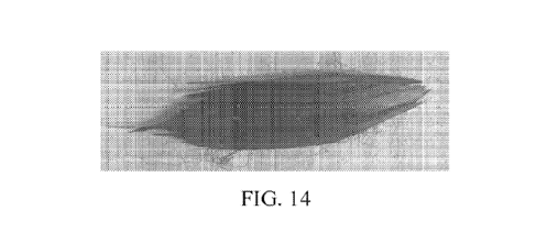
[0038] FIG. 14 illustrates changes in fiber activation achieved
with adjustment of
electrode position from that illustrated in FIG. 13.
[0039] FIG. 15 illustrates human thalamic imaging data from a
human subject with
traumatic brain injury (TBI) including the percentage activation of CL and PPN
targets and other
thalamic nuclei for avoidance (VPM, CM, MD).
[0040] FIG. 16 illustrates testing results for five subjects
receiving DBS according to the
vector-based targeting of the human central thalamus of FIG. 5.
[0041] FIG. 17 illustrates an exemplary approach to target
acquisition from a
representative human subject along with activation results from both
hemispheres.
[0042] FIG. 18 illustrates another exemplary approach to target
acquisition from another
representative human subject along with activation results from both
hemispheres.
[0043] FIG. 19 illustrates the placements of active contacts for
a plurality of human
subject in a common synthetic atlas space.
[0044] FIG. 20 illustrates cortical evoked potentials obtained
across a 128 channel EEG
array for activation across two active contacts using a 2Hz duty cycle of
stimulation
CA 03231861 2024-3- 14
WO 2023/043786 - 10 -
PCT/US2022/043451
DETAILED DESCRIPTION
[0045] The present technology relates to methods for vector-
based targeting of a human
central thalamus to guide deep brain stimulation (DBS). The present technology
also relates to
methods, devices, systems, and non-transitory computer readable media for
surgical planning for
vector-based targeting of a human central thalamus to guide DBS. More
specifically, the present
technology relates to methods of human central thalamic targeting to achieve
target activation
and successful target avoidance of regions of human intra-thalamic pathways
within the central
thalamus to achieve vector-based placement of deep brain stimulation
electrodes.
[0046] Devices and systems for carrying out vector-based
targeting of a human central
thalamus to guide DBS, including a surgical computing device, are described
herein. One aspect
of the present technology relates to a system for vector-based targeting of a
human central
thalamus to guide DBS. The system includes a surgical computing device of the
present
technology. The system also includes an imaging device operationally coupled
to the surgical
computing device and one or more electrodes. An electrical stimulator is
coupled to the surgical
computing device and the one or more electrodes to permit electrical
activation of the electrodes
based on instructions from the surgical computing device.
[0047] FIG. 1 illustrates an environment including system 12 for
vector-based targeting
of a human central thalamus to guide DBS. System 12 includes surgical
computing device 14,
imaging device 16, and DBS apparatus 18, although system 12 may include other
elements or
components in other combinations, such as additional computing devices. System
12 enables
treatment via the selective activation of structures within the central
thalamus to support
forebrain arousal regulation via the activation of fibers emanating from the
central lateral nucleus
of the central thalamus (CL) and the surrounding dorsal tegmental track medial
(DTTm)
(CL/DTTm).
[0048] Surgical computing device 14 of system 12 includes
processor(s) 20, memory 22,
and communication interface 24 that are coupled together by a bus 26 or other
communication
link, although surgical computing device 14 can include other types and/or
numbers of elements
in other configurations. Processor(s) 20 of surgical computing device 14 may
execute
programmed instructions stored in memory 22 for any number of the functions or
other
operations illustrated and described by way of the examples herein, including
surgical planning
for vector-based targeting of a human central thalamus to guide DBS.
Processor(s) 20 of surgical
computing device 14 may include one or more graphic processing units (GPUs),
central
CA 03231861 2024-3- 14
WO 2023/043786 - 11 -
PCT/US2022/043451
processing units (CPUs), or general purpose processors with one or more
processing cores, for
example, although other types of processor(s) can also be used.
[0049] Memory 22 of surgical computing device 14 stores these
programmed instructions
for one or more aspects of the present technology as illustrated and described
herein, although
some or all of the programmed instructions could be stored elsewhere. A
variety of different
types of memory storage devices, such as random access memory (RAM), read only
memory
(ROM), solid state drives (SSD), flash memory, or other computer readable
medium that is read
from and written to by a magnetic, optical, or other reading and writing
system that is coupled to
processor(s) 20 can be used for memory 22.
[0050] Accordingly, memory 22 of surgical computing device 14
can store application(s)
that can include executable instructions that, when executed by surgical
computing device 14,
cause surgical computing device 14 to perform actions, such as to perform
methods for vector-
based targeting of a human central thalamus to guide DB S as illustrated and
described by way of
the examples herein, such as in FIG. 5. The application(s) can be implemented
as modules or
components of other application(s). Further, the application(s) can be
implemented as operating
system extensions, modules, plugins, or the like.
[0051] Communication interface 24 of surgical computing device
14 operatively couples
and allows for communication between surgical computing device 14, imaging
device 16, and
DBS apparatus 18, which are all coupled together by one or more communication
network(s) 28,
although other types and/or numbers of connections and/or configurations to
other device and/or
elements can be used. Communication network(s) 28 can include any number
and/or types of
communication networks, such as local area network(s) (LAN(s)) or wide area
network(s)
(WAN(s)), and/or wireless networks, although other types and/or number of
protocols and/or
communication network(s) can be used.
100521 Although embodiments of surgical computing device 14 are
described and
illustrated herein, surgical computing device 14 can be implemented on any
suitable c.xmiputing
system or computing- device. It is to be understood that the devices and
systems described herein
are for exemplary purposes and many variations of the specific hardware and
software are
possible, as will be appreciated by those skilled in the relevant art(s).
[0053] In addition, two or more computing systems or devices can
be substituted for any
one of the systems described above. Accordingly, principles and advantages of
distributed
processing, such as redundancy and replication, also can be implemented, as
desired, to increase
the robustness and performance of the devices and systems described above. The
embodiments
of the present application may also be implemented on a computer system or
systems that extend
CA 03231861 2024-3- 14
WO 2023/043786 - P -
PCT/US2022/043451
across any suitable network using any suitable interface in echaiti sms and
communications
technologies, including, by way of example only, telecommunications in any
suitable form (e.g.,
voice and modern), wireless communications media, wireless communications
networks, cellular
communications networks, G3 communications networks, Public Switched Telephone
Networks
(PSTN-s), Packet Data Networks (PD-Ns), the Internet, intranets, and
combinations thereof.
[0054] Imaging device 16 may be any suitable imaging device to
obtain images of the
subject's brain, including devices suitable for computed tomography imaging,
although other
appropriate imaging devices may be employed. Imaging device 16 is coupled to
surgical
computing device 14 to provide images of the subject's brain for further
analysis in accordance
with the methods disclosed herein.
[0055] FIG. 2 is a perspective view and functional block diagram
of DBS apparatus 18.
DBS apparatus 18 includes first and second stimulators 30 coupled to stimulus
signal generator
32. Although DBS apparatus 18 is described with respect to first and second
stimulators 30, it is
to be understood that DBS apparatus 18 may include additional stimulators.
Further, although
DBS apparatus 18 is described, it is to be understood that other types of
stimulation devices
could be employed in the methods of the present technology including
stimulation devices that
employ other energy modalities.
[0056] First and second stimulators 30 include at least one
electrode 32 mounted on
shank 34. In one embodiment, more than one electrode 32 is mounted on shank 34
such that
stimulator 30 is a "multipolar electrode," with each electrode separately
controllable. In this
example, four electrodes 32 are located on each shank 34 to provide a
plurality of spaced
contacts, although other numbers of electrodes may be utilized. Electrodes 34
are connected to
one (or separate) insulated conductor(s) which passes through shank 34. The
insulated conductor
connects electrodes 32 to voltage control 36 and stimulus signal generator 38.
Voltage control 36
and stimulus signal generator 38 may be separate from one another or part of a
single unit. The
connections mentioned herein may be wired or wireless.
[0057] Electrodes 32 are made from a conducting material, which
may be an alloy such
as platinum/iridium, with impedances known in the art, for example, between
approximately of
100 and 150 kn. Electrodes 32 are approximately 0.5 mm in length. In one
embodiment, where
multiple electrodes 32 are mounted on shank 34, the separation between
electrodes 32 may be
variable or constant, and may be approximately 0.5 mm.
[0058] Shank 34 is configured to be implanted in the brain of
the subject. Shank 34 may
be configured as a cylinder, a square, a helix, or any other geometry known in
the art as suitable
CA 03231861 2024-3- 14
WO 2023/043786 - 13 -
PCT/US2022/043451
for implementation. In one embodiment, shank 34 is implanted in the central
thalamus of the
subject for selective activation of central thalamus fibers in the subject, as
described herein.
[0059] Stimulus signal generator 38 produces a selected pulse
train. In one embodiment,
stimulus signal generator 38 is capable of separately driving individual
electrodes 32 in a multi-
electrode system through various channels. In this example, stimulus signal
generator 38 may
operatively select any one of electrodes 32 to provide a stimulus signal.
Stimulus signal
generator 38 may provide stimulation with various parameters, such as
frequency or waveform,
across multiple electrodes 32 simultaneously, and independently.
[0060] Stimulus signal generator 38 is capable of generating
voltage wave trains of any
desired form (monophasic or biphasic sine, square wave, spike, rectangular,
triangular, ramp,
etc.) in a selectable voltage amplitude in the range from about 0.1 volts to
about 10.5 volts or
from about 0.1 mA to about 25.0 mA and at selectable frequencies in the range
from about 1 Hz
to about 10 kHz. In one embodiment, stimulus signal generator 38 is capable of
generating
constant current across at least one pair of electrodes 30 with either
electrode in the pair assigned
as a cathode or anode, although stimulus signal generator 38 may generate a
constant current
across two pairs of electrodes, across four pairs of electrodes, or across six
pairs of electrodes,
where either electrode in a pair can be assigned as a cathode or an anode. The
compliance
voltage of stimulus signal generator 38 is able to handle resistive loads
across any pair of
electrodes in the range from 0.5 kOhm to 10 kOhm. Each channel (cathode/anode
pair) is able to
deliver up to about 25.0 mA.
[0061] Stimulus signal generator 38 includes circuitry that
allows for monitoring of the
current delivered across each channel. In one embodiment, stimulus signal
generator 38 is
programmable in that pulse shapes, sequences, and frequencies of pulses can be
designed by
software on a computer, such as surgical computing device 14, and uploaded to
stimulus signal
generator 38 for delivery to electrodes 32 upon command. The cathode-anode
outputs from each
channel may be used to provide bipolar constant-current stimulation in the
intralaminar nuclei
through any pair of electrode contacts across implanted stimulators 30.
[0062] Voltage control 36 provides a selected current amplitude
or voltage to the waves
of the pulse train. In practice, the pulse train and voltage amplitudes
employed will be selected
on a trial and error basis by evaluating a subject's response to various types
and amplitudes of
electrical stimulation over a time course of from about 1 to about 12 months.
For example, after
implanting stimulators 30 in the subject's thalamic nuclei, stimulation with a
voltage within the
range of from about 0.1 to about 10.5 volts or higher at a rate within the
range of from about 1
Hz to about 10 kHz, is applied for from about 8 to about 12 hours a day. The
voltage control 36
CA 03231861 2024-3- 14
WO 2023/043786 - 14 -
PCT/US2022/043451
may provide continuous, periodic, or intermittent stimulation. In one
embodiment, voltage
control 36 provides an electrical stimulus that is carried out using one or
more stimulation
programs that are capable of being interleaved in time.
[0063] Referring now to FIGS. 3A and 3B, in one embodiment, DB S
apparatus 18
includes one or more sensors 40 connected to adaptive feedback controller 42.
Sensors 40 are
configured to detect neuronal activity of one or more cortical and/or
subcortical tissues of a
selected subject's brain, by means known in the art, although electrodes 32
may be utilized to
detect neuronal activity. In one embodiment, sensors 40 are incorporated into
stimulators 30,
although sensors 40 not incorporated into a stimulator, referred to herein as
"extra-stimulator
sensors- may be utilized. The extra-stimulator sensors may be implanted within
cortical or
subcortical regions or may be located on the scalp surface of the subject's
head. Sensors 40
collect neuronal data in the form of, for example, single-unit activity, local
field potentials,
and/or electrocorticogram ("EcoG") activity. Connections between sensor 40 and
brain tissue
may be electrical, electromagnetic (wireless), or optical to one or many
targets to be determined
by availability and involvement in specific patterns of brain injury.
[0064] In one embodiment, sensors 40 include computer and logic
circuitry, although
computer and logic circuitry associated with sensors 40 may be distributed
among other
components, such as incorporated into adaptive feedback controller 42, or in
the stimulus signal
generator 38, and/or one or more other devices, which may be implanted in the
subject or
external to the subject. In one embodiment, cortical placement of sensors 40
can detect the
occurrence of failures of human control and adaptive feedback 42 controller
can adjust
stimulation of thalamic targets in synchronism with the processes occurring in
deep brain
stimulation apparatus 18.
[0065] Referring now to FIGS. 3A, 3B, and 4, in one embodiment,
adaptive feedback
controller 42 includes neuronal recording module 44, state monitoring module
46, performance
monitoring module 48, processing module 50, and transmission module 52. The
modules
described here for adaptive feedback controller 42 may be located within one
physical device or
may be distributed among multiple devices, including surgical computing device
14, and may be
incorporated with other components or devices described herein. For example
and without
limitation, neuronal recording module 44 may be located in the same device as
an extra-
stimulator sensor and said device will have appropriate transmission pathways
to receive and
send information from and to other components of DBS apparatus 18, the
subject, and/or external
systems used to maintain, control, or inspect deep brain stimulation apparatus
18 or the subject,
including surgical computing device 14.
CA 03231861 2024-3- 14
WO 2023/043786 - 15 -
PCT/US2022/043451
[0066] Neuronal recording module 44 receives and stores various
items of information
from sensors 40, such as electrical waveform pattern data unique to the subj
ect. In one
embodiment, neuronal recording module 44 stores information received from
sensors 40 in real-
time when DB S apparatus 18 is being used. In one embodiment, neuronal
recording module 44
includes output means to allow retrieval of signals stored during an off-line
operation of DBS
apparatus 18.
[0067] State monitoring module 46 is coupled to sensors 40, and
is configured to store
and process a first set of variables associated with a state of the detected
neuronal activity,
particularly the spectral content of the local neuronal activity and in
particular, the total power
within the frequency ranges 10-15 Hz, 15-20 Hz, 20-25 Hz, 25-30 Hz, 10-30 Hz,
which have all
been empirically identified to increase within neuronal populations of the
cortex, basal ganglia,
and thalamus during either effective multi-site stimulation or during alert
cognitive function.
State monitoring module 46 may be used to sample the average characteristics
of neuronal
activity over time from sensors 40 or outside of the brain that collect
neuronal signals for this
purpose and to provide as feedback the real-time characteristics of the
signals via direct or
wireless (Bluetooth) connections. In one embodiment, state monitoring module
46 includes an
internal memory and computational resources to extract signal features of the
neuronal signal.
[0068] Performance monitoring module 48 is coupled to sensors 40
and is configured to
store and process a second set of variables associated with modulation of the
frequency of the
locally detected neuronal activity. Performance monitoring module 48 is used
to monitor the
performance characteristics of the stimulation in producing increases in
spectral power of local
populations at pre-specified frequency ranges (e.g., 15-25 Hz). In one
embodiment, performance
monitoring module 48 includes an internal memory and computational resources
to extract signal
features of the neuronal signal.
[0069] Processing module 50 is coupled to state monitoring
module 46 and performance
monitoring module 48. In one embodiment, processing module 50 is configured to
extract a
feature vector based upon the processed first and second set of variables, and
may be configured
to compute an optimal response stimulus signal based upon a comparison between
the extracted
feature vector and a pre-stored feature vector corresponding to the local
spectrum of neuronal
activity for the subject recording sites. Transmission module 52 is configured
to transmit the
optimal response stimulus signal computed by the processing module 50 to the
implanted
stimulus signal generator 38 to regulate the arousal level neuronal activity
of the subject.
[0070] Based upon respective sets of variables stored and/or
measured, performance
monitoring module 48 and state monitoring module 46 may be used to extract a
feature vector
CA 03231861 2024-3- 14
WO 2023/043786 - 16 -
PCT/US2022/043451
from the variables using computer and logic circuitry. Feature vectors
represent an
approximately complete mathematical description of electrical signals
resulting from neuronal
activity. Computed feature vectors can be used for further processing and to
synthesize a
feedback signal if necessary. A feedback signal can be outputted via a
transmission path, which
may be wired, wireless, or optical as known to one skilled in the art. The
same or a separate
component of DBS apparatus 18 computes an output signal and transmits it to
stimulator 30
placed within the brain to regulate their output in response to ongoing
analysis provided by
internal monitoring systems.
[0071] Referring again to FIGS. 3A and 4, an embodiment of the
present application
wherein the DBS apparatus 18 includes sensors 40 that are interfaced to
adaptive feedback
controller 42, which in turn is interfaced to stimulus signal generator 38, is
shown. Stimulus
signal generator 38 is configured to provide feedback control of electrical
stimulation of the
targeted brain regions, for example, the CL/DTTm fiber pathways. Upon receipt
of a signal via
a transmission path, which may be wired, wireless, or optical, stimulus signal
generator 38
provides a corresponding stimulus to these regions of the brain via at least
one of stimulators 12
to modulate or maintain the arousal state of a subject. The operating
characteristics of DBS
apparatus 18 may be adjusted automatically using adaptive feedback controller
42. In other
embodiments, sensor 40 or components of adaptive feedback controller 42 may
store information
for retrieval by an external system or by a physician, or may be used by a
physician/programmer
to adjust DBS apparatus 18 settings. Settings may be adjusted by the DBS
apparatus 18 itself or
by an external physician/programmer to raise a level of arousal, or impact on
local signal power.
[0072] One aspect of the present technology relates to a method
for vector-based
targeting of a human central thalamus to guide deep brain stimulation (DBS).
The method
involves providing one or more electrodes each with a plurality of contacts. A
three-dimensional
orientation of a dominant axis of a CL/DTTm fiber bundle of the human subject
is determined.
The plurality of contacts of the one or more electrodes are then positioned in
the human subject's
central thalamus fibers in substantial alignment with the determined three-
dimensional
orientation of the dominant axis of the CL/DTTm fiber bundle. An electrical
stimulus is then
applied to the positioned plurality of contacts of the one or more electrodes
to treat the human
subject for impaired arousal regulation. The positioning and the applying are
carried out to
maximize activation of a central lateral nucleus and medial dorsal tegmental
tract fiber pathway
in the human subject and to minimize activation of a centromedian-
parafascicularis fiber pathway
in the human subject.
CA 03231861 2024-3- 14
WO 2023/043786 - 17 -
PCT/US2022/043451
[0073] In a first step, one or more electrodes each with one or
more contacts are
provided. In one embodiment, deep brain stimulator apparatus 18 with
electrodes 32 may be
employed, although other devices for activation of the subject's central
thalamus may employed
such as a fiberoptic-optogenetic ("FOG") system, BION system, or ultrasound.
The one or more
electrodes are configured to provide for selective activation of the central
thalmus fibers of the
subject as described below. The present technology may be employed with single
lead systems
with multiple electrical contacts, single lead systems with multiple split
contacts, and multiple
lead systems with any combination of multi-contact electrodes including split
band contacts.
Importantly, the system will be capable of addressing any combination of
anodes and cathodes
across lead(s) contacts.
[0074] Next, the one or more electrodes, such as electrodes 32
are positioned in the
subject's central thalamus fibers. In one embodiment, once a relevant subject
is selected,
stimulator 30, as described above, is implanted in the subject's central
thalamus as illustrated in
FIG. 3B to maximize central lateral nucleus and medial dorsal tegmental tract
fiber pathway
activation in the subject and to minimize central medial parafascicularis
fiber pathway activation
in the subject. The zones of activation and suppression are illustrated in
FIG. 5. As discussed
above, stimulator 30 includes one or more electrodes 32. In some embodiments,
a plurality of
electrodes 32 are provided. One or more electrodes 32 have a plurality of
spaced contacts. The
CL/DTTm target can be activated optimally by shaping the applied electrical
field by utilizing
first and second stimulators 12, with many electrode 32 contacts as described
below. As shown,
this is achieved by positioning the most of electrodes 32 on stimulator 30 to
be in contact with
the central lateral nucleus and medial dorsal tegmental tract fibers while few
if any of electrodes
32 on stimulator 30 are in contact with the central median parafascicularis
fibers.
[0075] To carry our the above methods, a subject may be
conscious with application of
local anesthesia or mild sedation. in cases where a subject is not
sufficiently cooperative to
remain conscious during the procedure, the above-described approach can be
modified in ways
known in the art, to allow the operation to be completed under general
anesthesia.
[0076] Subjects may include any animal, including a human. Non.-
human animals
includes all vertebrates, e.g., rnanuuals and non-mammals, such as non-human
pi in ates, sheep,
dogs, cats, cows, horses, chickens, amphibians, and reptiles, although mammals
are preferred,
such as non-human primates, sheep, dogs, cats, cows and horses. The subject
may also be
livestock such as, cattle, swine, sheep, poultry, and horses; or pets, such as
dogs and cats.
[0077] The methods described herein can be employed for subjects
of any species,
gender, age, ethnic population, or genotype. Accordingly, the term subject
includes males and
CA 03231861 2024-3- 14
WO 2023/043786 - 18 -
PCT/US2022/043451
females, and it includes elderly, adult-to-elderly Iran Siti 011 age subjects
adults, pre-adult-to-adult
transition age subjects, and pre-adults, including adolescents, children, and
infants. In one
embodiment, subjects are ad ki tt subjects in their twenties to forties, who
have the most to gain
from treatment and represent the greatest cost to society if left untreated.
Examples of human
ethnic populations include Caucasians, Asians, Hispanics, Africans, African
Americans, Native
Americans, Semites, and Pacific -islanders. The term subject also includes
subjects of any
genotype or phenotype as long as they are in need of the treatment as
described herein. In
addition, the subject can have the genotype or phenotype for any hair color,
eye color, skin color
or any combination thereof. The term subject includes a subject of any body
height, body weight,
or any organ or body part size or shape
[0078] In one embodiment, stimulator 30 is introduced via burr
holes in the skull,
although in other examples multiple stimulators may be employed. Generally,
prior to the
introduction of stimulators 30, a detailed mapping with microelectrode and
microstimulation
following standard methods is carried out as described in Tasker et al., "The
Role of
the Thalamus in Functional Neurosurgery," Neurosurgery Clinics of North
America 6(1):73-104
(1995), which is incorporated herein by reference in its entirety. Imaging
device 16 may be
employed to image the subject's brain. The system will enable the user to plan
an implantation
of a stimulation system, such as stimulator 30, in an individual subject using
the neuroimaging
data from imaging device 16.
[0079] The imaging data is employed to model thalamic nuclei,
white matter fiber tracts
and connections, and the impact of electrical field activation within the
thalamus by directly
modeling the relative activation of CL/DTTm4TRN, Cm-Pf4TRN, and other adjacent
thalamic
pathways. The present technology enables the biophysical modeling of the
precise placement of
a single or multiple lead system to selectively activate CL/DTTm and avoid co-
activation of the
Cm-Pf fiber bundle. This system includes modeling of thalamic nuclei, modeling
of specific
white matter fiber pathways within the brain, bioelectric field modeling, and
probabilistic
mapping of target activation and target avoidance achieved with varying
configurations of lead
contact arrangements, cathode and anode geometries, pulse shapes, pulse
widths, and frequencies
of stimulation.
[0080] In one aspect, a segmented brain model of the subject's
central thalamus may be
produced using known techniques. Model electrode positions and electrical
stimulation
conditions may be identified using the segmented brain model that will
maximize central lateral
nucleus and medial dorsal tegmental tract fiber pathway activation in the
subject, while
minimizing the central medial parafascicularis fiber pathway activation in the
subject. A
CA 03231861 2024-3- 14
WO 2023/043786 - 19 -
PCT/US2022/043451
stimulation map is produced based on the identified electrode positions and
electrical stimulation
conditions. The stimulation map may then be employed to carry out the actual
positioning of the
system, such as stimulator 30. The stimulation map, in some examples, may also
be used to
determine the applying of stimulation, as described further below.
[0081] Next, an electrical stimulus is applied to the positioned
one or more electrodes 32
to selectively activate the central thalamus fibers of the subject. The
electrical stimulus may be
carried out in various conditions to maximize central lateral nucleus and
medial dorsal tegmental
tract fiber pathway activation in the subject and to minimize central medial
parafascicularis fiber
pathway activation in the subject. For example, the electrical stimulus may be
applied between
.1 to 25.0 milliamps or 0.1 to 10.5 volts, selected independently for each
electrode. The electrical
stimulus may be applied using continuous, intermittent or periodic
stimulation. The electrical
stimulus may be applied using substantially in-phase or substantially out-of-
phase stimulation on
each electrode 32. The electrical stimulus can be configured to be ramped up
or down at
different rates of speed to improve the selective activation. The electrical
stimulus is carried out
using voltage wave trains having a monophasic or biphasic sine, square, spike,
rectangular,
triangular or ramp configurations. The electrical stimulus can be applied at
one or more
frequencies of from 1 Hz to 10 kHz. Further, the electrical stimulus can be
carried out using one
or more stimulation programs that are capable of being interleaved in time.
[0082] The devices and systems of the present technology allow
for the precise placement
of single or multiple leads to selectively activate CL/DTTm fibers and
minimize adjacent OFF-
target fibers originating and passing through the centromedian-parafasicularis
nucleus complex
(Cm-N) that also project to the thalamic reticular nucleus (TRN), such as
shown in FIG. 3B. The
one or more electrodes 32 are positioned to maximize central lateral nucleus
and medial dorsal
tegmental tract fiber pathway activation in the subject and to minimize
central median
parafascicularis fiber pathway activation in the subject as shown in FIG. 5.
[0083] The present technology specifies the geometric
requirements for selective
activation of CL/DTTm to facilitate cognitively mediated behaviors (including
but not limited to
executive functions, vigilance, sustained attention, working memory, decision-
making, and motor
executive functions (e.g. controlled hand and arm movements). The primary
effect of selective
CL/DTTm stimulation is activation of neuronal populations across frontal
cortical structures and
the striatum, while minimizing OFF-target effects. Other cortical structures
such as posterior
parietal cortices and primary sensory cortices are additional direct targets
of CL/DTTm activation
based on known anatomical and physiological demonstrations. In one embodiment,
75% to
100% of the medial dorsal tegmental tract fibers in the central thalamus of
the subject are
CA 03231861 2024-3- 14
WO 2023/043786 - 20 -
PCT/US2022/043451
stimulated and less than 25% of the central medial parafascicularis fibers in
the central thalamus
of the subject are stimulated. In another embodiment, 90% to 100% of the
medial dorsal
tegmental tract fibers in the central thalamus of the subject are stimulated
and less than 10% of
the central median parafascicularis fibers in the central thalamus of the
subject are stimulated.
[0084] In one embodiment, deep brain stimulation apparatus 18
further includes sensors
40 that are configured to provide feedback to determine a state of neuronal
activity during
application of an electrical stimulus as described above. One or more of the
electrical stimulus
conditions can be adjusted based on the state of neuronal activity to provide
improved selective
activation of the subject's central thalamus based on feedback from sensors
40.
[0085] Another aspect of the present technology relates to a
method of treating a
condition characterized by impaired arousal regulation in a human subject. The
method involves
selecting a human subject with impaired arousal regulation. One or more
electrodes are provided
each with a plurality of contacts. A three-dimensional orientation of a
dominant axis of a
CL/DTTm fiber bundle of the human subject is determined. The plurality of
contacts of the one
or more electrodes are then positioned in the human subject's central thalamus
fibers in
substantial alignment with the determined three-dimensional orientation of the
dominant axis of
the CL/DTTm fiber bundle. An electrical stimulus is then applied to the
positioned plurality of
contacts of the one or more electrodes to selectively activate the central
thalmus fibers in
substantial alignment with the determined three-dimensional orientation of the
dominant axis of
the CL/DTTm fiber bundle. An electrical stimulus is then applied to the
positioned plurality of
contacts of the one or more electrodes to selectively activate the central
thalamus fibers of the
human subject. The positioning and the applying are carried out to maximize
activation of a
central lateral nucleus and medial dorsal tegmental tract fiber pathway in the
human subject and
to minimize activation of a centromedian-parafascicularis fiber pathway in the
human subject.
[0086] Impaired arousal regulation is a key underlying component
of a wide range of
acquired, inherited, and idiopathic neuropsychiatric illnesses. Most
prominently, traumatic brain
injuries produce impaired arousal regulation. Additional forms of structural
brain injuries that
disrupt arousal regulation include anoxia, hypoxia, hypoxic-ischemic injuries,
stroke,
encephalitis of infectious or autoimmune causes, and a wide range of primary
degenerative
illnesses such as Parkinson's disease. Importantly, supporting arousal
regulation is under present
clinical study for restoring cognitive function during seizures or post-ictal
states of depressed
cortical function. Impaired arousal regulation is an untreated primary feature
of neuropsychiatric
diseases such as schizophrenia or autism. Accordingly, the technology
described and illustrated
herein can be used to treat brain injury, a neurological degenerative disease,
epilepsy, a
CA 03231861 2024-3- 14
WO 2023/043786 - 21 -
PCT/US2022/043451
movement disorder, a post-encephalitis cognitive impairment, a development
disorder, a post-
hypoxic-ischemic injury cognitive impairment, a neuropsychiatric disorder,
post-intensive care
unit (ICU) mixed disorder cognitive impairment, and/or post-ICU adult
respiratory distress
syndrome. These applications are noted as relevant examples but are not
exhaustive of
applications for the specific use of the system to enable selective CL/DTTm
activation in an
individual to improve arousal regulation.
[0087] In one embodiment, a subject having a condition
characterized by impaired
arousal regulation may be selected for treatment using the method described
above. The subject
may have a condition selected from the group consisting of brain injury, a
neurological
degenerative disease, epilepsy, a movement disorder, a post-encephalitis
cognitive impairment, a
developmental disorder, a post hypoxic-ischemic injury cognitive impairment,
and a
neuropsychiatric disorder.
[0088] The present technology enables the specific positioning
of a system within the
central thalamus to optimize behavioral facilitation achievable with improved
arousal regulation.
The technology guides the conceptualization and placement of the system and
allows the user to
explore a space of stimulation configurations and modes of activation to map a
range of
behavioral outcomes to the system, as described in further detail below. These
maps are
inherently multi-dimensional: they include effects on CL/DTTm and Cm-Pf->TRN
pathways,
multiple possible behavioral facilitation effects, and just as important OFF-
target side effects.
[0089] Selective activation of the DTTm fiber pathway that
projects through the CL
nucleus, and not the Cm-Pf complex fiber projections, facilitates performance.
Such selective
activation can be utilized as therapeutic options for treatment of subjects
suffering from impaired
arousal regulation and enduring cognitive dysfunction. As disclosed in Baker,
et al., "Robust
Modulation of Arousal Regulation, Performance and Frontostriatal Activity
Through Central
Thalamic Deep Brain Stimulation in Healthy Non-Human Primates." J.
Neurophysiol. 116:2383-
2404 (2016), the disclosure of which is incorporated by reference herein in
its entirety, shaping
the DBS electrical field within the 'wing' of CL resulted in robust behavioral
facilitation and
enhancement of frontal and striatal population activity. These findings are
consistent with the
behavioral and physiological effects of conventional CT-DBS in a case study in
a very severely
traumatic brain injury (TBI) subject, as disclosed in Schiff, et al.,
"Behavioural Improvements
with Thalamic Stimulation After Severe Traumatic Brain Injury.- Nature. 448,
600-3 (2007), the
disclosure of which is incorporated herein by reference in its entirety.
[0090] The present technology disaggregates the CL thalamus by
isolating contributions
from CL and DTTm from the contributions of the Cm-Pf complex. Two mechanisms
may
CA 03231861 2024-3- 14
WO 2023/043786 _ 22 _
PCT/US2022/043451
explain these behavioral results: 1) an intrathalamic inhibitory network
similar to that defined in
the rodent, as disclosed in Crabtree, et al., "New Intrathalamic Pathways
Allowing Modality-
Related and Cross Modality Switching in the Dorsal Thalamus.' J. Neurosci. 22,
8754-8761
(2002) and Crabtree, -Functional Diversity of Thalamic Reticular Subnetworks."
Front. SysL
Neurosci. s12 (2018), the disclosures of which are incorporated by reference
herein in their
entirety), 2) the roles the two pathways play in controlling the anterior
forebrain mesocircuit, as
disclosed in N. D. Schiff, "Recovery of Consciousness After Brain Injury: A
Mesocircuit
Hypothesis." Trends Neurosci. 33, 1-9 (2010), the disclosure of which is
incorporated by
reference herein in its entirety, a system involving the thalamus, frontal
cortex, and basal ganglia
that regulates the overall level of activity in the anterior forebrain.
[0091] In one embodiment, the position of segmented single leads
and multi-lead systems
can be optimized to selectively target the cell bodies of CL and the DTTm
pathway and to avoid
the fiber projections emanating from Cm-Pf. The isolated activation of the
DTTm pathway
projecting from CL to frontostriatal targets facilitates behavioral
performance. In contrast, mixed
activation of the DTTm and fibers projecting from the Cm-Pf complex through
the TRN either
interrupts or mitigates these facilitation effects.
[0092] Although both CL and Cm-Pf have strong striatal
projections, their patterns of
innervations within the striatum are markedly different, both regionally and
with respect to
cellular elements and cell types innervated. Single fiber studies note that CL
afferents make en
passant synapses in TRN before fanning out broadly over the rostral striatum
as disclosed in
Deschenes, et al., "Striatal and Cortical Projections of Single Neurons From
the Central Lateral
Thalamic Nucleus in the Rat." Neuroscience. 72, 679-687 (1996), the disclosure
of which is
incorporated by reference herein in its entirety. By contrast, Cm-Pf fibers
project heavily into
regionally precise zones of the striatum and form bushy local arborizations,
as disclosed in
Parent, et al., "Axonal Collateralization in Primate Basal Ganglia and Related
Thalamic Nuclei."
Thalamus 1?elat. ,S'yst. 2, 71 (2002), Smith, et al., "The Thalamostriatal
Systems: Anatomical and
Functional Organization in Normal and Parkinsonian States." Brain Res. Bull.
78, 60-68 (2009),
Storch, et al., "Reliability and Validity of the Yale Global Tic Severity
Scale." Psychol. Assess
17, 486-491 (2005), and Smith, et al., "The Thalamostriatal System in Normal
and Diseased
States." Front. 5yst. Neurosci. 8 (2014), the disclosures of which are
incorporated by reference
herein in their entirety. CL and Pf afferents are known to project into the
main neuronal
populations of the striatum, the medium spiny neurons, as disclosed in (Bolam,
et al., "Synaptic
Organisation of the Basal Ganglia." J. Anat. 196, 527-542 (2000) and Ellender,
et al.,
"Heterogeneous Properties of Central Lateral and Parafascicular Thalamic
Synapses in the
CA 03231861 2024-3- 14
WO 2023/043786 _ 23 _
PCT/US2022/043451
Striatum." J. Physiol 591, 257-72 (2013), the disclosures of which are
incorporated by reference
herein in their entirety), whereas Cm neurons project into the local
cholinergic inhibitory
neurons, as disclosed in Smith, et al., "The Thalamostriatal Systems:
Anatomical and Functional
Organization in Normal and Parkinsonian States." Brain Res. Bull. 78, 60-68
(2009), the
disclosure of which is incorporated herein by reference in its entirety. Most
importantly, CL
fibers have strong and broad frontostriatal projections that strongly activate
the entire
frontal/prefrontal cortex and rostral striatum with high-frequency
stimulation, as disclosed in Li
et al., "Uncovering the Modulatory Interactions of Brain Networks in Cognition
with Central
Thalamic Deep Brain Stimulation Using Functional Magnetic Resonance Imaging."
Neuroscience. 440, 65-84 (2020), Liu, et al., "Frequency-Selective Control of
Cortical and
Subcortical Networks by Central Thalamus." Elife. 4, 1-27 (2015), and Baker,
et al., "Robust
Modulation of Arousal Regulation, Performance and Frontostriatal Activity
Through Central
Thalamic Deep Brain Stimulation in Healthy Non-Human Primates." J.
Neurophysiol. 116:2383-
2404 (2016), the disclosures of which are incorporated by reference herein in
their entirety.
100931 Despite these distinctions, improved arousal and
facilitation of behavior have been
reported for electrical stimulation of both CL and Cm-Pf. In rodent studies,
electrical stimulation
of CL facilitates object recognition memory (Shirvalkar, et al., "Cognitive
Enhancement with
Central Thalamic Electrical Stimulation." Proc. Natl. Acad. Sci. U S. A. 103,
17007-17012
(2006), the disclosure of which is incorporated by reference herein in its
entirety), working
memory (Chang, et al., "Modulation of Theta-Band Local Field Potential
Oscillations Across
Brain Networks With Central Thalamic Deep Brain Stimulation to Enhance Spatial
Working
Memory." Front. Neurosci. 13 (2019), the disclosure of which is incorporated
by reference
herein in its entirety), and decision-making (Mair, et al., "Memory
Enhancement with Event-
Related Stimulation of the Rostra] Intralaminar Thalamic Nuclei." J. Neurosci.
28, 14293-14300
(2008) and Mair, et at., "Cognitive Activation by Central Thalamic
Stimulation: The Yerkes-
Dodson Law Revisited." Dose-Response. 9, 313-331 (2011), the disclosures of
which are
incorporated by reference herein in their entirety). In healthy NHPs, CL
dominant stimulation,
that includes the DTTm as shown here, facilitates sustained attention, working
memory, and
pattern-recognition behaviors as disclosed in Baker, et al., "Robust
Modulation of Arousal
Regulation, Performance and Frontostriatal Activity Through Central Thalamic
Deep Brain
Stimulation in Healthy Non-Human Primates.- J. Neurophysiol. 116:2383-2404
(2016), the
disclosure of which is incorporated by reference herein in its entirety. In
humans, CL stimulation
has shown facilitation of a range of cognitive behaviors including motor
executive function and
speech production, as disclosed in Schiff, et al., "Behavioural Improvements
with Thalamic
CA 03231861 2024-3- 14
WO 2023/043786 - 24 -
PCT/US2022/043451
Stimulation After Severe Traumatic Brain Injury." Nature. 448, 600-3 (2007),
the disclosure of
which is incorporated herein by reference in its entirety. However, human
studies also report
speech facilitation with Cm-Pf stimulation (Bhatnagar, et al., "Effects of
Intralaminar Thalamic
Stimulation on Language Functions." Brain Lang. 92, 1-11(2005), the disclosure
of which is
incorporated by reference herein in its entirety) and restoration of arousal
in severe brain injury.
[0094] In rodents, Crabtree, et al., "New Intrathalamic Pathways
Allowing Modality-
Related and Cross Modality Switching in the Dorsal Thalamus." I Neurosci. 22,
8754-8761
(2002)., the disclosure of which is incorporated by reference herein in its
entirety, demonstrated a
structural basis for a rich system of intrathalamic inhibitory interactions
and characterized two
important findings relevant to the present results: 1) a rich network exists
for local inhibition
within the thalamus of separate sensory nuclei or motor nuclei; these
inhibitory networks appear
to be local to either sensory or motor thalamic nuclei; and 2) a cross sensory-
to-motor thalamus
pathway via the inhibition of the anterior intralaminar group by the caudal
intralaminar group.
Activation of the caudal intralaminar group produced powerful inhibition and
suppression of
neuronal firing in the anterior group via a disynaptic connection with TRN.
These findings
suggest an important motif of intra-thalamic inhibition of the two
intralaminar nuclear groups in
the thalamus. However, an important distinction in the rodent compared with
feline or primate
thalamus is the inclusion by Crabtree and Issac of CL as part of the caudal
intralaminar group, in
large part because the Cm-Pf nucleus is not present in the rodent as disclosed
in Jones, The
Thalamus Springer US, Boston, MA, ed. 2nd, 2007, the disclosure of which is
incorporated by
reference here in its entirety.
[0095] In comparison, Cm-Pf in primates is massively expanded
(Jones, et al.,
"Differential Calcium Binding Protein Immunoreactivity Distinguishes Classes
of Relay Neurons
in Monkey Thalamic Nuclei." Eur. J. IVeurosci. 1, 222-246 (1989) and Jones,
The Thalamus
Springer US, Boston, MA, ed. 2nd, 2007, the disclosures of which are
incorporated by reference
here in their entirety), and CL has been classified as a component of the
rostral intralaminar
group. Jones, The Thalamus Springer US, Boston, MA, ed. 2nd, 2007, the
disclosure of which is
incorporated by reference here in its entirety, particularly notes that the
paralamellar MD
densocellular components can be considered posterior cells of the CL nucleus,
these neurons
strongly project to frontal and pre-frontal cortices and are contiguous with
medial aspects of Cm-
Pf and the anterior aspects of Pf. Several anatomists have argued for these
regions to be included
in the human CL nucleus, as disclosed in Jones, The Thalamus Springer US,
Boston, MA, ed.
2nd, 2007, the disclosure of which is incorporated by reference here in its
entirety. Detailed
studies of Cm-Pf and CL interactions through the TRN are not available in non-
human primate,
CA 03231861 2024-3- 14
WO 2023/043786 _ 25 _
PCT/US2022/043451
and current modeling can be guided only by the observations in the rodent. A
direct inhibitory
effect on CL and surrounding association nuclei through TRN projections
activated by the Cm-
Pf-TRN fiber bundle can explain the apparent interference when activation is
balanced in the
DTTm and Cm-Pf-TRN fibers and the mitigation of this interference, with a
'push-pull' effect
tipping toward behavioral release as the DTTm becomes relatively more engaged.
[0096] DTTm activation facilitates selective activation of
frontostriatal neurons in the
awake state. Prior studies have demonstrated that facilitation of cognitively
mediated behaviors
in the healthy NHP requires a sufficiently powerful activation of frontal and
striatal neurons to
alter local field potential, as disclosed in Baker, et al., "Robust Modulation
of Arousal
Regulation, Performance and Frontostriatal Activity Through Central Thalamic
Deep Brain
Stimulation in Healthy Non-Human Primates." Neurophysiol 116:2383-2404 (2016),
the
disclosure of which is incorporated by reference herein in its entirety, and
individual neuronal
spiking dynamics. In the awake state, both frontal neocortical neurons and
striatal medium spiny
neurons are depolarized and receive a high rate of synaptic input, as
disclosed in Steriade, et al.,
-Natural Waking and Sleep States: A View From Inside Neocortical Neurons." .1.
Neurophysiol.
85, 1969-1985 (2001) and Grinner, et al., "Microcircuits in Action ¨From CPGs
to Neocortex "
Trends Neurosci . 28, 525-533 (2005), the disclosures of which are
incorporated by reference
herein in their entirety. Thus, to create sufficient impact as to be
measurable in behavioral
facilitation, the effects of DBS must be both spatially broad and strongly
effective across
frontostriatal populations.
[0097] Stimulation of CL with microelectrode techniques in awake
NHPs demonstrated
modest effects of behavioral facilitation, as disclosed in Smith, et al., "The
Thalamostriatal
Systems: Anatomical and Functional Organization in Normal and Parkinsonian
States." Brain
Res. Bull. 78, 60-68 (2009), the disclosure of which is incorporated herein by
reference in its
entirety. In contrast, the marked increase of behavioral facilitation achieved
by effective
geometries produced by 'field-shaping' within the central thalamus (fsCT-DBS)
when directly
compared with conventional CT-DBS, can be first understood in the context of
bulk activation
across frontostriatal networks, as disclosed in Baker, et al., "Robust
Modulation of Arousal
Regulation, Performance and Frontostriatal Activity Through Central Thalamic
Deep Brain
Stimulation in Healthy Non-Human Primates." J. Neurophysiol. 116:2383-2404
(2016), the
disclosure of which is incorporated by reference herein in its entirety. In
human subjects, bulk
activation of frontostriatal neuronal populations has been demonstrated as a
common mechanism
underlying a variety of effective pharmacological and electrophysiological
stimulation treatment
methods aimed at improving arousal regulation in the injured brain.
CA 03231861 2024-3- 14
WO 2023/043786 - 26 -
PCT/US2022/043451
[0098] In rodents, optogenetic stimulation of local neuronal
populations within the
central thalamus demonstrates that CL stimulation uniquely activates the
entire frontostriatal
system as measured at the whole brain level using functional magnetic
resonance, as disclosed in
Liu, et al., -Frequency-Selective Control of Cortical and Subcortical Networks
by Central
Thalamus. Elife. 4, 1-27 (2015), the disclosure of which is incorporated
herein by reference in its
entirety. The selective effect of stimulation of DTTm fibers demonstrated here
is consistent with
CL stimulation providing a broad excitatory input across frontal cortical and
striatal regions.
Even limited co-activation of the Cm-Pf->TRN fibers had a suppressive effect
on behavior draws
attention to the further distinctions of CL neurons and those within the
parafascularis (Pf) and
centromedian (Cm) nuclei.
[0099] The distinctions between CL and Cm-Pf neurons extend to
their postsynaptic
effects on inhibitory medium spiny neurons (MSNs), the neurons that project
out of the striatum
to the Globus pallidus (internal division). Whole-cell patch-clamp studies of
MSNs
optogenetically activated by either CL or Pf afferents show that CL afferents
act through AMPA
receptors and are more effective in driving MSN action potentials.
Additionally, the Pf afferents,
which act via NMDA receptors, generate long-term depression through mechanisms
of synaptic
plasticity, as disclosed in Ellender, et al., "Heterogeneous Properties of
Central Lateral and
Parafascicular Thalamic Synapses in the Striatum." I Physiol. 591, 257-72
(2013), the
disclosure of which is incorporated by reference herein in its entirety. These
physiological
distinctions likely provide additional contributions to the mitigation of
behavioral facilitation
achieved through DTTm activation when Cm-Pf fibers are co-activated because
these projections
continue in the striatum to MSNs. The excitation of MSNs by CL leads to
disynaptic
disinhibition of the thalamus through the anterior forebrain mesocircuit, as
disclosed in Fridman,
et al., "Neuromodul ati on of the Conscious State Following Severe Brain
Injuries." Curr. Op/n.
Neurobiol 29, 172-177 (2014), the disclosure of which is incorporated by
reference herein in its
entirety and Schiff, "Recovery of Consciousness After Brain Injury: A
Mesocircuit Hypothesis."
Trends Neurosci. 33, 1-9 (2010), the disclosure of which is incorporated by
reference herein in
its entirety, and co-activation of Pf fibers can oppose this thalamic
disinhibition through
suppression of the MSNs. Thus, the balance between CL/DTTm and CM-Pf afferents
to the
MSNs becomes a means by which the overall activity level of the thalamus can
be regulated.
[0100] Important distinctions at the cortical level are also
expected to influence the
impact of CL versus Cm-Pf activations; whereas CL innervates the cortex
broadly, Cm-Pf
projections are comparatively sparse, as disclosed in Jones, The Thalamus
Springer US, Boston,
MA, ed. 2nd, 2007, the disclosure of which is incorporated by reference here
in its entirety.
CA 03231861 2024-3- 14
WO 2023/043786 - 27 -
PCT/US2022/043451
Within the neocortex, the broad innervation of supragranular and infragranular
layers by CL
afferents is associated with supralinear summation of effects across cortical
columns, as
disclosed in Llinas, et al., "Temporal Binding Via Cortical Coincidence
Detection of Specific and
Nonspecific Thalamocortical Inputs: A Voltage-Dependent Dye-Imaging Study in
Mouse Brain
Slices." Proc. Natl. Acad. Sci. U. S. 816 A. 99, 449-454 (2002), the
disclosure of which is
incorporated herein by reference in its entirety. Collectively, it is likely
that the encroachment of
activation on Cm-Pf reduces the bulk activation of frontal cortical and
striatal regions through
local synaptic effects within the striatum where short-term depression may
affect patchy regions
of striatum innervated by Cm-Pf projections and interfere with behavioral
facilitation, as
disclosed in (Smith, et al., "The Thalamostriatal Systems: Anatomical and
Functional
Organization in Normal and Parkinsonian States." Brain Res. Bull. 78, 60-68
(2009) and
Ellender, et al., "Heterogeneous Properties of Central Lateral and
Parafascicular Thalamic
Synapses in the Striatum." J. Phy.siol. 591, 257-72 (2013), the disclosures of
which are
incorporated by reference herein in their entirety). Additionally, powerful
inhibition of cell
bodies within parts of CL or paralaminar thalamic regions (that contain
neurons with identical
properties (Jones, The Thalamus Springer US, Boston, MA, ed. 2nd, 2007 and
Miinkle, et al.,
"The Distribution of Calbindin, Calretinin and Parvalbumin Immunoreactivity in
the Human
Thalamus." J. Chem. Neuroanat. 19, 155-173 (2000), the disclosures of which
are incorporated
by reference herein in their entirety) via feedback inhibition from the TRN
(Crabtree, et al.,
"New Intrathalamic Pathways Allowing Modality-Related and Cross Modality
Switching in the
Dorsal Thalamus." J. Neurosci. 22, 8754-8761 (2002)., the disclosure of which
is incorporated
by reference herein in its entirety) as described above may suppress thalamic
output not captured
by direct electrical stimulation.
[0101] In comparison to the broad bulk activation required to
produce behavioral
facilitation with CT-DBS in DTTm, recent work in anesthetized NHPs has
demonstrated that
very local stimulation within the CL nucleus using multiple 25[Im contacts
spaced 200[tm apart
could produce arousal from Propofol and isoflurane anesthesia, as disclosed in
Redinbaugh, et
al., "Thalamus Modulates Consciousness via Layer-Specific Control of Cortex."
Neuron, 1-10
(2020), the disclosure of which is incorporated by reference herein in its
entirety. The effective
electrotonic length of these microprobe contacts, which determines the current
flow achieved
locally, as disclosed in Ranck, "Which Elements are Excited in Electrical
Stimulation of
Mammalian Central Nervous System: A Review." Brain Res. 98, 417-440 (1975),
the disclosure
of which is incorporated by reference herein in its entirety, is very short
compared to the broad
region activated by the fsCT-DBS configurations studied here. Of note,
stimulation at 50Hz but
CA 03231861 2024-3- 14
WO 2023/043786 _ 28 _
PCT/US2022/043451
not 200Hz was effective in producing arousal during anesthesia. In comparison,
in the awake
monkeys studied, stimulation at 150Hz-225Hz demonstrated the strongest
behavioral facilitation
and robust activation in frontal and striatal regions, as reflected by a
marked increase in the beta
and gamma frequency range and a decrease in the lower frequency bands measured
directly in
these locations, as disclosed in Baker, et al., "Robust Modulation of Arousal
Regulation,
Performance and Frontostriatal Activity Through Central Thalamic Deep Brain
Stimulation in
Healthy Non-Human Primates." J. Neurophysiot 116.2383-2404 (2016), the
disclosure of which
is incorporated by reference herein in its entirety. These differences likely
reflect the need, in
addition to achieving broad activation in the awake state, to increase levels
of background
synaptic activity received by neocortical and striatal neurons past particular
thresholds, as
disclosed in Larkum_ et al., "Calcium Electrogenesis in Distal Apical
Dendrites of Layer 5
Pyramidal Cells at a Critical Frequency of Back-Propagating Action
Potentials." Proc. Natl.
Acad. Sci. US.A. 96, 14600-14604 (1999), Larkum, et al., "Dendritic Spikes in
Apical Dendrites
of Neocortical Layer 2/3 Pyramidal Neurons. J. Neurosci. 27, 8999-9008 (2007),
and Larkum, et
al., -Synaptic Integration in Tuft Dendrites of Layer 5 Pyramidal Neurons: A
New Unifying
Principle." Science 325, 756-760 (2009), the disclosures of which are
incorporated by reference
herein in their entirety. Intrinsic integrative properties of individual
neocortical neurons change
with increasing levels of background synaptic input, as disclosed in
Bernanderõ et al., "Synaptic
Background Activity Influences Spatiotemporal Integration in Single Pyramidal
Cells." Proc.
Natl. Acad. Sci. U.S.A. 88, 11569-11573 (1991), the disclosure of which is
incorporated by
reference herein in its entirety. In order to trigger dendritic electrogenesis
in neocortical neurons,
across all layers, incoming excitatory inputs must have frequencies higher
than ¨130Hz, as
disclosed in Larkumõ et al., "Calcium Electrogenesis in Distal Apical
Dendrites of Layer 5
Pyramidal Cells at a Critical Frequency of Back-Propagating Action
Potentials." NOG. Natl.
Acad. Sci. US.A. 96, 14600-14604 (1999), Larkum, et al., "Dendritic Spikes in
Apical Dendrites
of Neocortical Layer 2/3 Pyramidal Neurons. .1. Neurosci. 27, 8999-9008
(2007), and Larkum, et
al., "Synaptic Integration in Tuft Dendrites of Layer 5 Pyramidal Neurons: A
New Unifying
Principle." Science 325, 756-760 (2009), the disclosures of which are
incorporated by reference
herein in their entirety. Similarly, the primary output neurons of the
striatum, medium spiny
neurons, require very high rates of background synaptic inputs to maintain
membrane
depolarization sufficient to generate action potentials, as disclosed in
Grillner, et al.,
"Mechanisms for Selection of Basic Motor Programs - Roles for the Striatum and
Pallidum."
Trends Neurosci. 28, 364-370 (2005), the disclosure of which is incorporated
by reference herein
in its entirety. Both mechanisms likely play a role in the requirement for
high-frequency
CA 03231861 2024-3- 14
WO 2023/043786 _ 29 _
PCT/US2022/043451
stimulation in the awake state, as disclosed in Schiff, -Central Lateral
Thalamic Nucleus
Stimulation Awakens Cortex via Modulation of Cross-Regional, Laminar-Specific
Activity
during General Anesthesia." Neuron. 106, 1-3 (2020), the disclosure of which
is incorporated by
reference herein in its entirety.
[0102] The selective effect of 50Hz CL stimulation in the
anesthetized monkey may
alternatively reflect antidromic activation of brainstem cholinergic and/or
noradrenergic fibers
that innervate CL. The brainstem neurons projecting to CL are known to have
resonant
properties at ¨40-50Hz whereas higher frequencies of stimulation actually
block action
potentials, as disclosed in Garcia-Rill, et al., "Coherence and Frequency in
the Reticular
Activating System (RAS).- Sleep ltled. Rev. 17, 227-238 (2013) and Garcia-
Rill, J, etal., "The
physiology of the pedunculopontine nucleus: implications for deep brain
stimulation." .1 Neural
Transm. 122, 225-235 (2015), the disclosures of which are incorporated by
reference herein in
their entirety, perhaps accounting for why others saw no effect during high-
frequency
stimulation.
[0103] A further aspect of the present technology relates to a
method for surgical
planning involving vector-based targeting of a human central thalamus to guide
DBS
implemented by one or more surgical computing devices. The method involves
segmenting the
central thalamus in an image of a bran of the human subject to produce a
segmented brain model.
One or more fiber pathways in the segmented brain model are modeled. A three-
dimensional
orientation of a dominant axis of a CL/DTTm fiber bundle of the human subject
is determined
based on the modelling. Initial model positions and orientations in the
segmented brain model
are generated for one or more electrodes based at least in part on the
determined three-
dimensional orientation of the dominant axis of the CL/DTTm fiber bundle of
the human subject.
A stimulation map is produced based on the modelling and the generating. A
position and
orientation for a plurality of contacts of the one or more electrodes in the
human subject's central
thalamus fibers and electrical stimulus conditions for the positioned and
oriented plurality of
contacts of the one or more electrodes are identified to selectively activate
the central thalamus
fibers of the human subject. This permits activation of a central lateral
nucleus and medial dorsal
tegmental tract fiber pathway in the human subj ect is maximized and
activation of a
centromedian-parafascicularis fiber pathway in the human subject is minimized
based on the
produced simulation map.
[0104] Yet another aspect of the present technology relates to a
non-transitory computer
readable medium having stored thereon instructions for surgical planning
involving vector-based
targeting of a human central thalamus to guide DBS. The non-transitory
computer readable
CA 03231861 2024-3- 14
WO 2023/043786 - 30 -
PCT/US2022/043451
medium includes executable code that, when executed by one or more processors,
causes the one
or more processors to segment the central thalamus in an image of the human
subject's brain to
produce a segmented brain model. One or more fiber pathways in the segmented
brain model are
modeled. A three-dimensional orientation of a dominant axis of a CL/DTTm fiber
bundle of the
human subject is determined based on the modelling. Initial model positions
and orientations in
the segmented brain model are generated for one or more electrodes based at
least in part on the
determined three-dimensional orientation of the dominant axis of the CL/DTTm
fiber bundle of
the human subject. A stimulation map is produced based on the modelling and
the generating. A
position and orientation for a plurality of contacts of the one or more
electrodes in the human
subject's central thalamus fibers and electrical stimulus conditions for the
positioned and oriented
plurality of contacts of the one or more electrodes are identified to
selectively activate the central
thalamus fibers of the human subject so that activation of a central lateral
nucleus and medial
dorsal tegmental tract fiber pathway in the human subject is maximized and
activation of a
centromedian-parafascicularis fiber pathway in the human subject is minimized
based on the
produced simulation map.
[0105] Another aspect of the present technology relates to a
surgical computing device.
The surgical computing device includes comprising memory comprising programmed
instructions stored thereon and one or more processors coupled to the memory
and configured to
execute the stored programmed instructions. The stored programmed instructions
include
segmenting the central thalamus in an image of a bran of the human subject to
produce a
segmented brain model. One or more fiber pathways in the segmented brain model
are modeled.
A three-dimensional orientation of a dominant axis of a CL/DTTm fiber bundle
of the human
subject is determined based on the modelling. Initial model positions and
orientations in the
segmented brain model are generated for one or more electrodes based at least
in part on the
determined three-dimensional orientation of the dominant axis of the CL/DTTm
fiber bundle of
the human subject. A stimulation map is produced based on the modelling and
the generating. A
position and orientation for a plurality of contacts of the one or more
electrodes in the human
subject's central thalamus fibers and electrical stimulus conditions for the
positioned and oriented
plurality of contacts of the one or more electrodes are identified to
selectively activate the central
thalamus fibers of the human subject so that activation of a central lateral
nucleus and medial
dorsal tegmental tract fiber pathway in the human subject is maximized and
activation of a
centromedian-parafascicularis fiber pathway in the human subject is minimized
based on the
produced simulation map.
CA 03231861 2024-3- 14
WO 2023/043786 - 31 -
PCT/US2022/043451
[0106] Referring to FIG. 5, a flowchart of an exemplary method
for surgical planning
involving vector-based targeting of a human central thalamus to guide deep
brain stimulation will
now be described. The method may be performed by one or more computing
devices, such as
surgical computing device 14 as illustrated in FIG. 1. Referring again to FIG.
5, in step 500 the
surgical computing device 14 segments the central thalamus is in image(s) of a
human subject's
brain to produce a segmented brain model.
[0107] In some examples, the imaging device 16 is used to
acquire pre-surgical magnetic
resonance imaging (MRI) image(s) of the human subject, optionally with
specific features that
assist with locating target activation regions and target avoidance regions.
In these examples, the
pre-surgical MRI image(s) include an image series that shows strong contrast
between white and
grey matter structures within the thalamus.
[0108] Optionally, white-matter-nulled magnetization-prepared
rapid acquisition
(WMnMPRAGE, or WMn) imaging of the human thalamus using MR acquisition
parameters
(e.g., inversion time TI, sequence repetition time TS, flip angle FA, receive
bandwidth RBW,
and/or k-space ordering strategy) can be used to produce strong intrathalamic
contrast, which
allows delineation of the intermedullary lamina and isolation and segmentation
of the central
thalamic (CL) volume. In some examples, the image resolution (i.e., voxel
size) is lmm or better
and/or isotropic (e.g. equal size for all three voxel dimensions), and/or the
imaging volume
covers the whole brain of thee human subject.
[0109] In some examples of this technology, the resulting WMn
images are processed
(e.g., by the surgical computing device 14) to segment (or define spatial
boundaries of) structures
within the thalamus of the human subject. One exemplary approach for this
segmentation is to
use the THalamus Optimized Multi-Atlas Segmentation (THOMAS) algorithm as
disclosed in Su
et al., "Thalamus Optimized Multi Atlas Segmentation (THOMAS): fast, fully
automated
segmentation of thalamic nuclei from structural MRI," Neuroimage. 2019 Jul
1;194:272-282,
which is incorporated herein by reference in its entirety, although other
methods for segmentation
can also be used. The THOMAS algorithm segments multiple thalamic nuclei on
each of the
brain image volumes
[0110] Another exemplary approach to segmentation in accordance
with the disclosed
technology is to use a single-atlas method for warping masks that label the CL
and VPM nuclei
from a template brain volume to the individual image volume of interest. Since
the THOMAS
algorithm does not identify or segment the CL and VPM nuclei, this second
stage of thalamic
segmentation may be performed in some examples of this technology. There are
several nuclei
identified by the THOMAS algorithm that may represent "target avoidance
regions" such as the
CA 03231861 2024-3- 14
WO 2023/043786 - 32 -
PCT/US2022/043451
CM nucleus, but the primary target activation region is the CL nucleus, which
is identified on
both sides of the brain for each individual image volume of interest using the
single-atlas method
of this second step of thalamic segmentation.
[0111] In step 502, the surgical computing device 14 models one
or more fiber pathways
in the segmented brain model generated in step 500. Identification of the
location of this
confluence of fiber pathways can be optimally achieved, for example, with the
use of diffusion
tensor imaging (DTI) according to Edlow et al., "Neuroanatomic Connectivity of
the Human
Ascending Arousal System Critical to Consciousness and Its Disorders,"].
Neuropathot Exp.
Neurol. 71(6):531-46 (2012), which is incorporated herein by reference in its
entirety. In some
examples, the surgical computing device 14 acquires diffusion weighted images
in a manner
consistent with DTI processing to cover the whole brain of the human subject
with isotropic
resolution at 2mm voxel dimension or better. The diffusion weighted images can
be acquired
using imaging sequence parameters that produce high image quality and signal-
to-noise ratio.
[0112] These diffusion weighted images can then be processed
using DTI fiber
tractography, in which a specific fiber tract is defined according to a seed
region where fiber
pathways originate, a "filter region" through which tracked fibers must pass,
and optionally an
endpoint region where tracked fibers may reach. Using fiber tractography
methods, the surgical
computing device 14 defines the CL/DTTm fiber bundle, which originates at the
pedunculopontine nucleus (PPN), passes through the CL nucleus of the thalamus,
and ends in the
frontal or parietal brain.
[0113] In step 504, the surgical computing device 14 generates a
position and orientation
in the segmented brain model for at least one electrode that has a plurality
of contacts. The
combination of the spatial location of the lateral wing of the CL as the
target point, and the
direction of the CL/DTTm, defined by the DTI and fiber tractography, yields a
dominant axis of
the CL/DTTm fiber bundle in three dimensions, as well as a target electrode
position and
orientation. More specifically, the orientation of the electrode is based on
the orientation of the
contacts of the electrode, which corresponds with the determined dominant axis
of the CL/DTTm
fiber bundle. The surgical computing device 14 also determines a surgical
trajectory of electrode
insertion to achieve the target position and orientation. Accordingly, the
target electrode position
and orientation guide DB S lead localization, as described and illustrated in
more detail below.
[0114] Optionally, the electrode position can further be
generated based on data stored on
surgical computing device 14 for identifying areas for implantation to provide
selective
activation of the subject's thalamus. In one embodiment, the segmented brain
model is registered
to a brain model atlas to identify anatomical nuclei in the segmented brain
model in order to
CA 03231861 2024-3- 14
WO 2023/043786 - 33 -
PCT/US2022/043451
identify the electrode position. The registration may be performed using a
technique such as
symmetric normalization, for example, although other techniques can also be
used.
[0115] In step 506, the surgical computing device 14 produces a
stimulation map. The
stimulation map is produced using the segmented model of the subject's central
thalamus. The
electrode position is used to apply a modeled stimulus in order to generate
the stimulation map to
identify the fiber pathways that are activated as a result of applying the
model stimulus. In some
examples, the activation fiber bundles, and/or avoidance fiber bundles, are
rendered using
biophysical modeling as applied to 3D fiber trajectories developed from DTI.
Modeling this
interaction is performed by first calculating the electric field produced in
the brain as a function
of the electrode location and stimulation settings, and second by predicting
activation based on
the voltage values along each tract or within each nucleus.
[0116] Referring to FIG. 6, methods used for image-guided
surgical planning to facilitate
vector-based targeting of a human central thalamus to guide deep brain
stimulation are
illustrated. In particular, FIG. 6 illustrates an overview of methods used for
image-guided
surgical planning of CT-DBS including segmentation of thalamus and thalamic
nuclei utilizing
MRI imaging with enhanced thalamic contrast and automated segmentation. In
this example,
WMn imaging is used with the THOMAS plus CL-VPM automated segmentation
thalamic
segmentation algorithms to define target and avoidance nuclei, DTI with
tractography is used to
define target and avoidance fiber tracts, and electrode and biophysical
modeling of neuronal
activation is used to identify electrode position and orientation and surgical
trajectory.
[0117] Referring to FIG. 7, WMn imaging showing contrast within
a thalamus to allow
identification of individual thalamic nuclei is illustrated. In this example,
WMn imaging shows
contrast within the thalamus to allow clear identification of individual
thalamic nuclei including
visual evidence of CL nucleus as well as sufficient contrast to permit
automated segmentation of
14 thalamic nuclei using the THOMAS algorithm. Accordingly, WMn imaging with
high
contrast within the thalamus of the human subject facilitates improved
segmentation of thalamic
nuclei using the THOMAS algorithm, which may not be possible using other
magnetic resonance
sequences that provide no or reduced contrast within the thalamus.
[0118] Referring to FIG. 8, a combination of WMn and DTI imaging
that provides both
target and avoidance nuclei, as well as target and avoidance fiber tracts, is
illustrated. The target
and avoidance nuclei and fiber tracts are used to define vector-based
targeting that takes into
account both the position and the trajectory (i.e., orientation) of the DBS
leads (e.g., electrode
contacts) relative to the target projections from the nucleus and the fiber
bundles emanating from
this nucleus. Accordingly, the vector-based targeting of this technology
combines the three-
CA 03231861 2024-3- 14
WO 2023/043786 - 34 -
PCT/US2022/043451
dimensional model of the thalamic nucleus and the model fibers projecting from
the nucleus to
target structures in the frontal cortex and striatum, for example, of a human
subject. High
resolution diffusion imaging followed by DTI tractography is used in this
example to identify the
DTTm fiber tract, which facilitates determination of the dominant axis of the
DTTm fiber tract
and corresponding orientation of electrode contacts and surgical trajectory.
[0119] Referring back to FIG. 5, in step 508, the surgical
computing device 14 optionally
determines whether the electrode position, contact orientation, and surgical
trajectory are
satisfactory. The determination regarding the electrode position and contact
orientation can be
based in part on the stimulation map produced in step 506 and whether the
electrode position is
ideal for selectively activating the central thalamus fibers of the subject so
that central lateral
nucleus and medial dorsal tegmental tract fiber pathway activation in the
subject is maximized
and centromedian-parafascicularis fiber pathway activation in the subject is
minimized. The
determination regarding the surgical trajectory may be based on particular
anatomical structures
of the human subject, such as one or more lesions that may be desirable to
avoid during insertion
of the electrode(s), for example. The one or more lesions are in one or more
of the central
thalamus, cerebral cortex, or striatum. In some examples, the determination in
step 508 can be
automated, such as when the surgical trajectory impacts a brain lesion, and in
other examples, the
determination in step 508 can be based on manual observation and surgeon input
to the surgical
computing device 14. If the surgical computing device 14 determines that one
or more of the
position, orientation, or surgical trajectory is unsatisfactory, then the No
branch is taken back to
step 504.
[0120] In a subsequent iteration of steps 504-506, the surgical
computing device
generates another electrode position and/or orientation and/or another
surgical trajectory that
remains in substantial alignment with the dominant axis of the CL/DTTm fiber
bundle, but
improves activation or avoidance, and/or avoids lesion(s), for example. In
some examples,
navigation around lesions within the thalamus is achieved by making
adjustments with respect to
increasing coverage of activation of remaining fibers available in target
acquisition structures and
avoidance of nearby regions of fibers representing target avoidance
structures.
[0121] For an illustrative example, modeling fibers surrounding
a local thalamic lesion
obstructing some of the fibers for target acquisition emanating within the
volume of tissue to be
stimulated may be problematic. Using the bioelectric field modeling described
above with
reference to step 506, single or multiple electrodes are virtually placed and
activation of each
fiber bundle from target acquisition or target avoidance structures is
quantitatively assessed based
on local positioning and orientation of the electrode(s) and simulated
activation under varying
CA 03231861 2024-3- 14
WO 2023/043786 - 35 -
PCT/US2022/043451
combinations of electrode contact geometries (e.g., active cathodes), and
stimulation parameters
(e.g., amplitude of voltage or current, pulse width of stimulation pulses,
frequency of stimulation
pulses, phase per contact of stimulation signal). This approach allows the
planning of single or
multi-electrode systems to navigate placements in brains with large,
multifocal lesions.
[0122] Referring back to step 508, if the surgical computing
device 14 determines that the
position, orientation, and surgical trajectory are satisfactory, then the Yes
branch is taken to step
510. In step 510, a position and orientation for the one or more electrodes in
the subject's central
thalamus fibers, surgical trajectory, and electrical stimulus conditions for
the electrode(s) are
established and used to insert the electrode(s) and selectively activate the
central thalamus fibers
of the subject so that central lateral nucleus and medial dorsal tegmental
tract fiber pathway
activation in the subject is maximized and central median parafascicularis
fiber pathway
activation in the subject is minimized based on the simulation map produced in
step 506.
[0123] Accordingly, the electrode(s) are positioned in the
subject's central thalamus
fibers such that the contacts of the electrode(s) are in substantial alignment
with the orientation of
the dominant axis of the CL/DTTm fiber bundle and so as to avoid lesions in
some examples.
Optionally, stimulation induced voltages are shaped to achieve selective
activation of the target
fiber pathways or nuclei while avoiding non-target pathways or nuclei. Shaping
is achieved
through the implantation of one or more DB S leads in each hemisphere, as well
as selection of
stimulation settings, including those where both inter- and intra-lead
stimulation can be applied.
[0124] The exemplary method may be employed in pre-operative,
intra-operative, and
post-operative settings. Pre-operative planning may be employed to determine
locations,
orientations, and trajectories to implant the electrodes/leads in each brain
hemisphere to have the
highest likelihood of activating the target structures while avoiding other
structures. During pre-
operative planning, a wide range of range of DBS lead positions, orientations,
and trajectories are
explored. The parameter space includes a 6 degree of freedom problem in terms
of spatial
transformations, and 7 degrees of freedom for directional DBS leads. The
described methods
allow for determining locations and orientations to implant the electrodes,
such as electrodes 32
to selectively activate the central thalamus fibers of the subject so that
central lateral nucleus and
medial dorsal tegmental tract fiber pathway activation in the subject is
maximized and central
medial parafascicularis fiber pathway activation in the subject is minimized.
[0125] The exemplary method may also be employed intra-
operatively to further
determine if the applied activation is on target during execution of the pre-
operative plan.
Information gathered intra-operatively, such as feedback from sensors 40, is
used to assess the
degree to which the pre-operative plan is being followed. This data is
recorded and stored in the
CA 03231861 2024-3- 14
WO 2023/043786 - 36 -
PCT/US2022/043451
subject model on surgical computing device 14. One or more sensors 40 are
temporarily
implanted in the subject to record neural activity that could indicate whether
the pre-operative
plan is being executed. Intra-operative imaging (MEd, CT, endoscopy) using
imaging device 16
may also be employed to confirm the lead position.
[0126] Additionally, the exemplary method may be utilized in
post-operative planning.
Post-operative planning may be utilized to program the stimulator, such as
stimulus signal
generator 38, to provide stimulation to the subject to provide a therapeutic
benefit. Post-
operative imaging (MRI or CT) using imaging device 16 is used to confirm the
actual DB S lead
locations and orientations, such as electrodes 32, in each hemisphere. This
imaging is co-
registered with the pre-operative imaging in the subject model stored on
surgical computing
device 14. At this point, the lead locations are fixed and cannot be changed
without an additional
surgery. Therefore, the electrical stimulation conditions, as described above,
such as which
electrodes to activate as anodes or cathodes and what waveforms to use to
achieve target
activation with minimal spill-over into other structures may be adjusted.
Simulations are used to
systematically explore this parameter space and recommend stimulation settings
for stimulus
signal generator 38, such as a pulse generator.
[0127] The system 12 will further allow the post-implantation
location of the electrode(s)
to be determined instantly to allow for accurate post-implantation titration
of behavioral effects
and annotation of positive and negative behavioral effects to customize the
system for
programming of electrical current for an individual subject. The system 12
will also allow for
post-implantation titration of electrical evoked activity when used in
conjunction with high
density EEG.
[0128] Referring to FIGS. 9A and 9B, a conceptual overview
showing placement of a
vector in a three-dimensional collection of fibers to be adjusted for bulk
activation of fibers of the
CL/DTTm structure is illustrated. The vector is placed via initial lead
placement in virtual space
using MR imaging to select a skull entry location and tip location in
substantial alignment with a
determined dominant axis of the CL/DTTm fiber bundle, estimation of activation
of target and
avoidance structures, and iterative adjustment of lead trajectory and tip
location until at least one
electrode can achieve objectives. Accordingly, the vector in FIGS. 9A and 9B
represents an
orientation of electrode contacts in three-dimensional space that
substantially corresponds with a
dominant axis of the CL/DTTm fiber bundle and is located and oriented to yield
satisfactory
target activation and target avoidance.
[0129] Referring to FIG. 10, a volumetric rendering of two
thalamic nuclei (activation
target) and centromedian (avoidance target), target DTTm fiber bundle, and a
DBS lead with
CA 03231861 2024-3- 14
WO 2023/043786 - 37 -
PCT/US2022/043451
active electrodes is illustrated. In this example, the two thalamic nuclei (CL-
blue (activation
target)) and centromedian (pink (avoidance target)) of a target DTTm fiber
bundle (purple) and
DBS leads with active electrodes (gray and white) are illustrated along with
an applied electric
field (yellow) that activates particular fibers. Referring to FIG. 11, another
volumetric rendering
of the two thalamic nuclei of FIG. 10 with isolation of fibers activated by
applied electric field is
illustrated. In this example, the isolated activated fibers are indicated in
yellow.
[0130] Referring to FIG. 12, multiple target activation and
avoidance pathways within the
human central thalamus are illustrated In this particular example, the CL and
PPN are target
fiber pathways and the MD, VPM, CM are avoidance fiber pathways, although
other pathways
can be target and/or avoidance fiber pathways in other examples.
[0131] Referring to FIG. 13, fiber activation profiles including
histograms of percentage
activation of target activation and target avoidance regions for a generic
thalamic model system
are illustrated. The illustrated histograms in this example show the
percentage activation of
activation targets (blue) and the percentage activation of avoidance targets
(yellow, green) for the
generic thalamic model system. Referring to FIG. 14, changes in fiber
activation achieved with
adjustment of electrode position from that illustrated in FIG. 13 are
illustrated. The electrode
position is adjusted between FIG. 13 and FIG. 14 in accordance with the
disclosed technology
such that the orientation of the contacts of the electrodes are substantially
aligned with the
dominant axis of the fiber bundle, resulting in improved target activation and
reduced activation
of avoidance targets/regions.
[0132] Referring to FIG. 15, human thalamic imaging data from a
human subject with
TBI including the percentage activation of CL and PPN targets and other
thalamic nuclei for
avoidance (VPM, CM, MD) is illustrated. As shown in FIG. 15, the activation of
the activation
targets is increased, and the activation of the avoidance targets is reduced,
via the four contacts of
an exemplary electrode according to the technology described and illustrated
herein.
[0133]
EXAMPLES
[0134] The present description is further illustrated by the
following examples, which
should not be construed as limiting in any way. In one example, the lateral
portion (wing') of
the central lateral thalamic nucleus and its associated fiber bundle, the
dorsal tegmental tract,
medial component (DTTm), CL/DTTm-DBS were selected as the target for
activation in six
human subjects (ages 23-60, 3-18 years post-injury), with five of the six
subjects completing
testing, as illustrated below in Table 1 along with corresponding
demographically-adjusted
scores.
CA 03231861 2024-3- 14
WO 2023/043786 - 38 - PC
T/US2022/043451
ra.bis 1
iPatgant 1 Pationt 2 Pxtient 5 PAtix,:ct. 4 Patimat 5
ArA M 83 22 3$ 29
Yrs sirmst, ississry 18 3 4 16 9
-1-,-4!i4 0 Pre,9tzrg 3.53 3117 39
41..0 100.01
Treis 0 End Treatment 12.9.7 87,9 29
32.4 VI -1,
54 Change Trajit, 8 -15.22241 431.7191
-25,641 .73.947 .41,768.17
Misers; .31.75*
Ts A Pra-z".-urg 62. 8i9 22,1 18.7
81.6
Traiis A End Treatment 41,S 45.9 16.5
147 44.72
,:x; Chang* TP3S.. A -
n.C.W.AS -4655% -IS. 304 41.3404 47.6825
Wars: -30.7=325
geL Attenton Fte-Stn 10 6 14 10 14
Qe. Attenton End Treatment 30 10 22 23 27
%Change 0.oi Attention 513,g34-74 66,8ti857
50 3.30 328.1143
tviam 76.4831.1
1. Ek.ettztR,e cut ion Pre-S1.;r4 36 20 23 28 24
Qei. EffKtzt,.-e Punction E,.;t1 Treatment 43 20 =a2 43 39
'.z. Chang of Oof. ENIec Function 13.444-44 0 28
5331143 62:5
kl.eant :32.70317
Dernagraphice8,., Adjuateti T=Store.t :,atient 1. P:sberit 2 Pat:eso.:3
Patieet. 4 P.aVentS
TI1.4T-31
Tra i i s S Pre4t..mg 22 33 Q C2 22
' Trail s 33 .Ert Treatment 22 50 72 .:11.
3,.S .
Change rwer time; 3 17 9 9 13
Average: 9.8
ImpA
Trad5 A Pre-Surg 21 70 49 3'./
27
-I
if ttZi A End Trilatmz rr. 13i3 55 55 71
________________________________________________________________________ 1
CkcIr4b1t.,ve tiim*: 3.1.-; 17 34
I
-1
AtotAget
3'3.4 I
[0135] The CL/DTTm targeting was implemented based on
positioning of stimulation
electrodes in the intended location, and adjusting the orientation of the
electrodes to optimize
stimulation of the intended CL/DTTm fiber bundle, according to the imaging,
thalamic
segmentation, and predictive biophysical models estimating activation of
projection fibers
described above in order to meet the need for precise and accurate location of
the vector
representing the CL/DTTm target in the human subjects. As the primary efficacy
endpoint, Part
B of the Trail Making Test (TMT-B) was selected, based on the well-established
relationship
between diffuse axonal injury (DAI) produced by msTBI and persistent
disabilities in executive
attention and controlled information processing speed.
[0136] All subjects had safe bilateral implantation of
electrodes, with position and
orientation guided by subject-specific imaging to target CL/DTTm. Five
subjects completed the
study, which included a two-week stimulation titration phase and a three-month
open label
treatment phase. All five subjects exceeded the pre-selected primary outcome
benchmark of 10%
improvement in completion time on TMT-B, from pre-surgical baseline to the end
of the
CA 03231861 2024-3- 14
WO 2023/043786 - 39 -
PCT/US2022/043451
treatment phase (showing 15%, 24%, 26%, 42%, and 52% improvement), as
explained in more
detail below.
101371 For each subject white-matter-nulled magnetization-
prepared rapid acquisition
gradient echo (WMn-MPRAGE) and DTI MM data was obtained for use in a dedicated
processing pipeline. In addition to a conventional scan protocol used for
clinical pre-surgical
DBS planning, subjects were scanned with a WMn-MPRAGE protocol and a DTI
protocol, on a
3T GE MR750 scanner using a 32-channel head coil. WMnMPRAGE image volumes were
acquired using the following parameters: 3D MPRAGE sequence, corona]
orientation, '1E 4.7ms,
TR 11.1ms, TI 500ms, TS 5000ms, views per segment 240, FA: 8 , RBW +/-11.9k1-
lz, spatial
resolution lmm isotropic, 220 slices per volume k-space ordering; 2D radial
fanbeam, ARC
parallel imaging acceleration: 1.5x1.5. DTI image volumes were acquired using
the following
parameters: 2D diffusion-weighted single-shot spin-echo echo planar imaging
(EPI) sequence,
axial orientation, TE 74ms, TR 8000ms, RBW +/-250k1-lz, diffusion directions:
60, diffusion
weighting (b-value): 2500 s/mm^2, spatial resolution 2mm isotropic, 70 slices
per volume,
parallel imaging acceleration: 2, scan time 1 lmin. WMnMPRAGE and DTI image
volumes were
visually inspected to ensure that scans were of sufficient quality for
analysis and were not
corrupted by motion artifact.
[0138] Each subject's WMn images were then processed using the
THOMAS automated
thalamic segmentation algorithm and, because the THOMAS algorithm did not
include the
central lateral nucleus as a default subnuclear structure, the CL boundary was
identified using a
single-atlas segmentation method that employed a CL atlas derived by manual
segmentation by
an expert neuroradiologist from the THOMAS template, an extremely high quality
WMn image
formed by non-linear registration and averaging of 20 WMn volumes.
[0139] More specifically, whole-brain WMnMPRAGE volumes were
processed with the
THOMAS thalamic segmentation tool with no preprocessing. The volumes of 12
lateralized
structures were segmented and extracted in each hemisphere of the brain: whole
thalamus, ten
thalamic nuclei (anteroventral [AV], centromedi an [CM], lateral geni cul ate
nucleus [LGN],
mediodorsal [MD], medial geniculate nucleus [MGN], pulvinar [Pul], ventral
anterior [VA],
ventral lateral anterior [VLA], ventral lateral posterior [VLP], and ventral
posterolateral [VPL]),
and one adjacent epithalamic structure, the habenula (Hb). THOMAS segments the
whole
thalamus separately from the thalamic nuclei; this whole thalamus encompasses
all these
preceding structures, as well as the mammillothalamic tract and some
additional unlabeled
thalamic areas (i.e., between segmented thalamic nuclei).
CA 03231861 2024-3- 14
WO 2023/043786 - 40 -
PCT/US2022/043451
[0140] In addition to THOMAS segmentation, the CL and VPM nuclei
in each
hemisphere were segmented using a single-atlas segmentation approach. This
utilized manually-
segmented CL and VPM nuclei, performed by a single expert neuroradiologist
(TT) on the
THOMAS template, which is an extremely high quality WMn brain volume formed by
carefully
registering and averaging 20 WMn volumes. The CL and VPM single-atlases
obtained this way
were non-linearly warped to the WMn volumes of individual subjects, and CL and
VPM
boundaries were finalized by trimming away any CL and VPM voxels which
overlapped with
THOMAS nuclei.
[0141] In other words, THOMAS segmentations were allocated
higher priority than CL
and VPM segmentations ¨ the rationale for this being that the THOMAS
segmentations (obtained
with a multi-atlas approach) are more accurate than the CL and VPM single-
atlas segmentations.
Thus, The CL and VPM segmentations were prevented from overlapping with THOMAS
nuclei
by giving priority to the latter. However, in alternative implementations, it
may be preferred to
prioritize the CL and/or VPM segmentations over the THOMAS segmentations. The
DTI images
were analyzed to obtain tractography models for fibers emanating from CL and
other
neighboring thalamic nuclei generated by the THOMAS algorithm.
[0142] The CL nucleus and the fiber bundle of axons emanating
from this region the
dorsal tegmental track medial (DTTm) were then targeted based on several
operational
distinctions delineating the boundaries of the intended target region. Based
on known
monosynaptic connections determined in prior physiological and anatomical
studies, stimulation
cell bodies and axonal regions with reciprocal connections of the 'lateral
wing' of CL and
prefrontal/frontal cortical regions include anterior cingulate (area 24),
premotor, pre-
supplementary motor/supplementary motor area (area 6), and dorsomedial
prefrontal cortex
including frontal eye fields (areas 8 and 9) was sought. In addition,
placements of electrodes
were planned to stimulate fibers emanating from the paralaminar region of
medial dorsalis
nucleus (p1MD) which have strong projections to dorsal lateral prefrontal
cortex (area 46).
Collectively, the primary monosynaptic projections in the expected stimulation
regions span the
medial prefrontal/frontal regions with some extension over the lateral
convexity of the frontal
cortex.
[0143] To guide electrode placements to achieve this targeting
of CL/DTTm, finite
element models and biophysical modeling of fiber activation using model
electrodes targeted in
subject brain space adjusted by safety and angle of entry point were then used
in consideration of
local blood vessel anatomy. Lead and electrode placement and orientation were
adjusted to
CA 03231861 2024-3- 14
WO 2023/043786 - 41 -
PCT/US2022/043451
simultaneously maximize activation of the CL/DTTm fiber tract and minimize
activation of off-
target fibers.
[0144] Five subjects completed the full study design that
included a two-week
stimulation titration phase (TP) and a three-month open label (OL) treatment
phase. As
illustrated in FIG. 16, all five of these subjects met the pre-selected
primary outcome benchmark
of a greater than 10% improvement in in completion time on TMT-B from pre-
surgical baseline
to the end of the TP (average improvement 31.75; min 15%, max 52%). The range
of
improvements spanned 15% to 52%. The greatest percentage improvements were
seen in the
subjects with greatest initial deficits. However, even subjects whose baseline
performance was in
the upper range of normal demonstrated a greater than 20% improvement in
performance times.
[0145] In more granular detail, FIGS. 18 and 19 illustrate
exemplary approaches to target
acquisition from representative human subjects along with activation results
from both
hemispheres. Images in the middle top row of FIG 18 identify the location of
active electrode
contacts in patient 3 displayed on coronal WMn images with CL volume shown in
yellow (blue
outlines for two left hemisphere, L3, L4, and two right hemisphere, R3, R4
contacts). Light red
markings in the coronal images delineate the passing DTTm fibers and show
their spatial
proximity to the active contacts. At the left and right of the top row are
illustrations of the
CL/DTTm fiber bundle activation achieved within the left and right hemisphere.
For this subject,
combined activation of the four active contacts achieved an 81% activation of
CL/DTTm fibers
within the left hemisphere and a 78% activation of these fibers within the
right hemisphere. The
histograms plotted in the lower middle row show the percentage of activation
for CL/DTTm,
MD, VPL, and Cm fibers. For most contacts CL/DTTm fiber activation dominated
the range of
current amplitudes modeled for single contact monopolar activation. These
histograms formed
the basis for titration testing used to establish electrode contact geometry
and stimulation
parameters in the treatment trial.
[0146] Across the patient subjects, a similar profile for
modeled activation of CL/DTTm
was obtained with most electrode placements resulting in a dominant activation
of these fibers.
However, in patient 3, electrode contacts within the right hemisphere failed
to activate modeled
CL/DTTm fibers (0.5% predicted activation). For most subjects, active contacts
produced
modeled activation of modeled CL/DTTm fibers with limited involvement of the
avoidance
fibers.
[0147] To compare electrode placements across the five human
subjects, a synthetic atlas
was developed to organize all patient electrode placements within a single
common space. FIG.
19 illustrates the placements of active contacts for each subject in the
common synthetic atlas
CA 03231861 2024-3- 14
WO 2023/043786 - 42 -
PCT/US2022/043451
space is illustrated. FIG. 19 demonstrates a tight clustering of active
contacts for left hemisphere
electrodes around the emergence of the CL/DTTm fibers exiting the CL nucleus
boundary (red
light marks), but placements of the active right hemisphere electrode contacts
showed greater
variability. This difference is likely influenced by shifts in brain volume
induced by loss of
cerebral spinal fluid during the procedures as the right hemisphere electrodes
were typically
placed after the left (4/5 subjects). Also illustrated in FIG. 19 are top and
angled lateral views of
the left and right electrodes illustrating the tight clustering of placements
within the left
hemisphere and the relationship of the CL/DTTm fiber bundle along with the
relative activation
percentage for CL/DTTm and the avoidance fibers from MD, VPL, and Cm.
[0148] Referring to FIG. 20, cortical evoked potentials obtained
across a 128 channel
EEG array for activation across two active contacts using a 2Hz duty cycle of
stimulation is
illustrated. Each row of FIG. 20 shows cortical evoked potential time tracings
from all 128
channels superimposed. For both hemispheres, these evoked responses typically
demonstrated
an initial positive deflection peaking at ¨200ms after the stimulation pulse
followed in most
subjects by second and sometimes third shallower peak activations, settling of
the evoked
response to fl at baseline typically occurred within ¨1 second Topographical
plots indicating the
spatial variation in depth of evoked response at the time of the peak (-200ms,
see red lines)
indicates that the strongest response appears within the frontal regions of
the ipsilateral
hemisphere between the medial and lateral regions.
[0149] As illustrated in FIG. 20, a more reproducible
localization, depth of modulation
and timing of peaked amplitude response is present across subjects in the left
hemisphere.
Comparing these finding to those obtained from the synthetic atlas in FIG. 19,
a correspondence
of the tighter cluster of electrode contact positions in the left electrode
leads suggest the inter-
subject consistency of activating the same fiber system is greater in the left
hemisphere. Right-
sided electrode placements showed greater variance in tip placement than at
the top contacts used
for activation. In some examples of this technology, intraoperative
measurements of the evoked
potentials can be used to facilitate or adjust electrode position,
orientation, or one or more other
parameters of electrode activation based on assessment of localization, depth
of modulation,
and/or timing of cortical evoked responses. Such implementations can employ
methods of
measurement of the electric activity of the brain (e.g. surface
electroencephalography electrodes,
subdural grid or strip electrodes, or indwelling tissue electrodes), memory
storage in a computer,
a method of averaging, and a method of visual display for real-time intra-
operative feedback to
the neurosurgeon, for example..
CA 03231861 2024-3- 14
WO 2023/043786 - 43 -
PCT/US2022/043451
[0150] Five subjects completed the full study design that
included a two-week
stimulation titration phase (TP) and a three-month open label (OL) treatment
phase. As seen in
FIG. 19, all five of these subjects met the pre-selected primary outcome
benchmark of a greater
than 10% improvement in in completion time on TMT-B from pre-surgical baseline
to the end of
the TP (average improvement 31.75; min 15%, max 52%). The range of
improvements spanned
15% to 52%. The greatest percentage improvements were seen in the patients
with greatest initial
deficits (i.e., patients 2 and 5 shown above in Table 1). However, even
subjects whose baseline
performance was in the upper range of normal (e.g., patients 3 and 4)
demonstrated a greater than
20% improvement in performance times.
[0151] To further assess these results, two additional
comparisons were conducted. The
Trail making test is among a set of neuropsychological tests that have been
demographically
adjusted for a range of variables as part of the Halstead-Reitan
Neuropsychological Test Battery.
Using the demographically adjusted T-scores applied for each subject's
specific characteristics it
was found that the average performance improvement across all subjects on TMT-
B is 9.6 ( as
shown in Table 1), which is 0.98 standard deviations (T scores are normalized
so that one
standard deviation equals 10 points).
[0152] Second, to estimate the likelihood of such changes in TMT-
B times occurring
spontaneously, the measurements to a database of longitudinal measurements of
TMT
performance obtained from 1 1 8 msTBI subjects followed at 1 and 3-to-5 year
timepoint (subjects
drawn from a subset of those included in a published study of Dikmen et al.,
"Outcome 3 to 5
Years After Moderate to Severe Traumatic Brain Injury," Arch Phys Med Rehabil
Vol 84,
October, 2003 ("Dikmen"), which is incorporated herein by reference in its
entirety). For the
primary outcome measure, TMT-B, improvements reflect changes in the central
executive
components of working memory and set-switching collected under the term
'cognitive-
flexibility"; improved TMT-B performance likely indexes functional changes in
prefrontal,
parietal cortical neurons (REFS) linked to CL/DTTm electrical stimulation.
[0153] FIG. 16 shows a scatterplot of 1 year versus 3-5 year TMT-
B performance in the
individual Dikmen subj ects (blue filled circles) and the five subjects of the
instant example
(orange filled circles). As seen in the figure the five subjects are
distributed along the lower edge
of the cloud of the distribution of longitudinal changes in the Dikmen
subjects. The observed set
of 5 longitudinal changes in TMT-B times found in our study (15 to 52% faster)
differs
substantially from the longitudinal changes in the Dikmen dataset (mean
change, 4% slower):
Kolmogorov-Smirnov test, p<0.005 [.0041. Note also that the three-month time
course of our
study (compared with the 3-to-5 year interval in Dikmen ) and the starting
point of three or later
CA 03231861 2024-3- 14
WO 2023/043786 - 44 -
PCT/US2022/043451
years after injury make this a conservative comparison. In addition, as seen
in FIG. 16, with
reference to the line of identical performance of the test (y=x) on each
measurement, the Dikmen
subjects tended toward worsening performance over time (with more data points
above the line).
[0154] The subj ects of this example also showed improved
performance on TMT-A,
which primarily tests search speed and may be also linked to frontostriatal
function. In addition,
the derived measure B-A, which probes executive control showed improvement.
Compared to the
test-retest data of Dikmen, the changes observed were significant (TMT-A: 21
to 47% faster in
present study, mean change 6% slower in Dikmen , Kolmogorov-Smirnov test,
p<0.001 [.00057
The demographically-adjusted average performance improvement across all
subjects on TMT-A
is 13.4 (as shown above in Table 1), indicating a greater than one standard
deviation
improvement. Collectively the comparison results in this example demonstrate
that the faster
completion times in TMT-B, TMT-A, and B-A in the CL/DTTm subjects of this
example are
highly unlikely to be the result of spontaneous test-retest fluctuations.
[0155] Additionally, the Ruff 2&7 test was used as an additional
performative measure to
further evaluate attentional function. One subject's baseline assessment was
lost due to test
administration error. This measure also showed broad improvements across the
four subjects
with improved speed difference and accuracy difference seen in all four,
improvements in
controlled search speed and auto detection speed in three of four subjects
completing the full set
of testing.
[0156] The pre-selected secondary measure TBIQoL-Fatigue showed
improvement for 2
participants who met the improvement benchmark, 1 remained stable, and two met
the
benchmark for decline. Four of the five subjects also showed a greater than
10% improvement
on the TBIQol-Executive Function (average improvement 32.7%; min 0, max 62%).
The
improvements on the TBIQoL-Attention and TBIQol -Executive Function scales
reflect self-
reported improvements. Despite the short three-month OL phase, two of the four
subjects who
completed the trial showed a 1-point increase in their Glasgow Outcome Scale
Extended (GOS-
E) rating from the presurgical baseline to the end of the TP.
[0157] Although preferred embodiments have been depicted and
described in detail
herein, it will be apparent to those skilled in the relevant art that various
modifications, additions,
substitutions, and the like can be made without departing from the spirit of
the application and
these are therefore considered to be within the scope of the application as
defined in the claims
which follow.
CA 03231861 2024-3- 14