Note: Descriptions are shown in the official language in which they were submitted.
CA 03232092 2024-03-12
ANTIBODY DRUG CONJUGATE FORMULATION AND USE THEREOF
Field of the Invention
[001] The present invention relates to the field of biomedicine, in
particular to an antibody-drug
conjugate formulation and use thereof.
Background of the Invention
[002] Epidermal growth factor receptor (EGFR, also known as HER1, c-ErbB1)
is a cell surface
receptor of epidermal growth factor family, is a transmembrane glycoprotein
composed of 1186 amino
acid residues, and has a molecular weight of 170 kD. EGFR belongs to type I
tyrosine kinase receptor
subfamily ErbB (ErbB 1-4) and has tyrosine kinase activity. EGFR is stably
expressed in many epithelial
tissues, including the skin and hair follicles. Abnormal expression of
epidermal growth factor receptor or
activation caused by receptor mutation may lead to carcinogenesis. There are
many solid tumors where
over expression of epidermal growth factor receptor are found, such as
colorectal cancer, head-neck
cancer, lung cancer, ovarian cancer, cervical cancer, bladder cancer and
esophageal cancer. Growth
factors such as transforming growth factor a and epidermal growth factor are
endogenous ligands for
EGFR. These ligands bind to epidermal growth factor receptor and activate
intracellular tyrosine protein
kinase activity, initiate a lot of downstream signal transduction pathways,
thereby regulating growth and
differentiation of normal cells, enhancing invasiveness of tumor cells,
promoting angiogenesis and
inhibiting apoptosis of tumor cells. Epidermal growth factor receptor
overexpression in tumor and its
important roles in the growth and differentiation of tumor cells make
epidermal growth factor receptor a
promising target for tumor therapy.
[003] At present, there are two anti-epidermal growth factor receptor
antibodies in the market, one is
human-mouse chimeric antibody C225 antibody (Erbitux or Cetuximab, ImClone
Company (now Eli Lilly
Company)), which has a specific binding affinity to epidermal growth factor
receptor, can block the
binding between a ligand such as EGF or TGF and a epidermal growth factor
receptor, inhibit its
phosphorylation and downstream signal transduction, thereby inhibiting tumor
cell growth, inducing
apoptosis, reducing production of matrix metalloproteinases and vascular
endothelial growth factor. The
1
Date Recue/Date Received 2024-03-12
CA 03232092 2024-03-12
FDA of the United States approved the use of Erbitux for treatment of
colorectal cancer in 2004, for
treatment of head and neck cancer in 2006, and for other cancer indications in
more clinical trials now.
Clinically, the overall response rate (ORR) of the combination of Erbitux and
irinotecan in treatment of
colorectal cancer is 23%, and the ORR of the combination of Erbitux and
chemotherapy drug such as
fluoropyrimidine in treatment of head and neck cancer is 13%-30%. Because of
being a human-mouse
chimeric antibody, Erbitux induced an anti-therapeutic antibody response in
3.7% of the patients in the
clinical trial.
[004] Another anti-epidermal growth factor receptor antibody is panitumumab
(Vectibix, Amgen
Company), which is a fully humanized monoclonal antibody prepared by using
transgenic mouse
technology, and is free of mouse original protein sequence. The antibody
targets epidermal growth factor
receptor (EGFR), and was approved by the FDA in September 2006, used in
combination with
fluoropyrimidine, Oxaliplatin and Irinotecan or for treatment of EGFR positive
metastatic colorectal
cancer after chemotherapy. In 2006, FDA approved its monotherapy for the
treatment of metastatic
colorectal cancer (mCRC) with chemotherapy tolerance. However, panitumumab is
an IgG2 subtype
antibody, and compared with IgG 1, IgG2 exhibits significantly decreased
biological activities such as
CDC activity and ADCC activity; in addition, IgG2 subtype antibodies usually
have poor stability. These
may be the main reasons that fully humanized antibody panitumumab shows no
obvious advantages in
clinical effects in comparison with chimeric antibody Erbitux. The overall
survival rate (OR) in clinical
treatment of colorectal cancer was merely 8% and the progression free survival
was only extended by 3.6
months.
[005] At present, large amount of clinical data showed that Erbitux and
panitumumab had therapeutic
effects only on wild type KRAS (KRAS wild type) with expression of EGFR, but
had no tumor growth
inhibitory activity to KRAS mutants. Therefore, the Guidelines published by
the American Society of
Clinical Oncology explicitly point out that anti-EGFR monoclonal antibody
drugs are only applicable to
KRAS wild type colorectal cancer patients (Allegra CJ, Jessup JIM, Somerield
MR, Hamilton SR,
Hammond EH, Hayes DF, et al. American Society of Clinical Oncology provisional
clinical opinion:
testing for KRAS gene mutations in patients with metastatic colorectal
carcinoma to predict response to
anti-epidermal growth factor monoclonal antibody therapy. J. Clin Oncol. 2009;
27:2091-2096; Bardelli
2
Date Recue/Date Received 2024-03-12
CA 03232092 2024-03-12
A, Siena S. Molecular mechanisms of resistance to cetuximab and panitumumab in
colorectal cancer. J
Clin Oncol. 2010; 28:1254-1261).
[006] Therefore, it is in need in the art to have humanized anti-epidermal
growth factor receptor
antibody drugs with biological activity, especially antibody drugs, such as
antibody-drug conjugates, with
curative effects to KRAS mutants, so as to further improve therapeutic
efficacy and reduce side effects.
Summary of the Invention
[007] Through extensive experiments and creative efforts, the inventors of
the present application
provide an anti-EGFR antibody-drug conjugate formulation which has a good
therapeutic effect on
diseases related to EGFR, and compared with the prior art, the stability of
the formulation is significantly
improved.
[008] In view of this, in the first aspect of the present invention, the
present invention provides an
antibody-drug conjugate formulation, comprising: an antibody-drug conjugate or
a salt thereof with a
concentration of 1-20 mg/mL (such as 1 mg/mL, 2 mg/mL, 3 mg/mL, 4 mg/mL, 5
mg/mL, 6 mg/mL, 7
mg/mL, 8 mg/mL, 9 mg/mL, 10 mg/mL, 11 mg/mL, 12 mg/mL, 13 mg/mL, 14 mg/mL, 15
mg/mL, 16
mg/mL, 17 mg/mL, 18 mg/mL, 19 mg/mL or 20 mg/mL, or 3-5 mg/mL, or 2-6 mg/mL,
or 1-7 mg/mL,
or 1-8 mg/mL, or 1-9 mg/mL, or 1-10 mg/mL);
[009] a citrate buffer with a concentration of 10-50 mM (such as 10 mM, 15
mM, 16 mM, 17 mM,
18 mM, 19 mM, 20 mM, 21 mM, 22 mM, 23 mM, 24 mM, 25 mM, 30 mM, 35 mM, 40 mM,
45 mM or
50 mM, or 19-21 mM, or 18-22 mM, or 17-23 mM, or 16-24 mM) and pH of 6.3-6.7
(such as 6.3, 6.4,
6.5, 6.6 or 6.7);
[0010] trehalose with a concentration of 1-10% (such as 1%, 2%, 3%, 4%, 4.5%,
5%, 5.5%, 6%,
6.5%, 7%, 8%, 9% or 10%, or 4-7%, or 4.5-6.5%, or 5-6%);
[0011] NaCl with a concentration of 0.1-2% (such as 0.1%, 0.2%, 0.3%, 0.4%,
0.5%, 0.6%, 0.7%,
0.8%, 0.9%, 1%, 1.5% or 2%, or 0.2-0.4%), and
[0012] Tween 80 with a concentration of 0.01-0.2% (such as 0.01%, 0.02%,
0.03%, 0.04%, 0.05%,
0.06%, 0.07%, 0.08%, 0.09%, 0.1%, 0.15% or 0.2%, or 0.02-0.08%, or 0.03-0.07%,
or 0.04-0.06%),
[0013] wherein the antibody-drug conjugate has a structure of formula I,
3
Date Recue/Date Received 2024-03-12
CA 03232092 2024-03-12
Ab-(L-D)p
formula I
[0014] wherein:
[0015] Ab represents an anti-EGFR antibody, wherein the anti-EGFR antibody
comprises a heavy
chain and a light chain, wherein CDR1, CDR2, and CDR3 in a heavy chain
variable region
respectively comprise sequences as shown in SEQ ID Nos: 5-7 or mutants
thereof, and CDR1, CDR2,
and CDR3 in a light chain variable region respectively comprise sequences as
shown in SEQ ID Nos:
12-14 or mutants thereof;
[0016] L represents a linker, and the linker is
6-maleimidocaproyl-valine-citrulline-p-aminobenzyloxycarbonyl (MC-vc-PAB);
[0017] D represents a cytotoxic agent, and the cytotoxic agent is MMAE; and
[0018] p represents 1-9 (such as 1, 2, 3, 4, 5, 6, 7, 8 or 9, or such as 1-7),
such as 2-6, 3-5,
specifically, such as 3.9, 4.0 or 4.1.
[0019] It should be noted that the concentration of trehalose, NaCl or Tween
80 represents the mass
volume concentration (w/v%), which refers to the mass (unit: g) of trehalose,
sodium chloride or
Tween 80 per 100 mL of the antibody-drug conjugate formulation.
[0020] In addition, "citrate buffer" is prepared from citric acid and sodium
citrate, and its different
pH values are achieved by adjusting different ratios of citric acid to sodium
citrate. The concentration
of the "citrate buffer" refers to the total concentration of citric acid and
sodium citrate.
[0021] In addition, it should be noted that the "antibody-drug conjugate"
described above refers to a
composition comprising ADC molecules with the same or different DAR.
[0022] Specifically, the present invention provides a composition comprising a
plurality of
anti-EGFR ADC molecules. In some cases, each ADC in the composition described
herein comprises
the same number of one or more cytotoxic agents. In other cases, each ADC in
the composition
described herein comprises a different number of one or more cytotoxic agents.
[0023] In the antibody-drug conjugate described herein, each anti-EGFR
antibody can be
conjugated with 1, 2, 3, 4, 5, 6, 7, 8, or more cytotoxic agents.
[0024] The drug-to-antibody ratio (DAR) mentioned above refers to the number
of molecules of
4
Date Recue/Date Received 2024-03-12
CA 03232092 2024-03-12
cytotoxic agent conjugated to anti-EGFR antibodies. The number of molecules of
cytotoxic agent
contained in the ADC described herein is generally an integer, when the number
(e.g., p in formula I)
of molecules of cytotoxic agent contained in the ADC described herein is a
fraction, that fraction
refers to the average number of cytotoxic agents conjugated to each anti-EGFR
antibody in a
composition comprising a plurality of ADC molecules.
[0025] In some embodiments, the concentration of the antibody-drug conjugate
or a salt thereof is 2
mg/mL-6 mg/mL.
[0026] In some embodiments, the concentration of the antibody-drug conjugate
or a salt thereof is 4
mg/mL.
[0027] In some embodiments, the concentration of the citrate buffer is 15 mM-
25 mM.
[0028] In some embodiments, the concentration of the citrate buffer is 20 mM.
[0029] In some embodiments, the pH of the citrate buffer is 6.3-6.5.
[0030] In some embodiments, the pH of the citrate buffer is 6.3.
[0031] In some embodiments, the concentration of the trehalose is 4%-7%.
[0032] In some embodiments, the concentration of the trehalose is 5.5%.
[0033] In some embodiments, the concentration of the NaCl is 0.1%-0.5%.
[0034] In some embodiments, the concentration of the NaCl is 0.3%.
[0035] In some embodiments, the concentration of the Tween 80 is 0.01%-0.1%.
[0036] In some embodiments, the concentration of the Tween 80 is 0.05%.
[0037] In some embodiments, FR1, FR2, FR3, and FR4 regions in the heavy chain
variable region
of the anti-EGFR antibody respectively comprise sequences as shown in SEQ ID
Nos: 8-11 or
mutants thereof.
[0038] In some embodiments, FR1, FR2, FR3, and FR4 regions in the light chain
variable region of
the anti-EGFR antibody respectively comprise sequences as shown in SEQ ID Nos:
15-18 or mutants
thereof.
[0039] In some embodiments, the heavy chain constant region of the anti-EGFR
antibody is
selected from human IgG, IgM, IgA, IgD, and IgA constant regions or mutants
thereof.
[0040] In some embodiments, the IgG is selected from IgGl, IgG2, IgG3 and
IgG4.
Date Recue/Date Received 2024-03-12
CA 03232092 2024-03-12
[0041] In some embodiments, the light chain constant region of the anti-EGFR
antibody is selected
from human lambda and kappa constant regions or mutants thereof.
[0042] In some embodiments, the sequence of the heavy chain variable region of
the anti-EGFR
antibody comprises a sequence as shown in SEQ ID NO: 1, or a sequence having
greater than 70%,
preferably greater than 75%, 80%, 85%, 90%, 95%, or 99% identity to the
sequence as shown in SEQ
ID NO: 1.
[0043] In some embodiments, the sequence of the heavy chain variable region of
the anti-EGFR
antibody is shown in SEQ ID NO: 1.
[0044] In some embodiments, the sequence of the light chain variable region of
the anti-EGFR
antibody comprises a sequence as shown in SEQ ID NO: 2, or a sequence having
greater than 70%,
preferably greater than 75%, 80%, 85%, 90%, 95%, or 99% identity to the
sequence as shown in SEQ
ID NO: 2.
[0045] In some embodiments, the sequence of the light chain variable region of
the anti-EGFR
antibody is shown in SEQ ID NO: 2.
[0046] In some embodiments, the sequence of the heavy chain constant region of
the anti-EGFR
antibody comprises a sequence as shown in SEQ ID NO: 3, or a sequence having
greater than 70%,
preferably greater than 75%, 80%, 85%, 90%, 95%, or 99% identity to the
sequence as shown in SEQ
ID NO: 3.
[0047] In some embodiments, the sequence of the heavy chain constant region of
the anti-EGFR
antibody is shown in SEQ ID NO: 3.
[0048] In some embodiments, the sequence of the light chain constant region of
the anti-EGFR
antibody comprises a sequence as shown in SEQ ID NO: 4, or a sequence having
greater than 70%,
preferably greater than 75%, 80%, 85%, 90%, 95%, or 99% identity to the
sequence as shown in SEQ
ID NO: 4.
[0049] In some embodiments, the sequence of the light chain constant region of
the anti-EGFR
antibody is shown in SEQ ID NO: 4.
[0050] In some embodiments, the trehalose is trehalose dihydrate.
[0051] In some embodiments, the citrate buffer is prepared from citric acid
and sodium citrate.
6
Date Recue/Date Received 2024-03-12
CA 03232092 2024-03-12
[0052] In some embodiments, the citric acid is citric acid monohydrate.
[0053] In some embodiments, the sodium citrate is sodium citrate dihydrate.
[0054] In the second aspect of the present invention, the present invention
provides a method for
preparing the formulation described above, comprising:
1) allowing the anti-EGFR antibody as described above to have a reduction
reaction with a
reducing agent to obtain a reduced anti-EGFR antibody;
2) allowing the reduced anti-EGFR antibody to have a conjugation reaction with
vcMMAE; and
3) quenching the conjugation reaction, and performing buffer exchange of the
conjugation
reaction product to obtain the antibody-drug conjugate formulation as
described above.
[0055] In some embodiments, the method includes:
(1) exchanging the anti-EGFR antibody described above (preferably 10 mg)
(preferably three
times) into a reducing buffer (preferably comprising: 25 mM sodium borate, pH
8.0, 25 mM NaCl, 5
mM EDTA), detecting the concentration of the protein, and calculating the
amount of the protein;
(2) adding DTT to the product of step (1) to have a reaction at room
temperature for 1-5 h
(preferably 2 h) to obtain a reduced anti-EGFR antibody, wherein the amount of
substance of the DTT
is 2.0-3.0 times, preferably 2.5 times, the amount of substance of the
protein;
(3) exchanging the product of step (2) (preferably three times) into a
coupling buffer (preferably
comprising: 50 mM Tris, pH7.2, 150 mM NaCl, 5 mM EDTA), and calculating the
amount of
substance of free thiol groups;
(4) adding vc-MMAE (i.e., MC-vc-PAB-MMAE) to the product of step (3) to have a
reaction at
room temperature for 1-5 h (preferably 2 h), wherein the amount of substance
of the vc-MMAE is
1.0-1.5 times, preferably 1.1 times, the amount of substance of the reduced
anti-EGFR antibody;
(5) adding N-acetylcysteine to the product of step (4) and allowing to stand
(preferably for 5
min) to obtain a mixed solution containing the antibody-drug conjugate
described above, wherein the
amount of substance of the N-acetylcysteine is 15-25 times, preferably 20
times, the amount of
substance of the vc-MMAE; and
(6) exchanging the mixed solution of step (5) (preferably three times) into
the conjugation stock
solution to obtain the antibody-drug conjugate formulation;
7
Date Recue/Date Received 2024-03-12
CA 03232092 2024-03-12
the conjugation stock solution comprises:
a citrate buffer with a concentration of 10-50 mM (such as 10 mM, 15 mM, 16
mM, 17 mM, 18
mM, 19 mM, 20 mM, 21 mM, 22 mM, 23 mM, 24 mM, 25 mM, 30 mM, 35 mM, 40 mM, 45
mM or 50
mM, or 19-21 mM, or 18-22 mM, or 17-23 mM, or 16-24 mM) and pH of 6.3-6.7
(such as 6.3, 6.4, 6.5,
6.6 or 6.7);
trehalose with a concentration of 1-10% (such as 1%, 2%, 3%, 4%, 4.5%, 5%,
5.5%, 6%, 6.5%,
7%, 8%, 9% or 10%, or 4-7%, or 4.5-6.5%, or 5-6%);
NaCl with a concentration of 0.1-2% (such as 0.1%, 0.2%, 0.3%, 0.4%, 0.5%,
0.6%, 0.7%, 0.8%,
0.9%, 1%, 1.5% or 2%, or 0.2-0.4%), and
Tween 80 with a concentration of 0.01-0.2% (such as 0.01%, 0.02%, 0.03%,
0.04%, 0.05%,
0.06%, 0.07%, 0.08%, 0.09%, 0.1%, 0.15% or 0.2%, or 0.02-0.08%, or 0.03-0.07%,
or 0.04-0.06%),
in the antibody-drug conjugate formulation, the concentration of the antibody-
drug conjugate is
1-20 mg/mL (such as 1 mg/mL, 2 mg/mL, 3 mg/mL, 4 mg/mL, 5 mg/mL, 6 mg/mL, 7
mg/mL, 8
mg/mL, 9 mg/mL, 10 mg/mL, 11 mg/mL, 12 mg/mL, 13 mg/mL, 14 mg/mL, 15 mg/mL, 16
mg/mL,
17 mg/mL, 18 mg/mL, 19 mg/mL or 20 mg/mL, or 3-5 mg/mL, or 2-6 mg/mL, or 1-7
mg/mL, or 1-8
mg/mL, or 1-9 mg/mL, or 1-10 mg/mL).
[0056] In some embodiments, the concentration of the citrate buffer is 15 mM-
25 mM.
[0057] In some embodiments, the concentration of the citrate buffer is 20 mM.
[0058] In some embodiments, the pH of the citrate buffer is 6.3-6.5.
[0059] In some embodiments, the pH of the citrate buffer is 6.3.
[0060] In some embodiments, the concentration of the trehalose is 4%-7%.
[0061] In some embodiments, the concentration of the trehalose is 5.5%.
[0062] In some embodiments, the concentration of the NaCl is 0.1%-0.5%.
[0063] In some embodiments, the concentration of the NaCl is 0.3%.
[0064] In some embodiments, the concentration of the Tween 80 is 0.01%-0.1%.
[0065] In some embodiments, the concentration of the Tween 80 is 0.05%.
[0066] In some embodiments, the concentration of the antibody-drug conjugate
is 2 mg/mL-6
mg/mL.
8
Date Recue/Date Received 2024-03-12
CA 03232092 2024-03-12
[0067] In some embodiments, the concentration of the antibody-drug conjugate
is 4 mg/mL.
[0068] In some embodiments, the trehalose is trehalose dihydrate.
[0069] In some embodiments, the citrate buffer is prepared from citric acid
and sodium citrate.
[0070] In some embodiments, the citric acid is citric acid monohydrate.
[0071] In some embodiments, the sodium citrate is sodium citrate dihydrate.
[0072] In the third aspect of the present invention, the present invention
provides an antibody-drug
conjugate formulation prepared by the method described above.
[0073] In the fourth aspect of the present invention, the present invention
provides a composition
comprising the formulation described above.
[0074] In some embodiments, the composition further comprises a known
chemotherapeutic drug
for the treatment of a tumor, and the chemotherapeutic drug can be, for
example, doxorubicin
(Adriamycin), cyclophosphamide and taxanes [e.g., paclitaxel (Taxol) and
docetaxel (Taxotere)],
capecitabine (Xeloda), gemcitabine (Gemzar), vinorelbine (Navelbine),
tamoxifen, aromatase
inhibitor (Arimidex, Femara, Aromasin), 5-FU/leucovorin, irinotecan
(camptosar), oxaliplatin,
cisplatin, carboplatin, estramustine, mitoxantrone (Novantrone), prednisone,
vincristine (Oncovin),
Doxorubicin, prednisone, etc., or a combination thereof.
[0075] In some embodiments, the composition further comprises a known
immunotherapeutic drug
for treating a tumor, wherein the immunotherapeutic drug is, for example, a PD-
1 monoclonal
antibody (such as Pembrolizumab and Nivolumab), a PD-Li monoclonal antibody
(such as
Atezolizumab), a TIGIT monoclonal antibody, a 4-1BB monoclonal antibody, a
VEGFR2 monoclonal
antibody (such as Ramucirumab and Apatinib), an HER2 monoclonal antibody (such
as Trastuzumab,
Trastuzumab biosimilar, and Trastuzumab-dkst), etc., or their combination.
[0076] In some embodiments, the composition further comprises an
immunosuppressive agent
selected from: (1) glucocorticoid, such as cortisone and prednisone; (2)
microbial metabolite, such as
cyclosporine and tacrolimus, etc.; (3) anti-metabolite, such as azathioprine
and 6-mercaptopurine,
etc.; (4) polyclonal and monoclonal anti-lymphocyte antibodies, such as anti-
lymphocyte globulin and
OKT3, etc.; (5) alkylating agent, such as cyclophosphamide. Specifically, the
immunosuppressive
agent is, for example, methylprednisolone, prednisone, azathioprine, Prograf,
Zenapax, Simulect,
9
Date Recue/Date Received 2024-03-12
CA 03232092 2024-03-12
cyclosporine, tacrolimus, rapamycin, mycophenolate, mizoribine,
cyclophosphamide, Fingolimod,
etc.
[0077] In some embodiments, the composition further comprises a
pharmaceutically acceptable
carrier, a diluent or an excipient.
[0078] In the fifth aspect of the present invention, the present invention
provides use of the
formulation described above or the composition described above in the
preparation of a medicament
for preventing and/or treating an EGFR-related disease.
[0079] In the sixth aspect of the present invention, the present invention
provides the formulation
described above or the composition described above for use in preventing
and/or treating an
EGFR-related disease.
[0080] In the seventh aspect of the present invention, the present invention
provides a method for
preventing and/or treating an EGFR-related disease, comprising: administering
to a subject in need
thereof a prophylactically and/or therapeutically effective amount of the
formulation described above
or the composition described above.
[0081] In some embodiments, the EGFR-related disease is an EGFR-related tumor,
such as a tumor
related to EGFR overexpression, further, for example, selected from the group
consisting of colon
cancer, rectal cancer, head and neck cancer, lung cancer, ovarian cancer,
cervical cancer, bladder
cancer, esophageal cancer, breast cancer, kidney cancer, prostate cancer,
stomach cancer, pancreatic
cancer and brain glioma.
[0082] In some embodiments, the tumor is a tumor with KRAS gene mutation,
further, for example,
colon cancer, rectal cancer, lung cancer or pancreatic cancer with KRAS gene
mutation.
[0083] In some embodiments, the tumor is a tumor with BRAF gene mutation,
further, for example,
selected from the group consisting of colon cancer, rectal cancer and lung
cancer with BRAF gene
mutation.
[0084] In the eighth aspect of the present invention, the present invention
provides use of the
formulation described above or the composition described above in the
preparation of a reagent or a
medicament for inhibiting tumor angiogenesis, delaying tumor progression,
inhibiting tumor growth,
or inhibiting tumor cell proliferation.
Date Recue/Date Received 2024-03-12
CA 03232092 2024-03-12
[0085] In the ninth aspect of the present invention, the present invention
provides the formulation
described above or the composition described above for use in inhibiting tumor
angiogenesis,
delaying tumor progression, inhibiting tumor growth, or inhibiting tumor cell
proliferation.
[0086] In the tenth aspect of the present invention, the present invention
provides a method for
inhibiting tumor angiogenesis, delaying tumor progression, inhibiting tumor
growth, or inhibiting
tumor cell proliferation, comprising: administering to a subject in need
thereof an effective amount of
the formulation described above or the composition described above.
[0087] In some embodiments, the tumor is selected from the group consisting of
colon cancer,
rectal cancer, head and neck cancer, lung cancer, ovarian cancer, cervical
cancer, bladder cancer,
esophageal cancer, breast cancer, kidney cancer, prostate cancer, stomach
cancer, pancreatic cancer
and brain glioma.
[0088] In some embodiments, the tumor is a tumor with KRAS gene mutation,
further, for example,
colon cancer, rectal cancer, lung cancer or pancreatic cancer with KRAS gene
mutation.
[0089] In some embodiments, the tumor is a tumor with BRAF gene mutation,
further, for example,
selected from the group consisting of colon cancer, rectal cancer and lung
cancer with BRAF gene
mutation.
[0090] Beneficial Effect
The antibody-drug conjugate formulation of the present invention remains clear
in appearance under
different temperature conditions, its DAR remains basically unchanged, its
amount of HMW% and acidic
peak % is moderate, and its formulation stability is significantly improved as
compared with that in the
prior art.
Brief Description of the Drawings
[0091] Fig. 1 is a HIC-HPLC chromatogram for determining the drug/antibody
ratio of an antibody
drug conjugate.
[0092] Fig. 2 shows results of detection of inhibition activity on cells in
vitro of monoclonal antibody
and antibody-drug conjugate, where o represents the monoclonal antibody BA03
and 1 represents the
11
Date Recue/Date Received 2024-03-12
CA 03232092 2024-03-12
antibody-drug conjugate MYK-3.
[0093] Fig. 3 shows the growth inhibition activity of MYK-3 against colon
cancer cell HT-29, where o
represents the monoclonal antibody BA03 and 1 represents the antibody-drug
conjugate MYK-3.
[0094] Fig. 4 shows the growth inhibition activity of MYK-3 against brain
glioma cancer cell U87-MG,
where o represents the monoclonal antibody BA03 and 1 represents the antibody-
drug conjugate
MYK-3.
[0095] Fig. 5 shows the growth inhibition activity of MYK-3 against lung
cancer cell A549, where o
represents the monoclonal antibody BA03 and 1 represents the antibody-drug
conjugate MYK-3.
[0096] Fig. 6 shows the growth inhibition activity of MYK-3 against KRAS
mutant colon cancer cell
LoVo, where <> represents the monoclonal antibody Erbitux and 1 represents the
antibody-drug
conjugate MYK-3.
[0097] Fig. 7 shows effects of monoclonal antibody and antibody-drug
conjugates on volume of HT-29
colon cancer xenografted tumor in mice, in which the data are expressed as the
mean standard deviation;
* indicates P<0.05, ** indicates P<0.01, and *** indicates P<0.001, as
compared with the buffer control
group.
[0098] Fig. 8 shows effects of monoclonal antibody and antibody-drug conjugate
on body weight of
mice of HT-29 colon cancer xenograft model.
[0099] Fig. 9 shows the growth inhibition activity of MYK-3 on xenografted
tumor with KRAS mutant
colon cancer cell LoVo in nude mice.
[00100] Fig. 10 shows the comparison of changes in quality attributes of MYK-3
in citrate buffers of
different pH.
Detailed Description of the Embodiments
[00101] The embodiments of the disclosure will be described in detail below in
conjunction with
examples. However, those skilled in the art will understand that the following
examples are only used
to illustrate the invention, not to limit the scope of the invention. Those
without specific conditions in
the examples are generally implemented under conventional conditions or
conditions recommended
by the manufacturers. The reagents or instruments used without specifying the
manufacturers are all
12
Date Recue/Date Received 2024-03-12
CA 03232092 2024-03-12
conventional products that can be purchased commercially.
[00102] In the invention, all scientific and technical terms used herein have
the meanings commonly
understood by those skilled in the art unless specified otherwise. In
addition, the related terms of
protein and nucleic acid chemistry, molecular biology, cell and tissue
culture, microbiology,
immunology, and laboratory procedures used herein are all terms and routine
procedures widely used
in the corresponding art. Moreover, definitions and explanations of related
terms are provided below
to understand the invention better.
[00103] Unless otherwise stated, in the invention, any concentration range,
percentage range, ratio
range or numerical range shall be understood to include any integer value
within the stated range and,
where appropriate, fractional values within the stated range.
[00104] The term "antibody", as used herein, refers to an immunoglobulin
molecule usually
composed of two pairs of identical polypeptide chains, each pair having a
"light" (L) chain and a
"heavy" (H) chain. The light chains of the antibody can be divided into two
categories: ic and X. The
heavy chains can be divided into five categories: [t, 8, y, a or 8. According
to the difference of heavy
chains, antibodies can be divided into five categories: IgM, IgD, IgG, IgA and
IgE. Within the light
and heavy chains, the variable and constant regions are connected by a "J"
region of approximately 12
or more amino acids, and the heavy chain also has a "D" region of
approximately 3 or more amino
acids. Each heavy chain consists of a heavy chain variable region (VH) and a
heavy chain constant
region (CH). The heavy chain constant region consists of three domains (CH1,
CH2 and CH3). Each
light chain consists of a light chain variable region (VL) and a light chain
constant region (CL). The
light chain constant region consists of one domain, CL. The constant regions
of the antibody can
mediate the binding of the immunoglobulin to host tissues or factors,
including various cells of the
immune system (e.g., effector cells) and component C lq of the complement
system. VH and VL can
also be subdivided into highly variable regions called complementarity-
determining regions (CDRs),
interspersed with more conservative regions called framework regions (FRs).
Each VH and VL
consists of, from the amino terminus to the carboxyl terminus, three CDRs and
four FRs arranged in
the following order: FR1, CDR1, FR2, CDR2, FR3, CDR3, FR4. The variable
regions (VH and VL) of
each heavy chain/light chain pair form an antibody binding site respectively.
The assignment of
13
Date Recue/Date Received 2024-03-12
CA 03232092 2024-03-12
amino acids to each region or structural domain follows the definition of
Kabat Sequences of Proteins
of Immunological Interest (National Institutes of Health, Bethesda, Md. (1987
and 1991)), or Chothia
& Lesk (1987) J. Mol. Biol. 196:901-917; Chothia et al. (1989) Nature 342:878-
883.
[00105] The monoclonal antibody variant described herein can be obtained
through traditional
genetic engineering methods. Those skilled in the art are fully aware of
methods for modifying DNA
molecules using nucleic acid mutations. In addition, nucleic acid molecules
encoding heavy chain and
light chain variants can also be obtained through chemical synthesis.
[00106] In the present invention, algorithms used to determine sequence
identity (homology) and
percent sequence similarity are, for example, the BLAST and BLAST 2.0
algorithms, respectively
described by Altschul et al., (1977) Nucl. Acid. Res. 25: 3389-3402 and
Altschul et al., (1990) J. Mol.
Biol. 215: 403-410. BLAST and BLAST2.0 can be used to determine percent of
amino acid sequence
identity of the present invention using, for example, parameters described in
the references or default
parameters. Software used to perform BLAST analysis is publicly available
through the National
Center for Biotechnology Information.
[00107] As described herein, said amino acid sequence having at least 70%
identity to an amino acid
sequence includes a polypeptide sequence that is substantially identical to
said amino acid sequence,
e.g., those sequences having at least 70%, preferably at least 75%, 80%, 85%,
86%, 87%, 88%, 89%,
90%, 91%, 92%, 93%, 94%, 95%, 96%, 97%, 98%, 99% or higher identity to the
polypeptide
sequence of the invention when using the methods described herein (such as
BLAST analysis using
standard parameters).
[00108] The mutant of said amino acid sequence, as used herein, refers to a
sequence which has
identity of more than 70%, such as more than 75%, 80%, 85%, 90%, 91%, 92%,
93%, 94%, 95%,
96%, 97%, 98%, or 99%, to said amino acid sequence, such as the sequence
having three, two or one
substitution, deletion or addition of amino acids. Preferably, no more than
three amino acids are
substituted, added or deleted. More preferably, no more than two amino acids
are substituted, added
or deleted. Most preferably, no more than one amino acid is substituted, added
or deleted.
[00109] A "substitutional" variant is one in which at least one amino acid
residue in the native
sequence has been removed and a different amino acid inserted in its same
position. The substitutions
14
Date Recue/Date Received 2024-03-12
CA 03232092 2024-03-12
can be single, wherein only one amino acid is substituted in the molecule, or
multiple, wherein the
same molecule has two or more amino acids substituted. Multiple substitutions
can be made at
consecutive sites. Likewise, one amino acid may be substituted by multiple
residues, wherein such
variants include both substitutions and insertions. An "insertion" (or
"additive") variant is one in
which one or more amino acids are inserted into a particular position
immediately adjacent to a native
sequence. Immediately adjacent to an amino acid means attachment to the alpha-
carboxyl or
alpha-amino functional group of the amino acid. A "deletion" variant is one in
which one or more
amino acids in the native amino acid sequence have been removed. Typically,
deletion variants have
one or two amino acids deleted in a specific region of their molecule.
[00110] In the present invention, the structure of the MMAE is
0
OH
N
[00111] In the present invention, the L-D in formula I is MC-vc-PAB-MMAE
having a structure
represented by:
0 0
0
0 H 0),I'Ncr NH 0
0 0 NH
0
H E H OH
0 0 \
1-11\1
41111
0 NH2
[00112] In the present invention, the anti-EGFR antibody Ab is linked to the L-
D through thiol generated
after its own disulfide bond is reduced, as shown below:
Li 0 H OH
0 ,C1-0
Val-Cit¨N 0, 0
[00113] In the present invention, the drug antibody ratio (DAR) or drug load
is represented by p, i.e., the
average number of drug modules (i.e., cytotoxic agents) per antibody in the
molecule of formula I:
Ab-(L-D), which may be an integer or a fraction. ADC of general formula I
includes a collection of
antibodies conjugated with a range of drug modules. The average number of drug
modules per antibody in
an ADC formulation from conjugation reaction can be verified by conventional
means, such as mass
Date Recue/Date Received 2024-03-12
CA 03232092 2024-03-12
spectrometry, ELISA, HIC and HPLC. The quantitative distribution of ADC in p
can also be determined.
In some cases, the separation, purification and verification of homogeneous
ADC with p of a certain value
from ADC with other DAR can be achieved by means such as reversed-phase HPLC
or electrophoresis.
[00114] In certain embodiments, less than the theoretical maximum of the drug
moiety is conjugated to
the antibody in the conjugation reaction. In general, antibodies do not
contain many free and reactive
cysteine thiol groups that can link drug moieties; in fact, most cysteine
thiol groups in antibodies exist as
disulfide bridges. In certain embodiments, the antibody can be reduced with a
reducing agent such as
dithiothreitol (DTT) or tris(2-carboxyethyl)phosphine (TCEP) under partially
or fully reducing conditions
to generate reactive cysteine thiol groups.
[00115] In the present invention, "treatment" refers to a clinical
intervention that attempts to alter the
natural course of the individual or cell being treated, either for prevention
or in the course of clinical
pathology. The desired effects of treatment include preventing the occurrence
or recurrence of disease,
alleviating symptoms, attenuating any direct or indirect pathological
consequences of the disease,
preventing metastasis, slowing down the disease progression, ameliorating or
alleviating the disease state,
and eliminating or improving prognosis. In some embodiments, the antibody-drug
conjugate of the present
invention is used to delay the onset of a disease or disorder, or to slow down
the progression of a disease
or disorder. The above-described parameters for assessing successful treatment
and amelioration of
disease can be readily measured by routine procedures familiar to physicians.
For cancer treatment,
efficacy can be measured, for example, by assessing time to progression of
disease (TTP) and/or
determining response rate (RR).
[00116] In the present invention, the term "subject" refers to vertebrates. In
some embodiments, the
vertebrates are mammals. The mammals include, but are not limited to,
livestock (such as cattle), pets
(such as cats, dogs, and horses), primates, mice and rats. In some
embodiments, the mammals refer to
humans.
[00117] In the present invention, "effective amount" refers to an amount
effective to achieve the desired
therapeutic or prophylactic effect at the necessary dose and time. The
"therapeutically effective amount"
of a substance/molecule of the present invention may vary depending on factors
such as the disease state,
age, sex and weight of the individual and the ability of the
substance/molecule to elicit a desired response
16
Date Recue/Date Received 2024-03-12
CA 03232092 2024-03-12
in the individual. A therapeutically effective amount also encompasses an
amount in which any toxic or
detrimental consequences of the substance/molecule are outweighed by the
therapeutically beneficial
effects. The "prophylactically effective amount" refers to an amount effective
at the necessary dose and
time to achieve the desired prophylactic effect. Usually, but not necessarily,
the prophylactically effective
amount will be less than the therapeutically effective amount because the
prophylactic dose is
administered to the subject prior to the onset of the disease or at an early
stage of the disease. In the case
of cancer, the therapeutically effective amount of the drug reduces the number
of cancer cells; shrinks the
tumor size; inhibits (i.e., slows to some extent, preferably stops)
infiltration of cancer cells into
surrounding organs; inhibits (i.e., slows to some extent, preferably stops)
tumor metastasis; inhibits tumor
growth in some degree; and/or alleviates one or more symptoms associated with
cancer in some degree.
[00118] For the prevention or treatment of disease, the appropriate dosage of
the antibody-drug
conjugate of the present invention (when used alone or in combination with one
or more other therapeutic
agents such as chemotherapeutic agents) will depend on the type of disease to
be treated, the type of the
antibody-drug conjugate, severity and progression of the disease, whether the
antibody-drug conjugate is
administered for prophylactic or therapeutic purposes, previous therapies, the
patient's clinical history and
reactivity to the antibody-drug conjugate, and the attending physician's
judgment. Suitably, the
antibody-drug conjugate is administered to the patient either once or over a
series of treatments.
[00119] "Pharmaceutically acceptable carrier", as used herein, generally
includes pharmaceutically
acceptable carriers, excipients or stabilizers that are nontoxic for cells or
mammals to which they are
exposed at the doses and concentrations employed. Typically, the
physiologically acceptable carriers refer
to aqueous pH buffer solutions. Examples of physiologically acceptable
carriers include buffers, such as
phosphates, citrates, and other organic acids; antioxidants, including
ascorbic acid; low-molecular-weight
(less than about 10 residues) polypeptides; proteins, such as serum albumin,
gelatin, or immunoglobulins;
hydrophilic polymers, such as polyvinylpyrrolidone; amino acids, such as
glycine, glutamine, asparagine,
arginine or lysine; monosaccharides, disaccharides and other carbohydrates,
including glucose, mannose,
sucrose, trehalose or dextrin; chelating agents, such as EDTA; sugar alcohols,
such as mannitol or
sorbitol; salt-forming counterions, such as sodium; and/or nonionic
surfactants, such as TWEENTm,
polyethylene glycol (PEG) and PLURONICSTm.
17
Date Recue/Date Received 2024-03-12
CA 03232092 2024-03-12
[00120] In some embodiments, the pharmaceutically acceptable salt is an
inorganic acid salt or an
organic acid salt, wherein the inorganic acid salt is hydrochloride,
hydrobromide, hydroiodide, nitrate,
bicarbonate, carbonate, sulfate or phosphate, the organic acid salt is
formate, acetate, propionate,
benzoate, maleate, fumarate, succinate, tartrate, citrate, ascorbate, a-
ketoglutarate, a-glycerophosphate,
alkyl sulfonate or aryl sulfonate; preferably, the alkyl sulfonate is
methanesulfonate or ethanesulfonate;
the aryl sulfonate is benzenesulfonate or p-toluenesulfonate.
[00121] Pharmaceutically acceptable salts can be obtained using standard
procedures well known in the
field, for example, by reacting a sufficient amount of a basic compound with a
suitable acid which
provides a pharmaceutically acceptable anion.
[00122] In the present invention, KRAS gene has the same meaning as K-RAS
gene. It is a member of
the RAS gene family, encoding K-ras protein, and is related to the generation,
proliferation, migration,
diffusion and angiogenesis of various tumors. Its common mutation sites are
codons 12 and 13 of exon 2
of the K-RAS gene, and codon 61 of exon 3. There are 7 mutation hotspots:
G12C, G12R, G12S, G12V,
G12D, G12A, and G13V/D. These 7 mutations account for more than 90%. In one
embodiment of the
present invention, the tumor is a tumor with KRAS gene mutation related to
EGFR overexpression.
[00123] In the present invention, the BRAF (v-raf murine sarcoma viral
oncogene homologB1) gene is a
proto-oncogene and a member of the RAF family. About 8% of human tumors have
BRAF mutations, and
most of the BRAF gene mutations are in the form of BRAFV600E mutations. This
mutation leads to the
continuous activation of the downstream MEK/ERK signaling pathway, which is
crucial for tumor
growth, proliferation, invasion and metastasis. In one embodiment of the
present invention, the tumor is a
tumor with BRAF gene mutation related to EGFR overexpression.
[00124] In the present invention, the 20 conventional amino acids and their
abbreviations follow
conventional usage. See Immunology-A Synthesis (second version, E.S. Golub and
D.R. Gren, Eds.,
Sinauer Associates, Sunderland, Mass. (1991)), which is incorporated herein by
reference.
[00125] The antibody BA03 in the present invention is the BA03 in the Chinese
invention patent
application CN103772504A. For its preparation method, refer to Example 3 in
the patent application. The
sequences of each part of the antibody are as follows:
[00126] the sequence of the heavy chain variable region is:
18
Date Recue/Date Received 2024-03-12
CA 03232092 2024-03-12
QVQLQESGPGL VKPSETL S L TCTVSGFSLSNYDVHWVRQAPGKGLEWLGVIWSGGNTDYNTPFTS
RLTISVDTSKNQFSLIUSSVTAADTAVYYCARALDYYDYEFAYWGQGTLVTVSS (SEQ ID NO: 1).
[00127] In the sequence, the underlined parts are CDR1 (SEQ ID NO: 5), CDR2
(SEQ ID NO: 6), and
CDR3 (SEQ ID NO: 7), respectively;
[00128] the parts without underline are FR1 (SEQ ID NO: 8), FR2 (SEQ ID NO:
9), FR3 (SEQ ID NO:
10), and FR4 (SEQ ID NO: 11), respectively.
[00129] The sequence of the light chain variable region is:
EIVL TQSPDFQSVTPKEKVTITCRASQSIGTNIHWYQQKPDQSPKLLIKYASESISGIPSRF SGSGSGT
DFTLTINSLEAEDAATYYCQQNNEWPTSFGQGTKLEIK (SEQ ID NO: 2).
[00130] In the sequence, the underlined parts are CDR1 (SEQ ID NO: 12), CDR2
(SEQ ID NO: 13), and
CDR3 (SEQ ID NO: 14), respectively;
[00131] the parts without underline are FR1 (SEQ ID NO: 15), FR2 (SEQ ID NO:
16), FR3 (SEQ ID
NO: 17), and FR4 (SEQ ID NO: 18), respectively.
[00132] The sequence of the heavy chain constant region is:
ASTKGPSVFPL APS SKST SGGTAAL GCL VKDYFPEPVTVSWNS GAL T SGVH TFPAVL QS SGLYSLS
SVVTVP S S SL GT QTYICNVNHKPSNTKVDKRVEPKSCDKTHTC PPCPAPEL L GGPSVFL FPPKPKDT
LMISRTPEVTCVVVDVSHEDPEVKFNWYVDGVEVHNAKTKPREEQYNSTYRVVSVLTVLHQDW
LNGKEYKCKVSNKALPAPIEKTI SKAKGQPREPQVYTLPPSREEMTKNQVSL TCLVKGFYPSDIAV
EWE SNGQPENNYKTTPPVLD SDGSFFL YSKL TVDKSRWQQGNVFSC SVMHEALHNHYTQKSL SL
SPGK (SEQ ID NO: 3).
[00133] The sequence of the light chain constant region is:
RTVAAPSVFIFPPSDEQLKS GTASVVCL LNNFYPREAKVQWKVDNALQ SGNSQESVTEQDSKD ST
YSLSSTLTLSKADYEKHKVYACEVTHQGLSSPVTKSFNRGEC (SEQ ID NO: 4).
The present invention will be further described below with reference to
specific embodiments.
Example 1: Preparation method of antibody-drug conjugate
mg of antibody BA03 was buffer exchanged into a reducing buffer (25 mM sodium
borate, pH
8.0, 25 mM NaCl, 5 mM EDTA) using a 15 mL 30KD ultrafiltration device for
three times in total (the
19
Date Recue/Date Received 2024-03-12
CA 03232092 2024-03-12
final volume was about 1 mL), transferred to a new Eppendorf centrifuge tube
(weighed), and weighed.
The concentration of the protein was detected and the total amount of the
protein was calculated. 2.5 times
molar amount of DTT was added to the antibody, the mixture was incubated at
room temperature for 2 h
and mixed continuously. The mixture was buffer exchanged into a coupling
buffer (50 mM Tris, pH 7.2,
150 mM NaCl, 5 mM EDTA) using a 15 ml 30KD ultrafiltration device for three
times in total. The
concentrated solution was taken, measured for the concentration of the protein
by A280, and weighed, and
the total amount of the protein was calculated. 10 1.11 of sample was taken to
measure the number of free
thiol groups by Ellman's test;
and the molar concentration of its free thiol groups was calculated according
to the following
formula:
ctliol_ A412 x 112
'la x14150 k
b: optical path length of cuvette (usually 1 cm).
The mole number of free thiol groups was calculated according to the molar
concentration of free
thiol groups and the volume of the total protein solution.
Vc-MMAE (purchased from Shanghai Haoyuan Chemexpress Co., Ltd., Cat. No. HY-
15575)
(dissolved in DMSO), 1.1 times the mole number of free thiol groups, was added
to the reduced antibody,
and the mixture was well mixed to react at room temperature for 2 h, with
intermittent mixing.
N-acetylcysteine, 20 times the mole number of the vc-MMAE, was added to the
reaction system. The
reaction mixture was well mixed, and allowed to stand for 5 min. The mixture
was buffer exchanged into
the conjugation stock solution (20 mM Na-citrate, 0.3% NaCl, 5% Trehalose,
0.05% Tween-80, pH 6.0)
using a 15 ml 30KD ultrafiltration device for three times in total to obtain
the antibody-drug conjugate
MYK-3. The sample was stored at 4 C. It should be noted that the antibody
drug conjugate MYK-3 had
been prepared after the conjugation reaction was quenched with N-
acetylcysteine. The subsequent step
(that is, the mixture was buffer exchanged into the conjugation stock solution
using a 15 ml 30KD
ultrafiltration device) was performed to place the antibody-drug conjugate MYK-
3 in the conjugation
stock solution for storage and later use.
Determination of drug-to-antibody ratio:
The prepared antibody-drug conjugate MYK-3 was analyzed by HIC-HPLC (Jun
Ouyang,
Date Recue/Date Received 2024-03-12
CA 03232092 2024-03-12
Drug-To-Antibody (DAR) Ratio and Drug Distribution by Hydrophobic Interaction
Chromatography and
Reverse Phase High Performance Chromatography, Laurent Ducry (ed.), Antibody
Drug Conjugates,
Chapter 17, Methods in Molecular Biology, Vol 1045, p275-283) to determine the
drug-to-antibody ratio
(DAR). As shown in Fig. 1, the average DAR calculated according to the peak
area of the spectrum was
4.1.
Example 2: Detection of Inhibition Activity of Antibody-Drug Conjugate MYK-3
on Cells in Vitro
Method for detection of inhibition activity on cells:
1.1 After the resuscitated cell lines had been passaged for 3-4 generations,
the culture medium was
removed first, the cells were then rinsed once with 5 mL of DPBS, digested
with 3 mL of trypsin,
resuspended in a culture medium and centrifuged with a centrifuge. The
supernatant was discarded. The
cells were resuspended again with a culture medium. 0.5 mL of cell
resuspension was taken to account cells
with a cell counter. The cells were plated on 96-well cell culture plates
(DiFi cells were plated at 10000
cells/well, HT-29 cells were plated at 5000 cells/well, A549 cells were plated
at 2000 cells/well, U87-MG
cells were plated at 3000 cells/well, and LoVo cells were plate at 4000
cells/well). After 24 h of culture, the
monoclonal antibody BA03 and the antibody-drug conjugate MYK-3 as diluted in a
series of concentrations
were added and incubated for 72 h. Then, 20 ill of CCK8 color reagent was
added to each well, 0D450-650
was tested at a wavelength of 450-650 nm on a microplate reader, and four-
parameter fitting was performed.
Results of the detection of inhibition activity on cells in vitro:
The following cell lines were purchased from Shanghai Institute of Cell
Biology, Chinese Academy of
Sciences.
Activity in DiFi cells (human colorectal cancer cells) with high EGFR
expression: MYK-3 had
significantly higher cell growth inhibition activity than the monoclonal
antibody BA03, and the EC50 was
reduced by about 10 times (EC50 of BA03 was 51.9 ng/ml, and EC50 of MYK-3 was
5.1 ng/ml), as shown in
Fig. 2.
Activity in other tumor cells with moderate and low expression of EGFR:
compared with the
monoclonal antibody itself, MYK-3 also showed obvious cell growth inhibition
activity against cancer cells
(human colon cancer cell HT29, human lung cancer cell A549, human brain
astroblastoma cell U87-MG)
21
Date Recue/Date Received 2024-03-12
CA 03232092 2024-03-12
with moderate and low expression of EGFR (as shown in Fig. 3, Fig. 4, and Fig.
5), where EC50 of HT-29
was 611 ng/ml, EC50 of A549 was 28.3 jig/ml, and EC50 of U87-MG was 5.3
jig/mi.
In addition, we also tested its activity in LoVo, a KRAS mutant colon cancer
cell with moderate
expression of EGFR (Dunn EF, Ilda M, Myers RA, Hintz KA, Campbell DA,
Armstrong EA, Li C and
Wheeler DL. Dasatinib sensitizes KRAS mutant colorectal tumors to cetruximab.
Oncogene 2011; 30:
561-574), and found that MYK-3 showed significant tumor growth inhibition
activity against the KRAS
mutant colon cancer cell LoVo (as shown in Fig. 6, EC50 was 3.2 gimp, but
BA03 had almost no inhibitory
activity against that cell line when used alone.
Example 3: In Vivo tumor xenograft test in mice
Experimental Method of In Vivo tumor xenograft test in mice:
HT-29 colon cancer cells were cell lines with relatively low expression of
EGFR and with BRAF
mutations, and the EGFR targeting monoclonal antibody Erbitux as currently
marketed for the treatment
of colorectal cancer had no growth inhibition activity against HT-29 cell
lines.
HT-29 cell xenograft model: the tumor cells at logarithmic growth phase were
collected and counted,
then resuspended in 1xPBS. The cell suspension concentration was adjusted to 3
x107/ml. The tumor cells
were inoculated subcutaneously on the right side of back of nude mice with a 1
ml syringe (4 gauge
needle), 3x10⁶/0.1m1/mouse. When the tumor volume reached 150-200 mm3, the
mice were grouped by a
randomized block method, 8 mice per group, so as to ensure that the tumor
volume and body weight of
mice between the groups were uniform. The difference between the mean value of
tumor volume in each
group and the mean value of tumor volume of all experimental animals was not
more than 10%. Tail
vein administration was performed, once every four days (the 1st, 5th, 9th and
13th day), for a total of 4
times, and the tumor volumes and body weights of mice were regularly measured.
There were 8 mice in
each administration group.
Experimental results of tumor xenograft study in mice
HT-29 colon cancer xenograft test in mice: there were 5 groups in the test,
including buffer solution
group (20 mM sodium citrate, 0.3% sodium chloride, 5% trehalose, 0.05% Tween
80, pH 6), BA03
monoclonal antibody group (5 mg/kg), MYK-3 group (1 mg/kg), MYK-3 group (5
mg/kg) and
22
Date Recue/Date Received 2024-03-12
CA 03232092 2024-03-12
non-binding ADC group (5 mg/kg) (human IgG-vcMMAE conjugate, in which IgG was
IgG obtained by
purification from human serum, and this conjugate was prepared by the same
method as MYK-3). The
xenograft tumor volume in the mice administrated with MYK-3 was significantly
lower than that of the
control group, showing a significant anti-tumor growth effect (Fig. 7). On the
18th day, for the group of
MYK-3 at dose of 5 mg/Kg, its tumor growth inhibition rate was up to 54% as
compared with the buffer
solution group, and its tumor growth inhibition rate was up to 46% as compared
with the group of
monoclonal antibody BA03 at the same dose, and its tumor growth inhibition
rate was up to 42% as
compared with the non-binding ADC.
Body weight of mice: the body weight of mice administrated with MYK-3 showed
no significant
change as compared with the control group (see Fig. 8), indicating that MYK-3
had not toxic effect of
reducing body weight of mice.
Example 4: Growth inhibition activity of MYK-3 against xenograft tumors with
KRAS mutant colon
cancer cell LoVo in nude mice
According to the model construction (inoculated with the tumor cell at 2x10⁶
cells/0.1m1/mouse) and
administration method of Example 3, the growth inhibition activity of MYK-3 on
the xenograft tumors
with KRAS mutant colon cancer cell LoVo in nude mice was tested. The
experiment was performed in six
groups, including a dilution buffer solution group (20 mM Na-citrate, 0.3%
NaCl, 5% Trehalose, 0.05%
Tween 80, pH 6), an Erbitux monoclonal antibody group (3 mg/kg), MYK-3 groups
(with three doses of
0.3 mg/kg, 1 mg/kg, and 3 mg/kg) and a non-binding ADC control group (3
mg/kg), wherein the
non-binding control ADC represented non-binding ADC control (which was anti-
CD20 mAb-vcMMAE)
with the same loading. The preparation method of the conjugate was the same as
that of MYK-3. The
experimental results are shown in Fig. 9.
As can be seen from the figure, MYK-3 showed complete inhibition on the growth
of LoVo cell
tumor at a dose of 3 mg/Kg, and MYK-3 had shown higher activity at a dose of 1
mg/Kg than marketed
drug Erbitux at a dose of 3 mg/Kg.
23
Date Recue/Date Received 2024-03-12
CA 03232092 2024-03-12
Example 5 Effect of pH on the stability of MYK-3
Using the antibody-drug conjugate obtained in Example 1 as a starting point,
the inventor conducted
a more in-depth study on pH. The results showed that the pH of the optimal MYK-
3 formulation is 6.3.
That is, the optimal formulation includes 20 mM citrate buffer, 5.5% trehalose
dihydrate, 0.3% NaCl
and 0.05% PS80, pH 6.3, and the concentration of the antibody-drug conjugate
MYK-3 is 4 mg/mL.
In order to investigate the effect of different pH on the quality of MYK-3 in
20 mM citrate buffer
(containing 5.5% trehalose dihydrate, 0.3% NaCl and 0.05% PS80), the same
procedure as in Example 1
was conducted except the formulation (Formulations Fl-F5 to be investigated)
of the conjugation stock
solution into which the conjugation reaction product was buffer exchanged
using a 15 ml 30KD
ultrafiltration device after the conjugation reaction was quenched with N-
acetylcysteine.
Formulations Fl-F5 to be investigated were the same in composition and
concentration except for the
pH of the citrate buffer. Specifically: 4 mg/mL antibody-drug conjugate MYK-3,
20 mM citrate buffer, pH
5.6 (F1), 6.0 (F2), 6.3 (F3), 6.5 (F4) or 6.7 (F5), 5.5% trehalose dihydrate,
0.3% NaCl and 0.05% PS80.
Specifically, the conjugation reaction product was exchanged into the
Formulations Fl-F5 (pH:
5.6-6.7) to be investigated and then investigated under 40 2 C/75% 5%RH for 10
days, and samples
were taken for analysis at set time points. The test items included
appearance, pH, protein
concentration, SEC, iCIEF and HIC. If the sample appears turbid or has obvious
opalescence,
subsequent analysis will not be performed (such as SEC, iCIEF and HIC). The
specific experimental
design is shown in Table 1.
Table 1 Experimental design of the effect of pH (5.6-6.7) on the stability of
MYK-3 (in 20 mM
citrate buffer)
40 2 C/75% 5%RH
Formulation pH TO
3D 7D
Fl 5.6
F2 6.0
F3 6.3
F4 6.5
F5 6.7
24
Date Recue/Date Received 2024-03-12
CA 03232092 2024-03-12
Note: TO represents the starting point, D represents day, and Ai represents
test.
In terms of appearance, as time increased, the samples of Fl placed at 40 C
for 3 days became
turbid, the samples of F2 placed at 40 C for 7 days became turbid, obvious
opalescence and
precipitation were observed in samples of Fl and F2 when the temperature was
reduced to room
temperature and 2-8 C, and the lower the pH, the more precipitation occurred;
however, samples of
other formulations always remained clear and no significant changes were
observed.
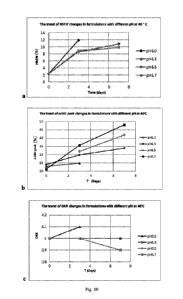
Test data of SEC, iCIEF and HIC and changing trends are shown in Table 2 and
Fig. 10. Since
the samples of Fl placed at 40 C for 3 days became turbid, the samples of the
formulation F 1 were
not tested by SEC, iCIEF and HIC on day 3 and day 7. And since the samples of
F2 placed at 40 C
for 7 days became turbid, the samples of F2 were not tested by SEC, iCIEF and
HIC on day 7. HIC
data shows that the DAR of F3 and F4 are basically unchanged with different pH
values (Fig. 10c);
SEC data shows that with the increase of pH, the growth rate of HMW% of F3, F4
and F5 shows a
downward trend and their downward trends are basically the same (Fig. 10a);
iCIEF data shows that
with the increase of pH, the acidic peak % of F3, F4 and F5 shows an upward
trend but the upward
trend of F3 is obviously lower than the upward trends of F4 and F5 (Fig. 10b).
Based on the above test
data and in consideration of test results about appearance, SEC, iCIEF, HIC
and the like, it is determined
that the optimal formulation of MYK-3 is F3, that is, the pH of the
formulation is 6.3. Accordingly, the
amount of citric acid and sodium citrate in the formulations were adjusted,
and the final formulation
gives a 20 mM citrate buffer with pH 6.3.
Table 2 Experimental data of the effect of pH (5.6-6.7) on the stability of
MYK-3 (in 20 mM
citrate buffer)
Formulation SEC iCIEF HIC
HMW Monomer LMW Acidic peak Primary Alkaline DAR
(%) (%) (%) (%) peak (%) peak (%)
pH 5.6 Od 2.2 97.8 0.1 23.3 49.5 27.3 4.0
3d Samples became turbid
pH 6.0 Od 2.3 97.6 0.1 23.0 50.7 26.3 4.0
3d 11.9 88.0 0.1 24.9 50.7 24.5 4.1
Date Recue/Date Received 2024-03-12
CA 03232092 2024-03-12
7d Samples became turbid
pH 6.3 Od 2.3 97.7 0.1 24.0 48.0 27.9 4.0
3d 8.6 91.3 0.1 29.9 51.6 18.5 4.0
7d 10.8 89.1 0.1 34.0 46.7 19.2 4.0
pH 6.5 Od 2.3 97.7 0.1 Not tested 4.0
3d 8.6 91.2 0.1 32.0 51.5 16.5 4.0
7d 9.9 90.1 0.1 42.0 44.2 13.8 4.0
pH 6.7 Od 2.3 97.7 0.1 21.2 52.2 26.5 4.0
3d 8.9 91.0 0.1 35.7 45.7 18.6 4.0
7d 9.8 90.1 0.1 48.1 39.4 12.5 3.9
Based on the above data, the final MYK-3 formulation was as follows:
Adjuvant Purpose Amount Manufacturer
Quality
(g/L) standard
Citric acid pH adjuster 0.27 Merck Chemical Technology
ChP
monohydrate (Shanghai) Co., Ltd.
Sodium citrate pH adjuster 5.50 J.T Baker USP
dihydrate
Trehalose Protein 55.26 Pfanstiehl
ChP
dihydrate protectant
PS80 Antitack 0.50 Nanjing Well
Chemical Co., Ltd. ChP
agent,
protein
protectant
NaCl Salt 3.00 Jiangsu Province Qinfen ChP
Pharmaceutical Co., Ltd.
Although the specific embodiments of the invention have been described in
detail, those skilled
26
Date Recue/Date Received 2024-03-12
CA 03232092 2024-03-12
in the art will understand that modifications and substitutions can be made to
the details according to
all the teachings that have been disclosed, and these changes are within the
scope of the invention.
The full scope of the invention is given by the appended claims and any
equivalents thereof.
27
Date Recue/Date Received 2024-03-12