Note: Descriptions are shown in the official language in which they were submitted.
ANTI-LAG3 ANTIBODY, PHARMACEUTICAL COMPOSITION
AND USE
TECHNICAL FIELD
The present invention belongs to the field of biomedicine, and relates to an
anti-LAG3
antibody, a pharmaceutical composition comprising same, and use thereof.
BACKGROUND
Tumor, especially a malignant tumor, is a serious health-threatening disease
in the world
today, and it is the second leading cause of death among various diseases. In
recent years,
the incidence of the disease has been increasing remarkably. The malignant
tumor is
characterized by poor treatment response, high late metastasis rate, and poor
prognosis.
Although conventional treatment methods (such as radiotherapy, chemotherapy,
and
surgical treatment) adopted clinically at present alleviate the pain to a
great extent and
prolong the survival time, the methods have great limitations, and it is
difficult to further
improve their efficacy.
Lymphocyte-activation gene 3 (LAG3), namely CD223, is a type I transmembrane
protein
composed of 498 amino acids and is a member of the immunoglobulin superfamily
(IgSF).
LAG3 is mainly expressed in activated CD4 + T cells and CD8+ T cells.
Additionally, in cells
such as natural killer (NK) cells, B cells, regulatory T cells (Tregs), and
plasmacytoid
dendritic cells (pDCs), LAG3 is also expressed. (Ruffo Elisa, Wu Richard C,
Bruno Tullia C
et al., Lymphocyte-activation gene 3 (LAG3): The next immune checkpoint
receptor.
[J].Semin Immunol, 2019, 42: 101305.).
The LAG3 molecule gene is located on human chromosome 12 (20p13.3), adjacent
to the
CD4 molecule gene, and both have the same exons and introns. The LAG3 molecule
and the
CD4 molecule have a high degree of structural similarity, although the amino
acid sequence
homology between the two is only about 20%. Major histocompatibility complex
class II
(MHC II) molecules, liver sinusoidal endothelial cell lectin (LSECtin)
molecules, and
galectin-3 molecules are related ligands for the LAG3 molecule. The MHC class
II molecules
1
CA 03233205 2024 3 26
I EC220407PCT
are the main ligands for LAG3. The affinity (Kd: 60 nmol=Lt) of LAG3 molecules
for the
MHC class II molecules is 100 times that of CD4 molecules, indicating that the
LAG3
molecules can effectively compete with the CD4 molecules for binding to the
MHC class II
molecules and inhibit T cell activation.
In the tumor microenvironment, the expression of the immunosuppressive
molecule LAG3
can be detected 24 hours after T cell activation, which then leads to T cell
dysfunction or
apoptosis. The LAG3 molecule, through its D1 domain (which contains one
proline-rich loop
structure), forms a dimer molecule to specifically bind to the MHC class II
molecule in the
first signaling axis of CD4 + T cell activation, "CD3-TCR-MHCI I", so that on
one hand, a
signal transduction pathway for T cell activation is blocked, and on the other
hand, an
intracellular segment of the LAG3 molecule (KIEELE motif) generates an
immunosuppressive signal to down-regulate the activity of CD4+ T cells. The
LAG3 molecule
can promote the differentiation of Treg cells, participate in downstream
signaling of signal
transducer and activator of transcription 5, and thus enhance the inhibitory
effect of Treg
cells, which is one of the mechanisms by which tumors escape from killing by
the immune
system (Andrews Lawrence P, Marciscano Ariel E, Drake Charles G, et al., LAG3
(CD223)
as a cancer immunotherapy target. (J ]. Immunol Rev, 2017, 276: 80-96.).
Multiple studies have shown that LAG3 is overexpressed in tumor-infiltrating
CD8+ T cells
of various malignant tumors. For example, in ovarian cancer, tumor-
infiltrating New York
esophageal squamous cell carcinoma 1 (NY-ESO-1) antigen-specific CD8+ T cells
express
high levels of PD-1 and LAG3, and have a reduced ability to produce IFN-y and
TNF-a,
thereby leading to lymphocyte inactivation. Galectin-3 and LSECtin interact
primarily with
LAG3 to regulate the activation and function of CD8+ T cells. In addition,
melanoma antigen-
specific T cells isolated from patients with metastatic melanoma exhibit a
significant up-
regulation in the expression of LAG3 and other immune checkpoint molecules
CTLA-4 and
TIM-3. (Liu Hao, Li Xinying, Luo Longlong, et al., Research advances in
biological function
of lymphocyte activation gene-3 (LAG-3) molecule and clinical application of
antibody drugs
targeting LAG-3 (J ]. Chinese Journal of Pharmacology and Toxicology, 2019,
33(01): 70-78.).
Currently, a plurality of LAG3 antibody medicaments have entered the clinical
research
2
CA 03233205 2024 3 26
I EC220407PCT
stages, among which Bristol Myers Squibb's Relatlimab has the fastest
progress, with 10
clinical studies underway. The vast majority of these studies involve the
combination therapy
of Relatlimab with nivolumab, used for the treatment of tumors such as
hematological
malignancies, melanoma, glioblastoma, renal cell carcinoma, non-small cell
lung cancer, and
the like.
There is currently a need to develop a novel anti-LAG3 antibody medicament.
SUMMARY
Through intensive studies and creative efforts, the inventors have obtained an
anti-LAG3
antibody. The inventors have surprisingly found that the anti-LAG3 antibody of
the present
invention (also referred to as the antibody or the antibody of the present
invention for short)
has superior affinity and/or specificity, and is even superior in one or more
respects
compared to positive control antibodies (e.g., Relatlimab). The present
invention is detailed
below.
One aspect of the present invention relates to an anti-LAG3 antibody or an
antigen-binding
fragment thereof, comprising a heavy chain variable region and a light chain
variable region,
wherein
the heavy chain variable region comprises HCDR1-HCDR3 having amino acid
sequences set
forth in SEQ ID NOs: 9-11, respectively, and the light chain variable region
comprises
LCDR1-LCDR3 having amino acid sequences set forth in SEQ ID NOs: 12-14,
respectively;
the heavy chain variable region comprises HCDR1-HCDR3 having amino acid
sequences set
forth in SEQ ID NOs: 9-11, respectively, and the light chain variable region
comprises
LCDR1-LCDR3 having amino acid sequences set forth in SEQ ID NO: 12, SEQ ID NO:
15
and SEQ ID NO: 16, respectively;
or
the heavy chain variable region comprises HCDR1-HCDR3 having amino acid
sequences set
forth in SEQ ID NOs: 9-11, respectively, and the light chain variable region
comprises
LCDR1-LCDR3 having amino acid sequences set forth in SEQ ID NO: 17, SEQ ID NO:
15
3
CA 03233205 2024 3 26
I EC220407PCT
and SEQ ID NO: 14, respectively.
In some embodiments of the present invention, the antibody or the antigen-
binding fragment
thereof is provided, wherein
the heavy chain variable region of the antibody has an amino acid sequence set
forth in SEQ
ID NO: 2, and the light chain variable region of the antibody has an amino
acid sequence set
forth in SEQ ID NO: 4;
the heavy chain variable region of the antibody has an amino acid sequence set
forth in SEQ
ID NO: 2, and the light chain variable region of the antibody has an amino
acid sequence set
forth in SEQ ID NO: 6;
or
the heavy chain variable region of the antibody has an amino acid sequence set
forth in SEQ
ID NO: 2, and the light chain variable region of the antibody has an amino
acid sequence set
forth in SEQ ID NO: 8.
In some embodiments of the present invention, the antibody or the antigen-
binding fragment
thereof is provided, wherein the antibody or the antigen-binding fragment
thereof is selected
from a Fab, a Fab', an F(a131)2, an Fd, an Fv, a dAb, a complementarity
determining region
fragment, a single chain fragment variable, a humanized antibody, or a
chimeric antibody.
In some embodiments of the present invention, the antibody or the antigen-
binding fragment
thereof is provided, wherein the antibody binds to human LAG3-mFc with an EC50
of less
than 0.2 nM, such as less than 0.15 nM, less than 0.1 nM, less than 0.08 nM,
0.06 nM, or less
than 0.05 nM, or less; preferably, the EC50 value is determined by indirect EL
ISA.
In some embodiments of the present invention, the antibody or the antigen-
binding fragment
thereof is provided, wherein
the antibody comprises a non-CDR region derived from a species other than
murine, such
as from a human antibody.
In some embodiments of the present invention, the antibody or the antigen-
binding fragment
thereof is provided, wherein
the antibody comprises a constant region derived from a human antibody;
4
CA 03233205 2024 3 26
I EC220407PCT
preferably, the constant region of the antibody is selected from constant
regions of human
IgG1, IgG2, IgG3 or IgG4.
In some embodiments of the present invention, the antibody or the antigen-
binding fragment
thereof is provided, wherein
a heavy chain constant region of the anti-LAG3 antibody is Ig gamma-1 chain C
region (e.g.,
as set forth in SEQ ID NO: 18) or Ig gamma-4 chain C region (e.g., as set
forth in SEQ ID
NO: 20), and a light chain constant region of the anti-LAG3 antibody is Ig
kappa chain C
region (e.g., as set forth in SEQ ID NO: 19).
In some embodiments of the present invention, the anti-LAG3 antibody is a
monoclonal
antibody.
In some embodiments of the present invention, the anti-LAG3 antibody is in an
immunoglobulin form.
In some embodiments of the present invention, the anti-LAG3 antibody is a
single chain
fragment variable.
Another aspect of the present invention relates to an antibody-drug conjugate
(ADC),
comprising an antibody or an antigen-binding fragment thereof and a small
molecule drug,
wherein the antibody or the antigen-binding fragment thereof is the anti-LAG3
antibody or
the antigen-binding fragment thereof according to any embodiment of the
present invention;
preferably, the small molecule drug is a small molecule cytotoxic drug; and
more preferably,
the small molecule drug is an anti-tumor chemotherapeutic drug.
The chemotherapeutic drug may be a conventional anti-tumor chemotherapeutic
drug, such
as an alkylating agent, an antimetabolite, an anti-tumor antibiotic, a plant-
based anticancer
agent, a hormone, and an immunological agent.
In one or more embodiments of the present invention, the antibody-drug
conjugate is
provided, wherein the antibody or the antigen-binding fragment thereof is
linked to the small
molecule drug via a linker; the linker may be one known to those skilled in
the art, for
example, a hydrazone bond, a disulfide bond, or a peptide bond.
5
CA 03233205 2024 3 26
I EC220407PCT
In one or more embodiments of the present invention, the antibody-drug
conjugate is
provided, wherein the molar ratio of the antibody or the antigen-binding
fragment thereof
to the small molecule drug is 1:(2-4), e.g., 1:2, 1:3, or 1:4.
Yet another aspect of the present invention relates to an isolated nucleic
acid molecule
encoding the anti-LAG3 antibody according to any embodiment of the present
invention.
Yet another aspect of the present invention relates to a recombinant vector
comprising the
isolated nucleic acid molecule of the present invention.
Yet another aspect of the present invention relates to a host cell comprising
the isolated
nucleic acid molecule of the present invention or the recombinant vector of
the present
invention.
Yet another aspect of the present invention relates to a method for preparing
the antibody
or the antigen-binding fragment thereof according to any embodiment of the
present
invention, comprising: culturing the host cell of the present invention in a
suitable condition,
and isolating the antibody or the antigen-binding fragment thereof from the
cell cultures.
Yet another aspect of the present invention relates to a pharmaceutical
composition
comprising the antibody or the antigen-binding fragment thereof according to
any
embodiment of the present invention, or the antibody-drug conjugate according
to any
embodiment of the present invention, wherein optionally, the pharmaceutical
composition
further comprises a pharmaceutically acceptable auxiliary material.
Yet another aspect of the present invention relates to use of the antibody or
the antigen-
binding fragment thereof according to any embodiment of the present invention,
or the
antibody-drug conjugate according to any embodiment of the present invention
in the
preparation of a medicament for treating and/or preventing a tumor or anemia,
wherein
preferably, the tumor is selected from one or more of ovarian cancer,
esophageal cancer,
melanoma, a hematological malignancy, glioblastoma, renal cell carcinoma, lung
cancer,
prostate cancer, bladder cancer, colon cancer, rectal cancer, liver cancer,
gastrointestinal
6
CA 03233205 2024 3 26
I EC220407PCT
cancer, breast cancer, brain cancer, pancreatic cancer, thyroid cancer, head
and neck
cancer, and kidney cancer;
preferably, the lung cancer is non-small cell lung cancer;
preferably, the hematological malignancy is leukemia;
preferably, the esophageal cancer is esophageal squamous cancer.
The antibody or the antigen-binding fragment thereof according to any
embodiment of the
present invention, or the antibody-drug conjugate according to any embodiment
of the
present invention is for use in treating and/or preventing a tumor or anemia,
wherein
preferably, the tumor is selected from one or more of ovarian cancer,
esophageal cancer,
3.0 melanoma, a hematological malignancy, glioblastoma, renal cell
carcinoma, lung cancer,
prostate cancer, bladder cancer, colon cancer, rectal cancer, liver cancer,
gastrointestinal
cancer, breast cancer, brain cancer, pancreatic cancer, thyroid cancer, head
and neck
cancer, and kidney cancer;
preferably, the lung cancer is non-small cell lung cancer;
preferably, the hematological malignancy is leukemia;
preferably, the esophageal cancer is esophageal squamous cancer.
Yet another aspect of the present invention relates to a method for treating
and/or preventing
a tumor or anemia, comprising a step of administering to a subject in need an
effective
amount of the antibody or the antigen-binding fragment thereof according to
any
embodiment of the present invention, or the antibody-drug conjugate according
to any
embodiment of the present invention, wherein
preferably, the tumor is selected from one or more of ovarian cancer,
esophageal cancer,
melanoma, a hematological malignancy, glioblastoma, renal cell carcinoma, lung
cancer,
prostate cancer, bladder cancer, colon cancer, rectal cancer, liver cancer,
gastrointestinal
cancer, breast cancer, brain cancer, pancreatic cancer, thyroid cancer, head
and neck
cancer, and kidney cancer;
preferably, the lung cancer is non-small cell lung cancer;
preferably, the hematological malignancy is leukemia;
preferably, the esophageal cancer is esophageal squamous cancer.
7
CA 03233205 2024 3 26
I EC220407PCT
In the present invention, unless otherwise defined, the scientific and
technical terms used
herein have the meanings generally understood by those skilled in the art. In
addition, the
laboratory operations of cell culture, molecular genetics, nucleic acid
chemistry and
immunology used herein are the routine procedures widely used in the
corresponding fields.
Meanwhile, in order to better understand the present invention, the
definitions and
explanations of the relevant terms are provided below.
As used herein, the term EC50 refers to the concentration for 50% of maximal
effect, i.e., the
concentration that can cause 50% of the maximal effect.
As used herein, the term "antibody" refers to an immunoglobulin molecule that
generally
consists of two pairs of polypeptide chains (each pair with one "light" (L)
chain and one
"heavy" (H) chain). Antibody light chains are classified into lc and X light
chains. Heavy
chains are classified into p, 8, y, a, or E. Isotypes of antibodies are
defined as IgM, IgD, IgG,
IgA, and IgE. In light chains and heavy chains, the variable region and
constant region are
linked by a "J" region of about 12 or more amino acids, and the heavy chain
further
comprises a "D" region of about 3 or more amino acids. Each heavy chain
consists of a heavy
chain variable region (VH) and a heavy chain constant region (CH). The heavy
chain
constant region consists of 3 domains (CH1, CH2, and CH3). Each light chain
consists of a
light chain variable region (VL) and a light chain constant region (CL). The
light chain
constant region consists of one domain CL. The constant region of the antibody
can mediate
the binding of immunoglobulins to host tissues or factors, including the
binding of various
cells of the immune system (e.g., effector cells) to the first component (C1q)
of the classical
complement system. The VH and VL regions can be further subdivided into
hypervariable
regions (called complementarity determining regions (CDRs)), between which
conservative
regions called framework regions (FRs) are distributed. Each VH and VL
consists of 3 CDRs
and 4 FRs arranged from amino terminus to carboxyl terminus in the following
order: FR1,
CDR1, FR2, CDR2, FR3, CDR3, and FR4. The variable regions (VH and VL) of each
heavy
chain/light chain pair form an antibody-binding site. The assignment of amino
acids to the
regions or domains is based on Bethesda M.d., Kabat Sequences of Proteins of
8
CA 03233205 2024 3 26
I EC220407PCT
Immunological Interest (National Institutes of Health, (1987 and 1991)), or
Chothia & Lesk
J. Mol. Biol., 1987; 196: 901-917; Chothia et al., Nature, 1989; 342: 878-883,
or the definition
of the I MGT numbering system, see the definition in Ehrenmann F, Kaas Q,
Lefranc M P.,
IMGT/3Dstructure-DB and I MGT/DomainGapAlign: a database and a tool for
immunoglobulins or antibodies, T cell receptors, MHC, IgSF and MhcSFU 1.,
Nucleic acids
research, 2009; 38(suppl_1): D301-D307.
The term "antibody" is not limited by any specific method for producing the
antibody. For
example, the antibody includes a recombinant antibody, a monoclonal antibody,
and a
polyclonal antibody. The antibody may be antibodies of different isotypes,
such as IgG (e.g.,
subtype IgG1, IgG2, IgG3, or IgG4), IgA1, IgA2, IgD, IgE, or IgM.
As used herein, the terms "mAb" and "monoclonal antibody" refer to an antibody
or a
fragment of an antibody that is derived from a group of highly homologous
antibodies, i.e.,
from a group of identical antibody molecules, except for natural mutations
that may occur
spontaneously. The monoclonal antibody is highly specific for a single epitope
on an antigen.
The polyclonal antibody, relative to the monoclonal antibody, generally
comprises at least 2
or more different antibodies which generally recognize different epitopes on
an antigen.
Monoclonal antibodies can generally be obtained using hybridoma technology
first reported
by Kohler et al. (Kohler G, Milstein C. Continuous cultures of fused cells
secreting antibody
of predefined specificity [J]. Nature, 1975; 256(5517): 495), but can also be
obtained using
recombinant DNA technology (see, e.g., U.S. Patent 4,816,567).
As used herein, the term "humanized antibody" refers to an antibody or
antibody fragment
obtained when all or a part of CDRs of a human immunoglobulin (receptor
antibody) is
replaced by the CDRs of a non-human antibody (donor antibody), wherein the
donor
antibody may be a non-human (e.g., mouse, rat or rabbit) antibody having
expected
specificity, affinity or reactivity. In addition, some amino acid residues in
the framework
regions (FRs) of the receptor antibody can also be replaced by the amino acid
residues of
corresponding non-human antibodies or by the amino acid residues of other
antibodies to
further improve or optimize the performance of the antibody. For more details
on
humanized antibodies, see, e.g., Jones et al., Nature, 1986; 321: 522-525;
Reichmann et al.,
9
CA 03233205 2024 3 26
I EC220407PCT
Nature, 1988; 332: 323-329; Presta, Curr. Op. Struct. Biol., 1992; 2: 593-596;
and Clark,
Immunol. Today, 2000; 21: 397-402.
As used herein, the term "isolated" refers to obtaining by artificial means
from a natural
state. If a certain "isolated" substance or component is present in nature, it
may be the case
that a change occurs in its natural environment, or that it is isolated from
the natural
environment, or both. For example, a certain non-isolated polynucleotide or
polypeptide
naturally occurs in a certain living animal, and the same polynucleotide or
polypeptide with
high purity isolated from such a natural state is referred to as an isolated
polynucleotide or
polypeptide. The term "isolated" does not exclude the existence of artificial
or synthetic
substances or other impurities that do not affect the activity of the
substance.
As used herein, the term "vector" refers to a nucleic acid vehicle into which
a polynucleotide
can be inserted. When a vector allows the expression of the protein encoded by
the inserted
polynucleotide, the vector is referred to as an expression vector. The vector
can be
introduced into a host cell by transformation, transduction or transfection,
such that the
genetic substance elements carried by the vector can be expressed in the host
cell. Vectors
are well known to those skilled in the art, including but not limited to:
plasmids; phagemids;
cosmids; artificial chromosomes, such as yeast artificial chromosome (YAC),
bacterial
artificial chromosome (BAC), or P1-derived artificial chromosome (PAC); phages
such as
lambda phages or M13 phages; and animal viruses. Animal viruses that can be
used as
vectors include, but are not limited to retroviruses (including lentiviruses),
adenoviruses,
adeno-associated viruses, herpes viruses (such as herpes simplex virus),
poxviruses,
baculoviruses, papillomaviruses, and papovaviruses (such as SV40). A vector
may comprise
a variety of elements that control expression, including, but not limited to,
promoter
sequences, transcription initiation sequences, enhancer sequences, selection
elements, and
reporter genes. In addition, the vector may further comprise a replication
initiation site.
As used herein, the term "host cell" refers to cells to which vectors can be
introduced,
including, but not limited to, prokaryotic cells such as E. coil or bacillus
subtilis, fungal cells
such as yeast cells or aspergillus, insect cells such as S2 drosophila cells
or Sf9, or animal cells
CA 03233205 2024 3 26
I EC220407PCT
such as fibroblasts, CHO cells, GS cells, COS cells, NSO cells, HeLa cells,
BHK cells, HEK
293 cells, or human cells.
As used herein, the term "specific binding" refers to a non-random binding
reaction between
two molecules, such as a reaction between an antibody and an antigen it
targets. In some
embodiments, an antibody specifically binding to an antigen (or an antibody
specific to an
antigen) means that the antibody binds to the antigen with an affinity (KD) of
less than about
10-5 M, such as less than about 10-8 M, 10-7 M, 10-8 M, 10-8 M, or 10-10 M, or
less.
As used herein, the term "KD" refers to a dissociation equilibrium constant
for a specific
antibody-antigen interaction, which is used to describe the binding affinity
between the
antibody and the antigen. A smaller dissociation equilibrium constant
indicates a stronger
antibody-antigen binding and a higher affinity between the antibody and the
antigen.
Generally, antibodies bind to antigens (e.g., PD-1 protein) with a
dissociation equilibrium
constant (KD) of less than about 10-5 M, such as less than about 104 M, 10-7
M, 104 M, 10-9
M, or 10-10 M, or less. KD can be determined using methods known to those
skilled in the art,
e.g., using a Fortebio molecular interaction instrument.
As used herein, the terms "monoclonal antibody" and "mAb" have the same
meaning and
can be used interchangeably; the terms "polyclonal antibody" and "pAb" have
the same
meaning and can be used interchangeably. Besides, as used herein, amino acids
are generally
represented by single-letter and three-letter abbreviations known in the art.
For example,
alanine can be represented by A or Ala.
As used herein, the term "pharmaceutically acceptable carrier and/or
excipient" refers to a
carrier and/or excipient that is pharmacologically and/or physiologically
compatible with
the subject and the active ingredient. Such carriers and/or excipients are
well known in the
art (see, e.g., Remington's Pharmaceutical Sciences, edited by Gennaro AR,
19th Ed.,
Pennsylvania, Mack Publishing Company, 1995), including but not limited to: pH
regulators,
surfactants, adjuvants and ionic strength enhancers. For example, the pH
regulators include,
but are not limited to, phosphate buffer; the surfactants include, but are not
limited to,
cationic, anionic or non-ionic surfactants, such as Tween-80; the ionic
strength enhancers
include, but are not limited to, sodium chloride.
11
CA 03233205 2024 3 26
I EC220407PCT
As used herein, the term "effective amount" refers to an amount sufficient to
obtain or at
least partially obtain a desired effect. For example, a prophylactically
effective amount
against a disease (e.g., a tumor) refers to an amount sufficient to prevent,
stop, or delay the
onset of the disease (e.g., a tumor); a therapeutically effective amount
refers to an amount
sufficient to cure or at least partially stop diseases and complications
thereof in patients
suffering from the disease. It is undoubtedly within the ability of those
skilled in the art to
determine such an effective amount. For example, the amount effective for
therapeutic
purpose will depend on the severity of the disease to be treated, the overall
state of the
patient's own immune system, the general condition of the patient such as age,
body weight
and gender, the route of administration, and other treatments given
concurrently, etc.
As used herein, when referring to the amino acid sequence of lymphocyte-
activation gene 3
(LAG3), it includes the full length of LAG3 protein, or the extracellular
fragment LAG3
ECD of LAG3, or a fragment comprising LAG3 ECD, and it also includes a fusion
protein
of the full length of LAG3 protein or a fusion protein of LAG3 ECD, such as a
fragment
fused to an Fc protein fragment of mouse or human IgG (mFc or hFc). However,
those skilled
in the art will appreciate that in the amino acid sequence of the LAG3
protein, mutations or
variations (including but not limited to, substitutions, deletions, and/or
additions) can be
naturally produced or artificially introduced without affecting biological
functions thereof.
Therefore, in the present invention, the term "LAG3 protein" should include
all such
sequences, including their natural or artificial variants. In addition, when
describing a
sequence fragment of the LAG3 protein, it also includes the corresponding
sequence
fragments in their natural or artificial variants.
Beneficial effects of the present invention
The present invention achieves one or more of the following effects:
(1) the anti-LAG3 antibody of the present invention has superior affinity and
specificity; and
(2) the anti-LAG3 antibody of the present invention can effectively block the
interaction
between LAG3 and MHC-I I and specifically relieve the immunosuppression of
LAG3 on an
organism.
12
CA 03233205 2024 3 26
I EC220407PCT
BRIEF DESCRIPTION OF THE DRAWINGS
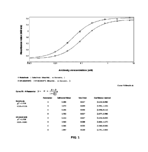
FIG. 1 shows the results of an assay for the binding activity of H7L8(hG1WT)
to the antigen
LAG3-mFc by indirect ELISA.
FIG. 2 shows the results of assays for the binding activity of H7L8(hG4WT),
H7L9(hG4WT),
and H7 L10(hG4WT) to the antigen human LAG3-mFc by ELISA.
FIG. 3 shows the results of assays for the binding activity of H7L8(hG4WT),
H7L9(hG4WT),
and H7 L10(hG4WT) to the antigen LAG3 on 293T-LAG3 cell surface by FACS.
FIG. 4 shows the results of assays for the activity of H7L8(hG4WT),
H7L9(hG4WT), and
H7L10(hG4WT) in competing with LAG3-mFc for binding to the antigen MHC II on
293T-
LAG3 cell membrane surface by competitive flow cytometry.
FIG. 5 shows the results of assays for the biological activity of anti-LAG3
antibodies in
promoting IFN-y secretion by mixed lymphocyte reaction (MLR).
FIG. 6 shows the results of assays for the biological activity of anti-LAG3
antibodies in
promoting IL-2 secretion by mixed lymphocyte reaction (MLR).
FIG. 7 shows the results of assays for the biological activity of anti-LAG
antibodies in
blocking the interaction between LAG-3 and MHC-I I.
DETAILED DESCRIPTION
The embodiments of the present invention will be described in detail below
with reference to
the examples. Those skilled in the art will appreciate that the following
examples are only
for illustrating the present invention, and should not be construed as
limitations to the scope
of the present invention. Examples where the specific technologies or
conditions are not
specified are performed according to the technologies or conditions described
in the
publications of the art (e.g., see, Molecular Cloning: A Laboratory Manual,
authored by J.
Sambrook et al., and translated by Huang Peitang et al., third edition,
Science Press) or
according to the package insert. Reagents or instruments used are commercially
available
conventional products if the manufacturers thereof are not specified.
The positive control antibody, Relatlimab, has sequences referenced to the
U.S. Patent
13
CA 03233205 2024 3 26
I EC220407PCT
Publication No. US20160326248A1, wherein the heavy chain amino acid sequence
is
referenced to SEQ ID NO: 1 of this patent publication and the light chain
amino acid
sequence is referenced to SEQ ID NO: 2 of this patent publication. Relatlimab
is an anti-
LAG-3 antibody.
Heavy chain amino acid sequence of Relatlimab:
QVQLQQWGAG L L KPSETLSLTCAVYGGSFSDYYWNW I RQ PPG KG L EW IG El NHR
GSTNSNPSLKSRVTLSLDTSKNQFSLKLRSVTAADTAVYYCAFGYSDYEYNWF DPW
GQGTLVTVSSASTKGPSVFPLAPCSRSTSESTAALGCLVKDYFPEPVTVSWNSGALT
SGVHTFPAVLQSSG LYS LSSVVTVPSSS LGTKTYTC NVDH KPS NTKVD KRVES KYG P
PC PPC PAP EF LGGPSVF LFPPKPKDTLM I SRTPEVTCVVVDVSQ EDPEVQF NWYVD
GVEVHNAKTKPRE EQF NSTYRVVSVLTVLHQDW L NG KEYKC KVSN KG LPSS I EKTI
SKAKGQPREPQVYTLPPSQEEMTKNQVSLTCLVKGFYPSDIAVEWESNGQPENNY
KTTPPVL DSDG SF F LYSRLTVD KSRWQ EG NVF SCSVMH EALHNHYTQ KS LS LSLG K
(SEQ ID NO: 23)
Light chain amino acid sequence of Relatlimab:
EIVLTQSPATLSLSPG ERATLSC RASQS I SSYLAWYQQ KPGQAPRL LIYDASNRATG I
PARFSGSGSGTDFTLTISSLEPEDFAVYYCQQRSNWPLTFGQGTN LEI KRTVAAPSV
F I F PPSD EQ L KSGTASVVC L LNNFYPREAKVQWKVDNALQSGNSQESVTEQDSKDS
TYSLSSTLTLSKADYEKHKVYACEVTHQGLSSPVTKSFNRGEC (SEQ ID NO: 24)
The control antibody 14C12H1L1(hG1TM) was an anti-PD-1 antibody constructed by
Akeso
Biopharma Inc. with a Batch No. B105Y2080601.
Heavy chain amino acid sequence of 14C12H1L1(hG1TM):
EVQLVESGGG LVQPGGSLRLSCAASGFAFSSYDMSWVRQAPG KG LDWVATISGGG
RYTYYPDSVKG RFT! SRDNSKNN LYLQ M NS LRAEDTALYYCAN RYG EAWFAYWG
QGTLVTVSSASTKGPSVFPLAPSSKSTSGGTAALGCLVKDYFPEPVTVSWNSGALTS
GVHTFPAVLQSSG LYS LSSVVTVPSSS LGTQTYICNVNH KPSNTKVD KKVEP KSC D K
THTCPPCPAPEAAGAPSVF LF PP KPKDTL M I SRTPEVTCVVVDVSH EDP EVKF NWY
VDGVEVH NAKTKPRE EQYNSTYRVVSVLTVLHQDW L NG KEYKC KVSNKALPAPI E
KTISKAKGQPREPQVYTLPPSRDELTKNQVSLTCLVKGFYPSDIAVEWESNGQPEN
14
CA 03233205 2024 3 26
I EC220407PCT
NYKTTPPVL DSDG SF F LYS KLTVD KSRWQQG NVF SC SVM H EALHNHYTQKSLSLSP
GK (SEQ ID NO: 21)
Light chain amino acid sequence of 14C12H1L1(hG1TM):
DI Q MTQSPSSMSASVG DRVTFTC RASQ D I NTYLSWFQQKPG KSPKTL IYRANRLVS
GVPSRFSGSGSGQDYTLTISSLQPEDMATYYCLQYDEFPLTFGAGTKLELKRTVAA
PSVF I FPPSDEQLKSGTASVVC LLNNFYPREAKVQWKVDNALQSG NSQ ESVTEQ DS
KDSTYSLSSTLTLSKADYEKHKVYACEVTHQG LSSPVTKSFNRG EC (SEQ ID NO: 22)
The cell line 293T-LAG3 was constructed by Akeso Biopharma Inc. The cell line
293T-LAG3
was produced by viral infection of HEK293T cells using 3rd Generation
Lentiviral Systems
(see, e.g., A Third Generation Lentivirus Vector with a Conditional Packaging
System. Dull
T, Zufferey R, Kelly M, Mandel RJ , Nguyen M, Trono D, and Naldini L., J
Virol., 1998.
72(11): 8463-8471), wherein the lentivirus expression vector used was
p1enti6.3/V5-
huLAG3FL-BSD (LAG3, Genebank ID: NM_002277.4; vector p1enti6.3/V5-BSD,
purchased
from Invitrogen, Cat. No. K5315-20).
The cell line Raji-PDL1 was constructed by Akeso Biopharma Inc. The cell line
Raji-PDL1
was produced by viral infection of Raji cells using 3rd Generation Lentiviral
Systems (see,
e.g., A Third Generation Lentivirus Vector with a Conditional Packaging
System. Dull T,
Zufferey R, Kelly M, Mandel RJ , Nguyen M, Trono D, and Naldini L., J
Virol.,1998. 72(11):
8463-8471), wherein the lentivirus expression vector used was p1enti6.3/V5-
PDL1 (PDL1,
Genebank ID: NP 054862.1; vector p1enti6.3/V5, purchased from Invitrogen, Cat.
No.
K5315-20).
The cell line J urkat-NFAT-PD1-LAG3 was constructed by Akeso Biopharma Inc.
The cell
line J urkat-NFAT-PD1-LAG3 was prepared by viral infection of PD-1 effector
cells (CPM,
manufacturer: Promega, Cat. No. J 112A) using 3rd Generation Lentiviral
Systems (see, e.g.,
A Third Generation Lentivirus Vector with a Conditional Packaging System. Dull
T,
Zufferey R, Kelly M, Mandel RJ , Nguyen M, Trono D, and Naldini L., J
Virol.,1998. 72(11):
8463-8471), wherein the lentivirus expression vector used was pCDH-huLAG3FL-
RFP-NE0
(LAG3, Genebank ID: NM_002277.4; vector pCDH-CMV-MCS-EF1-RFP+Neo, purchased
from Youbio, Cat. No. VT9005).
CA 03233205 2024 3 26
I EC220407PCT
Preparation Example 1: Design and Preparation of Anti-LAG3 Antibodies
1. Design of antibodies
The inventors creatively designed a series of antibody sequences based on the
known LAG3
protein sequence (NCI31 Reference Sequence: NP_002277.4) and the three-
dimensional
crystal structure thereof, etc. Through extensive screening and testing,
humanized
monoclonal antibodies specifically binding to LAG3 were finally obtained,
named H7L8,
H7L9 and H7L10, respectively. The amino acid sequences of the heavy and light
chain
variable regions of the monoclonal antibodies and the encoding sequences
thereof are as
follows.
Nucleotide sequence of the heavy chain variable region H7v of H7L8 (360 bp):
CAGGTGCAGCTGCAGCAGTGGGGAGCTGGACTGCTGAAACCTAGCGAGACACT
GAGCCTGACCTGTGCTGTGTACGGCGGATCTATCAGCGATTACTACTGGAACT
GGATCAGGCAGCCCCCTGGAAAGGGACTGGAATGGATCGGAGAGATCAACCAC
AGGGGCACCACCAACTCCAATCCCTCTCTGAAGAGCAGGGTGACACTGAGCCT
CGACACAAGCAAGAATCAGTTCAGCCTGAAGCTGAGGTCCGTGACCGCTGCTG
ATACAGCTGTGTACTACTGTGCCTTCGGCTACAGCGATTACGAGTACGATTGGT
TCGACCCTTGGGGCCAGGGAACACTGGTTACAGTGAGCTCC (SEQ ID NO: 1)
Amino acid sequence of the heavy chain variable region H7v of H7L8 (120 aa):
QVQLQQWGAG L L KPS ETLS LTCAVYGG S I SDYYWNW I RQPPG KG L EW IG El NH RG
TTNSN PS L KS RVT L S L DTSKNQFSL KLRSVTAADTAVYYCAFGYSDYEYDWF DPWG
QGTLVTVSS (SEQ ID NO: 2)
Nucleotide sequence of the light chain variable region L8v of H7L8 (321 bp):
GAGATCGTTCTGACCCAGAGCCCAGCTACACTGAGCCTGTCTCCTGGAGAGAG
GGCTACACTGTCCTGCAGAGCTAGCCAGACCATCAGCAGCTACCTGGCTTGGT
ACCAGCAGAAGCCTGGCCAAGCTCCAAGGCTGCTGATCTACGACGCCTCTAAT
AGGGCCACCGGCATCCCTGCTAGATTCTCTGGAAGCGGCAGCGGAACCGACTT
TACACTGACAATCAGCTCCCTGGAGCCCGAGGATTTCGCTGTTTACTACTGTCA
16
CA 03233205 2024 3 26
I EC220407PCT
GCAGCGCAGCAACTGGCCCATCACATTCGGACAGGGCACAAATCTGGAGATCA
AG (SEQ ID NO: 3)
Amino acid sequence of the light chain variable region L8v of H7L8 (107 aa):
EIVLTQSPATLSLSPG ERATLSCRASQTISSYLAWYQQKPGQAPRLL IYDASNRATG I
PARFSGSGSGTDFTLTISSLEPEDFAVYYCQQRSNWPITFGQGTNLEIK (SEQID NO:
4)
The nucleotide sequence of the heavy chain variable region H7v of H7L9 is
identical to the
nucleotide sequence of the heavy chain variable region H7v of H7L8, as set
forth in SEQ ID
NO: 1.
The amino acid sequence of the heavy chain variable region H7v of H7L9 is
identical to the
amino acid sequence of the heavy chain variable region H7v of H7L8, as set
forth in SEQ ID
NO: 2.
Nucleotide sequence of the light chain variable region L9v of H7L9 (321 bp):
GAGATCGTTCTGACCCAGAGCCCAGCTACACTGAGCCTGTCTCCTGGAGAGAG
GGCTACACTGTCCTGCAGAGCTAGCCAGACCATCAGCAGCTACCTGGCTTGGT
ACCAGCAGAAGCCTGGCCAAGCTCCAAGGCTGCTGATCTACGACGGCTCTAAT
AGGGCCACCGGCATCCCTGCTAGATTCTCTGGAAGCGGCAGCGGAACCGACTT
TACACTGACAATCAGCTCCCTGGAGCCCGAGGATTTCGCTGTTTACTACTGTCA
GCAGCGCAGCAACTGGCCCCTCACATTCGGACAGGGCACAAATCTGGAGATCA
AG (SEQ ID NO: 5)
Amino acid sequence of the light chain variable region L9v of H7L9 (107 bp):
EIVLTQSPATLSLSPG ERATLSCRASQTISSYLAWYQQKPGQAPRLL IYDGSNRATG I
PARFSGSGSGTDFTLTISSLEPEDFAVYYCQQRSNWPLTFGQGTN LEI K (SEQ ID NO:
6)
The nucleotide sequence of the heavy chain variable region H7v of H7L10 is
identical to the
nucleotide sequence of the heavy chain variable region H7v of H7L8, as set
forth in SEQ ID
NO: 1.
17
CA 03233205 2024 3 26
I EC220407PCT
The amino acid sequence of the heavy chain variable region H7v of H7L10 is
identical to the
amino acid sequence of the heavy chain variable region H7v of H7L8, as set
forth in SEQ ID
NO: 2.
Nucleotide sequence of the light chain variable region L10v of H7L10 (321 bp):
GAGATCGTTCTGACCCAGAGCCCAGCTACACTGAGCCTGTCTCCTGGAGAGAG
GGCTACACTGTCCTGCAGAGCTAGCCAGTCCATCAGCAGCTACCTGGCTTGGT
ACCAGCAGAAGCCTGGCCAAGCTCCAAGGCTGCTGATCTACGACGGCTCTAAT
AGGGCCACCGGCATCCCTGCTAGATTCTCTGGAAGCGGCAGCGGAACCGACTT
TACACTGACAATCAGCTCCCTGGAGCCCGAGGATTTCGCTGTTTACTACTGTCA
GCAGCGCAGCAACTGGCCCATCACATTCGGACAGGGCACAAATCTGGAGATCA
AG (SEQ ID NO: 7)
Amino acid sequence of the light chain variable region L10v of H7L10 (107 bp):
EIVLTQSPATLSLSPG ERATLSC RASQS I SSYLAWYQQ KPGQAPRL LIYDGSNRATG I
PARFSGSGSGTDFTLTISSLEPEDFAVYYCQQRSNWPITFGQGTNLEI K (SEQ ID NO:
8)
The amino acid sequences of the CDRs of the antibody H7L8 are as follows
(according to the
I MGT numbering system):
HCDR1: GGSISDYY (SEQ ID NO: 9);
HCDR2: INHRGTT (SEQ ID NO: 10);
HCDR3: AFGYSDYEYDWFDP (SEQ ID NO: 11);
LCDR1: QTISSY (SEQ ID NO: 12);
LCDR2: DAS (SEQ ID NO: 13); and
LCDR3: QQRSNWPIT (SEQ ID NO: 14).
The amino acid sequences of the CDRs of the antibody H7L9 are as follows
(according to the
I MGT numbering system):
HCDR1: GGSISDYY (SEQ ID NO: 9);
HCDR2: INHRGTT (SEQ ID NO: 10);
18
CA 03233205 2024 3 26
I EC220407PCT
HCDR3: AFGYSDYEYDWFDP (SEQ ID NO: 11);
LCDR1: QTISSY (SEQ ID NO: 12);
LCDR2: DGS (SEQ ID NO: 15); and
LCDR3: QQRSNWPLT (SEQ ID NO: 16).
The amino acid sequences of the CDRs of the antibody H7L10 are as follows
(according to
the I MGT numbering system):
HCDR1: GGSISDYY (SEQ ID NO: 9);
HCDR2: INHRGTT (SEQ ID NO: 10);
HCDR3: AFGYSDYEYDWFDP (SEQ ID NO: 11);
LCDR1: QSISSY (SEQ ID NO: 17);
LCDR2: DGS (SEQ ID NO: 15); and
LCDR3: QQRSNWPIT (SEQ ID NO: 14).
2. Expression and purification of humanized antibody H7L8(hG1WT)
The heavy chain cDNA sequence (the encoding sequence of the variable region
was set forth
in SEQ ID NO: 1; the constant region was Ig gamma-1 chain C region) and the
light chain
cDNA sequence (the encoding sequence of the variable region was set forth in
SEQ ID NO:
3; the constant region was human Ig kappa chain C region) of H7L8(hG1WT) were
separately cloned into pUC57simple vectors (supplied by GenScript), and
plasmids
pUC57simple-H7 and pUC57simple-L8 were obtained, respectively. The plasmids
pUC57simple-H7 and pUC57simple-L8 were each digested (Hindi! I&EcoRI). The
heavy
and light chains isolated by electrophoresis were separately subcloned into
pcDNA3.1
vectors, and recombinant plasmids were extracted to co-transfect 293F cells.
After 7 days of
cell culture, the culture medium was separated by high-speed centrifugation,
and the
supernatant was concentrated and loaded onto a HiTrap MabSelect SuRe column.
The
protein was eluted in one step with an elution buffer. The target sample was
isolated, and the
buffer was exchanged into PBS.
Amino acid sequence of the heavy chain constant region of H7L8(hG1WT)
19
CA 03233205 2024 3 26
I EC220407PCT
ASTKG PSVF PLAPSSKSTSGGTAALGCLVKDYF PE PVTVSW N SGALTSGVHTF PAVL
QSSG LYSLSSVVTVPSSSLGTQTYI C NVN H KPSNTKVDKKVEPKSC D KTHTC PPC PA
PEL LGGPSVF LF PP KPKDTL MISRTP EVTCVVVDVSH EDPEVKF NWYVDGVEVH NA
KTKPRE EQYNSTYRVVSVLTVL HQDW LNG KEYKCKVSNKALPAPI EKTISKAKGQ
PREPQVYTLPPSRDELTKNQVSLTC LVKGFYPSDIAVEW ESNGQPENNYKTTPPVL
DSDGSFFLYSKLTVDKSRWQQGNVFSCSVMHEALHNHYTQKSLSLSPGK (SEQ ID
NO: 18)
Amino acid sequence of the light chain constant region of H7L8(hG1WT)
RTVAAPSVFIF PPSDEQL KSGTASVVC LLNNFYPREAKVQWKVDNALQSGNSQESV
TEQDSKDSTYSLSSTLTLSKADYEKHKVYACEVTHQGLSSPVTKSFNRGEC (SEQ ID
NO: 19)
3. Expression and purification of humanized antibodies H7L8(hG4WT),
H7L9(hG4WT) and
H7L10(hG4WT)
The heavy chain cDNA sequences (the encoding sequences of the variable regions
were set
forth in SEQ ID NO: 1; the constant regions were Ig gamma-4 chain C regions)
of
H7L8(hG4WT), H7L9(hG4WT) and H7L10(hG4WT), the light chain cDNA sequence (the
encoding sequence of the variable region was set forth in SEQ ID NO: 3; the
constant region
was human Ig kappa chain C region) of H7L8(hG4WT), the light chain cDNA
sequence (the
encoding sequence of the variable region was set forth in SEQ ID NO: 5; the
constant region
was human Ig kappa chain C region) of H7L9(hG4WT), and the light chain cDNA
sequence
(the encoding sequence of the variable region was set forth in SEQ ID NO: 7;
the constant
region was human Ig kappa chain C region) of H7L10(hG4WT) were separately
cloned into
pUC57simple vectors (supplied by GenScript), and plasmids pUC57simple-H7,
pUC57simple-L8, pUC57simple-L9 and pUC57simple-L10 were obtained,
respectively. The
plasmids pUC57simple-H7, pUC57simple-L8, pUC57simple-L9 and pUC57simple-L10
were
each digested (Hind III&EcoRI). The heavy and light chains isolated by
electrophoresis were
separately subcloned into pcDNA3.1 vectors, and recombinant plasmids were
extracted to
co-transfect 293F cells. After 7 days of cell culture, the culture medium was
separated by
CA 03233205 2024 3 26
I EC220407PCT
high-speed centrifugation, and the supernatant was concentrated and loaded
onto a HiTrap
MabSelect SuRe column. The protein was eluted in one step with an elution
buffer. The
target sample was isolated, and the buffer was exchanged into PBS.
Amino acid sequence of the heavy chain constant region of H7L8(hG4WT), H7
L9(hG4WT),
or H7L10(hG4WT):
ASTKG PSVF PLAPCSRSTSESTAALGCLVKDYF PE PVTVSW N SGALTSGVHTF PAVL
QSSG LYSLSSVVTVPSSSLGTKTYTCNVDH KPSNTKVDKRVESKYG PPCPPCPAPEF
LGGPSVF LF PP KPKDTL M I SRTPEVTCVVVDVSQ E D PEVQ F NWYVDGVEVHNAKT
KPREEQF NSTYRVVSVLTVL HQ DW LNG KEYKC KVSN KG L PSS I E KTI SKAKG Q PRE
PQVYTL PPSQ E EMTKNQVS LTC LVKGFYPSDIAVEW ESNGQP EN NYKTTPPVLDSD
GSFFLYSRLTVDKSRWQEGNVFSCSVMHEALHNHYTQKSLSLSLGK (SEQ ID NO:
20)
Amino acid sequence of the light chain constant region of H7L8(hG4WT),
H7L9(hG4WT),
or H7 L10(hG4WT):
RTVAAPSVF I F PPSDEQL KSGTASVVC LLNNFYPREAKVQWKVDNALQSGNSQESV
TEQDSKDSTYSLSSTLTLSKADYEKHKVYACEVTHQGLSSPVTKSFNRG EC (SEQ ID
NO: 19)
Preparation Example 2: Preparation of Human Anti-Hen Egg Lysozyme Antibody
The sequence of the human anti-hen egg lysozyme IgG (anti-HEL, or human IgG,
abbreviated as hIgG) antibody was derived from the variable region sequence of
the Fab
F10.6.6 sequence in the study reported by Acierno et al., entitled "Affinity
maturation
increases the stability and plasticity of the Fv domain of anti-protein
antibodies" (Acierno et
al., J Mot Biol., 2007; 374(1): 130-46). The preparation method was as
follows:
Nanjing Genscript Biology was entrusted to carry out codon optimization of
amino acids and
gene synthesis on heavy and light chain (complete sequence or variable region)
genes of the
human IgG antibody, and by referring to the standard technologies introduced
in the "Guide
to Molecular Cloning Experiments (Third Edition)" and using standard molecular
cloning
21
CA 03233205 2024 3 26
I EC220407PCT
techniques such as PCR, enzyme digestion, DNA gel extraction, ligation
transformation,
colony PCR or enzyme digestion identification, the heavy and light chain genes
were
subcloned into the antibody heavy chain expression vector and antibody light
chain
expression vector of the mammalian expression system, respectively. The heavy
and light
chain genes of the recombinant expression vectors were further sequenced and
analyzed.
After the sequences were verified to be correct, a medium or large amount of
endotoxin-free
expression plasmids were prepared, and the heavy and light chain expression
plasmids were
transiently co-transfected into HEK293 cells for recombinant antibody
expression. After 7
days of culture, the cell culture medium was collected and subjected to
affinity purification
using an rProtein A column (GE), and the quality of the resulting antibody
sample was
determined using SDS-PAGE and SEC-HPLC standard analysis techniques.
Experimental Example 1: Assays for Binding Activity of Anti-LAG3 Antibodies to
Antigen
by ELISA
An ELI SA plate was coated with human LAG3-mFc (constructed by Akeso Biopharma
Inc.,
Batch No. 20200417) at 0.5 pg/mL and incubated at 4 C overnight. Then the
ELISA plate
coated with the antigen was washed once with PBST and then blocked with a PBS
solution
containing 1% BSA as a blocking solution at 37 C for 2 h. After blocking, the
ELISA plate
was washed 3 times with PBST. The antibodies serially diluted with PBST
solution (the
dilution gradients for the antibody are shown in Table 1) were added. The
ELISA plate
containing the test antibodies was incubated at 37 C for 30 min and then
washed 3 times
with PBST. After washing, a working solution of an HRP-labeled goat anti-human
IgG FC
(H+L) (Jackson, Cat. No. 109-035-098) secondary antibody diluted at a ratio of
1:5000 was
added, and then the plate was incubated at 37 C for 30 min. After incubation,
the plate was
washed 4 times with PBST, TMB (Neogen, 308177) was added for chromogenesis in
the dark
for 5 min, and then a stop solution was added to terminate the chromogenic
reaction. The
ELISA plate was put into an ELISA plate reader immediately, and the OD value
of each well
in the EL ISA plate was read at 450 nm. The data were analyzed and processed
by SoftMax
Pro 6.2.1.
22
CA 03233205 2024 3 26
I EC220407PCT
The assay results are shown in Table 1 and FIG. 1.
Table 1: Results of assays for the binding of Relatlimab and H7L8(hG1WT) to
LAG3-mFc
by ELISA
Antibody concentration Antigen-antibody binding OD (450 nm) value
(nM) Relatlimab H7L8(hG1WT)
7.000 2.703 2.660 2.852 2.861
2.333 2.620 2.640 2.746 2.787
0.778 2.478 2.486 2.687 2.788
0.259 2.006 2.025 2.466 2.569
0.086 1.294 1.322 1.895 2.019
0.029 0.703 0.719 1.176 1.269
0.010 0.394 0.376 0.620 0.681
0 0.194 0.187 0.207 0.221
EC5o(nM) 0.106 0.045
As can be seen from FIG. 1, Relatlimab and H7L8(hG1WT) could effectively bind
to the
antigen human LAG3-mFc in a dose-dependent manner. The absorbance intensity
for each
dose is shown in Table 1. By quantitative analysis of the absorbance of the
bound antibodies,
the binding efficiency EC50 values of the antibodies Relatlimab (as a positive
control) and
H7L8(hG1WT) obtained by curve fitting calculation were 0.106 nM and 0.045 nM,
respectively.
The above experimental results show that under the same experimental
conditions,
H7L8(hG1WT) had the activity of effectively binding to human LAG3-mFc, and the
binding
activity of H7L8(hG1WT) to human LAG3-mFc was stronger than that of the
positive drug
Relatlimab for the same target.
Example 2: Assays for Binding Activity of Anti-LAG3 Antibodies to Antigen by
ELISA
An ELISA plate was coated with human LAG3-mFc at 2 pg/mL and incubated at 4 C
overnight. Then the ELI SA plate coated with the antigen was washed once with
PBST and
then blocked with a PBS solution containing 1% BSA as a blocking solution at
37 C for 2 h.
23
CA 03233205 2024 3 26
I EC220407PCT
After blocking, the ELISA plate was washed 3 times with PBST. The antibodies
serially
diluted with PBST solution (the dilution gradients for the antibody are shown
in Table 1)
were added. The ELI SA plate containing the test antibodies was incubated at
37 C for 30
min and then washed 3 times with PBST. After washing, a working solution of an
HRP-
labeled goat anti-human IgG (H+L) (Jackson, Cat. No. 109-035-088) secondary
antibody
diluted at a ratio of 1:5000 was added, and then the plate was incubated at 37
C for 30 min.
After incubation, the plate was washed 4 times with PBST, TMB (Neogen, 308177)
was added
for chromogenesis in the dark for 5 min, and then a stop solution was added to
terminate the
chromogenic reaction. The ELISA plate was put into an ELISA plate reader
immediately,
and the OD value of each well in the ELISA plate was read at 450 nm. The data
were
analyzed and processed by SoftMax Pro 6.2.1.
The assay results are shown in Table 2 and FIG. 2.
Table 2: Results of assays for the binding of H7L8(hG4WT), H7L9(hG4WT), and
H7L10(hG4WT) to antigen human LAG3-mFc by ELI SA
Human LAG3-mFc, 2 Itg/mL, 50 ItLiwell
Antibody
H7L8 H7L9 H7L10
dilution
Relatlimab
(hG4WT) (hG4WT) (hG4WT)
(Itg/mL)
1 2.659 2.600 2.735 2.745 2.610 2.626 2.748 2.747
0.3 2.724 2.596 2.668 2.637 2.574 2.548 2.664 2.674
0.1 2.397 2.296 2.326 2.334 2.201 2.211 2.426 2.480
0.03 1.878 1.765 1.853 1.852 1.716 1.735 1.936 2.048
0.01 1.210 1.050 1.106 1.102 0.968 1.005 1.194 1.309
0.003 0.581 0.775 0.534 0.577 0.487 0.523 0.571 0.727
0.001 0.313 0.379 0.304 0.307 0.270 0.275 0.348 0.381
0 0.123 0.130 0.141 0.130 0.123 0.120 0.135 0.138
24
CA 03233205 2024 3 26
I EC220407PCT
Secondary
Goat Anti Human IgG(H+L), HRP(1:5000)
antibody
EC50 (nM) 0.131 0.142 0.157 0.112
The results show that the antibodies H7L8(hG4WT), H7L9(hG4WT) and H7L10(hG4WT)
could effectively bind to the antigen human LAG3-mFc in a dose-dependent
manner, and
had binding activity comparable to that of the positive control antibody
Relatlimab.
Example 3: Assays for Binding Activity of Anti-LAG3 antibodies to Antigen LAG3
on Cell
Surface by Flow Cytometry
1. Construction of 293T host cells expressing antigen LAG3
The procedures were as follows:
Construction of 293T host cells expressing antigen LAG3: A vector pLenti6.3/V5-
huLAG3FL-BSD containing LAG3 (the vector pLenti6.3 was purchased from
Invitrogen)
was transfected into 293T cells according to the instructions of a
lipofectamin transfection
kit (purchased from I nvitrogen), and a clone group 293T-LAG3 stably
expressing LAG3 was
obtained by screening.
2. Binding of antibodies to antigen on 293T-LAG3 cell surface
Antibody labeling and flow cytometer detection: The 293T-LAG3 host cells
expressing
antigen LAG3 obtained in the previous step were digested with conventional
pancreatin, and
the number of cells in each collection tube was made to be 3x10⁵. LAG3
antibody dilutions
prepared using 1% PBSA (PBS containing 1% BSA) at final concentrations of
0.0123 nM,
0.123 nM, 1.23 nM, 3.7 nM, 11.1 nM, 33.3 nM, 100 nM, and 300 nM, respectively,
were each
incubated with the 293T cells expressing LAG3 on ice for 1 h. After
centrifugation and
washing several times with 1% PBSA, 100 pL of FITC goat anti-human IgG
(purchased
from J ackson, Cat. No. 109-095-098) (diluted at a 1:500 ratio) was added into
each tube, and
the mixture was incubated on ice in the dark for 40 min. After washing once
with 1% PBSA,
200 pL of 1% PBSA was added to resuspend the cells. Fluorescence signals were
detected
with F ITC channel on a flow cytometer.
The results of the binding of humanized anti-LAG3 antibodies to 293T-LAG3
cells are shown
in FIG. 3. The binding efficiency EC50 values of the anti-LAG3 antibodies to
the antigen on
CA 03233205 2024 3 26
I EC220407PCT
293T-LAG3 cell surface are shown in Table 3.
Table 3: Results of assays for the binding activity of anti-LAG3 antibodies to
antigen on
293T-LAG3 cell surface by flow cytometer
EC50(nM)
Relatlimab 4.289
H7 L8( hG4WT) 4.929
H7 L9( hG4WT) 4.809
H7L10(hG4WT) 4.168
As can be seen from FIG. 3, the anti-LAG3 antibodies could effectively bind to
the target
LAG3 protein on the surface of the 293T-LAG3 host cells, and the binding
activity of the
anti-LAG3 antibodies H7L8(hG4WT), H7L9(hG4WT) and H7L10(hG4WT) to the antigen
on 293T-LAG3 cell surface was comparable to that of the positive control
antibody
Relatlimab.
Example 4: Assays for Competitive Binding Activity of Anti-LAG3 Antibodies in
Competing
with LAG3-mFc for Binding to Antigen MHC II on Cell Membrane Surface by
Competitive
Flow cytometry
Raji cells (medium: 1640 + 10% FBS) (Cell Resource Center, Shanghai Institutes
for
Biological Sciences, Chinese Academy of Sciences, Cat. No. TCHu 44) were added
into EP
tubes at 300,000 cells per sample. 1000 ItL of 1% PBSA (PBS containing 1% BSA)
was added.
The mixture was centrifuged at 600x g for 5 min, and the supernatant was
discarded.
According to the experimental design, 100 ItL of hIgG1 (constructed by Akeso
Biopharma
Inc., Batch No. 20190410) at a final concentration of 300 nM was added into
each tube, and
the mixture was incubated on ice for 1 h; 200 ItL of 1% PBSA was added to the
Raji cells
after incubation, and the mixture was centrifuged at 600x g for 5 min,
followed by removal
of the supernatant. Meanwhile, according to the experimental design,
correspondingly
diluted antibodies (at concentrations of 300 nM, 100 nM, 33.3 nM, 11.1 nM, 3.7
nM, 1.23 nM,
0.123 nM, and 0.0123 nM, respectively) were added into additional clean EP
tubes at 60
ItLitube, and a Blank group (PBSA + cells) was designed; then 60 ItL of LAG3-
mFc
26
CA 03233205 2024 3 26
I EC220407PCT
(constructed by Akeso Biopharma Inc., Batch No. 20190508) (at a final
concentration of 3
nM) was added to each corresponding antibody tube. The mixture was mixed well
and pre-
incubated on ice for 30 min. 100 pL of the pre-incubated mixture of antibody
and protein
was added to the sample. The mixture was mixed well and incubated on ice in
the dark for 1
h; 200 pL of 1% PBSA was added, and the mixture was centrifuged at 600x g for
5 min,
followed by removal of the supernatant, and then washed twice; 100 pL of an
APC anti
mouse antibody (purchased from Biolegend, Cat. No. 405308) (diluted at a 1:400
ratio) was
added, and the mixture was mixed well and incubated on ice in the dark for 40
min; 200 pL
of 1% PBSA was added, and the mixture was centrifuged at 600x g for 5 min,
followed by
removal of the supernatant; 200 pL of Washing Buffer was added into each tube
to
resuspend the cells, and then the suspension was transferred to a sample
loading tube for
testing on a flow cytometer.
The results are shown in FIG. 4 and Table 4. By fluorescence quantitative
analysis and curve
fitting, the competitive binding EC50 values of the antibodies Relalimab,
H7L8(hG4WT),
H7L9(hG4WT) and H7L10(hG4WT) were calculated to be 1.153 nM, 1.342 nM, 1.317
nM,
and 1.267 nM, respectively.
Table 4: Analysis results of fluorescence intensities of Relalimab,
H7L8(hG4WT),
H7L9(hG4WT), and H7L10(hG4WT) in competing for binding to antigen on Raji cell
surface determined by FACS
EC50(nM)
Relatlimab 1.153
H7L8(hG4WT) 1.342
H7L9(hG4WT) 1.317
H7 L10( hG4WT) 1.267
The results show that the antibodies H7L8(hG4WT), H7L9(hG4WT) and H7L10(hG4WT)
could effectively block the binding of LAG-3 to MHC II on the surface of Raji
host cells in a
dose-dependent manner, and had the activity comparable to that of the positive
control
antibody Relatlimab.
27
CA 03233205 2024 3 26
I EC220407PCT
Experimental Example 5: Assays for Biological Activity of Anti-LAG3 Antibodies
in
Promoting IFN-y and IL-2 Secretion by Mixed Lymphocyte Reaction (MLR)
1. Assays for biological activity of anti-LAG3 antibodies in promoting IFN-y
secretion in
Raji-PDL1 mixed lymphocyte reaction system
Raji-PDL1 cells were conventionally subcultured. PBMCs were thawed, cultured
in 10 mL
of a 1640 complete medium, and stimulated with SEB (Staphylococcal enterotoxin
B)
(Dianotech, Cat. No.: S010201) at 0.5 pig/mL for two days. The Raji-PDL1 cells
were treated
with MMC (Stressmarq, Cat. No. SIH-246-10MG) at 25 pig/mL, and incubated at 37
C in a
5% CO2 incubator for 1 h; the PBMCs stimulated with SEB for 2 days and the
Raji-PDL1
cells treated with MMC for 1 h were collected, washed twice with PBS, then
resuspended in
a complete medium (i.e., RPM! 1640 + 10% FBS), and counted. The PBMCs and Raji-
PDL1
cells were separately added to a U-shaped 96-well plate (Corning, Model No.
3799) at 10x104
cells/well and co-cultured. According to the experimental design, the
antibodies (the final
concentrations of each antibody were 300 nM, 30 nM, and 3 nM when used alone
or in
combination) were added and co-cultured with the cells in an incubator for 3
days; after 3
days, the cells were centrifuged at 1200 rpm for 5 min, and the cell culture
supernatant was
collected and assayed for IFN-y by ELISA.
As shown in FIG. 5, the mixed culture of human PBMCs and Raji-PDL1 cells
promoted the
secretion of IFN-y in PBMCs, and the addition of the antibodies to the mixed
culture system
could significantly induce the further secretion of IFN-y in PBMCs. In terms
of the level of
activity in promoting IFN-y secretion, the anti-LAG3 antibodies H7L8(hG4WT),
H7L9(hG4WT) and H7L10(hG4WT) each in combination with 14C12H1L1(hG1TM), and
the positive control antibody Relatlimab in combination with 14C12H1L1(hG1TM)
could
promote IFN-y secretion, with comparable activities.
2. Assays for biological activity of anti-LAG antibodies in promoting IL-2
secretion in Raji-
PDL1 mixed lymphocyte reaction system
Raji-PDL1 cells were conventionally subcultured. PBMCs were thawed, cultured
in 10 mL
28
CA 03233205 2024 3 26
I EC220407PCT
of a 1640 complete medium, and stimulated with SEB (Staphylococcal enterotoxin
B,
purchased from Dianotech, Cat. No. S010201) at 0.5 pg/mL for two days. The
Raji-PDL1
cells were treated with MMC (Stressmarq, Cat. No. SIH-246-10MG) at 25 pg/mL
and
incubated at 37 C in a 5% CO2 incubator for 1 h. The PBMCs stimulated with
SEB for 2
days and the Raji-PDL1 cells treated with M MC for 1 h were collected, washed
twice with
PBS, then resuspended in a complete medium (i.e., RPM! 1640 + 10% FBS), and
counted.
The PBMCs and Raji-PDL1 cells were separately added to a U-shaped 96-well
plate
(Corning, Model No. 3799) at 10x104 cells/well and co-cultured. According to
the
experimental design, the antibodies (the final concentrations of each antibody
were 300 nM,
30 nM, and 3 nM when used alone or in combination) were added and co-cultured
with the
cells for 3 days; after 3 days, the cells were centrifuged at 1200 rpm for 5
min, and the cell
culture supernatant was collected and assayed for IL-2 by ELISA.
As shown in FIG. 6, the mixed culture of human PBMCs (from healthy donors) and
Raji-
PDL1 cells promoted the secretion of IL-2 in PBMCs to some extent, and the
addition of the
antibodies to the mixed culture system could significantly induce the further
secretion of IL-
2 in PBMCs, exhibiting a significant dose-dependent relationship. In terms of
the level of
activity in promoting IL-2 secretion, the anti-LAG3 antibodies H7L8(hG4WT),
H7L9(hG4WT) and H7L10(hG4WT) each in combination with 14C12H1L1(hG1TM), and
the positive control antibody Relatlimab in combination with 14C12H1L1(hG1TM)
could
promote IL-2 secretion, with comparable activities.
Experimental Example 6: Assays for Biological Activity of Anti-LAG Antibodies
in Blocking
Interaction between LAG-3 and MHC-II (Reporter Gene Method)
J urkat-NFAT-PD1-LAG3 cells and Raji cells were used as a reporter gene
system. After a
superantigen SEE was added, a TCR-NFAT signaling pathway was activated to
induce the
expression of luciferase. The LAG-3 on the J urkat cells was bound to the MHC-
II on the
Raji cells, such that the NFAT signaling pathway was inhibited, and the
expression of
luciferase was down-regulated. The antibody, by specifically binding to LAG-3,
relieved the
inhibition and up-regulated the expression of luciferase.
29
CA 03233205 2024 3 26
I EC220407PCT
J urkat-NFAT-PD1-LAG3 cells and Raji cells (purchased from the Cell Resource
Center,
Shanghai Institutes for Biological Sciences, Chinese Academy of Sciences, Cat.
No. TCHu
44) were collected and centrifuged at 110x g for 5 min, followed by removal of
the
supernatant. The cells were then resuspended in a 1640 + 10% FBS medium and
counted.
The J urkat-NFAT-PD1-LAG3 cells were seeded into a black-bottom 96-well plate
(Corning,
Model No. 3916) at 105 cells/well (30 L/well); according to the experimental
design,
antibodies (at final concentrations of 0.3 nM, 3 nM, and 300 nM, respectively)
were added
at 10 L/well, and the mixture was pre-incubated at 37 C in a 5% CO2
incubator for 30
min. Meanwhile, SEE (Staphylococcal Enterotoxins E, purchased from Toxin
Technology,
Cat. No. ET404) (at a final concentration of 0.05 ng/mL) was added to the Raji
cells, and the
mixture was incubated at 37 C in a 5% CO2 incubator for 30 min. After 30 min,
the SEE-
treated Raji cells were added into the 96-well plate containing J urkat-NFAT-
PD1-LAG3
cells described above at 2x10⁴ cells/well (40 L/well), with the final volume
of each well being
80 L. The mixture was mixed well and incubated at 37 C in a 5% CO2 incubator
for 6 h.
After incubation, the culture plate was taken out and allowed to equilibrate
to room
temperature. Bright-GloTmLuciferase Assay System (purchased from Promega, Cat.
No.
E2650) was added at 80 L/well, and the mixture was incubated in the dark for
2 min. Then
the RLU values were read. The isotype control hIgG1DM was constructed by Akeso
Biopharma Inc. with a Batch No. 20181107; the isotype control hG4WT was
constructed by
Akeso Biopharma Inc. with a Batch No. 20190910.
As shown in FIG. 7, the anti-LAG antibodies H7L8(hG4WT), H7L9(hG4WT) and
H7L10(hG4WT), and the positive control antibody Relatlimab could block the
interaction
between LAG-3 and MHC-I I to up-regulate the expression of luciferase, and the
activities of
the anti-LAG antibodies H7L8(hG4WT), H7L9(hG4WT) and H7L10(hG4WT) were all
superior to that of the control antibody Relatlimab.
Although specific embodiments of the present invention have been described in
detail, those
skilled in the art will appreciate that various modifications and
substitutions can be made to
those details according to all the teachings that have been disclosed, and
these changes shall
CA 03233205 2024 3 26
I EC220407PCT
all fall within the protection scope of the present invention. The full scope
of the present
invention is given by the appended claims and any equivalent thereof.
31
CA 03233205 2024 3 26
I EC220407PCT