Note: Descriptions are shown in the official language in which they were submitted.
WO 2022/155213
PCT/US2022/012137
DETECTION AND ANALYSIS OF CIRCULATING TUMOR CELLS
CROSS-REFERENCE
100011 This application claims the benefit of U.S. Provisional Patent
Application No.
63/136,259 filed on January 12, 2021 and U.S. Provisional Patent Application
No.
63/285,951 filed on December 3, 2021, each of which is entirely incorporated
herein by
reference.
BACKGROUND
100021 Early stage and even small tumors can release cancer cells in blood
that carry a
signature in the form of circulating tumor cells (CTCs) and can be responsible
for the
creation of metastases. In some cases, cancer management can require frequent
monitoring
over time. Monitoring can be challenging, for example, when a remote tumor
site precludes
multiple repeat biopsies.
INCORPORATION BY REFERENCE
100031 All publications, patents, and patent applications mentioned in this
specification are
herein incorporated by reference to the same extent as if each individual
publication, patent,
or patent application was specifically and individually indicated to be
incorporated by
reference.
SUMMARY
100041 In some embodiments, the invention provides a method method for
analyzing a
biological sample obtained from a subject, the method comprising: (a)
contacting a sample of
cells of the biological sample with a plurality of detection moieties, wherein
the plurality of
detection moieties comprises (i) a first detection moiety exhibiting specific
binding to a first
target ligand, wherein the binding to the first target ligand results in
staining, and (ii) a second
detection moiety exhibiting specific binding to a second target ligand,
wherein the binding to
the second target ligand results in staining; (b) subsequent to (a), imaging
the biological
sample via selective plane image microscopy, to obtain a plurality of planar
images of the
sample of cells, wherein the plurality of planar images comprises: (i) a first
image indicative
of presence or absence of the first target ligand in the sample of cells based
on staining or
lack of staining by the first detection moiety, and (ii) a second image
indicative of presence
or absence of the second target ligand in the sample of cells based on
staining or lack of
-1 -
CA 03233431 2024- 3- 28
WO 2022/155213
PCT/US2022/012137
staining by the second detection moiety; and (c) analyzing the plurality of
planar images, to
identify a diseased cell from the sample of cells.
100051 In some embodiments, the invention provides a system for analyzing a
biological
sample obtained from a subject, the system comprising: a container for holding
a sample of
cells of the biological sample; an imaging unit configured to image the sample
of cells
disposed in the container via selective plane image microscopy; and a
processor operatively
coupled to the imaging unit, wherein the processor is configured to: (a)
direct the imaging
unit to image the sample of cells disposed in the container, to obtain a
plurality of planar
images of the sample of cells, wherein the plurality of planar images
comprises: (i) a first
image indicative of presence or absence of a first target ligand in the sample
of cells based on
staining or lack of staining of the first target ligand by a first detection
moiety, and (ii) a
second image indicative of presence or absence of a second target ligand in
the sample of
cells based on staining or lack of staining of the second target ligand by a
second detection
moiety; and (b) analyze the plurality of planar images, to identify a diseased
cell from the
sample of cells.
BRIEF DESCRIPTION OF THE DRAWINGS
100061 FIGs. 1A and 1B show assessment of mesenchymal cell and epithelial cell
phenotyp
comparison in CTCs. FIG. 1A: Each dot represents one CTC from timepoints Si
(higher
dots) and S2 (lower dots). Fluorescent intensity is the top grey level value
recorded from the
digital 3D image planes containing each CTC. FIG. 1B: A comparison of CK to
Vim
expressions between timepoints Si and S2, unpaired Mann-Whitney nonparametric
T-test (*
= P < 0.001). Range of grey level values is 0-255.
100071 FIGs. 2A-2C show assessment of expression of Tumor-associated calcium
signal
transducer 2 (Trop2), Vimentin (Vim), and/or Cytokeratin (CK) in CTCs. FIG.
2A:
Spearman correlation between expression levels for Trop2, Vim, and CK in
timepoints Si
and S2 (*=P<0.001). FIG. 2B: Difference in Trop2 expression in CTCs between
timepoints
Si and S2, statistically significant with unpaired Mann-Whitney nonparametric
T-test for
statistical analysis (*=P<0 001) FIG. 2C. Total CTC and Trop2+, Vim+ & CK+
CTCs in
Si and S2. Percentages indicate relative CTC frequencies within each
timepoint.
100081 FIG. 3 shows images of CTCs with varying expression of Trop2, Vimentin,
and
Cytokeratin (scale: 10 micrometer).
100091 FIG. 4 shows a drawing for the sample holder and of the sample handler
of the
present invention.
-2-
CA 03233431 2024- 3- 28
WO 2022/155213
PCT/US2022/012137
100101 FIG. 5 schematically illustrates Fluorescence Light Sheet Microscopy
Principle for
imaing, e.g., three-dimensional (3D) optimal tomography.
100111 FIG. 6 schematically illustrates an example process of analyzing a
blood sampel to
detect CTCs.
100121 FIGs. 7A and 7B show example immunofluorescent staining images of CTCs
in the
two aliquots: one used for diagnostic antibody panel (FIG. 7A), and the second
used for the
treatment antibody panel (FIG. 7B) (scale: 10 micrometer).
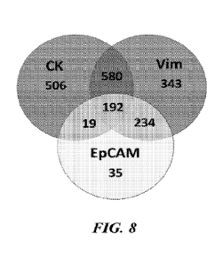
100131 FIG. 8 shows characterization of diagnostic markers of circulating
tumor cells in
patients studied.
100141 FIG. 9A shows total circulating tumor cell counts that change over time
for individual
patients, as ascertained by the systems and methods of the present disclosure.
100151 FIG. 9B shows phenotypic changes in diagnostic and treatment panels for
individual
patients, as ascertained by the systems and methods of the present disclosure
100161 FIGs. 10A and 10B show detection of total CTCs across multiple time
points, based
on use of diagnostic markers (FIG. 10A) or treatment markers (FIG. 10B).
DETAILED DESCRIPTION
100171 The present invention provides systems and compositions for detecting
circulating
tumor cells (CTCs), and methods thereof The systems of the present invention
can image
(e.g., via planar imaging) one or more cells (e.g., embedded in a solid or
semi-solid medium,
such as a gel) and analyze the cells, to identify one or more CTCs from the
one or more cells.
The compositions of the present invention can comprise one or more antibodies
(e.g., a
plurality of antibodies) to detect one or more cell types (e.g., a plurality
of cell types, such as
epithelical-like cell type and mesenchymal-like cell type), to identify one or
more CTCs from
a biological sample, such as a blood sample derived from a subject. The
methods of the
present invention can utilize any of the systems and compositions disclosed
herein to identify
one or more CTCs from a biological sample.
100181 Identification CTCs, e.g., via a liquid biopsy, can be used to predict
the characteristics
of a tumor and for prognostication of cancer The significance and presence of
CTCs can be
characterized and identified. For example, in breast cancer, CTCs can have an
independent
prognostic value in metastatic breast cancer (MBC) and early breast cancer.
For example, if
>5 CTCs are found per 7.5 millileter (mL) of blood from a subject with breast
cancer, this
can be associated with poor prognosis for the cancer. CTC-count can improve
the
prognostication of MBC when added to full clinicopathological predictive
models, which
-3-
CA 03233431 2024- 3- 28
WO 2022/155213
PCT/US2022/012137
cannot be done with serum tumor markers. Single cell molecular
characterization of isolated
CTCs can provide detailed mapping of cancer cell clones from the initial
and/or metastatic
tumor sites. Longitudinally, molecular information from cancer cell clones
resistant to
treatment can be a more responsive method for treatment optimization, rather.
than depending
on the detection of new mutations in fragments of circulating tumor DNA.
100191 In early breast cancer (EBC), CTC detection can be associated with a
poor clinical
outcome. Cytokeratin 19 (CK19)-mRNA can be used as a marker for CTC detection.
Following detection, administration of Trastuzumab can reduce or eliminate
chemotherapy-
resistant CK19mRNA+ cells and improve patient outcome.
100201 A CTC count (e.g., a CTC count of >5 per 7.5 mL of blood) at any time
during the
course of the disease can be associated with a poor prognosis and can be
predictive of shorter
Progression Free Survival (PFS) and Overall Survival (OS) in patients with
metastatic breast
cancer TABLE 1 below lists median PFS and OS based on CTC counts_
TABLE 1
Number of CTC PFS (months) OS (months)
At all-time <5 7.2 22.6
Baseline <5; at final draw >5 5.9 10.6
Baseline >5; at final draw <5 6.1 19.8
At all-time points >5 1.8 4.1
100211 A blood sample taken from a patient prior to a new line of therapy can
be used for the
baseline prediction while another sample taken at the first follow up visit
can be used to
predict whether the therapy is efficacious.
100221 Human epidermal growth factor receptor 2 (1-IER2) evaluation at the
DNA, mRNA,
and protein level has been performed on CTCs. Although HER2+ CTC can be more
commonly detected in women with HER2+ disease, in some women with HER2- breast
cancer, EIER2+ CTCs are observed. In ER- positive MBC, CTC enumeration,
phenotyping,
and genotyping can identify patients who would benefit from Fulvestrant
(selective estrogen
receptor down- regulator) escalation versus switching to alternative
therapies. CTCs are
found in patients with inflammatory breast cancer (IBC), a highly aggressive
form of breast
cancer. CTCs can be found in MC patients with abnormalities in adaptive
immunity.
Utilization of CTCs in patients with abnormalities in adaptive immunity could
be a surrogate
-4-
CA 03233431 2024- 3- 28
WO 2022/155213
PCT/US2022/012137
marker of a more aggressive disease with general immune system dysfunction.
100231 Described herein is a liquid biopsy test to detect one or more CTCs by
analyzing a
plurality of peripheral blood mononuclear cells (PBMCs) (e.g., lymphocytes
such as T cells,
B cells, NK cells; monocytes) derived from a subject. The liquid biopsy test
can be
performed subsequent to enrichment of the plurality of PBMCs. In some
embodiments, the
liquid biopsy test does not require any enrichment of the plurality of PBMCs.
In some
embodiments, described herein is a liquid biopsy test to detect CTCs by
analyzing PBMCs
without prior enrichment. This personalized approach to analysis of CTCs can
use various
imaging methods, such as Selective Plane Illumination Microscopy (SPIVI), to
detect CTCs.
In some embodiments, a method disclosed herein can use automated analysis to
screen the
entire PBMC population and identify CTCs based on staining (e.g.,
immunofluorescent
staining) with positive or negative markers for one or more target cells,
e.g., epithelial or
mesenchymal phenotypes, single cells, clusters and/or apoptotic (dying) cells
100241 In some embodiments, CTC detection, including live characterization and
characterization without enrichment, and using biomarkers for multiple
phenotypes, can open
the way for a standardized CTC definition and benefit precision cancer
diagnosis.
100251 The system as disclosed herein can permit ex vivo observation of cells
(e.g., cells that
have been stained with vital stains for CTC-specific biomarkers and maintained
alive for
periods of time) supported by a three-dimensional (3D) culture subsystem. In
some
embodiments, the syste, can comprise a biological holder and a handler. A
specially
designed cell chamber can be be fitted for input and output of culture media,
gas regulation
and control of environmental variables (temperature, pH etc). This can allow
ex vivo
observation of cells while perfused with culture media which may contain
various substances.
The chamber can be fitted with a micromanipulator (handler) used to isolate
target cells under
direct observation. Both the chamber and the micromanipulator can be operated
automatically
by a system computer and software system.
100261 The ex vivo liquid biopsy can offer longitudinal observation of target
cells, e.g. CTCs
and/or white blood cells (WBCs) and assessment of desired and undesired
toxicity of
therapeutic drug cocktails before used for patient treatment. This can drive
precision
medicine for improved outcomes and reduced adverse effects to the patient.
Cell isolation can
enable CTC genomic and transcriptomic analysis that may reveal improved
therapeutic
options, tuned to the patient's current disease status.
100271 The sample holder and handler of the present invention, combined with
deep
quantitation of every cell the specimen, can be a precision medicine tool.
Deep CTC
-5-
CA 03233431 2024- 3- 28
WO 2022/155213
PCT/US2022/012137
characterization and single-cell, genomic/transcriptomic analysis can enable
the oncologist to
select a treatment that is synchronized with the current disease stage. Ex
vivo assessment of
how a selected drug or drug combination affects CTCs and/or WBCs in the
patient's blood
can be assessed in view of patient outcomes.
[0028] In some embodiments, a central computer system operates a software
package that (a)
acquires and processes images of the biological specimen's features for
identification and
quantitation, (b) actuates the motorized components, pumps, sensors of the
system, (c)
operates a robotic arm that loads and unloads samples, and (d) handles digital
information
managed in local or wide area networks. The central computer system may
utilize local or
distributed processing protocols.
[0029] The system also includes or is coupled to a tunable laser source or
multiple single
wavelength laser sources, complete with light management optical path(s). An
optical system
modulating the light (e g , light sheet, such as laser light sheet) can
combine bilateral
illumination to produce the sheet illumination for Selective Plane
Illumination Microscopy
(SPIIVI).
[0030] In some embodiments, imaging is performed by illuminating the specimen
with
narrow spectrum excitation light provided by monochromatic and/or tunable
laser sources.
Images of the resulting emission are acquired by high sensitivity monochrome
cameras on a
field by field basis. These images are combined in 3D stacks, which are then
analyzed for
quantitative measurement of biomarker levels in the individual cells.
Alternatively, the
images can be analyzed individually (e.g., without combining multiple images
into a single
image).
[0031] In operation, a biological specime that can include live cells is
stained with a variety
of markers against proteins, nucleic acids or other cellular components and
encased in an
appropriately shaped cylindrical sheath to be fitted on a biological sample
holder. The
preparation is made by mixing the cell suspension with a solid or semi-solid
medium (e.g.,
gels, such as agarose or other hydrogels that are compatible with preserving
the subcellular
structure of the embedded cells), at a temperature where the solution is still
liquid. In addition
to the cells, fluorescent beads that serve the role of fiducial reference for
the identified cells
are added to the solution. The liquid cell/bead/gel suspension is aspirated in
tubing that is
chosen to be transparent to the fluorescence light regime utilized. After
being allowed to
solidify, the specimen can be visualized in the light path. The biological
specimen is mounted
on a specimen holder loaded onto the microscope stage.
-6-
CA 03233431 2024- 3- 28
WO 2022/155213
PCT/US2022/012137
[0032] FIG. 4 shows a drawing for the sample holder and of the sample handler
of the
present invention. Shown is a means 1 for advancing and manipulating the
sample 3 (not
visible in this FIG. 4) contained within a sample holder such as a capillary
tube 2 with a
plurality of holes 2A (the capillary tube is not visible in this FIG. 4). The
means 1 can be
any of a variety of mechanical devices, including, for example a glass
syringe. Shown is the
cylindrical sample chamber 5, with a fluid output or outlet port 4, a lens
holder 7, holding an
illumination lens 6A and a detection lens 6B (which are oriented orthogonally,
i.e. at 90
degrees to each other), and an access port 9A built into the cylindrical
sample chamber 5 for
allowing access for a device for retrieving particles of interest, such as a
micropipette 9. A
fluid input connector 10 is shown on the base of the lens holder 7. Not
visible is the fluid
input orifice, of the cylindrical sample chamber 5 located in the base of the
chamber. In
further embodiments, the means for advancing the sample can be controlled by
an external
motor, such as a 4-D motor 13 (not shown in this FIG. 4) to provide movement
and control
in the X, Y, and Z axes, as well as to provide for rotation of the sample. It
is important that
the optical axes of the lenses 6A and 6B are orthogonal and co-planar such
that the sample
chamber and sample can be positioned at the intersection of the respective
optical axes for the
lenses.
100331 In some embodiments, a cell suspension can be observed in SPEVI
instrument
mounted in fixture and embedded in hydrogels that allow cell perfusion with
fluorescently
labeled antibodies, fluorescence in situ hybridization immunostaining and/or
fluorescence in
situ hybridization (FISH) probes, and other stains as well as media that can
sustain ex vivo
cell observation.
[0034] In some embodiments, the following steps are performed: Compare
performance of
embedding gels including agarose, collagen, polyacrylamide and tubing such as
micro-
perforated, fluorinated polyethylene (FPE) and glass both for fixed and live
cells. Optimize
fixation/ permeabilizati on protocols. Assess need of antifading for
fluorescence bleaching
Adapt SPIM image acquisition to materials chosen. Quantitative analysis of
cell staining and
identification or analysis of CTCs (e.g., via 3D image analysis and/or
multiple antigen
staining as di scl soedherein).
[0035] In some embodiments, the present invention can comprise instruments and
kits for the
detection and characterization of CTCs and other target cell populations.
100361 In some embodiments, a sample of cells (e.g., comprising at least one
cell) of the
biological sample can be analyzed by the systems and methods of the present
disclosure. In
some embodiments, at least one cell from a biological sample obtained from a
subject can be
-7-
CA 03233431 2024- 3- 28
WO 2022/155213
PCT/US2022/012137
analyzed by the systems and methods of the present invention. The biological
sample can be
a liquid sample, such as blood. The at least one cell can comprise at least or
up to about 1
cell, at least or up to about 2 cells, at least or up to about 5 cells, at
least or up to about 10
cells, at least or up to about 20 cells, at least or up to about 50 cells, at
least or up to about
100 cells, at least or up to about 200 cells, at least or up to about 500
cells, at least or up to
about 1000 cells, or more.
100371 In some embodimetns, the at least one cell of the biological sample can
be stained
with a detection moiety (e.g., a plurality of detection moieties). The
detection moiety can be
capable of binding to a ligand of the at least one cell. The ligand can be an
extracellular
ligand, a membrane-bound ligand, or an intracellular ligand. The ligand can be
a small
molecule, a polypeptide (e.g., a peptide or a protein), or a polynucleotide
(e.g., ribunocleic
acid (RNA), mRNA, deoxyribonucleic acid (DNA), etc.). The detection moiety can
be an
antibody Non-limiting examples of an antibody can include a monoclonal
antibody, a
polyclonal antibody, a recombinant antibody, a human antibody, a humanized
antibody, a
Fab, a Fab', a F(ab')2, an Fv, a single chain antibody (e.g., scFv), a
minibody, a diabody, a
single-domain antibody ("sdAb- or "nanobodies- or "camelids-), or an Fc
binding domain.
In some examples, the at least one cell can be treated with the detection
moiety prior to being
immobilized in the sample holder as disclosed herein. Alternatively or in
addition to, the at
least one cell can be treated with the detection moiety subsequent to being
immobilized in the
sample holder.
100381 In some embodiments, the detection moiety can comprise a plurality of
detection
moieties that are different (e.g., multiplexing with multiple antibodies). The
plurality of
detection moieties can comprise at least or up to about 2 detection moieties,
at least or up to
about 3 detection moieties, at least or up to about 4 detection moieties, at
least or up to about
detection moieties, at least or up to about 6 detection moieties, at least or
up to about 7
detection moieties, at least or up to about 8 detection moieties, at least or
up to about 9
detection moieties, at least or up to about 10 detection moieties, at least or
up to about 15
detection moieties, or at least or up to about 20 detection moieties. The
plurality of detection
moieties can target different ligands.
100391 In some embodiments, the plurality of detection moieties can bind a
plurality of
ligands that are indicative of different cell functions or cell states (e.g.,
different cell types,
different cell origins, etc.). For examples, the plurality of ligands can be
indicative different
stages of cellual differentiation (or dedifferentiation). The plurality of
detection moieties can
comprise (i) a first detection moiety exhibiting specific binding to a first
target ligand,
-8-
CA 03233431 2024- 3- 28
WO 2022/155213
PCT/US2022/012137
wherein the first target ligand is a marker of a first cell type, and (ii) a
second detection
moiety exhibiting specific binding to a second target ligand, wherein the
second target ligand
is a marker for a second cell type that is different from the first cell type.
100401 In some embodiments, different cell states (e.g., differnet cell types)
can comprise
stem cells and/or differentiated cells. Non-limiting examples different cell
types (e.g.,
including stem cells and/or differentiated cells) can include lymphoid cells,
such as B cell, T
cell (Cytotoxic T cell, Natural Killer T cell, Regulatory T cell, T helper
cell), Natural killer
cell, cytokine induced killer (CIK) cells (see e.g. US20080241194); myeloid
cells, such as
granulocytes (Basophil granulocyte, Eosinophil granulocyte, Neutrophil
granulocyte/Hypersegmented neutrophil), Monocyte/Macrophage, Red blood cell
(Reticulocyte), Mast cell, Thrombocyte/Megakaryocyte, Dendritic cell; cells
from the
endocrine system, including thyroid (Thyroid epithelial cell, Parafollicular
cell), parathyroid
(Parathyroid chief cell, Oxyphil cell), adrenal (Chromaffin cell), pineal
(Pinealocyte) cells;
cells of the nervous system, including glial cells (Astrocyte, Microglia),
Magnocellular
neurosecretory cell, Stellate cell, Boettcher cell, and pituitary
(Gonadotrope, Corticotrope,
Thyrotrope, Somatotrope, Lactotroph); cells of the Respiratory system,
including
Pneumocyte (Type I pneumocyte, Type II pneumocyte), Clara cell, Goblet cell,
Dust cell;
cells of the circulatory system, including Myocardiocyte, Pericyte, cells of
the digestive
system, including stomach (Gastric chief cell, Parietal cell), Goblet cell,
Paneth cell, G cells,
D cells, ECL cells, I cells, K cells, S cells; enteroendocrine cells,
including enterochromaffm
cell, APUD cell, liver (Hepatocyte, Kupffer cell), Cartilage/bone/muscle; bone
cells,
including Osteoblast, Osteocyte, Osteoclast, teeth (Cementoblast, Ameloblast);
cartilage
cells, including Chondroblast, Chondrocyte; skin cells, including Trichocyte,
Keratinocyte,
Melanocyte (Nevus cell); muscle cells, including Myocyte; urinary system
cells, including
Podocyte, Juxtaglomerular cell, Intraglomerular mesangial cell/Extraglomerular
mesangial
cell, Kidney proximal tubule brush border cell, Macula densa cell;
reproductive system cells,
including Spermatozoon, Sertoli cell, Leydig cell, Ovum; and other cells,
including
Adipocyte, Fibroblast, Tendon cell, Epidermal keratinocyte (differentiating
epidermal cell),
Epidermal basal cell (stem cell), Keratinocyte of fingernails and toenails,
Nail bed basal cell
(stem cell), Medullary hair shaft cell, Cortical hair shaft cell, Cuticular
hair shaft cell,
Cuticular hair root sheath cell, Hair root sheath cell of Huxley's layer, Hair
root sheath cell of
Henle's layer, External hair root sheath cell, Hair matrix cell (stem cell),
Wet stratified barrier
epithelial cells, Surface epithelial cell of stratified squamous epithelium of
cornea, tongue,
oral cavity, esophagus, anal canal, distal urethra and vagina, basal cell
(stem cell) of epithelia
-9-
CA 03233431 2024- 3- 28
WO 2022/155213
PCT/US2022/012137
of cornea, tongue, oral cavity, esophagus, anal canal, distal urethra and
vagina, Urinary
epithelium cell (lining urinary bladder and urinary ducts), Exocrine secretory
epithelial cells,
Salivary gland mucous cell (polysaccharide-rich secretion), Salivary gland
serous cell
(glycoprotein enzyme-rich secretion), Von Ebner's gland cell in tongue (washes
taste buds),
Mammary gland cell (milk secretion), Lacrimal gland cell (tear secretion),
Ceruminous gland
cell in ear (wax secretion), Eccrine sweat gland dark cell (glycoprotein
secretion), Eccrine
sweat gland clear cell (small molecule secretion). Apocrine sweat gland cell
(odoriferous
secretion, sex-hormone sensitive), Gland of Moll cell in eyelid (specialized
sweat gland),
Sebaceous gland cell (lipid-rich sebum secretion), Bowman's gland cell in nose
(washes
olfactory epithelium), Brunner's gland cell in duodenum (enzymes and alkaline
mucus),
Seminal vesicle cell (secretes seminal fluid components, including fructose
for swimming
sperm), Prostate gland cell (secretes seminal fluid components), Bulbourethral
gland cell
(mucus secretion), Bartholin's gland cell (vaginal lubricant secretion), Gland
of Littre cell
(mucus secretion), Uterus endometrium cell (carbohydrate secretion), Isolated
goblet cell of
respiratory and digestive tracts (mucus secretion), Stomach lining mucous cell
(mucus
secretion), Gastric gland zymogenic cell (pepsinogen secretion), Gastric gland
oxyntic cell
(hydrochloric acid secretion), Pancreatic acinar cell (bicarbonate and
digestive enzyme
secretion), Paneth cell of small intestine (lysozyme secretion), Type II
pneumocyte of lung
(surfactant secretion), Clara cell of lung, Hormone secreting cells, Anterior
pituitary cells,
Somatotropes, Lactotropes, Thyrotropes, Gonadotropes, Corticotropes,
Intermediate pituitary
cell, Magnocellular neurosecretory cells, Gut and respiratory tract cells,
Thyroid gland cells,
thyroid epithelial cell, parafollicular cell, Parathyroid gland cells,
Parathyroid chief cell,
Oxyphil cell, Adrenal gland cells, chromaffin cells, Ley dig cell of testes,
Theca interna cell
of ovarian follicle, Corpus luteum cell of ruptured ovarian follicle,
Granulosa lutein cells,
Theca lutein cells, Juxtaglomerular cell (renin secretion), Macula densa cell
of kidney,
Metabolism and storage cells, Barrier function cells (Lung, Gut, Exocrine
Glands and
Urogenital Tract), Kidney, Type I pneumocyte (lining air space of lung),
Pancreatic duct cell
(centroacinar cell), Nonstriated duct cell (of sweat gland, salivary gland,
mammary gland,
etc.), Duct cell (of seminal vesicle, prostate gland, etc.), Epithelial cells
lining closed internal
body cavities, Ciliated cells with propulsive function, Extracellular matrix
secretion cells,
Contractile cells; Skeletal muscle cells, stem cell, Heart muscle cells, Blood
and immune
system cells, Erythrocyte (red blood cell), Megakaryocyte (platelet
precursor), Monocyte,
Connective tissue macrophage (various types), Epidermal Langerhans cell,
Osteoclast (in
bone), Dendritic cell (in lymphoid tissues), Microglial cell (in central
nervous system),
-10-
CA 03233431 2024- 3- 28
WO 2022/155213
PCT/US2022/012137
Neutrophil granulocyte, Eosinophil granulocyte, Basophil granulocyte, Mast
cell, Helper T
cell, Suppressor T cell, Cytotoxic T cell, Natural Killer T cell, B cell,
Natural killer cell,
Reticulocyte, Stem cells and committed progenitors for the blood and immune
system
(various types), Pluripotent stem cells, Totipotent stem cells, Induced
pluripotent stem cells,
adult stem cells, Sensory transducer cells, Autonomic neuron cells, Sense
organ and
peripheral neuron supporting cells, Central nervous system neurons and glial
cells, Lens cells,
Pigment cells, Melanocyte, Retinal pigmented epithelial cell, Germ cells,
Oogonium/Oocyte,
Spermatid, Spermatocyte, Spermatogonium cell (stem cell for spermatocyte),
Spermatozoon,
Nurse cells, Ovarian follicle cell, Sertoli cell (in testis), Thymus
epithelial cell, Interstitial
cells, and Interstitial kidney cells. Non-limiting examples of stem cells can
include adult
stem cells (e.g., mesenchymal stem cells), embdyonic stem cells, induced
pluripotent stem
cells, and progenitor cells (e.g., cardiac progenitor cells, neural progenitor
cells, etc.).
100411 In some embodiments, the first cell type as disclosed herein can be a
differentiated
cell type, such as an epithelial cell. The first ligand can comprise an
epithelial cell antigen,
such as epithelial cellular adhesion molecule (EpCAM) or cytokeratin (CK). In
some
examples, the first ligand can be one of EpCAM and CK, and the other antigen
of EpCAM
and CK can be bound and detected by a third detection moiety exhibiting
specific binding to
the other antigen. Non-limiting examples of the epithelial cell maker can
include EpCam,
Cadherin, Mucin-1, Cytokeratin (CK) 8, epidermal growth factor receptor
(EGFR),
cytokeratin (CK)19, ErbB2, PDGF, L6, and leukocyte associated receptor (LAR).
100421 In some embodiments, the second cell type as disclosed herein can be a
stem cell
type, such as a mesenchymal cell (e.g., mesenchymal stem cell). The second
ligand can
comprise a mesenchymal steat antigen, such as vimentin (Vim). Non-limiting
examples of
mesenchymal cell marker can include CD90, CD73, CD44, and vimentin.
100431 In some embodiments, the at least one cell as disclosed herein can be
detected to
exhibit only one of the plurality of ligands, and such characteristic can be
indicative of the at
least one cell being a CTC. In some embodiments, the at least one cell as
disclosed herein
can be detected to exhibit two or more of the plurality of ligands, and such
characteristic can
be indicative of the at least one cell being a CTC. In some embodiments, a CTC
from the
sample of cells may be determined to have been detected when (i) a number of
cells
determined to exhibit two or more of the plurality of ligands is greater than
or equal to (ii) a
number of cells determined to exhibit only one of the two or more of the
plurality of ligands.
For example, a CTC associated with breast cancer may be determined to have
been detected
from the sample of cells when (i) a number of cells determined to exhibit two
or more of the
-11-
CA 03233431 2024- 3- 28
WO 2022/155213
PCT/US2022/012137
plurality of ligands (e.g., EpCAM and Vim) is greater than or equal to (ii) a
number of cells
determined to exhibit only one of the two or more of the plurality of ligands
(e.g., EpCAM
substantially alone, or Vim substantially alone).
100441 In some embodiments, the method disclosed herein can identify different
types of
diseased cells. In some embodiments, the method disclosed herein can assess
heterogeneity
within a specific population of diseased cells. In some embodiments, the
specific population
of diseased cells can be CTCs, and the method disclosed herein can assess
heterogeneity
(e.g., different subtypes or phenotypes) within the specific population of the
CTCs. In some
embodiments, the method disclosed herein can assess different phenotypes or
states of a
population of CTCs from breast tumors. For example, the method disclosed
herein can
identify, distinguish, and/or quantitate (i) CTCs of mesenchymal phenotype
and/or (ii) CTCs
of epithelial phenotype. In another example, the method disclosed herein can
identify
distinguish, and/or quantitate (i) CTCs of Lumina] A breast cancer, (ii) CTCs
of Lumina] B
breast cancer, (iii) CTCs of triple-negative breast cancer, (iv) CTCs of HER2-
enriched breast
cancer, and/or (v) CTCs of normal-like breaste cancer.
100451 CTCs of Luminal A breast cancer can be hormone-receptor positive (e.g.,
estrogen-
receptor and/or progesterone-receptor positive), HER2 negative, and with low
levels of the
protein Ki-67. CTCs of Luminal B breast cancer can be hormone-receptor
positive (e.g.,
estrogen-receptor and/or progesterone-receptor positive), either HER2-positive
or HER2-
negative, and with high levels of Ki-67. CTCs of triple-negative breast cancer
can be
hormone-receptor negative (e.g., estrogen-receptor and progesterone-receptor
negative) and
HER2 negative. CTCs of HER2-enriched breast cancer can be hormone-receptor
negative
(e.g., estrogen-receptor and progesterone-receptor negative) and HER2
positive. CTCs of
normal-like breast cancer can be hormone-receptor positive (e.g., estrogen-
receptor and/or
progesterone-receptor positive), HER2 negative, and with low levels of the
protein Ki-67.
100461 In some embodiments, the diseased cells as disclosed herein can be
cancer cells.
Non-limiting examples of cancer cells can include cells of Acanthoma, Acinic
cell
carcinoma, Acoustic neuroma, Acral lentiginous melanoma, Acrospiroma, Acute
eosinophilic
leukemia, Acute lymphoblastic leukemia, Acute megakaryoblastic leukemia, Acute
monocytic leukemia, Acute myeloblastic leukemia with maturation, Acute myeloid
dendritic
cell leukemia, Acute myeloid leukemia, Acute promyelocytic leukemia,
Adamantinoma,
Adenocarcinoma, Adenoid cystic carcinoma, Adenoma, Adenomatoid odontogenic
tumor,
Adrenocortical carcinoma, Adult T-cell leukemia, Aggressive NK-cell leukemia,
AIDS-
Related Cancers, AIDS-related lymphoma, Alveolar soft part sarcoma,
Ameloblastic fibroma,
-12-
CA 03233431 2024- 3- 28
WO 2022/155213
PCT/US2022/012137
Anal cancer, Anaplastic large cell lymphoma, Anaplastic thyroid cancer,
Angioimmunoblastic T-cell lymphoma, Angiomyolipoma, Angiosarcoma, Appendix
cancer,
Astrocytoma, Atypical teratoid rhabdoid tumor, Basal cell carcinoma, Basal-
like carcinoma,
B-cell leukemia, B-cell lymphoma, Bellini duct carcinoma, Biliary tract
cancer, Bladder
cancer, Blastoma, Bone Cancer, Bone tumor, Brain Stem Glioma, Brain Tumor,
Breast
Cancer, Brenner tumor, Bronchial Tumor, Bronchioloalveolar carcinoma, Brown
tumor,
Burkitt's lymphoma, Cancer of Unknown Primary Site, Carcinoid Tumor,
Carcinoma,
Carcinoma in situ, Carcinoma of the penis, Carcinoma of Unknown Primary Site,
Carcinosarcoma, Castleman's Disease, Central Nervous System Embryonal Tumor,
Cerebellar Astrocytoma, Cerebral Astrocytoma, Cervical Cancer,
Cholangiocarcinoma,
Chondroma, Chondrosarcoma, Chordoma, Choriocarcinoma, Choroid plexus
papilloma,
Chronic Lymphocytic Leukemia, Chronic monocytic leukemia, Chronic myelogenous
leukemia, Chronic Myeloproliferative Disorder, Chronic neutrophilic leukemia,
Clear-cell
tumor, Colon Cancer, Colorectal cancer, Craniopharyngioma, Cutaneous T-cell
lymphoma,
Degos disease, Dermatofibrosarcoma protuberans, Dermoid cyst, Desmoplastic
small round
cell tumor, Diffuse large B cell lymphoma, Dysembryoplastic neuroepithelial
tumor,
Embryonal carcinoma, Endodermal sinus tumor, Endometrial cancer, Endometrial
Uterine
Cancer, Endometrioid tumor, Enteropathy-associated T-cell lymphoma,
Ependymoblastoma,
Ependymoma, Epithelioid sarcoma, Erythroleukemia, Esophageal cancer,
Esthesioneuroblastoma, Ewing Family of Tumor, Ewing Family Sarcoma, Ewing's
sarcoma,
Extracranial Germ Cell Tumor, Extragonadal Germ Cell Tumor, Extrahepatic Bile
Duct
Cancer, Extramammary Paget's disease, Fallopian tube cancer, Fetus in fetu,
Fibroma,
Fibrosarcoma, Follicular lymphoma, Follicular thyroid cancer, Gallbladder
Cancer,
Gallbladder cancer, Ganglioglioma, Ganglioneuroma, Gastric Cancer, Gastric
lymphoma,
Gastrointestinal cancer, Gastrointestinal Carcinoid Tumor, Gastrointestinal
Stromal Tumor,
Gastrointestinal stromal turnor, Germ cell turnor, Germinoma, Gestational
choriocarcinoma,
Gestational Trophoblastic Tumor, Giant cell tumor of bone, Glioblastoma
multiforme,
Glioma, Gliomatosis cerebri, Glomus tumor, Glucagonoma, Gonadoblastoma,
Granulosa cell
tumor, Hairy Cell Leukemia, Hairy cell leukemia, Head and Neck Cancer, Head
and neck
cancer, Heart cancer, Hemangioblastoma, Hemangiopericytoma, Hemangiosarcoma,
Hematological malignancy, Hepatocellular carcinoma, Hepatosplenic T-cell
lymphoma,
Hereditary breast-ovarian cancer syndrome, Hodgkin Lymphoma, Hodgkin's
lymphoma,
Hypopharyngeal Cancer, Hypothalamic Glioma, Inflammatory breast cancer,
Intraocular
Melanoma, Islet cell carcinoma, Islet Cell Tumor, Juvenile myelomonocytic
leukemia,
-13 -
CA 03233431 2024- 3- 28
WO 2022/155213
PCT/US2022/012137
Kaposi Sarcoma, Kaposi's sarcoma, Kidney Cancer, Klatskin tumor, Krukenberg
tumor,
Laryngeal Cancer, Laryngeal cancer, Lentigo maligna melanoma, Leukemia,
Leukemia, Lip
and Oral Cavity Cancer, Liposarcoma, Lung cancer, Luteoma, Lymphangioma,
Lymphangiosarcoma, Lymphoepithelioma, Lymphoid leukemia, Lymphoma,
Macroglobulinemia, Malignant Fibrous Histiocytoma, Malignant fibrous
histiocytoma,
Malignant Fibrous Histiocytoma of Bone, Malignant Glioma, Malignant
Mesothelioma,
Malignant peripheral nerve sheath tumor, Malignant rhabdoid tumor, Malignant
triton tumor,
MALT lymphoma, Mantle cell lymphoma, Mast cell leukemia, Mediastinal germ cell
tumor,
Mediastinal tumor, Medullary thyroid cancer, Medulloblastoma, Medulloblastoma,
Medulloepithelioma, Melanoma, Melanoma, Meningioma, Merkel Cell Carcinoma,
Mesothelioma, Mesothelioma, Metastatic Squamous Neck Cancer with Occult
Primary,
Metastatic urothelial carcinoma, Mixed Mullerian tumor, Monocytic leukemia,
Mouth
Cancer, Mucinous tumor, Multiple Endocrine Neoplasia Syndrome, Multiple
Myeloma,
Multiple myeloma, Mycosis Fungoides, Mycosis fungoides, Myelodysplastic
Disease,
Myelodysplastic Syndromes, Myeloid leukemia, Myeloid sarcoma,
Myeloproliferative
Disease, Myxoma, Nasal Cavity Cancer, Nasopharyngeal Cancer, Nasopharyngeal
carcinoma, Neoplasm, Neurinoma, Neuroblastoma, Neuroblastoma, Neurofibroma,
Neuroma,
Nodular melanoma, Non-Hodgkin Lymphoma, Non-Hodgkin lymphoma, Nonmelanoma
Skin Cancer, Non-Small Cell Lung Cancer, Ocular oncology, Oligoastrocytoma,
Oligodendroglioma, Oncocytoma, Optic nerve sheath meningioma, Oral Cancer,
Oral cancer,
Oropharyngeal Cancer, Osteosarcoma, Osteosarcoma, Ovarian Cancer, Ovarian
cancer,
Ovarian Epithelial Cancer, Ovarian Germ Cell Tumor, Ovarian Low Malignant
Potential
Tumor, Paget's disease of the breast, Pancoast tumor, Pancreatic Cancer,
Pancreatic cancer,
Papillary thyroid cancer, Papillomatosis, Paraganglioma, Paranasal Sinus
Cancer, Parathyroid
Cancer, Penile Cancer, Perivascular epithelioid cell tumor, Pharyngeal Cancer,
Pheochromocytoma, Pineal Parenchymal Tumor of Intermediate Differentiation,
Pineoblastoma, Pituicytoma, Pituitary adenoma, Pituitary tumor, Plasma Cell
Neoplasm,
Pleuropulmonary blastoma, Polyembryoma, Precursor T-lymphoblastic lymphoma,
Primary
central nervous system lymphoma, Primary effusion lymphoma, Primary
Hepatocellular
Cancer, Primary Liver Cancer, Primary peritoneal cancer, Primitive
neuroectodermal tumor,
Prostate cancer, Pseudomyxoma peritonei, Rectal Cancer, Renal cell carcinoma,
Respiratory
Tract Carcinoma Involving the NUT Gene on Chromosome 15, Retinoblastoma,
Rhabdomyoma, Rhabdomyosarcoma, Richter's transformation, Sacrococcygeal
teratoma,
Salivary Gland Cancer, Sarcoma, Schwannomatosis, Sebaceous gland carcinoma,
Secondary
-14-
CA 03233431 2024- 3- 28
WO 2022/155213
PCT/US2022/012137
neoplasm, Seminoma, Serous tumor, Sertoli-Leydig cell tumor, Sex cord-stromal
tumor,
Sezary Syndrome, Signet ring cell carcinoma, Skin Cancer, Small blue round
cell tumor,
Small cell carcinoma, Small Cell Lung Cancer, Small cell lymphoma, Small
intestine cancer,
Soft tissue sarcoma, Somatostatinoma, Soot wart, Spinal Cord Tumor, Spinal
tumor, Splenic
marginal zone lymphoma, Squamous cell carcinoma, Stomach cancer, Superficial
spreading
melanoma, Supratentorial Primitive Neuroectodermal Tumor, Surface epithelial-
stromal
tumor, Synovial sarcoma, T-cell acute lymphoblastic leukemia, T-cell large
granular
lymphocyte leukemia, T-cell leukemia, T-cell lymphoma, T-cell prolymphocytic
leukemia,
Teratoma, Terminal lymphatic cancer, Testicular cancer, Thecoma, Throat
Cancer, Thymic
Carcinoma, Thymoma, Thyroid cancer, Transitional Cell Cancer of Renal Pelvis
and Ureter,
Transitional cell carcinoma, Urachal cancer, Urethral cancer, Urogenital
neoplasm, Uterine
sarcoma, Uveal melanoma, Vaginal Cancer, Verner Morrison syndrome, Verrucous
carcinoma, Visual Pathway Glioma, Vulvar Cancer, Waldenstrom's macrogl
obulinemi a,
Warthin's tumor, and Wilms' tumor.
100471 In some embodiments, the CTC as detected or identified as disclosed
herein may be
associated with a solid tumor, such as breask cancer. In some embodiments, the
CTC as
detected or identified as disclosed herein may be associated with a blood
cancer (e.g., non-
solid tumor), such as leukemia, lymphoma, myelodysplastic syndromes (MDS),
myeloproliferative disorder (MPD), and multiple myeloma.
100481 In some embodiments, the method disclosed herein can scan a plurality
of cells (e.g.,
millions of cells) from the blood of a subject and acquire one or more 3-
dimensional cell
images per cell, with resolution comparable to that of confocal microscopy,
thereby
enhancing the accuracy of biomarker quantitation.
100491 The method disclosed herein can be used to identify CTCs exhibiting one
or more
target biomarkers. A target biomarker can be a tumor antigen (or a carcinoma-
associated
antigen). The tumor antigen can be encoded by a gene carrying one or more
mutations.
Alternatively, the tumor antigen can be encoded by a gene that does not carry
a mutation.
The tumor antigen can be a receptor polypeptide (e.g., a cell surface receptor
polypeptide).
The tumor antigen can be an ion channel, such as a cationinc ion channel for
calcium
singaling in a cell. In some embodiments, the tumor antigen can be a calcium
signal
transducer, such as Tumor-associated calcium signal transducer 2 (Trop2). In
some
embodiments, the tumor antigen may not be EpCAM, Vimentin (Vim), and/or
Cytokeratin
(CK).
100501 CTC assessment can be a way of identifying more aggressive components
of tumors.
-15-
CA 03233431 2024- 3- 28
WO 2022/155213
PCT/US2022/012137
By sequencing the tumor genome in patients with metastatic breast cancer and
enumerating
and characterizing the CTCs present, genetic alterations that could result in
higher levels of
more aggressive CTCs can be identified. Additionally, if an actionable genetic
alteration is
found, a targeted therapy could be used in treatment with continued follow-up
of CTCs over
time.
[0051] Multiplex testing (e.g., 10 antibodies on a single cell) can enhance
detection and
detailed characterization of circulating tumor cells. The counting process can
be automated.
EXAMPLES
EXAMPLE 1: Proof-of-Concept Study for Clinical Trial Sample Analysis.
[0052] The object of the present study was to determine the CTC frequency in
localized and
metastatic disease; to quantitate CTCs at diagnosis; timing of disappearance
or persistence
with treatment; to compare the CTC numbers and characteristics with
information from next
generation sequencing (NGS) done on the tumor biopsy in patients with
metastatic disease;
and to show that non-enriched CTC enumeration can compare and correlate with
the
epithelial capture-based, FDA-approved CellSearch test.
[0053] Study Population
[0054] The following patients were included in the study:
[0055] Male and female patients who were more than 18 yrs. of age with a
biopsy proven
diagnosis of breast cancer. All stages of patients were included in this
study.
[0056] Early stage breast cancer (Stage I to III) included newly diagnosed
patients before any
prior treatment (surgery, chemotherapy, hormone therapy).
[0057] Patients with metastatic disease can be at initial diagnosis or at any
point in treatment.
Available and/or recurrent tumor samples and saliva samples from metastatic
disease patients
may be collected and sent to next generation sequencing testing by a
commercial laboratory.
[0058] Study Design
[0059] This study is a prospective, single arm, proof-of-concept study.
[0060] Comparison of the method disclosed herein with the FDA-approved
CellSearch
technology for the detection and characterization of CTCs in peripheral blood
samples from
a total of 10 study patients who are either early stage or metastatic breast
cancer patients.
[0061] 60 (30 early stage and 30 metastatic, including the ten listed above)
breast cancer
patients are recruited and blood samples are collected and analyzed at study
time points
shown below.
-16-
CA 03233431 2024- 3- 28
WO 2022/155213
PCT/US2022/012137
[0062] Saliva collection kit as part of NGS testing
[0063] Available and/or recurrent tumor samples from metastatic disease
patients are
collected and sent for next generation sequencing testing by a commercial
laboratory.
[0064] Recruitment procedure
[0065] A total of 60 patients are enrolled in this study.
[0066] At two time points, approximately 22.5 ml peripheral blood sample are
obtained, and
CTCs are analyzed. The metastatic patients' saliva samples and available
and/or recurrent
tumor samples are collected for analysis.
[0067] Study Procedure
[0068] At each study time point, approximately a total of 15 ml blood samples
are collected
from the patients, including 2 x7.5 ml samples for CTC analysis.
[0069] The samples for CTC analysis are collected in Cell-Free blood
collection tubes. At
two study time points, approximately a total of 22 5 ml, blood samples are
collected from ten
patients including 2 x7.5 ml samples for CTC analysis and 1 x 7.5 ml sample
for NGS.
[0070] For the metastatic disease group, 0.65 ml of saliva sample and
available and/or
recurrent tumor sample are sent for NGS.
100711 During every 3-month time points, all patients are allowed to miss 1
time point.
100721 For themetastatic disease group, after disease progression, study
patients start over with
the every 3-month time point until maximum 2 years of study participation.
100731 For the early stage disease group, study patients who have distant
disease recurrence
are asked to be part of the metastatic disease group.
[0074] CTC Test protocol:
[0075] The CTC analysis was performed on cell preparations from 15 mL aliquots
of
peripheral blood. Following lysis of the red blood cells, the nucleated cell
pellet was collected
by centrifugation, and washed and resuspended in Phosphate Buffered Saline
(PBS). A
cocktail of fluorescently labelled antibodies in blocking buffer was added and
the cells were
incubated on ice for 30 minutes. The cocktail combined CTC positive
identification
antibodies against EpCAM epithelial cell surface antigen, Vimentin mesenchymal
cell
surface antigen, CD45 leukocyte common antigen for negative selection, and
combinations of
therapy related markers including targeted therapy markers such as ER and
FIER2 depending
on the patient's clinical information.
100761 After counterstaining, the cells were thoroughly mixed with an equal
volume of 2%
low melting point agarose in PBS at 37 C. The cell suspension was then drawn
into the
appropriate specimen fixture and allowed to solidify prior to analysis.
Control samples were
-17-
CA 03233431 2024- 3- 28
WO 2022/155213
PCT/US2022/012137
processed in parallel to monitor the efficiency of the staining and scanning
processes.
[0077] Specimens stained by immunofluorescence were analyzed using a
microscopy system
disclosed herein. The complete immobilized cell suspension was scanned using a
20X
objective and images were digitized in 3D-image stacks. For each cell,
(individual cells, cell
clusters and apoptotic cells) morphological and quantitative information was
obtained for
each biomarker. The cells were then ranked based on morphology and
quantitative
information of the immunofluorescent signals.
[0078] By analyzing every nucleated cell present in a sample for quantitative
immunofluorescent expression, the CTCs can be characterized as epithelial,
mesenchymal, or
as intermediate phenotypes.
[0079] Standard of care performance of Next Generation Sequencing: In
approximately
30 patients with metastatic disease, the recurrent tumor specimen and saliva
sample are sent
for NGS
[0080] Study Time Points
[0081] Blood samples are collected at the following time points. The
collection times can
vary depending on treatments given. The entire study duration is 2 years for
all study patients
except early stage study patients who have distant disease recurrence and
agree to participate
in the metastatic disease group.
[0082] Metastatic Disease Group:
= At the time of enrollment to the study (+ 3 weeks)
o Tempus NGS saliva collection is collected at time of enrollment (+ 3
months)
= Between 4-6 weeks after study enrollment
= Every 3 months on treatment if stable disease until study completion (+/-
3 weeks)
= Any point at disease recurrence (+/-3 weeks window)
o Tempus NGS saliva collection may be collected at time of disease
recurrence
(+ 3 months)
[0083] Approximately 13 blood collection time points expected for this group
[0084] An additional 1 X 7.5 ml of blood is collected for the CellSearch test
from ten
selected study patients at two of the time points as determined by the PI
[0085] Early Stage Group:
= At the time of enrollment to the study (+ 3 weeks)
= Completion of initial therapy (chemotherapy/surgery) (+/- 3 weeks)
= Completion of secondary therapy (chemotherapy/surgery) (+/- 3 weeks)
-18-
CA 03233431 2024- 3- 28
WO 2022/155213
PCT/US2022/012137
= Every 3 months until study completion (+/- 3-week window)
= Any point of recurrence(+/- 3-week window)
100861 Approximately 13 blood collection time points are expected for this
group
100871 An additional 1 X 7.5 ml of blood is collected for the CellSearch test
from ten
selected study patients at two of the time points as determined by the PI.
100881 Data Collection
100891 Demographic information including but not limited to date of birth
andgender.
100901 Complete medical history, surgical history, social history, family
history and current
medications.
100911 Imaging information related to the breast malignancy such as mammogram,
ultrasound
and MRI.
100921 Complete pathology information including laterality, lymph node status,
TNM staging
(clinical, pathological and post neoadjuvant, DCIS or LCIS information, ER/PR
status, HER2
status, Nottingham grade, Ki-67%, Miller Payne grade, lymphocytic infiltrate,
lymphovascular invasion and perineural invasion) and Oncotype dx score and
results of Next
Generation Sequencing where appropriate.
100931 Routine laboratory results done prior to starting treatment and after
are collected.
100941 These data include complete blood count, chemistries, tumor markers
(CEA and
CA15-3), germline genetic testing (i.e. BRCA), and other tests that have been
performed for
standard of care.
100951 During the maximum 2 years of study participation, all standard of care
and physical
exam data from clinic appointments are collected.
100961 Study Endpoints
100971 This Proof of Concept Study determines the potential of the liquid
biopsy method
disclosed herein in validating the identification of the CTCs in peripheral
blood samples from
breast cancer patients.
100981 Statistical Consideration
100991 Data Analysis Plan
101001 One primary goal of this study is to validate the ability of the liquid
biopsy method
disclosed herein to identify CTCs in peripheral blood samples from the breast
cancer patients.
For each patient sample, CTC counts by different methods were obtained and
compared.
Pearson's correlation and Bland-Altman method were used to assess the CTC
counts.
101011 Sample Size Justification
-19-
CA 03233431 2024- 3- 28
WO 2022/155213
PCT/US2022/012137
101021 The sample size was calculated based on the correlation between CTC
counts by the
liquid biopsy method disclosed herein and CTC counts by an accredited
cytogeneticist or
cytopathologist. A total of 30 early-stage breast cancer patients and 30
metastatic breast
cancer patients are recruited. Some of the baseline data from early stage and
metastatic
disease patients are used as training data set for the method disclosed
herein, while the
baseline data from early- stage breast cancer patients is used for validation
analysis. TABLE
2 below shows predicted statistical parameters.
TABLE 2
Sample size estimation by one-sided Fisher's z test with a null correlation of
0.6 using a
one-sided a=0.05
# of patients Effect size (Observed vs Null correlation)
Power
30 0.80 vs 0.60
69%
30 0.85 vs 0.60
91%
30 0.90 vs 0.60
99%
40 0.80 vs 0.60
80%
55 0.80 vs. 0.60
90%
EXAMPLE 2: Protocol for Isolation of White Blood Cells (WBCs)/CTCs from Blood
Samples.
101031 RBC Removal utilizing Ficoll/Hypaque protocol
1. Prepare 10% bleach solution in receptable for discarding pipettes, tubes,
and tips
during procedure.
2. Set out at room temperature:
a. Hypaque 1077
b. Hypaque 1119
c. Sterile PBS
d. RBC lysis buffer
e. cell culture media (RMPI plus 10% FBS, PenStrep, glutamine)
f. 2X freezing medium consisting of 20% DMSO in Fetal Bovine Serum (FBS)
3. Collect 2 mL whole human normal donor (ND) blood from each STRECK Cell-
Free DNA vacutainer tube
-20-
CA 03233431 2024- 3- 28
WO 2022/155213
PCT/US2022/012137
4. Carefully transfer equal volumes (2 mL) of whole blood to each of
three 15 mL conical tube labeled with sample/patient identifying number.
5. In a 15 mL conical tube, layer (e.g., all reagents at room temperature):
a. 4 mL Hypaque 1119 (bottom layer)
b. 4 mL Hypaque 1077 (middle layer)
c. Mix whole blood in equal parts sterile PBS and add to 15 mL tube (top
layer)
6. Centrifugation of tube at 700xg for 30 min. at room temperature
7. Two distinct layers of cells (monocytes and granulocytes respectively)
form at the
interfaces. Carefully remove both to a fresh 50 ml conical. The cells can then
be
washed and used (or subject to brief RBC lysis if necessary).
8. QS (quantity sufficient) volume of cells to 30 mL with room temperature RBC
lysis buffer, mix by inverting Label tube Rock on speed 15 at room temperature
for 10 min.
9. Add ¨25 ml of sterile, room temperature PBS to tubes. Centrifuge tubes at
300xg
for 10 minutes and discard supernatant, dab tube tops on sterile towel.
10. Resuspend PBMCs for Cryopreservation in 2m1 of room temperature cell
culture
media (RMPI plus 10% FBS, PenStrep, glutamine).
a. Count cells using the hemocytometer with trypan blue exclusion dye.
b. Resuspend cells at about 2X10^7 cells/ml in cell culture media at room
temperature.
c. Add dropwise enough 2X freezing medium at room temperature to double
the volume of the cell suspension. Gently swirl the tube when adding the
freezing medium.
d. Slowly remove the cell suspension into a pipette and dispense lmL per
cryovial
e. Place the cryovials in a room temperature freezing container and label the
container
f. Place the freezing container as soon as possible into the -80 C freezer
g. Transfer the cryovials to liquid nitrogen tank after 1-14 days.
11. Fix cells by adding 500 uL of 4% paraformaldehyde in PBS, gently vortex
and
incubating at room temperature for 10 min., while protecting from light.
Gently
vortex ¨3 min. to keep cells suspended.
-21 -
CA 03233431 2024- 3- 28
WO 2022/155213
PCT/US2022/012137
12. Dilute 4% PFA in QS PBS and centrifuge tubes at 300xg for 10 minutes and
discard supernatant, dab tube on sterile towel.
13. Gently resuspend pellets of two tubes by pipetting with 2 mL room
temperature PBS, leaving in second tube (Resuspend cells in initial whole
blood
volume; 2 mL of initial whole blood equates to 2 mL resuspension).
14. Dilute 20 ul of cell suspensions in 180 ul of 0.4% Trypan solution in an
Eppendorf
tube and count using a hemocytometer.
15. Count the 4 corner squares (16 small squares inside each) and average the
value;
calculate cells/ml in cell suspension. Cells/mL = average of 4 corners x
10,000
(hemocytometer volume conversion factor) x 10 (trypan dilution factor).
EXAMPLE 3: Antibody Staining of CTC Samples.
101041 WBC collection from previous blood draw and fixed WBC
1. Two metastatic blood 2 mL aliquots are treated for RBC removal as stated
above in
EXAMPLE 2.
2. One of the samples is fixed and counted for staining protocol.
3. The other sample is cryopreserved alive for future use.
101051 Preparation of reagents
1. Fixation and permeabilization Solution:
a. Final composition is 3:1 Methanol-Glacial Acetic Acid (make fresh for each
experiment)
b. For a final volume of 4 mL, mix: 3 Methanol with 1 Glacial Acetic Acid
2. Nuclear Stain for use after hybridization:
a. 4',6-diamidino-2-phenylindole (DAPI) prepared in Wash Buffer A at 5
ng/mL.
b. DAPI Stock 100 ng/mL; dilute 10 pL DAPI stock into 200 pL PBS and put
¨65 pL into each of the three tubes (when indicated below).
101061 Stain cell surface antigens
1. Wash cells by adding 3.5 mL cold PBS/2% BSA and centrifugation
at 300xg for 5 min.
2. Discard supernatant and pulse vortex to completely dissociate the
pellet. (typically, 50-70pL residual volume remains).
3. Add 2 pL FcR inhibitor and incubate tubes on ice for 5 min.
4. Add 2 pL anti-EpCAM-EBA-1-AF546, 2 pL anti-EpCAM-9C4-AF546, and 5 iaL
-22-
CA 03233431 2024- 3- 28
WO 2022/155213
PCT/US2022/012137
anti-CD45-AF488 to the appropriate tubes (TABLE 1) and incubate on ice for
30 min. Protect from light by covering with aluminum foil.
5. Wash with 3.5 mL PBS+2%BSA, discard supernatant by carefully decanting.
6. Permeabilize cells by adding 1 mL of methanol-acetic acid (Me0H-AcOH)
fixation
solution. Incubate at room temperature for 10 min.
7. Add 3.5 mL cold PBS+2% BSA. Centrifugation at 400 x g for 5 min. at 4 C.
Decant
supernatant.
8. Gently agitate by vortex to resuspend pellet and add 250 [IL of PBS/10%
BSA and
incubate covered for 30 min. at room temperature to block.
9. Add 2 mL PBS/2% BSA and spin by centrifugate at 400xg for 5 min. Decant
supernatant (-100 pL remains)
10. Add 2 p.L anti-Vimentin-AF594 antibody, and 2 p.L anti-TROP-2-AF647
antibody to
appropriate tubes (TABLE 3) and incubate at room temperature for 30 min.
11. Add 3.5 mL PBS/2% BSA and spin by centrifuge at 400 x g for 5 min. at 4
C.
Decant supernatant.
12. Add 300 pL Hoechst 33342 nuclear stain (Diluted 1:10,000 from stock in
sterile
PBS) to counterstain the nuclei. Incubate in the dark at 37 C for 30 min.
13. Add 3.5 mL cold PBS+2%BSA. Spin by centrifuge at 400 x g for 5 min. at 4
C.
Decant supernatant, then resuspend cells by agitating gently by vortex. Cells
should
be resuspended in 1-2 million WBC in ¨10 pt prior to agarose fixture.
14. Add 2 pL of 0.4 pm TetraSpeckTm microspheres (diluted from stock 1:100 in
PBS) to
each prior to fixture step.
TABLE 3
Tube
FcR mu. EpCAM- EpCAM- CD45- 90% PBS/ TROP2- Vimentin- V9 Hoechst
AF555 AF555
H130 Me0H- 10% F5 clone- clone-AF594 1:10,000
clone 9C4 clone clone- AcOH BSA AF647
EBA-1 AF488
# Description viL p.t viL viL viL 1_, ML ML
1.11,
1 All Tubes 2 2 2 5 1000 250 2
2 300
2 2 2 2 5 1000 250 2 2
300
101071 Agarose fixture preps
101081 Make agarose preps using 10 pL cell solution and 10 L 2% low-melting
agarose, all
-23-
CA 03233431 2024- 3- 28
WO 2022/155213
PCT/US2022/012137
combined at 37 C and 4 uL immediately loaded in fixture.
101091 1XPBS/10% BSA Solution:
[0110] For a 10% (100 mg/mL) stock solution of BSA, dissolve 4 g powdered
molecular
biology grade BSA in sterile PBS in a 50 mL conical flask. To avoid clumping,
add 20 mL of
PBS to 50 mL tube, layer BSA on surface, then add the rest of the PBS slowly
dropwise.
When finished, gently rock the capped tube until the BSA has dissolved
completely. Pass
through 0.2 um filter.
[0111] 2% Agarose in PBS:
[0112] Add 0.4 g low melting agarose to a 50 mL Erlenmeyer flask.
[0113] Add 20 mL of sterile PBS and swirl to make a slurry.
[0114] Heat the slurry in a microwave oven on a medium power setting until the
slurry just
starts to boil.
[0115] Let flask cool for several minutes, then carefully remove the flask and
gently swirl to
resuspend the gel particles.
[0116] Reheat the solution on a medium power setting until it just starts to
boil again and let
cool before use.
[0117] 1XPBS/2% BSA Solution:
101181 Dilute 40 mL of 1XPBS/10% BSA solution from above in 360 mL of sterile
PBS.
TABLE 4: Microscope Settings
Laser Intensity Exposure Target
405 20% 10 ms Hoechst
488 20% 200 ms CD45
555 20% 60 ms EpCAM
594 20% 30 ms Vimentin
640 20% 25 ms TROP-2
[0119] Load fixture with agarose-cell immobilized suspension in Fluorescence
Light Sheet
Microscope.
Acquire 3-D images that can be reviewed manually for CTC detection and be
utilized as for
development of automated detection software with a baseline for cancer cell
locations and
threshold of detection.
-24-
CA 03233431 2024- 3- 28
WO 2022/155213
PCT/US2022/012137
EXAMPLE 4: Identification of TROP2 expression in CTCs.
101201 One of the first patients enrolled in the study described in EXAMPLE 1,
had
metastatic triple negative breast cancer (mTNBC) and was undergoing treatment
with
(Trodelvy ). A first blood sample (Si) was collected, with a follow up sample
(S2) collected
ten weeks later. The samples were processed and stained with antibody markers
suitable for
assessing (i) numbers of epithelial vs. mesenchymal CTCs and (ii) whether
Circulating
Tumor Cells (CTC) express the cell-surface antigen Trop2.
101211 Available Clinical Data: Time point 1 vs 2. Marked reduction in tumor
markers from
Si to S2: LDH (125-220) 520 to 211; CA15-3 (0-31) 307 to 26. Marked reduction
in
measurable lesions on CT scan with near resolution of effusions, marked
reduction in
pulmonary metastasis and adenopathy; no areas of progression. Clinical
resolution of
palpable breast masses was not possible.
101221 Results
101231 CTC frequency was high in sample Si at ¨9.9K CTC/106 WBC but dropped in
S2 to
¨2.6K CTC/106 WBC. CTCs were detected after immunofluorescent staining with
antibodies
against the cancer markers EpCAM, Vimentin (Vim) and Cytokeratin (CK).
Verification of
CTCs was done with positive nuclear signal detection and negative staining for
CD45, only
present in blood leucocytes.
101241 Mesenchymal vs. epithelial cell phenotype was assessed by measuring
expression of
VIM vs. CK in combination with expression of Trop2 (FIG. 3). Biomarker
intensity
measurements were done from the acquired 8-bit, 3D images where pixel grey-
level values
range between 0 and 255. Detected CTCs co-expressed Vim and CK. Si CTCs
expressed
higher CK levels than Vim levels and the opposite was observed in S2 (FIGs. 1A
and 1B).
101251 Expression of Trop2 was higher in CK+ CTCs in both Si & S2 time-points
(FIG.
2A).
101261 Comparison of Trop2 expression as a digital measurement of fluorescence
intensity
showed a significantly higher level in Si (103 48.5) than in S2 (77.2 37.4),
as shown in
FIG. 2B. Total CTC numbers decreased between Si and S2 by almost 75% and the
total
numbers of Vim+ and CK+ CTCs dropped accordingly (FIG. 2C). However, the ratio
of
CK+/Vim+ cells changed from ¨2:1 in Si to ¨1:1.3 in S2, at which timepoint a
new
subpopulation of Vim+/CK- CTCs was also observed.
101271 The baseline sample SI contained significant numbers of CTCs in the
clinical study.
S2 collected 10 weeks later, showed 75% fewer CTCs in conjunction with
treatment with
-25-
CA 03233431 2024- 3- 28
WO 2022/155213
PCT/US2022/012137
sacituzumab govitecan. This result correlated with significant clinical
response and
normalization of tumor markers and disease demonstrated on CT scans.
101281 Trop2 co-expression in CK+ cells was observed in epithelial cells and
in CTCs during
tumor transition between epithelial and mesenchymal states. The expression of
TROP-2 can
be associated with biological aggressiveness and a poor prognosis in a number
of epithelial
cancers including breast, lung, and prostate. Within the detected CTC
populations in each of
timepoints SI and S2, the relative percentage of Trop2+ cells remained high.
101291 The beginning of a phenotypic reversal was also observed by comparing
the relative
frequencies of CK+ vs. Vim+ CTCs. In timepoint Si, a higher percentage of CK+
CTCs
suggests a stronger epithelial phenotype while in timepoint S2 the higher
number of Vim+
CTCs shows presence of Vim+/CK- CTCs and points to a stronger mesenchymal
phenotype
(FIG. 2C).
101301 In the case of this patient, persisting Trop2 expression in S2 followed
the course of
the patient's disease.
EXAMPLE 5: Liquid biopsy without prior enrichment
101311 Cancer heterogeneity can utilize enrichment-free characterization of
circulating tumor
cells to aid in biologic understanding and clinical management. Abundant
circulating breast
cancer cells in 13 breast cancer patients were revealed in a liquid biopsy
without prior
enrichment. Liquid biopsy of 13 healthy volunteers did not reveal circulating
breast cancer
cells.
101321 The presence of circulating tumor cells (CTCs) in both early and late
stage breast
cancer patients and the changes over time with the patient's clinical course
can be
demostrated. Changes in circulating tumor cell expression of epithelial,
mesenchymal, and
therapeutic markers over time with a patient's changing clinical course can be
demostrated.
101331 The system as disclosed herein (e.g., the RareScope system) utilized
Fluorescence
Light Sheet Microscopy to analyze intact, stained cells immobilized in
hydrogel, with a 3D
optical tomographic approach (FIG. 5).
101341 The system as disclosed herein can be a fluorescent light sheet
microscopy technology
for cell analysis. The system can be used to efficiently detect 2 cancer cells
spiked per 1
molar nucleated cells. From each patient enrolled in the study, 18 mililiters
of blood was
collected and processed as shown in FIG. 6. The study can be a single site,
prospective,
longitudinal trial for enrichment-free, over time, CTC direction and
characterization in breast
cancer patients. Following red blood cell lysis, a portion of the live,
nucleated cells was
-26-
CA 03233431 2024- 3- 28
WO 2022/155213
PCT/US2022/012137
stored at -80 C (FIG. 6, panel (a)) and another was fluorescently imunostained
and
immobilized in hydrogel within a fixture that can contain upwards of 3 million
intact cells
(FIG. 6, panel (b)). Cells were labeled with anti-CD45 (HI30, negative
circulating tumor cell
marker) and for characterization of [i] epithelial or mesenchymal phenotype
anti-epithelial
cell adhesion molecule (EpCAM) (9C4, EBA1), Pan-cytokeratin/CK (C11), and
Vimentin/Vim (V9) monoclonal antibodies, and [ii] treatment-specific phenotype
with anti-
trophoblast surface antigen 2 (TROP2) (F-5), estrogen receptor-a (ER-a) (F
10), and human
epidermal growth factor 2 (HER2) (3B5) antibodies. Imaging utilized 6 channels
to visualize
fluorescently immunostained nucleated cells (FIG. 6, panel (c)). A portion of
the 3D-imaged
stacks was analyzed manually for expert verification of circulating tumors
cells in the
background of white blood cells, and thus created a ground-truth data set
utilized for training
machine learning automated detection software (FIG. 6, panel (d)).
101351 About 18 mililiters of blood was collected at enrollment and at a
change of treatment,
or at 3-month intervals. An aliquot of all morphologically intact, un-enriched
nucleated cells
were placed in immobilized suspensions and analyzed. Circulating tumor cells
were defined
as: CD45(-) nucleated cells stained with markers for epithelial markers,
epithelial cell
adhesion molecule (EpCAM), cytokeratin (CK), and/or the mesenchymal marker
Vimentin
(Vim). The treatment markers evaluated included trophoblast surface antigen-2
(TROP-2),
estrogen receptor-a (ER-a), and human epidermal growth factor receptor 2
(HER2). Thirteen
normal volunteers were tested and no circulating tumor cells were detected. In
the analyzed
portions of the patient's samples, a median of 43 circulating tumor cells per
1.7-2.7 x10³
nucleated cells were detected (range 0-196 cells). The patient characteristics
are shown in
TABLE 5.
TABLE 5. Patient Characteristics
-27-
CA 03233431 2024- 3- 28
WO 2022/155213
PCT/US2022/012137
Mttti patiergts
Necaidjuvant pat.smnts
Average age at diaght./..: (yawl) 53,6 63.7
Pnnpusal 30
.Postmehopatisal 70
1.00.
ER+ 60 33
Tumor characteristics H:ER2+ :40 .67.
INBC ao
Stage Hfi 30 100
Stage at diAgrioss
Stege.70
Recurrert.disease (an .yeert since. irkitiei Oiegnosis) 113
F.n.decrine 50 33
ttER2I-directe4 therapy 40 67
Trtret
TR0PI4rected therapy 10 0
Cherhat h 70 100
101361 The diagnostic panel (FIG. 7A) shows expression of epithelial markers
(epithelial cell
adhesion molecules (EpCAM) and cytokeratin (CK)) and the mesenchymal markers
(Vimentin (Vim)). Circulating tumor cells can express one or more of the three
markers.
Some cells expressed both mesenchymal and epithelial markers (rows A and C)
and some
cells only expressed one marker (rows B and E).
101371 In the treatment panel (FIG. 7B), circulating tumor cells were analyzed
for human
epidermal growth factor receptor 2 (HER2), estrogen receptor-a (ER-a), and
trophoblast
surface antigen-2 (TROP-2) expression as presence of the markers could have
determined
therapeutic interventions. Circulating tumor cells can express one or more of
the three
markers. Cells that expressed dual markers are shown in rows A, B, and E, and
cells that
expressed a single marker are shown in row C and D.
101381 Of the 1909 total circulating tumor cells counted in all patients, 884
had only one
diagnostic markers expressed (FIG. 8). Of those 884 cells, only 35 (4%)
expressed epithelial
cell adhesion molecule (EpCAM) as the sole marker, while 25% of the total
cells were
epithelial cell adhesion molecule-positive cells (EpCAM-F). Out of the 884
circulating tumor
cells that expressed one marker, 506 cells (57%) expressed cytokeratin (CK) as
the sole
marker, while 68% of the total cells expressed cytokeratin (CK). Out of the
884 circulating
tumor cells that expressed one marker, 39% expressed Vimentin (Vim) as the
sole marker,
while 70% of total cells expressed Vimentin (Vim).
101391 In FIG. 9A and FIG. 9A, each plot represents an individual patient and
their total
-28-
CA 03233431 2024- 3- 28
WO 2022/155213
PCT/US2022/012137
CTC number and phenotypic changes across time points Si to S6.
[0140] In early and late-stage patient blood samples analyzed by the system as
disclosed
herein (e.g., the RareScope system), all but one sample contained circulating
tumor cells at a
median of 43 circulating tumor cells per 1.7-2.7 x10³ nucleated cells. No
circulating tumor
cells were detected in samples from normal volunteers. The immunofluorescence
approach
was successful at identifying and characterizing circulating tumor cells by
combining
epithelial and mesenchymal markers.
EXAMPLE 6: Circulating tumor cell counts and phenotype in single breast cancer
patient
101411 The breast cancer patient was a female patient with advanced breast
disease The
female patient had a breast biopsy that showed estrogen receptor-a (ER-a) poor
and human
epidermal growth factor receptor 2-negative (HER2-) cells. The female patient
was started on
on chemotherapy and had a mastectomy A sample from the mastectomy showed
triple
negative breast cancer. Immunotherapy was added to the female patient's
chemotherapy
regimen. After 3 months, the female patient experienced disease progression
andwas enrolled
in the study where a background sample was collected before the patient was
started on
Sacituzumab, an Antibody-Drug Conjugate therapeutic targeting TROP-2 that was
approved
for treatment of metastatic, triple-negative breast cancer (FIGs. 10A and
10B). Two aliquots
of white blood cells were tested, one with the diagnostic panel and the other
with the
treatment panel. Numerous CTCs were detected (1001 in FIG. 10A) along with
Her2+ (1002
in FIG. 10B) and ER-a+ (1003 in FIG. 10B) CTCs. Furthermore, TROP2+ (1004 in
FIG.
10B) cells were detected. In two follow-up blood samples TROP2+ CTCs declined
in
numbers, and Her2+ (1005 in FIG. 10B) and ER-a+ (1006 in FIG. 10B) increased
in
numbers.
101421 Local progression occurred in the axillary lymph node (1009) and
resection (1010)
showed human epidermal growth factor receptor 2-positive (HER2+) cells. The
presence of
Her2+ CTCs persisted during that period of the (1011 and 1012 FIG. 10B).
Trastuzumab was
added to the female patient's treatment regimen.
[0143] Human epidermal growth factor receptor 2-positive (HER2+) circulating
tumor cells
were detected about 8 months before the human epidermal growth factor 2-
positive (HER2+)
lymph node biopsy that led to Herceptin initiation (about 4 months after the
HER2+ lymph
node biopsy) as a result of the change in cancer cell phenotype.
[0144] TROP2+ CTCs were recorded before treatment with Sacituzumab was
initiated and a
response to treatment was observed for a 6-month period after treatment began.
TROP2+
-29-
CA 03233431 2024- 3- 28
WO 2022/155213
PCT/US2022/012137
CTCs with relatively higher numbers were observed in timepoints after the
diagnosis of the
enlarged lymph node.
101451 TROP2+ CTC detection can be used as a basis for a companion diagnostic
to support
optimization of treatment with Sacituzumab, particularly in patients where
tissue biopsy is
not possible.
101461 Circulating tumor cells can be monitored over time and mirrored the
patients' disease
courses.
101471 Expression of human epidermal growth factor receptor 2 (HER2), estrogen
receptor-
a (ER-a), and trophoblast surface antigen-2 (TROP2) varied in the same patient
at different
time points, which depended on patient treatment and disease progression.
101481 The ability to delineate marker expression on circulating tumor cells
in real time can
be used to determine actionable change in treatment. Simultaneous expression
of epithelial
and mesenchymal markers can be used to demostrate epithelial or mesenchymal
transition of
tumors.
EMBODIMENTS
101491 The following non-limiting embodiments provide illustrative examples of
the
invention, but do not limit the scope of the invention.
101501 Embodiment 1. A method for analyzing a biological sample obtained from
a subject,
the method comprising:
(a) contacting a sample of cells of the biological sample with a plurality of
detection
moieties, wherein the plurality of detection moieties comprises (i) a first
detection moiety
exhibiting specific binding to a first target ligand, wherein the binding to
the first target
ligand results in staining, and (ii) a second detection moiety exhibiting
specific binding to a
second target ligand, wherein the binding to the second target ligand results
in staining;
(b) subsequent to (a), imaging the biological sample via selective plane image
microscopy, to obtain a plurality of planar images of the sample of cells,
wherein the plurality
of planar images comprises: (i) a first image indicative of presence or
absence of the first
target ligand in the sample of cells based on staining or lack of staining by
the first detection
moiety, and (ii) a second image indicative of presence or absence of the
second target ligand
in the sample of cells based on staining or lack of staining by the second
detection moiety;
and
(c) analyzing the plurality of planar images, to identify a diseased cell from
the
sample of cells,
-30-
CA 03233431 2024- 3- 28
WO 2022/155213
PCT/US2022/012137
optionally wherein:
(A) the analyzing comprises determining that a cell of the sample of cells
comprises
the first target ligand but not the second target ligand;
(B) the analyzing comprises determining that a cell of the sample of cells
comprises
the second target ligand but not the first target ligand;
(C) the analyzing comprises determining that a cell of the sample of cells
comprises
the first target ligand and the second target ligand;
(D) the analyzing comprises comparing (i) a distribution of the staining of
the first
target ligand by the first detection moiety in the first image and (ii) a
distribution of the
staining of the second target ligand by the second detection moiety in the
second image;
(E) the diseased cell is a circulating tumor cell (CTC),
optionally wherein:
(1) the CTC is associated with a solid tumor, optionally wherein the CTC is
associated with breast cancer; or
(2) the CTC is associated with blood cancer; and/or
(F) the biological sample is not subjected to enrichment for the diseased cell
prior to
(b); and/or
(G) the first image and the second image are substantially from a common plane
of
the sample of cells; and/or
(H) the first image and the second image are contiguous cross-sectional images
of the
sample of cells; and/or
(I) the imaging comprises scanning the sample of cells with a plurality of
laser sheet
light sources; and/or
(J) the first detection moiety comprises an antibody or an antigen-binding
fragment
thereof; and/or
(K) the second detection moiety comprise an antibody or an antigen-binding
fragment
thereof; and/or
(L) the first cell type is a differentiated cell type,
optionally wherein the first cell type is an epithelial cell,
further optionally wherein:
(1) the first target ligand comprises epithelial cellular adhesion
molecule (EpCAM); and/or
(2) the first target ligand comprises cytokeratin (CK); and/or
(M) the second cell type is a stem cell type,
-3 1 -
CA 03233431 2024- 3- 28
WO 2022/155213
PCT/US2022/012137
optionally wherein the first cell type is a mesenchymal cell,
further optionally wherein the second target ligand comprises vimentin
(Vim); and/or
(N) the biological sample is derived from a blood sample of the subject;
and/or
(0) (1) the first target ligand is indicative of a first cell state; and (2)
the second target
ligand is indicative of a second cell state that is different than the first
cell state, function, or
type; and/or
(P) (1) the first target ligand is indicative of a first cell function; and
(2) the second
target ligand is indicative of a second cell function that is different than
the first cell state,
function, or type; and/or
(Q) (1) the first target ligand is indicative of a first cell type; and (2)
the second target
ligand is indicative of a second cell type that is different than the first
cell state, function, or
type; and/or
(R) the method further comprises repeating (a)-(c) for an additional
biological sample
that is obtained from the subject at a later time point than the biological
sample.
101511 Embodiment 2. A system for analyzing a biological sample obtained from
a subject,
the system comprising:
a container for holding a sample of cells of the biological sample;
an imaging unit configured to image the sample of cells disposed in the
container via
selective plane image microscopy; and
a processor operatively coupled to the imaging unit, wherein the processor is
configured to:
(a) direct the imaging unit to image the sample of cells disposed in the
container, to obtain a plurality of planar images of the sample of cells,
wherein the plurality
of planar images comprises: (i) a first image indicative of presence or
absence of a first target
ligand in the sample of cells based on staining or lack of staining of the
first target ligand by a
first detection moiety, and (ii) a second image indicative of presence or
absence of a second
target ligand in the sample of cells based on staining or lack of staining of
the second target
ligand by a second detection moiety; and
(b) analyze the plurality of planar images, to identify a diseased cell from
the
sample of cells,
optionally wherein:
(A) the processor is configured to determine that a cell of the sample of
cells
comprises the first target ligand but not the second target ligand; and/or
-32-
CA 03233431 2024- 3- 28
WO 2022/155213
PCT/US2022/012137
(B) the processor is configured to determine that a cell of the sample of
cells
comprises the second target ligand but not the first target ligand, and/or
(C) the processor is configured to determine that a cell of the sample of
cells
comprises the first target ligand and the second target ligand; and/or
(D) the processor is configured to compare (i) a distribution of the staining
of the first
target ligand by the first detection moiety in the first image and (ii) a
distribution of the
staining of the second target ligand by the second detection moiety in the
second image;
and/or
(E) the diseased cell is a circulating tumor cell (CTC),
optionally wherein:
(1) the CTC is associated with a solid tumor, optionally wherein the CTC is
associated with breast cancer; or
(2) the CTC is associated with blood cancer; and/or
(F) the biological sample is not subjected to enrichment for the diseased cell
prior to
(b); and/or
(G) the first image and the second image are substantially from a common plane
of
the sample of cells; and/or
(H) the first image and the second image are contiguous cross-sectional images
of the
sample of cells; and/or
(I) the imaging comprises scanning the sample of cells with a plurality of
laser sheet
light sources; and/or
(J) the first detection moiety comprise an antibody or an antigen-binding
fragment
thereof; and/or
(K) the second detection moiety comprise an antibody or an antigen-binding
fragment
thereof; and/or
(L) the first cell type is a differentiated cell type,
optionally wherein the first cell type is an epithelial cell,
further optionally wherein:
(1) the first target ligand comprises epithelial cellular adhesion
molecule (EpCAM), and/or
(2) the first target ligand comprises cytokeratin (CK); and/or
(M) the second cell type is a stem cell type,
optionally wherein the first cell type is a mesenchymal cell,
-33-
CA 03233431 2024- 3- 28
WO 2022/155213
PCT/US2022/012137
further optionally wherein the second target ligand comprises vimentin
(Vim), and/or
(N) the biological sample is derived from a blood sample of the subject;
and/or
(0) (1) the first target ligand is indicative of a first cell state; and (2)
the second target
ligand is indicative of a second cell state that is different than the first
cell state, function, or
type; and/or
(P) (1) the first target ligand is indicative of a first cell function; and
(2) the second
target ligand is indicative of a second cell function that is different than
the first cell state,
function, or type; and/or
(Q) (1) the first target ligand is indicative of a first cell type; and (2)
the second target
ligand is indicative of a second cell type that is different than the first
cell state, function, or
type; and/or
(R) the processor is configured to repeat (a) and (b) for an additional
biological
sample that is obtained from the subject at a later time point than the
biological sample.
-34-
CA 03233431 2024- 3- 28