Note: Descriptions are shown in the official language in which they were submitted.
WO 2023/059877
PCT/US2022/046041
IN THE UNITED STATES PATENT AND TRADEMARK OFFICE
APPLICATION FOR U.S. LETTERS PATENT
Title:
SURGICAL SYSTEMS, ANATOMICAL MODELS AND ASSOCIATED
METHODS
Inventors:
Nick Metcalfe
Michael Moreland
Aaron Hewitt
Michael Morris
George Rego
Giatina Vega-Soto
Timothy Thompson
Stephen A. Sequin, Jr.
CARLSON, GASKEY & OLDS, P.C.
400 W. Maple, Ste. 350
Birmingham, MI 48009
(248) 988-8360
1
CA 03233492 2024- 3- 28
WO 2023/059877
PCT/US2022/046041
SURGICAL SYSTEMS, ANATOMICAL MODELS AND
ASSOCIATED METHODS
CROSS-REFERENCE TO RELATED APPLICATION
i000li The present disclosure claims priority to United States Provisional
Application No. 63/253,290 filed October 7, 2021.
BACKGROUND
i0002i This disclosure relates to surgical systems, devices and methods for
planning and implementing surgical procedures utilizing physical models of
anatomy.
[0003] Many bones of the human musculoskeletal system include articular
surfaces. The articular surfaces articulate relative to other bones to
facilitate different
types and degrees of joint movement. The articular surfaces may erode (e.g.,
experience bone loss) over time due to repeated use or wear or can fracture as
a result
of a traumatic impact. These types of bone defects can cause joint instability
and pain.
[0004] Surgeons may prepare for an orthopaedic surgery by performing a
procedure on a cadaveric or saw bone specimen.
SUMMARY
[0005] This disclosure relates to systems, devices and methods of performing
a surgical procedure. The systems may be utilized for performing one or more
surgical procedures on physical models representative of anatomy.
[0006] A physical anatomical model according to an implementation of the
present disclosure includes, inter alia, a main body including a target zone
and one or
more warning zones adjacent to the target zone that may cooperate to establish
a
construction representative of an anatomy. The target zone may have a property
associated with a respective portion of the anatomy. Each warning zone may
have a
property that may differ from a natural property of a respective portion of
the anatomy
and that may differ from the property of the target zone.
[0007] A physical anatomical model according to an implementation of the
present disclosure includes, inter alia, a main body that may include a target
zone and
one or more warning zones adjacent to the target zone that may cooperate to
establish
a construction representative of an anatomy. The target zone may have a color
that
2
CA 03233492 2024- 3- 28
WO 2023/059877
PCT/US2022/046041
may correspond to a natural color of a respective portion of the anatomy. Each
warning zone may have a respective artificial color that may differ from a
natural
color of a respective portion of the anatomy and that may establish a visual
contrast
with the natural color associated with the target zone.
[00os] A training device for a surgical procedure according to an
implementation of the present disclosure includes, inter alia, a physical
anatomical
model including a main body representative of an anatomy and an indication
member
embedded in the main body. The indication member may be representative of a
nerve
of the anatomy. The indication member may be configured to generate an
indicator in
response to meeting a predetermined criterion.
[0009] A training assembly for a surgical procedure according to an
implementation of the present disclosure includes, inter alia, a physical
anatomical
model including a main body that may have a construction representative of an
anatomy. The main body may extend between a first end portion and a second end
portion. A measurement device may include a base, a tower and an outrigger.
The
tower may extend in a first direction from the base. The outrigger may extend
laterally from the tower. The outrigger may include a ruler that may be
situated over a
predetermined position along the base. The base may be dimensioned to support
a
resected surface along the first end portion of the main body at the
predetermined
position such that an indicator along the second end portion of the main body
may be
aligned with a position along ruler. Each position along the ruler may be
associated
with a respective angle relative an axis. The axis may extend in the first
direction
from the predetermined position.
[mom A system for rehearsing a surgical procedure according to an
implementation of the present disclosure includes, inter alia, a computing
device
including a processor coupled to memory. The processor may be configured to
access
a virtual anatomical model from the memory in response to selecting one or
more
parameters in a graphical user interface. The virtual anatomical model may be
associated with an anatomy. The processor may be configured to cause the
virtual
anatomical model to be displayed in the graphical user interface. The
processor may
be configured to generate a configuration associated with a physical
anatomical model
that may be representative of the virtual anatomical model.
[0oon] A method of rehearsing for a surgical procedure according to an
implementation of the present disclosure includes, inter alia, defining a
virtual
3
CA 03233492 2024- 3- 28
WO 2023/059877
PCT/US2022/046041
anatomical model associated with an anatomy and forming a plurality of layers
of
material to establish a physical anatomical model that may be representative
of the
virtual anatomical model. The layers of material may establish a target zone
and one
or more warning zones that may bound the target zone. The target zone may have
a
color that may correspond to a natural color of a respective portion of the
anatomy.
Each warning zone may have a respective artificial color that may establish a
visual
contrast with the natural color associated with the target zone.
[00012] The present disclosure may include any one or more of the individual
features disclosed above and/or below alone or in any combination thereof.
[00013] The various features and advantages of this disclosure will become
apparent to those skilled in the art from the following detailed description.
The
drawings that accompany the detailed description can be briefly described as
follows.
BRIEF DESCRIPTION OF THE DRAWINGS
[00014] Figure 1 illustrates an exemplary planning system.
[owns] Figure 2 illustrates another exemplary planning system including a
user interface.
[00016] Figure 3 illustrates the user interface of Figure 2 including a
display
window including various parameters.
[00017] Figure 4 illustrates a display window of the user interface of Figure
2
including a help screen presenting exemplary defect classifications.
[00018] Figure 5 illustrates the user interface of Figure 2 including display
windows depicting a virtual anatomical model.
[00019] Figure 6 illustrates the user interface of Figure 2 including display
windows depicting an implant model positioned relative to the virtual
anatomical
model of Figure 5.
[00020] Figure 7 illustrates a target zone established relative to the virtual
anatomical model of Figure 5.
[00021] Figure 8 illustrates warning zones established relative to the target
zone of Figure 7.
[mon] Figure 9 illustrates another virtual anatomical model in a graphical
user
interface.
[00023] Figures 10-1 1 illustrate views of another virtual anatomical model in
a
graphical user interface.
4
CA 03233492 2024- 3- 28
WO 2023/059877
PCT/US2022/046041
[00024] Figure 12 illustrates another virtual anatomical model including
target
and warning zones in a graphical user interface.
[00025] Figure 13 illustrates another virtual anatomical model in a graphical
user interface.
[00026] Figure 14 illustrates a fiber arrangement of a virtual anatomical
model
in a graphical user interface.
[00027] Figure 15 illustrates another fiber arrangement of a virtual
anatomical
in a graphical user interface.
[mom Figure 16 illustrates an arrangement of muscle groups of a virtual
anatomical in a graphical user interface.
[00029] Figure 17A illustrates a side view of a physical anatomical model.
[00030] Figure 17B is a grayscale image of the physical anatomical model of
Figure 17A.
[00031] Figures 18-19 illustrate side views of the physical anatomical model
of
Figure 17A coupled to fixtures.
[00032] Figure 20 illustrates a side view of another physical anatomical model
including a modification.
[00033] Figure 21 illustrates a perspective view of the physical anatomical
model of Figure 20.
[000341 Figures 22-23 are grayscale images of another physical anatomical
model.
[00035] Figure 24 illustrates another physical anatomical model including
target and warning zones exposed by a modification.
[00036] Figure 25 illustrates the physical anatomical model of Figure 24
including target and warning zones exposed by another modification.
[00037] Figures 26-27 are grayscale images of a sectioned physical anatomical
model including a warning zone established about a cancellous region of bone.
[00038] Figure 28 illustrates an assembly incorporating a physical anatomical
model.
[00039] Figure 29 illustrates another physical anatomical model including a
fiber arrangement.
[00040] Figures 30-31 illustrate modifications to the physical anatomical
model
of Figure 29.
CA 03233492 2024- 3- 28
WO 2023/059877
PCT/US2022/046041
[00041[ Figure 32 illustrates another physical anatomical model including an
arrangement of sheets.
[00042] Figure 33 illustrates a revised physical anatomical model relative to
imaging devices.
[00043] Figure 34 illustrates original and revised virtual anatomical models
associated with the physical anatomical model of Figure 33.
[00044] Figure 35 illustrates another physical anatomical model.
[00045] Figure 36 illustrates a revised version of the physical anatomical
model
of Figure 35 positioned relative to an imaging device.
[00046] Figure 37 illustrates original and revised virtual anatomical models
associated with the physical anatomical model of Figure 36.
[00047] Figure 38 illustrates a training assembly including a measurement
device positioned relative to a revised physical anatomical model.
[00048] Figure 39 illustrates the measurement device of Figure 38 positioned
relative to another revised physical anatomical model.
[00049] Figure 40 illustrates an exemplary method of planning and
implementing a surgical procedure.
[00050] Figure 41 illustrates another physical anatomical model.
[00051] Like reference numbers and designations in the various drawings
indicate like elements.
DETAILED DESCRIPTION
[00052] This disclosure relates to surgical systems, devices and methods for
planning and implementing surgical procedures utilizing physical models of
anatomy.
Physical anatomical models may be utilized to rehearse and train for various
surgical
procedures.
[00053] The disclosed techniques may be utilized to provide the surgeon a
training experience that may be targeted or tailored to the surgeon based on
skill set,
experience, etc. The surgeon may select a particular configuration of a
virtual
anatomical model that may be fabricated or otherwise formed to establish a
physical
anatomical model based on the anatomy or pathology that the surgeon may intend
to
treat. In scenarios, the surgeon may not be familiar with a particular
deformity and
may choose to train utilizing that configuration of the physical anatomical
model. The
surgeon may utilize the physical anatomical model to train with particular
6
CA 03233492 2024- 3- 28
WO 2023/059877
PCT/US2022/046041
instrumentation, implants and other devices that may be intended for a planned
surgery. Once training on the physical anatomical model is completed, the
surgeon
may select a more challenging case in a subsequent training cycle. Unlike
cadaveric
and saw bone specimens, the physical anatomical model may be associated with a
specific patient which may improve the ability to determine how well the
surgeon
actually performed the surgical procedure with respect to the intended
anatomy.
[00054] The surgeon, assistant or other user may interact with a graphical
user
interface (GUI) to select various parameters or characteristics of the
physical
anatomical model. The parameters may include anatomy, joint type, tissue type,
bone
density, defect type, color scheme, etc., to establish a desired configuration
of the
physical anatomical model. The surgeon may tailor or select one or more
variables or
parameters specific to a patient, depending on what the surgeon would like to
train.
The specified parameters may be represented in the physical anatomical model.
[00055] The surgeon may interact with the user interface to select a desired
case associated with a respective virtual anatomical model. The surgeon may
interact
with the user interface to review prior cases, such as the case of a
particular esteemed
surgeon which may be recognized as the "gold standard" for a respective
procedure.
The surgeon may select a case corresponding to an intended patient or may
select a
case that may closely correspond to a particular classification.
[00056] The target zones and warning zones may include one or more
properties that differ from each other and/or that differ from one or more
naturals
properties of respective portions of the anatomy. The physical anatomical
model may
include different constructions, including various densities, porosities,
textures,
coloring and/or shading. The physical anatomical model may incorporate one or
more
target zones and warning zones. The warning zones may provide feedback to the
surgeon, and may include visual, tactile and/or audible indicators. The target
zones
and warning zones may be implemented utilizing any of the techniques disclosed
herein, including different colors, shades, fluorescence, light emittance and
other
visual contrasts, different material properties including composition
comprising
metallic and/or non-materials, moduli of elasticity, densities, porosities and
conductivity, different tactile properties including different textures, and
different
audible properties including sound emittance.
[00057] The physical anatomical model may serve as an artifact for the
surgeon. The surgeon may leave a training facility with a revised physical
anatomical
7
CA 03233492 2024- 3- 28
WO 2023/059877
PCT/US2022/046041
model once training is completed. The surgeon may refer to the revised
physical
anatomical model prior to and during a surgical procedure on a respective
patient.
[mom A physical anatomical model according to an implementation of the
present disclosure includes, inter alia, a main body including a target zone
and one or
more warning zones adjacent to the target zone that may cooperate to establish
a
construction representative of an anatomy. The target zone may have a property
associated with a respective portion of the anatomy. Each warning zone may
have a
property that may differ from a natural property of a respective portion of
the anatomy
and that may differ from the property of the target zone.
[00059] In a further implementation, the property of the warning zone may
differ from the property of the target zone with respect to at least one of
color, shade,
fluorescence, light emittance, material property, modulus of elasticity,
density,
porosity, conductivity, tactile property, and audible property.
[00060] In a further implementation, the property of the target zone may
include a color that may correspond to a natural color of the respective
portion of the
anatomy. The property of each warning zone may include a respective artificial
color
that may differ from a natural color of the respective portion of the anatomy
and that
may establish a visual contrast with the natural color associated with the
target zone.
[00061] A physical anatomical model according to an implementation of the
present disclosure includes, inter alia, a main body that may include a target
zone and
one or more warning zones adjacent to the target zone that may cooperate to
establish
a construction representative of an anatomy. The target zone may have a color
that
may correspond to a natural color of a respective portion of the anatomy. Each
warning zone may have a respective artificial color that may differ from a
natural
color of a respective portion of the anatomy and that may establish a visual
contrast
with the natural color associated with the target zone.
[00062] In a further implementation, the one or more warning zones may be a
plurality of warning zones that may correspond to a plurality of layers in
stacked
relationship. The artificial colors of the layers may differ from each other.
[00063] In a further implementation, the plurality of layers may substantially
encircle the target zone.
[00064] In a further implementation, the target zone may have a truncated
conical geometry having a base establishing an entry point along an external
surface
of the main body.
8
CA 03233492 2024- 3- 28
WO 2023/059877
PCT/US2022/046041
[00065] In a further implementation, the plurality of layers may be offset at
different depths from an external surface of the main body.
[00066] In a further implementation, the target zone may establish the
external
surface and may be representative of cortical bone associated with the
anatomy. The
main body may include a third zone that may be representative of cancellous
bone
associated with the anatomy. The plurality of layers may be arranged such that
the
warning zones may be established between the target zone and the third zone.
[00067] In a further implementation, the plurality of layers may include a
first
set of layers and a second set of layers. The target zone may be established
between
the first and second sets of layers.
[00068] In a further implementation, the target zone may extend inwardly from
an external surface of the main body. The one or more warning zones may be
established below the external surface.
[00069] In a further implementation, the target zone and the one or more
warning zones may be representative of bone tissue associated with the
anatomy.
[00070] In a further implementation, the main body may include a polymeric
material.
[00071] In a further implementation, the target zone and the one or more
warning zones may be representative of soft tissue associated with the
anatomy.
[00072] In a further implementation, the soft tissue may include muscle
tissue.
The main body may include a bundle of fibers that may be representative of the
muscle tissue. One or more of the fibers may establish a respective one of the
warning
zones. The target zone may be established between an adjacent pair of the
fibers.
[00073] In a further implementation, each of the fibers may include an
elastomeric material.
[00074] In a further implementation, the bundle of fibers may include a first
set
of fibers and a second set of fibers. The first set of fibers may establish
the target
zone. Each fiber of the second set of fibers may establish a respective one of
the
warning zones.
[00075] In a further implementation, each of the fibers may include a core and
an outer sheath surrounding the core. The core may establish a respective one
of the
warning zones.
[00076] In a further implementation, the construction may be representative of
a glenoid.
9
CA 03233492 2024- 3- 28
WO 2023/059877
PCT/US2022/046041
[00077] A training device for a surgical procedure according to an
implementation of the present disclosure includes, inter alia, a physical
anatomical
model including a main body representative of an anatomy and an indication
member
embedded in the main body. The indication member may be representative of a
nerve
of the anatomy. The indication member may be configured to generate an
indicator in
response to meeting a predetermined criterion.
[00078] In a further implementation, the indication member may include an
electrically conductive material. The indicator may be associated with an
electrical
signal. The indication member may be configured to establish the electrical
signal in
response to contact between an electrically conductive device and the
indication
member.
[00079] In a further implementation, the indication member may be coupled to
a strain gauge. The strain gauge may be responsive to tensioning the
indication
member.
[mow In a further implementation, the main body may include a warning
zone that may extend along the indication member. The warning zone may have an
artificial color that may differ from a natural color of a respective portion
of the
anatomy.
[00081] A training assembly for a surgical procedure according to an
implementation of the present disclosure includes, inter alia, a physical
anatomical
model including a main body that may have a construction representative of an
anatomy. The main body may extend between a first end portion and a second end
portion. A measurement device may include a base, a tower and an outrigger.
The
tower may extend in a first direction from the base. The outrigger may extend
laterally from the tower. The outrigger may include a ruler that may be
situated over a
predetermined position along the base. The base may be dimensioned to support
a
resected surface along the first end portion of the main body at the
predetermined
position such that an indicator along the second end portion of the main body
may be
aligned with a position along ruler. Each position along the ruler may be
associated
with a respective angle relative an axis. The axis may extend in the first
direction
from the predetermined position.
[00082] In a further implementation, the portion of the main body may include
a first region that may be representative of cortical hone of the anatomy and
a second
region that may be representative of cancellous bone of the anatomy.
CA 03233492 2024- 3- 28
WO 2023/059877
PCT/US2022/046041
[00083] In a further implementation, the first end portion may include the
first
region. The first region may include at least one warning zone that may have
an
artificial color that may differ from a natural color of a respective portion
of the
anatomy.
[00084] A system for rehearsing a surgical procedure according to an
implementation of the present disclosure includes, inter alia, a computing
device
including a processor coupled to memory. The processor may be configured to
access
a virtual anatomical model from the memory in response to selecting one or
more
parameters in a graphical user interface. The virtual anatomical model may be
associated with an anatomy. The processor may be configured to cause the
virtual
anatomical model to be displayed in the graphical user interface. The
processor may
be configured to generate a configuration associated with a physical
anatomical model
that may be representative of the virtual anatomical model.
[00085] In a further implementation, the one or more parameters may include a
patient classification and a defect category that may be associated with a
plurality of
virtual anatomical models in the memory.
[00086] In a further implementation, the configuration may establish a target
zone and one or more warning zones that may cooperate to bound the target zone
in
the physical anatomical model. The configuration may include the target zone
assigned a color that may correspond to a natural color of a respective
portion of the
anatomy. The configuration may include each warning zone assigned a respective
artificial color that may establish a visual contrast with the natural color
associated
with the target zone.
[00087] In a further implementation, the processor may be configured to set at
least one parameter that may be associated with the one or more warning zones
of the
virtual anatomical model in response to user interaction with the graphical
user
interface.
[mow In a further implementation, the processor may be configured to
compare one or more revisions of the physical anatomical model to the virtual
anatomical model. The processor may be configured to generate an indicator in
the
graphical user interface in response to the one or more revisions meeting a
predetermined threshold.
[00089] A method of rehearsing for a surgical procedure according to an
implementation of the present disclosure includes, inter alia, defining a
virtual
11
CA 03233492 2024- 3- 28
WO 2023/059877
PCT/US2022/046041
anatomical model associated with an anatomy and forming a plurality of layers
of
material to establish a physical anatomical model that may be representative
of the
virtual anatomical model. The layers of material may establish a target zone
and one
or more warning zones that may bound the target zone. The target zone may have
a
color that may correspond to a natural color of a respective portion of the
anatomy.
Each warning zone may have a respective artificial color that may establish a
visual
contrast with the natural color associated with the target zone.
[00090] In a further implementation, the method may include selecting the
virtual anatomical model from a plurality of virtual anatomical models stored
in
memory of a computing device.
[00091] In a further implementation, the step of selecting the virtual
anatomical
model may include selecting from a patient classification and selecting from a
defect
category in response to user interaction with a graphical user interface.
[00092] In a further implementation, the step of defining the virtual
anatomical model may include setting one or more parameters of the virtual
anatomical model associated with the one or more warning zones in response to
user
interaction with a graphical user interface.
[00093] In a further implementation, the forming step may include printing the
layers of material on each other to establish the target zone and the one or
more
warning zones.
[00094] In a further implementation, the layers of material may have
respective
moduli of elasticity that substantially correspond to moduli of elasticity of
respective
portions of the anatomy.
[00095] In a further implementation, the one or more warning zones may
include a plurality of warning zones in stacked relationship such that the
warning
zones may be offset at different distances from the target zone. The
artificial colors of
the warning zones may differ from each other to establish a visual contrast.
[00096] In a further implementation, the forming step may occur such that the
warning zones may encircle the target zone.
[00097] In a further implementation, the method may include removing a
portion of the physical anatomical model to expose the one or more warning
zones.
[floo9x] In a further implementation, the method may include removing a
portion of the physical anatomical model to establish a revised physical
anatomical
model. The method may include comparing the revised physical anatomical model
to
12
CA 03233492 2024- 3- 28
WO 2023/059877
PCT/US2022/046041
a predetermined geometry of the virtual anatomical model. The method may
include
generating an indicator in response to the removed portion of the physical
anatomical
model meeting a predetermined threshold.
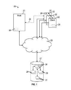
[00099] Figure 1 illustrates an exemplary planning system 20 that may be
utilized for planning surgical procedures. The system 20 may be used for
planning
orthopaedic procedures, including pre-operatively, intra-operatively and/or
post-
operatively to create, edit, execute and/or review surgical plans. The system
20 may
be used for training and rehearsing for various surgical procedures, including
prior
cases and surgical plans for patients.
[000100] The system 20 may include a host computer 21 and one or more client
computers 22. The host computer 21 may be configured to execute one or more
software programs. In some implementations, the host computer 21 is more than
one
computer jointly configured to process software instructions serially or in
parallel.
[mown The host computer 21 may be in communication with one or more
networks such as a network 23 comprised of one or more computing devices. The
network 23 may be a private local area network (LAN), a private wide area
network
(WAN), the Internet, or a mesh network, for example.
[000102] The host computer 21 and each client computer 22 may include one or
more of a computer processor, memory, storage means, network device and input
and/or output devices and/or interfaces. The input devices may include a
keyboard,
mouse, etc. The output device may include a monitor, speakers, printers, etc.
The
memory may, for example, include UVPROM, EEPROM, FLASH, RAM, ROM,
DVD, CD, a hard drive, or other computer readable medium which may store data
and/or other information relating to the features and techniques disclosed
herein. The
host computer 21 and each client computer 22 may be a desktop computer, laptop
computer, smart phone, tablet, or any other computing device. The interface
may
facilitate communication with the other systems and/or components of the
network
23.
[000103] Each client computer 22 may be configured to communicate with the
host computer 21 directly via a direct client interface 24 or over the network
23. The
client computers 22 may be configured to execute one or more software
programs,
such as various surgical tools. Each client computer 22 may be operable to
access and
locally and/or remotely execute a planning environment 26. The planning
environment 26 may be a standalone software package or may be incorporated
into
13
CA 03233492 2024- 3- 28
WO 2023/059877
PCT/US2022/046041
another surgical tool. The planning environment 26 may be configured to
communicate with the host computer 21 either over the network 23 or directly
through the direct client interface 24. In another implementation, the client
computers
22 are configured to communicate with each other directly via a peer-to-peer
interface
25.
[000104] The planning environment 26 may provide a display or visualization of
one or more virtual anatomical models 29 and related images and/or one or more
implant models 30 via one or more graphical user interfaces (GUI). Each
anatomical
model 29, implant model 30, and related images and other information may be
stored
in one or more files or records according to a specified data structure.
[moths] The system 20 may include at least one storage system 27, which may
be operable to store or otherwise provide data to other computing devices. The
storage
system 27 may be a storage area network device (SAN) configured to communicate
with the host computer 21 and/or the client computers 22 over the network 23.
In
implementations, the storage system 27 may be incorporated within or directly
coupled to the host computer 21 and/or client computers 22. The storage system
27
may be configured to store one or more of computer software instructions,
data,
database files, configuration information, etc.
[000106] In implementations, the system 20 may be a client-server architecture
configured to execute computer software on the host computer 21, which may be
accessible by the client computers 22 using either a thin client application
or a web
browser executing on the client computers 22. The host computer 21 may load
the
computer software instructions from local storage, or from the storage system
27, into
memory and may execute the computer software using the one or more computer
processors.
[000107] The system 20 may include one or more databases 28. The databases
28 may be stored at a central location, such as the storage system 27. In
implementations, one or more databases 28 may be stored at the host computer
21
and/or may be a distributed database provided by one or more of the client
computers
22. Each database 28 may be a relational database configured to associate one
or
more anatomical models 29 and/or one or more implant models 30 to each other
and/or a surgical plan 31. Each surgical plan 31 may be associated with a
respective
patient. Each anatomical model 29, implant model 30 and surgical plan 31 may
be
assigned a unique identifier or database entry. The database 28 may be
configured to
14
CA 03233492 2024- 3- 28
WO 2023/059877
PCT/US2022/046041
store data corresponding to the anatomical models 29, implant models 30 and
surgical
plans 31 in one or more database records or entries, and/or may be configured
to link
or otherwise associate one or more files corresponding to each respective
anatomical
model 29, implant model 30 and surgical plan 31. Anatomical models 29 stored
in the
database(s) 28 may correspond to respective patient anatomies from prior
and/or
planned surgical cases, and may be arranged into one or more predefined
categories
such as sex, age, ethnicity, size, defect category, procedure type, etc. The
anatomical
models 29 and/or implant models 30 may be associated with respective
instrumentation and devices to implement the associated surgical plan 31.
[000108] Each anatomical model 29 may include information obtained from one
or more medical devices or tools, such as a computerized tomography (CT),
magnetic
resonance imaging (MR1) machine and/or X-ray machine, that obtains one or more
images of a patient. The anatomical model 29 may include one or more digital
images
and/or coordinate information relating to an anatomy of the patient obtained
or
derived from the medical device(s). In implementations, one or more of the
anatomical models 29 may be created by a designer and may represent a
hypothetical
anatomy. Each implant model 30 may include coordinate information associated
with
a predefined design. The planning environment 26 may incorporate and/or
interface
with one or more modeling packages, such as a computer aided design (CAD)
package, to render the models 29, 30 as two-dimensional (2D) and/or three-
dimensional (3D) volumes or constructs.
[000109] The implant models 30 may correspond to implants and components of
various shapes and sizes. Each implant may include one or more components that
may
be situated at a surgical site including plates, anchors, screws, nails,
suture, grafts, etc.
Each implant model 30 may correspond to a single component or may include two
or
more components that may be configured to establish an assembly. Each
anatomical
model 29 and implant model 30 may correspond to 2D and/or 3D geometry, and may
be utilized to generate a wireframe, mesh and/or solid construct in a display.
[00ono] Each surgical plan 31 may be associated with one or more of the
anatomical models 29 and/or implant models 30. The surgical plan 31 may
include
one or more revisions to the anatomical model 29 and information relating to a
position of an implant model 30 relative to the original and/or revised
anatomical
model 29. The surgical plan 31 may include coordinate information relating to
the
revised anatomical model 29 and a relative position of the implant model 30 in
CA 03233492 2024- 3- 28
WO 2023/059877
PCT/US2022/046041
predefined data structure(s). Revisions to each anatomical model 29, implant
model
30 and surgical plan 31 may be stored in the database 28 automatically and/or
in
response to user interaction with the system 20.
[00orn] One or more surgeons, assistants and other users may be provided with
a planning environment 26 via the client computers 22 and may simultaneously
access
each anatomical model 29, implant model 30 and surgical plan 31 stored in the
database(s) 28. Each user may interact with the planning environment 26 to
create,
view and/or modify various aspects of the surgical plan 31. Each client
computer 22
may be configured to store local instances of the anatomical models 29,
implant
models 30 and/or surgical plans 31, which may be synchronized in real-time or
periodically with the database(s) 28. The planning environment 26 may be a
standalone software package executed on a client computer 22 or may be
provided as
one or more services executed on the host computer 21, for example.
[000112] Figure 2 illustrates a surgical system 120 for planning, rehearsal
and/or
training for a surgical procedure. The system 120 may be utilized to plan,
rehearse,
train and implement various orthopaedic and other surgical procedures, such as
an
arthroplasty to repair a joint. The system 120 may be utilized in planning a
resection
or revision of one or more bones. The system 120 may be utilized in planning
placement of an implant to restore functionality to a joint, such as a
shoulder joint
during an anatomical or reverse shoulder procedure. Although the planning
systems
and methods disclosed herein primarily refer to repair of a shoulder, it
should be
understood that the planning system 120 may be utilized in the repair of other
locations of the patient and other surgical procedures including repair of
other joints
such as an ankle, wrist, hand, hip or knee, and including repair of fractures
and other
tissue such as cartilage, muscles, tendons and ligaments.
[000113] The system 120 may be configured to generate configurations
associated with respective physical anatomical models. The configuration may
be
utilized in the formation of the physical anatomical model. Each physical
anatomical
model may be representative of a virtual anatomical model 129, including a
substantially corresponding geometry, density, porosity, color, etc. The
surgeon may
perform one or more modifications to the physical anatomical model to rehearse
or
train for a surgical procedure.
[000114] The system 120 may include a computing device 132 including at least
one processor 133 coupled to memory 134. The computing device 132 can include
16
CA 03233492 2024- 3- 28
WO 2023/059877
PCT/US2022/046041
any of the computing devices disclosed herein, including the host computer 21
and/or
client computer 22 of Figure 1. The processor 133 may be configured to execute
a
planning environment 126 for creating, editing, executing and/or reviewing one
or
more surgical (e.g., pre-operative) plans 131 during pre-operative, intra-
operative
and/or post-operative phases of a surgery. The anatomical model 129 and
surgical
plan 131 may be associated with an actual case for a patient or may be a
hypothetical
case established for training surgeons, assistants and medical staff.
[000115] The planning environment 126 may include at least a data module 135,
display module 136, spatial module 137 and comparison module 138. The
processor
133 may be configured to execute the data module 135, display module 136,
spatial
module 137 and comparison module 138. Although four modules are shown, it
should
be understood that fewer or more than four modules may be utilized and/or one
or
more of the modules may be combined to provide the disclosed functionality.
[000116] The data module 135 may be configured to access, retrieve and/or
store
data and other information in the database(s) 128 corresponding to one or more
virtual
anatomical model(s) 129, implant model(s) 130 and/or surgical plan(s) 131. The
data
and other information may be stored in the database 128 as one or more records
or
entries 139. In some implementations, the data and other information may be
stored
in one or more files that may be accessible by referencing one or more objects
or
memory locations referenced by the records 139.
[000117] The memory 134 may be configured to access, load, edit and/or store
instances of one or more anatomical models 129, implant models 130 and/or
surgical
plans 131 in response to one or more commands from the data module 135. The
data
module 135 may be configured to cause the memory 134 to store a local instance
of
the anatomical model(s) 129, implant model(s) 130 and/or surgical plan(s) 131
which
may be synchronized with records 139 in the database(s) 128.
i000118i The display module 136 may be configured to display data and other
information relating to one or more surgical plans 131 in at least one
graphical user
interface (GUI) 142. The computing device 132 may be coupled to a display
device
140. The display module 136 may be configured to cause the display device 140
to
display information in the user interface 142. A surgeon or other user may
interact
with the user interface 142 via the planning environment 126 to create, edit
and/or
review aspects of one or more anatomical models 129. The surgeon or other user
may
17
CA 03233492 2024- 3- 28
WO 2023/059877
PCT/US2022/046041
interact with the user interface 142 via the planning environment 126 to
create, edit,
execute and/or review aspects of one or more surgical plans 131.
[000119] Each surgical plan 131 may be associated with one or more (e.g.,
original) virtual anatomical models 129 prior to any revisions, which may
substantially approximate a patient anatomy. Each surgical plan 131 may be
associated with one or more (e.g., revised) virtual anatomical models 129
incorporating one or more revisions to the patient anatomy and/or physical
anatomical
model. The original and revised anatomical models 129 may be associated with
each
other in the surgical plan 131. In implementations, the revisions are stored
as one or
more parameters of the original anatomical model 129.
[000120] The planning system 120 may be configured to generate a link to a
surgical plan 131. The surgeon, assistant or other user may interact with the
link to
review and edit the surgical plan 131. Interacting with the link may cause the
planning
system 120 to display or otherwise present aspects of the surgical plan 131 in
the
graphical user interface 142.
[000121] Referring to Figure 3, with continuing reference to Figure 2, the
user
interface 142 may include one or more display windows 144 and one or more
objects
146. The objects 146 may include graphics such as menus, tabs and buttons
accessible
by user interaction, such as tabs 146T, buttons 146B, drop-down lists 146L,
menus
146M, entry fields 146E (e.g., Figure 7), directional indicators 146D, 146R
(e.g.,
Figure 5) and graphics 146G (e.g., Figure 7). Geometric objects, including
selected
virtual anatomical model(s) 129 and implant model(s) 130 (e.g., Figure 6), and
other
information relating to the surgical plan 131 may be displayed in one or more
of the
display windows 144.
[000122] The data module 135 may be configured to access a virtual anatomical
model 129 from memory, such as the memory 134 and/or database 128, in response
to
selecting one or more parameters in a display window 144-1 of the graphical
user
interface 142. The anatomical model 129 may be associated with an anatomy of a
patient, such as a prior case or a planned case. The display module 136 may be
configured to select an anatomical model 129 from memory, such as the database
128
or memory 134, in response to user interaction with the display window 144-1
or
another portion of the user interface 142.
[000123] Various parameters may be utilized to select an anatomical model 129.
The parameters of the display window 144-1 may be interconnected to provide a
18
CA 03233492 2024- 3- 28
WO 2023/059877
PCT/US2022/046041
filtering feature such that each selection of a parameter may cause the
remaining
parameter(s) to be filtered to depict available options. Each parameter may be
associated with a set of anatomical models 129 accessible by the planning
environment 126.
[000124] The surgeon may interact with the user interface 142 to select and
review a desired case, such as a prior, planned or hypothetical case
associated with a
surgical plan 131. The surgeon may interact with the user interface 142 to
review
prior cases, including prior cases for a particular surgical procedure,
anatomy and/or
group of patients. The planning system 120 may be configured to provide
analysis of
the prior case such as biometric testing of a repaired joint, finite element
analysis
(FEA), etc. The surgeon may select a virtual anatomical model 129
corresponding to
an intended patient. The selected model 129 may correspond to an acquired CT
scan
of the patient. The surgeon may select a virtual anatomical model 129 that may
be
associated with a particular classification.
[000125] The anatomical models 129 may be categorized by anatomy, patient,
defect, case, etc. The parameters may include a patient classification and a
defect
category. Anatomical parameters may be arranged in one or more lists 146L by
category (e.g., joint, etc.), sub-category (e.g., shoulder, ankle, hip, etc.),
model (e.g.,
glenoid, humerus, etc.) and anatomical size (e.g., small, medium, large). The
categories may be subdivided by gross anatomy including surface anatomy (e.g.,
the
external body), regional anatomy (e.g., specific regions of the body), and
systemic
anatomy (e.g., specific organ systems). Patient parameters may include sex,
age and
ethnicity. The spatial module 137 may be configured to scale a geometry of the
selected anatomical model 129 in response to selection of an anatomical size.
[000126] The surgeon may select the virtual anatomical model 129 according to
a severity of various defects, such as mild, severe, non-pathological,
fractures, etc.
Defect parameters may be established for the various defects and may be
arranged by
classification, subclassification, etc. The surgeon, assistant or other user
may interact
with a button 146B (see, e.g., question mark button 146Q) for an explanation
of the
defect parameters, as illustrated by the display window 144-2 of Figure 4. For
example, selection of a glenoid model from the list 146L may cause a help
screen to
be generated and displayed with one or more defect classifications in response
to
selection of the button 146Q. As illustrated in Figure 4, the defect
classifications may
include osteoarthritis including osteophytes associated with lateralization
(see, e.g.,
19
CA 03233492 2024- 3- 28
WO 2023/059877
PCT/US2022/046041
Samilson and Prieto), central glenoid wear (see, e.g., Levigne), inferior
glenoid wear
and lateralization (see, e.g., Habermeyer), rheumatoid arthritis including
superior
wear and lateralization (see, e.g., Levigne), glenoid wear including superior
wear (see,
e.g., Favard), glenoid version (see, e.g., Walch), subluxation (see, e.g.,
Walch) and
glenoid morphology including superior/inferior classification (see, e.g.,
Walch). The
defect classifications of Figure 4 are known, but utilization of the defect
classifications as disclosed herein is not known. For example, the surgeon may
select
a relatively rare case such as a B2 or C2 classification of a glenoid based on
information displayed in the help screen. The user may interact with a button
146B or
another portion of the user interface 142 to specify more than one defect. The
surgeon
may select from the various classifications to determine various treatment
options.
[000127] Case parameters may include case type (e.g., prior, planned and
hypothetical), case number, etc. The user may interact with the lists 146L to
select a
particular case, which may be filtered based on previous selections of the
parameters.
Each case may be associated with a respective surgical plan 131. The surgical
plan
131 may be associated with an anatomical model 129 prior to any revisions and
may
be associated with another (e.g., revised) anatomical model 129 incorporating
one or
more revisions based on implementation of an associated surgical procedure.
Exemplary revisions may include removal of material utilizing one or more
drilling,
milling, resection, reaming and cutting operations.
r0001281 Referring to Figure 5, with continuing reference to Figures 2-3, the
surgeon, assistant or other user may interact with the user interface 142 to
view the
selected anatomical model(s) 129 in display windows 144-2, 144-3 of the user
interface 142. The data module 135 may be configured to cause the display
module
136 to display the selected anatomical model(s) 129 in the user interface 142
in
response to selection of the parameter(s). In the implementation of Figure 5,
the
selected anatomical model 129 may be associated with a glenoid 129G of a
shoulder
joint 129S, although it should be understood that the system 120 may be
utilized with
various anatomy.
[000129] The display module 136 may be configured to display the selected
anatomical model 129 at different positions and/or orientations in the display
windows 144-2, 144-3. Although a particular number of display windows 144 are
illustrated, it should be understood that the user interface 142 may be
configured with
any number of display windows 144 in accordance with the teachings disclosed
CA 03233492 2024- 3- 28
WO 2023/059877
PCT/US2022/046041
herein. The display windows 144-2, 144-3 may be configured to display a two-
dimensional (2D) and/or three-dimensional (3D) representation of the selected
anatomical model 129. The user may interact with the user interface 142 to
view the
selected anatomical model(s) 129 at the various positions and orientations.
[000130] Referring to Figure 6, with continuing reference to Figures 2 and 5,
the
surgeon, assistant or other user may interact with the user interface 142 to
customize
or otherwise specify one or more parameters associated with the selected
anatomical
model 129. The user may interact with the menus 146M, directly with display
windows 144-4, 144-5, or with another portion of the user interface 142 to
specify one
or more aspects of, or modifications to, the surgical plan 131.
[000131] The user may interact with menu 146M or another portion of the user
interface 142 to select one or more implant models 130 from a set of implant
models
130 stored in memory such as the database 128 or memory 134. The data module
135
may be configured to access the selected implant model 130 automatically or in
response to user interaction with the user interface 142. The data module 135
may be
configured to store an instance of the virtual anatomical model and implant
model 130
in the memory 134.
[000132] The implant models 130 may be associated with implants of various
configurations, shapes, sizes, procedures, instrumentation, etc. The implant
model 130
may include one or more components. Exemplary implants may include base plates
coupled to an articulation member, bone plates configured to interconnect
adjacent
bones or bone fragments, intermedullary nails, suture anchors, etc. The
articulation
member may have an articular surface dimensioned to mate with an articular
surface
of an opposed bone or implant.
[000133] The display module 136 may be configured to display one or more
selected virtual anatomical models 129 and/or implant models 130 in the
display
windows 144. The display module 136 may be configured such that the selected
anatomical model 129 and/or implant model 130 may be selectively displayed and
hidden (e.g., toggled) in one or more of the display windows 144 in response
to user
interaction with the user interface 142, which may provide the surgeon with
enhanced
flexibility in reviewing aspects of the surgical plan 131.
[000134] The display windows 144-4, 144-5 may be configured to display the
selected anatomical model 129 and implant model 130 relative to each other.
The
spatial module 137 may be configured to position the selected implant model(s)
130
21
CA 03233492 2024- 3- 28
WO 2023/059877
PCT/US2022/046041
into contact with the anatomical model(s) 129 at a specified or defined
position and
orientation. The implant model 130 may be positioned relative to a surface of
the
anatomical model 129, such as an articulation surface 129AS which may be
associated with an articular surface of a bone. The user may interact with the
menus
146M, directly with the display windows 144-4, 144-5, or with another portion
of the
user interface 142 to position and orient the selected implant model 130
relative to the
anatomical model 129, including an implant axis IA of the implant model 130.
The
user may interact with the menu 146M, the display windows 144-4, 144-5 or
another
portion of the user interface 142 to move the selected anatomical model 129
and/or
selected implant model 130 in 2D space (e.g., up, down, left, right) and/or 3D
space
(e.g., rotation, tilt, zoom, etc.), which may occur in response to interaction
with the
directional indicators 146D, 146R.
[000135] The display module 136 may be configured to display one or more
fastener models F (shown in dashed lines). The fastener models F may be
associated
with respective fasteners configured to secure an implant associated with the
selected
implant model 130.
[000136] Referring to Figure 7, with continuing reference to Figures 2 and 5-
6,
the surgeon, assistant or other user may interact with the user interface 142
to
customize the selected anatomical model(s) 129. The display windows 144 may
include display windows 144-6, 144-7. The user may interact with the menu 146M
to
toggle on and off visibility of the selected implant model 130.
[000137] The surgeon may desire to specify one or more zones (e.g., regions)
associated with a surgical procedure. The zones may be formed in an associated
physical anatomical model to provide feedback to the surgeon. In
implementations,
the feedback may include one or more visual, audible and/or tactile indicators
in
response to interaction.
[000138] The spatial module 137 may be configured to establish one or more
target zones (e.g., localized regions) 148. The user may interact with the
menus
146M, display windows 144-6, 144-7 or another portion of the user interface
142 to
establish the target zone(s) 148. Each target zone 148 may be established
along any
portion of the virtual anatomical model 129. In implementations, the target
zone 148
may extend along the articulation surface 129AS of the anatomical model 129.
[000139] The user may interact with the user interface 142 to specify various
aspects of the target zone 148. The target zone 148 may have various 3D
geometries,
22
CA 03233492 2024- 3- 28
WO 2023/059877
PCT/US2022/046041
including cones, cylinders, cubes and cuboids, prisms, pyramids, spheres,
complex
geometries, etc. The spatial module 137 may be configured to assign a default
geometry of the target zone 148 based on the selected anatomical model 129,
implant
model 130, surgical plan 131 and/or other parameters selected by the user
(see, e.g.,
Figure 3). Each target zone 148 may be dimensioned to approximate a portion of
a
volume of the selected implant model 130. The target zone 148 may be
dimensioned
for positioning a fixation member such as an anchor or post of the selected
implant
model 130 (see, e.g., Figure 6). Each target zone 148 may be associated with a
respective graphic 146G. The graphic 146G may have a geometry that
substantially
corresponds to a geometry of the target zone 148.
[000140] The user may interact with the user interface 142 to specify a
position
and orientation of a target axis TA associated with the target zone 148. The
spatial
module 137 may be configured to set a position and orientation of the target
axis TA
to be substantially collinear with or otherwise substantially parallel to an
implant axis
IA of the implant model 130 (Figure 6). For the purposes of this disclosure,
the terms
"substantially," "about" and "approximately" mean - 5 percent of the stated
value or
relationship unless otherwise indicated. The user may interact with the menu
146M to
specify one or more dimensions of the target zone 148, including width,
length, depth,
radius, etc., based on a geometry of the target zone 148.
[0001411 Referring to Figure 8, with continuing reference to Figures 2 and 7,
the
spatial module 137 may be configured to establish one or more warning zones
150.
The warning zones 150 may be generated manually or automatically in response
to
establishing the respective target zone(s) 148. The warning zones 150 may be
established according to the selected parameters, including the selected
anatomy,
procedure type, defect classification(s), case, etc. The warning zones 150 may
be
procedure specific and/or pathological specific. The display module 136 may be
configured to display the warning zones 150 relative to the selected
anatomical model
129 in a display window 144-8. The spatial module 137 may be configured to
establish a third zone 154. The third zone 154 may include a remainder of a
volume of
the anatomical model 129 excluding the target zone(s) 148 and warning zone(s)
150.
[000142] The target zone(s), warning zone(s) 150 and/or third zone 154 may be
established by a main body 129B of the virtual anatomical model 129_ The
warning
zones 150 may be established along an external surface of the anatomical model
129
and/or within a thickness of the anatomical model 129 underlying the external
23
CA 03233492 2024- 3- 28
WO 2023/059877
PCT/US2022/046041
surface, such as the articulation surface 129AS. The surgeon, assistant or
other user
may interact with the user interface 142 to specify whether or not the warning
zones
150 are visible along an external surface of the anatomical model 129. In the
implementation of Figure 8, the warning zones 150 may include three warning
zones
150-1 to 150-3 that may extend inwardly from the articulation surface 129AS.
The
warning zones 150-1 to 150-3 may serve to provide the surgeon a visual
indication of
a proximity to the target zone 148. External warning zones may present less of
a
challenge to the surgeon. The surgeon may select to have warning zones only
formed
below the external surface of the anatomical model, which may more closely
approximately a surgical procedure on the anatomy and may be relatively more
challenging_
[000143] The planning environment 126 may be configured such that at least
one or more parameters associated with the warning zones 150 may be set in
response
to user interaction with the user interface 142. The user may interact with
the menu
146M, directly with the display window 144-8, or with another portion of the
user
interface 142 to define each of the warning zones 150. Various parameters
associated
with warning zones 150 may be utilized, including quantity, offset (e.g.,
thickness),
etc.
[000144] The warning zones 150 may correspond to a plurality of layers 152 in
stacked relationship such that the warning zones 150 are offset at different
predefined
distances from the target zone 148. In implementations, the warning zones 150
and
respective layers 152 may be established at increasing offsets from a
perimeter of the
target zone 148. The thicknesses of the warning zones 150 may be the same or
may
differ. In the implementation of Figure 8, a quantity of three warning zones
150 may
be established (indicated at 150-1, 150-2, 150-3) adjacent to the target zone
148.
Although three warning zones 150 are shown, it should be understood that fewer
or
more than three warning zones 150 may be established for each target zone 148,
such
as only one warning zone 150. The warning zones 150 and respective layers 152
may
substantially encircle or otherwise bound the target zone 148 to establish a
boundary
for performing a surgical procedure.
[000145] The warning zones 150 may visually contrast with each other and/or
the target zone 148 to provide the surgeon with an indication of an amount of
deviation from the target zone 148. For example, the surgeon may perform a
drilling
operation that may extend through the target zone 148 and into one or more of
the
24
CA 03233492 2024- 3- 28
WO 2023/059877
PCT/US2022/046041
warning zones 150. The warning zones 150 may serve to indication an amount of
excess depth of the drilling operation. The display module 136 may be
configured to
display a visual contrast between the warning zones 150 and a remainder of the
anatomical model 129, including the target zone(s) 148 and third zone 154. In
implementations, the visual contrast may be established by one or more visual
indicators VI applied to the respective target zone 148, warning zones 150
and/or
third zone 154 (shown in hatching for illustrative purposes). In
implementations, the
target zone(s) 148 and/or third zone(s) 154 may omit any visual indicators VI
(see,
e.g., Figure 9).
[0001461 The target zone(s) 148, warning zones 150 and third zone 154 may be
established according to one or more visual or color schemes. In
implementations, the
target zone 148 and/or third zone 154 may be assigned a color that corresponds
to a
natural color of a respective portion of the anatomy. The warning zones 150
may be
assigned one or more artificial colors to establish a visual contrast. The
visual contrast
may assist the surgeon in identifying the target zone 148 and any deviations
from the
target zone 148 into the warning zones 150. For the purposes of this
disclosure, the
term "natural" color means a color that substantially corresponds to an
expected or
actual color of the respective tissue, and the term "artificial" color means a
color that
does not naturally occur for the respective tissue.
[0001471 Each warning zone 150 may be assigned a respective artificial color
that may differ from a natural color of a respective portion of the anatomy
and that
may establish a visual contrast with the natural color associated with the
target zone
148. The artificial colors of the warning zones 150 and associated layers 152
may be
the same or may differ from each other. The respective colors of the warning
zones
150-1 to 150-3 are shown in hatching for illustrative purposes. Exemplary
artificial
colors may include yellow, orange, red, green, blue, etc. In other
implementations,
the warning zones 150 may be assigned a color that substantially corresponds
to a
natural color of a respective portion of the anatomy, but that may have a
shade that
differs by at least 10 percent or more from the natural color to establish a
visual
contrast.
[000148] The target zone(s) 148 and warning zone(s) 150 may have other
configurations. In the implementation of Figure 9, the virtual anatomical
model 229
may have a main body 229B. A target zone 248 and one or more warning zones 250
may be established in the main body 229B. The target zone 248 may have a
CA 03233492 2024- 3- 28
WO 2023/059877
PCT/US2022/046041
substantially truncated conical geometry having a first base B1 and a second
base B2.
The first base B1 may have a radius that differs from (e.g., is less than) a
radius of the
second base B2. The first base B1 may establish an entry point EP along an
external
surface of the main body 229B. The entry point EP may be substantially aligned
with
a target axis TA. The external surface may be established by an articulation
surface
229AS of the selected anatomical model 229. The target axis TA may be
associated
with a suitable trajectory of a portion of an implant and/or fastener. The
entry point
EP may be representative of safe passage for a surgical device through a
portion of
the tissue. The entry point EP may be dimensioned to receive a fastener model
F
(shown in dashed lines). Walls of the target zone 248 may be sloped to limit a
range
of angles of the fastener model F relative to the target axis TA. The warning
zones
250 may extend along the second base B2 to bound a height of the target zone
248
and an insertion depth of the fastener model F.
[000149] In the implementation of Figures 10-11, a volume of target zone 348
may have a substantially cuboid geometry with a substantially rectangular
perimeter.
Target zone 348 and warning zones 350 may be spaced apart from an external
surface
of a main body 329B of the anatomical model 329, which may be established by
an
articulation surface 329AS. The warning zones 350 may be configured to
substantially surround the target zone 348 as a set of concentric loops.
[0001501 In the implementation of Figure 12, the target zone(s) 448 and
warning
zone(s) 450 may be lateralized relative to an external surface of a virtual
anatomical
model 429. A graphical user interface 442 may include display windows 444-8,
444-
9. The display window 444-8 may be configured to display a sectional view of
the
anatomical model 429 relative to a reference plane REF1 (shown in dashed
lines). The
display window 444-9 may be configured to display a perspective view of the
anatomical model 429 sectioned along the reference plane REF1.
[000151] Target zone(s) 448, warning zone(s) 450 and/or third zone(s) 454 may
be established in a volume of a main body 429B of the anatomical model 429.
The
target zone 448 may be established along, and may extend inwardly from, an
external
surface of the main body 429B. The external surface may be an articular
surface
429AS associated with a joint of the respective anatomy.
[000152] The warning zones 450 may be lateralized and may correspond to
various reaming depths. The warning zones 450 may indicate that an implemented
reaming depth may be insufficient and/or excessive. The warning zones 450 may
26
CA 03233492 2024- 3- 28
WO 2023/059877
PCT/US2022/046041
include warning zones 450-1 to 450-3 established by respective layers 452. The
warning zones 450 may be established below the external surface of the main
body
429B. The warning zones 450 and respective layers 452 may be offset at
different
depths from the target zone 448 and/or external surface of the main body 429B.
[000153] The target zone 448 and warning zones 450 may be representative of
bone tissue associated with the anatomy. The target zone 448 may be
representative of
cortical bone associated with the anatomy, such as cortical bone along an
articular
surface of a glenoid. The third zone 454 may establish a non-cortical region
representative of cancellous or trabecular bone associated with the anatomy.
The
target zone 448 and third zone 454 may be assigned a color that closely
approximates
the cortical and trabecular bone of an anatomy of a patient, respectively. The
layers
452 may be arranged such that the warning zones 450 are established between
the
target zone 448 and third zone 454. In implementations, the warning zones 450-
1 to
450-3 may correspond to depths of 1 mm, 2 mm and 3 mm below the target zone
448.
[000154] In the implementation of Figure 13, a target zone 548 may be
established between one or more warning zones 550. The warning zones 550 may
be
formed by respective layers 552. The layers 552 may be substantially parallel
and
may have substantially the same thicknesses, or the thicknesses may differ.
The layers
552 may include a first set of layers 552-1 and a second set of layers 552-2.
The first
set of layers 552-1 may establish warning zones 550-1 to 550-3. The second set
of
layers 552-2 may establish warning zones 550-4 to 550-6. The target zone 548
may be
established between the first and second sets of layers 552-1, 552-2. The
target zone
548 may be associated with an intended resection plane that may be engaged
with a
reciprocating or oscillating saw to form a cut along a long bone such as a
proximal
portion of a tibia or a distal portion of a femur, for example.
[000155] The physical anatomical model may include other representative tissue
incorporating warning zones. Certain procedures may include navigating into
and
through 3D muscular intervals. The procedure may including navigating through
and
in between the layers without violating the muscular structure (e.g.,
separating
without cutting). For example, a surgeon planning for a hip arthroplasty may
desire to
minimize or otherwise reduce injury to the soft tissue. The warning zones may
confine a particular region of a physical anatomical model of the soft tissue
so that the
surgeon may be required to open up the representative soft tissue without
ripping the
representative soft tissue. If the surgeon cuts through the representative
soft tissue, the
27
CA 03233492 2024- 3- 28
WO 2023/059877
PCT/US2022/046041
warning zone may be presented to the surgeon. The surgeon may excise the
representative soft tissue along natural boundaries to avoid the warning
zones. The
warning zones may not be visible on the external surface of the representative
soft
tissue. If surgeon deviates to a side of the zone, the warning zone may be
revealed.
[000156] Referring to Figure 14, with continuing reference to Figure 2, the
virtual anatomical model 629 may include bone and other tissue, including soft
tissue.
The soft tissue may include tendon, ligament and/or muscle tissue including
one or
more fibers. The user interface 642 may include display windows 644-10, 644-
configured to display the anatomical model 629. The display window 644-11 may
be
configured to display a section of the anatomical model 629 with respect to a
reference plane REF2 (shown in dashed lines).
[000157] The spatial module 137 may be configured to establish one or more
target zones 648 and warning zone 650. The target zone(s) 648 and warning
zone(s)
650 may be representative of soft tissue associated with the anatomy. A third
zone
654 may be established, which may be representative of relatively hard tissue
such as
cartilage and/or bone tissue.
[000158] The anatomical model 629 may include one or more fibers 656
associated with fibers of the anatomy. A bundle of fibers 656 may be formed
along,
may extend from, or may be arranged adjacent to a main body 629B. The bundle
of
fibers 656 may be representative of the soft tissue, such as muscle tissue.
The target
zones 648 and warning zones 650 may cooperate to establish the bundle of
fibers 656.
The display module 136 may be configured to display the fibers 656 in plan and
sectional views, as illustrated in the display windows 644-10, 644-11,
respectively.
One or more target zones 648 may be established in the fibers 656 and/or
between
adjacent fibers 656.
[000159] Each of the fibers 656 may include an internal core 656C and an outer
sheath 656S. The sheath 656S may substantially surround the core 656C. One or
more
target zones 648 may be established between adjacent fibers 656. The target
zones
648 may be representative of a pathway between the adjacent fibers 656. The
core
656C may establish a respective one of the warning zones 650. The sheath 656S
may
substantially surround at least one warning zone 650 of the respective fiber
656. In
implementations, sheaths 656S of the adjacent fibers 656 may establish the
respective
target zone 648.
28
CA 03233492 2024- 3- 28
WO 2023/059877
PCT/US2022/046041
[000160] The external surface established by the sheath 656S of each fiber 656
may be assigned a color substantially corresponding to a natural color of the
respective tissue. The core 656C may be assigned an artificial color that may
differ
from a natural color of the respective tissue such that the target zone(s) 648
and
warning zone(s) 650 visually contrast each other.
[000161] In the implementation of Figure 15, a bundle of fibers 756 may
include
first, second and third set of fibers 756-1, 756-2, 756-3. The first set of
fibers 756-1
may establish a target zone 748. Each fiber 756 of the second and third sets
of fibers
756-2, 756-2 may establish a respective warning zone 750. Individual target
zones
748 may be established by respective fibers 756. Individual warning zones 750
may
be established by a core and/or sheath of the respective fiber 756. The second
and
third sets of fibers 756-2, 756-3 may be established on opposed sides of the
first set of
fibers 756-1 such that the first set of fibers 756-1 establishes a localized
region or
pathway.
[000162] In the implementation of Figure 16, a graphical user interface 842
may
include a display window 844-12 configured to display an anatomical model 829.
The anatomical model 829 may be associated with one or more tissue types,
including
bone, cartilage, tendons, ligaments, muscle tissue, etc. The anatomical model
829 may
include bundles of fibers 856 that establish one or more muscle groups 858.
The
muscle groups 858 may be associated with one or more target zones 848 and/or
warning zones 850. In the implementation of Figure 16, the anatomical model
829
may be representative of a shoulder joint including one or more of the muscle
groups
858.
[000163] The surgeon, assistant or other user may interact with menus 846M,
directly with the display window 844-12 or another portion of the user
interface 842
to set each of the muscle groups 858 as a target zone 848 or warning zone 850.
In the
implementation of Figure 16, the target zone 848 may be associated with a
lateral
head of a deltoid, warning zone 850-1 may be associated with a posterior head
of the
deltoid, and warning zone 850-2 may be associated with an anterior head of the
deltoid. In another implementation, a portion of a virtual anatomical model
associated
with a deltoid may be selected as a first warning zone assigned a first color
(e.g., red),
a teres minor may be selected as a second warning zone assigned a second color
(e.g.,
blue), and a region in between the deltoid and teres minor may be selected as
a target
zone assigned a third color (e.g., green). Utilizing the techniques disclosed
herein, the
29
CA 03233492 2024- 3- 28
WO 2023/059877
PCT/US2022/046041
surgeon may interact with a physical anatomical model formed from a
construction
defined by the virtual anatomical model 829 as a training tool to help the
surgeon
navigate or guide instruments through the muscle during a shoulder
arthroplasty.
[000164] Referring back to Figure 2, the surgeon, assistant or other user may
interact with the user interface 142 to approve selections of the various
parameters
associated with the selected virtual anatomical model 129. In implementations,
the
user may select a button 146B to approve the selections and other parameters
associated with the virtual anatomical model 129 (see, e.g., Figure 8).
[000165] The spatial module 137 may be configured to generate a configuration
(e.g., definition) associated with a physical anatomical model. The
configuration may
be representative of the virtual anatomical model 129. The configuration may
be
established according to the selection of the parameters associated with the
selected
virtual anatomical model 129, including any target zones 148 and warning zones
150
(see, e.g., Figure 8). The configuration may be a file stored in, or linked
to, a record
139 in the database 128. The configuration may include data and other
information
sufficient to establish the physical anatomical model based on a geometry and
the
parameters of the selected virtual anatomical model 129, including coordinate
information, moduli of elasticity of the associated tissues, color schemes
associated
with target zones 148 and warning zones 150, etc.
[000166] The physical anatomical model may be formed subsequent to selecting
and approving the virtual anatomical model 129 and the various parameters. The
physical anatomical model may be representative of various tissue including
bone
tissue and soft tissue. The physical anatomical model may have a construction
that is
representative of anatomy, including joints, bones, muscle, tendons,
ligaments,
internal organs, etc. The physical anatomical model may be configured to
provide
indicators or feedback to the surgeon prior to, during and/or subsequent to
execution
of a surgical procedure. The feedback may be patient or non-patient specific.
In
implementations, the planning system 120 may be configured to provide verbal
or
visual feedback such as a prompt for the surgeon to aim for a specific area of
bone or
soft tissue.
[000167] Various techniques may be utilized to construct or otherwise form the
physical anatomical models disclosed herein, including the physical anatomical
model
960. The physical anatomical models may be constructed or otherwise formed
utilizing any of the techniques disclosed herein, and may incorporate any of
the
CA 03233492 2024- 3- 28
WO 2023/059877
PCT/US2022/046041
features disclosed herein including a geometry of the respective virtual
anatomical
model(s), moduli of elasticity of the associated tissue(s), color scheme(s)
and
geometries associated with the target zone(s) and/or warning zones, layer and
fiber
arrangements, etc. The physical anatomical model may be formed to closely
resemble
or approximate a geometry of the associated anatomy, including soft tissue and
bone.
In implementations, the surgeon may interact with the physical anatomical
model
such that portion(s) of the physical anatomical model may feel similar to soft
(e.g.,
cancellous) bone tissue. The physical anatomical model may be printed or
otherwise
formed according to the various parameters selected in the user interface.
Various
parameters may be utilized to form the physical anatomical model, including
any of
the parameters disclosed herein, such as density of bone and soft tissue,
thickness of
cortical bone, target and warning zones, patient age, etc.
[000168] The planning system 120 may configured to cause the virtual
anatomical model 129 to be printed or otherwise formed based on the selected
parameters. In implementations, material may be printed in one or more layers
on a
substrate to establish the physical anatomical model. The material may be
printed
according to the configuration associated with the virtual anatomical model.
In other
implementations, the physical anatomical model is established by one or more
casting
operations. The physical anatomical model may have a unitary construction or
may
have two or more components fixedly attached or otherwise secured to each
other to
establish a unit.
[000169] Referring to Figure 17A, with continuing reference to Figure 2, an
implementation of a physical anatomical model 960 is shown. Figure 17B is a
grayscale image of the physical anatomical model 960. The physical anatomical
model 960 may be associated with any of the anatomy disclosed herein.
[000170] The anatomical model 960 may include portions 962, 964, 966. The
portion 962 may establish a main body 960B and may be representative of bone
tissue, such as a portion of a humerus including the humeral head. The
portions 964
may be formed on, or may otherwise extend from, the portion 962 and may be
representative of soft tissue such as one or more tendons. The tendons may be
associated with a rotator cuff. The portions 966 may extend from the
respective
portions 964 and may be representative of muscle tissue.
[000171] Various materials may be utilized to form the physical anatomical
models disclosed herein, including the physical anatomical model 960. In
31
CA 03233492 2024- 3- 28
WO 2023/059877
PCT/US2022/046041
implementations, the portion 962 establishing the main body 960B may be formed
from a substantially rigid material, such as a polymeric material, including
photopolymers, silicones and thermoplastics. Each of the fibers of the
portions 964,
966 may be formed from a relatively flexible material, including an
elastomeric
material such as rubber or silicone.
[000172] Referring to Figures 18-19, with continuing reference to Figure 17A,
the physical anatomical model 960 may be incorporated into an assembly (e.g.,
training device) 968. The assembly 968 may include one or more fixtures (e.g.,
jigs)
970. Each fixture 970 may be reusable or may be single use. Each fixture 970
may be
representative of surrounding tissue or a portion of a joint. A first fixture
970-1 may
be representative of a portion of a humerus. A second fixture 970-2 may be
representative of a portion of a shoulder such as a glenoid or an implant
having an
articular surface dimensioned to establish a joint. The physical anatomical
model 960
may be mounted or otherwise secured to one or more of the fixtures 970 at an
interface 972. The interface 972 may be established by an interference fit, a
quick
release connection or fasteners. The surgeon may utilize the fixtures 970 to
simulate
rotating or moving a limb in an operating room. The assembly 968 may include a
fixture 970-3 representative of skin tissue, as illustrated in Figure 19. The
fixture 970-
3 may be formed from a relatively flexible material, such as an elastomeric
material.
The surgeon may form one or more openings 974 in the fixture 970-3 to simulate
performing an incision to expose a joint or another portion of the anatomy.
[000173] Referring to Figures 20-21, with continuing reference to Figure 17,
the
surgeon may perform one or more modifications to the physical anatomical model
960, as illustrated by physical anatomical model 1060. The surgeon may perform
a
resection along a reference plane REF3, which may simulate resecting a portion
of an
articulation surface 1060AS, which may be associated with a humeral head
(shown in
dashed lines in Figure 19). The resection may expose a first region R1 and a
second
region R2 (Figure 20). The first region R1 may be substantially solid. The
second
region R2 may be relatively porous. The first region R1 may be representative
of
cortical bone. The second region R2 may be representative of cancellous bone.
[000174] Figures 22-23 are grayscale images of another physical anatomical
model including portions corresponding to different tissue types. The physical
anatomical model may be representative of a shoulder joint including a portion
of a
humerus and shoulder, tendons of a rotator cuff, and muscle tissue.
32
CA 03233492 2024- 3- 28
WO 2023/059877
PCT/US2022/046041
[000175] Figures 24-25 illustrate another physical anatomical model 1160. The
anatomical model 1160 may be established according to a configuration
generated by
the system 120 (Figure 2), such as a configuration associated with the virtual
anatomical model 429 and target and warning zones 448, 450 (Figure 12). The
anatomical model 1160 may include a main body 1160B establishing the target
zone
1148 (Figure 24) and one or more warning zones 1150 associated with the
virtual
anatomical model 429. The target zone 1148 and warning zones 1150 may
cooperate
to establish a construction representative of an anatomy. In implementations,
the
construction may be representative of a shoulder including a glenoid. The
target zone
1148, warning zones 1150 and third zone 1154 (Figure 25) may be established by
the
configuration associated with the virtual anatomical model 429.
[000176] The warning zones 1150 may be established adjacent to the target zone
1148. The warning zones 1150 and third zone 1154 may cooperate to bound the
target
zone 1148 in the physical anatomical model 1160. The target zone 1148 may
correspond to the target zone 448. The warning zones 1150 may correspond to
the
respective warning zones 450. The third zone 1154 may correspond to the third
zone
454. The configuration may be utilized to form target zone(s) 1148 assigned
respective color(s) corresponding to natural color(s) of respective portion(s)
of the
anatomy. The configuration may be utilized to form warning zone(s) 1150
assigned
respective artificial color(s) that establish a visual contrast with the
natural color(s)
associated with the target zone(s) 1148. Each warning zone 1150 may have a
respective artificial color that may differ from a natural color of a
respective portion
of the anatomy.
[000177] The surgeon may perform one or more modifications to the physical
anatomical model 1160. The surgeon may perform a reaming operation at various
angles or orientations (different orientations illustrated in dashed lines in
Figures 24-
25) relative to an articulation surface 1160AS or another external surface of
the
anatomical model 1160. The articulation surface 1160AS may be representative
of an
articular surface of a bone, such as a glenoid face. The reaming operation may
expose
or reveal the target zone 1148 and/or one or more of the warning zones 1150.
The
target zone 1148 and warning zones 1150 may be formed of material having
different
moduli of elasticity such that the surgeon may he required to apply different
amounts
of pressure to ream through the material. In implementations, the target zone
1148
may have a modulus of elasticity that may be greater than a modulus of
elasticity of
33
CA 03233492 2024- 3- 28
WO 2023/059877
PCT/US2022/046041
one or more of the warning zones 1150. The moduli of elasticity of the warning
zones
1150 may progressively decrease with respect to an offset of the respective
warning
zone 1150 from the target zone 1148.
[000178] Figures 26-27 are grayscale images of a physical anatomical model.
The physical anatomical model may be representative of a shoulder sectioned
along a
glenoid. The sectioning may expose material representative of cortical and
cancellous
regions of bone. The material may include a warning zone established about a
perimeter of the cancellous region.
[000179] Figure 28 illustrates another assembly (e.g., training device) 1268.
The
assembly 1268 may incorporate a physical anatomical model 1260. The physical
anatomical model 1260 may include a main body 1260B representative of an
anatomy, such as a bone or joint.
[000mo] The physical anatomical model 1260 may be configured to provide
feedback associated with adjacent tissue including bone and nerve tissue. For
older
patients, over tensioning may occur when a joint is moved. The physical
anatomical
model 1260 may be configured to generate a snapping noise in response to
interaction. In implementations, a particular portion of the physical
anatomical model
1260 that may be representative bone may be configured to snap. This may occur
for
instance when the surgeon attempts to move the representative joint over a
particular
range of motion. For example, 8 mm of lateralization may be suitable for
younger
patients, but may not be suitable for older patients. During a shoulder
repair, the
surgeon may consider an axial nerve that extends about a humerus. The patient
may
experience numbness due to over-tensioning of the axial nerve (e.g., with use
of a
retractor).
[000181] Various techniques may be utilized to establish the snapping feature.
At a predetermined force, deformation of soft tissue (e.g., muscle, nerve,
etc.) or bony
tissue may be permanent. A break line BL may be formed in the main body 1260B
or
another portion of the physical anatomical model 1260 (shown in dashed lines
for
illustrative purposes). The break line BL may be established by a notch, a
reduction in
material and/or a reduction in density.
[000182] The main body 1260B of the physical anatomical model 1260 may
include one or more target zones 1248 and one or more warning zones 1250. A
target
zone 1248 may be established a distance from the indication member 1276 (shown
in
dashed lines for illustrative purposes). The indication member 1276 may serve
as a
34
CA 03233492 2024- 3- 28
WO 2023/059877
PCT/US2022/046041
target zone or may serve as a warning zone or indicator. The indication member
1276
may establish the target zone 1148 (indicated at 1148'). One or more warning
zones
1250 may extend along the indication member 1276.
[000183] One or more indication members 1276 may be embedded in or may
extend along the main body 1260B. The indication member 1276 may be
representative of a nerve of the anatomy. The indication member 1276 may be
configured to generate an indicator in response to meeting a predetermined
criterion.
The indication member 1276 may be configured to establish the snapping
feature. The
indication member 1276 may be formed by a flexible material such as an
elastomer.
The indication member 1276 may be configured to snap in response to stretching
or
tensioning the indication member 1276 above a predetermined limit.
[000184] In other implementations, the indication member 1276 may be a cable
coupled to a gauge 1278, such as a strain gauge or an electronic circuit. The
gauge
1278 may be responsive to tensioning the indication member 1276. The
predetermined criterion may include a predefined threshold associated with
tensioning
the indication member 1276. The predefined threshold may be associated with an
amount of force sufficient to cause permanent deformation of a representative
nerve
or snapping of a representative bone. The gauge 1278 may generate an audible
signal
(e.g., buzz) or visual signal (e.g., illumination of a light emitting diode)
in response to
the predefined threshold or other predetermined criterion being met, which may
indicate that excessive tension may have been applied to the indication member
1276.
[000185] In implementations, the indication member 1276 may include an
electrically conductive material such as an exposed wire or one or more layers
of a
metal or alloy. The indication member 1276 may be formed on a non-conductive
material such as a plastic material. The main body 1260B may be printed or
otherwise
formed around a periphery of the indication member 1276, or the indication
member
1276 may be inserted after the main body 1260B is formed. The indication
member
1276 may be associated with an electrical signal. The indication member 1276
may be
configured to establish the electrical signal in response to contact between
the
indication member 1276 and an electrically conductive device such as a
metallic
instrument. The indication member 1276 may have other configurations, such as
suture sewn into the main body 1260B of the physical anatomical model 1260,
etc.
[000186] Figure 29 illustrates another physical anatomical model 1360. The
physical anatomical model 1360 may be associated with a bone, such as a
portion of a
CA 03233492 2024- 3- 28
WO 2023/059877
PCT/US2022/046041
humerus. The physical anatomical model 1360 may be formed according to a
configuration established by the system 120 (Figure 2), such as a
configuration
associated with the virtual anatomical model 629 and related parameters,
including
the target and warning zones 648, 650 (see Figure 14). The anatomical model
1360
may include portions 1362, 1364/1366. The portion 1362 may establish a main
body
1360B and may be representative of bone tissue. The portion 1364/1364 may be
formed on the portion 1362 and may include one or more fibers 1380. The fibers
1380
may be representative of soft tissue such as tendon, ligament or muscle
tissue. Each
fiber 1380 may include a core 1380C and sheath 1380S, which may be configured
according to respective cores 656C and sheaths 656S of the fibers 656 (Figure
14).
[000187] The cores 1380C may establish one or more warning zones 1350. One
or more target zones 1348 may be established between adjacent fibers 1380. The
target zones 1348 may be representative of a pathway between the adjacent
fibers
1380. In implementations, the sheaths 1380S of the adjacent fibers 1380 may
establish
respective target zones 1348.
[000188] The surgeon may perform one or more modifications to the physical
anatomical model 1360. The surgeon may separate the fibers 1380 along a
reference
plane REF4 and/or reference plane REF5 with an instrument such as a scalpel.
The
reference plane REF4 may intersect one or more of the target zones 1348. The
reference plane REFS may intersect with one or more of the warning zones 1350.
Separating the fibers 1380 along reference plane REF4 may avoid exposing or
revealing the warning zones 1350, as illustrated by the physical anatomical
model
1360' of Figure 30. Separating the fibers 1380' may represent separating or
parting
fibers of ligament, tendon or muscle tissue while leaving the fibers
substantially
intact. Separating the fibers 1380 along reference plane REFS may expose one
or
more of the warning zones 1350, as illustrated by the physical anatomical
model
1360" of Figure 31. Exposing or revealing the warning zones 1350" may
represent
cutting through fibers of ligament, tendon or muscle tissue.
[000189] In the implementation of Figure 32, physical anatomical model 1460
may include one or more sheets 1482 formed on or otherwise arranged along a
main
body 1460B. The main body 1460B may establish a portion 1462. The sheets 1482
may establish respective portions 1464/1466. The sheets 1482 may establish
respective target zones 1448 and/or warning zones 1450. The sheets 1482 may be
arranged in various patterns to establish one or more target zones 1448 and
warning
36
CA 03233492 2024- 3- 28
WO 2023/059877
PCT/US2022/046041
zones 1450. In implementations, a set of the sheets 1482 may establish a
column or
row of target zones 1448. Adjacent sets of the sheets 1482 may establish
columns or
rows of warning zones 1450. Adjacent sets of the sheets 1482 may be arranged
to
establish warning zones 1450 offset at different distances from the target
zone(s)
1448, as illustrated by warning zones 1450-1, 1450-2. The sheets 1482 may be
made
of any of the materials disclosed herein and may be relatively pliable.
[000190] Referring to Figure 33, with continuing reference to Figure 2, the
system 120 may include a comparison to plan feature configured to provide
feedback
to the surgeon. The surgeon may perform one or more modifications to a
physical
anatomical model 1560. The physical anatomical model 1560 may have
substantially
the same geometry and construction as the physical anatomical model 1160, but
may
omit the target zone 1148 and warning zones 1150 (Figure 25). The surgeon may
perform a reaming operation (shown in dashed lines) on the model 1560, which
may
simulate resecting a portion of an articular surface of a glenoid. The
resection may
expose a first region R1 and second region R2. The first region RI may be
substantially solid. The second region R2 may be relatively porous. The first
region
RI may be representative of cortical bone. The second region R2 may be
representative of cancellous bone. In implementations of the anatomical model
1560,
each of the first and second regions R1, R2 may have a color corresponding to
a
natural color of a respective portion of the anatomy.
[000191] One or more imaging devices 1584 (shown in dashed lines for
illustrative purposes) may be configured to capture a geometry of the physical
anatomical model 1560 and any modification(s) by the surgeon. The imaging
device
1584 may include one or more cameras arranged in an array about the model
1560.
The anatomical model 1560 may be scanned with the imaging device(s) 1584 and
digitized into one or more digital images.
[000192] Referring to Figure 34, with continuing reference to Figures 2 and
33,
the spatial module 137 may be configured to generate one or more revised
virtual
anatomical models 1529 based on the digital images from the imaging device(s)
1584.
The display module 136 may be configured to display the revised virtual
anatomical
model 1529 in display windows 1544-13, 1544-14 of the user interface 1542.
[000193] The comparison module 138 may be configured to compare the images
and/or revised virtual anatomical model 1529 associated with the model 1560 to
a
surgical plan 131 to determine how accurately the surgical plan 131 was
implemented
37
CA 03233492 2024- 3- 28
WO 2023/059877
PCT/US2022/046041
by the surgeon utilizing the physical anatomical model 1560. The comparison
module
138 may utilize various criteria to make the determination, such as trajectory
and
placement. In implementations, the comparison module 138 may cause the display
module 136 to display portions of each target zone 1548, warning zone 1550
and/or
third zone 1554 exposed by the modification(s) to the physical anatomical
model
1560. The exposed target zone 1548 may be associated with the first region R1,
and
the exposed warning zone(s) 1550 may be associated with the second region R2
of the
physical anatomical model 1560.
[000194] The comparison module 138 may be configured to compare one or
more revisions of the physical anatomical model 1560 to an original virtual
anatomical model 429 utilized to establish the configuration of the physical
anatomical model 1560, including any established target and warning zones 448,
450
and other parameters (Figure 12). The comparison module 138 may be configured
to
compare one or more revisions of the physical anatomical model 1560 as
represented
by the revised virtual anatomical model 1529 to the original virtual
anatomical model
429.
[000195] The comparison module 138 may be configured to generate one or
more indicators PI in the graphical user interface 1542 in response to the one
or more
revisions meeting predetermined threshold(s). Various indicators PI may be
utilized,
including textual and graphical indicators. A volume of the warning zones 1550
and
offsets from the target zone 1548 may establish respective thresholds. The
warning
zones 1550 may be associated with respective visual indicators VI that
establish the
indicators PI. In implementations, the user interface 1542 may include
graphic(s)
15461 that serve as indicator(s) PI.
[000196] In implementations, the surgeon may perform a surgical procedure on
the physical anatomical model 1560 based on prior education or training that
may be
outdated. The system 120 may be configured to generate a training plan
including
materials associated with the respective outcome. The planning system 120
(Figure 2)
may be configured to present to the surgeon relatively more recent literature
to
educate the surgeon. The literature may be tailored or presented to the
surgeon based
on which of the warning zone(s) 1550 may be engaged by the surgeon. In
implementations, a pop-up window may be generated in response to engaging a
particular one of the warning zone 1550 (e.g., if the surgeon engages zone
1550-2).
The surgeon, assistant or other user may interact with a button 1546B or
another
38
CA 03233492 2024- 3- 28
WO 2023/059877
PCT/US2022/046041
portion of the user interface 1542 to review the training materials. The
surgeon may
repeat a procedure utilizing another instance of the physical anatomical model
1560 to
validate the training, including for a new technique or product.
[000197] The system 120 may be utilized to compare digitized outcomes for a
group of surgeons. The system 120 may be utilized to generate instances of a
common
physical anatomical model associated with a common surgical plan 131.
Modifications to the common physical anatomical models by the surgeons may be
compared to validate or otherwise evaluate a surgical procedure,
instrumentation,
device, etc.
[000198] Figure 35 illustrates another physical anatomical model 1660. The
physical anatomical model 1660 may be a long bone, such as a tibia. The
surgeon
may perform one or more modifications to the physical anatomical model 1660.
For
example, the surgeon may perform a resection along reference plane REF6 on the
model 1660 to remove a portion of material of the model 1660, which may
simulate
resecting a portion of an articular surface of the bone. Removing the material
may
establish a revised physical anatomical model 1660', as illustrated in Figure
36.
Imaging device(s) 1684 (shown in dashed lines for illustrative purposes) may
be
configured to capture a geometry of the physical anatomical model 1660'
including
any modifications.
[000199] Referring to Figure 37, with continuing reference to Figures 2 and 35-
36, a revised virtual anatomical model 1629 may be generated based on digital
image(s) from the imaging device(s) 1684. The display module 136 may be
configured to display the revised virtual anatomical model 1629 in a display
window
1644-16 and an associated original virtual anatomical model 129 in a display
window
1644-15. The comparison module 138 may be configured to compare the images
and/or revised virtual anatomical model 1629 associated with the revised
physical
anatomical model 1660' to the original virtual anatomical model 129 of a
surgical
plan 131 to determine how accurately the surgical plan 131 was implemented by
the
surgeon utilizing the physical anatomical model 1660. The comparison module
138
may be configured to compare one or more revisions of the physical anatomical
model 1660 to the original virtual anatomical model 129 utilized to establish
the
configuration of the physical anatomical model 1660, including a specified
reference
plane REF7 and/or other parameters.
39
CA 03233492 2024- 3- 28
WO 2023/059877
PCT/US2022/046041
[000200] The comparison module 138 may be configured to compare the revised
physical anatomical model 1660', as represented by the virtual anatomical
model
1629, to a predetermined geometry of the associated virtual anatomical model
129. In
implementations, the reference plane REF7 may represent a planned resection
plane
with respect to a longitudinal axis A of the virtual anatomical model 129. The
reference plane REF7 may establish an angle a relative to the longitudinal
axis A. The
reference plane REF6 may represent an actual resection plane formed by the
resection
to the physical anatomical model 1660. The reference plane REF6 may establish
an
angle 13 relative to the longitudinal axis A. Angles a, 13 may be referred to
as a
"varus/valgus angle" or may be referenced with respect to a "posterior slope"
for a
proximal tibia, distal femur, distal humerus, etc. Angles a, (3 may be
referred to as a
"caput-collum-diaphyseal (CCD) angle" or "neck shaft angle" for a proximal
femur,
humeral head, etc. The comparison module 138 may be configured to determine a
difference between the angles a, 13. The comparison module 138 may be
configured to
generate an indicator PI in response to the removed portion of the physical
anatomical
model 1660 meeting a predetermined threshold. In implementations, a first
predetermined threshold may be a difference between the angles a, 13 being
greater
than 3 percent, absolute. More than one predetermined threshold may be
utilized. In
implementations, a second predetermined threshold may be a difference between
the
angles a, 13 being greater than 5 percent, absolute.
[0002011 Indicators PI may include one or more graphics 16461, 1646G. Graphic
1646G may be configured to display a value of the difference between the
angles a, 13.
Graphic 16461 may be configured to indicate whether or not the difference
between
the angles a, (3 exceeds the predetermined threshold(s).
[mom] Figure 38 illustrates a training assembly 1786 for a surgical procedure.
The training assembly 1786 may include a measurement device 1788 configured to
compare one or more revisions of a physical anatomical model 1760 to a virtual
anatomical model 129 (Figure 2) utilized to establish a configuration of the
physical
anatomical model 1760, including any of the parameters disclosed herein.
[000203] The measurement device 1788 may include a base 1789, a tower 1790
and an outrigger 1791. The tower 1790 may extend in a first (e.g., vertical)
direction
Y from the base 1789. The outrigger 1791 may extend laterally in a second
(e.g.,
horizontal) direction X from the tower 1790. The outrigger 1791 may include a
ruler
1792 situated over a predetermined position P along the base 1789. The ruler
1792
CA 03233492 2024- 3- 28
WO 2023/059877
PCT/US2022/046041
may be suspended from the outrigger 1791 over the base 1789 at a predetermined
position. The ruler 1792 and/or outrigger 1791 may be moveable relative to the
base
1789 and tower 1790. The base 1789 may be positioned on a table top or another
static structure.
[000204] The physical anatomical model 1760 may include a main body 1760B
having a construction representative of an anatomy. The main body 1760B may
extend between a first end portion 1760A and a second end portion 1760C.
[000205] One or more markers (e.g., indicators) M may be formed along
surfaces of the anatomical model 1760 for positioning the model 1760 relative
to the
device 1788. Each marker M may be established at a predetermined position and
orientation relative to an axis A of the model 1760. The markers M may include
first
and second markers Ml, M2. The first marker M1 may be formed along the first
end
portion 1760A of the model 1760. The second marker M2 may be formed along the
second end portion 1760C of the model 1760. The first marker M1 may be
substantially aligned with the predetermined position P such that the second
marker
M2 may be aligned with a position along the ruler 1792.
[000206] The base 1789 may be dimensioned to support a resected surface
1760R along the first end portion 1760A of the main body 1760B at the
predetermined position P such that the marker M2 may be aligned with a
position
along ruler 1792. The resected portion of the main body 1760B may include a
first
region representative of cortical bone of the anatomy and a second region
representative of cancellous bone of the anatomy (see, e.g., Figure 21). In
implementations, the first end portion 1760A may include the first region
and/or
second region. The first region may include at least one warning zone that may
have
an artificial color that differs from a natural color of a respective portion
of the
anatomy (see, e.g., Figure 33).
[000207] The ruler 1792 may include indicia 17921. Each position along the
ruler 1792 established by the indicia 17921 may be associated with a
respective angle
relative an axis L of the device 1788. The axis L may extend in the first
direction Y
from the predetermined position P. The axis L may extend through, and may be
substantially perpendicular to, the predetermined position P.
[00020xi The surgeon may evaluate an angle f3 established between the resected
surface 1760R and an axis A of the model 1760 based on a position of the
marker M2
relative to the indicia 17921. For example, the angle 13 may be less than an
angle 13
41
CA 03233492 2024- 3- 28
WO 2023/059877
PCT/US2022/046041
associated with a resected surface 1760R' of the physical anatomical model
1760' of
Figure 39. The device 1788 may serve to provide the surgeon with feedback
regarding
the angle 13 formed by the resection without evaluating the resection with an
imaging
device. The surgeon may make one or more additional modifications to the model
1760 to establish a new resected surface 1760R and then may evaluate changes
in the
associated angle 0.
[000209] Figure 40 illustrates an exemplary method of planning and performing
a surgical procedure in a flowchart 1894. The method 1894 may be utilized to
pre-
operatively plan, rehearse and/or train for various surgical procedures, such
as an
arthroplasty for restoring functionality to shoulders, ankles, knees, hips and
other
joints having advanced cartilage disease. The method 1894 may be utilized with
any
of the planning systems and virtual and physical anatomical models disclosed
herein.
The method 1894 may be utilized to evaluate the accuracy in which a surgeon
implements a surgical procedure on a physical anatomical model associated with
an
anatomy of a patient. Fewer or additional steps than are recited below could
be
performed within the scope of this disclosure, and the recited order of steps
is not
intended to limit this disclosure. Reference is made to the system 120 and
user
interface 142 for illustrative purposes.
[000210] Referring to Figure 2, with continuing reference to Figure 40, at
step
1894-1 one or more virtual anatomical models 129 may be generated. Each
virtual
anatomical model 129 may be associated with an anatomy of a patient and may be
generated utilizing any of the techniques disclosed herein.
[mum Referring to Figure 3, with continuing reference to Figures 2 and 36, at
step 1894-2 one or more virtual anatomical models 129 may be selected from a
set of
virtual anatomical models 129. The virtual anatomical models 129 may be stored
in
memory of a computing device, such as in the database 128 or the memory 134 of
the
computing device 132. Selecting the virtual anatomical model 129 may include
selecting from various parameters associated with the set of virtual
anatomical models
129. The parameters may include any of the parameters disclosed herein,
including
patient classification and defect category. The parameters may be selected in
response
to user interaction with the graphical user interface 142. The virtual
anatomical
models 129 may include any of the anatomies and tissue types disclosed herein,
including bone, ligament, tendon, cartilage, etc. Referring to Figure 5, with
continuing
42
CA 03233492 2024- 3- 28
WO 2023/059877
PCT/US2022/046041
reference to Figures 2 and 40, at step 1894-3 the selected virtual anatomical
model(s)
129 may be viewed in the graphical user interface 142.
m002121 Referring to Figure 6, with continuing reference to Figures 2 and 40,
at
step 1894-4 one or more implant models 130 may be selected and positioned
relative
to the selected virtual anatomical model(s) 129. Each implant model 130 may be
selected from a set of implant models 130. The implant models 130 may be
stored in
memory of a computing device, such as in the database 128 or the memory 134 of
the
computing device 132. The implant models 130 may be associated with any of the
implants disclosed herein.
[000213] At step 1894-5, aspects of one or more of the virtual anatomical
models 129 may be defined. Each virtual anatomical model 129 may be defined
prior,
during and/or subsequent to generating the virtual anatomical model 129 at
step 1894-
1 and/or selecting the virtual anatomical model 129 at step 1894-2. Defining
the
virtual anatomical model 129 may include setting one or more parameters of the
virtual anatomical model 129, including any of the parameters disclosed
herein. The
parameters may be selected in response to user interaction with the graphical
user
interface 142, as illustrated by Figures 7-8. The parameters may be associated
with
one or more target zones 148 and/or warning zones 150 (Figure 8).
[000214] Step 1894-5 may include establishing one or more target zones at step
1894-5A, as illustrated by the target zone 148 of Figure 7. Step 1894-5 may
include
establishing one or more warning zones at step 1894-5B, as illustrated by the
warning
zones 150 of Figure 8. The target zone(s) 148 and warning zone(s) 150 may be
established utilizing any of the techniques disclosed herein, including any of
the
disclosed geometries, arrangements, and visual contrast and color schemes.
[000215] At step 1894-6, one or more configurations (e.g., definitions) may be
generated. Each configuration may be associated with a physical anatomical
model
and may be generated utilizing any of the techniques disclosed herein. The
configuration may be representative of the selected virtual anatomical model
129.
Each configuration may be generated in response to selecting the respective
virtual
anatomical model 129 at step 1894-2 and/or defining the selected virtual
anatomical
model 129 at step 1894-5. The configuration may be established according to
the
selection or specification of any parameters associated with the selected
virtual
anatomical model 129, including any target zones 148 and warning zones 150
(see,
e.g., Figures 7-8). The configuration may include data and other information
43
CA 03233492 2024- 3- 28
WO 2023/059877
PCT/US2022/046041
sufficient to establish a physical anatomical model based on the parameters of
the
selected virtual anatomical model 129, including coordinate information,
moduli of
elasticity of the associated tissues, color schemes associated with target
zones and
warning zones, etc.
[000216] At step 1894-7, one or more physical anatomical models may be
fabricated or otherwise formed based on the generated configuration. Each
physical
anatomical model may be formed utilizing any of the techniques disclosed
herein. In
the implementation of Figure 41, one or more layers 1895 of material are
printed or
otherwise formed on a substrate 1896 to establish a physical anatomical model
1860.
The physical anatomical model 1860 may be representative of a virtual
anatomical
model. A device 1897 such as a three-dimensional printer may be configured to
form
the layers 1895 according to data and other information associated with the
respective
configuration (shown in dashed lines for illustrative purposes). The layers
1895 of
material may include any of the any of constructions, materials, coloring,
textures,
porosities, etc. disclosed herein. The layers 1895 may have respective moduli
of
elasticity that substantially correspond to moduli of elasticity of respective
biomaterial
of the anatomy.
[000217] The porosities of the material forming the physical anatomical model
1860 may substantially approximate the porosity or density of the respective
tissue. In
implementations, the porosities may be tailored or changed for certain
techniques.
The density may be varied independent of anatomic size and/or pathology. Poor
bone
density represented by a physical anatomical model may prompt the surgeon to
select
a particular implant for imbedding in the bone. If the surgeon resects a
physical
anatomical model of a humeral head, the surgeon may obtain a visual or tactile
response by feeling the softness of the representative bone and seeing the
darkness of
the representative bone that may likely to be represented in a scanned image.
If the
surgeon selects a stemless device for implantation in bone with poor bone
density, the
surgeon may understand why the selection may not be beneficial through
observation
of the modified physical anatomical model. The feedback may cause the surgeon
to
select a stem-based device based on bone quality. In another training session,
the
surgeon may be presented with a physical anatomical model representative of
relatively more dense bone. The surgeon may feel a surface representative of
resected
bone and based on feeling the surgeon may determine that the representative
bone is
relatively more dense, which may prompt the surgeon to select a stemless
implant.
44
CA 03233492 2024- 3- 28
WO 2023/059877
PCT/US2022/046041
Utilizing the techniques disclosed herein, the surgeon may have an opportunity
to
make implant selections based on feeling and visualize of the physical
anatomical
model.
[000218] Step 1894-7 may include forming the layers 1895 of material to
establish the physical anatomical model 1860. The layers 1895 may be formed
concurrently and/or sequentially. Each layer 1895 may be homogenous or
heterogenous. Heterogenous layers may incorporate different regions associated
with
respective tissue types, densities, porosities, colors, etc. Step 1894-7 may
include
printing the layers 1895 of material on each other to establish one or more
target
zones and/or warning zones bounding the target zone(s), as illustrated by the
target
zone 1148 and warning zones 1150 of the physical anatomical model 1160 of
Figure
25. The warning zones 1150 may be formed in stacked relationship such that the
warning zones 1150 are offset at different distances from the target zone
1148.
Layers 1195 may be formed such that the warning zones 1150 may substantially
encircle the target zone 1148. The target zones and warning zones may be
formed to
establish any of the color schemes, visual contrasts and other indicators
disclosed
herein. Each target zone may have a color that may correspond to a natural
color of a
respective portion of the anatomy. Each warning zone may have a respective
artificial
color that may establish a visual contrast with the natural color associated
with the
target zone, as illustrated by the target zone 1148 and warning zones 1150.
The
artificial colors of the warning zones may differ from each other to establish
a visual
contrast, also as illustrated by the target zone 1148 and warning zones 1150.
[000219] In other implementations, step 1894-7 may include forming one or
more fibers, which may be arranged in bundles, as illustrated by the portions
964, 966
and associated fibers of Figure 18.
[000220] At step 1894-8, the physical anatomical model may be positioned or
otherwise prepared. As illustrated by Figure 18, the physical anatomical model
960
may be secured to one or more fixtures 970 to establish an assembly 968.
[000221] At step 1894-9, one or more modifications to the physical anatomical
model(s) may be performed. Step 1894-9 may include removing a portion of the
physical anatomical model to establish a revised physical anatomical model.
Various
modifications may be performed to simulate surgical operations performed on an
anatomy, including any of the modifications disclosed herein such as one or
more
incision, cutting, drilling, reaming, resection and implantation operations
(see, e.g.,
CA 03233492 2024- 3- 28
WO 2023/059877
PCT/US2022/046041
Figures 20-21). Each modification may result in permanently altering a
geometry of
the physical anatomical model. Step 1894-9 may include removing a portion of
the
physical anatomical model to expose one or more warning zones, as illustrated
by the
physical anatomical model 1160 of Figures 24-25 and physical anatomical model
1360" of Figure 31 (see also Figures 26-27).
[000222] At step 1894-10, modification(s) to the physical anatomical model(s)
may be evaluated utilizing any of the techniques disclosed herein. Step 1894-
10 may
include generated a virtual anatomical model based on the revised physical
anatomical model, as illustrated by the physical model 1560 of Figure 32 and
the
revised virtual model 1529 of Figure 34 (see also models 1660' and 1629 of
Figures
36-37). Step 1894-10 may include comparing the revised physical anatomical
model(s) to a predetermined geometry of the virtual anatomical model(s), as
illustrated by Figures 34 and 37. Step 1894-10 may include generating one or
more
indicators at step 1894-10A, including any of the indicators disclosed herein.
The
indicator(s) may be generate in response to the removed portion of the
physical
anatomical model meeting any of the predetermined criteria disclosed herein,
such as
one or more predetermined thresholds, as illustrated in Figures 34 and 37.
Evaluating
the modification(s) to the physical anatomical model may include positioning
the
revised physical anatomical model relative to the measurement device 1788 of
Figure
37.
[000223] The novel devices and methods of this disclosure provide versatility
in
planning, rehearsing and training for surgical procedures utilizing physical
anatomical
models. The physical anatomical models may be representative of various
anatomy,
including anatomy associated with various classifications and defects. The
surgeon
may interact with the disclosed system to gain familiarity with the selected
anatomy
and various surgical procedures that may be utilized to implement a surgical
plan. The
physical anatomical models may be representative of various tissue types and
may
incorporate one or more indicators including target and warning zones. The
indicators
may assist the surgeon in determining the accuracy of implementing surgical
procedures on the physical anatomical model.
[000224] Although the different non-limiting embodiments are illustrated as
having specific components or steps, the embodiments of this disclosure are
not
limited to those particular combinations. It is possible to use some of the
components
46
CA 03233492 2024- 3- 28
WO 2023/059877
PCT/US2022/046041
or features from any of the non-limiting embodiments in combination with
features or
components from any of the other non-limiting embodiments.
[0002251 It should be understood that like reference numerals identify
corresponding or similar elements throughout the several drawings. It should
further
be understood that although a particular component arrangement is disclosed
and
illustrated in these exemplary embodiments, other arrangements could also
benefit
from the teachings of this disclosure.
[000226] The foregoing description shall be interpreted as illustrative and
not in
any limiting sense. A worker of ordinary skill in the art would understand
that certain
modifications could come within the scope of this disclosure. For these
reasons, the
following claims should be studied to determine the true scope and content of
this
disclosure.
47
CA 03233492 2024- 3- 28