Note: Descriptions are shown in the official language in which they were submitted.
WO 2023/057535
PCT/EP2022/077749
MULTISPECIFIC BINDING AGENTS AGAINST PD-L1 AND CD137 IN
COMBINATION WITH ANTI PD-1 ANTIBODIES FOR TREATING CANCERS
Technical Field
The present invention relates to combination therapy using a binding agent
that binds to human PD-Li
and to human CD137 in combination with pembrolizumab to reduce or prevent
progression of a tumor
or treating cancer.
Back2round
CD137 (4-1BB) is a member of the TNFR family and is a co-stimulatory molecule
on CD8I and CD4+
T cells, regulatory T cells (Tregs), Natural Killer T cells (NK(T) cells), B
cells and neutrophils. On T
cells, CD137 is not constitutively expressed, but induced upon T-cell receptor
(TCR) activation (for
example, on tumor infiltrating lymphocytes (TILs) (Gros et al., J. Clin Invest
2014;124(5):2246-59)).
Stimulation via its natural ligand 4-1BBL or agonist antibodies leads to
signaling using TRAF-2 and
TRAF-1 as adaptors. Early signaling by CD i37 involves K-63 poly-
ubiquitination reactions that
ultimately result in activation of the nuclear factor (NF)-KB and mitogen-
activated protein (MAP)-
kinase pathways. Signaling leads to increased T cell co-stimulation,
proliferation, cytokine production,
maturation and prolonged CD8+ T-cell survival. Agonistic antibodies against
CDI37 have been shown
to promote anti-tumor control by T cells in various pre-clinical models
(Murillo et al., Clin Cancer Res
2008;14(21):6895-906). Antibodies stimulating CD137 can induce survival and
proliferation of T cells,
thereby enhancing the anti-tumor immune response. Antibodies stimulating CD137
have been disclosed
in the prior art, and include urelumab, a human IgG4 antibody (AU 2004279877)
and utomilumab, a
human IgG2 antibody (Fisher et al., 2012, Cancer Immunol. Immunother, 61: 1721-
1733).
Programmed death ligand 1 (PD-L1, PDL1, CD274, B7H1) is a 33 kDa, single-pass
type I membrane
protein. Three isoforms of PD-Li have been described, based on alternative
splicing. PD-Li belongs to
the immunoglobulin (Ig) superfamily and contains one Ig-like C2-type domain
and one Ig-like V-type
domain. Freshly isolated T and B cells express negligible amounts of PD-Ll and
a fraction (about 16%)
of CD14 monocytes constitutively express PD-Li. However, interferon-7 (IFNy)
is known to
upregulate PD-Ll on tumor cells.
PD-Li obstructs anti-tumor immunity by 1) tolerizing tumor-reactive T cells by
binding to its receptor,
programmed cell death protein 1 (PD-1) (CD279) on activated T cells; 2)
rendering tumor cells resistant
to CD8 T cell and Fas ligand¨mediated lysis by PD-1 signaling through tumor
cell-expressed PD-Li;
3) tolerizing T cells by reverse signaling through T cell¨expressed CD80
(B7.1); and 4) promoting the
development and maintenance of induced T regulatory cells. PD-L1 is expressed
in many human
cancers, including melanoma, ovarian, lung and colon cancer (Latchman et al.,
2004 Proc Natl Acad Sci
USA 101, 10691-6).
CA 03233512 2024- 3- 28
WO 2023/057535
PCT/EP2022/077749
PD-Li blocking antibodies have shown clinical activity in several cancers
known to overexpress PD-
Li (incl. melanoma, NSCLC). For example, atczolizumab is a humanized IgG1
monoclonal antibody
against PD-Li. It is currently in clinical trials as an immunothcrapy for
several indications including
various types of solid tumors (see e.g. Rittmeyer et al., 2017 Lancet 389:255-
265) and is approved for
non-small-cell lung cancer and bladder cancer indications. Avelumab, a PD-Li
antibody, (Kaufman et
al Lancet Oncol. 2016;17(10):1374-1385) has been approved by the FDA for the
treatment of adults and
pediatric patients 12 years and older with metastatic Merkel cell carcinoma,
and is currently in clinical
trials in several cancer indiciations, including bladder cancer, gastric
cancer, head and neck cancer,
mesothelioma, NSCLC, ovarian cancer and renal cancer. Durvalumab, a PD-Li
antibody, is approved
for locally advanced or metastatic urothelial carcinoma indications, and is in
clinical development in
multiple solid tumors and blood cancers (see e.g. Massard et al., 2016 J Clin
Oncol. 34(26):3119-25).
Further anti-PD-Li antibodies have been described e.g in W02004004771.
Horton et al (J Immunother Cancer. 2015; 3(Suppl 2): 010) discloses
combination of an agonistic 4-
1BB antibody with a neutralizing PD-Li antibody. WO 2019/025545 provides
binding agents, such as
bispecific antibodies, binding human PD-Li and binding human CD137.
However, despite these advances in the art there is a considerable need for
improved therapies to prevent
progression of a tumor or treating cancer.
Summary
The present inventors have surprisingly found that a combination of (i)
stimulation with a binding agent
binding human PD-Li and binding human CD137 and (ii) an antibody binding to
Programmed Death-
1 (PD-1) amplifies the immune response.
Thus, in a first aspect, the present disclosure provides a binding agent for
use in a method for reducing
or preventing progression of a tumor or treating cancer in a subject, said
method comprising
administering to said subject the binding agent prior to, simultaneously with,
or after administration of
an antibody binding to Programmed Death-1 (PD-1), or an antigen-binding
fragment thereof, wherein
the binding agent comprises a first binding region binding to CD137 and a
second binding region binding
to PD-Li;
a) the first binding region comprising a heavy chain variable region (VH)
comprising the CDR1,
CDR2, and CDR3 sequences set forth in: SEQ TT) NO: 2, 3, and 4, respectively,
and a light chain
variable region (VL) comprising the CDR1, CDR2, and CDR3 sequences set forth
in: SEQ ID
NO: 6, 7, and 8, respectively;
and
b) the second antigen-binding region comprising a heavy chain variable region
(VH) comprising the
CDR1, CDR2, and CDR3 sequences set forth in: SEQ ID NO: 12, 13, and 14,
respectively, and a
2
CA 03233512 2024- 3- 28
WO 2023/057535
PCT/EP2022/077749
light chain variable region (VL) comprising the CDR1, CDR2, and CDR3 sequences
set forth in:
SEQ ID NO: 16, 17, and 18, respectively,
and, wherein the antibody binding to PD-1 comprises a heavy chain variable
region (VH) comprising
the CDR1, CDR2 and CDR3 sequences set forth in SEQ ID NOs: 43, 44 and 45,
respectively, and a
light chain variable region (VL) comprising the CDR1, CDR2, and CDR3 sequences
set forth in SEQ
ID NOs: 46, 47 and 48, respectively, or the antibody binding to PD-1 comprises
a heavy chain variable
region (VH) comprising the CDR1, CDR2 and CDR3 sequences set forth in SEQ TD
NOs: 62, 63 and
64, respectively, and a light chain variable region (VL) comprising the CDR1,
CDR2, and CDR3
sequences set forth in SEQ ID NOs: 65, 66 and 67, respectively.
In a second aspect, the present disclosure provides a kit comprising
(i) a binding agent comprising a first binding region binding to CD137 and a
second binding region
binding to PD-Li
a) the first binding region comprising a heavy chain variable region (VH)
comprising the CDR1,
CDR2, and CDR3 sequences set forth in: SEQ ID NO: 2, 3, and 4, respectively,
and a light chain
variable region (VL) comprising the CDR1, CDR2, and CDR3 sequences set forth
in: SEQ ID
NO: 6, 7, and 8, respectively,
and
b) the second antigen-binding region comprising a heavy chain variable region
(VH) comprising the
CDR1, CDR2, and CDR3 sequences set forth in: SEQ ID NO: 12, 13, and 14,
respectively, and a
light chain variable region (VL) comprising the CDR1, CDR2, and CDR3 sequences
set forth in:
SEQ ID NO: 16, 17, and 18, respectively,
and
(ii) an antibody binding to PD-1, or an antigen-binding fragment thereof,
wherein the antibody
comprises a heavy chain variable region (VH) comprising the CDR1, CDR2 and
CDR3 sequences set
forth in SEQ ID NO: 43, 44 and 45, respectively, and a light chain variable
region (VL) comprising the
CDR1, CDR2, and CDR3 sequences set forth in SEQ ID NO: 46, 47 and 48,
respectively, or the antibody
comprises a heavy chain variable region (VH) comprising the CDR1, CDR2 and
CDR3 sequences set
forth in SEQ ID NO: 62, 63 and 64, respectively, and a light chain variable
region (VL) comprising the
CDR1, CDR2, and CDR3 sequences set forth in SEQ ID NO: 65, 66 and 67,
respectively.
In a third aspect, the present disclosure provides a kit of the second aspect
for use in a method for
reducing or preventing progression of a tumor or treating cancer in a subject.
In a fourth aspect, the present disclosure provides a method for reducing or
preventing progression of a
tumor or treating cancer in a subject, said method comprising administering to
said subject a binding
agent comprising a first binding region binding to CD137 and a second binding
region binding to PD-
Li
3
CA 03233512 2024- 3- 28
WO 2023/057535
PCT/EP2022/077749
a) the first binding region comprising a heavy chain variable region (VH)
comprising the CDR1,
CDR2, and CDR3 sequences set forth in: SEQ ID NO: 2, 3, and 4, respectively,
and a light chain
variable region (VL) comprising the CDR1, CDR2, and CDR3 sequences set forth
in: SEQ ID
NO: 6, 7, and 8, respectively;
and
b) the second antigen-binding region comprising a heavy chain variable region
(VH) comprising the
CDR1, CDR2, and CDR3 sequences set forth in: SEQ ID NO: 12, 13, and 14,
respectively, and a
light chain variable region (VL) comprising the CDR1, CDR2, and CDR3 sequences
set forth in:
SEQ ID NO: 16, 17, and 18, respectively,
prior to, simultaneously with, or after administration of an antibody binding
to PD-1, or an antigen-
binding fragment thereof, wherein the antibody comprises a heavy chain
variable region (VH)
comprising the CDR1, CDR2 and CDR3 sequences set forth in SEQ ID NO: 43, 44
and 45,
respectively, and a light chain variable region (VL) comprising the CDR1,
CDR2, and CDR3
sequences set forth in SEQ ID NO: 46, 47 and 48, respectively, or the antibody
comprises a heavy
chain variable region (VH) comprising the CDR1, CDR2 and CDR3 sequences set
forth in SEQ ID
NO: 62, 63 and 64, respectively, and a light chain variable region (VL)
comprising the CDR1, CDR2,
and CDR3 sequences set forth in SEQ ID NO: 65, 66 and 67, respectively.
4
CA 03233512 2024- 3- 28
WO 2023/057535
PCT/EP2022/077749
Brief description of the Figures
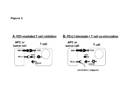
Fig. 1 shows a schematic representation of the anticipated mode of action of
CD137xPD-L1 bispecific
antibodies. (A) PD-L1 is expressed on antigen-presenting cells (APCs) as well
as on tumor cells. PD-
Li binding to T cells expressing the negative regulatory molecule PD-1
effectively overrides T cell
activation signals and eventually leads to T cell inhibition. (B) Upon
addition of a CD137xPD-L1
bispecific antibody, the inhibitory PD-1:PD-L1 interaction is blocked via the
PD-Li-specific arm and
at the same time, the bispecific antibody, through the cell-cell interaction
provides agonistic signaling
to CD137 expressed on the T cells resulting in strong T cell costimulation.
Fig. 2 shows IL-2 production induced by GEN1046 in combination with
pembrolizumab in a MLR assay
of LPS-matured mDCs and purified CD8+ T-cells. Purified CD8+ T cells were co-
cultured with
allogeneic mDCs for 5 days in the presence of GEN1046 (0.001 - 30 ps/mL),
pembrolizumab (0.01 -
100 ps/mL) either alone or in combination, control antibodies, or in the
absence of any antibodies (No
Tx). IL-2 secretion was analyzed by Luminex. Data shown are mean IL-2 SD of
duplicate wells. Each
individual graph represents one of three donor pairs.
Fig. 3 shows IFNy production induced by GEN1046 in combination with
pembrolizumab in a mixed
lymphocyte reaction (MLR) of LPS-matured dendritic cells (mDCs) and purified
CD8+ T cells. Purified
CD8+ T cells were co-cultured with allogeneic mDCs for 5 days in the presence
of GEN1046 (0.001 -
30 itg/mL), pembrolizumab (0.01 ¨ 100 itg/mL) either alone or in combination,
control antibodies, or
in the absence of any antibodies (No Tx). IFNy secretion was analysed by
ELISA. Data shown are mean
IFNy standard deviation (SD) of duplicate wells. Each individual graph
represents one of three DC/T-
cell donor pairs.
Fig. 4 shows TNFa production induced by GEN1046 in combination with
pembrolizumab in a MLR
assay of LPS-matured mDCs and purified CD8+ T-cells. Purified CDS+ T cells
were co-cultured with
allogeneic mDCs for 5 days in the presence of GEN1046 (0.001 - 30 ps/mL),
pembrolizumab (0.01 -
100 itg/mL) either alone or in combination, control antibodies, or in the
absence of any antibodies (No
Tx). TNFa secretion was analysed by Luminex. Data shown are mean TNFa SD of
duplicate wells.
Each individual graph represents one of three donor pairs.
Fig. 5 shows the MC38 syngeneic tumor model that was established by
subcutaneous inoculation of 1
106 MC38 cells into C57BL/6 mice. When tumors reached an average volume of 64
mm3, mice were
randomized and treated with mbsigG2a-PD-L1 x4-1BB (5 mg/kg), an anti-mouse PD-
1 antibody (anti-
mPD-1; 10 mg/kg), either alone or in combination, or PBS (all 2QWx3). A. Data
shown are the median
tumor volume per treatment group (n=10) with data carried forward for animals
that reached termination
criteria. Growth curves were discontinued when <50% of the animals within a
treatment group remained
5
CA 03233512 2024- 3- 28
WO 2023/057535
PCT/EP2022/077749
alive (PBS, mbsIgG2a-PD-L1x4-1BB, anti-mPD-1) or until Day 35 (combination of
mbsIgG2a-PD-
L lx4-1BB with anti-mPD-1). Arrows indicate days of treatment. B. Progression-
free survival, defined
as the percentage of mice with tumor volume smaller than 500 mm3, is shown as
Kaplan Meier curve.
Mantel Cox analysis was used to compare survival between treatment groups on
Day 45 (Table Y).
Fig. 6 shows analysis of the proliferation dose-response of GEN1046 (DuoBody-
PD-L1x4-1BB) and
anti-PD-1 antibody Pembrolizumab in an antigen-specific T cell assay with
active PD1/PD-L1 axis.
Carboxyfluorescein succinimidyl esther (CFSE)-labeled T cells electroporated
with a claudin-6-specific
T-cell receptor (TCR)- and PD-1- in vitro translated (IVT)-RNA were incubated
with claudin-6-IVT-
RNA-electroporated immature dendritic cells in the presence of (A) GEN1046 (at
3-fold serial dilutions
from 1 to 0.00015 ttg/mL) or (B) Pembrolizumab (at 4-fold serial dilutions
from 0.8 to 0.00005 ttg/mL)
for five days. CD8+ T cell proliferation was measured by flow cytometry. Data
shown are expansion
indices calculated using Flowfo software v10.7.1 as a function of the antibody
concentration. Error bars
(SD) indicate variation within the experiment (n=3 replicates in (A); n=2
duplicates in (B), using cells
from one representative donor). Curves were fitted by 4-parameter logarithmic
fit and EC50 values and
Hill-Slopes (shown in Table 1 and 2) were determined using GraphPad Prism
software v9Ø
Fig. 7 shows release of the PD-1/PD-Li-mediated T cell inhibition and
additional co-stimulation of
CD8+ T cell proliferation by GEN1046 in the absence or presence of anti-PD-1
antibody
Pembrolizumab. CFSE-labelled T cells electroporated with a claudin-6-specific
TCR- and PD-1-in vitro
translated (IVT)-RNA were incubated with claudin-6-IVT-RNA-electroporated
immature dendritic
cells in the presence of 0.2 ng/mL, 0.0067 ng/mL or 0.0022 ttg/mL GEN1046 in
combination with a
fixed concentration of 0.8 lug/mL Pembrolizumab or isotype control antibody
IgGl-ctrl or five days
(n=2 technical replicates per condition, using cells from n=3 individual
donors). Medium only,
0.8 ps/mL IgGl-ctrl only and 0.8 lug/mL Pembrolizumab only were used to
determine baseline
proliferation in the absence of GEN1046. CD8+ T cell proliferation was
measured by flow cy. tometry.
Bar graphs represent the mean SD of expansion indices per indicated condition
calculated using Flow.To
software v10.7.1. The dashed line represents baseline proliferation in the
presence of the anti-PD-1
antibody Pembrolizumab.
Fig. 8 is a schematic representation of a first-in-human, open-label, dose-
escalation trial with expansion
cohorts to evaluate safety of GEN1046 in subjects with malignant solid tumors.
Fig. 9 is a waterfall plot showing progression-free survival in subjects
having received prior therapy
with a checkpoint inhibitor (gray line) and checkpoint inhibitor naiive
patients (black line).
Fig. 10 compares time since last prior anti-PD-(L)1 in subjects across CP1-
experienced expansion
cohorts (GEN1046 monotherapy) with clinical response (PR), compared to those
with stable disease
6
CA 03233512 2024- 3- 28
WO 2023/057535
PCT/EP2022/077749
(SD) or progressive disease (PD). Response groups were compared using a
Wilcoxon test. PR vs. PD:
p=0.0017; PR vs. SD: p=0.034.
Fig.11 shows predicted partial reasponse (PR) and complete (CR) rates for
GEN1046 given as 100 mg
Q3W or Q6W in combination with Pembrolizumab in an integrated quantitative
systems pharmacology
(QSP) model.
Fig. 12 shows characterization of the exhausted phenotype of CD3+ T cells
after two rounds of
CD3/CD28 stimulation. (A) In vitro exhausted CD3+ T cells or naïve T cells
were stimulated with
CD3/CD28 beads. Secretion of IFNy was analyzed by ELISA. Data shown are mean +
standard
deviation (SD) of duplicate wells of one representative donor pair. (B)
Expression of TIM3, LAG3, PD-
1 and 4-1BB on naïve and in vitro exhausted CD3+ T cells was determined by
flow cytometry. Data
shown are the median fluorescence intensity corrected for background
fluorescence (AMFI). (C)
Expression of Ki67 on naïve and in vitro exhausted CD3+ T cells was determined
by flow cytometry.
Fig. 13 shows secretion of IFNy induced by GEN1046 in combination with
pembrolizumab in a mixed
lymphocyte reaction (MLR) of mature dcndritic cells (mDCs) and in vitro
exhausted CD3+ T cells (Tex).
Tex were co-cultured with allogeneic LPS-matured DCs (at a DC:T cell ratio of
1:4) in the presence of
GEN1046 (0.001 - 30 ttg/mL) or pembrolizumab (1 ttg/mL) alone or in
combination for 5 days. Co-
cultures without antibody treatment (w/o antibody) or treated with bsIgGl-PD-
L1 xctrl (30 lag/mL),
bsIgG1-ctrlx4-1BB (30 ps/mL), IgG4 isotype control (1 p..g/mL) or IgGl-ctrl-
FEAL (30 ttg/mL) were
included as controls. Secretion of IFNy was analyzed by ELISA. Data shown are
mean + standard
deviation (SD) of duplicate wells of one representative donor pair out of four
donor pairs tested.
Fig 14 shows the Highest single agent (HSA) synergy scores for the combination
of GEN1046 with
pembrolizumab in a MLR of inDCs and Tex. Tex were co-cultured with allogeneic
LPS-matured DCs
(at a DC:T cell ratio of 1:4) in the presence of GEN1046 (0.001 - 30 ps/mL) or
pembrolizumab (1
ttg/mL) alone or in combination for 5 days. Data shown are HSA synergy scores
of one representative
donor pair out of four donor pairs tested (same donor as shown in Figure 13).
Scores >10 are indicative
of synergy in this model.
Fig. 15 shows the MC38 colon cancer model that was established by SC
inoculation of 1 x 10⁶ MC38
cells into C57BL/6 mice. When tumors reached an average volume of 60 mm3, mice
were randomized
and treated with the indicated antibodies or combinations thereof (all 2QWx3).
A. Data shown are the
median tumor volume per treatment group (n=10) with data carried forward for
animals that reached
termination criteria. Growth curves were discontinued when <50% of the animals
within a treatment
group remained alive (mIgG2a-ctrl-AAKR, mbsIgG2a-PD-L1x4-1BB, anti-mouse PD-1
antibody [anti-
7
CA 03233512 2024- 3- 28
WO 2023/057535
PCT/EP2022/077749
mPD-1]) or until Day 60 (combination of mbsIgG2a-PD-L1x 4-1BB with anti-mPD-
1). Downward
facing triangles indicate days of treatment. B. Progression-free survival,
defined as the percentage of
mice with tumor volume smaller than 500 mm3, is shown as Kaplan Meier curve.
Mantel Cox analysis
was used to compare survival between treatment groups on Day 69 (Table 19).
Fig. 16 shows the (re)challenge of mice with complete tumor regression upon
treatment and a control
group of tumor-naïve mice. Mice were (re)challenged with 1 x 10⁶MC38 tumor
cells that were SC
injected on Day 121 after the treatment with antibodies was initiated. Data
shown are mean minor
volumes w SEM.
Fig. 17 shows quantitative IHC and ISH data on cellular immune and tumor
markers expressed in
resected tumor tissues from the MC38 colon cancer model. C57BL/6 mice were
inoculated with 1 x 10⁶
MC38 cells. When tumors reached an average volume of 50-70 mm3, mice were
randomized and treated
with mbsIgG2a-PD-L1 x 4-1 BB, anti-mPD-1 or the combination thereof. Tumors
were resected on Day
7 (n=5 per treatment group) or Day 14 (n=5 per treatment group) after
treatment initiation. Some of the
resected tumor samples were too small to perform IHC analysis, resulting in
analysis of 4-5 tumors per
treatment group. Sections of resected tumors (4 gm) were stained using anti-
CD3, anti-CD4, anti-CD8
or anti-PD-Li antibodies by immunohistochemistry (IHC), or were stained for 4-
1BB or PD-L2 by in
situ hybridization (ISH). Data from IHC are depicted as % marker postive cells
of the total cells counted
in the slide as well as mean w SEM per treatment group. Data from ISH are
depicted as RNAscope H-
score per slide as well as mean SEM per treatment group.
Fig. 18 shows GzmB and Ki67 expression in CD8 T-cell subsets from dissociated
tumor tissue from the
MC38 colon cancer model. C57BL/6 mice were inoculated with 1 x 10⁶ MC38 cells.
When tumors
reached an average volume of 50-70 mm3, mice were randomized and treated with
mbsIgG2a-PD-L1 x 4-
1BB, anti-mPD-1 or the combination thereof. Tumors were resected on Day 7 (n=5
per treatment group)
after treatment initiation, dissociated to single cells suspensions and
analyzed by flow cytometry. Data
shown are the percentage of Gzml3+ (A) or Ki67 + cells (B) within the CD8+ T-
cell population of
individual mice and the mean SEM per treatment group. Mann-Whitney
statistical analysis was
performed to compare the percentage of Gzml31 or Ki67' cells within the CD8 T-
cell population
between treatment groups, with * p <0.05 and **p <0.01.
Fig. 19 shows the cytokine levels in peripheral blood of MC38-tumor bearing
C57BL/6 mice treated
with mbsIgG2a-PD-L1 x4-1BB, an anti-mPD-1 antibody either as single agents or
in combination, or
nonbinding control antibody IgG2a-ctrl-AAKR. Peripheral blood samples were
taken at baseline (one
day before treatment pay -1], dotted line) and two days after each treatment
(Day 2 and Day 5).
Cytokine analysis was performed by ECLIA.
8
CA 03233512 2024- 3- 28
WO 2023/057535
PCT/EP2022/077749
Table 1 ¨ Sequences: Bold and underlined are F; E; A; L and R, corresponding
with positions 234 and
235; 265; 405 and 409, respectively, said positions being in accordance with
EU-numbering. IN SEQ
ID Nos.: 63 and 64 bold amino acids represent the ¨AAKR or ¨AALT mutations
required for
controlled Fab-arm exchange. In variable regions, said CDR regions that were
annotated in accordance
with IMGT definitions (unless otherwise stated or contradicted by context),
are underlined.
SEQ NAME SEQUENCE
Organism
ID
1 VH_CD137-009-H7
EVQLVESGGG LVQPGRSLRLSCTASGFSLNDYWMS synthetic
WVRQAPG KG LEWVGYI DVGGSLYYAASVKGRFTIS construct
RDDSKSIAYLQM NSLKTEDTAVYYCARGGLTYGFDL
WGQGTLVTVSS
2 VH_CD137-009- GFSLN DYW
synthetic
H7_CDR1
construct
3 VH_CD137-009- I DVGGSL
synthetic
H7_CDR2
construct
4 VH_CD137-009- ARGGLTYGFDL
synthetic
H7_CDR3
construct
5 VL_CD137-009-L2
DIVMTQSPSSLSASVG DRVTITCQASEDISSYLAWYQ synthetic
QK PG KAP KRLIYGASD LASGVPSRFSASGSGTDYTFT construct
ISSLQP EDIATYYCHYYATISGLGVAFGGGTKVEIK
6 VL_CD137-009-L2_CDR1 EDISSY
synthetic
construct
7 VL_CD137-009-L2_CDR2 GAS
synthetic
construct
8 VL_CD137-009-L2_CDR3 HYYATISGLGVA
synthetic
construct
9 VH_CD137-009
QSLEESGGRLVTPGTPLTLTCTVSGFSLN DYWMSW synthetic
VRQAPGKGLEWIGYIDVGGSLYYASWAKGRFTISRT construct
STTVDLKMTSLTTEDTATYFCARGGLTYGFDLWGPG
TLVTVSS
VL_CD137-009 DIVMTQTPASVSEPVGGTVTINCQASEDISSYLAWY synthetic
QQKPGQRPKRLIYGASDLASGVPSRFSASGSGTEYA construct
LTISDLESADAATYYCHYYATISGLGVAFGGGTEVVV
11 VH-P D-L1-547 EVQLLEPGGG LVQPGGSLRLSCEASGSTFSTYA MS
synthetic
WVRQAP G KG LEWVSGFSGSGGFTFYADSVRGRFTI construct
SRDSSKNTLFLQMSSLRAEDTAVYYCAIPARGYNYG
SFQHWGQGTLVTVSS
12 VH- PD-L1-547-CDR1 GSTFSTYA
synthetic
construct
13 VH- PD-L1-547-CDR2 FSGSGGFT
synthetic
construct
14 VH- PD-L1-547-CDR3 AI PARGYNYGSFQH
synthetic
construct
VL- PD-L1-547
SYVLTQPPSVSVAPGQTARITCGG NN IGSKSVHWY synthetic
QQKPGQAPVLVVYDDNDRPSGLPERFSGSNSGNTA construct
TLTISRVEAGDEADYYCQVWDSSSDHVVFGGGTKL
TVL
9
CA 03233512 2024- 3- 28
WO 2023/057535
PCT/EP2022/077749
16 VL- PD-L1-547-CDR1 NIGSKS
synthetic
construct
17 VL- PD-L1-547-CDR2 DDN
synthetic
construct
18 VL- PD-L1-547-CDR3 QVWDSSSDHVV
synthetic
construct
19 IgG1-Fc ASTKGPSVFPLAPSSKSTSGGTAALGCLVKDYFPEPV
synthetic
TVSWNSGALTSGVHTFPAVLQSSGLYSLSSVVTVPS construct
SSLGTQTYICNVNHKPSNTKVDKRVEPKSCDKTHTC
PPCPAPELLGGPSVFLFPPKPKDTLMISRTPEVTCVV
VDVSHEDPEVKFNWYVDGVEVHNAKTKPREEQYN
STYRVVSVLTVLHQDWLNGKEYKCKVSNKALPAPIE
KTISKAKGQPREPQVYTLPPSREEMTKNQVSLTCLV
KGFYPSDIAVEWESNGQPENNYKTTPPVLDSDGSFF
LYSKLTVDKSRWQQGNVFSCSVMHEALHNHYTQK
SLSLSPGK
20 IgG1-Fc_F405L ASTKGPSVFPLAPSSKSTSGGTAALGCLVKDYFPEPV
synthetic
TVSWNSGALTSGVHTFPAVLQSSGLYSLSSVVTVPS construct
SSLGTQTYICNVNHKPSNTKVDKRVEPKSCDKTHTC
PPCPAPELLGGPSVFLFPPKPKDTLMISRTPEVTCVV
VDVSHEDPEVKFNWYVDGVEVHNAKTKPREEQYN
STYRVVSVLTVLHQDWLNGKEYKCKVSNKALPAPIE
KTISKAKGQPREPQVYTLPPSREEMTKNQVSLTCLV
KGFYPSDIAVEWESNGQPENNYKTTPPVLDSDGSFL
LYSKLTVDKSRWQQGNVFSCSVMHEALHNHYTQK
SLSLSPGK
21 IgG1-Fc_K409R ASTKGPSVFPLAPSSKSTSGGTAALGCLVKDYFPEPV
synthetic
TVSWNSGALTSGVHTFPAVLQSSGLYSLSSVVTVPS construct
SSLGTQTYICNVNHKPSNTKVDKRVEPKSCDKTHTC
PPCPAPELLGGPSVFLFPPKPKDTLMISRTPEVTCVV
VDVSHEDPEVKFNWYVDGVEVHNAKTKPREEQYN
STYRVVSVLTVLHQDWLNGKEYKCKVSNKALPAPIE
KTISKAKGQPREPQVYTLPPSREEMTKNQVSLTCLV
KGFYPSDIAVEWESNGQPENNYKTTPPVLDSDGSFF
LYSRLTVDKSRWQQGNVFSCSVMHEALHNHYTQK
SLSLSPGK
22 IgG1-Fc_FEA ASTKGPSVFPLAPSSKSTSGGTAALGCLVKDYFPEPV
synthetic
TVSWNSGALTSGVHTFPAVLQSSGLYSLSSVVTVPS construct
SSLGTQTYICNVNHKPSNTKVDKRVEPKSCDKTHTC
PPCPAPEFEGGPSVFLFPPKPKDTLMISRTPEVTCVV
VAVSHEDPEVKFNWYVDGVEVHNAKTKPREEQYN
STYRVVSVLTVLHQDWLNGKEYKCKVSNKALPAPIE
KTISKAKGQPREPQVYTLPPSREEMTKNQVSLTCLV
KGFYPSDIAVEWESNGQPENNYKTTPPVLDSDGSFF
LYSKLTVDKSRWQQGNVFSCSVMHEALHNHYTQK
SLSLSPGK
23 IgG1-FEAR-Fc ASTKGPSVFPLAPSSKSTSGGTAALGCLVKDYFPEPV
synthetic
TVSWNSGALTSGVHTFPAVLQSSGLYSLSSVVTVPS construct
SSLGTQTYICNVNHKPSNTKVDKRVEPKSCDKTHTC
PPCPAPEFEGGPSVFLFPPKPKDTLMISRTPEVTCVV
VAVSHEDPEVKFNWYVDGVEVHNAKTKPREEQYN
STYRVVSVLTVLHQDWLNGKEYKCKVSNKALPAPIE
CA 03233512 2024- 3- 28
WO 2023/057535
PCT/EP2022/077749
KTISKAKGQPREPQVYTLPPSREEMTKNQVSLTCLV
KG FYPSDIAVEWESNGQP EN NYKTTPPVLDSDGSFF
LYSRLTVD KS RWQQG NVFSCSVM HEALH N HYTQK
SLSLSPGK
24 IgGl-FEAL-Fc
ASTKGPSVFPLAPSSKSTSGGTAALGCLVKDYFPEPV synthetic
TVSWNSGALTSGVHTFPAVLQSSGLYSLSSVVTVPS construct
SSLGTQTYICNVN H KPSNTKVD KRVEPKSCDKTHTC
P PCPAPEFEGGPSVFLFPPKPKDTLM ISRTPEVTCVV
VAVSH EDP EVK FNWYVDGVEVH NAKTKPREEQYN
STYRVVSVLTVLHQDW LNG KEYKCKVSN KALPAP I E
KTISKAKGQPREPQVYTLPPSREEMTKNQVSLTCLV
KG FYPSDIAVEWESNGQP EN NYKTTPPVLDSDGSFL
LYS KLTVD KS RWQQG NVFSCSVM H EALHN HYTQK
SLSLSPGK
25 IgGl-Fc without C-
ASTKGPSVFPLAPSSKSTSGGTAALGCLVKDYFPEPV synthetic
terminal Lysine TVSWNSGALTSGVHTFPAVLQSSGLYSLSSVVTVPS construct
SSLGTQTYICNVN H KPSNTKVD KRVEPKSCDKTHTC
P PCPAP ELLGG PSVF LEP P KP KDTLM ISRTPEVTCVV
VDVSH ED P EVKF NWYVDGVEVH NAKTKPREEQYN
STYRVVSVLTVLHQDW LNG KEYKCKVSN KALPAP I E
KTISKAKGQPREPQVYTLPPSREEMTKNQVSLTCLV
KG FYPSDIAVEWESNGQP EN NYKTTPPVLDSDGSFF
LYS KLTVD KS RWQQG NVFSCSVM H EALHN HYTQK
SLSLSPG
26 IgG1-Fc_F405L without
ASTKGPSVFPLAPSSKSTSGGTAALGCLVKDYFPEPV synthetic
C-terminal Lysine TVSWNSGALTSGVHTFPAVLQSSGLYSLSSVVTVPS construct
SSLGTQTYICNVN H KPSNTKVD KRVEPKSCDKTHTC
PPCPAPELLGGPSVFLEPPKPKDILM ISRTPEVTCVV
VDVSH ED P EVKF NWYVDGVEVH NAKTKPREEQYN
STYRVVSVLTVLHQDW LNG KEYKCKVSN KALPAP I E
KTISKAKGQPREPQVYTLPPSREEMTKNQVSLTCLV
KG FYPSDIAVEWESNGQP EN NYKTTPPVLDSDGSFL
LYS KLTVD KS RWQQG NVFSCSVM H EALHN HYTQK
SLSLSPG
27
IgG1-Fc_K409R without ASTKGPSVFPLAPSSKSTSGGTAALGCLVKDYFPEPV synthetic
C-terminal Lysine
TVSWNSGALTSGVHTFPAVLQSSGLYSLSSVVTVPS construct
SSLGTQTYICNVN H KPSNTKVD KRVEPKSCDKTHTC
PPCPAPELLGGPSVFLFPPKPKDTLM ISRTPEVTCVV
VDVSH ED P EVKF NWYVDGVEVH NAKTKPREEQYN
STYRVVSVLTVLHQDW LNG KEYKCKVSN KALPAP I E
KTISKAKGQPREPQVYTLPPSREEMTKNQVSLTCLV
KG FYPSDIAVEWESNGQP EN NYKTTPPVLDSDGSFF
LYSRLTVD KS RWQQG NVFSCSVM HEALH N HYTQK
SLSLSPG
28
IgGl-Fc_FEA without C- ASTKGPSVFPLAPSSKSTSGGTAALGCLVKDYFPEPV synthetic
terminal Lysine
TVSWNSGALTSGVHTFPAVLQSSGLYSLSSVVTVPS construct
SSLGTQTYICNVN H KPSNTKVD KRVEPKSCDKTHTC
P PCPAP EFEGG PSVF LF P P KPK DTLM ISRTPEVTCVV
VAVSH EDP EVK FNWYVDGVEVH NAKTKPREEQYN
STYRVVSVLTVLHQDW LNG KEYKCKVSN KALPAP I E
KTISKAKGQPREPQVYTLPPSREEMTKNQVSLTCLV
KG FYPSDIAVEWESNGQP EN NYKTTPPVLDSDGSFF
11
CA 03233512 2024- 3- 28
WO 2023/057535
PCT/EP2022/077749
LYSKLTVDKSRWQQGNVFSCSVMHEALHNHYTQK
SLSLSPG
29 IgG1-FEAR-Fc without C-
ASTKGPSVFPLAPSSKSTSGGTAALGCLVKDYFPEPV synthetic
terminal Lysine TVSWNSGALTSGVHTFPAVLQSSGLYSLSSVVTVPS
construct
SSLGTQTYICNVNHKPSNTKVDKRVEPKSCDKTHTC
PPCPAPEFEGGPSVFLEPPKPKDILMISRTPEVICVV
VAVSHEDPEVKFNWYVDGVEVHNAKTKPREEQYN
STYRVVSVLTVLHQDWLNGKEYKCKVSNKALPAPIE
KTISKAKGQPREPQVYTLPPSREEMTKNQVSLTCLV
KGFYPSDIAVEWESNGQPENNYKTTPPVLDSDGSFF
LYSRLTVDKSRWQQGNVFSCSVMHEALHNHYTQK
SLSLSPG
30 IgG1-FEAL-Fc without C-
ASTKGPSVFPLAPSSKSTSGGTAALGCLVKDYFPEPV synthetic
terminal Lysine TVSWNSGALTSGVHTFPAVLQSSGLYSLSSVVTVPS
construct
SSLGTQTYICNVNHKPSNTKVDKRVEPKSCDKTHTC
PPCPAPEFEGGPSVFLEPPKPKDILMISRTPEVICVV
VAVSHEDPEVKFNWYVDGVEVHNAKTKPREEQYN
STYRVVSVLTVLHQDWLNGKEYKCKVSNKALPAPIE
KTISKAKGQPREPQVYTLPPSREEMTKNQVSLTCLV
KGFYPSDIAVEWESNGQPENNYKTTPPVLDSDGSFL
LYSKLTVDKSRWQQGNVFSCSVMHEALHNHYTQK
SLSLSPG
31 CD137-009 heavy chain EVQLVESGGGLVQPGRSLRLSCTASGFSLNDYWMS
synthetic
WVRQAPG KG LEWVGYI DVGGSLYYAASVKGRFTIS construct
RDDSKSIAYLQMNSLKTEDTAVYYCARGGLTYGFDL
WGQGTLVTVSSASTKGPSVFPLAPSSKSTSGGTAAL
GCLVKDYFPEPVTVSWNSGALTSGVHTFPAVLQSS
GLYSLSSVVTVPSSSLGTQTYICNVNHKPSNTKVDKR
VEPKSCDKTHTCPPCPAPEFEGGPSVFLFPPKPKDTL
MISRTPEVTCVVVAVSHEDPEVKFNWYVDGVEVH
NAKTKPREEQYNSTYRVVSVLTVLHQDWLNGKEYK
CKVSN KALPAPI EKTISKAKGQPREPQVYTLPPSREE
MTKNQVSLTCLVKGFYPSDIAVEWESNGQPENNYK
TTPPVLDSDGSFFLYSRLTVDKSRWQQGNVFSCSV
MHEALHNHYTQKS LSLSPG
32 CD137-009 light chain
DIVMTQSPSSLSASVGDRVTITCQASEDISSYLAWYQ synthetic
QKPGKAPKRLIYGASDLASGVPSRFSASGSGTDYTFT construct
ISSLQPEDIATYYCHYYATISGLGVAFGGGTKVEIKRT
VAAPSVFIFPPSDEQLKSGTASVVCLLNNFYPREAKV
QWKVDNALQSGNSQESVTEQDSKDSTYSLSSTLTLS
KADYEKHKVYACEVTHQGLSSPVTKSFNRGEC
33 PD-L1-547 heavy chain
EVQLLEPGGGLVQPGGSLRLSCEASGSTFSTYAMS synthetic
WVRQAPGKGLEWVSGFSGSGGFTFYADSVRGRFTI construct
SRDSSKNTLFLQMSSLRAED
TAVYYCAIPARGYNYGSFQHWGQGTLVTVSSASTK
GPSVFPLAPSSKSTSGGTAALGCLVKDYFPEPVTVS
WNSGALTSGVHTFPAVLOSSGLYSLSSVVTVPSSSL
GTQTYICNVNHKPSNTKVDKRVEPKSCDKTHTCPPC
PAPEFEGGPSVFLFPPKPKDTLMISRTPEVTCVVVAV
SHEDPEVKFNWYVDGVEVHNAKTKPREEQYNSTYR
VVSVLTVLHQDWLNGKEYKCKVSNKALPAPIEKTISK
AKGQPREPQVYTLPPSREEMTKNQVSLTCLVKGFYP
12
CA 03233512 2024- 3- 28
WO 2023/057535
PCT/EP2022/077749
SDIAVEWESNGQPENNYKTTPPVLDSDGSFLLYSKL
TVDKSRWQQGNVFSCSVMHEALHNHYTQKSLSLS
PG
34 PD-L1-547 light chain
SYVLTQPPSVSVAPGQTARITCGGNNIGSKSVHWY synthetic
QQKPGQAPVLVVYDDNDRPSGLPERFSGSNSGNTA construct
TLTISRVEAGDEADYYCQVWDSSSDHVVFGGGTKL
TVLGQPKAAPSVTLFPPSSEELQANKATLVCLISDFYP
GAVTVAWKADSSPVKAGVETTTPSKQSNNKYAASS
YLSLTPEQWKSHRSYSCQVTHEGSTVEKTVAPTECS
35 Kappa-C RTVAAPSVFIFPPSDEQLKSGTASVVCLLNNFYPREA
synthetic
KVQWKVDNALQSGNSQESVTEQDSKDSTYSLSSTL construct
TLSKADYEKHKVYACEVTHQGLSSPVTKSFNRGEC
36 Lambda-C GQPKAAPSVTLFPPSSEELQANKATLVCLISDFYPGA
synthetic
VTVAWKADSSPVKAGVETTTPSKQSNNKYAASSYLS construct
LTPEQWKSHRSYSCQVTHEGSTVEKTVAPTECS
37 Human CD137 MGNSCYNIVALLLVLNFERTRSLQDPCSNCPAGTFC Homo
(UniProtKB - 007011; DNNRNQICSPCPPNSFSSAGGQRTCDICRQCKGVF
sapiens
incl. signal peptide RTRKECSSTSNAECDCTPGFHCLGAGCSMCEQDCK
sequence: aa 1-23) QGQELTKKGCKDCCFGTFNDQKRGICRPWTNCSLD
GKSVLVNGTKERDVVCGPSPADLSPGASSVTPPAPA
REPGHSPQIISFFLALTSTALLFLLFFLTLRFSVVKRGR
KKLLYIFKQPFMRPVQTTQEEDGCSCRFPEEEEGGC
EL
38 Human CD137 LQDPCSNCPAGTFCDNNRNQICSPCPPNSFSSAGG Homo
(UniProtKB - 007011; QRTCDICRQCKGVFRTRKECSSTSNAECDCTPGFHC
sapiens
mature sequence) LGAGCSMCEQDCKQGCIELTKKGCKDCCFGTFNDG
KRGICRPWTNCSLDGKSVLVNGTKERDVVCGPSPA
DLSPGASSVTPPAPAREPGHSPQIISFFLALTSTALLFL
LFFLTLRFSVVKRGRKKLLYIFKQPFMRPVQTTQEED
GCSCRFPEEEEGGCEL
39 Human PD-L1 MRIFAVFIFMTYWHLLNAFTVTVPKDLYVVEYGSN
Homo
(UniProtKB - Q9NZQ7; MTIECFPVEKQLDLAALIVYWEMEDKNIIQFVHGEE
sapiens
incl. signal peptide DLKVQHSSYRQRARLLKDQLSLGNAALQITDVKLQD
sequence: aa 1-18) AGVYRCMISYGGADYKRITVKVNAPYNKINQRILVV
DPVTSEHELTCQAEGYPKAEVIWTSSDHQVLSGKTT
TTNSKREEKLFNVTSTLRINTTTNEIFYCTFRRLDPEE
NHTAELVIPELPLAHPPNERTHLVILGAILLCLGVALT
FIFRLRKGRMMDVKKCGIQDTNSKKQSDTHLEET
40 Human PD-L1 FTVTVPKDLYVVEYGSNMTIECKFPVEKQLDLAALIV Homo
(UniProtKB - Q9NZQ7; YWEMEDKNIIQFVHGEEDLKVQHSSYRQRARLLKD
sapiens
mature sequence) QLSLGNAALQITDVKLQDAGVYRCMISYGGADYKRI
TVKVNAPYNKINQRILVVDPVTSEHELTCQAEGYPK
AEVIWTSSDHQVLSGKTTTTNSKREEKLFNVTSTLRI
NTTTNEIFYCTFRRLDPEENHTAELVIPELPLAHPPNE
RTHLVILGAILLCLGVALTFIFRLRKGRMMDVKKCGI
QDTNSKKQSDTHLEET
41 Human PD-1 MQIPQAPWPVVWAVLQLGWRPGWFLDSPDRPW Homo
NPPTFSPALLVVTEGDNATFTCSFSNTSESFVLNWY sapiens
RMSPSNQTDKLAAFPEDRSQPGQDCRFRVTQLPN
GRDFHMSVVRARRNDSGTYLCGAISLAPKAQIKESL
RAELRVTERRAEVPTAHPSPSPRPAGQFQTLVVGVV
GGLLGSLVLLVWVLAVICSRAARGTIGARRTGQPLK
13
CA 03233512 2024- 3- 28
WO 2023/057535
PCT/EP2022/077749
ED PSAVPVFSVDYG ELD FQWREKTPE PPVPCVP EQ
TEYATIVFPSG MGTSSPARRGSADG PRSAQP LRP ED
GHCSWPL
42 CTLA-4 MACLGFQRHKAQLNLATRTWPCTLLFFLLFIPVFCK Homo
AM HVAQPAVVLASSRG IASFVCEYASPG KATEVRVT sapiens
VLRQADSQVTEVCAATYMMGNELTFLDDSICTGTS
SGNQVN LTIQG LRAM DTGLYICKVELMYPPPYYLGI
GNGTQIYVIDPEPCPDSDFLLWILAAVSSGLFFYSFLL
TAVSLSKMLKKRSPLTTGVYVKMPPTEPECEKQFQP
YFIPIN
43 Pembrolizumab VH GYTFTNYY
synthetic
CDR1
construct
44 Pembrolizumab VH IN PSNGGT
synthetic
CDR2
construct
45 Pembrolizumab VH ARRDYRFDMGFDY
synthetic
CDR3
construct
46 Pembrolizumab VL KGVSTSGYSY
synthetic
CDR1
construct
47 Pembrolizumab VL LAS
synthetic
CDR2
construct
48 Pembrolizumab VL QHSRDLPLT
synthetic
CDR3
construct
49 Pembrolizumab VH
QVQLVQSGVEVKKPGASVKVSCKASGYTFTNYYMY synthetic
WVRQAPGQGLEWMGGINPSNGGTNFNEKFKNRV construct
TLITDSSITTAYMELKSLQFDDTAVYYCARRDYRFD
MGFDYWGQGTTVTVSS
50 Pembrolizumab VL
EIVLTQSPATLSLSPGERATLSCRASKGVSTSGYSYLH synthetic
WYQQKPGQAPRLLIYLASYLESGVPARFSGSGSGTD construct
FTLTISSLEPED FAVYYCQHSRD LP LTFGGGTKVEI K
51 Pembrolizumab Heavy QVQLVQSGVEVKKPGASVKVSCKASGYTFTNYYMY synthetic
chain WVRQAPGQGLEWMGG I NPSNGGTNFN EKFKNRV construct
TLITDSSITTAYMELKSLQFDDTAVYYCARRDYRFD
MGFDYWGQGTTVTVSSASTKGPSVFPLAPCSRSTS
ESTAALGCLVKDYFPEPVTVSWNSGALTSGVHTFPA
VLQSSGLYSLSSVVTVPSSSLGTKTYTCNVDHKPSNT
KVDKRVESKYGPPCPPCPAPEFLGGPSVFLFPPKPKD
TLM ISRTP EVTCVVVDVSQED PEVQFNWYVDGVEV
H NAKTKPREEQFNSTYRVVSVLTVLHQDWLNG KEY
KCKVSN KG LPSSI EKTISKAKGQPREPQVYTLP PSQEE
MTKNQVSLTCLVKGFYPSDIAVEWESNGQPENNYK
TTPPVLDSDGSFFLYSRLTVDKSRWQEGNVFSCSVM
H EA LH N H YTQKS LS LS LG K
52 Pembrolizumab Light EIVLTQSPATLSLSPGERATLSCRASKGVSTSGYSYLH synthetic
chain
WYQQKPGQAPRLLIYLASYLESGVPARFSGSGSGTD construct
FTLTISSLEPED FAVYYCQHSRD LP LTFGGGTKVEI KR
TVAAPSVF I FP PSD EQLKSGTASVVCLLN NFYPREAK
VQWKVDNALQSGNSQESVTEQDSKDSTYSLSSTLTL
SKADYEKH KVYACEVTHQG LSSPVTKSFN RG EC
53 VH_IgG1-b12
QVQLVQSGAEVKKPGASVKVSCQASGYRFSNFVIH synthetic
WVRQAPGQRFEWMGWINPYNGNKEFSAKFQDR construct
14
CA 03233512 2024- 3- 28
WO 2023/057535
PCT/EP2022/077749
VTFTADTSANTAYMELRSLRSADTAVYYCARVGPYS
WDDSPQDNYYMDVWGKGTTVIVSS
54 VL_IgG1-b12 EIVLTQSPGTLSLSPGERATFSCRSSHSIRSRRVAWY synthetic
QHKPGQAPRLVIHGVSNRASGISIDRFSGSGSGTDFT construct
LTITRVEPEDFALYYCQVYGASSYTFGQGTKLERK
55 m4-1BB-3H3 VH
EMQLVESGGGLVQPGRSMKLSCAGSGFTLSDYGVA synthetic
WVRQAPKKGLEWVAYISYAGGTTYYRESVKGRFTIS construct
RDNAKSTLYLQMDSLRSEDTATYYCTIDGYGGYSGS
HWYFDFWGPGTMVTVSS
56 m4-1 BB-3H3 VL
DIQMTQSPSLLSASVG DRVTLNCRTSQNVYKN LAW synthetic
YQQKLGEAPKLLIYNANSLQAGIPSRFSGSGSGTDFT construct
LTISSLQPEDVATYFCQQYYSGNTFGAGTNLELK
57 AALT AKTTAPSVYPLAPVCGDTTGSSVTLGCLVKGYFPEPV synthetic
TLTWNSGSLSSGVHTFPAVLQSDLYTLSSSVTVTSST construct
WPSQSITCNVAHPASSTKVDKKIEPRGPTIKPCPPCK
CPAPNAAGGPSVFIFPPKIKDVLMISLSPMVTCVVV
DVSEDDPDVQISWFVNNVEVLTAQTQTHREDYNST
LRVVSALPIQHQDWMSGKEFKCKVNNKALPAPIER
TISKPKGSVRAPQVYVLPPPEEEMTKKQVTLTCMVT
DFMPEDIYVEVVTNNGKTELNYKNTEPVLDSDGSYL
MYSKLTVEKKNWVERNSYSCSVVHEGLHNHHTTKS
FSRTPGK
58 AAKR AKTTAPSVYPLAPVCGDTTGSSVTLGCLVKGYFPEPV synthetic
TLTWNSGSLSSGVHTFPAVLQSDLYTLSSSVTVTSST construct
WPSQSITCNVAHPASSTKVDKKIEPRGPTIKPCPPCK
CPAPNAAGGPSVFIFPPKIKDVLMISLSPMVTCVVV
DVSEDDPDVQISWFVNNVEVLTAQTQTHREDYNST
LRVVSALPIQHQDWMSGKEFKCKVNNKALPAPIER
TISKPKGSVRAPQVYVLPPPEEEMTKKQVTLTCMVK
DFMPEDIYVEVVTNNGKTELNYKNTEPVLDSDGSYF
MYSRLRVEKKNWVERNSYSCSVVHEGLHNHHTTKS
FSRTPGK
59
constant region mouse RADAAPTVSIFPPSSEQLTSGGASVVCFLNNFYPKDI synthetic
kappa LC
NVKWKIDGSERQNGVLNSWTDQDSKDSTYSMSST construct
LTLIKDEYERHNSYTCEATHKTSTSPIVKSFNRNEC
60 MPDL3280A VH EVQLVESGGGLVQPGGSLRLSCAASGFTFSDSWIH
synthetic
WYRQAPGKGLEWYAWISPYGGSTYYADSVKGRFTI construct
SADTSKNTAYLQMNSLRAEDTAVYYCARRHWPGG
FDYWGQGTLVTVSS
61 MPDL3280A VL
DIQMTQSPSSLSASVGDRVTITCRASQDVSTAVAW synthetic
YQQKPGKAPKWYSASFLYSGVPSRFSGSGSGTDFTL construct
TISSLQPEDFATYYCQQYLYHPATFGQGTKVEIK
62 Pembrolizumab VH NYYMY
synthetic
CDR1 (Kabat
construct
numbering)
63 Pembrolizumab VH GINPSNGGTNFNEKFKN
synthetic
CDR2 (Kabat
construct
numbering)
64 Pembrolizumab VH RDYRFDMGFDY
synthetic
CDR3 (Kabat
construct
numbering)
65 Pembrolizumab VL RASKGVSTSGYSYLH
synthetic
CDR1 (Kabat
construct
numbering)
CA 03233512 2024- 3- 28
WO 2023/057535
PCT/EP2022/077749
66 Pembrolizumab VL LASYLES
synthetic
CDR2 (Kabat
construct
numbering)
67 Pembrolizumab VL QHSRDLPLT
synthetic
CDR3 (Kabat
construct
numbering)
Detailed Description of the Invention
Although the present disclosure is further described in more detail below, it
is to be understood that this
disclosure is not limited to the particular methodologies, protocols and
reagents described herein as these
may vary. It is also to be understood that the terminology used herein is for
the purpose of describing
particular embodiments only, and is not intended to limit the scope of the
present disclosure which will
be limited only by the appended claims. Unless defined otherwise, all
technical and scientific terms used
herein have the same meanings as commonly understood by one of ordinary skill
in the art.
In the following, the elements of the present disclosure will be described in
more detail. These elements
are listed with specific embodiments, however, it should be understood that
they may be combined in
any manner and in any number to create additional embodiments. The variously
described examples and
preferred embodiments should not be construed to limit the present disclosure
to only the explicitly
described embodiments. This description should be understood to support and
encompass embodiments
which combine the explicitly described embodiments with any number of the
disclosed and/or preferred
elements. Furthermore, any permutations and combinations of all described
elements in this application
should be considered disclosed by the description of the present application
unless the context indicates
otherwise. For example, if in a preferred embodiment of the binding agent used
herein the first heavy
chain comprises or consists essentially of or consists of an amino acid
sequence set forth in SEQ ID NO:
23 or 29 [IgGl-Fc_FEAR] and in another preferred embodiment of the binding
agent used herein the
second heavy chain comprises or consists essentially of or consists of an
amino acid sequence set forth
in SEQ ID NO: 24 or 30 [IgGl-Fc_FEAL], then in a further preferred embodiment
of the binding agent
used herein the first heavy chain comprises or consists essentially of or
consists of an amino acid
sequence set forth in SEQ ID NO: 23 or 29 [IgGl-Fc_FEAR] and the second heavy
chain comprises or
consists essentially of or consists of an amino acid sequence set forth in SEQ
ID NO: 24 or 30 [IgG1 -
Fc_FEAL J.
Preferably, the terms used herein are defined as described in "A multilingual
glossary of
biotechnological terms: (IUPAC Recommendations)", H.G.W. Leuenberger, B.
Nagel, and H. Kolbl,
Eds., Helvetica Chimica Acta, CH-4010 Basel, Switzerland, (1995).
The practice of the present disclosure will employ, unless otherwise
indicated, conventional chemistry,
biochemistry, cell biology, immunology, and recombinant DNA techniques which
are explained in the
16
CA 03233512 2024- 3- 28
WO 2023/057535
PCT/EP2022/077749
literature in the field (cf., e.g., Organikum, Deutscher Verlag der
Wissenschaften, Berlin 1990;
Streitwieser/Heathcook, "Organischc Chemic", VCH, 1990; Beyer/Walter,
"Lchrbuch der Organischen
Chcmic", S. Hirzel Verlag Stuttgart, 1988; Carcy/Sundbcrg, "Organische
Chcmic", VCH, 1995; March,
"Advanced Organic Chemistry", John Wiley & Sons, 1985; Rompp Chemie Lexikon,
Falbe/Regitz
(Hrsg.), Georg Thieme Verlag Stuttgart, New York, 1989; Molecular Cloning: A
Laboratory Manual,
2nd Edition, J. Sambrook et al. eds., Cold Spring Harbor Laboratory Press,
Cold Spring Harbor 1989.
All methods described herein can be performed in any suitable order unless
otherwise indicated herein
or otherwise clearly contradicted by the context. The use of any and all
examples, or exemplary language
(e.g., "such as"), provided herein is intended merely to better illustrate the
present disclosure and does
not pose a limitation on the scope of the present disclosure otherwise
claimed. No language in the
specification should be construed as indicating any non-claimed element
essential to the practice of the
present disclosure.
Recitation of ranges of values herein is merely intended to serve as a
shorthand method of referring
individually to each separate value falling within the range. Unless otherwise
indicated herein, each
individual value is incorporated into the specification as if it were
individually recited herein.
Several documents are cited throughout the text of this specification. Each of
the documents cited herein
(including all patents, patent applications, scientific publications,
manufacturer's specifications,
instructions, etc.), whether supra or infra, are hereby incorporated by
reference in their entirety. Nothing
herein is to be construed as an admission that the invention is not entitled
to antedate such disclosure by
virtue of prior invention.
Definitions
in the following, definitions will be provided which apply to all aspects of
the present disclosure. The
following terms have the following meanings unless otherwise indicated. Any
undefined terms have
their art recognized meanings.
Throughout this specification and the claims which follow, unless the context
requires otherwise, the
word "comprise", and variations such as "comprises" and "comprising", will be
understood to imply the
inclusion of a stated member, integer or step or group of members, integers or
steps but not the exclusion
of any other member, integer or step or group of members, integers or steps.
The term "consisting
essentially of' means excluding other members, integers or steps of any
essential significance. The term
"comprising" encompasses the term "consisting essentially of" which, in turn,
encompasses the term
"consisting of'. Thus, at each occurrence in the present application, the term
"comprising" may be
17
CA 03233512 2024- 3- 28
WO 2023/057535
PCT/EP2022/077749
replaced with the term "consisting essentially of' or "consisting of'.
Likewise, at each occurrence in the
present application, the term "consisting essentially of' may be replaced with
the term "consisting of'.
The terms "a", "an" and "the" and similar references used in the context of
describing the present
disclosure (especially in the context of the claims) are to be construed to
cover both the singular and the
plural, unless otherwise indicated herein or clearly contradicted by the
context.
Where used herein, "and/or" is to be taken as specific disclosure of each of
the two specified features or
components with or without the other. For example, "X and/or Y" is to be taken
as specific disclosure
of each of (i) X, (ii) Y, and (iii) X and Y, just as if each is set out
individually herein.
In the context of the present disclosure, the term "about" denotes an interval
of accuracy that the person
of ordinary skill will understand to still ensure the technical effect of the
feature in question. The term
typically indicates deviation from the indicated numerical value by
0.8%, 0.7%, 0.6%, 0.5%, 0.4%, 0.3%, 0.2%, 0.1%, 0.05%, and for example
0.01%. As
will be appreciated by the person of ordinary skill, the specific such
deviation for a numerical value for
a given technical effect will depend on the nature of the technical effect.
For example, a natural or
biological technical effect may generally have a larger such deviation than
one for a man-made or
engineering technical effect.
The term "binding agent" in the context of the present disclosure refers to
any agent capable of binding
to desired antigens. In certain embodiments of the present disclosure, the
binding agent is an antibody,
antibody fragment, or construct thereof. The binding agent may also comprise
synthetic, modified or
non-naturally occurring moieties, in particular non-peptide moieties. Such
moieties may, for example,
link desired antigen-binding functionalities or regions such as antibodies or
antibody fragments. In one
embodiment, the binding agent is a synthetic construct comprising antigen-
binding CDRs or variable
regions.
As used herein, "immune checkpoint" refers to regulators of the immune system,
and, in particular, co-
stimulatory and inhibitory signals that regulate the amplitude and quality of
T cell receptor recognition
of an antigen. In certain embodiments, the immune checkpoint is an inhibitory
signal. In certain
embodiments, the inhibitory signal is the interaction between PD-1 and PD-Li
and/or PD-L2. In certain
embodiments, the inhibitory signal is the interaction between CTLA-4 and CD80
or CD86 to displace
CD28 binding. In certain embodiments the inhibitory signal is the interaction
between LAG-3 and MHC
class II molecules. In certain embodiments, the inhibitory signal is the
interaction between TIM-3 and
one or more of its ligands, such as galcctin 9, PtdScr, HMGB1 and CEACAM1. In
certain embodiments,
the inhibitory signal is the interaction between one or several KIRs and their
ligands. In certain
18
CA 03233512 2024- 3- 28
WO 2023/057535
PCT/EP2022/077749
embodiments, the inhibitory signal is the interaction between TIGIT and one or
more of its ligands,
PVR, PVRL2 and PVRL3. In certain embodiments, the inhibitory signal is the
interaction between
CD94/NKG2A and HLA-E. In certain embodiments, the inhibitory signal is the
interaction between
VISTA and its binding partner(s). In certain embodiments, the inhibitory
signal is the interaction
between one or more Siglees and their ligands. In certain embodiments, the
inhibitory signal is the
interaction between GARP and one or more of its ligands. In certain
embodiments, the inhibitory signal
is the interaction between CD47 and STRPa. in certain embodiments, the
inhibitory signal is the
interaction between PVRIG and PVRL2. In certain embodiments, the inhibitory
signal is the interaction
between CSF1R and CSF1. In certain embodiments, the inhibitory signal is the
interaction between
BTLA and HVEM. In certain embodiments, the inhibitory signal is part of the
adenosinergic pathway,
e.g., the interaction between A2AR and/or A2BR and adenosine, produced by CD39
and CD73. In
certain embodiments, the inhibitory signal is the interaction between B7-H3
and its receptor and/or B7-
H4 and its receptor. In certain embodiments, the inhibitory signal is mediated
by IDO, CD20, NOX or
TDO.
The terms "checkpoint inhibitor" (CPI) and "immune checkpoint (ICP) inhibitor"
are used herein
synonymously. The terms refer to molecules, such as binding agents, which
totally or partially reduce,
inhibit, interfere with or negatively modulate one or more checkpoint proteins
or that totally or partially
reduce, inhibit, interfere with or negatively modulate expression of one or
more checkpoint proteins,
like molecules, such as binding agents, which inhibit an immune checkpoint, in
particular, which inhibit
the inhibitory signal of an immune checkpoint. In one embodiment, the immune
checkpoint inhibitor
binds to one or more checkpoint proteins. In one embodiment, the immune
checkpoint inhibitor binds
to one or more molecules regulating checkpoint proteins. In one embodiment,
the immune checkpoint
inhibitor binds to precursors of one or more checkpoint proteins e.g., on DNA-
or RNA-level. Any agent
that functions as a checkpoint inhibitor according to the present disclosure
can be used. The term
"partially" as used herein means at least 5%, 10%, 15%, 20%, 25%, 30%, 35%,
40%, 45%, 50%, 55%,
60%, 65%, 70%, 75%, 80%, 85%, 90%, 95%, 96%, 97%, 98% or 99% in the level,
e.g., in the level of
inhibition of a checkpoint protein.
In one embodiment, the checkpoint inhibitor can be any compound, such as any
binding agent, which
inhibits the inhibitory signal of an immune checkpoint, wherein the inhibitory
signal is selected from
the group consisting of: the interaction between PD-1 and PD-L1 and/or PD-L2;
the interaction between
CTLA-4 and CD80 or CD86 to displace CD28 binding; the interaction between LAG-
3 and MHC class
H molecules; the interaction between TIM-3 and one or more of its ligands,
such as galectin 9, PtdSer,
HMGB1 and CEACAM1; the interaction between one or several KIRs and their
ligands; the interaction
between TIGIT and one or more of its ligands, PVR, PVRL2 and PVRL3; the
interaction between
CD94/NKG2A and HLA-E; the interaction between VISTA and its binding
partner(s); the interaction
19
CA 03233512 2024- 3- 28
WO 2023/057535
PCT/EP2022/077749
between one or more Siglees and their ligands; the interaction between GARP
and one or more of its
ligands; the interaction between CD47 and SIRPa; the interaction between PVRIG
and PVRL2; the
interaction between CSF1R and CSF1; the interaction between BTLA and HVEM;
part of the
adenosinergic pathway, e.g., the interaction between A2AR and/or A2BR and
adenosine, produced by
CD39 and CD73; the interaction between B7-H3 and its receptor and/or B7-H4 and
its receptor; an
inhibitory signal mediated by 1DO, CD20, NOX or TDO. In one embodiment, the
checkpoint inhibitor
is at least one selected from the group consisting of PD-1 inhibitors, PD-Li
inhibitors, PD-L2 inhibitors,
CTLA-4 inhibitors, TIM-3 inhibitors, KIR inhibitors, LAG-3 inhibitors, TIGIT
inhibitors, VISTA
inhibitors, and GARP inhibitors. In one embodiment, the checkpoint inhibitor
may be a blocking
antibody, such as a PD-1 blocking antibody, a CTLA4 blocking antibody, a PD-Li
blocking antibody,
a PD-L2 blocking antibody, a TIM-3 blocking antibody, a KIR blocking antibody,
a LAG-3 blocking
antibody, a TIGIT blocking antibody, a VISTA blocking antibody, or a GARP
blocking antibody.
Examples of a PD-1 blocking antibody include pembrolizumab, nivolumab,
cemiplimab, and
spartalizumab. Examples of a CTLA4 blocking antibody include ipilimumab and
tremelimumab.
Examples of a PD-Li blocking antibody include atezolizumab, durvalumab, and
avelumab.
The term "immunoglobulin" relates to proteins of the immunoglobulin
superfamily, preferably to
antigen receptors such as antibodies or the B cell receptor (BCR). The
immunoglobulins are
characterized by a structural domain, i.e., the immunoglobulin domain, having
a characteristic
immunoglobulin (Ig) fold. The term encompasses membrane bound immunoglobulins
as well as soluble
immunoglobulins. Membrane bound immunoglobulins are also termed surface
immunoglobulins or
membrane immunoglobulins, which are generally part of the BCR. Soluble
immunoglobulins are
generally termed antibodies.
The structure of immunoglobulins has been well characterized. See, e.g.,
Fundamental -immunology Ch.
7 (Paul, W., ed., 2' ed. Raven Press, N.Y. (1989)). Briefly, immunoglobulins
generally comprise several
chains, typically two identical heavy chains and two identical light chains
which are linked via disulfide
bonds. These chains are primarily composed of immunoglobulin domains or
regions, such as the VL or
VL (variable light chain) domain/region, CL or CL (constant light chain)
domain/region, VH or VH
(variable heavy chain) domain/region, and the CH or CH (constant heavy chain)
domains/regions Cill
(CH1), C112 (CH2), C113 (CH3), and C114 (CH4). The heavy chain constant region
typically is comprised
of three domains, CHI, CH2, and CH3. The hinge region is the region between
the CHI and CH2
domains of the heavy chain and is highly flexible. Disulfide bonds in the
hinge region are part of the
interactions between two heavy chains in an IgG molecule. Each light chain
typically is comprised of a
VL and a CL. The light chain constant region typically is comprised of one
domain, CL. The VH and
VL regions may be further subdivided into regions of hypervariability (or
hypervariable regions which
CA 03233512 2024- 3- 28
WO 2023/057535
PCT/EP2022/077749
may be hypervariable in sequence and/or form of structurally defined loops),
also termed
complementarity determining regions (CDRs), interspersed with regions that are
more conserved,
termed framework regions (FRs). Each VH and VL is typically composed of three
CDRs and four FRs,
arranged from amino-terminus to carboxy -terminus in the following order: FR1,
CDR1, FR2, CDR2,
FR3, CDR3, FR4 (see also Chothia and Lesk J. Mol. Biol. 196, 901-917 (1987)).
Unless otherwise stated
or contradicted by context, CDR sequences herein are identified according to
1MGT rules using
DomainGapAlign (Lefranc MP., Nucleic Acids Research 1999;27:209-212 and
Ehreninann F., Kaas Q.
and Lefranc M.-P. Nucleic Acids Res., 38, D301-307 (2010); see also internet
http address
www.imgt.org. Unless otherwise stated or contradicted by context, reference to
amino acid positions in
the constant regions in the present disclosure is according to the EU-
numbering (Edelman et al., Proc
Natl Acad Sci USA. 1969 May;63(1):78-85; Kabat et al., Sequences of Proteins
of Immunological
Interest, Fifth Edition. 1991 NIH Publication No. 91-3242).
There are five types of mammalian immunoglobulin heavy chains, i.e., a, 6, a,
y, and which account
for the different classes of antibodies, i.e., IgA, IgD, IgE, IgG, and IgM. As
opposed to the heavy chains
of soluble immunoglobulins, the heavy chains of membrane or surface
immunoglobulins comprise a
transmembrane domain and a short cytoplasmic domain at their carboxy-terminus.
In mammals there
are two types of light chains, i.e., lambda and kappa. The immunoglobulin
chains comprise a variable
region and a constant region. The constant region is essentially conserved
within the different isotypcs
of the immunoglobulins, wherein the variable part is highly divers and
accounts for antigen recognition.
The term "amino acid" and "amino acid residue" may herein be used
interchangeably, and are not to be
understood limiting. Amino acids are organic compounds containing amine (-NH2)
and carboxyl
(-COOH) functional groups, along with a side chain (R group) specific to each
amino acid. In the context
of the present disclosure, amino acids may be classified based on structure
and chemical characteristics.
Thus, classes of amino acids may be reflected in one or both of the following
tables:
Table 2: Main classification based on structure and general chemical
characterization ofR group
Class Amino acid
Acidic Residues D and E
Basic Residues K, R, and H
Hydrophilic Uncharged Residues S, T, N, and Q
Aliphatic Uncharged Residues G, A, V. L, and 1
Non-polar Uncharged Residues C, M, and P
Aromatic Residues F, Y, and W
Table 3: Alternative Physical and Functional Classifications ofAmino Acid
Residues
Class Amino acid
Hydroxyl group containing residues S and T
Aliphatic residues I, L, V. and M
21
CA 03233512 2024- 3- 28
WO 2023/057535
PCT/EP2022/077749
Cy cloalkenyl-assoc iated residues F, H, W, and Y
Hydrophobic residues A, C, F, G, H, 1, L, M, R, T,
V. W, and Y
Negatively charged residues D and E
Polar residues C, D, E, H, K, N, Q, R, S,
and T
Positively charged residues H, K, and R
Small residues A, C, D, G, N, P. 5, T, and V
Very small residues A, G, and S
Residues involved in turn formation A, C, D, E, G, H, K, N, Q, R,
5, P, and T
Flexible residues Q, T, K, S, G, P, D, E, and R
For the purposes of the present disclosure, "variants" of an amino acid
sequence (peptide, protein or
polypeptide) comprise amino acid insertion variants, amino acid addition
variants, amino acid deletion
variants and/or amino acid substitution variants. The term "variant" includes
all mutants, splice variants,
posttranslationally modified variants, conformations, isoforms, allelic
variants, species variants, and
species homologs, in particular those which are naturally occurring. The term
"variant" includes, in
particular, fragments of an amino acid sequence.
Amino acid insertion variants comprise insertions of single or two or more
amino acids in a particular
amino acid sequence. In the case of amino acid sequence variants having an
insertion, one or more amino
acid residues are inserted into a particular site in an amino acid sequence,
although random insertion
with appropriate screening of the resulting product is also possible.
Amino acid addition variants comprise amino- and/or carboxy-tenninal fusions
of one or more amino
acids, such as 1, 2, 3, 5, 10, 20, 30, 50, or more amino acids.
Amino acid deletion variants are characterized by the removal of one or more
amino acids from the
sequence, such as by removal of 1, 2, 3, 5, 10, 20, 30, 50, or more amino
acids. The deletions may be in
any position of the protein. Amino acid deletion variants that comprise the
deletion at the N-terminal
and/or C-terminal end of the protein are also called N-terminal and/or C-
terminal truncation variants.
Amino acid substitution variants are characterized by at least one residue in
the sequence being removed
and another residue being inserted in its place. Substitution of one amino
acid for another may be
classified as a conservative or non-conservative substitution. Preference is
given to the modifications
being in positions in the amino acid sequence which are not conserved between
homologous proteins or
peptides and/or to replacing amino acids with other ones having similar
properties. Preferably, amino
acid changes in peptide and protein variants are conservative amino acid
changes, i.e., substitutions of
similarly charged or uncharged amino acids. A conservative amino acid change
involves substitution of
one of a family of amino acids which are related in their side chains. In the
context of the present
disclosure, a "conservative substitution" is a substitution of one amino acid
with another amino acid
having similar structural and/or chemical characteristics, such substitution
of one amino acid residue for
22
CA 03233512 2024- 3- 28
WO 2023/057535
PCT/EP2022/077749
another amino acid residue of the same class as defined in any of the two
tables above: for example,
leucine may be substituted with isoleucine as they arc both aliphatic,
branched hydrophobcs. Similarly,
aspartic acid may be substituted with glutamic acid since they are both small,
negatively charged
residues. Naturally occurring amino acids may also be generally divided into
four families: acidic
(aspartate, glutamate), basic (lysine, arginine, histidine), non-polar
(alanine, valine, leucine, isoleucine,
proline, phenylalanine, methionine, tryptophan), and uncharged polar (glycine,
asparagine, glutamine,
cysteine, serine, threonine, tyrosine) amino acids. Phenylalanine, tryptophan,
and tyrosine are
sometimes classified jointly as aromatic amino acids. In one embodiment,
conservative amino acid
substitutions include substitutions within the following groups:
- glycine, alanine;
- valine, isoleucine, leucine;
- aspartic acid, glutamic acid;
- asparagine, glutamine;
- serine, threonine;
- lysine, arginine; and
- phenylalanine, tyrosine.
The term "amino acid corresponding to position..." and similar expressions as
used herein refer to an
amino acid position number in a human IgG1 heavy chain. Corresponding amino
acid positions in other
immunoglobulins may be found by alignment with human IgGl. Thus, an amino acid
or segment in one
sequence that "corresponds to" an amino acid or segment in another sequence is
one that aligns with the
other amino acid or segment using a standard sequence alignment program such
as ALIGN, ClustalW
or similar, typically at default settings and has at least 50%, at least 80%,
at least 90%, or at least 95%
identity to a human IgG1 heavy chain. It is considered well-known in the art
how to align a sequence or
segment in a sequence and thereby determine the corresponding position in a
sequence to an amino acid
position according to the present disclosure.
The term "antibody" (Ab) in the context of the present disclosure refers to an
immunoglobulin molecule,
a fragment of an immunoglobulin molecule, or a derivative of either thereof,
which has the ability to
specifically bind to an antigen (in particular an epitope on an antigen) under
typical physiological
conditions, preferably with a half-life of significant periods of time, such
as at least about 30 minutes,
at least about 45 minutes, at least about one hour, at least about two hours,
at least about four hours, at
least about 8 hours, at least about 12 hours, about 24 hours or more, about 48
hours or more, about 3, 4,
5, 6, 7 or more days, etc., or any other relevant functionally-defined period
(such as a time sufficient to
induce, promote, enhance, and/or modulate a physiological response associated
with antibody binding
to the antigen and/or time sufficient for the antibody to recruit an effector
activity). In particular, the
term "antibody" refers to a glycoprotein comprising at least two heavy (H)
chains and two light (L)
23
CA 03233512 2024- 3- 28
WO 2023/057535
PCT/EP2022/077749
chains inter-connected by disulfide bonds. The term "antibody" includes
monoclonal antibodies,
recombinant antibodies, human antibodies, humanized antibodies, chimeric
antibodies and
combinations of any of the foregoing. Each heavy chain is comprised of a heavy
chain variable region
(VH) and a heavy chain constant region (CH). Each light chain is comprised of
a light chain variable
region (VL) and a light chain constant region (CL). The variable regions and
constant regions are also
referred to herein as variable domains and constant domains, respectively. The
VH and VL regions can
be further subdivided into regions of hypervariability, termed complementarity
determining regions
(CDRs), interspersed with regions that are more conserved, termed framework
regions (FRs). Each VH
and VL is composed of three CDRs and four FRs, arranged from amino-terminus to
carboxy-terminus
in the following order: FR1, CDR1, FR2, CDR2, FR3, CDR3, FR4. The CDRs of a VH
are termed
HCDR1, HCDR2 and HCDR3 (or CDR-H1, CDR-H2 and CDR-H3), the CDRs of a VL are
termed
LCDR1, LCDR2 and LCDR3 (or CDR-L1, CDR-L2 and CDR-L3). The variable regions of
the heavy
and light chains contain a binding domain that interacts with an antigen. The
constant regions of an
antibody comprise the heavy chain constant region (CH) and the light chain
constant region (CL),
wherein CH can be further subdivided into constant domain CHL a hinge region,
and constant domains
CH2 and CH3 (arranged from amino-terminus to carboxy-terminus in the following
order: CHL CH2,
CH3). The constant regions of the antibodies may mediate the binding of the
immunoglobulin to host
tissues or factors, including various cells of the immune system (e.g.,
effector cells) and components of
the complement system such as Clq. Antibodies can be intact immunoglobulins
derived from natural
sources or from recombinant sources and can be immunoactive portions of intact
immunoglobulins.
Antibodies are typically tetramers of immunoglobulin molecules. Antibodies may
exist in a variety of
forms including, for example, polyclonal antibodies, monoclonal antibodies,
Fv. Fab and F(ab)2, as well
as single chain antibodies and humanized antibodies.
The variable regions of the heavy and light chains of the immunoglobulin
molecule contain a binding
domain that interacts with an antigen. The terms "binding region" and "antigen-
binding region" are used
herein interchangeably and refer to the region which interacts with the
antigen and comprises both a VH
region and a VL region. An antibody as used herein comprises not only
monospecific antibodies, but
also multispecific antibodies which comprise multiple, such as two or more,
e.g., three or more, different
antigen-binding regions.
As indicated above, the term antibody herein, unless otherwise stated or
clearly contradicted by context,
includes fragments of an antibody that are antigen-binding fragments, i.e.,
retain the ability to
specifically bind to the antigen. it has been shown that the antigen-binding
function of an antibody may
be performed by fragments of a full-length antibody. Examples of antigen-
binding fragments
encompassed within the term "antibody" include (i) a Fab' or Fab fragment, a
monovalent fragment
consisting of the VL, VH, CL and CH1 domains, or a monovalent antibody as
described in
24
CA 03233512 2024- 3- 28
WO 2023/057535
PCT/EP2022/077749
WO 2007/059782 (Genmab); (ii) F(a13)2 fragments, bivalent fragments comprising
two Fab fragments
linked by a disulfide bridge at the hinge region; (iii) a Fd fragment
consisting essentially of the VH and
CHI domains; (iv) a Fv fragment consisting essentially of the VL and VH
domains of a single arm of
an antibody; (v) a dAb fragment (Ward et al., Nature 341, 544-546 (1989)),
which consists essentially
of a VH domain and also called domain antibodies (Holt et al; Trends
Biotechnol. 2003 Nov;21(11):484-
90); (vi) camelid or Nanobody molecules (Revets et al; Expert Opin Biol Ther.
2005 lan;5(1):111-24);
and (vii) an isolated complementarity determining region (CDR). Furthermore,
although the two
domains of the Fv fragment, VL and VH, are coded for by separate genes, they
may be joined, using
recombinant methods, by a synthetic linker that enables them to be made as a
single protein chain in
which the VL and VH regions pair to form monovalent molecules (known as single
chain antibodies or
single chain Fv (scFv), see for instance Bird et al., Science 242, 423-426
(1988) and Huston et al., PNAS
USA 85, 5879-5883 (1988)). Such single chain antibodies are encompassed within
the term antibody
unless otherwise noted or clearly indicated by context. Although such
fragments are generally included
within the meaning of antibody, they collectively and each independently are
unique features of the
present disclosure, exhibiting different biological properties and utility.
These and other useful antibody
fragments in the context of the present disclosure, as well as bispecific
formats of such fragments, are
discussed further herein. It also should be understood that the term antibody,
unless specified otherwise,
also includes poly clonal antibodies, monoclonal antibodies (mAbs), antibody -
like polypeptides, such as
chimeric antibodies and humanized antibodies, and antibody fragments retaining
the ability to
specifically bind to the antigen (antigen-binding fragments) provided by any
known technique, such as
enzymatic cleavage, peptide synthesis, and recombinant techniques.
An antibody as generated can possess any isotype. As used herein, the term
"isotype" refers to the
immunoglobulin class (for instance IgG (such as IgGl, IgG2, IgG3, IgG4), IgD,
IgA (such as IgAl,
IgA2), IgE, 1gM, or IgY) that is encoded by heavy chain constant region genes.
When a particular
isotype, e.g. igG1, is mentioned herein, the term is not limited to a specific
isotype sequence, e.g. a
particular IgG1 sequence, but is used to indicate that the antibody is closer
in sequence to that isotype,
e.g. IgGl, than to other isotypes. Thus, e.g. an IgG1 antibody disclosed
herein may be a sequence variant
of a naturally-occurring IgG1 antibody, including variations in the constant
regions.
IgG1 antibodies can exist in multiple polymorphic variants termed allotypes
(reviewed in Jefferis and
Lefrane 2009. mAbs Vol 1 Issue 4 1-7) any of which are suitable for use in
some of the embodiments
herein. Common allotypic variants in human populations are those designated by
the letters a, f, n, z or
combinations thereof. in any of the embodiments herein, the antibody may
comprise a heavy chain Fc
region comprising a human IgG Fc region. In further embodiments, the human IgG
Fc region comprises
a human IgGl.
CA 03233512 2024- 3- 28
WO 2023/057535
PCT/EP2022/077749
The term "multispecific antibody" in the context of the present disclosure
refers to an antibody having
at least two different antigen-binding regions defined by different antibody
sequences. In some
embodiments, said different antigen-binding regions bind different cpitopcs on
the same antigen.
However, in preferred embodiments, said different antigen-binding regions bind
different target
antigens. In one embodiment, the multispecific antibody is a "bispecific
antibody" or "bs". A
multispecific antibody, such as a bispecific antibody, can be of any format,
including any of the
bispecific or multispecific antibody formats described herein below.
The term "full-length" when used in the context of an antibody indicates that
the antibody is not a
fragment, but contains all of the domains of the particular isotype normally
found for that isotype in
nature, e.g. the VH, CHL CH2, CH3, hinge, VL and CL domains for an IgG1
antibody.
The term "human antibody", as used herein, is intended to include antibodies
having variable and
framework regions derived from human germline immunoglobulin sequences and a
human
immunoglobulin constant domain. The human antibodies disclosed herein may
include amino acid
residues not encoded by human germline immunoglobulin sequences (e.g.,
mutations, insertions or
deletions introduced by random or site-specific mutagenesis in vitro or by
somatic mutation in vivo).
However, the term "human antibody", as used herein, is not intended to include
antibodies in which
CDR sequences derived from the germline of another non-human species, such as
a mouse, have been
grafted onto human framework sequences.
The term "chimeric antibody" as used herein, refers to an antibody wherein the
variable region is derived
from a non-human species (e.g. derived from rodents) and the constant region
is derived from a different
species, such as human. Chimeric antibodies may be generated by antibody
engineering. "Antibody
engineering" is a term used generically for different kinds of modifications
of antibodies, and processes
for antibody engineering are well-known for the skilled person. in particular,
a chimeric antibody may
be generated by using standard DNA techniques as described in Sambrook et al.,
1989, Molecular
Cloning: A laboratory Manual, New York: Cold Spring Harbor Laboratory Press,
Ch. 15. Thus, the
chimeric antibody may be a genetically or an enzymatically engineered
recombinant antibody. It is
within the knowledge of the skilled person to generate a chimeric antibody,
and thus, generation of the
chimeric antibody may be performed by other methods than those described
herein. Chimeric
monoclonal antibodies for therapeutic applications in humans are developed to
reduce anticipated
antibody immunogenicity of non-human antibodies, e.g. rodent antibodies. They
may typically contain
non-human (e.g. murine or rabbit) variable regions, which are specific for the
antigen of interest, and
human constant antibody heavy and light chain domains. The terms "variable
region" or "variable
domain" as used in the context of chimeric antibodies, refer to a region which
comprises the CDRs and
framework regions of both the heavy and light chains of an immunoglobulin, as
described below.
26
CA 03233512 2024- 3- 28
WO 2023/057535
PCT/EP2022/077749
The term "humanized antibody" as used herein, refers to a genetically
engineered non-human antibody,
which contains human antibody constant domains and non-human variable domains
modified to contain
a high level of sequence homology to human variable domains. This can be
achieved by grafting of the
six non-human antibody complementarity-determining regions (CDRs), which
together form the antigen
binding site, onto a homologous human acceptor framework region (FR) (see WO
92/22653 and
EP 0 629 240). in order to fully reconstitute the binding affinity and
specificity of the parental antibody,
the substitution of framework residues from the parental antibody (i.e. the
non-human antibody) into the
human framework regions (back-mutations) may be required. Structural homology
modeling may help
to identify the amino acid residues in the framework regions that are
important for the binding properties
of the antibody. Thus, a humanized antibody may comprise non-human CDR
sequences, primarily
human framework regions optionally comprising one or more amino acid back-
mutations to the non-
human amino acid sequence, and fully human constant regions. Optionally,
additional amino acid
modifications, which are not necessarily back-mutations, may be applied to
obtain a humanized antibody
with preferred characteristics, such as affinity and biochemical properties.
As used herein, a protein which is "derived from" another protein, e.g., a
parent protein, means that one
or more amino acid sequences of the protein are identical or similar to one or
more amino acid sequences
in the other or parent protein. For example, in an antibody, binding arm,
antigen-binding region, constant
region, or the like which is derived from another or a parent antibody,
binding arm, antigen-binding
region, or constant region, one or more amino acid sequences are identical or
similar to those of the
other or parent antibody, binding arm, antigen-binding region, or constant
region. Examples of such one
or more amino acid sequences include, but are not limited to, those of the VH
and VL CDRs and/or one
or more or all of the framework regions, VH, VL, CL, hinge, or CH regions. For
example, a humanized
antibody can be described herein as "derived from" a non-human parent
antibody, meaning that at least
the VL and VH CDR sequences are identical or similar to the VH and VL CDR
sequences of said non-
human parent antibody. A chimeric antibody can be described herein as being
"derived from" a non-
human parent antibody, meaning that typically the VH and VL sequences may be
identical or similar to
those of the non-human parent antibody. Another example is a binding arm or an
antigen-binding region
which may be described herein as being "derived from" a particular parent
antibody, meaning that said
binding arm or antigen-binding region typically comprises identical or similar
VH and/or VL CDRs, or
VH and/or VL sequences to the binding arm or antigen-binding region of said
parent antibody. As
described elsewhere herein, however, amino acid modifications such as
mutations can be made in the
CDRs, constant regions or elsewhere in the antibody, binding arm, antigen-
binding region or the like,
to introduce desired characteristics. When used in the context of one or more
sequences derived from a
first or parent protein, a "similar" amino acid sequence preferably has a
sequence identity of at least
27
CA 03233512 2024- 3- 28
WO 2023/057535
PCT/EP2022/077749
about 50%, such as at least about 60%, at least about 70%, at least about 80%,
at least about 90%, at
least about 95%, or at least about 97%, 98% or 99%.
Non-human antibodies can be generated in a number of different species, such
as mouse, rabbit, chicken,
guinea pig, llama and goat.
Monoclonal antibodies can be produced by a variety of techniques, including
conventional monoclonal
antibody methodology, e.g., the standard somatic cell hybridization technique
of Kohler and Milstein,
Nature 256: 495 (1975). Other techniques for producing monoclonal antibodies
can be employed, e.g.,
viral or oncogenic transformation of B-lymphocytes or phage display techniques
using libraries of
antibody genes, and such methods are well known to a person skilled in the
art.
Hybridoma production in such non-human species is a very well-established
procedure. Immunization
protocols and techniques for isolation of splenocytes of immunized animals/non-
human species for
fusion are known in the art. Fusion partners (e.g., murine myeloma cells) and
fusion procedures are also
known.
When used herein, unless contradicted by context, the term "Fab-arm" or "arm"
refers to one heavy
chain-light chain pair and is used interchangeably with "half molecules"
herein.
The term "binding arm comprising an antigen-binding region" means an antibody
molecule or fragment
that comprises an antigen-binding region. Thus, a binding arm can comprise,
e.g., the six VH and VL
CDR sequences, the VH and VL sequences, a Fab or Fab' fragment, or a Fab-arm.
When used herein, unless contradicted by context, the term "Fe region" refers
to an antibody region
consisting of the two Fe sequences of the heavy chains of an immunoglobulin,
wherein said Fc
sequences comprise at least a hinge region, a CH2 domain, and a CH3 domain. In
one embodiment, the
term "Fe region", as used herein, refers to a region comprising, in the
direction from the N- to C-terminal
end of the antibody, at least a hinge region, a CH2 region and a CH3 region.
An Fe region of the antibody
may mediate the binding of the immunoglobulin to host tissues or factors,
including various cells of the
immune system (such as effector cells) and components of the complement
system.
In the context of the present disclosure, the term "induce Fe-mediated
effector function to a lesser extent"
used in relation to an antibody, including a multispecific antibody, means
that the antibody induces Fe-
mediated effector functions, such function in particular being selected from
the list of IgG Fe receptor
(FcgammaR, FcyR) binding, Clq binding, ADCC or CDC, to a lesser extent
compared to a human IgG1
antibody comprising (i) the same CDR sequences, in particular comprising the
same first and second
28
CA 03233512 2024- 3- 28
WO 2023/057535
PCT/EP2022/077749
antigen-binding regions, as said antibody and (ii) two heavy chains comprising
human IgG1 hinge, CH2
and CH3 regions.
Fc-mediated effector function may be measured by binding to FcyRs, binding to
Cl q, or induction of
Fe-mediated cross-linking via FcyRs.
The term "hinge region" as used herein refers to the hinge region of an
immunoglobulin heavy chain.
Thus, for example, the hinge region of a human IgG1 antibody corresponds to
amino acids 216-230
according to the EU numbering as set forth in Kabat (Kabat, E.A. et al.,
Sequences of proteins of
immunological interest. 5th Edition - US Depatiment of Health and Human
Services, NIH publication
No. 91-3242, pp 662,680,689 (1991). However, the hinge region may also be any
of the other subtypes
as described herein.
The term "CHI region" or "CHI domain" as used herein refers to the CHI region
of an immunoglobulin
heavy chain. Thus, for example, the CH1 region of a human IgG1 antibody
corresponds to amino acids
118-215 according to the EU numbering as set forth in Kabat (ibid). However,
the CH1 region may also
be any of the other subtypes as described herein.
The term "CH2 region" or "CH2 domain" as used herein refers to the CH2 region
of an immunoglobulin
heavy chain. Thus, for example, the CH2 region of a human IgG1 antibody
corresponds to amino acids
231-340 according to the EU numbering as set forth in Kabat (ibid). However,
the CH2 region may also
be any of the other subtypes as described herein.
The term "CH3 region" or "CH3 domain" as used herein refers to the CH3 region
of an immunoglobulin
heavy chain. Thus, for example, the CH3 region of a human IgG1 antibody
corresponds to amino acids
341-447 according to the EU numbering as set forth in Kabat (ibid). However,
the CH3 region may also
be any of the other subtypes as described herein.
The term "monovalent antibody" means in the context of the present disclosure
that an antibody
molecule is capable of binding a single molecule of the antigen, and thus is
not capable of antigen cross-
linking.
A "CD137 antibody" or "anti-CD137 antibody" is an antibody as described above,
which binds
specifically to the antigen CD137.
29
CA 03233512 2024- 3- 28
WO 2023/057535
PCT/EP2022/077749
A "CD137xPD-L1 antibody" or "anti-CD137xPD-L1 antibody" is a bispecific
antibody, which
comprises two different antigen-binding regions, one of which binds
specifically to the antigen CD137
and one of which binds specifically to the antigen PD-Li.
The term "biosimilar" (e.g., of an approved reference product/biological drug)
as used herein refers to a
biologic product that is similar to the reference product based on data from
(a) analytical studies
demonstrating that the biological product is highly similar to the reference
product notwithstanding
minor differences in clinically inactive components; (b) animal studies
(including the assessment of
toxicity); and/or (c) a clinical study or studies (including the assessment of
immunogenicity and
pharmacokinetics or pharmacodynamics) that are sufficient to demonstrate
safety, purity, and potency
in one or more appropriate conditions of use for which the reference product
is approved and intended
to be used and for which approval is sought (e.g., that there are no
clinically meaningful differences
between the biological product and the reference product in terms of the
safety, purity, and potency of
the product). In some embodiments, the biosimilar biological product and
reference product utilizes the
same mechanism or mechanisms of action for the condition or conditions of use
prescribed,
recommended, or suggested in the proposed labeling, but only to the extent the
mechanism or
mechanisms of action are known for the reference product. In some embodiments,
the condition or
conditions of use prescribed, recommended, or suggested in the labeling
proposed for the biological
product have been previously approved for the reference product. In some
embodiments, the route of
administration, the dosage form, and/or the strength of the biological product
are the same as those of
the reference product. A biosimilar can be, e.g., a presently known antibody
having the same primary
amino acid sequence as a marketed antibody, but may be made in different cell
types or by different
production, purification, or formulation methods.
As used herein, the terms "binding" or "capable of binding" in the context of
the binding of an antibody
to a predetermined antigen or epitope typically is a binding with an affinity
corresponding to a KD of
about 10-7 M or less, such as about 10-8M or less, such as about 10-9 M or
less, about 1040 M or less, or
about 10-11 M or even less, when determined using Bio-Layer Interferometry
(BLI) or, for instance,
when determined using surface plasmon resonance (SPR) technology in a BIAcore
3000 instrument
using the antigen as the ligand and the antibody as the analyte. The antibody
binds to the predetermined
antigen with an affinity corresponding to a KD that is at least ten-fold
lower, such as at least 100-fold
lower, for instance at least 1,000-fold lower, such as at least 10,000-fold
lower, for instance at least
100,000-fold lower than its KD for binding to a non-specific antigen (e.g.,
BSA, casein) other than the
predetermined antigen or a closely related antigen. The amount with which the
affinity is higher is
dependent on the KID of the antibody, so that when the KID of the antibody is
very low (that is, the antibody
is highly specific), then the degree to which the affinity for the antigen is
lower than the affinity for a
non-specific antigen may be at least 10,000-fold.
CA 03233512 2024- 3- 28
WO 2023/057535
PCT/EP2022/077749
The term "kd" (scc-1), as used herein, refers to the dissociation rate
constant of a particular antibody-
antigen interaction. Said value is also referred to as the k,,ff value.
The term "KD" (M), as used herein, refers to the dissociation equilibrium
constant of a particular
antibody-antigen interaction.
Two antibodies have the "same specificity" if they bind to the same antigen
and to the same epitope.
Whether an antibody to be tested recognizes the same epitope as a certain
antigen-binding antibody, i.e.,
the antibodies bind to the same epitope, may be tested by different methods
well known to a person
skilled in the art.
The competition between the antibodies can be detected by a cross-blocking
assay. For example, a
competitive ELISA assay may be used as a cross-blocking assay. E.g., target
antigen may be coated on
the wells of a microtiter plate and antigen-binding antibody and candidate
competing test antibody may
be added. The amount of the antigen-binding antibody bound to the antigen in
the well indirectly
correlates with the binding ability of the candidate competing test antibody
that competes therewith for
binding to the same epitope. Specifically, the larger the affinity of the
candidate competing test antibody
is for the same epitope, the smaller the amount of the antigen-binding
antibody bound to the antigen-
coated well. The amount of the antigen-binding antibody bound to the well can
be measured by labeling
the antibody with detectable or measurable labeling substances.
An antibody competing for binding to an antigen with another antibody, e.g.,
an antibody comprising
heavy and light chain variable regions as described herein, or an antibody
having the specificity for an
antigen of another antibody, e.g., an antibody comprising heavy and light
chain variable regions as
described herein, may be an antibody comprising variants of said heavy and/or
light chain variable
regions as described herein, e.g. modifications in the CDRs and/or a certain
degree of identity as
described herein.
An "isolated multispecific antibody" as used herein is intended to refer to a
multispecific antibody which
is substantially free of other antibodies having different antigenic
specificities (for instance an isolated
bispecific antibody that specifically binds to CD137 and PD-Li is
substantially free of monospecific
antibodies that specifically bind to CD137 or PD-L1).
The term "monoclonal antibody" as used herein refers to a preparation of
antibody molecules of single
molecular composition. A monoclonal antibody composition displays a single
binding specificity and
affinity for a particular epitope.
31
CA 03233512 2024- 3- 28
WO 2023/057535
PCT/EP2022/077749
When used herein the term "heterodimeric interaction between the first and
second CH3 regions" refers
to the interaction between the first CH3 region and the second CH3 region in a
first-CH3/second-CH3
heterodimeric antibody.
When used herein the term "homodimeric interactions of the first and second
CH3 regions" refers to the
interaction between a first CH3 region and another first CH3 region in a first-
CH3/first-CH3
homodimeric antibody and the interaction between a second CH3 region and
another second CH3 region
in a second-CH3/second-CH3 homodimeric antibody.
When used herein the term "homodimeric antibody" refers to an antibody
comprising two first Fab-arms
or half-molecules, wherein the amino acid sequence of said Fab-arms or half-
molecules is the same.
When used herein the term "heterodimeric antibody" refers to an antibody
comprising a first and a
second Fab-arin or half-molecule, wherein the amino acid sequence of said
first and second Fab-arms
or half-molecules are different. In particular, the CH3 region, or the antigen-
binding region, or the CH3
region and the antigen-binding region of said first and second Fab-arms/half-
molecules are different.
The term "reducing conditions" or "reducing environment" refers to a condition
or an environment in
which a substrate, such as a cysteine residue in the hinge region of an
antibody, is more likely to become
reduced than oxidized.
The present disclosure also describes multispecific antibodies, such as
bispecific antibodies, comprising
functional variants of the VL regions, VH regions, or one or more CDRs of the
bispecific antibodies of
the examples. A functional variant of a VL, VH, or CDR used in the context of
a bispecific antibody
still allows each antigen-binding region of the bispecific antibody to retain
at least a substantial
proportion (at least about 50%, 60%, 70%, 80%, 90%, 95% or more) of the
affinity and/or the
specificity/selectivity of the parent bispecific antibody and in some cases
such a bispecific antibody may
be associated with greater affinity, selectivity and/or specificity than the
parent bispecific antibody.
Such functional variants typically retain significant sequence identity to the
parent bispecific antibody.
The percent identity between two sequences is a function of the number of
identical positions shared by
the sequences (i.e.,% homology = # of identical positions/total # of positions
x 100), taking into account
the number of gaps, and the length of each gap, which need to be introduced
for optimal alignment of
the two sequences. The percent identity between two nucleotide or amino acid
sequences may e.g. be
determined using the algorithm of E. Meyers and W. Miller, Comput. Appl.
Biosci 4, 11-17 (1988)
which has been incorporated into the ALIGN program (version 2.0), using a
PAM120 weight residue
32
CA 03233512 2024- 3- 28
WO 2023/057535
PCT/EP2022/077749
table, a gap length penalty of 12 and a gap penalty of 4. In addition, the
percent identity between two
amino acid sequences may be determined using the Needleman and Wunsch, J. Mol.
Biol. 48, 444-453
(1970) algorithm.
In the context of the present disclosure, unless otherwise indicated, the
following notations are used to
describe a mutation: i) substitution of an amino acid in a given position is
written as e.g. K409R which
means a substitution of a lysine in position 409 of the protein with an
arginine; and ii) for specific
variants the specific three or one letter codes are used, including the codes
Xaa and X to indicate any
amino acid residue. Thus, the substitution of lysine with argininc in position
409 is designated as:
K409R, and the substitution of lysine with any amino acid residue in position
409 is designated as
K409X. In case of deletion of Mine in position 409 it is indicated by K409*.
Exemplary variants include those which differ from the VH and/or VL and/or
CDRs of the parent
sequences mainly by conservative substitutions; for example, 12, such as 11,
10, 9, 8, 7, 6, 5, 4, 3, 2 or
1 of the substitutions in the variant are conservative amino acid residue
replacements.
In the context of the present disclosure, conservative substitutions may be
defined by substitutions
within the classes of amino acids as defined in tables 2 and 3.
The term "CD137" as used herein, refers to CD137 (4-1BB), also referred to as
tumor necrosis factor
receptor superfamily member 9 (TNFRSF9), which is the receptor for the ligand
TNESF9/4-1BBL.
CD137 (4-1BB) is believed to be involved in T-cell activation. Other synonyms
for CD137 include, but
are not limited to, 4-1BB ligand receptor, CDw137, T-cell antigen 4-1BB
homolog and T-cell antigen
ILA. In one embodiment, CD137 (4-1BB) is human CD137 (4-1BB), having UniProt
accession number
Q07011. The sequence of human CD137 is also shown in SEQ ID NO: 37. Amino
acids 1-23 of SEQ
TD NO: 37 correspond to the signal peptide of human CD137; while amino acids
24-186 of SEQ TD NO:
37 correspond to the extracellular domain of human CD137; and the remainder of
the protein, i.e. from
amino acids 187-213 and 214-255 of SEQ ID NO: 37 are transmembrane and
cytoplasmic domain,
respectively.
The "Programmed Death-1 (PD-1)" receptor refers to an immuno-inhibitory
receptor belonging to the
CD28 family. PD-1 (also known as CD279) is expressed predominantly on
previously activated T cells
in vivo, and binds to two ligands, PD-L1 (also known as B7-H1 or CD274) and PD-
L2 (also known as
B7-DC or CD273). The term "PD-1" as used herein includes human PD-1 (bPD-1),
variants, isofornis,
and species homologs of hPD-1, and analogs having at least one common epitope
with hPD-1. The
sequence of human PD-1 is also shown in SEQ ID NO: 39. "Programmed Death
Ligand-1 (PD-L1)" is
33
CA 03233512 2024- 3- 28
WO 2023/057535
PCT/EP2022/077749
one of two cell surface glycoprotein ligands for PD-1 (the other being PD-L2)
that downregulates T cell
activation and cytokinc secretion upon binding to PD-1.
The term "PD-Li " as used herein includes human PD-Li (hPD-L1), variants,
isoforms, and species
homologs of hPD-L1, such as macaque (cynomolgus monkey), African elephant,
wild boar and mouse
PD-Li (cf., e.g., Genbank accession no. NP_054862.1, XP_005581836,
XP_003413533,
XP_005665023 and NP 06g693, respectively), and analogs having at least one
common epitope with
hPD-L 1. The sequence of human PD-Li is also shown in SEQ ID NO: 40, wherein
amino acids 1-18
arc predicted to be a signal peptide.. The term "PD-L2" as used herein
includes human PD-L2 (hPD-
L2), variants, isoforms, and species homologs of hPD-L2, and analogs having at
least one common
epitope with hPD-L2. The ligands of PD-1 (PD-Li and PD-L2) are expressed on
the surface of
antigen-presenting cells, such as dendritic cells or macrophages, and other
immune cells. Binding of
PD-1 to PD-Li or PD-L2 results in dovvnregulation of T cell activation. Cancer
cells expressing PD-Li
and/or PD-L2 are able to switch off T cells expressing PD-1 what results in
suppression of the anticancer
immune response. The interaction between PD-1 and its ligands results in a
decrease in tumor infiltrating
lymphocytes, a decrease in T cell receptor mediated proliferation, and immune
evasion by the cancerous
cells. Immune suppression can be reversed by inhibiting the local interaction
of PD-1 with PD-L1, and
the effect is additive when the interaction of PD-1 with PD-L2 is blocked as
well.
The term "dysfunctional", as used herein, refers to an immune cell that is in
a state of reduced immune
responsiveness to antigen stimulation. Dysfunctional includes unresponsive to
antigen recognition and
impaired capacity to translate antigen recognition into downstream T cell
effector functions, such as
proliferation, cytokine production (e.g., IL-2) and/or target cell killing.
The term "anergy", as used herein, refers to the state of unresponsiveness to
antigen stimulation resulting
from incomplete or insufficient signals delivered through the T cell receptor
(TCR). T cell anergy can
also result upon stimulation with antigen in the absence of co-stimulation,
resulting in the cell becoming
refractory to subsequent activation by the antigen even in the context of co-
stimulation. The
unresponsive state can often be overridden by the presence of IL-2. Anergic T
cells do not undergo
clonal expansion and/or acquire effector functions.
The term "exhaustion", as used herein, refers to immune cell exhaustion, such
as T cell exhaustion as a
state of T cell dysfunction that arises from sustained TCR signaling that
occurs during many chronic
infections and cancer. It is distinguished from anergy in that it arises not
through incomplete or deficient
signaling, but from sustained signaling. Exhaustion is defined by poor
effector function, sustained
expression of inhibitory receptors and a transcriptional state distinct from
that of functional effector or
memory T cells. Exhaustion prevents optimal control of diseases (e.g.,
infection and tumors).
34
CA 03233512 2024- 3- 28
WO 2023/057535
PCT/EP2022/077749
Exhaustion can result from both extrinsic negative regulatory pathways (e.g.,
immunoregulatory
cytokincs) as well as cell intrinsic negative regulatory pathways (inhibitory
immune checkpoint
pathways, such as described herein).
"Enhancing T cell function" means to induce, cause or stimulate a T cell to
have a sustained or amplified
biological function, or renew or reactivate exhausted or inactive T cells.
Examples of enhancing T cell
function include increased secretion of y-interferon from CDS+ T cells,
increased proliferation,
increased antigen responsiveness (e.g., tumor clearance) relative to such
levels before the intervention.
In one embodiment, the level of enhancement is as least 5%, 10%, 15%, 20%,
25%, 30%, 35%, 40%,
45%, 50%, 55%, 60%, 65%, 70%, 75%, 80%, 85%, 90%, 95%, 100%, 110%, 120%, 130%,
140%,
150%, 200%, or more. Manners of measuring this enhancement are known to one of
ordinary skill in
the art.
The term "inhibitory nucleic acid" or "inhibitory nucleic acid molecule" as
used herein refers to a nucleic
acid molecule, e.g., DNA or RNA, that totally or partially reduces, inhibits,
interferes with or negatively
modulates one or more PD-1 proteins. Inhibitory nucleic acid molecules
include, without limitation,
oligonucleotides, siRNA, shRNA, antiscnsc DNA or RNA molecules, and aptamcrs
(e.g., DNA or RNA
aptamers).
The term "oligonucleotide" as used herein refers to a nucleic acid molecule
that is able to decrease
protein expression, in particular expression of a PD-1 protein, such as the PD-
lproteins described herein.
Oligonucleotides are short DNA or RNA molecules, typically comprising from 2
to 50 nucleotides.
Oligonucleotides maybe single-stranded or double-stranded. A PD-1 inhibitor
oligonucleotide may be
an antisense-oligonucleotide.
Antisense-oligonucleotides are single-stranded DNA or RNA molecules that are
complementary to a
given sequence, in particular to a sequence of the nucleic acid sequence (or a
fragment thereof) of a PD-
1 protein. Antisense RNA is typically used to prevent protein translation of
mRNA, e.g., of mRNA
encoding a PD-1 protein, by binding to said mRNA. Antisense DNA is typically
used to target a specific,
complementary (coding or non-coding) RNA. If binding takes place, such a
DNA/RNA hybrid can be
degraded by the enzyme RNase H. Moreover, morpholino antisense
oligonucleotides can be used for
gene knockdowns in vertebrates. For example, Kryczek et al., 2006 (J Exp Med,
203:871-81) designed
B7-H4-specific morpholinos that specifically blocked B7-H4 expression in
macrophages, resulting in
increased T cell proliferation and reduced tumor volumes in mice with tumor
associated antigen (TAA)-
specific T cells.
CA 03233512 2024- 3- 28
WO 2023/057535
PCT/EP2022/077749
The terms "siRNA" or "small interfering RNA" or "small inhibitory RNA" are
used interchangeably
herein and refer to a double-stranded RNA molecule with a typical length of 20-
25 base pairs that
interferes with expression of a specific gene, such as a gene coding for a PD-
1 protein, with a
complementary nucleotide sequence. In one embodiment, siRNA interferes with
mRNA therefore
blocking translation, e.g., translation of a PD-1 protein. Transfection of
exogenous siRNA may be used
for gene knockdown, however, the effect maybe only transient, especially in
rapidly dividing cells.
Stable transfection may be achieved, e.g., by RNA modification or by using an
expression vector. Useful
modifications and vectors for stable transfection of cells with siRNA are
known in the art. siRNA
sequences may also be modified to introduce a short loop between the two
strands resulting in a "small
hairpin RNA" or "shRNA". shRNA can be processed into a functional siRNA by
Dicer. shRNA has a
relatively low rate of degradation and turnover. Accordingly, the PD-1
inhibitor may be a shRNA.
The term "aptamer" as used herein refers to a single-stranded nucleic acid
molecule, such as DNA or
RNA, typically in a length of 25-70 nucleotides that is capable of binding to
a target molecule, such as
a polypeptide. In one embodiment, the aptamer binds to a PD-1 protein such as
the PD-1 proteins
described herein. For example, an aptamer according to the disclosure can
specifically bind to a PD-1
protein or polypeptide, or to a molecule in a signaling pathway that modulates
the expression of a PD-1
protein or polypeptide. The generation and therapeutic use of aptamers is well
known in the art (see,
e.g., US 5,475,096).
The terms "small molecule inhibitor" or "small molecule" are used
interchangeably herein and refer to
a low molecular weight organic compound, usually up to 1000 daltons, that
totally or partially reduces,
inhibits, interferes with, or negatively modulates one or more PD-1 proteins
as described above. Such
small molecular inhibitors are usually synthesized by organic chemistry, but
may also be isolated from
natural sources, such as plants, fungi, and microbes. The small molecular
weight allows a small molecule
inhibitor to rapidly diffuse across cell membranes. For example, various A2AR
antagonists known in
the art are organic compounds having a molecular weight below 500 daltons.
The term "cell based therapy" refers to the transplantation of cells (e.g., T
lymphocytes, dendritic cells,
or stem cells) expressing a PD-1 inhibitor into a subject for the purpose of
treating a disease or disorder
(e.g., a cancer disease).
The term "oncolytic virus" as used herein, refers to a virus capable of
selectively replicating in and
slowing the growth or inducing the death of a cancerous or hyperproliferative
cell, either in vitro or in
vivo, while having no or minimal effect on normal cells. An oncolytic virus
for the delivery of a PD-1
inhibitor comprises an expression cassette that may encode a PD-1 inhibitor
that is an inhibitory nucleic
acid molecule, such as a siRNA, shRNA, an oligonucleotide, antisense DNA or
RNA, an aptamer, an
36
CA 03233512 2024- 3- 28
WO 2023/057535
PCT/EP2022/077749
antibody or a fragment thereof or a soluble PD-1 protein or fusion. The
oncolytic virus preferably is
replication competent and the expression cassette is under the control of a
viral promoter, e.g., synthetic
early/late poxvirus promoter. Exemplary oncolytic viruses include vesicular
stomatitis virus (VSV),
rhabdoviruses (e.g., picornaviruses such as Seneca Valley virus; SVV-001),
coxsackievirus, parvovirus,
Newcastle disease virus (NDV), herpes simplex virus (HSV; OncoVEX GMCSF),
retroviruses (e.g.,
influenza viruses), measles virus, reovirus, Sinbis virus, vaccinia virus, as
exemplarily described in WO
2017/209053 (including Copenhagen, Western Reserve, Wyeth strains), and
adenovirus (e.g., Delta-24,
Delta-24-RGD, ICOVIR-5, ICOVIR-7, Onyx-015, ColoAdl, H101, AD5/3-D24-GMC SF).
Generation
of recombinant oncolytic viruses comprising a soluble form of a PD-1 inhibitor
and methods for their
use are disclosed in WO 2018/022831, herein incorporated by reference in its
entirety. Oncolytic viruses
can be used as attenuated viruses.
"Treatment cycle" is herein defined as the time period, within the effects of
separate dosages of the
binding agent add on due to the pharmacodynamics of the binding agent, or in
other words the time
period after the subject's body is essentially cleared from the administrated
biding agent. Multiple small
doses in a small time window, e.g. within 2-24 few hours, such as 2-12 hours
or on the same day, might
be equal to a larger single dose.
In the present context, the term "treatment", "treating" or "therapeutic
intervention" relates to the
management and care of a subject for the purpose of combating a condition such
as a disease or disorder.
The term is intended to include the full spectrum of treatments for a given
condition from which the
subject is suffering, such as administration of the therapeutically effective
compound to alleviate the
symptoms or complications, to delay the progression of the disease, disorder
or condition, to alleviate
or relief the symptoms and complications, and/or to cure or eliminate the
disease, disorder or condition
as well as to prevent the condition, wherein prevention is to be understood as
the management and care
of an individual for the purpose of combating the disease, condition or
disorder and includes the
administration of the active compounds to prevent the onset of the symptoms or
complications. In one
embodiment, "treatment" refers to the administration of an effective amount of
a therapeutically active
binding agent, such as of a therapeutically active antibody, of the present
disclosure with the purpose of
easing, ameliorating, arresting or eradicating (curing) symptoms or disease
states.
The response to treatment as well as the resistance to, failure to respond to
and/or relapse from treatment
with a binding agent of the present disclosure may be determined according to
the Response Evaluation
Criteria in Solid Tumors; version 1.1 (RECIST Criteria v1.1). The RECIST
Criteria are set forth in the
table below (LD: longest dimension).
Table 4: Definition ofResponse (RECIST Criteria v1.1)
37
CA 03233512 2024- 3- 28
WO 2023/057535
PCT/EP2022/077749
Category Criteria
Based on target Complete Response Disappearance of all target lesions. Any
pathological lymph
lesions (CR) nodes must have reduction in short
axis to < 10 mm.
Partial Response > 30% decrease in the sum of the LD of
target lesions,
(PR) taking as reference the baseline sum
LD.
Stable Disease Neither sufficient shrinkage to
qualify for PR nor sufficient
(SD) increase to qualify for PD, taking as
reference the smallest
sum of LDs since the treatment started.
Progressive Disease > 20% increase in the sum of the LDs of target lesions,
(PD) taking as reference the smallest sum
of the LDs recorded
since the treatment started or the appearance of one or more
new lesions.
Based on non- CR Disappearance of all non-target
lesions and normalization of
target lesions tumor marker level. All lymph nodes
must be non-
pathological in size (< 10 mm short axis).
SD Persistence of one or more non-target
lesion(s) or/and
maintenance of tumor marker level above the normal limits.
PD Appearance of one or more new lesions
and/or unequivocal
progression of existing non-target lesions.
The "best overall response" is the best response recorded from the start of
the treatment until disease
progression/recurrence (the smallest measurements recorded since the treatment
started will be used as
the reference for PD). Subjects with CR or PR are considered to be objective
response. Subjects with
CR, PR or SD are considered to be in disease control. Subjects with NE are
counted as non-responders.
The best overall response is the best response recorded from the start of the
treatment until disease
progression/recurrence (the smallest measurements recorded since the treatment
started will be used as
the reference for PD). Subjects with CR, PR or SD are considered to be in
disease control. Subjects with
NE are counted as non-responders.
"Duration of response (DOR)" only applies to subjects whose confirmed best
overall response is CR or
PR and is defined as the time from the first documentation of objective tumor
response (CR or PR) to
the date of first PD or death due to underlying cancer.
"Progression-free survival (PFS)" is defined as the number of days from Day 1
in Cycle 1 to the first
documented progression or death due to any cause.
"Overall survival (OS)" is defined as the number of days from Day 1 in Cycle 1
to death due to any
cause. If a subject is not known to have died, then OS will be censored at the
latest date the subject was
known to be alive (on or before the cut-off date).
In the context of the present disclosure, the term "treatment regimen" refers
to a structured treatment
plan designed to improve and maintain health.
38
CA 03233512 2024- 3- 28
WO 2023/057535
PCT/EP2022/077749
The term "effective amount" or "therapeutically effective amount" refers to an
amount effective, at
dosages and for periods of time necessary, to achieve a desired therapeutic
result. A therapeutically
effective amount of a binding agent, such as an antibody, like a multispccific
antibody or monoclonal
antibody, may vary according to factors such as the disease state, age, sex,
and weight of the individual,
and the ability of the binding agent to elicit a desired response in the
individual. A therapeutically
effective amount is also one in which any toxic or detrimental effects of the
binding agent or a fragment
thereof, are outweighed by the therapeutically beneficial effects. In the case
that a reaction in a patient
is insufficient with an initial dose, higher doses (or effectively higher
doses achieved by a different,
more localized route of administration) may be used. In case that unwanted
side effects occur in a patient
with a dose, lower doses (or effectively lower doses achieved by a different,
more localized route of
administration) may be used.
As used herein, the term "cancer" includes a disease characterized by
aberrantly regulated cellular
growth, proliferation, differentiation, adhesion, and/or migration. By "cancer
cell" is meant an abnormal
cell that grows by a rapid, uncontrolled cellular proliferation and continues
to grow after the stimuli that
initiated the new growth cease.
The term "cancer" according to the present disclosure also comprises cancer
metastases. By "metastasis"
is meant the spread of cancer cells from its original site to another part of
the body. The formation of
metastasis is a very complex process and depends on detachment of malignant
cells from the primary
tumor, invasion of the extracellular matrix, penetration of the endothelial
basement membranes to enter
the body cavity and vessels, and then, after being transported by the blood,
infiltration of target organs.
Finally, the growth of a new tumor, i.e. a secondary tumor or metastatic
tumor, at the target site depends
on angiogenesis. Tumor metastasis often occurs even after the removal of the
primary tumor because
tumor cells or components may remain and develop metastatic potential. In one
embodiment, the term
"metastasis" according to the present disclosure relates to "distant
metastasis" which relates to a
metastasis which is remote from the primary tumor and the regional lymph node
system.
Terms such as "reduce", "inhibit", "interfere", and "negatively modulate" as
used herein means the
ability to cause an overall decrease, for example, of about 5% or greater,
about 10% or greater, about
15% or greater, about 20% or greater, about 25% or greater, about 30% or
greater, about 40% or greater,
about 50% or greater, or about 75% or greater, in the level. The term
"inhibit" or similar phrases includes
a complete or essentially complete inhibition, i.e. a reduction to zero or
essentially to zero.
Terms such as "increase" or "enhance" in one embodiment relate to an increase
or enhancement by at
least about 10%, at least about 20%, at least about 30%, at least about 40%,
at least about 50%, at least
about 80%, or at least about 100%.
39
CA 03233512 2024- 3- 28
WO 2023/057535
PCT/EP2022/077749
"Physiological pH" as used herein refers to a pH of 7.5 or about 7.5.
As used in the present disclosure, "% by weight" refers to weight percent,
which is a unit of
concentration measuring the amount of a substance in grams (g) expressed as a
percent of the total
weight of the total composition in grams (g).
The term "TPS" or "tumor proportion score," refers to the percentage of tumor
cells expressing PD-Li
on the cell membrane. TPS typically includes the percentage of ncoplastic
cells expressing PD-Li at
any intensity (weak, moderate, or strong), which can be determined using an
immunohistochemical
assay using a diagnostic anti-human PD-Li mAb, e.g. antibody 20C3 and antibody
22C3, described in
WO 2014/100079. Cells are considered to express PD-Li if membrane staining is
present, including
cells with partial membrane staining.
The term "freezing" relates to the solidification of a liquid, usually with
the removal of heat.
The tcrm "lyophilizing" or "lyophilization" refers to the freeze-drying of a
substance by freezing it and
then reducing the sun-ounding pressure (e.g., below 15 Pa, such as below 10
Pa, below 5 Pa, or 1 Pa or
less) to allow the frozen medium in the substance to sublimate directly from
the solid phase to the gas
phase. Thus, the terms "lyophilizing" and "freeze-drying" are used herein
interchangeably.
The term "recombinant" in the context of the present disclosure means "made
through genetic
engineering". In one embodiment, a "recombinant object" in the context of the
present disclosure is not
occurring naturally.
The term "naturally occurring" as used herein refers to the fact that an
object can be found in nature. For
example, a peptide or nucleic acid that is present in an organism (including
viruses) and can be isolated
from a source in nature and which has not been intentionally modified by man
in the laboratory is
naturally occurring. The term "found in nature" means "present in nature" and
includes known objects
as well as objects that have not yet been discovered and/or isolated from
nature, but that may be
discovered and/or isolated in the future from a natural source.
According to the present disclosure, the term "peptide" comprises oligo- and
polypeptides and refers to
substances which comprise about two or more, about 3 or more, about 4 or more,
about 6 or more, about
8 or more, about 10 or more, about 13 or more, about 16 or more, about 20 or
more, and up to about 50,
about 100 or about 150, consecutive amino acids linked to one another via
peptide bonds. The term
CA 03233512 2024- 3- 28
WO 2023/057535
PCT/EP2022/077749
"protein" refers to large peptides, in particular peptides having at least
about 151 amino acids, but the
terms "peptide" and "protein" arc used herein usually as synonyms.
A "therapeutic protein" has a positive or advantageous effect on a condition
or disease state of a subject
when provided to the subject in a therapeutically effective amount. In one
embodiment, a therapeutic
protein has curative or palliative properties and may be administered to
ameliorate, relieve, alleviate,
reverse, delay onset of or lessen the severity of one or more symptoms of a
disease or disorder. A
therapeutic protein may have prophylactic properties and may be used to delay
the onset of a disease or
to lessen the severity of such disease or pathological condition. The term
"therapeutic protein" includes
entire proteins or peptides and can also refer to therapeutically active
fragments thereof. It can also
include therapeutically active variants of a protein. Examples of
therapeutically active proteins include,
but are not limited to, antigens for vaccination and immunostimulants such as
cytokines.
The term "portion" refers to a fraction. With respect to a particular
structure such as an amino acid
sequence or protein the term "portion" thereof may designate a continuous or a
discontinuous fraction
of said structure.
The terms "part" and "fragment" are used interchangeably herein and refer to a
continuous element. For
example, a part of a structure such as an amino acid sequence or protein
refers to a continuous element
of said structure. When used in context of a composition, the term "part"
means a portion of the
composition. For example, a part of a composition may any portion from 0.1% to
99.9% (such as 0.1%,
0.5%, 1%, 5%, 10%, 50%, 90%, or 99%) of said composition.
"Fragment", with reference to an amino acid sequence (peptide or protein),
relates to a part of an amino
acid sequence, i.e. a sequence which represents the amino acid sequence
shortened at the N -terminus
and/or C-term inus. A fragment shortened at the C-term inus (N-term i n al
fragment) is obtainable, e.g., by
translation of a truncated open reading frame that lacks the 3'-end of the
open reading frame. A fragment
shortened at the N-terminus (C-terminal fragment) is obtainable, e.g., by
translation of a truncated open
reading frame that lacks the 5'-end of the open reading frame, as long as the
truncated open reading
frame comprises a start codon that serves to initiate translation. A fragment
of an amino acid sequence
comprises, e.g., at least 50 %, at least 60 %, at least 70 %, at least 80%, at
least 90% of the amino acid
residues from an amino acid sequence. A fragment of an amino acid sequence
preferably comprises at
least 6, in particular at least 8, at least 12, at least 15, at least 20, at
least 30, at least 50, or at least 100
consecutive amino acids from an amino acid sequence.
According to the present disclosure, a part or fragment of a peptide or
protein preferably has at least one
functional property of the peptide or protein from which it has been derived.
Such functional properties
41
CA 03233512 2024- 3- 28
WO 2023/057535
PCT/EP2022/077749
comprise a pharmacological activity, the interaction with other peptides or
proteins, an enzymatic
activity, the interaction with antibodies, and the selective binding of
nucleic acids. E.g., a
pharmacological active fragment of a pcptidc or protcin has at least onc of
thc pharmacological activities
of the peptide or protein from which the fragment has been derived. A part or
fragment of a peptide or
protein preferably comprises a sequence of at least 6, in particular at least
8, at least 10, at least 12, at
least 15, at least 20, at least 30 or at least 50, consecutive amino acids of
the peptide or protein. A part
or fragment of a peptide or protein preferably comprises a sequence of up to
8, in particular up to 10, up
to 12, up to 15, up to 20, up to 30 or up to 55, consecutive amino acids of
the peptide or protein.
By "variant" herein is meant an amino acid sequence that differs from a parent
amino acid sequence by
virtue of at least one amino acid modification. The parent amino acid sequence
may be a naturally
occurring or wild type (WT) amino acid sequence, or may be a modified version
of a wild type amino
acid sequence. Preferably, the variant amino acid sequence has at least one
amino acid modification
compared to the parent amino acid sequence, e.g., from 1 to about 20 amino
acid modifications, and
preferably from 1 to about 10 or from 1 to about 5 amino acid modifications
compared to the parent.
By "wild type" or "WT" or "native" herein is meant an amino acid sequence that
is found in nature,
including allelic variations. A wild type amino acid sequence, peptide or
protein has an amino acid
sequence that has not been intentionally modified.
Preferably the degree of similarity, preferably identity between a given amino
acid sequence and an
amino acid sequence which is a variant of said given amino acid sequence will
be at least about 60%,
70%, 80%, 81%, 82%, 83%, 84%, 85%, 86%, 87%, 88%, 89%, 90%, 91%, 92%, 93%,
94%, 95%, 96%,
97%, 98%, or 99%. The degree of similarity or identity is given preferably for
an amino acid region
which is at least about 10%, at least about 20%, at least about 30%, at least
about 40%, at least about
50%, at least about 60%, at least about 70%, at least about 80%, at least
about 90% or about 100% of
the entire length of the reference amino acid sequence. For example, if the
reference amino acid
sequence consists of 200 amino acids, the degree of similarity or identity is
given preferably for at least
about 20, at least about 40, at least about 60, at least about 80, at least
about 100, at least about 120, at
least about 140, at least about 160, at least about 180, or about 200 amino
acids, in some embodiments
continuous amino acids. In some embodiments, the degree of similarity or
identity is given for the entire
length of the reference amino acid sequence. The alignment for determining
sequence similarity,
preferably sequence identity can be done with art known tools, preferably
using the best sequence
alignment, for example, using Align, using standard settings, preferably
EMBOSS::needle, Matrix:
Blosum62, Gap Open 10.0, Gap Extend 0.5.
42
CA 03233512 2024- 3- 28
WO 2023/057535
PCT/EP2022/077749
"Sequence similarity" indicates the percentage of amino acids that either are
identical or that represent
conservative amino acid substitutions. "Sequence identity" between two amino
acid sequences indicates
the percentage of amino acids that arc identical between the sequences.
"Sequence identity" between
two nucleic acid sequences indicates the percentage of nucleotides that are
identical between the
sequences.
The terms "% identical" and "% identity" or similar terms are intended to
refer, in particular, to the
percentage of nucleotides or amino acids which are identical in an optimal
alignment between the
sequences to be compared. Said percentage is purely statistical, and the
differences between the two
sequences may be but are not necessarily randomly distributed over the entire
length of the sequences
to be compared. Comparisons of two sequences are usually carried out by
comparing the sequences,
after optimal alignment, with respect to a segment or "window of comparison",
in order to identify local
regions of corresponding sequences. The optimal alignment for a comparison may
be carried out
manually or with the aid of the local homology algorithm by Smith and
Waterman, 1981, Ads App.
Math. 2, 482, with the aid of the local homology algorithm by Neddleman and
Wunsch, 1970, J. Mol.
Biol. 48, 443, with the aid of the similarity search algorithm by Pearson and
Lipman, 1988, Proc. Natl
Acad. Sci. USA 88, 2444, or with the aid of computer programs using said
algorithms (GAP, BESTFIT,
FASTA, BLAST P, BLAST N and TFASTA in Wisconsin Genetics Software Package,
Genetics
Computer Group, 575 Science Drive, Madison, Wis.). In some embodiments,
percent identity of two
sequences is determined using the BLASTN or BLASTP algorithm, as available on
the United States
National Center for Biotechnology Information (NCBI) website (e.g., at
blast.ncbi.nlm.nih.gov/Blast.cgi). In some embodiments, the algorithm
parameters used for BLASTN
algorithm on the NCBI website include: (i) Expect Threshold set to 10; (ii)
Word Size set to 28; (iii)
Max matches in a query range set to 0; (iv) Match/Mismatch Scores set to 1, -
2; (v) Gap Costs set to
Linear; and (vi) the filter for low complexity regions being used. In some
embodiments, the algorithm
parameters used for BLASTP algorithm on the NCBi website include: (i) Expect
Threshold set to 10;
(ii) Word Size set to 3; (iii) Max matches in a query range set to 0; (iv)
Matrix set to BLOSUM62; (v)
Gap Costs set to Existence: 11 Extension: 1; and (vi) conditional
compositional score matrix adjustment.
Percentage identity is obtained by determining the number of identical
positions at which the sequences
to be compared correspond, dividing this number by the number of positions
compared (e.g., the number
of positions in the reference sequence) and multiplying this result by 100.
in some embodiments, the degree of similarity or identity is given for a
region which is at least about
50%, at least about 60%, at least about 70%, at least about 80%, at least
about 90% or about 100% of
the entire length of the reference sequence. For example, if the reference
amino acid sequence consists
of 200 amino acid residues, the degree of identity is given for at least about
100, at least about 120, at
43
CA 03233512 2024- 3- 28
WO 2023/057535
PCT/EP2022/077749
least about 140, at least about 160, at least about 180, or about 200 amino
acid residues, in some
embodiments continuous amino acid residues. In some embodiments, the degree of
similarity or identity
is given for the entire length of the reference sequence.
Homologous amino acid sequences exhibit according to the present disclosure at
least 40%, in particular
at least 50%, at least 60%, at least 70%, at least 80%, at least 90% and
preferably at least 95%, at least
98 or at least 99% identity of the amino acid residues.
The amino acid sequence variants described herein may readily be prepared by
the skilled person, for
example, by recombinant DNA manipulation. The manipulation of DNA sequences
for preparing
peptides or proteins having substitutions, additions, insertions or deletions,
is described in detail in
Sambrook et al. (1989), for example. Furthermore, the peptides and amino acid
variants described herein
may be readily prepared with the aid of known peptide synthesis techniques
such as, for example, by
solid phase synthesis and similar methods.
In one embodiment, a fragment or variant of an amino acid sequence (peptide or
protein) is preferably
a "functional fragment" or "functional variant". The term "functional
fragment" or "functional variant"
of an amino acid sequence relates to any fragment or variant exhibiting one or
more functional properties
identical or similar to those of the amino acid sequence from which it is
derived, i.e., it is functionally
equivalent. With respect to antigens or antigenic sequences, one particular
function is one or more
immunogenic activities displayed by the amino acid sequence from which the
fragment or variant is
derived. The term "functional fragment" or "functional variant", as used
herein, in particular refers to a
variant molecule or sequence that comprises an amino acid sequence that is
altered by one or more
amino acids compared to the amino acid sequence of the parent molecule or
sequence and that is still
capable of fulfilling one or more of the functions of the parent molecule or
sequence. e.g., inducing an
immune response. In one embodiment, the modifications in the amino acid
sequence of the parent
molecule or sequence do not significantly affect or alter the characteristics
of the molecule or sequence.
In different embodiments, the function of the functional fragment or
functional variant may be reduced
but still significantly present, e.g., immunogenicity of the functional
variant may be at least 50%, at least
60%, at least 70%, at least 80%, or at least 90% of the parent molecule or
sequence. However, in other
embodiments, immunogenicity of the functional fragment or functional variant
may be enhanced
compared to the parent molecule or sequence.
An amino acid sequence (peptide, protein or polypeptide) "derived from a
designated amino acid
sequence (peptide, protein or polypeptide) refers to the origin of the first
amino acid sequence.
Preferably, the amino acid sequence which is derived from a particular amino
acid sequence has an
amino acid sequence that is identical, essentially identical or homologous to
that particular sequence or
44
CA 03233512 2024- 3- 28
WO 2023/057535
PCT/EP2022/077749
a fragment thereof. Amino acid sequences derived from a particular amino acid
sequence may be
variants of that particular sequence or a fragment thereof. For example, it
will be understood by one of
ordinary skill in the art that the antigens suitable for use herein may be
altered such that they vary in
sequence from the naturally occurring or native sequences from which they were
derived, while
retaining the desirable activity of the native sequences.
"Isolated" means altered or removed from the natural state. For example, a
nucleic acid or a peptide
naturally present in a living animal is not "isolated", but the same nucleic
acid or peptide partially or
completely separated from the coexisting materials of its natural state is
"isolator. An isolated nucleic
acid or protein can exist in substantially purified form, or can exist in a
non-native environment such as,
for example, a host cell. In a preferred embodiment, the binding agent used in
the present disclosure is
in substantially purified form.
The term "genetic modification" or simply "modification" includes the
transfection of cells with nucleic
acid. The terin "transfection" relates to the introduction of nucleic acids,
in particular RNA, into a cell.
For purposes of the present disclosure, the term "transfection" also includes
the introduction of a nucleic
acid into a cell or the uptake of a nucleic acid by such cell, wherein the
cell may be present in a subject,
e.g., a patient. Thus, according to the present disclosure, a cell for
transfection of a nucleic acid described
herein can be present in vitro or in vivo, e.g. the cell can form part of an
organ, a tissue and/or an
organism of a patient. According to the present disclosure, transfection can
be transient or stable. For
some applications of transfection, it is sufficient if the transfected genetic
material is only transiently
expressed. RNA can be transfected into cells to transiently express its coded
protein. Since the nucleic
acid introduced in the transfection process is usually not integrated into the
nuclear genome, the foreign
nucleic acid will be diluted through mitosis or degraded. Cells allowing
episomal amplification of
nucleic acids greatly reduce the rate of dilution. If it is desired that the
transfected nucleic acid actually
remains in the genome of the cell and its daughter cells, a stable
transfection must occur. Such stable
transfection can be achieved by using virus-based systems or transposon-based
systems for transfection.
Generally, nucleic acid encoding antigen is transiently transfected into
cells. RNA can be transfected
into cells to transiently express its coded protein.
According to the present disclosure, an analog of a peptide or protein is a
modified form of said peptide
or protein from which it has been derived and has at least one functional
property of said peptide or
protein. E.g., a pharmacological active analog of a peptide or protein has at
least one of the
pharmacological activities of the peptide or protein from which the analog has
been derived. Such
modifications include any chemical modification and comprise single or
multiple substitutions,
deletions and/or additions of any molecules associated with the protein or
peptide, such as
carbohydrates, lipids and/or proteins or peptides. In one embodiment,
"analogs" of proteins or peptides
CA 03233512 2024- 3- 28
WO 2023/057535
PCT/EP2022/077749
include those modified forms resulting from glycosylation, acetylation,
phosphorylation, amidation,
palmitoylation, myristoylation, isoprcnylation, lipidation, alkylation,
dcrivatization, introduction of
protective/blocking groups, protcolytic cleavage or binding to an antibody or
to another cellular ligand.
The term "analog" also extends to all functional chemical equivalents of said
proteins and peptides.
"Activation" or "stimulation", as used herein, refers to the state of an
immune effector cell such as T cell
that has been sufficiently stimulated to induce detectable cellular
proliferation. Activation can also be
associated with initiation of signaling pathways, induced cytokine production,
and detectable effector
functions. The term "activated immune effector cells" refers to, among other
things, immune effector
cells that are undergoing cell division.
The term "priming" refers to a process wherein an immune effector cell such as
a T cell has its first
contact with its specific antigen and causes differentiation into effector
cells such as effector T cells.
The terin "clonal expansion" or "expansion" refers to a process wherein a
specific entity is multiplied.
In the context of the present disclosure, the term is preferably used in the
context of an immunological
response in which immune effector cells are stimulated by an antigen,
proliferate, and the specific
immune effector cell recognizing said antigen is amplified. Preferably, clonal
expansion leads to
differentiation of the immune effector cells.
An "antigen" according to the present disclosure covers any substance that
will elicit an immune
response and/or any substance against which an immune response or an immune
mechanism such as a
cellular response is directed. This also includes situations wherein the
antigen is processed into antigen
peptides and an immune response or an immune mechanism is directed against one
or more antigen
peptides, in particular if presented in the context of MHC molecules. In
particular, an "antigen" relates
to any substance, preferably a peptide or protein, that reacts specifically
with antibodies or T-
lymphocytes (T-cells). According to the present disclosure, the term "antigen"
comprises any molecule
which comprises at least one epitope, such as a T cell epitope. Preferably, an
antigen in the context of
the present disclosure is a molecule which, optionally after processing,
induces an immune reaction,
which is preferably specific for the antigen (including cells expressing the
antigen). In one embodiment,
an antigen is a disease-associated antigen, such as a tumor antigen, a viral
antigen, or a bacterial antigen,
or an epitope derived from such antigen.
The term "epitope" refers to an antigenic determinant in a molecule such as an
antigen, i.e., to a part in
or fragment of the molecule that is recognized by the immune system, for
example, that is recognized
by antibodies T cells or B cells, in particular when presented in the context
of MHC molecules. In one
embodiment, "epitope" means a protein determinant capable of specific binding
to an antibody. Epitopes
46
CA 03233512 2024- 3- 28
WO 2023/057535
PCT/EP2022/077749
usually consist of surface groupings of molecules such as amino acids or sugar
side chains and usually
have specific three-dimensional structural characteristics, as well as
specific charge characteristics.
Conformational and non-conformational cpitopcs are distinguished in that the
binding to the former but
not the latter is lost in the presence of denaturing solvents. The epitope may
comprise amino acid
residues directly involved in the binding and other amino acid residues, which
are not directly involved
in the binding, such as amino acid residues which are effectively blocked or
covered by the specifically
antigen-binding peptide (in other words, the amino acid residue is within the
footprint of the specifically
antigen-binding peptide).
An epitope of a protein preferably comprises a continuous or discontinuous
portion of said protein and
is preferably between about 5 and about 100, preferably between about 5 and
about 50, more preferably
between about 8 and about 0, most preferably between about 10 and about 25
amino acids in length, for
example, the epitope may be preferably 9, 10, 11, 12, 13, 14, 15, 16, 17, 18,
19, 20, 21, 22, 23, 24, or
25 amino acids in length. It is particularly preferred that the epitope in the
context of the present
disclosure is a T cell epitope.
The term "optional" or "optionally" as used herein means that the subsequently
described event,
circumstance or condition may or may not occur, and that the description
includes instances where said
event, circumstance, or condition occurs and instances in which it does not
occur.
As used herein, the terms "linked", "fused", or "fusion" are used
interchangeably. These terms refer to
the joining together of two or more elements or components or domains.
The term "disease" (also referred to as "disorder" herein) refers to an
abnormal condition that affects the
body of an individual. A disease is often construed as a medical condition
associated with specific
symptoms and signs. A disease may be caused by factors originally from an
external source, such as
infectious disease, or it may be caused by internal dysfunctions, such as
autoimmune diseases. In
humans, "disease" is often used more broadly to refer to any condition that
causes pain, dysfunction,
distress, social problems, or death to the individual afflicted, or similar
problems for those in contact
with the individual. In this broader sense, it sometimes includes injuries,
disabilities, disorders,
syndromes, infections, isolated symptoms, deviant behaviors, and atypical
variations of structure and
function, while in other contexts and for other purposes these may be
considered distinguishable
categories. Diseases usually affect individuals not only physically, but also
emotionally, as contracting
and living with many diseases can alter one's perspective on life, and one's
personality.
The term "therapeutic treatment" relates to any treatment which improves the
health status and/or
prolongs (increases) the lifespan of an individual. Said treatment may
eliminate the disease in an
47
CA 03233512 2024- 3- 28
WO 2023/057535
PCT/EP2022/077749
individual, arrest or slow the development of a disease in an individual,
inhibit or slow the development
of a disease in an individual, decrease the frequency or severity of symptoms
in an individual, and/or
decrease the recurrence in an individual who currently has or who previously
has had a disease.
The terms "prophylactic treatment" or "preventive treatment" relate to any
treatment that is intended to
prevent a disease from occurring in an individual. The terms "prophylactic
treatment" or "preventive
treatment" are used herein interchangeably. Similarly, the term "method for
preventing" in the context
of progression of a disease, such as progression of a tumor or cancer, relates
to any method that is
intended to prevent the disease from progressing in an individual.
The terms "individual" and "subject" are used herein interchangeably. They
refer to a human or another
mammal (e.g., mouse, rat, rabbit, dog, cat, cattle, swine, sheep, horse or
primate), or any other non-
mammal-animal, including birds (chicken), fish or any other animal species
that can be afflicted with or
is susceptible to a disease or disorder (e.g., cancer),. Unless otherwise
stated, the terms "individual" and
"subject" do not denote a particular age, and thus encompass adults,
elderlies, children, and newborns.
In embodiments of the present disclosure, the "individual" or "subject" is a
"patient".
The term "patient" means an individual or subject for treatment, in particular
a diseased individual or
subject.
Aspects anti embodiments of the present disclosure
In a first aspect, the present disclosure provides a binding agent for use in
a method for reducing or
preventing progression of a tumor or treating cancer in a subject, said method
comprising administering
to said subject the binding agent prior to, simultaneously with, or after
administration of an antibody
binding to Programmed Death-1 (PD-1), or an antigen-binding fragment thereof,
wherein the binding
agent comprises a first binding region binding to CD137 and a second binding
region binding to PD-
L1,
a) the first binding region comprising a heavy chain variable region (VH)
comprising the CDR1,
CDR2, and CDR3 sequences set forth in: SEQ ID NO: 2, 3, and 4, respectively,
and a light chain
variable region (VL) comprising the CDR1, CDR2, and CDR3 sequences set forth
in: SEQ ID
NO: 6, 7, and 8, respectively;
and
b) the second antigen-binding region comprising a heavy chain variable region
(VH) comprising
the CDR1, CDR2, and CDR3 sequences set forth in: SEQ ID NO: 12, 13, and 14,
respectively,
and a light chain variable region (VL) comprising the CDR1, CDR2, and CDR3
sequences set
forth in: SEQ ID NO: 16, 17, and 18, respectively,
and
48
CA 03233512 2024- 3- 28
WO 2023/057535
PCT/EP2022/077749
the antibody comprises a heavy chain variable region (VH) comprising the CDR1,
CDR2 and CDR3
sequences set forth in SEQ ID NO: 43, 44 and 45, respectively, and a light
chain variable region (VL)
comprising the CDR1, CDR2, and CDR3 sequences set forth in SEQ ID NO: 46, 47
and 48, respectively,
or the antibody comprises a heavy chain variable region (VH) comprising the
CDR1, CDR2 and CDR3
sequences set forth in SEQ ID NO: 62, 63 and 64, respectively, and a light
chain variable region (VL)
comprising the CDR1, CDR2., and CDR3 sequences set forth in SEQ ID NO: 65, 66
and 67, respectively.
Binding agent binding to CD137 and PD-L/
In one embodiment, CD137 is human CD137, in particular human CD137 comprising
the sequence set
forth in SEQ ID NO: 38. In one embodiment, PD-Li is human PD-L1, in particular
human PD-Li
comprising the sequence set forth in SEQ ID NO: 40. In one embodiment, CD137
is human CD137 and
PD-Li is human PD-Li. In one embodiment, CD137 is human CD137 comprising the
sequence set forth
in SEQ ID NO: 38, and PD-L1 is human PD-L1 comprising the sequence set forth
in SEQ ID NO: 40.
In one embodiment of the binding agent according to the first aspect, the
first binding region binding
to human CD137 comprises a heavy chain variable region (VH) comprising an
amino acid sequence
having at least 90%, at least 95%, at least 97%, at least 99%, or 100%
sequence identity to SEQ ID
NO: 1 or 9 and a light chain variable region (VL) region and comprising an
amino acid sequence
having at least 90%, at least 95%, at least 97%, at least 99%, or 100%
sequence identity to SEQ ID
NO: 5 or 10.
In further embodiment of the binding agent according to the first aspect, the
second binding region
binding to human PD-Li comprises a heavy chain variable region (VH) comprising
an amino acid
sequence having at least 90%, at least 95%, at least 97%, at least 99%, or 25
100% sequence identity to
SEQ ID NO: 11 and a light chain variable region (VL) region comprising an
amino acid sequence having
at least 90%, at least 95%, at least 97%, at least 99%, or 100% sequence
identity to SEQ ID NO: 15.
In one embodiment of the binding agent according to the first aspect,
a) the first
binding region binding to human CD137 comprises a heavy chain variable region
(VH)
comprising an amino acid sequence having at least 90%, at least 95%, at least
97%, at least 99%, or
100% sequence identity to SEQ Ill NO: 1 or 9 and a light chain variable region
(VL) region and
comprising an amino acid sequence having at least 90%, at least 95%, at least
97%, at least 99%, or
100% sequence identity to SEQ ID NO: 5 or 10; and
b) the second
binding region binding to human PD-L1 comprises a heavy chain variable region
(VH) comprising an amino acid sequence having at least 90%, at least 95%, at
least 97%, at least 99%,
or 25 100% sequence identity to SEQ ID NO: 11 and a light chain variable
region (VL) region
49
CA 03233512 2024- 3- 28
WO 2023/057535
PCT/EP2022/077749
comprising an amino acid sequence having at least 90%, at least 95%, at least
97%, at least 99%, or
100% sequence identity to SEQ ID NO: 15.
In one embodiment of the binding agent according to the first aspect, the
first binding region binding
to human CD137 comprises a heavy chain variable region (VH) comprising the
amino acid sequence
set forth in SEQ ID NO: 1 or 9 and a light chain variable region (VL) region
comprising the amino
acid sequence set forth in SEQ TD NO: 5 or 10.
In a further embodiment of the binding agent according to the first aspect,
the second binding region
binding to human PD-Li comprises a heavy chain variable region (VH) comprising
the amino acid
sequence set forth in SEQ ID NO: 11 and a light chain variable region (VL)
region comprising the amino
acid sequence set forth in SEQ ID NO: 15.
In one embodiment of the binding agent according to the first aspect,
a) the first
binding region binding to human CD137 comprises a heavy chain variable region
(VH)
comprising the amino acid sequence set forth in SEQ ID NO: 1 or 9 and a light
chain variable region
(VL) region comprising the amino acid sequence set forth in SEQ ID NO: 5 or
10;
and
b)
the second binding region binding to human PD-Li comprises a heavy chain
variable region
(VH) comprising the amino acid sequence set forth in SEQ ID NO: 11 and a light
chain variable region
(VL) region comprising the amino acid sequence set forth in SEQ ID NO: 15.
In one embodiment of the binding agent according to the first aspect,
a) the first binding region binding to human CD137 comprises a heavy chain
variable region (VH)
comprising the amino acid sequence set forth in SEQ ID NO: 1 and a light chain
variable region (VL)
region comprising the amino acid sequence set forth in SEQ TD NO: 5;
and
b) the second binding region binding to human PD-Li comprises a heavy chain
variable region
(VH) comprising the amino acid sequence set forth in SEQ ID NO: 11 and a light
chain variable region
(VL) region comprising the amino acid sequence set forth in SEQ ID NO: 15.
The binding agent may in particular be an antibody, such as a multispecific
antibody, e.g., a bispecific
antibody. Also, the binding agent may be in the format of a full-length
antibody or an antibody fragment.
It is further preferred that the binding agent is a human antibody or a
humanized antibody.
CA 03233512 2024- 3- 28
WO 2023/057535
PCT/EP2022/077749
Each variable region may comprise three complementarity determining regions
(CDR1, CDR2, and
CDR3) and four framework regions (FR1, FR2, FR3, and FR4).
The complementarily determining regions (CDRs) and the framework regions (FRs)
may be arranged
from amino-terminus to carboxy-terminus in the following order: FR1, CDR1,
FR2, CDR2, FR3, CDR3,
FR4.
In one embodiment of the first aspect, the binding agent comprises
i) a polypeptide comprising said first heavy chain variable region (VH) and
a first heavy chain
constant region (CH), and
ii) a polypeptide comprising said second heavy chain variable region (VH)
and a second heavy
chain constant region (CH).
In one embodiment of the first aspect, the binding agent comprises
i) a polypeptide comprising said first light chain variable region (VL) and
further comprising a
first light chain constant region (CL), and
ii) a polypeptide comprising said second light chain variable
region (VL) and further comprising
a second light chain constant region (CL).
In one embodiment of the first aspect, the binding agent is an antibody
comprising a first binding arm
and a second binding arm, wherein the first binding arm comprises
i) a polypeptide comprising said first heavy chain variable region (VH) and
said first heavy
chain constant region (CH), and
ii) a polypeptide comprising said first light chain variable region (VL)
and said first light chain
constant region (CL);
and the second binding arm comprises
iii) a polypeptide comprising said second heavy chain variable region (VH)
and said second heavy
chain constant region (CH), and
iv) a polypeptide comprising said second light chain variable region (VL)
and said second light
chain constant region (CL).
In one embodiment of the first aspect, the binding agent comprises i) a first
heavy chain and light chain
comprising said antigen-binding region capable of binding to CD137, the first
heavy chain comprising
a first heavy chain constant region and the first light chain comprising a
first light chain constant region;
and ii) a second heavy chain and light chain comprising said antigen-binding
region capable of binding
PD-L1, the second heavy chain comprising a second heavy chain constant region
and the second light
chain comprising a second light chain constant region.
51
CA 03233512 2024- 3- 28
WO 2023/057535
PCT/EP2022/077749
Each of the first and second heavy chain constant regions (CH) may comprise
one or more of a constant
heavy chain I (CHI) region, a hinge region, a constant heavy chain 2 (CH2)
region and a constant heavy
chain 3 (CH3) region, preferably at least a hinge region, a CH2 region and a
CH3 region.
Each of the first and second heavy chain constant regions (CHs) may comprise a
CH3 region, wherein
the two CH3 regions comprise asymmetrical mutations. Asymmetrical mutations
mean that the
sequences of said first and second CH3 regions contain amino acid
substitutions at non-identical
positions. For example, one of said first and second CH3 regions contains a
mutation at the position
corresponding to position 405 in a human IgG1 heavy chain according to EU
numbering, and the other
of said first and second CH3 regions contains a mutation at the position
corresponding to position 409
in a human IgG1 heavy chain according to EU numbering.
In said first heavy chain constant region (CH) at least one of the amino acids
in a position corresponding
to a position selected from the group consisting of T366, L368, K370, D399,
F405, Y407, and K409 in
a human IgG1 heavy chain according to EU numbering may have been substituted,
and in said second
heavy chain constant region (CH) at least one of the amino acids in a position
corresponding to a position
selected from the group consisting of T366, L368, K370, D399, F405, Y407, and
K409 in a human IgG1
heavy chain according to EU numbering may have been substituted. In particular
embodiments, the first
and the second heavy chains are not substituted in the same positions (i.e.,
the first and the second heavy
chains contain asymmetrical mutations).
In one embodiment of the binding agent according to the first aspect, (i) the
amino acid in the position
corresponding to F405 in a human IgG1 heavy chain according to EU numbering is
L in said first heavy
chain constant region (CH), and the amino acid in the position corresponding
to K409 in a human IgG1
heavy chain according to EU numbering is R in said second heavy chain constant
region (CH), or (ii)
the amino acid in the position corresponding to K409 in a human IgG1 heavy
chain according to EU
numbering is R in said first heavy chain, and the amino acid in the position
corresponding to F405 in a
human IgG1 heavy chain according to EU numbering is L in said second heavy
chain.
In one embodiment of the first aspect, the binding agent induces Fc-mediated
effector function to a
lesser extent compared to another antibody comprising the same first and
second antigen binding regions
and two heavy chain constant regions (CHs) comprising human IgG1 hinge, CH2
and CH3 regions.
In one particular embodiment of the binding agent according to the first
aspect, said first and second
heavy chain constant regions (CHs) are modified so that the antibody induces
Fc-mediated effector
function to a lesser extent compared to an antibody which is identical except
for comprising non-
52
CA 03233512 2024- 3- 28
WO 2023/057535
PCT/EP2022/077749
modified first and second heavy chain constant regions (CHs). In particular,
each or both of said non-
modified first and second heavy chain constant regions (CHs) may comprise,
consists of or consist
essentially of the amino acid sequence set forth in SEQ ID NO: 19 or 25.
The Fe-mediated effector function may be determined by measuring binding of
the binding agent to Fey
receptors, binding to Cl q, or induction of Fe-mediated cross-linking of Fey
receptors. In particular, the
Fe-mediated effector function may be determined by measuring binding of the
binding agent to Clq.
The first and second heavy chain constant regions of the binding agent may
have been modified so that
binding of Clq to said antibody is reduced compared to a wild-type antibody,
preferably reduced by at
least 70%, at least 80%, at least 90%, at least 95%, at least 97%, or 100%,
wherein C 1 q binding is
preferably determined by ELISA.
In one embodiment of the binding agent according to the first aspect, in at
least one of said first and
second heavy chain constant regions (CH), one or more amino acids in the
positions corresponding to
positions L234, L235, D265, N297, and P331 in a human IgG1 heavy chain
according to EU numbering,
are not L, L, D, N, and P, respectively.
In one embodiment of the binding agent according to the first aspect, the
positions corresponding to
positions L234 and L235 in a human IgG1 heavy chain according to EU numbering
may be F and E,
respectively, in said first and second heavy chains.
In particular, the positions corresponding to positions L234, L235, and D265
in a human IgG1 heavy
chain according to EU numbering may be F, E, and A, respectively, in said
first and second heavy chain
constant regions (HCs).
In one embodiment of the binding agent according to the first aspect, the
positions corresponding to
positions L234 and L235 in a human IgG1 heavy chain according to EU numbering
of both the first and
second heavy chain constant regions are F and E, respectively, wherein (i) the
position corresponding
to F405 in a human IgG1 heavy chain according to EU numbering of the first
heavy chain constant
region is L, and the position corresponding to 1(409 in a human IgG1 heavy
chain according to EU
numbering of the second heavy chain is R, or (ii) the position corresponding
to K409 in a human IgG1
heavy chain according to EU numbering of the first heavy chain constant region
is R, and the position
corresponding to F405 in a human IgG1 heavy chain according to EU numbering of
the second heavy
chain is L.
53
CA 03233512 2024- 3- 28
WO 2023/057535
PCT/EP2022/077749
In one embodiment of the binding agent according to the first aspect, the
positions corresponding to
positions L234, L235, and D265 in a human IgG1 heavy chain according to EU
numbering of both the
first and second heavy chain constant regions are F, E, and A, respectively,
wherein (i) the position
corresponding to F405 in a human IgG1 heavy chain according to EU numbering of
the first heavy chain
constant region is L, and the position corresponding to K409 in a human IgG1
heavy chain according to
EU numbering of the second heavy chain constant region is R, or (ii) the
position corresponding to K409
in a human igG1 heavy chain according to EU numbering of the first heavy chain
is R, and the position
corresponding to F405 in a human IgG1 heavy chain according to EU numbering of
the second heavy
chain is L.
In one embodiment of the binding agent according to the first aspect, the
constant region of said first
and/or second heavy chain comprises an amino acid sequence selected from the
group consisting of
a) the sequence set forth in SEQ ID NO: 19 or SEQ ID NO: 25 [IgGl-FC];
b) a subsequence of the sequence in a), such as a subsequence wherein 1, 2,
3, 4, 5, 6, 7, 8, 9 or 10
consecutive amino acids has/have deleted, starting from the N-terininus or C-
terininus of the
sequence defined in a); and
c) a sequence having at the most 10 substitutions, such as at the most 9
substitutions, at the most
8, at the most 7, at the most 6, at the most 5, at the most 4, at the most 3,
at the most 2 or at the
most 1 substitution compared to the amino acid sequence defined in a) or b).
In one embodiment of the binding agent according to the first aspect, the
constant region of said first or
second heavy chain, such as the second heavy chain, comprises or consists
essentially of or consists of
an amino acid sequence selected from the group consisting of
a) the sequence set forth in SEQ ID NO: 20 or SEQ ID NO: 26
[IgG1-F405L];
b) a subsequence of the sequence in a), such as a subsequence wherein 1, 2,
3, 4, 5, 6, 7, 8, 9 or 10
consecutive amino acids bas/have deleted, starting from the N-term inns or C-
terminus of the
sequence defined in a); and
c) a sequence having at the most 9 substitutions, such as at the
most 8, at the most 7, at the most
6, at the most 5, at the most 4, at the most 3, at the most 2 or at the most 1
substitution compared
to the amino acid sequence defined in a) or b).
In one embodiment of the binding agent according to the first aspect, the
constant region of said first or
second heavy chain, such as the first heavy chain comprises or consists
essentially of or consists of an
amino acid sequence selected from the group consisting of
a) the sequence set forth in SEQ ID NO: 21 or 27 [IgG1-F409R];
54
CA 03233512 2024- 3- 28
WO 2023/057535
PCT/EP2022/077749
b) a subsequence of the sequence in a), such as a subsequence wherein 1, 2,
3, 4, 5, 6, 7, 8, 9 or 10
consecutive amino acids has/have deleted, starting from the N-terminus or C-
terminus of the
sequence defined in a); and
c) a sequence having at the most 10 substitutions, such as at the most 9
substitutions, at the most
8, at the most 7, at the most 6, at the most 5, at the most 4 substitutions,
at the most 3, at the
most 2 or at the most 1 substitution compared to the amino acid sequence
defined in a) or b).
In one embodiment of the binding agent according to the first aspect, the
constant region of said first
and/or second heavy chain comprises or consists essentially of or consists of
an amino acid sequence
selected from the group consisting of
a) the sequence set forth in SEQ ID NO: 22 or SEQ ID NO: 28 FIgGl-Fc FEAl;
b) a subsequence of the sequence in a), such as a subsequence wherein 1, 2,
3, 4, 5, 6, 7, 8, 9 or 10
consecutive amino acids has/have deleted, starting from the N-terminus or C-
terminus of the
sequence defined in a); and
c) a sequence
having at the most 7 substitutions, such as at the most 6 substitutions, at
the most 5,
at the most 4, at the most 3, at the most 2 or at the most 1 substitution
compared to the amino
acid sequence defined in a) or b).
In one embodiment of the binding agent according to the first aspect, the
constant region of said first
and/or second heavy chain, such as the second heavy chain, comprises or
consists essentially of or
consists of an amino acid sequence selected from the group consisting of
a) the sequence set forth in SEQ ID NO: 24 or SEQ ID NO: 30 [IgGI-Fc FEAL];
b) a subsequence of the sequence in a), such as a subsequence wherein 1, 2,
3, 4, 5, 6, 7, 8, 9 or 10
consecutive amino acids has/have deleted, starting from the N-terminus or C-
terminus of the
sequence defined in a); and
c) a sequence having at the most 6 substitutions, such as at the most 5
substitutions, at the most 4
substitutions, at the most 3, at the most 2 or at the most 1 substitution
compared to the amino
acid sequence defined in a) or b).
In one embodiment of the binding agent according to the first aspect, the
constant region of said first
and/or second heavy chain, such as the first heavy chain, comprises or
consists essentially of or consists
of an amino acid sequence selected from the group consisting of
a) the sequence set forth in SEQ ID NO: 23 or SEQ ID NO: 29 [IgGI-Fc_FEAR];
b) a subsequence of the sequence in a), such as a subsequence wherein 1, 2,
3, 4, 5, 6, 7, 8, 9 or 10
consecutive amino acids has/have deleted, starting from the N-terminus or C-
terminus of the
sequence defined in a); and
CA 03233512 2024- 3- 28
WO 2023/057535
PCT/EP2022/077749
c) a sequence having at the most 6 substitutions, such as at the
most 5 substitutions, at the most 4,
at the most 3, at the most 2 or at the most 1 substitution compared to the
amino acid sequence
defined in a) or b).
In one embodiment of the first aspect, the binding agent comprises a kappa (x)
light chain constant
region.
In one embodiment of the first aspect, the binding agent comprises a lambda
(X) light chain constant
region.
In one embodiment of the binding agent according to the first aspect, the
first light chain constant region
is a kappa (x) light chain constant region or a lambda (X) light chain
constant region.
In one embodiment of the binding agent according to the first aspect, the
second light chain constant
region is a lambda (X) light chain constant region or a kappa (x) light chain
constant region.
In one embodiment of the binding agent according to the first aspect, the
first light chain constant region
is a kappa (K) light chain constant region and the second light chain constant
region is a lambda (X) light
chain constant region or the first light chain constant region is a lambda (X)
light chain constant region
and the second light chain constant region is a kappa (x) light chain constant
region.
In one embodiment of the binding agent according to the first aspect, the
kappa (lc) light chain comprises
an amino acid sequence selected from the group consisting of
a) the sequence set forth in SEQ ID NO: 35;
b) a subsequence of the sequence in a), such as a subsequence wherein 1, 2,
3, 4, 5, 6, 7, 8, 9 or 10
consecutive amino acids has/have been deleted, starting from the N-terminus or
C-terminus of
the sequence defined in a); and
c) a sequence having at the most 10 substitutions, such as at
the most 9 substitutions, at the most
8, at the most 7, at the most 6, at the most 5, at the most 4 substitutions,
at the most 3, at the
most 2 or at the most 1 substitution, compared to the amino acid sequence
defined in a) or b).
In one embodiment of the binding agent according to the first aspect, the
lambda (X) light chain
comprises an amino acid sequence selected from the group consisting of
a) the sequence set forth in SEQ TD NO: 36;
b) a subsequence of the sequence in a), such as a subsequence wherein 1, 2,
3, 4, 5, 6, 7, 8, 9 or 10
consecutive amino acids has/have been deleted, starting from the N-terminus or
C-terminus of
the sequence defined in a); and
56
CA 03233512 2024- 3- 28
WO 2023/057535
PCT/EP2022/077749
c)
a sequence having at the most 10 substitutions, such as at the most 9
substitutions, at the most
8, at the most 7, at the most 6, at the most 5, at the most 4 substitutions,
at the most 3, at the
most 2 or at the most 1 substitution, compared to the amino acid sequence
defined in a) or b).
The binding agent (in particular, antibody) according to the first aspect is
of an isotype selected from
the group consisting of 1gG1 , IgG2, IgG3, and IgG4. In particular, the
binding agent may be a full-length
igGl antibody. in preferred embodiments of the first aspect, the binding agent
(in particular, antibody)
is of the IgGlm(f) allotype.
In a preferred embodiment of the binding agent according to the first aspect,
the binding agent comprises
i) a first heavy chain and light chain comprising said antigen-binding
region capable of
binding to CD137, wherein the first heavy chain comprising the sequence set
forth in
SEQ ID NO: 31, and the first light chain comprising the sequence set forth in
SEQ ID
NO: 32;
ii) a second
heavy chain and light chain comprising said antigen-binding region capable of
binding PD-L1, wherein the second heavy chain comprising the sequence set
forth in
SEQ ID NO: 33, and the second light chain comprising the sequence set forth in
SEQ
ID NO: 34.
The binding agent for use according to the first aspect may in particular be
acasunlimab or a biosimilar
thereof.
In currently preferred embodiments, the amount of binding agent administered
in each dose and/or in
each treatment cycle is
a) about 0.3-5 mg/kg body weight or about 25-400 mg in total; and/or
b) about 2.1 x 10 ¨3.4 x 10' mol/kg body weight or about 1.7 x 10' ¨2.7 x 10'
mol in total.
According to these embodiments, the dose defined in mg/kg may be converted to
flat dose, and vice
versa, based on the median body weight of the subjects to whom the binding
agent is administered being
80 kg
The amount of binding agent administered in each dose and/or in each treatment
cycle may in particular
be about 0 3-4 0 mg/kg body weight or about 25-320 mg in total; and/or
about 2.1 x 10⁻⁹ ¨2.7 x 10' mol/kg body weight or about 1.7 x 10" ¨ 2.2 x 10¹⁶
mol in total.
The amount of binding agent administered in each dose and/or in each treatment
cycle may in particular
be about 0.38-4.0 mg/kg body weight or about 30-320 mg in total; and/or
about 2.6 x 10⁻⁹¨ 2.7 x 10' mol/kg body weight or about 2.4 x 10⁻⁷ ¨ 2.2 x 10-
6 mol in total.
The amount of binding agent administered in each dose and/or in each treatment
cycle may in particular
be about 0.5-3.3 mg/kg body weight or about 40-260 mg in total; and/or
57
CA 03233512 2024- 3- 28
WO 2023/057535
PCT/EP2022/077749
about 3.4 x 10⁻⁹- 2.2 x 10⁻⁸ mol/kg body weight or about 2.7 x 10⁻⁷- 1.8 x 10-
6 mol in total.
The amount of binding agent administered in each dose and/or in each treatment
cycle may in particular
be about 0.6-2.5 mg/kg body weight or about 50-200 mg in total; and/or
about 4.3 x 10⁻⁹- 1.7 x 10⁻⁸ mol/kg body weight or about 3.4 x 10⁻⁷- 1.4 x 10-
6 mol in total.
The amount of binding agent administered in each dose and/or in each treatment
cycle may in particular
be about 0.8-1.8 mg/kg body weight or about 60-140 mg in total; and/or
about 5.1 x 10' - 1.2 x mol/kg body weight or about 4.1 x 10⁻⁷ - 9.5 x
10-7 mol in total.
The amount of binding agent administered in each dose and/or in each treatment
cycle may in particular
be about 0.9-1.8 mg/kg body weight or about 70-140 mg in total; and/or
about 6.0 x 10⁻⁹- 1.2 x 10⁸ mol/kg body weight or about 4.8 x 10⁻⁷- 9.5 x 10⁻⁷
mol in total.
The amount of binding agent administered in each dose and/or in each treatment
cycle may in particular
be about 1-1.5 mg/kg body weight or about 80-120 mg in total; and/or
about 6.8 x 10⁻⁹ - 1.0 x 10⁻⁸ mol/kg body weight or about 5.5 x 10⁻⁷ -8.2 x 10-
7 mol in total.
The amount of binding agent administered in each dose and/or in each treatment
cycle may in particular
be about 1.1-1.4 mg/kg body weight or about 90-110 mg in total; and/or
about 7.7 x 10⁻⁹- 9.4 x 10' mol/kg body weight or about 6.1 x 10⁻⁷- 7.5 x 10⁻⁷
mol in total.
The amount of binding agent administered in each dose and/or in each treatment
cycle may in particular
be about 1.2-1.3 mg/kg body weight or about 95-105 mg in total; and/or
about 6.8 x 10⁻⁹- 8.9 x 10⁻⁹ mol/kg body weight or about 6.5 x 10⁻⁷ - 7.2 x 10-
7 mol in total.
The amount of binding agent administered in each dose and/or in each treatment
cycle may in particular
be about 0,8-1.5 mg/kg body weight or about 65-120 mg in total; and/or
about 5.5 x 10⁻⁹- 1.0 x 10' mol/kg body weight or about 4.4 x 10⁻⁷- 8.2 x 10⁻⁷
mol in total.
The amount of binding agent administered in each dose and/or in each treatment
cycle may in particular
be about 0.9-1.3 mg/kg body weight or about 70-100 mg in total; and/or
about 6.0 x 10⁻⁹- 8.5 x 10⁻⁹ mol/kg body weight or about 4.8 x 10⁻⁷ - 6.8 x 10-
7 mol in total.
about 0.9-1.1 mg/kg body weight or about 75-90 mg in total; and/or
about 6.4 x 10⁻⁹- 7.7 x 10' mol/kg body weight or about 5.1 x 10⁻⁷- 6.1 x 10⁻⁷
mol in total.
Further, the amount of binding agent administered in each dose and/or in each
treatment cycle may in
particular be 0.3-4.0 mg/kg body weight or 25-320 mg in total; and/or
2.1 x 10⁻⁹- 2.7 x 10⁻⁸ mol/kg body weight or 1.7 x 10⁻⁷- 2.2 x 10⁻⁶ mol in
total.
The amount of binding agent administered in each dose and/or in each treatment
cycle may in particular
be 0.38-4.0 mg/kg body weight or 30-320 mg in total; and/or
2.6 x 10⁻⁹ -2.7 x 10⁻⁸ mol/kg body weight or 2.4 x 10⁻⁷ -2.2 x 10⁻⁶ mol in
total.
The amount of binding agent administered in each dose and/or in each treatment
cycle may in particular
be 0.5-3.3 mg/kg body weight or 40-260 mg in total; and/or
3.4 x 10⁻⁹ - 2.2 x 10⁻⁸ mol/kg body weight or 2.7 x 10⁻⁷ - 1.8 x 10⁻⁶ mol in
total.
58
CA 03233512 2024- 3- 28
WO 2023/057535
PCT/EP2022/077749
The amount of binding agent administered in each dose and/or in each treatment
cycle may in particular
be 0.6-2.5 mg/kg body weight or 50-200 mg in total; and/or
4.3 x 10⁻⁹- 1.7 x 10⁸ mol/kg body weight or 3.4 x 10⁻⁷ - 1.4 x 10⁻⁶ mol in
total.
The amount of binding agent administered in each dose and/or in each treatment
cycle may in particular
be 0.8-1.8 mg/kg body weight or 60-140 mg in total; and/or
5.1 x 10⁻⁹ 1.2 x 10' mol/kg body weight or 4.1 x 10⁻⁷ 9.5 x 10 mol in total.
The amount of binding agent administered in each dose and/or in each treatment
cycle may in particular
be 0.9-1.8 mg/kg body weight or 70-140 mg in total; and/or
6.0 x 10⁻⁹- 1.2 x 10⁻⁸ mol/kg body weight or 4.8 x 10' -9.5 x 10' mol in
total.
The amount of binding agent administered in each dose and/or in each treatment
cycle may in particular
be 1-1.5 mg/kg body weight or 80-120 mg in total; and/or
6.8 x 10⁻⁹- 1.0 x 10⁻⁸ mol/kg body weight or 5.5 x 10⁻⁷ -8.2 x 10' mol in
total.
The amount of binding agent administered in each dose and/or in each treatment
cycle may in particular
be 1.1-1.4 mg/kg body weight or 90-110 mg in total; and/or
7.7 x 10⁻⁹- 9.4 x 10⁻⁹ mol/kg body weight or 6.1 x 10' - 7.5 x 10' mol in
total.
The amount of binding agent administered in each dose and/or in each treatment
cycle may in particular
be 1.2-1.3 mg/kg body weight or 95-105 mg in total; and/or
6.8 x 10⁻⁹ - 8.9 x 10⁻⁹ mol/kg body weight or 6.5 x 10' - 7.2 x 10' mol in
total.
The amount of binding agent administered in each dose and/or in each treatment
cycle may in particular
be 0,8-1.5 mg/kg body weight or 65-120 mg in total; and/or
5.5 x 10⁻⁹- 1.0 x 10⁻⁸ mol/kg body weight or 4.4 x 10' -8.2 x 10⁻⁷ mol in
total.
The amount of binding agent administered in each dose and/or in each treatment
cycle may in particular
be 0.9-1.3 mg/kg body weight or 70-100 mg in total; and/or
6.0 x 10⁻⁹- 8.5 x 10⁻⁹ mol/kg body weight or 4.8 x 10' -6.8 x 10' mol in
total.
The amount of binding agent administered in each dose and/or in each treatment
cycle may in particular
be 0.9-1.1 mg/kg body weight or 75-90 mg in total; and/or
6.4 x 10⁻⁹- 7.7 x 10⁻⁹ mol/kg body weight or 5.1 x 10⁻⁷ -6.1 x 10⁻⁷ mol in
total.
The amount of binding agent administered in each dose and/or in each treatment
cycle may be
a) about 1.1 mg/kg body weight or about 80 mg in total; and/or
b) about 6.8 x 10⁻⁹ mol/kg body weight or about 5.5 x 10⁻⁷ mol in total.
The amount of binding agent administered in each dose and/or in each treatment
cycle may be
a) 1.1 mg/kg body weight or 80 mg in total; and/or
b) 6.8 x 10⁻⁹ mol/kg body weight or 5.5 x 10⁻⁷ mol in total.
It is currently preferred that the amount of binding agent administered in
each dose and/or in each
treatment cycle is
a) about 1.25 mg/kg body weight or about 100 mg in total,
and/or
59
CA 03233512 2024- 3- 28
WO 2023/057535
PCT/EP2022/077749
b) about 8.5 x 10⁻⁹ mol/kg body weight or about 6.8 x 10 mol in
total.
It is equally preferred that the amount of binding agent administered in each
dose and/or in each
treatment cycle is
a) 1.25 mg/kg body weight or 100 mg in total; and/or
b) 8.5 x 10' mol/kg body weight or 6.8 x 10' mol in total.
The binding agent may be administered in any manner and by any route known in
the art. In a preferred
embodiment, the binding agent is administered systemically, such as
parenterally, in particular
intravenously.
The binding agent may be administered in the form of any suitable
pharmaceutical composition as
described herein. In a preferred embodiment, the binding agent is administered
in the form of an
infusion.
The binding agent for usc according to the invention may be administered by
using intravenous (IV)
infusion, such as by intravenous infusion over a minimum of 30 minutes, such
as over a minimum of 60
minutes e.g., by using intravenous infusion over 30 to 120 minutes.
Preferably, the binding agent for
use according to the invention is administered by using intravenous (IV)
infusion over 30 minutes.
The binding agent can be administered prior to, simultaneously with, or after
administration of the PD-
1 inhibitor.
In one embodiment, the binding agent is administered prior to the
administration of the PD-1 inhibitor.
For example, the gap between the end of the administration of the binding
agent and the beginning of
the administration of the PD-1 inhibitor can be at least about 10 mm, such as
at least about 15 mm, at
least about 20 min, at least about 25 min, at least about 30 mm, at least
about 35 min, at least about 40
mm, at least about 45 mm, at least about 50 mm, at least about 55 mm, at least
about 60 min, at least
about 90 mm, or at least about 120 min, and up to about 14 days (up to about 2
weeks), such as up to
about 13 days, up to about 12 days, up to about 11 days, up to about 10 days,
up to about 9 days, up to
about 8 days, up to about 7 days (up to aboutl week), up to about 6 days, up
to about 5 days, up to about
4 days, up to about 3 days, up to about 2 days, up to about 1 day (up to about
24 h), up to about 18 h, up
to about 12 h, up to about 6 h, up to about 5 h, up to about 4 It, up to about
3 h, up to about 2.5 h, or up
to about 2 h.
In one embodiment, the binding agent is administered after the administration
of the PD-1 inhibitor. For
example, the gap between the end of the administration of the PD-1 inhibitor
and the beginning of the
administration of the binding agent can be at least about 10 mm, such as at
least about 15 mm, at least
CA 03233512 2024- 3- 28
WO 2023/057535
PCT/EP2022/077749
about 20 min, at least about 25 min, at least about 30 min, at least about 35
min, at least about 40 min,
at least about 45 min, at least about 50 min, at least about 55 min, at least
about 60 min, at least about
90 min, or at least about 120 min, and up to about 14 days (up to about 2
weeks), such as up to about 13
days, up to about 12 days, up to about 11 days, up to about 10 days, up to
about 9 days, up to about 8
days, up to about 7 days (up to aboutl week), up to about 6 days, up to about
5 days, up to about 4 days,
up to about 3 days, up to about 2 days, up to about 1 day (up to about 24 h),
up to about 18 h, up to about
12 h, up to about 6 h, up to about 5 h, up to about 4 h, up to about 3 h, up
to about 2.5 h, or up to about
2 h.
In one embodiment, the binding agent is administered simultaneously with the
PD-1 inhibitor. For
example, the binding agent and the PD-1 inhibitor may be administered using a
composition comprising
both drugs. Alternatively, the binding agent may be administered into one
extremity of the subject, and
the PD-1 inhibitor may be administered into another extremity of the subject.
Antibody binding to PD-1
The antibody binding to PD-1 or the antigen-binding fragment thereof
preferably comprises a heavy
chain variable region comprising an amino acid sequence having at least 85%
sequence identity, such
as at least 90% sequence identity, 95% sequence identity, 98% sequence
identity or 99 % sequence
identity, to the amino acid sequence of SEQ ID NO: 49 and a light chain
variable region comprising
an amino acid sequence having at least 85% sequence identity, such as at least
90% sequence identity,
95% sequence identity, 98% sequence identity or 99 % sequence identity. to the
amino acid sequence
of SEQ ID NO: 50.
In currently most preferred embodiments, the antibody binding to PD-1 or the
antigen-binding
fragment thereof comprises a heavy chain variable region comprising,
consisting of or consisting
essentially of the amino acid sequence of SEQ ID NO: 49 and a light chain
variable region
comprising, consisting of or consisting essentially of the amino acid sequence
of SEQ ID NO: 50.
The antibody binding to PD-1 or the antigen-binding fragment thereof may
comprise a heavy chain
comprising, consisting of or consisting essentially of the amino acid sequence
of SEQ ID NO: 51 and
a light chain comprising, consisting of or consisting essentially of the amino
acid sequence of SEQ ID
NO: 52.
The antibody binding to PD-1 used according to the present invention
preferably prevents inhibitory
signals associated with PD-1. The antibody binding to PD-1 preferably
disrupts or inhibits inhibitory
signaling associated with PD-1.
61
CA 03233512 2024- 3- 28
WO 2023/057535
PCT/EP2022/077749
Inhibiting or blocking of PD-1 signaling, as described herein, results in
preventing or reversing immune-
suppression and establishment or enhancement of T cell immunity against cancer
cells. In one
embodiment, inhibition of PD-1 signaling, as described herein, reduces or
inhibits dysfunction of the
immune system. In one embodiment, inhibition of PD-1 signaling, as described
herein, renders
dysfunctional immune cells less dysfunctional. In one embodiment, inhibition
of PD-1 signaling, as
described herein, renders a dysfunctional T cell less dysfunctional.
In one embodiment, the PD-1 inhibitor prevents the interaction between PD-1
and PD-Li. In another
embodiment, the PD-1 inhibitor prevents the interaction between PD-1 and PD-
L2.
In particular, the antibody binding to PD-1 or the antigen-binding fragment
thereof is a chimerized,
humanized or human antibody.
In a preferred embodiment, the antibody binding to PD-1 is an isolated
antibody.
. Without being bound by theory the combination of a binding agent comprising
a first binding region
binding to CD137 and a second binding region binding to PD-Li as defined above
with an antibody
binding to PD-1 as defined above is believed to increase the response rate and
lead to improved duration
of response in subjects receiving the combination therapy because the
combination therapy leads to
complete blockade of the PD-1 pathway with concurrent conditional activation
of 4-1BB. A PD-1
blocking antibody blocks interaction with both PD-Li and PD-L2. It is further
believed that the
combination therapy with an antibody binding to PD-1 makes increased amounts
of PD-L1 available to
be bound by the binding agent.
The PD-1 inhibitor may in particular be pembrolizumab or a biosimilar thereof.
In a further embodiment the PD-1 inhibitor is an antibody comprising a heavy
chain variable region
(VH) comprising or consisting of or consisting essentially of the sequence set
forth in SEQ ID
NO: 49, and a light chain variable region (VL) comprising, consisting of or
consisting essentially of the
sequence set forth in SEQ ID NO: 50. The PD-1 inhibitor may in particular be
an antibody comprising
a heavy chain comprising, consisting of or consisting essentially of the amino
acid sequence set forth in
SEQ ID NO: 51, and a light chain comprising, consisting of or consisting
essentially of the amino acid
sequence set forth in SEQ TD NO: 52.
Anti-PD-1 antibodies of the disclosure are preferably monoclonal, and may be
multispccific, human,
humanized or chimeric antibodies, single chain antibodies, Fab fragments,
F(ab) fragments, fragments
62
CA 03233512 2024- 3- 28
WO 2023/057535
PCT/EP2022/077749
produced by a Fab expression library, and PD-1 binding fragments of any of the
above. In some
embodiments, an anti-PD-1 antibody described herein binds specifically to PD-1
(e.g., human PD-1).
The immunoglobulin molecules of the disclosure can be of any isotypc (e.g.,
IgG, IgE, IgM, IgD, IgA
and IgY), class (e.g., IgGl, IgG2, IgG3, IgG4, IgAl and IgA2) or subclass of
immunoglobulin molecule.
antigen-binding fragments (e.g., human antigen-binding fragments) as described
herein and include, but
are not limited to, Fab, Fab' and F(ab1)2, Fd, single-chain Fvs (scFv), single-
chain antibodies, disulfide-
linked Fvs (sdFv) and fragments comprising either a VL or VH domain. Antigen-
binding fragments,
including single-chain antibodies, may comprise the variable region(s) alone
or in combination with the
entirety or a portion of the following: hinge region, CH1, CH2, CH3 and CL
domains. Also included in
the present disclosure are antigen-binding fragments comprising any
combination of variable region(s)
with a hinge region, CH1, CH2, CH3 and CL domains. In some embodiments, the
anti-PD-1 antibodies
or antigen-binding fragments thereof are human, murine (e.g., mouse and rat),
donkey, sheep, rabbit,
goat, guinea pig, camelid, horse, or chicken.
In some embodiments, numbering of amino acid residues in CDR sequences of anti-
PD-1 antibodies or
antigen-binding fragments thereof provided herein are according to the IMGT
numbering scheme as
described in Lefranc, M. P. et al., Dev. Comp. Immunol., 2003, 27, 55-77.
The antibody binding to PD-1 or the antigen-binding fragment thereof also
include derivatives and
constructs that are modified, i.e., by the covalent attachment of any type of
molecule to the antibody
such that covalent attachment does not prevent the antibody from binding to PD-
1. For example, but not
by way of limitation, the anti-PD-1 antibody derivatives include antibodies
that have been modified,
e.g., by glycosylation, acetylation, PEGylation, phosphorylation, amidation,
derivatization by known
protecting/blocking groups, proteolytic cleavage, linkage to a cellular ligand
or other protein, etc. Any
of numerous chemical modifications may be carried out by known techniques,
including, but not limited
to specific chemical cleavage, acetylation, formylation, metabolic synthesis
of tunicamycin, etc.
Additionally, the derivative or construct may contain one or more non-
classical amino acids.
Preferably, the antibody binding to PD-1 or the antigen-binding fragment
thereof is administered in a
suitable amount. The amount of antibody binding to PD-1 or antigen-binding
fragment thereof
administered in each dose and/or treatment cycle may in particular be in a
range, wherein more than 5%,
preferably more than 10%, more preferably more than 15%, even more preferably
more than 20%, even
more preferably more than 25%, even more preferably more than 30%, even more
preferably more than
35%, even more preferably more than 40%, even more preferably more than 45%,
most preferably more
than 50% of said PD-1 inhibitors bind to PD-1.
63
CA 03233512 2024- 3- 28
WO 2023/057535
PCT/EP2022/077749
In certain embodiments, the antibody binding to PD-1 is pembrolizumab or a
biosimilar thereof and the
amount of PD-1 inhibitor administered, e.g., in each dose and/or in each
treatment cycle, is about 10 ¨
about 1000 mg in total such as about 100 ¨ about 600 mg in total, e.g., about
150 ¨ about 600 mg in
total, about 150 ¨ about 500 mg in total, about 175 ¨ about 500 mg in total,
about 175 ¨ about 450 mg
in total, about 200 ¨ about 450 mg in total or such as about 200 ¨ about 400
mg in total.
in certain embodiments, the antibody binding to PD-1 is pembrolizumab or a
biosimilar thereof and the
amount of PD-1 inhibitor administered, e.g., in each dose and/or in each
treatment cycle, is 10 ¨ 1000
mg in total such as 100 ¨ 600 mg in total, e.g., 150 ¨ 600 mg in total. 150 ¨
500 mg in total, 175 ¨ 500
mg in total, 175 ¨ 450 mg in total, 200 ¨ 450 mg in total or such as 200 ¨ 400
mg in total.
In certain embodiments, the antibody binding to PD-1 is pembrolizumab or a
biosimilar thereof and the
amount of antibody binding to PD-1 administered, e.g., in each dose and/or in
each treatment cycle, is
about 100 - 600 mg in total; and/or
about 6.84 x 10' ¨ 4.11 x 10⁻⁷ mol in total.
In certain embodiments, the antibody binding to PD-1 is pembrolizumab or a
biosimilar thereof and the
amount of antibody binding to PD-1 administered, e.g., in each dose and/or in
each treatment cycle, is
about 100 - 400 mg in total; and/or about 6.84 x 10' ¨2.73 x 10' mol in total,
such as 100 - 400 mg in
total; and/or 6.84 x 10 ¨ 2.73 x 10⁻⁶ mol in total.
In certain embodiments, the antibody binding to PD-1 is pembrolizumab or a
biosimilar thereof and the
amount of antibody binding to PD-1 administered, e.g., in each dose and/or in
each treatment cycle, is
about 200 - 400 mg in total; and/or about 6.84 x 10 ¨2.73 x 10⁻⁶ mol in total,
such as 200 - 400 mg in
total; and/or 6.84 x 10' ¨ 2.73 x 10⁻⁶ mol in total.
In certain embodiments, the amount of antibody binding to PD-1 or antigen-
binding fragment thereof
administered, e.g., in each dose and/or in each treatment cycle, is about 200
mg or about 1.37 x 10⁻⁶ mol
in total, such as 200 mg or 1.37 x i06 mol in total.
In certain embodiments, the antibody binding to PD-1 is pembrolizumab or a
biosimilar thereof and the
amount of antibody binding to PD-1 or antigen-binding fragment thereof
administered, e.g., in each dose
and/or in each treatment cycle, is about 200 mg or about 1.37 x 10⁻⁶ mol in
total, such as 200 mg or 1.37
x 10-6 mol in total.
64
CA 03233512 2024- 3- 28
WO 2023/057535
PCT/EP2022/077749
In certain embodiments, the amount of antibody binding to PD-1 or antigen-
binding fragment thereof
administered, e.g., in each dose and/or in each treatment cycle, is about 400
mg in total or about 2.73 x
10-6 in total, such as 400 mg in total or 2.73 x 10⁻⁶ in total.
In certain embodiments, the antibody binding to PD-1 is pembrolizumab or a
biosimilar thereof and the
amount of antibody binding to PD-1 or antigen-binding fragment thereof
administered, e.g., in each dose
and/or in each treatment cycle, is about 400 mg in total or about 2.73 x 10⁻⁶
in total, such as 400 mg in
total or 2.73 x 10⁻⁶ in total.
The antibody binding to PD-1 or antigen-binding fragment thereof may be
administered in any manner
and by any route known in the art. The mode and route of administration will
depend on the type of
antibody to be used. In a preferred embodiment, the antibody binding to PD-1
or antigen-binding
fragment thereof is administered systemically, such as parenterally, in
particular intravenously.
The antibody binding to PD-1 or antigen-binding fragment thereof may be
administered in the form of
any suitable pharmaceutical composition as described herein. In a preferred
embodiment, the antibody
binding to PD-1 or antigen-binding fragment thereof is administered in the
form of an infusion, such as
an intravenous infusion.
Subject and tumor or cancer to be treated
The subject to be treated according to the present disclosure is preferably a
human subject.
In one preferred embodiment, the tumor or cancer to be treated is a solid
tumor or cancer. The tumor or
cancer may be a metastatic tumor or cancer.
Preferably, the tumor or cancer may be selected from the group consisting of
melanoma, ovarian cancer,
lung cancer (e.g., non-small cell lung cancer (NSCLC)), colorectal cancer,
head and neck cancer, gastric
cancer, breast cancer, renal cancer, urothelial cancer, bladder cancer,
esophageal cancer, pancreatic
cancer, hepatic cancer, thymoma and thymic carcinoma, brain cancer, glioma,
adrenocortical carcinoma,
thyroid cancer, other skin cancers, sarcoma, multiple myeloma, leukemia,
lymphoma, myelodysplastic
syndromes, endometrial cancer, prostate cancer, penile cancer, cervical
cancer, Hodgkin's lymphoma,
non-Hodgkin's lymphoma, Merkel cell carcinoma and mesothelioma. More
preferably, the tumor or
cancer is selected from the group consisting of melanoma, lung cancer,
colorectal cancer, pancreatic
cancer, and head and neck cancer.
CA 03233512 2024- 3- 28
WO 2023/057535
PCT/EP2022/077749
In particular embodiments, the tumor or cancer is selected from the group
consisting of lung cancer (e.g.
non-small cell lung cancer (NSCLC), urothclial cancer (cancer of the bladder,
ureter, urethra, or renal
pelvis), endometrial cancer (EC), breast cancer (e.g. triple negative breast
cancer (TNBC)), squamous
cell carcinoma of the head and neck (SCCHN) (e.g. cancer of the oral cavity,
pharynx or larynx) and
cervical cancer.
Preferably, the tumor is a PD-Li positive tumor. in certain embodiments, it is
preferred that PD-Li is
expressed in >1% of the cancer cells or tumor cells. The expression of PD-Li
may be determined using
techniques known to the person skilled in the art and may e.g. be assessed by
immunohistochcmistry
(IHC).
The tumor or cancer may in particular be a lung cancer. The lung cancer may be
a non-small cell lung
cancer (NSCLC), such as a squamous or a non-squamous NSCLC. Lung cancer is the
second most
common malignancy with an estimated age-standardized incidence rate of 22.4
per 100,000 and a
leading cause of cancer death for both men and women (Kantar, 2021).
Worldwide, approximately
2,206,771 new cases of lung cancer and 1,796,144 deaths are estimated in 2020
(GLOBOCAN, 2020).
Non-small-cell lung cancer (NSCLC) accounts for 85% to 90% of all cases, with
a 5-year survival rate
of approximately 18% across all stages of the disease, and only 3.5% for
metastatic disease (Jemal et
al., 2011) (Kantar, 2021; SEER, 2018). In the 1L setting, treatment typically
consists of platinum-based
chemotherapy in combination with immunotherapy, or a targeted therapy,
depending on molecular and
biomarker analysis and the histology of the tumor (NCCN, 2021d). More
recently, the advent of PD-1
and programmed death ligand 1 (PD-L I) inhibitors have improved outcomes for
patients without driver
mutations (approximately 62% of the non-squamous population and 77% of the
squamous population
(Kantar, 2021) ) . More treatment alternatives are needed for patients whose
tumors do not harbor certain
oncogenic mutations or do not express the biomarker for checkpoint inhibitor
(CPI) options. Novel
combinations with complementary approaches to enhance response may further
address the unmet need
in this population. For patients in the 2L setting, SOC is limited to platinum-
based chemotherapy, a CPI
monotherapy or docetaxel with or without ramucirumab depending on the previous
therapy received.
For patients in the third-line (3L) setting, chemotherapy monotherapy is the
standard. Novel therapies
are needed to limit toxicity and potentially enhance efficacy in this
population (NCCN, 2021d).
In one embodiment, wherein the tumor or cancer is lung cancer, this tumor or
cancer is a non-small cell
lung cancer (NSCLC), such as a squamous or non-squamous NSCLC. The tumor or
cancer may in
particular be a metastatic cancer, such as metastatic NSCLC.
In one embodiment, wherein the tumor or cancer is lung cancer, in particular
NSCLC, the tumor or
cancer does not have an epidermal growth factor (EGFR)-sensitizing mutation
and/or anaplastic
66
CA 03233512 2024- 3- 28
WO 2023/057535
PCT/EP2022/077749
lymphoma (ALK) translocation / ROS1 rearrangement. EGFR-sensitizing mutations
are those mutations
that arc amenable to treatment with an approved tyrosine kinasc inhibitor
(TKI).
In one embodiment, wherein the tumor or cancer is lung cancer, in particular
NSCLC, the tumor or
cancer comprises cancer cells and PD-Li is expressed in >1% of the cancer
cells. Such expression may
be determined by any means and method known to the skilled person, such as by
immunohistochemistry
(IHC), such as determined by a local SOC testing (preferably an FDA-approved
test) or at a central
laboratory.
In one embodiment, wherein the tumor or cancer is lung cancer, in particular
NSCLC, the tumor or
cancer comprises cancer cells and PD-Li is expressed in 1% to 49% of the
cancer cells. Such expression
may be determined by any means and method known to the skilled person, such as
by
immunohistochemistry (IHC), such as determined by a local SOC testing
(preferably an FDA-approved
test) or at a central laboratory.
In one embodiment, wherein the tumor or cancer is lung cancer, in particular
NSCLC, the tumor or
cancer comprises cancer cells and PD-Li is expressed in >50% of the cancer
cells. Such expression may
be determined by any means and method known to the skilled person, such as by
immunohistochemistry
(IHC), such as determined by a local SOC testing (preferably an FDA-approved
test) or at a central
laboratory.
In one embodiment, the subject has not received prior systemic treatment of
metastatic disease i.e., the
subject has not received any systemic treatment of metastatic disease prior to
receiving treatment
according to the invention. According to this embodiment, the tumor or cancer
is preferably a lung
cancer, such as NSCLC.
In one embodiment, the subject has not received prior treatment with a
checkpoint inhibitor/an immune
checkpoint (ICP) inhibitor, i.e., before the treatment according to the first
aspect, the subject has not
received treatment with ICP inhibitor. In further embodiments, the subject has
not received prior
treatment with a PD-1 inhibitor or a PD-Li inhibitor, such as anti- PD-1
antibody or an anti-PD-Li
antibody. In these embodiments the tumor or cancer is preferably a lung
cancer, such as NSCLC.
In a further embodiment, the subject has not received prior treatment with a 4-
1BB (CD137) targeted
agent, with an antitumor vaccine, or with autologous cell immunotherapy. In
one embodiment, the
subject has not received prior treatment with an anti-4-1BB (CD137) antibody.
In these embodiments
the tumor or cancer is preferably a lung cancer, such as NSCLC.
67
CA 03233512 2024- 3- 28
WO 2023/057535
PCT/EP2022/077749
In other embodiments the tumor or cancer has relapsed and/or is refractory
after treatment, such as
systemic treatment with a checkpoint inhibitor.
The subject may have received at least one prior line of systemic therapy,
such as systemic therapy
comprising a PD-1 inhibitor or a PD-Li inhibitor, such as an anti-PD-1
antibody or an anti-PD-Li
antibody. The cancer or tumor may in particular have relapsed and/or is
refractory, or the subject has
progressed after treatment with a PD-1 inhibitor or a PD-L1 inhibitor, such as
an anti PD-1 antibody or
an anti-PD-Li antibody, the PD-1 inhibitor or PD-Li inhibitor being
administered as monotherapy or
as part of a combination therapy.
In particular embodiments the treatment according to the invention is provided
to a subject having
received prior treatment; e.g. as defined above, wherein the last prior
treatment was with a PD1 inhibitor
or PD-Li inhibitor, such as an anti PD-1 antibody or an anti-PD-Li antibody,
the PD-1 inhibitor or PD-
Li inhibitor being administered as monotherapy or as part of a combination
therapy. The last prior
treatment may be with a PD1 inhibitor or PD-Li inhibitor defined above.
Preferably, the therapy according to the invention is provided to a subject
when the time from
progression of that subject on last treatment with a PD1 inhibitor or PD-Li
inhibitor, such as an anti
PD-1 antibody or an anti-PD-Li antibody is 8 months or less, such as 7 months
or less, 6 months or less,
5 months or less, 4 months or less, 3 months or less, 2 months or less, 1
month or less, 3 weeks or less
or such as 2 weeks or less.
By analogy, it may be preferred to offer therapy according to the present
invention to a subjects when
the time from last dosing of a PD1 inhibitor or PD-Li inhibitor, such as an
anti PD-1 antibody or an
anti-PD-Li antibody as part of last prior treatment is 8 months or less, such
as 7 months or less, 6 months
or less, 5 months or less, 4 months or less, 3 months or less, 2 months or
less, 1 month or less, 3 weeks
or less or such as 2 weeks or less.
In further embodiments the cancer or tumor has relapsed and/or is refractory,
or the subject has
progressed during or after
i) platinum doublet chemotherapy following treatment with an anti-PD-1
antibody or an anti-
PD-Li antibody, or
ii) treatment with an anti-PD-1 antibody or an anti-PD-L1 antibody following
platinum
doublet chemotherapy.
Also, in these embodiments the tumor or cancer is preferably a lung cancer,
such as NSCLC.
68
CA 03233512 2024- 3- 28
WO 2023/057535
PCT/EP2022/077749
The subject receiving treatment according to the invention may in particular
be a subject who has not
received prior treatment with a taxane chemotherapeutic; e.g., docctaxel or
paclitaxel, such as prior
treatment of NSCLC with a taxane chemotherapeutic e.g., docetaxel.
lreatment regimen
The binding agent and the PD-1 inhibitor can be administered by any suitable
way, such as
intravenously, intraarterially, subcutaneously, intradermally,
intramuscularly, intranodally, or
intratumorally.
In one embodiment of the first aspect, the binding agent defined above is
administered to the subject by
systemic administration. Preferably, the binding agent is administered to the
subject by intravenous
injection or infusion. In one embodiment, the binding agent is administered in
at least one treatment
cycle.
In one embodiment, the antibody binding to PD-1 or antigen-binding fragment
thereof is in particular
administered to the subject by systcmic administration. Preferably, the
antibody binding to PD-1 or
antigen-binding fragment thereof is administered to the subject by intravenous
injection or infusion. In
one embodiment, the antibody binding to PD-1 or antigen-binding fragment
thereof is administered in
at least one treatment cycle.
In one embodiment, the binding agent defined above and the antibody binding to
PD-1 or antigen-
binding fragment thereof are in particular administered to the subject by
systemic administration.
Preferably, the binding agent and the antibody binding to PD-1 or antigen-
binding fragment thereof are
administered to the subject by intravenous injection or infusion. In one
embodiment, the binding agent
and the antibody binding to PD-1 or antigen-binding fragment thereof are
administered in at least one
treatment cycle.
In one embodiment, each treatment cycle is about two weeks (14 days), three
weeks (21 days) or four
weeks (28 days), five weeks (35 days) or 6 weeks (48 days). In preferred
embodiments each treatment
cycle is three weeks (21 days). In other preferred embodiments, each treatment
cycle is 6 weeks (48
days).
in particular embodiments, one dose of the binding agent defined above and one
dose of the antibody
binding to PD-1 or antigen-binding fragment thereof are administered or
infused every second week
(1Q2W), every third week (1Q3W) or every fourth week (1Q4W), every fifth week
(1Q5W), preferably
every third week (1Q3W). In other embodiments, one dose of the binding agent
defined above and one
69
CA 03233512 2024- 3- 28
WO 2023/057535
PCT/EP2022/077749
dose of the antibody binding to PD-1 or antigen-binding fragment thereof are
administered every six
weeks (1Q6W). The amount of binding agent and the amount of antibody binding
to PD-1 or antigen-
binding fragment thereof are preferably as defined above.
In some embodiments, one dose or each dose is administered or infused on day 1
of each treatment
cycle. For example, one dose of the binding agent defined above and one dose
of the antibody binding
to PD-1 or antigen-binding fragment thereof may be administered on day 1 of
each treatment cycle.
In some embodiments a 100 mg dose of the binding agent defined above and a 200
mg dose of the
antibody binding to PD-1 or antigen-binding fragment thereof are administered
every three weeks
(1Q3W).
In other embodiments a 100 mg dose of the binding agent defined above and a
400 mg dose of the
antibody binding to PD-1 or antigen-binding fragment thereof are administered
every six weeks
(1Q6W).
In particular embodiments, 100 mg dose of the binding agent, which is
acasunlimab or a biosimilar
thereof and a 200 mg dose of the antibody binding to PD-1 or antigen-binding
fragment thereof, which
is pembrolizumab or a biosimilar thereof, are administered every three weeks
(1Q3W), such as on day
one of each three-week treatment cycle.
In particular embodiments, the tumor or cancer is NSCLC; and a 100 mg dose of
the binding agent,
which is acasunlimab or a biosimilar thereof and a 200 mg dose of the antibody
binding to PD-1 or
antigen-binding fragment thereof, which is pembrolizumab or a biosimilar
thereof, are administered
every three weeks (1Q3W), such as on day one of each three-week treatment
cycle.
In other embodiments a 100 mg dose of the binding agent, which is acasunlimab
or a biosimilar thereof
and a 400 mg dose of the antibody binding to PD-1 or antigen-binding fragment
thereof, which is
pembrolizumab or a biosimilar thereof, are administered every six weeks
(1Q6W), such as on day one
of every six-week treatment cycle.
In still other embodiments, the tumor or cancer is NSCLC; and wherein a 100 mg
dose of the binding
agent, which is acasunlimab or a biosimilar thereof and a 400 mg dose of the
antibody binding to PD-1
or antigen-binding fragment thereof , which is pembrolizumab, are administered
every six weeks
(1Q6W), such as on day one of every six-week treatment cycle.
CA 03233512 2024- 3- 28
WO 2023/057535
PCT/EP2022/077749
The antibody binding to PD-1 or antigen-binding fragment thereof may be
administered first, followed
by the binding agent. Alternatively, the binding agent is administered first,
followed by the antibody
binding to PD-1 or antigen-binding fragment thereof .
Each dose may be administered or infused over a minimum of 30 minutes, such as
over a minimum of
60 minutes, a minimum of 90 minutes, a minimum of 120 minutes or a minimum of
240 minutes.
The binding agent may in particular be administered by using intravenous (IV)
infusion over 30 minutes,
such as over a minimum of 40 minutes, a minimum of 50 minutes or such as over
a minimum of 60
minutes.
The antibody binding to PD-1 or antigen-binding fragment thereof may in
particular be administered as
an intravenous infusion over 30 minutes, such as over a minimum of 40 minutes,
a minimum of 50
minutes or such as over a minimum of 60 minutes.
The binding agent defined above and the antibody binding to PD-1 or antigen-
binding fragment thereof
may be administered simultaneously. In an alternative preferred embodiment,
the binding agent and the
PD-1 inhibitor are administered separately.
The binding agent defined above and the antibody binding to PD-1 or antigen-
binding fragment thereof
may be administered in any suitable form (e.g., naked as such). However, it is
preferred that the binding
agent and the PD-1 inhibitor, are administered in the form of any suitable
pharmaceutical composition
as described herein. In one embodiment, at least the binding agent and the
antibody binding to PD-1 or
antigen-binding fragment thereof are administered in the form of separate
pharmaceutical compositions
(i.e., one pharmaceutical composition for the binding agent and one
pharmaceutical composition for the
antibody binding to PD-1 or antigen-binding fragment thereof), preferably the
binding agent and the
antibody binding to PD-1 or antigen-binding fragment thereof are administered
in the form of separate
pharmaceutical compositions (i.e., one pharmaceutical composition for the
binding agent and one
pharmaceutical composition for the antibody binding to PD-1 or antigen-binding
fragment thereof.
A composition or pharmaceutical composition may be formulated with a carrier,
excipient and/or diluent
as well as any other components suitable for pharmaceutical compositions,
including known adjuvants,
in accordance with conventional techniques such as those disclosed in
Remington: The Science and
Practice of Pharmacy, 19th Edition, Gennaro, Ed., Mack Publishing Co., Easton,
PA, 1995. The
pharmaceutically acceptable carriers or diluents as well as any known
adjuvants and excipients should
be suitable for the binding agent and/or the antibody binding to PD-1 or
antigen-binding fragment and
the chosen mode of administration. Suitability for carriers and other
components of pharmaceutical
71
CA 03233512 2024- 3- 28
WO 2023/057535
PCT/EP2022/077749
compositions is determined based on the lack of significant negative impact on
the desired biological
properties of the chosen compound or pharmaceutical composition (e.g., less
than a substantial impact
[10% or less relative inhibition, 5% or less relative inhibition, etc.] upon
antigen binding).
A composition, in particular the pharmaceutical composition of the binding
agent defined above, the
pharmaceutical composition of the antibody binding to PD-1 or antigen-binding
fragment thereof, may
include diluents, fillers, salts, buffers, detergents (e.g., a nonionic
detergent, such as Tween-20 or
Tween-80), stabilizers (e.g., sugars or protein-free amino acids),
preservatives, solubilizers, and/or other
materials suitable for inclusion in a pharmaceutical composition.
Pharmaceutically acceptable carriers, excipients or diluents for therapeutic
use are well known in the
pharmaceutical art, and are described, for example, in Remington's
Pharmaceutical Sciences, Mack
Publishing Co. (A. R Gennaro edit. 1985).
Pharinaceutical carriers, excipients or diluents can be selected with regards
to the intended route of
administration and standard pharmaceutical practice.
Pharmaceutically acceptable carriers include any and all suitable solvents,
dispersion media, coatings,
antibacterial and antifungal agents, isotonicity agents, antioxidants and
absorption-delaying agents, and
the like that are physiologically compatible with the active compound, in
particular a binding agent
defined above and the antibody binding to PD-1 or antigen-binding fragment
thereof.
Examples of suitable aqueous and non-aqueous carriers which may be employed in
the (pharmaceutical)
compositions include water, saline, phosphate buffered saline, ethanol,
dextrose, polyols (such as
glycerol, propylene glycol, polyethylene glycol, and the like), and suitable
mixtures thereof, vegetable
oils, such as olive oil, corn oil, peanut oil, cottonseed oil, and sesame oil,
carboxymethyl cellulose
colloidal solutions, tragacanth gum and injectable organic esters, such as
ethyl oleate, and/or various
buffers. Other carriers are well known in the pharmaceutical arts.
Pharmaceutically acceptable carriers include sterile aqueous solutions or
dispersions and sterile powders
for the extemporaneous preparation of sterile injectable solutions or
dispersion. The use of such media
and agents for pharmaceutically active substances is known in the art. Except
insofar as any conventional
media or agent is incompatible with the active compound, use thereof in the
(pharmaceutical)
compositions is contemplated.
The term "excipient" as used herein refers to a substance which may be present
in a (pharmaceutical)
composition of the present disclosure but is not an active ingredient.
Examples of excipients, include
72
CA 03233512 2024- 3- 28
WO 2023/057535
PCT/EP2022/077749
without limitation, carriers, binders, diluents, lubricants, thickeners,
surface active agents, preservatives,
stabilizers, emulsifiers, buffers, flavoring agents, or colorants.
The term "diluent" relates a diluting and/or thinning agent. Moreover, the
term "diluent" includes any
one or more of fluid, liquid or solid suspension and/or mixing media. Examples
of suitable diluents
include ethanol, glycerol and water
A (pharmaceutical) composition may also comprise pharmaceutically acceptable
antioxidants for
instance (1) water-soluble antioxidants, such as ascorbic acid, cysteine
hydrochloride, sodium bisulfate,
sodium metabisulfite, sodium sulfite and the like; (2) oil-soluble
antioxidants, such as ascorbyl
palmitate, butylated hydroxyanisole (BHA), butylated hydroxytoluene (BHT),
lecithin, propyl gallate,
alpha-tocopherol, and the like; and (3) metal-chelating agents, such as citric
acid, ethylenediamine
tetraacetic acid (EDTA), sorbitol, tartaric acid, phosphoric acid, and the
like.
A (pharmaceutical) composition may also comprise isotonicity agents, such as
sugars, polyalcohols,
such as mannitol, sorbitol, glycerol or sodium chloride in the composition.
A (pharmaceutical) composition may also contain one or more adjuvants
appropriate for the chosen
route of administration such as preservatives, wetting agents, emulsifying
agents, dispersing agents,
preservatives or buffers, which may enhance the shelf life or effectiveness of
the composition. The
composition as used herein may be prepared with carriers that will protect the
compound against rapid
release, such as a controlled release formulation, including implants,
transdermal patches, and micro-
encapsulated delivery systems. Such carriers may include gelatin, glyceryl
monostearate, glyceryl
distearate, biodegradable, biocompatible polymers such as ethylene vinyl
acetate, polyanhydrides,
polyglycolic acid, collagen, poly-ortho esters, and polylactic acid alone or
with a wax, or other materials
well known in the art. Methods for the preparation of such formulations are
generally known to those
skilled in the art, see e.g. Sustained and Controlled Release Drug Delivery
Systems, J.R. Robinson, ed.,
Marcel Dekker, Inc., New York, 1978.
"Pharmaceutically acceptable salts" comprise, for example, acid addition salts
which may, for example,
be formed by using a pharmaceutically acceptable acid such as hydrochloric
acid, sulfuric acid, fumaric
acid, maleic acid, succinic acid, acetic acid, benzoic acid, citric acid,
tartaric acid, carbonic acid or
phosphoric acid. Furthermore, suitable pharmaceutically acceptable salts may
include alkali metal salts
(e.g., sodium or potassium salts); alkaline earth metal salts (e.g., calcium
or magnesium salts);
ammonium (NH4): and salts formed with suitable organic ligands (e.g.õ
quaternary ammonium and
amine cations formed using counteranions such as halide, hydroxide,
carboxylate, sulfate, phosphate,
nitrate, alkyl sulfonate and aryl sulfonate). Illustrative examples of
pharmaceutically acceptable salts
73
CA 03233512 2024- 3- 28
WO 2023/057535
PCT/EP2022/077749
include, but are not limited to, acetate, adipate, alginate, arginate,
ascorbate, aspartate, benzenesulfonate,
benzoate, bicarbonate, bisulfate, bitartrate, borate, bromide, butyrate,
calcium edetate, camphorate,
camphorsulfonatc, camsylatc, carbonate, chloride, citrate, clavulanatc,
cyclopcntancpropionate,
digluconate, dihydrochloride, dodecylsulfate, edetate, edisylate, estolate,
esylate, ethanesulfonate,
formate, fumarate, galactate, galacturonate, gluceptate, glucoheptonate,
gluconate, glutamate,
glycerophosphate, glycolylarsanilate, hemisulfate, heptanoate, hexanoate,
hexylresorcinate,
hydrabain me, hydrobrom i de, hydrochloride,
hydroiodide, 2-by droxy -ethan e sul fon ate,
hydroxynaphthoate, iodide, isobutyrate, isothionate, lactate, lactobionate,
laurate, lauryl sulfate, malate,
malcatc, malonatc, mandelate, mesylate, methanesulfonate, mcthylsulfatc,
mucate, 2-
naphthalenesulfonate, napsylate, nicotinate, nitrate, N-methylglucamine
ammonium salt, oleate,
oxalate, pamoate (embonate), palmitate, pantothenate, pectinate, persulfate, 3-
phenylpropionate,
phosphate/diphosphate, phthalate, picrate, pivalate, polygalacturonate,
propionate, salicylate, stearate,
sulfate, suberate, succinate, tannate, tartrate, teoclate, tosylate,
triethiodide, undecanoate, valerate, and
the like (see, for example, S. M. Berge et al., "Pharmaceutical Salts", J.
Pharm. Sci., 66, pp. 1-19 (1977)).
Salts which are not pharmaceutically acceptable may be used for preparing
pharmaceutically acceptable
salts and are included in the present disclosure.
In one embodiment, the binding agent, and the PD-1 inhibitor, used herein may
be formulated to ensure
proper distribution in vivo. Pharmaceutically acceptable carriers for
parenteral administration include
sterile aqueous solutions or dispersions and sterile powders for the
extemporaneous preparation of sterile
injectable solutions or dispersion. The use of such media and agents for
pharmaceutically active
substances is known in the art. Except in so far as any conventional media or
agent is incompatible with
the active compound, use thereof in the compositions is contemplated. Other
active or therapeutic
compounds may also be incorporated into the compositions.
Pharmaceutical compositions for injection must typically be sterile and stable
under the conditions of
manufacture and storage. The composition may be formulated as a solution,
micro-emulsion, liposome,
or other ordered structure suitable to high drug concentration. The carrier
may be an aqueous or a non-
aqueous solvent or dispersion medium containing for instance water, ethanol,
polyols (such as glycerol,
propylene glycol, polyethylene glycol, and the like), and suitable mixtures
thereof, vegetable oils, such
as olive oil, and injectable organic esters, such as ethyl oleate. The proper
fluidity may be maintained,
for example, by the use of a coating such as lecithin, by the maintenance of
the required particle size in
the case of dispersion and by the use of surfactants. In many cases, it will
be preferable to include
isotonic agents, for example, sugars, polyalcohols such as glycerol, mannitol,
sorbitol, or sodium
chloride in the composition. Prolonged absorption of the injectable
compositions may be brought about
by including in the composition an agent that delays absorption, for example,
monostearate salts and
gelatin. Sterile injectable solutions may be prepared by incorporating the
active compound in the
74
CA 03233512 2024- 3- 28
WO 2023/057535
PCT/EP2022/077749
required amount in an appropriate solvent with one or a combination of
ingredients e.g. as enumerated
above, as required, followed by sterilization microfiltration. Generally,
dispersions are prepared by
incorporating the active compound into a sterile vehicle that contains a basic
dispersion medium and the
required other ingredients e.g. from those enumerated above. In the case of
sterile powders for the
preparation of sterile injectable solutions, examples of methods of
preparation are vacuum drying and
freeze-drying (lyophilization) that yield a powder of the active ingredient
plus any additional desired
ingredient from a previously sterile-filtered solution thereof.
Sterile injectable solutions may be prepared by incorporating the active
compounds in the required
amount in an appropriate solvent with one or a combination of ingredients
enumerated above, as
required, followed by sterilization microfiltration. Generally, dispersions
are prepared by incorporating
the active compound into a sterile vehicle that contains a basic dispersion
medium and the required other
ingredients from those enumerated above. In the case of sterile powders for
the preparation of sterile
injectable solutions, examples of methods of preparation are vacuum-drying and
freeze-drying
(lyophilization) that yield a powder of the active ingredient plus any
additional desired ingredient from
a previously sterile-filtered solution thereof
In certain embodiments the binding agent for use according to the invention is
formulated in a
composition or formulation comprising histidine, sucrose and Polysorbate-80,
and having a pH from
about 5 to about 6, such as from 5 to 6. In particular, the binding agent for
use according to the invention
may be in a composition or formulation comprising about 20 mM histidine, about
250 mM Sucrose,
about 0.02% Polysorbate-80, and having a pH of about 5.5, such as a
composition or formulation
comprising 20 mM histidine, 250 mM Sucrose, 0.02% Polysorbate-80, and having a
pH of 5.5. The
formulation may in particular embodiments comprise about 10 to about 30 mg
binding agent/mL, such
as 10-30 mg binding agent/mL, in particular about 20 mg binding agent/mL, such
as 20 mg binding
agent/mL.
The binding agent for use according to the invention may be provided in a
composition as defined
aboveand may then be diluted in 0.9% NaC1 (saline) prior to administration.
In a second aspect, the present disclosure provides a kit comprising
(i) a binding agent comprising a first binding region binding to CD137 and a
second binding region
binding to PD-L I
a) the first binding region comprising a heavy chain variable region (VH)
comprising the CDR1,
CDR2, and CDR3 sequences set forth in: SEQ ID NO: 2, 3, and 4, respectively,
and a light
chain variable region (VL) comprising the CDR1, CDR2, and CDR3 sequences set
forth in:
SEQ ID NO: 6, 7, and 8, respectively,
CA 03233512 2024- 3- 28
WO 2023/057535
PCT/EP2022/077749
and
b) the second antigen-binding region comprising a heavy chain variable region
(VH) comprising
the CDR1, CDR2, and CDR3 sequences set forth in: SEQ ID NO: 12, 13, and 14,
respectively,
and a light chain variable region (VL) comprising the CDR1, CDR2, and CDR3
sequences set
forth in: SEQ ID NO: 16, 17, and 18, respectively, and
(ii) an antibody binding to PD-1, or an antigen-binding fragment thereof,
wherein the antibody inhibits
PD-1 activity, and comprises a heavy chain variable region (VH) comprising the
CDR1, CDR2 and
CDR3 sequences set forth in SEQ ID NO: 43, 44 and 45, respectively, and a
light chain variable region
(VL) comprising the CDR1, CDR2, and CDR3 sequences set forth in SEQ ID NO: 46,
47 and 48,
respectively, or the antibody comprises a heavy chain variable region (VH)
comprising the CDR1,
CDR2 and CDR3 sequences set forth in SEQ ID NO: 62, 63 and 64, respectively,
and a light chain
variable region (VL) comprising the CDR1, CDR2, and CDR3 sequences set forth
in SEQ ID NO: 65,
66 and 67, respectively.
The embodiments disclosed herein with respect to the first aspect (in
particular regarding the binding
agent, and the antibody binding to PD-1, or an antigen-binding fragment
thereof also apply to the kit of
the second aspect. In one embodiment, the kit comprises at least two
containers, wherein one thereof
contains the binding agent (as such or in the form of a (pharmaceutical)
composition) and the second
container contains the antibody binding to PD-1, or an antigen-binding
fragment thereof (as such or in
the form of a (pharmaceutical) composition).
In a third aspect, the present disclosure provides a kit of the second aspect
for use in a method for
reducing or preventing progression of a tumor or treating cancer in a subject.
The embodiments disclosed
herein with respect to the first aspect (in particular regarding the binding
agent, the PD-1 inhibitor, the
treatment regimen, the specific tumor/cancer, and the subject) and/or the
second aspect also apply to the
kit for use of the third aspect.
In a fourth aspect, the present disclosure provides a method for reducing or
preventing progression of a
tumor or treating cancer in a subject, said method comprising administering to
said subject a binding
agent prior to, simultaneously with, or after administration of an antibody
binding to PD-1, or an antigen-
binding fragment thereof, wherein the binding agent comprises a first binding
region binding to CD137
and a second binding region binding to PD-Ll;
a) the first binding region comprising a heavy chain variable region (VH)
comprising the CDR1,
CDR2, and CDR3 sequences set forth in: SEQ TD NO: 2, 3, and 4, respectively,
and a light chain
variable region (VL) comprising the CDR1, CDR2, and CDR3 sequences set forth
in: SEQ ID
NO: 6, 7, and 8, respectively,
and
76
CA 03233512 2024- 3- 28
WO 2023/057535
PCT/EP2022/077749
b) the second antigen-binding region comprising a heavy chain variable region
(VH) comprising the
CDR1, CDR2, and CDR3 sequences set forth in: SEQ ID NO: 12, 13, and 14,
respectively, and a
light chain variable region (VL) comprising the CDR1, CDR2, and CDR3 sequences
set forth in:
SEQ ID NO: 16, 17, and 18, respectively,
and wherein the antibody inhibits PD-1 activity, and comprises a heavy chain
variable region (VH)
comprising the CDR1, CDR2 and CDR3 sequences set forth in SEQ ID NO: 43, 44
and 45, respectively,
and a light chain variable region (VL) comprising the CDR1, CDR2, and CDR3
sequences set forth in
SEQ ID NO: 46, 47 and 48, respectively, or the antibody comprises a heavy
chain variable region (VH)
comprising the CDR1, CDR2 and CDR3 sequences set forth in SEQ ID NO: 62, 63
and 64, respectively,
and a light chain variable region (VL) comprising the CDR1, CDR2, and CDR3
sequences set forth in
SEQ ID NO: 65, 66 and 67, respectively.
The embodiments disclosed herein with respect to the first aspect (in
particular regarding the binding
agent, the PD-1 inhibitor, the treatment regimen, the specific tumor/cancer,
and the subject) also apply
to the method of the fourth aspect.
In a further aspect, the present disclosure provides an antibody binding to PD-
1, or an antigen-binding
fragment thereof for use in a method for reducing or preventing progression of
a tumor or treating
cancer in a subject, said method comprising administering to said subject the
PD-1 inhibitor prior to,
simultaneously with, or after administration of an antibody binding to PD-1,
or an antigen-binding
fragment thereof,
wherein the antibody comprises a heavy chain variable region (VH) comprising
the CDR1. CDR2 and
CDR3 sequences set forth in SEQ ID NO: 43, 44 and 45, respectively, and a
light chain variable region
(VL) comprising the CDR1, CDR2, and CDR3 sequences set forth in SEQ ID NO: 46,
47 and 48,
respectively, or the antibody comprises a heavy chain variable region (VH)
comprising the CDR1,
CDR2 and CDR3 sequences set forth in SEQ ID NO: 62, 63 and 64, respectively,
and a light chain
variable region (VL) comprising the CDR1, CDR2, and CDR3 sequences set forth
in SEQ ID NO: 65,
66 and 67, respectively, and
wherein the binding agent comprises a first binding region binding to CD137
and a second binding
region binding to PD-L1
a) the first binding region comprising a heavy chain variable region (VH)
comprising the CDR1,
CDR2, and CDR3 sequences set forth in: SEQ ID NO: 2, 3, and 4, respectively,
and a light chain
variable region (VL) comprising the CDR1, CDR2, and CDR3 sequences set forth
in: SEQ ID
NO: 6, 7, and 8, respectively,
and
b) the second antigen-binding region comprising a heavy chain variable region
(VH) comprising the
CDR1, CDR2, and CDR3 sequences set forth in: SEQ ID NO: 12, 13, and 14,
respectively, and a
77
CA 03233512 2024- 3- 28
WO 2023/057535
PCT/EP2022/077749
light chain variable region (VL) comprising the CDR1, CDR2, and CDR3 sequences
set forth in:
SEQ ID NO: 16, 17, and 18, respectively.
The embodiments disclosed herein with respect to the first aspect (in
particular regarding the binding
agent, the PD-1 inhibitor, the optional one or more additional therapeutic
agents, the treatment regimen,
the specific tumor/cancer, and the subject) also apply to the PD-1 inhibitor
for use of this further aspect.
Citation of documents and studies referenced herein is not intended as an
admission that any of the
foregoing is pertinent prior art. All statements as to the contents of these
documents are based on the
information available to the applicants and do not constitute any admission as
to the correctness of the
contents of these documents.
The description (including the following examples) is presented to enable a
person of ordinary skill in
the art to make and use the various embodiments. Descriptions of specific
devices, techniques, and
applications are provided only as examples. Various modifications to the
examples described herein will
be readily apparent to those of ordinary skill in the art, and the general
principles defined herein may be
applied to other examples and applications without departing from the spirit
and scope of the various
embodiments. Thus, the various embodiments are not intended to be limited to
the examples described
herein and shown, but are to be accorded the scope consistent with the claims.
Items of the present disclosure
1. A binding agent for use in a method for reducing or preventing
progression of a tumor or treating
cancer in a subject, said method comprising administering to said subject the
binding agent prior
to, simultaneously with, or after administration of an antibody binding to
Programmed Death-1
(PD-1), or an antigen-binding fragment thereof
wherein
the binding agent comprises a first binding region binding to CD137 and a
second binding region
binding to PD-Li;
a) the first binding region comprising a heavy chain variable region (VH)
comprising the
CDR1, CDR2, and CDR3 sequences set forth in: SEQ ID NO: 2, 3, and 4,
respectively,
and a light chain variable region (VL) comprising the CDR1, CDR2, and CDR3
sequences set forth in: SEQ ID NO: 6, 7, and 8, respectively;
and
b) the second antigen-binding region comprising a heavy chain variable region
(VH)
comprising the CDR1, CDR2, and CDR3 sequences set forth in: SEQ ID NO: 12, 13,
and
14, respectively, and a light chain variable region (VL) comprising the CDR1,
CDR2, and
CDR3 sequences set forth in: SEQ ID NO: 16, 17, and 18, respectively
78
CA 03233512 2024- 3- 28
WO 2023/057535
PCT/EP2022/077749
and
the antibody binding to PD-1 comprises a heavy chain variable region (VH)
comprising the
CDR1, CDR2 and CDR3 sequences set forth in SEQ ID NO: 43, 44 and 45,
respectively, and a
light chain variable region (VL) comprising the CDR1, CDR2, and CDR3 sequences
set forth
in SEQ ID NO: 46, 47 and 48, respectively; or the antibody binding to PD-1
comprises a heavy
chain variable region (VH) comprising the CDR1, CDR2 and CDR3 sequences set
forth in SEQ
ID NO: 62, 63 and 64, respectively, and alight chain variable region (VL)
comprising the CDR1,
CDR2, and CDR3 sequences set forth in SEQ ID NO: 65, 66 and 67, respectively.
2. The binding agent for use according to item 1, wherein the antibody
binding to PD-1 or the
antigen-binding fragment thereof comprises a heavy chain variable region
comprising an
amino acid sequence having at least 85% sequence identity to the amino acid
sequence of SEQ
ID NO: 49 and a light chain variable region comprising an amino acid sequence
having at least
85% sequence identity to the amino acid sequence of SEQ ID NO: 50.
3. The binding agent for use of anyone of the preceding items, wherein the
antibody binding to
PD-1 or the antigen-binding fragment thereof comprises a heavy chain variable
region
comprising the amino acid sequence of SEQ ID NO: 49 and a light chain variable
region
comprising the amino acid sequence of SEQ ID NO: 50.
4. The binding agent for use of any one of the preceding items, wherein the
antibody binding to
PD-1 or the antigen-binding fragment thereof comprises a heavy chain
comprising the amino
acid sequence of SEQ ID NO: 51 and a light chain comprising the amino acid
sequence of
SEQ ID NO: 52.
5. The binding agent for use of any one of the preceding items, wherein the
antibody binding to
PD-1 is pembrolizurnab or a biosimilar thereof.
6. The binding agent for usc of any one of the preceding items, wherein PD-
Li is human PD-L1,
in particular human PD-Li comprising the sequence set forth in SEQ ID NO: 40,
and/or CD137
is human CD137, in particular human CD137 comprising die sequence set forth in
SEQ ID NO:
38.
7. The binding agent for use of any one of the preceding items, wherein
the first binding region of the binding agent comprises a heavy chain variable
region (VH)
comprising an amino acid sequence having at least 90%, at least 95%, at least
97%, at least 99%,
or 100% sequence identity to SEQ ID NO: 1 or 9 and a light chain variable
region (VL) region
79
CA 03233512 2024- 3- 28
WO 2023/057535
PCT/EP2022/077749
and comprising an amino acid sequence having at least 90%, at least 95%, at
least 97%, at least
99%, or 100% sequence identity to SEQ ID NO: 5 or 10.
8. The binding agent for use of any one of the preceding items,
wherein
the second binding region of the binding agent comprises a heavy chain
variable region (VH)
comprising an amino acid sequence having at least 90%, at least 95%, at least
97%, at least 99%,
or 25 100% sequence identity to SEQ ID NO: 11 and a light chain variable
region (VL) region
comprising an amino acid sequence having at least 90%, at least 95%, at least
97%, at least 99%,
or 100% sequence identity to SEQ ID NO: 15.
9. The binding agent for use of any one of the preceding items,
wherein the first binding region of
the binding agent comprises a heavy chain variable region (VH) comprising the
amino acid
sequence set forth in SEQ ID NO: 1 or 9 and a light chain variable region (VL)
region
comprising the amino acid sequence set forth in SEQ ID NO: 5 or 10.
10. The binding agent for use of any one of the preceding items,
wherein the second binding region
of the binding agent comprises a heavy chain variable region (VH) comprising
the amino acid
sequence set forth in SEQ ID NO: 11 and a light chain variable region (VL)
region comprising
the amino acid sequence set forth in SEQ ID NO: 15.
11. The binding agent for use of any one of the preceding items,
wherein
a) the first binding region of the binding agent comprises a heavy chain
variable region
(VH) comprising the amino acid sequence set forth in SEQ ID NO: 1 and a light
chain
variable region (VL) region comprising the amino acid sequence set forth in
SEQ ID
NO: 5;
and
b) the second binding region of the binding agent comprises a heavy chain
variable region
(VH) comprising the amino acid sequence set forth in SEQ ID NO: 11 and a light
chain
variable region (VL) region comprising the amino acid sequence set forth in
SEQ ID
NO: 15.
12. The binding agent for use of any one of the preceding items,
wherein the binding agent is a
multispecific antibody, such as a bispecific antibody.
13. The binding agent for use of any one of the preceding items, wherein
the binding agent is in the
format of a full-length antibody or an antibody fragment.
CA 03233512 2024- 3- 28
WO 2023/057535
PCT/EP2022/077749
14. The binding agent for use of any one of the preceding items,
wherein each variable region
comprises three complementarily determining regions (CDR1, CDR2, and CDR3) and
four
framework regions (FRI, FR2, FR3, and FR4).
15. The binding agent for use of item 13, wherein said complementarity
determining regions and
said framework regions are arranged from amino-terminus to carboxy -terminus
in the following
order: FR1, CDR1, FR2, CDR2, FR3, CDR3, FR4.
16. The binding agent for use of any one of the preceding items,
wherein the binding agent
comprises
i) a polypeptide comprising, consisting of or consisting essentially of,
said first heavy
chain variable region (VH) and a first heavy chain constant region (CH), and
ii) a polypeptide comprising, consisting of or consisting essentially of,
said second heavy
chain variable region (VH) and a second heavy chain constant region (CH).
17. The binding agent for use of any one of the preceding items,
wherein the binding agent
comprises
i)
a polypeptide comprising said first light chain variable region (VL) and
further
comprising a first light chain constant region (CL), and
ii) a
polypeptide comprising said second light chain variable region (VL) and
further
comprising a second light chain constant region (CL).
18. The binding agent for use of any one of the preceding items,
wherein the binding agent is an
antibody comprising a first binding arm and a second binding arm, wherein
the first binding arm comprises
i) a polypeptide comprising said first heavy chain variable region (VH) and
a first heavy
chain constant region (CH), and
ii) a polypeptide comprising said first light chain variable region (VL)
and a first light
chain constant region (CL);
and the second binding arm comprises
iii) a polypeptide comprising said second heavy chain variable region (VH)
and a second
heavy chain constant region (CH), and
iv) a polypeptide comprising said second light chain variable region (VL)
and a second
light chain constant region (CL).
19. The binding agent for use of any one of the preceding items,
wherein the binding agent
comprises
81
CA 03233512 2024- 3- 28
WO 2023/057535
PCT/EP2022/077749
i) a first heavy chain and light chain comprising said antigen-binding
region capable of
binding to CD137, and
ii) a second heavy chain and light chain comprising said antigen-binding
region capable of
binding PD-Li.
20. The binding agent for use of any one of the preceding items,
wherein said binding agent
comprises
i) a first heavy chain and light chain comprising said antigen-binding
region capable of
binding to CD137, the first heavy chain comprising a first heavy chain
constant region
and the first light chain comprising a first light chain constant region; and
ii) a second heavy chain and light chain comprising said antigen-binding
region capable of
binding PD-L1, the second heavy chain comprising a second heavy chain constant
region and the second light chain comprising a second light chain constant
region.
21. The
binding agent for use of any one of items 16-20, wherein each of the first and
second heavy
chain constant regions (CH) comprises one or more of a constant heavy chain 1
(CH1) region,
a hinge region, a constant heavy chain 2 (CH2) region and a constant heavy
chain 3 (CH3)
region, preferably at least a hinge region, a CH2 region and a CH3 region.
22. The
binding agent for use of any one of items 16-21, wherein each of the first and
second heavy
chain constant regions (CHs) comprises a CH3 region and wherein the two CH3
regions
comprise asymmetrical mutations.
23. The binding agent for use of any one of items 16-21, wherein in said
first heavy chain constant
region (CH) at least one of the amino acids in a position corresponding to a
position selected
from the group consisting ofT366, L368, K370, D399, F405, Y407, and K409 in a
human igG1
heavy chain according to EU numbering has been substituted, and in said second
heavy chain
constant region (CH) at least one of the amino acids in a position
corresponding to a position
selected from the group consisting of T366, L368, K370, D399, F405, Y407, and
K409 in a
human IgG1 heavy chain according to EU numbering has been substituted, and
wherein said
first and said second heavy chains are not substituted in the same positions.
24. The binding agent for use of item 23, wherein (i) the amino acid in the
position corresponding
to F405 in a human igG1 heavy chain according to EU numbering is L in said
first heavy chain
constant region (CH), and the amino acid in the position corresponding to K409
in a human
IgG1 heavy chain according to EU numbering is R in said second heavy chain
constant region
(CH), or (ii) the amino acid in the position corresponding to K409 in a human
IgG1 heavy chain
82
CA 03233512 2024- 3- 28
WO 2023/057535
PCT/EP2022/077749
according to EU numbering is R in said first heavy chain, and the amino acid
in the position
corresponding to F405 in a human IgG1 heavy chain according to EU numbering is
L in said
second heavy chain.
25. The binding agent for use of any of the preceding items, wherein said
binding agent induces Fc-
mediated effector function to a lesser extent compared to another antibody
comprising the same
first and second antigen binding regions and two heavy chain constant regions
(CHs) comprising
human IgG1 hinge, CH2 and CH3 regions.
26. The binding agent for use of item 25, wherein said first and second
heavy chain constant regions
(CHs) are modified so that the antibody induces Fe-mediated effector function
to a lesser extent
compared to an antibody which is identical except for comprising non-modified
first and second
heavy chain constant regions (CHs).
27. The binding agent for use of item 26, wherein each of said non-modified
first and second heavy
chain constant regions (CHs) comprises the amino acid sequence set forth in
SEQ ID NO: 19 or
25.
28. The binding agent for use of item 26 or 27, wherein said Fe-mediated
effector function is
measured by binding to Fey receptors, binding to Cl q, or induction of Fe-
mediated crosslinking
of Fey receptors.
29. The binding agent for use of item 28, wherein said Fe-mediated effector
function is measured
by binding to Clq.
30. The binding agent for use of any one of items 25-29, wherein said first
and second heavy chain
constant regions have been modified so that binding of C 1 q to said antibody
is reduced
compared to a wild-type antibody, preferably reduced by at least 70%, at least
80%, at least
90%, at least 95%, at least 97%, or 100%, wherein C 1 q binding is preferably
determined by
ELISA.
31. The binding agent for use of any one of the preceding items, wherein in
at least one of said first
and second heavy chain constant regions (CH), one or more amino acids in the
positions
corresponding to positions L234, L235, D265, N297, and P331 in a human igG1
heavy chain
according to EU numbering, are not L, L, D, N, and P, respectively.
83
CA 03233512 2024- 3- 28
WO 2023/057535
PCT/EP2022/077749
32.
The binding agent for use of item 31, wherein the positions corresponding
to positions L234
and L235 in a human IgG1 heavy chain according to EU numbering arc F and E,
respectively,
in said first and second heavy chains.
33. The
binding agent for use of item 31 or 32, wherein the positions corresponding to
positions
L234, L235, and D265 in a human IgG1 heavy chain according to EU numbering are
F, E, and
A, respectively, in said first and second heavy chain constant regions (HCs).
34. The binding agent for use of any one of items 31-33, wherein the
positions corresponding to
positions L234 and L235 in a human IgG1 heavy chain according to EU numbering
of both the
first and second heavy chain constant regions are F and E, respectively, and
wherein (i) the
position corresponding to F405 in a human IgG1 heavy chain according to EU
numbering of
the first heavy chain constant region is L, and the position corresponding to
K409 in a human
IgG1 heavy chain according to EU numbering of the second heavy chain is R, or
(ii) the position
corresponding to K409 in a human IgG1 heavy chain according to EU numbering of
the first
heavy chain constant region is R, and the position corresponding to F405 in a
human IgG1 heavy
chain according to EU numbering of the second heavy chain is L.
35. The binding agent for use of any one of items 31-34, wherein the
positions corresponding to
positions L234, L235, and D265 in a human IgG1 heavy chain according to EU
numbering of
both the first and second heavy chain constant regions are F, E, and A,
respectively, and wherein
(i) the position corresponding to F405 in a human IgG1 heavy chain according
to EU numbering
of the first heavy chain constant region is L, and the position corresponding
to K409 in a human
IgG1 heavy chain according to EU numbering of the second heavy chain constant
region is R,
or (ii) the position corresponding to K409 in a human IgG1 heavy chain
according to EU
numbering of the first 'heavy chain is R, and the position corresponding to
F405 in a human
IgG1 heavy chain according to EU numbering of the second heavy chain is L.
36. The binding agent for use of any one of items 16-35, wherein the
constant region of said first
and/or second heavy chain comprises or consists essentially of or consists of
an amino acid
sequence selected from the group consisting of
a) the sequence set forth in SEQ ID NO: 19 or 25 [IgGl-FC];
b) a subsequence of the sequence in a), such as a subsequence, wherein 1,
2, 3, 4, 5, 6, 7, 8,
9 or 10 consecutive amino acids bas/have been deleted, starting from the N-
terminus or
C-terminus of the sequence defined in a); and
84
CA 03233512 2024- 3- 28
WO 2023/057535
PCT/EP2022/077749
c)
a sequence having at most 10 substitutions, such as at most 9
substitutions, at most 8, at
most 7, at most 6, at most 5, at most 4, at most 3, at most 2 substitutions or
at most 1
substitution, compared to the amino acid sequence defined in a) or b).
37. The
binding agent for use of any one of items 16-36, wherein the constant region
of said first or
second heavy chain, such as the second heavy chain, comprises or consists
essentially of or
consists of an amino acid sequence selected from the group consisting of
a) the sequence set forth in SEQ ID NO: 20 or 26 [IgG1-F4051_1;
b) a subsequence of the sequence in a), such as a subsequence, wherein 1,
2, 3, 4, 5, 6, 7,
8, 9 or 10 consecutive amino acids has/have been deleted, starting from the N-
terminus
or C-terminus of the sequence defined in a); and
c) a sequence having at most 9 substitutions, such as at most 8, at most 7,
at most 6, at
most 5, at most 4, at most 3, at most 2 substitutions or at most 1
substitution, compared
to the amino acid sequence defined in a) or b).
38. The binding agent for use of any one of items 16-36, wherein the
constant region of said first or
second heavy chain, such as the first heavy chain comprises or consists
essentially of or consists
of an amino acid sequence selected from the group consisting of
a) the sequence set forth in SEQ ID NO: 21 or 27 [IgG1-
K409R];
b) a
subsequence of the sequence in a), such as a subsequence, wherein 1, 2, 3, 4,
5, 6, 7,
8, 9 or 10 consecutive amino acids has/have been deleted, starting from the N-
terminus
or C-terminus of the sequence defined in a); and
c) a sequence having at most 10 substitutions, such as at most 9
substitutions, at most 8, at
most 7, at most 6, at most 5, at most 4 substitutions, at most 3, at most 2
substitutions
or at most 1 substitution, compared to the amino acid sequence defined in a)
or b).
39.
The binding agent for use of any one of items 16-15, wherein the constant
region of said first
and/or second heavy chain comprises or consists essentially of or consists of
an amino acid
sequence selected from the group consisting of
a) the sequence set forth in SEQ ID NO: 22 or 28 [IgGI-Fc FEA];
b) a subsequence of the sequence in a), such as a subsequence, wherein 1,
2, 3, 4, 5, 6, 7,
8, 9 or 10 consecutive amino acids has/have been deleted, starting from the N-
terminus
or C-terminus of the sequence defined in a); and
c) a sequence having at most 7 substitutions, such as at most 6
substitutions, at most 5, at
most 4, at most 3, at most 2 substitutions or at most 1 substitution, compared
to the
amino acid sequence defined in a) or b).
CA 03233512 2024- 3- 28
WO 2023/057535
PCT/EP2022/077749
40. The binding agent for use of any one of items 16-39, wherein
the constant region of said first
and/or second heavy chain, such as the second heavy chain, comprises or
consists essentially of
or consists of an amino acid sequence selected from the group consisting of
a) the sequence set forth in SEQ ID NO: 24 or 30[IgG1-
Fc_FEAL];
b) a subsequence of the sequence in a), such as a subsequence, wherein 1,
2, 3, 4, 5, 6, 7,
8, 9 or 10 consecutive amino acids has/have been deleted, starting from the N -
terminus
or C-terminus of the sequence defined in a); and
c) a sequence having at most 6 substitutions, such as at
most 5 substitutions, at most 4
substitutions, at most 3, at most 2 substitutions or at most 1 substitution,
compared to
the amino acid sequence defined in a) or b).
41. The binding agent for use of any one of items 16-40, wherein
the constant region of said first
and/or second heavy chain, such as the first heavy chain, comprises or
consists essentially of or
consists of an amino acid sequence selected from the group consisting of
a) the sequence set forth in SEQ ID NO: 23 or 29 [IgGl-Fc_FEAR];
b) a subsequence of the sequence in a), such as a subsequence, wherein 1,
2, 3, 4, 5, 6, 7,
8, 9 or 10 consecutive amino acids has/have been deleted, starting from the N-
terminus
or C-terminus of the sequence defined in a); and
c) a sequence having at most 6 substitutions, such as at most 5
substitutions, at most 4, at
most 3, at most 2 substitutions or at most 1 substitution, compared to the
amino acid
sequence defined in a) or b).
42. The binding agent for use of any one of the preceding items,
wherein said binding agent
comprises a kappa (K) light chain constant region.
43. The binding agent for use of any one of the preceding items,
wherein said binding agent
comprises a lambda (X) light chain constant region.
44. The binding agent for use of any one of the preceding items,
wherein said first light chain
constant region is a kappa (K) light chain constant region or a lambda (X)
light chain constant
region.
45. The binding agent for use of any one of the preceding items,
wherein said second light chain
constant region is a lambda (X) light chain constant region or a kappa (K)
light chain constant
region.
86
CA 03233512 2024- 3- 28
WO 2023/057535
PCT/EP2022/077749
46. The binding agent for use of any one of the preceding items, wherein
said first light chain
constant region is a kappa (x) light chain constant region and said second
light chain constant
region is a lambda (X) light chain constant region or said first light chain
constant region is a
lambda (X) light chain constant region and said second light chain constant
region is a kappa (lc)
light chain constant region.
47. The binding agent for use of any one of items 42-46, wherein the kappa
(K) light chain comprises
an amino acid sequence selected from the group consisting of
a) the sequence set forth in SEQ ID NO:35,
b) a subsequence of the sequence in a), such as a subsequence, wherein 1,
2, 3, 4, 5, 6, 7,
8, 9 or 10 consecutive amino acids has/have been deleted, starting from the N-
terminus
or C-terminus of the sequence defined in a); and
c) a sequence having at most 10 substitutions, such as at
most 9 substitutions, at most 8, at
most 7, at most 6, at most 5, at most 4 substitutions, at most 3, at most 2
substitutions
or at most 1 substitution, compared to the amino acid sequence defined in a)
or b).
48. The binding agent for use of any one of items 43-47, wherein the lambda
(7,) light chain
comprises an amino acid sequence selected from the group consisting of
a) the sequence set forth in SEQ ID NO: 36,
b) a subsequence of the sequence in a), such as a subsequence, wherein 1,
2, 3, 4, 5, 6, 7,
8, 9 or 10 consecutive amino acids has/have been deleted, starting from the N-
terminus
or C-terminus of the sequence defined in a); and
c) a sequence having at most 10 substitutions, such as at
most 9 substitutions, at most 8, at
most 7, at most 6, at most 5, at most 4 substitutions, at most 3, at most 2
substitutions
or at most 1 substitution, compared to the amino acid sequence defined in a)
or b).
49. The binding agent for use of any one of the preceding items, wherein
the binding agent is of an
isotype selected from the group consisting of IgGl, IgG2, IgG3, and IgG4.
50. The binding agent for use of any one of the preceding items, wherein
the binding agent is a full-
length IgG1 antibody.
51. The binding agent for use of any one of the preceding items, wherein
the binding agent is an
antibody of the igGlm(f) allotype.
52. The binding agent for use of any one of the preceding items, wherein
the binding agent
comprises
87
CA 03233512 2024- 3- 28
WO 2023/057535
PCT/EP2022/077749
i)
a first heavy chain and light chain comprising said antigen-binding region
capable of
binding to CD137, wherein the first heavy chain comprising the sequence set
forth in
SEQ ID NO: 31, and the first light chain comprising the sequence set forth in
SEQ ID
NO: 32;
ii) a second
heavy chain and light chain comprising said antigen-binding region capable of
binding PD-L1, wherein the second heavy chain comprising the sequence set
forth in
SEQ TD NO: 33, and the second light chain comprising the sequence set forth in
SEQ
ID NO: 34.
53. The binding agent for use according to any one of the preceding items,
wherein the binding
agent is acasunlimab or a biosimilar thereof
54. The binding agent for use according to any one of the preceding items,
wherein the binding
agent is in a composition or formulation comprising histidine, sucrose and
Polysorbate-80, and
has a pH from 5 to 6.
55. The binding agent for use according to any one of the preceding items,
wherein the binding
agent is in a composition or formulation comprising about 20 mM histidine,
about 250 mM
Sucrose, about 0.02% Polysorbate-80, and having a pH of about 5.5.
56. The binding agent for use according to any one of the preceding items,
wherein the binding
agent is in a composition or formulation comprising 10-30 mg binding agent/mL,
such as 20 mg
binding agent/mL.
57. The binding agent for use according to any one of the preceding items,
wherein the binding
agent is in a composition as defined in any one of items 54 to 56 and is
diluted in 0.9% NaC1
(saline) prior to administration.
58. The binding agent for use of any one of the preceding items, wherein
the subject is a human
subject.
59. The binding agent for use of any one of the preceding items, wherein
the tumor or cancer is a
solid tumor or cancer.
60. The binding agent for use according to any one of the preceding items,
wherein said tumor is a
PD-Li positive tumor.
88
CA 03233512 2024- 3- 28
WO 2023/057535
PCT/EP2022/077749
61. The binding agent for use of any one of the preceding items, wherein
the tumor or cancer is
selected from the group consisting of melanoma, ovarian cancer, lung cancer
(e.g., non-small
cell lung cancer (NSCLC)), colorectal cancer, head and neck cancer, gastric
cancer, breast
cancer, renal cancer, urothelial cancer, bladder cancer, esophageal cancer,
pancreatic cancer,
hepatic cancer, thymoma and thymic carcinoma, brain cancer, glioma,
adrenocortical
carcinoma, thyroid cancer, other skin cancers, sarcoma, multiple myeloma,
leukemia,
lymphoma, myelodysplastic syndromes, endometrial cancer, prostate cancer,
penile cancer,
cervical cancer, Hodgkin's lymphoma, non-Hodgkin's lymphoma, Merkel cell
carcinoma and
mcsothclioma.
62. The binding agent for use according to any one of the preceding items,
wherein the tumor or
cancer is selected from the group consisting of lung cancer (e.g. non-small
cell lung cancer
(NSCLC), urothelial cancer (cancer of the bladder, ureter, urethra, or renal
pelvis), endometrial
cancer (EC), breast cancer (e.g. triple negative breast cancer (TNBC)) and
squamous cell
carcinoma of the head and neck (SCCHN) (e.g. cancer of the oral cavity,
pharynx or larynx)..
63. The binding agent for use of item 61 or 62, wherein the tumor or cancer
is lung cancer, in
particular a non-small cell lung cancer (NSCLC), such as a squamous or non-
squamous NSCLC.
64. The binding agent for use of any one of items 61 to 63, wherein the
tumor or cancer is metastatic,
such as metastatic NSCLC.
65. The binding agent for use of item 61 to 64, wherein the lung cancer, in
particular NSCLC, does
not have an epidermal growth factor (EGFR)-sensitizing mutation and/or
anaplastic lymphoma
(ALK) translocation / ROS1 rearrangement.
66. The binding agent for use of any one of items 61 to 65, wherein the
lung cancer, in particular
NSCLC, comprises cancer cells and PD-Li is expressed in >1% of the cancer
cells or tumor
cells e.g. as assessed by immunohistochemistry (IHC).
67. The binding agent for use of item 66, wherein the lung cancer, in
particular NSCLC, comprises
cancer cells and PD-Li is expressed in 1% to 49% of the cancer cells or tumor
cells e.g. as
assessed by immunohistochemistry (IHC).
68. The binding agent for use of item 66, wherein the lung cancer, in
particular NSCLC, comprises
cancer cells and PD-Li is expressed in >50% of the cancer cells or tumor cells
e.g. as assessed
by immunohistochemistry (IHC).
89
CA 03233512 2024- 3- 28
WO 2023/057535
PCT/EP2022/077749
69. The binding agent for use of the preceding items, wherein the
subject has not received prior
systemic treatment of metastatic disease.
70. The binding agent for use of any one of the preceding items, wherein
the subject has not received
prior treatment with a checkpoint inhibitor; e.g., a PD-1 inhibitor or a PD-Li
inhibitor, such as
an anti- PD-1 antibody or an anti-PD-Li antibody.
71. The binding agent for use of any one of the preceding items, wherein
the subject has not received
prior treatment with a 4-1BB (CD137) targeted agent, such as an anti-4-1BB
(CD137) antibody,
with an antitumor vaccine, or with autologous cell immunotherapy.
72. The binding agent for use of any one of items 1 to 68, wherein the
tumor or cancer has relapsed
and/or is refractory after treatment, such as systemic treatment with a
checkpoint inhibitor.
73. The binding agent for use of any one of items 1 to 68 and 72, wherein
the subject has received
at least 1 prior line of systemic therapy, such as systemic therapy comprising
a PD-1 inhibitor
or a PD-Li inhibitor, such as an anti-PD-1 antibody or an anti-PD-Li antibody.
74. The binding agent for use of any one of items 1 to 68, 72 and 73,
wherein the cancer or tumor
has relapsed and/or is refractory, or the subject has progressed after
treatment with a PD-1
inhibitor or a PD-L1 inhibitor, such as an anti PD-1 antibody or an anti-PD-L1
antibody, the
PD-1 inhibitor or PD-Li inhibitor being administered as monotherapy or as part
of a
combination therapy.
75. The binding agent for use of any one of items 1 to 68 and 72 to 74,
wherein last prior treatment
was with a PD1 inhibitor or PD-Li inhibitor, such as an anti PD-1 antibody or
an anti-PD-Li
antibody, the PD-1 inhibitor or PD-Li inhibitor being administered as
monotherapy or as part
of a combination therapy.
76. The binding agent for use of any one of items 1 to 68 and 72 to 74,
wherein the time from
progression on last treatment with a PD1 inhibitor or PD-Li inhibitor, such as
an anti PD-1
antibody or an anti-PD-L1 antibody is 8 months or less, such as 7 months or
less, 6 months or
less, 5 months or less, 4 months or less, 3 months or less, 2 months or less,
1 month or less, 3
weeks or less or such as 2 weeks or less.
CA 03233512 2024- 3- 28
WO 2023/057535
PCT/EP2022/077749
77. The binding agent for use of any one of items 1 to 68 and 72
to 74, wherein the time from last
dosing of a PD1 inhibitor or PD-Li inhibitor, such as an anti PD-1 antibody or
an anti-PD-Li
antibody as part of last prior treatment is 8 months or less, such as 7 months
or less, 6 months
or less, 5 months or less, 4 months or less, 3 months or less, 2 months or
less, 1 month or less,
3 weeks or less or such as 2 weeks or less.
78. The binding agent for use of any one of items 1 to 68 and 72
to 74, wherein the cancer or tumor
has relapsed and/or is refractory, or the subject has progressed during or
after
i) platinum doublet chemotherapy following treatment with an anti-PD-1
antibody or an anti-
PD-Li antibody, or
ii) treatment with an anti-PD-1 antibody or an anti-PD-Li antibody following
platinum
doublet chemotherapy.
79. The binding agent for use of any one of the preceding items,
wherein the subject has not received
prior treatment with a taxane chemotherapeutic agent e.g., docetaxel, such as
prior treatment of
NSCLC with a taxane chemotherapeutic agent e.g., docetaxel.
80. The binding agent for use of any one of the preceding items,
wherein the binding agent and the
antibody binding to PD-1, or the antigen-binding fragment thereof are
administered in at least
one treatment cycle, each treatment cycle being three weeks (21 days) or six
weeks (42 days).
81. The binding agent for use of any one of the preceding items,
wherein one dose of the binding
agent and one dose of the antibody binding to PD-1, or the antigen-binding
fragment thereof are
administered every third week (1Q3W).
82. The binding agent for use of any one of the preceding items,
wherein one dose of the binding
agent and one dose of the antibody binding to PD-1, or the antigen-binding
fragment thereof are
administered every six weeks (1Q6W).
83. The binding agent for use of any one of the preceding items, wherein
one dose of the binding
agent and one dose of the antibody binding to PD-1, or the antigen-binding
fragment thereof are
administered on day 1 of each treatment cycle.
84. The binding agent for use of any one of the preceding items,
wherein the amount of said binding
agent administered in each dose and/or in each treatment cycle is 100 mg.
91
CA 03233512 2024- 3- 28
WO 2023/057535
PCT/EP2022/077749
85. The binding agent for use of any one of the preceding items,
wherein the amount of said antibody
binding to PD-1, or the antigen-binding fragment thereof administered in each
dose and/or in
each treatment cycle is 200 mg.
86. The binding agent for use of any one of the preceding items, wherein
the amount of said antibody
binding to PD-1, or the antigen-binding fragment thereof administered in each
dose and/or in
each treatment cycle is 400 mg.
87. The binding agent for use of any one of the preceding items, wherein a
100 mg dose of the
binding agent and a 200 mg dose of the antibody binding to PD-1, or the
antigen-binding
fragment thereof are administered every three weeks (1Q3W).
88. The binding agent for use of any one of the preceding items, wherein a
100 mg dose of the
binding agent and a 400 mg dose of the antibody binding to PD-1, or the
antigen-binding
fragment thereof are administered every six weeks (1Q6W).
89. The binding agent for use of any one of the preceding items, wherein
the tumor or cancer is
NSCLC; and wherein a 100 mg dose of the binding agent, which is acasunlimab or
a biosimilar
thereof and a 200 mg dose of the antibody binding to PD-1, which is
pembolizumab, are
administered every three weeks (1Q3W), such as on day one of each three-week
treatment cycle.
90. The binding agent for use of any one of items 1-88, wherein the tumor
or cancer is NSCLC; and
wherein a 100 mg dose of the binding agent, which is acasunlimab or a
biosimilar therof and a
400 mg dose of the antibody binding to PD-1, which is pembolizumab, are
administered every
six weeks (1Q6W), such as on day one of every six-week treatment cycle.
91. The binding agent for use of any one of the preceding items, wherein
the antibody binding to
PD-1, or the antigen-binding fragment thereof is administered first, followed
by the binding
agent.
92. The binding agent for use of any one of the preceding items, wherein
the binding agent is
administered by using intravenous (IV) infusion over a minimum of 30 minutes,
such as over a
minimum of 60 minutes.
93. The binding agent for use of any one of the preceding items, wherein
the binding agent is
administered by using intravenous (IV) infusion over 30 minutes.
92
CA 03233512 2024- 3- 28
WO 2023/057535
PCT/EP2022/077749
94. The binding agent for use of any one of the preceding items, wherein
the PD-1 inhibitor is
administered as an intravenous infusion over 30 minutes.
95. A kit comprising
(i) a binding agent comprising a first binding region binding to CD137 and a
second binding
region binding to PD-Li
a) the first binding region comprising a heavy chain variable region (VH)
comprising the
CDR1, CDR2, and CDR3 sequences set forth in: SEQ ID NO: 2, 3, and 4,
respectively, and
a light chain variable region (VL) comprising the CDR1, CDR2, and CDR3
sequences set
forth in: SEQ ID NO: 6, 7, and 8, respectively,
b) the second antigen-binding region comprising a heavy chain variable region
(VH)
comprising the CDR1, CDR2, and CDR3 sequences set forth in: SEQ ID NO: 12, 13,
and
14, respectively, and a light chain variable region (VL) comprising the CDR1,
CDR2, and
CDR3 sequences set forth in: SEQ ID NO: 16, 17, and 18, respectively,
and
(ii) an antibody binding to PD-1, or an antigen-binding fragment thereof,
wherein the antibody
comprises a heavy chain variable region (VH) comprising the CDR1, CDR2 and
CDR3
sequences set forth in SEQ ID NO: 43, 44 and 45, respectively, and a light
chain variable region
(VL) comprising the CDR1, CDR2, and CDR3 sequences set forth in SEQ ID NO: 46,
47 and
48, respectively, or the antibody comprises a heavy chain variable region (VH)
comprising the
CDR1, CDR2 and CDR3 sequences set forth in SEQ ID NO: 62, 63 and 64,
respectively, and a
light chain variable region (VL) comprising the CDR1, CDR2, and CDR3 sequences
set forth
in SEQ ID NO: 65, 66 and 67, respectively.
96. The kit according to item 95, wherein the binding agent and/or the
antibody binding to PD-1, or
the antigen-binding fragment thereof is as defined in any one of items 1 to
94.
97. The kit according to item 95 or 96, wherein the binding agent, and the
antibody binding to PD-
1, or the antigen-binding fragment thereof are for systemic administration, in
particular for
injection or infusion, such as intravenous injection or infusion.
98. The kit according to any one of items 95-97 for use in a method for
reducing or preventing
progression of a tumor or treating cancer in a subject.
99. The kit for use according to item 98, wherein the tumor or cancer
and/or the subject and/or the
method is/arc as defined in any one of items 1-94.
93
CA 03233512 2024- 3- 28
WO 2023/057535
PCT/EP2022/077749
100. A method for reducing or preventing progression of a tumor or
treating cancer in a subject, said
method comprising administering to said subject a binding agent prior to,
simultaneously with,
or after administration of an antibody binding to PD-1, or an antigen-binding
fragment thereof,
wherein the binding agent comprises a first binding region binding to CD137
and a second
binding region binding to PD-Li
a) the first binding region comprising a heavy chain variable region (VH)
comprising the
CDR1, CDR2, and CDR3 sequences set forth in: SEQ TD NO: 2, 3, and 4,
respectively, and
a light chain variable region (VL) comprising the CDR1, CDR2, and CDR3
sequences set
forth in: SEQ ID NO: 6, 7, and 8, respectively;
and
b) the second antigen-binding region comprising a heavy chain variable region
(VH)
comprising the CDR1, CDR2, and CDR3 sequences set forth in: SEQ ID NO: 12, 13,
and
14, respectively, and a light chain variable region (VL) comprising the CDR1,
CDR2, and
CDR3 sequences set forth in: SEQ ID NO: 16, 17, and 18, respectively
and
wherein the antibody binding to PD-1 comprises a heavy chain variable region
(VH) comprising
the CDR1, CDR2 and CDR3 sequences set forth in SEQ ID NO: 43, 44 and 45,
respectively,
and a light chain variable region (VL) comprising the CDR1, CDR2, and CDR3
sequences set
forth in SEQ ID NO: 46, 47 and 48, respectively, or the antibody binding to PD-
1 comprises a
heavy chain variable region (VH) comprising the CDR1, CDR2 and CDR3 sequences
set forth
in SEQ ID NO: 62, 63 and 64, respectively, and a light chain variable region
(VL) comprising
the CDR1, CDR2, and CDR3 sequences set forth in SEQ ID NO: 65, 66 and 67,
respectively.
101. The method of item 100, wherein the tumor or cancer and/or the subject
and/or the method
and/or the binding agent and/or the PD-1 inhibitor is/are as defined in any
one of items 1-94.
Further aspects of the present disclosure are disclosed herein.
94
CA 03233512 2024- 3- 28
WO 2023/057535
PCT/EP2022/077749
Examples
Example 1: Cytokine secretion in co-cultures of purified CD8+ T cells and
allogeneic mature
dendritic cells (mDCs)
Methods
Monocyte and T cells from healthy donors
CD14+ monocytes and purified CD8+ T cells were obtained from Precision
Medicine or BiolVT.
Allogeneic donor pairs were used for the allogenic mixed lymphocyte reaction
(MLR assay).
Differentiation of monocvtes to immature dendritic cells
Human CD14 monocytes were obtained from healthy donors (see above). For
differentiation into
immature dendritic cells (iDCs), 1 - 1.5 x 10⁶ monocy tes/mL were cultured for
six days in Roswell Park
Memorial Institute (RPMI) 1640 complete medium (ATCC modification formula;
ThermoFisher, cat.
no. A1049101) supplemented with 10% heat-inactivated Fetal Bovine Serum (FBS;
Gibco, cat. no.
16140071), 100 ng/mL gran ulocy te-macrophage colony -stimulating factor (GM-C
SF, BioLe gelid, cat.
no. 766106) and 300 ng/mL interleukin-4 (IL-4; BioLegend, cat. no. 766206) in
T25 culture flasks
(Falcon, cat. no. 353108) at 37 C. Once during these six days, the medium was
replaced with fresh
medium with supplements.
Maturation of iDCs
To mature the iDCs, the cells were harvested by collecting non-adherent cells,
counted, incubated at 1 -
1.5 x 10⁶ cells/mL in RPMT 1640 complete medium supplemented with 10% FBS, 100
ng/mL GM-CSF,
300 ng/mL IL-4 and lx with lipopolysaccharide (LPS; ThermoFisher, cat. no. 00-
4976-93) for 24 h
prior to start of the MLR assay at 37 C.
Mixed lymphocyte reaction (MLR)
One day prior to the start of an MLR assay, purified CD8+ T cells, obtained
from allogeneic healthy
donors, were thawed. Cells were resuspended at 1 x 10⁶ cells/mL in RPMI 1640
complete medium
supplemented with 10% FBS and 10 ng/mL 1L-2 (BioLegend, cat. no. 589106) and
incubated 0/N at
37 C.
The next day, the LPS-matured dendritic cells (mDCs, see Maturation of iDCs)
and allogeneic purified
CD8+ T cells were harvested and resuspended in AIM-V medium (ThermoFisher,
cat. no. 12055091)
at 4 x 10⁵ cells/mL and 4 x 10⁶ cells/mL, respectively.
In co-cultures, 20,000 mDCs were incubated with 200,000 allogeneic purified
CD8+ T cells (DC:T cell
ratio of 1:10) in the presence of GEN1046 (0.001 - 30 litg/mL) either alone or
in combination with
research-grade pembrolizumab (0.1 - 30 pig/mL or 0.1 ¨ 100 pig/mL), research-
grade pembrolizumab
CA 03233512 2024- 3- 28
WO 2023/057535
PCT/EP2022/077749
(0.1 -30 pg/mL or 0.1 ¨ 100 pg/mL), bsIgGl-PD-L1 xctrl (30 ng/mL), bsIgGl-
ctr1x4-1BB (30 pg/mL),
isotypc control antibodies IgG4 (100 ng/mL), or IgGl-ctrl-FEAL (30 iig/mL;
Table 5) in AIM-V
medium in a 96-well round-bottom plate (Falcon, cat. no. 353227) at 37 C.
After 5 days, the plates were
centrifuged at 500 xg for 5 min and the supernatant was carefully transferred
from each well to a new
96-well round bottom plate.
The collected supernatants from the MLR assay were analyzed for interferon
(TFN)y levels by enzyme-
linked immunosorbent assay (ELISA) using an Alpha Lisa IFNy kit (Perkin Elmer,
cat. no. AL217) on
an Envision instrument, according to the manufacturer's instructions. TNFa and
IL-2 were measured as
part of the Milliplex MAP- Human cytokine/TH17 panel (Millipore Sigma, cat.
no. 5PR1526) on a
Luminex FLEXMAP 3D instrument.
Table 5:
Test compound Supplier, cat. no. Comprising SEQ ID NOs
CD137 binding arm: SEQ ID NOs:
1, 5, 35, 29
GEN1046 N/A
PD-L1 binding arm: SEQ ID NOs:
11, 15, 36, 30
SEQ ID NO: 11, 15, 53, 54, 35, 36,
bsIgG1-PD-L1xctr11 N/A
29, 30
SEQ ID NO: 35, 36, 1, 5, 35, 36,
bsIgG1-ctrIx4-1BB1 N/A
29, 30
IgG1-ctrl-FEAL2 N/A SEQ ID NO: 53, 54, 30
Selleckchem, cat. no. A2005 (non-
clinical/research-grade version of
Pembrolizumab N/A
the clinical product
pembrolizumab; Lot no. A200504)
Biolegend, cat. no. 403702 (isotype
IgG4 control antibody for N/A
pembrolizumab)
'Control binding moiety based on anti-HIV gp120 antibody IgG1-b12 (Barbas et
al., J Mol Biol 230:
812-823)
Results
GEN1046 induced TL-2 secretion in co-cultures of purified CDS+ T cells and
allogeneic mDCs
compared to IgGl-ctrl-FEAL in three donor pairs (see Figure 2). By contrast,
pembrolizumab only
induced limited increase in IL-2 (<50 pg/mL) compared to the isotype control
IgG4 antibody.
Concurrent exposure to GEN1046 and pembrolizumab induced a potent increase in
IL-2 compared to
either GEN1046 or pembrolizumab alone, increasing the maximum concentration of
IL-2 about 2-3 fold
compared to GEN1046 alone.
96
CA 03233512 2024- 3- 28
WO 2023/057535
PCT/EP2022/077749
In addition, GEN1046 induced IFNy secretion in co-cultures of purified CD8+ T
cells and allogeneic
mDCs compared to IgGI-ctrl-FEAL in three donor pairs (see Figure 3).
Similarly, pembrolizumab
enhanced IFNy secretion in all three donor pairs, although to a more limited
extent compared to
GEN1046. Concurrent exposure to GEN1046 and pembrolizumab induced a further
increase in IFNy
compared to either GEN1046 or pembrolizumab alone, especially at lower doses
of GEN1046 (<1
g/mL).
Furthermore, GEN1046 induced TNFa secretion in co-cultures of purified CD8+ T
cells and allogeneic
mDCs compared to IgGl-ctrl-FEAL in three donor pairs (see Figure 4). By
contrast, pembrolizumab
only induced limited amounts of TNFa compared to GEN1046. Concurrent exposure
to GEN1046 and
pembrolizumab induced a slight increase in TNFa compared to GEN1046 alone,
especially at 0.1 i.tg/mL
GEN1046 with all concentrations of pembrolizumab tested, indicating a left
shift in potency.
Together, these results indicate that combining GEN1046 with pembrolizumab
potentiates IFNy, IL-2
and TNFa secretion relative to each antibody alone in an mDC/CD8+ T cell MLR
assay. While
potentiation of IFNy was observed mainly at low concentrations of GEN1046,
combination of GEN1046
with pembrolizumab showed potentiation of IL-2 at multiple concentrations.
Example 2: MC38 mouse colon cancer tumor outgrowth
Methods
MC38 mouse colon cancer cells were cultured in Dulbecco's Modified Eagle
Medium supplemented
with 10% heat-inactivated fetal bovine serum at 37 C, 5% CO2. MC38 cells were
harvested from a cell
culture growing in log-phase and quantified.
MC38 cells (1 x 10⁶ tumor cells in 100 1.t1_, PBS) were injected
subcutaneously in the right lower flank
of female C57BL/6 mice (obtained from Vital River Laboratories Research Models
and Services; age
6-8 weeks at start of experiment).
Tumor growth was evaluated three times per week using a caliper. Tumor volumes
(mm3) were
calculated from caliper measurements as (-length] x rwidthl2) / 2, where the
length is the longest tumor
dimension and the width is the longest tumor dimension perpendicular to the
length.
Treatment was initiated when tumors had reached a median volume of 64 mm3.
Mice were randomized
into groups (n = 10/group) with equal average tumor volume prior to treatment
(64 mm3). On treatment
days, the mice were injected intraperitoneally with mbsIgG2a-PD-L1x4-1BB (5
mg/kg; injection
volume of 10 ILLL/g body weight; two doses weekly for three weeks 12QWx3D, an
anti-mouse PD-1
antibody (anti-mPD-1; 10 mg/kg; injection volume of 10i_d_ig body weight; 2QW
x3; clone RMP1-14;
97
CA 03233512 2024- 3- 28
WO 2023/057535
PCT/EP2022/077749
Leinco Technologies, cat. no. P372), a combination of mbsIgG2a-PD-L1x4-1BB (5
mg/kg) with anti-
mPD-1 (10 mg/kg; in two separate injections [mbsIgG2a-PD-L1x4-1BB followed by
anti-mPD-1 after
20 min] with an injection volume of 10 RL/g body weight; 2QWx3), or PBS with
an injection volume
of 10 L/g body weight (Table 6).
The mice were monitored daily for clinical signs of illness. Body weight
measurements were performed
three times a week after randomization. The experiment ended for the
individual mice when the tumor
volume exceeded 1500 mm3 or when the animals reached humane endpoints (e.g.
when mice showed
body weight loss > 20%, when tumors showed ulceration [> 75%], when serious
clinical signs were
observed and/or when the tumor growth blocked the physical activity of the
mouse).
Table 6. Treatment groups and dosing regimen
Treatment N per Dosing Dosing Seq
IDs/
Treatment Dose'
group group route regimen
Supplier, cat. no.
1 10 PBS N/A IP 2QWx3 N/A
a
2 10 Anti-mPD-1 10 mg/kg
2QWx3 Leinco Technologies, cat. no. P372
IP
2QWx3 Seq IDs: 60, 61, 55, 56, 57, 58, 59
3 10 mbsIgG2a-PD-L1x4-1BB 5 mg/kg
IP
4 10
mbsIgG2a-PD-L1x4-fBB 5 mg/kg 2QWx3 Seq IDs: 60,61, 55, 56, 57, 58, 59
IP
+ Anti -mPD-1 + 10 mg/kg a
Leinco Technologies, cat. no. P372
2QWx3: two doses weekly for three weeks
Results
Rapid tumor outgrowth was observed in MC38-bearing mice treated with PBS
(Figure 5A). In mice
treated with anti-mPD-1 (10 mg/kg) or mbsIgG2a-PD-L1x4-1BB (5 mg/kg) delayed
tumor outgrowth
was observed, with a more pronounced delay in tumor outgrowth induced by
mbsIgG2a-PD-L1x4-1BB
(Figure 5A). In mice treated with mbsIgG2a-PD-L lx4-1BB (5 mg/kg) combined
with anti-mPD-1 (10
mg/kg; both 2QWx3) complete tumor regressions were observed in 6/10 mice at
day 21 post-treatment
initiation compared to no complete tumor regressions observed for either agent
alone in this model
(Figure 5A). Kaplan-Meier analysis showed that treatment with the combination
of mbsIgG2a-PD-
L1 x4-1BB and anti-mPD-1 induced a significant increase in progression-free
survival, defined as the
percentage of mice with tumor volume smaller than 500 mm3, when compared to
the PBS-treated group
(p<0.001) and compared to either antibody alone (p<0.001; Mantel-Cox; Figure
5B, Table 7). Hence,
therapeutic synergy was observed with this combination, defined as superior
(p<0.05) antitumor efficacy
relative to the activity shown by each agent as monotherapy.
These results provide rationale for evaluating the combination of GEN1046 with
an anti-PD-1 antibody
to further amplify the anti-tumor immune response in cancer patients to
produce durable and deep
clinical responses and enhance survival.
98
CA 03233512 2024- 3- 28
WO 2023/057535
PCT/EP2022/077749
Table 7. Mantel-Cox analysis of the progression-free survival induced by
mbsIgG2a-PD-L1x4-1BB,
anti-mPD-1 (either alone or in combination) in the MC38 model in C57BL/6 mice
Progression-free survival'
Treatment groups compared
Mantel-Cox P value
PBS vs Anti-mPD-1 0.012
PBS vs mbsIgG2a-PD-L1x4-1BB <0.001
PBS vs mbsIgG2a-PD-L1x4-1BB + anti-mPD-1
<0.001
Anti-mPD-1 vs mbsIgG2a-PD-L1x4-1BB 0.515
Anti-mPD-1 vs mbsIgG2a-PD-L1x4-1BB -F anti-mPD-1
0.001
mbsIgG2a-PD-L1x4-1BB (5 mg/kg) vs mbsIgG2a-PD-L1x4-1BB + anti-mPD-1
<0.001
'Tumor volume < 500inu3 was used as the cut-off for progression-free survival.
Mantel-Cox analysis was performed at Day
45.
Example 3: Antigen-specific CD8+ T cell proliferation assay to determine the
proliferation dose-
response of GEN1046 and anti-PD-1 antibody Pembrolizumab in an antigen-
specific T cell assay
with active PD1/PD-L1 axis.
To measure induction of T cell proliferation by DuoBody -PD-L1x4-1BB or
Pembrolizumab, an antigen-
specific T cell proliferation assay with active PD 1/PD-Li axis was performed.
HLA-A2+ peripheral blood mononuclear cells (PBMCs) were obtained from healthy
donors
(Transfusionszentrale, University Hospital, Mainz, Germany). Monocytes were
isolated from PBMCs
by magnetic-activated cell sorting (MACS) technology using anti-CD14
MicroBeads (Miltenyi; cat. no.
130-050-201), according to the manufacturer's instructions. The peripheral
blood lymphocytes (PBLs,
CD14-negative fraction) were frozen for future T-cell isolation. For
differentiation into immature DCs
(iDCs), 1x10⁶ monocytes/mL were cultured for five days in RPMI GlutaMAX (Life
technologies
GmbH, cat. no. 61870-044) containing 5% human AB serum (Sigma-Aldrich Chemie
GmbH, cat. no.
H4522-100ML), sodium pyruvate (Life technologies GmbH, cat. no. 11360-039),
non-essential amino
acids (Life technologies GmbH, cat. no. 11140-015), 100 IU/mL penicillin-
streptomycin (Life
technologies GmbH, cat. no.15140-122) , 1000 IU/mL granulocyte-macrophage
colony-stimulating
factor (GM-CSF; Miltenyi, cat. no. 130-093-868) and 1000 IU/mL interleukin-4
(IL-4; Miltenyi, cat.
no. 130-093-924). Once during these five days, half of the medium was replaced
with fresh medium.
iDCs were harvested by collecting non-adherent cells and adherent cells were
detached by incubation
with PBS containing 2mM FDTA for 10 min at 37 After washing iDCs were frozen
in RPMT
GlutaMAX containing 10 % v/v DMSO (AppliChem GmbH, cat. no A3672,0050) + 50%
v/v human
AB serum for future antigen-specific T cell assays.
One day prior to the start of an antigen-specific CD8+ T cell proliferation
assay, frozen PBLs and iDCs,
from the same donor, were thawed. CD8+ T cells were isolated from PBLs by MACS
technology using
anti-CD8 MicroBeads (Miltenyi, cat. no. 130-045-201), according to the
manufacturer's instructions.
99
CA 03233512 2024- 3- 28
WO 2023/057535
PCT/EP2022/077749
About 10-15 x 106 CD8+ T cells were electroporated with 10 ug of in vitro
translated (IVT)-RNA
encoding the alpha-chain plus 10 lug of IVT-RNA encoding the beta-chain of a
claudin-6-specific
murinc TCR (HLA-A2-restricted; described in WO 2015150327 Al) plus 10 lug IVT-
RNA encoding
PD-1 in 250 p.1_, X-Vivol5 (Biozym Scientific GmbH, cat. no.881026) in a 4-mm
electroporation cuvette
(VWR International GmbH, cat. no. 732-0023) using the BTX ECM 830
Electroporation System
device (BTX; 500 V. 1 x 3 ms pulse). Immediately after electroporation, cells
were transferred into fresh
TMDM medium (Life Technologies GmbH, cat. no. 12440-061) supplemented with 5%
human AB
serum and rested at 37 C, 5% CO2 for at least 1 hour. T cells were labeled
using 1.6 iuM
carboxyfluorescein succinimidyl ester (CFSE; Invitrogen, cat. no. C34564) in
PBS according to the
manufacturer's instructions, and incubated in IMDM medium supplemented with 5%
human AB serum,
0/N.
Up to 5 x 10⁶ thawed iDCs were electroporated with either 1 lug (GEN1046 dose-
response) or 3 lug
(Pembrolizumab dose-response) IVT-RNA encoding full length claudin-6, in 250
!Lit X-Vivo 15
medium, using the electroporation system as described above (300 V. 1x12 ms
pulse) and incubated in
IMDM medium supplemented with 5% human AB serum, 0/N.
The next day, cells were harvested. Cell surface expression of claudin-6 and
PD-Li on DCs and TCR
and PD-1 on T cells was checked by flow cytometry. DCs were stained with an
Alexa647-conjugated
CLDN6-specific antibody (non-commercially available; in-house production) and
with anti-human
CD274 antibody (PD-L1, eBioscienes, cat. no.12-5983) and T cells were stained
with an anti-Mouse
TCR 13 Chain antibody (Becton Dickinson GmbH, cat. no. 553174) and with anti-
human CD279
antibody (PD-1, eBioscience, cat. no. 17-2799). Electroporated DCs were
incubated with electroporated,
CFSE-labeled T cells in a ratio of 1:10 in the presence of GEN1046 (at 3-fold
serial dilutions from 1 to
0.00015 p.g/mL) or clinical-grade Pembrolizumab (at 4-fold serial dilutions
from 0.8 to 0.00005 p.g/mL;
Key truda, Phoenix Apotheke, PZN 10749897) in IMDM GlutaMAX supplemented with
5% human AB
serum in a 96-well round-bottom plate. Flow cytometric analysis of T cell
proliferation based on CFSE-
dilution was performed after 5 days on a BD FACSCantoTM II or BD FACSCelestaTM
flow cytometer
(Becton Dickinson GmbH). Acquired data was analyzed using FlowJo software
version 10.7.1. The
expansion index values (determines the fold-expansion of the overall culture)
per treatment condition
were calculated and plotted as a function of the GEN1046 or Pembrolizumab
concentration. Dose-
response curves were generated and EC20, EC50, EC90 and Hill-Slope values were
calculated in GraphPad
Prism version 9 (GraphPad Software, Inc.) using a 4-parameter logarithmic fit.
The GEN1046 dose response was analyzed at 3-fold serial dilutions from 1 to
0.00015 ug/mL (Figure
6A) with EC,(), EC), EC9() and Hill-Slope values given in Table 8. A strong
proliferation induction effect
was seen with a mean EC50 of 0.0064 ug/mL across four donors tested.
100
CA 03233512 2024- 3- 28
WO 2023/057535
PCT/EP2022/077749
The Pembrolizumab dose response was analyzed at 4-fold serial dilutions from
0.8 to 0.00005 pz/mL
(Figure 6B) with EC50, EC90 and Hill-Slope values given in Table 9. A strong
proliferation induction
effect was seen with a mean EC50 of 0.0149 u.g/mL across four donors tested.
Table 8. Determination of ECzn, EC(i and EC9D-values of GEN1046 based on CDS+
T-cell expansion
data as measured by an antigen-specific T-cell proliferation assay. Data shown
are the values calculated
based on the four parameter logarithmic fits.
EC50 value Calc. EC20 Calc.
EC90
Donor Hill-Slope
iu g/m11 ha g/m11 ha g/m11
28 0.00754 1.485 0.00296 0.03311
89 0.00776 1.469 0.00302 0.03464
02 0.00523 1.910 0.00253 0.01651
72 0.00506 1.334 0.00179 0.02626
Mean 0.0064 1.549 0.0026 0.0276
Table 9. Determination of ECso and EC90-values of approved anti-PD-1 antibody
Pembrolizumab based
on CD8+ T-cell expansion data as measured by an antigen-specific T-cell
proliferation assay. Data
shown are the values calculated based on the four parameter logarithmic fits.
Mean is the arithmetic
mean.
EC50 value Calc. EC90
Donor Hill-Slope
ha g/m11 [Iug/m11
26268_B 0.0218 1.122 0.1545
26685_A 0.0115 0.974 0.1098
26395_B 0.0113 0.9689 0.1091
Mean 0.0149 1.021 0.1245
Example 4: Release of the PD-1/PD-Li-mediated T cell inhibition and additional
co-stimulation
of CD8+ T cell proliferation by GEN1046 in the presence or absence of anti-PD-
1 antibody
Pembrolizumab.
To measure induction of T cell proliferation by GEN1046 in combination with
anti-PD-1 antibody
Pembrolizumab or IgGl-ctrl antibody, an antigen-specific T cell proliferation
assay with active
PD 1/PD-L1 axis was pciformcd (general assay set-up analogous to example 1).
In short, claudin-6-IVT-
RNA electroporated DCs were incubated with claudin-6-specific TCR- and PD1-IVT-
RNA
electroporated, CF SE-labeled T cells (ratio of 1:10) in the presence of
GEN1046 in combination with a
fixed concentration of Pembrolizumab or isotype control antibody IgGl-ctrl in
IMDM GlutaMAX
101
CA 03233512 2024- 3- 28
WO 2023/057535
PCT/EP2022/077749
supplemented with 5% human AB serum in a 96-well round-bottom plate. Three
different
concentrations of GEN1046 were tested, representing optimal, half-maximal and
sub-optimal effective
concentrations determined in previous experiments (0.2 g/mL >EC90; 0.0067
g/mL -=-,'EC50;
0.0022 g/mL EC20, see Example 1, Table 1). Pembrolizumab and the IgGl-ctrl
antibody were tested
at a concentration of 0.8 g/mL, a concentration well above the EC90 value for
Pembrolizumab (see
Example 1, Table 2). Medium and 0.8 g/mL IgGl-ctrl only were used to
determine baseline
proliferation. Pembrolizumab (0.8 fig/mL) was used as additional checkpoint
inhibition control. Flow
cytometric analysis of T cell proliferation based on CFSE-dilution was
performed after 5 days on a BD
FACSCantoTM II or BD FACSCelestaTM flow cytometer (Becton Dickinson GmbH).
Acquired data was
analyzed using FlowJo software version 10.7.1. The expansion index values per
treatment condition
were calculated and plotted using GraphPad Prism version 9 (GraphPad Software,
Inc.).
Incubation of PD-1 and claudin-6-specific TCR expressing CD8+ T cells with DCs
expressing PD-Li
and cognate antigen resulted in a minimal proliferation induction with
expansion index values slightly
above 1 in the medium only and IgGl-ctrl treated cultures for all three donors
tested (see Figure 7).
Releasing the PD-1:PD-L1 mediated inhibition by adding Pembrolizumab to the co-
culture setting
resulted in a modest increase of the expansion index, indicated by the dashed
line in the graph. A more
pronounced as well as dose-dependent increase in T cell proliferation was
observed after addition of
GEN1046, with the highest concentration tested resulting in the highest
proliferation induction
compared to the medium and low concentration single compound treatment
conditions. Of note, the
lowest concentration of 0.0022 ps/mL GEN1046 (w/o Pembrolizumab combination)
resulted in
expansion index values which were on par or even below those values recorded
for the Pembrolizumab
only control, being indicative of a sub-optimal PD-1 :PD-L1 checkpoint
blockade. In striking contrast,
independent of the GEN1046 concentration tested, T cell proliferation
induction for the GEN1046 with
Pembrolizumab combination was always superior to the DuoBody-PD-L1x4-1BB
without
Pembrolizumab condition. The difference in expansion indices in between the w/
and w/o
Pembrolizumab condition was particularly strong for the medium and low GEN1046
concentrations.
Especially, in case of the sub-optimal GEN1046 condition (0.0022 ug/mL EC20),
addition of
Pembrolizumab induced CD8+ T cell proliferation with considerably higher
expansion indices
compared to those observed for the Pembrolizumab only control.
Example 6: First-in-human, open-label, dose-escalation trial with expansion
cohorts to evaluate
safety of GEN1046 in subjects with malignant solid tumors
The study is an open-label, multi-center, phase 1/2a safety trial of GEN1046
(DuoBody* PD L 1 x4
1BB). The trial consists of 2 parts; a first-in-human (F1H) dose escalation
(phase 1) and an expansion
(phase 2a). The dose escalation evaluated GEN1046 in subjects with solid
malignant tumors to
102
CA 03233512 2024- 3- 28
WO 2023/057535
PCT/EP2022/077749
determine the maximum tolerated dose (MTD) or maximum administered dose and/or
the recommended
phase 2 dose (RP2D).
The expansion further evaluates the safety, tolerability, PK, and anti-tumor
activity of the selected
dose(s) in select solid tumors expansion cohorts for non-small cell lung
cancer (NSCLC) (PD-1/L1 pre-
treated and PD-1/L1 naive), urothelial cancer (UC), endometrial cancer (EC),
triple negative breast
cancer (TNBC) (in subjects who have received prior treatment with a PD-1/L1
inhibitor and in subjects
who have not received such treatment): and squamous cell carcinoma of the head
and neck (SCCHN).
Table 10: Expansion cohorts
Cohort No. n Cancer Type Sub-cohort Prior
Treatment Trial Treatment
EC1 140 NSCLC Prior CPI treatment
GEN1046 100 mg
1Q3W
EC2 40 NSCLC PD-1/L1 naive GEN1046
100 mg
1Q3W
EC3 40 UC Prior CPI treatment
GEN1046 100 mg
1Q3W
EC4 40 Endometrial cancer PD-1/L1 naive GEN1046
100 mg
1Q3W
EC5 40 TNBC 5a Prior CPI treatment
GEN1046 100 mg
1Q3W
5b PD-1/L1 naive GEN1046
100 mg
1Q3W
ECG 40 SCCHN Ga Prior CPI treatment
GEN1046 100 mg
1Q3W
6b PD-1/L1 naive GEN1046
100 mg
1Q3W
A diagram of the trial design is provided in Figure 8. Further disclosure of
the dose escalation and the
expansion cohorts, as well as preliminary results from dose escalation are
provided in International
Patent Application WO 2021/156326.
Expansion Cohorts (EC) A and B: NSCLC Treatment-Naive for Metastatic Disease:
GEN1046 in
Combination with Pembrolizumab)
Expansion Cohorts ECA and ECB evaluates 100 mg GEN1046 in combination with
pembrolizumab at
2 different dosing schedules:
ECA tests a GEN1046 regimen of 100 mg 1Q3W with a pembrolizumab regimen of 200
mg 1Q3W.
Based on PK/pharmacodynamic modeling, this regimen of GEN1046 is expected to
result in peak trimer
formation and sustained 4-1BB activation, which in combination with
pembrolizumab may allow for
optimum engagement of both targets/pathway s and improved anti-tumor efficacy
.
ECB evaluates a GEN1046 regimen of 100 mg 1Q6W with a pembrolizumab regimen of
400 mg 1Q6W.
Based on PK/pharmacodynamic modeling, this regimen of GEN1046 is expected to
provide
intermittent/transient activation of 4-1BB in a 6-week dosing cycle compared
to sustained 4-IBB
103
CA 03233512 2024- 3- 28
WO 2023/057535
PCT/EP2022/077749
activation in a 3-week dosing cycle. Transient activation of 4-1BB is expected
to allow for resetting of
the T-ccll response and reduce chronic interferon signaling (Weber, E. W., et
al. (2021), Science 372
(6537)), which may prevent exhaustion of tumor infiltrating CD8+ T cells due
to continuous 4-IBB
activation and, in combination with pembrolizumab, may provide improved depth
and duration of
response (DoR).
Pembrolizumab regimens of 200 mg Q3W and 400 mg Q6W have been approved as
first- and second-
line SOC treatment for NSCLC, respectively.
Treatment Discontinuation
Treatment continues until the subject fulfills one of the treatment
discontinuation criteria (please see
below).
Inclusion criteria
Expansion cohorts A and B
a. Subjects with metastatic NSCLC who have received no prior systemic
treatment regimens for
metastatic disease. Subjects must not have received prior treatment with a PD-
1/L1 inhibitor.
Subjects must have radiographic disease progression on or after last prior
treatment. This is not
required for subjects who have newly diagnosed disease.
b. Subjects with NSCLC of any histology are enrolled. Subjects with a
histological or cytological
diagnosis of non-squamous NSCLC must not have an EGFR-sensitizing mutation
and/or ALK
translocation/ROS1 rearrangement. EGFR-sensitizing mutations are those
mutations that are
amenable to treatment with an approved TKI.
c. Subjects must have a PD-Li expression result from the central laboratory
available prior to
Cycle 1 Day 1 (CID I) from a fresh tumor sample obtained by core-needle or
excisional biopsy
OR from resected tumor tissue at the time that metastatic disease was
diagnosed.
d. Tumor demonstrates PD-Li expression in >1% of tumor cells (TPS >1%) as
assessed by
immunohistochemistry (IHC) determined by central laboratory testing.
For Both Dose Escalation and Expansion
3. Subject must be a man or woman >18 years of age.
4. Subject must sign an informed consent form (ICF) indicating that he or
she understands the
purpose of and procedures required for the trial and is willing to participate
in the trial prior to
any trial related assessments or procedures.
5. Subject must have measurable disease according to RECIST 1.1.
6. Subject must have Eastern Cooperative Oncology Group (ECOG) 0-1.
7. Subject must have organ and bone marrow function as follows:
104
CA 03233512 2024- 3- 28
WO 2023/057535
PCT/EP2022/077749
a. Bone marrow/hematological function:
absolute neutrophil count (ANC) >1.5 x 10⁹/L; hemoglobin >9.0 g/dL; platelet
count >100
x 109/L
b. Liver function:
- Total bilirubin < upper limit of normal (ULN)
-ALT <1.5 x ULN
- AST <1.5 x ULN
- Albumin >30 g/L
c. Coagulation status:
- Prothrombin time (PT)/international normalized ratio (INR) <1.5
- Activated partial thromboplastin time (aPTT) <1.5 x ULN (without
anticoagulation
therapy)
- Subjects receiving anticoagulant therapies should have the PT and aPTT
within the
therapeutic range of intended use of anticoagulants
d. Renal function:
Glomerular filtration rate (GFR) 45 mL/min/1.73 m2 ¨ e.g. according to the
abbreviated
Modification of Diet in Renal Disease equation:
GFR = 186 >< (SCr-1.1m) >< (age')
(where SCr, the serum creatinine level, is expressed in mg/dL; multiply it by
0.742 if the
subject is female; multiply it by 1.212, if the subject is African-American
(Levey et al.,
1999).
12. A) In the dose escalation part, all subjects must provide a tumor tissue
sample (Formalin Fixed
Paraffin Embedded blocks/slides) from archival tissue or fresh biopsy
collected before Cycle 1
Day 1, preferably derived from advanced disease stage.
B) In the expansion part, all subjects must provide a mandatory fresh biopsy
(formalin fixed
paraffin embedded [FFPE] blocks/slides) (bronchoscopy-guided biopsies, fine
needle aspirates,
cell blocks, cell pellets, clots, bone marrow, and cytological specimens are
not acceptable)
which contains tumor tissue and is taken after failure/stop of last prior
treatment.
Exclusion Criteria
Any potential subject who meets any of the following criteria will be excluded
from participating in the
trial.
1. Subject has uncontrolled intercurrent illness, including but not limited
to:
105
CA 03233512 2024- 3- 28
WO 2023/057535
PCT/EP2022/077749
a. Ongoing or active infection requiring IV treatment with anti-infective
therapy that has been
administered less than 2 weeks prior to first dose.
b. Symptomatic congestive heart failure (Grade III or IV as classified by
the New York Heart
Association), unstable angina pectoris or cardiac arrhythmia.
c. Uncontrolled hypertension defined as systolic blood pressure >160 mmHg
and/or diastolic
blood pressure >100 mmHg, despite optimal medical management.
d. Ongoing or recent (within 1 year) evidence of significant
autoimmune disease that required
treatment with systemic immunosuppressive treatments, which may suggest risk
for
immune-related adverse event (irAEs).
e. Subjects with a history of grade 3 or higher irAEs that led to treatment
discontinuation of
a prior immunotherapy treatment should be excluded. Subjects with irAEs below
grade 3
that led to discontinuation should be discussed with the sponsor.
f. Subjects with a prior history of myositis, Guillain-Barre
syndrome, or myasthenia gravis
of any grade are excluded.
g. History of chronic liver disease or evidence of hepatic cirrhosis.
h. History of non-infectious pneumonitis that has required steroids or
currently has
pneumonitis.
i. History of organ allograft (except for corneal transplant) or autologous
or allogeneic bone
marrow transplant, or stem cell rescue within 3 months prior to the first dose
of GEN1046.
j. Serious, non-healing wound, skin ulcer (of any grade), or bone fracture.
2. All subjects should undergo a computed tomography (CT) scan
or magnetic resonance imaging
(MR1) of the brain to document new or existing CNS lesions. Any history of
intracerebral
arteriovenous malformation, cerebral aneurysm, spinal cord compression (from
disease),
carcinomatous meningitis, or stroke will be excluded.
a. Transient ischemic attack >1 month prior to screening is allowed.
b. Subjects with newly identified or known unstable or
symptomatic CNS metastases will be
excluded. Subjects with previously treated brain metastases may participate
provided they
are radiologically stable (i.e. without evidence of progression) for at least
28 days by repeat
imaging (note that the repeat imaging should be performed during trial
screening). Subjects
should be clinically stable and should not be undergoing acute corticosteroid
therapy or
steroid taper or have received stereotactic radiation or whole-brain radiation
within 14 days
prior to Cl Dl. Chronic steroid therapy is acceptable provided that the dose
is stable for the
last 14 days prior to C1D1 (<10 mg prednisone daily or equivalent).
106
CA 03233512 2024- 3- 28
WO 2023/057535
PCT/EP2022/077749
3. Prior therapy:
a. Radiotherapy: Radiotherapy within 14 days prior to first GEN1046
administration.
Palliative radiotherapy will be allowed.
b. Unless otherwise noted below, treatment with an anti-cancer agent
(within 28 days or after
at least 5 half-lives of the drug, whichever is shorter), prior to GEN1046
administration.
Accepted exceptions are bisphosphonates (e.g. pamidronate, zoledronic acid,
etc) and
dcnosumab.
c. Subject has received any investigational agent (including
investigational vaccines) or used
an invasive investigational medical device within 28 days before the planned
first dose of
GEN1046 or is currently enrolled in an interventional trial.
Note: Subjects who are in the follow-up phase of an interventional trial may
participate if
the subject has not received the investigational agent within 28 days of the
first dose of
GEN 1046.
d. Prior treatment with live, attenuated vaccines within 3 weeks prior to
initiation of GEN1046
treatment.
e. Chronic systemic immunosuppressive corticosteroid doses, i.e. prednisone
>10 mg daily
or a cumulative dose >150 mg prednisone within 14 days before the first
GEN1046
administration. Replacement therapy (e.g. thyroxine, insulin, or physiologic
corticosteroid
replacement therapy for adrenal or pituitary insufficiency) is not considered
a form of
systemic treatment and is permitted.
f. Have received granulocyte colony stimulating factor (G-CSF) or
granulocyte/macrophage
colony stimulating factor (GM-CSF) support 4 weeks prior to first GEN1046
administration or being chronically transfusion dependent.
g. History of? grade 3 allergic reactions to monoclonal antibody (mAb) therapy
as well as
known or suspected allergy or intolerance to any agent given in the course of
this trial.
h. Subjects who discontinued treatment due to disease progression within
the first 6 weeks of
a CPI containing treatment.
i. Prior treatment with a 4-1BB (CD137) targeted agent.
j. Prior treatment with a T-cell agonist or anti-cytotoxic T lymphocyte-
associated protein 4
targeted agent within 12 weeks prior to the initiation of treatment.
4. Toxicities from previous anti-cancer therapies that have not resolved to
baseline levels or to
grade 1 or less with the exception of alopecia, anorexia, vitiligo, fatigue,
hyperthyroidism,
hypothyroidism, and peripheral neuropathy. Anorexia, hyperthyroidism,
hypothyroidism, and
peripheral neuropathy must have recovered to < grade 2.
107
CA 03233512 2024- 3- 28
WO 2023/057535
PCT/EP2022/077749
5. Known past or current malignancy other than inclusion
diagnosis, except for:
a. Cervical carcinoma of Stage 1B or less.
b. Non-invasive basal cell or squamous cell skin carcinoma.
c. Non-invasive, superficial bladder cancer.
d. Prostate cancer with currently undetectable PSA.
e. Breast cancer in BRCA1 or BRCA2 positive ovarian cancer subject (not
applicable for
breast cancer expansion cohort).
f. Any curable cancer with a CR of >2 years duration.
6. Subject has known allergies, hypersensitivity, or intolerance
to GEN1046 or its excipients.
7. Subject has any condition for which, in the opinion of the investigator,
participation would not
be in the best interest of the subject (e.g. compromise the well-being) or
that could prevent,
limit, or confound the protocol-specified assessments.
8. Subject has had major surgery, (e.g. requiring general anesthesia) within 4
weeks before
screening, or will not have fully recovered from surgery, or has surgery
planned during the time
the subject is expected to participate in the trial.
Note: Subjects with planned surgical procedures to be conducted under local
anesthesia may
participate.
9. Known history of seropositivity for human immunodeficiency
virus (HIV).
10. Known history/positive serology for hepatitis B (unless immune due to
vaccination or resolved
natural infection or unless passive immunization due to immunoglobulin
therapy):
a. Positive test for antibodies to hepatitis B core antigens
and
b. Negative test for antibodies to hepatitis B surface antigens.
11. Known medical history or ongoing hepatitis C infection that has not been
cured.
12. Substance abuse, medical, psychological, or social conditions that may
interfere with the
subject's participation in the trial or evaluation of the trial result.
13. Subject has been dosed in this trial before.
14. Subject is a woman who is pregnant or breast-feeding.
15. Subject has contraindications to the use of pembrolizumab per local
prescribing information.
Administration of GEN1046
108
CA 03233512 2024- 3- 28
WO 2023/057535
PCT/EP2022/077749
GEN1046 is administered using intravenous (IV) infusion over a minimum of 60
minutes on Day 1 of
each treatment cycle of either 21 days or 42 days after all procedures and
assessments have been
completed.
In ECA and -B, subjects receive GEN1046 100 mg 1Q3W in combination with
pembrolizumab 200 mg
1Q3W (ECA) or GEN1046 100 mg 1Q6W in combination with pembrolizumab 400 mg
1Q6W (ECB).
Administration of pembrolizumab
ECA and ECB: Pembrolizumab 200 mg or 400 mg is administered on Day 1 of each 3-
week OR 6-week
treatment cycle, respectively, after all pre-treatment procedures and
assessments have been completed.
Pembrolizumab is administered first, followed by GEN1046. Pembrolizumab is
administered as an
intravenous infusion over 30 minutes. Pembrolizumab must be promptly followed
by a saline flush to
clear the line before starting the infusion of GEN1046. The time in between
infusions is approximately
30 minutes or longer depending on the situation. Dose reductions for
pembrolizumab are not
recommended.
Trial drug information
GEN1046 ¨20 mg/mL formulated in 20 mM histidine, 250 mM Sucrose, 0.02%
Polysorbate-80, pH 5.5
¨ is a clear to opalescent, colorless to slightly yellow solution supplied as
a concentrate for solution for
infusion to be diluted (at site) in 0.9% NaC1 (saline).
Pembrolizumab (Keytruda*) infusion is a sterile, preservative-free, clear to
slightly opalescent,
colorless to slightly yellow solution that requires dilution for IV infusion.
Infusion-Related Reactions (IRR) to GEN1046
For subjects who experience an IRR associated with administration of GEN1046:
= Grade 1: If an IRR grade 1 occurs, the infusion does not need to be
interrupted and can be
continued at the investigator's discretion at half the infusion rate under
close medical
supervision.
= Grade 2-3: If an IRR grade 2 or 3 occurs, the infusion should be
interrupted, and appropriate
medical management instituted. The infusion may be re-started at the
investigator's discretion
at half the infusion rate under close medical supervision if symptoms have
resolved to grade
1 within an hour.
o Subjects who have experienced prior infusion related grade 2 or 3 reactions
in the trial
should be pre-medicated. Pre-medication to prevent IRR in subsequent infusions
may be
administered at the investigator's discretion according to local guidelines
but preferably
includes an antihistamine (e.g. diphenhydramine 50 mg or equivalent
antihistamine),
acetaminophen/paracetamol (e.g. acetaminophen 500-1000 mg or equivalent), and
if
considered necessary, subjects should receive corticosteroids at a suggested
maximum
dose of 100 mg prednisone or equivalent.
109
CA 03233512 2024- 3- 28
WO 2023/057535
PCT/EP2022/077749
o If the subject has a second grade 3 IRR despite pre-medication, the infusion
should be
stopped and the subject should be withdrawn from treatment.
= Grade 4: If anaphylaxis or grade 4 IRR occurs, administration of GEN1046
should be
discontinued immediately and permanently and appropriate medical therapy
should be
administered.
Infusion-Related Reactions to Pembrolizumab
Pembrolizumab may cause severe or life-threatening IRRs including severe
hypersensitivity or
anaphylaxis. Signs and symptoms usually develop during or shortly after drug
infusion and generally
resolve completely within 24 hours of completion of infusion.
Table 11: Side Effects Associated with Pembrolizumab
Most Common Side Effects (>20% of patients) Less Common Side Effects (may
be severe or life
threatening)
= Fatigue = Immune-related AEs
= Cough = Infusion-related
reactions
= Please refer to the local label of penthrolizuniab
= Nausea
= Prurau,s.
= Rash
= Decreased appetite
= Constipation
= Arthralgia
= Diarrhea
Discontinuation of Treatment
Subjects receive GEN1046 treatment on Day 1 of each 3-week or 6-week treatment
cycle until one of
the predefined discontinuation of treatment criteria (below) has been met.
Subjects in cohorts ECA and ECB receive pembrolizumab treatment in combination
with GEN1046 on
Day 1 of each 3-week treatment cycle or on Day 1 of each 6-week treatment
cycle, respectively, until
progressive disease, or until 1 of the predefined discontinuation of treatment
criteria has been met. Both
GEN1046 and pembrolizumab should be discontinued. Subjects may only continue
on GEN1046 or
pembrolizumab monotherapy if approved by sponsor's medical monitor. Subjects
move into the safety
follow-up period once both drugs have been discontinued.
= Radiographic disease progression or confirmed radiographic disease
progression by iRECIST
= Clinical progression
= Lost to follow-up
= Subject requests to discontinue treatment
= Death
= Unacceptable AEs requiring trial treatment discontinuation
= Investigator believes that it is in the best interest of the subject to
stop trial treatment
110
CA 03233512 2024- 3- 28
WO 2023/057535
PCT/EP2022/077749
= Withdrawal of consent
= Pregnancy
Efficacy Assessment
All subjects have imaging of the brain, thorax, abdomen, and pelvis performed
during screening. Head
and neck imaging is always required for subjects with SCCHN.
Tumor imaging is preferably acquired by computed tomography (CT). Up to 5
target lesions (maximum
2 per organ) are defined at screening and are followed throughout the trial.
Non-target lesions are also
assessed throughout the trial. Initial tumor imaging at screening is performed
within 21 days prior to the
date of first dose. The site reviews screening images to confirm the subject
has measurable disease per
RECIST 1.1.
On-trial imaging is performed every 6 weeks ( 7 days) for 50 weeks, and every
12 weeks ( 7 days)
thereafter from the date of first dose until disease progression is assessed
by the investigator (unless the
investigator elects to continue treatment and follow iRECIST), the start of
new anti-cancer therapy,
withdrawal of consent, or death, whichever occurs first.
RECIST 1.1 criteria are used for secondary endpoint response evaluation
(Eisenhauer et al., 2009, Eur
J Cancer 45, 228-247.); iRECIST are used for exploratory endpoint response
evaluation (Seymour et
al., 2017, Lancet Oncol 18, e143-e152). If the investigator elects to apply
iRECIST, treatment should
continue until PD has been verified.
Additional CT scans or MRI scans may be performed at the investigators
discretion to confirm response
or new symptoms. Tumor imaging to confirm PR or CR should be performed at
least 4 weeks after the
first indication of a response is observed.
iRECIST Assessment of Disease
iRECIST is based on RECIST 1.1 but has been modified to account for the unique
response patterns
observed with immunothcrapy. In this trial, iRECIST is evaluated as an
exploratory endpoint (Seymour
et al., 2017, Lancet Oncol 18, e143-e152).
iRECIST disease progression should be confirmed at least 4 to 7 weeks after
the first radiologic evidence
of PD in clinically stable participants. Subjects who have unconfirmed disease
progression may continue
on GEN1046 treatment until progression is confirmed as long as the subject is
clinically stable. Subjects
who are clinically stable must meet the following criteria:
= Subject must have clinical benefit from continuation of GEN1046 treatment
(as assessed by the
investigator) and must not have rapid disease progression
= Subject is tolerating GEN1046 treatment
= Subject must have a stable ECOG status
111
CA 03233512 2024- 3- 28
WO 2023/057535
PCT/EP2022/077749
= Treatment beyond progression will not delay an imminent intervention to
prevent serious
complications for disease progression (e.g. central nervous system metastases
requiring
immediate treatment).
Any clinically unstable subjects are discontinued from GEN1046 treatment at
the first occurrence of
radiographic disease progression. If repeat imaging shows iREC1ST confirmed
disease progression
(iCPD), subjects will discontinue GEN1046 treatment.
ECOG Performance Status
The ECOG performance status will be assessed by the investigator at screening,
on Day 1 of each cycle,
and at the treatment discontinuation visit. Performance status will be scored
using the ECOG
performance status scale index (Table 12).
Table 12:
Score Definition
0 Fully active, able to carry out all normal activity without
restriction.
1 Restricted in physically strenuous activity, but ambulatory
and able to carry out work of a light or
sedentary nature, e.g, light housework, office work.
2 Ambulatory and capable of all self-care but unable to carry
out any work activities.
Up and about more than 50% of waking hours.
3 Capable of only limited self-care, confined to bed or chair
more than 50% of waking hours.
4 Completely disabled. Cannot carry out any self-care.
Totally confined to bed or chair.
5 Dead.
ECOG=Eastern Cooperative Oncology Group.
Preliminary results and conclusions
= Doses of
25 to 1200 mg Q3W that were evaluated in the escalation phase of the FIH trial
were
safe and generally well tolerated. The MTD was not reached.
= Preliminary evaluation of safety data showed no dose dependency,
indicating there is no dose
response with respect to frequency of AEs.
= Responses according to RECIST v1.1 were observed at GEN1046 doses of 80
to 200 mg Q3W
in the dose-escalation phase of the FTH trial. Additionally, responses were
also observed in
expansion with a dose of 100 mg Q3W.
= Consistent modulation of pharmacodynamic markers (proliferating [Ki67+]
effector memory
CD8+ T cells and total CD8+ T cells and increased levels of IFNy and IP-10)
was observed in
peripheral blood at dose levels <200 mg. Reduced modulation of these endpoints
was observed
at higher dose levels (>400 mg).
= The semi-mechanistic PK/pharmacodynamic model (see example 13 in WO
2021/156326)
predicted a bell-shaped response for trimer formation, which peaked around 100
mg Q3W. To
112
CA 03233512 2024- 3- 28
WO 2023/057535
PCT/EP2022/077749
balance the trimer levels and target engagement with respect to PD-Li RO, a
dose of 100 mg
Q3W was chosen that may provide optimum initial response to GEN1046.
= For GEN1046 monotherapy, progression-free survival (PFS) was longer in
subjects having
received prior treatment with checkpoint inhibitor (Figure 9).
= Clinicla
response to GEN1046 monotherapy in checkpoint inhibitor pre-treated NSCLC
subjects associates with time from last prior anti-PD-1 therapy (Figure 12)
o NSCLC subjects with benefit on GEN1046 monotherapy showed a trend for
more recent
treatment with last anti-PD-1 agent
Shorter time since anti-PD-1 agent containing therapy may suggest residual
anti-PD-1
activity is facilitating response to GEN1046. Supportive of this, patients
treated with anti-
PD-1 agents in the clinic exhibit long-term PD-1 receptor occupancy by the
therapeutic
antibody which can last for more than 200 days (Brahmer et al., JC0 2010;
28(19): 3167-
3175). Having therapeutic a-PD-1 agent still bound to the PD-1 receptors may
in turn lead
to a larger number of free PD-Li molecules being available for binding to
GEN1046.
Presence of residual a-PD-1 activity may also allow for more complete blockade
of the PD-
1 pathway (blocking interaction of PD-1 with both PD-L1 and PD-L2), which may
be
important for the biological activity of GEN1046 in the post-CPI setting.
More recent anti-PD-1 treatment may have direct impact on the tumor
microenviromnent,
for example by initiating an anti-tumor immune response which can be enhanced
by
GEN1046 if it is given immediately or soon after progression on the anti-PD-1
containing
therapy.
o Responders presented with "low" PD-1+ CD8 T cell frequency, which may
reflect receptor
occupancy (RO) by prior a-PD-1 treatment
o Conversely, non-responders presented with generally high PD-1+ CD8 T cell
frequency
which may indicate a more exhausted phenotype
Example 8: Phase 2, Multicenter, Randomized, Open-Label Trial of GEN1046 as
Monotherapy
and in Combination With Pembrolizumab in Subjects With Relapsed/Refractory
Metastatic Non-
Small Cell Lung Cancer After Treatment With Standard of Care Therapy With an
Immune
Checkpoint Inhibitor
Trial design
The trial is a phase 2, multicenter, randomized, open-label trial evaluating
the safety and efficacy of
GEN1046 as monotherapy and in combination therapy with pembrolizumab in adult
subjects with
locally advanced or metastatic NSCLC after treatment with CPI-containing
therapy.
113
CA 03233512 2024- 3- 28
WO 2023/057535
PCT/EP2022/077749
Approximately 126 subjects will be enrolled in the trial, and 120 eligible
subjects (40 in each arm) will
be randomized to one of the treatment arms described below. Subjects must
provide fresh and/or archival
tumor tissue for prospective central confirmation of PD-L1 expression in the
tumors. The percentage of
subjects with non-squamous histology will be capped at approximately 70%.
Randomization will be
stratified by PD-Li expression (TPS >50% vs 1% to 49%) and histology (squamous
vs non-squamous).
A. GEN1046 100 mg Q3W for the first 2 cycles followed by GEN1046 500 mg Q6W
for the
subsequent cycles
B. GEN1046 100 mg Q3W in combination with pembrolizumab 200 mg Q3W
C. GEN1046 100 mg Q6W in combination with pembrolizumab 400 mg Q6W
During a preliminary safety run-in, 6 subjects will be enrolled to Arms B and
C (3 subjects per arm).
These subjects will be closely monitored and followed for a minimum of 3
weeks. After completion of
the safety run-in for these cohorts, the collected data (including, but not
limited to, all relevant safety
and clinical data) will be evaluated. After this review, if the combination
regimen is considered well
tolerated, randomization for Arms A, B, and C will begin.
Treatment for a subject should continue until the subject fulfils one of the
treatment discontinuation
criteria defined below.
Computed tomography (CT) with contrast or magnetic resonance imaging (MRI) is
obtained at baseline
before the first dose and 6, 12, 18, and 24 weeks ( 7 days) after the first
dose of the trial medication,
and thereafter, every 9 weeks ( 7 days). CT or MRI will continue to be
obtained until disease
progression (as assessed by the investigator), start of subsequent anticancer
therapy, withdrawal of
consent, or death, whichever occurs first. Response Evaluation Criteria in
Solid Tumors (RECIST) v1.1-
defined disease progression must be confirmed by an additional confirmatory
scan following the initial
documented progressive disease (PD). Any clinically unstable subject is
discontinued from trial
treatment at the first occurrence of radiographic disease progression and is
not required to have repeated
imaging to confirm PD. Should delayed response to treatment be suspected, the
investigator may
continue treatment beyond the time of RECIST v1.1-defined progression, if the
subject is experiencing
clinical benefit. Once a subject experiences PD, survival status is collected
every 12 weeks until death,
withdrawal of consent, loss to follow up, or the end of the trial, whichever
occurs first. Subsequent anti-
cancer treatments and the subject's response to them are also collected.
During treatment beyond
RECIST v1.1-defined progression, iRECIST is used to assess subsequent
progression.
Trial design rationale
The trial is a randomized, open-label trial evaluating the safety and efficacy
of GEN1046 as
monotherapy or in combination with pembrolizumab in adult subjects with
relapsed/refractory
metastatic NSCLC after treatment with CPI-containing therapy. Randomization is
used to eliminate
potential allocation bias while an open-label design will allow efficient
AE/SAE management. PD Li
114
CA 03233512 2024- 3- 28
WO 2023/057535
PCT/EP2022/077749
expression level is associated with efficacy for pembrolizumab and histology
is an important baseline
disease characteristic. Randomization will be stratified to ensure balance
across arms for these 2 factors.
The primary objective is to evaluate the anti-tumor activity objective
response rate (ORR) of GEN1046
as monotherapy and in combination with pembrolizumab. ORR is a well-
established efficacy parameter
for assessing anti-tumor activity in a proof-of-concept trial in NSCLC.
Dose and schedule rationale
The selection of the 100 mg Q3W dose for GEN1046 was based on clinical data
from the FIH trial,
GCT1046-01, where doses ranging from 25 to 1200 mg Q3W were evaluated in 61
subjects in the dose-
escalation phase. In addition, a PK/pharmacodynamic model was developed to
predict 4-1BB,
GEN1046, PD-Li trimolecular complex (trimer) formation, and RO for PD-Li in
tumors to understand
the PK/pharmacodynamic/efficacy relationship (see Example 9).
In summary, the GEN1046 doses of 100 mg Q3W was selected based on the dose
escalation study in
Example 7.
Arm A will test a regimen of an activation dose of GEN1046 (100 mg Q3W for 2
cycles) followed by a
higher maintenance dose of GEN1046 (500 mg administered Q6W for the subsequent
cycles), based on
the following:
= The semi-mechanistic PK/pharmacodynamic model shows that trimer formation
in the tumor
peaks at a GEN1046 regimen of 100 mg Q3W, which is expected to provide
continuous 4 IBB
activation and is selected as the activation dose for the first 2 cycles. In
the GCT1046-01 trial,
clinical data from the expansion cohort showed that the dose of 100 mg Q3W
resulted in
responses within the first 2 cycles.
= A maintenance regimen of GEN1046 500 mg Q6W will be used after the first
2 cycles and is
predicted to provide higher PD-L1 RO over the dosing cycle and intermittent 4-
i BB activation
via engaging trimers to a lesser extent in comparison to 100 mg Q3W. This dose
is expected to
provide improved duration of response (DOR).
Arms B and C will evaluate GEN1046 in combination with pembrolizumab at 2
different dosing
schedules:
= Arm B will test a GEN1046 regimen of 100 mg Q3W with a pembrolizumab
regimen of 200
mg Q3W. At this regimen, GEN1046 is expected to result in peak trimer
formation and sustained
4-1BB activation, which in combination with pembrolizumab may allow for
optimum
engagement of both targets/pathways and improved anti-tumor efficacy.
= Arm C will evaluate a GEN1046 regimen of 100 mg Q6W with a pembrolizumab
regimen of
400 mg Q6W. Based on the PK/pharmacodynamic model, GEN1046 is expected to
provide
intermittent/transient activation of 4-1BB in a 6-week dosing cycle compared
to sustained 4
115
CA 03233512 2024- 3- 28
WO 2023/057535
PCT/EP2022/077749
1BB activation in a 3-week dosing cycle. Transient activation of 4-1BB is
expected to allow for
resetting of the T-cell response and reduce chronic interferon signaling
(Weber, E. W., et al.
(2021), Science 372 (6537)), which may prevent exhaustion of tumor
infiltrating CD8+ T cells
due to continuous 4-1BB activation and, in combination with pembrolizumab, may
provide
improved depth and DOR.
Pembrolizumab regimens of 200 mg Q3W and 400 mg Q6W have been approved as
first- and second-
line SOC treatment for NSCLC, respectively. Pembrolizumab 200 mg Q3W and 400
mg Q6W are
expected to provide comparable efficacy and safety profiles (Lala et al.,
2020, Eur J Cancer 131, 68-
75).
Inclusion criteria
1. Subject must be at least 18 years of age.
2. Subject must have histologically or cytologically confirmed diagnosis of
stage 4 NSCLC with at
least 1 prior line of systemic therapy containing an anti-PD-1/PD-L1 mAb for
metastatic disease.
Note: Subject must have received at least 2 doses of an approved anti-PD-1/PD-
L1 mAb approved
in NSCLC.
a. Subject has progressed during or after treatment with 1 anti-PD-1/PD-L1 mAb
administered
either as monotherapy, or as SOC combination.
b. Subject has progressed during or after platinum doublet chemotherapy
following an anti-PD-
1/PD-L1 mAb.
c. Subject has progressed during or after an anti-PD-1/PD-L1 mAb following
platinum doublet
chemotherapy.
3. Subject must have PD-L1 tumor expression score of TPS >1% assessed by a
central laboratory
during screening.
4. Subject must have measurable disease per RECIST v1.1.
5. Subject must have Eastern Cooperative Oncology Group (ECOG) performance
status (PS) <1.
6. Subject must have life expectancy of at least 3 months.
7. Subject must have organ and bone marrow function as follows:
a. Absolute ncutrophil count (ANC) >1500/L.
b. Platelets >100,000/L.
c. Hemoglobin >9.0 g/dL (in the absence of transfusion within 4
weeks before randomization).
d. Total bilirubin <1.5 x institutional upper limit of normal (ULN) (except
Gilbert syndrome, then
direct bilirubin <2 x institutional ULN and total bilirubin <3 mg/dL).
e. Alanine aminotransferase (ALT) and aspartate aminotransferase (AST) <3 x
ULN. In case of
concomitant alkaline phosphatase increase of >2.5 x ULN, ALT and AST levels
must be <1.5
x ULN.
116
CA 03233512 2024- 3- 28
WO 2023/057535
PCT/EP2022/077749
f. Glomerular filtration rate >45 mL/min/1.73 m2 according to the abbreviated
Modification of
Diet in Renal Disease equation.
g. Prothrombin time (PT)/international normalized ration (INR) <1.5 ULN
(unless subject is on
anticoagulation therapy, in which case PT should be within the therapeutic
range of intended
use of the anticoagulants).
h. Activated partial thromboplastin time (aPTT) <1.5 >< ULN (unless subject
is on anticoagulation
therapy, in which case aPTT should be within the therapeutic range of intended
use of the
anticoagulants).
Exclusion criteria
Any potential subject who meets any of the following criteria will be excluded
from participating in the
trial.
1. Documentation of known EGFR, ROS1, or ALK mutations or gene rearrangements.
If
documentation of mutational status is not available, for subjects with non-
squamous histology
or a mixed histology of non-squamous and squamous, formalin-fixed, paraffin-
embedded
(FFPE) tumor tissue of any age should be submitted to a central laboratory
designated by the
sponsor for biomarker panel testing (which may include, but not limited to
BRAF, METex 14
skipping, KRAS mutations, RET rearrangement, high-level MET amplification, or
NTRK gene
infusions). Subjects must not be randomized until biomarker status is
available in source
documentation at the site.
2. Subject has been exposed to any of the following prior therapies:
a. Prior treatment with docetaxel for NSCLC.
b. Prior treatment with a 4-1BB (CD137) targeted agent, any type of
antitumor vaccine, or
autologous cell immtmotherapy.
c. Treatment with an anti-cancer agent within 28 days prior to GEN1046
administration.
d. Any investigational agent for the treatment of stage 4 NSCLC.
e. Prior treatment with live, attenuated vaccines within 30 days prior to
initiation of
GEN1046. Examples of live vaccines include, but are not limited to, the
following:
measles, mumps, rubella, varicella/zoster (chicken pox), yellow fever, rabies,
Bacillus
Calmette¨Guerin, and typhoid vaccine. Seasonal influenza vaccines for
injection are
generally killed virus vaccines and are allowed; however, intranasal influenza
vaccines
(e.g. FluMist0) are live attenuated vaccines and are not allowed. Experimental
and/or non-
authorized SARS-CoV-2 vaccinations are not allowed.
117
CA 03233512 2024- 3- 28
WO 2023/057535
PCT/EP2022/077749
f. Radiotherapy within 14 days prior to first GEN1046 administration. If a
subject received
radiation therapy of >30 Gy, they must have recovered from the toxicity and/or
complications from the intervention.
g. Chronic systemic immunosuppressive corticosteroid doses, i.e., prednisone
>10 mg daily
or a cumulative dose >150 mg prednisone within 14 days before the first GEN
1046
administration. Replacement therapy (e.g., thyroxine, insulin, or physiologic
corticosteroid
replacement therapy for adrenal or pituitary insufficiency) is not considered
a form of
systemic treatment and is permitted.
h. Have received granulocyte colony stimulating factor (G-CSF) or
granulocyte/macrophage
colony stimulating factor support 4 weeks prior to first GEN1046
administration or being
chronically transfusion dependent.
3. Subject has used an invasive investigational medical device within 28
days before the planned
first dose of GEN1046 or is currently enrolled in an interventional trial.
4. Subject discontinued treatment due to disease progression within the
first 6 weeks of an immune
CPI containing treatment.
5. Subject received their last dose of anti-PD-1/PD-L1 mAb >250 days prior
to enrollment in this
trial.
6. Subject has known past or current malignancy other than inclusion
diagnosis, except for non-
melanoma skin cancers; in situ cancers of bladder, gastric, colon,
cervical/dysplasia,
endometrial, melanoma, or breast; and any curable cancer with a complete
response of >2 years
duration that does not require or is not anticipated to require any additional
therapy.
7. All subjects should undergo a CT scan or MRI of the brain to document
new or existing central
nervous system (CNS) lesions. Subjects with history of intracerebral
arteriovenous
malfon-nation, cerebral aneurysm, progressive brain metastases, spinal cord
compression (from
disease), or stroke will be excluded.
8. Subjects with known unstable CNS metastases and any active or history of
carcinomatous
meningitis will be excluded. Subjects with previously treated brain metastases
may participate
provided they are radiologically stable (ie, without evidence of progression)
for at least 28 days
by repeat imaging (note that the repeat imaging should be performed during
trial screening).
Subjects should be clinically stable and should not be undergoing acute
corticosteroid therapy
or steroid taper or have received stereotactic radiation or whole-brain
radiation within 14 days
prior to C1D1. Chronic steroid therapy is acceptable provided that the dose is
stable for the last
14 days prior to C1D1 (<10 mg prednisone daily or equivalent).
9. Subject has known allergies, hypersensitivity, or intolerance to GEN1046
or its excipients.
118
CA 03233512 2024- 3- 28
WO 2023/057535
PCT/EP2022/077749
10. Subject has contraindications to the use of pembrolizumab per local
prescribing information.
11. Subject is a female who is pregnant, breast-feeding, or planning to become
pregnant while
enrolled in this trial or within 6 months after the last dose of GEN1046.
12. Subject is a male who plans to conceive a child while enrolled in this
trial or within 6 months
after the last dose of GEN1046.
13. .. Subject has evidence of active interstitial lung disease or active non-
infectious pneumonitis.
14. Subject has any of the following:
a. Ongoing or active infection requiring intravenous
treatment with anti-infective therapy that
has been administered <2 weeks prior to first dose.
b. Symptomatic congestive heart failure (grade III or IV as classified by the
New York Heart
Association), unstable angina pectoris, or cardiac arrhythmia.
c. Uncontrolled hypertension defined as systolic blood pressure >160 mmHg
and/or diastolic
blood pressure >100 mmHg, despite optimal medical management.
d. Ongoing or recent (within 6 months) evidence of significant autoimmune
disease that
required treatment with systemic immunosuppressive treatments, which may
suggest risk
for irAEs.
e. Ongoing grade >2 sensory or motor neuropathy.
a. Serious, non-healing wound, skin ulcer, or bone fracture.
f. Substance abuse, medical, psychological, or social conditions that may
interfere with the
subject's participation in the trial or evaluation of the trial result.
15. .. Subject has a known history of any of the following:
a. Grade 3 or higher irAEs that led to treatment discontinuation of a prior
immunotherapy
treatment.
b. Mvositis, Guillain-Barre syndrome, or myasthenia gravis of any grade.
c. Liver disease (e.g., alcoholic hepatitis or non-alcoholic steatohepatitis,
drug-related or
autoimmunc hepatitis, or evidence of hepatic cirrhosis).
d. Organ allograft (except for corneal transplant) or autologous or allogeneic
bone marrow
transplant, or stem cell rescue within 3 months prior to the first dose of
GEN1046.
e. Grade 3 or higher allergic reactions to monoclonal antibody therapy as
well as known or
suspected allergy or intolerance to any agent given in the course of this
trial.
119
CA 03233512 2024- 3- 28
WO 2023/057535
PCT/EP2022/077749
f.
Ongoing? grade 2 toxicities (with the exception of alopecia), related to
prior treatments,
unless AEs are clinically non-significant and/or stable on supportive therapy
in the opinion
of the investigator.
16. Subject has had major surgery (e.g., requiring extended recovery period)
and will not have fully
recovered to their prior baseline status prior to participation in this trial.
17. Subject has a known history of seropositivity for human immunodeficiency
virus HIV.
18. Subject has a history/positive serology for hepatitis B virus (HBV)
(unless immune due to
vaccination or resolved natural infection or unless passive immunization due
to immunoglobulin
therapy):
a. Positive test for antibodies to the hepatitis B core antigen (anti-HBc)
and
b. Negative test for antibodies to the hepatitis B surface
antigen (anti-HBs).
19. Subject has medical history of ongoing hepatitis C virus (HCV) infection
that has not been
cured.
20. Subject has medical history of IIBV (defined as positive for hepatitis B
surface antigen [IIBsAg]
or HBV DNA) or known active HCV virus (defined as HCV RNA [qualitative] is
detected)
infection.
21. Subject has any condition for which, in the opinion of the investigator,
participation would not
be in the best interest of the subject (cg, compromise the well-being) or that
could prevent, limit,
or confound the protocol-specified assessments.
Trial treatments administered
Trial treatments are administered as described in Table 13, until 1 or more of
the discontinuation criteria
below are met.
GEN1046 and pcmbrolizumab will be administered as IV infusions by qualified
site personnel. During
drug product preparation and handling, vigorous mixing or shaking is to be
avoided. Care must be taken
to assure sterility of the prepared solution as the product does not contain
any antimicrobial preservative
or bacteriostatic agent.
Table 13: Treatment Administration
120
CA 03233512 2024- 3- 28
WO 2023/057535
PCT/EP2022/077749
Dose Dosage
Cycle
Arm Treatment Route Dosing Instructions
Schedule
(mg) Formulation
Length
Concentrate for
A GEN1046 100 IV 30-minute infusion Once
3 weeks
solution
Concentrate for
A GEN1046 500 IV 30-minute infusion Once
6 weeks
solution
Concentrate for
GEN1046 100 IV 30-minute infusion Once
3 weeks
solution
30-minute infusion per
Pembrolizumab 200 Solution IV Once
3 weeks
prescribing information
Concentrate for
GEN1046 100 IV 30-minute infusion Once
6 weeks
solution
30-minute infusion per
Pembrolizumab 400 Solution IV Once
6 weeks
prescribing information
GEN1046 Monotherapy
In Arm A, GEN1046 100 mg Q3W is administered as a 30-minute IV infusion on Day
1 for the first 2
treatment cycles; thereafter, GEN1046 500 mg Q6W is administered as a 30-
minute IV infusion on Day
1 of the subsequent 6-week treatment cycles. No dose reduction is allowed for
GEN1046.
GEN1046 and Pembrolizumab
In Arm B, GEN1046 100 mg and pembrolizumab 200 mg will be administered on Day
1 of every 3-
week cycle.
In Arm C, GEN1046 100 mg and pembrolizumab 400 mg will be administered on Day
1 of every 6-
1() week cycle.
Pembrolizumab is administered first followed by GEN1046. Pembrolizumab is
promptly followed by a
saline flush to clear the line before starting the infusion of GEN1046. The
time in between infusions is
approximately 30 minutes or longer depending on the situation. Dose reductions
for GEN1046 and/or
pembrolizumab are not allowed.
Description of Trial Treatment
GEN1046 ¨20 mg/mL formulated in 20 mM histidine, 250 mM Sucrose, 0.02%
Polysorbate-80, pH 5.5
¨ is a clear to opalescent, colorless to slightly yellow solution supplied as
a concentrate for solution for
infusion to be diluted (at site) in 0.9% NaC1 (saline).
Pembrolizumab (Keytruda0) infusion is a sterile, preservative-free, clear to
slightly opalescent,
colorless to slightly yellow solution that requires dilution for IV infusion.
Discontinuation of Trial Treatment
A subject's trial treatment must be discontinued in the event of:
= Unacceptable AE requiring treatment discontinuation*
= Subject non-compliance
= Subject request to discontinue trial treatment*
121
CA 03233512 2024- 3- 28
WO 2023/057535
PCT/EP2022/077749
= Pregnancy
= Clinical progression*
= Disease progression (according to response criteria)
Survival Status
Survival status is assessed every 12 weeks ( 14 days), beginning from the day
of last GEN1046 dose
and continuing until the subject dies or withdraws from the trial. Subjects
who are not available, or
whose designated family members are not available, for this assessment are
entered as "lost to follow
up".
Subject withdrawal from the trial
A subject will be withdrawn from the trial for any of the following reasons:
= Death
= Lost to follow up
= Subject withdrawal of consent
= Trial closure
= Sponsor terminates the trial
If a subject discontinues trial treatment and withdraws from the trial before
demonstrating PD, the end-
of-treatment assessments should be obtained.
Efficacy Assessment
Tumor response is assessed locally according to the RECIST v1.1 criteria
(Eisenhauer et al., 2009). The
imaging assessment collection plan is presented below. In addition, prior
tumor scan images are
collected if feasible.
Imaging data are centrally collected and checked for quality by an imaging
contract research
organization (CRO) designated by the sponsor. The local investigator's
assessment will be used for the
primary endpoint analysis and for treatment decision-making.
Table 14: Imaging Assessment Collection Plan
122
CA 03233512 2024- 3- 28
WO 2023/057535
PCT/EP2022/077749
Procedure Screening/Baseline Du ring
Treatment/Follow up
Chest, abdomen, and pelvis CT or Mandated Mandated
MRI (with IV contrast
enhancement)
Brain CT or MRI If clinically indicated If lesions
were documented at
baseline, follow same schedule as
CT/MRI of chest, abdomen, and
pelvis
Whole body bone scan If clinically indicated If clinically
indicated
(per institutional standard of care
[eg, Tc-99 bone scan, whole body
bone MRI, FDG-PET or NaF PET])
Localized bone CT, MRI, or X-ray For any lesions identified on If
lesions were documented at
the whole-body bone scan that baseline, follow
same schedule as
are not visible on the chest, CT/MRI of chest,
abdomen, and
abdomen, and pelvis CT or pelvis
MRI
CT or MRI of other metastatic sites If clinically indicated If lesions were
documented at
(cg, neck) baseline, follow
same schedule as
CT/MRI of chest, abdomen, and
pelvis
Abbreviations: CT=computed tomography; MRI=magnetic resonance imaging;
PET=positron emission
tomography.
For subjects who continue trial treatment beyond initial RECIST-defined
disease progression, efficacy
assessment must continue.
For subjects who discontinue trial treatment without RECIST-defined disease
progression as assessed
by the investigator, on-trial imaging must continue until RECIST-defined
disease progression, start of
subsequent anti-cancer therapy, withdrawal of consent, death, or loss to
follow up, whichever occurs
first.
Baseline Imaging Assessments
Imaging assessments are performed at screening/baseline within 21 days of
administration of the first
dose of trial treatment (Day -21 to Day -1 prior to C1D1). All sites of
metastatic disease are reported as
target or non-target lesions at baseline and followed throughout the trial.
Any imaging assessments already completed during the regular evaluation of the
subject within 21 days
prior to start of treatment, including before signing the main trial ICF, can
be considered as the baseline
images for this trial only if they fulfill the technical imaging requirements
for the trial. Any imaging
assessments obtained after randomization cannot be considered baseline images.
All scheduled tumor imaging, including screening, must include complete
imaging of the chest,
abdomen, and pelvis. Tumor imaging is strongly preferred to be acquired by CT
with iodinated contrast.
For the abdomen and pelvis, MRI may be used when CT with iodinated contrast is
contraindicated, or
when local practice mandates it. Chest imaging must be done by CT, but may be
done without contrast
when iodinated contrast is contraindicated.
123
CA 03233512 2024- 3- 28
WO 2023/057535
PCT/EP2022/077749
MRI is the strongly preferred modality for imaging the brain. In case of known
or suspected brain
metastases, brain MRI is completed at baseline. Contrast-enhanced brain MRI is
preferred; however, if
MRI contrast is contraindicated, then MRI without contrast or CT with/without
contrast is acceptable.
Any potentially measurable lesion that has been previously treated with
radiotherapy should be
considered as a non-measurable lesion. However, if a lesion previously treated
with radiotherapy has
clearly progressed since the radiotherapy, it can be considered as a
measurable lesion.
Post-baseline Imaging Assessments
Imaging assessments as described in Table 14 are performed using the same
imaging modality used at
baseline, irrespective of trial treatment interruption or actual dosing. On-
trial imaging is performed every
6 weeks ( 7 days) for the first 24 weeks, and every 9 weeks ( 7 days)
thereafter from the date of first
dose until disease progression (as assessed by the investigator), start of
subsequent anti-cancer therapy,
withdrawal of consent, death, or loss to follow up, whichever occurs first.
imaging assessments are
scheduled using the date of first dose of trial treatment as the reference,
and are respected regardless of
whether treatment with trial drug is temporarily withheld or unscheduled
assessments performed.
Additional imaging assessments may be performed at any time during the trial
at the investigator's
discretion to support the efficacy evaluations for a subject, as necessary.
Clinical suspicion of disease
progression at any time requires a physical examination and imaging
assessments to be performed
promptly rather than waiting for the next scheduled imaging assessment.
Each lesion that is assessed at baseline must be assessed by the imaging
method and when possible, the
same local radiologist/physician throughout the trial so that the comparison
is consistent. If an off-
schedule imaging assessment is performed because progression is suspected,
subsequent imaging
assessments should be performed in accordance with the original imaging
schedule.
Combined positron emission tomography (PET)-CT may be used only if the CT is
of similar diagnostic
quality as a CT performed without PET, including the utilization of IV
contrast media. At the discretion
of the investigators, FDG-PET scans may be peifonned to document PD as per
RECIST 1.1.
Progression-Free Survival 2
Progression-free survival 2 (PFS2) is defined as time from first infusion to
objective tumor progression
on next-line treatment or death from any cause. For PFS2, objective tumor
progression will be
determined based on investigator assessment of progression on next-line
therapy. For this purpose,
subsequent anti-neoplastic therapies including start/end date, reason for
discontinuation, and date of
disease progression will be captured.
iRECIST Assessment of Disease
124
CA 03233512 2024- 3- 28
WO 2023/057535
PCT/EP2022/077749
iRECIST disease progression should be confirmed at least 4-7 weeks after the
first radiologic evidence
of PD in clinically stable participants. Subjects who have unconfirmed disease
progression may continue
on trial treatment until progression is confirmed as long as the subject is
clinically stable.
Subjects who are clinically stable must meet the following criteria:
= Subject
must have clinical benefit from continuation of GEN1046 or GEN1046 combination
regimen (as assessed by the investigator) and must not have rapid disease
progression.
= Subject is tolerating the trial treatment.
= Subject must have a stable ECOG status.
= Treatment beyond progression will not delay an imminent intervention to
prevent serious
complications for disease progression (eg, central nervous system metastases
requiring
immediate treatment).
Any clinically unstable subjects are discontinued from trial treatment at the
first occurrence of
radiographic disease progression. Subjects that are clinically unstable are
not required to have repeated
imaging to confirm PD by iRECIST; however, a confirmation of progression scan
may be obtained at
the investigator's discretion after consultation with the sponsor.
if repeat imaging shows iRECEST-confirmed disease progression (iCPD), subjects
will discontinue trial
treatment. However, if the subject is deriving benefit after iCPD is observed,
an exception to continue
trial treatment must be approved by the sponsor medical monitor. If repeat
imaging shows iRECIST
stable disease (iSD), iRECIST partial response (iPR), or iRECIST complete
response (iCR), imaging
should be continued every 6 weeks ( 7 days) and the subject should continue on
trial treatment.
Preliminary results
As of 10-Aug-2022, 4 patients have been enrolled in the safety run-in part of
the Arm C and DLT period
of the 4th patient has been completed. No DLTs has been reported in the
evaluable subjects, and none
of the reported SAEs were related to study drug (GEN1046 or Pembrolizumab). 1
subject experienced
partial response and 1 subject has stable disease.
Example 9: Pharmaeokinetie/Pharmaeodynamie model
An integrated quantitative systems pharmacology (QSP) model was developed that
combines a multi-
compartment physiologically-based pharmacokinetic (PBPK) model with a
mechanistic systems
biology model representing the tumor/immune processes and interactions that
occur in the tumor micro-
environment and draining lymph nodes. The PBPK model describes FcRn-mediated
GNE1046
transport and distribution to healthy tissue spaces and tumor spaces, with
return to plasma mediated by
lymphatic flow. The mechanistic systems biology model is designed to
recapitulate the known biology
involved in tumor proliferation, the cancer/immunity cycle, and the mechanisms
directly related to the
125
CA 03233512 2024- 3- 28
WO 2023/057535
PCT/EP2022/077749
activity of GEN1046 and an anti-PD1 drug Pembrolizumab. The model leveraged a
wide range of
literature, preclinical, pharmacokinctic, and pharmacodynamic data for
parameterization of these
pathways. The model was used to generate a population of virtual patients
spanning observed variability
in key markers (e.g., PD-1 and PD-Li expression at baseline) and was used to
predict objective response
rate (ORR) when GEN1046 and Pembrolizumab given as monotherapy or in
combination. Model was
validated based on objective response rate (ORR) observed for Pembrolizumab
(data from literature)
and GEN1046 monotherapy clinical trials. Evaluation was done for GEN1046 at
100 mg Q3W or 100
mg Q6W given as monotherapy or in combination with Pembrolizumab at an
approved dose of 200 mg
Q3W or 400 mg Q6W.
A weak dose-dependent relationship was observed in both partial reasponse (PR)
and complete (CR)
rates for GEN1046 given as 100 mg Q3W or Q6W in combination with
Pembrolizumab. Prediction for
PR and CR rates shows maximum response between GEN1046 100mg and 300mg Q3W.
Additionally,
predictions show minimal differences between GEN1046 and Pembrolizumab when
given Q3W or
Q6W (Figure 11).
Example 10: Effect of GEN1046 in combination with pembrolizumab on cytokine
secretion in an
allogeneic MLR assay of LPS-matured dendritic cells and in vitro exhausted T
cells
Objective: To analyze if the combination of GEN1046 with pembrolizumab could
reverse T-cell
exhaustion in a mixed lymphocyte reaction (MLR) assay, four unique, allogeneic
pairs of human mature
dendritic cells (mDCs) and in vitro exhausted T cells (Tex) were co-cultured
in the presence of
GEN1046 alone, pembrolizumab alone, or a combination of both antibodies.
Expression of inhibitory
receptors on Tex was determined by flow cytometry and secretion of interferon
(IFN)y was assessed in
the supernatants of the co-cultures.
Methods
Monocytes and T cells from healthy donors
CD14+ monocytes and purified CD3+ T cells were obtained from BioIVT. Four
unique allogeneic donor
pairs were used for the MLR assay.
Differentiation of monocytes to immature dendritic cells
Human CD14+ monocytes were obtained from healthy donors. For differentiation
into immature
dendritic cells (iDCs), 1 - 1.5 >< 106 monocytes/mL were cultured for six days
in Roswell Park Memorial
institute (RPMi) 1640 complete medium (ATCC modification formula;
TherinoFistier, cat. no.
A1049101) supplemented with 10% heat-inactivated fetal bovine serum (FBS;
Gibco, cat. no.
16140071), 100 ng/mL granulocyte-macrophage colony-stimulating factor (GM-CSF;
BioLegend, cat.
no. 766106) and 300 ng/mL interleukin (IL)-4 (BioLegend, cat. no. 766206) in
T25 culture flasks
126
CA 03233512 2024- 3- 28
WO 2023/057535
PCT/EP2022/077749
(Falcon, cat. no. 353108) at 37 C. After four days, the medium was replaced
with fresh medium and
supplements.
Differentiation of iDCs to mDCs
Prior to start of the MLR assay, iDCs were harvested by collecting non-
adherent cells and differentiated
to mDCs by incubating 1 - 1.5 x 10' cells/mL in RPMI 1640 complete medium
supplemented with 10%
FBS, 100 ng/mL GM-CSF, 300 ng/mL IL-4 and 5 pg/mL lipopolysaccharide (LPS;
ThermoFisher, cat.
no. 00-4976-93) for 24 h at 37 C.
Exhaustion of T cells
Purified CD3+ T cells obtained from healthy donors were thawed and resuspended
at 1 x cells/mL
in AIM-V medium (ThermoFisher, cat. no. 12055091) supplemented with 5% FBS and
10 ng/mL IL-2
(BioLegend, cat. no. 589106). To generate T cells with an exhausted-like
phenotype, the cells were
stimulated for two rounds with of DynabeadsTM Human T Activator CD3/CD28
(Gibco, cat. No.
11161D) at a bead:cell ratio of 1:1 for 48 hat 37 C and 5% CO2. The exhausted
phenotype of the T cells
was confirmed by hyporesponsiveness to CD3/CD28 restimulation (lack of IFNy
secretion), as
described below. High expression of the inhibitory receptors TEM3, LAG3 and PD-
1 was consistent
with an exhausted phenotype. After two rounds of stimulation, the exhausted
CD3+ T cells (Tex) were
rested for 24 11.
As a naive control, purified CD3+ T cells obtained from healthy donors were
thawed one day prior to
the start of the MLR assay, resuspended at 1 x 10' cells/mL in RPMI 1640
complete medium
supplemented with 10% FBS and 10 ng/mL IL-2 and incubated 0/N at 37 C. Prior
to the MLR assay,
aliquots of naive T cells and Tex were collected for flow cytometry.
Flow cytometry
For flow cytometry analysis of inhibitory receptors on Tex, cells were
pelleted at 400 x g for 5 min,
washed in phosphate-buffered saline (PBS), pelleted again, resuspended in 1 mL
PBS supplemented
with LIVE/DEADTM Fixable Near-IR Dead Cell Stain (ThermoFisher Scientific,
cat. no. L10119,
diluted 1:500) or Viability Live/Dead Blue (ThermoFisher Scientific, cat. no.
L2305, diluted 1:500) and
incubated for 20 min at 4 C in the dark. Next, cells were washed, pelleted,
resuspended to 8 x 10⁶
cells/mL in FACS buffer (Dulbecco's phosphate-buffered saline [DPBS, Gibco,
cat. no. 141901361
supplemented with 0.5% bovine serum albumin [BSA, Sigma, cat. no. A95761 and 2
mM
ethylenediaminetetraacetic acid [EDTA, Invitrogen, cat. no. 15575-0381)
containing 5% human serum
(Sigma, cat. no. H4522), and incubated for 15 min at 4 C. Then 25 tit
containing 2 x 10⁵ cells was
transferred to a new 96-well plate containing 150 uL staining mix with
fluorescently-labeled antibodies
shown in Table 15 diluted in FACS buffer supplemented with Brilliant Stain
Buffer Plus (BD Horizon,
cat. no. 566385) and incubated for 20 min at RT in the dark. Cells were
pelleted, washed using FACS
buffer, resuspended in 100 uL Fixation Buffer (Biolegend, cat. no. 420801) and
incubated for 15 min at
127
CA 03233512 2024- 3- 28
WO 2023/057535
PCT/EP2022/077749
4 C in the dark. Cells were pelleted again, washed and resuspended in 100 i.tL
FACS buffer. Samples
were analyzed on a Cytek0 Aurora flow cytometer (Cytek Biosciences).
Table 15: Antibodies used for flow cytomeny
Marker Fluorochrome Clone Vendor Cat#
Titer
TIM3 BV421 7D3 BD 565562
1:60
PD-1 PerCP-eFluor 710 J105 'Thenuofi sher
46-2799-42 1:15
Lag3 PE 11C3C65
Biolegend 369306 1:30
4-1BB PE-Cy5 4B4-1 Biolegend 309808
1:30
Ki67 BV786 B56 BD 563756
1:150
MLR assay
The mDCs (see Differentiation of iDCs to mDCs) were harvested and resuspended
in ATM-V medium
at 4 x 10⁵ cells/mL. Tex and naïve CD3 T cells (see Exhaustion of T cells)
were harvested and
resuspended in AIM-V medium at 4 x 10⁶ cells/mL. Co-cultures of mDC and Tex
were seeded at a
DC:T cell ratio of 1:4 or 1:10, corresponding to 2 x 10⁴ mDCs incubated with 8
x 104 or 2 x 10⁵ Tex,
and cultured in the presence of pembrolizumab (1 p.g/mL; non-clinical/research-
grade version of the
clinical product pembrolizumab; Selleckchem, cat. no. A2005) or GEN1046 (0.001
- 30 g/mL) as
single agent, or both agents combined in AIM-V medium in a 96-well round-
bottom plate (Falcon, cat.
no. 353227) at 37 C for 5 days. Co-cultures treated with bsIgGl-PD-L1 xctrl
(30 lag/mL), bsIgGl-
ctrlx4-1BB (30 itig/mL), IgGl-ctrl-FEAL (30 itig/mL) or IgG4 isotype control
(1 itig/mL) were included
as controls (Table 16). In parallel, co-cultures of mDC and naïve CD3 T cells
at a DC:T cell ratio of
1:10, corresponding to 2 x 10⁴ mDCs incubated with 2>< 105 T cells, were
cultured with and without 1
tig/mL pembrolizumab. After 5 days, the plates were centrifuged at 500 xg for
5 min and the supernatant
was carefully transferred from each well to a new 96-well round bottom plate.
The collected supernatants were analyzed for IFNy levels by enzyme-linked
immunosorbent assay
(EL1SA) using an AlphaL1SA 1FN7 kit (Perkin Elmer, cat. no. AL217) on an
Envision instrument,
according to the manufacturer's instructions.
Table 16: Antibodies
Test compound Supplier, cat. no. Comprising SEQ ID
NOs
N/A CD137 binding arm:
SEQ ID NOs:
1, 5, 35, 29
GEN1046
PD-L1 binding arm: SEQ ID NOs:
11, 15, 36, 30
N/A SEQ ID NO: 11, 15,
53, 54, 35, 36,
bsIgG1-PD-L1xctrIl
29,30
128
CA 03233512 2024- 3- 28
WO 2023/057535
PCT/EP2022/077749
N/A SEQ ID NO: 35, 36,
1, 5, 53, 54,
bsIgGl-ctrIx4-11313'
29, 30
IgG1-ctrl-FEAL1 N/A SEQ ID NO: 53, 54,
30, 35
Selleckchem, cat. no. A2005, Lot no.
Pembrolizumab A200504 (non-clinical/research-grade N/A
version of the clinical product
pembrolizumab)
IgG4 isotype control Biolegend, cat. no. 403702 (isotype N/A
control antibody for pembrolizumab)
'Control binding moiety based on anti-HIV gp120 antibody IgG1-b12 (Barbas et
al., J Mol Biol 1993, 230: 812-
823)
Highest single agent (HSA) synergy analysis
The cytokine concentration values in each treatment condition were normalized
by subtracting
the background control values (no treatment control wells) and expressed as a
percentage of the
maximal value in the assay. The combination effect was quantified by comparing
the observed
response against the expected response using the Highest Single Agent (HSA)
reference model,
which is defined as the maximum single drug response at corresponding
concentrations.
1()
Results & conclusion
After two rounds of stimulation with CD3/CD28 beads, the T cells became
hyporesponsive to dual anti-
CD3 and anti-CD28 stimulation, consistent with an exhausted phenotype as
demonstrated by reduced
secretion of IFNy (Figure 12A). Furthermore, the T cells showed an increased
expression of the
inhibitory receptors TIM3, LAG3 and PD-1 (Figure 12B) and reduced expression
of the proliferation
marker Ki67 (Figure 12C) compared to naïve T cells, consistent with an
exhausted-like phenotype.
Reduced TFNy secretion was also evident in MLR assays of mDCs and Tex as
compared to MLR assays
of mDCs and naive CD3+ T cells (Figure 13). Treatment with pembrolizumab or
GEN1046 as single
agents partially rescued IFNy secretion. Combination of > 0.1 litg/mL GEN1046
with 1 litg/mL
pembrolizumab further potentiated secretion of IFNy compared to single-agent
activity in these
mDC:Tex MLR assays (Figure 13), and showed synergy based on the HSA model
(Figure 14). These
data suggest that loss of cytokine secretion by exhausted T cells can be
partially reversed through
GEN1046 in combination with pembrolizumab.
Example 11: Anti-tumor activity in MC38 mouse colon cancer tumor outgrowth
upon treatment
with a combination of mbsIgG2a-PD-L1 x4-1BB with anti-mPD-1
Objective: To investigate the anti-tumor activity of mbsIgG2a-PD-L1x4-1BB
antibody either alone or
in combination with anti-mPD-1 in the MC38 colon cancer model in C57BL/6 mice.
Methods
129
CA 03233512 2024- 3- 28
WO 2023/057535
PCT/EP2022/077749
MC38 mouse colon cancer cells were cultured in Dulbecco's Modified Eagle
Medium supplemented
with 10% heat-inactivated fetal bovine serum at 37 C, 5% CO2. MC38 cells were
harvested from a cell
culture growing in log-phase and quantified.
MC38 cells (1 x 10⁶ tumor cells in 1001AL PBS) were injected subcutaneously in
the right lower flank
of female C57BL/6 mice (obtained from Shanghai Lingchang Biotechnology Co.,
Ltd and Services; age
6-8 weeks at start of experiment).
Tumor growth was evaluated three times per week using a caliper. Tumor volumes
(mm3) were
calculated from caliper measurements as (length l x [width]) / 2, where the
length is the longest tumor
dimension and the width is the longest tumor dimension perpendicular to the
length.
Treatment was initiated when tumors had reached a mean volume of 60 mm3. Mice
were randomized
into groups (n = 10/group) with equal mean tumor volume prior to treatment. On
treatment days (two
doses weekly for three weeks [2QWx 31), the mice were injected
intTaperitoneally with the antibodies
indicated in Table 17 in an injection volume of 10 1_,/g body weight. For
combination treatments,
antibodies were injected in two separate injections with 20 min in between
(Table 17). Dose levels were
based on previous experience with these antibodies in the MC38 mouse model.
The mice were monitored daily for clinical signs of illness. Body weight
measurements were performed
three times a week after randomization. The antibodies and combinations
thereof were well tolerated,
as mice showed minimal body weight loss (<20%) upon treatment, rather an
increase in body weight.
The experiment ended for the individual mice when the tumor volume exceeded
1500 mm3 or when the
animals reached humane endpoints (e.g. when mice showed body weight loss >
20%, when tumors
showed ulceration [> 75%1, when serious clinical signs were observed and/or
when the tumor growth
blocked the physical activity of the mouse).
Table 17. Treatment groups and dosing regimen
Treatmen N per Dosing Seq ids/
Treatment Dose
t group group regimen Supplier, cat.
no.
1 10 mIgG2a-ctrl-AAKR 5 mg/kg 2QWx3 Seq
ids: 53, 54, 58, 59
clone R1V1P1-14, Leine Technologies,
2 10 Anti-mPD-1 10 mg/kg 2QWx3
cat. no. P372
3 10 mbsIgG2a-PD-LI x4-IBB 5 mg/kg 2QWx3 Seq
ids: 55, 56, 57, 58, 59, 60,63
mbsIgG2a-PD-L1x4-1B13' 5 mg/kg See above: group 2 and 3
4 10 2QWx3
+ Anti-mPD-1 + 10 mg/kg
mbsIgG2a-PD-L1x4-1BB was injected first and the second antibody was injected
after 20 min
Mice that showed complete regression of tumors after antibody treatment were
rechallenged with MC38
tumor cells 121 days after treatment initiation. Mice were inoculated with 1 x
106 fresh MC38 tumor
cells on the opposite flank of the original tumor cell inoculation. As control
treatment of tumor
outgrowth, a group of age matched naive C57BL/6 mice (n = 6) was inoculated
with MC38 tumor cells
from the same cell culture.
130
CA 03233512 2024- 3- 28
WO 2023/057535
PCT/EP2022/077749
Results
Rapid tumor outgrowth was observed in MC38-bearing mice treated with
nonbinding control antibody
mIgG2a-ctrl-AAKR (5 mg/kg; Figure 15A). In mice treated with anti-mouse PD-1
antibody (anti-mPD-
1; 10 mg/kg) or mbsIgG2a-PD-L1x4-1BB (5 mg/kg; Figure 15A) as single agents,
delayed tumor
outgrowth was observed, with a more pronounced delay in tumor outgrowth
induced by mbsIgG2a-PD-
Ll >< 4-1BB. In mice treated with mbsIgG2a-PD-L1x4-1BB (5 mg/kg) combined with
anti-mPD-1 (10
mg/kg; both 2QWx 3) tumor outgrowth was further delayed compared to each agent
alone (Figure 15A)
and complete tumor regressions were observed in 4/10 mice at day 23 post-
treatment initiation
(compared to complete tumor regressions in 1/10 and 0/10 mice observed for
mbsIgG2a-PD-L1 x4-1BB
and anti-mPD-1 alone, respectively; Table 18). Kaplan-Meier analysis showed
that treatment with the
combination of mbsIgG2a-PD-L1x4-1BB and anti-mPD-1 led to a significant
increase in progression-
free survival, defined as the percentage of mice with tumor volume smaller
than 500 mm3, when
compared to the control antibody-treated group (p<0.001) and compared to
either antibody alone
(p<0.05; Mantel-Cox; Figure 15B, Table 19). Hence, therapeutic synergy was
observed with this
combination, defined as superior (p<0.05) antitumor efficacy relative to the
activity shown by each agent
as monotherapy.
Mice with complete tumor regression, eg, where the tumors disappeared
completely for the duration of
the observation period (Table 18), and a control group of six age-matched
tumor-naïve mice, were
(re)challenged with MC38 tumor cells that were SC injected on Day 121 after
the treatment with
antibodies was initiated. A control group of six age-matched tumor-naive mice
was SC injected with
MC38 tumor cells at the same time. In all naïve mice, the MC38 tumor grew out
to 1,500 mm3 at Day
24 after tumor inoculation, whereas there was no tumor outgrowth observed in
the rechallenged mice
during the entire follow-up period of 35 days after the rechallenge (156 days
after the original
inoculation with MC38 tumor cells), consistent with the development of immune
memory (Figure I ()).
These results provide rationale for evaluating the combination of GEN1046 with
an anti-PD-1 antibody
to further amplify the anti-tumor immune response in cancer patients to
produce durable and deep
clinical responses and enhance survival.
Table 18. Complete tumor regressions upon treatment of MC38-tumor bearing
mice.
Complete tumor regressions
Treatment
Treatment Dose (no. of mice
with CR/
group
total no. of mice per group)
1 mIgG2a-ctrl-AAKR 5 mg/kg 0/10
2 Anti-mPD-1 10 mg/kg 0/10
3 mbsIgG2a-PD-L1x4-IBB 5 mg/kg 1/10
4 mb sIgG2a-PD -L 1 x 4-1BB + Anti-mPD -1 5 mg/kg
+ 10 mg/kg 4/10
131
CA 03233512 2024- 3- 28
WO 2023/057535
PCT/EP2022/077749
Table 19. Mantel-Cox analysis of the progression-free survival induced by
mbsigG2a-PD-L1 x4-1BB,
anti-mPD-1, or combinations thereof in the MC38 model in C57BL/6 mice
Progression-free survival'
Treatment groups compared
Mantel-Cox P value
mIgG2a-ctrl-AAKR vs Anti-mPD-1 0.008
mIgG2a-ctrl-AAKR vs mbsIgG2a-PD-L1x4-1BB 0.002
mIgG2a-ctrl-AAKR vs mbsIgG2a-PD-L1x4-1BB + anti-mPD-1
<0.001
Anti-mPD-1 vs mbsIgG2a-PD-L1x4-1BB 0.070
Anti-mPD-1 vs mbsIgG2a-PD-L1x4-1BB anti-mPD-1
<0.001
mbsIgG2a-PD-L1x4-1BB vs mbsIgG2a-PD-L1x4-1BB + anti-mPD-1
0.043
'Tumor volume < 500mm3 was used as the cut-off for progression-five survival.
Mantel-Cox analysis was performed at Day
69.
2A p-value <0.05 was considered significant
Example 12: The combination of mbsIgG2a-PD-L1x4-1BB and anti-mPD-1 potentiates
anti-
tumor immunity in the MC38 mouse colon cancer tumor model via distinct and
complementary
immune modulatory effects
Objective: As described in Example 2 and 11, mbsIgG2a-PD-L1 x4-1BB combined
with anti-mPD-1
showed potent anti-tumor activity with a durable response in the MC38 colon
cancer model in C57BL/6
mice. Therefore, this model was used to further study the mechanism of action
of the combination of
mbsIgG2a-PD-L1 x4-1BB and anti-mPD-1 in vivo. MC38-bearing mice were treated
with mbsIgG2a-
PD-L1x4-1BB, anti-mPD-1 or the combination thereof
Methods
MC38 colon cancer model
MC38 mouse colon carcinoma tumors from two independent studies were collected
for
immunohistochemistry and flow cytometry assessments to characterize the in
vivo activity of
mbsIgG2a-PD-L1 x 4-1BB and anti-mPD-1 as monotherapy and in combination.
The MC38 tumor model was established as described in Examples 2 and 11.
Treatment of mice bearing
MC38 SC tumors was initiated when tumors had reached a tumor volume of 50-70
mm3 Mice were
randomized into groups with equal mean tumor volume prior to treatment. On
treatment days (two doses
weekly for two weeks [2()Wx 2]), the mice were injected intraperitoneally with
the antibodies indicated
in Table 20 in an injection volume of 10 i_d_,/g body weight. For combination
treatments, antibodies were
injected in two separate injections with 20 min in between (Table 20).
The mice were monitored daily for clinical signs of illness. Body weight
measurements were performed
three times a week after randomization. On Day 7 or 14 after initiation of
treatment, mice (n=5 per
group) were euthanized for resection of the tumors.
132
CA 03233512 2024- 3- 28
WO 2023/057535
PCT/EP2022/077749
Table 20. Treatment groups and dosing regimen
Treatmen Dosing Seq ids/
Treatment Do se
t group regimen Supplier, cat.
no.
1 PBS N/a 2QWx2 n/a
clone RMP1-14, Leinco Technologies,
2 Anti-mPD-1 10 mg/kg 2QW x2
cat. no. P172
3 mb sIgG2a-PD-L 1 x 4-1BB 5 mg/kg 2QWx2 Seq ids: 55,
56, 57, 58, 59, 60, 61
mbsIgG2a-PD-L1 x4- See above: group 2 and 3
mg/kg
4 1BB' 2QWx2
+ Anti-mPD-1 + 10 mg/kg
a mbsIgG2a-PD-L1x4-1BB was injected first and the second antibody was injected
after 20 min
immunohistochemistry and in situ hybridization of tumor tissue
5 Tumors were dissected, fixed in formalin, paraffin embedded and sectioned
(4 gm). For histologic
assessment, tumor sections were deparaffinized and stained with the Tissue-Tek
Prisma H&E Stain Kit
(Sakura [Torrance, CA], 6190) using the Tissue-Tek Prisma Plus Automated Slide
Stainer (Sakura). For
evaluation of CD3+, CD4+ and CD8+ cells within the tumor, sections were
deparaffinized and antigens
were retrieved using CC1 buffer (Roche, 950-124), followed by quenching of
endogenous peroxidase
(Dako Agilent, S2003) and blocking of aspecific binding sites with blocking
buffer (Roche,
05268869001) using the Roche Ventana Discovery (DISC) autostainer platform.
Sections were
incubated with primary antibodies (listed in Table 21), which were detected
using anti-rabbit
immunohistochemistry detection kits: for CD3 and CD4 with only anti-rabbit
DISC, Omnimap (Roche,
05269679001) for CD8 sequentially with DISC anti-rabbit HQ (Roche,
07017812001) and DISC, and
amplification for anti-HQ HRP Multimer (Roche, 06442544001). HRP was
visualized using 3,3'-
diaminobenzidine (ChromoMap DAB; Roche, 05266645001) according to manufacturer
instructions.
For evaluation of PD-L1+ cells within the tumor, sections were deparaffinized
and antigens were
retrieved using ER2 buffer (Leica Biosy stems, AR9640), followed by quenching
of endogenous
peroxidase (Dako Agilent, 52003) and blocking aspecific binding sites with
blocking buffer (Leica
Biosystems, DS9800) using the Leica Bond Rx autostainer platfonn. Sections
were incubated with the
primary antibody (listed in Table 21), which were detected using anti-rabbit
immunohistochemistry
detection kit (Leica Biosystems, D59800) according to manufacturer
instructions. For evaluation of 4-
1BR+ and PD-T,2+ cells within the tumor, RNAscope assays have been performed
on T,eica Bond Rx
with corresponding RNAscope probes (ACDBio, 493658 and 447788, respectively)
and RNAscope
detection kits (ACDBio, 322150) for detection of gene-specific mRNA molecules.
In all assays, nuclei
were counterstained by incubation with Mayer hematoxylin. Staining specificity
was controlled by
incorporating isotype, positive and negative control staining on consecutive
tissue sections. Stained
slides were subjected to whole slide imaging (Zeiss, Axioscan) and whole slide
images were uploaded
to and analyzed with Halo software (Indica Labs, Albuquerque, NM) using
preprogrammed software
analysis tools to determine CD3+, CD4 , CD8 and PD-LP' cells (CvtoNuclear
v2Ø9) and to determine
4-1BB+ and PD-L2+ cells (ISH v4.1.3). Quantitative data on CD3+, CD4+, CD8+,
and PD-L1+ cells were
133
CA 03233512 2024- 3- 28
WO 2023/057535
PCT/EP2022/077749
subsequently expressed as percentage of marker-positive cells in relation to
total cell numbers.
Quantitative data on 4-1BB and PD-L2+ cells were expressed as RNAscope H-
scores by creating four
RNAscopc intensity buckets and calculating H-scores with the formula: H-score
= [(0 x % cells with 0
dots/cell) + (1 x % cells with 1-3 dots/cell) + (2 x % cells with 4-9
dots/cell) + (3 x % cells with 10-15
dots/cell) + (4 x % cells with >15 dots/cell)].
Table 21. Antibodies used for immunohistochemistry
Target Label Clone Supplier
Catalog no.
CD3 unconjugated 2GV6 Ventana 790-
4341
CD4 unconjugated EPR19514 Abcam
Ab183685
CD8ct tinconjugaled D4W2Z Cell Signaling Technology
98941
PD-Li unconjugated D5V3B Cell Signalling Technology
64988
Flow cytometry of tumor tissue
Dissociated tumor cells were blocked with 1 g/mL Mouse BD Fc Block¨ (Fc
blocking buffer; BD, cat.
no. 553141) at 4 C in the dark for 10 min. For staining of cell surface
markers, the fluorescently-labeled
antibody mixture described in Tables 22 (except Ki67 and GzmB) diluted in Fc
blocking buffer were
added to the cells, and incubated at 4 C for 30 min, protected from light. For
intracellular staining (Ki67
and GzmB), the cells were permeabilized by incubation with 200 ittL Fix/Perm
concentrate (eBioscience,
cat. no. 00-5123) diluted in Fix/Perm dilution buffer (1:4; eBioscience, cat.
no. 00-5223) at RT for 30
min, protected from light. After washing twice in Permeabilization buffer
(eBioscience, cat. no. 00-
8333), cells were incubated with Ki67 and GzmB antibodies (Tables 22) diluted
in Permabilization
buffer at RT for 30 min, protcctcd from light. Finally, cells wcrc resuspended
in 250 !IL FACS buffer
(PBS supplemented with 10% FBS [Gibco, cat, no. 10099-1411 and 40 mM EDTA
[Boston BioProducts,
cat no. BM-711-KI) and measured at the BD LSRFortessa¨ X20 cell analyzer (BD
Biosciences, San
Jose, CA, USA). Data were analyzed using Kaluza Analysis Software.
Table 22. Antibodies used for flow cytometry
Target Label' Clone Supplier Cat. no.
CD45 BV785 30-F11 Biolegend 103149
CD3 BUV395 17A2 BD 740268
CD4 BV510 GK1.5 Biolegend 100449
CD8 PE-eFluor610 53-6.7 eBio sciences
61-0081-82
Ki67 PerCP/Cy 5.5 SolA15 eBioscience
46-5698-82
GzmB AF700 QA16A02 Biolegend 372222
Live/dead eFluor780 N/A eBioscience 65-0865
Results & conclusion
Tumor tissue sections were evaluated for T cell subsets and target expression
by immunohistochemistry
(IHC) and in situ hybridization (ISH) on day 7 and day 14 following treatment
initiation (Figure 17) and
dissociated tumor tissues were evaluated for Ki.67+ proliferating and GzmB+
cytotoxic intratumoral
CD8+ T cells by flow cytometry on day 7 post treatment initiation (Figure 18).
134
CA 03233512 2024- 3- 28
WO 2023/057535
PCT/EP2022/077749
Treatment with mbsIgG2a-PD-L1x 4-1BB and anti-mPD-1 as single agents enhanced
the percentage of
CD3+ cells within the tumor on Day 7 and Day 14 post-treatment. The
combination of mbsIgG2a-PD-
L1x4-IBB with anti-mPD-1 further increased the percentage of CD3+ cells on Day
14 (Figure 17A).
No differences in the percentage of CD4+ cells were observed between treatment
groups on Day 7. In
contrast, the percentage of CD4+ cells were increased by treatment with
mbsIgG2a-PD-L1>4-1BB and
anti-mPD-1 as single agents compared to the PBS-treated group on Day 14 and
even further enhanced
by the combination of mbsigG2a-PD-L1 x4-1BB with anti-mPD-1 (Figure 17B).
The percentage of CD8 + cells was increased by mbsIgG2a-PD-L1x4-1BB compared
to the PBS group
on both Day 7 and Day 14, but not by anti-mPD-1. The combination mbsIgG2a-PD-
L1><4-1BB with
anti-mPD-1 showed similar levels of CD8 + cells compared to mbsIgG2a-PD-L1x4-
1BB alone,
suggesting that the increase in CD8 + cells was driven by mbsIgG2a-PD-L1x4-1BB
(Figure 17C).
On Day 7 and/or Day 14, intratumoral PD-Li and PD-L2 expression was increased
by mbsIgG2a-PD-
L 1 x4-1BB and anti-mPD-1 as single agents compared to the PBS-treated mice.
By contrast, the
combination of mbsIgG2a-PD-L1 x 4-1 BB with anti-mPD-1 did not show such an
increase, as the levels
of intratumoral PD-L1 and PD-L2 were comparable to the levels in PBS-treated
mice (Figure 17D-E).
Finally, tumoral expression of 4-1BB was increased by mbsIgG2a-PD-Lix4-1BB on
Day 7. By contrast,
expression of 4-1BB was decreased by anti-mPD-1 as single agent and by the
combination of mbsIgG2a-
PD-L1x4-1BB with anti-mPD-1 on Day 14 (Figure 17F)
In dissociated tumor tissues, it was found that the percentage of Gzml3+
within the total intratumoral
CD8 + T cell population was significantly enhanced by the combination of
mbsIgG2a-PD-L1x4-1BB
and anti-mPD-1 compared to each single agent (Figure 18A), suggesting
increased CD8 T-cell
cytotoxicity. Similarly, the percentage of Ki67+ within the total tumor-
infiltrating CD8 + T cell
population was enhanced by the combination of mbsIgG2a-PD-L1x4-1BB and anti-
mPD-1 compared
to each single agent alone, suggesting increased CD8 T-cell proliferation
(Figure 18B).
Together, these results suggest that the combination of mbsIgG2a-PD-L1x4-1BB
and anti-mPD-1 leads
to distinct and complementary modulation of the tumor immune contexture
compared to treatment with
mbsIgG2a-PD-L1x4-1BB or anti-mPD-1 as single agents. In particular, the
greater frequency of
proliferating and cytotoxic CD8 TILs in the mbsIgG2a-PD-L1x 4-1BB with anti-
PD1 combination
treated group indicates enhanced functional and effector functions of TILs
likely associated with
improved antitumor activity.
Example 13: Cytokine analysis in peripheral blood of MC38-tumor bearing mice
treated with
combinations of mbsIgG2a-PD-L1 x4-1BB with an anti-mPD-1 antibody
Objective: To investigate cytokine levels in peripheral blood of MC38-tumor
bearing C57BL/6 mice
treated with mbsIgG2a-PD-Llx 4-1BB either alone or in combination with an anti-
mPD-1 antibody.
135
CA 03233512 2024- 3- 28
WO 2023/057535
PCT/EP2022/077749
Methods
In the experiment described in Example 11, blood samples were collected from
the MC38-tumor
bearing C57BL/6 mice at the following time points: Day -1 (baseline; one day
before treatment
with the first dose), Day 2 (2 days after first dose) and Day 5 (2 days after
second dose) after
initiation of treatment.
Cytokines were analyzed in plasma samples by electrochemiluminescence (ECLIA)
using the
V-PLEX Proinflammatory Panel 1 mouse Kit (MSD LLC, cat. no. K15048D-2) and the
V-
PLEX Cytokine Panel 1 mouse Kit (MSD LLC, cat. no. K15245D-2) on a MESO
QuickPlex
SQ 120 instrument (MSD, LLC. R31QQ-3), according to the manufacturer's
instructions.
Results
In mice treated with mIgG2a-ctrl-AAKR (5 mg/kg) or anti-mouse PD-1 antibody
(anti-mPD-1; 10
mg/kg) as single agent, no or minor changes in the levels of IFNy, TNFa, IL-2
and IP-10 were observed
on Day 2 or Day 5 compared to Day -1 (Figure 19). In mice treated with
mbsIgG2a-PD-L1><4-1BB (5
mg/kg), plasma levels of IFNy, TNFa, IL-2 and IP-10 were increased at Day 2
and further enhanced at
Day 5. In mice treated with the combination of mbsIgG2a-PD-L1x4-1BB (5 mg/kg)
and anti-mPD-1
(10 mg/kg), the increase in the levels of IFNy, TNFa, IL-2 and IP-10 was
potentiated on Day 2 and/or
Day 5 relative to each single agent (Figure 19). On Day 5 levels of IFNy, TNFa
and IP-10 were >3-fold
higher in mice treated with the combination of mbsIgG2a-PD-Llx 4-1BB and anti-
mPD-1 compared to
both mIgG2a-ctrl-AAKR and the anti-PD-1 treated groups, and levels of TNFa and
IP-10 were >1.48-
fold higher compared to the mbsIgG2-PD-L1x4-IBB treated groups (Table 23).
These results provide rationale for evaluating the combination of GEN1046 with
an anti-PD-1
antibody to further amplify the anti-tumor immune response in cancer patients.
Table 23. Fold change in cytokine levels in response to the combination
ofmbsIgG2a-PD-L1x4-1BB
with anti-mPD-1 compared to single agents
Ratio of median fold
Cytokine Treatment groups compared changes
Day 2
Days
IFNy mbsIgG2a-PD-L1x4-1BB + anti-mPD-1 vs mIgG2a-
ctrl-AAKR 1.77 3.39
IFNy mbsIgG2a-PD-L1x4-1BB + anti-mPD-1 vs Anti-
mPD-1 1.93 3.42
IFNy mbsIgG2a-PD-L1x4-1BB + anti-mPD-1 vs
mbsIgG2a-PD-L1x4-1BB 0.98 0.99
TNFa mbsIgG22-PD-L1x4-1BB + anti-mPD-1 vs mIgG2a-
ctrl-AAKR 3.07 3.56
TNFa mbsIgG22-PD-L1x4-1BB + 2nt1-mPD-1 vs Anti-
mPD-1 2.59 3.44
TNFa mbsIgG2a-PD-L1x4-1BB + anti-mPD-1 vs
mbsIgG2a-PD-L1x4-1BB 1.97 1.87
IL-2 mbsIgG2a-PD-L1x4-1BB -F anti-mPD-1 vs mIgG2a-
ctrl-AAKR 2.56 1.85
136
CA 03233512 2024- 3- 28
WO 2023/057535
PCT/EP2022/077749
IL-2 mbsIgG22-PD-L1x4-1BB + anti-mPD-1 vs Anti-
mPD-1 2.87 2.87
IL-2 mbsIgG2a-PD-L1x4-1BB + anti-mPD-1 vs
mbsIgG2a-PD-L1x4-1BB 1.39 1.17
IP-10 mbsIgG2a-PD-L1x4-1BB + anti-mPD-1 vs mIgG2a-
ctrl-AAKR 3.54 6.41
IP-10 mbsIgG2a-PD-L1x4-1BB + anti-mPD-1 vs Anti-
mPD-1 4.70 4.94
IP-10 mbsIgG2a-PD-L1x4-1BB + anti-mPD-1 vs
mbsIgG2a-PD-L1x4-1BB 1.41 1.48
137
CA 03233512 2024- 3- 28