Note: Descriptions are shown in the official language in which they were submitted.
WO 2023/084334
PCT/1B2022/059634
IMAGING APPARATUS WITH MULTIPLE STEREOSCOPIC CAMERAS
BACKGROUND
[0001]
Surgery is art. Accomplished artists create works of art that far exceed
the
capabilities of a normal person. Artists use a brush to turn canisters of
paint into vivid
images that provoke strong and unique emotions from viewers. Artists take
ordinary words
written on paper and turn them into dramatic and awe-inspiring performances.
Artists grasp
instruments causing them to emit beautiful music. Similarly, surgeons take
seemingly
ordinary scalpels, tweezers, and probes and produce life-altering biological
miracles.
[0002]
Like artists, surgeons have their own methods and preferences. Aspiring
artists
are taught the fundamentals of their craft. Beginners often follow prescribed
methods. As
they gain experience, confidence, and knowledge, they develop their own unique
artistry
reflective of themselves and their personal environment. Similarly, medical
students are
taught the fundamentals of surgical procedures. They are rigorously tested on
these
methods. As the students progress through residency and professional practice,
they
develop derivations of the fundamentals (still within medical standards) based
on how they
believe the surgery should best be completed. For instance, consider the same
medical
procedure performed by different renowned surgeons. The order of events,
pacing,
placement of staff, placement of tools, and use of imaging equipment varies
between each
of the surgeons based on their preferences. Even incision sizes and shapes can
be unique
to the surgeon.
[0003]
The artistic-like uniqueness and accomplishment of surgeons make them wary
of surgical tools that change or alter their methods. The tool should be an
extension of the
surgeon, operating simultaneously and/or in harmonious synchronization.
Surgical tools
that dictate the flow of a procedure or change the rhythm of a surgeon are
often discarded
or modified to conform.
[0004]
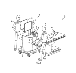
In an example, consider microsurgery visualization where certain surgical
procedures involve patient structures that are too small for a human to
visualize easily with
1
CA 03233597 2024- 4- 2
WO 2023/084334
PCT/IB2022/059634
the naked eye. For these microsurgery procedures, magnification is required to
adequately
view the microstructures. Surgeons generally want visualization tools that are
natural
extensions of their eyes. Indeed, early efforts at microsurgery visualization
comprised
attaching magnifying lens to head-mounted optical eyepieces (called surgical
loupes). The
first pair was developed in 1876. Vastly improved versions of surgical loupes
(some
including optical zooms and integrated light sources) are still being used by
surgeons today.
FIG. 1 shows a diagram of a pair of surgical loupes 100 with a light source
102 and
magnification lenses 104a-b. The 150-year staying power of surgical loupes can
be
attributed to the fact that they are literally an extension of a surgeon's
eyes.
[0005]
Despite their longevity, surgical loupes are not perfect. Loupes with
magnifying
lenses and light sources, such as the surgical loupes 100 of FIG. 1, have much
greater
weight. Placing even a minor amount of weight on the front of a surgeon's face
can increase
discomfort and fatigue, especially during prolonged surgeries. The surgical
loupes 100 also
include a cable 106 that is connected to a remote power supply. The cable
effectively acts
as a chain, thereby limiting the mobility of the surgeon during their surgical
performance.
[0006]
Another microsurgery visualization tool is the surgical microscope, also
referred
to as the operating microscope. Widespread commercial development of surgical
microscopes began in the 1950s with the intention of replacing surgical
loupes. Surgical
microscopes include optical paths, lenses, and focusing elements that provide
greater
magnification compared to surgical loupes. The large array of optical elements
(and
resulting weight) meant that surgical microscopes had to be detached from the
surgeon.
While this detachment gave the surgeon more room to maneuver, the bulkiness of
the
surgical microscope caused it to consume considerable operating space above a
patient,
thereby reducing the size of the surgical stage.
[0007]
FIG. 2 shows a diagram of a prior art surgical microscope 200. As one can
imagine, the size and presence of the surgical microscope in the operating
area made it
prone to bumping. To provide stability and rigidity at the scope head 201, the
microscope
is connected to relatively large boom arms 202 and 204 or other similar
support structure.
CA 03233597 2024- 4- 2
WO 2023/084334
PCT/IB2022/059634
The large boom arms 202 and 204 consume additional surgical space and reduce
the
maneuverability of the surgeon and staff. In total, the surgical microscope
200 shown in
FIG. 2 could weigh as much as 350 kilograms ("kg").
[0008]
To view a target surgical site using the surgical microscope 200, a surgeon
looks
directly though oculars 206. To reduce stress on a surgeon's hack, the oculars
206 are
generally positioned along a surgeon's natural line of sight using the boom
arm 202 to
adjust height. However, surgeons do not perform by only looking at a target
surgical site.
The oculars 206 have to be positioned such that the surgeon is within arm's
length of a
working di stance to the patient. Such precise positioning is critical to
ensure the surgical
microscope 200 becomes an extension rather than a hindrance to the surgeon,
especially
when being used for extended periods.
[0009]
Like any complex instrument, it takes surgeons tens to hundreds of hours to
feel
comfortable using a surgical microscope. As shown in FIG. 2, the design of the
surgical
microscope 200 requires a substantially 90 angle optical path from the
surgeon to the
target surgical site. For instance, a perfectly vertical optical path is
required from the target
surgical site to the scope head 201. This means that the scope head 201 has to
be positioned
directly above the patient for every microsurgical procedure. In addition, the
surgeon has
to look almost horizontally (or some slight angle downward) into the oculars
206. A
surgeon's natural inclination is to direct his vison to his hands at the
surgical site. Some
surgeons even want to move their heads closer to the surgical site to have
more precise
control of their hand movements. Unfortunately, the surgical microscope 200
does not give
surgeons this flexibility. Instead, surgical microscope 200 ruthlessly
dictates that the
surgeon is to place their eyes on the oculars 206 and hold their head at arm's
length during
their surgical performance, all while consuming valuable surgical space above
the patient.
A surgeon cannot even simply look down at a patient because the scope head 201
blocks
the surgeon's view.
[0010]
To make matters worse, some surgical microscopes, such as shown in surgical
microscope 200, include a second pair of oculars 208 for co-performers (e.g.,
assistant
3
CA 03233597 2024- 4- 2
WO 2023/084334
PCT/IB2022/059634
surgeons, nurses, or other clinical staff). The second pair of oculars 208 is
usually
positioned at a right angle from the oculars 206. The closeness between the
oculars 206
and 208 dictates that the assistant must stand (or sit) in close proximity to
the surgeon,
further restricting movement. This can be annoying to some surgeons who like
to perform
with some space. Despite their magnification benefits surgical microscopes
like surgical
microscope 200 are not natural extensions of a surgeon. Instead, they are
overbearing
directors in the surgical room. Accordingly, there is a need in the art for
improved surgical
microscopes.
SUMMARY
Aspects of the present disclosure provide an ophthalmic imaging apparatus. The
ophthalmic imaging apparatus includes a first camera head mounted in a first
orbital
position above a target surgical site associated with an eye of a patient,
wherein the first
camera head includes at least one stereoscopic lens set providing a first
viewing angle of
the target surgical site. Additionally, the ophthalmic imaging apparatus
includes at least a
second camera head mounted in a second orbital position above the target
surgical site,
wherein the second camera head includes at least one additional stereoscopic
lens set
providing a second viewing angle of the target surgical site different from
the first viewing
angle of the target surgical site.
[0011]
Aspects of the present disclosure provide a process for displaying
stereoscopic
video data of a target surgical site using an ophthalmic imaging apparatus.
The process
includes receiving light from a target surgical site associated with an eye of
a patient using
at least one stereoscopic lens set of a first camera head, wherein: the first
camera head is
mounted in a first orbital position above the target surgical site, and the at
least one
stereoscopic lens set provides a first viewing angle of the target surgical
site. The process
further includes receiving the light from the target surgical site using at
least one other
stereoscopic lens set of a second camera head, wherein: the second camera head
is mounted
in a second orbital position above the target surgical site different from the
first orbital
position, and the at least one other stereoscopic lens set provides a second
viewing angle
4
CA 03233597 2024- 4- 2
WO 2023/084334
PCT/IB2022/059634
of the target surgical site different from the first viewing angle of the
target surgical site.
The process further includes generating image data based on the light received
using the at
least one stereoscopic lens set, generating additional image data based on the
light received
using the at least one other stereoscopic lens set, converting the image data
into
stereoscopic video data and the additional image data into additional
stereoscopic video
data, and displaying at least one of the stereoscopic video data or the
additional
stereoscopic video data on a display monitor..
[0012] The above-described features and advantages and other
possible features and
advantages of the present disclosure will be apparent from the following
detailed
description of the best modes for carrying out the disclosure when taken in
connection with
the accompanying drawings.
BRIEF DESCRIPTION OF THE DRAWINGS
[0013] The drawings described herein are for illustrative
purposes only, are schematic
in nature, and are intended to be exemplary rather than to limit the scope of
the disclosure.
[0014] FIG. 1 shows a diagram of a pair of prior art surgical
loupes.
[0015] FIG. 2 shows a diagram of a prior art surgical
microscope.
[0016] FIG. 3 shows a perspective view of a stereoscopic
visualization camera.
[0017] FIG. 4 shows a diagram illustrative of optical elements
within the example
stereoscopic visualization camera.
[0018] FIG. 5 shows a diagram of a microsurgical environment
including the
stereoscopic visualization camera.
[0019] FIGs. 6A-6C show different views of an imaging apparatus
that includes a
plurality of stereoscopic lens sets each associated with a different fixed
magnification level.
[0020] FIG. 7 shows a diagram of modules of the example imaging
apparatus for
acquiring and processing image data.
[0021] FIG. 8 shows different display configurations for
stereoscopic image data.
CA 03233597 2024- 4- 2
WO 2023/084334
PCT/IB2022/059634
[0022]
FIG. 9 shows an example process for simultaneously displaying different
stereoscopic video data of a target surgical site.
[0023]
FIG. 10 illustrates an ophthalmic imaging apparatus that includes a
plurality of
camera heads, each providing a different viewing angle of a target surgical
site.
[0024]
FIG. 11 shows an example process for displaying different stereoscopic
video
data associated with different viewing angles of a target surgical site.
[0025]
The above summary is not intended to represent every possible embodiment or
every aspect of the subject disclosure. Rather, the foregoing summary is
intended to
exemplify some of the novel aspects and features disclosed herein. The above
features and
advantages, and other features and advantages of the subject disclosure, will
be readily
apparent from the following detailed description of representative embodiments
and modes
for carrying out the subject disclosure when taken in connection with the
accompanying
drawings and the appended claims.
DETAILED DESCRIPTION
[0026]
The present disclosure relates in general to an imaging apparatus and
platform.
The imaging apparatus may be referred to, in some cases, as a digital
stereoscopic
microscope ("DSM"). The example imaging apparatus and platform are configured
to
integrate microscope optical elements and video sensors into a self-contained
head unit or
housing that is significantly smaller, lighter, and more maneuverable than
prior art
microscopes (such as the surgical loupes 100 of FIG. 1 and the surgical
microscope 200
of FIG. 2). The example camera is configured to transmit/display stereoscopic
video data
to/on one or more television monitors, display monitors, projectors,
holographic devices,
smartglasses, virtual reality devices, or other visual display devices within
a surgical
environment.
[0027]
The monitors or other visual display devices may be positioned within the
surgical environment to be easily within a surgeon's line of sight while
performing surgery
on a patient. This flexibility enables the surgeon to place display monitors
based on
6
CA 03233597 2024- 4- 2
WO 2023/084334
PCT/IB2022/059634
personal preferences or habits. In addition, the flexibility and slim profile
of the
stereoscopic visualization camera disclosed herein reduces area consumed over
a patient.
Altogether, the stereoscopic visualization camera and monitors (e.g., the
stereoscopic
visualization platform) enable a surgeon and surgical team to perform complex
microsurgical procedures on a patient without being dictated or restricted in
movement
compared to the surgical microscope 200 discussed above. The example
stereoscopic
visualization platform accordingly operates as an extension of the surgeon's
eyes, enabling
the surgeon to perform masterpiece microsurgeries without dealing with the
stress,
restrictions, and limitations induced by previous known visualization systems.
[0028]
Aspects of the present disclosure provide techniques for enabling the
display of
different stereoscopic video data associated with different fields-of-view and
magnification
levels of a target surgical site. For example, certain surgical microscopes,
such as the
stereoscopic visualization camera 300 illustrated in FIG. 3 and described
below, achieve
these different fields-of-view and magnification levels of the target surgical
site using
multiple fixed focal length lenses that move forward and backward along rails.
[0029]
In certain cases, moving zoom lenses are heavy, expensive, and include
sensitive
optics prone to focusing issues, which makes the stereoscopic visualization
camera more
difficult and more expensive to manufacture. Additionally, the parts that move
the zoom
lenses (e.g., motors, rails, etc.) are prone to wearing down and breaking,
which can lead to
costly repairs. Moreover, a surgeon may only be able to view one field-of-
view/magnification level of the target surgical site at a time and may have to
pause surgery
to switch fields-of-view/magnification levels (e.g., to wait for the zoom
lenses to move),
causing delays in the surgery and slowing down workflow.
[0030]
Accordingly, aspects of the present disclosure provide an ophthalmic
imaging
apparatus that includes a plurality of stereoscopic lens sets each associated
with a different
fixed magnification level. Each of these different fixed magnification levels
may be
associated with a different field-of-view of a target surgical site, which may
be
simultaneously di splayed to a surgeon on a display monitor. By providing
multiple lens
7
CA 03233597 2024- 4- 2
WO 2023/084334
PCT/IB2022/059634
sets associated with different magnification levels and simultaneously
displaying
corresponding fields-of-view, the surgeon does not need to pause surgery to
change the
magnification level/field-of-view. Moreover, because the magnification levels
are fixed,
the stereoscopic imaging device may not require moving parts, avoiding complex
and
expensive manufacture and repair.
[0031]
Aspects of the present disclosure provide techniques for enabling the
display of
different stereoscopic video data associated with viewing angles of a target
surgical site.
These different viewing angles are especially important in certain types of
surgeries, such
as retinal and cataract surgeries. Using traditional techniques to achieve
these different
viewing angles involved techniques, such as moving a patients head and scleral
depression.
Newer techniques involve the use of a surgical microscope with a camera head
that is able
to move to different orbital positions above the target surgical site.
However, these
techniques slow down a surgeon's work-flow, leading to longer surgeries.
Additionally, in
some cases, certain techniques, such as scleral depression can lead to
additional trauma
caused to the patient. Additionally, surgical microscopes with moving camera
heads are
costly to manufacture and prone to malfunction, leading to costly repairs and
long down-
times.
[0032]
Accordingly, aspects of the present disclosure provide an ophthalmic
imaging
apparatus that includes a plurality of camera heads mounted in different
orbital positions
above a target surgical site. Each camera head of the plurality of camera
heads may be
configured to provide a different viewing angle of the target surgical site.
By providing
multiple camera heads associated with different viewing angles of the target
surgical site
and displaying different fields-of-view of the target surgical site
corresponding to these
different viewing angles, the surgeon does not need to pause surgery to move a
patient's
head to obtain a peripheral view of the target surgical site or correct
alignment issues when
moving the patient's head back. Nor does the surgeon need to perform scleral
depression,
eliminating the potential additional trauma to the patient associated with
scleral depression.
Further, providing multiple camera heads mounted in different orbital
positions above the
8
CA 03233597 2024- 4- 2
WO 2023/084334
PCT/IB2022/059634
target surgical site eliminates the need for the parts that facilitate the
physical movement
of the single camera of these certain surgical microscopes, avoiding the
manufacturing
expense as well as the complex and costly repairs of such moving parts.
[0033] The disclosure herein generally refers to microsurgery. The example
stereoscopic visualization camera may he used in virtually any microsurgical
procedure
including, for example, cranial surgery, brain surgery, neurosurgery, spinal
surgery,
ophthalmologic surgery, corneal transplants, orthopedic surgery, ear, nose and
throat
surgery, dental surgery, plastics and reconstructive surgery, or general
surgery.
[0034]
The disclosure also refers herein to target surgical site, scene, or field-
of-view.
As used herein, target surgical site or field-of-view includes an object (or
portion of an
object) that is being recorded or otherwise imaged by the example stereoscopic
visualization camera. Generally, the target surgical site, scene, or field-of-
view is a
working distance away from a main objective assembly of the example
stereoscopic
visualization camera and is aligned with the example stereoscopic
visualization camera.
The target surgical site may include a patient's biological tissue, bone,
muscle, skin or
combinations thereof. In these instances, the target surgical site may be
three-dimensional
by having a depth component corresponding to a progression of a patient's
anatomy. The
target surgical site may also include one or more templates used for
calibration or
verification of the example stereoscopic visualization camera. The templates
may be two-
dimensional, such as a graphic design on paper (or plastic sheet) or three
dimensional, such
as to approximate a patient's anatomy in a certain region.
[0035]
Reference is also made throughout to an x-direction, a y-direction, a z-
direction,
and a tilt-direction. The z-direction is along an axis from the example
stereoscopic
visualization camera to the target surgical site and generally refers to depth
The x -di recti on
and y-direction are in a plane incident to the z-direction and comprise a
plane of the target
surgical site. The x-direction is along an axis that is 90 from an axis of
the y-direction.
Movement along the x-direction and/or the y-direction refers to in-plane
movement and
may refer to movement of the example stereoscopic visualization camera,
movement of
9
CA 03233597 2024- 4- 2
WO 2023/084334
PCT/IB2022/059634
optical elements within the example stereoscopic visualization camera, and/or
movement
of the target surgical site.
Example Stereoscopic Visualization Camera
[0036]
FIG. 3 illustrates a perspective view of a stereoscopic visualization
camera 300.
As shown in FIG. 3, the stereoscopic visualization camera 300 includes a
housing 302
configured to enclose optical elements, lens motors (e.g., actuators), and
signal processing
circuity. FIG. 4 shows an example arrangement and positioning of the optical
elements of
the stereoscopic visualization camera 300. In some cases, the arrangement and
positioning
of the optical elements of the stereoscopic visualization camera 300 forms two
parallel
optical paths to generate a left view and a right view. The parallel optical
paths correspond
to a human's visual system such that the left view and right view, as
displayed on a
stereoscopic display, appear to be separated by a distance that creates a
convergence angle
of, for example, roughly 6 degrees, which is comparable to the convergence
angle for an
adult human's eyes viewing an object at approximately 4 feet away, thereby
resulting in
stereopsis. In some embodiments, image data generated from the left view and
right view
are combined together on the display monitor(s) to generate a stereoscopic
image of a target
surgical site or scene.
[0037] A stereoscopic view, as compared to a monoscopic view, mimics the human
visual system much more closely. A stereoscopic view provides depth
perception, distance
perception, and relative size perception to provide a realistic view of a
target surgical site
to a surgeon. For procedures such as retinal surgery, stereoscopic views are
useful because
surgical movements and forces are so small that the surgeon cannot feel them.
Providing a
stereoscopic view helps a surgeon's brain magnify tactile feel when the brain
senses even
minor movements while perceiving depth.
[0038]
FIG. 4 shows a side view of the example stereoscopic visualization camera
300
with the housing 302 being transparent to expose the optical elements. The
optical elements
shown in FIG. 4 may be part of a left optical path and may generate the left
view. It should
be appreciated that the arrangement and positioning of optical elements in a
right optical
CA 03233597 2024- 4- 2
WO 2023/084334
PCT/IB2022/059634
path in stereoscopic visualization camera 300 (e.g., generating the right
view) may
generally be identical to the left optical path.
[0039]
The example stereoscopic visualization camera 300 is configured to acquire
images of a target surgical site 400 (also referred to as a scene or field-of-
view) at a working
distance 406 above the target surgical site 400_ The target surgical site 400
includes an
anatomical location on a patient. The target surgical site 400 may also
include laboratory
biological samples, calibration slides/templates, etc. Images from the target
surgical site
400 are received at the stereoscopic visualization camera 300 via a main
objective assembly
402, which includes the front working distance lens 407 and a rear working
distance lens
404.
[0040]
To illuminate the target surgical site 400, the example stereoscopic
visualization
camera 300 includes one or more lighting sources, such as a near-infrared
("NIR") light
source 408b, and a near-ultraviolet ("NUV") light source 408c. In other
examples, the
stereoscopic visualization camera 300 may include additional or fewer (or no)
light
sources. For instance, the NIR and NUV light sources may be omitted. The
example light
sources 408 are configured to generate light, which is projected to the target
surgical site
400. The generated light interacts and reflects off the target scene, with
some of the light
being reflected to the main objective assembly 402. Other examples may include
external
light sources or ambient light from the environment.
[0041]
The projection of the light from light sources 408 through the main
objective
assembly provides the benefit of changing the lighted field-of-view based on
the working
distance 406 and/or focal plane. Since the light passes through the main
objective assembly
402, the angle at which light is projected changes based on the working
distance 406 and
corresponds to the angular field-of-view. This configuration accordingly
ensures the field-
of-view is properly illuminated by the light sources 408, regardless of
working distance or
magnification.
[0042]
Further, as illustrated in FIG. 4, the stereoscopic visualization camera
300
includes a deflecting element 412. In some cases, the deflecting element 412
may be
11
CA 03233597 2024- 4- 2
WO 2023/084334
PCT/IB2022/059634
configured to transmit a certain wavelength of light from the NUV light source
408c to the
target surgical site 400 through the main objective assembly 402. The
deflecting element
412 may also be configured to reflect light received from the target surgical
site 400 to
downstream optical elements, including a front lens set 414 for zooming and
recording. In
some embodiments, the deflecting element 412 may filter light received from
the target
surgical site 400 through the main objective assembly 402 so that light of
certain
wavelengths reaches the front lens set 414.
[0043]
The deflecting element 412 may include any type of mirror or lens to
reflect
light in a specified direction. In an example, the deflecting element 412
includes a dichroic
mirror or filter, which has different reflection and transmission
characteristics at different
wavelengths. The stereoscopic visualization camera 300 of FIG. 4 includes a
single
deflecting element 412, which provides light for both the right and left
optical paths. In
other examples, the stereoscopic visualization camera 300 may include separate
deflecting
elements for each of the right and left optical paths. Further, a separate
deflecting element
may be provided for the NUV light source 408c.
[0044]
The example stereoscopic visualization camera 300 of FIG. 4 includes one or
more zoom lens to change a focal length and angle of view of the target
surgical site 400
to provide zoom magnification. In the illustrated example of FIG. 4, the zoom
lens includes
the front lens set 414, a zoom lens assembly 416, and a lens barrel set 418.
In some cases,
the zoom lens may include additional lens(es) to provide further magnification
and/or
image resolution.
[0045]
The front lens set 414 includes a right front lens for the right optical
path and a
left front lens for the left optical path. The lenses left and right front
lenses may each
include a positive converging lens to direct light from the deflecting element
412 to
respective lenses in the zoom lens assembly 416. A lateral position of the
left and right
front lenses accordingly defines a beam from the main objective assembly 402
and the
deflecting element 412 that is propagated to the zoom lens assembly 416.
12
CA 03233597 2024- 4- 2
WO 2023/084334
PCT/IB2022/059634
[0046] The example zoom lens assembly 416 forms an afocal zoom system for
changing
the size of a field-of-view (e.g., a linear field-of-view) by changing a size
of the light beam
propagated to the lens barrel set 418. The zoom lens assembly 416 includes a
front zoom
lens set 424 with a right front zoom lens and a left front zoom lens. The zoom
lens assembly
416 also includes a rear zoom lens set 430 with a right rear zoom lens and a
left rear zoom
lens.
[0047]
The size of an image beam for each of the left and right optical paths is
determined based on a distance between the front zoom lenses in the front zoom
lens set
424, the rear zoom lenses in the rear zoom lens set 430, and the lens barrel
set 418.
Generally, the size of the optical paths reduces as the rear zoom lenses in
the rear zoom
lens set 430 move toward the lens barrel set 418 (along the respective optical
paths),
thereby decreasing magnification. In addition, the front zoom lenses in the
front zoom lens
set 424 may also move toward (or away from) the lens barrel set 418 (such as
in a parabolic
arc), as the rear zoom lenses in the rear zoom lens set 430 move toward the
lens barrel set
418, to maintain the location of the focal plane on the target surgical site
400, thereby
maintaining focus.
[0048]
The front zoom lenses in the front zoom lens set 424 may be included within
a
first carrier while the rear zoom lenses in the rear zoom lens set 430 are
included within a
second carrier. Each of the carriers may be moved on tracks (or rails) along
the optical
paths such left and right magnification may be uniformly adjusted (e.g.,
increased or
decreased). Altogether, the front lens set 414, the zoom lens assembly 416,
and the lens
barrel set 418 are configured to achieve an optical zoom, such as between 5X
to about 20X,
such as at a zoom level that has diffraction-limited resolution.
[0049]
After the light from the target surgical site 400, the light in each of the
right and
left optical paths may pass through one or more optical filters 440 (or filter
assemblies) to
selectively transmit desired wavelengths of light. The light in each of the
right and left
optical paths may then pass through a final optical element set 442 that is
configured to
focus light received from the optical filter 440 onto the optical image sensor
444_
13
CA 03233597 2024- 4- 2
WO 2023/084334
PCT/IB2022/059634
[0050]
As shown, the stereoscopic visualization camera 300 of FIG. 4 includes the
optical image sensor 444, which may be configured to acquire and/or record
incident light
that is received from the final optical element set 442. The optical image
sensor 444
includes a right optical image sensor configured to record light propagating
along the right
optical path and generate right image data associated with the right optical
path.
Additionally, the optical image sensor 444 also includes a left optical image
sensor
configured to record light propagating along the left optical path and
generate left image
data associated with the left optical path. After the right and left image
data are created,
one or more processors may synchronize and combine the left and right image
data to
generate a stereoscopic image. Additionally, the one or more processors may be
configured
to convert a plurality of stereoscopic images into stereoscopic video data for
display to a
user of the stereoscopic visualization camera 300 on a display monitor, such
as a
stereoscopic display.
[0051] Additional aspects of the stereoscopic visualization camera 300 may be
found in
U.S. Patent No. 11,058,513, titled "STEREOSCOPIC VISUALIZATION CAMERA AND
PLATFORM," the entirety of which is incorporated herein by reference.
[0052]
FIG. 5 shows a diagram of the stereoscopic visualization camera 300 used
within a microsurgical environment 500. In some embodiments, the microsurgical
environment 500 of FIG. 5 may be used for an ophthalmic surgery procedure. As
illustrated, the small footprint and maneuverability of the stereoscopic
visualization camera
300 (especially when used in conjunction with a multiple-degree of freedom
arm) enables
flexible positioning with respect to a patient 502. A portion of the patient
502 in view of
the stereoscopic visualization camera 300 includes the target surgical site
400. A surgeon
504 can position the stereoscopic visualization camera 300 in virtually any
orientation
while leaving more than sufficient surgical space above the patient 502 (lying
in the supine
position). The stereoscopic visualization camera 300 accordingly is minimally
intrusive (or
not intrusive) to enable the surgeon 504 to perform a life-altering
microsurgical procedure
without distraction or hindrance.
14
CA 03233597 2024- 4- 2
WO 2023/084334
PCT/IB2022/059634
[0053]
In FIG. 5, the stereoscopic visualization camera 300 is connected to a
mechanical arm 506 (e.g., also referred to a "robot arm-). The mechanical arm
506 may
include one or more rotational or extendable joints with electromechanical
brakes to
facilitate easy repositioning of the stereoscopic visualization camera 300. To
move the
stereoscopic visualization camera 300, the surgeon 504, or the assistant 508,
actuates brake
releases on one or more joints of the mechanical arm 506. After the
stereoscopic
visualization camera 300 is moved into a desired position, the brakes may be
engaged to
lock the joints of the mechanical arm 506 in place.
[0054]
A significant feature of the stereoscopic visualization camera 300 is that
it does
not include oculars. This means that the stereoscopic visualization camera 300
does not
have to be aligned with the eyes of the surgeon 504. This freedom enables the
stereoscopic
visualization camera 300 to be positioned and orientated in desirable
positions that were
not practical or possible with prior known surgical microscopes. In other
words, the
surgeon 504 can perform microsurgery with, for example, the most optimal view
for
conducting the procedure rather than being restricted to a merely adequate
view dictated
by oculars of a surgical microscope.
[0055]
As shown in FIG. 5, the stereoscopic visualization camera 300, via the
mechanical arm 506, is connected to a cart 510 with display monitors 512 and
514
(collectively a stereoscopic visualization platform 516). In the illustrated
configuration, the
stereoscopic visualization platform 516 is self-contained and may be moved to
any desired
location in the microsurgical environment 500 including between surgical
rooms. The
integrated stereoscopic visualization platform 516 enables the stereoscopic
visualization
camera 300 to be moved and used on-demand without time needed to configure the
system
by connecting the display monitors 512 and 514.
[0056]
Each of the display monitors 512 and 514 may include any type of display
including a high-definition television, an ultra-high definition television,
smart-eyewear, a
projector, one or more computer screens, a laptop computer, a tablet computer,
and/or a
smartphone. The di splay monitors 512 and 514 may be connected to mechanical
arms to
CA 03233597 2024- 4- 2
WO 2023/084334
PCT/IB2022/059634
enable flexible positioning similar to the stereoscopic visualization camera
300. In some
instances, one or more of the display monitors 512 and 514 may include a
touchscreen to
enable an operator to send commands to the stereoscopic visualization camera
300 and/or
adjust a setting of a display.
[0057]
In some embodiments, the cart 510 may include a computer 520. In these
embodiments, the computer 520 may control a robotic mechanical arm connected
to the
stereoscopic visualization camera 300. Additionally or alternatively, the
computer 520 may
process video (or stereoscopic video) signals (e.g., an image or frame stream)
from the
stereoscopic visualization camera 300 for display on the di splay monitors 512
and 514. For
example, the computer 520 may combine or interleave left and right video
signals from the
stereoscopic visualization camera 300 to create a stereoscopic signal for
displaying a
stereoscopic image of a target surgical site. The computer 520 may also be
used to store
video and/or stereoscopic video signals into a video file (stored to a memory)
so the
surgical performance can be documented and played back. Further, the computer
520 may
also send control signals to the stereoscopic visualization camera 300 to
select settings
and/or perform calibration.
Aspects Related to a Stereoscopic Imaging Apparatus with Multiple Fixed
Magnification
Levels
[0058]
Digital stereoscopic microscopes, such as the stereoscopic visualization
camera
300, are especially useful when performing eye surgery. Typically, in surgical
microscopes, such as the stereoscopic visualization camera 300, multiple zoom
or
magnification levels are accomplished by designing the surgical microscope to
have
moving zoom lens groups, such as the front and rear zoom lenses in the a zoom
lens
assembly 416 of the stereoscopic visualization camera 300 illustrated in FIG.
4. For
example, as the front and rear zoom lenses in the zoom lens assembly 416 move
forward
and backward along rails, light from the target surgical site 400 passing
through the these
lenses focuses at different distances, resulting in different zooms or
magnification levels.
However, moving zoom lenses are heavy, expensive, and include sensitive
objects prone
16
CA 03233597 2024- 4- 2
WO 2023/084334
PCT/IB2022/059634
to focusing issues, which makes the stereoscopic visualization camera 300 more
difficult
and more expensive to manufacture. Additionally, the parts that move the zoom
lenses
(e.g., motors, rails, etc.) are prone to wearing down and breaking, which can
lead to costly
repairs.
[0059]
Moreover, moving 700111 lenses are capable of producing only one
magnification
level at any given point in time. As a result, only one field-of-view of the
target surgical
site 400 may be displayed to a surgeon (e.g., surgeon 504 in FIG. 5) at any
given point.
This may be problematic since, during surgery, surgeons change between
different
zoom/magnification levels in order to accomplish various tasks. For example,
larger
zooms/greater magnification (e.g., resulting in a narrow field-of-view of the
target surgical
site 400) may be used when minute details of the target surgical site need to
be seen while
performing difficult surgical movements. In contrast, lower zooms/less
magnification may
be used when a "bigger picture" view of the target surgical site 400 is
needed, for example,
during instrument insertion/exchange. However, in order to change
zoom/magnification
level, the surgeon must pause during surgery and wait for the moving lenses to
adj ust to a
proper zoom/magnification level, causing delays in the surgery and slowing
down
workflow.
[0060]
Accordingly, aspects of the present disclosure provide an ophthalmic
imaging
apparatus that includes a plurality of stereoscopic lens sets each associated
with a different
fixed magnification level. Each of these different fixed magnification levels
may be
associated with a different field-of-view of a target surgical site, which may
be
simultaneously displayed to a surgeon on a display monitor. For example, in
some
embodiments, the ophthalmic imaging apparatus may include a first stereoscopic
lens set
associated with a first fixed magnification level and a first field-of-view,
such as a narrow
field-of-view showing minute details of the target surgical site.
Additionally, the
ophthalmic imaging apparatus may include a second lens set associated with a
second fixed
magnification level and second field-of-view, such as a broad field of view
showing a
"bigger picture" of the target surgical site.
17
CA 03233597 2024- 4- 2
WO 2023/084334
PCT/IB2022/059634
[0061]
Accordingly, these different field-of-views of the target surgical site may
be
simultaneously displayed to the surgeon on a display monitor. In some
embodiments, these
different field-of-views may be displayed using a picture-in-picture (PIP)
configuration or
side by side. By providing multiple lens sets associated with different fixed
magnification
levels and simultaneously displaying corresponding fields-of-view, the surgeon
does not
need to pause surgery to change the magnification level/field-of-view.
Moreover, because
the magnification levels are fixed, the stereoscopic imaging device may not
require moving
parts, avoiding the complex and expensive manufacture and repair.
[0062]
It should be understood that a stereoscopic lens set with a fixed
magnification
level refers to a stereoscopic lens set that is designed to a certain
magnification level or
focal length while including components that allow for making minor
adjustments to the
designed magnification level for fine focus. Accordingly, while each of the
first and the
second stereoscopic lens sets are designed to a different fixed magnification
level, the first
and the second stereoscopic lens sets may each include certain components that
allow for
minor adjustments to be made to the fixed magnification levels to enable fine
focusing.
[0063]
FIGs. 6A, 6B, and 6C respectively illustrate a perspective view, as left-
side
view, and a right-side view of an imaging apparatus 600 that includes a
plurality of
stereoscopic lens sets each associated with a different fixed magnification
level. In some
embodiments, the imaging apparatus 600 may be implemented in a microsurgical
environment, such as the microsurgical environment 500. More specifically, in
some
embodiments, the imaging apparatus 600 is configured to replace the
stereoscopic
visualization camera 300 in the microsurgical environment 500.
[0064]
As illustrated, the imaging apparatus 600 includes a housing 601 configured
to
enclose optical elements and signal processing circuity. Further, as
illustrated, the imaging
apparatus 600 includes a first stereoscopic lens set configured to receive
light from a target
surgical site 603, which may be an example of the target surgical site 400
illustrated in
FIG. 4. In some embodiments, the target surgical site 603 may be associated
with an eye
of a patient. In some embodiments, the received light may be generated by a
light source
18
CA 03233597 2024- 4- 2
WO 2023/084334
PCT/IB2022/059634
610. For example, the light source 610 may be configured to emit light on to
the target
surgical site 603. In some embodiments, the light source 610 may be an example
of one or
more of the light sources 408A-408C illustrated in FIG. 4.
[0065]
As illustrated, the first stereoscopic lens set may include at least a
first left lens
barrel 602A and a first right lens barrel 602B. As shown, the first left lens
barrel 602A and
the first right lens barrel 602B define respective first parallel left and
right optical paths,
such as the first left optical path 612A and the first right optical path
612B. The first left
lens barrel 602A and the first right lens barrel 602B are configured to
receive light from
slightly different perspectives of the target surgical site 603, providing a
stereoscopic view
of the target surgical site 603.
[0066]
Additionally, as illustrated, the imaging apparatus 600 also includes a
second
stereoscopic lens set configured to receive additional light from the target
surgical site
generated by the light source 610. For example, the second stereoscopic lens
set may
include a second left lens barrel 604A and a second right lens barrel 604B. As
shown, the
second left lens barrel 604A and the second right lens barrel 604B define
respective second
parallel left and right optical paths, such as the second left optical path
614A and the second
right optical path 614B. Similar to the first left lens barrel 602A and the
first right lens
barrel 602B, the second left lens barrel 604A and the second right lens barrel
604B are
configured to receive light from the target surgical site 603 at the slightly
different angles,
providing another stereoscopic view of the target surgical site 603.
[0067]
Further, in some embodiments, the first left lens barrel 602A and the first
right
lens barrel 602B of the first lens set include a first set of fixed focal
length lenses configured
to magnify the received light from the target surgical site 603 according to a
first fixed
magnification level. More specifically, as shown, the first left lens barrel
602A includes
the first left fixed focal length lens 606A and the first right lens barrel
602B includes the
first right fixed focal length lens 606B. Each of the fixed focal length
lenses 606A and
606B are configured to magnify the received light from the target surgical
site 603
according to the first fixed magnification level. In some embodiments, the
first fixed
19
CA 03233597 2024- 4- 2
WO 2023/084334
PCT/IB2022/059634
magnification level may depend on a focal length associated with the fixed
focal length
lenses 606A and 606B and may provide a first field-of-view of the target
surgical site 603.
For example, in some embodiments, the first fixed magnification level of the
fixed focal
length lenses 606A and 606B may provide a narrow field-of-view showing minute
details
of the target surgical site 603. Because the fixed focal length lenses 606A
and 606B are
associated with a fixed magnification level, the imaging apparatus 600 may not
require
moving parts (e.g., motors, rails, etc.) in order to achieve the narrow field-
of-view of the
target surgical site. It should be understood that, while the fixed focal
length lenses 606A
and 606B are designed to a first fixed magnification level or focal length,
the first left lens
barrel 602A and the first right lens barrel 602B may each include certain
components that
allow for minor adjustments to be made to the first fixed magnification level
to enable fine
focusing.
[0068]
Additionally, in some embodiments, the second left lens barrel 604A and the
second right lens barrel 604B of the second lens set include a second set of
fixed focal
length lenses configured to magnify the received additional light from the
target surgical
site 603 according to a second fixed magnification level different from the
first fixed
magnification level. More specifically, as shown, the second left lens barrel
604A includes
the second left fixed focal length lens 608A and the second right lens barrel
604B includes
the second right fixed focal length lens 608B. Each of the fixed focal length
lenses 608A
and 608B are configured to magnify the received light from the target surgical
site 603
according to the second fixed magnification level. In some embodiments, the
second fixed
magnification level may depend on a focal length associated with the fixed
focal length
lenses 608A and 608B and may provide a second field-of-view of the target
surgical site
603. For example, in some embodiments, the second fixed magnification level of
the fixed
focal length lenses 608A and 608B may provide a "bigger picture" or wide field-
of-view
showing larger/wider details of the target surgical site 603. Because the
fixed focal length
lenses 608A and 608B are associated with a fixed magnification level, the
imaging
apparatus 600 may not require moving parts (e.g., motors, rails, etc.) in
order to achieve
the "bigger picture"/wide field-of-view of the target surgical site. It should
be understood
CA 03233597 2024- 4- 2
WO 2023/084334
PCT/IB2022/059634
that, while the fixed focal length lenses 608A and 608B are designed to a
second fixed
magnification level, the second left lens barrel 604A and the second right
lens barrel 604B
may each include certain components that allow for minor adjustments to be
made to the
second fixed magnification level to enable fine focusing.
[0069]
Further, the imaging apparatus 600 may include a first plurality of
dichroic
mirrors and a second a second plurality of dichroic mirrors. As illustrated in
FIG. 6B, the
first plurality of dichroic mirrors may include a first left dichroic mirror
616A associated
with the first left lens barrel 602A. Additionally, as illustrated in FIG. 6C,
the first plurality
of dichroic mirrors may include a first right dichroic mirror 616B associated
with the first
right lens barrel 602B. Further, as illustrated in FIG. 6B, the second
plurality of dichroic
mirrors may include a second left dichroic mirror 618A associated with the
second left lens
barrel 604A. Additionally, as illustrated in FIG. 6C, second plurality of
dichroic mirrors
may include a second right dichroic mirror 618B associated with the second
right lens
barrel 604B.
[0070]
In some embodiments, the first plurality of dichroic mirrors is configured
to
direct the received light from the first left lens barrel 602A and first right
lens barrel 602B
to a first plurality of image sensors of the imaging apparatus 600. For
example, the first
plurality of image sensors may include a first left image sensor 620A
associated with the
first left lens barrel 602A and a first right image sensor 620B associated
with the first right
lens barrel 602B. Accordingly, the first left dichroic mirror 616A and the
first right dichroic
mirror 616B may be configured to direct the received light to the first left
image sensor
620A and first right image sensor 620B, respectively, along the first parallel
left and right
optical paths (e.g., along the first left optical path 612A and the first
right optical path
612B).
[0071]
Further, the second plurality of dichroic mirrors is configured to direct
the
received additional light from the second left lens barrel 604A and second
right lens barrel
604B to a second plurality of image sensors of the imaging apparatus 600. For
example,
the second plurality of image sensors may include and a second left image
sensor 622A
21
CA 03233597 2024- 4- 2
WO 2023/084334
PCT/IB2022/059634
associated with the second left lens barrel 604A and a second right image
sensor 622B
associated with the second right lens barrel 604B. Accordingly, the second
left dichroic
mirror 618A and the second right dichroic mirror 618B may be configured to
direct the
received additional light to the second left image sensor 622A and the second
right image
sensor 622B, respectively, along the second parallel left and right optical
paths (e.g., along
the second left optical path 614A and the second right optical path 614B).
[0072]
According to aspects, the first plurality of image sensors (e.g., the first
left image
sensor 620A and the first right image sensor 620B) may be configured to
receive the light
after passing through the first stereoscopic lens set and being directed by
the first left
dichroic mirror 616A and the first right dichroic mirror 616B, respectively.
Further, each
image sensor of the first plurality of image sensors (e.g., the first left
image sensor 620A
and the first right image sensor 620B) may be configured to generate first
image data based
on the light received from the first stereoscopic lens set. For example, the
first left image
sensor 620A may be configured to generate first left image data based on the
received light
from the first left lens barrel 602A and the first right image sensor 620B may
be configured
to generate first right image data based on the received light from the first
right lens barrel
602B. In some embodiments, the first image data (e.g., first left image data
and first right
image data) may provide images of a first field-of-view of the target surgical
site 603, such
as the narrow field-of-view described above showing minute details of the
target surgical
site 603.
[0073]
Similarly, the second plurality of image sensors (e.g., the second left
image
sensor 622A and the second right image sensor 622B) may be configured to
receive the
additional light after passing through the second stereoscopic lens set and
being directed
by the second left dichroic mirror 618A and the second right dichroic mirror
618B,
respectively. Further, each image sensor of the second plurality of image
sensors (e.g., the
second left image sensor 622A and the second right image sensor 622B) may be
configured
to generate second image data based on the additional light received from the
second
stereoscopic lens set. For example, the second left image sensor 622A may be
configured
77
CA 03233597 2024- 4- 2
WO 2023/084334
PCT/IB2022/059634
to generate second left image data based on the received additional light from
the second
left lens barrel 604A and the second right image sensor 622B may be configured
to generate
second right image data based on the received additional light from the second
right lens
barrel 604B. In some embodiments, the second image data (e.g., second left
image data
and second right image data) may provide images of a second field-of-view of
the target
surgical site 603, such as the "bigger picture- or wide field-of-view of the
target surgical
site 603, described above.
[0074]
As will be explained in greater detail below, the image data from
corresponding
left and right image sensors may be converted in to stereoscopic video data
for display on
a display monitor by one or more processors of the imaging apparatus 600. For
example,
FIG. 7 shows a diagram of modules of the example imaging apparatus 600 for
acquiring
and processing image data, according to an example embodiment of the present
disclosure.
It should be appreciated that the modules are illustrative of operations,
methods,
algorithms, routines, and/or steps performed by certain hardware, controllers,
processors,
drivers, and/or interfaces. In other embodiments, the modules may be combined,
further
partitioned, and/or removed. Further, one or more of the modules (or portions
of a module)
may be provided external to the imaging apparatus 600 such as in a remote
server,
computer, and/or distributed computing environment.
[0075]
In the illustrated embodiment of FIG. 7, the optical elements 702 may
include
the first left lens barrel 602A, the first right lens barrel 602B, the second
left lens barrel
604A, the second right lens barrel 604B, the first left fixed focal length
lens 606A, the first
right fixed focal length lens 606B, the second left fixed focal length lens
608A, the second
right fixed focal length lens 608B, the light source 610, the first left
dichroic mirror 616A,
the first right dichroic mirror 616B, the second left dichroic mirror 618A,
the second right
dichroic mirror 618B, the first left image sensor 620A, the first right image
sensor 620B,
the second left image sensor 622A, and the second right image sensor 622B. The
optical
elements 702 (specifically the left and right image sensors 620A, 620B, 622A,
and 622B)
are communicatively coupled to an image capture module 704 and a motor and
lighting
23
CA 03233597 2024- 4- 2
WO 2023/084334
PCT/IB2022/059634
module 706. The image capture module 704 is communicatively coupled to an
information
processing module 708, which may be communicatively coupled to an externally
located
user input device 710 and one or more display monitors 712. In some
embodiments, the
one or more display monitors may be examples of the display monitors 512
and/or 514
illustrated in FIG. 5.
[0076] The example image capture module 704 is configured to receive image
data from
the left and right image sensors 620A, 620B, 622A, and 622B. For example, the
image
capture module 704 may be configured to receive the first left image data from
the first left
image sensor 620A, the first right image data from the first right image
sensor 620B, the
second left image data from the second left image sensor 622A, and the second
right image
data from the second right image sensor 622B. The image capture module 1404
may also
specify image recording properties, such as frame rate and exposure time for
capturing the
image data.
[0077]
The example lighting module 706 is configured to control the light source
610.
For example, in some embodiments, the lighting module 706 may include one or
more
drivers for controlling the light source 610 to emit light on the target
surgical site 603.
[0078]
The example information processing module 708 is configured to process
image
data for display. For instance, the information processing module 708 may
provide color
correction to image data, filter defects from the image data, and/or render
image data for
stereoscopic display. The information processing module 708 may also perform
one or
more calibration routines to calibrate the imaging apparatus 600 by providing
instructions
to the image capture module 704 and/or the motor and lighting module 706 to
perform
specified adjustments to the optical elements. The information processing
module 708 may
further determine and provide real-time instructions to the image capture
module 704
and/or the motor and lighting module 706 to improve image alignment and/or
reduce
spurious parallax.
[0079] In some embodiments, the information processing module 708 may include
one
or more processors that are communicatively coupled to the first plurality of
image sensors
24
CA 03233597 2024- 4- 2
WO 2023/084334
PCT/IB2022/059634
(e.g., the first left image sensor 620A and the first right image sensor 620B)
and to the
second plurality of image sensors (e.g., the second left image sensor 622A and
the second
right image sensor 622B). In some embodiments, the one or more processors may
be
configured to convert the first image data into first stereoscopic video data
for display on
the one or more display monitors 712. For example, in some embodiments, the
one or more
processors may be configured to combine the first left image data generated by
the first left
image sensor 620A with the first right image data generated by the first right
image sensor
620B into the first stereoscopic video data. In some embodiments, converting
the first
image data into first stereoscopic video data may include interleaving rows of
pixels of the
first left image data and first right image data. In some embodiments, the
first stereoscopic
video data may represent and show the narrow field-of-view of the target
surgical site 603,
as discussed above with respect to the first image data.
[0080]
Additionally, the one or more processors of the information processing
module
708 may be configured to convert the second image data into second
stereoscopic video
data for display on the one or more display monitors 712. For example, in some
embodiments, the one or more processors may be configured to combine the
second left
image data generated by the second left image sensor 622A with the second
right image
data generated by the second right image sensor 622B into the second
stereoscopic video
data. In some embodiments, converting the second image data into second
stereoscopic
video data may include interleaving rows of pixels of the second left image
data and second
right image data. In some embodiments, the second stereoscopic video data may
represent
and show the -bigger picture" or wide field-of-view of the target surgical
site 603, as
discussed above with respect to the second image data.
[0081]
In some embodiments, the one or more processors of the information
processing
module 708 may be configured to display only one of the first stereoscopic
video data or
the second stereoscopic video data at a time on the one or more display
monitors 712. In
other embodiments, the one or more processors of the information processing
module 708
may be configured to display the first stereoscopic video data on the one or
more display
CA 03233597 2024- 4- 2
WO 2023/084334
PCT/IB2022/059634
monitors 712 simultaneously with the second stereoscopic video data. For
example, in
some embodiments, the one or more processors may display the first
stereoscopic video
data and the second stereoscopic video data side-by-side on the one or more
display
monitors 712. An example of this side-by-side display is illustrated in FIG.
8A. For
example, as shown in FIG. 8A, the one or more processors may display the first
stereoscopic video data 802 (e.g., corresponding to the "bigger picture- or
wide field-of-
view of the target surgical site 603) next to the second stereoscopic video
data 804 (e.g.,
corresponding to the narrow field-of-view of the target surgical site 603).
[0082]
In certain embodiments, the one or more processors may display the first
stereoscopic video data and the second stereoscopic video data on the one or
more display
monitors 712 using a picture-in-picture configuration. An example of this
picture-in-
picture configuration is illustrated in FIG. 8B. For example, as shown in FIG.
8B, the one
or more processors may di splay the first stereoscopic video data 802 (e.g.,
corresponding
to the "bigger picture- or wide field-of-view of the target surgical site 603)
spanning the
entire display area of the one or more display monitors. Further, the one or
more processors
may display the second stereoscopic video data 804 (e.g., corresponding to the
narrow
field-of-view of the target surgical site 603) in a frame within the first
stereoscopic video
data 802.
[0083] The example user input device 710 may include a computer to provide
instructions for changing operation of the imaging apparatus 600. The user
input device
710 may also include controls for selecting parameters and/or features of the
imaging
apparatus 600. In some embodiments, the user input device 710 may be
configured to allow
a user of the imaging apparatus 600 to switch between different magnification
levels and
fields-of-view of the target surgical site 603. For example, in some
embodiments, the user
input device 710 may allow a user of the imaging apparatus 600 to switch
between the first
fixed magnification level associated with fixed focal length lenses 606A and
606B (e.g.,
the narrow field-of-view of the target surgical site 603) to the second fixed
magnification
26
CA 03233597 2024- 4- 2
WO 2023/084334
PCT/IB2022/059634
level associated with fixed focal length lenses 608A and 608B (e.g., the
"bigger
picture-/wide field-of-view of the target surgical site 603).
[0084]
Because fixed focal length lenses 606A, 606B, 608A, and 608B of the imaging
apparatus 600 do not have moving parts, the different fields-of-view of the
target surgical
site 603 (e.g., the narrow field-of-view in the first stereoscopic video data
and the wide
field-of-view in the second stereoscopic video data) may be interchanged and
displayed on
the one or more display monitors 712 almost instantly. Additionally, in some
embodiments,
the user input device 710 may also be configured to allow the user of the
imaging apparatus
600 to switch between different display configurations associated with the
first
stereoscopic video data and the second stereoscopic video data, such as the
side-by-side
configuration illustrated in FIG. 8A and the picture-in-picture configuration
illustrated in
FIG. 8B.
[0085]
Further, in some embodiments, user input device 710 may include a button or
a
foot pedal on the imaging apparatus 600 that allows the user to switch between
the different
magnification levels and/or display configurations. In some embodiments, the
user input
device 710 may be hardwired to the information processing module 1408.
Additionally or
alternatively, the user input device 710 is wirelessly or optically
communicatively coupled
to the information processing module 1408.
[0086]
While the imaging apparatus 600 is describe above as including a first
stereoscopic lens set and a second stereoscopic lens set each associated with
a different
fixed magnification level, it should be understood that the imaging apparatus
may include
any number of stereoscopic lens sets (e.g., one, two, three, or more) that are
each associated
with a different fixed magnification level. Additionally, in some embodiments,
the first
stereoscopic lens set may include fixed focal length lenses and be associated
with a fixed
magnification level while the second stereoscopic lens set may include moving
zoom
lenses (e.g., similar to the front and rear zoom lenses in the a zoom lens
assembly 416 of
the stereoscopic visualization camera 300) and associated with an adjustable
magnification
level
27
CA 03233597 2024- 4- 2
WO 2023/084334
PCT/IB2022/059634
[0087]
FIG. 9 illustrates an example process 900 for displaying different
stereoscopic
video data of a target surgical site. In some embodiments, the different
stereoscopic video
data may be associated with different fields-of-view and magnification levels
of the target
surgical site. In some embodiments, the process 900 may be performed by an
imaging
apparatus, such as the imaging apparatus 600, or one or more component in the
imaging
apparatus 600, such as the optical elements 702, the image capture module 704,
the lighting
module 706, the information processing module 708, the user input device 710,
and/or the
one or more display monitors 712.
[0088]
The process 900 begins at 902 with receiving light from a target surgical
site
(e.g., target surgical site 603) using a first stereoscopic lens set. The
first stereoscopic lens
set may include one or more components, such as the first left lens barrel
602A, the first
right lens barrel 602B, the first left fixed focal length lens 606A, and/or
the first right fixed
focal length lens 606B of FIGs. 6A-6C. In some embodiments, the light received
from the
target surgical site refers to a portion of the light that is reflected from
the target surgical
site after being emitted from a light source (e.g., light source 610). In some
embodiments,
the light from the target surgical site may be received by a first plurality
of image sensors,
such as the first left image sensor 620A and the first right image sensor
620B.
[0089]
The process 900 continues at 904 with receiving additional light from the
target
surgical site using a second stereoscopic lens set. The second stereoscopic
lens set may
include one or more components, such as the second left lens barrel 604A, the
second right
lens barrel 604B, the second left fixed focal length lens 608A, and/or the
second right fixed
focal length lens 608B. In some embodiments, the additional light from the
target surgical
site may be received by a second plurality of image sensors, such as the
second left image
sensor 622A and the second right image sensor 622B.
[0090]
The process 900 continues at 906 with generating first image data and
second
image data based, respectively, on the light received using the first
stereoscopic lens set
and on the additional light received using the first stereoscopic lens set.
For example, in
some embodiments, the first plurality of image sensors (e_g_, the first left
image sensor
28
CA 03233597 2024- 4- 2
WO 2023/084334
PCT/IB2022/059634
620A and the first right image sensor 620B) may be used to generate the first
image data
based on the light received using the first stereoscopic lens set.
Additionally, the second
plurality of image sensors (e.g., the second left image sensor 622A and the
second right
image sensor 622B) may be used to generate the first image data based on the
additional
light received using the second stereoscopic lens set.
[0091]
The process 900 continues at 908 with converting the first image data into
first
stereoscopic video data and the second image data into second stereoscopic
video data. In
some embodiments, one or more processors of the information processing module
708 may
be used to convert the first image data into first stereoscopic video data and
the second
image data into second stereoscopic video data. In some embodiments,
converting the first
image data into the first stereoscopic video data may involve interleaving
rows of pixels of
first left image data generated by the first left image sensor 620A with first
right image
data generated by the first right image sensor 620B. Similarly, converting the
second image
data into the second stereoscopic video data may involve interleaving rows of
pixels of
second left image data generated by the second left image sensor 622A with
second right
image data generated by the second right image sensor 622B.
[0092]
The process 900 continues at 910 with displaying the first stereoscopic
video
data and the second stereoscopic video data on a display monitor, such as the
one or more
display monitors 712. In some embodiments, displaying the first stereoscopic
video data
and the second stereoscopic video data on a display monitor may be performed
by the one
or more processors of the information processing module 708. In some
embodiments,
displaying the first stereoscopic video data and the second stereoscopic video
data may
include simultaneously displaying the first stereoscopic video data and the
second
stereoscopic video data on the display monitor. In some embodiments,
simultaneously
displaying the first stereoscopic video data and the second stereoscopic video
data on the
display monitor may include displaying first stereoscopic video data and the
second
stereoscopic video data using a side-by-side configuration, as illustrated in
FIG. 8A. In
other embodiments, simultaneously displaying the first stereoscopic video data
and the
29
CA 03233597 2024- 4- 2
WO 2023/084334
PCT/IB2022/059634
second stereoscopic video data on the display monitor may include
simultaneously
displaying first stereoscopic video data and the second stereoscopic video
data using a
picture-in-picture configuration, as illustrated in FIG. 8B.
[0093]
In some embodiments, the process 900 may further include receiving input
from
a user and, based on the input from the user, switching from displaying the
first
stereoscopic video data on the display monitor to displaying the second
stereoscopic video
data on the display monitor.
Aspects Related to an Imaging Apparatus Including Multiple Stereoscopic
Cameras
[0094]
In certain ophthalmic surgeries, being able to view a target surgical site
(e.g.,
associated with a patient's eye), such as the target surgical site 603, from
different viewing
angles is important. For example, after retinal surgery, surgeons need to look
for small
retina tears and residual vitreous at a periphery of the eye. Additionally, in
cataract
surgeries, surgeons may need a different angles of view to see under an edge
of the iris of
the eye to see if an intraocular lens (IOL) has been placed in a sulcus of a
capsular bag of
the eye. Another type of surgery in which different viewing angles are
important is
minimally-invasive glaucoma surgery. Tn minimally-invasive glaucoma surgery,
the
surgeon may need different angles of view when removing a trabecular meshwork
of the
eye (e.g., an area of tissue in the eye located around the base of the cornea)
and/or when
placing stents around the cornea to allow for proper drainage of fluid within
the eye.
[0095]
These different viewing angles can be achieved using traditional techniques
and
a surgical microscope that includes a camera positioned directly above the
target surgical
site. For example, one way of achieving these different viewing angles is
through a
technique known as sclera] depression. Sclera] depression involves inserting a
tip of a
scleral depressor between a globe and an orbit of the eye. A space occupied by
the probe
displaces the retina inward and creates an elevation. This technique enhances
contrast
between a retinal lesion and the surrounding retinal tissue allowing for a
peripheral view of
the retina via the camera positioned directly above. Another way to achieve
the different
CA 03233597 2024- 4- 2
WO 2023/084334
PCT/IB2022/059634
viewing angles is by moving the patient's head slightly. In extreme cases, a
headset
connected to an indirect ophthalmoscope lens can be used to obtain these
different views.
[0096]
While these traditional techniques may be able to achieve different viewing
angle of a target surgical site, these techniques are not ideal and have
certain drawbacks.
For example, scleral depression can cause trauma to the patient's eye and lead
to longer
recovery times and additional pain. Additionally, scleral depression is
another step in the
surgeon's workflow, leading to longer and less efficient surgeries. Moreover,
moving the
patient's head is also dangerous for the patient and interrupts normal
workflow.
Additionally, there is no guarantee that the patient's eye will be in the same
place after
moving the patient's head back, again interrupting workflow and causing
alignment issues
that require the surgical microscope to be moved.
[0097]
Newer techniques for achieving different views of the target surgical site
involve
using a surgical microscope that includes an orbiting camera with lock-to-
target
functionality. For example, in these types of surgical microscopes, the camera
may be
positioned above the target surgical site and free to different orbital
positions around the target
surgical site to obtain the different views of the target surgical site.
Additionally, to help
facilitate this movement, the lock-to-target functionality allows the camera
to lock on to the
target surgical site while moving to a new orbital position so that the
patient does not have to
be repositioned. However, these types of surgical microscopes are expensive
and have bulky
controller boxes, necessitating a larger cart and bigger operating room
footprint. Moreover,
when a different view of the target surgical site is needed, the surgeon still
needs to wait for
the camera of the surgical microscope to move to a new position, which
interrupts surgical
work flow and results in longer surgeries. Additionally, the parts that
facilitate this type of
movement are expensive and prone to malfunction, leading to complex and costly
repairs as
well as loss of use for long periods of time.
[0098]
Accordingly, aspects of the present disclosure provide an ophthalmic
imaging
apparatus that includes a plurality of camera heads mounted in different
orbital positions
above a target surgical site. Each camera head of the plurality of camera
heads may be
31
CA 03233597 2024- 4- 2
WO 2023/084334
PCT/IB2022/059634
configured to provide a different viewing angle of the target surgical site.
For example, in
some embodiments, the ophthalmic imaging apparatus may include a first camera
head that
is located in a first an orbital position above the target surgical. The first
orbital position
may be directly above the target surgical site and allow the first camera head
to provide a
first viewing angle of the target surgical site. Additionally, in some
embodiments, the
ophthalmic imaging apparatus may include at least one additional camera head
positioned
in an additional orbital position above the target surgical site. In some
embodiments, this
additional orbital position may be located laterally to the first orbital
position and may
allow the additional camera to provide a second viewing angle of the target
surgical site
different from the first viewing angle. In some embodiments, the first viewing
angle may
provide a frontal view of the target surgical site while the second viewing
angle may
provide a peripheral view of the target surgical site
[0099]
In some embodiments, these different viewing angles of the target surgical
site
may be simultaneously displayed to the surgeon on a display monitor. In some
embodiments, these different field-of-views may be displayed using a picture-
in-picture
(PIP) configuration or side by side. In some embodiments these different
viewing angles
may be individually or sequentially displayed to the surgeon on the display
monitor. In
such embodiments, the surgeon may be able to switch between the different
viewing angles
almost instantly at the push of a button.
[00100] By providing multiple camera heads associated with different viewing
angles of
the target surgical site and displaying different fields-of-view of the target
surgical site
corresponding to these different viewing angles (e.g., simultaneously or
individually but
instantly switchable via a push of a button), the surgeon does not need to
pause surgery to
move a patient's head to obtain a peripheral view of the target surgical site
or correct
alignment issues when moving the patient's head back. Nor does the surgeon
need to
perform scleral depression, eliminating the potential additional trauma to the
patient
associated with scleral depression.
32
CA 03233597 2024- 4- 2
WO 2023/084334
PCT/IB2022/059634
[00101] Moreover, because these different viewing angles are provided by
multiple
camera heads mounted in different orbital positions above the target surgical
site, the
surgeon may not need to pause surgery to wait for a single camera head of
certain surgical
microscopes to lock on to a target and physically move to a new position to
obtain a
different view of the target surgical site. Further, providing multiple camera
heads mounted
in different orbital positions above the target surgical site eliminates the
need for the parts
that facilitate the physical movement of the single camera of these certain
surgical
microscopes, avoiding the manufacturing expense as well as the complex and
costly repairs
of such moving parts.
[00102] FIG. 10 illustrates a frontal view of an ophthalmic imaging apparatus
1000 that
includes a plurality of camera heads, each providing a different viewing angle
of a target
surgical site, such as the target surgical site 603. In some embodiments, the
ophthalmic
imaging apparatus 1000 may be implemented in a microsurgical environment, such
as the
microsurgical environment 500. More specifically, in some embodiments, the
ophthalmic
imaging apparatus 1000 is configured to replace the stereoscopic visualization
camera 300
in the microsurgical environment 500.
[00103] In some embodiments, as illustrated, the ophthalmic imaging apparatus
1000
includes a first camera head 1002A, a second camera head 1002B, and a third
camera head
1002C. Each of the first camera head 1002A, a second camera head 1002B, and a
third
camera head 1002C may be an example of the imaging apparatus 600 illustrated
in FIGs.
6A, 6B, and 6C. Thus, in addition to providing different viewing angles of the
target
surgical site 603, each of the first camera head 1002A, a second camera head
1002B, and
a third camera head 1002C may also be capable of providing multiple different
fixed
magnification levels (e.g., via different fixed focal length lenses) that, in
turn, provide
different fields-of-view of the target surgical site 603, such as the narrow
field-of-view and
wide/"bigger picture" field-of-view described above. The frontal view shown in
FIG. 10
is from the perspective of viewing the front side illustrated in FIG. 6A
directly head-on.
33
CA 03233597 2024- 4- 2
WO 2023/084334
PCT/IB2022/059634
[00104] In some embodiments, the first camera head 1002A includes at least a
one
stereoscopic lens set, which is configured to receive light from the target
surgical site 603
and provide a first viewing angle 1006A of the target surgical site 603. In
some
embodiments, the second camera head 1002B includes at least one other
stereoscopic lens
set, which is configured to receive light from the target surgical site 603
and provide a
second viewing angle 1006B of the target surgical site 603. In some
embodiments, the at
least one stereoscopic lens set includes a first stereoscopic lens set and a
second
stereoscopic lens set. In some embodiments, the at least one other
stereoscopic lens set
includes a third stereoscopic lens set and a fourth stereoscopic lens set.
[00105] In some embodiments, the first stereoscopic lens set comprises at
least a first left
lens barrel (e.g., the first left lens barrel 602A) and a first right lens
barrel (e.g., the first
right lens barrel 602B) defining respective first parallel left and right
optical paths
Additionally, in some embodiments, each of the first left lens barrel and the
first right lens
barrel includes a first fixed focal length lens (e.g., first left fixed focal
length lens 606A
and first right fixed focal length lens 606B) configured to magnify the
received light
according to a first fixed magnification level.
[00106] In some embodiments, the second stereoscopic lens set comprises at
least a
second left lens barrel (e.g., the second left lens barrel 604A) and a second
right lens barrel
(e.g., the second right lens barrel 604B) defining respective second parallel
left and right
optical paths. Additionally, in some embodiments, each of the second left lens
barrel and
the second right lens barrel includes a second fixed focal length lens (e.g.,
second left fixed
focal length lens 608A and second right fixed focal length lens 608B)
configured to
magnify the received additional light according to a second fixed
magnification level
different from the first fixed magnification level.
[00107] In some embodiments, the first camera head 1002A further includes a
first
plurality of image sensors configured to receive the light after passing
through the first
stereoscopic lens set. The first plurality of image sensors comprises a first
left image sensor
(e_g_, first left image sensor 620A) configured to generate first left image
data based on the
34
CA 03233597 2024- 4- 2
WO 2023/084334
PCT/IB2022/059634
light received from the first stereoscopic lens set. In some embodiments, the
first left image
data may include image data of the target surgical site 603 from the
perspective of the first
viewing angle 1006A. In some embodiments, the first left image sensor is
configured to
receive the light from the first left lens barrel. The first plurality of
image sensors also
comprises a first right image sensor (e.g., first right image sensor 620B)
configured to
generate first right image data based on the light received from the first
stereoscopic lens
set. In some embodiments, the first right image data may include image data of
the target
surgical site 603 from the perspective of the first viewing angle 1006A. In
some
embodiments, the first right image sensor is configured to receive the light
from the first
right lens barrel.
[00108] Additionally, the first camera head 1002A includes a second plurality
of image
sensors configured to receive the light after passing through the second
stereoscopic lens
set. The second plurality of image sensors comprises a second left image
sensor (e.g.,
second left image sensor 622A) configured to generate second left image data
based on the
light received from the second stereoscopic lens set. In some embodiments, the
second left
image sensor is configured to receive the light from the second left lens
barrel.
Additionally, the second plurality of image sensors comprises a second right
image sensor
(e.g., second right image sensor 622B) configured to generate second right
image data
based on the light received from second stereoscopic lens set. In some
embodiments, the
second right image sensor is configured to receive the light from the second
right lens
barrel.
[00109] Additionally, the first camera head 1002A includes a first processor
(e.g.,
information processing module 708) communicatively coupled to the first
plurality of
image sensors and the second plurality of image sensors. The first processor
is configured
to convert the first left image data and the first right image data into first
stereoscopic video
data for display on a display monitor (e.g., display monitor 712).
Additionally, the first
processor is configured convert the second left image data and second right
image data into
second stereoscopic video data for display on the display monitor. In some
embodiments,
CA 03233597 2024- 4- 2
WO 2023/084334
PCT/IB2022/059634
the first stereoscopic video data and the second stereoscopic video data may
include video
data of the target surgical site 603 from the perspective of the first viewing
angle 1006A.
[00110] As noted above, in some embodiments, the at least one other
stereoscopic lens
set of the second camera head 1002B includes the third stereoscopic lens set
and the fourth
stereoscopic lens set.
[00111] In some embodiments, the third stereoscopic lens set comprises at
least a third
left lens barrel (e.g., the first left lens barrel 602A) and a third right
lens barrel (e.g., the
first right lens barrel 602B) defining respective third parallel left and
right optical paths.
Additionally, in some embodiments, each of the third left lens barrel and the
third right
lens barrel includes a third fixed focal length lens (e.g., first left fixed
focal length lens
606A and first right fixed focal length lens 606B) configured to magnify the
received light
according to the first fixed magnification level.
[00112] In some embodiments, the second stereoscopic lens set comprises at
least a
fourth left lens barrel (e.g., the second left lens barrel 604A) and a fourth
right lens barrel
(e.g., the second right lens barrel 604B) defining respective fourth parallel
left and right
optical paths. Additionally, in some embodiments, each of the fourth left lens
barrel and
the second right lens barrel includes a fourth fixed focal length lens (e.g.,
second left fixed
focal length lens 608A and second right fixed focal length lens 608B)
configured to
magnify the received additional light according to the second fixed
magnification level
different from the first fixed magnification level.
[00113] In some embodiments, the second camera head 1002B further includes a
third
plurality of image sensors configured to receive the light after passing
through the third
stereoscopic lens set. The third plurality of image sensors comprises a third
left image
sensor (e.g., first left image sensor 620A) configured to generate third left
image data based
on the light received from the third stereoscopic lens set. In some
embodiments, the third
left image data may include image data of the target surgical site 603 from
the perspective
of the second viewing angle 1006B. In some embodiments, the third left image
sensor is
configured to receive the light from the third left lens barrel. The third
plurality of image
36
CA 03233597 2024- 4- 2
WO 2023/084334
PCT/IB2022/059634
sensors also comprises a third right image sensor (e.g., first right image
sensor 620B)
configured to generate third right image data based on the light received from
the third
stereoscopic lens set. In some embodiments, the fourth left image data may
include image
data of the target surgical site 603 from the perspective of the second
viewing angle 1006B.
In some embodiments, the third right image sensor is configured to receive the
light from
the third right lens barrel.
[00114] Additionally, the second camera head 1002B includes a fourth plurality
of image
sensors configured to receive the light after passing through the fourth
stereoscopic lens
set. The fourth plurality of image sensors comprises a fourth left image
sensor (e.g., second
left image sensor 622A) configured to generate fourth left image data based on
the light
received from the fourth stereoscopic lens set. In some embodiments, the
fourth left image
sensor is configured to receive the light from the fourth left lens barrel.
Additionally, the
fourth plurality of image sensors comprises a fourth right image sensor (e.g.,
second right
image sensor 622B) configured to generate fourth right image data based on the
light
received from fourth stereoscopic lens set. In some embodiments, the fourth
right image
sensor is configured to receive the light from the fourth right lens barrel.
[00115] Additionally, the second camera head 1002B includes a second processor
(e.g.,
information processing module 708) communicatively coupled to the third
plurality of
image sensors and the fourth plurality of image sensors. The second processor
is configured
to convert the third left image data and the third right image data into third
stereoscopic
video data for display on a display monitor (e.g., display monitor 712).
Additionally, the
second processor is configured convert the fourth left image data and fourth
right image
data into fourth stereoscopic video data for display on the display monitor.
In some
embodiments, the third stereoscopic video data and the fourth stereoscopic
video data may
include video data of the target surgical site 603 from the perspective of the
second viewing
angle 1006B.
[00116] Because the third camera head 1002C may be the same as the first
camera head
1002A and the second camera head 1002B, the discussion regarding the optical
elements
37
CA 03233597 2024- 4- 2
WO 2023/084334
PCT/IB2022/059634
(e.g., lens barrels, fixed focal length lenses, etc.) and signal processing
elements (e.g.,
processors, image sensors, etc.) have been omitted here. Additionally, because
the first
camera head 1002A, the second camera head 1002B, and the third camera head
1002C are
all examples of the imaging apparatus 600 shown in FIGs. 6A, 6B, and 6C,
additional
details regarding the optical elements and signal processing elements of these
camera heads
may be found in the description of the imaging apparatus 600 above.
[00117] In some embodiments, however, the first camera head 1002A, the second
camera head 1002B, and the third camera head 1002C may each be a different
type of
camera head while still providing different viewing angles of the target
surgical site (e.g.,
viewing angles 1006A-1006C). For example, in some embodiments, the first
camera head
1002A may be an example of the imaging apparatus 600 illustrated in FIGs. 6A,
6B, and
6B including multiple stereoscopic lens sets (e.g., the first stereoscopic
lens set and the
second stereoscopic lens set) each associated with a different fixed
magnification level. In
some embodiments, the second camera head 1002B may be similar to the imaging
apparatus 600 illustrated in FIGs. 6A, 6B, and 6B but only include one
stereoscopic lens
set (e.g., the first stereoscopic lens set associated with a fixed
magnification level. Further,
in some embodiments, the third camera head 1002C may be an example of the
stereoscopic
visualization camera 300 illustrated in FIGs. 3-4, which includes moving front
and rear
zoom lenses in the zoom lens assembly 416 to achieve different magnification
levels.
[00118] As shown, the first camera head 1002A is mounted in a first orbital
position
1004A directly above the target surgical site 603. The second camera head
1002B is
mounted in a second orbital position 1004B above the target surgical site 603
laterally, to
the left of the first orbital position 1004A of the first camera head 1002A.
Additionally, the
third camera head 1002C is mounted in a third orbital position 1004C above the
target
surgical site 603 laterally, to the right of the first orbital position 1004A
of the first camera
head 1002A.
[00119] As shown, the first orbital position 1004A of the first camera head
1002A
provides a first viewing angle 1006A of the target surgical site 603. The
second orbital
38
CA 03233597 2024- 4- 2
WO 2023/084334
PCT/IB2022/059634
position 1004B of the second camera head 1002B provides a second viewing angle
1006B
of the target surgical site 603, which is different from the first viewing
angle 1006A.
Further, the third orbital position 1004C of the third camera head 1002C
provides a third
viewing angle 1006C of the target surgical site 603, which is different from
the first
viewing angle 1006A and the second viewing angle 1006B.
[00120] The first viewing angle associated with the first camera head 1002A
provides a
frontal view of the target surgical site, while the second viewing angle
associated with the
second camera head 1002B provides a first peripheral view of the target
surgical site 603.
Similarly, the third viewing angle associated with the third camera head 1002C
provides a
second peripheral view of the target surgical site 603. Accordingly, by
including multiple
cameras heads (e.g., the first camera head 1002A, the second camera head
1002B, and the
third camera head 1002C), the ophthalmic imaging apparatus 1000 is able to
provide
different views of the target surgical site 603, which is especially helpful
during certain
types of ophthalmic procedures.
[00121] For example, as noted above, during retinal surgery, surgeons need to
look for
small retina tears and residual vitreous at a periphery of a patient's eye.
One way to
accomplish this is by scleral depression, which involves inserting a tip of a
scleral depressor
between a globe and an orbit of the eye, displacing the retina inward and
creating an
elevation to be able to see the periphery of the eye. As noted, scleral
depression is not ideal
and can cause additional trauma to the patient.
[00122] The ophthalmic imaging apparatus 1000 helps to avoid the need for
scleral
depression. For example, during retinal surgery, not only is the surgeon able
to see a frontal
view of the patient's eye via the first viewing angle of the first camera head
1002A, but the
surgeon may also switch between the different viewing angles provided by the
second
camera head 100BA and/or the third camera head 1002C to see peripheral views
of the
patient's eye in order to determine whether there is any small retina tears or
residual
vitreous.
39
CA 03233597 2024- 4- 2
WO 2023/084334
PCT/IB2022/059634
[00123] In some embodiments, in addition to being able to display and switch
between
different magnification levels and fields-of-view of the target surgical site
603, as described
above, the ophthalmic imaging apparatus 1000 may allow the surgeon to be able
to display
and switch between video data associated with the different viewing angles of
the target
surgical site 603. As the first camera head 1002A, the second camera head
1002B, and the
third camera head 1002C are examples of the imaging apparatus 600, the
following
description regarding displaying and switching between video data associated
with the
different viewing angles will be made in relation to FIG. 7, which shows the
different
modules the imaging apparatus 600 for acquiring and processing image data.
[00124] For example, in some embodiments, the one or more processors of the
information processing module illustrated in FIG. 7 may be configured to
display only the
stereoscopic video data associated with the first viewing angle 1006A (e.g.,
the first
stereoscopic video data and/or the second stereoscopic video data) on the one
or more
display monitors 712. In some embodiments, the information processing module
708
illustrated in FIG. 7 may be configured to display only the stereoscopic video
data
associated with the second viewing angle 1006B (e.g., the third stereoscopic
video data
and/or the fourth stereoscopic video data) on the one or more display monitors
712.
[00125] In some embodiments, the one or more processors of the information
processing
module 708 may be configured to display the stereoscopic video data associated
with the
first viewing angle 1006A (e.g., the first stereoscopic video data and/or the
second
stereoscopic video data) simultaneously with stereoscopic video data
associated with the
second viewing angle 1006B (e.g., the third stereoscopic video data and/or the
fourth
stereoscopic video data) on the one or more display monitors 712. For example,
in some
cases, the one or more processors of the information processing module 708 may
display
the first stereoscopic video data associated with the first viewing angle
1006A
simultaneously with the third stereoscopic video data associated with the
second viewing
angle 1006B. In general, the one or more processors of the information
processing module
708 may be configured to different combinations of the first stereoscopic
video data,
CA 03233597 2024- 4- 2
WO 2023/084334
PCT/IB2022/059634
second stereoscopic video data, third stereoscopic video data, and fourth
stereoscopic video
data on the one or more display monitors 712. In some cases, the one or more
processors
of the information processing module 708 may display the stereoscopic video
data
associated with these different viewing angles using a side-by-side
configuration (e.g.,
similar to that illustrated in FIG. 8A) or a picture-in-picture configuration
(e.g., similar to
that illustrated in FIG. 8B).
[00126] FIG. 11 illustrates an example process 1100 for displaying
stereoscopic video
data of a target surgical site. In some embodiments, the different
stereoscopic video data
may be associated with different viewing angles of the target surgical site.
In some
embodiments, the process 1100 may be performed by an imaging apparatus, such
as the
ophthalmic imaging apparatus 1000 illustrated in FIG. 10, including multiple
camera heads
(e.g., two or more of the first camera head 1002A, the second camera head
1002B, or the
third camera head 1002C). In some cases, the process 1100 may be performed by
one or
more components of any of these camera heads, such as the optical elements
702, the image
capture module 704, the lighting module 706, the information processing module
708, the
user input device 710, and/or the one or more display monitors 712 illustrated
in FIG. 7.
[00127]
The process 1100 begins at 1102 with receiving light from a target surgical
site
(e.g., target surgical site 603) associated with an eye of a patient using at
least one
stereoscopic lens set of a first camera head (e.g., first camera head 1002A).
In some
embodiments, the first camera head is mounted in a first orbital position
(e.g., first orbital
position 1004A) above the target surgical site. Additionally, in some
embodiments, the at
least one stereoscopic lens set provides a first viewing angle (e.g., first
viewing angle
1006A) of the target surgical site. In some embodiments, the at least one
stereoscopic lens
set includes a first stereoscopic lens set and a second stereoscopic lens set.
[00128] In some embodiments the first stereoscopic lens set may include one or
more
components, such as the first left lens barrel 602A, the first right lens
barrel 602B, the first
left fixed focal length lens 606A, and/or the first right fixed focal length
lens 606B of FIGs.
6A-6C. Additionally, in some embodiments the second stereoscopic lens set may
include
41
CA 03233597 2024- 4- 2
WO 2023/084334
PCT/IB2022/059634
one or more components, such as the second left lens barrel 604A, the second
right lens
barrel 604B, the second left fixed focal length lens 608A, and/or the second
right fixed
focal length lens 608B.
[00129] In some embodiments, the light received from the target surgical site
refers to a
portion of the light that is reflected from the target surgical site after
being emitted from a
light source (e.g., light source 610). In some embodiments, the light from the
target surgical
site may be received by a first plurality of image sensors and second
plurality of image
sensors, such as the first left image sensor 620A, the first right image
sensor 620B, the
second left image sensor 622A, and the second right image sensor 622B.
[00130] The process continues at 1104 with receiving the light from the target
surgical
site using at least one other stereoscopic lens set of a second camera head
(e.g., the second
camera head 1002B). In some embodiments, the second camera head is mounted in
a
second orbital position (e.g., second orbital position 1004B) above the target
surgical site
different from the first orbital position. In some embodiments, the at least
one other
stereoscopic lens set provides a second viewing angle (e.g., second viewing
angle 1006B)
of the target surgical site different from the first viewing angle of the
target surgical site.
In some embodiments, the at least one other stereoscopic lens set includes a
third
stereoscopic lens set and a fourth stereoscopic lens set.
[00131] In some embodiments, the third stereoscopic lens set may include one
or more
components, such as the first left lens barrel 602A, the first right lens
barrel 602B, the first
left fixed focal length lens 606A, and/or the first right fixed focal length
lens 606B of FIGs.
6A-6C. Additionally, in some embodiments, the fourth stereoscopic lens set may
include
one or more components, such as the second left lens barrel 604A, the second
right lens
barrel 604B, the second left fixed focal length lens 608A, and/or the second
right fixed
focal length lens 608B.
[00132] The process 1100 continues at 1106 with generating image data based on
the
light received using the at least one stereoscopic lens set. In some
embodiments, generating
the image data based on the light received using the at least one stereoscopic
lens set
42
CA 03233597 2024- 4- 2
WO 2023/084334
PCT/IB2022/059634
comprises generating first image data and second image data based,
respectively, on the
light received using the first stereoscopic lens set and on the light received
using the second
stereoscopic lens set. For example, in some embodiments, the first plurality
of image
sensors (e.g., the first left image sensor 620A and the first right image
sensor 620B) may
be used to generate the first image data by generating first left image data
and first right
image data based on the light received using the first stereoscopic lens set.
Additionally,
the second plurality of image sensors (e.g., the second left image sensor 622A
and the
second right image sensor 622B) may be used to generate the second image data
by
generating second left image data and second left image data based on the
light received
using the second stereoscopic lens set.
[00133] The process 1100 continues at 1108 with generating additional image
data based
on the light received using the at least one other stereoscopic lens set. In
some
embodiments, generating the additional image data based on the light received
using the at
least one other stereoscopic lens set comprises generating third image data
and fourth
image data based, respectively, on the light received using the third
stereoscopic lens set
and on the light received using the fourth stereoscopic lens set. For example,
in some
embodiments, the third plurality of image sensors (e.g., the first left image
sensor 620A
and the first right image sensor 620B) may be used to generate the third image
data by
generating third left image data and third right image data based on the light
received using
the third stereoscopic lens set. Additionally, the fourth plurality of image
sensors (e.g., the
second left image sensor 622A and the second right image sensor 622B) may be
used to
generate the fourth image data by generating fourth left image data and fourth
left image
data based on the light received using the fourth stereoscopic lens set.
[00134] The process 1100 continues at 1110 with converting the image data into
stereoscopic video data and the additional image data into additional
stereoscopic video
data. For example, in some embodiments, converting the image data into
stereoscopic
video and the additional image data into additional stereoscopic video data
comprises
converting the first image data into first stereoscopic video data, the second
image data into
43
CA 03233597 2024- 4- 2
WO 2023/084334
PCT/IB2022/059634
second stereoscopic video data, the third image data into third stereoscopic
video data, and
the fourth image data into fourth stereoscopic video data
[00135] In some embodiments, converting the first image data into the first
stereoscopic
video data may involve interleaving rows of pixels of the first left image
data generated by
the first left image sensor with the first right image data generated by the
first right image
sensor. Similarly, converting the second image data into the second
stereoscopic video data
may involve interleaving rows of pixels of the second left image data
generated by the
second left image sensor with the second right image data generated by the
second right
image sensor. Further, converting the third image data into the third
stereoscopic video
data may involve interleaving rows of pixels of the third left image data
generated by the
third left image sensor with the third right image data generated by the third
right image
sensor. Similarly, converting the fourth image data into the fourth
stereoscopic video data
may involve interleaving rows of pixels of the fourth left image data
generated by the fourth
left image sensor with the fourth right image data generated by the fourth
right image
sensor.
[00136] The process 1100 continues at 1112 with displaying at least one of the
stereoscopic video data or the additional stereoscopic video data on a display
monitor, such
as the one or more display monitors 712. In some embodiments, displaying at
least one of
the stereoscopic video data or the additional stereoscopic video data
comprises displaying
at least two of the first stereoscopic video data, the second stereoscopic
video data, the third
stereoscopic video data, or the fourth stereoscopic video data on a display
monitor. In some
embodiments, displaying the at least two of the first stereoscopic video data,
the second
stereoscopic video data, the third stereoscopic video data, or the fourth
stereoscopic video
data on a display monitor may be performed by the one or more processors of
the
information processing module 708. In some embodiments, displaying the at
least two of
the first stereoscopic video data, the second stereoscopic video data, the
third stereoscopic
video data, or the fourth stereoscopic video data may include simultaneously
displaying
the first stereoscopic video data and the second stereoscopic video data on
the display
44
CA 03233597 2024- 4- 2
WO 2023/084334
PCT/IB2022/059634
monitor. In some embodiments, simultaneously displaying the first stereoscopic
video data
and the second stereoscopic video data on the display monitor may include
displaying first
stereoscopic video data and the second stereoscopic video data using a side-by-
side
configuration or using a picture-in-picture configuration.
[00137] In some embodiments, the process 1100 may further include receiving
input
from a user and, based on the input from the user, switching from displaying
the first
stereoscopic video data associated with the first viewing angle on the display
monitor to
displaying the third stereoscopic video data associated with the second
viewing angle on
the display monitor_
Additional Considerations
[00138] As used herein, a phrase referring to -at least one of' a list of
items refers to any
combination of those items, including single members. As an example, "at least
one of: a,
b, or c" is intended to cover a, b, c, a-b, a-c, b-c, and a-b-c, as well as
any combination with
multiples of the same element (e.g., a-a, a-a-a, a-a-b,
a-b-b, a-c-c, b-b, b-b-b, b-b-c,
c-c, and c-c-c or any other ordering of a, b, and c).
[00139] The foregoing description is provided to enable any person skilled in
the art to
practice the various embodiments described herein. Various modifications to
these
embodiments will be readily apparent to those skilled in the art, and the
generic principles
defined herein may be applied to other embodiments. Thus, the claims are not
intended to
be limited to the embodiments shown herein, but are to be accorded the full
scope
consistent with the language of the claims.
[00140] Within a claim, reference to an element in the singular is not
intended to mean
"one and only one" unless specifically so stated, but rather "one or more."
Unless
specifically stated otherwise, the term "some" refers to one or more. All
structural and
functional equivalents to the elements of the various aspects described
throughout this
disclosure that are known or later come to be known to those of ordinary skill
in the art are
expressly incorporated herein by reference and are intended to be encompassed
by the
CA 03233597 2024- 4- 2
WO 2023/084334
PCT/IB2022/059634
claims. Moreover, nothing disclosed herein is intended to be dedicated to the
public
regardless of whether such disclosure is explicitly recited in the claims. No
claim element
is to be construed under the provisions of 35 U.S.C. 112(f) unless the
element is expressly
recited using the phrase "means for" or, in the case of a method claim, the
element is recited
using the phrase "step for." The word "exemplary" is used herein to mean
"serving as an
example, instance, or illustration." Any aspect described herein as "exemplary-
is not
necessarily to be construed as preferred or advantageous over other aspects.
46
CA 03233597 2024- 4- 2