Note: Descriptions are shown in the official language in which they were submitted.
- 1 -
DESCRIPTION
METHOD FOR PREPARING PREFILLED SYRINGE FORMULATION
TECHNICAL FIELD
[0001] The present invention relates to a pharmaceutical formulation that
comprises a
protein in a solution as an active ingredient, and is filled in a container.
BACKGROUND ART
[0002] In recent years, various antibody formulations have been developed and
put into
practical use, but many antibody formulations are used as formulations for
intravenous
injection. Meanwhile, due to needs in actual medical practice, there are
increasing demands
for developing antibody-containing formulations as self-injectable
formulations for
subcutaneous injection. Especially, there is a high demand for developing a
liquid
formulation enclosed in a pre-filled syringe due to its convenience.
[0003] In design of an antibody-containing formulation for subcutaneous
injection, it is
essential that the concentration of an antibody in a liquid to be administered
is high because
while the amount of the antibody to be administered per dose is large (about
80 to 200 mg),
the amount of an injection liquid is generally restricted in subcutaneous
injection.
[0004] In recent years, a pre-filled syringe is being used in actual medical
practice as a pre-
filled syringe formulation for self-injection, which comprises a cylindrical
injection syringe
body filled in with a drug, a needle attached to the leading end of the
injection syringe body,
a detachably-attached syringe cap covering the needle, and a plunger that is
inserted into the
injection syringe body and is slidable in the axial direction of the injection
syringe body.
[0005] In using the pre-filled syringe, the syringe cap is removed, the needle
is inserted into
an administration site, and then the plunger is moved forward with a plunger
rod to excrete
and administer a drug solution. Generally, in order to ensure slidability of
the plunger, a
lubricant of silicone oil or the like is applied to the inside wall of the pre-
filled syringe and
the plunger.
[0006] Formation of particles in an aqueous solution is a problem in an
antibody-containing
CA 03233924 2024- 4-4
- 2 -
formulation. The particles to be formed are aggregates that are larger than
multimers such
as dimers and trimers, and known particles include sub-visible particles
(SVPs), that is,
microparticles with a particle size of 1.5 gm to less than 50 gm that are
generally difficult to
see with eyes, and visible particles (VPs, larger than 100 gm) that are
visually detectable
under standard illuminance (about 2,000 to 3,000 lx). A visual detection rate
of visible
particles in a pharmaceutical formulation varies greatly depending on an
examiner, and under
standard illuminance prescribed in the Japanese pharmacopoeia (about 2,000 to
3,000 lx), it
is reported that detection sensitivity of particles with a particle size of
100 gm is about 40%,
detection sensitivity of particles with a particle size of 150 gm is about
70%, and detection
sensitivity of particles with a particle size of 200 gm is almost 100% (Non
Patent Literature
1). Actually, particles with a smaller particle size of a
minimum of about 40 gm can be
visually detected by increasing illuminance for observing a pharmaceutical
formulation or by
increasing an observation time period. Herein, such particles in a range of
from 40 gm to
100 gm are particularly referred to as particles visually detectable only
under high
illuminance. Besides, particles with a particle size of 40 gm or greater are
particles visually
detectable under high illuminance, and are referred to as visually detectable
particles.
[0007] Generally, antibodies have a property of adsorbing to and aggregating
on interfaces
such as air-liquid interfaces and solid-liquid interfaces. The presence of
such interfaces may
contribute to the formation of the visually detectable particles described
above. It is
reported that application of mechanical stress to a syringe filled with an
antibody solution
results in marked increase of microparticles ascribable to the presence of an
interface (Non
Patent Literature 2). An air-liquid interface is formed in an antibody
solution filled in a
syringe due to presence of air bubbles, and a solid-liquid interface is formed
when the
solution contacts a plunger and a syringe barrel. Further, when silicone is
applied to the
plunger and the barrel of the pre-filled syringe, the antibody solution
contacts the silicone on
the solid phase surface and forms a new solid-liquid interface. Also, it is
reported that
proteins that adsorb to and aggregate on a solid-liquid interface are detached
into the solution
by the movement of air in the pre-filled syringe and appear as visible
particles (Non Patent
CA 03233924 2024- 4-4
- 3 -
Literature 3).
[0008] An example of a method for reducing stress applied on various
interfaces includes
reduction of the amount of air bubbles in the pre-filled syringe. The amount
of air that
moves inside the pre-filled syringe can be reduced by reducing the amount of
the air bubbles,
and as a result, it is presumed that adsorption to an air-liquid interface or
a solid-liquid
interface and desorption of aggregates can be inhibited.
[0009] It has been reported that the amount of invisible particles and visible
particles in a
solution not containing a surfactant can be reduced in a pre-filled syringe
containing a
specific antibody by reducing the amount of air bubbles in the solution, but
both of these
literatures describe specific molecular theoretical results and evaluation
results obtained
under extremely unstable conditions (Patent Literatures 1 and 2).
[0010] Amino acid residues that are constituent elements of protein have
different physical
properties due to a difference in functional groups contained in side chains.
Characteristics
of physical properties of side chains can be roughly classified into two types
depending on
whether or not a functional group having a charge is included in the side
chain, and how
much the side chain is hydrophobic.
[0011] Those skilled in the art know that a three-dimensional structure model
of a protein
can be constructed in a computer with computational chemistry software by
inputting the
amino acid sequence information.
With respect to a charge among the physical properties of amino acid residues,
a
partial charge at an atomic level can be calculated by using a parameter
designated as
molecular force field. In Molecular Operating Environment (MOE; Chemical
Computing
Group Inc. (CCG)) used as the computational chemistry software, molecular
force field
designated as Amber 10: EHT is employed, and a partial charge of each atom
constituting
amino acid of protein is allocated by version ffl 0 of Amber force field (Non
Patent Literature
4) having been continuously improved since publication in 1995. As an index of
hydrophobicity of an amino acid residue, a hydrophobicity index in correlation
with
experimentally measurable octanol/water partition coefficient logP has been
established in
CA 03233924 2024- 4-4
- 4 -
the 1990's, and an index developed by Crippen et al. is used in MOE (Non
Patent Literature
5).
[0012] A method for detecting, at a predetermined level or higher,
localization (patches) of
a hydrophobic amino acid residue with a charge in the three-dimensional
structure of protein
based on these indexes of a charge and hydrophobicity of individual amino acid
residues has
been proposed in 1990's (Non Patent Literature 6), and it has been reported in
2018 that these
can be detected with the above-described computational chemistry software MOE
as a
charged patch and a hydrophobic patch, respectively, and are correlated to
some extent with
experimental data obtained in drug discovery (Non Patent Literature 7).
[0013] Besides, as an example of application of the computational chemistry
software MOE
to medicament development, it has been reported that an area of a hydrophobic
patch of an
antibody calculated with MOE is correlated to some extent with a formation
rate of visible
particles (Non Patent Literature 8).
CITATION LIST
PATENT LITERATURE
[0014] [Patent Literature 1] Japanese Patent Laid-Open No. 2015-042638
[Patent Literature 2] International Publication No. WO 2017/184880
NON PATENT LITERATURE
[0015] [Non Patent Literature 1] James A. Melchore, AAPS PharmSciTech; 2011;
12(1):
215-221
[Non Patent Literature 2] Torisu et al., J. Pharm. Sci. 106 (2017) 2966-2978
[Non Patent Literature 3] Gerhardt et al., J. Pharm. Sci. 103 (2014) 1601-1612
[Non Patent Literature 4] Cornell et al., J. Am. Chem. Soc. 1995, 117, 5179-
5197
[Non Patent Literature 5] Wildman et al., J. Chem. Inf. Comput. Sci. 1999, 39,
868-
873
[Non Patent Literature 6] Jones et al., J. Mol. Biol. 1997 272, 133-143
[Non Patent Literature 7] Jetha et al., MABS 2018, 10, 6, 890-900
[Non Patent Literature 8] Grapentin et al., J. Pharm. Sci. 109 (2020) 2393-
2404
CA 03233924 2024- 4-4
- 5 -
SUMMARY OF INVENTION
TECHNICAL PROBLEM
[0016] Nothing has been known about correlation between a parameter other than
a
hydrophobic patch based on a computation result of a three-dimensional
structure model, and
a risk of formation of visually detectable particles in a solution contained
in a pre-filled
syringe formulation. Besides, there is a demand for a better method for
inhibiting particle
formation.
[0017] The present inventors have found that it is difficult, in a biological
medicament in
which a large number of modifications have been caused in a molecule to
increase deviation
of hydrophobicity and charge, to completely inhibit formation of visually
detectable particles
even after addition of an appropriate amount of a surfactant.
SOLUTION TO PROBLEM
[0018] Therefore, the present inventors have found the following: Regarding an
antibody
having a numerical value, calculated based on an area of a hydrophobic patch
and an area of
a charged patch, equal to or larger than a prescribed value, formation of
visually detectable
particles in a pre-filled syringe formulation, which cannot be sufficiently
inhibited even after
addition of a surfactant, can be greatly inhibited by reducing an air volume.
The present
specification encompasses the following disclosures of the invention.
[0019] [1-1] A method for determining, in a pharmaceutical formulation
comprising a
protein as an active ingredient in a solution, a protein having a high risk of
forming particles
in a solution, the method comprising: constructing a three-dimensional
structure model of a
protein based on an amino acid sequence of the protein by homology modeling or
antibody
modeling; specifying, in a surface of the obtained model, a portion where
hydrophobic
residues are accumulated in a cluster, and a portion where residues with a
charge are
accumulated in a cluster as a hydrophobic patch and a charged patch,
respectively, and
calculating areas of the patches; calculating a sum of areas of top 5
hydrophobic patches
ranked according to the area (X ((angstrom)2)), and a total area of charged
patches (Y
((angstrom)2)); and determining that a protein having a "X + Y x 1.5" value of
1,700 or
CA 03233924 2024- 4-4
- 6 -
greater is a protein having a high risk of forming particles in a solution,
wherein the particles
have a particle size of 40 lim or greater.
[0020] [1-2] The method according to [1-1], wherein the charge is a positive
charge.
[1-3] The method according to [1-1], wherein the charge is a negative charge.
[1-4] The method according to any one of [1-1] to [1-3], wherein a protein
having a
"X + Y x 1.5" value of 2,000 or greater is determined as a protein having a
high risk of
forming particles in a solution.
[0021] [1-5] The method according to any one of [1-1] to [1-4], wherein Amber
10: EHT is
used as molecular force field in the homology modeling or the antibody
modeling.
[1-6] The method according to any one of [1-1] to [1-5], wherein the particles
have a
particle size larger than 100 pm.
[0022] [1-7] The method according to any one of [1-1] to [1-6], wherein the
solution is an
aqueous solution.
[1-8] The method according to any one of [1-1] to [1-7], wherein the protein
is a
monoclonal antibody, a fusion protein, a hormone, a cytokine, an enzyme, or a
vaccine.
[0023] [1-9] The method according to any one of [1-1] to [1-8], wherein the
protein is a
monoclonal antibody.
[1-10] The method according to [1-9], wherein the monoclonal antibody is a
monospecific antibody or a bispecific antibody.
[0024] [1-11] The method according to [1-9], wherein the monoclonal antibody
is any one
of IgGl, IgG2, and IgG4.
[1-12] The method according to any one of [1-1] to [1-11], wherein the
constructing
a three-dimensional structure model is performed by the antibody modeling.
[0025] [1-13] The method according to any one of [1-1] to [1-12], wherein the
homology
modeling or the antibody modeling is performed by Molecular Operating
Environment
(MOE) software.
[0026] [2-1] A method for determining, in a pharmaceutical formulation
comprising a
protein as an active ingredient in a solution, a protein having a high risk of
forming particles
CA 03233924 2024- 4-4
- 7 -
in a solution, the method comprising: constructing a three-dimensional
structure model of a
protein based on an amino acid sequence of the protein by homology modeling or
antibody
modeling; specifying a portion on a surface of the obtained model
corresponding to a cluster
of residues with a charge as a charged patch, and calculating a total area of
charged patches
(Y ((angstrom)2)); and determining that a protein having a Y value of 600 or
greater is a
protein having a high risk of forming particles in a solution, wherein the
particles have a
particle size of 40 p.m or greater.
[0027] [2-2] The method according to [2-1], wherein the charge is a positive
charge.
[2-3] The method according to [2-1], wherein the charge is a negative charge.
[2-4] The method according to any one of [2-1] to [2-3], wherein a protein
having a
Y value of 700 or greater is determined as a protein having a high risk of
forming particles in
a solution.
[0028] [2-5] The method according to any one of [2-1] to [2-4], wherein Amber
10: EHT is
used as molecular force field in the homology modeling or the antibody
modeling.
[2-6] The method according to any one of [2-1] to [2-5], wherein the particles
have a
particle size larger than 100 [tm.
[0029] [2-7] The method according to any one of [2-1] to [2-6], wherein the
solution is an
aqueous solution.
[2-8] The method according to any one of [2-1] to [2-7], wherein the protein
is a
monoclonal antibody, a fusion protein, a hormone, a cytokine, an enzyme, or a
vaccine.
[0030] [2-9] The method according to any one of [2-1] to [2-8], wherein the
protein is a
monoclonal antibody.
[2-10] The method according to [2-9], wherein the monoclonal antibody is a
monospecific antibody or a bispecific antibody.
[0031] [2-11] The method according to [2-9], wherein the monoclonal antibody
is any one
of IgGl, IgG2, and IgG4.
[2-12] The method according to any one of [2-1] to [2-11], wherein the
constructing
a three-dimensional structure model is performed by the antibody modeling.
CA 03233924 2024- 4-4
- 8 -
[0032] [2-13] The method according to any one of [2-1] to [2-12], wherein the
homology
modeling or the antibody modeling is performed by Molecular Operating
Environment
(MOE) software.
[0033] [3-1] A method for reducing, in a formulation for injection in which a
solution
comprising a protein as an active ingredient is filled in a container,
formation of particles in a
solution, the method comprising: reducing a volume of air bubbles in the
container to 40 IA
or less, wherein the container is a syringe or a cartridge, and the protein is
the protein
determined, by the method according to any one of [1-1] to [1-13] and [2-1] to
[2-13], to
have a high risk of forming particles in a solution.
[0034] [3-2] The method according to [3-1], comprising reducing the volume of
air bubbles
in the container to 10 gL or less.
[3-3] The method according to [3-1] or [3-2], wherein the container is a
syringe.
[0035] [3-4] The method according to [3-3], wherein the syringe is stoppered
by a vacuum
stopper placement method or a mechanical stopper placement method.
[3-5] The method according to any one of [3-1] to [3-4], wherein the particles
have a
particle size of 100 gm or greater.
[0036] [3-6] The method according to any one of [3-1] to [3-5], wherein the
solution is an
aqueous solution.
[3-7] The method according to any one of [3-1] to [3-6], wherein the protein
is a
monoclonal antibody, a fusion protein, a hormone, a cytokine, an enzyme, or a
vaccine.
[0037] [3-8] The method according to any one of [3-1] to [3-7], wherein the
protein is a
monoclonal antibody.
[3-9] The method according to [3-8], wherein the monoclonal antibody is a
monospecific antibody or a bispecific antibody.
[0038] [3-10] The method according to [3-8], wherein the monoclonal antibody
is any one
of IgGl, IgG2, and IgG4.
[3-11] The method according to [3-8], wherein the monoclonal antibody is an
antibody having an Fl-chain of SEQ ID NOs: 3 and 4 and an L-chain of SEQ ID
NO: 5, or an
CA 03233924 2024- 4-4
- 9 -
antibody having an IT-chain of SEQ ID NO: 6 and an L-chain of SEQ ID NO: 7.
[3-12] The method according to [3-8], wherein the monoclonal antibody is an
antibody having a combination of an H-chain of SEQ ID NO: 8 and an L-chain of
SEQ ID
NO: 9 and a combination of an H-chain of SEQ ID NO: 11 and an L-chain of SEQ
ID NO:
10.
[0039] [4-1] A method for preparing a formulation for injection in which a
solution
comprising a protein as an active ingredient is filled in a container,
comprising filling the
solution in the container in such a manner as to have a volume of air bubbles
of 40 IA or less
in the container of the formulation for injection to be obtained, wherein the
container is a
syringe or a cartridge, and the protein is the protein determined, by the
method according to
any one of [1-1] to [1-13] and [2-1] to [2-13], to have a high risk of forming
particles in a
solution.
[0040] [4-2] The method according to [4-1], comprising filling the solution in
the container
in such a manner as to have a volume of air bubbles of 10 IA or less in the
container of the
formulation for injection to be obtained.
[4-3] The method according to [4-1] or [4-2], wherein the container is a
syringe.
[0041] [4-4] The method according to 4-3], wherein the container is stoppered
by a
vacuum stopper placement method or a mechanical stopper placement method in
filling the
solution in the container.
[4-5] The method according to any one of [4-1] to [4-4], wherein the particles
have a
particle size of 100 ,m or greater.
[0042] [4-6] The method according to any one of [4-1] to [4-5], wherein the
solution is an
aqueous solution.
[4-7] The method according to any one of [4-1] to [4-6], wherein the protein
is a
monoclonal antibody.
[0043] [4-8] The method according to [4-7], wherein the monoclonal antibody is
a
monospecific antibody or a bispecifie antibody.
[4-9] The method according to [4-7], wherein the monoclonal antibody is any
one of
CA 03233924 2024- 4-4
- 10 -
IgGl, IgG2, and IgG4.
[0044] [4-10] The method according to [4-7], wherein the monoclonal antibody
is an
antibody having an IT-chain of SEQ ID NOs: 3 and 4 and an L-chain of SEQ ID
NO: 5, or an
antibody having an H-chain of SEQ ID NO: 6 and an L-chain of SEQ ID NO: 7.
[4-11] The method according to [4-7], wherein the monoclonal antibody is an
antibody having a combination of an H-chain of SEQ ID NO: 8 and an L-chain of
SEQ ID
NO: 9 and a combination of an H-chain of SEQ ID NO: 11 and an L-chain of SEQ
ID NO:
10.
[0045] [5-1] A formulation for injection in which a solution comprising a
protein as an
active ingredient is filled in a container, wherein the protein is the protein
determined, by the
method according to any one of [1-1] to [1-13] and [2-1] to [2-13], to have a
high risk of
forming particles in a solution, the container is a syringe or a cartridge,
and a volume of air
bubbles in the container is 40 L or less.
[0046] [5-2] The formulation for injection according to [5-1], wherein the
volume of air
bubbles in the container is 10 L or less.
[5-3] The formulation for injection according to [5-1] or [5-2], wherein the
container
is a syringe.
[0047] [5-4] The formulation for injection according to any one of [5-1] to [5-
3], wherein a
concentration of the protein in the solution is 0.1 mg/mL or more.
[5-5] The formulation for injection according to any one of [5-1] to [5-4],
wherein a
concentration of the protein in the solution is in a range of 0.1 to 300
mg/mL.
[0048] [5-6] The formulation for injection according to any one of [5-1] to [5-
5], wherein a
concentration of the protein in the solution is in a range of 1 to 200 mg/mL.
[5-7] The formulation for injection according to any one of [5-1] to [5-6],
wherein
an amount of the solution contained in a 1 mL syringe is in a range of 0.1 to
1.2 mL, or an
amount of the solution contained in a 2.25 mL syringe is in a range of 0.1 to
2.5 mL.
[0049] [5-8] The formulation for injection according to any one of [5-1] to [5-
7], wherein
an amount of the solution contained in a 1 mL syringe is in a range of 0.2 to
1.1 mL, or an
CA 03233924 2024- 4-4
- 11 -
amount of the solution contained in a 2.25 mL syringe is in a range of 0.3 to
2.3 mL.
[0050] [5-9] The formulation for injection according to any one of [5-1] to [5-
8], wherein
the formulation for injection in which a solution comprising a protein as an
active ingredient
is filled in a container comprises a syringe or a cartridge holding a
pharmaceutical
formulation therein, and a stopper, and the syringe or the cartridge is made
of glass or a
cycloolefin-based resin.
[0051] [5-10] The formulation for injection according to [5-9], wherein the
cycloolefin-
based resin is a cycloolefin polymer (COP) or a cycloolefin copolymer (COC).
[5-11] The formulation for injection according to any one of [5-1] to [5-10],
wherein
the particles have a particle size of 100 pm or greater.
[0052] [5-12] The formulation for injection according to any one of [5-1] to
[5-11], wherein
the solution is an aqueous solution.
[5-13] The formulation for injection according to any one of [5-1] to [5-12],
wherein
the protein is a monoclonal antibody.
[0053] [5-14] The formulation for injection according to [5-13], wherein the
monoclonal
antibody is a monospecific antibody or a bispecific antibody.
[5-15] The formulation for injection according to [5-13], wherein the
monoclonal
antibody is any one of IgGl, IgG2, and IgG4.
[0054] [5-16] The formulation for injection according to [5-13], wherein the
monoclonal
antibody is an antibody having an H-chain of SEQ ID NOs: 3 and 4 and an L-
chain of SEQ
ID NO: 5, or an antibody having an H-chain of SEQ ID NO: 6 and an L-chain of
SEQ ID
NO: 7.
[0055] [5-17] The formulation for injection according to any one of [5-1] to
[5-16], wherein
the solution comprises one or more pharmaceutically acceptable excipients
including a sugar,
a sugar alcohol, a buffer, a preservative, a carrier, an antioxidant, a
chelating agent, a natural
polymer, a synthetic polymer, a cryoprotective agent, a surfactant, an
extending agent, and a
stabilizing agent, or a combination thereof.
[0056] [5-18] The formulation for injection according to [5-17], wherein the
surfactant is
CA 03233924 2024- 4-4
- 12 -
polysorbate, poloxamer 188, sodium lauryl sulfate, polyol, poly(ethylene
glycol), glycerol,
propylene glycol, or poly(vinyl alcohol).
[0057] [5-19] The formulation for injection according to [5-17] or [5-18],
wherein the
surfactant is polysorbate or poloxamer 188.
[5-20] The formulation for injection according to any one of [5-17] to [5-19],
wherein a concentration of the surfactant in the solution is 0.01 mg/mL or
more.
[0058] [5-21] The formulation for injection according to any one of [5-17] to
[5-20],
wherein a concentration of the surfactant in the solution is in a range of
0.01 to 5 mg/mL.
[5-22] The formulation for injection according to [5-17] to [5-21], wherein a
concentration of the surfactant in the solution is in a range of 0.25 to 0.75
mg/mL.
[0059] [5-23] The formulation for injection according to any one of [5-1] to
[5-22], wherein
a pH of the solution is in a range of 4.5 to 7.5.
[5-24] The formulation for injection according to any one of [5-1] to [5-23],
wherein
a pH of the solution is in a range of 5.0 to 7Ø
[0060] [5-25] The formulation for injection according to [5-1] to [5-24],
wherein a pH of
the solution is in a range of 5.5 to 6.5.
[5-26] The formulation for injection according to any one of [5-1] to [5-25],
wherein
an average number of particles in the formulation for injection after storage
for 3 months
with drop stress applied during the storage at 25 C is reduced as compared
with that in
employing a condition that the volume of air bubbles in the formulation for
injection is
120 L.
[0061] [5-27] The formulation for injection according to any one of [5-1] to
[5-25], wherein
an average number of particles in the formulation for injection comprising
0.01 mg/mL of a
surfactant after storage for 1 day at 5 C is reduced as compared with that in
employing a
condition that the volume of air bubbles in the formulation for injection is
120 L.
[5-28] The formulation for injection according to any one of [5-13], [5-14] or
[5-17]
to [5-26], wherein the monoclonal antibody is an antibody having a combination
of an H-
chain of SEQ ID NO: 8 and an L-chain of SEQ ID NO: 9 and a combination of an H-
chain of
CA 03233924 2024- 4-4
- 13 -
SEQ ID NO: 11 and an L-chain of SEQ ID NO: 10.
[0062] [6-1] A system for determining, in a pharmaceutical formulation
comprising a
protein as an active ingredient in a solution, a protein having a high risk of
forming particles
in a solution, the system comprising: means for constructing a three-
dimensional structure
model of a protein based on an amino acid sequence of the protein by homology
modeling or
antibody modeling; means for specifying, in a surface of the obtained model, a
portion where
hydrophobic residues are accumulated in a cluster, and a portion where
residues with a
charge are accumulated in a cluster as a hydrophobic patch and a charged
patch, respectively,
and calculating areas of the patches; means for calculating a sum of areas of
top 5
hydrophobic patches ranked according to the area (X ((angstrom)2)), and a
total area of
charged patches (Y ((angstrom)2)); and means for determining that a protein
having a "X + Y
x 1.5" value of 1,700 or greater is a protein having a high risk of forming
particles in a
solution, wherein the particles have a particle size of 40 [tm or greater.
[0063] [6-2] The system according to [6-1], wherein the charge is a positive
charge.
[6-3] The system according to [6-1], wherein the charge is a negative charge.
[6-4] The system according to any one of [6-1] to [6-3], wherein a protein
having a
"X + Y x 1.5" value of 2,000 or greater is determined as a protein having a
high risk of
forming particles in a solution.
[0064] [6-5] The system according to any one of [6-1] to [6-4], wherein Amber
10: EHT is
used as molecular force field in the homology modeling or the antibody
modeling.
[6-6] The system according to any one of [6-1] to [6-5], wherein the particles
have a
particle size larger than 100 [tm.
[0065] [6-7] The system according to any one of [6-1] to [6-6], wherein the
solution is an
aqueous solution.
[6-8] The system according to any one of [6-1] to [6-7], wherein the protein
is a
monoclonal antibody, a fusion protein, a hormone, a cytokine, an enzyme, or a
vaccine.
[0066] [6-9] The system according to any one of [6-1] to [6-8], wherein the
protein is a
monoclonal antibody.
CA 03233924 2024- 4-4
- 14 -
[6-10] The system according to [6-9], wherein the monoclonal antibody is a
monospecific antibody or a bispecifie antibody.
[0067] [6-11] The system according to [6-9], wherein the monoclonal antibody
is any one
of IgG1 , IgG2, and IgG4.
[6-12] The system according to any one of [6-1] to [6-11], wherein the
constructing
a three-dimensional structure model is performed by the antibody modeling.
[0068] [6-13] A program causing a computer to operate the respective means of
the system
according to any one of [6-1] to [6-12].
[6-14] A storage medium storing the program according to [6-13].
[0069] [6-15] An apparatus for determining, in a pharmaceutical formulation
comprising a
protein as an active ingredient in a solution, a protein having a high risk of
forming particles
in a solution, comprising the program according to [6-13] installed therein.
[0070] [7-1] A system for determining, in a pharmaceutical formulation
comprising a
protein as an active ingredient in a solution, a protein having a high risk of
forming particles
in a solution, the system comprising: means for constructing a three-
dimensional structure
model of a protein based on an amino acid sequence of the protein by homology
modeling or
antibody modeling; means for specifying a portion on a surface of the obtained
model
corresponding to a cluster of residues with a charge as a charged patch, and
calculating a total
area of charged patches (Y ((angstrom)2)); and means for determining that a
protein having a
Y value of 600 or greater is a protein having a high risk of forming particles
in a solution,
wherein the particles have a particle size of 40 pm or greater.
[0071] [7-2] The system according to [7-1], wherein the charge is a positive
charge.
[7-3] The system according to [7-1], wherein the charge is a negative charge.
[7-4] The system according to any one of [7-1] to [7-3], wherein a protein
having a
Y value of 700 or greater is determined as a protein having a high risk of
forming particles in
a solution.
[0072] [7-5] The system according to any one of [7-1] to [7-4], wherein Amber
10: EHT is
used as molecular force field in the homology modeling or the antibody
modeling.
CA 03233924 2024- 4-4
- 15 -
[7-6] The system according to any one of [7-1] to [7-5], wherein the particles
have a
particle size larger than 100 gm.
[0073] [7-7] The system according to any one of [7-1] to [7-6], wherein the
solution is an
aqueous solution.
[7-8] The system according to any one of [7-1] to [7-7], wherein the protein
is a
monoclonal antibody, a fusion protein, a hormone, a cytokine, an enzyme, or a
vaccine.
[0074] [7-9] The system according to any one of [7-1] to [7-8], wherein the
protein is a
monoclonal antibody.
[7-10] The system according to [7-9], wherein the monoclonal antibody is a
monospecific antibody or a bispecific antibody.
[0075] [7-11] The system according to [7-9], wherein the monoclonal antibody
is any one
of IgG1 , IgG2, and IgG4.
[7-12] The system according to any one of [7-1] to [7-11], wherein the
constructing
a three-dimensional structure model is performed by the antibody modeling.
[0076] [7-13] A program causing a computer to operate the respective means of
the system
according to any one of [7-1] to [7-12].
[7-14] A storage medium storing the program according to [7-13].
[0077] [7-15] An apparatus for determining, in a pharmaceutical formulation
comprising a
protein as an active ingredient in a solution, a protein having a high risk of
forming particles
in a solution, comprising the program according to [7-13] installed therein.
ADVANTAGEOUS EFFECTS OF INVENTION
[0078] In one aspect of the present invention, it is possible to determine a
protein having a
high risk of forming visually detectable particles in a solution of a pre-
filled syringe
formulation. In another aspect of the present invention, a pre-filled syringe
formulation in
which formation of visually detectable particles is maximally inhibited can be
provided.
BRIEF DESCRIPTION OF DRAWINGS
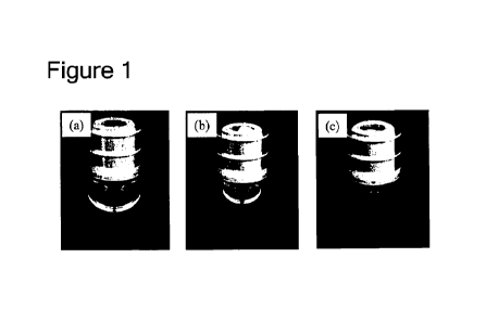
[0079] Figure 1 illustrates photographs of syringes containing air bubbles in
a volume of
120 IA (a), 40 [it (b), and 10 [it (c).
CA 03233924 2024- 4-4
- 16 -
Figure 2 is a schematic diagram of a cardboard box used in a drop test.
Figure 3 is an exemplified configuration diagram of a syringe.
Figure 4 is a histogram of sizes of visually detectable proteinaceous
particles of a
protein identified in Example 3.
Figure 5 illustrates a process flow in executing a program for an apparatus
for
determining a protein having a high risk of forming particles in a solution
based on a
hydrophobic patch and a charged patch.
Figure 6 illustrates a process flow in executing a program for an apparatus
for
determining a protein having a high risk of forming particles in a solution
based on a charged
patch.
Figure 7 is a schematic configuration diagram of an apparatus for determining
a
protein having a high risk of forming particles in a solution.
DESCRIPTION OF EMBODIMENTS
[0080] (1) Determination of risk of forming visually detectable particles
Herein, a visually detectable particle refers to a particle that is visually
detectable
under high illuminance and has a particle size of 40 [tm or greater. Among
them, particles
that are visually detectable under standard illuminance as prescribed in the
Japanese
pharmacopoeia (about 2,000 to 3,000 lx) are designated as "visible particles"
or "insoluble
visible particles". Generally, visible particles have a particle size greater
than 100 l_tm (Non
Patent Literature 1). Particles that are smaller in size than visible
particles, and cannot be
seen with eyes with standard illuminance as prescribed in the Japanese
pharmacopoeia (about
2,000 to 3,000 lx) but can be visually detected by increasing illuminance or
by increasing an
observation time period are "particles visually detectable only under high
illuminance", and
have a particle size of 40 ,m to 100 m. Visible particles can be confirmed by
visual
inspection with naked eyes for 5 seconds or longer under illumination at
standard illuminance
(about 2,000 to 3,000 lx), by slowly rotating or inverting the container in
front of a black
background or a white background. Particles visually detectable only under
high
illuminance can be confirmed by visual inspection with naked eyes for 30
seconds or longer
CA 03233924 2024- 4-4
- 17 -
under illumination at high illuminance (6,000 lx or greater) by slowly
rotating or inverting
the container in front of a black background. Visible particles can also be
confirmed with
inspection under high illuminance. Particles except for those generated from
protein
molecules in a solution are not considered as "visually detectable particles"
regardless of the
size. Whether visually detectable particles are derived from protein molecules
can be
confirmed by Raman microspectroscopic measurement. The only content contained
as a
protein in the solution is an active pharmaceutical ingredient (API), and
visually detectable
particles are generated from API. The particle size and number of visually
detectable
particles can be determined by a light obscuration particle count method, a
microscopic
particle count method, a flow cytometric particle image analysis method,
visual inspection,
and infrared microspectroscopy (infrared spectroscopy ;IR) or Raman
microspectroscopic
measurement performed after isolation of particles, and are measured
preferably by a
combination of visual inspection and infrared microspectroscopy or Raman
microspectroscopic measurement.
[0081] Herein, the term "high risk of forming particles" in a solution of a
pharmaceutical
formulation implies that protein molecules in the solution easily aggregate
and form visually
detectable particles. Examples of proteins having a high risk of forming
particles include
proteins that can form visually detectable particles even after addition of an
appropriate
amount of a surfactant.
[0082] Herein, the term "air bubble" refers to a space between a liquid in a
container and
the wall of the container, or gas present in the liquid. The air bubble is of
a size that can be
seen with naked eyes or by light-microscopic examination. The term "in the
container"
includes, in the case of a pre-filled syringe with a needle, the entire space
stoppered with a
rigid needle shield (RNS) and a stopper, and more specifically includes a
space inside the
needle and inside the barrel. When the container is in a vertical position, an
air bubble does
not extend across the diameter of the container and not all but part of the
liquid touches the
bottom surface of a container closure (such as a stopper). In one embodiment,
an air bubble
is spherical. In another embodiment, an air bubble is not spherical. In such
an
CA 03233924 2024- 4-4
- 18 -
embodiment, the air bubble has an egg-shape.
[0083] In one aspect of the present invention, the volume of air bubbles may
be 120 L or
less, 110 [IL or less, 100 L or less, 90 [IL or less, 80 L or less, 70 L or
less, 60 L or less,
50 IA or less, 40 IA or less, 30 IA or less, 20 [IL or less, or 10 [IL or
less. In one
embodiment of the present invention, the volume of air bubbles is determined
as the volume
of air bubbles obtained when all the bubbles in the container are unified. The
volume of air
bubbles may be determined by a method of "actually measuring the volume of air
bubbles by
extruding from the needle tip in the order of the gas and the solution into an
appropriate
container with scales such as a pipette already containing a solution",
"obtaining an image
and calculating the volume of air bubbles based on the area of air bubble
portions", or
"calculating the volume of air bubbles based on the height of a bubble portion
on the basis of
already-known information on the barrel internal diameter".
[0084] In one aspect of the present invention, "homology modeling" is employed
for
determining a protein having a high risk of forming particles in a solution.
"Homology
modeling" is a method for estimating a three-dimensional structure of a
protein having a
specific sequence based on similarity in sequence to one or more proteins
having known
three-dimensional structures. Usually, homology modeling for a specific amino
acid
sequence includes the following steps of: 1) specifying a homolog having a
known structure
in the Protein Data Bank, 2) arranging a target sequence in correspondence to
a template
structure, 3) constructing a model based on such alignment, and 4) evaluating
and refining
the model (Xiang, Curr Protein Pept Sci. 2006 June; 7(3): 217-227). Examples
of software
for estimating a three-dimensional structure equipped with homology modeling
function
include, but are not limited to, Molecular Operating Environment (MOE;
Chemical
Computing Group Inc. (CCG) (Canada), Web Antibody Modelling (WAM;
http://antibody.bath.ac.uk), Rosetta
(https://www.rosettacommons.org/software), Prime
(Schrodinger), MODELLER (Eswar, et al., Comparative Protein Structure Modeling
With
MODELLER, Current Protocols in Bioinformatics, John Wiley & Sons, Inc.,
Supplement 15,
5.6.1-5.6.30, 200), SEGMOD/ENCAD (Levitt M. J Mol Biol 1992; 226: 507-533),
SWISS-
CA 03233924 2024- 4-4
- 19 -
MODEL (Schwede T., Kopp J., Guex N., Peitsch M. C. Nucleic Acids Research
2003; 31:
3381-3385.), 3D-JIGSAW (Bates et al., Proteins: Structure, Function and
Genetics, Suppl
2001; 5: 39-46), NEST (Xiang, CUIT Protein Pept Sci. 2006 June; 7(3): 217-
227), and
BUILDER (Koehl and Delarue, Curr Opin Struct Biol 1996; 6(2): 222-226). A
preferred
example of the software includes MOE.
[0085] In one aspect of the present invention, the term "antibody modeling"
refers to a
function for estimating a three-dimensional structure, and database
specialized in monoclonal
antibodies. In antibody modeling, the entire structure can be assembled based
on a structure
of a fragment. For example, an antibody Fab fragment can be added to an Fc
fragment
crystal structure, and a Fab fragment can be formed as an estimated protein
structure and
added to an Fc fragment crystal structure. For example, functions provided in
MOE can be
used to carry out antibody modeling.
[0086] In one aspect of the present invention, the term "patch" refers to a
surface region of a
cluster of residues representing a specific physicochemical property in a
three-dimensional
structure of a protein or an antibody. Examples of the patch include a
hydrophobic patch
and a charged patch. A hydrophobic patch is a surface region of a portion
where
hydrophobic residues are accumulated in a cluster. The portion where
hydrophobic residues
are accumulated in a cluster can also include residues other than hydrophobic
residues. A
charged patch is a surface region of a portion where residues with a charge
are accumulated
in a cluster. The portion where residues with a charge are accumulated in a
cluster can also
include residues without a charge.
[0087] In one aspect of the present invention, a feature amount concerning a
patch area of a
protein can be calculated using Protein properties function of MOE. With
respect to the
patch area of a specific protein surface, when a sum of areas of top 5
hydrophobic patches
ranked according to the area of hydrophobic patches is set as X ((angstrom)2),
and a total area
of charged patches is set as Y ((angstrom)2), a risk of the protein of forming
particles can be
determined based on a "X + Y x 1.5" value. In one embodiment, a protein having
a "X + Y
x 1.5" value of 1,700 or greater can be determined as a protein having a high
risk of forming
CA 03233924 2024- 4-4
- 20 -
particles. In another embodiment, a protein having a "X + Y x 1.5" value of
2,000 or
greater, 2,500 or greater, 3,000 or greater, 3,500 or greater, or 4,000 or
greater can be
determined as a protein having a high risk of forming particles.
[0088] In one aspect of the present invention, the feature amount concerning a
patch area of
a protein can be calculated using Protein properties function of MOE, and with
respect to the
patch area of a specific protein surface, a risk of the protein of forming
particles can be
determined based on a total area of charged patches Y ((angstrom)2). In one
embodiment, a
protein having a Y value of 600 or greater is determined as a protein having a
high risk of
forming particles. In another embodiment, a protein having a Y value of 700 or
greater, 800
or greater, 900 or greater, 1,000 or greater, 1,500 or greater, 2,000 or
greater, 2,500 or
greater, 3,000 or greater, or 4,000 or greater is determined as a protein
having a high risk of
forming particles.
[0089] Herein, the term ranking according to the area of hydrophobic patches
refers to a list
of hydrophobic patches identified on a protein surface and arranged in the
order of the area,
and each hydrophobic patch means a hydrophobic patch consisting of a cluster
of
hydrophobic residues, independently present on the protein surface and having
a certain size.
A sum of areas of top 5 hydrophobic patches in the ranking ((angstrom)2) is
calculated as X.
Herein, a sum of the areas of top 5 hydrophobic patches refers to a total
value of the areas of
the top 5 hydrophobic patches. However, if there are 4 or less hydrophobic
patches in a
molecule, the sum refers to a total value of the areas of all the hydrophobic
patches actually
present.
[0090] A total area of charged patches means the sum of the areas
((angstrom)2) of all
positively or negatively charged patches present on the protein surface.
In one aspect of the present invention, the term "molecular force field" is
parameterization, in the form of a function, of force applied to each atom
present in a
molecule. In molecular mechanics calculation and molecular dynamics
calculation based on
molecular force field, inter-atomic force is expressed as numeric values of
potential function
determined according to the types of atoms and mode of binding, with
parameters
CA 03233924 2024- 4-4
- 21 -
representing inter-atomic bonds (such as a bond distance and a bond angle) as
variables. In
molecular mechanics calculation and molecular dynamics calculation based on
molecular
force field, inter-atomic force is expressed as numeric values of potential
function determined
according to the types of atoms and mode of binding, with parameters
representing inter-
atomic bonds (such as a bond distance and a bond angle) as variables.
[0091] In one aspect of the present invention, the molecular force field that
can be used is
not particularly limited but can be appropriately selected depending on
purpose, and
examples include Amber molecular force field, CHARMm molecular force field,
and OPLS
molecular force field. Examples of Amber molecular force field include Amber
10/14:
EHT, Amber ff99SB-ILDN, and Amber 12SB. An example of CHARMm molecular force
field includes CHARMm 36. Among these, Amber 10: EHT is preferable when MOE is
used.
[0092] (2) Reduction of particle formation
In one aspect of the present invention, the term "reducing formation of
particles"
refers to adjusting the volume of air bubbles so as not to form visually
detectable particles or
to reduce the number of formed particles in a solution of a pharmaceutical
formulation in
which visually detectable particles are formed under a given condition.
Reduction of
formation of visually detectable particles can be confirmed by counting the
number of
particles before and after adjusting the volume of air bubbles. The size and
number of
particles can be determined by a light obscuration particle count method, a
microscopic
particle count method, a flow cytometric particle image analysis method,
visual inspection,
and infrared microspectroscopy (infrared spectroscopy ;IR) or Raman
microspectroscopic
measurement after isolation of particles, and are measured preferably by a
combination of
visual inspection and infrared microspectroscopy or Raman microspectroscopic
measurement.
[0093] (3) Pharmaceutical formulation
In one aspect of the present invention, a pharmaceutical formulation is a
solution
containing a protein as an active ingredient. The pharmaceutical formulation
may be a
CA 03233924 2024- 4-4
- 22 -
formulation for injection.
[0094] In one aspect of the present invention, a formulation for injection is
a
pharmaceutical formulation that is filled in a container for injection to be
administered by
injection, and contains a protein as an active ingredient in a solution.
In one aspect of the present invention, the term "formulation for injection to
be
obtained" refers to a formulation for injection obtained as a final product
after adjusting the
air volume.
[0095] In one aspect of the present invention, the pharmaceutical formulation
is stored
without freezing the solution in the container at -30 C to 25 C, preferably
from the freezing
point of the solution to 25 C, more preferable at 1 C to 10 C, more preferably
at 2 C to 8 C,
and even more preferably at 5 C. The storage is carried out for 1 hour, 2
hours, 3 hours, 4
hours, 5 hours, 6 hours, 7 hours, 8 hours, 9 hours, 10 hours, 11 hours, 12
hours, 13 hours, 14
hours, 15 hours, 16 hours, 17 hours, 18 hours, 19 hours, 20 hours, 21 hours,
22 hours, 23
hours, 24 hours, 25 hours, 26 hours, 27 hours, 28 hours, 29 hours, 30 hours,
31 hours, 32
hours, 33 hours, 34 hours, 35 hours, 36 hours, 48 hours, 60 hours, 72 hours,
84 hours, or 96
hours. The storage is carried out for at least 24 hours, at least 2 days, at
least 3 days, at least
4 days, at least 10 days, at least 20 days, at least 30 days, at least 40
days, at least 50 days, at
least 60 days, at least 1 month, at least 2 months, at least 3 months, at
least 4 months, at least
months, at least 6 months, at least 7 months, at least 8 months, at least 9
months, at least 10
months, at least 11 months, or at least 12 months.
[0096] In one aspect of the present invention, the protein used in a liquid
formulation
encompasses, but is not limited to, an antibody, a fusion protein, an enzyme,
a hormone, a
cytokine, and a vaccine. More specifically, the protein encompasses a
monoclonal
antibody, granulocyte colony-stimulating factor (G-CSF), granulocyte
macrophage colony-
stimulating factor (GM-CSF), erythropoietin (EPO), interferon, interleukins
such as IL-1 or
IL-6, tissue plasminogen activator (TPA), thrombopoietin, urokinase, serum
albumin, blood
coagulation factor VIII, leptin, stem cell factor (SCF), and the like.
[0097] In one aspect of the present invention, the protein used in the
pharmaceutical
CA 03233924 2024- 4-4
- 23 -
formulation has substantially the same biological activity as a bioactive
protein of a mammal,
in particular of a human, and encompasses proteins derived from nature and
those obtained
by genetic engineering. Proteins obtained by genetic engineering include those
having the
same amino acid sequence as a native protein, or those obtained by deleting,
substituting, or
adding one or more amino acid sequences and having the above-described
biological activity.
[0098] In one aspect of the present invention, a concentration of the protein
in the solution
may be 0.1 mg/mL or more, within a range of 0.1 to 300 mg/mL, or within a
range of 1 to
200 mg/rnL.
In one aspect of the present invention, an antibody to be used is not
particularly
limited as long as it binds to a desired antigen, may be a polyclonal antibody
or a monoclonal
antibody, and is preferably a monoclonal antibody because a homogeneous
antibody can be
thus stably produced. In one aspect of the present invention, the antibody to
be used may be
a monospecific antibody or a bispecific antibody, or may be a multispecific
antibody having
three or more antigen-recognizing sites in a molecule.
[0099] In one aspect of the present invention, the monoclonal antibody to be
used
encompasses not only a monoclonal antibody derived from an animal, such as a
human, a
mouse, a rat, a hamster, a rabbit, a sheep, a camel, or a monkey, but also a
recombinant
antibody obtained by artificial modification, such as a chimeric antibody, a
humanized
antibody, or a bispecific antibody. Furthermore, a recombinant antibody
obtained by
artificial modification of a constant region or the like of an antibody for
modifying physical
properties of an antibody molecule (specifically, modification of an
isoelectric point (pI),
modification of affinity of Fe receptor, and the like) for purposes of
improving retention in
blood and pharmacokinetics is also encompassed.
[0100] In one aspect of the present invention, the immunoglobulin class of the
antibody to
be used is not particularly limited, but may be any of classes including IgG
such as IgG 1,
IgG2, IgG3, and IgG4, and IgA, IgD, IgE, and IgM, among which IgG is
preferred, and
IgGl, IgG2 and IgG4 are particularly preferred.
[0101] Further, in one aspect of the present invention, the antibody to be
used encompasses
CA 03233924 2024- 4-4
- 24 -
not only an antibody including a constant region and a variable region (full-
length antibody)
but also an antibody fragment, such as Fv, Fab, and F(ab)2, and a low-
molecular-weight
antibody like a bispecific antibody such as one- or two-associated single
chain Fv (scF,
sc(Fv)2) having a variable region of the antibody bound with a linker such as
a peptide linker,
or a scFv dimer, and a full-length antibody is preferred.
[0102] In one aspect of the present invention, the antibody to be used can be
produced by a
known method. A hybridoma used for producing a monoclonal antibody can be
produced
as follows basically by known techniques. Specifically, a desired antigen or a
cell
expressing a desired antigen used as a sensitizing antigen is immunized by a
usual
immunization method, and the resultant immune cell is fused with a known
parent cell by a
usual cell fusion method, the resultant is subjected to a usual screening
method for screening
a monoclonal antibody producing cell (hybridoma), and thus, the hybridoma can
be
produced. The production of a hybridoma can be carried out in accordance with,
for
example, the method of Milstein et al., (Kohler, G. and Milstein, C., Methods
Enzymol.
(1981) 73: 3-46) or the like. If immunogenicity of the antigen is low, the
antigen may be
bound to a macromolecule having immunogenicity, such as albumin, before the
immunization.
[0103] Alternatively, a recombinant antibody obtained by cloning an antibody
gene from a
hybridoma, and incorporating the gene into an appropriate vector to be
introduced into a host
by genetic engineering techniques can be used (see, for example, Carl, A. K.
Borrebaeck,
James, W. Larrick, THERAPEUTIC MONOCLONAL ANTIBODIES, Published in the
United Kingdom by MACMILLAN PUBLISHERS LTD., 1990). Specifically, cDNA of a
variable region (V region) of an antibody is synthesized from mRNA of the
hybridoma with
reverse transcriptase. DNA encoding the V region of the target antibody thus
obtained is
linked to DNA encoding a desired antibody constant region (C region), and the
resultant is
incorporated into an expression vector. Alternatively, DNA encoding the V
region of the
antibody may be incorporated into an expression vector containing DNA of an
antibody C
region. The resultant is incorporated into the expression vector so as to
express under
CA 03233924 2024- 4-4
- 25 -
control of an expression control region, such as an enhancer or a promoter.
Next, a host cell
is transformed with the resultant expression vector, and thus, the antibody
can be expressed.
[0104] In one aspect of the present invention, a recombinant antibody obtained
by artificial
modification for purpose of reducing heteroantigenicity against human, such as
a chimeric
antibody or a humanized antibody, can be used. Such a modified antibody can be
produced
by a known method. A chimeric antibody is an antibody containing heavy-chain
and light-
chain variable regions of an antibody of a mammal other than a human, for
example, a mouse
antibody, and heavy-chain and light-chain constant regions of a human
antibody, and can be
obtained by linking DNA encoding a variable region of a mouse antibody to DNA
encoding a
constant region of a human antibody, incorporating the resultant into an
expression vector,
and introducing the vector into a host to produce the antibody.
[0105] A humanized antibody is designated also as a reshaped human antibody,
and is
obtained by transplanting a complementary determining region (CDR) of an
antibody of a
mammal other than a human, for example, a mouse antibody, into a complementary
determining region of a human antibody, and a general genetic engineering
method for such
an antibody is also known. Specifically, a DNA sequence designed to link CDR
of a mouse
antibody to a framework region (FR) of a human antibody is synthesized by PCR
method
from several oligonucleotides produced to have overlapping portions at the
ends. The thus
obtained DNA is linked to DNA encoding a human antibody constant region, the
resultant is
subsequently incorporated into an expression vector, and the resultant vector
is introduced
into a host to produce the antibody (see European Patent Application No.
239400, and WO
96/02576). As the FR of a human antibody to be linked via CDR, one having a
complementary determining region forming a good antigen-binding site is
selected. If
necessary, an amino acid in a framework region of a variable region of an
antibody may be
substituted so as to cause the complementary determining region of the
reshaped human
antibody to form a suitable antigen-binding site (Sato, K. et al., Cancer Res.
(1993) 53, 851-
856).
[0106] As techniques for substituting an amino acid of an antibody for
improving activity,
CA 03233924 2024- 4-4
- 26 -
physical properties, pharmacokinetics, safety and the like of the antibody,
for example, the
following techniques are known, and in one aspect of the present invention,
the antibody to
be used encompasses such an antibody having substitution (including deletion
and addition)
of an amino acid.
[0107] As techniques for amino acid substitution in a variable region of an
IgG antibody,
not only humanization (Tsurushita N., Hinton P.R., Kumar S., Design of
humanized
antibodies: from anti-Tac to Zenapax., Methods, 2005 May; 36 (1): 69-83) but
also affinity
maturation by amino acid substitution in a complementary determining region
(CDR) for
enhancing binding activity (Rajpal A., Beyaz N., Haber L., Cappuccilli G., Yee
II, Bhatt
R.R., Takeuchi T., Lerner R.A., Crea R., A general method for greatly
improving the affinity
of antibodies by using combinatorial libraries, Proc Natl Acad Sci USA, 2005
Jun 14; 102
(24): 8466-71), and improvement of physicochemical stability by amino acid
substitution in
framework (FR) (Ewert S., Honegger A., Pluckthun A., Stability improvement of
antibodies
for extracellular and intracellular applications: CDR grafting to stable
frameworks and
structure-based framework engineering, Methods, 2004 Oct; 34(2): 184-99.
Review) have
been reported. As techniques for amino acid substitution in Fc region of an
IgG antibody,
techniques for enhancing antibody dependent cellular cytotoxicity (ADCC) or
complement
dependent cellular cytotoxicity (CDC) are known (Kim S.J., Park Y., Hong H.J.,
Antibody
engineering for the development of therapeutic antibodies, Mol Cells, 2005 Aug
31; 20(1):
17-29 Review.). In addition, techniques for amino acid substitution in Fe by
not only
enhancing such effector function but also improving half-life in blood of an
antibody have
been reported (Hinton P.R., Xiong J.M., Johlfs M.G., Tang M.T., Keller S.,
Tsurushita N.,
An engineered human IgG1 antibody with longer serum half-life, J Immunol. 2006
Jan 1;
176(1): 346-56, Ghetie V., Popov S., Borvak J., Radu C., Matesoi D., Medesan
C., Ober R.J.,
Ward E.S., Increasing the serum persistence of an IgG fragment by random
mutagenesis, Nat
Biotechnol. 1997 Jul; 15(7): 637-40.). Furthermore, various techniques for
amino acid
substitution in a constant region for purposes of improving physical
properties of an antibody
are known (WO 09/41613).
CA 03233924 2024- 4-4
- 27 -
[0108] Besides, there are known methods for obtaining a human antibody. For
example,
human lymphocyte is sensitized with a desired antigen or a cell expressing a
desired antigen
in vitro, and the thus sensitized lymphocyte is fused to a human myeloma cell,
for example,
U266, and thus, a desired human antibody having a binding activity to an
antigen can be
obtained (see Japanese Patent Publication No. 1-59878). Furthermore, when a
transgenic
animal having all repertoires of human antibody genes is immunized with an
antigen, a
desired human antibody can be obtained (see WO 93/12227, WO 92/03918, WO
94/02602,
WO 94/25585, WO 96/34096, and WO 96/33735). Besides, a technique for obtaining
a
human antibody by panning using a human antibody library is also known. For
example, a
variable region of a human antibody is expressed as a single chain antibody
(scFv) on the
surface of a phage by a phage display method, and thus, a phage binding to an
antigen can be
selected. When the gene of the selected phage is analyzed, a DNA sequence
encoding the
variable region of a human antibody binding to the antigen can be determined.
When the
DNA sequence of scFv binding to the antigen is clarified, an appropriate
expression vector
containing the sequence can be produced to obtain a human antibody. These
methods are
already known, and can be executed with the reference to WO 92/01047, WO
92/20791, WO
93/06213, WO 93/11236, WO 93/19172, WO 95/01438, and WO 95/15388. In one
aspect
of the present invention, the antibody to be used encompasses such human
antibodies.
[0109] When an antibody is produced by isolating an antibody gene once, and
introducing
the gene into an appropriate host, an appropriate combination of a host and an
expression
vector can be used. When a eukaryotic cell is used as the host, an animal
cell, a plant cell,
or a fungal cell can be used. As the animal cell, (1) mammal cells such as
CHO, COS,
myeloma, BHK (baby hamster kidney), HeLa, and Vero, (2) amphibian cells such
as
Xenopus oocyte, and (3) insect cells such as sf9, sf21, and Tn5 are known. As
the plant
cell, a cell derived from the genus Nicotiana, such as a cell derived from
Nicotiana tabacum,
is known, and this cell may be callus cultured. As the fungal cell, yeast, for
example, the
genus Saccharomyces, such as Saccharomyces serevisiae, and filamentous fungus,
for
example, the genus Aspergillus such as Aspergillus niger are known. When a
prokaryotic
CA 03233924 2024- 4-4
- 28 -
cell is used, there is a production system using a bacterial cell. As the
bacterial cell,
Escherichia coli (E. coli) and Bacillus subtilis are known. When a target
antibody gene is
introduced into such a cell by transformation, and the thus transformed cell
is cultured in
vitro, the antibody can be obtained.
[0110] Besides, the antibody to be used in the pharmaceutical formulation
encompasses a
modified antibody. For example, antibodies binding to various molecules of
polyethylene
glycol (PEG), cytotoxic drugs and the like can be used (Farmaco. 1999 Aug 30;
54(8): 497-
516, Cancer J. 2008 May-June; 14(3): 154-69). Such a modified antibody can be
obtained
by chemically modifying an antibody. Such a method has been already
established in this
field.
[0111] In one aspect of the present invention, the antibody of the present
disclosure may be
a chimeric antibody. A chimeric antibody is described in, for example, US
Patent
No. 4,816,567, and Morrison et al., Proc. Natl. Acad. Sci. USA, 81: 6851-6855
(1984). A
chimeric antibody may contain a non-human variable region (variable region
derived from,
for example, a non-human primate such as a monkey, or a mouse, a rat, a
hamster, a rabbit or
the like) and a human constant region.
[0112] In one aspect of the present invention, the antibody of the present
disclosure may be
a humanized antibody. Representatively, a humanized antibody is humanized for
reducing
immunogenicity in a human with specificity and affinity of a parent non-human
antibody
retained. A humanized antibody representatively contains one or more variable
regions, and
an HVR, for example, CDR derived from a non-human antibody (or a part thereof)
and FR
derived from a human antibody sequence (or a part thereof) are present
therein. A
humanized antibody can optionally contain at least a part of a human constant
region. In
one embodiment, amino acid residues of FR in a humanized antibody may be
substituted
with corresponding amino acid residues of a non-human antibody (for example,
an antibody
from which HVR residues are derived) for, for example, retaining or improving
specificity
and affinity of the antibody.
[0113] A humanized antibody and a method for producing the same are reviewed
in, for
CA 03233924 2024- 4-4
- 29 -
example, the following (Almagro and Fransson, Front. Biosci. 13: 1619-1633
(2008)), and
are described in, for example, the following: Riechmann et al., Nature 332:
323-329 (1988);
Queen et al., Proc. Nat'l Acad. Sci. USA 86: 10029-10033 (1989); US Patent
Nos. 5,821,337,
7,527,791, 6,982,321, and 7,087,409; Kashmiri et al., Methods 36: 25-34 (2005)
(describing
specificity determining region (SDR) grafting); Padlan, Mol. Immunol. 28: 489-
498 (1991)
(describing "resurfacing"); Dall'Acqua et al., Methods 36: 43-60 (2005)
(describing "FR
shuffling"); and Osbourn et al., Methods 36: 61-68 (2005) and Klimka et al.,
Br. J. Cancer,
83: 252-260 (2000) (describing "guided selection" approach to FR shuffling).
[0114] In one aspect of the present invention, a human framework that may be
used in
humanization may contain, for example, a framework selected by a "best fit"
method (Sims et
al., J. Immunol. 151: 2296 (1993)), a framework derived from a consensus
sequence of a
human antibody belonging to a specific subgroup of a heavy chain or light
chain variable
region (Carter et al., Proc. Natl. Acad. Sci. USA, 89: 4285 (1992), and Prest
et al., J.
Immunol., 151: 2623 (1993)), or a framework region derived from screening of
FR library
(Baca et al., J. Biol. Chem. 272: 10678-10684 (1997) and Rosok et al., J.
Biol. Chem. 271:
22611-22618 (1996)).
[0115] In one aspect of the present invention, the antibody of the present
disclosure may be
a human antibody. A human antibody can be produced by various techniques. A
human
antibody is outlined in, for example, van Dijk and van de Winkel, Curr. Opin.
Pharmacol. 5:
368-374 (2001) and Lonberg, Curr. Opin. Immunol. 20: 450-459 (2008). A human
antibody
may be prepared by administering an immunogen to a transgenic animal having
been
modified to produce a complete human antibody or a complete antibody
containing a human
variable region in response to an antigen. Such an animal representatively
contains the
entire or a part of human immunoglobulin locus, and the entire or a part of
the human
immunoglobulin locus is present in a state where it is substituted with an
endogenous
immunoglobulin locus, or it is randomly incorporated outside the chromosome or
inside the
chromosome of the animal. In such a transgenic mouse, endogenous
immunoglobulin locus
is usually inactivated. A method for obtaining a human antibody from a
transgenic animal
CA 03233924 2024- 4-4
- 30 -
is reviewed in Lonberg, Nat. Biotech. 23: 1117-1125 (2005). Besides, make
reference to,
for example, US Patent Nos. 6,075,181 and 6,150,584 describing XENOMOUSE(TM)
technology; US Patent No. 5,770,429 describing HUMAB(R) technology; US Patent
No.
7,041,870 describing K-M MOUSE(R); and US2007/0061900 describing
VELOCIMOUSE(R) technology. A human variable region from a complete antibody
generated from such an animal may be further modified by, for example,
combining with a
different human constant region.
[0116] In another aspect of the present invention, a human antibody can be
produced by a
method based on a hybridoma. A human myeloma cell and a mouse-human
heteromyeloma
cell line for producing a human monoclonal antibody is described in the
following (for
example, Kozbor J. Immunol., 133: 3001(1984); Brodeur et al., Monoclonal
Antibody
Production Techniques and Applications, pp. 51-63 (Marcel Dekker, Inc., New
York, 1987);
and Boerner et al., J. Immunol., 147: 86 (1991)). A human antibody generated
through
human B cell hybridoma technology is described in Li et al., Proc. Natl. Acad.
Sci. USA,
103: 3557-3562 (2006). Other examples of the method include US Patent No.
7,189,826
(describing production of a monoclonal human IgM antibody from a hybridoma
cell line),
and Ni, Xiandai Mianyixue, 26 (4): 265-268 (2006) (describing a human-human
hybridoma).
Human hybridoma technology (trioma technology) is described in Vollmers and
Brandlein,
Histology and Histopathology, 20(3): 927-937 (2005), and Vollmers and
Brandlein, Methods
and Findings in Experimental and Clinical Pharmacology, 27(3): 185-91(2005).
[0117] In another aspect of the present invention, a human antibody may also
be generated
by isolating an Fv clone variable domain sequence selected from human-derived
phage
display libraries. Such a variable region sequence can then be combined with a
desired
human constant region. Techniques for selecting a human antibody from antibody
libraries
will be described below.
[0118] In one aspect of the present invention, the antibody of the present
disclosure may be
isolated by screening a combinatorial library for an antibody having one or
more desired
activities. For example, a method for creating a phage display library, a
method for
CA 03233924 2024- 4-4
- 31 -
screening such a library for an antibody having a desired binding
characteristic, and the like
are known in this technical field. Such methods are reviewed in Hoogenboom et
al. in
Methods in Molecular Biology 178:1-37 (O'Brien et al., ed., Human Press,
Totowa, NJ,
2001), and are described in, for example, McCafferty et al., Nature 348: 552-
554; Clackson
et al., Nature 352: 624-628 (1991); Marks et al., J. Mol. Biol. 222: 581-597
(1992); Marks
and Bradbury, Molecular Biology 248: 161-175 (Lo, ed., Human Press, Totowa,
NJ, 2003);
Sidhu et al., J. Mol. Biol. 338(2): 299-310 (2004); Lee et al., J. Mol. Biol.
340(5): 1073-1093
(2004); Fellouse, Proc. Natl. Acad. Sci. USA 101(34): 12467-12472 (2004); and
Lee et al., J.
Immunol. Methods 284(1-2): 119-132(2004).
[0119] In a specific phage display method employed in one aspect of the
present invention,
repertoires of VH and VL can be separately cloned by polyrnerase chain
reaction (PCR), and
recombined randomly in phage libraries, and the phage libraries can then be
screened for
antigen-binding phage as described in Winter et al., Ann. Rev. Immunol., 12:
433-455
(1994). A phage displays an antibody fragment such as scFv or Fab. Libraries
from
immunized sources can provide high-affinity antibodies to the immunogen
without the
requirement of constructing hybridomas. In another embodiment, a naive
repertoire can be
cloned (for example, from a human) to provide a single source of antibodies to
a wide range
of non-self or self-antigens without any immunization as described by
Griffiths et al., EMBO
J, 12: 725-734 (1993). In a still another embodiment, naive libraries can also
be created
synthetically by cloning unrearranged V-gene segments from stem cells, and
using PCR
primers containing a random sequence encoding the highly variable region CDR3
and to
accomplish rearrangement in vitro, as described in Hoogenboom and Winter, J.
Mol. Biol.,
227: 381-388 (1992). Examples of patent publications describing human antibody
phage
libraries include US Patent No. 5,750,373, and US Patent Publication Nos.
2005/0079574,
2005/0119455, 2005/0266000, 2007/0117126, 2007/0160598, 2007/0237764,
2007/0292936,
and 2009/0002360.
[0120] An antibody or an antibody fragment isolated from a human antibody
library is
herein regarded as a human antibody or a human antibody fragment.
CA 03233924 2024- 4-4
- 32 -
In one aspect of the present invention, the antibody of the present disclosure
is a
multispecific antibody (such as a bispecific antibody). A multispecific
antibody is an
antibody having binding specificities in at least two different sites (for
example, a
monoclonal antibody). In one embodiment, one of the binding specificities is
specificity to
an antigen, and the other is specificity to another antigen. In another
embodiment, a
bispecific antibody may bind to two epitopes of different antigens. The
bispecific antibody
may be used for localizing a cytotoxic agent in a cell expressing the antigen.
The bispecific
antibody may be prepared as a full-length antibody or an antibody fragment.
[0121] A method for producing a multispecific antibody is not limited, and
examples
include recombinant co-expression of two immunoglobulin heavy chain-light
chain pairs
having different specificities (for example, Milstein and Cuello, Nature 305:
537 (1983),
W093/08829, and Traunecker et al., EMBO J. 10: 3655 (1991)), and knob-in-hole
technology (for example, US Patent No. 5,731,168). A multispecific antibody
may be
produced by controlling electrostatic steering effects for producing an Fe
heterodimer
molecule (for example, W02009/089004 Al); cross-linking two or more antibodies
or
antibody fragments (for example, US Patent No. 4,676,980 and Brennan et al.,
Science, 229:
81 (1985)); producing an antibody having two specificities with leucine zipper
(for example,
Kostelny et al., J. Immunol., 148 (5): 1547-1553 (1992)); producing a
bispecific antibody
fragment by "diabody" technology (for example, Hollinger et al., Proc. Natl.
Acad. Sci. USA,
90: 6444-6448 (1993)); using a scFv dimer (for example, Gruber et al., J.
Immunol., 152:
5368 (1994)); or preparing a trispecific antibody (for example, Tutt et al.,
J. Immunol. 147:
60 (1991)). Alternatively, the multispecific antibody may be an antibody
engineered to
have three or more functional antigen-binding sites, including an "octopus
antibody" (for
example, US2006/0025576).
[0122] In one aspect of the present invention, the antibody or the antibody
fragment thereof
of the present disclosure may be a "dual acting Fab" or "DAF" containing one
antigen-
binding site binding to the antigen and another different antigen (for
example,
U52008/0069820).
CA 03233924 2024- 4-4
- 33 -
[0123] In one aspect, a variant (mutant) of an amino acid sequence of the
antibody of the
present disclosure can be prepared by introducing an appropriate modification
to a nucleic
acid encoding a molecule of the antibody, or by synthesizing a peptide. Such a
modification
may be performed through one of or an appropriate combination of a plurality
of deletion,
insertion, and substitution of an arbitrary amino acid (residue) in the amino
acid sequence.
An arbitrary combination of deletion, insertion, and substitution can be
employed as long as a
final construct possesses a desired characteristic (for example, antigen-
binding property).
[0124] In one aspect of the present invention, when an antigen variant
(mutant) resulting
from one of or a plurality of amino acid substitutions is provided, a target
site for introducing
substitution mutation can contain HVR and FR.
Examples of the antibody used in the pharmaceutical formulation include, but
are
not limited to, an anti-tissue factor antibody, anti-IL-6 receptor antibody,
an anti-IL-6
antibody, an anti-glypican-3 antibody, an anti-CD3 antibody, an anti-CD20
antibody, an anti-
GPIlb/Ina antibody, an anti-TNF antibody, an anti-CD25 antibody, an anti-EGFR
antibody,
an anti-Her2/neu antibody, an anti-RSV antibody, an anti-CD33 antibody, an
anti-CD52
antibody, an anti-IgE antibody, an anti-CD1 1 a antibody, an anti-VEGF
antibody, an anti-
VLA4 antibody, an anti-HM1.24 antibody, an anti-parathyroid gland hormone-
related
peptide antibody (anti-PTHrP antibody), an anti-ganglioside GM3 antibody, an
anti-TPO
receptor agonist antibody, a coagulation factor VIII substitution antibody, an
anti-IL31
receptor antibody, an anti-HLA antibody, an anti-AXL antibody, an anti-CXCR4
antibody,
an anti-NR10 antibody, and a bispecific antibody of factor IX and factor X.
[0125] Examples of a preferable reshaped humanized antibody to be used in the
pharmaceutical formulation include a humanized anti-interleukin 6 (IL-6)
receptor antibody
(tocilizumab, hPM-1, or MRA, see W092/19759), a humanized anti-HM1.24 antigen
monoclonal antibody (see W098/14580), a humanized anti-parathyroid gland
hormone-
related peptide antibody (anti-PTHrP antibody) (see W098/13388), a humanized
anti-tissue
factor antibody (see W099/51743), a humanized anti-glypican-3 IgGlx antibody
(codrituzumab, GC33, see W02006/006693), a humanized anti-NR10 antibody (see
CA 03233924 2024- 4-4
- 34 -
W02009/072604), and a bispecific humanized antibody of factor IX and factor X
(ACE910,
see W02012/067176).
[0126] In one aspect of the present invention, the pharmaceutical formulation
can be
prepared, if necessary, by mixing with an appropriate pharmaceutically
acceptable carrier,
medium, or the like into a liquid formulation. The solvent of a liquid
formulation is water
or a pharmaceutically acceptable organic solvent. Examples of such an organic
solvent
include propylene glycol (1,2-propanediol), polyethylene glycol 300,
polyethylene glycol
400, ethanol, glycerol, and acetic acid. Examples of appropriate
pharmaceutically
acceptable carrier and medium include sterilized water and physiological
saline, a stabilizing
agent, an antioxidant (such as ascorbic acid), a buffer (such as phosphoric
acid, citric acid,
histidine, or another organic acid), an antiseptic agent, a surfactant (such
as PEG, or Tween),
a chelating agent (such as EDTA), and a binding agent. Other low molecular
weight
polypeptides, proteins such as serum albumin, gelatin, and immunoglobulin,
amino acids
such as glycine, glutamine, asparagine, glutamic acid, aspartic acid,
methionine, arginine, and
lysine, sugars and carbohydrates such as polysaccharides and monosaccharides,
and sugar
alcohols such as mannitol and sorbitol may be contained. In the case of
obtaining a solution
for injection, examples include physiological saline, an isotonic solution
containing glucose
or another auxiliary drug, such as D-sorbitol, D-mannose, D-mannitol, or
sodium chloride,
and it may be used together with an appropriate solubilizing agent such as
alcohol (such as
ethanol), polyalcohol (such as propylene glycol, or PEG), and a nonionic
surfactant (such as
polysorbate 80, polysorbate 20, poloxamer 188, or HCO-50).
[0127] In one aspect of the present invention, the buffer to be used in a
liquid formulation is
prepared using a substance for retaining the pH of the solution. In one aspect
of the present
invention, a liquid formulation containing high concentration of an antibody
has a pH of the
solution of preferably 4.5 to 7.5, more preferably 5.0 to 7.0, and even more
preferably 5.5 to
6.5. In one aspect of the present invention, a usable buffer can adjust the pH
within this
range, and is pharmaceutically acceptable. Such a buffer is known to those
skilled in the art
in the field of liquid formulation, and inorganic salts such as phosphates
(sodium or
CA 03233924 2024- 4-4
- 35 -
potassium) and sodium bicarbonate; organic salts such as citrates (sodium or
potassium),
sodium acetate, and sodium succinate; and acids such as phosphoric acid,
carbonic acid, citric
acid, succinic acid, malic acid, and gluconic acid can be used. Alternatively,
Tris buffers,
and Good's buffers such as MES, MOPS, and HEPES, histidine (such as histidine
hydrochloride), glycine, and such can be used.
[0128] Generally, the concentration of the buffer is 1 to 500 mmol/L,
preferably 5 to
100 mmol/L and more preferably 10 to 20 mmol/L. In using a histidine buffer,
the buffer
preferably contains 5 to 25 mmol/L histidine, and more preferably contains 10
to 20 mmol/L
histidine.
[0129] In one aspect of the present invention, a liquid formulation containing
a high
concentration of an antibody is preferably stabilized by addition of a
stabilizing agent
suitable for the antibody corresponding to the active ingredient. In one
aspect of the present
invention, no significant change is observed in a "stable" liquid formulation
containing a high
concentration of an antibody for at least 12 months, preferably for 2 years,
and more
preferably for 3 years at refrigeration temperature (2 to 8 C); or for at
least 3 months,
preferably for 6 months, and more preferably for 1 year at room temperature
(22 to 28 C).
For example, a total amount of dimers and decomposed matters after storage at
5 C for 2
years is 5.0% or less, preferably 2% or less, and more preferably 1.5% or
less, or the total
amount of dimers and decomposed matters after storage at 25 C for 6 months is
5.0% or less,
preferably 2% or less, and more preferably 1.5% or less.
[0130] In one aspect of the present invention, representative examples of the
surfactant
include nonionic surfactants, for example, sorbitan fatty acid esters such as
sorbitan
monocaprylate, sorbitan monolaurate, and sorbitan monopalmitate; glycerin
fatty acid esters
such as glycerin monocaprylate, glycerin monomyristate, and glycerin
monostearate;
polyglycerin fatty acid esters such as decaglyceryl monostearate, decaglyceryl
distearate, and
decaglyceryl monolinoleate; polyoxyethylene sorbitan fatty acid esters such as
polyoxyethylene sorbitan monolaurate, polyoxyethylene sorbitan monooleate,
polyoxyethylene sorbitan monostearate, polyoxyethylene sorbitan monopalmitate,
CA 03233924 2024- 4-4
- 36 -
polyoxyethylene sorbitan trioleate, and polyoxyethylene sorbitan tristearate;
polyoxyethylene
sorbit fatty acid esters such as polyoxyethylene sorbit tetrastearate and
polyoxyethylene
sorbit tetraoleate; polyoxyethylene glycerin fatty acid esters such as
polyoxyethylene glyceryl
monostearate; polyethylene glycol fatty acid esters such as polyethylene
glycol distearate;
polyoxyethylene alkyl ethers such as polyoxyethylene lauryl ether;
polyoxyethylene
polyoxypropylene alkyl ethers such as polyoxyethylene polyoxypropylene glycol
ether,
polyoxyethylene polyoxypropylene propyl ether, and polyoxyethylene
polyoxypropylene
cetyl ether; polyoxyethylene alkyl phenyl ethers such as polyoxyethylene
nonylphenyl ether;
polyoxyethylene hydrogenated castor oils such as polyoxyethylene castor oil,
and
polyoxyethylene hydrogenated castor oil (polyoxyethylene hydrogen castor oil);
polyoxyethylene beeswax derivatives such as polyoxyethylene sorbit beeswax;
polyoxyethylene lanolin derivatives such as polyoxyethylene lanolin;
polyoxyethylene fatty
acid amides such as polyoxyethylene stearic acid amide that have HLB 6 to 18;
anionic
surfactants, for example, alkyl sulfates having an alkyl group having 10 to 18
carbon atoms
such as sodium cetyl sulfate, sodium lauryl sulfate, and sodium oleyl sulfate;
polyoxyethylene alkyl ether sulfates such as sodium polyoxyethylene lauryl
sulfate having an
average added molar number of ethylene oxide of 2 to 4 and an alkyl group
having 10 to 18
carbon atoms; alkylsulfosuccinic acid esters having an alkyl group having 8 to
18 carbon
atoms such as sodium lauryl sulfosuccinate ester; and natural surfactants, for
example,
lecithin and glycerophospholipids; sphingo-phospholipids such as
sphingomyelin; and
sucrose fatty acid esters of fatty acids having 12 to 18 carbon atoms. In one
aspect of the
present invention, one or more of these surfactants can be added in
combination to the
formulation.
[0131] Preferred surfactants include polyoxyethylene sorbitan fatty acid
esters and
polyoxyethylene polyoxypropylene alkyl ethers, polysorbates 20, 21, 40, 60,
65, 80, 81, 85,
and Pluronic(R) surfactants are particularly preferred, and polysorbates 20
and 80 and
Pluronic(R) F-68 (poloxamer 188) are most preferred.
[0132] In one aspect of the present invention, an amount of the surfactant to
be added to the
CA 03233924 2024- 4-4
- 37 -
antibody formulation is generally 0.0001 to 10% (mg,/mL), preferably 0.001 to
5%, and more
preferably 0.005 to 3%.
[0133] To the formulation of the present invention, a cryoprotectant, a
suspending agent, a
dissolution assisting agent, a tonicity agent, a preservative, an adsorption
inhibitor, a diluent,
an excipient, a pH adjustor, a soothing agent, a sulfur-containing reducing
agent, an
antioxidant and the like can be appropriately added.
[0134] Examples of the cryoprotectant include sugars such as trehalose,
sucrose, and
sorbitol.
Examples of the dissolution assisting agent include polyoxyethylene
hydrogenated
castor oil, polysorbate 80, nicotinamide, polyoxyethylene sorbitan
monolaurate, macrogol,
and castor oil fatty acid ethyl ester.
[0135] Examples of the tonicity agent include sodium chloride, potassium
chloride, and
calcium chloride.
Examples of the preservative include methyl parahydroxybenzoate, ethyl
parahydroxybenzoate, sorbic acid, phenol, cresol, and chlorocresol.
[0136] Examples of the adsorption inhibitor include human serum albumin,
lecithin,
dextran, an ethylene oxide/propylene oxide copolymer, hydroxypropyl cellulose,
methyl
cellulose, polyoxyethylene hydrogenated castor oil, and polyethylene glycol.
[0137] Examples of the sulfur-containing reducing agent include those
containing
sulfhydryl groups such as N-acetylcysteine, N-acetylhomocysteine, thioctic
acid,
thiodiglycol, thioethanol amine, thioglycerol, thiosorbitol, thioglycolic acid
and salts thereof,
sodium thiosulfate, glutathione, and thioalkanoic acids having 1 to 7 carbon
atoms.
[0138] Examples of the antioxidant include erythorbic acid,
dibutylhydroxytoluene,
butylhydroxyanisole, a-tocopherol, tocopherol acetate, L-ascorbic acid and
salts thereof, L-
ascorbic acid palmitate, L-ascorbic acid stearate, sodium hydrogen sulfite,
sodium sulfite,
triamyl gallate, propyl gallate, and chelating agents such as disodium
ethylenediamine
tetraacetate (EDTA), sodium pyrophosphate, and sodium metaphosphate.
[0139] In one aspect of the present invention, the pharmaceutical formulation
is used for
CA 03233924 2024- 4-4
- 38 -
treating an autoimmune disease, an immune disease, an infectious disease, an
inflammatory
disease, a nervous system disease, and a tumor disease and a neoplasm disease
including
cancer. In a specific embodiment, the pharmaceutical is used for treating
congestive heart
failure (CHF), ischemia-induced severe arrhythmia, hypercholesteremia,
vasculitis, rosacea,
acne, eczema, myocarditis, and other states of cardiac muscle, Kawasaki
Disease, systemic
lupus erythematosus, diabetes, spondylosis, synovial fibroblast, and bone
marrow stroma;
bone loss; Paget's disease, giant cell tumor of bone; breast cancer; disuse
bone loss;
malnutrition, periodontal disease, Gaucher's disease, Langerhans cell
histiocytosis, spinal
cord injury, acute purulent arthritis, osteomalacia, Cushing's syndrome,
monostotic fibrous
dysplasia, polyostotic fibrous dysplasia, periodontal membrane reconstruction,
and fracture;
sarcoidosis; melanoma, prostate cancer, pancreas cancer, osteolytic bone
cancer, breast
cancer, lung cancer, stomach cancer, renal cancer, and rectal cancer; bone
metastasis, bone
pain management, humoral hypercalcemia of malignancy, ankylosing spondylitis,
and other
spondylarthrosis; transplant rejection, viral infection, hematological
neoplasm, and
neoplasm-like state such as Hodgkin's lymphoma; non-Hodgkin's lymphoma
(Burkitt's
lymphoma, small lymphocytic lymphoma/chronic lymphatic leukemia, mycosis
fungoides,
mantle cell lymphoma, follicularlymphoma, diffuse large B-cell lymphoma,
marginal zone
lymphoma, hairy cell leukemia, and lymphoplasmacytic leukemia), tumors of
lymphocyte
progenitor cells including B-cell acute lymphoblastic leukemia/lymphoma and T-
cell acute
lymphoblastic leukemia/lymphoma, thymoma, peripheral T-cell leukemia, adult T-
cell
leukemia/T-cell lymphoma, and tumors of mature T-cell and nature NK cell
including large
granular lymphocyte leukemia, Langerhans cell histiocytosis, AML with
maturation, AML
without differentiation, acute myelocytic leukemias including acute
promyelocytic leukemia,
acute myelomonocytic leukemia, and acute monocytic leukemia, myelodysplastic
syndromes,
and bone marrow neoplasms such as chronic myeloproliferative disease including
chronic
myelocytic leukemia, tumors of central nervous system, for example, brain
tumor (glioma,
neuroblastoma, astrocytoma, medulloblastoma, ependymoma, and retinoblastoma),
solid
tumor (nasopharyngeal cancer, basal cell carcinoma, pancreatic cancer,
cholangiocarcinoma,
CA 03233924 2024- 4-4
- 39 -
Kaposi's sarcoma, testicular cancer, uterine cancer, vaginal cancer or
cervical cancer, ovarian
cancer, primary hepatic cancer, or endometrial cancer, and tumors of vascular
system
(angiosarcoma and hemangiopericytoma), osteoporosis, hepatitis, HIV, AIDS,
spondyloarthritis, rheumatoid arthritis, inflammatory bowel disease (IBD),
sepsis and septic
shock, Crohn's disease, psoriasis, scleroderma, graft versus host disease
(GVHD), allogeneic
islet graft rejection, multiple myeloma (MM), myelodysplastic syndromes (MDS),
and
hematologic malignancy such as acute myelogenous leukemia (AML), inflammation
related
to tumor, peripheral nerve injury, or demyelinating disease. In a specific
embodiment, the
pharmaceutical is used for treating psoriasis vulgaris, pancreatitis,
ulcerative colitis, non-
Hodgkin's lymphoma, breast cancer, colorectal cancer, mesothelioma, soft
tissue sarcoma,
juvenile idiopathic arthritis, macular degeneration, respiratory syncytial
virus, Crohn's
disease, rheumatoid arthritis, psoriatic arthritis, Castleman's disease,
ankylosing spondylitis,
osteoporosis, treatment-induced bone loss, bone metastasis, multiple myeloma,
Alzheimer's
disease, glaucoma, Sjogren's disease, Still's disease, multiple sclerosis,
hyperimmunoglobulinemia, anemia, mesangial proliferative nephritis, and
asthma.
[0140] In one aspect of the present invention, an antigen to which the
antibody has a
specific binding property can be a transmembrane molecule (such as a receptor)
or a ligand
such as a growth factor. Representative examples of the antigen include a
molecule such as
renin; growth hormones including human growth hormone and bovine growth
hormone; a
growth hormone-releasing factor; parathyroid gland hormone; thyroid
stimulating hormone;
lipoprotein; a-1 anti-trypsin; insulin A chain; insulin B chain; proinsulin;
follicle-stimulating
hormone; calcitonin; luteinizing hormone; glucagon; coagulation factors such
as factor
VIIIC, factor IX, tissue factor (IF), and von Willebrand factor;
anticoagulation factors such
as protein C; atrial natriuretic factor; a pulmonary surfactant; plasminogen
activators such as
urokinase, and human urinary or tissue plasminogen activator (t-PA); bombesin;
thrombin;
hematopoietic cell growth factor; tumor necrosis factor-a and -13;
enkephalinase; RANTES
(regulated on activation normally T-cell expressed and secreted); human
macrophage
inflammatory protein (MIP-1-a); serum albumin such as human serum albumin;
Mullerian-
CA 03233924 2024- 4-4
- 40 -
inhibiting substance; relaxin A chain; relaxin B chain; prorelaxin; mouse
gonadotropin
releasing hormone-related peptide; microbial proteins such as P-lactamase;
DNAse; IgE;
cytotoxic T-lymphocyte related antigens (CTLA) such as CTLA-4; inhibin;
activin; vascular
endothelial growth factor (VEGF); hormone or growth factor receptors; protein
A or D;
rheumatoid factor; neurotrophic factor, such as bone-derived neurotrophic
factor (BDNF), or
neurotrophin-3, -4, -5, or -6 (NT-3, NT-4, NT-5, or NT-6), and nerve growth
factor such as
NGF-b; platelet-derived growth factor (PDGF); fibroblast growth factors such
as aFGF and
bFGF; epidermal growth factor (EGF); transforming growth factors (TGF) such as
TGF-a,
and TGF-p including TGF-bl, TGF-b2, TGF-b3, TGF-b4, and TGF-b5; tumor necrosis
factors (TNF) such as TNF-a and TNF-p; insulin-like growth factor-I and -II
(IGF-I and
IGF-II); des(1-3)-1GF-I (brain IGF-I), and insulin-like growth factor binding
protein; CD
proteins such as CD3, CD4, CD8, CD19, CD20, CD22, and CD40; erythropoietin;
bone
morphogenetic protein; an antitoxin; bone morphogenetic protein (BMP);
interferons such as
interferon-a, -13, and -7; colony stimulating factors (CSF) such as M-CSF, GM-
CSF, and G-
CSF; interleukins (IL) such as IL-1, IL-2, IL-3, IL-4, IL-5, IL-6, IL-7, IL-8,
IL-9, and IL-10;
superoxide dismutase; a T-cell receptor; a surface membrane protein; a decay
acceleration
factor; viral antigens such as a part of AIDS envelope; a transport protein; a
homing receptor;
addressin; regulatory proteins; integrins such as CD11a, CD1 lb, CD11 c, CD18,
ICAM,
VLA-4, and VCAM; tumor-related antigens such as HER2, HER3, and HER4
receptors; and
a fragment of any of the above-described polypeptides.
[0141] In one aspect of the present invention, examples of a molecular target
of the
antibody comprised therein include CD proteins such as CD3, CD4, CD8, CD19,
CD20,
CD22, CD34, and CD40; members of the ErbB receptor family such as EGF
receptor, and
HER2, HER3, or HER4 receptor; B-cell surface antigens such as CD20 and BR3;
members
of the tumor necrosis receptor superfamily including DRS; prostate stem cell
antigen
(PSCA); cell adhesion molecules such as LFA-1, Macl, p150.95, VLA-4, ICAM-1,
VCAM,
and av433 integrins including any one of a4/p7 integrin, and a or p subunit
thereof (such as
an anti-CD11a, anti-CD18, or anti-CD1lb antibody); growth factors such as VEGF
and
CA 03233924 2024- 4-4
- 41 -
receptors thereof; tissue factors (TF); tumor necrosis factors (TNF), such as
TNF-a or TNF-
13, and a-interferon (a-IFN); interleukins such as IL-8; IgE; blood group
antigens; flk2/flk3
receptor; obesity (OB) receptor; mpl receptor; CTLA-4; and protein C.
[0142] (4) Container
In one aspect of the present invention, examples of a container to be filled
with the
pharmaceutical formulation include a syringe and a cartridge.
[0143] In one aspect of the present invention, the term "pre-filled syringe"
means a syringe
used as a container and filled with a liquid composition. In one embodiment, a
pre-filled
syringe contains a pharmaceutical composition for administration to a patient
filled in the
syringe. Here, the syringe may be covered with a syringe closure, such as, but
not limited
to, a stopper. In one embodiment, the syringe was filled with the composition
in a filling
facility for production. In one embodiment, the syringe is sterilized before
being filled with
the composition therein. In one embodiment, the pre-filled syringe may be
stored, before
administering the composition to a patient, for a period of one day, or at
least 7 days, or at
least 14 days, or at least 1 month, or at least 6 months, or at least 1 year,
or at least 2 years.
In one embodiment, the pre-filled syringe is exposed to storage and/or
transportation
conditions.
[0144] In one embodiment of the present invention, the pre-filled syringe is
exposed to
mechanical stress. Examples of the mechanical stress include, but are not
limited to, drop
stress, vibration stress, and rotation stress. In one embodiment of the
present invention, the
pre-filled syringe is exposed to drop stress once, twice, three times, four
times, five times, six
times, seven times, eight times, nine times, ten times, eleven times, twelve
times, thirteen
times, fourteen times, fifteen times, sixteen times, seventeen times, eighteen
times, nineteen
times, twelve times, twenty one times, twenty two times, twenty three times,
twenty four
times, twenty five times, twenty five times or more, thirty times or more, or
forty times or
more. Stress applied to the pre-filled syringe at the time of drop is varied
not only
depending on the number of dropping times but also a drop height, a drop
direction and the
like. The drop height is, but not limited to, for example, 38.1 cm described
in American
CA 03233924 2024- 4-4
- 42 -
Society for Testing and Materials (ASTM) D4169. Besides, for purposes of
applying
equivalent drop stress with high reproducibility, the pre-filled syringe may
be appropriately
packaged so as not to change the direction of the pre-filled syringe during
the drop. An
example of the packaging includes, but is not limited to, "putting it in a
tray, and stacking
trays", and "packaging stacked trays in a cardboard box having surfaces
numbered as
illustrated in Figure 2 with the needle of the pre-filled syringe facing the
surface 2".
Preferably, with respect to the drop height and direction, the pre-filled
syringe is dropped
from the height of 38.1 cm with the surface facing downward in the drop
changed in the
order of the surface 1, the surface 2, the surface 3 and the surface 4. With
this drop defined
as one set of the drop, two sets of the drop are performed for applying the
drop stress once.
[0145] As shown in Figure 3, the pre-filled syringe includes a syringe for
administering a
drug. Also, the pre-filled syringe includes a stopper to be inserted into the
syringe. The
pre-filled syringe further includes a needle to be connected to the syringe.
The pre-filled
syringe includes a cap for capping the needle. The drug is a liquid drug, and
is, for
example, a protein formulation.
[0146] The syringe includes a barrel formed in a cylindrical shape having a
leading end
portion and a base end portion. Also, the syringe is substantially
cylindrical.
The barrel is a member for containing a drug therein. The barrel of the
present
embodiment includes, in addition to the leading end portion and the base end
portion, a
cylindrical portion connecting the leading end portion and the base end
portion (see Figure
3). Also, the barrel has a flange portion extending outward
(radially outward direction of
the cylindrical portion) from the entire outer circumference of the other end
in the axial
direction of the cylindrical portion.
[0147] The barrel is formed, for example, from a material that is transparent
and can
withstand an internal pressure applied in administering the drug.
Specifically, the material
of the barrel is a resin containing cyclic olefin such as norbornene in a
repeating unit. More
specifically, examples of the material of the barrel include transparent
resins such as COP
(cycloolefin polymer) that is a homopolymer of cyclic olefin, and COC
(cycloolefin
CA 03233924 2024- 4-4
- 43 -
copolymer) that is a copolymer of cyclic olefin and ethylene or the like. The
barrel may be
made of PP (polypropylene) or glass.
[0148] On the inner surface of the barrel (for example, the inner surface of
the cylindrical
portion), silicone oil may be applied as a lubricant for reducing sliding
resistance of a piston
against the inner surface of the barrel.
[0149] In one aspect of the present invention, the silicone oil is
polydimethylsiloxane.
Some non-restrictive examples of the polydimethylsiloxane include Dow
Corning(R) 360
Medical Fluid, non-restrictively including Dow Corning (R) 360 Medical Fluid
having a
viscosity of 350 centistokes, Dow Corning (R) 360 Medical Fluid having a
viscosity of 1,000
centistokes, Dow Corning (R) 360 Medical Fluid having a viscosity of 12,500
centistokes,
and Dow Corning (R) MDX4-4159 fluid.
[0150] In one aspect of the present invention, the size (standard) of a volume
of the syringe
is not particularly limited. Specifically, in one aspect of the present
invention, the
advantageous effect is notable in the case of a small-sized syringe having a
volume of 0.5 mL
to 5.0 mL, and preferably 1 mL. The volume of a solution contained in a
syringe of 1 mL
standard is within a range of 0.1 to 1.2 mL, and preferably within a range of
0.2 to 1.1 mL.
The volume of the solution contained in a syringe of 2.5 mL standard is within
a range of 0.1
to 2.5 mL, and preferably within a range of 0.3 to 2.3 mL. When the
pharmaceutical
formulation contains an aqueous solution, the volume of the aqueous solution
contained in a
syringe of 1 mL standard is within a range of 0.1 to 1.2 mL, and preferably
within a range of
0.2 to 1.1 mL. The volume of the aqueous solution contained in a syringe of
2.5 mL
standard is within a range of 0.1 to 2.5 mL, and preferably within a range of
0.3 to 2.3 mL.
[0151] In one aspect of the present invention, the size (standard) of a volume
of the
cartridge is not particularly limited. Specifically, the volume may be, but is
not limited to,
0.5 mL to 20.0 mL, for example, 1.0 mL, 1.5 mL, 1.8 mL, 2.0 mL, 2.2 mL, 3.0
mL, 5.0 mL,
10.0 mL, 15.0 mL, or 20.0 mL.
[0152] In one aspect of the present invention, the cartridge to be used may be
a standard
cartridge for injection made of plastic or glass. The dimension and tolerance
of a glass
CA 03233924 2024- 4-4
- 44 -
injection cartridge are defined in International Standard ISO 13926-1.
Stoppers and seals
(caps and discs) are described in International Standard ISO 13926-2 and 3.
The dimension
and tolerance of a ready-to-fill syringe or pre-filled syringe are defined in
International
Standard ISO 11040-4. In one embodiment, the cartridge to be used may be a
cartridge for
injection made of plastic or glass that meets one or more of the above-
discussed International
Standards. In another aspect of the present invention, the cartridge to be
used may be a
cartridge for injection made of plastic or glass that does not comply with
international
standards such as ISO.
[0153] (5) System
In one aspect of the present invention, a system for determining, in a
pharmaceutical
formulation comprising a protein as an active ingredient in a solution, a
protein having a high
risk of forming particles in a solution is provided. This system includes
means for
constructing a three-dimensional structure model of a protein based on an
amino acid
sequence of the protein by homology modeling or antibody modeling; means for
specifying,
in a surface of the obtained model, a portion where hydrophobic residues are
accumulated in
a cluster, and a portion where residues with a charge are accumulated in a
cluster as a
hydrophobic patch and a charged patch, respectively, and calculating areas of
the patches;
means for calculating a sum of areas of top 5 hydrophobic patches ranked
according to the
area (X ((angstrom)2)), and a total area of charged patches (Y ((angstrom)2));
and means for
determining that a protein having a "X + Y x 1.5" value of 1,700 or greater is
a protein
having a high risk of forming particles in a solution.
[0154] In one aspect of the present invention, the system is a system for
performing a
method for determining a protein having a high risk of forming particles in a
solution, and
can perform the method by installing the following program in a determination
apparatus,
such as a computer.
[0155] In one aspect of the present invention, the program is a program for
causing a
computer to operate the respective means in the system, and even when a
determination
apparatus, in a pharmaceutical formulation comprising a protein as an active
ingredient in a
CA 03233924 2024- 4-4
- 45 -
solution, a protein having a high risk of forming particles in a solution, is
a general-purpose
apparatus, the program is a computer program that can make the general-purpose
apparatus
usable as the determination apparatus when installed in the general-purpose
apparatus. In
one aspect of the present invention, the computer program is not always
necessary to be
installed in the determination apparatus but can be stored in, for example, a
recording
medium to be provided. Here, the term "recording medium" refers to a medium
capable of
carrying a program that does not occupy a space by itself, and examples
include a flexible
disk, a hard disk, a CD-R, a CD-RW, an MO (magneto-optical disk), a DVD-R, a
DVD-RW,
and a flash memory. Besides, the computer program can be transmitted from a
computer
storing the computer program to another computer or apparatus via a
communication line.
In one aspect of the present invention, the computer program encompasses such
a computer
program stored in a computer, and a computer program under transmission.
[0156] (Program for apparatus determining protein having high risk of forming
particles in
solution based on hydrophobic patch and charged patch)
In one aspect of the present invention, the present invention relates to a
program that
is used in an apparatus for determining, in a pharmaceutical formulation
comprising a protein
as an active ingredient in a solution, a protein having a high risk of forming
particles in a
solution based on a hydrophobic patch and a charged patch, or is stored in a
recording
medium to be used.
[0157] The program causes the apparatus or a computer to execute means for
constructing a
three-dimensional structure model of a protein based on an amino acid sequence
of the
protein by homology modeling or antibody modeling; means for specifying, in a
surface of
the obtained model, a portion where hydrophobic residues are accumulated in a
cluster, and a
portion where residues with a charge are accumulated in a cluster as a
hydrophobic patch and
a charged patch, respectively, and calculating areas of the patches; means for
calculating a
sum of areas of top 5 hydrophobic patches ranked according to the area (X
((angstrom)2)),
and a total area of charged patches (Y ((angstrom)2)); and means for
determining that a
protein having a "X + Y x 1.5" value of 1,700 or greater is a protein having a
high risk of
CA 03233924 2024- 4-4
- 46 -
forming particles in a solution.
[0158] In a configuration diagram of an apparatus (10) for determining a
protein having a
high risk of forming particles in a solution illustrated in Figure 7, the
means for constructing
a model is executed by a model construction part (12) based on an amino acid
sequence input
through an input part (11). The means for specifying a hydrophobic patch and a
charged
patch, calculating areas of the patches, and calculating a sum of areas of top
5 hydrophobic
patches ranked according to the area (X ((angstrom)2), and a total area of
charged patches (Y
((angstrom)2) is performed by a calculation part (13). The means for
determining that a
protein having a "X + Y x 1.5" value of 1,700 or greater is a protein having a
high risk of
forming particles in a solution is performed by a determination part (14). A
result of the
determination is output from an output part (15). These means are executed by,
for
example, a CPU reading the computer program stored in an HDD.
[0159] The computer program cannot singly occupy a space, but can be stored in
an
information recording medium to be distributed. Here, the term "information
recording
medium" refers to, for example, a flexible disk, a hard disk, a CD-ROM, a CD-
R, a CD-RW,
an MO (magneto-optical disk), an MD, a DVD-R, a DVD-RW, a flash memory, or an
IC
card. The information recording medium is connected to a data input/output
part of the
apparatus, and thus, the computer program can be installed in a memory such as
an HDD in
the apparatus. Besides, the computer program can be transmitted from another
computer
storing the computer program via a communication line to the apparatus to be
installed in a
memory such as an HDD therein.
[0160] Figure 5 illustrates a flow of a process performed by the apparatus
when the
computer program for an apparatus for determining a protein having a high risk
of forming
particles in a solution is executed. The CPU of the apparatus reads the
computer program
for the apparatus stored in the memory such as the HDD or the like of the
apparatus, and
constructs a three-dimensional model of a protein based on an amino acid
sequence of the
protein by homology modeling or antibody modeling. Next, the CPU reads the
computer
program, and specifies, in the constructed model, a portion where hydrophobic
residues are
CA 03233924 2024- 4-4
- 47 -
accumulated in a cluster, and a portion where residues with a charge are
accumulated in a
cluster as a hydrophobic patch and a charged patch, respectively, and
calculates areas of the
patches, and calculates a sum of areas of top 5 hydrophobic patches ranked
according to the
area (X ((angstrom)2)), and a total area of charged patches (Y ((angstrom)2)).
Next, the
CPU reads the computer program, and determines that a protein having a "X + Y
x 1.5" value
of 1,700 or greater is a protein having a high risk of forming particles in a
solution.
[0161] (Program for apparatus determining protein having high risk of forming
particles in
solution based on charged patch)
In one aspect of the present invention, the present invention relates to a
program that
is used in an apparatus for determining, in a pharmaceutical formulation
comprising a protein
as an active ingredient in a solution, a protein having a high risk of forming
particles in a
solution based on a charged patch, or is stored in a recording medium to be
used.
[0162] The program causes the apparatus or computer to execute means for
constructing a
three-dimensional structure model of a protein based on an amino acid sequence
of the
protein by homology modeling or antibody modeling; means for specifying, in a
surface of
the obtained model, a portion where residues with a charge are accumulated in
a cluster as a
charged patch, and calculating a total area of charged patches (Y
((angstrom)2)); and means
for determining that a protein having a Y value of 600 or greater is a protein
having a high
risk of forming particles in a solution.
[0163] The means for constructing a model is executed by a model construction
part. The
means for specifying a charged patch and calculating a total area of charged
patches (Y
((angstrom)2)) is performed by a calculation part. The means for determining
that a protein
having a Y value of 600 or greater is a protein having a high risk of forming
particles in a
solution is performed by a determination part. These means are executed by,
for example, a
CPU reading a computer program stored in an HDD.
[0164] The computer program cannot singly occupy a space, but can be stored in
an
information recording medium to be distributed. Here, the term "information
recording
medium" refers to, for example, a flexible disk, a hard disk, a CD-ROM, a CD-
R, a CD-RW,
CA 03233924 2024- 4-4
- 48 -
an MO (magneto-optical disk), an MD, a DVD-R, a DVD-RW, a flash memory, or an
IC
card. The information recording medium is connected to a data input/output
part of the
apparatus for determining a portion having a high risk of forming particles in
a solution, and
thus, the computer program can be installed in a memory such as an HDD in the
apparatus.
Besides, the computer program can be transmitted from another computer storing
the
computer program via a communication line to the apparatus to be installed in
a memory
such as an HDD therein.
[0165] Figure 6 illustrates a flow of a process performed by the apparatus
when the
computer program for an apparatus for determining a protein having a high risk
of forming
particles in a solution is executed. The CPU of the apparatus reads the
computer program
for the apparatus stored in the memory such as the HDD or the like of the
apparatus, and
constructs a three-dimensional model of a protein based on an amino acid
sequence of the
protein by homology modeling or antibody modeling. Next, the CPU reads the
computer
program to specify, in the constructed model, a portion where residues with a
charge are
accumulated in a cluster as a charged patch, and calculates a total area of
charged patches (Y
((angstrom)2)). Next, the CPU determines that a protein having a Y value of
600 or greater
is a protein having a high risk of forming particles in a solution.
EXAMPLES
[0166] Example 1 Counting visually detectable particles
With respect to each of six antibodies of mAbl (H-chain/SEQ ID NO: 1, L-
chain/SEQ ID NO: 2; tocilizumab), mAb2 (H-chain/SEQ ID NOs: 3 and 4: common L-
chain:
SEQ ID NO: 5), mAb3 (H-chain/SEQ ID NO: 6, L-chain/SEQ ID NO: 7), mAb4
(humanized
bispecific antibody having blood coagulation factor VIII (F VIII) cofactor
function alternative
activity), mAb5 (anti-latent myostatin sweeping humanized antibody), and mAb6
(combination of H-chain/SEQ ID NO: 8 and L-chain/SEQ ID NO: 9, and combination
of H-
chain/SEQ ID NO: 11 and L-chain/SEQ ID NO: 10; anti-HLA-DQ2.5 humanized
bispecific
antibody), an antibody-containing solution (each of mAbl to mAb6: 50 mg/mL,
buffer:
20 mmol/L histidine, stabilizer: 150 mmol/L arginine and 162 mmol/L aspartic
acid,
CA 03233924 2024- 4-4
- 49 -
surfactant: 0.01 mg/mL poloxamer 188, pH 6.0) was prepared, and the resultant
was filtrated
with a 0.22 m filter, then, 1.0 mL of the resultant solution was filled in a
COP syringe with
a 27G needle (1 mL standard) having been sterilized with radiation (25 kGy),
and the
resultant syringe was stoppered with a stopper. With respect to each of the
filled and
stoppered samples, Evaluation 1 of visually detectable particles was performed
immediately
after stoppering, and a sample determined to contain visually detectable
particles in the
syringe was excluded. Ten samples of each antibody were tested, andmAb6 were
highly
unstable as compared with other antibodies, and visually detectable particles
were very
highly frequently formed therein immediately after filling, and therefore,
three syringes each
were tested. Based on a standard curve created by the following method for
measuring and
setting an air volume, the position of the stopper of each syringe was
adjusted so as to
prepare samples having the air volume of 120 L and 10 L. For each sample of
the air
volume, Evaluation 2 of visually detectable particles was performed after
storage at 5 C for 1
day.
[0167] [Evaluation 1 of visually detectable particles]
The outer surface of the syringe container of each sample was cleaned, the
syringe
was slowly rotated or inverted at a position directly below a white light
source, at a
brightness of about 10,000 lx, and in front of a black background, and visual
inspection was
performed for about 30 seconds with naked eyes to examine whether or not
visually
detectable particles were present in the solution filled in the syringe.
[0168] [Method for measuring/setting air volume]
The COP syringe with a 27G needle (1 mL standard) of each antibody solution-
containing syringe sample filled with 1.0 mL of the antibody-containing
solution and
stoppered with a stopper was held with the needle facing upward, air was
pushed out from
the needle tip by ascending the air up to the base of the needle, and the
position of the stopper
was adjusted so that a distance from the flange to the stopper be 11 mm. The
solution and
the whole air contained in the syringe were injected into a tube with an inner
diameter of
0.5 mm. The air volume in the syringe was calculated by measuring the length
of an air
CA 03233924 2024- 4-4
- 50 -
space thus formed in the tube. This measurement was repeated 4 times, and the
results
revealed that an average air volume was 120 IA when the distance from the
flange to the
stopper was 11 mm. Next, 3 samples were prepared by letting air out from the
needle tip of
the syringe as much as possible, and the distance from the flange to the
stopper was
measured. The average distance from the flange to the stopper was 15.1 mm.
Besides, the
actual air volume was 3 L. A standard curve of the distance from the flange
to the stopper,
and the air volume was created based on these measurement results, and the air
volume was
set based on the distance from the flange to the stopper. The conditions for
the air volume
thus set are shown in Table 1 below.
[0169] [Table 1]
Table 1 Setting Conditions for Air Volume
Distance from Flange to Stopper Air Volume
1.1. min 120 41,
14.75 mm 104
[0170] [Evaluation 2 of visually detectable particles]
The outer surface of the syringe container of the sample was cleaned, the
syringe
was slowly rotated or inverted at a position directly below a white light
source, at a
brightness of about 8,000 lx, and in front of a black background, and visual
inspection was
performed for about 30 seconds to examine whether or not visually detectable
particles were
present in the solution filled in the syringe. Regarding a sample in which
visually detectable
particles were found to be present, the number of visually detectable
particles in the syringe
was counted with naked eyes by slowly rotating or inverting the syringe at a
position directly
below a white light source, at a brightness of about 8,000 lx, and in front of
a black
background.
[0171] [Evaluation results]
The results of Evaluation 2 of visually detectable particles in the syringes
after
storing the samples at 5 C for 1 day are shown in Table 2 below.
[0172]
CA 03233924 2024- 4-4
- 51 -
[Table 2]
Table 2 Evaluation Results of Visually Detectable Particles after Storage at 5
C for 1 Day
Sample Filled Air Volume (4) Number of Number of
Total Number Average of Visually
Name Amount Visual Samples of Visually
Detectable Particles
(mL) Inspection
goetnetactianibnige Visually Detectable per Syringe
Samples Particles
Particles
¨
mAb1-120 1 120 10 0 0 0
mAb1-10 10 10 0 0 0
mAb2-120 1 120 10 9 15 1.5
mAb2-10 10 10 6 10 . 1.0
mAb3-120 1 120 10 , 7 13 1.3
_
.
mAb340 10 10 3 3 0.3
mAb4-120 1 120 10 9 22 2.2
mAb4-10 10 10 4 5 0.5
mAb5-120 1 120 10 4 6 0.6
mAb5-10 10 10 2 2 0.2
mAb6-120 1 120 3 3 15 5.0
mAb6-10 10 3 3 6 2.0
[0173] Example 2 Determination of conditions of high risk of forming particles
1. Construction of three-dimensional structure model of antibody
In the Antibody modeling function of Molecular Operating Environment (MOE),
2019.01 (Chemical Computing Group ULC), amino acid sequences of the six
antibodies
(mAbl to mAb6) were input to be matched with individual templates for each of
complementary determining regions (CDR), and the resultants were combined to
construct a
three-dimensional structure model of each antibody in a range of full length
IgG. As
conditions for calculation, Chains: VL and VH option was employed for a
monospecific
antibody, Chains: bispecific option was employed for some of bispecific
antibodies, and Ig
Immunoglobulin was employed for Model type. With respect to all the
antibodies, crystal
structure information evaluated as optimal by Antibody modeling function was
used for
template structures of a framework region and a complementary determining
region. As a
threshold of a gradient value (gradient limit) of energy minimization
convergence of the
CA 03233924 2024- 4-4
- 52 -
structure, 0.1 kcal/moll(angstrom)2 was applied. Default values were used as
other
parameters. As for the force field, Amber 10: EHT, which is the standard of
MOE 2019.01
was used.
[0174] Residues that were not contained in the antibody sequences of mAb2 and
mAb3 but
automatically complemented by MOE were deleted, and energy minimization was
performed
on residues around the deleted ones. Residues were not particularly deleted
for mAbl.
[0175] 2. Calculation of physical properties of three-dimensional structure
models of
antibodies
The three-dimensional structure models of the six antibodies (mAbl to mAb6)
constructed in 1. were input to comprehensively calculate a feature amount of
each antibody
by Protein properties function of MOE 2019.01. Except that Target pH was
changed to 6,
default values were used as the other parameters. Each feature amount thus
output was
stored as an MDB file.
[0176] With respect to each antibody, four feature amounts out of the
calculated feature
amounts, specifically, a sum of areas of top 5 hydrophobic patches among all
hydrophobic
patches ranked according to the area (Patch_hyd_5), a sum of areas of all the
hydrophobic
patches (Patch_hyd), a sum of areas of top 5 charged patches among all the
charged patches
ranked according to the area (Patch_ion_5), and a sum of areas of all the
charged patches
(Patch_ion) were extracted from the MDB file to examine correlation with
experimental
values. At this point, correlation with experimental values was also examined
for a
combination of two feature amounts. In the examination of the correlation,
Pearson's
product-moment correlation coefficient and Spearman's rank correlation
coefficient using
each feature amount as a variable, and using an average number of visually
detectable
particles per syringe of a sample having the air volume of 120 IA shown in
Table 2 of
Example 1 as a variable were used. All the feature amounts use a unit of the
area of
(angstrom)2.
[0177] In all these calculations using MOE above, as for the molecular force
field, Amber
10: EHT that is the standard of MOE 2019.01 was used.
CA 03233924 2024- 4-4
- 53 -
The values of Patch_hyd_5, Patch_hyd, Patch_ion_5, and Patch_ion of the six
antibodies (mAbl to mAb6), and the combined values (all using unit of
(angstrom)2), and the
Pearson's product-moment correlation coefficient and the Spearman's rank
correlation
coefficient with the average number of visually detectable particles per
syringe of the sample
having the air volume of 120 IA shown in Table 2 of Example 1 are shown in
Table 3.
[0178] As a result, it was found that there was a high correlation between the
value of
Patch_ion and the average number of visually detectable particles per syringe
of the sample
having the air volume of 120 IA (product-moment correlation coefficient:
0.91). Besides, it
was found that a sum of "Patch_hyd_5" and "(Patch_ion*1.5)" had a highest
correlation with
the average number of visually detectable particles per syringe of the sample
having the air
volume of 120 IA (product-moment correlation coefficient: 0.92).
[0179] [Table 3]
Table 3 Calculated Values by MOE 2019.01 (unit: (angstrom)2) of Six Antibodies
(mAbl
to mAb6), and Correlation Coefficients with Average of Visually Detectable
Particles per
Syringe of Sample having Air Volume of 120 p,L
Sample Patch_hyd_5 Patch_hyd Patch_ion_5
Patch_lon Patch_hyd_5
Name
+(Patch_ion*-1.
5)
mAb6 690 2310 500 4040 6750
mAb 4 720 2720 550 2660 4710
mAb2 710 2560 500 2420 4340
mAb3 860 2400 300 620 1790
inAb5 710 2190 260 720 1790
inAbl 730 2720 220 580 1600
Product-moment <0.80 <0.80 <0.80 0.91 0.92
Correlation
Coefficient
Rank <0.80 <0.80 <0.80 0.94 0.99
Correlation
Coefficient
[0180] A relationship between the average number of visually detectable
particles per
syringe of the sample having the air volume of 120 IA shown in Table 2 of
Example 1 and
CA 03233924 2024- 4-4
- 54 -
the value of "Patch_hyd_5 + (Patch_ion*1.5)" is shown in Table 4 below. By
comparing
the numbers of visually detectable particles in the samples having the air
volumes of 120 gL
and 10 L, a degree of reduction of the average number of visually detectable
particles
(reduction rate of visually detectable particles) was provided.
[0181] [Table 4]
Table 4 Relationship between Average of Visually Detectable Particles per
Syringe
and "Patch_hyd_5+(Patch_ion*1.5)"
Sample Average of Visually Detectable Reduction Rate
Patch_hyd_5
Name Particles per Syringe of Visually
+(Patch_ion*1.5)
Detectable
Air Volume Air Volume Particles (%)
120 'IL 10iL
mAb6 5.0 2.0 60 6750
mAb4 2.2 0.5 77 4710
mAb2 1.5 1.0 33 4340
mAb3 1.3 0.3 77 1790
mAb5 0.6 0.2 67 1790
mAbl 0 0 1600
[0182] Example 3 Confirmation test for formation of visually detectable
particles of
mAb2
An mAb2-containing solution (mAb2: 150 mg/mL, buffer: 20 mmol/L histidine,
stabilizer: 150 mmol/L arginine and about 162 mmol/L aspartic acid,
surfactant: 0.5 mg/mL
poloxamer 188, pH 6.0) was filtrated with a 0.22 gm filter, then, 1.0 tuL of
the resultant
solution was filled in a COP syringe with a 27G needle (1 mL standard) having
been
sterilized with radiation (25 kGy), and the resultant syringe was stoppered
with a stopper.
With respect to the filled and stoppered sample, Evaluation 1 of visually
detectable particles
was performed immediately after stoppering, and a sample determined to contain
visually
detectable particles in the syringe was excluded. Based on the standard curve
created in
Example 1, the position of the stopper of each syringe was adjusted to be
adjusted to a target
air volume shown in Table 5. Each sample of each air volume was stored at 5 C
for about 7
months, then the storage was changed to 25 C storage, and it was stored at 25
C for 6 weeks.
CA 03233924 2024- 4-4
- 55 -
During the 25 C storage, mechanical stress described below was applied three
times, and
Evaluation 2 of visually detectable particles was performed after elapse of 6
weeks. In a
sample found to contain visually detectable particles present therein, the
visually detectable
particles were identified by Raman spectrum measurement using Raman Imaging
Microscope
(DXR2xi) to confirm that the particles were derived from mAb2.
[0183] [Evaluation 1 of visually detectable particles]
It was examined whether or not visually detectable particles were present in
the
solution filled in the syringe in the same manner as in Evaluation 1 of
visually detectable
particles of Example 1 except that the brightness at the position directly
below a white light
source was about 8,000 lx.
[0184] [Table 5]
Table 5 Setting Conditions for Air Volume
Distance from Flange to Stopper Air Volume
mm 120 !IL
12 mm 90 pl
12.7 mm 69 ill,
13.5 mm 46 pi,
14.3 mm 23L
15.1 mm 3 pi-
[0185] [Mechanical stress]
Referring to ASTM D4169, the following drop test and vibration test were
combined in the order of the drop test, the vibration test, and the drop test
for stress
application.
[0186] [Drop test]
A pre-filled syringe was put in a tray, and total three trays were stacked.
The trays
were stacked in the descending order of an empty tray, a sample tray, and an
empty tray.
Surfaces of a cardboard box were numbered as illustrated in Figure 2. The
stacked trays
were packaged in the cardboard box with the needle tip of the pre-filled
syringe facing the
surface 2. The sample thus packaged in the cardboard box was dropped from a
height of
CA 03233924 2024- 4-4
-56-
38.1 cm with the surface facing downward in the drop changed in the order of
the surface 1,
the surface 2, the surface 3 and the surface 4. With such drop defined as one
set of the drop,
two sets of the drop were performed in one drop test.
[0187] [Vibration test]
A syringe sample was put in a tray, the tray was packaged in a cardboard box,
and
the cardboard box was placed with the barrel of the syringe extending in
parallel to the
ground. Vibration stress was applied to the cardboard box with intensity set
to Truck Low
40 min, Truck Middle 15 min, Truck High 5 min, and Air level 1120 min.
[0188] [Evaluation 2 of visually detectable particles]
It was examined whether or not visually detectable particles were present in
the
solution filled in the syringe in the same manner as in Evaluation 1 of
visually detectable
particles of Example 1 except that the brightness at the position directly
below a white light
source was about 6,000 lx.
[0189] [Method for identifying visually detectable particles]
In each of all samples that had been found to contain visually detectable
particles by
Evaluation 2 of visually detectable particles performed after the mechanical
stress
application, the entire amount of the solution was aspiration filtered with a
nickel filter
having a pore size of 3 m. A Raman spectrum of a foreign matter having the
largest size
among particles captured on the filter was obtained, and it was confirmed by
identification
that the particle was derived from mAb2.
[0190] [Evaluation results]
Results of identification of the visually detectable particles obtained after
the
mechanical stress application are shown in Table 6 below. As shown below, even
when an
appropriate amount of a surfactant was contained, visually detectable
proteinaceous particles
were found in a plurality of samples when the air volume was usual air volume
of 120 L.
It was found that when the air volume was reduced to 69 L or less, visually
detectable
proteinaceous particles, which could not be inhibited by addition of a
surfactant, could be
reduced.
CA 03233924 2024- 4-4
- 57 -
[0191] A histogram of sizes of the visually detectable proteinaceous particles
thus identified
is illustrated in Figure 4. A range of the sizes of the visually detectable
proteinaceous
particles was 46.0 to 279 gm.
[0192] [Table 6]
Table 6 Results of Identification of Visually Detectable Particles after
Mechanical Stress
Application
Sample Filled Amount Air Volume (4) Number of Number of
Samples
Name (mL) Samples Containing
Visually
Detectable
Proteinaceous Particles
mAb2-01 1 120 10 4
mAb2-02 90 10 4
mAb2-03 69 10 1
mAb2-04 46 10 1
mAb2-05 23 10 1
mAb2-06 3 10 0
[0193] Example 4 Confirmation test for formation of visible particles of mAb3
An mAb3-containing solution (mAb3: 120 mg/mL, buffer: 20 mmol/L histidine,
stabilizer: 150 mmol/L arginine and about 162 mmol/L aspartic acid,
surfactant: 0.5 mg/mL
poloxamer 188, pH 6.0) was filtrated with a 0.22 gm filter, then, 1.0 mL of
the resultant
solution was filled in a COP syringe with a 27G needle (1 mL standard) having
been
sterilized with radiation (25 kGy), and the resultant syringe was stoppered
with a stopper.
With respect to the filled and stoppered sample, Evaluation 1 of visible
particles was
performed immediately after stoppering, and a sample determined to contain
visible particles
in the syringe was excluded. Based on the standard curve created in Example 1,
the position
of the stopper of the syringe was adjusted to be adjusted to a target air
volume shown in
Table 7. Each sample of each air volume was stored at 40 C for about 60 days
after
mechanical stress application at the beginning of the test, and then,
Evaluation 2 of visible
particles was performed under the same conditions as in Evaluation 1 of
visible particles of
the present example.
CA 03233924 2024- 4-4
- 58 -
[0194] [Evaluation 1 of visible particles]
In the same manner as in Evaluation 1 of visually detectable particles of
Example 1,
the syringe was slowly rotated or inverted at a brightness of about 3,000 to
3,750 lx, in front
of a black background for 11 seconds or longer and in front of a white
background for 5
seconds or longer, and was observed to examine whether or not visible
particles were present
in the solution filled in the syringe.
[0195] [Table 7]
Table 7 Setting Conditions for Air Volume
' Distance from Flange to Stopper Air Volume
11 mm 120 [LI,
12.5 mm 761.1L
13.7 mm 42 p,L
15.1 mm 31.1.1,
[0196] [Mechanical stress]
Referring to ASTM D4169, vibration stress was applied under the following
conditions. Then, rotation stress was applied 200 times under the following
conditions.
[0197] [Vibration stress]
A syringe sample was put in a tub, and the tub was placed with the barrel of
the
syringe extending vertically to the ground. Vibration stress was applied to
the tub with
intensity of Truck Low 40 min, Truck Middle 15 min, Truck High 5 min, and Air
level II
120 min.
[0198] [Rotation stress]
A syringe sample was put in a tub to be placed with the barrel of the syringe
extending vertically to the ground. The resultant was manually rotated at a
speed at which
the air within the syringe sufficiently moved.
[0199] [Evaluation results]
Results of Evaluation 2 of visible particles obtained after storage at 40 C
for 60 days
are shown in Table 8 below. In the same manner as in Table 6 of Example 3,
even when an
CA 03233924 2024- 4-4
- 59 -
appropriate amount of a surfactant was contained, visible particles were found
in a plurality
of samples having the air volume of 120 gL. It was found that when the air
volume was
reduced to 42 1., or less, the number of visible particles, which could not
be inhibited by
addition of a surfactant, could be reduced.
[0200] [Table 8]
Table 8 Evaluation Results of Visible Particles after Storage at 40 C for 60
days
Sample Filled Air Volume ( L) Number of Number of
Samples
Name Amount (mL) Samples Containing
Visually Detectable
Particles
mAb3-01 1 120 20 3
mAb3-02 76 20 3
mAb3-03 42 20 1
mAb3-04 3 20 0
...
[0201] Example 5 Confirmation test for formation of visually detectable
particles of
mAb3
An mAb3-containing solution (mAb3: 120 mg/mL, buffer: 20 mmol/L histidine,
stabilizer: 150 mmol/L arginine and 162 mmol/L aspartic acid, surfactant: 0.5
mg,/mL
poloxamer 188, pH 6.0) was filtrated with a 0.22 gm filter, then, 2.0 mL of
the resultant
solution was filled in a COP syringe with a 27G needle (2.25 mL standard)
having been
sterilized with radiation (25 kGy), and the resultant syringe was stoppered
with a stopper.
With respect to the filled and stoppered sample, Evaluation 1 of visually
detectable particles
was performed immediately after stoppering, and a sample determined to contain
visually
detectable particles in the syringe was excluded. Based on a standard curve
created by the
following method for measuring/setting an air volume, the position of the
stopper of the
syringe was adjusted to be adjusted to a target air volume shown in Table 9.
Each sample of
each air volume was stored at 25 C for about 3 months, and then Evaluation 2
of visually
detectable particles was performed. During the storage, mechanical stress was
applied three
times in total, that is, at the beginning of the storage, 2 weeks after
starting the storage, and 3
CA 03233924 2024- 4-4
- 60 -
weeks after starting the storage.
[0202] [Evaluation 1 of visually detectable particles]
It was examined whether or not visually detectable particles were present in
the
solution filled in the syringe in the same manner as in Evaluation 1 of
visually detectable
particles of Example 1 except that the brightness at the position directly
below a white light
source was about 8,000 lx.
[0203] [Method for measuring/setting air volume]
A COP syringe with a 27G needle (2.25 mL standard) of each antibody solution-
containing syringe sample filled with 2.0 mL of the antibody-containing
solution and
stoppered with a stopper was held with the needle facing upward, air was
pushed out from
the needle tip by ascending the air up to the base of the needle, and thus, a
sample from
which air was let out as much as possible was prepared. A distance from the
flange to the
stopper was 14.2 mm.
[0204] Into the sample from which air had been let out as much as possible,
120 I., of air
was injected by sticking a needle of another syringe from a rubber stopper
side of the sample.
The distance from the flange to the stopper was 12 mm.
[0205] Based on these measurement results, a standard curve of the distance
from the flange
to the stopper and the air volume was created, and the air volume was set in
accordance with
the distance from the flange to the stopper. The conditions for the air volume
thus set are
shown in Table 9 below.
[0206] [Table 9]
Table 9 Setting Conditions for Air Volume
Distance from Flange to Stopper Air Volume
12 mm 120 pil,
14.02 mm 10 111..
14.2 mm 41 -
[0207] [Mechanical stress]
Referring to ASTM D4169, the following drop test and vibration test were
CA 03233924 2024- 4-4
- 61 -
combined in the order of the drop test, the vibration test, and the drop test
for stress
application at the beginning of storage. Stress of the drop test alone was
applied by
repeating 2 sets of the drop test in two drop tests performed 2 weeks after
starting the storage
and 3 weeks after starting the storage. The drop test and the vibration test
were performed
in the same manner as in Example 3.
[0208] [Evaluation 2 of visually detectable particles]
It was examined whether or not visually detectable particles were present in
the
solution filled in the syringe in the same manner as in Evaluation 1 of
visually detectable
particles of Example 1 except that the brightness at the position directly
below a white light
source was about 6,000 lx.
[0209] [Evaluation results]
Results of the evaluation of visually detectable particles obtained after
storage at
25 C for about 3 months are shown in Table 8 below. In the same manner as in
Table 6 of
Example 3, even when an appropriate amount of a surfactant was contained,
visually
detectable particles were found to be present in a plurality of samples having
the air volume
of 120 L.
[0210] It was found that when the air volume was reduced to 10 I, or less,
the number of
visually detectable particles, which could not be inhibited by addition of a
surfactant, could
be reduced.
[0211] [Table 10]
Table 10 Evaluation Results of Visually Detectable Particles after Storage at
25 C for 3
Months
Sample Filled Amount Air Volume ( L) Number of Number
of Samples
Name (mL) Samples Containing
Visually
Detectable Particles
mAb3-01 2 120 10 10
mAb3-02 10 10 3
,
=
inAb3-03 3 10 1
,
CA 03233924 2024- 4-4