Note: Descriptions are shown in the official language in which they were submitted.
WO 2023/069882
PCT/US2022/078182
TITLE
METHOD FOR REJUVENATING GLIAL PROGENITOR CELLS AND
REJUVENATED GLIAL PROGENITOR CELLS PER SE
[0001] This application claims priority from U.S. provisional No. 63/257,853,
filed
October 20, 2021, which is incorporated herein by reference.
[0002] This invention was made with government support under NS110776 and
AG072298 awarded by National Institutes of Health. The government has certain
rights in the
invention.
FIELD
[0003] This application relates to genetically modified glial progenitor cells
and
methods of utilizing the genetically modified glial progenitor cells to
rejuvenate glial cells
and to treat a variety of conditions amenable to cell therapy.
BACKGROUND
[0004] Glial dysfunction is a causal contributor to a broad spectrum of
neurological
conditions. Besides the many disorders of myelin, it is now clear that
astrocytic and
oligodendrocytic pathology underlie the genesis and progression of a number of
both
neurodegenerative and neuropsychiatric disorders, including conditions as
varied as
amyotrophic lateral sclerosis (ALS) (Giorgio, F. P. D., et al., -Non-Cell
Autonomous Effect
of Glia on Motor Neurons in an Embryonic Are Sensitive to the Toxic Effect of
Glial Cells
Carrying an ALS-Causing Mutation," Cell Stem Cell 3: 637-648 (2008); Yamanaka,
K. et al.
"Astrocytes as determinants of disease progression in inherited amyotrophic
lateral
sclerosis," Nat Neurosci 11: 251-253 (2008); Lee, Y. et al. "Oligodendroglia
Metabolically
Support Axons and Contribute to Neurodegeneration," Nature 487: 443-448
(2012); and
Meyer, K. et al. "Direct Conversion of Patient Fibroblasts Demonstrates Non-
Cell
Autonomous Toxicity of Astrocytes to Motor Neurons in Familial and Sporadic
ALS," Proc
National Acad Sci 111: 829-832 (2014)) and Huntington's disease (HD) (Shin, J.-
Y. et al.
"Expression of Mutant Huntingtin in Glial Cells Contributes to Neuronal
Excitotoxicity," J
Cell Biology 171: 1001-1012 (2005); Faideau, M. et al. -In Vivo Expression of
Polyglutamine-Expanded Huntingtin by Mouse Striatal Astrocytes Impairs
Glutamate
Transport: A Correlation with Huntington's Disease Subjects," Hum Mol Genet
19: 3053-
3067 (2010); Tong, X. et al. "Astrocyte Kir4,1 Ion Channel Deficits Contribute
to Neuronal
Dysfunction in Huntington's Disease Model Mice," Nat Neurosci 17, 694-703
(2014);
1
CA 03233935 2024- 4-4
SUBSTITUTE SHEET (RULE 26)
WO 2023/069882
PCT/US2022/078182
Benraiss, A. et al., Human Glia can both Induce and Rescue Aspects of Disease
Phenotype in
Huntington Disease," Nat Commun 7, 11758 (2016); Diaz-Castro, B., et. al.,
"Astrocyte
Molecular Signatures in Huntington's Disease,- Sci Transl Med 11, eaaw8546
(2019);
Benraiss, A. et al. "Cell-intrinsic Glial Pathology is Conserved Across Human
and Murine
Models of Huntington's Disease,- Cell Reports 36, 109308 (2021)) as well as
schizophrenia
and bipolar disease (Tkachev, D. et al., "Oligodendrocyte Dysfunction in
Schizophrenia and
Bipolar Disorder," Lancet 362, 798-805 (2003); Katsel, P. et al., "Astrocyte
and Glutamate
Markers in the Superficial, Deep, and White Matter Layers of the Anterior
Cingulate Gyms
in Schizophrenia," Neuropsychopharmacol 36, 1171-1177 (2011); Voineskos, A. N.
et al.,
"Oligodendrocyte Genes, White Matter Tract Integrity, and Cognition in
Schizophrenia,"
Cereb Cortex 23, 2044-2057 (2013); Aleksovska, K. et al., "Systematic Review
and Meta-
Analysis of Circulating SlOOB Blood Levels in Schizophrenia," Plos One 9,
e106342 (2014);
Windrem, M. S. et al., "Human iPSC Glial Mouse Chimeras Reveal Glial
Contributions to
Schizophrenia," Cell Stem Cell 21, 195-208.c6 (2017).
100051 In such conditions, the replacement of diseased glia by healthy wild-
type glial
progenitor cells may provide substantial therapeutic benefit (Goldman, S. A.,"
Stem and
Progenitor Cell-Based Therapy of the Central Nervous System: Hopes, Hype, and
Wishful
Thinking,- Cell Stem Cell 18, 174-188 (2016) and Franklin, R. J. M., et. al.,
"Remyelination
in the CNS: from Biology to Therapy," Nat Rev Neurosci 9, 839-855 (2008)) due
to the
migration and expansion competence of human glial progenitor cells (hGPCs), as
well as
their lineage plasticity and ability to generate both astrocytes and myelin-
forming
oligodendrocytes in a context-dependent manner (Nunes, M. C. et al., -
Identification and
Isolation of Multipotential Neural Progenitor Cells from the Subcortical White
Matter of the
Adult Human Brain," Nat Med 9, 439-447 (2003); Sim, F. J. et al., "CD140a
Identifies a
Population of Highly Myelinogenic, Migration-competent and Efficiently
Engrafting Human
Oligodendrocyte Progenitor cells,- Nat Biotechnol 29, 934-941 (2011); Windrem,
M. S. et
al., "A Competitive Advantage by Neonatally Engrafted Human Glial Progenitors
Yields
Mice Whose Brains Are Chimeric for Human Gila,- J Neurosci 34, 16153-16161
(2014); and
Windrem, M. S. et al., "Human Glial Progenitor Cells Effectively Remyelinate
the
Demyelinated Adult Brain," Cell Reports 31, 107658 (2020)). However, to effect
therapeutic
replacement, allogeneic hGPCs must compete against the endogenous pool,
displace them,
and eventually repopulate the afflicted areas of the host's brain. In prior
studies of mouse-to-
mouse allografts, the competitive interactions between healthy and diseased
glial progenitor
cells (GPCs) favor the expansion and integration of the healthy donor
population (Givogri,
2
CA 03233935 2024- 4-4
SUBSTITUTE SHEET (RULE 26)
WO 2023/069882
PCT/ITS2022/078182
M. I. et al., "Oligodendroglial Progenitor Cell Therapy Limits Central
Neurological Deficits
in Mice with Metachromatie Leukodystrophy," J Neurosci 26, 3109-3119 (2006),
U.S. Patent
No. 10,279,051 to Goldman, and U.S. Patent No. 10,779,519 to Goldman).
Nonetheless, it
remains unclear whether healthy human GPCs can outcompete and replace their
diseased
human counterparts.
[0006] The present disclosure is directed to overcoming these and other
deficiencies
in the art.
SUMMARY
[0007] One aspect of the present application relates to a method of
rejuvenating glial
cells of the brain and/or brain stem in a subject, said method comprising:
introducing the
population of genetically modified glial progenitor cells into the brain
and/or brain stem of
the subject, wherein the genetically modified glial progenitor cells have
increased expression
of one or more genes compared to the same type of glial progenitor cells that
have not been
genetically modified, wherein the one or more genes arc selected from the
group consisting
of ARX, CEBPZ, DLX1, DLX2, ELK1, ETS1, ETV4, KLF16, MYBL2, MYC, NFYB,
POU3F1, SMAD1, SOX3, SP5, TCF12, TFDP1, TP53, ZIC3 and ZNF195, and wherein
said
increased expression of the one or more genes in the genetically modified
glial progenitor
cells confer competitive advantage over native or already resident glial
progenitor cells in the
subject.
[0008] Another aspect of the present application relates to an isolated
population of
genetically modified glial progenitor cells, wherein the genetically modified
glial progenitor
cells have increased expression of one or more genes compared to the same type
of glial
progenitor cells that have not been genetically modified, and wherein the one
or more genes
are selected from the group consisting of ARX, CEBPZ, DLX1, DLX2, ELK1, ETS1,
ETV4,
KLF16, MYBL2, MYC, NFYB, POU3F1, SMAD1, SOX3, SP5, TCF12, TFDP1, TP53,
ZIC3 and ZNF195.
BRIEF DESCRIPTION OF THE DRAWINGS
10091 FIG. 1, Panels A-B show representative images of expression of WT-
mCherry
and HD-EGFP. Panel A shows workflow employed in the genetic engineering of the
adeno-
associated virus integration site 1 (AAVS1) locus of hESC lines to
constitutively express
transgenes of interest. Panel A' shows the mechanism of CRISPR-Cas9 mediated
transgene
integration into the AAVS1 locus (located in the first intron of the protein
phosphatase 1
regulatory subunit 12C (PPP1R12C) gene). Panels B-B' show representative
images of
expression of WT-mCherry and HD-EGFP. Panels C-D illustrate transgene
constructs
3
CA 03233935 2024- 4-4
SUBSTITUTE SHEET (RULE 26)
WO 2023/069882
PCT/US2022/078182
driving expression of either mCherry or EGFP (enhanced green fluorescent
protein) inserted
into the AAVS1 safe-harbor locus of WT GENEA019 (mcherry) and HD GENEA020
(EGFP) hESCs. Panel E shows representative images of WT-mCherry (Panel B) and
HD-
EGFP expression in the brain (Panel B').
100101 FIG. 2, Panel A shows representative karyotypes from WT-mCherry and HD-
EGFP to assess acquired copy number variants (CNVs) and loss-of-heterozygosity
regions
(LOH). Panels B-C show karvotype analysis.
[0011] FIG. 3, Panel A illustrates creation of HD-chimeric mice. Panels B-C
show
characterization of cells in HD-chimeric mice. Panel D shows representative
images and
characterization of cells in HD-chimeric mice.
[0012] FIG. 4 shows adult-transplanted WT human GPCs outcompete and replace
neonatally resident HD hGPCs. Panel A. Experimental design and analytical
endpoints. Panel
B -Engraftment of WT glia (mCherry+, red) into the striatum of HD chimeras
yielded
progressive replacement of HD glia (EGFP+, green) creating extensive exclusive
domains in
their advance. Dashed outlines (white) demarcate the striatal outlines within
which human
cells were mapped and quantified. Panel C-D. The border between advancing WT
and
retreating HD hGPCs was typically well-delineated, such that exclusive domains
are formed
as WT GPCs (01ig2+, white) displace their HD counterparts. Panel E. GPC
replacement
precedes astrocytic replacement, as within regions colonized by WT hGPCs,
stray HD
astrocytes (hGFAP+, white) could still be found. Panel F. Mapped distributions
of human
glia in host striata. Human glia were mapped in 15 equidistant sections (5 are
shown as
example) and reconstructed in 3D. Their distribution was measured radially as
a function of
distance to the injection site. Panel G. Rendered examples of mapped striata.
Panel H.
Volumetric quantification shows that WT gradually replaced their HD
counterparts as they
expanded from their implantation site; Hl: WT vs. HD (Allograft; n=8 for 54
weeks, n=7 for
72 weeks). The advance of WT cells was accompanied by a progressive
elimination of HD
glia from the tissue, relative to untransplanted HD chimeras (HD control); H2:
HD (Allograft;
n=8 for 54 weeks, n=7 for 72 weeks) vs. HD Control (n=4 for both timepoints; 2-
way
ANOVA with µSidak's multiple comparisons tests. 4. " P < 0 . 0 0 0 1 ,
***P<0.001, "P<0.01,
*P<0.05; data are presented as means + SEM). Panel I. At the boundary between
WT and HD
glia, a high incidence of Ki67+ (white) cells can be seen exclusively within
the WT glial
population. Panel I'. Higher magnification of two WT daughter cells at the
edge of the
competitive boundary. Panel J. Quantification of Ki67+ glia within each
population as a
function of time shows a significant proliferative advantage by WT glia, that
is sustained
4
CA 03233935 2024- 4-4
SUBSTITUTE SHEET (RULE 26)
WO 2023/069882
PCT/US2022/078182
throughout the experiment. HD control: 54 wks (n=4), 72 wks (n=4); WT control:
54 wks
(n=5), 72 wks: n=3; WT vs. HD allograft: 54 wks (n=5), 72 wks (n=3).
Comparisons by 2-
way ANOVA with S'iclak's multiple comparisons tests; mean + SEM. STR, striatum
(caudate-
putamen); LV, lateral ventricle; CTX, cortex. Dashed rectangle (orange)
represents inset
(Panel B'). Scale: Panel B, 500 um; Panel C', 100 um; Panel D, 50 um; Panel E,
10 um;
Panel I, 100 um; Panel I', 10 um.
[0013] FIG. 5 illustrates the experimental design of the HD vs WT mouse and
the HD
control mouse.
[0014] FIG. 6, Panels A-C show human wildtype glia outcompete previously
integrated human HD glia. Panel A provides stereological estimations
demonstrate that the
total number of HD glia progressively decreases relatively to HD chimera
controls as WT
glia expands within the humanized striatum; Two-way ANOVA with idak's multiple
comparisons test. Panel B and Panel C show the proportion of GPCs (01ig2+,
Panel B) and
astrocytes (GFAP+, Panel C) in both populations was maintained as they
competed for
striatal dominance; HD Control - n=4 for both timepoints; WT Control - n=4 for
54 weeks,
n=3 for 72 weeks; HD vs WT - n=5 for 54 weeks, n=3 for 72 weeks; Orange arrows
point to
co-labelled cells. Data shown as means s.e.m with individual data points.
Panels D-E shows
representative images of HD glia (Panel D) and WT glia (Panel E) of WT glia
expanded as
01ig2+ (white) GPCs displacing their HD counterparts. Within areas where they
became
dominant, they further differentiated into hGFAP+ (white) astrocytes.
[0015] FIG. 7, Panels A-B illustrates the experimental design and analytic
timepoints
of the WT Control group (Panel A). Panel B shows representative images of
engraftment of
WT glia (mCherry+, red) into the adult striatum of Ragl(-/-) mice yields
substantial
humanization of the murine striatum over time. Panels C-D show volumetric
quantifications
show that WT glia infiltrate and disperse throughout the murine striatum over
time, and they
do so more broadly than those grafted onto HD chimeras; WT (HD vs WT Group) -
n=8 for
54 weeks, n=7 for 72 weeks vs WT Control - n=7 for 54 weeks, n=5 for 72 weeks;
Two-way
ANOVA with S'iclak's multiple comparisons test; Main effects are shown as
numerical P
values; Data is presented as means s.e.m.
[0016] FIG. 8 illustrates the experimental design for mice that received a 1:1
mixture
of mCherry-tagged (WT-mCherry) and untagged (WT-untagged) WT glia.
100171 FIG. 9, Panels A-D show co-engrafted isogenic clones of wildtype glia
thrive
and admix while displacing HD glia. Panel A shows immunolabeling against human
nuclear
antigen (hN) shows that both WT-mCherry (mCherry+ hN+, red, white) and WT-
untagged
CA 03233935 2024- 4-4
SUBSTITUTE SHEET (RULE 26)
WO 2023/069882
PCT/US2022/078182
(mCherry- EGFP- hN+, white) glia expanded within the previously humanized
striatum,
progressively displacing HD glia (EGFP+ hN+, green, white). Scale bar 500 nm.
Panel B
shows vast homotypic domains were formed as mixed WT glia expanded and
displaced
resident HD glia. Scale bar 100 nm. Panel C shows isogenic WT-mCherry and WT-
untagged
were found admixing. Scale bar 100 nm. Panel D shows that within WT glia
dominated
domains, only more complex astrocyte-like HD glia could be found, typically
within white
matter tracts. Scale bar: 10 nm.
[0018] FIG. 10 shows quantification of the proportion of WT-mCherry and WT-
untagged glia within the striatum showed no significant difference between the
two
populations at either quantified timepoint (n=6 for each timepoint); Two-way
ANOVA with
'Sidak's multiple comparisons test; means I s.e.m.
[0019] FIG. 11 illustrates the experimental design for co-engrafting WT and HT
glia
in neonatal mice.
[0020] FIG. 12, Panels A-C show representative images of the proportion of WT
and
HD glia within the striatum in mice co-engrafted with WT and HT glia. The
images show no
significant growth advantage to either cell population; n=5; two-tailed paired
t-test.
[0021] FIG. 13, Panels A-B demonstrates equal growth of neonatally engrafted
WT
and HD glia is sustained by equally proliferative Ki67+ (white) glial pools;
HD Control -
n=3; WT Control - n=4; HD vs WT - n=5; One-way ANOVA with Tukey's multiple
comparisons test.
[0022] FIG. 14, Panels A-B demonstrate differences in cellular age are
sufficient to
drive human glial repopulation.
[0023] FIG. 15, Panels A-D show murine chimeras with striata substantially
humanized by HD glia were generated to provide an in vivo model by which to
assess the
replacement of diseased human glia by their healthy counterparts. hGPCs
derived from
mHTT-expressing hESCs engineered to express EGFP were implanted into the
neostriatum
of immunocompromised Ragl (-/-) mice and their expansion histologically was
monitored.
Panels E-J show murine chimeras with striata substantially humanized by HD
glia were
generated to provide an in vivo model by which to assess the replacement of
diseased human
glia by their healthy counterparts. hGPCs derived from mHTT-expressing hESCs
engineered
to express EGFP were implanted into the neostriatum of immunocompromised Ragl
(-/-)
mice and their expansion histologically was monitored.
[0024] FIG. 16, Panels A-B show proliferative advantage drives WT glia to
advance
through the humanized HD striatum.
6
CA 03233935 2024- 4-4
SUBSTITUTE SHEET (RULE 26)
WO 2023/069882
PCT/US2022/078182
[0025] FIG. 17, Panels A-E show differences in cellular age are sufficient to
drive
competitive glial repopulation. shows differences in cell age are sufficient
to drive
competitive repopulation of humanized striata. Panel A. Experimental design
and analytical
endpoints. Panel B. Engraftment of younger WT glia (EGFP+, green) into the
striatum of WT
chimeras yielded selective replacement of their aged counterparts (mCherty+,
red). Dashed
outlines demarcate the striatal regions within which human cells were mapped
and quantified.
Panel C. WT chimeric control, engrafted only at birth. Panel D. Rendered
examples of
mapped striata. Volumetric quantification shows that the younger WT glia
replace their older
isogenic counterparts as they expand from their injection site; Panel E. Aged
vs. Young
(Isograft), n=3. Their advance tracked the progressive elimination of aged WT
glia from the
tissue, relative to control WT chimeras (Aged control); Panel F. Aged
(Isograft) vs. Aged
(Control) n=3 each; 2-way ANOVA with iiclak's multiple comparisons test;
Interactions or
main effects are shown as numerical P values, while post-hoc comparisons are
shown as:
**** P<0.0001, *** P<0.001, **P<0.01, *P<0.05; data presented as means I SEM.
Panel G.
At the interface between young and aged WT glia, a higher incidence of K167+
(white) cells
can be seen within the younger population. Dashed square represents inset
color split (H).
Panel I. Quantification of Ki67+ cells shows that younger WT glia are
significantly more
proliferative than their aged counterparts; n=3 for all experimental groups;
One-way ANOVA
with iiclak's multiple comparisons test; data are shown as means + SEM with
individual data
points. Panels B-C. STR, striatum (caudate-putamen); LV, lateral ventricle;
CTX, cortex.).
Scale: Panel B, 500 jam; Panel C, 100 pm; Panel E - 100 pm; Panel G - 50 jam.
[0026] FIG. 18, Panels A-B show gating strategy flow cytometry analysis.
[0027] FIG. 19 shows WT glia acquire a dominant competitor transcriptional
profile
in the face of resident HD glia. Panel A. Experimental design. Panels B and C.
Uniform
manifold approximation projection (UMAP) visualization of the integrated
(Panel B) and
split by group (Panel C) scRNA-seq data identifies six major cell populations.
Panel D.
Stacked bar plot proportions of cell types in each group. Panel E. Cell cycle
analysis notched
box plots of cycling GPCs and GPCs in the G2/1\4 phase. The box indicates the
interquartile
range, the notch indicates the 95% confidence interval with the median at the
center of the
notch, and the error bars represent the minimum and maximum non-outlier
values. Panel F.
Venn diagram of pairwise differentially expressed GPC genes (Log2 fold change
> 0.15,
adjusted p-value < 0.05). Panel G. Curated ingenuity pathway analysis of genes
differentially
expressed between GPC groups. The size of circles represent p-value while the
shading
indicates activation Z-Score with red being more active in the upper group and
green being
7
CA 03233935 2024- 4-4
SUBSTITUTE SHEET (RULE 26)
WO 2023/069882
PCT/US2022/078182
more active in the lower group. Panel H. Heatmap of curated pairwise
differentially
expressed GPC genes. Panel I - Violin plots of pairwise differentially
expressed GPC
ribosomal gene 1og2 fold changes. Comparisons between groups in Panel E
utilized Dunn
tests following a Kruskal-Wallis test with multiple comparisons adjusted via
the Benjamini-
Hochberg method. * = < 0.05, ** <0.01, *** = < 0.001, **** = < 0.0001 adjusted
p-value.
[0028] FIG. 20 shows aged human glia are eliminated by their younger
counterparts
through induced apoptosis. Panel A. At the border between young (EGFP+, green)
and aged
WT glia (mCherry+, red), a higher incidence of apoptotic TUNEL+ (white) cells
are apparent
in the aged population. Panel B. Higher magnification of a competitive
interface between
these distinct populations shows resident glia selectively undergoing
apoptosis. Panel C.
Quantification of TUNEL+ cells shows significantly higher incidence of TUNEL+
cells
among aged resident WT glia, relative to both their younger isogenic
counterparts, and to
aged WT chimeric controls not challenged with younger cells. Quantification
was performed
on pooled samples from 60 and 80 weeks timepoints (n=5 for all experimental
groups). One-
way ANOVA with S' idak's multiple comparisons test; data are shown as means
SEM with
individual data points. Scale: Panel A, 100 um; Panel B, 50 um.
[0029] FIG. 21 shows WT glia acquire a dominant transcriptional profile when
confronting their aged counterparts. Panel A. Experimental design. Panel B-C.
Uniform
manifold approximation projection (UMAP) visualization of the integrated
(Panel B) and
split by group (Panel C) scRNA-seq data identifies six major cell populations.
Panel D.
Stacked bar plot proportions of cell types in each group. Panel E. Cell cycle
analysis notched
box plots of cycling GPCs and GPCs in the G2/1\4 phase. The box indicates the
interquartile
range, the notch indicates the 95% confidence interval with the median at the
center of the
notch, and the error bars represent the minimum and maximum non-outlier
values. Panel F.
Venn diagram of pairwise differentially expressed GPC genes (Log2 fold change
> 0.15,
adjusted p-value < 0.05). Panel G. Curated Ingenuity Pathway analysis of genes
differentially
expressed between GPC groups. The size of circles represents p-value while the
shading
indicates activation Z-Score with red being more active in the upper group and
green being
more active in the lower group. Panel H. Heatmap of curated pairwise
differentially
expressed GPC genes. Panel I. Violin plots of pairwise differentially
expressed GPC
ribosomal gene 1og2 fold changes. Comparisons between groups in E utilized
Dunn tests,
following a Kruskal-Wallis test with multiple comparisons adjusted via the
Benjamini-
Hochberg method. * = < 0.05, ** <0.01, *** = < 0.001, **** = <0.0001 adjusted
p-value.
8
CA 03233935 2024- 4-4
SUBSTITUTE SHEET (RULE 26)
WO 2023/069882
PCT/US2022/078182
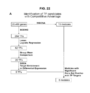
[0030] FIG. 22 shows transcriptional signature of competitive advantage. Panel
A.
Schematic of transcription factor candidate identification. Panel B. Violin
plots of identified
WGCNA module eigengenes per condition. Represented are significant modules
(black,
green, blue, brown, red, cyan), whose members are enriched for the downstream
targets of
the five transcription factors in Panel E. Panel C. Relative importance
analysis to estimate the
differential contribution of each biological factor (age vs genotype) to each
module
eigengene. Panel D. Gene set enrichment analysis (GSEA) highlighted those
prioritized
transcription factors whose regulons were enriched for upregulated genes in
dominant young
WT cells. Panel E. Important transcription factors predicted via SCENIC to
establish
competitive advantage and their relative activities across groups. Panel F.
Regulatory
network with represented downstream targets and their functional signaling
pathways.
Targets belong to highlighted modules in Panel B, and their expressions are
controlled by at
least one other important transcription factors in Panel E. NES: Network
enrichment score.
[0031] FIG. 23 shows Bulk RNA-Scq Characterization of human fetal GPCs. Panel
A. Workflow of bulk and scRNA-Sequencing of CD140a+, CD140a-, and A2B5+/PSA-
NCAM--selected 2nd trimester human fetal brain isolates. Panel B. Principal
component
analysis of all samples across two batches. Panel C. Venn diagram of CD140a+
vs CD140a-
and CD140+ vs A2B5+/PSA-NCAM- differentially-expressed gene sets (p <0.01 and
absolute 1og2-fold change >1). Panel D. Significant Ingenuity Pathway Analysis
terms for
both genesets. Size represents -log10 p-value and color represents activation
Z-Score (Blue,
CD140a+; Red, A2B5+ or CD140a-). Panel E. Log2-fold changes of significant
genes for
both genesets. Missing bars were not significant. Panel F. Heatmap of
transformed transcripts
per million (TPM) of selected genes in Panel E.
[0032] FIG. 24 shows single cell RNA-sequencing of CD140a and A2B5 selected
human fetal GPCs. Panel A. UMAP plot of the primary cell types identified
during scRNA-
Seq analysis of FACS isolated hGPCs derived from 20 week gestational age human
fetal
VZ/SVZ. Panel B- Panel C. UMAP of only PSA-NCAM-/A2B5+ (B) or CD140a+ (C)
human fetal cells. Panel D. Violin plots of cell type-selective marker genes.
Panel E. Volcano
plot of GPC vs pre-GPC populations. Panel F. Feature plots of select
differentially expressed
genes between GPCs and pre-GPCs. Panel G. Select significantly-enriched GPC
and pre-
GPC IPA terms, indicating their -log10 p-value and activation Z-Score. Panel
H. Select
feature plots of transcription factors predicted to be significantly activated
in fetal hGPCs.
Relative transcription factor regulon activation is displayed as calculated
using the SCENIC
package.
9
CA 03233935 2024- 4-4
SUBSTITUTE SHEET (RULE 26)
WO 2023/069882
PCT/US2022/078182
[0033] FIG. 25 shows adult human GPCs are transcriptionally and functionally
distinct from fetal GPCs. Panel A. Workflow of bulk RNA-Seq analysis of human
adult and
fetal GPCs. Panel B. Principal component analysis of all samples across three
batches. Panel
C. Venn Diagram of both Adult vs Fetal differential expression gene sets.
Panel D. IPA
network of curated terms and genes. Node size is proportionate to node degree.
Label color
corresponds to enrichment in either adult (red) or fetal (blue) populations.
Panel E. Bar plots
of significant IPA terms by module. Z-Scores indicate predicted activation in
fetal (blue) or
adult (red) hGPCs. Panel F. Bar plot of 1og2-fold changes and heatmap of
network genes'
TPM.
[0034] FIG. 26 shows inference of transcription factor activity implicates a
set of
transcriptional repressors in the establishment of adult hGPC identity. Panel
A. Normalized
enrichment score plots of significantly enriched transcription factors
predicted to be active in
fetal and adult GPCs. Each dot is a motif whose size indicates how many genes
in which that
motif is predicted to be active, and the color represents the window around
the promoter at
which that motif was found enriched. Panel B. Heatmap of enriched TF TPMs, and
Panel C,
log-fold changes vs adult GPCs, for both fetal hGPC isolates. Panels D-G.
Predicted direct
transcription factor activity of curated genes split into Panel D, fetal
activators; Panel E, fetal
repressors; Panel F, adult activators; and Panel G, adult repressors. Color
indicates
differential expression in either adult (red) or fetal (blue) hGPCs; shape
dictates type of node
(octagon, repressor; rectangle, activator; oval, other target gene). Boxed and
circled genes
indicate functionally-related genes contributing to either glial
progenitor/oligodendrocyte
identity, senescence/proliferation targets, or upstream or downstream TFs that
were also
deemed activated.
[0035] FIG. 27 shows induction of an aged GPC transcriptome via adult hGPC-
enriched repressors. Panel A. Schematic outlining the structure of four
distinct doxycycline
(Dox)-inducible EGFP lentiviral expression vectors, each encoding one of the
transcriptional
repressors: E2F6, IKZF3, MAX, or ZNF274. Panel B. Induced pluripotent stem
cell (iPSC)-
derived hGPC cultures (line C27 (Chambers et al., 2009; Wang et al., 2013))
were transduced
with a single lentivirus or vehicle for one day, and then treated with Dox for
the remainder of
the experiment. At 3, 7, and 10 days following initiation of Dox-induced
transgene
expression, hGPCs were isolated via FACS for qPCR. Panel C. qPCRs of Dox-
treated cells
showing expression of each transcription factor, vs matched timepoint
controls. Panel D.
qPCR fold-change heatmap of select aging related genes. Within timepoint
comparisons to
CA 03233935 2024- 4-4
SUBSTITUTE SHEET (RULE 26)
WO 2023/069882
PCT/US2022/078182
controls were calculated via post hoc least-squares means tests of linear
models following
regression of a cell batch effect. FDR adjusted p-values: *<0.05, ** <0.01,
*** <0.001.
[0036] FIG. 28 shows miRNAs drive adult GPC transcriptional divergence in
parallel to transcription factor activity. Panel A. Principal component
analysis of miRNA
microarray samples from human A2B5+ adult and CD140a+ fetal GPCs. Panel B.
Log2 fold
change bar plots and heatmap of differentially expressed miRNAs. Panel C.
Characterization
bubble plot of enrichment of miRNAs, versus the average log2 FC of its
predicted gene
targets. Panel D-Panel E. Curated signaling networks of Panel D, fetal (top)
and Panel E,
adult (bottom) enriched miRNAs and their predicted targets.
DETAILED DESCRIPTION
[0037] Reference will be made in detail to certain aspects and exemplary
embodiments of the application, illustrating examples in the accompanying
structures and
figures. The aspects of the application will be described in conjunction with
the exemplary
embodiments, including methods, materials and examples, such description is
non-limiting,
and the scope of the application is intended to encompass all equivalents,
alternatives, and
modifications, either generally known, or incorporated here. The described
aspects, features,
advantages, and characteristics of the invention may be combined in any
suitable manner in
one or more further embodiments. One skilled in the relevant art will
recognize that the
invention may be practiced without one or more of the specific aspects or
advantages of a
particular embodiment. In other instances, additional aspects, features, and
advantages may
be recognized and claimed in certain embodiments that may not be present in
all
embodiments of the invention. Further, one skilled in the art will recognize
many techniques
and materials similar or equivalent to those described here, which could be
used in the
practice of the aspects and embodiments of the present application. The
described aspects and
embodiments of the application are not limited to the methods and materials
described.
[0038] Unless otherwise defined, all technical and scientific terms used
herein have
the same meaning as commonly understood by one of ordinary skill in the art to
which this
application belongs.
100391 Ranges may be expressed herein as from "about" one particular value
and/or
to "about" another particular value. When such a range is expressed, another
embodiment
includes from the one particular value and/or to the other particular value.
Similarly, when
values are expressed as approximations, by use of the antecedent "about," it
will be
understood that the particular value forms another embodiment. It will be
further understood
that the endpoints of each of the ranges are significant both in relation to
the other endpoint,
11
CA 03233935 2024- 4-4
SUBSTITUTE SHEET (RULE 26)
WO 2023/069882
PCT/US2022/078182
and independently of the other endpoint. It is also understood that there are
a number of
values disclosed herein, and that each value is also herein disclosed as
"about" that particular
value in addition to the value itself. For example, if the value "10" is
disclosed, then "about
10" is also disclosed. It is also understood that when a value is disclosed
that "less than or
equal to "the value," greater than or equal to the value' and possible ranges
between values
are also disclosed, as appropriately understood by the skilled artisan. For
example, if the
value "10" is disclosed the "less than or equal to 10" as well as "greater
than or equal to 10"
is also disclosed.
[0040] As used in this specification and the appended claims, the singular
forms "a,"
"an" and "the" include plural referents unless the content clearly dictates
otherwise. Thus, for
example, reference to -a peptide" includes "one or more" peptides or a
"plurality" of such
peptides.
I. Definitions
[0041] As used herein, the following terms or phrases (in parentheses) shall
have the
following meanings:
[0042] The term -about" or "approximately" includes being within a
statistically
meaningful range of a value. Such a range can be within an order of magnitude,
preferably
within 50%, more preferably within 20%, still more preferably within 10%, and
even more
preferably within 5% of a given value or range. The allowable variation
encompassed by the
term "about" or "approximately" depends on the particular system under study,
and can be
readily appreciated by one of ordinary skill in the art.
[0043] The term -and/or" as used herein means that the listed items are
present, or
used, individually or in combination. In effect, this term means that "at
least one of" or "one
or more" of the listed items is used or present.
[0044] As will be understood by one skilled in the art, for any and all
purposes, such
as in terms of providing a written description, all ranges disclosed herein
also encompass any
and all possible subranges and combinations of subranges thereof. Any listed
range can be
easily recognized as sufficiently describing and enabling the same range being
broken down
into at least equal halves, thirds, quarters, fifths, tenths, and so on. As a
non-limiting example,
each range discussed herein can be readily broken down into a lower third,
middle third and
upper third, and so on. As will also be understood by one skilled in the art
all language such
as "up to," "at least," and the like include the number recited and refer to
ranges which can be
subsequently broken down into subranges as discussed above. Finally, as will
be understood
by one skilled in the art, a range includes each individual member.
12
CA 03233935 2024- 4-4
SUBSTITUTE SHEET (RULE 26)
WO 2023/069882
PCT/US2022/078182
[0045] In understanding the scope of the present application, the term
"comprising"
and its derivatives, as used herein, are intended to be open ended terms that
specify the
presence of the stated features, elements, components, groups, integers,
and/or steps, but do
not exclude the presence of other unstated features, elements, components,
groups, integers
and/or steps. The foregoing also applies to words having similar meanings such
as the terms,
"including", "involving", "having", and their derivatives. The term
"consisting" and its
derivatives, as used herein, are intended to be closed terms that specify the
presence of the
stated features, elements, components, groups, integers, and/or steps, but
exclude the
presence of other unstated features, elements, components, groups, integers
and/or steps. The
term "consisting essentially of', as used herein, is intended to specify the
presence of the
stated features, elements, components, groups, integers, and/or steps as well
as those that do
not materially affect the basic and novel characteristic(s) of features,
elements, components,
groups, integers, and/or steps. In embodiments or claims where the term
comprising (or the
like) is used as the transition phrase, such embodiments can also be
envisioned with
replacement of the term -comprising" with the terms -consisting of' or -
consisting
essentially of" The methods, kits, systems, and/or compositions of the present
disclosure can
comprise, consist essentially of, or consist of, the components disclosed.
[0046] In embodiments comprising an "additional- or "second- component, the
second component as used herein is different from the other components or
first component.
A "third" component is different from the other, first, and second components,
and further
enumerated or "additional" components are similarly different.
[0047] The term -complementary" when used in connection with nucleic acid,
refers
to the pairing of bases, A with T or U, and G with C. The term "complementary"
refers to
nucleic acid molecules that are completely complementary, that is, form A to T
or U pairs
and G to C pairs across the entire reference sequence, as well as molecules
that are partially
(e.g., at least 80%, 81%, 82%, 83%, 84%, 85%, 86%, 87%, 88%, 89%, 90%, 91%,
92%,
93%, 94%, 95%, 96%, 97%, 98%, or 99%) complementary.
[0048] The terms "nucleic acid-, "nucleotide", and "polynucleotide- encompass
both
DNA and RNA unless specified otherwise.
[0049] The term "polypeptide," "peptide" or "protein" are used interchangeably
and
to refer to a polymer of amino acid residues. The terms encompass all kinds of
naturally
occurring and synthetic proteins, including protein fragments of all lengths,
fusion proteins
and modified proteins, including without limitation, glycoproteins, as well as
all other types
of modified proteins (e.g., proteins resulting from phosphorylation,
acetylation,
13
CA 03233935 2024- 4-4
SUBSTITUTE SHEET (RULE 26)
WO 2023/069882
PCT/US2022/078182
myristoylation, palmitoylation, glycosylation, oxidation, formylation,
amidation,
polyglutamylation, ADP-ribosylation, pegylation, biotinylation, etc.).
[0050] The terms "abrogate", "abrogation" "eliminate-, or "elimination- of
expression of a gene or gene product (e.g., RNA or protein) refers to a
complete loss of the
transcription and/or translation of a gene or a complete loss of the gene
product (e.g., RNA or
protein). Expression of a gene or gene product (e.g., RNA or protein) can be
detected by
standard art known methods such as those described herein, as compared to a
control, e.g., an
unmodified cell.
[0051] The terms -express" and -expression" mean allowing or causing the
information in a gene or DNA sequence to become produced, for example
producing an RNA
or a protein by activating the cellular functions involved in transcription
and/or translation of
a corresponding gene or DNA sequence. A DNA sequence is expressed in or by a
cell to form
an -expression product" such as an RNA or a protein. The expression product
itself, e.g., the
resulting protein, may also be said to be -expressed" by the cell. An
expression product can
be characterized as intracellular, extracellular or transmembrane.
[0052] The term -competitive advantage" as referred to herein encompasses the
preferential proliferation, population expansion, durable survival and/or
stable integration of
a cell population placed in apposition to or admixture with a genetically
and/or
epigenetically-distinct cell population, to the detriment and eventual partial
or complete
replacement of the latter.
[0053] As used herein, the term "glial cells" refers to a population of non-
neuronal
cells that provide support and nutrition, maintain homeostasis, either form
myelin or promote
myelination, and participate in signal transmission in the nervous system.
"Cilia' cells" as
used herein encompasses fully differentiated cells of the glial lineage, such
as
oligodendrocytes or astrocytes, as well as glial progenitor cells, each of
which can be referred
to as macroglial cells.
100541 Certain terms employed in the specification, examples, and claims are
collected herein. Unless defined otherwise, all technical and scientific terms
used in this
disclosure have the same meanings as commonly understood by one of ordinary
skill in the
art to which this disclosure belongs.
[0055] Preferences and options for a given aspect, feature, embodiment, or
parameter of the disclosure should, unless the context indicates otherwise, be
regarded as
having been disclosed in combination with any and all preferences and options
for all other
aspects, features, embodiments, and parameters of the disclosure.
14
CA 03233935 2024- 4-4
SUBSTITUTE SHEET (RULE 26)
WO 2023/069882
PCT/US2022/078182
II. Genetically Modified Cell Populations
[0056] A first aspect of the present disclosure is directed to an isolated
population of
progenitor cells genetically modified to have a competitive advantage over
progenitor cells
which have not been genetically modified. As explained above, progenitor cells
genetically
modified to have a "competitive advantage" are cells modified to exhibit
preferential
proliferation, population expansion, durable survival and/or stable
integration of a cell
population placed in apposition to or admixture with a genetically and/or
epigenetically-
distinct cell population, to the detriment and eventual partial or complete
replacement of the
latter.
[0057] In one embodiment, the isolated population of progenitor cells is a
population
of central nervous system progenitor cells. Accordingly, in some embodiments,
the
genetically modified cell population is an isolated population of neural
progenitor cells,
neuronal progenitor cells, or glial progenitor cells genetically modified to
have a competitive
advantage over corresponding progenitor cells which have not been genetically
modified.
[0058] In one embodiment, the isolated population of progenitor cells is a
population
of glial progenitor cells. Accordingly, in one embodiment, the genetically
modified cell
population is an isolated population of glial progenitor cells genetically
modified to have a
competitive advantage over progenitor cells which have not been genetically
modified.
Suitable glial progenitor cell populations include, bi-potential glial
progenitor cells,
oligodendrocyte-biased glial progenitor cells, and astrocyte-biased glial
progenitor cells.
[0059] Other populations of progenitor cells that can be genetically modified
as
described herein include, without limitation, bone marrow progenitor cells,
cardiac progenitor
cells, endothelial progenitor cells, epithelial progenitor cells, mesenchymal
progenitor cells,
hematopoietic progenitor cells, hepatic progenitor cells, osteoprogenitor
cells, muscle
progenitor cells, pancreatic progenitor cells, pulmonary progenitor cells,
renal progenitor
cells, vascular progenitor cells, and retinal progenitor cells. In accordance
with the present
disclosure, any one of the aforementioned progenitor cells populations can be
genetically
modified as described herein to have a competitive advantage over progenitor
cells which
have not been genetically modified.
[0060] In some embodiments, the population of progenitor cells are genetically
modified to increase expression of one or more genes encoding proteins that
confer to the
cells a competitive advantage over progenitor cells which have not been
genetically modified.
In other embodiments, the progenitor cells are genetically modified so as to
decrease,
suppress, abrogate, or silence one or more genes encoding proteins that are
associated with a
CA 03233935 2024- 4-4
SUBSTITUTE SHEET (RULE 26)
WO 2023/069882
PCT/US2022/078182
competitive disadvantage over progenitor cells which have not been genetically
modified. In
yet another embodiment, progenitor cells of the populations described herein
are genetically
modified to express one or more genes that confer to the cells a competitive
advantage and to
suppress or silence one or more genes that are associated with a competitive
disadvantage.
100611 In some embodiments, the population of glial progenitor cells are
genetically
modified to express one or more genes that confer to the glial progenitor
cells a competitive
advantage over glial progenitor cells which have not been genetically
modified. In other
embodiments, the glial progenitor cells are genetically modified so as to
decrease, suppress,
or silence one or more genes that are associated with a competitive
disadvantage over glial
progenitor cells which have not been genetically modified. In yet another
embodiment, glial
progenitor cells of the populations described herein are genetically modified
to express one or
more genes that confer to the cells a competitive advantage and genetically
modified to
suppress or silence one or more genes that are associated with a competitive
disadvantage.
[0062] In accordance with all aspects of the present disclosure, the
population of
progenitor cells genetically modified as described herein are mammalian
progenitor cells. In
some embodiment, the population of glial progenitor cells is a population of
human
progenitor cells. In some embodiment, the population of glial progenitor cells
is a population
of human glial progenitor cells.
[0063] In some embodiment, the progenitor cells genetically modified as
described
herein are glial progenitor cells. In some embodiments, the genetically
modified glial
progenitor cells are genetically modified bi-potential glial progenitor cells.
In some
embodiments, the genetically modified glial progenitor cells are genetically
modified
oligodendrocyte-biased glial progenitor cells. In some embodiments, the
genetically modified
glial progenitor cells are genetically modified astrocyte-biased glial
progenitor cells. Methods
and markers for producing and distinguishing bi-potential glial progenitor
cells, astrocyte-
biased glial progenitor cells, and oligodendrocyte-biased glial progenitor
cells are described
herein.
[0064] Glial progenitor cells suitable for genetic modification as described
here can
be derived from multipotent (e.g., neural stem cells) or pluripotent cells
(e.g., embryonic stem
cells and induced pluripotent stem cells) using methods known in the art or
described herein.
[0065] In some embodiments, glial progenitor cells are derived from embryonic
stem
cells. Embryonic stem cells are derived from totipotent cells of the early
mammalian embryo
and are capable of unlimited, undifferentiated proliferation in vitro. As used
herein, the term
"embryonic stem cells- refer to cells isolated from an embryo, placenta, or
umbilical cord, or
16
CA 03233935 2024- 4-4
SUBSTITUTE SHEET (RULE 26)
WO 2023/069882
PCT/US2022/078182
an immortalized version of such a cells, i.e., an embryonic stem cell line.
Suitable embryonic
stem cell lines include, without limitation, lines WA-01 (H1), WA-07, WA-09
(H9), WA-13,
and WA-14 (H14) (Thomson et al., "Embryonic Stem Cell Lines Derived from Human
Blastocytes,- Science 282 (5391): 1145-47 (1998) and U.S. Patent No. 7,029,913
to Thomson
et al., which are hereby incorporated by reference in their entirety). Other
suitable embryonic
stem cell lines includes the HAD-C100 cell line (Tannenbaum et al.,
"Derivation of Xeno-
free and GMP-grade Human Embryonic Stem Cells -Platforms for Future Clinical
Applications," PLoS One 7(6):e35325 (2012), which is hereby incorporated by
reference in
its entirety, the WIBR4. WIBR5, WIBR6 cell lines (Lengner et al., -Derivation
of Pre-x
Inactivation Human Embryonic Stem Cell Line in Physiological Oxygen
Conditions," Cell
141(5):872-83 (2010), which is hereby incorporated by reference in its
entirety), and the
human embryonic stem cell lines (HUES) lines 1-17 (Cowan et al., "Derivation
of Embryonic
Stem-Cell Lines from Human Blastocytes," N. Engl. J. Med. 350:1353-56 (2004),
which is
hereby incorporated by reference in its entirety).
100661 In some embodiments, glial progenitor cells are derived from induced
pluripotential cells (iPSCs). -Induced pluripotent stem cells" as used herein
refers to
pluripotent cells that are derived from non-pluripotent cells, such as somatic
cells or tissue
stem cells. For example, and without limitation, iPSCs can be derived from
embryonic, fetal,
newborn, and adult tissue, from peripheral blood, umbilical cord blood, and
bone marrow
(see e.g., Cai et al., "Generation of Human Induced Pluripotent Stem Cells
from Umbilical
Cord Matrix and Amniotic Membrane Mesenchymal Cells," J. Biol. Chem. 285(15):
112227-
11234 (2110); Giorgetti et al., "Generation of Induced Pluripotent Stem Cells
from Human
Cord Blood Cells with only Two Factors: 0ct4 and Sox2," Nature Protocols,
5(4):811-820
(2010); Streckfuss-Bomeke et al., "Comparative Study of Human- Induced
Pluripotent Stem
Cells Derived from Bone Marrow Cells, Hair Keratinocytes, and Skin
Fibroblasts," Eur.
Heart J. doi: 10.1093/eurheartj/ehs203 (July 12, 2012); Hu et al., "Efficient
Generation of
Transgene-Free Induced Pluripotent Stem Cells from Normal and Neoplastic Bone
Marrow
and Cord Blood Mononuclear Cells,- Blood doi: 10.1182/blood-2010-07-298331
(Feb. 4,
2011); Sommer et al., "Generation of Human Induced Pluripotent Stem Cells from
Peripheral
Blood using the STEMCCA Lentiviral Vector," J. Vis. Exp. 68: e4327
doi:10.3791/4327
(2012), which are hereby incorporated by reference in their entirety).
Exemplary somatic
cells that can be used include fibroblasts, such as dermal fibroblasts
obtained by a skin
sample or biopsy, synoviocytes from synovial tissue, keratinocytes, mature B
cells, mature T
cells, pancreatic 13 cells, melanocytes, hepatocytes, foreskin cells, cheek
cells, or lung
17
CA 03233935 2024- 4-4
SUBSTITUTE SHEET (RULE 26)
WO 2023/069882
PCT/US2022/078182
fibroblasts (see e.g., Streckfuss-Bomeke et al., "Comparative Study of Human-
Induced
Pluripotent Stem Cells Derived from Bone Marrow Cells, Hair Keratinocytes, and
Skin
Fibroblasts," Eur. Heart J. doi: 10.1093/eurheartj/ehs203 (2012), which is
hereby
incorporated by reference in its entirety). Although skin and cheek provide a
readily available
and easily attainable source of appropriate cells, virtually any cell can be
used. Exemplary
stem or progenitor cells that are suitable for iPSC production include,
without limitation,
myeloid progenitors, hematopoietic stem cells, adipose-derived stem cells,
neural stem cells,
and liver progenitor cells.
100671 Autologous, allogenic, or xenogenic non-pluripotent cells can be used
in to
produce the iPSCs used to generate the genetically modified glial progenitor
cells. Allogenic
cells for production of iPSCs, for example, are harvested from healthy, non-
recipient donors
and/or donor sources having suitable immunohistocompatibility. Xenogeneic
cells can be
harvested from a pig, monkey, or any other suitable mammal for the production
if iPSCs.
Autologous non-pluripotcnt cells can also be harvested from the same subject
to be treated.
Autologous cells may need to be genetically modified as described herein and
further
genetically modified and/or otherwise treated to correct certain
dysregulations so that they
exhibit normal, non-disease related expression and/or activity in addition to
levels prior to
administration.
[0068] Induced pluripotent stem cells can be produced by expressing a
combination
of reprogramming factors in a somatic cell. Suitable reprogramming factors
that promote and
induce iPSC generation include one or more of 0ct4, Klf4, Sox2, c-Myc, Nanog,
C/EBPa,
Esrrb, Lin28, and Nr5a2. In certain embodiments, at least two reprogramming
factors are
expressed in a somatic cell to successfully reprogram the somatic cell. In
other embodiments,
at least three reprogramming factors are expressed in a somatic cell to
successfully reprogram
the somatic cell. In other embodiments, at least four reprogramming factors
are expressed in a
somatic cell to successfully reprogram the somatic cell.
100691 iPSCs may be derived by methods known in the art including the use of
integrating viral vectors (e.g., lentiviral vectors, inducible lentiviral
vectors, and retroviral
vectors), excisable vectors (e.g., transposon and foxed lentiviral vectors),
and non-
integrating vectors (e.g., adenoviral and plasmid vectors) to deliver the
aforementioned genes
that promote cell reprogramming (see e.g., Takahashi and Yamanaka, Cell
126:663-676
(2006) Okita. et al., Nature 448:313-317 (2007) Nakagawa et al., Nat.
Biotechnol. 26:101-
106 (2007); Takahashi et al., Cell 131:1-12 (2007); Meissner et al. Nat.
Biotech. 25:1177-
1181 (2007); Yu et al. Science 318:1917-1920 (2007); Park et al. Nature
451:141-146 (2008);
18
CA 03233935 2024- 4-4
SUBSTITUTE SHEET (RULE 26)
WO 2023/069882
PCT/US2022/078182
and U.S. Patent Application Publication No. 2008/0233610, which are hereby
incorporated
by reference in their entirety). Other methods for generating IPS cells
include those disclosed
in W02007/069666, W02009/006930, W02009/006997, W02009/007852,
W02008/118820, U.S. Patent Application Publication Nos. 2011/0200568 to Ikeda
et al.,
2010/0156778 to Egusa et al., 2012/0276070 to Musick, and 2012/0276636 to
Nakagawa, Shi
et al., Cell Stem Cell 3(5): 568-574 (2008), Kim et al., Nature 454: 646-650
(2008), Kim et
al., Cell 136(3 :411-419 (2009), Huangfu et al., Nature Biotechnology 26: 1269-
1275 (2008),
Zhao et al., Cell Stem Cell 3: 475-479 (2008), Feng et al., Nature Cell
Biology 11: 197-203
(2009), and Hanna et al., Cell 133(2): 250-264 (2008), which are hereby
incorporated by
reference in their entirety.
[0070] Integration free approaches, i.e., those using non-integrating and
excisable
vectors, for deriving iPSCs free of transgenic sequences are particularly
suitable in the
therapeutic context. Suitable methods of iPSC production that utilize non-
integrating vectors
include methods that use adenoviral vectors (Stadtfeld et al., -Induced
Pluripotcnt Stem Cells
Generated without Viral Integration," Science 322: 945-949 (2008), and Okita
et al.,
"Generation of Mouse Induced Pluripotent Stem Cells without Viral Vectors,"
Science 322:
949-953 (2008), which are hereby incorporated by reference in their entirety),
Sendi virus
vectors (Fusaki et al., "Efficient Induction of Transgene-Free Human
Pluripotent Stem Cells
Using a Vector Based on Sendi Virus, an RNA Virus That Does Not Integrate into
the Host
Genome," Proc Jpn Acad. 85: 348-362 (2009), which is hereby incorporated by
reference in
its entirety), polycistronic minicircle vectors (Jia et al., "A Nonviral
Minicircle Vector for
Deriving Hyman iPS Cells," Nat. Methods 7: 197-199 (2010), which is hereby
incorporated
by reference in its entirety), and self-replicating selectable episomes (Yu et
al., "Human
Induced Pluripotent Stem Cells Free of Vector and Transgene Sequences,"
Science 324: 797-
801 (2009), which is hereby incorporated by reference in its entirety).
Suitable methods for
iPSC generation using excisable vectors are described by Kaji et al., "Virus-
Free Induction of
Pluripotency and Subsequent Excision of Reprogramming Factors,- Nature 458:
771-775
(2009), Soldner et al., "Parkinson's Disease Patient-Derived Induced
Pluripotent Stem Cells
Free of Viral Reprogramming Factors," Cell 136:964-977 (2009), Woltjen et al.,
"PiggyBac
Transposition Reprograms Fibroblasts to Induced Pluripotent Stem Cells,"
Nature 458: 766-
770 (2009), and Yusa et al.. "Generation of Transgene-Free Induced Pluripotent
Mouse Stem
Cells by the PiggyBac Transposon," Nat. Methods 6: 363-369 (2009), which are
hereby
incorporated by reference in their entirety. Suitable methods for iPSC
generation also include
methods involving the direct delivery of reprogramming factors as recombinant
proteins
19
CA 03233935 2024- 4-4
SUBSTITUTE SHEET (RULE 26)
WO 2023/069882
PCT/US2022/078182
(Zhou et al., "Generation of Induced Pluripotent Stem Cells Using Recombinant
Proteins,"
Cell Stem Cell 4: 381-384 (2009), which is hereby incorporated by reference in
its entirety)
or as whole-cell extracts isolated from ESCs (Cho et al., "Induction of
Pluripotent Stem Cells
from Adult Somatic Cells by Protein-Based Reprogramming without Genetic
Manipulation,"
Blood 116: 386-395 (2010), which is hereby incorporated by reference in its
entirety).
[0071] The methods of iPSC generation described above can be modified to
include
small molecules that enhance reprogramming efficiency or even substitute for a
reprogramming factor.
[0072] These small molecules include, without limitation, epigenetic
modulators
such as the DNA methyltransferase inhibitor 5.-azacytidine, the histone
deacetylase inhibitor
VPA, and the G9a histone methyltransferase inhibitor BIX-01294 together with
BayK8644,
an L-type calcium channel agonist. Other small molecule reprogramming factors
include
those that target signal transduction pathways, such as TGF-f3 inhibitors and
kinase inhibitors
(e.g., kenpaullone) (see review by Sommer and Mostoslaysky, -Experimental
Approaches for
the Generation of Induced Pluripotent Stem Cells," Stem Cell Res. Ther. 1:26
doi:10.1186/scrt26 (2010), which is hereby incorporated by reference in its
entirety).
[0073] Methods of obtaining highly enriched preparations of glial progenitor
cells
from the iPSCs or embryonic stem cells (e.g., human embryonic stem cells) that
are suitable
for treating a neuropsychiatric disorder as described herein are disclosed in
W02014/124087
to Goldman and Wang, and Wang et al., "Human iPSC-Derived Oligodendrocyte
Progenitors
Can Myelinate and Rescue a Mouse Model of Congenital Hypomyelination," Cell
Stem Cell
12(2):252-264 (2013), which are hereby incorporated by reference in their
entirety.
[0074] In yet another embodiment, glial progenitor cells can be extracted from
embryonic tissue, fetal tissue, or adult brain tissue containing a mixed
population of cells
directly by using the promoter specific separation technique, as described in
U.S. Patent
Application Publication Nos. 20040029269 and 20030223972 to Goldman, which are
hereby
incorporated by reference in their entirety. In accordance with this
embodiment, the glial
progenitor cells are isolated from ventricular or subventricular zones of the
brain or from the
subcortical white matter.
[0075] In some embodiments, it may be preferable to enrich a cell preparation
comprising glial progenitor cells prior to or after genetic modification to
increase the
concentration and/or purity of the glial progenitor cells exhibiting a
competitive advantage
for therapeutic administration. Accordingly, in one embodiment, the A2B5
monoclonal
antibody (mAb) that recognizes and binds to gangliosides present on glial
progenitor cells
CA 03233935 2024- 4-4
SUBSTITUTE SHEET (RULE 26)
WO 2023/069882
PCT/US2022/078182
early in the developmental or differentiation process is utilized to separate
glial progenitor
cells from a mixed population of cells (Nunes et al., "Identification and
Isolation of
Multipotential Neural Progenitor Cells From the Subcortical White Matter of
the Adult
Human Brain.,- Nat Med. 9(4):439-47 (2003), which is hereby incorporated by
reference in
its entirety). Using the A2B5 mAb, glial progenitor cells can be separated,
enriched, or
purified from a mixed population of cell types. In another embodiment,
selection of
CD140a/PDGFRa positive cells is employed to produce a purified or enriched
preparation of
bi-potential glial progenitor cells. In another embodiment, selection of CD9
positive cells is
employed to produce a purified or enriched preparation of oligodendrocyte-
biased glial
progenitor cells. In yet another embodiment, both CD140a/PDGFRa and CD9
positive cell
selection is employed to produce a purified or enriched preparation of
oligodendrocyte-biased
glial progenitor cells. In another embodiment, selection of CD44 positive
cells is employed to
produce a purified or enriched preparation of astrocyte-biased glial
progenitor cells (Liu et
al., -CD44 Expression Identifies Astrocyte-Restricted Precursor Cells," Dev.
Biol. 276(1):31-
46 (2004), which is hereby incorporated by reference in its entirety.) In
another embodiment,
both CD140ct/PDGFRa and CD44 positive cell selection is employed to produce a
purified or
enriched preparation of oligodendrocyte-biased glial progenitor cells. In
another embodiment,
CD140a/PDGFRa, CD9, and CD44 positive cell selection is employed to produce a
purified
or enriched preparation of oligodendrocyte-biased glial progenitor cells.
[0076] The genetically modified glial progenitor cell population described
herein is
preferably negative for a PSA-NCAM marker and/or other neuronal lineage
markers, and/or
negative for one or more inflammatory cell markers, e.g., negative for a CD11
marker,
negative for a CD32 marker, and/or negative for a CD36 marker (which are
markers for
microglia). Optionally, the preparation of glial progenitor cells is negative
for any
combination or subset of these additional markers. Thus, for example, the
preparation of glial
progenitor cells is negative for any one, two, three, or four of these
additional markers.
100771 In accordance with the present disclosure the population of genetically
modified glial progenitor cells as described herein comprises at least about
80% glial
progenitor cells, including, for example, about 80%, 85%, 90%, 95%, 96%, 97%,
98%, 99%,
100% glial progenitor cells. The population of genetically modified glial
progenitor cells is
preferably devoid (e.g., containing less than 20, 15, 10, 9, 8, 7, 6, 5, 4, 3,
2, or 1%) of other
cells types such as neurons or cells of neuronal lineage, fibrous astrocytes
and cells of fibrous
astrocyte lineage, multipotent cells, and pluripotential stem cells (like ES
cells). Optionally,
exemplary cell populations are substantially pure populations of glial
progenitor cells.
21
CA 03233935 2024- 4-4
SUBSTITUTE SHEET (RULE 26)
WO 2023/069882
PCT/US2022/078182
[0078] Positive and/or negative selection for cell markers of interest (e.g.,
PDGFRct
marker, A2B5 marker, and/or a CD44 marker) can be carried out serially or
sequentially and
can be performed using conventional methods known in the art such as
immunopanning. The
selection methods optionally involve the use of fluorescence sorting (FACS),
magnetic
sorting (MACS), or any other method that allows rapid, efficient cell sorting.
Examples of
methods for cell sorting are taught for example in U.S. Patent No. 6,692,957
to Goldman,
which is hereby incorporated by reference in its entirety, at least for
compositions and
methods for cell selection and sorting.
100791 Generally, cell sorting methods use a detectable moiety. Detectable
moieties
include any suitable direct or indirect label, including, but not limited to,
enzymes,
fluorophores, biotin, chromophores, radioisotopes, colored beads,
electrochemical, chemical-
modifying or chemiluminescent moieties. Common fluorescent moieties include
fluorescein,
cyanine dyes, coumarins, phycoerythrin, phycobiliproteins, dansyl chloride,
Texas Red, and
lanthanide complexes or derivatives thereof.
[0080] The genetically modified glial progenitor cell populations described
herein,
including the enriched preparations can be optionally expanded in culture to
increase the total
number of cells for therapeutic administration. The cells can be expanded by
either
continuous or pulsatile exposure to PDGF-AA or AB as mitogens that support the
expansion
of oligodendrocyte progenitor cells; they can be exposed to fibroblast growth
factors,
including FGF2, FGF4, FGF8 and FGF9, which can support the mitotic expansion
of the glial
progenitor cells, but which can bias their differentiation to a mixed
population of astrocytes
as well as oligodendrocytes. The cells can also be expanded in media
supplemented with
combinations of FGF2, PDGF, and NT3, which can optionally be supplemented with
either
platelet-depleted or whole serum (see Nunes et al. "Identification and
Isolation of Multipotent
Neural Progenitor Cells from the Subcortical White Matter of the Adult Human
Brain,"
Nature Medicine 9:239-247; Windrem et al., "Fetal and Adult Human
Oligodendrocyte
Progenitor Cell Isolates Myelinate the Congenitally Dysmyelinated Brain,-
Nature Medicine
10:93-97 (2004), which are incorporated by reference for the methods and
compositions
described therein).
100811 As described supra, in some embodiments, the population of glial
progenitor
cells as described herein is genetically modified to have a competitive
advantage over glial
progenitor cells which have not been genetically modified. In some
embodiments, cells of the
isolated population are modified to increase expression of one or more genes
that confers a
competitive advantage to the modified cells relative to glial progenitor cells
which have not
22
CA 03233935 2024- 4-4
SUBSTITUTE SHEET (RULE 26)
WO 2023/069882
PCT/US2022/078182
been genetically modified. In some embodiments, cells of the isolated
population are
modified to decrease or silence expression of one or more genes that confers a
competitive
disadvantage to the modified cells relative to glial progenitor cells which
have not been
genetically modified.
100821 In some embodiments, the isolated population of glial progenitor cells
as
described herein contains cells that have been modified to express one or more
genes that
confer a competitive advantage to the cells and cells that have been modified
to decrease
expression of one or more genes that confer a competitive disadvantage to the
cells.
100831 In some embodiments, cells of the isolated population are genetically
modified to express one or more genes that confer a competitive advantage and
modified to
decrease expression of one or more genes that confer a competitive
disadvantage to the glial
progenitor cells compared to glial progenitor cells which have not been
genetically modified.
Genetic Modifications to Express One or More Genes that Confer a Competitive
Advantage
100841 Genes whose expression provides progenitor cells a competitive
advantage
were identified using the models of cell competition described in the Examples
herein. In
particular, differential gene expression between various cell populations
utilized in the model
(e.g., healthy glial progenitor cells vs. diseased glial progenitor cells and
similarly aged
healthy vs. diseased progenitor cells) were analyzed and compared to identify
genes that
confer a competitive advantage and genes that confer a competitive
disadvantage to
transplanted cells as compared to the resident cells.
100851 The one or more genes identified herein as providing cells a
competitive
advantage over resident cells upon transplantation (advantage genes) are
provided in Table 1
below by their gene name. Also provided in Table 1 is the Entrez ID accession
number and
Ensembl ID for each gene, which are each hereby incorporated by reference in
their entirety
for their disclosure of the gene sequences and the corresponding protein
encoded by each
sequence. All gene products referred to in this application include the wild
type gene product
and functional variants thereof. A "functional variant of a gene product"
refers to a modified
gene product (e.g., by deletion, substitution, insertion, glycosylation, etc.)
that retains at least
50% of the biological activity of the unmodified (wild-type) gene product in a
competition
assay.
23
CA 03233935 2024- 4-4
SUBSTITUTE SHEET (RULE 26)
WO 2023/069882
PCT/US2022/078182
[0086] Table 1. Genes That Confer A Competitive Advantage to Cells (advantage
genes)
Las..m./..11.11D Eak:'1W ID CkIik,, E.tmlabl ID
Etarkit ID
AC:TB EN5C406(5X175624 60 NITBS.R21.5 EN
223 1.00463485
AKRICI i ENSG01101101571.3.4 1645 NII1.12A. EN
&X)1 10627
ANA PC1 I t F...15/50600::101.415-52 51529
Nri'l...I.2B .ENS(3601.1001. 1 &.C.8.0 1.03910
i
_______________________________________________________________________________
_
AP2D I EN131:31.015150336115 163 N.ACA
EN1¶.31:112909 I 96531 4666
1 AI'LP-Z LN 5:C.4.A100ct184'2.34 334 :N,..Ikt$ /
LN5Ci-1.X8.100134440 4C 7
4.1. .......................................... ,. ....... - ...........
ARFI.6 I ENSC306900004059 381 NCI.
.ENSCK10:5.2.$0115053 .4691
., .....................................................................
ARL4A. 1 ENSC-06000122644 101.2.4 NDUFAI -
ENSI3000C411211356 4694
ARPo EN r.GCNO:31.1.1229 19C04 NDUTAll
IIN000.1.?48;1.6 126320
A RPP1.0 EIN8<7.10000012.8989 10776 NDUF- An
.ENSG$X1060 I 86010 510:79
. .
ATOX1 EN %µ.000001..T7556 475 NIKTA3.
EN5K:1000001..,"W06 4696
.ATP5FIE F:NSG00000:17417,. 44 N.1it.1.:44 r:Nsoxgx.ioIVA43
4697
s, ....................
ATP:SI:ICI EN15(W06(5.)1591 9.9 516 IND15.F.131
.E4SCM1100 IS.3648 4707
.AT15.NX3 EN SCt001.1001.54518 518 N.M..1:191 I EN
001.1014712.3 ,545;;;9
AT? 41 EN!.8(350 ..01173.1 1 5 545:31 NIIITRI. P_N-
86-0000601,10266 *708.
.ATF51\11/. ENSi70:10W16%120 faI NIBUT36 ENS(.1011010I
65264 4712
ATP-5:NIF ENS/Y:410(5)241468 9551 NDI:17.37
s ENSC14)15.280099195 4713. .
A TRiNIS 1%31 NIRTC2: EN -1.1011011/.
51366 471S
ATP5MPL. EMGOW:`015641 t 9556 NDIT35 .:-
.7.3s6oxowia6:53 472,5
ATP:5FF EN1'.1<1000:831.54723 .522 NUC,81.
EN1(101.)11W0692.75 64710
A TP6V0a t P.:NS(..ii?0{)-(it ..7-4:10 I. fi.3":' 4.) AZ1
- EN8G011160 I (4904 4946
4
ATP6 VOL 1 EN5Gi.):.0C..)113732 8992 OLFM2.
EN..;GM)::01.00:74 93 i4.5
ATXN71.31B ENS6000M2.53719 '...f.i2g 9 ' OSBPUi
E:NSCKW091039 1 i 4.=:,..8.1.!. ,
BIM EN80500(,'01.66710 1167 135T4
_________________ EN:'1X100000.225474 1.IX1128 n 1 1
.B3GAT2 F\3..'11.1(iN119011112399 135152 OTC
EN8GC43901.11. 98856 5850.5 I
BENI EN51;0901.10I3316 55859 PA BPC1 -----------
---- .ENS:G O,V.R.T-i=0756 269S6 1
EN5G06000I66681 27018 PC:BP2 EN SOW:00197111
5094 I
1
'BLOC:1S1. ENSG09 135441 :2647 PCD111.8 EN SOMX0138650
57575 1
11.3TERBI aix.',o-A-.-1,:3156780 :89911 RED /III. X:
EN 50101001112290 2-7323 1
;
Cl6m.132 .EN6QX-16:1111,77576 I. 497661 .1)(1)1117
EN.5<3.11kX1001 I 5946 27253 1
Clori122 FLN-G1A11000.1.97982 127687 PC-D.11B2 EN
SG00060.I.12852 56133
.
_______________________________________________________________________________
__ .
24
CA 03233935 2024- 4-4
SUBSTITUTE SHEET (RULE 26)
WO 2023/069882
PCT/US2022/078182
Gikno Ex/sombl TD Entrez ID Gme- Et/m.11M ID
Entrez ID
CI.Q8P ******1EMG00909108Ii61. V 708 PCIIIIC.86 -
** EN )2533
C4o1-013 .ENSGOW90741449 401115 PDGFRA ENSCFM071.34g.5.4 5156
CADM4 ENSK3(X)(1001 0 5 -7 6 7 ic:97 --i 1
__________________ PDIA6 EN,1Cs 009W143370 19130 ,
CALM:1 ENSG9(10110 I 98668 801 PE G-111
.ENSCM0911242265 23089
CA'LM 3 ENSG,0009 D.10014 808 PFNI F.:MGM:0)10851g
5219
CALR ,ENSG030001792.1 8 :.11. PGRNICA EN8(101:16M
018.56 1.0857
...............................................................................
... ,
CANX i---7NS6017696I7'1077 871 MLA.
ENS(11/a01171933 5369 .
CAV2 ENSGW060 I 05971 858 PIPP3 -ENSGW000162407
8613
CC21)1A ENSG90090132924 54862 PLPPRI ENSG 18100914-8123
.54886
CCNDI. ENISG1X8)0011.0092 595 PPIA :ENSGO119M196262
5478
,
...............................................................................
CCM ENS(.34.Y.160011. gal 6 19913 .PRDX1
.ENSGMNY3:1174.50 50.52
._
C1)63 ENS GOW09115494 967 PRDX2 EN SC,' W6167815
7001
CD82 PN Siii091-19W851-1 7 3732 .PR.DX5 ENSC1-
000W126432 25824
C DC42 ENSG00011W70831 998 PS MB/. ENSG-
91190O01181118 5689
CD112 1.",--N8C1010001791158 1000 PSM119 I-
IN8GW000246965 5698
CFI! ENSGMW 172757 1072 PTNIS EN SG 3W00 1 59?
15 5763
,
_______________________________________________________________________________
__
CHC11D2 ENSG1)9090I(X5I53 511.12 FYN .ENSGW99619.5894
5764
(HG E ENSGW9600:89199 1114 PTPRA FM01169601 a247o
5789
CIA02B ENSOXJ000166395 51647 RAMO -ENKii)ONXM433.3
W90
CIA 3 ENS(1000001.1195.72 I t2 RAB14 ENS-
01810(8'119396 51552
([TA EN93900 '22705 1211 Rk112 A. ENSG-00006I
041388 5862
CITC .EN.Sa7-11690141367 1213 RAD111 ENS-
GW600:1118461 11031
C:N/fZ3' ENSG10011911715I1/ 1266 RAC1 ENSGW01136238 5879
CNTN I P-NS(101.X19W-18:736 1777 .1ACK1 "PNS-
C1/91.0379462g 19399
COIL! ENSGW1011010.31V 23406 RAIDN2- ENSGO11911911584I
151191.
C0X411 ,E2,1.860M90131143 1327 ROO
EN8Cif1116W11574 7 67.3R
COX6A1 EN-SG:10909111775 337 R01101 ' ENSGW909169115.5
6091
COX6C .ENSGW0901649:19 134:5 RRAG.B, ENSCKWM08.3750
10325
C OX7A2 *1M1.1269.5 1347 RT1s3 ' EN-SG-
9090013331a 10313
CO2CC ENSGOW96127/M 1350 6100B ' IENSG0000O160307
62g5
(0\S4 ENSV:0000017634.0 1351 SARAF EN SC., 000W
133s72 51669
CPNNSGON00 I:19 I 17 144402 SAT). ENSCMOM 3-006,6 *
I8 E 6303
---
CPS-I ENSG0Mg.)0:',Im 1373 SIMS ENS3909(9126524
51.119
.............................................. ., ..................... .4.
......
CRNDE ENSGOX3110:1145694 60011 :SCARR11 '-S( 7$0
950 .. ,
CSPG4 ENSCAX196173,546 1464 SCP11 FNS-
G11911991.16,171 6147 .
CTIIRCI -EIN6G9:8169164932 I 15908 ;.SCR 611.
ENSG1109W164196 11341
CU L418 ENS(X/90110/ '7.99 8450 :SEC,62 EM4,;-
3011105089.57 2095
C'VP'5 IA1 ENS(10000o07 5i0 159.5 SE I.I.NOK
EMi.WOOW i. II ';'. I I. 58515
DIM EN:SO:WWI .7333 a 1622 .SELENOT ENSCM1700198843
51714
...............................................................................
... t
DCX * ENA-30000/X/7727 1541 SELENOW EN
0001. 78180 64 I 5
CA 03233935 2024- 4-4
SUBSTITUTE SHEET (RULE 26)
WO 2023/069882
PCT/US2022/078182
-13-tra Entena161 .f..D Exam ID =Gm*. Eftwatb1 ID
Extra ID
DDAI-11 ENSC.000(..:1539N 2'55.7'6 + 8ER F:1
ENSG1X*00140264 10.169
DDX1 . rNS6000c.607.9785 1.653 8ERF.012
ENSGOAX1Ø 3.5919 5.270
1ENNDI0 liN 1101100119979 40461% '7SET EN SaVX01193.35
6418 , .D.N1D ENSki000W198947 1756 811313(371.. ENSG0X00 I 31171
6451
DN1RT2 s EY.:Q100001.73253 10655 ' SKP1
ENS001000113558 6500
_______________________________________________________________________________
___ ,
11:NAJA 2 ENS(.500090:.169345- 19294 .81.11::2.5A6
ENSQ0X90169109 293
+
DPYSL2 .ENSG.0000,92964 1808 SLIT2 ENSGM-00145147
9353
+ - ... - ..... -
----
DRAM ENS0000M173.350 103:89 ,SLITRICI: "1:,--NO>5000185985
84631
_______________________________________________________________________________
___ ,
DSTN EN Sia0M1.2:5 868 11034 S.M.C3 ENX0108055 91126
DYNCHI EN.60-90QM7-7380 178i 8 NIIIII ENS,.-C.49C050 I
83172 9I81
EDF1 E,N8600000107223 8721 8NIOCI ENSGIX:',0001:
08732 64093
F.V.F1 A 1 FiNi<1.110Ng) .:5.650g 1411. SNIS EN.sCAVA
.1021:72 6611
EEF1 DI .PNS60000011494). 1933. SN CA ENSGUX:011 45335
6612
.............................................. + .......
lin ....v,N.T.Q00f.-)001676.58 1938
SN114;29 " RN:SU:K*00i .75061 12:5144.
.E1D1 EN8C00000255302 23741 NNFIG6 ENS-COM0245910
641638
EI.F3J EN S Q0904:0104131 8669 S.NX22 EN Kz(00001
:77.34 79856 .
.EL0.B =ENS600000103363 6923 S7'. ki X3 r-
NSCOX00I12335
EN P2 " T-(3,000(W.13:8.51 ' .?z SO1M
EM3M:',00142.168 6647
E.11 ENS X3684 2098 80X II ENSO.X8MI ':'.-7 '
6664.
+
_______________________________________________________________________________
__
ETV' .ENSGOUOMV6468. 2115 80X2 ENSG(8X00.1 8 1449
6657 .
FAI3P7 .,rN8000000164434 2173 80X9 FNSC.-4X*001.25398
6662
...................................................................... _ ..
1.7FAMr IB ENSC000M144369 165.215 SPC52 ENX4M.00 I 18363
9789
FAM177A1 EN 5G00000 /51327 283635 SPC63
ENSGO.:8>00 1 29128 60559
EMS -.ENSCi0(0W149306 2197 RP14 ENSG000011140319
6727
,
HSI EN 00000214253 51024 SSR4 ENSGC00;00 .180879
6748
FXY1)6 EN-36000%137726 53826 STAG2 F.NA-A0=.00101972
16715
.............................................. +
.................................
GAP43 .ENSCi00(03172020 2596 STNINI ENSCOM01116.32
.3925
...................... - .......
GcSII SNS00000014090.5 2653 SUPT.1611 EN5CA00092.201
11198
,GN AS ENSG00000087460 2778 YALD01 ENSG0O001 771 5:6
On
GOLM I EN '..K;O:00 I 3 .5(152 5 MO TBCB E2SCA8,V9
1 Q 5254 1,1'55
-
GM:16B irN8600M8046653 2824 TC:EAL-7 ENS )000 I 82916
56849
C.;-8T II EN '5C.,00000%42117 2.)50 WEALS ENSCA8X8)
I 8?.)964 91843
11.3-IN ENS(.3000.0-016304I 3020 WEALS ENSGOX091 8.5222
M 186
.............................................. ,-
113-3B EINSCi:WW,In 32475 30.11 IBM EN &0) 1 G2:2613
7076
HINT' EN8(.:0009016.9.567 8094 ILE5 ENSCOX00 I 04964
166
UN RiNTA 1 ' EN SG100001 35486 3178 TM4S1.1 EN 3G0M016990S
4071
FES-RN PA3 ENSM1M.1, '70 144. 220988 INI9SF11 ENS
GOMX107.71.47 56889
IINRNPAB 7NSG-000M 97451 :3182 TAIA.7 ENSG(8X00232112
51372 ,
ITYRNPC: ENSOW00.00921.99 3183 TN1BI16 ENSG1X00013.9µ44
' 7009
! IINIZSPIC . mqgG00000:165I1.9 31% 'TUC 01 INSGOV00143181
54499
26
CA 03233935 2024- 4-4
SUBSTITUTE SHEET (RULE 26)
WO 2023/069882
PCT/US2022/078182
C-tne Ensombt ID Et m M Gene EusevAl. ID 1 --
----
i Elam ID
------------------------------------------------------------------------- 4. --
---
1:11NR,NPM ENSCM000099733 ' 4670 TNIEM:147 EN 50.0)00105677
10430
I(NRNPR ENSCHK000125944 1023 ME,M258 ENS:60X)0134325 746
IISPA5 ENSGC0(0).014574 3309 TMEM59A EN ..(7.-
.?0.....001837:26 23585
..
T.G.F13.1)2 ENSGX$000115457 __ $485 I'M ... OD2
ENSC{M001.2.1172 2.767
,
.
ITGFA ENSGCKVg)10q3.5 1695 TMSR10 ENSS..7i0WN:C:-14510
WA.
ITNEA ENTSM1000076596 9452 -11.D.:-.3B4X.
ENSCA*00205542 , 711.4
_______________________________________________________________________________
___ -4
'-iTNI2B ENSW00001361.56 9445 1 IPT1 ENSCM090133-112 1
7178:
+
õIPTI ENSG.C.:00001S0 /59 51155 1 -TRAF4 EN
sco,x0)071.36{,14 961g
i
KDELR1 ENSa)-000010f.:,438 10945 TSCLITA ENSC,1)6N:1 I
66925 81 628
' .10ERI1I-ASI ENSGOOK,0245648 101 928109 TSPA1N6 T:Nscammak,,:)3 i
7105
KRE CAP2 EMU:V:100163463 20018,5 ISP7 EN SC00,)00156298
1 7102:
F----L.--
_________________________________________________________________________ -1.
.KIN I NSCW000126777 3895 TTC3 ENSGWONI 82-670
7267
,
LDHB I ENSGag01 I 11716 3945
TUE:9 EN SCOXII,A1%23.0 20306
UL 8
ETLI 1 ENSt.:X8)009-187416 375612. IMA.52
.ENSGW(0822-1983 7311
I ,.
LRRC:411 ,1 ENSGW0001.314-09 94050 VEL.5:
ENS.X.OX;00Ini.5,1 592.6
AIA1)2 1 ENSGM0000Th01.8 4133 IVCRI 0 EN
SCi0(000.184ti76 29796
MARCKS E\' 744$ 4032 I.:TQC.RI1 ENSQVX:00 I
,17540 10975 -4
ilkt ARCKSLI EIN..QM001 75130 ' 65.108 VQ.C.R.13. EN5XAMYAI56467
7381
...............................................................................
... 4
.111.A. EN8CA70002.61357 1.9:.1 VIM En3.0WW26.025 7431
M1COS1 0 ENSC:100000173436 440574 WS132 .ENSGA:00.176171
55384
MU ENSCi4.20000240972. 4212 WSCD1. 0ØX1017.931 4
23302
...............................................................................
... .4
MIR9-1fIG ENSa,`M)01.2"...:462 10485 Y.SX1 ENS-60000065978
4904
N1MG711 ENSG0000016;k446 93380 lIVILAB ENSC00:)00166913 7579
AIPZLI ENSC:01000197%:.5 90ts1 YWILA,E ENS-CAM0IN953 7531
1
;WILY ENSG00090175701 .205251 I ZF Ail EN S.COX:001
n410 .441951
.............................................. + ....................... 4
MIRNK2L1.2 ENSUX40002:69:128 100462981 1 ZNF-423 EN Sa'0,X.101
.31:116 126299
GADDA ENSGMM-1.16717 , 1647 1 LNT462 'ENS:C0X:091
48143 58499
,
IN61.1 ENS G00000176956 4062 1 ----------------------- 4-
_____
1
------------------------------------------------------------------------- .
[0087] In some embodiments, glial progenitor cells of the isolated populations
described herein are genetically modified to increase expression of one or
more genes listed
in Table 1, relative to non-genetically modified progenitor cells. In some
embodiments, glial
progenitor cells of the isolated populations described herein are genetically
modified to
increase expression of any two of the above noted genes relative to non-
genetically modified
progenitor cells. In some embodiments, the glial progenitor cells of the
isolated population
are modified to increase expression of any 3 of the above noted genes. In some
embodiments, the OM progenitor cells of the isolated population are modified
to increase
expression of any 4 of the above noted genes. In some embodiments, glial
progenitor cells of
the isolated population are modified to increase expression of any 5, 6, 7, 8,
9, 10, 11, 12, 13,
14, or more of the above identified genes.
27
CA 03233935 2024- 4-4
SUBSTITUTE SHEET (RULE 26)
WO 2023/069882
PCT/US2022/078182
[0088] Besides the genes provided in Table 1, atop-ranked set of genes is
provided
in Table 2 below, which additionally includes genes upregulated in advantaged
cells
("winners") while concurrently suppressed in disadvantaged cells ("losers").
In some
embodiments, the glial progenitor cells of the isolated population are
modified to increase
expression of any one of the genes provided in Table 2 below relative to non-
genetically
modified progenitor cells. In some embodiments, the glial progenitor cells of
the isolated
population are modified to increase expression of any one, two or more genes
selected from
the genes of Table 2 relative to non-genetically modified progenitor cells. In
some
embodiments, the glial progenitor cells of the isolated population are
modified to increase
expression of any 3 of the below noted genes. In some embodiments, the glial
progenitor
cells of the isolated population are modified to increase expression of any 4
of the below
noted genes. In some embodiments, glial progenitor cells of the isolated
population are
modified to increase expression of any 1, 2, 3, 4, 5, 6, 7, 8, 9, 10, 11, 12,
13, 14, 15, 16, 17,
18, 19, 20, 21, 22, 23, or all 24 of these genes, relative to non-genetically
modified progenitor
cells.
[0089] Table 2: Top-ranked Genes that Confer a Competitive Advantage
Gtzte EnTeembi ID Entrez ID Gene E:sirs.emb.1 ID
'Entrez
TRIO ENSG0.00000..3.8$ 7204 111,11AB ENSG00.000166-
91.3 75.29
ITN131A ENSG.0000C}078596 9452 B211 ENSSOM00166.710
567
BEX5 ENS0000001. 84515 34054" PTIIIS EG000159335
5.765
.B-EX3 ENSG000001.66621 7701.3 OLEA12 EN SG-0000010.5088
93145
CIHRCI ENSG0}0000.1.4-A932 1 1.5908 1.1'6H ENSO00.00.
0176956 .400-
EDIL3 E.NSG00;00.01.64176. 10085 MT 3 ENSG0N-
)000,87:Y50 .4504
:NITA ENSG0000026.1857 8190 ITBA52 EN SG00000.221983
7311
.ENIC.10 ENSG000001616.71 284361 SNX3 E2TSG00(}0011:-.-
333 .2:724
C CND'. EN6(3000001.10092 595 FABF7 EN SCOW:301.64434
2.113
GAD.D45A ENSG00000116717 1647 ERRC413 ENSO00000131.409
94030:
POD ENS000000129058 347 RAMP! E2SG000(}0132329
10267
TRAF4 EN3G00:00807.6504. 9618 NET74
NSCiOty.)0002:04099 129807
[0090] In some embodiments, the glial progenitor cells of the isolated
population are
modified to increase expression of one or more genes selected from the genes
listed in Table
2, relative to non-genetically modified progenitor cells.
[0091] Table 3 provides another embodiment of transcripts conferring
advantage,
which includes top-ranked genes exhibiting significant transcriptional
upregulation in WT
cells presented with diseased and disadvantaged HD-derived cells, relative to
singly engrafted
28
CA 03233935 2024- 4-4
SUBSTITUTE SHEET (RULE 26)
WO 2023/069882
PCT/US2022/078182
WT cells, that also manifest significant transcriptional downregulation in the
disadvantaged
HD cells, relative to singly engrafted HD cells.
[0092] Table 3: Genes that Confer a Competitive Advantage
Gene Ensembl ID Entrez ID
ANAPC11 ENSG00000141552 51529
APOD ENSG00000189058 347
ATP5MC3 ENSG00000154518 518
B2M ENSG00000166710 567
CALM1 ENSG00000198668 801
MT3 ENSG00000087250 4504
NEU4 ENSG00000204099 129807
PEBP1 ENSG00000089220 5037
RAMP1 ENSG00000132329 10267
SOD1 ENSG00000142168 6647
TBCB ENSG00000105254 1155
[0093] In some embodiments, the glial progenitor cells of the isolated
population are
modified to increase expression of one or more genes selected from the genes
listed in Table
3, relative to non-genetically modified progenitor cells. In some embodiments,
the glial
progenitor cells of the isolated population are modified to increase the
expression of any 3, 4,
5, 6, 7, 8, 9, 10 or 11 of the genes in Table 3.
[0094] Table 4 provides another set of genes that confer a competitive
advantage.
29
CA 03233935 2024- 4-4
SUBSTITUTE SHEET (RULE 26)
WO 2023/069882
PCT/US2022/078182
[0095] Table 4: Genes that Confer a Competitive Advantage
Gene Ensembl ID Entrez ID
APOD ENSG00000189058 347
BEX3 ENSG00000166681 27018
BEX5 ENSG00000184515 340542
CCND1 ENSG00000110092 595
CTHRC1 ENSG00000164932 115908
EDIL3 ENSG00000164176 10085
EMC10 ENSG00000161671 284361
GADD45A ENSG00000116717 1647
ITM2A ENSG00000078596 9452
MIA ENSG00000261857 8190
TRAF4 ENSG00000076604 9618
TRIO ENSG00000038382 7204
[0096] In some embodiments, the glial progenitor cells of the isolated
population are
modified to increase expression of one or more genes selected from the genes
listed in Table
4, relative to non-genetically modified progenitor cells. In some embodiments,
the glial
progenitor cells of the isolated population are modified to increase the
expression of any 3, 4,
5, 6, 7, 8, 9, 10, Ii or 12 of the genes in Table 4.
[0097] Table 5 provides another set of genes that confer a competitive
advantage
In some embodiments, the glial progenitor cells of the isolated population are
modified to
increase expression of one or more genes selected from the genes listed in
Table 5, relative to
non-genetically modified progenitor cells. In some embodiments, the glial
progenitor cells of
the isolated population are modified to increase the expression of any 3, 4,
.5, 6, 7, 8, 9, 10, 11
or 12 of the genes in Table 5.
CA 03233935 2024- 4-4
SUBSTITUTE SHEET (RULE 26)
WO 2023/069882
PCT/US2022/078182
[0098] Table 5: Genes that Confer a Competitive Advantage
Gene Ensembl ID Entrez ID
B2M ENSG00000166710 567
FABP7 ENSG00000164434 2173
LRRC4B ENSG00000131409 94030
LY6H ENSG00000176956 4062
MT3 ENSG00000087250 4504
NEU4 ENSG00000204099 129807
OLFM2 ENSG00000105088 93145
PTMS ENSG00000159335 5763
RAMP1 ENSG00000132329 10267
SNX3 ENSG00000112335 8724
UBA52 ENSG00000221983 7311
YWHAB ENSG00000166913 7529
[0099] In some embodiments, the glial progenitor cells of the isolated
population are
modified to increase expression of one or more genes selected from the genes
listed in Table
5, relative to non-genetically modified progenitor cells. In some embodiments,
the glial
progenitor cells of the isolated population are modified to increase the
expression of any 3, 4,
5, 6, 7, 8, 9, 10, Ii or 12 of the genes in Table 5.
101001 In some embodiments, the glial progenitor cells of the isolated
population are
modified to increase expression of one or more genes selected from the group
consisting of
LY6H, MIA, GADD45A, ITM2A and ITM2B.
[0101] In some embodiments, the glial progenitor cells of the isolated
population are
modified to increase expression of one or more genes by 50% or greater, 100%
or greater,
150% or greater, 200% or greater, 300% or greater, 400% or greater, 500% or
greater, 600%
or greater, 700% or greater, 800% or greater, 900% or greater, or 1000% or
greater at the
mRNA level.
[0102] In some embodiments, the glial progenitor cells of the isolated
population are
modified to increase expression of one or more genes by 10% or greater, 20% or
greater, 30%
or greater, 40% or greater, 50% or greater, 60% or greater, 70% or greater,
80% or greater,
90% or greater, or 100% or greater at the protein level.
31
CA 03233935 2024- 4-4
SUBSTITUTE SHEET (RULE 26)
WO 2023/069882
PCT/US2022/078182
[0103] To express the one or more genes conferring a competitive advantage in
the
glial progenitor cells described herein, polynucleotides which encode the one
or more genes
are ligated into a nucleic acid construct suitable for glial progenitor cell
expression. The
nucleic acid construct is then introduced into the glial progenitor cells or
into a less
differentiated progenitor/stem population, e.g., neural progenitor cells,
embryonic stem cells,
induced pluripotent stem cells, etc., from which the glial progenitor cells
will be derived
from.
[0104] Nucleic acid constructs comprising one or more polynucleotide encoding
any
one or more of the genes in Table 1 or Table 2 further include one or more
promoter and/or
enhancer sequences for directing transcription of the polynucleotide sequence
in the cell in a
constitutive or inducible manner.
[0105] In some embodiments, the promoter sequence for directing transcription
of the
polynucleotide sequence in the glial progenitor cells includes a constitutive
promoter.
Constitutive promoters suitable for use with some embodiments described herein
include
promoter sequences which are active under most environmental conditions and
most types of
cells such as the cytomegalovirus (CMV) and Rous sarcoma virus (RSV). Other
suitable
promoters for inclusion in the genetically modified glial progenitor cells of
the present
disclosure include, without limitation, human elongation factor la promoter
("EF1A-),
human ubiquitin C promoter ("UBC"), and phosphoglycerokinase ("PGK") promoter.
[0106] In some embodiments, the promoter sequence for directing transcription
of the
polynucleotide sequence in the glial progenitor cells includes an inducible
promoter and/or
operator system. Suitable inducible promoter and/or operator systems for
inclusion in the
genetically modified cells of the present disclosure are well known in the art
and include,
without limitation, a tetracycline-controlled operator system, a cumate-
controlled operator
system, rapamycin inducible system, a FKCsA inducible system, and an ABA
inducible
system (see, e.g., Kallunki et al., "How to Choose the Right Inducible Gene
Expression
System for Mammalian Studies?- Cells 8(8):796 (2019); U.S. Patent No. 8728759;
and US
Patent No. 7745592, which are hereby incorporated by reference in their
entirety).
[0107] In some embodiments, the inducible promoter is a tetracycline-
controlled
operator system that comprises a repression-based configuration, in which a
Tet operator
("Tet0-) is inserted between a constitutive promoter and gene of interest and
where the
binding of the Tet repressor ("TetR") to the operator suppresses downstream
transcription of
a nucleic acid sequence of interest (see, e.g., Kallunki et al., "How to
Choose the Right
Inducible Gene Expression System for Mammalian Studies?- Cells 8(8):796
(2019), which is
32
CA 03233935 2024- 4-4
SUBSTITUTE SHEET (RULE 26)
WO 2023/069882
PCT/US2022/078182
hereby incorporated by reference in its entirety). In accordance with such
embodiments, the
addition of tetracycline (or the synthetic tetracycline derivative
doxycycline) results in the
disruption of the association between TetR and Tet0, thereby triggering Tet0-
dependent
transcription of the nucleic acid sequence of interest.
101081 In some embodiments, the tetracycline-controlled operator system
comprises a
Tet- off configuration, where tandem Tet0 sequences are positioned upstream of
a minimal
promoter followed by a nucleic acid sequence of interest (see, e.g., Kallunki
et al., "How to
Choose the Right Inducible Gene Expression System for Mammalian Studies?"
Cells
8(8):796 (2019), which is hereby incorporated by reference in its entirety).
In accordance
with such embodiments, a chimeric protein consisting of TetR and VP16 ("tTA"),
a
eukaryotic transactivator derived from herpes simplex virus type 1, is
converted into a
transcriptional activator, and the expression plasmid is transfected together
with the operator
plasmid. Thus, culturing cells with tetracycline (or the synthetic
tetracycline derivative
doxycycline) switches off the expression of a nucleic acid sequence of
interest, while
removing tetracycline switches it on.
[0109] In some embodiments, the tetracycline-controlled operator system
comprises a
Tet-on configuration, where a nucleic acid sequence of interest is transcribed
when
tetracycline is present (see, e.g., Kallunki et al., "How to Choose the Right
Inducible Gene
Expression System for Mammalian Studies?" Cells 8(8):796 (2019), which is
hereby
incorporated by reference in its entirety). In accordance with such
embodiments, tandem
Tet0 sequences are positioned upstream of a minimal promoter followed by a
nucleic acid
sequence of interest. In the presence of tetracycline (or the synthetic
tetracycline derivative
doxycycline), a mutant rTa ("rtTa") binds to Tet0 sequences, thereby
activating the minimal
promoter.
[0110] In some embodiments, the inducible promoter and/or operator system is a
cumate-controlled operator system. Similar to the tetracycline-controlled
operator system, the
cumate- controlled operator system, the cumate operator ("Cu0-) and its
repressor ("Cymlr)
may be engineered into a repressor configuration, an activator configuration,
and a reverse
activator configuration (see, e.g., Kallunki et al., "How to Choose the Right
Inducible Gene
Expression System for Mammalian Studies?" Cells 8(8):796 (2019), which is
hereby
incorporated by reference in its entirety).
[0111] In some embodiments, the cumate-controlled operator system comprises a
repression- based configuration, in which the cumate operator ("Cu0") is
inserted between a
constitutive promoter and gene of interest and where the binding of the cumate
repressor
33
CA 03233935 2024- 4-4
SUBSTITUTE SHEET (RULE 26)
WO 2023/069882
PCT/US2022/078182
("CymR") to the operator suppresses downstream transcription of a nucleic acid
sequence of
interest. In accordance with such embodiments, the addition of cumate releases
CymR
thereby triggering CuO-dependent gene expression.
101121 In some embodiments, the cumate-controlled operator system comprises an
activator configuration, where chimeric molecular ("cTA") is formed via the
fusion of CymR
and VP16. In this configuration, a minimal promoter is placed downstream of
the
multimerized operator binding sites (e.g., 6xCu0). Transcription of a nucleic
acid sequence
of interest is controlled by the minimal promoter, which is activated in the
absence of cumate.
[0113] In some embodiments, the cumate-controlled operator system comprises a
reverse activator configuration, where a nucleic acid sequence is transcribed
when cumate is
present. In accordance with such embodiments, tandem CuO sequences are
positioned
upstream of a minimal promoter followed by a nucleic acid sequence of
interest. In the
presence of cumate, a cTA mutant (`rcTA") binds to CuO sequences, thereby
activating the
minimal promoter.
[0114] Eukaryotic promoters typically contain two types of recognition
sequences,
the TATA box and upstream promoter elements. The TATA box, located 25-30 base
pairs
upstream of the transcription initiation site, is thought to be involved in
directing RNA
polymerase to begin RNA synthesis. The other upstream promoter elements
determine the
rate at which transcription is initiated.
[0115] In aspects where it is desirable to limit expression of a particular
gene to only
glial progenitor cells and not differentiated cells that arise therefrom, the
promoter utilized in
the nucleic acid construct to produce the genetically modified glial
progenitor cells, including
bi-potential glial progenitor cells, oligodendrocyte-biased glial progenitor
cells, and
astrocyte-biased glial progenitor cells, is a promoter of a gene that is
selectively or
specifically expressed by glial progenitor cells. Promoter sequences suitable
for driving
expression of the genes providing a competitive advantage as described herein
include,
without limitation, the platelet derived growth factor alpha (PDGFRA)
promoter, the zinc
finger protein 488 (ZNF488), the G protein-coupled receptor (GPR17) promoter,
the
oligodendrocyte Transcription Factor 2 (OLIG2) promoter, the chondroitin
sulfate
proteoglycan 4 (CSPG4) promoter, and the SRY-box transcription factor 10
(S0X10).
[0116] The nucleic acid constructs utilized to genetically modify the glial
progenitor
cells described herein can further comprise enhancer elements. Enhancer
elements can
stimulate transcription up to 1,000 fold from linked homologous or
heterologous promoters.
Enhancers are active when placed downstream or upstream from the transcription
initiation
34
CA 03233935 2024- 4-4
SUBSTITUTE SHEET (RULE 26)
WO 2023/069882
PCT/US2022/078182
site. Many enhancer elements derived from viruses have a broad host range and
are active in
a variety of tissues. Suitable enhancer elements for use in producing the
genetically modified
glial progenitor cells as described herein include, for example, the SV40
early gene enhancer
which is suitable for many cell types. Other enhancer/promoter combinations
that are suitable
for use include those derived from polyoma virus, human or murine
cytomegalovirus (CMV),
the long-term repeat from various retroviruses such as murine leukemia virus,
murine or Rous
sarcoma virus and HIV (see e.g., Enhancers and Eukaryotic Expression, Cold
Spring Harbor
Press, Cold Spring Harbor, N.Y. 1983, which is incorporated herein by
reference in its
entirety).
[0117] In the construction of the nucleic acid construct, the promoter is
preferably
positioned approximately the same distance from the heterologous transcription
start site as it
is from the transcription start site in its natural setting. As is known in
the art, however, some
variation in this distance can be accommodated without loss of promoter
function.
[0118] Suitable expression vectors for introducing the nucleic acid construct
of
interest to genetically modify the glial progenitor cells describe herein can
optionally contain
other specialized elements intended to increase the level of expression of
cloned nucleic acids
or to facilitate the identification of cells that carry the recombinant DNA.
For example, a
number of animal viruses contain DNA sequences that promote the extra
chromosomal
replication of the viral genome in permissive cell types. Plasmids bearing
these viral
replicons are replicated episomally as long as the appropriate factors are
provided by genes
either carried on the plasmid or with the genome of the host cell.
[0119] The vector may or may not include a eukaryotic replicon. If a
eukaryotic
replicon is present, then the vector is amplifiable in eukaryotic cells using
the appropriate
selectable marker. If the vector does not comprise a eukaryotic replicon, no
episomal
amplification is possible. Instead, the recombinant DNA integrates into the
genome of the
engineered cell, where the promoter directs expression of the desired nucleic
acid.
101201 Examples for mammalian expression vectors include, but are not limited
to,
pcDNA3, pcDNA3.1(+/¨), pGL3, pZeoSV2(+/¨), pSecTag2, pDisplay, pEF/myc/cyto,
pCMV/myc/cyto, pCR3.1, pSinRep5, DH26S, DHBB, pNMT1, pNMT41, pNMT81, which
are available from Invitrogen, pCI which is available from Promega, pMbac,
pPbac, pBK-
RSV and pBK-CMV which are available from Strategene, pTRES which is available
from
Clontech, and their derivatives.
[0121] Expression vectors containing regulatory elements from eukaryotic
viruses
such as retroviruses can be also used. 5V40 vectors include pSVT7 and pMT2.
Vectors
CA 03233935 2024- 4-4
SUBSTITUTE SHEET (RULE 26)
WO 2023/069882
PCT/US2022/078182
derived from bovine papilloma virus include pBV-1MTHA, and vectors derived
from Epstein
Bar virus include pHEBO, and p205. Other exemplary vectors include pMSG,
pAV009/A+,
pMT010/A+, pMAMneo-5, baculovirus pDSVE, and any other vector allowing
expression
of proteins under the direction of the SV-40 early promoter, SV-40 later
promoter,
metallothionein promoter, murine mammary tumor virus promoter, Rous sarcoma
virus
promoter, polyhedrin promoter, or other promoters shown effective for
expression in
eukaryotic cells.
[0122] As described above, viruses are very specialized infectious agents that
have
evolved, in many cases, to elude host defense mechanisms. Typically, viruses
infect and
propagate in specific cell types. The targeting specificity of viral vectors
utilizes its natural
specificity to specifically target predetermined cell types and thereby
introduce a recombinant
gene into the infected cell. Thus, the type of vector used by some embodiments
of the
invention will depend on the cell type transformed. The ability to select
suitable vectors
according to the cell type transformed is well within the capabilities of the
ordinary skilled
artisan and as such no general description of selection consideration is
provided herein.
[0123] Suitable viral expression vectors include, without limitation,
adenovirus
vectors, adeno-associated virus ("AAV") vectors, retrovirus vectors,
lentivirus vectors,
vaccinia virus vectors, herpes virus vectors, and any other vector suitable
for introduction of
the encoded nucleic acid inhibitor described herein into a given organism or
genetic
background by any means to facilitate expression of the encoded nucleic acid
inhibitor.
[0124] In some embodiments, the vector is a lentiviral vector (see, e.g., U.S.
Patent
No. 748,529 to Fang et al.; Ura et al., "Developments in Viral Vector-Based
Vaccines,"
Vaccines 2: 624- 641 (2014); and Hu et al., "Immunization Delivered by
Lentiviral Vectors
for Cancer and Infection Diseases," Immunol. Rev. 239: 45-61 (2011), which are
hereby
incorporated by reference in their entirety).
[0125] In some embodiments, the vector is a retroviral vector (see e.g., U.S.
Patent
No. 748,529 to Fang et al., and Ura et al., "Developments in Viral Vector-
Based Vaccines,"
Vaccines 2: 624-641 (2014), which are hereby incorporated by reference in
their entirety), a
vaccinia virus, a replication deficient adenovirus vector, and a gutless
adenovirus vector (see
e.g., U.S. Pat. No. 5,872,005, which is incorporated herein by reference in
its entirety).
[0126] In other embodiments, the vector is an adeno-associated virus (AAV)
vector
(see, e.g., Krause et al., "Delivery of Antigens by Viral Vectors for
Vaccination," Ther.
Deliv. 2(1):51-70 (2011); Ura et al., "Developments in Viral Vector-Based
Vaccines,"
Vaccines 2: 624-641 (2014); Buning et al, "Recent Developments in Adeno-
associated Virus
36
CA 03233935 2024- 4-4
SUBSTITUTE SHEET (RULE 26)
WO 2023/069882
PCT/US2022/078182
Vector Technology," J. Gene Med. 10:717-733 (2008), each of which is
incorporated herein
by reference in its entirety).
[0127] Methods for generating and isolating viral expression vectors suitable
for use
as vectors are known in the art (see, e.g., Bulcha et al., "Viral Vector
Platforms within the
Gene Therapy Landscape," Nature 6:53 (2021); Bouard et al., "Viral Vectors:
From Virology
to Transgene Expression," Br. J. Pharmacol. 157(2):153-165 (2009); Grieger &
Samulski,
"Adeno-associated Virus as a Gene Therapy Vector: Vector Development,
Production and
Clinical Applications." Adv. Biochem. Engin/Biotechnol. 99: 119-145 (2005);
Buning et al,
"Recent Developments in Adeno- associated Virus Vector Technology," J. Gene
Med.
10:717-733 (2008), each of which is incorporated herein by reference in its
entirety).
[0128] Various methods can be used to introduce the expression vector of some
embodiments of the invention into glial progenitor cells. Such methods are
generally
described in Sambrook et al., Molecular Cloning: A Laboratory Manual, Cold
Springs Harbor
Laboratory, New York (1989, 1992), in Ausubel et al., Current Protocols in
Molecular
Biology, John Wiley and Sons, Baltimore, Md. (1989), Chang et al., Somatic
Gene Therapy,
CRC Press, Ann Arbor, Mich. (1995), Vega et al., Gene Targeting, CRC Press,
Ann Arbor
Mich. (1995), Vectors: A Survey of Molecular Cloning Vectors and Their Uses,
Butterworths, Boston Mass. (1988) and Gilboa et al. Biotechniques 4 (6): 504-
512, 1986
(which are hereby incorporated by reference in their entirety) and include,
for example, stable
or transient transfection, lipofection, electroporation and infection with
recombinant viral
vectors. In addition, see U.S. Pat. Nos. 5,464,764 and 5,487,992 for positive-
negative
selection methods. Introduction of nucleic acids by viral infection offers
several advantages
over other methods such as lipofection and electroporation, since higher
transfection
efficiency can be obtained due to the infectious nature of viruses.
[0129] Other vectors can be used that are non-viral, such as cationic lipids,
polylysine, and dendrimers. Nanoparticles are also contemplated.
101301 The genetically modified glial progenitor cells described herein are
modified
in accordance with the present disclosure to comprise the recombinant genetic
vector at any
point prior to transplantation into the subject in need thereof For example,
in one
embodiment, the recombinant genetic construct is introduced into the bi-
potential glial
progenitor, oligodendrocyte- biased progenitor cells, or astrocyte-biased
progenitor cells just
prior to transplant. In another embodiment, the recombinant genetic construct
is introduced
into a precursor cell of the glial progenitor cells, e.g., neural progenitor
or pluripotent stem
cells.
37
CA 03233935 2024- 4-4
SUBSTITUTE SHEET (RULE 26)
WO 2023/069882
PCT/US2022/078182
Genetic Modifications to Suppress Expression of One or More Genes that Confer
a
Competitive Disadvantage
[0131] As described supra, in some embodiments, the population of glial
progenitor
cells as described herein is genetically modified to suppress, i.e., suppress
or silence one or
more genes encoding a protein that confers a competitive disadvantage to the
modified cells
relative to glial progenitor cells which have not been genetically modified
(disadvantage
genes). The one or more genes identified herein as providing cells a
competitive disadvantage
over resident cells upon transplantation are provided in Table 6 below by
their gene name.
Also provided in Table 3 is the Entrez ID accession number and Ensembl ID for
each gene,
which are each hereby incorporated by reference in their entirety for their
disclosure of the
gene sequences and the corresponding protein encoded by each sequence. All
gene products
referred to in this application include the wild type gene product and
functional variants
thereof. A -functional variant of a gene product" refers to a modified gene
product (e.g., by
deletion, substitution, insertion, glycosylation, etc.) that retains at least
50% of the biological
activity of the unmodified (wild-type) gene product in an competition assay.
38
CA 03233935 2024- 4-4
SUBSTITUTE SHEET (RULE 26)
WO 2023/069882
PCT/US2022/078182
[0132] Table 6: Genes that Confer a Competitive Disadvantage (disadvantage
genes)
LumxiblID En ttvz: ID Gm- LastaIbt ID
Entrim ID ,
ABC61 ENSG1006160179 9619 KCNQIOTI.
EM'<f.R.X0)Ct2:6982,1 1091.4 1
ADGRBI ENSGWOMI:31.790 YI5 ICI.F3-AiI ENSGVX-W3,1160
NA
AKAP9 EN: 0000127914 16142 LANIP2 EN
A,015893 , 392:0
I
AL31./01S13 E.N.St.X0.160-,1,54.J36 NA UNCV1 U6 EN.G.M)0016.33(3,4-
i NA
ANKRD.10 ENS(141M.W)0344S 5560#1 1NC:41A41 F.:Nk'WV:002.51
:496 i NA.
ARGLIS1 ENSG601M I $4H4 SM2 LIN C61896 ENSµ.R1C006263.146 i
NA
ARU6 LNS(AuloQ-,!1:1,37 339231 LR14 ENSG-Mg:WI3,456.9
4W g
ATPUB ENSGMK1611a322 23126 LKI1C7 EN :C,6M)..31,1`
IllaNT7 ENSC.iz:000156466 301(.1 NIACTI ENSOV)66127603
2.3499
BHII1E4.1 ENSG1X190g13.3095 79365 MAI. AT1
EN '..s5G(k1.0a.a5 / 562 376938
,
BPIF ENSGM0,131.634 21 g6 NIA SP1 ENS4R..06127241
564S
11RI1 ENSW61160164713 , 2579g :MITI ENW10000014641
4190
BX644615,2 ENS,:j0009ti253S116 NA MTIL I:NSfAa.:9016971.5
4493
RN99M4.1 ENSCi-MQ:I:if5-756 NA xityn E NS(3-e.V)66196132
4661
CI-Q1.1 ENSON060 '3'44119 . 0257 NAK.= ENW-
00O00IE3.:r96 467g
*
CAM12NI ENSG00900162.545 55450 NKTR. ENSGMX101.14957
t
NUTNI2A-
CCDC8:5B ENSGM117502 1?. .A.SI .ENSC4.M00. 2234S2
.72199
CCNII riNsG0M16:3660 -.7M8 1 0/DI ENKi.,,X=WgX4665I
9491
CHCHDIO ENS(10000PC479 400916 PCD11135 ENAW:0:10.113269
26167
CHORDel ENS.G000110110:172 26973 1CMIGA3, ENS(1-4W69254245
:56112
C1R11P ENSGM10009,7622 1153 . PEN 1 EN S00000.......99
51g4
CLDNI 6 ENSG666001.34V3 9671 PHGD11 ENSG*X.:,606092.621
Itpn
CfM.,9A1 ENSG61.1011011:2280 _ 12w ES1p2
EN.5G-e,W101475$,S !,1375
COLA EINSGMW049N9 1298 PNI S. R ENS6K.)06013."4,4
159.57
DAN C It ENSG00000226950 57291. PP-P1R1.4A
NSG.M60167641 4174
MAR ENSG669001697:31 51181 .,. PTGOS
ENStiCOR/111107317 , 5730 J
39
CA 03233935 2024- 4-4
SUBSTITUTE SHEET (RULE 26)
WO 2023/069882
PCT/US2022/078182
G si'llk Eluari01 ID E-aurcz. 11/ -6,k,..a.
Enwm/11.11) Eutr.n ID
DFIX36 ENS(....03000174953 170506
Itis.H.HE ENS00;:a0127328 1171-77
D LI...3 EN'S-60m0090932 10683 RAF1
E=NSG601M13.2.155: 5894
DNAJA1 EN10M)008606I 5.301 NAPIGAP
ENSOM470:076864 5909
D.N.N1:3 EN S(.;:a=00197)59 2g152 RARRES.2
.ENSG0:10003.06.38 5919
.E.C.151 ENSG000001 ...1482.3 1891
REM2-5 ENSGOOC.03I 19707 .58.5.17
-EGR1 ., EN SG:A*012073S 1958' RBNIX
...ENS(300000147274 27316
EITI AN. ENSG00000173674 1964 IREV,51..
ENSCAW0)0094I3 5980
.FLAVI..3 ENSO00000196361 1995 RHOETB3.
ENSGO.X:q.VI 64292 22836
EMI)] ENSG0M00186998 129080 IRINI52
ENSG00W0176106 9699
.EIT13 M0000010.53.79 2109 R112 ENS(11.100M52',14 ,
6014
FAMI.:33A. EN SQ00000 I 7908.3 286499
HAITI E.i=c;.0430001.25844 6238
FAN113.3B F.NSG00000234545 2.57415 .W.,RP1 :1-
7.NSCi1X0AII 7616 57035
F13X0.1 õ. EN6G00000116661 5B2 4]
ENS(103:1000I678 6171
FERNITI ENSG0000101311 55612 S.100A16
ENSG00000188643 140574
FOS ENSOCW00170:345 2355 5C G2
..E1.4000I 71.9'...il 78.57
FOS13 EN 6(100006125740 2354 SEMA3F.
ENS600000I70381 972$
FfiA.:N1. EMUA.K..=000'7=..i618 6,624
sERTADI EMC00M)/.97019 29950
ISI.N. EN66.01k06188738 ,101..Q4 6E261..
E.Ni(i0X.00I OW95 23544
GABP B1.--
.AS1. E46600000244879 NA ;,;Ezo-L::: 000] 7493
26470
.GAI.R1 EN 93.00000166573 2587 :,11-12.-('i1..1:31.
ENSGO,MM48-141. .56904
GNG8 ENSGX: 000167414 9.41.235 SN11G 15
ENSGWV)232956 28".:958
GNPTAB F.NS(118X01111.670 79158
SNRNP-70 ENSC0V:0104851: 6625
GOLGA SA ENS(K.000017525 2.3015 8R SF5
.ENS(310i...)00100650 6430
G4311.GA.813 ENSCKW00215252 440270 STXRP6
ENS(10)0001.68952 NMI
GPR1:55 ENssoco:x163328 1.51556 SYN14..G EN
.801(M0275066 11276
GRID2 E MO(20)00152268 2895 TLE4
ENSO000001.06829 7091
G5INI7 EN 56.0,µX.00 196277 2917 -
TNIENI1-76E ENSj:.031=00I 06565 28959
HAP LM EN SG0000014568 I 1404 T1,11
.ENSG00000111669 7167
farm Exio00000215612 5166 -17STC22Da
ENSCOX:03I 57514 1831
1{8PAI A ENSCXM00204.389 .3303 1.7$ P11 00)
2726 8237
HSPAIB ENS600009204388 3304 VC AN
E:NSGt.W0)038427 1462.
HTRA1 ENS G00000166033 5654 WFDC-1
ENS(11X:KM10.3175 5'8189
SAG). EN.6G000001013134 182 W3B1
EN$(.400001. 09046 2.6118
,11:N. EN S<A8X.90 I 77606 3725 1 IFITE1.6
.E::N....::60000W393.19 9765 .
RI: S11 ENSGOM0171223 3726 ZNF528
EN.S(A8101.67555 84436
1
14....N1P4 E.Nik.iM1011185774 1 60131
ZNF528-AS.1 .ENS,S00000.269834 NA. i
[0133] Thus, in some embodiments, glial progenitor cells of the isolated
populations
described herein are genetically modified to decrease or silence the
expression of one or more
genes listed in Table 6 relative to non-genetically modified progenitor cells.
In some
embodiments, glial progenitor cells of the isolated populations described
herein are
genetically modified to decrease or silence expression of any two or more of
the above noted
genes of Table 6 relative to non- genetically modified progenitor cells. In
some embodiments,
CA 03233935 2024- 4-4
SUBSTITUTE SHEET (RULE 26)
WO 2023/069882
PCT/US2022/078182
glial progenitor cells of the isolated population are modified to decrease or
silence the
expression of any 1, 2, 3, 4, 5, 6, 7, 8, 9, 10, 11, 12, 13, 14, or more of
the above identified
Genes In some embodiments, the glial progenitor cells of the isolated
population are
modified to decrease or silence the expression of any 3 of the above noted
genes. In some
embodiments, the glial progenitor cells of the isolated population are
modified to suppress or
silence the expression of any 4 of the above noted genes.
101341 Besides the genes provided in Table 6, a top-ranked group of those
genes is
provided in Table 7 below, which additionally includes top-ranked genes
downregulated in
advantaged cells (-winners") while concurrently upregulated in disadvantaged
cells (losers")
. In some embodiments, the glial progenitor cells of the isolated population
are modified to
decrease or silence the expression of any one of the genes provided in Table 6
relative to non-
genetically modified progenitor cells. In some embodiments, glial progenitor
cells of the
isolated population are modified to decrease expression or silence any two or
more genes
provided in Table 6 relative to non-genetically modified progenitor cells. In
some
embodiments, glial progenitor cells of the isolated population are modified to
decrease
expression or silence any 1, 2, 3, 4, 5, 6, 7, 8, 9, 10, 11, 12, 13, 14,15,
16, 17, 18, 19, 20, 21,
22, 23, or all 24 of these genes. In some embodiments, the glial progenitor
cells of the
isolated population are modified to decrease or silence the expression of any
3 of the genes in
Table 7. In some embodiments, the glial progenitor cells of the isolated
population are
modified to decrease or silence the expression of any 4 of the genes in Table
7.
41
CA 03233935 2024- 4-4
SUBSTITUTE SHEET (RULE 26)
WO 2023/069882
PCT/US2022/078182
[0135] Table 7: Top-ranked Genes that Confer a Competitive Disadvantage
Entrez En
trez
Gene. Ensernbl ID ID Gene L.nsembi ID ID
CXADR EN8G)0000154639 1.523 SAT!
ENSG00000130066 6303
SE Z6L ENS:600000100095 23544 TLE4
ENSG0000010582 9 7091
FABP 5 ENS G00000164687 2171
ARNICA 6 ENSG00000198960 54470
PTGDS ENS0300001 D7317 5730
S PARCL I ENSCK10000152563 8404
THB S4 ENSG)0000113296 7060 -MIN
ENSG000.00176971 387758
NIT2A ENS Ci00000125148 4502
PCDHGA3 ENSG0000023424.5 56112
NITIE ENS G00000169715 4-493
PCDHGB 6 ENSG0000025 3305 56100
ADGRIG1 ENS6)00002135336 9289 PLCG2 S20000019794
5336
DLL3 EN5G)0000090932 10683 LRRC-7
ENSC43000W33122 57354
ATPIR1 ENS G00000069249 483
NIA P3K13 ENSG000000738075 917
ATPI A2 E5 G00000016525 625 477 .IGFBP2
ENSGD01)001154.57 .3483
B3CiNT7 ENS G00000156966 93010 ARIAC
ENSG00000188042 10123.
[0136] In some embodiments, the glial progenitor cells of the isolated
population are
modified to decrease expression of one or more genes selected from the genes
listed in Table
7, relative to non-genetically modified progenitor cells
[0137] Table 8 provides another embodiment of transcripts conferring
disadvantage,
includes top-ranked genes exhibiting significant transcriptional
dovolregulation in WT cells
presented with diseased HD-derived cells, relative to singly engrafted WT
cells, that also
manifest significant transcriptional upregulation in the disadvantaged HD
cells, relative to
singly engrafted HD cells
[0138] Table 8: Genes that Confer a Competitive Disadvantage
Gene Ensembl ID Entrez ID
EGR1 ENSG00000120738 1958
HSPH I ENSG00000129694 10808
WSB1 ENSG00000109046 26118
RBMX ENSG00000147274 27316
ARGLU1 ENSG00000134884 55082
TLE4 ENSG00000106829 7091
MACF1 ENSG00000127603 23499
STAT3 ENSG00000168610 6774
FSIP2 ENSG00000188738 401024
NKTR ENSG00000114857 4820
42
CA 03233935 2024- 4-4
SUBSTITUTE SHEET (RULE 26)
WO 2023/069882
PCT/US2022/078182
[0139] In some embodiments, the glial progenitor cells of the isolated
population are
modified to decrease expression of one or more genes selected from the genes
listed in Table
8. In some embodiments, the glial progenitor cells of the isolated population
are modified to
decrease or silence the expression of any 3, 4, 5, 6, 7, 8, 9 or 10 of the
genes in Table 8.
101401 Table 9 provides another set of genes that confer a competitive
disadvantage.
[0141] Table 9: Genes that Confer a Competitive Disadvantage
Gene Ensembl ID Entrez ID
ADGRGI ENSG00000205336 9289
ATP1A2 ENSG00000018625 477
ATP1B3 ENSG00000069849 483
B3GNT7 ENSG00000156966 93010
CXADR ENSG00000154639 1525
DLL3 ENSG00000090932 10683
FABP5 ENSG00000164687 2171
MT1E ENSG00000169715 4493
MT2A ENSG00000125148 4502
PTGDS ENSG00000107317 5730
SEZ6L ENSG00000100095 23544
THBS4 ENSG00000113296 7060
[0142] In some embodiments, the glial progenitor cells of the isolated
population are
modified to decrease expression of one or more genes selected from the genes
listed in Table
9, relative to non-genetically modified progenitor cells. In some embodiments,
the glial
progenitor cells of the isolated population are modified to decrease or
silence the expression
of any 3, 4, 5,6, 7, 8,9, 10,11 or 12 of the genes in Table 9.
[0143] Table 10 provides another set of genes that confer a competitive
disadvantage.
43
CA 03233935 2024- 4-4
SUBSTITUTE SHEET (RULE 26)
WO 2023/069882
PCT/US2022/078182
[0144] Table 10: Genes that Confer a Competitive Disadvantage
Gene Ensembl ID Entrez ID
ARL4C ENSG00000188042 10123
ARMCX6 ENSG00000198960 54470
FIBIN ENSG00000176971 387758
IGEBP2 ENSG00000115457 3485
LRRC7 ENSG00000033122 57554
MAP3K13 ENSG00000073803 9175
PCDHGA3 ENSG00000254245 56112
PCDHGB6 ENSG00000253305 56100
PLCG2 ENSG00000197943 5336
SAT1 ENSG00000130066 6303
SPARCL1 ENSG00000152583 8404
TLE4 ENSG00000106829 7091
[0145] In some embodiments, the glial progenitor cells of the isolated
population are
modified to decrease expression of one or more genes selected from the genes
listed in Table
10, relative to non-genetically modified progenitor cells. In some
embodiments, the glial
progenitor cells of the isolated population are modified to decrease or
silence the expression
of any 3, 4, 5, 6, 7, 8, 9, 10, 11 or 12 of the genes in Table 10.
101461 In some embodiments, the glial progenitor cells of the isolated
population are
modified to decrease expression of the one or more disadvantage genes by 50%
or greater,
60% or greater, 70% or greater, 80% or greater, or 90% or greater at the mRNA
level.
[0147] In some embodiments, the glial progenitor cells of the isolated
population are
modified to decrease expression of the one or more disadvantage genes by 10%
or greater,
20% or greater, 30% or greater, 40% or greater, 50% or greater, 60% or
greater, 70% or
greater, 80% or greater, or 90% or greater at the protein level.
[0148] In some embodiments, glial progenitor cells of the isolated populations
described herein are genetically modified using a nuclease-based gene editing
system to
suppress the expression of one or more of the aforementioned genes involved in
conferring a
competitive disadvantage to glial progenitor cells. As used herein, the term
"nuclease-based
gene editing system" refers to a system comprising a nuclease or a derivative
thereof,
including a catalytically inactivated nuclease, that is recruited to a target
sequence in the
44
CA 03233935 2024- 4-4
SUBSTITUTE SHEET (RULE 26)
WO 2023/069882
PCT/US2022/078182
genome. Suitable nuclease-based systems that can be utilized to genetically
modify the glial
progenitor cell populations as described herein include, without limitation, a
Clustered
Regularly Interspaced Short Palindromic Repeat-associated ("Cm") protein
(e.g., Cas9,
Cas12a, and Cas12b) system, a zinc finger nuclease ("ZFNs-) system, or a
transcription
activator-like effector nucleases ("TALEN") system.
[0149] In some embodiments, the nuclease-based gene editing system is a
CRISPR/Cas system targeted to suppress or silence the expression of the one or
more genes
identified above to confer a competitive disadvantage to glial progenitor
cells. The
CRISPR/Cas system may comprise a Cas protein or a nucleic acid molecule
encoding the Cas
protein and a guide RNA comprising a nucleotide sequence that is complementary
to a
portion of a target DNA sequence of the one or more identified genes of Table
3 or Table 4.
[0150] As described herein, Cas proteins form a ribonucleoprotein complex with
a
guide RNA, which guides the Cas protein to a target DNA sequence. Suitable Cas
proteins
include Cas nucleases (i.e., Cas proteins capable of introducing a double
strand break at a
target nucleic acid sequence), Cas nickases (i.e., Cas protein derivatives
capable of
introducing a single strand break at a target nucleic acid sequence), and
nuclease dead Cas
(dCas) proteins (i.e., Cas protein derivatives that do not have any nuclease
activity).
[0151] In some embodiments, the Cas protein is a Cas9 protein. As used herein,
the
term "Cas9 protein" or "Cas9" includes any of the recombinant or naturally-
occurring forms
of the CRISPR-associated protein 9 (Cas9) or variants or homologs thereof In
some
embodiments, the variants or homologs have at least 90%, 95%, 96%, 97%, 98%,
99% or
100% amino acid sequence identity across the whole sequence or a portion of
the sequence
(e.g., a 50, 100, 150, or 200 continuous amino acid portion) compared to a
naturally
occurring Cas9 protein. In some embodiments, the Cas9 protein is substantially
identical to
the protein identified by the UniProt reference number Q99ZW2, G3ECR1, J7RUA5,
A0Q5Y3, or13F2B0 (which are hereby incorporated by reference in their
entirety) or a
variant or homolog having substantial identity thereto. In some embodiments,
the Cas9
protein is selected from the group consisting of a Cas9 nuclease, a Cas9
nickases, and a
nuclease dead Cas 9 ("dCas9").
[0152] In some embodiments, the Cas protein is a Cas12a protein. As used
herein, the
term "Cas12a protein- or "Cas12a- includes any of the recombinant or naturally-
occurring
forms of the CRISPR-associated protein 12 (Cas12a) or variants or homologs
thereof In
some embodiments, the variants or homologs have at least 90%, 95%, 96%, 97%,
98%, 99%
or 100% amino acid sequence identity across the whole sequence or a portion of
the sequence
CA 03233935 2024- 4-4
SUBSTITUTE SHEET (RULE 26)
WO 2023/069882
PCT/US2022/078182
(e.g., a 50, 100, 150, or 200 continuous amino acid portion) compared to a
naturally
occurring Cas12a protein. In some embodiments, the Cas12a protein is
substantially identical
to the protein identified by the UniProt reference number A0Q7Q2, U2UMQ6,
A0A7C6JPC1, A0A7C9HOZ9, or A0A7JOAY55 (which are hereby incorporated by
reference
in their entirety) or a variant or homolog having substantial identity
thereto. In some
embodiments, the Cas 12a protein is selected from the group consisting of a
Cas12a nuclease,
a Cas12a nickase, and a nuclease dead Cas12a ("dCas12a").
[0153] In some embodiments, the Cas protein is a Cas12b protein. As used
herein, the
term -Cas12b protein" or -Cas12b" includes any of the recombinant or naturally-
occurring
forms of the CRISPR-associated protein 12 (Cas12b) or variants or homologs
thereof In
some embodiments, the variants or homologs have at least 90%, 95%, 96%, 97%,
98%, 99%
or 100% amino acid sequence identity across the whole sequence or a portion of
the sequence
(e.g., a 50, 100, 150, or 200 continuous amino acid portion) compared to a
naturally
occurring Cas12b protein. In some embodiments, the Cas12b protein is
substantially identical
to the protein identified by the UniProt reference number TOD7A2, A0A613SP16,
A0A617FUC4, A0A6N9TP17, A0A6M1UF64, A0A7Y8V748, A0A7X7K1S4,
A0A7X8X2U5, or A0A7X8UMW7 (which are hereby incorporated by reference in their
entirety) or a variant or homolog having substantial identity thereto. In some
embodiments,
the Cas 12b protein is selected from the group consisting of a Cas12b
nuclease, a Cas12b
nickase, and a nuclease dead Cas12b ("dCas12b").
[0154] As used herein, the term "guide RNA" or "gRNA" refers to a
ribonucleotide
sequence capable of binding a nucleoprotein, thereby forming ribonueleoprotein
complex.
The guide RNA comprises (i) a DNA-targeting sequence that is complementary to
a target
nucleic acid sequence (e.g., sequence of a gene identified to confer a
competitive
disadvantage to glial progenitor cells) and (ii) a binding sequence for the
Cas protein (e.g.,
Cas9 nuclease, Cas9 nickase, dCas9, Cas12a nuclease, Cas12a nickase, or
dCas12a).
101551 In some embodiments, the guide RNA is a single guide RNA molecule
(single
RNA nucleic acid), which may include a "single-guide RNA- or "sgRNA-. In other
embodiments, the nucleic acid of the present disclosure includes two RNA
molecules (e.g.,
joined together via hybridization at the binding sequence). Thus, the term
guide RNA is
inclusive, referring both to two-molecule nucleic acids and to single molecule
nucleic acids
(e.g., sgRNAs).
[0156] In some embodiments, the gRNA is 10, 20, 30, 40, 50, 60, 70, 80, 90,
100 or
more nucleic acid residues in length. In some embodiments, the gRNA is from 10
to 30
46
CA 03233935 2024- 4-4
SUBSTITUTE SHEET (RULE 26)
WO 2023/069882
PCT/US2022/078182
nucleic acid residues in length. In some embodiments, the gRNA is 20 nucleic
acid residues
in length. In some embodiments, the length of the gRNA is at least 5, 6, 7, 8,
9, 10, 11, 12,
13, 14, 15, 16, 17, 18, 19, 20, 21, 22, 23, 24, 25, 26, 27, 28, 29, 30, 31,
32, 33, 34, 35, 36, 37,
38, 39, 40, 41, 42, 43, 44, 45, 46, 47, 48, 49, 50, 51, 52, 53, 54, 55, 56,
57, 58, 59, 60, 61, 62,
63, 64, 65, 66, 67, 68, 69, 70, 71, 72, 73, 74, 75, 76, 77, 78, 79, 80, 81,
82, 83, 84, 85, 86, 87,
88, 89, 90, 91, 92, 93, 94, 95, 96, 97, 98, 99, 100 or more nucleic acid
residues or sugar
residues in length. In some embodiments, the gRNA is from 5 to 50, 10 to 50,
15 to 50, 20 to
50, 25 to 50, 30 to 50, 35 to 50, 40 to 50, 45 to 50, 5 to 75, 10 to 75, 15 to
75, 20 to 75, 25 to
75, 30 to 75, 35 to 75, 40 to 75, 45 to 75, 50 to 75, 55 to 75, 60 to 75, 65
to 75, 70 to 75, 5 to
100, 10 to 100, 15 to 100, 20 to 100, 25 to 100, 30 to 100, 35 to 100, 40 to
100. 45 to 100, 50
to 100, 55 to 100, 60 to 100, 65 to 100, 70 to 100, 75 to 100, 80 to 100, 85
to 100, 90 to 100,
95 to 100, or more residues in length. In some embodiments, the gRNA is from
10 to 15, 10
to 20, 10 to 30, 10 to 40, or 10 to 50 residues in length.
[0157] In some embodiments, where the CRISPR/Cas system is targeted to silence
any one or more genes selected from the genes provided in Table 3. In some
embodiments,
where the CRISPR/Cas system is targeted to silence any one or more genes
selected from the
genes provided in Table 4.
[0158] In some embodiments, the nuclease-based gene editing system utilized to
suppress or silence expression of the one or more genes identified above in
Table 3 or Table
4 is a CRISPR interference or "CRISPRi" system. The CRISPRi system allows for
sequence-
specific repression of gene expression. CRISPRi systems comprise nuclease dead
Cas
(-dCas") proteins (i.e.., nuclease- inactivated Cas proteins) to block the
transcription of a
target gene, without cutting the target DNA sequence. Nuclease inactivated Cas
proteins and
methods of generating nuclease-inactivated Cas proteins are well known in the
art (see, e.g.,
Qi et al., "Repurposing CRISPR as an RNA-Guided Platform for Sequence-Specific
Control
of Gene Expression,- Cell 152(5):1173-1183 (2013), which is hereby
incorporated by
reference in its entirety).
[0159] The CRISPRi system suitable for genetically modifying glial progenitor
cells
as described herein may comprise (i) a nuclease dead Cas (dCas) protein (i.e.,
a nuclease-
inactivated Cas protein) or nucleic acid molecule encoding the dCas protein
and (ii) a guide
RNA comprising a nucleotide sequence that is complementary to a portion of a
target gene,
i.e., any one or more of the genes identified above to confer a competitive
disadvantage to
glial progenitor cells.
47
CA 03233935 2024- 4-4
SUBSTITUTE SHEET (RULE 26)
WO 2023/069882
PCT/US2022/078182
[0160] In some embodiments, the nuclease dead Cas (dCas) protein is selected
from
the group consisting of dCas9, dCas12a, and dCas12b.
[0161] In some embodiments, the nuclease dead Cas (dCas) protein is a fusion
protein
comprising a Cas protein and one or more epigenetic modulators suitable for
suppressing or
silencing expression of one or more genes identified above in Table 3 or
identified in Table 4.
Suitable epigenetic modulators include, without limitation, DNA
methyltransferase enzymes
(e.g., DNA methyltransferase 3 alpha ("DNMT3A") and DNA methyltransferase 3
like
("DNMT3L")), histone demethylation enzymes (e.g., lysine-specific histone
demethylase 1
(-LSD histone methyltransferase enzymes (e.g., G9A and
SuV39h1), transcription factor
recruitment domains (e.g., Krtippel-associated box domain ("KRAB"), KRAB-
Methyl-CpG
binding protein 2 domain ("KRAB-MeCP2"), enhancer of Zeste 2 ("EZH2")), zinc
finger
transcriptional repressor domains (e.g., spalt like transcription factor 1
("SALL1") and
suppressor of defective silencing protein 3 (-SDS3")) (see, e.g., Brezgin et
al., -Dead Cas
Systems: Types, Principles, and Applications," Int. J. Mol. Sci. 20:6041
(2019), which is
hereby incorporated by reference in its entirety).
[0162] In some embodiments, the epigenetic modulator is selected from the
group
consisting of DNMT3A, DNMT2L, LSD1, KRAB, KRAB-MeCP2, EZH2, SALL1, SDS3,
G9A, and Suv39h1 (see, e.g., Yeo et al., "An Enhanced CRISPR Repressor for
Targeted
Mammalian Gene Regulation," 15(8):611-616 (2018); Alerasool et al., "An
Efficient KRAB
Domain for CRISPRi Applications in Human Cells," Nature Methods 17:1093-1096
(2020);
and Duke et al., "An Improved CRISPR/dCas9 Interference Tool for Neuronal Gene
Suppression," Frontiers in Genome Editing 2:9 (2020), which are hereby
incorporated by
reference in their entirety).
[0163] In some embodiments, the isolated glial progenitor cell population as
described herein is genetically modified with a CRISPRi system targeted to
silence one or
more genes selected from the genes provided in Table 3 or the genes provided
in Table 4
above.
[0164] In some embodiments, the nuclease-based gene editing system suitable
for
genetically modifying glial progenitor cells as described herein comprises the
FokI nuclease
editing system. In this system, glial progenitor cells are genetically
modified to contain a first
nucleic acid molecule encoding a first sequence specific gene editing nuclease
and a first
DNA binding motif, where the first DNA binding motif hybridizes to a first DNA
sequence
of any one the genes in Table 3 or Table 4 identified as conferring a
competitive disadvantage
to glial progenitor cells. The glial progenitor cells further comprise or
contain a second
48
CA 03233935 2024- 4-4
SUBSTITUTE SHEET (RULE 26)
WO 2023/069882
PCT/US2022/078182
nucleic acid molecule encoding a second sequence specific gene editing
nuclease and a
second DNA binding motif, where the second DNA binding motif binds a second
DNA
sequence the gene bound by the first DNA binding motif The first, second, or
both
nucleotide sequences further comprise an inducible promoter system sequence
that is
operatively coupled to the respective sequences to allow for controlled
suppression of the one
or more target genes.
[0165] Suitable sequence specific gene editing nuclease systems for use in
preparing
the genetically modified cells as described herein are well known in the art
and include,
without limitation, zinc finger nucleases (-ZFNs") and transcription activator-
like effector
nucleases ("TALENs").
[0166] In some embodiments, the first and second gene editing nucleases are
ZFNs.
In accordance with such embodiments, the first and second DNA binding motifs
are zinc
finger motifs. ZFNs are artificial endonueleases that comprise at least 1 zinc
finger motif
(e.g., at least 2, 3, 4, or 5 zinc finger motifs) fused to a nuclease domain
(e.g., the cleavage
domain of the Fokl restriction enzyme). Heterodimerization of two individual
ZFNs at a
target nucleic acid sequence can result in cleavage of the target sequence.
For example, two
individual ZFNs may bind opposite strands of a target DNA sequence to induce a
double-
strand break in the target nucleic acid sequence. Methods of designing
suitable ZFNs
genetically modifying glial progenitor cells as described herein are well
known in the art (see,
e.g., Umov et al., "Genome Editing with Engineered Zinc Finger Nucleases,"
Nat. Rev.
Genet. 11(9).636-646 (2010); Gaj et al., "Targeted Gene Knockout by Direct
Delivery of
Zinc-Finger Nuclease Proteins," Nat. Methods 9(8).805-807 (2012); U.S. Pat.
No. 6,534,261;
U.S. Patent No. 6,607,882; U.S. Patent No. 6,746,838; U.S. Patent No.
6,794,136; U.S.
Patent No. 6,824,978; U.S. Patent No. 6,866,997; U.S. Patent No. 6,933,113;
U.S. Patent No.
6,979,539; U.S. Patent No. 7,013,219; U.S. Patent No. 7,030,215; U.S. Patent
No. 7,220,719;
U.S. Patent No. 7,241,573; U.S. Patent No. 7,241,574; U.S. Patent No.
7,585,849; U.S.
Patent No. 7,595,376; U.S. Patent No. 6,903,185; and U.S. Patent No.
6,479,626, which are
hereby incorporated by reference in their entirety). In some embodiments, the
first and
second gene editing nucleases are ZFNs. In accordance with such embodiments,
the first and
second DNA binding motifs are zinc finger motifs.
[0167] In some embodiments, the first and second gene editing nucleases are
transcription activator-like effector nucleases (TALENs). TALENs are
engineered
transcription activator-like effector nucleases that comprise a DNA-binding
domain and a
nuclease domain (e.g., a cleavage domain of the FokI restriction enzyme). The
DNA-binding
49
CA 03233935 2024- 4-4
SUBSTITUTE SHEET (RULE 26)
WO 2023/069882
PCT/US2022/078182
domain comprises a series of 33-35 amino acid repeat domains that each
recognize a single
bp. Heterodimerization of two individual TALENs at a target nucleic acid
sequence can result
in cleavage of the target sequence. For example, two individual TALENs may
bind opposite
strands of a target DNA sequence to induce a double-strand break in the target
nucleic acid
sequence. Methods of designing suitable TALENs for inclusion in the
genetically modified
cells of the presently disclosure are well known in the art (see, e.g.,
Scharenberg et al.,
"Genome Engineering with TAL-Effector Nucleases and Alternative Modular
Nuclease
Technologies," Curr. Gene Ther. 13(4):291-303 (2013); Gaj et al., -Targeted
Gene Knockout
by Direct Delivery of Zinc-Finger Nuclease Proteins," Nat, Methods 9(8):805-
807 (2012);
Beurdeley et al., "Compact Designer TALENs for Efficient Genome Engineering,"
Nat.
Commun. 4:1762 (2013); U.S. Pat. No. 8,440,431; U.S. Pat. No. 8,440,432; U.S.
Pat. No.
8,450,471; U.S. Pat. No. 8,586,363; and U.S. Pat. No. 8,697,853, which are
hereby
incorporated by reference in their entirety). In some embodiments, the first
and second gene
editing nucleases arc TALENs. In accordance with such embodiments, the first
and second
DNA binding motifs are TAL motifs.
[0168] In some embodiments, the first and second sequence specific gene
editing
nucleases comprise a FokI nuclease domain.
[0169] Genetically modified glial progenitor cells according to this
embodiment, are
produced by introducing one or more expression vectors comprising the first
and second
nucleotide sequences encoding the nuclease editing proteins linked to the DNA
binding
motifs. Suitable expression vectors and methods for introducing such vectors
into the glial
progenitor cells are described supra. As noted above, in some embodiments,
these nucleotide
sequences can be operatively coupled to an inducible promoter/operator
sequence. Suitable
inducible promoter sequences for use in the systems according to the present
disclosure are
well known in the art and described in more detail supra.
Genetic Modification to Express One or More Youth-related Genes that Confer
Competitive Advantage in young GPCs
[0170] Another aspect of the present application relates to an isolated
population of
genetically modified glial progenitor cells and their competitive advantage
over the same type
of glial progenitor cells that have not been genetically modified.
[0171] In some embodiments, the glial progenitor cells of the isolated
population are
modified to increase expression of one or more youth-related genes selected
from the group
consisting of ARX, CEBPZ, DLX1, DLX2, ELK1, ETS1, ETV4, KLF16, MYBL2, MYC,
NFYB, POU3F1, SMAD1, SOX3, SP5, TCF12, TFDP1, TP53, ZIC3 and ZNF195. These
CA 03233935 2024- 4-4
SUBSTITUTE SHEET (RULE 26)
WO 2023/069882
PCT/US2022/078182
genes are listed in Table 11. Expression of these genes are closely related to
the dominance
of transplanted young human glial progenitor cells over the residential older
human glial
progenitor cells.
101721 Table 11. Genes related to the dominance of young human glial
progenitor
cells
Gene Ensembl ID Entrez ID
ARX ENSG00000004848 170302
CEBPZ ENSG00000115816 10153
DLX1 ENSG00000144355 1745
DLX2 ENSG00000115844 1746
ELK1 ENSG00000126767 2002
ETS1 ENSG00000134954 2113
ETV4 ENSG00000175832 2118
KLF16 ENSG00000129911 83855
MYBL2 ENSG00000101057 4605
MYC ENSG00000136997 4609
NFYB ENSG00000120837 4801
POU3F1 ENSG00000185668 5453
SMAD1 ENSG00000170365 4086
SOX3 ENSG00000134595 6658
SP5 ENSG00000204335 389058
TCF12 ENSG00000140262 6938
TFDP1 ENSG00000198176 7027
TP53 ENSG00000141510 7157
ZIC3 ENSG00000156925 7547
ZNF195 ENSG00000005801 7748
[0173] In some embodiments, the glial progenitor cells of the isolated
population are
modified to increase expression of one or more youth-related genes selected
from the group
consisting of CEBPZ, MYBL2, MYC, NFYB and TFDP1.
101741 In some embodiments, the glial progenitor cells of the isolated
population are
modified to increase expression of CEBPZ, MYBL2, MYC, NFYB or TFDP1.
[0175] In some embodiments, the glial progenitor cells of the isolated
population are
modified to increase expression of a combination of CEBPZ and MYBL2, CEBPZ and
MYC,
51
CA 03233935 2024- 4-4
SUBSTITUTE SHEET (RULE 26)
WO 2023/069882
PCT/US2022/078182
CEBPZ, CEBPZ and NFYB, CEBPZ and TFDP1, MYBL2 and MYC, MYBL2 and NFYB,
MYBL2 and TFDP I, MYC and NFYB. MYC and TFDP I, or NFYB and TFDP I.
[0176] In some embodiments, the glial progenitor cells of the isolated
population are
modified to increase expression of a combination of CEBPZ, MYBL2 and MYC;
CEBPZ,
MYBL2 and NFYB; CEBPZ; MYBL2 and TFDPI; CEBPZ, MYC and NFYB; CEBPZ,
MYC and TFDP 1; CEBPZ, NFYB and TFDPI; MYBL2, MYC and NFYB; MYBL2, MYC
and TFDP I; or MYC, NFYB and TFDP I.
[0177] In some embodiments, the glial progenitor cells of the isolated
population are
modified to increase expression of a combination of CEBPZ, MYBL2, MYC and
NFYB,
CEBPZ, MYBL2. MYC and TFDPI; or MYBL2, MYC, NFYB and TFDP I.
[0178] In some embodiments, the CEBPZ, MYBL2, MYC, NFYB and/or TFDP1
described above are human gene products with their respective protein
sequences listed in
SEQ ID NOS:4-10.
[0179] All gene products referred to in this application include the wild type
gene
product and functional variants thereof A -functional variant of a gene
product" refers to a
modified gene product (e.g., by deletion, substitution, insertion,
glycosylation, etc.) that
retains at least 50% of the biological activity of the unmodified (wild-type)
gene product in a
competition assay.
[0180] In some embodiments, the glial progenitor cells of the isolated
population are
modified to increase expression of one or more youth-related genes that
selectively activate
one or more signaling pathways selected from the group consisting of YAP I,
MYC and
MYCN, so as to confer a competitive advantage to the glial cells, while not
leading to
uncontrolled growth of these cells. In some embodiments, the glial progenitor
cells of the
isolated population are modified to increase expression of TEAD2 and one or
more genes that
selectively activate the YAP I signaling pathway.
[0181] As used herein the term "healthy human glial progenitor cells- refers
to glial
progenitor cells, which may function normally to expand and/or differentiate
into functional
oligodendrocytes and astrocytes. In some embodiments, transplanted healthy
human glial
progenitor cells can outcompete the host glial pool to ultimately colonize and
dominate
recipient brains.
[0182] As used hereinafter, the term "youth-related genes- refers to genes
with
significantly increased expression in young glial progenitor cells compared to
older glial
progenitor cells.
52
CA 03233935 2024- 4-4
SUBSTITUTE SHEET (RULE 26)
WO 2023/069882
PCT/US2022/078182
[0183] In some embodiments, the term "young glial progenitor cells" refers to
stem
cells that are induced to start differentiation into glial progenitor cell in
an in vitro setting at
differentiation stage 6 based on the protocol of Wang et al. Cell Stem Cell
12, 252-264, 2013,
or at the equivalent differentiation stage based on other protocols. Compared
with old glial
progenitor cells, young glial progenitor cells may have one or more of the
following
characteristics: (i) growing or proliferating or dividing faster, (ii) longer
telomeres and/or
higher telomerase activity, and (iii) having lower levels than old of
senescence-associated
transcripts encoding CDKN1A (p21Cipl) and CDKN2/p16(INK4) and p14(ARF).
[0184] In some embodiments, the term -young glial progenitor cells" refers to
glial
progenitor cells that are within 1-20 weeks of transplantation into a host.
The term "older
glial progenitor cells" or "old glial progenitor cells" is used in relative to
the term 'young
glial progenitor cells".
[0185] In some embodiments, the young glial progenitor cells are glial
progenitor
cells that have been cultured for 1-5, 5-10, 5-20, 5-30, 10-20, 10-30, or 20-
30 weeks at
differentiation stage 6 based on the protocol of Wang et al. Cell Stem Cell
12, 252-264, 2013,
or at the equivalent differentiation stage based on other protocols.
[0186] In some embodiments, old glial progenitor cells are glial progenitor
cells that
have been cultured for 5-100, 5-10, 5-20, 5-30, 5-40, 5-50, 5-60, 5-70, 5-80,
5-90, 10-20, 10-
30, 10-40, 10-50, 10-60, 10-70, 10-80, 10-90, 10-100, 20-30, 20-40, 20-50, 20-
60, 20-70, 20-
80, 20-90, 20-100, 30-40, 30-50, 30-60, 30-70, 30-80, 30-90, 30-100, 40-50, 40-
60, 40-70,
40-80, 40-90, 40-100, 50-60, 50-70, 50-80, 50-90, 50-100, 60-70, 60-80, 60-
90,60-100, 70-
80, 70-90, 70-100, 80-90, 80-100, or 90-100 weeks at differentiation stage 6
based on the
protocol of Wang et al. Cell Stem Cell 12, 252-264, 2013, or at the equivalent
differentiation
stage based on other protocol.
[0187] In some embodiments, old glial progenitor cells are glial progenitor
cells
(including cells derived therefrom) that have been transplanted into a host
for 5-10, 5-20, 5-
30, 5-40, 5-50, 5-60, 5-70, 5-80, 5-90, 5-100, 10-20, 10-30, 10-40, 10-50, 10-
60, 10-70, 10-
80, 10-90, 10-100, 20-30, 20-40, 20-50, 20-60, 20-70, 20-80, 20-90, 20-100, 30-
40, 30-50,
30-60, 30-70, 30-80, 30-90, 30-100, 40-50, 40-60, 40-70, 40-80, 40-90, 40-100,
50-60, 50-70,
50-80, 50-90, 50-100, 60-70, 60-80, 60-90,60-100, 70-80, 70-90, 70-100, 80-90,
80-100, or
90-100 weeks.
[0188] In some embodiments, old glial progenitor cells refer to native glial
progenitor
cells in a host, while young glial progenitor cells refer to glial progenitor
cells engrafted or
transplanted into the host.
53
CA 03233935 2024- 4-4
SUBSTITUTE SHEET (RULE 26)
WO 2023/069882
PCT/US2022/078182
[0189] As used hereinafter, the term "significantly increased expression"
refers to an
at least 20% increase at the mRNA or protein level. In some embodiments, the
term
"significantly increased expression" refers to at least 50%, 100%, 150%, 200%,
300%, 400%,
500%, 600%, 700%, 800%, 900% or 1000% at the mRNA level.
101901 As used hereinafter, the term "significantly increased expression-
refers to an
at least 20% increase at the mRNA or protein level. In some embodiments, the
term
"significantly increased expression" refers to at least 10%, 20%, 30%, 40%,
50%, 60%, 70%,
80%, 90%, or 100% at the protein level.
101911 In some embodiments, the glial progenitor cells of the isolated
population are
modified to increase expression of (1) one or more youth-related genes
selected from the
group consisting of ARX, CEBPZ, DLX1, DLX2, ELK1, ETS1, ETV4, KLF16, MYBL2,
MYC, NFYB, POU3F1, SMADL SOX3, SP5, TCF12, TFDP1, 1P53, ZIC3 and ZNF195,
preferably selected from the group consisting of CEBPZ, MYBL2, MYC, NFYB and
TFDP1,
and (2) one or more additional genes selected from the group consisting ACTB,
AKRIC1,
ANAPC11, AP2B1, APLP2, APOD, ARF5, ARL4A, ARPC3, ARPP19, ATOM, ATP5F1E,
ATP5MC1, ATP5MC3, ATP5MD, ATP5ME, ATP5MF, ATP5MG, ATP5MPL, ATP5PF,
ATP6V0B, ATP6V0E1, ATXN7L3B, B2M, B3GAT2, BEXI, BEX3, BEX5, BLOC1S1,
BMERB1, C18orf32, Clorf122, Cl QBP, C4orf48, CADM4, CALM1, CALM3, CALR,
CANX, CAV2, CC2D1A, CCND1, CCNI, CD63, CD82, CDC42, CDH2, CFL1, CHCHD2,
CHGB, CIA02B, CLCN3, CLTA, CLTC, CNN3, CNTN1, COTL1, COX4I1, COX6A1,
COX6C, COX7A2, COX7C, COX8A, CPNE8, CPS1, CRNDE, CSPG4, CTHRC1, CUL4B,
CYP51A1, DB1, DCX, DDAH1, DDX1, DENND10, DMD, DMRT2, DNAJA2, DPYSL2,
DRAP1, DSTN, DYNC1I2, EDF1, EDIL3, EEF1A1, EEF1B2, EEF2, EID1, EIF3J, ELOB,
EMC10, EMP2, ESD, ETV1, FABP7, FAM171B, FAM177A1, FAU, FIS1, FXYD6,
GADD45A, GAP43, GCSH, GNAS, GOLM1, GPM6B, GSTP1, H3-3A, H3-3B, HINT1,
HNRNPA1, HNRNPA3, HNRNPAB, HNRNPC, HNRNPK, HNRNPM, HNRNPR, HSPA5,
IGFBP2, ITGB8, ITM2A, ITM2B, JPT1, KDELR1, KLRK1-AS1, KRTCAP2, KTN1,
LDHB, LHFPL3, LRRC4B, LY6H, MAP2, MARCKS, MARCKSL1, MIA, MICOS10, MIF,
MIR9-1HG, MMGT1, MPZL1, MT3, MTLN, MTRNR2L12, MTRNR2L8, MYL12A,
MYL12B, NACA, NARS1, NCL, NDUFA1, NDUFA11, NDUFA13, NDUFA3, NDUFA4,
NDUFB1, NDUFB11, NDUFB2, NDUFB6, NDUFB7, NDUFC2. NDUFS5, NEU4,
NUCKS1, OAZ1, OLFM2, OSBPL8, OST4, OSTC, PABPC1, PCBP2, PCDH10,
PCDH11X, PCDH17, PCDHB2, PCDHGB6, PDGFRA, PDIA6, PEBP1, PEG10, PFN1,
PGRMC1, PKIA, PLPP3, PLPPR1, PPIA, PRDX1, PRDX2, PRDX5, PSMB1, PSMB9,
54
CA 03233935 2024- 4-4
SUBSTITUTE SHEET (RULE 26)
WO 2023/069882
PCT/US2022/078182
PTMS, PTN, PTPRA, RAB10, RAB14, RAB2A, RAB31, RAC1, RACK1, RMDN2,
RAMP1, R060, ROB01, RRAGB, RTN3, S100B, SARAF, SAT1, SBDS, SCARB2, SCP2,
SCRG1, SEC62, SELENOK, SELENOT, SELENOW, SERF2, SERPINE2, SET,
SH3BGRL, SKP1, SLC25A6, SLIT2, SLITRK2, SMC3, SMDT1, SMOC1, SMS, SNCA,
SNHG29, SNHG6, SNX3, SNX22, SOD1, SOX11, SOX2, SOX9, SPCS2, SPCS3, SRP14,
SSR4, STAG2, STMN1, SUPT16H, TALD01, TBCB, TCEAL7, TCEAL8, TCEAL9,
TIMP1, TLE5, TM4SF1, TM9SF3, TMA7, TMBIM6, TMC01, TMEM147, TMEM258,
TMEM50A, TMOD2, TMSB10, TMSB4X, TPT1, TRAF4, TRIO, TSC22D4, TSPAN6,
TSPAN7, TTC3, TUBB, UBA52, UBL5, UQCR10, UQCR11, UQCRB, VIM, WSB2,
WSCD1, YBX1, YWHAB, YWHAE, ZFAS1, ZNF428, and ZNF462.
101921 In some embodiments, the glial progenitor cells of the isolated
population are
modified to increase expression of (I) one or more youth-related genes
selected from the
group consisting of ARX, CEBPZ, DLXI, DLX2, ELK1, ETS I, ETV4, KLF16, MYBL2,
MYC, NFYB, P0U3F1, SMAD1, SOX3, SP5, TCF12, TFDP1, TP53, ZIC3 and ZNF195,
preferably selected from the group consisting of CEBPZ, MYBL2, MYC, NFYB and
TFDP1,
and (2) one or more additional genes elected from the group consisting of
APOD, B2M,
BEX3, BEX5, CCNDI, CTHRC1, EDIL3, EMC10, FABP7, GADD45A, ITM2A, LRRC4B,
LY6H, MIA, MT3, NEU4, OLFM2, PTMS, RAMP 1, SNX3, TRAF4, TRIO, UBA52, and
YWHAB.
101931 In some embodiments, the glial progenitor cells of the isolated
population are
modified to increase expression of (1) one or more youth-related genes
selected from the
group consisting of ARX, CEBPZ, DLXI, DLX2, ELK1, ETS1, ETV4, KLF16, MYBL2,
MYC, NFYB, POU3F1, SMADI, SOX3, SP5, TCF12, TFDP1, TP53, ZIC3 and ZNF195,
preferably selected from the group consisting of CEBPZ, MYBL2, MYC, NFYB and
TFDP1,
and (2) one or more additional genes elected from the group consisting of
ANAPC11, APOD,
ATP5MC3, B2M, CALMI, MT3, NEU4, PEBP1, RAMP1, SOD1 and TBCB.
101941 In some embodiments, the glial progenitor cells of the isolated
population are
modified to increase expression of (1) one or more youth-related genes
selected from the
group consisting of ARX, CEBPZ, DLXI, DLX2, ELK1, ETSI, ETV4, KLF16, MYBL2,
MYC, NFYB, POU3F1, SMADI, SOX3, SP5, TCF12, TFDP1, TP53, ZIC3 and ZNF195,
preferably selected from the group consisting of CEBPZ, MYBL2, MYC, NFYB and
TFDP1,
and (2) one or more additional genes elected from the group consisting of
APOD, BEX3,
BEX5, CCND1, CTHRCI, EDIL3, EMC10, GADD45A, ITM2A, MIA, TRAF4, and TRIO.
CA 03233935 2024- 4-4
SUBSTITUTE SHEET (RULE 26)
WO 2023/069882
PCT/US2022/078182
101951 In some embodiments, the glial progenitor cells of the isolated
population are
modified to increase expression of (1) one or more youth-related genes
selected from the
group consisting of ARX, CEBPZ, DLX1, DLX2, ELKI, ETSI, ETV4, KLF16, MYBL2,
MYC, NFYB, P0U3F1, SMAD1, SOX3, SP5, TCF12, TFDPI, TP53, ZIC3 and ZNF195,
preferably selected from the group consisting of CEBPZ, MYBL2, MYC, NFYB and
TFDP1,
and (2) one or more additional genes elected from the group consisting of B2M,
FABP7,
LRRC4B, LY6H, MT3, NEU4, OLFM2, PTMS, RAMP1, SNX3, UBA52, and YWHAB.
101961 In some embodiments, the glial progenitor cells of the isolated
population are
modified to increase expression of (1) one or more youth-related genes
selected from the
group consisting of ARX. CEBPZ, DLX1, DLX2, ELKI, ETSI, ETV4, KLF16, MYBL2,
MYC, NFYB, POU3F1, SMAD1, SOX3, SP5, TCF12, TFDP1, TP53, ZIC3 and ZNF195,
preferably selected from the group consisting of CEBPZ, MYBL2, MYC, NFYB and
TFDP1,
and (2) one or more additional genes elected from the group consisting of
LY6H, MIA,
GADD45A, ITM2A and ITM2B.
101971 In some embodiments, the glial progenitor cells of the isolated
population are
modified to (1) increase expression of one or more youth-related genes
selected from the
group consisting of ARX, CEBPZ, DLX1, DLX2, ELK1, ETSI, ETV4, KLF16, MYBL2,
MYC, NFYB, POU3F1, SMADI, SOX3, SP5, TCF12, TFDP1, TP53, ZIC3 and ZNF195,
preferably selected from the group consisting of CEBPZ, MYBL2, MYC, NFYB and
TFDPI,
and (2) decrease expression of one or more disadvantage genes selected from
the group
consisting of ABCGI, ADGRBI, ADGRGI, AKAP9, AL360181.3, ANKRD10, ARGLU1,
ARL4C, ARL16, ARMCX6, ATP1A2, ATP1B3, ATP10B, B3GNT7, BHLHE41, BPTF,
BRI3, BX664615.2, BX890604.1, C1QL2, CAMK2N1, CCDC85B, CCNL1, CHCHD10,
CHORDC I, CIRBP, CLDN10, COL9A1, COL9A2, CXADR, DANCR, DCXR, DHX36,
DLL3, DNAJA1, DNM3, ECHI, EGR1, EIFIAX, ELAVL3, EMIDI, ETFB, FABP5,
FAM133A, FAM133B, FBX02, FERMTI, FIBIN, FOS, FOSB, FSCN1, FSIP2, GABPB1-
AS I, GALRI, GNG8, GNPTAB, GOLGA8A, GOLGA8B, GPR155, GRID2, GRM7,
HAPLN1, HMX1, HSPA1A, HSPA1B, HSPH1, HTRAI, IGFBP2, JAG1, JUN, JUNB,
KCNIP4, KCNQ10T1, KLF3-AS1, LAMP2, LINC01116, LINC01301, LINC01896, LRP4,
LRRC7, MACFI, MALATI, MAP3K13, MASPI, MDHI, MT1E, MT2A, MYT1, NASP,
NKTR, NUTM2A-AS I, OFDI, PCDHB5, PCDHGA3, PCDHGB6, PEPD, PHGDH,
PLCG2, PMP2, PNISR, PPP1R14A, PTGDS, RAB3IP, RAFI, RAPI GAP, RARRES2,
RBM25, RBMX, REV3L, RHOBTB3, RIMS2, RIT2, RRBPI, RSRP1, S100A1, S100A16,
SATI, SCG2, SEMA3E, SERTADI, SEZ6L, SEZ6L2, SH3GLB2, SNHG15, SNRNP70,
56
CA 03233935 2024- 4-4
SUBSTITUTE SHEET (RULE 26)
WO 2023/069882
PCT/US2022/078182
SPARCLI, SRSF5, STAT3, STXBP6, SYNRG, THBS4, TLE4, TMEM176B, TPII,
TSC22D3, USP11, VCAN, WFDC I, WSBI, ZFYVE16, ZNF528, and ZNF528-AS1.
[0198] In some embodiments, the glial progenitor cells of the isolated
population are
modified to (1) increase expression of one or more youth-related genes
selected from the
group consisting of ARX, CEBPZ, DLX1, DLX2, ELK1, ETS1, ETV4, KLF16, MYBL2,
MYC, NFYB, POU3F1, SMADI, SOX3, SP5, TCF12, TFDP1, TP53, ZIC3 and ZNF195,
preferably selected from the group consisting of CEBPZ, MYBL2, MYC, NFYB and
TFDP1,
and (2) decrease expression of one or more genes selected from the group
consisting of
ADGRG1 ARL4C, ARMCX6, ATP IA2, ATP1B3, B3GNT7, CXADR, DLL3. FABP5,
FIB1N, IGFBP2, LRRC7, MAP3K13, MTIE, MT2A, PCDHGA3, PCDHGB6, PLCG2,
PTGDS, SATI, SEZ6L, SPARCL I, THBS4, and TLE4.
[0199] In some embodiments, the glial progenitor cells of the isolated
population are
modified to (1) increase expression of one or more youth-related genes
selected from the
group consisting of ARX, CEBPZ, DLX1, DLX2, ELK1, ETS1, ETV4, KLF16, MYBL2,
MYC, NFYB, POU3F1, SMAD1, SOX3, SP5, TCF12, TFDP1, TP53, ZIC3 and ZNF195,
preferably selected from the group consisting of CEBPZ, MYBL2, MYC, NFYB and
TFDP1,
and (2) decrease expression of one or more genes selected from the group
consisting of
EGRI, HSPHI, WSB1, RBMX, ARGLUI, TLE4, MACF I, STAT3, FSIP2 and NKTR.
[0200] In some embodiments, glial progenitor cells of the isolated population
are
modified to (1) increase expression of one or more advantage genes selected
from the group
consisting of ARX, CEBPZ, DLXI, DLX2, ELK1, ETS1, ETV4, KLF16, MYBL2, MYC,
NFYB, POU3F1, SMAD1, SOX3, SP5, TCF12, TFDP1, TP53, ZIC3 and ZNF195,
preferably selected from the group consisting of CEBPZ, MYBL2, MYC, NFYB and
TFDP1,
(2) increase expression of one or more advantage genes selected from the group
consisting of
ACTB, AKR1C1, ANAPC11, AP2B1, APLP2, APOD, ARF5, ARL4A, ARPC3, ARPP19,
ATOX1, ATP5F1E, ATP5MC1, ATP5MC3, ATP5MD, ATP5ME, ATP5MF, ATP5MG,
ATP5MPL, ATP5PF, ATP6V0B, ATP6V0E1, ATXN7L3B, B2M, B3GAT2, BEX1, BEX3,
BEX5, BLOC1S1, BMERB1, C18orf32, Clorf122, ClQBP, C4orf48, CADM4, CALM1,
CALM3, CALR, CANX, CAV2, CC2D1A, CCND1, CCNI, CD63, CD82, CDC42, CDH2,
CFL1, CHCHD2, CHGB, CIA02B, CLCN3, CLTA, CLTC, CNN3, CNTN1, COTL1,
COX4I1, COX6A1. COX6C, COX7A2, COX7C, COX8A, CPNE8, CPS1, CRNDE, CSPG4,
CTHRC1, CUL4B, CYP51A1, DBI, DCX, DDAH1, DDX1, DENND10, DMD, DMRT2,
DNAJA2, DPYSL2, DRAPI, DSTN, DYNC1I2, EDFI, EDIL3, EEF1A1, EEF1B2, EEF2,
EIDI, EIF3J, ELOB, EMC10, EMP2, ESD, ETVI, FABP7, FAM171B, FAM177A1, FAU,
57
CA 03233935 2024- 4-4
SUBSTITUTE SHEET (RULE 26)
WO 2023/069882
PCT/US2022/078182
FIS1, FXYD6, GADD45A, GAP43, GCSH, GNAS, GOLM1, GPM6B, GSTP1, H3-3A, H3-
3B, HINT1, HNRNPA1, HNRNPA3, HNRNPAB, HNRNPC, HNRNPK, HNRNPM,
HNRNPR, HSPA5, IGFBP2, ITGB8, ITM2A, ITM2B, JPT1, KDELR1, KLRK1-AS1,
KRTCAP2, KTN1, LDHB, LHFPL3, LRRC4B, LY6H, MAP2, MARCKS, MARCKSL1,
MIA, MICOS10, MIF, MIR9-1HG, MMGT1, MPZL1, MT3, MTLN, MTRNR2L12,
MTRNR2L8, MYL12A, MYL12B, NACA, NARS1, NCL, NDUFA1, NDUFAll,
NDUFA13, NDUFA3, NDUFA4, NDUFB1, NDUFB11, NDUFB2, NDUFB6, NDUFB7,
NDUFC2, NDUFS5, NEU4, NUCKS1, OAZ1. OLFM2, OSBPL8, OST4. OSTC, PABPC1,
PCBP2, PCDH10, PCDH11X, PCDH17, PCDHB2, PCDHGB6. PDGFRA, PDIA6. PEBP1.
PEG10, PFN1, PGRMC1, PKIA, PLPP3, PLPPR1, PPIA, PRDX1, PRDX2, PRDX5,
PSMB1, PSMB9, PTMS, PTN, PTPRA, RAB10, RAB14, RAB2A, RAB31, RAC1, RACK1,
RMDN2, RAMP1, R060, ROB01, RRAGB, RTN3, S100B, SARAF, SAT1, SBDS,
SCARB2, SCP2, SCRG1, SEC62, SELENOK, SELENOT, SELENOW, SERF2, SERPINE2,
SET, SH3BGRL, SKP1, SLC25A6, SLIT2, SLITRK2. SMC3, SMDT1, SMOC1, SMS,
SNCA, SNHG29, SNHG6, SNX3, SNX22, SOD1, SOX11, SOX2, SOX9, SPCS2, SPCS3,
SRP14, SSR4, STAG2, STMN1, SUPT16H, TALD01, TBCB, TCEAL7, TCEAL8,
TCEAL9, TIMP1, TLE5, TM4SF1, TM9SF3, TMA7, TMBIM6, TMC01, TMEM147,
TMEM258, TMEM50A, TMOD2, TMSB10, TMSB4X, TPT1, TRAF4, TRIO, TSC22D4,
TSPAN6, TSPAN7, TTC3, TUBB, UBA52, UBL5, UQCR10, UQCR11, UQCRB, VIM,
WSB2, WSCD1, YBX1, YWHAB, YWHAE, ZFAS1, ZNF428, and ZNF462, and (3)
decrease expression of one or more disadvantage genes selected from the group
consisting of
ABCG1, ADGRB1, ADGRG1, AKAP9, AL360181.3, ANKRD10, ARGLU1, ARL4C,
ARL16, ARMCX6, ATP1A2, A1P1B3, ATP10B, B3GNT7, BHLHE41, BPTF, BRI3,
BX664615.2, BX890604.1, C1QL2, CAMK2N1, CCDC85B, CCNL1, CHCHD10,
CHORDC1, CIRBP, CLDN10, C0L9A1, COL9A2, CXADR, DANCR, DCXR, DHX36,
DLL3, DNAJA1, DNM3, ECH1, EGR1, EIF1AX, ELAVL3, EMID1, ETFB, FABP5,
FAM133A, FAM133B, FBX02, FERMT1, FIBIN, FOS, FOSB, FSCN1, FSIP2, GABPB1-
AS1, GALR1, GNG8, GNPTAB, GOLGA8A, GOLGA8B, GPR155, GRID2, GRM7,
HAPLN1, HMX1, HSPA1A, HSPA1B, HSPH1, HTRA1, IGFBP2, JAG1, JUN, JUNB,
KCNIP4, KCNQ10T1, KLF3-AS1, LAMP2, LINC01116, LINC01301, LINC01896, LRP4,
LRRC7, MACF1, MALAT1, MAP3K13, MASP1, MDH1, MT1E, MT2A, MYT1, NASP,
NKTR, NUTM2A-AS1, OFD1, PCDHB5, PCDHGA3, PCDHGB6, PEPD_ PHGDH,
PLCG2, PMP2, PNISR, PPP1R14A, PTGDS, RAB3IP, RAF1, RAP1GAP, RARRES2,
RBM25, RBMX, REV3L, RHOBTB3, RIMS2, RIT2, RBMX, RRBP1, RSRP1, S100A1,
58
CA 03233935 2024- 4-4
SUBSTITUTE SHEET (RULE 26)
WO 2023/069882
PCT/US2022/078182
S100A16, SAT1, SCG2, SEMA3E, SERTAD1, SEZ6L, SEZ6L2, SH3GLB2, SNHG15,
SNRNP70, SPARCL1, SRSF5, STAT3, STXBP6, SYNRG, THBS4, TLE4, TMEM176B,
TPI1, TSC22D3, USP11, VCAN, WFDC1, WSB1, ZFYVE16, ZNF528, and ZNF528-AS1.
102011 In some embodiments, the glial progenitor cells of the isolated
population are
modified to increase expression of the one or more youth-related genes and/or
advantage
genes by 50% or greater, 100% or greater, 150% or greater, 200% or greater,
300% or
greater, 400% or greater, 500% or greater, 600% or greater, 700% or greater,
800% or
greater, 900% or greater, or 1000% or greater, at the mRNA or protein level.
[0202] In some embodiments, the glial progenitor cells of the isolated
population are
modified to decrease expression of the one or more disadvantage genes by 50%
or greater,
60% or greater, 70% or greater, 80% or greater, or 90% or greater at the mRNA
level, or by
10% or greater, 20% or greater, 30% or greater, 40% or greater, 50% or
greater, 60% or
greater, 70% or greater, 80% or greater, or 90% or greater at the protein
level.
Methods of Treatment
[0203] Another aspect of the present disclosure is directed to methods of
treatment
using the genetically modified cells described herein. In one aspect, the
present application is
directed to a method of treating a disorder in a subject that involves
providing a population of
isolated glial progenitor cells genetically modified to have a competitive
advantage over
native or already resident progenitor cells and introducing the population of
isolated glial or
glial progenitor cells into the subject to treat the disorder. In another
aspect, the present
application provides method of rejuvenating glial cells of the brain and/or
brain stem in a
subject using the genetically modified glial progenitor cells described
herein.
[0204] In accordance with this aspect of the disclosure, the isolated
genetically
modified progenitor cells can be a genetically modified population of bone
marrow
progenitor cells, cardiac progenitor cells, endothelial progenitor cells,
epithelial progenitor
cells, mesenchvmal progenitor cells, hematopoietic progenitor cells, hepatic
progenitor cells,
osteoprogenitor cells, muscle progenitor cells, pancreatic progenitor cells,
pulmonary
progenitor cells, renal progenitor cells, vascular progenitor cells, retinal
progenitor cells.
These progenitor cell populations can be derived from fetal tissue, embryonic
stem cells, or
induced pluripotent stem cells.
[0205] In some embodiments, the isolated glial progenitor cells are
genetically
modified to increase the expression of one or more genes provided in Table 1
or Table 2
supra that confer a competitive advantage to the progenitor cells compared to
glial progenitor
cells which have not been genetically modified.
59
CA 03233935 2024- 4-4
SUBSTITUTE SHEET (RULE 26)
WO 2023/069882
PCT/US2022/078182
[0206] In some embodiments, the isolated glial progenitor cells are
genetically
modified to decrease expression of one or more genes as provided in Table 3 or
Table 4 supra
that confer a competitive disadvantage to the glial progenitor cells compared
to the glial
progenitor cells which have not been genetically modified.
102071 In some embodiments, the isolated glial progenitor cells of the
population are
genetically modified to express one or more genes that confer a competitive
advantage and
genetically modified to decrease expression of one or more genes that confer a
competitive
disadvantage to the glial or glial progenitor cells compared to glial
progenitor cells which
have not been genetically modified.
[0208] In some embodiments, the isolated glial progenitor cells are
genetically
modified to increase expression of one or more youth-genes compared to glial
progenitor
cells which have not been genetically modified.
[0209] In some embodiments, the isolated glial progenitor cells are
genetically
modified to increase expression of one or more youth-genes and one or more
advantage genes
compared to glial progenitor cells which have not been genetically modified.
[0210] In some embodiments, the isolated glial progenitor cells are
genetically
modified to increase expression of one or more youth-genes and decrease
expression of one
or more disadvantage genes compared to glial progenitor cells which have not
been
genetically modified.
[0211] In some embodiments, the isolated glial progenitor cells are
genetically
modified to increase expression of one or more youth-genes and one or more
advantage
genes, and decrease expression of one or more disadvantage genes compared to
glial
progenitor cells which have not been genetically modified.
[0212] Suitable disorders to be treated in accordance with this aspect of the
disclosure
include any condition amendable to cell therapy treatment. In one embodiment,
the condition
to be treated is a liver condition, e.g., chronic liver failure, cd-
antitrypsin deficiency, familial
hypercholesterolemia, hereditary tyrosinemia, and chronic biliary disorders
such as primary
sclerosing cholangitis, primary biliary cirrhosis, or ischemic cholangiopathy
after transplant,
that is amendable to treatment with progenitor cell therapy. These conditions
can be treated
with genetically modified hepatocyte and liver stem/progenitor cells,
mesenchymal stem cells
or bone marrow stem cells.
[0213] In another embodiment, the condition to be treated is a pancreatic
condition
that is amendable to treatment with progenitor cell therapy. Suitable
conditions include,
without limitation, acute pancreatitis, chronic pancreatitis, and diabetes.
These conditions can
CA 03233935 2024- 4-4
SUBSTITUTE SHEET (RULE 26)
WO 2023/069882
PCT/US2022/078182
be treated with genetically modified pancreatic progenitor cells populations,
e.g., genetically
modified islet progenitor cells, stem cell derived 13 cell, or mesenchymal
stem cells.
[0214] In another embodiment, the condition to be treated is a heart condition
that is
amendable to treatment with progenitor cell therapy. Suitable conditions
include, without
limitation, chronic heart failure and related conditions. These conditions can
be treated with
genetically modified cardiac progenitor cells populations, e.g., genetically
modified cardiac
progenitor cells, mesenchymal stromal cells, endothelial progenitor cells and
bone marrow
derived progenitor cells.
[0215] In another embodiment, the condition to be treated is a kidney
condition that is
amendable to treatment with progenitor cell therapy. Suitable conditions
include, without
limitation, acute and chronic kidney disease including end-stage renal
disease. These
conditions can be treated with genetically modified renal progenitor cells
populations, e.g.,
genetically modified renal progenitor cells, mesenchymal stromal cells, and
hematopoietic
stem cells.
[0216] In another embodiment, the condition to be treated is a lung condition
that is
amendable to treatment with progenitor cell therapy. Suitable conditions
include, without
limitation, chronic obstructive pulmonary disease (COPD), pulmonary fibrosis,
cystic
fibrosis, pulmonary arterial hypertension and bronchiolitis obliterans. These
conditions can
be treated with genetically modified pulmonary progenitor cells populations,
e.g., genetically
modified pulmonary progenitor cells, alveolar type 2 progenitor cells,
alveolar type 1
progenitor cells, endothelial progenitor cells.
[0217] In another embodiment, the condition to be treated is a bone marrow
condition
that is amendable to treatment with progenitor cell therapy. Suitable
conditions include,
without limitation, leukemias, lymphomas, aplastic anemia, and immune
deficiency
disorders. These conditions can be treated with genetically modified pulmonary
progenitor
cells populations, e.g., genetically modified bone marrow stem cells and
hematopoietic stem
cells.
[0218] In another embodiment, the condition to be treated is a skin condition
that is
amendable to treatment with progenitor cell therapy. Suitable conditions
include, without
limitation, acute and chronic inflammatory skin conditions including psoriasis
and atopic
dermatitis. These conditions can be treated with genetically modified
mesenchymal stem cell
populations.
[0219] Another aspect of the present disclosure is directed to a method of
treating a
disorder of the brain and/or brain stem in a subject. This method comprises
providing a
61
CA 03233935 2024- 4-4
SUBSTITUTE SHEET (RULE 26)
WO 2023/069882
PCT/US2022/078182
population of isolated glial progenitor cells genetically modified to have a
competitive
advantage over native or already resident glial progenitor cells and
introducing the population
of isolated glial progenitor cells into the brain and/or brain stem of the
subject to treat the
disorder.
102201 In accordance with this aspect of the disclosure, the isolated
genetically
modified glial progenitor cells can be a genetically modified population of bi-
potential glial
progenitor cells, oligodendrocyte-biased glial progenitor cells, or astrocyte-
biased glial
progenitor cells. As described in detail supra, these progenitor cell
populations can be derived
from fetal tissue, embryonic stem cells. or induced pluripotent stem cells.
[0221] In some embodiments, the isolated glial progenitor cells are
genetically
modified to increase the expression of one or more genes as provided in Table
1 or Table 2
above that confer a competitive advantage to the glial progenitor cells
compared to glial
progenitor cells which have not been genetically modified.
[0222] In some embodiments, the isolated glial progenitor cells are
genetically
modified to decrease or silence the expression of one or more genes provided
in Table 3 or
Table 4 above that confer a competitive disadvantage to the glial progenitor
cells compared to
glial progenitor cells which have not been genetically modified.
[0223] In some embodiments, the isolated glial progenitor cells of the
population are
genetically modified to increase the expression one or more genes that confer
a competitive
advantage and genetically modified to decrease or silence the expression of
one or more
genes that confer a competitive disadvantage to the glial progenitor cells
compared to glial
progenitor cells which have not been genetically modified.
[0224] Conditions of the brain and/or brain stem that can be treated in
accordance
with the methods described herein include, without limitation,
neurodegenerative disorders,
neuropsychiatric disorders, conditions associate with myelin loss or
deficiency.
[0225] Exemplary neurodegenerative diseases that can be treated with the
genetically
modified glial progenitor cell populations as described herein include,
without limitation,
Huntington's disease, frontotemporal dementia, Parkinson's disease,
multisystem atrophy,
and amyotrophic lateral sclerosis.
[0226] Exemplary neuropsychiatric disorders that can be treated with the
genetically
modified glial progenitor cell populations as described herein include,
without limitation,
schizophrenia, autism spectrum disorder, and bipolar disorder.
[0227] Exemplary conditions associated with myelin loss or myelin deficiency
that
can be treated with the genetically modified cell glial progenitor cell
populations as described
62
CA 03233935 2024- 4-4
SUBSTITUTE SHEET (RULE 26)
WO 2023/069882
PCT/US2022/078182
herein include, without limitation, hypomyelination disorders and
demyelinating disorders. In
one embodiment, the condition is an autoimmune demyelination condition, such
as e.g.,
multiple sclerosis, neuromyelitis optica, transverse myelitis, and optic
neuritis. In another
embodiment, the myelin-related disorder is a vascular leukoencephalopathy,
such as e.g.,
subcortical stroke, diabetic leukoencephalopathy, hypertensive
leukoencephalopathy, age-
related white matter disease, and spinal cord injury. In another embodiment,
the myelin-
related condition is a radiation induced demyelination condition. In another
embodiment, the
myelin-related disorder is a pediatric leukodystrophy, such as e.g., Pelizaeus-
Merzbacher
Disease, Tay-Sach Disease, Sandhoff s gangliosidoses, Krabbe's disease,
metachromatic
leukodystrophy. mucopolysaccharidoses, Niemann-Pick A disease,
adrenoleukodystrophy,
Canavan's disease, Vanishing White Matter Disease, and Alexander Disease. In
yet another
embodiment, the myelin-related condition is periventricular leukomalacia or
cerebral palsy.
[0228] The number of genetically modified glial progenitor cells administered
to the
subject can range from about 102-108 cells at each transplantation (e.g.,
injection site),
depending on the size and species of the recipient, and the volume of tissue
requiring myelin
production or replacement.
[0229] Single transplantation (e.g., injection) doses can span ranges of 103-
105, iO4-
i07, and 1 05-108 cells, or any amount in total for a transplant recipient
patient.
[0230] Delivery of the genetically modified glial progenitor cells to the
subject can
include either a single step or a multiple step injection directly into the
nervous system.
Specifically, the cells can be delivered directly to one or more sites of the
brain, the brain
stem, the spinal cord, and/or any combination thereof. For localized disorders
such as
demyelination of the optic nerve, a single injection can be used. Although the
genetically
modified glial progenitor cells disperse widely within a transplant
recipient's brain, for
widespread demyelinating or hypomyelination disorders, multiple injections
sites can be
performed to optimize treatment. Injection is optionally directed into areas
of the central
nervous system such as white matter tracts like the corpus callosum (e.g.,
into the anterior
and posterior anlagen), dorsal columns, cerebellar peduncles, cerebral
peduncles via
intraventricular, intracallosal, or intraparenchymal injections. Such
injections can be made
unilaterally or bilaterally using precise localization methods such as
stereotaxie surgery,
optionally with accompanying imaging methods (e.g., high resolution MRI
imaging). One of
skill in the art recognizes that brain regions vary across species however,
one of skill in the
art also recognizes comparable brain regions across mammalian species.
63
CA 03233935 2024- 4-4
SUBSTITUTE SHEET (RULE 26)
WO 2023/069882
PCT/US2022/078182
[0231] The genetically modified glial progenitor cell transplants are
optionally
injected as dissociated cells but can also be provided by local placement of
non-dissociated
cells. In either case, the cellular transplants optionally comprise an
acceptable solution. Such
acceptable solutions include solutions that avoid undesirable biological
activities and
contamination. Suitable solutions include an appropriate amount of a
pharmaceutically-
acceptable salt to render the formulation isotonic. Examples of the
pharmaceutically-
acceptable solutions include, but are not limited to, saline, Ringer's
solution, dextrose
solution, and culture media. The pH of the solution is preferably from about 5
to about 8, and
more preferably from about 7 to about 7.5.
[0232] The injection of the genetically modified glial progenitor cell
transplant can be
a streaming injection made across the entry path, the exit path, or both the
entry and exit
paths of the injection device (e.g., a cannula, a needle, or a tube).
Automation can be used to
provide a uniform entry and exit speed and an injection speed and volume.
[0233] Optionally a multifocal delivery strategy can be used to deliver the
genetically
modified glial progenitor cell transplants. Such a multifocal delivery
strategy is designed to
achieve widespread, dense, whole neuraxis donor cell engraftment throughout
the recipient
central nervous system. Injection sites can be chosen to permit contiguous
infiltration of
migrating donor cells into one or more of the major brain areas, brainstem,
and spinal cord
white matter tracts, without hindrance (or with limited hindrance) from
intervening gray
matter structures. For example, injection sites optionally include four
locations in the
forebrain subcortex, specifically into the anterior and posterior anlagen of
the corpus
callosum bilaterally, and into a fifth location in the cerebellar peduncle
dorsally.
[0234] The present application is further illustrated by the following
examples that
should not be construed as limiting. The contents of all references, patents,
and published
patent applications cited throughout this application, as well as the Figures
and Tables, are
incorporated herein by reference.
EXAMPLES
Example 1: Materials and Methods
[0235] Human embryonic stem cell lines and culture conditions
[0236] Sibling human embryonic stem cells (hESCs) lines GENEA019 (WT: 18;15
CAG; Giorgio, F. P. D., et al., -Non-Cell Autonomous Effect of Glia on Motor
Neurons in
an Embryonic Stem Cell-Based ALS Model," Nat Neurosci 10: 608-614 (2007),
which is
hereby incorporated by reference in its entirety) and GENEA020 (HD: 48;17 CAG;
Giorgio,
F. P. D., et al., "Human Embryonic Stem Cell-Derived Motor Neurons Are
Sensitive to the
64
CA 03233935 2024- 4-4
SUBSTITUTE SHEET (RULE 26)
WO 2023/069882
PCT/US2022/078182
Toxic Effect of Glial Cells Carrying an ALS-Causing Mutation," Cell Stem Cell
3: 637-648
(2008), which is hereby incorporated by reference in its entirety) were
obtained from
GENEA, Inc. (Sydney, Australia). hESC were regularly cultured under feeder-
free conditions
on 0.55 ug/cm2 human recombinant laminin 521 (Biolamina, cat. no. LN521)
coated cell
culture flasks with mTeSR1 medium (StemCell Technologies, cat. no. 85850).
Daily medium
changes were performed. hESCs were routinely passaged at 80% confluencv onto
freshly
coated flasks. Passaging was performed using ReLeSR (StemCell Technologies,
cat. no.
05872). All hESCs and differentiated cultures were maintained in a 5% CO2
incubator at 37
C and routinely checked for contamination and mycoplasma free status.
Generation of fluorescent reporter hESCs
[0237] For ubiquitous and distinct fluorescent labeling of wildtype (WT) and
Huntington's disease (HD) cells (FIG. 1), reporter constructs driving
expression of either
mCherry or EGFP (enhanced green fluorescent protein) were inserted into the
AAVS1 safe-
harbor locus of WT GENEA019 and HD GENEA020 hESCs, respectively, using a
modified
version of the CRISPR-Cas9 (clustered regularly interspaced short palindromic
repeats-
CRISPR associated protein 9) mediated strategy previously described in
(Yamanaka, K. et
al., "Astrocytes as Determinants of Disease Progression in Inherited
Amyotrophic Lateral
Sclerosis,- Nat Neurosci 11: 251-253 (2008), which is hereby incorporated by
reference in
its entirety). To prepare hESCs for plasmid delivery by electroporation, hESC
were harvested
as single cell suspension following dissociation with Accutase (StemCell
Technologies, cat.
no. 07920), washed in culture medium, and counted with the automated cell
counter
NucleoCounter NC-200 (ChemoMetec). Per electroporation, a total of 1.5 x 10⁶
cells were
mixed with 5 jig of the AAVS1 targeting CRISPR-Cas9 plasmid (pXAT2) and 5 jig
of
reporter donor plasmid (pAAVS1-P-CAG-mCh or pAAVS1-P-CAG-GFP). pXAT2
(Addgene plasmid no. 80494), pAAVS1-P-CAG-mCh (Addgene plasmid no. 80491) and
pAAVS1-P-CAG-GFP (Addgene plasmid no. 80492) were a gift from Knut Woltjen.
Electroporation was performed using an Amaxa 4D-Nucleofector (Lonza) with the
P3
primary cell kit (Lonza, cat. no. V4XP-3024) according to manufacturer's
guidelines. After
nucleofection, the electroporated hESC suspensions were transferred to 10 cm
cell culture
dishes and cultured with mTeSR1 supplemented with 10 IVI Y-27632 (Tocris,
Scat. no.
1254) for the first 24h. Electroporated hESCs were grown for 48-72h and then
treated with
0,5 14/ L puromycin (ThermoFisher, cat. no. A1113803). Electroporated hESC
cultures
were kept under puromycin until individual colonies were large enough to be
picked
manually. Colonies were assessed by fluorescent microscopy and transferred to
a 96-well
CA 03233935 2024- 4-4
SUBSTITUTE SHEET (RULE 26)
WO 2023/069882
PCT/US2022/078182
plate based on uniformity of fluorescent protein expression. Following their
expansion, each
clone was split for further expansion and for genotyping. For genotyping, DNA
was extracted
using the prepGEM Tissue DNA extraction kit (Zygem). Correctly targeted
transgenic
integrations in the AAVS1 locus were detected by PCR using the following
primers: dna803:
TCGACTTCCCCTCTTCCGATG (SEQ ID NO; 1) and dna804:
CTCAGGTTCTGGGAGAGGGTAG (SEQ ID NO; 2); while the zygosity of the integrations
was determined by the presence or absence of a WT allele using an additional
primer:
(dna803 and dna183: GAGCCTAGGGCCGGGATTCTC (SEQ ID NO; 3)). hESC clones
with correctly targeted insertions were cryopreserved with Pro-Freeze CDM
medium (Lonza,
cat. no. BEBP12-769E) and expanded for karyotyping and array comparative
genomic
hybridization (aCGH) characterization prior to experimental application.
Karyotyping and aCGH
[0238] The karyogram of generated reporter hESC lines was analyzed on
metaphase
spreads by G-banding (Institut fiIr Mcdizinishchc Genctik und Angewandte
Gcnomik,
Universitatsklinikum Tubingen). All hESC lines used in this study harbor a
normal
karyotype. Additionally, acquired copy number variants (CNVs) and loss-of-
heterozygosity
regions (LOH) were assessed by aCGH (Cell Line Genetics). A variety of CNVs
and LOH
within and outside of normal range were identified (FIG. 2), but none that are
expected to
influence the outcomes of competitive interactions between the clones.
Derivation of hGPCs from reporter WT and HD hESCs
[0239] Human GPCs were derived from both reporter WT and HD hESCs using our
well-established protocol (Lee, Y. et al., "Oligodendroglia Metabolically
Support Axons and
Contribute to Neurodegeneration," Nature 487: 443-448 (2012), which is hereby
incorporated by reference in its entirety), with minor modifications to the
embryoid body
(EB) generation step. Details on the EB generation step are included in the
supplementary
information. Cells were collected for xenotransplantation between 150 and 200
DIV, at which
time the cultures derived from both WT-mCherry and HD-EGFP hESCs were rich in
PDGFRa+/CD44+ bipotential glial progenitor cells. A detailed characterization
of the
generated cultures by flow cytometry and immunocytochemistry can be found in
FIG. 3 and
FIG. 18, Panel A and Panel B.
[0240] Cell preparation for xenotransplantation
[0241] To prepare cells for xenotransplantation, glial cultures were collected
in
Ca2+/Mg2+-free Hanks' balanced salt solution (HBSS (-/-); ThermoFisher, cat.
no. 14170112),
mechanically dissociated to small clusters by gentle pipetting and counted
with a
66
CA 03233935 2024- 4-4
SUBSTITUTE SHEET (RULE 26)
WO 2023/069882
PCT/US2022/078182
hemocytometer. The cell suspension was then spun and resuspended in cold HBSS
(-/-) at a
final concentration of 105 cells/pi and kept on ice until transplanted.
Hosts and xenotransplantation paradigms
[0242] In vivo modelling of human glial striatal repopulation: To generate
human-
mouse chimeras harboring mHTT-expressing human glia (HD chimeras), newborn
immunocompromised Ragl(-/-) pups (Meyer, K. et al. "Direct Conversion Of
Patient
Fibroblasts Demonstrates Non-Cell Autonomous Toxicity Of Astrocytes To Motor
Neurons
In Familial And Sporadic ALS." Proc National Acad Sci 111: 829-832 (2014),
which is
hereby incorporated by reference in its entirety) were cryoanesthetized,
secured in a custom
baked clay stage, and injected bilaterally with 100,000 HD-EGFP glia (50,000
per
hemisphere) into the presumptive striatum within 48h from birth. Cells were
delivered using
a 10 pL syringe (Hamilton, cat. no. 7653-01) with pulled glass pipettes at a
depth of 1.2 to
1.4 mm. The pups were then returned to their mother, until weaned. To model
human glial
striatal repopulation, 36 weeks old HD chimeras were anesthetized by
ketamine/xylazine and
secured in a stereotaxic frame. 200,000 WT glia were delivered bilaterally
using a 10 pl.
syringe and metal needle into the humanized striatum (AP: + 0.8 mm; ML: + 1.8
mm; DV: -
2.5 to -2.8 mm). To minimize damage, cells were infused at a controlled rate
of 175 nL/min
using a controlled micropump system (World Precision Instruments). Backflow
was
prevented by leaving the needle in place for an additional 5 min. Experimental
animals were
compared to HD chimeric littennates that did not receive WT glia and to non-
chimeric
Ragl (-/-) mice that received WT glia at 36 weeks of age following this exact
procedure.
Neonatal striatal co-engraftments
[0243] To model the cell-intrinsic effects of mHTT-expression on the outcomes
of
competition between human glia, newborn Ragl (-/-) mice were injected
following the same
neonatal striatal xenotransplant protocol above described, but instead a total
of 200,000
human glia (100,000 per hemisphere) composed of a 1:1 mixture of glia derived
from WT-
mCherry and HD-EGFP hESCs were delivered. Control littermates received
injections
composed of either WT-mCherry or HD-EGFP human glia.
102441 Aseptic technique was used for all xenotransplants. All mice were
housed in a
pathogen-free environment, with ad libitum access to food and water, and all
procedures were
performed in agreement with protocols approved by the University of Rochester
Committee
on Animal Resources.
67
CA 03233935 2024- 4-4
SUBSTITUTE SHEET (RULE 26)
WO 2023/069882
PCT/US2022/078182
Tissue processing
[0245] Experimental animals were perfused with HBSS (-/-) followed by 4% PFA.
The brains were removed, post-fixed for 2h in 4% PFA and rinsed 3x with PBS.
They were
then incubated in 30% sucrose solution (Sigma-Aldrich, cat. no. S9378) until
equilibrated at
which point, they were embedded in OCT in a sagittal orientation (Sakura, cat.
no. 4583),
frozen in 2- methylbutane (Fisher Scientific, cat. no. 11914421) at
temperatures between -60
and -70 C and transferred to a -80 C freezer. The resulting blocks were then
cut in 20 gm
sections on a CM1950 cryostat (Leica), serially collected on adhesion slides
and stored at -
20 C until further use.
Immunostaining
[0246] Phenotyping of human cells was accomplished by immunostaining for their
respective fluorescent reporter, together with a specific phenotype marker:
01ig2
(oligodendrocyte transcription factor, marking GPCs) and GFAP (glial
fibrillary acidic
protein, marking astrocytes). Fluorescent reporters were used as makers for
human cells as
their expression remained ubiquitous throughout the animal's life (FIG. 4). In
animals that
received a 1:1 mixture of WT-mCherry and WT-untagged human glia, the latter
were
identified by the expression of human nuclear antigen and the lack of
fluorescent reporter
expression. To immunolabel, sections were rehydrated with PBS, then
permeabilized and
blocked using a permeabilization/blocking buffer (PBS + 0.1% Triton-X (Sigma-
Aldrich cat.
no. T8787) + 10% Normal Goat Serum (ThermoFisher, cat. no. 16210072)) for 2h.
Sections
were then incubated overnight with primary antibodies targeting phenotypic
makers at 4 C.
The following day, the primary antibodies were thoroughly rinsed from the
sections with PBS
and secondary antibodies were applied to the sections for lh. After thoroughly
rinsing out the
secondary antibodies with PBS, a second round of primary antibodies, this time
against
fluorescent reporters, were applied to the sections overnight at 4 C. These
were rinsed with
PBS the following day and the sections were incubated with secondary
antibodies for lh. The
slides were again thoroughly washed with PBS and mounted with Vectashield
Vibrance
(Vector Labs, cat. no. H-1800).
Xenotransplant mapping and 3D reconstruction
[0247] To map human cell distribution within the murine striatum, whole brain
montages of 15 equidistantly spaced 160 gm apart sagittal sections spanning
the entire
striatum were captured using a Nikon Ni-E Eclipse microscope equipped with a
DS-Fil
camera at 10x magnification and processed in the NIS-Elements imaging software
(Nikon).
The striatum within each section was outlined and immunolabeled human cells
were
68
CA 03233935 2024- 4-4
SUBSTITUTE SHEET (RULE 26)
WO 2023/069882
PCT/US2022/078182
identified and mapped within the outlined striatum using the
StereoInvestigator software
(MicroBrightField Bioscience). When applicable, the injection site for WT glia
was mapped
as a reference point for further volumetric quantification of human cell
distribution. Mapped
sections were then aligned using the lateral ventricle as a reference to
produce a 3D
reconstructed model of the humanized murine striatum.
[0248] After 3D reconstruction, the cartesian coordinates for each human cell
marker,
injection site and striatal outlines were exported for further analysis.
[0249] To assess the distribution and proportion of proliferative cells in
each human
cell population within the striatum, immunolabeled human cells expressing K167
were
mapped in every third section of the 15 sections when performing the 3D
reconstructions.
Volumetric Quantification
[0250] To quantify the spatial distribution of HD glia in HD chimeras, the
volumes
for each quantified striatal section were calculated by multiplying the
section thickness (20
lim) by the section area. The cell density for each section was then
calculated by dividing the
number of marked cells in each section by their respective volume.
[0251] To quantify the spatial-temporal dynamics of competing WT and HD glia,
a
program was developed to calculate the volumetric distribution of each cell
population as a
function of distance to the WT glia delivery site in 3D reconstructed datasets
(FIG. 4). To that
end, each quantified section was given an upper and lower boundary zu, zi, by
representing
the striatal outline as two identical polygons separated from each other by
the section
thickness (20 vm). Then, since the depth-wise location of each cell marker
within each
individual section is unknown, marked cells within each section were
represented as uniform
point probability functions with constant probability across the section.
I.e., each cell marker
in a section from zzii to zzuu has a probability function:
_1
P(z,) = zõ ¨ z1 ' {
ot 0, z/ s z < z,,
herwise .
[0252] The spatial distribution of each cell population was then measured by
counting
the number of marked cells within concentric spherical shells radiating from
the WT glia
delivery site in radial increments of 125 IAM (For control HD chimeras, an
average of the
coordinates of the WT glia delivery site was used). Marked cells were counted
if their
respective representative line segments are fully inside, fully outside or
intersecting the
spherical shell at either the upper or lower boundary. The density of each
cell population pa,b
69
CA 03233935 2024- 4-4
SUBSTITUTE SHEET (RULE 26)
WO 2023/069882
PCT/US2022/078182
¨ where a,b represents the minimum and maximum radii of the spherical shell ¨
was then
calculated by dividing number of marked cells within the spherical shell by
the combined
section volume within the shell:
/
Pitt,b
where Nab is the sum of integrated point probability functions over each
section for
each point and Va.b is the combined section volume within the spherical shell.
Subsequent
analyses were restricted to a 2 mm spherical radius. The code was implemented
in Python 3.8
and the package Shapely 1.7 to represent polygons and calculate circle
intersections of the
polygons.
Stereological estimations and phenotyping
[0253] Estimations of the total amount of human cells and their respective
phenotyping were performed stereologically using the optical fractionator
method (Shin, J.-
Y. et al. "Expression Of Mutant Huntingtin In Glial Cells Contributes To
Neuronal
Excitotoxicity" J Cell Biology 171, 1001-1012 (2005), which is hereby
incorporated by
reference in its entirety)in 5 equidistantly separated 4801.1m apart sections
spanning the entire
striatum. First, whole striatum z-stacked montages were captured using a Nikon
Ni-E Eclipse
microscope equipped with a DS- Fil camera at 20x magnification and processed
in the NIS-
Elements imaging software (Nikon). Each z-stack tile was captured using a 0.9
p.m step size.
The montages were then loaded onto StereoInvestigator and outlines of the
striatum were
defined. A set of 200 x 200 tim counting frames was placed by the software in
a systematic
random fashion within a 400>< 400 p.m grid covering the outlined striatum of
each section
Counting was performed in the entire section height (without guard zones) and
cells were
counted based on their immunolabelling in the optical section in which they
first came into
focus.
Statistical analysis and reproducibility
[0254] Samples exhibiting artifacts related to technical issues from
experimental
procedures ¨ such as mistargeted injections, overt surgical damage, or
injections into gliotic
foci ¨ were excluded from this study. Statistical tests were performed using
GraphPad Prism
9. For comparisons between more than two groups, one-way analysis of variance
(Tukey's
multiple comparison test) was applied. For comparisons between two groups with
more than
two factors, two-way analysis of variance (Sidak's multiple comparison test)
was applied.
When comparing between two matched groups, paired two-tailed t-tests were
applied for
CA 03233935 2024- 4-4
SUBSTITUTE SHEET (RULE 26)
WO 2023/069882
PCT/US2022/078182
normally distributed data sets, while for unmatched groups, unpaired two-
tailed t-tests were
applied. Significance was defined as P <0.05. Respective P values were stated
in the figures
whenever possible, otherwise, **** P<0.0001, ***" P<0.001, **P<0.01, *P<0.05.
The
number of replicates is indicated in the figure legends, with n denoting the
number of
independent experiments. Data are represented as the mean standard error of
mean (s.e.m).
Example 2: Generation of distinctly color-ta22ed human 2lia from WT and HD
hESCs
102551 To assess the ability of healthy glia to replace their diseased
counterparts in
vivo, fluorophore-tagged reporter lines of WT and HD human embryonic stem
cells (hESC)
were first generated, so as to enable the production of spectrally-distinct
GPCs of each
genotype, whose growth in vivo could then be independently monitored. A CRISPR-
Cas9-
mediated knock-in strategy was first used to integrate EGFP and mCherry
reporter cassettes
into the AAVS1 locus of matched, female sibling wild-type (WT, GENEA019) and
mHTT-
expressing (HD, GENEA020) hESCs (FIG. 1, Panel A). The reporter cassettes were
verified
as stably integrated into each of these clones (FIG. 1, Panel D), and that
editing did not
influence the self-renewal, pluripotency, or karyotypic stability of the
tagged hESCs (FIG. 1,
Panel E and FIG. 2 Panel A). From these tagged and spectrally-distinct lines,
a differentiation
protocol was used (Benraiss, A. et al. Human glia can both induce and rescue
aspects of
disease phenotype in Huntington disease. Nature Communications 7, 11758
(2016)) to
produce color-coded human glial progenitor cells (hGPCs) from each line, whose
behaviors
in vivo could be compared, both alone and in competition. The ability of each
line to
maintain EGFP or mCherry expression after maturation as astrocytes or
oligodendrocytes
was validated, and their lack of any significant differentially-expressed
oncogenic mutations,
or copy number variants (CNVs) that could bias growth (FIG. 2, Panel B¨Panel
C); it was
also verified that both the WT and mHTT-expressing hGPCs, when injected alone,
colonized
the murine host brains (FIG. 15, Panel A-B, FIG. 5, and FIG. 6, Panel A).
[0256] Both WT-mCherry and HD-EGFP hESCs were differentiated using a protocol
for generating hGPCs (Wang, S. et al. Human iPSC-Derived Oligodendrocyte
Progenitor
Cells Can Myelinate and Rescue a Mouse Model of Congenital Hypomyelination.
Cell Stem
Cell 12, 252--264 (2013)) and both their capacity to differentiate into glia
and the stability of
their reporter expression upon acquisition of glial fate were assessed (FIG.
3, Panels A-D).
By 150 days in vitro (DIV). glial cultures derived from both WT-mCherry and HD-
EGFP
were equally enriched for PDGFRa+/CD44+ bipotential GPCs (P=0.78), comprising
around
half of the cells in the cultures, with the rest being immature A2B5+ GPCs 27
and PDGFRot-
/CD44+ astrocytes and their progenitors (FIG. 3, Panel C and FIG. 18, Panels A-
B).
71
CA 03233935 2024- 4-4
SUBSTITUTE SHEET (RULE 26)
WO 2023/069882
PCT/US2022/078182
Importantly, virtually all immune-phenotyped cells derived from WT-mCherry and
HD-
EGFP hESCs ¨ including mature astrocytes as well as GPCs ¨ continued to
express their
respective fluorescent reporter, indicating that transgene expression remained
stable upon
acquisition of terminal glial identity (FIG. 3, Panel D).
Example 3: Establishment of human HD glial chimeric mice
[0257] Murine chimeras with striata substantially humanized by HD glia (HD
chimeras, FIG. 5) were generated to provide an in vivo model by which to
assess the
replacement of diseased human glia by their healthy counterparts. hGPCs
derived from
mHTT-expressing hESCs engineered to express EGFP (FIG. 1 and FIG. 5;
henceforth
designated as HD) were implanted into the neostriatum of immunocompromised
Ragl (-/-)
mice and their expansion histologically was monitored (FIG. 15, Panel A).
[0258] Following implantation, HD glia rapidly infiltrated the murine
striatum,
migrating and expanding firstly within the striatal white matter tracts (FIG.
15, Panel B).
Gradually, these cells expanded outwards, progressively displacing their
murine counterparts
from the striatal neuropil, so that by 36 weeks, the murine striatum was
substantially
humanized by HD glia (FIG. 15, Panel B, 15, Panel F, and 15, Panel G). The
advance of HD
glia was driven by their mitotic expansion, with their total number doubling
between 12 and
36 weeks (FIG. 15, Panel C; P=0.0032). Inversely, as they expanded and matured
within their
newly established domains, their proliferative cell pool (Ki67+) was
progressively depleted
(FIG. 15, Panel D, and I; P=0.0036), slowing their expansion rate over time.
[0259] Most of the HD glia expanded as 01ig2+ GPCs (72.7 1.9%), which
persisted
as the new resident pool after replacing their murine counterparts. A fraction
of these (4.8
0.9%) further differentiated into GFAP+ astrocytes (FIG. 15, Panel I and 15,
Panel J).
Astrocytic differentiation was mostly observed within striatal white matter
tracts. These sick
astrocytes lacked the structural complexity typically observed in healthy
counterparts and
displayed abnormal fiber architecture, as previously reported (FIG. 15, Panel
J; Osipovitch,
M. et al., "Human ESC-Derived Chimeric Mouse Models of Huntington's Disease
Reveal
Cell-Intrinsic Defects in Glial Progenitor Cell Differentiation,- Cell Stem
Cell 24: 107-122
(2019), which is hereby incorporated by reference in its entirety).
Example 4: Healthy WT hGPCs infiltrate the HD chimeric adult striatum and
outcompete resident glia
[0260] The established chimeras whose striatal glia are largely mHTT-
expressing and
human were used to determine how the resident HD human glia might respond to
the
introduction of healthy hGPCs and whether the resident glial populations might
to some
72
CA 03233935 2024- 4-4
SUBSTITUTE SHEET (RULE 26)
WO 2023/069882
PCT/US2022/078182
extent be replaced. hGPCs derived from WT hESCs engineered to express mCherry
(FIG. 1,
FIG. 2, and FIG. 3; henceforth designated as WT) were engrafted into the
striatum of 36
weeks old HD chimeras and monitored for expansion using histology as they
competed for
striatal domination (FIG. 5).
102611 Following engraftment, WT glia pervaded the previously humanized
striatum,
gradually displacing their HD counterparts as they expanded from their
implantation site
(FIG. 4). This process was slow but sustained, over time yielding substantial
repopulation of
the HD striatum (FIG. 4; 54 weeks, p<0.0001; 72 weeks, p<0.0001). Remarkably,
the
expansion of WT glia was paralleled by a concurrent elimination of HD glia
from the tissue
(as opposed to their spatial relocation) (FIG. 4; 54 weeks ¨ P<0.0001, 72
weeks ¨ P<0.0001),
and was typically characterized by a discrete advancing front behind which
almost no HD
glia could be found (FIG. 4).
[0262] Mutually exclusive domains formed in the wake of competition between
01ig2+ GPCs (FIG. 4). These comprised most of the WT glial population (80.1 +
4.7% at 72
weeks), which persisted as the new resident GPC pool after replacing their HD
counterparts.
Their potential to generate astroglia was maintained, as a fraction of these
(4.0 + 1.5% at 72
weeks) further differentiated into GFAP+ astrocytes (FIG. 6) within their
newly established
domains. Curiously, within regions dominated by WT glia, HD astrocytes (GFAP+)
lingered,
primarily within white matter tracts (FIG. 4). Nonetheless, the overall ratio
between 01ig2+
and GFAP+ glia remained stable throughout the experiment in both populations
(FIG. 6)
indicating that while GPC replacement precedes astrocytic replacement,
proportional
phenotypic repopulation is achieved over time.
[0263] Interestingly, human-human glial replacement developed at a slower rate
than
human-murine glial replacement, as WT hGPCs implanted into naive adult Ragl (-
/-) mice
expanded throughout the host striatum more broadly than those grafted into
neonatally-
chimerized adult Ragl (-/-) mice (FIG. 7; 54 weeks: P=0.14, 72 weeks:
P=0.0009). These
results indicate that competitive glial replacement develops with species-
specific kinetics that
differ between xenogeneic and allogeneic grafts.
102641 These results were not an artifact of off-target effects derived from
gene
editing nor fluorescent reporter expression toxicity, as co-engrafted hGPCs
derived from WT-
mCherry and their unmodified counterparts (WT-untagged) (FIG. 8), expanded
equally
within the striatum of HD chimeras and yielded analogous glial repopulation
(FIG. 9 and
FIG. 10; 54 Weeks P=0.5075 - 72 Weeks P=0.1460). As such, analysis done in
(FIG. 4)
and (FIG. 6 and FIG. 7) reports samples from both experimental paradigms.
Remarkably,
73
CA 03233935 2024- 4-4
SUBSTITUTE SHEET (RULE 26)
WO 2023/069882
PCT/US2022/078182
while WT and HD glia strongly segregated from each other, the two isogenic
clones of WT
glia could be found admixing (FIG. 9), indicating that active recognition
precedes
competitive elimination of HD glia from the tissue.
Example 5: Human WT glia enjoy a proliferative advantage relative to resident
HD glia
102651 Striatal humanization by HD glia progressed with a gradual exhaustion
of their
proliferative cell pool as they expanded and matured within the tissue (FIG.
1, Panel D).
Therefore, whether the selective expansion of younger WT glia within the HD
striatum was
sustained by a difference in proliferative capacity between the two
populations was tested.
The temporal expression of Ki67 in both WT and HD glial populations was
assessed as
competitive striatal repopulation unfolded.
[0266] At both 54 and 72 weeks of age, the mitotic fraction of implanted WT
glia was
significantly larger than that of resident HD glia (FIG. 4, Panels I and J, 54
weeks ¨
P<0.0001, 72 weeks ¨ P=0.009). These data indicate that the repopulation of
the HD striatum
by WT glia was fueled by a relatively enriched proliferative cell pool. It's
important to note
that while this proliferative advantage became less pronounced as the cells
aged, it was
maintained throughout the experiment. With this in mind, the sustained
proliferative
advantage of implanted WT glia over their HD counterparts, should provide a
driving force
for continuous striatal repopulation beyond the observed experimental
timepoints.
Example 6: Human WT elia assume a dominant competitor profile when
encountering
HD glia
[0267] Having established that implanted WT hGPCs effectively colonize the HD
glial chimeric striata at the expense of the resident mHTT-expressing glia, it
was next sought
to define the molecular signals underlying their competitive dominance. To
that end, the
transcriptional profiles of WT and HD human glia isolated from the striata of
chimeras in
which the two cell populations were co-resident and competing were analyzed,
as well as
from their respective controls in which one or the other was transplanted
without the other,
using single cell RNA-sequencing (scRNA-seq; 10X Genomics, v3.1 chemistry)
(FIG. 19,
Panel A). Following integration of all captures and aligning against human
sequence,
Louvain community detection revealed six major populations of human glia;
these included
hGPCs, cycling hGPCs, immature oligodendrocytes (i0L), neural progenitor cells
(NPCs),
astrocytes, and their intermediate progenitors (astrocyte progenitor cells,
APCs) (FIG. 19,
Panels B-D). Within these populations, cell cycle analysis predicted a higher
fraction of
actively proliferating G2/M phase cells in competing WT cells compared to
their HD
counterparts (FIG. 19, Panel E), aligning with the histological observations
(FIG. 4, Panel J).
74
CA 03233935 2024- 4-4
SUBSTITUTE SHEET (RULE 26)
WO 2023/069882
PCT/US2022/078182
To proceed, study focused on hGPCs as the primary competing population in the
model.
Pairwise differential expression revealed discrete sets of differentially
expressed genes across
groups (FIG. 19, Panel F), and subsequent functional analysis with Ingenuity
pathway
analysis (IPA) within the hGPC population revealed numerous salient terms
pertaining to
their competition (FIG. 19, Panel G).
[0268] During competition, it was found WT GPCs activate pathways driving
protein
synthesis, whereas HD GPCs were predicted to downregulate them. Predicted
upstream
transcription factor activation identified YAP1, MYC, and MYCN ¨ conserved
master
regulators of cell growth and proliferation¨ as significantly modulated across
experimental
groups. Importantly, YAP1 and MYC targets were found to be selectively down-
regulated in
competing HD GPCs relatively to their controls (FIG. 19, Panel G). Notably,
this down-
regulation was attended by a marked repression of ribosomal encoding genes
(FIG. 19, Panel
I). Conversely, competing WT hGPCs showed an upregulation of both YAP1 and MYC
targets, as well as in the expression of ribosomal encoding genes, relative to
controls (FIG.
19, Panels G-H). As such, these data suggest that the implanted WT hGPCs
actively assumed
a competitively dominant phenotype upon contact with their HD counterparts, to
drive the
latter's local elimination while promoting their own expansion and
colonization.
Example 7: A2e differences drive competitive human 2lial repopulation
[0269] Since WT cells transplanted into adult hosts were fundamentally younger
than
the resident host cells that they displaced and replaced, it was next asked if
differences in cell
age, besides disease status, might have contributed to the competitive success
of the late
donor cells. To that end, engrafted hGPCs newly produced from WT hESCs were
engineered
to express EGFP into the striata of 40 week-old adult glial chimeras, which
had been
perinatally engrafted with hGPCs derived from mCherry-tagged, otherwise
isogenic WT
hESCs (FIG. 17, Panel A). The expansion of the transplanted cells
histologically was
monitored, so as to map the relative fitness and competitive performance of
these isogenic,
but otherwise distinctly aged pools of hGPCs.
102701 The expansion of implanted WT glia within the striatum of WT chimeras
was
strikingly similar to their expansion in the striata of HD chimeras (FIG. 4).
Following
engraftment, the younger WT glia rapidly infiltrated the previously humanized
striatum,
progressively displacing their aged counterparts as they expanded from their
implantation
site, ultimately yielding substantial recolonization of the tissue (FIG. 17,
Panels B-D and E:
P<0.0001). Their expansion was paralleled by the local elimination of aged WT
glia (FIG.
17, Panels B-D and F; P<0.0001), which was also marked by a discrete advancing
front,
CA 03233935 2024- 4-4
SUBSTITUTE SHEET (RULE 26)
WO 2023/069882
PCT/US2022/078182
behind which few already-resident WT glia could be found (FIG. 17, Panel C).
Accordingly,
it was also noted that the mitotic fraction of implanted WT glia was
significantly larger than
that of their resident aged counterparts (FIG. 17, Panels G-I; P=0.018).
Together, these data
indicated that the repopulation of the human WT glial chimeric striatum by
younger isogenic
hGPCs was attended by the replacement of the older cells by their younger
counterparts,
fueled in part by the relative expansion of the younger, more mitotically
active cell
population.
Example 8: Young cells replace their older counterparts via the induction of
apoptosis
02711 Since younger glia appeared to exert clear competitive dominance over
their
older counterparts, it was next asked whether the elimination of the older
glia by younger
cells occurred passively, as a result of the higher proliferation rate of the
younger cells
leading to the relative attrition of the older residents during normal
turnover, or whether
replacement was actively driven by the induction of programmed cell death in
the older cells
by the more fit younger cells. To address this question, the TUNEL assay was
used to
compare the rates of apoptosis in aged and young WT glial populations as they
competed in
the host striatum, as well as at their respective baselines in singly-
transplanted controls. It
was found that as competitive repopulation unfolded, that aged WT glia
underwent apoptosis
at a markedly higher rate than their younger counterparts (FIG. 20, Panels A-
C; P<0.0001).
Critically, the increased apoptosis of older, resident glia appeared to be
driven by their
interaction with younger cells, since a significantly higher proportion of
aged glia was found
to be apoptotic in chimeras transplanted as adults with younger cells, than in
controls that did
not receive the later adult injection (FIG. 30, Panel C; P=0.0013). These data
suggest that
aged resident glia confronted by their younger counterparts are actively
eliminated, at least in
part via apoptosis triggered by their encounter with the younger hGPCs, whose
greater
relative fitness permitted their repopulation of the chimeric host striatum.
Example 9: Young hGPCs acquire a signature of dominance when challenged with
older isogenic cells
[0272] To ascertain if the molecular signals underlying the competitive
dominance of
younger WT glia over aged WT glia are similar to those underlying their
dominance over HD
glia, the transcriptional signatures of competing young and aged WT glia and
their respective
controls were analyzed, using scRNA-seq (FIG. 21, Panel A). Within the
sequenced
populations (FIG. 21, Panel B-D), it was noted that the fraction of competing
aged WT cells
in the G2/M phase of the cell cycle to be markedly lower than their younger
counterparts
(FIG. 21, Panel E), in accord with the histological data (FIG. 17, Panel I).
Differential
76
CA 03233935 2024- 4-4
SUBSTITUTE SHEET (RULE 26)
WO 2023/069882
PCT/US2022/078182
expression analysis revealed discrete sets of genes differentially expressed
between
competing young and aged WT GPCs (FIG. 21, Panel F and H), and subsequent IPA
analysis
of those gene sets revealed a signature similar to that observed between donor
(young) WT
and already-resident (aged) HD GPCs in our competitive allograft model (FIG.
21, Panel G).
In particular, genes functionally associated with protein synthesis, including
ribosomal genes
as well as upstream YAP1, MYC and MYCN signaling, were all activated in
competing
young WT GPCs relative to their aged counterparts (FIG. 21, Panel G). Yet
despite these
similarities, in other respects aged WT GPCs responded differently than did HD
GPCs to
newly implanted WT GPCs. In contrast to HD GPCs, aged WT cells confronted with
younger
isogenic competitors upregulated both YAP1 and MYC targets relative to their
non-
competing controls (FIG. 21, Panel G) with a concomitant upregulation of
ribosomal genes
(FIG. 21, Panel I). This difference in their profiles may represent an
intrinsic capacity to
respond competitively when challenged, which mHTT-expressing HD hGPCs lack.
Nonetheless, this upregulation was insufficient to match the greater fitness
of their younger
counterparts, which similarly ¨ but to a relatively greater degree -
manifested the selective
upregulation of YAP1 and MYC targets, as well as ribosomal genes, relative to
their non-
competing controls (FIG. 21, Panels G-H). Together, these data indicate that
the determinants
of relative cell fitness may be conserved across different scenarios of
challenge, and that the
outcomes of the resultant competition are heavily influenced by the relative
ages of the
competing populations.
Example 10: Competitive advanta2e is linked to a discrete set of transcription
factors
102731 It was next asked what gene signatures would define the competitive
advantage of newly-transplanted human GPCs over resident cells. To that end, a
multi-
stepped analysis using lasso-regulated logistic regression was applied (FIG.
22, Panel A), that
pinpointed 5 TFs (CEBPZ, MYBL2, MYC, NFYB, TFDP1) whose activities could
significantly explain the dominance of young WT GPCs over both aged HD and
aged WT
GPCs. These 5 TFs and their putative targets established gene sets (regulons)
which were
unregulated (normalized enrichment score INES1> 0, adjusted p < 10-2) in the
young WT
cells, in both the allograft and isograft models (FIG. 22, Panel D). It was
also noticed that
while their activities varied when not in a competitive environment (aged HD,
aged WT,
young WT alone), their mean activities were higher in the dominant young WT
cells in both
allograft (vs HD) and isograft (vs older isogenic self) paradigms, especially
so for MYC
(FIG. 22, Panel E).
77
CA 03233935 2024- 4-4
SUBSTITUTE SHEET (RULE 26)
WO 2023/069882
PCT/US2022/078182
[0274] Next, it was set out to identify cohorts of genes with defined
expression
patterns, as well as significant overlaps to the five prioritized regulons
above. Weighted gene
co-expression network analysis (WGCNA) was first employed to detect a total of
19 modules
in the GPC dataset (FIG. 22, Panel A). Six modules harbored genes with
significant overlap
to the targets of CEBPZ, MYBL2, MYC, NFYB, and TFDP1 (FIG. 22, Panel B). It
was then
asked if the expression pattern of prioritized modules could be explained by
the age of cells
(young vs. old), by their genotype (HD vs. WT), or both. WGCNA defines module
eigengene
as the first principal component of a gene cohort, representing thereby the
general expression
pattern of all genes within that module. As such, linear models were built
where module
eigengene was a response that was described by both age and genotype. It was
observed that
modules brown, red, and cyan were mostly influenced by age, while modules
black, blue, and
green were influenced by both age and genotype (FIG. 22, Panel C).
[0275] MYC, whose regulated pathway activation had already been inferred as
conferring competitive advantage was also one of the five prioritized TFs. The
MYC rcgulon
and its downstream targets were further characterized, and it was noticed how
these
downstream targets were also regulated by the other prioritized TFs (FIG. 22,
Panel F).
Interestingly, while MYC localized to module brown, a large proportion of its
targets
belonged to module blue. The blue module genes were similarly expressed in the
non-
competing control paradigms, but their expression levels were higher in the
young WT
compared to the aged HD in the WT vs HD allograft paradigm (FIG. 22, Panel B),
a pattern
suggesting that the blue signature was not activated unless cells were in a
competing
environment. Furthermore, lower expression of these genes was noted in the
aged HD
relative to the aged WT hGPCs (FIG. 22, Panel E-F), which may highlight the
intrinsically
greater capacity of WT cells to compete, congruent with the earlier
observation that aged WT
hGPCs respond differently than HD hGPCs when challenged with newly-engrafted
WT
GPCs. Importantly, the blue module eigengene could be described by both
genotype and age,
demonstrating that the competitive advantage associated with MYC signaling was
driven by
both of these variables. Accordingly, the targets in this network were
enriched for pathways
regulating cell proliferation (TP53, RICTOR, YAP), gene transcription (MYCN,
MLXIPL),
and protein synthesis (LARP1), each of which had been previously noted as
differentially-
expressed in each competitive scenario (FIGs. 19 and 21). As such, the output
of this
competition-triggered regulatory network appeared to confer competitive
advantage upon
young WT hGPCs when introduced into the adult brain, whether confronted by
older HD-
derived or isogenic hGPCs.
78
CA 03233935 2024- 4-4
SUBSTITUTE SHEET (RULE 26)
WO 2023/069882
PCT/US2022/078182
Example 11: CD140a selection enriches for human fetal glial progenitors more
efficiently than does A2B5
[0276] To identify the transcriptional concomitants to GPC aging, the study
first used
bulk and single cell RNA-Seq to characterize hGPCs derived from second
trimester fetal
human tissue, whether isolated by targeting the CD140a epitope of PDGFRa (Sim
et al.
(2011a). Nature Biotechnology 29, 934-941). or the glial gangliosides
recognized by
monoclonal antibody A2B5 (e.g. Windrem, et al. (2004). Nat Med 10, 93-974). To
that end,
two sample-matched experiments were carried out whereby the
ventricular/subventricular
zones (VZ/SVZ) of 18-22 week gestational age (g.a.) fetal brains were
dissociated and sorted
via fluorescence activated cell sorting (FACS), for either CD140a+ and
A2B5+/PSA-NCAM-
(A2B5+) GPCs isolated from the same fetal brain (n=3), or for CD140a+ GPCs as
well as the
CD140a-depleted remainder (n=5; Figure 23, Panel A). Bulk RNA-Seq libraries
were then
generated and deeply sequenced for both experiments. Principal component
analysis (PCA)
showed segregation of the CD140a+ and A2B5+ cells, and further segregation of
both from
the CD140a-depleted samples (Figure 23, Panel B). Differential expression in
both paired
cohorts (p<0.01, absolute 1og2 fold change > 1) identified 723 genes as
differentially-
expressed between CD140a+ and A2B5+ GPCs (435 in CD140a, 288 in A2B5). In
contrast,
2,629 genes distinguished CD140a+ GPCs from CD140a- cells (Figure 23, Panel
C).
Differential gene expression directionality was highly consistent when
comparing CD140+ to
either A2B5+ or CD140- cells, with all but 4 genes being concordant.
[0277] Pathway enrichment analysis using Ingenuity Pathway Analysis (IPA) of
both
of these gene sets identified similar pathways as relatively active in CD140+
GPCs; these
pathways included cell movement, oligodendroglial differentiation, lipid
synthesis, and
downstream PDGF, SOX10, and TCF7L2 signaling (Figure 23, Panel D). As
expected,
stronger activation Z-scores were typically observed when comparing CD140a+
GPCs to
CD140a- cells rather than to A2B5+ GPCs. Interestingly, CD140a+ cells also
differentially
expressed a number of pathways related to the immune system, likely due to
small amounts
of microglial contamination as a result of re-expression of PDGFaR epitopes on
the
microglial surface. A2B5+ samples additionally displayed upregulated ST8SIA1,
the enzyme
responsible for A2B5 synthesis (Sim et al. (2009). Neuron Glia Biol 5, 45-55),
as well as pro-
neural pathways.
[0278] Among the genes differentially upregulated in CD140a+ isolates were
PDGFRA itself, and a number of early oligodendroglial genes including OLIG1,
OLIG2,
NI0(2-2, SOX10, and GPR17 (Figures 23, Panel E-F). Furthermore, the CD140a+
fraction
79
CA 03233935 2024- 4-4
SUBSTITUTE SHEET (RULE 26)
WO 2023/069882
PCT/US2022/078182
also exhibited increased expression of later myelinogenesis-associated genes,
including MBP,
GAL3ST1, and UGT8. Beyond enrichment of the oligodendroglial lineage, many
genes
typically associated with microglia were also enriched in the CD140a isolates,
including
CD68, C2, C3, C4, and TREM2. In contrast, A2B5+ isolates exhibited enrichment
of
astrocytic (AQ4, CLU) and early neuronal (NEUROD1, NEUROD2, GABRG1, GABRA4,
EOMES, HTR2A) genes, suggesting the expression of A2B5 by immature astrocytes
and
neurons as well as by GPCs and oligodendroglial lineage cells. Overall then,
oligodendroglial
enrichment was significantly greater in CD140a+ GPCs than A2B5-defined GPCs,
when
each was compared to depleted fractions, suggesting the CD140a isolates as
being the more
enriched in hGPCs, and thus CD140a as the more appropriate phenotype for head-
to-head
comparison with adult hGPCs.
Example 12: Sin2le cell RNA-Seomencin2 reveals cellular hetero2eneitv within
human
fetal GPC isolates
[0279] To further delineate the composition of fetal hGPC isolates at single
cell
resolution, the study isolated both CD140a+ and A2B5+ hGPCs from 20-week g.a.
fetal
VZ/SVZ via FACS, and then assayed the transcriptomes of each by single cell
RNA-Seq. The
study sought to capture >1,000 cells of each; following filtration of low-
quality cells (unique
genes <500, mitochondrial gene percentage >15%), the study was left with 1,053
PSA-
NCAM-/A2B5+ and 957 CD140a+ high quality cells (median 6,845 unique molecular
identifiers and 2,336 unique genes per cell). Dimensional reduction via
uniform manifold
approximation and projection (UMAP), followed by shared nearest neighbor
modularity-
based clustering of all cells using Seurat (Butler et al. (2018). Nat
Biotechnol 36, 411-420),
revealed 11 clusters with 8 primary cell types, as defined by their
differential enrichment of
marker genes. These primary cell types included: GPCs, pre-GPCs, neural
progenitor cells
(NPCs), immature neurons, neurons, microglia, and a cluster consisting of
endothelial cells
and pericytes. The study found that the CD140a+ FACS isolates were more
enriched for GPC
and pre-GPC populations than were the fetal A2B5+/PSA-NCAM- cells (Fig. 24,
Panel A-
D). Furthermore, whereas the CD140a-sorted cells were largely limited to GPCs
and pre-
GPCs, with only scattered microglial contamination, the A2B5+/PSA-NCAM-
isolates also
included astrocytes and neuronal lineage cells, the latter despite the upfront
depletion of
neuronal PSA-NCAM. These data supported the more selective and phenotypically-
restricted
nature of CD140a rather than A2B5-based GPC isolation.
[0280] On that basis, the study next explored the gene expression profiles of
the
predominant cell populations in the CD140a+ fetal isolates, GPCs and pre-GPCs.
Differential
CA 03233935 2024- 4-4
SUBSTITUTE SHEET (RULE 26)
WO 2023/069882
PCT/US2022/078182
expression between these two pools yielded 269 (143 upregulated, 126 down-
regulated; p <
0.01, 1og2 fold change > 0.5; Fig. 24, Panel E). During the pre-GPC to GPC
transition, early
oligodendroglial lineage genes were rapidly upregulated (OLIG2, SOX10, NKX2-2,
PLLP,
APOD), whereas those expressed in pre-GPCs effectively disappeared (VIM, HOPX,
TAGLN2, TNC). Interestingly, genes involved in the human leukocyte antigen
system,
including HLA-A, HLA-B, HLA-C and B2M, were all downregulated as the cells
transitioned to GPC stage (Fig. 24, Panel F). IPA analysis indicated that pre-
GPCs were
relatively enriched for terms related to migration, proliferation, and those
presaging astrocytic
identity (BMP4, AGT, and VEGF signaling), whereas GPCs displayed enrichment
for terms
associated with acquisition of an oligodendroglial identity (PDGF-AA, FGFR2,
CCND1), in
addition to activation of the MYC and MYCN pathways (Fig. 24, Panel G). Using
single cell
co-expression data together with promoter motif enrichment using the SCENIC
package
(Aibar et al. (2017). Nat Methods 14, 1083-1086), the study then identified
262 transcription
factors that were predicted to be relatively activated in GPCs vs pre-GPCs
(Wilcoxon rank
sum test, p < 0.01). These included SATB1, as well as the early GPC
specification factors
OL1G2, SOX10, and NKX2-2 (Fig. 24, Panel H).
Example 13: Human adult and fetal GPCs are transcriptionally distinct
[0281] The study next asked how adult hGPCs might differ in their
transcription from
fetal hGPCs. To this end, A2B5+ hGPCs were isolated from surgically-resected
adult human
temporal neocortex (19-21 years old, n=3) and their bulk RNA expression
assessed, as paired
together with four additional fetal CD140a+ samples. Previously it has been
noted that A2B5
selection is sufficient to isolate GPCs from adult human brain, and is more
sensitive than
CD140a in that regard, given the maturation-associated down-regulation of
PDGFRA
expression in adult hGPCs (e.g. Sim, et al. (2006). Ann Neurol 59, 763-779).
Confirming that
prior observation, the study found here that PDGFRA in A2B5+ adult GPCs was
expressed
with a median TPM of 0.55, compared to a median TPM of 47.56 for fetal A2B5+
cells. By
pairing our sequencing and analysis with fetal CD140a-selected cells, we
enabled regression
of sequencing batch effects while simultaneously increasing power (Fig. 25,
Panel A).
Depletion of PSA-NCAM+ cells was not necessary for adult hGPC samples, as the
expression of PSA-NCAM ceases in the adult cortex and white matter (Seki and
Arai (1993).
Neurosci Res 17, 265-290). As a result, PCA of human adult and fetal GPCs
illustrated tight
clustering of adult GPCs, sharply segregated from both sorted fetal hGPC pools
(Fig. 25,
Panel B). Differential expression of adult GPCs compared to either A2B5+ or
CD140a+ fetal
GPC populations yielded 3,142 and 5,282 significant genes, respectively
(p<0.01; absolute
81
CA 03233935 2024- 4-4
SUBSTITUTE SHEET (RULE 26)
WO 2023/069882
PCT/US2022/078182
10g2 fold-change >1) (Fig. 25, Panel C). To increase the accuracy of defining
differential
expression, downstream analyses were carried out on the intersecting 2,720
genes (Fig. 25,
Panel D, 1,060 up-regulated and 1,660 down-regulated in adult GPCs, compared
to fetal
hGPCs). Remarkably, within these two differentially-expressed gene sets, 100%
of genes
were directionally concordant.
[0282] To better understand the differences between adult and fetal GPCs, we
next
constructed a gene ontology network of non-redundant significant IPA terms and
their
contributing differentially-expressed genes (Fig. 25, Panel D-E). Spin glass
community
detection of this network (Reichardt and Bornholdt (2006). Phys Rev E Stat
Nonlin Soft
Matter Phys 74, 016110) uncovered three modules (Modules M1-M3) of highly
connected
functional terms (Fig. 25, Panel E) and genes (Fig. 25, Panel F). M1 included
terms and
genes linked to glial development, proliferation, and movement. Notably, a
number of genes
associated with GPC ontogeny were downregulated in adult GPCs; these included
CSPG4/NG2, PCDH15, CHRDL1, LMNB1, PTPRZ1, and ST8SIA1 (e.g. Yattah, ct al.
(2020). Neurochem Res 45, 606-619). In contrast, numerous genes whose
appearance
precedes and continues through oligodendrocyte differentiation and myelination
were
unregulated in adult GPCs, including MAG, MOG, MYRF, PLPI, CD9, CLDN11, CNP,
ERBB4, GJB1, PMP22, and SEMA4D.
[0283] Module 2 harbored numerous terms associated with cellular aging and the
modulation of proliferation and senescence. Cell cycle progression and mitosis
were
predicted to be activated in fetal GPCs due to strong enrichment of
proliferative factors
including MKI67, TOP2A, CENPF, CENPH, CHEK1, EZH2 and numerous cyclins,
including CDK1 and CDK4. Furthermore, proliferation-inducing pathways were
also inferred
to be activated; these included MYC, CCNDI, and YAPI signaling, of which both
YAPI and
MYC transcripts were similarly upregulated (e.g. Bretones, et al. (2015).
Biochim Biophys
Acta 1849, 506-516). In that regard, transient overexpression of MYC in aged
rodent GPCs
has recently been shown to restore their capacity to both proliferate and
differentiate
(Neumann et al. (2021a). Nature Aging 1, 826-837). Conversely, adult GPCs
exhibited an
upregulation of senescence-associated transcripts, including F2F6, MAP3K7,
DMTF1/DMP1, OGT, AHR, RUNX1, and RUNX2 (Lee and Zhang (2016). Proceedings of
the National Academy of Sciences 113, E3213-E3220). At the same time, adult
hGPCs
exhibited a down-regulation of fetal transcripts that included LMNBI, PATZ1,
BCLIIA,
HDAC2, FNI, EZH2, and YAPI and its cofactor TEADI (e.g. Sundar, et al. (2018).
FASEB
journal : official publication of the Federation of American Societies for
Experimental
82
CA 03233935 2024- 4-4
SUBSTITUTE SHEET (RULE 26)
WO 2023/069882
PCT/US2022/078182
Biology 32, 4955-4971). As a result, functional terms predicted to be active
in adult hGPCs
included senescence, the rapid onset of aging observed in Hutchinson-Gilford
progeria, and
cyclin-dependent kinase inhibitory pathways downstream of CDKN1A/p21 and
CDKN2A/p16. Furthermore, AHR and its signaling pathway, which has been
implicated in
driving senescence via the inhibition of MYC (Yang, et al. (2005). Oncogene
24, 7869-7881),
was similarly upregulated in adult GPCs.
102841 Module 3 consisted primarily of developmental and disease linked
signaling
pathways that have also been associated with aging. This included the
predicted activation of
ASCL1 and BDNF signaling in fetal hGPCs and MAPT/Tau, APP, and REST signaling
in
adult GPCs (e.g. Harris, et al. (2021). Cell Stem Cell). Overall, the
transcriptional and
functional profiling of adult GPCs revealed a reduction in transcripts
associated with
proliferative capacity, and a shift toward senescence and more mature
phenotype.
Example 14: Inference of transcription factor activity implicates adult GPC
transcriptional repressors
102851 Given the significant transcriptional disparity between adult and fetal
GPCs,
the study next asked whether it could infer which transcription factors direct
their identities.
To accomplish this, the study first scanned two promoter windows (500bp
up/100bp down,
10kb up/lOkb down) of adult or fetal enriched GPC gene sets to infer
significantly enriched
TF motifs (Aibar, et al. (2017). Nat Methods 14, 1083-1086). This identified
48 TFs that
were also differentially- expressed in the scanned intersecting dataset. Among
these, the
study focused on TFs whose primary means of DNA interaction were exclusively
either
repressive or stimulatory, while also considering the enrichment of their
known cofactors.
This analysis yielded 12 potential upstream regulators to explore (Fig. 26,
Panel A-C): 4 adult
repressors, E2F6, ZNF274, MAX, and IKZF3; 1 adult activator, STAT3; 3 fetal
repressors,
BCL11A HDAC2, and EZH2; and 4 fetal activators, MYC, HMGA2, NFIB, and TEAD2.
Interestingly, of these predicted TFs, 3 groups shared a high concordance of
motif similarity
within their targeted promoters: 1) E2F6, ZNF274, MAX, and MYC; 2) STAT3 and
BCL11A; and 3) EZH2 and HDAC2, suggesting that they may cooperate or compete
for
DNA binding at shared loci (Fig. 26, Panel A).
102861 The study next constructed four potential signaling pathways based on
curated
transcriptional interactions, to predict those genes targeted by our set of
TFs (Fig. 26, Panel
D-G). Among activators enriched in fetal GPCs (Fig. 26, Panel D), MYC, a
proliferative
factor (Dang (1999). Molecular and Cellular Biology 19, 1.), NFIB, a key
determinant of
gliogenesis (Deneen, et al. (2006). Neuron 52, 953-968), TEAD2, a YAP/TAZ
effector, and
83
CA 03233935 2024- 4-4
SUBSTITUTE SHEET (RULE 26)
WO 2023/069882
PCT/US2022/078182
HMGA2, another proliferative factor, were each predicted to activate cohorts
of progenitor
stage genes, including both mitogenesis-associated transcripts and those
demonstrated to
inhibit the onset of senescence (e.g. Diepenbruck, et al. (2014). Journal of
cell science 127,
1523-1536). Direct positive regulation was also predicted between these four
fetal activators,
with NFIB being driven by HMGA2 and TEAD2, MYC being driven by TEAD2 and NFIB,
HMGA2 being driven by MYC and TEAD2, and TEAD2 being reciprocally driven by
MYC
(Fig. 26, Panel D). In contrast to these fetal activators, fetal stage
repressors, including the
C2H2 type zinc finger BCL11A, the polycomb repressive complex subunit EZH2,
and
histone deacetylase HDAC2, were each predicted to repress more mature
oligodendrocytic
gene expression at this stage (Fig. 26, Panel E) (Nakamura, et al. (2000). Mol
Cell Biol 20,
3178-3186). Furthermore, all three of these TFs were predicted to inhibit
targets implicated in
senescence. As such, these factors appear to directly orchestrate downstream
transcriptional
events leading to maintenance of the cycling progenitor state.
[0287] The study next assessed these predicted adult GPC signaling networks
for a
potential mechanism responsible for their age-related gene expression changes.
STAT3 was
predicted to shift GPC identity towards glial maturation via the upregulation
of a large cohort
of early differentiation- and myelination- associated oligodendrocytic genes
(Fig. 26, Panel
F). In addition, STAT3 was also inferred to activate a set of senescence-
associated genes
including BIN1, RUNXL RUNX2, DMTF1, CD47, MAP3K7, CTNNA1, and OGT. At the
same time, repression in adult GPCs was predicted to be effected through the
Ikaros family
zinc finger IKZF3/Aiolos, the KRAB (kruppel associated box) zinc finger
ZNF274, the
MYC-associated factor MAX, and cell cycle regulator E2F6 (Fig. 26, Panel G)
(e.g. Frietze,
et al. (2010). PLoS One 5, e15082). Targeting by this set of transcription
factors predicted
repression of those gene sets contributing to the fetal GPC signature, and
this was indeed
observed in the down-regulation of the early progenitor genes PDGFRA and C
SPG4, as well
as of the cell cyclicity genes CDK1. CDK4, and MKI67. Repression of YAP1,
LMNB1, and
TEAD1, whose expression slows or prevents the onset of senescence, was also
predicted.
Interestingly, this set of four adult repressors predicted the down-regulated
expression of each
of the fetal enriched activators NFIB, MYC, TEAD2, and HMGA2, in addition to
the fetal
enriched repressors BCL11A, EZH2, and HDAC2.
Example 15: Expression of adult-enriched repressors induces ace-associated
transcriptional chances in GPCs
[0288] The next asked whether the four adult-enriched transcriptional
repressors that
were identified, E2F6, IKZF3, MAX, and ZNF274, were individually sufficient to
induce
84
CA 03233935 2024- 4-4
SUBSTITUTE SHEET (RULE 26)
WO 2023/069882
PCT/US2022/078182
aspects of the age-associated changes in gene expression by otherwise young
GPCs. To
accomplish this, the study designed doxycycline (Dox) inducible overexpression
lentiviruses
for each transcription factor (Fig. 27, Panel A). Briefly, the study first
identified which
protein-coding isoform was most abundant in adult GPCs for each repressor, so
as to best
mimic endogenous age- associated upregulation; these candidates were E2F6-202,
IKZF3-
217, MAX-201, and ZNF274-201. These cDNAs were cloned downstream of a
tetracycline
response element promoter, and upstream of a T2A self-cleaving EGFP reporter
(Fig. 27,
Panel A). Human induced pluripotent stem cell (iPSC)- derived hGPC cultures,
prepared
from the C27 line as previously described (Wang, et al. (2013). Cell Stem Cell
12, 252-264),
were then infected for 24 hrs, and then treated with Dox to induce transgene
overexpression.
C27 iPSC-derived GPCs were chosen as their transcriptome resembles that of
fetal GPCs,
and they are similarly capable of engrafting and myelinating dysmyelinated
mice upon
transplantation (e.g. Windrem, et al. (2017). Cell Stem Cell 21, 195-
208.e196). Over-
expressing cells were selected via FACS for EGFP expression, at 3, 7, and 10
days following
Dox addition (Fig. 27, Panel B, n = 3-5). Uninfected cultures given Dox were
used as
controls.
[0289] RNA was extracted and aging-associated genes of interest were analyzed
by
qPCR. Significant induction of each adult-enriched repressor was observed at
each timepoint
following Dox supplementation (Fig. 27, Panel C). MKI67 and CDK1, genes whose
upregulation are associated with active cell division, were significantly
repressed at two or
more timepoints in each over-expression paradigm (Fig. 27, Panel D). This was
consistent
with their diminished expression in adult GPCs, and suggested their direct
repression by
E2F6, MAX, and ZNF274 (MKI67), or by all four (CDK1). The GPC stage marker
PDGFRA, the cognate receptor for PDGF-AA, was also significantly repressed at
two
timepoints in the IKZF3-transduced GPCs, as well as in the E2F6-transduced
GPCs at day 3,
consistent with its repression in normal adult GPCs. Interestingly, the
senescence-associated
cyclin-dependent kinase inhibitor CDKN1A/p21 was upregulated in response to
each of the
tested repressors at all timepoints, while CDKN2A/p16 was similarly
upregulated in at all
timepoints in ZNF274-transduced hGPCs, as well as in the E2F6-over-expressing
GPCs at
day 7 (Fig. 27, Panel D). In addition, MBP and ILIA, both of which are
strongly upregulated
in adult hGPCs relative to fetal, both exhibited sharp trends towards
upregulated expression
in response to repressor transduction, although timepoint-associated
variability prevented
their increments from achieving statistical significance. Together, these data
supported the
prediction that forced, premature expression of the adult-enriched GPC
repressors, E2F6,
CA 03233935 2024- 4-4
SUBSTITUTE SHEET (RULE 26)
WO 2023/069882
PCT/US2022/078182
IKZF3, MAX, and ZNF274, are individually sufficient to induce multiple
features of the aged
GPC transcriptome in young, iPSC-derived GPCs
Example 16: The miRNA expression pattern of fetal hGPCs predicts their
suppression
of senescence
102901 To identify potential post-transcriptional regulators of gene
expression, we
assessed differences in miRNA expression between adult and fetal GPCs (n = 4)
utilizing
Affymetrix GeneChip miRNA 3.0 arrays. PCA displayed segregation of both GPC
populations as defined by their miRNA expression profiles (Fig. 28, Panel A).
Differential
expression between both ages (adjusted p-value <0.01) yielded 56 genes (23
enriched in adult
GPCs, 33 enriched in fetal GPCs, Fig. 28, Panel B-C). Notably among these
differentially
expressed miRNAs were the adult oligodendrocyte regulators miR-219a-3p and miR-
338-5p
(e.g. Wang, et al. (2017). Dev Cell 40, 566-582.e565) in addition to fetal
progenitor stage
miRNAs miR-9-3p, miR-9-5p (Lau, et al. (2008). J Neurosci 28, 11720-11730),
and miR-17-
5p (Budde, et al. (2010). Development 137, 2127).
102911 The study next utilized this cohort of miRNAs to predict genes whose
expression might be expected to be repressed via miRNA upregulation,
separately analyzing
both the adult and fetal GPC pools. To accomplish this, the study used
miRNAtap to query
five miRNA gene target databases: DIANA (Maragkakis, et al. (2011). Nucleic
Acids Res 39,
W145-148), Miranda (Enright, et al. (2003). MicroRNA targets in Drosophila.
Genome
biology 5, R1), PicTar (Lail, et al. (2006). Current biology: CB 16, 460-
471.), TargetScan
(Friedman, et al. (2009). Genome Res 19, 92-105), and miRDB (Wong and Wang
(2015).
Nucleic Acids Res 43, D146-152). To maximize precision, genes were only
considered a
target if they appeared in at least two databases. Among fetal-enriched miRs,
this approach
predicted an average of 36.3 (SD = 24.5) repressed genes per miRNA. In
contrast, among
adult hGPC-enriched miRNAs, an average of 46.4 (SD = 37.8) genes were
predicted as
targets per miRNA (Fig. 28, Panel C). Altogether, this identified the
potential repression of
48.8% of adult GPC-enriched genes via fetal miRNAs, and repression of 39.9% of
fetal GPC-
enriched genes by adult miRNAs.
102921 To assess the functional importance of these miRNA-dependent post-
transcriptional regulatory mechanisms, we curated fetal and adult networks
according to
miRNA targeting of functionally-relevant, differentially expressed genes (Fig.
28. Panel D-
E). Our proposed upstream adult transcriptional regulators STAT3, E2F6, and
MAX were
predicted to be inhibited via 7 miRNAs in fetal GPCs (Fig. 28, Panel D); these
included the
already- validated repression of STAT3 in other cell types by miR-126b-5p, miR-
106a-5p,
86
CA 03233935 2024- 4-4
SUBSTITUTE SHEET (RULE 26)
WO 2023/069882
PCT/US2022/078182
miR- L7-5p, miR-130a-3p, and miR-130b-3p (e.g. Du, et al. (2014a). Cellular
Physiology and
Biochemistry 34, 955-965). In parallel, a number of early and mature
oligodendrocytic genes
were concurrently targeted for inhibition, all consistent with maintenance of
the progenitor
state; these included MBP, UGT8, CD9, PLP1, MYRF, and PMP22 (Goldman and
Kuypers,
(2015). Development 142, 3983-3995). Importantly, a cohort of genes linked to
either the
induction of senescence or inhibition of proliferation, or both, were also
predicted to be
actively repressed in fetal GPCs. These included RUNX1, RUNX2, BIN1,
DMTF1/DMP1,
CTNNA1, SERPINEL CDKN1C, PAK1, IFI16, EFEMP1, MAP3K7, AHR, OGT, CBX7,
and CYLD (e.g. Eckers, et al. (2016). Sci Rep 6, 19618). Inhibition of
senescence or
activation of proliferation have also been noted by several of the miRNAs
identified here,
including miR-17-5p, miR-93-3p, miR-1260b, miR-106a- 5p, miR-767-5p, miR-130a-
3p,
miR-9-3p, miR-9-5p, and miR-130b-3p (e.g, Borgdorff, et al. (2010). Oncogene
29, 2262-
2271). Together, these data provide a complementary mechanism by which fetal
hGPCs may
maintain their characteristic progenitor transcriptional state and signature.
Example 17: Adult miRNA signaling may repress the proliferative progenitor
state and
augur senescence
[0293] The study next inspected the potential miRNA regulatory network within
adult
hGPCs (Fig. 28, Panel E). This implicated five miRNAs controlling five
identified active
fetal transcriptional regulators including HDAC2, NFIB, BCLL1A, TEAD2, and
HMGA2,
whose silencing via miR-4651 has previously been shown to inhibit
proliferation (Han, et al.
(2020). Int J Oral Sci 12, 10.). This cohort of miRNAs were predicted to
operate in parallel to
adult transcriptional repressors in inhibiting expression of genes involved in
maintaining the
GPC progenitor state including PDGFRA, PTPRZ1, ZBTB18, SOX6, EGFR, and NRXN1.
Furthermore, the adult miRNA environment was predicted to repress numerous
genes known
to induce a proliferative state or to delay senescence, including LMNB1
(Freund, et al.
(2012). Mol Biol Cell 23, 2066-2075), PATZ1(Cho, et al. (2012). Cell Death
Differ 19, 703-
712), GADD45A (Hollander, et al. (1999). Nat Genet 23, 176-184.), YAP1 and
TEAD1 (Xie
et al., 2013), CDK1 (Diril, et al. (2012). Proc Natl Acad Sci U S A 109, 3826-
3831.), TPX2
(Rohrberg, et al. (2020). Cell Rep 30, 3368- 3382 e3367), S1PR1 (Liu, et at
(2019). Journal
of Experimental & Clinical Cancer Research 38, 369), RRM2 (Aird, et al.
(2013). Cell Rep 3,
1252-1265). CCND2 (Bunt, et al. (2010). Mol Cancer Res 8, 1344-1357), SGO1
(Murakami-
Tonami, et al. (2016). Scientific Reports 6, 31615), MCM4 and MCM6 (Mason, et
al. (2004).
Oncogene 23, 9238-9246), ZNF423 (Hemandez-Segura, et al. (2017). Current
biology : CB
27, 2652-2660 e2654), PHB (Piper, et al. (2002). Aging cell 1, 149-157), WLS
(Poudel, et al.
87
CA 03233935 2024- 4-4
SUBSTITUTE SHEET (RULE 26)
WO 2023/069882
PCT/US2022/078182
(2020). Stem Cells.), and ZMAT3 (Kim, et al. (2012). EMBO J 31, 4289-4303).
More
directly, induction of senescence or inhibition of proliferation has been
linked to the
upregulation of miR-584-5p (Li, et al. (2017). J Exp Clin Cancer Res 36, 59),
miR-193a-5p
(Chen, et al. (2016). J Exp Clin Cancer Res 35, 173), miR-548ac (Song, et al.
(2020). Oncol
Lett 20, 69), miR-23b-3p (Campos-Viguri, et al. (2020). Sci Rep 10, 3256), miR-
140-3p and
miR-330-3p (Wang, Yet al. (2020b). Aging 12, 20366-20379). Taken together,
these data
implicate these miRs as active participants in maintenance of the progenitor
state in fetal
hGPCs, and their modulation as a likely mechanism by which adult hGPCs assume
their
signatory gene expression profile.
Example 18: Transcription factor re2ulation of miRNAs establishes and
consolidates
CPC identity
[0294] The study next sought to predict the upstream regulation of
differentially
expressed miRNAs in fetal and adult GPCs by querying the TransmiR
transcription factor
miRNA regulation database (Tong, et al. (2019). TransmiR v2.0: an updated
transcription
factor-microRNA regulation database. Nucleic Acids Res 47, D253-D258). This
approach
predicted regulation of 54 of 56 of age-specific GPC miRNAs via 66
transcription factors that
were similarly determined to be significantly differentially expressed between
fetal and adult
GPCs. Interestingly, the top four predicted miRNA-regulating TFs were all MYC-
associated
factors including MAX, MYC itself, E2F6, and the fetal enriched MYC associated
zinc finger
protein, MAZ, targeting 36, 33, 30, and 28 unique differentially expressed
miRNAs
respectively.
[0295] Inspection of proposed relationships in the context of the 12 TF
candidates
indicated a large number of fetal hGPC-enriched miRNAs that were predicted to
be targeted
by both fetal activators and adult repressors, whereas those miRNAs enriched
in adult GPCs
were more uniquely targeted. MYC was predicted to drive the expression of
numerous
miRNAs in fetal GPCs, many of which were predicted to be repressed in
adulthood via E2F6,
MAX or both. miR-130a-3p in particular was predicted to be targeted by MYC,
MAX, and
E2F6, in addition to activation via TEAD2. Notably among validated TF-miRNA
interactions
in other cell types, the upregulation of the rejuvenating miR-17-5p by MYC,
and its
repression by MAX (Du, et al. (2014b). miR-17 extends mouse lifespan by
inhibiting
senescence signaling mediated by MKP7. Cell Death Dis 5, e1355), has been
reported.
Similarly, the parallel activation of the proliferative miR-130-3p by MYC or
TEAD2 and
YAP1 (Shen, et al. (2015). A miR-130a- YAP positive feedback loop promotes
organ size
and tumorigenesis. Cell Res 25, 997-1012), has been reported, as has the
activation of both
88
CA 03233935 2024- 4-4
SUBSTITUTE SHEET (RULE 26)
WO 2023/069882
PCT/US2022/078182
arms of miR-9 by MYC (Ma, L., et al. (2010a). miR-9, a MYC/MYCN-activated
microRNA,
regulates E-cadherin and cancer metastasis. Nat Cell Biol 12, 247-256), which
decreases with
oligodendrocytic maturity (Lau, P., et al. (2008). Identification of
dynamically regulated
microRNA and mRNA networks in developing oligodendrocytes. J Neurosci 28,
11720-
11730).
[0296] In adult GPCs, enriched miRNAs predicted to be regulated by our
significantly enriched TF cohort were more likely to be only targeted by an
adult activator of
fetal repressor with only miR-151a-5p and miR- 4687-3p, a predicted inhibitor
of HMGA2,
being targeted in opposition by STAT3 versus BCL11A and EZH2 respectively.
Beyond this.
miR-1268b was predicted to be inhibited by both EZH2 and HDAC2 in parallel.
Notably, key
oligodendrocytic microRNA, miR-219a-2-3p was predicted to remain inhibited in
fetal GPCs
via EZH2, whereas STAT3 likely drives the expression of 7 other miRs
independently.
Interestingly, STAT3, whose increased activity has been linked to senescence
(Kojima, et al.
(2013). IL-6-STAT3 signaling and premature senescence. JAKSTAT 2, e25763), was
also
predicted to drive the expression of a cohort of miRNAs independently
associated with the
induction of senescence, including miR- 584-5p, miR-330-3p, miR-23b-3p, and
miR-140-3p.
[0297] Through integration of transcriptional and miRNA profiling, pathway
enrichment analyses, and target predictions, we propose a model of human GPC
aging
whereby fetal hGPCs maintain progenitor gene expression, activate
proliferative programs,
and prevent senescence, while repressing oligodendrocytic and senescent gene
programs both
transcriptionally, and post-transcriptionally via microRNA. With adult
maturation and the
passage of time as well as of population doublings, hGPCs begin to upregulate
repressors of
these fetal progenitor-linked networks, while also activating programs to
further a
progressively more differentiated and ultimately senescent phenotype.
[0298] While various embodiments have been described above, it should be
understood that such disclosures have been presented by way of example only
and are not
limiting. Thus, the breadth and scope of the subject compositions and methods
should not be
limited by any of the above-described exemplary embodiments, but should be
defined only in
accordance with the following claims and their equivalents.
[0299] The above description is for the purpose of teaching the person of
ordinary
skill in the art how to practice the present invention, and it is not intended
to detail all those
obvious modifications and variations of it which will become apparent to the
skilled worker
upon reading the description. It is intended, however, that all such obvious
modifications and
variations be included within the scope of the present invention, which is
defined by the
89
CA 03233935 2024- 4-4
SUBSTITUTE SHEET (RULE 26)
WO 2023/069882
PCT/US2022/078182
following claims. The claims are intended to cover the components and steps in
any
sequence which is effective to meet the objectives there intended, unless the
context
specifically indicates the contrary.
CA 03233935 2024- 4-4
SUBSTITUTE SHEET (RULE 26)