Note: Descriptions are shown in the official language in which they were submitted.
WO 2023/064779
PCT/US2022/077920
GLYCOPROTEIN A REPETITIONS PREDOMINANT (GARP)-
BINDING ANTIBODIES AND USES THEREOF
STATEMENT REGRADING FEDERAL FUNDING
100011 The invention was made with government support under Grant Nos.
R01 AI070603, PO I CA I 86866, RO I CA188419, and .P3OCA1383 I 3 awarded by
the National
Institutes of Health. The government has certain rights in the invention.
INCORPORATION OF SEQUENCE LISTING
100021 The sequence listing that is contained in the file named "103361-
134W01.xml", which is 48.7 KB and which was created on October 11, 2022, is
filed
herewith by electronic submission and is incorporated by reference herein.
CROSS-REFERENCE TO RELATED APPLICATIONS
100031 -This Application claims the benefit of U.S. Provisional Application
No.
63/254,182 filed on October 11, 2021, and U.S. Provisional Application No.
63/402,763 filed
on August 31, 2022, which are incorporated herein by reference in their
entireties.
BACKGROUND
1. Field of the Invention
100041 The present disclosure relates generally to the fields of cancer
biology,
immunology and medicine. More particularly, it concerns GARP (Glycoprotein-A
Repetitions Predominant Protein) targeting monoclonal antibodies for the
treatment and
detection of cancer, and methods of treating cancer using immunotherapy.
Specifically, a
method of treating cancer by combining T cell therapy with an anti-platelet
agent is provided.
2. Description of Related Art
10005.1 TGF-P is a pleiotropic cytokine widely expressed in most tissues.
Aberrance in its signaling has been implicated in multiple diseases and cancer
in
particular (Derynck el al., 2001; Massague, 2008). in addition to growth
arrest, TGF-p
induces a variety of malignant cellular phenotypes including invasion, loss of
cellular
adhesion, epithelial-mesenchymal transition and metastasis (Bhowmick etal.,
2001; Derynck
el al., 2001; Oft el al., 1998). Importantly, the role of TGF-p in shaping the
tumor
CA 03234326 2024- 4- 9
WO 2023/064779
PCT/US2022/077920
micro- environment is a critical aspect of its function in carcinogenesis. For
example,
TGF-131 is a potent inducer of angiogenesis (Roberts et at, 1986), either
directly by inducing
VEGF expression (Pertovaara et al., 1994) or by recruiting other cells such as
monocytes which in turn secrete pro- angiogenic molecules (Sunderkotter et
al., 1991).
TGF-I3 can also manipulate the tumor micro- environment by favoring the
evasion of
cancer cells from immune-surveillance, via tampering the effective antitumor
functions of T
cells, NK cells, B cells or others (Kehrl etal., 1986; Kopp etal., 2009),
through its direct
effect as well as its ability to induce Foxp3+ regulatory T cells (Li and
Flavell, 2008).
100061 Biochemically, TGF-13 exists in at least 4 different forms: 1) freely
soluble active TGF-13; 2) soluble TGF-43 associated with latency associated
peptide or
LAP (forming a TGF-P-LAP complex, known as latent TGF-I3 or LTGF-13); 3) LTGF-
fi
associated covalently with large TGF-0-binding protein (LTBP), thus forming
the TGF-
f3-LAP-LTBP complex; and 4) the membrane latent form of TGF-I3 (mTGF-P) (Li
and
Flavell, 2008; Tran, 2012). Only LAP-free TGF-il is known to be biologically
active.
Therefore, a large pool of TGF-f3 is sequestered in the extracellular matrix
in a latent form
before being activated by proteases such as MMP2, MMP9 and plasmin (Lyons et
al.,
1990; Sato and Rifkin, 1989; Yu and Stamenkovic, 2000), which are in turn
secreted
by tumor cells and other cells in the tumor microenvironment. mLTGF-13 is
expressed
by two hematopoietic cell types; platelets and regulatory T cells in
association with the
transmembrane protein Glycoprotein A Repetitions Predominant (GARP), also
known as
leucine-rich repeat containing 32 (I.ARC32) (Tran etal., 2009; Wang etal.,
2012) Besides
its role as mLTGF43 docking receptor, GARP is critical for regulating TGFO
activation and bioavailability: GARP enhances proTGF-fl maturation and
cooperates with
integrins in murGF-13 activation (Wang etal., 2012). The potential role of
GARP in cancer
is described herein.
100071 Passive immunization through the adoptive transfer of a large number of
tumor-reactive lymphocytes, known as adoptive cell therapy (ACT) has shown
promising
activity experimental in the treatment for patients with metastatic melanoma,
and is
extensively explored for the treatment of other human cancers. ACT involves
the
administration of large numbers of highly selective cells with high avidity
for tumor antigens.
These T cells can be programmed and activated ex vivo to exhibit antitumor
effector
functions. Furthermore, T cell infusion may be preceded by 'conditioning' of
the patient with
2
CA 03234326 2024- 4- 9
WO 2023/064779
PCT/US2022/077920
lymphodepleting chemotherapy or total body irradiation, which enables the
diminution of
immunosuppressive cell types/factors followed by the infusion of tumor-
specific T cells.
Although ACT appears to be promising in many aspects, extensive works needs to
be done in
order for the treatment to be more successful.
100081 The encouraging clinical achievements of ACT are confronted with major
obstacles which limit the clinical benefit and broader application of this
approach. Whereas
some of the intrinsic difficulties are attributable to the particular method
employed for
isolation, propagation or generation of the effector lymphocytes, others, such
as the
exhaustion of the proliferative and survival potential of fully differentiated
T cells, seem to be
a more general phenomena related to the effector phenotype. Other difficulties
arise from
extrinsic suppressive mechanisms exerted at the tumor site, which are mediated
either by
direct cell-to-cell contact with tumor cells, stromal cells and regulatory T
cells (Tregs), or by
inhibitory cytokines such as TGF-0. As a result, the administered T cells
exhibit decreased
intratumoral persistence and impaired functionality, and often fall short from
executing a
detectable tumoricidal effect. Thus, there is a need for methods to evade or
subvert these
suppressive mechanisms and augment the curative outcome of ACT.
SUMMARY
100091 Aspects of the present disclosure provide methods for the treatment of
cancer.
In one aspect, there is provided isolated monoclonal antibodies, wherein the
antibodies
specifically bind to GARP. In some aspects, the antibodies comprise (a) a
first VH CDR at
least 80% 90%, 95%, 98%, 99% or 100% identical to Vi CDR1 of humanized P110-1
(SEQ
Ill NO: 1) or 5c5 (SEQ Ill NO: 9); (b) a second VII CDR at least 80% 90%, 95%,
98%, 99%
or 100% identical to VH CDR2 of humanized P110-1 (SEQ ID NO: 2) or 5c5 (SEQ ID
NO:
10); (c) a third WI CDR at least 80%, 90%, 95%, 98%, 99% or 100% identical to
Vii CDR3
of humanized P110-1 (SEQ ID NO: 3) or 5c5 (SEQ ID NO: II); (d) a first VL. CDR
at least
80%, 90%, 95%, 98%, 99% or 100% identical to Vr.. CDR1 of humanized P110-1
(SEQ ID
NO: 5) or 5c5 (SEQ ID NO: 13); (e) a second VI., CDR at least 80% 90%, 95%,
98%, 99% or
100%identical to Vi CDR2 of humanized P110-1 (SEQ ID NO: 6) or 5c5 (SEQ ID NO:
14);
and (f) a third VI_ CDR at least 80% 90%, 95%, 98%, 99% or 100% identical to
Vi. CDR3 of
humanized P110-1 (SEQ ID NO: 7) or 5c5 (SEQ ID NO: 15). Thus, in one aspect
disclosed
herein are isolated anti-glycoprotein A repetitions predominant (GARP)
monoclonal
antibodies, wherein the antibodies specifically bind to GARP and comprises i)
a variable
3
CA 03234326 2024- 4- 9
WO 2023/064779
PCT/US2022/077920
heavy chain (VH) complementarity determining region 1 (CDR1), CDR2, and CDR3
as set
forth in SEQ ID NO: 1, SEQ ID NO: 2, and SEQ ID NO: 3, respectively and ii) a
variable
light chain (VL) complementarity determining region 1 (CURD, CDR2, and CDR3 as
set
forth in SEQ ID NO: 5, SEQ ID NO: 6, and SEQ ID NO: 7, respectively; or the
antibody
comprises i) a variable heavy chain (VH) complementarily determining region 1
(CDR1),
CDR2, and CDR3 as set forth in SEQ ID NO: 9, SEQ ID NO: 10, and SEQ ID NO: 11,
respectively and ii) a variable light chain (VI-) complementarily determining
region 1
(CDR1), CDR2, and CDR3 as set forth in SEQ ID NO: 13, SEQ ID NO: 14, and SEQ
ID
NO: 15, respectively.
1.0010J In certain aspects, the antibody comprises a first Vu CDR at least 80%
90%,
95%, 98%, 99% or 100% identical to SEQ ID NO: 1, a second VH CDR at least 80%
90%,
95%, 98%, 99% or 100% identical to SEQ ID NO: 2, a third VH CDR at least 80%
90%,
95%, 98%, 99% or 100% identical to SEQ ID NO: 3, a first VL CDR at least 80%
90%, 95%,
98%, 99% or 100% identical to SEQ ID NO: 5, a second VL CDR at least 80% 90%,
95%,
98%, 99% or 100% identical to SEQ ID NO: 6, and a third VL CDR at least 80%
90%, 95%,
98%, 99% or 100% identical to SEQ ID NO: 7. In a specific aspect, the antibody
comprises
a first VH CDR is identical to SEQ ID NO: 1, a second Vii CDR is identical to
SEQ ID NO:
2, a third VH CDR is identical to SEQ ID NO: 3, a first Vt. CDR is identical
to SEQ ID NO:
5, a second VL CDR is identical to SEQ ID NO: 6, and a third VL CDR is
identical to SEQ ID
NO: 7.
100111 In other aspects, the antibody comprises a first VII CDR at least 80%
90%,
95%, 98%, 99% or 100% identical to SEQ ID NO: 9, a second VH CDR at least 80%
90%,
95%, 98%, 99% or 100% identical to SEQ ID NO: 10, a third WI CDR at least 80%
90%,
95%, 98%, 99% or 100% identical to SEQ ID NO: 11, a first VL CDR at least 80%
90%,
95%, 98%, 99% or 100% identical to SEQ ID NO: 13, a second VL CDR at least 80%
90%,
95%, 98%, 99% or 100% identical to SEQ ID NO: 14, and a third Vt. CDR at least
80%
90%, 95%, 98%, 99% or 100% identical to SEQ ID NO: 15. In a particular aspect,
the
antibody comprises a first VH CDR is identical to SEQ ID NO: 9, a second VH
CDR is
identical to SEQ ID NO: 10, a third VH CDR is identical to SEQ ID NO: 11, a
first VL CDR
is identical to SEQ ID NO: 13, a second VL CDR is identical to SEQ IlD NO: 14,
and a third
VL CDR is identical to SEQ ID NO: 15.
4
CA 03234326 2024- 4- 9
WO 2023/064779
PCT/US2022/077920
100121 In yet other aspects, the binding site or epitope is within the
extracellular
domain of CARP and may comprise, consist essentially of, consist of or be
located within
GARP residues 171-207 for humanized P110-1
(DMPALEQLDLHSNVLMDIEDGAFEGLPRLTHLNLSRN; SEQ ID NO: 4) and 20-61 for
5C5 (HQDKVPCKMVDKKVSCQVLGLLQVPSVLPPDTETLDLSGNQ; SEQ ID NO: 8).
100131 In some aspects, the antibody comprises (i) a VH domain at least about
80%
90%, 95%, 98%, 99% or 100% identical to the VH domain of humanized P110-1 (SEQ
ID
NO: 18, 19, 20, or 21) and a VL domain at least about 80% 90%, 95%, 98%, 99%
or 100%
identical to the VL domain of humanized P1:10-1 (SEQ 1D NO: 22, 23, or 24); or
(ii) a VH
domain at least about 80% 90%, 95%, 98%, 99% or 100% identical to the VH
domain of 5c5
(SEQ ID NO: 12) and a VL domain at least about 80% 90%, 95%, 98%, 99% or 100%
identical to the VI. domain of 5c5 (SEQ iD NO: 16). In a specific aspect, the
antibody
comprises a VH domain identical to the VH domain of humanized PII0-1 (S:EQ :ID
NO: 18,
19, 20, or 21) and a VL domain identical to the VL domain of humanized P110-1
(SEQ ID
NO: 22, 23, or 24). In another particular aspect; the antibody comprises a VH
domain
identical to the VH domain of 5c5 (SEQ ID NO: 12) and a Vr, domain identical
to the Vt.
domain 5c5 (SEQ ID NO: 16). In one specific aspect, the antibody is the
humanized P110-1
antibodies (i.e., HuPII0-1VH1/L1, HuPII0-1VH1/L2, HuPII0-1VH2/L1, HuPII0-
1VH1/L3,
HuP110-1.V112/L2, HuP110-1V1I2/L3, HuPI10-1'VII3/1,1, HuPII0-1V112/13, IluPII0-
1VI13/L3, HuP1:1:0-1.VH4/L1, HuP110-1.VH4/L2, and/or HuPII0-1VH4/L3) or 5c5
antibody.
Accordingly, also disclosed herein are anti-CARP antibodies of any preceding
aspect,
wherein the antibody comprises a VH domain at least about 80%, 90%, 95%, 98%
or 99%
identical to the VH domain of the humanized P110-1 (huPII0-1) antibodies as
set forth in
SEQ ID NO: 18, 19, 20 or 21 and/or a VL domain at least about 80% 90%, 95%,
98% or 99%
identical to the Vt. domain of the huP110-1 antibodies as set forth in SEQ ID
NO: 22, 23, or
24. In some aspects the antibody comprises a VH domain as set forth in SEQ ID
NO: 18, 19,
20, or 21 and/or a VL domain as set forth in SEQ ID NO: 22, 23 or 24. For
example,
disclosed herein are anti-GARP antibodies of any preceding aspect wherein the
antibody
comprises a VH domain as set forth in SEQ ED NO: 20 and VL domain as set forth
in SEQ ID
NO: 23 (VH1VL1), a VH domain as set forth in SEQ ID NO: 20 and VL domain as
set forth
in SEQ ID NO: 24 (VH1'VL2), a VH domain as set forth in SEQ ID NO: 21 and Vt.
domain as
set forth in SEQ ID NO: 23 (VH1VL1), SEQ ID NO: 20 and VL domain as set forth
in SEQ
ID NO: 22 (VH1VL3), a VH domain as set forth in SEQ ID NO: 21 and VL domain as
set
CA 03234326 2024- 4- 9
WO 2023/064779
PCT/US2022/077920
forth in SEQ ID NO: 24 (VH2VL2), a Vii domain as set forth in SEQ ID NO: 21
and VL
domain as set forth in SEQ ID NO: 22 (VH2VL3), a Vii domain as set forth in
SEQ ID NO:
19 and VL domain as set forth in SEQ ID NO: 23 (VHIVLI), a VH domain as set
forth in
SEQ ID NO: 19 and VI. domain as set forth in SEQ :ID NO: 24 (VH3VL2), a Vii
domain as
set forth in SEQ ID NO: 19 and VL domain as set forth in SEQ ID NO: 22
(VH3VL3), a Vii
domain as set forth in SEQ ID NO: 18 and VL domain as set forth in SEQ ID NO:
23
(VII4'VLI), a VII domain as set forth in SEQ ID NO: 18 and VL domain as set
forth in SEQ
ID NO: 24 (VH4VL2), or a VH domain as set forth in SEQ ID NO: 18 and VI.
domain as set
forth in SEQ ID NO: 22 (VH4VL3). In further aspects, the antibody is
recombinant.
100141 In additional aspects, the antibody of any preceding aspect is an IgG
(such as,
for example, IgG1 , IgG2, IgG3, or IgG4), IgM, IgA or an antigen binding
fragment thereof
In certain aspects, the antibody is a Fab', a F(ab1)2, a F(ab')3, a monovalent
scFv, a bivalent
scFv, nanobody, or a single domain antibody. In some aspects, the antibody of
any preceding
aspect may be a human, humanized antibody or de-immunized antibody.
100151 Also disclosed herein are antibodies of any preceding aspect wherein
the
antibody is conjugated to a platelet binding agent (such as, for example, a
cyclooxygenase
inhibitor, adenosine diphosphate (ADP) inhibitor (including, but not limited
to clopidogrel,
prasugrel, or ticlopidine), phosphodiesterase inhibitor, protease-activated
receptor-1 (PAR-1)
antagonist, glycoprotein IIB/IIIA inhibitor, adenosine reuptake inhibitor, and
thromboxane
inhibitor), an imaging agent, a chemotherapeutic agent, a toxin, a
radionuclide, a cytoldne, or
other therapeutic moieties. In certain aspects, the antibody has at least
second binding
specificity, such as a bispecific antibody that binds to GARP and a second
target.
100161 Humanized antibodies of the disclosure do not all perform equivalently.
For
example, Table G establishes that huP110-1VII1VL2 (and also, huPI10-1V112VL1)
are
superior to huPII0-1VHIVLI. In addition, FIG. 16A/Table H establish that VHI
VL2 has
superior homogeneity versus clone huP110-1VH2VLI. Moreover, huP1I0-1VI-11 V1,2
appears
to have superior thermostability over the parental 4D3 chimeric antibody as
described in
paragraph [00243] and Table F.
100171 Also disclosed herein are polynucleotide molecules comprising a nucleic
acid
sequence encoding the antibody of any preceding aspect.
6
CA 03234326 2024 4 9
WO 2023/064779
PCT/US2022/077920
100181 A further aspect of the disclosure provides a composition comprising an
antibody of any preceding aspect and aspects described herein in a
pharmaceutically
acceptable carrier. In some aspects, the composition can further comprise an
anti-cancer
agent (such as, for example, an immune checkpoint inhibitor including but not
limited to
antibodies that block PD-1 (such as, for example, Nivolumab (BMS-936558 or
MDX1106),
pembrolizumab, CT-011, AMP-224, MK-3475), PD-L1 (such as, for example,
atezolizumab,
avelumab, durvalumab, MDX-1105 (BMS-936559), MPD1,3280A, or MSB00.10718C), PD-
L2 (such as, for example, rifIgM12B7), CTLA-4 (such as, for example,
Ipilimumab (MDX-
010), Tremelimumab (CP-675,206)), 1DO, B7-H3 (such as, for example, MGA271.
MGD009, omburtamab), B7-H4, B7-H3, T cell iinmunoreceptor with Ig and HIM
domains
(TIGIT)(such as, for example BMS-986207, OMP-313M32, MK-7684, AB-154, ASP-
8374,
MTIG7192A, or PVSRIPO), CD96, B- and T-lymphocyte attenuator (BTLA), V-domain
Ig
suppressor of T cell activation (VISTA)(such as, for example, .1NJ-61610588,
CA-170),
TIM3 (such as, for example, TSR-022, MBG453, Sym023, INCAGN2390, LY3321367,
BMS-986258, SHR-1702, R07121661), LAG-3 (such as, for example, BMS-986016,
LAG525, MK-4280, RE0N3767, TSR-033, B1754111, Sym022, FS118, MGD013, and
Immutep).
100191 In still a further aspect, the disclosure provides an isolated
polynucleotide
molecule comprising a nucleic acid sequence encoding an antibody of any
preceding aspect
or other aspects described herein. For example, disclosed herein are
recombinant
polypeptides comprising an antibody VH domain comprising CDRs 1, 2, and 3 of
the Nix
domain of the huPII0-1 antibodies as set forth in SEQ ID NOs: 1, 2, and 3,
respectively or
CDRs 1, 2, and 3 of the VH domain of 5c5 as set forth in SEQ ID NOs: 9, 10,
and 11,
respectively and/or an antibody NT', domain comprising CDRs 1, 2, and 3 of the
VI., domain of
the huP110-1 antibodies as set forth in SEQ ID NOs: 5, 6, and 7, respectively
or CDRs 1, 2,
and 3 of the NIL domain of 5c5 as set fbrth in SEQ ID NOs: 13, 14, and 15,
respectively.
100201 In one aspect, disclosed herein are isolated polynucleotide molecules
comprising a nucleic acid sequence encoding the antibody of any or the
polypeptide of any
preceding aspect. For example, disclosed herein are isolated polynucleotide
molecules,
wherein the nucleic acid comprises SEQ ID NO: 25, SEQ ID NO: 26, SEQ ID NO:
27, SEQ
ID NO: 28, SEQ ID NO: 29, SEQ ID NO: 30, and/or SEQ ID NO: 31.
7
CA 03234326 2024- 4- 9
WO 2023/064779
PCT/US2022/077920
100211 In still yet a further embodiment, the disclosure provides a host cell
comprising one or more polynucleotide molecule(s) encoding an antibody of any
preceding
aspect or a recombinant polypeptide of any preceding aspect, or the isolated
nucleic acid of
any preceding aspect. In some aspects, the host cell is a mammalian cell, a
yeast cell, a
bacterial cell, a ciliate cell or an insect cell.
100221 Also disclosed herein are methods for treating, inhibiting, reducing,
decreasing, ameliorating, and/or preventing a cancer and/or metastasis (such
as, for example,
breast cancer, lung cancer, head & neck cancer, prostate cancer, esophageal
cancer, tracheal
cancer, skin cancer, brain cancer, liver cancer, bladder cancer, stomach
cancer, pancreatic
cancer, ovarian cancer, uterine cancer, cervical cancer, testicular cancer,
colon cancer, rectal
cancer, a hematological cancer, clear cell kidney cancer, head/neck squamous
cell carcinoma,
lung squamous cell carcinoma, melanoma, non-small-cell lung cancer (NSCL,C),
renal cell
cancer, small-cell lung cancer (SCI,C7), triple negative breast cancer, acute
lymphoblastic
leukemia (ALL), acute myeloid leukemia (AML), chronic lymphocytic leukemia
(CLL),
chronic myeloid leukemia (CML), diffuse large B-cell lymphoma (DLBCL),
follicular
lymphoma, Hodgkin's lymphoma (11L), mantle cell lymphoma (MCL), multiple
myeloma
(MM), myeloid cell leukemia- I protein (1\iic1-1), myelodysplastic syndrome
(MDS), non-
Hodgkin's lymphoma (NHL), or small lymphocytic lymphoma (SLL)) in a subject
with a
cancer comprising administering to the subject a therapeutically effective
amount of an
antibody of any preceding aspect or the composition of any aspect. In some
aspect, the
cancer is a GARP positive cancer
100231 In one aspect disclosed herein are methods for treating, inhibiting,
reducing,
decreasing, ameliorating, and/or preventing a cancer and/or metastasis of any
preceding
aspect, wherein the antibody is in a pharmaceutically acceptable composition.
In some
specific aspects, the antibody is administered systemically. In other aspects,
the antibody is
administered intravenously, intradermally, intratumorally, intramuscularly,
intraperitoneally,
subcutaneously, or locally.
100241 Also disclosed herein are methods for treating, inhibiting, reducing,
decreasing, ameliorating, and/or preventing a cancer and/or metastasis of any
preceding
aspect, wherein the method further comprises administering to the subject at
an anticancer
therapy and/or an anticancer agent (such as, for example, i) a TGFp inhibitor
including, but
not limited to LY2157299, trabedersen, fresolimumab, LY2382770, lucanix, or IT-
8
CA 03234326 2024- 4- 9
WO 2023/064779
PCT/US2022/077920
03446962, and/or ii) an anti-platelet agent including, but not limited to a
cyclooxygenase
inhibitor, adenosine diphosphate (ADP) inhibitor (such as, for example,
clopidogrel,
prasugrel, or ticlopidine), phosphodiesterase inhibitor, protease-activated
receptor-1 (PAR-I)
antagonist, glycoprotein LIB/ILIA inhibitor, adenosine reuptake inhibitor, and
thromboxane
inhibitor and/or iii) an immune checkpoint inhibitor (such as., for example,
antibodies that
block PD-1 (such as, for example, Nivolumab (BMS-936558 or MDX1106),
pembrolizumab,
CT-011, AMP-224, MK-3475), PD-L1 (such as, for example, atezolizumab,
avelurnab,
durvalumab, MDX-1105 (BMS-936559), MPDL3280A, or MSB0010718C), PD-L2 (such as,
for example, rHIgM12B7), CTLA-4 (such as, for example, 1pilimumab (MDX-010),
Tremelimumab (CP-675,206)), [DO, B7-H3 (such as, for example, MGA271, MGD009,
omburtamab), B7-H4, B7413, T cell immunoreceptor with Ig and 'TIM domains
(TEGIT)(such as, for example BMS-986207, OMP-313M32, MK-7684, AB-154, ASP-
8374,
MTIG7192A, or PVSR TP0), CD96, B- and T-lymphocyte attenuator (BTLA), V-domain
ig
suppressor of T cell activation (VISTA)(such as, for example, JNJ-61610588, CA-
170),
TIM3 (such as, for example, TSR-022, MBG453, Sym023, INCAGN2390, LY3321367,
BMS-986258, SHR-1702, R07121661), LAG-3 (such as, for example, BMS-986016,
LAG525, MK-4280, REGN3767, TSR-033, BI754111, Sym022, FS118, MGD013, and
Irnmutep) to the subject. In some of these aspects, the second anticancer
therapy is a surgical
therapy, chemotherapy, radiation therapy, cryotherapy, hormonal therapy,
targeted therapy,
immunotherapy (such as, for example, adoptive cell transfer therapy) or
cytokine therapy. In
some aspects, the the immunotherapy is administered before the anti-platelet
agent,
simultaneous with the anti-platelet agent, or after the anti-platelet agent.
In some aspects, the
method can further comprise lymphodepletion (such as, for example, via
administration of
cyclophosphamide and/or fludarabine) of the subject prior to administration of
the T cell
therapy. In particular aspects,, the anti-platelet agent is any of the anti-
GARP antibodies of
any preceding aspect or fragment thereof.
100251 In one aspect, disclosed herein are methods for treating, inhibiting,
reducing,
decreasing, ameliorating, and/or preventing a cancer and/or metastasis of any
preceding
aspect, wherein the adoptive cell transfer therapy comprises the transfer of T
cells (including,
but not limited to tumor infiltrating lymphocytes (Ms), chimeric antigen
receptor (CAR) T
cells, CDS+ T cells and/or CD4+ T cells), chimeric antigen receptor (CAR) T
cells, B cells,
Natural Killer (NK) cells, CAR NK cells, CAR macrophage (CARMA),and/or NK T
cells.
In some aspect, the T cells are tumor specific.
9
CA 03234326 2024- 4- 9
WO 2023/064779
PCT/US2022/077920
100261 Also disclosed herein are methods for treating, inhibiting, reducing,
decreasing, ameliorating, and/or preventing a cancer and/or metastasis of any
preceding
aspect wherein the tumor-specific T cells are engineered to express a T cell
receptor (TCR) or
chimeric antigen receptor (CAR) receptor having antigenic specificity for a
tumor antigen
(such as, for example, tEGFR, Her2, CD19, CD20, CD22, mesothelin, CEA, CD23,
CD24,
CD30, CD33, CD38, CD44, EGFR, EGP-2, EGP-4, EPHa2, ErbB2, FBP, MAGE-Al,
MUC1, NY-ESO-1, and/or MART-1. In some aspects, the CAR comprises co-
stimulatory
molecule endodomains selected from the group consisting of CD28, CD27, 4- IBB,
0X40
ICOS, and a combination thereof.
10027J In some aspects, disclosed herein are methods for treating, inhibiting,
reducing, decreasing, ameliorating, and/or preventing a cancer and/or
metastasis of any
preceding aspect wherein the adoptively transferred cells are autologous.
100281 Yet still a further embodiment of the disclosure provides a method for
detecting a cancer in a subject comprising obtaining a potentially cancerous
tissue sample
form a subject and testing the tissue sample for the presence of increased
levels of GARP
(including, but not limited to soluble GARP or GARP expressing cells) relative
to a
noncancerous control. In some aspects, the detection of GARP is obtained
through the use of
the anti-GARP antibodies of any preceding aspect. In some aspects, the method
is further
defined as an in vitro or ex vivo method.
100291 In one aspect, disclosed herein are methods of stimulating T cells
and/or B
cells in a subject with a cancer comprising administering to the subject an
effective amount of
the anti-GARP antibody of any preceding aspect. For example, disclosed herein
are methods
of stimulating T cells (such as, for example Thl CD4+ T cells, Th2 CD4+ T
cells, effector
CD8-1- T cells (CD25-1-, CD45:11A-+, CD45R0-, and CD127-), and/or effector
memory CD8.+-
T cells (CD25-, CD45RA-, CD45R0+, and CD127+) and/or B cells (including, but
not
limited to T cells and B cells in a tumor microenvironment) in a subject with
a cancer
comprising administering to the subject an effective amount of an anti-GARP
antibody (such
as, for example, an anti-GARP antibody comprising a heavy chain CDR1, CDR2,
and CDR3
as set forth in SEQ ID NO: 1, SEQ ID NO: 2, and SEQ ID NO: 3, respectively
(such as, for
example a heavy chain variable domain as set forth in SEQ ID NO: 18, 19, 20,
or 21) and/or a
light chain CDR1, C:DR2, and CDR3 as set forth in SEQ ID NO: 5, SEQ ID NO: 6,
and SEQ
ID NO: 7, respectively (such as, for example, a light chain variable domain as
set forth in
0
CA 03234326 2024- 4- 9
WO 2023/064779
PCT/US2022/077920
SEQ ED NO: 22, 23, 24). Such antibodies can include, but are not limited to
kluPII0-
1V111/L1, HuPII0-1VH1/L2, HuPII0-1VH2/L1, HuPII0-1VH1/L3, HuPII0-1VH2/L2,
HuP110-1V142/L3, HuP1110-1VH3/L1, HuPEIO-1VE12/L3, HuP1:10-1VH3/L3, HuPE10-
1VH4/L1, HuPII0-1VH4/L2, and/or HuPIE0-1VH4/L3.
100301 In one aspect, the T cells stimulated by any of the preceding methods
are
endogenous tumor infiltrating lymphocytes (Tits). Also disclosed herein are
methods of
stimulating T cells of any preceding aspect, wherein the CD8 T cells are TILs
or chimeric
antigen receptor (CAR) T cells administered to the subject as a component of
an
immunotherapy.
190311 Also disclosed herein are methods of stimulating adoptively transferred
donor
T cells (such as, for example, Thl CD4+ T cells, Th2 CD4+ T cells, effector
CD8+ T cells
(CD25+, CD45RA-+, C',D45R0-, and CD127-), and/or effector memory CD8+ T cells
(CD25-, CD45RA-, CD45R0+, and Cal 27+) in a tumor microenvironment of a
subject
comprising administering the T cells and an anti-GARP antibody (such as, for
example, an
anti-GARP antibody comprising a heavy chain CDR1, CDR2, and CDR3 as set forth
in SEQ
ID NO: 1, SEQ ID NO: 2, and SEQ ID NO: 3, respectively (such as, for example a
heavy
chain variable domain as set forth in SEQ ID NO: 18, 19, 20, or 21) and/or a
light chain
CDR1, CDR2, and CDR3 as set forth in SEQ ID NO: 5, SEQ ID NO: 6, and SEQ ID
NO: 7,
respectively (such as, for example, a light chain variable domain as set forth
in SEQ ID NO:
22, 23, 24). Such antibodies can include, but are not limited to HuPI10-
1VH1/1,1, HuPI10-
1VIII/L2, HuP110-1VH2/L1, HuP110-1VH1/L3, HuP110-1VH2/1,2, HuP110-1VH2/L3,
HuPI10-1VH3/L1, HuPII0-1VH2/L3, HuPII0-1VH3/L3, HuPII0-1VH4/L1, HuPII0-
1VH4/L2, and/or HuP110-1VH4/L3. In one aspect, the anti-GARP antibody can be
administered prior to, concurrent with, or after the transfer of donor T
cells. In one aspect,
the 1' cells are TILs or chimeric antigen receptor (CAR) 1' cells administered
to the subject as
a component of an immunotherapy.
100321 In one aspect, disclosed herein are methods of inducing T cell or B
cell
proliferation in a subject with a cancer comprising administering to the
subject an effective
amount of an anti-GARP antibody of any preceding aspect (such as, for example,
an anti-
GARP antibody comprising a heavy chain CDR1. CDR2, and CDR3 as set forth in
SEQ ID
NO: 1, SEQ ID NO: 2, and SEQ ID NO: 3, respectively (such as, for example a
heavy chain
variable domain as set forth in SEQ ID NO: 18, 19, 20, or 21) and/or a light
chain CDR1,
11
CA 03234326 2024- 4- 9
WO 2023/064779
PCT/US2022/077920
CDR2, and CDR3 as set forth in SEQ ID NO: 5, SEQ ID NO: 6, and SEQ ID NO: 7,
respectively (such as, for example, a light chain variable domain as set forth
in SEQ ID NO:
22, 23, 24). Such antibodies can include, but are not limited to HuP110-
1VH1/L1, HOBO-
1VH1/L2, HuPII0-1VH2/L1, HuPII0-1VH1/L3, HuPII0-1VH2/L2, HuPII0-1VH2/1,3,
HuP110- I VT134,1, HuP110-1V112/1,3, HuPI10-1V113/13, HuPI10-1V114/1,1, HuPII0-
1VH4/L2, and/or HuP110-1VH4/L3.
100331 Also disclosed herein are methods of inducing T cell or B cell
proliferation in
a subject with a cancer comprising administering to the subject an effective
amount of the
anti-GARP antibody of any preceding aspect.
190341 In one aspect, disclosed herein are methods of blocking 'I' cell
exhaustion of a
CD8+ T cell comprising contacting the CD8+ T cell with an effective amount of
the anti-
GARP antibody of any preceding aspect. In some aspects the CD8+ T cell is
contacted with
the anti-GARP antibody ex vivo. In other aspects, the CD8+ T cells are located
in the tumor
microenvironment.
100351 Also disclosed herein are methods of inhibiting Tregs in a tumor
microenvironment in a subject comprising administering to the subject a
therapeutically
effective amount of the anti-GARP antibody of any preceding aspect.
100361 In one aspect, disclosed herein are methods of blocking GARP-LTG1131
complex formation in a cancer comprising contact the cancer with a
therapeutically effective
amount of the anti-GARP antibody of any preceding aspect.
100371 Also disclosed herein are methods of increasing the efficacy of a
immune
checkpoint blockade (ICB) therapy in a subject comprising administering to a
subject
receiving ICB therapy a therapeutically effective amount of the anti-GARP
antibody of any
preceding aspect.
100381 In one aspect, disclosed herein are methods of activating T cells or B
cells
comprising in a subject with a cancer comprising administering to the subject
an effective
amount of an anti-GARP antibody of any preceding aspect. For example,
disclosed herein
are methods of activating T cells (such as, for example, Thl CD4+ T cells, Th2
CD4+ T
cells, effector CD8+ T cells (CD25+, CD45RA-+, CD45R0-, and CD127-), and/or
effector
memory CD8+ T cells (CD25-, CD45RA-, CD45R0+, and CD127+) or B cells
comprising in
12
CA 03234326 2024- 4- 9
WO 2023/064779
PCT/US2022/077920
a subject with a cancer comprising administering to the subject an effective
amount of an
anti-GARP antibody (such as, for example, an anti-GARP antibody comprising a
heavy chain
CDR1, CDR2, and CDR3 as set forth in SEQ ID NO: 1, SEQ ID NO: 2, and SEQ :ID
NO: 3,
respectively (such as, for example a heavy chain variable domain as set forth
in SEQ ID NO:
18, 19, 20, or 21) and/or a light chain CDR1, CDR2, and CDR.3 as set forth in
SEQ ID NO: 5,
SEQ 11) NO: 6, and SEQ Ill NO: 7, respectively (such as, for example, a light
chain variable
domain as set forth in SEQ ID NO: 22, 23, 24). Such antibodies can include,
but are not
limited to HuPII0-1VH1/L1, HuPII0-1VH1/L2, HuPII0-1VH2/L1, HuPII0-1VH1/L3,
HuP110-1VH2/L2, HuPI10-1VH2/L3, HuP110-1VH3/L1, HuP110-1VH2/L3, HuP110-
1VI13/L3, HuP1:10-1VH4/L1, HuP110-1VH4/L2, and/or HuPII0-1VH4/L3. In one
aspect,
the T cells and/or B cells are located in a tumor microenvironment.
Also disclosed herein are methods of assessing the sensitivity of a cancer to
an immune
checkpoint blockade (ICB) therapy comprising obtaining a cancerous tissue
sample and
assaying the sample for GARP expression; wherein elevated expression of GARP
relative to
a noncancerous control indicates the cancer is resistant to ICB therapy and
low expression of
GARP or equivalent expression of GARP relative to a noncancerous control
indicates the
cancer is sensitive to ICB therapy. In some aspects GARP expression levels can
be obtained
through an assay using any of the anti-GARP antibodies of any preceding
aspect.
In one aspect, disclosed herein are methods of making a cancer cell sensitive
to immune
checkpoint blockade (ICB) therapy comprising contacting an ICB therapy
resistant cancer
cell with the anti-GARP of any preceding aspect.
100391 Other objects, features and advantages of the present disclosure will
become
apparent from the following detailed description. It should be understood,
however, that the
detailed description and the specific examples, while indicating certain
embodiments of the
invention, are given by way of illustration only, since various changes and
modifications
within the spirit and scope of the disclosure will become apparent to those
skilled in the art
from this detailed description.
BRIEF DESCRIPTION OF TIIE DRAWINGS
100401 The following drawings form part of the present specification and are
included
to further demonstrate certain aspects of the present disclosure. The
disclosure may be better
understood by reference to one or more of these drawings in combination with
the detailed
description of specific embodiments presented herein.
13
CA 03234326 2024- 4- 9
WO 2023/064779
PCT/US2022/077920
[00411 The patent or application file contains at least one drawing executed
in color.
Copies of this patent or patent application publication with color drawing(s)
will be provided
by the Office upon request and payment of the necessary fee.
[00421 Figures 1A-IF show C3-ARP upregulation in cancer correlates with poor
prognostic significance. (FIG. I A) Summary of cross-cancer alteration studies
for GARP.
Data were obtained from www.cbioportal.org in response to query for GARP gene
LR.R(.732 on Nov. 16, 2015. (FIG. 1B) Specificity analysis of hGARP antibody
in pre-B
EV and pre-B leukemic cells expressing hGARP. (FIG. IC) Patient- matched
uninvolved and primary breast cancer. Shown are representative images and the
IFIC
GARP scores. (FIG. 1D) Representative images of G.ARP II-IC (darkened regions)
of
normal tissues and cancers. Scale bar: 20 um. (FIG. 1E) Expression intensity
of G.ARP-
positive cells. (FIG. 1F) Correlation between GARP expression and overall
survival of
colon and lung cancer (left and middle panel) as well as Gleason score of
prostate
cancer (right panel). The number of samples (n) are indicated. Kaplan Meier
curves are
shown in FIG. IF for lung and colon cancer with p-values calculated by log-
rank tests. Two
sample t-tests were used to compare group differences in FIGS. 1C, 1E and the
prostate
cancer in FIG. IF. HR. stands for hazard ratio. *P<0.05. **P<0.01.
***P<0.00I..
****P<0.0001.
100431 Figures 2A-2F show shedding of membrane-bound GARP from cancer cells
and its significance as a potential cancer biomarker. (FIG. 2A) GARP cleavage
in the post-
ER compartment occurs only in the presence of grp94. N-terminal FLAG-tagged
GARP
was stably expressed in WT or grp94 Pre-B KO cells. The whole cell lysate was
treated
with :Endo H or PNGase F followed by immunoblot with FLAG antibody. (FIG. 2B)
Lower fragment protein is GARP based on both immunoreactivity and mass
spectrometry
analysis. The peptide sequence from. GARP that was identified by mass
spectrometry is
indicated (SEQ ID NO: 17). FIG. (2C) Soluble GAIU' in the serum of prostate
cancer
patients and control normal subjects. (FIG. 2D) Correlation analysis between
GARP
positivity and PSA1 level (left panel), the GARP positivity and the metastatic
status of
prostate cancer (right panel). (FIG. 2E) Quantification of GARP-TGF-131
complex in the
sera of prostate cancer patients and normal subjects by a sandwich ELISA.
(FI(:. 2F)
Active TGFI3 ELISA level from purified recombinant soluble GARP-Fc. The
difference in
14
CA 03234326 2024- 4- 9
WO 2023/064779
PCT/US2022/077920
distribution in FIG. 2D was calculated by Chi-squared test. Two sample t-
tests were used
to compare group differences in FIG. 2E. *P<0.05. ***P<0.001.
100441 Figures 3A-33 show enforced GARP expression on normal mammary gland
epithelial cells enhances TGF-13 signaling and drives epithelial -mesenchymal
cell
transition (Em.r) and invasion. (FIG. 3A) NMuMG cells were transfected to
stably
express membrane bound GARP, followed by Western blot for E-cadherin, vimentin
and
phosphor-SMAD-2/3. (FIG. 3B) NMuMG cells were treated with the recombinant
human
TGF-13I, soluble GARP, and isotype antibody control or left untreated in serum-
free
medium for 24 h, followed by morphological analysis. (FIG. 3C) NMuMG cells
were
treated for the indicated time with soluble GARP-Fc (sGARP) in serum-free
medium.
Vimentin upregulation was detected by Western blot analysis. (FIG. 3D) NMuMG
cells
were treated with increasing doses of soluble GARP, followed by immunoblot for
vimentin. (FIG. 3E) Immunoblot of GARP, TGFT3 and 11-actin control. (FIG. 3F)
ELISA
quantification of soluble GARP in the condition medium of NMuMG EV, GARP, and
GARP-Fc cells. (FIG. 3G) In vitro scratch assay to indicate the difference in
the gap
closure at 24 h. (FIG. 3H) Summary of three independent scratch assays. (FIG.
31) In vivo
imaging of the luciferin-enhanced bioluminescence in mice after injection of
GARP,
GARP-Fc and control NMuMG cells at week 3 and 6. (FIG. 3J) Histological
analysis of
NMuMG-GARP tumors by H&E, and expression of vimentin and E-cadherin by IFIC.
Scale bar: 20 p.m. Two sample t-tests were used to compare group differences
in FIG. 3H.
*p<0.05. **p<0.01.Two independent experiments were performed with similar
findings.
100451 Figures 4A-4G show GARP silencing blocks growth and metastasis of
mammary carcinoma. (FIG. 4A) ShRNA knockdown of GARP mRNA in NMUMG*
cells. Cells treated with scrambled shRNA (SCR) were used as control. (FIG.
4B) Flow
cytometric analysis of cell surface GARP expression by GARP KD and SCR .NMuMG*
cells. (FI(I. 4C) Immunoblot of total GARP and TGF-I3 level in GARP Kll and
SCR
NMuMG cells. (FIG. 4D) MTT assay to compare the growth kinetics of NMuMG*-SCR
with NMuMG*-GARP-KD cells. (FI(IS. 4E-4G) NMuMG* SCR and NMuMG*-GARP
KD cells were injected into NOD-Rag/-/- mice, followed by monitoring the tumor
growth kinetics (FIG. 4G) and tumor metastasis (FIG. 4F and FIG. 4G). Tumor
growth
differences in FIG. 4D and FIG. 4E were calculated by 2-way ANOVA. Two sample
t-
tests were used to compare group differences in FIG. 4F and FIG. 4G. **P<0.01.
CA 03234326 2024- 4- 9
WO 2023/064779
PCT/US2022/077920
100461 Figures 5A-5J show GARP upregulation i n murine mammary cancer cells
promotes TGF-(3 activation, tumor growth, metastasis and immune tolerance.
(FIG. 5A)
Immunoblot for GARP, TGF-13 and fi-actin control in 4T1 cells stably
engineered to
express GARP, GARP-Fc or control EV. (FIG. 5B) Quantification of active TGF-
131 by
ELISA in the 72 hr conditioned medium from 4T1 EV, GARP and GARP-Fe cells.
(FIG. 5C) Naïve CD4+ 'I' cells were stimulated with anti-CD3, and anti-CD-28
mAb in
the presence of 50% 3-day condition medium from 4T1-EV, 4T1-GARP and 4T1-
GARP-Fc cells. Foxp3 expression was analyzed on day 3 by flow cytometry. (FIG.
5D)
Female BALB/c mice were injected in the 4th mammary fat pad of indicated
tumors.
Tumors volume was measured every 3 days. (FIG. 5E) The weight of tumors in
grams
at the end point of (FIG. 5D). (FIG. 5F) Lungs were isolated and paraffin-
embedded.
Numbers of tumor nodules in the lungs were counted. (FIG. 5G) The 3-week
tumors
were isolated and embedded in OCT. Fresh frozen sections were stained for p-
SMAD-
2/3 mAb. Scale bar: 100 p.m. (FIG. 5H) Summary statistics for p-SMAD-2/3
staining
intensity, defined independently by the studying pathologist. (FIGS. 5I-5J)
Tumor-
infiltrating lymphocytes were isolated and the numbers of CD4+CD25+Foxp3*
Tregs were
enumerated by flow cytometry. (50 Representative flow plots. (F IG. 5J)
Summary of
the percentage of Tregs in the tumor microenvironment. Tumor growth difference
in
51) was calculated by 2-way ANOVA. Two sample 1-tests were used to compare
group differences in other Panels. *P<0.05. **P<0.01. ***P<0.001.
100471 Figures 6A-6G show GARP upregulation in B16 mouse melanoma tumor
diminishes the effect of the adoptive T cell immunotherapy. (FIG. 6A.)
Experimental
scheme. (FIG. 6B) Average tumor growth kinetics of B16-GARP-Fc and B16-EV (n-
6).
(FIG. 6C) Difference in survival between two experimental groups as indicated.
(FIG. 6:D)
A representative FACS plot of antigen-specific donor T cells in the peripheral
blood
indicated by CD81-CD90.1.* surface marker. (FIG. 6E) Frequency of donor T
cells in the
peripheral blood of tumor-bearing mice at different time points post ACT.
(FIG. 6F) A
representative PACS plot of intracellular IFNI, stain of peripheral blood
antigen-specific
donor T cells in response to stimulation by the cognate gp1.00 peptide. (FIG
6G)
Quantification of the frequency of IFN-y-producing donor T cells in the
peripheral blood
of mice received either B16-GARP- Fc or B16-EV. The p-value in FIG. 6C was
calculated by log-rank test. Two sample t-tests were used to compare group
differences in
other panels. *P<0.05. ***P<0.001.
16
CA 03234326 2024- 4- 9
WO 2023/064779
PCT/US2022/077920
100481 Figures 7A-7F show platelet-intrinsic GARP plays critical roles in
generating
active TGF13. (FIG. 7A) Depletion of platelets resulted in a complete loss of
active and total
TGF13. (FIGS. 7B-7D) Expression of GARP and LAP in indicated mouse models.
Platelets
from Plt-Tgff31K0 mice express similar levels of surface GARP-TG1111 complex
when
compared with WT platelets. (FIG. 7:E) Measure of active TGFP in mice. In WT
mice, active
TGFI3 is elevated in serum compared to plasma. (FIG. 7F) Measure of total
TFG13 in mice.
The total latent TG11-3 level in the serum is reduced in Plt-Tgfp1K0 mice but
not Plt-gp96K0
or Plt-GARPKO mice.
100491 Figures 8A-8D show the efficacy of adoptive T cell therapy of melanoma
in
WT, Plt-T031K0 and Plt-GARPKO recipient mice. (FIG. 8A) Tumor growth is
controlled
more efficiently in Plt-GARPKO mice compared with WT mice. (FIG. 8B) Enhanced
persistence and (FIG. 8C) firnctionality of Pmel cells in peripheral blood of
Plt-GARPKO
mice. (FIG. 8D) Plt-Tgfril KO mice, whose platelets express GARP and remain
capable of
activating TGFP, do not have improved control of tumors.
100501 Figures 9A-9H show platelet-derived GARP-TGF13 complex blunts anti-
tumor
T cell immunity. (FIGS. 9A-9C) Tumor size (9A) and overall survival of WT and
Plt-
GARPKO mice. The growth of MC38 is significantly diminished in Plt-GARPKO mice
compared to WT mice. (FIG. 9D) MC38-bearing Plt-GARPKO mice have reduced serum
levels of active TGFP. (FIGS. 9E-9F) Immunohistochemical staining for p-
Smad2/3 (p-
Smad2/3) in MC38 tumor sections demonstrates a remarkable attenuation of TGF13
signaling
in MC38 cells in Plt-GARPKO mice. (FIG 9G) Reduction of both systemic myeloid-
derived
suppressor cells (FIG. 9H) and tumor-infiltrating regulatory T cells in Plt-
GARPKO mice.
100511 Figures 10A-10D show anti-platelet pharmacological agents potentiate
adoptive T cell therapy of cancer. (FIG. 10A) Effect of Cy and AP on tumor
growth (left).
Anti-platelet agents plus adoptive T cell transfer are highly effective
against B16-F1 with
relapse-free survival of most mice beyond 3 months (right). (FIG. 10B) Antigen-
specific T
cells sustained at higher numbers in the blood, inguinal lymph nodes (ILNs)
and spleens of
mice receiving concurrent anti-platelet therapy and ACT. (FIG. IOC)
Antiplatelet agents
conferred no benefit when the transferred T cells lacked TFN-yamma (FIG. 10D)
or when
anti-IFN-y neutralization antibodies were administered.
100521 Figure 11 shows binding affinity and thermostability assay.
17
CA 03234326 2024- 4- 9
WO 2023/064779
PCT/US2022/077920
100531 Figures 12 shows Baculovirus ELISA evaluation of non-specific antibody
binding.
100541 Figures 13A and 13B show reducing (FIG. 13A) and non-reducing (FIG.
13B)
CE-SDS results.
100551 Figure 14 shows PITO-1 humanization candidate heavy chain variable
region
sequences. For huPII0-11T1IL nucleic acid is SEQ ID NO: 27 and amino acid
sequence is
S:EQ ID NO: 20. For hu P110-1V112, nucleic acid is SEQ ID NO: 28 and amino
acid
sequence is SEQ ID NO: 21. For huPIE0-1VH3, nucleic acid is SEQ ID NO: 26 and
amino
acid sequence is SEQ ID NO: 19. For huPII0-1VH4, nucleic acid is SEQ ID NO: 25
and
amino acid sequence is SEQ ID NO: 18.
100561 Figures 15 shows P110-1 humanization candidate light chain variable
region
sequences (top three) and leader sequence for both heavy and light chains
(bottom sequence;
SEQ ED NO: 32 for the nucleic acid and SEQ ID NO: 37 for the amino acid.). For
IniP110-
1VL1, nucleic acid is SEQ lD NO: 30 and amino acid sequence is SEQ ID NO: 23.
For
huP110-1VL2, nucleic acid is SEQ ID NO: 31 and amino acid sequence is SEQ ID
NO: 24.
For huPII0-1VL3, nucleic acid is SEQ ID NO: 29 and amino acid sequence is SEQ
ID NO:
22.
100571 Figure 16 shows human kappa constant light region sequence (top;
nucleic
acid is SEQ ID NO: 33; amino acid is SEQ ID NO: 34) and human IgG1 constant
region
heavy chain sequence (bottom; nucleic acid is SEQ ID NO: 35; amino acid is SEQ
ID NO:
36).
100581 Figures 17A-17E show the characterization of anti-GARP monoclonal
antibodies. 17A. Surface GARP on human platelets and Tregs detected by flow
cytometry
and cellular specificity of a- GARP m Abs. 17B. Using 293T cells transfected
with hGARP
(free GARP), or 11GAR.P andTGF13 (GARP-LAP complex), the specificity of anti-
GARP Ab
clones was determined byflow cytometry. 17C. 293T cells expressing mGARP/hGARP
chimeras were examined for recognition by anti-GARP antibodies. 17D. pre-B-
hGARP cells
were incubated without or with human LTGIF13 (huLTG1713), in the presence of
anti-GARP or
isotype control. Cells were stained for cell surface liLTGF13 (hl.õAP) to
determine the ability
of the Ab to block binding of huroFp to GARP. 17E. Jurkat-hGARP cells were
treated with
18
CA 03234326 2024- 4- 9
WO 2023/064779
PCT/US2022/077920
2H4 anti-GARP Ab (20 i.ig/m1) for 24 h; followed by immunoblot for pSMAD3
level in the
total cell lysate.
100591 Figures 18A-18:F show the generation of GARP humanized mice. A. The
scheme of construct design. inLrre32 indicates mouse allele. hLrrc321C1
denotes human
knockin allele. B. PCR confirmation of genotypes of the indicated mice.110,
homozygous. C. Confirmation of GARP expression on CD41+ platelets by flow
cytometry
usingspecies-specific GARPantibodies. D. Binding specificity ofour hGARP
monoclonal
antibodies on platelets (left) and CD4 CD25 Treg cells (right) from the
peripheralblood
(PB) of mice. E. and F. Toxicity study of huPII0-1. hLrre32-KI mice
(n=5/group) were
injected i.p.with 200 lig migG1 isotype or huPII0-1 anti-GARP antibody twice
per week
(n=5/group) for 6 doses. :Body weight or PB platelet levels measured.
100601 Figures 19A-19E show humanized P110-1 and anti-PD1 combination therapy
were effect against CMT I 67 lung cancer and remodeled tumor-infiltrating CD8+
T cell
compartment. Figure 19A shows tumor volume 18 days after s.c. injection of
1x1.05 cm:r-
167 cells. Mice were treated with 4 injections of indicated antibody (day 8,
11, 14 and 17).
Figure 19B shows the frequency of tumor-infiltrating CD8+ T cells of Day 18
tumors (left-
representative flow plots gated on CD45 cells; right ¨ data quantification).
Figure 19C
shows LTIvIAP dimension reduction of multi-color T cell exhaustion panel gated
on live
CD45+CD3+CD8+ T cells, subsampled on 5000 cells per sample. Unsupervised
clustering
analysis using FlowSOM algorithm with an elbow method approach for number
identification. The left panel shows all cell clusters of concatenated CD8+
TILs. The middle
and right panel show clusters 3 and 10 only in the indicated treatment groups.
Figure 19D
shows Cluster 3 and 10 are highly accumulated in combination group. Analysis
was done by
EdgeRbetween anti-PD1 and the combination groups. Figure 19E shows a heat map
for
expression of indicated markers of all CD8+ T cell subclusters. N=5-7 per
group. * p <0.05,
** p<0.01; A, Two way repeated measures ANOVA with multiple comparisons. B.
Two-
tailed independent Student's t-test. Data = mean +/- SEM.
100611 Figures 20A-20F show the Impact of LRRC32 gene expression on immune
landscape in human cancer and 1CB responsiveness. A-C. TCGA analysis. A.
Correlation of
LRRC32 expression level with indicated immune pathways in multiple human
cancer types.
19
CA 03234326 2024- 4- 9
WO 2023/064779
PCT/US2022/077920
Values in each cell indicates t-statistics comparing the top 1/3 vs. the
bottom 1/3 :LRRC32
expression groups. B. Correlation of immune subtypes between LRRC32 expression
and non-
small cell lung cancer. (Cl. Wound healing. C2. IFN-y dominant. C3.
Inflammatory. C4.
Lymphocyte depleted. C6. TGF-13 dominant) C. Box plot comparing related immune
pathway
enrichment in human lung cancers between high and low LRRC32 gene expression
groups.
Statistical significance was determined using t-tests for A and C, and
Fisher's exact test for B.
Significance codes:****p<0.0001, ***p < 0.001, **p < 0.01, *p <0.05. D-F. Bulk
RNA-seq
data analysis of pre-treatment tumor samples from 167 patients with metastatic
urothelial
cancer (mUC) who received atezolizumab in phase 2 trial (I1.vigor210). D. Box
plots
comparing the expression of LRRC32 gene (left) as well as LRRC32-TGFB related
signatures
(right, defined in Methods) in all types, immune-desert, excluded and inflamed
tumors from
167 patients of IMvigor210 between responder (CR/PR, red) and non-responders
(SD/PD,
blue). CR, complete response; PR, partial response; SD, stable disease; PD,
progressive
disease. E-F. Kaplan-Meier survival plot comparing overall survival
probability (y-axis) and
follow-up time (months, x-axis) from IMvigor210 in all types, immune-desert,
excluded,
inflamed tumors. Groups were split by high (red) or low (green) expression
levels of LRRC32
gene (E) and LRRC32-TGFB related signatures (F). Significance was determined
by using log
rank tests. *p<0.05; ** p<0.01.
100621 Figures 21A-21E show In vitro characterization of anti-GARP antibody
PII0-
1. A. GARP expression on human regulatory T cells and platelets was evaluated
by flow
cytometry with P110-1 at 10 pg/ml. B. 293 cell line were transfected with
empty vector (EV),
human GARP (hGARP) only or co-transfected with hGARP and latent TGFI31. GARP
expression on indicated cell line was detected by flow cytometry with P110-1
at 10 ps/ml. C.
Human GARP sequence was replaced by murine GARP sequentially according to the
schematic diagram to generate the chimeric constructs of human and murine
GARP.
Transection efficiency was detected by HA tag expression on the constructs.
All constructs
were transfected into 293 cells. D. The crystal structure GARP (green)-LTGF13
(grey) complex
(PDB DOT: 10.2210/pdb6GFF/pdb). The region of PI10-1 recognition was
highlighted by
orange color. E. Jurkat cell line, made to overex.press hGARP, were incubated
with LTGFI31
along with isotype control or P110-1 at indicated concentration for 30 min at
37 C. Human
LAP expression level was detected by flow cytometry. All experiments are the
representative
of 2-6 independent experiments.
CA 03234326 2024- 4- 9
WO 2023/064779
PCT/US2022/077920
100631 Figures 22A-221 show P110-1 enhanced anti-tumor efficacy of anti-PD-1
ICB
in GARP+ triple negative breast cancer. A. Experimental scheme. BALB/c mice
were injected
with 1x10⁵ 4T1-hGARP mammary tumor cells in the mammary fat pad, followed by
i.p.
injection of 100 pg/mouse of P110-1 antibody and/or 150 pg/mouse anti-PD-1
every three
days. B. Primary tumor growth curve. C. Overall survival of four group of
mice. D. Summary
of the incidence of tumor free mice among groups. E. Lungs were collected and
sectioned at
the end points of the experiment, then stained with H&E. Representative images
from each
group of mice are shown. Scale bar, 201En. The numbers of visible lung
metastatic nodules are
graphed and compared. F. Summary of the incidence of metastasis among groups.
G. Tumors
were collected at the end points, tumors were stained by IHC for pSMAD3, a-
SMA.
Representative images of tumor tissues from four groups of mice are shown
(left). Scale bar,
50 pm. Quantification of the 11-IE staining (right). H. Serum were collected
at the end point of
each mouse. Serum total and active TGFO were assessed by ELISA. I. Mice which
had
regressed tumors in combination group were monitored for 300 days, then
rechallenged with
5x10⁵ wild type 4T1 mammary tumor in contralateral mammary fat pad. Naive
BALB/c mice
without pre-exposure to tumor were used as control. Overall survival of two
groups of mice.
*p<0.05; **, p<0.005; ***, r0.001. Tumor curve analysis was performed using
repeated
measures 2-way analysis of variance (ANOVA). Overall survival is analyzed by
Log-rank
(Mantel-Cox) test. Figure E, G were analyzed by paired t-test according to the
tumor collection
time points. Other data was analyzed by two-tailed Student's t test with
GraphPad Prism.
Figure B, C were corrected for multiple testing using the Turkey procedure.
All data are
presented as mean SEM.
100641 Figures 23A-23G show P110-1 monotherapy modulates CD8+ T cells in the
TME and confers protection against cancer in hLRRC32KI mice. A. 1x105 MB-49
Bladder
Cancer cells were injected s.c. on the right flank of hLRRC32KI mice. P110-1
was delivered
(200 pg/mouse, i.p.) every 3 days for a total of 4 treatments starting on day
4. Representative
tumor curve. B. 1 x105 MB-49 Bladder cancer cells were injected s.c. on the
right flank of
hLRRC32KI mice. P110-1 was delivered (200 !As/mouse, i.p.) on day 6 and 9.
Tumors were
collected and perform flow cytometry on day 10. Frequency of CD8+ T cells as a
proportion
of live CD45+ lymphocytes (left). lx i05 MB-49 Bladder Cancer cells were
injected s.c. on the
right flank of hLRRC32KI mice. P110-1 was delivered (200 g/mouse, i.p.) every
three days
for total 6 treatments starting on day 6. Tumors were collected and perform
flow cytometry on
day 22. Comparison of CD8-1- T cells between ISO and P110-I. (right). C.
Frequency of
21
CA 03234326 2024- 4- 9
WO 2023/064779
PCT/US2022/077920
CD25+Foxp3+ Tregs in CD4+ tumor-infiltrating T cells (left). Frequency of
CTLA4+VISTA+
Tregs in tumor (right) D. Differential expression analysis of cluster
frequency of CD8+ TILs
between ISO and P110-i. UMAP dimension reduction of tumor-infiltrating CD8+ T
cells from
B after staining with 33 markers and spectral flow cytometry analysis. Shown
is the data gated
on live CD45.-1-CD3-1-CD8+ T cells, subsampled on 5000 cells per sample.
Unsupervised
clustering analysis was done using FlowSOM algorithm with an elbow method
approach for
cluster number determination. E. Heatmap of D showing expression levels of
indicated markers
by each cluster. A-E. N=4-6 per group, data (mean+/-SEM) representative of two
independent
experiments. F. Differential expression analysis of cytokine production by
CD8+ TILs between
ISO and PI10-i treated tumors. lx1.05 MB-49 bladder cancer cells were injected
s.c. to the right
flank of hLRRC32KI mice. P110-1 was administered (200 1.1g/mouse, i.p.) every
3 days for a
total of 4 treatments starting on day 5. Tumors were collected on day 17.
Intracellular stain for
17 cytokine panel was done followed by spectral flow cytometry and analysis of
CD45+CD3+CD8+ T cells. G. Cytokine level in panel F indicated by heatmap
showing
expression intensity of cytokines by each CD8+ T cell cluster. * p <0.05, **
p<0.01; Tumor
curve analysis was performed using repeated measures two-way analysis of
variance
(ANOVA). Cluster differences were measured by two-tailed Student's t test.
100651 Figures 24A-24D show P110-1 potentiates preclinical activity of anti-PD-
I
antibody against bladder cancer. A. Experimental scheme. lx 105 MB-49 Bladder
Cancer cells
were injected s.c. on the right flank of hLRRC32KI mice. P11.0-1 (200
1g/mouse, .p.) and
anti-PD-1 antibody were delivered (100 rig/mouse, i.p.) every 3 days. P110-1
started on day 4
for 6 doses and anti-PD-1 antibody started on day 10 for 4 doses. B.
Represented overall
survival of mice treated with isotype control antibody (n=5), P110-1 (n=6),
anti-PD-1 Ab
(n=10) or combination of anti-PD-1 Ab and P110-.1 (n=1.0). C. Summary of
therapeutic efficacy
based on complete response. D. P110-1 and anti-PD-1 generated better anti-
tumor memory
responses. Mice rendered tumor-free by indicated treatment were rechallenged
with live MB-
49 subcutaneously. The overall survival was compared. Tumor-free naïve mice
were used as
control. *p<0.05; **, p<0.005; ***, p<0.001. Overall survival is analyzed by
log-rank (Mantel-
Cox) test. :Figures B, :D were corrected for multiple testing using the Turkey
procedure. p-
values in all data are presented as mean SEM.
100661 Figures 25A-25E show P110-1 attenuates canonical TGF13 pathway in tumor-
infiltrating immune cells and rejuvenates anti-tumor immunity in hLRRC32KI
mice. A. Ix i05
22
CA 03234326 2024- 4- 9
WO 2023/064779
PCT/US2022/077920
MB-49 Bladder Cancer cells were injected s.c. in the right flank of hLRRC32KI
mice. PI.10-1
(200 Jig/mouse, i.p.) were administered on day 18 and 20 for 2 doses. Tumors
were collected
on day 21. TILs were then isolated and stained for intracellular pSMAD2/3 and
indicated cell
linage markers on the cell surface, followed by flow cytometry analysis. B.
Quantification of
panel A. 1x105 MB-49 Bladder Cancer cells were injected s.c. on the right
flank of hLRRC32KI
mice. P110-1 (200 1.i.g/mouse, i.p.) were delivered on day 6 and 9 for 2
doses. Tumors were
collected on day 10. Single cell suspension and RNA isolation were prepared,
and then
subjected to bulk RNA sequencing. C. Volcano plot of gene expression.
Differential gene
expression was shown in red (up) or blue (down). Representative transcripts
such as Cc13, Cc19,
Cxcl14, Cxcl15, 116 and Tnfrsf25 were indicated. D. Gene set enrichment
analysis of
differential expression genes between tumors treated with PBS and P110-1. E.
Comparison of
TILs between PBS and P11.0-1 treated tumor based on deconvolution of bulk RNA
sequencing
data. * p <0.05, ** p<0.01; Other data was performed using two-tailed
Student's t test, data
presented as mean+/-SEM..
100671 Figures 26A-26L show PI10-1 promotes anti-tumor activity that is
dependent
on CD8+ T cells and CXCR-3. A and B. CD8-dependence of anti-tumor activity. A.
Experimental scheme. B. Tumor growth curve of mice treated with indicated
conditions
(Isotype, n=5; P110-1, n=5; anti-CD8, n=3; Combo, n=5). C-F. Anti-tumor
activity of P110-1
depends on active egress of lymphocytes from the secondary lymphoid tissues.
C.
Experimental scheme. D. Tumor growth curve of mice treated with indicated
conditions
(Isotype, n=4; P110-I, n=4; FTY720, n-6; Combo, n=6). E. The frequencies of
CD8+ and
CD4+ T cells in the peripheral blood of indicated groups of mice. F. Absolute
number of CD8+
T cells in the tumors. G. Impact of P110-1 on CXCR3 expression and number of
CD8+ T cells
in the draining LNs. MB-49 bearing hLRRC32KI mice were treated with 2 courses
of P110-1
or ISO, followed by analysis of CXCR3 expression on CD8+ T cells in the
draining LN. H- L.
Anti-tumor effect of P110-1 requires CXCR3. H. Experimental scheme. I. Tumor
growth curve
of mice treated with indicated conditions (Isotype, n=5; P110-1, n=5; FTY720,
n=7; Combo,
J. Tumor weight on day 17. K. Absolute number of CD8+ T cells in the dLN. L.
Absolute
number of CD8+ T cells in tumors. * p <0.05, ** p<0.01; Tumor curve analysis
was performed
using repeated measures two-way analysis of variance (ANOVA). Other data was
performed
using two-tailed Student's t test. Figures B, D, I were corrected for multiple
testing using the
Sidak procedure. Data presented as mean-q-SEM.
23
CA 03234326 2024- 4- 9
WO 2023/064779
PCT/US2022/077920
100681 Figures 27A-27F Systemic administration of P.110-1 to hLRR.C32KI mice
increases peripheral LN cellularity including CD8+ T cells and their function.
A. hLRRC32KI
mice were injected i.v. with 200 pg/mouse P110-1 or mIgG1 every 48 hours for a
total of 3
injections. Mice were sacrificed and peripheral lymph nodes were harvested 24
hours after the
3rd injection of P110-I. B. Absolute number of live cells from peripheral
lymph nodes. C-E.
Flow cytometric analysis of peripheral lymph node examining the frequency of,
C CD3+CD8+
T cells, D. Ki67+ CD8+ T cells, and E. Foxp3+ regulatory T cells. F.
Percentage of IFNI, and
TNFa producing CD8+ T cells by intracellular staining. N=3 per group, data
representative of
two independent experiments. Two-tailed Student's t test was used for
statistics. Data presented
as mean +/- SEM. *p<0.05, **p<0.01.
100691 Figures 28A and 28B show GARP expression alters CD8 T cell phenotype
in
the TME. A. Subcluster analysis of tumor-infiltrating CD8+ T cells in EV vs h0
ARP over-
expressing MB-49 tumor. lx i05 MB-49-EV or hGARP cells were injected into
C57B1_16 mice
s.c. and tumors were harvested on day 18. UMAP dimension reduction of tumor-
infiltrating
CD8+ T cells was done after staining with 33 markers and spectral flow
cytometry analysis.
Shown is the data gated on live CD45+CD3+CD8+ T cells, subsampled on 5000
cells per
sample. Unsupervised clustering analysis was performed using FlowSOM algorithm
with an
elbow method approach for cluster number determination. B. Heatmap of A
showing
expression levels of indicated markers by each cluster. Cluster difference was
measured by
two-tailed Student's t test. Data presented as mean +/- SEM. *** p<0.001.
[0070] Figures 29A-29D show P110-1 alters CD8+ T cell infiltration and
clustering. A.
Cell density analysis of tumor-infiltrating CD8+ T cells in MB-49 tumor
treated with mIgG1
or P110-i. lx105 MB-49 cells were injected s.c. on the right flank ofhLRRC32K1
male mice.
P110-1 or ISO was delivered (200 ttgimouse, i.p.) on days 6 and 9. Tumors were
collected on
day 10 and multiplex IF analysis was performed on histology samples of the
tumors. (Left)
Histology samples were stained with CD45, CD8, SMA, DAP1. (Upper right) Shows
tumor
regions defined for computational analysis. The boundary at a=1 denotes the
boundary between
the stromal and the tumor region. This boundary was scaled down by a to create
additional
tumor regions (see supplemental methods for further details). (Lower right)
CD8+ T cell
density was quantified in the regions defined in (A) for ISO and the P110-1
treated. P110-1
treatment increased CD8+ T cell density in the intermediate II region compared
to ISO. B. Co-
dependence of the densities of SMA+ and CD8+ T cells in the interior region
defined in (A).
24
CA 03234326 2024- 4- 9
WO 2023/064779
PCT/US2022/077920
Densities obtained from slides from different mice are shown with different
symbols. The
magnitude of the negative correlation between the SMA+ and CD8+ T cells in the
ISO (Cort=--
- 0.86) decreases when the tumor is treated with P110-1 (Corr= -0.62). C. Core
steps used in
the calculation of the two point correlation function where the density of
CD8+ T cells in an
annular region of radius r and thickness corresponding to a CD8+ T cell at the
center is
calculated (see supplemental methods for further details). D. Variation of the
two point
correlation function C(r) with the distance r for the CDS+ T cells in the
interior (left) and the
intermediate II (right) tumor regions for tumors in ISO and P110-1 treated
mice. Multiple
curves in the same color show the data for C(r) obtained from different slides
in different mice
(ISO or P110-1 treated). The data shows that C(r) has larger peaks at r;=. 7
p.m for the P110-1
treated compared to ISO in the intermediate H region. This indicates increased
grouping of
CD8+ T cells within a length scale of 7 p.m in the intermediate It region when
treated with
P110- I . Cell density difference measured by Welch's t test. Data presented
as mean +/- SEM
100711 Figures 30A-30C show P110-1 overcomes resistance to PD-1 blockade in
LLC
and CMT-167 models and promotes CD8+ T cell infiltration. A. Summary of number
of mice
in each treatment group with uncontrolled tumors (> 115 mm2 on day 17). 5x10⁵
LLC cells
were injected s.c. on the right flank of hLRRC32KI female mice, followed by
treatment with
ISO, P110-I, PD-1 or combination. Treatments were delivered on day 8 after
tumor inoculation
and every 3 days thereafter for a total of 4 doses. B. Tumor volume 18 days
after s.c. injection
of 1x10⁵ CMT-167 cells. Mice were treated with 4 injections of indicated
antibody (day 8, 11,
14 and 17). C. Frequency of tumor-infiltrating CD8+ T cells of day 18 CMT-167
tumors (left-
representative flow plots gated on CD45+ cells; right data quantification).
Data from A is
analyzed by two-tailed Fisher's exact test. Other data is analyzed by two-
tailed Student's t test.
All data are presented as mean SEM. * p <0.05, **** p<0.000 I .
100721 Figures 31A-31C show P110-1 attenuates canonical TGIT pathway in immune
cells and target Tregs primarily in the dLN. Figure 31A shows lx105MB-49 cells
were injected
s.c. in the right flank of hLRRC32KI male mice. Humanized P110-1 (200
p.g/mouse, i.p.) was
administered on days 18 and 20. dLNs were collected on day 21, then isolated
and stained for
intracellular pSMAD2/3 with cell linage markers (see supplemental methods for
further
details), followed by flow cytometry analysis. pSMAD2/3 expression level in
cells from dLN
was shown. Figure 31B shows quantification of panel 31A. Figure 31C shows
lx105 MB-49
cells were injected s.c. in the right flank of hLRRC32K1 male mice. Humanized
P110-1 (200
CA 03234326 2024- 4- 9
WO 2023/064779
PCT/US2022/077920
p.g/mouse, i.p.) was administered on day 18. Tumor dLNs, tumor and spleen were
collected on
day 19. Humanized P110-1 was detected by anti-human Fc flow antibody.
Humanized P110-1
and LAP co-expressed cells were gated and further analyzed for cell identity.
Data was
performed using two-tailed Student's t test and presented as mean+/-SEM.. * p
<0.05, **
p<0.01.
100731 Figure 32 shows Anti-CXCR3, with or without anti-GARP antibody P110-1
does not alter Treg numbers in the TME. 1 x105 MB-49 cells were injected s.c.
in the right
flank of hLRRC32KI male mice. Humanized P110-1 and anti-CXCR3 antibody were
administered (200 ptg/m.ouse, i.p.) every 3 days for a total of 4 treatments
starting on day 5.
Absolute number of Treg cells in the tumor was then quantified by flow
cytometry, based on
live gating of TILs with the following phenotype: CD45+CD3+CD4+CD25+Foxp3+.
Data are
presented as mean+/-SEM. No significant difference between groups was observed
based on
two-tailed Student's t test.
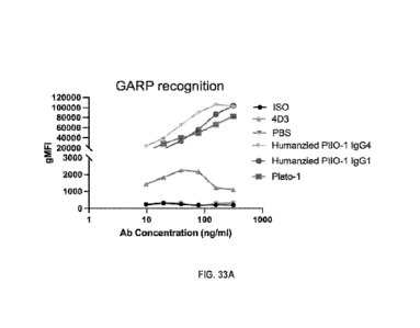
100741 Figures 33A and 33B show the characterization of anti-human GARP
antibodies
for recognition of cell surface GARP and blocking of GARP-LTGFP interaction.
Figure 33A
shows GARP expression on Jurkat-hGARP cell line was detected by flow cytometry
with anti-
GARP antibodies at indicated concentrations. Geometric mean fluorescence
intensity (gMFI)
of human GARP was plotted. Figure 33B shows stable hGARP-expressing Jurkat
cell line was
incubated with recombinant LTG1131 together with isotype control or anti-GARP
antibodies at
indicated concentrations for 30 min at 37 C. Human urciFpl expression level
was detected
by flow cytometry.
DESCRIPTION OF ILLUSTRATIVE EMBODIMENTS
190751 It is demonstrated herein that both membrane-bound and soluble GARP is
widely expressed by human cancer cells but less by normal epithelial cells,
and the
expression of GARP correlates uniformly with an advanced stage of cancer and
poor
prognosis. Additionally, it was found that GARP itself has a transformation
potential, which
renders normal mammary gland epithelial cells tum.orgenic. It was observed
that GARP
expression in cancer cells led to increased TGF-13 activity, likely due to its
ability to
concentrate LTGF-f1 in cis as well as trans, to contribute to cancer
aggressiveness and
metastasis. GARP expression in the tumor microenvironment promoted the
induction of
regulatory T cells and thus blunting the function of effector T cells against
cancers.
26
CA 03234326 2024- 4- 9
WO 2023/064779 PCT/US2022/077920
However, neutralizing GARP by blockings its ability to bind to TGF-13 results
in anti-cancer
activity even, without chemotherapy. In particular, there are provided here
new antibody
molecules, the humanized P110-I antibodies HuP110-1VH1/L1, HuP110-1VHI/L2,
HuP1I0-
1VH2/L1, HuPII0-1VH1/L3, HuPII0-1VH2/L2, HuPII0-1VH2/L3, HuPII0-1VH3/1,1,
HuP110-1VH2/L3, HuP110-1V113/1,3, HuPII0-1'VH4/1,1, HuPII0-1'VH4/1,2, HuPII0-
1VH4/L3 and 5c5 antibodies that can effectively bind to and neutralize GARP.
Thus, the
antibodies of the embodiments can be used in methods for treating cancers and
enhancing
immune response (e.g.. in conjunction with an adoptive T-cell therapy).
100761 While T cell therapy has the potential to treat cancer by recognizing
and
attacking tumor cells, the tumor microenvironment can evade the immune system
through the
induction of regulatory T cells which blunt the ability of adoptively
transferred effector T
cells to control cancer. Accordingly, embodiments of the present disclosure
overcomes
challenges associated with current technologies by providing methods for the
treatment of
cancer comprising the combination of a T cell therapy and an anti-platelet
agent. In this
method, the anti-platelet agent can potentiate the adoptive T cell therapy of
tumors as soluble
factors secreted from activated platelets have been shown to suppress T cells.
For example, it
has been shown that platelet-secreted latent TGFI3 and GARP can lead to the
resistance of
cancer cells to adoptive T cell therapy. Thus, anti-platelet factors such as
an anti-GARP
monoclonal antibody (that can block TGET3 binding) can be used in combination
with the T
cell therapy to overcome this resistance and treat cancer. In addition, other
immunotherapies
such as an immune checkpoint inhibitor can be used in combination with the T
cell therapy
and anti-platelet agent to enhance the immune response.
I. Definitions
100771 As used herein, "essentially free," in terms of a specified component,
is used
herein to mean that none of the specified component has been purposefully
formulated into a
composition and/or is present only as a contaminant or in trace amounts. The
total amount of
the specified component resulting from any unintended contamination of a
composition is
therefore well below 0.05%. Most preferred is a composition in which no amount
of the
specified component can be detected with standard analytical methods.
190781 As used herein in the specification and claims, "a" or "an" may mean
one or
more. As used herein in the specification and claims, when used in conjunction
with the
27
CA 03234326 2024- 4- 9
WO 2023/064779
PCT/US2022/077920
word "comprising", the words "a" or "an" may mean one or more than one. As
used herein,
in the specification and claim, "another" or "a further" may mean at least a
second or more.
[0079] As used herein in the specification and claims, the term "about" is
used to
indicate that a value includes the inherent variation of enror for the device,
the method being
employed to determine the value, or the variation that exists among the study
subjects.
[0080] "Treatment" and "treating" refer to administration or application of a
therapeutic
agent to a subject or performance of a procedure or modality on a subject for
the purpose of
obtaining a therapeutic benefit of a disease or health-related condition. For
example, a treatment
may include administration of a pharmaceutically effective amount of an
antibody that inhibits
the GARP signaling. In another example, a treatment may include administration
of a T cell
therapy and a pharmaceutically effective amount of an anti-platelet agent
(e.g., an antibody that
inhibits the GARP signaling).
100811 "Subject" and "patient" refer to either a human or non-human, such as
primates,
mammals, and vertebrates. In particular embodiments, the subject is a human.
[0082] An "increase" can refer to any change that results in a greater amount
of a
symptom, disease, composition, condition or activity. An increase can be any
individual, median,
or average increase in a condition, symptom, activity, composition in a
statistically significant
amount. Thus, the increase can be a 1, 2, 3, 4, 5, 6, 7, 8, 9, 10, 15, 20, 25,
30, 35, 40, 45, 50, 55,
60, 65, 70, 75, 80, 85, 90, 95, or 100% increase so long as the increase is
statistically significant.
[0083] A "decrease" can refer to any change that results in a smaller amount
of a
symptom, disease, composition, condition, or activity. A substance is also
understood to decrease
the genetic output of a gene when the genetic output of the gene product with
the substance is
less relative to the output of the gene product without the substance. Also
for example, a decrease
can be a change in the symptoms of a disorder such that the symptoms are less
than previously
observed. A decrease can be any individual, median, or average decrease in a
condition,
symptom, activity, composition in a statistically significant amount. Thus,
the decrease can be a
1, 2, 3, 4, 5, 6, 7, 8, 9, 10, 15, 20, 25, 30, 35, 40, 45, 50, 55, 60, 65, 70,
75, 80, 85, 90, 95, or
100% decrease so long as the decrease is statistically significant.
[0084] "Inhibit," "inhibiting," and "inhibition" mean to decrease an activity,
response,
condition, disease, or other biological parameter. This can include but is not
limited to the
28
CA 03234326 2024- 4- 9
WO 2023/064779
PCT/US2022/077920
complete ablation of the activity, response, condition, or disease. This may
also include, for
example, a 10% reduction in the activity, response, condition, or disease as
compared to the
native or control level. Thus, the reduction can be a 10, 20, 30, 40, 50, 60,
70, 80, 90, 100%, or
any amount of reduction in between as compared to native or control levels.
100851 By "reduce" or other forms of the word, such as "reducing" or
"reduction," is
meant lowering of an event or characteristic (e.g., tumor growth). It is
understood that this is
typically in relation to some standard or expected value, in other words it is
relative, but that it is
not always necessary for the standard or relative value to be referred to. For
example, "reduces
tumor growth" means reducing the rate of growth of a tumor relative to a
standard or a control.
100861 By "prevent" or other forms of the word, such as "preventing" or
"prevention," is
meant to stop a particular event or characteristic, to stabilize or delay the
development or
progression of a particular event or characteristic, or to minimize the
chances that a particular
event or characteristic will occur. Prevent does not require comparison to a
control as it is
typically more absolute than, for example, reduce. As used herein, something
could be reduced
but not prevented, but something that is reduced could also be prevented.
Likewise, something
could be prevented but not reduced, but something that is prevented could also
be reduced. It is
understood that where reduce or prevent are used, unless specifically
indicated otherwise, the use
of the other word is also expressly disclosed.
100871 The term "treatment" refers to the medical management of a patient with
the
intent to cure, ameliorate, stabilize, or prevent a disease, pathological
condition, or disorder. This
term includes active treatment, that is, treatment directed specifically
toward the improvement of
a disease, pathological condition, or disorder, and also includes causal
treatment, that is,
treatment directed toward removal of the cause of the associated disease,
pathological condition,
or disorder. In addition, this term includes palliative treatment, that is,
treatment designed for the
relief of symptoms rather than the curing of the disease, pathological
condition, or disorder;
preventative treatment, that is, treatment directed to minimizing or partially
or completely
inhibiting the development of the associated disease, pathological condition,
or disorder; and
supportive treatment, that is, treatment employed to supplement another
specific therapy directed
toward the improvement of the associated disease, pathological condition, or
disorder.
100881 The term "therapeutic benefit" or "therapeutically effective" as used
throughout
this application refers to anything that promotes or enhances the well-being
of the subject with
29
CA 03234326 2024- 4- 9
WO 2023/064779
PCT/US2022/077920
respect to the medical treatment of this condition. This includes, but is not
limited to, a reduction
in the frequency or severity of the signs or symptoms of a disease. For
example, treatment of
cancer may involve, for example, a reduction in the size of a tumor, a
reduction in the
invasiveness of a tumor, reduction in the growth rate of the cancer, or
prevention of metastasis.
Treatment of cancer may also refer to prolonging survival of a subject with
cancer.
[0089] "Effective amount" of an agent refers to a sufficient amount of an
agent to provide
a desired effect. The amount of agent that is "effective" will vary from
subject to subject,
depending on many factors such as the age and general condition of the
subject, the particular
agent or agents, and the like. Thus, it is not always possible to specify a
quantified "effective
amount." However, an appropriate "effective amount" in any subject case may be
determined by
one of ordinary skill in the art using routine experimentation. Also, as used
herein, and unless
specifically stated otherwise, an "effective amount" of an agent can also
refer to an amount
covering both therapeutically effective amounts and prophylactically effective
amounts. An
"effective amount" of an agent necessary to achieve a therapeutic effect may
vary according to
factors such as the age, sex, and weight of the subject. Dosage regimens can
be adjusted to
provide the optimum therapeutic response. For example, several divided doses
may be
administered daily or the dose may be proportionally reduced as indicated by
the exigencies of
the therapeutic situation.
100901 "Therapeutically effective amount" or "therapeutically effective dose"
of a
composition (e.g. a composition comprising an agent) refers to an amount that
is effective to
achieve a desired therapeutic result. In some embodiments, a desired
therapeutic result is the
control of type I diabetes. In some embodiments, a desired therapeutic result
is the control of
obesity. Therapeutically effective amounts of a given therapeutic agent will
typically vary
with respect to factors such as the type and severity of the disorder or
disease being treated
and the age, gender, and weight of the subject. The term can also refer to an
amount of a
therapeutic agent, or a rate of delivery of a therapeutic agent (e.g., amount
over time),
effective to facilitate a desired therapeutic effect, such as pain relief The
precise desired
therapeutic effect will vary according to the condition to be treated, the
tolerance of the
subject, the agent and/or agent formulation to be administered (e.g., the
potency of the
therapeutic agent, the concentration of agent in the formulation, and the
like), and a variety of
other factors that are appreciated by those of ordinary skill in the art. In
some instances, a
CA 03234326 2024- 4- 9
WO 2023/064779
PCT/US2022/077920
desired biological or medical response is achieved following administration of
multiple
dosages of the composition to the subject over a period of days, weeks, or
years.
100911 An "anti-cancer" agent is capable of negatively affecting a cancer
cell/tumor in
a subject, for example, by promoting killing of cancer cells, inducing
apoptosis in cancer
cells, reducing the growth rate of cancer cells, reducing the incidence or
number of
metastases, reducing tumor size, inhibiting tumor growth, reducing the blood
supply to a
tumor or cancer cells, promoting an immune response against cancer cells or a
tumor,
preventing or inhibiting the progression of cancer, or increasing the lifespan
of a subject with
cancer.
190921 The term "antibody" herein is used in the broadest sense and
specifically
covers monoclonal antibodies (including full length monoclonal antibodies),
polyclonal
antibodies, multispecific antibodies (e.g., bispecific antibodies), and
antibody fragments so
long as they exhibit the desired biological activity.
100931 The term "monoclonal antibody" as used herein refers to an antibody
obtained
from a population of substantially homogeneous antibodies, e.g., the
individual antibodies
comprising the population are identical except for possible mutations, e.g.,
naturally
occurring mutations, that may be present in minor amounts. Thus, the modifier
"monoclonal"
indicates the character of the antibody as not being a mixture of discrete
antibodies. In certain
embodiments, such a monoclonal antibody typically includes an antibody
comprising a
polypeptide sequence that binds a target, wherein the target-binding
polypeptide sequence
was obtained by a process that includes the selection of a single target
binding polypeptide
sequence from a plurality of polypeptide sequences. For example, the selection
process can
be the selection of a unique clone from a plurality of clones, such as a pool
of hybridoma
clones, phage clones, or recombinant DNA clones. It should be understood that
a selected
target binding sequence can be further altered, for example, to improve
affinity for the target,
to humanize the target binding sequence, to improve its production in cell
culture, to reduce
its immunogenicity in vivo, to create a multi specific antibody, etc., and
that an antibody
comprising the altered target binding sequence is also a monoclonal antibody
of this
invention. in contrast to polyclonal antibody preparations, which typically
include different
antibodies directed against different determinants (epitopes), each monoclonal
antibody of a
monoclonal antibody preparation is directed against a single determinant on an
antigen. In
31
CA 03234326 2024- 4- 9
WO 2023/064779
PCT/US2022/077920
addition to their specificity, monoclonal antibody preparations are
advantageous in that they
are typically uncontaminated by other immunoglobulins.
100941 The phrases "pharmaceutical or pharmacologically acceptable" refers to
molecular
entities and compositions that do not produce an adverse, allergic, or other
untoward reaction
when administered to an animal, such as a human, as appropriate. The
preparation of a
pharmaceutical composition comprising an antibody or additional active
ingredient will be
known to those of skill in the art in light of the present disclosure.
Moreover, for animal (e.g.,
human) administration, it will be understood that preparations should meet
sterility, pyrogenicity,
general safety, and purity standards as required by FDA Office of Biological
Standards.
100951 As used herein, "pharmaceutically acceptable carrier" includes any and
all
aqueous solvents (e.g., water, alcoholic/aqueous solutions, saline solutions,
parenteral vehicles,
such as sodium chloride, Ringer's dextrose, etc.), non-aqueous solvents (e.g.,
propylene glycol,
polyethylene glycol, vegetable oil, and injectable organic esters, such as
ethyloleate), dispersion
media, coatings, surfactants, antioxidants, preservatives (e.g., antibacterial
or antifungal agents,
anti-oxidants, chelating agents, and inert gases), isotonic agents, absorption
delaying agents,
salts, drugs, drug stabilizers, gels, binders, excipients, disintegration
agents, lubricants,
sweetening agents, flavoring agents, dyes, fluid and nutrient replenishers,
such like materials and
combinations thereof, as would be known to one of ordinary skill in the art.
The pH and exact
concentration of the various components in a pharmaceutical composition are
adjusted according
to well-known parameters.
100961 The term "unit dose" or "dosage" refers to physically discrete units
suitable for
use in a subject, each unit containing a predetermined quantity of the
therapeutic composition
calculated to produce the desired responses discussed above in association
with its
administration, i.e., the appropriate route and treatment regimen. The
quantity to be
administered, both according to number of treatments and unit dose, depends on
the effect
desired. The actual dosage amount of a composition of the present embodiments
administered to
a patient or subject can be determined by physical and physiological factors,
such as body
weight, the age, health, and sex of the subject, the type of disease being
treated, the extent of
disease penetration, previous or concurrent therapeutic interventions,
idiopathy of the patient, the
route of administration, and the potency, stability, and toxicity of the
particular therapeutic
substance. For example, a dose may also comprise from about 1 pg/kg/body
weight to about
1000 mg/kg/body weight (this such range includes intervening doses) or more
per administration,
32
CA 03234326 2024- 4- 9
WO 2023/064779
PCT/US2022/077920
and any range derivable therein. In non-limiting examples of a derivable range
from the numbers
listed herein, a range of about 5 pg/kg/body weight to about 100 mg/kg/body
weight, about 5
is/kg/body weight to about 500 mg/kg/body weight, etc., can be administered.
The practitioner
responsible for administration will, in any event, determine the concentration
of active
ingredient(s) in a composition and appropriate dose(s) for the individual
subject.
[0097] The terms "contacted" and "exposed," when applied to a cell, are used
herein to
describe the process by which a therapeutic construct and a chemotherapeutic
or radiotherapeutic
agent are delivered to a target cell or are placed in direct juxtaposition
with the target cell. To
achieve cell killing, for example, both agents are delivered to a cell in a
combined amount
effective to kill the cell or prevent it from dividing.
10098] The term "immune checkpoint" refers to a molecule such as a protein in
the
immune system which provides inhibitory signals to its components in order to
balance
immune reactions. Known immune checkpoint proteins comprise cytotoxic T-
lymphocyte-
associated protein 4 (CTLA-4), program cell death protein 1 (PD!) and its
ligands
programmed death ligand 1 (PD-LI) and programmed death ligand 2 (PD-L2) and in
addition
LAG-3, lymphocyte activation gene 3 (LAG-3), B- and T-lymphocyte attenuator
(BTLA), B7
homolog 3 (B7H3), B7 homolog 4 (B7H4), T-cell immunoglobulin and mucin domain
3
(Tim-3), killer iminutioglobuliti-like receptor (K ER). The pathways involving
LAG3, B- and
T-lym phoc3,,te attenuator (BTLA), V-domain Ig suppressor of T cell activation
(VISTA),
B7H3, B7H4, TIM3, T cell immunoreceptor with Ig and ITEM domains (TIGIT), and
KIR are
recognized in the art to constitute immune checkpoint pathways similar to the
CTLA-4 and
PD-1 dependent pathways (see, e.g., Pardoll, 2012, Nature Rev Cancer 12:252-
264; Mellman
et al., 201 1, Nature 480:480- 489).
100991 An "immune checkpoint inhibitor" refers to any compound inhibiting the
function of an immune checkpoint protein. Inhibition includes reduction of
function and full
blockade. In particular the immune checkpoint protein is a human immune
checkpoint
protein. Thus, the immune checkpoint protein inhibitor in particular is an
inhibitor of a
human immune checkpoint protein. Examples of checkpoint inhibitors include,
but are not
limited to anti- PD-1 (such as, for example, Nivolumab (BMS-936558 or
MDX1106),
pembrolizurnab, CT-011, MK-3475), anti-PD-L1 (such as, for example,
atezolizimmb,
aveliirnab, durvaitiroab, :MDX-1105 (BMS-936559), MPDL3280A, or
MSB0010718C)such
as, for example, PD-L2 (rHigM12B7), anti-CTLA-4 (such as, for example,
Ipilimumab
33
CA 03234326 2024- 4- 9
WO 2023/064779
PCT/US2022/077920
(MDX-010), Tremelimumab (CP-675,206)), anti-1130, anti-B7-H3 (such as, for
example,
MGA271, MGD009, omburtamab), anti-B7-H4, anti-TEvI3 (such as, for example, TSR-
022,
MBG453, Sym023,INCAGN2390, LY3321367, BMS-986258, SHR-1702, R07121661),
anti -TIGIT (such as, for example BMS-986207, OMP-313M32, MK-7684, AB-154, ASP-
8374, MT1G7192A, or PVSRIPO), anti-BTLA, anti-LAG-3 (such as, for example, BMS-
986016, LAG525, MK-4280, REGN3767, TSR-033, B1754111, Sym022, FS118, MGD013,
and lmmutep).
II. Antibodies of the Embodiments
1001001 In certain embodiments, an antibody or a fragment
thereof that binds to
at least a portion of GARP protein and inhibits GARP signaling and cancer cell
proliferation
are contemplated. As used herein, the term "antibody" is intended to refer
broadly to any
immunologic binding agent, such as 1gG, 1gM, IgA, IgD, 1gE, and genetically
modified 1gG
as well as polypeptides comprising antibody CDR domains that retain antigen
binding
activity. The antibody may be selected from the group consisting of a chimeric
antibody; an
affinity matured antibody, a polyclonal antibody, a monoclonal antibody, a
humanized
antibody, a human antibody, or an antigen-binding antibody fragment or a
natural or
synthetic ligand. Preferably, the anti-GA1U? antibody is a monoclonal antibody
or a
humanized antibody.
1901011 Thus, by known means and as described herein,
polyclonal or
monoclonal antibodies, antibody fragments, and binding domains and CDRs
(including
engineered forms of any of the foregoing) may be created that are specific to
GARP protein,
one or more of its respective epitopes, or conjugates of any of the foregoing,
whether such
antigens or epitopes are isolated from natural sources or are synthetic
derivatives or variants
of the natural compounds.
1001021 Examples of antibody fragments suitable for the present embodiments
include,
without limitation: (i) the Fab fragment, consisting of VL, VH, CL, and CHI
domains; (ii) the "Fd"
fragment consisting of the VFI and Cm domains; (iii) the "Fv" fragment
consisting of the VL and
VH domains of a single antibody; (iv) the "dAb" fragment, which consists of a
VII domain; (v)
isolated CDR regions; (vi) F(ab')2 fragments, a bivalent fragment comprising
two linked Fab
fragments; (vii) single chain Fv molecules ("say"), wherein a VH domain and a
VL domain are
linked by a peptide linker that allows the two domains to associate to form a
binding domain;
34
CA 03234326 2024- 4- 9
WO 2023/064779
PCT/US2022/077920
(viii) bi-specific single chain Fv dimers (see U.S. Pat. No. 5,091,513); and
(ix) diabodies,
multivalent or multispecific fragments constructed by gene fusion (U.S. Patent
Pub.
20050214860). Fv, scFv, or diabody molecules may be stabilized by the
incorporation of
disulphide bridges linking the VH and VI., domains. Minibodies comprising a
scFv joined to a
C1713 domain may also be made (Hu etal., 1996).
1001031 Antibody-like binding peptidomimetics are also contemplated in
embodiments. Liu et at (2003) describe "antibody like binding peptidomimetics"
(ABiPs), which
are peptides that act as pared-down antibodies and have certain advantages of
longer serum half-
life as well as less cumbersome synthesis methods.
1001041 Animals may be inoculated with an antigen, such as a GARP
extracellular
domain (ECD) protein, in order to produce antibodies specific for GARP
protein. Frequently an
antigen is bound or conjugated to another molecule to enhance the immune
response. As used
herein, a conjugate is any peptide, polypeptide, protein, or non-proteinaceous
substance bound to
an antigen that is used to elicit an immune response in an animal. Antibodies
produced in an
animal in response to antigen inoculation comprise a variety of non-identical
molecules
(polyclonal antibodies) made from a variety of individual antibody producing B
lymphocytes. A
polyclonal antibody is a mixed population of antibody species, each of which
may recognize a
different epitope on the same antigen. Given the correct conditions for
polyclonal antibody
production in an animal, most of the antibodies in the animal's serum will
recognize the
collective epitopes on the antigenic compound to which the animal has been
immunized. This
specificity is further enhanced by affinity purification to select only those
antibodies that
recognize the antigen or epitope of interest.
1001051 A monoclonal antibody is a single species of antibody wherein every
antibody
molecule recognizes the same epitope because all antibody producing cells are
derived from a
single B-lymphocyte cell line. The methods for generating monoclonal
antibodies (MAbs)
generally begin along the same lines as those for preparing polyclonal
antibodies. In some
embodiments, rodents such as mice and rats are used in generating monoclonal
antibodies. In
some embodiments, rabbit, sheep, or frog cells are used in generating
monoclonal antibodies.
The use of rats is well known and may provide certain advantages Mice (e.g.,
BALB/c mice)
are routinely used and generally give a high percentage of stable fusions.
CA 03234326 2024- 4- 9
WO 2023/064779
PCT/US2022/077920
1001061 Hybridoma technology involves the fusion of a single B lymphocyte from
a
mouse previously immunized with a GARP antigen with an immortal myeloma cell
(usually
mouse myeloma). This technology provides a method to propagate a single
antibody-producing
cell for an indefinite number of generations, such that unlimited quantities
of structurally
identical antibodies having the same antigen or epitope specificity
(monoclonal antibodies) may
be produced.
[00107] Plasma B cells (CD45'CD5-CD19) may be isolated from
freshly
prepared rabbit peripheral blood mononuclear cells of immunized rabbits and
further selected
for GARP binding cells. After enrichment of antibody producing B cells, total
RNA may be
isolated and cDNA synthesized. DNA sequences of antibody variable regions from
both
heavy chains and light chains may be amplified, constructed into a phage
display Fab
expression vector, and transformed into E. coil. GARP specific binding Fab may
be selected
out through multiple rounds enrichment panning and sequenced. Selected GARP
binding hits
may be expressed as full-length IgG in rabbit and rabbit/human chimeric forms
using a
mammalian expression vector system in human embryonic kidney (HEK293) cells
(Invitrogen) and purified using a protein G resin with a fast protein liquid
chromatography
(FPI,C) separation unit.
1001081 In one embodiment, the antibody is a chimeric antibody, for example,
an
antibody comprising antigen binding sequences from a non-human donor grafted
to a
heterologous non-human, human, or humanized sequence (e.g., framework and/or
constant
domain sequences). Methods have been developed to replace light and heavy
chain constant
domains of the monoclonal antibody with analogous domains of human origin,
leaving the
variable regions of the foreign antibody intact. Alternatively, "fully human"
monoclonal
antibodies are produced in mice transgenic for human immunoglobulin genes.
Methods have
also been developed to convert variable domains of monoclonal antibodies to
more human form
by recombinantly constructing antibody variable domains having both rodent,
for example,
mouse, and human amino acid sequences. In "humanized" monoclonal antibodies,
only the
hypervariable CDR is derived from mouse monoclonal antibodies, and the
framework and
constant regions are derived from human amino acid sequences (see U.S. Pat.
Nos. 5,091,513 and
6,881,557). It is thought that replacing amino acid sequences in the antibody
that are
characteristic of rodents with amino acid sequences found in the corresponding
position of
human antibodies will reduce the likelihood of adverse immune reaction during
therapeutic use.
36
CA 03234326 2024 4- 9
WO 2023/064779
PCT/US2022/077920
A hybridoma or other cell producing an antibody may also be subject to genetic
mutation or other
changes, which may or may not alter the binding specificity of antibodies
produced by the
hybridoma.
1001091 Methods for producing polyclonal antibodies in various animal species,
as
well as for producing monoclonal antibodies of various types, including
humanized, chimeric,
and fully human, are well known in the art and highly predictable. For
example, the following
U.S. patents and patent applications provide enabling descriptions of such
methods: U.S. Patent
Application Nos. 2004/0126828 and 2002/0172677; and U.S. Pat, Nos. 3,817,837;
3,850,752;
3,939,350; 3,996,345; 4,196,265; 4,275,149; 4,277,437; 4,366,241; 4,469,797;
4,472,509;
4,606,855; 4,703,003; 4,742,159; 4,767,720; 4,816,567; 4,867,973; 4,938,948;
4,946,778;
5,021,236; 5,164,296; 5,196,066; 5,223,409; 5,403,484; 5,420,253; 5,565,332;
5,571,698;
5,627,052; 5,656,434; 5,770,376; 5,789,208; 5,821,337; 5,844,091; 5,858,657;
5,861,155;
5,871,907; 5,969,108; 6,054,297; 6,165,464; 6,365,157; 6,406,867; 6,709,659;
6,709,873;
6,753,407; 6,814,965; 6,849,259; 6,861,572; 6,875,434; and 6,891,024. All
patents, patent
application publications, and other publications cited herein and therein are
hereby incorporated
by reference in the present application.
1001101 Antibodies may be produced from any animal source, including birds and
mammals. Preferably, the antibodies are ovine, mtuine (e.g., mouse and rat),
rabbit, goat, guinea
pig, camel, horse, or chicken. In addition, newer technology permits the
development of and
screening for human antibodies from human combinatorial antibody libraries.
For example,
bacteriophage antibody expression technology allows specific antibodies to be
produced in the
absence of animal immunization, as described in U.S. Pat. No. 6,946,546, which
is incorporated
herein by reference. These techniques are further described in: Marks (1992);
Stemmer (1994);
Gram et al. (1992); Barbas etal. (1994); and Schier etal. (1996).
1001111 It is fully expected that antibodies to GARP will have the ability to
neutralize
or counteract the effects of GARP regardless of the animal species, monoclonal
cell line, or other
source of the antibody. Certain animal species may be less preferable for
generating therapeutic
antibodies because they may be more likely to cause allergic response due to
activation of the
complement system through the "Fe" portion of the antibody. However, whole
antibodies may
be enzymatically digested into "Fe" (complement binding) fragment, and into
antibody fragments
having the binding domain or CDR. Removal of the Fe portion reduces the
likelihood that the
antigen antibody fragment will elicit an undesirable immunological response,
and thus,
37
CA 03234326 2024- 4- 9
WO 2023/064779
PCT/US2022/077920
antibodies without Fc may be preferential for prophylactic or therapeutic
treatments. As
described above, antibodies may also be constructed so as to be chimeric or
partially or fully
human, so as to reduce or eliminate the adverse immunological consequences
resulting from
administering to an animal an antibody that has been produced in, or has
sequences from, other
species.
1001121 Substitutional variants typically contain the exchange of one amino
acid for
another at one or more sites within the protein and may be designed to
modulate one or more
properties of the polypeptide, with or without the loss of other functions or
properties.
Substitutions may be conservative, that is, one amino acid is replaced with
one of similar shape
and charge. Conservative substitutions are well known in the art and include,
for example, the
changes of alanine to serine; arginine to lysine; asparagine to glutamine or
histidine; aspartate to
glutamate; cysteine to serine; glutamine to asparagine; glutamate to
aspartate; glycine to praline;
histidine to asparagine or glutamine; isoleucine to leucine or valine; leucine
to valine or
isoleucine; lysine to arginine; methionine to leucine or isoleucine;
phenylalanine to tyrosine,
leucine or methionine; serine to threonine; threonine to serine; tryptophan to
tyrosine; tyrosine to
tryptophan or phenylalanine; and valine to isoleucine or leucine.
Alternatively, substitutions may
be non-conservative such that a function or activity of the polypeptide is
affected. Non-
conservative changes typically involve substituting a residue with one that is
chemically
dissimilar, such as a polar or charged amino acid for a nonpolar or uncharged
amino acid, and
vice versa.
1001131 Proteins may be recombinant or synthesized in vitro. Alternatively, a
non-
recombinant or recombinant protein may be isolated from bacteria. It is also
contemplated that a
bacteriim containing such a variant may be implemented in compositions and
methods.
Consequently, a protein need not be isolated.
1001141 It is contemplated that in compositions there is between about 0.001
mg and
about 10 mg of total polypeptide, peptide, and/or protein per ml. Thus, the
concentration of
protein in a composition can be about, at least about or at most about 0.001,
0.010, 0.050, 0.1,
0.2, 0.3, 0.4, 0.5, 0.6, 0.7, 0.8, 0.9, 1.0, 1.5, 2.0,2.5, 3.0, 3.5, 4.0, 4.5,
5.0, 5.5, 6.0, 6.5, 7.0, 7.5,
8.0, 8.5, 9.0, 9.5, 10.0 mg/m1 or more (or any range derivable therein). Of
this, about, at least
about, or at most about 1, 2, 3, 4, 5, 6, 7, 8, 9, 10, 11, 12, 13, 14, 15, 16,
17, 18, 19, 20, 21, 22,
23, 24, 25, 26, 27, 28, 29, 30, 31, 32, 33, 34, 35, 36, 37, 38, 39, 40, 41,
42, 43, 44, 45, 46, 47, 48,
49, 50, 51, 52, 53, 54, 55, 56, 57, 58, 59, 60, 61, 62, 63, 64, 65, 66, 67,
68, 69, 70, 71, 72, 73, 74,
38
CA 03234326 2024- 4- 9
WO 2023/064779
PCT/US2022/077920
75, 76, 77, 78, 79, 80, 81, 82, 83, 84, 85, 86, 87, 88, 89, 90, 91, 92, 93,
94, 95, 96, 97, 98, 99, or
100% may be an antibody that binds GARP .
1001151 An antibody or preferably an immunological portion of an antibody, can
be
chemically conjugated to, or expressed as, a fusion protein with other
proteins. For purposes of
this specification and the accompanying claims, all such fused proteins are
included in the
definition of antibodies or an immunological portion of an antibody.
1001161 Embodiments provide antibodies and antibody-like molecules against
GARP,
polypeptides and peptides that are linked to at least one agent to form an
antibody conjugate or
payload. In order to increase the efficacy of antibody molecules as diagnostic
or therapeutic
agents, it is conventional to link or covalently bind or complex at least one
desired molecule or
moiety. Such a molecule or moiety may be, but is not limited to, at least one
effector or reporter
molecule. Effector molecules comprise molecules having a desired activity,
e.g., cytotoxic
activity. Non-limiting examples of effector molecules that have been attached
to antibodies
include toxins, therapeutic enzymes, antibiotics, radio-labeled nucleotides
and the like. By
contrast, a reporter molecule is defined as any moiety that may be detected
using an assay. Non-
limiting examples of reporter molecules that have been conjugated to
antibodies include
enzymes, radiolabels, haptens, fluorescent labels, phosphorescent molecules,
chemiluminescent
molecules, chromophores, luminescent molecules, photoaffinity molecules,
colored particles or
ligands, such as biotin.
1001171 Several methods are known in the art for the attachment or conjugation
of an
antibody to its conjugate moiety. Some attachment methods involve the use of a
metal chelate
complex employing, for example, an organic chelating agent such a
diethylenetriaminepentaacetic acid anhydride (DTPA);
ethylenetriaminetetraacetic acid; N-
chloro-p-toluenesulfonamide; and/or tetrachloro-3-6-diphenylglycouril-3
attached to the
antibody. Monoclonal antibodies may also be reacted with an enzyme in the
presence of a
coupling agent such as glutaraldehyde or periodate. Conjugates with
fluorescein markers are
prepared in the presence of these coupling agents or by reaction with an
isothiocyanate.
ilL T Cell Therapy
[00118]
Certain embodiments of the present disclosure concern obtaining and
administering T cells to a subject as an immunotherapy to target cancer cells.
Several basic
approaches for the derivation, activation and expansion of functional anti-
tumor effector T
39
CA 03234326 2024- 4- 9
WO 2023/064779
PCT/US2022/077920
cells have been described in the last two decades. These include: autologous
cells, such as
tumor-infiltrating lymphocytes (TILs); T cells activated ex-vivo using
autologous DCs,
lymphocytes, artificial antigen-presenting cells (APCs) or beads coated with T
cell ligands
and activating antibodies, or cells isolated by virtue of capturing target
cell membrane;
allogeneic cells naturally expressing anti-host tumor T cell receptor (TCR);
and non-tumor-
specific autologous or allogeneic cells genetically reprogrammed or
"redirected" to express
tumor-reactive TCR or chimeric TCR molecules displaying antibody-like tumor
recognition
capacity known as "T-bodies". These approaches have given rise to numerous
protocols for T
cell preparation and immunization which can be used in the methods of the
present
disclosure.
A. T Cell Preparation
1001191 in some embodiments, the T cells are derived from
the blood, bone
marrow, lymph, or lymphoid organs in some aspects, the cells are human cells.
The cells
typically are primary cells, such as those isolated directly from a subject
and/or isolated from
a subject and frozen. In some embodiments, the cells include one or more
subsets of T cells
or other cell types, such as whole T cell populations, CD4 cells, CD8+ cells,
and
subpopulations thereof, such as those defined by function, activation state,
maturity, potential
for differentiation, expansion, recirculation, localization, and/or
persistence capacities,
antigen- specificity, type of antigen receptor, presence in a particular organ
or compartment,
marker or cytokine secretion profile, and/or degree of differentiation. With
reference to the
subject to be treated, the cells may be allogeneic and/or autologous. In some
aspects, such as
for off-the-shelf technologies, the cells are pluripotent and/or multipotent,
such as stem cells,
such as induced pluripotent stem cells (iPSCs). In some embodiments, the
methods include
isolating cells from the subject, preparing, processing, culturing, and/or
engineering them, as
described herein, and re-introducing them into the same patient, before or
after
cryopreservation.
1001201 Among the sub-types and subpopulations of T cells
(e.g., CD4+ and/or
CD8+ T cells) are naive T (TN) cells, effector T cells (TEFF), memory T cells
and sub-types
thereof, such as stem cell memory T (TSCm), central memory T (TCm), effector
memory T
(Tum), or terminally differentiated effector memory T cells, tumor-
infiltrating lymphocytes
(TIL), immature T cells, mature T cells, helper T cells, cytotoxic T cells,
mucosa-associated
invariant T (MAIT) cells, naturally occurring and adaptive regulatoiy T (Treg)
cells, helper T
CA 03234326 2024- 4- 9
WO 2023/064779
PCT/US2022/077920
cells, such as TI-I1 cells, TH2 cells, TH3 cells, THI 7 cells, TH9 cells,
'EH22 cells, follicular
helper T cells, alpha/beta T cells, and delta/gamma T cells.
1001211 In some embodiments, one or more of the T cell
populations is
enriched for or depleted of cells that are positive for a specific marker,
such as surface
markers, or that are negative for a specific marker. In some cases, such
markers are those that
are absent or expressed at relatively low levels on certain populations of T
cells (e.g., non-
memory cells) but are present or expressed at relatively higher levels on
certain other
populations of T cells (e.g., memory cells). In one embodiment, the cells
(e.g., CD81 cells or
CD3 cells) are enriched for (i.e., positively selected for) cells that are
positive or expressing
high surface levels of CD45RO, CCR7, CD28, CD27, CD44, CD127, and/or CD62L
and/or
depleted of (e.g., negatively selected for) cells that are positive for or
express high surface
levels of CD45R A. In some embodiments, cells are enriched for or depleted of
cells positive
or expressing high surface levels of CD122, CD95, CD25, CD27, and/or IL7-Ra
(CD127).
some examples, CDS+ T cells are enriched for cells positive for CD45R0 (or
negative for
CD45RA) and for CD621-
1001221 In some embodiments, T cells are separated from a
PBMC sample by
negative selection of markers expressed on non-T cells, such as B cells,
monocytes, or other
white blood cells, such as CD14. In some aspects, a CD4' or CDS' selection
step is used to
separate CD4' helper and CDS+ cytotoxic T cells. Such CD4 and CDS'
populations can be
further sorted into sub-populations by positive or negative selection for
markers expressed or
expressed to a relatively higher degree on one or more naive, memory, and/or
effector T cell
subpopulations.
1001231 In some embodiments, CD8' cells are further
enriched for or depleted
of naive, central memory, effector memory, and/or central memory stem cells,
such as by
positive or negative selection based on surface antigens associated with the
respective
subpopulation. In some embodiments, enrichment for central memory T (Tcm)
cells is carried
out to increase efficacy, such as to improve long-term survival, expansion,
and/or
engraftment following administration, which in some aspects is particularly
robust in such
sub-populations. See Teralcuraet al. (2012) Blood. 1:72- 82; Wang et aL (2012)
J Immunother.
35(9):689-701. In some embodiments, combining Tem- enriched CD84* T cells and
CD4+ T
cells further enhances efficacy.
41
CA 03234326 2024- 4- 9
WO 2023/064779
PCT/US2022/077920
1001241 in some embodiments, the T cells are autologous T
cells, in this
method, tumor samples are obtained from patients and a single cell suspension
is obtained.
The single cell suspension can be obtained in any suitable manner, e.g.,
mechanically
(disaggregating the tumor using, e.g., a gentleMACSTm Dissociator, Miltenyi
Biotec, Auburn,
Calif.) or enzymatically (e.g., collagenase or DNase). Single-cell suspensions
of tumor
enzymatic digests are cultured in interleukin-2 (1L-2). The cells are cultured
until confluence
(e.g., about 2x I 061ymphocytes), e.g., from about 5 to about 21 days,
preferably from about
to about 14 days. For example, the cells may be cultured from 5 days, 5.5
days, or 5.8
days to 21 days, 21.5 days, or 21.8 days, such as from 10 days, 10.5 days, or
10.8 days to 14
days, 14.5 days, or 14.8 days.
1001251 The cultured T cells can be pooled and rapidly
expanded. Rapid
expansion provides an increase in the number of antigen-specific T-cells of at
least about 50-
fold (e.g., 50-, 60-, 70-, 80-, 90-, or 100-fold, or greater) over a period of
about 10 to about 14
days, preferably about 14 days. More preferably, rapid expansion provides an
increase of at
least about 200-fold (e.g., 200-, 300-, 400-, 500-, 600-, 700-, 800-, 900-, or
greater) over a
period of about 10 to about 14 days, preferably about 14 days.
1001261 Expansion can be accomplished by any of a number
of methods as are
known in the art. For example, T cells can be rapidly expanded using non-
specific T-cell
receptor stimulation in the presence of feeder lymphocytes and either
interleukin-2 (IL-2) or
interleukin-15 (1L-15), with 1L-2 being preferred. The non-specific T-cell
receptor stimulus
can include around 30 ng/ml of OKT3, a mouse monoclonal anti-CD3 antibody
(available
from Ortho-McNeil , Raritan, N.J.). Alternatively, T cells can be rapidly
expanded by
stimulation of peripheral blood mononuclear cells (PBMC) in vitro with one or
more antigens
(including antigenic portions thereof, such as epitope(s), or a cell) of the
cancer, which can be
optionally expressed from a vector, such as an human leukocyte antigen A2 (HLA-
A2)
binding peptide, in the presence of a T-cell growth factor, such as 300
lli/mIlL-2 or 1L-15,
with 1L-2 being preferred. The in vitro-induced T-cells are rapidly expanded
by re-
stimulation with the same antigen(s) of the cancer pulsed onto HLA-A2-
expressing antigen-
presenting cells. Alternatively, the T-cells can be re-stimulated with
irradiated, autologous
lymphocytes or with irradiated HLA-A2+ allogeneic lymphocytes and EL-2, for
example.
1001271 The autologous T-cells can be modified to express
a T-cell growth
factor that promotes the growth and activation of the autologous T-cells.
Suitable T-cell
42
CA 03234326 2024- 4- 9
WO 2023/064779
PCT/US2022/077920
growth factors include, for example, interleukin (IL)-2, IL-7, IL-15, and IL-
12. Suitable
methods of modification are known in the art. See, for instance, Sambrook
etal., Molecular
Cloning: A Laboratory Manual, 3nled., Cold Spring Harbor Press, Cold Spring
Harbor, N.Y.
2001; and Ausubel et al., Current Protocols in Molecular Biology, Greene
Publishing
Associates and John Wiley & Sons, NY, 1994. In particular aspects, modified
autologous T-
cells express the T-cell growth factor at high levels. T-cell growth factor
coding sequences,
such as that of IL-I2, are readily available in the art, as are promoters, the
operable linkage of
which to a T-cell growth factor coding sequence promote high-level expression.
B. Genetically Engineered Antigen Receptors
1901281 The T cell can genetically engineered to express
antigen receptors such
as engineered TCRs and/or chimeric antigen receptors (CARs). For example, the
autologous
T-cells are modified to express a T cell receptor (I'M) having antigenic
specificity for a
cancer antigen. Suitable TCRs include, for example, those with antigenic
specificity for a
melanoma antigen, e.g., gp100 or MART-1. Suitable methods of modification are
known in
the art. See, for instance, Sambrook and Ausubel, supra. For example, the T
cells may be
transduced to express a T cell receptor (TCR) having antigenic specificity for
a cancer
antigen using transduction techniques described in Heemskerk et al. Hum Gene
Ther. 19:496-
510 (2008) and Johnson et al. Blood 114:535-46 (2009).
1901291 In some embodiments, the T cells comprise one or
more nucleic acids
introduced via genetic engineering that encode one or more antigen receptors,
and genetically
engineered products of such nucleic acids. In some embodiments, the nucleic
acids are
heterologous, i.e., normally not present in a cell or sample obtained from the
cell, such as one
obtained from another organism or cell, which for example, is not ordinarily
found in the cell
being engineered and/or an organism from which such cell is derived. In some
embodiments,
the nucleic acids are not naturally occurring, such as a nucleic acid not
found in nature (e.g.,
chimeric).
1001301 In some embodiments, the CAR contains an
extracellular antigen-
recognition domain that specifically binds to an antigen. In some embodiments,
the antigen is
a protein expressed on the surface of cells. in some embodiments, the CAR is a
TCR-like
CAR and the antigen is a processed peptide antigen, such as a peptide antigen
of an
43
CA 03234326 2024- 4- 9
WO 2023/064779
PCT/US2022/077920
intracellular protein, which, like a TCR, is recognized on the cell surface in
the context of a
major histocompatibility complex (WIC) molecule.
1001311 Exemplary antigen receptors, including CARS and
recombinant TCRs,
as well as methods for engineering and introducing the receptors into cells,
include those
described, for example, in international patent application publication
numbers
W0200014257, W02013126726, W02012/129514, W02014031687, W02013/166321,
W02013/071154, W02013/123061 U.S. patent application publication numbers
US2002131960, US2013287748, US20130149337, U.S. Patent Nos. 6,451,995,
7,446,190,
8,252,592õ 8,339,645, 8,398,282, 7,446,179, 6,410,319, 7,070,995, 7,265,209,
7,354,762,
7,446,191, 8,324,353, and 8,479,118, and European patent application number
EP2537416,
and/or those described by Sadelain et aL, Cancer Discov. 2013 April; 3(4): 388-
398; Davila
et al. (2013) PLoS ONE 8(4): e6.1338; Turtle etal., Curr. Opin. Immunol., 2012
October;
24(5): 633-39; Wu etal., Cancer, 2012 March 18(2): 160-75. In some aspects,
the genetically
engineered antigen receptors include a CAR as described in U.S. Patent No.
7,446,190, and
those described in International Patent Application Publication No.:
'WO/2014055668 Al.
1001321 In some aspects, the tumor antigen is a human
telomerase reverse
transcriptase (hTERT), survivin, mouse double minute 2 homolog (MDM2),
cytochrome
P450 1.B1 (CYP1B), HER2/neu, Wilms' tumor gene 1 (WT1), livin,
alphafetoprotein (AFP),
carcinoembryonic antigen (CEA), mucin 16 (MUC16), MUC1, prostate-specific
membrane
antigen (PSMA), p53 or cyclin (D1). For example, the target antigen is hTERT
or survivin. In
some aspects, the target antigen is CD38. In other aspects, the target antigen
is CD33 or TIM:-
3. In other aspects, it is CD26, CD30, CD53, CD92, CD148, CD150, CD200, CD261,
CD262, or CD362. In some embodiments, the engineered immune cells can contain
an
antigen that targets one or more other antigens. In some embodiments, the one
or more other
antigens is a tumor antigen or cancer marker. Other antigens include orphan
tyrosine kinase
receptor ROR1, tEGFR, Her2, Ll-CAM, CD19, CD20, CD22, mesothelin, CEA, and
hepatitis
B surface antigen, anti-folate receptor, CD23, CD24, CD30, CD33, C.D38, CD44,
.EGFR.,
EGP-2, EGP-4, EPHa2, ErbB2, 3, or 4, FBP, fetal acethycholine e receptor, GD2,
GD3,
ITMW-MA A, IL-22R-alpha, IL-13R-a1pha2, kdr, kappa light chain, Lewis Y, L1-
cell
adhesion molecule, MAGE-Al, mesothelin, MUC1, MUC16, PSCA., .NICG2D Ligands,
NY-
ES0-1, MART-1, gp100, oncofetal antigen, ROR1, TAG72, VEGF-R2,
carcinoembryonic
antigen (CEA), prostate specific antigen, PSMA, Her2/neu, estrogen receptor,
progesterone
44
CA 03234326 2024- 4- 9
WO 2023/064779
PCT/US2022/077920
receptor, ephrinB2, CD 123, CS-1, c-Met, GD-2, and MAGE A3, CE7, Wilms Tumor 1
(WT-
1), a cyclin, such as cyclin Al (CCNA1), and/or biotinylated molecules, and/or
molecules
expressed by HIV, HCV, HBV or other pathogens.
1. Chimeric Antigen Receptors
1001331 In some embodiments, the engineered antigen
receptors include
chimeric antigen receptors (CARs), including activating or stimulatory CARs,
costimulatory
CARs (see W02014/055668), and/or inhibitory CARs (iCARs, see Fedorov el al.,
S'cl.
Transl. Medicine, 5(215) (2013). The CARs generally include an extracellular
antigen (or
ligand) binding domain linked to one or more intracellular signaling
components, in some
aspects via linkers and/or transmembrane domain(s). Such molecules typically
mimic or
approximate a signal through a natural antigen receptor, a signal through such
a receptor in
combination with a costimulatory receptor, and/or a signal through a
costimulatory receptor
alone
1001341 In some embodiments, CAR is constructed with a
specificity for a
particular antigen (or marker or ligand), such as an antigen expressed in a
particular cell type
to be targeted by adoptive therapy, e.g., a cancer marker, and/or an antigen
intended to induce
a dampening response, such as an antigen expressed on a normal or non-diseased
cell type.
Thus, the CAR typically includes in its extracellular portion one or more
antigen binding
molecules, such as one or more antigen-binding fragment, domain, or portion,
or one or more
antibody variable domains, and/or antibody molecules. some embodiments, the
CAR
includes an antigen-binding portion or portions of an antibody molecule, such
as a single-
chain antibody fragment (scFv) derived from the variable heavy (VH) and
variable light (VL)
chains of a monoclonal antibody (mAb).
1001351 In some aspects, the antigen-specific binding, or
recognition
component is linked to one or more transmembrane and intracellular signaling
domains. In
some embodiments, the CAR includes a transmembrane domain fused to the
extracellular
domain of the CAR. In one embodiment, the transmembrane domain that naturally
is
associated with one of the domains in the CAR is used. In some instances, the
transmembrane
domain is selected or modified by amino acid substitution to avoid binding of
such domains
to the transmembrane domains of the same or different surface membrane
proteins to
minimize interactions with other members of the receptor complex.
CA 03234326 2024- 4- 9
WO 2023/064779
PCT/US2022/077920
1001361 The transmembrane domain in some embodiments is
derived either
from a natural or from a synthetic source. Where the source is natural, the
domain in some
aspects is derived from any membrane-bound or transmembrane protein.
Transmembrane
regions include those derived from (i.e. comprise at least the transmembrane
region(s) of) the
alpha, beta or zeta chain of the T- cell receptor, CD28, CD3 epsilon, CD45,
CD4, CD5, CDS,
CD9, CD 16, CD22, CD33, CD37, CD64, CD80, CD86, CD 134, CD137, CD 154.
Alternatively, the transmembrane domain in some embodiments is synthetic. In
some aspects,
the synthetic transmembrane domain comprises predominantly hydrophobic
residues such as
leucine and valine. In some aspects, a triplet of phenylalanine, tryptophan
and valine will be
found at each end of a synthetic transmembrane domain.
1001371 The CAR generally includes at least one
intracellular signaling
component or components. In some embodiments, the CAR includes an
intracellular
component of the TCR complex, such as a TCR CD3 chain that mediates T-cell
activation
and cytotoxicity, e.g., CD3 zeta chain. Thus, in some aspects, the antigen
binding molecule is
linked to one or more cell signaling modules. In some embodiments, cell
signaling modules
include CD3 transmembrane domain, CD3 intracellular signaling domains, and/or
other CD
transmembrane domains. In some embodiments, the CAR further includes a portion
of one or
more additional molecules such as Fc receptor T. CD8, CD4, CD25, or CD16. For
example,
in some aspects, the CAR includes a chimeric molecule between CD3-zeta (CD3-Q
or Fc
receptor 7 and CD8, CD4, CD25 or CD16.
2. 1' Cell Receptor (TCR)
1001381 in some embodiments, the genetically engineered
antigen receptors
include recombinant T cell receptors (TCRs) and/or TCRs cloned from naturally
occurring T
cells. A "T cell receptor or "TCR" refers to a molecule that contains a
variable a and
chains (also known as TCRa and TCRp, respectively) or a variable 7 and ö
chains (also
known as TCRy and TCR5, respectively) and that is capable of specifically
binding to an
antigen peptide bound to a MEC receptor. In some embodiments, the TCR is in
the ctf3 form.
Typically, TCRs that exist in etit and 76 forms are generally structurally
similar, but T cells
expressing them may have distinct anatomical locations or functions. A TCR can
be found on
the surface of a cell or in soluble form. Generally, a TCR is found on the
surface of T cells
(or T lymphocytes) where it is generally responsible for recognizing antigens
bound to major
histocompatibility complex NBC) molecules. In some embodiments, a TCR also can
46
CA 03234326 2024- 4- 9
WO 2023/064779
PCT/US2022/077920
contain a constant domain, a transmembrane domain and/or a short cytoplasmic
tail (see, e.g.,
Janeway et al, Immunobiology: The Immune System in Health and Disease, 3rd
Ed., Current
Biology Publications, p. 4:33, 1997). For example, in some aspects, each chain
of the TCR
can possess one N-terminal immunoglobulin variable domain, one immunoglobulin
constant
domain, a transmembrane region, and a short cytoplasmic tail at the C-
terminal end. In some
embodiments, a TCR is associated with invariant proteins of the CD3 complex
involved in
mediating signal transduction. Unless otherwise stated, the term "TCR" should
be understood
to encompass functional TCR fragments thereof. The term also encompasses
intact or full-
length TCRs, including TCRs in the c(13 form or yo form.
1001391 Thus, for purposes herein, reference to a TCR
includes any TCR or
functional fragment, such as an antigen-binding portion of a TCR that binds to
a specific
antigenic peptide bound in an MT-IC molecule, i.e. MT-IC-peptide complex. An
"antigen-
binding portion" or antigen- binding fragment" of a TCR, which can be used
interchangeably,
refers to a molecule that contains a portion of the structural domains of a
TCR, but that binds
the antigen (e.g., MHC-peptide complex) to which the full TCR binds. In some
cases, an
antigen-binding portion contains the variable domains of a TCR, such as
variable a chain and
variable 13 chain of a TCR, sufficient to form a binding site for binding to a
specific MIX-
peptide complex, such as generally where each chain contains three
complementarity
determining regions.
100101 In some embodiments, the variable domains of the
TCR chains
associate to form loops, or complementarity determining regions (CDRs)
analogous to
immunoglobulins, which confer antigen recognition and determine peptide
specificity by
forming the binding site of the TCR molecule and determine peptide
specificity. Typically,
like immunoglobulins, the CDRs are separated by framework regions (FRs) (see,
e.g., Jores
et al., NcrtlAcad. Sci. U.S.A. 87:9138, 1990; Chothia el al, 1A1B0 J. 7:3745,
1988; see also
Lefranc et Dev. Comp. Immtmol. 27:55, 2003). In some embodiments,
CDR3 is the main
CDR. responsible for recognizing processed antigen, although CDR1 of the alpha
chain has
also been shown to interact with the N-terminal part of the antigenic peptide,
whereas CDR1
of the beta chain interacts with the C-terminal part of the peptide. CDR2 is
thought to
recognize the MHC molecule. In some embodiments, the variable region of the 0-
chain can
contain a further hypervariability (HV4) region.
47
CA 03234326 2024-4- 9
WO 2023/064779
PCT/US2022/077920
1001411 in some embodiments, the TCR chains contain a
constant domain. For
example, like immunoglobulins, the extracellular portion of TCR chains (e.g.,
a-chain, 3-
chain) can contain two immunoglobulin domains, a variable domain (e.g., Va or
Vp; typically
amino acids 1 to 116 based on Kabat numbering Kabat etal., "Sequences of
Proteins of
Immunological Interest, U.S. Dept. Health and Human Services, Public Health
Service
National Institutes of Health, 1991, 5th ed.) at the N-terminus, and one
constant domain (e.g.,
a-chain constant domain or Ca, typically amino acids 117 to 259 based on
Kabat, 13-chain
constant domain or Cp, typically amino acids 117 to 295 based on Kabat)
adjacent to the cell
membrane. For example, in some cases, the extracellular portion of the TCR
formed by the
two chains contains two membrane-proximal constant domains, and two membrane-
distal
variable domains containing CDRs. The constant domain of the TCR domain
contains short
connecting sequences in which a cysteine residue forms a disulfide bond,
making a link
between the two chains. In some embodiments, a TCR may have an additional
cysteine
residue in each of the a and f3 chains such that the TCR contains two
disulfide bonds in the
constant domains.
1001421 In some embodiments, the TCR chains can contain a
transmembrane
domain. In some embodiments, the transmembrane domain is positively charged.
In some
cases, the TCR chains contains a cytoplasmic tail. In some cases, the
structure allows the
TCR to associate with other molecules like CD3. For example, a TCR containing
constant
domains with a transmembrane region can anchor the protein in the cell
membrane and
associate with invariant subunits of the CD3 signaling apparatus or complex.
1001431 Generally, CD3 is a multi-protein complex that can
possess three
distinct chains (y, 8, and e) in mammals and the c,-chain. For example, in
mammals the
complex can contain a CD3y chain, a CD3 8 chain, two CD3s chains, and a
homodimer of
CD3 C chains. The CD3y, CD38, and CD3s chains are highly related cell surface
proteins of
the immunoglobulin superfamily containing a single immunoglobulin domain. The
transmembrane regions of the CD3y, CD38, and CD3e chains are negatively
charged, which
is a characteristic that allows these chains to associate with the positively
charged T cell
receptor chains. The intracellular tails of the CD3y, CD38, and CD3s chains
each contain a
single conserved motif known as an immunoreceptor tyrosine-based activation
motif or
ITAM, whereas each CD3C, chain has three. Generally, ITAMs are involved in the
signaling
capacity of the TCR complex. These accessory molecules have negatively charged
48
CA 03234326 2024- 4- 9
WO 2023/064779
PCT/US2022/077920
transmembrane regions and play a role in propagating the signal from the TCR
into the cell.
The CD3- and -chains, together with the TCR, form what is known as the T cell
receptor
complex.
[00144] In some embodiments, the TCR may be a heterodimer
of two chains a
and 13 (or optionally 7 and 5) or it may be a single chain TCR construct. In
some
embodiments, the TCR is a heterodimer containing two separate chains (a and 13
chains or 7
and 5 chains) that are linked, such as by a disulfide bond or disulfide bonds.
[0140] In some
embodiments, a TCR for a target antigen (e.g., a cancer antigen) is identified
and introduced
into the cells. In some embodiments, nucleic acid encoding the TCR can be
obtained from a
variety of sources, such as by polymerase chain reaction (PCR) amplification
of publicly
available TCR DNA sequences. In some embodiments, the TCR is obtained from a
biological
source, such as from cells such as from a T cell (e.g., cytotoxic T cell), T-
cell hybridornas or
other publicly available source. In some embodiments, the T-cells can be
obtained from in
vivo isolated cells. In some embodiments, a high-affinity T cell clone can be
isolated from a
patient, and the TCR isolated. In some embodiments, the T- cells can be a
cultured T-cell
hybridoma or clone. In some embodiments, the TCR clone for a target antigen
has been
generated in transgenic mice engineered with human immune system genes (e.g.,
the human
leukocyte antigen system, or HLA). See, e.g., tumor antigens (see, e.g.,
Parkhurst etal.
(2009) Chn Cancer Res. 15: 169-180 and Cohen etal. (2005)J. hnmunol. 175:5799-
5808. In
some embodiments, phage display is used to isolate TCRs against a target
antigen (see, e.g.,
Varela-Rohena et al. (2008) Nat. Med. 14: 1390-1395 and Li (2005) Nat.
Biotechnol. 23:349-
354. In some embodiments, the TCR or antigen-binding portion thereof can be
synthetically
generated from knowledge of the sequence of the TCR.
IV. Methods of Treatment
[00145] Certain aspects of the present embodiments can be used to prevent or
treat a
disease or disorder associated with GARP signaling. Signaling of GARP may be
reduced by any
suitable drugs to prevent cancer cell proliferation. Preferably, such
substances would be an anti-
GARP antibody.
[00146] Provided herein, in certain embodiments, are
methods for treating or
delaying progression of cancer in an individual comprising administering to
the individual an
effective amount an anti-platelet agent and T cell therapy. Examples of
cancers contemplated
49
CA 03234326 2024- 4- 9
WO 2023/064779
PCT/US2022/077920
for treatment include lung cancer, head and neck cancer, breast cancer,
pancreatic cancer,
prostate cancer, renal cancer, bone cancer, testicular cancer, cervical
cancer, gastrointestinal
cancer, lymphomas, pre-neoplastic lesions in the lung, colon cancer, melanoma,
and bladder
cancer.
1001471 In some embodiments, the individual has cancer that
is resistant (has
been demonstrated to be resistant) to one or more anti-cancer therapies. In
some
embodiments, resistance to anti-cancer therapy includes recurrence of cancer
or refractory
cancer. Recurrence may refer to the reappearance of cancer, in the original
site or a new site,
after treatment. in some embodiments, resistance to anti-cancer therapy
includes progression
of the cancer during treatment with the anti-cancer therapy. In some
embodiments, the cancer
is at early stage or at late stage.
1001481 In some embodiments of the methods of the present
disclosure,
activated CD4 and/or CD8 T cells in the individual are characterized by 7-1FN
producing
CD4 and/or CD8 T cells and/or enhanced cytolytic activity relative to prior to
the
administration of the combination. 7-1FN may be measured by any means known in
the art,
including, e.g., intracellular cytokine staining (ICS) involving cell
fixation, permeabilization,
and staining with an antibody against y-IFN. Cytolytic activity may be
measured by any
means known in the art, e.g., using a cell killing assay with mixed effector
and target cells.
1001491 A T cell therapy may be administered before, during, after, or in
various
combinations relative to an anti-platelet agent. The administrations may be in
intervals ranging
from concurrently to minutes to days to weeks. In embodiments where the T cell
therapy is
provided to a patient separately from an anti-platelet agent, one would
generally ensure that a
significant period of time did not expire between the time of each delivery,
such that the two
compounds would still be able to exert an advantageously combined effect on
the patient. In
such instances, it is contemplated that one may provide a patient with the
antibody therapy and
the anti-cancer therapy within about 12 to 24 or 72 h of each other and, more
particularly, within
about 6-12 h of each other. In some situations, it may be desirable to extend
the time period for
treatment significantly where several days (2, 3, 4, 5, 6, or 7) to several
weeks (1, 2, 3, 4, 5, 6, 7,
or 8) lapse between respective administrations.
1001501 In some embodiments, the subject can be
administered
nonmyeloablative lymphodepleting chemotherapy prior to the T cell therapy. The
CA 03234326 2024- 4- 9
WO 2023/064779
PCT/US2022/077920
nonmyeloablative lymphodepleting chemotherapy can be any suitable such
therapy, which
can be administered by any suitable route. The nonmyeloablative
lymphodepleting
chemotherapy can comprise, for example, the administration of cyclophosphamide
and
fludarabine, particularly if the cancer is melanoma, which can be metastatic.
An exemplary
route of administering cyclophosphamide and fludarabine is intravenously.
Likewise, any
suitable dose of cyclophosphamide and fludarabine can be administered. In
particular aspects,
around 60 mg/kg of cyclophosphamide is administered for two days after which
around 25
mg/m2fludarabine is administered for five days.
1001511 In certain embodiments, a T-cel I growth factor
that promotes the
growth and activation of the autologous T cells is administered to the subject
either
concomitantly with the autologous T cells or subsequently to the autologous 1'
cells. The T-
cell growth factor can be any suitable growth factor that promotes the growth
and activation
of the autologous T-cells. Examples of suitable T-cell growth factors include
interleukin (IL)-
2, IL-7, IL-15, and IL-12, which can be used alone or in various combinations,
such as 1L-2
and 1L-7, 11,-2 and IL-15, 11,-7 and 11.-15, 11,-2, 11,7 and 1L-15, 1L-12 and
IL-7, IL-12 and
IL-15, or IL-12 and IL2. 1L-12 is a preferred T-cell growth factor.
1001521 The T cell therapy and anti-platelet agent may be
administered by the
same route of administration or by different routes of administration. In some
embodiments,
the T cell therapy and/or anti-platelet agent is administered intravenously,
intramuscularly,
subcutaneously, topically, orally, transdennally, intraperitoneally,
intraorbitally, by
implantation, by inhalation, intrathecally, intrayentricularly, or
intranasally. An effective
amount of the T cell therapy and anti-platelet agent may be administered for
prevention or
treatment of disease. The appropriate dosage of the T cell therapy and anti-
platelet agent be
determined based on the type of disease to be treated, severity and course of
the disease, the
clinical condition of the individual, the individual's clinical history and
response to the
treatment, and the discretion of the attending physician.
1001531 Intratumoral injection, or injection into the tumor
vasculature is
specifically contemplated for discrete, solid, accessible tumors. Local,
regional or systemic
administration also may be appropriate. For tumors of >4 cm, the volume to be
administered
will be about 4-10 ml (in particular 10 ml), while for tumors of <4 cm, a
volume of about 1-3
ml will be used (in particular 3 ml). Multiple injections delivered as single
dose comprise
about 0.1 to about 0.5 ml volumes.
51
CA 03234326 2024- 4- 9
WO 2023/064779
PCT/US2022/077920
A. Pharmaceutical Compositions
1001541 Where clinical application of a therapeutic composition containing an
inhibitory antibody is undertaken, it will generally be beneficial to prepare
a pharmaceutical or
therapeutic composition appropriate for the intended application. In certain
embodiments,
pharmaceutical compositions may comprise, for example, at least about 0.1% of
an active
compound. In other embodiments, an active compound may comprise between about
2% to
about 75% of the weight of the unit, or between about 25% to about 60%, for
example, and any
range derivable therein.
[00155] Also provided herein are pharmaceutical
compositions and
formulations comprising T cell therapy, an anti-platelet agent and a
pharmaceutically
acceptable carrier.
1001561 The therapeutic compositions of the present embodiments are
advantageously
administered in the form of injectable compositions either as liquid solutions
or suspensions;
solid forms suitable for solution in, or suspension in, liquid prior to
injection may also be
prepared. These preparations also may be emulsified.
1001571 The active compounds can be formulated for parenteral administration,
e.g.,
formulated for injection via the intravenous, intramuscular, sub-cutaneous, or
even
intraperitoneal routes. Typically, such compositions can be prepared as either
liquid solutions or
suspensions; solid forms suitable for use to prepare solutions or suspensions
upon the addition of
a liquid prior to injection can also be prepared; and, the preparations can
also be emulsified.
1001581 The pharmaceutical forms suitable for injectable use include sterile
aqueous
solutions or dispersions; formulations including sesame oil, peanut oil, or
aqueous propylene
glycol; and sterile powders for the extemporaneous preparation of sterile
injectable solutions or
dispersions. In all cases the form must be sterile and must be fluid to the
extent that it may be
easily injected. It also should be stable under the conditions of manufacture
and storage and
must be preserved against the contaminating action of microorganisms, such as
bacteria and
fungi.
[00159] The proteinaceous compositions may be formulated into a neutral or
salt form.
Pharmaceutically acceptable salts include the acid addition salts (formed with
the free amino
groups of the protein) and which are formed with inorganic acids such as, for
example,
52
CA 03234326 2024- 4- 9
WO 2023/064779
PCT/US2022/077920
hydrochloric or phosphoric acids, or such organic acids as acetic, oxalic,
tartaric, mandelic, and
the like. Salts formed with the free carboxyl groups can also be derived from
inorganic bases
such as, for example, sodium, potassium, ammonium, calcium, or ferric
hydroxides, and such
organic bases as isopropylamine, trimethylamine, histidine, procaine and the
like.
1001601 A pharmaceutical composition can include a solvent or dispersion
medium
containing, for example, water, ethanol, polyol (for example, glycerol,
propylene glycol, and
liquid polyethylene glycol, and the like), suitable mixtures thereof, and
vegetable oils. The
proper fluidity can be maintained, for example, by the use of a coating, such
as lecithin, by the
maintenance of the required particle size in the case of dispersion, and by
the use of surfactants.
The prevention of the action of microorganisms can be brought about by various
antibacterial and
antifungal agents, for example, parabens, chlorobutanol, phenol, sorbic acid,
thimerosal, and the
like. In many cases, it will be preferable to include isotonic agents, for
example, sugars or
sodium chloride. Prolonged absorption of the injectable compositions can be
brought about by
the use in the compositions of agents delaying absorption, for example,
aluminum monostearate
and gelatin.
1001611 Pharmaceutical compositions and formulations as
described herein can
be prepared by mixing the active ingredients (such as an antibody or a
polypeptide) having
the desired degree of purity with one or more optional pharmaceutically
acceptable carriers
(Remington's Pharmaceutical Sciences 22nd edition, 2012), in the form of
lyophilized
formulations or aqueous solutions. Pharmaceutically acceptable carriers are
generally
nontoxic to recipients at the dosages and concentrations employed, and
include, but are not
limited to: buffers such as phosphate, citrate, and other organic acids;
antioxidants including
ascorbic acid and methionine; preservatives (such as octadecyldimethylbenzyl
ammonium
chloride; hexamethonium chloride; benzalkonium chloride; benzethonium
chloride; phenol,
butyl or benzyl alcohol; alkyl parabens such as methyl or propyl paraben;
catechol;
resorcinol; cyclohexanol; 3-pentanol; and m-cresol); low molecular weight
(less than about
residues) polypeptides; proteins, such as serum albumin, gelatin, or
immunoglobulins;
hydrophilic polymers such as polyvinylpyrrolidone; amino acids such as
glycine, glutamine,
asparagine, hi stidine, arginine, or lysine; monosaccharides, di saccharides,
and other
carbohydrates including glucose, mannose, or dextrins; chelating agents such
as EDTA;
sugars such as sucrose, mannitol, trehalose or sorbitol; salt-forming counter-
ions such as
sodium; metal complexes (e.g., Zn- protein complexes); and/or non-ionic
surfactants such as
53
CA 03234326 2024- 4- 9
WO 2023/064779
PCT/US2022/077920
polyethylene glycol (PEG). Exemplary pharmaceutically acceptable carriers
herein further
include insterstitial drug dispersion agents such as soluble neutral-active
hyaluronidase
glycoproteins (sHASEGP), for example, human soluble PH-20 hyaluronidase
glycoproteins,
such as rHuPH20 (HYLENEX4), Baxter International, Inc.). Certain exemplary
sHASEGPs
and methods of use, including rfluPH20, are described in US Patent Publication
Nos.
2005/0260186 and 2006/0104968. In one aspect, a sHASEGP is combined with one
or more
additional glycosaminoglycanases such as chondroitinases.
B. Anti-platelet Agents
1001621 Embodiments of the present methods concern anti-
platelet agents. The
phrase "anti-platelet agent" refers to any compound which inhibits activation,
aggregation,
and/or adhesion of platelets, and is intended to include all pharmaceutically
acceptable salts,
prodrugs e.g., esters and solvate forms, including hydrates, of compounds
which have the
activity, compounds having one or more chiral centers may occur as racemates,
racemic
mixtures and as individual diastereomers or enantiomers with all such isomeric
forms and
mixtures thereof being included, any crystalline polymorphs, co-crystals and
the amorphous
form are intended to be included.
1001631 Non-limiting examples of antiplatelet agents that
may be used in the
oral dosage forms of the present disclosure include adenosine diphosphate
(ADP) antagonists
or P2Yi2 antagonists, phosphodiesterase (PDE) inhibitors, adenosine reuptake
inhibitors,
Vitamin K antagonists, heparin, heparin analogs, direct thrombin inhibitors,
glycoprotein
IlB/ITIA inhibitors, anti-clotting enzymes, as well as pharmaceutically
acceptable salts,
isomers, enantiomers, polymorphic crystal forms including the amorphous form,
solvates,
hydrates, co-crystals, complexes, active metabolites, active derivatives and
modifications,
pro-drugs thereof, and the like.
1001641 ADP antagonists or P2Y12 antagonists block the ADP
receptor on
platelet cell membranes. This P2Yi2 receptor is important in platelet
aggregation, the cross-
linking of platelets by fibrin. The blockade of this receptor inhibits
platelet aggregation by
blocking activation of the glycoprotein Ilballa pathway. In an exemplary
embodiment, the
antiplatelet agent is an ADP antagonist or P2Yi2 antagonist. In another
exemplary
embodiment, the antiplatelet agent is a thienopyridine. In another exemplary
embodiment, the
ADP antagonist or P2Yi2 antagonist is a thienopyridine.
54
CA 03234326 2024- 4- 9
WO 2023/064779
PCT/US2022/077920
[00165] in another exemplary embodiment, the ADP antagonist
or P2Yi2
antagonist is a member selected from sulfinpyrazone, ticlopidine, clopidogrel,
prasugrel, R-
99224 (an active metabolite of prasugrel, supplied by Sankyo), R-1381727, R-
125690 (Lilly),
C- 1330-7, C-50547 (Millennium Pharmaceuticals), INS-48821, INS-48824, 1NS-
446056,
INS-46060, INS-49162, 1NS-49266, 1NS-50589 (Inspire Pharmaceuticals) and Sch-
572423
(Schering Plough). In another exemplary embodiment, the ADP antagonist or
P2Yi2
antagonist is ticlopidine hydrochloride (TICLIDTm). In another exemplary
embodiment, the
ADP antagonist or P2Yi2 antagonist is a member selected from sulfinpyrazone,
ticlopidine,
AZD6140, clopidogrel, prasugrel and mixtures thereof In another exemplary
embodiment,
the ADP antagonist or P2Y12 antagonist is clopidogrel. in another exemplary
embodiment, the
therapeutically effective amount of clopidogrel is from about 50 mg to about
100 mg. In
another exemplary embodiment, the therapeutically effective amount of
clopidogrel is from
about 65 mg to about 80 mg. In another exemplary embodiment, the ADP
antagonist or P2Y12
antagonist is a member selected from clopidogrel bisulfate (PLA VIXTm),
clopidogrel
hydrogen sulphate, clopidogrel hydrobromide, clopidogrel mesylate, cangrelor
tetrasodium
(AR-09931 MX), ARL67085, AR-C66096 AR-C 126532, and AZD-6140 (AstraZeneca). In
another exemplary embodiment, the ADP antagonist or P2Yi2 antagonist is
prasugrel. In
another exemplary embodiment, the therapeutically effective amount of
prasugrel is from
about 1 mg to about 20 mg. In another exemplary embodiment, the
therapeutically effective
amount of clopidogrel is from about 4 mg to about 11 mg. In another exemplary
embodiment,
the ADP antagonist or P2Yi2 antagonist is a member selected from clopidogrel,
ticlopidine,
sulfinpyrazone, AZD6140, prasugrel and mixtures thereof.
[00166] In certain embodiments the anti-platelet agent is
clopidogrel or a
pharmaceutically acceptable salt, solvate, polymorph, co-crystal, hydrate,
enantiomer or
prodrug thereof. In another embodiment clopidogrel or pharmaceutically
acceptable salt,
solvate, polymorph, co-crystal, hydrate, enantiomer or prodrug thereof is a
powder.
[00167] A PDE inhibitor is a drug that blocks one or more
of the five subtypes
of the enzyme phosphodiesterase (PDE), preventing the inactivation of the
intracellular
second messengers, cyclic adenosine monophosphate (cAMP) and cyclic guanosine
monophosphate (cGMP), by the respective PDE subtype(s). in an exemplary
embodiment,
the antiplatelet agent is a PDE inhibitor. In an exemplary embodiment, the
antiplatelet agent
CA 03234326 2024- 4- 9
WO 2023/064779
PCT/US2022/077920
is a selective cAMP PDE inhibitor, hi an exemplary embodiment, the PDE
inhibitor is
cilostazol (PletalTm).
1001681 Adenosine reuptake inhibitors prevent the cellular
reuptake of
adenosine into platelets, red blood cells and endothelial cells, leading to
increased
extracellular concentrations of adenosine. These compounds inhibit platelet
aggregation and
cause vasodilation, hi an exemplary embodiment, the antiplatelet agent is an
adenosine
reuptake inhibitor. In an exemplary embodiment, the adenosine reuptake
inhibitor is
dipyridamole (Persantinerm).
[00169] Vitamin K inhibitors are given to people to stop
thrombosis (blood
clotting inappropriately in the blood vessels). This is useful in primary and
secondary
prevention of deep vein thrombosis, pulmonary embolism, myocardial infarctions
and strokes
in those who are predisposed. In an exemplary embodiment, the anti-platelet
agent is a
Vitamin K inhibitor, hi an exemplary embodiment, the Vitamin K inhibitor is a
member
selected from acenocoumarol, clorindione, dicumarol (Dicoumarol), diphenadi
one, ethyl
biscoumacetate, phenprocoumon, phenindione, tioclomarol and warfarin.
1001701 Heparin is a biological substance, usually made
from pig intestines. It
works by activating antithrombin III, which blocks thrombin from clotting
blood. In an
exemplary embodiment, the antiplatelet agent is heparin or a prodrug of
heparin. In an
exemplary embodiment, the antiplatelet agent is a heparin analog or a prodrug
of a heparin
analog. In an exemplary embodiment, the heparin analog a member selected from
Antithrombin III, Bemiparin, Daheparin, Danaparoid, Enoxaparin, Fondaparinux
(subcutaneous), Nadroparin, Parnaparin, Reviparin, Sulodexide, and Tinzaparin.
[00171] Direct thrombin inhibitors (DTIs) are a class of
medication that act as
anticoagulants (delaying blood clotting) by directly inhibiting the enzyme
thrombin. In an
exemplary embodiment, the antiplatelet agent is a DTI. In another exemplary
embodiment,
the DTI is univalent. In another exemplary embodiment, the DTI is bivalent. In
an exemplary
embodiment, the DTI is a member selected from hirudin, bivalirudin (IV),
lepirudin,
desirudin, argatroban (IV), dabigatran, dabigatran etexilate (oral
formulation), melagatran,
ximelagatran (oral formulation but liver complications) and prodrugs thereof
1001721 In an exemplary embodiment, the anti-platelet agent
is a member
selected from aloxiprin, beraprost, carbasal ate calcium, cloricromen,
defibroti de, ditazole,
56
CA 03234326 2024- 4- 9
WO 2023/064779
PCT/US2022/077920
epoprostenol, indobufen, iloprost, picotamide, rivaroxaban (oral FXa
inhibitor) treprostinil,
triflusal, or prodrugs thereof.
1001731 In certain embodiments, the anti-platelet agent is
an antibody or a
fragment thereof that binds to at least a portion of GARP protein. As used
herein, the term
"antibody" is intended to refer broadly to any immunologic binding agent, such
as IgG, IgM,
IgA, Ig,D, IgE, and genetically modified IgG as well as polypeptides
comprising antibody
CDR domains that retain antigen binding activity. The antibody may be selected
from the
group consisting of a chimeric antibody, an affinity matured antibody, a
polyclonal antibody,
a monoclonal antibody, a humanized antibody, a human antibody, or an antigen-
binding
antibody fragment or a natural or synthetic ligand. Preferably, the anti-GARP
antibody is a
monoclonal antibody or a humanized antibody. Thus, by known means and as
described
herein, polyclonal or monoclonal antibodies, antibody fragments, and binding
domains and
CDRs (including engineered forms of any of the foregoing) may be created that
are specific
to GARP protein, one or more of its respective epitopes, or conjugates of any
of the
foregoing, whether such antigens or epitopes are isolated from natural sources
or are
synthetic derivatives or variants of the natural compounds.
1001741 Examples of antibody fragments suitable for the present embodiments
include,
without limitation: (i) the Fab fragment, consisting of VL,
CL, and Cm domains; (ii) the "Fd"
fragment consisting of the Vii and CHI domains; (iii) the "Fv" fragment
consisting of the VL and
Vii domains of a single antibody; (iv) the "dAb" fragment, which consists of a
Vim domain; (v)
isolated CDR regions; (vi) F(ab')2 fragments, a bivalent fragment comprising
two linked Fab
fragments; (vii) single chain Fv molecules ("scFv"), wherein a VH domain and a
VT, domain are
linked by a peptide linker that allows the two domains to associate to form a
binding domain;
(viii) bi-specific single chain Fv dimers (see U.S. Pat. No. 5,091,513); and
(ix) diabodies,
multivalent or multi specific fragments constructed by gene fusion (US Patent
App. Pub.
20050214860). Fv, scFv, or diabody molecules may be stabilized by the
incorporation of
disulphide bridges linking the Vii and VL domains. Minibodies comprising a say
joined to a
CH3 domain may also be made (Hu etal., 1996).
C. Additional Therapy
1001751 In certain embodiments, the compositions and methods of the present
embodiments involve an antibody or an antibody fragment against GARP to
inhibit its activity in
57
CA 03234326 2024- 4- 9
WO 2023/064779
PCT/US2022/077920
cancer cell proliferation, in combination with a second or additional therapy.
Such therapy can
be applied in the treatment of any disease that is associated with GARP-
mediated cell
proliferation. For example, the disease may be cancer.
1001761
In certain embodiments, the compositions and methods of the present
embodiments involve a T cell therapy and an anti-platelet agent in combination
with at least
one additional therapy. The additional therapy may be radiation therapy,
surgery (e.g.,
lumpectomy and a mastectomy), chemotherapy, gene therapy, DNA therapy, viral
therapy,
RNA therapy, itnmunotherapy, bone marrow transplantation, nanotherapy,
monoclonal
antibody therapy, or a combination of the foregoing. sl'he additional therapy
may be in the
form of adjuvant or neoadjuvant therapy.
1001771 The methods and compositions, including combination therapies, enhance
the
therapeutic or protective effect, and/or increase the therapeutic effect of
another anti-cancer or
anti-hyperproliferative therapy Therapeutic and prophylactic methods and
compositions can be
provided in a combined amount effective to achieve the desired effect, such as
the killing of a
cancer cell and/or the inhibition of cellular hyperproliferation. This process
may involve
contacting the cells with both an antibody or antibody fragment and a second
therapy. A tissue,
tumor, or cell can be contacted with one or more compositions or
pharmacological formulation(s)
comprising one or more of the agents (i.e., antibody or antibody fragment or
an anti-cancer
agent), or by contacting the tissue, tumor, and/or cell with two or more
distinct compositions or
formulations, wherein one composition provides 1) an antibody or antibody
fragment, 2) an anti-
cancer agent, or 3) both an antibody or antibody fragment and an anti-cancer
agent. Also, it is
contemplated that such a combination therapy can be used in conjunction with
chemotherapy,
radiotherapy, surgical therapy, or immunotherapy.
1001781 The terms "contacted" and "exposed," when applied to a cell, are used
herein
to describe the process by which a therapeutic construct and a
chemotherapeutic or
radiotherapeutic agent are delivered to a target cell or are placed in direct
juxtaposition with the
target cell. To achieve cell killing, for example, both agents are delivered
to a cell in a combined
amount effective to kill the cell or prevent it from dividing.
1001791 An inhibitory antibody may be administered before, during, after, or
in various
combinations relative to an anti-cancer treatment. The administrations may be
in intervals
ranging from concurrently to minutes to days to weeks. In embodiments where
the antibody or
58
CA 03234326 2024- 4- 9
WO 2023/064779
PCT/US2022/077920
antibody fragment is provided to a patient separately from an anti-cancer
agent, one would
generally ensure that a significant period of time did not expire between the
time of each
delivery, such that the two compounds would still be able to exert an
advantageously combined
effect on the patient. In such instances, it is contemplated that one may
provide a patient with the
antibody therapy and the anti-cancer therapy within about 12 to 24 or 72 h of
each other and,
more particularly, within about 6-12 h of each other. In some situations, it
may be desirable to
extend the time period for treatment significantly where several days (2, 3,
4, 5, 6, or 7) to
several weeks (1, 2, 3, 4, 5, 6, 7, or 8) lapse between respective
administrations.
1001801 In certain embodiments, a course of treatment will last 1-90 days or
more (this
such range includes intervening days). It is contemplated that one agent may
be given on any
day of day 1 to day 90 (this such range includes intervening days) or any
combination thereof,
and another agent is given on any day of day 1 to day 90 (this such range
includes intervening
days) or any combination thereof. Within a single day (24-hour period), the
patient may be given
one or multiple administrations of the agent(s). Moreover, after a course of
treatment, it is
contemplated that there is a period of time at which no anti-cancer treatment
is administered.
This time period may last 1-7 days, and/or 1-5 weeks, and/or 1-12 months or
more (this such
range includes intervening days), depending on the condition of the patient,
such as their
prognosis, strength, health, etc. It is expected that the treatment cycles
would be repeated as
necessary.
1001811
In some embodiments, the additional therapy is the administration of
small molecule enzymatic inhibitor or anti-metastatic agent. In some
embodiments, the
additional therapy is the administration of side- effect limiting agents
(e.g., agents intended to
lessen the occurrence and/or severity of side effects of treatment, such as
anti-nausea agents,
etc.). In some embodiments, the additional therapy is radiation therapy. In
some
embodiments, the additional therapy is surgery. In some embodiments, the
additional therapy
is a combination of radiation therapy and surgery. In some embodiments, the
additional
therapy is gamma irradiation. In some embodiments, the additional therapy is
therapy
targeting PBK/AKT/mTOR pathway, HSP90 inhibitor, tubulin inhibitor, apoptosis
inhibitor,
and/or chemopreventative agent The additional therapy may be one or more of
the
chemotherapeutic agents known in the art.
1001821 Various combinations may be employed. For the example below an
antibody
therapy, or a T cell therapy and anti-platelet agent, is "A" and an anti-
cancer therapy is "B":
59
CA 03234326 2024- 4- 9
WO 2023/064779
PCT/US2022/077920
A/B/A B/AJB B/B/A AJA/B A/B/B B/A/A A/B/B/B B/A/B/B
B/B/B/A B/B/A/B A/A/B/B A/B/A/B A/B/B/A B/B/A/A
B/A/B/A B/A/A/B AJA/A/B B/A/A/A A/B/A/A AJAJB/A
1001831 Administration of any compound or therapy of the present embodiments
to a
patient will follow general protocols for the administration of such
compounds, taking into
account the toxicity, if any, of the agents. Therefore, in some embodiments
there is a step of
monitoring toxicity that is attributable to combination therapy.
1. Chemotherapy
1001841 A wide variety of chemotherapeutic agents may be used in accordance
with
the present embodiments. The term "chemotherapy" refers to the use of drugs to
treat cancer. A
"chemotherapeutic agent" is used to connote a compound or composition that is
administered in
the treatment of cancer. These agents or drugs are categorized by their mode
of activity within a
cell, for example, whether and at what stage they affect the cell cycle.
Alternatively, an agent
may be characterized based on its ability to directly cross-link DNA, to
intercalate into DNA, or
to induce chromosomal and mitotic aberrations by affecting nucleic acid
synthesis.
1001851 Examples of chemotherapeutic agents include alkylating agents, such as
thiotepa and cyclosphosphamide; alkyl sulfonates, such as busulfan,
improsulfan, and piposulfan;
aziridines, such as benzodopa, carboquone, meturedopa, and uredopa;
ethylenimines and
methylamelamines, including altretamine, triethylenemelamine,
trietylenephosphoramide,
triethiylenethiophosphoramide, and trimethylolomelamine; acetogenins
(especially bullatacin and
bullatacinone); a camptothecin (including the synthetic analogue topotecan);
bryostatin;
callystatin; CC-1065 (including its adozelesin, carzelesin and bizelesin
synthetic analogues);
cryptophycins (particularly cryptophycin I and cryptophycin 8); dolastatin;
duocarmycin
(including the synthetic analogues, KW-2189 and CB1-TM1); eleutherobin;
pancratistatin; a
sarcodictyin; spongistatin; nitrogen mustards, such as chlorambucil,
chlornaphazine,
cholophosphamide, estramustine, ifosfamide, mechlorethamine, mechlorethamine
oxide
hydrochloride, melphalan, novembichin, phenesterine, prednimustine,
trofosfamide, and uracil
mustard; nitrosureas, such as carmustine, chlorozotocin, fotemustine,
lomustine, nimustine, and
ranimnustine; antibiotics, such as the enediyne antibiotics (e.g.,
calichearnicin, especially
calicheamicin gammalI and calicheamicin omegail); dynemicin, including
dynennicin A;
bisphosphonates, such as clodronate; an esperamicin; as well as
neocarzinostatin chromophore
CA 03234326 2024- 4- 9
WO 2023/064779
PCT/US2022/077920
and related chromoprotein enediyne antiobiotic chromophores, aclacinomysins,
actinomycin,
authramycin, azaserine, bleomycins, cactinomycin, carabicin, carminomycin,
carzinophilin,
chromomycinis, dactinomycin, daunorubicin, detorubicin, 6-diazo-5-oxo-L-
norleucine,
doxorubicin (including morpholino-doxorubicin, cyanomorpholino-doxonthicin, 2-
pyrrolino-
doxorubicin and deoxydoxorubicin), epirubicin, esorubicin, idarubicin,
marcellomycin,
mitomycins, such as mitomycin C, mycophenolic acid, nogalamycin, olivomycins,
peplomycin,
potfiromycin, puromycin, quelarnycin, rodorubicin, streptonigrin,
streptozocin, tubercidin,
ubenimex, zinostatin, and zorubicin; anti-metabolites, such as methotrexate
and 5-fluorouracil (5-
FU); folic acid analogues, such as denopterin, pteropterin, and trimetrexate;
purine analogs, such
as fludarabine, 6-mercaptopurine, thiatniprine, and thioguanine; pyrimidine
analogs, such as
ancitabine, azacitidine, 6-azauridine, cannofur, cytarabine, dideoxyuridine,
doxifluridine,
enocitabine, and floxuridine; androgens, such as calusterone, dromostanolone
propionate,
epitiostanol, mepitiostane, and testolactone; anti-adrenals, such as mitotane
and trilostane; folic
acid replenisher, such as frolinic acid; aceglatone; aldophosphamide
glycoside; aminolevulinic
acid; eniluracil; amsacrine; bestrabucil; bisantrene; edatraxate; defofamine;
demecolcine;
diaziquone; elformithine; elliptinium acetate; an epothilone; etoglucid;
gallium nitrate;
hydroxyurea; lentinan; lonidainine; maytansinoids, such as maytansine and
ansamitocins;
mitoguazone; mitoxantrone; mopidanmol; nitraerine; pentostatin; phenamet;
pirarubicin;
losoxantrone; podophyllinic acid; 2-ethylhydrazide; procarbazine;
PSKpolysaccharide complex;
razoxane; rhizoxin; sizofiran; spirogermanium; tenuazonic acid; triaziquone;
2,2',2"-
trichlorotriethylamine; trichothecenes (especially T-2 toxin, verracurin A,
roridin A and
anguidine); urethan; vindesine; dacarbazine; mannomustine; mitobronitol;
mitolactol;
pipobroman; gacytosine; arabinoside ("Am-C"); cyclophosphamide; taxoids, e.g.,
paclitaxel and
docetaxel gemcitabine; 6-thioguanine; mercaptopurine; platinum coordination
complexes, such
as cisplatin, oxaliplatin, and carboplatin; vinblastine; platinum; etoposide
(VP-16); ifosfamide;
mitoxantrone; vincristine; vinorelbine; novantrone; teniposide; edatrexate;
daunomycin;
aminopterin; xeloda; ibandronate; irinotecan (e.g., CPT-11); topoisomerase
inhibitor RFS 2000;
difluorometlhylomithine (DMF0); retinoids, such as retinoic acid;
capecitabine; carboplatin,
procarbazine,plicomycin, gerncitabien, navelbine, farnesyl-protein tansferase
inhibitors,
transplatinum, and pharmaceutically acceptable salts, acids, or derivatives of
any of the above,
61
CA 03234326 2024 4.9
WO 2023/064779
PCT/US2022/077920
2. Radiotherapy
1001861 Other factors that cause DNA damage and have been used extensively
include
what are commonly known as T-rays, X-rays, and/or the directed delivery of
radioisotopes to
tumor cells. Other forms of DNA damaging factors are also contemplated, such
as microwaves,
proton beam irradiation (U.S. Patents 5,760,395 and 4,870,287), and UV-
irradiation. It is most
likely that all of these factors affect a broad range of damage on DNA, on the
precursors of DNA,
on the replication and repair of DNA, and on the assembly and maintenance of
chromosomes.
Dosage ranges for X-rays range from daily doses of 50 to 200 roentgens for
prolonged periods of
time (3 to 4 wk), to single doses of 2000 to 6000 roentgens. Dosage ranges for
radioisotopes
vary widely, and depend on the half-life of the isotope, the strength and type
of radiation emitted,
and the uptake by the neoplastic cells.
3. immunotherapy
1001871 The skilled artisan will understand that additional immunotherapies
may be
used in combination or in conjunction with methods of the embodiments. In the
context of
cancer treatment, immunotherapeutics, generally, rely on the use of immune
effector cells and
molecules to target and destroy cancer cells. Rituximab (RITUXANO) is such an
example. The
immune effector may be, for example, an antibody specific for some marker on
the surface of a
tumor cell. The antibody alone may serve as an effector of therapy or it may
recruit other cells to
actually affect cell killing. The antibody also may be conjugated to a drug or
toxin
(chemotherapeutic, radionuclide, ricin A chain, cholera toxin, pertussis
toxin, etc.) and serve as a
targeting agent. Alternatively, the effector may be a lymphocyte carrying a
surface molecule that
interacts, either directly or indirectly, with a tumor cell target. Various
effector cells include
cytotoxic T cells and NK cells
1001881 Antibody-drug conjugates have emerged as a
breakthrough approach to
the development of cancer therapeutics. Cancer is one of the leading causes of
deaths in the
world. Antibody-drug conjugates (ADCs) comprise monoclonal antibodies (MAbs)
that are
covalently linked to cell-killing drugs. This approach combines the high
specificity of MAbs
against their antigen targets with highly potent cytotoxic drugs, resulting in
"armed" MAbs
that deliver the payload (drug) to tumor cells with enriched levels of the
antigen (Carter etal.,
2008; Teicher 2014; Leal et al., 2014). Targeted delivery of the drug also
minimizes its
exposure in normal tissues, resulting in decreased toxicity and improved
therapeutic index.
62
CA 03234326 2024- 4- 9
WO 2023/064779
PCT/US2022/077920
The approval of two ADC drugs, ADGETRISO (brentuximab vedotin) in 2011 and
KADCYLA (trastuzumab emtansine or T-DM1) in 2013 by FDA validated the
approach.
There are currently more than 30 ADC drug candidates in various stages of
clinical trials for
cancer treatment (Leal et al., 2014). As antibody engineering and linker-
payload
optimization are becoming more and more mature, the discovery and development
of new
ADCs are increasingly dependent on the identification and validation of new
targets that are
suitable to this approach (Teicher 2009) and the generation of targeting MAbs.
Two criteria
for ADC targets are upregulated/high levels of expression in tumor cells and
robust
internalization.
1001891 In one aspect of immunotherapy, the tumor cell must bear some marker
that is
amenable to targeting, i.e., is not present on the majority of other cells.
Many tumor markers
exist and any of these may be suitable for targeting in the context of the
present embodiments
Common tumor markers include CD20, carcinoembryonic antigen, tyrosinase (p97),
gp68, TAG-
72, HMFG, Sialyl Lewis Antigen, MucA, MucB, PLAP, laminin receptor, erb B, and
p155. An
alternative aspect of immunotherapy is to combine anticancer effects with
immune stimulatory
effects. Immune stimulating molecules also exist including: cytokines, such as
IL-2, IL-4, 1L-12,
GM-CSF, gamma-IFN, chemokines, such as MIP-1, MCP-1, IL-8, and growth factors,
such as
FLT3 ligand.
1001901 Examples of immunotherapies currently under investigation or in use
are
immune adjuvants, e.g., Mycobacterium bovis, Plasmodium falciparum,
dinitrochlorobenzene,
and aromatic compounds (U.S. Patents 5,801,005 and 5,739,169; H:ui and
Hashimoto, 1998;
Christodoulides et al., 1998); cytoldne therapy, e.g., interferons a, 13, and
y, IL-1, GM-CSF, and
TNF (Bukowski etal., 1998; Davidson et al., 1998; Hellstrand etal., 1998);
gene therapy, e.g.,
INF, IL-1, IL-2, and p53 (Qin etal., 1998; Austin-Ward and Villaseca, 1998;
U.S. Patents
5,830,880 and 5,846,945); and monoclonal antibodies, e.g., anti-CD20, anti-
ganglioside GM2,
and anti-p185 (Hollander, 2012; Hanibuchi etal., 1998; U.S. Patent 5,824,311).
It is
contemplated that one or more anti-cancer therapies may be employed with the
antibody
therapies described herein.
1001911 In some embodiments, the immunotherapy may be an
immune
checkpoint inhibitor. Immune checkpoints are molecules in the immune system
that either
turn up a signal (e.g., co-stimulatory molecules) or turn down a signal.
Inhibitory checkpoint
molecules that may be targeted by immune checkpoint blockade include adenosine
A2A
63
CA 03234326 2024 4 9
WO 2023/064779
PCT/US2022/077920
receptor (A2AR), B7-H3 (also known as CD276), B and T lymphocyte attenuator
(BTLA),
cytotoxic T-lymphocyte-associated protein 4 (CTLA-4, also known as CD! 52),
indoleamine
2,3-dioxygenase (EDO), killer-cell immunoglobulin (Kilt), lymphocyte
activation gene-3
(LAG3), programmed death 1 (PD-1), T-cell immunoglobulin domain and mucin
domain 3
(TIM-3) and V-domain Ig suppressor of T cell activation (VISTA). In
particular, the immune
checkpoint inhibitors target the PD-1 axis and/or CTLA-4.
[00192] The immune checkpoint inhibitors may be drugs such
as small
molecules, recombinant forms of ligand or receptors, or, in particular, are
antibodies, such as
human antibodies (e.g., international Patent Publication W02015016718;
Pardoll, Nat Rev
cancer, 12(4): 252-64, 2012; both incorporated herein by reference). Known
inhibitors of the
immune checkpoint proteins or analogs thereof may be used, in particular
chimerized,
humanized or human forms of antibodies may be used. As the skilled person will
know,
alternative and/or equivalent names may be in use for certain antibodies
mentioned in the
present disclosure. Such alternative and/or equivalent names are
interchangeable in the
context of the present invention For example it is known that lambrolizumab is
also known
under the alternative and equivalent names MK-3475 and pembrolizumab.
[00193] In some embodiments, the PD-1 binding antagonist is
a molecule that
inhibits the binding of PD-1 to its ligand binding partners. In a specific
aspect, the PD-1
ligand binding partners are PDL1 and/or PDL2. In another embodiment, a PDL1
binding
antagonist is a molecule that inhibits the binding of PDL I to its binding
partners. In a specific
aspect, PDL1 binding partners are PD-1 and/or B7-1. In another embodiment, the
PDL2
binding antagonist is a molecule that inhibits the binding of PDL2 to its
binding partners. In a
specific aspect, a PDL2 binding partner is PD- I . The antagonist may be an
antibody, an
antigen binding fragment thereof, an immunoadhesin, a fusion protein, or
oligopeptide.
Exemplary antibodies are described in U.S. Patent Nos, US8735553, US8354509,
and
US8008449, all incorporated herein by reference. Other PD-1 axis antagonists
for use in the
methods provided herein are known in the art such as described in U.S. Patent
Application
No. US20140294898, US2014022021, and US20110008369, all incorporated herein by
reference.
[00194] In some embodiments, the PD-1 binding antagonist is
an anti-PD-1
antibody (e.g., a human antibody, a humanized antibody, or a chimeric
antibody). In some
embodiments, the anti-PD-1 antibody is selected from the group consisting of
nivolumab,
64
CA 03234326 2024- 4- 9
WO 2023/064779
PCT/US2022/077920
pembrolizumab, and CT-011. In some embodiments, the P:D-1 binding antagonist
is an
immunoadhesin (e.g., an immunoadhesin comprising an extracellular or PD-1
binding portion
of PDLI or PDL2 fused to a constant region (e.g., an Fc region of an
immunoglobulin
sequence). In some embodiments, the PD-1 binding antagonist is AMP- 224.
Nivolumab, also
known as MDX-I106-04, MDX-1106, ONO-4538, BMS-936558, and OPDIVO , is an anti-
PD-1 antibody described in W02006/121168. Pembrolizumab, also known as MK-
3475,
Merck 3475, lambrolizumab, KEYTRUDA , and SCH-900475, is an anti-PD-I antibody
described in W02009/114335. CT-011, also known as hBAT or hBAT-1, is an anti-
PD-1
antibody described in W02009/101611. AMP-224, also known as B7-DC1g, is a PDL2-
Fc
fusion soluble receptor described in W02010/027827 and W02011/066342.
1001951 Another immune checkpoint that can be targeted in
the methods
provided herein is the cytotoxic T-lymphocyte-associated protein 4 (CTLA-4),
also known as
CD152. The complete cDNA sequence of human CTLA-4 has the Genbank accession
number Li 5006. CTLA-4 is found on the surface of T cells and acts as an "off'
switch when
bound to CD80 or CD86 on the surface of antigen-presenting cells. CTLA4 is a
member of
the immunoglobulin superfamily that is expressed on the surface of Helper T
cells and
transmits an inhibitory signal to T cells. CTLA4 is similar to the T-cell co-
stimulatory
protein, CD28, and both molecules bind to CD80 and CD86, also called B7-1 and
B7-2
respectively, on antigen-presenting cells. CTLA4 transmits an inhibitory
signal to T cells,
whereas CD28 transmits a stimulatory signal. Intracellular CTLA4 is also found
in regulatory
T cells and may be important to their function. T cell activation through the
T cell receptor
and CD28 leads to increased expression of CTLA-4, an inhibitory receptor for
B7 molecules.
1001961 In some embodiments, the immune checkpoint
inhibitor is an anti-
CTLA-4 antibody (e.g., a human antibody, a humanized antibody, or a chimeric
antibody), an
antigen binding fragment thereof, an immunoadhesin, a fusion protein, or
oligopeptide.
1001971 Anti-human-CTLA-4 antibodies (or VII and/or 'VI.,
domains derived
therefrom) suitable for use in the present methods can be generated using
methods well
known in the art. Alternatively, art recognized anti-CTLA-4 antibodies can be
used. For
example, the anti-CTLA-4 antibodies disclosed in: US 8,119,129, WO 01/14424,
WO
98/42752; WO 00/37504 (CP675,206, also known as tremelimumab; formerly
ticilimumab),
U.S. Patent No. 6,207,156; Hurwitz etal. (1998) Proc Nati Acad Sci USA 95(17):
10067-
10071; Camacho etal. (2004) J Clin Oncology 22(145): Abstract No. 2505
(antibody CP-
CA 03234326 2024- 4- 9
WO 2023/064779
PCT/US2022/077920
675206); and Mokyr etal. (1998) Cancer Res 58:5301-5304 can be used in the
methods
disclosed herein. The teachings of each of the aforementioned publications are
hereby
incorporated by reference. Antibodies that compete with any of these art-
recognized
antibodies for binding to CTLA-4 also can be used. For example, a humanized
CTLA-4
antibody is described in International Patent Application No. W02001014424,
W02000037504, and U.S. Patent No. US8017114; all incorporated herein by
reference.
[00198] An exemplary anti-CTLA-4 antibody is ipilimumab
(also known as
ID!, MDX- 010, MDX- 101, and Yervoye) or antigen binding fragments and
variants
thereof (see, e.g., WOO 1/14424). in other embodiments, the antibody comprises
the heavy
and light chain CDRs or VRs of ipilimumab. Accordingly, in one embodiment, the
antibody
comprises the C,DR1, CDR2, and CDR3 domains of the VH region of ipilimumab,
and the
CDR I, CDR2 and CDR3 domains of the VT., region of ipilimumab. In another
embodiment,
the antibody competes for binding with and/or binds to the same epitope on
CTLA-4 as the
above- mentioned antibodies. In another embodiment, the antibody has at least
about 90%
variable region amino acid sequence identity with the above-mentioned
antibodies (e.g., at
least about 90%, 95%, or 99% variable region identity with ipilimumab).
[00199] Other molecules for modulating CTLA-4 include CTLA-
4 ligands and
receptors such as described in U.S. Patent Nos. US5844905, US5885796 and
International
Patent Application Nos. W01995001994 and W01998042752; all incorporated herein
by
reference, and immunoadhesions such as described in U.S. Patent No. US8329867,
incorporated herein by reference.
4. Surgery
1002001 Approximately 60% of persons with cancer will undergo surgery of some
type, which includes preventative, diagnostic or staging, curative, and
palliative surgery.
Curative surgery includes resection in which all or part of cancerous tissue
is physically
removed, excised, and/or destroyed and may be used in conjunction with other
therapies, such as
the treatment of the present embodiments, chemotherapy, radiotherapy, hormonal
therapy, gene
therapy, immunotherapy, and/or alternative therapies. Tumor resection refers
to physical removal
of at least part of a tumor. In addition to tumor resection, treatment by
surgery includes laser
surgery, cryosurgery, electrosurgery, and microscopically-controlled surgery
(Mohs' surgery).
66
CA 03234326 2024- 4- 9
WO 2023/064779
PCT/US2022/077920
1002011 Upon excision of part or all of cancerous cells, tissue, or tumor, a
cavity may
be formed in the body. Treatment may be accomplished by perfusion, direct
injection, or local
application of the area with an additional anti-cancer therapy. Such treatment
may be repeated,
for example, every 1, 2, 3, 4, 5, 6, or 7 days, or every 1, 2, 3, 4, and 5
weeks or every 1, 2, 3, 4, 5,
6, 7, 8, 9, 10, 11, or 12 months. These treatments may be of varying dosages
as well.
5. Other Agents
1002021 It is contemplated that other agents may be used in combination with
certain
aspects of the present embodiments to improve the therapeutic efficacy of
treatment. These
additional agents include agents that affect the upregulation of cell surface
receptors and GAP
junctions, cytostatic and differentiation agents, inhibitors of cell adhesion,
agents that increase
the sensitivity of the hyperproliferative cells to apoptotic inducers, or
other biological agents.
Increases in intercellular signaling by elevating the number of GAP junctions
would increase the
anti-hyperproliferative effects on the neighboring hyperproliferative cell
population. In other
embodiments, cytostatic or differentiation agents can be used in combination
with certain aspects
of the present embodiments to improve the anti-hyperproliferative efficacy of
the treatments.
Inhibitors of cell adhesion are contemplated to improve the efficacy of the
present embodiments.
Examples of cell adhesion inhibitors are focal adhesion kinase (FAKs)
inhibitors and Lovastatin.
It is further contemplated that other agents that increase the sensitivity of
a hyperproliferative cell
to apoptosis, such as the antibody c225, could be used in combination with
certain aspects of the
present embodiments to improve the treatment efficacy.
V. Articles of Manufacture or Kits
[00203]
In various aspects of the embodiments, a kit is envisioned containing
therapeutic
agents and/or other therapeutic and delivery agents. In some embodiments, the
present
disclosure contemplates a kit for preparing and/or administering a therapy of
the embodiments.
The kit may comprise one or more sealed vials containing any of the
pharmaceutical
compositions of the present embodiments. The kit may include, for example, at
least one GARP
antibody as well as reagents to prepare, formulate, and/or administer the
components of the
embodiments or perform one or more steps of the inventive methods. In some
embodiments, the
kit may also comprise a suitable container, which is a container that will not
react with
components of the kit, such as an Eppendorf tube, an assay plate, a syringe, a
bottle, or a tube.
The container may be made from sterilizable materials such as plastic or
glass.
67
CA 03234326 2024- 4- 9
WO 2023/064779
PCT/US2022/077920
10020411 In some embodiment, an article of manufacture or a
kit is provided
comprising adoptive T cells and an anti-platelet agent (e.g., anti-GARP
antibody) is also
provided herein. The article of manufacture or kit can further comprise a
package insert
comprising instructions for using the adoptive T cells in conjunction with an
anti-platelet
agent to treat or delay progression of cancer in an individual or to enhance
immune function
of an individual having cancer. Any of the adoptive T cells and/or anti-
platelet agents
described herein may be included in the article of manufacture or kits. In
some embodiments,
the adoptive T cells and anti-platelet agent are in the same container or
separate containers.
Suitable containers include, for example, bottles, vials, bags and syringes.
The container may
be formed from a variety of materials such as glass, plastic (such as
polyvinyl chloride or
polyolefin), or metal alloy (such as stainless steel or hastelloy). In some
embodiments, the
container holds the formulation and the label on, or associated with, the
container may
indicate directions for use. The article of manufacture or kit may further
include other
materials desirable from a commercial and user standpoint, including other
buffers, diluents,
filters, needles, syringes, and package inserts with instructions for use. In
some embodiments,
the article of manufacture further includes one or more of another agent
(e.g., a
chemotherapeutic agent, and anti-neoplastic agent). Suitable containers for
the one or more
agent include, for example, bottles, vials, bags and syringes.
1002051 The kit may further include an instruction sheet that outlines the
procedural
steps of the methods set forth herein, and will follow substantially the same
procedures as
described herein or are known to those of ordinary skill in the art. The
instruction information
may be in a computer readable media containing machine-readable instructions
that, when
executed using a computer, cause the display of a real or virtual procedure of
delivering a
pharmaceutically effective amount of a therapeutic agent.
VI. Examples
1002061 The following examples are included to demonstrate preferred
embodiments of the invention. It should be appreciated by those of skill in
the art that the
techniques disclosed in the examples which follow represent techniques
discovered by the
inventor to function well in the practice of the invention, and thus can be
considered to
constitute preferred modes for its practice. However, those of skill in the
art should, in light
of the present disclosure, appreciate that many changes can be made in the
specific
68
CA 03234326 2024- 4- 9
WO 2023/064779
PCT/US2022/077920
embodiments which are disclosed and still obtain a like or similar result
without departing
from the spirit and scope of the invention.
Example 1 ¨ Expression of GARP in Cancer Cells
[00207]
Recent studies, including The Cancer Genome Atlas (TCGA) project,
have shown that the GARP gene, LRRC32, is amplified in up to 30% of patients
with many
human cancer types, including ovarian, lung, breast, and head and neck cancers
(FIG. 1A).
To examine GARP protein expression, immunohistochemistry (IFIC) was performed
on
a human tumor microarray from archived human tumors and it was subsequently
determined whether GARP expression carried any prognostic significance. The
specificity of the anti-human GARP antibody was ascertained by its staining of
a Pre-B
leukemia cell line stably transfected with human GARP (FIG. IB). Given that
LRRC 32
was amplified in human breast cancer (Szepetowski et al., 1992), GARP
expression
was first evaluated in breast cancer patients using MC. The results were read
and
scored by a clinical pathologist in a double-blinded fashion. MC analysis of
patient-
matched uninvolved breast tissue versus primary breast cancer (n=16) indicated
a
significant increase of GARP expression on cancer tissues in 9 out of 16
patients (MG.
1C). By RT-PCR, GARP mRNA expression was increased by L- 2-fold in 28.5% of
patients with breast cancer (n=42) compared with normal breast tissues. MEC
was then
performed on cancer specimens, including 55 colon cancer specimens, 55
adjacent normal
tissues, and 11 corresponding lymph nodes, and adjacent normal tissues (FIG.
1D).
Normal epithelial specimens showed no significant GARP positivity (FIG. ID and
1E).
However, the primary cancers (colon and lung) and lymph node (LN) metastatic
tissues
stained variably positive for GARP (uniformly negative with isotype control
antibody)
(FIG. 1E). Compared to the undetectable level (defined as 0) in normal tissue,
the
percentage of GARP positive cells was 26.1% (p=8.6 x l(To) in primary cancer
and 25%
(p=0.008) in LN metastasis. On a scale of 0 to 4, GARP intensity score ranged
between
0 and 3, averaging at 0.78 (p=1.1 x le) in primary colon cancers and 1..18
(p=Ø003)
in LN metastasis (FIG. 1E). Similarly, significantly increased GARP levels
were found
in primary cancers of the lung and the prostate (FIG. FE). More importantly,
GARP
levels correlated inversely with overall survival in patients with colon and
lung cancer,
regardless of the pathological grade of tumors or lymph node status of the
disease (FIG.
IF). High GARP expression also correlated with high Gleason score in prostate
cancer
69
CA 03234326 2024- 4- 9
WO 2023/064779
PCT/US2022/077920
(p..Ø035) (FIG. IF). These results demonstrate for the first time that GARP
is widely
expressed in human cancers, and that the level of expression correlates with
disease
aggressiveness.
t002081 /n vitro biochemical studies have established that
GARP also exists
in a soluble form that is secreted in complex with latent TGF-01 from Treg
cells (Gauthy
ei at, 2013). It has also been shown that GARP depends on the molecular
chaperone
grp94 in the endoplasmic reticulum for folding and cell surface expression
(Zhang etal.,
2015). To determine whether GARP secretion is a Treg cell-specific event or a
GARP-
intrinsic phenomenon, N-terminal hemagglutinin (I-IA)-tagged GARP was
expressed in
murine Pre-B cells with and without grp94, and then GARP expression was
analyzed in cell
lysates and conditioned media. Three GARP bands were observed only in gp96+
(\VT)
cells: approximately 75, 45, and 30 kDa in molecular weight, respectively
(FIG. 2A). The
45 and 30 k-Da fragments appeared to be the postra.nslationally cleaved
products of the
full-length cell surface GARP (75 kDa) because they were more resistant to
Endo H
compared with. PNGase F and were found in WT,, but not grp94 KO cells. If so,
the 30
kDa N-terminal GARP fragment should be liberated from the cell surface into
the
media. Indeed, gel extraction and sequencing of the 30 kDa protein in the
media by mass
spectrometry confirmed that it was derived from the N-terminal fragment of
GARP (FIG.
213).
1002091 It was next determined whether soluble GARP (sGARP)
was
present in the sera of cancer patients and whether the serum levels of sGARP
had any
prognostic significance. Sera were collected from male normal controls (n=7)
and prostate
cancer patients (n=48) and analyzed for GARP by :ELISA. It was found that
sGARP was
present in serum from both normal individuals and from prostate cancer
patients (FIG. 2C).
Further analysis revealed that higher GARP levels correlated with increased
prostate cancer
specific antigen (PSA) levels and metastasis (FIG. 2D). Moreover, the presence
of the
sGARP-TGF-131 complex was evaluated in the serum of prostate cancer patients
and
normal controls using a GARP-TGF431 sandwich ELISA. As predicted, cancer
patients'
sera contained higher levels of soluble GARP and TGF-131 complex than normal
subjects
(FIG. 2E). To gain insight into the function of soluble GARP, a fusion protein
was
prepared consisting of the N-terminal extracellular domain of GARP linked to
an Fc
domain of IgG (GARP-Fc). The construct was expressed in the Chinese hamster
ovary
CA 03234326 2024- 4- 9
WO 2023/064779
PCT/US2022/077920
(CHO) cells. The GARP fusion protein was then purified from the conditioned
medium. As
measured by active TGF-I3 ELBA, a direct association was found between GARP-Fc
and
active TGF-13I (FIG. 2F), indicating the presence of GARP-Fc- TGF-131
complexes..
Example 2 ¨ GARP and TGT-p
1002101 Enforced GARP expression in normal marine mammary
epithelial cells upregulates TGF-P expression and drives oncogenesis. In
normal
mutine mammary gland epithelia (NMuMG) cells, TGF-13 exerts both a growth
inhibitory
response and an epithelial-to-mesenchymal cell transition (EMT) response (Xie
et al.,
2003). As such, NMuMG cells have been extensively utilized to study TGF-f3
signaling
and biology (Xu et al., 2009). Given that GARP regulates the bioavailability
of TGF-I3,
NMuMG cells were used in a bioassay to study the effect of both membrane-bound
GARP and soluble GARP on epithelial cells. It was found that stable GARP
expression
induced Smad-2/3 phosphorylati on and expression of vi Tr; entin, but
downregulated E-
cadherin, consistent with increased canonical TGF-I3 signaling (FIG. 3A).
Moreover,
NMuMG cells stimulated with soluble GARP-Fc changed from their typical
polygonal and
flattened epithelial cell morphology to a spindle-shaped morphology within 24
hours
(FIG. 3B), with an accompanying time- and dose-dependent upregulation of
vimentin
(FIGS. 3C and 3D). As expected, NMuMG cells stably expressing either GARP or
GARP-Fc had higher expression of active TGF4:1 (FIG. 3E) as well as soluble
GARP
(FIG. 3F), compared to cells transduced with empty vector (EV). An in vitro
"scratch"
assay was performed to gauge the migratory properties of GARP-expressing
cells. The
closure rate of the gap (created by scratching the culture plate) was
significantly
increased with GARP-expressing cells, indicating increased acquired migratory
ability
(FIGS. 3G and 311). It was also examined whether enforced GARP expression
enabled
NMuMG cells to establish tumors in vivo. To this end, female itnmunodeficient
NOD-
Ragl mice were injected in the fourth mammary fat pad with GARP-expressing
NMuMG cells or with EV control cells all of which were also engineered to co-
express
luciferase. By in vivo imaging of the bioluminescence, it was found that the
bioactive mass
Formed only in mice that received GARP'or GARP-Fc+NMuMG, but not in mice
receiving
EV transduced cells (FIG. 31). The tumor formation by GARP-expressing cells
was
confirmed by histology (FIG. 31). Collectively, these results demonstrate that
GARP has
71
CA 03234326 2024- 4- 9
WO 2023/064779
PCT/US2022/077920
a transforming property via upregulation of TGF-13, identifying GARP as a
potential
novel oncogene.
1002111 Silencing GARP delays tumor growth. A variant of
the normal
murine mammary gland epithelial cell line (NMuMG*), in which an RNA-binding
protein
hnRNPE1 is knocked down by RNA interference, was recently described as being
capable of forming tumors in nude mice (Howley et al., 2015). Intriguingly, it
was found
that these cells expressed a significant level of endogenous GARP (FIGS. 4A-
4C), raising
the possibility that heightened TGF-13 biogenesis, in addition to the
silencing of the
TGF-fi-mediated translation repression complex, drives mammary cancer in this
model. To
test this hypothesis, short hairpin RNA (shRNA) knock down (I(D) of GARP was
performed in the NMuMG* cells (FIGS. 4A-4C). GARP silencing did not affect the
in
vitro proliferation of NMuMG* cells as determined by MTT assay (FIG. 4D).
Remarkably,
silencing of GARP alone in the NMUMG* cells significantly attenuated their
growth in
vivo (FIG. 4:E). Further, the ability of these GARP KD cells to metastasize to
the lungs and
liver was compromised (FIGS. 4F and 4G).
1002121 GARP upregulation in muriiie mammary cancer cells
promotes
TGF-0 activation, tumor growth, metastasis and immune tolerance. LRRC32 was
initially described in breast cancer as a frequently amplified gene
(011endorff etal., 1994),
and TGF-P signaling has been shown to promote breast cancer invasion and
metastasis
(Massague, 2008; Padua et at., 2008; Siegel et al., 2003). However, an under-
studied
aspect of TGF-f3 biology in cancer is the cancer-extrinsic role of TGF-fi via
modulating
the host immune response (Li and Flavell, 2008). Thus, the impact of GARP on
cancer
growth and metastasis in a syngeneic immune-sufficient setting was examined in
t he
highly aggressive and metastatic 4T1 mammary carcinoma model in BALB/c mice
(Pulaski and Ostrand-Rosenberg, 2001). Similar to the NMuMG system, the over-
expression of GARP or GARP-Fc in 4T1 cells led to increased production of
active
TGF-0 (FIGS. 5A and 5B). One of the key mechanisms by which TGF-13 inhibits
tumor-
specific immunity is via the induction of Foxp3+Tregs. To this end, purified
naïve
CD41" T cells were cultured in vitro with conditioned media from 4T1-GARP, 4T1-
GARP-Fc and empty vector (EV) control cells in the presence of polyclonal T
cell
activators for 3 days. The conditioned media from GARP-expressing cells was 2-
to 3-
fold more efficient at inducing Treg differentiation compared to media from
control cells
72
CA 03234326 2024- 4- 9
WO 2023/064779
PCT/US2022/077920
(FIG. 5C). 4T1-EV, 4T1-GARP and 4T1-GARP-Fc cells were injected orthotopically
in
the fourth right mammary fat pad of 6-8 weeks old female BALBk mice. It was
found
that GARP-expressing cells were more aggressive, as indicated by both
increased
growth kinetics of the primary tumor (FIGS. 5D and 5E) and increased lung
metastasis
(FIG. 5F). It was also found that this aggressiveness correlated with enhanced
TGF-13
signaling in the tumor microenvironment as determined by increased p-Smad-2/3
in
cancer cells (FIGS. 5G and 5H), as well as by expansion of tolerogenic Treg
cells
(FIGS. 51 and 5J).
Example 3¨ Melanoma Studies
1902131 The studies in the 41'1 tumor model prompted the
question of
whether GARP exerts an inhibitory effect on the function of tumor-specific T
cells. To
address this possibility, a B16 melanoma model with a defined antigen
specificity was
utilized along with CD8+ T cell receptor (TCR) transgenic mice (Pmel) with T
cells
specific for the melanoma-associated antigen gp100 (Muranski etal., 2008;
Overwijk et
al., 2003). B16-F1 cells were prepared with or without GARP-Fc, and then
injected
subcutaneously in C5713116 mice. The tumor bearing mice were then
lymphodepleted
with cyclophosphamide (CY) before adoptive cell transfer (ACT) of ex-vivo
activated
Pmel cells (Rubinstein ca L, 2015) (FIG. 6A). It was found that expression of
GARP-Fe
by B16 cells led to increased resistance to ACT (FIGS. 6B and 6C), which was
associated with reduced numbers of antigen-specific Pmel cells in the
recipient mice,
particularly during the first four weeks of tumor growth when the tumor
surface area was
less than 100 mm2 (FIGS. 6D and 6E). Similarly, the ability of Pmel CD8 T cell
cells to
produce IFNT in response to antigen stimulation was also impaired in mice
bearing GARP-
Fe B16 melanoma (FIGS. 6F and 6G).
Example 4¨ GARP as a novel therapeutic target in cancer
1002141 The studies described herein have demonstrated that
GARP is
aberrantly expressed in multiple human cancers, and that GARP expression in
murine
tumors is associated with increased TGF-13 bioavailability, cancer
aggressiveness, and T
cell tolerance. It was next determined whether GARP could serve as a novel
therapeutic
target in cancer, using an antibody-based strategy. For the generation of anti-
GARP
monoclonal antibodies (mAbs), mice were immunized with recombinant human GARP,
73
CA 03234326 2024- 4- 9
WO 2023/064779
PCT/US2022/077920
followed by boosting with irradiated whole myeloma SP2/0 cells stably
expressing
human GARP, with the aim of generating mAbs against GARP that were
conformation-
specific.
[00215] Platelets not only produce and store high levels of
TGFP
intracellularly, but also are the only cellular entity known so far that
constitutively expresses
cell surface docking receptor GARP for TGFP. Thus, platelets may contribute to
the systemic
levels of TGFP via active secretion as well as GARP-mediated capturing from
other cells or
the extracellular matrix. To what extent and how platelets contribute to the
physiological
`17GF3 pool were addressed. Baseline sera were obtained from wild type (WT)
mice followed
by administration of a platelet depleting antibody. These mice were
sequentially bled and
serum TGFP was quantified by :ELISA. Depletion of platelets resulted in a
complete loss of
active and total TG93, which rebounded effectively as soon as platelet count
recovered (FIG.
7A). These experiments demonstrate that platelets contribute dominantly to the
circulating
TGFP level.
1002161 The biology of platelet-derived TGFp in cancer
immunity was
experimentally addressed by focusing on the role of platelet GARP in the
production of
active TGFP. In addition to platelet-specific Hsp90b1 KO mice, two additional
mouse models
were generated: One with selective deletion of GARP in platelets (Pf4-cre-
Lrrc32flox/flox, or
Plt-GARPKO) and another with platelet-restricted knockout of TGFP1 (Pf4-cre-
Tgfbiflox/flox or Plt-Tgf131K0). As gp96 is also an obligate chaperone for
GARP, platelets
from neither Plt-gp96K0 mice nor Plt-GARPKO mice expressed cell surface GARP-
17GFP
complex. Platelets from Plt-Tgf131K0 mice, however, expressed similar levels
of surface
GARP-TGFP1 complex when compared with WT platelets (FIGS. 7:B-8D), indicating
that
the GARP-TGFP1 complex can be formed without autocrine TGF131.
[00217] The levels of active and latent TGFP were then
measured in the plasma
and sera of WT and knockout mice (FIGS. 7E-F). In WT mice, active TGFP was
elevated in
serum compared to plasma, indicating a role for platelets and/or the
coagulation cascade in
TGFP activation (FIG. 7E). Importantly, Plt-gp96K0 and Plt-GARPKO mice had
very little
active TGFO in their sera, confirming the importance of platelet-intrinsic
GARP in converting
latent TGFP to the active form. In contrast, the serum level of active TGFj3
in Plt-Tgf131K0
mice was comparable to that of WT mice (FIG. 7E), indicating that platelets
are capable of
activating TGFf3 from non-platelet sources in a trans fashion. Significantly,
the total latent
74
CA 03234326 2024- 4- 9
WO 2023/064779
PCT/US2022/077920
TG1713 level in the serum is only reduced in Pit-1'8131K mice but not Plt-
gp96K0 or Plt-
GARPKO mice (FIG. 7F). Collectively, these data indicate that platelet-
intrinsic GARP is the
most important mechanism in the activation of TuFf3 systemically. This
experiment also
categorically confirmed that serum but not plasma level of active TGF13
reflects exclusively
platelet activation.
1002181 It is hypothesized that platelet-specific GARP play
critically negative
roles in anti-tumor T cell immunity. This hypothesis was addressed by
comparing the
efficacy of adoptive T cell therapy of melanoma in WT, Pit-Tgf131K0 and Plt-
GARPKO
recipient mice (HG. 8). B16-F1 melanomas were established in either Wsl' or KO
mice,
followed by lymphodepletion with cyclophosphamide (Cy) on day 9, and the
infusion of ex
vivo activated Pmel T cells on day 10 (FIG. 8A). Tumors were controlled much
more
efficiently in the Plt-GARPKO mice compared with WT mice (FIG. 8A). This was
associated
with enhanced persistence (FIG. 813) and functionality of Pmel cells in the
peripheral blood
of Plt-GARPKO mice (FIG. 8C). In stark contrast, Plt-Tgf131K0 mice, whose
platelets
express GARP and remain capable of activating TG113, did not have improved
control of
tumors (FIG. 8D). The generality of these findings was next studied in the
MC38 colon
carcinoma system given that the growth of this transplantable tumor in
syngeneic mice
undergoes both CD4 and CD8-mediated immune pressure. The growth of MC38 was
significantly diminished in Plt-GARPKO mice compared to WT mice (FIGS. 9A-9C).
The
MC38-bearing Plt-GARPKO mice had reduced serum levels of active 17GFf3 (9D).
M:ore
importantly, staining for p-Smad2/3 (p-Smad2/3) in MC38 tumor sections
demonstrated a
remarkable attenuation of TGFI3 signaling in MC38 cells in Plt-GARPKO mice
(FIGS. 9:E
and 9F). This was associated with reduction of both systemic myeloid-derived
suppressor
cells (FIG. 9G) and tumor-infiltrating regulatory T cells in Plt-GARPKO mice
(FIG. 9H).
Taken together, this demonstrates that platelets are the commanding source of
TG93 activity
in the tumor microenvironment and they exert potent immunosuppressive effects
on anti-
tumor immunity via GARP-TGFP.
1002191 To establish the clinical relevance of the
suppressive effect of platelets
on anti-tumor immunity, the impact of platelets on immunotherapy was addressed
pharmacologically. B16-F1 melanomas were established in C57BL/6 mice after
subcutaneous
injection on day 0, followed by lymphodepletion with Cy on day 7, and infusion
of ex vivo
primed Pmel cells on day 8, along with anti-platelet (AP) agents: aspirin and
clopidogrel.
CA 03234326 2024- 4- 9
WO 2023/064779
PCT/US2022/077920
Aspirin and clopidogrel inhibit platelet activation by blocking cycloxyenase
and ADP
receptors, respectively. Cy alone failed to control tumors, and the additional
AP also had no
anti-tumor effects in this model (FIG. 10A, left panel). Melanoma was
controlled well with T
cells plus Cy for about one month, but most mice eventually relapsed. In
contrast, anti-
platelet agents plus adoptive T cell transfer were highly effective against
B16-F1 with
relapse-free survival of most mice beyond 3 months (FIG. 10A, right panel). As
a further
proof, antigen-specific T cells were sustained at higher numbers in the blood,
inguinal lymph
nodes (11,Ns) and spleens of mice receiving concurrent anti-platelet therapy
and ACT (FIG.
10B). Importantly, antiplatelet agents conferred no benefit when the
transferred T cells lacked
IFNgamma (FR1 10C) or when anti-IFNgamma neutralization antibodies were
administered
(FIG. 10D), demonstrating that the effects of anti-platelet agents were immune-
mediated.
Example 5¨Materials and Methods
1002201 Cell lines and mice. Pre-B cell line (70Z/3) was a
kind gift from
Brian Seed (Harvard University) (Randow and Seed, 2001). The 4T1 mouse mammary
epithelial cell cancer line, wild-type (WT) normal murine mammary gland
epithelial
cells (NMuMG) and NMuMG* subline with silencing of linRNP El. B1.6-F1 and
291FT
cell lines were purchased from ATCC.
1902211 6-8 weeks old female BALB/c, C57BL/6J, NSG
breeder pairs (NOD Scid Gamma) and Pmel 1 T cell receptor (TCR) transgenic
(Tg) mice
were purchased from The Jackson Laboratory (Bar Harbor, ME USA). All animal
experiments involving mice were approved by Medical University of South
Carolina's
Institutional Animal Care and Use Committee, and the established guidelines
were followed.
Control and treated mice were co-housed, and 6-8 weeks old female age-matched
mice were
used in all experiments.
1002221 Tissue microarrays and human serum. All human tumor
microarrays (TM As) were made out of fbrmalin-lixed, paraffin embedded
tissues. Colon,
lung and one of two breast cancer TMAs were developed from specimens collected
at the
Medical University of South Carolina (MITSC; Charleston, SC). Each patient
specimen
in these TMAs was represented in two cores on the slide and each core measured
1 mm
in diameter. TMAs for breast and prostate cancers were purchased commercially
from
Imgenex, Inc (San Diego, CA). These patient specimens were available in a
single core
76
CA 03234326 2024 4- 9
WO 2023/064779
PCT/US2022/077920
of 2 mm in diameter. Clinical and demographic information were obtained from
the
Cancer Registry of the Hollings Cancer Center at MUSC or provided by the
commercial source. This study was approved by the Institutional Review Board
(1RB) at
MUSC.
1002231 Immunohistochemistry (IHC). The mouse anti-human
GARI'
antibody used in this study (AI...X-804-867-C100, :Enzo Life Sciences) was
first tested
by Western blot in untransfected and IIGARP-transfected Human Embryonic Kidney
(HEK.)-293 cells and by IF1C using hGARP-transfected and control vector-
transfected
mouse Pre-B leukemic cells 70Z/3. Both analyses demonstrated specificity of
the
antibody and dilutions used from 1:250 (colon cancer) to 1:60 (all other
cancers).
1002241 TMA slides were baked for 2 h at 62 C, followed by
de-
paraffinization in xylenes and rehydration. Antigen retrieval was then
performed by
boiling in citrate buffer (pH = 6.0) for 30 min in a steamer. Slides were
incubated in
3% 11202 in dI-120 for 7 min and non-specific binding was blocked by 2% normal
horse serum for 2 h at room temperature. Samples were incubated with anti-h-
GARP
antibody at 4 C for 16 h, followed by secondary antibody (Vectastain ABC Kit)
and
development using DAB substrate (Vector Labs SK-4100). Staining was specific
to the
cytoplasm and cell membrane, with negative nuclear staining.
1002251 For mouse INC, primary tumors and lungs were
isolated. Tumor
tissue was either placed into OCT media for fresh frozen sections or fixed in
4%
parafonnaldehyde overnight for fixed sections. For hematoxylin and eosin (H&E)
analysis of
the tumor and lungs, fixed tissue was incubated in 70% ethanol overnight prior
to paraffin
embedding, and then cut for H&E staining. For p-Smad-2/3 on fresh frozen tumor
sections, 5 pm sections were fixed with 4% parafonnaldehyde followed by
incubation
with 3% H202. To minimize nonspecific staining, sections were incubated with
the
appropriate animal serum for 20 min at room temperature, followed by
incubation with
primary anti-p-Smad-2/3 (EP823Y; Abeam) overnight at 4 C. Standard protocol of
anti-
rat Vectastain ABC Kil (Vector Labs) was followed.
1002261 The staining intensity of GARP and pSmad-2/3 was
graded by a
board-certified pathologist (S.S.) with the sample identity blinded (0:
negative; 1: faint; 2:
moderate; 3: strong but less intense than 4; and 4: intense). Percentage of
positive cells
77
CA 03234326 2024- 4- 9
WO 2023/064779
PCT/US2022/077920
per patient sample in the TMA was also calculated; in TMAs where specimens
where
spotted in duplicates, the average of both cores was used as the
representative value.
Student t-test was implemented to compare categorical variables like normal
versus cancer
or different disease stages or categories. Kaplan-Meier analysis for
correlation of GARP
with survival was performed using X-tile software (Camp et al., 2004).
Population
characteristics were tested for statistically significant differences between
low and high
GARP expressers using Chi- squared test.
1002271
Immunofluorescence analysis. Fresh frozen tumor cryosections
(5 gm) were air-dried, fixed in acetone for 10 min and then incubated with
Phycoerythrin conjugated anti-CD31 antibody (1:50). Vessel density was
determined by
calculating the area of C,D31 staining using an Imagell v1.34 software program
("NIH)
after imaging on an Olympus fluorescent microscope.
1002281
GARP knockdown by lentivirus-expressed short hairpin RNA.
A lentivirus vector-expressing short hairpin RNA (shRNA) targeting the mouse
GARP
transcript was purchased from Sigma-Aldrich (St. Louis, MO). Ecotropic GARP
shRNA and control scrambled lentiviral shRNA particles were produced in
HEK293FT
cells. To knock down GARP in NMuMG* cells, the cells were transduced with
lentiviral
supernatants targeting GARP and scrambled control. The knockdown efficiency
was
assessed by RT-PCR (Applied Biosystems Step-One Plus) and flow cytometry (BD
Verse) using an anti-mouse GARP antibody (eBioscience).
1002291
Generation of GARP-expression vectors. GARP was amplified
by PCR and subcloned between the BglII and HpaI sites in a MigR1 retroviral
vector.
A cDNA construct for expression of the recombinant GARP-Fe fusion protein was
generated by joining the extracellular domain of GARP sequence to the sequence
encoding the Fc portion of murine IgG2a constant region. The Fc sequence was
amplified by PCR from the phCMV1 vector and GARP was amplified using PCR from
MigR1 retroviral vector. The two fragments were ligated and cloned into the
MigR1
retroviral expression vector. Ecotropic GARP and GARP-Fc retroviral particles
were
packaged into the Pheonix-Ecotropic cells. Virus propagation and transduction
of Pre-B
cells, 4T1 cells and NMuMG* cells were based on the established protocols (Wu
etal.,
2012; Zhang etal., 2015). Cells were stably selected by culturing in presence
of
blasticidin 48 h post transduction for at least 72 h.
78
CA 03234326 2024 4 9
WO 2023/064779
PCT/US2022/077920
[00230] Purification of GARP-Fe. For purification of GARP-
Fc protein
GARP-Fc, MigRl vector was transfected into Chinese hamster ovary (CHO) cells
using Lipofectamine 2000 (Invitrogen) according to the manufacturer's
instructions.
Stably transfected clones were selected by blasticidin (5 ps/m1) and protein
expression
was quantified by SDS-PAGE and Western blot under reducing conditions using
anti---
mouse GARP and anti-mouse Fc antibody. Recombinant GARP-Fc was purified from
cell culture supernatants by protein A affinity chromatography (GE Health).
[00231] Generation and characterization of anti-GARP
antibody. Four
BALB/c mice were immunized with recombinant human GARP (R&D Systems,
Minneapolis, MN) with Freund's complete adjuvant, followed by boosting with
SP2/0
cells stably expressing human GARP for 2-3 times. Splenic B cells from mice
with high
anti-GARP antibody titers were fused to SP2/0 cells in the presence of
polyethylene
glycol. Hybridomas were selected in HAT medium and cloned by limiting dilution
assay. The specificity of antibody was screened and determined by ELISA and
flow
cytometry using 70Z/3 cells stably transduced with empty vector (70Z/3-EV) and
overexpression of human GARP (70Z/3-GARP).
[00232] Protein extraction, humunoprecipitation, and
Western blot
analysis. Cells were harvested by trypsin-EDTA when necessary, washed in PBS,
and
lysed on ice in radio- immunoprecipitation assay (RIPA) lysis buffer in the
presence of
a protease inhibitor cocktail (Sigma- Aldrich). Nuclear-free protein lysate
was quantified
by Bradford assay (Bio-Rad), and an equal amount of lysate was analysed by SDS-
PAGE and Western blot under reducing conditions using anti¨mouse GARP (AF6229;
R&D system), anti-mouse Vimentin (D21H3; Cell signaling), anti-mouse E-
Cadherin
(24E10; Cell Signaling) and anti-mouse p-Smad-2/3 (EP823Y; Abeam).
[00233] Cell proliferation and in vitro wound healing
assay. NMuMG
cells (4 x 10⁵) were starved overnight in serum free DMEM (Coming cellgro).
Starved cells were cultured at the indicated times with GARP-Fc in 2% FBS
DMEM.
To measure cell proliferation, 2.5 x 10⁴ cells were seeded in a 96-well plate
in
complete medium (DMEM, 10% FCS, 1% penicillin- streptomycin) and incubated
overnight. Proliferation was determined with 3-[4,5 dimethylthiazol-2-y1]- 2,5-
diphenyltetrazolium bromide (MTT), which was added to the cells at the
indicated times
79
CA 03234326 2024- 4- 9
WO 2023/064779
PCT/US2022/077920
and incubated for an additional 3 h at 37 C. The medium was then removed and
mixed with 100 ial of DMSO for 15 minutes by shaking. Absorbance at 570 nm was
then measured using a plate reader. The cell migration was measured by the
wound-
healing assay: at 100% confluence, two parallel wounds were made using a 1 ml
pipette tip. Migration was assessed after 24, 48 and 72 hours and
quantification of
wound closure was measured using the ImageJ software (N11-1.).
1002341 4T1 Tumor model and GARP antibody therapy. Female
BALB/c mice, 6-8-week old were inoculated in the fourth mammary fat pad
subcutaneously (s.q.) with 5 x 10⁵ cells (4T1 EV, 41-1 GARP, or 4T1 GARP-Fe).
Tumor growth was monitored three times per week with a digital vernier caliper
and
tumor volume was calculated using the following formula: tumor volume (mm3) =
[(width)2 x length]/2. In GARP antibody therapy experiments, beginning at 3
days post-
tumor inoculation, anti-GARP antibody or polyclonal isotype-controlled
antibody (0.1
mg/mouse in 0.1 mL PBS; three times per week) were administered
intraperitoneally
(i.p.) into mice. For combination therapy with cyclophosphamide (CY) and
antibody,
mice were treated with one injection of CY (4 mg/mouse) 3 days post-tumor
inoculation in addition to the antibody treatment. At end-point, mice were
sacrificed and
the primary tumor, draining LNs, spleen, lungs and liver were isolated. Tumor
infiltrated
lymphocytes were isolated by Collagenase D (Sigma) digestion followed by
Histopaque-1083 (Sigma) mediated density separation.
1002351 B16-FI tumor model and adoptive T cell therapy
(ACT). Three
groups (B16 EV, and B16 GARP-Fe; n=5-7 each group) of 6-8-week old female
C57BL/6j mice were inoculated s.q. in the right flank using 2.5 x 10⁵ cells
and, when
specified, treated with one intra- peritoneal injection of CY (4 mg/mouse) a
day prior to
adoptive T cell therapy. To obtain gp100-specific 'F cells, the splenocytes
from Pmel
TCR transgenic female mouse were stimulated with hgp100 (25-33 epitope,
11.tg/ml,
American peptide Company) and mouse 1L-12 (10 ng/ml, Shenandoa) for 3 days.
ACT
was done via tail vein injection of 2 x 10⁶ activated Pmel T cells per
recipient mouse a
day after injection of CY. Primary tumor growth was monitored 3 times per week
with
vernier calipers. Peripheral adoptively transferred Pmel cells were monitored
at 2, 3, 4,
and 5 weeks after ACT. Ex-vivo Pmel IFN- y production was assess stimulating
Pmel
CA 03234326 2024- 4- 9
WO 2023/064779
PCT/US2022/077920
cells for 3 h in presence of hgp100 and brefeldin A (BFA) at 37 C and analyzed
by flow
cytometry.
1002361 NMuMG tumor model. Female NOD-Rag-14" (n=5 each
group;
6-8 week-old) mice were inoculated in the fourth and left mammary fat pad
subcutaneously using 5 x 10⁵ cells (NIMuMG*-EV, GARP knockdown NMuMG*).
Animals were weighed and tumors measured weekly. At endpoint, primary tumors,
lungs and livers were harvested. In another experiment, female NOD-Rag-I mice
(n=4-
each group; 6-8 week-old) were inoculated in the fourth left mammary fat pad
subcutaneously with 5 x 10⁵ cells (NMuMG-GA P.P-Luc, NMuMG-GARP-Fc-Luc or
NMuMG-Luc cells). In vivo luciferase imaging was evaluated weekly as follows:
mice
were intraperitoneally injected with D-luciferin (Perkin Elmer) at a dose of
150 mg/kg
per mouse and anesthetized. Bioluminescence images were then acquired using
Xenogen :NIS imaging system. Bioluminescence signal was quantified as photon
flux
(photons/s/cm2) in defined regions of interest using Living Image software
(Xenogen).
1002371 TGF-111, GARP, and GARP-TGF-ill. analysis. Active
TUFA-31,
total TGF-1.11, and soluble GARP were measured in human and mouse serum using
TGF-01 and GARP ELISA kits (BioLegend, San Diego, CA) according to the
manufacturer's protocols. To measure GARP-TGF-I31 complex by ELISA, 96-well
plates
were coated with TGIF-ill capture antibody according to the manufacturer's
instructions
(BioLegend, San Diego, CA). Samples were incubated for 2 h at room temperature
followed by the incubation with the anti-hGARP detection antibody developed in
our lab
for another 2 h.
1002381 For MFB-Fll functional assay, MFB-F1l cells (a kind
gift from
Tony Wyss-Coray, Stanford University) were cultured in DMEM with 10% 17.13S
and 1%
penicillin/strepomycin. 2 x 10⁴ cells were seeded per well and incubated
overnight. Prior
to addition of diluted serum or tumor supernatant, cells were serum starved
for 2-3 hours.
Diluted serum or tumor supernatant samples were incubated for 24 hours,
followed by
analysis using QUANTI-Blue Medium (InvivoGen, San Diego, CA) (Tesseur ei al.,
2006).
1002391 Statistical Analysis. In TMAs where specimens were
spotted in
duplicate, the average of both cores was used as the representative value.
Student t-test was
implemented to compare categorical variables such as normal versus cancer or
different
81
CA 03234326 2024- 4- 9
WO 2023/064779
PCT/US2022/077920
disease stages or categories. Kaplan-M:eier analysis for correlation of GARP
with survival
was performed using X-tile software (Camp etal., 2004). Population
characteristics were
tested for statistically significant differences between low and high GARP
expressers
using Chi - squared test. Tumor curve analysis was performed using 2-way
analysis of
variance (ANOVA); all other experiments were analyzed using Two-tailed Student
T-test
with GraphPad Prism. All data are presented as mean SEM. P values less than
0.05
were considered to be statistically significant.
Example 6¨ Humanization Report for Antibody 4D3
[00240] Computational modeling. MAb 4D3 Fv homology model
was built
up by using pdb IKC5 as model structure and humanization design was double
checked with
another hetero model built up on pdb IMCP and pdb 32C2.
1002411 Backmutation design rule. During the humanization
process, mouse
CDRs were grafted into the human framework acceptor, residues in human
framework which
are different from those in mouse framework were studied. Backmutations from
human
residue to mouse residue were designed based on the following rule:
[00242] If a new contact (ironical interaction, hydrogen
bond, hydrophobic
interaction) is created between this human residue to mouse Fv CDR residue,
canonical
residue, interface residue or vernier residue, this human residue needs to be
hack-mutated to
mouse residue. If an old contact (ironical interaction, hydrogen bond,
hydrophobic
interaction) between a mouse residue and canonical residue, interface residue
or vernier
residue is lost when a human residue replacing a mouse residue, this human
residue needs to
be back mutated to mouse residue. Replacement of mouse canonical residue,
interface residue
or Vernier residue with human residue should be carefully studied and usually
avoided.
[00243] Schrodinger surface analysis data. Schrodinger
surface analysis of
mouse MAb 4D3 Fv and huVHvIVLvl was performed. Only if an aggregation problem
is
observed in humanization leads from bench work in the future, the surface
analysis data is
further studied in order to fix the problem.
[00244] Schrodinger post-translational modification data.
Schrodinger post-
translational modification of mouse MAb4D3 Fv and huP110-1VHIVL I (data from
the
humanized version with the highest humanization percentage) was performed.
Only typical
82
CA 03234326 2024- 4- 9
WO 2023/064779
PCT/US2022/077920
PTM motif with side chain has > 50% accessibility to 31) surface are ranked as
"high risk"
residues (e.g., "NG" is typical deamidation site, "QG" is non-typical). These
kinds of PTIVIs
were found in the huMAIWII0-1\1111 VI, 1 PIM analysis files.
1002451 T-cell epitope, B cell epitope and MEW ii epitope
study. All
potential T-cell epitope, B cell epitope, NIFIC ii epitope and antigenicity
epitopes predicted
by Protean 3D in the framework of the highest humanized version P110-
1VEIIVI..1, which
contain backmutations, were listed. Those framework epitopes contain
backmutations.
Removal of these backmutations may lead to loss of affinity and/or
developability.
1002461 Ranking humanized candidates. Four humanized VHs
and 3 VLs
were designed, resulting in 12 VH/V.L., combination, igG protein of the 12
humanized leads
and the wild-type I-ICL64 clone were produced in small scale, and the
unpurified culture
supernatant was used in the following HASA assay.
Table A ¨ FACS analysis with Ag(+) cell
FACS with Ae ,celi IMi
conc.Imetn )
H LI HI L
1 .u=
25.7fi 24.5 ZS.3. 2,6,18 ZSZR 24,117 2.a7 2S:14 2.S.64
2s.:34 26,2Ei 26-00
Z98 24.24 ZZ:13 sZ2.41.
Z54:64 2i-41
[00247] Binding test on FACS with Culture Supernatant. The FACS
result showed
that all the humanized leads kept the similar binding capacity to the wild-
type 4D3 chimeric
clone. The plateau MR is relatively low. To double check the binding, the FACS
binding
assay was repeated. To quickly evaluate the thermostability of the
humanization leads, the
inventor treated the supernatant sample under 70 C for 5 mins, then use the
sample to repeat
the FA.CS assay with the sam.e conditions. The results show that both the 41)3
chimeric clone
and all the designed humanization clones are totally resistant to the heating
treatment.
83
CA 03234326 2024- 4- 9
WO 2023/064779
PCT/US2022/077920
Table B ¨ Preliminary thermostability test
FACS wit* Agl-tirdeii and Heated supernatant :samptd
iktt
:44411, *Mt Nail': ;-; : :
µ11':
2624 Z.6;i5 :MO:r.aS S 2.25,1} ti
.92 2-S,
:M/2 25.4.M 14.1g 24,a4 24..641 25,02 1%,41 25:41
24,0
naa 23:M is 11S2 za.61. 2233 2134 n27
22.14 24,17 22.5*
9.5
1002481 No clone showed any non-specific binding, on the
Ag(-) cell.
Table C FACS Analysis with Age) cell
FA-CS wit11.4e-;)
*.aLz
$,:ga g.0 5.2' :=s
=AwkIkkg. .5A 3:2S 1:n
us2.,
.S.4,41grzL
1002491 Conclusion. Since all the designed humanization
clones are very
similar in specific binding and thermo-stability assays, the inventor selected
clone VfilVL1.
VT-fl VL2, VII2V1,1 for more tests mainly based on humanization percentage
(NTH VH3>V1-14, VL >VL2>VL3).
1002501 Methods - transient transfeetion. Synthesize the
wild-type 4)3
(chimeric) and humanized V.HPVL DNA. Transient transfect Expi293 cell with
different
WI/VI, combination. Three days-post transfection, collect the culture
supernatant, measure
the IgG level using ProteinA sensor on Gator (similar to Octet, ProbeLife at
Palo Alto, CA)
and amended with ELISA measurement.
1002511 Methods - FACS analysis. Cell preparation: Cell
were harvested and
washed with Pl3S+2%FBS for one time. Cell density was adjusted to 1,5E6/m1_,
in
PBS12%FBS. Cell were added to 96 U--bottom well plates at 100 !IL/well.
Antibody samples
preparation: Antibody in supernatant was adjusted to 10 1.,i,g/mL using
PI3S+2%Ff3S, with
serial 5 times dilution performed to obtain 3 concentrations of antibody
solution. The blank
was PBS-F-2%143S. Antibody solutions were placed into 96 U-bottom well plates
at 100
84
CA 03234326 2024- 4- 9
WO 2023/064779
PCT/US2022/077920
pt/well in the same arranging pattern as the cell preparation. Incubation: The
antibody
samples (100 pl) were mixed with the cells (100 pl), and incubated at RT for
lh, then
centrifuged the plate for 3 min at 1000 rpm (swing bucket). The supernatant
was pipetted and
the cells were washed with PBS+213/oFBS for one time. Incubation with the
secondary
antibody.: Cy3-Conjugated AffiniPure Goat Anti-Human IgG was diluted 250 x by
PBS+2%FBS, and added to the 96 U-bottom well plate with 1000/well, then
incubated at
RT for 30 min. After that, the plate was centrifuged at 1000 rpm for 3 min and
the
supernatant was pipetted out. The cell was washed with PBS+2%FBS twice. Cells
were re-
suspended in 200 pi PBS4-2%FBS and analyzed on FACS. The M.F1 of total live
cells were
used as binding signal.
1002521 Methods - heat treatment. Culture supernatant was
serial diluted to
the indicated concentration of igG with cell media and heated at 70 C for
5mins on a PCR
machine, then quickly cooled down to room temperature.
1002531 Characterizing selected humanization leads with
purified IgG.
Based on the culture supernatant results, the inventor picked three humanized
leads VH1VL1,
and VIT2VL1. Expi 293 cells were co-transfected with 'VI-I and 'VL plasmid DNA
of each of the selected leads and IgG was purified for each candidate. FACS
analysis was
repeated with the purified antibody to compare the humanized leads with the
wild-type
chimeric in specific binding capacity. Preliminary assays were conducted to
compare their
thermo-stability and non-specific binding. The results are shown in FIG. 11.
1002541 Conclusion. With the purified IgG antibodies, the
results confirmed
that the selected candidates have very similar binding affinity. Under
treatment of 70 C for 5
minutes, all of three leads showed similar binding ability compared with
chimeric antibody.
1002551 Methods - FACS analysis. Cell preparation: Cell
were harvested and
washed with PBS-1-2%FES once. Cell density was adjusted to 1.5E6/mL in
PBS/2%FBS. The
cells were added to 96 U-bottom well plate at 100 l/well. Antibody samples
preparation:
Antibody concentration was adjusted to 20 pg/mL using PBS-1-2 /oFBS, then
serial dilutions
were performed to achieve different concentrations of antibody solution. The
blank was
PBS-4-2%.FBS, and antibody solutions were placed into 96 U-bottom well plate
with 100
1/well using the same pattern as for the cell preparation. Incubation: The
antibody samples
(100 pl) were mixed with the cells (100 pl) and incubated at RT for lhr. Then
the plates
CA 03234326 2024- 4- 9
WO 2023/064779
PCT/US2022/077920
were centrifuged for 3 min at 1000 rpm (swing bucket). The supernatant was
pipetted out and
the cells were washed once with PBS+2%FBS. Incubation with the secondary
antibody: Cy3-
Conjugated AffiniPure Goat Anti-Human NG was diluted 250 x by PBS+2%FBS, added
to
the 96 U-bottom well plate at 100 Id/well and then incubated at RT for 30 min.
After that, the
plate was centrifuged at 1000 rpm for 3 inin and the supernatant was pipetted
out. The cells
were washed twice with PBS+2%FBS twice. FACS detection: The cells were
resuspended
with 200 pi, PBS+2%FBS and then analyzed by FACS.
[00256]
Methods - heat treatment. Antibody was heated at 70 C for 5 nuns
on a PC:R. machine, then quickly cooled down to room temperature.
[00257] Affinity measurement. Label-free kinetic binding
assay was
performed on Gator (similar to Octet). The results show that the 3
humanization leads (HILI,
H1L2, H2LI also referred to herein as WI IVL1, VII1V-L2, and VE12VLI,
respectively) have
the same KID value to the chimeric.
Table D Analyzed kinetic binding data
Sensor Anybody kob, W11' kon KD Rmax Reo 'Response
34E.-
0.4MOS:12: .74,-E+ -8.;:= 2 ia,536- ana
.4:04
E
CH2 .cs C,743
0.7
0i3
0,524
8,03Z-
CH4 0.z13-.13 a.V.:=?C6 C.75
= =
=
[00258]
Methods. Affinity was measured on Gator (ProbeLife, Palo Alto). In
brief, the purified IgG- sample was diluted in K buffer at 2 is/ml, and the
antigen was diluted
at 5 Wm'. The antibody-loaded anti-human Fc probes were dipped in antigen
wells for 5
mins and then moved to K buffer wells for 5 mins. In the whole process, the
sample plate was
shaken at 1,000 rpm. The data analysis was carried out using Gator software
(ProbeLife).
86
CA 03234326 2024- 4- 9
WO 2023/064779
PCT/US2022/077920
1002591 Evaluation of the humanized lead's non-specific
binding to "sugar,
lipid and protein". Baculovirus (BV) ELISA was employed as a preliminary assay
to
evaluate antibody's potential nonspecific binding risk. The results are shown
in FIG. 12 and
Table E below.
Table E --- Raw BY binding data
Ab. Conc. BY ELISA with purified igG
(ug/m1) 493 P1:10- PHD-
Rituxan
chimeric 1NTHIN/L1 IVHI V L2 1VH2YL1
20 150.9 157.2 253.4 161.0
98.8
69.9 52.7 102.3 82.7 66.0
5 30.3 24.9 38.1 30.8
22.7
2.5 26.4 18.3 39.6 17.8
17.0
1.25 16.8 16.0 26.6 18.3
13.0
0.625 15.7 14.7 23.5 15.6
13.7
0.3125 14.2 16.2 15.4 14.6
13.7
0.15625 14.3 15.0 15.6 14.5
12.1
0 14.0 14.0 14.0 14.0
14.0
1002601 Conclusion and Summary. In Baculovirus (BV) ELISA,
the inventor
used Rituxan Mab as reference. If an antibody shows weaker BV-binding than
Rituxan, it
will be classno non-specific binding issue. With purified antibodies, the
results show that
chimeric, VIII VL I , VH2VL I and Rituxan have similar binding, and clone
VH1VL2 has a bit
higher signal. Humanized clones VHIVLI, VIIIVL2, and VH2VL1 (also referred to
herein
as H1L1, H1L2, and H2L1, respectively) are very similar in specific binding,
preliminary
thermostability, and non-specific binding assay (BV ELISA). In the BV ELISA
assay, clone
VHI VL2 has a bit higher signal than the other clones, but none of the clones
show any non-
specific binding on Ag (-) cells (see Experiments 1.1.1 and 2.1.1). If a clone
only has binding
on BV, but not on 293 cells, it will be considered as having a low risk of non-
specific
binding. Thus, clone VH1VL1, VH1VL2, and VH2VL1 can be the candidates for
further
assessment.
1002611 Methods - Baculovirus ELISA. Plates are coated with
50 pl of 1:500
diluted Baculovirus sample in PBS in each well. Plates are kept at 4 C for
overnight. The
plates are washed with 300 pL of wash buffer x3 and 200 gl blocking buffer (1%
BSA) is
added at RT for 60 min. Plates are washed with 300 pi of wash buffer x3 and
100 p.1 of
diluted Abs are added at different concentrations/well, followed by RT
incubation for 1 hr.
Plates are washed with 300 pl of wash buffer x6 and 100 p.1 of 1:5000 HRP
conjugated
87
CA 03234326 2024- 4- 9
WO 2023/064779
PCT/US2022/077920
second Ab in PBS is added followed by RT incubation for 1 hr. The plates are
washed with
300 I of wash buffer x6. Developing buffer is added and the plate is read.
1002621 Developability Analysis for P110-1 humanization
leads. Antibodies
were analyzed for thermostability by DSF/SLS. Samples were submitted to the
UNcle system.
(Unchained Labs) for analysis. A temperature ramp of 1 'C/min was performed
with
monitoring from 20 C to 95 C for DSF and SLS. UNcle measures SLS at 266 nm
and 473
nm. Tm and Tagg were calculated and analyzed by using the UNcle Analysis
Software.
DSF: Differential scanning fluorimetry; SLS: Static light scattering; Tm:
Melting
temperature; 'Fagg 266: Thermal aggregation when SLS at 266 nm; Tagg 473:
Thermal
aggregation when SLS at 473 nm. A summary is shown below in Table F:
Table F
DSF ( C) SLS
( C)
Sample Tm I TIV12 Tm3 Tagg 266 Tagg
473
4D3
chimeric 72.0 81.0 75.0
75.4
huPII0-
1VH1VL1 70.2 83.7 74.2
74.9
huPII0-
1 V111 VL2 70.0 76.7 87.5 78.8
79.3
hurl I 0-
1VH2VI,1 69.9 83.2 73.7
74.7
1002631 IgG is a multi-domain structure and each domain has
its own melting
Temperature (Tm). CH2 domain usually has Tm of ¨70 C in PBS, while CH3 is
more stable,
exhibiting a Tm of about 80 C. Fabs have Tm in a wide range, generally about
50-85 C, due
to large sequence variation. Therefore, the 'Tm values measured by various
analytical
techniques are usually "apparent" transition temperatures rather than the real
Tm for each
domain. In the case of whole IgG, there are often 2-3 Tm values in DSF
measurement,
presenting some challenge in determining which Tm represents which domain.
1002641 All the huP1I0-1 clones measured have two or three
Tms and it is very
likely that the higher one (Tm2) represents Cf13, while Tml represents Fab-I-
CI-12. The DSF
results show that the three hull-10-1 candidates have similar thermos-
stability to the 4D3
wild-type clone.
88
CA 03234326 2024- 4- 9
WO 2023/064779
PCT/US2022/077920
1002651 Tagg is the temperature at which SLS starts to
detect aggregation
particles. Tagg266 measures SLS at 266 nm, which is more sensitive and
suitable to detect
smaller aggregation particles. Tagg473 measures SLS at 473 nm and is better to
detect larger
particles.
1002661 All the huPII0-1 candidates have somewhat different
Taggs value as
compared to the 4D3 wild-type clone, meaning that the candidates have similar
aggregation
potential as wild-type clone.
1002671 Aggregation potential by DLS analysis. DLS was
performed on
UNcle system (Unchained Labs). DLS was measured at 25 C. Data was calculated
and
analyzed using UNcle Analysis Software. DLS: Dynamic light scattering PDI:
Polydispersity
index, PDI = (standard deviation/mean hydrodynamic radius). A summary of the
results in
shown in Table G. below:
Table G DLS Results Summary
Peak! Peak 2
Sample Mode Mass (%) P1)1
Mode Diameter Mass (%)
Diameter (nm) (nm)
4D3
chimeric 10.41 99.94 0.250 N/A
0
huPII0-
1 VHIVIA 9.68 99.95 1.228 N/A
t.)
huPII0-
VL2 9.68 99.94 0.361 N/A
0
huPII0-
1VII2VL1 . 9.68 100 0.141 N/A
0
1002681 Dynamic Light Scattering (DLS) is used to detect
aggregation in the
antibody sample. "Mode diameter" is protein particle diameter and "mass
percentage" is the
amount of each size fraction in percentage. "PDI" is Polydispersity Index; the
higher this
index, the more polydispersity in the sample is. As shown in the above table,
VH1VL2 and
VH2VL1 have similar or better PDI compared with WT, and PDI for VH1VL I is
slight
worse than WT. Peak1 is the major peak and represents the IgG monomer. The
inventor takes
"Peak1 mass percentage" and "PDI" value into consideration in selecting a
lead. VH I VL2
and VI-I2'VLI have very similar Peakl mass percentage and PDI value to the
chimeric clone.
89
CA 03234326 2024- 4- 9
WO 2023/064779
PCT/US2022/077920
1002691 Analysis of the heterogeneity by Capillary Electrophoresis (CE).
The sample was prepared in reducing and non-reducing labeling buffer before
being
submitted to the CE analysis. A summary of the Reducing-CE-SDS and Non-
Reducing-CE-
SDS results are shown in FIG. 13AJTable H and FIG. 13B/Table I, respectively:
Table Ii
10KD PK#1 PK#2 PK#3
PK#1 PK#2 PK#3
OTHER
Sample Moving (%) Moving (vo) Moving Moving
(%)
Time Time Time (u/o)
Tune
41)3
chimeric 13.353 1.3 15.925 24.9 16.592 65.5 20.883 8.2
huP110-
1VH1V1,1 13.355 5.6 16.95 23.2 16.55 70.4 20.808._ 0.9
huPII0-
1 VI11 VI,2 13.375 1.4 15.983 29.1 16.608 68.7
20.833 0.9
huP110-
1VH2VL1 13.392 21.7 16.033 6.9 16.575 68.7 20.85 2.7
Table
Sample .10KD Moving Main Peak
Main Peak (%)
OTHER ( /9)
Time Moving Time
4D3 chimeric 13.187 98.7 29.575
1.5
linPHO- =
=
1VH1VL1 13.212 97.6 29.408 2.4
huP110-
1 VH1VL2 13.241 97.6 _____________ 29.442 2.4
huP110-
1VH2VL1 13.267 89.1 29.567 10.8 --
1002701 Compared with the chimeric 4D3 clone, the humanized
clone VH1VL2 has
very similar binding affinity, thermostability (heat treatment), purity in CE,
and aggregation
potential in DLS assay. In DLS, VHI VL2 has a slightly higher PDI than that of
the chimeric
clone, but it also has significant better Tagg266 and Tagg473 in SLS assay
(78.8 vs 75.0,
79.3 vs 75.4), suggesting that VH1V1.2 has very low aggregation risk.
CA 03234326 2024- 4- 9
WO 2023/064779
PCT/US2022/077920
Example 7 - Antibodies against GARP (1,RRC32)
1002711 Table J. GARP monoclonal antibody clones
mAb Heavy Chain Sequence
CDR1 CDR2 CDR3
Amino acid sequence
CDRs for GYSITSDYA. (SEQ ID ISYSGST (SEQ ID
AKSGGDYYGSSSY
humanized NO: 1) NO: 2) WYFDV (SEQ
ID
P110-1 NO: 3)
antibodies
HuP110- QVQL0ESGPGINKPSQTLSLTCTVSGYSITSDYAWNWIRQFPGNKLEW
1VH1 MGYISYSGSTSYTPSLKSRVTISRDTSKNHFSLKLSSVTAADTAVYYCA
K.SGGDYYGSSSYWYFDVWGQGTMVTVSS
(SEQ ID NO: 20)
HuP110- QVQLQESGPGLVKPSQTLsi.,TcryssYsITSDYAWNWIRQFPGNKLEW
1VH2 MGYISYSGSTSYTPSLKSRITISRDTSKNHFFLKLSSVTAADTAVYYCA
K SGGDYYGSSSYWYFDVW GQGTNIVTVSS
(SEQ ID NO: 21)
HuPII0- DVQLQESGPGINKPSQTLSLICTVSGYSITSDYAWNWIRQFPGNKLEW
1V1-13 M GY I SY SGSTSYTPSLKsRmSRryr SKNEIFFIICE,SSVTAADTATYYCAK
SGGDYYGSSSYWYFDVWGQGTTVTVSS
(SEQ ID NC): 19)
HuP110- DVQLQESGPGLVKPSQTLSLTCTVSGYSITSDYAW NW1RQFPGNKLEW
1 VH4 IVIGYISYscisTsYTPSLKSRITISRDTSKNHFFLQLSSVTAEDTATY YCAK
SOGDYYGSSSYWYFDVAVGQGTIVINSS
(SEQ ID NO: 18)
m5c5 GFTFSNYV (SEQ ISSGGSYT
ARGYDNGDYVMD¨
ID NO: 9) (SEQ ID Na 10) I Y (SEQ ID
NO: 11)
EVKLVESGGGSVKPGGSLKLSCA.ASGFTFSNYVMSWVRQTPEKRLEW
VATISSGGSYTYYPDSVICGRLTISRDNAKNTLYLQMSSLRSEDTAMYY
CARGYDNGDYVIVIDYWGQGTSVTVSS (SEQ ID NO: 12)
mAb Tight Chain Sequence
CDR1 CDR2 CDR3
Amino acid sequence
CDRs for QSLLNSRSQKNY GAS (SEQ ID NO: 6)
QNDHSYPFT(SEQ
humanized (SEQ TD NO: 5) ID NO: 7)
91
CA 03234326 2024- 4- 9
WO 2023/064779
PCT/US2022/077920
P110-1
antibodies
HuPII0- DIVLTQSPSSLAV LGER_ATIVINCKSSQSLLNSRSOKNY:LAWYQQKPGQ
1VL1 PPK.LLIYGASTRGSGVPDRFSGSGSGTDFTLTISSLQAEDVAVYYCOND
HSYPFTFGQGTKLEIKR (SEQ ID NO: 23)
HuPII0- DIVLTQSPSSLAVSLGERVTMNCKSSQSLLNSRSQKNYLAWYQQKPGQ
1VL2 PPK LL1YG A STRG SG VPDRFSGSG SGTDFTLT1 SSVQA EDVA
VVYCQED.
HSYPFTFGQGTKLEIKR (SEQ ID NO: 24)
P110-1VL3 DIVLTC.)SPSSLAVSAGERVTIVISCKSSOSLLNSRSOKNYLANVYQQKPGQ
PPKLLIYGASTR.GSGVPDRFSGSGSGTDFTLTISSVQAEDVAVYYCQND
HSYPFTFGSGTKLEIKR (SEQ ID NO: 22)
m5c5 ESVDTYGNSF RAS (SEQ ID NO: 14) QQTNEHPPT
(SEQ
(SEQ ID NO: 13) ID NO: 15)
DIW.,TQSPA SLAVSLGQR A TISCR A SESVDTYGNSFMHWYQQIPGQPPK
VLEYRASNLESGIPARFSGSGSR.TDFTLTINP'VEAGD'VATYYCOOTNEH
PP'TFGGGTKLEIK (SEQ ID NO: 16)
1002721 One of the goals of the Ohio State University (OSLT) SPORE in Lung
Cancer is to generate novel cancer therapeutics targeting GARP. Anti-human
GARP
antibodies were generated by immunizing mice with recombinant human GARP
(hGARP)
and boosting with irradiated SP2/0 myeloma cells stably made to express hGARP.
Confirmation of antigen specificity of the multiple clones generated was
performed by flow
cytometry (Fig. 17A). Of the seven clones reported here, all recognized hGARP
on Tregs
while only five clones (excluding clones 1C12 and huPII0-1) recognized hGARP
on
platelets. To further characterize antibody function, we tested whether the
clones recognize
free GARP or GARP-TGFP complex (GARP-LAP).
1002731 Using an overexpression system with 293 cells, we found that only
clone
huPll0-1 recognized free GARP, while the other antibodies recognized both five
GARP and
the GARP-LAP complex (Fig. 17B). Given the antibody's low affinity for mouse
GARP
(mGARP), we utilized a series of HA-tagged mGARP/hGARP chimeras to map the
epitopes
of theses clones. We found that huP110-1 was the only one that recognized
aa171-297 (Fig.
17C). Importantly, huPII0-1 was also the only one that blocks binding of
exogenous human
LIGFfi-1 (hid ,TG1111) to surface GARP (Fig. 17D). Thus, we have generated and
validated a
library of anti-GARP antibodies to further test the capacity of GARP as a bona
fide immune-
oncology target
92
CA 03234326 2024- 4- 9
WO 2023/064779
PCT/US2022/077920
Example 8 - Generation of GARP humanized mice.
1002741 Most of the anti-GARP antibodies recognize human but not mouse GARP.
In order to facilitate the translational effort, we have generated a human
GARP knockin
(arre32A7) mouse (Fig. 18A-C). We confirmed that humanized 4D3 (huPI10-1)
recognizes
GARP on Tregs but not on platelets (Fig. 18D). Moreover, we found that i.v.
administration
of huP110-1 is well tolerated without causing significant thrombocytopenia and
overt toxicity
(Fig. 18E-F). These findings validate huPII0-1 as a strong candidate for
clinical development
and provides a system to study the underlying mechanism of action for this
drug.
Example 9- huI110-1 has an immune modulatory activity in Lrre32-humanized
mice.
1002751 We next utilized the hGARP-mice to determine if anti-GARP antibody
huP110-1 could modulate the host immune response. Two experiments were
performed. First,
tumor-free hGARP- mice were injected i.v. with huPII0-1 or IgG1 (200pg for 3
doses every
2 days) followed by immune phenotyping. We observed higher cellularity in the
peripheral
lymph nodes (pLN) of ImPI10-1 treated mice, which was associated with
increased frequency
of CD8+ T cells (Fig. 27A-B). We also observed elevated Ki67 in CD8+ T cells
indicating the
enhanced cellularity was due to increased proliferation (Fig. 27E). In
addition, we noted a
modest but significant decrease in the frequency of Tregs in the pLN (Fig.
27D). Second,
hGARP- mice were injected s.c. with MB-49 bladder cancer cells made to express
hGARP,
followed by treatment with huP110-1. We then examined the activity of nail by
intracellular staining of pSMAD2/3in tumor-infiltrating immune cells. We found
that
huPHO-1 was able to dampen TGF13 activity from all immune cell subsets
examined
including T, B cells, MI, M2 macrophages and dendritic cells in the TME (Fig.
25). Taken
together, we conclude that huPII0-1 has immune modulating activities, likely
through
blocking the ability of GARP to bind and activate LTGFO.
Example 10- huPII0-1 monotherapy facilitates CD8+ T cell recruitment into the
TME
and confers single agent activity against cancer in Lrre32 humanized mice.
I002761 Recent studies demonstrated that TGFI3 pathway not only attenuates T
cell
effector function but also blocks CDS' T cell trafficking into the TME by
specifically
suppressing (AC:R.3 expression. We next addressed if huP110-1 could induce
CXCR3
expression on CD81- T cells and therefore contribute to anti-tumor activity
(Fig. 26H). We
93
CA 03234326 2024- 4- 9
WO 2023/064779
PCT/US2022/077920
found that huPI10-1 has a significant single agent activity against MB49 (Fig.
261-4 which
is associated with increased CD8f T cells in the draining LNs (Fig. 26K)., and
the TME (Fig.
26L). To examine the contribution of CXCR3 in the process, we blocked CRCX3
with an
antagonistic antibody during huP110-1 treatment. We observed that this
antibody abolished
the anti-tumor efficacy of huPTIO-1 completely (Fig. 261- J). Mechanistically,
anti-CXCR3
blocked the CD8' T cell recruitment both in the dLN and TME (Fig. 26K-L).
Collectively,
the data indicate that huPITO-1 promotes T cell trafficking via CXCR3 by
removing TGFI.3
from TME.
Example 11 - Potential of anti-GARP antibody huP1I0-1 to overcome resistance
to PD-
1 blockade in lung cancer.
1002771 Mechanistically, PD-1 blockade works primarily by targeting the
progenitor exhausted population of CD84 T cells in the TME (TCF-1 expressing,
SlamF6
expressing, PD-1 intermediate to low expressing). These cells deliver the
proliferative burst
following treatment resulting in increased differentiation to the terminal
exhausted
population, which is responsible for tumor clearance. As the monotherapy data
showed,
huP110-1 significantly modulated CD8 T cells both in the TME and in the
draining LN.
Thus, we hypothesized GARP expression can contribute to PD-1/1.1 ICB
resistance. To
address this hypothesis, we first mined the POPLAR database which compared PD-
Ll
blockade Atezolizumab with chemotherapy (i.e., docetaxel) for the platinum
resistant
advanced NSCLC. In this multi- center international phase B trial, bulk RNAseq
data from
pre-treatment tumors were available for 86 patients enrolled primarily in the
US sites. We
divided these patients into GARP high and low expression group with median
expression
level of 1RA732 transcript as the cutoff. We found that Atezolizumab treatment
benefits
more for patients with low GARP expression in both overall survival (HR=1.89,
p=0 086;
n=22 in Atezolizumab group vs n=21 in docetaxel group) and disease control
rate (68.2% in
Atezolizumab group vs 9.5% in docetaxel group, p=0.047703). In patients with
high GARP
expression, Atezolizumab (n=21) and docetaxel (n=23) did not demonstrate any
difference in
either OS (p=0.9655) or disease control rate. Although explorative in nature,
this data
indicates that high GARP expression can contribute to PD-L1 ICB resistance. To
further
address this hypothesis, we tested the combination therapy of anti- GARP
antibody huPTIO-1
and PD-1 blockade in murine Lewis :Lung Carcinoma (LLC) and C:MT-167 lung
cancer
models, both of which are considered immunologically cold tumors with
resistance to single
94
CA 03234326 2024- 4- 9
WO 2023/064779
PCT/US2022/077920
agent PD-1 ICB. To do so, the human GARP :10 mice were challenged with LCC
followed by
treatment with 2001.tg/mouse of Isotype, huPI10-1, anti-PD-1 antibody, or a
combination of
huP110-1 and PD-1 every 3 days starting on day 8 post tumor challenge for a
total of 4
injections. We found that the combination of huP110-1 and PD-1 blockade had
the greatest
effect in slowing LLC growth when compared to the monotherapy treatments (Fig.
36A).
While endpoint TTI, analysis found that all groups receiving PD-1 blockade
demonstrated a
reduction in the frequency of CD8+TCF-1+ T cells, it was only the combination
group that
was associated with a subsequent increase in TOX expression. These data are
intriguing for
two reasons: 1) TOX is known to be associated with and required for terminal T
cell
exhaustion, as well as generation of memory T cells, and 2) TOX expression has
been shown
to be downstream of TCR stimulation. Importantly, recent work has demonstrated
a direct
role for TGFO signaling in suppressing the antitumor CD8 + T cells response by
raising the
threshold for TCR activation. Thus, these data indicate that huP110-1 can
improve PD-1
blockade response by increasing the differentiation of progenitor exhausted
cells via
enhancing TCR stimulation. Finally, we also confirmed that huPII0-1 can
overcome anti-
PD-1 resistance in CMT-1 67. The activity correlates significantly with
increased CD8' T cell
populations in the TME (Fig. 19A-19B). Additionally, we analyzed the CD8 + TIL
dynamics
with spectral flow cytometry (Cytek Aurora) using the established T cell panel
(CD45, CD3,
CD8, CD4, Foxp3, CD69, CD25, PD-1, Tim3, Slamf6, TOX, Tcf-1, CD44, CD62L,
CTLA4,
Lag-3, KIrgl, T-bet, Ki-67, GARP, EOMES, Vista, TIGIT, CX3CR I, ICOS, CXCR3,
0X40,
CD28, GITR, CD101, CD95, and Granzyme B. We found that combination therapy led
to
significant increase of two CD8 T cell clusters (Fig. 19C-19E), reflecting
newly activated
effector progenitors (cluster #10: CD44+Tox-PD-1-GZMB-V1steligitl0%) and Teff-
like cells
(cluster #3: CD44+Tox- Tcfl- PD-1-GZMVisteTigitl").
[00278] All of the methods disclosed and claimed herein can be
made and executed
without undue experimentation in light of the present disclosure. While the
compositions and
methods of this disclosure have been described in terms of preferred
embodiments, it will be
apparent to those of skill in the art that variations may be applied to the
methods and in the
steps or in the sequence of steps of the method described herein without
departing from the
concept, spirit and scope of the invention. More specifically, it will be
apparent that certain
agents which are both chemically and physiologically related may be
substituted for the
agents described herein while the same or similar results would be achieved.
All such similar
CA 03234326 2024- 4- 9
WO 2023/064779
PCT/US2022/077920
substitutes and modifications apparent to those skilled in the art are deemed
to be within the
spirit, scope and concept of the disclosure as defined by the appended claims.
Example 12 High LRRC32-TGFB expression in human cancers correlates with
unfavorable TM E and poorer clinical response to ICB.
1002791 To understand the immunological and clinical
implication of GARP
expression in cancer, we first mined The Immune Landscape of Cancer database,
which
developed a global immuno-profiling classification by the bulk transcriptomic
analysis of over
10,000 patients from TCGA. The wound healing classification (Cl) reflects an
induced
expression of genes related to angiogenesis. The interferon y (IFNy) dominant
classification
(C2) contains a highest population of type 1 macrophages (M1) and CD8+ I
cells, with high
T cell receptor (TCR) density. Increased T helper (Th) 17 and 111 related
genes, reduced tumor
cell proliferation were included in the inflammatory classification (C3). A
low Thl/high type
2 macrophage (M2) response phenotype characterized the lymphocyte depleted
classification
(C4). The immunologically quiet classification (C5) shows the lowest
lymphocyte infiltration
and highest M2 response. The TGF13 dominant classification (C6) represents
tumors with the
highest TGFB gene signature. In 10 common types of solid tumors including
bladder and breast
cancers, we found that GARP expression positively correlated with tumors rich
for stromal,
TGFfi, and macrophage signatures and negatively with tumors with T follicular
helper (Tn)
signatures, memory B cells, plasma cells, and activated den dri ti c cells (DC
s) (Fig. 20A). The
negative correlation between GARP expression and immune cells such as Tfh. B
cells, plasma
cells, and activated DCs suggested that GARP-rich IME is unfavorable for the
generation of
tertiary lymphoid structure (TLS) in the tumors, although this conclusion
requires further
histological study. Within the lung squamous cell carcinoma cohort, GARPhigh
tumors had
greater TGFI3 dominant immune signatures and lower activated NK cells, CD8+ T
cells and
IFNy signatures compared to GARPlow tumors (Fig. 20B-C). We next evaluated the
significance of LRRC32 expression and LRRC32-TGFB related signatures on
patients'
responsiveness to immunotherapy in metastatic urothelial cancer (mUC). We
defined the
LRRC32-TGFB related signature using genes involved in the TGFI3 activation
process such as
aV integrins. LRRC32 expression and LRRC32-TGFB related signatures are higher
in patients
who did not respond to anti-PD-Li ICB (atezolizumab) (Fig. 20D). Elevated
LRRC32 gene
signature expression was predominantly observed in immune-excluded tumors, and
we found
that high LRRC32 expression (Fig. 20:E) and high LIKRC32-TGFB related gene
signature (Fig.
96
CA 03234326 2024- 4- 9
WO 2023/064779
PCT/US2022/077920
20F) significantly correlated with worse overall survival in these patients.
Therefore, we
conclude that high LRRC32-TGFB expression in human cancers correlates with an
unfavorable
TME and poorer clinical response to anti-PD-L1 ICB, and that GARP is a
biologically relevant
target for cancer immunotherapy.
Example 13: Anti-GARP antibody P110-1 blocks the formation of GARP-LTGFP1
complex.
1002801 To generate anti-GARP monoclonal antibodies (mAbs),
mice were
immunized with recombinant hGARP, followed by boosting with irradiated whole
myeloma
hGARP-expressing SP2/0 cells. We characterized the binding of seven antibodies
recognizing
hGARP using flow cytometry. While all clones recognized hGARP on Tregs, only
one failed
to recognize hGARP on platelets (P110-I; Fig. 17A). GARP is known to exist
biochemically
in three major forms: ligand-free membrane-bound GARP; membrane-bound GARP-
LTGF13
complex; and soluble GARP (released after proteolytic cleavage).Tregs express
both ligand-
free and complexed GARP on their cell surface, whereas platelets only express
the complexed
form. Since P110-1 can only recognize GARP on Tregs but not platelets, we can
infer that it
binds the ligand-free form of GARP (Fig. 21A.). To confirm this prediction, we
used
1-EK293FT cells transfected with plasmids expressing hGARP with or without
TGFI31 to
create cells expressing either ligand-free GARP (293-hGARP) or the GARP-LTGFT3
complex
(293-hGARP-TGFI31) (Fig. 21B and Fig. 17B). We generated a series of HA-tagged
murine
GARP (mGARP)/hGARP chimeras through standard PCR-based cloning techniques and
determined that PI10-1 binds an epitope corresponding to amino acids 171-207
on hGARP
(Fig. 21C), which is the known site for LTGFJ3 binding (Fig. 21D). Using a
competition binding
assay, we found that LTGIF131 blocked PI10-1 binding to GARP (Fig. 17D).
Importantly, we
found that the expression of cell surface latency-associated peptide (LAP)
decreases in the
presence of PII0-1 in a dose-dependent manner, indicating that P110-i prevents
complex
formation between GARP and the exogenous L1'GFI31 [half-maximal inhibitory
concentration
(1050) 653.4 ng/m1] (Fig. 21E). In summary, we generated a unique monoclonal
antibody
that specifically binds to ligand-free GARP at the LTGF131 binding site and
blocks the
formation of the GARP-LTG931 complex. This antibody specifically targets GARP
on Tregs
and other cells expressing the ligand-free form of GARP but does not recognize
the TG1713-
GARP complex on platelets.
Example 14: Targeting GARP on tumor cells enhanced PD-1 blockade efficacy in
TNBC.
97
CA 03234326 2024 4- 9
WO 2023/064779
PCT/US2022/077920
1002811 Since P110-1 can bind GARP on Tregs, we next
addressed whether
combining P110-1 with anti-PD-1 ICB could augment efficacy by shifting an
unfavorable TME
towards a phenotype sensitive to immunotherapy. We implanted 4T1 murine triple
negative
mammary gland cancer cells that stably express hGARP (t1T1-hGARP)
orthotopically into
BALB/c mice. Mice with established tumors (day 7) were treated with single or
combination
therapies of P110-1 (200 gg/mouse) and anti-PD-1 (150 gg/mouse) every three
days
(experimental schema in Fig. 22A.). Combination therapy slowed tumor growth
and prolonged
overall survival, resulting in complete response in 46% of mice treated with
both P110-1 and
anti-PD-1 (Fig. 22B-D). Furthermore, lung metastasis in mice treated with P110-
1 (either single
agent or combination therapy) were significantly reduced (Fig. 22E-F). To
assess the impact
of PII0-1 on TGFO downstream signaling in the TME, we stained tumors collected
at endpoint
for phosphorylated SMAD3 (pSMAD3) and a-smooth muscle actin (a-SMA). SMAD3 is
phosphorylated upon TG113 activation; a-SMA, a marker of cancer-associated
fibroblasts
(CAFs), is induced upon TGFT3 activation. CAFs contribute to primary
therapeutic resistance
and are an emerging target for cancer immunotherapy. We found that both pSMAD3
and a-
SMA were reduced in the TME after P110-1 treatment (Fig. 22G), suggesting that
local TGF13
signaling was effectively blunted. Both total and active TGFP were decreased
in circulation
following combination treatment (Fig. 22H). Finally, mice that experience
complete response
following combination treatment (Fig. 22C) were completely protected against
rechallenge
from the wild type 4T1 (4T1-WT) tumor cells, demonstrating that P110-1
promotes anti-tumor
memory response (Fig. 221). Taken together, these results demonstrate that
combining P110-1
with anti-PD-1 leads to enhanced anti-tumor efficacy and anti-tumor memory,
and this is likely
mediated by a down-regulation of TGFI3 activity in the TME.
Example 15: Targeting TGF13-GARP signaling modulates immune homeostasis and
promotes the differentiation of anti-tumor effector CD8+ T cells in the TME.
1002821 Next, we generated a hl.ARC32K1 mouse wherein the
extracellular
domains of mouse GARP are replaced with the corresponding human GARP domains
in the
germline (Fig. 18A-C). This model allows us to assess pre-clinical safety and
efficacy of PII0-
1. In the platelets of hLRRC32KI mice, hGARP associates with mouse LAP as
effectively as
mGARP (Fig. 18D). EP injections of P110-1 were well tolerated without causing
significant
thrombocytopenia or overt toxicity (Fig. 18E-F). All P110-1 treated mice were
found to have
stable weight for at least 20 days without evidence of cardiac failure such as
fluid overload and
98
CA 03234326 2024- 4- 9
WO 2023/064779
PCT/US2022/077920
shortness of breath clinically. To assess the impact of PI:10-1 on the immune
compartment in
non-tumor bearing hLRRC32KI mice, we injected P110-1 or mIgG1 (200 fig/mouse
each) i.v.
every two days for three treatments, followed by tissue harvest, single cell
isolation, and
immune phenotyping (Fig. 27A). P110-1 treatment was associated with increased
cellularity of
peripheral lymph nodes (pLNs) and elevated frequency of CD8+ T cells (Fig.
27I3-C). In
addition, we saw reduced `I'regs in the pLNs following P110-1 treatment (Fig.
271)), consistent
with TG1713's known role in inducing and maintaining Treg
lineage.Corresponding to attenuated
Treg function and reduced active TGFO, P110-1 increased Ki 67 expression and
tumor necrosis
factor a (TNFa) production by CD8+ T cells in pLNs (Fig. 27E-F). No difference
in immune
cell composition was observed in other organs, such as spleen, thymus,
mesenteric lymph node
(mLN) or peripheral blood.
[00283] Next, we implanted hLRRC32KI mice s.c. with MB-49
murine
urothelial carcinoma, an immunologically "lukewarm- tumor that only partially
responds to
anti -PD-1 therapy. Starting four days after the tumor implantation, PI10-1 or
mIgG.1 was
administered i.p. every three days for four total treatments. P110-1-treated
mice showed a
significant delay in tumor growth (Fig. 23A). Since murine MB-49 does not
express human
GARP, this observed anti-tumor activity must be attributed to an increased
anti-tumor immune
response. Thus, in a separate experiment, we treated day 6 MB-49 tumors every
three days
with P110-1 or mIgG1 for two (short-term) or six (longer-term) total doses. We
harvested
tumors 24 hours after final treatment, and isolated tumor-infiltrating
lymphocytes (TILs) for
analysis by high dimensional spectral flow cytometry. Short term P110-1
increased the
frequency of CD8+ T cells in the TME (Fig. 23B, left), and this effect was
augmented following
longer-term treatment (Fig. 23B, right). Longer-term exposure to P110-1 also
decreased both
Treg frequency (Fig. 23C, left) and suppressive function, as indicated by
downregulation of
CTLA4 and VISTA (Fig. 23C, right).
[00284] To examine the effect of P110-1 on CD8+ T cells in
the TME at the
single cell level, we used a 33-marker T cell exhaustion panel for high
dimensional spectral
flow. We performed dimension reduction using the Uniform Manifold
Approximation and
Projection (UMAP) approach, which allowed the data to be displayed in two
dimensions (Fig.
23D-E). We then performed unsupervised clustering analysis using Flovv'SOM to
partition the
data and allow for differential expression analysis between groups. This
analysis identified 17
distinct clusters, one of which (cluster 14) was significantly enriched in
CD8+ I cells from
99
CA 03234326 2024- 4- 9
WO 2023/064779
PCT/US2022/077920
P110-1-treated mice. Cluster 14 displayed elevated expression of activation
markers including
LAG-3, CD44, GITR, TIM-3, and PD-1 (Fig. 23D-E), but not TOX, a transcription
factor
associated with terminal exhaustion. Our data supports the hypothesis that
P110-1 induces
CD8+ T cell effector differentiation (Fig. 23D, orange circled population in
UMAP) and blocks
T cell exhaustion. Indeed, with prolonged P110-1 treatment (starting on day 5
for 4 doses),
there was a decrease in a terminally exhausted population (cluster 9), as
indicated by its
TOXhigh status with little or no effector cytokine production (including 11.-
2, TN1Fa,
IFNI, and others; Fig. 23F-G). Taken together, our data suggest a multifaceted
effect of PII0-
1, wherein it simultaneously shifts immunologically lukewarm tumors towards a
pro-
inflammatory state with increased CD8+ T cell infiltration, while promoting
activation and
preventing terminal exhaustion of these TILs. To support these results, we
found that enforced
expression of hGARP in MB-49 cells (MB-49-hGARP) resulted in higher frequency
of tumor-
infiltrating CD8+ T cells with an exhausted phenotype compared to empty vector
(EV)
transfected MB-49 (cluster 9; Fig. 28A-B).
1002851
Next, we analyzed MB-49 tumors spatially using multiplex IF imaging.
Tumors were stained with CD45, CD8, a-SMA and partitioned into tumor interior,
intermediate I, intermediate II and exterior regions. CD8+ T cell density
increased in the
intermediate II region indicating enhanced intratumoral infiltration after
PILO-1 treatment (Fig.
29A). In the interior regions of mIgG1 treated tumors, a-SMA+ cell density
negatively
correlated with CD8+ T cell density. P110-1 treatment decreased the magnitude
of this negative
correlation (Fig. 2913), suggesting that blocking the GARP-TGF13 axis
decreased stromal
formation and increased T cell infiltration. By applying spatial two-point
correlation analysis,
we found that CD8+ T cells co-localize more frequently in both the interior
and intermediate
II regions of PII0-1 treated tumors, compared to controls (Fig. 29C-D). In
summary, treatment
of MB-49 with P110-1 alters CD8+ T cell intratumoral infiltration kinetics and
mediates
functional and spatial changes to their phenotype.
Example 16: Anti-GARP antibody enhances anti-PD-1 ICB against GARP-negative
tumors.
100286.1
Mechanistically, PD-1 blockade targets progenitor exhausted CD8+ T
cells in the TmE, which persistently express TCF-1 and SlamF6 with low levels
of PD-1 and
TIM-3. These cells undergo a robust proliferation following anti-PD-1
treatment resulting in
=100
CA 03234326 2024- 4- 9
WO 2023/064779
PCT/US2022/077920
differentiation towards an effector phenotype, which induces tumor clearance.
Since P110-1
monotherapy significantly reduced CD8+ T cell exhaustion in the TME, we
evaluated whether
it could potentiate the anti-tumor activity of anti-PD-1 1CB. We treated
hLRRC32K1 mice
bearing subcutaneous day 4 MB-49 tumors sequentially using P110-1 (200
g/mouse; six
doses) and anti-PD-1 antibody (100 ug/mouse; four doses started day 10) (Fig.
24A). While
single agent P110-1 modestly prolonged overall survival compared to control
(Fig. 24B),
combination therapy with anti-PD-1 resulted in complete tumor response in 60%
of mice (Fig.
24C). Finally, when we rechallenged cured mice with MB-49 cells, those mice
that previously
received combination therapy had better anti-tumor memory function (Fig. 24D),
indicating
that P110-1 impacts favorably the generation of anti-tumor immunological
memory.
1002871 We also tested P110-1 and anti-PD-1 combination
therapy against
murine Lewis Lung Carcinoma (LLC) and CMT-167 lung cancer models, both of
which are
immunologically cold tumors and are resistant to anti-PD-1 ICB. Day 8 LLC
tumors in
hLRRC32KI mice were treated every three days with single or combination
therapy (P110-1
200 i.tg anti-P:D-1 1001.1.g; four doses total). The combination of P110-1
and anti-PD-1 was
most effective in slowing LLC growth when compared to anti-PD-1 monotherapy
(Fig. 30A).
These results were recapitulated in the CMT-167 model wherein adding 1110-1
overcame the
anti-PD-1 resistance seen in CMT-167, which correlated with increased CDS+ T
cells in the
TME (Fig. 30B-C).
Example 17: Humanized P110-1 blunts canonical TGF13 signaling in tumor-
infiltrating
immune cells and promotes pro-inflammatory TME.
1902881 We next humanized P110-1 by fusing its
complementarity determining
regions (CDR) of the variable domains with the remainder of the chain from
human IgG4. The
humanized P110-1 has identical affinity to the parental antibody for human
GARP (1(d, 1-3
nI14) and it had similar mono-agent anti-tumor efficacy in MB-49 tumor model.
Moreover,
P110-1 treatment of MB49-bearing tumors resulted in decreased pSMAD2/3
signaling in major
tumor-infiltrating immune cell subsets including T, B cell, macrophages, and
DCs (Fig. 25A-
B), as well as T and B cells in the dLN (Fig. 31A-B). interestingly, on a per
cell basis, tumor
infiltrating CD8+ T cells had the highest TGFI3 signaling activity indicated
by pSMAD level
(Fig. 25B). To determine the immune cell target of P110-I, we injected it into
tumor bearing
hLRR.C32KI mice. Twenty-four hours later, tumors, dl-Ns, and spleens were
harvested, and
single cell suspensions were analyzed for cell surface binding of P110-1. We
found that P110-
01
CA 03234326 2024- 4- 9
WO 2023/064779
PCT/US2022/077920
1 only recognizes cells in the tumor and the dLN but not in the spleen (Fig.
31C). 'I'regs were
the major cell population that bound P110-1 in the dLN (Fig. 31C). The
preferential targeting
of P110-1 to tumors and the dLNs, but not the spleen underscores the favorable
biodistribution
of this antibody.
1002891 Anti-tumor function of cytotoxic CD8+ T cells
requires lyric function as
well as pro-inflammatory cytokine production (e.g., TN.Foc and IFNy). In
addition, TGFI3 is
known to dampen CD8-1- T cell function and migration into the TME. To gain
further insight
into the mechanism of action of P110-1, we performed bulk transcriptome
analysis of day 10
MB-49 tumors in hLRRC32KI mice treated with PBS or P110-1 on days 6 and 9.
mRNA
expression analysis revealed that the transcripts of pro-inflammatory
cytokines (e.g., Tnf super
family, 116) and chemokines (e.g., Cc13, CcI9, Cxcl 14, Cxcl 15) were
increased in the P110-1-
treated tumors (Fig. 25C), consistent with the ability of P110-1 to induce a
proinflammatory
TME. GSEA showed a similar picture especially with increased TNF-NFicB
signaling as well
as lymphocyte chemotaxis in P110-1-treated tumors (Fig. 25D). The deconvoluti
on analysis of
tumor bulk mRNA sequencing data demonstrated enrichment of CD8+ T cells, mast
cells and
activated NK cells in the TIVEE after P110-1 administration (Fig. 25E). TGFT.i
can block mast
cell activation through inhibiting its expression of high affinity IgE
receptor (F'cE.RI). In
summary, we conclude that treatment with single agent P 110-1 remodels an
immunosuppressive TME and shifts toward improved immune fitness with a rich
pro-
inflammatory cytokine milieu and abundance of effector lymphocytes.
Example 18: Humanized PHO-i enhanced anti-tumor immunity by facilitating CD8+
T
cell recruitment into tumors through CXCR3.
1002901 We next addressed the roles of CD8+ T cells in the
protective immunity
elicited by P110-1 and the potential underlying mechanisms. Depleting CD8+ I
cells
completely ablated the anti-tumor effects of P110-1. against MB-49 tumors
(Fig. 26A-B),
underscoring the importance of CD8+ T cells in P110-1-mediated tumor control.
To determine
if anti-tumor immunity is dependent on continuing migration of activated I
cells from the dLNs
to the tumor, we blocked T cell egress from dLNs with SIP receptor agonist
FTY720 (Fig.
26C). We found that FTY20 abrogated the anti-tumor efficacy of P110-1 and
effectively
blocked T cell infiltration (Fig. 26D-F), indicating that expansion of pre-
existing CD8+ T cells
in the TME alone was unlikely a contributing factor for the P110-1 anti-tumor
activity.
Consistent with chemokine-mediated CD8+ T cell migration, we found that the
CXCR3+
102
CA 03234326 2024- 4- 9
WO 2023/064779
PCT/US2022/077920
CD8+ T cell population was enriched in the dLN after P110-1 administration
(Fig. 26G), likely
due to attenuated TGFT3 signaling. Blocking CXCR3 during PI104 treatment (Fig.
26H)
completely abolished the anti-tumor activity of P110.1 (Fig. 26I-J), which
correlated with
reduced CD8+ 'F cell (and not Treg) recruitment to the 'TIME (Fig. 26K-L and
Fig. 32).
Collectively, by blocking TGF13 activation within the TME, P110-1 promotes
anti-tumor CD8+
T cell immunity, in part through increased CXCR3-dependent T cell trafficking
into the tumor.
Discussion
1002911 A key challenge in the field of immuno-oncology is
primary and
adaptive immune resistance to ICB seen in the majority of patients with
cancer, including those
with pancreatic cancer, ovarian cancer and most TNBCs. One underlying
mechanism of
primary and acquired ICB resistance in advanced malignancies relates to the
accumulation of
active TGFI3 in the TME, which diives immune dysfunction by multiple
mechanisms such as
inducing Tregs, excluding and inhibiting the function of effector CD8+ T
cells, and limiting
effector T cell migration into the TME. However, targeting TGFf3 has proven
difficult to do
for the treatment of human diseases due to pleotropic functions that are
highly context
dependent. Using a GARP-specific monoclonal antibody that blocks LTGFO binding
to Tregs,
tumor cells and other cell types in the TME without affecting circulating
platelets, we have
accomplished tumor-selective targeting of the GARP-TGFP pathway, as well as
anti-tumor
activity in multiple pre-clinical tumor models.
1002921 P110-1 offers advantages over other technologies
that attempt to drug
the TGFI3 pathway. It only targets GARP-expressing cells, which are primarily
found in the
TME, unlike agents that block TG93 systemically such as anti-TM antibodies and
small
molecule inhibitors against TGF13 signaling receptors. It differs from
existing anti-GARP
antibodies such as ABBV-151 under clinical evaluation in several aspects.
First, P110-1 binds
to ligand-free GARP and blocks the binding of GARP to all LTGFI3 isoforms.
Second, platelets
express abundant GARP-LTGF131 complex due to their high levels of autocrine
LTGF131.
Antibodies targeting the GARP-LTGFI31 complex (such as ABBV-151) pose a
potential risk
for platelet-related side effects; the unique epitope targeted by P110-I (free
GARP) ablates this
risk. Third, the preferential targeting of P110-1 to tumors and dLNs
underscores the favorable
biodistribution of P110-1 over ABBV-151, which likely distributes non-
selectively in the
peripheral blood, bone marrow, and spleen.
103
CA 03234326 2024- 4- 9
WO 2023/064779
PCT/US2022/077920
1002931 P110-1 monotherapy successfully modulated the TME
by reducing
active TGFO signaling and associated stromal formation, and enhanced
accumulation of
effector CD8+ T cells within the tumor. Furthermore, combination of P110-1 and
anti-PD-I
therapy showed robust anti-tumor activities against GARP- tumors in humanized
GA.1111
knock-in mice. Mechanistic studies uncovered several intriguing biological
insights related to
the roles of GARP in the TrvIE. The increased migration of C08+ T cells to the
TME in
response to P110-1 is perhaps expected since there was evidence for reduced
stromal formation
and therefore less immune exclusion. Migration was likely also supported by
increased
chemokine production in the TME and the ability of TGFill to suppress
expression of CXCR.3
on CD8+ T cells. We confirmed that P110-1 promotes CXCR3+ CD8+ T cells in the
tumor
dLNs. Interestingly, we found that CXCR3 is not required for Tregs to migrate
into the TME.
Therefore, increased CD8+ T cell migration over Tregs into the TME shall
translate into
reduction of Tregs proportionally, which appeared to be indeed the case.
Importantly, P110-1
treatment curtailed CD8+ 17 cell exhaustion. Using a chronic viral infection
model, Gabriel et
al. recently reported that TGF01 maintains progenitor exhausted T cells via
suppressing mTOR
activity, eventually leading to a more terminally exhausted CD8+ T cell state.
In our study, we
used single cell high dimensional flow cytometry to demonstrate that P110-1
treatment
significantly blocked formation of terminally exhausted CD8+ T cells in the
TME, as indicated
by high TOX expression and little-to-no expression of effector cytokines.
Thus, by blocking
active TGFI3 production within the TME and dLNs, P110-1 augments CD8+ T cell
biology in
two ways ¨ first, it promotes priming and migration of antigen-specific T
cells in the dLNs,
and second, it attenuates CD8+ T cell exhaustion in the TME.
[00294] Platelets are the major source of active TGFP
through GARP-mediated
latent TGFO maturation. Since P110-1 does not block platelet GARP-LTGFP axis,
we came to
the conclusion that targeting GARP in the non-platelet compartment is
sufficient to induce anti-
tumor activity. Alternatively, extravasated tumor-infiltrating platelets,
unlike circulating
platelets, may also be a target of P110-1; this hypothesis is under active
investigation using
tissue-based spatial technology.
1002951 in conclusion, we generated, humanized and
characterized a unique anti-
GARP antibody that blocks activation of all isofonns of LTGFii in the TME.
Using our human
LRRC32 knock-in mice and multiple preclinical tumor models, we demonstrated
the potential
drugability of the GARP-LTG93 pathway for cancer immunotherapy. By doing so,
we
I 04
CA 03234326 2024- 4- 9
WO 2023/064779
PCT/US2022/077920
unraveled several new biological aspects of GARP, including how it contributes
to immune
exclusion, ICB resistance, CD8+ T cell exhaustion, and CD8+ T cell miwation
into the TME.
Thus, P110-1 warrants further clinical development as a promising
immunotherapeutic agent
against advanced cancers with ICB resistance, both as a monotherapy and in
combination with
ICBs.
Methods
The cancer Genotne Atlas (TCGA) database analysis
1002961 LRRC32 expression values were obtained from TCGA
using RNA-seq
data available in the cBioPortal database and further integrated with the
Immune Landscape of
Cancer data using patient IDs. Comparison of each parameter in the Immune
Landscape of
Cancer between the top 1/3 (LRRC32 high) vs. the bottom 1/3 expression groups
(LRRC32
low) was implemented by an independent t-test.
Generation of anti-human GARP (hGARP) antibodies
[00297] The generation of anti-hGARP antibody has been
described. BALB/c
mice was immunized with recombinant human GARP (R&D Systems) in Freund's
complete
adjuvant and followed by boosting with SP2/0-11GARP cells for 2-3 times.
Splenic B cells with
high anti-GARP antibody titers from the immunized mice were fused to SP2/0
cells in the
presence of polyethylene glycol. Hybridoma selection was done in HAT medium
and cloning
was done by limiting dilution assay.
LAP competition binding assay
[00298] 1x10⁵ Jurkat-hG ARP cells were incubated with 400
ng human
recombinant LTG1131 (R&D) and murine IgG1 isotype control (mIgG1) or anti-GARP
antibodies at indicated concentration for 30 min at 37 C. Cells were washed
with PBS twice
and flow cytometry was performed using an anti-LAP antibody (eBioscience) to
determine cell
surface expression.
In vivo models
[00299] hLRRC32KI mice received mIgGlor P110-1 (200 pg)
intravenously
(i.v.) every other day for three treatments. Indicated organs were collected
on day 5. The single
cell suspension was prepared, followed by staining and flow cytometiy
analysis.
105
CA 03234326 2024- 4- 9
WO 2023/064779
PCT/US2022/077920
1003001 TNBC Model. 4T1-hGARP (1x10⁵ cells) was injected
into the fourth
mammal), fat pad of 6-8 weeks old female BALB/c mice. Antibodies were given
intraperitoneally (i.p.) at day 7 post tumor injection and continued once
every three days for 5
injections. Critical parameters were measured include tumor growth, body
weight, survival
time to the point of necessary euthanasia, lung metastasis. TGFI3 level in the
sera. To study
anti-tumor memory response, mice with complete rejection of the tumors were
then
rechallenged with 4T1-WT (5x10⁵ cells), followed by close monitoring of tumor
growth and
overall survival time.
1003011 Bladder Cancer Model. MB-49 (1 x 10⁵ cells) was
injected
subcutaneously (s.c.) on the right flank of hLRRC732KI male mice. mIgG1 or
P110-1 were
given i.p. every three days on indicated days. Indicated tissues were then
collected 24 hours
after the last treatment. To study the efficacy of combination therapy, P110-1
(200 lig) and anti-
PD-1 antibody were delivered (100 1..tg) every 3 days i.p. post MB-49
injection. P110-1 started
on day 4 for 6 doses and anti-PD-1 antibody started on day 10 for 4 doses.
Tumors were
monitored daily. Mice which rejected tumor completely in indicated groups were
then
rechallenged with MB-49 (1x10⁵) s.c.. Tumor growth and overall survival time
were
monitored.
1003021 MB-49 (1 x 10⁵ cells) were injected s.c. on the
right flank of hLRRC32KI
male mice. Anti-CD8a antibody (200 lig, i.p) was delivered on day 4, 6, 8, 11
and 14. P110-1
was given at 200 gig, i.p. on day 5, 8, 11 and 14. Tumor growth was monitored.
To study the
roles of T cell migration in the anti-tumor activity, FTY720 (2 mg/kg) was
given on day 6
every two days for 6 doses. P110-1 was delivered (200 rig, i.p) on day 6, 9,
12 and 15.
Experiment ended on day 17 with tumors and other organs analyzed. The roles of
CXCR3 were
also evaluated in the MB-49 model, with blocking anti-CXCR3 antibody and P110-
1 (200 gig,
i.p, each) given on day 5 post MB-49 injection every three days for 4
treatments. Tumor growth
was then monitored, with end-of-experiment analysis performed on day 16.
1003031 Tumor sizes were measured by longest width and
length in mm and
reported as tumor areas (widthxlength). For 4T1, LLC1, CMT167 tumor models,
treatment
was started when tumor area was around 30 mm2 (=-=,-,75 mm3 tumor volume) and
for MI3-49
model, treatment began when tumor area was around 12-24 mm2 (A-18-48 mm3).
106
CA 03234326 2024- 4- 9
WO 2023/064779
PCT/US2022/077920
High dimensional flow cytometry analysis, multi-plex immunofluorescence OF)
microscopy
1003041 Antibody staining and high dimensional spectral
flow cytometry
analysis (Cytek) was performed. Multi-plex IF was done using Vectra Polaris.
Detailed
methods including spatial analysis were provided in the supplemental file.
RNA-seq alignment, preprocess, and analysis
1003051 Sequencing was outsourced to Macrogen and performed
on an Illumina
Hi5eq6000. Reads were aligned to the GRCm38 reference using the Hi sat2
(v.2Ø5), and read
counts were determined with the featureCounts (v1.5.0-p3) software. Raw read
counts were
used for DEGs analysis based on the DESeq2 package. The enrichment analyses of
GO terms
were performed via the It package dusterProfiler (v.3.18.0). Gene Set
Enrichment Analyses
(GSEA) (v.4Ø3) was implemented for enrichment analysis and visualization.
The
deconvolution was performed using TIMER 2Ø Detailed methods were provided in
the
supplemental file.
Statistical Analysis
1003061 The Student's t-test was implemented to compare
continuous variables
between two groups such as control versus treatment. Kaplan-Meier curves were
used to
visualize different groups' survival and the log rank test was to quantify
significance. Tumor
curve analysis was performed using repeated measures analysis of valiance
(ANOVA). All
data are presented as mean SEM. P-values less than 0.05 were statistically
significant. Turkey
or Sidak procedures was used for multiple testing correction.
Mice
1003071 Wild type C57BL/6 (strain# 00064) and BALB/c
(strain# 000651) mice
were purchased from Jackson Laboratory (Bar Harbor, ME). hLRRC32KI mice in
C57BL/6
background was generated by ingenious targeting laboratory (Ronkonkoma, NY).
Age- and
sex-matched mice were used for all the in vivo experiments. All experimental
animals were 6-
11 weeks old.
107
CA 03234326 2024- 4- 9
WO 2023/064779
PCT/US2022/077920
Cell lines and mice
1003081 Jurkat, 4T1, MB-49 with hGARP oy-erexpression were
used. Cancer
cells were authenticated by gene expression analysis, in vivo growth and
histology. MB-49
urothelial carcinoma cell line was kindly provided by Dr. Xue Li (Cedars-Sinai
Medical Center,
Los Angeles, CA). 293FT and LLC1 lines were purchased from ATCC (Manassas,
VA). civil:-
167 cell line was obtained from Sigma (St. Louis, MO). Alt cell lines were
tested to be free of
Mycoplasma by PCR. For all the in vivo tumor experiment, tumor cells were used
within the
first four passages of the culture.
Generation of human/mouse GARP-expression vectors
Human and mouse CARP was amplified by PC.R and subeloned between the 13g1II
and Hpal
sites in a MigR1 retroviral vector.
For chimeric construction, we used the following primers:
20-60 Forward: GCTCTCTACTIGTCCCIGGAACCAACTGCGGAGTATCCTGGCCTCACCC (SEQ ID NO:
38)
20-60 reverse: GGGIGAGGCCAGGATACICCGCAGI I GG I FCCCGGACAAGTAGAGAGC (SEQ ID
NO: 39)
61-100 forward: CAGGCCCTGCCCTACCTGGAGCACCTCAGCCTGGCTCACAACCGGCTG (SEQ ID NO:
40)
61-100 reverse: CAGCCGGTTGTGAGCCAGGCTGAGGTGCTCCAGGTAGGGCAGGGCCTG (HQ ID NO:
41)
101-140 forward: AACAGCCTGCATGGCAATCTGGTGGAGCGGCTGCTGGGGGAGGCACCC (SEQ ID NO:
47)
101-140 reverse: GGGIGCCTCCCCCAGCAGCCGCTCCACCAGATTGCCATGCAGGCTGTI (SEQ ID NO:
43)
141-170 forward: CGCCIGGCACGCCACACCTICIGGGACATGCCTGCGCTGGAGCAGCTT (SEQ ID NO:
44)
141-170 reverse: AAGCTGCTCCAGCGCAGGCATGTCCCAGAAGGIGTGGCGTGCCAGGCG (SEQ ID NO:
45)
171-207 forward: ACTCACCTCAATCTCTCCAGAAACTCCCTCACCTGCATCTCCGACTIC (SEQ ID NO;
46)
171-207 reverse: GAAGTCGGAGATGCAGGTGAGGGAGTTTCTGGAGAGATTGAGGTGAGT (SEQ ID NO:
47)
208-265 forward: TICCCTGACCIGGCCGIGTICCCGAGACICATCTACCIGAACTIGTCC (SEQ ID NO:
48)
208-265 reverse: GGACAAGTICAGGTAGATGAGTCTCGGGAACACGGCCAGGTCAGGGAA (SEQ ID NO:
49)
266-322 forward: AATGAGATCGAACTGGTCCCTGCTAGC
______________________________________ I I I CTTGAGCACCTGACCTCC (SEC), ID NO:
50)
266-322 reverse: GGAGGTCAGGTGCTCAAGAAAGCTAGCAGGGACCAGTTCGATCTCATT (SEQ ID NO;
51)
108
CA 03234326 2024- 4- 9
WO 2023/064779
PCT/US2022/077920
All constructs were subcloned into MigR1 retroviral vector for retrovirus
production. The
efficiency of mutagenesis was assessed by DNA sequencing. Chimeric
constructions were
transfected into 293 FT cells and the cells with desired expression level of
the construct were
selected by FACS sorting.
In vivo Murine tumor model
1003091 LLC1 tumor cells (5x10⁵) or CMT-167 cells (1 x10⁵)
were injected s.c.
on the right flank of hLRRC32KI female mice. Mice were given PII0-1 (200 jig),
anti-PD-1
(100 lig) or combination of both on day 8 every three days for 4 treatments.
Tumor growth was
monitored, and tissues were collected on day 18. Flow cytometry were analyzed
at the end
point of the experiment.
1003101 MB-49-hGARP or -EV tumor cells (1x10⁵) were
injected s.c. in the
right flank of C56BL/6 male mice. Tumors were harvested on day 18. The single
cell
suspension was prepared, stained with the proper antibodies, followed by flow
cytometry
analysis.
Tissue digestion, cell isolation and flow cytometry
[00311] Thymus, spleen, mesenteric lymph nodes (mLN), and
peripheral lymph
nodes (pLN), were dissociated into a single-cell suspension and RBC lysis
buffer (Biolegend)
was used to remove red blood cells. To isolate tumor, tissues were dissected
and incubated for
20 minutes at 37 C with collagenase D (1 mg/mL; Roche), dispase (0.05 U/mL;
Worthington),
and DNase 1 (100 mg/mL; SigmaAldrich). Digested tissue was then filtered
through a 40- m
nylon strainer (VWR). Blood cells were removed with RBC lysis buffer
(Biolegend). Cell
suspension was washed by PBS.
1003121 For flow cytometry staining, cells were washed
twice in FACS buffer
and FcR blocking was applied 10 minutes at 4 C. Live/dead staining was
performed for 10
minutes at 4 C with Fixable Viability Dye (Affymetrix) or live/dead blue
(Thermofisher)
before staining with the surface antibody (described below) mix for 30 minutes
at 4 C in FACS
buffer. For intracellular staining, Foxp3/Transcription Factor Staining Buffer
Set (eBioscience)
was used according to the manufacturer's protocol. Cells were then incubated
with antibodies
for 1-3 hours in permeabilization buffer. Cells for cytokine production
assessment were
stimulated in T cell medium with anti-CD3 (1 pg/m1)/CD28 (51.4/m1) for 5 hours
at 37 C then
followed with FACS staining. Samples were analyzed immediately on BD FACSDiva,
=109
CA 03234326 2024- 4- 9
WO 2023/064779
PCT/US2022/077920
Fortessa or Cytek Aurora, and data analysis was performed using Flowlo (Tree
Star) or OMIQ
software.
1003131 For pSMAD2/3 staining, tissues were meshed in the
fixation buffer
(Invitrogen) for 30 minutes and filtered through a 40-11m nylon strainer
(VWR). Cell
suspensions were permeabilized in the perm buffer at room temperature (R 'I')
for 30 minutes.
Cell surface markers were stained at RT in FACS buffer for 1 hour. pSMAD2/3
and Foxp3
were stained overnight at 4 C, in FACS buffer. Flow cytometry was performed
immediately
using Cytek Aurora.
Immune phenotyping panel:
1003141 Anti-CD45 (Clone 30-F11, Brilliant Violet 510,
BioLegend), anti-CD3
(Clone 17A2, BUV737, BD Biosciences), anti-CD8a (Clone 53-6.7, BUV496, BD
Biosciences), anti-CD4 (Clone RM4-5, APC/FireTM 810, BioLegend), anti-Foxp3
(Clone KIK-
16s, eFluor450, Invitrogen), anti-CD25 (Clone PC61.5, Super Bright 600,
Invitrogen), anti-
CD1 lb (Clone :M1/70, Alexa Fluor 532, Invitrogen), anti-F4-80 (Clone T45-
2342, BUV395,
BD Horizon), anti-CD!!c (Clone N418, Brilliant Violet 750, BioLegend), anti-
IA:ETC-11 (Clone
M1/42, BUV615, BD Biosciences), anti-NK-1.1 (Clone PK136, Brilliant Violet
570,
BioLegend), anti-Ly-6C (Clone HK1.4, Brilliant Violet 605, BioLegend), anti-Ly-
6G (Clone
1A8-146g, Super Bright 436, Invitrogen), anti-CD103 (Clone 2E7, Brilliant
Violet 711,
BioLegend), anti-PD-1 (Clone J43, FITC, Invitrogen), anti-PD-Ll (Clone B7-H1,
Brilliant
Violet 421, BioLegend), anti-CD206 (Clone MR6F3, APC-et1our780, Invitrogen),
anti-CD38
(Clone 90/CD38, P:E/Cyanine7, BioLegend), anti-Arginase 1 (Clone Al exF5,
Alexa Fluor 700,
Invitrogen), anti-CD64 (Clone X54-5/7.1, APC, BioLegend), XCR I (Clone ZET,
PerCP/Cyanine5.5, BioLegend), anti-CD172 (Clone P84, PE/Dazzlerm 594,
BioLegend), anti-
CD19 (Clone 6D5, Spark NIRTm 685, BioLegend), anti-CD24 (Clone M.1/69, BV480,
BD
Biosciences);
T cell exhaustion panel:
1003151 Anti-CD45 (Clone 30-F11, Brilliant Violet 510,
BioLegend), anti-CD3
(Clone 17A2, BUV737, BD Biosciences), anti-CD8a (Clone 53-6.7, BUV496, BD
:Biosciences), anti-CD4 (Clone RM4-5, APC/Firerm 810, BioLegend), anti-Foxp3
(Clone FJK-
16s, eFluor450, Invitrogen), anti-CD25 (Clone PC61.5, Super Bright 600,
Invitrogen), anti-
TOX (Clone REA473, PE, Miltenyi Biotec), anti-CD44 (Clone 13,47, BLIV611,
Invitrogen),
110
CA 03234326 2024- 4- 9
WO 2023/064779
PCT/US2022/077920
anti-CD62L (Clone MEL-14, Brilliant Violet 421, BioLegend), anti-Slamf6 (Clone
13G3-19D,
APC, Invitrogen), anti-PD-1 (Clone J43, APC-eflour780, Invitrogen), anti-Tim3
(Clone
RMT3-23, Brilliant Violet 711, BioLegend), anti-Lag3 (Clone C9B7W, BUV 805, BD
Biosciences), anti-laral (Clone 2F1, Pacific Orange, Invitrogen), anti-CD27
(Clone LG.3A10,
BUV563, BD Biosciences), anti-CD38 (Clone 90/CD38, Brilliant Violet 750, BD
Biosciences), anti-1COS (Clone 7E. 17G9, Super Bright 436, Invitrogen), anti-
CD69 (Clone
Hi .2F3, PFICyanine7, BioLegend), anti-TIGIT (Clone 1G9, Brilliant Violet 650,
BD
Optibuild), anti-GITR (Clone MIH44, BUV615, BD Biosciences), anti-CTLA4 (Clone
UC10-
4B9, PE/DazzleTM 594, BioLegend), anti-CD95 (Clone Jo2, BV480, BD
Biosciences), anti-
Ki67 (Clone B56, BUV395, BD Biosciences), anti-Tcfl (Clone C.',63D9,
PE/Cyanine5, Cell
Signaling Technology), anti-Bc1-2 (Clone BCl/10C4, Alexa Fluor 647,
BioLegend), anti-
Granzyme B (Clone QA16A02, Alexa Fluor 700, BioLegend), anti-T-bet (Clone 04-
46,
Brilliant Violet 786, BD Biosciences);
Cytokine panel:
1003161 Anti-CD45 (Clone 30-F11, Brilliant Violet 510,
BioLegend), anti-CD3
(Clone 17A2, BUV737, BD Biosciences), anti-CD8a (Clone 53-6.7, BUV496, BD
Biosciences), anti-CD4 (Clone RM4-5, APC/FireTM 810, BioLegend), anti-Foxp3
(Clone
FJK-
16s, eFluor450, Invitrogen), anti-CD1 1 b (Clone M1/70, Alexa Fluor 532,
Invitrogen), anti-
TOX (Clone REA473, PE, Miltenyi Biotec), anti-Tcfl (Clone C63D9, PE/Cyanine7,
Cell
Signaling Technology), anti-TNFa (Clone MP6-XT22, Percp-eflour 710,
Invitrogen), anti-
IFNT (Clone XMG1.2, Brilliant Violet 786, BD Biosciences), anti-Granzyme B
(Clone
QA16A02, Alexa Fluor 700, BioLegend), anti-PerforM (Clone eBio0MAK-D, FITC,
Invitrogen), anti-IL-2 (Clone JES6-5H34, PE-eflu610, Invitrogen), anti- 1L-4
(Clone 11B11,
BV605, BD Horizon), anti-IL-10 (Clone JES5-16E3, APC, Invitrogen), anti-IL-17A
(Clone
TC11-181110, APC-Cy7, I3D Pharmingen), anti-]L-21 (Clone :117A21, eFluor660,
Invitrogen);
phospho-flow panel:
1003171 Anti-CD45 (Clone 30-F11, Brilliant Violet 510,
BioLegend), anti-CD3
(Clone 17A2, BUV737, BD Biosciences), anti-CD8a (Clone 53-6.7, BUV496, BD
Biosciences), anti-CD4 (Clone RM4-5, APC/Fi renta 810, BioLegend), anti-Foxp3
(Clone FJK-
16s, eFluor450, lnvitrogen), anti-CD25 (Clone PC61.5, Super Bright 600,
Invitrogen), anti-
CD1 lb (Clone M1/70, Alexa Fluor 532, Invitrogen), anti-F4-80 (Clone T45-2342,
BUV395,
111
CA 03234326 2024- 4- 9
WO 2023/064779
PCT/US2022/077920
BD Horizon), anti-C!)11c (Clone N418, Brilliant Violet 750, BioLegend), anti-
MHC-II (Clone
M1/42, BUV615, BD Biosciences), anti-NK-1.1 (Clone PK136, Brilliant Violet
570,
BioLegend), anti-Ly-6C (Clone HK1.4, Brilliant Violet 605, BioLegend), anti-Ly-
6G (Clone
1A8-Ly6g, Super Bright 436, Invitrogen), anti-CD206 (Clone 1\4R6F3, APC-
eflour780,
Invitrogen), anti-CD19 (Clone 6D5, Spark NIRTM 685, BioLegend), anti-pSMAD2/3
(Clone
072670, PE, BD Pharminen)
Multiplex immunofluorescence analysis
[00318]
The samples were outsourced to Fred Hutch for the IF staining and the
method
was provided by Fred Hutch. Formalin-fixed paraffin-embedded tissues were
sectioned at 4
microns and baked for lh at 60 C. The slides were dewaxed by using dewax
solution (Leica).
Antigen retrieval (Bond Wash Solution) was applied at 100 C for 20 mins. 3% H0
was used
for endogenous peroxidase blocking for 5 mins followed by incubating 10%
normal mouse
serum in Tcr buffer (0.05M 'Eris, 0.15M NaCI, 0.25% Casein, 0.1% Tween 20, pH
7.6) for 10
mins. CD45 Ica antibody was applied for lh and the secondary antibody was
stained for 10
mins. Then, the tertiary TSA.-amplification reagent was applied (PerkinElmer
OPAL fluor) for
mins. After secondary and tertiary application, a high stringency wash was
performed by
using high-salt TBST solution (0.05M Tris, 0.3M NaC1, and 0.1% Tween-20, pH
7.2-7.6).
Polymer HRP as secondary was indicated in the table (Leica). See Table K
below.
Table K
Manufacturer/
Clone/ Catalog
Opal
Position Antibody Host Number
Dilution Secondary Dye
1 Rabbit Abeam PowerVision
Opal
cd45 Ica 1:3000
polyclonal ab10558 Rabbit HRP
520
2 SMA Rabbit Proteintech 1:5000
PowerVision Opal
polyclonal 80008-1-RR Rabbit .1-
IRP 540
3 cd8a Rabbit Cell signaling 1:1500
PowerVision Opal
D4W2Z 98941 Rabbit HRP
570
[00319]
SMA staining was done after stripping process in retrieval solution for
mins at 100 C. Before SMA staining, 3% H202 was used for endogenous
peroxidase
blocking. The process CD8a staining was repeated as SMA. Lastly, slides were
stained with
DAPI for 5 minutes, rinsed and coverslipped in Prolong Gold Antifade reagent
(Invitrogen).
112
CA 03234326 2024- 4- 9
WO 2023/064779
PCT/US2022/077920
Images were acquired on the Perkin Elmer Vectra 3.0 Automated Imaging System
(Akoya
Biosciences, Marlborough, MA) using the filters and exposure times in the
table L below.
Table L
Filter Sean exposure time 2: Field exposure
time 2:
DAPI 25 200
FITC 150 250
CY3 30 70
.I.exas Red 25 40
CY5 ISO 200
[003201 Briefly, the slides were first scanned using long
pass filters at 10x
magnification to capture the entire tissue section. These images were
annotated for the Regions
of Interests (ROls) covering the entire tissue. Next, these ROIs were imaged
using mul ti spectral
imaging settings for each biomarker. The resulting .1m3 multispectral images
were quantified
for CD45, CD8a, SMA and DAN. These ROIs were imported into the inForm software
for
further analyses. First, the images were annotated for biomarkers and
fluorophores. The
autofluorescence signal was isolated and the multiplexed fluorescence signals
were unmixed.
The images were normalized to the exposure time. The inForm software allows
development
of machine-learning based segmentation of tissues categories and segmentation
of cells. A
subset of ROIs was sampled to make training set for image processing, tissue
segmentation,
cell segmentation and phenotyping algorithms. These algorithms were applied to
all ROIs of
all images in the dataset for batch analyses. The resulting comprehensive data
that was further
analyzed using phenoptr package and R-programming for identifying and
quantifying cells for
each biomarker within each tissue compartment (defined as tumor and stroma) as
well as in the
entire tissue section.
Region Definitions
[003211 The imaged cells were classified into stromal or
tumor cell categories
by a machine learning algorithm (inform software from Akoya). Next, we
determined the
largest cluster of tumor cells by using a flood-fill algorithm in the
following way. The region
was discretized into a square lattice with lattice constant 301.t.m where a
pixel is considered
occupied if at least one tumor cell is present in it. The occupied pixels were
connected to form
113
CA 03234326 2024- 4- 9
WO 2023/064779
PCT/US2022/077920
clusters by joining face sharing nearest neighbors. We calculated the convex
hull of the largest
cluster of tumor cells to define the boundary between the tumor mass and the
exterior stromal
region. The center of mass of the tumor was calculated by taking the average
position of all the
tumor cells in the largest cluster of tumor cells. We then use the boundary
between tumor mass
and the stromal region and the center of mass of largest cluster of tumor
cells to divide the
tumor region into three regions (Intermediate 1, Intermediate 11, and
Interior) (Fig. 30A) based
on their proximity to the center of mass in the following way. If {xi(b),
yi(b)) represent the
positions of the tumor cells in the boundary where the center of mass is the
origin, then the
boundary of a region in the tumor mass is given by {axi(b), ayi(b)), where
a<1. The a values
corresponding to the boundaries are shown in Fig. 30A. The spatial
distributions of CD8+ T
cells and other cells were analyzed in these regions to evaluate the changes
in the organization
of these cells based on the proximity of the cells to the center of the tumor
region.
Density
1003221
Density (a) of a particular cell type, e.g., CD8+ T cell, in a region is
calculated by the ratio of the total number (Nrot) of the cells and the area
(A) of that region,
i.e., a =
The area of a region is calculated numerically by partitioning the
region (e.g.,
A
Intermediate II) into a square lattice with lattice constant a = 30 pm and
then calculating the
area of the filled portion of the lattice.
Two-Point Correlation
1003231
We compute spatial two point correlation for CD8+ T cells in a region
(e.g., Intermediate II) in the following way (see page 34 for more on the two
point correlation).
For any CDS+ T cell (indexed by i) in the region, we draw an annular region of
radius r and
thickness 8 (= 31.tm) with the CD8+ T cell positioned at the center and
compute the density of
other CD8+ T cells in that annular region (Fig. 30B). Defining nUr---
812,r+672) as the number
of CD8+ T cells in the annulus and Aannulus as the area of the annular region,
the density ai(r)
of the CD8+ T cells in the annular region surrounding the ith CD8+ T cell is
given by,
L( 8
,
t(r) = ______________________________________________
IlAnnulus
where Aatutuius¨gro. The total number (Nam+) of CD8+ T cells and density of
the CD8 T cells
(Gam Nicns-Aarea of the region)) in the region is also computed. The pair
correlation function
is then given by,
114
CA 03234326 2024- 4- 9
WO 2023/064779
PCT/US2022/077920
C(r) = _______________________________________ al(r). crcos
"CD8
This calculation is done for multiple radii r and the resulting function is
plotted as a function
of r.
1003241 Bulk RNA-seq analysis
Public data access and analysis.
1003251 The bulk RNA-seq data of bladder cancer were
downloaded from, in
support of survival analysis and LRRC32 gene expression analysis. The 167
bladder tumor
samples were selected based on the "Best Confirmed Overall Response"
annotation, including
15 CR. (complete response), PR (partial response), SD (stable disease), and PD
(progressive
disease). LRRC32-TGFB related signature includes: LRRC32, ITGB6, ITGB8, TTGAV,
ITGA2B, SFLP, F2, TGFB1 genes. The DESeq 2 (v.1.30) normalization method was
applied
before the survival analysis and GARP gene expression. The survival analysis
was peiformed
based on the package survival (v 3.1).
Samples and library preparation
1003261 l x105 MB-49 cells were injected s.c. on the right
flank of hIARC32KI
male mice. PI10-1 (200 ilgimouse, i.p.) were delivered on day 6 and 9 for 2
doses. Tumors
were collected on day 10. Single cell suspension and RNA isolation were
prepared. Total RNA
was isolated by using RNeasy Kits (Qiagen) and then subjected to bulk RNA
sequencing. RNA
quality was verified with an Agilent Bioanalyser. Libraries were prepared
using NEBNext
Ultra TM RNA Library Prep Kit for lllumina (NEB. USA), following
manufacturer's
recommendations.
1003271 Alignment and quantification
1003281 Sequencing was outsourced to Macrogen and performed
on an .I.(lumina
Hiseq6000 with the following requirement: 150 pb of read length, paired-end
reads, and 300 M
reads/sample. The reads were removed if they contained adapters, N was greater
than 10% (N
represents a base that could not be determined), or they were identified as
low-quality reads in
which the Q score (Quality value) was less than 5. Filtered reads were then
aligned to the
=115
CA 03234326 2024- 4- 9
WO 2023/064779
PCT/US2022/077920
GRCm38 mouse genome using the Hisat2 (v.2Ø5) followed default settings, and
read counts
were determined with the featureCounts (v1.5.0-p3) software. Raw read counts
were
normalized using the :DESeq2 package with default settings.
Pathway enrichment analysis and decon vol uti on analysis
1903291 The DEGs were selected if the p-value were less
than 0.001 and the
absolute value of log-fold change was higher than 0.5. Based on the identified
DEGs, the
enrichment analyses of GO terms (I3iological Process, Cell Component, and
Molecular
Function) were performed via the R package clusterProfiler (v.3.18.0). GSEA
(v.4Ø3) was
also implemented for enrichment analysis and visualization 7 . The
deconvolution was
performed using TIMER. 2.0 following its tutorial 8.
1m m unohi stoc hem i stry (IHC)
1003301 Mouse tumor slides were processed, and antigen
retrieved. For mouse
114C, tissues were collected and place into 4% paraformaldehyde overnight for
fixation, then
fixed tissue was incubated in 70% ethanol overnight prior to paraffin
embedding, and then cut
for hematoxylin and eosin (ME) staining. For pSMAD2/3 or a-SMA on paraffin
tumor
sections, 4 um sections were incubated with 3% H202. To minimize nonspecific
staining,
sections were incubated with the appropriate animal serum for 20 min at RT,
followed by
incubation with primary anti-pSMAD2/3 antibody (Abeam) or a-SMA (Abeam)
overnight at
4 C. Staining with secondary antibodies (Vectastain ABC Kit) was then
performed before
development using DAB substrate (Vector Labs SK -4100). The staining intensity
of
pSMAD2/3 or a-SMA was graded as follows with the sample identity blinded (0:
negative; 1:
faint; 2: moderate; 3: strong but less intense than 4; and 4: intense).
Soluble TGFI31 ELISA
1003311 Mouse blood was collected in Eppendorf tubes. Sera
were collected after
coagulation for 1 hour at RT and centrifugation at 5,000 rpm for 15 minutes.
Capture ELISA
for To9-31 was performed according to manufacturer instructions (BioLegend).
Active 'MIT I
was measured with no additional manipulation. Total TGFI31 was measured
following acidic
activation using 1 M: HC1 for 10 min at RT, and neutralization with 1.2N NaOH.
Activel7GFOI
and total TGFI31 levels were measured using TGFf31 ELISA kits according to the
manufacturer's protocols.
116
CA 03234326 2024- 4- 9
WO 2023/064779
PCT/US2022/077920
Binding assay
1003321 lx105 .Iurkat-hGARP cells were collected and
washed with PBS twice.
Cells were stained with live dead blue (1:1000, Cat. L23105, Invitrogen) at 4
C for 15min.
Cells were washed with FACS buffer twice and incubated with isotype control or
P110-1 at
indicated concentration (20, 10, 5, 2.5, 1.25,Ø625, 0.3125, 0ps/m1) for 30
min at 4 C in FACS
buffer. Then, washed with FA.CS buffer twice and further stained with anti-
mouse Ig-PE or
anti-human Fc-PE 30 min at 4 C in FACS buffer. Surface GARP staining will be
performed
for flow cytometry.
Groups
1. Murine IgG1 isotype control (BioXcell)
:2. Murine P110-1 (Hybridoma, BioXcell)
3. mouse antibody
4. PBS control for humanized antibody
5. Humanized P110-1 (IgG4, Thermofisher)
6. Humanized P110-1 (IgGl, Ab studio)
7. Murine anti-GARP antibody (Plato-1, Enzo)
Readout: Genomic mean fluorescence intensity of GARP in different antibody
concentration.
Competition assay
1x10⁵ Jurkat-hGARP cells were incubated with 400ng human recombinant LTGF131
(R&D)
and
isotype control or P110-1 at indicated concentration (20, 10, 5, 2.5,
1.25,Ø625, 0.3125,
Op,g/ml) for 30 min at 37 C. Cells were washed with PBS twice and further
performed flow
cytometry to determine LAP (eBioscience) expression on cell surface.
Groups:
1. MurinelgG1 isotype control (BioXcell)
2. Murine P110-1 (Hybridoma, BioXcell)
3. mouse antibody
4. PBS control for humanized antibody
5. Humanized P110-1 (IgG4, Thermofisher)
6. Humanized PII0-1 (IgGI, Ab studio)
Readout: Genomic mean fluorescence intensity of LAP in different antibody
concentration.
117
CA 03234326 2024- 4- 9
WO 2023/064779
PCT/US2022/077920
Example 19 Binding data regarding the superiorly of P110-1 to 41)3
1003331 We generated multiple antibodies including 4D3,
humanized P110-1
IgG1 and humanized P110-1 IgG4 against human GARP. The recombinant humanized
P110-1
IgG4 was made by Thermo Fisher in CHO cells, and humanized P110-1 IgG I was
generated
by Ab Studio. We also used Plato-1, a commercially available anti-GARP
antibody (Enzo) for
some of the experiments. We performed experiments to examine their ability to
bind to GARP
as well as their properties to inhibit the interaction between GARP and the
extracellular latent
TG113.
1003341 To determine if they were able to recognize GARP on
cell surface, we
utilized human GARP overexpressing Turkat cells. In brief, I x105 Turkat-hGARP
cells (GARP
overexpressing Jurkat cell) were incubated with anti-GARP antibodies at
indicated
concentration (312.5, 156.25, 78, 39, 20, 9.7, 0 ng/m1) for 30 min at 4 C.
This was then
followed by incubating with anti-mouse Ig-PE or anti-human Fc-PE secondary
antibody. The
GARP expression level was assessed by flow cytometry, with results quantified
by the
geometric mean of fluorescence intensity (gIVIFI). We found that all anti-GARP
antibodies
recognize GARP in a dose-dependent manner except isotype control antibody
(ISO). However,
4D3 does not bind to GARP as efficiently as P110-i. (Fig. 33A). To determine
if these
antibodies are able to block the blinding between GARP and latent TGFI31 (I-
TGFT31), ix i05
Jurkat-hGARP cells were incubated with 400 ng human recombinant LTGFP1 (R&D),
in the
presence of isotype control or anti-GARP antibodies at indicated concentration
(20, 10, 5, 2.5,
1.25,Ø625, 0.3125, 0 g/ml) for 30 min at 37 C. Cells were then thoroughly
washed with PBS
twice to remove free unbound LTGFI31. The cell surface LTGF131 was then
detected by anti-
LTGFI31 antibody (eBioscience), followed by flow cytometry analysis and
quantification.
Using this competition binding assay, we found that P110-1 blocked all LTGFT31
binding to
GARP, however, 4D3 or Plato-1 failed to block the binding between GARP and
LTGFfil
Importantly, we found that the competition of P110-1 over LTGFj31 for binding
to GARP is
dose-dependent (Fig. 33B).
1003351 in summary, these experiments demonstrated that
original P110-1,
humanized P110-I IgGI and humanized P110-1 IgG1 were able to effectively
interact with
GARP, resulting in robust blocking of the binding between GARP and LTGF111.
41)3 has the
118
CA 03234326 2024- 4- 9
WO 2023/064779
PCT/US2022/077920
ability to recognize GARP but does not inhibit the interaction between GARP
and LTGF131 as
efficiently as P110-I.
REFERENCES
Ahmadzadeh M, Rosenberg SA. TGF-beta 1 attenuates the acquisition and
expression of
effector function by tumor antigen-specific human memory CD8 T cells. J
Immunol
2005;174(9):5215-23. doi: 10.4049/j i mmuno1.174.9.5215
Ahn, Y.O., Lee, J.C., Sung, MW., and Heo, D.S. (2009). Presence of membrane-
hound
TGIF-betal and its regulation by 1L-2-activated immune cell-derived IFN-gamma
in head
and neck squamous cell carcinoma cell lines. Journal of Immunology 182, 6114-
6120.
Aldiurst RJ. Targeting TGF-beta Signaling for Therapeutic Gain. Gold Spring
Harb Perspect
Biol 2017;9(10) doi: 10.1101/cshperspect.a022301
Akhurst, R.J., and Hata, A. (2012). Targeting the TGFbeta signalling pathway
in disease.
Nature Reviews Drug Discovery 11, 790-811.
Alberti C, Manzenreither RA, Sowemimo I, et al. Cell-type specific sequencing
of
microRNAs from complex animal tissues. Nat Methods 2018;15(4):283-89. doi:
10.1038/nmeth.4610
Alfei F, Kanev K, Hofmann M, Wu M. Ghoneim HE, Roelli P. Utzschneider DT, von
Hoesslin M, Cullen JG,Fan Y, Eisenberg V, Wohlleber D, Steiger K, Merkler D,
Delorenzi
M, Knolle PA, Cohen CJ, Thimme R., Youngblood B, Zehn D. TOX reinforces the
phenotype
and longevity of exhausted T cells in chronic viral infection. Nature.
2019;571(7764):265-9.
Bagchi S, Yuan R, Engleman EG. Immune Checkpoint Inhibitors for the Treatment
of
Cancer: Clinical Impact and Mechanisms of Response and Resistance. Annu Rev
Pathol
2021;16:223-49. doi: 10.1146/annurev-pathol-042020-042741
Baker, K., Raut, P., and Jass, J.R. (2008). Colorectal cancer cells express
functional cell
surface- bound TGFbeta. International Journal of Cancer /22, 1695-1700.
Barber DI., Wherry Ej, Masopust D, et al. Restoring function in exhausted CD8
T cells
during chronic viral infection. Nature 2006:439(7077):682-7. doi:
10.1038/nature04444
Batlle E, Massague J. Transforming Growth Factor-beta Signaling in Immunity
and Cancer.
Immunity. 2019;50(4):924-40.
119
CA 03234326 2024- 4- 9
WO 2023/064779
PCT/US2022/077920
Bellomo C, Caj a L, :Moustakas A. Transforming growth factor beta as regulator
of cancer
sternness and metastasis. Br J Cancer. 2016;115(7):761-9.
Beltra JC, Manne S, Abdel-Hakeem MS, et al. Developmental Relationships of
Four
Exhausted CD8(+) T Cell Subsets Reveals Underlying Transcriptional and
Epigenetic
Landscape Control Mechanisms. Immunity 2020;52(5):825-41 e8. doi :
10.1016/j Immuni.2020.04.014
Beltra JC, Manne S, Abdel-Hakeem MS, Kurachi M, Giles JR, Chen Z, Casella V.
Ngiow SF,
Khan 0, HuangYJ, Yan P. Nzingha K, Xu W, Amaravadi RK, Xu X, Karakousis GC,
Mitchell
17C, Schuchter LM, :Huang AC,Wherry EJ. Developmental Relationships of Four
Exhausted
CD8(+) T Cell Subsets Reveals Underlying Transcriptional and Epigenetic
Landscape
Control Mechanisms. Immunity. 2020;52(5):825-41 e8.
Beyer, M., and Schultze, J.L. (2006). Regulatory T cells in Cancer. Blood 108,
804-811.
Bhowmick, N.A., Ghiassi, M., Bakin, A., Aakre, M., Lundquist, C.A., Engel,
ME.,
Arteaga, C.L., and Moses, H.L. (2001). Transforming growth factor-betal
mediates
epithelial to mesenchymal transditTerentiation through a RhoA-dependent
mechanism.
Molecular Biology of the Cell 12, 27-36.
Blank CU, Haining WN, Held W, Hogan PG, Kal lies A, Lugli E, Lynn RC, Philip
M, Rao A,
Restifo NP, Schietinger A, Schumacher TN, Schwartzberg PL, Sharpe All, Speiser
DE,
Wherry Ej, Youngblood :BA, Zehn D. Defining '1' cell exhaustion'. Nat Rev
Iinmunol.
2019;19(11):665-74.
Boutet M, Gauthier L, Leclerc M, et al. TGFbeta Signaling Intersects with
CD103 Integrin
Signaling to Promote T-Lymphocyte Accumulation and Antitumor Activity in the
Lung
Tumor Microenvironment. Cancer Res 2016;76(7):1757-69. doi: 10.1158/0008-
5472.CAN-
15-1545
Camp, R.L., Dolled-Filhart, M., and Rinun, D.L. (2004). X-tile: a new bio-
informatics tool
for biomarker assessment and outcome-based cut-point optimization. Clinical
Cancer
Research 10, 7252-7259.
Campbell MG, Cormier A, Ito S, Seed RI, Bondesson AJ, Lou J, Marks JD, Baron
IL, Cheng
Y, Nishimura SL. Cryo-EM. Reveals Integrin-Mediated T6F-beta Activation
without Release
from Latent TGF-beta. Cell. 2020;180(3):490-501 e16.
120
CA 03234326 2024- 4- 9
WO 2023/064779
PCT/US2022/077920
Carrillo-Galvez AB, Cobo M, Cuevas-Ocana S. et al. Mesenchymal stromal cells
express
GARP/LRRC32 on their Surface: Effects on their biology and immunomodulatory
capacity.
Stem cells 2014 doi : 10.1002/stem .1821
Carrillo-Galvez, A.B., Cobo, M., Cuevas-Ocana, S., Gutierrez-Guerrero, A.,
Sanchez-
Gilabert, A., Bongarzone, P., Garcia-Perez, A., Munoz, P., Benabdellah, K.,
Toscano,
M.G., et al. (2015). Mesenchymal stromal cells express GARP/LRRC32 on their
surface:
effects on their biology and immunomodulatory capacity. Stem Cells 33, 183-
195.
Chaikin PM, Lubensky TC. Principles of condensed matter physics. Cambridge ;
New York,
NY, USA: Cambridge University Press 1995.
Chen L, Han X. Anti-PD-1/PD-L1 therapy of human cancer: past, present, and
future. j Clin
Invest, 2015;125(9):3384-91.
Chen S. Fan .1, Zhang M, Qin L, Dominguez D, Long A, Wang G, Ma R, Li H, Zhang
Y,
Fang D, Sosman J,Zhang B. CD73 expression on effector T cells sustained by TGF-
beta
facilitates tumor resistance to anti-4-1BB/CD137 therapy. Nat Commun.
2019;10(1):150.
Chen Z, Ji Z, .Ngiow SF, Manne S. Cai Z, Huang AC, Johnson J, Staupe RP,
Bengsch B, Xu C,
Yu 5, KurachiM, Herati RS, Vella LA, Baxter AE, Wu JE, Khan 0, Beltra JC,
Giles JR.
Stelekati :E, :McLane LM, Lau CW,Yang X, Berger SL, Vahedi G, Ji H, Wherry EJ.
TCF-1-
Centered Transcriptional Network Drives an Effectorversus Exhausted CD8 T Cell-
Fate
Decision. Immunity. 2019;51(5):840-55 e5.
Chen, X., Yang, Y., Zhou, Q., Weiss, J.M:., Howard, 0.Z., McPherson, J.M.,
Wakefield,
L.M., and Oppenheim, 3.3. (2014). Effective chemoimmunotherapy with anti-
TGFbeta
antibody and cyclophosphamide in a mouse model of breast cancer. PloS One 9,
e85398.
Chow MT, Ozga AJ, Servis RL, et al. Intratumoral Activity of the CXCR3
Chemokine
System Is Required for the Efficacy of Anti-PD-1 Therapy. Immunity
2019;50(6):1498-512
e5. doi 10.1016/j inununi.2019.04.010
Conley JM, Gallagher MP, Berg U. T Cells and Gene Regulation: The Switching On
and
Turning Up of Genes after T Cell Receptor Stimulation in CD8 T Cells. Front
Immunol.
2016;7:76. Epub 2016/03/15. doi: 10.3389/fimmu.2016.00076. PubMed PMID:
26973653;
PMCID: PMC4770016.
Cuende J, Li enart S. Dedobbeleer 0, van der Woning B, De Boeck G, Stockis J,
Huygens C,
Colau D, Somja..1, Delvenne P. Hannon M, Baron F, Durnoutier L, Renauld JC, De
Haard H,
I 21
CA 03234326 2024- 4- 9
WO 2023/064779
PCT/US2022/077920
Saunders M, Coulie PG, Lucas S. Monoclonal antibodies against GARP/TGF-betal
complexes inhibit the immunosuppressive activity of human regulatory T cells
in vivo. Sci
Trans' Med. 2015;7(284):284ra56.
Cuende, J., Lienart, S., Dedobbeleer, 0., van der Woning, B., De Boeck, G.,
Stockis,
Huygens, C., Colau, D., Somja, J., Delvenne, P., etal. (2015). Monoclonal
antibodies
against GARP/TGF-betal complexes inhibit the immunosuppressive activity of
human
regulatory T cells in vivo. Science Translational Medicine 7, 284ra256.
de Streel G, Bertrand C, Chalon N, et al. Selective inhibition of TGF-beta I
produced by
GARP-expressing Tregs overcomes resistance to PD-1/PD-L1 blockade in cancer.
Nat
Connnun 2020;11(1):4545. doi: 10.1038/s41467-020-17811-3
Derynck R, Turley Sj, Akhurst RJ. TGFbeta biology in cancer progression and
immunotherapy. Nat Rev Clin Oncol. 2021;18(1):9-34.
Derynck R, Turley SJ, Akhurst RJ. TGFbeta biology in cancer progression and
immunotherapy. Na! Rev Clin Oncol 2021;18(1):9-34. doi: 10.1038/s41571-020-
0403-1
Derynck, R., Akhurst, R.J., and Balmain, A. (2001). TGF-beta signaling in
tumor
suppression and cancer progression. Nature Genetics 29, 117-129.
Edwards, J.P., Fujii, H., Zhou, A. X., Creemers, J., Unutmaz, D., and Shevach,
E.M.
(2013). Regulation of the expression of GARP/latent TGF-betal complexes on
mouse T
cells and their role in regulatory T cell and T1i17 differentiation. Journal
of Immunology
190, 5506-5515.
Fehrenbacher L, Spira A, Ballinger M, Kowanetz M, Vansteenkiste J, Mazieres J,
Park K,
Smith D, Artal- Cortes A, Lewanski C, Braiteh F, Waterkamp D, He P. Zou W,
Chen DS, Yi
Sandler A, Riftmeyer A, GroupPS. Atezolizumab versus docetaxel for patients
with
previously treated non-small-cell lung cancer (POPLAR): a multicentre, open-
label, phase 2
randomised controlled trial. Lancet. 2016;387(10030):1837- 46.
Flavell, R.A., Sanjabi, S., Wrzesinski, S.H., and Licona-Limon, P. (2010). The
polarization of immune cells in the tumour environment by TGFbeta. Nature
Reviews
Immunology 10, 554-567.
Francisco LM, Sage PT, Sharpe AH. The PD- I pathway in tolerance and
autoimmunity.
Immunol Rev 2010;236:219-42. doi: 10.1111/j .1600-065X.2010.00923.x
122
CA 03234326 2024- 4- 9
WO 2023/064779
PCT/US2022/077920
Freeman GI, Long AJ, hvai Y, et al. Engagement of the PD-1 immunoinhibitory
receptor by
a novel B7 family member leads to negative regulation of lymphocyte
activation. J Exp Med
2000;192(7):1027-34. doi: 10.1084/jem.192.7.1027
Fu C, Jiang A. Dendfitic Cells and CD8 T Cell Immunity in Tumor
Microenvironment. Front
Immunol. 2018;9:3059.
Gabriel SS, Tsui C, Chisanga D, et al. Transforming growth factor-beta-
regulated mTOR
activity preserves cellular metabolism to maintain long-term T cell responses
in chronic
infection. Immunity 2021;54(8):1698-714 e5. doi: 10.1016/j.immuni.2021.06.007
Gabriel SS, Tsui C, Chisanga D, Weber F, Llano-Leon M, Gubser PM, Bartholin L,
Souza-
Fonseca- Guimaraes F, Huntington ND, Shi W, Utzschneider DT, Kallies A.
Transforming
growth factor-beta-regulatedmTOR activity preserves cellular metabolism to
maintain long-
term T cell responses in chronic infection. Immunity. 2021;54(8):1698-714 e5.
Gao H, Cal H, Liu J, Wang X, Zheng P. Devenport M, Xtr T, Dou F, Liu Y, Zhou
A.
Structure of CTLA-4 complexed with a pH-sensitive cancer immtmotherapeufic
antibody.
Cell Discov. 2020;6(1):79.
Gauthy, E., Cuende, J., Stockis, J., Huygens, C., Lethe, B., Collet, J.F.,
Bommer, G.,
Coulie, P.G., and Lucas, S. (2013). GARP is regulated by miRNAs and controls
latent
TGF-betal production by human regulatory T cells. PloS One 8, e76186.
Gomez G, Ramirez CD, Rivera J, et al. TGF-beta 1 inhibits mast cell Fe epsilon
RI
expression¨. / Immunol 2005;174(10):5987-93. doi: 10.4049/j
immuno1.174.10.5987
Gonzalez-Junca A, Driscoll KE, Pellicciotta I, Du S. Lo CH, Roy R, Parry R,
Tenvooren I,
Marquez DM, Spitzer MK Barcellos-Hoff MIT Autocrine TGFbeta Is a Survival
Factor for
Monocytes and Drives Immunosuppressive Lineage Commitment. Cancer Immunol Res.
2019;7(2):306-20.
Griffith JW, Sokol CL, Luster AD. Chemokines and chemokine receptors:
positioning cells
for host defenseand immunity. Annu Rev Immunol. 2014;32:659-702.
Gunderson AJ, Yamazaki T, McCarty K, et al. TGFbeta suppresses CD8(+) T cell
expression
of CXCR3 and tumor trafficking. Nat Commun 2020;11(1):1749. doi:
10.1038/s41467-020-
15404-8
Gunderson AJ, Yamazaki T, McCarty K, Fox N, Phillips M, Alice A, Blair T,
Whiteford M,
O'Brien D, AhmadR, Kiely MX, Hayman A, Crocenzi T, Gough Mj, Crittenden MR,
Young
123
CA 03234326 2024- 4- 9
WO 2023/064779
PCT/US2022/077920
KH. TGFbeta suppresses CD8(+) Teell expression of CXCR3 and tumor trafficking.
Nat
Commun. 2020;11(1):1749.
Hahn, S.A.., Stahl, H.F., Becker, C., Correll, A., Schneider, F.J.,
Tuettenberg, A., and
Jonuleit, H. (2013). Soluble GARP has potent antiinflammatory and
immunomodulatory
impact on human CD4(+) T cells. Blood 122, 11824191.
Hammerl D, Martens JWM, Timmertnans M, et at Spatial immunophenotypes predict
response to anti-PD1 treatment and capture distinct paths of T cell evasion in
triple negative
breast cancer. Na! Commun 2021;12(1):5668. doi: 10.1038/s41467-021-25962-0
Hashimoto M, Kamphorst AO, Im SJ, et al. CD8 T Cell Exhaustion in Chronic
Infection and
Cancer: Opportunities for Interventions. Annu Rev Med 2018;69:301-18. doi:
10.11.46/annurev-med-012017-043208
Hashimoto M, Kamphorst AO, Irn SJ, Kissick HT, Pillai RN, Ramalingam SS, Araki
K,
Ahmed R.. CD8 T CellExhaustion in Chronic Infection and Cancer: Opportunities
for
Interventions. Annu Rev Med. 2018;69:301- 18.
Hogquist KA, Jameson SC, Heath WR, Howard JL, Bevan MJ, Carbone FR. T cell
receptor
antagonist peptides induce positive selection. Cell. 1994;76(1):17-27.
Hong, F., Liu, B., Chiosis, G., Gewirth, D.T., and Li, Z. (2013). alpha7 Helix
Region of
alpha! Domain Is Crucial for Integrin Binding to Endoplasmic Reticulum
Chaperone gp96:
A POTENTIAL THERAPEUTIC TARGET FOR CANCER METASTASIS. 'T.'he Journal
of Biological Chemistry 288, 18243-18248.
Hon S, Nomura T, SakaµYuchi S. Control of regulatory T cell development by the
transcription factor Foxp3.Science. 2003;299(5609):1057-61.
Hon i S, Nomura T, Sakaguchi S. Control of regulatory T cell development by
the
transcription factor Foxp3. Science 2003;299(5609):1057-61. doi:
10.1126/science.1079490
Howley, B.V., Hussey, G.S., Link, L.A., and Howe, P.H. (2015). Translational
regulation
of inhibin betaA by TGFbeta via the RNA-binding protein hnRNP El enhances the
invasiveness of epithelial-to- mesenchymal transitioned cells. Oncogene.
Huang Y, Wange RL. T cell receptor signaling: beyond complex complexes. J Biol
Chem.
2004;279(28):28827-30.
124
CA 03234326 2024- 4- 9
WO 2023/064779
PCT/US2022/077920
Hussey, G.S., Chaudhury, A., :Dawson, A.E., Lindner, D.J., Knudsen, C.R.,
Wilce, M.C.,
Merrick, W.C., and Howe, P.II. (2011). Identification of an mRNP complex
regulating
tumorigenesis at the translational elongation step. Molecular Cell 4/, 419-
431.
Huygens, C., Li enart, S., Dedobbeleer, 0., Stockis, j., Gauthy, E., Coulie,
P.O., and
Lucas, S. (2015). Lysosomal-associated Transmembrane Protein 4B (LAPTM4B)
Decreases Transforming Growth Factor betal (TGF-betal) Production in Human
Regulatory T Cells. Journal of Biological Chemistry 290, 20105-20116.
Jackson Elõ Willis N, Mercer K, Bronson RT, Crowley D, Montoya R., Jacks T,
Tuveson DA.
Analysis of lungtumor initiation and progression using conditional expression
of oncogenic
K-ras. Genes Dev. 2001;15(24):3243-8.
Jie HB, Schuler PJ, Lee SC, et al. CTLA-4(+) Regulatory T Cells Increased in
Cetuximab-
Treated Head and Neck Cancer Patients Suppress NK Cell Cytotoxicity and
Correlate with
Poor Prognosis. Cancer Res 2015;75(11):2200-10. doi: 10.1158/0008-5472.CAN-14-
2788
Johnson CB, Win SY. Combination therapy with PD-1/PD-L1 blockade: An overview
of
ongoing clinical trials.Oncoimmunology. 2018;7(4):e1408744.
Johnson L, Mercer K, Greenbaum D, Bronson RT, Crowley D, Tuveson DA, Jacks T.
Somatic activation ofthe K-ras oncogene causes early onset lung cancer in
mice. Nature.
2001;410(6832):1111-6.
Kakarla S, Song XT, Gottschalk S. Cancer-associated fibroblasts as targets for
therapy. . Immunotherapy 2012;4(11):1129-38. doi: 10.2217/imt.12.112
Kalbasi A, Ribas A. Tumour-intrinsic resistance to immune checkpoint blockade.
Nat Rev
linmunol 2020;20(1):25-39. doi 10.1038/s41577-019-0218-4
Kehrl, j.H., Wakefield, L.M., Roberts, A.B., jakowlew, S., Alvarez-.Mon, M.,
Derynck,
R., Sporn, M.B., and Fauci, A.S. (1986). Production of transforming growth
factor beta by
human T lymphocytes and its potential role in the regulation of T cell growth.
The journal
of Experimental Medicine 163, 1037-1050.
Khan M, Arooj 5, Wang H. NK Cell-Based Immune Checkpoint Inhibition. Front
immunol
2020;11:167. doi: 10.3389/fimmu.2020.00167
Khan 0, Giles JR, McDonald S. Manne S. Ngiow SF, Patel KP, Werner MT, Huang
AC,
Alexander KA, WuJE, Attanasio J, Yan P. George SM, Bengsch B, Staupe RP,
Donahue G,
Xu W, Amaravadi RK, Xu X, Karakousis GC, Mitchell 'FC, Schuchter LM, Kaye J,
125
CA 03234326 2024- 4- 9
WO 2023/064779
PCT/US2022/077920
Berger SL, Wherry :EJ. TOX transcriptionally and epigenetically programs
CD8(+) T cell
exhaustion. Nature. 2019;571(7764):211-8.
Kopp, HG., Placke, T., and Sahli, H.R. (2009). Platelet-derived transforming
growth
factor-beta down- regulates .NKG21) thereby inhibiting natural killer cell
antitumor
reactivity. Cancer Research 69, 7775- 7783.
Kurtulus S, Madi A, :Escobar G, Klapholz M, Nyman J, Christian E, Pawlak M,
Dionne D,
Xia J, Rozenblatt-Rosen 0, Kuchroo VK, Regev A, Anderson AC. Checkpoint
Blockade
Immunotherapy Induces Dynamic Changes in PD-I(-)CD8(+) Tumor-Infiltrating T
Cells.
Immunity. 2019;50(1):181-94 e6.
Kwon H, Chung D, Keneko S, Li A, Zhou L, Riesenberg B, Song N-J, Sundi D, Li
X, Li Z.
Distinct CD8+ T cell programming in the tumor 1 microenvironment contributes
to sex bias
in bladder cancer outcome. BioRxiv. 2020;2020.04.13.039735:
Kwon H, Schafer JTvf, Song NJ, et al. Androgen conspires with the CD8(+) T
cell exhaustion
program and contributes to sex bias in cancer. Sci immunoi
2022;7(73):eabni2630. doi:
10.1126/sciimmunol.abg2630
Lan Y, Zhang D, Xu C, et al. Enhanced preclinical antitumor activity of M7824,
a
bifunctional fusion protein simultaneously targeting PD-L1 and TGF-beta. Sci
Transl Med
2018;10(424) doi: 10.1126/scitranslmed.aan5488
Li HY, McSharry M, Bullock B, Nguyen TT, Kwak J, Poczobutt JM, Sippel TR,
Heasley LE,
Weiser-Evans MC, CI ambey ET, Nemenoff RA. The Tumor Microenvironment
Regulates
Sensitivity of Murine Lung Tumorsto PD-1/PD-Li Antibody Blockade. Cancer
Immunol Res.
2017;5(9):767-77.
Li MO, Flavell RA. TGF-beta: a master of all T cell trades. Cell
2008;134(3):392-404. doi:
S0092-8674(08)00945-8 [pi]
Li T. Fu J, Zeng Z, et al. TIMER2.0 for analysis of tumor-infiltrating immune
cells. Nucleic
Acids Res 2020;48(W1):W509-W14. doi: 10.1093/natigkaa407
Li, M.O., and Flavell, R.A. (2008). TGF-beta: a master of all T cell trades.
Cell 134, 392-
404.
Li, Y., Kim, B.G., Qiari, S., Letterio, J.J., Fung, J.J., Lu, L., and Lin, F.
(2015). Hepatic
Stellate Cells Inhibit T Cells through Active TGF-betal from a Cell Surface-
Bound Latent
TGF-betal /GARP Complex. Journal of Immunology 195, 2648-2656.
126
CA 03234326 2024- 4- 9
WO 2023/064779
PCT/US2022/077920
Lienart S, Merceron R, Vanderaa C, et al. Structural basis of latent TGF-betal
presentation
and activation by GARP on human regulatory T cells. Science 2018;362(6417):952-
56. doi:
10.1126/science.aau2909
Lienart S, Merceron R, Vanderaa C, Lambert F, Colau D, Stockis j, van der
Woning B, De
Haard H, Saunders M, Coulie PG, Savvides SN, Lucas S. Structural basis of
latent TGF-
betal presentation and activation by GARP on human regulatoiy T cells.
Science.
2018;362(6417):952-6.
Lin JT, Martin SL, XiaL, Gorham JD. TGF-beta 1 uses distinct mechanisms to
inhibit IFN-
gamma expressionin CD4+ T cells at priming and at recall: differential
involvement of 5tat4
and T-bet. J Immunol. 2005;174(10):5950-8.
Lind H, Gameiro SR, Jochems C, et al. Dual targeting of TGF-beta and PD-Li via
a
bifunctional anti-PD-Ll/TGF-betaRn agent: status of preclinical and clinical
advances. J
immunother Cancer 2020;8(1) doi: 10. 1136/j itc-2019-000433
Lines J-1, Pantazi E, Mak J, et al. VISTA is an immune checkpoint molecule for
human T
cells. Cancer Res 2014;74(7):1924-32. doi: 10.1158/0008-5472.CAN-13-1504
Liu S, Ren J, Ten Dijke P. Targeting TGFbeta signal transduction for cancer
therapy. Signal
Transduct Targetl7her. 2021;6(1):8.
Liu Y, Zheng P. Preserving the CTLA-4 Checkpoint for Safer and More Effective
Cancer
Immunotherapy. Trends Phannacol Sci. 2020;41(1):4-12.
Lyons, R.M., Gently, Purchio, A.F., and Moses, H.L. (1990).
Mechanism of
activation of latent recombinant transforming growth factor beta 1 by plasmin.
J Cell Biol
110, 1361-1367.
Ma A, Sun M, McDermaid A, Liu B, :Ma Q. M:etaQUB1C: a computational pipeline
for gene-
level functional profiling of metagenome and metatranscriptome.
Bioinformatics.
2019;35(20:4474-7.
Ma A, Wang C, Chang Y, Brennan FH, McDermaid A, :Liu B, Zhang C, Popovich PG,
Ma Q.
IRIS3: integratedcell-type-specific regulon inference server from single-cell
RNA-Seq.
Nucleic Acids Res. 2020.
Macaulay IC, Tijssen MR, Thijssen-Timmer DC, Gusnanto A, Steward M, Burns P,
Langford
CF, Ellis PD, Dudbridge F, Zwaginga JJ, Watkins NA, van der Schoot CE,
Ouwehand WEI.
Comparative gene expressionprofiling of in vitro differentiated megakaryocytes
and
127
CA 03234326 2024- 4- 9
WO 2023/064779
PCT/US2022/077920
erythroblasts identifies novel activatory and inhibitoryplatelet membrane
proteins. Blood.
2007;109(8):3260-9.
Mann TH, Kaech SM. Tick-TOX, it's time for T cell exhaustion. Nat Immunol.
2019;20(9): 1092-4.
Mariathasan S. et al. TGFbeta attenuates tumour response to PD-Ll blockade by
contributing
to exclusion of T cells. Nature. 2018;554(7693):544-8.
Mariathasan 5, Turley SS, Nickles D, et al. TGFbeta attenuates tumour response
to PD-L1
blockade by contributing to exclusion of T cells. Nature 2018;554(7693):544-
48. doi:
10.1038/nature25501
Mariathasan S. Turley SS, Nickles D, et al. TGFbeta attenuates tumour response
to PD-L I
blockade by contributing to exclusion of T cells. Nature 2018;554(7693):544-
48. doi:
10.1038/nature25501
Massague, J. (2008). TGFbeta in Cancer Cell 134, 215-230.
McLane LM, Abdel-:Hakeem MS, Wherry ES. CD8 T Cell Exhaustion During Chronic
Viral
Infection and Cancer. Annu Rev Immunol. 2019;37:457-95.
Metelli A, Salem M, Wallace CH:, Wu BX, Li A, Li X, Li Z. Immunoregulatoiy
functions
and the therapeutic implications of GARP-TGF-beta in inflammation and cancer.
J Hematol
Oncol. 2018;11(1):24.
Metelli A, Wu BX, Fugle CW, et al. Surface Expression of TGFbeta Docking
Receptor
GARP Promotes Oncogenesis and Immune Tolerance in Breast Cancer. Cancer Res
2016;76(24):7106-17. doi: 10.1158/0008-5472.CAN-16-1456
Metelli A, Wu BX, Fugle CW, et al. Surface Expression of TGFbeta Docking
Receptor
GARP Promotes Oncogenesis and Immune Tolerance in Breast Cancer. Cancer Res
2016;76(24):7106-17. doi: 10.1158/0008-5472.CAN-16-1456
Metelli A, Wu BX, Fugle CW, Rachidi S, Sun S, Zhang Y, Wu J, Tomlinson S. Howe
PH,
Yang Y, Garrett- Mayer E, Liu B, Li Z. Surface Expression of TGFbeta Docking
Receptor
GARP Promotes Oncogenesis andImmune Tolerance in Breast Cancer. Cancer Res.
2016;76(24):7I 06-17.
128
CA 03234326 2024- 4- 9
WO 2023/064779
PCT/US2022/077920
Metelli A, Wu BX, Riesenherg B, et al. Thrombin contributes to cancer immune
evasion via
proteolysis of platelet-hound GARP to activate LTGF-beta. Sc! Transl Med
2020;12(525)
doi: 10.1126/scitranslmed.aay4860
Metelli A, Wu BX, Riesenberg B, Guglietta. S. Huck JD, Mills C, Li A, Rachidi
S. Krieg C,
Rubinstein MP, Gewirth DT, Sun S, Lilly MB, Wahlquist AH, Carbone DP, Yang Y,
Liu B, Li
Z. Thrombin contributes to cancer immune evasion via proteolysis of platelet-
bound GARP to
activate LTGF-beta. Sci Transl Med. 2020;12(525).
Millar DG, Garza KM, Odermatt B, Elford AR, Ono N, Li Z, Ohashi PS. Hsp70
promotes
antigen-presentingcell function and converts T-cel I tolerance to autoimmunity
in vivo. Nat
Med. 2003;9(12):1469-76.
Miller BC, et al. Subsets of exhausted CD8(4-) T cells differentially mediate
tumor control
and respond to checkpoint blockade. Nat Immunol. 2019;20(3):326-36.
Mohammad HP, et al. A DNA Hypomethylation Signature Predicts Antitumor
Activity of
LSD1 Inhibitors in SCLC. Cancer Cell. 2015;28(1):57-69.
Mohammed J, Beura LK, Bohr AõAstry B, Chicoine B, Kashem SW, Welty NE, Igyarto
BZ,
Wijeyesinghe S,Thompson EA, Matte C, Bartholin L, Kaplan A, Sheppard D,
Bridges AG,
Shlomchik WD, Masopust D, Kaplan DH. Stromal cells control the epithelial
residence of
DCs and memory T cells by regulated activationof TGF-beta. Nat Immunol.
2016;17(4):414-
21.
Morad G, :Helmink BA, Sharma P, et al. Hallmarks of response, resistance, and
toxicity to
immune checkpoint blockade. Cell 2021;184(21):5309-37. doi: 10.1016/j
.ce11.2021.09.020
[published Online First: 2021/10/09]
Moreau JM, Velegraki M, Bolyard C, et al. Transforming growth factor-betal in
regulatory T
cell biology. Sc! Immunol 2022;7(69):eabi4613. doi: 10.1126/sciimmunol.abi4613
Muranski, P., Boni, A., Antony, P.A.., Cassard, L., Irvine, KR., Kaiser, A.,
Paulos, C.M.,
Palmer, D.C., Touloukian, C.E., Ptak, K., et al. (2008). Tumor-specific Th17-
polarized
cells eradicate large established melanoma. Blood 112, 362-373.
Nanda R, Chow LQ, Dees EC, Berger R, Gupta S, Geva R, Pusztai L, Pathiraja K,
Aktan G,
Cheng JD, Karantza V. Buisseret L. Pembrolizumab in Patients With Advanced
Triple-
Negative Breast Cancer: PhaseIb KEYNOTE-012 Study. J Clin Oncol.
2016;34(21):2460-7.
129
CA 03234326 2024- 4- 9
WO 2023/064779
PCT/US2022/077920
Nasrallah R, Imianowski CJ, Bossini-Castillo L, Grant FM, Dogan M, Placek L,
Kozhaya L,
Kuo P, Sadiyah F, Whiteside SK, Mumbach MR, Glinos D, Vardaka P, Whyte CE,
Lozano
T, Fujita T, Fujii H, Liston A, Andrews S. Cozzani A, Yang J, Mitra S. Lugli
E, Chang HY,
Unutmaz D, Trynka G, Roychoudhuri R. A distal enhancer at risk locus 11q13.5
promotes
suppression of colitis by Treg cells. Nature. 2020.
Niu j, Yue W, Le-Le Z, et al. M:esenchymal stem cells inhibit T cell
activation by releasing
TGF-betal from TGF-betal/GARP complex. Oncotargel 2017;8(59):99784-800. doi:
10.18632/oncotarget. 21549
Nousbeck j, Irvine AD. Atopic Dermatitis According to GARP: New Mechanistic
Insights in
Disease Pathogenesis. J Invest Dermatol. 2016;136(12):2340-1.
Oft, M.., Heider, K.H., and Beug, H. (1998). TGFbeta signaling is necessary
for carcinoma
cell invasiveness and metastasis. Curr Blot 8, 1243-1252.
Oh SA, Li MO. TGF-beta: guardian of T cell function...! Mum/no/
2013;191(8):3973-9. doi:
10.4049/j immunol . 1301843
011endorff V. Noguchi T, deLapeyriere 0, Birnbaum D. The GARP gene encodes a
new
member of the family of leucine-rich repeat-containing proteins. Cell Growth
Differ.
1994;5(2):213-9.
011endorff, V., Noguchi, T., deLapey dere, 0., and Birnbaum, D. (1994). The
GARP gene
encodes a new member of the family of leucine-rich repeat-containing proteins.
Cell
growth & differentiation: the molecular biology journal of the American
Association for
Cancer Research 5, 213-219.
O'Sullivan JA, Zloza A, Kohlhapp 17.1, Moore TV, Lacek AT, Dull n NO, Guevara-
Pati no JA.
Priming with verylow-affinity peptide ligands gives rise to CD8( ) T-cell
effectors with
enhanced function but with greater susceptibility to transforming growth
factor (TGF)beta-
mediated suppression. Cancer Iminunol Immunother.2011;60(11):1543-51.
Overwijk, W.W., Theoret, M.R., Finkelstein, S.E., Surman, D.R., de Jong, L.A.,
Vyth-
Dreese, F.A., Dellemijn, T.A., Antony, P.A.., Spiess, P.J., Palmer, D.C., et
at (2003).
Tumor regression and autoimmunity after reversal of a functionally tolerant
state of self-
reactive CD8-1- T cells. The Journal of Experimental Medicine 198, 569-580.
130
CA 03234326 2024- 4- 9
WO 2023/064779
PCT/US2022/077920
Padua, D., Zhang, X.H., Wang, Q., Nadal, C., Gerald, W.L., Gomis, R.R., and
Massague,
J. (2008). TGTheta primes breast tumors for lung metastasis seeding through
angiopoietin-
like 4. Cell 133, 66- 77.
Parichatikanond W, Luangmonkong T, Mangmool S. et al. Therapeutic Targets for
the
Treatment of Cardiac Fibrosis and Cancer: Focusing on TGE-beta Signaling.
Front
Cardiovase Med 2020;7:34. doi: 10.3389/fcvm.2020.00034
Pender A, Titmuss E, Pleasance ED, et al. Genome and Transcriptome Biomarkers
of
Response to Immune Checkpoint Inhibitors in Advanced Solid Tumors. Clin cancer
Res
2021;27(1):202-12. doi: 10.1158/1078-0432.CCR-20-1163
Peng LS, Zhang JY, Teng YS, Zhao YL, Wang TT, Mao FY, Lv YP, Cheng P, Li WH,
Chen N,
Duan M, ChenW, Guo G, Zou QM, Zhuang Y. Tumor-Associated Monocytes/Macrophages
Impair NK-Cell Function via TGFbetal in Human Gastric Cancer. Cancer Immunol
Res.
2017;5(3):248-56.
Pertovaara, L., Kaipainen, A., Mustonen, T., Orpana, A., Ferrara, N., Saksela,
O., and
Mitalo, K. (1994). Vascular endothelial growth factor is induced in response
to
transforming growth factor-beta in fibroblastic and epithelial cells. J Biol
Chem 269,
6271-6274.
Petty AJ, Dai R. Lapalombella R, Baiocchi RA, Benson DM, Li Z. Huang X, Yang
Y.
Hedgehog-induced PD- Li on tumor-associated macrophages is critical for
suppression of
tumor-infiltrating CD8+ T cell function. JCI Insight. 2021;6(6).
Petty AJ, Li A, Wang X, Dai R, Heyman B. Hsu D, Huang X, Yang Y. Hedgehog
signaling
promotes tumor-associated macrophage polarization to suppress intratumoral
CDS+ T cell
recruitment. J Clin Invest. 2019;129(12):5151-62.
Pulaski, B.A., and Ostrand-Rosenberg, S. (2001). Mouse 4T1 breast tumor model.
Current
Protocols in Immunology, ed. John E Coligan et at Chapter 20, Unit 20 22.
Rachidi S, Metelli A, Riesenberg B, et al. Platelets subvert T cell immunity
against cancer via
GARP-TGFbeta axis. Sc! linmwtoi 2017;2(11) doi: 10.1126/sciimmunol.aai79 I 1
Rachidi S. Metelli A, Riesenberg B, Wu BX, Nelson :MH, Wallace C, Paulos CM,
Rubinstein
MP, Garrett- Mayer E, Hennig M, Bearden DW, Yang Y, Liu B, Li Z. Platelets
subvert T cell
immunity against cancer viaGARP-TGFbeta axis. Sci Immunol. 2017;2(11).
13 I
CA 03234326 2024- 4- 9
WO 2023/064779
PCT/US2022/077920
Randow, F., and Seed, B. (2001). Endoplasmic reticulum chaperone gp96 is
required for
innate immunity but not cell viability. Nature Cell Biology 3, 891-896.
Ravi R, Noonan KA, Pharn V, et al. Bifunctional immune checkpoint-targeted
antibody-
ligand traps that simultaneously disable TCiFbeta enhance the efficacy of
cancer
immunotherapy. Nat Comma': 2018;9(1):741. doi: 10.1038/s41467-017-02696-6
Richter M. Yumul R, Saydaminova K, Wang H, Gough M, Baldessari A, Cattaneo R,
Lee F,
Wang CH, JangH, Astier A, Gopal A, Carter D, Lieber A. Preclinical safety,
pharmacokinetics, pharmacodynamics, and biodistribution studies with A.d35K-F-
1- protein: a
novel rituximab cotherapeutic. Mol Ther M:ethods Clin Dev. 2016;5:16013.
Riesenberg BP, Ansa-Addo EA, Gutierrez J, Timmers CD, Liu B, Li Z. Cutting
Edge:
Targeting Thrombocytes to Rewire Anticancer Immunity in the Tumor
Microenvironment
and Potentiate Efficacy of PD-1 Blockade. J Immunol. 2019;203(5):1105-10.
Roberts, A.B., Sporn, M.B., Assoian, R.K., Smith, J.M., Roche, N. S.,
Wakefield, L.M.,
Heine, U.I., Licrtta, L.A., Falanga, V., K.ehrl, J.H., etal. (1986).
Transforming growth
factor type beta: rapid induction of fibrosis and angiogenesis in vivo and
stimulation of
collagen formation in vitro. PN A S USA 83, 4167-4171.
Rubinstein, M.P., Su, E.W., Suriano, S., Cloud, C.A., Andrijauskaite, K.,
Kesarwani, P.,
Schwartz, K.M., Williams, K.M., Johnson, C.B., Li, M., et al. (2015).
Interleulcin-12
enhances the function and anti- tumor activity in murine and human C08(+) T
cells.
Cancer Immunology, Immunotherapy: CII 64, 539- 549.
Saeidi A, Zandi K, Cheok YY, Saeidi H, Wong WF, Lee CYQ, Cheong HC, Yong YK,
Larsson :M, Shankar EM:. T-Cell Exhaustion in Chronic Infections: Reversing
the State of
Exhaustion and Reinvigorating OptimalProtective Immune Responses. Front
Immunol.
2018;9:2569.
Salem M, Wallace C, Velegraki M, et al. GARP Dampens Cancer Immunity by
Sustaining
Function and Accumulation of Regulatory T Cells in the Colon. Cancer Res
2019;79(6):1178-90. doi: 10.1158/0008-5472.CAN-18-2623
Salem M, Wallace C, Velegraki M, Li A, Ansa-Addo E, Metelli A, Kwon H,
Riesenberg B,
Wu B, Zhang Y, Guglietta S, Sun S, Liu B, Li Z. GARP Dampens Cancer Immunity
by
Sustaining Function and Accumulationof Regulatory T Cells in the Colon. Cancer
Res.
2019;79(6):1178-90.
132
CA 03234326 2024- 4- 9
WO 2023/064779
PCT/US2022/077920
Sanrnamed ME, Chen L. A Paradigm Shift in Cancer Immunotherapy: From
Enhancement to
Normalization.Cell. 2018;175(2):313-26.
Sanmamed MF, Chen L. A Paradigm Shift in Cancer Immunotherapy: From.
Enhancement to
Normalization. Cell 2019;176(3):677. doi: 10.1016/j.ce11.2019.01.008
Sato, Y., and Rifkin, D.B. (1989). Inhibition of endothelial cell movement by
pericytes
and smooth muscle cells: activation of a latent transforming growth factor-
beta 1-like
molecule by plasmin during co-culture. J Cell Biol 109, 309-315.
Scott AC, Dundar F, Zumbo P, et al. TOX is a critical regulator of tumour-
specific T cell
differentiation. Maitre 2019;571(7764):270-74. doi: 10.1038/s41 586-019-1.324-
y
Sekine T, Perez-Potti A, Nguyen S. Gorin IB, Wu VH:, Gostick :E, Llewellyn-
Lacey S.
Hammer Q, Faick- Jones S. Vangeti S. Yu M, Smed-Sorensen A, Gaballa A, Uhlin
M,
Sandberg JK, Brander C, Nowak P. Goepfert PA, Price DA, Betts MR, Buggert M.
TOX is
expressed by exhausted and polyfunctional human effector memory CD8(+) T
cells. Sci
Immunol. 2020;5(49).
Seo H, Chen J, Gonzalez-Avalos E, Samaniego-Castruita D, Das A, Wang YH, Lopez-
Moyado IF, GeorgesRO, Zhang W, Onodera A, Wu CJ, Lu LF, Hogan PG, Bhandoola A,
Rao A. TOX and TOX2 transcription factors cooperate with :NR4A transcription
factors to
impose CD8(+) T cell exhaustion. Proc Nati Acad Sci USA. 2019;116(25):12410-5.
Shafer-Weaver K.A, Anderson MJ, Stagliano K, Malyguine A, Greenberg NM,
Hurwitz AA.
Cutting Edge: Tumor-specific CD8+ T cells infiltrating prostatic tumors are
induced to become
suppressor cells. J Immuno1.2009;183(8):4848-52.
Sharma P. Allison JP. Immune checkpoint targeting in cancer therapy: toward
combination
strategies with curative potential. Cell. 2015;161(2):205-14.
Sharma P. Allison JP. The future of immune checkpoint therapy. Science.
2015;348(6230):56-61
Sharpe AH, Pauken KE. The diverse functions of the PD1 inhibitory pathway. Nat
Rev
Immunol 2018;18(3):153-67. doi: 10.1038/nri.2017.108 [published Online First:
2017/10/11]
Sheng W, Liu Y, Chakraborty D, Dcbo B, Shi Y. Simultaneous Inhibition of LSD.]
and
TGFbeta Enables Eradication of Poorly Immunogenic Tumors with Anti-PD-1
Treatment.
Cancer Discov. 2021;11(8):1970-81.Epub 2021/03/11. doi: 10.1158/2159-8290.CD-
20-0017.
PubMed MUD: 33687985.
133
CA 03234326 2024- 4- 9
WO 2023/064779
PCT/US2022/077920
Siegel, P.:M., Shu, W., Cardiff, R.D., Muller, W.J., and Massague, J. (2003).
Transforming
growth factor beta signaling impairs Neu-induced mammary tumorigenesis while
promoting pulmonary metastasis. PNAS USA 100, 8430-8435.
Singer M, Wang C, Cong L, Marjanovic ND, Kowalczyk MS, Zhang H, Nyman J,
Sakuishi
K, Kurtulus S, Gennert D, Xia J, Kwon JYH, Nevin J, Herbst RH, Yanai I,
Rozenblatt-Rosen
0, Kuchroo VK, Regev A, Anderson AC. A Distinct Gene Module for Dysfunction
Uncoupled from Activation in Tumor-Infiltrating T Cells. Cell.
2016;166(6):1500-11 e9.
Song NJ, Allen C, Vilgelm AE, et al. Treatment with soluble CD24 attenuates
COVID-19-
associated systemic immunopathology. J Hematol Oncol 2022;15(1):5. doi:
10.1186/s1 3045-
021-01222-y
Stockis, J., Colau, D., Coulie, P.G., and Lucas, S. (2009). Membrane protein
GARP is a
receptor for latent TGF-beta on the surface of activated human Treg. European
Journal of
Immunology 39, 3315- 3322.
Strauss I.õ Mahmoud MA.A, Weaver JD, et al. Targeted deletion of PD-1 in
myeloid cells
induces antitumor immunity. ,.5ci immuno/ 2020;5(43) doi:
10.1126/sciimmunol.aay1863
Subramanian A, Tamayo P. Mootha VK, et al. Gene set enrichment analysis: a
knowledge..
based approach for interpreting genome-wide expression profiles. Proc Nati
Acad S'ci USA
2005;102(43):15545-50. doi: 10.1073/pnas.0506580102
Sunderk.otter, C., Goebeler, M., Schulze-Osthoff, K., Bhardwaj, R., and Sorg,
C. (1991).
Macrophage- derived angiogenesis factors. Pharmacol Ther 51, 195-216.
Szepetowski P. 011endorff V, Grosgeorge J, et al. DNA amplification at 11q13.5-
q14 in
human breast cancer. Oncogene 1992;7(12):2513-7
Szepetowski, P., 011endorff, V., Grosgeorge, J., Courseaux, A., Birnbaum, :D.,
Theillet, C.,
and Gaudray, P. (1992). DNA amplification at 11q13.5-q14 in human breast
cancer.
Oncogene 7, 2513- 2517.
Tanaka A, Sakaguchi S. Regulatory T cells in cancer immunotherapy. Cell Res.
2017;27(1):109-18.
Tauriello MT, Palomo-Ponce S, Stork D, et at TGFbeta drives immune evasion in
genetically reconstituted colon cancer metastasis. Nature 2018;554(7693):538-
43. doi:
10.1038/nature25492
134
CA 03234326 2024- 4- 9
WO 2023/064779
PCT/US2022/077920
Terabe M, Robertson FC, Clark K, De Ravin E, Bloom A, Venzon DJ, Kato S.
:Mirza A,
Berzofsky JA. Blockade of only TGF-beta 1 and 2 is sufficient to enhance the
efficacy of
vaccine and PD-1 checkpoint blockade immunotherapy. Oncoimmunology.
2017;6(5):e1308616.
Tesseur, I., Zou, K., Berber, E., Zhang, H., and Wyss-Coray, T. (2006). Highly
sensitive
and specific bioassay for measuring bioactive TGF-beta. BMC Cell Biology 7, 1
5 .
Thomas DA, Massague J. TGF-beta directly targets cytotoxic T cell functions
during tumor
evasion of immune surveillance. Cancer Cell. 2005;8(5):369-80,
Thorsson V. Gibbs DL, Brown SD, et at. The Immune Landscape of Cancer.
lininurtiO,
2019;51(2):411-12. doi: 10.1016/j.immuni.2019.08.004
Tone Y, Furuuchi K, Kojima Y, Tykocinski ML, Greene MI, Tone M. Smad3 and NFAT
cooperate to induceFoxp3 expression through its enhancer. Nat Immunol.
2008;9(2):194-202.
Tran, D.Q. (2012). TGF-beta: the sword, the wand, and the shield of FOXP3(+)
regulatory
T cells. J Mol Cell Biol 4, 29-37.
Trap., D.Q., Andersson, J., Wang, R., Ramsey, H., Unutmaz, D., and Shevach,
E.M.
(2009). GARP (LRRC32) is essential for the surface expression of latent TGF-
beta on
platelets and activated FOXP3-1- regulatory T cells. PNAS USA/06, 13445-13450.
Tu E, Chia CPZ, Chen W, et at. T Cell Receptor-Regulated TGF-beta Type I
Receptor
Expression Determines T Cell Quiescence and Activation. Irnmunily
2018;48(4):745-59 e6.
doi: 10. 1016/j .itnmuni .20 18.03.025
Wallace CH, Wu BX, Salem M., Ansa-Addo EA, Metelli A, Sun S, Gilkeson G,
Shlomchik
MJ, Liu B, Li Z. B lymphocytes confer immune tolerance via cell surface GARP-
TGF-beta
complex. JC I: Insight. 2018;3(7).
Wallace CH, Wu BX, Salem M, et at. B lymphocytes confer immune tolerance via
cell
surface GARP-TGF-beta complex. JCI Insight 2018;3(7) doi:
10.1172/jci.insight.99863
Wang P. Huang B, Gao Y, Yang J, Liang Z, Zhang N, Fu X, Li L. CD103(-1-)CD8(+)
T
lymphocytes in non- small cell lung cancer are phenotypically and functionally
primed to
respond to PD-1 blockade. Cell Immunol.2018;325:48-55.
135
CA 03234326 2024- 4- 9
WO 2023/064779
PCT/US2022/077920
Wang X, He Q, Shen H, Xia A, Tian W, Yu W, Sun B. TOX promotes the exhaustion
of
antitumor CD8(+) Tcells by preventing PD1 degradation in hepatocellular
carcinoma. J
Hepatol. 2019;71(4):731-41.
Wang, R., Kozhaya, L., Mercer, F., K.haitan, A., Fujii, H., and Unutmaz, D.
(2009).
Expression of GARP selectively identifies activated human FOXP3+ regulatory T
cells.
PNAS USA 106, 13439-13444.
Wang, R., Wan, Q., Kozhaya, L., Fujii, H., and Unutmaz, D. (2008).
Identification of a
regulatory T cell specific cell surface molecule that mediates suppressive
signals and
induces Foxp3 expression. PloS One 3, e2705.
Wang, R., Zhu, J., Dong, X., Shi, M., Lu, C., and Springer, T.A. (2012). GARP
regulates
the bioavailability and activation of TGFbeta. Molecular Biology of the Cell
23, 1129-
1139.
Wherry EJ, Kurachi M. Molecular and cellular insights into T cell exhaustion.
Nat Rev
Immunol. 2015;15(8):486-99. Epub 2015/07/25. doi: 10.1038/nri3862.
Woicka-Kolejwa K, Jerzynska J, Majak P. Koniarek A, Stelmach I. Crlycoprotein
A (GARP)
in children who outgrow food allergy. Allergol Immunopathol (Madr).
2020;48(1):67-72.
Wu BX, Li A, Lei L, Kaneko 5, Wallace C, Li X, Li Z. Glycoprotei.n A
repetitions
predominant (GARP) positively regulates transforming growth factor (TGF) beta3
and is
essential for mouse palatogenesis. J BiolChem. 2017;292(44):18091-7.
Wu M, Chen X, Lou .1, Zhang S. Zhang X., Huang L, Sun R, Huang P, Wang F, Pan
S. TGF-
betal contributesto CD8+ Treg induction through p38 MAPK signaling in ovarian
cancer
microenvironment. Oncotarget. 2016;7(28):44534-44.
Wu T, ii Y, Moseman EA, Xu HC, Manglani. M, Kirby M, Anderson SM, Handon R,
Kenyon E, Elkahloun A,Wu W, Lang PA, Gattinoni L, McGavem DB, Schwartzberg PL.
The
TCF1-Bc16 axis counteracts type I interferon to repress exhaustion and
maintain T cell
sternness. Sci Immunol. 2016;1(6).
Wu, S., Hong, F., Gewirth, D., Guo, B., Liu, B., and Li, Z. (2012). The
molecular
chaperone gp96/GRP94 interacts with Toll-like receptors and integrins via its
C-terminal
hydrophobic domain. Journal of Biological Chemistry 287, 6735-6742.
136
CA 03234326 2024- 4- 9
WO 2023/064779
PCT/US2022/077920
Xie, L., Law, B.K., Aakre, M.E., Edgerton, M., Shyr, Y., Bhowmick, NA., and
Moses,
H.L. (2003). Transforming growth factor beta-regulated gene expression in a
mouse
mammary gland epithelial cell line. Breast Cancer Research: BCR 5, R187-198.
Xu, J., Lamouille, S., and Derynck, R. (2009). TGF-beta-induced epithelial to
mesenchymal transition. Cell Research 19, 156-172.
Yang BIT, Wang K, Wan S. Liang Y, Yuan X, Dong Y, Cho S. Xu W, Jepsen K, Feng
GS,
Lu LF, Xue HH, Fu W. TCF1 and LEF1 Control Treg Competitive Survival and Tfr
Development to Prevent Autoimmune Diseases. Cell Rep. 2019;27(12):3629-45 e6.
Yao C, Sun 11W, Lacey NE, Ji Y, Moseman EA, Shih HY, Heuston EF, Kirby M,
Anderson S,
Cheng J, Khan , Handon R, Reilley J, Fioravanti J, Hu J, Gossa S, Wherry EJ,
Gattinoni L,
McGavern DB, O'Shea JJ, Schwart2berg PL, Wu T. Single-cell RNA-seq reveals TOX
as a
key regulator of CD8(+) T cell persistence in chronic infection. Nat Immunol.
2019;20(7):890-901.
Yu G, Wang LG, Han Y, et al. clusterProfder: an R package for comparing
biological themes
among gene clusters. OM/CS 2012;16(5):284-7. doi : 10.1089/omi.2011.0118
Yu, Q., and Stamenkovic, I. (2000). Cell surface-localized matrix
metalloproteinase-9
proteolytically activates TGF-beta and promotes tumor invasion and
angiogenesis. Genes
& Development 14, 163- 176.
Zhang F, Wang H, Wang X, Jiang G, Liu H, Zhang G, Wang II, Fang R, Bu X, Cai
S. Du T.
TGF-beta inducesM2-like macrophage polarization via SNAIL-mediated suppression
of a
pro-inflammatory phenotype. Oncotarget. 2016;7(32):52294-306.
Zhang P, Xiong X, Rolfo C, Du X, Zhang Y, Yang H, Russo A, Devenport M, Zhou
P, Liu
Y, Zheng P. Mechanism- and Immune Landscape-Based Ranking of Therapeutic
Responsiveness of 22 Major Human Cancers to Next Generation Anti-C'T.LA-4
Antibodies.
Cancers (Basel). 2020;12(2).
Zhang X, Huang H, Yuan J, Sun D, Hou WS, Gordon J, Xiang J. CD4-8- dendritic
cells
prime CD4+ T regulatory 1 cells to suppress antitumor immunity. J Immunol.
2005;175(5):2931-7.
Zhang Y, DU X, Liu M, Tang F, Zhang P, Ai C, Fields JK, Sundberg EJ, Latinovic
OS,
Devenport M, ZhengP, Liu Y. Hijacking antibody-induced CTLA-4 lysosomal
degradation
for safer and more effective cancer immunotherapy. Cell Res. 2019;29(8):609-
27.
137
CA 03234326 2024- 4- 9
WO 2023/064779
PCT/US2022/077920
Zhang, Y.., Wu, B.X.., Mete A., 'fhaxton, J.E., Hong, F., IRachidi, S.,
Ansa-Addo, E.,
Sun, S., Vasu; C., Yang, Y., etal. (2015). GP96 is a GARP chaperone and
controls
regulatory T cell functions. Journal of Clinical investigation 125, 859-869.
Zhen.g II, Li Z. Cutting edge: cross-presentation of cell-associated antigens
to mric class
molecule is regulated by a major transcription factor for heat shock proteins.
JImmunol.
2004;173(10):5929-33.
Zou W, Wolchok ID, Chen L. PD-L1 (B7-H1) and FD-i pathway blockade for cancer
therapy: Mechanisms, response biomarkers, and combinations. Sci Transi Med
2016;8(328):328w4. doi: 10.1126/scitransimed.a.ad7118
138
CA 03234326 2024- 4- 9