Note: Descriptions are shown in the official language in which they were submitted.
WO 2023/089417 PCT/IB2022/060256
1
REDUCING RETINAL RADIATION EXPOSURE DURING LASER SURGERY
TECHNICAL FIELD
[0001] The present disclosure relates generally to laser vitreolysis systems,
and more
particularly to reducing retinal radiation exposure during laser surgery.
BACKGROUND
[0002] In laser vitreolysis, a laser beam is directed into the vitreous to
treat vitreous eye
floaters. Eye floaters are microscopic collagen fibers that tend to clump and
cast shadows on the
retina, which disturb the vision of the patient. The laser beam disintegrates
the floaters to improve
vision.
BRIEF SUMMARY
[0003] In certain embodiments, an ophthalmic laser surgical system for
treating a floater
in a vitreous of an eye includes a floater detection system, a laser device,
and a computer. The
floater detection system determines the location of the floater in the
vitreous of the eye. The laser
device directs a laser beam along a laser beam path towards the floater. The
computer accesses a
three-dimensional scan pattern for the laser beam that yields a three-
dimensional pulse pattern of
laser pulses. The three-dimensional pulse pattern has a bubble shield pulse
pattern at the posterior
side of the three-dimensional pulse pattern. The bubble shield pulse pattern
forms a bubble shield
that reduces laser radiation exposure at a retina of the eye. The computer
instructs the laser device
to direct the laser beam towards the floater according to the three-
dimensional scan pattern.
[0004] Embodiments may include none, one, some, or all of the following
features:
[0005] * The computer instructs the laser device to scan a posterior portion
of the three-
dimensional scan pattern prior to scanning an anterior region of the three-
dimensional scan pattern.
[0006] * The ophthalmic laser system includes an xy-scanner that: receives a
detection
beam from the floater detection system and directs the detection beam along
the detection beam
path towards an xy-location of the floater; and receives the laser beam from
the laser device and
directs the laser beam along the detection beam path towards the xy-location
of the floater.
CA 03234768 2024-4- 11
WO 2023/089417 PCT/1B2022/060256
2
[0007] In certain embodiments, a method for treating a floater in a vitreous
of an eye
comprises determining, by a floater detection system, the location of the
floater in the vitreous of
the eye. A three-dimensional scan pattern for a laser beam that yields a three-
dimensional pulse
pattern of laser pulses is accessed by a computer. The three-dimensional pulse
pattern comprises
a bubble shield pulse pattern at a posterior side of the pattern. The bubble
shield pulse pattern
forms a bubble shield that reduces laser radiation exposure at the retina of
the eye. A laser device
is instructed by the computer to direct the laser beam towards the floater
according to the three-
dimensional scan pattern. The laser beam is directed by the laser device along
a laser beam path
towards the floater.
[0008] Embodiments may include none, one, some, or all of the following
features:
[0009] * Instructing the laser device to direct the laser beam towards the
floater according
to the three-dimensional scan pattern comprises instructing the laser device
to scan the posterior
portion of the three-dimensional scan pattern prior to scanning the anterior
region of the three-
dimensional scan pattern.
[0010] A detection beam from the floater detection system is received by an xy-
scanner
and directed along the detection beam path towards an xy-location of the
floater. The laser beam
from the laser device is received by the xy-scanner and directed along the
detection beam path
towards the xy-location of the floater.
[0011] In certain embodiments, an ophthalmic laser surgical system for
treating a floater
in a vitreous of an eye includes a floater detection system, a laser device,
and a computer. The
floater detection system determines a location of the floater in the eye. The
laser device directs a
laser beam along a laser beam path towards the floater. The computer:
calculates a radiant exposure
at a component of the eye according to a floater-to-component distance between
a z-location of
the floater and the component; calculates a safety factor from the radiant
exposure, the safety factor
describing a mathematical relationship between the radiant exposure and a
maximum exposure;
determines if directing the laser beam along the laser beam path towards the
floater is allowable
according to a predetermined boundary of the safety factor; and instructs the
laser device to direct
the laser beam along the laser beam path towards the floater if that is
allowable.
[00 I 2] Embodiments may include none, one, some, or all of the following
features:
[0013] * The safety factor is equal to the ratio of the maximum exposure and
the radiant
exposure.
CA 03234768 2024-4- 11
WO 2023/089417 PCT/1B2022/060256
3
[0014] * The radiant exposure describes radiant exposure at a retina of the
eye, and the
maximum radiant exposure describes a maximum radiant exposure for a single
pulse at the retina.
[0015] * The radiant exposure describes radiant exposure at a retina of the
eye, and the
maximum radiant exposure describes a maximum average power at the retina.
[0016] * The radiant exposure describes radiant exposure at a lens of the eye,
and the
maximum exposure describes a maximum radiant exposure at the lens.
[0017] * The computer calculates the radiant exposure at the component of the
eye
according to the z-location of the floater by: determining a laser spot size
of the laser beam on the
component: and calculating the radiant exposure according to the laser spot
size of the laser beam
and the floater-to-component distance.
[0018] * The computer calculates a closest floater-to-component distance at
which the eye
can be treated, given a laser pulse energy of the laser beam.
[0019] * The computer calculates a maximum laser pulse energy at which the eye
can be
treated, given the floater-to-component distance.
[0020] If directing the laser beam along the laser beam path towards the
floater is not
allowable, the computer prevents the laser device from directing the laser
beam towards the floater.
[0021] In certain embodiments, a method for treating a floater in an eye
comprises
determining, by a floater detection system, the location of the floater in the
eye. The radiant
exposure at a component of the eye is calculated by a computer according to
the floater-to-
component distance between the z-location of the floater and the component. A
safety factor is
calculated from the radiant exposure by a computer. The safety factor
describes a mathematical
relationship between the radiant exposure and a maximum exposure. Whether
directing a laser
beam along the laser beam path towards the floater is allowable according to a
predetermined
boundary of the safety factor is determined by the computer. A laser device is
instructed by the
computer to direct the laser beam along a laser beam path towards the floater
if that is allowable.
The laser beam is directed by the laser device along the laser beam path
towards the floater.
[0022] Embodiments may include none, one, some, or all of the following
features:
[0023] * The safety factor is equal to the ratio of the maximum exposure and
the radiant
exposure.
[0024] * The radiant exposure describes radiant exposure at a retina of the
eye, and the
maximum radiant exposure describes a maximum radiant exposure for a single
pulse at the retina.
CA 03234768 2024-4- 11
WO 2023/089417 PCT/1B2022/060256
4
[0025] * The radiant exposure describes radiant exposure at a retina of the
eye, and the
maximum radiant exposure describes a maximum average power at the retina.
[0026] * The radiant exposure describes radiant exposure at a lens of the eye,
and the
maximum exposure describes a maximum radiant exposure at the lens.
[0027] * Calculating the radiant exposure at the component of the eye
according to the z-
location of the floater comprises: determining the laser spot size of the
laser beam on the
component; and calculating the radiant exposure according to the laser spot
size of the laser beam
and the floater-to-component distance.
[0028] * A closest floater-to-component distance at which the eye can be
treated, given a
laser pulse energy of the laser beam, is calculated by the computer.
[0029] * A maximum laser pulse energy at which the eye can be treated, given
the floater-
to-component distance, is calculated by the computer.
[0030] * If directing the laser beam along the laser beam path towards the
floater is not
allowable, the laser device is prevented from directing the laser beam towards
the floater by the
computer.
BRIEF DESCRIPTION OF THE DRAWINGS
[0031] FIGURE 1 illustrates an example of an ophthalmic laser surgical system
that may
be used to treat a floater in an eye, according to certain embodiments;
[0032] FIGURE 2 illustrates an example of a retinal image that may be
generated by the
system of FIGURE 1;
[0033] FIGURES 3, 4A, and 4B illustrate an example of a three-dimensional (3D)
pulse
pattern that may be created by the system of FIGURE 1, according to certain
embodiments; and
[0034] FIGURE 5 illustrates an example of a method for fragmenting a floater
with a three-
dimensional (3D) scan pattern that may he performed by the system of FIGURE 1,
according to
certain embodiments.
DESCRIPTION OF EXAMPLE EMBODIMENTS
[0035] Referring now to the description and drawings, example embodiments of
the
disclosed apparatuses, systems, and methods are shown in detail. The
description and drawings
are not intended to be exhaustive or otherwise limit the claims to the
specific embodiments shown
CA 03234768 2024-4- 11
WO 2023/089417 PCT/1B2022/060256
in the drawings and disclosed in the description. Although the drawings
represent possible
embodiments, the drawings are not necessarily to scale and certain features
may be simplified,
exaggerated, removed, or partially sectioned to better illustrate the
embodiments.
[0036] Laser vitreolysis is performed to remove eye floaters. However, care
must be taken
to not overexpose the retina to laser radiation. Accordingly, an ophthalmic
laser surgical system
reduces exposure of the retina by creating a gas bubble shield that protects
the retina from
overexposure. In addition, the system uses multiple laser pulses to fragment a
floater more
efficiently and to reduce the likelihood of unpredictable floater movement.
Furthermore, the
system calculates safety factors that can be used to evaluate whether a
procedure will cause too
much retinal exposure.
[0037] FIGURE 1 illustrates an example of an ophthalmic laser surgical system
10 that
may be used to treat a floater in an eye, according to certain embodiments. In
the embodiments, a
floater detection system determines the location of a floater in an eye. A
computer instructs a laser
device to direct a three-dimensional (3D) pattern of laser pulses towards the
floater. The pattern
includes a bubble shield that reduces radiation exposure at the retina of the
eye. The laser device
directs a laser beam towards the floater according to the pattern.
[0038] As an overview, system 10 includes a floater detection system 19, a
laser device
22, one or more shared components 24, and a computer 26, coupled as shown.
Floater detection
system 19 includes a scanning laser ophthalmoscope (SLO) device 20 and an
interferometer device
21. Laser device 22 includes an ultrashort pulse laser 30 and a z-focusing
component 32, coupled
as shown. Shared components 24 include an xy-scanner 40, an xy-encoder 41, and
optical elements
(such as a mirror 42 and lenses 44 and 46), coupled as shown. Computer 26
includes logic 50, a
memory 52 (which stores a computer program 54), and a display 56, coupled as
shown.
[0039] As an overview of operation of system 10, xy-scanner 40 receives an SLO
beam
from SLO device 20 and directs the SLO beam along an SLO beam path towards the
eye_ SLO
device 20 generates an SLO image of the floater shadow cast by a floater onto
the retina. SLO
device 20 also provides the xy-location of the floater shadow, where the xy-
location is related to
xy-scanner 40. Interferometer device 21 provides the z-distance of the floater
from the retina
(which may be referred to as the z-locati on). Z-focusing component 32 of
laser device 22 receives
the z-location of the floater from interferometer device 21 and focuses the
focal point of the laser
beam onto the z-location of the floater. Computer 26 instructs laser device 22
to direct a three-
CA 03234768 2024-4- 11
WO 2023/089417 PCT/1B2022/060256
6
dimensional (3D) pattern of laser pulses towards the floater. The pattern
includes a bubble shield
that reduces radiation exposure at the retina of the eye. Xy-scanner 40
receives the laser beam from
the laser device and directs the laser beam along the SLO beam path towards
the xy-location of
the floater shadow according to the 3D pattern.
[0040] Turning to the parts of the system, floater detection system 19
includes one or more
detection devices that detect, locate, and/or image a floater and/or a floater
shadow cast by the
floater on the retina. To detect, locate, and/or image a floater and/or a
floater shadow, a detection
device directs a detection beam along a detection beam path towards the
interior of the eye. The
interior reflects the detection beam, and the device detects the reflected
light and detects, locates,
and/or images a floater and/or a floater shadow.
[0041] In certain embodiments, floater detection system 19 includes SLO device
20 and
interferometer device 21. SLO device 20 utilizes confocal laser scanning to
generate images of the
interior of the eye. In certain embodiments, SLO device 20 generates an image
of the floater
shadow that a floater casts on the retina and provides the xy-location of the
floater shadow in
encoder units. Interferometer device 21 provides the z-location of the floater
relative to the retina.
Interferometer device 21 has any suitable interferometer, e.g., a Fourier
domain type (such as a
swept source or a spectral domain type) that utilizes a fast Fourier transform
(FFT). Examples of
interferometer device 21 include an optical coherence tomography (OCT) device
(such as a swept-
source OCT device) and a swept source A-scan interferometer (SSASI) device
(where a SASSI
device performs only A-scans). Swept Source OCT and SSASI devices have a
measuring range
up to about 30 millimeters (mm) that can measure the depth (i.e., z-location
relative to the retina)
within the full length of the eye from the cornea to the retina.
[0042] Turning to laser device 22, laser 30 may generate ultrashort laser
pulses. Unlike
YAG lasers currently used for laser vitreolysis, an ultrashort pulse laser may
be used without
harming the retina On the one hand, YAG laser emits longer pulses with a
higher pulse energy
(e.g., 5 millijoules (ml)). However, the higher pulse energy yields retinal
exposure that exceeds
the ANSI Retinal Maximum Permissible Exposure (MPE) at floater-to-retina
distances where
clinically significant floaters are typically located, around 3 mm or closer
to the retina. For
example, given pulse energy PE = 5 inJ, laser beam numerical aperture NA =
0.1, and floater-to-
retina distance D = 3 millimeters (mm) = 0.3 centimeters (cm), the energy
density ED on the retina
is approximately ED = PE / (D * 2NA)2 = 5 mJ/(0.3 cm * 0.2)2 = 1.39 J/cm2. The
ANSI Retinal
CA 03234768 2024-4- 11
WO 2023/089417 PCT/1B2022/060256
7
Maximum Permissible Exposure MPE for a nanosecond pulse is MPE = 0.020 J/cm2.
Thus, the
exposure with the YAG laser at distance D = 3 mm exceeds the MPE at by ED /
MPE = 1.39/0.02
70 times.
[0043] On the other hand, an ultrashort pulse laser uses a lower pulse energy
to treat
floaters. The threshold of the laser breakdown energy is proportional to the
square root of the pulse
duration. For example, a 300-femtosecond laser has 10000 x 0.5 = 100 times
lower energy
threshold than a 3-nanosecond laser. Thus, femtosecond lasers can treat a
floater with a pulse
energy of 10 to 30 microjoules (j..15), such as 15 to 20 tJ, which is about
100 times less than that
of a YAG laser. The lower pulse energy yields lower retinal exposure that can
satisfy the ANSI
Retinal Maximum Permissible Exposure (MPE), which is MPE = 0.008 J/cm2 for a
femtosecond
pulse. Given pulse energy PE = 20 id.T and laser beam numerical aperture NA =
0.1, the floater-to-
retina distance D that satisfies the MPE is D (20 i_tJ / (0.008 J/cm2 *
0.22))05 2.5 mm. That is,
the 20 p,J femtosecond pulse satisfies the MPE up to 2.5 mm away from the
retina, while at 3 mm
from the retina the 5 mJ nanosecond YAG pulse exceeds the MPE at by 70 times.
In addition to
providing for treatment that satisfies the MPE, the lower pulse energy also
allows for multi-pulse
treatment, which more effectively fragments a floater, and the lower pulse
energy is less likely to
cause a floater to jump unpredictably.
[0044] In certain embodiments, laser device 22 or the optical delivery system
includes
adaptive optics. The adaptive optics correct phase front errors of the laser
beam to minimize the
spot size of the laser beam, which in turn minimizes the required pulse energy
(e.g., a few
microjoules (vi.T) to the nanojoule (nJ) range) and radiation exposure at the
retina. In certain
embodiments, adaptive optics are used to optimize the laser beam prior to
treatment. In the
embodiments, the laser beam is directed near the floater using subthreshold
energy levels. A
feedback signal (e.g., a two-photon fluorescence or a second harmonic feedback
signal) from the
vitreous is detected. Adaptive optics (e.g., an adaptive mirror) in the laser
beam path are used to
maximize the intensity of the feedback signal to minimize aberrations of the
eye and the optical
system.
[0045] In certain embodiments, laser device 22 includes an optical element
that forms a
Bessel or Bessel-like long focal length beam, which may increase the
efficiency of floater
destruction. In general, as compared with Gaussian beams, Bessel beams have a
1.6x smaller spot
size, longer focal length (resulting in shorter treatment time), and larger
divergence (yielding a
CA 03234768 2024-4- 11
WO 2023/089417 PCT/1B2022/060256
8
larger spot size on the retina, reducing risk of retinal damage). Examples of
optical elements that
form Bessel or Bessel-like long focal length beams include an axicon, circular
grating, proper
phase plate, spatial light modulator (SLM), and Fabry-Perot interferometer.
[0046] Z-focusing component 32 longitudinally directs the focal point of the
laser beam to
a specific location in the direction of the floater shadow. In certain
embodiments, z-focusing
component 32 receives the z-location of the floater from interferometer device
21 (and may receive
it via computer 26), and directs the laser beam towards the z-location of the
floater. Z-focusing
component 32 may include a lens of variable refractive power, a mechanically
tunable lens, an
electrically tunable lens (e.g., Optotune lens), an electrically or
mechanically tunable telescope. In
certain embodiments, laser device 22 or the optical delivery system also
includes a fast xy-scanner
used in tandem with z-focusing component 32 to, e.g., create a 3D focal spot
pattern. Examples of
such scanners include galvo, MEMS, resonant, or acousto-optical scanners.
[0047] Shared components 24 direct beams from SLO device 20, interferometer
device 21,
and laser device 22 towards the eye. Because SLO, interferometer, and/or laser
beams share
components 24, the beams are affected by the same optical distortions (e.g.,
fan distortion of
scanners, barrel or pillow distortions of the scanner lens, refractive
distortions from the inner eye
surfaces, and other distortions). The distortions affect the beams in the same
way, so the beams
propagate along the same path. This allows for aiming the laser beam precisely
at the floater.
[0048] As an overview of operation of shared components 24, mirror 42 directs
a beam
(SLO, interferometer, and/or laser beam) towards xy-scanner 40, which
transversely directs the
beam towards lens 44. Lenses 44 and 46 direct the beam towards eye. Shared
components 24 may
also provide spectral and polarization coupling and decoupling of SLO,
interferometer, and laser
beams to allow the beams to share the same path.
[0049] Turning to the details of shared components 24, in certain embodiments,
xy-scanner
40 receives the xy-location of the floater shadow from SLO device 20, and
directs the SLO,
interferometer, and/or laser beam towards the xy-location. Xy-scanner 40 may
be any suitable xy-
scanner that transversely directs the focal point of the beam in the x- and y-
directions and changes
the angle of incidence of the beam into the pupil. For example, xy-scanner 40
includes a pair of
gal van ometri c ally- actuated scanner mirrors that can be tilted about
mutually perpendicular axes.
As another example, xy-scanner 40 includes an acousto-optical crystal that can
acousto-optically
CA 03234768 2024-4- 11
WO 2023/089417 PCT/1B2022/060256
9
steer the beam. As another example, xy-scanner 40 includes a fast scanner
(e.g., a galvo, resonant,
or acousto optical scanner) that can create, e.g., a 2D matrix of laser spots.
[0050] Xy-encoder 41 detects the angular position of xy-scanner 40 and reports
the
position as the xy-location measured in angular units. For example, xy-encoder
41 detects the
angular orientations of the galvanometer mirrors of xy-scanner 40 in encoder
units. Xy-encoder
41 may report the position in encoder units to SLO device 20, interferometer
device 21, laser
device 22, and/or computer 26. Since SLO device 20, interferometer device 21,
and laser device
22 share xy-scanner 40, computer 26 can use the encoder units to instruct
system 20 and device 22
where to aim their beams, making it unnecessary to perform the computer-
intensive conversion
from encoder units to a length unit such as millimeters. Xy-encoder 41 reports
the positions at any
suitable rate, e.g., once every 5 to 50 milliseconds (ins), such as every 10
to 30 or approximately
every 20 ms.
[0051] Shared components 24 also include optical elements. In general, an
optical element
can act on (e.g., transmit, reflect, refract, diffract, collimate, condition,
shape, focus, modulate,
and/or otherwise act on) a laser beam. Examples of optical elements include a
lens, prism, mirror,
diffractive optical element (DOE), holographic optical element (HOE), and
spatial light modulator
(SLM). In the example, optical elements include mirror 42 and lenses 44 and
46. Mirror 42 may
be a trichroic mirror. Lenses 44 and 46 may be scanning optics of an SLO
device.
[0052] Computer 26 controls components of system 10 in accordance with
computer
program 54. Examples of computer programs 54 include floater shadow imaging,
floater shadow
tracking, image processing, floater evaluation, retinal exposure calculation,
patient education, and
insurance authorization programs. For example, computer 26 controls components
(e.g., floater
detection system 19, laser device 24, and shared components 24) to image a
floater and focus a
laser beam at the floater. Computer program 54 may include instructions to
create a pattern of laser
pulses according to a scan pattern_ Computer 26 may he separated from
components or may he
distributed among system 10 in any suitable manner, e.g., within floater
detection system 19, laser
device 24, and/or shared components 24. In certain embodiments, portions of
computer 26 that
control floater detection system 19, laser device 24, and/or shared components
24 may be part of
floater detection system 19, laser device 24, and/or shared components 24,
respectively.
[0053] In certain embodiments, computer 26 uses an image processing program 54
to
analyze the digital information of the image to extract information from the
image. In certain
CA 03234768 2024-4- 11
WO 2023/089417
PCT/1B2022/060256
embodiments, image processing program 54 analyzes an image of a floater shadow
to obtain
information about the floater. For example, program 54 detects a floater by
detecting a darker
shape in an image (using, e.g., edge detection or pixel analysis) that may be
the floater shadow.
As another example, program 54 detects the shape and size of a floater shadow,
which indicate the
size and shape of the floater. As another example, program 54 detects the tone
or luminance of the
floater shadow, which indicates the density of the floater. In certain
embodiments, computer 26
uses a tracking program 54 to track a floater shadow.
[0054] In certain embodiments, computer 26 determines the radiant exposure at
the retina
from a laser pulse directed at a particular z-location. The determination may
consider any suitable
factors, e.g., laser pulse energy, laser radiation wavelength, number of laser
pulses, laser pulse
duration, cone angle of the focused laser beam, and the focus to the retina.
For example, the
exposure can be calculated using the laser spot size of the laser beam and the
distance between the
floater and retina. The radiant exposure should be less than a maximum radiant
exposure, which
may be determined in accordance with accepted standards. For example, the
maximum radiant
exposure may be set in accordance with ANSI Z80.36-2016. If the radiant
exposure exceeds the
maximum radiant exposure of the retina, lens, and/or IOL, computer 26 may
modify any suitable
factor (e.g., lower the pulse energy), provide a notification to the user,
and/or prevent firing of the
laser beam as an important safety feature.
[0055] In certain embodiments, computer 26 calculates safety factors that
indicate
radiation exposure relative to a maximum exposure standard. For example, a
safety factor SF may
take the form of: SF = E / ME, where E represents the exposure at the ocular
tissue (e.g., retina or
lens), and ME represents the maximum exposure, which may be defined by a
standard. In certain
situations, a standard allows the maximum exposure to be exceeded. For
example, ANSI Z80.36-
2016 does not apply to radiation for treatment of ocular tissues, and the
stated MPE limit is about
10 times less than the experimentally determined retinal damage threshold_ A
surgeon can exceed
the ANSI limits if the therapeutic advantage justifies the risk of the retinal
exposure. The safety
factors guide the surgeon in deciding whether or not the advantage justifies
the risk.
[0056] Computer 26 calculates safety factors from values stored at computer
26, e.g., pulse
energy, pulse duration, number of pulses in a pulse train, laser beam
numerical aperture, laser
beam wavelength, repetition rate, location of the laser focus (e.g., relative
to the retina, lens, and/or
IOL), and other parameters. Computer 26 may output the safety factors to the
surgeon during
CA 03234768 2024-4- 11
WO 2023/089417
PCT/1B2022/060256
11
surgery. If safety factor exceeds a predetermined amount (e.g., 10), computer
26 may notify the
surgeon and/or prevent the surgery.
[0057] Examples of safety factors include:
[0058] (1) Retinal Safety Factor for Single Pulse RSFSP = RE/MPESP, where RE
represents retinal exposure, and MPE represents a maximum exposure limit for a
single pulse, e.g.,
the limit set by ANSI Z80.36-2016.
[0059] (2) Safety Factor for Average Retinal Exposure SFARE = RE/ARE where RE
represents retinal exposure, and ARE represents a maximum average power at the
retina per unit
area. The maximum average power may be, e.g., the limit set by ANSI Z80.36-
2016 or a value
determined from data. For example, given data from a million Femtosecond Laser
Assisted
Cataract Surgery (FLACS) surgeries, 11.0 W/crn2 power density is considered
safe.
[0060] (3) Safety Factor for Lens SFL = LE/LMPE, where LE represents the lens
exposure
and LMPE represents a maximum exposure. ANSI does not set safety limits for
lenses (natural
and IOL), but since lenses are less sensitive to the laser radiation than the
retina, values safe for
the retina should also be safe for the lens.
[0061] Involuntary and voluntary eye movements (e.g., saccadic and micro-
saccadic
movements, drift, and tremor) can make laser treatment difficult. To reduce
movement, the eye
can be stabilized during treatment in any suitable manner to reduce movement
of the eye. For
example, the treated eye and/or the other eye can be stabilized using a
fixation light. As another
example, a patient interface or handheld surgical contact lens can be used to
mechanically stabilize
the eye. In addition, movement of the treated eye and/or the other eye can be
tracked in any suitable
manner. Any suitable portion of the eye (e.g., pupil, pupil edge, iris, blood
vessels) and/or
reflections from the eye (e.g., Purkinje reflections) can be tracked.
[0062] FIGURE 2 illustrates an example of a retinal image 60 that may be
generated by
system 10 of FIGURE 1. Image 60 shows the retina 62 of an eye, with a fovea]
region (or fovea)
64 and a parafoveal region (or parafovea) 66. Generally, fovea 64 has a visual
angle of
approximately +/- one degree, and parafovea 66 has a visual angle of
approximately +/- seven
degrees. Image 60 also shows floater shadows 68 (68a, 68b, 68c) that floaters
cast on retina 62. In
general, non-moving shadows are not caused by floaters, and may be caused by,
e.g., corneal or
lens opacities or anatomical changes of the retina, so floater treatment is
not concerned with non-
moving shadows.
CA 03234768 2024-4- 11
WO 2023/089417
PCT/1B2022/060256
12
[0063] A floater may be regarded as clinically significant if it can cause a
visual
disturbance, which can be determined from any suitable features of the floater
shadow, e.g., the
size and/or density of the shadow, proximity of the shadow to the fovea and/or
parafovea, and/or
the track of the shadow relative to the fovea and/or parafovea. As an example,
a floater can cause
a visual disturbance if it permanently or transiently casts a shadow 68 on
fovea 64 or can cause
distraction or annoyance if it permanently or transiently casts a shadow 68 on
parafovea 66.
Accordingly, if a floater shadow falls within or is predicted to move within
fovea 64 and/or
parafovea 66, the floater may be designated as clinically significant. As
another example, floater
shadow 68 can be used to estimate the size and density of the floater. Larger,
denser floaters are
more likely to cause a visual disturbance. Thus, a shadow 68 larger than a
critical shadow size can
indicate a clinically significant floater. A shadow 68 with a higher contrast
relative to the
background may indicate a clinically significant floater.
[0064] FIGURES 3, 4A, and 4B illustrate an example of a three-dimensional (3D)
pulse
pattern 134 that may be created by system 10 of FIGURE 1, according to certain
embodiments.
FIGURE 3 shows pulse pattern 134 within the eye. FIGURE 4A shows pulse pattern
134 in the
enface view, and FIGURE 4B shows pulse pattern 134 relative to retina 138. In
certain
embodiments, three-dimensional (3D) pulse pattern that may more effectively
fragment floater
110 and may include a bubble shield that reduces retinal radiation exposure at
the retina of the eye.
[0065] The laser pulses of 3D pulse pattern 134 create rapidly expanding
cavitation
bubbles that disintegrate floater 110. For example, a 20 microjoules (jA)
femtosecond laser pulse
creates a cavitation bubble with a maximum transient diameter of approximately
400 micrometers
(pm), which expands and collapses within approximately 38 milliseconds (ms).
The acceleration
of the bubble wall-tissue interface is approximately 107 meter/second2 (m/s2),
i.e., approximately
1,000,000 G, which functions like a violent explosion that disintegrates the
collagen fibers of a
floater. The cavitation bubbles expand and contract several times, growing
smaller with each
iteration. After a few iterations, the water vapors within the bubbles
condense into water and some
gases (e.g., hydrogen, oxygen, CO2, and NOX) remain inside of the bubbles.
After 30 seconds to
a few minutes, the bubbles dissolve in the vitreous and upward forces lift the
bubbles away from
the visual field. While alive, posterior bubbles form a bubble shield, an
opaque layer that shields
the retina from exposure by subsequent anterior pulses.
CA 03234768 2024-4- 11
WO 2023/089417
PCT/1B2022/060256
13
[0066] 3D pulse pattern 134 may have any suitable size and shape. In certain
embodiments,
pattern 134 may be a rectangular cuboid (e.g., a cube) of pulses. The sides
may have any suitable
dimensions (e.g., 10 to 2000 p.m, such as 100 to 15001.1m) with any suitable
pulse separation (e.g.,
to 1000 p.m, such as 100 to 500 im). The posterior layer (e.g., enface layer)
of pulses operates
as a bubble shield 136 that protects the retina 138. Pattern 134 may be formed
in any suitable
manner, e.g., starting from posterior layers to anterior layers. In some
embodiments, posterior
layers, e.g., the bubble shield, are formed with a lower repetition rate
(e.g., 1000 to 2000 hertz
(Hz), such as 1080 Hz) and/or lower pulse energy (e.g., 10 to 15 J) to
protect the retina, and
anterior layers are then formed with a higher rep rate (e.g., 2000 to 100,000
Hz, such as 15,000 to
50,000 Hz) and/or higher pulse energy (e.g., 15 to 35 td, such as 20 to 30
IA).
[0067] Examples of pulse patterns 134 include:
[0068] (1) Pulse pattern 134 is a 3 x 3 x 3 matrix of pulses separated by 400
micrometers
(p.m). The first plane of nine pulses form the bubble shield at the posterior
part of the floater at a
lower repetition rate (e.g., 1080 hertz (Hz)). The bubble shield shields the
retina from the
remaining 18 pulses, so they can be delivered at higher repetition rate (e.g.,
5000 Hz). The total
treatment time is approximately 12 milliseconds (ms).
[0069] (2) Pulse pattern 134 is a 10 x 10 x 10 matrix of pulses separated by
100 p.m. The
pattern may treat a 1 mm floater. The first plane 100 laser pulses form the
bubble shield posterior
to the floater by about 300 1..im at a lower repetition rate (e.g., 541 Hz)
and lower pulse energy
(e.g., 10 microjoules (P)). The bubble shield shields the retina from the
remaining 900 pulses, so
they can be delivered at higher repetition rate (e.g., 5000 Hz) and/or pulse
energy (e.g., 20 [J.J).
The total treatment time is approximately 0.365 seconds.
[0070] (3) Pulse pattern 134 is a 15 x 15 x 8 matrix of 1800 pulses separated
by 100 lam in
the x- and y-directions and 200 vim in the z-direction. The pattern may treat
a 1.5 mm floater. The
repetition rate is 50,000 Hz, and the laser pulse energy 10 pi The treatment
time is approximately
0.036 seconds.
[0071] (4) Pulse pattern 134 is a 15 x 15 x 15 = 3375 3D matrix of pulses
separated by 100
um. The pattern may treat a 1.5 mm floater. At a repetition rate of 50,000 Hz,
treatment time is
3375/50,000 = 0.0675 seconds.
CA 03234768 2024-4- 11
WO 2023/089417
PCT/1B2022/060256
14
[0072] In certain embodiments, the 3D pulse pattern 134 provides safe average
laser power
per area (APD) of the retina. From data from millions of FLACS surgeries, the
average laser power
per area APD = 11.0 W/cm2 appears to be safe. A 3D pulse pattern 134 can
satisfy this value. For
example, given pulse energy 20 microjoules ([1,1), repetition rate 1080 Hertz
(Hz), floater-to-retina
distance 2.5 millimeters (mm), and full angle numerical aperture 0.2, APD =
1080 Hz * 201.1..1/[(2.5
mm * 0.2)2 * 7/4] = -11.0 W/cm2. As another example, given pulse energy 30
[1.,1, repetition rate
15,000 Hz, floater-to-retina distance 12 mm, and full angle numerical aperture
0.2, APD = 15,000
Hz * 30 J/[(12 mm * 0.2)2 * 7/4] = - 10 W/cm2.
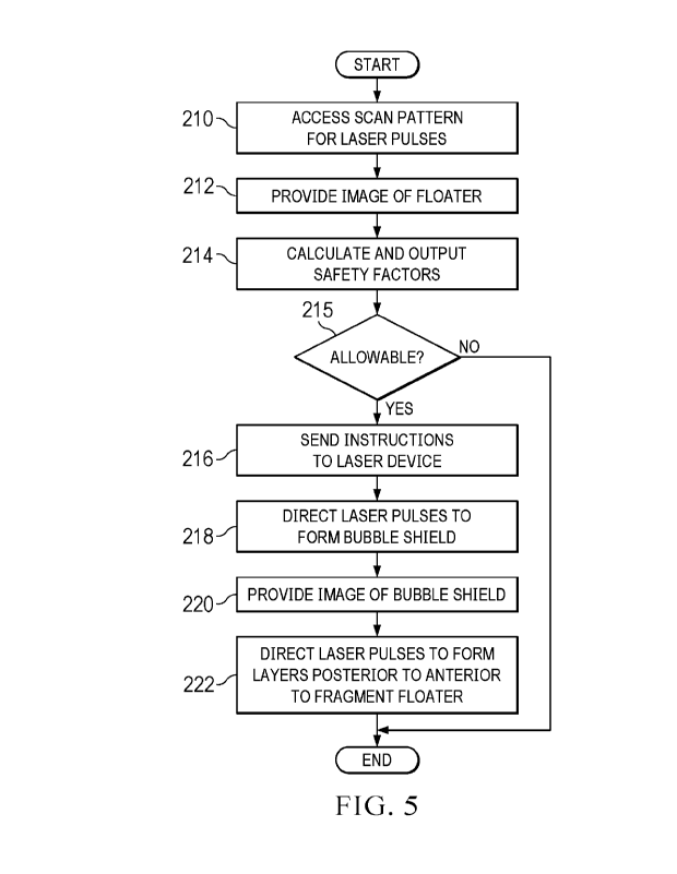
[0073] FIGURE 5 illustrates an example of a method for fragmenting a floater
with a three-
dimensional (3D) scan pattern that may be performed by system 10 of FIGURE 1,
according to
certain embodiments. A user such as a surgeon may use a 3D pulse pattern to
fragment a floater
within the vitreous of a patient eye. The 3D pulse pattern includes a bubble
shield that reduces
retinal radiation exposure at the retina of the eye.
[0074] The method starts at step 210, where computer 26 accesses the 3D scan
pattern for
laser pulses. The scan pattern may be stored in memory 52. Floater detection
system 19 provides
an image of the floater to the user at step 212. The image may allow the user
to locate the floater.
Computer 26 calculates and outputs safety factors at step 214. Safety factors
indicate radiation
exposure in the eye relative to a maximum exposure limit. They guide the user
in deciding whether
or not the advantage of the surgery justifies the risk of retinal radiation
exposure. The treatment
may be allowable at step 215. If the treatment is allowable, the method
proceeds to step 216. If it
is not, the method ends.
[0075] Computer 26 sends instructions to laser device 22 to direct pulses
towards the eye
according to the scan pattern at step 216. Any suitable 3D scan pattern, e.g.,
as described herein,
may be used. Laser device 22 directs laser pulses towards the eye to form
bubble shield within the
vitreous at step 218. The bubble shield reduces retinal radiation exposure at
the retina of the eye.
Floater detection system 19 provides an image of the bubble shield to the user
at step 220. The
image may allow the user to check that the bubble shield is sufficiently
opaque to protect the retina.
Laser device 22 directs laser pulses to form layers, from posterior to
anterior layers, to fragment
floater at step 222.
CA 03234768 2024-4- 11
WO 2023/089417
PCT/1B2022/060256
[0076] A component (such as the control computer) of the systems and
apparatuses
disclosed herein may include an interface, logic, and/or memory, any of which
may include
computer hardware and/or software. An interface can receive input to the
component and/or send
output from the component, and is typically used to exchange information
between, e.g., software,
hardware, peripheral devices, users, and combinations of these. A user
interface is a type of
interface that a user can utilize to communicate with (e.g., send input to
and/or receive output
from) a computer. Examples of user interfaces include a display, Graphical
User Interface (GUI),
touchscreen, keyboard, mouse, gesture sensor, microphone, and speakers.
[0077] Logic can perform operations of the component. Logic may include one or
more
electronic devices that process data, e.g., execute instructions to generate
output from input.
Examples of such an electronic device include a computer, processor,
microprocessor (e.g., a
Central Processing Unit (CPU)), and computer chip. Logic may include computer
software that
encodes instructions capable of being executed by an electronic device to
perform operations.
Examples of computer software include a computer program, application, and
operating system.
[0078] A memory can stole information and may comprise tangible, computer-
readable,
and/or computer-executable storage medium. Examples of memory include computer
memory
(e.g., Random Access Memory (RAM) or Read Only Memory (ROM)), mass storage
media (e.g.,
a hard disk), removable storage media (e.g., a Compact Disk (CD) or Digital
Video or Versatile
Disk (DVD)), database, network storage (e.g., a server), and/or other computer-
readable media.
Particular embodiments may be directed to memory encoded with computer
software.
[0079] Although this disclosure has been described in terms of certain
embodiments,
modifications (such as changes, substitutions, additions, omissions, and/or
other modifications) of
the embodiments will be apparent to those skilled in the art. Accordingly,
modifications may be
made to the embodiments without departing from the scope of the invention. For
example,
modifications may be made to the systems and apparatuses disclosed herein. The
components of
the systems and apparatuses may be integrated or separated, or the operations
of the systems and
apparatuses may be performed by more, fewer, or other components, as apparent
to those skilled
in the art. As another example, modifications may be made to the methods
disclosed herein. The
methods may include more, fewer, or other steps, and the steps may be
performed in any suitable
order, as apparent to those skilled in the art.
CA 03234768 2024-4- 11
WO 2023/089417
PCT/1B2022/060256
16
[0080] To aid the Patent Office and readers in interpreting the claims,
Applicants note that
they do not intend any of the claims or claim elements to invoke 35 U.S.C.
112(f), unless the
words "means for" or "step for" are explicitly used in the particular claim.
Use of any other term
(e.g., "mechanism," "module," "device," "unit," "component," "element,"
"member,"
"apparatus," "machine," "system," "processor," or "controller") within a claim
is understood by
the applicants to refer to structures known to those skilled in the relevant
art and is not intended to
invoke 35 U.S.C. 112(f).
CA 03234768 2024-4- 11