Note: Descriptions are shown in the official language in which they were submitted.
WO 2023/081727
PCT/US2022/079180
TREATMENTS FOR CANCERS UTILIZING CELL-TARGETED THERAPIES
AND ASSOCIATED RESEARCH PROTOCOLS
CROSS-REFERENCE TO RELATED APPLICATIONS
[0001] This application claims priority to U.S. Provisional Patent Application
No. 63/274,914 filed
November 2, 2021 and U.S. Provisional Patent Application No. 63/371,265 filed
August 12, 2022,
which are both incorporated herein by reference in their entirety as if fully
set forth herein.
REFERENCE TO SEQUENCE LISTING
[0002] The Sequence Listing associated with this application is provided in
XML format in lieu of
a paper copy and is hereby incorporated by reference into the specification.
The name of the XML
file containing the Sequence Listing is 2SF5710.xml. The XML file is 147 KB,
was created on
October 28, 2022, and is being submitted electronically via Patent Center.
FIELD OF THE DISCLOSURE
[0003] The current disclosure provides targeted cancer treatments for cancer
cells expressing
FOLR1, MEGF10, HPSE2, KLRF2, PCDH19, or FRAS1. The targeted therapeutic can
include a
chimeric antigen receptor (CAR) expressed by an immune cell, such as a T cell.
Treated cancers
include a variety of solid tumor cancers and blood cancers.
BACKGROUND OF THE DISCLOSURE
[0004] According to the World Health Organization, cancer is a leading cause
of death globally,
and was responsible for nearly 10 million deaths in 2020. Beyond traditional
cancer treatments
such as surgery, chemotherapy, and radiation therapy, more targeted therapies
have emerged to
specifically target cancer cells by identifying and exploiting specific
molecular changes seen
primarily in those cells. For example, immune cells can be genetically
engineered to target and
kill cancer cells. Many of these immune cells are T cells have been
genetically engineered to
express a chimeric antigen receptor (CAR) which recognizes a protein or
molecule expressed on
the surface of the cancer cell so that the genetically modified T cell can
recognize and kill the
cancer cells. Furthermore, antibodies or binding fragments thereof that bind a
protein or molecule
expressed on the surface of the cancer cell can be used to trigger immune
reactions against
cancer cells. These antibodies or binding fragments thereof can be conjugated
to cytotoxic drugs
to further enhance their cytotoxic effects.
[0005] Numerous cancer types would benefit from the development of additional
CAR-based
therapies, such as leukemias, peritoneal cancer, fallopian tube cancer,
ovarian cancer,
1
CA 03235607 2024- 4- 18
WO 2023/081727
PCT/US2022/079180
endometrial cancer, cervical cancer, breast cancer, bladder cancer, renal cell
carcinoma, pituitary
tumors, lung cancer, uterine cancer, squamous cell carcinoma, ureter cancer,
urethral cancer,
osteosarcoma, and transitional cell carcinoma.
[0006] Pediatric acute myeloid leukemia (AML), for example, is a diverse group
of diseases
classified based on morphology, lineage, and genetics (Rubnitz, Blood 119:5980-
8, 2012) and its
prognosis depends on several cytogenetic and molecular characteristics.
Despite improved
survival and remission induction rates, outcomes vary significantly amongst
the different biological
subtypes of AML (Kim, Blood Res. 55(Suppl): S5-S13, 2020). To better stratify
risk and survival
outcomes, genomic investigations of AML has led to new genomic classifications
and predictive
biomarkers (Arber, Semin Hematol. 56: 90-5, 2019; and Arber et al., Blood 127:
2391-405, 2016).
One such genetic prognostic marker includes the CBFA2T3-GLIS2 (C/G) fusion
gene. The C/G
fusion gene characterizes a subtype of leukemia that is extremely aggressive
and specific to
pediatrics. This subtype of AML is highly refractory to conventional
therapies, resulting in survival
rates as low as 15-30% (Masetti et al., Br J Haematol. 184(3): 337-347, 2019).
Because of the
significant morbidity and mortality rates for C/G AML, efforts to identify new
therapies is under
continual investigation.
SUMMARY OF THE DISCLOSURE
[0007] The current disclosure provides targeted therapies against cancer cells
expressing
FOLR1, MEGF10, HPSE2, KLRF2, PCDH19, and/or FRAS1. Pediatric acute myeloid
leukemia
(AML) provides an example of a cancer type that can be treated with targeted
therapies against
cancer cells expressing FOLR1, MEGF10, HPSE2, KLRF2, PCDH19, and/or FRAS1.
Leukemias,
peritoneal cancer, fallopian tube cancer, ovarian cancer, endometrial cancer,
cervical cancer,
breast cancer, bladder cancer, renal cell carcinoma, pituitary tumors, lung
cancer, uterine cancer,
squamous cell carcinoma, ureter cancer, urethral cancer, osteosarcoma, and
transitional cell
carcinoma provide examples of cancer types that can be treated with targeted
therapies against
cancer cells expressing FOLR1.
[0008] In particular embodiments, a targeted therapeutic disclosed herein
includes a chimeric
antigen receptor (CAR) expressed by an immune cell, such as a T cell. In
certain examples, the
CAR includes a binding domain that binds FOLR1, an IgG4 spacer, a CD28
transmembrane
domain, and a 4-1BB/CD3( intracellular effector domain. Targeted therapeutics
can also include
antibody conjugates, such as antibody-drug conjugates, antibody-radioisotope
conjugates, or
antibody-nanoparticle conjugates.
2
CA 03235607 2024- 4- 18
WO 2023/081727
PCT/US2022/079180
BRIEF DESCRIPTION OF THE FIGURES
[0009] Some of the drawings submitted herein may be better understood in
color. Applicant
considers the color versions of the drawings as part of the original
submission and reserves the
right to present color images of the drawings in later proceedings.
[0010] FIGs. 1A-1J. CBFA2T3-GLIS2 (C/G)-cord blood (CB) cells induce leukemia
recapitulating
primary disease. 1A. Diagram of experimental design. 1B. Kaplan-Meier survival
curves of NSG-
SGM3 mice transplanted with green fluorescent protein (GFP)-CB control and C/G-
CB cells.
Statistical differences in survival were evaluated using Logrank Mantel-Cox.
1C. Representative
histology of hematoxylin and eosin (H&E) stain of femurs taken from mice
transplanted with C/G-
CB cells (top) and a C/G positive patient sample (bottom) after development of
leukemia. PDX
stands for C/G patient-derived leukemia cells. Magnification: left (2.5X),
middle (5X), right (C/G-
CB 40X; PDX, 20X). 1D. Expression of the RAM immunophenotype in C/G-CB cells
harvested
from the bone marrow of a representative mouse at necropsy compared to a
primary patient
sample and PDX marrow xenograft cells. In all three samples, malignant cells
were gated based
on human CD45 expression and side scatter (SSC). 1E. Left and middle,
representative
immunohistochemistry showing high expression of ERG (10X magnification) and
CD56 (5X
magnification) in the femur of a representative mouse transplanted with C/G-CB
cells. Right, small
aggregates of blasts with high CD56 expression detected in a bone marrow
biopsy of a
chemotherapy refractory C/G fusion positive patient, consistent with residual,
adherent, patchy
disease distribution (100X magnification). IF. Kaplan-Meier plot showing
survival in primary (1 ),
secondary (2 ) and tertiary (3 ) transplantations of C/G-CB cells. 1G.
Engraftment of C/G-CB cells
in the bone marrow at time of symptomatic leukemia, shown as percent human
CD45+. 1H.
Quantification of CD56+ cells amongst human CD45+ cells isolated from the bone
marrow (BM)
at necropsy following development of symptomatic leukemia. 11. Expression of
acute
megakaryocytic leukemia (AMKL) markers, CD41 and CD42, in C/G-CB and PDX cells
harvested
from the bone marrow at necropsy. GIG-GB cells were gated on human CD45+
cells. PDX cells
were gated on human CD45+CD56+ cells. 1J. Quantification of CD41/CD42 subsets
described
in FIG. 11. Bars indicate mean +/- standard error of mean (SEM).
[0011] FIG. 2. GIG-GB cells form tight clusters in mouse bone marrow. (related
to FIGs. 1A-1J).
Histology of femurs taken from primary, secondary and tertiary transplants of
C/G-CB cells.
[0012] FIGs. 3A-3C. Expression of CD56 and AMKL markers in C/G-CB xenograft
cells following
development of symptomatic leukemia in NSG-SGM3 mice. 3A. Percent human 0D45+
cells in
the bone marrow, spleen, liver and peripheral blood (PB) from mice
transplanted with C/G-CB
3
CA 03235607 2024- 4- 18
WO 2023/081727
PCT/US2022/079180
cells in primary (1 ), secondary (2 ) and tertiary (3 ) transplants. 3B, 3C.
Percent CD56+ and
CD41/CD42 subsets in mouse tissues described in FIG. 3A.
[0013] FIGs. 4A-4J. Endothelial cells (ECs) enhance the proliferative
potential and promote
leukemic progression of C/G-CB cells. 4A. Diagram of experimental design. 4B.
Growth kinetics
of C/G-CB and GFP-CB cells in EC co-culture or myeloid promoting conditions
(MC). 4C. C/GCB
cells expanded in EC co-culture for 9 weeks were reseeded in EC co-culture
either directly (direct
contact) or in EC transwells (indirect contact) or placed in liquid culture
containing serum free
expansion medium (SFEM) II (+SCF, FLT3L, and TPO). After 7 days, the number of
GFP+ cells
was quantified by flow cytometry. 4D. At 6 and 12 weeks, a fraction of each
culture was transferred
to MegaCult cultures. Colonies derived from megakaryocytic (Mk) progenitors
were scored and
enumerated. Data are normalized to the 500 input cells at the start of the EC
co-culture or MC
culture. A representative colony stained with anti-human CD41 and an alkaline
phosphotase
detection system is shown. 4E. Equivalent number of GIG-GB and GFPCB cells
were transplanted
into NSG-SGM3 mice (5-10x106/mouse) at indicated timepoints. Due to
insufficient expansion,
GFP-CB cells were not transplanted after 3 weeks in either condition,
similarly for C/G-CB cells
after 6 weeks in MC culture. Median survival and Kaplan-Meier survival curves
are shown. GIG-
CB (N=3 mice/group), GFP-CB (N=2 mice/group) 4F, 4G. Quantification of CD56+
cells (4F) and
CD41/CD42 subsets (4G) amongst human CD45+ cells over weeks in culture. 4H.
Unsupervised
clustering by uniform manifold and projection (UMAP) analysis of C/G-CB and
GFP-CB cells in
reference to primary AML samples. Dashed circle indicates C/G-CB cells co-
cultured with ECs at
week 6 and 12 timepoints. NBM=normal bone marrow. 41. Heatmap of
differentially expressed
genes in C/G-CB versus GFP-CB cells in EC co-culture or MC. 4J. GSEA plots of
C/G and HSC
signature genes comparing C/G-CB cells in EC co-culture versus MC. (4B-4E, 4G-
4I) Data
presented as mean +/- standard deviation from 3 technical replicates.
[0014] FIGs. 5A-5C. Assessment of RAM and AMKL markers in GIG-GB cells
isolated from mice
transplanted with engineered cells cultured in EC co-culture or MC. 5A.
Percent human CD45+
cells in the bone marrow, spleen liver and peripheral blood from mice
transplanted with C/G-CB
and GFP-CB cells at indicated timepoints in EC co-culture or MC. 5B, 5C.
Percent CD41/0D42
subsets (5B) andCD56+ cells (5C) among live human CD45+ in mouse tissues
described in FIG.
5A. Data analyzing CB cells in the liver for mice transplanted with GFP-CB
cells from MC culture
are not included as not enough cells were present in the samples. Peripheral
blood data from 2
mice transplanted with C/G-CB cells grown in MC are also not included as
enough cells were not
present in the samples.
4
CA 03235607 2024- 4- 18
WO 2023/081727
PCT/US2022/079180
[0015] FIGs. 6A-6C. C/G-CB cells cultured with ECs recapitulate the
immunophenotype and
morphology of C/G fusion positive AML. 6A. Expression of the RAM
immunophenotype in C/G-
CB cells after 6 weeks in EC co-culture or MC. 6B. Quantification of CD41/CD42
subsets at
indicated timepoints in EC co-culture or MC. 6C. Morphological evaluation of
the C/G-CB cells
cultured with ECs or in MC for 9 weeks showed features of megakaryocytic
differentiation,
including open chromatin, prominent nucleoli, and abundant focally, basophilic
and vacuolated
cytoplasm with cytoplasmic blebbing.
[0016] FIGs. 7A-7D. ECs promote transformation of C/G-CB cells. 7A. Schematic
of transduction
and long-term cultures of cord blood CD34+ HSPCs from a second donor. 7B.
Growth kinetics of
transduced cells over days in EC or MC as determined by the cumulative number
of GFP+ cells.
Mean +/- standard deviation from 3 technical replicates are shown. Growth rate
constant k was
determined by regression analysis using the formula N(t) = N(0)ekt where t is
measured in days.
7C. Following 6 and 12 weeks of culture, a fraction of each culture was
transferred to megacult
to enumerate Mk colonies. Data are normalized to the CD34+ input cells at the
start of the culture
and presented as mean +/- standard deviation from 3 technical replicates. 7D.
Expression of the
RAM immunophenotype in C/G-CB cells after 6 weeks in either EC co-culture or
MC.
[0017] FIGs. 8A, 8B. C/G-specific genes and pathways that are recapitulated in
C/G-CB cells
cultured with ECs versus in MC. 8A. The expression (labeled Expression (Log2
cpm)) of ERG,
BMP2 and GATA1 in GFP-CB versus C/G-CB cells over weeks in EC and MC
conditions as well
as in C/G fusion positive primary versus normal marrow samples. Single-sample
gene-set
enrichment (ssGSEA) scores (labeled Enrichment Score) of Hedgehog, TGFB, and
WNT
signaling pathways for GFP-CB versus C/G-CB cells and normal bone marrow
samples versus
primary fusion positive samples. 8B. Pathways that are upregulated (left) and
downregulated
(right) in C/G-CB cells in EC co-culture compared to MC.
[0018] FIGs. 9A-9C. Expression of C/G-specific genes. Heat maps showing
expression of C/G-
specific focal adhesion and cell adhesion molecule genes (9A), genes
associated with primary
C/G fusion positive AML (913), and HSC signature genes (9C). Unsupervised
hierarchical
clustering demonstrates clustering of C/G-CB cells cultured with ECs for 6 and
12 weeks with
primary C/G samples.
[0019] FIGs. 10A-10G. Integrative transcriptomics of primary samples and C/G-
CB identify
FOLR1 therapeutic target. 10A. Diagram of computational workflow to identify
C/G-specific CAR
targets. See Methods and FIG. 11 for details. Normal tissues include bulk bone
marrow (BM)
samples and peripheral blood (PB) CD34+ samples. 10B, 10C. Expression of C/G-
specific CAR
targets in primary fusion positive patients versus normal bone marrow (NBM)
(10B) and C/G-CB
CA 03235607 2024- 4- 18
WO 2023/081727
PCT/US2022/079180
versus GFP-CB cells (10C). 10D. Top, gating strategies used to identify AML
cells and normal
lymphocytes, monocytes and myeloid cells in 4 representative patients based on
CD45
expression and SSC. Bottom, FOLR1 expression in the AML blast subpopulation
versus normal
cells. 10E. Quantification of FOLR1 expression (geometric mean fluorescent
intensity, MFI)
among AML blasts and their normal counterparts across N=15 patients.
Autofluorescence was
used as control. ***, p<0.0005 (paired Student t-test) 10F, 10G. Expression of
FOLR1 (10F) and
quantification of FOLR1+ cells (10G) amongst GFP-CB and C/G-CB over weeks in
EC co-culture.
[0020] FIG. 11. Identification of GIG fusion-specific CAR targets. (Related to
FIG. 10A) Flow
diagram of AML-restricted gene and CAR-T target identification. The procedure
involves three
main steps: 1) Determine the ratio of expression for AML primary samples
versus healthy normal
hematopoietic tissue samples (bulk marrows and 0D34+ peripheral blood) from
log10
transformed normalized gene expression. The ratio is calculated per gene from
mean AML
expression and mean normal hematopoietic tissue expression, where normal
tissue values are
the divisor, which acts as a measure of over or under expression. A normal
curve is fit to the ratios
and this procedure is completed for all heterogenous AML samples as a group,
and iteratively
within fusion and mutation subtypes; genes with ratios greater than +2
standard deviations and
with absent expression in normal hematopoietic tissues were retained (N=607)
for further
analysis. 2) The AML restricted genes were further selected if found to be
significantly
overexpressed in fusion positive patient samples compared to healthy marrows
and were likewise
overexpressed in C/G-CB at weeks 6 and 12 in EC co-culture with absent
expression in GFPCB
controls, providing several candidate (N=42) targets. 3) Optimal CAR-T targets
were selected by
the identification of candidate genes with cell surface localization
potential, and those with an
absence of expression in healthy tissue controls as noted in step 1, but
expression in > 75% of
GIG patient samples, and with moderate to high expression levels (N=6).
[0021] FIG. 12. Expression of FOLR1 transcript in C/G-CB cells cultured on
ECs. RT-PCR
analysis of FOLR1 expression in engineered CB cells and in fusion positive
cell lines M07e and
WSU-AML. Expression is normalized as fold-change relative to GFP-CB/EC Wk 3
samples.
[0022] FIGs. 13A-13D. Pre-clinical efficacy of FOLR1 CAR T cells against C/G
AML cells. 13A.
Cytolytic activity of CD8 T cells unmodified or transduced with FOLR1 CAR
following 6 hours of
co-culture with GIG-GB, WSU-AML, Kasumi-1 FOLR1+ and Kasumi-1 parental cells.
Data
presented are mean leukemia specific lysis +/- SD from 3 technical replicates
at indicated effector:
target (E:T) ratios. Data are representative of 2 donors (see related data in
FIG. 16). 13B.
Concentration of secreted IL-2, IFN-y, and INF-a in the supernatant following
24 hour of T
cell/AML co-culture at 1:1 E:T ratio as measured by ELISA. Data are
representative of 2 donors
6
CA 03235607 2024- 4- 18
WO 2023/081727
PCT/US2022/079180
and are presented as mean +/- SD from 3 technical replicates (see related data
in FIGs. 17A-
17F). Where concentrations of cytokines are too low to discern, the number
above the x-axis
indicates the average concentration. Statistical significance was determined
by unpaired
Student's t test, assuming unequal variances. p<0.05 (*), p<0.005 (**),
p<0.0005 (***). 13C.
Representative flow cytometric analysis of cell proliferation of Cell
Proliferation Dye (Celltrace)-
labeled unmodified and FOLR1 CAR T cells after 4-day co-culture with target
cells at 1:1 E:T ratio.
CAR T cells divided rapidly and diluted their Celltrace fluorescence after 4-
hour co-incubation with
FOLR1-positive AML cells. Data are representative of 2 donors. 13D.
Bioluminescent imaging of
C/G-CB, WSU-AML, Kasumi-1 FOLR1+ and Kasumi-1 leukemias in mice treated with
unmodified
or FOLR1 CAR T cells at 5 x10⁶ T cells per mouse. N=5 mice/group. Radiance
scale indicates an
increase in leukemia from blue to red; X indicates death.
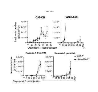
[0023] FIG. 14A-14C. In vivo efficacy of FOLR1-directed CAR T. (Related to
FIGs. 13A-13D)
14A. Quantification of leukemia burden over time based on IVIS radiance in
CBFA2T3-GLIS2-
transduced HSPCs, WSU-AML, Kasumi-1 FOLR1+ and Kasumi-1 xenografts treated
with
unmodified or FOLR1 CAR T cells at 5 x10⁶/mouse. Leukemia burden is shown for
each mouse.
N = 5 mice per group. 14B. Quantification of human T cells in the mouse
peripheral blood at
indicated time points after T cell injection. Shown is human CD45+CD3+
frequency amongst
DAPI- cells. N = 5 mice per group. Data presented are the average +/- standard
deviation from 5
mice. *, p<0.05 (unpaired Student's t-test) 14C. Kaplan-Meier survival curves
of xenografts
treated with unmodified or FOLR1 CAR T cells. N=5 per group. Statistical
differences in survival
were evaluated using Log-rank Mantel-Cox. Note: 2 C/G-CB bearing mice treated
with CAR T
cells died without leukemia and T cells present in bone marrow, spleen and
liver tissues and in
peripheral blood as determined by flow cytometric analysis
[0024] FIGs. 15-15F. FORL1-directed CART effectively eliminate C/G-CB cells
without impacting
viability of HSPCs. 15A. Gating strategy used to identify HPSC subsets from a
representative
CD34-enriched marrow sample from a healthy donor. Shown is representative of 3
donors.
Immunophenotype of the HSPCs is as follows: CD34+CD38-CD9O+CD45RA-
(hematopoietic
stem cell, HSC); CD34+CD38-CD9O-CD45RA- (multipotent progenitors, MPP);
CD34+CD38-CD9O-CD45RA+ multi-lymphoid progenitors, MLP); CD34+CD38+CD10+
(Common lymphoid progenitor, CLP); CD34+CD38+CD1O-CD123-CD45RA- (megakaryocyte-
erythroid progenitor, MEP); CD34+CD38+CD1O-CD123+CD45RA- (common myeloid
progenitor, CMP); 0D34+0D38+CD1O-CD123+CD45RA+ (granulocyte monocyte
progenitor,
GMP). 15B. Histogram of FOLR1 expression in normal HSPC subsets. 15C.
Quantification of
percent FOLR1+ in C/G-CB cells (>12 weeks of EC co-culture) and HSPC subsets
from three
7
CA 03235607 2024- 4- 18
WO 2023/081727
PCT/US2022/079180
CD34-enriched samples from healthy donors. 15D. Percent specific lysis in C/G-
CB cells and the
HSPC subsets shown in FIG. 15C following 4-hour incubation with unmodified or
FOLR1 CAR T
cells at 2:1 E:T ratio. Note that data points for C/G-CB cells are from 2
technical replicates. Only
two out of three normal CD34+ samples were used in this experiment. 15E, 15F.
After 4 hours,
co-cultures of healthy donor CD34+ or C/G-CB cells with either unmodified or
MSLN CAR T cells
at 2:1 E:T ratio were transferred to methylcellulose with cytokines for colony-
forming cell (CFC)
assay. Colonies derived from erythroid (E), granulocyte-macrophage (G, M, and
GM) and
multipotential granulocyte, erythroid, macrophage, megakaryocyte (GEM M)
progenitors were
scored and enumerated after 7-10 days (15E). Total colonies from C/G-CB cells
are tabulated
(15F). Data are presented as mean +/- SD from 3 technical replicates for each
donor No
significant difference in the total number of colonies was detected between co-
cultures with
unmodified T cells versus FOLR1 CAR T cells for normal HSPCs as determined by
unpaired
Student's t test, assuming unequal variances. Statistical significance was
determined by unpaired
Student's t test, assuming unequal variances. P<0.05 (*), p<0.005 (**),
p<0.0005 (***).
[0025] FIG. 16. Expression of C/G transcript in C/G-CB cells. RT-PCR analysis
of C/G expression
in engineered CB cells and in fusion positive cell lines M07e and WSU-AML.
[0026] FIGs. 17A-17F. FOLR1 CAR constructs and reactivity of short,
intermediate and long
FOLR1 CAR T cells. 17A. Schematic diagram of second-generation FOLR1 CAR
constructs with
different IgG4 spacer lengths. SP= GM-CSFR signal peptide; scFv= single-chain
variable
fragment; TM = transmembrane domain; CD = costimulatory domain; SD =
stimulatory domain;
tCD19 = transduced marker truncated CD19. The anti-FOLR1 scFv could be
replaced with a
different binding domain including binding domains that bind to MEGF10, HPSE2,
KLRF2,
PCDH19, FRAS1, or other binding domains that bind to FOLR1. 17B. Expression of
FOLR1 in
C/G-CB, M07e, WSU-AML, Kasumi-1 FOLR1+ and Kasumi-1 parental cells. 17C.
Cytolytic
activity of CD8 T cells unmodified or transduced with short, intermediate or
long FOLR1 CAR
construct against C/G- CB, M07e, WSU-AML, Kasumi-1 FOLR1+ and Kasumi-1
parental cells in
a 6-hour assay. Shown is mean percent specific lysis +/- SD from 3 technical
replicates at
indicated Effector:Target (E:T) ratios. 17D. Concentration of secreted IL-2,
IFN-y, and TNF-a in
the supernatant following 24 hour of CD8 T cell/AML co-culture at 1:1 E:T
ratio. Mean +/- SD from
3 technical replicates is shown. 17E. Representative flow plots showing
expression of N FAT, NF-
kB and AP-1 in Jurkat Nur77 reporter transduced with FOLR1 CAR constructs
cultured alone (top)
or co-incubated with Kasumi-1 FOLR1+ target cells for 24 hours at 1:1 E:T
ratio (bottom). Kasumi-
1 FOLR1+ cells were labeled with Violet Cell Proliferation Dye to
differentiate from Jurkat cells.
Transduced Jurkat cells were gated based on tCD19 expression. Number in top
right corner
8
CA 03235607 2024- 4- 18
WO 2023/081727
PCT/US2022/079180
indicates the percentage of positive cells. Analysis was performed on day 4
post transduction.
17F. Quantification of percent NFAT+, NF-kB+ and AP-1+ cells in FIG. 17E.
[0027] FIG. 18. Information for antibodies.
[0028] FIG. 19. Sequences supporting the disclosure include IgG4 hinge coding
sequence-A
(SEQ ID NO: 1); IgG4 hinge coding sequence-B (SEQ ID NO: 2); IgG4 hinge S1OP
(SEQ ID NO:
135); Hinge+intermediate spacer (DS) (SEQ ID NO: 136); IgG4-int(DS) coding
sequence (SEQ
ID NO: 3); IgG4-long coding sequence (SEQ ID NO: 4); CD3 coding sequence (SEQ
ID NO: 5);
CD3C protein-A (SEQ ID NO: 6); CD3C protein-B (SEQ ID NO: 7); 4-1BB signaling
coding
sequence-A (SEQ ID NO: 8); 4-1BB signaling coding sequence-B (SEQ ID NO: 9); 4-
1BB protein-
A (SEQ ID NO: 10); 4-1BB protein-B (SEQ ID NO: 11); CD28TM coding sequence-A
(SEQ ID
NO: 12); CD28TM coding sequence-B (SEQ ID NO: 13); CD28TM coding sequence-C
(SEQ ID
NO: 14); CD28TM protein-A (SEQ ID NO: 15); CD28TM protein-B (SEQ ID NO: 16);
T2A coding
sequence (SEQ ID NO: 137); P2A (SEQ ID NO: 17); T2A (SEQ ID NO: 18); E2A (SEQ
ID NO:
19); F2A (SEQ ID NO: 20); tCD19 coding sequence (SEQ ID NO: 117); Psi (SEQ ID
NO: 118);
RRE (SEQ ID NO: 119); and Flap (SEQ ID NO: 120).
DETAILED DESCRIPTION
[0029] For many years, the chosen treatments for cancer were surgery,
chemotherapy, and/or
radiation therapy. In recent years, more targeted therapies have emerged to
specifically target
cancer cells by identifying and exploiting specific molecular and/or
immunophenotypic changes
seen primarily in those cells. For example, many cancer cells preferentially
express particular
antigens on their cellular surfaces and these antigens have provided targets
for successful
antibody- and cell-based therapeutics.
[0030] Although targeted therapies have been successful in treating many
cancers, the targeted
therapy of acute myeloid leukemia (AML) remains a challenge given significant
overlap of target
antigens expressed on AML and normal hematopoietic cells.
[0031] Pediatric acute myeloid leukemia (AML) is a diverse group of diseases
classified based
on morphology, lineage, and genetics (Rubnitz, Blood 119:5980-8, 2012) and its
prognosis
depends on several cytogenetic and molecular characteristics. Although, the
overall survival and
remission-induction rates of children with AML have improved over the past
three decades,
outcomes vary significantly amongst the different biological subtypes of AML
(Kim, Blood Res.
55(Suppl): S5-S13, 2020). To better stratify risk and survival outcomes,
genomic investigations
of AML have led to new genomic classifications and predictive biomarkers
(Arber, Semin Hematol.
56: 90-5, 2019; and Arber et al., Blood 127: 2391-405, 2016). One such genetic
prognostic marker
includes the CBFA2T3-GLIS2 (C/G) fusion gene. The C/G fusion gene
characterizes a subtype
9
CA 03235607 2024- 4- 18
WO 2023/081727
PCT/US2022/079180
of leukemia that is extremely aggressive and specific to pediatrics. This
subtype of AML is highly
refractory to conventional therapies, resulting in survival rates as low as 15-
30% (Masetti et al.,
Br J Haematol. 184(3): 337-347, 2019).
[0032] C/G AML and other AML-restricted genes were discovered through an
expansive target
discovery effort through TARGET and Target Pediatric AML (TpAML). These genes
were further
filtered to include those that are upregulated in both C/G AML and in C/G-cord
blood (CB) cells
cultured with endothelial cells and to those genes that encode proteins that
localize to the plasma
membrane. This resulted in seven C/G fusion-specific targets: FOLR1, MEGF10,
HPSE2, KLRF2,
PCDH19, and FRAS1 which were identified to be highly expressed in C/G patients
and in C/G-
CB cells but entirely silent in normal hematopoiesis. The current disclosure
provides targeted
therapeutic treatments with binding domains that bind FOLR1, MEGF10, HPSE2,
KLRF2,
PCDH19, or FRAS1 for the treatment of AML including C/G AML.
[0033] Targeted therapeutics disclosed herein that bind FOLR1 can additionally
be used to treat
other cancers including other leukemias, peritoneal cancer, fallopian tube
cancer, ovarian cancer
(e.g., epithelial ovarian cancer), endometrial cancer, cervical cancer, breast
cancer (e.g., triple-
negative breast cancer, HER2-breast cancer), bladder cancer, renal cell
carcinoma, pituitary
tumors, lung cancer (e.g., lung adenocarcinoma or epithelial lung cancer such
as non-small cell
lung cancer), uterine cancer, squamous cell carcinoma, ureter cancer, urethral
cancer,
osteosarcoma, or transitional cell carcinoma.
[0034] Particular examples of targeted therapeutics disclosed herein include
chimeric antigen
receptors (CAR). In particular embodiments, the CAR include a binding domain
that binds FOLR1.
In particular embodiments, the binding domain that binds FOLR1 is a
Farletuzumab scFv. In
particular embodiments, the CAR include a binding domain that binds MEGF10. In
particular
embodiments, the CAR include a binding domain that binds HPSE2. In particular
embodiments,
the CAR include a binding domain that binds KLRF2. In particular embodiments,
the CAR include
a binding domain that binds PCDH19. In particular embodiments, the CAR include
a binding
domain that binds FRAS1.
[0035] In particular embodiments, the current disclosure provides CAR having
an intermediate
spacer region. In particular embodiments, the intermediate spacer region
includes the hinge
region and the CH3 domain of IgG4. In particular embodiments, the spacer is a
short spacer. In
particular embodiments, the spacer is a long spacer.
[0036] In particular embodiments the current disclosure provides CAR having a
transmembrane
domain including the CD28 transmembrane domain. In particular embodiments, the
current
CA 03235607 2024- 4- 18
WO 2023/081727
PCT/US2022/079180
disclosure provides CAR having an intracellular effector domain including the
4-1BB and CD3
signaling domains.
[0037] In particular embodiments, the CAR including a binding domain that
binds FOLR1 is
encoded by SEQ ID NO: 134.
[0038] The current disclosure also provides targeted therapeutics for the
treatment of cancer
based on antibody formats, such as antibody-drug conjugates, antibody-
radioisotope conjugates,
antibody-immunotoxin conjugates, or antibody-nanoparticle conjugates.
[0039] The current disclosure also provides methods and assays to further
study the cancer
biology of C/G AML. The cancer biology of C/G AML can be studied by the
development of a
model for C/G AML cells prepared by transduction of a C/G fusion gene into
target cells. In
particular embodiments, the cells include cord blood (CB) hematopoietic stem
and progenitor cells
(HSPCs). CB-HSPC cells transduced with the C/G fusion gene are referred to
herein as C/G-CB
cells. Furthermore, to model C/G AML cells, the microenvironment of C/G AML is
recreated by
either culturing the transduced cells in an animal model or in micro-
environment stimulating
conditions in monoculture. In particular embodiments, micro-environment
stimulating conditions
include co-culture with endothelial cells. In particular embodiments, micro-
environment
stimulating conditions include myeloid promoting conditions.
[0040] Aspects of the current disclosure are now described with additional
detail and options as
follows: (i) Immune Cells; (ii) Cell Sample Collection and Cell Enrichment;
(iii) Genetically
Modifying Cell Populations to Express Chimeric Antigen Receptors (CAR); (iii-
a) Genetic
Engineering Techniques; (iii-b) CAR Subcomponents; (iii-b-i) Binding Domains;
(iii-b-ii) Spacer
Regions; (iii-b-iii) Transmembrane Domains; (iii-b-iv) Intracellular Effector
Domains; (iii-b-v)
Linkers; (iii-b-vi) Control Features Including Tag Cassettes, Transduction
Markers, and/or Suicide
Switches; (iv) Cell Activating Culture Conditions; (v) Ex Vivo Manufactured
Cell Formulations; (vi)
Antibody Conjugates; (vii) Compositions; (viii) Methods of Use; (ix) Reference
Levels Derived
from Control Populations; (x) Cell Transformation Methods; (xi) Exemplary
Embodiments; (xii)
Experimental Examples; and (xiii) Closing Paragraphs. These headings are
provided for
organizational purposes only and do not limit the scope or interpretation of
the disclosure.
[0041] (i) Immune Cells. The present disclosure describes cells genetically
modified to express
CAR. Genetically modified cells can include T-cells, B cells, natural killer
(NK) cells, NK-T cells,
monocytes/macrophages, lymphocytes, hematopoietic stem cells (HSCs),
hematopoietic
progenitor cells (HPC), and/or a mixture of HSC and HPC (i.e., HSPC). In
particular embodiments,
genetically modified cells include T-cells.
11
CA 03235607 2024- 4- 18
WO 2023/081727
PCT/US2022/079180
[0042] Several different subsets of T-cells have been discovered, each with a
distinct function.
For example, a majority of T-cells have a T-cell receptor (TCR) existing as a
complex of several
proteins. The actual T-cell receptor is composed of two separate peptide
chains, which are
produced from the independent T-cell receptor alpha and beta (TCRa and TCR)
genes and are
called a- and p-TCR chains.
[0043] y5 T-cells represent a small subset of T-cells that possess a distinct
T-cell receptor (TCR)
on their surface. In yo T-cells, the TCR is made up of one y-chain and one 5-
chain. This group of
T-cells is much less common (2% of total T-cells) than the ap T-cells.
[0044] CD3 is expressed on all mature T cells. Activated T-cells express 4-1BB
(CD137), CD69,
and 0D25. CD5 and transferrin receptor are also expressed on T-cells.
[0045] T-cells can further be classified into helper cells (CD4+ T-cells) and
cytotoxic T-cells
(CTLs, CD8+ T-cells), which include cytolytic T-cells. T helper cells assist
other white blood cells
in immunologic processes, including maturation of B cells into plasma cells
and activation of
cytotoxic T-cells and macrophages, among other functions. These cells are also
known as CD4+
T-cells because they express the CD4 protein on their surface. Helper T-cells
become activated
when they are presented with peptide antigens by MHC class II molecules that
are expressed on
the surface of antigen presenting cells (APCs). Once activated, they divide
rapidly and secrete
small proteins called cytokines that regulate or assist in the active immune
response.
[0046] Cytotoxic T-cells destroy virally infected cells and tumor cells and
are also implicated in
transplant rejection. These cells are also known as CD8+ T-cells because they
express the CD8
glycoprotein on their surface. These cells recognize their targets by binding
to antigen associated
with MHC class I, which is present on the surface of nearly every cell of the
body.
[0047] "Central memory" T-cells (or "TCM") as used herein refers to an antigen
experienced CTL
that expresses CD62L or CCR7 and CD45R0 on the surface thereof and does not
express or
has decreased expression of CD45RA as compared to naive cells. In particular
embodiments,
central memory cells are positive for expression of CD62L, CCR7, CD25, CD127,
CD45RO, and
0D95, and have decreased expression of CD45RA as compared to naive cells.
[0048] "Effector memory" T-cell (or "TEM") as used herein refers to an antigen
experienced T-
cell that does not express or has decreased expression of CD62L on the surface
thereof as
compared to central memory cells and does not express or has decreased
expression of CD45RA
as compared to a naive cell. In particular embodiments, effector memory cells
are negative for
expression of CD62L and CCR7, compared to naive cells or central memory cells,
and have
variable expression of 0D28 and CD45RA. Effector T-cells are positive for
granzyme B and
perforin as compared to memory or naive T-cells.
12
CA 03235607 2024- 4- 18
WO 2023/081727
PCT/US2022/079180
[0049] "Naive" T-cells as used herein refers to a non-antigen experienced T
cell that expresses
CD62L and CD45RA and does not express CD45R0 as compared to central or
effector memory
cells. In particular embodiments, naive CD8+ T lymphocytes are characterized
by the expression
of phenotypic markers of naive T-cells including CD62L, CCR7, CD28, CD127, and
CD45RA.
[0050] Natural killer cells (also known as NK cells, K cells, and killer
cells) are activated in
response to interferons or macrophage-derived cytokines. They serve to contain
viral infections
while the adaptive immune response is generating antigen-specific cytotoxic T
cells that can clear
the infection. NK cells express CD8, CD16 and CD56 but do not express CD3.
[0051] NK cells include NK-T cells. NK-T cells are a specialized population of
T cells that express
a semi-invariant T cell receptor (TCR ab) and surface antigens typically
associated with natural
killer cells. NK-T cells contribute to antibacterial and antiviral immune
responses and promote
tumor-related immunosurveillance or immunosuppression. Like natural killer
cells, NK-T cells can
also induce perforin-, Fas-, and TNF-related cytotoxicity. Activated NK-T
cells are capable of
producing I FN-y and IL-4. In particular embodiments, NK-T cells are
CD3+/CD56+.
[0052] Macrophages (and their precursors, monocytes) reside in every tissue of
the body (in
certain instances as microglia, Kupffer cells and osteoclasts) where they
engulf apoptotic cells,
pathogens and other non-self-components. Monocytes/macrophages express CD11b,
F4/80;
CD68; CD11c; IL-4Ra; and/or CD163.
[0053] Immature dendritic cells (i.e., pre-activation) engulf antigens and
other non-self-
components in the periphery and subsequently, in activated form, migrate to T-
cell areas of
lymphoid tissues where they provide antigen presentation to T cells. Dendritic
cells express CD1a,
CD1b, CD1c, CD1d, CD21, 0D35, CD39, CD40, CD86, CD101, CD148, CD209, and DEC-
205.
[0054] Hematopoietic Stem/Progenitor Cells or HSPC refer to a combination of
hematopoietic
stem cells and hematopoietic progenitor cells.
[0055] Hematopoietic stem cells refer to undifferentiated hematopoietic cells
that are capable of
self-renewal either in vivo, essentially unlimited propagation in vitro, and
capable of differentiation
to all other hematopoietic cell types.
[0056] A hematopoietic progenitor cell is a cell derived from hematopoietic
stem cells or fetal
tissue that is capable of further differentiation into mature cell types. In
certain embodiments,
hematopoietic progenitor cells are CD24I Lin- CD117+ hematopoietic progenitor
cells. HPC can
differentiate into (i) myeloid progenitor cells which ultimately give rise to
monocytes and
macrophages, neutrophils, basophils, eosinophils, erythrocytes,
megakaryocytes/platelets, or
dendritic cells; or (ii) lymphoid progenitor cells which ultimately give rise
to T-cells, B-cells, and
NK-cells.
13
CA 03235607 2024- 4- 18
WO 2023/081727
PCT/US2022/079180
[0057] HSPC can be positive for a specific marker expressed in increased
levels on HSPC
relative to other types of hematopoietic cells. For example, such markers
include 0D34, 0D43,
CD45RO, CD45RA, CD59, CD90, CD109, CD117, CD133, CD166, HLA DR, or a
combination
thereof. Also, the HSPC can be negative for an expressed marker relative to
other types of
hematopoietic cells. For example, such markers include Lin, CD38, or a
combination thereof.
Preferably, the HSPC are CD34+ cells.
[0058] A statement that a cell or population of cells is "positive" for or
expressing a particular
marker refers to the detectable presence on or in the cell of the particular
marker. When referring
to a surface marker, the term can refer to the presence of surface expression
as detected by flow
cytometry, for example, by staining with an antibody that specifically binds
to the marker and
detecting said antibody, wherein the staining is detectable by flow cytometry
at a level
substantially above the staining detected carrying out the same procedure with
an isotype-
matched control under otherwise identical conditions and/or at a level
substantially similar to that
for cell known to be positive for the marker, and/or at a level substantially
higher than that for a
cell known to be negative for the marker.
[0059] A statement that a cell or population of cells is "negative" for a
particular marker or lacks
expression of a marker refers to the absence of substantial detectable
presence on or in the cell
of a particular marker. When referring to a surface marker, the term can refer
to the absence of
surface expression as detected by flow cytometry, for example, by staining
with an antibody that
specifically binds to the marker and detecting said antibody, wherein the
staining is not detected
by flow cytometry at a level substantially above the staining detected
carrying out the same
procedure with an isotype-matched control under otherwise identical
conditions, and/or at a level
substantially lower than that for cell known to be positive for the marker,
and/or at a level
substantially similar as compared to that for a cell known to be negative for
the marker.
[0060] Cells to be genetically modified according to the teachings of the
current disclosure can
be patient-derived cells (autologous) or allogeneic when appropriate and can
also be in vivo or
ex vivo. In particular embodiments, cells to be genetically modified include
CD4+ or CD8+ T cells.
[0061] (ii) Cell Sample Collection and Cell Enrichment. Methods of sample
collection and
enrichment are known by those skilled in the art. In some embodiments, cells
are derived from
cell lines. In particular embodiments, cells are derived from humans. In some
embodiments, cells
are obtained from a xenogeneic source, for example, from mouse, rat, non-human
primate, or
pig.
[0062] In some embodiments, T cells are derived or isolated from samples such
as whole blood,
peripheral blood mononuclear cells (PBMCs), leukocytes, bone marrow, thymus,
tissue biopsy,
14
CA 03235607 2024- 4- 18
WO 2023/081727
PCT/US2022/079180
tumor, leukemia, lymphoma, lymph node, gut associated lymphoid tissue, mucosa
associated
lymphoid tissue, spleen, other lymphoid tissues, liver, lung, stomach,
intestine, colon, kidney,
pancreas, breast, bone, prostate, cervix, testes, ovaries, tonsil, or other
organ, and/or cells
derived therefrom. In particular embodiments, cells from the circulating blood
of a subject are
obtained, e.g., by apheresis or leukapheresis. The samples, in particular
embodiments, contain
lymphocytes, including T cells, monocytes, granulocytes, B cells, other
nucleated
white blood cells, HSC, HPC, HSPC, red blood cells, and/or platelets, and in
some aspects,
contains cells other than red blood cells and platelets and further processing
is necessary.
[0063] In some embodiments, blood cells collected from a subject are washed,
e.g., to remove
the plasma fraction and to place the cells in an appropriate buffer or media
for subsequent
processing steps. In particular embodiments, the cells are washed with
phosphate buffered saline
(PBS). In some embodiments, the wash solution lacks calcium and/or magnesium
and/or many
or all divalent cations. Washing can be accomplished using a semi-automated
"flow-through"
centrifuge (for example, the Cobe 2991 cell processor, Baxter) according to
the manufacturers
instructions. Tangential flow filtration (TFF) can also be performed. In
particular embodiments,
cells can be re-suspended in a variety of biocompatible buffers after washing,
such as,
Ca++/Mg++ free PBS.
[0064] The isolation can include one or more of various cell preparation and
separation steps,
including separation based on one or more properties, such as size, density,
sensitivity or
resistance to particular reagents, and/or affinity, e.g., immunoaffinity, to
antibodies or other
binding partners. In particular embodiments, the isolation is carried out
using the same apparatus
or equipment sequentially in a single process stream and/or simultaneously. In
particular
embodiments, the isolation, culture, and/or engineering of the different
populations is carried out
from the same starting material, such as from the same sample.
[0065] In particular embodiments, a sample can be enriched for T cells by
using density-based
cell separation methods and related methods. For example, white blood cells
can be separated
from other cell types in the peripheral blood by lysing red blood cells and
centrifuging the sample
through a Percoll or Ficoll gradient.
[0066] In particular embodiments, a bulk T cell population can be used that
has not been enriched
for a particular T cell type. In particular embodiments, a selected T cell
type can be enriched for
and/or isolated based on cell-marker based positive and/or negative selection.
In positive
selection, cells having bound cellular markers are retained for further use.
In negative selection,
cells not bound by a capture agent, such as an antibody to a cellular marker
are retained for
further use. In some examples, both fractions can be retained for a further
use.
CA 03235607 2024- 4- 18
WO 2023/081727
PCT/US2022/079180
[0067] The separation need not result in 100% enrichment or removal of a
particular cell
population or cells expressing a particular marker. For example, positive
selection of or
enrichment for cells of a particular type refers to increasing the number or
percentage of such
cells but need not result in a complete absence of cells not expressing the
marker. Likewise,
negative selection, removal, or depletion of cells of a particular type refers
to decreasing the
number or percentage of such cells but need not result in a complete removal
of all such cells.
[0068] In some examples, multiple rounds of separation steps are carried out,
where the
positively or negatively selected fraction from one step is subjected to
another separation step,
such as a subsequent positive or negative selection.
[0069] In some embodiments, an antibody or binding domain for a cellular
marker is bound to a
solid support or matrix, such as a magnetic bead or paramagnetic bead, to
allow for separation
of cells for positive and/or negative selection. For example, in some
embodiments, the cells and
cell populations are separated or isolated using immunomagnetic (or affinity
magnetic) separation
techniques (reviewed in Methods in Molecular Medicine, vol. 58: Metastasis
Research Protocols,
Vol. 2: Cell Behavior In Vitro and In Vivo, p 17-25 Edited by: S. A. Brooks
and U. Schumacher
Humana Press Inc., Totowa, NJ); see also US 4,452,773; US 4,795,698; US
5,200,084; and EP
452342.
[0070] In some embodiments, affinity-based selection is via magnetic-activated
cell sorting
(MACS) (Miltenyi Biotec, Auburn, CA). MACS systems are capable of high-purity
selection of cells
having magnetized particles attached thereto. In certain embodiments, MACS
operates in a mode
wherein the non-target and target species are sequentially eluted after the
application of the
external magnetic field. That is, the cells attached to magnetized particles
are held in place while
the unattached species are eluted. Then, after this first elution step is
completed, the species that
were trapped in the magnetic field and were prevented from being eluted are
freed in some
manner such that they can be eluted and recovered. In certain embodiments, the
non-target cells
are labelled and depleted from the heterogeneous population of cells.
[0071] In some embodiments, a cell population described herein is collected
and enriched (or
depleted) via flow cytometry, in which cells stained for multiple cell surface
markers are carried in
a fluidic stream. In some embodiments, a cell population described herein is
collected and
enriched (or depleted) via preparative scale (FACS)-sorting. In certain
embodiments, a cell
population described herein is collected and enriched (or depleted) by use of
microelectromechanical systems (MEMS) chips in combination with a FACS-based
detection
system (see, e.g., WO 2010/033140, Cho etal. (2010) Lab Chip 10, 1567-1573;
and Godin et al.
16
CA 03235607 2024- 4- 18
WO 2023/081727
PCT/US2022/079180
(2008) J Biophoton. 1(5):355¨ 376). In both cases, cells can be labeled with
multiple markers,
allowing for the isolation of well-defined cell subsets at high purity.
[0072] Cell-markers for different T cell subpopulations are described above.
In particular
embodiments, specific subpopulations of T cells, such as cells positive or
expressing high levels
of one or more surface markers, e.g., CCR7, CD45RO, CD8, CD27, CD28, CD62L,
CD127, CD4,
and/or CD45RA T cells, are isolated by positive or negative selection
techniques. CD3+, CD28+
T cells can be positively selected for and expanded using anti-CD3/anti-CD28
conjugated
magnetic beads (e.g., DYNABEADSO M-450 CD3/CD28 T Cell Expander).
[0073] In particular embodiments, a CD8+ or CD4+ selection step is used to
separate CD4+
helper and CD8+ cytotoxic T cells. Such CD8+ and CD4+ populations can be
further sorted into
sub-populations by positive or negative selection for markers expressed or
expressed to a
relatively higher degree on one or more naive, memory, and/or effector T cell
subpopulations.
[0074] In some embodiments, enrichment for central memory T (TCM) cells is
carried out. In
particular embodiments, memory T cells are present in both CD62L subsets of
CD8+ peripheral
blood lymphocytes. PBMC can be enriched for or depleted of CD62L, CD8 and/or
CD62L+CD8+
fractions, such as by using anti-CD8 and anti-CD62L antibodies.
[0075] In some embodiments, the enrichment for central memory T (TCM) cells is
based on
positive or high surface expression of CCR7, CD45RO, CD27, CD62L, CD28, CD3,
and/or
CD127; in some aspects, it is based on negative selection for cells expressing
or highly
expressing CD45RA and/or granzyme B. In some aspects, isolation of a CD8+
population
enriched for TCM cells is carried out by depletion of cells expressing CD4,
CD14, CD45RA, and
positive selection or enrichment for cells expressing CCR7, CD45RO, and/or
CD62L. In one
aspect, enrichment for central memory T (TCM) cells is carried out starting
with a negative fraction
of cells selected based on CD4 expression, which is subjected to a negative
selection based on
expression of CD14 and CD45RA, and a positive selection based on CD62L. Such
selections in
some aspects are carried out simultaneously and in other aspects are carried
out sequentially, in
either order. In some aspects, the same CD4 expression-based selection step
used in preparing
the CD8+ cell population or subpopulation, also is used to generate the CD4+
cell population or
sub-population, such that both the positive and negative fractions from the
CD4-based separation
are retained, optionally following one or more further positive or negative
selection steps.
[0076] In a particular example, a sample of PBMCs or other white blood cell
sample is subjected
to selection of CD4+ cells, where both the negative and positive fractions are
retained. The
negative fraction then is subjected to negative selection based on expression
of CD14 and
CD45RA or RORI, and positive selection based on a marker characteristic of
central memory T
17
CA 03235607 2024- 4- 18
WO 2023/081727
PCT/US2022/079180
cells, such as CCR7, CD45RO, and/or CD62L, where the positive and negative
selections are
carried out in either order.
[0077] In particular embodiments, PBMCs are isolated over Lymphoprep (StemCell
Technologies, Cat# 07851). In particular embodiments CD4+ and/or CD8+ T cells
are isolated
from PBMCs using negative magnetic selection. In particular embodiments,
negative magnetic
selection includes using Easy Sep Human CD4+ T cell Isolation Kit II (StemCell
Technologies,
Cat # 17952) and Easy Sep Human CD8+ T cell Isolation Kit ll (StemCell
Technologies, Cat #
17953).
[0078] Other cell types can be enriched based on known marker profiles and
techniques. For
example, CD34+ HSC, HSP, and HSPC can be enriched using anti-CD34 antibodies
directly or
indirectly conjugated to magnetic particles in connection with a magnetic cell
separator, for
example, the CliniMACSO Cell Separation System (Miltenyi Biotec, Bergisch
Gladbach,
Germany).
[0079] (iii) Genetically Modifying Cell Populations to Express Chimeric
Antigen Receptors (CAR).
Cell populations are genetically modified to express chimeric antigen
receptors (CAR) described
herein.
[0080] (iii-a) Genetic Engineering Techniques. Desired genes encoding CAR
disclosed herein
can be introduced into cells by any method known in the art, including
transfection,
electroporation, microinjection, lipofection, calcium phosphate mediated
transfection, infection
with a viral or bacteriophage vector including the gene sequences, cell
fusion, chromosome-
mediated gene transfer, microcell-mediated gene transfer, spheroplast fusion,
in vivo
nanoparticle-mediated delivery, etc. Numerous techniques are known in the art
for the
introduction of foreign genes into cells (see e.g., Loeffler and Behr, 1993,
Meth. Enzymol.
217:599-618; Cohen, et al., 1993, Meth. Enzymol. 217:618-644; Cline, 1985,
Pharmac. Ther.
29:69-92) and may be used, provided that the necessary developmental and
physiological
functions of the recipient cells are not unduly disrupted. The technique can
provide for the stable
transfer of the gene to the cell, so that the gene is expressible by the cell
and, in certain instances,
preferably heritable and expressible by its cell progeny.
[0081] The term "gene" refers to a nucleic acid sequence (used interchangeably
with
polynucleotide or nucleotide sequence) that encodes a CAR. This definition
includes various
sequence polymorphisms, mutations, and/or sequence variants wherein such
alterations do not
substantially affect the function of the encoded CAR. The term "gene" may
include not only coding
sequences but also regulatory regions such as promoters, enhancers, and
termination regions.
The term further can include all introns and other DNA sequences spliced from
an mRNA
18
CA 03235607 2024- 4- 18
WO 2023/081727
PCT/US2022/079180
transcript, along with variants resulting from alternative splice sites. Gene
sequences encoding
the molecule can be DNA or RNA that directs the expression of the chimeric
molecule. These
nucleic acid sequences may be a DNA strand sequence that is transcribed into
RNA or an RNA
sequence that is translated into protein. The nucleic acid sequences include
both the full-length
nucleic acid sequences as well as non-full-length sequences derived from the
full-length protein.
The sequences can also include degenerate codons of the native sequence or
sequences that
may be introduced to provide codon preference in a specific cell type.
Portions of complete gene
sequences are referenced throughout the disclosure as is understood by one of
ordinary skill in
the art.
[0082] Gene sequences encoding CAR are provided herein and can also be readily
prepared by
synthetic or recombinant methods from the relevant amino acid sequences and
other description
provided herein. In embodiments, the gene sequence encoding any of these
sequences can also
have one or more restriction enzyme sites at the 5' and/or 3' ends of the
coding sequence in order
to provide for easy excision and replacement of the gene sequence encoding the
sequence with
another gene sequence encoding a different sequence. In embodiments, the gene
sequence
encoding the sequences can be codon optimized for expression in mammalian
cells.
[0083] "Encoding" refers to the property of specific sequences of nucleotides
in a gene, such as
a cDNA, or an mRNA, to serve as templates for synthesis of other
macromolecules such as a
defined sequence of amino acids. Thus, a gene codes for a protein if
transcription and translation
of mRNA corresponding to that gene produces the protein in a cell or other
biological system. A
"gene sequence encoding a protein" includes all nucleotide sequences that are
degenerate
versions of each other and that code for the same amino acid sequence or amino
acid sequences
of substantially similar form and function.
[0084] Polynucleotide gene sequences encoding more than one portion of an
expressed CAR
can be operably linked to each other and relevant regulatory sequences. For
example, there can
be a functional linkage between a regulatory sequence and an exogenous nucleic
acid sequence
resulting in expression of the latter. For another example, a first nucleic
acid sequence can be
operably linked with a second nucleic acid sequence when the first nucleic
acid sequence is
placed in a functional relationship with the second nucleic acid sequence. For
instance, a
promoter is operably linked to a coding sequence if the promoter affects the
transcription or
expression of the coding sequence. Generally, operably linked DNA sequences
are contiguous
and, where necessary or helpful, join coding regions, into the same reading
frame.
[0085] In any of the embodiments described herein, a polynucleotide can
include a polynucleotide
that encodes a self-cleaving polypeptide, wherein the polynucleotide encoding
the self-cleaving
19
CA 03235607 2024- 4- 18
WO 2023/081727
PCT/US2022/079180
polypeptide is located between the polynucleotide encoding the CAR construct
and a
polynucleotide encoding a transduction marker (e.g., tCD19 or tEGFR).
Exemplary self-cleaving
polypeptides include 2A peptide from porcine teschovirus-1 (P2A), Thosea
asigna virus (T2A),
equine rhinitis A virus (E2A), foot-and-mouth disease virus (F2A), or variants
thereof (see FIG.
36). Further exemplary nucleic acid and amino acid sequences of 2A peptides
are set forth in, for
example, Kim etal. (PLOS One 6:e18556 (2011).
[0086] A "vector" is a nucleic acid molecule that is capable of transporting
another nucleic acid.
Vectors may be, e.g., plasmids, cosmids, viruses, or phage. An "expression
vector" is a vector
that is capable of directing the expression of a protein encoded by one or
more genes carried by
the vector when it is present in the appropriate environment
[0087] "Lentivirus" refers to a genus of retroviruses that are capable of
infecting dividing and non-
dividing cells. Several examples of lentiviruses include HIV (human
immunodeficiency virus:
including HIV type 1, and HIV type 2); equine infectious anemia virus; feline
immunodeficiency
virus (Fly); bovine immune deficiency virus (BIV); and simian immunodeficiency
virus (Sly).
[0088] "Retroviruses" are viruses having an RNA genome. "Gammaretrovirus"
refers to a genus
of the retroviridae family. Exemplary gammaretroviruses include mouse stem
cell virus, murine
leukemia virus, feline leukemia virus, feline sarcoma virus, and avian
reticuloendotheliosis
viruses.
[0089] Retroviral vectors (see Miller, etal., 1993, Meth. Enzymol. 217:581-
599) can be used. In
such embodiments, the gene to be expressed is cloned into the retroviral
vector for its delivery
into cells. In particular embodiments, a retroviral vector includes all of the
cis-acting sequences
necessary for the packaging and integration of the viral genome, i.e., (a) a
long terminal repeat
(LTR), or portions thereof, at each end of the vector; (b) primer binding
sites for negative and
positive strand DNA synthesis; and (c) a packaging signal, necessary for the
incorporation of
genomic RNA into virions. More detail about retroviral vectors can be found in
Boesen, et al.,
1994, Biotherapy 6:291-302; Clowes, etal., 1994, J. Clin. Invest. 93:644-651;
Kiem, etal., 1994,
Blood 83:1467-1473; Salmons and Gunzberg, 1993, Human Gene Therapy 4:129-141;
and
Grossman and Wilson, 1993, Curr. Opin. in Genetics and Devel. 3:110-114.
Adenoviruses,
adeno-associated viruses (AAV) and alphaviruses can also be used. See Kozarsky
and Wilson,
1993, Current Opinion in Genetics and Development 3:499-503, Rosenfeld, etal.,
1991, Science
252:431-434; Rosenfeld, et al., 1992, Ce// 68:143-155; Mastrangeli, et al.,
1993, J. Clin. Invest.
91:225-234; Walsh, et al., 1993, Proc. Soc. Exp. Bioi. Med. 204:289-300; and
Lundstrom, 1999,
J. Recept. Signal Transduct. Res. 19: 673-686. Other methods of gene delivery
include use of
mammalian artificial chromosomes (Vos, 1998, Curr. Op. Genet. Dev. 8:351-359);
liposomes
CA 03235607 2024- 4- 18
WO 2023/081727
PCT/US2022/079180
(Tarahovsky and Ivanitsky, 1998, Biochemistry (Mosc) 63:607-618); ribozymes
(Branch and
Klotman, 1998, Exp. Nephrol. 6:78-83); and triplex DNA (Chan and Glazer, 1997,
J. Mol. Med.
75:267-282).
[0090] There are a large number of available viral vectors suitable within the
current disclosure,
including those identified for human gene therapy applications (see Pfeifer
and Verma, 2001, Ann.
Rev. Genomics Hum. Genet. 2:177). Methods of using retroviral and lentiviral
viral vectors and
packaging cells for transducing mammalian host cells with viral particles
including CAR
transgenes are described in, e.g., US 8,119,772; Walchli, etal., 2011, PLoS
One 6:327930; Zhao,
etal., 2005, J. lmmunol. 174:4415; Engels, etal., 2003, Hum. Gene Ther.
14:1155; Frecha, etal.,
2010, Moi. Ther 18:1748; and Verhoeyen, et al_, 2009, Methods Mol_ Biol_
506:97. Retroviral and
lentiviral vector constructs and expression systems are also commercially
available.
[0091] Targeted genetic engineering approaches may also be utilized. The
CRISPR (Clustered
Regularly Interspaced Short Palindromic Repeats)/Cas (CRISPR-associated
protein) nuclease
system is an engineered nuclease system used for genetic engineering that is
based on a
bacterial system. Information regarding CRISPR-Cas systems and components
thereof are
described in, for example, US8697359, US8771945, US8795965, US8865406,
US8871445,
US8889356, US8889418, US8895308, US8906616, US8932814, US8945839, US8993233
and
US8999641 and applications related thereto; and W02014/018423, W02014/093595,
W02014/093622, W02014/093635, W02014/093655, W02014/093661, W02014/093694,
W02014/093701, W02014/093709, W02014/093712, W02014/093718, W02014/145599,
W02014/204723, W02014/204724, W02014/204725, W02014/204726, W02014/204727,
W02014/204728, W02014/204729, W02015/065964, W02015/089351, W02015/089354,
W02015/089364, W02015/089419, W02015/089427, W02015/089462, W02015/089465,
W02015/089473 and W02015/089486, W02016205711, W02017/106657, W02017/127807
and applications related thereto.
[0092] Particular embodiments utilize zinc finger nucleases (ZFNs) as gene
editing agents. ZFNs
are a class of site-specific nucleases engineered to bind and cleave DNA at
specific positions.
ZFNs are used to introduce double stranded breaks (DSBs) at a specific site in
a DNA sequence
which enables the ZFNs to target unique sequences within a genome in a variety
of different cells.
For additional information regarding ZFNs and ZFNs useful within the teachings
of the current
disclosure, see, e.g., US 6,534,261; US 6,607,882; US 6,746,838; US 6,794,136;
US 6,824,978;
6,866,997; US 6,933,113; 6,979,539; US 7,013,219; US 7,030,215; US 7,220,719;
US 7,241,573;
US 7,241,574; US 7,585,849; US 7,595,376; US 6,903,185; US 6,479,626; US
2003/0232410
and US 2009/0203140 as well as Gaj etal., Nat Methods, 2012, 9(8):805-7;
Ramirez etal., Nucl
21
CA 03235607 2024- 4- 18
WO 2023/081727
PCT/US2022/079180
Acids Res, 2012, 40(12):5560-8; Kim et al., Genome Res, 2012, 22(7): 1327-33;
Urnov et al.,
Nature Reviews Genetics, 2010, 11 :636-646; Miller, etal. Nature biotechnology
25, 778-785
(2007); Bibikova, etal. Science 300, 764 (2003); Bibikova, etal. Genetics 161,
1169-1175 (2002);
Wolfe, etal. Annual review of biophysics and biomolecular structure 29, 183-
212 (2000); Kim, et
a/. Proceedings of the National Academy of Sciences of the United States of
America 93, 1156-
1160 (1996); and Miller, etal. The EMBO journal 4, 1609-1614 (1985).
[0093] Particular embodiments can use transcription activator like effector
nucleases (TALENs)
as gene editing agents. TALENs refer to fusion proteins including a
transcription activator-like
effector (TALE) DNA binding protein and a DNA cleavage domain. TALENs are used
to edit genes
and genomes by inducing double DSBs in the DNA, which induce repair mechanisms
in cells.
Generally, two TALENs must bind and flank each side of the target DNA site for
the DNA cleavage
domain to dimerize and induce a DSB. For additional information regarding
TALENs, see US
8,440,431; US 8,440,432; US 8,450,471; US 8,586,363; and US 8,697,853; as well
as Joung and
Sander, Nat Rev Mol Cell Biol, 2013, 14(l):49-55; Beurdeley etal., Nat Commun,
2013, 4: 1762;
Scharenberg et al., Curr Gene Ther, 2013, 13(4):291-303; Gaj et al., Nat
Methods, 2012,
9(8):805-7; Miller, etal. Nature biotechnology 29, 143-148 (2011); Christian,
etal. Genetics 186,
757-761 (2010); Boch, etal. Science 326, 1509-1512 (2009); and Moscou, &
Bogdanove, Science
326, 1501 (2009).
[0094] Particular embodiments can utilize MegaTALs as gene editing agents.
MegaTALs have a
sc rare-cleaving nuclease structure in which a TALE is fused with the DNA
cleavage domain of a
meganuclease. Meganucleases, also known as homing endonucleases, are single
peptide chains
that have both DNA recognition and nuclease function in the same domain. In
contrast to the
TALEN, the megaTAL only requires the delivery of a single peptide chain for
functional activity.
[0095] Nanoparticles that result in selective in vivo genetic modification of
targeted cell types
have been described and can be used within the teachings of the current
disclosure. In particular
embodiments, the nanoparticles can be those described in W02014153114,
W02017181110,
and W0201822672.
[0096] In particular embodiments, T cells are transduced with a lentivirus
encoding CAR.
[0097] (iii-b) CAR Subcomponents. As described previously, CAR molecules
include several
distinct subcomponents that allow genetically modified cells to recognize and
kill unwanted cells,
such as cancer cells. The subcomponents include at least an extracellular
component and an
intracellular component. The extracellular component includes a binding domain
that specifically
binds a marker that is preferentially present on the surface of unwanted
cells. VVhen the binding
domain binds such markers, the intracellular component activates the cell to
destroy the bound
22
CA 03235607 2024- 4- 18
WO 2023/081727
PCT/US2022/079180
cell. CAR additionally include a transmembrane domain that links the
extracellular component to
the intracellular component, and other subcomponents that can increase the
CAR's function. For
example, the inclusion of a spacer region and/or one or more linker sequences
can allow the CAR
to have additional conformational flexibility, often increasing the binding
domain's ability to bind
the targeted cell marker.
[0098] (iii-b-i) Binding Domains. The current disclosure provides CAR with
binding domains that
bind FOLR1, MEGF10, HPSE2, KLRF2, PCDH19, or FRAS1.
[0099] Binding domains include any substance that binds to a cellular marker
to form a complex.
The choice of binding domain can depend upon the type and number of cellular
markers that
define the surface of a target cell. Examples of binding domains include
cellular marker ligands,
receptor ligands, antibodies, peptides, peptide aptamers, receptors (e.g., T
cell receptors), or
combinations and engineered fragments or formats thereof.
[0100] Antibodies are one example of binding domains and include whole
antibodies or binding
fragments of an antibody, e.g., Fv, Fab, Fab', F(ab')2, and single chain (Sc)
forms and fragments
thereof that bind specifically a cellular marker (such as FOLR1). Antibodies
or antigen binding
fragments can include all or a portion of polyclonal antibodies, monoclonal
antibodies, human
antibodies, humanized antibodies, synthetic antibodies, non-human antibodies,
recombinant
antibodies, chimeric antibodies, bispecific antibodies, mini bodies, and
linear antibodies.
[0101] Antibodies are produced from two genes, a heavy chain gene and a light
chain gene.
Generally, an antibody includes two identical copies of a heavy chain, and two
identical copies of
a light chain. Within a variable heavy chain and variable light chain,
segments referred to as
complementary determining regions (CDRs) dictate epitope binding. Each heavy
chain has three
CDRs (i.e., CDRH1, CDRH2, and CDRH3) and each light chain has three CDRs
(i.e., CDRL1,
CDRL2, and CDRL3). CDR regions are flanked by framework residues (FR). The
precise amino
acid sequence boundaries of a given CDR or FR can be readily determined using
any of a number
of well-known schemes, including those described by: Kabat etal. (1991)
"Sequences of Proteins
of Immunological Interest," 5th Ed. Public Health Service, National Institutes
of Health, Bethesda,
Md. (Kabat numbering scheme); Al-Lazikani et al. (1997) J Mol Biol 273: 927-
948 (Chothia
numbering scheme); Maccallum et al. (1996) J Mol Biol 262: 732-745 (Contact
numbering
scheme); Martin et al. (1989) Proc. Natl. Acad. Sci., 86: 9268-9272 (AbM
numbering scheme);
North etal. (2011) J. Mol. Biol. 406(2):228-56 (North numbering scheme);
Lefranc M P etal.
(2003) Dev Comp Imnnunol 27(1): 55-77 (IMGT numbering scheme); and Honegger
and
Pluckthun (2001) J Mol Biol 309(3): 657-670 ("Aho" numbering scheme). The
boundaries of a
given CDR or FR may vary depending on the scheme used for identification. For
example, the
23
CA 03235607 2024- 4- 18
WO 2023/081727
PCT/US2022/079180
Kabat scheme is based on structural alignments, while the Chothia scheme is
based on structural
information. Numbering for both the Kabat and Chothia schemes is based upon
the most common
antibody region sequence lengths, with insertions accommodated by insertion
letters, for
example, "30a," and deletions appearing in some antibodies. The two schemes
place certain
insertions and deletions ("indels") at different positions, resulting in
differential numbering. The
Contact scheme is based on analysis of complex crystal structures and is
similar in many respects
to the Chothia numbering scheme. In particular embodiments, the antibody CDR
sequences
disclosed herein are according to Kabat numbering. North numbering uses longer
sequences in
the structural analysis of the conformations of CDR loops. CDR residues can be
identified using
software programs such as ABodyBuilder.
[0102] The folate receptor 1 (FOLR1) is encoded by the FOLR1 gene. In
particular embodiments,
the binding domain binds FOLR1. In particular embodiments, the amino acid
sequence for human
FOLR1 includes the sequence:
MAQRMTTQLLLLLVVVVAVVGEAQTRIAWARTELLNVCMNAKHHKEKPGPEDKLHEQCRPWR
KNACCSTNTSQEAHKDVSYLYRENWNHCGEMAPACKRHFIQDTCLYECSPNLGPWIQQVDQ
SWRKERVLNVPLCKEDCEQVWVEDCRTSYTCKSNWH KGWN VVTSGF N KCAVGAACQ PF H FY
FPTPTVLCN EIVVTHSYKVSNYSRGSGRCIQMWFDPAQG N PN EEVARFYAAAM SGAG PWAAW
PFLLSLALMLLWLLS (SEQ ID NO: 21).
[0103] In particular embodiments, the FOLR1-binding domain includes the
Farletuzumab scFv.
In particular embodiments, the Farletuzumab scFv includes the sequence:
DI QLTQSPSSLSASVG DRVTITCSVSSSI SSN N LHVVYQQKPGKAPKPWIYGTSN LASGVPSRFS
GSGSGTDYTFTISSLQPEDIATYYCQQWSSYPYMYTFGQGTKVEI KGGGGSGGGGSGGGGS
GGGGSEVQLVESGGGVVQPG RSLRLSCSASG FTFSGYG LSVVVRQAPGKGLEVVVAM I SSGGS
YTYYADSVKG RFAI SRDNAKNTLFLQM DSLRPEDTGVYFCARHG DDPAWFAYWGQGTPVTVS
S (SEQ ID NO: 22).
[0104] In particular embodiments, the FOLR1-binding domain includes the
Farletuzumab scFv.
In particular embodiments, the Farletuzumab scFv includes the sequence:
EVQLVESGGGVVQPGRSLRLSCSASGFTFSGYGLSVVVRQAPGKGLEVVVAMISSGGSYTYYA
DSVKGRFAI SRD NAKNTLFLQM DSLRPEDTGVYFCARHG DDPAWFAYWGQGTPVTVSSGGG
GSGGGGSGGGGSGGGGSDIQLTQSPSSLSASVGDRVTITCSVSSSISSNNLHVVYQQKPGKAP
KPWIYGTSNLASGVPSRFSGSGSGTDYTFTISSLQPEDIATYYCQQWSSYPYMYTFGQGTKVE
IK (SEQ ID NO: 23).
[0105] In particular embodiments, the FOLR1-binding domain includes the
Farletuzumab
antibody (MorAb-003). In particular embodiments, the FOLR1-binding domain is a
human or
24
CA 03235607 2024- 4- 18
WO 2023/081727
PCT/US2022/079180
humanized binding domain including a variable heavy chain including a CDRH1
sequence
including GYGLS (SEQ ID NO: 24), a CDRH2 sequence including MISSGGSYTYYADSVKG
(SEQ ID NO: 25), and a CDRH3 sequence including HGDDPAWFAY (SEQ ID NO: 26),
and a
variable light chain including a CDRL1 sequence including SVSSSISSNNLH (SEQ ID
NO: 27), a
CDRL2 sequence including GTSNLAS (SEQ ID NO: 28), and a CDRL3 sequence
including
QQWSSYPYMYT (SEQ ID NO: 29), according to Kabat numbering scheme.
[0106] In particular embodiments, the FOLR1-binding domain includes the
Farletuzumab
antibody. In particular embodiments, a sequence that binds human FOLR1
includes a heavy chain
region including sequence:
EVQ LVESGGGVVQ PG RSLR LSCSASG FTFSGYG LSWVRQAPG KG LEVVVAM I SSGGSYTYYA
DSVKGRFAISRDNAKNTLFLQMDSLRPEDTGVYFCARHGDDPAWFAYWGQGTPVTVSS (SEQ
ID NO: 30), and a light chain region including sequence:
DI QLTQSPSSLSASVG DRVTITCSVSSSI SSN N LHVVYQQKPGKAPKPWIYGTSN LASGVPSR FS
GSGSGTDYTFTISSLQPEDIATYYCQQWSSYPYMYTFGQGTKVEIK (SEQ ID NO: 31).
[0107] In particular embodiments, the FOLR1-binding domain includes a variable
heavy chain
region encoded by the sequence:
GAGGTACAGCTTGTCGAGAGCGGTGGTGGAGTAGTCCAACCGGGTCGAAGTCTTAGGCTT
TCCTGTAGCGCATCTGGGTTCACTITTAGTGGCTACGGCCTCTCCTGGGTGAGACAGGCG
CCTGGGAAGGGGCTGGAGTGGGTAGCCATGATTTCATCTGGTGGCTCATATACTTATTATG
CCGACTCCGTAAAGGGAAGATTCGCAATATCACGCGATAACGCTAAAAATACACTCTTCTT
GCAGATGGATTCTTTGAGACCTGAGGATACCGGGGTTTACTTTTGCGCCAGACACGGGGA
TGACCCCGCCTGGTTTGCCTATTGGGGACAGGGAACCCCTGTGACGGTATCCTCT (SEQ ID
NO: 138), and a variable light chain region encoded by the sequence:
GATATTCAGCTTACTCAAAGTCCGAGTAGTCTGTCTGCCTCAGTTGGCGATAGGGTGACCA
TCACTTGCTCCGTAAGTAGTTCTATTTCTICCAACAACCTGCATTGGTATCAACAGAAACCA
GGTAAAGCACCTAAGCCGTGGATCTACGGAACGTCCAACCTTGCGTCTGGCGTACCAAGC
CGGTTCTCCGGGAGTGGGAGTGGTACAGATTACACATTTACTATCAGTICTCTTCAACCGG
AAGACATTGCCACATATTATTGCCAGCAATGGTCATCTTACCCCTATATGTACACATTIGGT
CAGGGTACAAAGGTTGAAATAAAA (SEQ ID NO: 139).
[0108] In particular embodiments, the FOLR1-binding domain includes the
huMOV19 (M9346A)
antibody. In particular embodiments, the FOLR1-binding domain is a human or
humanized
binding domain including a variable heavy chain including a CDRH1 sequence
including GYFMN
(SEQ ID NO: 32), a CDRH2 sequence including RI HPYDGDTFYNQKFQG (SEQ ID NO:
33), and
a CDRH3 sequence including YDGSRAMDY (SEQ ID NO: 34), and a variable light
chain including
CA 03235607 2024- 4- 18
WO 2023/081727
PCT/US2022/079180
a CDRL1 sequence including KASQSVSFAGTSLMH (SEQ ID NO: 35), a CDRL2 sequence
including RASN LEA (SEQ ID NO: 36), and a CDRL3 sequence including QQSREYPYT
(SEQ ID
NO: 37), according to Kabat numbering scheme.
[0109] In particular embodiments, the FOLR1-binding domain includes the
huMOV19 version
1.00. In particular embodiments, a sequence that binds human FOLR1 includes a
variable heavy
chain region including sequence:
QVQLVQSGAEVVKPGASVKI SCKASGYTFTGYFM NVVVKQSPGQSLEWIG RI HPYDGDTFYNQ
KFQGKATLTVDKSSNTAHMELLSLTSEDFAVYYCTRYDGSRAMDYWGQGTIVIVSS (SEQ ID
NO: 38), and a variable light chain region including sequence:
DIVLTQSPLSLAVSLGQPAI I SCKASQSVSFAGTSLM HVVYHQKPGQQPRLLIYRASN LEAGVPD
RFSGSGSKTDFTLNISPVEAEDAATYYCQQSREYPYTFGGGTKLEIKR (SEQ ID NO: 39).
[0110] In particular embodiments, the FOLR1-binding domain includes the
huMOV19 version
1.60. In particular embodiments, a sequence that binds human FOLR1 includes a
variable heavy
chain region including sequence:
QVQLVQSGAEVVKPGASVKI SCKASGYTFTGYFM NVVVKQSPGQSLEWIG RI HPYDGDTFYNQ
KFQGKATLTVDKSSNTAHMELLSLTSEDFAVYYCTRYDGSRAMDYWGQGTTVTVSS (SEQ ID
NO: 40), and a variable light chain region including sequence:
DIVLTQSPLSLAVSLGQPAI I SCKASQSVSFAGTSLM HVVYHQKPGQQPR LLIYRASN LEAGVPD
RFSGSGSKTDFTLTISPVEAEDAATYYCQQSREYPYTFGGGTKLEIKR (SEQ ID NO: 41).
[0111] In particular embodiments, the FOLR1-binding domain includes the RA15-7
antibody. In
particular embodiments, the FOLR1-binding domain is a human or humanized
binding domain
including a variable heavy chain including a CDRH1 sequence including DFYMN
(SEQ ID NO:
42), a CDRH2 sequence including FIRNKANGYTTEFNPSVKG (SEQ ID NO: 43), and a
CDRH3
sequence including TLYGYAYYYVMDA (SEQ ID NO: 44), and a variable light chain
including a
CDRL1 sequence including RTSEDIFRNLA (SEQ ID NO: 45), a CDRL2 sequence
including
DTNRLAD (SEQ ID NO: 46), and a CDRL3 sequence including QQYDNYPLT (SEQ ID NO:
47),
according to Kabat numbering scheme.
[0112] In particular embodiments, the FOLR1-binding domain includes the RA15-7
antibody. In
particular embodiments, a sequence that binds human FOLR1 includes a variable
heavy chain
region including sequence:
EVQLVESGGGLVQPGGSLRLSCAASGFTFTDFYMNVVVRQPPGKAPEWLGFI RN KA NGYTTEF
NPSVKGRFTISRDNSKNSLYLQM NSLKTEDTATYYCARTLYGYAYYYVMDAWGQGTLVTVSS
(SEQ ID NO: 48), and a variable light chain region including sequence:
26
CA 03235607 2024- 4- 18
WO 2023/081727
PCT/US2022/079180
DIQMTQSPSSLSASLGDRVTITCRTSEDI FRNLAVVYQQKPGKAPKLLIYDTN RLADGVPSRFSG
SGSGTDYTLTISSLQPEDFATYFCQQYDNYPLTFGQGTKLEIK (SEQ ID NO: 49).
[0113] In particular embodiments, the FOLR1-binding domain includes the huFR1-
48. In
particular embodiments, the FOLR1-binding domain is a human or humanized
binding domain
including a variable heavy chain including a CDRH1 sequence including NYVVMQ
(SEQ ID NO:
50), a CDRH2 sequence including AIYPGNGDSRYTQKFQG (SEQ ID NO: 51), and a CDRH3
sequence including RDGNYAAY (SEQ ID NO: 52), and a variable light chain
including a CDRL1
sequence including RASENIYSNLA (SEQ ID NO: 53), a CDRL2 sequence including
AATNLAD
(SEQ ID NO: 54), and a CDRL3 sequence including QHFWASPYT (SEQ ID NO: 55),
according
to Kabat numbering scheme.
[0114] In particular embodiments, the FOLR1-binding domain includes the huFR1-
48. In
particular embodiments, a sequence that binds human FOLR1 includes a variable
heavy chain
region including sequence:
QVQLVQSGAEVAKPGASVKLSCKASGYTFTNYVVMQWI KQRPGQG LEWIGAIYPG N G DSRYT
QKFQGKATLTADKSSSTAYMQVSSLTSEDSAVYYCARRDGNYAAYWGQGTLVTVSA (SEQ ID
NO: 56), and a variable light chain region including sequence:
DI QMTQSPSSLSVSVG ERVTITCRASEN IYSN LAVVYQQKPG KSPKLLVYAATN LADGVPSRFSG
SESGTDYSLKINSLOPEDFGSYYCQHFWASPYTFGQGTKLEIKR (SEQ ID NO: 57).
[0115] In particular embodiments, the FOLR1-binding domain includes the huFR1-
49. In
particular embodiments, the FOLR1-binding domain is a human or humanized
binding domain
including a variable heavy chain including a CDRH1 sequence including NYWMY
(SEQ ID NO:
58), a CDRH2 sequence including AIYPGNSDTTYNQKFQG (SEQ ID NO: 59), and a CDRH3
sequence including RHDYGAMDY (SEQ ID NO: 60), and a variable light chain
including a CDRL1
sequence including RASENIYTNLA (SEQ ID NO: 61), a CDRL2 sequence including
TASNLAD
(SEQ ID NO: 62), and a CDRL3 sequence including QHFVVVSPYT (SEQ ID NO: 63),
according
to Kabat numbering scheme.
[0116] In particular embodiments, the FOLR1-binding domain includes the huFR1-
49. In
particular embodiments, a sequence that binds human FOLR1 includes a variable
heavy chain
region including sequence:
QVQLQQSGAVVAKPGASVKMSCKASGYTFTNYVVMYWIKQRPGQGLELIGAIYPGNSDTTYNQ
KFQGKATLTAVTSANTVYMEVSSLTSEDSAVYYCTKRHDYGAMDYWGQGTSVTVSS
(SEQ ID NO: 64), and a variable light chain region including sequence:
DI QMTQSPSSLSVSVG ERVTITCRASEN !YIN LAVVYQQKPG KSPKLLVYTASN LADGVPSRFSG
SGSGTDYSLKINSLQPEDFGTYYCQHFVVVSPYTFGQGTKLEIKR (SEQ ID NO: 64).
27
CA 03235607 2024- 4- 18
WO 2023/081727
PCT/US2022/079180
[0117] In particular embodiments, the FOLR1-binding domain includes the huFR1-
57. In
particular embodiments, the FOLR1-binding domain is a human or humanized
binding domain
including a variable heavy chain including a CDRH1 sequence including SFGMH
(SEQ ID NO:
66), a CDRH2 sequence including YISSGSSTISYADSVKG (SEQ ID NO: 67), and a CDRH3
sequence including EAYGSSMEY (SEQ ID NO: 68), and a variable light chain
including a CDRL1
sequence including RASQNINNNLH (SEQ ID NO: 69), a CDRL2 sequence including
YVSQSVS
(SEQ ID NO: 70), and a CDRL3 sequence including QQSNSWPHYT (SEQ ID NO: 71),
according
to Kabat numbering scheme.
[0118] In particular embodiments, the FOLR1-binding domain includes the huFR1-
57. In
particular embodiments, a sequence that binds human FOLR1 includes a variable
heavy chain
region including sequence:
EVQLVESGGGLVQPGGSRRLSCAASGFTFSSFGMHVVVRQAPGKGLEVVVAYISSGSSTISYAD
SVKGRFTISRDNSKKTLLLQMTSLRAEDTAMYYCAREAYGSSM EYWGQGTLVTVSS
(SEQ ID NO: 72), and a variable light chain region including sequence:
EIVLTQSPATLSVTPGDRVSLSCRASQNINNNLHWYQQKPGQSPRLLIKYVSQSVSGIPDRFSG
SGSGTDFTLSISSVEPEDFGMYFCQQSNSVVPHYTFGQGTKLEIKR (SEQ ID NO: 73).
[0119] In particular embodiments, the FOLR1-binding domain includes the huFR1-
65. In
particular embodiments, the FOLR1-binding domain is a human or humanized
binding domain
including a variable heavy chain including a CDRH1 sequence including SYTMH
(SEQ ID NO:
74), a CDRH2 sequence including YINPISGYTNYNQKFQG (SEQ ID NO: 75), and a CDRH3
sequence including GGAYGRKPMDY (SEQ ID NO: 76), and a variable light chain
including a
CDRL1 sequence including KASQNVGPNVA (SEQ ID NO: 77) , a CDRL2 sequence
including
SASYRYS (SEQ ID NO: 78), and a CDRL3 sequence including QQYNSYPYT (SEQ ID NO:
79),
according to Kabat numbering scheme.
[0120] In particular embodiments, the FOLR1-binding domain includes the huFR1-
65. In
particular embodiments, a sequence that binds human FOLR1 includes a variable
heavy chain
region including sequence:
QVQLVQSGAEVAKPGASVKMSCKASGYTFTSYTMHVVVKQRPGQGLAWIGYINPISGYTNYNQ
KFQGKATLTADKSSSTAYMQLNSLTSEDSAVYYCASGGAYGRKPMDYWGQGTSVTVSS
(SEQ ID NO: 80), and a variable light chain region including sequence:
EIVMTQSPATMSTSPGDRVSVTCKASQNVGPNVAVVYQQKPGQSPRALIYSASYRYSGVPARF
TGSGSGTDFTLTISNMQSEDLAEYFCQQYNSYPYTFGQGTKLEIKR (SEQ ID NO: 81).
[0121] In particular embodiments, the FOLR1-binding domain includes a sequence
having at
least 90% sequence identity to SEQ ID NOs: 22-81. In particular embodiments,
the FOLR1-
28
CA 03235607 2024- 4- 18
WO 2023/081727
PCT/US2022/079180
binding domain includes a sequence having at least 95%, at least 96%, at least
97%, at least
98%, or at least 99% sequence identity to SEQ ID NOs: 22-81. In certain
embodiments, the
FOLR1-binding domain is an antibody and/or the polypeptide that specifically
binds FOLR1.
[0122] The multiple EGF like domain 10 (MEGF10) protein is encoded by the
MEGF10 gene. In
particular embodiments, the binding domain binds MEGF10. In particular
embodiments, the
amino acid sequence for human MEGF10 includes the sequence:
MVISLNSCLSFICLLLCHWIGTASPLNLEDPNVCSHWESYSVTVQESYPHPFDQIYYTSCTDI LN
WFKCTRH RVSYRTAYRHG EKTMYRRKSQCCPGFYESG EMCVP HCADKCVH GRCIAPNTCQC
EPGWGGTNCSSACDGDHWGPHCTSRCQCKNGALCN PITGACHCAAGFRGWRCEDRCEQG
TYGNDCHQRCQCQNGATCDHVTGECRCPPGYTGAFCEDLCPPGKHGPQCEQRCPCQNGG
VCHHVTGECSCPSGVVMGTVCGQPCPEGRFGKNCSQECQCHNGGTCDAATGQCHCSPGYT
G ERCQDECPVGTYGVLCAETCQCVNGGKCYHVSGACLCEAG FAG ERCEARLCPEGLYG I KC
DKRCPCHLENTHSCHPMSGECACKPGWSGLYCNETCSPGFYGEACQQICSCQNGADCDSVT
G KCTCAPGFKG I DCSTPCPLGTYGI NCSSRCGCKNDAVCSPVDGSCTCKAGWHGVDCSI RCP
SGTWG FGCN LTCQCLNGGACNTLDGTCTCAPGWRG EKCELPCQDGTYGLNCAERCDCSHA
DGCH PTTGHCRCLPGWSGVHC DSVCAEGRWGPNCSLPCYCKNGASCSPD DG I CECAPG FR
GTTCQRICSPGFYGHRCSQTCPQCVHSSGPCHHITGLCDCLPGFTGALCNEVCPSGRFGKNC
AGICTCTN NGTCN PI DRSCQCYPGWIGSDCSQPCPPAHWGPNCI HTCNCHNGAFCSAYDGEC
KCTPGVVTGLYCTQRCPLGFYGKDCALICQCQNGADCDH I SGQCTCRTG FMG RHCEQKCPSG
TYGYGCRQICDCLNNSTCDHITGTCYCSPGWKGARCDQAGVI IVGN LNSLSRTSTALPADSYQI
GAIAGI II LVLVVLFLLALFI IYRHKQKGKESSMPAVTYTPAMRVVNADYTISGTLPHSNGGNANSH
YFTNPSYHTLTQCATSPHVNNRDRMTVTKSKNNQLFVNLKNVNPGKRGPVGDCTGTLPADWK
HGGYLN ELGAFGLDRSYMGKSLKDLGKNSEYNSSNCSLSSSEN PYATI KDPPVLIPKSSECGY
VEMKSPARRDSPYAEI NNSTSANRNVYEVEPTVSVVQGVFSN NGRLSQDPYDLPKNSH I PCHY
DLLPVRDSSSSPKQEDSGGSSSNSSSSSE (SEQ ID NO: 82).
[0123] In particular embodiments, binding domains that bind MEGF10 include the
LS-C678634,
LS-C668447, LSC497216, or PA5-76556 antibodies or binding fragments thereof.
[0124] The heparinase-2 (HPSE2) enzyme is encoded by the HPSE2 gene. In
particular
embodiments, the binding domain binds HPSE2. In particular embodiments, the
amino acid
sequence for human HPSE2 includes the sequence:
MRVLCAFPEAMPSSNSRPPACLAPGALYLALLLHLSLSSQAGDRRPLPVDRAAGLKEKTLI LLD
VSTKN PVRTVN EN FLSLQLDPSI I HDGWLDFLSSKRLVTLARGLSPAFLRFGGKRTDFLQFQNL
RN PAKSRGGPGPDYYLKNYEDDIVRSDVALDKQKGCKIAQH PDVM LELQREKAAQM H LVLLKE
QFSNTYSN LI LTARSLDKLYN FADCSGLH LI FALNALRR N PN NSWNSSSALSLLKYSASKKYN IS
29
CA 03235607 2024- 4- 18
WO 2023/081727
PCT/US2022/079180
WELGN EPN NYRTM HGRAVNGSQ LGKDYIQ LKSLLQ PI RIYSRASLYGPNIGRPRKNVIALLDGF
MKVAGSTVDAVTWQHCYIDGRVVKVMDFLKTRLLDTLSDQIRKIQKVVNTYTPGKKIWLEGVVT
TSAGGTNNLSDSYAAGFLWLNTLGMLANQGI DVVI RHSFFDHGYNHLVDQNFNPLPDYWLSLL
YKRLIGPKVLAVHVAGLQRKPRPGRVI RDKLRIYAHCTNHHNHNYVRGSITLFI IN LHRSRKKI KL
TGTLRDKLVHQYLLQPYGQEGLKSKSVQLNGQPLVMVDDGTLPELKPRPLRAGRTLVI PPVTM
GFFVVKNVNALACRYR (SEQ ID NO: 83).
[0125] In particular embodiments, binding domains that bind HPSE2 include the
LS-B14593, LS-
C322089, LS-C378319, or HPA044603 antibodies or binding fragments thereof.
[0126] The killer cell lectin like receptor F2 (KLRF2) protein is encoded by
the KLRF2 gene. In
particular embodiments, the binding domain binds KLRF2. In particular
embodiments, the amino
acid sequence for human KLRF2 includes the sequence:
MEN EDGYMTLSFKN RCKSKQKSKDFSLYPQYYCLLLI FGCIVI LI FIMTGI DLKFWHKKMDFSQN
VNVSSLSGHNYLCPNDWLLN EGKCYWFSTSFKTWKESQRDCTQLQAHLLVIQNLDELEFIQNS
LKPG H FGWI GLYVTFQG N LVVM WI DEH FLVPELFSVIG PTDDRSCAVITGNVVVYSEDCSSTF KG!
CQRDAILTHNGTSGV (SEQ ID NO: 84).
[0127] In particular embodiments, binding domains that bind KLRF2 include the
LS-C329740,
LS-C203747, SAB2108513, SAB2108684, HPA055964, SAB2108320, or SAB2108355
antibodies or binding fragments thereof_
[0128] The protocadherin-19 (PCDH19) protein is encoded by the PCDH19 gene. In
particular
embodiments, the binding domain binds PCDH19. In particular embodiments, the
amino acid
sequence for human PCDH19 includes the sequence:
MESLLLPVLLLLAI LVVTQAAALI NLKYSVEEEQRAGTVIANVAKDAREAGFALDPRQASAFRVVS
NSAPHLVDI NPSSGLLVTKQKI DRDLLCRQSPKCI I SLEVMSSSM EICVI KVEI KDLNDNAPSFPA
AQI ELEISEAASPGTRI PLDSAYDPDSGSFGVQTYELTPNELFGLEI KTRGDGSRFAELVVEKSL
DRETQSHYSFRITALDGGDPPRLGTVGLSI KVTDSNDNNPVFSESTYAVSVPENSPPNTPVI RL
NASDPDEGTNGQVVYSFYGYVN DRTRELFQ I DPHSGLVTVTGALDYEEGHVYELDVQAKDLG
PNSI PAHCKVTVSVLDTNDNPPVI NLLSVNSELVEVSESAPPGYVIALVRVSDRDSGLNGRVQC
RLLGNVPFRLQEYESFSTI LVDGRLDREQHDQYN LTIQARDGGVPMLQSAKSFTVLITDENDNH
PH FSKPYYQVIVQEN NTPGAYLLSVSARDPDLG LNGSVSYQIVPSQVRDM PVFTYVSI NPNSG
DIYALRSFNHEQTKAFEFKVLAKDGGLPSLQSNATVRVI I LDVNDNTPVITAPPLI NGTAEVYI PR
NSGI GYLVTVVKA EDYDEGENGRVTYDMTEGDRGFF El DQVNGEVRTTRTFGESSKSSYELIV
VAHDHGKTSLSASALVLIYLSPALDAQESMGSVNLSLI Fl IALGSIAGI LFVTM I FVAI KCKRDNKEI
RTYNCSNCLTITCLLGCFI KGQNSKCLHCISVSPISEEQDKKTEEKVSLRGKRIAEYSYGHQKKS
SKKKKISKN DI RLVPRDVEETDKMNVVSCSSLTSSLNYFDYHQQTLPLGCRRSESTFLNVENQN
CA 03235607 2024- 4- 18
WO 2023/081727
PCT/US2022/079180
TRNTSANHIYHHSFNSQGPQQPDLI I NGVPLPETENYSFDSNYVNSRAH LI KSSSTFKDLEGNSL
KDSGH EESDQTDSEH DVQRSLYCDTAVN DVLNTSVTSMGSQM PDH DQN EGFHCREECRI LG
HSDRCVVM PRN PM PI RSKSPEHVRN I !ALS! EATAADVEAYDDCGPTKRTFATFGKDVSDH PAEE
RPTLKGKRTVDVTICSPKVNSVI REAGNGCEAISPVTSPLHLKSSLPTKPSVSYTIALAPPARDLE
QYVNNVNNGPTRPSEAEPRGADSEKVMHEVSPILKEGRNKESPGVKRLKDIVL (SEQ ID NO:
85).
[0129] In particular embodiments, binding domains that bind PCDH19 include the
LS-C676224,
LS-C496779, LS-C761991, HPA027533, an HPA001461 antibodies or binding
fragments thereof.
[0130] The Fraser extracellular matrix complex subunit 1 (FRAS1) protein is
encoded by the
FRAS1 gene_ In particular embodiments, the binding domain binds FRAS1. In
particular
embodiments, the amino acid sequence for human FRAS1 includes the sequence:
MGVLKVWLGLALALAEFAVLPH HSEGACVYQGSLLADATI WKPDSCQSCRCHG DI VI CKPAVC
RN PQCAFEKGEVLQIAANQCCPECVLRTPGSCH H EKKI HEHGTEWASSPCSVCSCNHGEVRC
TPQPCPPLSCGHQELAFI PEGSCCPVCVGLGKPCSYEGHVFQDGEDWRLSRCAKCLCRNGV
AQCFTAQCQPLFCNQDETVVRVPGKCCPQCSARSCSAAGQVYEHGEQWSENACTICICDRG
EVRCH KQACLPLRCGKGQSRARRHGQCCEECVSPALASQSVGIAGMSH HAQSLLGPF LTQ I K
KPHFSCLE (SEQ ID NO: 86).
[0131] In particular embodiments, binding domains that bind FRAS1 include the
LS-C763132,
LS-B5486, LS-C754337, HPA011281, or HPA051601 antibodies or binding fragments
thereof.
[0132] In some instances, additional scFvs based on the binding domains
described herein and
for use in a CAR can be prepared according to methods known in the art (see,
for example, Bird
etal., (1988) Science 242:423-426 and Huston et al., (1988) Proc. Natl. Acad.
Sci. USA 85:5879-
5883). ScFv molecules can be produced by linking VH and VL regions of an
antibody together
using flexible polypeptide linkers. If a short polypeptide linker is employed
(e.g., between 5-10
amino acids) intrachain folding is prevented. Interchain folding is also
required to bring the two
variable regions together to form a functional epitope binding site. For
examples of linker
orientations and sizes see, e.g., Hollinger etal. 1993 Proc Natl Acad. Sci.
U.S.A. 90:6444-6448,
US 2005/0100543, US 2005/0175606, US 2007/0014794, and W02006/020258 and
W02007/024715. More particularly, linker sequences that are used to connect
the VL and VH of
an scFv are generally five to 35 amino acids in length. In particular
embodiments, a VL-VH linker
includes from five to 35, ten to 30 amino acids or from 15 to 25 amino acids.
Variation in the linker
length may retain or enhance activity, giving rise to superior efficacy in
activity studies. scFv are
commonly used as the binding domains of CAR.
31
CA 03235607 2024- 4- 18
WO 2023/081727
PCT/US2022/079180
[0133] Other binding fragments, such as Fv, Fab, Fab', F(ab')2, can also be
used within the CAR
disclosed herein. Additional examples of antibody-based binding domain formats
for use in a CAR
include scFv-based grababodies and soluble VH domain antibodies. These
antibodies form
binding regions using only heavy chain variable regions. See, for example,
Jespers et al., Nat.
Biotechnol. 22:1161, 2004; Cortez-Retamozo etal., Cancer Res. 64:2853, 2004;
Baral etal.,
Nature Med. 12:580, 2006; and Barthelemy etal., J. Biol. Chem. 283:3639, 2008.
[0134] In particular embodiments, the binding domain includes a humanized
antibody or an
engineered fragment thereof. In particular embodiments, a non-human antibody
is humanized,
where one or more amino acid residues of the antibody are modified to increase
similarity to an
antibody naturally produced in a human or fragment thereof These nonhuman
amino acid
residues are often referred to as "import" residues, which are typically taken
from an "import"
variable domain. As provided herein, humanized antibodies or antibody
fragments include one or
more CDRs from nonhuman immunoglobulin molecules and framework regions wherein
the
amino acid residues including the framework are derived completely or mostly
from human
germline. A humanized antibody can be produced using a variety of techniques
known in the art,
including CDR-grafting (see, e.g., European Patent No. EP 239,400; WO
91/09967; and US
5,225,539, US 5,530,101, and US 5,585,089), veneering or resurfacing (see,
e.g., EP 592,106
and EP 519,596; Padlan, 1991, Molecular Immunology, 28(4/5):489-498; Studnicka
etal., 1994,
Protein Engineering, 7(6):805-814; and Roguska et al., 1994, PNAS, 91:969-
973), chain shuffling
(see, e.g., US. 5,565,332), and techniques disclosed in, e.g., US
2005/0042664, US
2005/0048617, US 6,407,213, US 5,766,886, WO 9317105, Tan etal., J. Immunol.,
169:1119-25
(2002), Caldas et al., Protein Eng., 13(5):353-60 (2000), Morea et a/.,
Methods, 20(3):267-79
(2000), Baca et al., J. Biol. Chem., 272(16): 10678-84 (1997), Roguska et al.,
Protein Eng.,
9(10):895-904 (1996), Couto etal., Cancer Res., 55(23 Supp):59735-59775
(1995), Couto etal.,
Cancer Res., 55(8):1717-22 (1995), Sandhu J S, Gene, 150(2):409-10 (1994), and
Pedersen et
al., J. Mol. Biol., 235(3):959-73 (1994). Often, framework residues in the
framework regions will
be substituted with the corresponding residue from the CDR donor antibody to
alter, for example
improve, cellular marker binding. These framework substitutions are identified
by methods well-
known in the art, e.g., by modeling of the interactions of the CDR and
framework residues to
identify framework residues important for cellular marker binding and sequence
comparison to
identify unusual framework residues at particular positions. (See, e.g., US
5,585,089; and
Riechmann etal., 1988, Nature, 332:323).
[0135] Functional variants include one or more residue additions or
substitutions that do not
substantially impact the physiological effects of the protein. Functional
fragments include one or
32
CA 03235607 2024- 4- 18
WO 2023/081727
PCT/US2022/079180
more deletions or truncations that do not substantially impact the
physiological effects of the
protein. A lack of substantial impact can be confirmed by observing
experimentally comparable
results in an activation study or a binding study. Functional variants and
functional fragments of
intracellular domains (e.g., intracellular signaling domains) transmit
activation or inhibition signals
comparable to a wild-type reference when in the activated state of the current
disclosure.
Functional variants and functional fragments of binding domains bind their
cognate antigen or
ligand at a level comparable to a wild-type reference.
[0136] In particular embodiments, a VL region in a binding domain of the
present disclosure is
derived from or based on a VL of an antibody disclosed herein and contains one
or more (e.g., 2,
3, 4, 5, 6, 7, 8, 9, 10) insertions, one or more (e.g., 2, 3, 4, 5, 6, 7, 8,
9, 10) deletions, one or more
(e.g., 2, 3, 4, 5, 6, 7, 8, 9, 10) amino acid substitutions (e.g.,
conservative amino acid
substitutions), or a combination of the above-noted changes, when compared
with the VL of the
antibody disclosed herein. An insertion, deletion or substitution may be
anywhere in the VL region,
including at the amino- or carboxy-terminus or both ends of this region,
provided that each CDR
includes zero changes or at most one, two, or three changes and provided a
binding domain
containing the modified VL region can still specifically bind its target with
an affinity similar to the
wild type binding domain.
[0137] In particular embodiments, a binding domain VH region of the present
disclosure can be
derived from or based on a VH of an antibody disclosed herein and can contain
one or more (e.g.,
2, 3, 4, 5, 6, 7, 8, 9, 10) insertions, one or more (e.g., 2, 3, 4, 5, 6, 7,
8, 9, 10) deletions, one or
more (e.g., 2, 3, 4, 5, 6, 7, 8, 9, 10) amino acid substitutions (e.g.,
conservative amino acid
substitutions or non-conservative amino acid substitutions), or a combination
of the above-noted
changes, when compared with the VH of the antibody disclosed herein. An
insertion, deletion or
substitution may be anywhere in the VH region, including at the amino- or
carboxy-terminus or
both ends of this region, provided that each CDR includes zero changes or at
most one, two, or
three changes and provided a binding domain containing the modified VH region
can still
specifically bind its target with an affinity similar to the wild type binding
domain.
[0138] In particular embodiments, a binding domain includes or is a sequence
that is at least
90%, at least 91%, at least 92%, at least 93%, at least 94%, at least 95%, at
least 96%, at least
97%, at least 98%, at least 99%, at least 99.5%, or 100% identical to an amino
acid sequence of
a light chain variable region (VL) or to a heavy chain variable region (VH),
or both, wherein each
CDR includes zero changes or at most one, two, or three changes, from an
antibody disclosed
herein or fragment or derivative thereof that specifically binds to a cellular
marker of interest.
33
CA 03235607 2024- 4- 18
WO 2023/081727
PCT/US2022/079180
[0139] (iii-b-ii) Spacer Regions. Spacer regions are used to create
appropriate distances and/or
flexibility from other CAR sub-components. In particular embodiments, the
length of a spacer
region is customized for binding targeted cells and mediating destruction. In
particular
embodiments, a spacer region length can be selected based upon the location of
a cellular marker
epitope, affinity of a binding domain for the epitope, and/or the ability of
the targeting agent to
mediate cell destruction following target binding.
[0140] Spacer regions typically include those having 10 to 250 amino acids, 10
to 200 amino
acids, 10 to 150 amino acids, 10 to 100 amino acids, 10 to 50 amino acids, or
10 to 25 amino
acids.
[0141] In particular embodiments, a spacer region is 5 amino acids, 8 amino
acids, 10 amino
acids, 12 amino acids, 14 amino acids, 20 amino acids, 21 amino acids, 26
amino acids, 27 amino
acids, 45 amino acids, or 50 amino acids. These lengths qualify as short
spacer regions.
[0142] In particular embodiments, a spacer region is 100 amino acids, 110
amino acids, 120
amino acids, 125 amino acids, 128 amino acids, 131 amino acids, 135 amino
acids, 140 amino
acids, 150 amino acids, 160 amino acids, or 170 amino acids. These lengths
qualify as
intermediate spacer regions.
[0143] In particular embodiments, a spacer region is 180 amino acids, 190
amino acids, 200
amino acids, 210 amino acids, 212 amino acids, 214 amino acids, 216 amino
acids, 218 amino
acids, 220 amino acids, 230 amino acids, 240 amino acids, or 250 amino acids.
These lengths
qualify as long spacer regions.
[0144] Exemplary spacer regions include all or a portion of an immunoglobulin
hinge region. An
immunoglobulin hinge region may be a wild-type immunoglobulin hinge region or
an altered wild-
type immunoglobulin hinge region. In certain embodiments, an immunoglobulin
hinge region is a
human immunoglobulin hinge region. As used herein, a "wild type immunoglobulin
hinge region"
refers to a naturally occurring upper and middle hinge amino acid sequences
interposed between
and connecting the CH1 and CH2 domains (for IgG, IgA, and IgD) or interposed
between and
connecting the CHI and CH3 domains (for IgE and IgM) found in the heavy chain
of an antibody.
[0145] An immunoglobulin hinge region may be an IgG, IgA, IgD, IgE, or IgM
hinge region. An
IgG hinge region may be an IgG1, IgG2, IgG3, or IgG4 hinge region. Sequences
from IgG1, IgG2,
IgG3, IgG4 or IgD can be used alone or in combination with all or a portion of
a CH2 region; all or
a portion of a CH3 region; or all or a portion of a CH2 region and all or a
portion of a CH3 region.
[0146] In particular embodiments, the spacer is a short spacer including an
IgG4 hinge region. In
particular embodiments the short spacer is encoded by either of SEQ ID NOs: 1
or 2. In particular
embodiments, the spacer is an IgG4 hinge S10P. In particular embodiments, the
IgG4 hinge S1OP
34
CA 03235607 2024- 4- 18
WO 2023/081727
PCT/US2022/079180
is encoded by SEQ ID NO: 135. In particular embodiments, the spacer is an
intermediate spacer
including an IgG4 hinge region and an IgG4 hinge CH3 region. In particular
embodiments the
intermediate spacer is encoded by SEQ ID NO: 3. In particular embodiments, the
spacer is a
hinge and intermediate spacer (DS). In particular embodiments, the hinge and
intermediate
spacer (DS) is encoded by SEQ ID NO: 136. In particular embodiments, the
spacer is a long
spacer including an IgG4 hinge region, an IgG4 CH3 region, and an IgG4 CH2
region. In particular
embodiments the long spacer is encoded by SEQ ID NO: 4.
[0147] Other examples of hinge regions that can be used in CAR described
herein include the
hinge region present in the extracellular regions of type 1 membrane proteins,
such as CD8a,
CD4, CD28 and CD7, which may be wild-type or variants thereof.
[0148] In particular embodiments, a spacer region includes a hinge region that
includes a type II
C-Iectin interdomain (stalk) region or a cluster of differentiation (CD)
molecule stalk region. A
"stalk region" of a type II C-Iectin or CD molecule refers to the portion of
the extracellular domain
(ECD) of the type II C-Iectin or CD molecule that is located between the C-
type lectin-like domain
(CTLD; e.g., similar to CTLD of natural killer cell receptors) and the
hydrophobic portion
(transmembrane domain). For example, the ECD of human CD94 (GenBank Accession
No.
AAC50291.1) corresponds to amino acid residues 34-179, but the CTLD
corresponds to amino
acid residues 61-176, so the stalk region of the human CD94 molecule includes
amino acid
residues 34-60, which are located between the hydrophobic portion
(transmembrane domain)
and CTLD (see Boyington etal., Immunity 10:15, 1999; for descriptions of other
stalk regions, see
also Beavil etal., Proc. Nat'l. Acad. Sci. USA 89:153, 1992; and Figdor etal.,
Nat. Rev. Immunol.
2:11, 2002). These type ll C-Iectin or CD molecules may also have junction
amino acids
(described below) between the stalk region and the transmembrane region or the
CTLD. In
another example, the 233 amino acid human NKG2A protein (GenBank Accession No.
P26715.1)
has a hydrophobic portion (transmembrane domain) ranging from amino acids 71-
93 and an ECD
ranging from amino acids 94-233. The CTLD includes amino acids 119-231 and the
stalk region
includes amino acids 99-116, which may be flanked by additional junction amino
acids. Other
type ll C-Iectin or CD molecules, as well as their extracellular ligand-
binding domains, stalk
regions, and CTLDs are known in the art (see, e.g., GenBank Accession Nos. NP
001993.2;
AAH07037.1; NP 001773.1; AAL65234.1; CAA04925.1; for the sequences of human
CD23,
CD69, CD72, NKG2A, and NKG2D and their descriptions, respectively).
[0149] (iii-b-iii) Transmembrane Domains. As indicated, transmembrane domains
within a CAR
serve to connect the extracellular component and intracellular component
through the cell
CA 03235607 2024- 4- 18
WO 2023/081727
PCT/US2022/079180
membrane. The transmembrane domain can anchor the expressed molecule in the
modified cell's
membrane.
[0150] The transmembrane domain can be derived either from a natural and/or a
synthetic
source. When the source is natural, the transmembrane domain can be derived
from any
membrane-bound or transmembrane protein. Transmembrane domains can include at
least the
transmembrane region(s) of the a, p or chain of a T-cell receptor, CD28, CD27,
CD3 epsilon,
CD45, CD4, CD5, CD8, CD9, CD16, CD22; CD33, CD37, CD64, CD80, CD86, CD134,
CD137
and CD154. In particular embodiments, a transmembrane domain may include at
least the
transmembrane region(s) of, e.g., KIRDS2, 0X40, CD2, CD27, LFA-1 (CD 11a,
CD18), ICOS
(CD278), 4-1BB (CD137), GITR, CD40, BAFFR, HVEM (LIGHTR), SLAMF7, NKp80
(KLRF1),
NKp44, NKp30, NKp46, CD160, CD19, IL2R13, IL2Ry, IL7R a, ITGA1, VLA1, CD49a,
ITGA4, IA4,
CD49D, ITGA6, VLA-6, CD49f, ITGAD, CD! Id, ITGAE, CD103, ITGAL, CD! la, ITGAM,
CD! lb,
ITGAX, CD! lc, ITGB1, CD29, ITGB2, CD18, ITGB7, TNFR2, DNAM1(CD226), SLAMF4
(CD244,
2B4), 0D84, 0D96 (Tactile), CEACAM1, CRT AM, Ly9(0D229), PSGL1, CD100
(SEMA4D),
SLAMF6 (NTB-A, LyI08), SLAM (SLAMF1, CD150, IP0-3), BLAME (SLAMF8), SELPLG
(CD162), LTBR, PAG/Cbp, NKG2D, or NKG2C. In particular embodiments, a variety
of human
hinges can be employed as well including the human Ig (immunoglobulin) hinge
(e.g., an IgG4
hinge, an IgD hinge), a GS linker (e.g., a GS linker described herein), a
KIR2DS2 hinge or a CD8a
hinge.
[0151] In particular embodiments, a transmembrane domain has a three-
dimensional structure
that is thermodynamically stable in a cell membrane, and generally ranges in
length from 15 to
30 amino acids. The structure of a transmembrane domain can include an a
helix, a 13 barrel, a 13
sheet, a p helix, or any combination thereof.
[0152] A transmembrane domain can include one or more additional amino acids
adjacent to the
transmembrane region, e.g., one or more amino acid within the extracellular
region of the CAR
(e.g., up to 15 amino acids of the extracellular region) and/or one or more
additional amino acids
within the intracellular region of the CAR (e.g., up to 15 amino acids of the
intracellular
components). In one aspect, the transmembrane domain is from the same protein
that the
signaling domain, co-stimulatory domain or the hinge domain is derived from.
In another aspect,
the transmembrane domain is not derived from the same protein that any other
domain of the
CAR is derived from. In some instances, the transmembrane domain can be
selected or modified
by amino acid substitution to avoid binding of such domains to the
transmembrane domains of
the same or different surface membrane proteins to minimize interactions with
other unintended
members of the receptor complex. In particular embodiments, the transmembrane
domain is
36
CA 03235607 2024- 4- 18
WO 2023/081727
PCT/US2022/079180
encoded by the nucleic acid sequence encoding the CD28 transmembrane domain
(SEQ ID NOs:
12-14). In particular embodiments, the transmembrane domain includes the amino
acid sequence
of the CD28 transmembrane domain (SEQ ID NOs: 15 and 16).
[0153] (iii-b-iv) Intracellular Effector Domains. The intracellular effector
domains of a CAR are
responsible for activation of the cell in which the CAR is expressed. The term
"effector domain" is
thus meant to include any portion of the intracellular domain sufficient to
transduce an activation
signal. An effector domain can directly or indirectly promote a biological or
physiological response
in a cell when receiving the appropriate signal. In certain embodiments, an
effector domain is part
of a protein or protein complex that receives a signal when bound, or it binds
directly to a target
molecule, which triggers a signal from the effector domain. An effector domain
may directly
promote a cellular response when it contains one or more signaling domains or
motifs, such as
an immunoreceptor tyrosine-based activation motif (ITAM). In other
embodiments, an effector
domain will indirectly promote a cellular response by associating with one or
more other proteins
that directly promote a cellular response, such as co-stimulatory domains.
[0154] Effector domains can provide for activation of at least one function of
a modified cell upon
binding to the cellular marker expressed by a cancer cell. Activation of the
modified cell can
include one or more of differentiation, proliferation and/or activation or
other effector functions. In
particular embodiments, an effector domain can include an intracellular
signaling component
including a T cell receptor and a co-stimulatory domain which can include the
cytoplasmic
sequence from co-receptor or co-stimulatory molecule.
[0155] An effector domain can include one, two, three or more intracellular
signaling components
(e.g., receptor signaling domains, cytoplasmic signaling sequences), co-
stimulatory domains, or
combinations thereof. Exemplary effector domains include signaling and
stimulatory domains
selected from: 4-1BB (CD137), CARD11, CD3y, CD3O, CD3c, CD3, CD27, CD28,
CD79A,
CD79B, DAP10, FcRa, FcR[3 (FccR1b), FcRy, Fyn, HVEM (LIGHTR), !COS, LAG3, LAT,
Lck,
LRP, NKG2D, NOTCH1, pTa, PTCH2, 0X40, ROR2, Ryk, SLAMF1, Slp76, TCRa, TCR13,
TRIM,
Wnt, Zap70, or any combination thereof. In particular embodiments, exemplary
effector domains
include signaling and co-stimulatory domains selected from: 0D86, FcyRIla,
DAP12, CD30,
CD40, PD-1, lymphocyte function-associated antigen-1 (LFA-1), CD2, CD7, LIGHT,
NKG2C, B7-
H3, a ligand that specifically binds with CD83, CDS, ICAM-1, GITR, BAFFR,
SLAMF7, NKp80
(KLRF1), CD127, CD160, CD19, CD4, CD8a, CD813, IL2R13, IL2Ry, IL7Ra, ITGA4,
VLA1, CD49a,
IA4, CD49D, ITGA6, VLA-6, CD49f, ITGAD, CD11d, ITGAE, CD103, ITGAL, CD11a,
ITGAM,
CD11 b, ITGAX, CD11c, ITGB1, CD29, ITGB2, CD18, ITGB7, TNFR2, TRANCE/RANKL,
DNAM1
(0D226), SLAMF4 (0D244, 2B4), 0D84, 0D96 (Tactile), CEACAM1, CRTAM, Ly9
(0D229),
37
CA 03235607 2024- 4- 18
WO 2023/081727
PCT/US2022/079180
PSGL1, CD100 (SEMA4D), CD69, SLAMF6 (NTB-A, Ly108), SLAM (CD150, IP0-3), BLAME
(SLAMF8), SELPLG (0D162), LTBR, GADS, PAG/Cbp, NKp44, NKp30, or NKp46.
[0156] Intracellular signaling component sequences that act in a stimulatory
manner may include
iTAMs. Examples of iTAMs including primary cytoplasmic signaling sequences
include those
derived from CD3y, CD3O, CD3e, CD34, CD5, CD22, CD66d, CD79a, CD79b, and
common FcRy
(FCER1G), FcyRIla, FcRi3 (Fcc Rib), DAP10, and DAP12. In particular
embodiments, variants of
CD34 retain at least one, two, three, or all ITAM regions.
[0157] In particular embodiments, an effector domain includes a cytoplasmic
portion that
associates with a cytoplasmic signaling protein, wherein the cytoplasmic
signaling protein is a
lymphocyte receptor or signaling domain thereof, a protein including a
plurality of ITAMs, a co-
stimulatory domain, or any combination thereof.
[0158] Additional examples of intracellular signaling components include the
cytoplasmic
sequences of the CD34 chain, and/or co- receptors that act in concert to
initiate signal transduction
following binding domain engagement.
[0159] A co-stimulatory domain is a domain whose activation can be required
for an efficient
lymphocyte response to cellular marker binding. Some molecules are
interchangeable as
intracellular signaling components or co-stimulatory domains. Examples of
costimulatory domains
include CD27, CD28, 4-1BB (CD 137), 0X40, CD30, CD40, PD-1, ICOS, lymphocyte
function-
associated antigen-1 (LFA-1), CD2, CD7, LIGHT, NKG2C, B7-H3, and a ligand that
specifically
binds with CD83. For example, CD27 co-stimulation has been demonstrated to
enhance
expansion, effector function, and survival of human CART cells in vitro and
augments human T
cell persistence and anti-cancer activity in vivo (Song etal. Blood. 2012;
119(3):696-706). Further
examples of such co-stimulatory domain molecules include CDS, ICAM-1, GITR,
BAFFR, HVEM
(LIGHTR), SLAMF7, NKp80 (KLRF1), NKp44, NKp30, NKp46, CD160, CD19, CD4, CD8a,
CD813,
IL2R13, IL2Ry, IL7Ra, ITGA4, VLA1, CD49a, ITGA4, IA4, CD49D, ITGA6, VLA-6,
CD49f, ITGAD,
CDIld, ITGAE, CD103, ITGAL, CDIIa, ITGAM, CD! lb, ITGAX, CDIIc, ITGBI, CD29,
ITGB2, CD18,
ITG97, TNFR2, TRANCE/RANKL, DNAM1 (CD226), SLAMF4 (CD244, 294), CD84, CD96
(Tactile), NKG2D, CEACAM1, CRTAM, Ly9 (0D229), PSGL1, CD100 (SEMA4D), 0D69,
SLAMF6 (NTB-A, LyI08), SLAM (SLAMF1, CD150, IP0-3), BLAME (SLAMF8), SELPLG
(CD162), LTBR, LAT, GADS, SLP-76, PAG/Cbp, and CD19a.
[0160] In particular embodiments, the nucleic acid sequences encoding the
intracellular signaling
components includes CD34 encoding sequence (SEQ ID NO: 5) and 4-1BB signaling
encoding
sequence (SEQ ID NOs: 8 and 9). In particular embodiments, the amino acid
sequence of the
38
CA 03235607 2024- 4- 18
WO 2023/081727
PCT/US2022/079180
intracellular signaling component includes a CD3 (SEQ ID NOs: 6 and 7) and a
portion of the 4-
1BB (SEQ ID NO: 10 and 11) intracellular signaling component.
[0161] In particular embodiments, the intracellular signaling component
includes (i) all or a portion
of the signaling domain of CD3, (ii) all or a portion of the signaling domain
of 4-1BB, or (iii) all or
a portion of the signaling domain of CD3 and 4-1 BB.
[0162] Intracellular components may also include one or more of a protein of a
Wnt signaling
pathway (e.g., LRP, Ryk, or ROR2), NOTCH signaling pathway (e.g., NOTCH1,
NOTCH2,
NOTCH3, or NOTCH4), Hedgehog signaling pathway (e.g., RICH or SMO), receptor
tyrosine
kinases (RTKs) (e.g., epidermal growth factor (EGF) receptor family,
fibroblast growth factor
(FGF) receptor family, hepatocyte growth factor (HGF) receptor family, insulin
receptor (I R) family,
platelet-derived growth factor (PDGF) receptor family, vascular endothelial
growth factor (VEGF)
receptor family, tropomycin receptor kinase (Trk) receptor family, ephrin
(Eph) receptor family,
AXL receptor family, leukocyte tyrosine kinase (LTK) receptor family, tyrosine
kinase with
immunoglobulin-like and EGF-like domains 1 (TIE) receptor family, receptor
tyrosine kinase-like
orphan (ROR) receptor family, discoidin domain (DDR) receptor family,
rearranged during
transfection (RET) receptor family, tyrosine-protein kinase-like (PTK7)
receptor family, related to
receptor tyrosine kinase (RYK) receptor family, or muscle specific kinase
(MuSK) receptor family);
G-protein-coupled receptors, GPCRs (Frizzled or Smoothened); serine/threonine
kinase
receptors (BMPR or TGFR); or cytokine receptors (IL1R, I L2R, IL7R, or I
L15R).
[0163] (iii-b-v) Linkers. As used herein, a linker can include any chemical
moiety that serves to
connect two other subcomponents of the molecule. Some linkers serve no purpose
other than to
link components while many linkers serve an additional purpose. Linkers can,
for example, link
VL and VH of antibody derived binding domains of scFvs and serve as junction
amino acids
between subcomponent portions of a CAR.
[0164] Linkers can be flexible, rigid, or semi-rigid, depending on the desired
function of the linker.
Linkers can include junction amino acids. For example, in particular
embodiments, linkers provide
flexibility and room for conformational movement between different components
of CAR.
Commonly used flexible linkers include Gly-Ser linkers. In particular
embodiments, the linker
sequence includes sets of glycine and serine repeats such as from one to ten
repeats of
(GlyxSery)n, wherein x and y are independently an integer from 0 to 10
provided that x and y are
not both 0 and wherein n is an integer of 1, 2, 3, 4, 5, 6, 7, 8, 9 or 10).
Particular examples include
(Gly4Ser)n (SEQ ID NO: 87), (Gly3Ser)n(Gly4Ser)n (SEQ ID NO: 88),
(Gly3Ser)n(Gly2Ser)n (SEQ ID
NO: 89), or (Gly3Ser)n(Gly4Ser)1 (SEQ ID NO: 90). In particular embodiments,
the linker is
(Gly4Ser)4 (SEQ ID NO: 91), (Gly4Ser)3 (SEQ ID NO: 92), (Gly4Ser)2 (SEQ ID NO:
93), (Gly4Ser)i
39
CA 03235607 2024- 4- 18
WO 2023/081727
PCT/US2022/079180
(SEQ ID NO: 94), (Gly3Ser)2 (SEQ ID NO: 95), (Gly3Ser)i (SEQ ID NO: 96),
(Gly2Ser)2 (SEQ ID
NO: 97) or (Gly2Ser)i, GGSGGGSGGSG (SEQ ID NO: 98), GGSGGGSGSG (SEQ ID NO:
99),
or GGSGGGSG (SEQ ID NO: 100). In particular embodiments, a (Gly4Ser)4 linker
is encoded by
the sequence as set forth in SEQ ID NO: 91.
[0165] In particular embodiments, a linker region is (GGGGS)n (SEQ ID NO: 87)
wherein n is an
integer including, 1, 2, 3, 4, 5, 6, 7, 8, 9, or more. In particular
embodiments, the spacer region is
(EAAAK)n (SEQ ID NO: 101) wherein n is an integer including 1, 2, 3, 4, 5, 6,
7, 8, 9, or more.
[0166] In some situations, flexible linkers may be incapable of maintaining a
distance or
positioning of CAR needed for a particular use. In these instances, rigid or
semi-rigid linkers may
be useful. Examples of rigid or semi-rigid linkers include proline-rich
linkers. In particular
embodiments, a proline-rich linker is a peptide sequence having more proline
residues than would
be expected based on chance alone. In particular embodiments, a proline-rich
linker is one having
at least 30%, at least 35%, at least 36%, at least 39%, at least 40%, at least
48%, at least 50%,
or at least 51% proline residues. Particular examples of proline-rich linkers
include fragments of
proline-rich salivary proteins (PRPs).
[0167] Linkers can be susceptible to cleavage (cleavable linker), such as,
acid-induced cleavage,
photo-induced cleavage, peptidase-induced cleavage, esterase-induced cleavage,
and disulfide
bond cleavage. Alternatively, linkers can be substantially resistant to
cleavage (e.g., stable linker
or non-cleavable linker). In some aspects, the linker is a pro-charged linker,
a hydrophilic linker,
or a dicarboxylic acid-based linker.
[0168] Junction amino acids can be a linker which can be used to connect
sequences when the
distance provided by a spacer region is not needed and/or wanted. For example,
junction amino
acids can be short amino acid sequences that can be used to connect co-
stimulatory intracellular
signaling components. In particular embodiments, junction amino acids are 9
amino acids or less
(e.g., 2, 3, 4, 5, 6, 7, 8, or 9 amino acids). In particular embodiments, a
glycine-serine doublet can
be used as a suitable junction amino acid linker. In particular embodiments, a
single amino acid,
e.g., an alanine, a glycine, can be used as a suitable junction amino acid.
[0169] (iii-b-vi) Control Features Including Tag Cassettes, Transduction
Markers, and/or Suicide
Switches. In particular embodiments, CAR constructs can include one or more
tag cassettes
and/or transduction markers. Tag cassettes and transduction markers can be
used to activate,
promote proliferation of, detect, enrich for, isolate, track, deplete and/or
eliminate genetically
modified cells in vitro, in vivo and/or ex vivo. "Tag cassette" refers to a
unique synthetic peptide
sequence affixed to, fused to, or that is part of a CAR, to which a cognate
binding molecule (e.g.,
ligand, antibody, or other binding partner) is capable of specifically binding
where the binding
CA 03235607 2024- 4- 18
WO 2023/081727
PCT/US2022/079180
property can be used to activate, promote proliferation of, detect, enrich
for, isolate, track, deplete
and/or eliminate the tagged protein and/or cells expressing the tagged
protein. Transduction
markers can serve the same purposes but are derived from naturally occurring
molecules and are
often expressed using a skipping element that separates the transduction
marker from the rest of
the CAR molecule.
[0170] In particular embodiments, CAR include a T2A ribosomal skip element
that separates the
expressed CAR from a truncated CD19 (tCD19) transduction marker. In particular
embodiments,
the T2A ribosomal skip element is encoded by SEQ ID NO: 137.
[0171] Tag cassettes that bind cognate binding molecules include, for example,
His tag
(HHHHHH; SEQ ID NO: 102), Flag tag (DYKDDDDK; SEQ ID NO: 103), Xpress tag
(DLYDDDDK;
SEQ ID NO: 104), Avi tag (GLNDIFEAQKIEWHE; SEQ ID NO: 105), Calmodulin tag
(KRRWKKNFIAVSAANRFKKISSSGAL; SEQ ID NO: 106), Polyglutamate tag, HA tag
(YPYDVPDYA; SEQ ID NO: 107), Myc tag (EQKLISEEDL; SEQ ID NO: 108), Strep tag
(which
refers the original STREP tag (WRHPQFGG; SEQ ID NO: 109), STREP tag ll
(WSHPQFEK
SEQ ID NO: 110 (IBA Institut fur Bioanalytik, Germany); see, e.g., US
7,981,632), Softag 1
(SLAELLNAGLGGS; SEQ ID NO: 111), Softag 3 (TQDPSRVG; SEQ ID NO: 112), and V5
tag
(GKPIPNPLLGLDST; SEQ ID NO: 113).
[0172] Conjugate binding molecules that specifically bind tag cassette
sequences disclosed
herein are commercially available. For example, His tag antibodies are
commercially available
from suppliers including Life Technologies, Pierce Antibodies, and
GenScript.Flag tag antibodies
are commercially available from suppliers including Pierce Antibodies,
GenScript, and Sigma-
Aldrich. Xpress tag antibodies are commercially available from suppliers
including Pierce
Antibodies, Life Technologies and GenScript. Avi tag antibodies are
commercially available from
suppliers including Pierce Antibodies, IsBio, and Genecopoeia. Calmodulin tag
antibodies are
commercially available from suppliers including Santa Cruz Biotechnology,
Abcam, and Pierce
Antibodies. HA tag antibodies are commercially available from suppliers
including Pierce
Antibodies, Cell Signal and Abcam. Myc tag antibodies are commercially
available from suppliers
including Santa Cruz Biotechnology, Abcam, and Cell Signal. Strep tag
antibodies are
commercially available from suppliers including Abcam, lba, and Qiagen.
[0173] Transduction markers may be selected from at least one of a truncated
CD19 (tCD19; see
Budde etal., Blood 122: 1660, 2013); a truncated human EGFR (tEGFR; see Wang
etal., Blood
118: 1255, 2011); an ECD of human CD34; and/or RQR8 which combines target
epitopes from
CD34 (see Fehse eta!, Mol. Therapy 1( 5 Pt 1); 448-456, 2000) and CD20
antigens (see Philip
eta!, Blood 124: 1277-1278).
41
CA 03235607 2024- 4- 18
WO 2023/081727
PCT/US2022/079180
[0174] In particular embodiments, a polynucleotide encoding an iCaspase9
construct (iCasp9)
may be inserted into a CAR construct as a suicide switch.
[0175] Control features may be present in multiple copies in a CAR or can be
expressed as
distinct molecules with the use of a skipping element (SEQ ID NOs: 17-20). For
example, a CAR
can have one, two, three, four or five tag cassettes and/or one, two, three,
four, or five transduction
markers could also be expressed. For example, embodiments can include a CAR
construct
having two Myc tag cassettes, or a His tag and an HA tag cassette, or a HA tag
and a Softag 1
tag cassette, or a Myc tag and a SBP tag cassette. Exemplary transduction
markers and cognate
pairs are described in US 13/463,247.
[0176] One advantage of including at least one control feature in a CAR is
that cells expressing
CAR administered to a subject can be increased or depleted using the cognate
binding molecule
to a tag cassette. In certain embodiments, the present disclosure provides a
method for depleting
a modified cell expressing a CAR by using an antibody specific for the tag
cassette, using a
cognate binding molecule specific for the control feature, or by using a
second modified cell
expressing a CAR and having specificity for the control feature. Elimination
of modified cells may
be accomplished using depletion agents specific for a control feature. For
example, if tEGFR is
used, then an anti-tEGFR binding domain (e.g., antibody, scFv) fused to or
conjugated to a cell-
toxic reagent (such as a toxin, radiometal) may be used, or an anti-tEGFR
/anti-CD3 bispecific
scFv, or an anti-tEGFR CAR T cell may be used.
[0177] In certain embodiments, modified cells expressing a chimeric molecule
may be detected
or tracked in vivo by using antibodies that bind with specificity to a control
feature (e.g., anti-Tag
antibodies), or by other cognate binding molecules that specifically bind the
control feature, which
binding partners for the control feature are conjugated to a fluorescent dye,
radio-tracer, iron-
oxide nanoparticle or other imaging agent known in the art for detection by X-
ray, CT-scan, MRI-
scan, PET-scan, ultrasound, flow-cytometry, near infrared imaging systems, or
other imaging
modalities (see, e.g., Yu, et al., Theranostics 2:3, 2012).
[0178] Thus, modified cells expressing at least one control feature with a CAR
can be, e.g., more
readily identified, isolated, sorted, induced to proliferate, tracked, and/or
eliminated as compared
to a modified cell without a tag cassette.
[0179] (iv) Cell Activating Culture Conditions. Cell populations can be
incubated in a culture-
initiating media to expand genetically modified cell populations. The
incubation can be carried out
in a culture vessel, such as a bag, cell culture plate, flask, chamber,
chromatography column,
cross-linked gel, cross-linked polymer, column, culture dish, hollow fiber,
microtiter plate, silica-
coated glass plate, tube, tubing set, well, vial, or other container for
culture or cultivating cells.
42
CA 03235607 2024- 4- 18
WO 2023/081727
PCT/US2022/079180
[0180] Culture conditions can include one or more of particular media,
temperature, oxygen
content, carbon dioxide content, time, agents, e.g., nutrients, amino acids,
antibiotics, ions, and/or
stimulatory factors, such as cytokines, chemokines, antigens, binding
partners, fusion proteins,
recombinant soluble receptors, and any other agents designed to activate the
cells.
[0181] In some aspects, incubation is carried out in accordance with
techniques such as those
described in US 6,040,1 77, Klebanoff etal. (2012) J Immunother. 35(9): 651-
660, Terakura etal.
(2012) Blood.1:72-82, and/or Wang etal. (2012) J Immunother. 35(9):689-701.
[0182] Exemplary culture media for culturing T cells include (i) RPM!
supplemented with non-
essential amino acids, sodium pyruvate, and penicillin/streptomycin; (ii) RPM!
with HEPES, 5-
15% human serum, 1-3% L-Glutamine, 0.5-1.5% penicillin/streptomycin, and
0.25x10⁻⁴ -0.75x10⁻⁴ M 8-MercaptoEthanol; (iii) RPMI-1640 supplemented with
10% fetal bovine serum
(FBS), 2mM L-glutamine, 10mM HEPES, 100 Wm! penicillin and 100 m/mL
streptomycin; (iv)
DMEM medium supplemented with 10% FBS, 2mM L-glutamine, 10mM HEPES, 100 Wm!
penicillin and 100 m/mL streptomycin; and (v) X-Vivo 15 medium (Lonza,
Walkersville, MD)
supplemented with 5% human AB serum (Gemcell, West Sacramento, CA), 1% HEPES
(Gibco,
Grand Island, NY), 1% Pen-Strep (Gibco), 1% GlutaMax (Gibco), and 2% N-acetyl
cysteine
(Sigma-Aldrich, St. Louis, MO). T cell culture media are also commercially
available from Hyclone
(Logan, UT). Additional T cell activating components that can be added to such
culture media are
described in more detail below.
[0183] In some embodiments, the T cells are expanded by adding to the culture-
initiating media
feeder cells (e.g., such that the resulting population of cells contains at
least 5, 10, 20, or 40 or
more feeder cells for each T lymphocyte in the initial population to be
expanded); and incubating
the culture (e.g., for a time sufficient to expand the numbers of T cells). In
some aspects, the non-
dividing feeder cells can include gamma-irradiated feeder cells. In some
embodiments, the feeder
cells are irradiated with gamma rays in the range of 3000 to 3600 rads to
prevent cell division. In
some aspects, the feeder cells are added to culture medium prior to the
addition of the populations
of T cells. In particular embodiments, a time sufficient to expand the numbers
of T cells includes
24 hours. In particular embodiments, the ratio of T cells to feeder cells is
1:1, 2:1, or 1:2. In
particular embodiments, the feeder cells include cells expressing FOLR1,
MEGF10, HPSE2,
KLRF2, PCDH19, or FRAS1. In particular embodiments, the feeder cells include
cancer cells. In
particular embodiments, the feeder cells include AML feeder cells.
[0184] In some embodiments, the stimulating conditions include temperature
suitable for the
growth of human T lymphocytes, for example, at least 25 C, at least 30 C, or
37 C.
43
CA 03235607 2024- 4- 18
WO 2023/081727
PCT/US2022/079180
[0185] The activating culture conditions for T cells include conditions
whereby T cells of the
culture-initiating media proliferate or expand. T cell activating conditions
can include one or more
cytokines, for example, interleukin (IL)-2, IL-7, IL-15 and/or IL-21. IL-2 can
be included at a range
of 10 - 100 ng/ml (e.g., 40, 50, 01 60 ng/ml). IL-7, IL-15, and/or IL-21 can
be individually included
at a range of 0.1 - 50 ng/ml (e.g., 5, 10, or 15 ng/ml).
[0186] In particular embodiments, T cell activating culture condition
conditions can include T cell
stimulating epitopes. T cell stimulating epitopes include CD3, CD27, CD2, CD4,
CD5, CD7, CD8,
CD28, CD30, CD40, CD56, CD83, CD90, CD95, 4-1BB (CD 137), B7-H3, CTLA-4,
Frizzled-1
(FZD1), FZD2, FZD3, FZD4, FZD5, FZD6, FZD7, FZD8, FZD9, FZD10, HVEM, ICOS, IL-
1R, LAT,
LFA-1, LIGHT, MHCI, MHCII, NKG2D, 0X40, ROR2 and RTK.
[0187] CD3 is a primary signal transduction element of T cell receptors. As
indicated previously,
CD3 is expressed on all mature T cells. In particular embodiments, the CD3
stimulating molecule
(i.e., CD3 binding domain) can be derived from the OKT3 antibody (see US
5,929,212; US
4,361,549; ATCCO CRL-8001 TM, and Arakawa etal., J. Biochem. 120, 657-662
(1996)), the
20G6-F3 antibody, the 4134-D7 antibody, the 4E7-C9, or the 18F5-H10 antibody.
[0188] In particular embodiments, CD3 stimulating molecules can be included
within culture
media at a concentration of at least 0.25 or 0.5 ng/ml or at a concentration
of 2.5 - 10 pg/ml.
Particular embodiments utilize a CD3 stimulating molecule (e.g., OKT3) at 5
pg/ml.
[0189] In particular embodiments, activating molecules associated with avi-
tags can be
biotinylated and bound to streptavidin beads. This approach can be used to
create, for example,
a removable T cell epitope stimulating activation system.
[0190] An exemplary binding domain for CD28 can include or be derived from
TGN1412, CD80,
CD86 or the 9D7 antibody. Additional antibodies that bind CD28 include 9.3,
KOLT-2, 15E8,
248.23.2, EX5.3D10, and CD28.3 (deposited as a synthetic single chain Fv
construct under
GenBank Accession No. AF451974.1; see also Vanhove etal., BLOOD, 15 Jul. 2003,
Vol. 102,
No. 2, pages 564-570). Further, 1YJD provides a crystal structure of human
CD28 in complex
with the Fab fragment of a mitogenic antibody (5.11A1). In particular
embodiments, antibodies
that do not compete with 9D7 are selected.
[0191] 4-1BB binding domains can be derived from LOB12, IgG2a, LOB12.3, or
IgG1 as
described in Taraban et a/. Eur J Immunol. 2002 December; 32(12):3617-27. In
particular
embodiments a 4-1BB binding domain is derived from a monoclonal antibody
described in US
9,382,328. Additional 4-1BB binding domains are described in US 6,569,997, US
6,303,121, and
Mittler etal. Immunol Res. 2004; 29(1-3):197-208.
44
CA 03235607 2024- 4- 18
WO 2023/081727
PCT/US2022/079180
[0192] 0X40 (CD134) and/or ICOS activation may also be used. 0X40 binding
domains are
described in US20100196359, US 20150307617, WO 2015/153513, W02013/038191 and
Melero etal. Clin Cancer Res. 2013 Mar. 1; 19(5):1044-53. Exemplary binding
domains that can
bind and activate ICOS are described in e.g., US20080279851 and Deng et al.
Hybrid
Hybridomics. 2004 June; 23(3):176-82.
[0193] When in soluble form, T-cell activating agents can be coupled with
another molecule, such
as polyethylene glycol (PEG) molecule. Any suitable PEG molecule can be used.
Typically, PEG
molecules up to a molecular weight of 1000 Da are soluble in water or culture
media. In some
cases, such PEG based reagent can be prepared using commercially available
activated PEG
molecules (for example, PEG-NHS derivatives available from NOF North America
Corporation,
Irvine, Calif., USA, or activated PEG derivatives available from Creative PEG
Works, Chapel Hills,
N.C., USA).
[0194] In particular embodiments, cell stimulating agents are immobilized on a
solid phase within
the culture media. In particular embodiments, the solid phase is a surface of
the culture vessel
(e.g., bag, cell culture plate, chamber, chromatography column, cross-linked
gel, cross-linked
polymer, column, culture dish, hollow fiber, microtiter plate, silica-coated
glass plate, tube, tubing
set, well, vial, other structure or container for culture or cultivation of
cells).
[0195] In particular embodiments, a solid phase can be added to a culture
media. Such solid
phases can include, for example, beads, hollow fibers, resins, membranes, and
polymers.
[0196] Exemplary beads include magnetic beads, polymeric beads, and resin
beads (e.g., Step-
Tactin Sepharose, Strep-Tactin Superflow, and Strep-Tactine MacroPrep IBA
GmbH,
Gottingen)). Anti-CD3/anti-CD28 beads are commercially available reagents for
T cell expansion
(Invitrogen). These beads are uniform, 4.5 pm superparamagnetic, sterile, non-
pyrogenic
polystyrene beads coated with a mixture of affinity purified monoclonal
antibodies against the
CD3 and CD28 cell surface molecules on human T cells. Hollow fibers are
available from
TerumoBCT Inc. (Lakewood, Colo., USA). Resins include metal affinity
chromatography (IMAC)
resins (e.g., TALON resins (Westburg, Leusden)). Membranes include paper as
well as the
membrane substrate of a chromatography matrix (e.g., a nitrocellulose membrane
or a
polyvinylidene difluoride (PVDF) membrane).
[0197] Exemplary polymers include polysaccharides, such as polysaccharide
matrices. Such
matrices include agarose gels (e.g., SuperflowTM agarose or a Sepharosee
material such as
SuperflowTM Sepharosee that are commercially available in different bead and
pore sizes) or a
gel of crosslinked dextran(s). A further illustrative example is a particulate
cross-linked agarose
CA 03235607 2024- 4- 18
WO 2023/081727
PCT/US2022/079180
matrix, to which dextran is covalently bonded, that is commercially available
(in various bead sizes
and with various pore sizes) as Sephadexe or Superdexe, both available from GE
Healthcare.
[0198] Synthetic polymers that may be used include polyacrylamide,
polymethacrylate, a co-
polymer of polysaccharide and agarose (e.g. a polyacrylamide/agarose
composite) or a
polysaccharide and N,N'-methylenebisacrylamide. An example of a copolymer of a
dextran and
N,N'-methylenebisacrylamide is the Sephacryle (Pharmacia Fine Chemicals, Inc.,
Piscataway,
NJ) series of materials.
[0199] Particular embodiments may utilize silica particles coupled to a
synthetic or to a natural
polymer, such as polysaccharide grafted silica, polyvinylpyrrolidone grafted
silica, polyethylene
oxide grafted silica, poly(2-hydroxyethylaspartamide) silica and poly(N-
isopropylacrylamide)
grafted silica.
[0200] Cell activating agents can be immobilized to solid phases through
covalent bonds or can
be reversibly immobilized through non-covalent attachments.
[0201] In particular embodiments, T cells are activated with anti-CD3/0D28
beads (3:1 beads:
cell, Gibco, 11131D) on Retronectin-coated plates. In particular embodiments,
CAR T cells are
sorted with CD19 microbeads 8 to 10 days post activation. In particular
embodiments, sorted cells
are further expanded in CTL (+50 U/mL IL-2) media.
[0202] Culture conditions for HSC/HSP can include expansion with a Notch
agonist (see, e.g.,
US 7,399,633; US 5,780,300; US 5,648,464; US 5,849,869; and US 5,856,441 and
growth factors
present in the culture condition as follows: 25-300 ng/ml SCF, 25-300 ng/ml
Flt-3L, 25-100 ng/ml
TPO, 25-100 ng/ml IL-6 and 10 ng/ml IL-3. In more specific embodiments, 50,
100, or 200 ng/ml
SCF; 50, 100, or 200 ng/ml of Flt-3L; 50 or 100 ng/ml TPO; 50 or 100 ng/ml IL-
6; and 10 ng/ml
IL-3 can be used.
[0203] (v) Ex Vivo Manufactured Cell Formulations. In particular embodiments,
genetically
modified cells can be harvested from a culture medium and washed and
concentrated into a
carrier in a therapeutically-effective amount. Exemplary carriers include
saline, buffered saline,
physiological saline, water, Hanks' solution, Ringer's solution, Nonnosol-R
(Abbott Labs),
PLASMA-LYTE A (Baxter Laboratories, Inc., Morton Grove, IL), glycerol,
ethanol, and
combinations thereof.
[0204] In particular embodiments, carriers can be supplemented with human
serum albumin
(HSA) or other human serum components or fetal bovine serum. In particular
embodiments, a
carrier for infusion includes buffered saline with 5% HSA or dextrose.
Additional isotonic agents
include polyhydric sugar alcohols including trihydric or higher sugar
alcohols, such as glycerin,
erythritol, arabitol, xylitol, sorbitol, or mannitol.
46
CA 03235607 2024- 4- 18
WO 2023/081727
PCT/US2022/079180
[0205] Carriers can include buffering agents, such as citrate buffers,
succinate buffers, tartrate
buffers, fumarate buffers, gluconate buffers, oxalate buffers, lactate
buffers, acetate buffers,
phosphate buffers, histidine buffers, and/or trimethylamine salts.
[0206] Stabilizers refer to a broad category of excipients which can range in
function from a
bulking agent to an additive which helps to prevent cell adherence to
container walls. Typical
stabilizers can include polyhydric sugar alcohols; amino acids, such as
arginine, lysine, glycine,
glutamine, asparagine, histidine, alanine, ornithine, L-Ieucine, 2-
phenylalanine, glutamic acid, and
threonine; organic sugars or sugar alcohols, such as lactose, trehalose,
stachyose, mannitol,
sorbitol, xylitol, ribitol, myoinisitol, galactitol, glycerol, and cyclitols,
such as inositol; PEG; amino
acid polymers; sulfur-containing reducing agents, such as urea, glutathione,
thioctic acid, sodium
thioglycolate, thioglycerol, alpha-monothioglycerol, and sodium thiosulfate;
low molecular weight
polypeptides (i.e., <10 residues); proteins such as HSA, bovine serum albumin,
gelatin or
immunoglobulins; hydrophilic polymers such as polyvinylpyrrolidone;
monosaccharides such as
xylose, mannose, fructose and glucose; disaccharides such as lactose, maltose
and sucrose;
trisaccharides such as raffinose, and polysaccharides such as dextran.
[0207] Where necessary or beneficial, compositions and/or formulations can
include a local
anesthetic such as lidocaine to ease pain at a site of injection.
[0208] Exemplary preservatives include phenol, benzyl alcohol, meta-cresol,
methyl paraben,
propyl paraben, octadecyldimethylbenzyl ammonium chloride, benzalkonium
halides,
hexamethonium chloride, alkyl parabens such as methyl or propyl paraben,
catechol, resorcinol,
cyclohexanol, and 3-pentanol.
[0209] Therapeutically effective amounts of cells within compositions and/or
formulations can be
greater than 102 cells, greater than 103 cells, greater than 104 cells,
greater than 105 cells, greater
than 106 cells, greater than 107 cells, greater than 108 cells, greater than
109 cells, greater than
1010 cells, or greater than 1011.
[0210] In compositions and formulations disclosed herein, cells are generally
in a volume of a
liter or less, 500 mls or less, 250 mls or less or 100 mls or less. Hence the
density of administered
cells is typically greater than 104 cells/ml, 107 cells/ml or 108 cells/ml.
[0211] As indicated, formulations include at least one genetically modified
cell type (e.g., modified
T cells, NK cells, or stem cells). Formulations can include different types of
genetically-modified
cells (e.g.,T cells, NK cells, and/or stem cells in combination).
[0212] Different types of genetically-modified cells or cell subsets (e.g.,
modified T cells, NK cells,
and/or stem cells) can be provided in different ratios e.g., a 1:1:1 ratio,
2:1:1 ratio, 1:2:1 ratio,
1:1:2 ratio, 5:1:1 ratio, 1:5:1 ratio, 1:1:5 ratio, 10:1:1 ratio, 1:10:1
ratio, 1:1:10 ratio, 2:2:1 ratio,
47
CA 03235607 2024- 4- 18
WO 2023/081727
PCT/US2022/079180
1:2:2 ratio, 2:1:2 ratio, 5:5:1 ratio, 1:5:5 ratio, 5:1:5 ratio, 10:10:1
ratio, 1:10:10 ratio, 10:1:10 ratio,
etc. These ratios can also apply to numbers of cells expressing the same or
different CAR
components. If only two of the cell types are combined or only 2 combinations
of expressed CAR
components are included within a formulation, the ratio can include any 2-
number combination
that can be created from the 3 number combinations provided above. In
embodiments, the
combined cell populations are tested for efficacy and/or cell proliferation in
vitro, in vivo and/or ex
vivo, and the ratio of cells that provides for efficacy and/or proliferation
of cells is selected.
[0213] The cell-based formulations disclosed herein can be prepared for
administration by, e.g.,
injection, infusion, perfusion, or lavage. The formulations and formulations
can further be
formulated for bone marrow, intravenous, intradermal, intraarterial,
intranodal, intralymphatic,
intraperitoneal, intralesional, intratumoral, intravesicular, and/or
subcutaneous injection.
[0214] (vi) Antibody Conjugates. An antibody conjugate refers to a binding
domain as disclosed
herein linked to another entity. The other entity can be, for example, a
toxin, a drug, label, a
radioisotope, or a nanoparticle. In particular examples, an antibody conjugate
is an immunotoxin,
an antibody-drug conjugate (ADC), an antibody-radioisotope conjugate, or an
antibody-
nanoparticle conjugate.
[0215] Immunotoxins include a binding domain (e.g., an antibody or binding
fragment thereof)
disclosed herein conjugated to one or more cytotoxins (e.g., protein toxins,
enzymatically active
toxins of bacterial, fungal, plant, or animal origin, or fragments thereof). A
toxin can be any agent
that is detrimental to cells. Frequently used plant toxins are divided into
two classes: (1) holotoxins
(or class ll ribosome inactivating proteins), such as ricin, abrin, mistletoe
lectin, and modeccin,
and (2) hemitoxins (class I ribosome inactivating proteins), such as pokeweed
antiviral protein
(PAP), saporin, Bryodin 1, bouganin, and gelonin. Commonly used bacterial
toxins include
diphtheria toxin (DT) and Pseudomonas exotoxin (PE). Kreitman, Current
Pharmaceutical
Biotechnology 2:313-325 (2001). The toxin may be obtained from essentially any
source and can
be a synthetic or a natural product.
[0216] Immunotoxins with multiple (e.g., four) cytotoxins per binding domain
can be prepared by
partial reduction of the binding domain with an excess of a reducing reagent
such as dithiothreitol
(DTT) or tris(2-carboxyethyl)phosphine (TCEP) at 37 C for 30 min, then the
buffer can be
exchanged by elution through SEPHADEX G-25 resin with 1 mM DTPA (diethylene
triamine
penta-acetic acid) in Dulbecco's phosphate-buffered saline (DPBS). The eluent
can be diluted
with further DPBS, and the thiol concentration of the binding domain can be
measured using 5,5'-
dithiobis(2-nitrobenzoic acid) [El!man's reagent]. An excess, for example 5-
fold, of a linker-
cytotoxin conjugate can be added at 4 C. for 1 hr, and the conjugation
reaction can be quenched
48
CA 03235607 2024- 4- 18
WO 2023/081727
PCT/US2022/079180
by addition of a substantial excess, for example 20-fold, of cysteine. The
resulting immunotoxin
mixture can be purified on SEPHADEX G-25 equilibrated in PBS to remove
unreacted linker-
cytotoxin conjugate, desalted if desired, and purified by size-exclusion
chromatography. The
resulting immunotoxin can then be sterile filtered, for example, through a 0.2
pm filter, and can
be lyophilized if desired for storage.
[0217] In particular embodiments, immunotoxins can include binding domains
conjugated to
toxins for targeted cell killing.
[0218] Antibody-drug conjugates (ADC) allow for the targeted delivery of a
drug moiety to a cell
expressing the target cellular marker. In particular embodiments, the drug
moiety can include a
cytotoxic drug or a therapeutic drug or agent.
[0219] In particular embodiments, ADC refer to targeted chemotherapeutic
molecules which
combine properties of both binding domains and cytotoxic drugs by targeting
potent cytotoxic
drugs to antigen-expressing cancer cells (Teicher, B. A. (2009) Current Cancer
Drug Targets
9:982-1004), thereby enhancing the therapeutic index by maximizing efficacy
and minimizing off-
target toxicity (Carter, P. J. and Senter P. D. (2008) The Cancer Jour.
14(3):154-169; Chari, R. V.
(2008) Acc. Chem. Res. 41:98-107). See also Kamath & lyer (Pharm Res. 32(11):
3470-3479,
2015), which describes considerations for the development of ADCs.
[0220] The cytotoxic drug moiety of the ADC may include any compound, moiety
or group that
has a cytotoxic or cytostatic effect. Cytotoxic drug moieties may impart their
cytotoxic and
cytostatic effects by mechanisms including tubulin binding, DNA binding or
intercalation, and
inhibition of RNA polymerase, protein synthesis, and/or topoisomerase.
Exemplary drugs include
actinomycin D, anthracycline, auristatin, calicheamicin, camptothecin, CC1065,
colchicin,
cytochalasin B, daunorubicin, 1-dehydrotestosterone, dihydroxy
anthracinedione, dolastatin,
doxorubicin, duocarmycin, elinafide, emetine, ethidium bromide, etoposide,
gramicidin D,
glucocorticoids, lidocaine, maytansinoid (including monomethyl auristatin E
[MMAE]; vedotin),
mithramycin, mitomycin, mitoxantrone, nemorubicin, PNU-159682, procaine,
propranolol,
puromycin, pyrrolobenzodiazepine (PBD), taxane, taxol, tenoposide, tetracaine,
trichothecene,
vinblastine, vinca alkaloid, vincristine, and stereoisomers, isosteres,
analogs, and derivatives
thereof that have cytotoxic activity.
[0221] In particular embodiments, the ADC compounds include a binding domain
conjugated,
i.e., covalently attached, to the drug moiety. In particular embodiments, the
binding domain is
covalently attached to the drug moiety through a linker. A linker can include
any chemical moiety
that is capable of linking a binding domain, an antibody, antibody fragment
(e.g., antigen binding
fragments) or functional equivalent to another moiety, such as a drug moiety.
Linkers can be
49
CA 03235607 2024- 4- 18
WO 2023/081727
PCT/US2022/079180
susceptible to cleavage (cleavable linker), such as, acid-induced cleavage,
photo-induced
cleavage, peptidase-induced cleavage, esterase-induced cleavage, and disulfide
bond cleavage,
at conditions under which the compound or the binding domain remains active.
Alternatively,
linkers can be substantially resistant to cleavage (e.g., stable linker or
noncleavable linker). In
some aspects, the linker is a procharged linker, a hydrophilic linker, or a
dicarboxylic acid-based
linker. The ADCs selectively deliver an effective dose of a drug to cancer
cells whereby greater
selectivity, i.e., a lower efficacious dose, may be achieved while increasing
the therapeutic index
("therapeutic window").
[0222] To prepare ADCs, linker-drug conjugates can be made by conventional
methods
analogous to those described by Doronina et al. (Bioconjugate Chem. 17: 114-
124, 2006).
Antibody-drug conjugates with multiple (e.g., four) drugs per binding domain
can be prepared by
partial reduction of the binding domain with an excess of a reducing reagent
such as dithiothreitol
(DTT) or tris(2-carboxyethyl)phosphine (TCEP) at 37 C for 30 min, then the
buffer can be
exchanged by elution through SEPHADEX G-25 resin with 1 mM DTPA in Dulbecco's
phosphate-
buffered saline (DPBS). The eluent can be diluted with further DPBS, and the
thiol concentration
of the binding domain can be measured using 5,5'-dithiobis(2-nitrobenzoic
acid) [El!man's
reagent]. An excess, for example 5-fold, of a linker-drug conjugate can be
added at 4 C. for 1 hr,
and the conjugation reaction can be quenched by addition of a substantial
excess, for example
20-fold, of cysteine. The resulting ADC mixture can be purified on SEPHADEX G-
25 equilibrated
in PBS to remove unreacted linker-drug conjugate, desalted if desired, and
purified by size-
exclusion chromatography. The resulting ADC can then be sterile filtered, for
example, through a
0.2 pm filter, and can be lyophilized if desired for storage.
[0223] Antibody-radioisotope conjugates include a binding domain linked to a
radioisotope for
use in nuclear medicine. Nuclear medicine refers to the diagnosis and/or
treatment of conditions
by administering radioactive isotopes (radioisotopes or radionuclides) to a
subject. Therapeutic
nuclear medicine is often referred to as radiation therapy or
radioimmunotherapy (RIT).
[0224] Examples of radioactive isotopes that can be conjugated to binding
domains of the present
disclosure include actinium-225, iodine-131, arsenic-211, iodine-131, indium-
111, yttrium-90, and
lutetium-177, as well as alpha-emitting radionuclides such as astatine-211 or
bismuth-212 or
bismuth-213. Methods for preparing radioimmunoconjugates are established in
the art. Examples
of radioimmunoconjugates are commercially available, including
Zevalin TM (DEC
Pharmaceuticals), and similar methods can be used to prepare
radioimmunoconjugates using the
binding domains of the disclosure.
CA 03235607 2024- 4- 18
WO 2023/081727
PCT/US2022/079180
[0225] Examples of radionuclides that are useful for radiation therapy include
225AC and 227Th.
225AC is a radionuclide with the half-life of ten days. As 225AC decays the
daughter isotopes 221Fr,
213Bi, and 208Pb are formed. 227Th has a half-life of 19 days and forms the
daughter isotope 223Ra.
[0226] Additional examples of useful radioisotopes include 228Ac, 111Ag,
124Am, 74As, 211As, 209At,
194Au, 128Ba, 7Be, 206Bi, 245Bk, 246Bk, 76Br, 11C, 47Ca, 254Cf, 242Cm, 51Cr,
67Cu, 153Dy, 157Dy, 159Dy,
165Dy, 166Dy, 171Er, 250Es, 254Es, 147Eu, 157Eu, 52Fe, 59Fe, 251Fm, 252Fm,
253Fm, 66Ga, 72Ga, 146Gd,
153Gd, 68Ge, 170Fif, 171Hf, 193Fig, 193mFig, isomHo, 1301,1311, 1351, iumin,
1951r, 42K, 43K, 76Kr, 78Kr, 81mKr,
132La, 262Lr, 168Lu, 174mLu, immLu, 257mo, 260mo, 25mg, 52mn, 90mo, 24Na,
861\lb, 138Nd, 67Ni, 66Ni,
234Np, 150, 1820s, 189m0s, 1910s, 321D, 201pb, 101pd, 143pr, 191 pt, 243pu,
225Ra, 81Rb, 188Re, 105Rh,
211Rn, 103Ru, 35S, 44Sc, 72Se, 153Sm, 125Sn, 91Sr, 173Ta, 154Tb, 127Te, 234Th,
45Ti, 166Tm, 230U, 237U,
2401J, 48V, 178W, 181W, 188W, 125Xe, 127Xe, 133Xe, 133mXe, 135Xe, 85my, 86y,
90y, 93y, 169yb, 175yb, 66Zn,
71mZn, 86Zr, 96Zr, and/or 97Zr.
[0227] In particular embodiments, the antibody conjugate includes antibody-
nanoparticle
conjugates. Antibody-nanoparticle conjugates can function in the targeted
delivery of a payload
(e.g., small molecules or genetic engineering components) to a cell ex vivo or
in vivo that
expresses the target cell marker. For example, scFv or other binding fragments
can be linked to
the surface of nanoparticles to guide delivery to target cells. The linkage
can be through, for
example, covalent attachment.
[0228] Examples of nanoparticles include metal nanoparticles (e.g., gold,
platinum, or silver),
liposomes, and polymer-based nanoparticles.
[0229] Methods of forming liposomes are described in, for example, US Patent
Nos. 4,229,360;
4,224,179; 4,241,046; 4,737,323; 4,078,052; 4,235,871; 4,501,728; and
4,837,028, as well as in
Szoka et a/., Ann. Rev. Biophys. Bioeng. 9:467 (1980) and Hope et at., Chem.
Phys. Lip. 40:89
(1986). For additional information regarding nanoparticles, see Yetisgin et
al., Molecules 2020,
25, 2193.
[0230] Examples of polymers that can be used within nanoparticles include
polyglutamic acid
(PGA); poly(lactic-co-glycolic acid) (PLGA); Polylactic acid (PLA); poly-D-
lactic acid (PDLA);
PLGA-dimethacrylate; polyamines; polyorganic amines (e.g., polyethyleneimine
(PEI),
polyethyleneimine celluloses); poly(amidoamines) (PAMAM); polyamino acids
(e.g., polylysine
(PLL), polyarginine); polysaccharides (e.g., cellulose, dextran, DEAE dextran,
starch); spermine,
spermidine, poly(vinylbenzyl trialkyl ammonium), poly(4-vinyl-N-alkyl-
pyridiumiun), poly(acryloyl-
trialkyl ammonium), and Tat proteins.
[0231] In particular embodiments, the nanoparticles can include a coating,
particularly when used
in vivo. A coating can serve to shield the encapsulated cargo and/or reduce or
prevent off-target
51
CA 03235607 2024- 4- 18
WO 2023/081727
PCT/US2022/079180
binding. Off-target binding is reduced or prevented by reducing the surface
charge of the
nanoparticles to neutral or negative. Coatings can include neutral or
negatively charged polymer-
and/or liposome-based coatings. In particular embodiments, the coating is a
dense surface
coating of hydrophilic and/or neutrally charged hydrophilic polymer sufficient
to prevent the
encapsulated cargo from being exposed to the environment before release into a
target cell. In
particular embodiments, the coating covers at least 80% or at least 90% of the
surface of the
nanoparticle.
[0232] Examples of neutrally charged polymers that can be used as a
nanoparticle coating
include polyethylene glycol (PEG); poly(propylene glycol); and polyalkylene
oxide copolymers,
(PLURONIC , BASF Corp., Mount Olive, NJ).
[0233] The size of particles can vary over a wide range and can be measured in
different ways.
In preferred embodiments, nanoparticles are <130 nm in size. However,
nanoparticles can also
have a minimum dimension of equal to or less than 500 nm, less than 150 nm,
less than 140 nm,
less than 120 nm, less than 110 nm, less than 100 nm, less than 90 nm, less
than 80 nm, less
than 70 nm, less than 60 nm, less than 50 nm, less than 40 nm, less than 30
nm, less than 20
nm, or less than 10 nm. In particular embodiments, nanoparticles are 90 to 130
nm in size.
[0234] Dimensions of the particles can be determined using, e.g., conventional
techniques, such
as dynamic light scattering and/or electron microscopy.
[0235] (vii) Compositions. A composition as described herein includes (i) an
antibody or antibody
binding fragments; (ii) antibody conjugates; and/or (iii) nanoparticles
(collectively referred to as
"active ingredients" hereafter) and a pharmaceutically acceptable carrier. Any
of the active
ingredients described herein in any exemplary format or conjugation form can
be formulated alone
or in combination into compositions for administration to subjects. Salts
and/or pro-drugs of the
active ingredients can also be used.
[0236] A pharmaceutically acceptable salt includes any salt that retains the
activity of the active
ingredients and is acceptable for pharmaceutical use. A pharmaceutically
acceptable salt also
refers to any salt which may form in vivo as a result of administration of an
acid, another salt, or
a prodrug which is converted into an acid or salt.
[0237] Suitable pharmaceutically acceptable acid addition salts can be
prepared from an
inorganic acid or an organic acid. Examples of such inorganic acids are
hydrochloric,
hydrobromic, hydroiodic, nitric, carbonic, sulfuric and phosphoric acid.
Appropriate organic acids
can be selected from aliphatic, cycloaliphatic, aromatic, arylaliphatic,
heterocyclic, carboxylic and
sulfonic classes of organic acids.
52
CA 03235607 2024- 4- 18
WO 2023/081727
PCT/US2022/079180
[0238] Suitable pharmaceutically acceptable base addition salts include
metallic salts made from
aluminum, calcium, lithium, magnesium, potassium, sodium and zinc or organic
salts made from
N,N'-dibenzylethylene-diamine, chloroprocaine, choline, diethanolamine,
ethylenediamine, N-
methylglucamine, lysine, arginine and procaine.
[0239] A prodrug includes an active ingredient which is converted to a
therapeutically active
compound after administration, such as by cleavage or by hydrolysis of a
biologically labile group.
[0240] Exemplary generally used pharmaceutically acceptable carriers include
any and all
absorption delaying agents, antioxidants, binders, buffering agents, bulking
agents or fillers,
chelating agents, coatings, disintegration agents, dispersion media, gels,
isotonic agents,
lubricants, preservatives, salts, solvents or co-solvents, stabilizers,
surfactants, and/or delivery
vehicles. Exemplary carriers include saline, buffered saline, physiological
saline, water, Hanks'
solution, Ringer's solution, Nonnosol-R (Abbott Labs), Plasma-Lyte A (Baxter
Laboratories, Inc.,
Morton Grove, IL), glycerol, ethanol, and combinations thereof.
[0241] Exemplary antioxidants include ascorbic acid, methionine, and vitamin
E.
[0242] Exemplary buffering agents include citrate buffers, succinate buffers,
tartrate buffers,
fumarate buffers, gluconate buffers, oxalate buffers, lactate buffers, acetate
buffers, phosphate
buffers, histidine buffers, and/or trimethylamine salts.
[0243] An exemplary chelating agent is EDTA (ethylene-diamine-tetra-acetic
acid).
[0244] Exemplary isotonic agents include polyhydric sugar alcohols including
trihydric or higher
sugar alcohols, such as glycerin, erythritol, arabitol, xylitol, sorbitol, or
mannitol.
[0245] Exemplary preservatives include phenol, benzyl alcohol, meta-cresol,
methyl paraben,
propyl paraben, octadecyldimethylbenzyl ammonium chloride, benzalkonium
halides,
hexamethonium chloride, alkyl parabens such as methyl or propyl paraben,
catechol, resorcinol,
cyclohexanol, and 3-pentanol.
[0246] Stabilizers refer to a broad category of excipients which can range in
function from a
bulking agent to an additive which solubilizes the active ingredients or helps
to prevent
denaturation or adherence to the container wall. Typical stabilizers can
include polyhydric sugar
alcohols; amino acids, such as arginine, lysine, glycine, glutamine,
asparagine, histidine, alanine,
ornithine, L-Ieucine, 2-phenylalanine, glutamic acid, and threonine; organic
sugars or sugar
alcohols, such as lactose, trehalose, stachyose, mannitol, sorbitol, xylitol,
ribitol, myoinisitol,
galactitol, glycerol, and cyclitols, such as inositol; PEG; amino acid
polymers; sulfur-containing
reducing agents, such as urea, glutathione, thioctic acid, sodium
thioglycolate, thioglycerol, a-
monothioglycerol, and sodium thiosulfate; low molecular weight polypeptides
(i.e., <10 residues);
proteins such as human serum albumin, bovine serum albumin, gelatin or
immunoglobulins;
53
CA 03235607 2024- 4- 18
WO 2023/081727
PCT/US2022/079180
hydrophilic polymers such as polyvinylpyrrolidone; monosaccharides such as
xylose, mannose,
fructose and glucose; disaccharides such as lactose, maltose and sucrose;
trisaccharides such
as raffinose, and polysaccharides such as dextran. Stabilizers are typically
present in the range
of from 0.1 to 10,000 parts by weight based on therapeutic weight.
[0247] The compositions disclosed herein can be formulated for administration
by, for example,
injection, inhalation, infusion, perfusion, lavage, or ingestion. The
compositions disclosed herein
can further be formulated for intravenous, intradermal, intraarterial,
intranodal, intralymphatic,
intraperitoneal, intralesional, intraprostatic, intravaginal, intrarectal,
topical, intrathecal,
intratumoral, intramuscular, intravesicular, oral, sublingual, and/or
subcutaneous administration.
[0248] For injection, compositions can be formulated as aqueous solutions,
such as in buffers
including Hanks' solution, Ringer's solution, or physiological saline. The
aqueous solutions can
include formulatory agents such as suspending, stabilizing, and/or dispersing
agents.
Alternatively, the composition can be in lyophilized and/or powder form for
constitution with a
suitable vehicle, e.g., sterile pyrogen-free water, before use.
[0249] Compositions can be formulated as an aerosol. In particular
embodiments, the aerosol is
provided as part of an anhydrous, liquid or dry powder inhaler. Aerosol sprays
from pressurized
packs or nebulizers can also be used with a suitable propellant, e.g.,
dichlorodifluoromethane,
trichlorofluoromethane, dichlorotetrafluoroethane, carbon dioxide or other
suitable gas.
[0250] Additionally, compositions can be formulated as sustained-release
systems utilizing
semipermeable matrices of solid polymers including at least one type of
antibody conjugate or
nanoparticle.
[0251] In particular embodiments, the compositions include active ingredients
of at least 0.1%
w/v or w/w of the composition; at least 1% w/v or w/w of composition; at least
10% w/v or w/w of
composition; at least 20% w/v or w/w of composition; at least 30% w/v or w/w
of composition; at
least 40% w/v or w/w of composition; at least 50% w/v or w/w of composition;
at least 60% w/v or
w/w of composition; at least 70% w/v or w/w of composition; at least 80% w/v
or w/w of
composition; at least 90% w/v or w/w of composition; at least 95% w/v or w/w
of composition; or
at least 99% w/v or w/w of composition.
[0252] Any composition disclosed herein can advantageously include any other
pharmaceutically
acceptable carriers which include those that do not produce significantly
adverse, allergic, or other
untoward reactions that outweigh the benefit of administration. Exemplary
pharmaceutically
acceptable carriers are disclosed in Remington's Pharmaceutical Sciences, 18th
Ed. Mack
Printing Company, 1990. Moreover, compositions can be prepared to meet
sterility, pyrogenicity,
54
CA 03235607 2024- 4- 18
WO 2023/081727
PCT/US2022/079180
general safety, and purity standards as required by U.S. FDA Office of
Biological Standards
and/or other relevant foreign regulatory agencies.
[0253] (viii) Methods of Use. Methods disclosed herein include treating
subjects (humans,
veterinary animals (dogs, cats, reptiles, birds, etc.) livestock (horses,
cattle, goats, pigs, chickens,
etc.) and research animals (monkeys, rats, mice, fish, etc.) with formulations
and/or compositions
disclosed herein. Treating subjects includes delivering therapeutically
effective amounts.
Therapeutically effective amounts include those that provide effective
amounts, prophylactic
treatments and/or therapeutic treatments.
[0254] An "effective amount" is the amount of a formulation and/or composition
necessary to
result in a desired physiological change in the subject For example, an
effective amount can
provide an immunogenic anti-cancer effect. Effective amounts are often
administered for research
purposes. Effective amounts disclosed herein can cause a statistically
significant effect in an
animal model or in vitro assay relevant to the assessment of a cancer's
development or
progression. An immunogenic amount can be provided in an effective amount,
wherein the
effective amount stimulates an immune response.
[0255] A "prophylactic treatment" includes a treatment administered to a
subject who does not
display signs or symptoms of a cancer or displays only early signs or symptoms
of a cancer such
that treatment is administered for the purpose of diminishing or decreasing
the risk of developing
the cancer further. Thus, a prophylactic treatment functions as a preventative
treatment against
a cancer.
[0256] A "therapeutic treatment" includes a treatment administered to a
subject who displays
symptoms or signs of a cancer and is administered to the subject for the
purpose of diminishing
or eliminating those signs or symptoms of the cancer. The therapeutic
treatment can reduce,
control, or eliminate the presence or activity of the cancer and/or reduce
control or eliminate side
effects of the cancer.
[0257] Function as an effective amount, prophylactic treatment or therapeutic
treatment are not
mutually exclusive, and in particular embodiments, administered dosages may
accomplish more
than one treatment type.
[0258] In particular embodiments, therapeutically effective amounts provide
anti-cancer effects.
Anti-cancer effects include a decrease in the number of cancer cells, decrease
in tumor size, an
increase in life expectancy, induced chemo- or radiosensitivity in cancer
cells, inhibited cancer
cell proliferation, prolonged subject life, reduced cancer-associated pain,
and/or reduced relapse
or re-occurrence of cancer following treatment.
CA 03235607 2024- 4- 18
WO 2023/081727
PCT/US2022/079180
[0259] In particular embodiments, therapeutically effective amounts induce an
immune response.
The immune response can be against a cancer cell.
[0260] In particular embodiments, the cancer cell expresses FOLR1, MEGF10,
HPSE2, KLRF2,
PCDH19, and/or FRAS1. In particular embodiments, the cancer includes leukemia,
peritoneal
cancer, fallopian tube cancer, ovarian cancer (e.g., epithelial ovarian
cancer), endometrial cancer,
cervical cancer, breast cancer (e.g., triple-negative breast cancer, HER2-
breast cancer), bladder
cancer, renal cell carcinoma, pituitary tumors, lung cancer (e.g., lung
adenocarcinoma or epithelial
lung cancer such as non-small cell lung cancer), uterine cancer, squamous cell
carcinoma, ureter
cancer, urethral cancer, osteosarcoma, transitional cell carcinoma. In
particular embodiments, the
leukemia is acute myeloid leukemia (AML). In particular embodiments, the AML
is
CBFA2T3/GLIS2 (C/G) AML. In particular embodiments, the cancer cell is a C/G
AML cell,
expressing FOLR1, MEGF10, HPSE2, KLRF2, PCDH19, and/or FRAS1. In particular
embodiments, the cancer cell is a leukemia, peritoneal cancer, fallopian tube
cancer, ovarian
cancer, endometrial cancer, cervical cancer, breast cancer, bladder cancer,
renal cell carcinoma,
pituitary tumor, lung cancer, uterine cancer, squamous cell carcinoma, ureter
cancer, urethral
cancer, osteosarcoma, or transitional cell carcinoma cell expressing FOLR1.
[0261] The following clinical trials, by Trial Identifier No., provide further
support for the efficacy
of binding FOL1R in the treatment of various cancer types: GDCT0356356:
Indications: Peritoneal
Cancer (PC), Fallopian Tube Cancer (FTC), Epithelial Ovarian Cancer (EOC);
GDCT0374537:
Indications: Ovarian Cancer (OC), EOC, FTC, PC; GDCT0429750: Indications: OC,
Solid Tumor,
Endometrial Cancer (EC), Non-Small Cell Lung Cancer (NSCLC), FTC, PC, EOC,
Triple-Negative
Breast Cancer (TNBC); GDCT0026391: Indications: FTC, PC, OC, EOC; GDCT0447204:
Indications: EOC, PC, FTC; GDCT0232423: Indications: EOC, PC, FTC, OC;
GDCT0229058:
Indications: NSCLC; GDCT0445760: Indications: OC; GDCT0001547: Indications:
EOC, OC, PC,
FTC; GDC10198047: Indications: NSCLC; GDC10201658: Indications: TNBC, Breast
Cancer
(BC), Human Epidermal Growth Factor Receptor 2 Negative Breast Cancer (HER2-
Breast
Cancer (HER2-BC)); GDCT0043006: Indications: OC; GDCT0423171: Indications: OC;
GD0T0002756: Indications: EOC; GDCT0011290: Indications: Solid Tumor;
GDCT0198051:
Indications: OC; GDCT0286303: Indications: Metastatic BC, TNBC, BC, HER2-BC;
GDCT0227787: Indications: OC, EOC, PC, FTC; GDCT0403589: Indications: FTC, PC,
EOC;
GDCT0004066: Indications: OC; GDCT0007040: Indications: Metastatic Renal Cell
Carcinoma
(RCC), RCC; GDCT0007042: Indications: OC, EC; GD0T0445900: Indications: OC;
GDC10041274: Indications: Adenomas, Pituitary Tumor; GDCT0016528: Indications:
Lung
Adenocarcinoma; GDCT0346710: Indications: EC, Uterine Cancer (UC);
GDCT0347063:
56
CA 03235607 2024- 4- 18
WO 2023/081727
PCT/US2022/079180
Indications: EC; GDCT0347076: Indications: EC; GDCT0014731: Indications: EOC,
FTC, PC,
OC; GD0T0144078: Indications: OC; GDCT0048904: Indications: Lung
Adenocarcinoma,
NSCLC; GDCT0152905: Indications: NSCLC, Squamous Cell Carcinoma; GDCT0010332:
Indications: PC, EOC, FTC, OC; GDCT0380005: Indications: EOC, OC, FTC, PC,
Metastatic OC;
GDCT0433366: Indications: OC, PC, FTC, EOC; GD0T0291846: Indications: Bladder
Cancer,
Transitional Cell Carcinoma (Urothelial Cell Carcinoma), RCC, Ureter Cancer,
Urethral Cancer,
Metastatic Transitional (Urothelial) Tract Cancer, Bladder Carcinoma;
GDCT0381573:
Indications: TNBC, EC, OC, NSCLC, PC, FTC, EOC; GD0T0232424: Indications: EOC,
PC, FTC,
EC, OC; GDCT0325007: Indications: EC, OC, FTC, PC, Carcinomas, UC, Metastatic
BC;
GDCT0401603: Indications: BC, Lung Cancer (LC); GDCT0405347: Indications: OC;
GDCT0429611: Indications: EOC, EC, Solid Tumor, PC, FTC, NSCLC; GDCT0209078:
Indications: Solid Tumor; GDCT0158131: Indications: Solid Tumor, EC, EOC,
NSCLC, OC, FTC,
PC, Transitional Cell Carcinoma (Urothelial Cell Carcinoma), Cervical Cancer,
RCC;
GDCT0301058: Indications: Osteosarcoma; GD0T0456860: Indications: NSCLC, RCC,
EOC,
Solid Tumor, FTC, PC, EC, Squamous Non-Small Cell Lung Carcinoma; GDCT0450501:
Indications: PC, NSCLC; GDCT0284998: Indications: OC, EOC, Carcinomas;
GDCT0005507:
Indications: OC, EOC; GDCT0006565: Indications: Metastatic Cancer Advanced
Malignancy;
GDCT0250249: Indications: Unspecified Cancer; GDCT0006477: Indications:
Metastatic Cancer;
GD0T0291176: Indications: Osteosarcoma; GD0T0429953: Indications: OC;
GDCT0002840:
Indications: Solid Tumor; GDCT0017012: Indications: Solid Tumor Lymphoma;
GDCT0007464:
Indications: Advanced Malignancy Solid Tumor, Metastatic OC, Unspecified
Cancer;
GD0T0205391: Indications: Solid Tumor OC, EC, NSCLC, TNBC; GDCT0452526:
Indications:
Osteosarcoma; GDCT0198062: Indications: Metastatic RCC, OC; GDCT0281679:
Indications:
TNBC; GDCT0243737: Indications: OC, FTC, Solid Tumor, Malignant Mesothelioma,
EC;
GD0T0170283: Indications: EC, Advanced Malignancy, Metastatic Cancer, Solid
Tumor;
GD0T0162335: Indications: Solid Tumor; GD0T0289943: Indications: Solid Tumor
EC, TNBC,
OC, FTC, NSCLC, PC; GDCT0279766: Indications: OC; GDCT0278064: Indications:
FTC, OC,
PC, UC, EC, Carcinomas, TNBC, BC; GDCT0250120: Indications: TNBC; GDCT0295131:
Indications: OC, LC, TNBC; GDCT0391284: Indications: FTC, EC, EOC, Peritoneal
Tumor, OC,
PC; GDCT0003999: Indications: Metastatic Cancer, Metastatic RCC; GDCT0011743:
Indications:
FTC, PC, EOC; GDCT0232969: Indications: Solid Tumor, Hodgkin Lymphoma (B-Cell
Hodgkin
Lymphoma), Non-Hodgkin Lymphoma, OC, EC; GDCT0327878: Indications: OC, FTC,
PC, EOC;
GDCT0434405: Indications: Solid Tumor; GD0T0319681: Indications: OC, EC, EOC,
FTC, PC,
Gynecological Cancer.
57
CA 03235607 2024- 4- 18
WO 2023/081727
PCT/US2022/079180
[0262] Formulations and/or compositions disclosed herein can also be used to
treat a
complication or disease related to C/G AML. For example, complications
relating to AML may
include a preceding myelodysplastic syndrome (MDS, formerly known as
"preleukemia"),
secondary leukemia, in particular secondary AML, high white blood cell count,
and absence of
Auer rods. Among others, leukostasis and involvement of the central nervous
system (CNS),
hyperleukocytosis, residual disease, are also considered complications or
diseases related to
AML.
[0263] For administration, therapeutically effective amounts (also referred to
herein as doses)
can be initially estimated based on results from in vitro assays and/or animal
model studies. Such
information can be used to more accurately determine useful doses in subjects
of interest. The
actual dose amount administered to a particular subject can be determined by a
physician,
veterinarian or researcher taking into account parameters such as physical and
physiological
factors including target, body weight, severity of condition, stage of cancer,
previous or concurrent
therapeutic interventions, idiopathy of the subject and route of
administration.
[0264] Therapeutically effective amounts of cell-based formulations can
include 104 to 109
cells/kg body weight, or 103 to 10" cells/kg body weight. Therapeutically
effective amounts to
administer can include greater than 102 cells, greater than 103 cells, greater
than 104 cells, greater
than 105 cells, greater than 106 cells, greater than 107 cells, greater than
108 cells, greater than
109 cells, greater than 1010 cells, or greater than 1011.
[0265] Therapeutically effective amounts of compositions can include 0.1 pg/kg
to 5 mg/kg body
weight, 0.5 pg/kg to 2 mg/kg, or 1 mg/kg to 4 mg/kg. Therapeutically effective
amounts to
administer can include greater than 0.1 pg/kg, greater than 0.6 pg/kg, greater
than 1 mg/kg,
greater than 2 mg/kg, greater than 3 mg/kg, greater than 4 ring/kg, or greater
than 5 mg/kg.
[0266] Therapeutically effective amounts can be achieved by administering
single or multiple
doses during the course of a treatment regimen (e.g., daily, every other day,
every 3 days, every
4 days, every 5 days, every 6 days, weekly, every 2 weeks, every 3 weeks,
monthly, every 2
months, every 3 months, every 4 months, every 5 months, every 6 months, every
7 months, every
8 months, every 9 months, every 10 months, every 11 months or yearly). In
particular
embodiments, the treatment protocol may be dictated by a clinical trial
protocol or an FDA-
approved treatment protocol.
[0267] Therapeutically effective amounts can be administered by, e.g.,
injection, infusion,
perfusion, or lavage. Routes of administration can include bolus intravenous,
intradermal,
intraarterial, intraparenteral, intranodal, intralymphatic,
intraperitoneal, intralesional,
intraprostatic, intrathecal, intratumoral, intravesicular, and/or
subcutaneous.
58
CA 03235607 2024- 4- 18
WO 2023/081727
PCT/US2022/079180
[0268] In certain embodiments, formulations and/or compositions are
administered to a patient in
conjunction with (e.g., before, simultaneously or following) any number of
relevant treatment
modalities. In particular embodiments, cells may be used in combination with
chemotherapy,
radiation, immunosuppressive agents, such as cyclosporin, azathioprine,
methotrexate,
mycophenolate, and FK506, antibodies, or other immunoablative agents such as
CAM PATH,
anti-CD3 antibodies or other antibody therapies, cytoxin, fludaribine,
cyclosporin, FK506,
rapamycin, mycoplienolic acid, steroids, FR901228, cytokines, and irradiation.
[0269] (ix) Reference Levels Derived from Control Populations. Obtained values
for parameters
associated with a therapy described herein can be compared to a reference
level derived from a
control population, and this comparison can indicate whether a therapy
described herein is
effective for a subject in need thereof. Reference levels can be obtained from
one or more relevant
datasets from a control population. A "dataset" as used herein is a set of
numerical values
resulting from evaluation of a sample (or population of samples) under a
desired condition. The
values of the dataset can be obtained, for example, by experimentally
obtaining measures from
a sample and constructing a dataset from these measurements. As is understood
by one of
ordinary skill in the art, the reference level can be based on e.g., any
mathematical or statistical
formula useful and known in the art for arriving at a meaningful aggregate
reference level from a
collection of individual data points; e.g., mean, median, median of the mean,
etc. Alternatively, a
reference level or dataset to create a reference level can be obtained from a
service provider such
as a laboratory, or from a database or a server on which the dataset has been
stored.
[0270] A reference level from a dataset can be derived from previous measures
derived from a
control population. A "control population" is any grouping of subjects or
samples of like specified
characteristics. The grouping could be according to, for example, clinical
parameters, clinical
assessments, therapeutic regimens, disease status, severity of condition, etc.
In particular
embodiments, the grouping is based on age range and non-immunocompromised
status. In
particular embodiments, a normal control population includes individuals that
are age-matched to
a test subject and non-immune compromised. In particular embodiments, age-
matched includes,
e.g., 0-1 years old; 1-2 years old, 2-4 years old, 4-5 years old, 5-18 years
old, 18-25 years old,
25-50 years old, 50-80 years old, etc., as is clinically relevant under the
circumstances. In
particular embodiments, a control population can include those that have a
cancer having cancer
cells that express FOLR1, MEGF10, HPSE2, KLRF2, PCDH19, and/or FRAS1 and have
not been
administered a therapeutically effective amounts of compositions or
formulations as described
herein.
59
CA 03235607 2024- 4- 18
WO 2023/081727
PCT/US2022/079180
[0271] In particular embodiments, the relevant reference level for values of a
particular parameter
associated with a therapy described herein is obtained based on the value of a
particular
corresponding parameter associated with a therapy in a control population to
determine whether
a therapy disclosed herein has been therapeutically effective for a subject in
need thereof.
[0272] In particular embodiments, conclusions are drawn based on whether a
sample value is
statistically significantly different or not statistically significantly
different from a reference level. A
measure is not statistically significantly different if the difference is
within a level that would be
expected to occur based on chance alone. In contrast, a statistically
significant difference or
increase is one that is greater than what would be expected to occur by chance
alone. Statistical
significance or lack thereof can be determined by any of various methods well-
known in the art.
An example of a commonly used measure of statistical significance is the p-
value. The p-value
represents the probability of obtaining a given result equivalent to a
particular data point, where
the data point is the result of random chance alone. A result is often
considered significant (not
random chance) at a p-value less than or equal to 0.05. In particular
embodiments, a sample
value is "comparable to" a reference level derived from a normal control
population if the sample
value and the reference level are not statistically significantly different.
[0273] (x) Cell Transformation Methods. The current disclosure also provides
methods and
assays to further study the cancer biology of C/G AML A model of C/G AML cells
is provided by
expressing the C/G fusion construct in cells by any appropriate protein
expression technology. In
particular embodiments, the methods include inserting the C/G fusion construct
into a vector,
producing viral particles, and transducing a target cell type with the viral
particle. In particular
embodiments, the transduced cell type is cocultured with endothelial cells to
recreate the
microenvironment of C/G AML cells.
[0274] In particular embodiments, the C/G fusion construct can be inserted
into a lentivirus vector.
In particular embodiments, the C/G fusion construct is a MSCV-CBFA2T3-GLIS2-
IRES-mCherry
construct. In particular embodiments, the C/G fusion gene and MND promoter are
inserted into a
lentivirus vector. In particular embodiments, the lentivirus vector is a
pRRLhPGK-GFP lentivirus
vector.
[0275] In particular embodiments, the transduced cells include cord blood (CB)
hematopoietic
stem and progenitor cells (HSPCs). These cells are referred to herein as C/G-
CB cells. In
particular embodiments, transduced cells are grown on Notch ligand at 37 C in
5% CO2. In
particular embodiments, transduced cells are transplanted into an animal or
grown in micro-
environment stimulating conditions in monoculture. In particular embodiments,
micro-environment
stimulating conditions include co-culture with endothelial cells. In
particular embodiments, micro-
CA 03235607 2024- 4- 18
WO 2023/081727
PCT/US2022/079180
environment stimulating conditions include myeloid promoting conditions. In
particular
embodiments, cells are in monoculture at 75,000 cells per well in a 6-well
plate. In particular
embodiments, cells are in monoculture at 300,000 cells per well in a 12-well
plate.
[0276] Co-culture with endothelial cells or EC co-culture includes culture
with endothelial cells in
serum free expansion medium (SFEM) ll supplemented with 50ng/mL SCF, 50ng/mL
TPO,
50ng/mL FLT3L, and 100U/mL Penicillin/Streptomycin. In particular embodiments,
endothelial
cells include human umbilical vein endothelial cells (HUVECs). In particular
embodiments,
endothelial cells are transduced with E4ORF1 construct and propagated. In
particular
embodiments, endothelial cells are seeded at 800,000 cells per well in a 6-
well plate. In particular
embodiments, endothelial cells are seeded at 300,000 cells per well in a 12-
well plate. Endothelial
cells can be cultured in medium 199 supplemented with FBS, endothelial
mitogen, Heparin,
HEPES, L-Glutamine, and Penicillin/Streptomycin. Before co-culture,
endothelial cells can be
washed with buffer (e.g., phosphate buffered saline). In EC co-culture,
endothelial cells can be
replaced every week. In particular embodiments, 3-20% of the cultures are
replated every week.
[0277] Myeloid promoting conditions or MC include Iscove's Modified Dulbecco's
Medium (IMDM,
Gibco 12-440- 053) supplemented with 15% fetal bovine serum (FBS, Corning, 35-
010-CV),
100U/mL Penicillin-Streptomycin (Pen/Strep, Gibco, 15- 140-122), 1Ong/mL SCF,
1Ong/mL TPO,
1Ong/mL FLT3L, 1Ong/mL IL-6 (Shenandoah Biotechnology, Cat#100-10), and
1Ong/mL IL3
(Shenandoah, Cat#100-80).
[0278] The Exemplary Embodiments and Examples below are included to
demonstrate particular,
non-limiting embodiments of the disclosure. Those of ordinary skill in the art
will recognize in light
of the present disclosure that many changes can be made to the specific
embodiments disclosed
herein and still obtain a like or similar result without departing from the
spirit and scope of the
disclosure.
[0279] (xi) Exemplary Embodiments.
1. A targeted therapeutic molecule including a binding domain that binds
folate receptor 1
(FOLR1), multiple EGF like domain 10 (MEGF10), heparinase-2 enzyme (HPSE2),
killer cell
lectin like receptor F2 (KLRF2), protocadherin-19 (PCDH19), or Fraser
extracellular matrix
complex subunit 1 (FRAS1).
2. The targeted therapeutic molecule of embodiment 1, wherein the targeted
therapeutic
molecule is a chimeric antigen receptor (CAR) including, when expressed by a
cell,
an extracellular component including the binding domain that binds FOLR1,
MEGF10,
HPSE2, KLRF2, PCDH19, or FRAS1;
an intracellular component including an effector domain; and
61
CA 03235607 2024- 4- 18
WO 2023/081727
PCT/US2022/079180
a transmembrane domain linking the extracellular component to the
intracellular component.
3. The targeted therapeutic molecule of embodiment 2, wherein the binding
domain specifically
binds FOLR1.
4. The targeted therapeutic molecule of embodiments 2 or 3, wherein the
binding domain
includes a single chain variable fragment (scFv).
5. The targeted therapeutic molecule of embodiment 4, wherein the scFv has the
sequence as
set forth in SEQ ID NO: 22 or SEQ ID NO: 23 or has a sequence with at least
95% sequence
identity to the sequence as set forth in SEQ ID NO: 22 or SEQ ID NO: 23.
6. The targeted therapeutic molecule of any of embodiments 3-5, wherein the
binding domain
includes
a variable heavy chain set forth in SEQ ID NO: 30 and a variable light chain
set forth in
SEQ ID NO: 31 or a variable heavy chain having at least 95% sequence identity
to the
sequence as set forth in SEQ ID NO: 30 and a variable light chain having at
least 95%
sequence identity to the sequence as set forth in SEQ ID NO: 31;
a variable heavy chain set forth in SEQ ID NO: 38 and a variable light chain
set forth in
SEQ ID NO: 39 or a variable heavy chain having at least 95% sequence identity
to the
sequence as set forth in SEQ ID NO: 38 and a variable light chain having at
least 95%
sequence identity to the sequence as set forth in SEQ ID NO: 39;
a variable heavy chain set forth in SEQ ID NO: 40 and a variable light chain
set forth in
SEQ ID NO: 41 or a variable heavy chain having at least 95% sequence identity
to the
sequence as set forth in SEQ ID NO: 40 and a variable light chain having at
least 95%
sequence identity to the sequence as set forth in SEQ ID NO: 41;
a variable heavy chain set forth in SEQ ID NO: 48 and a variable light chain
set forth in
SEQ ID NO: 49 or a variable heavy chain having at least 95% sequence identity
to the
sequence as set forth in SEQ ID NO: 48 and a variable light chain having at
least 95%
sequence identity to the sequence as set forth in SEQ ID NO: 49;
a variable heavy chain set forth in SEQ ID NO: 56 and a variable light chain
set forth in
SEQ ID NO: 57 or a variable heavy chain having at least 95% sequence identity
to the
sequence as set forth in SEQ ID NO: 56 and a variable light chain having at
least 95%
sequence identity to the sequence as set forth in SEQ ID NO: 57;
a variable heavy chain set forth in SEQ ID NO: 64 and a variable light chain
set forth in
SEQ ID NO: 65 or a variable heavy chain having at least 95% sequence identity
to the
sequence as set forth in SEQ ID NO: 64 and a variable light chain having at
least 95%
sequence identity to the sequence as set forth in SEQ ID NO: 65;
62
CA 03235607 2024- 4- 18
WO 2023/081727
PCT/US2022/079180
a variable heavy chain set forth in SEQ ID NO: 72 and a variable light chain
set forth in
SEQ ID NO: 73 or a variable heavy chain having at least 95% sequence identity
to the
sequence as set forth in SEQ ID NO: 72 and a variable light chain having at
least 95%
sequence identity to the sequence as set forth in SEQ ID NO: 73; or
a variable heavy chain set forth in SEQ ID NO: 80 and a variable light chain
set forth in
SEQ ID NO: 81 or a variable heavy chain having at least 95% sequence identity
to the
sequence as set forth in SEQ ID NO: 80 and a variable light chain having at
least 95%
sequence identity to the sequence as set forth in SEQ ID NO: 81.
7. The targeted therapeutic molecule of any of embodiments 3-6, wherein the
binding domain
includes a variable heavy chain with complementarity determining regions
(CDRH) 1 as set
forth in SEQ ID NO: 24, a CDRH2 as set forth in SEQ ID NO: 25, and a CDRH3 as
set forth
in SEQ ID NO: 26, and
a variable light chain complementarity determining region (CDRL) 1 as set
forth in SEQ ID
NO: 27, a CDRL2 as set forth in SEQ ID NO: 28, and a CDRL3 as set forth in SEQ
ID NO:
29;
a CDRH1 as set forth in SEQ ID NO: 32, a CDRH2 as set forth in SEQ ID NO: 33,
and a
CDRH3 as set forth in SEQ ID NO: 34, and
a CDRL1 as set forth in SEQ ID NO: 35, a CDRL2 as set forth in SEQ ID NO: 36,
and a
CDRL3 as set forth in SEQ ID NO: 37;
a CDRH1 as set forth in SEQ ID NO: 42, a CDRH2 as set forth in SEQ ID NO: 43,
and a
CDRH3 as set forth in SEQ ID NO: 44, and
a CDRL1 as set forth in SEQ ID NO: 45, a CDRL2 as set forth in SEQ ID NO: 46,
and a
CDRL3 as set forth in SEQ ID NO: 47;
a CDRH1 as set forth in SEQ ID NO: 50, a CDRH2 as set forth in SEQ ID NO: 51,
and a
CDRH3 as set forth in SEQ ID NO: 52, and
a CDRL1 as set forth in SEQ ID NO: 53, a CDRL2 as set forth in SEQ ID NO: 54,
and a
CDRL3 as set forth in SEQ ID NO: 55;
a CDRH1 as set forth in SEQ ID NO: 58, a CDRH2 as set forth in SEQ ID NO: 59,
and a
CDRH3 as set forth in SEQ ID NO:60, and
a CDRL1 as set forth in SEQ ID NO: 61, a CDRL2 as set forth in SEQ ID NO: 62,
and a
CDRL3 as set forth in SEQ ID NO: 63;
a CDRH1 as set forth in SEQ ID NO: 66, a CDRH2 as set forth in SEQ ID NO: 67,
and a
CDRH3 as set forth in SEQ ID NO: 68, and
63
CA 03235607 2024- 4- 18
WO 2023/081727
PCT/US2022/079180
a CDRL1 as set forth in SEQ ID NO: 69, a CDRL2 as set forth in SEQ ID NO: 70,
and a
CDRL3 as set forth in SEQ ID NO: 71; and
a CDRH1 as set forth in SEQ ID NO: 74, a CDRH2 as set forth in SEQ ID NO: 75,
and a
CDRH3 as set forth in SEQ ID NO: 76, and
a CDRL1 as set forth in SEQ ID NO: 77, a CDRL2 as set forth in SEQ ID NO: 78,
and a
CDRL3 as set forth in SEQ ID NO: 79,
according to the Kabat numbering scheme.
8. The targeted therapeutic molecule of embodiment 2, encoded by the sequence
as set forth in
SEQ ID NO: 134 or a sequence having at least 90% sequence identity to the
sequence as set
forth in SEQ ID NO: 134.
9. The targeted therapeutic molecule of embodiment 2, wherein the binding
domain specifically
binds MEGF10.
10. The targeted therapeutic molecule of embodiment 9, wherein the binding
domain includes LS-
0678634, LS-0668447, LS0497216, or PA5-76556, or a binding fragment thereof.
11. The targeted therapeutic molecule of embodiment 2, wherein the binding
domain specifically
binds HPSE2.
12. The targeted therapeutic molecule of embodiment 11, wherein the binding
domain includes
LS-B14593, LS-C322089, LS-C378319, or HPA044603, or a binding fragment
thereof.
13. The targeted therapeutic molecule of embodiment 2, wherein the binding
domain specifically
binds KLRF2.
14. The targeted therapeutic molecule of embodiment 13, wherein the binding
domain includes
LS-0329740, LS-0203747, 5AB2108513, 5AB2108684, HPA055964, 5AB2108320, or
SAB2108355, or a binding fragment thereof.
15. The targeted therapeutic molecule of embodiment 2, wherein the binding
domain specifically
binds PCDH19.
16. The targeted therapeutic molecule of embodiment 15, wherein the binding
domain includes
LS-C676224, LS-C496779, LS-C761991, HPA027533, or HPA001461, or a binding
fragment
thereof.
17. The targeted therapeutic molecule of embodiment 2, wherein the binding
domain specifically
binds FRAS1.
18. The targeted therapeutic molecule of embodiment 17, wherein the binding
domain includes
LS-0763132, LS-B5486, LS-0754337, HPA011281, or HPA051601, or a binding
fragment
thereof.
64
CA 03235607 2024- 4- 18
WO 2023/081727
PCT/US2022/079180
19. The targeted therapeutic molecule of any of embodiments 2-18, wherein the
extracellular
component further includes a spacer region.
20. The targeted therapeutic molecule of embodiment 19, wherein the spacer
region includes a
long spacer region, intermediate spacer region, or short spacer region.
21. The targeted therapeutic molecule of embodiment 20, wherein the
intermediate spacer region
is 135 amino acids or less.
22. The targeted therapeutic molecule of embodiments 20 or 21, wherein the
intermediate spacer
region is 131 amino acids or less and includes a hinge region and a CH3 domain
of IgG4.
23. The targeted therapeutic molecule of embodiment 22, wherein the
intermediate spacer region
is encoded by the sequence as set forth in SEQ ID NO: 136 or a sequence having
at least
90% sequence identity to the sequence as set forth in SEQ ID NO: 136.
24. The targeted therapeutic molecule of any of embodiments 20-23, wherein the
intermediate
spacer region is encoded by the sequence as set forth in SEQ ID NO: 3 or a
sequence having
at least 90% sequence identity to the sequence as set forth in SEQ ID NO: 3.
25. The targeted therapeutic molecule of embodiment 20, wherein the long
spacer region is
greater than 200 amino acids and includes an IgG4 hinge, IgG4 CH3 region, and
an IgG4
CH2 region.
26. The targeted therapeutic molecule of embodiments 20 or 25, wherein the
long spacer region
is encoded by the sequence as set forth in SEQ ID NO: 4 or a sequence having
at least 90%
sequence identity to the sequence as set forth in SEQ ID NO: 4.
27. The targeted therapeutic molecule of embodiment 20, wherein the short
spacer region is less
than 50 amino acids and includes an IgG4 hinge.
28. The targeted therapeutic molecule of embodiments 20 or 27, wherein the
short spacer region
is encoded by the sequence as set forth in SEQ ID NO: 1 or SEQ ID NO: 2 or a
sequence
having at least 90% sequence identity to the sequence as set forth in SEQ ID
NO: 1 or SEQ
ID NO: 2.
29. The targeted therapeutic molecule of any of embodiments 2-28, wherein the
intracellular
effector domain includes all or a portion of the signaling domain of CD34 and
4-1 BB.
30. The targeted therapeutic molecule of embodiment 29, wherein the CD34
signaling domain is
encoded by the CD34 coding sequence as set forth in SEQ ID NO: 5 or a sequence
having at
least 90% sequence identity to the sequence as set forth in SEQ ID NO: 5.
31. The targeted therapeutic molecule of embodiments 29 or 30, wherein the
CD34 signaling
domain includes the sequence as set forth in SEQ ID NO: 6 or SEQ ID NO: 7 or a
sequence
CA 03235607 2024- 4- 18
WO 2023/081727
PCT/US2022/079180
having at least 90% sequence identity to the sequence as set forth in SEQ ID
NO: 6 or SEQ
ID NO: 7.
32. The targeted therapeutic molecule of any of embodiments 29-31, wherein the
4-1BB signaling
domain is encoded by SEQ ID NO: 8 or SEQ ID NO: 9 or a sequence having at
least 90%
sequence identity to the sequence as set forth in SEQ ID NO: 8 or SEQ ID NO:
9.
33. The targeted therapeutic molecule of any of embodiments 29-32, wherein the
4-1BB signaling
domain includes the sequence as set forth in SEQ ID NO: 10 or SEQ ID NO: 11 or
a sequence
having at least 90% sequence identity to the sequence as set forth in SEQ ID
NO: 10 or SEQ
ID NO: 11.
34. The targeted therapeutic molecule of any of embodiments 2-33, wherein the
transmembrane
domain includes a 0D28 transmembrane domain.
35. The targeted therapeutic molecule of embodiment 34, wherein the CD28
transmembrane
domain is encoded by SEQ ID NO: 12, SEQ ID NO: 13, or SEQ ID NO: 14 or a
sequence
having at least 90% sequence identity to the sequence as set forth in SEQ ID
NO: 12, SEQ
ID NO: 13, or SEQ ID NO: 14.
36. The targeted therapeutic molecule of embodiments 34 or 35, wherein the
CD28
transmembrane domain includes SEQ ID NO: 15 or SEQ ID NO: 16 or a sequence
having at
least 90% sequence identity to the sequence as set forth in SEQ ID NO: 15 or
SEQ ID NO:
16.
37. The targeted therapeutic molecule of any of embodiments 2-36, further
including a control
feature selected from a tag cassette, a transduction marker, and/or a suicide
switch.
38. The targeted therapeutic molecule of embodiment 37, wherein the
transduction marker
includes a truncated CD19.
39. The targeted therapeutic molecule of embodiment 38, wherein the truncated
CD19 is encoded
by SEQ ID NO: 117 or a sequence having at least 90% sequence identity to the
sequence as
set forth in SEQ ID NO: 117.
40. The targeted therapeutic molecule of any of embodiments 2-39, further
including a ribosomal
skip element.
41. The targeted therapeutic molecule of embodiment 40, wherein the ribosomal
skip element
includes T2A, P2A, E2A, or F2A.
42. The targeted therapeutic molecule of embodiments 40 or 41, wherein the
ribosomal skip
element includes T2A.
66
CA 03235607 2024- 4- 18
WO 2023/081727
PCT/US2022/079180
43. The targeted therapeutic molecule of embodiment 42, wherein T2A is encoded
by SEQ ID
NO: 137 or a sequence having at least 90% sequence identity to the sequence as
set forth in
SEQ ID NO: 137.
44. A genetic construct encoding the CAR of any of embodiments 2-43.
45. A nanoparticle encapsulating the genetic construct of embodiments 44.
46. A cell genetically modified to express the CAR of any of embodiments 2-43.
47. The cell of embodiment 46, wherein the cell is an autologous cell or an
allogeneic cell in
reference to a subject.
48. The cell of embodiments 46 or 47, wherein the cell is in vivo or ex vivo.
49. The cell of any of embodiments 46-48, wherein the cell is a T cell, B
cell, natural killer (NK)
cell, NK-T cell, monocyte/macrophage, hematopoietic stem cells (HSC), or a
hematopoietic
progenitor cell (H PC).
50. The cell of embodiment 49, wherein the cell is a T cell selected from a
CD3+ T cell, a CD4+
T cell, a CD8+ T cell, a central memory T cell, an effector memory T cell,
and/or a naïve T
cell.
51. The cell of embodiments 49 or 50, wherein the cell is a CD8+ T cell and/or
a CD4+ T cell.
52. The targeted therapeutic molecule of any of embodiments 1-40, wherein the
binding domain
is conjugated to a cytotoxic payload.
53. The targeted therapeutic of any of embodiments 1-43, wherein the binding
domain specifically
binds FOLR1.
54. The targeted therapeutic of any of embodiments 1-43, wherein the binding
domain includes a
single chain variable fragment (scFv).
55. The targeted therapeutic of embodiment 54, wherein the scFv has the
sequence as set forth
in SEQ ID NO: 22 or SEQ ID NO: 23 or a sequence having at least 90% sequence
identity to
the sequence as set forth in SEQ ID NO: 22 or SEQ ID NO: 23.
56. The targeted therapeutic of any of embodiments 52-55, wherein the binding
domain includes
a variable heavy chain set forth in SEQ ID NO: 30 and a variable light chain
set forth in SEQ
ID NO: 31 or a variable heavy chain having at least 95% sequence identity to
the sequence as
set forth in SEQ ID NO: 30 and a variable light chain having at least 95%
sequence identity to the
sequence as set forth in SEQ ID NO: 31;
a variable heavy chain set forth in SEQ ID NO: 38 and a variable light chain
set forth in SEQ
ID NO: 39 or a variable heavy chain having at least 95% sequence identity to
the sequence as
set forth in SEQ ID NO: 38 and a variable light chain having at least 95%
sequence identity to the
sequence as set forth in SEQ ID NO: 39;
67
CA 03235607 2024- 4- 18
WO 2023/081727
PCT/US2022/079180
a variable heavy chain set forth in SEQ ID NO: 40 and a variable light chain
set forth in SEQ
ID NO: 41 or a variable heavy chain having at least 95% sequence identity to
the sequence as
set forth in SEQ ID NO: 40 and a variable light chain having at least 95%
sequence identity to the
sequence as set forth in SEQ ID NO: 41;
a variable heavy chain set forth in SEQ ID NO: 48 and a variable light chain
set forth in SEQ
ID NO: 49 or a variable heavy chain having at least 95% sequence identity to
the sequence as
set forth in SEQ ID NO: 48 and a variable light chain having at least 95%
sequence identity to the
sequence as set forth in SEQ ID NO: 49;
a variable heavy chain set forth in SEQ ID NO: 56 and a variable light chain
set forth in SEQ
ID NO: 57 or a variable heavy chain having at least 95% sequence identity to
the sequence as
set forth in SEQ ID NO: 56 and a variable light chain having at least 95%
sequence identity to the
sequence as set forth in SEQ ID NO: 57;
a variable heavy chain set forth in SEQ ID NO: 64 and a variable light chain
set forth in SEQ
ID NO: 65 or a variable heavy chain having at least 95% sequence identity to
the sequence as
set forth in SEQ ID NO: 64 and a variable light chain having at least 95%
sequence identity to the
sequence as set forth in SEQ ID NO: 65;
a variable heavy chain set forth in SEQ ID NO: 72 and a variable light chain
set forth in SEQ
ID NO: 73 or a variable heavy chain having at least 95% sequence identity to
the sequence as
set forth in SEQ ID NO: 72 and a variable light chain having at least 95%
sequence identity to the
sequence as set forth in SEQ ID NO: 73; or
a variable heavy chain set forth in SEQ ID NO: 80 and a variable light chain
set forth in SEQ
ID NO: 81 or a variable heavy chain having at least 95% sequence identity to
the sequence as
set forth in SEQ ID NO: 80 and a variable light chain having at least 95%
sequence identity to the
sequence as set forth in SEQ ID NO: 81.
57. The targeted therapeutic of any of embodiments 52-55, wherein the binding
domain includes
a variable heavy chain with complementarity determining regions (CDRH) 1 as
set forth in
SEQ ID NO: 24, a CDRH2 as set forth in SEQ ID NO: 25, and a CDRH3 as set forth
in SEQ
ID NO: 26, and
a variable light chain complementarity determining region (CDRL) 1 as set
forth in SEQ ID
NO: 27, a CDRL2 as set forth in SEQ ID NO: 28, and a CDRL3 as set forth in SEQ
ID NO:
29;
a CDRH1 as set forth in SEQ ID NO: 32, a CDRH2 as set forth in SEQ ID NO: 33,
and a
CDRH3 as set forth in SEQ ID NO: 34, and
68
CA 03235607 2024- 4- 18
WO 2023/081727
PCT/US2022/079180
a CDRL1 as set forth in SEQ ID NO: 35, a CDRL2 as set forth in SEQ ID NO: 36,
and a
CDRL3 as set forth in SEQ ID NO: 37;
a CDRH1 as set forth in SEQ ID NO: 42, a CDRH2 as set forth in SEQ ID NO: 43,
and a
CDRH3 as set forth in SEQ ID NO: 44, and
a CDRL1 as set forth in SEQ ID NO: 45, a CDRL2 as set forth in SEQ ID NO: 46,
and a
CDRL3 as set forth in SEQ ID NO: 47;
a CDRH1 as set forth in SEQ ID NO: 50, a CDRH2 as set forth in SEQ ID NO: 51,
and a
CDRH3 as set forth in SEQ ID NO: 52, and
a CDRL1 as set forth in SEQ ID NO: 53, a CDRL2 as set forth in SEQ ID NO: 54,
and a
CDRL3 as set forth in SEQ ID NO: 55;
a CDRH1 as set forth in SEQ ID NO: 58, a CDRH2 as set forth in SEQ ID NO: 59,
and a
CDRH3 as set forth in SEQ ID NO:60, and
a CDRL1 as set forth in SEQ ID NO: 61, a CDRL2 as set forth in SEQ ID NO: 62,
and a
CDRL3 as set forth in SEQ ID NO: 63;
a CDRH1 as set forth in SEQ ID NO: 66, a CDRH2 as set forth in SEQ ID NO: 67,
and a
CDRH3 as set forth in SEQ ID NO: 68, and
a CDRL1 as set forth in SEQ ID NO: 69, a CDRL2 as set forth in SEQ ID NO: 70,
and a
CDRL3 as set forth in SEQ ID NO: 71; and
a CDRH1 as set forth in SEQ ID NO: 74, a CDRH2 as set forth in SEQ ID NO: 75,
and a
CDRH3 as set forth in SEQ ID NO: 76, and
a CDRL1 as set forth in SEQ ID NO: 77, a CDRL2 as set forth in SEQ ID NO: 78,
and a
CDRL3 as set forth in SEQ ID NO: 79,
according to the Kabat numbering scheme.
58. The targeted therapeutic molecule of any of embodiments 1-43, wherein the
binding domain
specifically binds MEGF10.
59. The targeted therapeutic molecule of embodiment 58, wherein the binding
domain includes
LS-C678634, LS-C668447, L5C497216, or PA5-76556, or a binding fragment
thereof.
60. The targeted therapeutic of any of embodiments 1-43, wherein the binding
domain specifically
binds HPSE2.
61. The targeted therapeutic molecule of embodiment 60, wherein the binding
domain includes
LS-B14593, LS-C322089, LS-C378319, or HPA044603, or a binding fragment
thereof.
62. The targeted therapeutic molecule of any of embodiments 1-43, wherein the
binding domain
specifically binds KLRF2.
69
CA 03235607 2024- 4- 18
WO 2023/081727
PCT/US2022/079180
63. The targeted therapeutic molecule of embodiment 62, wherein the binding
domain includes
LS-0329740, LS-0203747, SAB2108513, SAB2108684, HPA055964, SAB2108320, or
SAB2108355, or a binding fragment thereof.
64. The targeted therapeutic molecule of any of embodiments 1-43, wherein the
binding domain
specifically binds PCDH19.
65. The targeted therapeutic molecule of embodiment 64, wherein the binding
domain includes
LS-C676224, LS-C496779, LS-C761991, HPA027533, or HPA001461, or a binding
fragment
thereof.
66. The targeted therapeutic molecule of any of embodiments 1-43, wherein the
binding domain
specifically binds FRAS1.
67. The targeted therapeutic molecule of embodiment 66, wherein the binding
domain includes
LS-C763132, LS-B5486, LS-C754337, HPA011281, or HPA051601, or a binding
fragment
thereof.
68. The targeted therapeutic molecule of any of embodiments 52-67, wherein the
cytotoxic
payload includes a cytotoxin, a cytotoxic drug, a radioisotope, or a
nanoparticle.
69. The targeted therapeutic molecule of embodiment 68, wherein the cytotoxin
includes a
holotoxin or a hemitoxin.
70. The targeted therapeutic molecule of embodiment 68, wherein the cytotoxic
drug includes
actinomycin D, anthracycline, auristatin, calicheamicin, camptothecin, CC1065,
colchicin,
cytochalasin B, daunorubicin, 1-dehydrotestosterone, dihydroxy
anthracinedione, dolastatin,
doxorubicin, duocarmycin, elinafide, emetine, ethidium bromide, etoposide,
gramicidin D,
glucocorticoids, lidocaine, maytansinoid, mithramycin, mitomycin,
mitoxantrone, nemorubicin,
PNU-159682, procaine, propranolol, puromycin, pyrrolobenzodiazepine (PBD),
taxane, taxol,
tenoposide, tetracaine, trichothecene, vinblastine, vinca alkaloid,
vincristine, or
stereoisomers, isosteres, analogs, or derivatives thereof.
71. The targeted therapeutic molecule of embodiment 68, wherein the
radioisotope includes 228AC,
111Ag, 124Am, 74As, 211As, 209At, 194Au, 128Ba, 7Be, 206Bi, 245Bk, 246Bk,
76Br, 11C, 47Ca, 254Cf; 242Cm,
51Cr, 67Cu, 153Dy, 157Dy, 159Dy, 165Dy, 166Dy, inEr, 250Es, 254Es, 147Eu,
157Eu, 52Re, 59Fe, 251Fm,
252pm, 253pm, 66Ga, 72Ga, 146Gd, 153Gd, 68Ge, 170Hf, 1711_1f, 103Hg, 193mHg,
160mHo, 1301, 1311, 1351,
iiamin, 1551r, 42K, 43./IN,
76Kr, 79Kr, 81mKr, 132La, 262Lr, 169Lu, 174mLu, 176mLu, 257Md, 260Md, 28Mg,
52Mn, 90Mp, 24Na, 95Nb, 138Nd, 57Ni, 66Ni, 234Np, 150, 1820s, 189m0s, 1910s,
32p, 201pb, 101pd,
143pr, 191pt, 243pu, 225Ra, 81Rb, 188Re, 105Rn, 211Rn, 103Ru, 35s, Sc,44
72se, 153sm, 125sn, 01sr,
173Ta, 164Tb, 127Te, 234Th, 46Ti, 166Tm, 230U, 237U, 240U, 48V, 178w, 181w,
188w, 125xe, 127xe, 133xe,
133rnXe, 136Xe, 86rnY, 86Y, 80Y, 83Y, 168Yb, 176Yb, 66Zn, 71mZn, 66Zr, 86Zr,
and/or 87Zr.
CA 03235607 2024- 4- 18
WO 2023/081727
PCT/US2022/079180
72. The targeted therapeutic molecule of embodiment 68, wherein the
nanoparticle includes a
metal nanoparticle, a liposome, or a polymer nanoparticle.
73. A formulation including cells genetically modified to express the CAR
system of any of
embodiments 2-43.
74. The formulation of embodiment 73, wherein the cells are T cells, natural
killer cells,
monocyte/macrophages, hematopoietic stem cells or hematopoietic progenitor
cells.
75. The formulation of embodiment 74, wherein the T cells are selected from
CD3 T cells, CD4 T
cells, CD8 T cells, central memory T cells, effector memory T cells, and/or
naive T cells.
76. The formulation of embodiments 74 or 75, wherein the T cells are CD4 T
cells and/or CD8 T
cells.
77. The formulation of any of embodiments 73-76, further including a
pharmaceutically acceptable
carrier.
78. A composition including the targeted therapeutic of any of embodiments 52-
72 and a
pharmaceutically acceptable carrier.
79. A method of treating a subject in need thereof including administering a
therapeutically
effective amount of the formulation of any of embodiments 73-77 and/or the
composition of
embodiment 78 to the subject thereby treating the subject in need thereof.
80. The method of embodiment 79, wherein the subject in need thereof has
cancer
81. The method of embodiment 80, wherein the cancer includes cancer cells
expressing FOLR1,
MEGF10, HPSE2, KLRF2, PCDH19, or FRAS1.
82. The method of embodiment 81, wherein the cancer includes leukemia.
83. The method of embodiment 82, wherein the leukemia is acute myeloid
leukemia (AML).
84. The method of embodiment 83, wherein the AML includes CBFA2T3/GLIS2 AML.
85. The method of any of embodiments 80-84, wherein the cancer includes cancer
cells
expressing FOLR1.
86. The method of embodiment 85, wherein the cancer includes leukemia,
peritoneal cancer,
fallopian tube cancer, ovarian cancer, endometrial cancer, cervical cancer,
breast cancer,
bladder cancer, renal cell carcinoma, pituitary tumors, lung cancer, uterine
cancer, squamous
cell carcinoma, ureter cancer, urethral cancer, osteosarcoma, or transitional
cell carcinoma.
87. The method of embodiment 86, wherein the cancer is metastatic.
88. The method of embodiment 86 or 87, wherein the ovarian cancer includes
epithelial ovarian
cancer.
89. The method of embodiment 86 or 87, wherein the breast cancer includes
triple-negative breast
cancer or HER2-breast cancer.
71
CA 03235607 2024- 4- 18
WO 2023/081727
PCT/US2022/079180
90. The method of embodiment 86 or 87, wherein the lung cancer includes lung
adenocarcinoma
or epithelial lung cancer such as non-small cell lung cancer.
91. The method of any of embodiments 79-90, wherein the formulation includes
autologous cells
or allogeneic cells.
92. A method of treating a subject with CBFA2T3/GLIS2 acute myeloid leukemia
(AML) including
administering a therapeutically effective amount of the formulation of any of
embodiments 73-
77 and/or the composition of embodiment 78 to the subject thereby treating the
subject with
the CBFA2T3/GLIS2 AML.
93. The method of embodiment 92, wherein the formulation includes autologous
cells or
allogeneic cells.
[0280] (xii) Experimental Examples. Example 1. CBFA2T3-GLIS2 oncogenic fusion
is sufficient
for leukemic transformation.
[0281] Abstract. Fusion oncoproteins are the initiating event in acute myeloid
leukemia (AML)
pathogenesis, although they are thought to require additional cooperating
mutations for leukemic
transformation. CBFA2T3-GLIS2 (C/G) fusion occurs exclusively in infants and
is associated with
highly aggressive disease (de Rooij et al., Nat Genet 49: 451-456, 2017;
Gruber et al., Cancer
Cell 22, 683-697, 2012; Masetti et al., Blood 121: 3469-3472, 2013; and Smith
etal., Clin Cancer
Res 26: 726-737, 2020). Here it is reported that lentiviral transduction of
C/G fusion is sufficient
to induce malignant transformation of human cord blood hematopoietic stem and
progenitor cells
(CB HSPCs) that fully recapitulates C/G AML. Engineered CB HSPCs co-cultured
with endothelial
cells undergo complete malignant transformation with identical molecular,
morphologic,
phenotypic and disease characteristics observed in primary C/G AML.
Interrogating the
transcriptome of engineered cells identified a library of C/G fusion-specific
targets that are
candidates for chimeric antigen receptor (CAR) T cell therapy. CAR-T cells
directed against one
of the targets, FOLR1, were developed. These CAR-T cells demonstrated the pre-
clinical efficacy
against C/G AML while sparing normal hematopoiesis. The findings underscore
the role of the
endothelial niche in promoting leukemic transformation of C/G-transduced CB
HSPCs. Moreover,
this work has broad implications for studies of leukemogenesis applicable to a
variety of
oncogenic fusion-driven pediatric leukemias, providing a robust and tractable
model system to
characterize the molecular mechanisms of leukemogenesis and identify
biomarkers for disease
diagnosis and targets for therapy.
[0282] Results. C/G expression transforms human CB HSPCs. CBFA2T3 (ET02) is a
member
of the ETO family of transcription factors. Its fusion partner GLIS2 is a zinc
finger protein regulated
by the Hedgehog pathway. C/G AML is devoid of recurrent cooperating mutations
(Gruber et al.,
72
CA 03235607 2024- 4- 18
WO 2023/081727
PCT/US2022/079180
Cancer Cell 22, 683-697, 2012; Smith et al., Clin Cancer Res 26: 726-737,
2020; and Bolouri et
al., Nat Med 25: 530, 2019), suggesting that the fusion might be sufficient
for malignant
transformation. To test this, the C/G fusion or GFP control were expressed in
CB HSPCs (C/G-
CB or GFP-CB) by lentiviral transduction and transplanted the transduced cells
into NSG-SGM3
mice (FIG. 1A). Within 60 days of transplant, all mice (4/4) injected with C/G-
CB cells developed
florid leukemia, while all control mice (4/4) survived until study endpoint
(FIG. 1B). Histology of
the femur from C/G-CB xenograft mice revealed extensive leukemia with bone
remodeling
resembling the pathology observed in xenograft mice bearing C/G patient-
derived leukemia cells
(PDX, FIGs. 10 and 2). The malignant cells had a unique pattern of focal
adhesion to neighboring
cells characteristic of C/G AML Flow cytometric analysis of marrow C/G-CB
xenograft cells
identified a malignant population that is of the RAM immunophenotype (CD56hi,
CD45dim, and
CD38dirili-, FIG. 1D) previously reported in infants with C/G AML (Pardo et
at, Cytometry B Clin
Cytom 98: 52-56, 2020; and Eidenschink Brodersen et al., Leukemia 30: 2077-
2080, 2016).
Immunohistochemistry further showed high expression of ERG and CD56 (markers
associated
with C/G AML (Pardo et al., Cytometty B Clin Cytom 98: 52-56, 2020;
Eidenschink Brodersen et
al., Leukemia 30: 2077-2080, 2016; and Thirant et al., Cancer Ce// 31: 452-
465, 2017)) in the
mouse bone marrow indicative of malignant transformation, similar to the high
0D56 expression
in leukemia aggregates present in a bone marrow biopsy from a C/G patient
(FIG. 1E).
[0283] To evaluate whether C/G imparts enhanced self-renewal to leukemia-
initiating cells (LICs),
serial transplantation of C/G-CB cells was performed. All mice from secondary
(8/8) and tertiary
(5/5) transplants also developed AML, with a median survival of 69 and 72
days, respectively
(FIG. 1F). Bone marrow engraftment of C/G-CB cells was variable in these mice
at time of
symptomatic disease (5-70%, FIG. 1G); focal clusters of leukemia cells were
present in the femur
in all mice (FIG. 2), resembling those of the primary transplant and the PDX
model. Notably, there
was immunophenotypic evolution during the serial transplants with expanded
population of
0D56+ cells (FIG. 1H). Similar observations were made in other tissues at
necropsy (FIGs. 3A
and 3B).
[0284] Acute megakaryocytic leukemia (AMKL) is a form of AML that is
characterized by
immature blasts expressing megakaryocytic markers CD41, CD42 or CD61 (Paredes-
Aguilera et
al., Am J Hematol 73: 71-80, 2003). Since AMKL is prevalent in C/G-positive
patients (Smith et
al., Clin Cancer Res 26: 726-737, 2020), CD41 and 0D42 expression were
assessed on C/G-CB
cells. Immunophenotype analysis revealed an aberrant megakaryocytic subset
(CD41-CD42 ) in
the primary and subsequent serial transplantations (FIGs. 11 and 30).
Bertuccio et. al. previously
73
CA 03235607 2024- 4- 18
WO 2023/081727
PCT/US2022/079180
identified a similar subpopulation whose gene expression most closely matched
that of human
C/G leukemia (Bertuccio et al., Hemasphere 4: e319, 2020). Monitoring CD41 and
0D42
expression during serial transplantation showed an immunophenotypic evolution
from CD41-
CD42+ to the mature CD41+CD42+ megakaryocytic subsets (FIGs. 11, 1J, and 3C).
Taken
together, these results demonstrate that the expression of C/G induces
transformation of CB
HSPCs that faithfully recapitulates human C/G AMKL.
[0285] ECs promote leukemic progression ex vivo. Mounting evidence supports
the role of the
microenvironment in the leukemic process. Vascular niche endothelial cells
(ECs), in particular,
play a critical role in both normal and malignant hematopoiesis, contributing
to maintenance and
self-renewal of HSPCs as well as supporting leukemic progression, leukemia
precursor survival
and drug resistance (Pinho et al., Nat Rev Mol Cell Biol 20, 303-320, 2019;
Poulos, M. G. et al.
Exp Hematol 42: 976-986 e971-973, 2014; Walter, R. B. et al_ Leukemia 28: 1969-
1977, 2014;
and Le et al., Leukemia:35:601-605, 2021). Previous studies have demonstrated
that human
umbilical vein endothelial cells transduced with E4ORF1 virus (E4 ECs) support
the expansion of
CB HSPCs (Butler et al., Blood 120: 1344-1347, 2012) and provide efficient
conditions for long-
term culture of primary AML precursors (Walter, R. B. etal. Leukemia 28: 1969-
1977, 2014), thus
effectively recapitulating the EC niche ex vivo. To assess whether ECs support
leukemic
transformation of C/G fusion, C/G-CB cells were cultured in E4 EC co-culture
(Butler et al., Blood
120: 1344-1347, 2012) or in myeloid-promoting conditions (Imren etal., Blood
124: 3608- 3612,
2014) (MC, FIG. 4A). C/G-CB cells expanded faster with prolonged lifespan in
EC co-culture
compared to MC, as determined by the cumulative number of GFP+ cells (FIG.
4B). In contrast,
GFP-CB cells exhibited limited, short-lived proliferation reaching exhaustion
after 3 weeks in
either condition. Proliferation of C/G-CB cells declined after transfer to
either an EC trans-well
culture or in suspension culture (FIG. 4C), suggesting that the growth
promoting effect of the ECs
is mediated by direct contact and secreted factors.
[0286] The C/G fusion has been previously shown to confer self-renewal to
hematopoietic
progenitors (Gruber etal., Cancer Cell 22, 683-697, 2012; and Thirant etal.,
Cancer Cell 31: 452-
465, 2017). This property in C/G-CB cells was further enhanced by EC co-
culture (or culture with
endothelial cells). At 6 weeks, C/G-CB cells in EC co-culture formed
significantly more
megakaryocytic colonies than C/G-CB cells grown in MC or C/G-GFP cells grown
in either
condition. Strikingly, after 12 weeks C/G-CB cells cultured in EC co- culture
produced a large
number of megakaryocytic colonies (FIG. 4D), demonstrating long lived self-
renewal of the C/G-
CB cells co-cultured with ECs.
74
CA 03235607 2024- 4- 18
WO 2023/081727
PCT/US2022/079180
[0287] To determine whether the EC niche promotes the generation and
propagation of LICs, the
engraftment of C/G-CB cells expanded on ECs or in MC following 3, 6, 9 and 12
weeks of culture
was evaluated. Remarkably, C/G-CB cells cultured in EC co-culture at each time
point exhibited
robust engraftment that progressed to frank leukemia in vivo (FIGs. 4E, and 5A-
5C),
demonstrating that EC co-culture promotes long-term maintenance of functional
LICs. C/G-CB
cells grown in the MC also induced leukemia from 3- and 6-week cultures but
then became
senescent at 9 and 12 weeks, suggesting limited preservation of the LICs.
[0288] To monitor leukemic evolution, the expression of the RAM
immunophenotype and AMKL
markers on C/G-CB cells from EC co-culture and MC was assessed. C/G-CB cells
in EC co-
culture constituted an almost homogeneous population that expressed the RAM
immunophenotype, whereas only a subset was detected in the MC at week 6 (FIG.
6A). A high
percentage of CD56+ cells was maintained in EC co-culture for 6-12 weeks (FIG.
4F). Emergence
of the aberrant CD41-CD42+ subset occurred by week 3 in both culture
conditions, albeit more
prominently in EC co-culture (FIGs. 4G and 6B), then progressed to the more
mature
immunophenotype. Morphological evaluation showed megakaryocytic features among
C/G-CB
cells in both culture conditions (FIG. 6C). These results were reproduced in a
separate experiment
with CB HSPCs from another donor (FIG. 7A-7D). Thus, EC co-culture supports
the development
of C/G-transformed CB HSPCs that recapitulate the series of immunophenotypic
changes
associated with transformation in primary C/G AML.
[0289] Fidelity of engineered cells to C/G AML. To determine the fidelity of
transformation to
primary leukemia, RNA-sequencing was performed of C/G-CB cells cultured with
ECs or in MC.
Remarkably, unsurpervised clustering analysis demonstrated that the C/G- CB
cells from weeks
6 and 12 in EC co-culture clustered with primary C/G-positive patient samples,
but not C/G-CB
cells cultured in MC nor GFP controls (FIG. 4H). This suggested that the
signaling pathways that
are aberrantly dysregulated in primary C/G leukemia are faithfully
recapitulated in C/G-CB cells
co-cultured with ECs. Further transcriptome analysis revealed up-regulation of
ERG and BMP2,
downstream genes previously shown to be strongly upregulated in C/G AML
(Gruber et al.,
Cancer Cell 22, 683-697, 2012; and Thirant et al., Cancer Cell 31: 452-465,
2017), and down-
regulation of erythroid- megakaryocyte differentiation gene GATA1 (Welch et
al., Blood 104:
3136-3147, 2004; Wang et al., EMBO J 21: 5225-5234, 2002; Vyas etal., Blood
93: 2867-2875,
1999; Shivdasani et al., EMBO J 16: 3965-3973, 1997; and Kuhl et al., Mol Cell
Biol 25: 8592-
8606, 2005), also down-regulated in C/G AML (Thirant et al, Cancer Cell 31:
452-465, 2017), in
both EC co-culture and MC (FIG. 8A). However, there were significant
differences in the global
expression profiles of C/G-CB cells from EC co-culture compared to MC (FIG.
41).
CA 03235607 2024- 4- 18
WO 2023/081727
PCT/US2022/079180
[0290] To determine the effects of ECs on malignant transformation, the status
was assessed of
the WNT, HEDGEHOG and TGF-beta pathways known to be dysregulated in C/G
leukemia
(Gruber et al., Cancer Cell 22, 683-697, 2012; and Smith et al., Clin Cancer
Res 26: 726-737,
2020). These pathways were highly enriched in C/G-CB cells grown in EC co-
culture but not in
MC (FIG. 8A). It has been demonstrated that a number of cell adhesions and
integrins are
upregulated in C/G leukemia (Smith et al., Clin Cancer Res 26: 726-737, 2020).
A majority of
these genes were upregulated in C/G-CB cells independent of the culture
condition (FIG. 9A),
suggesting this pathway is determined by the fusion and not the
microenvironment. The
expression of cell adhesions and integrins presumably contributes to the focal
distribution and
adherent morphology identified in the C/G-CB xenograft mice (FIG. 1C).
[0291] Gene Set Enrichment Analysis (GSEA) also revealed that C/G and HSC
signature genes,
previously identified to be associated with C/G AML (Smith et al., Clin Cancer
Res 26: 726-737,
2020; and Thirant et al., Cancer Cell 31: 452-465, 2017), were both
significantly enriched in C/G-
CB cells grown in EC culture relative to MC (FIGs. 4J, 9B, and 90).
[0292] Hippo signaling pathway and tight junction are other C/G-specific
pathways (see Smith et
al., Clin Cancer Res 26: 726-737, 2020) that were also significantly enriched
in the C/G-CB cells
in EC co-culture compared to MC (FIG. 8B). Together, these results suggest
that ECs induce
transcriptional programs that synergize with the fusion to recapitulate the
primary leukemia
[0293] Upregulation of FOLR1 therapeutic target. Although CAR T therapy has
proven successful
in treating B-cell acute lymphoblastic leukemia (B-ALL), immunotherapeutic
targeting of AML
remains a challenge given significant overlap of target antigens expressed on
AML and normal
hematopoietic cells. The expansive target discovery effort through TARGET and
Target Pediatric
AML (TpAML) has identified a library of AML-restricted genes (expression in
AML, silent in normal
hematopoiesis) in one or more AML subtypes, including C/G AML (607 genes,
FIGs. 10A and
11). Of these, 42 were upregulated in both C/G AML and in C/G-CB cells
cultured with ECs,
representing C/G fusion-linked genes. Eighteen of these encode proteins that
localize to the
plasma membrane, of which seven C/G fusion-specific CAR targets (FOLR1,
MEGF10, HPSE2,
KLRF2, PCDH19, and FRAS1) were identified to be highly expressed in C/G
patients and in C/G-
CB cells but entirely silent in normal hematopoesis (FIGs. 10B and 10C).
[0294] FOLR1 was prioritized for further development given its existing record
as a target in solid
tumors (Scaranti etal., Nat Rev Clin Oncol 17: 349-359, 2020). FOLR1
transcript expression was
confirmed by qPCR (FIG. 12). Flow cytometric analysis of primary AML cells
showed that FOLR1
was expressed on AML blasts but not on normal lymphocytes, monocytes, and
myeloid cells
within individual patients (FIGs. 10D and 10E). Surface FOLR1 protein was
detected in C/G-CB
76
CA 03235607 2024- 4- 18
WO 2023/081727
PCT/US2022/079180
cells as early as 6 weeks of EC co-culture, progressing to near uniform
expression by week 12
(FIGs. 10f and 10G).
[0295] Targeting C/G AML with FOLR1 CAR T. The evidence that FOLR1 is causally
linked to
the GIG fusion and uniquely expressed in AML blasts suggested that targeting
FOLR1 may
provide a specific strategy to eliminate C/G leukemia without impacting normal
hematopoiesis.
To evaluate the therapeutic potential of targeting FOLR1, a FOLR1-directed CAR
was generated
using anti-FOLR1 binder (Farletuzumab), IgG4 intermediate spacer and 41-
BB/CD3zeta signaling
domains (see Methods). The target specificity of FOLR1-directed CAR T cells
was tested against
FOLR1-positive (C/G-CB, WSU-AML, Kasumi-1 FOLR1+) and FOLR1-negative (Kasumi-
1) cells.
CD8 FOLR1 CAR T cells demonstrated cytolytic activity against FOLR1 positive
but not FOLR1
negative cells (FIG. 13A). Furthermore, both CD8 and CD4 FOLR1 CAR T cells
produced higher
levels of IL-2, IFN-y, and TNF-a and proliferated more robustly than did
unmodified T cells when
co-incubated with FOLR1 positive but not FOLR1 negative cells (FIGs. 13B and
13C). These
results indicate highly specific reactivity of FOLR1 CAR T cells against AML
cells expressing
FOLR1.
[0296] The in vivo efficacy of FOLR1-directed CAR T cells was next
investigated. In GIG-GB,
WSU-AML, and Kasumi-1 FOLR/+xenograft models, treatment with FOLR1 CAR T cells
induced
leukemia clearance, while disease progression occurred in all mice that
received unmodified T
cells (FIGs. 13D and 14A). Leukemia clearance was associated with expansion of
CAR T cells in
the peripheral blood of C/G-CB and WSU-AML xenografts (FIG. 14B). Importantly,
treatment with
FOLR1 CAR T cells significantly extended the median survival in mice bearing
C/G-CB, WSU-
AML, Kasumi-1 FOLR1+ leukemias, respectively (FIG. 14C). Activity of FOLR1 CAR
T cells in
vivo was target specific, as they did not limit the leukemia progression nor
extend the survival of
Kasumi-1 xenografts (FIGs. 13D and 14C).
[0297] To determine whether FOLR1 is expressed on normal HSPCs, FOLR1
expression was
characterized in CB CD34+ samples from three healthy donors. FOLR1 expression
was entirely
silent in HSPC subsets (FIGs. 15A-15c). Consistent with lack of expression, no
cytolytic activity
was detected against HPSCs after 4-hour co-incubation with CAR T cells (FIG.
15D). Moreover,
FOLR1 CAR T cells did not affect the self-renewal and multilineage
differentiation capacity of
normal HSPCs as compared to unmodified control T cells (FIG. 15E), whereas
significant
eradication of colonies were detected in the C/G-CB cells (FIG. 15F). Taken
together, these
results suggest that FOLR1 CAR T can eradicate C/G AML cells without
compromising normal
HSPCs and may be a promising therapy for C/G AML.
77
CA 03235607 2024- 4- 18
WO 2023/081727
PCT/US2022/079180
[0298] Discussion. Previous attempts to generate overt leukemia from C/G-
transduced murine
marrow hemopoietic cells have not been successful (Gruber etal., Cancer Ce//
22, 683-697, 2012;
and Dang etal., Leukemia 31: 2228-2234, 2017), leading to the notion that
cooperating mutations
are required for leukemic transformation. This example demonstrates that the
C/G oncogenic
fusion is sufficient to transform human CB HSPCs that faithfully recapitulates
the transcriptome,
morphology and immunophenotype of C/G AML observed in infants as well as
highly aggressive
leukemia in xenograft models. It is further demonstrated that direct
interactions with EC niche are
required for malignant transformation by this fusion protein. These results
demonstrate that
oncogenic fusions may be sufficient to induce frank AML phenotype given the
appropriate
developmental milieu (CB HSPCs) and the permissive microenvironment (EC
niche). This
contrasts with the widely accepted "cooperative" model of AML requiring
synergy between a class
ll (fusion) and class I (SNVs) variants for recapitulating the AML phenotype
(Gilliland et al., Curr
Opin Hematol 8: 189-191, 2001).
[0299] Progress in elucidating mechanisms of disease and development of novel
therapies for
the C/G AML cohort is currently limited by a lack of relevant model systems
that accurately
recapitulate human disease. The EC co-culture platform overcomes this barrier
and recapitulates
the vascular EC niche that supports malignant transformation, self-renewal and
LIC propagation
in vitro. This platform is thus suited to interrogating AML-niche interactions
and identifying novel
therapeutic targets for C/G, and it should be extended to studies with other
oncogenic fusions.
[0300] Finally, the results presented here address a fundamental challenge in
immunotherapy for
AML, as AML-restricted targets have been elusive. By integrating
transcriptomics of primary C/G
AML and engineered CB cells, seven C/G fusion-specific genes have been
identified that
represent potential high-value targets. The present disclosure provides
validation for FOLR1, by
showing that FOLR1-direct CAR T effectively eradicates C/G AML cells while
sparing normal
HSPCs. These results provide a pre-clinical foundation for further development
of FOLR-directed
CAR T in clinical trials for the treatment of C/G AML.
[0301] Materials and Methods.
[0302] Animals. NOD/SCID/yc-/- (NSG) and NOD.CG-Prkdcscid ll2rgtm
Tg(CMV-1L3, CSF2,
KITLG) 1Eav/MloySzJ (NSG-SGM3) mice were obtained from the Jackson Laboratory.
For all
experiments, 6-10-week-old age-matched females were randomly assigned to
experimental
groups. Mice transplanted with engineered CB or AML cell lines were monitored
and euthanized
when they exhibited symptomatic leukemia (tachypnea, hunchback, persistent
weight loss,
fatigue or hind-limb paralysis). Experiments were performed after approval by
Institutional Animal
78
CA 03235607 2024- 4- 18
WO 2023/081727
PCT/US2022/079180
Care and Use Committee (protocol #51068) and in accordance with institutional
and national
guidelines and regulations.
[0303] Primary Specimens. Human umbilical cord blood samples were obtained
from normal
deliveries at Swedish Medical Center (Seattle, WA). Frozen aliquots of AML
diagnostic bone
marrow samples were obtained from the Children's Oncology Group. Cells were
thawed in
Iscove's Modified Dulbecoo's Medium (IMDM) supplemented with 20% fetal bovine
serum (FBS)
and 100 U/mL DNasel (Sigma, Cat#D5025). A bone marrow biopsy from a C/G
patient was
obtained from a patient treated at the University of Minnesota Masonic
Children's Hospital.
Healthy donor T cells were obtained from Bloodworks Northwest (Seattle, WA).
It was confirmed
these cells lacked infectious agents (Epstein-Barr virus (EBV), human
cytomegalovirus (HCMV),
Hepatitis A, Hepatitis B, Hepatitis C, human herpesvirus (HHV) 6, HHV 8, human
immunodeficiency virus (HIV)1, HIV2, human papillomavirus (HPV)16, HPV18,
herpes simplex
virus (HSV)1, HSV2, human T-Iymphotropic virus (HTLV) 1, HTLV 2, and
Mycoplasma sp)
through IDEXX Bioanalytics (West Sacramento, CA). All specimens used in this
example were
obtained after written consent from patients and donors. The research was
performed after
approval by the FHCRC Institutional Review Board (protocol #9950). The study
was conducted
in accordance with the Declaration of Helsinki.
[0304] Cell lines. MO7e (DSMZ, Cat# ACC104), WSU-AML (BiolVT, Cat# HCL-WSUAML-
AC),
and Kasumi-1 (ATCC, Cat# CRL-2724) cell lines were maintained per the
manufacturer's
instructions. The Kasumi-1 FOLR1+ cell line was engineered by transducing
Kasumi-1 cells with
a lentivirus containing the FOLR1 transgene driven by the EF1a promoter
(Genecopoeia, Cat#
LPP-0O250-Lv156-050). Jurkat Nur77 reporter cells (Rosskopf etal., Oncotarget
9: 17608-17619,
2018) were maintained in RPM! supplemented with 20% FBS and 2 mM L-Glutamine.
[0305] Constructs and Lentivirus production. The MSCV-CBFA2T3-GLIS2-IRES-
mCherry
construct was a gift from Dr. Tanja Gruber (Department of Oncology, St. Jude
Children's
Research Hospital, Memphis, Tn, (Gruber etal., Cancer Cell 22, 683-697,
2012)). The C/G fusion
gene from this construct and the MND promoter were inserted into pRRLhPGK-GFP
lentivirus
vector (Dull et al., J Virol 72: 8463-8471, 1998) as described in Smith et al.
(C/in Cancer Res 26:
726-737, 2020).
[0306] CAR constructs containing IgG4 short, intermediate and long spacers are
previously
described in Turtle et al. (Sci Transl Med 8: 355ra116, 2016). The VL and VH
sequences from
Farletuzumab were used to construct the anti-FOLR1 scFv with VL/VH orientation
using G4SX4
linker. The anti-FOLR1 scFv DNA fragment was human codon optimized and
synthesized by IDT
79
CA 03235607 2024- 4- 18
WO 2023/081727
PCT/US2022/079180
gBlock gene fragment and cloned into the CAR vectors with Nhel and Rsrll
restriction sites
upstream of the IgG4 spacer.
[0307] Farletuzumab scFv:
DI QLTQSPSSLSASVG DRVTITCSVSSSI SSN N LHVVYQQKPGKAPKPWIYGTSN LASGVPSRFS
GSGSGTDYTFTISSLQPEDIATYYCQQWSSYPYMYTFGQGTKVEI KGGGGSGGGGSGGGGS
GGGGSEVQLVESGGGVVQPG RSLRLSCSASG FTFSGYG LSVVVRQAPGKGLEVVVAM I SSGGS
YTYYADSVKG RFAI SRDNAKNTLFLQM DSLRPEDTGVYFCARHG DDPAWFAYWGQGTPVTVS
S (SEQ ID NO: 22; linker underlined).
[0308] Lentivirus particles were produced in 293T cells (ATCC, Cat#CRL-3216).
293T cells were
transfected with transfer vector, viral packaging vector (psPAX2), and viral
envelope vector
(pMD2G) at 4:2:1 ratio using Mirus 293Trans-IT transfection agent (Mirus, Cat#
MIR2700) as
directed by manufacturer's protocol. Viral particles were collected each day
for 4 days post
transfection, filtered through 0.45 pm membrane (Thermo Fisher; Cat NAL-166-
0045) and
concentrated (overnight spin at 4 C, 5000rpm) before use.
[0309] Transduction of cord blood CD34+ cells. CB samples were processed with
red blood cell
lysis buffer and enriched for CD34+ cells using CliniMACS CD34 MicroBeads
(Miltenyi Biotec,
Cat# 130-017-501). CB CD34+ cells were then seeded onto retronectin (5 ug/mL,
Takara,
Cat#T100A) + Notch ligand Delta1 (2.5 ug/mL, (Delaney et al_, Nat Med 16: 232-
236, 2010))
coated plates overnight in SFEM ll medium (StemCell Technologies, Cat#
09650FH) containing
50 ng/mL stem cell factor (SCF, StemCell Technologies, Cat# 78062), 50ng/mL
thrombopoietin
(TPO, StemCell Technologies, Cat# 78210) and 50ng/mL Fms-like tyrosine kinase
3 ligand
(FLT3L, StemCell Technologies, Cat# 78009). Cells were transduced the
following day with the
C/G construct at an MOI of 200 or GFP control construct at multiplicity of
infection (M01) of 50.
Transduced cells were grown on Notch ligand at 37 C in 5% CO2 for 6 days then
sorted for GFP+
cells. Sorted GFP+ cells were either transplanted into NSG-SGM3 mice at
200,000 cells per
mouse or placed in EC co-culture or myeloid promoting condition (MC, see Imren
et al. (Blood
124: 3608- 3612, 2014) and below) for long term culture at 75,000 cells per 6-
well. In a
subsequent experiment using a CB CD34+ sample from another donor (CB 2, see
FIGs. 7A-7D),
transduced cells were grown on Notch ligand for 2 days prior to placement in
EC co-culture or
MC plating at 100,000 cells per 12-well.
[0310] Long term culture of transduced cord blood CD34+ cells. Transduced
cells were placed in
either EC co-culture with serum free expansion medium (SFEM) ll medium
supplemented with
50ng/mL SCF, 50ng/mL TPO, 50ng/mL FLT3L, and 100U/mL Penicillin/Streptomycin,
or MC
containing Iscove's Modified Dulbecco's Medium (IMDM, Gibco 12-440- 053)
supplemented with
CA 03235607 2024- 4- 18
WO 2023/081727
PCT/US2022/079180
15% fetal bovine serum (FBS, Corning, 35-010-CV), 100U/mL Penicillin-
Streptomycin (Pen/Strep,
Gibco, 15- 140-122), lOng/mL SCF, 1Ong/mL TPO, lOng/mL FLT3L, lOng/mL IL-6
(Shenandoah
Biotechnology, Cat#100-10), and 10ng/mL IL3 (Shenandoah, Cat#100-80). For EC
co-cultures,
human umbilical vein endothelial cells (HUVECs) transduced with E4ORF1
construct (E4 ECs)
were propagated as previously described (Walter, R. B. et al. Leukemia 28:
1969-1977, 2014;
and Butler et al., Cell Stem Cell 6: 251-264, 2010). One day prior to co-
culture, E4 ECs were
seeded into 6-well or 12-well plates at 800,000 or 300,000 cells per well,
respectively, and cultured
in medium 199 (Biowhittaker #12-117Q) supplemented with FBS (20%, Hyclone,
Cat#SH30088.03), endothelial mitogen (Biomedical Technologies, Cat#BT203),
Heparin (Sigma,
Cat# H3149), HEPES (Gibco, Cat# 15630080), L-Glutamine (Gibco, Cat# 25030),
and Pen/Strep.
After 24 hours, E4 ECs were washed with phosphate buffered saline (PBS) and
cultured with
transduced CB cells in media described above. Transduced CB cells in either
culture condition
were propagated with fresh media and E4 ECs replaced every week until cells
stopped
proliferating. Three-to-twenty percent of the cultures were re-plated each
week for long-term
culture.
[0311] GIG and FOLR1 expression in engineered cells over weeks in culture was
confirmed using
RT-PCR (FIG. 16). Tranduced CB cells were sorted for GFP+ cells on an FACSAria
ll using
FACSDiva Software (BD Biosciences). DNA and RNA from sorted cells were
extracted with
AllPrep DNA/RNA/miRNA Universal Kit using the QIAcube platform (QIAGEN).
Expression of the
fusion transcript in GFP+ cells was confirmed by RT-qPCR TaqMan assay and
QuantStudio 5
real-time PCR system using the primers: Forward 5-CCCTGACGGTCATCAACCA-3 (SEQ
ID
NO: 114), Reverse 5-CACCATCCAAATAGCGCAGTG-3 (SEQ ID NO: 115), and TaqMan probe
5-[FAM]- CAGCGAGGACTTCCAG-[MGB]-3 (SEQ ID NO: 116). FOLR1 expression was
determined using RT-qPCR TaqMan assay (Hs01124177_m 1, cat# 4331182).
[0312] Cell surface analysis. For xenograft CB cells, mouse bone marrow,
peripheral blood,
spleen, and liver were harvested at necropsy and processed with red blood cell
lysis buffer.
Spleen and liver were processed into cell suspension with glass slides and
passed through a 70-
pm cell strainer. CB cells in EC co-culture and MC were harvested after
vigorously pipetting to
resuspend CB cells. CB cells from processed mouse tissues and cultures were
washed in 2%
FBS in PBS, blocked with 2% human AB serum in PBS, then stained with a
cocktail of
fluorescently labeled monoclonal antibodies for 20 min on ice (see FIG. 18).
Labeled cells were
washed with PBS and resuspended in 2% FBS/PBS prior to flow cytometric
analysis.
FACSymphony equipped with FACSDiva Software (BD Biosciences) was used to
assess cell
surface expressions and FlowJo Software was used for the analysis. Dead cells
were excluded
81
CA 03235607 2024- 4- 18
WO 2023/081727
PCT/US2022/079180
based on LIVE/DEADTM Fixable Violet Dead Cell Stain (FVD, lnvitrogen, cat#
L34955). For EC
co-cultures, ECs were excluded by gating on CD45+ cells or CD45+CD144- cells.
[0313] A fraction of the C/G-CB cells isolated from xenograft models or
cultured in EC co-culture
or MC at various timepoints were sent to Hematologics, Inc. (Seattle, WA) for
assessment of the
RAM immunophenotype along with C/G patient samples.
[0314] Histology and lmmunocytochemistsry. Sample tissues were fixed in 10%
formalin,
processed into paraffin sections and stained with hematoxylin and eosin (H&E).
Immunohistochemistry was performed using antibodies to ERG (EP111; Cell
Marque) and CD56
(MRQ-42; Cell Marque) following citrate pretreatment and visualized with 3, 3'-
diaminobenzidine
(DAB) on a Ventana Bench Mark Ultra.
[0315] All tissues were examined by a board certified Hematopathologist. The
bone marrow core
biopsy specimen was fixed in acetic acid-zinc-formalin (AZF), decalcified, and
embedded in
paraffin, and sections were stained for CD56 (clone MRQ-42; Cell Marque,
Rockin, California).
[0316] RNA seq analysis. RNA-sequencing Library Construction. Total RNA was
extracted using
the QIAcube automated system with AllPrep DNA/RNA/miRNA Universal Kits
(QIAGEN,
Valencia, CA, #80224) for diagnostic pediatric AML samples from peripheral
blood or bone
marrow, as well as, bulk healthy bone marrows, and healthy CD34+ peripheral
blood samples.
Total RNA from C/G-CB and GFP-CB cells in EC co-culture and MC at indicated
timepoints was
purified as described above. The 75bp strand-specific paired-end mRNA
libraries were prepared
using the ribodepletion 2.0 protocol by the British Columbia Genome Sciences
Center (BCGSC,
Vancouver, BC) and sequenced on the IIlumina HiSeq 2000/2500. Sequenced reads
were
quantified using Kallisto v0.45.0(Bray et al., Nat Biotechnol 34: 525- 527,
2016) with a GRCh38
transcriptome reference prepared using the coding and noncoding transcript
annotations in in
Gencode v29 and RepBase v24.01 and gene-level counts and abundances were
produced using
tximport v1.16.1 (Soneson etal., F1000Res 4: 1521, 2015).
[0317] Screening of C/G Fusion in patient samples. The C/G fusion transcript
was detected by
fragment length analysis or fusion detection algorithms STAR-fusion v1.1.0 and
TransAbyss
v1.4.10 (Haas etal., Genome Biol 20: 213, 2019; and Robertson etal., Nat
Methods 7: 909-912,
2010). Details of the procedure are described previously (Smith etal., Clin
Cancer Res 26: 726-
737, 2020).
[0318] Transcriptome Analysis: Differentially expressed genes between C/G-CB
and GFP-CB
cells were identified using the limma voom (v3.44.3 R package) with trimmed
mean of M values
(TMM) normalized gene counts (Ritchie et al., Nucleic Acids Res 43: e47,
2015). Genes with
absolute 10g2 fold-change > 1 and Benjamini¨Hochberg adjusted p-values < 0.05
were retained.
82
CA 03235607 2024- 4- 18
WO 2023/081727
PCT/US2022/079180
Unsupervised hierarchical clustering was completed using the ComplexHeatnnap R
package
(v2.4.3), utilizing Euclidean distances with the ward. D2 linkage algorithm.
Log2 transformed TMM
normalized counts per million (CPM) were used as input, with a count of 1
added to avoid taking
the log of zero. Hierarchical clustering of primary C/G AML samples and C/G-CB
cells using a
C/G transcriptome signature was carried out. The signature genes (N=1,116
genes) were defined
as those within the 751h percentile of absolute 10g2 fold-changes and adj.
p.value < 0.001, when
contrasting C/G fusion positive patients (N=39) against a heterogenous AML
reference cohort
(N=1,355). The 85" percentile of this signature (N = 167 genes) was used to
define a GIG gene
set in GSEA.
[0319] Gene-set enrichment scores were calculated using the single-sample gene-
set
enrichment (ssGSEA) method (GSVA v1.32.0), which transforms normalized count
data from a
gene by sample matrix to a gene-set by sample matrix (Hanzelmann et a/., BMC
Bioinformatics
14: 7, 2013). Counts were TMM normalized and 10g2(x+1) transformed prior to
gene-set analysis.
Curated signaling and metabolic gene-sets from the KEGG database were included
in the
analysis (gageData v2.26.0). Significant gene-sets (Benjamini¨Hochberg
adjusted p-values <
0.05) associated with C/G-CB cells were identified using limma v3.44.3 with
the GSVA
transformed gene-set by sample matrix as input.
[0320] GSEA was performed using the 'unpaired' comparison in the GAGE R
package (v2.38.3),
which tests for differential expression of gene-sets by contrasting GIG-GB
against GFP-CB cells
in each condition to define pathways enriched in EC co-culture versus MC. Non-
redundant gene-
sets were extracted for further analysis, followed by the identification of
core genes that contribute
to the pathway enrichment. Gene-sets from the Molecular Signatures Database
(MSigDB) and
the KEGG pathway database were used. Enrichment score plots for the HSC and
GIG signatures
were generated using the R package fgsea (v1.14.0). Log fold change values
obtained from limma
(contrasting C/G-CB EC week 6 against GIG-GB MC week 6) were used as a ranking
metric for
genes in the two signatures.
[0321] Unsupervised clustering of GIG-GB cells with pediatric AML primary
diagnostic samples
(N=1,033) and healthy normal bone marrows (N=68) was performed by uniform
manifold
approximation and projection (UMAP) using the uwot v0.1.8 R package (Leland
McInnes and
Melville. UMAP: Uniform ManifoldApproximation and Projection forDimension
Reduction.
arXiv:1802.03426, 2020). For UMAP clustering, gene counts underwent variance
stabilizing
transformation (VST) using the DESeq2 v1.28.1 package. Input genes for
clustering (N=6,678
genes) were selected using the mean versus dispersion parametric model trend
(SeqGlue v0.1)
to identify genes with high variability.
83
CA 03235607 2024- 4- 18
WO 2023/081727
PCT/US2022/079180
[0322] Identification of fusion-specific CAR targets involves three main
steps: 1) Determine the
ratio of expression for AML primary samples versus healthy normal
hematopoietic tissue samples
(bulk normal bone marrow, N=68, in combination with CD34+ selected peripheral
blood samples,
N=16) from 10g10 transformed normalized expression as transcripts per million,
(TPM).
Normalization was completed on the full gene expression matrix followed by
ratio analysis on
19,901 annotated protein-coding genes for the identification of therapeutic
targets. The ratio is
calculated per gene from the mean expression in AML and normal tissues, where
normal healthy
hematopoietic tissue mean expression is the divisor, which acts as a measure
of over or under
expression. A normal curve is fit to the ratio values, and genes with ratios
greater than +2 standard
deviations were retained. This process is carried out for all heterogenous AML
samples (N=1483)
as a group and then repeated iteratively within AML fusion and mutation
subtypes, including C/G,
to ensure the inherent variability of gene expression in different fusion
classes is addressed and
all viable targets are identified for any given subtype. Genes are then
further refined to include
those with maximum expression < 1.0 TPM in normal healthy hematopoietic tissue
samples, and
thus considered to have AML restricted expression when compared to healthy
controls. 2) AML
restricted genes were further selected if found to be significantly
overexpressed by RNA-seq for
bulk fusion positive patient samples compared to bulk healthy bone marrows and
were likewise
overexpressed in C/G-CB at weeks 6 and 12 in EC co-culture with an absence of
expression (<
1.0 TPM) in GFP-CB controls providing several candidate targets. 3). Final
selection of optimal
CAR-T targets was determined by the identification of candidate genes with
cell surface
localization potential as annotated by the Human Protein Atlas
(www.proteinatlas.orq/) or Jensen
Lab compartments database (https://compartments.jensenlab.org/), in addition
to having
moderate to high expression in C/G patient samples (maximum expression 10
TPM), expression
in a majority (> 75%) of patient samples, and an absence of expression in
healthy hematopoietic
tissues as noted in step 1 above.
[0323] Generation of human FOLR1 CAR T cells. CAR T cells were generated by
transducing
healthy donor T cells (Bloodworks Northwest) with lentivirus carrying the
FOLR1 CAR vectors.
Peripheral blood mononuclear cells from healthy donors were isolated over
Lymphoprep
(StemCell Technologies, Cat# 07851). CD4 or CD8 T cells were isolated by
negative magnetic
selection using Easy Sep Human CD4+ T cell Isolation Kit 11 (StemCell
Technologies, Cat #
17952) and Easy Sep Human CD8+ T cell Isolation Kit 11 (StemCell Technologies,
Cat # 17953).
[0324] Purified T cells were cultured in CTL media [RPM! supplemented with 10%
Human serum
(Bloodworks Northwest), 2% L-glutamine (Gibco, Cat# 25030-081 1% pen-strep
(Gibco,
Cat#15140-122), 0.5 M beta-mercaptoethanol (Gibco, Cat# 21985-023), and 50 Wm!
IL-2
84
CA 03235607 2024- 4- 18
WO 2023/081727
PCT/US2022/079180
(aldesleukin, Prometheus)] at 37 C in 5% CO2. T cells were activated with anti-
CD3/CD28 beads
(3:1 beads: cell, Gibco, 11131D) on Retronectin-coated plates (5 pg/mL, coated
overnight at 4 C;
Takara, Cat# T100B) and transduced with CAR lentivirus (M01 = 50) one day
after activation via
spinoculation at 800g for 90 min at 25 C in CTL media (+50 U/mL IL-2)
supplemented with 8ug/mL
protamine sulfate. Transduction used 200,000 cells per well in 24-well plates.
Transduced cells
were expanded in CTL media (+50 U/mL IL-2) and separated from beads on day 5.
As truncated
CD19 was co-expressed with the CAR by a T2A ribosomal skip element, it was
used to select for
transduced cells. Transduced cells were sorted for CD19 expression [using anti-
human CD19
microbeads (Miltenyi Biotec, Cat# 130-050-301)] on Automacs 8-10 days post
activation. Sorted
cells were further expanded in CTL (+50 U/mL IL-2) media for an additional 4-6
days prior to in
vitro and in vivo cytotoxicity assays.
[0325] In vitro cytotoxicity studies. Target cells (C/G-CB >9 weeks in EC co-
culture, M07e, WSU-
AML, Kasumi-1 FOLRI+ and Kasumi-1 parental) were split 1-2 days prior to
cytotoxicity assay.
Target leukemia cells were labeled with 2.5 pM carboxyfluorescein succinimidyl
ester (CFSE)
(Invitrogen, Cat # C34554) per the manufacturer's protocol, washed with 1X
PBS, and
resuspended in CTL media (without IL-2). For T cell proliferation assay,
effector cells (unmodified
or CAR T cells) were labeled with 2.5 pM Violet Cell Proliferation Dye
(Invitrogen, Cat # C34557)
washed with 1X PBS, serial diluted in CTL media (without IL-2) and combined
with target cells at
various effector:target (E:T) ratios in 96-well U-bottom plate. Cytotoxicity
(at indicated time points)
and T cell proliferation (4 days) were assessed by flow cytometry after
staining cells with live/dead
fixable viability dyes [FVD; Invitrogen, Cat# L34964 (cytotoxicity) or L10120
(T cell proliferation)].
Percent dead amongst target cells was assessed by gating on FVD+ amongst CFSE+
target cells.
Percent specific lysis was calculated by subtracting the average of the three
replicate wells
containing target cells only from each well containing target and effector
cells at each E:T ratio.
After 24 hours of co-culture, media supernatant was assessed for IL-2, IFN-y,
and TNF-a
production by Luminex microbead technology (provided by FHCRC Immune
Monitoring Core).
[0326] Optimization of IgG4 spacer region for efficient CAR T activity. To
evaluate the therapeutic
potential of targeting FOLR1, FOLR1-directed CAR were generated by fusing the
single-chain
variable fragment (scFv) derived from anti-FOLR1 antibody Farletuzumab to the
IgG4 spacer,
CD28 transmembrane, 4-1BB co-stimulatory and CD3z signaling domains (FIG.
17A). The IgG4
spacer region was optimized against fusion-positive cells lines (MO- 7e and
WSU-AML), C/G-CB
cells, Kasumi-1 cells engineered to express FOLR1 (Kasumi-1 FOLRI+) and Kasumi-
1 parental
cells (FIG. 17B). Although all constructs conferred similar cytotoxicity
against FOLR1+ cells,
intermediate spacer CAR produced higher levels of proinflammatory cytokines
(IL-2, IFN-y and
CA 03235607 2024- 4- 18
WO 2023/081727
PCT/US2022/079180
TNF-a) compared to short and long IgG4 spacers (FIGs. 17C and 17D). NFAT, NFkB
and AP-1
expression were assayed in Jurkat Nur77 reporter cells (Rosskopf et al.,
Oncotarget 9: 17608-
17619, 2018) transduced with the CAR constructs either cultured alone or co-
cultured with
Kasumi-1 FOLR1+ cells. None of the FOLR1 CAR constructs demonstrated tonic
signaling in the
absence of target binding (FIGs. 17E and 17F).
[0327] In vivo cytotoxicity studies. Target leukemia cells were transduced
with
mCherry/ffluciferase (C/G-CB, weeks 9-12 in EC co-culture; Plasmid #104833,
Addgene) or
eGFP/ffluciferase construct (WSU-AML, Kasumi-1 FOLR1+ and Kasumi-1 parental;
Plasmid
#104834, Addgene) and sorted for mCherry+ or GFP+ cells, respectively.
Luciferase-expressing
cells were injected intravenously into NSG-SGM3 (C/G-CB) at 5x10⁶ cells per
mouse or NSG
(WSU-AML, Kasumi-1 FOLR1+ and Kasumi-1 parental) mice at 1x10⁶ cells through
the tail vein.
Mice were treated with FOLR1 CAR T or unmodified T cells via tail vein
intravenous injection one
week following leukemia cell injection.
[0328] Leukemia burden was measured by bioluminescence imaging weekly.
Leukemia burden
and T cell expansion were monitored by flow cytometric analysis of mouse
peripheral blood, which
was drawn by retro-orbital bleeds for the indicated time points starting from
the first week of T cell
injection. Flow cytometric analysis of peripheral blood and tissues was
performed as described
elsewhere herein (FIG. 18).
[0329] Colony-forming cell assay. Following 6 and 12 weeks of culture, cells
were placed in
Megacult (Megacult-C, Collagen & Medium with Cytokines Stemcell Technologies,
Vancouver,
Canada, Cat #04961) and incubated at 37 C in 5% CO2 for 10-14 days. Colonies
from megacult
cultures were fixed in 3.7% formaldehyde, and then washed in PBS, and stained
with MegaCultTm-
C Staining Kit for CFU-Mk (StemCell Technologies, Vancouver, Canada, Cat#
04962) per the
manufacturer's instructions; or were permeabilized after fixation in 0.1%
Triton X-100 for 10min,
blocked in in 1% BSA in PBST(PBS+0.1 /0 Tween-20) for 30min, then stained with
biotin-
conjugated mouse anti-human CD41 (Biolegend, cat# 303734) and FITC-conjugated
goat anti-
GFP (abcam, cat# ab6662) followed by secondary stain with Alexa 647-labeled
Streptavidin
(Biolegend, cat# 405237) per the manufacturer's instructions, and colonies
were stained with
DAPI prior to imaging using the TissueFAX microscope. Mk colonies were scored
based on
positive staining for CD41 and enumerated.
[0330] C/G-CB and normal HPSCs after co-culture with unmodified or CAR T cells
for 4 hours
were placed in Methocult H4034 Optimum (Stemcell Technologies, Cat #04034).
Colonies
derived from erythroid (E), granulocyte-macrophage (G, M, and GM) and
multipotential
86
CA 03235607 2024- 4- 18
WO 2023/081727
PCT/US2022/079180
granulocyte, erythroid, macrophage, megakaryocyte (GEMM) progenitors were
scored and
enumerated after 7-10 days as directed by manufacturer's instructions.
[0331] Statistical analysis for in vitro and in vivo studies. Unpaired, two-
tailed Student's t test was
used to determine statistical significance for all in vitro studies. Log-rank
(Mantel-Cox) test was
used to compare Kaplan-Meier survival curves between experimental groups.
Statistical
significance is defined for p<0.05.
[0332] Data and code Availability. RNA-seq data on primary patient samples are
deposited in
GDC, SRA and Target Data Matrix. RNA-seq data on engineered CB are deposited
in GEO. All
codes used in this are publicly available.
[0333] Example 2. Development and Preclinical Assessment of FOLR1-directed
Chimeric
Antigen Receptor T cells in CBF2AT3-GLIS2/RAM AML.
[0334] Background. A rare but highly aggressive type of acute myeloid leukemia
(AML) that is
only seen in infants with a unique immunophenotype (RAM phenotype which is
characterized by
positive 0D56 expression, negative 0D45 expression, negative 0D38 expression,
and negative
HLA-DR expression) is caused by cryptic CBFA2T3-GLIS2 (CBF/GLIS) fusion. This
infant AML
is highly refractory to conventional chemotherapy with near uniform fatality
despite highly
intensive and myeloablative therapy (Gruber et al., Cancer Cell. 22(5):683-97,
2012).
Transcriptome profiling of CBF/GLIS AML has revealed new insights into the
pathogenesis of the
fusion and uncovered fusion-specific molecular biomarkers that could be used
for risk stratification
and to inform treatment (Masetti etal., Br J Haematol. 184(3):337-47, 2019).
Studying the largest
cohort of these high-risk infants, several alterations were demonstrated in
gene expression and
transcriptional networks in these CBF/GLIS-positive patient samples that have
potential for
therapeutic targeting (Smith et a/., Clin Cancer Res. 26(3):726-737, 2020).
FOLR1, which
encodes for folate receptor alpha, was highly and uniquely expressed in
CBF/GLIS AML but was
entirely absent in AML with other cytogenetics abnormalities and in normal
hematopoietic cells.
Furthermore, it was demonstrated that forced expression of CBF/GLIS enhances
the proliferation
and alters differentiation in cord blood (CB) CD34+ early precursors towards
megakaryocytic
lineage that recapitulates acute megakaryocytic leukemia seen in infants
(Smith et al., Clin
Cancer Res. 26(3):726-737, 2020). Of significance, FOLR1 surface expression is
shown to be
causally linked to CBF/GLIS-induced malignant transformation, thus making it
an attractive
antigen for targeted therapies against CBF/GLIS AML cells. Given that chimeric
antigen receptor
(CAR) T cells are extremely effective at eradicating relapsed/refractory B-
cell acute lymphoblastic
leukemia (B-ALL) malignancies, FOLR1-directed CAR T cells were developed for
pre-clinical
evaluation in CBF/GLIS AML.
87
CA 03235607 2024- 4- 18
WO 2023/081727
PCT/US2022/079180
[0335] Methods. A FOLR1-directed CAR was generated using anti-FOLR1 binder
(Farletuzumab), IgG4 intermediate spacer and 41BB/CD3zeta signaling domains.
The pre-clinical
efficacy of FOLR1 CAR T cells was evaluated against CBF/GLIS AML cell lines in
vitro and in
vivo. CBF/GLIS AML models include CB CD34+ cells transduced with CBF/GLIS
expression
construct (CBF/GLIS-CB) and WSU-AML cell line. Kasumi-1 cell line was also
engineered to
express FOLR1 (Kasumi-1 FOLR1+) to evaluate target specificity (FIG. 17B).
[0336] Results. The target specificity of FOLR1-directed CAR T cells was
tested against FOLR1-
positive (CBF/GLIS-CB, WSU-AML, Kasumi-1 FOLR1+) and FOLR1-negative (Kasumi-1)
cells.
CD8 FOLR1 CAR T cells demonstrated cytolytic activity against FOLR1 positive
but not FOLR1
negative cells (FIG. 13A). Furthermore, both CD8 and CD4 FOLR1 CAR T cells
produced higher
levels of IL-2, IFN-y, and TNF-a and proliferated more robustly than did
unmodified T cells when
co-incubated with FOLR1 positive but not FOLR1 negative cells (FIG. 13B).
These results indicate
highly specific reactivity of FOLR1 CAR T cells against AML cells expressing
FOLR1. Next, the
in vivo efficacy of FOLR1-directed CAR T cells was investigated. In CBF/GLIS-
CB, WSU-AML,
and Kasumi-1 FOLR1+ xenograft models, treatment with FOLR1 CAR T cells induced
leukemia
clearance, while disease progression occurred in all mice that received
unmodified T cells (FIG.
13D). Activity of FOLR1 CAR T cells in vivo was target specific, as they did
not limit the leukemia
progression nor extend the survival of Kasumi-1 xenografts (FIG. 13D).
[0337] To determine whether FOLR1 is expressed on normal hematopoietic stem
and progenitor
cells (HSPCs), FOLR1 expression was characterized in normal CB CD34+ samples.
FOLR1
expression was entirely silent in HSPC subsets (FIG. 15C). Consistent with
lack of expression,
no cytolytic activity was detected against HSPCs Moreover, FOLR1 CAR T cells
did not affect the
self-renewal and multilineage differentiation capacity of normal HSPCs as
compared to
unmodified control T cells (FIG. 15E), whereas significant eradication of
colonies were detected
in the CBF/GLIS-CB cells (FIG. 15F).
[0338] Conclusion. In this example, it is demonstrated that FOLR1 CAR T
effectively eradicates
CBF/GLIS AML cells without compromising normal HSPCs, providing a promising
approach for
the treatment of high-risk CBF/GLIS AML. Transition of this CAR T to clinical
development for
infant AML is underway.
[0339] (xiii) Closing Paragraphs. The nucleic acid and amino acid sequences
provided herein are
shown using letter abbreviations for nucleotide bases and amino acid residues,
as defined in 37
C.F.R. 1.822 and set forth in the tables in WIPO Standard ST.25 (1998),
Appendix 2, Tables 1
and 3. Only one strand of each nucleic acid sequence is shown, but the
complementary strand is
understood as included in embodiments where it would be appropriate.
88
CA 03235607 2024- 4- 18
WO 2023/081727
PCT/US2022/079180
[0340] To the extent not explicitly provided herein, coding sequences for
proteins disclosed
herein and protein sequences for coding sequences disclosed herein can be
readily derived from
one of ordinary skill in the art.
[0341] Variants of the sequences disclosed and referenced herein are also
included. Guidance
in determining which amino acid residues can be substituted, inserted, or
deleted without
abolishing biological activity can be found using computer programs well known
in the art, such
as DNASTARTm (Madison, Wisconsin) software. Preferably, amino acid changes in
the protein
variants disclosed herein are conservative amino acid changes, i.e.,
substitutions of similarly
charged or uncharged amino acids. A conservative amino acid change involves
substitution of
one of a family of amino acids which are related in their side chains.
[0342] In a peptide or protein, suitable conservative substitutions of amino
acids are known to
those of skill in this art and generally can be made without altering a
biological activity of a
resulting molecule. Those of skill in this art recognize that, in general,
single amino acid
substitutions in non-essential regions of a polypeptide do not substantially
alter biological activity
(see, e.g., Watson et al. Molecular Biology of the Gene, 4th Edition, 1987,
The
Benjamin/Cummings Pub. Co., p. 224). Naturally occurring amino acids are
generally divided into
conservative substitution families as follows: Group 1: Alanine (Ala), Glycine
(Gly), Serine (Ser),
and Threonine (Thr); Group 2: (acidic): Aspartic acid (Asp), and Glutamic acid
(Glu); Group 3:
(acidic; also classified as polar, negatively charged residues and their
amides): Asparagine (Asn),
Glutamine (Gin), Asp, and Glu; Group 4: Gin and Asn; Group 5: (basic; also
classified as polar,
positively charged residues): Arginine (Arg), Lysine (Lys), and Histidine
(His); Group 6 (large
aliphatic, nonpolar residues): Isoleucine (Ile), Leucine (Leu), Methionine
(Met), Valine (Val) and
Cysteine (Cys); Group 7 (uncharged polar): Tyrosine (Tyr), Gly, Asn, Gin, Cys,
Ser, and Thr;
Group 8 (large aromatic residues): Phenylalanine (Phe), Tryptophan (Trp), and
Tyr; Group 9 (non-
polar): Proline (Pro), Ala, Val, Leu, Ile, Phe, Met, and Trp; Group 11
(aliphatic): Gly, Ala, Val, Leu,
and Ile; Group 10 (small aliphatic, nonpolar or slightly polar residues): Ala,
Ser, Thr, Pro, and Gly;
and Group 12 (sulfur-containing): Met and Cys. Additional information can be
found in Creighton
(1984) Proteins, W.H. Freeman and Company.
[0343] In making such changes, the hydropathic index of amino acids may be
considered. The
importance of the hydropathic amino acid index in conferring interactive
biologic function on a
protein is generally understood in the art (Kyte and Doolittle, 1982, J. Mol.
Biol. 157(1), 105-32).
Each amino acid has been assigned a hydropathic index on the basis of its
hydrophobicity and
charge characteristics (Kyte and Doolittle, 1982). These values are: Ile
(+4.5); Val (+4.2); Leu
(+3.8); Phe (+2.8); Cys (+2.5); Met (+1.9); Ala (+1.8); Gly (-0.4); Thr (-
0.7); Ser (-0.8); Trp (-0.9);
89
CA 03235607 2024- 4- 18
WO 2023/081727
PCT/US2022/079180
Tyr (-1.3); Pro (-1.6); His (-3.2); Glutamate (-3.5); Gin (-3.5); aspartate (-
3.5); Asn (-3.5); Lys
(-3.9); and Arg (-4.5).
[0344] It is known in the art that certain amino acids may be substituted by
other amino acids
having a similar hydropathic index or score and still result in a protein with
similar biological
activity, i.e., still obtain a biological functionally equivalent protein. In
making such changes, the
substitution of amino acids whose hydropathic indices are within 2 is
preferred, those within 1
are particularly preferred, and those within 0.5 are even more particularly
preferred. It is also
understood in the art that the substitution of like amino acids can be made
effectively on the basis
of hydrophilicity.
[0345] As detailed in U.S. Pat. No. 4,554,101, the following hydrophilicity
values have been
assigned to amino acid residues: Arg (+3.0); Lys (+3.0); aspartate (+3.0 1);
glutamate (+3.0 1);
Ser (+0.3); Asn (+0.2); Gln (+0.2); Gly (0); Thr (-0.4); Pro (-0.5 1); Ala (-
0.5); His (-0.5); Cys
(-1.0); Met (-1.3); Val (-1.5); Leu (-1.8); Ile (-1.8); Tyr (-2.3); Phe (-
2.5); Trp (-3.4). It is
understood that an amino acid can be substituted for another having a similar
hydrophilicity value
and still obtain a biologically equivalent, and in particular, an
immunologically equivalent protein.
In such changes, the substitution of amino acids whose hydrophilicity values
are within 2 is
preferred, those within 1 are particularly preferred, and those within 0.5
are even more
particularly preferred.
[0346] As outlined above, amino acid substitutions may be based on the
relative similarity of the
amino acid side-chain substituents, for example, their hydrophobicity,
hydrophilicity, charge, size,
and the like.
[0347] As indicated elsewhere, variants of gene sequences can include codon
optimized variants,
sequence polymorphisms, splice variants, and/or mutations that do not affect
the function of an
encoded product to a statistically significant degree.
[0348] Variants of the protein, nucleic acid, and gene sequences disclosed
herein also include
sequences with at least 70% sequence identity, 80% sequence identity, 85%
sequence, 90%
sequence identity, 95% sequence identity, 96% sequence identity, 97% sequence
identity, 98%
sequence identity, or 99% sequence identity to the protein, nucleic acid, or
gene sequences
disclosed herein.
[0349] "To sequence identity" refers to a relationship between two or more
sequences, as
determined by comparing the sequences. In the art, "identity" also means the
degree of sequence
relatedness between protein, nucleic acid, or gene sequences as determined by
the match
between strings of such sequences. "Identity" (often referred to as
"similarity") can be readily
calculated by known methods, including (but not limited to) those described
in: Computational
CA 03235607 2024- 4- 18
WO 2023/081727
PCT/US2022/079180
Molecular Biology (Lesk, A. M., ed.) Oxford University Press, NY (1988);
Biocomputing:
Informatics and Genome Projects (Smith, D. W., ed.) Academic Press, NY (1994);
Computer
Analysis of Sequence Data, Part I (Griffin, A. M., and Griffin, H. G., eds.)
Humana Press, NJ
(1994); Sequence Analysis in Molecular Biology (Von Heijne, G., ed.) Academic
Press (1987);
and Sequence Analysis Primer (Gribskov, M. and Devereux, J., eds.) Oxford
University Press,
NY (1992). Preferred methods to determine identity are designed to give the
best match between
the sequences tested. Methods to determine identity and similarity are
codified in publicly
available computer programs. Sequence alignments and percent identity
calculations may be
performed using the Megalign program of the LASERGENE bioinformatics computing
suite
(DNASTAR, Inc., Madison, Wisconsin). Multiple alignment of the sequences can
also be
performed using the Clustal method of alignment (Higgins and Sharp CABIOS, 5,
151-153 (1989)
with default parameters (GAP PENALTY=10, GAP LENGTH PENALTY=10). Relevant
programs
also include the GCG suite of programs (Wisconsin Package Version 9.0,
Genetics Computer
Group (GCG), Madison, Wisconsin); BLASTP, BLASTN, BLASTX (Altschul, et al., J.
Mol. Biol.
215:403-410 (1990); DNASTAR (DNASTAR, Inc., Madison, Wisconsin); and the FASTA
program
incorporating the Smith-Waterman algorithm (Pearson, Comput. Methods Genome
Res., [Proc.
Int. Symp.] (1994), Meeting Date 1992, 111-20. Editor(s): Suhai, Sandor.
Publisher: Plenum, New
York, N.Y. Within the context of this disclosure, it will be understood that
where sequence analysis
software is used for analysis, the results of the analysis are based on the
"default values" of the
program referenced. As used herein "default values" will mean any set of
values or parameters,
which originally load with the software when first initialized.
[0350] Variants also include nucleic acid molecules that hybridizes under
stringent hybridization
conditions to a sequence disclosed herein and provide the same function as the
reference
sequence. Exemplary stringent hybridization conditions include an overnight
incubation at 42 C
in a solution including 50% formamide, 5XSSC (750 mM NaCI, 75 mM trisodium
citrate), 50 mM
sodium phosphate (pH 7.6), 5XDenhardt's solution, 10% dextran sulfate, and 20
pg/ml denatured,
sheared salmon sperm DNA, followed by washing the filters in 0.1XSSC at 50 C.
Changes in the
stringency of hybridization and signal detection are primarily accomplished
through the
manipulation of formamide concentration (lower percentages of formamide result
in lowered
stringency); salt conditions, or temperature. For example, moderately high
stringency conditions
include an overnight incubation at 37 C in a solution including 6XSSPE
(20XSSPE=3M NaCI;
0.2M NaH2PO4; 0.02M EDTA, pH 7.4), 0.5% SDS, 30% formamide, 100 pg/ml salmon
sperm
blocking DNA; followed by washes at 50 C with 1XSSPE, 0.1% SDS. In addition,
to achieve even
lower stringency, washes performed following stringent hybridization can be
done at higher salt
91
CA 03235607 2024- 4- 18
WO 2023/081727
PCT/US2022/079180
concentrations (e.g. 5XSSC). Variations in the above conditions may be
accomplished through
the inclusion and/or substitution of alternate blocking reagents used to
suppress background in
hybridization experiments. Typical blocking reagents include Denhardt's
reagent, BLOTTO,
heparin, denatured salmon sperm DNA, and commercially available proprietary
formulations. The
inclusion of specific blocking reagents may require modification of the
hybridization conditions
described above, due to problems with compatibility.
[0351] "Binds" refers to an association of a binding domain (of, for example,
a CAR binding
domain or a nanoparticle selected cell targeting ligand) to its cognate
binding molecule with an
affinity or Ka (i.e., an equilibrium association constant of a particular
binding interaction with units
of 1/M) equal to or greater than 105 M-1, while not significantly associating
with any other
molecules or components in a relevant environment sample. Binding domains may
be classified
as "high affinity" or "low affinity". In particular embodiments, "high
affinity" binding domains refer
to those binding domains with a Ka of at least 107 M-1, at least 108 M-1, at
least 109 M-1, at least
1010 M-1, at least 1011 M-1, at least 1012 M-1, or at least 1013 M-1. In
particular embodiments, "low
affinity" binding domains refer to those binding domains with a Ka of up to
107 M-1, up to 106 M-1,
up to 105 M-1. Alternatively, affinity may be defined as an equilibrium
dissociation constant (Kd) of
a particular binding interaction with units of M (e.g., 10-5 M to 10-13 M). In
certain embodiments, a
binding domain may have "enhanced affinity," which refers to a selected or
engineered binding
domains with stronger binding to a cognate binding molecule than a wild type
(or parent) binding
domain. For example, enhanced affinity may be due to a Ka (equilibrium
association constant) for
the cognate binding molecule that is higher than the reference binding domain
or due to a Kd
(dissociation constant) for the cognate binding molecule that is less than
that of the reference
binding domain, or due to an off-rate (Koff) for the cognate binding molecule
that is less than that
of the reference binding domain. A variety of assays are known for detecting
binding domains that
specifically bind a particular cognate binding molecule as well as determining
binding affinities,
such as Western blot, ELISA, and BIACORE analysis (see also, e.g., Scatchard,
et al., 1949,
Ann. N.Y. Acad. Sci. 51:660; and U.S. Patent Nos. 5,283,173, 5,468,614, or the
equivalent).
[0352] Unless otherwise indicated, the practice of the present disclosure can
employ
conventional techniques of immunology, molecular biology, microbiology, cell
biology and
recombinant DNA. These methods are described in the following publications.
See, e.g.,
Sambrook, etal. Molecular Cloning: A Laboratory Manual, 2nd Edition (1989); F.
M. Ausubel, et
al. eds., Current Protocols in Molecular Biology, (1987); the series Methods
IN Enzymology
(Academic Press, Inc.); M. MacPherson, et al., PCR: A Practical Approach, IRL
Press at Oxford
University Press (1991); MacPherson etal., eds. PCR 2: Practical Approach,
(1995); Harlow and
92
CA 03235607 2024-4- 18
WO 2023/081727
PCT/US2022/079180
Lane, eds. Antibodies, A Laboratory Manual, (1988); and R. I. Freshney, ed.
Animal Cell Culture
(1987).
[0353] As will be understood by one of ordinary skill in the art, each
embodiment disclosed herein
can comprise, consist essentially of or consist of its particular stated
element, step, ingredient or
component. Thus, the terms "include" or "including" should be interpreted to
recite: "comprise,
consist of, or consist essentially of." The transition term "comprise" or
"comprises" means has, but
is not limited to, and allows for the inclusion of unspecified elements,
steps, ingredients, or
components, even in major amounts. The transitional phrase "consisting of"
excludes any
element, step, ingredient or component not specified. The transition phrase
"consisting essentially
of" limits the scope of the embodiment to the specified elements, steps,
ingredients or
components and to those that do not materially affect the embodiment. A
material effect would
cause a statistically significant reduction in the ability to treat cancer, as
described herein.
[0354] Unless otherwise indicated, all numbers expressing quantities of
ingredients, properties
such as molecular weight, reaction conditions, and so forth used in the
specification and claims
are to be understood as being modified in all instances by the term "about."
Accordingly, unless
indicated to the contrary, the numerical parameters set forth in the
specification and attached
claims are approximations that may vary depending upon the desired properties
sought to be
obtained by the present invention. At the very least, and not as an attempt to
limit the application
of the doctrine of equivalents to the scope of the claims, each numerical
parameter should at least
be construed in light of the number of reported significant digits and by
applying ordinary rounding
techniques. When further clarity is required, the term "about" has the meaning
reasonably
ascribed to it by a person skilled in the art when used in conjunction with a
stated numerical value
or range, i.e. denoting somewhat more or somewhat less than the stated value
or range, to within
a range of 20% of the stated value; 19% of the stated value; 18% of the
stated value; 17%
of the stated value; 16% of the stated value; 15% of the stated value; 14%
of the stated value;
13% of the stated value; 12% of the stated value; 11% of the stated value;
10% of the stated
value; 9% of the stated value; 8% of the stated value; 7% of the stated
value; 6% of the
stated value; 5% of the stated value; 4% of the stated value; 3% of the
stated value; 2% of
the stated value; or 1% of the stated value.
[0355] Notwithstanding that the numerical ranges and parameters setting forth
the broad scope
of the invention are approximations, the numerical values set forth in the
specific examples are
reported as precisely as possible. Any numerical value, however, inherently
contains certain
errors necessarily resulting from the standard deviation found in their
respective testing
measurements.
93
CA 03235607 2024- 4- 18
WO 2023/081727
PCT/US2022/079180
[0356] The terms "a," "an," "the" and similar referents used in the context of
describing the
invention (especially in the context of the following claims) are to be
construed to cover both the
singular and the plural, unless otherwise indicated herein or clearly
contradicted by context.
Recitation of ranges of values herein is merely intended to serve as a
shorthand method of
referring individually to each separate value falling within the range. Unless
otherwise indicated
herein, each individual value is incorporated into the specification as if it
were individually recited
herein. All methods described herein can be performed in any suitable order
unless otherwise
indicated herein or otherwise clearly contradicted by context. The use of any
and all examples, or
exemplary language (e.g., "such as") provided herein is intended merely to
better illuminate the
invention and does not pose a limitation on the scope of the invention
otherwise claimed. No
language in the specification should be construed as indicating any non-
claimed element
essential to the practice of the invention.
[0357] Groupings of alternative elements or embodiments of the invention
disclosed herein are
not to be construed as limitations. Each group member may be referred to and
claimed individually
or in any combination with other members of the group or other elements found
herein. It is
anticipated that one or more members of a group may be included in, or deleted
from, a group for
reasons of convenience and/or patentability. When any such inclusion or
deletion occurs, the
specification is deemed to contain the group as modified thus fulfilling the
written description of
all Markush groups used in the appended claims.
[0358] Certain embodiments of this invention are described herein, including
the best mode
known to the inventors for carrying out the invention. Of course, variations
on these described
embodiments will become apparent to those of ordinary skill in the art upon
reading the foregoing
description. The inventor expects skilled artisans to employ such variations
as appropriate, and
the inventors intend for the invention to be practiced otherwise than
specifically described herein.
Accordingly, this invention includes all modifications and equivalents of the
subject matter recited
in the claims appended hereto as permitted by applicable law. Moreover, any
combination of the
above-described elements in all possible variations thereof is encompassed by
the invention
unless otherwise indicated herein or otherwise clearly contradicted by
context.
[0359] Furthermore, numerous references have been made to patents, printed
publications,
journal articles and other written text throughout this specification
(referenced materials herein).
Each of the referenced materials are individually incorporated herein by
reference in their entirety
for their referenced teaching.
[0360] In closing, it is to be understood that the embodiments of the
invention disclosed herein
are illustrative of the principles of the present invention. Other
modifications that may be employed
94
CA 03235607 2024- 4- 18
WO 2023/081727
PCT/US2022/079180
are within the scope of the invention. Thus, by way of example, but not of
limitation, alternative
configurations of the present invention may be utilized in accordance with the
teachings herein.
Accordingly, the present invention is not limited to that precisely as shown
and described.
[0361] The particulars shown herein are by way of example and for purposes of
illustrative
discussion of the preferred embodiments of the present invention only and are
presented in the
cause of providing what is believed to be the most useful and readily
understood description of
the principles and conceptual aspects of various embodiments of the invention.
In this regard, no
attempt is made to show structural details of the invention in more detail
than is necessary for the
fundamental understanding of the invention, the description taken with the
drawings and/or
examples making apparent to those skilled in the art how the several forms of
the invention may
be embodied in practice.
[0362] Definitions and explanations used in the present disclosure are meant
and intended to be
controlling in any future construction unless clearly and unambiguously
modified in the examples
or when application of the meaning renders any construction meaningless or
essentially
meaningless. In cases where the construction of the term would render it
meaningless or
essentially meaningless, the definition should be taken from Webster's
Dictionary, 3rd Edition or
a dictionary known to those of ordinary skill in the art, such as the Oxford
Dictionary of
Biochemistry and Molecular Biology (Eds, Attwood T et al., Oxford University
Press, Oxford,
2006).
CA 03235607 2024- 4- 18