Note: Descriptions are shown in the official language in which they were submitted.
WO 2023/075663
PCT/SE2022/050974
METHOD
Technical Field
The present invention relates to a sample analysis method, and in particular
to such a
sample analysis method for measuring, analyzing and quantifying
polynucleotides and/or
oligonucleotides, such as rolling circle amplification (RCA) products (RCPs).
Prior Art and Background
The precise quantification of bionnolecules, in particular of nucleic acids,
is of paramount
importance for biomedical research, genetic engineering and drug development.
Single
molecule solutions have proven to be superior to bulk measurements as they
allow to
detect subtle differences in amounts, e.g., digital polymerase chain reaction
(PCR) has
many advantages over classical PCR.
RCA is a single molecule amplification technique that can be used to detect
individual
copies of molecules. RCA is inherently digital, meaning it does not require
compartmentalization into droplets or wells as digital PCR does, to be able to
distinguish
single molecule copies in a complex solution. RCPs are most often detected by
an optical
sensor when being labeled with fluorophores. However, other optical and non-
optical
readout modes have been explored as well. A major challenge for quantifying
RCPs from
a liquid sample containing RCPs is to match the final reaction volume with the
focal volume
of the optical device. This creates a mismatch, while the absolute numbers of
RCPs in a
sample may be sufficiently high to detect, the concentration of RCPs in the
sample may
be low, which might require that the entire sample volume has to be analyzed
in order to
detect all, or a substantial fraction of all RPCs in the liquid sample to
reach statistical
significance.
RCPs in a liquid sample can be applied and spread onto a 2-dimensional (2D)
surface, such
as a glass slide, and the total number of RCPs can then be determined by
imaging the
entire glass slide. Such a procedure, however, requires a sophisticated
automated
microscope with scanning stage that acquires images of several adjacent fields
of view of
the microscope optical objective with high precision so that the entire area
can be
captured.
Capturing nucleic acids on beads has been shown to be useful for a variety of
applications.
1
CA 03236105 2024- 4- 23
WO 2023/075663
PCT/SE2022/050974
For example, Sato et al. Microbead-based rolling circle amplification in a
microchip for
sensitive DNA detection. Lab Chip (2010); 10:1262-1266 describes the use of
nnicrobeads
for the amplification of RCPs on beads and the subsequent digital
quantification. However,
this system required loading with reactions, and the bead-bound products are
unable to
be easily concentrated into a small surface area. In another example, Soares
etal. Silica
bead-based microfluidic device with integrated photodiodes for the rapid
capture and
detection of rolling circle amplification products in the femtomolar range,
Biosens.
Bioelectron. (2019), 1;128:68-75 describes the trapping of RCPs on silica
nnicrobeads for
a fluorescence intensity-based readout. However, this system required
continuous flow,
uses large beads of several tens of micro-meter size and does not allow for
digital
quantification of RCPs. Yet another example of Donolato et al. Quantification
of rolling
circle amplified DNA using magnetic nanobeads and a Blu-ray optical pick-up
unit. Biosens.
Bioelectron. (2014); 67:649-655 discloses the use of nanobeads for the capture
of RCPs
and subsequent opto-magnetic quantification. However, RCPs are bound to
multiple
magnetic beads to increase the magnetic momentum and a digital quantification
of single
RCPs is not possible. In summary, none of these methods have described the
possibility
to concentrate bead-bound RCPs in a small area in order to digitally quantify
the nucleic
acids in a single field of view. Furthermore, the increased fluorescence
intensity observed
of bead-bound RCPs has not been described.
Presented herein is a new method using magnetic beads to capture (or generate
on them)
polynucleotides and/or oligonucleotides in a liquid sample, and concentrating
them into,
or towards, a small surface area using a magnetic source. This method allows
to maintain
the number of polynucleotides/oligonucleotides originally in the sample volume
and
effectively increases the local concentration of
polynucleotides/oligonucleotides into a
single field of view of an optical sensing device, such as a microscope
objective. The
sample analysis method facilitates analysis of samples containing RCPs with
simple optical
readout, while still achieving a high detection sensitivity.
Also presented herein are sample analysis devices for use in the method.
Disclosure of the Invention
According to a first aspect of the invention there is provided a method of
analyzing a
sample comprising of a plurality of polynucleotides and/or oligonucleotides of
interest,
wherein the method comprises:
2
CA 03236105 2024- 4- 23
WO 2023/075663
PCT/SE2022/050974
(i) providing a sample solution comprising a plurality of polynucleotides
and/or
oligonucleotides of interest;
(ii) attaching the polynucleotides/oligonucleotides to magnetic beads to
provide
bead-bound polynucleotides/oligonucleotides, thereby providing a further
sample solution;
(iii) applying the further sample solution to a first surface of a sample
support
element; and
(iv) providing a magnetic source so as to draw (e.g., attract) the bead-
bound
polynucleotides/oligonucleotides to a position on the first surface of the
sample
support element, and which method is referred to hereinafter as "the method
of the invention".
It is an object of the present disclosure to overcome or at least mitigate one
or more of
the problems discussed above, and to provide advantages and aspects not
provided by
hitherto known techniques.
A particular objective of the method of the invention is to enable the
concentration and
focus of polynucleotides/oligonucleotides from the further sample solution
onto/into a
small defined area. This and other objectives are met by the invention as
disclosed herein.
To explain further, in step (iv) where it is stated that the magnetic source
draws (e.g.
attracts) the bead-bound polynucleotides/oligonucleotides to a position on the
first surface
of the sample support element, this means that prior to providing the magnetic
source the
bead-bound polynucleotides/oligonucleotides are distributed within the further
sample
solution as it is applied on the first surface of the sample support element.
Following the
provision of the magnetic source, the magnetic beads are drawn (e.g.,
attracted) towards
a pre-determined position of the first surface of the sample support element.
For the
avoidance of doubt, it is not necessary for the magnetic beads to be in
contact with the
first surface of the sample support element for the invention to be put into
practice, so
long as the magnetic beads are drawn (e.g. attracted) towards the area to
allow analysis
and/or visualization.
By the term "drawn" we include that the bead-bound
polynucleotides/oligonucleotides are
"attracted" to a position on the first surface of the sample by the magnetic
source, or that
the bead-bound polynucleotides/oligonucleotides are "repelled" to a position
on the first
surface of the sample by the magnetic source.
Indeed, the bead-bound
polynucleotides/oligonucleotides may be drawn to the position by a combination
of
attractive and repellant forces provided by an arrangement of multiple
magnetic sources,
3
CA 03236105 2024- 4- 23
WO 2023/075663
PCT/SE2022/050974
such that the combination of forces provides a focal point towards which the
bead-bound
polynucleotides/oligonucleotides are drawn. The term "draw" as used herein may
be
replaced with either "attract" or "repel".
That is to say, in step (iv) the magnetic source may be provided so as to
attract the bead-
bound polynucleotides/oligonucleotides to a position on the first surface of
the sample
support element.
In step (iv), the magnetic source may be provided at a second surface of the
sample
support element opposite to the first surface. By this we refer to a magnetic
source, for
example a magnet, being in contact with the second surface of the sample
support
element.
Alternatively, the magnetic source may be provided in the vicinity of the
sample support
element so as to draw (e.g. attract) the bead-bound
polynucleotides/oligonucleotides to a
position on the first surface of the sample support element. By this we mean
that a
magnetic source, or indeed multiple magnetic sources, is/are provided close
enough to
the sample support element so that their magnetic fields are focused so as to
draw (e.g.,
attract) the bead-bound polynucleotides/oligonucleotides to a position on the
first surface
of the sample support element. This means that the magnetic source need not
necessarily
be in contact with the sample support element to put the invention into
practice. For
example, the magnetic source may be an array of magnets or electromagnets, or
a
combination thereof, that are spatially configured around the sample support
element so
as to produce focused magnetic fields that draw (e.g., attract) the bead-bound
polynucleotides/oligonucleotides to a position on the first surface of the
sample support
element.
Furthermore, the magnetic source may be positioned in the vicinity of a second
surface of
the sample support element opposite to the first surface or indeed may be
positioned in
the vicinity of the first surface of the sample support element.
The term "polynucleotides", as used herein, refers to a biopolymer composed of
nucleotide
monomers in a chain, for example DNA and/or cDNA and/or RNA.
Typically,
polynucleotides comprise at least 14 nucleotides in a chain.
The term "oligonucleotides", as used herein, refers to any short single
strands of synthetic
DNA or RNA. Typically, oligonucleotides comprise about three to twenty
nucleotides in a
chain.
4
CA 03236105 2024- 4- 23
WO 2023/075663
PCT/SE2022/050974
As used herein, the term "plurality" refers to at least two of the features of
interest. For
example, a plurality of polynucleotides/oligonucleotides in the sample
solution means that
the sample solution contains at least two polynucleotides/oligonucleotides.
Furthermore,
the plurality of polynucleotides/oligonucleotides may be identical, or indeed
the sample
solution may comprise a plurality of different
polynucleotides/oligonucleotides for analysis.
The skilled person will understand that the phrase "the polynucleotide and/or
oligonucleotides of interest" as used herein refers to the polynucleotides
and/or
oligonucleotides which are to be amplified and/or analysed. The skilled person
will
understand that such polynucleotides and/or oligonucleotides may refer to
synthetic
and/or naturally occurring polynucleotides and/or oligonucleotides.
For the avoidance of doubt, when we refer to polynucleotides/oligonucleotides
herein
without the term "plurality" we are referring to the plurality of
polynucleotides and/or
oligonucleotides.
The magnetic beads may have an average size of from about 10 nm to about 5 pm,
for
example from about 10 nm to about 2 pm, such as about 500 nm to about 2 pm. In
this
regard, the magnetic beads may have an average diameter from about 10 nm to
about 5
pm, for example from about 10 nm to about 2 pm, such as about 500 nm to about
2 pm,
or about 10 nm to about 1 pm, such as about 10 nm to about 500 nm, for example
about
nm to about 200 nm, or about 50 nm to about 200 nm.
25 The coefficient of variation (CV), also commonly referred to as the
relative standard of
deviation (RSD), of the size of the magnetic beads may be less than about 10%,
such as
less than about 5
The skilled person is aware of suitable methods for determining the size of
magnetic beads
30 in the nm to pm range and such methods include, but are in no way
limited to, dynamic
light scattering (DLS), transmission electron microscopy (TEM) scattering
electron
microscopy (SEM), atomic force microscopy (AFM) and laser diffraction
analysis.
As used herein, the term "magnetic beads" refers to beads which are magnetic
and/or
possess magnetic properties.
The magnetic beads may be ferrinnagnetic or superparamagnetic. It is preferred
that the
magnetic beads are superparannagnetic.
5
CA 03236105 2024- 4- 23
WO 2023/075663
PCT/SE2022/050974
The magnetic beads may comprise iron, nickel, cobalt, or combinations thereof.
Preferably, the magnetic beads comprise iron oxide, such as magnetite
(Fe:304).
Examples of magnetic beads that may be used include Dynabeads (e.g.
DynabeadsTM
MyOneTM Streptavidin Ti (Thermo Fisher Scientific), DynabeadsTM MyOneTM
Streptavidin
C 1 (Thermo Fisher Scientific), DynabeadsTm M-270 Streptavidin (Thermo Fisher
Scientific),
DynabeadsTM M-280 Streptavidin (Thermo Fisher Scientific), DynabeadsTM MyOneTM
Silane
(Thermo Fisher Scientific)), MACS MicroBeads and MACSxpress Beads (Miltenyi
Biotec), Turbobeads (Turbobeads LIc), Sera-MagTm beads (Cytiva), Ni-NTA
Magnetic
Agarose Beads (QIAGEN), SuperMag Streptavidin magnetic beads (Ocean NanoTech)
and
MagSi (AMSBIO).
It is to be understood by the skilled person that "attaching" the
polynucleotides and/or
oligonucleotides to magnetic beads may include the binding of such
polynucleotides and/or
oligonucleotides using standard methods in the field, such as via adsorption
and/or
conjugation, or a combination thereof. It is preferred that the attachment is
carried out
via conjugation.
In the case of attachment of the polynucleotides and/or oligonucleotides to
magnetic beads
via conjugation, such conjugation may be either directly or indirectly (e.g.
via a
complementary capture oligonucleotide) to the polynucleotide and/or
oligopeptide of
interest.
The magnetic beads may comprise surface coatings and/or modifications
configured for
enabling the attachment of polynucleotides and/or oligonucleotides to the
magnetic beads.
Such surface coating may comprise reactive groups for conjugating to the
polynucleotides/oligonucleotides and such reactive groups may be selected from
the group
consisting of carbodiimide (e.g. 1-Ethyl-3-(3-
dirnethylarninopropyl)carbodiinnide (EDC)),
amines (e.g., alkylamines), succinimides (such as N-hydroxy succinimide
esters), imidates
(e.g., imidoesters), imides (e.g. maleimide), haloacetyls, disulfides (e.g.,
pyridyldisulfide),
hydrazines, diazirines or azides (such as aryl azides), avidins (e.g.,
streptavidin and
Neutravidin), biotins, carboxyls, alkynes and thiols.
It is also to be understood that polynucleotides and/or oligonucleotides of
the method of
the invention may comprise a compound for conjugating to the surface coating
of the
magnetic bead. Such compounds may comprise reactive groups for conjugating to
the
polynucleotides/oligonucleotides and such reactive groups may be selected from
the group
6
CA 03236105 2024- 4- 23
WO 2023/075663
PCT/SE2022/050974
consisting of carbodiinnide (e.g. 1-Ethyl-3-(3-
dinnethylanninopropyl)carbodiinnide (EDC)),
amines (e.g., alkylannines), succininnides (such as N-hydroxysuccininnide
esters), innidates
(e.g., imidoesters), imides (e.g. maleimide), haloacetyls, disulfides (e.g.,
pyridyldisulfide),
hydrazines, diazirines or azides (such as aryl azides), avidins (e.g.,
streptavidin and
Neutravidin), biotins, carboxyls, alkynes and thiols.
The polynucleotide/oligonucleotide may be conjugated to the surface coating of
the
magnetic bead through click chemistry. For example, the surface of the
magnetic bead
may comprise an azide group and the polynucleotide/oligonucleotide may
comprise an
alkyne group which conjugate through click chemistry. For the avoidance of
doubt, the
conjugating groups may be switched around, for instance the magnetic bead
surface may
comprise an alkyne group and the polynucleotide/oligonucleotide may comprise
an azide
group.
Furthermore, the surface of the magnetic beads may comprise a layer, such as a
silver or
gold layer, to enhance the conjugation of the surface coating reactive groups
to the
magnetic bead surface.
Step (i) of the method of the invention involves providing a sample solution
comprising a
plurality of polynucleotides and/or oligonucleotides of interest. It is to be
understood that
the method may comprise a step prior to step (i) which includes the generation
of the
plurality of polynucleotides and/or oligonucleotides of interest as mentioned
hereinbefore
by appropriate amplification methods according to those known in the arts.
Alternatively, or additionally, following step (ii) of
attaching the
polynucleotides/oligonucleotides to the magnetic beads, the method may
comprise a step
of amplifying the bead-bound polynucleotides/oligonucleotides.
In this sense, the
polynucleotides/oligonucleotides that are bound to the beads for amplification
may be
padlock probes that are used to generate RCPs on the bead.
The beforementioned "amplification methods" include Polymerase Chain Reaction
(PCR),
Strand Displacement Assay (SDA), Transcription Mediated Assay (TMA), and
single
molecule amplification methods, such as Hybridization Chain Reaction (HCR)
and, in
particular, Rolling Circle Amplification (RCA).
RCA is a well-known single molecule amplification method that allows for
digital
quantification without compartmentalization. After labelling RCA products (may
be
referred to as "RCP" hereinafter) with molecules of defined optical properties
such as
7
CA 03236105 2024- 4- 23
WO 2023/075663
PCT/SE2022/050974
fluorophores, said amplified molecules can be detected as single dots that can
be
quantified individually.
Circular oligonucleotide templates to perform RCA may be
designed and produced by a number of highly target specific means, and these
targets
may be virtually any nucleotide sequence.
RCA uses highly processive polynnerases on a circular DNA target to generate a
long ssDNA
(i.e. single-stranded DNA) concatenner in hundreds of nanonneters- to
micrometer-range
(Baner, 3.; Nilsson, M.; Mendel-Hartvig, M.; Landegren, U. Signal
Amplification of Padlock
Probes by Rolling Circle Replication. Nucleic Acids Res. 1998, 26 (22), 5073-
5078). RCA
is often combined with "padlock probes" (PLPs), sequence specific
oligonucleotides binding
in a circular manner to the target strand which can then be covalently linked
by a ligation
step. A PLP-based RCA assay offers extreme stringency with single base
precision
(Nilsson, M.; Malnngren, H.; Sanniotaki, M.; Kwiatkowski, M.; Chowdhary, B.
P.;
Landegren, U. Padlock Probes: Circularizing Oligonucleotides for Localized DNA
Detection.
Science. 1994, 265 (5181), 2085-2088). Similar to PLPs, "selector" probes may
be
combined with RCA, where the target is circularized prior to RCA (3ohansson,
H.; Isaksson,
M.; SOrqvist, E. F.; Roos, F.; Stenberg, 3.; Sjoblonn, T.; Botling, 3.; Micke,
P.; Edlund, K.;
Fredriksson, S.; Kultima, H. G.; Ericsson, 0.; Nilsson, M. Targeted
Resequencing of
Candidate Genes Using Selector Probes. Nucleic Acids Res. 2011, 39 (2), e8).
As used herein, the term "rolling circle amplification products" refers to
products generated
by rolling circle amplification (RCA), such as long repetitive single-stranded
annplicon consisting of hundreds of reverse complementary elements of a
circular
template, lined up in a single molecule. For the avoidance of doubt,
polynucleotides and/or
oligonucleotides generated by RCA may be referred to hereinafter as "RCA-
products" or
"RCP".
Hybridization Chain Reaction (HCR) is also a well-known single molecule
amplification
method that is similar to RCA, but does not rely on the use of enzymes for
amplicon
generation.
It is preferred that the polynucleotides and/or oligonucleotides in the method
of the
invention as defined hereinbefore are rolling circle amplification products or
hybridization
chain reaction products.
The inventors have found that the method typically arrives at only one
polynucleotide or
oligonucleotide being bound to one magnetic bead. Therefore, in an embodiment
a single
polynucleotide or oligonucleotide is bound to each magnetic bead. Without
wishing to be
8
CA 03236105 2024- 4- 23
WO 2023/075663
PCT/SE2022/050974
bound by theory the inventors have two hypotheses for this occurrence. The
first
hypothesis is that the amplification of the polynucleotide or oligonucleotide
occurs at a
rate that it locally exhausts all reagents to start another amplification at
the same location.
The second hypothesis is that once the amplification product is formed it
inhibits other
amplification events from occurring by steric hindrance. In a similar manner,
when the
amplification products are already formed for capturing on the bead (rather
than being
formed on the bead) it is a stochastic process and due to the size of beads
and amplification
products, once on amplification product becomes bead bound it repels others
from binding
to the same bead.
In step (i) the sample solution comprises a plurality of polynucleotides
and/or
oligonucleotides that are not bead bound and, following step (ii) the
plurality of
polynucleotides and/or oligonucleotides are then bead-bound thus providing a
further
sample solution and, in this case, the sample solution in step (i) may also be
referred to
as a first sample solution and the sample solution prepared in step (ii) may
be referred to
as a second sample solution.
For the avoidance of doubt, in the method of the invention it is not necessary
for all
polynucleotides and/or oligonucleotides to become bead-bound in step (ii) to
put the
invention into practice and the skilled person will understand that due to
thermodynamic
and kinetic factors it is possible that not all polynucleotides and/or
oligonucleotides will
become bead-bound in the sample solution even if there is an excess of
magnetic beads.
Step (iii) of the method of the invention involves applying the further sample
solution
containing the bead-bound polynucleotides/oligonucleotides to a first surface
of a sample
support element. That is to say, the sample support element comprises a first
surface
(e.g. a planar surface) onto which the further sample solution can be applied
and retained
in position on the sample support element. Such a support element may have a
second
surface opposite to the first surface.
The sample support element may comprise any material provided that it allows
for the
further sample solution to be applied and retained in place on a surface for
further
analysis/visualization. For example, the sample support element may be a
microscope
slide (e.g., a glass microscope slide) or a membrane. Alternatively, the first
surface of the
sample support element may form the bottom of a sample receiving well for
receiving the
further sample solution, optionally wherein the well comprises an aperture for
introducing
the further sample solution into the sample receiving well.
9
CA 03236105 2024- 4- 23
WO 2023/075663
PCT/SE2022/050974
The amount of further sample solution added to the first surface of the sample
support
element may be in the range of from about 1 to about 50 pL, such as about 5 to
about 20
pL.
Step (iv) of the method of the invention involves providing a magnetic source
so as to
draw (e.g. attract) the bead-bound polynucleotides to a position on the first
surface of the
sample support element.
According to the method of the invention, the magnetic source as defined
hereinbefore
(e.g. in step (iv) of the method of the invention) may draw (e.g. attract) the
bead-bound
polynucleotides/oligonucleotides to a position on the first surface of the
sample support
element that is equivalent to, or smaller than, the field of view of an
optical sensing device.
Alternatively, a funnel may be used to constrain the sample solution into
multiple wells,
wherein thereafter a magnetic source that spans multiple wells may be used to
attract the
bead-bound polynucleotides to multiple positions on the first surface of each
well of the
sample support element.
The magnetic source may be a permanent or non-permanent magnet, such as a
neodymium magnet or an electromagnet. Furthermore, the magnetic source may be
an
array of magnets or electromagnets, or a combination thereof, that are
spatially configured
around the sample support element so as to produce focused magnetic fields
that draw
(e.g. attract) the bead-bound polynucleotides/oligonucleotides to a position
on the first
surface of the sample support element.
The magnet may have a surface area that is facing the support element in the
range of
from about 0.75 nnnn2 to about 25 cnn2, such as from about 0.75 nnnn2 to about
12 cnn2, for
example from about 7 nnnn2 to about 12 cnn2.
The magnetic holding force (also commonly referred to as the pull force) of
the magnetic
source may be in the range of from about 1 g to about 50 kg, such as from
about 1 g to
about 500 g, for example from about 250 g to about 500 g. For the avoidance of
doubt,
the skilled person will understand that the magnetic holding force of a magnet
is the force
required to pull the magnet straight free from a 3.175 mm thick steel plate.
Following the provision of the magnetic source, the method may comprise an
incubation
step at room temperature to allow the bead-bound
polynucleotides/oligonucleotides
sufficient time to migrate towards the position on the first surface of the
sample support
element. For the avoidance of doubt, by the term "incubation" in this sense we
mean that
CA 03236105 2024- 4- 23
WO 2023/075663
PCT/SE2022/050974
the sample is left undisturbed for a certain period of time and does not
necessarily mean
the sample is heated, for example the incubation may be at room temperature.
However,
the incubation may also be carried out under a controlled temperature, such as
a
temperature of from about 25 to about 50 C, such as about 25 to about 40 C.
It is to be noted that the step of providing a magnetic source includes the
magnetic source
already being present in the vicinity of the second surface of the sample
support element
when the further sample solution is applied. For example, the magnetic source
may be a
magnet that is fixed to the second surface of the sample support element
meaning that
when the sample solution is applied to the first surface the bead-bound
polynucleotides/oligonucleotides immediately, or at least substantially
immediately, begin
being attracted towards the magnetic source.
Following step (iv) the bead-bound polynucleotides/oligonucleotides may be
visualized
and/or quantified using an optical device, such as a microscope, for example a
fluorescence
microscope, preferably an epifluorescence microscope. The method may,
therefore,
include a step of labelling the polynucleotides/oligonucleotides.
As explained in Examples 4 and 5, the inventors have unexpectedly found that
the
fluorescence signal of bead-bound polynucleotides/oligonucleotides is greater
than the
sum of the fluorescence of the beads and polynucleotides/oligonucleotides
alone.
Various labels can be used including fluorophores, colorinnetric labels,
chemilunninescent
labels, phosphorescent labels and particles, such as gold and silver
particles, as well as
quantum dots. For example the polynucleotides/oligonucleotides may be labelled
with
fluorescently tagged oligonucleotides or biotin tagged nucleotides.
The
polynucleotides/oligonucleotides may be labelled before, or after, binding to
the beads.
Prior to visualization, the bead-bound polynucleotides/oligonucleotides may be
washed.
That is to say, after step (iv) of drawing (e.g. attracting) the bead-bound
polynucleotides/oligonucleotides to a position on the first surface of the
sample support
element, a volume of the solution (i.e., the supernatant) may be removed and
the bead-
bound polynucleotides/oligonucleotides may be washed with a further solution.
The further solution for washing may comprise a surfactant, such as a
polysorbate
surfactant, for example polysorbate 20, which is also commonly referred to by
the brand
name Tween 20. The surfactant may be present in an amount of from about 0.1 to
5 %
(v/v).
11
CA 03236105 2024- 4- 23
WO 2023/075663
PCT/SE2022/050974
The solution for washing may also comprise salts, such as sodium chloride. The
salt may
be included in an amount of from about 20 to about 200 mM, such as from about
50 to
about 150 nnM.
The washing solution may also comprise a chelating agent, such as EDTA,
optionally in an
amount of from about 1 to about 20 nnM, such as about 2 to about 10 nnM.
The washing solution may further comprise a buffer, such as
tris(hydroxymethyl)aminomethane (commonly referred to as Tris), optionally in
an
amount of from about 1 to about 20 nnM, such as from about 5 to about 15 nnM.
Alternatively, following removal of the volume of the solution, a further
aliquot of the
sample solution comprising bead-bound polynucleotides/oligonucleotides may be
applied
to the first surface of the sample support element after which the bead-bound
polynucleotides/oligonucleotides in the further aliquot are also attracted to
the position on
the first surface of the sample support element. This allows for dilute
samples to be
concentrated in a quick and easy manner to allow visualization/quantification
in a single
field of view an optical sensing device. For the avoidance of doubt, following
the addition
of (a) further aliquot(s) of the sample solution, the bead-bound
polynucleotides/oligonucleotides may be washed.
After being drawn (e.g. attracted) to the position on the first surface of the
sample support
element, the method may comprise the step of immobilising or fixing the bead-
bound
polynucleotides/oligonucleotides on the first surface of the sample support
element. Once
fixed the sample support element to be removed from the vicinity of the
magnetic source
for visualization. In this way, the magnet field does not have to be applied
consistently to
keep the bead-RCP complexes in position for subsequent imaging.
The bead-bound polynucleotides/oligonucleotides can be immobilized/fixed in a
gel-like
conformation after providing the magnetic source. Compounds that can be used
to
cast/create a gel may be selected from the group consisting of polyacrylamide,
agarose,
curing mounting media (e.g., VECTASHIELD Vibrance Antifade Mounting Media), UV
curing
chemicals, such as (meth)acrylate monomers, (meth)acrylated oligomers and
photo-
initiators (e.g., Diphenyl (2,4,6-trinnethylbenzoyl) phosphine oxide (TPO)),
and Epoxy,
adhesives, e.g. cyanoacrylate based (Superglue) and silicone, or combinations
of the
above mentions gel chemistries.
12
CA 03236105 2024- 4- 23
WO 2023/075663
PCT/SE2022/050974
It is also to be understood that polynucleotides and/or oligonucleotides or
beads of the
method of the invention may comprise a compound for conjugating to a coating
on the
first surface of the sample support element.
Such compounds for coating on the first surface of the sample support element
may
comprise reactive groups for conjugating to the
polynucleotides/oligonucleotides or the
beads, and such reactive groups may be selected from the group consisting of
carbodiinnide
(e.g. 1-Ethyl-3-(3-dinnethylanninopropyl)carbodiinnide (EDC)), amines (e.g.,
alkylamines),
succinimides (such as N-hydroxysuccinimide esters), imidates (e.g.,
imidoesters), imides
(e.g. maleimide), haloacetyls, disulfides (e.g., pyridyldisulfide),
hydrazines, diazirines or
azides (such as aryl azides), avidins (e.g., streptavidin and Neutravidin),
biotins,
carboxyls, alkynes and thiols, or combinations thereof.
The present invention further relates to a sample analysis device as described
hereinafter
that enables polynucleotides and/or oligonucleotides of interest from a sample
solution to
be focused onto a small defined area that corresponds to the area of a single
field of view
of an optical sensing device, such as a microscope objective. The sample
analysis device
facilitates analysis of samples containing polynucleotides and/or
oligonucleotides of
interest with simple optical read-out, while still achieving a high detection
sensitivity.
Therefore, according to a second aspect of the invention there is provided a
sample
analysis device comprising a sample support element having a first and second
surface,
wherein a magnetic source is attached to the second surface of sample analysis
device.
The size of the magnetic source may be equivalent to, or smaller than, the
field of view of
an optical sensing device and may comprise any of the features as outlined
above in
respect of the first aspect of the invention.
The magnetic source may be an array of magnets or electromagnets, or a
combination
thereof, that are spatially configured around the sample support element so as
to produce
focused magnetic fields that draw (e.g., attract) the bead-bound
polynucleotides/oligonucleotides to a position on the first surface of the
sample support
element.
The sample analysis device may be configured to enable polynucleotides and/or
oligonucleotides of interest in a sample solution as defined herein to be
focused into a
small defined area that corresponds to the area of a single field of view of
an optical
sensing device, such as a microscope objective.
13
CA 03236105 2024- 4- 23
WO 2023/075663
PCT/SE2022/050974
The sample analysis device may be used to enrich the polynucleotides and/or
oligonucleotides of interest from a sample according to the method of the
invention
containing a low concentration of such polynucleotides and/or oligonucleotides
onto the
sensor detection zone so that detection of the polynucleotides and/or
oligonucleotides
requires only a single measurement that detects all polynucleotides and/or
oligonucleotides of interest contained in the sample and thereby avoids the
need to
measure at several different areas on the field of detection and avoids using
sophisticated
imaging tools.
The sample analysis device is in particular designed to analyse and quantify
polynucleotides/oligonucleotides generated (i.e. amplified) by rolling circle
amplification.
The first surface of the sample support element according to the sample
analysis device
of the invention may form the bottom of a sample receiving well for receiving
a sample
molecule.
The sample receiving well according to the sample analysis device of the
invention may
comprise an aperture for introducing a sample solution into the sample
receiving well. It
is to be understood by the skilled person that such a well may also be an open
well.
Alternatively, the well may be covered, at least partially, preferably by an
optically
transparent material.
In the method of the invention the sample solution may be introduced into the
well through
the aperture and following step (iv) of attracting the bead-bound
polynucleotides/oligonucleotides to a position on the first surface of the
sample support
element, the supernatant may be removed leaving the bead-bound
polynucleotides/oligonucleotides in position followed by the introduction of a
further
aliquot of sample solution containing further bead-bound
polynucleotides/oligonucleotides,
or a washing solution (e.g., 10 mM Tris-HCI (pH 7.5), 5 mM EDTA, 100 mM NaCI
and 0.1
A) (v/v) Tween-20) may be introduced to wash the bead-bound
polynucleotides/oligonucleotides.
Furthermore, the well may comprise an absorbent material positioned at one end
of the
well away from the area in which the bead-bound
polynucleotides/oligonucleotides will be
attracted to, and the absorbent material acts to draw in the sample solution
through
capillary forces thus allowing further sample solution to be added.
14
CA 03236105 2024- 4- 23
WO 2023/075663
PCT/SE2022/050974
There is further provided an alternative sample analysis device according to a
second
aspect of the invention for use in the method of the invention, which sample
analysis
device comprises:
a sample support element comprising a plurality of wells for receiving a
sample
solution; and
a base element comprising a plurality of magnetic sources, wherein the base
element
is adapted so that the sample support element can be placed on top of the base
element
and wherein the plurality of magnetic sources are spatially configured to
produce magnetic
fields such that a focal point of the magnetic field is provided towards the
centre of the
bottom of each well in the sample support element.
For the avoidance of doubt, the bottom of each well in the sample support
element is to
be taken as the first surface of the sample support element as defined herein
in relation
to the method of the invention.
The sample support element and base element may be configured such that they
are
couplable in one orientation only. This ensures that the spatial arrangement
of the
magnetic sources is correct each time the sample support element is placed on
top of the
base element. For example, the sample support element and the base element may
be
shaped in a corresponding fashion such that they can only be coupled in one
orientation.
Alternatively, or additionally, the base element may comprise pins and the
sample support
element may comprise through holes, such that the sample support element will
only fit
on the base element if the pins and the through holes align.
This sample analysis device is useful in the present invention in that once
sample solutions
have been placed in each well, the sample support element can be placed on top
of the
base element and the bead-bound polynucleotides/oligonucleotides are drawn
(e.g.
attracted) to the centre of the bottom of each well. Following this, the
sample support
element can be removed and analysed. For example, the sample support element
may
be a 96 well plate that can be placed in a holder of a visualisation
instrument allowing
analysis of multiple samples.
As outlined above, the sample may be immobilized/fixed following the bead-
bound
polynucleotides/oligonucleotides being drawn (e.g. attracted) to the centre of
the bottom
of each well. To enable fixation, the bottom of each well in the sample
support element
may comprise a coating of reactive groups for conjugating to the
polynucleotides/oligonucleotides or the beads. Suitable coating compounds are
outlined
above in respect of the method of the invention.
CA 03236105 2024- 4- 23
WO 2023/075663
PCT/SE2022/050974
For the avoidance of doubt, although the two-part device is useful when it is
desired to
not have a magnet present during visualization, it is also contemplated that
the sample
within the wells can be visualized when the sample support element and the
base element
remain coupled.
In all embodiments when the sample analysis device is intended to be
visualized in the
presence of magnets, the bottom of the sample support element may comprise an
opaque
layer between the magnetic source to reduce fluorescence signal reflection and
refraction
from the magnet.
According to a third aspect of the invention there is also provided a kit-of-
parts comprising:
i)
a container or plurality of containers comprising rolling circle
amplification
reagents and/or hybridization chain reaction reagents;
ii) a container
comprising magnetic beads, such as the magnetic beads described
herein; and
iii) a sample analysis device according to the second aspect of the
invention and/or
instructions for use according to the method of the first aspect of the
invention.
According to a fourth aspect of the invention there is provided a kit-of-parts
comprising:
i) a container or plurality of containers comprising rolling circle
amplification
reagents and/or hybridization chain reaction reagents;
ii) a container comprising magnetic beads, such as the magnetic beads
described
herein; and
iii)
instructions for use of the kit in the method according to the method of the
first
aspect of the invention.
The kit-of-parts according to the third or fourth aspect of the invention may
further
comprise a container comprising reagents for coating the first surface of the
sample
support element, which reagents comprise reactive groups for conjugating to
the
polynucleotides/oligonucleotides or the beads. Suitable compounds for coating
the first
surface of the sample support element are described above in respect of the
method of
the invention.
The kit-of-parts according to the third or fourth aspect of the invention may
further
comprise a container comprising reagents for casting/creating a gel for
immobilizing the
bead-bound polynucleotides/oligonucleotides. Suitable reagents for preparing
such gels
are outlined above in respect of the method of the invention.
16
CA 03236105 2024- 4- 23
WO 2023/075663
PCT/SE2022/050974
Wherever the word 'about' is employed herein in the context of amounts, for
example
absolute amounts, such as weights, volumes, sizes, diameters, or relative
amounts (e.g.
percentages) of individual constituents in a composition or a component of a
composition
(including concentrations and ratios), timeframes, and parameters such as
temperatures
etc., it will be appreciated that such variables are approximate and as such
may vary by
10%, for example 5% and preferably 2% (e.g. 1%) from the actual numbers
specified herein. This is the case even if such numbers are presented as
percentages in
the first place (for example 'about 10%' may mean 10% about the number 10,
which is
anything between 9% and 11%).
The embodiments, together with further objectives and advantages thereof, may
best be
understood by referring to the following description of the drawings taken
together with
the examples.
Brief Description Of The Drawings
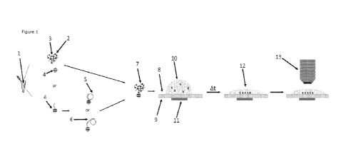
FIG. 1. Capture principle of an RCP, padlock probe or target DNA with padlock
probe onto
a carrier bead and side views of a cross section of an embodiment of the
sample analysis
module illustrating the main operations and principle of the sample analysis
module. RCPs
can either be captured after the RCA reaction, or the target DNA and/or the
padlock probes
can be captured on beads and amplified directly on them.
FIG. 2. Side views of cross sections of exemplary embodiments of the sample
analysis
module illustrating different design layouts. The use of open channels
structures as well
as the absorbent pad can increase the loading volume, thereby additionally
increasing the
detection sensitivity. A) Exemplary chip design 1 with 1 inlet and 1 outlet;
B) Exemplary
chip design 2 with 1 inlet and a chamber to hold the liquid; C) Exemplary chip
design 3
with well, the well can be sealed with a cover slip prior image acquisition;
and D)
Exemplary chip design 4 with 1 inlet and an absorbent pad on the opposing end
to allow
more liquid to be filled inside the chip
FIG. 3. Picture of fluorescently labeled RCPs immobilized (A) on a glass slide
and (B) within
the sample receiving well of an embodiment of the sample analysis module. A.
showing 1
pM RCPs under a coverslip on a glass slide imaged with a 20x microscope
objective. B.
showing the same 1 pM RCP solution from (A) after use of the here described
enrichment
method with a 20x microscope objective.
17
CA 03236105 2024- 4- 23
WO 2023/075663
PCT/SE2022/050974
FIG. 4. Graphs illustrating quantification of serial dilutions of RCPs using
an embodiment
of the sample analysis module. A. showing the increased detection sensitivity
compared
to detection on a glass slide. B. showing the linear regression for the serial
dilution. The
average of two individual measurements is shown.
FIG. 5. Picture of fluorescently labeled RCPs from human genonnic DNA
immobilized by an
embodiment of the sample analysis module. The RCPs are labelled with different
fluorescent barcodes to distinguish the probes for the control, the target and
the reference
gene. The number of RCPs should be equal for all three genes (ratio 1:1:1),
which is
confirmed by the RCA in conjunction with the sample analysis module to
visualize the low
concentration of RCPs in the solution. Inset images show the respective RCPs
for each of
the fluorescent barcodes.
FIG. 6. Graph and images of exemplary RCPs illustrating the fluorescence
intensity
enhancing features of bead-bound RCPs compared to "free"/unbound RCPs. The
enhancing
properties are here exemplified by three different fluorescence channels. A.
Graph of the
fluorescence intensity of RCPs on slide and on beads. The population of RCPs
stems from
the same reaction, highlighting the increased intensity of bead-bound RCPs
exemplary
shown in three fluorescence channels. B. Image series showing single exemplary
RCPs in
solution (on a glass slide) and bead-bound. Images were set to the same
thresholds to
show the increased intensity and size of bead-bound RCPs.
FIG. 7. Exemplary images showing that the fluorescence intensity of bead-bound
RCPs is
greater than the sum of beads and RCPs on their own. A. Exemplary images of
magnetic
beads, RCPs and bead-bound RCPs on a microscopy slide under a fluorescence
microscope.
The number in the left-hand corner corresponds to the highest fluorescence
intensity of
the image. B. Calculation of the exemplary images demonstrating the surprising
fact that
the sum of beads and RCPs is less than bead-bound RCPs.
FIG. 8. Graph and images showing the autofluorescence of nitrocellulose
membrane and
magnetic beads in comparison. A. Graph of the autofluorescence levels of a
nitrocellulose
membrane and MyOne Dynabeads Cl for different fluorescence channels. B.
Exemplary
image and inset of FITC-labelled RCPs on membrane and bead bound. RCPs are
clearly
distinguishable with the magnetic enrichment using beads while the
autofluorescence of
nitrocellulose masks the RCPs.
FIG. 9. Exemplary embodiment of a two-component sample analysis module with a
(discardable) quantification chip and a reusable chip holder. A. Top view of
an exemplary
18
CA 03236105 2024- 4- 23
WO 2023/075663
PCT/SE2022/050974
chip and chip holder design, and the assembly of both. B. Schematic side view
of the
exemplary assembled embodiment. C. Photograph of the assembled exemplary
embodiment.
FIG. 10. Two exemplary concepts of magnet placement and design to create a
homogeneous magnetic field in the center of the magnet or magnet arrangement.
A.
Exemplary schematic illustration of an inverted microscope for imaging through
a well
which would not work in the case of a magnet being place between the chamber
and the
objective as it would block the view. B. and C. illustrate the arrangement of
4 magnets
around the chamber D. and E. illustrate the arrangement of a single ring-
shaped magnet
to create a homogenous magnetic field in its center.
FIG. 11. Two exemplary multi-well designs to enable high throughput screening
using the
herein disclosed method. A. Illustration and example of a multi-well plate and
plate holder
housing ring-shaped magnetic sources as described in FIG. 3 D. and E. Fig.
11B.
Illustration and example of a multi-well plate and plate holder housing disc-
shaped
magnetic sources.
FIG. 12. Graph and images of exemplary RCPs illustrating the fluorescence
intensity
enhancing features of bead-bound RCPs compared to "free/unbound RCPs and the
independence on bead size (in a certain size range). A. Box plot of the
fluorescence
intensity of RCPs on slide and bound to beads. B. Image series showing single
exemplary
RCPs in solution (on a glass slide) and bead-bound (on the same glass slide).
FIG. 13. Images of 6 exemplary bead sizes illustrating the non-trivial optical
differences
between them. Scale bar represents 40 pm.
FIG. 14. Images of 5 exemplary bead sizes with RCPs bound to them and their
non-trivial
behavior and optical differences under magnetic force. Scale bar represents 20
pm.
In the figures, the following reference numbers have been used:
1. Solution containing Rolling Circle Amplification Product
2. Capture molecule (triangle shape in figure). Chemical or biological,
e.g., thiol or
biotin. Capture molecule can either be added by hybridization, reaction or
during
strand synthesis
3. Fluorescence dye (star shape in figure), e.g., sequence-specific or
intercalating
4. Magnetic particle with functional groups. Chemical or biological (e.g.,
Streptavidin-
functionalized or with capture oligonucleotide)
19
CA 03236105 2024- 4- 23
WO 2023/075663
PCT/SE2022/050974
5. DNA circle or ligated padlock probe
6. Target DNA
7. Rolling Circle Amplification Product bound/immobilized on magnetic
particle
7. Samples support element
8. First surface of the sample support element
9. Second surface of the sample support element
10. Solution with bead-bound Rolling Circle Amplification Products
11. Magnetic source, e.g., permanent or electrical
12. Rolling Circle Amplification products concentrated and magnetically
immobilized on
the surface
13. Microscope objective, e.g., 10x or 20x with a field of view matching
the
concentrated Rolling Circle Amplification Product area
14. Aperture
15. Thin glass, plastic layer or other transparent material to allow for
short working
distance imaging. Can be same material as the chip itself
16. Absorbent pad, e.g., paper or cotton
17. Transparent upper layer
18. Through hole
19. Enrichment chamber/channel
20. Enrichment chip
21. Magnet
22. Pin for holding chip in place
23. Chip holder
24. Well/chamber
25. Well/chamber plate holder housing the magnetic source
26. Well/chamber plate, e.g. 96-well plate
27. Well/chamber plate holder combined with Well/chamber plate
Throughout the drawings, the same reference numbers are used for similar or
corresponding elements.
Detailed Description of the Invention
Figure 1 details the capture principle of an RCP, padlock probe or target DNA
with padlock
probe onto a carrier bead (4) and side views of a cross section of an
embodiment of the
sample analysis element illustrating the main operations and principle of the
sample
analysis module. RCPs can either be captured after the RCA reaction, or the
target DNA
and/or the padlock probes can be captured on beads and amplified directly on
them.
CA 03236105 2024- 4- 23
WO 2023/075663
PCT/SE2022/050974
The sample solution (1) comprising the rolling circle amplification products
(7) is provided
in an Eppendorf tube and magnetic particle (4) with functional groups
(chemical or
biological, e.g., Streptavidin-functionalized) or with capture
oligonucleotides (2) are
provided and added to the sample solution. Following a period of time to allow
the rolling
circle amplification products to bind to the magnetic beads, bead-bound RCPs
were
achieved (7) the sample solution was transferred to the first surface (8) of a
glass slide
acting as a sample support element.
A magnet (11) was then provided at a second surface (9) of the glass slide
opposite to the
first surface to attract the beads towards a certain area on the glass slide
(12) that was
equal to, or smaller than, the field of view of an optical imaging device
(13), such as a
fluorescence microscope.
Figure 2 shows four further embodiments of a sample analysis device according
to the
second aspect of the invention. Embodiment A shows the device wherein the
first surface
(8) of the support element forms the bottom of a well in which the sample
solution is
placed. The sample analysis device has an upper layer (17), which is
transparent to allow
visualization, and where the device comprises two apertures (14) to allow the
sample
solution to be added and removed as needed.
Embodiment B equates to embodiment A, but wherein the device comprises only
one
aperture (14). Embodiment C equates to embodiment A, but rather than having
separate
apertures, a glass cover slip (15) is provided over the top of the opening of
the well to
seal the volume.
Embodiment D is similar to embodiment A, but on one side of the device the
aperture (14)
is filled with an absorbent material (16) that absorbs excess sample solution
through
capillary forces to allow for further sample solution to be added once the
bead-bound
polynucleotides are held in place by the magnetic source.
Figure 9 shows an example of a two-component sample analysis module with a
(discardable) quantification/enrichment chip (20) and a reusable chip holder
(23). The
quantification chip has through holes (18) that allow exact position and fit
onto the chip
holder pins (22). This allows imaging on up-right as well as inverted imaging
systems.
The 8 enrichment channels (19) are 9 mm apart from one another which allows
loading
with a standard multi-channel pipette. Figure 9A shows Top view of an
exemplary chip
(20) and chip holder (23) design, and the assembly of both. Figure 9B is a
schematic side
21
CA 03236105 2024- 4- 23
WO 2023/075663
PCT/SE2022/050974
view of the exemplary assembled embodiment. Figure 9C is a photograph of the
assembled exemplary embodiment. To reduce fluorescence signal reflection and
refraction
from the magnet (21), either the chip holder can be laminated with non-light
absorbent
paint or the enrichment chip itself has an opaque bottom layer; both cases
have been
explored with similar outcome.
Figure 10 shows two exemplary concepts of magnet (11) placement and design to
create
a homogeneous magnetic field in the center of the magnet or magnet
arrangement. These
magnet setups enable to create a homogeneous magnetic field for enrichment of
the bead-
bound RCPs while, at the same time, keeping the center of the chamber free to
allow, e.g.,
image acquisition from the bottom (inverted microscopy). Figure 10A shows an
exemplary
schematic illustration of an inverted microscope (13) for imaging through a
well (24) which
would not work in the case of a magnet being place between the chamber and the
objective
as it would block the view. Figures 10B and 10C illustrate the arrangement of
4 magnets
(11) around the chamber (side view and top view, respectively) to create a
homogeneous
magnetic field in the center of the magnet arrangement.
The arrangement and creation of such magnetic fields is well known and is
described
thoroughly in literature, such examples of which are: Tretiak, 0., Bliimler,
P., & Bougas,
L. (2019). Variable single-axis magnetic-field generator using permanent
magnets. AIP
Advances, 9(11), 115312. doi:10.1063/1.5130896; Manz, B., Benecke, M., &
Volke, F.
(2008); and a simple, small and low-cost permanent magnet design to produce
homogeneous magnetic fields. Journal of Magnetic Resonance, 192(1), 131-138.
doi:10.1016/j.jrnr.2008.02.011.
The importance of such an arrangement is to ensure an equal distance between
the
magnets among one another (d1) as well as an equal distance of them to the
chamber
(d2). Figures 10D and 10E illustrate the arrangement of a single ring-shaped
magnet to
create a homogenous magnetic field in its center. By positioning the chamber
with a
solution containing RCPs (12) in the center of the magnet (d3), the RCPs will
be enriched
in the center. The two configurations illustrated in Figures 10B and 10C, as
well as 10D
and 10E, would both allow for inverted microscopy through the bottom layer of
the
chamber.
Figure 11 shows two exemplary multi-well designs to enable high throughput
screening
using the herein disclosed method. While Figure 3 shows an exemplary chip
design in the
size of a standard microscope slide (2.5 cm by 7.5 cm), here the exemplary
plate is a
standard 96-well plate to be able to fit into various standardized image
acquisition units,
22
CA 03236105 2024- 4- 23
WO 2023/075663
PCT/SE2022/050974
such as microscopes and plate readers. Figure A is an illustration and example
of a multi-
well plate and plate holder housing ring-shaped magnetic sources as described
in FIG. 3
D. and E.. This concept allows for the processing and analysis of 96 samples
at a time.
Figure B is an illustration and example of a multi-well plate and plate holder
housing disc-
shaped magnetic sources.
Sequences
SEQ ID NO.: 1
Padlock probe 1
PO4-
GGGCAGCTGTCTAATTTTTGAGTCGGAAGTACTACTCTCTGTGTATGCAGCTCCTCAGTAATAGT
GTCTTACGTATCCTCGGAGAAGGTT
SEQ ID NO.: 2
Synthetic target 1
AGACCTGTTACATCTGGGTGCTTTCCTATAATGCACGACAGAACAAAAATTAGACAGCTGCCCAA
CCTTCTCCGAGGATAC
SEQ ID NO.: 3
Detection probe for padlock probe 1
Cy3-AGTCGGAAGTACTACTCTCT
SEQ ID NO.: 4
Capture oligo
Biotin-TTTTTCCTCAGTAATAGTGTCTTAC
SEQ ID NO.: 5
RPP30 padlock probe
PO4-
TTGTTGAGTGTTGGCGTGTATGCAGCTCCTCAGTAATAGTGTCTTACATTTAGCATACATCGTCG
CGTGCATAACCAGGCCA
SEQ ID NO.: 6
NRXN1 unedited padlock probe
PO4-
CGGCGGCCGCCTGCAGTGTATGCAGCTCCTCAGTAATAGTGTCTTACGGGCCTTATTCCGGTGC
TATGCTGATTCTGACGCG
23
CA 03236105 2024- 4- 23
WO 2023/075663
PCT/SE2022/050974
SEQ ID NO.: 7
NRXN1 reference padlock probe
PO4-
AATAAGGGTCCCGAGGTGTATGCAGCTCCTCAGTAATAGTGTCTTACAGAGAGTAGTACTTCCGA
CTACACCGTGACGAAGA
SEQ ID NO.: 8
Detection probe for RPP30
Cy3-ATTTAGCATACATCGTCGCG
SEQ ID NO.: 9
Detection probe for NRXN1 unedited
FITC-GGGCCTTATTCCGGTGCTAT
SE0 ID NO.: 10
Detection probe for NRXN1
Cy5-AGAGAGTAGTACTTCCGACT
SEQ ID NO.: 11
External primer
TACTGAGGAGCTGCATAC*A*C
SEQ ID NO.: 12
External primer
ACACTATTACTGAGG
SEQ ID NO.: 13
Detection probe 2 for NRXN1
FITC-AGAGAGTAGTACTTCCGACT
Note that "*" denotes a phosphonothioate base.
Examples
List of Abbreviations
RCA = Rolling Circle Amplification
24
CA 03236105 2024- 4- 23
WO 2023/075663
PCT/SE2022/050974
DNA = Deoxyribonucleic Acid
BSA = Bovine Serum Albumin
dNTPs = Deoxynucleotide triphosphates
RCP(s) = Rolling Circle Amplification Product(s)
EDTA = ethylenediaminetetraacetic acid
Tth = Thernnus Thernnophilus
NAD = Nicotinannide Adenine Dinucleotide
PBS = Phosphate buffered saline
Example 1
This example demonstrates the increased RCA product count per field of view
using the
invention when compared to a standard quantification on slide by spreading the
RCA
products under a cover slip. The example is shown in Figure 3 which shows that
without
being captured on magnetic beads and magnetically attracted to a predetermined
position
the number of RCA amplicons in a single field of view is much lower than those
captured
on magnetic beads.
RCP production
Circular templates to serve for the subsequent RCA were generated by
performing a
padlock probe ligation reaction tennplated by a synthetic single-stranded DNA
target
mimicking that of a conserved 40 nt region of the Hemagglutinin gene from
Influenza B.
The ligation of padlock probes was performed with a mix composed of 100 pM
padlock
probes
(PO4-
GGGCAGCTGTCTAATTTTTGAGTCGGAAGTACTACTCTCTGTGTATGCAGCTCCTCAGTAATAGT
GTCTTACGTATCCTCGGAGAAGGTT, SEQ ID NO: 1), 1 pM synthetic target
(AGACCTGTTACATCTGGGTGCTTTCCTATAATGCACGACAGAACAAAAATTAGACAGCTGCCCA
ACCTTCTCCGAGGATAC, SEQ ID NO: 2), Tth ligase buffer (20 mM Tris-HCI (pH 8.3),
25
mM KCI, 10 mM MgCl2, 0.5 mM NAD, and 0.01% (v/v) Triton X-100) and 5 U Tth
DNA
ligase (Blirt S.A.) in a final volume of 20 pL. The mixture was incubated at
55 C for 20
min.
Next, the resulting circles were amplified by target-primed RCA, for which a
mixture
comprising 0.2 pg/pL BSA (Fisher Scientific), 125 pM dNTPs (Fisher Scientific)
and 8 U
phi29 DNA polynnerase (Blirt S.A.) in a final volume of 30 pL. The RCA
reaction was
incubated at 37 C for 2 h and 65 C for 2 min.
CA 03236105 2024- 4- 23
WO 2023/075663
PCT/SE2022/050974
Labelling of RCPs
The resulting RCPs were labelled using fluorescently tagged oligonucleotides
and biotin
tagged oligonucleotides complementary to the repeats within the RCPs. For
this, the RCP
products were mixed with 30 pL of labelling buffer (10 nnM Tris-HCI (pH 8.0),
10 nnM
ethylenedianninetetraacetic acid (EDTA), 0.05% (v/v) Tween 20, 1 M NaCI
containing 5 nM
Cyanine 3 (Cy3)- (Cy3-AGTCGGAAGTACTACTCTCT, SEQ ID NO: 3) and biotin-tagged
oligonucleotide (biotin-TTTTTCCTCAGTAATAGTGTCTTAC, SEQ ID NO: 4). The
labelling
reaction was incubated at 75 C for 2 min and 55 C for 15 min.
Capturing RCPs on beads
The resulting labelled RCPs were captured on DynabeadsTM MyOneTM Streptavidin
Ti
(Thermo Fisher Scientific).
DynabeadsTM MyOneTM Streptavidin Ti beads are
superparamagnetic beads having a diameter of 1 pm, with a monolayer, not a
nnultilayer,
of recombinant streptavidin covalently coupled to the surface and further
blocked with
BSA. For this, the beads were prepared according to the manufacturer's
instructions and
subsequently added to the RCP solution at a concentration of 0.125 pg/pL. The
capture
reaction was incubated at 37 C for 20 min and the bead subsequently washed
once in
washing buffer and resuspended in the same.
Imaging of RCPs
To visualize the resulting bead-bound RCPs, 10 pL of the capture reaction were
put on
Superfrost glass slide (Thermo Fisher Scientific). A 1.5 mm circular magnet
(Supernnagnete) was attached to the slide to allow the local concentration of
the bead-
bound RCPs on a small surface area. To spread the solution, a 24 x 24 nnnn2
coverslip was
placed on top of the solution. The slide was incubated at room temperature for
5 min to
allow the beads to be enriched. After incubation, the slide was imaged with an
Olympus
IX72 inverted fluorescence microscope with a 20x magnification objective and a
field of
view of 0.65 x 0.65 pm2.
To visualize RCPs that were not captured on magnetic beads a similar procedure
was used;
however, no magnet was attached to the glass slide as the RCPs are not
affected by
magnetic force.
In conclusion, there is a striking difference in the number of RCPs observed
in Figure 3a
compared to Figure 3b. The number of RCPs is much higher in the same field of
view
26
CA 03236105 2024- 4- 23
WO 2023/075663
PCT/SE2022/050974
when using the magnetic enrichment method as RCPs are attracted to a small
surface
area. The result is an increased sensitivity and, therefore, also simplified
detection as
samples containing low concentrations of RCPs do not need to be scanned.
Thereby
overcoming one of the major limitations of RCA which is the detection of RCPs
at low
concentrations.
Example 2
Analytical capabilities of the invention.
This example demonstrates the increased analytical capabilities using the
invention when
compared to a regular readout on slide. This example is illustrated in Figure
4.
RCPs were prepared the same way as described in the Example 1. In short,
different
synthetic target concentrations were circularized via ligation and amplified
into RCPs for 2
h. Next, RCPs were labelled with a fluorescent and biotin probe.
For the enrichment method, the RCPs were incubated with magnetic beads as
described
in Example 1. The sample solution (10 pL of the 60 pL reaction volume) was
applied to a
cell counter slide (BioRad) which had a 1.5 mm magnet in diameter attached to
its bottom.
After 5 min, the cell counter slide was placed on the microscope stage and the
enriched
RCPs visualized using a 20x objective.
For the comparison method, the labelled RCPs were not captured on beads. The
sample
solution (10 pL of the 60 pL reaction volume) was applied to a cell counter
slide, but no
magnet attached. After 5 min the RCPs were settled down and could be
visualized using
the same 20x objective.
RCP quantification
The resulting images were analyzed using a custom-made pipeline in the
CellProfiler
software (version 4.1.3; httpsLiqprofHer,or_ci by the Broad Institute and
initially
published by Lamprecht et al. CellProfiler: free, versatile software for
automated biological
image analysis, Biotechniques (2007); 42(1):71-75). The pipeline consisted of
image
enhancement and object identification with manual thresholding.
In conclusion, the results confirm the increased sensitivity of the enrichment
method
disclosed herein when compared to a regular readout on a microscope glass
slide. With
27
CA 03236105 2024- 4- 23
WO 2023/075663
PCT/SE2022/050974
the enrichment method, RCPs can be detected at concentrations where the
regular readout
appears blank (Figure 4a). Additionally, the number of detected RCPs
correlates linearly
with the concentration of input target. Thereby, confirming a concentration
independent
RCP enrichment (Figure 4b). Another benefit is that the regular pipeline for
identifying
RCPs can be used which makes the adaption of this method almost barrier-free.
Example 3
Quantification of human genomic DNA
This example demonstrates the capabilities to quantify different genes in
genomic DNA
and is illustrated in Figure 5. In this example, three different padlock
probes were used
to detect three different gene segments. One padlock probe served as the assay
control
if all steps were performed correctly (marked as Control); one padlock probe
was used as
reference (marked as Reference) to quantify the editing level when related to
the gene of
interest for the gene of interest; and, one padlock probe served as to
identify the location
of the gene edit (marked as Target gene). This means, in a wildtype experiment
one
would expect equal number of RCPs for all three padlock probes, while for an
edited
genome, one would expect equal number of RCPs for the Control and Reference
but a
reduced count for the Target gene. In this sample, we used wild type human
genomic
DNA, therefore, the number of RCPs for each of the padlock probes is equal.
This description is very generic as some cell lines might have varying
chromosome or/and
gene copy numbers. Therefore, it is advised to standardize a genome editing
experiment
against a wild-type sample and avoid potential biases.
RCP production
Human genomic DNA (Merck) was used to generate circular templates for the RCA
reaction. Three different regions on the genomic DNA were targeted, one region
of RPP30
gene and two regions on the NRXN1 gene. First, 1 pg of human genomic DNA was
fragmented in fragmentation mix consisting of buffer (20 mM Tris-HCI (pH 8.3),
25 mM
KCI, 10 mM MgCl2, 0.5 mM NAD, and 0.01% (v/v) Triton() X-100) and 15 U AluI
(New
England Biolabs) in a total volume of 20 pL. The reaction was incubated at 37
C for 5
min.
For the ligation, 10 pL of ligation mix were added containing Tth ligase
buffer (20 mM Tris-
HCI (pH 8.3), 25 mM KCI, 10 mM MgCl2, 0.5 mM NAD, and 0.01% (v/v) Triton X-
100),
28
CA 03236105 2024- 4- 23
WO 2023/075663
PCT/SE2022/050974
1 nM of padlock probes
(PO4-
TTGTTGAGTGTTGGCGTGTATGCAGCTCCTCAGTAATAGTGTCTTACATTTAGCATACATCGTCG
CGTGCATAACCAGGCCA, SEQ ID NO: 5;
PO4-
CGGCGGCCGCCTGCAGTGTATGCAGCTCCTCAGTAATAGTGTCTTACGGGCCTTATTCCGGTGC
TATGCTGATTCTGACGCG, SEQ ID NO: 6; and,
PO4-
AATAAGGGTCCCGAGGTGTATGCAGCTCCTCAGTAATAGTGTCTTACAGAGAGTAGTACTTCCGA
CTACACCGTGACGAAGA, SEQ ID NO: 7) and 7.5 U of Tth DNA ligase. The ligation
reaction
was incubated at 98 C for 3 min and 55 C for 45 min.
Next, the resulting circles were amplified by RCA, for which a mixture
comprising 0.2 pg/pL
BSA, 125 pM dNTPs, 5 nM external primer (TACTGAGGAGCTGCATAC*A*C, SEQ ID NO:
11; the star denotes a phosphonothioate base to escape exonucleic activity of
the
polymerase), 10.5 U exoI (New England Biolabs) and 28 U phi29 DNA polynnerase
in a
final volume of 35 pL. The RCA reaction was incubated at 37 C for 3 h and 65
C for 2
min.
Labelling of RCPs
The resulting RCPs were labelled using fluorescently tagged oligonucleotides
and biotin
tagged oligonucleotides as described in Example 1. In short, the RCP products
were mixed
with 15 pL of labelling buffer (10 mM Tris-HCI (pH 8.0), 10 mM
ethylenediaminetetraacetic
acid (EDTA), 0.05% (v/v) Tween 20, 1 M NaCI) containing 5 nM Cyanine 3 (Cy3)-
(Cy3-
ATTTAGCATACATCGTCGCG, SEQ ID NO: 8), biotin-
(biotmn-
liii
SEQ ID NO: 4), AlexaFluor488 (FITC)- (FITC-
GGGCCTTATTCCGGTGCTAT, SEQ ID NO: 9) and Cyanine 5 (Cy5)-tagged oligonucleotide
(Cy5-AGAGAGTAGTACTTCCGACT, SEQ ID NO: 10). The labelling reaction was
incubated
at 75 C for 2 min and 55 C for 15 min.
Capturing RCPs on beads & Imaging of RCPs
Capturing of the labelled RCPs, imaging and subsequent image analysis was done
as
described in Example 1 and 2.
In conclusion, Figure 5 shows a composite of all three channels. The insets
show the RCPs
separately for each of the three channels. As apparent, the number of RCPs for
each
channel is equal, thereby confirming the concept as this human genomic DNA
should not
carry any edit.
29
CA 03236105 2024- 4- 23
WO 2023/075663
PCT/SE2022/050974
Example 4
This example demonstrates the increased fluorescence intensity of bead-bound
RCPs when
compared to unbound/in-solution RCPs. The finding of this example is
illustrated in Figure
6.
RCPs were generated and quantified as described in Example 3. For the analysis
of the
RCP fluorescence intensity, the CellProfiler pipeline was adapted to contain
another module
which measures the fluorescence intensity of each object.
The findings of this example illustrate that RCPs that are bound to a bead are
brighter
than unbound (Figure 6A). Furthermore, the increased fluorescence intensity
made RCPs
appear bigger when compared to RCPs in solution (Figure 6B). The comparison
was based
on the average of several hundred RCPs that were acquired in solution and
bound to beads.
The conclusion drawn from this example is that the invention of binding and
visualizing
RCPs on magnetic beads results in an unexpected increased fluorescence
intensity making
the quantification easier, e.g. shorter exposure time needed and less
sensitive optical
device required.
Example 5
This example confirms that bead-bound RCPs display a higher fluorescence
intensity than
the sum of blank magnetic beads (without a bound RCP) and un-bound RCPs. RCPs
were
generated and quantified as described in Example 3. For the exemplified
calculation, blank
magnetic beads (without a bound RCP), RCPs in solution and bead-bound RCPs
were
selected by hand and the maximum pixel intensity taken via Image] software.
The sum of
blank bead and in-solution RCP is smaller than the fluorescence intensity
observed on
bead-bound RCPs. This effect is exemplified with three fluorescence channels
(Cy3, FITC
and Cy5).
The fluorescence intensity results can be seen in Figure 7. When using Cy3 as
the
fluorescence label, the intensity of the signal from the bead alone was 494
and the RCP
alone was 1555, whereas the bead-bound-RCP intensity was 2461 equaling an
intensity
increase of 20.1% compared to the sum of the bead and RCP alone.
CA 03236105 2024- 4- 23
WO 2023/075663
PCT/SE2022/050974
When using FITC as the fluorescence label, the intensity of the signal from
the bead alone
was 472 and the RCP alone was 1732, whereas the bead-bound-RCP intensity was
3078
equaling an intensity increase of 39.7% compared to the sum of the bead and
RCP alone.
When using Cy5 as the fluorescence label, the intensity of the signal from the
bead alone
was 1985 and the RCP alone was 2661, whereas the bead-bound-RCP intensity was
5233
equaling an intensity increase of 12.6% compared to the sum of the bead and
RCP alone.
Example 6
This example demonstrates the improved RCP quantification capability of the
described
method when compared to trapping of RCPs on a membrane. The example is
illustrated in
Figure 8.
The membrane and beads were imaged using a 20x objective in different
fluorescence
channels using the same exposure time. The membrane was wetted with PBS, and
the
beads eluted in PBS. The Fluorescence intensity was measured using Image]
software and
taking the overall fluorescence intensity of the microscope image.
For the comparison of RCP intensities in Figure 8B, RCPs were generated as
described in
Example 3. Here, only the FITC-labelled RCPs are shown on the membrane and
magnet
as the difference is most apparent for shorter wavelengths. The top left image
shows
FITC-labelled RCPs on a nitrocellulose membrane and the zoom-in on the right
illustrates
that RCPs are not easily resolved due to the high autofluorescence. The image
on the
bottom left shows RCPs from the same solution but bound and enriched on beads.
Apparent from the zoom-in on the bottom right, FITC-labelled RCPs can easily
be resolved
and quantified illustrating the advantages of the magnetic enrichment approach
over the
membrane one.
The membrane chip was manufactured by Aline, Inc. The filter membrane was a
ProtranTM
NC Nitrocellulose membrane with a 0.1 pm pore size (GE Healthcare
lifesciences), the
absorption layer was a cellulose fiber sample pad sheet (Merck), the spacing
layer was in
the form of pressure sensitive adhesive (Aline) and the liquid-impermeable
layer was of
polyethylene terephthalate (Aline). The sample receiving wells had a diameter
of 1.5 mm.
The sample analysis device was manufactured to have dimensions of a standard
microscope slide 25x75 mm with ten sample receiving wells arrayed over the
sample
analysis device. The full features of this membrane chip are described in
PCT/EP2020/060771 (published as WO 2020/212531).
31
CA 03236105 2024- 4- 23
WO 2023/075663
PCT/SE2022/050974
For imaging and to concentrate the RCPs on the membrane, 10 pL of the sample
solution
were put on the sample receiving well. After the liquid passed through the
membrane, 10
pL of Slowfade Gold (Fisher Scientific) were added on the membrane and a cover
slip
(Menzel) placed on top of it.
The benefits using the present inventive method to concentrate RCPs are:
= The bead-bound RCPs can be washed leading to lower background from assay
or
bio-sample components.
= The sensitivity seems to be increased per field of view when compared to
spreading
the solution on a glass slide, as bead-bound RCPs can be magnetically
attracted to
a small area matching that of the field of view of the imaging device.
= The spectral-labelling possibilities for this method are increased as the
magnetic
beads show low autofluorescence when compared to membrane enrichment.
Example 7
This example confirms that bead-bound RCPs display a higher fluorescence
intensity than
the sum of blank magnetic beads (without a bound RCP) and un-bound RCPs.
Human genomic DNA (Roche) was used to generate circular templates for the RCA
reaction. Three different regions on the genonnic DNA were targeted, a region
on the
GAPDH gene, one region on the NRXN1 gene and on the PLA3G6 gene. First, 1 pg
of
human genonnic DNA was fragmented in fragmentation mix consisting of buffer
(20 mM
Tris-HCI (pH 8.3), 25 mM KCI, 10 mM MgCl2, 0.5 mM NAD, and 0.01% (v/v) Triton
X-
100) and 2.5 U AluI (New England Biolabs) in a total volume of 20 pL. The
reaction was
incubated at 37 C for 15 min.
For the ligation, 10 pL of ligation mix were added containing Tth ligase
buffer (20 mM Tris-
HCI (pH 8.3), 25 mM KCI, 10 mM MgCl2, 0.5 mM NAD, and 0.01% (v/v) Triton X-
100),
100 ng salmon sperm DNA (Thermo Fisher Scientific), 5 nM external primer
(ACACTATTA
CTGAGG, SEQ ID NO: 12), 1 nM of padlock probe (PO4-
AATAAGGGTCCCGAGGTGTATGCAGCTCCTCAGTAATAGTGTCTTACAGAGAGTAGTACTTCCGA
CTACACCGTGACGAAGA, SEQ ID NO: 7) and 1.26 U of Tth DNA ligase. The ligation
reaction
was incubated at 98 C for 10 min and 55 C for 20 min.
32
CA 03236105 2024- 4- 23
WO 2023/075663
PCT/SE2022/050974
Next, the resulting circles were amplified by RCA, for which a mixture
comprising 125 pM
dNTPs, 4 U exoI (New England Biolabs) and 5.32 U phi29 DNA polynnerase in a
final volume
of 35 pL. The RCA reaction was incubated at 37 C for 2 h and 65 C for 2 min.
Labelling and capture of RCPs
The resulting RCPs were labelled using fluorescently tagged oligonucleotides
and biotin
tagged oligonucleotides complementary to the repeats within the RCPs. For
this, the RCP
products were mixed with 10 pL of labelling buffer (10 mM Tris-HCI (pH 8.0),
10 mM
ethylenediaminetetraacetic acid (EDTA), 0.05% (v/v) Tween 20, 120 mM NaCI, 10
mM
MgCl2, containing 5 nM AlexaFluor488 (FITC)- (FITC-AGAGAGTAGTACTTCCGACT, SEQ
ID
NO: 13), biotin-tagged oligonucleotide (biotin-TTTTTCCTCAGTAATAGTGTCTTAC, SEQ
ID
NO: 4) and 0.025 pg/pL SuperMag Streptavidin magnetic beads, 50nm (Ocean
NanoTech).
The labelling reaction was incubated at 75 C for 2 min and 55 C for 10 min.
The
aforementioned SuperMag Streptavidin magnetic beads are superparamagnetic
beads
having a diameter of 50 nnn, with a nnonolayer, not a nnultilayer, of
recombinant
streptavidin covalently coupled to the surface and further blocked with BSA.
Capturing RCPs on beads & Imaging of RCPs
Capturing of the labelled RCPs, imaging was done as described in Example 1.
The resulting
images were analyzed using a custom-made pipeline in the Image] software. The
pipeline
consisted of image enhancement, object identification with manual
thresholding, and
fluorescence intensity measurement of each object.
This example confirms the surprising increase in fluorescence intensity of
bead-bound
RCPs in which the intensity exceeds the sum of blank magnetic beads and un-
bound RCPs.
For the exemplified calculation, blank magnetic beads, RCPs in solution and
bead-bound
RCPs were selected and the maximum pixel intensity taken via Image] software.
The sum
of blank bead and in-solution RCP is smaller than the fluorescence intensity
observed on
bead-bound RCPs.
The fluorescence intensity results can be seen in Figure 12. Figure 12A shows
the
increased fluorescence intensity for multiple RCPs, while Figure 125
illustrates this
synergistic effect on an exemplary case. When using FITC as the fluorescence
label, the
exemplary intensity of the signal from the bead alone was 1786 and the RCP
alone was
2549, whereas the bead-bound-RCP intensity was 5236 equaling an intensity
increase of
20.8% compared to the sum of the bead and RCP alone.
33
CA 03236105 2024- 4- 23
WO 2023/075663
PCT/SE2022/050974
Example 8
This example confirms that 50 nm magnetic beads show the best performance in
terms of
low autofluorescence background, enrichment capability and binding efficiency.
For the comparison of fluorescence intensity of blank beads, 0.025 pg/pL of
TurboBeads
Streptavidin (Turbobeads GmbH), 0.025 pg/pL of aforementioned SuperMag
Streptavidin
magnetic beads of 50 - 200 nm (Ocean NanoTech), 0.125 pg/pL of the
aforementioned
DynabeadsTM MyOneTM Streptavidin Ti (Thermo Fisher Scientific), as well as
0.125 pg/pL
Dynabeads¨ M-270 Carboxylic Acid (Thermo Fisher Scientific), were prepared in
Milli-Q
water (Sigma-Aldrich). 10 pL of the sample were prepared for- and imaged on a
microscope slide as described in Example 1, and quantified as described in
Example 7. The
exemplary images of bare nanoparticles are shown in Figure 13, while the
nanoparticles
with RCPs under a magnetic field (enriched) are shown in Figure 14.
When using the FITC channel, the 30 nm beads up to the 200 nm beads were not
visible,
while the 1 pm beads showed some level of autofluorescence and the 2.8 pm
beads a high
level of autofluorescence intensity. The intensity of the signal from the 30 -
200 nm in
diameter beads were undistinguishable from the background, at around 1500 AU.
For the
Dynabeads¨ MyOne¨ Streptavidin Ti, 1 pm in diameter, the fluorescence
intensity was
3108 AU, whereas for the Dynabeads¨ M-270 Carboxylic Acid, 2.8 pm in size, the
fluorescence intensity was even higher at 7451 AU. These results are
illustrated in Figure
13 with exemplary images.
To further evaluate the beads in terms of optical behavior and RCP capture and
enrichment
efficiency, in Figure 14 the beads were decorated with RCPs and enriched under
a magnetic
field, here the 30 nm Turbobeads showed high levels of autofluorescence and
aggregation
with intensity levels of around 6748 AU, which does not allow a quantification
of RCPs.
For 50 nm beads the number of RCPs in a single field of view is highest
compared to 100
nm, 200 nm and 1 pm beads. Additionally, 1 pm beads start forming mosaic like
structure
when under magnetic force which increase the overall background intensity
(noise) and
makes image analysis of events more challenging. This shows that the ideal
bead size for
the shown beads is between 30 nm and 100 nm which is a counter intuitive
result as often
large particles (several pm to mm) are used to capture long polynucleotide
sequences,
e.g., in genomic extraction kits.
34
CA 03236105 2024- 4- 23