Note: Descriptions are shown in the official language in which they were submitted.
WO 2023/076275
PCT/US2022/047734
CELL CULTURE FEEDING DEVICE
FIELD OF INVENTION
This disclosure pertains to devices for feeding and culturing mammalian cells.
Disclosed
herein is a non-degradable device for use in controlled feeding mammalian cell
cultures
including by way of example cultures of stem cells such as induced pluripotent
stem cells
(iPSCs).
BACKGROUND
Growth factors (GFs) such as for example FGF2, FGF4, FGF8, FGF1 8, WNT1,
WNT3A,
EGF, BDNF, GDNF, NT3, TGFbl, TGFb3, BMP2, BMP4, PDGFBB, PDGFAA, IGF1,
VEGFA, VEGFC, SHH, cAMP, LIF, NRG1, SCF, ACTIVIN A, ILlb, IL2, IL6, IL7, IL12,
IL1 5, IL21, IL34, IFNa, IFNy, TAU, ABETA, A-SYNUCLEIN or modified versions
and other
cell culture media additives such as fetal bovine serum (FBS) are needed to
maintain cells in cell
cultures, to promote cell proliferation to expand cultures, or to guide the
development or
differentiation of cells into desired cell or tissue products
The term "growth factor" refers to a naturally occurring, endogenous or
exogenous
protein, or recombinant protein, capable of stimulating cell growth, survival
and inhibiting
and/or stimulating differentiation of cells, such as e.g., stem or progenitor
cells. The term
"growth factor" also can encompass lipid, chemical, and other non-protein
agents, e.g., small
molecules that are capable of stimulating cell growth, survival and inhibiting
and/or stimulating
cell differentiation or mixtures of these as found in FB S. In certain
embodiments, the term
"growth factor" refers to any polypeptide or other agent that is capable of
stimulating cell
growth, survival and inhibiting or stimulating cell differentiation, e.g.,
when present in effective
amounts in a stem or progenitor cell culture. Growth factor polypeptides
referred to herein
include both naturally occurring and recombinant proteins, which may be either
endogenous or
exogenous to the cells being cultured. In addition, a growth factor may be a
synthetic protein,
such as a fusion or other protein construct or a chemical modification of the
amino acid
sequences derived from a naturally occur- ring growth factor or other protein.
Such growth
1
CA 03236223 2024- 4- 24
WO 2023/076275
PCT/US2022/047734
factors may be used in combination, to produce, e.g., an additive or
synergistic effect, according
to the present methods.
It is well known in the art that GFs generally have short half-lives The
labile nature of
GFs means that the cells in culture require frequent, often daily, addition of
GFs to the culture
media to sustain the level of GFs needed to successfully maintain cells or to
sustain cell growth
and development or cell differentiation over time. Frequent feeding schedules
subject cells to
fluctuating levels of GF signaling due to GF half-lives on the order of hours
to minutes. Because
different growth factors have different rates of decay, the ratio of different
GFs in the culture
medium varies. The resulting fluctuations in GF levels and GF ratios impede
effective cell
culture while frequent manual replenishment of GFs results in high medium
usage and increased
labor. It is desirable that cell culture research or clinical use occur under
controlled GF
conditions, and this is not achieved with labile GFs. These practical
challenges to creating
quality cell cultures can be overcome by a device that provides steady,
controlled GF levels.
Systems that degrade in an aqueous environment ("degradable systems") have
been
employed to provide controlled-release GF to overcome many of the limitations
that soluble
GFs pose to effective cell culture. One example is PLGA encapsulated
fibroblast growth factor-
2 (FGF2) microbeads. PLGA, PLG, or poly(lactic-co-glycolic acid) is a
copolymer which is
used in a host of Food and Drug Administration (FDA) approved therapeutic
devices, owing to
its biodegradability and biocompatibility. Biodegradable "microspheres" and
"millicylinders"
prepared from biocompatible polyesters of glycolic and lactic acids
("PLGA").are known for
delivering protein drugs to patients, and PLGA millicylinders encapsulated
with recombinant
human FGF2 (also known as ''basic fibroblast growth factor or "bfgf' have been
described by
Zhu et al. (Nature Biotechnology (2000) 18:52-57) for such applications. Olaye
et al.
(European Cells and Materials (2008) 16 (Suppl. 3):86) disclose that "PLGA
microspheres
have been extensively used for the sustained delivery of growth factors for
embryonic stem cell
differentiation," The value of such degradable controlled release GF
formulations, however, is
limited by inability to readily remove these formulations (microbeads) from
the cell cultures.
For example, degradable beads stick to cells in the culture vessel and are
difficult to fully wash
away. Other degradable feeding formats such as films become friable as they
resorb over time
making clean removal from the culture difficult, leaving breakdown products.
Residual
2
CA 03236223 2024- 4- 24
WO 2023/076275
PCT/US2022/047734
degradable GF formulations are problematic because they impair the ability to
control the
amount of GF in the medium. Furthermore, residual degradable GF formulations
impede the
desired differentiation of cells that require a clean exchange of one GF
environment to another.
The ability to completely remove one or more GFs from the medium to leave only
a negligible
(not enough to provide detectable bioactivity) trace of the GF can be of great
importance in
some instances.
Another disadvantage of current biodegradable cell culture additives such as
beads or
films is their interference with imaging of the cells.
Another aspect to maintaining quality cell cultures is removing unwanted
factors from the
cell culture medium. Currently, this is achieved by frequent medium exchanges,
once again
resulting in a high and expensive level of medium usage as well as increased
labor. Using the
devices disclosed herein, unwanted growth factors that are desirably removed
from the cell
culture medium can be sequestered and removed from culture medium, obviating
frequent
medium changes and costly feeding.
SUMMARY
The present disclosure describes a new platform technology that addresses the
above
limitations by providing a cell culture feeding device that is not degradable
in aqueous
environments and which provides a controlled level of GF release.
The feeding devices disclosed herein can be readily removed from, and
installed in, cell
culture media without requiring the medium to be exchanged or refreshed
In some embodiments the cell culture feeding device comprises a hydrogel
polymer
support, a plurality of microbeads within the support, the microbeads carrying
cellular growth
factors (GFs).
In some embodiments the hydrogel polymer support is non-degradable.
In some embodiments the support is biologically acceptable material.
3
CA 03236223 2024- 4- 24
WO 2023/076275
PCT/US2022/047734
In some embodiments wherein the microbeads carry at least one GF member
selected
from the group consisting of FGF2, FGF4, FGF8, FGF18, WNT1, WNT3A, EGF, BDNF,
GDNF, NT3, TGFbl, TGFb3, BMP2, BMP4, PDGFBB, PDGFAA, IGF1, VEGFA, VEGFC,
SHUT, cAMP, LIF, NRG1, SCF, ACTIVIN A, IL lb, IL2, IL6, IL7, IL12, IL15, IL21,
IFNct, IFNy,
TAU, ABETA, A-SYNUCLEIN or modified versions.
In some embodiments the support includes particulate elements carrying GFs.
In some embodiments the support is transparent.
In some embodiments the microbeads are degradable.
In some embodiments the microbeads carry small molecule compounds.
In some embodiments the microbeads comprise living cells.
In some embodiments a tether is attached to the support.
In some embodiments the support comprises magnetic particles.
In some embodiments the support includes a color.
In some embodiments the support contains gas bubbles to enable the support to
float at or
near the surface of cell culture media.
In some embodiments the support comprises a color, magnetic particles and one
or more
GFs.
Some embodiments comprise a biologic cell culture medium containing the
support and
microbeads carrying a GF.
Some embodiments provide a method of feeding a cell culture which comprises
depositing
an inert hydrogel polymer support into a cell culture media, the support
carrying microbeads
bearing one or more cellular GFs, the GFs being continuously released into the
cell culture
medium at a controlled rate over a period of time.
4
CA 03236223 2024- 4- 24
WO 2023/076275
PCT/US2022/047734
In some embodiments the GFs used in the method of feeding are selected from
the group
consisting of FGF2, FGF4, FGF8, FGF18, WNT1, WNT3A, EGF, BDNF, GDNF, NT3,
TGFbl,
TGFb3, BMP2, BMP4, PDGFBB, PDGFAA, IGF1, VEGFA, VEGFC, SHH, cAMP, LIF, NRG1,
SCF, ACTIVIN A, ILlb, IL2, IL6, IL7, IL12, ILLS, IL21, IFNct, IFN7, TAU,
ABETA, and A-
SYNUCLEIN or modified versions thereof.
In some embodiments the support is removed from the cell culture with a tether
attached to
the support.
In some embodiments the culture media contains a plurality of supports, each
support is
colored and bears a different GF and all of the supports have a different
color.
In some embodiments the method of feeding includes depositing a plurality of
colored
supports into the cell culture medium
In some embodiments the support comprises an open lattice structure.
In some embodiments the lattice structure comprises open pores.
Some embodiments include a method of making a feeding device for cell cultures
by
preparing a solution containing a biologically acceptable polymer and a
quantity of microspheres
bearing at least one GF, dispensing droplets of the solution onto a surface,
and exposing the
droplets to actinic radiation to form a hydrogel support
In some embodiments, the GFs are released in a cell culture over a period of
time.
In one implementation the support is preferably in the shape of a disc,
square, triangle or
rectangle or comprises a free form arrangement.
In one embodiment the support is a hydrogel formed from a biologically
acceptable
polymer material and does not degrade in aqueous environments
CA 03236223 2024- 4- 24
WO 2023/076275
PCT/US2022/047734
In another embodiment, microbeads or millicylinders loaded with one or more
GFs are
encapsulated within the support. The amount of GF released by the microbeads
is adjusted by
controlling the quantity of microbeads embedded in the support.
In some embodiments the supports can float on or just below the surface of
culture
media.
In other embodiments the supports are configured for removal from culture
media.
In some embodiments the supports do not degrade in cell culture media or in
the
presence of biologic, hydrolytic or enzymatic conditions.
In a further embodiment the hydrogel support comprises a polyethylene glycol
polymer.
In one embodiment, the hydrogel support is loaded with beads that carry growth
factors.
In one implementation the microbeads are StemBeads .
In a further implementation the StemBeads are loaded with FGF.
In another embodiment, the microbeads beads contain a variety of GFs.
In a still further alternative, the beads release GFs over a period of time.
In another embodiment, the beads include magnetic particles or beads.
In a still further embodiment, a recovery device such a wire, string, thread
or fishing
line is attached to the hydrogel support.
In one implementation one or more feeding devices are deposited into the same
cell
culture.
In another implementation hydrogel supports with different GF payloads are
deposited
into a cell culture and then selectively removed.
These and other embodiments and implementations are described in more detail
below.
6
CA 03236223 2024- 4- 24
WO 2023/076275
PCT/US2022/047734
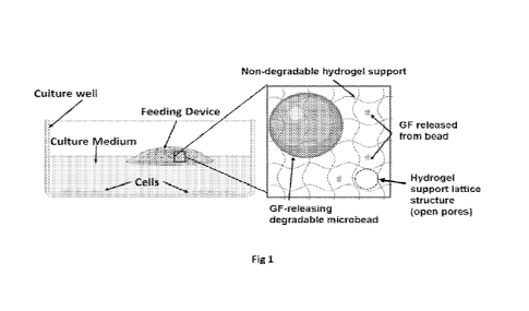
DESCRIPTION OF DRAWINGS
Fig 1 is a schematic that depicts a feeding device comprised of degradable
microbeads
releasing growth factors loaded into a non-degradable hydrogel support with
open lattice
structure and deployed into a cell culture well containing medium.
Fig 2A is a flow diagram describing manufacture of feeding devices from
StemBeads
FGF2 loaded into a 16 [IL PEG hydrogel support via photochemistry.
Fig 2B is a graph that demonstrates the amount of FGF2 released into cell
culture
medium over 7 days from a PEG hydrogel support loaded with StemBeads FGF2 set
to release
at 10 ng/mL when added into 2 mL of medium at 37 C (n = 3, error bars = st
dev).
Fig 2C is a graph that demonstrates FGF2 levels from an 8 IIL sized (1-2 mm
diameter
disc) PEG hydrogel support loaded with about 10,000 StemBeads FGF2 when added
into 1 mL.
of medium can achieve the same FGF2 level as a 16 1..iL sized (2-3 mm diameter
disc) PEG
hydrogel support loaded with about 20,000 StemBeads FGF2 added into 2 mL of
medium. (n =
4-6, unpaired t-test, ns = not significant).
Fig 3A is a graph that demonstrates the amount of FGF2 released into the
medium
between 1 and 24 hours at 37 C comparing the levels from StemBeads FGF2
delivered into
culture without a support and StemBeads FGF2 in a hydrogel support normalized
to the level
at 24 hours (set to 1) (n = 3 hydrogel supports, error bars = st dev).
Fig 3B is a graph that demonstrates the amount of FGF2 released into the
medium
between 1 and 14 days at 37 C comparing the levels from StemBeads FGF2
delivered into
culture without a support and StemBeads FGF2 in a hydrogel support (error
bars = st dev; n =
3, unpaired t-test p<0.05, " p < 0.005).
Fig 4A graphs the average FGF2 levels over 7 days at 37 C from 16 [IL sized
PEG
hydrogel support loaded with about 20,000 StemBeads FGF2 added into 1, 2 or 3
mL of cell
culture medium. (n = 3, en-or bars = st dev).
7
CA 03236223 2024- 4- 24
WO 2023/076275
PCT/US2022/047734
Fig 4B is a graph that depicts the average FGF2 levels in a culture medium
over a period
of 7 days at 37 C from 16 p.L sized PEG hydrogel support loaded with about
20,000 or 100,000
StemBeads FGF2 (n = 3, error bars = st dev).
Fig 5A is a graph that demonstrates EGF and FGF2 levels released in a culture
medium at
37 C from a 16 pL sized PEG hydrogel support loaded with StemBeads EGFCD and
StemBeads
FGF2 (n = 3, en-or bars = st dev)
Fig 5B is a graph that depicts the FGF2 levels in a culture medium over 6 days
at 37 C
as released from a 16 pL sized PEG hydrogel support loaded with magnetic beads
and
StemBeads FGF2 (n = 2, error bars = st dev).
Fig 5C is a graph that depicts the FGF2 levels in culture medium over 6 days
at 37 C as
released from a 16 41_, sized (PEG hydrogel support manufactured with
microbubbles (floating
hydrogel support) and loaded with StemBeads FGF2 (n = 2, error bars = st
dev).
Figs. 6A-F are graphs and histograms that compare a conventional method of
culturing
iPSCs with daily feeds of mTESR1 medium (containing soluble FGF2) delivered
into culture
without a support to the improved culture method with less frequent feeds of
mTESR1 medium
delivered to the culture with an FGF2 feeding device.
Fig 6A is a graphical schematic that illustrates the FGF2 levels for cultures
grown with
daily feeds of mTESR1 medium delivered into culture without a support (method
#1).
Fig 6B is a pie graph that illustrates the time cultures spend at different
levels of FGF2
when cells are grown with daily feeds of mTESR1 medium delivered into culture
without a
support (method #1).
Fig 6C is a graphical schematic that illustrates the FGF2 levels for cultures
grown with
less frequent feeds of mTESR1 medium delivered with an FGF2 hydrogel feeding
device
(method #2)
8
CA 03236223 2024- 4- 24
WO 2023/076275
PCT/US2022/047734
Fig 6D is a pie graph that illustrates the time cultures spend at different
levels of FGF2
when cultures are grown with less frequent feeds of mTESR1 medium delivered
with an FGF2
hydrogel feeding device (method #2).
Fig 6E are histogram plots of flow cytometry data where the percent of cells
that are
positive for the pluripotency marker Tra-1-60 are labeled. These plots compare
three iPSC lines
cultured with daily feeds of mTESR1 medium (method #1, top graphs) compared to
less frequent
feeds of mTESR1 medium delivered with an FGF2 hydrogel feeding device (method
#2, bottom
graphs).
Fig 6F are histogram plots of flow cytometry data where the percent of cells
that are
positive for pluripotency marker SSEA4 are labeled. These plots compare two
iPSC lines
cultured with daily feeds of mTESR1 medium (method #1, top graphs) compared to
less frequent
feeds of mTESR1 medium delivered with an FGF2 feeding device (method #2,
bottom graphs).
Figs. 7A-C are graphs that compare 5 different methods to grow iPSCs and
demonstrates
improved mesoderm differentiation is achieved when iPSCs were cultured with an
FGF2 feeding
device.
Fig 7A graphs the mesoderm marker brachyury gene expression of mesoderm
cultures
derived from iPSCs cultured with daily feeds of mTESR1 medium (containing
soluble FGF2)
(method #1) in comparison expression after less frequent feeds of mTESR1
medium delivered
with an FGF2 hydrogel feeding device (method #2), (n = 3 cell lines; n=2-3
wells per line; ** p
<0.005).
Fig 7B graphs the mesoderm marker brachyury gene expression of mesoderm
cultures
derived from iPSC cultured with 3-times a week feeds of mTESR1-Plus medium
(containing
stabilized soluble FGF2) delivered into culture without a feeding device
(method #3) in
comparison to less frequent feeds of mTESR1-Plus delivered with an FGF2
feeding device
(method #4), (n = 3 cell lines; n=2-3 wells per line; **** p <0.00005).
9
CA 03236223 2024- 4- 24
WO 2023/076275
PCT/US2022/047734
Fig 7C graphs the mesoderm marker brachyury gene expression of mesoderm
cultures
derived from iPSC cultured with feeds of mTESR1 medium delivered with
StemBeads FGF2
(no hydrogel support, method 45) and compared to feeds of mTESR1 medium
delivered with an
FGF2 hydrogel feeding device (method #2), (n = 3 cell lines, n=2-3 wells per
line, * p < 0.05).
Figs. 8A-G are graphs that compare a conventional method of culturing iPSCs
with daily
feeds of E8 medium (containing soluble FGF2) to the improved culture method
with less
frequent feeds of E8 medium (made up without soluble FGF2) delivered to the
culture with an
FGF2 feeding device.
Fig 8A is a graphical schematic that illustrates the FGF2 levels for cultures
grown with
daily feeds of E8 medium with soluble FGF2.
Fig 8B is a pie graph that illustrates the time cultures spend at different
levels of FGF2
when cells are grown with daily feeds of E8 medium with soluble FGF2.
Fig 8C is a graphical schematic illustrates the FGF2 levels for cultures grown
with an
FGF2 feeding device added into E8 medium without soluble FGF2.
Fig 8D is a pie graph that illustrates the time cultures spend at different
levels of FGF2
when cultures grown with an FGF2 feeding device added into E8 medium without
soluble FGF2.
Fig 8E graphs the endoderm marker SOX17 gene expression of endoderm cultures
derived from iPSC cultured with daily feeds of E8 medium (with soluble FGF2)
compared to
less frequent feeds of E8 medium (without soluble FGF2) delivered with an FGF2
hydrogel
feeding device, (n = 1 cell line; n=3 wells; unpaired t-test **** p <
0.00005).
Fig 8F graphs the mesoderm marker brachyury (T) gene expression of mesoderm
cultures
derived from iPSC cultured with daily feeds of E8 medium (with soluble FGF2)
delivered into
culture without a hydrogel support in comparison to less frequent feeds of E8
medium (without
CA 03236223 2024- 4- 24
WO 2023/076275
PCT/US2022/047734
soluble FGF2) delivered with an FGF2 hydrogel feeding device, as disclosed
herein (n = 1 cell
line; n=3 wells; unpaired t-test *** p <0.0005).
Fig 8G graphs the ectoderm marker PAX6 gene expression of ectoderm cultures
derived
from iPSC cultured with daily feeds of E8 medium (with soluble FGF2) compared
to less
frequent feeds of E8 medium (without soluble FGF2) delivered with an FGF2
feeding device, (n
= 1 cell line; n=3 wells; unpaired t-test *** p <0.0005).
Figs 9 is a graph that demonstrates cerebral organoids have improved levels of
cortex
neuronal subtypes when organoids are generated from iPSC cultured with less
frequent feeds of
mTESR1 medium delivered to the culture with an FGF2 feeding device compared to
conventional method of daily feeds of mTESR1 medium (containing soluble FGF2)
as shown by
higher gene expression levels of positive cerebral cortex markers (PAX6,
FOXG1, TBR1,
EMX2) from 2-month cerebral organoids (n = 2 cell lines; n=3 organoids pooled
per line or n = 3
individual organoids per line; * p < 0.05 ***p<0.0005).
DETAILED DESCRIPTION
Unless otherwise defined, all technical and scientific terms used herein have
the same
meaning as commonly understood by one of ordinary skill in the art to which
this disclosure
belongs. Methods and materials are described herein for use in the present
disclosure; other,
suitable methods and materials known in the art can also be used. The
materials, methods, and
examples are illustrative only and not intended to be limiting. All
publications, patent
applications, patents, sequences, database entries, and other references
mentioned herein are
incorporated by reference in their entirety. In case of conflict, the present
specification, including
definitions, will control.
As used herein the term "non-degradable" refers to biologically acceptable
materials that
do not break down or deteriorate chemically. More specifically the term refers
to biologically
acceptable plastics and other materials that do not deteriorate or break down
in cell culture
medium including by way of non-limiting example, the cell culture mediums
disclosed herein.
11
CA 03236223 2024- 4- 24
WO 2023/076275
PCT/US2022/047734
The definition also embraces materials that do not deteriorate or break down
when exposed to
biological, hydrolytic or enzymatic conditions.
As used herein the term "small molecules" refers to those compounds with a
molecular
weight below 1000 Daltons.
As used herein, the term "about" or "approximately" means within a
statistically
meaningful range of a value. Such a range can be within an order of magnitude,
preferably
within 50%, more preferably within 20%, still more preferably within 10%, and
even more
preferably within 5% of a given value or range. The allowable variation
encompassed by the
term "about" or "approximately" depends on the particular system under study,
and can be
readily appreciated by one of ordinary skill in the art.
As used herein the term "biologically acceptable" means the material that is
gene i ally safe, non-toxic, and neither biologically nor otherwise,
undesirable and includes that
which is acceptable for veterinary use as well as human pharmaceutical use.
The feeding devices disclosed herein generally comprise a hydrogel support
that is a non-
degradable, biologically acceptable, inert material that can hold a cargo or
payload, such as for
example degradable microbeads. The hydrogel support material has an open
lattice structure that
allows GFs to diffuse through and be released into the cell culture medium but
is small enough to
retain the microbead cargo (See Fig 1).
The inert non-degradable hydrogel support material prevents the feeding device
from
interfering with growth of cells in culture. This support can be easily added
and removed from
cultures. The hydrogel support can comprise a variety of different polymers
including by way of
non-limiting example synthetic polymers (e.g. polyethylene glycols,
polyacrylamides) and
naturally occurring polymers (e.g. polysaccharides, polypeptides).
The hydrogel support may contain a cargo, such as multiple types of GF
releasing
microbeads, colored beads, magnetic beads, air bubbles and/or a tether (which
can be for
example a wire, filament, thread or string), to assist in its functionality as
a removable feeding
device for cell culture. Using one embodiment of the feeding devices disclosed
herein it has been
12
CA 03236223 2024- 4- 24
WO 2023/076275
PCT/US2022/047734
shown that cell culture quality is significantly and surprisingly improved as
compared to
conventional feeding methods (soluble GF added daily without a hydrogel
support) and feeding
methods using microbeads without a hydrogel support.
Unlike other degradable materials used to deliver factors to cell cultures,
the hydrogel
supports described herein do not degrade in cell culture media or in the
presence of biologic,
hydrolytic or enzymatic conditions. The devices disclosed herein are also
'inert' defined as
having anti-fouling properties by discouraging non-specific protein adsorption
via highly
hydrophilic hydrogel polymer backbone. 'Inert' is also defined as a material
that does not
contain cell binding motifs and does not promote cell attachment. These non-
degradable and
inert properties of the hydrogel support are beneficial to the feeding device
as they prevent the
device from interfering with the cells in culture. Previous technology, such
as degradable GF
microbeads comprised of PLGA or naturally occurring polymers constructs
(collagen, gelatin,
laminin, fibrin, matrigel, etc.) are not inert to cells and have been shown to
incorporate into cell
monolayers and 3D organoids. Additionally, these materials are degradable and
thus can release
byproducts that can alter the cell culture environment. The removable feeding
devices described
herein circumvent these concerns. For example, before the feeding devices
disclosed herein,
degradable microbeads (e.g. StemBeads , StemCultures LLC) added into a 2D cell
culture
would stick to cells, multiple washes were required to assist in the removal
of microbeads and a
full removal was not readily achieved. StemBeads are controlled release micro
particles
composed of a biodegradable polymer that is loaded with one or more GFs such
as recombinant
FGF2 (StemBeads and are available from StemCultures, 1 Discovery Drive,
Rensselaer NY)
(See for example US patent 8,481,308 incorporated herein in its entirety by
reference).
With the devices disclosed herein, the degradable microbeads (e.g. StemBeads ,
StemCultures LLC) are loaded into an inert non-degradable hydrogel support,
the microbeads are
retained within the feeding device, do not intermingle with the cultured cells
and full removal of
the device and its bead cargo is easily achieved without any washing steps.
The hydrogel supports described herein are preferably transparent and do not
interfere
with imaging of the cell cultures. The cargo carried by the support, such as
beads, or particles
may not be transparent. The hydrogel devices can be added to and later removed
easily from cell
13
CA 03236223 2024- 4- 24
WO 2023/076275
PCT/US2022/047734
cultures, achieving a controlled environment and essentially complete and
efficient removal of
GFs from the culture, with negligible (not enough to provide detectable
bioactivity) GF
remaining after removal of the device bearing the GF from the culture. Removal
of the feeding
device from the cell culture does not require a medium exchange (i.e. cell
culture media) which
is required to remove residual degradable additives. This generates savings on
culture media and
labor while providing controlled growth signaling to cells.
Hydrogel Support:
The supports disclosed herein are primarily made from hydrogels. Hydrogels are
water
insoluble, cross-linked three dimensional polymeric networks, which have the
ability to hold
water within the spaces available among the polymeric chains. Crosslinking
facilitates
insolubility in water and provides required mechanical strength and physical
integrity. Hydrogel
is mostly water (the mass fraction of water is much greater than that of
polymer). The ability of a
hydrogel to hold significant amounts of water implies that the polymer chains
must have at least
moderate hydrophilic character. Like a liquid, small molecules diffuse through
a hydrogel.
The water holding capacity of the hydrogels arise mainly from the presence of
hydrophilic groups (e.g., amino, carboxyl and hydroxyl groups), in the polymer
chains. The
greater the number of hydrophilic groups, the greater the water holding
capacity, while with
an increase in the cross-linking density there is a decrease in the
equilibrium swelling.
Hydrogels are cross-linked polymeric networks and these networks provide the
hydrogel with
a three-dimensional polymeric structure.
Polymers useful for making the hydrogel feeding devices disclosed herein are
those that
are inert, non-degradable and form sufficiently open lattice structures to
allow small
molecules/proteins to diffuse through but that also retain bead components
within their matrix.
The hydrogel supports open lattice structure can have a pore size between
about 20 nm to about
p.m but are preferably in the range between 500 nm and 5 [tm.
Hydrogel Support Polymers
A wide range of biologically acceptable polymers that exist as hydrogels
including
synthetic polymers (e.g. polyethylene glycols, polyacrylamides) and naturally
occurring
14
CA 03236223 2024- 4- 24
WO 2023/076275
PCT/US2022/047734
polymers (e.g. polysaccharides, polypeptides) can be used to prepare the
supports described
herein.
In one preferred embodiment PEG-diacrylate monomers (cat# ACRL-PEG-ACRL-
20K-5g, Laysan Bio Arab, AL; cat # ACLT-PEG-ACLT, JenKem Plano, TX) are used
for the
hydrogel support. Alternatively, other hydrogel forming polymers include
acrylate
functionalized polysaccharides such as alginate (cat# 5310, Advanced Biomatrix
Carlsbad,
CA 92010; PhotoAlginate-INK, CELLINK Boston, MA; cat# 912387, Sigma-Aldrich
St.
Louis MO) and hyaluronic acid (cat# 5212, Advanced Biomatrix Carlsbad, CA;
cat#
D16110025376, CELLINK Boston, MA; cat# HA40K-1, LifeCore, Chaska, MN) and Poly
(2-hydroxyethylmethacrylate) (pHEMA) (cat# 529265-5G, Sigma-Aldrich, St. Louis
MO).
Also, polyacrylamide (cat # 9003-05-9, Sigma-Aldrich, St. Louis MO; cat#
1610154, Bio-Rad,
Hercules, CA) hydrogels can also be used as a hydrogel support.
In preferred embodiments, the support is a hydrogel made from a polyethylene
glycol
(PEG) polymer. PEG is an FDA approved material with excellent non-toxic, anti-
biofouling,
non-immunogenic properties due to its flexible and hydrophilic polymer chains.
PEG can be
functionalized and cross-linked to form a hydrogel. In one example, the
hydrogel support can
be comprised multi-armed PEGs (i.e. 8-arm PEG-norbornene (8ARM(TP)-NB. JenKem,
Plano, TX) or 4-arm PEG-rnaleimide (4ARM-MAL, JenKemPlano, TX; 4artn-P
20K-1g, Laysan Bio Arab, AL)) with PEG-dithiol crosslinks (SH-PEG-SH-3400-5g,
Laysan
Bio, Arab, AL) via chain growth polymerization (i.e. Lhiol-ene chemistry).
In one preferred embodiment, PEG monomer functionalized with acrylate groups
(preferred) is used to make a hydrogel support by crosslinking of the monomers
via chain
growth polymerization chemistry. For example, polyethylene glycol diacrylate
(PEGDA
(cat# ACRL-PEG-ACRL-20K-5g, Laysan Bio Arab, AL; cat# A.CLT-PEG20K-ACLT,
JenKem Plano, TX) is added to an aqueous solution (e.g. water or phosphate
buffered saline
(PBS)) and mixed with the desired cargo (e.g. microbeads). In one embodiment
the molecular
weight (MW) of the PEGDA monomer is 20 KDa but in other embodiments PEGDA
monomers having a MW between 1 KDa and 200 KDa and preferably between 15 KDa
and
35 KDa may be used to create the support. In one embodiment the final PEG
concentration in
CA 03236223 2024- 4- 24
WO 2023/076275
PCT/US2022/047734
the precursor PBS or water solution prior to polymerization of the hydrogel
support is () 1
g/mL (10% weight by volume) and in other embodiments it can be between about
0.05 g/mL
(5% weight by volume) and 0.4 g/mL (40% weight by volume).
Hydrogel Support Polymerization
One preferred way to polymerize chemically cross-linked hydrogels is by using
actinic
light exposure and a photo-initiator to initiate the reaction between acryl
ate functionalized
monomers to form a cross-linked hydrogel e.g. methacrylate alginate,
methacrylate
hyaluronic acid, PEG-diacrylate hydrogel.
One preferred photo-initiator used is P-Phenyl-P-(2, 4, 6-trimethylbenzoyl)
phosphinic
acid (LAP), available from companies such as Tocris Bio-Techne (Minneapolis
MN, cat#
6146) and Advanced Biomatrix (Carlsbad, CA cat # 5269). The final LAP
concentration in
PEG solution is 10 mM. In other embodiments LAP concentration can be used
between 1 p.M
to 100 m1\4, more preferably between 1 mM and 20mM concentration to initiate
photopolymerization. Other photo-initiators that are useful in preparing the
hydrogel supports
disclosed herein include Irgacure-2959 (cat# 410896, Sigma-Aldrich, St. Louis
MO; cat#
5200, Advanced BioMatrix, Carlsbad, CA) 2, 2-ditnethoxy-2-phenylacetophenone
(cat#
24650-42-8, Sigma-Aldrich St. Louis MO), eosin Y (cat# 15086-94-9, Sigma-
Aldrich St.
Louis MO) and Ruthenium (cat# 5248, Advanced BioMatrix, Carlsbad, CA).
To activate the polymerization for polymer solutions containing LAP as the
photo-
initiator, the solutions are exposed to UV light (390 nm wavelength, between
365 ¨ 400 nm)
for 30 seconds. UV exposure time can be between about 5 seconds and about 5
minute to
polymerize the droplet based on UV power, droplet size, photo-initiator type
and
concentration. The UV light wavelength parameters (i.e. wavelength, strength,
exposure time)
will all be selected based on the photo-initiator type and concentration being
used. Other
photo-initiators that are useful in creating the devices disclosed herein and
which require UV
light (wavelength ¨365 nm) for activation include Irgacure-2959 and 2,2-
dimethoxy-2-
phenylacetophenone. Photo-initiators that require visible light (wavelength
¨510 nm) are
eosin Y and Ruthenium and require exposure time between 1 minute and 1 hour.
16
CA 03236223 2024- 4- 24
WO 2023/076275
PCT/US2022/047734
Hydrogel Support Sizes and Shapes
The hydrogel monomer solution (prior to cross-linking) is mixed together with
a desired
cargo (e.g. microbeads loaded with one or more GFs) to uniformly disperse
cargo in the solution
The hydrogel supports disclosed herein can be made into different sizes. Prior
to cross-linking
the monomer/microbead liquid mixture can be formed into different geometric
shapes and sizes.
Thus, the mixture can be deposited into shaped receptacles that may be in the
form of generally
circular droplets (size between 1 and 20 mm in diameter), balls, squares,
rectangles, triangles or
free form shapes. Changing the volume of the droplet pipetted from precursor
hydrogel-cargo
solution can provide different circular-shaped discs with sizes such as 0.5
mm, 1 mm, 2 mm, and
mm in diameter.
The minimum and maximum size of devices has no theoretical limit beyond the
smallest
size needed to encapsulate the desired number and size of beads, which can be
nano-or micron
sized and the largest size needed for the specific application, such as
compatibility with a large
bioreactor. Preferred volumes of the hydrogel feeding devices are between 1 pt
and 1000
In a non-limiting example, droplets are pipetted on a hydrophobic surface,
such as a non-
tissue treated plastic dish to form disc shaped support devices. In one
embodiment, small
volumes (e.g. 16 iiiL) of the monomer/bead mixture are pipetted to form
circular feeding supports
about 2-3 mm in diameter and 05-1 mm in center thickness and then exposed to
actinic light to
crosslink the monomer and form a hydrogel (see Fig 2A). These dimensions are
the size of the
devices at manufacture; however, the finished hydrogel product can swell to
between about two
to three times its initial size when added to a solution e.g. a culture
medium.
The preferred device volume for a 6-well or 12-well culture dish is between
about 10 - 20
[IL and 1 - 4 mm in diameter (prior to swelling). The preferred device volume
for a 24-well or
48-well culture dish is between 5 and 10 lit and 0.5 ¨ 1 mm in diameter (prior
to swelling).
Feeding devices can be made to release the same level of GF and be packaged in
different sized
hydrogel supports for different culture vessel sizes (i.e. different medium
volumes).
To make different shaped versions of the removable device, a photomask can be
used
with the UV light to photo crosslink specific shapes such as squares,
rectangles, triangles,
donuts, rods, etc. Another way to generate different shaped devices is to bio-
print with a 3D
printer (e.g. Bio X, D16110020717. CELLINK, Boston, MA). Devices with
different shapes can
17
CA 03236223 2024- 4- 24
WO 2023/076275
PCT/US2022/047734
be generated by extruding precursor solution from a flow-controlled nozzle
into a pre-designed
shape or pattern and then subsequently crosslinking with a UV light source.
This will create a
polymer support of the pre-designed shape or pattern. In another
implementation, microbeads
can be spatially controlled within the three-dimensional hydrogel structure
and then the hydrogel
support cross-linked to lock the microbeads into position. This can be
accomplished by bio-
printing different precursor solutions containing different amounts of cargo,
i.e. StemBeads in
a pre-designed pattern. This can then provide a gradient or pattern of release
relative to the cells
in culture.
Hydrogel Support Porosity and GF Release Kinetics
Hydrogel support characteristics can alter the GF release kinetics by
adjusting the lattice
structure through manipulation of molecular weight of the monomer,
crosslinking densities
and/or monomer concentration.
For example, hydrogel supports made up of lower molecular weight polymer
monomers
(i.e. PEGDA monomers with MW between 1 to 10 KDa) will facilitate slower GF
release and
slower diffusion rates compared to hydrogel supports comprised of monomers
with
higher MW (i.e. PEGDA monomers with MW between 10 KDa ¨ 100 KDa).
Similarly, hydrogel supports comprised of high cros slink densities will
facilitate slower
GF release (i.e. slower diffusion rates) than hydrogel supports made up of
fewer crosslinks. In
one non-limiting example, increasing cross link density is accomplished by
decreasing the
reaction time of free radical polymerization (i.e. reducing exposure time of
PEGDA monomers
to UV light). In another example, instead of using a 4-arm PEG monomer in a
step-growth
polymerized hydrogel support, the crosslink density is increased by using an 8-
arm PEG
monomer in the hydrogel support.
Adjusting the monomer concentration of the hydrogel support is another
approach to
alter the rate of GF release. In a preferred embodiment, the hydrogel support
is comprised of
20% w/v PEGDA monomers and the GF release is retarded by increasing amount of
PEGDA
monomers from 20% to 40%. In the same vein, the rate of GF release can be
increased by
reducing the quantity of PEGDA monomers used to create the hydrogel support.
18
CA 03236223 2024- 4- 24
WO 2023/076275
PCT/US2022/047734
Hydrogel supports provide a method to avoid burst effects of microbeads. The
burst
effect is an undesired event in controlled release technologies but an often-
unavoidable outcome.
The burst effect is defined as a short burst of high concentrations of GF
released after the initial
exposure to the solution. Burst effects can occur when large concentration
gradients exist
between the microbeads and the medium. When microbeads are first added to the
medium, high
GF concentrations are localized within the microbead (e.g. 1000 ng/mL) and
there is no/low GF
in the medium (e.g. 0-10 ng/mL). Burst effects can also occur when some of the
GFs are located
on the surface of the microbead. The slower diffusion rate that exists through
the hydrogel
support provides a localized microenvironment around the beads to dampen this
gradient and can
reduce and/or avoid the burst effect (See Fig 3A).
Hydrogel supports provide a method to extend the controlled release of GFs.
Lower
molecular weight, higher concentration of monomers and/or higher degree of
cros slinking will
result in smaller pore lattice structure, thus decreasing the rate of
diffusion through the hydrogel
support and can contribute to the control over GF release by extending,
delaying and/or slowing
the GF release rates. The slower diffusion rate through the hydrogel support
provides a localized
microenvironment around the beads to slow the degradation of the beads and
extend the
sustained release time period (See Fig 3B).
Dehydrated Hydrogel Supports for Storage and Handling
The non-degradable hydrogel support described herein, is capable of swelling
or de
swelling reversibly in water and retaining large volumes of liquid in the
swollen state. In a
prefinTed embodiment, feeding devices are dried and dehydrated after
manufacture for storage
and ease of handling. The drying and dehydration process removes essentially
all of the water
from the hydrogel composition. After polymerization reaction, the support is
transparent.
However after dryiwg, the support is no longer transparent hut is dry to the
touch. When
hydrogel support is dehydrated, shelf life of hydrolytic degradable cargo
(i.e. PLGA
microbeads) can be extended. The polymerized hydrogels are dried for between
about 12 to 24
hours at a temperature between about 18 C and about 22 C and preferably at
about 20 C..
The preferred humidity for drying is between 30 % and 50%, and is preferably
about 40%.
19
CA 03236223 2024- 4- 24
WO 2023/076275
PCT/US2022/047734
The hydrogel device is dried to remove liquid and stored in this dried format
in an airtight
container at -20 'C or refrigerated at 4 C. The hydrogel device can also be
stored in solution as
a wet format at 4 C. Furthermore, the hydrogel support has improved handling
characteristics,
i.e. easier to pick up with forceps, in the dehydrated format. Once added to
medium, the
hydrogel support will rehydrate and can swell two to three times in size. The
GF begins to be
released from the microbeads encapsulated in the hydrogel support when
hydrated.
Microbeads Description:
Various types, amounts and combinations of microbeads can be loaded into a
hydrogel
support to achieve different device embodiments such as GF-releasing, small
molecule-releasing,
endotoxin-removing and cellular output measuring devices. Microbeads can be
nanometers to
microns in diameter (e.g. generally between about 0.01 p.m to about 1 mm in
size). One preferred
microbead for use in the devices disclosed herein is between about 10-100
t..tm in diameter.
Microbeads preferred for use in the devices disclosed herein are available
from various vendors
including from StemCultures LLC Rensselaer NY, Miltenyi Biotec Gaithersburg,
MD, Cube
Biotech Wayne PA, Co spheric Santa Barbara, CA, etc.
While microbeads are customarily ball shaped, the microbeads useful in the
hydrogel
feeding devices disclosed herein can be of any geometric shape. Thus, the
microbeads may for
example, have a ball shape, or be configured in the shape of a pyramid, brick
or cube. This
includes different forms of particles including solid, hollow, amorphous, and
solubilized.
Microbeads useful in the hydrogel feeding devices disclosed herein are
preferably PLGA
microspheres but can be of other degradable biocompatible plastics such as
poly (lactic acid),
poly (glycolic acid), poly (e-caprolactone). The microbeads can also be made
of non-degradable
inorganic materials such silica or non-degradable petrochemical plastics such
as polypropylene
and polystyrene. The microbeads can also be made of naturally occurring
materials such as
alginate, collagen, gelatin, hyaluronic acid, chitosan, fibrin, and agarose.
The microbeads may be
magnetic beads (e.g. made of iron oxide particles such as magnetite) or hollow
beads.
Microbeads useful in the hydrogel supports disclosed herein include, by way of
non-limiting
example controlled-release degradable (GF) beads (e.g. StemBeads available
from Stem
Cultures LLC Rensselaer, NY 12144-see US patent 8,481.308), agarose magnetic
beads (cat#
130-093-657, Cube Biotech Wayne, PA), polyethylene-colored microspheres (cat#
BLPMS-
CA 03236223 2024- 4- 24
WO 2023/076275
PCT/US2022/047734
1 Cospheric Santa Barbara, CA), glass hollow microspheres
(cat # HGMS-0 6 5-
30 um, Cospheric Santa Barbara, CA) or endotoxin-removal beads (e.g. from cat#
130-093-657,
Miltenyi Biotec Gaithersburg, MID). One preferred microbead implementation
comprises GF
encapsulated PLGA beads (StemBeadsg) that have diameters in the 1-100 !um
(e.g.
StemBeadsg).
Growth Factors and Small Molecules:
Growth factors and small molecules, like biologic growth factors such as FGF2
or
EGF, are labile in culture medium and have short half-lives. When molecules
are encapsulated
into microbeads, molecules are protected from aqueous environment and thus
stabilized.
Degradable microbeads then slowly release molecules over time and overcome
these
limitations.
The microbeads useful in the feeding devices disclosed herein can be loaded
with
growth factors such as for example FGF2, EGF, BDNF, GDNF, TGFbl, BMP4, IL2,
IL34
and other cell culture media additives such fetal bovine serum (FBS). Typical
GF
concentrations of PLGA microbeads for cell culture range from about 0.1 to
about 300 ng/mL,
preferably from about 0.5 to about 20 ng/mL. The microbeads are embedded in
the polymer
support device.
Microbeads may be loaded with small molecule substances (molecular weight
between
about 300 g/mol and about 1 kg/mol) such as chir99021 (cat# 4423, Tocris Bio-
Techne
Minneapolis, MN), LDN 193189 (cat# S2618, Selleckchem Houston, TX),
Dorsomorphin
(cat# 3093, Tocris Bio-Techne Minneapolis, MN), XAV 939 (cat# 3748, Tocris Bio-
Techne
Minneapolis, MN). Typical small molecule concentrations in the microbeads can
range
between about 0.1 nM to about 100 uM and more preferably from about 50 nM to
about 20
The feeding devices disclosed herein ensure the presence in a culture of a
controlled
concentration range of growth factor over time (e.g., at least one day, at
least 2 days, at least 3
days, at least 4 days, at least 5 days, at least 6 days, at least 7 days, or
longer). Thus, in preferred
embodiments, one, or two, or three, or more growth factors may be delivered
using the hydrogel
21
CA 03236223 2024- 4- 24
WO 2023/076275
PCT/US2022/047734
polymer support in controlled release formulations to a cell culture at the
beginning of the
culturing process, and no further medium changes are required during an
extended time period
(e.g., for multiple days) and no additional exchanges of feeding devices are
required during an
extended time period (e.g., for up to 7 days or more). When loaded with
degradable GF
microbeads, the inert devices can release GF proteins at a relatively uniform
release rate for up
to 7 days or more (see Fig 2B), enabling much less frequent medium exchanges.
In one embodiment the PEGDA monomers (20KDa) are dissolved in aqueous solution
at
a concentration of 0.2 g/mL (20% weight by volume) with a water-soluble photo
initiator LAP
(Torcis) at 20 mM in PEGDA solution. The monomer solution is filtered for
sterility (0.22 jam
syringe filter) and then mixed with sterile microbeads at a 1:1 volume ratio.
This final solution
contains 0.1 g/mL (10% weight by volume) of PEGDA and 10 mM of LAP. The
concentration
of stock microbeads is determined by 1) the desired level of GF released 2)
the desired volume
of medium the device will be added to and 3) the specific size (volume) of
each device. For
example, in iPSC cultures, 8 pL FGF2 feeding devices were made to release at
10 ng/mL when
added to 2 mL of medium for smaller well-plate format (i.e. 48 well plate)
(See Fig 2C). For
larger well-plates (i.e. 6-well or 12-well format), 16 pt volume FGF2 feeding
devices were
made to release at 10 ng/mL when added to 2 mL of medium (See Fig 2C).
The volume of medium into which the feeding device is dispensed affects the GF
concentration level. In one example, FGF2 feeding device made to release at
about 12 ng/mL
when added to 1 mL of medium can alternatively be added into 2 mL of medium to
achieve a
release level of 6 ng/mL or added to 3 mL of medium to achieve a release level
of about 3 ng/mL
(See Fig 4A).
The quantity of microbeads deployed in the hydrogel support also determines
the level
of GF that is dispensed by the feeding device when it is installed in the
culture medium.
Incorporating more microbeads results in higher levels of GF being released
into the culture
medium. If the polymer support contains fewer beads, a lower level of GF is
released into the
medium. In one example, a 16 1..tL sized hydrogel support can be loaded with
about 20,000
StemBeads FGF2 and release 20 ng/mL of FGF2 when added to 1 mL of medium at
37 C.
Alternatively, a 16 [IL sized hydrogel support can be loaded with about
100,000 microbeads
22
CA 03236223 2024- 4- 24
WO 2023/076275
PCT/US2022/047734
and release 100 ng/mL of FGF2 (See Fig 4B) This demonstrates that by using a
feeding
device described herein, it is possible to achieve a wide range of GF levels
in the culture
medium by adding only one single item alone (a hydrogel support loaded with
microbeads)
into a culture vessel compared to dispensing microbeads (or other degradable
GF-releasing
technologies) without a support which require large numbers (e.g. 20,000 -
100,000
StemBeads FGF2g) of beads (i.e. microbeads) to be dispensed into a culture
vessel.
Multiple types of GFs released Feeding Devices:
A single removable device can be loaded with different types of microbeads to
perform
multiple tasks at once, such as releasing different types of GFs
simultaneously. For example,
multiple bead types loaded with different growth factor payloads can be
encapsulated into a
single hydrogel support and this feeding device thus releases multiple GF
types at once (See Fig
SA). When more than one GF is loaded into a hydrogel support, this feeding
device can replace
complex cell culture reagents such as fetal bovine serum (FBS). Alternatively,
several removable
hydrogel devices can each be loaded into a single culture and controlled
independently of each
other.
In another implementation, the hydrogel support can be loaded with beads that
release
GFs at different times. This will allow one feeding device to change the GFs
in the medium
without a medium change. For example, one hydrogel support may have one type
of microbead
(e.g. silica microbeads loaded with small molecule chir99021) that releases
all of its content
within two days and another type of bead (delay-release double layered PLGA
microbeads
loaded with VEGF) in which the content release is delayed for two days after
the support is
installed in the culture medium.
In another implementation, the hydrogel support can be loaded with microbeads
that
contain living cells such as for example astrocytes or neurons. Cells secrete
many GFs and this
combination of GFs can be used to grow cells, differentiate cells or maintain
a cell fate. In this
implementation, one type of cell such as astrocytes are encapsulated into
microbeads, e.g.
collagen microbeads. These beads are installed within a hydrogel support. The
hydrogel support
will allow the encapsulated cells to secrete GF into the medium but prevent
the encapsulated
cells from directly interacting or co-mingling with the cells in culture. The
hydrogel support
23
CA 03236223 2024- 4- 24
WO 2023/076275
PCT/US2022/047734
allows these cells to be kept separate from the cells in culture. The cells
are also easily added to
or removed from the culture.
Colored Feeding Devices:
The hydrogel supports disclosed herein may be constructed with a color,
Different
colored supports may carry different OFs. This enables one OF to be removed
from a culture
without removing any other GFs.
Color may be added into the supports by embedding colored microbeads in the
support
structure. Colored beds are embedded into the support as an identifying
mechanism. Supports
bearing a particular OF can be identified by colored beads in the support. For
example, a support
loaded with FGF-2 into which blue colored beads are embedded. Thus, making it
relatively
straightforward to identify supports bearing FGF2. Different color microbeads
can be embedded
in supports bearing different GFs. Hydrogel supports with different GF
payloads can be added to
the cell culture and then removed selectively. These devices loaded
respectively with FGF2-
containing beads (and for example red color microbeads) and Fetal Bovine Serum
(FBS)
containing beads in a support that also contains blue color microbeads can be
placed into a cell
culture receptacle, then the device loaded with FGF2-containing beads can be
readily identified
and then removed leaving the FBS-containing device in the culture. This cannot
be achieved
using biodegradable additives such as FGF2-beads and FBS-beads because once
they are added
to the culture they mix and cannot be readily separated or identified.
In such an arrangement, upon removal of the support, each GF is completely
removed
from the culture medium leaving negligible (not enough to provide a detectable
bioactivity)
levels of that GF in the culture, by simply removing the support bearing the
GF from the culture.
The color incorporated into the hydrogel supports makes it possible to have
multiple feeding
device of different colors, each bearing a different GF or combination of GFs.
For example, a
dye can be incorporated in the hydrogel composition. In one implementation
StemBeads
containing FGF2 are embedded in a hydrogel support tinted with a blue dye (and
any other GFs
or small molecules are embedded in hydrogel devices each having a different
color), the FGF2 is
removed from the culture by simply removing the blue hydrogel device with a
sterile forceps or
24
CA 03236223 2024- 4- 24
WO 2023/076275
PCT/US2022/047734
via aspiration Easy removal of the devices containing these small molecules
and/or growth
factors enables such a sequence without having to change the cell culture
medium.
Magnetic Feeding Devices:
The removable feeding devices described herein can be magnetized to facilitate
easier
removal from cultures, for example in large suspension cultures and allowing
devices to be
controlled (add/removed/moved/anchored/float) using magnetic force (See Fig
5B).
In this implementation, magnetic particles (e.g. iron, steel, nickel, cobalt,
gadolinium,
Neodymium) are incorporated into the hydrogel structure to create e.g. a
feeding disc that can be
retrieved with a magnet. In one implementation, magnetic beads are added into
a precursor
solution prior to hydrogel photo-crosslinking. A magnet is used to remove the
hydrogel device
containing the magnetic beads from large suspension cultures, such as a
spinner flask or bag. In
addition, an external magnet is employed to control the precise location of
the magnetized device
within a culture, such as positioning the device on one side of the culture
flask or floating near
the surface of the medium. The hydrogel feeding device can also double as a
stir bar when it is
made into a rod shape and the culture flask is placed on a stir plate.
Floating Feeding Devices:
In another feature, the feeding devices are manufactured to float to assist in
easier
removal and prevent device interactions with the cells growing at the bottom
of the dish. In this
implementation, gas (e.g. air, oxygen, nitrogen) bubbles are introduced into a
hydrogel precursor
solution prior to hydrogel photo-crosslinking. In a non-limiting example,
bubbles were added to
the hydrogel-microbead precursor solution prior to polymerization (e.g. prior
to exposure to
actinic light) by triturating (i.e. pipetting up and down several times) with
a 200 L pipette tip
that did not contain liquid (i.e. contained air) and thus the titration
introduced air bubbles into the
solution. The bubbles were maintained in the hydrogel support after the
hydrogel was
polymerized (i.e. exposed to actinic light). This resulted in feeding devices
that released GFs and
floated on or just below the surface of the culture medium (See Fig 5C).
Feeding Devices with a Tether Attachment:
CA 03236223 2024- 4- 24
WO 2023/076275
PCT/US2022/047734
In another implementation, a hydrogel support can encapsulate a tether such as
for
example one end of a wire, suture, fluorocarbon filament or nylon thread, to
act as a mechanism
for retrieval of the feeding device from a cell culture. In one
implementation, during manufacture
of the support the precursor hydrogel solution is pipetted on top of one end
of the tether prior to
photo-crosslinking. In one embodiment, a float (hydrogel support loaded with
bubbles) is
attached at the opposite end of the tether from the hydrogel support
containing GF releasing
microbeads. During crosslinking the tether will be encapsulated into the
hydrogel, linking the
two together. The float support can be used to remove the GF releasing feeding
device from the
culture medium. In another embodiment, tethers can help hold multiple feeding
devices together
and assist in the addition or removal from a culture.
Devices to Remove Unwanted Factors:
Cell culture hydrogel feeding devices described herein can also help to remove
unwanted
factors from media. For example, endotoxins are lipopolysaccharides (LPS) that
can be left over
when biological materials, such as recombinant proteins or plasmids are
produced in E. coli
bacteria. Even small levels of endotoxins introduced into mammalian cells
cultures can be
significantly harmful. As a safety feature, endotoxin-removal devices can be
added directly to
cultures to remove any residual LPS. These are made by adding endotoxin-
removal beads (cat#
ab239707, Abeam 1 Kendall Sq Ste B2304 Cambridge, MA 02139 United States; cat#
130-093-
657 Miltenyi 201 Clopper Rd, Gaithersburg, MD 2087) into precursor hydrogel
solutions,
pipetting the droplets and performing UV crosslinking, as described above.
These hydrogel
devices have encapsulated endotoxin-removal beads. When these devices are
added into culture
vessels, LPS will bind to the beads within the device and then be isolated
from the culture
medium and safely removed.
Antibodies can be included in the support to bind to and remove molecules from
the cell
culture media. For example, microbeads bearing tau antibodies can be loaded
onto a support in
order to bind soluble tau present in cell culture medium. Cells secrete tau
and a build-up of tau
protein in culture media can be toxic. Thus this device can help to maintain
cell cultures in a
healthy state by collecting soluble tau and keeping this toxic protein from
interacting with the
cells (2D) or brain organoids (3D) in the culture.
26
CA 03236223 2024- 4- 24
WO 2023/076275
PCT/US2022/047734
Devices to Measure Cellular Outputs:
Removable devices disclosed herein can be used to measure cellular outputs.
For
example, pH sensitive dyes such as phenol red and metabolic dyes such as
Alamar Blue (Thermo
Fisher) can be localized within microbeads and encapsulated into removable
devices. Changes in
the color of these dyes would reflect changes in the culture without the dyes
or similar reporters
directly interacting with the cells themselves. In one example, phenol red is
chemically
conjugated into the hydrogel support. This device is added into phenol-free
medium. When the
pH changes in the culture, the feeding device changes color from red to
orange. This is useful for
cultures in which phenol red interferes with the cells or imaging assay. The
device can still report
pH changes in real time without disruption of the cells in culture/other
readouts that require
phenol-free medium. In another embodiment, the device can include engineered
fluorescent
reporter cells such as cells loaded with a calcium indicator dye to assess
intracellular calcium
levels or cells loaded with a pH sensitive dye to examine lysosomal function
are encapsulated
within the hydrogel. The reporter cells fluoresce after exposure to a
composition in the medium
and are used to read the level of cellular products released into the medium.
Such devices are
readily removed for further readout quantifications or downstream cell
applications without a
medium change.
Devices to add reagents into the medium (not sustained release):
Hydrogel supports can be manufactured with solutions of cell culture reagents
such as
buffers (i.e. HEPES, sodium bicarbonate) and lipids (i.e. cholesterol, oleic
acid) that are to be
released into the medium immediately (not sustained release). This is useful
for culture medium
components that are not labile. In one embodiment, PEG hydrogel supports
contain lg of
PEGDA monomers (20KDa MW) that is dissolved into 1 mL of HEPES buffer (1 M).
PEG
monomers are polymerized and HEPES buffer is encapsulated into hydrogel. When
the hydrogel
support is added into cell culture medium, HEPES buffer is released into the
medium within the
first 30 minutes to achieve a HEPES concentration of 10 mM to help maintain
the desired pH in
the cell culture medium.
Utility of Feeding Devices for Cell Culture:
27
CA 03236223 2024- 4- 24
WO 2023/076275
PCT/US2022/047734
Feeding devices can be used in three different stages of cell culture Feeding
devices can
be used to help (1) the growth of cells, such as replacing FBS, (2) to
maintain a desired cell fate,
such as iPSCs, NPCs etc. or (3) to differentiate cells from a progenitor cell
into a desired cell
fate. For example, a sequence of different small molecules and or biologic
growth factors such
as FGF2 are applied over days-to-weeks to a stem cell culture to obtain a
desired stem cell
product. Sustained levels of FGF2 supplied by an FGF2 feeding device to iPSC
cultures are able
to maintain pluripotency across iPSC lines better then cultures fed by
conventional methods (i.e.
feeding daily with high levels of soluble FGF2 with no support), FGF2 feeding
devices improve
direct differentiation of iPSCs into endoderm, mesoderm and ectoderm
progenitor cells
compared to alternative feeding methods including feeding with soluble FGF2,
stabilized FGF2
and FGF2-releasing microbeads (i.e. StemBeads FGF2 ) FGF2 feeding devices used
to culture
iPSCs improve organoid production compared to conventional iPSC culture method
(feeding
daily with high levels of soluble FGF2).
The following examples further illustrate the manufacture and use of the
feeding
devices disclosed herein.
EXAMPLES
Example 1: FGF2 feeding device with a PEG hydrogel support preparation
This example describes manufacture of FGF2 feeding devices using PEG-hydrogel
supports containing StemBeads FGF2 (see Fig 2A). The loaded hydrogel support
was made
from a 16 11.1_, sized droplet which yielded disc shaped devices of about 2-3
mm in diameter
(before swelling) The feeding devices had a relatively uniform controlled
release rate of 10
ng/mL FGF2 over 7 days when added into 2 mL of medium (see Fig 2B).
Lyophilized recombinant FGF2 proteins were encapsulated into PLGA microbeads
via
double emulsion process. 5 mg of human FGF2 (Shenandoah, Warminster, PA) was
dissolved
into a 50 mL solution containing 0.6 mg/mL of magnesium hydroxide in TE buffer
and 5 mL of
heparin solution was added from a 2 mg/mL solution This aqueous solution was
added to an
organic phase solution containing PLGA (lactide: glycolide 75:25) dissolved in
Dichloromethane
(DCM) at a 1:1 volume ratios (e.g. 2 mL of FGF2 solution and 2 mL of PLGA
solution were
28
CA 03236223 2024- 4- 24
WO 2023/076275
PCT/US2022/047734
added to a tube) This solution was then vortexed to create an emulsion Next, 3
mL of 05% of
polyvinyl alcohol (PVA) in water was added to produce a water/oil/water
emulsion and this was
repeated 2 additional times. Finally, the solution was added to a large volume
of 0.5% PVA
solution (200 mL) to remove aqueous phase and harden PLGA microbeads. The
microbeads
were then isolated by centrifugation (1500 rpm, 3 min) and washed 3 times with
distilled water.
Microbead preparation resulted in 120 mI, total volume of bead solution at a
bead concentration
of about 2.5x10⁶ beads per mL.
The GF release level from microbeads can vary and therefore was determined
empirically. The FGF2 release level from microbeads was determined by adding 8
!AL of beads
(about 20,000 beads) into 1 mL of medium in a 24 well plate. The plate was
transferred to a cell
culture incubator set at 37 'C. 70 IAL samples of the medium were taken at 24,
48 and 72 hours.
The FGF2 level in the medium was measured by ELISA (cat# DFB50, R&D Biotechne
Minneapolis, MN) or using a flow cytometry based FGF2 FlexSet (cat# BD 558327,
BD
Bioscience Franklin Lakes, NJ). The average FGF2 release level over 24-72
hours was about 20
ng/mL when 8 I, of bead solution was added into 1 mL of medium.
StemBeads FGF2 purchased from StemCultures LLC when concentrated 2-fold
brought about similar effects. StemCultures LLC released about 10 ng/mL when 8
tL of beads
was added into 1 mL of medium. StemBeads FGF2 were concentrated by taking 10
mL of the
bead suspension and centrifuging. Then, 5 mL (50% of the volume) of the liquid
above the beads
was removed (no beads were removed), thus concentrating the StemBeads FGF2 2-
fold.
Next, the PEGDA-hydrogel materials were prepared. 1.2 g of polyethylene glycol
diacrylate (PEGDA, Laysan Bio Arab, AL, cat# ACRL-PEG-ACRL-20K-5g) was weighed
out
and the powder was transferred into a 15 mL conical tube. 5.4 mL of PBS
(Gibco, 14190-144)
was added to dissolve the PEG monomers. 600 [IL of a 200 m1\4 stock solution
of the photo
initiator LAP (Tocris Bio-Techne Minneapolis MN, Cat No 6146) dissolved in PBS
was added
to achieve a 2X-working concentration of LAP and PEGDA: 20mM of LAP and 20%
weight by
volume PEGDA. This PEGDA-LAP solution was sterilized by passing it through a
syringe filter
with a 0.22 p.m filter. After filtering, an equal volume of the StemBeads FGF2
solution was
added (e.g. 6 mL of PEG solution was added to 6 mL of StemBeads FGF2 ). The
PEGDA-
29
CA 03236223 2024- 4- 24
WO 2023/076275
PCT/US2022/047734
LAP-StemBeads solution was mixed thoroughly and transferred to a reagent
reservoir. 8 [IL or
16 lit droplets were dispensed into non-treated cell culture plastic dishes.
The dishes containing 16 [11_, droplets were exposed to UV light (wavelength
390nm,
power 80mW/cm2) for 30 seconds to polymerize the hydrogel and encapsulate the
FGF2-
StemBeads . The dishes containing 8 IAL droplets were exposed to UV light
(wavelength
390nm, power 80mW/cm2) for 15 seconds to polymerize the hydrogel and
encapsulate the
FGF2-StemBeads . The pipetting step was repeated until all the PEGDA-LAP-
StemBeads
solution has been used to make droplets and all droplets have been exposed to
UV. The diameter
of each 16 IAL hydrogel support was between about 2-3 mm and each was between
about 0.2 -
0.5 mm thick after polymerization. These dimensions increased 2-3 times after
the FGF2 feeding
devices were added to culture medium and fully hydrated. 5 mg of FGF2 protein
yielded about
15,000 feeding devices of 16 !AL size hydrogel supports with an average
release level of 10
ng/mL of FGF2 when added to 2 mL of medium.
Feeding devices were made at two different sizes and added into different
volume of
medium resulted in the same FGF2 release concentration. One 16 IAL sized
feeding device was
added to 2 mL of medium and compared to one 8 IAL sized feeding device was
added to 1 ml. of
medium. FGF2 release levels were measured from medium samples collected over 7
days. Both
sized feeding devices achieved an average release at the 10 ng/mL level (See
Fig 2C).
Example 2: Feeding device with an alginate hydrogel support preparation
This describes how to make FGF2 feeding devices using a polysaccharide
alginate
hydrogel support. Alginate can be chemically cross-linked via adding
methacrylate groups to
create a non-degradable hydrogel. Alginate is inert and does not support cell
attachment. This
method can be adjusted to make feeding devices of various types and amounts of
GFs within
hydrogel supports of various sizes and shapes.
Lyophilized recombinant FGF2 proteins is encapsulated into PLGA microbeads via
double emulsion process as described in Example 1. The release level can vary
and therefore is
determined empirically, described in Example 1.
CA 03236223 2024- 4- 24
WO 2023/076275
PCT/US2022/047734
Next, the Alginate-hydrogel materials are prepared_ 1.2 g of alginate
methacrylate
powder (Alginate-MA, Sigma-Aldrich St. Louis MO) is weighed out in a 15 mL
conical tube.
5.4 mL of PBS is added to dissolve the alginate-MA monomers. 600 L of a 200
mM stock
solution of the photo initiator LAP (Tocris Bio-Techne Minneapolis MN, Cat. No
6146) is
dissolved in PBS and is added to achieve a 2X-working concentration of LAP and
Alginate-MA:
20 mM of LAP and 20% weight by volume alginate-MA. This solution is sterilized
by passing it
through a syringe filter with a 0.22 um filter. After filtering, an equal
volume of the StemBeads
FGF2 solution is added (e.g. 6 mL of PEG solution is added to 6 mL of
StemBeads ). The
Alginate-MA-LAP-StemBeads solution is mixed thoroughly and transferred to a
reagent
reservoir. Using a multi-channel pipettor, 16 L droplets are dispensed into
non-treated cell
culture plastic dishes The dishes containing the droplets are exposed to UV
light (wavelength
390nm, power 80mW/cm2) for 30 seconds to polymerize the hydrogel and
encapsulate the
StemBeads . The pipetting steps are repeated until all the Alginate-MA-LAP-
StemBeads
solution has been used to make droplets The resulting feeding devices are disc
shaped and about
2-3 mm in diameter. In all this makes about 750 devices.
Example 3: Feeding device with a hyaluronic acid (HA) hydrogel support
preparation
This describes preparation of FGF2 feeding devices using a hyaluronic acid
(HA)
hydrogel support. HA that is chemically cross-linked creates a non-degradable
hydrogel and HA
does not support cell attachment (i.e. HA is inert).
Lyophilized recombinant FGF2 proteins are encapsulated into PLGA microbeads
via
double emulsion process as described in Example 1. The release level can vary
and therefore is
determined empirically, as for example described in Example 1.
Next, the HA hydrogel materials are prepared. First, 100 mg of HAMA powder
(PhotoHA-Stiff, #5275-1KIT, Advanced BioMatrix 5930 Sea Lion Pl, Carlsbad, CA
92010) will
be weighed out in a 15 mL conical tube. 5 mL of PBS is added to dissolve the
HAMA
monomers, followed by 5 mL of FGF2-StemBead solution, Next 200 L of photo
initiator
ruthenium solution is added and mixed to the HAMA solution from a stock
solution of 37.4
mg/mL of ruthenium in PBS. 200 L of sodium persulfate is added from a stock
solution of 119
mg/mL of sodium persulfate in PBS. The HAMA-LAP-StemBeads solution is mixed
thoroughly and transferred to a reagent reservoir. Using a multi-channel
pipettor, 161AL droplets
31
CA 03236223 2024- 4- 24
WO 2023/076275
PCT/US2022/047734
are dispensed into non-treated cell culture plastic dishes. The dishes
containing the droplets are
exposed to visible light (wavelength 400-450 nm) for 15 minutes for
crosslinking. The feeding
devices have a disc shape and are about 2-3 mm in diameter. In all this makes
about 625 devices.
Example 4: FGF2 feeding device with a polyacrylamide (PA) hydrogel support
preparation
This describes preparation of FGF2-feeding devices from a polyacrylamide
hydrogel
support. This example demonstrates feeding devices can be made with a
different type of
polymerization and a different non-degradable hydrogel material. This example
also
demonstrates a feeding device that releases at very low concentrations (less
than 1 ng/mL) which
can mimic in vivo GF levels.
Lyophilized recombinant FGF2 proteins were encapsulated into PLGA microbeads
via
double emulsion process as described in Example 1. Next, the polyacrylamide
solution was
prepared. Acrylamide solution (40%, 29:1 acrylamide:bis-acrylamide) was mixed
with water and
StemBeadse at a 1:1:1 volume ratio (e.g. 1 mL of acrylamide solution was added
to 1 mL of
water and 1 mL of StemBeads FGF2 ). Then, 1% of APS (10% w/v) and 0.1% of
TEMED was
added (e.g. in a 1 mL of acrylamide-bead solution, 10 [IL of APS and 1 pL of
TEMED was
added). This solution was mixed thoroughly and pipetted in between 2 glass
slides with 0.5 mm
spacers. This cassette with the solution was incubated at room temperature for
about 1 hour to
allow for free radical polymerization to occur. This allowed the hydrogel
support to polymerize
and encapsulate the beads. After polymerization, the gel was removed from the
cassette. Using a
biopsy punch, disc shaped pieces were cut out to form feeding devices. In this
example, the
feeding devices were 5 mm in diameter and 0.5 mm in thickness.
One feeding device was added into 500 pL of medium in a well of a 24-well
plate. The
24-well plate was then placed in the cell culture incubator. Medium samples
were taken at day 2
and day 7. The FGF2 released was measured using FGF2 ELISA (cati4DFB50, R&D
Biotechne
Minneapolis, MN 55413). The average FGF2 level was 206.4 +/- 6 pg/mL (n = 2
feeding
devices, n = 2 timepoints).
32
CA 03236223 2024- 4- 24
WO 2023/076275
PCT/US2022/047734
Example 5: Lack of GF burst release when beads are within a hydrogel support
This example illustrates the lack of a burst release by feeding with beads
loaded within
hydrogel supports. Burst release refers to the initial fast release of a
significant fraction of a
payload after delivery of the payload (here GF's) into the release medium. The
burst effect is an
undesired event in controlled release technologies but an often-unavoidable
outcome. Herein, the
burst effect is defined as a short burst of high concentrations of GF released
after the initial
delivery of the payload (GFs) into the culture medium. This example compares
two methods for
controlled release of FGF2 into culture medium. A first method uses StemBeads
FGF2 (no
support) dispensed into a basal culture medium containing DMEM (cat# 10313-
021, Gibco) and
FBS (cat# A38400-01, Gibco) to release FGF2 over 24 hours . The second method
employs an
FGF2-hydrogel feeding device, comprised of StemBeads FGF2 encapsulated into a
PEG
hydrogel support as disclosed in Example 1 above.
For the first method, 16 pL of StemBeads FGF2 (about 20,000 beads) were
pipetted
into one well of a 24-well plate containing 2 mL of medium.
In the second method, one FGF2 feeding device (which contains about 20,000
StemBeads FGF2 within a PEG hydrogel support) was incubated for 30 minutes in
the same
culture medium as used in the first method above, at room temperature and then
placed into a
different well of a 24-well plate containing 2 mL of the same medium. The 24-
well plate was
then placed in the cell culture incubator. Medium samples were taken at 1
hour, 5 hours and 24
hours.
All samples were measured for FGF2-release levels using an FGF2 BD Flexset
(cat# BD
558327, BD Bioscience Franklin Lakes, NJ).
StemBeads FGF2 (first method) released a 1.86-fold greater amount of FGF2 at
1-hour
time-point and a 1.35-fold greater amount of FGF2 at 5-hour time-point
compared to the level of
FGF2 release achieved at 24 hours. This higher amount of FGF2 within the first
hours of feeding
with StemBeads FGF2 was characteristic of a burst effect. Conversely,
StemBeads FGF2
loaded into a hydrogel support (second method) released a 70% at 1-hour and
then 90% at 5-hour
timepoints of the FGF2 level achieved at the 24-hour timepoint. This gradual
increase in FGF2
33
CA 03236223 2024- 4- 24
WO 2023/076275
PCT/US2022/047734
level within the first hours of feeding with FGF2 feeding device demonstrated
a lack of FGF2
burst. (See Fig 3A)
It was surprising and unexpected that StemBeads FGF2 when loaded in a
hydrogel
support were able to avoid burst release.
Example 6: Increased longevity of GF sustained release when beads are within a
hydrogel
support
This example illustrates the improvement in longevity of sustained release
achieved by
loading microbeads in a hydrogel support Longevity of sustained release refers
to the ability to
sustain the GF level of the payload (here GF's) over extended time periods.
This example
compares two methods for controlled release of FGF2 into culture medium. The
first method
uses StemBeads FGF2 (no support) dispensed into a basal culture medium
containing DMEM
(cat # 10313-021, Gibco) and FBS (cat # A38400-01, Gibco) to release FGF2 over
14 days. The
second method employs an FGF2-hydrogel feeding device, comprised of StemBeads
FGF2
encapsulated into a PEG hydrogel support that was made as disclosed in Example
1 above.
For the first method, 16 1..tL of StemBeads FGF2 (about 20,000 beads) were
pipetted
into one well of a 24-well plate containing 2 mL of medium.
In the second method, one FGF2 feeding device was added directly into a well
of a 24-
well plate containing 2 mL of the same medium used in the first method. GF
release by the first
and second methods were compared for longer time points; thus medium samples
were taken at
day 1, day 4, day 7, day 10 and day 14.
All samples were measured for FGF2 release levels using an FGF2 ELISA (cat#
DFB50,
R&D Biotechne Minneapolis, MN).
Over the 2 weeks after the deposit into culture medium, the FGF2 feeding
device (second
method) sustained FGF2 levels better than StemBeads FGF2 (first method). By
day 4 and
onward, the FGF2 feeding device (second method) maintained FGF2 levels
significantly better
than StemBeads FGF2 (first method). By day 7, the FGF2 feeding device was
releasing around
60% of the day 1 GF levels whereas StemBeads FGF2 were releasing about 30% of
day 1 GF
34
CA 03236223 2024- 4- 24
WO 2023/076275
PCT/US2022/047734
levels By day 14, the FGF2 feeding device was releasing around 45% of day 1 GF
levels
whereas StemBeads FGF2 was releasing about 20% of day 1 GF levels (See Fig
3B).
This example establishes that the FGF2 feeding device improves the controlled
release of
FGF2 between 1 and 2 weeks by 2-fold. Conclusion; The non-biodegradable
hydrogel supports
disclosed herein can prolong and stabilize the release of FGF2 from the
encapsulated FGF2-
degradable beads into culture medium
Example 7: Feeding device added to different volumes of medium results in
different GF
levels
This example shows how feeding devices can be added to different volumes of
medium
to achieve different levels of GF.
16 pi, sized FGF2 feeding devices were prepared following methods described in
Example 1 and set to release 10 ng/mL of FGF2 in 1 mL of culture medium.
For the first method, one FGF2 feeding device was dispensed into one well of a
24-well
plate containing 1 mL of medium (a basal culture medium containing DMEM (cat#
10313-021,
Gibco) and FBS (cat# A38400-01, Gibco)). For the second method, one FGF2
feeding device
was dispensed into one well of a 24-well plate containing 2 mL of the same
medium. For the
third method, one FGF2 feeding device was dispensed into one well of a 24-well
plate containing
3 mL of the same medium.
Next, the well plate was placed in a cell culture incubator at 37 C for one
week. FGF2
levels were measured from medium samples collected over the 7 days using an
FGF2 ELISA.
This example demonstrated a linear relationship between FGF2 release level and
the volume of
culture medium into which a device is deposited into (See Fig 4A).
Example 8: Hydrogel supports loaded with different amounts of GF-releasing
beads results
in different GF levels
CA 03236223 2024- 4- 24
WO 2023/076275
PCT/US2022/047734
This example demonstrates preparation of 16 pL sized (2-3 mm diameter disc
shaped
supports) feeding devices that release different levels of GF when added to
the same volume of
medium.
For the first method, 16 pL sized FGF2 feeding devices were manufactured
following
methods described in Example 1. This yielded hydrogel supports about 2-3 mm in
diameter and
contained about 20,000 StemBeads FGF2 . These feeding devices released an
average of about
20 ng/mL FGF2 over 7 days when added to 1 mL of culture medium at 37 C
measured using
flow cytometry with a FGF2 FlexSet (cat# BD 558327, BD Bioscience Franklin
Lakes, NJ)
In the second method, 16 pL sized FGF2 feeding devices (about 2-3 mm in
diameter)
were manufactured using the methods described in Example 1 but with a
concentrated
StemBeads solution. To concentrate the StemBeads solution, 100 mL of the
StemBeads
FGF2 solution (microbeads suspended in aqueous solution) was centrifuged and
then 80 mL of
the liquid above the beads was removed (no beads were removed), thus
concentrating the bead
solution 5-fold. This yielded hydrogel supports containing about 100,000
StemBeads FGF2 .
These feeding devices released an average of about 100 ng/mL FGF2 over 7 days
when added to
1 mL of culture medium at 37 C measured using flow cytometry with a FGF2
FlexSet (cat# BD
558327, BD Bioscience Franklin Lakes, NJ).
These two methods of making feeding devices resulted in the same size and
shaped
devices (2-3 mm diameter discs) but yielded different FGF2 release levels when
added into the
same volume of culture medium, (See Fig 4B).
Example 9: Feeding devices containing EGF and FGF2 releasing beads within a
PEG
hydrogel support
This example describes the creation of a hydrogel support loaded with multiple
different
GFs. One application of this type of feeding device is to replace fetal bovine
serum (FBS). FBS
contains a variety of GFs and stabilizing agents that have a potent influence
on cell behavior. In
this example, FGF2 and EGF were loaded into a single hydrogel support. This
feeding device
36
CA 03236223 2024- 4- 24
WO 2023/076275
PCT/US2022/047734
had a 16 pL volume size and was disc shaped (diameter of 2-3 mm) and had a
controlled release
of GFs: 10 ng/mL of EGF and 10 ng/mL of FGF2 when added to 1 mL of medium.
Lyophilized recombinant GF proteins were encapsulated into PLGA microbeads via
double emulsion process as described in Example 1. The average release level
over 24-72 hours
for 8 pL of StemBeads FGF2 was 20 ng/mL and 10 pL of StemBeads EGF was 20
ng/ml.
The PEGDA-hydrogel materials were prepared as described in Example 1.
Next, one part PEG-LAP solution was mixed with 0.5 parts StemBeads FGF2 and
0.5
parts StemBeads EGF (e.g. 2 mL of PEG solution was added to 1 mL of StemBeads
FGF2
and 1 mL of StemBeads EGF ). 16 !AL droplets of the combined solution were
dispensed into
non-treated cell culture plastic dishes as described in Example 1. In all
about 225 16 pI sized (2-
3 mm diameter discs) feeding devices were created.
One EGF and FGF2 combination feeding device was added into 1 mL of medium and
placed at 37 C for one week. Medium samples were collected on day 1, day 5
and day 7 and
measured for EGF and FGF2 levels using an EGF ELISA (cat# DEGOO, R&D Biotechne
Minneapolis, MN) and an FGF2 ELISA (cat# DFB50, R&D Biotechne Minneapolis,
MN). The
average release of FGF2 over the 7 days was 8.1 ng/mL and the average release
of EGF over the
7 days was 9.5 ng/mL (n = 3 devices, n = 3 time points) (See Fig 5A).
Example 10: Colored feeding devices with a PEG hydrogel support preparation
This describes how to make colored feeding devices using PEG-hydrogel
supports. This
colored feeding device is 16 iL volume size and contains controlled release
GFs. This method
can be adjusted to make feeding devices of various types and containing
various quantities s of
GFs within hydrogel supports of different sizes, shapes and colors (red,
green, yellow etc.).
Lyophilized recombinant GF proteins are encapsulated into PLGA microbeads via
double
emulsion process and PEGDA-hydrogel materials are prepared as described in
Example 1.
Next, one part PEG-LAP solution is mixed with 0.5 parts StemBeads FGF2 and
0.5
parts colored beads (e.g. 2 mL of PEG solution is added to 1 mL of StemBeads
FGF2 and 1
mL of yellow-colored beads). 16 pt droplets are dispensed into non-treated
cell culture plastic
dishes and polymerized as described in Example 1.
37
CA 03236223 2024- 4- 24
WO 2023/076275
PCT/US2022/047734
One feeding device containing StemBeads FGF2 and red-colored beads and one
feeding device containing StemBeads EGF and yellow-colored beads are added
into a single
well of a 6-well plate. These distinguishable feeding devices can be removed
at different times
from the culture, e.g. the FGF2 red feeding device is removed with a fine tip
tweezer from the
culture after 3 days of culture and the EGF yellow feeding device is removed
after 1 week. This
effectively removes FGF2 from the culture dish on day 3 without requiring a
medium change
and without removing EGF. This cannot be achieved using StemBeads without a
hydrogel
support nor soluble GFs.
Example 11: Magnetic FGF2 feeding device with a PEG hydrogel support
This describes how to make magnetic feeding devices to assist in the addition
to, removal
from and location of GFs within cell culture vessels. In the example, the
feeding device is 16 L
volume size (disc shaped diameter 2-3 mm).
Lyophilized recombinant GF proteins were encapsulated into PLGA microbeads via
double emulsion process and PEGDA-hydrogel materials were prepared as
described in Example
1. Then, one-part PEG-LAP solution was mixed with 0.5-part StemBeads FGF2 and
0.5-part
magnetic beads (e.g. 2 mL of PEG solution was added to 1 mL of StemBeads FGF2
and 1 mL
of magnetic agarose beads (20-60 [tm in diameter, cat# 130-093-657 Cube
Biotech Wayne, PA).
16 lit droplets were dispensed into non-treated cell culture plastic dishes
and droplets
polymerized as described in Example 1 This procedure made about 225 16 [IL
sized disc shape
(2-3 mm diameter) feeding devices.
One FGF2-feeding device containing magnetic beads was inserted into a culture
dish
with 2 mL of medium and placed in a cell culture incubator at 37 C. The FGF2
release from the
magnetic feeding device was measured from medium samples taken at day 1, day 3
and day 6
using an FGF2 ELISA (cat# DFB50, R&D Biotechne Minneapolis, MN). An average
release
was 5.8 ng/mL (n = 2 devices, n = 3 time points) (See Fig 5B). The magnetic
feeding device was
removed from the culture dish using a magnet attached to the end of a thin
plastic rod (also
known as a stir bar retriever). This effectively removed FGF2 from the culture
dish.
38
CA 03236223 2024- 4- 24
WO 2023/076275
PCT/US2022/047734
Example 12: Floating FGF2 feeding device with a PEG hydrogel support
This describes how to make feeding devices that float near the top of the cell
culture
medium to prevent feeding devices interfering with cells growing at the bottom
of the plate. In
the example, the feeding device is 161.iL volume size and disc shaped with a
diameter of 2 - 3
mm (prior to swelling).
Lyophilized recombinant GF proteins were encapsulated into PLGA microbeads via
double emulsion process and PEGDA-hydrogel materials were prepared as
described in Example
1.
Then, one-part PEG-LAP solution was mixed with one-part StemBeads (e.g. 2 mL
of
PEG solution was added to 2 mL of StemBeads FGF2 ). 16 1_, droplets were
dispensed into
non-treated cell culture plastic dishes. Then the solution was triturated
(e.g. pipetting up and
down in the bead-hydrogel solution several times) with a 200 RL pipette tip
that contained air
(the pipette tip did not contain solution) to introduce air bubbles. After the
air bubbles were
visible in the droplet, the droplets were exposed to UV light (wavelength 390
nm, power 80
mW/cm2) for 30 seconds to polymerize the hydrogel with bubbles and encapsulate
beads.
One floating FGF2 feeding device was inserted into a culture dish with 2 mL of
medium
using sterile forceps and placed in the cell culture incubator at 37 C. The
device remained near
the surface of the medium and FGF2 release from the floating device was from
medium samples
collected over 6 days using an FGF2 ELISA kit (catil DFB50, R&D Biotechne
Minneapolis,
MN). The average FGF2 release from the floating feeding device was 10.9 ng/mL
(n = 2 devices,
n = 3 time-points), (See Fig 5C).
Example 13: Tethered feeding device made of PEG hydrogel support
This describes how to make hydrogel feeding devices that have a tether to
assist in adding and
removing from cell culture. A piece of sterile non-biodegradable suture made
of nylon
(Medtronic) about 3 inches long (length can vary based on application) was
placed onto a non-
tissue treated dish. The suture (intended to act as a tether) was laid in a
straight line. Next, a 25
!IL droplet (prepared as in Example 1) was dispensed at one end of the suture,
with the suture
end located in the middle of the droplet. The droplet was exposed to UV light
(wavelength 390
nm, power 80 mW/cm2) for 30 seconds to polymerize the hydrogel around the end
of the suture
39
CA 03236223 2024- 4- 24
WO 2023/076275
PCT/US2022/047734
and encapsulate the GF releasing beads One tethered feeding device made as set
forth above
was inserted into and subsequently removed from a culture dish by handling the
tether.
Example 14: Improved pluripotency of iPSCs using an FGF2 feeding device
compared to
conventional culture method (daily feeds of soluble FGF2)
iPSC lines from different donors can vary greatly, including how easy or
difficult they
are to maintain in the pluripotent (undifferentiated) state. In this example,
two methods of
maintaining iPSCs in culture across iPSC lines derived from different donors
that exhibit
different ease of culture using conventional methods were compared. This
example demonstrates
using FGF2 feeding devices was superior in maintaining iPSC lines in an
undifferentiated state
compared to conventional culture methods using daily feeding of soluble FGF2,
specifically for
iPSC lines that were difficult to maintain in a pluripotent state. This
example shows that using a
feeding device uses less medium during iPSC culture and yet still improves
pluripotency of
iPSCs across lines.
In the first method, iPSCs were cultured in the conventional method of daily
medium
exchanges of mTESR1 (containing 100 ng/mL of soluble FGF2). Each well
contained 2 mL of
mTESR1 medium and was replaced with fresh medium daily which totals 14 mL of
medium
used per week per well. FGF2 level fluctuated daily (See Fig 6A) and cells
spent about a third of
the time with FGF2 levels less than 5 ng/mL per week, since soluble FGF2 has a
half-life of
about 4 hours (See Fig 6B).
In the second method, iPSCs were fed using FGF2 feeding devices added to
mTESR1
medium. Each well contained 2 mL of mTESR1 medium and one FGF2 feeding device
(method
described in Example 1). The medium was replaced with 2-3 times per week and
the feeding
device was replaced once a week. This totals 4 - 6 mL of medium was used per
week per well.
FGF2 level fluctuates less often due to the reduced medium exchanges compared
to the first
method (See Fig 6A compared to Fig 6C) and cells spend no time with FGF2
levels less than 5
ng/mL per week (See Fig 6D).
3 iPSC lines (cell line #1, F11350.1, cell line #2 FA14530.1-d1E02, cell line
#3
FA14530.1-d1G12) were thawed onto Matrigel coated 6-well dishes and cultured
in both
CA 03236223 2024- 4- 24
WO 2023/076275
PCT/US2022/047734
methods, described above iPSCs were passaged about once a week using ReLeSR
(Stem Cell
Technologies). After 4 passages (about 4 weeks of culture) using these two
different culturing
methods, cells were grown to about 60-80% confluence and collected for flow
cytometry to
quantitatively measure expression levels of pluripotent markers such as Tra-1-
60.
In a repeat experimental set up, 2 additional iPSC lines (cell line #4, GIH-7-
d2d2B12
and cell line #5 GII-I-7-C2) were used and cultured following the same methods
described above.
Similarly, after 4 passages cells were collected for flow cytometry to
quantitatively measure
expression levels of pluripotent markers such as SSEA4.
Results
Cell line #1 maintained high levels of pluripotency marker Tra-1-60 in both
methods
Cell line #2 and #3 (both difficult to maintain in the pluripotent state),
improved pluripotency
when cultured with less frequent medium changes and FGF2 feeding devices
(second method).
Cell lines #2 and #3 cultured by the conventional method of daily mTESR1
medium changes
(first method) measured 80-86% cells positive for pluripotency marker Tra-1-
60. This improved
to 95% Tra-1-60 positive cells when these lines were cultured with less
frequent medium
changes and FGF2 feeding devices (second method), (See Fig 6E).
In the repeat experiment, cell line #4 maintained high levels of pluripotency
marker
SSEA4 in either method Cell line #5 (difficult to maintain in the pluripotent
state) improved
pluripotency when cultured with FGF2 feeding devices (second method) When cell
line #5 was
cultured with daily mTESR1 (first method), 50% of cells were positive for
SSEA4. This
improved to 96% of cells positive for SSEA4 when cell line #5 was cultured
with less frequent
medium changes and FGF2 feeding devices (second method) (See Fig 6F)
This example establishes that the FGF2 feeding device improves maintenance in
the
pluripotent state of iPSC lines that are difficult to keep in an
undifferentiated state despite a 3-
fold lower use of medium per week when iPSCs were cultured with FGF2 feeding
devices
(second method) compared to the conventional mTESR1 daily feds (first method).
41
CA 03236223 2024- 4- 24
WO 2023/076275
PCT/US2022/047734
Example 15: Using an FGF2 feeding device to grow iPSCs results improved
mesoderm
differentiation compared to daily soluble FGF2, stabilized FGF2 or FGF2-
releasing
microbeads
This example compares 5 methods of growing iPSCs to subsequently make mesoderm
cells. This example compares growing iPSCs with (1) first method- soluble FGF2
using
mTESR1 medium, (2) second method- FGF2-feeding device with mTESR1 medium (3)
third
method stabilized soluble FGF2 using mTESR1-Plus medium (Stem Cell
Technologies), (4)
fourth method- FGF2 feeding device with mTESR1-Plus medium and (5) fifth
method-
StemBeads FGF2 with mTESR1 medium, and measures how well iPSCs cultured in
these
different methods can make quality mesoderm brachyury progenitor cells (2D,
mesoderm
lineage). The example demonstrates that using a feeding device requires less
medium and less
FGF2 during iPSC culture yet still improves iPSCs ability to be differentiated
into mesoderm
cells compared to iPSCs cultured with soluble FGF2, stabilized soluble FGF2 or
StemBeads
alone (no hydrogel support).
In the first method, soluble GFs were used to grow iPSCs by changing the
medium daily
(no beads, no support). The medium used was mTESR1 which contains 100 ng/mL of
soluble
FGF2. 2mL of medium was used daily, a total of 14 mL of medium was used per
week per well.
In the second method, cells were grown using one FGF2 feeding device
containing about
20,000 StemBeads FGF2 (refer to Example 1 for methods) added once per week
and 2 mL of
mTeSR1 medium was added fresh twice a week per well (feeding device was not
removed
during medium exchanges). A total of 4 mL of medium was used per week per
well.
In the third method, mTESR1-Plus medium was used to grow the iPSCs. In this
condition, iPSCs were grown in mTESR1-Plus following the manufacturing
protocol: 2 mL
medium change on Mondays and Wednesdays and a 4 mL medium change on Fridays. A
total of
8 mL of medium was used per week per well.
In the fourth method, iPSCs were grown using one FGF2-feeding device
containing
about 20,000 StemBeads FGF2 (refer to Example 1 for methods) added once per
week and 2
mL of mTESR1-Plus was added fresh after iPSC passage and once mid-week
(feeding device
42
CA 03236223 2024- 4- 24
WO 2023/076275
PCT/US2022/047734
was not removed during the medium exchange) A total of 4 mL of medium was used
per week
per well.
In fifth method, iPSCs were grown by adding about 20,000 StemBeads FGF2 (no
hydrogel support) into the medium. Both mTESR1 medium and StemBeads FGF2 were
exchanged on Mondays, Wednesdays and Fridays. To replace StemBeads FGF2 , the
culture
well was first washed 2 times with DMEM-F12 to remove old beads before fresh 2
mL of
mTESR1 and fresh StemBeads FGF2 were added. StemBeads FGF2 were added at a
concentration that maintains a relatively uniform level of 10 ng/mL of FGF2
(the same
controlled release level of FGF2 achieved from the FGF2 feeding device used in
this example).
A total of 6 mL of medium was used per week per well.
iPSC lines derived from different donors were thawed into Matrigel coated 6-
well plate
and grown following the five different iPSC culture methods described above.
All culture
methods were passaged about once a week using ReLeSR (Stem Cell Technologies).
In methods
that used a feeding device, the feeding device was replaced once a week after
passaging the
iPSCs. After 4 passages (approximately 4 weeks of culture), iPSCs grown with
each culture
method were single cell harvested and plated at 95-100% confluency into a
Matrigel-coated 96
well plate. The next day, mesoderm differentiation medium (Stem Cell Tech,
cat# 05233) was
added to all wells. At the 30-hour time-point, mesoderm progenitor cells were
harvested, RNA
was isolated and cDNA was generated for qPCR. Brachyury (which has the gene
symbol 'T'), a
positive marker for progenitor mesoderm cells was measured via qPCR.
Results
Mesoderm cells generated from iPSCs grown with an FGF2 feeding device with
mTESR1 (method #2) had a 2.1-fold increase in brachyury expression compared to
mesoderm
cells generated from iPSCs cultured with daily mTESR1 medium changes (method
#1) (See Fig
7A). Note using mTESR1 daily in method #1 required 3.5-fold more medium
compared to using
a feeding device delivered with mTESR1 medium (method #2).
Mesoderm cells generated from iPSCs grown with an FGF2 feeding device with
mTESR1-Plus (method #4) had a 4.7-fold increase in brachyury expression
compared to
mesoderm cells generated from iPSCs cultured with mTESR- Plus alone (method
#3) (See Fig
43
CA 03236223 2024- 4- 24
WO 2023/076275
PCT/US2022/047734
7B) Note using mTESR1-Plus in method #3 required 2-fold more medium compared
to using a
feeding device delivered with mTESR1-Plus medium (method #4).
Mesoderm cells generated from iPSCs grown with an FGF2 feeding device with
mTESR1 (method #2) had a 1.1-fold increase in brachyury expression compared to
mesoderm
cells generated from iPSCs cultured with StemBeads FGF2 (no hydrogel support)
with
mTESR1 (method #5) (See Fig 7C). Note using StemBeads alone (no hydrogel
support)
delivered with mTESR1 medium in method #5 required 1.5-fold more medium
compared to
using a feeding device delivered with mTESR1 medium (method #2). Also note
using
StemBeads FGF2 alone (no hydrogel support) in method #5 required 3-fold more
StemBeads
FGF2 compared to methods using a feeding device (method #2 and #4).
Overall, higher-quality mesoderm cell fate was achieved from cultures
originating from
iPSCs grown with an FGF2-device in either mTESR1 or in mTESR1-Plus medium, as
demonstrated by a higher gene expression level of the marker brachyury (T). It
is surprising that
across multiple iPSC media and even when a more stabilized FGF2 is included
(mTESR1-Plus),
the FGF2-feeding device is beneficial for quality cell differentiation across
iPSC lines. The
beneficial effect of FGF2 with a feeding device (method #2) over StemBeads
FGF2 (method
#5) is observed even though the feeding device uses 1/3 the number of
StemBeads FGF2 per
week.
Example 16: FGF2 feeding device delivered with medium without soluble FGF2 for
iPSC
culture subsequently improves directed differentiation of iPSCs into endoderm,
mesoderm
and ectoderm lineages
This example compares two methods of growing iPSCs to subsequently make
endoderm,
mesoderm and ectoderm cells. The example compares growing iPSCs by a
conventional method
of daily medium feeds of soluble FGF2 to using an FGF2 feeding device with
less frequent
medium changes of a medium without soluble FGF2. The initial quality of iPSC
cultures
determines the efficiency of subsequent differentiation into specific cell
types. This example
demonstrates iPSCs grown under conventional culture (with daily soluble FGF2-
no support)
method fail to efficiently differentiate into endoderm, mesoderm and ectoderm
progenitor cells.
However, when they were cultured with FGF2 feeding devices (no soluble FGF2)
successfully
44
CA 03236223 2024- 4- 24
WO 2023/076275
PCT/US2022/047734
increase directed differentiation efficiencies across all 3 germ layers even
with less medium used
and no soluble FGF2 added into the medium.
In the first method, soluble GFs were used to grow iPSCs by daily medium
changes of
Essential8 medium (E8, Gibco). E8 is made up of Essential6 medium (E6, Gibco)
with soluble
TGFb1 (2 ng/mL) and soluble FGF2 (100 ng/mL). When 2 mL of E8 medium was added
daily,
a total of 14 mL of medium was used per week per well. FGF2 level fluctuate
daily (See Fig 8A)
and cells spent about a third of the time with FGF2 levels less than 5 ng/mL
per week, since
soluble FGF2 has a half-life of about 4 hours (See Fig 8B).
In the second method, cells were grown using FGF2 feeding devices (refer to
Example 1
for methods) delivered to E8 medium made up without soluble FGF2. E8 medium
without
soluble FGF2 was made up by adding soluble TGFb1 (2 ng/mL) to E6 medium. A
total of 6 mL
of medium used per week per well. FGF2 level was provided solely from the
feeding device and
thus the level was relatively uniform (See Fig 8C). 100% of the time FGF2
levels were between
¨ 15 ng/mL, supplied by the FGF2 feeding device (See Fig 6D).
iPSCs were thawed into Matrigel coated 6-well plate and grown following the
two
culture methods described above. iPSCs were passaged about once a week using
ReLeSR (Stem
Cell Technologies). After 4 passages (approximately 4 weeks of culture), iPSCs
grown with each
culture method were single cell harvested and plated at 95-100% confluence
into a Matrigel-
coated 96 well plate (9 wells were plated for each iPSC culture method, 18
wells total).
The next day, three wells that contained iPSCs cultured with soluble FGF2
(first method)
and three wells that contained cells cultured with FGF2 feeding devices
(second method) were
fed with 150 L of endoderm medium (Stem Cell Tech, Cat#05233). These wells
were re-fed
every 24 hours and after 72 hours of differentiation, the wells were harvested
for RNA isolation
and qPCR was carried out for gene analysis of the endoderm marker SOX17.
Three wells that contained iPSCs cultured with soluble FGF2 (first method) and
three
wells that contained cells cultured with FGF2 feeding devices (second method)
were fed with
150111_, of mesoderm medium (Stem Cell Tech, Cat#05233) and re-fed every 24
hours. After 30
CA 03236223 2024- 4- 24
WO 2023/076275
PCT/US2022/047734
hours of differentiation, the culture wells were harvested RNA isolated and
qPCR conducted for
gene analysis of the mesoderm marker Brachyury (the gene symbol for Brachyury
is 'T').
Three wells that contained iPSCs cultured with soluble FGF2 (first method) and
three
wells that contained cells cultured with FGF2 feeding devices (second method)
were fed with
150 I.J.L of ectoderm differentiation medium (Stem Cell Tech, Cat#05233) every
24 hours. After
6 days of differentiation, the culture wells were harvested for RNA isolation
and ciPCR for gene
analysis of the ectoderm marker PAX6.
Results
iPSC cultures grown with FGF2 feeding devices (no soluble FGF2) generated
endoderm
cultures with about 6,000-fold increase in SOX17 expression compared to
endoderm cultures
generated from iPSC cultures grown with daily feds of E8 medium (with soluble
FGF2). (n=3
wells; unpaired t-test **** p <0.00005). (See Fig 8E)
Mesoderm cells generated from iPSCs grown with an FGF2 feeding device (no
soluble FGF2)
had an 11-fold increase in mesoderm marker brachyury expression compared to
mesoderm cells
generated from iPSCs cultured with daily feds of E8 medium (with soluble
FGF2). (n = 1 cell
line; n=3 wells; unpaired t-test *** p <0.0005). (See Fig 8F)
Ectoderm cells generated from iPSCs grown with an FGF2 feeding device (no
soluble FGF2)
had a 7-fold increase in ectoderm marker PAX6 expression compared to ectoderm
cells
generated from iPSCs cultured with daily soluble feds of E8 medium (with
soluble FGF2). (n = 1
cell line; n=3 wells; unpaired t-test *** p < 0.0005). (See Fig 8G)
It was unexpected and surprising that iPSC cultures grown with an FGF2 feeding
device
generated endoderm, mesoderm and ectoderm cultures with improved efficiency
compared to the
46
CA 03236223 2024- 4- 24
WO 2023/076275
PCT/US2022/047734
conventional culture method which feeds without a support, despite the
decrease in medium (23-
fold less) to grow iPSCs.
Example 17: Using an FGF2 feeding device to grow iPSCs results in improved
(neuroectoderm) cerebral organoid differentiation
This example compares two methods of growing iPSCs to then generate cerebral
organoids The
example compares growing iPSCs with no feeding device and addition of daily
soluble FGF2
(mTESR1 medium) to a method using less frequent mTESR1 feeds delivered with an
FGF2
feeding device. The procedure measures how well iPSCs cultured using these two
different
methods can make quality cerebral organoids (3D, neuroectoderm lineage). The
initial quality of
iPSC cultures determines the efficiency of subsequent differentiation into
specific cell types.
iPSC lines grown using traditional protocols sometimes differentiate poorly
into cerebral cortex
organoids. This example demonstrates iPSCs lines that had been grown using the
conventional
method (no feeding device) failed to produce organoids, those iPSC lines now
cultured with
FGF2-feeding devices differentiated into cerebral organoids efficiently.
In the first method, iPSCs were cultured in the conventional method of daily
medium
exchanges of mTESR1 (containing 100 ng/mL of soluble FGF2)-no feeding device.
Each well
contained 2 mL of mTESR1 medium and was replaced with fresh medium daily,
which totals 14
mL of medium was used per week per well. FGF2 level fluctuate daily (See Fig
6A) and cells
spent about a third of the time with FGF2 levels less than 5 ng/mL per week,
since soluble FGF2
has a half-life of about 4 hours (See Fig 6B).
In the second method, iPSCs were fed using FGF2 feeding devices added to
mTESR1
medium. Each well contained 2 mL of mTESR1 medium and one FGF2 feeding device
(method
described in Example 1). The medium was replaced with 2-3 times per week and
the feeding
device was replaced once a week. This totals 4 - 6 mL of medium was used per
well per week.
FGF2 level fluctuates less often due to the reduced medium exchanges compared
to the first
method (See Fig 6A compared to Fig 6C) and cells spend no time with FGF2
levels less than 5
ng/mL per week (See Fig 6D).
iPSCs (frozen at passage 18) were thawed and grown on Matrigel coated six well
plates
for 4-5 weeks and passaged approximately once per week (4-5 passages total).
After 4-5 weeks
47
CA 03236223 2024- 4- 24
WO 2023/076275
PCT/US2022/047734
and 4-5 passages, iPSCs grown with each method were then harvested for
cerebral organoid
production following published methods (See Bowles et al, Temple S.Cell 2021
Aug 19;184
(17): (4547-4563). After 2 months of cerebral organoid differentiation and
culture, organoids
were evaluated for cerebral cortex neuron subtype markers including CTIP2 and
TBR1.
This example demonstrates that iPSC lines that previously failed to produce
cerebral
organoids after traditional iPSC growth (no feeding device) generated well-
patterned cerebral
cortex organoids after iPSC growth using an FGF2 feeding device. Organoids
generated from
iPSC cultured with an FGF2 feeding device (second method) demonstrated a 24-
fold increase in
gene expression of PAX6, a 27-fold increase in gene expression of FOXG1, a 145-
fold increase
in gene expression of TBR1 and a 23-fold increase in gene expression of EMX2
compared to
organoids generated from iPSCs grown with daily medium changes (first method)
(See Fig 9).
Hence, iPSC grown with the FGF2 feeding devices produced cerebral organoids
surprisingly
more efficiently than iPSCs grown with soluble GF despite the 3-fold less
medium used to grow
the iPSCs.
A number of embodiments have been described. Nevertheless, it will be
understood that
various modifications may be made without departing from the spirit and scope
of the disclosure.
Accordingly, other embodiments are within the scope of the following claims.
48
CA 03236223 2024- 4- 24