Note: Descriptions are shown in the official language in which they were submitted.
WO 2023/073617
PCT/IB2022/060357
A SENSOR
FIELD OF TECHNOLOGY
[001] The present invention relates to sensor devices for use in detecting
subcutaneous
tissue movement from a skin surface, and more particularly but not solely to a
sensor and
combinations of sensors for use in estimating physical positions or movements
of a user and
controlling robotic devices based on the estimated positions or movements.
BACKGROUND
[002] The following includes information that may be useful in understanding
the present
inventions. It is not an admission that any of the information provided herein
is prior art, or relevant,
to the presently described or claimed inventions, or that any publication or
document that is
specifically or implicitly referenced is prior art. Any discussion of the
prior art throughout the
specification should in no way be considered as an admission that such prior
art is widely known or
forms part of the common general knowledge in the field.
[003] Human-machine interfaces transform user inputs into machine actionable
outputs.
Conventional machine controls rely on conscious human interactions. Examples
of conscious
control interfaces include keyboards, joysticks, and touch screen displays. In
contrast, innate
machine interfaces translate physiological outputs into a form that can be
used for machine
control. For example, electromyography ([MG) interfaces measure electrical
activation patterns in
skeletal muscle that are representative of muscle movement. The electrical
signals obtained from
electromyography (EMG) measurements can be processed to distinguish and/or
estimate different
types of movement and identify user intentions. Where non-invasive sensing is
desired, surface
electromyography (sEMG) may be utilised. sEMG interfaces may require complex
electronics for
data acquisition and processing and may require precise placement of gel-based
electrodes to
obtain accurate data. The efficacy of gel-based electrodes may be susceptible
to moisture.
[004] Forcemyography (FMG) interfaces may also be used to capture the pressure
from
muscle contractions to try to identify a user's movement, such as the hand
gestures or various
types of grip assumed by a user's hand. Because accurately placed gel-based
electrodes are not
required, a FMG interfaces may be less prone to inaccuracy of placement of the
interface and to
moisture. However, as FMG interfaces do not detect muscle activation
direction, but rather do so
based on sensed muscle volume changes, their accuracy may be limited.
[005] Another form of interface that may be utilised are
electroencephalography (EEG)
interfaces, for example configured as helmet or other wearable.
[006] Vision-based systems may also be used to try to identify a user's
movement.
Vision-based systems may be susceptible to occlusion or changes in lighting
and may require very
significant training in order to provide suitable accuracy.
1
CA 03236264 2024- 4- 24
WO 2023/073617
PCT/IB2022/060357
[007] Innate machine interfaces can interpret user intent without conscious
user
interaction. They can also be more intuitive than conventional controls when a
machine is
mimicking human movements. This makes them well suited for applications where
conscious
interaction with a machine is disadvantageous. Anthropomorphic robotics,
actuated exoskeletons
and active prosthetic limbs are some examples where innate machine interfaces
can be used. For
instance, an electromyography (EMG) interface can sense electrical activation
of the muscles in a
user's forearm (muscles that are responsible for hand gestures and/or various
type of grip) and
generate a control output for a grasping device (such as a robotic gripper,
actuated prosthetic hand
or exoskeleton glove). There are similar examples for devices that mimic lower
limbs and other
appendages.
[008] Robotic grippers, actuated prosthetic hands and exoskeleton gloves are
examples
of soft robotic grasping devices that attempt to replicate, assist, or enhance
human manipulation
and/or grip. Robotic grippers have been developed for a diverse range of
applications, including
fruit picking, vehicle assembly and material handling. There are two basic
robotic gripper
categories: vacuum grippers and actuated grippers (such as pneumatic,
hydraulic, and servo-
electric grippers). Some actuated gripping systems can be used for other
applications, such as
prosthetic hands and grip augmenting gloves, where there is a tendency to
mimic anthropomorphic
features (i.e. humanlike characteristics).
[009] Actuated prosthetic hands are intended to restore the form and function
of a
human hand. A partial hand prosthesis is used where the recipient has lost one
or more fingers. A
complete prosthesis is used when the recipient has lost an entire hand. Both
types of prosthesis
interface with the recipient in some way to translate the recipient's
intentions into finger and/or
hand movements. This can be achieved with mechanical systems that transfer
force from another
part of the recipient's body to the prosthesis (i.e. a body powered
prosthesis), or via sensors (e.g.
electromyography (EMG) sensors that control motorised systems).
[010] Grip augmenting gloves are used to enhance the functionality of a
recipient's hand.
The gloves can be used to increase fatigue tolerance for demanding gripping
tasks (e.g. repetitive
or heavy work), help with rehabilitation (e.g. for stroke patients) and
improve long term mobility
and/or strength for recipients that suffer from degenerative neurological
and/or musculoskeletal
diseases (e.g. arthritis, Cerebral Palsy and Parkinson's Disease). These
devices usually comprise
artificial tendons (e.g. cable or pneumatic/hydraulic lines) that extend from
some form of actuator to
the fingertips of a glove. Actuation of the actuator pulls the fingers toward
the palm of the hand,
replicating the recipient's natural grip.
[011] The form and function of a grasping device is often defined by its
intended
application. For example, grasping devices that are intended to replace or
augment human hands
are expected to be wearable (e.g. battery powered, lightweight and
appropriately sized for the
2
CA 03236264 2024- 4- 24
WO 2023/073617
PCT/IB2022/060357
recipient), whereas grippers that are used for industrial assembly lines will
often prioritise grasping
and/or lifting force.
INCORPORATION BY REFERENCE
[012] The U.S. provisional patent application with Application No. 63/272669
titled "A
SENSOR", filed on 27 October 2021 is hereby incorporated by reference in its
entirety, meaning the
Specification, Indicative Claims, Drawings, Annex 1, and Annex 2, should be
considered part of the
incorporated disclosure.
SUMMARY
[013] It is an object of the invention provide an improved sensor device which
addresses
or ameliorates one or more disadvantages or limitations associated with the
prior art, or at least to
provide the public with a useful choice.
[014] In a first aspect, the disclosure provides a sensor device for detecting
subcutaneous tissue movement from a skin surface of a user, the sensor device
comprising:
an elastic light-transmissive layer having an optical property that changes in
response to
deformation of the light-transmissive layer,
a first light source configured to emit a first incident light through the
light-transmissive
layer towards the skin surface,
a second light source configured to emit a second incident light towards the
skin surface,
and
a photodetector configured to detect a first reflected light, which represents
a reflection of
the first incident light by a cutaneous tissue layer or at or adjacent a skin
surface, and a second
reflected light, which represents a reflection of the second incident light by
a subcutaneous tissue
layer.
[015] The photodetector is provided on the light-transmissive layer to detect
the first
reflected light and second reflected light through the light-transmissive
layer.
[016] At least a portion of a side of the light-transmissive layer that is
configured to
contact the skin surface of the user is reflective and configured to reflect
light of the first incident
light.
[017] The first light source is provided on the light-transmissive layer to
emit the first
incident light through the light-transmissive layer.
[018] The light transmissive layer has a thickness of about 5 mm between the
side of the
light-transmissive layer that is configured to contact the skin surface of the
user and an opposed
side at which the first light source is provided, whereby an intensity of the
detected first reflection
light is increased.
3
CA 03236264 2024- 4- 24
WO 2023/073617
PCT/IB2022/060357
[019] The second light source is provided on the skin surface of the user and
configured
to emit the second incident light directly through the skin surface of the
user.
[020] The light-transmissive layer, first light source, second light source,
and
photodetector define a sensor module, and the sensor device comprises a
plurality of sensor
module, each sensor module for use in detecting subcutaneous tissue movement
associated with a
different muscle group of the user.
[021] The first incident light has a first wavelength, the second incident
light has a second
wavelength, and the first wavelength is shorter than the second wavelength.
[022] The first wavelength ranges from about 500 nm to about 565 nm, and the
second
wavelength ranges from about 625 nm to about 1,400 nm.
[023] The light-transmissive layer is configured to resiliently deform, and a
deformation
of the light-transmissive layer affects one of a path and an intensity of
light traversing through the
light-transmissive layer.
[024] The first and second light sources are configured to non-
contemporaneously emit
the respective first and second incident lights towards the skin surface of
the user.
[025] The sensor device further comprises at least one of:
a band configured to apply a bias force on the light-transmissive layer
against the skin
surface of the user, and
a processor configured to estimate a subcutaneous tissue movement based on the
detected first reflected light and second reflected light.
[026] The processor is configured to provide values of the detected first
reflected light
and second reflected light as inputs to a model, and from an output of the
model to determine a
gestural condition of a body part of the user.
[027] The first reflected light represents a reflection of the first incident
light by a
cutaneous tissue layer of tissue, and the cutaneous layer is an epidermis
layer.
[028] In another aspect, the disclosure provides a method of estimating a
muscular
contraction state of a target muscle using a sensor device as described
herein, the method
comprising the steps of:
receiving, using a processor, a sensed value of each of the first reflected
light and second
reflected light at both a first time point and a second time point,
estimating, using the processor, a deformation of a cutaneous region adjacent
the skin of a
user based on a change in the sensed value of the first reflected light,
estimating, using the processor, a deformation of a subcutaneous region
adjacent the
cutaneous region of the user based on a change in the sensed value of the
second reflected light,
and
4
CA 03236264 2024- 4- 24
WO 2023/073617
PCT/IB2022/060357
estimating, using the processor, muscular contraction state of the target
muscle based on
the estimated cutaneous deformation and estimated subcutaneous deformation.
[029] The foregoing steps are repeated at different times to provide multiple
temporal
estimates of the muscular contraction state of the target muscle, and wherein
the multiple temporal
estimates of muscular contraction state are used to infer a gestural movement
of a body part of the
user associated with the target muscle.
[030] In another aspect, the disclosure provides a sensor device comprising:
a light-transmissive layer elastically deformable to cause a corresponding
change in an
optical characteristic of light traversing through the light-transmissive
layer;
a first light emitting component configured to emit light through the light-
transmissive layer
towards a skin site for reflection by a corresponding epidermis skin portion;
a second light emitting component configured to emit light towards the skin
site for
reflection by a corresponding non-epidermis skin portion; and
a photodetector configured to detect light reflected by the epidermis and non-
epidermis
skin portions.
[031] The non-epidermis skin portion is a subcutaneous tissue portion.
[032] The light-transmissive layer is adapted to be arranged on the skin site
and is
configured to space the first light emitting component apart from the skin
site by 5mm, whereby
light reflected by the epidermis skin portion and detected by the
photodetector is increased in
intensity.
[033] The photodetector is configured to detect the reflected light via the
light-
transmissive layer.
[034] In another aspect of the disclosure, a method is provided. The method
comprises
estimating subcutaneous tissue movement from a composite light measurement,
wherein the
composite light measurement comprises light of a first wavelength reflected
from the surface of
the skin, and light of a second wavelength reflected from subcutaneous tissue
beneath the surface
of the skin.
[035] In another aspect of the disclosure, a sensor is provided. The sensor
comprises:
a first light source, wherein the first light source is configured to sit on
or adjacent the skin
of a recipient and direct incident light of a first wavelength through at
least the upper layers of the
recipient's skin, a second light source, wherein the second light source is
configured direct incident
light of a second wavelength onto the recipient's skin at a location near the
first light source, a
compliant pad made from a transparent or translucent elastomer, wherein the
compliant pad is
configured to sit between the second light source and the skin of the
recipient, and at least one
photodetector, wherein the at least one photodetector is configured to receive
light reflected from
CA 03236264 2024- 4- 24
WO 2023/073617
PCT/IB2022/060357
the skin and/or subcutaneous tissue of the recipient, and measure an intensity
of light at the first
wavelength and an intensity of light at the second wavelength.
[036] In another aspect of the disclosure, a method is provided. The method
comprises:
obtaining a first deformation measurement, wherein the first deformation
measurement
represents deformation of an epidermis layer of the skin,
obtaining a second deformation measurement, wherein the second deformation
measurement represents deformation of a dermis layer of the skin that is in
close proximity to the
epidermis layer represented in the first deformation measurement, and
combining the first deformation measurement and the second deformation
measurement
to produce an estimate of subcutaneous tissue movement.
[037] In another aspect of the disclosure, a belt is provided. The belt
comprises a first
light sensor that is configured to detect displacement of skin under the belt,
and a second light
sensor that is configured to detect displacement of soft tissue below the skin
surface.
[038] In another aspect of the disclosure, a device is provided. The device
comprises a
strap with a first light sensor that is disposed adjacent an inner surface of
the strap, and a second
light sensor that is spaced circumferentially from the first light sensor,
wherein the second light
sensor is offset outwardly from the inner surface of the strap compared to the
first light sensor, and
the strap comprises a compressible elastomeric pad that is disposed between
the second light
sensor and the inner surface of the strap.
[039] In another aspect of the disclosure, a band is provided. The band
comprises at least
two light sensors that are configured to detect displacement of soft tissue in
close proximity to the
band, wherein the at least two light sensors operate in different frequency
ranges of the light
spectrum, and the band is configured to position the at least two light
sensors at different
distances from the skin.
[040] In another aspect, the disclosure provides a method comprising
estimating
subcutaneous tissue movement from a composite light measurement, wherein the
composite light
measurement comprises light of a first wavelength reflected from the surface
of the skin, and light
of a second wavelength reflected from subcutaneous tissue beneath the surface
of the skin.
[041] The method comprises obtaining a deformation estimate for an epidermis
layer of
the skin from an intensity of light captured at the first wavelength, and
obtaining a deformation
estimate for a dermis layer of the skin from an intensity of light captured at
the second wavelength.
[042] The method comprises combining the deformation estimates for the
epidermis
layer and the dermis layer of the skin to create an estimate of subcutaneous
tissue movement.
[043] The method comprises calculating an estimate of muscle and/or tendon
contraction from the intensity of light at the first wavelength and the
intensity of light at the second
wavelength present in the composite light measurement.
6
CA 03236264 2024- 4- 24
WO 2023/073617
PCT/IB2022/060357
[044] The method comprises actuating an artificial limb responsive to the
calculated
estimate of muscle contraction.
[045] The method comprises:
making light from the first light source incident on the surface of the skin
for a first time
period, and concurrently measuring the intensity of light reflected from the
surface of the skin with
a photodetector, and
making light from the second light source incident on the surface of the skin
for a second
time period, and concurrently measuring the intensity of light reflected from
the subcutaneous
tissue with the photodetector,
wherein the first time period and the second time period are not
contemporaneous.
[046] In another aspect, the disclosure provides a sensor comprising:
a first light source, wherein the first light source is configured to sit on
or adjacent the skin
of a recipient and direct incident light of a first wavelength through at
least the upper layers of the
recipient's skin,
a second light source, wherein the second light source is configured direct
incident light of
a second wavelength onto the recipient's skin at a location near the first
light source,
a compliant pad made from a transparent or translucent elastomer, wherein the
compliant
pad is configured to sit between the second light source and the skin of the
recipient, and
at least one photodetector, wherein the at least one photodetector is
configured to receive
light reflected from the skin and/or subcutaneous tissue of the recipient, and
measure an intensity
of light at the first wavelength and an intensity of light at the second
wavelength.
[047] The sensor comprises a band, the band holds the first light source, the
second light
source, the compliant pad and the at least one photodetector in proximity to
the skin, and the band
is configured to pre-load the compliant pad against the skin of the recipient
by applying pressure to
the recipient's skin.
[048] The band is configured to circumscribe a limb of the recipient.
[049] The compliant pad is configured to channel light from the second light
source to
the skin of the recipient.
[050] The compliant pad is configured to absorb a variable amount of light at
the second
wavelength as the pad is compressed, and the amount of light that the
compliant pad absorbs is
dependent on the deformation of the elastomer.
[051] A second light source has a wavelength that is greater than 650 nm, and
the first
light source has a wavelength that is less than 550 nm.
[052] In another aspect, the disclosure provides a method comprising:
obtaining a first deformation measurement, wherein the first deformation
measurement
represents deformation of an epidermis layer of the skin,
7
CA 03236264 2024- 4- 24
WO 2023/073617
PCT/IB2022/060357
obtaining a second deformation measurement, wherein the second deformation
measurement represents deformation of n dermis layer of the skin that is in
close proximity to the
epidermis layer represented in the first deformation measurement, and
combining the first deformation measurement and the second deformation
measurement
to produce an estimate of subcutaneous tissue movement.
the method comprises estimating the first deformation measurement from an
intensity of
light reflected from the skin at a first wavelength, and estimating the second
deformation
measurement from an intensity of light reflected from the skin at a second
wavelength.
[053] The method comprises interleaving light from a first light source, that
emits light at
the first wavelength, with light from a second light source, that emits light
at the second
wavelength, and measuring the intensity of light from the first light source
and the intensity of light
from the second light source with a single photodetector.
[054] In another aspect, the disclosure provides a belt comprising first light
sensor that is
configured to detect displacement of skin under the belt, and a second light
sensor that is
configured to detect displacement of soft tissue below the skin surface.
[055] The first light sensor is held above the surface of the skin and
operates with a
wavelength that is between 625 nm and 1 mm, and the second light sensor is
held adjacent the skin
and operates with a wavelength between 10 nm and 565 nm.
[056] In another aspect, the disclosure provides a device comprising a strap
with a first
light sensor that is disposed adjacent an inner surface of the strap, and a
second light sensor that is
spaced circumferentially from the first light sensor, wherein the second light
sensor is offset
outwardly from the inner surface of the strap compared to the first light
sensor, and the strap
comprises a compressible elastomeric pad that is disposed between the second
light sensor and
the inner surface of the strap.
[057] The second light sensor is displaced outwardly from an inner surface of
the strap
by more than 2 mm, and the elastonneric pad occupies the space between the
second light source
and the inner surface of the strap.
[058] The first light sensor comprises a first light source with a first
wavelength and a first
photodetector, and the second light sensor comprises a second light source
with a second
wavelength and a second photodetector.
[059] The strap is configured to apply a radially compressive force on a limb
of a recipient
that places the first light sensor in contact with, or immediately adjacent,
the skin of the recipient.
[060] In another aspect, the disclosure provides a band comprising at least
two light
sensors that are configured to detect displacement of soft tissue in close
proximity to the band,
wherein the at least two light sensors operate in different frequency ranges
of the light spectrum,
8
CA 03236264 2024- 4- 24
WO 2023/073617
PCT/IB2022/060357
and the band is configured to position the at least two light sensors at
different distances from the
skin.
[061] The band comprises a compliant elastomeric material that is disposed
between a
first of the at least two light sensors and an inner circumference of the
band, and the compliant
elastomeric material is configured to contact the skin and deform in response
to movement of soft
tissue immediately under the contacted skin.
[062] The compliant elastomeric material contains a dopant, and the dopant is
configured
to alter the transmissibility of light, from the first of the at least two
light sensors, through the
compliant elastomeric material dependent on the deformation of the compliant
elastomeric
material.
[063] The first of the at least two sensors is configured to measure
deformation of the
compliant elastomeric material as a proxy for displacement of soft tissue, and
a second of the at
least two sensors is positioned closer to the skin than the first of the at
least two sensors, wherein
the second of the at least two sensor is configured to measure displacement of
soft tissue below
the skin.
[064] A first light sensor of the at least two light sensors comprises a red
or infrared LED
light source, and a second light sensor of the at least two light sensors
comprises a green, blue,
violet, or ultraviolet LED light source.
[065] In another aspect, the disclosure provides a sensor device comprising: a
light-
transmissive layer elastically deformable to cause a corresponding change in
an optical
characteristic of light traversing through the light-transmissive layer; a
first light emitting
component configured to emit light through the light-transmissive layer
towards a skin site for
reflection by a corresponding epidermis skin portion; a second light emitting
component
configured to emit light towards the skin site for reflection by a
corresponding non-epidermis skin
portion; and a photodetector configured to detect light reflected by the
epidermis and non-
epidermis skin portions.
[0661 The photodetector may preferably be configured to detect the reflected
light via
the light-transmissive layer.
[067] Preferably, the optical characteristic may comprise at least one of a
path and an
intensity.
[068] It may be preferred that the light-transmissive layer may be adapted to
be arranged
on the skin site and may be configured to space the first light emitting
component apart from the
skin site by 5mm, whereby light reflected by the epidermis skin portion and
detected by the
photodetector is increased in intensity.
[069] The first and second light emitting components may be configured to
alternatingly
emit light.
9
CA 03236264 2024- 4- 24
WO 2023/073617
PCT/IB2022/060357
[070] The non-epidermis skin portion may be a subcutaneous tissue portion.
[071] It may be preferred that sensor device may further comprise a reflective
layer
configured to reflect light, which is reflected by either one of the epidermis
and non-epidermis skin
portions and which is undetected by the photodetector, towards the skin site
for reflection by the
corresponding one of the epidermis and non-epidermis skin portions.
[072] The sensor device may further comprise at least one of: a fastening
component
adapted to maintain contact of the light-transmissive layer with the skin
site; and a processor
configured to determine a subcutaneous tissue movement of the skin site based
on light detected
by the photodetector.
[073] According to another aspect, the disclosure provides a method of
determining a
subcutaneous tissue movement, comprising: determining, using a processor, a
deformation of an
epidermis skin portion of a skin site based on light reflected by the
epidermis skin portion and
detected by a photodetector; determining, using the processor, a deformation
of a non-epidermis
skin portion of the skin site based on light reflected by the non-epidermis
skin portion and detected
by the photodetector; and determining, using the processor, a subcutaneous
tissue movement
based on the determined deformations.
[074] According to another aspect, there is provided a method of determining a
muscle
movement, comprising: receiving feature data representing detections of light
reflected by an
epidermis skin layer and light reflected by a non-epidermis skin layer; and
performing a regression
operation on the feature data to determine a muscle movement.
[075] Preferably, the received feature data may represent unprocessed data
generated
by at least one photodetector. Preferably, the received feature data may
represent unprocessed
photodetector data.
[076] It is preferred that the regression operation may relate to one of a
random forest
(RF) process, a convolutional neural network (CNN) process, and a temporal
multi-channel vision
transformer (TMC-ViT) process.
[077] Preferably, the method further comprises determining one of a gesture
and a force
based on the determined muscle movement.
[078] In another aspect, the disclosure provides a method of determining a
muscle
movement, comprising: receiving feature data representing detections of light
reflected by an
epidermis skin layer and light reflected by a non-epidermis skin layer; and
performing a regression
operation on the feature data to determine a muscle movement.
[079] It may be preferred that the received feature data represents
unprocessed data
generated by at least one photodetector.
[080] The method may further comprise determining a gesture based on the
determined
muscle movement.
CA 03236264 2024- 4- 24
WO 2023/073617
PCT/IB2022/060357
[081] The term "and/or can mean and or or.
[082] The terms "properties" and "characteristics" may be used interchangeably
herein.
[083] As used herein "(s)" following a noun means the plural and/or singular
forms of the
noun.
[084] As used in this specification and claims, the words "comprise
"comprises",
"comprising", and similar words, are not to be interpreted in an exclusive or
exhaustive sense. In
other words, they are intended to mean "including, but not limited to When
interpreting each
statement in this specification that includes the term "comprise "comprises",
or "comprising",
features other than that or those prefaced by the term may also be present.
"comprises", or
"comprising", features other than that or those prefaced by the term may also
be present.
[085] The term "axis" as used in this specification means the axis of
revolution about
which a line or a plane may be revolved to form a symmetrical shape. For
example, a line revolved
around an axis of revolution will form a surface, while a plane revolved
around an axis of revolution
will form a solid.
[086] For the purposes of this specification, the term "plastic" shall be
construed to mean
a general term for a wide range of synthetic or semisynthetic polymerization
products, and
generally consisting of a hydrocarbon-based polymer.
[087] Any method detailed herein also corresponds to a disclosure of a device
and/or
system configured to execute one, or more, or all, of the method actions.
Likewise, any disclosure
of a device and/or system detailed herein corresponds to a method of making
and/or using the
device and/or system, including a method of using that device according to the
functionality
detailed herein. And any disclosure of a device and/or system detailed herein
also corresponds to a
disclosure of otherwise providing that device and/or system.
[088] For the purpose of this specification and claims, where method steps are
described
in sequence, the sequence does not necessarily mean that the steps are to be
chronologically
ordered in that sequence, unless there is no other logical manner of
interpreting the sequence.
[089] It should be noted that various changes and modifications to the
presently
preferred embodiments described herein will be apparent to those skilled in
the art. Such changes
and modifications may be made without departing from the spirit and scope of
the invention and
without diminishing its attendant advantages. It is therefore intended that
such changes and
modifications be included within the present invention.
[090] Aspects of the disclosure are provided by way of example only, and it
should be
appreciated that variations, modifications, and additions may be made without
departing from the
scope of the disclosure.. Furthermore, where known equivalents exist to
specific features, such
equivalents are incorporated as if specifically referred in this
specification. Thus, where herein
11
CA 03236264 2024- 4- 24
WO 2023/073617
PCT/IB2022/060357
reference is made to integers or components having known equivalents thereof,
those integers are
herein incorporated as if individually set forth.
[091] The invention may also be said broadly to consist in the parts, elements
and
features referred to or indicated in the specification of the application,
individually or collectively, in
any or all combinations of two or more of said parts, elements or features.
[092] Unless otherwise specified or otherwise not enabled by the art, any one
or more
teachings detailed herein with respect to one embodiment or example can be
combined with one or
more teachings of any other teaching detailed herein with respect to other
embodiments or
examples, and this includes the duplication or repetition of any given
teaching of one component
with any like component.
[093] Some exemplary embodiments include the utilization of devices to
implement some
or all of the method actions detailed herein. In some exemplary embodiments,
these devices
include or otherwise are logic circuits or electronics devices, such as
processors, which
processors can include or otherwise can have access to memory components.
Alternatively,
and/or in addition to this, computer chips can be configured or otherwise
programmed to execute
one or more of the method actions detailed herein. In some embodiments, there
is a system,
comprising a processor and/or microchip or some form of electronic logic
circuitry and/or a sensor
configured to execute at least some of the method actions detailed herein. The
logic circuitry can
be part of or can be the laptop computer or other types of computer devices
(desktop and/or
server or mainframe) that can enable the teachings detailed herein that are
programmed or
otherwise configured to implement at least some of the method actions detailed
herein. The
computing device can be a smart phone or a smart device. And note that in some
embodiments,
some features are in wireless and/or wired communication with such computing
devices.
[094] Other aspects of the invention may become apparent from the following
description which is given by way of example only and with reference to
embodiments illustrated in
the accompanying drawings.
BRIEF DESCRIPTION OF THE DRAWINGS
[095] Embodiments are described with reference to the accompanying drawings,
wherein:
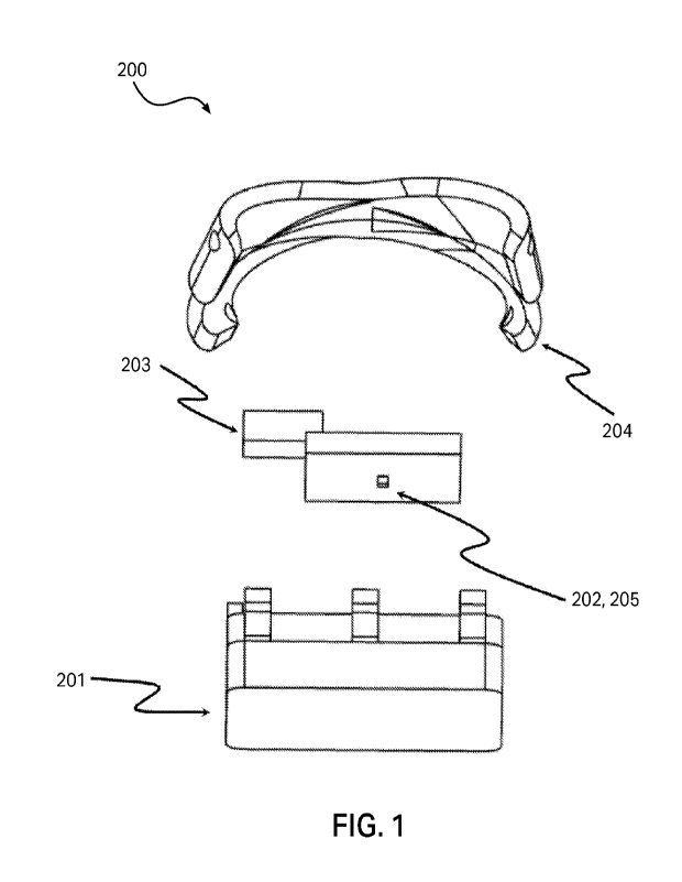
[096] Fig. 1 is an exploded view of a multi-layer sensor module comprising two
light
sources of different wavelength and a single photodetector.
[097] Fig. 2 is schematic view of an exemplary armband comprising five light
sensors
arranged circumferentially around the armband.
[098] Figs. 3A and 3B are cross-sectional diagram of the multi-layer sensor
module
shown in Fig. 1 illustrating one path that light can take from each of the two
light sources to the
photodetector.
12
CA 03236264 2024- 4- 24
WO 2023/073617
PCT/IB2022/060357
[099] Fig. 4 is a series of graphs illustrating the wavelength dependent
output from a
multi-layer light sensor for three distinct finger movements.
[100] Fig. 5 is schematic view of a grip augmenting glove showing a sheathed
cable
tendon extending from a differential to the forefinger of the glove.
[101] Fig. 6 is a schematic view of a grip augmenting glove showing the
routing and
termination of a cable tendon with the glove.
[102] Fig. 7 is a schematic view of a grip augmenting glove showing five cable
tendons
extending to each finger of the glove.
[103] Fig. 8 is a schematic view of a grip augmenting glove being used by a
recipient to
grasp a cylindrical object.
[104] Fig. 9A shows five photos of respective hand gestures.
[105] Fig. 9B shows three charts of signal measurements for the hand gestures
of Fig. 9A.
[106] Fig. 10 shows two diagrams of accuracy measurement.
[107] Fig. 11 shows a table of accuracy for three gesture decoding models.
[108] Fig. 12 shows a table of correlation and accuracy for three regression
model.
[109] Fig. 13 shows line charts of estimated clenching force versus actual
clenching
force.
DETAILED DESCRIPTION
[110] In one aspect, the disclosure provides for a sensor device. Elsewhere
herein the
sensor device may be referred to as a light sensor, or as a sensor module. The
sensor device may
be usable for detecting a physical state of tissue of a user, where the sensor
device is located at or
near a skin surface of the user. The sensor device is usable to detect a
property of tissue of a user
by light emission and the detection of reflected light. More particularly, in
some examples the
sensor device is usable to detect changes in a property or properties of
tissue of a user over time
by light emission and the detection of changes in reflected light.
[111] The sensor device may include a plurality of light sources (e.g., light
emitting
components), each source to emit incident light for reflection by the tissue
of the user, and a
photodetector to detect the reflected light.
[112] The light emitted by each light source may include one or more different
wavelengths. For example, a first light source may emit light of a first
wavelength or range of
wavelengths, and a second light source may emit light of a second wavelength
or range of
wavelengths that is/are different to those of the first light source.
[113] The user of the sensor device may be a human. In other examples, the
user may be
another non-human animal.
[114] Humans and other animals have bodily tissue that includes a skin
surface, beneath
which are cutaneous tissue layers and then subcutaneous tissue layers. The
cutaneous tissue
13
CA 03236264 2024- 4- 24
WO 2023/073617
PCT/IB2022/060357
layers may include, from the skin surface inwardly, the surface of the skin,
an epidermis layer, and a
dermis layer. The subcutaneous tissue layers may include a hypodermis layer
and then muscle. The
skin surface of interest, the associated cutaneous tissue layers of interest
and the associated
subcutaneous tissue layer of interest may be collectively referred to herein
as "skin site".
[115] The properties of tissue that the sensor device may be usable to detect
may
include one or more physical states of tissue. The tissue may for example be a
subcutaneous tissue
layer. For example, a sensor device may be usable to detect a contraction
state, or a change in
contraction state, of a muscle or group of muscles beneath the skin surface
where the sensor
device is located. The contraction states of a muscle may be associated with
particular optical
properties, for example reflective properties, of the muscle tissue. The
reflective properties of the
muscle tissue may change because of, for example, one or more of a
displacement or reshaping of
the muscle, a change in local volume, a change in local hardness, or a change
in local density of the
muscle in different contraction states.
[116] The contraction state of a muscle may be associated with particular
optical
properties of other non-muscle tissue layers. For example, the contraction
state of a muscle may
be associated with particular optical properties of one or more of an adjacent
hypodermis layer,
dermis layer, or epidermis layer. The contraction state of a muscle may be
associated with
particular optical properties of a skin surface beneath which the muscle is
located. The optical
properties of tissue layers adjacent to a target muscle may be affected by
physical changes in the
muscle at different contraction states. For example, in different contraction
states a muscle may
locally change in shape or volume. These changes may deform surrounding tissue
layers, and these
deformations may result in changes in the optical properties the tissue
layers. For example, a
contraction of muscle tissue may cause pressure outwards against the adjacent
hypodermis,
dermis, and epidermis. This may cause a decrease in the thickness of one or
more of these tissue
layers, which may alter their optical properties. The contraction of muscle
tissue may also cause
deformation of the skin surface, by either or both stretching it in-plane or
locally deforming it
outwards. Such changes at the skin surface may cause changes in the optical
properties of
components of a sensor device that are placed in contact with the skin
surface.
[117] In other examples, a sensor device may additionally or alternatively be
usable to
detect other properties of tissue and more particularly of subcutaneous
tissue. For example, the
sensor device may be usable to detect a heart rate of a user to which the
sensor is applied. As
changes in blood flow within tissue are associated with successive beats of an
animal's heart, the
changes in blood flow may cause changes in the optical properties of the
tissue where the blood is
flowing, and/or in adjacent tissue layers.
[118] In other examples, a sensor device may additionally or alternatively be
usable to
detect other properties of subcutaneous tissue, such as the presence or
concentration of different
14
CA 03236264 2024- 4- 24
WO 2023/073617
PCT/IB2022/060357
elements or compounds, where the presence or concentration of those elements
or compounds
change the optical properties of the tissue. For example, a sensor device may
be usable to detect a
blood oxygen level of blood in subcutaneous tissue due to the optical
properties exhibited by the
tissue at different oxygen saturation levels.
[119] The use of a sensor device or sensor devices to identify one or more
properties of
tissue of the user, such as the activity of a user's muscles, in accordance
with the present
disclosure may be characterised, in contrast to electromyography (EMG) or
forcemyography (FMG)
techniques, as lightmyography (LMG).
[120] Any of the optical properties of tissue layers, or other components
between either
or both a light source and a photodetector and a skin surface, may be
associated with particular
properties of reflected light from the incident light of the light source or
sources by different layers
of tissue. The reflected light in respect of a layer represents a reflection
of the incident light by the
respective layer. These properties may include an intensity of the reflected
light. In some examples,
these properties may include a distance from the skin layer of interest to
each of the light source
and the photodetector. Where the properties of tissue change, the changes in
properties may be
associated with corresponding changes in the properties of reflections of
light from within layers of
the tissue from incident light of the light source or sources. In other words,
changes in tissue
properties result in corresponding changes in light reflected by (or at) the
skin layers of interest and
detected by the photodetector.
[121] By affecting a particular physical state of the tissue of a user, for
example by
contracting a muscle to a particular contraction state while using the sensor
device, the associated
properties of light that is reflected from within the tissue may be obtained.
Similarly, by affecting
different changes in the properties of the tissue of a user, for example by
contracting a muscle
between two contraction states, changes in the resulting associated properties
of light that is
reflected from within the tissue may be obtained. Accordingly, the sensor
device may detect the
physical state of tissue beneath the skin surface based on the properties of
light that is reflected
from within the tissue and received by the sensor device.
[122] Where the sensor device is used to detect the physical state of a
particular muscle,
a determined contraction state of that muscle may be usable to infer a
relative position of body
parts of the user, for example, a flexion or extension position of the forearm
and upper arm of a user
relative to each other. Similarly, where the sensor device is used to sense
the physical state of a
particular muscle, changes in a sensed contraction state of that muscle may be
usable to infer
changes in a relative position of different body parts of the user. For
example, a change in a flexion
or extension position of the forearm and upper arm of a user relative to each
other.
[123] Data collected from sensed reflected light properties from a sensor
device in
association with a particular region of tissue, for example a particular
muscle, may be used to
CA 03236264 2024- 4- 24
WO 2023/073617
PCT/IB2022/060357
generate a model of properties of reflected light that are associated with a
particular physical state
or change in physical state of tissue, particularly subcutaneous tissue, of a
user. For example, a
sensor device may be used to gather data of reflected light properties in
relation to a chosen
contraction state of a particular muscle, or a chosen change in contraction
state of a particular
muscle. The gathered data may then be used to train a model of characteristic
sensed properties
that are associated with the chosen contraction state or chosen change in
contraction state of that
muscle.
[124] For example, the data may be used for training a machine learning model.
Such a
machine learning model may be trained on sensor data associated with
particular physical positions
or movements of the user. Once trained, sensor data of the properties of
reflected light sensed
when a sensor device is applied to a user can be inputted to the trained
machine learning model
and classified by it, in order to obtain an estimate of a current physical
position or movement of a
body part of the user.
[125] Accordingly, by training models of the properties of reflected light
that are
associated with different contraction states of different muscles, a trained
model may be
generated which is able to classify sensor data of one or more sensor devices
and generate an
estimate of the contraction states or specific changes in contraction states
of different individual
muscles.
[126] In some examples, more than one sensor device may be used, each to sense
one or
more different properties of different regions of tissue of the user. For
example, multiple sensor
devices may be used each to determine the contraction state of corresponding
different muscles
of a user. A model based on data of properties of reflected light may be
accordingly trained based
on sensed values from a plurality of sensor devices, and the trained model can
then be used to
estimate a physical position or movement of a body part of the user that is
associated with the
operation of multiple different muscles. For example, multiple sensor devices
may be provided for
determining the contraction states of different muscles, such as the different
muscles in the
forearm of a user. Where a trained model of the properties of reflected light
that are associated
with different contraction states of each of those muscles has been developed,
sensed values from
each of the sensor devices may be inputted into the trained model, classified
by it, and from those
classifications and an estimate of the contraction states of each of the
muscles may be obtained.
[127] As the contraction state of the muscle or change in contraction state of
a muscle or
muscles may be associated with a movement of a body part of a user to whom the
sensor device is
applied, estimation of the physical state of one or more muscles or other
subcutaneous tissue
regions may allow a physical position assumed by a body part of the user to be
estimated. For
example, where detectable physical states of a muscle or muscles are
associated with a particular
physical position assumed by a body part of the user, the estimation of these
physical states by
16
CA 03236264 2024- 4- 24
WO 2023/073617
PCT/IB2022/060357
one or more sensor devices may allow, by inputting of the sensed values to the
trained model, for
the estimation of the physical position of the user. Similarly, where
detectable changes in the
physical states of a muscle or muscle are associated with a particular
movement, for example the
assumption of a gesture, of the user, the estimation of these physical states
by a sensor device or
devices may allow, by inputting the sensed values to the trained model, the
estimation of the
movement or gesture conducted by the user.
[128] An estimated physical position of a body part of a user or estimated
movement or
gesture of a user may be used to control of devices such as anthropomorphic
robotics, actuated
exoskeletons, and active prosthetic limbs. For example, the sensor device or a
plurality of sensor
devices may be used to estimate movements of a user and operate a robot to
support or mimic the
movement.
[129] In one tested arrangement, a plurality of sensor devices were arranged
in an
armband, and a machine learning model, the lightmyography model, was trained
using data of
several different hand gestures of a number of different users. The hand
gestures included a rest
position, forefinger and thumb pinch gesture, a tripod gesture, a power or
fist pose, and an
extended pose in which each of the fingers of the hand are fully extended. A
matching model was
also trained using sensor data from an EMG for comparison. Use of the trained
models found that
the lightmyography model provided higher accuracy of classification of the
hang gestures than
were provided by the trained EMG model.
[130] EMG sensor data may require processing (e.g., RMS) to be used as
training data.
This may involve computational power and additional time. Compared to the use
of EMG data, the
sensor data from the photodetector(s) of the sensor device or devices of the
disclosure may, in a
model training context, be directly utilised advantageously as training data,
without requiring
intermediate processing. In an application context, sensor data of the
photodetector(s) may be
directly provided to the trained model without any intermediate processing.
[131] Where a sensor device is used to provide input data to a trained model
in order to
receive an estimated physical position or movement of a user to whom the
sensor device is
applied, a processor and a storage medium may be provided. The storage medium
may store the
trained model, and the processor may receive the sensor data, input the sensor
data to the trained
model, and receive classifications, and accordingly estimate a physical
position or movement. The
processor may store the outputted estimate. The processor may in some examples
communicate
the outputted estimate to a user, for example by a user interface. The
processor may in some
examples communicate the output to another device, for example to a server or
a controller for a
robotic device that is to be operated based on the estimated physical position
or movement of the
user.
17
CA 03236264 2024- 4- 24
WO 2023/073617
PCT/IB2022/060357
[132] Where a processor is used in conjunction with a sensor device or sensor
devices,
the processor may be located along with the sensor device or devices. In other
examples, the
processor may not be located with the sensor device or devices but is able to
receive sensor data
from the sensor device or devices.
[133] Where a storage medium is used in conjunction with a sensor device and
processor, the storage medium may be located along with either the sensor
device or processor. In
other examples, the storage medium may be located away from the sensor device
or processor, for
example in a remote location but communicably connected to the processor.
[134] A method of estimating a muscular contraction state of a target muscle
may utilise
one or more sensor devices. According to such a method, a sensed value of each
of the first
reflected light and second reflected light at both a first time point and a
second time point may be
received. The sensed values may be received by a processor, for example. Once
the sensed values
are received, an estimation may be performed using a processor of a
deformation of a cutaneous
region adjacent the skin of a user to whom the one or more sensor devices are
applied. The
estimation may be based on the sensed value of the fist reflected light. More
particularly, the
estimation may be based on a classification of the sensed value of the first
reflected light by a
trained model. An estimation may also be performed using a process or a
deformation estimate of a
subcutaneous region of a user to whom the one or more sensor devices are
applied. The estimation
may be based on the sensed value of the second reflected light. More
particularly, the estimation
may be based on a classification of the sensed value of the second reflected
light by a trained
model. An estimation of a muscular contraction state of a muscle or respective
muscles adjacent
the or each light sensor may be performed using the processor. The estimation
may be based on a
combination of the estimated cutaneous deformation and estimated subcutaneous
deformation.
[135] The properties of reflected light sensed by a sensor device may include
the
properties of reflected light originally incident from two different light
sources.
[136] The properties of reflected light sensed by a sensor device may include
the
properties of reflected light of two different wavelengths.
[137] The properties of reflected light sensed by a sensor device may include
the
properties of reflected light from two different light sources, where the
reflected lights from the
different light sources are of different wavelengths.
[138] A sensor device may include an elastic light-transmissive layer which
has optical
properties that change as a result of deformation of the light-transmissive
layer. The elastic light-
transmissive layer may resiliently deform. The light-transmissive layer may
elsewhere herein be
referred to as an elastonner layer or an elastomeric pad. The light-
transmissive layer may have a
first side and a second side, and a thickness between the two sides. The
second side of the light-
transmissive layer may be for being located against a skin surface of a user
in use.
18
CA 03236264 2024- 4- 24
WO 2023/073617
PCT/IB2022/060357
[139] The optical properties of tissue or of a light-transmissive layer that
may change
with deformation of the tissue or layer may include one or more of the
transmittance, absorption,
scattering, or reflectance of light incident on or passing through the tissue
or layer. The optical
properties of tissue or a light-transmissive layer may also include the
perceived colour of the tissue
or layer.
[140] In at least some examples, the sensor device may detect an intensity of
light
reflected from (or by) different layers of tissue.
[141] A deformation of a light-transmissive layer may include an in-plane
stretching of the
light-transmissive layer, which causes a change in thickness of the light-
transmissive layer between
its first and second sides. A deformation may additionally or alternatively
include a compression of
the light-transmissive layer between its first and second sides, such that a
thickness of the light-
transmissive layer changes. Such changes in the thickness of the light-
transmissive layer may
change the optical properties of the layer, so that it accordingly affects
light that traverses through
it. For example, an increase in the thickness of the light-transmissive layer
may be associated with
an increased attenuation of light that traverses therethrough, while a
decreased in the thickness of
the light-transmissive layer may be associated with a decrease in the
attenuation of light that
traverses therethrough.
[142] Where a light-transmissive layer deforms, a change in shape of the light-
transmissive layer due to the deformation may additionally or by itself alter
the optical properties of
the light-transmissive layer. For example, where deformation changes the
thickness of the light-
transmissive layer, an increase or reduction in the thickness may cause a
respective decrease or
increase in the intensity of a constant intensity of incident light from one
side of the light-
transmissive layer which sensed at the other side of the light-transmissive
layer.
[143] The use of a light-transmissive layer in a sensor device may allow one
or both of a
light source and photodetector to be spaced away from the skin surface of the
user in use. This
may allow for incident light from the light source to be reflected from the
skin surface and received
by the photodetector. This spaced arrangement is suitable for shorter
wavelengths (e.g., green).
Where deformation of the light-transmissive layer occurs, the resulting change
in the optical
properties of the light-transmissive layer may enhance or cause a change in
the properties of light
that is reflected from tissue and that passes outwardly through the light-
transmissive layer. That is,
the change in optical properties of the light-transmissive layer may cause a
corresponding change
in optical properties of light passing therethrough.
[144] A light-transmissive layer may be made from an elastic material. For
example, the
light transmissive material may include silicone having a Shore harness of 00-
30. Portions of the
light-transmissive layer may be dyed to alter influence of environmental light
on the light detection.
19
CA 03236264 2024- 4- 24
WO 2023/073617
PCT/IB2022/060357
For example, the light-transmissive layer may have portions that are dyed
black to reduce such
influence of environmental light.
[145] The thickness of the light-transmissive layer may be selected to provide
a desired
effect on either or both of incident light and reflected light that passes
through it. An increased
thickness of the light-transmissive layer may be associated with the
transmission of light having
longer wavelengths. The thickness of the light-transmissive layer may be
selected to provide a
desired reflection area of the incident light on the skin surface or other
tissue layer.
[146] In some examples, the light-transmissive layer may include regions of
different
thicknesses, each with a different light source and/or photodetector
associated therewith.
[147] As is subsequently described, the light-transmissive layer may include
one or more
dopants to alter the optical properties of the light-transmissive layer. The
one or more dopants may
additionally alter how the optical properties of the light-transmissive layer
change as its thickness
changes. That is, the light-transmissive layer may be doped or otherwise
configured to alter a
relation between the thickness and the optical properties of the light-
transmissive layer.
[148] The first light source of a sensor device may be configured to emit a
first incident
light through the light-transmissive layer towards the skin surface (or a skin
site of interest). To this
end, the first light source may be located at or facing the first side of the
light-transmissive layer,
opposite the skin surface of the user in use. In some examples the first light
source may be partially
or fully embedded within the light-transmissive layer. Where the sensor device
is configured so the
light-transmissive layer or part thereof is between the first light source and
the skin surface of the
user, at least the first incident light from the light sensor will pass
through the light-transmissive
layer.
[149] The second light source of a sensor device may be configured to emit a
second
incident light. In some examples the second light source may also be
configured to emit the second
incident light through the light-transmissive layer. In other examples, the
second light source may
be configured to emit the second incident light other than through the light-
transmissive layer. For
example, the second light source may be configured to emit the second incident
light directly to the
skin surface of a user.
[150] The photodetector of a sensor device may be configured to detect a first
reflected
light which represents a reflection of the first incident light. The
photodetector may be configured
to detect a second reflected light which represents a reflection of the second
incident light. The
first reflected light and second reflected light may be reflected by different
tissue layers of the user.
For example, the first reflected light may be reflected by a cutaneous tissue
layer, and the second
reflected light may be reflected by a subcutaneous tissue layer.
[151] The photodetector may be provided at or towards the first side of the
light-
transmissive layer, being the side facing away from the skin surface of the
user in use. In some
CA 03236264 2024- 4- 24
WO 2023/073617
PCT/IB2022/060357
examples, the photodetector may be partially or fully embedded within the
light-transmissive layer.
Where the light-transmissive layer or part thereof is provided between the
photodetector and the
skin surface of the user, the first reflected light and second reflected light
may pass through the
light-transmissive layer before being received by the photodetector.
[152] In some examples, a sensor device may include a first photodetector and
a second
photodetector for detecting the first and second reflect lights, respectively.
The first
photodetector may be provided at or towards the first side of the light-
transmissive layer to detect
the first reflected light via the light-transmissive layer, and the second
photodetector may be
arranged to avoid detecting the second reflected light via the light-
transmissive layer. For example,
the second photodetector may be located on the skin surface, either at the
second side of the
light-transmissive layer or on laterally adjacent of the light-transmissive
layer to directly detect the
second reflected light.
[153] The wavelength or wavelengths of light emitted by the first light source
and second
light source may be different to each other. As different wavelengths of light
may penetrate to
different depths within tissue before being reflected, the emission of
different wavelengths of light
by the first light source and second light source may allow the sensor device
to receive reflected
lights from different tissue layers.
[154] Shorter wavelengths may penetrate less into the tissue, providing an
increased
intensity of the reflected light. This may provide a more sensitive response,
allowing for physical
changes in the tissue to be more accurately sensed. Longer wavelengths may
penetrate deeper
into the tissue, providing a lower intensity of reflected light, but allowing
sensing of physical
changes in deeper layers of the tissue.
[155] In some examples, a sensor device may be configured so that the first
reflected
light is reflected at or adjacent to a skin surface of the user. For example,
the wavelength of light
emitted by the first light sensor may be such that at least some of the first
incident light is reflected
as the first reflected light by the skin surface. In such examples, the
wavelength of light emitted by
the first light sensor may correspond to a green light, having a wavelength of
about 500 nm to
about 565 nm.
[156] In other examples, a reflective element may be provided at the skin
surface, which
reflects the first incident light. The reflective element may act to increase
the reflection of incident
light thereon. In such examples, the wavelength of light emitted by the first
light sensor may be any
wavelength or plurality of wavelengths which is reflected by the reflected by
the reflective surface.
The reflective surface may for example be provided at the second side (the
skin-facing side) of the
light-transmissive layer. The second side of the light-transmissive layer may
be provided with a
reflective coating. The reflective coating may be a flexible reflective
coating. The reflective element
may be arranged in a path of the incident light to improve an intensity of the
resultant reflected light
21
CA 03236264 2024- 4- 24
WO 2023/073617
PCT/IB2022/060357
while allowing the incident light to penetrate the skin surface. The
reflective element in one
configuration is suitable for use with an incident light of a shorter
wavelength (e.g., green) relative to
one with a longer wavelength (e.g., infrared). In an example with incident
lights of different
wavelengths, respective reflective element portions may be associated
accordingly with the
incident lights of suitable, compatible wavelengths. The reflective surfaces
may be employed to
advantageously reduce adverse effects of factors, such as reflectance,
roughness and
pigmentation of the human skin. With a shinier surface of a higher
reflectance, a higher signal
response or SNR can be achieved to facilitate distinguishment between
different muscle
movements.
[157] In some examples, a sensor device may be configured so that the second
reflected
light is reflected at or within a subcutaneous tissue layer. In such examples,
the wavelength of light
emitted by the second light sensor may have a wavelength of about 625 nm to
about 1,400 nm.
[158] Where a sensor device has a reflective surface at the skin surface, the
first
photodetector may be configured to receive the first reflected light from the
reflective surface, and
a second photodetector may be provided and configured to receive the second
reflected light from
a subcutaneous tissue layer. The second photodetector may be provided so that
second reflected
light received by it passes through the light-transmissive layer. In other
examples, the second
photodetector may be provided so that the second reflected light received by
it does not pass
through the light-transmissive layer. For example, the second photodetector
may be provided
directly facing the skin surface of the user.
[159] Where a sensor device includes a first and second light sources, the
light sources
may be configured to alternately emit the respective first incident light and
second incident light.
The alternate emissions may be configured so that they are non-contemporaneous
with each
other. By such a configuration, a distinguishment of the first reflected light
and second reflected
light may be aided at the photodetector or photodetectors of the sensor
device.
[160] Where a sensor device has a first light source that is provided at a
first distance
away from the skin surface of a user, and a second light source that is
provided at a different
second distance away from the skin of the user, the sensor device emits light
from multiple,
different distances from the skin surface. Such a sensor device may be
referred to as a multi-layer
sensor device, or as a multi-layer light sensor. In some examples, the first
distance may be a
distance equal to the thickness of the light-transmissive layer, while the
second distance is zero, so
that the second light source emits light at the skin surface.
[161] The light sources of a sensor device may be provided by any light
emitting device.
In at least some examples, the light sources of a sensor device are provided
as light emitting diodes
(LEDs).
22
CA 03236264 2024- 4- 24
WO 2023/073617
PCT/IB2022/060357
[162] In some examples, the sensor device may be configured to block external
environmental light from being received by the photodetector or
photodetectors. For example, a
housing or other non-light-transmissible element may be provided about the
light sources,
photodetector or photodetectors, and the light-transmissible layer, to block
environmental light
when the sensor device is placed in contact with the skin surface of the user.
Such light blocking
arrangements blocks unintentional detection of the environmental light by the
photodetector.
[163] The technologies disclosed herein, such as the sensor device and
combinations
thereof, are generally applicable to sensing movement, deformation and/or
displacement of soft
tissue adjacent the skin surface. Some exemplary applications of the
technology include wearable
human-machine interfaces, physiological sensors (such as heartrate monitors),
and/or soft tissue
pressure sensors. Detailed examples of the technology are presented for human-
machine
interfaces. In these examples, the disclosure relates to systems and methods
for detecting muscle
activation underneath the skin. Muscle activation can be used for the control
of a variety of devices,
such as computer systems, bionic/prosthetic grippers and/or lower limb
prostheses. However, the
technology can be readily adapted for other soft tissue sensing and/or
measurement applications.
[164] In at least some examples, a wearable band, such as an armband or leg
band, is
used to hold several multi-layer light sensors or sensor devices next to the
skin of a user. The light
sensors can detect muscle movement from deformation of the skin close to the
band. In at least
some examples, the light sensors comprise at least two light sources, such as
LEDs, that project
light onto the skin, and at least one photodetector that is arranged to
receive light from the light
sources. The sensor output can be used for the control of devices such
anthropomorphic robotics,
actuated exoskeletons, and active prosthetic limbs.
[165] For example, the band can be worn by a user about the forearm to provide
data for
interpretation of grip type (e.g. power grip / pinch grip / cylindrical grip)
and/or gestures for an
active prosthetic hand or exoskeleton glove. In at least some examples, three
or more light sensors
or sensor devices are arranged circumferentially around the band to detect
movement of the
muscles in the forearm that affect finger and/or wrist movement.
[166] In at least some examples, the light sensor is configured with an
elastomer layer,
such as silicone, disposed between one of the light sources and the skin. The
elastomer may have a
thickness of about 1 mm to about 20 mm. For example, the elastomer layer may
have a thickness of
between 3 mm and 12 mm. In other examples, other light-transmissive elastic
layers may be used.
[167] The intermediate elastomer layer creates a layered structure of light
sources within
the sensor. For example, a light source that is held immediately adjacent to,
or in contact with, the
skin is offset from a light source that sits behind the elastomer layer by
approximately the thickness
of the elastomer layer. The elastomer layer can channel and/or guide light
between the light source
and the skin. In at least some examples, the light sources of different layers
emit light of different
23
CA 03236264 2024- 4- 24
WO 2023/073617
PCT/IB2022/060357
wavelengths. For example, a light source that is adjacent the skin can be
configured to emit light
with a longer wavelength than a light source that is offset from the skin. In
some opposite examples,
a light source that is adjacent the skin can emit light with a wavelength
shorter than about 500 nm,
whereas a light source that is separated from the skin by an elastomer layer
can emit light with a
wavelength longer than about 650 nm.
[168] The elastomer layer can comprise an elastomeric pad that is interposed
between
one or more light sources and the skin to regulate the distance between the
light source and the
skin, manipulate the transmissivity of the gap between the light source and
the skin, and/or control
other attributes that can be used to tune the response of the sensor.
[169] For example, the elastomeric pad can incorporate a dopant that changes
the
absorptivity of the material responsive to deformation. In some examples, the
elastomeric pad can
have one or more sections that reflect, scatter, or absorb at least a portion
of the light emitted by
the light source.
[170] For example, the elastomeric pad can have a reflective surface
configured to cover
a portion (e.g. a third, a quarter, a half) of the skin surface so that a
corresponding portion of light
emitted by the light source is reflected by the reflective surface, and that a
remaining portion of
light emitted by the light source is reflected by an epidermal layer of the
skin.
[171] In some examples, the mechanical properties of the elastomeric pad can
be used to
tune the response of the sensor. For example, the deformation characteristics
of the elastomeric
pad can be adjusted to control the dynamic range and/or resolution of the
sensor by creating non-
linear transmissivity and/or absorptivity characteristics.
[172] At least one photodetector is arranged to receive light reflected from
the skin of
the user. In some examples, a single photodetector may be arranged to receive
light from two or
more light sources.
[173] For example, a single photodetector can be offset from the skin and
disposed
laterally between adjacent light sources. In at least some examples, an
elastomer layer is
interposed between the photodetector and the skin surface. The photodetector
can be disposed in
the same layer as one of the light sources, or in a layer intermediate to the
light sources. For
example, the photodetector can be offset from the skin by an intermediate
elastomeric layer so
that the photodetector is closer to the skin than one light source, and
farther from the skin than
another light source.
[174] In at least some examples, the sensor is configured to interleave light
pulses from
two or more light sources that share a common photodetector. For example, the
sensor can
interleave the time periods that each of the light sources are emitting light
so that the
photodetector does not concurrently receive light from more than one light
source. The sensor can
be configured to measure (or detect) the intensity of light incident on the
photodetector
24
CA 03236264 2024- 4- 24
WO 2023/073617
PCT/IB2022/060357
concurrently with each light pulse. In some examples, the light captured by
the photodetector in
sequential time periods is reflected from different types of soft tissue
and/or different layers of the
skin. The sensor can be configured to alternate measurements from the surface
of the skin with
measurements from beneath the surface of the skin to create a composite light
measurement. In
some examples, each light source has a dedicated photodetector so that
measurements from
different types of soft tissue and/or layers of the skin can be obtained
concurrently.
[175] In at least some examples, the sensor is configured to detect movement
and/or
deformation in distinct layers, sections, and/or depths of soft tissue. For
example, the light sources
can be configured so that the light that they emit penetrates the skin to
different extents. In at least
some examples, a red or infrared LED is held adjacent the surface of the skin
so that the light
emitted by the LED, and subsequently captured by the photodetector, is
reflected by tissue
beneath the skin surface, such as subcutaneous fat and/or muscle. Light of
longer wavelengths can
achieve a deeper skin penetration and provide complementary data that can be
beneficial for
decoding (e.g., estimating) the user's muscle movements. Light of longer
wavelengths can also be
used to acquire some health information, which can be a potential advantage of
light-based
myography over classic EMG techniques. A shorter wavelength LED (such as a
green, blue, violet,
or ultraviolet LED) can be used to obtain more superficial measurements (e.g.
from the epidermal
layer of the skin). Light of shorter wavelengths penetrate the skin less
resulting in better reflection
and leading to a more sensitive response. In at least some examples,
differential tissue
measurements can be obtained and/or enhanced by the physical arrangement of
the light sources
with respect to the skin surface. For example, the gap between a light source
and the skin surface
can be adjusted to regulate the depth of light penetration.
[176] In some examples, the sensor may be used to estimate subcutaneous tissue
movements from a composite light measurement. The composite light measurement
can comprise
light of a first wavelength reflected from the surface of the skin, and light
of a second wavelength
reflected from tissue beneath the surface of the skin, such as muscle,
connective tissue and/or
subcutaneous fat. For example, the sensor can be configured to obtain a
deformation estimate for
an epidermis layer of the skin from an intensity of light captured at the
first wavelength, and a
deformation estimate for a dermis layer of the skin from an intensity of light
captured at the second
wavelength. The combined deformation estimates for the epidermis layer and the
dermis layer of
the skin can function as a proxy for muscle and/or connective tissue movement
in at least some
applications. For example, an estimate of muscle contraction may be calculated
from the intensity
of light at the first wavelength and the intensity of light at the second
wavelength present in the
composite light measurement. In at least some examples, an artificial limb,
exoskeleton glove
and/or robotic gripper can be actuated or otherwise controlled responsive to
the calculated
estimate of muscle contraction.
CA 03236264 2024- 4- 24
WO 2023/073617
PCT/IB2022/060357
[177] An exploded view of an exemplary multi-layer sensor module 200 is shown
in Fig. 1.
The sensor module 200 has two LED light sources 202, 203 and a single
photodetector 205. The
photodetector 205 may be provided in the form of a photodiode. The photodiode
is co-located with
the first light LED source 202 in the illustrated embodiment. The LED light
sources 202, 203 and
photodetector 205 are secured within a housing 201 when the sensor module is
assembled. The
housing 201 has an elastomer layer that covers the skin facing surface of the
sensor module in the
illustrated embodiment. The thickness of the elastomer layer varies across the
skin facing surface
of the housing 201. For example, the thickness of the elastomer layer adjacent
the first LED light
source 202 can be about 4 mm to about 8 mm, whereas the thickness of the
elastomer layer
adjacent to the second LED light source is generally negligible (e.g. less
than about 1 mm thick). In
some examples, the elastomer layer does not extend across an area adjacent the
second LED light
source 203. A backing plate 204 holds the LED light sources securely within
the housing 201. In at
least some examples, the backing plate 204 biases the LED light sources toward
the elastomer
layer so that there is a tight interference fit and/or not appreciable gap
between the LED light
sources and the elastomer layer. In some examples the backing plate 204 may
elastomeric or
include an elastomeric portion.
[178] An armband 300 comprising five sensor modules 101, 103, 105, 107, 109 is
presented in Fig. 2. The sensor modules 101, 103, 105, 107, 109 are
distributed evenly around the
circumference of the armband 300 in the illustrated embodiment. Compliant
straps 102, 104, 106,
108, 110 extend between adjacent sensor modules 101, 103, 105, 107, 109 to
hold the sensor
modules 101, 103, 105, 107, 109 together in the band. The length of the
compliant straps 102, 104,
106, 108, 110 defines the distribution of sensor modules 101, 103, 105, 107,
109 around the
circumference of the band and the inter-sensor spacing. A pin is used to
secure the lateral ends of
each sensor module 101, 103, 105, 107, 109 to a corresponding strap 102, 104,
106, 108, 110.
[179] The illustrated armband 300 is configured to be worn about a mid-forearm
of a
user. In at least some examples, the unstrained circumference of the armband
is configured to be
smaller than the circumference of the user's forearm so that the strain
induced in the armband
causes the elastomer layer of a housing of each sensor module 101, 103, 105,
107, and 109 to be
held in contact with the user's skin when the armband is worn. In the
illustrated embodiment, the
compliant straps 102, 104, 106, 108, 110 stretch to increase the inner
circumference of the
armband 300 and accommodate the forearm of the user. This produces an axial
strain in the straps
102, 104, 106, 108, 110 that keeps the armband 300 firmly fitted to the user's
forearm and the
sensor modules 101, 103, 105, 107, 109 in contact with the skin. The elastomer
layer on the skin
surface of the sensor modules 101, 103, 105, 107, 109 can be configured to
distribute the force
applied by the armband 300 on the skin and/or alleviate localised pressure
concentration. The
26
CA 03236264 2024- 4- 24
WO 2023/073617
PCT/IB2022/060357
elastomer layer on the skin surface may be compressed by the pressure of the
elastomer layer
against the user's arm.
[180] The action of the compliant straps 102, 104, 106, 108, and 110 where
they are
stretched to apply to the body of the user may act to provide a biasing of the
sensor modules 101,
103, 105, 107, and 109 against the skin surface of the user.
[181] The number, size, distribution and/or configuration of sensor modules
can be
adapted to suit different applications. For example, sensors with larger skin
facing surface areas,
more light sources, and/or more photodetectors can be used for lower limb
applications. Likewise,
the sensor modules can be concentrated adjacent particular muscles instead of
evenly distributed
around a limb or other body part. For example, a heartrate monitor may
comprise one or more
sensor modules clustered adjacent the sternum.
[182] A cross-sectional schematic view of a two-layer sensor modules 400
placed onthe
soft tissue of the user is shown in Figs. 3A and 3B. As seen in Figs. 3A and
3B, the soft tissue under
the sensor module comprises the skin surface 11, an epidermis layer 12, a
dermis layer 13,
hypodermis layer 14, and a muscle layer 15. The sensor module 400 comprises
two light sources
411 and 412 disposed respectively at opposite lateral sides of a photodiode
420. A red or infrared
LED light source 412 is disposed next (i.e., adjacent) to the skin to the
right of the photodiode 420.
A green LED light source 411 is disposed to the left of the photodiode 420. A
silicone layer 430 is
disposed between the green LED light source 411 and the photodiode 420 and the
skin surface 11.
In this example, the silicone layer 430 is shown to be arranged atop the skin
surface 11, and the
photodiode 420 and the light source 411 are shown to be arranged atop the
silicone layer 430. The
photodiode 420 is thus disposed on the same layer as the green LED light
source 411, with the
photodiode 420 and the light source 411 having the same spacing from the skin
surface 11. The
lateral spacing between each LED light source 411 and 412 and the photodiode
420 is determined,
at least in part, by the respective path traversed by light emitted by the
respective light source 411
and 412 to reach the photodiode 420, with the light path of the light source
411 crossing the
silicone layer 430. In some examples, the lateral spacing between each LED
light source and the
photodiode can be determined entirely from the expected light path. In other
examples, the light
sources can be evenly spaced about the photodiode.
[183] In Fig. 3A, the sensor module 400 is shown emitting light from the green
LED light
source 411, while the red or infrared LED light source is inactive 412. The
emitted light, with a
wavelength in the green spectrum (from 500 nm to 565 nm in wavelength,
approximately), is shown
passing through the silicone layer 430, penetrating the surface 11 of the skin
and reflected from (or
by) the epidermal layer 12. In some examples, the surface 11 of the skin can
reflect a portion of the
incident light. The reflected light passes through the silicone layer 430 to
be detected by the
photodiode 420. The intensity of green light captured by the photodiode 420
can depend, at least
27
CA 03236264 2024- 4- 24
WO 2023/073617
PCT/IB2022/060357
in part, on the distance the light travels. For example, movement of an
underlying muscle and/or
connective tissue can compress the silicone layer 430 and shorten the light
path between the
green LED light source 411 and the skin surface 11. The optical properties of
the skin surface
adjacent the sensor module 400 can also be altered by movement of an
underlying muscle and/or
connective tissue. For example, the density, mix and geometry of the tissue
can change with
deformation and cause the intensity of light captured by the photodiode 420 to
change. The light
source 411 can thus be understood to be configured to emit light through the
silicone layer 430
towards the skin (e.g., a skin site) for reflection by the epidermal layer 12
(or an epidermis skin
portion) and, in some examples, by the skin surface 11. Furthermore, in this
example, the silicone
layer 430 is adapted to be arranged on the skin surface 11 and is configured
to normally space the
light source 411 apart from the skin surface 11 by 5 mm. This 5 mm spacing
permits an increased
amount of reflection of light emitted by the light source 411 to be detected
by the photodiode 420,
which improves an intensity of the detected light.
[184] In Fig. 3B, the sensor module 400 is shown emitting light from the red
or infrared
LED light source 412, while the green LED light source 411 is inactive. The
emitted light, with a
wavelength in the red or infrared spectrum (approximately 625 nm-1400 nm, or
longer), is shown
penetrating the surface 11 of the skin and reflected from (or by) a layer of
subcutaneous fat, for
example the hypodermis layer 14. In at least some examples, light in the red
or infrared spectrum
can be reflected from the dermal or dernnis layer13 of the tissue,
subcutaneous fat (such as the
hypodermis layer 14, otherwise known as the subcutaneous tissue layer), muscle
15, and/or
connective tissue. The reflected light passes through the overlapping layers
of tissue and the
silicone layer 430 to reach the photodiode 420. The intensity of red or
infrared light captured by the
photodiode 420 can depend, at least in part, on the distance the light
travels. For example,
movement of an underlying muscle and/or connective tissue can compress the
silicone layer 430
and shorten the light path between the skin and the photodiode 420. The
optical properties of the
soft tissue under the skin surface can also be altered by movement of an
underlying muscle and/or
connective tissue. For example, the density, mix and geometry of the tissue
can change with
deformation and cause the intensity of light captured by the photodiode to
change. In this example,
the light source 412 is placed in contact with the skin to achieve a deeper
light penetration. The
light source 412 can be understood to be configured to emit light toward the
skin for reflection by
the hypodermis layer 14 (or a non-epidermis skin portion) and, in some other
examples, by the
other layers 13 and 15. The infrared light of the light source 412 may
interact with other layers such
as the dermis when passing therethrough, and the detected reflection of the
infrared light may thus
be indicative of movements occurring at those other layers.
[185] It can thus be understood that the silicone layer 430 is elastic and
light-
transmissive and has an optical property that changes in response to
deformation of the silicone
28
CA 03236264 2024- 4- 24
WO 2023/073617
PCT/IB2022/060357
layer 430. It may also be understood that the silicone layer 430 is
elastically deformable to cause a
corresponding change in an optical characteristic of light traversing through
the silicone layer 430.
For example, the silicone layer 430 may have an optical property that changes
in response to
elastic deformation of the silicone layer 430 to cause a corresponding change
in an optical
characteristic of light traversing through the silicone layer 430.
[186] In some examples where the photodiode 420 is placed in contact with the
skin
surface 11, reflected light is detected directly by the photodiode 420 without
traversing through
the silicone layer 430.
[187] A series of exemplary graphs showing the tested output from the sensor
module of
Figs. 3A and 3B for three different types of gesture are presented in Fig. 4.
The upper graphs
represent the intensity of infrared light captured by the photodiode. The
lower graphs represent the
intensity of green light captured by the photodiode. The sensor module was
held against the skin of
a user's forearm while the user moved the fingers of the corresponding hand.
The graphs on the left
represent the photodiode output for concurrent flexion of the ring and pinky
fingers. Those in the
middle represent the photodiode output for flexion of the pinky finger alone.
Those on the right
represent the photodiode output for flexion of the ring finger alone. It is
evident form the graphs of
Fig. 4 that the two LED light sources 411 and 412 in the sensor module 400
produce
complementary information indicative of the movement and/or deformation of
soft tissue in the
forearm.
[188] In at least some examples, the soft tissue at different layers and/or
depths below
the skin surface respond differently to movement and/or deformation of muscle
and/or connective
tissue. For example, subcutaneous fat can shift and/or reconfigure beneath the
skin surface as an
underlying muscle contracts. These changes are not always evident from the
skin surface. In at
least some examples, a composite measurement compiled from light reflected at
different layers of
soft tissue can produce more accurate and/or more robust estimates of muscle
and/or connective
tissue movement and/or deformation than a single measurement. For example,
combining
measurement obtained from the skin surface and/or epidermal layer with
measurements obtained
at the dermal and/or subcutaneous layer can produce more accurate and/or more
robust estimates
than a single measurement from either location. In at least some examples, an
independent
component analysis is used to associate the intensity of light captured by the
photodiode at
different wavelengths with user intent. For example, an independent component
analysis can be
used to correlate the output of the sensor modules 101, 103, 105, 107, and 109
shown in Fig. 2 with
hand gestures and/or types of grip.
[189] A method for estimating subcutaneous tissue movement comprises obtaining
a
first deformation measurement representative of deformation of an epidermis
layer of the skin,
obtaining a second deformation measurement representative of deformation of a
dermis layer of
29
CA 03236264 2024- 4- 24
WO 2023/073617
PCT/IB2022/060357
the skin that is in close proximity to the epidermis layer represented in the
first deformation
measurement, and combining the first deformation measurement and the second
deformation
measurement to produce the estimate of subcutaneous tissue movement.
[190] In some examples, the method can comprise estimating the first
deformation
measurement from an intensity of light reflected from the skin at a first
wavelength and estimating
the second deformation measurement from an intensity of light reflected from
the skin at a second
wavelength. For example, the method can comprise interleaving light from a
first light source, that
emits light at the first wavelength, with light from a second light source,
that emits light at the
second wavelength, and measuring the intensity of light from the first light
source and the intensity
of light from the second light source with a single photodetector.
[191] In at least some examples, composite light measurements from different
soft tissue
layers can be obtained with a sensor device comprising a first light source
that is configured to sit
on or adjacent the skin of a recipient and direct incident light of a first
wavelength through at least
the upper layers of the recipient's skin, and a second light source that is
configured direct incident
light of a second wavelength onto the recipient's skin at a location near the
first light source. A
compliant pad, made from a transparent or translucent elastomer, can be
configured to sit between
the second light source and the skin of the recipient. And at least one
photodetector can be
configured to receive light reflected from the skin and/or subcutaneous tissue
of the user and
measure an intensity of light at the first wavelength and an intensity of
light at the second
wavelength.
[192] The sensor can comprise a band that holds the first light source, the
second light
source, the compliant pad (the silicone layer or the light-transmissive layer)
and the at least one
photodetector in proximity to the skin. The band can be configured to pre-load
(e.g., bias) the
compliant pad against the skin of the recipient by applying pressure to the
recipient's skin. That is,
the band or the like may be configured to apply a bias force on the compliant
pad against the skin.
Such an arrangement may prevent a gap forming between the compliant pad and
the skin during,
for example, arm movements, which may in some cases adversely affect the
reflection detection by
the sensor device. In some examples, the band is configured to circumscribe a
limb of the recipient.
The compliant pad can be configured to channel light from the second light
source to the skin of
the recipient. The compliant pad can also be configured to absorb and/or
scatter and/or reflect a
variable amount of light at the second wavelength as the pad is compressed,
and the amount of
light that the compliant pad absorbs and/or scatters and/or reflects is
dependent on the
deformation of the elastomer. In some examples, the second light source has a
wavelength that is
greater than 650 nm, and the first light source has a wavelength that is less
than 550 nm.
[193] In at least some examples, one or more sensors can be combined with a
strap, band
and/or belt that is configured to hold the sensor(s) next to the skin (e.g.
adjacent and/or in contact
CA 03236264 2024- 4- 24
WO 2023/073617
PCT/IB2022/060357
with the skin). For example, a device comprising a strap with a first light
sensor that is disposed
adjacent an inner surface of the strap, and a second light sensor that is
spaced circumferentially
from the first light sensor can be used to obtained composite light
measurements from different
layers of soft tissue. In at least some examples, the second light sensor is
offset outwardly from the
inner surface of the strap compared to the first light sensor, and the strap
comprises a
compressible elastomeric pad that is disposed between the second light sensor
and the inner
surface of the strap. In some examples, the second light sensor is displaced
outwardly from an
inner surface of the strap by more than about 2 mm, and the elastomeric pad
occupies the space
between the second light source and the inner surface of the strap. The first
light sensor can
comprise a first light source with a first wavelength and a first
photodetector, and the second light
sensor can comprise a second light source with a second wavelength and a
second photodetector.
The strap can be configured to apply a radially compressive force on a limb of
a recipient that
places the first light sensor in contact with, or immediately adjacent, the
skin of the recipient.
[194] In some examples, a belt comprising a first light sensor that is
configured to detect
displacement of skin under the band, and a second light sensor that is
configured to detect
displacement of soft tissue below the skin surface can be used to obtained
composite light
measurements from different layers of soft tissue. The first light sensor can
be held above the
surface of the skin and operate with a wavelength that is between about 625 nm
and about 1 mm.
The second light sensor can be held adjacent the skin and operate with a
wavelength between
about 10 nm and about 565 nm.
[195] In some examples, a band comprising at least two light sensors that are
configured
to detect displacement of soft tissue in close proximity to the band can be
used to obtained
composite light measurements from different layers of soft tissue. The at
least two light sensors
can operate in different frequency ranges of the light spectrum, and the band
can be configured to
position the at least two light sensors at different distances from the skin
surface. In some
examples, the band comprises a compliant elastomeric material that is disposed
between a first of
the at least two light sensors and an inner circumference of the band, and the
compliant
elastomeric material is configured to contact the skin and deform in response
to movement of soft
tissue immediately under the contacted skin. The compliant elastomeric
material can contain a
dopant, and the dopant can be configured to alter the transmissibility of
light, from the first of the at
least two light sensors, through the compliant elastomeric material dependent
on the deformation
of the compliant elastomeric material. In at least some examples, the first of
the at least two
sensors can be configured to measure deformation of the compliant elastomeric
material as a
proxy for displacement of soft tissue. A second of the at least two sensors
can be positioned closer
to the skin than the first of the at least two sensors. The second of the at
least two sensor can be
configured to measure displacement of soft tissue below the skin. In some
examples, a first light
31
CA 03236264 2024- 4- 24
WO 2023/073617
PCT/IB2022/060357
sensor of the at least two light sensors comprises a red or infrared LED light
source, and a second
light sensor of the at least two light sensors comprises a green, blue,
violet, or ultraviolet LED light
source.
[196] The light sensor or sensor device of the disclosure can function as an
interface that
permits a user to intuitively control and/or interact with a machine and/or
environment. For
example, estimates of soft tissue movement and/or deformation can enable a
user to interact with a
virtual reality or augmented reality environment, and/or control a robot. An
exoskeleton glove is
presented here as an example. In this example, the output from the sensor is
transformed into a
control signal that is used to control an electric motor (e.g. the motor speed
and/or torque) of the
exoskeleton glove. The motor actuates the fingers of the glove to initiate
different types and/or
strengths of grip. The control platform for the exoskeleton glove can be
readily transferred to other
physical systems, such as prosthetic limbs and/or anthropomorphic robots, or
artificial
environments, such as AR/VR.
[197] An exemplary grip augmentation system 500 is shown schematically in Fig.
5. The
system comprises an actuated glove 150 that can be worn by a recipient to
enhance grip strength.
Torque is transferred from an electric motor 140 to the fingers 100 of the
glove 150 by a network of
artificial tendons. A single artificial tendon 120 is shown in Fig. 5. The
depicted tendon comprises a
sheathed cable 121 that extends from the actuator, for example electric motor
140, to the tip of the
forefinger 100a. The distal end of the cable 121 terminates at the tip of the
forefinger 100a in a
finger cap 155. The finger cap 155 anchors the tendon 120 to the glove and
distributes forces from
the tendon to the recipient's forefinger 100a. The illustrated finger cap 155
also includes a sensor
160. The sensor 160 may be a light sensor or sensor device as described
herein. The output from
the sensor 160 is fed back to a controller that controls operation of the
glove. For example, the
controller can use the sensor 160 for touch detection, grip regulation and/or
performance tracking
(e.g. monitoring force distribution to each finger 100).
[198] A proximal end of the cable 121 is wound about the drum of a pulley 125.
The pulley
125 is driven by the electric motor 140 to tension the cable 121. The cable
121 transfers force from
the electric motor 140 to the finger cap 155 of the glove 150 as it is
progressively retracted and
wound onto the drum of the pulley 125. The tension forces in the cable 121
cause the recipient's
finger to contract, folding inward toward the palm of the glove to augment the
recipient's natural
grip. A sheath 122 extends from the electric motor 140 to the base of the
glove 150. The glove 150
and motor housing (not shown) have ferrules that locate and secure the sheath
122. The sheath
122 is sufficiently compression resistant to maintain a substantially constant
cable path length
between the electric motor 140 and glove 150 (e.g. preventing contraction of
the cable path
between the electric motor 140 and the glove 150). In some examples, the
sheath can incorporate a
low friction coating that reduces the sliding friction experienced by the
cable.
32
CA 03236264 2024- 4- 24
WO 2023/073617
PCT/IB2022/060357
[199] An exemplary glove cable guide 124 is shown in Fig. 6. The cable guide
124
restrains the cable 121 to a defined path within the glove 150. The
illustrated cable guide 124
comprises a section of stitching that extends along an inner side of the
forefinger 100a. Another
section of stitching (not shown in Fig. 6) extends from the proximal end of
the glove (e.g. adjacent
the wrist or forearm) to the base of the palm. The cable 121 is unrestrained
across the palm of the
glove 150 in the embodiment illustrated in Fig. 6. In other examples, the
stitching can extend
unimpeded between the base of the glove and the finger cap 155, or in discrete
sections of
different length / configuration. The cable guide 124 can incorporate a
compliant liner that reduces
cable friction within the glove 150. For example, a PTFE coated elastomeric
tube can be used to
route the cable 121 through the material of the glove 150 without restricting
the recipient's mobility
/ flexibility. In the embodiment illustrated in Fig. 6, the liner can be
stitched into the fabric of the
glove 150 at the forefinger 100a and extend unrestrained across the palm. The
glove 150 can also
incorporate a rigid or semi-rigid (e.g. thermoset plastic) palm guide to
prevent or alleviate pressure
induced cable friction (produced by the clamping forces from some forms of
grip).
[200] The fingertip terminated examples shown in Figs. 5 and 6 cause the
forefinger 100a
to bend in flexion. In some examples, flexion can be replaced or supplemented
with other forms of
anatomical motion. For example, the actuator such as an electric motor 140 can
cause adduction of
the thumb by tensioning a cable 121 that terminates at the base of the thumb.
The glove 150 can
be reinforced at the base of the thumb (e.g. with a thermoplastic insert
and/or reinforced loop
around the thumb metacarpophalangeal joint) to anchor the cable 121, transfer
forces to the
thumb, and/or guide movement of the thumb in adduction. In some examples, the
glove 150 can be
configured to support multiple forms of anatomical motion. For example, thumb
adduction can be
used in combination with flexion of the thumb for some forms of grip.
Independent adduction and
flexion can be achieved with separate cables 121 that terminate at the base
and tip of the thumb,
respectively.
[201] A grip augmenting glove 150 with five artificial tendons 120a, 120b,
120c, 120d,
and 120e is shown schematically in Fig. 7. The tendons 120 extend from a
sheath 122 at the base of
the glove 150 and splay outwardly toward each of the five fingers 100. The
glove fingers 100 have
cable guides 124 that route the tendons 120 to a termination point (e.g. a
finger cap 155) on each
finger 100. The tendons 120 are actuated by one or more motors that apply and
release tension as
needed to affect an adequate grip. The sheath 122 routes the tendon cables 121
from the motor(s)
to the base of the glove 150. For prosthetic hands and grip augmentation
gloves, the motor(s) are
usually housed in a wearable module that the recipient carries with them. For
example, the motor
module can be carried in a backpack, suspended from a belt that's worn around
the waist, or held to
the recipient's arm by an armband. The carrying mode for wearable systems is
usually influenced by
the weight and form-factor of the motor module. In some examples, the motor
module also houses
33
CA 03236264 2024- 4- 24
WO 2023/073617
PCT/IB2022/060357
control electronics. The control electronics regulate the output of the
motor(s) to modulate the
grasping force applied by the glove 150. For example, the control electronics
can incorporate one
or more sensors (e.g. [MG and/or force sensors) that the control electronics
that to infer recipient
intent for force modulation and/or grip initiation.
[202] Fig. 8 shows a grip augmenting glove 150 being used by a recipient to
grasp an
object 170. In the illustrated embodiment, the artificial tendons 120 are
applying a force to at least
three of the fingers 100a, 100b, 100c, 100d, and 100e (e.g. via a finger cap
155 on the
corresponding finger). The combination of natural flexion and tendon tension
causes the fingers
100 of the glove 150 to curl inwardly toward the palm of the recipient's hand,
producing the
cylindrical-type grip shown in Fig. 8. The force between the fingers and palm
of the hand is
representative of the grip strength. In some examples (e.g. rehabilitation),
the glove 150 can be
configured to replicate a healthy adult grip strength. In other examples (e.g.
workplace specific
tasks), the glove 150 can be configured to produce forces that exceed natural
human grip strength.
[203] The distribution of force to the fingers 100 of the glove 150 can
influence the
efficacy of the grasp the recipient forms. Grip stability is closely related
to the contact area formed
with an object. For gripping applications with well-defined constraints (e.g.
robotic grippers for
repetitive tasks), the force distribution to the fingers can be optimised for
specific grip types (e.g.
cylindrical, spherical, or pinch grips). Adaptive grippers that conform to the
shape of an object can
be used for a diverse range of applications and are especially useful for
grasping objects with
irregular shape. Some examples of adaptive grip can be affected by
distributing force to each of the
actuated fingers in a way that doesn't impede their freedom of movement (i.e.
actuating each finger
to independently conform to the surface of the object). For under-actuated
systems, this involves
splitting the output from an actuator amongst several fingers without
inhibiting their freedom of
movement when one of the fingers is constrained (e.g. when one of the fingers
contacts the surface
of an object and stops moving).
[204] Fig. 9A shows five example hand gestures used in an experiment for
demonstration
of accuracy achievable with an armband implemented with a configuration
similar to that of Fig. 2,
incorporating five sensor module . Shown in this figure is a rest gesture 910,
a pinch gesture 920, a
tripod gesture 930, a power (clenched fist) gesture 940, and an extension
gesture 950.
[205] Fig. 9B shows three charts 960, 970 and 980 of signal measurement. The
first chart
960 shows signal measurements taken with the armband worn by a subject for the
five gestures
910-950. The vertical axis represents a scale of normalised activation values
indicative of light
intensities, and the horizontal axis represents time in seconds. For each of
the gestures 910-950,
the subject alternates between 15 seconds of rest and 15 seconds of gesture
performance,
starting with 15 seconds of rest. The first chart 960 shows five measurements
in respective shades
of grey, representing the five gestures.
34
CA 03236264 2024- 4- 24
WO 2023/073617
PCT/IB2022/060357
[206] The second chart 970 shows signal measurement of raw EMG activation
taken
concurrently using a conventional bipolar EMG technique (USBamp bioamplifiers
from g.tec). The
third chart 980 shows root means square (RMS) representations of the
measurements of the
second chart 970.
[207] Data obtained in relation to Figs. 9B is processed using different
processes. In one
process, a sliding window of 200 ms and a stride of 20 ms is used to extract
features. A sample size
(or duration) greater than 125 ms and smaller than 300 ms reduces biases and
variance due to real-
time constraints of typical prosthetic control systems. In another process,
the data is balanced to
ensure a same number of samples is used in respect of each gesture to reduce
bias towards a
particular class. It is worth noting that, unlike with the EMG technique, data
obtained with the
armband can be processed directly without any filtering operations. Data
obtained with the EMG
technique is filtered using a Butterworth bandpass configured with a range of
5 Hz to 500 Hz. In
addition to the RMS operation, processing of the raw EMG data of the second
chart 1020 further
includes waveform length, zero crossing, mean absolute value, integrated EMG,
Willison amplitude,
variance of the EMG signal, and log detector value. Deep learning models with
batch normalization
layers is available to increase training speed and to eliminate the need for
normalization during pre-
processing steps.
[208] Three machine learning classification models are developed and used to
compare
the performance of the armband with that of the conventional EMG technique,
namely Random
Forest (RF), Convolutional Neural Network (CNN), and Temporal Multi-Channel
Vision Transformer
(TMC-Vit). The RF model is an ensemble classification method based on a
combination of multiple
decision trees. In the RF model, the output is the most popular class among
the decisions of
individual trees. The CNN model comprises three convolutional blocks, four
fully connected layers,
and a final softmax layer to predict the hand gestures, with each
convolutional block being
composed of convolutional, batch normalization, and dropout layers. The TMC-
ViT model is a
Transformer-based model that adapts the Vision Transformer to process temporal
data with
multiple channels, e.g. LMG signals, as input by employing convolutional and
max-pooling layers to
reduce the input dimension and extract its embeddings. In the TMC-ViT mode,
two convolutional
layers are used before the data is supplied to a ViT that extracts 2x2 patches
and provides the
output to a Transformer encoder composed of four Multi-head Attention layers41
with four heads
each.
[209] The models, serving as classifiers, are trained and validated using 5-
fold cross-
validation with one separated repetition for testing per fold, with sparse
categorical cross-entry as
the loss function. The trained models are optimised using Adam and are
assessed based on
accuracy. Training and optimisation are performed for each model in respect of
each gesture and
each subject.
CA 03236264 2024- 4- 24
WO 2023/073617
PCT/IB2022/060357
[210] In another experiment, measurements of muscle activities taken with the
same
armband worn on the forearm are used to estimate a grasp force (clenching
force). The same
regression models are used, and the same model configurations are adopted. In
this experiment,
each test subject wearing the armband is instructed to perform a first clench
with a maximum force
and to perform a subsequent clench of a half the maximum force. Each clench
lasts for 15 seconds
and is spaced apart from the next by a rest period of 15 seconds. For this
experiment, a sliding
window of 200 ms with a stride of 20 ms is employed, and data obtained using
the armband is
balanced. In other words, only data obtained during periods in which force is
detected by the
sensor device is used to train and test the same regression models in this
experiment. In this
experiment, however, the models have a dense layer with one neuron and a
linear activation
function serving as the last layer, and the models are trained and validated
using 10-fold cross-
validation with one separated repetition for testing per fold.
[211] During model training, a mean squared error (MSE) loss function is
employed.
Efficiencies of the trained regression models are assessed using the Pearson
correlation
coefficient. Accuracy comparison of actual and estimated forces is expressed
in percentage of the
normalised mean square error (NMSE). An NMSE value of 0% denotes a bad fit
where as an NMSE
value of 100% denotes the two trajectories being identical. The NMSE can be
calculated as follows:
i
( /
NAI S E (%) - 100* I , ' ,-y
!Ix,. )
mean,,13-r - 1 - .1
,
where11.11indicates the 2-norm of a vector, xr represents the actual reference
motion, and
xp represents the estimated force.
[212] Figure 10 shows, on the left, a first radar chart 1010 of decoding
accuracy in
percentage for each regression model in respect of the armband, and, on the
right, a second radar
chart 1020 of decoding accuracy in percentage for each regression model in
respect of the
conventional EMG technique. The first radar chart 1010 shows first, second and
third lines 1011,
1012 and 1013 corresponding to the TMC-ViT, CNN and RF models, respectively.
The second radar
chart 1020 shows first, second and third lines 1021, 1022 and 1023
corresponding to the TMC-ViT,
CNN and RF models, respectively. As can be seen, the TMC-ViT model achieves
the highest
accuracy for all test subjects, followed by the CNN model and the RF model.
Based on a comparison
of the model results, the armband implementation achieves signal performances
that are higher
and more consistent than those achieved with the conventional EMG technique.
According to the
TMC-ViT results, the armband can achieve a classification accuracy of up to
99.11%.
[213] Figure 11 shows a table with classification accuracy for each model in
respect of
each of the armband and the conventional EMG technique.
36
CA 03236264 2024- 4- 24
WO 2023/073617
PCT/IB2022/060357
[214] It can be seen that measurements obtained with the armband
implementation can
be used to achieve a better decoding accuracy in respect of each regression
models, with a
comparatively smaller standard deviation. In addition, measurements obtained
using the armband
are provided directly to the models, which is more efficient in terms of time
and processing
resources. In contrast, with the conventional [MG technique, measurements
obtained to be
processed through additional steps before being provided to the models. This
technical advantage
is especially important for real-time applications where minimizing sample
processing time is of
paramount importance. Moreover, due to the bioamplifier size and weight,
feature extracted EMG is
not a suitable solution for portable applications. In contrast, the armband
implementation is
advantageous in terms of simpler components, smaller size, lower weight, and
lower cost.
[215] Figure 12 shows, in relation to the grasp force experiment, a table of
correlation and
accuracy in respect of each model, where columns marked by "C" represent
correlation and those
marked by "A" represent accuracy. The TMC-ViT results show the highest
correlation and accuracy
and the lowest standard deviation, followed by the CNN and the RF results. The
regression results
demonstrate that the clenching force can be decoded directly, without any raw
data processing,
from measurements taken by the armband. With the proposed armband, achievable
accuracy and
correlation for force estimation can be as high as 92% and 96%, respectively.
In Figure 13, a first
line chart 1310 shows a first line 1311 representing decoded force and a
second line 1312
representing a true force (actual force) in respect of one test subject, and a
second line chart 1320
shows a first line 1321 representing decoded force and a second line 1322
representing a true
force (actual force) in respect of another test subject.
[216] In summary, the RF, CNN and TMC-ViT models show that measurements taken
with
the armband can achieve improved average accuracies of 96.64%, 97.18% and
97.86% in
estimating wearer intention, respectively. Moreover, the measurements can also
be used to
achieve an averaged accuracy of 86.05% with a high correlation of 93.55% in
estimating grasping
force. The sensor device and the armband incorporating the same are
advantageous in terms of
component complexity, size, weight, portability and cost.
[217] Where in the foregoing description reference has been made to elements
or
integers having known equivalents, then such equivalents are included as if
they were individually
set forth.
[218] Although embodiments have been described with reference to a number of
illustrative embodiments thereof, it will be understood by those skilled in
the art that various
changes in form and details may be made therein without departing from the
spirit and scope of the
invention as defined by the appended claims. Therefore, the preferred
embodiments should be
considered in a descriptive sense only and not for purposes of limitation, and
also the technical
scope of the invention is not limited to the embodiments. Furthermore, the
present invention is
37
CA 03236264 2024- 4- 24
WO 2023/073617
PCT/IB2022/060357
defined not by the detailed description of the invention but by the appended
claims, and all
differences within the scope will be construed as being comprised in the
present disclosure.
[219] Many modifications will be apparent to those skilled in the art without
departing
from the scope of the present invention as herein described with reference to
the accompanying
drawings.
38
CA 03236264 2024- 4- 24