Note: Descriptions are shown in the official language in which they were submitted.
WO 2023/086169
PCT/US2022/045514
ROTATIONAL GUIDED GROWTH DEVICES, SYSTEMS, AND METHODS
TECHNICAL FIELD
[0001]
The present disclosure relates to bone fixation devices, systems, and
methods. More
specifically, the present disclosure relates to tether assemblies, systems,
and methods for surgically
changing the rotational alignment of intact bones.
BACKGROUND
[0002]
in orthopedics, rotational deformities of the bones of the lower
extremities can change the
relative orientation between various anatomical features of the hip, knee, and
ankle. For example, in the
femur, angulation of the femoral neck in the upper femur with respect to the
transcondylar axis of the
lower femur is referred to as femoral anteversion. In normal human
development, femoral anteversion
is generally about 11 . FIG. IB is a superior view of a femur over the bones
of a foot, with non-nal
anteversion, enabling proper gait with the foot facing forward.
[0003]
In contrast to normal human development, Fig. 2B illustrates an abnormal
femoral
anteversion angle of about 41 , or 30 of abnormal anteversion. This abnormal
femoral anteversion
results in a knee that twists inward relative to the hip, which results in "in-
toeing" of the foot. This may
predispose a patient to joint injuries at either end of the femur, such as
ligament or labrum injuries in
the hip joint and patella dislocations or ligament (e.g., anterior cruciate
ligament) injuries in the knee
joint. Persistent rotational deformity due to anteversion cannot be corrected
with a brace or with
physical therapy. Rotational deformities are also referred to as torsional
deformities. In either case,
these terms refer to the orientation of anatomic features relative to the long
axis of the bone.
[0004]
The current standard-of-care surgical remedy for anteversion is a
rotational osteotomy of
the femur. The femur is severed and the superior and inferior segments are re-
attached to each other at
a relative orientation that provides proper anteversion. This typically
requires internal fixation with a
large plate or intramedullary rod that is usually removed once the bone has
healed after the procedure.
[0005]
Specifically, during a traditional correction procedure for abnormal
femoral anteversion,
called a femoral de-rotation osteotomy, the surgeon cuts the femur
perpendicular to the long axis of the
bone, rotates the distal portion outward, typically about 20 to 30 , relative
to the proximal end of the
bone to achieve the correct rotational alignment, and then reattaches the
transected bone portions
together. A large bone plate or an intramedullary rod is then implanted to
hold the transected bone
portions in a corrected rotational alignment. However, this surgery is
extremely invasive and associated
with many negative side effects. Some of the negative side effects associated
with this procedure
include: (1) significant pain associated with bone cutting and healing; (2)
relatively large incision and
resulting large scar with increased risk of surgical site infection and wound
complications, (3) delayed
walking for weeks and sometimes months after the procedure to protect the bone
while it heals; (4) risk
of loss of bone fixation or implant failure and subsequent reoperation: (5)
risk of delayed bone healing;
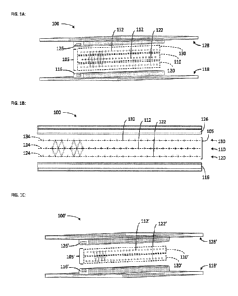
1
CA 03236350 2024- 4- 25
WO 2023/086169
PCT/US2022/045514
(6) risk of non-union of the bone; (7) risk of neurovascular injury, etc.
Accordingly, improved implant
devices, systems, and methods that can alleviate some, or all, of these
negative side effects would be
desirable.
SUMMARY
100061
The various implant devices, systems, and methods of the present disclosure
have been
developed in response to the present state of the art, and in response to the
problems and needs in the
art that have not yet been fully solved by currently available implant
devices, systems, and methods. In
some embodiments, the implant devices, systems, and methods of the present
disclosure may provide
improved rotational correction of the long bones of the lower extremities.
[0007]
In some embodiments, a tether assembly may be attached to a bone to correct
a rotational
deformity in a bone, such as femoral anteversion. The bone may have a growth
plate that separates a
first section of the bone from a second section of the bone. The tether
assembly may have a tether
member with a first end, a second end, and a central portion extending between
the first end and the
second end. The first end may have a closed outer wall that defines and fully
bounds a first aperture.
The second end may have an open outer wall that defines and partially bounds a
second aperture. The
open outer wall may define a slot in communication with the second aperture.
The first and second
ends may be securable to the first and second sections of the bone via
coupling members inserted
through the first and second apertures and anchored in the first and second
sections, respectively.
[0008]
The tether assembly may further include the first coupling member, which
may have a first
head and a first shank with a first bone engagement feature configured to
retain the first shank in the
bone. The tether assembly may further include the second coupling member,
which may have a second
head and a second shank with a second bone engagement feature configured to
retain the second shank
in the bone.
[0009]
The second shank may have a second shank width and the second head may have
a second
head width. The second shank width and/or the second head width may not be
smaller than a
corresponding portion of the slot through which it must pass in order to exit
the second aperture through
the slot, such that the second coupling member is movable through the slot
only in response to exertion
of a threshold level of tension between the second coupling member and the
second end.
[0010]
The corresponding portion of the slot may be configured to deform
elastically to permit
passage of the second shank therethrough in response to exertion of the
threshold level of tension.
[0011]
At least one of the first head and the second head may have a spherical
surface. The
corresponding one of the first aperture and the second aperture may have a
complementary spherical
surface sized to receive the spherical surface to provide adjustable
positioning of the first head or the
second head relative to the first aperture or the second aperture.
[0012]
The central portion may have a contoured shape created by projecting an
elongate area
defined on a sagittal plane onto a medial epicondylar bone surface or a
lateral epicondylar bone surface
2
CA 03236350 2024- 4- 25
WO 2023/086169
PCT/US2022/045514
of a pediatric distal femur. The elongate area may have a long axis and a
short axis orthogonal to the
long axis.
[0013] The long axis as measured on the sagittal plane may be
positioned at an angle relative to a
transverse plane. The angle may be within the range of 30 to 60 .
[0014] The slot may be oriented nonperpendicular to a
longitudinal length of the central portion.
[0015] The slot may be oriented at an angle relative to the
longitudinal length. The angle may be
within the range of 30 to 80 .
[0016] In some embodiments, a tether assembly may be attachable
to a bone to correct a rotational
deformity in a bone, such as femoral anteversion. The bone may have a growth
plate that separates a
first section of the bone from a second section of the bone. The tether
assembly may have a first
coupling member with a first head and a first shank with a first bone
engagement feature configured to
retain the first shank in the bone. The tether assembly may further have a
second coupling member
with a second head and a second shank with a second bone engagement feature
configured to retain the
second shank in the bone. The tether assembly may further have a tether member
with a first end, a
second end, and a central portion extending between the first end and the
second end. The first end
may be configured to engage the first head to nonrcleasably secure the first
end to the first section of
the bone. The second end may be configured to engage the second head to
rcleasably secure the second
end to the second section of the bone such that, in response to exertion of a
threshold level of tension
between the second coupling member and the second end, the second end is
released from the second
section of the bone.
[0017] The first end may have a fully-bounded first aperture. The
second end may have a partially-
bounded second aperture that is accessible via a slot.
[0018] The second shank may have a second shank width that is not
smaller than a slot width of
the slot and is movable through the slot in response to exertion of the
threshold level of tension.
100191 The slot may be configured to deform elastically to permit
passage of the second shank
therethrough in response to exertion of the threshold level of tension.
[0020] The slot may be oriented nonperpendicular to a
longitudinal length of the central portion.
[0021] At least one of the first head and the second head may
have a spherical surface. The
corresponding one of the first end and the second end may have a complementary
spherical surface
sized to receive the spherical surface to provide adjustable positioning of
the first head or the second
head relative to the first end or the second end.
[0022] The central portion may have a contoured shape created by
projecting an elongate area
defined on a sagittal plane onto a medial epicondylar bone surface or a
lateral epicondylar bone surface
of a pediatric distal femur. The elongate area may have a long axis and a
short axis orthogonal to the
long axis.
[0023] In some embodiments, a method may be used to perform
rotational deformity correction
on a bone with a growth plate that separates a first section of the bone from
a second section of the
3
CA 03236350 2024- 4- 25
WO 2023/086169
PCT/US2022/045514
bone. The method may include positioning a tether member of a tether assembly
on the bone. The
tether assembly may include a first coupling member with a first head and a
first shank with a first bone
engagement feature configured to retain the first shank in the bone, a second
coupling member with a
second head and a second shank with a second bone engagement feature
configured to retain the second
shank in the bone, and the tether member. The tether member may have a first
end, a second end, and
a central portion extending between the first end and the second end. The
method may further include,
with the first coupling member, securing the first end of the tether member to
the first section of the
bone, and, with the second coupling member, releasably securing the second end
of the tether member
to the second section of the bone such that, in response to exertion of a
threshold level of tension
between the second coupling member and the second end, the second end is
released from the second
section of the bone.
[0024] The first end may have a fully-bounded first aperture. The
second end may have a partially-
bounded second aperture that is accessible via a slot. Securing the first end
to the first section may
include inserting the first shank through the first aperture and anchoring the
first shank to the first
section. Releasably securing the second end to the second section may include
inserting the second
shank through the second aperture and anchoring the second shank to the second
section.
[0025] The slot may be oriented nonperpendicular to a
longitudinal length of the central portion.
[0026] Each of the first head and the second head may have a
spherical surface. Each of the first
end and the second end may have a complementary spherical surface. Securing
the first end to the first
section may include receiving the spherical surface of the first head in the
complementary spherical
surface of the first end to provide adjustable positioning of the first head
relative to the first end.
Securing the second end to the second section may include receiving the
spherical surface of the second
head in the complementary spherical surface of the second end to provide
adjustable positioning of the
second head relative to the second end.
100271 These and other features and advantages of the present
disclosure will become more fully
apparent from the following description and appended claims or may be learned
by the practice of the
devices, systems, and methods set forth hereinafter.
BRIEF DESCRIPTION OF THE DRAWINGS
[0028] Exemplary embodiments of the present disclosure will
become more fully apparent from
the following description taken in conjunction with the accompanying drawings.
Understanding that
these drawings depict only exemplary embodiments and are, therefore, not to be
considered limiting of
the scope of the present disclosure, the exemplary embodiments of the present
disclosure will be
described with additional specificity and detail through use of the
accompanying drawings in which:
[0029] FIG. lA is a posterior view of lower skeletal extremities
with normal anteversion.
[0030] FIG. 1B is a superior view of the right lower extremity in
FIG. 1A.
[0031] FIG. 2A is a posterior view of the lower skeletal
extremities with abnormal anteversion.
4
CA 03236350 2024- 4- 25
WO 2023/086169
PCT/US2022/045514
[0032] FIG. 2B is a superior view of the right lower extremity in
FIG. 2A.
[0033] FIG. 3A is a medial view of a right pediatric knee with a
tether assembly according to one
embodiment attached to the femur, at the time of surgeiy.
[0034] FIG. 3B is an inferior view of the femur of FIG. 3A.
[0035] FIG. 4A is a medial view of a right pediatric knee with
the tether assembly of FIG. 3A, at
a first period after the surgery.
100361 FIG. 4B is an inferior view of the femur of FIG. 4A.
[0037] FIG. 5A is a medial view of a right pediatric knee with
the tether assembly of FIG. 3A, at
a second period after the surgery.
[0038] FIG. 5B is an inferior view of the femur of FIG. 5A.
[0039] FIG. 6A is a top view of the tether assembly of FIG. 3A.
[0040] FIG. 6B is a side view of the tether assembly of FIG. 3A,
showing the tether member with
multiple potential orientations of each of the coupling members.
[0041] FIG. 6C is a close-up perspective view of the tether
member and one coupling member of
the tether assembly of FIG. 3A.
[0042] FIG. 7A is a top view of the tether member of the tether
assembly of FIG. 3A.
[0043] FIG. 7B is a side elevation, partial section view of the
tether member of FIG. 3A.
[0044] FIG. 8A is a side elevation view of a coupling member of
the tether assembly of FIG. 3A.
[0045] FIG. 8B is a side elevation, section view of the coupling
member of FIG. 8A.
100461 FIG. 9 is a table showing a relationship between
correction angle, femur width, start angle,
and treatment time for plate lengths of 20inm, 26inm and 32mm, respectively.
[0047] It is to be understood that the drawings are for purposes
of illustrating the concepts of the
present disclosure and may be drawn to scale, or may include variations from
scale drawings.
Furthermore, the drawings illustrate exemplary embodiments and do not
represent limitations to the
scope of the present disclosure.
DETAILED DESCRIPTION
[0048] Exemplary embodiments of the present disclosure will be
best understood by reference to
the drawings, wherein like parts are designated by like numerals throughout.
It will be readily
understood that the components of the present disclosure, as generally
described and illustrated in the
drawings, could be arranged, and designed in a wide variety of different
configurations. Thus, the
following more detailed description of the embodiments of the implants,
systems, and methods, as
represented in the drawings, is not intended to limit the scope of the present
disclosure, but is merely
representative of exemplary embodiments of the present disclosure.
[0049] The word "exemplary" is used herein to mean "serving as an
example, instance, or
illustration." Any embodiment described herein as "exemplary" is not
necessarily to be construed as
CA 03236350 2024- 4- 25
WO 2023/086169
PCT/US2022/045514
preferred or advantageous over other embodiments. While the various aspects of
the embodiments are
presented in the drawings, the drawings are not necessarily drawn to scale
unless specifically indicated.
[0050]
The following examples have been included to provide guidance to one of
ordinary skill in
the art for practicing representative embodiments of the presently disclosed
subject matter. In light of
the present disclosure and the general level of skill in the art, those of
skill in the art can appreciate that
the following examples are intended to be exemplary only and that numerous
changes, modifications,
and alterations can be employed without departing from the scope of the
presently disclosed subject
matter.
[0051]
It will be understood that any feature of any embodiment described or
contemplated herein
may be combined with any other embodiment that is described or contemplated
herein without
departing from the spirit or scope of the present disclosure.
[0052]
FIG. 1A is a posterior view (i.e. a view from a posterior viewpoint) of the
lower skeletal
extremities showing normal alignment of thc left extremity 20 and the right
extremity 22 in the pediatric
population. FIG. 1B is a superior view of the right extremity 22, showing the
anteversion of thc femoral
neck 30 and femoral head 32. It can be appreciated in FIG. 1B that the foot 34
is well aligned with the
knee 36 and the hip 38. Anteversion is the angle, or anteversion angle 40,
measured between a line 42
tangent to the posterior condyles of the distal femur, also referred to herein
as the transcondylar axis,
and a line 44 that bisects the femoral neck and head. In FIG. 1B, the
anteversion angle 40 is 110, which
is considered to be within the normal range in the general pediatric
population.
100531
FIG. 2A is a posterior view of the left extremity 20 and the right
extremity 22 showing an
abnormal alignment of the left extremity 20 and the right extremity 22 in the
pediatric population. FIG.
2B is a superior view of the right extremity 22, showing the anteversion of
the femoral neck 30 and
femoral head 32. It can be appreciated in FIG. 2B that the foot 34 and the
knee 36 are turned inward
relative to the hip 38. In FIG. 2B, the anteversion angle 50 is 410 (measured
between the line 44 that
bisects the femoral neck and head and a line 46 tangent to the posterior
condyles of the distal femur),
which is outside the normal range in the general pediatric population. This
abnormal anteversion is
observed as "in-toeing" of the feet, causing the left foot to be over rotated
in the clockwise direction
and the foot 34 to be over rotated in the counterclockwise direction, as
viewed when looking down at
the feet from a standing position. Abnormal anteversion is also referred to as
a rotational or torsional
deformity, as it represents an angular abnormality as viewed along the long
axis of the bone. It can
cause "in-toeing" as mentioned above, or splay the foot outward. Either can
beneficially be corrected
via the present disclosure. Although anteversion is used as a specific
example, those of skill in the art
will recognize that the techniques, implants, and principles taught by this
disclose may be applied to
other rotational deformities in femurs and/or other bones.
[0054]
FIG. 2A provides an example of a bilateral femoral rotational deformity,
which is common
when femoral rotational deformity is present; however, unilateral femoral
rotational deformities are also
occasionally present in the pediatric population. Although the preceding
discussion is limited to the
6
CA 03236350 2024- 4- 25
WO 2023/086169
PCT/US2022/045514
femur, it is understood that rotational defects can exist in other bones of
the extremities, such as the
tibia, and that the devices, systems and methods presented herein are equally
applicable to other
extremity bones, including but not limited to any other bones of the legs,
feet, arms or hands.
[0055]
FIG. 3A is a medial view of the distal portion of a femur 58 showing a
physis 60, also
referred to as a growth plate, and showing an embodiment of the present
invention. The femur 58 may
have an epiphyseal section 62 distal to the physis 60, and a metaphy seal
section 64 proximal to the
physis 60. The physis 60 may separate the epiphyseal section 62 from the
metaphyseal section 64. The
femur 58 may be a pediatric femur, which may continue to grow longitudinally
from the physis 60.
[0056]
Coupled to the distal portion of the femur 58 is a tether assembly 100,
which may include
a tether member 102, also referred to herein as a -bone plate," and two
coupling members 104, which
are also referred to herein as "bone screws." The tether member 102 may have a
first end 110, a second
end 112, and a central portion 114 extending between the first end 110 and the
second end 112.
[0057]
The tether member 102 may be fabricated from any of a variety of rigid
biocompatiblc
materials, such as but not limited to: stainless steel, titanium and its
alloys, nickel titanium alloy,
polyetheretherketone (PEEK), carbon fiber reinforced PEEK, biodegradable
polymers such as poly-L-
lactic acid (PLLA), and combinations of the foregoing. Alternatively, the
tether member 102 may be
formed of flexible biocompatiblc textiles, such as those used for sutures or
surgical meshes.
Alternatively, the tether member 102 may be a hybrid construct in which the
central portion 114 is
comprised of a flexible textile material and the first end 110 and the second
end 112 are comprised of
a rigid material. Alternatively, the tether member 102 may be formed of a
composite material using
any of the aforementioned polymers as a matrix and any of the aforementioned
textiles as fiber
reinforcement of the matrix. Composite material may be customized to provide
high stiffness and
strength in the direction of highest tensile stresses, such as along a
longitudinal axis of the tether member
102, but provide more flexibility and compliance in other directions, to allow
the tether member 102 to
better conform to the contours of the bone surface of the distal portion of
the femur 58. In some
embodiments, the tether member 102 may be resorbable.
[0058]
The coupling members 104 may be fabricated from any of the rigid
biocompatible materials
listed above for the tether member 102, so long as the material used for the
coupling members 104 is
electrochemically and mechanically compatible with the material used for the
tether member 102. The
coupling members 104 may be type of bone screws known in the orthopedic arts.
In alternative
embodiments (not shown), coupling members may include staples, suture anchors,
pins, tacks and/or
other bone fastening devices known to those skilled in the art. Coupling
members may also be made
resorbable if desired.
[0059]
The tether member 102 shown in FIG. 3A may be coupled to the medial side of
the distal
portion of the femur 58, which may be a right femur. The first end 110 and the
second end 112 of the
tether member 102 may each be secured to the femur 58 by one of the coupling
members 104.
7
CA 03236350 2024- 4- 25
WO 2023/086169
PCT/US2022/045514
[0060]
The central portion 114 of the tether member 102 may be elongated in shape,
with a length,
extending along a longitudinal axis 70 passing through the first end 110 and
the second end 112, that is
greater than its width transverse to the longitudinal axis. The first end 110
may have a closed outer
wall 120 and a first aperture 122 fully bounded by the closed outer wall 120.
The second end 112 may
have an open outer wall 124 and a second aperture 126 bounded by the open
outer wall 124. The open
outer wall 124 may define an opening, or slot 128, through which the
associated one of the coupling
members 104 may be removed from the second aperture 126 along a direction 130
that is generally in-
plane with, and nonperpendicular to, the tether member 102. By contrast, the
associated one of the
coupling members 104 retained within the first aperture 122 may only be
withdrawable from the first
aperture 122 along a direction generally perpendicular to the tether member
102 (i.e., out of the page,
in the view of FIG. 3A).
[0061]
As shown, the slot 128 may oriented nonparallel and/or nonperpendicular to
the length of
the central portion 114 of the tether member 102. Thus, an angle 132 may exist
between the direction
130 and the longitudinal axis 70 of the tether member 102. The angle 132 may
be a constant for all
patients, or may be selected on a patient-specific basis to control the
desired amount of anteversion
correction. The angle 132 may be greater than 00 and less than 90 . Further,
the angle 132 may be
greater than 20 and less than 70 . Yet further, the angle 132 may be greater
than 30 and less than 50 .
Still further, the angle 132 may be greater than 35 and less than 45 . In
some embodiments, the angle
132 may be about 40 .
100621
As will be described in greater detail below, the slot 128 may enable the
second end 112 to
be releasably secured to the epiphyseal section 62. "Releasable" securement
means the securement of
two items together in a surgical setting such that they can be detached from
each other by time or by
the body after the surgery is complete, rather than requiring another surgical
intervention to effect
release. By contrast, "nonreleasable" securement means the securement of two
items together in a
surgical setting such that they cannot generally be detached from each other
without another surgical
intervention. The first end 110 may be nonreleasably secured to the
metaphyseal section 64.
[0063]
Use of a slotted aperture is only one mechanism capable of providing
releasable
securement. In alternative embodiments, releasable securement may be provided
by making a tether
member or coupling member weak enough to break under the desired conditions.
For example, the
tether member 102 could be modified to make the central portion 114 much
thinner, and thus breakable
under tension. Alternatively, one of the coupling members 104 could be
modified to have a breakaway
head or the like.
[0064]
Returning to FIG. 3A, the longitudinal axis 70 may bisect the central
portion 114 of the
tether member 102. The tether member 102 may further have a third aperture 140
positioned
approximately at the mid-point of the longitudinal axis 70. The third aperture
140 may be used to
position the tether member 102 centrally over the physis 60 by aligning the
third aperture 140 with the
phy-sis 60. This may be done visually by a surgeon installing the tether
member 102, for example, by
8
CA 03236350 2024- 4- 25
WO 2023/086169
PCT/US2022/045514
ensuring that the physis 60 is visible through the third aperture 140 when the
tether member 102 is
seated on the distal end of the femur 58. Additionally or alternatively, a
guidewire or other instrument
may be registered on or near the physis 60 and inserted through the third
aperture 140 and into contact
with the physis 60 to guide placement of the tether member 102 such that the
third aperture 140 is
placed on or near the physis 60.
[0065]
The longitudinal axis 70 may be at an alignment angle a to a transverse
axis 72 that is
aligned with an anatomic transverse plane that is perpendicular to the
longitudinal axis 74 of the femur
58. Angle a is referred herein as the "initial alignment angle."
[0066]
Once the tether member 102 has been properly positioned on the femur 58,
one of the
coupling members 104 may be placed through the first aperture 122 to couple
the tether member 102
to the anterior portion of the metaphyseal section 64 of the femur 58,
proximal to the physis 60. Another
of the coupling members 104 may be placed through the second aperture 126 to
couple the tether
member 102 to the posterior portion of the epiphyscal section 62, distal to
the physis 60 and posterior
to the first end 110. The coupling members 104 may advantageously be placed a
minimum distance of
6mm to 8mm away from the central portion of the physis 60 to ensure that the
coupling members 104
do not impede or interfere with the natural growth emanating from the physis
60. In alternative
embodiments, the tether member 102 may be reversed, such that the first end
110 is secured to the
metaphyseal section 64 and the second end 112 is secured to the epiphyseal
section 62.
[0067]
FIG. 3B shows the femur 58 of FIG. 3A without the tether assembly 100. It
can be
appreciated in FIG. 3A that the femur 58 has abnormal anteversion; the degree
of abnormality is shown
as the angle e in FIG. 3B. This angle e is the amount of anteversion in excess
of the normal anteversion
angle of 110. The angle 0 may be the anteversion angle 50 of FIG. 2B minus the
anteversion angle 40
(110) of FIG. 1B. It is desirable to correct the rotational deformity by
rotating the distal end of the
femur 58 by 0 relative to the proximal end of the femur 58, so that the
posterior condy les of the femur
58 are restored to a normal alignment with the femoral neck and head of the
femur 58. To ensure that
the induced rotational change in the femur 58 is radially symmetric, a second
tether assembly (not
shown) may advantageously be placed on a second side of the femur 58. In the
case of the distal end
of the femur 58, the second tether assembly may be placed on the lateral side
of the femur 58, opposite
and radially symmetrical to the placement of the tether assembly 100 on the
medial side as shown in
FIG. 's 3A, 4A and 5A.
[0068]
Those of skill in the art will recognize that the use of two tether
assemblies is optional. In
some embodiments, only a single tether assembly may be used. A single tether
assembly may be placed
on the lateral, medial, anterior, or posterior sides of the femur 58, or even
on the postero-lateral, antero-
lateral, postero-medial, or antero-medial sides of the femur 58. In
alternative embodiments, more than
two tether assemblies may be used. In such cases, the tether assemblies may
optionally be arranged
and oriented in radially-symmetrical fashion about the distal end of the femur
58, and may be placed
on any of the sides of the femur 58 set forth above. In further alternative
embodiments, two tether
9
CA 03236350 2024- 4- 25
WO 2023/086169
PCT/US2022/045514
assemblies may be used, and may be arranged differently than described above.
For example, the tether
assemblies need not necessarily be placed on the lateral and medial sides of
the femur 58, but may be
placed on any of the sides set forth above. Again, radial symmetry is
optional.
[0069]
FIGS. 3A and 3B illustrate the femur 58 and the tether assembly 100 at the
time of a surgery
in which thc tether assembly 100 (and optionally one or more additional tether
assemblies) arc initially
installed. FIGS. 4A and 4B Illustrate the femur 58 and tether assembly 100
shown in FIG. 's 3A and
3B after a first period of time following the surgery. During the first period
of time, natural growth of
the femur 58 may occur, increasing the longitudinal spacing between the
epiphyseal section 62 and the
metaphy seal section 64. Due to the constraint of the tether assembly 100 (and
optional one or more
additional tether assemblies), some of the longitudinal growth of the femur 58
may be converted to a
relative rotation, about the longitudinal axis 74, between the epiphyseal
section 62 and the metaphyseal
section 64, thereby reducing and eventually eliminating the abnormal
anteyersion of e that was
initially present in the femur 58. The relative rotation between the
epiphyscal section 62 and the
metaphy seal section 64 may cause the tether member 102 to rotate from the
initial alignment angle a
(as shown in FIGS. 3A and 3B), relative to the transverse axis 72, to a
terminal intermediate alignment
angle fi (as shown in FIGS. 4A and 4B).
[0070]
Upon reaching the terminal alignment of p , the changed alignment (relative
to the
transverse plane) of the slot in the tether member is such that the slot 128
of the second aperture 126 is
oriented to permit withdrawal of the associated one of the coupling members
104 from the second
aperture 126 in response to continued longitudinal growth of the femur 58.
Thus, any further
longitudinal growth of the femur 58 may cause the associated one of the
coupling members 104 to move
along the direction 130 to escape the second aperture 126. This may release
the tether member 102
from attachment to the epiphyscal section 62, thereby permitting the femur 58
to elongate without
further rotational adjustment.
100711
The femur 58 shown in FIGS. 5A and 5B illustrates the femur 58 of FIGS. 3A,
3B, 4A and
4B after a second period following the surgery, where the second period is
greater than the first period.
During the interval of time after the first period until the end of the second
period, additional
longitudinal growth of the femur 58 has occurred, causing the one of the
coupling members 104
previously captured in the second aperture 126 to traverse the length of the
slot 128 of the second
aperture 126 and to translate outside the outer perimeter of the second end
112 of the tether member
102. Thus, the amount of rotational correction as measured by the angle e can
be "programmed" into
the surgical technique by selecting the right combination of a, 13, and the
distance 150 between the first
aperture 122 and the second aperture 126 of the tether member 102 (referred to
herein as "plate length")
for a given diametrical width of a distal femur. The foregoing list of
dimensional parameters have
analytical geometry relationships that can be expressed in equation form to
determine the right
parameter values to achieve a target rotational correction angle e.
CA 03236350 2024- 4- 25
WO 2023/086169
PCT/US2022/045514
[0072]
It can be appreciated that the femur in FIG. 5B has an unchanged rotational
alignment when
compared to the femur in FIG. 4B, as the one of the coupling members 104
formerly captured in the
second aperture 126 has escaped the tether member 102, and thus the constraint
that forced the prior
rotational change is no longer in effect. Furthermore, it may be advantageous
to provide an "automated"
removal of the constraint imposed by the tether member 102 once the rotational
deformity in the femur
58 is corrected and before the tether member 102 migrates to a more vertical
alignment with respect to
the transverse plane. If the tether member 102 were to continue to constrain
the distance between the
coupling members 104 as it achieved vertical alignment, the tether member 102
would arrest further
longitudinal growth of the femur 58. Indeed, such a constraint is known in the
clinical literature as
"shutting down the growth plate," a condition that permanently disables the
ability of the growth plate,
or physis 60, to generate new bone to continue the natural growth of the bone.
Such a clinical condition
can be very deleterious to the child, as it could lead to leg length
discrepancies or failure to achieve
normal height in adulthood.
[0073]
FIG. 6A is a top view of the tether assembly 100 of FIG. 3A, in isolation.
The coupling
members 104 are positioned in the first aperture 122 and the second aperture
126. FIG. 6B is a side
view of the tether assembly 100 of FIG. 3A, illustrating how the coupling
members 104 can articulate
with the first aperture 122 and the second aperture 126 to allow a multitude
of relative orientations
between the tether member 102 and the coupling members 104. The aforementioned
articulation may
be achieved by having spherical surfaces 160 on the coupling members 104 and
complementary
spherical surfaces 162 on the first aperture 122 and the second aperture 126.
The phrase "spherical
surface" will be understood to require not an entire sphere, but any three-
dimensional portion of a
concave or convex spherical shape.
[0074]
Each of the complementary spherical surfaces 162 may be a concave spherical
segment
defined between two spaced-apart parallel planes. One of these planes may be
defined by the top
surface of the first end 110 or the second end 112, as applicable, and the
other may pass through the
space between this top surface and the associated bottom surface. The second
aperture 126 may further
be bounded by a plane positioned at an angle to the two parallel planes to
create the slot 128. A close-
up view of the slot 128 is provided in FIG. 6C.
[0075]
It is noted that the relative alignment of the mid-range articulation
position 170 of each of
the coupling members 104 relative to the tether member 102 is at a divergent
angle. This is to help
ensure that the coupling members 104 are directed away from the physis 60 when
the tether member
102 is attached to the femur 58, as placement of the coupling members 104 into
the physis 60 could
inhibit the bone growth from the physis 60. While FIG. 6B shows the vertical
range for orienting the
coupling members 104 relative to the tether member 102, due to the spherical
articulation described
above, a similar range of motion may be present in all planes containing the
axis 172 of each of the
coupling members 104 located at its mid-range articulation position 170.
11
CA 03236350 2024- 4- 25
WO 2023/086169
PCT/US2022/045514
[0076]
It can be further appreciated in FIG. 's 6A and 6B that the tether member
102 has a three-
dimensional contour. This contour may be selected to match that of the medial
or lateral epicondylar
bone surface, on which the tether member 102 is to be attached. Since the
tether member 102 is to
rotate on the associated bone surface in the course of correcting the
anteversion of the bone, the tether
member 102 may be contoured to match a portion of the bone that is angled
between the initial and final
alignment angles (for example, angled between a as shown in FIG. 3A and 13 as
shown in FIG. 4A).
The contour that is to be matched may thus be the portion of bone that will be
overlaid by the tether
member 102 partway through the anteversion correction process.
[0077]
The central portion 114 of the tether member 102 may have a central bend
180 that exists
as part of this contouring. The third aperture 140 may pass through the
central bend 180. Further, the
central portion 114 may have a central twist such that the first end 110 and
the second end 112 are not
in the same plane. As a result, the second aperture 126 may have an axis 176
that is not parallel to the
axis 174 of the first aperture 122 (as shown in Fig. 7B).
[0078]
In somc embodiments, this contour may be created by projecting an elongate
area defined
on a sagittal plane onto one of the medial and lateral epicondylar bone
surfaces of a representative
pediatric distal femur, such as the femur 58. The elongate area may have a
long axis and a short axis
orthogonal to the long axis. The long axis, measured on the sagittal plane,
may be positioned at an angle
relative to a transverse plane, wherein the angle is less than 70 . The long
axis is generally between
20 and 70 , preferably between 30 and 60 , and more preferably between 40
and 50 . The long axis
may be at about 45 .
[0079]
FIG. 7A shows another top view of an embodiment of the tether member 102 of
the tether
assembly 100 of FIG. 3A, and FIG. 7B is a side elevation, partial sectional
view of the tether member
102. As shown, the slot 128 may have side walls 190 and a slot axis 192 that
is located between the
side walls 190 and bisects the side walls 190. The section view shows the
portion of the complementary
spherical surface 162 of the second aperture 126 and also shows the side wall
190 on one slide of the
slot 128. A portion of the slot 128 has a slot width 194 that is smaller than
the diameter 196 of the
second aperture.
[0080]
FIG. 8A is a side elevation view of one of the coupling members 104 of the
tether assembly
100 of FIG. 3A. FIG. 8B is a side elevation, section view of one of the
coupling members 104. Each
of the coupling members 104 may be a bone screw or other bone fastening device
of any type known
in the orthopedic arts. As shown, each of the coupling members 104 may have a
head 200 and a shank
202 extending from the head 200. The shank 202 may have a plurality of bone
engagement features
extending therefrom. As embodied in FIG. 8B, the bone engagement features may
be screw threads
204.
[0081]
The shank 202 may have a shank diameter 206 that is larger than the slot
width 194 and
smaller than the diameter 196 of the second aperture 126. The spherical
surfaces 160 of the coupling
members 104 may be on the head 200 of each of the coupling members 104. The
spherical surfaces
12
CA 03236350 2024- 4- 25
WO 2023/086169
PCT/US2022/045514
160 may mate with the complementary spherical surface 162 of the first
aperture 122 and the second
aperture 126 to enable polyaxial articulation as set forth above. The relative
sizing between the shank
diameter 206 and the diameter 196 of the second aperture 126 may enable the
spherical articulation
between the second end 112 of the tether member 102 and the associated one of
the coupling members
104 as demonstrated in FIG. 6B.
[0082]
The shank 202 may be positioned in the second aperture 126 such that the
shank diameter
206 is aligned with the slot 128 as shown in FIG. 6C. Thus, the shank 202 can
pass through the slot
128, as described above. However, the relative sizing between the shank
diameter 206 and the slot
width 194 may be selected such that a threshold force must be applied between
the tether member 102
and the shank 202 before the shank 202 passes through the slot 128.
Specifically, the relatively smaller
size of the slot width 194 relative to the shank diameter 206 may be selected
such that when a threshold
force is applied to the shank 202 in a direction aligned with the slot axis
192, the shank 202 will
elastically deform (i.e., the resulting strain is below the yield strain of
the material) the side walls 190
of the slot 128, thus permitting the shank 202 to escape from the second
aperture 126 via the slot 128.
[0083]
Because escapement of the shank 202 from the tether member 102 induces
fully
recoverable strain in the tether member 102, the shank 202 can be reengaged
with the second aperture
126 with no loss in the threshold force required to induce another escapement.
Furthermore, the
threshold force may be selected such that it induces a corresponding
restraining force on the physis 60
that is below the force that would induce growth from the physis 60 to "shut
down." Alternatively,
another embodiment (not shown) may have an escapement configuration that
causes the slot side walls
to permanently deform upon escapement of the coupling member from the tether
member.
[0084]
In addition to or in the alternative to interference between the shank 202
and the slot 128,
the head 200 may interfere with passage of the head 200 through the slot 128.
Specifically, the head
200 may have a head width 208, shown in FIG. 8A, at a portion of the head 200
that also passes through
the slot 128, in addition to the shank 202. As more clearly shown in FIG. 7B,
the slot 128 may have a
shank-contacting portion 210 that will lie adjacent to the surface of the
femur 58, and a head-contacting
portion 212 further from the femur 58.
[0085]
The shank-contacting portion 210 may provide interference with the shank
202 as described
above. However, in some embodiments, given the poly axially-adjustable
adjustability of the orientation
of the shank 202 relative to the tether member 102, interference between the
shank 202 and the shank-
contacting portion 210 may provide relatively unpredictable pullout force.
Specifically, if the shank
202 is angularly displaced from perpendicularity with the tether member 102,
the pullout force may be
higher than if the shank 202 is perpendicular to the tether member 102.
[0086]
Accordingly, it may be beneficial to have interference between the bead 200
and the head-
contacting portion 212. Thus, the head width 208 may be equal to or larger
than the width of the head-
contacting portion 212, at the depth at which the head 200 is to contact the
head-contacting portion 212.
FIG. 7A illustrates that the second aperture 126 may sweep across more than
180'. Thus, in order for
13
CA 03236350 2024- 4- 25
WO 2023/086169
PCT/US2022/045514
the head 200 to escape the second aperture 126 and enter the slot 128, the
head 200 may have to push
hard enough on the adjoining shoulders 214 of the head-contacting portion 212
to push them apart,
thereby increasing the width of the head-contacting portion 212 of the slot
128. The head 200 may then
enter and pass through the slot 128, permitting the second end 112 to
disengage from the corresponding
one of the coupling members 104 as described above.
[0087]
FIG. 9 is a table 250 illustrating the start angle a (in degrees) and
approximate treatment
time (in months) that correspond to a rotational correction angle 0 (in
degrees), an average distal femur
width (in mm) for a given length plate (in mm). The plate length provided in
the table 250 is the distance
150 between a center of the first aperture 122 and a center of the second
aperture 126, as shown in FIG.
5A.
[0088]
All of the values in the table 250 are based on a constant end angle 13 of
70 . Using
analytical geometry, similar tables can be developed for other values of end
angle, start angle, treatment
timc, femur width, rotational correction angle and plate lengths as needed.
For more severe deformities
than provided in the table 250, multiple treatments may be applied to the same
patient. For example,
for a patient having 35 of rotational deformity and a 54mm femur width, the
surgeon can apply the
tether member 102 at a start angle of 43 to achieve 20 of rotational
correction in approximately 7
months. Then in a subsequent surgical procedure, the surgeon can remove and
reapply the coupling
members 104 and the tether member 102 (or a tether member 102 with a different
size and/or contour)
at a start angle of 52 to achieve another 15 of rotational correction in
approximately 5 months. Thus,
the patient undergoes a total of 35 of rotational correction in approximately
12 months.
[0089]
The foregoing disclosure describes only selected embodiments encompassed
within the
scope of the disclosure. Those of skill in the art will recognize that the
principles taught herein may be
applied to generate many alternative concepts. For example, various clips,
clasps, staples, plates,
screws, and/or other fastening systems may be used to secure two sections of a
bone together on either
side of a growth plate. Such fastening systems may be made deliberately
releasable, through detachable
connections and/or breakable components, to effect release when the desired
anteversion correction has
been obtained.
[0090]
Reference throughout this specification to an embodiment" or the
embodiment" means
that a particular feature, structure, or characteristic described in
connection with that embodiment is
included in at least one embodiment. Thus, the quoted phrases, or variations
thereof, as recited
throughout this specification are not necessarily all referring to the same
embodiment.
[0091]
Similarly, it should be appreciated that in the above description of
embodiments, various
features are sometimes grouped together in a single embodiment, figure, or
description thereof for the
purpose of streamlining the present disclosure. This method of disclosure,
however, is not to be
interpreted as reflecting an intention that any embodiment requires more
features than those expressly
recited in that embodiment. Rather, inventive aspects lie in a combination of
fewer than all features of
any single foregoing disclosed embodiment.
14
CA 03236350 2024- 4- 25
WO 2023/086169
PCT/US2022/045514
[0092]
As used herein, the term "proximal" means a location at the end of a part
that faces a user
when the user is installing the part. The term "distal" means a location at
the opposite end of the
proximal end. For example, when a user installs a bone screw into a material
with a driver, the end of
the bone screw engaged with the driver is the proximal end, and the tip of the
bone screw that first
engages the material is the distal end. The term "caimulated" means having a
central bore extending
along a longitudinal axis of a part between a proximal end and a distal end of
the part.
100931
Recitation of the term "first" with respect to a feature or element does
not necessarily imply
the existence of a second or additional such feature or element. Elements
recited in means-plus-function
format are intended to be construed in accordance with 35 U.S.C. 112(f). It
will be apparent to those
having skill in the art that changes may be made to the details of the above-
described embodiments
without departing from the underlying principles set forth -herein.
[0094]
The phrases "connected to," "coupled to" and "in communication with" refer
to any form
of interaction between two or more entities, including mechanical, electrical,
magnetic,
electromagnetic, fluid, and thermal interaction. Two components may be
functionally coupled to each
other even though they are not in direct contact with each other. The term
"coupled" can include
components that arc coupled to each other via integral formation, as well as
components that arc
removably and/or non-removably coupled with each other. The term "abutting"
refers to items that may
be in direct physical contact with each other, although the items may not
necessarily be attached
together. The phrase "fluid communication" refers to two or more features that
are connected such that
a fluid within one feature is able to pass into another feature. As defined
herein the term "substantially"
means within +1- 20% of a target value, measurement, or desired
characteristic.
[0095]
While specific embodiments and applications of the present disclosure have
been illustrated
and described, it is to be understood that the scope of this disclosure is not
limited to the precise
configuration and components disclosed herein. Various modifications, changes,
and variations which
will be apparent to those skilled in the art may be made in the arrangement,
operation, and details of the
devices, systems, and methods disclosed herein.
CA 03236350 2024- 4- 25