Note: Descriptions are shown in the official language in which they were submitted.
CA 03236730 2024-04-25
WO 2023/076974
PCT/US2022/078750
DEVICES FOR CELL SEPARATION
Government Support
This invention was made with Government support under contract 75F40119C10158
awarded by the Food & Drug Administration. The Government has certain rights
in the invention.
Field
The present invention relates to devices and methods suitable for cell
separation. The
devices herein include non-random voids interconnected through non-random
pores and/or non-
random solid geometrical structures optionally connected through solid non-
random
interconnecting elements. Such devices are preferably suitable for affinity-
based cell isolation
techniques which rely upon binding interactions.
Background
Cell purification remains an important tool to improve on the separation of
cells for
therapeutic, diagnostic and research purposes. Cell purification methods have
utilized non-affinity
methods that rely on the physio-chemical properties of cells such as their
size, shape, and density.
Techniques that rely upon non-affinity methods include density gradient
centrifugation,
dielectrophoresis, sonication, and filtration. Affinity methods, which focus
on binding
interactions, can include chromatography, fluorescence activated cell sorting
(FACS) and
magnetic-activated cell sorting (MACS).
Accordingly, there remains a continuing focus on the development of new
devices and
methods for improving cell isolation that would, e.g., offer relatively large
and controlled surface-
volume ratios and optimized geometric and spatial properties that can be
optimized for a given cell
purification protocol.
Summary
A method to separate one or more targeted cells from a plurality of cells
comprising:
(a) supplying a device comprising biocompatible polymer material having a
plurality of
voids having a diameter D and a plurality of pore openings between said voids
having
1
CA 03236730 2024-04-25
WO 2023/076974
PCT/US2022/078750
a diameter d, including a surface area for cell separation, wherein 90% or
more of said
voids have a selected void volume (V) that does not vary by more than +/-
10.0% and
90% or more of said pore openings between said voids have a value of d that
does not
vary by more than +/- 10.0%;
(b) providing a surface coating on said voids wherein said surface coating
provides for cell
binding;
(c) passing a plurality of cells in a fluid through said device to provide a
fluid output; and
(1) capturing one or more selected cells from said plurality of cells on the
coated
surface of said device; or
(2) capturing the one or more selected cells from the plurality of cells in
said fluid
output.
A method to separate one or more targeted cells from a plurality of cells
comprising:
(a) supplying a device comprising biocompatible polymer material having a
plurality of
solid geometrical structures wherein 90% or more of said geometrical
structures have
a volume (V) that does not vary by more than +/- 10.0%;
(b) providing a surface coating on said outer surface of the geometrical
structures wherein
said surface coating provides for cell binding;
(c) passing a plurality of cells in a fluid through said device to provide a
fluid output; and
(1) capturing one or more selected cells from said plurality of cells on the
coated
surface of said device; or
(2) capturing the one or more selected cells from the plurality of cells in
said fluid
output.
A device for cell separation comprising solid geometrical structures have
outer surfaces
wherein 90% or more of said solid geometrical structures have a volume (V)
that does not vary by
more than +\- 10.0 %, including a surface coating on said outer surfaces
wherein said surface
coating provides for cell binding; a plurality of solid interconnecting
elements between said solid
geometrical structures wherein 90% or more of said solid interconnecting
elements define a
volume that does not vary by more than +\- 10.0 %; and an inlet and outlet to
allow for inflow and
outflow of a fluid containing cells for separation.
2
CA 03236730 2024-04-25
WO 2023/076974
PCT/US2022/078750
Brief Description of the Drawings
FIG. lA illustrates a sectional view of a first cell separation device of a
first configuration
herein containing non-random voids and non-random openings or pores.
FIG. IB illustrates a unit negative model of the cell separation device of the
first
configuration that shows the overlapping of neighborhood spheres.
FIG. IC illustrates a unit negative model with each sphere (defining a void
region)
surrounded by 12 identical neighborhood spheres.
FIG. ID illustrates the device of the first configuration and its fixed bed
geometry showing
an interconnected void system.
FIG. lE illustrates the device of the first configuration in cross-sectional
view.
FIG. IF illustrates in 2D view the device of the first configuration
containing non-random
spherical voids and their overlapping areas to form the interconnected
openings or pores between
such spherical voids.
FIG. 2A illustrates a portion of the device herein of the second
configuration.
FIG. 2B illustrates a portion of the device herein of the second
configuration.
FIG. 2C illustrates a preferred configuration of the device herein of the
second
configuration.
FIG. 2D illustrates a preferred shape of the device herein of the second
configuration
wherein the non-random geometrical shapes include oval structures.
FIG. 2E illustrates a preferred shape of the device herein of the second
configuration
wherein the non-random solid geometrical structures include a polygonal
structure.
FIG. 2F illustrates another preferred shape of the device of the second
configuration
wherein the solid interconnecting structures include a polygonal shape.
FIG. 3 illustrates a portion of the device of the second configuration
illustrating the
exemplary use of spheres and optional use of interconnecting rod elements.
3
CA 03236730 2024-04-25
WO 2023/076974
PCT/US2022/078750
FIG. 4 illustrates the device of either the first or second configuration
positioned in a
housing between an inlet and outlet for inflow and outflow of fluid containing
cells for separation.
FIG. 8 provides green fluorescent images A, B, C, D, E and F, of PBMCs non-
specifically
attached on PDA-mPEG coated test disks.
FIG. 6 is a graph that shows that 30 i.t.g/mL resulted in the highest areal
concentration of
bound antibodies (1.5 i.t.g/cm2 on the test coupon surface).
FIG. 7 illustrates the percent of PBMCs non-specifically attached on PDA pre-
coated test
disks.
FIG. 8 provides green fluorescent images of PBMCs non-specifically attached on
PDA-
mPEG coated test disks.
FIG. 9 shows percent of PBMCs non-specifically attached on test disks with
different
coatings.
FIGS. 10A and 10B provide a sequence of fluorescent images of the test group #
in Table
2, at the center (left column images A, B, C, D, E and F) and edge (right hand
column images A',
B', C', D', E' and F') of the test disks.
FIG. 11 shows the percentage of Jurkat cells remaining on the coupons in non-
specific and
specific binding cases.
FIG. 12 shows the fluorescent microscopic image of the cells in FIG. 11 due to
non-
specific attachment.
FIG. 13 shows the fluorescent microscopic image of the cells in FIG. 11 due to
specific
attachment.
FIG. 14 shows the percentage of Jurkat cells remaining on the coupons in non-
specific
and specific binding cases.
FIG. 15 shows the fluorescent image Jurkat cells remaining on the coupons due
to non-
specific binding, where almost no cells attach to the disks.
FIG. 16 shows the fluorescent image of Jurkat cells remaining on the coupons
due to
specific binding.
4
CA 03236730 2024-04-25
WO 2023/076974
PCT/US2022/078750
Detailed Description of the Drawings
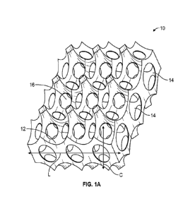
FIG. 1A illustrates a sectional view of a first cell separation device 10
herein containing
non-random voids 14 and non-random openings or pores 16. Reference to cell
separation may be
understood to include cell isolation and/or cell purification and/or cell
enrichment. More
specifically, the first device includes an interconnected 3D surface area 12
with non-random voids
14 which are preferably of spherical shape and preferably have internal
concave surfaces to
maximize the surface-to-volume ratio. A void is understood as an open space
with some identified
volume. By reference to non-random it should be understood that one can
identify a targeted or
selected number of voids and selected number of pores in the device that
results in a repeating void
size or pore size of a desired tolerance.
Accordingly, the device 10 includes non-random interconnecting pore openings
16 as
between the non-random voids. Again, reference to non-random should be
understood that one
can now identify a targeted or selected number of pores for the voids, of a
selected pore diameter,
that results in an actual number of pores having pore diameters of a desired
tolerance. The device
as illustrated in cut-away view also ultimately defines a layer of non-random
voids (see arrow "L")
and it may be appreciated that the multiple layers of the device may then
allow for identification
of a plurality of such non-random voids within a column (see arrow "C").
FIG. 1B illustrates a unit negative model of the device 10 that shows the
overlapping of
neighborhood spheres. The device is then preferably created by reversing the
negative model to
create the positive model comprising the interconnected void system. More
specific techniques
for forming the device 10 are discussed herein. FIG. 1C illustrates a unit
negative model with
each sphere (defining a void region) surrounded by 12 identical neighborhood
spheres. FIG. 1D
illustrates the device 10 fixed bed geometry showing an interconnected void
system. FIG 1E
illustrates the device 10 in cross-sectional view. It should be noted that
preferably, the device 10
has a diameter 40 in the range of 2.0 mm to 10,000 mm and a height H in the
range of 1.0 mm to
5,000 mm. Preferably the device 10 indicate a ratio 41:0/H in the range of
greater than 1:1.
FIG. 1F illustrates in 2D view the identified preferred non-random spherical
voids and
their overlapping areas to form the interconnected openings or pores between
such spherical voids.
For the preferred geometry illustrated in FIG. 1F, Spherical Void 1 is
represented by a solid circle,
5
CA 03236730 2024-04-25
WO 2023/076974
PCT/US2022/078750
diameter is D (indicated by the arrows). Diameter "D" may therefore be
understood as the longest
distance between any two points on the internal void surface. Spherical Void 2
is represented by
a dash circle and would also have diameter D (not shown). Spherical Void 2 is
one of the 12 of
neighborhood voids of Spherical Void 1. Due to the overlap between the
neighborhood voids, it
forms interconnected pores between the spherical voids, with the diameter of
"d" as also indicated
by the generally horizontal arrow. Diameter "d" may therefore be understood as
the longest
distance between any two points at the pore opening. The total 3D spherical
surface area of the
void is SAvmd = 4xnx(D/2)2. The surface area between A and B, called Scap =
axDxh, where h =
D-AID2-d2
2 . The useful void surface for a given void in the 3D bioreactor
would be SA u = SAvmd
[12xScap].
The smaller the void diameter D, the larger the number of voids can be packed
into a set
3D space (volume), and therefore results larger overall surface. The diameter
of the pores d may
fall in the range of 0.01 mm to 10.0 mm and more preferably 0.05 mm to 2.0 mm
as most
mammalian cell size is between 5 - 100 p.m Most preferably, d > 0.1 mm and
falls in in the range
of 0.1 mm to 2.0 mm.
If D = 0.018 mm or less, the computed SAõ is less than 0 when d = 0.01 mm,
which leads
to an impossible structure therefore, D has to be > 0.018 mm for this 3D
bioreactor geometry.
However, D can have a value in the range of 0.09 mm to 100.0 mm, more
preferably, 0.2 mm to
50.0 mm, and also in the range of 0.4 mm to 25.0 mm. Accordingly, for the
preferred geometry
illustrated in FIG. 1F, D > 0.4 mm (the diameter of the void) and d > 0.20 mm
(the diameter of
the pore openings). It is also worth noting that with respect to any selected
value of diameter D
for the voids in the range of 0.018 mm to 100.0 mm, and any selected value of
diameter d for the
pores in the range of 0.2 mm to 10.0 mm, the value of D is such that it is
greater than the value of
d (D>1.8d).
It can now be appreciated that the device 10 can be characterized with respect
to its non-
random characteristics. Preferably, the voids within the 3D bioreactor are
such that they have
substantially the same volume to achieve the most efficient 3D space packing
and offer the largest
corresponding surface area. With respect to the total number of interconnected
voids present in
any given cell purification device, preferably, 90.0 % or more of such voids,
or even 95.0 % or
6
CA 03236730 2024-04-25
WO 2023/076974
PCT/US2022/078750
more of such voids, or even 99.0 % to 100 % of such voids have a void volume
(V) whose tolerance
is such that it does not vary by more than +/- 10.0%, or +/- 5.0%, or +/- 2.5%
or +/- 1.0%, or +/-
0.5% or +/- 0.1%. It should be noted that while the voids in FIG. 1A are shown
as generally
spherical, as repeatedly noted, other void geometries are contemplated. The
diameter of voids are
therefore preferably chosen to optimize cell purification.
As noted, another non-random characteristic of the device 10 herein are the
pore openings
between the voids, having a diameter d (see again FIG. 1E). Similar to the
above, 90.0 % or more
of the pore openings, or even 95.0 % or more of the pore openings, or even
99.0 % to 100 % of
the pore openings between the voids, indicate a value of d whose tolerance
does not vary more
than +/- 10.%, or +/- 5.0%, or +/- 2.5% or +/- 1.0%, or +/- 0.5% or +/- 0.1%.
The diameter of pore
openings are preferably chosen to optimize unbound cells released from the
bioreactor.
It can therefore now by appreciated that the device 10 herein for cell
separation comprises
a plurality of voids having a diameter D (the longest distance between any two
points on the
internal void surface), a plurality of pore openings between said voids having
a diameter d (the
longest distance between any two points at the pore opening), where D>1.8d. In
addition, 90% or
more of the voids have a void volume (V) that does not vary by more than +/-
10.0%, and 90% or
more of the pore openings have a value of d that does not vary by more than +/-
10.0%.
In addition, the device herein for cell separation can include a first
plurality of voids having
a diameter Di, a plurality of pore openings between said first plurality of
voids having a diameter
di, wherein Di >di, where 90% or more of the first plurality of voids have a
void volume (Vi) with
a tolerance that does not vary by more than +/- 10.0%. Such device may also
have a second
plurality of voids having a diameter D2, a plurality of pore openings between
said second plurality
of voids having a diameter d2 wherein D2>d2, wherein 90% of the second
plurality of voids have
a void volume (V2) with a tolerance that does not vary by more than +/- 10.0%.
The values of Vi
and V2 are different and outside of their tolerance variations. Stated another
way, the value of Vi,
including its tolerance of +/- 10.0 % and the value of V2, including its
tolerance of +/- 10.0%, are
different, or [VI_ +/- 10.0%[ [V2 +/- 10.0%[.
The device herein for cell separation may also be constructed in a second
configuration
comprising a plurality of non-random solid geometrical structures and
optionally, a plurality of
7
CA 03236730 2024-04-25
WO 2023/076974
PCT/US2022/078750
non-random solid interconnecting elements between such structures. Such solid
geometrical
structures may preferably include spheres, ovals, and/or polygonal shapes,
thereby presenting an
outer surface for cell purification. As noted, such solid geometrical
structures may optionally be
connected via a plurality of solid interconnecting elements. Such solid
interconnecting elements
may also assume various geometrical shapes, including rod or columnar shape,
oval shape, and/or
polygonal type shape. Such solid interconnecting structures may also all
provide an outer surface
for cell separation. It should be noted that reference to solid geometrical
structures as well as solid
interconnecting elements is reference to the fact that such structures and
elements provide an outer
surface for cell separation as disclosed herein. The solid geometrical
structures or the solid
interconnecting elements themselves are not necessarily completely solid and
may contain a
partially hollow interior. Accordingly, the partially hollow interior may be
utilized to place
nutrients and/or other reagents and/or for gas transfer. Such nutrients and/or
reagents and gas
transfer may then operate to improve the performance of the devices herein for
cell separation.
FIGS. 2A and 2B illustrate a portion of a first preferred device 18 of the
second
configuration wherein the solid non-random geometrical structures and optional
solid non-random
geometrical interconnecting elements preferably comprise spheres 20 and
interconnecting rods 22.
FIG. 2C illustrates one preferred configuration for the device 18 where it can
be appreciated that
the plurality of spheres 20 and plurality of interconnecting rods 22 are
preferably organized in two
or more layers where each layer is offset from an adjacent layer. Such offset
of the layers can
therefore promote fluid flow through the device 18 to enhance the interaction
between the fluid
flow and the spheres 20. In addition, the plurality of spheres 20 can each be
preferably connected
by 4, 5, 6, 7, or 8 rods.
In addition, as illustrated in FIG. 2C, the device 18 preferably has a
diameter 4:1:0 in the
range of 2.0 mm ¨ 10,000 mm and a height H in the range of 1.0 mm ¨ 5,000 mm.
Preferably the
device 10 indicate a ratio 4:1:0/H in the range of greater than 1:1.
FIG. 2D illustrates a preferred shape of the device of the second
configuration wherein the
non-random solid geometrical shapes include oval structures 21 (outline of an
egg). FIG. 2E
illustrates another preferred shape of the device of the second configuration
wherein the non-
random solid geometrical structures include polygonal structure 23. FIG. 2F
illustrates another
8
CA 03236730 2024-04-25
WO 2023/076974
PCT/US2022/078750
preferred shape of the device of the second configuration wherein the solid
interconnecting
structures 25 between the spheres include a polygonal shape.
A portion of the device of the second configuration is illustrated in FIG. 3,
with respect to
the exemplary use of spheres and the optional use of interconnecting rod
elements. The non-
random solid geometrical shapes herein preferably have a diameter D' (longest
distance between
two points on the outer surface of the solid geometrical structure and through
the structure interior)
in the range of 2.0 p.m to 25.0 mm, 200 microns to 25.0 mm, 5.0 p.m to 10.0
mm, or 5.0 p.m to 6.0
mm. Another preferred value for D' is in the range 1.0 mm to 25.0 mm.
The solid geometrical interconnecting elements (ICE) preferably have a
diameter D" in
the range of 1.0 p.m to 12.5 mm, more preferably in the range of 1.0 p.m to
3.0 mm. The length of
the solid interconnecting structures ICEL preferably ranges from 0.1 p.m to
25.0 mm, more
preferably 100.0 p.m to 5.0 mm, and even more preferably, 100.0 p.m to 3.0 mm.
It is also preferred
that the diameter of the solid interconnecting structures (e.g. rods) are less
than half of the value
of the diameter of the solid geometrical shapes (e.g. spheres).
Similar to the first configuration of the device noted above, the second
configuration can
also be characterized by its overall non-random characteristics. That is, with
respect to the solid
geometrical structures (e.g., spheres 20), 90% or more of such solid
geometrical structures, or even
95.0% or more of such solid geometrical structures, or even 99.0% to 100% of
such solid
geometrical structures, define a volume whose tolerance is such that it does
not vary by more than
+/- 10.0%, or +/- 5.0%, or +/- 2.5% or +/- 1.0%, or +/- 0.5% or +/- 0.1%.
Similarly, with respect
to the optional use of the solid interconnecting elements (e.g., rods 20), 90%
or more of such solid
interconnecting elements, or even 95.0% or more of such solid interconnecting
elements, or even
99.0% to 100% of such solid interconnecting elements, define a volume whose
tolerance is such
that it does not vary by more than +/- 10.0%, or +/- 5.0%, or +/- 2.5% or +/-
1.0%, or +/- 0.5% or
+1-0.1%.
The device of the first configuration or second configuration are preferably
made of
biocompatible or bio-inert polymeric materials such as polystyrene,
polycarbonate, acrylonitrile-
butadiene-styrene (ABS), polylactic acid (PLA), polycaprolactone (PCL) used in
FDM (fused
deposition modeling) 3D printing technology. Reference to biocompatible or bio-
inert should be
understood as a material that is non-toxic to the culturing cells. In
addition, the polymeric materials
9
CA 03236730 2024-04-25
WO 2023/076974
PCT/US2022/078750
for the device of the first or second configuration are preferably selected
from those polymers that
at not susceptible to hydrolysis during cell cultivation, such that the amount
of hydrolysis does not
exceed 5.0 % by weight of the polymeric material present, more preferably it
does not exceed 2.5
% by weight, and most preferably does not exceed 1.0 % by weight. The device
of the first or
second configuration may also be made of biocompatible photosensitive
materials (e.g., Pro3Dure,
Somos WaterShed XC 11122, etc.) used in SLA (stereolithography) and DLP
(digital light
processing) 3D printing technologies. Furthermore, the device of the first or
second configuration
may be formed of an interpenetrating polymer network (IPN). An IPN is
reference to a polymer
comprising two or more networks which are at least partially interlaced on a
polymer scale but not
covalently bonded to each other.
It is preferable that the material used to fabricate the devices of either the
first or second
configuration herein are not degradable in aqueous medium and can provide a
mechanical stable
structure to tolerate aqueous medium flow during cell purification. It is
preferable that the material
and manufacturing process can result a solid and relatively smooth
interconnected surface area.
By reference to a solid surface, it should be further understood that the
surface is such that it will
preferably reduce or prevent penetration or embedding by cells, which
typically have a diameter
of about 20 microns to 100 microns. Preferably, the devices herein of either
the first or second
configuration have a surface that has a surface roughness value (Ra), which is
reference to the
arithmetic average of the absolute values of the profile height deviations
from the mean line,
recorded within an evaluation length. Accordingly, it is contemplated herein
that Ra of the devices
herein will have a value of less than or equal to 20 p.m, more preferably,
less than or equal to 5
!JIM
The devices of the first or second configuration herein are also preferably
formed from
material that indicates a Shore D Hardness of at least 10, or in the range of
10-95, and more
preferably in the range of 45-95. In such regard, it is also worth noting that
the devices herein
preferably do not make use of a hydrogel type structure, which may be
understood as a hydrophilic
type polymeric structure, that includes some amount of crosslinking, and which
absorbs significant
amounts of water (e.g., 10-40 % by weight). It is also worth noting that the
devices herein
preferably do not make use of collagen, alginate, fibrin and other polymers
that cells can easily be
digested and undergo remodeling.
CA 03236730 2024-04-25
WO 2023/076974
PCT/US2022/078750
Furthermore, the devices herein of the first or second configuration are
preferably made
from materials that have a Tensile Modulus of at least 0.01 GPa. More
preferably, the Tensile
Modulus has a value that is in the range of 0.01 GPa to 20.0 GPa, at 0.01 GPa
increments. Even
more preferably, the Tensile Modulus for the material for devices herein are
in the range of 0.01
GPa to 10.0 GPa or 1.0 GPa to 10 GPa. For example, with respect to the earlier
referenced
polymeric materials suitable for manufacture of the devices herein,
polystyrene indicates a Tensile
Modulus of about 3.0 GPa, polycarbonate at about 2.6 GPa, ABS at about 2.3
GPa, PLA at about
3.5 GPa and PCL at about 1.2 GPa.
The devices herein of either the first or second configuration with such
preferred regular
geometric characteristics and/or surface area are preferably fabricated by
additive manufacturing
technologies, such as fused deposition modeling FDM, selective laser sintering
(SLS),
stereolithography (SLA), digital light processing (DLP) 3D printing
technologies, etc., according
to computer generated designs made available by, e.g., a SolidWorksTM computer-
aided design
(CAD) program.
The devices of the first or second configuration may then be configured such
that they may
configured as a fixed bed along with an inlet and outlet to allow for inflow
and outflow of fluid.
Reference is made to FIG. 4 wherein the device of either the first or second
configuration noted
above may be positioned in a housing 24 and then placed between and inlet 26
and outlet 28 for
which inflow and outflow of fluid may be provided containing cells for
separation.
The surfaces of the device of the first or second configuration are preferably
coated and
functionalized such that they allow for selective ligand or cell binding.
Reference to cell binding
may therefore be understood to include a chemical interaction between the cell
and the coating,
such as covalent binding and/or secondary type binding (e.g. polar
interactions or hydrogen type
bonding). For example, in the case of the device of the first configuration,
exemplified by the
voids 14 illustrated in FIG. 1A, the surface of such voids can be so
functionalized, and in the case
of the device of the second configuration, the surfaces of the solid spheres
20 and interconnecting
elements 22 in FIG. 2C can also be similarly functionalized. When a mixed
population of cells
flows through the device of either the first or second configuration, with
such functionalized
surfaces, the binding of the cells allow for separation and capture of target
cells while allowing the
rest of the cells to pass through.
11
CA 03236730 2024-04-25
WO 2023/076974
PCT/US2022/078750
FIG. 5 illustrates the cell separation that can now be achieved with the
device herein of
either the first or second configuration when employed in a fluid flow. As
illustrated at 30 a
plurality of cells, e.g., types A, B and C, are preferably suspended in a
fluid that flows into the
device and through the fixed bed. Depending on the characteristics of the
surface coating on the
fixed bed, different levels of cell separation will be obtained.
For example, depending upon the surface coating, and in Case I, and as shown
at 32 in
FIG. 5, cell Types B and C are selectively captured by the surface coating of
the fixed bed. In
such manner, it can be appreciated that Cell Type A may therefore be said to
be selectively
separated from a plurality of cell types A, B and C. This is also understood
herein as negative cell
selection, in that the unwanted cells (B and C) are depleted from the mixture
and remain within
the device. As noted above, this also represents an affinity-based technique
of cell separation.
In addition, depending again on the surface coating, and in Case II, and as
shown at 34,
cell type B is separated and captured on the surface coating of the fixed bed,
and cell types A and
C flow out. This is known herein as positive cell selection, in that the
targeted cell B is retained
in the device and cells A and C pass through. In such manner, it can be
appreciated that cell type
B may therefore be said to be selectively separated from cell types A and C.
The above being the case, it should be appreciated that the device herein of
either the first
or second configuration with its available surfaces can be coated and
functionalized, such that a
selected cell can be separated, which as noted herein includes the feature
that the cell may be
isolated, purified, and/or enriched from a plurality of cells. In addition,
for a given plurality of
cells within a fluid that passes through the device herein of either the first
or second configuration,
the separation of a selected cell from the plurality of cells can be achieved
by: (1) capturing the
selected cell on the coated or functionalized surface of the devices herein;
and/or (2) capturing the
selected cell within the fluid output.
As further illustrated at 36 and 38, although the devices herein of either the
first and second
configuration can be provided with surface functionalization to provide for
the above referenced
affinity type cellular separation, some limited amount of non-affinity type
interactions may occur.
More specifically, at 36, although the surface was functionalized to capture
cells B and C, some
relatively small amount of capture of cell A may occur. At the same time, a
relatively small amount
of B or C cells may be present at the output of the device. In addition, as
shown at 38, although
12
CA 03236730 2024-04-25
WO 2023/076974
PCT/US2022/078750
the surface was functionalized to capture only cell B, some relatively small
amount of cell A may
also be immobilized within the device.
Therefore, the efficiency of cell separation that occurs with the devices
herein of either the
first or second configuration can be quantitatively described as selectively
isolating a targeted cell
from a plurality of cells at a level of greater than 50% of the total targeted
cells introduced into the
device. More preferably a selected or targeted cell can now be separated from
a plurality of cells
at a level of greater than 50% to 100%, more preferably at a level of 60% to
100%, or 70% to
100%, or 80% to 100%, or 90% to 100%, or 95% to 100% of the total targeted
cell introduced into
the device. Reference to such quantitative efficiency of cell separation from
a plurality of cells
introduced into the device occurs herein by: (1) capturing the selected or
targeted cell(s) on the
surface of the devices herein (or called positive selection); and/or (2)
providing the selected or
targeted cell(s) within the fluid output after passing through the devices
herein (also called negative
selection).
With regards to surface coating of the devices of either the first or second
configuration,
preferably, such coatings are those that may also provide for affinity-based
cellular capture. The
coatings may therefore preferably comprise substituted or unsubstituted poly(p-
xylylene) from the
polymerization of parylene monomers, 13-casein or polydopamine (PDA). Such
coatings may
preferably be present at a thickness in the range of 200 Angstroms to 100.0
p.m.
Accordingly, the coating procedure preferably relies upon the use of parylene
monomers,
e.g., [2.2(paracyclophanes, that may be preferably functionalized with
identified R1, R2, R3 and R4
groups according to the following general reaction scheme. It should be
appreciated that in the
scheme below, the start of polymerization is initiated by a ring opening at
elevated
temperature(-550 C) in the low pressure gas phase remotely prior to
deposition on the 3D device
which is preferably maintained at relatively lower temperature (e.g., < 100
C):
13
CA 03236730 2024-04-25
WO 2023/076974
PCT/US2022/078750
Ri R3
__________________________________________________ 7.-
R4
R2
R4
R3
R2
n
Ri
m
poly(p-xylylene)
In the above, when one of the R groups per repeat unit "m" and/or repeat unit
"n" is
chlorine, and the other R groups are hydrogen, the above represents the
polymerization of parylene
C. It is a USP Class VI and ISO-10993-6 certified biocompatible material. The
values of "m" and
"n" of the identified crosslinked, repeating units are such that molecular
weight values are
relatively high, such as ¨500,000. It is therefore contemplated that the use
of the parylene
monomers and ensuing polymeric coatings are such that one may now coat the
devices of the
above reference first or second configuration herein with an impermeable film.
The film may
preferably have a thickness between 200 Angstroms to 100.0 p.m. It may be
appreciated that R1,
R2, R3, and R4 may be selected from hydrogen, a halogen (-Cl or -Br) as well
as other functional
groups such as amines (-NH2), aliphatic aldehydes (-CHO), carboxylic acid
functionality (-
COOH), hydroxyl (-OH) or carboxylate functionality as in -C(0)CF3. One may
also initially coat
with a first layer of impermeable parylene C followed by a coating of a
different parylene, e.g.,
wherein R1, R2, R3, and R4 may then be selected from an amines (-NH2) and/or
aldehyde (-CHO)
functionality. Accordingly, one may provide polymeric coatings for the devices
herein of the first
and/or second configuration, wherein the coating comprises a plurality of
layers, each with its own
particular and different chemical composition (i.e. the identity of at least
one of R1, R2, R3, and R4
are different between at least two of the layers).
14
CA 03236730 2024-04-25
WO 2023/076974 PCT/US2022/078750
One preferred method of coating the surface of the devices herein with
functionalized
poly(p-xylylene) applies when one or more of the R1, R2, R3 and/or R4 groups
noted above
comprise ester carboxylic acid functionality. In such case, one may utilize N-
hydroxysuccinimide
(NHS) to form an ester linkage. Next, NH2-mPEG (methoxy terminated
oligoethylene glycol) or
NH2-PEG-biotin may be covalently bonded to the device surface via the amine-
NHS ester reaction
to form an amide bond. Then, avidin or NeutrAvidin or streptavidin can be
bound to the biotin.
As avidin/NeutrAvidin/streptavidin have four bonding sites, the remaining
three sites are then
available to bind biotinylated antibodies, such as anti-CD3 and anti-CD28 to
capture T cells
through surface receptors specific to these antibodies.
It is further contemplated that the cell separation devices herein of either
the first or second
embodiment, with a functionalized poly(p-xylylene) coating wherein one or more
of R1, R2, R3
and R4 comprise an aldehyde can undergo reaction with, e.g., antibody proteins
(e.g. anti-CD3/28)
with end flexible tethers that are amino terminated (or other organic terminal
group) of an
oligoethylene oxide (OEG) of different lengths. In other words, the use of OEG
type tethers that
include functional terminal groups such as an amine group, as in:
....../.......õ..õ...O.õ......._õ......
Antibody n NH2
where the value of n may be in the range of 1-200, and which may then bind to
the functionalized
parylene coating on the devices herein as follows, where one binding reaction
site is illustrated
and where it should be appreciated that multiple binding reactions may take
place depending upon
regulation of the reaction parameters (e.g. temperature and time to increase
binding reaction yield):
0I I 0I I 0ll
CH CH CH
1 1 1 )0
Antibody n NH2
Cell Separation Device
)1.
+ Functionalized Coating
CA 03236730 2024-04-25
WO 2023/076974
PCT/US2022/078750
0 0 N Antibody
11 11
I
CH CH CH
1 1 1
Cell Separation Device +
Functionalized Coating
As may therefore be appreciated, in the above, one may vary the identified
antibody to
target selected cell surface receptors, distinctive for a given cell phenotype
for a given cell
separation protocol. In addition, the antibody as shown may be surface inter-
dispersed with
poly(ethylene oxide) (PEG) and/or methyl terminated PEG (mPEG) of varying
molecular weights
to minimize non-specific adsorption.
In addition, in the case of poly-p-xylylene coating, it is contemplated that
one may
chemically modify the surface of such coatings via plasma activation where a
gas, such as oxygen,
ammonia or mixtures of these gases with volatile polymerizable monomers are
ionized by plasma
discharge and allowed to condense and form a functionalized coating on the
poly(p-xylylene)
structure.
In addition, it is contemplated that when the poly(p-xylylene) herein is not
functionalized,
one may apply a molecule containing a hydrophobic and hydrophilic end, which
is then coated on
the unfunctionalized poly-p-xylylene coating. The hydrophobic end of the
molecule is therefore
contemplated to coat the unfunctionalized poly-p-xylylene leaving the
hydrophilic end, containing
various reactive groups, capable of affinity bonding with a given cellular
surface. Along such
lines, it is contemplated that one may therefore utilize as one example, (3-
casein, which would then
be applied on the poly(p-xylylene) coating.
As also noted, the devices may be coated directly with PDA to facilitate the
binding of,
e.g., a biotin linker and/or mPEG layer. More specifically, one may form a PDA
coating on the
surfaces of the devices here which at pH 8.5 is converted to a diquinone type
structure which can
then undergo Michael addition and Schiff base formation. More specifically,
NH2-mPEG or NH2-
16
CA 03236730 2024-04-25
WO 2023/076974
PCT/US2022/078750
PEG-biotin can be immobilized on the PDA layer. The immobilized NH2-PEG-biotin
can then be
further conjugated with avidin/NeutrAvidin/streptavidin and then a
biotinylated antibody and or
aptamers.
Examples
NHS-Ester Functionalized Poly(p-xylylene) (Parylene C type)
Flat disk test coupons, with 1.0 cm2 surface area, were SLA printed. The disks
were then
washed and parylene C coated. NHS-ester functionalized parylene was then
synthesized. A layer
of NHS-ester functionalized parylene film was coated onto the test coupons
using the CVD process
discussed above. Commercially available biotin-PEG23-NH2 (note: (PEG)23 = (-
CH2CH2-0-)23 was
then bound to the disk surface through the NHS-ester surface functionality to
form an amide bond.
This commercially available biotin-PEG23-NH2 had a MW of 1299.60.
The areal density of biotin-PEG23-NH2 molecules immobilized on the coupons
under
different reaction conditions was quantified using commercially available
fluorescein labeled
NeutraAvidin. The coupons without NHS-ester functionalized parylene coating
were used as a
negative controls registering only background fluorescence. To assess the
average fluorescent
intensity from the disk surface, four images were taken at four different
locations on the coupons
and the average relative fluorescence units (RFU) calculated for each coupon.
It was established
that an incubation time of 6 hours at pH=7 was preferred for conjugating
biotin-PEG23-NH2 to the
NHS-coated test coupons through an amide bond.
Next, an evaluation was made of the preferred concentration of NeutraAvidin to
bind to
the biotin-PEG23-amide- immobilized on the test disks. The QuantiProTM BCA
assay kit (Sigma-
Aldrich) was used to quantify the residual NeutrAvidin in the solution after
incubation with the
coupons. The residual concentration of NeutrAvidin was determined using a
standard curve
generated with known concentrations of NeutrAvidin beforehand using the BCA
kit. The bound
NeutrAvidin (in micrograms) was then derived by subtracting the residual
unbound NeutrAvidin
from the total amount of NeutraAvidin in the incubation solution. The results
indicated that 10
mg/mL NeutrAvidin yielded the most bound NeutraAvidin.
17
CA 03236730 2024-04-25
WO 2023/076974
PCT/US2022/078750
Based on the results above, 5mM biotin-PEG23-NH2 and 10 mg/ml NeutrAvidin
concentrations were used to test the binding of biotinylated CD3 antibody
(Miltenyi Biotech) to
the coupons. After NeutraAvidin with four available biotin binding sites was
bound to the
immobilized biotin-PEG23-amide- on the coupons, sites were still available to
immobilize
biotinylated CD3 antibody which in turn can reversibly bind/capture CD3+
(receptor) T cells. In
order to characterize the biotinylated CD3-antibody binding to NeutrAvidin at
different
concentrations, namely at 30 i.t.g/ml, 20 i.t.g/ml, and 10 i.t.g/ml, 150 ill
of each concentration were
added to the coupons in separate experiments.
In all experiments, the QuantiProTM BCA assay was used to quantify the
residual unbound
CD3 antibodies. After reaction, the coupons were washed with PBS and then the
residual unbound
protein in the total collected washing solution was measured to assess the
amount of bound CD3
antibody. A standard curve was established beforehand using different
concentrations of
biotinylated CD3 antibodies using the same BCA assay.
To test whether the NHS ester surface might bond directly to the amino groups
of the
NeutrAvidin, the NHS ester-parylene coated test disk was treated with 10 mg/ml
NeutrAvidin and
subsequently with 10 i.t.g/m1 biotinylated CD3 antibody, but without
precoating with a biotin-
PEG23-amide-layer. However, the aqueous reaction conditions used were such
that the unstable
NHS functional groups could have been hydrolyzed prior to reacting with the
potentially more
slowly reacting amino groups of NeutrAvidin. Thus no binding of the
NeutrAvidin took place and
no binding of biotinylated CD3 antibody was detected.
FIG. 6 shows that 30 i.t.g/mL resulted in the highest areal concentration of
bound antibodies
(1.5 i.t.g/cm2 on the test coupon surface). By contrast, the antibody coating
areal density on the
Miltenyi Biotec's MACSiBead surfaces is typically 0.78 i.t.g/cm2 using the
same 30 i.t.g/mL
antibody concentration. Therefore, about twice the antibody coating density on
the surface was
achieved as compared to the beads. A higher CD3 antibody surface density is
expected to increase,
e.g., T cell binding (and activation) efficiency. The experiments indicate
that a direct-antibody
coating method has been developed for NHS ester-parylene coated surfaces of
the devices herein.
Bioconjugation Of Antibodies To Ammonia/Ethylene Activated Surface
18
CA 03236730 2024-04-25
WO 2023/076974
PCT/US2022/078750
Two plasma methods were used to generate amine groups on the test disks coated
by
parylene C. One used only ammonia gas, the other used the combination of
ammonia and ethylene
gases as referred to in the literature referenced above.
For a quantitative comparison of the number of -NH2 groups grafted onto the
test disks
after different plasma treatments, two quantification methods were developed.
The first method
used Coomassie Brilliant Blue (Sigma-CBB dyes) to quantify the -NH2 density on
the coated
surface. As each molecule of CBB dye can only bind one of the -NH2 groups on
the surface, the
areal density of -NH2 molecules can be estimated by the solution depletion of
CBB dye. A second
method used NHS ester-fluorescein (a green fluorescence dye) to bind with the -
NH2 groups on
the coated surface to form stable amide bonds. The green fluorescence
intensity on the surface
measured by microscopy is proportional to the areal density of -NH2 molecules
on the coated
surface.
The data show that the plasma treatment with ammonia alone resulted a higher
number of
-NH2 groups on the surface. The inclusion of ethylene gas did not increase the
areal density of
surface -NH2 groups. The average areal density of -NH2 groups on the test
disks was estimated
around 1.2x10¹⁵ -NH2/cm2. This -NH2 areal density can bind more than enough
antibodies for T
cell purification (and subsequent activation). For example, one -NH2 group can
bind one NHS
ester-biotin, and subsequently one NeutrAvidin, and then three biotinylated
antibodies. According
to one study, the needed CD3 antibody areal density for CD3+ T cell
purification (and activation)
is only about 4x101 to 1.4x10¹²/cm2. Therefore, the 1.2x10¹⁵ -NH2/cm2 density
produced by the
ammonia plasma will provide enough binding sites for immobilized antibodies to
interact with the
CD3+ T cells.
The data from fluorescence intensities of the coated surfaces were consistent
with the data
noted above. Both assays demonstrated that the ammonia-only plasma at 80 to 10
W and 0.2 mbar
of pressure can product the highest density of -NH2 groups on the test disk
surface.
PDA Priming And m-PEG Coating To Minimize Non-Specific Cell Attachment
Using PDA as the priming coating, different concentrations and lengths of NH2-
mPEG for
effectiveness in reducing non-specific binding of the mixed cell population in
peripheral blood
mononuclear cells (PBMCs) to the fixed-bed surface were tested. This study
also used the same
test disks (1 cm2 surface area) as above as the test samples to simulate the
fixed-bed surface. The
19
CA 03236730 2024-04-25
WO 2023/076974
PCT/US2022/078750
coupons were pre-coated with PDA using a 2 mg/ml of dopamine hydrochloride
(Sigma) solution
in Tris buffer at pH = 8.5, incubated at 30 C overnight. After PDA coating
NH2-mPEG
(Biochempeg) with different concentrations and lengths as listed in Table 1
were incubated with
the disks at 50 C for 3 hours.
BPMCs (-6x10⁵ cells/cm2), suspended in about 120 0_, of PBS, were incubated on
the test
disks (triplicate measurement) for 20 minutes at 4 C. Then the disks were
immersed into a 50-mL
vial containing about 10 mL of PBS. The test disks, held by a pair of
tweezers, were moved left-
and-right, up-and-down to shake off non-attached cells. The cells collected
from three disks in the
50-mL vial (each group) were centrifuged and counted for the non-attached
cells. Then the residual
cells on the coupons were then estimated by staining with Calcein AM dye and
then observed with
green fluorescence imaging.
Table 1: Comparison Of Different Concentrations Of mPEG
Coating On PDA-Precoated Test Disks
Test Group # Description Of Coating
A 10mg/mL of NH2-mPEG5000
B 20mg/mL of NH2-mPEG5000
C 30mg/mL of NH2-mPEG5000
D 5mg/mL of NH2-mPEG2000 + 15
mg/mL
NH2-mPEG5000
E Polylysine (Negative
Control)
F Non-adherent well (Positive
Control
The results are illustrated in FIGS. 7 and 8, respectively. The data indicate
that a layer of
dopamine-mPEG 5000 at the concentration between 20 ¨ 30 mg/mL can reduce the
non-specific
cell attachment by 80%. More specifically, FIG. 7 illustrates the percent of
PBMCs non-
specifically attached on PDA pre-coated test disks with 10 mg/ml, 20 mg/ml,
and 30 mg/ml of
NH2-mPEG 5000, and 20 mg/ml of 25:75 ratio of NH2-mPEG 2000:NH2-mPEG 5000
second-
CA 03236730 2024-04-25
WO 2023/076974
PCT/US2022/078750
layer coating. The controls include test disks that were coated with 0.01 %
poly-lysine and a non-
adherent culture well. FIG. 8 provides green fluorescent images of PBMCs non-
specifically
attached on PDA-mPEG coated test disks after reaction with (A) 10 mg/ml, (B)
20 mg/ml, and (C)
30 mg/ml of NH2-mPEG 5000, and (D) 20 mg/ml of 25:75 ratio of NH2-mPEG 2000:
NH2-mPEG
5000. The controls include (E) test disks that were reacted with 0.01 % poly-
lysine and (F) a non-
adherent culture well.
PDA Priming Followed With PLL-g-mPEG or PEI-mPEG Coating And II-Casein Coating
To
Prevent Non-Specific Cell Attachment
Besides NH2-mPEG, other coatings, including PDA coating subsequently coated
with
PLL(poly 1-lactide)-g (graft)-PEG 2000, or PEI (polyethylene imine)-PEG
coating, were
compared with 13-casein coating as listed in Table 2. To apply the PLL-g-mPEG
coating, the PDA-
pre-coated test disks were incubated with 0.1 mg/mL of PLL-g-PEG 2000 (Susos,
Switzerland) in
Tris buffer (pH = 8.5) overnight at 50 C. To apply PEI-mPEG, PDA-pre-coated
test disks were
incubated in a mixture of 0.25 mg/mL of dopamine and 1 mg/mL of PEI-PEG 2000
(Biochempeg)
for 2 hours at room temperature. The 13-casein coating was applied on the test
disks by incubating
the test disks in 1 mg/mL of 13-casein (Sigma) for one hour at room
temperature under light shaking.
Table 2: Comparison Of Different Coatings On Test Disks
Test Group # Description Of Coating
A 30 mg/mL with NH2-mPEG 2000:NH2-
mPEG
= 25:75
B Pre-coated 0.25 mg/mL PDA + 0.1
mg/mL of
PLL-mPEG 2000
C Pre-coated 0.25 mg/mL PDA + 1
mg/mL PEI-
PEG 2000
D 1 mg/mL of 13-Casein
E Non-coated (Control)
F Non-adherent well (Positive
Control)
21
CA 03236730 2024-04-25
WO 2023/076974
PCT/US2022/078750
Similar to the above example, BPMCs (-6x10⁵ cells/cm2), suspended in 120 0_,
of PBS,
were then incubated with the above test disks (triplicate measurement) for 20
minutes at 4 C. Non-
attached cells were removed from the surface by pipetting and then added to a
50-ml Vial
containing about 10 mL of PBS. The test disks were washed three-times via
pipetting fresh PBS
onto to the test disks. After that, the test disks were held by a pair of
tweezers and immersed into
a 50-mL vial, moved left-and-right, up-and-down to further shake off non-
attached cells. The total
collected cells from three test disks in each 50-mL vial were centrifuged and
the non-attached cells
counted. BPMCs remaining on the test disks were stained with Calcein AM dye
and imaged with
green fluorescence imaging.
The results are shown in FIGS. 9, 10A and 10B. Specifically, FIG. 9 shows
percent of
PBMCs non-specifically attached on test disks with different coatings as
described in Table 2:
NH2-mPEG, PLL-PEG, PEI-PEG, 13-Casein. The controls include non-coated coupons
and a non-
adherent culture well. From FIG. 9, PDA-PLL-PEG, 13-Casein coated test disks,
non-coated blank
disks (no PDA primary coating and other coatings except for the base parylene
c coating), and
Corning 0 ultra-low attachment control well show low cell attachment. 13-
Casein coated test disks
also show fewer non-specific cell attachments
In this experiment, fluorescence images were taken at the center of the
coupons (FIG. 10A
and B, left column) and near edge of the test disks (FIG. 10 A and B, right
column), respectively.
The center of the test received more fluid shear during washing with
pipetting. The data also show
that the test disks coated with PDA-PLL-PEG or 13-Casein achieved minimal non-
specific cell
attachment.
II-Casein Conjugated With Nth-PEG-biotin/NeutrAvidin For Specific And Non-
Specific
Binding of Immortalized Jurket Cells
In this experiment, six test disks (1 cm2 area) were first coated with 1 mg/mL
of 13-casein.
Then the 13-casein-coated test disks were coated again with NH2-PEG5000-biotin
(Biochempeg).
The coating was carried out through a reactive carbodiimide (EDC) and Sulfo-
NHS as a catalyst
using a two-step coupling protocol. In step one, the 13-casein coated coupon
surface was first
incubated in 0.1M of MES buffer (pH = 4.7) containing 0.1 M of EDC and 5 mM
Sulfo-NHS for
15 minutes at room temperature. This step activates the carboxylic acid groups
in 13-casein. After
activation, the EDC/Sulfo-NHS/MES solution on the coated test disks was
removed. In step 2, the
22
CA 03236730 2024-04-25
WO 2023/076974
PCT/US2022/078750
coated disk surface was incubated with 20 mg/mL of NH2-PEG5000-biotin in PBS
(pH 7.4) for 2
hours at room temperature. After reaction, the test disks were washed
thoroughly with PBS with
0.05% of Tween 20.
Six test disks immobilized with NH2-PEG5000-biotin via 13-casein are further
coated with
0.2 mg/mL of NeutrAvidin for 2 hours at 4 C through the NeutrAvidin-biotin
binding. After
NeutrAvidin coating the test disks are ready to capture additional
biotinylated molecules as
additional binding sites on NeutrAvidin are available. The test disks were
divided into two groups
for the specific and non-specific binding study.
Jurkat cell clone E6-1, an immortalized CD3+ T-cell cell line, was used in
this experiment.
Jurkat cells were divided in two groups. The Jurkat cells in the first group
were labeled with 10
(.1.g/mL of biotinylated anti-CD3 for 10 minutes at 4 C. This group was used
in the specific-binding
experiment. The Jurkat cells in the second group had no antibody labeling.
This group was referred
to as the non-specific binding experiment.
Two groups of Jurkat cells were seeded on the two groups of coupons,
respectively. Each
group includes three test disks. Each test disk was seeded with about 4x10⁵
Jurkat cells. The cells
were incubated on the coupon surface for 15 minutes at 4 C. After incubation,
three test disks in
each group were washed carefully inside a 50 mL falcon tube filled with about
25 mL of cold
buffer. The cells washed off from the disks were collected inside the 50-mL
falcon tube and
counted. The percentage of Jurkat cells remaining on the coupons in non-
specific and specific
binding cases were calculated as shown in FIG. 11. The cells remaining on the
coupons were
stained with Calcein green and imaged with fluorescent microscope as shown in
FIG. 12 and FIG.
13. The result indicates 1) about 8.5% of Jurkat cells not tagged with
antibody remained on the
coupons due to non-specific attachment (FIG. 12); 2) about 39.2% of the Jurkat
cell tagged with
anti-CD3 antibody remained on the coupons (FIG 13), most likely due to
specific attachment.
To optimize the process, we changed different parameters in the process. For
example, we
1) increased the antibody labeling time from 10 minutes to 20 minutes; 2)
extended the cell
incubation time on the coupons from 15 minutes to 30 minutes; 3) used both
anti-CD3 and anti-
CD28 antibodies to label the Jurkat cells instead of anti-CD3 antibody alone;
and 4) changed
biotin-PEG linker from biotin-PEG5000-NH2 to biotin-PEG23-NH2. As shown in
FIGS 14, 15 and
16, the new experiment yielded significantly better results. The percentage of
Jurkat cells
23
CA 03236730 2024-04-25
WO 2023/076974 PCT/US2022/078750
remained on the coupons in non-specific and specific binding cases were
calculated as shown in
FIG. 14. The fluorescence images in FIGS. 15 and 16 indicate almost no cells
non-specifically
attach to the disks (FIG. 15). In contrast, the disk (FIG. 16) was almost
fully packed with the
antibody labeled Jurkat cells because of specific binding.
24