Note: Descriptions are shown in the official language in which they were submitted.
WO 2023/077230
PCT/CA2022/051628
PRODUCTS AND METHODS FOR THE DIAGNOSIS AND TREATMENT
OF HEPARIN-INDUCED THROMBOCYTOPENIA
CROSS-REFERENCE TO RELATED APPLICATIONS
[0001] This application claims the benefit of priority to
U.S. Provisional
Application No. 63/275,098, filed November 3, 2021, the contents of which are
incorporated herein by reference in their entirety.
INCORPORATION OF SEQUENCE LISTING
[0002] A computer readable form of the Sequence Listing
"3244-
P66547PC00.xml" (39,806 bytes) created on November 2, 2022, is herein
incorporated
by reference.
FIELD
[0003] The present application relates to the field of
thrombosis, and in
particular, mutant single chain variable fragments for the diagnosis and
treatment of
heparin induced thrombocytopenia
BACKGROUND
[0004] Heparin-induced thrombocytopenia (HIT) is an
immune-mediated
adverse drug reaction to the anticoagulant heparin. HIT is characterized by
pathogenic
antibodies that form immune complexes with platelet factor 4 (PF4) and
heparin, which
causes platelet activation and thrombosis. Current HIT laboratory diagnostic
assays
face limitations surrounding either detection specificity or performance
feasibility.
Approximately 50-70% of patients exposed to heparin can produce anti-
PF4/heparin
antibodies depending on the clinical situation, but only a fraction of these
antibodies are
able to activate platelets and cause HIT.'-6 For instance, anti-PF4/heparin
antibody
production is remarkably common in patients undergoing cardiopulmonary bypass
surgery, but the frequency of HIT in these patients is low.6 Therefore, the
polyclonal
and polyspecific HIT immune response creates difficulties distinguishing
between
platelet-activating (pathogenic) and non-activating (non-pathogenic)
antibodies.'
Although immunoassays, like the anti-PF4/heparin IgG-EIA, are easy to perform
and
offer high sensitivity (-90%)' detection of anti-PF4/heparin antibodies, they
have a low
specificities (-50-70%)6=9 for platelet activating pathogenic HIT antibodies.
- 1 -
CA 03236833 2024- 4- 30
WO 2023/077230
PCT/CA2022/051628
[0005] Low specificity tests exacerbate these diagnostic
challenges as many
patients referred for testing are falsely positive in immunoassays (20.9%; EIA-
positive/SRA-negative).1 Although most suspected patients do not have HIT
(65.6%;
EIA-negative/SRA-negative),1 the reliance on immunoassays or rapid assays for
diagnosis contribute to over-diagnosis and over-treatment.10'11 Unnecessary
HIT
treatments increase the risk of bleeding events, which can have detrimental
consequences for patients.'" However, HIT patients experience a 5-10% daily
increased risk of experiencing severe thrombotic events, which necessitates
immediate
treatment.12'13 Despite the improved diagnostic specificity provided by
platelet
activating assays, they are laborious, technically challenging, and can
further delay
diagnosis.9=1 =14
[0006] In Canada, only a small number of laboratories are
equipped to
perform HIT testing and even fewer that perform functional platelet activation
assays.
This can lead to longer turnaround times for referring hospitals, leading
doctors to begin
treatment ahead of laboratory confirmation. An more accurate and reliable test
for HIT
would reduce the reliance on a clinical or immunoassay diagnosis alone, which
currently have poor specificity and lead to disease overcal1.10 Furthermore,
accessible
testing would allow clinicians to receive laboratory results faster without
compromising
diagnostic accuracy, not only improving patient clinical outcomes by
implementing
earlier treatment but shortening hospital stays and reducing associated costs
of
treatment. For instance, a global study conducted in 2016 found that
speculative
treatment of HIT with a replacement medication ahead of laboratory
confirmation is
associated with maximum total costs of $39,616, $11,839, and $6833 USD per
patient
in the US, UK, and Germany, respectively.15 However, availability of an
accurate and
rapid assay with the ability to differentiate between platelet activating
(pathogenic) and
non-activating (non-pathogenic) antibodies remains a key challenge when
diagnosing
HIT.
SUMMARY
[0007] Previous studies of the polyclonal immune response
in HIT have
identified the existence of multiple antibody binding sites on PF4.16'"7
Epitope mapping
of anti-PF4/heparin antibodies have reveled several clinically significant
binding sites
on PF4 using HIT patient sera and the murine monoclonal antibody KK018 as a
model
- 2 -
CA 03236833 2024- 4- 30
WO 2023/077230
PCT/CA2022/051628
for pathogenic HIT antibodies (Figure 1). This work revealed pathogenic
antibodies
against PF4/heparin from multiple HIT patients bind to a localized region on
PF4.19
The monoclonal antibody KKO was also found to bind the same overlapping site
as
anti-PF4/heparin antibodies, despite having different PF4 epitopes.19 Further
epitope
mapping of sera containing false-positive anti-PF4/heparin antibodies (HIT-
negative;
EIA+/SRA-) also showed that non-pathogenic antibodies do not bind any
consistent
region on PF4.19 This is unlike what is observed with pathogenic HIT
antibodies,
suggesting a difference in their binding sites. Therefore, pathogenic
antibodies
recognize a specific region on PF4 that is also distinct from non-pathogenic
antibody
binding sites. This is further supported by previous inhibition experiments
using HIT-
positive and HIT-negative patient sera containing anti-PF4/heparin
antibodies.18=2
Therefore, a useful strategy to improve the accuracy of current diagnostic
assays for
HIT could involve blocking this key epitope on PF4 to inhibit pathogenic
antibody
binding.
[0008] The present application discloses mutants of a
single chain variable
fragment (scFv) derived from KKO. As demonstrated herein, a KKO-derived scFy
and
mutants thereof disclosed herein can be used in a diagnostic assay to rapidly
identify
patients with pathogenic anti-PF4/heparin antibodies based on their specific
binding
sites on PF4. ScFvs are generated from the variable heavy and light chain Fab
domains
of an antibody, which allows it to retain antigen-binding functions while
lacking the Fc
fragment. This is an important feature of the disclosure because KKO-scFy can
still
bind to the pathogenic site on PF4, but unlike full-length KKO, is unable to
interact
with platelet Fc receptors and cause platelet activation. To improve affinity,
a
mutagenesis library of KKO-derived scFvs was created using error prone
polymerase
chain reaction (PCR). This library was cloned into the pADL-22c phage display
vector
(Antibody Design Labs Inc.), with a library depth of 1.5x10⁷ unique sequences.
Phage
displaying the mutant scFy library underwent 5 rounds of bio-panning against
biotin-
heparin/PF4 using streptavidin beads. After each round of bio-panning, 40-50
colonies
were selected at random and analyzed by Sanger sequencing (McMaster Genomics
Facility, Mobix Laboratory) to identify mutants that had become enriched. The
resulting antibodies are useful for in competitive assays for identifying
pathogenic anti-
PF4/heparin antibodies in patient samples, and are shown to inhibit platelet
activation
- 3 -
CA 03236833 2024- 4- 30
WO 2023/077230
PCT/CA2022/051628
in a modified serotonin-release assay (SRA) and are useful for treating or
preventing
HIT.
[0009] An aspect includes an isolated anti-PF4 antibody
which specifically
binds an epitope of PF4, wherein the antibody binds PF4 and/or a PF4/heparin
complex
with at least or about 2-fold, at least or about 3-fold, at least or about 4-
fold, at least or
about 5-fold, at least or about 10-fold, at least or about 100-fold, or more
than 100-fold
greater affinity than an scFAT having an amino acid sequence of SEQ ID NO: 3
as
determined by Biolayer Interrerometry (BLT).
[0010] In an embodiment, the antibody comprises a light
chain variable (VL)
domain and a heavy chain variable (VH) domain, the VL domain comprising
complementarily determining regions (CDRs) CDR-L1, CDR-L2, and CDR-L3, and
the VH domain comprising CDRs CDR-H1, CDR-H2, and CDR-H3, wherein the
amino acid sequences of said CDRs are as shown in of any one of a), b), c),
d), or e):
a)
CDR-L1 KAS QNV GTNV A SEQ ID NO: 18;
CDR-L2 SASYRYS SEQ ID NO: 19;
CDR-L3 QQYNSYPLT SEQ ID NO: 20;
CDR-H1 KYFIY SEQ ID NO: 24;
CDR-H2 EINPRNGDTNFNEKFES SEQ ID NO: 25; and
CDR-H3 SPYGNNYGFTY SEQ ID NO: 23;
b)
CDR-L1 KAS QNV GTNV A SEQ ID NO: 18;
CDR-L2 NASHRYS SEQ ID NO: 26;
CDR-L3 QQYNSYPLT SEQ ID NO: 20;
CDR-H1 NYFIY SEQ ID NO: 21
CDR-H2 E1NPRNGDTDFNEKFES SEQ ID NO: 22 and
CDR-H3 SPYGNNYGFTY SEQ ID NO: 23;
c)
CDR-L1 KAS QNV GTNV A SEQ ID NO: 18;
CDR-L2 SASYRYS SEQ ID NO: 19;
CDR-L3 QQYNSYPLT SEQ ID NO: 20;
CDR-H1 NYFTH SEQ ID NO: 27;
CDR-H2 ElNPRNGDTDFNEKFES SEQ ID NO: 22 and
CDR-H3 SPYGNNYGFTY SEQ ID NO: 23;
d)
- 4 -
CA 03236833 2024- 4- 30
WO 2023/077230
PCT/CA2022/051628
CDR-L1 KAS QNV GTNV A SEQ ID NO: 18;
CDR-L2 SASYRYS SEQ ID NO: 19;
CDR-L3 QQYNSYPLT SEQ ID NO: 20:
CDR-H1 NYFIH SEQ ID NO: 27;
CDR-H2 EINPKNGDTGFNEKFES SEQ ID NO: 28; and
CDR-H3 SPYGNNYGFTY SEQ ID NO: 23:
or
CDR-L1 KASQNVGTNVA SEQ ID NO: 18;
CDR-L2 SASYRYS SEQ ID NO: 19;
CDR-L3 QQYNSYPLT SEQ ID NO: 20;
CDR-H1 NYF1Y SEQ ID NO: 21
CDR-H2 EINPRNGDTDFNVKFKS SEQ ID NO: 29; and
CDR-H3 SPYRNNYGFTY SEQ ID NO: 30.
[0011]
In an embodiment, the VL domain and VH domain comprise i) a
polypeptide having an amino acid sequence of a) SEQ ID NOs: 11 and 12; b) SEQ
ID
NOs: 13 and 10; c) SEQ ID NOs: 9 and 14; d) SEQ ID NOs: 9 and 15; or e) SEQ ID
NOs: 9 and 16; ii) a polypeptide having an amino acid sequence with at least
80%, at
least 90%, or at least 95% sequence identity to a) SEQ ID NOs: 11 and 12; b)
SEQ ID
NOs: 13 and 10; c) SEQ ID NOs: 9 and 14; d) SEQ ID NOs: 9 and 15; ore) SEQ ID
NOs: 9 and 16 wherein the CDR sequences are those described herein; or iii) a
conservatively substituted amino acid sequence of i) wherein the CDR sequences
are
those described herein.
[0012]
In an embodiment, the VL domain comprises i) a polypeptide having
an amino acid sequence of SEQ ID NO: 9;
a polypeptide having an amino acid
sequence with at least 80%, at least 90%, or at least 95% sequence identity to
SEQ ID
NO: 9; or iii) a conservatively substituted amino acid sequence of SEQ ID NO:
9, and
the VH domain comprises i) a polypeptide having an amino acid sequence of SEQ
ID
NO: 10; ii) a polypeptide having an amino acid sequence with at least 80%, at
least
90%, or at least 95% sequence identity to SEQ Ill NO: 10; or iii) a
conservatively
substituted amino acid sequence of SEQ ID NO: 10, and wherein the antibody
comprises one or more mutations at positions selected from R18, S50, and Y53
of SEQ
- 5 -
CA 03236833 2024- 4- 30
WO 2023/077230
PCT/CA2022/051628
ID NO: 9, and/or one or more mutations at positions selected from N30, Y34,
D58,
E61, E64, G101, and Q111 of SEQ ID NO: 10.
[0013] In an embodiment, the one or more mutations are
selected from
R18K, S5ON, and Y53H of SEQ ID NO: 9, and/or selected from N30K, Y34H, D58N,
D58G, E61V, E64K, GI 01R, and Q111P of SEQ ID NO: 10.
[0014] In an embodiment, the one or more mutations are
selected from the
following combinations a), b), c), d), and e):
a) R18K of SEQ ID NO: 9 and N3OK and D58N of SEQ ID NO: 10;
b) S5ON and Y53H of SEQ ID NO: 9;
c) Y34H of SEQ ID NO: 10;
d) Y34H, D58G, and Q111P of SEQ ID NO: 10; and
e) E61V, E64K, and G101R of SEQ ID NO: 10.
[0015] In an embodiment the antibody is an antibody
fragment that does not
comprise an Fc domain.
[0016] In an embodiment, the antibody is a scFv.
[0017] In an embodiment, the scFv comprises, from N-
terminus to C-
terminus, VL-linker-VH.
[0018] In an embodiment, the scFv comprises a polypeptide
having an amino
acid sequence of any one of SEQ ID NOs: 4-8.
[0019] An aspect includes a nucleic acid molecule
encoding the antibody or
fragment thereof described herein.
[0020] In an embodiment, the nucleic acid molecule has a
sequence of any
one of SEQ ID NOs: 33-37, or functional variants thereof
[0021] An aspect includes a cell comprising a nucleic
acid molecule
described herein, or expressing an antibody or fragment thereof described
herein.
[0022] An aspect includes a pharmaceutical composition
comprising the
antibody or fragment thereof described herein and a pharmaceutically
acceptable carrier
or ex ci pi ent.
- 6 -
CA 03236833 2024- 4- 30
WO 2023/077230
PCT/CA2022/051628
[0023] An aspect includes a method of diagnosing heparin-
induced
thrombocytopenia (HIT) in a patient, the method comprising: a) obtaining a
biological
sample comprising patient antibodies to PF4/heparin from the patient; b)
contacting the
sample with i) PF4/heparin in the presence of an antibody or fragment thereof
described
herein, or a wildtype KKO antibody fragment comprising a light chain variable
(VL)
domain comprising complementarily determining regions (CDRs) CDR-L1, CDR-L2,
and CDR-L3 having amino acid sequences of SEQ ID NOs: 18-20, and a heavy chain
variable (VH) domain comprising CDRs CDR-HI, CDR-H2, and CDR-H3 having
amino acid sequences SEQ ID NOs: 21-23, and ii) PF4/heparin in the absence of
an
antibody described herein, or a wildtype KKO antibody fragment, under
conditions
permissive for forming PF4/heparin : patient antibody complexes; c) detecting
the
presence of any PF4/heparin : patient antibody complexes in i) and ii),
wherein the
detecting does not detect the antibody or fragment thereof described herein,
or the
wildtype KKO antibody fragment; and d) determining the relative amount of
PF4/heparin : patient antibody complexes in i) and ii) thereby determining if
PF4/heparin: patient antibody binding is inhibited; wherein the patient is
diagnosed as
having HIT if PF4/heparin: patient antibody binding is inhibited.
[0024] In an embodiment, the PF4/heparin is contacted
with the antibody or
fragment thereof described herein, or the wildtype KKO antibody fragment prior
to
contacting with the sample.
[0025] In an embodiment, the biological sample comprises
blood, serum, or
plasma.
[0026] An aspect includes an antibody or fragment thereof
described herein,
a pharmaceutical composition comprising said antibody or fragment thereof, or
a
wildtype KKO antibody fragment comprising a light chain variable (VL) domain
comprising complementarity determining regions (CDRs) CDR-L1, CDR-L2, and
CDR-L3 having amino acid sequences of SEQ ID NOs: 18-20, and a heavy chain
variable (VH) domain comprising CDRs CDR-F11, CDR-H2, and CDR-H3 having
amino acid sequences SEQ ID NOs: 21-23, for use in the treatment or prevention
of
heparin-induced thrombocytopeni a in a subject in need thereof.
- 7 -
CA 03236833 2024- 4- 30
WO 2023/077230
PCT/CA2022/051628
[0027] An aspect includes a use of an antibody or
fragment thereof described
herein, a pharmaceutical composition comprising said antibody or fragment
thereof, or
a wildtype KKO antibody fragment comprising a light chain variable (VL) domain
comprising complementarity determining regions (CDRs) CDR-L1, CDR-L2, and
CDR-L3 having amino acid sequences of SEQ ID NOs: 18-20, and a heavy chain
variable (VH) domain comprising CDRs CDR-H1, CDR-H2, and CDR-H3 having
amino acid sequences SEQ ID NOs: 21-23, for the treatment or prevention of
heparin-
induced thrombocytopenia in a subject in need thereof
[0028] An aspect includes a use of an antibody or
fragment thereof described
herein, or a wildtype KKO antibody fragment comprising a light chain variable
(VL)
domain comprising complementarily determining regions (CDRs) CDR-L1, CDR-L2,
and CDR-L3 having amino acid sequences of SEQ ID NOs: 18-20, and a heavy chain
variable (VH) domain comprising CDRs CDR-H1, CDR-H2, and CDR-H3 having
amino acid sequences SEQ ID NOs: 21-23, in the manufacture of a medicament for
the
treatment or prevention of heparin-induced thrombocytopenia.
[0029] An aspect includes a method of treating or
preventing heparin-
induced thrombocytopenia, the method comprising administering a
therapeutically
effective amount of an antibody or fragment thereof described herein, a
pharmaceutical
composition comprising said antibody or fragment thereof, or a wildtype KKO
antibody
fragment comprising a light chain variable (VL) domain comprising
complementarily
determining regions (CDRs) CDR-L1, CDR-L2, and CDR-L3 having amino acid
sequences of SEQ ID NOs: 18-20, and a heavy chain variable (VH) domain
comprising
CDRs CDR-H1, CDR-H2, and CDR-H3 having amino acid sequences SEQ ID NOs:
21-23, to a subject in need thereof.
[0030] Other features and advantages of the present
application will become
apparent from the following detailed description. It should be understood,
however, that
the detailed description and the specific examples, while indicating
embodiments of the
application, are given by way of illustration only and the scope of the claims
should not
be limited by these embodiments but should be given the broadest
interpretation
consistent with the description as a whole.
- 8 -
CA 03236833 2024- 4- 30
WO 2023/077230
PCT/CA2022/051628
DRAWINGS
[0031] The embodiments of the application will now be
described in greater
detail with reference to the attached drawings in which:
[0032] FIG. 1 shows sequences of wildtype (SEQ ID NO: 3)
and mutant
scFvs B-F (SEQ ID NOs: 4-8, respectively) in an exemplary embodiment of the
application. Lead mutant candidates were selected after five rounds of phage
display
bio-panning based on frequency. Asterisks (*) indicate conserved residues.
Sequence
alignment, analysis, and figure were generated using Clustal Omega version
1.2.4.
[0033] FIG. 2 shows the purification of wildtype and
mutant scFv from BL21
cells using Ni-NTA affinity chromatography. SDS-PAGE and Coomassie
SimplyBlueTM SafeStain analysis of NI-NTA column purification of scFv wildtype
and
mutant constructs. Std represents protein ladder standards (kDa). Lanes 1-6
represent
concentrated dilates containing scFv displaced from the column at 500 mM
imidazole
buffer. Lane 1 shows wildtype scFv, lane 2 shows scFv mutant B, lane 3 shows
scFv
mutant C, lane 4 shows scFv mutant D, lane 5 shows scFv mutant E, and lane 6
shows
scFv mutant F. The arrow indicates protein bands corresponding to purified
scFv
variants at approximately 28 kDa. All samples were separated on a denaturing 4-
20%
SDS polyacrylamide gradient gel followed by Coomassie staining.
[0034] FIG. 3 shows binding kinetics of wildtype scFv
against biotinylated
PF4 complexes without heparin immobilized on streptavidin sensors in an
exemplary
embodiment of the application. a) Spectrogram showing the binding response of
wild-type scFv with lines of best fit. Binding was measured at six different
concentrations: 4 ng/mL, 2 ng/mL, 1 ng/mL, 0.5 ng/mL, and 0.25 ng/mL. b) Table
showing BLI binding responses and kinetic data, including binding affinity
(Ku) as well
as association (km) and dissociation (kar) rates. All data was analyzed based
on a 1:1
homogenous ligand binding model using Octet User Software version 3.1.
[0035] FIG. 4 shows binding kinetics of wildtype scFv
against biotinylated
PF4 and heparin complexes (PF4/heparin) immobilized on streptavidin sensors in
an
exemplary embodiment of the application. a) Spectrogram representing the
binding
response of wildtype scFv with lines of best fit. Binding was measured at six
different
concentrations: 4 ing/mL, 2 itig/mL, 1 ug/mL, 0.5 ng/mL, and 0.25 ng/mL. b)
Table
- 9 -
CA 03236833 2024- 4- 30
WO 2023/077230
PCT/CA2022/051628
showing BLI binding responses and kinetic data, including association (1(011)
and
dissociation (Le') rate constants and binding affinity (KO. All data was
analyzed based
on a 1:1 homogenous ligand binding model using Octet User Software version
3.1.
[0036] FIG. 5 shows BLI Binding response of mutant scFv
against
biotinylated PF4 complexes without heparin in an exemplary embodiment of the
application. a) Spectrogram representing the binding responses of wildtype and
mutant
scFv showing lines of best fit. BLI experiments were performed using wildtype
scFv,
mutant B, mutant C, mutant D, mutant E. and mutant F at 1 ug/mL. Binding was
measured against immobilized biotinylated PF4 on streptavidin biosensors. b)
Table
showing kinetic data of wildtype and mutant scFv, including binding responses
and
association and dissociation rates. All data was analyzed based on a 1:1
homogenous
ligand binding model using Octet User Software version 3.1.
[0037] FIG 6 shows 111.1 binding response of mutant scFv
against
PF4/heparin in an exemplary embodiment of the application. a) Spectrogram
representing the binding responses of wildtype and mutant scFv showing lines
of best
fit. BLI experiments were performed using wildtype scFv, mutant B, mutant C.
mutant
D, mutant E, and mutant F at 1 pg/mL. Binding was measured against immobilized
biotinylated PF4/heparin complexes on streptavidin biosensors. b) Table
showing
kinetic data of wildtype and mutant scFv, including binding responses and
association
and dissociation rates. All data was analyzed based on a 1:1 homogenous ligand
binding
model using Octet User Software version 3.1.
[0038] FIG. 7 shows wildtype scFv inhibits full-length
KKO in an exemplary
embodiment of the application. Dose-dependent inhibition of KKO (2 itg/mL)
from
binding to PF4/heparin complexes using concentrations of scFv ranging from 0
to 160
pg/mL (n=3). Results are shown as the percentage (%) of KKO binding in the
presence
of wildtype, mutant B, mutant C, mutant D, mutant E, and mutant F scFv at
increasing
concentrations. The ability of each construct to inhibit KKO binding resulting
in a
>50% decrease (black dotted line) in absorbance (OD) at 405 nm was determined
relative to control wells containing KKO in the absence of scFv.
100391 FIG. 8 shows the amino acids that are critical for
the binding of scFv
to PF4. a) PF4 tetramer showing full-length KKO, wildtype scFv, scFv mutant B,
scFv
- 10 -
CA 03236833 2024- 4- 30
WO 2023/077230
PCT/CA2022/051628
mutant C, scFv mutant D, scFv mutant E, and scFv mutant F surface antibody
binding
sites. b) The primary sequence of PF4 (SEQ ID NO: 17) highlighting amino acids
predicted to make-up the binding sites for scFv wildtype, scFv mutant B. scFv
mutant
C, scFv mutant D, scFv mutant E, and scFv mutant compared to full-length (FL)
KKO.
Images are modified from the Protein Data Bank (PDB) entry 1RHP.
100401 FIG. 9 shows the large-scale screening of HIT-
positive and HIT-
negative patients in a streptavidin enzyme immunoassay (EIA) using scFv
wildtype,
mutant B, and mutant F. Previously tested HIT patient samples (HIT positive
sera,
n=20; HIT negative sera, n=20) were tested in an anti-PF4 IgG-specific
streptavidin
inhibition EIA. Results are shown as the absorbance (OD) at 405 nm with
(+scFv) or
without (-scFv) the addition of the indicated scFv or mutant variant. The
ability of'
wild-type, mutant B or mutant F scFv to inhibit antibody binding resulting in
a decrease
in absorbance below the negative cut-off (Oa4o5nm=0.45, dotted line) was
determined.
Statistical significance was calculated using an unpaired 1-test where (***)
represents
p >0.0005.
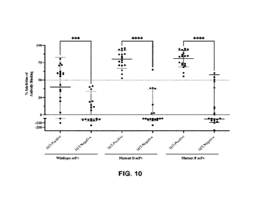
[0041] FIG. 10 shows how this assay can distinguish
between pathogenic
and non-pathogenic anti-PF4/heparin antibodies in a streptavidin EIA.
Previously
tested EIA-positive/SRA-positive HIT patient samples (HIT positive sera; n=20)
and
EIA-positive/SRA-negative (HIT negative sera; n=20) were tested in a modified
streptavidin EIA. Results are shown as the percent (%) inhibition of anti-
PF4/heparin
antibody binding in the presence of scFv wild-type, mutant B, or mutant F at
50 ug/mL.
The ability of each scFv construct to inhibit antibody binding resulting in a
decrease in
binding below 50% (dotted line) was determined. Statistical significance was
calculated
using a two-way ANOVA analysis where (***) represents p > 0.0005 and (****)
represents p < 0.00005. Error bars denoting mean inhibition standard
deviation (s.d.)
are shown.
[0042] FIG. 11 shows the receiver operating
characteristics (ROC) curves of
an IgG-specific streptavidin anti-PF4/heparin EIA using scFv. ROC curves were
generated for the PF4/heparin IgG-specific streptavidin EIA alone and using
scFv
wildtype, mutant B, or mutant obtained from testing two clinical cohorts of
HIT-
positive (n=20, EIA+/SRA+) and HIT-negative (n=20, EIA+/SRA-).
- 11 -
CA 03236833 2024- 4- 30
WO 2023/077230
PCT/CA2022/051628
100431
FIG. 12 shows inhibition of HIT antibody-mediated platelet
activation in the "C-serotonin release assay by scFv. Previously tested EIA-
positive/SRA-positive HIT patient samples (n=5) were tested in a modified 14C-
serotonin-release assay (SRA) in the presence of scFv a) wildtype, b) mutant
B, c)
mutant C, d) mutant D, e) mutant E, or 0 mutant F at either 0; 50, or 75 ug/mL
concentrations. The ability of each scFv construct to inhibit platelet
activation resulting
in a moderate (220%) or strong (250%) decrease in percent 14C-serotonin
release was
determined relative to control wells containing patient sera in the absence of
scFv.
DETAILED DESCRIPTION
100441
The following is a detailed description provided to aid those skilled
in the art in practicing the present disclosure. Unless otherwise defined, all
technical
and scientific terms used herein have the same meaning as commonly understood
by
one of ordinary skill in the art to which this disclosure belongs. The
terminology used
in the description herein is for describing particular embodiments only and is
not
intended to be limiting of the disclosure. All publications, patent
applications, patents,
figures and other references mentioned herein are expressly incorporated by
reference
in their entirety.
100451
HIT is an antibody-mediated drug disorder arising in patients
receiving heparin as an anticoagulant medication, often following major
surgical
procedures.''' Heparin binds favourably to PF4 tetramers and forms
structurally
stabilized complexes, which can trigger anti-PF4/heparin antibody production
and lead
to the formation of large immune complexes.21-25 Although the development of
anti-
PF4/heparin antibodies is common in heparin-treated patients, only a small
percentage
of individuals develop
This is because PF4/heparin induces a highly
polyclonal and polyspecific antibody response where most antibodies produced
are
non-pathogenic and cannot cause HIT.7'1"8 Rapid and accurate differentiation
between
anti-PF4/heparin antibodies that can and cannot cause platelet activation
remains a
significant diagnostic challenge due to the reduced specificity and limited
availability
of current laboratory as says.1 '11 Recently, it was shown that pathogenic
antibodies bind
to a restricted region on PF4 distinct from sites bound by non-pathogenic
antibodies.'
Based on these different binding sites, this work aims to use an epitope-
targeted strategy
- 12 -
CA 03236833 2024- 4- 30
WO 2023/077230
PCT/CA2022/051628
to distinguish between pathogenic and non-pathogenic antibodies in an EIA
using scFv
mutants by blocking the heparin-dependent pathogenic epitope on PF4.
[0046] In the present disclosure, random mutagenesis was
used to improve
the affinity of an scFv derived from the monoclonal antibody KKO. Five mutant
variants of scFv were identified using phage display after various rounds of
bio-panning
selection on magnetic beads coated with the target antigen for HIT antibodies,
PF4/heparin. Functional studies were then carried out to assess the binding
characteristics and affinity of each variant compared to wildtype. BLI was
used for
kinetic analysis of wildtype and mutant scFv. Mutants B. D, E, and F had
substantially
improved affinity towards PF4 alone and PF4 complexed with heparin compared to
wildtype, and mutant C had moderately improved affinity towards PF4/heparin
compared to wild-type. Binding inhibition experiments using KKO were also
performed
to evaluate the strength of each construct against a full-length antibody and
determine
approximate IC50 values. These studies revealed that scFv mutants B, D, E, and
F were
able to strongly inhibit KKO binding in a streptavidin anti-PF4/hep EIA.
[0047] As further shown in the Examples, epitope mapping
revealed scFv
wildtype, mutant C, and mutant D bound to the same amino acids as full-length
KKO.
Whereas mutant B recognized amino acids that differed from the other variants
but still
overlapped with the heparin-dependent binding site on PF4. A small sample of
patient
sera was then tested in an anti-PF4/heparin EIA using wildtype and mutant
scFv, which
showed each construct inhibited HIT-positive antibodies but did not affect the
binding
of HIT-negative antibodies. These findings suggest scFv can eliminate false-
positive
signals in an EIA that arise from antibodies that do not cause HIT.
[0048] As shown in the Examples, wildtype scFv and each
of the five
mutants reduced antibody binding from HIT-positive patients. Four scFv
constructs,
mutants B, D, E, and F, were able to reduce the false-positive rate of EIAs
caused by
the presence of non-pathogenic antibodies when testing HIT-positive and HIT-
negative
patient sera. Pathogenic antibody binding was reduced to negative detection
levels
(0D4o5. < 0.45) in this optimized assay in the presence of mutant B, D, E, and
F. As
predicted, these four mutants also had a stronger effect and higher mean
percent
inhibition of pathogenic antibody binding compared to both wildtype and mutant
C
- 13 -
CA 03236833 2024- 4- 30
WO 2023/077230
PCT/CA2022/051628
(Table 5-6). These findings also show a correlation with BLI analysis and
inhibition
studies with KKO, which demonstrate mutant B, D, E, and F have superior
performance
and binding kinetics. Out of five mutant scFv constructs, two were chosen as
lead
candidates to move forward with to perform a large-scale screening of patient
sera and
evaluate the diagnostic performance of this assay.
100491 Mutant B and F demonstrated the foremost ability
to distinguish
between pathogenic and non-pathogenic antibodies compared to all scFv
constructs.
Testing these mutants against a larger population of HIT-positive (n=20) and
HIT-
negative (n=20) patient samples revealed pathogenic antibody binding was
almost
always significantly reduced in the presence of scFv while non-pathogenic
antibody
binding was not. ROC curve analysis revealed a significant increase in the
performance
of the streptavidin anti-PF4/hep EIA with the addition of scFv. Compared to
previous
literature reports of the anti-PF4/hep EIA diagnostic performance,"
incorporating scFv
showed an improvement in specificity to 90.0% while maintaining the high
sensitivity
of this assay.29'3 Overall, targeting clinically significant epitopes on PF4
proves to be
an effective method of distinguishing between HIT antibodies based on their
individual
binding sites. The addition of wildtype or mutant scFv also demonstrates
improved
diagnostic performance compared to the IgG-specific anti-PF4/heparin EIA for
identifying clinically significant HIT antibodies.
[0050] In the present disclosure, scFv mutants
demonstrated an advanced
diagnostic performance in EIAs used to identify HIT antibodies. Current EIAs
designed
to diagnose HIT have low specificities (-50-70%)" for clinically significant
antibodies
in HIT. Automated rapid assays are also frequently employed for HIT
diagnosis,31-35
but they can require costly and specialized equipment to perform, making them
inaccessible to many labs despite their higher specificities. The addition of
mutant B
and F scFv to the streptavidin anti-PF4/hep EIA improved the diagnostic
specificity to
90.0% in a cohort containing HIT-positive and HIT-negative patients without
reducing
sensitivity. The assay described here provides a promising alternative to
functional and
automated rapid assays for the diagnosis of HIT. High affinity scFv mutants
can
accurately differentiate between pathogenic and non-pathogenic HIT antibodies
in an
EIA, thus providing a rapid and cost-effective solution to common limitations
of HIT
- 14 -
CA 03236833 2024- 4- 30
WO 2023/077230
PCT/CA2022/051628
diagnostic assays. Furthermore, this EIA can be performed easily with standard
laboratory equipment, increasing the overall availability of HIT testing.
[0051]
Unless otherwise indicated, the definitions and embodiments
described in this and other sections are intended to be applicable to all
embodiments
and aspects of the present application herein described for which they are
suitable as
would be understood by a person skilled in the art. For example, in the
following
passages, different aspects of the disclosure are defined in more detail. Each
aspect so
defined may be combined with any other aspect or aspects unless clearly
indicated to
the contrary. In particular, any feature described herein may be combined with
any other
feature or features described herein.
I. Definitions
[0052]
As used herein, the following terms may have meanings ascribed to
them below, unless specified otherwise. However, it should be understood that
other
meanings that are known or understood by those having ordinary skill in the
art are also
possible, and within the scope of the present disclosure. All publications,
patent
applications, patents, and other references mentioned herein are incorporated
by
reference in their entirety. In the case of conflict, the present
specification, including
definitions, will control. In addition, the materials, methods, and examples
are
illustrative only and not intended to be limiting.
[0053]
Where a range of values is provided, it is understood that each
intervening value, to the tenth of the unit of the lower limit unless the
context clearly
dictates otherwise, between the upper and lower limit of that range and any
other stated
or intervening value in that stated range is encompassed within the
description. Ranges
from any lower limit to any upper limit are contemplated. The upper and lower
limits
of these smaller ranges which may independently be included in the smaller
ranges is
also encompassed within the description, subject to any specifically excluded
limit in
the stated range. Where the stated range includes one or both of the limits,
ranges
excluding either or both of those included limits are also included in the
description.
[0054]
All numerical values herein are modified by "about" or
"approximately" the indicated value, and take into account experimental error
and
variations that would be expected by a person having ordinary skill in the
art.
- 15 -
CA 03236833 2024- 4- 30
WO 2023/077230
PCT/CA2022/051628
[0055] The terms "about", "substantially" and
"approximately" as used
herein mean a reasonable amount of deviation of the modified term such that
the end
result is not significantly changed. These terms of degree should be construed
as
including a deviation of at least 5% of the modified term if this deviation
would not
negate the meaning of the word it modifies or unless the context suggests
otherwise to
a person skilled in the art.
[0056] As used herein, the singular forms "a", "an", and
"the" include plural
references unless the context clearly dictates otherwise.
[0057] The phrase "and/or," as used herein in the
specification and in the
claims, should be understood to mean "either or both" of the elements so
conjoined,
i.e., elements that are conjunctively present in some cases and disjunctively
present in
other cases. Multiple elements listed with "and/or" should be construed in the
same
fashion, i . e. , "one or more" of the elements so conjoined. Other elements
may optionally
be present other than the elements specifically identified by the "and/or"
clause,
whether related or unrelated to those elements specifically identified.
[0058] As used herein, "or" should be understood to have
the same meaning
as "and/or" as defined above. For example, when separating items in a list,
"or" or
"and/or" shall be interpreted as being inclusive, i.e., the inclusion of at
least one, but
also including more than one, of a number or list of elements, and,
optionally, additional
unlisted items. Only terms clearly indicated to the contrary, such as "only
one of or
"exactly one of' or, when used in the claims, "consisting of' will refer to
the inclusion
of exactly one element of a number or list of elements. In general, the term
"or" as used
herein shall only be interpreted as indicating exclusive alternatives (i.e.,
"one or the
other but not both") when preceded by terms of exclusivity, such as "either,"
"one of,"
"only one of," or "exactly one of"
100591 As used herein, all transitional phrases such as
"comprising,"
"including," "carrying," "having," "containing," "involving," "holding,"
"composed of,"
and the like are to be understood to inclusive or be open-ended, i.e., to mean
including
but not limited to, and do not exclude additional, unrecited elements or
process steps.
[0060] The term "consisting" and its derivatives as used
herein are intended
to be closed terms that specify the presence of the stated features, elements,
- 16 -
CA 03236833 2024- 4- 30
WO 2023/077230
PCT/CA2022/051628
components, groups, integers, and/or steps, and also exclude the presence of
other
unstated features, elements, components, groups, integers and/or steps.
[0061] The term "consisting essentially of', as used
herein, is intended to
specify the presence of the stated features, elements, components, groups,
integers,
and/or steps as well as those that do not materially affect the basic and
novel
characteristic(s) of these features, elements, components, groups, integers,
and/or steps.
[0062] As used herein, the phrase "at least one," in
reference to a list of one
or more elements, should be understood to mean at least one element selected
from
anyone or more of the elements in the list of elements, but not necessarily
including at
least one of each and every element specifically listed within the list of
elements and
not excluding any combinations of elements in the list of elements. This
definition also
allows that elements may optionally be present other than the elements
specifically
identified within the list of elements to which the phrase "at least one"
refers, whether
related or unrelated to those elements specifically identified.
[0063] It should also be understood that, in certain
methods described herein
that include more than one step or act, the order of the steps or acts of the
method is not
necessarily limited to the order in which the steps or acts of the method are
recited
unless the context indicates otherwise.
[0064] Further, the definitions and embodiments described
in particular
sections are intended to be applicable to other embodiments herein described
for which
they are suitable as would be understood by a person skilled in the art. For
example, in
the following passages, different aspects of the disclosure are defined in
more detail.
Each aspect so defined may be combined with any other aspect or aspects unless
clearly
indicated to the contrary. In particular, any feature described herein may be
combined
with any other feature or features described herein.
II. Antibodies, Nucleic Acids, and Cells
[0065] The inventors show herein the development of five
mutants of a
single chain variable fragment (scFv) antibodies derived from the murine KKO
(referred to herein as "KKO-scFv" or "scFv") antibody from a library of
1.5x10⁷ unique
sequences. These scFv mutants bind to PF4 (SEQ ID NO: 17) and/or a PF4/heparin
- 17 -
CA 03236833 2024- 4- 30
WO 2023/077230
PCT/CA2022/051628
complex with at least 2-fold, at least 3-fold, at least 4-fold, at least 5-
fold, at least 10-
fold, at least 100-fold, or greater than 100-fold binding affinity relative to
wildly pe scFy
(SEQ ID NO: 3) as determined by Biolayer Interferometry (BLI). Accordingly,
provided herein are anti-PF4 antibodies which specifically bind an epitope of
PF4,
wherein the antibodies bind PF4 and/or a PF4/heparin complex with at least or
about
2-fold, at least or about 3-fold, at least or about 4-fold, at least or about
5-fold, at least
or about 10-fold, at least or about 100-fold, or more than 100-fold greater
affinity than
an scFy having an amino acid sequence of SEQ ID NO: 3. As used herein,
"PF4/heparin- or "PF4/heparin complex- refers to multimeric protein complex
comprising PF4 and heparin. Optionally, the PF4 has an amino acid sequence of
SEQ
ID NO: 17. The anti-PF4 antibodies described herein include antibodies
comprising the
complementarity determining regions (CDRs) of any one of a), b), c), d), or
e):
a)
CDR-L1 KASQNVGTNVA SEQ ID NO: 18;
CDR-L2 SASYRYS SEQ ID NO: 19:
CDR-L3 QQYNSYPLT SEQ ID NO: 20;
CDR-H1 KYFIY SEQ ID NO: 24;
CDR-H2 EINPRNGDTNFNEKFES SEQ ID NO: 25: and
CDR-H3 SPYGNNYGFTY SEQ ID NO: 23;
b)
CDR-Li KASQNVGTNVA SEQ ID NO: 18;
CDR-L2 NASHRYS SEQ ID NO: 26;
CDR-L3 QQYNSYPLT SEQ ID NO: 20;
CDR-H1 NYFTY SEQ ID NO: 21
CDR-H2 EINPRNGDTDFNEKFES SEQ ID NO: 22 and
CDR-H3 S PY GNNY GF TY SEQ ID NO: 23;
c)
CDR-L1 KASQNVGTNVA SEQ ID NO: 18:
CDR-L2 SASYRYS SEQ ID NO: 19;
CDR-L3 QQYNSYPLT SEQ ID NO: 20;
CDR-H1 NYFIH SEQ ID NO: 27;
CDR-H2 EINPRNGDTDFNEKFES SEQ ID NO: 22 and
CDR-H3 S PY GNNY GF TY SEQ ID NO: 23;
d)
CDR-L1 KASQNVGTNVA SEQ ID NO: 18;
-18 -
CA 03236833 2024- 4- 30
WO 2023/077230
PCT/CA2022/051628
CDR-L2 SASYRYS SEQ ID NO: 19;
CDR-L3 QQYNSYPLT SEQ ID NO: 20;
CDR-HI NYFTH SEQ ID NO: 27:
CDR-H2 EINPKNGDTGFNEKFES SEQ ID NO: 28; and
CDR-H3 SPYGNNYGFTY SEQ ID NO: 23;
or
e)
CDR-Li KASQNVGTNVA SEQ ID NO: 18;
CDR-L2 SASYRYS SEQ ID NO: 19;
CDR-L3 QQYNSYPLT SEQ ID NO: 20;
CDR-H1 NYFIY SEQ ID NO: 21
CDR-H2 EINPRNGDTDFNVKFKS SEQ ID NO: 29; and
CDR-H3 SPYRNNYGFTY SEQ ID NO: 30.
[0066] The antibodies may comprise light chain variable
(VL) and heavy
chain variable (VH) domains set out in a) SEQ ID NOs: 11 and 12; b) SEQ ID
NOs: 13
and 10; c) SEQ ID NOs: 9 and 14; d) SEQ ID NOs: 9 and 15; ore) SEQ ID NOs: 9
and
16, or variants thereof having the CDR sequences specified in a), b), c), d),
or e), above.
[0067] The antibodies may comprise variants of the VL
and/or VH domains
of SEQ ID NOs: 9 and 10, respectively, comprising one or more mutations at
positions
selected from R18, S50, and Y53 of SEQ ID NO: 9, and/or one or more mutations
at
positions selected from N30, Y34, D58, E61, E64, G101, and Q111 of SEQ ID NO:
10,
or variants thereof having the specified mutations. In an embodiment, the one
or more
mutations are selected from R18K., S5ON, and Y53H of SEQ ID NO: 9, and/or
selected
from N30K, Y34H, D58N, D58G, E61V, E64K, G101R, and Q111P of SEQ ID NO:
10. In an embodiment, the one or more mutations are selected from the
following
combinations: a) R18K of SEQ ID NO: 9 and N3OK and D58N of SEQ ID NO: 10; b)
S5ON and Y53H of SEQ ID NO: 9: c) Y34H of SEQ ID NO: 10; d) Y34H, D58G, and
Q111P of SEQ ID NO: 10; and e) E61V, E64K, and G101R of SEQ ID NO: 10.
100681 The basic antibody structural unit is known to
comprise a tetramer
composed of two identical pairs of polypeptide chains, each pair having one
light ("L-)
(about 25 kDa) and one heavy ("H") chain (about 50-70 kDa). The amino-terminal
portion of the light chain forms a light chain variable domain (VL) and the
amino-
terminal portion of the heavy chain forms a heavy chain variable domain (VH).
- 19 -
CA 03236833 2024- 4- 30
WO 2023/077230
PCT/CA2022/051628
Together, the VH and VL domains form the antibody variable region (Fv) which
is
primarily responsible for antigen recognition/binding. Within each of the VH
and VL
domains are three hypervariable regions or complementarity determining regions
(CDRs, commonly denoted CDR-H1, CDR-H2, CDR-H3, CDR-L1, CDR-L2, and
CDR-L3). The carboxy-terminal portions of the heavy and light chains together
form a
constant region (Fc domain) primarily responsible for effector function.
[0069] The term "antibody" as used herein is intended to
include monoclonal
antibodies, chimeric and humanized antibodies, and binding fragments thereof,
including for example a single chain Fab fragment, Fab'2 fragment, or single
chain Fv
fragment. The antibody may be from recombinant sources and/or produced in
transgenic animals. Humanized or other chimeric antibodies may include
sequences
from one or more than one isotype, class, or species. Antibodies may be any
class of
immunoglobulins including: IgG, IgM. IgD, IgA, or IgE; and any isotype
thereof,
including IgGl, IgG2 (e.g. IgG2a, IgG2b), IgG3 and IgG4. Further, these
antibodies
are typically produced as antigen binding fragments such as Fab, Fab' F(ab')2,
Fd, Fv
and single domain antibody fragments, or as single chain antibodies (e.g.
scFv) in which
the heavy and light chains are linked by a spacer or linker. The antibodies
may include
sequences from any suitable species including human. Also, the antibodies may
exist
in monomeric or polymeric form.
[0070] The term "antibody fragment" or "binding fragment"
as used herein
is intended to include without limitations Fab, Fab', F(ab')2, scFab, scFv,
dsFy, ds-scFv,
dimers, minibodies, diabodies, and multimers thereof, and Domain Antibodies.
Antibodies can be fragmented using conventional techniques. For example,
F(ab')2
fragments can be generated by treating the antibody with pepsin. The resulting
F(ab')2
fragment can be treated to reduce disulfide bridges to produce Fab' fragments.
Papain
digestion can lead to the formation of Fab fragments. Fab, Fab' and F(ab')2,
scFv, dsFy,
ds-scFv, dimers, minibodies, diabodies, and other fragments can also be
synthesized by
recombinant techniques. In some embodiments, the antibody fragment does not
comprise an Fc domain and/or does not interact with platelet Fc receptors. In
an
embodiment, the antibody is an scFv. In an embodiment, the scFv comprises,
from N-
terminus to C-terminus, VL-linker-VH. In an embodiment, the linker comprises a
GS-
linker peptide, optionally having a sequence of SEQ ID NO: 31. In an
embodiment, the
- 20 -
CA 03236833 2024- 4- 30
WO 2023/077230
PCT/CA2022/051628
scFy comprises a polypeptide having an amino acid sequence of any one of SEQ
ID
NOs: 4-8.
[0071] The term "complementarity determining region- or
"CDR- as used
herein refers to particular hypervariable regions of antibodies that are
commonly
presumed to contribute to epitope binding. Computational methods for
identifying CDR
sequences include Kabat, Chothia, and IMGT. The CDRs listed in the present
disclosure are identified using Kabat. A person skilled in the art having
regard to the
sequences comprised herein would also be able to identify CDR sequences based
on
IMGT and Chothia etc. Such antibodies are similarly encompassed.
[0072] The phrase "isolated antibody" refers to antibody
produced in vivo or
in vitro that has been removed from the source that produced the antibody, for
example,
an animal, hybridoma or other cell line (such as recombinant insect, yeast or
bacteria
cells that produce antibody). In some embodiments the antibody is an isolated
antibody.
The isolated antibody is optionally "purified", which means at least: 80%,
85%, 90%,
95%, 98% or 99% purity.
[0073] The term "epitope" as commonly used means an
antibody binding
site, typically a polypeptide segment having a particular structural
conformation, in an
antigen that is specifically recognized by the antibody. For example an
antibody
generated or selected against a recombinant protein comprising the identified
target
region (e.g. PF4; SEQ ID NO: 17) recognizes part or all of said epitope
sequence.
[0074] The term "greater affinity" as used herein refers
to a relative degree
of antibody binding where an antibody X binds to target Y more strongly (Ic,n)
and/or
with a smaller dissociation constant (Koff) than does comparator antibody Z,
and in this
context antibody X has a greater affinity for target Y than Z. Likewise, the
term "lesser
affinity" herein refers to a degree of antibody binding where an antibody X
binds to
target Y less strongly and/or with a larger dissociation constant than does
antibody Z,
and in this context antibody X has a lesser affinity for target Y than Z. The
affinity of
binding between an antibody and its target antigen can be expressed
quantitatively as
KA equal to 1/ KD where KD is equal to kon/koff. As such, a greater affinity
corresponds
to a lower Ku. The km and koff values can be measured using surface plasmon
resonance
- 21 -
CA 03236833 2024- 4- 30
WO 2023/077230
PCT/CA2022/051628
technology, and/or as described herein. Binding affinity can also be assessed
using
other techniques such as flow cytometry.
[0075] The term "functional variant- as used herein
includes modifications
of the polypeptide sequences disclosed herein that perform substantially the
same
function as the polypepti de molecules disclosed herein in substantially the
same way.
For example, the functional variant may comprise sequences having at least
80%, or at
least 90%, or at least 95% sequence identity to the sequences disclosed herein
provided
that the variant retains at least or about the same binding affinity for PF4
and/or
PF4/heparin. The functional variant may also comprise conservatively
substituted
amino acid sequences of the sequences disclosed herein.
[0076] The term "sequence identity" as used herein refers
to the percentage
of sequence identity between two amino acid sequences or two nucleic acid
sequences.
To determine the percent identity of two amino acid sequences or of two
nucleic acid
sequences, the sequences are aligned for optimal comparison purposes (e.g.
gaps can
be introduced in the sequence of a first amino acid or nucleic acid sequence
for optimal
alignment with a second amino acid or nucleic acid sequence). The amino acid
residues
or nucleotides at corresponding amino acid positions or nucleotide positions
are then
compared. When a position in the first sequence is occupied by the same amino
acid
residue or nucleotide as the corresponding position in the second sequence,
then the
molecules are identical at that position. The percent identity between the two
sequences
is a function of the number of identical positions shared by the sequences
(i.e., %
identity = [number of identical overlapping positions] / [total number of
positions] X
100%). The determination of percent identity between two sequences can also be
accomplished using a mathematical algorithm. One non-limiting example of a
mathematical algorithm utilized for the comparison of two sequences is the
algorithm
of Karlin and Altschul, 1990, Proc. Natl. Acad. Sci. U.S.A. 87:2264-2268,
modified as
in Karlin and Altschul, 1993, Proc. Natl. Acad. Sci. U.S.A. 90:5873-5877. Such
an
algorithm is incorporated into the NBLAST and )(BLAST programs of Altschul et
al.,
1990. BLAST nucleotide searches can be performed with the NBLAST nucleotide
program parameters set, e.g. for score=100, Ivordlength=12 to obtain
nucleotide
sequences homologous to a nucleic acid molecules of the present disclosure.
BLAST
protein searches can be performed with the XBLAST program parameters set, e.g.
to
- 22 -
CA 03236833 2024- 4- 30
WO 2023/077230
PCT/CA2022/051628
score-50, wordlength=3 to obtain amino acid sequences homologous to a protein
molecule of the present disclosure. To obtain gapped alignments for comparison
purposes, Gapped BLAST can be utilized as described in Altschul et al., 1997,
Nucleic
Acids Res. 25:3389-3402. Alternatively, PSI-BLAST can be used to perform an
iterated
search which detects distant relationships between molecules. When utilizing
BLAST,
Gapped BLAST, and PSI-Blast programs, the default parameters of the respective
programs (e.g. of XBLAST and NBLAST) can be used (see, e.g. the NCBI website).
Another non-limiting example of a mathematical algorithm utilized for the
comparison
of sequences is the algorithm of Myers and Miller, 1988, CABIOS 4:11-17. Such
an
algorithm is incorporated in the ALIGN program (version 2.0) which is part of
the GCG
sequence alignment software package. When utilizing the ALIGN program for
comparing amino acid sequences, a PAM120 weight residue table, a gap length
penalty
of 12, and a gap penalty of 4 can be used. The percent identity between two
sequences
can be determined using techniques similar to those described above, with or
without
allowing gaps. In calculating percent identity, typically only exact matches
are counted.
[0077] For antibodies, percentage sequence identities can
be determined
when antibody sequences are maximally aligned by IMGT or other (e.g. Kabat or
Chothia numbering conventions). The terms "IMGT numbering" or "ImMunoGeneTics
database numbering", which are recognized in the art, refer to a system of
numbering
amino acid residues which are more variable (i.e. hypervari able) than other
amino acid
residues in the heavy and light chain variable regions of an antibody, or
antigen binding
portion thereof After alignment, if a subject antibody region (e.g., the
entire mature
variable region of a heavy or light chain) is being compared with the same
region of a
reference antibody, the percentage sequence identity between the subject and
reference
antibody regions is the number of positions occupied by the same amino acid in
both
the subject and reference antibody region divided by the total number of
aligned
positions of the two regions, with gaps not counted, multiplied by 100 to
convert to
percentage. Accordingly, IMGT and other alignment systems can also be used to
identify or annotate CDRs in an antibody sequence.
[0078] A "conservative amino acid substitution" as used
herein is one in
which one amino acid residue is replaced with another amino acid residue
without
abolishing the protein's desired properties. Suitable conservative amino acid
- 23 -
CA 03236833 2024- 4- 30
WO 2023/077230
PCT/CA2022/051628
substitutions can be made by substituting amino acids with similar
hydrophobicity,
polarity, and R-chain length for one another. Examples of conservative
substitutions
include the substitution of one non-polar (hydrophobic) residue such as
alanine,
isoleucine, valine, leucine or methionine for another, the substitution of one
polar
(hydrophilic) residue for another such as between arginine and lysine, between
glutamine and asparagine, between glycine and senile, the substitution of one
basic
residue such as lysine, arginine or histidine for another, or the substitution
of one acidic
residue, such as aspartic acid or glutamic acid for another. The phrase
"conservative
substitution" also includes the use of a chemically derivatized residue or non-
natural
amino acid in place of a non-derivatized residue provided that such
polypeptide
displays the requisite activity.
[0079] The antibodies described herein may be provided as
immunoconjugates. Accordingly, also provided herein are immunoconjugates
comprising an antibody described herein and a suitable reagent such as a
therapeutic,
cytotoxic agent, or detectable label. Suitable reagents can be identified by
the skilled
person depending on the application. Detectable labels include radionuclides,
fluorescent dyes, enzymes, or biotin may be used depending on the application,
and are
contemplated herein.
100801 Immunoconjugates may be generated using any
suitable technique.
Common conjugation techniques include N-hydroxysuccinimide ester (NHS ester)
or
maleimide crosslinking, but other techniques are known in the art.
[0081] A further aspect is an isolated nucleic acid
encoding an antibody or
fragment thereof described herein.
[0082] Nucleic acids encoding a heavy chain or a light
chain or parts thereof
are also provided, for example encoding a heavy chain variable domain
comprising
CDR-F11, CDR-H2 and/or CDR-H3 regions described herein or encoding a light
chain
variable domain comprising CDR-L1, CDR-L2 and/or CDR-L3 regions described
herein, variable heavy and light domains described herein, and codon optimized
and
codon degenerate versions thereof
[0083] The present disclosure also provides variants of
the nucleic acid
sequences that encode for the antibody and/or binding fragment thereof
disclosed
- 24 -
CA 03236833 2024- 4- 30
WO 2023/077230
PCT/CA2022/051628
herein. For example, the variants include nucleotide sequences that hybridize
to the
nucleic acid sequences encoding the antibody and/or binding fragment thereof
disclosed herein under at least moderately stringent hybridization conditions
or codon
degenerate or optimized sequences In another embodiment, the variant nucleic
acid
sequences have at least 50%, at least 60%, at least 70%, most preferably at
least 80%,
even more preferably at least 90% and even most preferably at least 95%
sequence
identity to nucleic acid sequences encoding any of the amino acid sequences
described
herein for example as shown in SEQ ID NOs: 4-16, or functional variants
thereof.
[0084] A further aspect is an isolated nucleic acid
encoding an antibody
described herein, for example the nucleic acids shown in any of SEQ ID NOs: 33-
37,
or variants thereof.
[0085] Another aspect is an expression cassette, plasmid,
or vector
comprising the nucleic acid herein disclosed.
[0086] The term -expression cassette" refers to a DNA
molecule encoding
an RNA or protein operably linked to a promoter and a transcriptional
termination
signal (e.g. poly adenylation signal), such that certain portions of the
expression cassette
are capable of being transcribed into RNA such as a messenger RNA that is
subsequently translated into protein by cellular machinery.
[0087] The term "operably linked" as used herein refers
to a relationship
between two components that allows them to function in an intended manner. For
example, where a DNA encoding an RNA of interest is operably linked to a
promoter,
the promoter actuates expression of the RNA encoded therein.
[0088] The term "promoter" or "promoter sequence"
generally refers to a
regulatory DNA sequence capable of being bound by an RNA polymerase to
initiate
transcription of a downstream (i.e. 3') sequence to generate an RNA. Suitable
promoters may be derived from any organism and may be bound or recognized by
any
RNA polymerase. Suitable promoters will be known to the skilled person. In
some
expression cassettes, the promoter is an inducible promoter and/or comprises a
binding
sequence for a transactivator or a repressor that will activate or inhibit
transcription
respectively, for example 1PTCi-inducible promoters are commonly used for
expression
- 25 -
CA 03236833 2024- 4- 30
WO 2023/077230
PCT/CA2022/051628
in E. colt. Other suitable promoters will depend on the expression system
and/or host
cell being used.
[0089] Suitable regulatory sequences may be derived from
a variety of
sources, including bacterial, fungal, viral, mammalian, or insect genes.
[0090] Examples of such regulatory sequences include: a
transcriptional
promoter and enhancer or RNA polymerase binding sequence, a ribosomal binding
sequence, including a translation initiation signal. Additionally, depending
on the host
cell chosen and the vector employed, other sequences, such as an origin of
replication,
additional DNA restriction sites, enhancers, and sequences conferring
inducibility of
transcription may be incorporated into the expression vector.
[0091] The nucleic acid molecules may be incorporated in
a known manner
into an appropriate expression vector which ensures expression of the protein.
Possible
expression vectors include but are not limited to cosmids, plasmids, or
modified viruses
(e.g. replication defective retroviruses, adenoviruses and adeno-associated
viruses).
The vector should be compatible with the host cell used. The expression
vectors are
"suitable for transformation of a host cell", which means that the expression
vectors
contain a nucleic acid molecule encoding the peptides corresponding to
antibodies
described herein.
[0092] The vector can be any vector, including vectors
suitable for producing
an antibody and/or binding fragment thereof described herein. In an
embodiment, the
vector is an isolated vector.
[0093] The recombinant expression vectors may also
contain a marker gene
which facilitates the selection of host cells transformed, infected or
transfected with a
vector for expressing an antibody or epitope peptide described herein.
100941 The recombinant expression vectors may also
contain expression
cassettes which encode a fusion moiety (i.e. a -fusion protein-) which
provides
increased expression or stability of the recombinant peptide; increased
solubility of the
recombinant peptide; and aid in the purification of the target recombinant
peptide by
acting as a ligand in affinity purification, including for example tags and
labels
described herein. Further, a proteoly tic cleavage site may be added to the
target
- 26 -
CA 03236833 2024- 4- 30
WO 2023/077230
PCT/CA2022/051628
recombinant protein to allow separation of the recombinant protein from the
fusion
moiety subsequent to purification of the fusion protein. Typical fusion
expression
vectors include pGEX (Amrad Corp., Melbourne, Australia), pMAL (New England
Biolabs, Beverly, MA) and pRIT5 (Pharmacia, Piscataway, NJ) which fuse
glutathione
S-transferase (GST), maltose E binding protein, or protein A, respectively, to
the
recombinant protein.
[0095] Also provided in another aspect is a cell,
optionally an isolated and/or
recombinant cell, expressing an antibody described herein or comprising a
vector herein
disclosed.
[0096] The recombinant cell can be generated using any
cell suitable for
producing a polypeptide, for example suitable for producing an antibody and/or
binding
fragment thereof For example to introduce a nucleic acid (e.g. a vector) into
a cell, the
cell may be transfected, transformed or infected, depending upon the vector
employed.
[0097] Suitable host cells include a wide variety of
prokaryotic and
eukaryotic host cells. For example, the proteins described herein may he
expressed in
bacterial cells such as E. coli, insect cells (using baculovirus), yeast
cells, or mammalian
cells.
III. Compositions
[0098] The antibodies or fragments thereof described
herein are suitably
formulated in a conventional manner into compositions using one or more
carriers or
diluents. Accordingly, the present description also includes a composition
comprising
one or more antibodies or fragments thereof described herein and a carrier or
diluent.
The antibodies or fragments thereof described herein are suitably formulated
into
pharmaceutical compositions or dosage forms for administration to patients in
a
biologically compatible form suitable for administration in vivo. Accordingly,
the
present description further includes a pharmaceutical composition comprising
an
antibody or fragment thereof described herein, and a pharmaceutically
acceptable
carrier. Also provided herein are dosage forms comprising an antibody or
fragment
thereof described herein. In some embodiments the pharmaceutical compositions
or
dosage forms are used in the treatment of any of the diseases, disorders or
conditions
described herein, for example HIT.
- 27 -
CA 03236833 2024- 4- 30
WO 2023/077230
PCT/CA2022/051628
100991 The term "dosage form" as used herein refers to
the physical form of
a dose for example comprising an antibody or fragment thereof described
herein, and
includes without limitation injectable dosage forms, including, for example,
sterile
solutions and sterile powders for reconstitution, and the like, that are
suitably
formulated for injection, resuspendable powders, liquids and solutions. For
example the
injectable dosage form can be a subcutaneous, intradermal, or intramuscular
depot
injection that allows the compound to be released in a controlled and
consistent way
over a period of time, for example over one month. Methods for making depot
injections are described, for example, in U.S. patent no. 3,089,815 entitled
"Injectable
pharmaceutical preparation, and a method of making same" and herein
incorporated by
reference in its entirety.
[00100] The compositions or dosage forms described herein
can be prepared
by per se known methods for the preparation of pharmaceutically acceptable
compositions that can be administered to patients, such that an effective
quantity of the
active substance is combined in a mixture with a pharmaceutically acceptable
vehicle.
[00101] Pharmaceutical compositions include, without
limitation, lyophilized
powders or aqueous or non-aqueous sterile injectable solutions or suspensions,
which
may further contain antioxidants, buffers, bacteriostats and solutes that
render the
compositions substantially compatible with the tissues or the blood of an
intended
recipient. Other components that may be present in such compositions include
water,
surfactants (such as Tween), alcohols, polyols, glycerin and vegetable oils,
for example.
Extemporaneous injection solutions and suspensions may be prepared from
sterile
powders, granules, tablets, or concentrated solutions or suspensions. The
composition
may be supplied, for example but not by way of limitation, as a lyophilized
powder
which is reconstituted with sterile water or saline prior to administration to
the patient.
[00102] Pharmaceutical compositions may comprise a
pharmaceutically
acceptable carrier. Suitable pharmaceutically acceptable carriers include
essentially
chemically inert and nontoxic compositions that do not interfere with the
effectiveness
of the biological activity of the pharmaceutical composition. Examples of
suitable
pharmaceutical carriers include, but are not limited to, water, saline
solutions, glycerol
solutions, ethanol, N-(1(2,3-dioleyloxy)propyl)N,N,N-trimethylammonium
chloride
- 28 -
CA 03236833 2024- 4- 30
WO 2023/077230
PCT/CA2022/051628
(DOTMA), diolesylphosphotidyl-ethanolamine (DOPE), and liposomes.
Such
compositions should contain a therapeutically effective amount of the
compound,
together with a suitable amount of carrier so as to provide the form for
direct
administration to the patient.
[00103]
The composition may be in the form of a pharmaceutically acceptable
salt which includes, without limitation, those formed with free amino groups
such as
those derived from hydrochloric, phosphoric, acetic, oxalic, tartaric acids,
etc., and
those formed with free carboxyl groups such as those derived from sodium,
potassium,
ammonium, calcium, ferric hydroxides, isopropylamine, triethylamine, 2-
ethylarnino
ethanol,
[00104]
In an embodiment, the composition comprises an antibody described
herein. In another embodiment, the composition comprises an antibody described
herein and a diluent In an embodiment, the composition is a sterile
composition
[00105]
The antibodies or fragments thereof described herein may be
administered to a patient in a variety of forms depending on the selected
route of
administration, as will be understood by those skilled in the art. For
example, the
antibodies or fragments thereof described herein may be administered by
parenteral
administration and the pharmaceutical compositions formulated accordingly. In
some
embodiments, administration is by means of a pump for periodic or continuous
delivery. Conventional procedures and ingredients for the selection and
preparation of
suitable compositions are described, for example, in Remingt, on's
Pharmaceutical
Sciences (2000 - 20th edition) and in The United States Pharmacopeia: The
National
Formulary (USP 24 NF19) published in 1999.
[00106]
Parenteral administration includes systemic delivery routes other than
the gastrointestinal (GI) tract, and includes, for example intravenous, intra-
arterial,
intraperitoneal, subcutaneous, intramuscular, modes of administration.
Parenteral
administration may be by continuous infusion over a selected period of time.
[00107]
In some embodiments, the antibodies or fragments thereof described
herein are administered parenterally. For example, solutions of one or more
antibodies
or fragments thereof described herein are prepared in water suitably mixed
with a
surfactant such as hydroxypropylcellulose. In some embodiments, dispersions
are
- 29 -
CA 03236833 2024- 4- 30
WO 2023/077230
PCT/CA2022/051628
prepared in glycerol, liquid polyethylene glycols, DMS 0 and mixtures thereof
with or
without alcohol, and in oils. Under ordinary conditions of storage and use,
these
preparations contain a preservative to prevent the growth of microorganisms. A
person
skilled in the art would know how to prepare suitable formulations. For
parenteral
administration, sterile solutions of the antibodies or fragments thereof
described herein
are usually prepared, and the pH of the solutions are suitably adjusted and
buffered. For
intravenous use, the total concentration of solutes should be controlled to
render the
preparation isotonic. For ocular administration, ointments or droppable
liquids are
delivered, for example, by ocular delivery systems known to the art such as
applicators
or eye droppers. In some embodiment, such compositions include mucomimetics
such
as hyaluronic acid, chondroitin sulfate, hydroxypropyl methylcellulose or
polyvinyl
alcohol, preservatives such as sorbic acid, EDTA or benzyl chromium chloride,
and the
usual quantities of diluents or carriers. For pulmonary administration,
diluents or
carriers will be selected to be appropriate to allow the formation of an
aerosol.
1001081 In some embodiments, the antibodies or fragments
thereof described
herein are formulated for parenteral administration by injection, including
using
conventional catheterization techniques or infusion. Formulations for
injection are, for
example, presented in unit dosage form, e.g., in ampoules or in multi-dose
containers,
with an added preservative. In some embodiments, the compositions take such
forms
as sterile suspensions, solutions or emulsions in oily or aqueous vehicles,
and contain
formulating agents such as suspending, stabilizing and/or dispersing agents.
In all cases,
the form must be sterile and must be fluid to the extent that easy
syringability exists.
Alternatively, antibodies or fragments thereof described herein are suitably
in a sterile
powder form for reconstitution with a suitable vehicle, e.g., sterile pyrogen-
free water,
before use.
1001091 The antibodies or fragments thereof described
herein, including
pharmaceutically acceptable salts and/or solvates thereof, are suitably used
on their own
but will generally be administered in the form of a pharmaceutical composition
in which
the one or more antibodies or fragments thereof described herein (the active
ingredient)
is in association with a pharmaceutically acceptable carrier. Depending on the
mode of
administration, the pharmaceutical composition will comprise from about 0.05
wt% to
about 99 wt% or about 0.10 wt% to about 70 wt%, of the active ingredient, and
from
- 30 -
CA 03236833 2024- 4- 30
WO 2023/077230
PCT/CA2022/051628
about 1 wt% to about 99.95 wt% or about 30 wt% to about 99.90 wt% of a
pharmaceutically acceptable carrier, all percentages by weight being based on
the total
composition.
IV. Kits
[00110] A further aspect relates to a kit comprising i) an
antibody and/or
binding fragment thereof described herein, ii) a nucleic acid of said antibody
or a part
thereof described herein, iii) composition comprising an antibody or fragment
thereof
described herein or iv) dosage form comprising an antibody or fragment thereof
described herein, comprised in a vial such as a sterile vial or other housing
and
optionally a reference agent and/or instructions for use thereof
[00111] In an embodiment, the kit is for diagnosing or
monitoring HIT. In an
embodiment, the kit further comprises one or more of a collection vial,
standard buffer,
PF4, PF4/heparin, and/or one or more detection reagents for detecting patient
antibodies. In an embodiment, the kit further comprises one or more of a pre-
coated
PF4/PVS or PF4/heparin microwell strips, one or more buffers, including, for
example,
a standard wash buffer (e.g. PBS + detergent + blocking agent) and specimen
dilutant
(e.g. PBS + blocking agent, optionally including a visual dye), positive and
negative
controls, detection reagents (such as substrate (e.g. p-nitrophenylphosphate)
and
substate buffer (DEA buffer)), and/or plastic plate sealers.
1001121 In another embodiment, the kit is for treating or
preventing HIT. In
an embodiment, the kit further comprises one or more of a sterile buffer for
reconstitution and/or a syringe or other device for administration.
V. Methods and Uses
[00113] As shown in the Examples, the antibodies and
fragments thereof
described herein can be use to distinguish between pathogenic (platelet-
activating) and
non-pathogenic (non-platelet activating) anti-PF4/heparin antibodies in
patient
samples. Specifically, the antibodies and fragments thereof described herein
can
distinguish between true positive -HIT positive" (EIA+/SRA+) and false
positive -HIT
negative" (EIA+/SRA-) patient samples. Accordingly, the antibodies and
fragments
thereof described herein can be used in diagnostic assays to rapidly identify
patients
-31 -
CA 03236833 2024- 4- 30
WO 2023/077230
PCT/CA2022/051628
with pathogenic anti-PF4/heparin antibodies and/or who have heparin-induced
thrombocytopenia (HIT). Accordingly, provided herein are methods for
identifying
patients with pathogenic anti-PF4/heparin antibodies. Also provided herein are
methods
for diagnosing HIT. In an embodiment, the method for identifying patients with
pathogenic anti-PF4/heparin antibodies or diagnosing HIT comprises a
competitive
binding assay, optionally an enzyme inun uno as s ay (EIA). Antibodies
suitable for use
in the diagnostic assays and methods described herein typically a) lack an Fc
domain,
b) are a different class of antibody from the pathogenic antibodies, and/or c)
are from a
different species than the patient. For example, an scFv, a human IgM, or a
mouse IgG
comprising the CDRs and/or VL and VH domains described herein could be used in
identifying human patients with pathogenic anti-PF4/heparin antibodies.
[00114] Any suitable competitive binding assay may be
used, for example as
described herein and in the Examples. In an embodiment, the method comprises
obtaining a biological sample comprising patient antibodies to PF4/heparin
from a
patient, contacting the sample with i) PF4/heparin in the presence of an
antibody or
fragment thereof described herein, or a wildtype KKO antibody fragment, and
ii)
PF4/heparin in the absence of an antibody or fragment thereof described
herein, or a
wildtype KKO antibody fragment, under conditions permissive for forming
PF4/heparin: patient antibody complexes; detecting the presence of any
PF4/heparin:
patient antibody complexes in i) and ii), wherein the detecting does not
detect the
antibody or antibody fragment described herein or the wildtype KKO antibody
fragment; and determining the relative amount of PF4/heparin : patient
antibody
complexes in i) and ii) thereby determining if PF4/heparin: patient antibody
binding is
inhibited; wherein the patient is identified as having pathogenic anti-
PF4/heparin
antibodies and/or diagnosed with HIT if PF4/heparin : patient antibody binding
is
inhibited. In some embodiments, in i), the PF4/heparin is contacted with
antibody or
fragment thereof described herein or wildtype KKO fragment, prior to
contacting with
the sample.
[00115] As used herein, -wildtype KKO antibody fragment"
refers to a KKO-
derived antibody fragment comprising a light chain variable (VL) domain
comprising
complementarity determining regions (CDRs) CDR-L1, CDR-L2, and CDR-L3 having
amino acid sequences of SEQ ID NOs: 18-20, and a heavy chain variable (VH)
domain
- 32 -
CA 03236833 2024- 4- 30
WO 2023/077230
PCT/CA2022/051628
comprising CDRs CDR-H1, CDR-H2, and CDR-H3 having amino acid sequences SEQ
ID NOs: 21-23. In some embodiments, the vvildtype KKO antibody fragment
comprises
a VL domain having an amino acid sequence of SEQ ID NO: 9, and/or or a VH
domain
having an amino acid sequence of SEQ ID NO: 10, optionally the wildtype KKO
antibody fragment comprises an amino acid sequence of SEQ ID NO: 3.
[00116] Suitable biological samples include, without
limitation a liquid
biopsy such as a blood sample, serum sample, or plasma sample. Accordingly, in
an
embodiment, the biological sample is a blood sample, serum sample, or plasma
sample.
[00117] In an embodiment, the sample is obtained from a
human patient. In
an embodiment, the patient has been administered heparin, or is suspected of
being at
risk of developing HIT.
[00118] Any suitable method can be used to determine the
presence of
PF4/heparin : patient antibody complexes, for example by using an enzyme-
linked or
labeled antibody to specifically detect the presence of bound patient
antibodies. For
example, alkaline phosphatase-conjugated anti-human IgG and a suitable
substrate e g
p-nitrophenylphosphate (PNPP) can be used to detect anti-PF4/heparin
antibodies from
human patients by measuring absorbance at 405 nm (0D405nm). Other enzyme
conjugates (e.g. horseradish peroxidase) may also be used in combination with
a
suitable substrate. Conjugates comprising fluorophores or other dyes or labels
may also
be used.
[00119] The inhibition of binding may be determined using
any suitable
method, for example as a percent inhibition. By way of example, where
detection of
binding is determined using alkaline phosphatase and PNPP, percent inhibition
can be
calculated using the formula:
Percentage of inhibition (%)
(0D405nm
¨sera OD 405nmKKO scFv + sera)]
Ix 100
I. OD405nm
¨sera
[00120] The above formula can be readily adapted for
calculating the percent
inhibition when using other enzyme/substrate combinations, fluorophores, etc.
for
detection. In an embodiment, the patient is identified as having pathogentic
anti-
PF4/heparin antibodies and/or diagnosed with HIT if the percent inhibition is
greater
- 33 -
CA 03236833 2024- 4- 30
WO 2023/077230
PCT/CA2022/051628
than the positivity thresholds pre-determined for this assay using each
individual scFv
construct. For example, the pre-determined thresholds for wildtype, mutant B,
and
mutant F can be a percentage of inhibition greater or equal to (>) 42.0%,?
39.8%, and
> 54.6%, respectively, are determined to be positive for pathogenic HIT
antibodies.
1001211 The antibodies and fragments thereof described
herein are shown to
inhibit platelet activation in a modified serotonin-release assay (SRA).
Accordingly,
provided herein are methods for inhibiting platelet activation in response to
heparin in
a patient in need thereof, the method comprising administering an effective
amount of
an antibody or fragment thereof described herein, or a wildtype KKO antibody
fragment, to the patient. Also provided herein are a use of an antibody or
fragment
thereof described herein, or a wildtype KKO antibody fragment, for inhibiting
platelet
activation in a patient in need thereof A further aspect includes the use of
an antibody
or fragment thereof described herein, or a wildtype KKO antibody fragment, in
the
manufacture of a medicament for inhibiting platelet activation. Another aspect
includes
an antibody or fragment thereof described herein, or a wildtype KKO antibody
fragment, for use in inhibiting platelet activation. In these aspects, the
antibody
fragment does not comprise an Fc domain and/or does not interact with or form
a
complex with platelet Fc receptors.
1001221 Also provided herein are methods for treating or
preventing HIT in a
patient, the method comprising administering an effective amount of antibody
or
fragment thereof described herein, or a wildtype KKO antibody fragment, to the
patient.
Further provided herein is the use of an antibody or fragment thereof
described herein,
or a wildtype KKO antibody fragment, for treating or preventing HIT. Also
provided
herein is the use of an antibody or fragment thereof described herein, or a
wildtype
KKO antibody fragment, in the manufacture of a medicament for treating or
preventing
HIT. A further aspect includes an antibody or fragment thereof described
herein, or a
wildtype KKO antibody fragment, for use in treating or preventing HIT. In
these
aspects, the antibody fragment does not comprise an Fc domain and/or does not
interact
with or form a complex with platelet Fc receptors.
- 34 -
CA 03236833 2024- 4- 30
WO 2023/077230
PCT/CA2022/051628
[00123] Optionally, the patient is identified as having
pathogenic anti-
PF4/heparin antibodies and/or diagnosed with HIT, for example using the
methods
described herein.
[00124] The term "patient" as used herein includes all
members of the animal
kingdom including mammals, and suitably refers to humans.
[00125] The term "patient in need thereof" refers to a
patient that could benefit
from the method(s) or treatment(s) described herein, and optionally refers to
a patient
identified as having pathogenic anti-PF4/heparin antibodies and/or diagnosed
with HIT,
and/or a heparin-treated patient.
[00126] The term "treating" or "treatment" as used herein
and as is well
understood in the art, means an approach for obtaining beneficial or desired
results,
including clinical results. Beneficial or desired clinical results include,
but are not
limited to alleviation or amelioration of one or more symptoms or conditions,
diminishment of extent of disease, stabilized (i.e. not worsening) state of
disease,
preventing spread of disease, delay or slowing of disease progression,
amelioration or
palliation of the disease state, diminishment of the reoccurrence of disease,
and
remission (whether partial or total), whether detectable or undetectable.
"Treating" and
"treatment" can also mean prolonging survival as compared to expected survival
if not
receiving treatment. Treatment methods comprise administering to a patient a
therapeutically effective amount of one or more antibodies or fragments
thereof
described herein and optionally consist of a single administration, or
alternatively
comprise a series of administrations.
[00127] "Palliating" a disease, disorder or condition
means that the extent
and/or undesirable clinical manifestations of a disease, disorder or condition
are
lessened and/or time course of the progression is slowed or lengthened, as
compared to
not treating the disorder.
[00128] The term "prevention" or "prophylaxis-, or
synonym thereto, as used
herein refers to a reduction in the risk or probability of a patient becoming
afflicted with
a disease, disorder or condition or manifesting a symptom associated with a
disease,
disorder or condition, for example HIT. For example, a patient identified as
being at
risk of developing HIT can be treated with one or more antibodies or fragments
thereof
- 35 -
CA 03236833 2024- 4- 30
WO 2023/077230
PCT/CA2022/051628
described herein to prevent HIT from developing, or to reduce the severity or
extent of
HIT compared to expected severity or extent if not receiving preventative or
prophylactic treatment. Prevention methods comprise administering to a patient
a
therapeutically effective amount of one or more antibodies or fragments
thereof
described herein and optionally consist of a single administration, or
alternatively
comprise a series of administrations.
[00129] The term "disease, disorder or condition" as used
herein refers to a
disease, disorder or condition treatable using the antibodies or fragments
thereof, for
example by blocking pathogenic anti-PF4/heparin antibody binding to
PF4/heparin
and/or preventing platelet activation by pathogenic anti-PF4/heparin
antibodies.
[00130] The term "administered" or "administering" as used
herein means
administration of a therapeutically effective amount of a compound or
composition of
the disclosure to a patient. The antibodies or fragments thereof described
herein may
be administered using a variety of routes of administration. For example, the
antibodies
or fragments thereof described herein may be administered by parenteral
administration. Optionally the antibodies or fragments thereof described
herein may be
administered by intravenous, intra-arterial, intraperitoneal, subcutaneous, or
intramuscular administration. Optionally, the administration may be by
continuous
infusion over a selected period of time
[00131] The term "coadministration" or "combination
therapy" shall mean
that at least two compounds or compositions are administered to the patient at
the same
time, such that effective amounts or concentrations of each of the two or more
compounds may be found in the patient at a given point in time. Although
compounds
according to the present disclosure may be co-administered to a patient at the
same
time, the term embraces both administration of two or more agents at the same
time or
at different times, provided that effective concentrations of all
coadministered
compounds or compositions are found in the patient at a given time.
[00132] As used herein, the phrase "effective amount" or
"therapeutically
effective amount" means an amount effective, at dosages and for periods of
time
necessary to achieve the desired result. For example in the context of
treating or
preventing HIT, an effective amount is an amount that for example prevents the
- 36 -
CA 03236833 2024- 4- 30
WO 2023/077230
PCT/CA2022/051628
occurrence of HIT, or reduces the severity or extent of HIT compared to the
response
obtained without administration of the compound. Effective amounts may vary
according to factors such as the disease state, age, sex and weight of the
animal. The
amount of a given compound that will correspond to such an amount will vary
depending upon various factors, such as the given drug or compound, the
pharmaceutical formulation, the route of administration, the administration
schedule,
the identity of the patient being treated, and the like, but can nevertheless
be routinely
determined by one skilled in the art.
[00133] Suitable administration schedules may include,
without limitation, at
least once a week, from about one time per two weeks, three weeks or one
month, about
one time per week to about once daily, 2, 3, 4, 5 or 6 times daily. The length
of the
treatment period may depend on a variety of factors, such as the severity of
the disease,
disorder or condition, the age of the patient, the concentration and/or the
activity of the
antibody or fragment thereof described herein and/or a combination thereof It.
will also
be appreciated that the effective dosage of the antibody or fragment thereof
described
herein used for the treatment may increase or decrease over the course of a
particular
treatment regime. Changes in dosage may result and become apparent by standard
diagnostic assays known in the art. In some instances, chronic administration
is
required. For example, the antibody or fragment thereof described herein is
administered to the patient in an amount and for duration sufficient to treat
the patient.
[00134] The antibody or fragment thereof described herein
may be either used
alone or in combination with other known agents useful for treating diseases,
disorders
or conditions. When used in combination with other agents useful in treating
such
diseases, disorders or conditions, the antibody or fragment thereof described
herein may
be administered contemporaneously with those agents. As used herein,
-contemporaneous administration" of two substances to a patient means
providing each
of the two substances so that they are both active in the individual at the
same time. The
exact details of the administration will depend on the pharmacokinetics of the
two
substances in the presence of each other, and can include administering the
two
substances within a few hours of each other, or even administering one
substance within
24 hours of administration of the other, if the pharmacokinetics are suitable.
Design of
suitable dosing regimens is routine for one skilled in the art. In particular
embodiments,
- 37 -
CA 03236833 2024- 4- 30
WO 2023/077230
PCT/CA2022/051628
two substances will be administered substantially simultaneously, i.e., within
minutes
of each other, or in a single composition that contains both substances. In
other
embodiments, the combination of agents is administered to a patient in a non-
contemporaneous fashion. In an embodiment, the antibody or fragment thereof,
composition, etc. described herein is administered with another therapeutic
agent
simultaneously or sequentially in separate unit dosage forms or together in a
single unit
dosage form. Accordingly, the present description provides a single unit
dosage form
comprising the antibody or fragment thereof, composition, etc. described
herein, an
additional therapeutic agent, and a pharmaceutically acceptable carrier.
[00135] The dosage of the antibody or fragment thereof
described herein
varies depending on many factors such as the pharmacodynamic properties of the
antibody or fragment, the mode of administration, the age, health and weight
of the
recipient, the nature and extent of the symptoms, the frequency of the
treatment and the
type of concurrent treatment, if any, and the clearance rate of the
compound/cell in the
patient to be treated. One of skill in the art can determine the appropriate
dosage based
on the above factors. In some embodiments, the antibody or fragment thereof
described
herein is administered initially in a suitable dosage that is adjusted as
required,
depending on the clinical response. Dosages will generally be selected to
maintain
sufficient levels of the antibody or fragment thereof, composition, etc.
described herein.
EXAMPLES
[00136] The following non-limiting examples are
illustrative of the present
application:
Example 1. Materials and Methods
[00137] Phage Display Library Construction_ The M13
filamentous phage
display vector, pADL-22c, was purchased from Antibody Design Laboratories Inc.
(San Diego, CA). A library of mutant scFv sequences was first constructed for
expression in the pADL-22c phage display system. In PCR reactions, primers S1
and
AS1 (Table 1) were used to amplify the variable heavy and light chain regions
of KKO
using the BioRad T100 Thermal Cycler (BioRad, Hercules, CA). Mutagenic PCR was
performed using Mutazyme II Polymerase (Agilent Technologies, Inc., Santa
Clara,
- 38 -
CA 03236833 2024- 4- 30
WO 2023/077230
PCT/CA2022/051628
CA), according to manufacturer's instructions. scFy PCR products were digested
with
and cloned into the overlapping Bgll restriction sites of pADL-22c using T4
DNA
Ligase (New England Biolabs Ltd., Ipswich, MA). Ligated products were
electroporated into TG1 Electrocompetent E. colt cells (Lucigen, Middleton,
WI) using
the BioRad Gene Pulser (BioRad). Library depth was quantified by determining
the
number of colony forming units per inL (CFU) on LB agar plates supplemented
with
1001..tg/mL ampicillin. Colonies were then randomly selected to confirm scFy
insertion
by sanger sequencing.
Table 1: Primers for scFAT amplification from pADL-22c phagemid. Underlined
amino
acids represent the BglIISfil overlapping restriction enzyme recognition
sites.
Primer" Sequence
Sl...Sense Primer 5'
(SEQ ID NO: 1) GTTATTAC TC GC GGC CCAGC C GGC CATGGCTGATAT
CCAGATGATCCAGAGCC 3'
AS1...Anti-Sense 5'
Primer CCATGATGGTGGTGATGGTGTTGGCCTCCCGGGCCA
(SEQ ID NO: 2) C TAGT GGC AGAC AC GGTAAC C AGG 3'
[00138] Phage Library Preparation and Purification. Phage
library preparation
and purification were performed using a polyethylene glycol (PEG)/NaC1
precipitation
method as described by Barbas et al. (2001)36 and Kretz (2017).37 A culture
of'
transformed TG1 E. colt (Lucigen) was grown at 37 C while shaking at 250 rpm
to the
mid-exponential phase (0D600= 0.5-0.6 nm) in LB media containing 100 pg/mL
ampicillin. The helper phage M13K07 was then added and incubated with the E.
coli
culture for 1 h at 37 C while shaking at 250 rpm. Culture media was then
centrifuged
(6000 x g, 15 min) to pellet bacteria. The bacterial pellet was then
resuspended in 2xYT
media containing 100 ng/mL ampicillin and 50 mg/mL kanamycin and incubated
overnight at 30 C while shaking at 250 rpm. The following day, culture media
was
centrifuged (6000 x g, 10 min) to remove cell debris and phages were
precipitated from
the supernatant with 0.15x vol of PEG/NaCl (4% w/v PEG-8000 + 3% w/v NaCl) at
4 C overnight. The phages were pelleted by centrifugation (6500 x g, 60 mM, 4
C) and
re-suspended in Tris buffered saline (TBS) (50 mM Tns-HC1, pH 7.5, 150 mM
NaCl).
The supernatant was cleared of debris by centrifugation (10 000 x g, 10 min)
followed
by an additional round of precipitation before storing in TB S with 0.1% v/w
Tween-20
- 39 -
CA 03236833 2024- 4- 30
WO 2023/077230
PCT/CA2022/051628
(TBS-T) at 4 C. Phage preparations were titered by first growing a fresh
culture of K91
E. coil in LB media containing 50 pg/mL kanamycin followed by infection with
eluted
phages for 1 h at 37 C while shaking.
[00139] Phage titers were calculated according to the
following formula:
# of colony forming units (CPU)
_____________________________________________________ = 100 RL x dilution
factor (df) Cf (phage I L)
[00140] Fluid-Phase Phage Bio-Panning Using Streptavidin
Coated Magnetic
Beads. Streptavidin-coated magnetic beads were pre-washed with 0.1% v/w Tween-
20
in Tris buffered saline (TBS-T, 50 rriM Tris-HCI, pH 7.5, 150 mM NaC1). To
immobilize the target antigen, biotinylated-heparin (0.5 U/mL) and PF4 (30
Rg/mL) in
TBS-T supplemented with 2% BSA were incubated with 25 RL streptavidin-coated
magnetic beads (DynabeadsTM M-450 Streptavidin, ThermoFisher, Hercules, CA)
for
1 h at room temperature while rotating. All wash steps were performed by
adding 1 mL
of TBS-T and incubating while rotating for 3 minutes before magnetically
separating
and discarding the supernatant. After washing 3x with TBS-T, beads were
incubated
with phage in TBS-T + 2% BSA for 2 h at room temperature while rotating. Beads
were
then washed 5 x 3 min with TBS-T and magnetically separated. After the final
wash,
phages were eluted in 0.1 M Glycine-HC1 (pH 2) and incubated for 10 min at
room
temperature while rotating. The eluted phage solution was neutralized by
adding Tris-
HC1 buffer (pH 9). A fresh culture of K12 ER2738 F'pilus positive E. colt (New
England Biolabs Ltd.) was grown simultaneously (0D600n. = 0.5) in LB media
containing 10 p.g/mL tetracycline and infected with eluted phages for 1 h at
37 C while
shaking. An aliquot of the infected culture was then incubated overnight on LB
agar +
2% glucose (w/v) + 100 pg/mL ampicillin plates at 37 C and used for sequence
analysis
by PCR. The remaining culture was pelleted and resuspended into fresh LB media
+
2% glucose (w/v) + 100 pg/mL ampicillin and grown up overnight to amplify the
phage.
The following day, a new phage library preparation was performed as described
in
section 3.2. Five rounds of selection were performed before selecting lead
candidate
scFv mutants based on sequence analysis.
[00141] Construction and Bacterial Expression of Wildtype
and Mutant scFv.
The vector pADL-22c expressing the heavy and light chain variable regions
(scFv) of
- 40 -
CA 03236833 2024- 4- 30
WO 2023/077230
PCT/CA2022/051628
KKO was used for protein expression in bacterial cells. scFv was transformed
into
chemically competent E. coil One Shot BL21 (DE3) cells (ThermoFisher) before
induction. To overexpress scFv, bacterial cultures were grown at 37 C to the
mid-
exponential phase (0D600. = 0.7-0.8), before adding 0.5 mM isopropyl fl-D-1-
thiogalactopyranoside (IPTG) to induce protein production while shaking at 225
rpm
and 37 C for 3 h. Following induction, culture media was centrifuged at 5000
rpm for
15 min to harvest cells. Protein expression was then assessed by examining the
pellet
of each E. coli strain on a 4-20% SDS polyacrylamide gradient gel (BioRad).
Bacterial
pellets collected from 1L of culture media were then re-suspended in 50 mL
lysis
buffer: 50 mM sodium phosphate, pH 8, 300 mM NaCl, 20 mM imidazole, 5% (v/v)
glycerol, 1% (v/v) Triton-X 100 (ThermoFisher), and 0.5% (v/v) DOC
(SigmaAldrich,
Oakville, ON) with protease inhibitors (Roche cOmplete EDTA free protease
inhibitor
tablets, Roche, Laval, QC). Cell lysis was carried out by sonication in 5 s
pulses at 60%
amplitude with 5 s cooling cycles on ice for a total of 4 min. The supernatant
was then
centrifuged at 12,000 x g for 30 mM and collected for purification by Ni2+
affinity
chromatography.
[00142] Ni-NTA Affinity Purification of scFv. Empty
gravity flow
chromatography columns (BioRad) loaded with 1 mL HisPurTM Ni-NTA Resin
(ThermoFisher) were used for scFv purification. The supernatant collected
following
bacterial lysis was added to the Ni-NTA column followed by 2x50 mL washes with
buffer (50 mM NaPO4, pH 8, 300 mM NaCl, and 20 mM imidazole). Histidine-tagged
scFv was then eluted in a buffer solution containing 50 mM NaPO4, pH 8, 300 mM
NaCl, and 500 mM imidazole. Eluted fractions with the highest absorbance at
280 nm
were then pooled and concentrated using Amicon Ultra-4 10K centrifugal filter
units
(MilliporeSigma, Burlington, MA). Purification was confirmed by examining a
sample
of each eluted fraction on a denaturing 4-20% SDS polyacrylamide gradient gel
(BioRad) and visualized using Coomassie SimplyBluelm SafeStain (ThermoFisher).
[00143] Inhibitory PF4/heparin IgG-specific Streptavi din
Enzyme
Immunoassay (EIA). The binding activity of wildtype and mutant scFv antibodies
to
PF4/heparin complexes was measured using an inhibitory PF4/heparin IgG-
specific
EIA with a streptavidin-biotin capture system. Before performing this
protocol, 96-well
NUNC Maxisorp plates (ThermoFisher) were coated with 10 i1g/mL streptavidin
for 24
- 41 -
CA 03236833 2024- 4- 30
WO 2023/077230
PCT/CA2022/051628
h. Plates were then blocked with 3% bovine serum albumin (BSA) in phosphate
buffered saline (PBS) for 2 h at room temperature. Plates were then coated
with 1 U/mL
biotinylated-heparin for 1 h followed by 30 pg/mL PF4 for 1 h. Increasing
concentrations of wildtype or mutant scFv were added and incubated for 1 hr at
room
temperature. Without removing wildtype or scFv, HIT patient sera in a 1:50
dilution or
full-length KKO (2 pg/mL) was then incubated for 1 Ii at room temperature.
After
washing, alkaline phosphatase-conjugated anti-human or anti-mouse IgG (Jackson
Immuno Research Laboratories, Inc., West Grove, PA) was added at a 1:4000
dilution
and incubated for 1 h at room temperature. For detection, 1 mg/mL p-
nitrophenylphosphate (PNPP, Sigma-Aldrich) substrate dissolved in 1 mol/L
diethanolamine (DEA) buffer (pH 9.6) was added. The optical density (OD) was
measured at 2 min intervals for 30 mm at 405 nm (0D405.) using a BioTek 800 TS
plate reader (BioTek Instruments Inc., Winooski, VT) to determine if anti-
PF4/heparin
antibody binding was inhibited. Results were reported as a decrease in OD
(absorbance
at 405nm) from baseline levels.
[00144] The percentage of inhibition relative to anti-
PF4/heparin antibody
binding in the absence of scFv was also calculated using the following
formula:
Percentage of inhibition (%)
(00405nm
¨sera ¨ 0D4OSIIMKKo õF, + sera)]
OD 405 riMsera X 100
[00145] The concentration of scFv achieving 50% inhibition
(IC50) of KKO
binding was calculated using non-linear dose-response curve fitting analysis
on
GraphPad Prism version 9.1.2 (GraphPad Software, Inc).
[00146] Biolayer Interferometry (BLI) using Wildtype and
Mutant scFv. PF4
was purified and biotinylated as previously described.38'39 Briefly, PF4 was
incubated
with 5x Heparin Sepharose 6 Fast Flow Chromatography Medium (GE Healthcare,
Chicago, IL) for 1 h while shaking before adding 20 molar excess EZ-link-Sulfo-
NHS-
LC-Biotin (ThermoFisher). The biotin-PF4-heparin-sepharose mixture was
incubated
for 1 h with shaking to allow for the biotinylating reaction to occur.
Biotinylated-PF4
was eluted from the heparin sepharose at high sodium concentration (PBS + 2M
NaCl)
and the absorbance at 280 nm was measured on a spectrophotometer (Eppendorf,
- 42 -
CA 03236833 2024- 4- 30
WO 2023/077230
PCT/CA2022/051628
Hamburg, Germany) to determine the concentration. Biotinylation of PF4 was
then
confirmed in a streptavidin anti-PF4/heparin ETA using the murine monoclonal
antibody KKO.
[00147] The Octet-QK Red 96 (ForteBio, Menlo Park, CA)
instrumentation
was used to conduct all BLI experiments. A 96-well microtiter plate (black,
flat-bottom)
was used to hold 200 !AL per well of either sample or buffer. Pre-coated
streptavidin
biosensor tips (ForteBio) were hydrated in phosphate buffered saline (PBS)
containing
1% bovine serum albumin (BSA, Sigma-Aldrich) before performing all kinetic
assays.
Each step was performed at an operating temperature of 23 C with agitation at
1000
rpm. Streptavidin tips were first equilibrated in PBS + 1% BSA to establish a
baseline.
Biosensors were then dipped into wells containing 7.5 i_ig/mL biotinylated-PF4
(7.5
ug/mL in PBS + 1% BSA) alone or complexed with 0.125 U/mL unfractionated
heparin
for 15 minutes for antigen immobilization. The PF4-coated tips were then
dipped into
PBS + 1% BSA for 13 minutes to re-establish a baseline. Tips were then
incubated in
an analyte solution containing half-fold dilutions of wildtype scFv (4.0, 2.0,
1.0, 0.5,
0.25 ug/mL in PBS + 1% BSA) for 10 minutes to measure association. For mutant
scFv
association, tips were incubated in an analyte solution containing 200 tuL of
each mutant
and wildtype scFv at a final concentration of 1 ug/mL in PBS + 1% BSA.
Antibody
dissociation steps were performed by dipping sensors in wells containing PBS +
1%
BSA for 56 minutes. The following negative controls included in these kinetic
experiments were also measured: 200 itiL of buffer to replace both the antigen
and scFv
and 200 p.L buffer replacing scFv only. Each experiment was performed with PF4
or
with PF4/heparin as the immobilized antigen.
[00148] BLI Data Acquisition and Kinetic Analysis. Data
acquisition was
performed using Octet User Software version 3.1 and analyzed using the 1:1
homogenous ligand binding model. Results were processed by subtracting
reference
from control values and aligned to fit curves correctly according to the
baseline.
Average rate constants ( standard deviation) for association (k0,),
dissociation (1c0f f
and affinity (KD) along with error and R2 values were calculated automatically
by the
supplier's software. The binding profile of each analyte was also described in
terms of
an average wavelength shift (nm; response), representing the change in
molecules
bound to the biosensor surface for the duration of the association step.
- 43 -
CA 03236833 2024- 4- 30
WO 2023/077230
PCT/CA2022/051628
[00149] Epitope Mapping of KKO, and Wildtype and Mutant
scFv Binding to
PF4. Epitope mapping was performed as described in a modified streptavidin
PF4/heparin IgG-specific EIA using a library of 70 PF4 mutants.19=4
Previously, the
DNA coding sequence of human PF4 was expressed in the pET22b expression vector
at the NdeI and HindIII restriction enzyme sites (GenScript, Piscatawa, NJ).
Mutants of
PF4 were designed using alanine-scanning mutagenesis, where each residue of
PF4 was
mutated to alanine or valine in the case of an alanine residue.19 The
purification of
mutant PF4 was also performed as previously described.19'38 Mutant PF4 was
transformed and over-expressed in ArcticExpress (DE3) E.coli cells (Agilent
Technologies) at 37 C before inducing at 37 C for 3h in the presence of 0.5 mM
IPTG.
Cells containing mutant PF4 were lysed by sonication in a buffer (pH 7.2)
containing
20 mM Na2PO4, 400 mM NaCl, 1.4 mM I3-Me, 5% (v/v) glycerol, 1% (v/v) Triton X-
100 (ThermoFisher), 0.5% (w/v) DOC (MilliporeSigma), 2 mM MgCl2, 10 ng
m1-1 DNaseI (MilliporeSigma), and EDTA-free protease inhibitor cocktail
(Roche).
Lysis supernatant was then centrifuged for 40 mM at 40,000xg and purified in a
two-
step process first loading the supernatant on a HiTrap Q HP column (Cytiva
Life
Sciences, Marlborough, MA) followed by purification on a HiTrap Heparin HP
column
(Cytiva Life Sciences). Each PF4 mutant was eluted from the column on a linear
gradient of 0.5 to 2M NaCI and fractions containing pure protein were
concentrated and
stored in PBS.
[00150] The binding of scFv to mutant or wildtype PF4 was
then assessed in
a modified streptavidin PF4/heparin IgG-specific ETA as performed by Huynh et
al.
(2019, 2021).19'4 NUNC Maxisorp 384-well microtiter plates (ThermoFisher)
were
coated with 10 ng/mL streptavidin for 24 h. Plates were then blocked with 3%
BSA in
PBS for 2 h at room temperature and coated with 1 U/mL biotinylated-heparin
for 1 h.
Wild-type or mutant PF4 using a concentration of 5 ng/mL were then incubated
for 1 h
at room temperature followed by diluted scFv (wildtype and mutant) at a
concentration
of 5 Lig/mL. To detect scFv binding, an anti-6xhistidine-tag antibody
(ThermoFisher)
at a 1:10,000 dilution was added for 1 h at room temperature. After washing,
alkaline
phosphatase-conjugated anti-mouse IgG (Jackson Immuno Research Laboratories,
Inc.) was incubated on plates for lh at a 1:4000 dilution. Finally, 1 mg/mL
PNPP
(Sigma-Aldrich) substrate dissolved in 1 mol/L DEA buffer (pH 9.6) was added.
The
- 44 -
CA 03236833 2024- 4- 30
WO 2023/077230
PCT/CA2022/051628
OD was measured at 2 min intervals for 30 min at 405 nm (013405nm) 1 and 490nm
(0D490) using a BioTek 800 TS plate reader (BioTek Instruments Inc.) to
determine
if scFv binding was perturbed. An amino acid was defined as essential to the
scFv
epitope if it resulted in a greater than () 50% reduction in binding when
mutated to
alanine or valine compared to the wildtype residue. The percentage of binding
loss
relative to wildtype PF4 was calculated using the following formula:
% binding relative to WT ¨ P F4
(0D405nmmutant PF4/0D405nmwT pF4)Test ab
X 100
(OD 405nm
¨mutant PF4/0 D405nmWT PF4)Polyclonal HIT-ab
[00151] Serotonin-Release Assay (SRA) using scFv.
Previously tested ETA-
positive/SRA-positive HIT patient samples (n=5) were tested in a modified SRA
to
determine if scFv can inhibit platelet activation. Platelets labelled with 14C-
serotonin
were pre-incubated with 50 pig/mL and 75 pig/mL of wildtype or mutant scFv
constructs
for 15 min. Platelets and scFv were then incubated with patient sera in the
presence of
therapeutic heparin doses (0.2 U/mL) as previously described.41 Plates were
incubated
for 1 hr with shaking, followed by the addition of PBS-EDTA before
centrifuging. To
determine 14C-serotonin-release, an aliquot of supernatant was removed from
each well
and measured using a scintillation counter (Packard Topcount, Meriden, CT,
USA).
The ability of each scFv construct to inhibit immune complex formation
resulting in a
moderate (>20%) or strong (>50%) decrease in 14C-serotonin release was
determined
relative to control wells containing patient sera in the absence of scFv.
[00152] GraphPad Prism version 9.1.2 for Mac OS software
(GraphPad
Software, Inc) was used to create all graphs and perform statistical analysis.
Differences
between data sets were tested for statistical significance using an unpaired,
two-tailed,
1-test using Microsoft Excel for Mac version 15.37 where p < 0.01 was
considered
statistically significant.
Example 2. Results
[00153] Isolation of scFv clones from a phage display
library: A large phage
library was screened for scFv mutants specific for complexes of PF4/heparin.
Five
rounds of panning were performed to enrich scFv mutants and eliminate non-
specific,
weak, and/or unbound phage through various stringent washing steps (Table 2).
At each
- 45 -
CA 03236833 2024- 4- 30
WO 2023/077230
PCT/CA2022/051628
cycle of bio-panning, input and output phage titers were monitored followed by
Sanger
sequencing (Table 2).
Table 2: Phage library selection and enrichment process. Each round of bio-
panning
was performed using PF4 (30 pg/mL) and heparin (0.5 U/mL) complexes as the
target
antigen. Stringent washing steps were performed to remove unbound, non-
specific, and
weakly bound phage displaying scFv mutants. The number of input and out phage
titers
(phage4tL) were tracked throughout each cycle to monitor enrichment.
Panning Antigen Tween- No. of Input Phage
Output Phage
Round 20 (%) Washings (phage/nL)
(phage/nL)
1 PF4 (30 mg/mL) 0.1 3 x 5 min 6.8x10⁹
6.0x10⁵
Heparin (0.5 U/mL)
2 PF4 (30 ug/mL) 0.1 3 x 5 min 2.0x10⁷
5.0Ex105
Heparin (0.5 U/mL)
3 PF4 (30 ug/mL) 0.1 3 x 5 min 3.5x10⁸
4.6x10⁶
Heparin (0.5 U/mL)
4 PF4 (30 mg/mL) 0.1 3 x 5 min 2.0x10⁷
9.6x10⁵
Heparin (0.5 U/mL)
PF4 (30 itg/mL) 0.1 3 x 5 min 2.4x10' 3.7x10⁶
Heparin (0.5 U/mL)
1001541 Sequence analysis was performed on 40 randomly
chosen colonies at
each round, identifying five frequently occurring scFv mutants with unique
sequences
(Table 3). The lead candidate mutants observed include: scFv (R18K, N152K,
1)180N), scFv
(SOON, )(53H), scFv 0(15(5H), scFv (`1156H, D180G, Q233P), and scFv (E183V'
E186K, G223R), henceforth
referred to as mutant B, C, D, E, and F, respectively (Figure 1). After five
rounds, the
frequency of mutant B was 7.5%, mutant C was 7.5%, mutant D was 15%, mutant E
was 12.5%, and mutant F was 4% (Table 3). Additionally, an identical mutation
(Y156H) was observed in both mutant D and mutant E along with another
overlapping
mutation at position D180 between mutant B (D1S0N) and mutant E (D180G). This
observation suggests these two positions (Y156 and D180) may be important
sites
within the scFv paratope.
Table 3: Frequency of identical mutant sequences. Lead candidate scFv mutants
were
selected based on enrichment following five rounds of bio-panning.
Mutant ID Grouped Mutations Frequency
Frequency
- 46 -
CA 03236833 2024- 4- 30
WO 2023/077230
PCT/CA2022/051628
(/Total Colonies) (6)
R18K, N152K, D180N 3/40 7.5
S5ON, Y53H 3/40 7.5
Y156H 6/40 15.0
Y156H, D180G, Q233P 5/40 12.5
E183V, E186K, G223R 2/48 4.0
[00155] A number of key mutations also appeared within the
complementary
determining regions (CDR), which are six hypervariable loops located between
both
the VH and VL domains that form the primary antigen binding site.42 The two
previously highlighted mutated residues of interest (Y156 and D180) were both
located
within the VH CDR, specifically CDR-H1 and CDR-H2, respectively. In fact, the
four
strongest affinity scFy variants (mutant B, D, E, and F) had at least two or
all mutations
identified on the CDR loops found within the VH domain, which are primarily
involved
in antigen binding. The SEQ ID NOs: for each of the VH, VL, and CDR domains
are
listed in Table 4.
Table 4: SEQ ID NOs: for variable light and heavy domains and CDR domains for
wild-type scFy and various mutant scFvs
VL VH CDRL1 CDRL2 CDRL3 CDRH1 CDRH2 CDRH3
WT 9 10 18 19 20 21 22 23
Mut B 11 12 18 19 20 24 25 23
Mut C 13 10 18 26 20 21 22 23
Mut D 9 14 18 19 20 27 22 23
Mut E 9 15 18 19 20 27 28 23
Mut F 9 16 18 19 20 21 29 30
[00156] After identifying five novel scFy variants, each
mutant and wildtype
were expressed in BL21 E. coli and purified on a Ni-NTA column for further
characterization (Figure 2). After affinity purification, the protein yield
for wildtype
scFy was 2.16 mg, mutant B was 6.00 mg, mutant C was 1.15 mg, mutant D was
5.29
mg, mutant E was 2.33 mg, and mutant F was 1.42 mg. Concentrated fractions
containing pure protein as determined by measuring absorbance at 280 nm were
separated on a 4-20% SDS PAGE gel. scFy migrated as a single band with
relative
mobility of approximately 28 kDa, which was observed following Coomassie
SimplyBluelm SafeStain analysis (Figure 2).
- 47 -
CA 03236833 2024- 4- 30
WO 2023/077230
PCT/CA2022/051628
[00157]
BLI experiments were performed to evaluate binding responses and
affinities of wildtype and mutant scFv. Biotinylated PF4 alone or complexed
with
heparin was immobilized on streptavidin biosensors and used to capture
wildtype scFv.
Half-fold serial dilutions of wildtype scFv ranging from 0.25 iag/mL to 4
[ig/mL were
used to determine the optimal concentrations for future experiments PF4 (PF4
Figure
3, PF4/heparin Figure 4). An optimal concentration of 1 iiig/mL was selected
and BLI
was repeated with immobilized biotinylated PF4 alone (Figure 5) or complexed
with
heparin (Figure 6) to compare average binding responses [wavelength shift (nm)
standard deviation (s.d.)] and kinetic rates [equilibrium constant (I(D)
s.d.1 of
wild-type to each mutant scFv.
[00158]
BLI analysis revealed that wildtype scFv had an average binding
response of 0.0386 1 0.01 nm against PF4 and 0.0758 0.01 nm against
PF4/heparin
(Figure 5b, 6b). Mutant C showed comparable binding responses to wildtype scFv
for
PF4 (0.0399 0.01 nm) but had 2-fold higher binding responses to PF4/heparin
(0.1591 0.01 nm) (Figure 5b, 6b). Mutant D demonstrated at least 3-fold
higher
binding responses than wildtype when tested against PF4 alone (0.1450 0.02
nm) and
PF4/heparin (0.1325 0.01 nm) (Figure 5b, 6b). Mutant E had a 2-fold higher
binding
response to PF4 (0.0986 0.01 nm) and a similar binding response when tested
against
PF4/heparin (0.0907 nm s.d. 0.01 nm) (Figure 5b, 6b). The binding response
of
mutant B had a 7-fold increase against PF4 (0.2665 0.03 nm) and 4-fold
increase
(0.3787 0.01 nm) against PF4/heparin (Figure 5b, 6b). Lastly, mutant F had a
5-fold
increase binding response (0.2110 0.03 nm) against PF4 and 5-fold increase
binding
response (0.2849 0.01 nm) against PF4/heparin compared to wild-type (Figure
5b, 6b).
[00159]
The average KD values for wildtype scFv were calculated using the
kon and toff rates for PF4 (KD = 1.32x10' 1.11x1t16M) and PF4/heparin (KD =
5.38
x 10-9 1.14 x 10⁻⁹ M) (Figure 5b, 6b). Mutant C had a comparable affinity as
wildtype
scFv for PF4 alone (1.66x10 1.49x10⁻⁶ M) and a 10-fold increased affinity
for
PF4/heparin (3.81 x 10⁻⁹ 7.38 x 10⁻¹⁰ M) (Figure 5b, 6b). Mutant D had a
1000-fold
stronger affinity (5.83x10⁻⁹M 3.13x10⁻⁹M) and a 100-fold stronger affinity
(3.77x10-
9 3.95x10⁻⁹M) compared to wildtype scFv, while mutant E had 10.000-fold
(9.54x10-
6.59x10⁻¹ M) and 100-fold (2.75x10⁻¹ 1.56x10⁻⁹ M) stronger affinities when
tested against PF4 and PF4/heparin, respectively (Figure 5b, 6b). The KD for
Mutant B
- 48 -
CA 03236833 2024- 4- 30
WO 2023/077230
PCT/CA2022/051628
against PF4 alone increased by 1000-fold (1.69x10⁻⁹ 5.14x10⁻¹- M) and 100-
fold
(1.36x10⁻⁹
1.51x10⁻¹¹ M) against PF4/heparin (Figure 5b, 6b). Mutant F also
demonstrated improved affinities, with a KD of 6.07x10⁴ 3.12x10⁻¹ M) against
PF4
and 2.92x10⁻¹- I 1.07x10⁻¹- M against PF4/heparin (Figure 5b, 6b). The data
presented
here demonstrate at least four mutant variants of scFv out of the five
isolated have
stronger binding responses and increased affinity for PF4 alone, and all five
mutant
variants have stronger binding responses and increased affinity for
PF4/heparin,
suggesting these mutants have an improved kinetic performance compared to
wildtype.
1001601
To further evaluate the binding of mutant and wildtype scFv, their
ability to inhibit full-length KKO from binding PF4/heparin complexes was
assessed in
a modi lied streptavidin-capture ETA. Biotinylated heparin was immobilized
with PF4
on streptavidin-coated plates and incubated with KKO in the presence of scFv
at half-
fold serial dilutions from 0 to 160 pg/mL. The concentration that achieves 50%
inhibition (IC50) of KKO binding was then determined for wildtype and each
mutant
scFv variant, where lower IC50 values reflect mutants with superior inhibitory
strength.
Wildtype scFv and mutant C were unable to inhibit full-length KKO binding at
these
concentrations and IC50 values for these constructs were not calculated.
Mutant B had
an IC50 value of 57.3 nM (1.6 1..tg/mL), mutant D had an IC50 value of 111.1
nM (3.1
pg/mL), mutant E had an IC50 value of 292.8 nM (8.2 Kg/mL), and mutant F had
an
IC50 value of 46.8 (1.3 lig/mL).
1001611
Wild-type scFv was unable to significantly prevent full-length KKO
at any concentration, achieving a maximum inhibition of 24.2 4.9% (Figure
7).
Mutant B appeared much stronger than wildtype scFv, illustrated by the
substantial
improvement of KKO inhibition and effectiveness at low concentrations (IC50 =
1.6
pg/mL) (Figure 7). Mutant C had a similar response against KKO to the
wildtype, with
weak inhibitory activity at all concentrations and a maximum of only 38.0 +
6.7%
inhibition (Figure 7). Mutant D and E also showed improved inhibitory activity
against
KKO at high and low concentrations compared to wildtype, with low IC50 values
(3.1
p.g/mL and 8.2 pg/mL, respectively) (Figure 7). Mutant F demonstrated the
strongest
inhibitory activity against KKO at all concentrations with the lowest IC50
value (1.3
pg/mL). These results also suggest scFv mutant B, D, E, and F have improved
binding
- 49 -
CA 03236833 2024- 4- 30
WO 2023/077230
PCT/CA2022/051628
affinities for PF4/heparin as indicated by their stronger inhibitory effect on
full-length
KKO compared to wildtype, supported by earlier BLI data.
[00162] Epitope mapping was then done using a library of
70 PF4 mutants,
previously generated by alanine-scanning mutagenesis, to identify the wildtype
scFv
binding site. Amino acids critical for scFv binding were identified as those
causing a
reduction in binding by 60% or greater when mutated to an alanine residue (or
valine if
the wildtype amino acid was alanine) compared to wildtype PF4. Epitope mapping
identified thirteen key amino acids (L8, C10, V13, A32, K31, C36, A39,141,
C52, L55,
Q56, L67, and L68) that compose the binding site for wildtype scFv. Comparing
the
binding site also revealed eight critical amino acids (C10, V13, A32, C36,
C52, L55,
Q56, and L68) overlap between the wildtype scFv and full-length KKO binding
site on
PF419'43 (Figure 8b). As predicted based on previous epitope mapping studies
with
KK0,19 these key amino acids were also localized to the same region when
displayed
on the PF4 tetramer (Figure 8a).
[00163] To show any changes to the wildtype scFv binding
site, epitope
mapping of each mutant construct was then performed (Figure 8). The epitope
for
mutant C was found to contain eight amino acids on PF4 (C10, V13, A32, C36,
C52,
L55, Q56, L67), seven of which correspond to amino acids on PF4 bound by both
full-
length KKO and wildtype scFv. Epitope mapping further identified 20 amino
acids on
PF4 that make up the mutant D binding site (N7, C10, L11, C12, V13, Q18, 130,
C36,
L45, K46, 151, C52, L55, Q56, A57, Q56, L67, L68, Q69, S70), of which eight
amino
acids were shared by both full-length KKO and wildtype scFv and overlapped
with the
same region on PF4 (Figure 8). The mutant E scFv epitope was found to contain
6
amino acids on PF4 (C11, V14, C37, C53, L56, L69), of which all overlapped
with the
epitope for KKO and wild-type say (Figure 8). Mutant F epitope mapping
revealed 6
amino acids (C 11, C37, A44, C53, L56, Q57) critical for binding to PF4
(Figure 8). Of
these 6 amino acids, 4 were shared with the KKO epitope and 5 were shared with
the
scFv epitope (Figure 8b).
[00164] Epitope mapping of mutant B revealed this
construct bound different
amino acids on PF4 compared to wildtype scFv (Figure 8). None of the 11
identified
residues (L11, K14, H23, 124, 130, K31, Q40, K46, K50, 163, and 164) were
common
- 50 -
CA 03236833 2024- 4- 30
WO 2023/077230
PCT/CA2022/051628
between either the scFv or full-length KKO epitope (Figure 8b). However,
mutant B
did appear to bind within a similar region on PF4 that overlaps with both full-
length
KKO and scFv binding sites, which is also recognized by pathogenic HIT
antibodies19
(Figure 8a). Interestingly, two amino acids in the mutant B epitope (H23 and
K46)
overlapped with the heparin-binding site on PF4.4" This site is also known to
bind
group 3/heparin-independent antibodies that are produced in combination with
group
2/heparin-dependent antibodies by a subset of HIT-positive patients." Mutant B
similarly bound to two amino acids on PF4 (H23 and Q40) that were previously
reported as critical for the binding of HIT patient sera that contained a
polyclonal
mixture of heparin-dependent and -independent antibodies."'"
[00165] Inhibiting HIT-Positive (EIA+/SR A+) and HIT-
Negative
(EIA+/SRA-) antibody binding: Sera (n=10) from confirmed HIT-positive patients
(EIA-positive/SRA-positive) containing anti-PF4/heparin antibodies were used
in a
modified streptavidin-biotin based anti-PF4/heparin IgG EIA (streptavidin anti-
PF4/hep EIA) with scFv at 50 ug/mL. On average, antibody binding to
PF4/heparin
was reduced in the presence of scFv.
[00166] The ability of each scFv construct to inhibit
antibody binding is
shown as a decrease in absorbance (OD) at 405 nm where an OD405nm 0.45 in this
assay is considered positive for anti-PF4/heparin antibodies. The weakest
inhibitors in
the streptavidin anti-PF4/hep EIA were wildtype scFv and mutant C, which also
showed
the worst performance in earlier characterization experiments. Only 2/10
(20.0%) HIT-
positive patients became negative (0D405nm < 0.45) with the addition of either
wildtype scFv or mutant C at maximum concentrations, respectively (Table 5).
scFv
mutants D and E showed improved inhibition of pathogenic antibody binding, as
7/10
(70.0%) and 8/10 (80.0%) HIT-positive patients become negative in with the
addition
of either construct at 50 pg/mL, respectively (Table 5). The strongest
inhibitors of
pathogenic antibody binding in the streptavidin anti-PF4/hep EIA were scFv
mutants B
and F. When 50 ng/mL of either construct was added, 9/10 (90.0%) HIT-positive
patients became negative and when 10 ng/mL was added, 7/10 (Mutant B, 70.0%)
and
6/10 (Mutant F, 60.0%) HIT-positive patients became negative, further
highlighting the
strength of these constructs (Table 5).
-51 -
CA 03236833 2024- 4- 30
WO 2023/077230
PCT/CA2022/051628
[00167] A significantly greater difference between
antibody binding
(0D405nm > 0.45) was also observed in all HIT-positive patients with or
without the
addition of wildtype scFv (1.657 and 0.931, p = 0.018), mutant B (1.565 and
0.275, p
= 0.001), mutant C (1.684 and 0.799, p = 0.010), mutant D (1.840 and 0.283. p
= 0.001),
mutant E (1.682 and 0.319, p = 0.001), and mutant F (1.723 and 0.281, p =
0.002).
[00168] Sera (n=10) from confirmed HIT-negative patients
(EIA-
positive/SRA-negative) containing anti-PF4/heparin antibodies were then used
in the
same ETA with scFv constructs to determine if antibody binding was decreased
in this
cohort (Table 5). Neither wildtype nor mutant scFv constructs were able to
significantly
inhibit antibody binding to PF4/heparin at any concentration (Table 5). The
mean
OD405nm at 50 i_tg/mL for all HIT-negative patients with and without the
addition of
wildtype scFv was 1.983 and 1.407 (p = 0.303), mutant B was 0.893 and 1.102 (p
=
0.438), mutant C was 0.932 and 1.235 (p = 0.285), mutant D was 0.910 and 1.002
(p =
0.700), mutant E was 0.876 and 1.080 (p = 0.430), and mutant F was 0.841 and
1.189
(p = 0.195).
Table 5: Mean absorbance (optical density [OD] at 405nm) of anti-PF4/heparin
antibodies from HIT-Positive (n=10) and HIT-negative (n=10) patient sera.
Statistical
significance (p-value) between anti-PF4/heparin antibody binding before (-)
and after
(+) the addition of 50 iLig/mL wildtype or mutant scFv is indicated.
KKO-s cFv HIT-Positive Sera Statistical
Significance
Construct (Mean OD405nm, n=10)
- scFv + scFv (p value)
Wildtype 1.657 0.931 0.018
Mutant B 1.565 0.275 0.001
Mutant C 1.684 0.799 0.010
Mutant D 1.840 0.283 0.001
Mutant E 1.682 0.319 0.001
Mutant F 1.723 0.281 0.002
KKO-s cFv HIT-Negative Sera Statistical
Significance
Construct (Mean OD405nm, n=10)
- scFv + scFv (p value)
Wildtype 1.083 1.407 0.303
Mutant B 0.893 1.102 0.438
Mutant C 0.932 1.235 0.285
Mutant D 0.910 1.002 0.700
- 52 -
CA 03236833 2024- 4- 30
WO 2023/077230
PCT/CA2022/051628
Mutant E 0.876 1.080 0.430
Mutant F 0.841 1.189 0.195
[00169] The percentage of inhibition for each scFy
construct was also
determined against sera from confirmed HIT-positive (n=10) and HIT-negative
(n=10)
patients at 50 vg/mL (Table 6). Better diagnostic discrimination between HIT-
positive
and HIT-negative patients in the streptavidin anti-PF4/hep EIA was observed in
the
presence of each scFy construct. Wildtype scFy inhibited HIT-positive and HIT-
negative antibodies by an average percentage of 44.7% and -38.3% (p=0.0002),
respectively (Table 6). Mutant B inhibited HIT-positive and HIT-negative
antibodies
by an average percentage of 77.8% and -31.4% (p<0.0001), respectively (Table
6).
Mutant C inhibited HIT-positive and HIT-negative antibodies by an average
percentage
of 50.6% and -42.5% (p=0.0004), respectively (Table 6). Mutant D inhibited HIT-
positive and HIT-negative antibodies by an average percentage of 78.3% and -
17.3%
(p<0.0001), respectively (Table 6). Mutant E inhibited HIT-positive and HIT-
negative
antibodies by an average percentage of 76.5% and -34.0% (p=0.0001),
respectively
(Table 6). Mutant F inhibited HIT-positive and HIT-negative antibodies by an
average
percentage of 80.0% and -57.0% (p=0.0005), respectively (Table 6). While each
construct of scFy was able to decrease antibody binding to varying degrees,
mutants B,
D, E. and F remained the strongest inhibitors compared to wildtype.
Furthermore,
antibody binding in HIT-negative patient samples was not significantly altered
in the
presence of any scFy construct in this assay.
Table 6: Wildtype and mutant scFy mean percent (%) inhibition of anti-
PF4/heparin
antibodies from HIT-Positive (n=10) and HIT-Negative (n=10) patient sera at 50
[tg/mL.
scFv Construct HIT-Positive Sera HIT-Negative Sera
Statistical
(% Inhibition) (`)/0 Inhibition)
Significance
(Mean, n=10) (Mean, n=10)
(p value)
Wildtype 44.7% -38.3%
0.0002
Mutant B 77.8% -31.4%
<0.0001
Mutant C 50.6% -42.5%
0.0004
Mutant D 78.3% -17.3%
<0.0001
Mutant E 76.5% -34.0%
0.0001
- 53 -
CA 03236833 2024- 4- 30
WO 2023/077230
PCT/CA2022/051628
Mutant F 80.0% -57.0% 0.0005
1001701 Assessing the clinical applications of high-
affinity scFv mutants. A
large-scale screening of HIT patient sera was performed to further evaluate
the utility
of this assay as a diagnostic test. Based on previous characterization
experiments,
mutant B and mutant F were chosen as lead candidate mutants because they had
stronger affinities in BLI, superior inhibitory activities against KKO full-
length, and
provided the best separation between pathogenic and non-pathogenic anti-
PF4/heparin
antibodies. Sera from confirmed HIT-positive (n=20) and HIT-negative (n=20)
patients
were then tested to further determine if mutant scFv can improve the
diagnostic
performance of the streptavidin anti-PF4/hep EIA by eliminating the high rate
of false-
positive results typically generated in this assay. Wildtype scFv was tested
alongside
mutant B and F scFv as a control to compare performances.
1001711 The ability of scFv to inhibit antibody binding at
an optimal
concentration of 50 [tg/mL was determined based on the ability of these
constructs to
decrease antibody absorbance levels in a particular sample to negative
threshold levels
(0D405nm < 0.45nm). Levels of pathogenic and non-pathogenic anti-PF4/heparin
antibody binding remained significantly different between the upscaled HIT-
positive
and HIT-negative patient cohorts before and after the addition of scFv
wildtype
(p=0.019), mutant B (p=0.0001), and mutant F (p=0.0007) (Figure 9).
Furthermore, the
addition of either mutant B or mutant F to the EIA assay almost always blocked
binding
of anti-PF4/heparin antibodies (0D405nm < 0.45nm) in sera from HIT-positive
patients, providing superior separation between pathogenic and non-pathogenic
antibodies compared to wildtype scFv (Figure 9). Only 4/20 (20.0%) and 6/20
(30.0%)
patients remained positive in the presence of scFv mutant B or mutant F,
respectively
compared to 17/20 (85.0%) patients in the presence of wildtype scFv (Figure
9). A
significant difference between pathogenic and non-pathogenic anti-PF4/heparin
antibody binding was also observed in the presence of mutant B (mean
inhibition;
75.8% vs. -4.4%, p<0.0001, Figure 10) and mutant F (mean inhibition; 76.0% vs.
-
13.0%, p<0.0001, Figure 10).
- 54 -
CA 03236833 2024- 4- 30
WO 2023/077230
PCT/CA2022/051628
[00172] Determining new positivity thresholds and
diagnostic performance
of a novel HIT diagnostic assay. Using the previously tested patient samples
(HIT-
positive, n=20, HIT-negative; n=20), receiver operating characteristic (ROC)
curves for
the streptavidin anti-PF4/heparin IgG EIA using wildtype scFv, mutant B, or
mutant F
were generated to further assess the diagnostic performance of this assay
(Figure 11).
The ideal measurement for this assay was determined to be the percentage of
inhibition
rather than absorbance levels because they provide a better diagnostic
sensitivity and
specificity (data not shown). Based on ROC curve analysis. the positivity
thresholds for
this assay when using scFv wildtype, mutant B, and mutant F was a percentage
of
inhibition greater or equal to (>) 42.0%, > 39.8%, and > 54.6%, respectively.
The area
under the curve (AUC) value, which measures the ability of an assay to
discriminate
between patients with or without the disease,45 was then determined. As shown
in Table
7, scFv wildtype had an AUC value of 0.84 (95% Confidence Interval ICI] 0.71-
0.97),
mutant B had an AUC of 0.98 (95% CI 0.95-1.00); and mutant F had an AUC of
0.98
(95% CI 0.94-1.00).
Table 7: Area under the curve (AUC) and positivity threshold for the
streptavidin IgG-
specific anti-PF4/heparin enzyme immunoassay with (anti-PF4/hep-EIA) and
without
scFv (scFv anti-PF4/hep-EIA)
Assay Type AUC
(95% CI) Positivity
Threshold
Anti-PF4/hep-EIA 0.66
(0.49 to 0.83)
OD4o5111110.8
WT scFv anti-PF4/hep-EIA 0.84
(0.71 to 0.97) > 42.0%
Inhibition
Mutant B scFv anti-PF4/hep-EIA 0.98
(0.95 to 1.00) > 39.8%
Inhibition
Mutant F scFv anti-PF4/hep-EIA 0.98
(0.94 to 1.00) > 54.6%
Inhibition
CT = Confidence InLerval
[00173] The sensitivity and specificities of this assay
were then determined
using each scFv construct at the ROC determined optimal thresholds in a
patient cohort
specifically curated and with a disease prevalence of 50% (Table 8). In this
cohort, the
EIA at positivity threshold of OD4o5n. > 0.8 had a sensitivity and specificity
of 81.0%
(60.0- 92.3%) and 52.4% (32.4-71.7%), respectively with a positive predictive
value
- 55 -
CA 03236833 2024- 4- 30
WO 2023/077230
PCT/CA2022/051628
(PPV) of 63.0% (50.9-73.6%) and negative predictive value (NPV) of 73.3% (51.0-
87.9%). The EIA with wildtype scFy at a positivity threshold of >42.0%
inhibition had
a sensitivity and specificity of 70% (95% CI 45.7-88.1) and 95% (75.1-99.9),
respectively with a positive predictive value (PPV) of 93.3% (95% CI 67.0-
99.0) and
negative predictive value (NPV) of 76.0% (95% CI 61.7-86.2). When using Mutant
B
at a positivity threshold of -- -39.8% inhibition, this assay had a
sensitivity and specificity
of the EIA was 100.0% and 90.0% (95% CI 68.3-88.8), respectively with a PPV of
90.9% (95% CI 72.8-97.4) and NPV of 100.0%. Lastly, the addition of mutant F
in this
assay at a positivity threshold of >54.6% inhibition had a sensitivity and
specificity of
95.0% (95% CI 75.1-99.9) and 90.0% (95% CI 68.3-98.8), respectively with a PPV
of
90.5% (95% CI 71.8-97.3) and NPV of 94.7% (95% CI 79.6-98.4). Therefore,
mutant
scFy was not only able to distinguish between pathogenic and non-pathogenic
anti-
PF4/heparin antibodies, but significantly improved the specificity (90.0%) of
the
streptavidin anti-PF4/hep EIA without sacrificing sensitivity (95.0-100.0%)
compared
to previous reports. 8'9'29'3
Table 8: Performance characteristics of a streptavidin IgG-specific anti-
PF4/heparin
inhibitory EIA with (scFy anti-PF4/hep-EIA) and without scFy in a pre-screened
patient
cohort with a disease prevalence of 50%.
Sensitivity Specificity PPV NPV
Assay Type
(95% CI) (95% CI) (95% CI) (95%
CI)
Streptavidin anti- 81.0% 52.4% 63.0%
73.3%
PF4/hep EIA (60.0-92.3%) (32.4-71.7%) (50.9-73.6%) (51.0-
87.9%)
Wildtype scFy
70.0% 95.0% 93.3% 76.0%
Streptavidin anti-
(45.7-88.1%) (75.1-99.9%) (67.0-99.0%) (61.7-
86.2%)
PF4/hep EIA
Mutant B scFy
100.0% 90.0% 90.9% 100.0%
Streptavidin anti-
(83.2-100.0%) (68.3-98.8%) (72.9-97.4%)
PF4/hep EIA
Mutant F scFy 95.0% 90.0% 90.5%
94.7%
Streptavidin anti- (75.1-99.9%) (68.3-98.8%) (71.8-97.3%)
(79.6-98.4%)
PF4/hep EIA
CT = Confidence inteival
- 56 -
CA 03236833 2024- 4- 30
WO 2023/077230
PCT/CA2022/051628
[00174] Inhibiting immune complex-mediated platelet
activation.
Experiments were then performed to determine if wildtype and mutant scFv can
inhibit
the functional activity of pathogenic antibodies in an SRA. As shown in Table
9, the
ability of each scFv variant to inhibit platelet activation resulting in a
moderate (>20%)
or strong (>50%) decrease in the percent of "C-serotonin release was tested
using
strongly reactive ("C-serotonin release > 85%) HIT-positive (EIA+/SRA+)
patient sera
(Figure 12).
Table 9: Percent CYO inhibition of serotonin release. HIT-positive patients
(n=5)
were tested in the C"-serotonin release assay (SRA) in the presence of 50
ug/mL and
75 ug/mL wildtype or mutant scFv to determine if these constructs can inhibit
platelet
activation by anti -PF4/heparin antibodies. Percentage of inhibition was
calculated by
determining the baseline serotonin-release of patient sample in the SRA
without any
scFv added.
HIT-Positive HIT-Positive HIT-Positive
HIT-Positive HIT-Positive
Construct Patient #1 Patient #2 Patient #3
Patient #4 Patient #5
(scFv)
Inhibition ( /()) Inhibition (%) Inhibition (%)
Inhibition (%) Inhibition (%)
[cone]
(u.g/mL 50 75 50 75 50 75 50 75 50
75
scFv)
Wildtype
15.6 17.8 37.2 15.4 4.8 12.0 6.7
4.4 -6.9 20.8
Mutant B
71.1 97.8 71.8 19.2 6.0 14.5 7.8
58.9 90.3 80.6
Mutant C
14.4 22.2 15.4 21.8 -1.2 9.6 3.3
5.6 22.2 6.9
Mutant D
42.2 94.4 12.8 16.7 10.8 36.1
20.0 38.9 69.4 87.5
Mutant E
14.4 25.6 9.0 10.3 14.5 9.6 6.7
4.4 47.2 59.7
Mutant F
97.8 N/A 21.8 N/A 50.6 N/A
50.0 N/A 90.3 N/A
[00175] Baseline platelet activation was assessed before
the addition of any
construct and in the presence of wildtype or mutant scFv at 50 and 75 ug/mL
concentrations (Figure 12). Wildtype scFv inhibited platelet activation
moderately in
2/5 (40%) HIT-positive samples and strongly inhibited platelet activation in
0/5 (0.0%)
samples at both 50 and 70 ug/mL concentrations (Figure 12a). Comparatively,
mutant
B strongly inhibited platelet activation in 4/5 (80.0%) HIT-positive samples
at either
one or both concentrations (Figure 12b). Mutant C performed similarly to
wildtype,
moderately inhibiting 3/5 (60.0%) HIT-positive samples and strongly inhibiting
0/5
- 57 -
CA 03236833 2024- 4- 30
WO 2023/077230
PCT/CA2022/051628
(0.0%) HIT-positive samples at all concentrations (Figure 12c). At one or both
concentrations, mutant D inhibited platelet activation moderately in 3/5
(60.0%) HIT-
positive samples and strongly in 2/5 (40%) HIT-positive samples (Figure 12d).
Platelet
activation was moderately inhibited by mutant E in 1/5 (20.0%) HIT-positive
samples
and strongly inhibited in 1/5 (20.0%) HIT-positive samples at 75 tig/mL
(Figure 12e).
Although mutant F was only tested at one concentration (50 i_ig/mL) due to
insufficient
sample volume, this construct was able to strongly inhibit platelet activation
in 3/5
(60.0%) HIT-positive samples (Figure 120.
[00176] These findings demonstrate platelet activation was
inhibited to
varying degrees with each patient sample by different scFy constructs.
However,
mutants B and F caused the largest decrease in platelet activation when
compared to
other constructs, aligning with findings from previous experiments that
further
highlight the improved performance of these mutants.
[00177] While the present application has been described
with reference to
examples, it is to be understood that the scope of the claims should not be
limited by
the embodiments set forth in the examples but should be given the broadest
interpretation consistent with the description as a whole.
[00178] All publications, patents and patent applications
are herein
incorporated by reference in their entirety to the same extent as if each
individual
publication, patent or patent application was specifically and individually
indicated to
be incorporated by reference in its entirety. Where a term in the present
application is
found to be defined differently in a document incorporated herein by
reference, the
definition provided herein is to serve as the definition for the term.
- 58 -
CA 03236833 2024- 4- 30
WO 2023/077230
PCT/CA2022/051628
Sequences:
Si Sense Primer (SEQ ID NO: 1)
GTTATTACTCGCGGCCCAGC,CGGCCATGGCTGATATCCAGATGATCCAGA
GCC
AS1 Anti-Sense Primer (SEQ ID NO: 2)
CCATGATGGTGGTGATGGTGTTGGCCTCCCGGGCCACTAGTGGCAGACAC
GGTAACCAGG
For SEQ ID NOs: 3-8, italics indicate GS-linker peptide, separating N-terminal
variable
light chain region from C-terminal variable heavy chain region., underlined
amino acids
indicate CDR regions, and bolded residues indicate a mutation from the
original
sequence
Wildtype scFy amino acid sequence (SEQ ID NO: 3)
DIQMIQSQKFMSTSVGDRVTVTCKASQNVG'TNVAWYQQKPGQSPNALIYSA
SYRY S GVP DRFT GS GS GTDFTL TIT NV Q SEDL ADYF C Q QYNSYPL TF GTGTKL
DLKGGGGSGGGGSGGGGSVQLQQSGAELVKPGASVKLSCKASGYTFTNYFIY
WVKQRPGQGLEWIGEINPRNGDTDFNEKFESRATLTVDKSSSTAYMQLSSLT
S ED S AIYY C TR S PY GNNYGF TYW GQ GTLV TV S A
Mutant B scFv amino acid sequence (SEQ ID NO: 4)
DIQMIQSQKFMSTSVGDKVTVTCKASQNVGTNVAWYQQKPGQSPNALIYSA
SYRYSGVPDRFTGSGSGTDFTLTITNVQSEDLADYFCQQYNSYPLTFGTGTKL
DLKGGGGSGGGGSGGGGSVQLQQSGAELVKPGASVKLSCKASGYTFTKYFIY
WVKQRPGQGLEWIGEINPRNGDTNFNEKFESRATLTVDKSSSTAYMQLSSLT
SEDSAIY Y CTRSPY GNN YU-1'Y WCiQGIL VTV SA
Mutant C scFv- amino acid sequence (SEQ ID NO: 5)
DIQMIQSQKFMSTSVGDRVTVTCKASQNVGTNVAWYQQKPGQSPNALIYNA
S HRY S GVPDRFTGS GS GTDFTLTITNV Q SEDL ADYFC Q QYNSYPL TF GTGTKL
DLKGGGGSGGGGSGGGGSVQLQQSGAELVKPGASVKLSCKASGYTFTNYFIY
WVKQRPGQGLEWIGE1NPRNGDTDFNEKFESRATLTVDKSSSTAYMQLS SLT
SEDSAIYYCTRSPYGNNYGFTYWGQGTLVTVSA
Mutant D scFy amino acid sequence (SEQ ID NO: 6)
DIQMIQSQKFMSTSVGDRVTVTCKASQNVGTNVAWYQQKPGQSPNALIYSA
SYRY S GVP DRFT GS GS GTDFTL TITNV Q SEDL ADYF C Q QYNSYPL TF GTGTKL
DLKGGGGSGGGGSGGGGSVQLQQSGAELVKPGASVKLSCKASGYTFTNYFIH
WVKQRPGQGLEWIGEINPRNGDTDFNEKFESRATLTVDKSSSTAYMQLSSLT
SEDSAIYYCTRSPYGNNYGF'TYWGQGTLVTVSA
Mutant E scFy amino acid sequence (SEQ ID NO: 7)
DIQMIQSQKFMSTSVGDRVTVTCKASQNVGTNVAWYQQKPGQSPNALIYSA
SYRY S GVP DRFT GS GS GTDFTL TITNV Q SEDL ADYF C Q QYNSYPL TF GTGTKL
DLKGGGGSGGGGSGGGGSVQLQQSGAELVKPGASVKLSCKASGYTFTNYFIll
WVKQRPGQGLEWIGEINPKNGDTGFNEKFESRATLTVDKSSSTAYMQLSSLT
- 59 -
CA 03236833 2024- 4- 30
WO 2023/077230
PCT/CA2022/051628
SEDSAIYYCTRSPYGNNYGFTYWGPGTLVTVSA
Mutant F scFy amino acid sequence (SEQ ID NO: 5)
DIQMIQSQKFMSTSVGDRVTVTCKASQNVGTNVAWYQQKPGQSPNALIYSA
SYRY S GVPDRFTG SOS GTDFTLTITNV Q SEDLADYFCQQYNSYPLTFGTGTKL
DLKGGGGSGGGGSGGGGSVQLQQSGAELVKPGASVKLSCKASGYTFTNYFIY
WVKQRPGQGLEWIGEINPRNGDTDFNVKFKSRATLTVDKSSSTAYMQLSSLT
SEDSAIYYCTRSPYRNNYGFTYWGQGTLVTVSA
Wildtype variable light chain (SEQ ID NO: 9)
DIQMIQS QKFMS TSVGDRVTVTC K A S QNVGTNVAWYQQKPGQSPNALIYS A
SYRY S GVP DRF T GS GS GTDF TL TITNV Q S EDL ADYF C Q QYN SYP L TF GT GTKL
DLK
Wildtype variable heavy chain (SEQ ID NO: 10)
VQLQQSGAELVKPGASVKLSCKASGYTFTNYFIYWVKQRPGQGLEWIGEINP
RNGDTDFNEKFESR ATLTVDKSS ST AYMQL S SLTSEDS AIYYCTRSPYGNNYG
FTYWGQGTLVTVSA
Mutant B variable light chain (SEQ ID NO: 11)
DIQMIQSQKFMSTSVGDKVTVTCKASQNVGTNVAWYQQKPGQSPNALIYSA
SYRY S GVP DRF T GS GS GTDF TL TITNV Q S EDL ADYF C Q QYN SYP L TF GT GTKL
DLK
Mutant B variable heavy chain (SEQ ID NO: 12)
VQLQQSGAELVKPGASVKLSCKASGYTFTKYFIYWVKQRPGQGLEWIGEINP
RNGDTNFNEKFESRATLTVDKSS STAYMQLS SLTSEDSAIYYCTRSPYGNNYG
FlY WGQGIL WV SA
Mutant C variable light chain (SEQ ID NO: 13)
DIQMIQSQKFMSTSVGDRVTVTCKASQNVGTNVAWYQQKPGQSPNALIYNA
SHRY S GVP DRF T GS GS GTDF TL TITNV Q S EDL ADYF C Q QYN SYP L TF GT GTKL
DLK
Mutant D variable heavy chain (SEQ ID NO: 14)
VQLQQSGAELVKPGASVKLSCKASGYTFTNYFIHWVKQRPGQGLEWIGEINP
RNGDTDFNEKFESRATLTVDKSS STAYMQLS SLTSEDSAIYYCTRSPYGNNYG
FTYWGQGTLVTVSA
Mutant E variable heavy chain (SEQ ID NO: 15)
VQLQQSGAELVKPGASVKLSCKASGYTFTNYFIHWVKQRPGQGLEWIGEINP
KNGDTGFNEKFESRATLTVDKSS ST AYMQL SSLT SEDS AIYYCTRSPYGNNYG
FTYWGPGTLVTVSA
Mutant F variable heavy chain (SEQ ID NO: 16)
VQLQQSGAELVKPGASVKLSCKASGYTFTNYFIYWVKQRPGQGLEWIGEINP
RNGDTDFNVKFKSRATLTVDKSSSTAYMQLSSLTSEDSAIYYCTRSPYRNNY
GFTYWGQGTLVTVSA
- 60 -
CA 03236833 2024- 4- 30
WO 2023/077230
PCT/CA2022/051628
Human PF4 (CXCL4) (SEQ ID NO: 17)
EAEEDGDLQCLCVKTTSQVRPRHITSLEVIKAGPHCPTAQUATLKNGRKICLD
L QAPLYKKIIKKL LE S
CDR Li Wildtype (SEQ ID NO: 18)
KAS QNVGTNVA
CDR L2 Wildtype (SEQ ID NO: 19)
SASYRYS
CDR L3 Wildtype (SEQ ID NO: 20)
QQYNSYPLT
CDR HI Wildtype (SEQ ID NO: 21)
NYFIY
CDR H2 Wildtype (SEQ ID NO: 22)
ElNPRNGDTDFNEKFES
CDR H3 Wildtype (SEQ ID NO: 23)
SPYGNNYGFTY
CDR H1 Mutant B (SEQ ID NO: 24)
KYFIY
CDR H2 Mutant B (SEQ ID NO: 25)
E1NPRN GDTN FNEKFES
CDR L2 Mutant C (SEQ ID NO: 26)
NASHRYS
CDR H1 Mutant D, E (SEQ ID NO: 27)
NYF1H
CDR H2 Mutant E (SEQ ID NO: 28)
EINPKNGDTGFNEKFES
CDR H2 Mutant F (SEQ ID NO: 29)
EINPRNGDTDENVKFKS
CDR H3 Mutant F (SEQ ID NO: 30)
SPYRNNYGFTY
linker (SEQ ID NO: 31)
GGGGS GGGGS GGGGS
Wildtype scFv nucleic acid sequence (SEQ ID NO: 32)
- 61 -
CA 03236833 2024- 4- 30
WO 2023/077230
PCT/CA2022/051628
GATATCCAGATGATCC AGAGCCAGAAATTCAT GAGCACCAGC GT GGGC GACA
GAGTGACCGT GACATGTAAAGCCAGCCAGAACGT GGGCACCAAC GT GGCCT GGT
ATCAGCAGAAGCCTGGACAGAGCCCCAACGCTCTGATCTACAGCGCCAGCTACA
GATACAGCGGCGTGCCCGATAGATTCACAGGCAGCGGCTCTGGCACCGACTTCAC
CCT GACCATCACCAAT GT G CAGAGC GAGGACCT G G CC GACTACTTCT GCCAGCAG
TACAACAGCTACCCTCTGACCTTTGGCACCGGCACCAAGCTGGATCTTAAAGGCG
GCGGAGGATCTGGCGGAGGTGGAAGCGGAGGCGGTGGATCTGTTCAGCTGCAAC
AATCTGGCG CC GAGCTGGTTAAGCCT GGC GCC TCTGTGAAGCTGAGCTGCAAGGC
C AGCGGCTAC AC CTTCAC C AAC TAC TTCATC TAC TGGGTC AAGC AGC GGC CAGGC
CAGGGACTC GAATGGATC GGAGAGATC AACCCCAGAAACGGC GACACCGATTTC
AACGAGAAGTTCGAGAGCCGGGCCACACTGACCGTGGATAAGTCTAGCAGCACC
GCCTACATGCAGCTGAGCAGCCTGACAAGCGAGGACTCCGCCATCTACTACTGCA
CAAGAAGCCCCTACGGCAACAACTACGGCTTTACCTATTGGGGCCAGGGCACCCT
GGTTACCGTGTCTGCC
Mutant B scFv nucleic acid sequence (SEQ ID NO: 33)
GATATCCAGATGATCC AGAG CCAGAAATTCAT GAG CACCAG C GT GGGC GACA
AAGTGACCGT GACATGTAAAGCCAGCCAGAACGT GGGCACCAAC GT GGCCT GGT
ATCAGCAGAAGCCTGGACAGAGCCCCAACGCTCTGATCTACAGCGCCAGCTACA
GATACAGCGGCGTGCCCGATAGATTCACAGGCAGCGGCTCTGGCACCGACTTCAC
CCT GACCATCACCAAT GT GCAGAGC GAGGACCT GGCCGACTACTTCT GCCAGCAG
TAC AAC AGC TAC CC TCT GACC TTTGGCAC C GGCAC CAAGCT GGATC TTAAAGG C G
GCGGAGGATCTGGCGGAGGTGGAAGCGGAGGCGGTGGATCTGTTCAGCTGCAAC
AATCTGGCG CC GAGCTGGTTAAGCCT GGC GCC TCTGTGAAGCTGAGCTGCAAGGC
CAGCGGCTACACCTTCACCAAGTACTTCATCTACTGGGTCAAGCAGCGGCCAGGC
CAGGGACTCGAATGGATCGGAGAGATCAACCCCAGAAACGGCGACACCAATTTC
AACGAGAAGTTCGAGAGCCGGGCCACACTGACCGTGGATAAGTCTAGCAGCACC
GCCTACATGCAGCTGAGCAGCCTGACAAGCGAGGACTCCGCCATCTACTACTGCA
CAAGAAGCCCCTACGGCAACAACTACGGCTTTACCTATTGGGGCCAGGGCACCCT
GGTTACCGTGTCTGCC
Mutant C scFv nucleic acid sequence (SEQ ID NO: 34)
GATATCCAGATGATCC AGAGCCAGAAATTCAT GAGCACCAGC GT GGGC GACA
GAGTGACCGT GACATGTAAAGCCAGCCAGAACGT G GGCACCAAC GT CiCiCCT GGT
ATCAGCAGAAGCCTGGACAGAGCCCCAACGCTCTGATCTACAACGCCAGCCACA
GATACAGCGGCGTGCCCGATAGATTCACAGGCAGCGGCTCTGGCACCGACTTCAC
C CT GACC ATC AC CAAT GT GCAGAGC GAGGAC CT GGCCGACTACTTCT GCCAGCAG
TAC AACAGC TAC CC TCT GACC TTTGGCAC C GGCAC CAAGCT GGATC TTAAAGG C G
GCGGAGGATCTGGCGGAGGTGGAAGCGGAGGCGGTGGATCTGTTCAGCTGCAAC
AATCTGGCGCCGAGCTGGTTAAGCCTGGCGCCTCTGTGAAGCTGAGCTGCAAGGC
CAGCGGCTACACCTTCACCAACTACTTCATCTACTGGGTCAAGCAGCGGCCAGGC
CAGGGACTCGAATGGATCGGAGAGATCAACCCCAGAAACGGCGACACCGATTTC
AACGAGAAGTTCGAGAGCCGGGCCACACTGACCGTGGATAAGTCTAGCAGCACC
GCCTACATGCAGCTGAGCAGCCTGACAAGCGAGGACTCCGCCATCTACTACTGCA
CAAGAAGCCCCTACGGCAACAACTACGGCTTTACCTATTGGGGCCAGGGCACCCT
GGTTACCGTGTCTGCC
Mutant D scFv nucleic acid sequence (SEQ ID NO: 35)
GATATCCAGATGATCC AGAGCCAGAAATTCAT GAGCACCAGC GT GGGC GACA
GAGTGACCGT GACATGTAAAGCCAGCCAGAACGT GGGCACCAAC GT GGCCT GGT
ATCAGCAGAAGCCTGGACAGAGCCCCAACGCTCTGATCTACAGCGCCAGCTACA
GATACAGCGGCGTGCCCGATAGATTCACAGGCAGCGGCTCTGGCACCGATTTCAC
- 62 -
CA 03236833 2024- 4- 30
WO 2023/077230
PCT/CA2022/051628
CCT GACCATCACCAAT GT GCAGAGC GAGGACCT GGCCGACTACTTCT GCCAGCAG
TACAACAGCTATCCTCTGACCTTTGGCACCGGCACCAAGCTGGATCTTAAAGGCG
GCGGAGGATCTGGCGGAGGTGGAAGCGGAGGCGGTGGATCTGTTCAGCTGCAAC
AATCTGGCGCCGAGCTGGTTAAGCCTGGCGCCTCTGTGAAGCTGAGCTGCAAGGC
CAGCGGCTACACCTTCACCAACTACTTCATCCACTGGGTCAAGCAGCGGCCAGGC
CAGGGACTCGAATGGATAGGAGAGATCAACCCCAGAAACGGCGACACCGATTTC
AACGAGAAGTTCGAGAGCCGGGCCACACTGACCGTGGATAAGTCTAGCAGCACC
GCCTACATGCAGCTGAGCAGCCTGACAAGCGAGGACTCCGCCATCTACTACTGCA
C AAGAAGC CC CTAC GGCAACAACTAC GGCTTTACCTATTGGGGC C AGGGC AC C CT
GGTTACCGTGTCTGCC
Mutant E scFv nucleic acid sequence (SEQ ID NO: 36)
GATATCCAGATGATCCAGAGCCAGAAATTCATGAGCACCAGCGTGGGCGACA
GAGTGACCGTGACATGTAAAGCCAGCCAGAACGTGGGCACCAACGTGGCCTGGT
ATCAGCAGAAGCCTGGACAGAGCCCCAACGCTCTGATCTACAGCGCCAGCTACA
GATACAGCGGCGTGCCCGATAGATTCACAGGCAGCGGCTCTGGCACCGACTTCAC
C CT GACC ATC AC CAAT GT GCAGAGC GAGGAC CT GGCCGACTACTTCT GCC AGC AG
TACAACAGCTACCCTCTGACCTTTGGCACCGGCACCAAGCTGGATCTTAAAGGCG
GCGGAGGATCTGGCGGAGGTGGTAGCGGAGGCGGTGGATCTGTTCAGCTGCAAC
AATCTGGCGCCGAGCTGGTTAAGCCTGGCGCCTCTGTGAAGCTGAGCTGCAAGGC
CAGCGGCTACACCTTCACCAACTACTTCATCCACTGGGTCAAGCAGCGGCCAGGC
CAGGGACTCGAATGGATCGGAGAGATCAACCCCAAAAACGGCGACACCGGTTTC
AACGAGAAGTTCGAGAGCCGGGCCACACTGACCGTGGATAAGTCTAGCAGCACC
GCCTACATGCAGCTGAGCAGCCTGACAAGCGAGGACTCCGCCATCTACTACTGCA
CAAGAAGCCCCTACGGCAACAACTACGGCTTTACCTATTGGGGCCCGGGCACCCT
GGTTACCGTGTCTGCC
Mutant F scFv nucleic acid sequence (SEQ ID NO: 37)
GATATCCAGATGATCCAGAGCCAGAAATTCATGAGCACCAGCGTGGGCGACA
GAGTGACCGTGACATGTAAAGCCAGCCAGAACGTGGGCACCAACGTGGCCTGGT
ATCAGCAGAAGCCTGGACAGAGCCCCAACGCTCTGATCTACAGCGCCAGCTACA
GATACAGCGGCGTGCCCGATAGATTCACAGGCAGCGGCTCTGGCACCGACTTCAC
CCTGACCATCACCAATGTGCAGAGCGAGGACCTGGCCGACTACTTCTGCCAGCAG
TACAACAGCTACCCTCTGACCTTTGGCACCGGCACCAAGCTGGATCTTAAAGGCG
GCGGAGGATCTGGCGGAGGTGGAAGCGGAGGCGGTGGATCTGTTCAGCTGCAAC
AATCTGGCGCCGAGCTGGTTAAGCCTGGCGCCTCTGTGAAGCTGAGCTGCAAGGC
CAGCGGCTACACCTTCACCAACTACTTCATCTACTGGGTCAAGCAGCGGCCAGGC
C AGGGACTCGAATGGATC GGAGAGATC AAC C CC AGAAAC GGC GAC AC C GATTTC
AACGTGAAGTTCAAAAGCCGGGCCACACTGACCGTGGATAAGTCTAGCAGCACC
GCCTACATGCAGCTGAGCAGCCTGACAAGCGAGGACTCCGCCATCTACTACTGCA
CAAGAAGCCCCTACCGCAACAACTACGGCTTTACCTATTGGGGCCAGGGCACCCT
GGTTACCGTGTCTGCC
- 63 -
CA 03236833 2024- 4- 30
WO 2023/077230
PCT/CA2022/051628
References
1. Warkentin TE, Sheppard JI, Sun JC, Jung H, Eikelboom JW. Anti-
PF4/heparin
antibodies and venous graft occlusion in postcoronary artery bypass surgery
patients
randomized to postoperative unfractionated heparin or fondaparinux
thromboprophylaxis. J Thromb Haemost 2013; 11(2): 253-60.
2. Gerotziafas GT, Elalamy I, Lecrubier C, et al. The role of platelet
factor 4 in
platelet aggregation induced by the antibodies implicated in heparin-induced
thrombocytopenia. Blood Coagul Fibrinolysis 2001; 12(7): 511-20.
3. Cines DB, Rauova L, Arepally G, et al. Heparin-induced thrombocytopenia:
an
autoimmune disorder regulated through dynamic autoantigen
assembly/disassembly. J
Clin Apher 2007; 22(1): 31-6.
4. Trossaert M, Gaillard A, Commin PL, Amiral J, Vissac AM, Fres sinaud E.
High
incidence of anti-heparin/platelet factor 4 antibodies after cardiopulmonary
bypass
surgery. Br J Haematol 1998; 101(4): 653-5.
5. Selleng S. Selleng K, Wollert HG, et al. Heparin-induced
thrombocytopenia in
patients requiring prolonged intensive care unit treatment after
cardiopulmonary
bypass. J Thromb Haemost 2008; 6(3): 428-35.
6. Warkentin TE, Sheppard JA, Horsewood P, Simpson PJ, Moore JC, Kelton JG.
Impact of the patient population on the risk for heparin-induced
thrombocytopenia.
Blood 2000; 96(5): 1703-8.
7. Nazi I, Arnold DM, Warkentin TE, Smith JW, Staibano P, Kelton JG.
Distinguishing between anti-platelet factor 4/heparin antibodies that can and
cannot
cause heparin-induced thrombocytopenia. J Thromb Haemost 2015; 13(10): 1900-7.
8. Warkentin TE, Sheppard JI, Moore JC, Sigouin CS, Kelton JG. Quantitative
interpretation of optical density measurements using PF4-dependent enzyme-
immunoassays. J Thromb Raemost 2008; 6(8): 1304-12.
9. Warkentin TE. Laboratory diagnosis of heparin-induced thrombocytopenia.
Int
J Lab Hernatol 2019; 41 Suppl 1: 15-25.
10. Nazi 1, Arnold DM, Moore JC, et al. Pitfalls in the diagnosis of
heparin-Induced
thrombocytopenia: A 6-year experience from a reference laboratory. Am J
Hematol
2015; 90(7): 629-33.
11. Lo GK, Sigouin CS, Warkentin TE. What is the potential for
overdiagnosis of
heparin-induced thrombocytopenia? Am .l Hematol 2007; 82(12): 1037-43.
12. Lee GM, Arepally GM. Diagnosis and management of heparin-induced
thrombocytopenia, Hematol Oncol Clin North Am 2013; 27(3): 541-63.
13. Cuker A, Cines DB. How I treat heparin-induced thrombocytopenia. Blood
2012; 119(10): 2209-18.
14. Warkentin TE, Arnold DM, Nazi I, Kelton JG. The platelet serotonin-
release
assay. Am el- Hematol 2015; 90(6): 564-72.
15. Caton S, O'Brien E, Pannelay AJ, Cook RG. Assessing the clinical and
cost
impact of on-demand immunoassay testing for the diagnosis of heparin induced
thrombocytopenia. Thromb Res 2016; 140: 155-62.
- 64 -
CA 03236833 2024- 4- 30
WO 2023/077230
PCT/CA2022/051628
16. Suh JS, Aster RH, Visentin GP. Antibodies from patients with heparin-
induced
thrombocytopenia/thrombosis recognize different epitopes on heparin: platelet
factor 4.
Blood 1998; 91(3): 916-22.
17. Li ZQ, Liu W, Park KS, et al. Defining a second epitope for heparin-
induced
thrombocytopenia/thrombosis antibodies using KKO, a murine HIT-like monoclonal
antibody. Blood 2002; 99(4): 1230-6.
18. Arepally GM, Kamei S. Park KS, et al. Characterization of a murine
monoclonal
antibody that mimics heparin-induced thrombocytopenia antibodies. Blood 2000;
95(5): 1533-40.
19. Huynh A, Arnold DM, Kelton JG, et al. Characterization of platelet
factor 4
amino acids that bind pathogenic antibodies in heparin-induced
thrombocytopenia. J
Thromb Haemos t 2019; 17(2): 389-99.
20. Cuker A, Rux AH, Hinds JL, et al. Novel diagnostic assays for heparin-
induced
thrombocytopenia. Blood 2013; 121(18): 3727-32.
21. Warkentin TE, Kelton JG. A 14-year study of heparin-induced
thrombocytopenia. Am J Med 1996; 101(5): 502-7.
22. Greinacher A, Gopinadhan M, Gunther JU, et al. Close approximation of
two
platelet factor 4 tetramers by charge neutralization forms the antigens
recognized by
HIT antibodies. Arterioscler Thromb Vasc Blot 2006; 26(10): 2386-93.
23. Rauova L, Poncz M, McKenzie SE, et al. Ultralarge complexes of PF4 and
heparin are central to the pathogenesis of heparin-induced thrombocytopenia.
Blood
2005; 105(1): 131-8.
24. Warkentin TE, Sheppard JI. Generation of platelet-derived
microparticles and
procoagulant activity by heparin-induced thrombocytopenia IgG/serum and other
IgG
platelet agonists: a comparison with standard platelet agonists. Platelets
1999; 10(5):
319-26.
25. Warkentin TE, Hayward CP, Boshkov LK, et al. Sera from patients with
heparin-induced thrombocytopenia generate platelet-derived microparticles with
procoagulant activity: an explanation for the thrombotic complications of
heparin-
induced thrombocytopenia. Blood 1994; 84(11): 3691-9.
26. Amiral J, Bridey F, Wolf M, et al. Antibodies to macromolecular
platelet factor
4-heparin complexes in heparin-induced thrombocytopenia: a study of 44 cases.
Thromb Haemost 1995; 73(1): 21-8.
27. Visentin GP, Malik M, Cyganiak KA, Aster RH. Patients treated with
unfractionated heparin during open heart surgery are at high risk to form
antibodies
reactive with heparin:platelet factor 4 complexes. J Lab Clin Med 1996;
128(4): 376-
83.
28. Nguyen TH, Greinacher A. Distinct Binding Characteristics of Pathogenic
Anti-
Platelet Factor-4/Polyanion Antibodies to Antigens Coated on Different
Substrates: A
Perspective on Clinical Application. ACS Nano 2018; 12(12): 12030-41.
29. Husseinzadeh HD, Gimotty PA, Pishko AM, Buckley M, Warkentin TE, Cuker
A. Diagnostic accuracy of IgG-specific versus polyspecific enzyme-linked
immunoassays in heparin-induced thrombocytopenia: a systematic review and meta-
analysis. J Thromb Haemost 2017; 15(6): 1203-12.
- 65 -
CA 03236833 2024- 4- 30
WO 2023/077230
PCT/CA2022/051628
30. Nagler M, Bachmann LM, ten Cate H, ten Cate-Hoek A. Diagnostic value of
immunoassays for heparin-induced thrombocytopenia: a systematic review and
meta-
analysis. Blood 2016; 127(5): 546-57.
31. Warkentin TE, Sheppard JI, Linkins LA, Arnold DM, Nazy I. Performance
characteristics of an automated latex immunoturbidimetric assay iHemosIL((R))
HIT-
Ab(PF4-H)1 for the diagnosis of immune heparin-induced thrombocytopenia.
Thromb
Res 2017; 153: 108-17.
32. Refaai MA, Conley G, Ortel TL, Francis JL. Evaluation of a rapid and
automated heparin-induced thrombocytopenia immunoassay. Int J Lab Hernatol
2019;
41(4): 478-84.
33. Meyer 0, Salama A, Pittet N, Schwind P. Rapid detection of heparin-
induced
platelet antibodies with particle gel immunoassay (ID-HPF4). Lancet 1999;
354(9189):
1525-6.
34. Warkentin TE, Sheppard JI, Smith JW, et al. Combination of two
complementary automated rapid assays for diagnosis of heparin-induced
thrombocytopenia (HIT). J Thromb Haemost 2020; 18(6): 1435-46.
35. Althaus K, Hron G, Strobel U, et al. Evaluation of automated
immunoassays in
the diagnosis of heparin induced thrombocytopenia. Thromb Res 2013; 131(3):
e85-90.
36. Barbas CF, Burton; D.R., Scott, J. K. & Silverman, G. J. Phage Display:
A
Laboratory Manual: Cold Springs Harbor Laboratory Press; 2001.
37. Kretz CA. Mapping the Substrate Recognition Landscapes of
Metalloproteases
Using Comprehensive Mutagenesis. Methods 1VIol Biol 2017; 1579: 209-28.
38. Huynh A, Arnold DM, Moore JC, Smith JW, Kelton JG, Nazy I. Development
of a high-yield expression and purification system for platelet factor 4.
Platelets 2018;
29(3): 249-56.
39. Huynh A, Arnold DM, Smith JW, et al. The role of fluid-phase immune
complexes in the pathogenesis of heparin-induced thrombocytopenia. Thromb Res
2020; 194: 135-41.
40. Huynh A, Kelton JG, Arnold DM, Daka M, Nazy I. Antibody epitopes in
vaccine-induced immune thrombotic thrombocytopaenia. Nature 2021; 596(7873):
565-9.
41. Kelton JG, Sheridan D, Santos A, et al. Heparin-induced
thrombocytopenia.
laboratoiy studies. Blood 1988; 72(3): 925-30.
42. Clark LA, Ganesan S, Papp S, van Vlijmen HW. Trends in antibody
sequence
changes during the somatic hypermutation process. J Immunol 2006; 177(1): 333-
40.
43. Cai Z, Yarovoi SV, Zhu Z, et al. Atomic description of the immune
complex
involved in heparin-induced thrombocytopenia. Nat Conunun 2015; 6: 8277.
44. Krauel K, Weber C, Brandt S, et al. Platelet factor 4 binding to lipid
A of Gram-
negative bacteria exposes PF4/heparin-like epitopes. Blood 2012; 120(16): 3345-
52.
45. Mandrekar TN. Receiver operating characteristic curve in diagnostic
test
assessment. J Thome Oncol 2010; 5(9): 1315-6.
- 66 -
CA 03236833 2024- 4- 30